EP3071710B1 - Method for analysing the interaction of nucleotide sequences in a three-dimensional dna structure - Google Patents
Method for analysing the interaction of nucleotide sequences in a three-dimensional dna structure Download PDFInfo
- Publication number
- EP3071710B1 EP3071710B1 EP14815428.9A EP14815428A EP3071710B1 EP 3071710 B1 EP3071710 B1 EP 3071710B1 EP 14815428 A EP14815428 A EP 14815428A EP 3071710 B1 EP3071710 B1 EP 3071710B1
- Authority
- EP
- European Patent Office
- Prior art keywords
- dna
- interaction
- interactions
- restriction enzyme
- fragments
- Prior art date
- Legal status (The legal status is an assumption and is not a legal conclusion. Google has not performed a legal analysis and makes no representation as to the accuracy of the status listed.)
- Active
Links
- 230000003993 interaction Effects 0.000 title claims description 218
- 108020004414 DNA Proteins 0.000 title claims description 163
- 238000000034 method Methods 0.000 title claims description 93
- 108091028043 Nucleic acid sequence Proteins 0.000 title claims description 40
- 239000012634 fragment Substances 0.000 claims description 120
- 210000004027 cell Anatomy 0.000 claims description 97
- 108091008146 restriction endonucleases Proteins 0.000 claims description 97
- 108010077544 Chromatin Proteins 0.000 claims description 69
- 210000003483 chromatin Anatomy 0.000 claims description 69
- 238000012163 sequencing technique Methods 0.000 claims description 58
- 239000000523 sample Substances 0.000 claims description 49
- 239000002773 nucleotide Substances 0.000 claims description 48
- 125000003729 nucleotide group Chemical group 0.000 claims description 48
- 108090000623 proteins and genes Proteins 0.000 claims description 48
- 108020005187 Oligonucleotide Probes Proteins 0.000 claims description 40
- 239000002751 oligonucleotide probe Substances 0.000 claims description 40
- 239000011324 bead Substances 0.000 claims description 36
- 210000000349 chromosome Anatomy 0.000 claims description 36
- 108091034117 Oligonucleotide Proteins 0.000 claims description 32
- 238000004458 analytical method Methods 0.000 claims description 30
- 201000010099 disease Diseases 0.000 claims description 23
- 208000037265 diseases, disorders, signs and symptoms Diseases 0.000 claims description 23
- 239000000835 fiber Substances 0.000 claims description 23
- 238000002493 microarray Methods 0.000 claims description 22
- 238000004132 cross linking Methods 0.000 claims description 19
- 230000002068 genetic effect Effects 0.000 claims description 17
- 239000003795 chemical substances by application Substances 0.000 claims description 15
- 230000000694 effects Effects 0.000 claims description 14
- 230000014509 gene expression Effects 0.000 claims description 13
- JLCPHMBAVCMARE-UHFFFAOYSA-N [3-[[3-[[3-[[3-[[3-[[3-[[3-[[3-[[3-[[3-[[3-[[5-(2-amino-6-oxo-1H-purin-9-yl)-3-[[3-[[3-[[3-[[3-[[3-[[5-(2-amino-6-oxo-1H-purin-9-yl)-3-[[5-(2-amino-6-oxo-1H-purin-9-yl)-3-hydroxyoxolan-2-yl]methoxy-hydroxyphosphoryl]oxyoxolan-2-yl]methoxy-hydroxyphosphoryl]oxy-5-(5-methyl-2,4-dioxopyrimidin-1-yl)oxolan-2-yl]methoxy-hydroxyphosphoryl]oxy-5-(6-aminopurin-9-yl)oxolan-2-yl]methoxy-hydroxyphosphoryl]oxy-5-(6-aminopurin-9-yl)oxolan-2-yl]methoxy-hydroxyphosphoryl]oxy-5-(6-aminopurin-9-yl)oxolan-2-yl]methoxy-hydroxyphosphoryl]oxy-5-(6-aminopurin-9-yl)oxolan-2-yl]methoxy-hydroxyphosphoryl]oxyoxolan-2-yl]methoxy-hydroxyphosphoryl]oxy-5-(5-methyl-2,4-dioxopyrimidin-1-yl)oxolan-2-yl]methoxy-hydroxyphosphoryl]oxy-5-(4-amino-2-oxopyrimidin-1-yl)oxolan-2-yl]methoxy-hydroxyphosphoryl]oxy-5-(5-methyl-2,4-dioxopyrimidin-1-yl)oxolan-2-yl]methoxy-hydroxyphosphoryl]oxy-5-(5-methyl-2,4-dioxopyrimidin-1-yl)oxolan-2-yl]methoxy-hydroxyphosphoryl]oxy-5-(6-aminopurin-9-yl)oxolan-2-yl]methoxy-hydroxyphosphoryl]oxy-5-(6-aminopurin-9-yl)oxolan-2-yl]methoxy-hydroxyphosphoryl]oxy-5-(4-amino-2-oxopyrimidin-1-yl)oxolan-2-yl]methoxy-hydroxyphosphoryl]oxy-5-(4-amino-2-oxopyrimidin-1-yl)oxolan-2-yl]methoxy-hydroxyphosphoryl]oxy-5-(4-amino-2-oxopyrimidin-1-yl)oxolan-2-yl]methoxy-hydroxyphosphoryl]oxy-5-(6-aminopurin-9-yl)oxolan-2-yl]methoxy-hydroxyphosphoryl]oxy-5-(4-amino-2-oxopyrimidin-1-yl)oxolan-2-yl]methyl [5-(6-aminopurin-9-yl)-2-(hydroxymethyl)oxolan-3-yl] hydrogen phosphate Polymers Cc1cn(C2CC(OP(O)(=O)OCC3OC(CC3OP(O)(=O)OCC3OC(CC3O)n3cnc4c3nc(N)[nH]c4=O)n3cnc4c3nc(N)[nH]c4=O)C(COP(O)(=O)OC3CC(OC3COP(O)(=O)OC3CC(OC3COP(O)(=O)OC3CC(OC3COP(O)(=O)OC3CC(OC3COP(O)(=O)OC3CC(OC3COP(O)(=O)OC3CC(OC3COP(O)(=O)OC3CC(OC3COP(O)(=O)OC3CC(OC3COP(O)(=O)OC3CC(OC3COP(O)(=O)OC3CC(OC3COP(O)(=O)OC3CC(OC3COP(O)(=O)OC3CC(OC3COP(O)(=O)OC3CC(OC3COP(O)(=O)OC3CC(OC3COP(O)(=O)OC3CC(OC3COP(O)(=O)OC3CC(OC3COP(O)(=O)OC3CC(OC3CO)n3cnc4c(N)ncnc34)n3ccc(N)nc3=O)n3cnc4c(N)ncnc34)n3ccc(N)nc3=O)n3ccc(N)nc3=O)n3ccc(N)nc3=O)n3cnc4c(N)ncnc34)n3cnc4c(N)ncnc34)n3cc(C)c(=O)[nH]c3=O)n3cc(C)c(=O)[nH]c3=O)n3ccc(N)nc3=O)n3cc(C)c(=O)[nH]c3=O)n3cnc4c3nc(N)[nH]c4=O)n3cnc4c(N)ncnc34)n3cnc4c(N)ncnc34)n3cnc4c(N)ncnc34)n3cnc4c(N)ncnc34)O2)c(=O)[nH]c1=O JLCPHMBAVCMARE-UHFFFAOYSA-N 0.000 claims description 12
- 230000029087 digestion Effects 0.000 claims description 12
- 230000008520 organization Effects 0.000 claims description 11
- 238000011179 visual inspection Methods 0.000 claims description 10
- 101710163270 Nuclease Proteins 0.000 claims description 9
- 238000004088 simulation Methods 0.000 claims description 7
- 208000011580 syndromic disease Diseases 0.000 claims description 7
- 230000008859 change Effects 0.000 claims description 6
- 238000012165 high-throughput sequencing Methods 0.000 claims description 6
- 238000010008 shearing Methods 0.000 claims description 6
- 238000012800 visualization Methods 0.000 claims description 6
- 238000003556 assay Methods 0.000 claims description 5
- 238000003745 diagnosis Methods 0.000 claims description 5
- 150000002500 ions Chemical class 0.000 claims description 3
- 230000005855 radiation Effects 0.000 claims description 3
- 238000004393 prognosis Methods 0.000 claims description 2
- 238000005094 computer simulation Methods 0.000 claims 1
- 241000699666 Mus <mouse, genus> Species 0.000 description 50
- 238000009396 hybridization Methods 0.000 description 39
- 102100021519 Hemoglobin subunit beta Human genes 0.000 description 33
- 108091005904 Hemoglobin subunit beta Proteins 0.000 description 33
- 230000001605 fetal effect Effects 0.000 description 31
- WSFSSNUMVMOOMR-UHFFFAOYSA-N Formaldehyde Chemical compound O=C WSFSSNUMVMOOMR-UHFFFAOYSA-N 0.000 description 27
- 230000006399 behavior Effects 0.000 description 26
- LFQSCWFLJHTTHZ-UHFFFAOYSA-N Ethanol Chemical compound CCO LFQSCWFLJHTTHZ-UHFFFAOYSA-N 0.000 description 24
- HEMHJVSKTPXQMS-UHFFFAOYSA-M Sodium hydroxide Chemical compound [OH-].[Na+] HEMHJVSKTPXQMS-UHFFFAOYSA-M 0.000 description 22
- 238000002474 experimental method Methods 0.000 description 21
- 239000000463 material Substances 0.000 description 21
- 102000004190 Enzymes Human genes 0.000 description 18
- 108090000790 Enzymes Proteins 0.000 description 18
- 239000000872 buffer Substances 0.000 description 18
- 229940088598 enzyme Drugs 0.000 description 18
- 239000006228 supernatant Substances 0.000 description 18
- 210000004940 nucleus Anatomy 0.000 description 17
- 210000004958 brain cell Anatomy 0.000 description 16
- 210000000267 erythroid cell Anatomy 0.000 description 16
- 108010014064 CCCTC-Binding Factor Proteins 0.000 description 15
- 102000016897 CCCTC-Binding Factor Human genes 0.000 description 15
- 101001022957 Homo sapiens LIM domain-binding protein 1 Proteins 0.000 description 14
- 101001022948 Homo sapiens LIM domain-binding protein 2 Proteins 0.000 description 14
- 102100035113 LIM domain-binding protein 2 Human genes 0.000 description 14
- 108010047956 Nucleosomes Proteins 0.000 description 14
- 230000006870 function Effects 0.000 description 14
- 210000001623 nucleosome Anatomy 0.000 description 14
- 235000018102 proteins Nutrition 0.000 description 13
- 102000004169 proteins and genes Human genes 0.000 description 13
- 241000723792 Tobacco etch virus Species 0.000 description 12
- 210000004185 liver Anatomy 0.000 description 11
- 241000894007 species Species 0.000 description 11
- 238000013461 design Methods 0.000 description 10
- 238000011534 incubation Methods 0.000 description 10
- 239000000203 mixture Substances 0.000 description 10
- 238000000746 purification Methods 0.000 description 10
- 230000001105 regulatory effect Effects 0.000 description 10
- 238000000926 separation method Methods 0.000 description 10
- 241000699670 Mus sp. Species 0.000 description 9
- 210000004556 brain Anatomy 0.000 description 9
- 239000008188 pellet Substances 0.000 description 9
- 238000005295 random walk Methods 0.000 description 9
- 238000000527 sonication Methods 0.000 description 9
- 238000003776 cleavage reaction Methods 0.000 description 8
- 230000007017 scission Effects 0.000 description 8
- 239000007787 solid Substances 0.000 description 8
- 239000000243 solution Substances 0.000 description 8
- 238000005119 centrifugation Methods 0.000 description 7
- 230000016507 interphase Effects 0.000 description 7
- 230000004048 modification Effects 0.000 description 7
- 238000012986 modification Methods 0.000 description 7
- 238000012421 spiking Methods 0.000 description 7
- 210000001519 tissue Anatomy 0.000 description 7
- 239000011534 wash buffer Substances 0.000 description 7
- 102000040945 Transcription factor Human genes 0.000 description 6
- 108091023040 Transcription factor Proteins 0.000 description 6
- 230000033228 biological regulation Effects 0.000 description 6
- 210000003855 cell nucleus Anatomy 0.000 description 6
- 238000005056 compaction Methods 0.000 description 6
- 239000007788 liquid Substances 0.000 description 6
- 102000039446 nucleic acids Human genes 0.000 description 6
- 108020004707 nucleic acids Proteins 0.000 description 6
- 150000007523 nucleic acids Chemical class 0.000 description 6
- 229920000642 polymer Polymers 0.000 description 6
- 238000002360 preparation method Methods 0.000 description 6
- 102000053602 DNA Human genes 0.000 description 5
- 239000000853 adhesive Substances 0.000 description 5
- 230000001070 adhesive effect Effects 0.000 description 5
- 230000015572 biosynthetic process Effects 0.000 description 5
- 239000003153 chemical reaction reagent Substances 0.000 description 5
- 239000003431 cross linking reagent Substances 0.000 description 5
- 238000009826 distribution Methods 0.000 description 5
- 210000002257 embryonic structure Anatomy 0.000 description 5
- 238000013467 fragmentation Methods 0.000 description 5
- 238000006062 fragmentation reaction Methods 0.000 description 5
- 210000005229 liver cell Anatomy 0.000 description 5
- 238000013507 mapping Methods 0.000 description 5
- 230000031864 metaphase Effects 0.000 description 5
- 230000008569 process Effects 0.000 description 5
- 239000000047 product Substances 0.000 description 5
- XLYOFNOQVPJJNP-UHFFFAOYSA-N water Substances O XLYOFNOQVPJJNP-UHFFFAOYSA-N 0.000 description 5
- YBJHBAHKTGYVGT-ZKWXMUAHSA-N (+)-Biotin Chemical compound N1C(=O)N[C@@H]2[C@H](CCCCC(=O)O)SC[C@@H]21 YBJHBAHKTGYVGT-ZKWXMUAHSA-N 0.000 description 4
- 102100029952 Double-strand-break repair protein rad21 homolog Human genes 0.000 description 4
- 101000584942 Homo sapiens Double-strand-break repair protein rad21 homolog Proteins 0.000 description 4
- FAPWRFPIFSIZLT-UHFFFAOYSA-M Sodium chloride Chemical compound [Na+].[Cl-] FAPWRFPIFSIZLT-UHFFFAOYSA-M 0.000 description 4
- 239000007983 Tris buffer Substances 0.000 description 4
- 238000003491 array Methods 0.000 description 4
- 230000008901 benefit Effects 0.000 description 4
- 239000012620 biological material Substances 0.000 description 4
- 238000006243 chemical reaction Methods 0.000 description 4
- 238000012217 deletion Methods 0.000 description 4
- 230000037430 deletion Effects 0.000 description 4
- 238000010828 elution Methods 0.000 description 4
- 238000005516 engineering process Methods 0.000 description 4
- 239000011521 glass Substances 0.000 description 4
- 239000011159 matrix material Substances 0.000 description 4
- 238000005259 measurement Methods 0.000 description 4
- 238000000386 microscopy Methods 0.000 description 4
- 230000003252 repetitive effect Effects 0.000 description 4
- 230000000284 resting effect Effects 0.000 description 4
- 150000003839 salts Chemical class 0.000 description 4
- 238000003860 storage Methods 0.000 description 4
- UCSJYZPVAKXKNQ-HZYVHMACSA-N streptomycin Chemical compound CN[C@H]1[C@H](O)[C@@H](O)[C@H](CO)O[C@H]1O[C@@H]1[C@](C=O)(O)[C@H](C)O[C@H]1O[C@@H]1[C@@H](NC(N)=N)[C@H](O)[C@@H](NC(N)=N)[C@H](O)[C@H]1O UCSJYZPVAKXKNQ-HZYVHMACSA-N 0.000 description 4
- 238000003786 synthesis reaction Methods 0.000 description 4
- 238000013518 transcription Methods 0.000 description 4
- 230000035897 transcription Effects 0.000 description 4
- LENZDBCJOHFCAS-UHFFFAOYSA-N tris Chemical compound OCC(N)(CO)CO LENZDBCJOHFCAS-UHFFFAOYSA-N 0.000 description 4
- 230000000007 visual effect Effects 0.000 description 4
- FWBHETKCLVMNFS-UHFFFAOYSA-N 4',6-Diamino-2-phenylindol Chemical compound C1=CC(C(=N)N)=CC=C1C1=CC2=CC=C(C(N)=N)C=C2N1 FWBHETKCLVMNFS-UHFFFAOYSA-N 0.000 description 3
- QTBSBXVTEAMEQO-UHFFFAOYSA-N Acetic acid Chemical compound CC(O)=O QTBSBXVTEAMEQO-UHFFFAOYSA-N 0.000 description 3
- 238000001712 DNA sequencing Methods 0.000 description 3
- 239000006144 Dulbecco’s modified Eagle's medium Substances 0.000 description 3
- 108010067770 Endopeptidase K Proteins 0.000 description 3
- 101150002416 Igf2 gene Proteins 0.000 description 3
- ZDXPYRJPNDTMRX-VKHMYHEASA-N L-glutamine Chemical compound OC(=O)[C@@H](N)CCC(N)=O ZDXPYRJPNDTMRX-VKHMYHEASA-N 0.000 description 3
- 229930182816 L-glutamine Natural products 0.000 description 3
- 108091005804 Peptidases Proteins 0.000 description 3
- 239000004365 Protease Substances 0.000 description 3
- 238000011529 RT qPCR Methods 0.000 description 3
- 102100037486 Reverse transcriptase/ribonuclease H Human genes 0.000 description 3
- 229920004890 Triton X-100 Polymers 0.000 description 3
- 239000013504 Triton X-100 Substances 0.000 description 3
- 230000006154 adenylylation Effects 0.000 description 3
- 238000004113 cell culture Methods 0.000 description 3
- 230000002759 chromosomal effect Effects 0.000 description 3
- 108010045512 cohesins Proteins 0.000 description 3
- 238000009833 condensation Methods 0.000 description 3
- 230000005494 condensation Effects 0.000 description 3
- 238000011109 contamination Methods 0.000 description 3
- 230000001419 dependent effect Effects 0.000 description 3
- 229960003722 doxycycline Drugs 0.000 description 3
- XQTWDDCIUJNLTR-CVHRZJFOSA-N doxycycline monohydrate Chemical compound O.O=C1C2=C(O)C=CC=C2[C@H](C)[C@@H]2C1=C(O)[C@]1(O)C(=O)C(C(N)=O)=C(O)[C@@H](N(C)C)[C@@H]1[C@H]2O XQTWDDCIUJNLTR-CVHRZJFOSA-N 0.000 description 3
- 238000001493 electron microscopy Methods 0.000 description 3
- 238000011156 evaluation Methods 0.000 description 3
- 230000007717 exclusion Effects 0.000 description 3
- 238000003197 gene knockdown Methods 0.000 description 3
- 238000007901 in situ hybridization Methods 0.000 description 3
- 230000001965 increasing effect Effects 0.000 description 3
- 210000002353 nuclear lamina Anatomy 0.000 description 3
- 230000002688 persistence Effects 0.000 description 3
- RGCLLPNLLBQHPF-HJWRWDBZSA-N phosphamidon Chemical compound CCN(CC)C(=O)C(\Cl)=C(/C)OP(=O)(OC)OC RGCLLPNLLBQHPF-HJWRWDBZSA-N 0.000 description 3
- 239000004033 plastic Substances 0.000 description 3
- 229920003023 plastic Polymers 0.000 description 3
- 235000019419 proteases Nutrition 0.000 description 3
- 238000010186 staining Methods 0.000 description 3
- 238000001890 transfection Methods 0.000 description 3
- 239000013598 vector Substances 0.000 description 3
- 102000029816 Collagenase Human genes 0.000 description 2
- 108060005980 Collagenase Proteins 0.000 description 2
- -1 DNA Chemical class 0.000 description 2
- 230000007023 DNA restriction-modification system Effects 0.000 description 2
- 102000004163 DNA-directed RNA polymerases Human genes 0.000 description 2
- 108090000626 DNA-directed RNA polymerases Proteins 0.000 description 2
- ZHNUHDYFZUAESO-UHFFFAOYSA-N Formamide Chemical compound NC=O ZHNUHDYFZUAESO-UHFFFAOYSA-N 0.000 description 2
- DHMQDGOQFOQNFH-UHFFFAOYSA-N Glycine Chemical compound NCC(O)=O DHMQDGOQFOQNFH-UHFFFAOYSA-N 0.000 description 2
- 229920002527 Glycogen Polymers 0.000 description 2
- 108010034791 Heterochromatin Proteins 0.000 description 2
- 101001076292 Homo sapiens Insulin-like growth factor II Proteins 0.000 description 2
- 102100025947 Insulin-like growth factor II Human genes 0.000 description 2
- 102000003960 Ligases Human genes 0.000 description 2
- 108090000364 Ligases Proteins 0.000 description 2
- 239000012097 Lipofectamine 2000 Substances 0.000 description 2
- NWIBSHFKIJFRCO-WUDYKRTCSA-N Mytomycin Chemical compound C1N2C(C(C(C)=C(N)C3=O)=O)=C3[C@@H](COC(N)=O)[C@@]2(OC)[C@@H]2[C@H]1N2 NWIBSHFKIJFRCO-WUDYKRTCSA-N 0.000 description 2
- 102000008297 Nuclear Matrix-Associated Proteins Human genes 0.000 description 2
- 108010035916 Nuclear Matrix-Associated Proteins Proteins 0.000 description 2
- 229930182555 Penicillin Natural products 0.000 description 2
- JGSARLDLIJGVTE-MBNYWOFBSA-N Penicillin G Chemical compound N([C@H]1[C@H]2SC([C@@H](N2C1=O)C(O)=O)(C)C)C(=O)CC1=CC=CC=C1 JGSARLDLIJGVTE-MBNYWOFBSA-N 0.000 description 2
- VYPSYNLAJGMNEJ-UHFFFAOYSA-N Silicium dioxide Chemical compound O=[Si]=O VYPSYNLAJGMNEJ-UHFFFAOYSA-N 0.000 description 2
- 108020004459 Small interfering RNA Proteins 0.000 description 2
- VMHLLURERBWHNL-UHFFFAOYSA-M Sodium acetate Chemical compound [Na+].CC([O-])=O VMHLLURERBWHNL-UHFFFAOYSA-M 0.000 description 2
- 108010090804 Streptavidin Proteins 0.000 description 2
- 101710172711 Structural protein Proteins 0.000 description 2
- 108010076818 TEV protease Proteins 0.000 description 2
- 239000011543 agarose gel Substances 0.000 description 2
- 230000004075 alteration Effects 0.000 description 2
- 230000003321 amplification Effects 0.000 description 2
- 238000013459 approach Methods 0.000 description 2
- 229960002685 biotin Drugs 0.000 description 2
- 235000020958 biotin Nutrition 0.000 description 2
- 239000011616 biotin Substances 0.000 description 2
- 230000022131 cell cycle Effects 0.000 description 2
- 239000006285 cell suspension Substances 0.000 description 2
- 230000001413 cellular effect Effects 0.000 description 2
- 238000007385 chemical modification Methods 0.000 description 2
- YTRQFSDWAXHJCC-UHFFFAOYSA-N chloroform;phenol Chemical compound ClC(Cl)Cl.OC1=CC=CC=C1 YTRQFSDWAXHJCC-UHFFFAOYSA-N 0.000 description 2
- 229960002424 collagenase Drugs 0.000 description 2
- 230000000295 complement effect Effects 0.000 description 2
- 238000001816 cooling Methods 0.000 description 2
- 238000012937 correction Methods 0.000 description 2
- 238000010219 correlation analysis Methods 0.000 description 2
- 230000009089 cytolysis Effects 0.000 description 2
- 238000009792 diffusion process Methods 0.000 description 2
- 238000010790 dilution Methods 0.000 description 2
- 239000012895 dilution Substances 0.000 description 2
- 238000001035 drying Methods 0.000 description 2
- 230000002708 enhancing effect Effects 0.000 description 2
- 230000007613 environmental effect Effects 0.000 description 2
- 238000001976 enzyme digestion Methods 0.000 description 2
- 230000001973 epigenetic effect Effects 0.000 description 2
- 238000011067 equilibration Methods 0.000 description 2
- 108020001507 fusion proteins Proteins 0.000 description 2
- 102000037865 fusion proteins Human genes 0.000 description 2
- 229940096919 glycogen Drugs 0.000 description 2
- 210000004458 heterochromatin Anatomy 0.000 description 2
- 210000005260 human cell Anatomy 0.000 description 2
- 210000003917 human chromosome Anatomy 0.000 description 2
- 238000010166 immunofluorescence Methods 0.000 description 2
- 238000001727 in vivo Methods 0.000 description 2
- 238000012623 in vivo measurement Methods 0.000 description 2
- 239000012139 lysis buffer Substances 0.000 description 2
- 229920002521 macromolecule Polymers 0.000 description 2
- 238000004519 manufacturing process Methods 0.000 description 2
- 239000002609 medium Substances 0.000 description 2
- 238000002844 melting Methods 0.000 description 2
- 230000008018 melting Effects 0.000 description 2
- 230000011278 mitosis Effects 0.000 description 2
- 230000035772 mutation Effects 0.000 description 2
- 210000000299 nuclear matrix Anatomy 0.000 description 2
- 238000003199 nucleic acid amplification method Methods 0.000 description 2
- 229940049954 penicillin Drugs 0.000 description 2
- 230000009467 reduction Effects 0.000 description 2
- 230000008439 repair process Effects 0.000 description 2
- 238000011160 research Methods 0.000 description 2
- 239000001632 sodium acetate Substances 0.000 description 2
- 235000017281 sodium acetate Nutrition 0.000 description 2
- 239000011780 sodium chloride Substances 0.000 description 2
- 229960005322 streptomycin Drugs 0.000 description 2
- 238000012360 testing method Methods 0.000 description 2
- 238000003260 vortexing Methods 0.000 description 2
- 238000012070 whole genome sequencing analysis Methods 0.000 description 2
- 108091032973 (ribonucleotides)n+m Proteins 0.000 description 1
- 241001244729 Apalis Species 0.000 description 1
- 102100025570 Cancer/testis antigen 1 Human genes 0.000 description 1
- OKTJSMMVPCPJKN-UHFFFAOYSA-N Carbon Chemical compound [C] OKTJSMMVPCPJKN-UHFFFAOYSA-N 0.000 description 1
- 238000001353 Chip-sequencing Methods 0.000 description 1
- 108020004635 Complementary DNA Proteins 0.000 description 1
- CMSMOCZEIVJLDB-UHFFFAOYSA-N Cyclophosphamide Chemical compound ClCCN(CCCl)P1(=O)NCCCO1 CMSMOCZEIVJLDB-UHFFFAOYSA-N 0.000 description 1
- 238000000116 DAPI staining Methods 0.000 description 1
- 230000007067 DNA methylation Effects 0.000 description 1
- 230000004543 DNA replication Effects 0.000 description 1
- 102000016928 DNA-directed DNA polymerase Human genes 0.000 description 1
- 108010014303 DNA-directed DNA polymerase Proteins 0.000 description 1
- 101100364969 Dictyostelium discoideum scai gene Proteins 0.000 description 1
- ZFIVKAOQEXOYFY-UHFFFAOYSA-N Diepoxybutane Chemical compound C1OC1C1OC1 ZFIVKAOQEXOYFY-UHFFFAOYSA-N 0.000 description 1
- YQYJSBFKSSDGFO-UHFFFAOYSA-N Epihygromycin Natural products OC1C(O)C(C(=O)C)OC1OC(C(=C1)O)=CC=C1C=C(C)C(=O)NC1C(O)C(O)C2OCOC2C1O YQYJSBFKSSDGFO-UHFFFAOYSA-N 0.000 description 1
- 241000179517 Erythrura gouldiae Species 0.000 description 1
- 108010022894 Euchromatin Proteins 0.000 description 1
- 102100036263 Glutamyl-tRNA(Gln) amidotransferase subunit C, mitochondrial Human genes 0.000 description 1
- 239000004471 Glycine Substances 0.000 description 1
- 102100039869 Histone H2B type F-S Human genes 0.000 description 1
- 108010033040 Histones Proteins 0.000 description 1
- 102000006947 Histones Human genes 0.000 description 1
- 101000856237 Homo sapiens Cancer/testis antigen 1 Proteins 0.000 description 1
- 101001001786 Homo sapiens Glutamyl-tRNA(Gln) amidotransferase subunit C, mitochondrial Proteins 0.000 description 1
- 101001035372 Homo sapiens Histone H2B type F-S Proteins 0.000 description 1
- 101000976075 Homo sapiens Insulin Proteins 0.000 description 1
- 241000430519 Human rhinovirus sp. Species 0.000 description 1
- 102000006496 Immunoglobulin Heavy Chains Human genes 0.000 description 1
- 108010019476 Immunoglobulin Heavy Chains Proteins 0.000 description 1
- 208000026350 Inborn Genetic disease Diseases 0.000 description 1
- 208000024556 Mendelian disease Diseases 0.000 description 1
- 108010059724 Micrococcal Nuclease Proteins 0.000 description 1
- 101100364971 Mus musculus Scai gene Proteins 0.000 description 1
- 206010028980 Neoplasm Diseases 0.000 description 1
- 101100384865 Neurospora crassa (strain ATCC 24698 / 74-OR23-1A / CBS 708.71 / DSM 1257 / FGSC 987) cot-1 gene Proteins 0.000 description 1
- 239000000020 Nitrocellulose Substances 0.000 description 1
- 239000004677 Nylon Substances 0.000 description 1
- 239000004743 Polypropylene Substances 0.000 description 1
- 238000012300 Sequence Analysis Methods 0.000 description 1
- 108020004682 Single-Stranded DNA Proteins 0.000 description 1
- 108700019146 Transgenes Proteins 0.000 description 1
- 210000001015 abdomen Anatomy 0.000 description 1
- 238000009825 accumulation Methods 0.000 description 1
- 230000021736 acetylation Effects 0.000 description 1
- 238000006640 acetylation reaction Methods 0.000 description 1
- 230000004913 activation Effects 0.000 description 1
- 230000002776 aggregation Effects 0.000 description 1
- 238000004220 aggregation Methods 0.000 description 1
- 150000001413 amino acids Chemical class 0.000 description 1
- 230000031016 anaphase Effects 0.000 description 1
- 238000003149 assay kit Methods 0.000 description 1
- 230000004888 barrier function Effects 0.000 description 1
- 230000003796 beauty Effects 0.000 description 1
- 230000002457 bidirectional effect Effects 0.000 description 1
- 238000003766 bioinformatics method Methods 0.000 description 1
- 230000008827 biological function Effects 0.000 description 1
- 239000012472 biological sample Substances 0.000 description 1
- 210000000481 breast Anatomy 0.000 description 1
- 201000011510 cancer Diseases 0.000 description 1
- 229910052799 carbon Inorganic materials 0.000 description 1
- 230000015556 catabolic process Effects 0.000 description 1
- 210000003679 cervix uteri Anatomy 0.000 description 1
- DQLATGHUWYMOKM-UHFFFAOYSA-L cisplatin Chemical compound N[Pt](N)(Cl)Cl DQLATGHUWYMOKM-UHFFFAOYSA-L 0.000 description 1
- 230000001010 compromised effect Effects 0.000 description 1
- 210000002808 connective tissue Anatomy 0.000 description 1
- 239000012297 crystallization seed Substances 0.000 description 1
- 230000001955 cumulated effect Effects 0.000 description 1
- 238000005520 cutting process Methods 0.000 description 1
- 229960004397 cyclophosphamide Drugs 0.000 description 1
- 230000003013 cytotoxicity Effects 0.000 description 1
- 231100000135 cytotoxicity Toxicity 0.000 description 1
- 238000007405 data analysis Methods 0.000 description 1
- 230000003247 decreasing effect Effects 0.000 description 1
- 238000012350 deep sequencing Methods 0.000 description 1
- 238000004090 dissolution Methods 0.000 description 1
- 230000009977 dual effect Effects 0.000 description 1
- 230000008846 dynamic interplay Effects 0.000 description 1
- 210000004407 endothelial cell of postcapillary venule of lymph node Anatomy 0.000 description 1
- 239000003623 enhancer Substances 0.000 description 1
- 210000002919 epithelial cell Anatomy 0.000 description 1
- 210000000632 euchromatin Anatomy 0.000 description 1
- 210000003527 eukaryotic cell Anatomy 0.000 description 1
- 238000000605 extraction Methods 0.000 description 1
- 238000007710 freezing Methods 0.000 description 1
- 208000016361 genetic disease Diseases 0.000 description 1
- 230000037442 genomic alteration Effects 0.000 description 1
- 239000003102 growth factor Substances 0.000 description 1
- JYGXADMDTFJGBT-VWUMJDOOSA-N hydrocortisone Chemical compound O=C1CC[C@]2(C)[C@H]3[C@@H](O)C[C@](C)([C@@](CC4)(O)C(=O)CO)[C@@H]4[C@@H]3CCC2=C1 JYGXADMDTFJGBT-VWUMJDOOSA-N 0.000 description 1
- 230000006698 induction Effects 0.000 description 1
- 239000012212 insulator Substances 0.000 description 1
- PBGKTOXHQIOBKM-FHFVDXKLSA-N insulin (human) Chemical compound C([C@@H](C(=O)N[C@@H](CC(C)C)C(=O)N[C@H]1CSSC[C@H]2C(=O)N[C@H](C(=O)N[C@@H](CO)C(=O)N[C@H](C(=O)N[C@H](C(N[C@@H](CO)C(=O)N[C@@H](CC(C)C)C(=O)N[C@@H](CC=3C=CC(O)=CC=3)C(=O)N[C@@H](CCC(N)=O)C(=O)N[C@@H](CC(C)C)C(=O)N[C@@H](CCC(O)=O)C(=O)N[C@@H](CC(N)=O)C(=O)N[C@@H](CC=3C=CC(O)=CC=3)C(=O)N[C@@H](CSSC[C@H](NC(=O)[C@H](C(C)C)NC(=O)[C@H](CC(C)C)NC(=O)[C@H](CC=3C=CC(O)=CC=3)NC(=O)[C@H](CC(C)C)NC(=O)[C@H](C)NC(=O)[C@H](CCC(O)=O)NC(=O)[C@H](C(C)C)NC(=O)[C@H](CC(C)C)NC(=O)[C@H](CC=3NC=NC=3)NC(=O)[C@H](CO)NC(=O)CNC1=O)C(=O)NCC(=O)N[C@@H](CCC(O)=O)C(=O)N[C@@H](CCCNC(N)=N)C(=O)NCC(=O)N[C@@H](CC=1C=CC=CC=1)C(=O)N[C@@H](CC=1C=CC=CC=1)C(=O)N[C@@H](CC=1C=CC(O)=CC=1)C(=O)N[C@@H]([C@@H](C)O)C(=O)N1[C@@H](CCC1)C(=O)N[C@@H](CCCCN)C(=O)N[C@@H]([C@@H](C)O)C(O)=O)C(=O)N[C@@H](CC(N)=O)C(O)=O)=O)CSSC[C@@H](C(N2)=O)NC(=O)[C@H](CCC(N)=O)NC(=O)[C@H](CCC(O)=O)NC(=O)[C@H](C(C)C)NC(=O)[C@@H](NC(=O)CN)[C@@H](C)CC)[C@@H](C)CC)[C@@H](C)O)NC(=O)[C@H](CCC(N)=O)NC(=O)[C@H](CC(N)=O)NC(=O)[C@@H](NC(=O)[C@@H](N)CC=1C=CC=CC=1)C(C)C)C1=CN=CN1 PBGKTOXHQIOBKM-FHFVDXKLSA-N 0.000 description 1
- 230000008863 intramolecular interaction Effects 0.000 description 1
- 230000001788 irregular Effects 0.000 description 1
- 238000002955 isolation Methods 0.000 description 1
- 238000005304 joining Methods 0.000 description 1
- 230000000670 limiting effect Effects 0.000 description 1
- 108700041430 link Proteins 0.000 description 1
- 239000006166 lysate Substances 0.000 description 1
- 238000010297 mechanical methods and process Methods 0.000 description 1
- 229960004961 mechlorethamine Drugs 0.000 description 1
- HAWPXGHAZFHHAD-UHFFFAOYSA-N mechlorethamine Chemical class ClCCN(C)CCCl HAWPXGHAZFHHAD-UHFFFAOYSA-N 0.000 description 1
- 230000021121 meiosis Effects 0.000 description 1
- 229960001924 melphalan Drugs 0.000 description 1
- SGDBTWWWUNNDEQ-LBPRGKRZSA-N melphalan Chemical compound OC(=O)[C@@H](N)CC1=CC=C(N(CCCl)CCCl)C=C1 SGDBTWWWUNNDEQ-LBPRGKRZSA-N 0.000 description 1
- 230000011987 methylation Effects 0.000 description 1
- 238000007069 methylation reaction Methods 0.000 description 1
- 229960004857 mitomycin Drugs 0.000 description 1
- 230000000394 mitotic effect Effects 0.000 description 1
- 239000011259 mixed solution Substances 0.000 description 1
- 238000002156 mixing Methods 0.000 description 1
- 238000002703 mutagenesis Methods 0.000 description 1
- 231100000350 mutagenesis Toxicity 0.000 description 1
- 238000001956 neutron scattering Methods 0.000 description 1
- 229920001220 nitrocellulos Polymers 0.000 description 1
- 239000011824 nuclear material Substances 0.000 description 1
- 229920001778 nylon Polymers 0.000 description 1
- 238000002966 oligonucleotide array Methods 0.000 description 1
- 230000036961 partial effect Effects 0.000 description 1
- 230000037361 pathway Effects 0.000 description 1
- 230000010399 physical interaction Effects 0.000 description 1
- 210000002826 placenta Anatomy 0.000 description 1
- 239000013612 plasmid Substances 0.000 description 1
- 208000003580 polydactyly Diseases 0.000 description 1
- 229920001155 polypropylene Polymers 0.000 description 1
- 238000011176 pooling Methods 0.000 description 1
- 230000035935 pregnancy Effects 0.000 description 1
- 238000004321 preservation Methods 0.000 description 1
- 235000004252 protein component Nutrition 0.000 description 1
- 239000013014 purified material Substances 0.000 description 1
- 239000013074 reference sample Substances 0.000 description 1
- 230000002040 relaxant effect Effects 0.000 description 1
- 238000007790 scraping Methods 0.000 description 1
- 238000005204 segregation Methods 0.000 description 1
- 238000004904 shortening Methods 0.000 description 1
- 239000000377 silicon dioxide Substances 0.000 description 1
- 229910001415 sodium ion Inorganic materials 0.000 description 1
- 230000000392 somatic effect Effects 0.000 description 1
- 238000009987 spinning Methods 0.000 description 1
- 238000010561 standard procedure Methods 0.000 description 1
- 239000000758 substrate Substances 0.000 description 1
- 238000001308 synthesis method Methods 0.000 description 1
- 230000002123 temporal effect Effects 0.000 description 1
- 150000005621 tetraalkylammonium salts Chemical class 0.000 description 1
- 230000009261 transgenic effect Effects 0.000 description 1
- 230000007704 transition Effects 0.000 description 1
- 238000009966 trimming Methods 0.000 description 1
- 238000005406 washing Methods 0.000 description 1
- 210000001325 yolk sac Anatomy 0.000 description 1
Images


Classifications
-
- C—CHEMISTRY; METALLURGY
- C12—BIOCHEMISTRY; BEER; SPIRITS; WINE; VINEGAR; MICROBIOLOGY; ENZYMOLOGY; MUTATION OR GENETIC ENGINEERING
- C12Q—MEASURING OR TESTING PROCESSES INVOLVING ENZYMES, NUCLEIC ACIDS OR MICROORGANISMS; COMPOSITIONS OR TEST PAPERS THEREFOR; PROCESSES OF PREPARING SUCH COMPOSITIONS; CONDITION-RESPONSIVE CONTROL IN MICROBIOLOGICAL OR ENZYMOLOGICAL PROCESSES
- C12Q1/00—Measuring or testing processes involving enzymes, nucleic acids or microorganisms; Compositions therefor; Processes of preparing such compositions
- C12Q1/68—Measuring or testing processes involving enzymes, nucleic acids or microorganisms; Compositions therefor; Processes of preparing such compositions involving nucleic acids
- C12Q1/6806—Preparing nucleic acids for analysis, e.g. for polymerase chain reaction [PCR] assay
-
- C—CHEMISTRY; METALLURGY
- C12—BIOCHEMISTRY; BEER; SPIRITS; WINE; VINEGAR; MICROBIOLOGY; ENZYMOLOGY; MUTATION OR GENETIC ENGINEERING
- C12Q—MEASURING OR TESTING PROCESSES INVOLVING ENZYMES, NUCLEIC ACIDS OR MICROORGANISMS; COMPOSITIONS OR TEST PAPERS THEREFOR; PROCESSES OF PREPARING SUCH COMPOSITIONS; CONDITION-RESPONSIVE CONTROL IN MICROBIOLOGICAL OR ENZYMOLOGICAL PROCESSES
- C12Q1/00—Measuring or testing processes involving enzymes, nucleic acids or microorganisms; Compositions therefor; Processes of preparing such compositions
- C12Q1/68—Measuring or testing processes involving enzymes, nucleic acids or microorganisms; Compositions therefor; Processes of preparing such compositions involving nucleic acids
- C12Q1/6813—Hybridisation assays
- C12Q1/6816—Hybridisation assays characterised by the detection means
- C12Q1/682—Signal amplification
-
- C—CHEMISTRY; METALLURGY
- C12—BIOCHEMISTRY; BEER; SPIRITS; WINE; VINEGAR; MICROBIOLOGY; ENZYMOLOGY; MUTATION OR GENETIC ENGINEERING
- C12Q—MEASURING OR TESTING PROCESSES INVOLVING ENZYMES, NUCLEIC ACIDS OR MICROORGANISMS; COMPOSITIONS OR TEST PAPERS THEREFOR; PROCESSES OF PREPARING SUCH COMPOSITIONS; CONDITION-RESPONSIVE CONTROL IN MICROBIOLOGICAL OR ENZYMOLOGICAL PROCESSES
- C12Q2521/00—Reaction characterised by the enzymatic activity
- C12Q2521/30—Phosphoric diester hydrolysing, i.e. nuclease
- C12Q2521/301—Endonuclease
-
- C—CHEMISTRY; METALLURGY
- C12—BIOCHEMISTRY; BEER; SPIRITS; WINE; VINEGAR; MICROBIOLOGY; ENZYMOLOGY; MUTATION OR GENETIC ENGINEERING
- C12Q—MEASURING OR TESTING PROCESSES INVOLVING ENZYMES, NUCLEIC ACIDS OR MICROORGANISMS; COMPOSITIONS OR TEST PAPERS THEREFOR; PROCESSES OF PREPARING SUCH COMPOSITIONS; CONDITION-RESPONSIVE CONTROL IN MICROBIOLOGICAL OR ENZYMOLOGICAL PROCESSES
- C12Q2521/00—Reaction characterised by the enzymatic activity
- C12Q2521/50—Other enzymatic activities
- C12Q2521/501—Ligase
-
- C—CHEMISTRY; METALLURGY
- C12—BIOCHEMISTRY; BEER; SPIRITS; WINE; VINEGAR; MICROBIOLOGY; ENZYMOLOGY; MUTATION OR GENETIC ENGINEERING
- C12Q—MEASURING OR TESTING PROCESSES INVOLVING ENZYMES, NUCLEIC ACIDS OR MICROORGANISMS; COMPOSITIONS OR TEST PAPERS THEREFOR; PROCESSES OF PREPARING SUCH COMPOSITIONS; CONDITION-RESPONSIVE CONTROL IN MICROBIOLOGICAL OR ENZYMOLOGICAL PROCESSES
- C12Q2523/00—Reactions characterised by treatment of reaction samples
- C12Q2523/10—Characterised by chemical treatment
- C12Q2523/101—Crosslinking agents, e.g. psoralen
-
- C—CHEMISTRY; METALLURGY
- C12—BIOCHEMISTRY; BEER; SPIRITS; WINE; VINEGAR; MICROBIOLOGY; ENZYMOLOGY; MUTATION OR GENETIC ENGINEERING
- C12Q—MEASURING OR TESTING PROCESSES INVOLVING ENZYMES, NUCLEIC ACIDS OR MICROORGANISMS; COMPOSITIONS OR TEST PAPERS THEREFOR; PROCESSES OF PREPARING SUCH COMPOSITIONS; CONDITION-RESPONSIVE CONTROL IN MICROBIOLOGICAL OR ENZYMOLOGICAL PROCESSES
- C12Q2565/00—Nucleic acid analysis characterised by mode or means of detection
- C12Q2565/10—Detection mode being characterised by the assay principle
- C12Q2565/133—Detection mode being characterised by the assay principle conformational analysis
-
- C—CHEMISTRY; METALLURGY
- C12—BIOCHEMISTRY; BEER; SPIRITS; WINE; VINEGAR; MICROBIOLOGY; ENZYMOLOGY; MUTATION OR GENETIC ENGINEERING
- C12Q—MEASURING OR TESTING PROCESSES INVOLVING ENZYMES, NUCLEIC ACIDS OR MICROORGANISMS; COMPOSITIONS OR TEST PAPERS THEREFOR; PROCESSES OF PREPARING SUCH COMPOSITIONS; CONDITION-RESPONSIVE CONTROL IN MICROBIOLOGICAL OR ENZYMOLOGICAL PROCESSES
- C12Q2565/00—Nucleic acid analysis characterised by mode or means of detection
- C12Q2565/50—Detection characterised by immobilisation to a surface
- C12Q2565/501—Detection characterised by immobilisation to a surface being an array of oligonucleotides
Definitions
- the present invention relates to a method for analysing the interaction of nucleotide sequences in a three-dimensional DNA structure such as chromatin.
- topological domains generally range from 300 kilobasepairs (kb) to 1 megabasepair (1Mb).
- a topological domain consists of a series of chromatin loops, where a loop is defined as bringing two parts of the chromatin in close proximity allowing interaction between the regions, although the latter need not be the case. These loops are dynamic and dependent on a large number of proteins including CTCF and cohesion and a series of transcription factors required for the regulation of genes within the domain.
- loops within a domain are thought to be purely structural, i.e to enable folding of the genome creating separate domains; while other loops have a function in the expression of genes. Loops (chromatin proximity) of the latter type are frequent within topological domains and much less so between chromatin located in different topological domains.
- Regulatory DNA elements interact with each other and the genes within a domain and form complex interaction networks. Changes within these elements and their interactions (in addition to mutations in the genes) are responsible for changes in gene expression, which in turn is responsible for the differences between individuals of a species or causing disease. Thus these elements have become important for the diagnosis and treatment of disease.
- these regulatory networks are still relatively unknown, although significant effort has recently been put into the elucidation of their function.
- Regulatory elements are short fragments which contain one or more binding sites for transcription factors which activate or repress genes. Regulatory elements are often located far from their target genes and, although they can be recognized by the binding of particular factors such as p300 or chromatin modifications, it is often not clear with which genes they interact. In the spatial organization of the genome, they are in close proximity with their target genes. For example in polydactyly, although the enhancer affected is located about 1Mbp away from the affected growth factor gene Shh on a linear map of the genome it is closely associated with the gene in the 3D space of the nucleus.
- chromosome conformation capture brought a revolution in the field by allowing the rapid identification of such interactions.
- the basic principal of the 3C technique is that the close proximity of DNA fragments in the nuclear space can be detected by crosslinking followed by restriction enzyme digestion, ligation and amplification of the ligated product.
- a number of 3C types techniques have subsequently been developed which provide more information about the interactions and the way the genes are regulated: 3C/3C-qPCR; 3C-seq/4C-seq; 4C (3C-on-a chip); 5C (3C carbon copy); and Hi-C.
- HiC is very expensive as it requires a very large amount of sequencing in order to analyse the whole genome without offering high resolution analysis (normally 40Kbp).
- the most recent HiC method of analysis uses a new algorithm and provides a resolution of 10 Kbp. However, it requires an enormous amount of sequencing (3.4 billion mapped paired-end reads from 6 biological replicates). Sequencing on this scale is not available to most research groups. Also, the interest very often relates to a specific question involving a limited set of specific loci or domains, for example the regions involved in genomic alterations in disease, which means that a significant proportion of the sequencing performed by the HiC method is superfluous for these applications.
- T2C 'Targeted-Chromatin Capture'
- T2C employs a selective enrichment of the 3C ligation products from one or more region(s) of interest in order to identify the interactions within a domain and the compartmentalization of one or several specific regions of the genome.
- the region of interest may be a large (e.g. many megabase-sized) continuous genomic region or may alternatively be a collection of smaller regions (a few megabases each).
- Every captured restriction fragment can be used as a "viewpoint", identifying the nucleotide sequences which interact with that sequence in the three-dimensional genome structure.
- the output of T2C provides a local interaction map with restriction fragment-level resolution.
- the method involves considerably less sequence efforts and less intricate bioinformatics analysis than the Hi-C method.
- the method also is not hampered by the limitations of the 5C method since T2C also identifies interactions of the fragments within the targeted region(s) with regions outside of the targeted region(s).
- the present invention provides a method for analysing the interaction of one or more nucleotide sequence(s) from one or more region(s) of interest with other nucleotides sequences in a three-dimensional DNA structure, comprising the steps of:
- the method may be used for analysing the interaction of one or more nucleotide sequence(s) from one or more genomic region(s) of interest with other nucleotides sequences in three-dimensional chromatin structure.
- the first restriction enzyme may be any restriction enzyme that recognises a 6-8 bp recognition site.
- the first restriction enzyme may be selected from the group consisting of Bgl II , Hind III, Eco RI, Bam HI, Spe I, Pst I and Nde I.
- the ligated molecules may be fragmented by digestion with a second restriction enzyme, such as an enzyme recognises a 4 or 5 bp nucleotide sequence recognition site or even a dinucleotide sequence.
- a second restriction enzyme such as an enzyme recognises a 4 or 5 bp nucleotide sequence recognition site or even a dinucleotide sequence.
- the second restriction enzyme may be selected from the group consisting of Tsp EI, Mae II, Alu I, Nla III , Hpa II, Fnu DII, Mae I, Dpn I, Mbo I, Hha I, Hae III, R saI, Taq I, Cvi RI, Mse I, Sth 132I , Aci I, Dpn II, Sau 3AI and Mnl I.
- the ligated molecules may be fragmented by mechanical means, such as shearing or sonication.
- the first restriction enzyme may be any restriction enzyme that recognizes a 4-6 base pair recognition site (where the 6bp is a degenerate sequence) in which case the second restriction enzyme would be replaced by a non specific nuclease or mechanical means of shearing. This would result in a higher number of oligonucleotides for hybridisation (see below) and a higher resolution of the interactions, because there are more primary restriction fragments.
- the nucleotide sequence fragments may be hybridised to a set of oligonucleotide probes which comprises a plurality of oligonucleotides, each of which hybridises to a sequence which is within 100 bp of the restriction site of the first restriction enzyme on a nucleotide sequence from the genomic region of interest.
- the set of oligonucleotide probes comprises probes specific to substantially all the restriction fragments obtainable by treating the genomic region(s) of interest with the first restriction enzyme.
- An adapter sequence may be ligated to one or both ends of the nucleotide sequence fragments from (e) before step (f) such that the ligated nucleotide sequence fragments may be captured on the array by hybridisation, amplified and/or sequenced or allow the distinction of different samples hybridised to the same sets of oligonucleotide probes.
- the adapter may contain a specific address sequence that allows one sample to be distinguished from another sample. All sequences with a particular address sequence are then known to originate from one particular sample.
- Step (g) of the method may involve high throughput sequencing of the enriched nucleotide sequence fragments.
- Step (g) may be followed by bioinformatical analysis and/or visualisation of the interaction(s).
- the region of interest (such as the genomic region of interest) may comprise a genetic locus of interest.
- the region of interest may be about 1-50 MB in length altogether.
- the method of the present invention may be used to analyse the interaction of a particular genetic element with other nucleotides sequences in three-dimensional structure, if in step (g) only the sequence of the enriched nucleotide sequence fragments comprising the particular genetic element are analysed in order to identify the nucleotide sequence(s) involved in interaction(s) with the genetic element.
- the genetic element may comprise a binding site for a transcription factor or an insulator or barrier element.
- the genetic element may be in the region of interest, for example an element frequently involved in or close to a genomic region that is rearranged or deleted in disease.
- the method of the present invention may also be used to determine the expression status of a gene by analysing the number, type or density of interactions in a region of interest which comprises the gene.
- the method may be used to compare gene activity between two samples, by analysing both samples and comparing the number, type or density of interactions in a region of interest.
- the method may be used to identify which protein, such as a transcription factor is responsible for particular interactions.
- the samples may, for example be: from different tissues from the same subject; from a single subject over different time points; from equivalent tissues from different subjects (e.g. healthy/diseased/suspected diseased subjects).
- the method may be used to identify one or more DNA-DNA interactions that are indicative of a particular disease state by analysing a sample of cross-linked DNA from a diseased and a non-diseased cell, a difference between the interaction of nucleotides sequences in three-dimensional chromatin structure between the DNA sequences from the diseased and non-diseased cells showing that the DNA-DNA interaction or pattern of DNA-DNA interactions is indicative of a particular disease state.
- step (a) involves providing a sample of cross-linked DNA from a subject; and and step (g) involves comparing the interaction between the DNA sequences with that of an unaffected control; a difference between the control and the subject being indicative that the subject is suffering from the disease or syndrome or being indicative that the subject will suffer from the disease or syndrome.
- the disease may be an inherited genetic disease, or a somatic genetic disease such as cancer.
- the invention also provides an assay method for identifying one or more agents that modulate the three dimensional structure of DNA comprising the steps of:
- T2C offers significant advantages over known 5C or HiC methods, for example:
- the present invention relates to a method for analysing the interaction between nucleotides sequences in a three-dimensional DNA structure.
- three dimensional DNA structure means a structure comprising DNA which has a higher order structure that the DNA double helix, forming, for example, loops and folds, similar to the higher order structure of an amino acid sequence in a protein molecule.
- the structure may be composed solely of DNA, or may comprise in addition other molecules, such as proteins. Chromatin is an example of a complex between DNA and proteins.
- the method of the invention is ideally suited for analysis of the three dimensional chromatin architecture of genomes.
- chromatin The primary functions of chromatin are 1) to package DNA into a smaller volume to fit in the cell, 2) to provide anchor points on the DNA to allow mitosis, and 4) to control gene expression, DNA replication and repair.
- the most abundant protein components of chromatin are histones that compact the DNA.
- the structure of chromatin depends on several factors. The overall structure depends on the stage of the cell cycle: during interphase the chromatin is structurally loose to allow access to RNA and DNA polymerases that transcribe and replicate the DNA.
- the local structure of chromatin during interphase depends on the genes present on the DNA: DNA coding genes that are actively transcribed are more loosely packaged and are found associated with RNA polymerases (referred to as euchromatin) while DNA coding inactive genes are found associated with structural proteins and are more tightly packaged (heterochromatin).
- euchromatin DNA coding genes that are actively transcribed are more loosely packaged and are found associated with RNA polymerases
- DNA coding inactive genes are found associated with structural proteins and are more tightly packaged (heterochromatin).
- Epigenetic chemical modification of the structural proteins in chromatin also alter the local chromatin structure, in particular chemical modifications of histone proteins by methylation and acetylation. As the cell prepares to divide, i.e. enters
- topological domains In the nucleus of eukaryotic cells, interphase chromosomes occupy distinct chromosome territories. Recently large megabase-sized local chromatin interaction domains have been identified, termed “topological domains” ( Dixon et al (2012, Nature 485, 376-380 ). These domains correlate with regions of the genome that constrain the spread of heterochromatin. The domains are stable across different cell types and highly conserved across species, indicating that topological domains are an inherent property of mammalian genomes.
- topological domains also interact with each other suggesting a possibly higher order structure of the genome into a series of rosette like structures.
- the method of the invention may be used to identify and characterise topological domains or higher order structures within a genome, chromosome or part thereof.
- the method of the invention is ideally suited for analysis of the three dimensional chromatin architecture of genomes, it can be applied to analyse nucleotide sequence interaction in any three-dimensional structure.
- Nucleic acids such as DNA
- DNA can spontaneously form a "quaternary structure" with itself, other nucleic acids and other molecules, such as proteins.
- the method of the invention can be used to analyse the three-dimensional architecture of any nucleic acid-containing structure. For example the method could be used to investigate and verify the hierarchical assembly of artificial nucleic acid building blocks used in DNA nanotechnology.
- the present invention involves analysing the interactions between nucleotide sequence(s) in a region of interest with other nucleotide sequences.
- the region of interest may be a genomic region of interest within one (or more) chromosomes.
- the region of interest may comprise a particular genetic locus of interest.
- a genetic locus is the specific location of a gene or DNA sequence or position on a chromosome.
- the genomic region of interest may comprise a particular locus, such as the sequence of a particular gene, together with one or both flanking regions.
- the region of interest may, for example, comprise the sequence of about 1, 2, 3 or 4 MB on both sides of the gene.
- nucleotide sequences i.e. the nucleotide sequences with which the nucleotide sequences within the region of interest interact, may themselves be located in the region of interest, or they may be from other regions, such as other parts of the same chromosome(s) of from a different chromosome. Interactions with such regions may change in case of disease when the regulation of genes has changed or when genes are lost.
- the 3D DNA structure may comprise genomic DNA - consisting of or comprising one or more genomic loci.
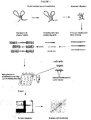
- the method of the invention includes the following steps:
- a 3C-like template may be prepared using known methods, such as the method described by Splinter et al., (2004) Methods Enzymol. 375, 493-507 . Briefly, a sample - such as cells, tissues or nuclei - is fixed using a cross-linking agent - such as formaldehyde. The primary restriction enzyme digestion is then performed such that the DNA is digested in the context of the cross-linked nucleus. Intramolecular ligation is then performed at low DNA concentrations, which favours ligation between cross-linked DNA fragments ( ie. intramolecular ligation) over ligation between non-cross-linked DNA fragments ( ie . intermolecular or random ligation). Next, the cross links are reversed and the DNA can be purified. The 3C template that is yielded contains restriction fragments that are ligated because they were originally close in the nuclear space.
- an enzyme recognition site for the primary restriction enzyme will separate the first (target) nucleotide sequence and the nucleotide sequence that has been ligated. Accordingly, the primary restriction enzyme recognition site is located between the first (target) nucleotide sequence and the ligated nucleotide sequence ( ie. the ligated second sequence).
- Cross-linking agents - such as formaldehyde - can be used to cross link proteins to other neighbouring proteins and nucleic acid.
- two or more nucleotide sequences can be cross-linked via proteins bound to (one of) these nucleotide sequences.
- Cross-linking agents other than formaldehyde can also be used in accordance with the present invention, including those cross-linking agents that directly cross link nucleotide sequences.
- agents that cross-link DNA include, but are not limited to, UV light, mitomycin C, nitrogen mustard, melphalan, 1,3-butadiene diepoxide, cis diaminedichloroplatinum(II) and cyclophosphamide.
- the cross-linking agent will form cross-links that bridge relatively short distances - such as about 2 ⁇ - thereby selecting intimate interactions that can be reversed.
- Cross-linking may be performed by, for example, incubating the cells in 2% formaldehyde at room temperature - such as by incubating 1 ⁇ 10 7 cells in 10 ml of DMEM-10% FCS supplemented with 2% formaldehyde for 10 min at room temperature.
- the cross-linked DNA is digested with a first restriction enzyme.
- Restriction endonucleases are enzymes that cleave the sugar-phosphate backbone of DNA. In most practical settings, a given restriction enzyme cuts both strands of duplex DNA within a stretch of just a few bases.
- the substrates for restriction enzymes are sequences of double-stranded DNA called recognition sites/sequences.
- the length of restriction recognition sites varies, depending on the restriction enzyme that is used.
- the length of the recognition sequence dictates how frequently the enzyme will cut in a sequence of DNA.
- Restriction enzymes which recognise a 4 bp sequence of DNA, together with their restriction sites include: AATT (TspEI), ACGT (Maell), AGCT (Alul), CATG (NlaIII), CCGG (Hpall), CGCG (FnuDII), CTAG (Mael), GATC (Dpnl, Dpnll, Sau3AI & Mbol), GCGC (Hhal), GGCC (Haelll), GTAC (Rsal), TCGA (Taql), TGCA (CviRI), TTAA (Msel), CCCG (Sth132I), CCGC (AciI) and CCTC (Mnll)
- Restriction enzymes which recognise a 6 bp sequence of DNA, together with their restriction sites include: AACGTT (Acll), AAGCTT (Hindlll), AATATT (Sspl), ACATGT (BspLU11I), ACCGGT (Agel), ACGCGT (Mlul), ACTAGT (Spel), AGATCT (BglII), AGCGCT (Eco47III), AGGCCT (Stul), AGTACT (ScaI), ATCGAT (Clal), ATGCAT (AvaIII), ATTAAT (Vspl), CAATTG (Mfel), CACGTG (PmaCI), CAGCTG (Pvull), CATATG (Ndel), CCATGG (Ncol), CCCGGG (Smal), CCGCGG (Sacll), CCTAGG (AvrII), CGATCG (Pvul), CGGCCG (Xmalll), CGTACG (Spll), CTCGAG (Xhol), CTGCAG (Pst
- Restriction enzymes which recognise a 7 bp sequence of DNA, together with their restriction sites, include: CCTNAGG (Saul), GCTNAGC (Espl), GGTNACC BstEII and TCCNGGA Pfol.
- Restriction enzymes which recognise an 8 bp sequence of DNA, together with their restriction sites, include: ATTTAAAT (Swal), CCTGCAGG (Sse8387I), CGCCGGCG (Sse232I), CGTCGACG (SgrDI), GCCCGGGC (Srfl), GCGATCGC (Sgfl), GCGGCCGC (Notl), GGCCGGCC (Fsel), GGCGCGCC (AscI), GTTTAAAC (Pmel) and TTAATTAA (Pacl).
- restriction enzymes which recognise degenerate sequences which means that two or more bases are possible at a particular position in the recognition sequence effectively resulting in 3or 5bp sequences of DNA that is recognized.
- the first restriction enzyme (or combination of enzymes) may recognise a 2, 4, 5, 6, 7 or 8 bp sequence of DNA.
- the first restriction enzyme may, in particular, be a 6-cutter, such as Hindlll or BglII.
- the second restriction enzyme may recognize a 2 or 4 bp sequence of DNA or be replaced by a nonspecific nuclease (in which case only a limited digestion would be applied) or mechanical fragmentation.
- the digestion step is then followed by ligation under diluted conditions that favour intra-molecular interactions and joining of the DNA via the compatible ends.
- Ligation may induced by the addition of a ligase enzyme.
- the ligation reaction may be performed at a low DNA concentration, such as about 1-5 ng/ ⁇ l.
- Cross-linking may be reversed by the addition of an agent such as proteinase K.
- the method of the invention may also involve:
- the ligated DNA molecule may be fragmented by various methods known in the art, such as digestion with a second restriction enzyme or other nucleases; using radiation or heavy ions; or mechanical means such as sonication or shearing.
- the second restriction enzyme should cut DNA more frequently than the first restriction enzyme used in step (b) of the method.
- the second restriction enzyme may recognise a shorter or more common stretch of DNA (recognition site) than the first restriction enzyme.
- the second restriction enzyme may be, for example, a 2 or 4-cutter.
- the second restriction enzyme may, for example, be a 4-cutter such as Dpn II of NlaIII.
- the second restriction enzyme may recognize a 2 or 4 bp sequence of DNA or be replaced by a nonspecific nuclease (in which case only a limited digestion would be applied) or mechanical fragmentation.
- a nonspecific nuclease in which case only a limited digestion would be applied
- mechanical fragmentation There are a large number of non-sequence specific nucleases, such as Micrococcal nuclease or DNasel.
- nucleotide sequences may need to be 'repaired' by standard methods to allow the next steps.
- An adapter may be ligated to the ends of the fragments from step (e) for sequencing purposes, i.e. to enable sequence analysis for methods such as the Illumina method.
- the adapter may comprise an address sequence. Different address sequences are used for different samples to allow multiplexing (hybridisation of different samples to the same set of oligonucleotide probes) where the address sequence allows the matching of a sequence with the sample it was derived from. Address sequences are useful when multiple samples or internal spiking is used.
- the adapter sequence prefferably added before hybridisation. It is possible to add them on by ligation after hybridisation but it is likely to be less efficient as the DNA comes off the hybridisation as single stranded DNA.
- step (f) of the method the nucleotide sequence fragments are hybridised to one or more oligonucleotide probe(s) in order to enrich for fragments which comprise an interacting nucleotide sequence
- the oligonucleotide probes are attached to or can be captured on a solid support, such as an array or beads (see below).
- the oligonucleotide probes are designed based on the sequence(s) from the region of interest, bearing in mind the position of the restriction sites of the first restriction enzyme.
- Each oligonucleotide probe corresponds to a sequence located within 100 bp of the first restriction site.
- the ligated DNA molecule made in step (d) of the method of the invention comprises different nucleotide sequences, joined at the restriction site of the first restriction enzyme.
- the different nucleotide sequences were "interacting" (i.e. in close enough proximity to be cross-linked) in the three dimensional structure.
- some fragments will be derived from a single nucleotide sequence, from internal fragmentation (e.g. internal digestion by the second restriction enzyme). Other fragments will be derived from both the interacting nucleotide sequences.
- fragments which have a sequence which is located close to the first restriction site are enriched for those which represent an "interacting fragment" i.e. comprise a portion of two nucleotide sequences joined at the at the restriction site of the first restriction enzyme by the ligation step (c).
- the oligonucleotide probes will be at least 15, 20, 25, 30 or 40 nucleotides in length.
- the oligonucleotide probes are designed to be as close as possible to the restriction enzyme recognition site of the first restriction enzyme.
- the oligonucleotide probes are designed such that they recognise a site within 100 nucleotides - such as about 90, 80, 70, 60, 50, 40, 30, 20, 10, 9, 8, 7, 6, 5, 4, 3, 2 or 1 nucleotide(s) away from the first restriction enzyme recognition site.
- the region of interest has X recognition sites of the first restriction enzyme (RE1), digestion with RE1 will produce X+1 fragments. These fragments will have an RE1 recognition site at both ends, so it is necessary to design 2X oligonucleotide probes to encompass all fragments in the region of interest.
- RE1 the first restriction enzyme
- the library of oligonucleotide probes may comprise oligonucleotides specific to substantially all the restriction fragments obtained by treating the region(s) of interest with the first restriction enzyme. "Substantially all” in this context, means at least 60, 70, 80, 90, 95 or 99% of the restriction fragment-flanking sites.
- oligonucleotide probe representing one of the ends, for example:
- oligonucleotide probes to that particular RE1 restriction fragment or end thereof may be omitted from the set of oligonucleotide probes, but the oligonucleotide set would still contain oligonucleotide probes to "substantially all" of the RE1-flanking sites.
- nucleotide sequences involved in the interaction may be characterised by sequencing.
- Pair-end sequencing may be carried our using known techniques, such as the Illumina system.
- An adapter sequence may be ligated to one or both ends of the nucleotide sequence fragments from (e) preferably before or less preferred after step (f) such that the ligated nucleotide sequence fragments may be captured on an array, amplified and/or sequenced.
- the adapter sequence may provide an address to recognize a sample when several samples are analysed on the same array, i.e. multiplexing. It is possible to multiplex 8 samples in one lane of an Illumina machine presently yielding ⁇ 150 million sequence reads per lane.
- the fragments may be end repaired and A-tailed, and the indexed adapters ligated to the A-tailed DNA fragments.
- the resulting adapter-modified DNA library may be captured, eluted and PCR amplified.
- the fragments may not be PCR amplified prior to the enrichment step (step (f)).
- Cluster generation and high-throughput sequencing may then be performed by known techniques (e.g. using the Illumina cluster reagents and a HiSeq 2000 sequencer).
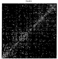
- the interaction frequencies may be visualised by producing a two dimensional heat map as previously described ( Liberman-Aiden et al (Science 2009 326:289-293 ; Dixon et al (2012, as above). Interaction frequencies between any two loci can be visualised by identifying the point off the axis where diagonals originating from each locus intersect, in a manner similar to a linkage disequilibrium plot.
- Each point on the map represents an interaction point between two fragments (two fragments in close proximity).
- the intensity of each interaction point on the map is relative to the frequency of interaction/proximity of the fragments which it represents.
- the points on the diagonal represent self-ligation effect as well as ligation to the immediately neighbouring fragments.
- the visualisation is basically a matrix analysis.
- a sample may be any physical entity comprising DNA that is or is capable of being cross-linked.
- the sample may be or may be derived from biological material.
- the sample may be or may be derived from one or more cells, one or more nuclei, or one or more tissue samples.
- the entities may be or may be derivable from any entities in which DNA - such as chromatin - is present.
- the sample may be or may be derived from one or more isolated cells or one or more isolated tissue samples, or one or more isolated nuclei.
- the sample may be or may be derived from living cells and/or dead cells and/or nuclear lysates and/or isolated chromatin.
- the sample may be or may be derived from cells of diseased and/or non-diseased subjects.
- the sample may be or may be derived from a subject that is suspected to be suffering from a disease.
- the sample may be or may be derived from a subject that is to be tested for the likelihood that they will suffer from a disease in the future.
- the sample may be or may be derived from viable or non-viable patient material.
- a standard sample may be added to each experimental sample (spiking) to allow better comparison between different sample as the samples may be normalised using the sequence reads of the spiking sample.
- the spiking sample may be from a different species than the experimental sample to allow spiking in the form of cells at the first step, alternatively the spiking sample may have its own address or be from a different species when spiking at later stages in the procedure.
- the set of oligonucleotide probes will be immobilised on a support or be captured on a solid support such as beads.
- Supports eg. solid supports
- When attached to a solid support it is preferably rigid and have a planar surface.
- Supports typically have from about 1-10,000,000 discrete spatially addressable regions, or cells. Supports having about 10-1,000,000 or about 100-100,000 or about 1000-100,000 cells are common. The density of cells is typically at least about 1000, 10,000, 100,000 or 1,000,000 cells within a square centimeter.
- all cells are occupied by pooled mixtures of oligonucleotide probes or a set of oligonucleotide probes. In other supports, some cells are occupied by pooled mixtures of probes or a set of oligonucleotide probes, and other cells are occupied, at least to the degree of purity obtainable by synthesis methods, by a single type of oligonucleotide.
- a single array of about 2 x 750,000 oligonucleotide probes can be used to cover, for example, the complete human or mouse genome, with 1 oligonucleotide probe at each side of each restriction site.
- Oligonucleotide probes in solution may contain a moiety that can be captured on a solid surface, such as oligonucleotides containing a biotin that can be captured by streptavidin beads. Hybridisation in solution may be more efficient.
- Capture may take place after hybridisation
- hybridisation shall include “the process by which a strand of nucleic acid joins with a complementary strand through base pairing”.
- Nucleotide sequences capable of selective hybridisation will be generally be at least 75%, 85%, 90%, 95% or 98% homologous to the corresponding complementary nucleotide sequence over the length of the oligonucleotide probe. Selectivity is determined by the salt and temperature conditions during the hybridisation.
- Stringent conditions are conditions under which a oligonucleotide probe will hybridise to its target sequence, but to no other sequences.
- Stringent conditions are sequence-dependent and are different in different circumstances. Longer sequences hybridise specifically at higher temperatures.
- very stringent conditions are selected to be about 5 °C lower than the thermal melting point (Tm) for the specific sequence at a defined ionic strength and pH.
- the hybridisation temperature is the temperature below the melting temperature (Tm) and the closer the hybridisation temperature is to the Tm the more stringent the hybridisation is, meaning that mismatched DNA sequences will not hybridise to each other.
- the oligonucleotide sequences should be in excess over the genomic DNA to ensure efficient, preferably complete and thereby quatitative hybridisation.
- stringent conditions include a salt concentration of at least about 0.01 to 1.0 M Na ion concentration (or other salts) at pH 7.0 to 8.3.
- Stringent conditions can also be achieved with the addition of destabilising agents - such as formamide or tetraalkyl ammonium salts.
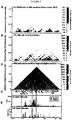
- Example 1 - T2C identifies known long-range interactions
- the inventors first chose the IGF/H19 region on human chromosome 11 that has previously been used to study the role of cohesion and CTCF for chromosomal long-range interactions and for which Hi-C and 4C data are already available for comparison ( Figure 2 ).
- a set of array-based oligonucleotides were designed mapping near the ends of all the Bglll fragments covering an approximately 2.1Mbp region of the H19 locus, totalling 524 oligonucleotides corresponding to 344 Bglll fragments.
- a number of Bglll fragments did not allow the design of an oligonucleotide representing one of the ends because the sequence was either repetitive or the 4bp recognition enzyme site (Nlalll) was too close to the Bglll site or completely absent from the Bglll fragment.
- the crosslinked Bglll restricted DNA was ligated, decrosslinked, digested with Nlalll enzyme and hybridized to the oligonucleotide array after decrosslinking (see Methods).
- the T2C method therefore yields reproducible results, faithfully detects the fragments that interact (or are in close proximity), clearly reproduces the overall genomic structure in topological domains and gives resolution around the 4-5kbp expected for a 6bp recognition restriction fragment.
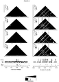
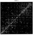
- Example 2 - T2C identifies different interaction networks based on different biological materials
- T2C was applied in in vivo mouse primary erythroid cells from mouse fetal liver and brain cells from E12.5 mice.
- the well-studied ⁇ -globin locus was used as an example in a region of ⁇ 2 MB around the gene. It is well established that as ⁇ -globin is expressed more highly in primary erythroid cells compared to fetal brain cells, a denser number of interactions is expected around the gene and between the gene and its locus control region (LCR) in this cell type.
- LCR locus control region
- the ⁇ -globin region was digested with Hindlll as the 6bp enzyme and 799 oligonucleotide probes were designed to cover the ends of the Hindlll fragments in the locus (724 fragments, many of which are repetitive) and after crosslinking re-digested with Dpnll.
- the analysis of the hybridised fragment after cleavage with Dpnll showed 5 topological domains in the region of interest ( ⁇ 2 MB) in both mouse primary erythroid cells and mouse fetal brain cells with many interactions within each topological domain.
- the topological domains also interact with each other suggesting a possibly higher order structure of the genome into a series of rosette like structures.
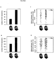
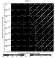
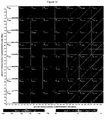
- the number of topological domains between the different biological materials appears to be the same the interactions within and between the topological domains appear to be less dense in mouse fetal brain cells comparing mouse primary erythroid cells ( Figure 3 ). Zooming in on the all the ⁇ -globin region shows all the well-known interactions in the ⁇ -globin locus in the fetal liver material.
- LDB1 is highly enriched on the ⁇ -globin locus and its LCR in mouse primary erythroid cells when compared to fetal brain cells.
- T2C is therefore a useful tool to detect topological domains and the different interactions within domains depending on the expression status of the genes such as the active ⁇ -globin locus in primary fetal liver cells versus the same silent locus in fetal brain.
- the high level of resolution of the interaction allows novel observations such as shown for the ⁇ -globin locus LDB1 binding sites and size of loops. Deletions within such a locus as for example in ⁇ -thaloidemia caused by DNA deletions would be immediately visible through the change of interaction signals.
- T2C satisfies these needs. Every restriction fragment can serve as a 'viewpoint' and all their interactions, either sort or long or to other chromosomes (not shown here), can be identified. Thus, multiple 3C-seq, 4C or 5C experiments do not have to be performed. Moreover, with T2C, the compartmentalization of the genome can be identified in the regions of interest without requiring the large sequence effort that was required for HiC, which increases the costs significantly.
- T2C Due to the design of T2C, a better coverage and resolution of the locus is obtained when compared to other techniques.
- the resolution of the T2C is based on the restriction enzyme used. Digesting crosslinked chromatin from primary erythroid cells and HB2 cells with Hindlll or BglII resulted on an average resolution of 2.9Kb and 6.1Kb respectively. This provides a significantly better resolution than the usual 40Kbp bins obtained with HiC.
- Moreover by adding the appropriate addresses in the oligonucleotides ligated on to the fragments (after the second cleavage before hybridisation) for sequencing purposes allows the multiplexing of different samples to the same set of oligonucleotides as the address sequence identifies the sample from which it was derived. Multiplexing further reduces the cost of T2C.
- T2C is an affordable, cost effective tool to explore the local spatial organization of the genome and chromatin interactions without requiring laborious procedures or massive sequencing efforts.
- Nuclei from mouse primary erythroid cells from mouse fetal liver E12.5, mouse fetal brain cells and a human breast endothel cell line (HB2) were isolated and crosslinked.
- the chromatin was digested with a 6-cutter (Hindlll for mouse cells and BglII for the HB2 cells), ligated and de-cross-linked. From the resulting libraries 50 ⁇ g DNA was digested with a frequent 4-cutter (Dpnll or NlaIII for the mouse cells, Nlalll for the HB2 cells). All these steps were performed according to the 3C-seq protocol previously described ( Stadhouders, R. et al. Nat Protoc 8, 509-524 (2013 )).
- a microarray for the ⁇ -globin locus was designed containing unique oligonucleotides as close as possible to the Hindlll restriction sites spanning ⁇ 2 MB around the gene (chr7: 109875617-111971734, mm9).
- unique oligonucleotides were designed close to BglII restriction sites (ch11: 1091427- 3228670, hg19) spanning an area of ⁇ 2.1 MB.
- the ligation products enriched by hybridization on the microarray were sequenced by paired-end sequencing yielding more than 100 million unique read pairs for the first and the second design respectively.
- the final library is prepared for analysis on the Illumina Cluster Station and HiSeq 2000 Sequencer according to the Illumina TruSeq DNA protocol with modifications (www.illumina.com).
- 20 ⁇ g of the digested library was purified using AMPure XP beads (Beckman Coulter) and end-repaired.
- the now blunt-ended fragments were A-tailed using the Klenow exo enzyme in the presence of ATP and purified again using AMPure XP beads.
- Indexed adapters (Illumina) were ligated to the A-tailed DNA fragments with subsequent purification using AMPure XP beads.
- the resulting adapter-modified DNA library was hybridized for 64 hours at 42°C on a custom-made NimbleGen Sequence Capture 2.1M capture array according to the NimbleGen Sequence Capture array protocol (www.nimblegen.com/seqcapez) on the NimbleGen Hybridization System.
- the captured DNA fragments are eluted from the hybridised array and purified using MinElute columns (Qiagen).
- the captured DNA fragments are amplified by PCR using Phusion polymerase as follow: 30 s at 98°C, 24 cycles of (10 s at 98°C, 30 s at 60°C, 30 s at 72°C), 5 min at 72°C final extension.
- PCR products are purified using AMPure XP beads and eluted in 30 ⁇ l of resuspension buffer.
- One microliter is loaded on an Agilent Technologies 2100 Bioanalyzer using a DNA 1000 assay to determine the library concentration and to check for quality.
- Cluster generation is performed according to the Illumina Cluster Reagents preparation protocol (www.illumina.com). Briefly, 1 ⁇ l of a 10 nM TruSeq DNA library stock is denatured with NaOH, diluted to 9-10 pM and hybridized onto the flowcell. The hybridized fragments are sequentially amplified, linearized and end-blocked according to the Illumina Paired-end Sequencing user guide protocol. After hybridization of the sequencing primer, sequencing-by-synthesis is performed using the HiSeq 2000 sequencer with a 101 cycle protocol according to manufacturer's protocol. The sequenced fragments were denaturated with NaOH using the HiSeq 2000 and the index-primer was hybridized onto the fragments. The index was sequenced with a 7-cycle protocol. The fragments are denaturated with NaOH, sequentially amplified, linearized and end-blocked. After hybridization of the sequencing primer, sequencing-by-synthesis of the third read is performed using the HiSeq 2000 sequencer with a 101-cycle protocol.
- Method i) starting with ⁇ 10 7 cultured/prepared cells, ii) the cells are formaldehyde fixed (i.e. DNA-DNA, RNA-RNA, DNA-RNA, protein-protein, DNA-protein, RNA-protein and more complex genomic crosslinks are formed), iii) permeablized to allow intra-nuclear restriction with a first restriction enzyme, iv) large dilution by extraction of the crosslinked fragments to allow re-ligation primarily within these complexes, before v) de-crosslinking, purification, and final shortening of the DNA chimeric fragments to sizes ⁇ 500bp by a second high-frequent restricting enzyme or by sonication (for highest resolution).
- a cleaned regional HRHT iCIC 2 DNA interaction fragment library is produced using DNA capture arrays (bead capturing is also possible) with ⁇ 10 9 -10 10 molecules per unique and hybridization optimized oligo (i.e. the capture is always in the linear regime and far from saturation), which sequentially are directly placed next to the first restriction enzyme, vii) After high-throughput sequencing, the obtained sequences are trimmed to contain only a sequence piece up to the first restriction enzyme, then mapped first to the whole reference genome and in case of using two restriction enzymes also against a masked sequence containing only the regions between 1st and 2nd restriction enzyme, to use finally only 100% uniquely mapped sequences.
- the human chromosome 11p 15.5-15.4, i.e. the IGF/H19 region, and the mouse chromosome 7qE3-F1, i.e. the ⁇ -globin region were selected, since both -2.1 Mbp regions are classic well studied by FISH and 3C examples of epigenetic and local control region regulation.
- Bgl II and Hind III as first and NlaIII as second restriction enzyme this results in many fragments down to -200-500 bp with an average of 6121 and 2915 bp, respectively.
- the interaction pattern is characterized by again clearly distinct gaps between the interactions, which are arranged in a crossed linear or grid-like pattern.
- the linear pattern continuous outside the sub-chromosomal domain and "crosses" there with the linear pattern originating from the subsequent sub-chromosomal domain.
- the general lower general interaction frequency level outside the domains and the less complicated interaction pattern seen there allows to follow the linear pattern back into the domains revealing that the much simpler/clear patter outside clearly also is present within the domains, but there enriched and more complicated by additional interactions.
- the data structure on different scales also proof also the assumption that on all scales interactions can indeed be cross-linked and depend on different underlying cross-linkable agents is correct, and thus that crosslinks between a specific DNA location or protein etc. create such patterns is very unlikely.
- the simple pattern is more complex due to the variation in the compaction density of chromatin and the fact that within the loops additional interactions take place of various forms: on a large scale simple loop or even super-helical like patterns seem likely, whereas on a low scale the patterns around the major loop base interactions indicate the structure of the rosette core and the local chromatin compaction and the entanglement of both there.
- the human IGF/H19 11p 15.5-15.4 region was investigated, and the mouse ⁇ -globin 7qE3-F1, in human HB2 and the TEV/HEV cells, and mouse fetal brain (FB) and liver (FL) cells: Whereas the general domains are clearly the same in HB2, TEV, HEV, and FB and FL cells, and thus in different cell types of the same species, but different between human and mouse at least due to the region chosen. On a finer degree of detail human HB2 cells seem to show a more and denser interaction pattern within the domains in comparison to the TEV/HEV system.
- the scaling behaviour of the different regions is in comparison a scaling behaviour of subset of a chromosome and thus is dominated by the local architecture deviations from the general scaling behaviour and is also compromised by the amount of interactions used here. Nevertheless, the scaling behaviour shows i) a long-range multi-scaling behaviour with a fine-structure, ii) with subtle but not in general differences for the various species, cell type, functional/distortion differences.
- the behaviour in general and in detail is stronger in the human compared to the mouse case, but within the different chromosomes nearly identical and only deviating for certain chromosomes to a somewhat larger extent.
- the behaviour on all scales is not only equivalent to the long-range multi-scaling of the genome architecture in detail but also at the right scales.
- the second maximum found corresponds to in size and position to the sub-chromosomal domains.
- the previously already proven association to nucleosomal binding at the first general maximum can now be extended to second maximum as well and associated with the looped structure in there as previously predicted.
- the interaction scaling at highest resolution shows the same behaviour across different chromosomes as the scaling behaviour of the DNA sequence in the same maximum: although a fine-structure cannot be found due to the experimental resolution, the general behaviour with a broad peak and a stronger interaction decrease on scales above ⁇ 4 kbp strongly suggests, in both the interaction experiment and in the DNA sequence, that a compacted chromatin fibre indeed exists.
- HB2 cells (1-7HB2, a clonal derivative of the human mammary luminal epithelial cell line MTSV1-7) were cultured in DMEM supplemented with 0.2 mM L-glutamine, 100 units/ml penicillin, 100 mg/ml streptomycin, 10% FCS, 5 ⁇ g/ml hydroxycortisone, and 10 ⁇ g/ml human insulin.
- DMEM fetal bovine serum
- the cleavable TEV/HRV RAD21-eGFP cell line system is a HEK293T cell line system, which was transfected with a pRTS-1 vector encoding for a cleavable RAD21-eGFP fusion protein and an siRNA for endogenous RAD21 knock-down. Both are expressed by doxycycline induced activation of a bidirectional promoter inbetween and thus simultaneously.
- RAD2-eGFP fusion-protein For the RAD2-eGFP fusion-protein, a cleavable RAD21, where the first RAD21-separase cleavage site is replaced by that of the 3C protease of the human rhinovirus (HRV protease) using a PCR-based mutagenesis (the second cleavage site remained unchanged to ensure less cell cytotoxicity) was inserted before eGFP.
- the tobacco etch virus protease (TEV protease)
- TMV protease does not recognize the HRV cleavage site and thus can act as a control.
- the endogenous RAD21 knock-down sequence allows knock-down with the following 3'UTR-directed siRNA's:
- the original HEK293T cell line was cultured in DMEM supplemented with 0.2mM L-glutamine, 100 units/ml penicillin, 100 mg/ml streptomycin, 10% FCS, and was grown at 37°C and 5% CO 2 .
- DMEM fetal calf serum
- Lipofectamine 2000 (Invitrogen) according to the instructions of the manufacturer was used. Cells carrying the vector were selected by growth in 150 ⁇ g/mL hygromycin containing medium.
- TEV/HRV RAD21-eGFP cell line was as well cultured in DMEM supplemented with 0.2mM L-glutamine at 37°C and 5% CO 2 .
- HRV or TEV which serves as a control, thus a transfection takes place, but no cleavage
- the cells were cultured for 3 days in the presence of 2 ⁇ g/ml of doxycycline.
- mice For mouse fetal liver and fetal brain cells, ⁇ 10 embryos on day 12.5 of pregnancy from one to two transgenic FVB/N mice were used for the -10 million cells required by the experiment to have a complex enough cell population and enough DNA in the end to be sequenced: The mice were cleaned with 70% EtOH and the abdomen was opened to remove the cervix containing the embryos, before cutting them lose and removing them from the yolk sac and placenta. Small and underdeveloped embryos were discarded. The embryos are collected in petri dishes on ice with 0.5 ml 10% FCS/PBS. Then the fetal liver and brain were dissected from the embryos and collected in tubes (1 ml) on ice containing again 500 ⁇ l 10% FCS/PBS.
- the cells were then resuspended with a P1000/1ml plastic pipette tip and connective tissue was digested by adding 25 ⁇ l of a 2.5% Collagenase stock (0.125% end concentration) and incubated for ⁇ 45 min at 37°C. Thereafter, the cell suspension was transferred to falcon tubes with 12 ml 10% FCS/PBS at room temperature and thereafter was gently squeezed through a scraper mesh which was placed inside a 6-well plate using again a P1000/1ml plastic pipette tip. The mesh was washed with 2ml 10% FCS/PBS at room temperature to get all the cells from the mesh.
- the resulting single cell suspension was again collected in falcon tubes with an end volume of 12 ml 10%FCS/PBS at room temperature. Notably, we tried to keep the stress of the cells to the minimum, to avoid any damage to the cell nuclei. Both after the resuspension, the Collagenase treatment, and/or after the scraping of fetal liver and brain material/cells were spotted on glass slides to check for cellular and especially nuclear integrity by microscopy, with or without staining of the nucleus with DAPI (or eventually any other immunofluorescence or fluorescence in situ hybridization).
- the cells were first counted and their concentration adjusted to 10 million in 12 ml 10% FCS/PBS at room temperature and put into 15ml polypropylene tubes (used for cell culture and thus not excessively absorbing/fixing cells to the tube wall, Greiner Bio One). Then 650 ⁇ l of a 37 % formaldehyde PBS solution were added, i.e. a final concentration of 1.9 % of formaldehyde is used for crosslinking/fixation, at room temperature for 10 min while softly tumbling to avoid cell aggregation.
- the concentration of formaldehyde for crosslinking/fixation at this stage is ideal for the following steps and in respect to cell/nuclear integrity for the human and mouse cells we used here; although this might hold in general, cases are known where other concentrations and incubation times achieve better results.
- the tube was put on ice (from now on we kept everything on ice up to the 1st restriction of the DNA (see below) to avoid any damage of the material) and 1.6 ml of cold 1M Glycine in PBS were added to stop the crosslink/fixation reaction.
- the cells were spun down for 8 min at 1300 rpm at 4°C, the resulting pellet was washed in ice-cold PBS, and taken up first in 1 ml before adding up to 14 ml of PBS, followed again by spinning down for 8 min at 1300 rpm at 4°C. After discarding the supernatant, the pellet could now also be frozen for storage, although we advise to straight away continue with lysis and the 1st restriction. Again cells were spotted on glass slides to check for cellular and especially nuclear integrity by microscopy, with/without staining of the nucleus with DAPI (Note: again the cells could now also be used eventually for any other immunofluorescence or fluorescence in situ hybridization experiment).
- the pellet prepared in the last step of crosslinking/fixation was taken up in 1 ml of this lysis buffer, resuspended and filled up with another 4 ml to a total of 5 ml and incubated for 10 min on ice.
- the now free cell nuclei were spun down for 5 min at 1800 rpm at 4°C, the pellet was taken up in 0.5 ml of ice cold PBS safe-lock tube, and spun for 1 min at 2600 rpm a at 4°C. Again here it is possible after removal of the supernatant and snap-freezing to store the nuclei at -80°C.
- nuclei On glass slides to check for nuclear integrity by microscopy and/or staining of the nucleus with DAPI.
- the nuclei were now resuspended in 0.5 ml/tubes with 1.2x restriction buffer (60 ⁇ l restriction buffer, 440 ⁇ l MilliQ and adjusted for BSA if necessary) and transferred to a 1.5 ml safe-lock tube. Then to gently permeabilize the nuclear lamina the tubes were put to 37°C and 7.5 ⁇ l of 20% SDS (0.3% endconcentration) were added, and incubated at 37°C for 1 h, while shaking at 900 rpm.
- 1.2x restriction buffer 60 ⁇ l restriction buffer, 440 ⁇ l MilliQ and adjusted for BSA if necessary
- the fully digested nuclear material was diluted by transferal to a 50 ml falcon tube and addition of 6.125 ml 1.15x ligation buffer (6.125ml: 5421 ml MilliQ + 704 ⁇ l ligation buffer). Then 375 ⁇ l of 20% Triton-X-100 (endconcentration 1.0%) was added and incubated in a 37°C water bath for 1 h, while being shaken every 10 min by hand. Then 20 ⁇ l Ligase HC 5U/ ⁇ l (100 U in total, Roche) was added and incubated at 16°C over night ( ⁇ 20 h) followed by an additional 30 min of incubation at room temperature.
- the DNA was purified by adding 30 ⁇ l 10 mg/ml RNAse (300 ⁇ g in total) and incubation for at 37°C for 30-45 min, followed by brief cooling to room temperature and addition of 7 ml phenol-chloroform and vigorous shaking. Then the sample was centrifuged at 4,000 rmp (2200xg) for 15 min, before the upper phase was put in a new 50 ml tube and 7 ml of MilliQ was added as well as 1 ⁇ l of glycogen per ml, 1.5 ml of 2M Sodium Acetate pH 5.6, and add 35 ml of 100% ethanol to enhance the purification, gently but thoroughly mixed and thereafter put at -80°C for 1.5-3 h.
- the resulting re-ligated and de-crosslinked purified material was shortened by a 2nd restriction: First, to control the amount of DNA at this stage an aliquot of 1 ⁇ l was run alongside a reference sample of species-matched genomic DNA of known concentration on a 2% agarose gel. Then the DNA was adjusted in 0.5 ml/tubes to a 100 ng/ ⁇ l concentration and restricted with the 2nd restriction enzyme by adding 1 U per ⁇ g of DNA of the selected restriction enzyme and incubated over night ( ⁇ 20 h) at 37°C. For the human cells in all cases the restriction enzyme Nlall (New England Biolabs) was used. For the mouse cells we used either if Hindlll was used as 1st restriction enzyme Dpnll (New England Biolabs) or if Apol was used as 1st restriction enzyme sonication with 10 cycles of 15 sec on and 45 sec off.
- Nlall New England Biolabs
- the DNA was purified by adding 3 ⁇ l 10 mg/ml RNAse and incubation for at 37°C for 30-45 min, followed by brief cooling to room temperature and addition of MilliQ up to 500 ⁇ l ( ⁇ 400 ml) as well as 500 ⁇ l phenol-chloroform and vigorous shaking.
- the DNA T2C fragment library was prepared for sequencing analysis on the Illumina Cluster Station and HiSeq 2000 Sequencer according to the Illumina TruSeq DNA protocol with enhancing modifications from us (www.illumina.com. TruSeq DNA sample prep LS protocol; part#15026489 Rev. C): i) purification of the DNA fragments, ii) end-repair to reach blunt end status, iii) 3'-end Adenylation to avoid chimera, in the iv) sequencing adapter ligation including eventual multiplexing step, and finally v) purification of the T2C whole genome sequencing DNA fragment library.
- the concentration of the T2C DNA fragment library was measured again for fine tuning using 1 ⁇ l of material using Quant-it dsDNA broad range assay kit. Then the samples were split into 4 sets of 5 ⁇ g each of the T2C DNA fragment library and the following complete procedure done for each of these 4 sets of material:
- oligo was placed up- and one downstream as near as possible to the 1st restriction site used in the nuclear whole genome restriction, since the interest lies in sequencing just each side after re-ligation of this 1st restriction.
- the oligos were designed by NimbleGen and us for the selected regions of the human and mouse genomes using genome builts mm9 and HG19 with oligo length of 72 ⁇ 3 bp, unique appearance (no mismatch allowed) in the entire genome, and with respect to best and similar, i.e. similar hybridization, capturing on a microarray.
- the oligos were further selected: in the case of using a 2nd restriction enzyme to shorten the re-ligated DNA library for sequencing the oligo had to be situated between the 1st and 2nd restriction site.
- a 2nd restriction enzyme to shorten the re-ligated DNA library for sequencing the oligo had to be situated between the 1st and 2nd restriction site.
- sonication to shorten the re-ligated DNA only oligos within 150 bp of the 1st restriction site were chosen. If only one oligo was present, which crossed either the 1st or 2nd or even both restriction sites, only cuts at the oligo beginning or end of in total not more than 10 % were allowed, i.e. that the oligos could definitely capture DNA pieces with a minimum of 62 bp to guaranty specificity and similar hybridization efficiency.
- oligos and capture arrays In the case of a 1st and 2nd restriction enzyme we chose the oligos and capture arrays as follows: In the human case this was done for the H19/IGF2 region on chromosome 11 from basepair position -1,110,650 to -3,216,350, i.e. a 2,105,700 bp sized region and 525 oligos. In the mouse case this was done for the ⁇ -Globin region on chromosome 7 from basepair position -109,876,350 to 111,966,600, i.e. a 2,090,250 bp sized region and 800 oligos.
- the pooled DNA library after ligation of the sequencing adapters was subject to subselection with the above described newly and specifically developed capturing microarrays using the NimbleGen Array capture protocol and hybridization system with enhancing modifications from (www.nimblegen.com/segcapez, NimbleGen Arrays User's Guide, Sequence Capture Array Delivery version 3.2): The entire procedure consisted of i) microarray hybridization, ii) washing before iii) elution of the captured regional DNA library from the microarray.
- T2C regional DNA fragment sequencing library was enriched for sequencing first by PCR using Phusion polymerase using 30 sec at 98°C, 12 cycles of (10 sec at 98°C, 30 sec at 60°C, 30 sec at 72°C), 5 min at 72C final extension.
- Phusion polymerase using 30 sec at 98°C, 12 cycles of (10 sec at 98°C, 30 sec at 60°C, 30 sec at 72°C), 5 min at 72C final extension.
- For each 1 ⁇ g of T2C regional DNA fragment library 5 ⁇ l of the PCR primer cocktail and 25 ⁇ l PCR master mix was added to the PCR plate.
- AMPure XP beads (Beckman Coulter) were used by adding 1.8 ⁇ l AMPure XP beads per 1.0 ⁇ l of DNA.
- Cluster generation was performed according to the Illumina cBot User Guide ( www.illumina.com , part#15006165 RevE). Briefly, 1 ⁇ l of a 10 nM TruSeq DNA library stock DNA was denatured with NaOH, diluted to 10 pM and hybridized onto the flowcell. The hybridized fragments are sequentially amplified, linearized and end-blocked according to the Illumina Paired-end Sequencing user guide protocol. After hybridization of the sequencing primer, sequencing-by-synthesis was performed using the HiSeq 2000 sequencer with a 101 cycle protocol according to the instructions of the manufacturer. The sequenced fragments were denaturated with NaOH using the HiSeq 2000 and the index-primer was hybridized onto the fragments.
- the index was sequenced with a 7-cycle protocol.
- the fragments are denaturated with NAOH, sequentially amplified, linearized and end-blocked.
- sequencing-by-synthesis of the third read was performed using the HiSeq 2000 sequencer with a 101-cycle protocol.
- the raw sequence reads were checked for the existence of the first restriction enzyme recognition sequence in the sequencing direction.
- the sequence after the first enzyme recognition site was removed. If the bases of the recognition site after the overhang were not unambiguously, the read was further trimmed by removing all the bases after the end of the overhang. Then these trimmed sequences were aligned using the Burrows-Wheeler Alignment (BWA) tool to the whole human genome NCBI36/hg18 assembly and to the mouse NCBI37/mm9 assembly. Therefore the following default parameter set was used (with the value of the parameter in [] brackets): bwa aln [options] ⁇ prefix> ⁇ in.fq>
- the resulting paired-end sequences then contain the interaction information with an error rate determined by the error rate of sequencing, the quality of the reference sequence, and the difference the DNA sequence of our cells/mice to this reference genome.
- a rough estimate of the false positive and false negative results for unique sequences without mismatches at the end of this process using known error rates indicate the error to be smaller than 1% after accumulation of errors and the reduction of errors due to our procedure. This can be also deducted from the reduction of sequence pairs from the initial raw sequence throughout the entire process to the final result.
Landscapes
- Chemical & Material Sciences (AREA)
- Organic Chemistry (AREA)
- Life Sciences & Earth Sciences (AREA)
- Health & Medical Sciences (AREA)
- Engineering & Computer Science (AREA)
- Zoology (AREA)
- Wood Science & Technology (AREA)
- Proteomics, Peptides & Aminoacids (AREA)
- Analytical Chemistry (AREA)
- Molecular Biology (AREA)
- Bioinformatics & Cheminformatics (AREA)
- Immunology (AREA)
- General Health & Medical Sciences (AREA)
- Genetics & Genomics (AREA)
- Biotechnology (AREA)
- Biochemistry (AREA)
- Microbiology (AREA)
- General Engineering & Computer Science (AREA)
- Physics & Mathematics (AREA)
- Biophysics (AREA)
- Chemical Kinetics & Catalysis (AREA)
- Measuring Or Testing Involving Enzymes Or Micro-Organisms (AREA)
- Bioinformatics & Computational Biology (AREA)
- General Chemical & Material Sciences (AREA)
- Apparatus Associated With Microorganisms And Enzymes (AREA)
Applications Claiming Priority (2)
| Application Number | Priority Date | Filing Date | Title |
|---|---|---|---|
| GBGB1320351.8A GB201320351D0 (en) | 2013-11-18 | 2013-11-18 | Method |
| PCT/IB2014/002485 WO2015071748A1 (en) | 2013-11-18 | 2014-11-18 | Method for analysing the interaction of nucleotide sequences in a three-dimensional dna structure |
Publications (2)
| Publication Number | Publication Date |
|---|---|
| EP3071710A1 EP3071710A1 (en) | 2016-09-28 |
| EP3071710B1 true EP3071710B1 (en) | 2019-05-15 |
Family
ID=49883800
Family Applications (1)
| Application Number | Title | Priority Date | Filing Date |
|---|---|---|---|
| EP14815428.9A Active EP3071710B1 (en) | 2013-11-18 | 2014-11-18 | Method for analysing the interaction of nucleotide sequences in a three-dimensional dna structure |
Country Status (9)
| Country | Link |
|---|---|
| US (1) | US10934575B2 (enExample) |
| EP (1) | EP3071710B1 (enExample) |
| JP (1) | JP2016537029A (enExample) |
| KR (1) | KR102332522B1 (enExample) |
| CN (1) | CN105992825A (enExample) |
| AU (1) | AU2014349817B2 (enExample) |
| CA (1) | CA2928012C (enExample) |
| GB (1) | GB201320351D0 (enExample) |
| WO (1) | WO2015071748A1 (enExample) |
Families Citing this family (24)
| Publication number | Priority date | Publication date | Assignee | Title |
|---|---|---|---|---|
| GB2554572B (en) * | 2015-03-26 | 2021-06-23 | Dovetail Genomics Llc | Physical linkage preservation in DNA storage |
| JP2018518203A (ja) * | 2015-06-24 | 2018-07-12 | オックスフォード バイオダイナミックス リミテッド | エピジェネティックな染色体相互作用 |
| WO2017066908A1 (zh) * | 2015-10-19 | 2017-04-27 | 安诺优达基因科技(北京)有限公司 | 构建高分辨率、大信息量单细胞Hi-C文库的方法 |
| CN115369161A (zh) * | 2015-12-04 | 2022-11-22 | 10X 基因组学有限公司 | 用于核酸分析的方法和组合物 |
| GB201608000D0 (en) * | 2016-05-06 | 2016-06-22 | Oxford Biodynamics Ltd | Chromosome detection |
| EP3455356B1 (en) | 2016-05-13 | 2021-08-04 | Dovetail Genomics LLC | Recovering long-range linkage information from preserved samples |
| CA3045070A1 (en) * | 2016-12-01 | 2018-06-07 | Oxford Biodynamics Limited | Application of epigenetic chromsomal interactions in cancer diagnostics |
| WO2019005763A1 (en) * | 2017-06-26 | 2019-01-03 | Phase Genomics Inc. | METHOD FOR REGROUPING DNA SEQUENCES |
| CN108300767B (zh) * | 2017-10-27 | 2021-08-20 | 清华大学 | 一种核酸复合体中核酸区段相互作用的分析方法 |
| CN108197431B (zh) * | 2018-01-24 | 2022-04-05 | 清华大学 | 染色质相互作用差异的分析方法和系统 |
| US12378592B2 (en) * | 2018-01-31 | 2025-08-05 | Dovetail Genomics, Llc. | Sample prep for DNA linkage recovery |
| EP3847261A1 (en) * | 2018-09-05 | 2021-07-14 | Max-Delbrück-Centrum für Molekulare Medizin in der Helmholtz-Gemeinschaft | A method for engineering synthetic cis-regulatory dna |
| WO2020234608A1 (en) * | 2019-05-22 | 2020-11-26 | Oxford Nanopore Technologies Limited | Protocol for detecting interactions within one or more dna molecules within a cell |
| GB2607514B (en) | 2020-07-20 | 2023-06-21 | Enanta Pharm Inc | Functionalized peptides as antiviral agents |
| US11384090B2 (en) | 2020-11-23 | 2022-07-12 | Enanta Pharmaceuticals, Inc. | Spiropyrrolidine derived antiviral agents |
| CN116829571A (zh) | 2020-11-23 | 2023-09-29 | 英安塔制药有限公司 | 新型螺旋吡咯烷衍生的抗病毒药物 |
| US11352363B1 (en) | 2020-11-23 | 2022-06-07 | Enanta Pharmaceuticals, Inc. | Spiropyrrolidine derived antiviral agents |
| CN113215141A (zh) * | 2021-02-23 | 2021-08-06 | 华南农业大学 | 细菌hi-c基因组及质粒构象捕获方法 |
| US11319325B1 (en) | 2021-05-11 | 2022-05-03 | Enanta Pharmaceuticals, Inc. | Macrocyclic spiropyrrolidine derived antiviral agents |
| US11325916B1 (en) | 2021-07-29 | 2022-05-10 | Enanta Pharmaceuticals, Inc. | Spiropyrrolidine derived antiviral agents |
| US11339170B1 (en) | 2021-07-23 | 2022-05-24 | Enanta Pharmaceuticals, Inc. | Spiropyrrolidine derived antiviral agents |
| GB202111195D0 (en) * | 2021-08-03 | 2021-09-15 | Cergentis B V | Method for targeted sequencing |
| CN113946730B (zh) * | 2021-10-19 | 2023-03-17 | 四川大学 | 一种基于基因数据的染色质层次结构分析的可视化方法 |
| US20240095226A1 (en) * | 2022-09-15 | 2024-03-21 | Rodney Kuhn Haffnerson King | Methods and related devices for storing and accessing data using multi-level fractal grids |
Family Cites Families (5)
| Publication number | Priority date | Publication date | Assignee | Title |
|---|---|---|---|---|
| US7348178B1 (en) * | 1994-03-14 | 2008-03-25 | New York University Medical Center | Recombinant adenoviral vector system |
| NZ564262A (en) * | 2005-07-04 | 2010-02-26 | Univ Erasmus Medical Ct | Chromosome conformation capture-on-chip (4C) assay |
| AU2008204338B2 (en) * | 2007-01-11 | 2014-03-06 | Erasmus University Medical Center | Circular chromosome conformation capture (4C) |
| CA2804450C (en) | 2010-07-09 | 2022-10-11 | Max Jan van Min | 3-d genomic region of interest sequencing strategies |
| US10287621B2 (en) * | 2013-04-11 | 2019-05-14 | Pelin Sahlén | Targeted chromosome conformation capture |
-
2013
- 2013-11-18 GB GBGB1320351.8A patent/GB201320351D0/en not_active Ceased
-
2014
- 2014-11-18 CA CA2928012A patent/CA2928012C/en active Active
- 2014-11-18 EP EP14815428.9A patent/EP3071710B1/en active Active
- 2014-11-18 CN CN201480062775.XA patent/CN105992825A/zh active Pending
- 2014-11-18 WO PCT/IB2014/002485 patent/WO2015071748A1/en not_active Ceased
- 2014-11-18 JP JP2016553748A patent/JP2016537029A/ja active Pending
- 2014-11-18 KR KR1020167015969A patent/KR102332522B1/ko active Active
- 2014-11-18 US US15/037,210 patent/US10934575B2/en active Active
- 2014-11-18 AU AU2014349817A patent/AU2014349817B2/en active Active
Non-Patent Citations (1)
| Title |
|---|
| None * |
Also Published As
| Publication number | Publication date |
|---|---|
| EP3071710A1 (en) | 2016-09-28 |
| AU2014349817B2 (en) | 2020-11-12 |
| WO2015071748A1 (en) | 2015-05-21 |
| CA2928012A1 (en) | 2015-05-21 |
| KR102332522B1 (ko) | 2021-11-29 |
| GB201320351D0 (en) | 2014-01-01 |
| CN105992825A (zh) | 2016-10-05 |
| US10934575B2 (en) | 2021-03-02 |
| CA2928012C (en) | 2023-05-16 |
| AU2014349817A1 (en) | 2016-05-19 |
| US20160289738A1 (en) | 2016-10-06 |
| KR20160079120A (ko) | 2016-07-05 |
| JP2016537029A (ja) | 2016-12-01 |
Similar Documents
| Publication | Publication Date | Title |
|---|---|---|
| EP3071710B1 (en) | Method for analysing the interaction of nucleotide sequences in a three-dimensional dna structure | |
| Zhang et al. | Transcriptionally active HERV-H retrotransposons demarcate topologically associating domains in human pluripotent stem cells | |
| Szalaj et al. | Three-dimensional organization and dynamics of the genome | |
| Goel et al. | The macro and micro of chromosome conformation capture | |
| CN106566828B (zh) | 一种高效的全基因组染色质构象技术eHi-C | |
| US20180320224A1 (en) | Single-cell proteomic assay using aptamers | |
| EP2083090B1 (en) | Nucleic acid interaction analysis | |
| US20180340939A1 (en) | Multiplex analysis of single cell constituents | |
| EP3365464B1 (en) | Method of analysing dna sequences | |
| CN114507711B (zh) | 一种单细胞转录组测序方法及其应用 | |
| Zade et al. | Biased selection of propagation-related TUPs from phage display peptide libraries | |
| WO2017066907A1 (zh) | 一种构建高可利用数据率的Hi-C文库的方法 | |
| US20110269647A1 (en) | Method | |
| JP2017529104A (ja) | Rnaスティッチシーケンシング:細胞におけるrna:rna相互作用の直接マッピングのための解析 | |
| US20190187156A1 (en) | Methods for identifying macromolecule interactions | |
| US20220033807A1 (en) | Method for capturing rna in situ higher-order structures and interactions | |
| HK1228460B (en) | Method for analysing the interaction of nucleotide sequences in a three-dimensional dna structure | |
| HK1228460A1 (en) | Method for analysing the interaction of nucleotide sequences in a three-dimensional dna structure | |
| Knoch | A Guided Protocol for Array Based T2C: A High‐Quality Selective High‐Resolution High‐Throughput Chromosome Interaction Capture | |
| Tian et al. | Landscape of the Epstein-Barr virus-host chromatin interactome and gene regulation | |
| Key | Molecular genetics, recombinant DNA, & genomic technology | |
| Beliveau et al. | A guide to studying 3D genome structure and dynamics in the kidney | |
| Metkar | RIPPLiT and ChimeraTie: High throughput tools for understanding higher order RNP structures | |
| Mehta et al. | Let us know how access to this document benefits yo u | |
| WO2024168092A2 (en) | Methods and kits for labeling cellular molecules for multiplex analysis |
Legal Events
| Date | Code | Title | Description |
|---|---|---|---|
| PUAI | Public reference made under article 153(3) epc to a published international application that has entered the european phase |
Free format text: ORIGINAL CODE: 0009012 |
|
| 17P | Request for examination filed |
Effective date: 20160618 |
|
| AK | Designated contracting states |
Kind code of ref document: A1 Designated state(s): AL AT BE BG CH CY CZ DE DK EE ES FI FR GB GR HR HU IE IS IT LI LT LU LV MC MK MT NL NO PL PT RO RS SE SI SK SM TR |
|
| AX | Request for extension of the european patent |
Extension state: BA ME |
|
| DAX | Request for extension of the european patent (deleted) | ||
| STAA | Information on the status of an ep patent application or granted ep patent |
Free format text: STATUS: EXAMINATION IS IN PROGRESS |
|
| 17Q | First examination report despatched |
Effective date: 20170608 |
|
| REG | Reference to a national code |
Ref country code: HK Ref legal event code: DE Ref document number: 1228460 Country of ref document: HK |
|
| REG | Reference to a national code |
Ref country code: DE Ref legal event code: R079 Ref document number: 602014046925 Country of ref document: DE Free format text: PREVIOUS MAIN CLASS: C12Q0001680000 Ipc: C12Q0001682000 |
|
| GRAP | Despatch of communication of intention to grant a patent |
Free format text: ORIGINAL CODE: EPIDOSNIGR1 |
|
| STAA | Information on the status of an ep patent application or granted ep patent |
Free format text: STATUS: GRANT OF PATENT IS INTENDED |
|
| RIC1 | Information provided on ipc code assigned before grant |
Ipc: C12Q 1/6806 20180101ALI20180706BHEP Ipc: C12Q 1/682 20180101AFI20180706BHEP |
|
| INTG | Intention to grant announced |
Effective date: 20180726 |
|
| GRAJ | Information related to disapproval of communication of intention to grant by the applicant or resumption of examination proceedings by the epo deleted |
Free format text: ORIGINAL CODE: EPIDOSDIGR1 |
|
| STAA | Information on the status of an ep patent application or granted ep patent |
Free format text: STATUS: EXAMINATION IS IN PROGRESS |
|
| GRAP | Despatch of communication of intention to grant a patent |
Free format text: ORIGINAL CODE: EPIDOSNIGR1 |
|
| STAA | Information on the status of an ep patent application or granted ep patent |
Free format text: STATUS: GRANT OF PATENT IS INTENDED |
|
| INTG | Intention to grant announced |
Effective date: 20181207 |
|
| RIC1 | Information provided on ipc code assigned before grant |
Ipc: C12Q 1/6806 20180101ALI20180706BHEP Ipc: C12Q 1/682 20180101AFI20180706BHEP |
|
| GRAS | Grant fee paid |
Free format text: ORIGINAL CODE: EPIDOSNIGR3 |
|
| GRAA | (expected) grant |
Free format text: ORIGINAL CODE: 0009210 |
|
| STAA | Information on the status of an ep patent application or granted ep patent |
Free format text: STATUS: THE PATENT HAS BEEN GRANTED |
|
| AK | Designated contracting states |
Kind code of ref document: B1 Designated state(s): AL AT BE BG CH CY CZ DE DK EE ES FI FR GB GR HR HU IE IS IT LI LT LU LV MC MK MT NL NO PL PT RO RS SE SI SK SM TR |
|
| REG | Reference to a national code |
Ref country code: CH Ref legal event code: EP |
|
| REG | Reference to a national code |
Ref country code: DE Ref legal event code: R096 Ref document number: 602014046925 Country of ref document: DE |
|
| REG | Reference to a national code |
Ref country code: IE Ref legal event code: FG4D |
|
| REG | Reference to a national code |
Ref country code: NL Ref legal event code: FP |
|
| REG | Reference to a national code |
Ref country code: LT Ref legal event code: MG4D |
|
| PG25 | Lapsed in a contracting state [announced via postgrant information from national office to epo] |
Ref country code: NO Free format text: LAPSE BECAUSE OF FAILURE TO SUBMIT A TRANSLATION OF THE DESCRIPTION OR TO PAY THE FEE WITHIN THE PRESCRIBED TIME-LIMIT Effective date: 20190815 Ref country code: ES Free format text: LAPSE BECAUSE OF FAILURE TO SUBMIT A TRANSLATION OF THE DESCRIPTION OR TO PAY THE FEE WITHIN THE PRESCRIBED TIME-LIMIT Effective date: 20190515 Ref country code: LT Free format text: LAPSE BECAUSE OF FAILURE TO SUBMIT A TRANSLATION OF THE DESCRIPTION OR TO PAY THE FEE WITHIN THE PRESCRIBED TIME-LIMIT Effective date: 20190515 Ref country code: SE Free format text: LAPSE BECAUSE OF FAILURE TO SUBMIT A TRANSLATION OF THE DESCRIPTION OR TO PAY THE FEE WITHIN THE PRESCRIBED TIME-LIMIT Effective date: 20190515 Ref country code: PT Free format text: LAPSE BECAUSE OF FAILURE TO SUBMIT A TRANSLATION OF THE DESCRIPTION OR TO PAY THE FEE WITHIN THE PRESCRIBED TIME-LIMIT Effective date: 20190915 Ref country code: HR Free format text: LAPSE BECAUSE OF FAILURE TO SUBMIT A TRANSLATION OF THE DESCRIPTION OR TO PAY THE FEE WITHIN THE PRESCRIBED TIME-LIMIT Effective date: 20190515 Ref country code: AL Free format text: LAPSE BECAUSE OF FAILURE TO SUBMIT A TRANSLATION OF THE DESCRIPTION OR TO PAY THE FEE WITHIN THE PRESCRIBED TIME-LIMIT Effective date: 20190515 Ref country code: FI Free format text: LAPSE BECAUSE OF FAILURE TO SUBMIT A TRANSLATION OF THE DESCRIPTION OR TO PAY THE FEE WITHIN THE PRESCRIBED TIME-LIMIT Effective date: 20190515 |
|
| PG25 | Lapsed in a contracting state [announced via postgrant information from national office to epo] |
Ref country code: LV Free format text: LAPSE BECAUSE OF FAILURE TO SUBMIT A TRANSLATION OF THE DESCRIPTION OR TO PAY THE FEE WITHIN THE PRESCRIBED TIME-LIMIT Effective date: 20190515 Ref country code: GR Free format text: LAPSE BECAUSE OF FAILURE TO SUBMIT A TRANSLATION OF THE DESCRIPTION OR TO PAY THE FEE WITHIN THE PRESCRIBED TIME-LIMIT Effective date: 20190816 Ref country code: BG Free format text: LAPSE BECAUSE OF FAILURE TO SUBMIT A TRANSLATION OF THE DESCRIPTION OR TO PAY THE FEE WITHIN THE PRESCRIBED TIME-LIMIT Effective date: 20190815 Ref country code: RS Free format text: LAPSE BECAUSE OF FAILURE TO SUBMIT A TRANSLATION OF THE DESCRIPTION OR TO PAY THE FEE WITHIN THE PRESCRIBED TIME-LIMIT Effective date: 20190515 |
|
| REG | Reference to a national code |
Ref country code: AT Ref legal event code: MK05 Ref document number: 1133488 Country of ref document: AT Kind code of ref document: T Effective date: 20190515 |
|
| PG25 | Lapsed in a contracting state [announced via postgrant information from national office to epo] |
Ref country code: SK Free format text: LAPSE BECAUSE OF FAILURE TO SUBMIT A TRANSLATION OF THE DESCRIPTION OR TO PAY THE FEE WITHIN THE PRESCRIBED TIME-LIMIT Effective date: 20190515 Ref country code: RO Free format text: LAPSE BECAUSE OF FAILURE TO SUBMIT A TRANSLATION OF THE DESCRIPTION OR TO PAY THE FEE WITHIN THE PRESCRIBED TIME-LIMIT Effective date: 20190515 Ref country code: CZ Free format text: LAPSE BECAUSE OF FAILURE TO SUBMIT A TRANSLATION OF THE DESCRIPTION OR TO PAY THE FEE WITHIN THE PRESCRIBED TIME-LIMIT Effective date: 20190515 Ref country code: DK Free format text: LAPSE BECAUSE OF FAILURE TO SUBMIT A TRANSLATION OF THE DESCRIPTION OR TO PAY THE FEE WITHIN THE PRESCRIBED TIME-LIMIT Effective date: 20190515 Ref country code: EE Free format text: LAPSE BECAUSE OF FAILURE TO SUBMIT A TRANSLATION OF THE DESCRIPTION OR TO PAY THE FEE WITHIN THE PRESCRIBED TIME-LIMIT Effective date: 20190515 Ref country code: AT Free format text: LAPSE BECAUSE OF FAILURE TO SUBMIT A TRANSLATION OF THE DESCRIPTION OR TO PAY THE FEE WITHIN THE PRESCRIBED TIME-LIMIT Effective date: 20190515 |
|
| REG | Reference to a national code |
Ref country code: DE Ref legal event code: R097 Ref document number: 602014046925 Country of ref document: DE |
|
| PG25 | Lapsed in a contracting state [announced via postgrant information from national office to epo] |
Ref country code: IT Free format text: LAPSE BECAUSE OF FAILURE TO SUBMIT A TRANSLATION OF THE DESCRIPTION OR TO PAY THE FEE WITHIN THE PRESCRIBED TIME-LIMIT Effective date: 20190515 Ref country code: SM Free format text: LAPSE BECAUSE OF FAILURE TO SUBMIT A TRANSLATION OF THE DESCRIPTION OR TO PAY THE FEE WITHIN THE PRESCRIBED TIME-LIMIT Effective date: 20190515 |
|
| PLBE | No opposition filed within time limit |
Free format text: ORIGINAL CODE: 0009261 |
|
| STAA | Information on the status of an ep patent application or granted ep patent |
Free format text: STATUS: NO OPPOSITION FILED WITHIN TIME LIMIT |
|
| PG25 | Lapsed in a contracting state [announced via postgrant information from national office to epo] |
Ref country code: TR Free format text: LAPSE BECAUSE OF FAILURE TO SUBMIT A TRANSLATION OF THE DESCRIPTION OR TO PAY THE FEE WITHIN THE PRESCRIBED TIME-LIMIT Effective date: 20190515 |
|
| 26N | No opposition filed |
Effective date: 20200218 |
|
| PG25 | Lapsed in a contracting state [announced via postgrant information from national office to epo] |
Ref country code: PL Free format text: LAPSE BECAUSE OF FAILURE TO SUBMIT A TRANSLATION OF THE DESCRIPTION OR TO PAY THE FEE WITHIN THE PRESCRIBED TIME-LIMIT Effective date: 20190515 |
|
| PG25 | Lapsed in a contracting state [announced via postgrant information from national office to epo] |
Ref country code: SI Free format text: LAPSE BECAUSE OF FAILURE TO SUBMIT A TRANSLATION OF THE DESCRIPTION OR TO PAY THE FEE WITHIN THE PRESCRIBED TIME-LIMIT Effective date: 20190515 |
|
| REG | Reference to a national code |
Ref country code: CH Ref legal event code: PL |
|
| PG25 | Lapsed in a contracting state [announced via postgrant information from national office to epo] |
Ref country code: LU Free format text: LAPSE BECAUSE OF NON-PAYMENT OF DUE FEES Effective date: 20191118 Ref country code: LI Free format text: LAPSE BECAUSE OF NON-PAYMENT OF DUE FEES Effective date: 20191130 Ref country code: MC Free format text: LAPSE BECAUSE OF FAILURE TO SUBMIT A TRANSLATION OF THE DESCRIPTION OR TO PAY THE FEE WITHIN THE PRESCRIBED TIME-LIMIT Effective date: 20190515 Ref country code: CH Free format text: LAPSE BECAUSE OF NON-PAYMENT OF DUE FEES Effective date: 20191130 |
|
| REG | Reference to a national code |
Ref country code: BE Ref legal event code: MM Effective date: 20191130 |
|
| PG25 | Lapsed in a contracting state [announced via postgrant information from national office to epo] |
Ref country code: IE Free format text: LAPSE BECAUSE OF NON-PAYMENT OF DUE FEES Effective date: 20191118 |
|
| PG25 | Lapsed in a contracting state [announced via postgrant information from national office to epo] |
Ref country code: BE Free format text: LAPSE BECAUSE OF NON-PAYMENT OF DUE FEES Effective date: 20191130 |
|
| PG25 | Lapsed in a contracting state [announced via postgrant information from national office to epo] |
Ref country code: CY Free format text: LAPSE BECAUSE OF FAILURE TO SUBMIT A TRANSLATION OF THE DESCRIPTION OR TO PAY THE FEE WITHIN THE PRESCRIBED TIME-LIMIT Effective date: 20190515 |
|
| PG25 | Lapsed in a contracting state [announced via postgrant information from national office to epo] |
Ref country code: IS Free format text: LAPSE BECAUSE OF FAILURE TO SUBMIT A TRANSLATION OF THE DESCRIPTION OR TO PAY THE FEE WITHIN THE PRESCRIBED TIME-LIMIT Effective date: 20190915 |
|
| PG25 | Lapsed in a contracting state [announced via postgrant information from national office to epo] |
Ref country code: MT Free format text: LAPSE BECAUSE OF FAILURE TO SUBMIT A TRANSLATION OF THE DESCRIPTION OR TO PAY THE FEE WITHIN THE PRESCRIBED TIME-LIMIT Effective date: 20190515 Ref country code: HU Free format text: LAPSE BECAUSE OF FAILURE TO SUBMIT A TRANSLATION OF THE DESCRIPTION OR TO PAY THE FEE WITHIN THE PRESCRIBED TIME-LIMIT; INVALID AB INITIO Effective date: 20141118 |
|
| PG25 | Lapsed in a contracting state [announced via postgrant information from national office to epo] |
Ref country code: MK Free format text: LAPSE BECAUSE OF FAILURE TO SUBMIT A TRANSLATION OF THE DESCRIPTION OR TO PAY THE FEE WITHIN THE PRESCRIBED TIME-LIMIT Effective date: 20190515 |
|
| P01 | Opt-out of the competence of the unified patent court (upc) registered |
Effective date: 20230516 |
|
| PGFP | Annual fee paid to national office [announced via postgrant information from national office to epo] |
Ref country code: NL Payment date: 20241118 Year of fee payment: 11 |
|
| PGFP | Annual fee paid to national office [announced via postgrant information from national office to epo] |
Ref country code: DE Payment date: 20241108 Year of fee payment: 11 |
|
| PGFP | Annual fee paid to national office [announced via postgrant information from national office to epo] |
Ref country code: GB Payment date: 20241121 Year of fee payment: 11 |
|
| PGFP | Annual fee paid to national office [announced via postgrant information from national office to epo] |
Ref country code: FR Payment date: 20241108 Year of fee payment: 11 |