EP3016683B1 - Anticorps monoclonal neutralisant la protéine mic soluble pour le traitement du cancer - Google Patents
Anticorps monoclonal neutralisant la protéine mic soluble pour le traitement du cancer Download PDFInfo
- Publication number
- EP3016683B1 EP3016683B1 EP14819961.5A EP14819961A EP3016683B1 EP 3016683 B1 EP3016683 B1 EP 3016683B1 EP 14819961 A EP14819961 A EP 14819961A EP 3016683 B1 EP3016683 B1 EP 3016683B1
- Authority
- EP
- European Patent Office
- Prior art keywords
- antibody
- cells
- antigen
- cell
- tumor
- Prior art date
- Legal status (The legal status is an assumption and is not a legal conclusion. Google has not performed a legal analysis and makes no representation as to the accuracy of the status listed.)
- Active
Links
- 206010028980 Neoplasm Diseases 0.000 title claims description 224
- 201000011510 cancer Diseases 0.000 title claims description 74
- 230000003472 neutralizing effect Effects 0.000 title description 15
- 239000000427 antigen Substances 0.000 claims description 119
- 108091007433 antigens Proteins 0.000 claims description 119
- 102000036639 antigens Human genes 0.000 claims description 119
- 230000027455 binding Effects 0.000 claims description 112
- 108090000765 processed proteins & peptides Proteins 0.000 claims description 107
- 102000004196 processed proteins & peptides Human genes 0.000 claims description 93
- 229920001184 polypeptide Polymers 0.000 claims description 85
- 238000011282 treatment Methods 0.000 claims description 77
- 238000000034 method Methods 0.000 claims description 68
- 125000003275 alpha amino acid group Chemical group 0.000 claims description 48
- 150000007523 nucleic acids Chemical class 0.000 claims description 41
- 108020004707 nucleic acids Proteins 0.000 claims description 34
- 102000039446 nucleic acids Human genes 0.000 claims description 34
- 108060003951 Immunoglobulin Proteins 0.000 claims description 32
- 102000018358 immunoglobulin Human genes 0.000 claims description 32
- 238000006467 substitution reaction Methods 0.000 claims description 22
- 239000002299 complementary DNA Substances 0.000 claims description 13
- 238000009169 immunotherapy Methods 0.000 claims description 8
- 239000008194 pharmaceutical composition Substances 0.000 claims description 7
- 210000002919 epithelial cell Anatomy 0.000 claims description 5
- 208000002250 Hematologic Neoplasms Diseases 0.000 claims description 4
- 102000043129 MHC class I family Human genes 0.000 claims description 4
- 108091054437 MHC class I family Proteins 0.000 claims description 4
- 230000009033 hematopoietic malignancy Effects 0.000 claims description 4
- BWGNESOTFCXPMA-UHFFFAOYSA-N Dihydrogen disulfide Chemical compound SS BWGNESOTFCXPMA-UHFFFAOYSA-N 0.000 claims description 3
- 230000009977 dual effect Effects 0.000 claims description 3
- 239000003937 drug carrier Substances 0.000 claims description 2
- 241000699670 Mus sp. Species 0.000 description 152
- 101001109501 Homo sapiens NKG2-D type II integral membrane protein Proteins 0.000 description 146
- 102100022680 NKG2-D type II integral membrane protein Human genes 0.000 description 137
- 210000000822 natural killer cell Anatomy 0.000 description 134
- 210000004027 cell Anatomy 0.000 description 123
- 102100030300 MHC class I polypeptide-related sequence B Human genes 0.000 description 120
- 101000991061 Homo sapiens MHC class I polypeptide-related sequence B Proteins 0.000 description 119
- 230000014509 gene expression Effects 0.000 description 97
- 241000282414 Homo sapiens Species 0.000 description 96
- 239000003446 ligand Substances 0.000 description 94
- 108090000623 proteins and genes Proteins 0.000 description 79
- 210000001744 T-lymphocyte Anatomy 0.000 description 70
- 210000002966 serum Anatomy 0.000 description 58
- 241001465754 Metazoa Species 0.000 description 56
- 210000002307 prostate Anatomy 0.000 description 54
- 206010060862 Prostate cancer Diseases 0.000 description 46
- 241000699666 Mus <mouse, genus> Species 0.000 description 45
- 108010047041 Complementarity Determining Regions Proteins 0.000 description 42
- 238000002560 therapeutic procedure Methods 0.000 description 42
- 206010027476 Metastases Diseases 0.000 description 38
- 230000009401 metastasis Effects 0.000 description 38
- 239000012634 fragment Substances 0.000 description 36
- 239000013598 vector Substances 0.000 description 36
- 102000004169 proteins and genes Human genes 0.000 description 35
- 235000018102 proteins Nutrition 0.000 description 34
- 102100035360 Cerebellar degeneration-related antigen 1 Human genes 0.000 description 31
- 201000009030 Carcinoma Diseases 0.000 description 30
- 238000004519 manufacturing process Methods 0.000 description 29
- 210000000952 spleen Anatomy 0.000 description 28
- 208000000236 Prostatic Neoplasms Diseases 0.000 description 27
- 210000004072 lung Anatomy 0.000 description 27
- 238000004458 analytical method Methods 0.000 description 26
- 239000000203 mixture Substances 0.000 description 26
- 230000002093 peripheral effect Effects 0.000 description 26
- 230000000694 effects Effects 0.000 description 25
- 230000006870 function Effects 0.000 description 25
- 210000004881 tumor cell Anatomy 0.000 description 25
- 235000001014 amino acid Nutrition 0.000 description 23
- 210000001165 lymph node Anatomy 0.000 description 23
- 238000000684 flow cytometry Methods 0.000 description 22
- 230000007246 mechanism Effects 0.000 description 22
- 108091028043 Nucleic acid sequence Proteins 0.000 description 21
- 150000001413 amino acids Chemical class 0.000 description 20
- 206010061289 metastatic neoplasm Diseases 0.000 description 20
- 240000004808 Saccharomyces cerevisiae Species 0.000 description 19
- 239000003795 chemical substances by application Substances 0.000 description 19
- 208000037265 diseases, disorders, signs and symptoms Diseases 0.000 description 19
- 239000013604 expression vector Substances 0.000 description 19
- -1 for example Chemical class 0.000 description 19
- 230000001225 therapeutic effect Effects 0.000 description 19
- 108020004414 DNA Proteins 0.000 description 18
- 230000001965 increasing effect Effects 0.000 description 18
- 201000001514 prostate carcinoma Diseases 0.000 description 18
- 230000004044 response Effects 0.000 description 17
- 210000004988 splenocyte Anatomy 0.000 description 17
- 201000010099 disease Diseases 0.000 description 16
- 230000036210 malignancy Effects 0.000 description 16
- 230000003393 splenic effect Effects 0.000 description 16
- 230000009885 systemic effect Effects 0.000 description 16
- 238000001727 in vivo Methods 0.000 description 15
- 230000003902 lesion Effects 0.000 description 15
- 230000002829 reductive effect Effects 0.000 description 15
- 210000001519 tissue Anatomy 0.000 description 15
- 238000010172 mouse model Methods 0.000 description 14
- 238000012546 transfer Methods 0.000 description 14
- 230000005751 tumor progression Effects 0.000 description 14
- 239000002246 antineoplastic agent Substances 0.000 description 13
- 229940127089 cytotoxic agent Drugs 0.000 description 13
- 238000002474 experimental method Methods 0.000 description 13
- 239000012528 membrane Substances 0.000 description 13
- 230000009467 reduction Effects 0.000 description 13
- 206010061309 Neoplasm progression Diseases 0.000 description 12
- 238000003556 assay Methods 0.000 description 12
- 238000011161 development Methods 0.000 description 12
- 230000018109 developmental process Effects 0.000 description 12
- 210000004379 membrane Anatomy 0.000 description 12
- 230000001394 metastastic effect Effects 0.000 description 12
- 238000010186 staining Methods 0.000 description 12
- 230000009261 transgenic effect Effects 0.000 description 12
- 230000003442 weekly effect Effects 0.000 description 12
- 102000004127 Cytokines Human genes 0.000 description 11
- 108090000695 Cytokines Proteins 0.000 description 11
- 230000037396 body weight Effects 0.000 description 11
- 210000001151 cytotoxic T lymphocyte Anatomy 0.000 description 11
- 230000036039 immunity Effects 0.000 description 11
- 238000012423 maintenance Methods 0.000 description 11
- 230000001404 mediated effect Effects 0.000 description 11
- 230000004083 survival effect Effects 0.000 description 11
- 208000024891 symptom Diseases 0.000 description 11
- 208000015914 Non-Hodgkin lymphomas Diseases 0.000 description 10
- 210000001185 bone marrow Anatomy 0.000 description 10
- 230000010001 cellular homeostasis Effects 0.000 description 10
- 230000007423 decrease Effects 0.000 description 10
- 238000010494 dissociation reaction Methods 0.000 description 10
- 230000005593 dissociations Effects 0.000 description 10
- 210000004962 mammalian cell Anatomy 0.000 description 10
- WOVKYSAHUYNSMH-RRKCRQDMSA-N 5-bromodeoxyuridine Chemical compound C1[C@H](O)[C@@H](CO)O[C@H]1N1C(=O)NC(=O)C(Br)=C1 WOVKYSAHUYNSMH-RRKCRQDMSA-N 0.000 description 9
- 241001529936 Murinae Species 0.000 description 9
- 230000005809 anti-tumor immunity Effects 0.000 description 9
- 230000001580 bacterial effect Effects 0.000 description 9
- 210000004369 blood Anatomy 0.000 description 9
- 239000008280 blood Substances 0.000 description 9
- 239000006285 cell suspension Substances 0.000 description 9
- 241000894007 species Species 0.000 description 9
- NFGXHKASABOEEW-UHFFFAOYSA-N 1-methylethyl 11-methoxy-3,7,11-trimethyl-2,4-dodecadienoate Chemical compound COC(C)(C)CCCC(C)CC=CC(C)=CC(=O)OC(C)C NFGXHKASABOEEW-UHFFFAOYSA-N 0.000 description 8
- 241000829100 Macaca mulatta polyomavirus 1 Species 0.000 description 8
- 108091034117 Oligonucleotide Proteins 0.000 description 8
- 241000283984 Rodentia Species 0.000 description 8
- 108700019146 Transgenes Proteins 0.000 description 8
- 230000003915 cell function Effects 0.000 description 8
- 238000005516 engineering process Methods 0.000 description 8
- 239000003623 enhancer Substances 0.000 description 8
- 102000044042 human KLRK1 Human genes 0.000 description 8
- 230000001900 immune effect Effects 0.000 description 8
- 238000000338 in vitro Methods 0.000 description 8
- 210000000056 organ Anatomy 0.000 description 8
- 239000013612 plasmid Substances 0.000 description 8
- 208000023958 prostate neoplasm Diseases 0.000 description 8
- 235000002639 sodium chloride Nutrition 0.000 description 8
- 230000002269 spontaneous effect Effects 0.000 description 8
- 238000011830 transgenic mouse model Methods 0.000 description 8
- 238000002965 ELISA Methods 0.000 description 7
- 206010035226 Plasma cell myeloma Diseases 0.000 description 7
- 239000004480 active ingredient Substances 0.000 description 7
- 238000010171 animal model Methods 0.000 description 7
- 230000008901 benefit Effects 0.000 description 7
- 239000003153 chemical reaction reagent Substances 0.000 description 7
- 239000002131 composite material Substances 0.000 description 7
- 230000000875 corresponding effect Effects 0.000 description 7
- 210000004408 hybridoma Anatomy 0.000 description 7
- 238000001802 infusion Methods 0.000 description 7
- 230000005764 inhibitory process Effects 0.000 description 7
- 238000001990 intravenous administration Methods 0.000 description 7
- PGHMRUGBZOYCAA-UHFFFAOYSA-N ionomycin Natural products O1C(CC(O)C(C)C(O)C(C)C=CCC(C)CC(C)C(O)=CC(=O)C(C)CC(C)CC(CCC(O)=O)C)CCC1(C)C1OC(C)(C(C)O)CC1 PGHMRUGBZOYCAA-UHFFFAOYSA-N 0.000 description 7
- PGHMRUGBZOYCAA-ADZNBVRBSA-N ionomycin Chemical compound O1[C@H](C[C@H](O)[C@H](C)[C@H](O)[C@H](C)/C=C/C[C@@H](C)C[C@@H](C)C(/O)=C/C(=O)[C@@H](C)C[C@@H](C)C[C@@H](CCC(O)=O)C)CC[C@@]1(C)[C@@H]1O[C@](C)([C@@H](C)O)CC1 PGHMRUGBZOYCAA-ADZNBVRBSA-N 0.000 description 7
- 108020004999 messenger RNA Proteins 0.000 description 7
- 239000010445 mica Substances 0.000 description 7
- 229910052618 mica group Inorganic materials 0.000 description 7
- 230000001575 pathological effect Effects 0.000 description 7
- 239000000902 placebo Substances 0.000 description 7
- 229940068196 placebo Drugs 0.000 description 7
- 230000008488 polyadenylation Effects 0.000 description 7
- 230000010076 replication Effects 0.000 description 7
- 230000000638 stimulation Effects 0.000 description 7
- 230000002103 transcriptional effect Effects 0.000 description 7
- 241000196324 Embryophyta Species 0.000 description 6
- 241000283973 Oryctolagus cuniculus Species 0.000 description 6
- 238000013459 approach Methods 0.000 description 6
- 230000000903 blocking effect Effects 0.000 description 6
- 239000000872 buffer Substances 0.000 description 6
- 230000004663 cell proliferation Effects 0.000 description 6
- 230000001684 chronic effect Effects 0.000 description 6
- 238000011156 evaluation Methods 0.000 description 6
- 230000036541 health Effects 0.000 description 6
- 210000002443 helper t lymphocyte Anatomy 0.000 description 6
- 210000004698 lymphocyte Anatomy 0.000 description 6
- 125000001360 methionine group Chemical group N[C@@H](CCSC)C(=O)* 0.000 description 6
- 238000002703 mutagenesis Methods 0.000 description 6
- 231100000350 mutagenesis Toxicity 0.000 description 6
- 201000000050 myeloid neoplasm Diseases 0.000 description 6
- 239000002773 nucleotide Substances 0.000 description 6
- 125000003729 nucleotide group Chemical group 0.000 description 6
- 238000004393 prognosis Methods 0.000 description 6
- 230000035755 proliferation Effects 0.000 description 6
- 102000005962 receptors Human genes 0.000 description 6
- 108020003175 receptors Proteins 0.000 description 6
- 230000001105 regulatory effect Effects 0.000 description 6
- 150000003839 salts Chemical class 0.000 description 6
- 238000001890 transfection Methods 0.000 description 6
- 230000004614 tumor growth Effects 0.000 description 6
- 239000013603 viral vector Substances 0.000 description 6
- 230000003612 virological effect Effects 0.000 description 6
- 241000283707 Capra Species 0.000 description 5
- 102000004190 Enzymes Human genes 0.000 description 5
- 108090000790 Enzymes Proteins 0.000 description 5
- PEDCQBHIVMGVHV-UHFFFAOYSA-N Glycerine Chemical compound OCC(O)CO PEDCQBHIVMGVHV-UHFFFAOYSA-N 0.000 description 5
- 108010086911 MICB antigen Proteins 0.000 description 5
- 241000124008 Mammalia Species 0.000 description 5
- 241000699660 Mus musculus Species 0.000 description 5
- 101100404853 Mus musculus Klrk1 gene Proteins 0.000 description 5
- 241000288906 Primates Species 0.000 description 5
- 238000002619 cancer immunotherapy Methods 0.000 description 5
- 238000010367 cloning Methods 0.000 description 5
- 230000002596 correlated effect Effects 0.000 description 5
- 231100000135 cytotoxicity Toxicity 0.000 description 5
- 230000003013 cytotoxicity Effects 0.000 description 5
- 230000003247 decreasing effect Effects 0.000 description 5
- 230000006735 deficit Effects 0.000 description 5
- 238000012217 deletion Methods 0.000 description 5
- 230000037430 deletion Effects 0.000 description 5
- 239000003651 drinking water Substances 0.000 description 5
- 235000020188 drinking water Nutrition 0.000 description 5
- 230000017188 evasion or tolerance of host immune response Effects 0.000 description 5
- 230000005714 functional activity Effects 0.000 description 5
- 210000004602 germ cell Anatomy 0.000 description 5
- 230000002414 glycolytic effect Effects 0.000 description 5
- 230000003284 homeostatic effect Effects 0.000 description 5
- 238000011532 immunohistochemical staining Methods 0.000 description 5
- 238000003364 immunohistochemistry Methods 0.000 description 5
- 238000012744 immunostaining Methods 0.000 description 5
- 238000007912 intraperitoneal administration Methods 0.000 description 5
- 208000030776 invasive breast carcinoma Diseases 0.000 description 5
- 210000004185 liver Anatomy 0.000 description 5
- 230000010287 polarization Effects 0.000 description 5
- 230000003389 potentiating effect Effects 0.000 description 5
- 239000000047 product Substances 0.000 description 5
- 238000003757 reverse transcription PCR Methods 0.000 description 5
- 230000002459 sustained effect Effects 0.000 description 5
- 230000005760 tumorsuppression Effects 0.000 description 5
- 108091032973 (ribonucleotides)n+m Proteins 0.000 description 4
- VVIAGPKUTFNRDU-UHFFFAOYSA-N 6S-folinic acid Natural products C1NC=2NC(N)=NC(=O)C=2N(C=O)C1CNC1=CC=C(C(=O)NC(CCC(O)=O)C(O)=O)C=C1 VVIAGPKUTFNRDU-UHFFFAOYSA-N 0.000 description 4
- 238000011740 C57BL/6 mouse Methods 0.000 description 4
- 102000017420 CD3 protein, epsilon/gamma/delta subunit Human genes 0.000 description 4
- 208000005623 Carcinogenesis Diseases 0.000 description 4
- 102000003952 Caspase 3 Human genes 0.000 description 4
- 108090000397 Caspase 3 Proteins 0.000 description 4
- AOJJSUZBOXZQNB-TZSSRYMLSA-N Doxorubicin Chemical compound O([C@H]1C[C@@](O)(CC=2C(O)=C3C(=O)C=4C=CC=C(C=4C(=O)C3=C(O)C=21)OC)C(=O)CO)[C@H]1C[C@H](N)[C@H](O)[C@H](C)O1 AOJJSUZBOXZQNB-TZSSRYMLSA-N 0.000 description 4
- WQZGKKKJIJFFOK-GASJEMHNSA-N Glucose Natural products OC[C@H]1OC(O)[C@H](O)[C@@H](O)[C@@H]1O WQZGKKKJIJFFOK-GASJEMHNSA-N 0.000 description 4
- 241000238631 Hexapoda Species 0.000 description 4
- 241000282412 Homo Species 0.000 description 4
- 101001018097 Homo sapiens L-selectin Proteins 0.000 description 4
- 101000713602 Homo sapiens T-box transcription factor TBX21 Proteins 0.000 description 4
- 108091092195 Intron Proteins 0.000 description 4
- 102100033467 L-selectin Human genes 0.000 description 4
- 108700018351 Major Histocompatibility Complex Proteins 0.000 description 4
- 241000700159 Rattus Species 0.000 description 4
- 108010003723 Single-Domain Antibodies Proteins 0.000 description 4
- 102100036840 T-box transcription factor TBX21 Human genes 0.000 description 4
- 230000004913 activation Effects 0.000 description 4
- 230000003044 adaptive effect Effects 0.000 description 4
- 208000009956 adenocarcinoma Diseases 0.000 description 4
- 230000004075 alteration Effects 0.000 description 4
- 125000000539 amino acid group Chemical group 0.000 description 4
- 230000036952 cancer formation Effects 0.000 description 4
- 231100000504 carcinogenesis Toxicity 0.000 description 4
- 230000001413 cellular effect Effects 0.000 description 4
- 238000002512 chemotherapy Methods 0.000 description 4
- 238000010790 dilution Methods 0.000 description 4
- 239000012895 dilution Substances 0.000 description 4
- 229940079593 drug Drugs 0.000 description 4
- 239000003814 drug Substances 0.000 description 4
- 235000008191 folinic acid Nutrition 0.000 description 4
- 239000011672 folinic acid Substances 0.000 description 4
- VVIAGPKUTFNRDU-ABLWVSNPSA-N folinic acid Chemical compound C1NC=2NC(N)=NC(=O)C=2N(C=O)C1CNC1=CC=C(C(=O)N[C@@H](CCC(O)=O)C(O)=O)C=C1 VVIAGPKUTFNRDU-ABLWVSNPSA-N 0.000 description 4
- 238000009472 formulation Methods 0.000 description 4
- 230000013632 homeostatic process Effects 0.000 description 4
- 230000001771 impaired effect Effects 0.000 description 4
- 230000008595 infiltration Effects 0.000 description 4
- 238000001764 infiltration Methods 0.000 description 4
- UWKQSNNFCGGAFS-XIFFEERXSA-N irinotecan Chemical compound C1=C2C(CC)=C3CN(C(C4=C([C@@](C(=O)OC4)(O)CC)C=4)=O)C=4C3=NC2=CC=C1OC(=O)N(CC1)CCC1N1CCCCC1 UWKQSNNFCGGAFS-XIFFEERXSA-N 0.000 description 4
- 229960001691 leucovorin Drugs 0.000 description 4
- 239000000463 material Substances 0.000 description 4
- 208000010658 metastatic prostate carcinoma Diseases 0.000 description 4
- 238000001000 micrograph Methods 0.000 description 4
- 238000012986 modification Methods 0.000 description 4
- 230000035772 mutation Effects 0.000 description 4
- 230000007935 neutral effect Effects 0.000 description 4
- 239000012188 paraffin wax Substances 0.000 description 4
- 210000003819 peripheral blood mononuclear cell Anatomy 0.000 description 4
- 230000037452 priming Effects 0.000 description 4
- 238000011002 quantification Methods 0.000 description 4
- 239000000523 sample Substances 0.000 description 4
- 230000020382 suppression by virus of host antigen processing and presentation of peptide antigen via MHC class I Effects 0.000 description 4
- WYWHKKSPHMUBEB-UHFFFAOYSA-N tioguanine Chemical compound N1C(N)=NC(=S)C2=C1N=CN2 WYWHKKSPHMUBEB-UHFFFAOYSA-N 0.000 description 4
- 241000894006 Bacteria Species 0.000 description 3
- 241000283690 Bos taurus Species 0.000 description 3
- 206010006187 Breast cancer Diseases 0.000 description 3
- 208000026310 Breast neoplasm Diseases 0.000 description 3
- 206010011968 Decreased immune responsiveness Diseases 0.000 description 3
- 241000588724 Escherichia coli Species 0.000 description 3
- LFQSCWFLJHTTHZ-UHFFFAOYSA-N Ethanol Chemical compound CCO LFQSCWFLJHTTHZ-UHFFFAOYSA-N 0.000 description 3
- 241000282326 Felis catus Species 0.000 description 3
- GHASVSINZRGABV-UHFFFAOYSA-N Fluorouracil Chemical compound FC1=CNC(=O)NC1=O GHASVSINZRGABV-UHFFFAOYSA-N 0.000 description 3
- WSFSSNUMVMOOMR-UHFFFAOYSA-N Formaldehyde Chemical compound O=C WSFSSNUMVMOOMR-UHFFFAOYSA-N 0.000 description 3
- 108700005091 Immunoglobulin Genes Proteins 0.000 description 3
- 108010067060 Immunoglobulin Variable Region Proteins 0.000 description 3
- 102000017727 Immunoglobulin Variable Region Human genes 0.000 description 3
- 108050003558 Interleukin-17 Proteins 0.000 description 3
- FBOZXECLQNJBKD-ZDUSSCGKSA-N L-methotrexate Chemical compound C=1N=C2N=C(N)N=C(N)C2=NC=1CN(C)C1=CC=C(C(=O)N[C@@H](CCC(O)=O)C(O)=O)C=C1 FBOZXECLQNJBKD-ZDUSSCGKSA-N 0.000 description 3
- 108010031034 MHC class I-related chain A Proteins 0.000 description 3
- OKKJLVBELUTLKV-UHFFFAOYSA-N Methanol Chemical compound OC OKKJLVBELUTLKV-UHFFFAOYSA-N 0.000 description 3
- 229930012538 Paclitaxel Natural products 0.000 description 3
- 241000009328 Perro Species 0.000 description 3
- 239000004698 Polyethylene Substances 0.000 description 3
- 206010039491 Sarcoma Diseases 0.000 description 3
- 102000004874 Synaptophysin Human genes 0.000 description 3
- 108090001076 Synaptophysin Proteins 0.000 description 3
- 101001099217 Thermotoga maritima (strain ATCC 43589 / DSM 3109 / JCM 10099 / NBRC 100826 / MSB8) Triosephosphate isomerase Proteins 0.000 description 3
- 102100027881 Tumor protein 63 Human genes 0.000 description 3
- 230000035508 accumulation Effects 0.000 description 3
- 238000009825 accumulation Methods 0.000 description 3
- 239000002253 acid Substances 0.000 description 3
- 239000013543 active substance Substances 0.000 description 3
- 238000007792 addition Methods 0.000 description 3
- 210000004102 animal cell Anatomy 0.000 description 3
- 230000000259 anti-tumor effect Effects 0.000 description 3
- 230000000890 antigenic effect Effects 0.000 description 3
- 230000009286 beneficial effect Effects 0.000 description 3
- 210000000988 bone and bone Anatomy 0.000 description 3
- 230000008859 change Effects 0.000 description 3
- 238000012512 characterization method Methods 0.000 description 3
- 230000002759 chromosomal effect Effects 0.000 description 3
- 238000010276 construction Methods 0.000 description 3
- 231100000433 cytotoxic Toxicity 0.000 description 3
- 230000001472 cytotoxic effect Effects 0.000 description 3
- 230000006378 damage Effects 0.000 description 3
- 230000034994 death Effects 0.000 description 3
- 238000001514 detection method Methods 0.000 description 3
- 208000035475 disorder Diseases 0.000 description 3
- 239000012636 effector Substances 0.000 description 3
- 238000004520 electroporation Methods 0.000 description 3
- 210000000981 epithelium Anatomy 0.000 description 3
- 210000003743 erythrocyte Anatomy 0.000 description 3
- 210000003527 eukaryotic cell Anatomy 0.000 description 3
- 239000013613 expression plasmid Substances 0.000 description 3
- 229960002949 fluorouracil Drugs 0.000 description 3
- 230000004927 fusion Effects 0.000 description 3
- 108020001507 fusion proteins Proteins 0.000 description 3
- 102000037865 fusion proteins Human genes 0.000 description 3
- 230000013595 glycosylation Effects 0.000 description 3
- 238000006206 glycosylation reaction Methods 0.000 description 3
- 230000002607 hemopoietic effect Effects 0.000 description 3
- 238000004128 high performance liquid chromatography Methods 0.000 description 3
- 230000007236 host immunity Effects 0.000 description 3
- 230000003053 immunization Effects 0.000 description 3
- 238000002649 immunization Methods 0.000 description 3
- 229940072221 immunoglobulins Drugs 0.000 description 3
- 230000001939 inductive effect Effects 0.000 description 3
- 238000003780 insertion Methods 0.000 description 3
- 230000037431 insertion Effects 0.000 description 3
- 230000003993 interaction Effects 0.000 description 3
- 238000010212 intracellular staining Methods 0.000 description 3
- 210000003734 kidney Anatomy 0.000 description 3
- 208000032839 leukemia Diseases 0.000 description 3
- 239000007788 liquid Substances 0.000 description 3
- 239000003550 marker Substances 0.000 description 3
- 238000005259 measurement Methods 0.000 description 3
- GLVAUDGFNGKCSF-UHFFFAOYSA-N mercaptopurine Chemical compound S=C1NC=NC2=C1NC=N2 GLVAUDGFNGKCSF-UHFFFAOYSA-N 0.000 description 3
- 229960000485 methotrexate Drugs 0.000 description 3
- KKZJGLLVHKMTCM-UHFFFAOYSA-N mitoxantrone Chemical compound O=C1C2=C(O)C=CC(O)=C2C(=O)C2=C1C(NCCNCCO)=CC=C2NCCNCCO KKZJGLLVHKMTCM-UHFFFAOYSA-N 0.000 description 3
- 230000004048 modification Effects 0.000 description 3
- 238000010369 molecular cloning Methods 0.000 description 3
- 108091008819 oncoproteins Proteins 0.000 description 3
- 102000027450 oncoproteins Human genes 0.000 description 3
- 229960001756 oxaliplatin Drugs 0.000 description 3
- DWAFYCQODLXJNR-BNTLRKBRSA-L oxaliplatin Chemical compound O1C(=O)C(=O)O[Pt]11N[C@@H]2CCCC[C@H]2N1 DWAFYCQODLXJNR-BNTLRKBRSA-L 0.000 description 3
- 229960001592 paclitaxel Drugs 0.000 description 3
- 210000005259 peripheral blood Anatomy 0.000 description 3
- 239000011886 peripheral blood Substances 0.000 description 3
- 239000002953 phosphate buffered saline Substances 0.000 description 3
- 239000013600 plasmid vector Substances 0.000 description 3
- 108091033319 polynucleotide Proteins 0.000 description 3
- 102000040430 polynucleotide Human genes 0.000 description 3
- 239000002157 polynucleotide Substances 0.000 description 3
- 238000010837 poor prognosis Methods 0.000 description 3
- 238000002360 preparation method Methods 0.000 description 3
- 230000008569 process Effects 0.000 description 3
- 238000012545 processing Methods 0.000 description 3
- 230000002285 radioactive effect Effects 0.000 description 3
- 230000002441 reversible effect Effects 0.000 description 3
- HXCHCVDVKSCDHU-PJKCJEBCSA-N s-[(2r,3s,4s,6s)-6-[[(2r,3s,4s,5r,6r)-5-[(2s,4s,5s)-5-(ethylamino)-4-methoxyoxan-2-yl]oxy-4-hydroxy-6-[[(2s,5z,9r,13e)-9-hydroxy-12-(methoxycarbonylamino)-13-[2-(methyltrisulfanyl)ethylidene]-11-oxo-2-bicyclo[7.3.1]trideca-1(12),5-dien-3,7-diynyl]oxy]-2-m Chemical compound C1[C@H](OC)[C@@H](NCC)CO[C@H]1O[C@H]1[C@H](O[C@@H]2C\3=C(NC(=O)OC)C(=O)C[C@@](C/3=C/CSSSC)(O)C#C\C=C/C#C2)O[C@H](C)[C@@H](NO[C@@H]2O[C@H](C)[C@@H](SC(=O)C=3C(=C(OC)C(O[C@H]4[C@@H]([C@H](OC)[C@@H](O)[C@H](C)O4)O)=C(I)C=3C)OC)[C@@H](O)C2)[C@@H]1O HXCHCVDVKSCDHU-PJKCJEBCSA-N 0.000 description 3
- 238000003118 sandwich ELISA Methods 0.000 description 3
- 230000011664 signaling Effects 0.000 description 3
- 238000002741 site-directed mutagenesis Methods 0.000 description 3
- 230000009870 specific binding Effects 0.000 description 3
- 238000007619 statistical method Methods 0.000 description 3
- RCINICONZNJXQF-MZXODVADSA-N taxol Chemical compound O([C@@H]1[C@@]2(C[C@@H](C(C)=C(C2(C)C)[C@H](C([C@]2(C)[C@@H](O)C[C@H]3OC[C@]3([C@H]21)OC(C)=O)=O)OC(=O)C)OC(=O)[C@H](O)[C@@H](NC(=O)C=1C=CC=CC=1)C=1C=CC=CC=1)O)C(=O)C1=CC=CC=C1 RCINICONZNJXQF-MZXODVADSA-N 0.000 description 3
- 238000012360 testing method Methods 0.000 description 3
- 239000003053 toxin Substances 0.000 description 3
- 231100000765 toxin Toxicity 0.000 description 3
- 108700012359 toxins Proteins 0.000 description 3
- 238000013519 translation Methods 0.000 description 3
- 238000011269 treatment regimen Methods 0.000 description 3
- IUCJMVBFZDHPDX-UHFFFAOYSA-N tretamine Chemical compound C1CN1C1=NC(N2CC2)=NC(N2CC2)=N1 IUCJMVBFZDHPDX-UHFFFAOYSA-N 0.000 description 3
- XLYOFNOQVPJJNP-UHFFFAOYSA-N water Substances O XLYOFNOQVPJJNP-UHFFFAOYSA-N 0.000 description 3
- STQGQHZAVUOBTE-UHFFFAOYSA-N 7-Cyan-hept-2t-en-4,6-diinsaeure Natural products C1=2C(O)=C3C(=O)C=4C(OC)=CC=CC=4C(=O)C3=C(O)C=2CC(O)(C(C)=O)CC1OC1CC(N)C(O)C(C)O1 STQGQHZAVUOBTE-UHFFFAOYSA-N 0.000 description 2
- 108700028369 Alleles Proteins 0.000 description 2
- IJGRMHOSHXDMSA-UHFFFAOYSA-N Atomic nitrogen Chemical compound N#N IJGRMHOSHXDMSA-UHFFFAOYSA-N 0.000 description 2
- 208000003174 Brain Neoplasms Diseases 0.000 description 2
- 238000012756 BrdU staining Methods 0.000 description 2
- 102100032912 CD44 antigen Human genes 0.000 description 2
- OYPRJOBELJOOCE-UHFFFAOYSA-N Calcium Chemical compound [Ca] OYPRJOBELJOOCE-UHFFFAOYSA-N 0.000 description 2
- GAGWJHPBXLXJQN-UORFTKCHSA-N Capecitabine Chemical compound C1=C(F)C(NC(=O)OCCCCC)=NC(=O)N1[C@H]1[C@H](O)[C@H](O)[C@@H](C)O1 GAGWJHPBXLXJQN-UORFTKCHSA-N 0.000 description 2
- 206010009944 Colon cancer Diseases 0.000 description 2
- 108020004635 Complementary DNA Proteins 0.000 description 2
- 241000699800 Cricetinae Species 0.000 description 2
- CMSMOCZEIVJLDB-UHFFFAOYSA-N Cyclophosphamide Chemical compound ClCCN(CCCl)P1(=O)NCCCO1 CMSMOCZEIVJLDB-UHFFFAOYSA-N 0.000 description 2
- UHDGCWIWMRVCDJ-CCXZUQQUSA-N Cytarabine Chemical compound O=C1N=C(N)C=CN1[C@H]1[C@@H](O)[C@H](O)[C@@H](CO)O1 UHDGCWIWMRVCDJ-CCXZUQQUSA-N 0.000 description 2
- 241000701022 Cytomegalovirus Species 0.000 description 2
- 102000053602 DNA Human genes 0.000 description 2
- 108010092160 Dactinomycin Proteins 0.000 description 2
- 229920002307 Dextran Polymers 0.000 description 2
- 206010014733 Endometrial cancer Diseases 0.000 description 2
- 206010014759 Endometrial neoplasm Diseases 0.000 description 2
- 241000283073 Equus caballus Species 0.000 description 2
- 241000287828 Gallus gallus Species 0.000 description 2
- 108700028146 Genetic Enhancer Elements Proteins 0.000 description 2
- WZUVPPKBWHMQCE-UHFFFAOYSA-N Haematoxylin Chemical compound C12=CC(O)=C(O)C=C2CC2(O)C1C1=CC=C(O)C(O)=C1OC2 WZUVPPKBWHMQCE-UHFFFAOYSA-N 0.000 description 2
- 101000868273 Homo sapiens CD44 antigen Proteins 0.000 description 2
- 101000971533 Homo sapiens Killer cell lectin-like receptor subfamily G member 1 Proteins 0.000 description 2
- MHAJPDPJQMAIIY-UHFFFAOYSA-N Hydrogen peroxide Chemical compound OO MHAJPDPJQMAIIY-UHFFFAOYSA-N 0.000 description 2
- DGAQECJNVWCQMB-PUAWFVPOSA-M Ilexoside XXIX Chemical compound C[C@@H]1CC[C@@]2(CC[C@@]3(C(=CC[C@H]4[C@]3(CC[C@@H]5[C@@]4(CC[C@@H](C5(C)C)OS(=O)(=O)[O-])C)C)[C@@H]2[C@]1(C)O)C)C(=O)O[C@H]6[C@@H]([C@H]([C@@H]([C@H](O6)CO)O)O)O.[Na+] DGAQECJNVWCQMB-PUAWFVPOSA-M 0.000 description 2
- 102000009786 Immunoglobulin Constant Regions Human genes 0.000 description 2
- 108010009817 Immunoglobulin Constant Regions Proteins 0.000 description 2
- 108010054477 Immunoglobulin Fab Fragments Proteins 0.000 description 2
- 102000001706 Immunoglobulin Fab Fragments Human genes 0.000 description 2
- 102000006496 Immunoglobulin Heavy Chains Human genes 0.000 description 2
- 108010019476 Immunoglobulin Heavy Chains Proteins 0.000 description 2
- 108090000174 Interleukin-10 Proteins 0.000 description 2
- 108090000978 Interleukin-4 Proteins 0.000 description 2
- 208000008839 Kidney Neoplasms Diseases 0.000 description 2
- 102100021457 Killer cell lectin-like receptor subfamily G member 1 Human genes 0.000 description 2
- 239000005551 L01XE03 - Erlotinib Substances 0.000 description 2
- 239000002136 L01XE07 - Lapatinib Substances 0.000 description 2
- 206010025323 Lymphomas Diseases 0.000 description 2
- 108010000684 Matrix Metalloproteinases Proteins 0.000 description 2
- 102000002274 Matrix Metalloproteinases Human genes 0.000 description 2
- NWIBSHFKIJFRCO-WUDYKRTCSA-N Mytomycin Chemical compound C1N2C(C(C(C)=C(N)C3=O)=O)=C3[C@@H](COC(N)=O)[C@@]2(OC)[C@@H]2[C@H]1N2 NWIBSHFKIJFRCO-WUDYKRTCSA-N 0.000 description 2
- 102100032870 Natural cytotoxicity triggering receptor 1 Human genes 0.000 description 2
- 208000003788 Neoplasm Micrometastasis Diseases 0.000 description 2
- 206010033128 Ovarian cancer Diseases 0.000 description 2
- 206010061535 Ovarian neoplasm Diseases 0.000 description 2
- 238000010222 PCR analysis Methods 0.000 description 2
- 102000003992 Peroxidases Human genes 0.000 description 2
- 102000011755 Phosphoglycerate Kinase Human genes 0.000 description 2
- 108091000080 Phosphotransferase Proteins 0.000 description 2
- 239000002202 Polyethylene glycol Substances 0.000 description 2
- 239000012980 RPMI-1640 medium Substances 0.000 description 2
- 108020004511 Recombinant DNA Proteins 0.000 description 2
- 208000015634 Rectal Neoplasms Diseases 0.000 description 2
- 206010038389 Renal cancer Diseases 0.000 description 2
- 208000005718 Stomach Neoplasms Diseases 0.000 description 2
- FOCVUCIESVLUNU-UHFFFAOYSA-N Thiotepa Chemical compound C1CN1P(N1CC1)(=S)N1CC1 FOCVUCIESVLUNU-UHFFFAOYSA-N 0.000 description 2
- MZVQCMJNVPIDEA-UHFFFAOYSA-N [CH2]CN(CC)CC Chemical group [CH2]CN(CC)CC MZVQCMJNVPIDEA-UHFFFAOYSA-N 0.000 description 2
- 230000002159 abnormal effect Effects 0.000 description 2
- 230000002378 acidificating effect Effects 0.000 description 2
- RJURFGZVJUQBHK-UHFFFAOYSA-N actinomycin D Natural products CC1OC(=O)C(C(C)C)N(C)C(=O)CN(C)C(=O)C2CCCN2C(=O)C(C(C)C)NC(=O)C1NC(=O)C1=C(N)C(=O)C(C)=C2OC(C(C)=CC=C3C(=O)NC4C(=O)NC(C(N5CCCC5C(=O)N(C)CC(=O)N(C)C(C(C)C)C(=O)OC4C)=O)C(C)C)=C3N=C21 RJURFGZVJUQBHK-UHFFFAOYSA-N 0.000 description 2
- 230000003213 activating effect Effects 0.000 description 2
- 239000002671 adjuvant Substances 0.000 description 2
- 230000002411 adverse Effects 0.000 description 2
- SHGAZHPCJJPHSC-YCNIQYBTSA-N all-trans-retinoic acid Chemical compound OC(=O)\C=C(/C)\C=C\C=C(/C)\C=C\C1=C(C)CCCC1(C)C SHGAZHPCJJPHSC-YCNIQYBTSA-N 0.000 description 2
- 230000003321 amplification Effects 0.000 description 2
- 239000003242 anti bacterial agent Substances 0.000 description 2
- 230000000340 anti-metabolite Effects 0.000 description 2
- 230000006023 anti-tumor response Effects 0.000 description 2
- 229940088710 antibiotic agent Drugs 0.000 description 2
- 238000009175 antibody therapy Methods 0.000 description 2
- 102000025171 antigen binding proteins Human genes 0.000 description 2
- 108091000831 antigen binding proteins Proteins 0.000 description 2
- 229940100197 antimetabolite Drugs 0.000 description 2
- 239000002256 antimetabolite Substances 0.000 description 2
- 230000006907 apoptotic process Effects 0.000 description 2
- VSRXQHXAPYXROS-UHFFFAOYSA-N azanide;cyclobutane-1,1-dicarboxylic acid;platinum(2+) Chemical compound [NH2-].[NH2-].[Pt+2].OC(=O)C1(C(O)=O)CCC1 VSRXQHXAPYXROS-UHFFFAOYSA-N 0.000 description 2
- 210000003719 b-lymphocyte Anatomy 0.000 description 2
- 210000000270 basal cell Anatomy 0.000 description 2
- WQZGKKKJIJFFOK-VFUOTHLCSA-N beta-D-glucose Chemical compound OC[C@H]1O[C@@H](O)[C@H](O)[C@@H](O)[C@@H]1O WQZGKKKJIJFFOK-VFUOTHLCSA-N 0.000 description 2
- 230000033228 biological regulation Effects 0.000 description 2
- 229960000074 biopharmaceutical Drugs 0.000 description 2
- 238000001574 biopsy Methods 0.000 description 2
- 230000015572 biosynthetic process Effects 0.000 description 2
- 238000009395 breeding Methods 0.000 description 2
- 230000001488 breeding effect Effects 0.000 description 2
- 239000011575 calcium Substances 0.000 description 2
- 229910052791 calcium Inorganic materials 0.000 description 2
- 229930195731 calicheamicin Natural products 0.000 description 2
- 229960004562 carboplatin Drugs 0.000 description 2
- 125000003178 carboxy group Chemical group [H]OC(*)=O 0.000 description 2
- 239000000969 carrier Substances 0.000 description 2
- 238000004113 cell culture Methods 0.000 description 2
- 210000004671 cell-free system Anatomy 0.000 description 2
- 230000005754 cellular signaling Effects 0.000 description 2
- 239000013043 chemical agent Substances 0.000 description 2
- 210000004978 chinese hamster ovary cell Anatomy 0.000 description 2
- 238000004587 chromatography analysis Methods 0.000 description 2
- 150000001875 compounds Chemical class 0.000 description 2
- 230000001276 controlling effect Effects 0.000 description 2
- 239000012228 culture supernatant Substances 0.000 description 2
- XUJNEKJLAYXESH-UHFFFAOYSA-N cysteine Natural products SCC(N)C(O)=O XUJNEKJLAYXESH-UHFFFAOYSA-N 0.000 description 2
- 238000002784 cytotoxicity assay Methods 0.000 description 2
- 231100000263 cytotoxicity test Toxicity 0.000 description 2
- STQGQHZAVUOBTE-VGBVRHCVSA-N daunorubicin Chemical compound O([C@H]1C[C@@](O)(CC=2C(O)=C3C(=O)C=4C=CC=C(C=4C(=O)C3=C(O)C=21)OC)C(C)=O)[C@H]1C[C@H](N)[C@H](O)[C@H](C)O1 STQGQHZAVUOBTE-VGBVRHCVSA-N 0.000 description 2
- 210000004443 dendritic cell Anatomy 0.000 description 2
- 230000001419 dependent effect Effects 0.000 description 2
- 239000008121 dextrose Substances 0.000 description 2
- BFMYDTVEBKDAKJ-UHFFFAOYSA-L disodium;(2',7'-dibromo-3',6'-dioxido-3-oxospiro[2-benzofuran-1,9'-xanthene]-4'-yl)mercury;hydrate Chemical compound O.[Na+].[Na+].O1C(=O)C2=CC=CC=C2C21C1=CC(Br)=C([O-])C([Hg])=C1OC1=C2C=C(Br)C([O-])=C1 BFMYDTVEBKDAKJ-UHFFFAOYSA-L 0.000 description 2
- 230000003828 downregulation Effects 0.000 description 2
- 230000008030 elimination Effects 0.000 description 2
- 238000003379 elimination reaction Methods 0.000 description 2
- AAKJLRGGTJKAMG-UHFFFAOYSA-N erlotinib Chemical compound C=12C=C(OCCOC)C(OCCOC)=CC2=NC=NC=1NC1=CC=CC(C#C)=C1 AAKJLRGGTJKAMG-UHFFFAOYSA-N 0.000 description 2
- MHMNJMPURVTYEJ-UHFFFAOYSA-N fluorescein-5-isothiocyanate Chemical compound O1C(=O)C2=CC(N=C=S)=CC=C2C21C1=CC=C(O)C=C1OC1=CC(O)=CC=C21 MHMNJMPURVTYEJ-UHFFFAOYSA-N 0.000 description 2
- OVBPIULPVIDEAO-LBPRGKRZSA-N folic acid Chemical compound C=1N=C2NC(N)=NC(=O)C2=NC=1CNC1=CC=C(C(=O)N[C@@H](CCC(O)=O)C(O)=O)C=C1 OVBPIULPVIDEAO-LBPRGKRZSA-N 0.000 description 2
- CHPZKNULDCNCBW-UHFFFAOYSA-N gallium nitrate Chemical compound [Ga+3].[O-][N+]([O-])=O.[O-][N+]([O-])=O.[O-][N+]([O-])=O CHPZKNULDCNCBW-UHFFFAOYSA-N 0.000 description 2
- 206010017758 gastric cancer Diseases 0.000 description 2
- 238000001502 gel electrophoresis Methods 0.000 description 2
- 229960005277 gemcitabine Drugs 0.000 description 2
- SDUQYLNIPVEERB-QPPQHZFASA-N gemcitabine Chemical compound O=C1N=C(N)C=CN1[C@H]1C(F)(F)[C@H](O)[C@@H](CO)O1 SDUQYLNIPVEERB-QPPQHZFASA-N 0.000 description 2
- 230000000762 glandular Effects 0.000 description 2
- 208000005017 glioblastoma Diseases 0.000 description 2
- 239000008103 glucose Substances 0.000 description 2
- 235000011187 glycerol Nutrition 0.000 description 2
- 230000012010 growth Effects 0.000 description 2
- 210000005260 human cell Anatomy 0.000 description 2
- 206010020718 hyperplasia Diseases 0.000 description 2
- 229960001101 ifosfamide Drugs 0.000 description 2
- HOMGKSMUEGBAAB-UHFFFAOYSA-N ifosfamide Chemical compound ClCCNP1(=O)OCCCN1CCCl HOMGKSMUEGBAAB-UHFFFAOYSA-N 0.000 description 2
- 230000028993 immune response Effects 0.000 description 2
- 230000037451 immune surveillance Effects 0.000 description 2
- 208000026278 immune system disease Diseases 0.000 description 2
- 230000006028 immune-suppresssive effect Effects 0.000 description 2
- 238000002513 implantation Methods 0.000 description 2
- 230000001976 improved effect Effects 0.000 description 2
- 239000007924 injection Substances 0.000 description 2
- 238000002347 injection Methods 0.000 description 2
- 230000010354 integration Effects 0.000 description 2
- 230000003834 intracellular effect Effects 0.000 description 2
- 229960004768 irinotecan Drugs 0.000 description 2
- 201000010982 kidney cancer Diseases 0.000 description 2
- BCFGMOOMADDAQU-UHFFFAOYSA-N lapatinib Chemical compound O1C(CNCCS(=O)(=O)C)=CC=C1C1=CC=C(N=CN=C2NC=3C=C(Cl)C(OCC=4C=C(F)C=CC=4)=CC=3)C2=C1 BCFGMOOMADDAQU-UHFFFAOYSA-N 0.000 description 2
- 230000000670 limiting effect Effects 0.000 description 2
- 239000002502 liposome Substances 0.000 description 2
- 239000007791 liquid phase Substances 0.000 description 2
- 230000007774 longterm Effects 0.000 description 2
- 210000005210 lymphoid organ Anatomy 0.000 description 2
- 230000002934 lysing effect Effects 0.000 description 2
- 230000003211 malignant effect Effects 0.000 description 2
- 201000001441 melanoma Diseases 0.000 description 2
- 229960001428 mercaptopurine Drugs 0.000 description 2
- 208000037819 metastatic cancer Diseases 0.000 description 2
- 208000011575 metastatic malignant neoplasm Diseases 0.000 description 2
- 238000000520 microinjection Methods 0.000 description 2
- 229960001156 mitoxantrone Drugs 0.000 description 2
- QZGIWPZCWHMVQL-UIYAJPBUSA-N neocarzinostatin chromophore Chemical compound O1[C@H](C)[C@H](O)[C@H](O)[C@@H](NC)[C@H]1O[C@@H]1C/2=C/C#C[C@H]3O[C@@]3([C@@H]3OC(=O)OC3)C#CC\2=C[C@H]1OC(=O)C1=C(O)C=CC2=C(C)C=C(OC)C=C12 QZGIWPZCWHMVQL-UIYAJPBUSA-N 0.000 description 2
- 230000000955 neuroendocrine Effects 0.000 description 2
- 210000004412 neuroendocrine cell Anatomy 0.000 description 2
- 238000006386 neutralization reaction Methods 0.000 description 2
- 230000009871 nonspecific binding Effects 0.000 description 2
- 238000003199 nucleic acid amplification method Methods 0.000 description 2
- 210000004940 nucleus Anatomy 0.000 description 2
- 238000011275 oncology therapy Methods 0.000 description 2
- 210000000287 oocyte Anatomy 0.000 description 2
- 210000000496 pancreas Anatomy 0.000 description 2
- 230000036961 partial effect Effects 0.000 description 2
- 239000002245 particle Substances 0.000 description 2
- 238000012335 pathological evaluation Methods 0.000 description 2
- 108040007629 peroxidase activity proteins Proteins 0.000 description 2
- 239000000546 pharmaceutical excipient Substances 0.000 description 2
- 230000035479 physiological effects, processes and functions Effects 0.000 description 2
- BASFCYQUMIYNBI-UHFFFAOYSA-N platinum Chemical compound [Pt] BASFCYQUMIYNBI-UHFFFAOYSA-N 0.000 description 2
- 229920001223 polyethylene glycol Polymers 0.000 description 2
- 230000004481 post-translational protein modification Effects 0.000 description 2
- 208000017805 post-transplant lymphoproliferative disease Diseases 0.000 description 2
- 108010042121 probasin Proteins 0.000 description 2
- 210000001236 prokaryotic cell Anatomy 0.000 description 2
- 230000002062 proliferating effect Effects 0.000 description 2
- 210000000064 prostate epithelial cell Anatomy 0.000 description 2
- 210000001938 protoplast Anatomy 0.000 description 2
- 238000000746 purification Methods 0.000 description 2
- RXWNCPJZOCPEPQ-NVWDDTSBSA-N puromycin Chemical compound C1=CC(OC)=CC=C1C[C@H](N)C(=O)N[C@H]1[C@@H](O)[C@H](N2C3=NC=NC(=C3N=C2)N(C)C)O[C@@H]1CO RXWNCPJZOCPEPQ-NVWDDTSBSA-N 0.000 description 2
- 238000003127 radioimmunoassay Methods 0.000 description 2
- 238000001959 radiotherapy Methods 0.000 description 2
- 238000002708 random mutagenesis Methods 0.000 description 2
- 230000007420 reactivation Effects 0.000 description 2
- 206010038038 rectal cancer Diseases 0.000 description 2
- 201000001275 rectum cancer Diseases 0.000 description 2
- 230000022532 regulation of transcription, DNA-dependent Effects 0.000 description 2
- 238000011160 research Methods 0.000 description 2
- 230000000717 retained effect Effects 0.000 description 2
- 229930002330 retinoic acid Natural products 0.000 description 2
- 230000003248 secreting effect Effects 0.000 description 2
- 230000028327 secretion Effects 0.000 description 2
- 239000011734 sodium Substances 0.000 description 2
- 229910052708 sodium Inorganic materials 0.000 description 2
- 239000002904 solvent Substances 0.000 description 2
- 206010041823 squamous cell carcinoma Diseases 0.000 description 2
- 238000010561 standard procedure Methods 0.000 description 2
- 201000011549 stomach cancer Diseases 0.000 description 2
- UCSJYZPVAKXKNQ-HZYVHMACSA-N streptomycin Chemical compound CN[C@H]1[C@H](O)[C@@H](O)[C@H](CO)O[C@H]1O[C@@H]1[C@](C=O)(O)[C@H](C)O[C@H]1O[C@@H]1[C@@H](NC(N)=N)[C@H](O)[C@@H](NC(N)=N)[C@H](O)[C@H]1O UCSJYZPVAKXKNQ-HZYVHMACSA-N 0.000 description 2
- PVYJZLYGTZKPJE-UHFFFAOYSA-N streptonigrin Chemical compound C=1C=C2C(=O)C(OC)=C(N)C(=O)C2=NC=1C(C=1N)=NC(C(O)=O)=C(C)C=1C1=CC=C(OC)C(OC)=C1O PVYJZLYGTZKPJE-UHFFFAOYSA-N 0.000 description 2
- 239000000126 substance Substances 0.000 description 2
- 238000003786 synthesis reaction Methods 0.000 description 2
- 230000008685 targeting Effects 0.000 description 2
- 229960001196 thiotepa Drugs 0.000 description 2
- 229960003087 tioguanine Drugs 0.000 description 2
- 238000013518 transcription Methods 0.000 description 2
- 230000035897 transcription Effects 0.000 description 2
- 230000032258 transport Effects 0.000 description 2
- 229960001727 tretinoin Drugs 0.000 description 2
- GETQZCLCWQTVFV-UHFFFAOYSA-N trimethylamine Chemical compound CN(C)C GETQZCLCWQTVFV-UHFFFAOYSA-N 0.000 description 2
- 231100000588 tumorigenic Toxicity 0.000 description 2
- 230000000381 tumorigenic effect Effects 0.000 description 2
- 210000000689 upper leg Anatomy 0.000 description 2
- 238000011816 wild-type C57Bl6 mouse Methods 0.000 description 2
- NNJPGOLRFBJNIW-HNNXBMFYSA-N (-)-demecolcine Chemical compound C1=C(OC)C(=O)C=C2[C@@H](NC)CCC3=CC(OC)=C(OC)C(OC)=C3C2=C1 NNJPGOLRFBJNIW-HNNXBMFYSA-N 0.000 description 1
- YXTKHLHCVFUPPT-YYFJYKOTSA-N (2s)-2-[[4-[(2-amino-5-formyl-4-oxo-1,6,7,8-tetrahydropteridin-6-yl)methylamino]benzoyl]amino]pentanedioic acid;(1r,2r)-1,2-dimethanidylcyclohexane;5-fluoro-1h-pyrimidine-2,4-dione;oxalic acid;platinum(2+) Chemical compound [Pt+2].OC(=O)C(O)=O.[CH2-][C@@H]1CCCC[C@H]1[CH2-].FC1=CNC(=O)NC1=O.C1NC=2NC(N)=NC(=O)C=2N(C=O)C1CNC1=CC=C(C(=O)N[C@@H](CCC(O)=O)C(O)=O)C=C1 YXTKHLHCVFUPPT-YYFJYKOTSA-N 0.000 description 1
- FLWWDYNPWOSLEO-HQVZTVAUSA-N (2s)-2-[[4-[1-(2-amino-4-oxo-1h-pteridin-6-yl)ethyl-methylamino]benzoyl]amino]pentanedioic acid Chemical compound C=1N=C2NC(N)=NC(=O)C2=NC=1C(C)N(C)C1=CC=C(C(=O)N[C@@H](CCC(O)=O)C(O)=O)C=C1 FLWWDYNPWOSLEO-HQVZTVAUSA-N 0.000 description 1
- CGMTUJFWROPELF-YPAAEMCBSA-N (3E,5S)-5-[(2S)-butan-2-yl]-3-(1-hydroxyethylidene)pyrrolidine-2,4-dione Chemical compound CC[C@H](C)[C@@H]1NC(=O)\C(=C(/C)O)C1=O CGMTUJFWROPELF-YPAAEMCBSA-N 0.000 description 1
- TVIRNGFXQVMMGB-OFWIHYRESA-N (3s,6r,10r,13e,16s)-16-[(2r,3r,4s)-4-chloro-3-hydroxy-4-phenylbutan-2-yl]-10-[(3-chloro-4-methoxyphenyl)methyl]-6-methyl-3-(2-methylpropyl)-1,4-dioxa-8,11-diazacyclohexadec-13-ene-2,5,9,12-tetrone Chemical compound C1=C(Cl)C(OC)=CC=C1C[C@@H]1C(=O)NC[C@@H](C)C(=O)O[C@@H](CC(C)C)C(=O)O[C@H]([C@H](C)[C@@H](O)[C@@H](Cl)C=2C=CC=CC=2)C/C=C/C(=O)N1 TVIRNGFXQVMMGB-OFWIHYRESA-N 0.000 description 1
- XRBSKUSTLXISAB-XVVDYKMHSA-N (5r,6r,7r,8r)-8-hydroxy-7-(hydroxymethyl)-5-(3,4,5-trimethoxyphenyl)-5,6,7,8-tetrahydrobenzo[f][1,3]benzodioxole-6-carboxylic acid Chemical compound COC1=C(OC)C(OC)=CC([C@@H]2C3=CC=4OCOC=4C=C3[C@H](O)[C@@H](CO)[C@@H]2C(O)=O)=C1 XRBSKUSTLXISAB-XVVDYKMHSA-N 0.000 description 1
- XRBSKUSTLXISAB-UHFFFAOYSA-N (7R,7'R,8R,8'R)-form-Podophyllic acid Natural products COC1=C(OC)C(OC)=CC(C2C3=CC=4OCOC=4C=C3C(O)C(CO)C2C(O)=O)=C1 XRBSKUSTLXISAB-UHFFFAOYSA-N 0.000 description 1
- AESVUZLWRXEGEX-DKCAWCKPSA-N (7S,9R)-7-[(2S,4R,5R,6R)-4-amino-5-hydroxy-6-methyloxan-2-yl]oxy-6,9,11-trihydroxy-9-(2-hydroxyacetyl)-4-methoxy-8,10-dihydro-7H-tetracene-5,12-dione iron(3+) Chemical compound [Fe+3].COc1cccc2C(=O)c3c(O)c4C[C@@](O)(C[C@H](O[C@@H]5C[C@@H](N)[C@@H](O)[C@@H](C)O5)c4c(O)c3C(=O)c12)C(=O)CO AESVUZLWRXEGEX-DKCAWCKPSA-N 0.000 description 1
- JXVAMODRWBNUSF-KZQKBALLSA-N (7s,9r,10r)-7-[(2r,4s,5s,6s)-5-[[(2s,4as,5as,7s,9s,9ar,10ar)-2,9-dimethyl-3-oxo-4,4a,5a,6,7,9,9a,10a-octahydrodipyrano[4,2-a:4',3'-e][1,4]dioxin-7-yl]oxy]-4-(dimethylamino)-6-methyloxan-2-yl]oxy-10-[(2s,4s,5s,6s)-4-(dimethylamino)-5-hydroxy-6-methyloxan-2 Chemical compound O([C@@H]1C2=C(O)C=3C(=O)C4=CC=CC(O)=C4C(=O)C=3C(O)=C2[C@@H](O[C@@H]2O[C@@H](C)[C@@H](O[C@@H]3O[C@@H](C)[C@H]4O[C@@H]5O[C@@H](C)C(=O)C[C@@H]5O[C@H]4C3)[C@H](C2)N(C)C)C[C@]1(O)CC)[C@H]1C[C@H](N(C)C)[C@H](O)[C@H](C)O1 JXVAMODRWBNUSF-KZQKBALLSA-N 0.000 description 1
- INAUWOVKEZHHDM-PEDBPRJASA-N (7s,9s)-6,9,11-trihydroxy-9-(2-hydroxyacetyl)-7-[(2r,4s,5s,6s)-5-hydroxy-6-methyl-4-morpholin-4-yloxan-2-yl]oxy-4-methoxy-8,10-dihydro-7h-tetracene-5,12-dione;hydrochloride Chemical compound Cl.N1([C@H]2C[C@@H](O[C@@H](C)[C@H]2O)O[C@H]2C[C@@](O)(CC=3C(O)=C4C(=O)C=5C=CC=C(C=5C(=O)C4=C(O)C=32)OC)C(=O)CO)CCOCC1 INAUWOVKEZHHDM-PEDBPRJASA-N 0.000 description 1
- RCFNNLSZHVHCEK-IMHLAKCZSA-N (7s,9s)-7-(4-amino-6-methyloxan-2-yl)oxy-6,9,11-trihydroxy-9-(2-hydroxyacetyl)-4-methoxy-8,10-dihydro-7h-tetracene-5,12-dione;hydrochloride Chemical compound [Cl-].O([C@H]1C[C@@](O)(CC=2C(O)=C3C(=O)C=4C=CC=C(C=4C(=O)C3=C(O)C=21)OC)C(=O)CO)C1CC([NH3+])CC(C)O1 RCFNNLSZHVHCEK-IMHLAKCZSA-N 0.000 description 1
- NOPNWHSMQOXAEI-PUCKCBAPSA-N (7s,9s)-7-[(2r,4s,5s,6s)-4-(2,3-dihydropyrrol-1-yl)-5-hydroxy-6-methyloxan-2-yl]oxy-6,9,11-trihydroxy-9-(2-hydroxyacetyl)-4-methoxy-8,10-dihydro-7h-tetracene-5,12-dione Chemical compound N1([C@H]2C[C@@H](O[C@@H](C)[C@H]2O)O[C@H]2C[C@@](O)(CC=3C(O)=C4C(=O)C=5C=CC=C(C=5C(=O)C4=C(O)C=32)OC)C(=O)CO)CCC=C1 NOPNWHSMQOXAEI-PUCKCBAPSA-N 0.000 description 1
- FPVKHBSQESCIEP-UHFFFAOYSA-N (8S)-3-(2-deoxy-beta-D-erythro-pentofuranosyl)-3,6,7,8-tetrahydroimidazo[4,5-d][1,3]diazepin-8-ol Natural products C1C(O)C(CO)OC1N1C(NC=NCC2O)=C2N=C1 FPVKHBSQESCIEP-UHFFFAOYSA-N 0.000 description 1
- FDKXTQMXEQVLRF-ZHACJKMWSA-N (E)-dacarbazine Chemical compound CN(C)\N=N\c1[nH]cnc1C(N)=O FDKXTQMXEQVLRF-ZHACJKMWSA-N 0.000 description 1
- AGNGYMCLFWQVGX-AGFFZDDWSA-N (e)-1-[(2s)-2-amino-2-carboxyethoxy]-2-diazonioethenolate Chemical compound OC(=O)[C@@H](N)CO\C([O-])=C\[N+]#N AGNGYMCLFWQVGX-AGFFZDDWSA-N 0.000 description 1
- FONKWHRXTPJODV-DNQXCXABSA-N 1,3-bis[2-[(8s)-8-(chloromethyl)-4-hydroxy-1-methyl-7,8-dihydro-3h-pyrrolo[3,2-e]indole-6-carbonyl]-1h-indol-5-yl]urea Chemical compound C1([C@H](CCl)CN2C(=O)C=3NC4=CC=C(C=C4C=3)NC(=O)NC=3C=C4C=C(NC4=CC=3)C(=O)N3C4=CC(O)=C5NC=C(C5=C4[C@H](CCl)C3)C)=C2C=C(O)C2=C1C(C)=CN2 FONKWHRXTPJODV-DNQXCXABSA-N 0.000 description 1
- OWEGMIWEEQEYGQ-UHFFFAOYSA-N 100676-05-9 Natural products OC1C(O)C(O)C(CO)OC1OCC1C(O)C(O)C(O)C(OC2C(OC(O)C(O)C2O)CO)O1 OWEGMIWEEQEYGQ-UHFFFAOYSA-N 0.000 description 1
- BTOTXLJHDSNXMW-POYBYMJQSA-N 2,3-dideoxyuridine Chemical compound O1[C@H](CO)CC[C@@H]1N1C(=O)NC(=O)C=C1 BTOTXLJHDSNXMW-POYBYMJQSA-N 0.000 description 1
- BOMZMNZEXMAQQW-UHFFFAOYSA-N 2,5,11-trimethyl-6h-pyrido[4,3-b]carbazol-2-ium-9-ol;acetate Chemical compound CC([O-])=O.C[N+]1=CC=C2C(C)=C(NC=3C4=CC(O)=CC=3)C4=C(C)C2=C1 BOMZMNZEXMAQQW-UHFFFAOYSA-N 0.000 description 1
- MIJDSYMOBYNHOT-UHFFFAOYSA-N 2-(ethylamino)ethanol Chemical compound CCNCCO MIJDSYMOBYNHOT-UHFFFAOYSA-N 0.000 description 1
- QCXJFISCRQIYID-IAEPZHFASA-N 2-amino-1-n-[(3s,6s,7r,10s,16s)-3-[(2s)-butan-2-yl]-7,11,14-trimethyl-2,5,9,12,15-pentaoxo-10-propan-2-yl-8-oxa-1,4,11,14-tetrazabicyclo[14.3.0]nonadecan-6-yl]-4,6-dimethyl-3-oxo-9-n-[(3s,6s,7r,10s,16s)-7,11,14-trimethyl-2,5,9,12,15-pentaoxo-3,10-di(propa Chemical compound C[C@H]1OC(=O)[C@H](C(C)C)N(C)C(=O)CN(C)C(=O)[C@@H]2CCCN2C(=O)[C@H](C(C)C)NC(=O)[C@H]1NC(=O)C1=C(N=C2C(C(=O)N[C@@H]3C(=O)N[C@H](C(N4CCC[C@H]4C(=O)N(C)CC(=O)N(C)[C@@H](C(C)C)C(=O)O[C@@H]3C)=O)[C@@H](C)CC)=C(N)C(=O)C(C)=C2O2)C2=C(C)C=C1 QCXJFISCRQIYID-IAEPZHFASA-N 0.000 description 1
- VNBAOSVONFJBKP-UHFFFAOYSA-N 2-chloro-n,n-bis(2-chloroethyl)propan-1-amine;hydrochloride Chemical compound Cl.CC(Cl)CN(CCCl)CCCl VNBAOSVONFJBKP-UHFFFAOYSA-N 0.000 description 1
- 108020005345 3' Untranslated Regions Proteins 0.000 description 1
- YIMDLWDNDGKDTJ-QLKYHASDSA-N 3'-deamino-3'-(3-cyanomorpholin-4-yl)doxorubicin Chemical compound N1([C@H]2C[C@@H](O[C@@H](C)[C@H]2O)O[C@H]2C[C@@](O)(CC=3C(O)=C4C(=O)C=5C=CC=C(C=5C(=O)C4=C(O)C=32)OC)C(=O)CO)CCOCC1C#N YIMDLWDNDGKDTJ-QLKYHASDSA-N 0.000 description 1
- NDMPLJNOPCLANR-UHFFFAOYSA-N 3,4-dihydroxy-15-(4-hydroxy-18-methoxycarbonyl-5,18-seco-ibogamin-18-yl)-16-methoxy-1-methyl-6,7-didehydro-aspidospermidine-3-carboxylic acid methyl ester Natural products C1C(CC)(O)CC(CC2(C(=O)OC)C=3C(=CC4=C(C56C(C(C(O)C7(CC)C=CCN(C67)CC5)(O)C(=O)OC)N4C)C=3)OC)CN1CCC1=C2NC2=CC=CC=C12 NDMPLJNOPCLANR-UHFFFAOYSA-N 0.000 description 1
- PWMYMKOUNYTVQN-UHFFFAOYSA-N 3-(8,8-diethyl-2-aza-8-germaspiro[4.5]decan-2-yl)-n,n-dimethylpropan-1-amine Chemical compound C1C[Ge](CC)(CC)CCC11CN(CCCN(C)C)CC1 PWMYMKOUNYTVQN-UHFFFAOYSA-N 0.000 description 1
- OSJPPGNTCRNQQC-UWTATZPHSA-N 3-phospho-D-glyceric acid Chemical compound OC(=O)[C@H](O)COP(O)(O)=O OSJPPGNTCRNQQC-UWTATZPHSA-N 0.000 description 1
- AOJJSUZBOXZQNB-VTZDEGQISA-N 4'-epidoxorubicin Chemical compound O([C@H]1C[C@@](O)(CC=2C(O)=C3C(=O)C=4C=CC=C(C=4C(=O)C3=C(O)C=21)OC)C(=O)CO)[C@H]1C[C@H](N)[C@@H](O)[C@H](C)O1 AOJJSUZBOXZQNB-VTZDEGQISA-N 0.000 description 1
- TVZGACDUOSZQKY-LBPRGKRZSA-N 4-aminofolic acid Chemical compound C1=NC2=NC(N)=NC(N)=C2N=C1CNC1=CC=C(C(=O)N[C@@H](CCC(O)=O)C(O)=O)C=C1 TVZGACDUOSZQKY-LBPRGKRZSA-N 0.000 description 1
- 108020003589 5' Untranslated Regions Proteins 0.000 description 1
- LGZKGOGODCLQHG-CYBMUJFWSA-N 5-[(2r)-2-hydroxy-2-(3,4,5-trimethoxyphenyl)ethyl]-2-methoxyphenol Chemical compound C1=C(O)C(OC)=CC=C1C[C@@H](O)C1=CC(OC)=C(OC)C(OC)=C1 LGZKGOGODCLQHG-CYBMUJFWSA-N 0.000 description 1
- IDPUKCWIGUEADI-UHFFFAOYSA-N 5-[bis(2-chloroethyl)amino]uracil Chemical compound ClCCN(CCCl)C1=CNC(=O)NC1=O IDPUKCWIGUEADI-UHFFFAOYSA-N 0.000 description 1
- NMUSYJAQQFHJEW-KVTDHHQDSA-N 5-azacytidine Chemical compound O=C1N=C(N)N=CN1[C@H]1[C@H](O)[C@H](O)[C@@H](CO)O1 NMUSYJAQQFHJEW-KVTDHHQDSA-N 0.000 description 1
- 101150117081 51 gene Proteins 0.000 description 1
- WYXSYVWAUAUWLD-SHUUEZRQSA-N 6-azauridine Chemical compound O[C@@H]1[C@H](O)[C@@H](CO)O[C@H]1N1C(=O)NC(=O)C=N1 WYXSYVWAUAUWLD-SHUUEZRQSA-N 0.000 description 1
- 229960005538 6-diazo-5-oxo-L-norleucine Drugs 0.000 description 1
- YCWQAMGASJSUIP-YFKPBYRVSA-N 6-diazo-5-oxo-L-norleucine Chemical compound OC(=O)[C@@H](N)CCC(=O)C=[N+]=[N-] YCWQAMGASJSUIP-YFKPBYRVSA-N 0.000 description 1
- ZGXJTSGNIOSYLO-UHFFFAOYSA-N 88755TAZ87 Chemical compound NCC(=O)CCC(O)=O ZGXJTSGNIOSYLO-UHFFFAOYSA-N 0.000 description 1
- HDZZVAMISRMYHH-UHFFFAOYSA-N 9beta-Ribofuranosyl-7-deazaadenin Natural products C1=CC=2C(N)=NC=NC=2N1C1OC(CO)C(O)C1O HDZZVAMISRMYHH-UHFFFAOYSA-N 0.000 description 1
- HPLNQCPCUACXLM-PGUFJCEWSA-N ABT-737 Chemical compound C([C@@H](CCN(C)C)NC=1C(=CC(=CC=1)S(=O)(=O)NC(=O)C=1C=CC(=CC=1)N1CCN(CC=2C(=CC=CC=2)C=2C=CC(Cl)=CC=2)CC1)[N+]([O-])=O)SC1=CC=CC=C1 HPLNQCPCUACXLM-PGUFJCEWSA-N 0.000 description 1
- 208000002008 AIDS-Related Lymphoma Diseases 0.000 description 1
- 206010069754 Acquired gene mutation Diseases 0.000 description 1
- 241000251468 Actinopterygii Species 0.000 description 1
- 208000024893 Acute lymphoblastic leukemia Diseases 0.000 description 1
- 102000007698 Alcohol dehydrogenase Human genes 0.000 description 1
- 108010021809 Alcohol dehydrogenase Proteins 0.000 description 1
- 102000002260 Alkaline Phosphatase Human genes 0.000 description 1
- 108020004774 Alkaline Phosphatase Proteins 0.000 description 1
- CEIZFXOZIQNICU-UHFFFAOYSA-N Alternaria alternata Crofton-weed toxin Natural products CCC(C)C1NC(=O)C(C(C)=O)=C1O CEIZFXOZIQNICU-UHFFFAOYSA-N 0.000 description 1
- QGZKDVFQNNGYKY-UHFFFAOYSA-O Ammonium Chemical compound [NH4+] QGZKDVFQNNGYKY-UHFFFAOYSA-O 0.000 description 1
- 102000004452 Arginase Human genes 0.000 description 1
- 108700024123 Arginases Proteins 0.000 description 1
- 206010003445 Ascites Diseases 0.000 description 1
- 241000282672 Ateles sp. Species 0.000 description 1
- 241000972773 Aulopiformes Species 0.000 description 1
- 241000271566 Aves Species 0.000 description 1
- NOWKCMXCCJGMRR-UHFFFAOYSA-N Aziridine Chemical class C1CN1 NOWKCMXCCJGMRR-UHFFFAOYSA-N 0.000 description 1
- 208000010839 B-cell chronic lymphocytic leukemia Diseases 0.000 description 1
- 208000003950 B-cell lymphoma Diseases 0.000 description 1
- 241000193830 Bacillus <bacterium> Species 0.000 description 1
- 206010004146 Basal cell carcinoma Diseases 0.000 description 1
- VGGGPCQERPFHOB-MCIONIFRSA-N Bestatin Chemical compound CC(C)C[C@H](C(O)=O)NC(=O)[C@@H](O)[C@H](N)CC1=CC=CC=C1 VGGGPCQERPFHOB-MCIONIFRSA-N 0.000 description 1
- 241000157302 Bison bison athabascae Species 0.000 description 1
- 229940122361 Bisphosphonate Drugs 0.000 description 1
- 206010005003 Bladder cancer Diseases 0.000 description 1
- 108010006654 Bleomycin Proteins 0.000 description 1
- 208000031648 Body Weight Changes Diseases 0.000 description 1
- 206010005949 Bone cancer Diseases 0.000 description 1
- 208000018084 Bone neoplasm Diseases 0.000 description 1
- 241000701822 Bovine papillomavirus Species 0.000 description 1
- MBABCNBNDNGODA-LTGLSHGVSA-N Bullatacin Natural products O=C1C(C[C@H](O)CCCCCCCCCC[C@@H](O)[C@@H]2O[C@@H]([C@@H]3O[C@H]([C@@H](O)CCCCCCCCCC)CC3)CC2)=C[C@H](C)O1 MBABCNBNDNGODA-LTGLSHGVSA-N 0.000 description 1
- KGGVWMAPBXIMEM-JQFCFGFHSA-N Bullatacinone Natural products O=C(C[C@H]1C(=O)O[C@H](CCCCCCCCCC[C@H](O)[C@@H]2O[C@@H]([C@@H]3O[C@@H]([C@@H](O)CCCCCCCCCC)CC3)CC2)C1)C KGGVWMAPBXIMEM-JQFCFGFHSA-N 0.000 description 1
- KGGVWMAPBXIMEM-ZRTAFWODSA-N Bullatacinone Chemical compound O1[C@@H]([C@@H](O)CCCCCCCCCC)CC[C@@H]1[C@@H]1O[C@@H]([C@H](O)CCCCCCCCCC[C@H]2OC(=O)[C@H](CC(C)=O)C2)CC1 KGGVWMAPBXIMEM-ZRTAFWODSA-N 0.000 description 1
- COVZYZSDYWQREU-UHFFFAOYSA-N Busulfan Chemical compound CS(=O)(=O)OCCCCOS(C)(=O)=O COVZYZSDYWQREU-UHFFFAOYSA-N 0.000 description 1
- 102000002086 C-type lectin-like Human genes 0.000 description 1
- 108050009406 C-type lectin-like Proteins 0.000 description 1
- 102100027207 CD27 antigen Human genes 0.000 description 1
- 101100268670 Caenorhabditis elegans acc-3 gene Proteins 0.000 description 1
- UXVMQQNJUSDDNG-UHFFFAOYSA-L Calcium chloride Chemical compound [Cl-].[Cl-].[Ca+2] UXVMQQNJUSDDNG-UHFFFAOYSA-L 0.000 description 1
- KLWPJMFMVPTNCC-UHFFFAOYSA-N Camptothecin Natural products CCC1(O)C(=O)OCC2=C1C=C3C4Nc5ccccc5C=C4CN3C2=O KLWPJMFMVPTNCC-UHFFFAOYSA-N 0.000 description 1
- 241000282465 Canis Species 0.000 description 1
- 241000282461 Canis lupus Species 0.000 description 1
- GAGWJHPBXLXJQN-UHFFFAOYSA-N Capecitabine Natural products C1=C(F)C(NC(=O)OCCCCC)=NC(=O)N1C1C(O)C(O)C(C)O1 GAGWJHPBXLXJQN-UHFFFAOYSA-N 0.000 description 1
- SHHKQEUPHAENFK-UHFFFAOYSA-N Carboquone Chemical compound O=C1C(C)=C(N2CC2)C(=O)C(C(COC(N)=O)OC)=C1N1CC1 SHHKQEUPHAENFK-UHFFFAOYSA-N 0.000 description 1
- 208000009458 Carcinoma in Situ Diseases 0.000 description 1
- AOCCBINRVIKJHY-UHFFFAOYSA-N Carmofur Chemical compound CCCCCCNC(=O)N1C=C(F)C(=O)NC1=O AOCCBINRVIKJHY-UHFFFAOYSA-N 0.000 description 1
- DLGOEMSEDOSKAD-UHFFFAOYSA-N Carmustine Chemical compound ClCCNC(=O)N(N=O)CCCl DLGOEMSEDOSKAD-UHFFFAOYSA-N 0.000 description 1
- 102000014914 Carrier Proteins Human genes 0.000 description 1
- 241000282693 Cercopithecidae Species 0.000 description 1
- 241000282994 Cervidae Species 0.000 description 1
- 206010008342 Cervix carcinoma Diseases 0.000 description 1
- JWBOIMRXGHLCPP-UHFFFAOYSA-N Chloditan Chemical compound C=1C=CC=C(Cl)C=1C(C(Cl)Cl)C1=CC=C(Cl)C=C1 JWBOIMRXGHLCPP-UHFFFAOYSA-N 0.000 description 1
- XCDXSSFOJZZGQC-UHFFFAOYSA-N Chlornaphazine Chemical compound C1=CC=CC2=CC(N(CCCl)CCCl)=CC=C21 XCDXSSFOJZZGQC-UHFFFAOYSA-N 0.000 description 1
- MKQWTWSXVILIKJ-LXGUWJNJSA-N Chlorozotocin Chemical compound OC[C@@H](O)[C@@H](O)[C@H](O)[C@H](C=O)NC(=O)N(N=O)CCCl MKQWTWSXVILIKJ-LXGUWJNJSA-N 0.000 description 1
- 208000006332 Choriocarcinoma Diseases 0.000 description 1
- 102000010792 Chromogranin A Human genes 0.000 description 1
- 108010038447 Chromogranin A Proteins 0.000 description 1
- 108020004705 Codon Proteins 0.000 description 1
- 208000001333 Colorectal Neoplasms Diseases 0.000 description 1
- 108091035707 Consensus sequence Proteins 0.000 description 1
- 241000699802 Cricetulus griseus Species 0.000 description 1
- 229930188224 Cryptophycin Natural products 0.000 description 1
- 206010050685 Cytokine storm Diseases 0.000 description 1
- WEAHRLBPCANXCN-UHFFFAOYSA-N Daunomycin Natural products CCC1(O)CC(OC2CC(N)C(O)C(C)O2)c3cc4C(=O)c5c(OC)cccc5C(=O)c4c(O)c3C1 WEAHRLBPCANXCN-UHFFFAOYSA-N 0.000 description 1
- NNJPGOLRFBJNIW-UHFFFAOYSA-N Demecolcine Natural products C1=C(OC)C(=O)C=C2C(NC)CCC3=CC(OC)=C(OC)C(OC)=C3C2=C1 NNJPGOLRFBJNIW-UHFFFAOYSA-N 0.000 description 1
- 108010002156 Depsipeptides Proteins 0.000 description 1
- 102000001477 Deubiquitinating Enzymes Human genes 0.000 description 1
- 108010093668 Deubiquitinating Enzymes Proteins 0.000 description 1
- AUGQEEXBDZWUJY-ZLJUKNTDSA-N Diacetoxyscirpenol Chemical compound C([C@]12[C@]3(C)[C@H](OC(C)=O)[C@@H](O)[C@H]1O[C@@H]1C=C(C)CC[C@@]13COC(=O)C)O2 AUGQEEXBDZWUJY-ZLJUKNTDSA-N 0.000 description 1
- AUGQEEXBDZWUJY-UHFFFAOYSA-N Diacetoxyscirpenol Natural products CC(=O)OCC12CCC(C)=CC1OC1C(O)C(OC(C)=O)C2(C)C11CO1 AUGQEEXBDZWUJY-UHFFFAOYSA-N 0.000 description 1
- 208000002699 Digestive System Neoplasms Diseases 0.000 description 1
- 241000271571 Dromaius novaehollandiae Species 0.000 description 1
- 229930193152 Dynemicin Natural products 0.000 description 1
- 238000012286 ELISA Assay Methods 0.000 description 1
- 238000008157 ELISA kit Methods 0.000 description 1
- 102100025137 Early activation antigen CD69 Human genes 0.000 description 1
- AFMYMMXSQGUCBK-UHFFFAOYSA-N Endynamicin A Natural products C1#CC=CC#CC2NC(C=3C(=O)C4=C(O)C=CC(O)=C4C(=O)C=3C(O)=C3)=C3C34OC32C(C)C(C(O)=O)=C(OC)C41 AFMYMMXSQGUCBK-UHFFFAOYSA-N 0.000 description 1
- SAMRUMKYXPVKPA-VFKOLLTISA-N Enocitabine Chemical compound O=C1N=C(NC(=O)CCCCCCCCCCCCCCCCCCCCC)C=CN1[C@H]1[C@@H](O)[C@H](O)[C@@H](CO)O1 SAMRUMKYXPVKPA-VFKOLLTISA-N 0.000 description 1
- 241000305071 Enterobacterales Species 0.000 description 1
- HTIJFSOGRVMCQR-UHFFFAOYSA-N Epirubicin Natural products COc1cccc2C(=O)c3c(O)c4CC(O)(CC(OC5CC(N)C(=O)C(C)O5)c4c(O)c3C(=O)c12)C(=O)CO HTIJFSOGRVMCQR-UHFFFAOYSA-N 0.000 description 1
- OBMLHUPNRURLOK-XGRAFVIBSA-N Epitiostanol Chemical compound C1[C@@H]2S[C@@H]2C[C@]2(C)[C@H]3CC[C@](C)([C@H](CC4)O)[C@@H]4[C@@H]3CC[C@H]21 OBMLHUPNRURLOK-XGRAFVIBSA-N 0.000 description 1
- 241000283086 Equidae Species 0.000 description 1
- 241001646716 Escherichia coli K-12 Species 0.000 description 1
- 241001302584 Escherichia coli str. K-12 substr. W3110 Species 0.000 description 1
- 208000000461 Esophageal Neoplasms Diseases 0.000 description 1
- 229930189413 Esperamicin Natural products 0.000 description 1
- JOYRKODLDBILNP-UHFFFAOYSA-N Ethyl urethane Chemical compound CCOC(N)=O JOYRKODLDBILNP-UHFFFAOYSA-N 0.000 description 1
- 108700024394 Exon Proteins 0.000 description 1
- 108091092566 Extrachromosomal DNA Proteins 0.000 description 1
- 241000282324 Felis Species 0.000 description 1
- 241000233866 Fungi Species 0.000 description 1
- 102100039558 Galectin-3 Human genes 0.000 description 1
- 101000834253 Gallus gallus Actin, cytoplasmic 1 Proteins 0.000 description 1
- 206010017993 Gastrointestinal neoplasms Diseases 0.000 description 1
- 108700039691 Genetic Promoter Regions Proteins 0.000 description 1
- 108700007698 Genetic Terminator Regions Proteins 0.000 description 1
- 102000025850 HLA-A2 Antigen Human genes 0.000 description 1
- 108010074032 HLA-A2 Antigen Proteins 0.000 description 1
- 208000032843 Hemorrhage Diseases 0.000 description 1
- 229920000209 Hexadimethrine bromide Polymers 0.000 description 1
- 208000017604 Hodgkin disease Diseases 0.000 description 1
- 208000021519 Hodgkin lymphoma Diseases 0.000 description 1
- 208000010747 Hodgkins lymphoma Diseases 0.000 description 1
- 101000914511 Homo sapiens CD27 antigen Proteins 0.000 description 1
- 101000934374 Homo sapiens Early activation antigen CD69 Proteins 0.000 description 1
- 101000608757 Homo sapiens Galectin-3 Proteins 0.000 description 1
- 101001046686 Homo sapiens Integrin alpha-M Proteins 0.000 description 1
- 101001057504 Homo sapiens Interferon-stimulated gene 20 kDa protein Proteins 0.000 description 1
- 101001055144 Homo sapiens Interleukin-2 receptor subunit alpha Proteins 0.000 description 1
- 101000917858 Homo sapiens Low affinity immunoglobulin gamma Fc region receptor III-A Proteins 0.000 description 1
- 101000917839 Homo sapiens Low affinity immunoglobulin gamma Fc region receptor III-B Proteins 0.000 description 1
- 101000581981 Homo sapiens Neural cell adhesion molecule 1 Proteins 0.000 description 1
- 241000701024 Human betaherpesvirus 5 Species 0.000 description 1
- VSNHCAURESNICA-UHFFFAOYSA-N Hydroxyurea Chemical compound NC(=O)NO VSNHCAURESNICA-UHFFFAOYSA-N 0.000 description 1
- 101150106931 IFNG gene Proteins 0.000 description 1
- MPBVHIBUJCELCL-UHFFFAOYSA-N Ibandronate Chemical compound CCCCCN(C)CCC(O)(P(O)(O)=O)P(O)(O)=O MPBVHIBUJCELCL-UHFFFAOYSA-N 0.000 description 1
- XDXDZDZNSLXDNA-TZNDIEGXSA-N Idarubicin Chemical compound C1[C@H](N)[C@H](O)[C@H](C)O[C@H]1O[C@@H]1C2=C(O)C(C(=O)C3=CC=CC=C3C3=O)=C3C(O)=C2C[C@@](O)(C(C)=O)C1 XDXDZDZNSLXDNA-TZNDIEGXSA-N 0.000 description 1
- XDXDZDZNSLXDNA-UHFFFAOYSA-N Idarubicin Natural products C1C(N)C(O)C(C)OC1OC1C2=C(O)C(C(=O)C3=CC=CC=C3C3=O)=C3C(O)=C2CC(O)(C(C)=O)C1 XDXDZDZNSLXDNA-UHFFFAOYSA-N 0.000 description 1
- 108010021625 Immunoglobulin Fragments Proteins 0.000 description 1
- 102000008394 Immunoglobulin Fragments Human genes 0.000 description 1
- 102000013463 Immunoglobulin Light Chains Human genes 0.000 description 1
- 108010065825 Immunoglobulin Light Chains Proteins 0.000 description 1
- 102000012745 Immunoglobulin Subunits Human genes 0.000 description 1
- 108010079585 Immunoglobulin Subunits Proteins 0.000 description 1
- 208000015580 Increased body weight Diseases 0.000 description 1
- 206010061218 Inflammation Diseases 0.000 description 1
- 102100022338 Integrin alpha-M Human genes 0.000 description 1
- 102100037850 Interferon gamma Human genes 0.000 description 1
- 108010074328 Interferon-gamma Proteins 0.000 description 1
- 102100026878 Interleukin-2 receptor subunit alpha Human genes 0.000 description 1
- 102000015696 Interleukins Human genes 0.000 description 1
- 108010063738 Interleukins Proteins 0.000 description 1
- 238000010824 Kaplan-Meier survival analysis Methods 0.000 description 1
- HNDVDQJCIGZPNO-YFKPBYRVSA-N L-histidine Chemical compound OC(=O)[C@@H](N)CC1=CN=CN1 HNDVDQJCIGZPNO-YFKPBYRVSA-N 0.000 description 1
- LRQKBLKVPFOOQJ-YFKPBYRVSA-N L-norleucine Chemical compound CCCC[C@H]([NH3+])C([O-])=O LRQKBLKVPFOOQJ-YFKPBYRVSA-N 0.000 description 1
- 102000008192 Lactoglobulins Human genes 0.000 description 1
- 108010060630 Lactoglobulins Proteins 0.000 description 1
- 206010023825 Laryngeal cancer Diseases 0.000 description 1
- 229920001491 Lentinan Polymers 0.000 description 1
- 102000003960 Ligases Human genes 0.000 description 1
- 108090000364 Ligases Proteins 0.000 description 1
- GQYIWUVLTXOXAJ-UHFFFAOYSA-N Lomustine Chemical compound ClCCN(N=O)C(=O)NC1CCCCC1 GQYIWUVLTXOXAJ-UHFFFAOYSA-N 0.000 description 1
- 102100029185 Low affinity immunoglobulin gamma Fc region receptor III-B Human genes 0.000 description 1
- 206010058467 Lung neoplasm malignant Diseases 0.000 description 1
- 208000007433 Lymphatic Metastasis Diseases 0.000 description 1
- 206010025312 Lymphoma AIDS related Diseases 0.000 description 1
- 210000004322 M2 macrophage Anatomy 0.000 description 1
- 241000282553 Macaca Species 0.000 description 1
- GUBGYTABKSRVRQ-PICCSMPSSA-N Maltose Natural products O[C@@H]1[C@@H](O)[C@H](O)[C@@H](CO)O[C@@H]1O[C@@H]1[C@@H](CO)OC(O)[C@H](O)[C@H]1O GUBGYTABKSRVRQ-PICCSMPSSA-N 0.000 description 1
- 208000025205 Mantle-Cell Lymphoma Diseases 0.000 description 1
- VJRAUFKOOPNFIQ-UHFFFAOYSA-N Marcellomycin Natural products C12=C(O)C=3C(=O)C4=C(O)C=CC(O)=C4C(=O)C=3C=C2C(C(=O)OC)C(CC)(O)CC1OC(OC1C)CC(N(C)C)C1OC(OC1C)CC(O)C1OC1CC(O)C(O)C(C)O1 VJRAUFKOOPNFIQ-UHFFFAOYSA-N 0.000 description 1
- 241000283923 Marmota monax Species 0.000 description 1
- 108010076557 Matrix Metalloproteinase 14 Proteins 0.000 description 1
- 102000011716 Matrix Metalloproteinase 14 Human genes 0.000 description 1
- 229930126263 Maytansine Natural products 0.000 description 1
- 208000006395 Meigs Syndrome Diseases 0.000 description 1
- 206010027139 Meigs' syndrome Diseases 0.000 description 1
- IVDYZAAPOLNZKG-KWHRADDSSA-N Mepitiostane Chemical compound O([C@@H]1[C@]2(CC[C@@H]3[C@@]4(C)C[C@H]5S[C@H]5C[C@@H]4CC[C@H]3[C@@H]2CC1)C)C1(OC)CCCC1 IVDYZAAPOLNZKG-KWHRADDSSA-N 0.000 description 1
- 206010027458 Metastases to lung Diseases 0.000 description 1
- VFKZTMPDYBFSTM-KVTDHHQDSA-N Mitobronitol Chemical compound BrC[C@@H](O)[C@@H](O)[C@H](O)[C@H](O)CBr VFKZTMPDYBFSTM-KVTDHHQDSA-N 0.000 description 1
- 229930192392 Mitomycin Natural products 0.000 description 1
- 241000713869 Moloney murine leukemia virus Species 0.000 description 1
- 208000034578 Multiple myelomas Diseases 0.000 description 1
- 101001132523 Mus musculus Retinoic acid early-inducible protein 1-epsilon Proteins 0.000 description 1
- 241000282339 Mustela Species 0.000 description 1
- OVBPIULPVIDEAO-UHFFFAOYSA-N N-Pteroyl-L-glutaminsaeure Natural products C=1N=C2NC(N)=NC(=O)C2=NC=1CNC1=CC=C(C(=O)NC(CCC(O)=O)C(O)=O)C=C1 OVBPIULPVIDEAO-UHFFFAOYSA-N 0.000 description 1
- ZDZOTLJHXYCWBA-VCVYQWHSSA-N N-debenzoyl-N-(tert-butoxycarbonyl)-10-deacetyltaxol Chemical compound O([C@H]1[C@H]2[C@@](C([C@H](O)C3=C(C)[C@@H](OC(=O)[C@H](O)[C@@H](NC(=O)OC(C)(C)C)C=4C=CC=CC=4)C[C@]1(O)C3(C)C)=O)(C)[C@@H](O)C[C@H]1OC[C@]12OC(=O)C)C(=O)C1=CC=CC=C1 ZDZOTLJHXYCWBA-VCVYQWHSSA-N 0.000 description 1
- 108010001657 NK Cell Lectin-Like Receptor Subfamily K Proteins 0.000 description 1
- 102000000812 NK Cell Lectin-Like Receptor Subfamily K Human genes 0.000 description 1
- 108010004217 Natural Cytotoxicity Triggering Receptor 1 Proteins 0.000 description 1
- 102100027347 Neural cell adhesion molecule 1 Human genes 0.000 description 1
- 206010029260 Neuroblastoma Diseases 0.000 description 1
- 239000000020 Nitrocellulose Substances 0.000 description 1
- SYNHCENRCUAUNM-UHFFFAOYSA-N Nitrogen mustard N-oxide hydrochloride Chemical compound Cl.ClCC[N+]([O-])(C)CCCl SYNHCENRCUAUNM-UHFFFAOYSA-N 0.000 description 1
- KGTDRFCXGRULNK-UHFFFAOYSA-N Nogalamycin Natural products COC1C(OC)(C)C(OC)C(C)OC1OC1C2=C(O)C(C(=O)C3=C(O)C=C4C5(C)OC(C(C(C5O)N(C)C)O)OC4=C3C3=O)=C3C=C2C(C(=O)OC)C(C)(O)C1 KGTDRFCXGRULNK-UHFFFAOYSA-N 0.000 description 1
- 206010030113 Oedema Diseases 0.000 description 1
- 206010030155 Oesophageal carcinoma Diseases 0.000 description 1
- 229930187135 Olivomycin Natural products 0.000 description 1
- 108700026244 Open Reading Frames Proteins 0.000 description 1
- TUVCWJQQGGETHL-UHFFFAOYSA-N PI-103 Chemical compound OC1=CC=CC(C=2N=C3C4=CC=CN=C4OC3=C(N3CCOCC3)N=2)=C1 TUVCWJQQGGETHL-UHFFFAOYSA-N 0.000 description 1
- 241000282579 Pan Species 0.000 description 1
- VREZDOWOLGNDPW-MYVCAWNPSA-N Pancratistatin Natural products O=C1N[C@H]2[C@H](O)[C@H](O)[C@H](O)[C@H](O)[C@@H]2c2c1c(O)c1OCOc1c2 VREZDOWOLGNDPW-MYVCAWNPSA-N 0.000 description 1
- VREZDOWOLGNDPW-ALTGWBOUSA-N Pancratistatin Chemical compound C1=C2[C@H]3[C@@H](O)[C@H](O)[C@@H](O)[C@@H](O)[C@@H]3NC(=O)C2=C(O)C2=C1OCO2 VREZDOWOLGNDPW-ALTGWBOUSA-N 0.000 description 1
- 206010061902 Pancreatic neoplasm Diseases 0.000 description 1
- 241001494479 Pecora Species 0.000 description 1
- 229930182555 Penicillin Natural products 0.000 description 1
- JGSARLDLIJGVTE-MBNYWOFBSA-N Penicillin G Chemical compound N([C@H]1[C@H]2SC([C@@H](N2C1=O)C(O)=O)(C)C)C(=O)CC1=CC=CC=C1 JGSARLDLIJGVTE-MBNYWOFBSA-N 0.000 description 1
- 108010057150 Peplomycin Proteins 0.000 description 1
- 241000157426 Pernis Species 0.000 description 1
- 206010048734 Phakomatosis Diseases 0.000 description 1
- KMSKQZKKOZQFFG-HSUXVGOQSA-N Pirarubicin Chemical compound O([C@H]1[C@@H](N)C[C@@H](O[C@H]1C)O[C@H]1C[C@@](O)(CC=2C(O)=C3C(=O)C=4C=CC=C(C=4C(=O)C3=C(O)C=21)OC)C(=O)CO)[C@H]1CCCCO1 KMSKQZKKOZQFFG-HSUXVGOQSA-N 0.000 description 1
- ZLMJMSJWJFRBEC-UHFFFAOYSA-N Potassium Chemical compound [K] ZLMJMSJWJFRBEC-UHFFFAOYSA-N 0.000 description 1
- WCUXLLCKKVVCTQ-UHFFFAOYSA-M Potassium chloride Chemical class [Cl-].[K+] WCUXLLCKKVVCTQ-UHFFFAOYSA-M 0.000 description 1
- HFVNWDWLWUCIHC-GUPDPFMOSA-N Prednimustine Chemical compound O=C([C@@]1(O)CC[C@H]2[C@H]3[C@@H]([C@]4(C=CC(=O)C=C4CC3)C)[C@@H](O)C[C@@]21C)COC(=O)CCCC1=CC=C(N(CCCl)CCCl)C=C1 HFVNWDWLWUCIHC-GUPDPFMOSA-N 0.000 description 1
- 206010036909 Prostate cancer metastatic Diseases 0.000 description 1
- 102100035703 Prostatic acid phosphatase Human genes 0.000 description 1
- 206010051482 Prostatomegaly Diseases 0.000 description 1
- 108010076504 Protein Sorting Signals Proteins 0.000 description 1
- 102100024924 Protein kinase C alpha type Human genes 0.000 description 1
- 101710109947 Protein kinase C alpha type Proteins 0.000 description 1
- 241000589516 Pseudomonas Species 0.000 description 1
- 108020005067 RNA Splice Sites Proteins 0.000 description 1
- 108091008778 RORγ2 Proteins 0.000 description 1
- 208000006265 Renal cell carcinoma Diseases 0.000 description 1
- 201000000582 Retinoblastoma Diseases 0.000 description 1
- OWPCHSCAPHNHAV-UHFFFAOYSA-N Rhizoxin Natural products C1C(O)C2(C)OC2C=CC(C)C(OC(=O)C2)CC2CC2OC2C(=O)OC1C(C)C(OC)C(C)=CC=CC(C)=CC1=COC(C)=N1 OWPCHSCAPHNHAV-UHFFFAOYSA-N 0.000 description 1
- NSFWWJIQIKBZMJ-YKNYLIOZSA-N Roridin A Chemical compound C([C@]12[C@]3(C)[C@H]4C[C@H]1O[C@@H]1C=C(C)CC[C@@]13COC(=O)[C@@H](O)[C@H](C)CCO[C@H](\C=C\C=C/C(=O)O4)[C@H](O)C)O2 NSFWWJIQIKBZMJ-YKNYLIOZSA-N 0.000 description 1
- 241000714474 Rous sarcoma virus Species 0.000 description 1
- 239000012722 SDS sample buffer Substances 0.000 description 1
- 241000235070 Saccharomyces Species 0.000 description 1
- 206010061934 Salivary gland cancer Diseases 0.000 description 1
- 241000293869 Salmonella enterica subsp. enterica serovar Typhimurium Species 0.000 description 1
- 241000277331 Salmonidae Species 0.000 description 1
- MTCFGRXMJLQNBG-UHFFFAOYSA-N Serine Natural products OCC(N)C(O)=O MTCFGRXMJLQNBG-UHFFFAOYSA-N 0.000 description 1
- 241000607715 Serratia marcescens Species 0.000 description 1
- 108010029180 Sialic Acid Binding Ig-like Lectin 3 Proteins 0.000 description 1
- 102000001555 Sialic Acid Binding Ig-like Lectin 3 Human genes 0.000 description 1
- 208000000453 Skin Neoplasms Diseases 0.000 description 1
- 206010041067 Small cell lung cancer Diseases 0.000 description 1
- FAPWRFPIFSIZLT-UHFFFAOYSA-M Sodium chloride Chemical compound [Na+].[Cl-] FAPWRFPIFSIZLT-UHFFFAOYSA-M 0.000 description 1
- 108091081024 Start codon Proteins 0.000 description 1
- 108010090804 Streptavidin Proteins 0.000 description 1
- 241000272534 Struthio camelus Species 0.000 description 1
- 238000000692 Student's t-test Methods 0.000 description 1
- 241000282887 Suidae Species 0.000 description 1
- 241000282898 Sus scrofa Species 0.000 description 1
- 230000006044 T cell activation Effects 0.000 description 1
- 230000005867 T cell response Effects 0.000 description 1
- BXFOFFBJRFZBQZ-QYWOHJEZSA-N T-2 toxin Chemical compound C([C@@]12[C@]3(C)[C@H](OC(C)=O)[C@@H](O)[C@H]1O[C@H]1[C@]3(COC(C)=O)C[C@@H](C(=C1)C)OC(=O)CC(C)C)O2 BXFOFFBJRFZBQZ-QYWOHJEZSA-N 0.000 description 1
- BPEGJWRSRHCHSN-UHFFFAOYSA-N Temozolomide Chemical compound O=C1N(C)N=NC2=C(C(N)=O)N=CN21 BPEGJWRSRHCHSN-UHFFFAOYSA-N 0.000 description 1
- CGMTUJFWROPELF-UHFFFAOYSA-N Tenuazonic acid Natural products CCC(C)C1NC(=O)C(=C(C)/O)C1=O CGMTUJFWROPELF-UHFFFAOYSA-N 0.000 description 1
- 208000024313 Testicular Neoplasms Diseases 0.000 description 1
- 206010057644 Testis cancer Diseases 0.000 description 1
- 210000000447 Th1 cell Anatomy 0.000 description 1
- 210000000068 Th17 cell Anatomy 0.000 description 1
- 208000024770 Thyroid neoplasm Diseases 0.000 description 1
- 102000004887 Transforming Growth Factor beta Human genes 0.000 description 1
- 108090001012 Transforming Growth Factor beta Proteins 0.000 description 1
- UMILHIMHKXVDGH-UHFFFAOYSA-N Triethylene glycol diglycidyl ether Chemical compound C1OC1COCCOCCOCCOCC1CO1 UMILHIMHKXVDGH-UHFFFAOYSA-N 0.000 description 1
- 208000007097 Urinary Bladder Neoplasms Diseases 0.000 description 1
- 208000006105 Uterine Cervical Neoplasms Diseases 0.000 description 1
- 208000002495 Uterine Neoplasms Diseases 0.000 description 1
- 102000009524 Vascular Endothelial Growth Factor A Human genes 0.000 description 1
- 108010073929 Vascular Endothelial Growth Factor A Proteins 0.000 description 1
- 241000251539 Vertebrata <Metazoa> Species 0.000 description 1
- JXLYSJRDGCGARV-WWYNWVTFSA-N Vinblastine Natural products O=C(O[C@H]1[C@](O)(C(=O)OC)[C@@H]2N(C)c3c(cc(c(OC)c3)[C@]3(C(=O)OC)c4[nH]c5c(c4CCN4C[C@](O)(CC)C[C@H](C3)C4)cccc5)[C@@]32[C@H]2[C@@]1(CC)C=CCN2CC3)C JXLYSJRDGCGARV-WWYNWVTFSA-N 0.000 description 1
- 108700005077 Viral Genes Proteins 0.000 description 1
- 241000700605 Viruses Species 0.000 description 1
- 241000282485 Vulpes vulpes Species 0.000 description 1
- 206010047741 Vulval cancer Diseases 0.000 description 1
- 208000033559 Waldenström macroglobulinemia Diseases 0.000 description 1
- 108010027570 Xanthine phosphoribosyltransferase Proteins 0.000 description 1
- SPJCRMJCFSJKDE-ZWBUGVOYSA-N [(3s,8s,9s,10r,13r,14s,17r)-10,13-dimethyl-17-[(2r)-6-methylheptan-2-yl]-2,3,4,7,8,9,11,12,14,15,16,17-dodecahydro-1h-cyclopenta[a]phenanthren-3-yl] 2-[4-[bis(2-chloroethyl)amino]phenyl]acetate Chemical compound O([C@@H]1CC2=CC[C@H]3[C@@H]4CC[C@@H]([C@]4(CC[C@@H]3[C@@]2(C)CC1)C)[C@H](C)CCCC(C)C)C(=O)CC1=CC=C(N(CCCl)CCCl)C=C1 SPJCRMJCFSJKDE-ZWBUGVOYSA-N 0.000 description 1
- IFJUINDAXYAPTO-UUBSBJJBSA-N [(8r,9s,13s,14s,17s)-17-[2-[4-[4-[bis(2-chloroethyl)amino]phenyl]butanoyloxy]acetyl]oxy-13-methyl-6,7,8,9,11,12,14,15,16,17-decahydrocyclopenta[a]phenanthren-3-yl] benzoate Chemical compound C([C@@H]1[C@@H](C2=CC=3)CC[C@]4([C@H]1CC[C@@H]4OC(=O)COC(=O)CCCC=1C=CC(=CC=1)N(CCCl)CCCl)C)CC2=CC=3OC(=O)C1=CC=CC=C1 IFJUINDAXYAPTO-UUBSBJJBSA-N 0.000 description 1
- XZSRRNFBEIOBDA-CFNBKWCHSA-N [2-[(2s,4s)-4-[(2r,4s,5s,6s)-4-amino-5-hydroxy-6-methyloxan-2-yl]oxy-2,5,12-trihydroxy-7-methoxy-6,11-dioxo-3,4-dihydro-1h-tetracen-2-yl]-2-oxoethyl] 2,2-diethoxyacetate Chemical compound O([C@H]1C[C@](CC2=C(O)C=3C(=O)C4=CC=CC(OC)=C4C(=O)C=3C(O)=C21)(O)C(=O)COC(=O)C(OCC)OCC)[C@H]1C[C@H](N)[C@H](O)[C@H](C)O1 XZSRRNFBEIOBDA-CFNBKWCHSA-N 0.000 description 1
- JLCPHMBAVCMARE-UHFFFAOYSA-N [3-[[3-[[3-[[3-[[3-[[3-[[3-[[3-[[3-[[3-[[3-[[5-(2-amino-6-oxo-1H-purin-9-yl)-3-[[3-[[3-[[3-[[3-[[3-[[5-(2-amino-6-oxo-1H-purin-9-yl)-3-[[5-(2-amino-6-oxo-1H-purin-9-yl)-3-hydroxyoxolan-2-yl]methoxy-hydroxyphosphoryl]oxyoxolan-2-yl]methoxy-hydroxyphosphoryl]oxy-5-(5-methyl-2,4-dioxopyrimidin-1-yl)oxolan-2-yl]methoxy-hydroxyphosphoryl]oxy-5-(6-aminopurin-9-yl)oxolan-2-yl]methoxy-hydroxyphosphoryl]oxy-5-(6-aminopurin-9-yl)oxolan-2-yl]methoxy-hydroxyphosphoryl]oxy-5-(6-aminopurin-9-yl)oxolan-2-yl]methoxy-hydroxyphosphoryl]oxy-5-(6-aminopurin-9-yl)oxolan-2-yl]methoxy-hydroxyphosphoryl]oxyoxolan-2-yl]methoxy-hydroxyphosphoryl]oxy-5-(5-methyl-2,4-dioxopyrimidin-1-yl)oxolan-2-yl]methoxy-hydroxyphosphoryl]oxy-5-(4-amino-2-oxopyrimidin-1-yl)oxolan-2-yl]methoxy-hydroxyphosphoryl]oxy-5-(5-methyl-2,4-dioxopyrimidin-1-yl)oxolan-2-yl]methoxy-hydroxyphosphoryl]oxy-5-(5-methyl-2,4-dioxopyrimidin-1-yl)oxolan-2-yl]methoxy-hydroxyphosphoryl]oxy-5-(6-aminopurin-9-yl)oxolan-2-yl]methoxy-hydroxyphosphoryl]oxy-5-(6-aminopurin-9-yl)oxolan-2-yl]methoxy-hydroxyphosphoryl]oxy-5-(4-amino-2-oxopyrimidin-1-yl)oxolan-2-yl]methoxy-hydroxyphosphoryl]oxy-5-(4-amino-2-oxopyrimidin-1-yl)oxolan-2-yl]methoxy-hydroxyphosphoryl]oxy-5-(4-amino-2-oxopyrimidin-1-yl)oxolan-2-yl]methoxy-hydroxyphosphoryl]oxy-5-(6-aminopurin-9-yl)oxolan-2-yl]methoxy-hydroxyphosphoryl]oxy-5-(4-amino-2-oxopyrimidin-1-yl)oxolan-2-yl]methyl [5-(6-aminopurin-9-yl)-2-(hydroxymethyl)oxolan-3-yl] hydrogen phosphate Polymers Cc1cn(C2CC(OP(O)(=O)OCC3OC(CC3OP(O)(=O)OCC3OC(CC3O)n3cnc4c3nc(N)[nH]c4=O)n3cnc4c3nc(N)[nH]c4=O)C(COP(O)(=O)OC3CC(OC3COP(O)(=O)OC3CC(OC3COP(O)(=O)OC3CC(OC3COP(O)(=O)OC3CC(OC3COP(O)(=O)OC3CC(OC3COP(O)(=O)OC3CC(OC3COP(O)(=O)OC3CC(OC3COP(O)(=O)OC3CC(OC3COP(O)(=O)OC3CC(OC3COP(O)(=O)OC3CC(OC3COP(O)(=O)OC3CC(OC3COP(O)(=O)OC3CC(OC3COP(O)(=O)OC3CC(OC3COP(O)(=O)OC3CC(OC3COP(O)(=O)OC3CC(OC3COP(O)(=O)OC3CC(OC3COP(O)(=O)OC3CC(OC3CO)n3cnc4c(N)ncnc34)n3ccc(N)nc3=O)n3cnc4c(N)ncnc34)n3ccc(N)nc3=O)n3ccc(N)nc3=O)n3ccc(N)nc3=O)n3cnc4c(N)ncnc34)n3cnc4c(N)ncnc34)n3cc(C)c(=O)[nH]c3=O)n3cc(C)c(=O)[nH]c3=O)n3ccc(N)nc3=O)n3cc(C)c(=O)[nH]c3=O)n3cnc4c3nc(N)[nH]c4=O)n3cnc4c(N)ncnc34)n3cnc4c(N)ncnc34)n3cnc4c(N)ncnc34)n3cnc4c(N)ncnc34)O2)c(=O)[nH]c1=O JLCPHMBAVCMARE-UHFFFAOYSA-N 0.000 description 1
- 230000001594 aberrant effect Effects 0.000 description 1
- 238000010317 ablation therapy Methods 0.000 description 1
- 229940028652 abraxane Drugs 0.000 description 1
- ZOZKYEHVNDEUCO-XUTVFYLZSA-N aceglatone Chemical compound O1C(=O)[C@H](OC(C)=O)[C@@H]2OC(=O)[C@@H](OC(=O)C)[C@@H]21 ZOZKYEHVNDEUCO-XUTVFYLZSA-N 0.000 description 1
- 229950002684 aceglatone Drugs 0.000 description 1
- 238000010306 acid treatment Methods 0.000 description 1
- 150000007513 acids Chemical class 0.000 description 1
- 229930183665 actinomycin Natural products 0.000 description 1
- RJURFGZVJUQBHK-IIXSONLDSA-N actinomycin D Chemical compound C[C@H]1OC(=O)[C@H](C(C)C)N(C)C(=O)CN(C)C(=O)[C@@H]2CCCN2C(=O)[C@@H](C(C)C)NC(=O)[C@H]1NC(=O)C1=C(N)C(=O)C(C)=C2OC(C(C)=CC=C3C(=O)N[C@@H]4C(=O)N[C@@H](C(N5CCC[C@H]5C(=O)N(C)CC(=O)N(C)[C@@H](C(C)C)C(=O)O[C@@H]4C)=O)C(C)C)=C3N=C21 RJURFGZVJUQBHK-IIXSONLDSA-N 0.000 description 1
- 239000011149 active material Substances 0.000 description 1
- 230000033289 adaptive immune response Effects 0.000 description 1
- 229950004955 adozelesin Drugs 0.000 description 1
- BYRVKDUQDLJUBX-JJCDCTGGSA-N adozelesin Chemical compound C1=CC=C2OC(C(=O)NC=3C=C4C=C(NC4=CC=3)C(=O)N3C[C@H]4C[C@]44C5=C(C(C=C43)=O)NC=C5C)=CC2=C1 BYRVKDUQDLJUBX-JJCDCTGGSA-N 0.000 description 1
- 229940009456 adriamycin Drugs 0.000 description 1
- 208000037844 advanced solid tumor Diseases 0.000 description 1
- 230000009824 affinity maturation Effects 0.000 description 1
- 125000001931 aliphatic group Chemical group 0.000 description 1
- 229930013930 alkaloid Natural products 0.000 description 1
- 150000003797 alkaloid derivatives Chemical class 0.000 description 1
- 229940045714 alkyl sulfonate alkylating agent Drugs 0.000 description 1
- 150000008052 alkyl sulfonates Chemical class 0.000 description 1
- 229940100198 alkylating agent Drugs 0.000 description 1
- 239000002168 alkylating agent Substances 0.000 description 1
- 230000002152 alkylating effect Effects 0.000 description 1
- 208000026935 allergic disease Diseases 0.000 description 1
- WQZGKKKJIJFFOK-PHYPRBDBSA-N alpha-D-galactose Chemical compound OC[C@H]1O[C@H](O)[C@H](O)[C@@H](O)[C@H]1O WQZGKKKJIJFFOK-PHYPRBDBSA-N 0.000 description 1
- 229960000473 altretamine Drugs 0.000 description 1
- 230000001668 ameliorated effect Effects 0.000 description 1
- 125000003277 amino group Chemical group 0.000 description 1
- 229960003437 aminoglutethimide Drugs 0.000 description 1
- ROBVIMPUHSLWNV-UHFFFAOYSA-N aminoglutethimide Chemical compound C=1C=C(N)C=CC=1C1(CC)CCC(=O)NC1=O ROBVIMPUHSLWNV-UHFFFAOYSA-N 0.000 description 1
- 229960002749 aminolevulinic acid Drugs 0.000 description 1
- 229960003896 aminopterin Drugs 0.000 description 1
- 238000012870 ammonium sulfate precipitation Methods 0.000 description 1
- 229960000723 ampicillin Drugs 0.000 description 1
- AVKUERGKIZMTKX-NJBDSQKTSA-N ampicillin Chemical compound C1([C@@H](N)C(=O)N[C@H]2[C@H]3SC([C@@H](N3C2=O)C(O)=O)(C)C)=CC=CC=C1 AVKUERGKIZMTKX-NJBDSQKTSA-N 0.000 description 1
- 229960001220 amsacrine Drugs 0.000 description 1
- XCPGHVQEEXUHNC-UHFFFAOYSA-N amsacrine Chemical compound COC1=CC(NS(C)(=O)=O)=CC=C1NC1=C(C=CC=C2)C2=NC2=CC=CC=C12 XCPGHVQEEXUHNC-UHFFFAOYSA-N 0.000 description 1
- BBDAGFIXKZCXAH-CCXZUQQUSA-N ancitabine Chemical compound N=C1C=CN2[C@@H]3O[C@H](CO)[C@@H](O)[C@@H]3OC2=N1 BBDAGFIXKZCXAH-CCXZUQQUSA-N 0.000 description 1
- 229950000242 ancitabine Drugs 0.000 description 1
- 239000003098 androgen Substances 0.000 description 1
- 229940030486 androgens Drugs 0.000 description 1
- 208000007502 anemia Diseases 0.000 description 1
- 230000033115 angiogenesis Effects 0.000 description 1
- 230000001093 anti-cancer Effects 0.000 description 1
- 229940045687 antimetabolites folic acid analogs Drugs 0.000 description 1
- 229940041181 antineoplastic drug Drugs 0.000 description 1
- 239000008365 aqueous carrier Substances 0.000 description 1
- 239000007864 aqueous solution Substances 0.000 description 1
- 150000008209 arabinosides Chemical class 0.000 description 1
- 125000003118 aryl group Chemical group 0.000 description 1
- 229960002756 azacitidine Drugs 0.000 description 1
- 229950011321 azaserine Drugs 0.000 description 1
- 150000001541 aziridines Chemical class 0.000 description 1
- 230000004888 barrier function Effects 0.000 description 1
- 239000011324 bead Substances 0.000 description 1
- 230000006399 behavior Effects 0.000 description 1
- GUBGYTABKSRVRQ-QUYVBRFLSA-N beta-maltose Chemical compound OC[C@H]1O[C@H](O[C@H]2[C@H](O)[C@@H](O)[C@H](O)O[C@@H]2CO)[C@H](O)[C@@H](O)[C@@H]1O GUBGYTABKSRVRQ-QUYVBRFLSA-N 0.000 description 1
- 230000001588 bifunctional effect Effects 0.000 description 1
- 201000009036 biliary tract cancer Diseases 0.000 description 1
- 208000020790 biliary tract neoplasm Diseases 0.000 description 1
- 108091008324 binding proteins Proteins 0.000 description 1
- 238000004166 bioassay Methods 0.000 description 1
- 239000003124 biologic agent Substances 0.000 description 1
- 230000004071 biological effect Effects 0.000 description 1
- 230000009141 biological interaction Effects 0.000 description 1
- 239000000090 biomarker Substances 0.000 description 1
- 229950008548 bisantrene Drugs 0.000 description 1
- 150000004663 bisphosphonates Chemical class 0.000 description 1
- 229950006844 bizelesin Drugs 0.000 description 1
- 201000000053 blastoma Diseases 0.000 description 1
- 230000000740 bleeding effect Effects 0.000 description 1
- OYVAGSVQBOHSSS-UAPAGMARSA-O bleomycin A2 Chemical class N([C@H](C(=O)N[C@H](C)[C@@H](O)[C@H](C)C(=O)N[C@@H]([C@H](O)C)C(=O)NCCC=1SC=C(N=1)C=1SC=C(N=1)C(=O)NCCC[S+](C)C)[C@@H](O[C@H]1[C@H]([C@@H](O)[C@H](O)[C@H](CO)O1)O[C@@H]1[C@H]([C@@H](OC(N)=O)[C@H](O)[C@@H](CO)O1)O)C=1N=CNC=1)C(=O)C1=NC([C@H](CC(N)=O)NC[C@H](N)C(N)=O)=NC(N)=C1C OYVAGSVQBOHSSS-UAPAGMARSA-O 0.000 description 1
- 238000004820 blood count Methods 0.000 description 1
- 230000004579 body weight change Effects 0.000 description 1
- 229960001467 bortezomib Drugs 0.000 description 1
- GXJABQQUPOEUTA-RDJZCZTQSA-N bortezomib Chemical compound C([C@@H](C(=O)N[C@@H](CC(C)C)B(O)O)NC(=O)C=1N=CC=NC=1)C1=CC=CC=C1 GXJABQQUPOEUTA-RDJZCZTQSA-N 0.000 description 1
- 210000004556 brain Anatomy 0.000 description 1
- 229960005520 bryostatin Drugs 0.000 description 1
- MJQUEDHRCUIRLF-TVIXENOKSA-N bryostatin 1 Chemical compound C([C@@H]1CC(/[C@@H]([C@@](C(C)(C)/C=C/2)(O)O1)OC(=O)/C=C/C=C/CCC)=C\C(=O)OC)[C@H]([C@@H](C)O)OC(=O)C[C@H](O)C[C@@H](O1)C[C@H](OC(C)=O)C(C)(C)[C@]1(O)C[C@@H]1C\C(=C\C(=O)OC)C[C@H]\2O1 MJQUEDHRCUIRLF-TVIXENOKSA-N 0.000 description 1
- MUIWQCKLQMOUAT-AKUNNTHJSA-N bryostatin 20 Natural products COC(=O)C=C1C[C@@]2(C)C[C@]3(O)O[C@](C)(C[C@@H](O)CC(=O)O[C@](C)(C[C@@]4(C)O[C@](O)(CC5=CC(=O)O[C@]45C)C(C)(C)C=C[C@@](C)(C1)O2)[C@@H](C)O)C[C@H](OC(=O)C(C)(C)C)C3(C)C MUIWQCKLQMOUAT-AKUNNTHJSA-N 0.000 description 1
- 239000000337 buffer salt Substances 0.000 description 1
- MBABCNBNDNGODA-LUVUIASKSA-N bullatacin Chemical compound O1[C@@H]([C@@H](O)CCCCCCCCCC)CC[C@@H]1[C@@H]1O[C@@H]([C@H](O)CCCCCCCCCC[C@@H](O)CC=2C(O[C@@H](C)C=2)=O)CC1 MBABCNBNDNGODA-LUVUIASKSA-N 0.000 description 1
- 229960002092 busulfan Drugs 0.000 description 1
- 210000004899 c-terminal region Anatomy 0.000 description 1
- 108700002839 cactinomycin Proteins 0.000 description 1
- 229950009908 cactinomycin Drugs 0.000 description 1
- 239000001110 calcium chloride Substances 0.000 description 1
- 229910001628 calcium chloride Inorganic materials 0.000 description 1
- 239000001506 calcium phosphate Substances 0.000 description 1
- 229910000389 calcium phosphate Inorganic materials 0.000 description 1
- 235000011010 calcium phosphates Nutrition 0.000 description 1
- IVFYLRMMHVYGJH-PVPPCFLZSA-N calusterone Chemical compound C1C[C@]2(C)[C@](O)(C)CC[C@H]2[C@@H]2[C@@H](C)CC3=CC(=O)CC[C@]3(C)[C@H]21 IVFYLRMMHVYGJH-PVPPCFLZSA-N 0.000 description 1
- 229950009823 calusterone Drugs 0.000 description 1
- 229940088954 camptosar Drugs 0.000 description 1
- 229940127093 camptothecin Drugs 0.000 description 1
- VSJKWCGYPAHWDS-FQEVSTJZSA-N camptothecin Chemical compound C1=CC=C2C=C(CN3C4=CC5=C(C3=O)COC(=O)[C@]5(O)CC)C4=NC2=C1 VSJKWCGYPAHWDS-FQEVSTJZSA-N 0.000 description 1
- 230000000711 cancerogenic effect Effects 0.000 description 1
- 229960004117 capecitabine Drugs 0.000 description 1
- 229960002115 carboquone Drugs 0.000 description 1
- 231100000357 carcinogen Toxicity 0.000 description 1
- 239000003183 carcinogenic agent Substances 0.000 description 1
- 238000012754 cardiac puncture Methods 0.000 description 1
- 229960003261 carmofur Drugs 0.000 description 1
- 229960005243 carmustine Drugs 0.000 description 1
- 229950007509 carzelesin Drugs 0.000 description 1
- BBZDXMBRAFTCAA-AREMUKBSSA-N carzelesin Chemical compound C1=2NC=C(C)C=2C([C@H](CCl)CN2C(=O)C=3NC4=CC=C(C=C4C=3)NC(=O)C3=CC4=CC=C(C=C4O3)N(CC)CC)=C2C=C1OC(=O)NC1=CC=CC=C1 BBZDXMBRAFTCAA-AREMUKBSSA-N 0.000 description 1
- 108010047060 carzinophilin Proteins 0.000 description 1
- 239000005018 casein Substances 0.000 description 1
- BECPQYXYKAMYBN-UHFFFAOYSA-N casein, tech. Chemical compound NCCCCC(C(O)=O)N=C(O)C(CC(O)=O)N=C(O)C(CCC(O)=N)N=C(O)C(CC(C)C)N=C(O)C(CCC(O)=O)N=C(O)C(CC(O)=O)N=C(O)C(CCC(O)=O)N=C(O)C(C(C)O)N=C(O)C(CCC(O)=N)N=C(O)C(CCC(O)=N)N=C(O)C(CCC(O)=N)N=C(O)C(CCC(O)=O)N=C(O)C(CCC(O)=O)N=C(O)C(COP(O)(O)=O)N=C(O)C(CCC(O)=N)N=C(O)C(N)CC1=CC=CC=C1 BECPQYXYKAMYBN-UHFFFAOYSA-N 0.000 description 1
- 235000021240 caseins Nutrition 0.000 description 1
- 238000012219 cassette mutagenesis Methods 0.000 description 1
- 241001233037 catfish Species 0.000 description 1
- 230000010261 cell growth Effects 0.000 description 1
- 238000002659 cell therapy Methods 0.000 description 1
- 230000007248 cellular mechanism Effects 0.000 description 1
- 230000033077 cellular process Effects 0.000 description 1
- 201000007455 central nervous system cancer Diseases 0.000 description 1
- 201000010881 cervical cancer Diseases 0.000 description 1
- 238000006243 chemical reaction Methods 0.000 description 1
- 229960004630 chlorambucil Drugs 0.000 description 1
- JCKYGMPEJWAADB-UHFFFAOYSA-N chlorambucil Chemical compound OC(=O)CCCC1=CC=C(N(CCCl)CCCl)C=C1 JCKYGMPEJWAADB-UHFFFAOYSA-N 0.000 description 1
- 229960005091 chloramphenicol Drugs 0.000 description 1
- WIIZWVCIJKGZOK-RKDXNWHRSA-N chloramphenicol Chemical compound ClC(Cl)C(=O)N[C@H](CO)[C@H](O)C1=CC=C([N+]([O-])=O)C=C1 WIIZWVCIJKGZOK-RKDXNWHRSA-N 0.000 description 1
- 229950008249 chlornaphazine Drugs 0.000 description 1
- 229960001480 chlorozotocin Drugs 0.000 description 1
- 239000013611 chromosomal DNA Substances 0.000 description 1
- 210000000349 chromosome Anatomy 0.000 description 1
- 229960004316 cisplatin Drugs 0.000 description 1
- DQLATGHUWYMOKM-UHFFFAOYSA-L cisplatin Chemical compound N[Pt](N)(Cl)Cl DQLATGHUWYMOKM-UHFFFAOYSA-L 0.000 description 1
- 239000007979 citrate buffer Substances 0.000 description 1
- ACSIXWWBWUQEHA-UHFFFAOYSA-N clodronic acid Chemical compound OP(O)(=O)C(Cl)(Cl)P(O)(O)=O ACSIXWWBWUQEHA-UHFFFAOYSA-N 0.000 description 1
- 229960002286 clodronic acid Drugs 0.000 description 1
- 239000013599 cloning vector Substances 0.000 description 1
- 230000004186 co-expression Effects 0.000 description 1
- 210000001072 colon Anatomy 0.000 description 1
- 208000029742 colonic neoplasm Diseases 0.000 description 1
- 238000004440 column chromatography Methods 0.000 description 1
- LGZKGOGODCLQHG-UHFFFAOYSA-N combretastatin Natural products C1=C(O)C(OC)=CC=C1CC(O)C1=CC(OC)=C(OC)C(OC)=C1 LGZKGOGODCLQHG-UHFFFAOYSA-N 0.000 description 1
- 230000000052 comparative effect Effects 0.000 description 1
- 238000012875 competitive assay Methods 0.000 description 1
- 230000000295 complement effect Effects 0.000 description 1
- 230000001010 compromised effect Effects 0.000 description 1
- 238000004590 computer program Methods 0.000 description 1
- 238000012790 confirmation Methods 0.000 description 1
- 230000021615 conjugation Effects 0.000 description 1
- 201000010918 connective tissue cancer Diseases 0.000 description 1
- 238000007796 conventional method Methods 0.000 description 1
- 235000012343 cottonseed oil Nutrition 0.000 description 1
- 239000002385 cottonseed oil Substances 0.000 description 1
- 238000004132 cross linking Methods 0.000 description 1
- 108010089438 cryptophycin 1 Proteins 0.000 description 1
- PSNOPSMXOBPNNV-VVCTWANISA-N cryptophycin 1 Chemical compound C1=C(Cl)C(OC)=CC=C1C[C@@H]1C(=O)NC[C@@H](C)C(=O)O[C@@H](CC(C)C)C(=O)O[C@H]([C@H](C)[C@@H]2[C@H](O2)C=2C=CC=CC=2)C/C=C/C(=O)N1 PSNOPSMXOBPNNV-VVCTWANISA-N 0.000 description 1
- 108010090203 cryptophycin 8 Proteins 0.000 description 1
- PSNOPSMXOBPNNV-UHFFFAOYSA-N cryptophycin-327 Natural products C1=C(Cl)C(OC)=CC=C1CC1C(=O)NCC(C)C(=O)OC(CC(C)C)C(=O)OC(C(C)C2C(O2)C=2C=CC=CC=2)CC=CC(=O)N1 PSNOPSMXOBPNNV-UHFFFAOYSA-N 0.000 description 1
- 208000035250 cutaneous malignant susceptibility to 1 melanoma Diseases 0.000 description 1
- WZHCOOQXZCIUNC-UHFFFAOYSA-N cyclandelate Chemical compound C1C(C)(C)CC(C)CC1OC(=O)C(O)C1=CC=CC=C1 WZHCOOQXZCIUNC-UHFFFAOYSA-N 0.000 description 1
- 229960004397 cyclophosphamide Drugs 0.000 description 1
- 235000018417 cysteine Nutrition 0.000 description 1
- 125000000151 cysteine group Chemical group N[C@@H](CS)C(=O)* 0.000 description 1
- 229960000684 cytarabine Drugs 0.000 description 1
- 206010052015 cytokine release syndrome Diseases 0.000 description 1
- 239000002254 cytotoxic agent Substances 0.000 description 1
- 231100000599 cytotoxic agent Toxicity 0.000 description 1
- 229960003901 dacarbazine Drugs 0.000 description 1
- 229960000640 dactinomycin Drugs 0.000 description 1
- 229960000975 daunorubicin Drugs 0.000 description 1
- 230000007123 defense Effects 0.000 description 1
- 230000002950 deficient Effects 0.000 description 1
- 229960005052 demecolcine Drugs 0.000 description 1
- 238000013461 design Methods 0.000 description 1
- 230000006866 deterioration Effects 0.000 description 1
- 229950003913 detorubicin Drugs 0.000 description 1
- 238000003745 diagnosis Methods 0.000 description 1
- WVYXNIXAMZOZFK-UHFFFAOYSA-N diaziquone Chemical compound O=C1C(NC(=O)OCC)=C(N2CC2)C(=O)C(NC(=O)OCC)=C1N1CC1 WVYXNIXAMZOZFK-UHFFFAOYSA-N 0.000 description 1
- 229950002389 diaziquone Drugs 0.000 description 1
- 230000029087 digestion Effects 0.000 description 1
- 239000003085 diluting agent Substances 0.000 description 1
- 239000000539 dimer Substances 0.000 description 1
- 230000003292 diminished effect Effects 0.000 description 1
- LOKCTEFSRHRXRJ-UHFFFAOYSA-I dipotassium trisodium dihydrogen phosphate hydrogen phosphate dichloride Chemical compound P(=O)(O)(O)[O-].[K+].P(=O)(O)([O-])[O-].[Na+].[Na+].[Cl-].[K+].[Cl-].[Na+] LOKCTEFSRHRXRJ-UHFFFAOYSA-I 0.000 description 1
- 230000005750 disease progression Effects 0.000 description 1
- VSJKWCGYPAHWDS-UHFFFAOYSA-N dl-camptothecin Natural products C1=CC=C2C=C(CN3C4=CC5=C(C3=O)COC(=O)C5(O)CC)C4=NC2=C1 VSJKWCGYPAHWDS-UHFFFAOYSA-N 0.000 description 1
- 239000003534 dna topoisomerase inhibitor Substances 0.000 description 1
- AMRJKAQTDDKMCE-UHFFFAOYSA-N dolastatin Chemical compound CC(C)C(N(C)C)C(=O)NC(C(C)C)C(=O)N(C)C(C(C)C)C(OC)CC(=O)N1CCCC1C(OC)C(C)C(=O)NC(C=1SC=CN=1)CC1=CC=CC=C1 AMRJKAQTDDKMCE-UHFFFAOYSA-N 0.000 description 1
- 229930188854 dolastatin Natural products 0.000 description 1
- 239000002552 dosage form Substances 0.000 description 1
- 230000002222 downregulating effect Effects 0.000 description 1
- ZWAOHEXOSAUJHY-ZIYNGMLESA-N doxifluridine Chemical compound O[C@@H]1[C@H](O)[C@@H](C)O[C@H]1N1C(=O)NC(=O)C(F)=C1 ZWAOHEXOSAUJHY-ZIYNGMLESA-N 0.000 description 1
- 229950005454 doxifluridine Drugs 0.000 description 1
- 229960004679 doxorubicin Drugs 0.000 description 1
- NOTIQUSPUUHHEH-UXOVVSIBSA-N dromostanolone propionate Chemical compound C([C@@H]1CC2)C(=O)[C@H](C)C[C@]1(C)[C@@H]1[C@@H]2[C@@H]2CC[C@H](OC(=O)CC)[C@@]2(C)CC1 NOTIQUSPUUHHEH-UXOVVSIBSA-N 0.000 description 1
- 229950004683 drostanolone propionate Drugs 0.000 description 1
- 229960005501 duocarmycin Drugs 0.000 description 1
- VQNATVDKACXKTF-XELLLNAOSA-N duocarmycin Chemical compound COC1=C(OC)C(OC)=C2NC(C(=O)N3C4=CC(=O)C5=C([C@@]64C[C@@H]6C3)C=C(N5)C(=O)OC)=CC2=C1 VQNATVDKACXKTF-XELLLNAOSA-N 0.000 description 1
- 229930184221 duocarmycin Natural products 0.000 description 1
- AFMYMMXSQGUCBK-AKMKHHNQSA-N dynemicin a Chemical compound C1#C\C=C/C#C[C@@H]2NC(C=3C(=O)C4=C(O)C=CC(O)=C4C(=O)C=3C(O)=C3)=C3[C@@]34O[C@]32[C@@H](C)C(C(O)=O)=C(OC)[C@H]41 AFMYMMXSQGUCBK-AKMKHHNQSA-N 0.000 description 1
- 230000005014 ectopic expression Effects 0.000 description 1
- FSIRXIHZBIXHKT-MHTVFEQDSA-N edatrexate Chemical compound C=1N=C2N=C(N)N=C(N)C2=NC=1CC(CC)C1=CC=C(C(=O)N[C@@H](CCC(O)=O)C(O)=O)C=C1 FSIRXIHZBIXHKT-MHTVFEQDSA-N 0.000 description 1
- 229950006700 edatrexate Drugs 0.000 description 1
- 210000003162 effector t lymphocyte Anatomy 0.000 description 1
- VLCYCQAOQCDTCN-UHFFFAOYSA-N eflornithine Chemical compound NCCCC(N)(C(F)F)C(O)=O VLCYCQAOQCDTCN-UHFFFAOYSA-N 0.000 description 1
- 235000013601 eggs Nutrition 0.000 description 1
- XOPYFXBZMVTEJF-UHFFFAOYSA-N eleutherobin Natural products C1=CC2(OC)OC1(C)C(OC(=O)C=CC=1N=CN(C)C=1)CC(C(=CCC1C(C)C)C)C1C=C2COC1OCC(O)C(O)C1OC(C)=O XOPYFXBZMVTEJF-UHFFFAOYSA-N 0.000 description 1
- XOPYFXBZMVTEJF-PDACKIITSA-N eleutherobin Chemical compound C(/[C@H]1[C@H](C(=CC[C@@H]1C(C)C)C)C[C@@H]([C@@]1(C)O[C@@]2(C=C1)OC)OC(=O)\C=C\C=1N=CN(C)C=1)=C2\CO[C@@H]1OC[C@@H](O)[C@@H](O)[C@@H]1OC(C)=O XOPYFXBZMVTEJF-PDACKIITSA-N 0.000 description 1
- 229950000549 elliptinium acetate Drugs 0.000 description 1
- 230000013020 embryo development Effects 0.000 description 1
- 201000008184 embryoma Diseases 0.000 description 1
- 210000001671 embryonic stem cell Anatomy 0.000 description 1
- 210000002257 embryonic structure Anatomy 0.000 description 1
- 239000003995 emulsifying agent Substances 0.000 description 1
- 239000000839 emulsion Substances 0.000 description 1
- JOZGNYDSEBIJDH-UHFFFAOYSA-N eniluracil Chemical compound O=C1NC=C(C#C)C(=O)N1 JOZGNYDSEBIJDH-UHFFFAOYSA-N 0.000 description 1
- 229950010213 eniluracil Drugs 0.000 description 1
- 229950011487 enocitabine Drugs 0.000 description 1
- 238000001976 enzyme digestion Methods 0.000 description 1
- 102000052116 epidermal growth factor receptor activity proteins Human genes 0.000 description 1
- 108700015053 epidermal growth factor receptor activity proteins Proteins 0.000 description 1
- 229960001904 epirubicin Drugs 0.000 description 1
- 229950002973 epitiostanol Drugs 0.000 description 1
- 229930013356 epothilone Natural products 0.000 description 1
- 150000003883 epothilone derivatives Chemical class 0.000 description 1
- 229960001433 erlotinib Drugs 0.000 description 1
- 201000004101 esophageal cancer Diseases 0.000 description 1
- 229950002017 esorubicin Drugs 0.000 description 1
- ITSGNOIFAJAQHJ-BMFNZSJVSA-N esorubicin Chemical compound O([C@H]1C[C@@](O)(CC=2C(O)=C3C(=O)C=4C=CC=C(C=4C(=O)C3=C(O)C=21)OC)C(=O)CO)[C@H]1C[C@H](N)C[C@H](C)O1 ITSGNOIFAJAQHJ-BMFNZSJVSA-N 0.000 description 1
- LJQQFQHBKUKHIS-WJHRIEJJSA-N esperamicin Chemical compound O1CC(NC(C)C)C(OC)CC1OC1C(O)C(NOC2OC(C)C(SC)C(O)C2)C(C)OC1OC1C(\C2=C/CSSSC)=C(NC(=O)OC)C(=O)C(OC3OC(C)C(O)C(OC(=O)C=4C(=CC(OC)=C(OC)C=4)NC(=O)C(=C)OC)C3)C2(O)C#C\C=C/C#C1 LJQQFQHBKUKHIS-WJHRIEJJSA-N 0.000 description 1
- 229960001842 estramustine Drugs 0.000 description 1
- FRPJXPJMRWBBIH-RBRWEJTLSA-N estramustine Chemical compound ClCCN(CCCl)C(=O)OC1=CC=C2[C@H]3CC[C@](C)([C@H](CC4)O)[C@@H]4[C@@H]3CCC2=C1 FRPJXPJMRWBBIH-RBRWEJTLSA-N 0.000 description 1
- QSRLNKCNOLVZIR-KRWDZBQOSA-N ethyl (2s)-2-[[2-[4-[bis(2-chloroethyl)amino]phenyl]acetyl]amino]-4-methylsulfanylbutanoate Chemical compound CCOC(=O)[C@H](CCSC)NC(=O)CC1=CC=C(N(CCCl)CCCl)C=C1 QSRLNKCNOLVZIR-KRWDZBQOSA-N 0.000 description 1
- 229960005237 etoglucid Drugs 0.000 description 1
- VJJPUSNTGOMMGY-MRVIYFEKSA-N etoposide Chemical compound COC1=C(O)C(OC)=CC([C@@H]2C3=CC=4OCOC=4C=C3[C@@H](O[C@H]3[C@@H]([C@@H](O)[C@@H]4O[C@H](C)OC[C@H]4O3)O)[C@@H]3[C@@H]2C(OC3)=O)=C1 VJJPUSNTGOMMGY-MRVIYFEKSA-N 0.000 description 1
- 230000007717 exclusion Effects 0.000 description 1
- 208000024519 eye neoplasm Diseases 0.000 description 1
- 210000002950 fibroblast Anatomy 0.000 description 1
- 238000011049 filling Methods 0.000 description 1
- 235000019688 fish Nutrition 0.000 description 1
- 229960000961 floxuridine Drugs 0.000 description 1
- ODKNJVUHOIMIIZ-RRKCRQDMSA-N floxuridine Chemical compound C1[C@H](O)[C@@H](CO)O[C@H]1N1C(=O)NC(=O)C(F)=C1 ODKNJVUHOIMIIZ-RRKCRQDMSA-N 0.000 description 1
- 229960000390 fludarabine Drugs 0.000 description 1
- GIUYCYHIANZCFB-FJFJXFQQSA-N fludarabine phosphate Chemical compound C1=NC=2C(N)=NC(F)=NC=2N1[C@@H]1O[C@H](COP(O)(O)=O)[C@@H](O)[C@@H]1O GIUYCYHIANZCFB-FJFJXFQQSA-N 0.000 description 1
- 239000012530 fluid Substances 0.000 description 1
- 238000001943 fluorescence-activated cell sorting Methods 0.000 description 1
- 235000019152 folic acid Nutrition 0.000 description 1
- 239000011724 folic acid Substances 0.000 description 1
- 229960000304 folic acid Drugs 0.000 description 1
- 150000002224 folic acids Chemical class 0.000 description 1
- 230000003325 follicular Effects 0.000 description 1
- 201000003444 follicular lymphoma Diseases 0.000 description 1
- 229960004783 fotemustine Drugs 0.000 description 1
- YAKWPXVTIGTRJH-UHFFFAOYSA-N fotemustine Chemical compound CCOP(=O)(OCC)C(C)NC(=O)N(CCCl)N=O YAKWPXVTIGTRJH-UHFFFAOYSA-N 0.000 description 1
- 238000010230 functional analysis Methods 0.000 description 1
- 230000002538 fungal effect Effects 0.000 description 1
- 229930182830 galactose Natural products 0.000 description 1
- 229940044658 gallium nitrate Drugs 0.000 description 1
- 229940020967 gemzar Drugs 0.000 description 1
- 230000002068 genetic effect Effects 0.000 description 1
- 238000010353 genetic engineering Methods 0.000 description 1
- 150000004676 glycans Chemical class 0.000 description 1
- 229930182470 glycoside Natural products 0.000 description 1
- 244000144993 groups of animals Species 0.000 description 1
- 239000003102 growth factor Substances 0.000 description 1
- 201000009277 hairy cell leukemia Diseases 0.000 description 1
- 201000010536 head and neck cancer Diseases 0.000 description 1
- 208000014829 head and neck neoplasm Diseases 0.000 description 1
- 238000007490 hematoxylin and eosin (H&E) staining Methods 0.000 description 1
- 230000002440 hepatic effect Effects 0.000 description 1
- 206010073071 hepatocellular carcinoma Diseases 0.000 description 1
- UUVWYPNAQBNQJQ-UHFFFAOYSA-N hexamethylmelamine Chemical compound CN(C)C1=NC(N(C)C)=NC(N(C)C)=N1 UUVWYPNAQBNQJQ-UHFFFAOYSA-N 0.000 description 1
- HNDVDQJCIGZPNO-UHFFFAOYSA-N histidine Natural products OC(=O)C(N)CC1=CN=CN1 HNDVDQJCIGZPNO-UHFFFAOYSA-N 0.000 description 1
- 238000013427 histology analysis Methods 0.000 description 1
- 239000005556 hormone Substances 0.000 description 1
- 229940088597 hormone Drugs 0.000 description 1
- 239000003668 hormone analog Substances 0.000 description 1
- 108091008039 hormone receptors Proteins 0.000 description 1
- 235000011167 hydrochloric acid Nutrition 0.000 description 1
- 230000002209 hydrophobic effect Effects 0.000 description 1
- 150000004679 hydroxides Chemical class 0.000 description 1
- 229960001330 hydroxycarbamide Drugs 0.000 description 1
- 230000002390 hyperplastic effect Effects 0.000 description 1
- 229940015872 ibandronate Drugs 0.000 description 1
- 229960000908 idarubicin Drugs 0.000 description 1
- 229960002751 imiquimod Drugs 0.000 description 1
- DOUYETYNHWVLEO-UHFFFAOYSA-N imiquimod Chemical compound C1=CC=CC2=C3N(CC(C)C)C=NC3=C(N)N=C21 DOUYETYNHWVLEO-UHFFFAOYSA-N 0.000 description 1
- 230000008004 immune attack Effects 0.000 description 1
- 210000002865 immune cell Anatomy 0.000 description 1
- 108091008915 immune receptors Proteins 0.000 description 1
- 102000027596 immune receptors Human genes 0.000 description 1
- 230000008629 immune suppression Effects 0.000 description 1
- 210000000987 immune system Anatomy 0.000 description 1
- 238000003018 immunoassay Methods 0.000 description 1
- 238000003119 immunoblot Methods 0.000 description 1
- 238000003125 immunofluorescent labeling Methods 0.000 description 1
- 230000005847 immunogenicity Effects 0.000 description 1
- 230000016784 immunoglobulin production Effects 0.000 description 1
- 230000002055 immunohistochemical effect Effects 0.000 description 1
- 230000001024 immunotherapeutic effect Effects 0.000 description 1
- 230000008676 import Effects 0.000 description 1
- DBIGHPPNXATHOF-UHFFFAOYSA-N improsulfan Chemical compound CS(=O)(=O)OCCCNCCCOS(C)(=O)=O DBIGHPPNXATHOF-UHFFFAOYSA-N 0.000 description 1
- 229950008097 improsulfan Drugs 0.000 description 1
- 230000006872 improvement Effects 0.000 description 1
- 238000011065 in-situ storage Methods 0.000 description 1
- 238000011534 incubation Methods 0.000 description 1
- 230000006698 induction Effects 0.000 description 1
- 230000004054 inflammatory process Effects 0.000 description 1
- 239000004615 ingredient Substances 0.000 description 1
- 239000003112 inhibitor Substances 0.000 description 1
- 230000000977 initiatory effect Effects 0.000 description 1
- 238000011081 inoculation Methods 0.000 description 1
- 150000007529 inorganic bases Chemical class 0.000 description 1
- 229940047122 interleukins Drugs 0.000 description 1
- 210000000936 intestine Anatomy 0.000 description 1
- 208000020082 intraepithelial neoplasia Diseases 0.000 description 1
- 239000007928 intraperitoneal injection Substances 0.000 description 1
- 230000002601 intratumoral effect Effects 0.000 description 1
- 238000011835 investigation Methods 0.000 description 1
- 102000007236 involucrin Human genes 0.000 description 1
- 108010033564 involucrin Proteins 0.000 description 1
- 230000007794 irritation Effects 0.000 description 1
- 238000002955 isolation Methods 0.000 description 1
- JJWLVOIRVHMVIS-UHFFFAOYSA-N isopropylamine Chemical compound CC(C)N JJWLVOIRVHMVIS-UHFFFAOYSA-N 0.000 description 1
- 238000005304 joining Methods 0.000 description 1
- 238000002372 labelling Methods 0.000 description 1
- 229960004891 lapatinib Drugs 0.000 description 1
- 206010023841 laryngeal neoplasm Diseases 0.000 description 1
- 201000004962 larynx cancer Diseases 0.000 description 1
- 229940115286 lentinan Drugs 0.000 description 1
- 210000000265 leukocyte Anatomy 0.000 description 1
- 210000000088 lip Anatomy 0.000 description 1
- 238000001638 lipofection Methods 0.000 description 1
- 239000006193 liquid solution Substances 0.000 description 1
- 239000006194 liquid suspension Substances 0.000 description 1
- 201000007270 liver cancer Diseases 0.000 description 1
- 208000014018 liver neoplasm Diseases 0.000 description 1
- 230000004807 localization Effects 0.000 description 1
- 238000001325 log-rank test Methods 0.000 description 1
- 229960002247 lomustine Drugs 0.000 description 1
- YROQEQPFUCPDCP-UHFFFAOYSA-N losoxantrone Chemical compound OCCNCCN1N=C2C3=CC=CC(O)=C3C(=O)C3=C2C1=CC=C3NCCNCCO YROQEQPFUCPDCP-UHFFFAOYSA-N 0.000 description 1
- 229950008745 losoxantrone Drugs 0.000 description 1
- 201000005249 lung adenocarcinoma Diseases 0.000 description 1
- 201000005202 lung cancer Diseases 0.000 description 1
- 208000020816 lung neoplasm Diseases 0.000 description 1
- 230000000527 lymphocytic effect Effects 0.000 description 1
- 230000002101 lytic effect Effects 0.000 description 1
- 229920002521 macromolecule Polymers 0.000 description 1
- 210000002540 macrophage Anatomy 0.000 description 1
- 230000014759 maintenance of location Effects 0.000 description 1
- 208000015486 malignant pancreatic neoplasm Diseases 0.000 description 1
- 210000005075 mammary gland Anatomy 0.000 description 1
- 238000007726 management method Methods 0.000 description 1
- MQXVYODZCMMZEM-ZYUZMQFOSA-N mannomustine Chemical compound ClCCNC[C@@H](O)[C@@H](O)[C@H](O)[C@H](O)CNCCCl MQXVYODZCMMZEM-ZYUZMQFOSA-N 0.000 description 1
- 229950008612 mannomustine Drugs 0.000 description 1
- WKPWGQKGSOKKOO-RSFHAFMBSA-N maytansine Chemical compound CO[C@@H]([C@@]1(O)C[C@](OC(=O)N1)([C@H]([C@@H]1O[C@@]1(C)[C@@H](OC(=O)[C@H](C)N(C)C(C)=O)CC(=O)N1C)C)[H])\C=C\C=C(C)\CC2=CC(OC)=C(Cl)C1=C2 WKPWGQKGSOKKOO-RSFHAFMBSA-N 0.000 description 1
- 229960004961 mechlorethamine Drugs 0.000 description 1
- HAWPXGHAZFHHAD-UHFFFAOYSA-N mechlorethamine Chemical compound ClCCN(C)CCCl HAWPXGHAZFHHAD-UHFFFAOYSA-N 0.000 description 1
- 239000002609 medium Substances 0.000 description 1
- 229960001924 melphalan Drugs 0.000 description 1
- SGDBTWWWUNNDEQ-LBPRGKRZSA-N melphalan Chemical compound OC(=O)[C@@H](N)CC1=CC=C(N(CCCl)CCCl)C=C1 SGDBTWWWUNNDEQ-LBPRGKRZSA-N 0.000 description 1
- 229950009246 mepitiostane Drugs 0.000 description 1
- MYWUZJCMWCOHBA-VIFPVBQESA-N methamphetamine Chemical compound CN[C@@H](C)CC1=CC=CC=C1 MYWUZJCMWCOHBA-VIFPVBQESA-N 0.000 description 1
- VJRAUFKOOPNFIQ-TVEKBUMESA-N methyl (1r,2r,4s)-4-[(2r,4s,5s,6s)-5-[(2s,4s,5s,6s)-5-[(2s,4s,5s,6s)-4,5-dihydroxy-6-methyloxan-2-yl]oxy-4-hydroxy-6-methyloxan-2-yl]oxy-4-(dimethylamino)-6-methyloxan-2-yl]oxy-2-ethyl-2,5,7,10-tetrahydroxy-6,11-dioxo-3,4-dihydro-1h-tetracene-1-carboxylat Chemical compound O([C@H]1[C@@H](O)C[C@@H](O[C@H]1C)O[C@H]1[C@H](C[C@@H](O[C@H]1C)O[C@H]1C[C@]([C@@H](C2=CC=3C(=O)C4=C(O)C=CC(O)=C4C(=O)C=3C(O)=C21)C(=O)OC)(O)CC)N(C)C)[C@H]1C[C@H](O)[C@H](O)[C@H](C)O1 VJRAUFKOOPNFIQ-TVEKBUMESA-N 0.000 description 1
- HPNSFSBZBAHARI-UHFFFAOYSA-N micophenolic acid Natural products OC1=C(CC=C(C)CCC(O)=O)C(OC)=C(C)C2=C1C(=O)OC2 HPNSFSBZBAHARI-UHFFFAOYSA-N 0.000 description 1
- 235000013336 milk Nutrition 0.000 description 1
- 239000008267 milk Substances 0.000 description 1
- 210000004080 milk Anatomy 0.000 description 1
- 150000007522 mineralic acids Chemical class 0.000 description 1
- 229960005485 mitobronitol Drugs 0.000 description 1
- 229960003539 mitoguazone Drugs 0.000 description 1
- MXWHMTNPTTVWDM-NXOFHUPFSA-N mitoguazone Chemical compound NC(N)=N\N=C(/C)\C=N\N=C(N)N MXWHMTNPTTVWDM-NXOFHUPFSA-N 0.000 description 1
- VFKZTMPDYBFSTM-GUCUJZIJSA-N mitolactol Chemical compound BrC[C@H](O)[C@@H](O)[C@@H](O)[C@H](O)CBr VFKZTMPDYBFSTM-GUCUJZIJSA-N 0.000 description 1
- 229950010913 mitolactol Drugs 0.000 description 1
- 229960004857 mitomycin Drugs 0.000 description 1
- 229960000350 mitotane Drugs 0.000 description 1
- 230000009456 molecular mechanism Effects 0.000 description 1
- 210000000214 mouth Anatomy 0.000 description 1
- 210000003205 muscle Anatomy 0.000 description 1
- 229960000951 mycophenolic acid Drugs 0.000 description 1
- HPNSFSBZBAHARI-RUDMXATFSA-N mycophenolic acid Chemical compound OC1=C(C\C=C(/C)CCC(O)=O)C(OC)=C(C)C2=C1C(=O)OC2 HPNSFSBZBAHARI-RUDMXATFSA-N 0.000 description 1
- 208000025113 myeloid leukemia Diseases 0.000 description 1
- NJSMWLQOCQIOPE-OCHFTUDZSA-N n-[(e)-[10-[(e)-(4,5-dihydro-1h-imidazol-2-ylhydrazinylidene)methyl]anthracen-9-yl]methylideneamino]-4,5-dihydro-1h-imidazol-2-amine Chemical compound N1CCN=C1N\N=C\C(C1=CC=CC=C11)=C(C=CC=C2)C2=C1\C=N\NC1=NCCN1 NJSMWLQOCQIOPE-OCHFTUDZSA-N 0.000 description 1
- YOHYSYJDKVYCJI-UHFFFAOYSA-N n-[3-[[6-[3-(trifluoromethyl)anilino]pyrimidin-4-yl]amino]phenyl]cyclopropanecarboxamide Chemical compound FC(F)(F)C1=CC=CC(NC=2N=CN=C(NC=3C=C(NC(=O)C4CC4)C=CC=3)C=2)=C1 YOHYSYJDKVYCJI-UHFFFAOYSA-N 0.000 description 1
- 239000002105 nanoparticle Substances 0.000 description 1
- 210000000581 natural killer T-cell Anatomy 0.000 description 1
- 229930014626 natural product Natural products 0.000 description 1
- 229940086322 navelbine Drugs 0.000 description 1
- 230000009826 neoplastic cell growth Effects 0.000 description 1
- 201000002120 neuroendocrine carcinoma Diseases 0.000 description 1
- 208000004235 neutropenia Diseases 0.000 description 1
- 229960001420 nimustine Drugs 0.000 description 1
- VFEDRRNHLBGPNN-UHFFFAOYSA-N nimustine Chemical compound CC1=NC=C(CNC(=O)N(CCCl)N=O)C(N)=N1 VFEDRRNHLBGPNN-UHFFFAOYSA-N 0.000 description 1
- 229920001220 nitrocellulos Polymers 0.000 description 1
- 229910052757 nitrogen Inorganic materials 0.000 description 1
- 229950009266 nogalamycin Drugs 0.000 description 1
- KGTDRFCXGRULNK-JYOBTZKQSA-N nogalamycin Chemical compound CO[C@@H]1[C@@](OC)(C)[C@@H](OC)[C@H](C)O[C@H]1O[C@@H]1C2=C(O)C(C(=O)C3=C(O)C=C4[C@@]5(C)O[C@H]([C@H]([C@@H]([C@H]5O)N(C)C)O)OC4=C3C3=O)=C3C=C2[C@@H](C(=O)OC)[C@@](C)(O)C1 KGTDRFCXGRULNK-JYOBTZKQSA-N 0.000 description 1
- 208000002154 non-small cell lung carcinoma Diseases 0.000 description 1
- 238000010899 nucleation Methods 0.000 description 1
- 201000008106 ocular cancer Diseases 0.000 description 1
- 239000003921 oil Substances 0.000 description 1
- 238000006384 oligomerization reaction Methods 0.000 description 1
- CZDBNBLGZNWKMC-MWQNXGTOSA-N olivomycin Chemical class O([C@@H]1C[C@@H](O[C@H](C)[C@@H]1O)OC=1C=C2C=C3C[C@H]([C@@H](C(=O)C3=C(O)C2=C(O)C=1)O[C@H]1O[C@@H](C)[C@H](O)[C@@H](OC2O[C@@H](C)[C@H](O)[C@@H](O)C2)C1)[C@H](OC)C(=O)[C@@H](O)[C@@H](C)O)[C@H]1C[C@H](O)[C@H](OC)[C@H](C)O1 CZDBNBLGZNWKMC-MWQNXGTOSA-N 0.000 description 1
- 201000005443 oral cavity cancer Diseases 0.000 description 1
- 150000007524 organic acids Chemical class 0.000 description 1
- 235000005985 organic acids Nutrition 0.000 description 1
- 150000007530 organic bases Chemical class 0.000 description 1
- 230000002611 ovarian Effects 0.000 description 1
- 230000001590 oxidative effect Effects 0.000 description 1
- 239000006179 pH buffering agent Substances 0.000 description 1
- VREZDOWOLGNDPW-UHFFFAOYSA-N pancratistatine Natural products C1=C2C3C(O)C(O)C(O)C(O)C3NC(=O)C2=C(O)C2=C1OCO2 VREZDOWOLGNDPW-UHFFFAOYSA-N 0.000 description 1
- 201000002528 pancreatic cancer Diseases 0.000 description 1
- 208000008443 pancreatic carcinoma Diseases 0.000 description 1
- 230000007170 pathology Effects 0.000 description 1
- 229940049954 penicillin Drugs 0.000 description 1
- 229960002340 pentostatin Drugs 0.000 description 1
- FPVKHBSQESCIEP-JQCXWYLXSA-N pentostatin Chemical compound C1[C@H](O)[C@@H](CO)O[C@H]1N1C(N=CNC[C@H]2O)=C2N=C1 FPVKHBSQESCIEP-JQCXWYLXSA-N 0.000 description 1
- QIMGFXOHTOXMQP-GFAGFCTOSA-N peplomycin Chemical compound N([C@H](C(=O)N[C@H](C)[C@@H](O)[C@H](C)C(=O)N[C@@H]([C@H](O)C)C(=O)NCCC=1SC=C(N=1)C=1SC=C(N=1)C(=O)NCCCN[C@@H](C)C=1C=CC=CC=1)[C@@H](O[C@H]1[C@H]([C@@H](O)[C@H](O)[C@H](CO)O1)O[C@@H]1[C@H]([C@@H](OC(N)=O)[C@H](O)[C@@H](CO)O1)O)C=1NC=NC=1)C(=O)C1=NC([C@H](CC(N)=O)NC[C@H](N)C(N)=O)=NC(N)=C1C QIMGFXOHTOXMQP-GFAGFCTOSA-N 0.000 description 1
- 229950003180 peplomycin Drugs 0.000 description 1
- 230000002572 peristaltic effect Effects 0.000 description 1
- 210000003200 peritoneal cavity Anatomy 0.000 description 1
- 201000002628 peritoneum cancer Diseases 0.000 description 1
- 239000008196 pharmacological composition Substances 0.000 description 1
- 210000003800 pharynx Anatomy 0.000 description 1
- 235000011007 phosphoric acid Nutrition 0.000 description 1
- 150000003016 phosphoric acids Chemical class 0.000 description 1
- LFGREXWGYUGZLY-UHFFFAOYSA-N phosphoryl Chemical group [P]=O LFGREXWGYUGZLY-UHFFFAOYSA-N 0.000 description 1
- 102000020233 phosphotransferase Human genes 0.000 description 1
- 239000002504 physiological saline solution Substances 0.000 description 1
- 229960000952 pipobroman Drugs 0.000 description 1
- NJBFOOCLYDNZJN-UHFFFAOYSA-N pipobroman Chemical compound BrCCC(=O)N1CCN(C(=O)CCBr)CC1 NJBFOOCLYDNZJN-UHFFFAOYSA-N 0.000 description 1
- NUKCGLDCWQXYOQ-UHFFFAOYSA-N piposulfan Chemical compound CS(=O)(=O)OCCC(=O)N1CCN(C(=O)CCOS(C)(=O)=O)CC1 NUKCGLDCWQXYOQ-UHFFFAOYSA-N 0.000 description 1
- 229950001100 piposulfan Drugs 0.000 description 1
- 229960001221 pirarubicin Drugs 0.000 description 1
- 210000002706 plastid Anatomy 0.000 description 1
- 150000003057 platinum Chemical class 0.000 description 1
- 229910052697 platinum Inorganic materials 0.000 description 1
- 229920000642 polymer Polymers 0.000 description 1
- 229920001282 polysaccharide Polymers 0.000 description 1
- 239000005017 polysaccharide Substances 0.000 description 1
- 239000013641 positive control Substances 0.000 description 1
- 230000001124 posttranscriptional effect Effects 0.000 description 1
- 230000001323 posttranslational effect Effects 0.000 description 1
- 239000011591 potassium Substances 0.000 description 1
- 229910052700 potassium Inorganic materials 0.000 description 1
- 235000011164 potassium chloride Nutrition 0.000 description 1
- ZRHANBBTXQZFSP-UHFFFAOYSA-M potassium;4-amino-3,5,6-trichloropyridine-2-carboxylate Chemical compound [K+].NC1=C(Cl)C(Cl)=NC(C([O-])=O)=C1Cl ZRHANBBTXQZFSP-UHFFFAOYSA-M 0.000 description 1
- 230000003334 potential effect Effects 0.000 description 1
- 238000001556 precipitation Methods 0.000 description 1
- 229960004694 prednimustine Drugs 0.000 description 1
- 230000002265 prevention Effects 0.000 description 1
- 125000002924 primary amino group Chemical group [H]N([H])* 0.000 description 1
- MFDFERRIHVXMIY-UHFFFAOYSA-N procaine Chemical compound CCN(CC)CCOC(=O)C1=CC=C(N)C=C1 MFDFERRIHVXMIY-UHFFFAOYSA-N 0.000 description 1
- 229960004919 procaine Drugs 0.000 description 1
- CPTBDICYNRMXFX-UHFFFAOYSA-N procarbazine Chemical compound CNNCC1=CC=C(C(=O)NC(C)C)C=C1 CPTBDICYNRMXFX-UHFFFAOYSA-N 0.000 description 1
- 229960000624 procarbazine Drugs 0.000 description 1
- 230000000750 progressive effect Effects 0.000 description 1
- 108010067988 prolactin-binding protein Proteins 0.000 description 1
- 230000001737 promoting effect Effects 0.000 description 1
- 208000017497 prostate disease Diseases 0.000 description 1
- 108010043671 prostatic acid phosphatase Proteins 0.000 description 1
- 230000001681 protective effect Effects 0.000 description 1
- 238000000159 protein binding assay Methods 0.000 description 1
- 230000012846 protein folding Effects 0.000 description 1
- 230000009145 protein modification Effects 0.000 description 1
- 230000020978 protein processing Effects 0.000 description 1
- 238000001742 protein purification Methods 0.000 description 1
- WOLQREOUPKZMEX-UHFFFAOYSA-N pteroyltriglutamic acid Chemical compound C=1N=C2NC(N)=NC(=O)C2=NC=1CNC1=CC=C(C(=O)NC(CCC(=O)NC(CCC(=O)NC(CCC(O)=O)C(O)=O)C(O)=O)C(O)=O)C=C1 WOLQREOUPKZMEX-UHFFFAOYSA-N 0.000 description 1
- 150000003212 purines Chemical class 0.000 description 1
- 229950010131 puromycin Drugs 0.000 description 1
- 150000003230 pyrimidines Chemical class 0.000 description 1
- 238000010791 quenching Methods 0.000 description 1
- 230000000171 quenching effect Effects 0.000 description 1
- ZAHRKKWIAAJSAO-UHFFFAOYSA-N rapamycin Natural products COCC(O)C(=C/C(C)C(=O)CC(OC(=O)C1CCCCN1C(=O)C(=O)C2(O)OC(CC(OC)C(=CC=CC=CC(C)CC(C)C(=O)C)C)CCC2C)C(C)CC3CCC(O)C(C3)OC)C ZAHRKKWIAAJSAO-UHFFFAOYSA-N 0.000 description 1
- BMKDZUISNHGIBY-UHFFFAOYSA-N razoxane Chemical compound C1C(=O)NC(=O)CN1C(C)CN1CC(=O)NC(=O)C1 BMKDZUISNHGIBY-UHFFFAOYSA-N 0.000 description 1
- 229960000460 razoxane Drugs 0.000 description 1
- 230000009257 reactivity Effects 0.000 description 1
- 238000003259 recombinant expression Methods 0.000 description 1
- 238000010188 recombinant method Methods 0.000 description 1
- 210000003289 regulatory T cell Anatomy 0.000 description 1
- 210000002345 respiratory system Anatomy 0.000 description 1
- 230000004043 responsiveness Effects 0.000 description 1
- 108091008146 restriction endonucleases Proteins 0.000 description 1
- 238000012552 review Methods 0.000 description 1
- 201000009410 rhabdomyosarcoma Diseases 0.000 description 1
- OWPCHSCAPHNHAV-LMONGJCWSA-N rhizoxin Chemical compound C/C([C@H](OC)[C@@H](C)[C@@H]1C[C@H](O)[C@]2(C)O[C@@H]2/C=C/[C@@H](C)[C@]2([H])OC(=O)C[C@@](C2)(C[C@@H]2O[C@H]2C(=O)O1)[H])=C\C=C\C(\C)=C\C1=COC(C)=N1 OWPCHSCAPHNHAV-LMONGJCWSA-N 0.000 description 1
- 229960004641 rituximab Drugs 0.000 description 1
- 229920002477 rna polymer Polymers 0.000 description 1
- 229950004892 rodorubicin Drugs 0.000 description 1
- MBABCNBNDNGODA-WPZDJQSSSA-N rolliniastatin 1 Natural products O1[C@@H]([C@@H](O)CCCCCCCCCC)CC[C@H]1[C@H]1O[C@@H]([C@H](O)CCCCCCCCCC[C@@H](O)CC=2C(O[C@@H](C)C=2)=O)CC1 MBABCNBNDNGODA-WPZDJQSSSA-N 0.000 description 1
- IMUQLZLGWJSVMV-UOBFQKKOSA-N roridin A Natural products CC(O)C1OCCC(C)C(O)C(=O)OCC2CC(=CC3OC4CC(OC(=O)C=C/C=C/1)C(C)(C23)C45CO5)C IMUQLZLGWJSVMV-UOBFQKKOSA-N 0.000 description 1
- VHXNKPBCCMUMSW-FQEVSTJZSA-N rubitecan Chemical compound C1=CC([N+]([O-])=O)=C2C=C(CN3C4=CC5=C(C3=O)COC(=O)[C@]5(O)CC)C4=NC2=C1 VHXNKPBCCMUMSW-FQEVSTJZSA-N 0.000 description 1
- 201000003804 salivary gland carcinoma Diseases 0.000 description 1
- 235000019515 salmon Nutrition 0.000 description 1
- 229930182947 sarcodictyin Natural products 0.000 description 1
- 238000013391 scatchard analysis Methods 0.000 description 1
- 210000005212 secondary lymphoid organ Anatomy 0.000 description 1
- 230000035945 sensitivity Effects 0.000 description 1
- 230000009450 sialylation Effects 0.000 description 1
- 238000004088 simulation Methods 0.000 description 1
- 229960000714 sipuleucel-t Drugs 0.000 description 1
- 229960002930 sirolimus Drugs 0.000 description 1
- QFJCIRLUMZQUOT-HPLJOQBZSA-N sirolimus Chemical compound C1C[C@@H](O)[C@H](OC)C[C@@H]1C[C@@H](C)[C@H]1OC(=O)[C@@H]2CCCCN2C(=O)C(=O)[C@](O)(O2)[C@H](C)CC[C@H]2C[C@H](OC)/C(C)=C/C=C/C=C/[C@@H](C)C[C@@H](C)C(=O)[C@H](OC)[C@H](O)/C(C)=C/[C@@H](C)C(=O)C1 QFJCIRLUMZQUOT-HPLJOQBZSA-N 0.000 description 1
- 201000000849 skin cancer Diseases 0.000 description 1
- 208000000587 small cell lung carcinoma Diseases 0.000 description 1
- 150000003384 small molecules Chemical class 0.000 description 1
- 239000011780 sodium chloride Substances 0.000 description 1
- 239000001488 sodium phosphate Substances 0.000 description 1
- 229910000162 sodium phosphate Inorganic materials 0.000 description 1
- 210000004872 soft tissue Anatomy 0.000 description 1
- 239000007787 solid Substances 0.000 description 1
- 239000000243 solution Substances 0.000 description 1
- 210000001082 somatic cell Anatomy 0.000 description 1
- 230000000392 somatic effect Effects 0.000 description 1
- 230000037439 somatic mutation Effects 0.000 description 1
- 238000001228 spectrum Methods 0.000 description 1
- 229950006315 spirogermanium Drugs 0.000 description 1
- 210000004989 spleen cell Anatomy 0.000 description 1
- ICXJVZHDZFXYQC-UHFFFAOYSA-N spongistatin 1 Natural products OC1C(O2)(O)CC(O)C(C)C2CCCC=CC(O2)CC(O)CC2(O2)CC(OC)CC2CC(=O)C(C)C(OC(C)=O)C(C)C(=C)CC(O2)CC(C)(O)CC2(O2)CC(OC(C)=O)CC2CC(=O)OC2C(O)C(CC(=C)CC(O)C=CC(Cl)=C)OC1C2C ICXJVZHDZFXYQC-UHFFFAOYSA-N 0.000 description 1
- 208000017572 squamous cell neoplasm Diseases 0.000 description 1
- 230000010473 stable expression Effects 0.000 description 1
- 230000004936 stimulating effect Effects 0.000 description 1
- 229960005322 streptomycin Drugs 0.000 description 1
- 229960001052 streptozocin Drugs 0.000 description 1
- ZSJLQEPLLKMAKR-GKHCUFPYSA-N streptozocin Chemical compound O=NN(C)C(=O)N[C@H]1[C@@H](O)O[C@H](CO)[C@@H](O)[C@@H]1O ZSJLQEPLLKMAKR-GKHCUFPYSA-N 0.000 description 1
- 125000000472 sulfonyl group Chemical group *S(*)(=O)=O 0.000 description 1
- 208000014794 superficial urinary bladder carcinoma Diseases 0.000 description 1
- 230000001629 suppression Effects 0.000 description 1
- 238000001356 surgical procedure Methods 0.000 description 1
- 239000000725 suspension Substances 0.000 description 1
- 230000002194 synthesizing effect Effects 0.000 description 1
- 238000007910 systemic administration Methods 0.000 description 1
- 229940120982 tarceva Drugs 0.000 description 1
- RCINICONZNJXQF-XAZOAEDWSA-N taxol® Chemical compound O([C@@H]1[C@@]2(CC(C(C)=C(C2(C)C)[C@H](C([C@]2(C)[C@@H](O)C[C@H]3OC[C@]3(C21)OC(C)=O)=O)OC(=O)C)OC(=O)[C@H](O)[C@@H](NC(=O)C=1C=CC=CC=1)C=1C=CC=CC=1)O)C(=O)C1=CC=CC=C1 RCINICONZNJXQF-XAZOAEDWSA-N 0.000 description 1
- 229940063683 taxotere Drugs 0.000 description 1
- 229960004964 temozolomide Drugs 0.000 description 1
- 230000002123 temporal effect Effects 0.000 description 1
- NRUKOCRGYNPUPR-QBPJDGROSA-N teniposide Chemical compound COC1=C(O)C(OC)=CC([C@@H]2C3=CC=4OCOC=4C=C3[C@@H](O[C@H]3[C@@H]([C@@H](O)[C@@H]4O[C@@H](OC[C@H]4O3)C=3SC=CC=3)O)[C@@H]3[C@@H]2C(OC3)=O)=C1 NRUKOCRGYNPUPR-QBPJDGROSA-N 0.000 description 1
- 229960001278 teniposide Drugs 0.000 description 1
- 201000003120 testicular cancer Diseases 0.000 description 1
- BPEWUONYVDABNZ-DZBHQSCQSA-N testolactone Chemical compound O=C1C=C[C@]2(C)[C@H]3CC[C@](C)(OC(=O)CC4)[C@@H]4[C@@H]3CCC2=C1 BPEWUONYVDABNZ-DZBHQSCQSA-N 0.000 description 1
- 229960005353 testolactone Drugs 0.000 description 1
- 206010043554 thrombocytopenia Diseases 0.000 description 1
- 201000002510 thyroid cancer Diseases 0.000 description 1
- YFTWHEBLORWGNI-UHFFFAOYSA-N tiamiprine Chemical compound CN1C=NC([N+]([O-])=O)=C1SC1=NC(N)=NC2=C1NC=N2 YFTWHEBLORWGNI-UHFFFAOYSA-N 0.000 description 1
- 229950011457 tiamiprine Drugs 0.000 description 1
- 210000002303 tibia Anatomy 0.000 description 1
- 238000004448 titration Methods 0.000 description 1
- 210000002105 tongue Anatomy 0.000 description 1
- 229940044693 topoisomerase inhibitor Drugs 0.000 description 1
- 229960000303 topotecan Drugs 0.000 description 1
- UCFGDBYHRUNTLO-QHCPKHFHSA-N topotecan Chemical compound C1=C(O)C(CN(C)C)=C2C=C(CN3C4=CC5=C(C3=O)COC(=O)[C@]5(O)CC)C4=NC2=C1 UCFGDBYHRUNTLO-QHCPKHFHSA-N 0.000 description 1
- 231100000331 toxic Toxicity 0.000 description 1
- 230000002588 toxic effect Effects 0.000 description 1
- 230000001988 toxicity Effects 0.000 description 1
- 231100000419 toxicity Toxicity 0.000 description 1
- 230000005030 transcription termination Effects 0.000 description 1
- 230000009466 transformation Effects 0.000 description 1
- 230000001131 transforming effect Effects 0.000 description 1
- 230000007704 transition Effects 0.000 description 1
- 230000014616 translation Effects 0.000 description 1
- 108091005703 transmembrane proteins Proteins 0.000 description 1
- 102000035160 transmembrane proteins Human genes 0.000 description 1
- 238000002054 transplantation Methods 0.000 description 1
- 229950001353 tretamine Drugs 0.000 description 1
- 229960004560 triaziquone Drugs 0.000 description 1
- PXSOHRWMIRDKMP-UHFFFAOYSA-N triaziquone Chemical compound O=C1C(N2CC2)=C(N2CC2)C(=O)C=C1N1CC1 PXSOHRWMIRDKMP-UHFFFAOYSA-N 0.000 description 1
- QORWJWZARLRLPR-UHFFFAOYSA-H tricalcium bis(phosphate) Chemical compound [Ca+2].[Ca+2].[Ca+2].[O-]P([O-])([O-])=O.[O-]P([O-])([O-])=O QORWJWZARLRLPR-UHFFFAOYSA-H 0.000 description 1
- 229930013292 trichothecene Natural products 0.000 description 1
- 150000003327 trichothecene derivatives Chemical class 0.000 description 1
- 229960001670 trilostane Drugs 0.000 description 1
- KVJXBPDAXMEYOA-CXANFOAXSA-N trilostane Chemical compound OC1=C(C#N)C[C@]2(C)[C@H]3CC[C@](C)([C@H](CC4)O)[C@@H]4[C@@H]3CC[C@@]32O[C@@H]31 KVJXBPDAXMEYOA-CXANFOAXSA-N 0.000 description 1
- NOYPYLRCIDNJJB-UHFFFAOYSA-N trimetrexate Chemical compound COC1=C(OC)C(OC)=CC(NCC=2C(=C3C(N)=NC(N)=NC3=CC=2)C)=C1 NOYPYLRCIDNJJB-UHFFFAOYSA-N 0.000 description 1
- 229960001099 trimetrexate Drugs 0.000 description 1
- RYFMWSXOAZQYPI-UHFFFAOYSA-K trisodium phosphate Chemical compound [Na+].[Na+].[Na+].[O-]P([O-])([O-])=O RYFMWSXOAZQYPI-UHFFFAOYSA-K 0.000 description 1
- 229960000875 trofosfamide Drugs 0.000 description 1
- UMKFEPPTGMDVMI-UHFFFAOYSA-N trofosfamide Chemical compound ClCCN(CCCl)P1(=O)OCCCN1CCCl UMKFEPPTGMDVMI-UHFFFAOYSA-N 0.000 description 1
- HDZZVAMISRMYHH-LITAXDCLSA-N tubercidin Chemical compound C1=CC=2C(N)=NC=NC=2N1[C@@H]1O[C@@H](CO)[C@H](O)[C@H]1O HDZZVAMISRMYHH-LITAXDCLSA-N 0.000 description 1
- 208000029729 tumor suppressor gene on chromosome 11 Diseases 0.000 description 1
- 229940094060 tykerb Drugs 0.000 description 1
- 229950009811 ubenimex Drugs 0.000 description 1
- 230000004222 uncontrolled growth Effects 0.000 description 1
- 241000701161 unidentified adenovirus Species 0.000 description 1
- 241000701447 unidentified baculovirus Species 0.000 description 1
- 229960001055 uracil mustard Drugs 0.000 description 1
- 201000005112 urinary bladder cancer Diseases 0.000 description 1
- 230000002485 urinary effect Effects 0.000 description 1
- 206010046766 uterine cancer Diseases 0.000 description 1
- 229960005486 vaccine Drugs 0.000 description 1
- 230000002792 vascular Effects 0.000 description 1
- 235000015112 vegetable and seed oil Nutrition 0.000 description 1
- 239000008158 vegetable oil Substances 0.000 description 1
- 239000003981 vehicle Substances 0.000 description 1
- 108700026220 vif Genes Proteins 0.000 description 1
- 229960003048 vinblastine Drugs 0.000 description 1
- JXLYSJRDGCGARV-XQKSVPLYSA-N vincaleukoblastine Chemical compound C([C@@H](C[C@]1(C(=O)OC)C=2C(=CC3=C([C@]45[C@H]([C@@]([C@H](OC(C)=O)[C@]6(CC)C=CCN([C@H]56)CC4)(O)C(=O)OC)N3C)C=2)OC)C[C@@](C2)(O)CC)N2CCC2=C1NC1=CC=CC=C21 JXLYSJRDGCGARV-XQKSVPLYSA-N 0.000 description 1
- 229960004528 vincristine Drugs 0.000 description 1
- OGWKCGZFUXNPDA-UHFFFAOYSA-N vincristine Natural products C1C(CC)(O)CC(CC2(C(=O)OC)C=3C(=CC4=C(C56C(C(C(OC(C)=O)C7(CC)C=CCN(C67)CC5)(O)C(=O)OC)N4C=O)C=3)OC)CN1CCC1=C2NC2=CC=CC=C12 OGWKCGZFUXNPDA-UHFFFAOYSA-N 0.000 description 1
- OGWKCGZFUXNPDA-XQKSVPLYSA-N vincristine Chemical compound C([N@]1C[C@@H](C[C@]2(C(=O)OC)C=3C(=CC4=C([C@]56[C@H]([C@@]([C@H](OC(C)=O)[C@]7(CC)C=CCN([C@H]67)CC5)(O)C(=O)OC)N4C=O)C=3)OC)C[C@@](C1)(O)CC)CC1=C2NC2=CC=CC=C12 OGWKCGZFUXNPDA-XQKSVPLYSA-N 0.000 description 1
- 229960004355 vindesine Drugs 0.000 description 1
- UGGWPQSBPIFKDZ-KOTLKJBCSA-N vindesine Chemical compound C([C@@H](C[C@]1(C(=O)OC)C=2C(=CC3=C([C@]45[C@H]([C@@]([C@H](O)[C@]6(CC)C=CCN([C@H]56)CC4)(O)C(N)=O)N3C)C=2)OC)C[C@@](C2)(O)CC)N2CCC2=C1N=C1[C]2C=CC=C1 UGGWPQSBPIFKDZ-KOTLKJBCSA-N 0.000 description 1
- GBABOYUKABKIAF-IELIFDKJSA-N vinorelbine Chemical compound C1N(CC=2C3=CC=CC=C3NC=22)CC(CC)=C[C@H]1C[C@]2(C(=O)OC)C1=CC([C@]23[C@H]([C@@]([C@H](OC(C)=O)[C@]4(CC)C=CCN([C@H]34)CC2)(O)C(=O)OC)N2C)=C2C=C1OC GBABOYUKABKIAF-IELIFDKJSA-N 0.000 description 1
- 229960002066 vinorelbine Drugs 0.000 description 1
- CILBMBUYJCWATM-PYGJLNRPSA-N vinorelbine ditartrate Chemical compound OC(=O)[C@H](O)[C@@H](O)C(O)=O.OC(=O)[C@H](O)[C@@H](O)C(O)=O.C1N(CC=2C3=CC=CC=C3NC=22)CC(CC)=C[C@H]1C[C@]2(C(=O)OC)C1=CC([C@]23[C@H]([C@@]([C@H](OC(C)=O)[C@]4(CC)C=CCN([C@H]34)CC2)(O)C(=O)OC)N2C)=C2C=C1OC CILBMBUYJCWATM-PYGJLNRPSA-N 0.000 description 1
- 230000006490 viral transcription Effects 0.000 description 1
- 210000002845 virion Anatomy 0.000 description 1
- 238000012800 visualization Methods 0.000 description 1
- 229960000237 vorinostat Drugs 0.000 description 1
- WAEXFXRVDQXREF-UHFFFAOYSA-N vorinostat Chemical compound ONC(=O)CCCCCCC(=O)NC1=CC=CC=C1 WAEXFXRVDQXREF-UHFFFAOYSA-N 0.000 description 1
- 201000005102 vulva cancer Diseases 0.000 description 1
- 230000004580 weight loss Effects 0.000 description 1
- 238000009736 wetting Methods 0.000 description 1
- 239000000080 wetting agent Substances 0.000 description 1
- 229940053867 xeloda Drugs 0.000 description 1
- 229950009268 zinostatin Drugs 0.000 description 1
- 229960000641 zorubicin Drugs 0.000 description 1
- FBTUMDXHSRTGRV-ALTNURHMSA-N zorubicin Chemical compound O([C@H]1C[C@@](O)(CC=2C(O)=C3C(=O)C=4C=CC=C(C=4C(=O)C3=C(O)C=21)OC)C(\C)=N\NC(=O)C=1C=CC=CC=1)[C@H]1C[C@H](N)[C@H](O)[C@H](C)O1 FBTUMDXHSRTGRV-ALTNURHMSA-N 0.000 description 1
Images
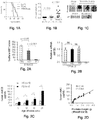


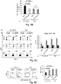
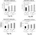
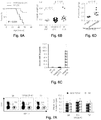

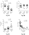

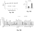
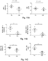
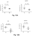
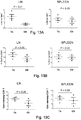


Classifications
-
- C—CHEMISTRY; METALLURGY
- C07—ORGANIC CHEMISTRY
- C07K—PEPTIDES
- C07K16/00—Immunoglobulins [IGs], e.g. monoclonal or polyclonal antibodies
- C07K16/18—Immunoglobulins [IGs], e.g. monoclonal or polyclonal antibodies against material from animals or humans
- C07K16/28—Immunoglobulins [IGs], e.g. monoclonal or polyclonal antibodies against material from animals or humans against receptors, cell surface antigens or cell surface determinants
- C07K16/2803—Immunoglobulins [IGs], e.g. monoclonal or polyclonal antibodies against material from animals or humans against receptors, cell surface antigens or cell surface determinants against the immunoglobulin superfamily
- C07K16/2833—Immunoglobulins [IGs], e.g. monoclonal or polyclonal antibodies against material from animals or humans against receptors, cell surface antigens or cell surface determinants against the immunoglobulin superfamily against MHC-molecules, e.g. HLA-molecules
-
- A—HUMAN NECESSITIES
- A01—AGRICULTURE; FORESTRY; ANIMAL HUSBANDRY; HUNTING; TRAPPING; FISHING
- A01K—ANIMAL HUSBANDRY; AVICULTURE; APICULTURE; PISCICULTURE; FISHING; REARING OR BREEDING ANIMALS, NOT OTHERWISE PROVIDED FOR; NEW BREEDS OF ANIMALS
- A01K67/00—Rearing or breeding animals, not otherwise provided for; New or modified breeds of animals
- A01K67/027—New or modified breeds of vertebrates
- A01K67/0275—Genetically modified vertebrates, e.g. transgenic
- A01K67/0278—Knock-in vertebrates, e.g. humanised vertebrates
-
- A—HUMAN NECESSITIES
- A61—MEDICAL OR VETERINARY SCIENCE; HYGIENE
- A61P—SPECIFIC THERAPEUTIC ACTIVITY OF CHEMICAL COMPOUNDS OR MEDICINAL PREPARATIONS
- A61P35/00—Antineoplastic agents
-
- C—CHEMISTRY; METALLURGY
- C07—ORGANIC CHEMISTRY
- C07K—PEPTIDES
- C07K16/00—Immunoglobulins [IGs], e.g. monoclonal or polyclonal antibodies
- C07K16/18—Immunoglobulins [IGs], e.g. monoclonal or polyclonal antibodies against material from animals or humans
- C07K16/28—Immunoglobulins [IGs], e.g. monoclonal or polyclonal antibodies against material from animals or humans against receptors, cell surface antigens or cell surface determinants
- C07K16/2851—Immunoglobulins [IGs], e.g. monoclonal or polyclonal antibodies against material from animals or humans against receptors, cell surface antigens or cell surface determinants against the lectin superfamily, e.g. CD23, CD72
-
- C—CHEMISTRY; METALLURGY
- C07—ORGANIC CHEMISTRY
- C07K—PEPTIDES
- C07K16/00—Immunoglobulins [IGs], e.g. monoclonal or polyclonal antibodies
- C07K16/18—Immunoglobulins [IGs], e.g. monoclonal or polyclonal antibodies against material from animals or humans
- C07K16/28—Immunoglobulins [IGs], e.g. monoclonal or polyclonal antibodies against material from animals or humans against receptors, cell surface antigens or cell surface determinants
- C07K16/30—Immunoglobulins [IGs], e.g. monoclonal or polyclonal antibodies against material from animals or humans against receptors, cell surface antigens or cell surface determinants from tumour cells
-
- A—HUMAN NECESSITIES
- A01—AGRICULTURE; FORESTRY; ANIMAL HUSBANDRY; HUNTING; TRAPPING; FISHING
- A01K—ANIMAL HUSBANDRY; AVICULTURE; APICULTURE; PISCICULTURE; FISHING; REARING OR BREEDING ANIMALS, NOT OTHERWISE PROVIDED FOR; NEW BREEDS OF ANIMALS
- A01K2217/00—Genetically modified animals
- A01K2217/07—Animals genetically altered by homologous recombination
- A01K2217/072—Animals genetically altered by homologous recombination maintaining or altering function, i.e. knock in
-
- A—HUMAN NECESSITIES
- A01—AGRICULTURE; FORESTRY; ANIMAL HUSBANDRY; HUNTING; TRAPPING; FISHING
- A01K—ANIMAL HUSBANDRY; AVICULTURE; APICULTURE; PISCICULTURE; FISHING; REARING OR BREEDING ANIMALS, NOT OTHERWISE PROVIDED FOR; NEW BREEDS OF ANIMALS
- A01K2227/00—Animals characterised by species
- A01K2227/10—Mammal
- A01K2227/105—Murine
-
- A—HUMAN NECESSITIES
- A01—AGRICULTURE; FORESTRY; ANIMAL HUSBANDRY; HUNTING; TRAPPING; FISHING
- A01K—ANIMAL HUSBANDRY; AVICULTURE; APICULTURE; PISCICULTURE; FISHING; REARING OR BREEDING ANIMALS, NOT OTHERWISE PROVIDED FOR; NEW BREEDS OF ANIMALS
- A01K2267/00—Animals characterised by purpose
- A01K2267/03—Animal model, e.g. for test or diseases
- A01K2267/0331—Animal model for proliferative diseases
-
- A—HUMAN NECESSITIES
- A61—MEDICAL OR VETERINARY SCIENCE; HYGIENE
- A61K—PREPARATIONS FOR MEDICAL, DENTAL OR TOILETRY PURPOSES
- A61K39/00—Medicinal preparations containing antigens or antibodies
- A61K2039/505—Medicinal preparations containing antigens or antibodies comprising antibodies
-
- C—CHEMISTRY; METALLURGY
- C07—ORGANIC CHEMISTRY
- C07K—PEPTIDES
- C07K2317/00—Immunoglobulins specific features
- C07K2317/20—Immunoglobulins specific features characterized by taxonomic origin
- C07K2317/24—Immunoglobulins specific features characterized by taxonomic origin containing regions, domains or residues from different species, e.g. chimeric, humanized or veneered
-
- C—CHEMISTRY; METALLURGY
- C07—ORGANIC CHEMISTRY
- C07K—PEPTIDES
- C07K2317/00—Immunoglobulins specific features
- C07K2317/30—Immunoglobulins specific features characterized by aspects of specificity or valency
- C07K2317/31—Immunoglobulins specific features characterized by aspects of specificity or valency multispecific
-
- C—CHEMISTRY; METALLURGY
- C07—ORGANIC CHEMISTRY
- C07K—PEPTIDES
- C07K2317/00—Immunoglobulins specific features
- C07K2317/50—Immunoglobulins specific features characterized by immunoglobulin fragments
- C07K2317/51—Complete heavy chain or Fd fragment, i.e. VH + CH1
-
- C—CHEMISTRY; METALLURGY
- C07—ORGANIC CHEMISTRY
- C07K—PEPTIDES
- C07K2317/00—Immunoglobulins specific features
- C07K2317/50—Immunoglobulins specific features characterized by immunoglobulin fragments
- C07K2317/515—Complete light chain, i.e. VL + CL
-
- C—CHEMISTRY; METALLURGY
- C07—ORGANIC CHEMISTRY
- C07K—PEPTIDES
- C07K2317/00—Immunoglobulins specific features
- C07K2317/50—Immunoglobulins specific features characterized by immunoglobulin fragments
- C07K2317/54—F(ab')2
-
- C—CHEMISTRY; METALLURGY
- C07—ORGANIC CHEMISTRY
- C07K—PEPTIDES
- C07K2317/00—Immunoglobulins specific features
- C07K2317/50—Immunoglobulins specific features characterized by immunoglobulin fragments
- C07K2317/55—Fab or Fab'
-
- C—CHEMISTRY; METALLURGY
- C07—ORGANIC CHEMISTRY
- C07K—PEPTIDES
- C07K2317/00—Immunoglobulins specific features
- C07K2317/50—Immunoglobulins specific features characterized by immunoglobulin fragments
- C07K2317/56—Immunoglobulins specific features characterized by immunoglobulin fragments variable (Fv) region, i.e. VH and/or VL
- C07K2317/565—Complementarity determining region [CDR]
-
- C—CHEMISTRY; METALLURGY
- C07—ORGANIC CHEMISTRY
- C07K—PEPTIDES
- C07K2317/00—Immunoglobulins specific features
- C07K2317/50—Immunoglobulins specific features characterized by immunoglobulin fragments
- C07K2317/56—Immunoglobulins specific features characterized by immunoglobulin fragments variable (Fv) region, i.e. VH and/or VL
- C07K2317/569—Single domain, e.g. dAb, sdAb, VHH, VNAR or nanobody®
-
- C—CHEMISTRY; METALLURGY
- C07—ORGANIC CHEMISTRY
- C07K—PEPTIDES
- C07K2317/00—Immunoglobulins specific features
- C07K2317/60—Immunoglobulins specific features characterized by non-natural combinations of immunoglobulin fragments
- C07K2317/62—Immunoglobulins specific features characterized by non-natural combinations of immunoglobulin fragments comprising only variable region components
- C07K2317/622—Single chain antibody (scFv)
-
- C—CHEMISTRY; METALLURGY
- C07—ORGANIC CHEMISTRY
- C07K—PEPTIDES
- C07K2317/00—Immunoglobulins specific features
- C07K2317/60—Immunoglobulins specific features characterized by non-natural combinations of immunoglobulin fragments
- C07K2317/62—Immunoglobulins specific features characterized by non-natural combinations of immunoglobulin fragments comprising only variable region components
- C07K2317/624—Disulfide-stabilized antibody (dsFv)
-
- C—CHEMISTRY; METALLURGY
- C07—ORGANIC CHEMISTRY
- C07K—PEPTIDES
- C07K2317/00—Immunoglobulins specific features
- C07K2317/60—Immunoglobulins specific features characterized by non-natural combinations of immunoglobulin fragments
- C07K2317/62—Immunoglobulins specific features characterized by non-natural combinations of immunoglobulin fragments comprising only variable region components
- C07K2317/626—Diabody or triabody
-
- C—CHEMISTRY; METALLURGY
- C07—ORGANIC CHEMISTRY
- C07K—PEPTIDES
- C07K2317/00—Immunoglobulins specific features
- C07K2317/70—Immunoglobulins specific features characterized by effect upon binding to a cell or to an antigen
- C07K2317/73—Inducing cell death, e.g. apoptosis, necrosis or inhibition of cell proliferation
-
- C—CHEMISTRY; METALLURGY
- C07—ORGANIC CHEMISTRY
- C07K—PEPTIDES
- C07K2317/00—Immunoglobulins specific features
- C07K2317/70—Immunoglobulins specific features characterized by effect upon binding to a cell or to an antigen
- C07K2317/76—Antagonist effect on antigen, e.g. neutralization or inhibition of binding
Definitions
- Elevation in serum soluble NKG2D ligand, soluble sMIC, is associated with metastasis in certain types of cancer.
- development of metastatic disease is correlated with marked elevation of the serum soluble NKG2D ligand, sMIC, and profound loss of NK cells in the circulation.
- US2012/315287 describes a method for treating cancer using an anti-MIC antibody, where the antibody binds membrane-bound MIC and is relevant for treating early stage cancer.
- sMIC neutralizing serum soluble Major Histocompatibility Complex class I chain-related
- “decrease,” “reduce,” “reduced”, “reduction”, “decrease,” and “inhibit” are all used herein generally to mean a decrease by a statistically significant amount relative to a reference.
- “reduce,” “reduction” or “decrease” or “inhibit” typically means a decrease by at least 10% as compared to a reference level and can include, for example, a decrease by at least about 20%, at least about 25%, at least about 30%, at least about 35%, at least about 40%, at least about 45%, at least about 50%, at least about 55%, at least about 60%, at least about 65%, at least about 70%, at least about 75%, at least about 80%, at least about 85%, at least about 90%, at least about 95%, at least about 98%, at least about 99%, up to and including, for example, the complete absence of the given entity or parameter as compared to the reference level, or any decrease between 10-99% as compared to the absence of a given treatment.
- the terms “increased”, “increase” or “enhance” or “activate” are all used herein to generally mean an increase by a statically significant amount; for the avoidance of any doubt, the terms “increased”, “increase” or “enhance” or “activate” means an increase of at least 10% as compared to a reference level, for example an increase of at least about 20%, or at least about 30%, or at least about 40%, or at least about 50%, or at least about 60%, or at least about 70%, or at least about 80%, or at least about 90% or up to and including a 100% increase or any increase between 10-100% as compared to a reference level, or at least about a 2-fold, or at least about a 3-fold, or at least about a 4-fold, or at least about a 5-fold or at least about a 10-fold increase, or any increase between 2-fold and 10-fold or greater as compared to a reference level.
- isolated refers, in the case of a nucleic acid or polypeptide, to a nucleic acid or polypeptide separated from at least one other component (e.g., nucleic acid or polypeptide) that is present with the nucleic acid or polypeptide as found in its natural source and/or that would be present with the nucleic acid or polypeptide when expressed by a cell, or secreted in the case of secreted polypeptides.
- component e.g., nucleic acid or polypeptide
- a chemically synthesized nucleic acid or polypeptide or one synthesized using in vitro transcription/translation is considered “isolated.”
- the terms “purified” or “substantially purified” refer to an isolated nucleic acid or polypeptide that is at least 95% by weight the subject nucleic acid or polypeptide, including, for example, at least 96%, at least 97%, at least 98%, at least 99% or more.
- Kabat numbering “Kabat definitions” and “Kabat labeling” are used interchangeably herein. These terms, which are recognized in the art, refer to a system of numbering amino acid residues which are more variable (i.e. hypervariable) than other amino acid residues in the heavy and light chain variable regions of an antibody, or an antigen binding portion thereof as described in Kabat et al. (1971) Ann. NY Acad, Sci. 190:382-391 and/or Kabat, E. A., et al. (1991) Sequences of Proteins of Immunological Interest, Fifth Edition, U.S. Department of Health and Human Services, NIH Publication No. 91-3242 .
- an "epitope" can be formed on a polypeptide both from contiguous amino acids, or noncontiguous amino acids juxtaposed by tertiary folding of a protein. Epitopes formed from contiguous amino acids are typically retained on exposure to denaturing solvents, whereas epitopes formed by tertiary folding are typically lost on treatment with denaturing solvents.
- An epitope typically includes at least 3, and more usually, at least 5, about 9, or about 8-10 amino acids in a unique spatial conformation.
- An “epitope” includes the unit of structure conventionally bound by an immunoglobulin VH/VL pair. Epitopes define the minimum binding site for an antibody, and thus represent the target of specificity of an antibody.
- epitope In the case of a single domain antibody, an epitope represents the unit of structure bound by a variable domain in isolation.
- the terms “antigenic determinant” and “epitope” can also be used interchangeably herein.
- epitope determinants include chemically active surface groupings of molecules such as amino acids, sugar side chains, phosphoryl, or sulfonyl, and, in certain embodiments, may have specific three dimensional structural characteristics, and/or specific charge characteristics.
- “Avidity” is the measure of the strength of binding between an antigen-binding molecule (such as an antibody or antibody fragment thereof described herein) and the pertinent antigen. Avidity is related to both the affinity between an antigenic determinant and its antigen binding site on the antigen-binding molecule, and the number of pertinent binding sites present on the antigen-binding molecule.
- antigen-binding proteins such as an antibody or portion of an antibody as described herein
- KD dissociation constant
- KA association constant
- Any KD value greater than 10 -4 mol/liter (or any KA value lower than 10 4 M -1 ) is generally considered to indicate non-specific binding.
- the KD for biological interactions which are considered meaningful are typically in the range of 10 -10 M (0.1 nM) to 10 -5 M (10000 nM).
- a binding site on an antibody or portion thereof described herein will bind to the desired antigen with an affinity less than 500 nM, such as less than 200 nM, or less than 10 nM, such as less than 500 pM.
- Specific binding of an antigen-binding protein to an antigen or antigenic determinant can be determined in any suitable manner known per se, including, for example, Scatchard analysis and/or competitive binding assays, such as radioimmunoassays (RIA), enzyme immunoassays (EIA) and sandwich competition assays, and the different variants thereof known per se in the art; as well as other techniques as mentioned herein.
- Scatchard analysis and/or competitive binding assays such as radioimmunoassays (RIA), enzyme immunoassays (EIA) and sandwich competition assays, and the different variants thereof known per se in the art; as well as other techniques as mentioned herein.
- selectively binds or “specifically binds” refers to the ability of an anti-MIC-binding peptide (e.g., an antibody or portion thereof) described herein to bind to a target, such as a MIC molecule present on the cell-surface, with a KD 10 -5 M (10000 nM) or less, e.g., 10 -6 M, 10 -7 M, 10 -8 M, 10 -9 M, 10 -10 M, 10 -11 M, 10 -12 M, or less.
- Specific binding can be influenced by, for example, the affinity and avidity of the polypeptide agent and the concentration of polypeptide agent.
- polypeptide agents described herein selectively bind the targets using any suitable methods, such as titration of a polypeptide agent in a suitable cell binding assay.
- a polypeptide specifically bound to a target is not displaced by a non-similar competitor.
- an antibody or antigen-binding portion thereof is said to specifically bind an antigen when it preferentially recognizes its target antigen in a complex mixture of proteins and/or macromolecules.
- a reagent that binds specifically to sMIC binds specifically to sMIC as compared to non-MIC polypeptides. In some embodiments, a reagent that binds specifically to sMIC binds specifically to sMIC as compared to non-soluble MIC (e.g. as compared to the extracellular domain of uncleaved, mature MIC).
- an antibody or antigen-binding portion thereof as described herein which binds to a sMIC polypeptide with a dissociation constant (KD) of 10 -5 M (10000 nM) or less, e.g., 10 -6 M, 10 -7 M, 10 -8 M, 10 -9 M, 10 -10 M, 10 -11 M, 10 -12 M, or less.
- KD dissociation constant
- an isolated antibody or antigen-binding portion thereof described herein binds a sMIC polypeptide with a dissociation constant (KD) of from about 10 -5 M to 10 -6 M.
- an isolated antibody or antigen-binding portion thereof described herein binds a sMIC polypeptide with a dissociation constant (KD) of from about 10 -6 M to 10 -7 M. In some embodiments, an isolated antibody or antigen-binding portion thereof described herein binds a sMIC polypeptide with a dissociation constant (KD) of from about 10 -7 M to 10 -8 M. In some embodiments, an isolated antibody or antigen-binding portion thereof described herein binds a sMIC polypeptide with a dissociation constant (KD) of from about 10 -8 M to 10 -9 M.
- KD dissociation constant
- an isolated antibody or antigen-binding portion thereof described herein binds a sMIC polypeptide with a dissociation constant (KD) of from about 10 -9 M to 10 -10 M. In some embodiments, an isolated antibody or antigen-binding portion thereof described herein binds a sMIC polypeptide with a dissociation constant (KD) of from about 10 -10 M to 10 -11 M. In some embodiments, an isolated antibody or antigen-binding portion thereof described herein binds a sMIC polypeptide with a dissociation constant (KD) of from about 10 -11 M to 10 -12 M. In some embodiments, an isolated antibody or antigen-binding portion thereof described herein binds a sMIC polypeptide with a dissociation constant (KD) of less than 10 -12 M.
- KD dissociation constant
- compositions, methods, and respective component(s) thereof are used in reference to compositions, methods, and respective component(s) thereof, that are essential to the method or composition, yet open to the inclusion of unspecified elements, whether essential or not.
- the term “consisting essentially of” refers to those elements required for a given embodiment. The term permits the presence of elements that do not materially affect the basic and novel or functional characteristic(s) of that embodiment.
- compositions, methods, and respective components thereof as described herein, which are exclusive of any element not recited in that description of the embodiment.
- CPCB Current Protocols in Cell Biology
- CPCB Current Protocols in Cell Biology
- Culture of Animal Cells A Manual of Basic Technique by R. Ian Freshney, Publisher: Wiley-Liss; 5th edition (2005 ), Animal Cell Culture Methods (Methods in Cell Biology, Vol. 57, Jennie P. Mather and David Barnes editors, Academic Press, 1st edition, 1998 ).
- the technology described herein may relate to antibodies and/or polypeptides comprising an antigen-binding portion of an antibody which bind a MIC polypeptide.
- the MIC polypeptide is a soluble MIC polypeptide (sMIC).
- sMIC soluble MIC polypeptide
- soluble MIC refers to a portion of a MIC polypeptide that is lacking a transmembrane domain, e.g. an extracellular portion of MIC that has been cleaved from the transmembrane domain.
- soluble MIC can comprise about, e.g., amino acids 24-260 of SEQ ID NOs: 15 or 16.
- soluble MIC can comprise about, e.g., 20 or more amino acids of residues 24-260 of SEQ ID NO: 15 or 16, e.g., 20, 50, 100, 150 or more amino acids of residues 24-260 of SEQ ID NO: 15 or 16.
- MIC polypeptides are surface transmembrane proteins. The presence of a MIC polypeptide on the cell surface can signal the immune receptor NKG2D for tumor immune destruction, typically by natural killer cells (NK cells) and cytotoxic T cells (CTLs). However, in many tumors, MIC is shed from the tumor surface, resulting in decreased host immunity against the tumor cell and promoting tumor evasion and progression.
- MIC polypeptides include, but are not limited to the human MICA (e.g. NCBI Ref Seqs NP_000238 (SEQ ID NO:15) and 001170990) and human MICB (e.g. NCBI Ref Seq: NP_005922 (SEQ ID NO: 16).
- a MIC polypeptide can comprise MICA.
- a MIC polypeptide may comprise MICB.
- the methods and compositions described herein may relate to inhibition of sMIC, e.g. reducing the level and/or activity of sMIC that is available to interact with cellular receptors.
- inhibition of sMIC can be a reduction of unbound sMIC (e.g. sMIC not bound to a receptor and/or available to be bound by the antibody reagents described herein).
- inhibition of sMIC can be a reduction of the level of sMIC, e.g. the level of sMIC in circulation.
- an isolated antibody or antigen-binding portion thereof that specifically binds a sMIC polypeptide, said antibody or antigen-binding portion thereof comprising one or more heavy and light chain complementarity determining regions (CDRs) selected from the group consisting of: (a) a light chain CDR1 having the amino acid sequence of SEQ ID NO: 9; (b) a light chain CDR2 having the amino acid sequence of SEQ ID NO: 10; (c) a light chain CDR3 having the amino acid sequence of SEQ ID NO: 11; (d) a heavy chain CDR1 having the amino acid sequence of SEQ ID NO: 12; (e) a heavy chain CDR2 having the amino acid sequence of SEQ ID NO: 13; and (f) a heavy chain CDR3 having the amino acid sequence of SEQ ID NO: 14.
- CDRs heavy and light chain complementarity determining regions
- an isolated antibody or antigen-binding portion thereof that specifically binds a sMIC polypeptide, said antibody or antigen-binding portion thereof comprising the light chain complementarity determining regions (CDRs): (a) a light chain CDR1 having the amino acid sequence of SEQ ID NO: 9; (b) a light chain CDR2 having the amino acid sequence of SEQ ID NO: 10; and (c) a light chain CDR3 having the amino acid sequence of SEQ ID NO: 11.
- CDRs light chain complementarity determining regions
- an isolated antibody or antigen-binding portion thereof that specifically binds a sMIC polypeptide, said antibody or antigen-binding portion thereof comprising the heavy chain complementarity determining regions (CDRs): (d) a heavy chain CDR1 having the amino acid sequence of SEQ ID NO: 12; (e) a heavy chain CDR2 having the amino acid sequence of SEQ ID NO: 13; and (f) a heavy chain CDR3 having the amino acid sequence of SEQ ID NO: 14.
- CDRs heavy chain complementarity determining regions
- the invention provides an isolated antibody or antigen-binding portion thereof that specifically binds a sMIC polypeptide, said antibody or antigen-binding portion thereof comprising the heavy and light chain complementarity determining regions (CDRs): (a) a light chain CDR1 having the amino acid sequence of SEQ ID NO: 9; (b) a light chain CDR2 having the amino acid sequence of SEQ ID NO: 10; (c) a light chain CDR3 having the amino acid sequence of SEQ ID NO: 11; (d) a heavy chain CDR1 having the amino acid sequence of SEQ ID NO: 12; (e) a heavy chain CDR2 having the amino acid sequence of SEQ ID NO: 13; and (f) a heavy chain CDR3 having the amino acid sequence of SEQ ID NO: 14.
- CDRs heavy and light chain complementarity determining regions
- an antibody or polypeptide comprising an antigen-binding fragment of an antibody described herein comprises one or more CDRs, e.g. 1 CDR, 2 CDRs, 3 CDRs, 4 CDRs, 5 CDRs, or 6 CDRs, selected from the group consisting of (a) a light chain CDR1 having the amino acid sequence of SEQ ID NO: 9; (b) a light chain CDR2 having the amino acid sequence of SEQ ID NO: 10; (c) a light chain CDR3 having the amino acid sequence of SEQ ID NO: 11; (d) a heavy chain CDR1 having the amino acid sequence of SEQ ID NO: 12; (e) a heavy chain CDR2 having the amino acid sequence of SEQ ID NO: 13; and (f) a heavy chain CDR3 having the amino acid sequence of SEQ ID NO: 14.
- CDRs e.g. 1 CDR, 2 CDRs, 3 CDRs, 4 CDRs, 5 CDRs, or 6 CDRs, selected from the group consist
- An antibody or polypeptide comprising an antigen-binding fragment of an antibody disclosed herein may comprise a heavy chain or a portion thereof, comprising one or more CDRs, e.g., 1 CDR, 2 CDRs, or 3 CDRs selected from the group consisting of a heavy chain CDR1 having the amino acid sequence of SEQ ID NO: 12; a heavy chain CDR2 having the amino acid sequence of SEQ ID NO: 13; and a heavy chain CDR3 having the amino acid sequence of SEQ ID NO: 14.
- CDRs e.g., 1 CDR, 2 CDRs, or 3 CDRs selected from the group consisting of a heavy chain CDR1 having the amino acid sequence of SEQ ID NO: 12; a heavy chain CDR2 having the amino acid sequence of SEQ ID NO: 13; and a heavy chain CDR3 having the amino acid sequence of SEQ ID NO: 14.
- An antibody or polypeptide comprising an antigen-binding fragment of an antibody disclosed herein may comprise a light chain or a portion thereof, comprising one or more CDRs, e.g., 1 CDR, 2 CDRs, or 3 CDRs selected from the group consisting of a light chain CDR1 having the amino acid sequence of SEQ ID NO:9; a light chain CDR2 having the amino acid sequence of SEQ ID NO: 10; a light chain CDR3 having the amino acid sequence of SEQ ID NO: 11.
- An isolated antibody or antigen-binding portion thereof disclosed herein may comprise a heavy chain comprising the amino acid sequence of SEQ ID NO: 8.
- the isolated antibody or antigen-binding portion thereof can comprise a light chain comprising the amino acid sequence of SEQ ID NO: 7.
- an antibody as disclosed herein comprises at least one CDR which is not identical to the sequence of SEQ ID NOs: 9-14
- the amino acid sequence of that at least one CDR can be selected by methods well known to one of skill in the art. For example, Fujii, 2004, “Antibody affinity maturation by random mutagenesis” in Methods in Molecular Biology: Antibody Engineering 248: 345-349 , particularly at Figure 2 and Section 3.3, describes methods of generating a library for any CDR of interest.
- antibody refers to immunoglobulin molecules and immunologically active portions of immunoglobulin molecules, i.e., molecules that contain an antigen binding site that immunospecifically binds an antigen.
- the term also refers to antibodies comprised of two immunoglobulin heavy chains and two immunoglobulin light chains as well as a variety of forms including full length antibodies and antigen-binding portions thereof; including, for example, an immunoglobulin molecule, a monoclonal antibody, a chimeric antibody, a CDR-grafted antibody, a humanized antibody, a Fab, a Fab', a F(ab')2, a Fv, a disulfide linked Fv, a scFv, a single domain antibody (dAb), a diabody, a multispecific antibody, a dual specific antibody, an anti-idiotypic antibody, a bispecific antibody, a functionally active epitope-binding fragment thereof, bifunctional hybrid antibodies (e.g., L
- Each heavy chain is composed of a variable region of said heavy chain (abbreviated here as HCVR or VH) and a constant region of said heavy chain.
- the heavy chain constant region consists of three domains CH1, CH2 and CH3.
- Each light chain is composed of a variable region of said light chain (abbreviated here as LCVR or VL) and a constant region of said light chain.
- the light chain constant region consists of a CL domain.
- the VH and VL regions may be further divided into hypervariable regions referred to as complementarity-determining regions (CDRs) and interspersed with conserved regions referred to as framework regions (FR).
- CDRs complementarity-determining regions
- FR framework regions
- Each VH and VL region thus consists of three CDRs and four FRs which are arranged from the N terminus to the C terminus in the following order: FR1, CDR1, FR2, CDR2, FR3, CDR3, FR4. This structure is well known to those skilled in the art.
- CDR refers to the complementarity determining regions within antibody variable sequences. There are three CDRs in each of the variable regions of the heavy chain and of the light chain, which are designated CDR1, CDR2 and CDR3, for each of the variable regions. The exact boundaries of these CDRs have been defined differently according to different systems.
- the system described by Kabat Kabat et al., Sequences of Proteins of Immunological Interest (National Institutes of Health, Bethesda, Md. (1987 ) and (1991)) not only provides an unambiguous residue numbering system applicable to any variable region of an antibody, but also provides precise residue boundaries defining the three CDRs. These CDRs may be referred to as Kabat CDRs.
- proteins and “polypeptides” are used interchangeably herein to designate a series of amino acid residues each connected to the other by peptide bonds between the alpha-amino and carboxy groups of adjacent residues.
- protein and “polypeptide”, which are used interchangeably herein, refer to a polymer of protein amino acids, including modified amino acids (e.g., phosphorylated, glycated, glycosylated, etc.) and amino acid analogs, regardless of its size or function.
- Protein and polypeptide are often used in reference to relatively large polypeptides, whereas the term “peptide” is often used in reference to small polypeptides, but usage of these terms in the art overlaps.
- the terms “protein” and “polypeptide” are used interchangeably herein when referring to an encoded gene product and fragments thereof.
- exemplary polypeptides or proteins include gene products, naturally occurring proteins, homologs, orthologs, paralogs, fragments and other equivalents, variants, fragments, and analogs of the foregoing.
- antigen-binding fragment or "antigen-binding portion" of an antibody, used interchangeably herein, refer to one or more fragments of an antibody as described herein, said fragments) still having the binding affinities as defined above herein. Fragments of a complete antibody have been shown to be able to carry out the antigen-binding function of an antibody.
- binding fragments include (i) an Fab fragment, i.e. a monovalent fragment composed of the VL, VH, CL and CH1 domains; (ii) an F(ab')2 fragment, i.e.
- a bivalent fragment comprising two Fab fragments linked to one another in the hinge region via a disulfide bridge; (iii) an Fd fragment composed of the VH and CH1 domains; (iv) an Fv fragment composed of the FL and VH domains of a single arm of an antibody; and (v) a dAb fragment ( Ward et al., (1989) Nature 341:544-546 ) consisting of a VH domain or of VH, CH1, CH2, DH3, or VH, CH2, CH3 (dAbs, or single domain antibodies, comprising only V L domains have also been shown to specifically bind to target eptiopes).
- the two domains of the Fv fragment namely VL and VH
- a synthetic linker e.g. a poly-G4S amino acid sequence ('G4S' disclosed as SEQ ID NO: 17)
- recombinant methods making it possible to prepare them as a single protein chain in which the VL and VH regions combine in order to form monovalent molecules (known as single chain Fv (ScFv); see, for example, Bird et al. (1988) Science 242:423-426 ; and Huston et al. (1988) Proc. Natl. Acad. Sci. USA 85:5879-5883 ).
- an antibody is also intended to comprise such single chain antibodies.
- Other forms of single chain antibodies such as “diabodies” are likewise included here.
- Diabodies are bivalent, bispecific antibodies in which VH and VL domains are expressed on a single polypeptide chain, but using a linker which is too short for the two domains being able to combine on the same chain, thereby forcing said domains to pair with complementary domains of a different chain and to form two antigen-binding sites (see, for example, Holliger, R, et al. (1993) Proc. Natl. Acad. Sci. USA 90:64446448 ; Poljak, R. J, et al. (1994) Structure 2:1121-1123 ).
- An immunoglobulin constant domain refers to a heavy or light chain constant domain. Human IgG heavy chain and light chain constant domain amino acid sequences are known in the art.
- an antibody as described herein or an antigen-binding portion thereof may be part of a larger immunoadhesion molecule formed by covalent or noncovalent association of said antibody or antibody portion with one or more further proteins or peptides.
- immunoadhesion molecules are the use of the streptavidin core region in order to prepare a tetrameric scFv molecule ( Kipriyanov, S. M., et al. (1995) Human Antibodies and Hybridomas 6:93-101 ) and the use of a cystein residue, a marker peptide and a C-terminal polyhistidinyl, e.g.
- hexahistidinyl tag ('hexahistidinyl tag' disclosed as SEQ ID NO: 18) in order to produce bivalent and biotinylated scFv molecules ( Kipriyanov, S. M., et al. (1994) Mol. Immunol. 31:10471058 ).
- the antibody and/or antigen-binding portion thereof described herein may be an immunoglobulin molecule, a monoclonal antibody, a chimeric antibody, a CDR-grafted antibody, a humanized antibody, a Fab, a Fab', a F(ab')2, a Fv, a disulfide linked Fv, a scFv, a single domain antibody, a diabody, a multispecific antibody, a dual specific antibody, an anti-idiotypic antibody, a bispecific antibody, and a functionally active epitope-binding fragment thereof.
- amino acid sequences one of skill will recognize that individual substitutions, deletions or additions to a nucleic acid, peptide, polypeptide, or protein sequence which alters a single amino acid or a small percentage of amino acids in the encoded sequence is a "conservatively modified variant" where the alteration results in the substitution of an amino acid with a chemically similar amino acid and retain the ability to specifically bind the target antigen (e.g. an epitope present on sMIC) of a MIC polypeptide).
- target antigen e.g. an epitope present on sMIC
- a conservatively modified variant of an antibody reagent may comprise alterations other than in the CDRs, e.g. a conservatively modified variant of an antibody reagent can comprise CDRs having the sequence of one or more of SEQ ID NOs 9-14.
- a given amino acid can be replaced by a residue having similar physiochemical characteristics, e.g., substituting one aliphatic residue for another (such as Ile, Val, Leu, or Ala for one another), or substitution of one polar residue for another (such as between Lys and Arg; Glu and Asp; or Gln and Asn).
- Other such conservative substitutions e.g., substitutions of entire regions having similar hydrophobicity characteristics, are well known.
- Polypeptides comprising conservative amino acid substitutions can be tested in any one of the assays described herein to confirm that a desired activity, e.g. antigen-binding activity and specificity of a native or reference polypeptide is retained.
- Amino acids can be grouped according to similarities in the properties of their side chains (in A. L. Lehninger, in Biochemistry, second ed., pp. 73-75, Worth Publishers, New York (1975 )): (1) non-polar: Ala (A), Val (V), Leu (L), Ile (I), Pro (P), Phe (F), Trp (W), Met (M); (2) uncharged polar: Gly (G), Ser (S), Thr (T), Cys (C), Tyr (Y), Asn (N), Gln (Q); (3) acidic: Asp (D), Glu (E); (4) basic: Lys (K), Arg (R), His (H).
- Naturally occurring residues can be divided into groups based on common side-chain properties: (1) hydrophobic: Norleucine, Met, Ala, Val, Leu, Ile; (2) neutral hydrophilic: Cys, Ser, Thr, Asn, Gln; (3) acidic: Asp, Glu; (4) basic: His, Lys, Arg; (5) residues that influence chain orientation: Gly, Pro; (6) aromatic: Trp, Tyr, Phe.
- Non-conservative substitutions will entail exchanging a member of one of these classes for another class.
- Particular conservative substitutions include, for example; Ala into Gly or into Ser; Arg into Lys; Asn into Gln or into H is; Asp into Glu; Cys into Ser; Gln into Asn; Glu into Asp; Gly into Ala or into Pro; His into Asn or into Gln; Ile into Leu or into Val; Leu into Ile or into Val; Lys into Arg, into Gln or into Glu; Met into Leu, into Tyr or into Ile; Phe into Met, into Leu or into Tyr; Ser into Thr; Thr into Ser; Trp into Tyr; Tyr into Trp; and/or Phe into Val, into Ile or into Leu.
- the antibody and/or antigen-binding portion thereof disclosed herein may be a variant of a sequence described herein, e.g. a conservative substitution variant of an antibody polypeptide.
- the variant is a conservatively modified variant.
- Conservative substitution variants can be obtained by mutations of native nucleotide sequences, for example.
- a "variant,” as referred to herein, is a polypeptide substantially homologous to a native or reference polypeptide, but which has an amino acid sequence different from that of the native or reference polypeptide because of one or a plurality of deletions, insertions or substitutions.
- Variant polypeptide-encoding DNA sequences encompass sequences that comprise one or more additions, deletions, or substitutions of nucleotides when compared to a native or reference DNA sequence, but that encode a variant protein or fragment thereof that retains activity, e.g. antigen-specific binding activity for the relevant target polypeptide, e.g. a sMIC polypeptide.
- a wide variety of PCR-based site-specific mutagenesis approaches are also known in the art and can be applied by the ordinarily skilled artisan.
- substitution variants include conservative substitution of amino acids, e.g. in a V H or V L , domain, that do not alter the sequence of a CDR.
- a conservative substitution in a sequence not comprised by a CDR can be a substitution relative to a wild-type or naturally-occurring sequence, e.g. human or murine framework and/or constant regions of an antibody sequence.
- a variant amino acid or DNA sequence preferably is at least 90%, at least 91%, at least 92%, at least 93%, at least 94%, at least 95%, at least 96%, at least 97%, at least 98%, at least 99%, or more, identical to a native or reference sequence.
- the degree of homology (percent identity) between a native and a mutant sequence can be determined, for example, by comparing the two sequences using freely available computer programs commonly employed for this purpose on the world wide web (e.g. BLASTp or BLASTn with default settings).
- Alterations of the native amino acid sequence can be accomplished by any of a number of techniques known to one of skill in the art. Mutations can be introduced, for example, at particular loci by synthesizing oligonucleotides containing a mutant sequence, flanked by restriction sites enabling ligation to fragments of the native sequence. Following ligation, the resulting reconstructed sequence encodes an analog having the desired amino acid insertion, substitution, or deletion. Alternatively, oligonucleotide-directed site-specific mutagenesis procedures can be employed to provide an altered nucleotide sequence having particular codons altered according to the substitution, deletion, or insertion required. Techniques for making such alterations are very well established and include, for example, those disclosed by Walder et al.
- cysteine residues not involved in maintaining the proper conformation of the polypeptide also can be substituted, generally with serine, to improve the oxidative stability of the molecule and prevent aberrant crosslinking.
- cysteine bond(s) can be added to the polypeptide to improve its stability or facilitate oligomerization.
- the antibody or antigen-binding portion thereof is a fully human antibody. In some embodiments, the antibody or antigen-binding portion thereof is a humanized antibody. In some embodiments, the antibody or antigen-binding portion thereof is a chimeric antibody. In some embodiments, the antibody or antigen-binding portion thereof is a recombinant polypeptide.
- human antibody refers to antibodies whose variable and constant regions correspond to or are derived from immunoglobulin sequences of the human germ line, as described, for example, by Kabat et al. (see Kabat, et al. (1991) Sequences of Proteins of Immunological Interest, Fifth Edition, U.S. Department of Health and Human Services, NIH Publication No. 91-3242 ).
- the human antibodies can contain amino acid residues not encoded by human germ line immunoglobulin sequences (for example mutations which have been introduced by random or site-specific mutagenesis in vitro or by somatic mutation in vivo), for example in the CDRs, and in particular in CDR3.
- Recombinant human antibodies as described herein have variable regions and may also contain constant regions derived from immunoglobulin sequences of the human germ line (see Kabat, E. A., et al. (1991) Sequences of Proteins of Immunological Interest, Fifth Edition, U.S. Department of Health and Human Services, NIH Publication No. 91-3242 ).
- Such recombinant human antibodies may be subjected to in-vitro mutagenesis (or to a somatic in-vivo mutagenesis, if an animal is used which is transgenic due to human Ig sequences) so that the amino acid sequences of the VH and VL regions of the recombinant antibodies are sequences which although related to or derived from VH and VL sequences of the human germ line, do not naturally exist in vivo within the human antibody germ line repertoire.
- recombinant antibodies of this kind are the result of selective mutagenesis or back mutation or of both.
- mutagenesis leads to an affinity to the target which is greater, and/or an affinity to non-target structures which is smaller than that of the parent antibody.
- chimeric antibody refers to antibodies which contain sequences for the variable region of the heavy and light chains from one species and constant region sequences from another species, such as antibodies having murine heavy and light chain variable regions linked to human constant regions.
- Humanized antibodies have variable region framework residues substantially from a human antibody (termed an acceptor antibody) and complementarity determining regions substantially from a non-human antibody, e.g. a mouse-antibody, (referred to as the donor immunoglobulin). See, Queen et al., Proc Natl Acad Sci USA 86:10029-10033 (1989 ) and WO 90/07861 , U.S. Pat. No. 5,693,762 , U.S. Pat. No. 5,693,761 , U.S.
- the constant region(s), if present, are also substantially or entirely from a human immunoglobulin.
- the human variable domains are usually chosen from human antibodies whose framework sequences exhibit a high degree of sequence identity with the (murine) variable region domains from which the CDRs were derived.
- the heavy and light chain variable region framework residues can be substantially similar to a region of the same or different human antibody sequences.
- the human antibody sequences can be the sequences of naturally occurring human antibodies or can be consensus sequences of several human antibodies. See Carter et al., WO 92/22653 .
- the antibody reagents (e.g. antibodies) disclosed herein may not be naturally-occurring biomolecules.
- a murine antibody raised against an antigen of human origin would not occur in nature absent human intervention and manipulation, e.g. manufacturing steps carried out by a human.
- Chimeric antibodies are also not naturally-occurring biomolecules, e.g., in that they comprise sequences obtained from multiple species and assembled into a recombinant molecule.
- the human antibody reagents disclosed herein may not be naturally-occurring biomolecules, e.g., fully human antibodies directed against a human antigen would be subject to negative selection in nature and are not naturally found in the human body.
- monoclonal antibodies have been produced as native molecules in murine hybridoma lines.
- the methods and compositions described herein provide for recombinant DNA expression of monoclonal antibodies. This allows the production of humanized antibodies as well as a spectrum of antibody derivatives and fusion proteins in a host species of choice.
- the production of antibodies in bacteria, yeast, transgenic animals and chicken eggs are also alternatives for hybridoma-based production systems.
- the main advantages of transgenic animals are potential high yields from renewable sources.
- Nucleic acid molecules encoding amino acid sequence variants of antibodies are prepared by a variety of methods known in the art. These methods include, but are not limited to preparation by oligonucleotide-mediated (or site-directed) mutagenesis, PCR mutagenesis, and cassette mutagenesis of an earlier prepared variant or a non-variant version of the antibody.
- a nucleic acid sequence encoding at least one antibody, portion or polypeptide as described herein can be recombined with vector DNA in accordance with conventional techniques, including blunt-ended or staggered-ended termini for ligation, restriction enzyme digestion to provide appropriate termini, filling in of cohesive ends as appropriate, alkaline phosphatase treatment to avoid undesirable joining, and ligation with appropriate ligases.
- a nucleic acid molecule such as DNA, is said to be "capable of expressing" a polypeptide if it contains nucleotide sequences which contain transcriptional and translational regulatory information and such sequences are “operably linked” to nucleotide sequences which encode the polypeptide.
- An operable linkage is a linkage in which the regulatory DNA sequences and the DNA sequence sought to be expressed are connected in such a way as to permit gene expression as peptides or antibody portions in recoverable amounts.
- the precise nature of the regulatory regions needed for gene expression may vary from organism to organism, as is well known in the analogous art. See, e.g., Sambrook et al., 1989; Ausubel et al., 1987-1993.
- Suitable hosts include bacterial or eukaryotic hosts, including yeast, insects, fungi, bird and mammalian cells either in vivo, or in situ, or host cells of mammalian, insect, bird or yeast origin.
- the mammalian cell or tissue can be of human, primate, hamster, rabbit, rodent, cow, pig, sheep, horse, goat, dog or cat origin, but any other mammalian cell may be used.
- yeast ubiquitin hydrolase system in vivo synthesis of ubiquitin-transmembrane polypeptide fusion proteins can be accomplished.
- the fusion proteins so produced can be processed in vivo or purified and processed in vitro, allowing synthesis of an antibody or portion thereof as described herein with a specified amino terminus sequence. Moreover, problems associated with retention of initiation codon-derived methionine residues in direct yeast (or bacterial) expression maybe avoided. Sabin et al., 7 Bio/Technol. 705 (1989 ); Miller et al., 7 Bio/Technol. 698 (1989 ). Any of a series of yeast gene expression systems incorporating promoter and termination elements from the actively expressed genes coding for glycolytic enzymes produced in large quantities when yeast are grown in mediums rich in glucose can be utilized to obtain recombinant antibodies or antigen-binding portions thereof as described herein. Known glycolytic genes can also provide very efficient transcriptional control signals. For example, the promoter and terminator signals of the phosphoglycerate kinase gene can be utilized.
- the introduced nucleotide sequence may be incorporated into a plasmid or viral vector capable of autonomous replication in the recipient host.
- a plasmid or viral vector capable of autonomous replication in the recipient host.
- Any of a wide variety of vectors can be employed for this purpose and are known and available to those or ordinary skill in the art. See, e.g., Ausubel et al., 1987, 1993.
- Factors of importance in selecting a particular plasmid or viral vector include: the ease with which recipient cells that contain the vector may be recognized and selected from those recipient cells which do not contain the vector; the number of copies of the vector which are desired in a particular host; and whether it is desirable to be able to "shuttle" the vector between host cells of different species.
- Example prokaryotic vectors known in the art include plasmids such as those capable of replication in E. coli., for example.
- Other gene expression elements useful for the expression of cDNA encoding antibodies or antigen-binding portions thereof include, but are not limited to (a) viral transcription promoters and their enhancer elements, such as the SV40 early promoter. ( Okayama et al., 3 Mol. Cell. Biol.
- Rous sarcoma virus LTR Rous sarcoma virus LTR ( Gorman et al., 79 PNAS 6777 (1982 )), and Moloney murine leukemia virus LTR ( Grosschedl et al., 41 Cell 885 (1985 )); (b) splice regions and polyadenylation sites such as those derived from the SV40 late region (Okayarea et al., 1983), and (c) polyadenylation sites such as in SV40 (Okayama et al., 1983).
- Immunoglobulin cDNA genes can be expressed as described by Liu et al., infra, and Weidle et al., 51 Gene 21 (1987 ), using as expression elements the SV40 early promoter and its enhancer, the mouse immunoglobulin H chain promoter enhancers, SV40 late region mRNA splicing, rabbit S-globin intervening sequence, immunoglobulin and rabbit S-globin polyadenylation sites, and SV40 polyadenylation elements.
- the transcriptional promoter can be human cytomegalovirus
- the promoter enhancers can be cytomegalovirus and mouse/human immunoglobulin
- mRNA splicing and polyadenylation regions can be the native chromosomal immunoglobulin sequences.
- the transcriptional promoter may be a viral LTR sequence
- the transcriptional promoter enhancers may be either or both the mouse immunoglobulin heavy chain enhancer and the viral LTR enhancer
- the splice region may contain an intron of greater than 31 bp
- the polyadenylation and transcription termination regions may be derived from the native chromosomal sequence corresponding to the immunoglobulin chain being synthesized.
- cDNA sequences encoding other proteins may be combined with the above-recited expression elements to achieve expression of the proteins in mammalian cells.
- Each fused gene is assembled in, or inserted into, an expression vector.
- Recipient cells capable of expressing the chimeric immunoglobulin chain gene product are then transfected singly with an antibody, antigen-binding portion thereof, or chimeric H or chimeric L chain-encoding gene, or are co-transfected with a chimeric H and a chimeric L chain gene.
- the transfected recipient cells are cultured under conditions that permit expression of the incorporated genes and the expressed immunoglobulin chains or intact antibodies or fragments are recovered from the culture.
- the fused genes encoding the antibody, antigen-binding fragment thereof, or chimeric H and L chains, or portions thereof may be assembled in separate expression vectors that are then used to co-transfect a recipient cell.
- Each vector can contain two selectable genes, a first selectable gene designed for selection in a bacterial system and a second selectable gene designed for selection in a eukaryotic system, wherein each vector has a different pair of genes. This strategy results in vectors which first direct the production, and permit amplification, of the fused genes in a bacterial system.
- the genes so produced and amplified in a bacterial host are subsequently used to co-transfect a eukaryotic cell, and allow selection of a co-transfected cell carrying the desired transfected genes.
- Non-limiting examples of selectable genes for use in a bacterial system are the gene that confers resistance to ampicillin and the gene that confers resistance to chloramphenicol.
- Selectable genes for use in eukaryotic transfectants include the xanthine guanine phosphoribosyl transferase gene (designated gpt) and the phosphotransferase gene from Tn5 (designated neo).
- the fused genes encoding chimeric H and L chains can be assembled on the same expression vector.
- the recipient cell line can be a myeloma cell.
- Myeloma cells can synthesize, assemble and secrete immunoglobulins encoded by transfected immunoglobulin genes and possess the mechanism for glycosylation of the immunoglobulin.
- the recipient cell is the recombinant Ig-producing myeloma cell SP2/0 (ATCC #CRL 8287). SP2/0 cells produce only immunoglobulin encoded by the transfected genes.
- Myeloma cells can be grown in culture or in the peritoneal cavity of a mouse, where secreted immunoglobulin can be obtained from ascites fluid.
- Other suitable recipient cells include lymphoid cells such as B lymphocytes of human or non-human origin, hybridoma cells of human or non-human origin, or interspecies heterohybridoma cells.
- An expression vector carrying a chimeric, humanized, or composite human antibody construct, antibody, or antigen-binding portion thereof as described herein can be introduced into an appropriate host cell by any of a variety of suitable means, including such biochemical means as transformation, transfection, conjugation, protoplast fusion, calcium phosphate-precipitation, and application with polycations such as diethylaminoethyl (DEAE) dextran, and such mechanical means as electroporation, direct microinjection, and microprojectile bombardment.
- biochemical means as transformation, transfection, conjugation, protoplast fusion, calcium phosphate-precipitation, and application with polycations such as diethylaminoethyl (DEAE) dextran, and such mechanical means as electroporation, direct microinjection, and microprojectile bombardment.
- DEAE diethylaminoethyl
- Yeast provides certain advantages over bacteria for the production of immunoglobulin H and L chains. Yeasts carry out post-translational peptide modifications including glycosylation. A number of recombinant DNA strategies exist that utilize strong promoter sequences and high copy number plasmids which can be used for production of the desired proteins in yeast. Yeast recognizes leader sequences of cloned mammalian gene products and secretes peptides bearing leader sequences (i.e., pre-peptides). Hitzman et al., 11th Intl. Conf. Yeast, Genetics & Molec. Biol. (Montpelier, France, 1982 ).
- Yeast gene expression systems can be routinely evaluated for the levels of production, secretion and the stability of antibodies, and assembled chimeric, humanized, or composite human antibodies, portions and regions thereof. Any of a series of yeast gene expression systems incorporating promoter and termination elements from the actively expressed genes coding for glycolytic enzymes produced in large quantities when yeasts are grown in media rich in glucose can be utilized. Known glycolytic genes can also provide very efficient transcription control signals. For example, the promoter and terminator signals of the phosphoglycerate kinase (PGK) gene can be utilized. A number of approaches can be taken for evaluating optimal expression plasmids for the expression of cloned immunoglobulin cDNAs in yeast. See II DNA Cloning 45, (Glover, ed., IRL Press, 1985 ) and e.g., U.S. Publication No. US 2006/0270045 A1 .
- Bacterial strains can also be utilized as hosts for the production of the antibody molecules or peptides described herein, E. coli K12 strains such as E. coli W3110 (ATCC 27325), Bacillus species, enterobacteria such as Salmonella typhimurium or Serratia marcescens, and various Pseudomonas species can be used. Plasmid vectors containing replicon and control sequences which are derived from species compatible with a host cell are used in connection with these bacterial hosts. The vector carries a replication site, as well as specific genes which are capable of providing phenotypic selection in transformed cells.
- Host mammalian cells can be grown in vitro or in vivo.
- Mammalian cells provide post-translational modifications to immunoglobulin protein molecules including leader peptide removal, folding and assembly of H and L chains, glycosylation of the antibody molecules, and secretion of functional antibody protein.
- Mammalian cells which can be useful as hosts for the production of antibody proteins include cells of fibroblast origin, such as Vero (ATCC CRL 81) or CHO-K1 (ATCC CRL 61) cells.
- exemplary eukaryotic cells that can be used to express polypeptides include, but are not limited to, COS cells, including COS 7 cells; 293 cells, including 293-6E cells; CHO cells, including CHO-S and DG44 cells; PER.C6TM cells (Crucell); and NSO cells.
- a particular eukaryotic host cell may be selected based on its ability to make desired post-translational modifications to the heavy chains and/or light chains. For example, CHO cells produce polypeptides that have a higher level of sialylation than the same polypeptide produced in 293 cells.
- One or more antibodies or antigen-binding portions thereof as described herein can be produced in vivo in an animal that has been engineered or transfected with one or more nucleic acid molecules encoding the polypeptides, according to any suitable method.
- An antibody or antigen-binding portion thereof as described herein may be produced in a cell-free system.
- Nonlimiting exemplary cell-free systems are described, e.g., in Sitaraman et al., Methods Mol. Biol. 498: 229-44 (2009 ); Spirin, Trends Biotechnol. 22: 538-45 (2004 ); Endo et al., Biotechnol. Adv. 21: 695-713 (2003 ).
- H and L chain genes are available for the expression of cloned H and L chain genes in mammalian cells (see Glover, 1985). Different approaches can be followed to obtain complete H 2 L 2 antibodies. As discussed above, it is possible to co-express H and L chains in the same cells to achieve intracellular association and linkage of H and L chains into complete tetrameric H 2 L 2 antibodies or antigen-binding portions thereof. The co-expression can occur by using either the same or different plasmids in the same host. Genes for both H and L chains or portions thereof can be placed into the same plasmid, which is then transfected into cells, thereby selecting directly for cells that express both chains.
- cells can be transfected first with a plasmid encoding one chain, for example the L chain, followed by transfection of the resulting cell line with an H chain plasmid containing a second selectable marker.
- Cell lines producing antibodies, antigen-binding portions thereof and/or H 2 L 2 molecules via either route could be transfected with plasmids encoding additional copies of peptides, H, L, or H plus L chains in conjunction with additional selectable markers to generate cell lines with enhanced properties, such as higher production of assembled H 2 L 2 antibody molecules or enhanced stability of the transfected cell lines.
- Antibodies can be expressed in plant cell culture, or plants grown conventionally. The expression in plants may be systemic, limited to susb-cellular plastids, or limited to seeds (endosperms). See, e.g., U.S. Patent Pub. No. 2003/0167531 ; U.S. Pat. No. 6,080,560 ; No. 6,512,162 ; WO 0129242 .
- Several plant-derived antibodies have reached advanced stages of development, including clinical trials (see, e.g., Biolex, NC).
- a humanized antibody which is prepared by a process which comprises maintaining a host transformed with a first expression vector which encodes the light chain of the humanized antibody and with a second expression vector which encodes the heavy chain of the humanized antibody under such conditions that each chain is expressed and isolating the humanized antibody formed by assembly of the thus-expressed chains.
- the first and second expression vectors can be the same vector.
- DNA sequences encoding the light chain or the heavy chain of the humanized antibody an expression vector which incorporates a said DNA sequence; and a host transformed with a said expression vector.
- Generating a humanized antibody from the sequences and information provided herein can be practiced by those of ordinary skill in the art without undue experimentation.
- there are four general steps employed to humanize a monoclonal antibody see, e.g., U.S. Pat. No. 5,585,089 ; No. 6,835,823 ; No. 6,824,989 . These are: (1) determining the nucleotide and predicted amino acid sequence of the starting antibody light and heavy variable domains; (2) designing the humanized antibody, i.e., deciding which antibody framework region to use during the humanizing process; (3) the actual humanizing methodologies/techniques; and (4) the transfection and expression of the humanized antibody.
- CDR regions in humanized antibodies and human antibody variants are substantially identical, and more usually, identical to the corresponding CDR regions in the mouse or human antibody from which they were derived. Although not usually desirable, it is sometimes possible to make one or more conservative amino acid substitutions of CDR residues without appreciably affecting the binding affinity of the resulting humanized immunoglobulin or human antibody variant. Occasionally, substitutions of CDR regions can enhance binding affinity.
- chimeric antibodies In addition, techniques developed for the production of "chimeric antibodies" (see Morrison et al., Proc. Natl. Acad. Sci. 81:851-855 (1984 ); Neuberger et al., Nature 312:604-608 (1984 ); Takeda et al., Nature 314:452-454 (1985 ); by splicing genes from a mouse, or other species, antibody molecule of appropriate antigen specificity together with genes from a human antibody molecule of appropriate biological activity can be used.
- a chimeric antibody is a molecule in which different portions are derived from different animal species, such as those having a variable region derived from a murine monoclonal antibody and a human immunoglobulin constant region, e.g., humanized antibodies.
- variable segments of chimeric antibodies are typically linked to at least a portion of an immunoglobulin constant region (Fc), typically that of a human immunoglobulin.
- Fc immunoglobulin constant region
- Human constant region DNA sequences can be isolated in accordance with well-known procedures from a variety of human cells, such as immortalized B-cells ( WO 87/02671 ).
- the antibody can contain both light chain and heavy chain constant regions.
- the heavy chain constant region can include CH1, hinge, CH2, CH3, and, sometimes, CH4 regions. For therapeutic purposes, the CH2 domain can be deleted or omitted.
- Single chain antibodies are formed by linking the heavy and light chain fragments of the Fv region via an amino acid bridge, resulting in a single chain polypeptide.
- Techniques for the assembly of functional Fv fragments in E. coli can also be used (see, e.g. Skerra et al., Science 242:1038-1041 (1988 ).
- Chimeric, humanized and human antibodies are typically produced by recombinant expression.
- Recombinant polynucleotide constructs typically include an expression control sequence operably linked to the coding sequences of antibody chains, including naturally-associated or heterologous promoter regions.
- the expression control sequences are eukaryotic promoter systems in vectors capable of transforming or transfecting eukaryotic host cells. Once the vector has been incorporated into the appropriate host, the host is maintained under conditions suitable for high level expression of the nucleotide sequences, and the collection and purification of the cross-reacting antibodies.
- These expression vectors are typically replicable in the host organisms either as episomes or as an integral part of the host chromosomal DNA.
- expression vectors contain selection markers, e.g., ampicillin-resistance or hygromycin-resistance, to permit detection of those cells transformed with the desired DNA sequences.
- E. coli is one prokaryotic host particularly useful for cloning the DNA sequences.
- Microbes, such as yeast are also useful for expression. Saccharomyces is a preferred yeast host, with suitable vectors having expression control sequences, an origin of replication, termination sequences and the like as desired.
- Typical promoters include 3-phosphoglycerate kinase and other glycolytic enzymes.
- Inducible yeast promoters include, among others, promoters from alcohol dehydrogenase, isocytochrome C, and enzymes responsible for maltose and galactose utilization.
- Mammalian cells are a preferred host for expressing nucleotide segments encoding immunoglobulins or fragments thereof. See Winnacker, From Genes to Clones, (VCH Publishers, NY, 1987 ).
- suitable host cell lines capable of secreting intact heterologous proteins have been developed in the art, and include CHO cell lines, various COS cell lines, HeLa cells, L cells and multiple myeloma cell lines.
- Expression vectors for these cells can include expression control sequences, such as an origin of replication, a promoter, an enhancer ( Queen et al., "Cell-type Specific Regulation of a Kappa Immunoglobulin Gene by Promoter and Enhancer Elements," Immunol Rev 89:49 (1986 ), and necessary processing information sites, such as ribosome binding sites, RNA splice sites, polyadenylation sites, and transcriptional terminator sequences.
- Preferred expression control sequences are promoters substantially similar to a region of the endogenous genes, cytomegalovirus, SV40, adenovirus, bovine papillomavirus, and the like.
- antibody coding sequences can be incorporated in transgenes for introduction into the genome of a transgenic non-human animal and subsequent expression in the milk of the transgenic animal (e.g., according to methods described in U.S. Pat. No. 5,741,957 , U.S. Pat. No. 5,304,489 , U.S. Pat. No. 5,849,992 .
- Suitable transgenes include coding sequences for light and/or heavy chains in operable linkage with a promoter and enhancer from a mammary gland specific gene, such as casein or beta lactoglobulin.
- the vectors containing the DNA segments of interest can be transferred into the host cell by well-known methods, depending on the type of cellular host. For example, calcium chloride transfection is commonly utilized for prokaryotic cells, whereas calcium phosphate treatment, electroporation, lipofection, biolistics or viral-based transfection can be used for other cellular hosts. Other methods used to transform mammalian cells include the use of polybrene, protoplast fusion, liposomes, electroporation, and microinjection (see generally, Sambrook et al., supra.
- transgenes can be microinjected into fertilized non-human oocytes or can be incorporated into the genome of non-human embryonic stem cells, and the nuclei of such cells transferred into enucleated non-human oocytes.
- antibodies can be purified according to standard procedures of the art, including HPLC purification, column chromatography, gel electrophoresis and the like (see generally, Scopes, Protein Purification (Springer-Verlag, NY, 1982 ).
- the whole antibodies, their dimers, individual light and heavy chains, or other immunoglobulin forms of the present invention can be recovered and purified by known techniques, e.g., immunoabsorption or immunoaffinity chromatography, chromatographic methods such as HPLC (high performance liquid chromatography), ammonium sulfate precipitation, gel electrophoresis, or any combination of these. See generally, Scopes, PROTEIN PURIF. (Springer-Verlag, NY, 1982 ). Substantially pure immunoglobulins of at least about 90% to 95% homogeneity are advantageous, as are those with 98% to 99% or more homogeneity, particularly for pharmaceutical uses.
- a humanized or composite human antibody can then be used therapeutically or in developing and performing assay procedures, immunofluorescent stainings, and the like. See generally, Vols. I & II Immunol. Meth. (Lefkovits & Pernis, eds., Acad. Press, NY, 1979 and 1981 ).
- a recombinant humanized antibody can be further optimized to decrease potential immunogenicity, while maintaining functional activity, for therapy in humans.
- functional activity means a polypeptide capable of displaying one or more known functional activities associated with a recombinant antibody or antigen-binding portion thereof as described herein. Such functional activities include inhibition of sMIC, and/or anti-cancer activity.
- a polypeptide having functional activity means the polypeptide exhibits activity similar, but not necessarily identical to, an activity of a reference antibody or antigen-binding portion thereof as described herein, including mature forms, as measured in a particular assay, such as, for example, a biological assay, with or without dose dependency.
- the candidate polypeptide will exhibit greater activity, or not more than about 25-fold less, about 10-fold less, or about 3-fold less activity relative to the antibodies and antigen-binding fragments described herein).
- the technology described herein relates to a nucleic acid encoding an antibody or antigen-binding portion thereof as described herein.
- nucleic acid or “nucleic acid sequence” refers to a polymeric molecule incorporating units of ribonucleic acid, deoxyribonucleic acid or an analog thereof.
- the nucleic acid can be either single-stranded or double-stranded.
- a single-stranded nucleic acid can be one strand nucleic acid of a denatured double-stranded DNA.
- the nucleic acid can be a cDNA, e.g., a nucleic acid lacking introns.
- a nucleic acid encoding an antibody or antigen-binding portion thereof as described herein is comprised by a vector.
- a nucleic acid sequence encoding an antibody or antigen-binding portion thereof as described herein, or any module thereof, may be operably linked to a vector.
- the term "vector”, as used herein, refers to a nucleic acid construct designed for delivery to a host cell or for transfer between different host cells.
- a vector can be viral or non-viral.
- the term “vector” encompasses any genetic element that is capable of replication when associated with the proper control elements and that can transfer gene sequences to cells.
- a vector can include, but is not limited to, a cloning vector, an expression vector, a plasmid, phage, transposon, cosmid, chromosome, virus, virion, etc.
- expression vector refers to a vector that directs expression of an RNA or polypeptide from sequences linked to transcriptional regulatory sequences on the vector.
- the sequences expressed will often, but not necessarily, be heterologous to the cell.
- An expression vector may comprise additional elements, for example, the expression vector may have two replication systems, thus allowing it to be maintained in two organisms, for example in human cells for expression and in a prokaryotic host for cloning and amplification.
- expression refers to the cellular processes involved in producing RNA and proteins and as appropriate, secreting proteins, including where applicable, but not limited to, for example, transcription, transcript processing, translation and protein folding, modification and processing.
- “Expression products” include RNA transcribed from a gene, and polypeptides obtained by translation of mRNA transcribed from a gene.
- the term “gene” means the nucleic acid sequence which is transcribed (DNA) to RNA in vitro or in vivo when operably linked to appropriate regulatory sequences.
- the gene may or may not include regions preceding and following the coding region, e.g. 5' untranslated (5'UTR) or "leader” sequences and 3' UTR or “trailer” sequences, as well as intervening sequences (introns) between individual coding segments (exons).
- viral vector refers to a nucleic acid vector construct that includes at least one element of viral origin and has the capacity to be packaged into a viral vector particle.
- the viral vector can contain the nucleic acid encoding an antibody or antigen-binding portion thereof as described herein in place of non-essential viral genes.
- the vector and/or particle may be utilized for the purpose of transferring any nucleic acids into cells either in vitro or in vivo. Numerous forms of viral vectors are known in the art.
- recombinant vector is meant a vector that includes a heterologous nucleic acid sequence, or "transgene” that is capable of expression in vivo. It should be understood that the vectors described herein can, in some embodiments, be combined with other suitable compositions and therapies. In some embodiments, the vector is episomal. The use of a suitable episomal vector provides a means of maintaining the nucleotide of interest in the subject in high copy number extra chromosomal DNA thereby eliminating potential effects of chromosomal integration.
- compositions comprising an antibody or antigen-binding portion thereof as described herein or a nucleic acid encoding an antibody or antigen-binding portion thereof as described herein.
- the composition is a pharmaceutical composition.
- pharmaceutical composition refers to the active agent in combination with a pharmaceutically acceptable carrier accepted for use in the pharmaceutical industry.
- pharmaceutically acceptable is employed herein to refer to those compounds, materials, compositions, and/or dosage forms which are, within the scope of sound medical judgment, suitable for use in contact with the tissues of human beings and animals without excessive toxicity, irritation, allergic response, or other problem or complication, commensurate with a reasonable benefit/risk ratio.
- compositions that contains active ingredients dissolved or dispersed therein are well understood in the art and need not be limited based on formulation. Typically such compositions are prepared as injectable either as liquid solutions or suspensions, however, solid forms suitable for solution, or suspensions, in liquid prior to use can also be prepared. The preparation can also be emulsified or presented as a liposome composition.
- the active ingredient can be mixed with excipients which are pharmaceutically acceptable and compatible with the active ingredient and in amounts suitable for use in the therapeutic methods described herein. Suitable excipients are, for example, water, saline, dextrose, glycerol, ethanol or the like and combinations thereof.
- composition can contain minor amounts of auxiliary substances such as wetting or emulsifying agents, pH buffering agents and the like which enhance or maintain the effectiveness of the active ingredient.
- auxiliary substances such as wetting or emulsifying agents, pH buffering agents and the like which enhance or maintain the effectiveness of the active ingredient.
- the therapeutic composition as described herein can include pharmaceutically acceptable salts of the components therein.
- Pharmaceutically acceptable salts include the acid addition salts (formed with the free amino groups of the polypeptide) that are formed with inorganic acids such as, for example, hydrochloric or phosphoric acids, or such organic acids as acetic, tartaric, mandelic and the like.
- Salts formed with the free carboxyl groups can also be derived from inorganic bases such as, for example, sodium, potassium, ammonium, calcium or ferric hydroxides, and such organic bases as isopropylamine, trimethylamine, 2-ethylamino ethanol, histidine, procaine and the like.
- Physiologically tolerable carriers are well known in the art.
- Exemplary liquid carriers are sterile aqueous solutions that contain no materials in addition to the active ingredients and water, or contain a buffer such as sodium phosphate at physiological pH value, physiological saline or both, such as phosphate-buffered saline.
- aqueous carriers can contain more than one buffer salt, as well as salts such as sodium and potassium chlorides, dextrose, polyethylene glycol and other solutes.
- Liquid compositions can also contain liquid phases in addition to and to the exclusion of water. Exemplary of such additional liquid phases are glycerin, vegetable oils such as cottonseed oil, and water-oil emulsions.
- the amount of an active agent used in the invention that will be effective in the treatment of a particular disorder or condition will depend on the nature of the disorder or condition, and can be determined by standard clinical techniques.
- composition comprising an antibody or antigen-binding portion thereof as described herein or a nucleic acid encoding an antibody or antigen-binding portion thereof as described herein can be a lyophilisate.
- the technology disclosed herein may relate to a syringe comprising a therapeutically effective amount of a composition described herein.
- the phrase "therapeutically effective amount”, “effective amount” or “effective dose” refers to an amount that provides a therapeutic or aesthetic benefit in the treatment, prevention, or management of a tumor or malginancy, e.g. an amount that provides a statistically significant decrease in at least one symptom, sign, or marker of a tumor or malignancy. Determination of a therapeutically effective amount is well within the capability of those skilled in the art. Generally, a therapeutically effective amount can vary with the subject's history, age, condition, sex, as well as the severity and type of the medical condition in the subject, and administration of other pharmaceutically active agents.
- the technology disclosed herein may relate to a method comprising administering an antibody or antigen-binding portion thereof as described herein or a nucleic acid encoding an antibody or antigen-binding portion thereof as described herein to a subject.
- the subject may be in need of treatment for a cancer and/or malignancy.
- the subject may be in need of treatment for an epithelial cell tumor or a hematopoietic malignancy.
- the method may be a method of treating a subject.
- the method may be a method of treating an epithelial cell tumor or a hematopoietic malignancy in a subject.
- a “tumor” as used herein refers to an uncontrolled growth of cells which interferes with the normal functioning of the bodily organs and systems.
- cancer and “malignancy” refer to a tumor that is metastatic, i.e. that is it has become invasive, seeding tumor growth in tissues remote from the original tumor site.
- a subject that has a cancer or a tumor is a subject having objectively measurable cancer cells present in the subject's body. Included in this definition are benign tumors and malignant cancers, as well as potentially dormant tumors or micrometastatses. Cancers which migrate from their original location and seed other vital organs can eventually lead to the death of the subject through the functional deterioration of the affected organs.
- Hemopoietic cancers such as leukemia, are able to out-compete the normal hemopoietic compartments in a subject, thereby leading to hemopoietic failure (in the form of anemia, thrombocytopenia and neutropenia) ultimately causing death.
- cancer examples include but are not limited to, carcinoma, lymphoma, blastoma, sarcoma, and leukemia. More particular examples of such cancers include, but are not limited to, basal cell carcinoma, biliary tract cancer; bladder cancer; bone cancer; brain and CNS cancer; breast cancer; cancer of the peritoneum; cervical cancer; choriocarcinoma; colon and rectum cancer; connective tissue cancer; cancer of the digestive system; endometrial cancer; esophageal cancer; eye cancer; cancer of the head and neck; gastric cancer (including gastrointestinal cancer); glioblastoma (GBM); hepatic carcinoma; hepatoma; intra-epithelial neoplasm.; kidney or renal cancer; larynx cancer; leukemia; liver cancer; lung cancer (e.g., small-cell lung cancer, non-small cell lung cancer, adenocarcinoma of the lung, and squamous carcinoma of the lung); lymphoma including Hodgkin's
- the tumor or malignancy is MIC-positive.
- the term "MIC-positive tumor” is used to describe a tumor cell, a cluster of tumor cells or a tumor mass, which produces a MIC protein. This term is intended to encompass all tumor cells and/or tumor masses that shed all or part of a MIC protein, thus these cells may only display a MIC protein on its surface for a short timeperiod-that is, the term encompasses tumors that shed MIC protein, regardless of whether detectable MIC protein remains present on their cell surface or not.
- any tumor that is capable of escaping innate immune rejection by shedding MIC is considered to be a "MIC-positive tumor” as that term is used herein.
- Some non-limiting examples of MIC-positive tumors include epithelial cell tumors and hematopoietic malignancies.
- the MIC-positive tumor or malignancy can be a MIC-positive prostate cancer and/or metastasis thereof.
- a "subject” means a human or animal. Usually the animal is a vertebrate such as a primate, rodent, domestic animal or game animal. Primates include chimpanzees, cynomologous monkeys, spider monkeys, and macaques, e.g., Rhesus. Rodents include mice, rats, woodchucks, ferrets, rabbits and hamsters.
- Domestic and game animals include cows, horses, pigs, deer, bison, buffalo, feline species, e.g., domestic cat, canine species, e.g., dog, fox, wolf, avian species, e.g., chicken, emu, ostrich, and fish, e.g., trout, catfish and salmon.
- Patient or subject includes any subset of the foregoing, e.g., all of the above, but excluding one or more groups or species such as humans, primates or rodents.
- the subject may be a mammal, e.g., a primate, e.g., a human.
- the terms, "patient”, “individual” and “subject” are used interchangeably herein.
- the subject is a mammal.
- the mammal can be a human, non-human primate, mouse, rat, dog, cat, horse, or cow, but are not limited to these examples. Mammals other than humans can be advantageously used, for example, as subjects that represent animal models of, for example, various cancers.
- the methods described herein can be used to treat domesticated animals and/or pets.
- a subject can be male or female.
- a subject can be one who has been previously diagnosed with or identified as suffering from or having a condition in need of treatment (e.g. a cancer) or one or more complications related to such a condition, and optionally, but need not have already undergone treatment for a condition or the one or more complications related to the condition.
- a subject can also be one who has not been previously diagnosed as having a condition in need of treatment or one or more complications related to such a condition.
- a subject can be one who exhibits one or more risk factors for a condition or one or more complications related to a condition or a subject who does not exhibit risk factors.
- a "subject in need" of treatment for a particular condition can be a subject having that condition, diagnosed as having that condition, or at risk of developing that condition.
- the terms “treat,” “treatment,” “treating,” or “amelioration” when used in reference to a disease, disorder or medical condition refer to therapeutic treatments for a condition, wherein the object is to reverse, alleviate, ameliorate, inhibit, slow down or stop the progression or severity of a symptom or condition.
- the term “treating” includes reducing or alleviating at least one adverse effect or symptom of a condition. Treatment is generally “effective” if one or more symptoms or clinical markers are reduced. Alternatively, treatment is “effective” if the progression of a condition is reduced or halted.
- treatment includes not just the improvement of symptoms or markers, but also a cessation or at least slowing of progress or worsening of symptoms that would be expected in the absence of treatment.
- Beneficial or desired clinical results include, but are not limited to, alleviation of one or more symptom(s), diminishment of extent of the deficit, stabilized (i.e., not worsening) state of a tumor or malignancy, delay or slowing of tumor growth and/or metastasis, and an increased lifespan as compared to that expected in the absence of treatment.
- administering refers to the placement of an antibody or antigen-binding portion thereof as described herein or a nucleic acid encoding an antibody or antigen-binding portion thereof as described herein into a subject by a method or route which results in at least partial localization of the agents at a desired site.
- the pharmaceutical composition comprising an antibody or antigen-binding portion thereof as described herein or a nucleic acid encoding an antibody or antigen-binding portion thereof as described herein can be administered by any appropriate route which results in an effective treatment in the subject.
- the dosage ranges for the agent depend upon the potency, and encompass amounts large enough to produce the desired effect e.g., slowing of tumor growth or a reduction in tumor size.
- the dosage should not be so large as to cause unacceptable adverse side effects.
- the dosage will vary with the age, condition, and sex of the patient and can be determined by one of skill in the art.
- the dosage can also be adjusted by the individual physician in the event of any complication.
- the dosage may ranges fro 0.001 mg/kg body weight to 0.5 mg/kg body weight.
- the dose range may be from 5 ⁇ g/kg body weight to 100 ⁇ g/kg body weight.
- the dose range can be titrated to maintain serum levels between 1 ⁇ g/mL and 1000 ⁇ g/mL.
- subjects can be administered a therapeutic amount, such as, e.g. 0.1 mg/kg, 0.5 mg/kg, 1.0 mg/kg, 2.0 mg/kg, 2.5 mg/kg, 5 mg/kg, 10 mg/kg, 15 mg/kg, 20 mg/kg, 25 mg/kg, 30 mg/kg, 40 mg/kg, 50 mg/kg, or more.
- a therapeutic amount such as, e.g. 0.1 mg/kg, 0.5 mg/kg, 1.0 mg/kg, 2.0 mg/kg, 2.5 mg/kg, 5 mg/kg, 10 mg/kg, 15 mg/kg, 20 mg/kg, 25 mg/kg, 30 mg/kg, 40 mg/kg, 50 mg/kg, or more.
- Administration of the doses recited above can be repeated.
- long term administration is contemplated, e.g. first to treat the tumor itself, and then to provide continued surveillance against the development of tumor cells that gain the ability to shed MIC.
- the doses may be given once a day, or multiple times a day, for example but not limited to three times a day.
- the doses recited above may be administered daily for several weeks or months. The duration of treatment depends upon the subject's clinical progress and responsiveness to therapy.
- the dose can be from about 2 mg/kg to about 15 mg/kg..
- the dose may be about 2 mg/kg.
- the dose can be about 4 mg/kg.
- the dose can be about 5 mg/kg.
- the dose can be about 6 mg/kg.
- the dose can be about 8 mg/kg.
- the dose can be about 10 mg/kg.
- the dose can be about 15 mg/kg.
- the dose can be from about 100 mg/m 2 to about 700 mg/m 2 .
- the dose can be about 250 mg/m 2 .
- the dose can be about 375 mg/m 2 .
- the dose can be about 400 mg/m 2 .
- the dose can be about 500 mg/m 2 .
- the dose can be administered intravenously.
- the intravenous administration can be an infusion occurring over a period of from about 10 minute to about 3 hours.
- the intravenous administration can be an infusion occurring over a period of from about 30 minutes to about 90 minutes.
- the dose can be administered about weekly.
- the dose can be administered weekly.
- the dose can be administered weekly for from about 12 weeks to about 18 weeks.
- the dose can be administered about every 2 weeks.
- the dose can be administered about every 3 weeks.
- the dose can be from about 2 mg/kg to about 15 mg/kg administered about every 2 weeks.
- the dose can be from about 2 mg/kg to about 15 mg/kg administered about every 3 weeks.
- the dose can be from about 2 mg/kg to about 15 mg/kg administered intravenously about every 2 weeks.
- the dose can be from about 2 mg/kg to about 15 mg/kg administered intravenously about every 3 weeks.
- the dose can be from about 200 mg/m 2 to about 400 mg/m 2 administered intravenously about every week.
- the dose can be from about 200 mg/m 2 to about 400 mg/m 2 administered intravenously about every 2 weeks.
- the dose can be from about 200 mg/m 2 to about 400 mg/m 2 administered intravenously about every 3 weeks.
- a total of from about 2 to about 10 doses may be administered.
- a total of 4 doses may be administered.
- a total of 5 doses may be administered.
- a total of 6 doses may be administered.
- a total of 7 doses may be administered.
- a total of 8 doses may be administered.
- the administration may occur for a total of from about 4 weeks to about 12 weeks.
- the administration may occur for a total of about 6 weeks.
- the administration may occur for a total of about 8 weeks.
- the administration may occur for a total of about 12 weeks.
- the initial dose can be from about 1.5 to about 2.5 fold greater than subsequent doses.
- the dose can be from about 1 mg to about 2000 mg.
- the dose can be about 3 mg.
- the dose can be about 10 mgThe dose can be about 30 mg.
- the dose can be about 1000 mg.
- the dose can be about 2000 mg.
- the dose can be about 3 mg given by intravenous infusion daily.
- the dose can be about 10 mg given by intravenous infusion daily.
- the dose can be about 30 mg given by intravenous infusion three times per week.
- a therapeutically effective amount is an amount of an agent that is sufficient to produce a statistically significant, measurable change in tumor size, tumor growth etc. (efficacy measurements are described below herein). Such effective amounts can be gauged in clinical trials as well as animal studies.
- An agent can be administered intravenously by injection or by gradual infusion over time.
- agents useful in the methods and compositions described herein can be administered intravenously, intranasally, by inhalation, intraperitoneally, intramuscularly, subcutaneously, intracavity, and can be delivered by peristaltic means, if desired, or by other means known by those skilled in the art. It is preferred that the compounds used herein are administered orally, intravenously or intramuscularly to a patient having cancer. Local administration directly to a tumor mass is also specifically contemplated.
- compositions containing at least one agent can be conventionally administered in a unit dose, for example.
- unit dose when used in reference to a therapeutic composition refers to physically discrete units suitable as unitary dosage for the subject, each unit containing a predetermined quantity of active material calculated to produce the desired therapeutic effect in association with the required physiologically acceptable diluent, i.e., carrier, or vehicle.
- compositions may be administered in a manner compatible with the dosage formulation, and in a therapeutically effective amount.
- the quantity to be administered and timing depends on the subject to be treated, capacity of the subject's system to utilize the active ingredient, and degree of therapeutic effect desired.
- Precise amounts of active ingredient required to be administered depend on the judgment of the practitioner and are particular to each individual. However, suitable dosage ranges for systemic application are disclosed herein and depend on the route of administration. Suitable regimes for administration are also variable, but are typified by an initial administration followed by repeated doses at one or more hour intervals by a subsequent injection or other administration. Alternatively, continuous intravenous infusion sufficient to maintain concentrations in the blood in the ranges specified for in vivo therapies are contemplated.
- the methods may further comprise administering the pharmaceutical composition described herein along with one or more additional chemotherapeutic agents, biologics, drugs, or treatments as part of a combinatorial therapy.
- the chemotherapeutic agent biologic, drug, or treatment may be selected from the group consisting of: radiation therapy, surgery, gemcitabine, cisplastin, paclitaxel, carboplatin, bortezomib, AMG479, vorinostat, rituximab, temozolomide, rapamycin, ABT-737, and PI-103.
- the methods may further comprise administering one or more chemotherapeutic agents to the subject being administered the pharmaceutical composition described herein.
- chemotherapeutic agents can include alkylating agents such as thiotepa and CYTOXAN® cyclosphosphamide; alkyl sulfonates such as busulfan, improsulfan and piposulfan; aziridines such as benzodopa, carboquone, meturedopa, and uredopa; ethylenimines and methylamelamines including altretamine, triethylenemelamine, trietylenephosphoramide, triethiylenethiophosphoramide and trimethylolomelamine; acetogenins (especially bullatacin and bullatacinone); a camptothecin (including the synthetic analogue topotecan); bryostatin; callystatin; CC-1065 (including its adozelesin, carzelesin and bizele
- dynemicin including dynemicin A; bisphosphonates, such as clodronate; an esperamicin; as well as neocarzinostatin chromophore and related chromoprotein enediyne antiobiotic chromophores), aclacinomysins, actinomycin, authramycin, azaserine, bleomycins, cactinomycin, carabicin, caminomycin, carzinophilin, chromomycinis, dactinomycin, daunorubicin, detorubicin, 6-diazo-5-oxo-L-norleucine, ADRIAMYCIN® doxorubicin (including morpholino-doxorubicin, cyanomorpholino-doxorubicin, 2-pyrrolino-doxorubicin and deoxydoxorubicin),
- cytotoxic agent refers to a substance that inhibits or prevents the function of cells and/or causes destruction of cells.
- the term is intended to include radioactive isotopes (e.g. At 211 , I 131 , I 125 , Y 90 , Re 186 , Re 188 , Sm 153 , Bi 212 , P 32 and radioactive isotopes of Lu), chemotherapeutic agents, and toxins, such as small molecule toxins or enzymatically active toxins of bacterial, fungal, plant or animal origin, including fragments and/or variants thereof.
- radioactive isotopes e.g. At 211 , I 131 , I 125 , Y 90 , Re 186 , Re 188 , Sm 153 , Bi 212 , P 32 and radioactive isotopes of Lu
- chemotherapeutic agents e.g. At 211 , I 131 , I 125 , Y 90 , Re 186
- chemotherapeutic agent refers to any chemical agent with therapeutic usefulness in the treatment of diseases characterized by abnormal cell growth. Such diseases include tumors, neoplasms and cancer as well as diseases characterized by hyperplastic growth. Chemotherapeutic agents as used herein encompass both chemical and biological agents. These agents function to inhibit a cellular activity upon which the cancer cell depends for continued survival. Categories of chemotherapeutic agents include alkylating/alkaloid agents, antimetabolites, hormones or hormone analogs, and miscellaneous antineoplastic drugs. Most if not all of these agents are directly toxic to cancer cells and do not require immune stimulation. A chemotherapeutic agent may be an agent of use in treating neoplasms such as solid tumors.
- a chemotherapeutic agent may be a radioactive molecule.
- a chemotherapeutic agent of use e.g. see Slapak and Kufe, Principles of Cancer Therapy, Chapter 86 in Harrison's Principles of Internal Medicine, 14th editi on; Perry et al., Chemotherapy, Ch. 17 in Abeloff, Clinical Oncology 2nd Edition, 2000 Churchill Livingstone, Inc ; Baltzer L, Berkery R (eds): Oncology Pocket Guide to Chemotherapy, 2nd ed. St. Louis, Mosby-Year Book, 1995 ; Fischer D S, Knobf M F, Durivage H J (eds): The Cancer Chemotherapy Handbook, 4th ed. St. Louis, Mosby-Year Book, 1993 ).
- the bispecific and multispecific polypeptide agents described herein can be used in conjunction with additional chemotherapeutic agents.
- radiation therapy is meant the use of directed gamma rays or beta rays to induce sufficient damage to a cell so as to limit its ability to function normally or to destroy the cell altogether. It will be appreciated that there will be many ways known in the art to determine the dosage and duration of treatment. Typical treatments are given as a one time administration and typical dosages range from 10 to 200 units (Grays) per day.
- the methods described herein can further comprise administering an additional immunotherapy to the subject.
- immunotherapy refers to a diverse set of therapeutic strategies designed to induce the patient's own immune system to fight the tumor, and include, but are not limited to, intravesical BCG immunotherapy for superficial bladder cancer, vaccines to generate specific immune responses, such as for malignant melanoma and renal cell carcinoma, the use of Sipuleucel-T for prostate cancer, in which dendritic cells from the patient are loaded with prostatic acid phosphatase peptides to induce a specific immune response against prostate-derived cells, administration of cytokines, growth factors and/or signaling molecules that stimulate one or more immune cell type (e.g.
- the immunotherapy may stimulate NK responses.
- the immunotherapy may be an adoptive cell transfer approach.
- Efficacy of a given treatment for cancer can be determined by the skilled clinician. However, a treatment is considered "effective treatment," as the term is used herein, if any one or all of the signs or symptoms of e.g., a tumor are altered in a beneficial manner or other clinically accepted symptoms are improved, or even ameliorated, e.g., by at least 10% following treatment with an agent as described herein. Efficacy can also be measured by a failure of an individual to worsen as assessed by hospitalization or need for medical interventions (i.e., progression of the disease is halted). Methods of measuring these indicators are known to those of skill in the art and/or described herein.
- An effective amount for the treatment of a disease means that amount which, when administered to a mammal in need thereof, is sufficient to result in effective treatment as that term is defined herein, for that disease.
- Efficacy of an agent can be determined by assessing physical indicators of, for example cancer, e.g., tumor size, tumor mass, tumor density, angiogenesis, tumor growth rate, etc.
- efficacy of an agent can be measured by a decrease in circulating MIC peptides or fragments thereof in a subject being treated with an agent comprising an antibody or antigen-binding portion thereof as described herein or a nucleic acid encoding an antibody or antigen-binding portion thereof as described herein.
- NKG2D-mediated anti-tumor immunity is evident in experimental animal models. However, whether NKG2D ligands contribute to tumor suppression or progression clinically remains controversial. Described herein are two novel lines of "humanized" bi-transgenic (bi-Tg) mice in which native human NKG2D ligand MICB or the engineered membrane-restricted MICB (MICB.A2) was expressed in the prostate of TRAMP mouse model of spontaneous carcinogenesis. Bi-Tg TRAMP/MICB mice exhibited markedly increased incidence of progressed carcinomas and metastasis, whereas TRAMP/MICB.A2 mice enjoyed long-term tumor-free survival conferred by sustained NKG2D-mediated anti-tumor immunity.
- NKG2D and its ligands are significant in anti-tumor immunity as evidenced in experimental animal models.
- NKG2D is an activating receptor expressed by all natural killer (NK) cells, most NKT cells, subsets of ⁇ T cells, all human CD8 T cells and activated mouse CD8 T cells (1, 2). Engagement of NKG2D can activate NK cells and co-stimulate CD8 and ⁇ T cells in vitro (3-5).
- Enforced expression of NKG2D ligand causes tumor cells to be rejected in syngeneic mice in a manner that is dependent on NK cells and, in some cases, primed CD8 T cells (6, 7).
- NKG2D-deficient TRAMP mice transgenic adenocarcinoma of the mouse prostate
- PD poorly-differentiated prostate carcinoma
- NKG2D wt TRAMP mice progression to PD prostate carcinoma was mostly associated with down-regulation of NKG2D ligand expression by tumor cells (9).
- NKG2D ligands Curiously, most human tumors, in particular those of epithelial origins express abundant NKG2D ligands yet progress to advanced diseases (10, 11), suggesting that NKG2D function is compromised in cancer patents.
- Various mechanisms have been proposed to explain how tumor cells evade NKG2D immunity.
- One prevailing concept is that exhaustion of NKG2D function by chronic exposure to its ligands contributes significantly to tumor immune evasion and progression (12-19).
- NKG2D ligands as a result of tumor-associated shedding, are the negative regulator for NKG2D function and confer the mechanisms of immune evasion. Indeed, clinical data demonstrated that elevated serum levels of soluble NKG2D ligands correlated with advanced epithelial malignancies (13, 14, 25-29).
- An alternative hypothesis proposes that chronic exposure to membrane-bound NKG2D ligands on tumor cell surface also impairs NKG2D immunity based on observations that constitutive and ectopic expression of NKG2D ligands in normal mouse down-modulates NKG2D function (12, 15, 19).
- NKG2D neurotrophic factor
- MHC major histocompatibility complex
- MIC major histocompatibility complex
- ULBPs UL-16 binding proteins
- the known ligands for NKG2D include the retinoic acid early inducible family of proteins RAE-1, the minor histocompatibility antigen H60 and its variants, and the murine ULBP-like transcript 1 (MULT1) (10, 31).
- MULT1 murine ULBP-like transcript 1
- human or mouse NKG2D does not recognize the ligands of their counterparts, except that mouse NKG2D can recognize human MICB and selective alleles of human MICA (32-34).
- tumor-shedding of NKG2D ligands to down-regulate NKG2D function has been described and proposed as one of the immune evasion mechanisms in human cancer, but has not been described in mice.
- NKG2D-mediated suppression of ligand expression on tumor cells was postulated to be the main mechanism of tumor immune evasion (9).
- Bi-Tg TRAMP/MIC mice exhibit accelerated progression to poorly-differentiated (PD) prostate carcinoma and metastasis.
- the minimal rat probasin (rPb) promoter (35) was used to direct the expression of the native human NKG2D ligand MICB and the engineered membrane-restricted MICB.A2 (36) encoding transgenes to the prostate epithelium in independent lines of C57BL/6 mice (designated as MICB/B6 and MICB.A2/B6 respectively, Fig. 14A-14C ).
- MICB/B6 and MICB.A2/B6 Fig. 14A-14C
- Single copy integration and prostate-specific expression of the transgenes were confirmed respectively by genomic PCR against a limited template dilution standard (data not shown) and RT-PCR ( Figs. 14A-14C ).
- These transgenic mice exhibit normal prostate physiology and immune constitution as wild type B6 animals ( Figs. 14A-14C and 15A-15D ).
- TRAMP/MICB mice were crossed with TRAMP female mice on a B6 background to generate the bi-Tg TRAMP/MICB mice.
- TRAMP mice of B6 background develop hyperplasia and adenocarcinomas by 18 weeks of age and metastatic disease by 30 weeks of age (35, 37), with rare incidence of poorly differentiated (PD) carcinomas.
- TRAMP/MICB mice elicited much more aggressive tumor growth and progression than TRAMP littermates with significant reduction in overall survival ( Fig. 1A and Table 1).
- the remaining tumors (2/8) were mildly-differentiated (MD) carcinomas, a transitional lesion in progression that was characterized by nearly anaplastic sheets of cells with remnants of glandular architecture (data not shown) (37).
- MD mildly-differentiated
- sMICB soluble MICB
- MICB expression was examined in the prostate of TRAMP/MICB mice by immunohistochemistry with the monoclonal antibody (mAb) 6D4.6 which is specific for the ⁇ 1 ⁇ 2 ectodomain of MICA and MICB (11).
- mAb monoclonal antibody
- pre-malignant PIN p rostate i ntraepithelia n eoplasia
- WD carcinoma displayed intensive MIC immunostaining, predominantly located on the epithelial cell surface of the prostate gland, whereas little or no MICB expression was detected on surface of PD tumor cells ( Fig. 2A ).
- sMICB perturbs NK cell peripheral maintenance and facilitates tumor metastasis
- splenic CD8 T and NK cell populations and their NKG2D expression in TRAMP/MICB mice vs. their TRAMP littermates were examined. All the TRAMP/MICB and TRAMP mice had comparable frequency and absolute numbers of splenic CD8 T cells ( Figs. 3A and 16A ). Only a small fraction of the CD8 T cells expressed NKG2D and the expression levels were not significantly different between TRAMP/MICB and TRAMP mice. Given that NKG2D is only expressed by activated mouse CD8 T cells and that antigen-specific CD8 T cell tolerance is well-documented in TRAMP mice (39, 40), this observation is expected.
- sMICB-associated depletion of splenic NK cells in TRAMP/MICB mice is a systemic effect or splenic-specific effect due to impaired trafficking.
- splenic NK cells Despite marked reduction of splenic NK cells in association with high serum levels of sMICB, no accumulation of CD3 - NK1.1 + cell population in the lymph nodes or bone marrow was seen. Rather, a significant reduction of NK cell frequency in the lymph nodes was also observed ( Fig. 3D ). Only a modest but not significant reduction of NK cells was seen in the bone marrow ( Fig. 3D ).
- NK cell infiltration in the tumor parenchyma of PD carcinomas was also significantly reduced ( Fig. 3E ), whereas CD8 T cell infiltration was not affected ( Fig. 16C ). Nevertheless, NKG2D + CD8 T cells were significantly reduced among CD8 T cells infiltrated in the PD carcinomas ( Fig. 16C ).
- TRAMP-C2 TRAMP-C2
- TC2 TRAMP carcinoma-derived syngeneic metastatic cell line
- NK cells were found in the spleen, LN, and lung of mice implanted with TC2-sMICB cells with comparison to those implanted with TC2 cells ( Figs. 4C , and 18 ) (microscopy image data not shown).
- lung micro-metastatic deposits of TC2-sMICB were significant reduced by sMIC-neutralizing antibody ( Fig. 4B ) (microscopy image data not shown), with the concurrent restoration of peripheral pool of NK cells ( Fig. 4C ).
- cell trace dye V450-labeled congenic CD45.1 + splenocytes were adoptively transferred into mice four weeks post-implantation with TC2 or TC2-sMICB cells as described above. These mice were pre-treated with sMIC-neutralizing antibody P2B10G5 or control IgG (twice weekly) one week prior to the transfer. Five days post-transfer, the frequency of congenic NK cells in the spleen and lymph nodes was examined.
- mice that were implanted with TC2-sMICB cells had significantly reduced frequency of CD45.1 + NK cells in both spleens and lymph nodes ( Figs. 5A, 5B and 19A ).
- treatment with sMIC-neutralizing antibody led to a significant increase of congenic CD45.1 + NK cells in mice inoculated with TC2-sMICB cells ( Figs. 5A, 5B , and 19A ).
- the sMIC-neutralizing antibody did not have a significant effect on the frequency of CD45.1 + NK cells in mice implanted with TC2 cells ( Figs. 5A, 5B , and 19A ).
- V450 dilution analyses of the transferred congenic CD45.1 + cells revealed a significant increase in proliferation of CD45.1 + NK cells in both spleen and lymph nodes with the treatment of sMIC-neutralizing antibody in mice implanted with TC2-sMICB cells ( Figs. 5C, 5D , and 19B ).
- a significant difference of the expression of NK cell homing receptor CD62L in the spleen or lymph nodes among all groups of animals was not detected ( Fig. 19C ), indicating that sMIC-associated loss of NK cell in the peripheral was not likely due to impaired NK trafficking.
- This adoptive transfer experiment has clearly and conclusively demonstrated that sMICB perturbs NK cell peripheral maintenance, in part through impairing NK cell proliferation.
- bi-Tg MICB.A2/TRAMP mice were generated by crossing male MICB.A2/B6 mice to female TRAMP (B6 background) mice.
- the engineered MICB.A2 was generated by replacing a shedding regulatory region of MICB with corresponding sequence of HLA-A2.
- MICB.A2 could not be shed from tumor cell surface and is membrane-restricted (36).
- Fig. 6A the TRAMP/MICB.A2 mice remained long-time tumor-free survival.
- Prostates from the remaining 13 of these animals exhibited varying grade of PIN-like lesions represented by reduced p63-positivity in the interior of the multilayered lesions and an increased ratio of luminal cell to p63-positive basal cells (data not shown) (37). Furthermore, the SV40T oncogenic protein which drives carcinoma development in TRAMP mice remained present in epithelium of the prostates from all TRAMP/MICB.A2 mice (35). Importantly, no soluble form of sMICB was detected in the sera of these animals ( Fig. 6C ). These data clearly demonstrate that membrane-bound form of MICB suppresses prostate cancer oncogenesis and progression.
- TRAMP/MICB.A2 mice are mediated by NKG2D immunity as a consequence of prostate-specific expression of MICB.A2
- cohorts of TRAMP/MICB.A2 mice were treated with the NKG2D blocking antibody C7 or control rat IgG twice weekly for an 8-week duration. Without an exception, all the mice treated with C7 had elevated prostate weight and developed prostate carcinoma ( Fig. 6D and data not shown).
- Metastatic prostate cancer in men is associated with reduced peripheral NK cells and elevated serum sMIC
- Hybridomas were generated by fusing splenocytes from immunized mice with the murine myeloma cell line SP2/0 using polyethylene glycol. Fusion hybrids were selected using HAT medium and the reactivity of the secreted antibody was tested by ELISA and flow cytometry analysis against C1R and C1R-MIC cells. Antibody produced by one of the hybridoma p2B10G5 was purified by BioXcell (Labnon, NH).
- NKG2D ligands we differentially expressed two forms of NKG2D ligands to distinguish their effects on tumor progression: the native NKG2D ligand MIC which can be shed by tumors to generate the soluble form as naturally occurred in cancer patients and the mutant membrane-restricted NKG2D ligand MIC.A2 which could not be shed from tumors. With these two lines of mouse models, it was for the first time clearly demonstrated that membrane-restricted and soluble NKG2D ligands pose opposite impact on tumor progression and metastasis.
- NK cell number has been found to predict a poor clinical outcome in certain type of cancer patient (46, 47).
- retrospective studies suggest that serum sMIC has a predictive value for epithelial cancer progression to metastasis (13, 14, 29).
- the failure of clinical trial of adoptive NK cell transfer to treat human malignancy (48, 49) may also be explained in part by soluble NKG2D ligand mediated NK cell depletion.
- One of the challenges for improving the therapeutic outcome is to sustain infused NK cells in cancer patients (48, 49).
- the current study indicates that elimination of soluble NKG2D ligands is a viable avenue to explore to improve NK cell-based cancer immunotherapy. Further studies are necessary to uncover the cellular and molecular mechanism by which sMIC impairs NK cell peripheral homeostasis.
- one transgenic mouse model that was created by expressing human MIC under the constitutive and ubiquitous mouse MHC class I H-2K b promoter on a C57BL/6 background showed impaired ability of NK cells to reject MICA-transfected RMA tumors in comparison to the wild-type counterparts (19).
- NKG2D ligand RAE-1 ⁇ was expressed in normal mice under the constitutive involucrin promoter (inducing squamous epithelium expression) or the ubiquitous chicken ⁇ -actin promoter; local and systemic NKG2D downregulation was noted in these mice in comparison to the wild-type counterparts.
- NKG2D ligand expression was "ectopic" and under the direction of a constitutive or ubiquitous promoter in somatic cells.
- NKG2D signaling on activating NK cells
- down-modulation of NKG2D function is expected in these transgenic mice in comparison to an otherwise wild type counterpart as a self-regulatory mechanism to allow normal embryonic development.
- the findings from these mouse models are interesting, they do not represent the real situation in cancer patients.
- described herein are unique transgenic mice that express NKG2D ligands in the prostate that is prone to develop cancer spontaneously due to concurrent expression of SV40 T antigen.
- NKG2D ligand during cancer progression closely resembles that in human cancers.
- the results described herein reflect more closely what is happening in human patients, which is validated by consistent clinical observations: prostate cancer progression correlated profoundly with increased soluble MIC and concurrent depletion of NK pool in the circulation.
- NKG2D ligands present on tumor tissues were membrane-bound or soluble. This ambiguity may account for the contradiction in prognostic value of NKG2D ligands in patients, even with the same type of cancer at different disease stages.
- high expression of NKG2D ligand MIC in tumors was reported to predict a good prognosis for early breast cancer but a poor prognosis for invasive breast carcinoma (21, 24).
- Tumor shedding of sMIC is mediated by members of matrix metalloproteinase (MMP) families (50, 51) .
- MMP matrix metalloproteinase
- NKG2D ligands were detected in early and invasive breast cancer tissues. Because soluble NKG2D ligands negatively regulate the function of multiple anti-tumor effector cells within tumor tissue and systemically, it is contemplated herein that a combination of vigilant scoring of NKG2D ligand expression in tumor tissues with serum levels of sMIC can provide an accurate prognosis of a malignancy.
- mice were bred and housed under specific pathogen-free conditions in the animal facility of the University of Washington and the Medical university of South Carolina in accordance with institutional guidelines with approved IACUC protocols. All mice used in this study were on the C57BL6 (B6) background.
- the rPB-MICB and rPB-MICB.A2 expression cassette were constructed by replacing the SV40T fragment from rPB-SV40T expression cassette (35) with the cDNA fragments encoding MICB or MICB.A2 respectively.
- the cDNA encoding for MICB.A2 was similarly described (33).
- MICB.A2 has the same function as MICB in recognition of NKG2D (33, 36).
- the entire rPB-MICB or rPB-MICB.A2 expression cassette was gel purified following digestion with HindIII and was microinjected into fertilized B6 embryos respectively (performed at University of Washington Comparative Medicine transgenic core facility).
- Transgenic progeny were identified by PCR analysis of DNA extracted from tail biopsies using the forward primer specific for rPB (5'-acaagtgcatttagcctctccagta-3') and the reverse primer specific for MICB (5'-tgtgtcttggtcttcatggc-3') or MICB.A2 (5'-cagagacagcgtggtgagtcatatg-3').
- the male rPB-MICB and rPB-MICB.A2 animals were bred with female TRAMP mice to generate the experimental male TRAMP-MICB and TRAMP-MICB.A2 animals. Presence of double transgenes was identified by PCR analysis of DNA extracted from tail biopsies using the primers specific for SV40Tag (Forward 5'-gatatggctgatcatgaacagact-3' and Reverse 5'-tttgaggatgtaaagggcactg-3') and MICB or MICB.A2 as described above. All experimental mice were randomly assigned to cohorts and sacrificed at defined age for evaluation. In some cohorts, animals were administrated i.p.
- mice do not naturally express homologs to human MIC, we chose to use the transgenic MICB/B6 mice instead of B6 mice in this experiment to avoid the effect of autoantibody against the human molecule sMICB during the experiment.
- mice in each group were further randomized into two subgroups: with anti-MICB mAb p2B10G5 (or named as B10G5) treatment or control mouse IgG.
- mice in the treatment group received i.p. the dose of 100 ⁇ g mAb p2B10G5 or mouse IgG twice weekly for two weeks before sacrifice.
- mice were supplies with 0.8 mg/ml of BrdU (Sigma) in drinking water with daily refreshing.
- 1x10 7 congenic CD45.1 + splenocytes were labeled with Cell Trace Dye V450 before being i.v. transferred into each mouse.
- spleens, bone marrows, and LNs were harvested for single cell suspension analyses. Lung were harvested, fixed in 10% neutral fixation buffer, paraffin embedded and sectioned for histology evaluation.
- the mouse prostate and other soft tissues were fixed in 10% formaldehyde and embedded in paraffin wax. The bone tissues were de-calcificated for 24h before embedded. Five-micrometer sections were cut and stained with H&E for pathological evaluation.
- Sections were also stained with: 1) anti-MICB (and MICB.A2) mAb 6D4.6 (mouse IgG; 1:500; Biolegend); 2) anti-panRAE-1 antibody (Rat mAb IgG2a; 1:500; R&D); 3) anti-SV40T antigen (Rabbit polyclonal IgG, 1:200; Santa Cruz); 4) anti-mouse p63 antibody (Mouse mAb IgG2a; 1:200; Thermo Scientific). Sections were deparaffinized, incubated for 10 min in 10 mM citrate buffer (pH 6.0) at 95°C for antigen retrieval. Endogenous peroxidase activity was quenched with 3% hydrogen peroxide.
- NK cell frequency in human PBMC single cell suspension was stained with FITC-conjugated anti-human CD3 and PE-conjugated anti-human CD56 antibodies (eBiosciences). Samples were analyzed with BD FACScanTM. NK cells were defined as CD3CD56 + .
- splenocytes For analyzing mouse samples, single cell suspensions of splenocytes, LN and bone marrow were incubated on ice for 30 min with a combination of FITC-conjugated mNK-specific mAb NK1.1 or DX5 or mCD8-specific antibody (ebiosciences) in combination with PE-conjugated anti-mouse NKG2D mAb CX5 (ebioscience) and PerCP-conjugated anti-mouse CD3 antibody (eiosciences). Cells were analyzed using a BD FACscanTM or FortessaTM. Data were analyzed using the BD FlowJoTM software (Tree Star).
- Cytotoxicity Assay Mouse NK cells were isolated as previously described from pooled splenocytes from two to three representative mice with similar prostate disease progression within the same experimental group were used as effectors (33). 51 Cr-labeled TRAMP-MICB.A2 cells were used as target cells. Cyototoxicity assay was performed using standard 4-hour 51 Cr release assay as described previously (33). 30 ⁇ g/ml of anti-NKG2D antibody C7 (ebioscience) was for blocking NKG2D function.
- ELISA for sMIC Serum was diluted 1:2 with PBS. Amount of soluble MIC in human serum was measured using MICA DuoSetTM sandwich ELISA kit (R&D Systems) following manufacture's protocol. Amount of soluble MICB in mouse serum was measured using human MICB DuoSetTM sandwich ELISA kit (R&D Systems) as previously described (33).
- Antibody neutralizing soluble NKG2D ligand revives host anti-tumor immunity and alleviates advanced prostate cancer
- Activation of the c-type lectin-like stimulatory receptor NKG2D is important for maintaining anti-tumor immunity. Retaining NKG2D ligands on tumor cell surface is critical for sustaining NKG2D-mediated tumor suppression. In cancer patients, shedding of NKG2D ligands by cancer cells down-regulates NKG2D receptor expression in CD8 T cells and NK cells and, thus, compromises NKG2D-Mediated immune surveillance. More severely, chronic exposure to soluble NKG2D ligand can induce depletion of peripheral NK cells, which is particularly profound in patients who progress to metastatic diseases.
- NKG2D ligands are the MHC I-chain related molecules A and B (MICA/B). Shedding of MIC has been shown to correlate with disease stages of various types of human cancer. Given the fact that no human MIC homolog is identified in rodents and that regulation of NKG2D-function in tumor bearing host is implied with an alternative mechanism, described herein is a "NKG2D-ligand humanized" mouse prostate tumor TRAMP/MIC model and demonstration of the simulation of this model to men with prostate cancer pathologically and immunologically. In this study, it is demonstrated that neutralizing serum soluble MIC with a monoclonal antibody dramatically induces regression of advanced prostate cancer and suppresses metastasis. The therapeutic effect is through restoring NK cell homeostasis and priming CD4 and CD8 T cell anti-tumor immunity.
- MICA/B MHC I-chain related molecules A and B
- Antibody neutralizing sMIC causes regression of advanced prostate cancer and suppresses metastasis
- mice develop poorly differentiated prostate carcinoma and metastasis by 24 week of age, as a result of tumor shedding of sMIC.
- a cohort of 27 to 28-week old TRAMP/MIC mice were treated with the monoclonal antibody, "P2B10G5", which is specific to the extracellular domain of human MICA and MICB, or control mouse IgG (Placebo), twice weekly by intraperitoneal injection at 30 mg/KG body weight ( Fig 9A ). After 6 weeks of treatment, all mice in the antibody treated group were generally healthy whereas all the mice in the control group showed severe symptoms of illness.
- the antibody treatment causes significant apoptosis of tumor cells as shown by cleaved Caspase-3 immunohistochemistry staining (data not shown).
- Antibody treatment restores NK cell homeostasis and enhances NK cell function
- Tumor-derived sMIC perturbs NK peripheral homeostasis to deplete peripheral NK cells (JCI revision).
- peripheral NK cell homeostasis was significantly restored as measured by NK cell frequency in the spleen and draining lymph nodes ( Fig. 11A ).
- BrdU pulsing (5-day in the drinking water, 5 mg/ml) revealed that the rate of NK cell renewal was remarkably enhanced with the antibody treatment ( Fig. 11B ).
- antibody treatment markedly increased NK cell IFN ⁇ production in response to restimulation ( Fig. 11C ).
- Antibody treatment breaks CD8 T cell tolerance
- CD8 T cell tolerization has been described as a common mechanism of immune dysfunction in cancer patients and well documented in the prostate cancer mouse model TRAMP as well.
- TRAMP/MIC mice With the antibody treatment to neutralize serum sMIC in TRAMP/MIC mice, a significant activation of CD8 T cells in the spleen and LN was seen as examined by NKG2D expression expression ( Fig. 12A ).
- NKG2D is only expressed by activated CD8 T cells.
- a significant greater response to restimulation as measured by IFN ⁇ production was seen in CD8 T cells from animals that received antibody treatment ( Fig. 12B ).
- antibody treatment significant increased the pool of central memory CD8 T cells in both LN and spleen (data not shown).
- Antibody treatment polarizes CD4 T helper cells to Th1 and Th17 types
- Antibody neutralizing sMIC did not have a significant effect on total CD4 T cell pool in the spleen or LN (data not shown). However, with the neutralization of sMIC, CD4 T cell were primed to polarization into Th1 and Th17 helper cells as assayed by staining with transcriptional factor T-bet and RORyt ( Figs. 13A and 13B ). Similar effects on CD4 T cells were seen with adoptively transferred congenic splenocytes. Moreover, central memory CD4 T cell pool in the LN was significantly increased with the antibody treatment ( Fig. 13C ).
- a monoclonal antibody e.g., P2B10G5
- P2B10G5 can effectively induce regression of advanced prostate carcinoma and inhibit metastasis with a clinically relevant animal model. It is further demonstrated that the tumor suppression and clearance is through re-establishment of NK cell homeostasis, polarization of CD4 T helper cells to Th1 and Th17, and reactivation of CD8 T cells. Further, it is demonstrated herein that adoptively transferred NK cells cannot be sustained in tumor host, unless sMIC was depleted. Importantly, reported herein is a novel class of monoclonal antibodies that can treat many advanced solid tumors, including but not limited to advanced prostate cancer.
- mice Animals and Antibody administration. Breeding of TRAMP/MICB has been previously described (JCI revision). When mice reached 26-28 weeks of age, cohorts of animals were i.p. injected with mAb P2B10G5 or isotype control IgG at the dose of 30 mg/KG body weight twice weekly. All animals were treated for 6 weeks before euthanization which is defined as the study end point. Mice were fed with 0.8mg/mL BrdU (Sigma) in drinking water for 5 days before study end point. All animals were housed in specific pathogen-free facilities. All experimental procedures were approved by the Institutional Animal Care and Use Committee.
- Congenic cells transfer For adaptive transfer of congenic spleen cells, C57BL/6 (CD45.1/Ly5.2) mice were purchased from Charles River Laboratories (Frederick Cancer Research Center, Frederick, Maryland). Upon euthanizaiton, whole nucleated splenocytes were obtained after removal of red blood cells with red blood cell lysing buffer (Sigma) and labeled with V450 cell proliferation dye (eBioscience) according to manufacturer's instruction. V450-Labeled splenocytes were suspended in 1XPBS and i.v. injected into recipient at the dose of 2X10 ⁇ 7/mouse. The adoptive transfer was done 5 days before end point and tissue collection.
- Peripheral blood mononuclear cells were collected using a tail bleed procedure the day before end point day.
- Single cell suspensions of PBMC were processed for FACS staining using 1XPBS based 1% Dextran T500 and red blood cell lysing buffer. Serum was collected for measurement of cytokine and sMIC.
- Spleens, lymph nodes, prostates, lungs, femur and tibia were collected.
- Single cell suspension of lymph nodes, spleen, and bone marrow were prepared as described elsewhere for phnotyping and functional analyses.
- Prostates, lungs, Kidney, and pancreas were collected, fixed in 10% NBF (neutral fixation buffer), paraffin embeded, and sectioned for histology analyses.
- NBF neutral fixation buffer
- CD69 H1.2F3, INF-y(XMG1.2), CD25(clone PC61) from BD.
- PMA 50ng/mL PMA and 500ng/mL Ionomycin
- complete RPMI 1640 medium with (Hyclone) 10% FBS(Atlanta Biologicals, GA), penicillin/streptomycin(Hyclone) and 50uM 2ME(Millipore) for 4-6h and then analyzed by intracellular staining with antibodies specific to IFNg, IL-4, IL-17, and IL-10.
- BrdU staining was performed using a methanol fixation and acid treatment protocol as described.
- Multi-parameter Flow cytometry analyses were performed on an LSR IITM (BD) and analyzed with FlowJoTM software (Tree Star).
- IHC staining and data collecting The IHC staining protocol has been previously described. The following antigen-specific antibodies were used: anti-SV40T (Santa Cluz, clone v300), anti-Arginase I (Santa Cluz, clone H52), F4/80(Biolegend, clone BM8), anti-Ki67 (Neomarker, clone SP6), MAC-2 (ATCC hybridoma), anti-Cleaved Caspase-3(cell signaling, clone 5A1E). All tissues were counter stained with Hemotoxyline (Thermo Scientific).
- Cytokines panel reading Serum cytokine concentrations were measured by Eve Technologies Corporation (Canada), using the mouse 32-plex discovery assay.
- ELISA assay Serum soluble MICB concentrations were tested using a MICB ELISA kit from R&D according to manufacturer's manual. Triplicates for each sample were assayed. OD450 referenced by OD570 value was read and compared between the treated group and the control group.
- sMIC soluble NKG2D ligand, MHC I chain related molecule
- sMIC is an effective therapeutic target for MIC + advanced cancer and that co-targeting sMIC significantly potentiates adaptive-based immunotherapy for cancer.
- MIC is an induced molecule expressed on human cancers and rarely present on normal tissues. In patients with metastatic cancer, serum levels of sMIC were consistently elevated. Rodents do not express MIC homolog and thus tumor shedding of sMIC is a human-specific phenomenon.
- TRAMP/MIC(B) mouse that express human MIC in the prostate epithelial cells of TRAMP mice directed by the prostate-specific promoter was developed. The TRAMP/MIC mouse closely recapitulates many immunological and pathological features of human cancer patients.
- TRAMP/MIC mice with high serums levels of sMIC exhibited impaired NK cell peripheral homeostasis and high incidence of poorly-differentiated (PD) prostate carcinoma and metastasis.
- PD poorly-differentiated prostate carcinoma
- cohorts of 27 to 28-week old TRAMP/MIC mice were treated with a sMIC-specific monoclonal antibody B10G5 or control mouse IgG (cIgG, placebo) twice weekly via i.p. at the dose of 3.4 mg/kg body weight ( Fig. 21A ).
- mice in the antibody treated group in general are healthy whereas all the mice in the control group showed severe symptoms of illness.
- Mice received B10G5 therapy had significantly smaller prostate in compare to those in the placebo group ( Figs. 21B-21C ).
- No metastasis was present in the distant organ in the B10G5 treated mice.
- mice in the placebo group developed metastasis in distant organs ( Fig. 21D ).
- Systemic response to B10G5 treatment was determined.
- serum levels of sMIC were significantly reduced ( Fig. 21E ).
- a serum cytokine "storm” was presented with marked elevation of major anti-tumor cytokines, e.g. IFN ⁇ , IL-12 ( Fig. 21F ).
- major anti-tumor cytokines e.g. IFN ⁇ , IL-12
- significant systemic cytotoxicity in response to B10G5 therapy was not observed, as no significant change in animal body weight, organ inflammation, or total serum IgG level was detected ( Fig. 21G ).
- B10G5 therapy significantly inhibited the proliferation of prostate epithelial cells and induced apoptosis of as shown by Ki-67 and cleaved caspase-3 immunohistochemistry staining respectively (data not shown).
- NK cells were rarely found in tumor infiltrates.
- NK cell infiltrates correlated with better clinical outcomes (Desbois M and Chaput N, 2012), suggesting an important role of NK cell in controlling cancer progression.
- TRAMP/MIC mice NK infiltration in progressive prostate cancer is rarely found due to tumor shedding of sMIC.
- Elevated serum sMIC was shown to impair peripheral NK function in cancer patients at large by down regulating NKG2D expression. With TRAMP/MIC mice, it was demonstrated that high levels of sMIC perturb NK cell homeostatic maintenance and are correlated with depletion of NK cell in cancer patients. This is thought to be the major mechanism by which sMIC confers immune suppression and facilitates cancer progression to metastasis.
- B10G5 therapy markedly restored peripheral NK cell pool in the periphery ( Fig. 22A ) and the ability of NK cell to renewal in the spleen and bone marrow as shown by BrdU uptake after a continuous BrdU pulsing (5-day in the drinking water, 0.8 mg/ml) ( Fig. 22B ).
- B10G5 therapy markedly increased NK cell function as represented by IFN ⁇ production in response to PMA and ionomycin stimulation ( Fig. 22C ).
- congenic splenocytes were transferred to these animals, similar effects were demonstrated (data not shown). Together these data indicate that eliminating serum sMIC heightens NK cell homeostatic maintenance and function in MIC + cancer host.
- CD4 T cell were primed to polarization into Th1 and Th17 helper cells as assessed by significant increases in T-bet + and ROR ⁇ t + population among CD4 T cells and cytokine IFNr and IL-17 in response to stimulation ex vivo ( Fig. 22F ).
- sMIC is an effective target for cancer therapy
- co-targeting sMIC can enhance current clinical practice of cancer immunotherapy, whether NK or adaptive-based cancer immunotherapy, for MIC + tumors.
- mice and antibody therapy Breeding of TRAMP/MICB has been described.
- cohorts of animals were i.p. injected with B10G5 or isotype control IgG at the dose of 4 mg/Kg body weight twice weekly.
- Generation and Characterization of the B10G5 antibody were described previously. All animals were treated for eight weeks before euthanization which was designated as the study end point. Mice received daily refreshed drinking water containing 0.8mg/mL BrdU for five consecutive days before study end point. All animals were housed in specific pathogen-free facilities. All experimental procedures were approved by the Institutional Animal Care and Use Committee. The study was repeated three times.
- NK depletion mice were injected with NK1.1-specific antibody PK136 (BioXcell, Labnon, NH) at the dose of 200 ⁇ g/mouse one day before B10G5 antibody therapy and thereafter twice weekly at the dose of 100 ⁇ g/mouse till study end point.
- PK136 BioXcell, Labnon, NH
- Three wild type C57BL/6 mice also received the same dose of PK136 antibody to monitor the efficacy of NK depletion.
- Efficiency of NK depletion was confirmed by flow cytometry analyses of NK cell population with an antibody to NK receptor NKp46 in the peripheral blood of experimental mice and control wild type C57BL/6 mice. Greater than 99% NK depletion was achieved one week after PK136 administration (data not shown).
- Tissue Collection Blood was collected via tail bleeding during therapy and via cardiac puncture after euthanization. Spleens and draining lymph nodes (dLN) were collected for immunological analyses. Prostate, lung, liver, kidney, pancreas, and intestines were collected, fixed in 10% neutral fixation buffer followed by paraffin embedment or directly embedded in OCT, for pathological and histological analyses. In some experiments, partial of prostate tumors was digested with collegenase for analyses of tumor infiltrated lymphocytes.
- CD3e Single cell suspension from splenocytes, dLN, or tumor infiltrates was prepared as described. Combination of the following antibody were used for cell surface or intracellular staining to define populations of NK, CD8, and subsets of CD4 T cells: CD3e (clone 145-2c11), CD8a (clone 53-6.7), CD4 (clone GK1.5), NK1.1 (clone PK136), NKp46, NKG2D (clone CX5), CD45.1 (clone A20), CD44 (clone IM7), CD62L (MEL-14), FoxP3 (clone FJK-16s, T-bet (clone eBio4B10), and RORyt (clone B2D).
- IHC immunohistochemistry staining
- Prostate, Lung, and other organs were sectioned and stained with H&E for histological evaluation.
- immunohistochemistry staining to detect specific antigens the following antibodies were used: anti-SV40T (Santa Cluz, clone v300), anti-Arginase I (Santa Cluz, clone H52), anti-Ki67 (Neomarker, clone SP6), anti-cleaved Caspase-3(cell signaling, clone 5A1E), anti-FoxP3, anti-CD8, anti-NK1.1 (PK136), and anti-synaptophysin.
- the IHC staining protocol has been previously described. All tissues were counter stained with Hemotoxyline for visualization of nucleus.
- Serum sMIC and cytokine detection Serum levels of sMICB were assessed using a MICB Sandwich ELISA kit (R&D systems). Serum levels of cytokines were assayed by Eve Technologies Corporation using the Luminex technology (Alberta, Canada).
- cDNA of the B10G5 light chain variable region was amplified by PCR. After sequence confirmation, the cDNA fragment of VL was in-frame fused to the constant region of human IgG1 light chain by cloning to the expression vector pFUSE2-CLIg-hk (InvivoGen). cDNA of the B10G5 heavy chain variable region (VH) of was amplified by PCR. The cDNA fragment with correct sequence of VH was in-frame fused to the constant region of human IgG1 heavy chain by cloning to the expression vector pFUSE-CHIg-hG1 (Invivogen). The two expression plasmids, bearing the hybrid VL and VH, were co-transfected into 293T cells. Expression and specificity of the hybrid antibody in the transfected 293 T culture supernatant was determined by flow cytometry with MIC-expressing cells ( Fig. 24 ).
Landscapes
- Health & Medical Sciences (AREA)
- Chemical & Material Sciences (AREA)
- Life Sciences & Earth Sciences (AREA)
- Immunology (AREA)
- Organic Chemistry (AREA)
- General Health & Medical Sciences (AREA)
- Medicinal Chemistry (AREA)
- Biophysics (AREA)
- Genetics & Genomics (AREA)
- Molecular Biology (AREA)
- Proteomics, Peptides & Aminoacids (AREA)
- Biochemistry (AREA)
- Environmental Sciences (AREA)
- Veterinary Medicine (AREA)
- Animal Behavior & Ethology (AREA)
- Zoology (AREA)
- Animal Husbandry (AREA)
- Biodiversity & Conservation Biology (AREA)
- Engineering & Computer Science (AREA)
- Biotechnology (AREA)
- Cell Biology (AREA)
- Chemical Kinetics & Catalysis (AREA)
- Nuclear Medicine, Radiotherapy & Molecular Imaging (AREA)
- Pharmacology & Pharmacy (AREA)
- Public Health (AREA)
- General Chemical & Material Sciences (AREA)
- Peptides Or Proteins (AREA)
- Medicines Containing Antibodies Or Antigens For Use As Internal Diagnostic Agents (AREA)
- Preparation Of Compounds By Using Micro-Organisms (AREA)
Claims (12)
- Anticorps isolé ou partie de liaison à l'antigène de celui-ci qui se lie spécifiquement à un polypeptide associé à la chaîne de complexe majeur d'histocompatibilité de classe I (MIC), l'anticorps ou la partie de liaison à l'antigène de celui-ci comprenant des régions déterminantes complémentaires à chaîne lourde et légère (CDR) de :une CDR1 à chaîne légère présentant la séquence d'acides aminés SEQ ID NO : 9 ;une CDR2 à chaîne légère présentant la séquence d'acides aminés SEQ ID NO : 10 ;une CDR3 à chaîne légère présentant la séquence d'acides aminés SEQ ID NO : 11 ;une CDR1 à chaîne lourde présentant la séquence d'acides aminés SEQ ID NO : 12 ;une CDR2 à chaîne lourde présentant la séquence d'acides aminés SEQ ID NO : 13 ; etune CDR3 à chaîne lourde présentant la séquence d'acides aminés SEQ ID NO : 14.
- Anticorps ou partie de liaison à l'antigène de celui-ci selon la revendication 1, comprenant une chaîne légère présentant la séquence SEQ ID NO : 7 ; et/ou
comprenant une chaîne lourde présentant la séquence SEQ ID NO : 8. - Anticorps ou partie de liaison à l'antigène de celui-ci selon la revendication 2, comprenant en outre une substitution de conservation dans une séquence non incluse dans une CDR.
- Anticorps ou partie de liaison à l'antigène de celui-ci selon l'une quelconque des revendications 1 à 3, dans lequel l'anticorps ou la partie de liaison à l'antigène de celui-ci est sélectionné parmi le groupe constitué de :une molécule d'immunoglobuline, un anticorps monoclonal, un anticorps chimérique, un anticorps greffé à une CDR, un anticorps humanisé, un Fab, un Fab', un F(ab')2, un Fv, un Fv lié à du disulfure, un scFv, un dimère, un anticorps multispécifique, un anticorps à double spécificité, et un anticorps bispécifique ; et/oudans lequel l'anticorps ou la partie de liaison à l'antigène de celui-ci se lie spécifiquement à des MIC.
- Composition pharmaceutique comprenant un anticorps ou une partie de liaison à l'antigène de celui-ci selon l'une quelconque des revendications 1 à 4 et un excipient pharmaceutiquement acceptable.
- Acide nucléique codant pour un anticorps ou une partie de liaison à l'antigène de celui-ci selon l'une quelconque des revendications 1 à 4.
- Acide nucléique selon la revendication 6, dans lequel une ou plusieurs des séquences d'acides nucléiques codant pour les CDR de l'anticorps ou la partie de liaison à l'antigène de celui-ci sont sélectionnées parmi le groupe constitué de SEQ ID NO : 1-6 ; ou
dans lequel les séquences d'acides nucléiques codant pour les CDR de l'anticorps ou la partie de liaison à l'antigène de celui-ci sont SEQ ID NO : 1-6. - Acide nucléique selon l'une quelconque des revendications 6 à 7, dans lequel l'acide nucléique est un ADNc.
- Anticorps ou partie de liaison à l'antigène de celui-ci selon l'une quelconque des revendications 1 à 3, pour une utilisation dans une méthode de traitement comprenant la réduction de la concentration en MIC circulantes.
- Anticorps ou partie de liaison à l'antigène de celui-ci selon l'une quelconque des revendications 1 à 4 ou composition pharmaceutique selon la revendication 5, pour une utilisation dans le traitement d'un cancer MIC positif.
- Anticorps ou partie de liaison à l'antigène de celui-ci pour l'utilisation selon l'une quelconque des revendications 9 à 10, dans lequel le cancer MIC positif est une tumeur ; ou
dans lequel le cancer MIC positif est une tumeur des cellules épithéliales ; ou
dans lequel le cancer MIC positif est une tumeur maligne hématopoïétique. - Anticorps ou partie de liaison à l'antigène de celui-ci pour l'utilisation selon l'une quelconque des revendications 9 à 11, dans lequel l'anticorps ou la partie de liaison à l'antigène est destiné à être administré en association avec une immunothérapie additionnelle.
Priority Applications (1)
| Application Number | Priority Date | Filing Date | Title |
|---|---|---|---|
| EP20160573.0A EP3708189B1 (fr) | 2013-07-05 | 2014-07-03 | Anticorps monoclonal de neutralisation mic soluble pour le traitement du cancer |
Applications Claiming Priority (3)
| Application Number | Priority Date | Filing Date | Title |
|---|---|---|---|
| US201361843182P | 2013-07-05 | 2013-07-05 | |
| US201361893734P | 2013-10-21 | 2013-10-21 | |
| PCT/US2014/045366 WO2015003114A1 (fr) | 2013-07-05 | 2014-07-03 | Anticorps monoclonal neutralisant la protéine mic soluble pour le traitement du cancer |
Related Child Applications (2)
| Application Number | Title | Priority Date | Filing Date |
|---|---|---|---|
| EP20160573.0A Division EP3708189B1 (fr) | 2013-07-05 | 2014-07-03 | Anticorps monoclonal de neutralisation mic soluble pour le traitement du cancer |
| EP20160573.0A Division-Into EP3708189B1 (fr) | 2013-07-05 | 2014-07-03 | Anticorps monoclonal de neutralisation mic soluble pour le traitement du cancer |
Publications (3)
| Publication Number | Publication Date |
|---|---|
| EP3016683A1 EP3016683A1 (fr) | 2016-05-11 |
| EP3016683A4 EP3016683A4 (fr) | 2017-02-22 |
| EP3016683B1 true EP3016683B1 (fr) | 2020-06-24 |
Family
ID=52144208
Family Applications (2)
| Application Number | Title | Priority Date | Filing Date |
|---|---|---|---|
| EP20160573.0A Active EP3708189B1 (fr) | 2013-07-05 | 2014-07-03 | Anticorps monoclonal de neutralisation mic soluble pour le traitement du cancer |
| EP14819961.5A Active EP3016683B1 (fr) | 2013-07-05 | 2014-07-03 | Anticorps monoclonal neutralisant la protéine mic soluble pour le traitement du cancer |
Family Applications Before (1)
| Application Number | Title | Priority Date | Filing Date |
|---|---|---|---|
| EP20160573.0A Active EP3708189B1 (fr) | 2013-07-05 | 2014-07-03 | Anticorps monoclonal de neutralisation mic soluble pour le traitement du cancer |
Country Status (5)
| Country | Link |
|---|---|
| US (2) | US9803017B2 (fr) |
| EP (2) | EP3708189B1 (fr) |
| JP (1) | JP6450381B2 (fr) |
| CN (1) | CN105517572B (fr) |
| WO (1) | WO2015003114A1 (fr) |
Families Citing this family (7)
| Publication number | Priority date | Publication date | Assignee | Title |
|---|---|---|---|---|
| EP3708189B1 (fr) | 2013-07-05 | 2023-11-29 | University of Washington through its Center for Commercialization | Anticorps monoclonal de neutralisation mic soluble pour le traitement du cancer |
| US20190185571A1 (en) * | 2016-07-28 | 2019-06-20 | Musc Foundation For Research Development | Methods and compositions for the treatment of cancer combining an anti-smic antibody and immune checkpoint inhibitors |
| WO2019147863A2 (fr) * | 2018-01-25 | 2019-08-01 | Pdi Therapeutics, Inc. | Anticorps anti-mica/b et leurs méthodes d'utilisation |
| BR112020018539A2 (pt) | 2018-03-23 | 2020-12-29 | Bristol-Myers Squibb Company | Anticorpos contra mica e/ou micb e usos dos mesmos |
| WO2020035345A1 (fr) | 2018-08-14 | 2020-02-20 | Innate Pharma | Traitement du cancer colorectal par une association d'un anticorps anti-mica et d'un anticorps anti-nkg2a |
| TW202216776A (zh) | 2020-07-07 | 2022-05-01 | 美商康愈有限責任公司 | Mic抗體及結合劑以及其使用方法 |
| CN113287569B (zh) * | 2021-05-27 | 2023-04-07 | 四川康城生物科技有限公司 | 一种免疫低下动物模型的构建方法 |
Family Cites Families (68)
| Publication number | Priority date | Publication date | Assignee | Title |
|---|---|---|---|---|
| CH652145A5 (de) | 1982-01-22 | 1985-10-31 | Sandoz Ag | Verfahren zur in vitro-herstellung von hybridomen welche humane monoklonale antikoerper erzeugen. |
| US4737462A (en) | 1982-10-19 | 1988-04-12 | Cetus Corporation | Structural genes, plasmids and transformed cells for producing cysteine depleted muteins of interferon-β |
| US4518584A (en) | 1983-04-15 | 1985-05-21 | Cetus Corporation | Human recombinant interleukin-2 muteins |
| US4634666A (en) | 1984-01-06 | 1987-01-06 | The Board Of Trustees Of The Leland Stanford Junior University | Human-murine hybridoma fusion partner |
| JPS63501765A (ja) | 1985-11-01 | 1988-07-21 | インタ−ナショナル、ジェネティック、エンジニアリング インコ−ポレ−テッド | 抗体遺伝子のモジュ−ル組立体、それにより産生された抗体及び用途 |
| US5225539A (en) | 1986-03-27 | 1993-07-06 | Medical Research Council | Recombinant altered antibodies and methods of making altered antibodies |
| US4946778A (en) | 1987-09-21 | 1990-08-07 | Genex Corporation | Single polypeptide chain binding molecules |
| EP0832981A1 (fr) | 1987-02-17 | 1998-04-01 | Pharming B.V. | Séquences d'ADN pour diriger des protéines vers les glandes mammaires, afin d'être secrétées efficacement |
| US5530101A (en) | 1988-12-28 | 1996-06-25 | Protein Design Labs, Inc. | Humanized immunoglobulins |
| IL162181A (en) | 1988-12-28 | 2006-04-10 | Pdl Biopharma Inc | A method of producing humanized immunoglubulin, and polynucleotides encoding the same |
| US5633076A (en) | 1989-12-01 | 1997-05-27 | Pharming Bv | Method of producing a transgenic bovine or transgenic bovine embryo |
| EP1690934A3 (fr) | 1990-01-12 | 2008-07-30 | Abgenix, Inc. | Génération d'anticorps xenogéniques |
| US5427908A (en) | 1990-05-01 | 1995-06-27 | Affymax Technologies N.V. | Recombinant library screening methods |
| GB9015198D0 (en) | 1990-07-10 | 1990-08-29 | Brien Caroline J O | Binding substance |
| US5633425A (en) | 1990-08-29 | 1997-05-27 | Genpharm International, Inc. | Transgenic non-human animals capable of producing heterologous antibodies |
| US5625126A (en) | 1990-08-29 | 1997-04-29 | Genpharm International, Inc. | Transgenic non-human animals for producing heterologous antibodies |
| US5789650A (en) | 1990-08-29 | 1998-08-04 | Genpharm International, Inc. | Transgenic non-human animals for producing heterologous antibodies |
| CA2089661C (fr) | 1990-08-29 | 2007-04-03 | Nils Lonberg | Animaux transgeniques non humains capables de produire des anticorps heterologues |
| US5661016A (en) | 1990-08-29 | 1997-08-26 | Genpharm International Inc. | Transgenic non-human animals capable of producing heterologous antibodies of various isotypes |
| US5770429A (en) | 1990-08-29 | 1998-06-23 | Genpharm International, Inc. | Transgenic non-human animals capable of producing heterologous antibodies |
| US5874299A (en) | 1990-08-29 | 1999-02-23 | Genpharm International, Inc. | Transgenic non-human animals capable of producing heterologous antibodies |
| US5545806A (en) | 1990-08-29 | 1996-08-13 | Genpharm International, Inc. | Ransgenic non-human animals for producing heterologous antibodies |
| US5877397A (en) | 1990-08-29 | 1999-03-02 | Genpharm International Inc. | Transgenic non-human animals capable of producing heterologous antibodies of various isotypes |
| US5814318A (en) | 1990-08-29 | 1998-09-29 | Genpharm International Inc. | Transgenic non-human animals for producing heterologous antibodies |
| US6277969B1 (en) | 1991-03-18 | 2001-08-21 | New York University | Anti-TNF antibodies and peptides of human tumor necrosis factor |
| US5871907A (en) | 1991-05-15 | 1999-02-16 | Medical Research Council | Methods for producing members of specific binding pairs |
| US5858657A (en) | 1992-05-15 | 1999-01-12 | Medical Research Council | Methods for producing members of specific binding pairs |
| DE69233254T2 (de) | 1991-06-14 | 2004-09-16 | Genentech, Inc., South San Francisco | Humanisierter Heregulin Antikörper |
| ES2136092T3 (es) | 1991-09-23 | 1999-11-16 | Medical Res Council | Procedimientos para la produccion de anticuerpos humanizados. |
| EP0746609A4 (fr) | 1991-12-17 | 1997-12-17 | Genpharm Int | Animaux transgeniques non humains capables de produire des anticorps heterologues |
| US5733743A (en) | 1992-03-24 | 1998-03-31 | Cambridge Antibody Technology Limited | Methods for producing members of specific binding pairs |
| JP3720353B2 (ja) | 1992-12-04 | 2005-11-24 | メディカル リサーチ カウンシル | 多価および多重特異性の結合タンパク質、それらの製造および使用 |
| US5827690A (en) | 1993-12-20 | 1998-10-27 | Genzyme Transgenics Corporatiion | Transgenic production of antibodies in milk |
| US5914349A (en) | 1994-01-10 | 1999-06-22 | Teva Pharmaceutical Industries, Ltd. | Compositions containing and methods of using 1-aminoindan and derivatives thereof and process for preparing optically active 1-aminoindan derivatives |
| US6080560A (en) | 1994-07-25 | 2000-06-27 | Monsanto Company | Method for producing antibodies in plant cells |
| US6852318B1 (en) | 1998-05-08 | 2005-02-08 | The Regents Of The University Of California | Methods for detecting and inhibiting angiogenesis |
| US20030167531A1 (en) | 1998-07-10 | 2003-09-04 | Russell Douglas A. | Expression and purification of bioactive, authentic polypeptides from plants |
| US6512162B2 (en) | 1998-07-10 | 2003-01-28 | Calgene Llc | Expression of eukaryotic peptides in plant plastids |
| JP2002533408A (ja) | 1998-12-23 | 2002-10-08 | ジー.ディー.サール エルエルシー | 心臓血管に適用するためのコレステリルエステル転送タンパク質阻害剤およびニコチン酸誘導体の組み合わせ |
| US6972198B2 (en) | 1999-02-26 | 2005-12-06 | Cyclacel, Ltd. | Methods and compositions using protein binding partners |
| BR0014945A (pt) | 1999-10-21 | 2004-08-31 | Monsanto Co | Modificação pós-traducional de proteìnas recombinantes produzidas em plantas |
| US6824989B1 (en) | 2000-09-01 | 2004-11-30 | Upstate Biotechnology, Inc. | Recombinant monoclonal antibody to phosphotyrosine-containing proteins |
| US7229960B2 (en) | 2000-11-03 | 2007-06-12 | University Of Vermont And State Agricultural College | Methods and compositions for inhibiting GRB7 |
| EP1349867A4 (fr) | 2000-12-15 | 2004-10-27 | Stratagene California | Polypeptides fluorescents dimeres |
| CA2474497C (fr) | 2002-01-30 | 2013-12-03 | The Brigham And Women's Hospital, Inc. | Compositions et methodes associees a tim-3, molecule de surface cellulaire specifique a th1 |
| CA2483343A1 (fr) | 2002-04-22 | 2003-10-30 | Fred Hutchinson Cancer Research Center | Polypeptides mic solubles utilises en tant que marqueurs pour le diagnostic, le pronostic et le traitement d'un cancer et de maladies ou d'affections auto-immunes |
| CN1735808A (zh) | 2002-11-07 | 2006-02-15 | 鹿特丹伊拉兹马斯大学 | 用于检测相互作用的分子的fret探针和方法 |
| US7666417B2 (en) | 2003-04-22 | 2010-02-23 | Fred Hutchinson Cancer Research Center | Methods and compositions for treating autoimmune diseases or conditions |
| US9416187B2 (en) | 2003-05-09 | 2016-08-16 | Duke University | CD-20 specific antibodies and methods of employing same |
| EP1685159B1 (fr) | 2003-10-03 | 2012-08-01 | Brigham & Women's Hospital | Polypeptides de Tim-3 |
| AU2004283299B2 (en) | 2003-10-22 | 2009-06-18 | Alder Biopharmaceuticals, Inc. | Methods of synthesizing heteromultimeric polypeptides in yeast using a haploid mating strategy |
| US20070248607A1 (en) | 2005-11-03 | 2007-10-25 | Thomas Spies | Negative immunomodulation of immune responses by nkg2d-positive cd4+ cells |
| US20100111973A1 (en) * | 2006-09-22 | 2010-05-06 | Glenn Dranoff | Methods for treating mica-related disorders |
| DK2102239T3 (da) * | 2006-11-30 | 2012-05-29 | Res Dev Foundation | Forbedrede immunoglobulin-biblioteker |
| CA2682093A1 (fr) * | 2007-03-29 | 2008-10-09 | Technion Research & Development Foundation Ltd. | Anticorps, procedes et kits de diagnostic et de traitement de melanomes |
| US7959916B2 (en) | 2007-04-23 | 2011-06-14 | Fred Hutchinson Cancer Research Center | Negative immunomodulation of immune responses by ERp5 |
| EP2065398A1 (fr) * | 2007-11-29 | 2009-06-03 | Cytos Biotechnology AG | Anticorps spécifiques à la nicotine monoclonale humaine |
| US20090275124A1 (en) | 2008-04-25 | 2009-11-05 | Dyax Corp. | Methods and Compositions Comprising Anti-Idiotypic Antibodies to Anti-MMP-14 Antibodies |
| US8182809B1 (en) | 2008-09-08 | 2012-05-22 | University Of Washington | Methods for treating cancer by inhibiting MIC shedding |
| US20120195831A1 (en) * | 2009-10-09 | 2012-08-02 | Ningyan Zhang | Generation, characterization and uses thereof of anti-her3 antibodies |
| EP3421493B1 (fr) | 2009-10-16 | 2024-01-17 | Progastrine et Cancers S.à r.l. | Anticorps monoclonaux contre la progastrine et leurs utilisations |
| LT5855B (lt) | 2010-12-07 | 2012-08-27 | Uab Profarma | Rekombinantinis viengrandžio antikūno fragmentas, neutralizuojantis vaginolizino citolitinį aktyvumą |
| US8753640B2 (en) * | 2011-05-31 | 2014-06-17 | University Of Washington Through Its Center For Commercialization | MIC-binding antibodies and methods of use thereof |
| US10040853B2 (en) | 2011-09-09 | 2018-08-07 | Fred Hutchinson Cancer Research Center | Methods and compositions involving NKG2D inhibitors and cancer |
| EP3210625B1 (fr) | 2011-09-30 | 2019-08-28 | Dana-Farber Cancer Institute, Inc. | Les peptides thérapeutiques comprenant la liaison aux anticorps polypeptide mhc de classe 1 série a (mica) |
| EP3708189B1 (fr) | 2013-07-05 | 2023-11-29 | University of Washington through its Center for Commercialization | Anticorps monoclonal de neutralisation mic soluble pour le traitement du cancer |
| ES2755129T3 (es) | 2013-12-06 | 2020-04-21 | Dana Farber Cancer Inst Inc | Péptidos terapéuticos |
| JP2017510631A (ja) | 2014-03-14 | 2017-04-13 | デイナ ファーバー キャンサー インスティチュート,インコーポレイテッド | 癌に対しnkg2d経路機能を回復させるためのワクチン組成物および方法 |
-
2014
- 2014-07-03 EP EP20160573.0A patent/EP3708189B1/fr active Active
- 2014-07-03 CN CN201480048968.XA patent/CN105517572B/zh active Active
- 2014-07-03 JP JP2016524366A patent/JP6450381B2/ja active Active
- 2014-07-03 WO PCT/US2014/045366 patent/WO2015003114A1/fr active Application Filing
- 2014-07-03 US US14/900,510 patent/US9803017B2/en active Active
- 2014-07-03 EP EP14819961.5A patent/EP3016683B1/fr active Active
-
2017
- 2017-09-28 US US15/719,228 patent/US20180016339A1/en not_active Abandoned
Non-Patent Citations (2)
| Title |
|---|
| VAN DEN BEUCKEN T ET AL: "Building novel binding ligands to B7.1 and B7.2 based on human antibody single variable light chain domains", JOURNAL OF MOLECULAR BIO, ACADEMIC PRESS, UNITED KINGDOM, vol. 310, no. 3, 13 July 2001 (2001-07-13), pages 591 - 601, XP004464206, ISSN: 0022-2836, DOI: 10.1006/JMBI.2001.4703 * |
| WARD E S ET AL: "BINDING ACTIVITIES OF A REOPERTOIRE OF SINGLE IMMUNOGLOBULIN VARIABLE DOMAINS SECRETED FROM ESCHERICHIA COLI", NATURE, MACMILLAN JOURNALS LTD., ETC, vol. 341, no. 6242, 12 October 1989 (1989-10-12), pages 544 - 546, XP000086104, ISSN: 0028-0836, DOI: 10.1038/341544A0 * |
Also Published As
| Publication number | Publication date |
|---|---|
| US20180016339A1 (en) | 2018-01-18 |
| EP3708189A9 (fr) | 2021-02-24 |
| EP3708189B1 (fr) | 2023-11-29 |
| JP6450381B2 (ja) | 2019-01-09 |
| US20160368991A1 (en) | 2016-12-22 |
| EP3016683A1 (fr) | 2016-05-11 |
| JP2016523929A (ja) | 2016-08-12 |
| WO2015003114A1 (fr) | 2015-01-08 |
| US9803017B2 (en) | 2017-10-31 |
| EP3708189A1 (fr) | 2020-09-16 |
| CN105517572B (zh) | 2019-05-31 |
| EP3016683A4 (fr) | 2017-02-22 |
| CN105517572A (zh) | 2016-04-20 |
Similar Documents
| Publication | Publication Date | Title |
|---|---|---|
| US20180237526A1 (en) | Mic-binding antibodies and methods of use thereof | |
| EP3016683B1 (fr) | Anticorps monoclonal neutralisant la protéine mic soluble pour le traitement du cancer | |
| US10881711B2 (en) | Anti-CSPG4 reagents and methods of treating cancer | |
| US11667725B2 (en) | Methods and compositions relating to anti-CHI3L1 antibody reagents for the treatment of cancer | |
| US11667726B2 (en) | Methods and compositions relating to bispecific anti-CHI3L1 antibody reagents for the treatment of cancer | |
| TW202023629A (zh) | 用於調節單核球及巨噬細胞發炎表型之組合物及方法以及其免疫療法用途 | |
| US20240076407A1 (en) | Methods and compositions relating to bispecific anti-chi3l1 antibody reagents | |
| US10806787B2 (en) | Anticancer agents or antimetastatic agents using FSTL1 and combination drug thereof | |
| US11427636B2 (en) | Methods and compositions relating to anti-PD1 antibody reagents | |
| KR20210102331A (ko) | 암 및 기타 질환의 진단 및 치료를 위한 종양 촉진 암종 관련 섬유아세포의 식별 및 표적화 | |
| US20240093304A1 (en) | Alk fusion genes and uses thereof | |
| US11623953B2 (en) | Antibody constructs and methods of treating cancer | |
| WO2023137447A1 (fr) | Fusions de gènes alk et leurs utilisations | |
| WO2023039539A1 (fr) | Fusions de gènes dans un sarcome |
Legal Events
| Date | Code | Title | Description |
|---|---|---|---|
| PUAI | Public reference made under article 153(3) epc to a published international application that has entered the european phase |
Free format text: ORIGINAL CODE: 0009012 |
|
| 17P | Request for examination filed |
Effective date: 20160114 |
|
| AK | Designated contracting states |
Kind code of ref document: A1 Designated state(s): AL AT BE BG CH CY CZ DE DK EE ES FI FR GB GR HR HU IE IS IT LI LT LU LV MC MK MT NL NO PL PT RO RS SE SI SK SM TR |
|
| AX | Request for extension of the european patent |
Extension state: BA ME |
|
| DAX | Request for extension of the european patent (deleted) | ||
| A4 | Supplementary search report drawn up and despatched |
Effective date: 20170120 |
|
| RIC1 | Information provided on ipc code assigned before grant |
Ipc: A61K 39/00 20060101ALI20170116BHEP Ipc: A61P 35/00 20060101ALI20170116BHEP Ipc: C12P 21/08 20060101ALI20170116BHEP Ipc: C07H 21/04 20060101ALI20170116BHEP Ipc: C07K 16/28 20060101ALI20170116BHEP Ipc: C07K 16/30 20060101ALI20170116BHEP Ipc: A61K 39/395 20060101AFI20170116BHEP Ipc: C07K 16/00 20060101ALI20170116BHEP Ipc: A01K 67/027 20060101ALI20170116BHEP |
|
| STAA | Information on the status of an ep patent application or granted ep patent |
Free format text: STATUS: EXAMINATION IS IN PROGRESS |
|
| 17Q | First examination report despatched |
Effective date: 20171213 |
|
| GRAP | Despatch of communication of intention to grant a patent |
Free format text: ORIGINAL CODE: EPIDOSNIGR1 |
|
| STAA | Information on the status of an ep patent application or granted ep patent |
Free format text: STATUS: GRANT OF PATENT IS INTENDED |
|
| INTG | Intention to grant announced |
Effective date: 20191022 |
|
| GRAS | Grant fee paid |
Free format text: ORIGINAL CODE: EPIDOSNIGR3 |
|
| GRAJ | Information related to disapproval of communication of intention to grant by the applicant or resumption of examination proceedings by the epo deleted |
Free format text: ORIGINAL CODE: EPIDOSDIGR1 |
|
| GRAL | Information related to payment of fee for publishing/printing deleted |
Free format text: ORIGINAL CODE: EPIDOSDIGR3 |
|
| STAA | Information on the status of an ep patent application or granted ep patent |
Free format text: STATUS: EXAMINATION IS IN PROGRESS |
|
| INTC | Intention to grant announced (deleted) | ||
| GRAJ | Information related to disapproval of communication of intention to grant by the applicant or resumption of examination proceedings by the epo deleted |
Free format text: ORIGINAL CODE: EPIDOSDIGR1 |
|
| GRAP | Despatch of communication of intention to grant a patent |
Free format text: ORIGINAL CODE: EPIDOSNIGR1 |
|
| GRAR | Information related to intention to grant a patent recorded |
Free format text: ORIGINAL CODE: EPIDOSNIGR71 |
|
| STAA | Information on the status of an ep patent application or granted ep patent |
Free format text: STATUS: GRANT OF PATENT IS INTENDED |
|
| GRAJ | Information related to disapproval of communication of intention to grant by the applicant or resumption of examination proceedings by the epo deleted |
Free format text: ORIGINAL CODE: EPIDOSDIGR1 |
|
| GRAP | Despatch of communication of intention to grant a patent |
Free format text: ORIGINAL CODE: EPIDOSNIGR1 |
|
| GRAJ | Information related to disapproval of communication of intention to grant by the applicant or resumption of examination proceedings by the epo deleted |
Free format text: ORIGINAL CODE: EPIDOSDIGR1 |
|
| GRAR | Information related to intention to grant a patent recorded |
Free format text: ORIGINAL CODE: EPIDOSNIGR71 |
|
| GRAJ | Information related to disapproval of communication of intention to grant by the applicant or resumption of examination proceedings by the epo deleted |
Free format text: ORIGINAL CODE: EPIDOSDIGR1 |
|
| GRAP | Despatch of communication of intention to grant a patent |
Free format text: ORIGINAL CODE: EPIDOSNIGR1 |
|
| GRAA | (expected) grant |
Free format text: ORIGINAL CODE: 0009210 |
|
| STAA | Information on the status of an ep patent application or granted ep patent |
Free format text: STATUS: THE PATENT HAS BEEN GRANTED |
|
| INTG | Intention to grant announced |
Effective date: 20200506 |
|
| INTG | Intention to grant announced |
Effective date: 20200512 |
|
| AK | Designated contracting states |
Kind code of ref document: B1 Designated state(s): AL AT BE BG CH CY CZ DE DK EE ES FI FR GB GR HR HU IE IS IT LI LT LU LV MC MK MT NL NO PL PT RO RS SE SI SK SM TR |
|
| REG | Reference to a national code |
Ref country code: GB Ref legal event code: FG4D |
|
| REG | Reference to a national code |
Ref country code: CH Ref legal event code: EP |
|
| REG | Reference to a national code |
Ref country code: AT Ref legal event code: REF Ref document number: 1283237 Country of ref document: AT Kind code of ref document: T Effective date: 20200715 |
|
| REG | Reference to a national code |
Ref country code: DE Ref legal event code: R096 Ref document number: 602014067059 Country of ref document: DE |
|
| REG | Reference to a national code |
Ref country code: IE Ref legal event code: FG4D |
|
| PG25 | Lapsed in a contracting state [announced via postgrant information from national office to epo] |
Ref country code: FI Free format text: LAPSE BECAUSE OF FAILURE TO SUBMIT A TRANSLATION OF THE DESCRIPTION OR TO PAY THE FEE WITHIN THE PRESCRIBED TIME-LIMIT Effective date: 20200624 Ref country code: GR Free format text: LAPSE BECAUSE OF FAILURE TO SUBMIT A TRANSLATION OF THE DESCRIPTION OR TO PAY THE FEE WITHIN THE PRESCRIBED TIME-LIMIT Effective date: 20200925 Ref country code: NO Free format text: LAPSE BECAUSE OF FAILURE TO SUBMIT A TRANSLATION OF THE DESCRIPTION OR TO PAY THE FEE WITHIN THE PRESCRIBED TIME-LIMIT Effective date: 20200924 Ref country code: SE Free format text: LAPSE BECAUSE OF FAILURE TO SUBMIT A TRANSLATION OF THE DESCRIPTION OR TO PAY THE FEE WITHIN THE PRESCRIBED TIME-LIMIT Effective date: 20200624 Ref country code: LT Free format text: LAPSE BECAUSE OF FAILURE TO SUBMIT A TRANSLATION OF THE DESCRIPTION OR TO PAY THE FEE WITHIN THE PRESCRIBED TIME-LIMIT Effective date: 20200624 |
|
| REG | Reference to a national code |
Ref country code: LT Ref legal event code: MG4D |
|
| PG25 | Lapsed in a contracting state [announced via postgrant information from national office to epo] |
Ref country code: HR Free format text: LAPSE BECAUSE OF FAILURE TO SUBMIT A TRANSLATION OF THE DESCRIPTION OR TO PAY THE FEE WITHIN THE PRESCRIBED TIME-LIMIT Effective date: 20200624 Ref country code: LV Free format text: LAPSE BECAUSE OF FAILURE TO SUBMIT A TRANSLATION OF THE DESCRIPTION OR TO PAY THE FEE WITHIN THE PRESCRIBED TIME-LIMIT Effective date: 20200624 Ref country code: RS Free format text: LAPSE BECAUSE OF FAILURE TO SUBMIT A TRANSLATION OF THE DESCRIPTION OR TO PAY THE FEE WITHIN THE PRESCRIBED TIME-LIMIT Effective date: 20200624 Ref country code: BG Free format text: LAPSE BECAUSE OF FAILURE TO SUBMIT A TRANSLATION OF THE DESCRIPTION OR TO PAY THE FEE WITHIN THE PRESCRIBED TIME-LIMIT Effective date: 20200924 |
|
| REG | Reference to a national code |
Ref country code: NL Ref legal event code: MP Effective date: 20200624 |
|
| REG | Reference to a national code |
Ref country code: AT Ref legal event code: MK05 Ref document number: 1283237 Country of ref document: AT Kind code of ref document: T Effective date: 20200624 |
|
| PG25 | Lapsed in a contracting state [announced via postgrant information from national office to epo] |
Ref country code: AL Free format text: LAPSE BECAUSE OF FAILURE TO SUBMIT A TRANSLATION OF THE DESCRIPTION OR TO PAY THE FEE WITHIN THE PRESCRIBED TIME-LIMIT Effective date: 20200624 Ref country code: NL Free format text: LAPSE BECAUSE OF FAILURE TO SUBMIT A TRANSLATION OF THE DESCRIPTION OR TO PAY THE FEE WITHIN THE PRESCRIBED TIME-LIMIT Effective date: 20200624 |
|
| PG25 | Lapsed in a contracting state [announced via postgrant information from national office to epo] |
Ref country code: ES Free format text: LAPSE BECAUSE OF FAILURE TO SUBMIT A TRANSLATION OF THE DESCRIPTION OR TO PAY THE FEE WITHIN THE PRESCRIBED TIME-LIMIT Effective date: 20200624 Ref country code: PT Free format text: LAPSE BECAUSE OF FAILURE TO SUBMIT A TRANSLATION OF THE DESCRIPTION OR TO PAY THE FEE WITHIN THE PRESCRIBED TIME-LIMIT Effective date: 20201026 Ref country code: CZ Free format text: LAPSE BECAUSE OF FAILURE TO SUBMIT A TRANSLATION OF THE DESCRIPTION OR TO PAY THE FEE WITHIN THE PRESCRIBED TIME-LIMIT Effective date: 20200624 Ref country code: RO Free format text: LAPSE BECAUSE OF FAILURE TO SUBMIT A TRANSLATION OF THE DESCRIPTION OR TO PAY THE FEE WITHIN THE PRESCRIBED TIME-LIMIT Effective date: 20200624 Ref country code: EE Free format text: LAPSE BECAUSE OF FAILURE TO SUBMIT A TRANSLATION OF THE DESCRIPTION OR TO PAY THE FEE WITHIN THE PRESCRIBED TIME-LIMIT Effective date: 20200624 Ref country code: IT Free format text: LAPSE BECAUSE OF FAILURE TO SUBMIT A TRANSLATION OF THE DESCRIPTION OR TO PAY THE FEE WITHIN THE PRESCRIBED TIME-LIMIT Effective date: 20200624 Ref country code: SM Free format text: LAPSE BECAUSE OF FAILURE TO SUBMIT A TRANSLATION OF THE DESCRIPTION OR TO PAY THE FEE WITHIN THE PRESCRIBED TIME-LIMIT Effective date: 20200624 Ref country code: AT Free format text: LAPSE BECAUSE OF FAILURE TO SUBMIT A TRANSLATION OF THE DESCRIPTION OR TO PAY THE FEE WITHIN THE PRESCRIBED TIME-LIMIT Effective date: 20200624 |
|
| PG25 | Lapsed in a contracting state [announced via postgrant information from national office to epo] |
Ref country code: SK Free format text: LAPSE BECAUSE OF FAILURE TO SUBMIT A TRANSLATION OF THE DESCRIPTION OR TO PAY THE FEE WITHIN THE PRESCRIBED TIME-LIMIT Effective date: 20200624 Ref country code: PL Free format text: LAPSE BECAUSE OF FAILURE TO SUBMIT A TRANSLATION OF THE DESCRIPTION OR TO PAY THE FEE WITHIN THE PRESCRIBED TIME-LIMIT Effective date: 20200624 Ref country code: IS Free format text: LAPSE BECAUSE OF FAILURE TO SUBMIT A TRANSLATION OF THE DESCRIPTION OR TO PAY THE FEE WITHIN THE PRESCRIBED TIME-LIMIT Effective date: 20201024 |
|
| REG | Reference to a national code |
Ref country code: CH Ref legal event code: PL |
|
| REG | Reference to a national code |
Ref country code: DE Ref legal event code: R097 Ref document number: 602014067059 Country of ref document: DE |
|
| PG25 | Lapsed in a contracting state [announced via postgrant information from national office to epo] |
Ref country code: MC Free format text: LAPSE BECAUSE OF FAILURE TO SUBMIT A TRANSLATION OF THE DESCRIPTION OR TO PAY THE FEE WITHIN THE PRESCRIBED TIME-LIMIT Effective date: 20200624 |
|
| REG | Reference to a national code |
Ref country code: BE Ref legal event code: MM Effective date: 20200731 |
|
| PG25 | Lapsed in a contracting state [announced via postgrant information from national office to epo] |
Ref country code: LU Free format text: LAPSE BECAUSE OF NON-PAYMENT OF DUE FEES Effective date: 20200703 Ref country code: LI Free format text: LAPSE BECAUSE OF NON-PAYMENT OF DUE FEES Effective date: 20200731 Ref country code: CH Free format text: LAPSE BECAUSE OF NON-PAYMENT OF DUE FEES Effective date: 20200731 Ref country code: DK Free format text: LAPSE BECAUSE OF FAILURE TO SUBMIT A TRANSLATION OF THE DESCRIPTION OR TO PAY THE FEE WITHIN THE PRESCRIBED TIME-LIMIT Effective date: 20200624 Ref country code: IE Free format text: LAPSE BECAUSE OF NON-PAYMENT OF DUE FEES Effective date: 20200703 |
|
| PLBE | No opposition filed within time limit |
Free format text: ORIGINAL CODE: 0009261 |
|
| STAA | Information on the status of an ep patent application or granted ep patent |
Free format text: STATUS: NO OPPOSITION FILED WITHIN TIME LIMIT |
|
| PG25 | Lapsed in a contracting state [announced via postgrant information from national office to epo] |
Ref country code: BE Free format text: LAPSE BECAUSE OF NON-PAYMENT OF DUE FEES Effective date: 20200731 |
|
| 26N | No opposition filed |
Effective date: 20210325 |
|
| PG25 | Lapsed in a contracting state [announced via postgrant information from national office to epo] |
Ref country code: SI Free format text: LAPSE BECAUSE OF FAILURE TO SUBMIT A TRANSLATION OF THE DESCRIPTION OR TO PAY THE FEE WITHIN THE PRESCRIBED TIME-LIMIT Effective date: 20200624 |
|
| PG25 | Lapsed in a contracting state [announced via postgrant information from national office to epo] |
Ref country code: TR Free format text: LAPSE BECAUSE OF FAILURE TO SUBMIT A TRANSLATION OF THE DESCRIPTION OR TO PAY THE FEE WITHIN THE PRESCRIBED TIME-LIMIT Effective date: 20200624 Ref country code: MT Free format text: LAPSE BECAUSE OF FAILURE TO SUBMIT A TRANSLATION OF THE DESCRIPTION OR TO PAY THE FEE WITHIN THE PRESCRIBED TIME-LIMIT Effective date: 20200624 Ref country code: CY Free format text: LAPSE BECAUSE OF FAILURE TO SUBMIT A TRANSLATION OF THE DESCRIPTION OR TO PAY THE FEE WITHIN THE PRESCRIBED TIME-LIMIT Effective date: 20200624 |
|
| PG25 | Lapsed in a contracting state [announced via postgrant information from national office to epo] |
Ref country code: MK Free format text: LAPSE BECAUSE OF FAILURE TO SUBMIT A TRANSLATION OF THE DESCRIPTION OR TO PAY THE FEE WITHIN THE PRESCRIBED TIME-LIMIT Effective date: 20200624 |
|
| P01 | Opt-out of the competence of the unified patent court (upc) registered |
Effective date: 20230529 |
|
| PGFP | Annual fee paid to national office [announced via postgrant information from national office to epo] |
Ref country code: FR Payment date: 20230620 Year of fee payment: 10 |
|
| PGFP | Annual fee paid to national office [announced via postgrant information from national office to epo] |
Ref country code: GB Payment date: 20230601 Year of fee payment: 10 |
|
| PGFP | Annual fee paid to national office [announced via postgrant information from national office to epo] |
Ref country code: DE Payment date: 20230531 Year of fee payment: 10 |