EP2982744A1 - Procédés hautement efficaces pour reprogrammer des cellules différenciées et pour produire des animaux et des cellules souches embryonnaires à partir de cellules reprogrammées - Google Patents
Procédés hautement efficaces pour reprogrammer des cellules différenciées et pour produire des animaux et des cellules souches embryonnaires à partir de cellules reprogrammées Download PDFInfo
- Publication number
- EP2982744A1 EP2982744A1 EP15164113.1A EP15164113A EP2982744A1 EP 2982744 A1 EP2982744 A1 EP 2982744A1 EP 15164113 A EP15164113 A EP 15164113A EP 2982744 A1 EP2982744 A1 EP 2982744A1
- Authority
- EP
- European Patent Office
- Prior art keywords
- cells
- cell
- embryo
- blastomere
- embryos
- Prior art date
- Legal status (The legal status is an assumption and is not a legal conclusion. Google has not performed a legal analysis and makes no representation as to the accuracy of the status listed.)
- Withdrawn
Links
- 210000004027 cell Anatomy 0.000 title claims abstract description 744
- 238000000034 method Methods 0.000 title claims abstract description 259
- 210000001671 embryonic stem cell Anatomy 0.000 title abstract description 60
- 241001465754 Metazoa Species 0.000 title abstract description 54
- 230000008672 reprogramming Effects 0.000 title abstract description 11
- 210000001161 mammalian embryo Anatomy 0.000 claims abstract description 303
- 210000001109 blastomere Anatomy 0.000 claims description 324
- 210000002459 blastocyst Anatomy 0.000 claims description 121
- 238000012258 culturing Methods 0.000 claims description 71
- 210000002950 fibroblast Anatomy 0.000 claims description 23
- 239000003550 marker Substances 0.000 claims description 7
- 101800000414 Corticotropin Proteins 0.000 claims description 6
- IDLFZVILOHSSID-OVLDLUHVSA-N corticotropin Chemical compound C([C@@H](C(=O)N[C@@H](CO)C(=O)N[C@@H](CCSC)C(=O)N[C@@H](CCC(O)=O)C(=O)N[C@@H](CC=1NC=NC=1)C(=O)N[C@@H](CC=1C=CC=CC=1)C(=O)N[C@@H](CCCNC(N)=N)C(=O)N[C@@H](CC=1C2=CC=CC=C2NC=1)C(=O)NCC(=O)N[C@@H](CCCCN)C(=O)N1[C@@H](CCC1)C(=O)N[C@@H](C(C)C)C(=O)NCC(=O)N[C@@H](CCCCN)C(=O)N[C@@H](CCCCN)C(=O)N[C@@H](CCCNC(N)=N)C(=O)N[C@@H](CCCNC(N)=N)C(=O)N1[C@@H](CCC1)C(=O)N[C@@H](C(C)C)C(=O)N[C@@H](CCCCN)C(=O)N[C@@H](C(C)C)C(=O)N[C@@H](CC=1C=CC(O)=CC=1)C(=O)N1[C@@H](CCC1)C(=O)N[C@@H](CC(N)=O)C(=O)NCC(=O)N[C@@H](C)C(=O)N[C@@H](CCC(O)=O)C(=O)N[C@@H](CC(O)=O)C(=O)N[C@@H](CCC(O)=O)C(=O)N[C@@H](CO)C(=O)N[C@@H](C)C(=O)N[C@@H](CCC(O)=O)C(=O)N[C@@H](C)C(=O)N[C@@H](CC=1C=CC=CC=1)C(=O)N1[C@@H](CCC1)C(=O)N[C@@H](CC(C)C)C(=O)N[C@@H](CCC(O)=O)C(=O)N[C@@H](CC=1C=CC=CC=1)C(O)=O)NC(=O)[C@@H](N)CO)C1=CC=C(O)C=C1 IDLFZVILOHSSID-OVLDLUHVSA-N 0.000 claims description 6
- 229960000258 corticotropin Drugs 0.000 claims description 6
- 230000008186 parthenogenesis Effects 0.000 claims description 5
- 239000000275 Adrenocorticotropic Hormone Substances 0.000 claims description 4
- 102100027467 Pro-opiomelanocortin Human genes 0.000 claims description 2
- 210000000287 oocyte Anatomy 0.000 abstract description 52
- 238000010367 cloning Methods 0.000 abstract description 46
- 241000124008 Mammalia Species 0.000 abstract description 20
- 238000010374 somatic cell nuclear transfer Methods 0.000 abstract description 20
- 210000004962 mammalian cell Anatomy 0.000 abstract description 15
- 210000002257 embryonic structure Anatomy 0.000 description 204
- 239000002609 medium Substances 0.000 description 156
- 210000004940 nucleus Anatomy 0.000 description 113
- 235000013601 eggs Nutrition 0.000 description 95
- 238000012546 transfer Methods 0.000 description 62
- 108010085895 Laminin Proteins 0.000 description 47
- 102000007547 Laminin Human genes 0.000 description 47
- 241000894007 species Species 0.000 description 44
- 238000011161 development Methods 0.000 description 43
- 230000018109 developmental process Effects 0.000 description 43
- 230000004069 differentiation Effects 0.000 description 41
- 239000005090 green fluorescent protein Substances 0.000 description 40
- 108010043121 Green Fluorescent Proteins Proteins 0.000 description 37
- 102000004144 Green Fluorescent Proteins Human genes 0.000 description 37
- 241000699666 Mus <mouse, genus> Species 0.000 description 37
- 230000001605 fetal effect Effects 0.000 description 37
- 210000000472 morula Anatomy 0.000 description 33
- 108090000623 proteins and genes Proteins 0.000 description 32
- 230000014509 gene expression Effects 0.000 description 31
- 239000001963 growth medium Substances 0.000 description 31
- 210000000130 stem cell Anatomy 0.000 description 31
- 241000699670 Mus sp. Species 0.000 description 28
- 230000004927 fusion Effects 0.000 description 28
- 238000011282 treatment Methods 0.000 description 27
- 238000001727 in vivo Methods 0.000 description 26
- 230000004913 activation Effects 0.000 description 22
- 238000000338 in vitro Methods 0.000 description 22
- 102000004169 proteins and genes Human genes 0.000 description 22
- 210000001519 tissue Anatomy 0.000 description 22
- 102100035423 POU domain, class 5, transcription factor 1 Human genes 0.000 description 21
- GBOGMAARMMDZGR-UHFFFAOYSA-N UNPD149280 Natural products N1C(=O)C23OC(=O)C=CC(O)CCCC(C)CC=CC3C(O)C(=C)C(C)C2C1CC1=CC=CC=C1 GBOGMAARMMDZGR-UHFFFAOYSA-N 0.000 description 21
- YPHMISFOHDHNIV-FSZOTQKASA-N cycloheximide Chemical compound C1[C@@H](C)C[C@H](C)C(=O)[C@@H]1[C@H](O)CC1CC(=O)NC(=O)C1 YPHMISFOHDHNIV-FSZOTQKASA-N 0.000 description 21
- GBOGMAARMMDZGR-TYHYBEHESA-N cytochalasin B Chemical compound C([C@H]1[C@@H]2[C@@H](C([C@@H](O)[C@@H]3/C=C/C[C@H](C)CCC[C@@H](O)/C=C/C(=O)O[C@@]23C(=O)N1)=C)C)C1=CC=CC=C1 GBOGMAARMMDZGR-TYHYBEHESA-N 0.000 description 21
- GBOGMAARMMDZGR-JREHFAHYSA-N cytochalasin B Natural products C[C@H]1CCC[C@@H](O)C=CC(=O)O[C@@]23[C@H](C=CC1)[C@H](O)C(=C)[C@@H](C)[C@@H]2[C@H](Cc4ccccc4)NC3=O GBOGMAARMMDZGR-JREHFAHYSA-N 0.000 description 21
- 210000000805 cytoplasm Anatomy 0.000 description 21
- 102100037362 Fibronectin Human genes 0.000 description 20
- 108010067306 Fibronectins Proteins 0.000 description 20
- 101710126211 POU domain, class 5, transcription factor 1 Proteins 0.000 description 20
- 102000004887 Transforming Growth Factor beta Human genes 0.000 description 20
- 108090001012 Transforming Growth Factor beta Proteins 0.000 description 20
- 238000001574 biopsy Methods 0.000 description 20
- ZRKFYGHZFMAOKI-QMGMOQQFSA-N tgfbeta Chemical compound C([C@H](NC(=O)[C@H](C(C)C)NC(=O)CNC(=O)[C@H](CCC(O)=O)NC(=O)[C@H](CCCNC(N)=N)NC(=O)[C@H](CC(N)=O)NC(=O)[C@H](CC(C)C)NC(=O)[C@H]([C@@H](C)O)NC(=O)[C@H](CCC(O)=O)NC(=O)[C@H]([C@@H](C)O)NC(=O)[C@H](CC(C)C)NC(=O)CNC(=O)[C@H](C)NC(=O)[C@H](CO)NC(=O)[C@H](CCC(N)=O)NC(=O)[C@@H](NC(=O)[C@H](C)NC(=O)[C@H](C)NC(=O)[C@@H](NC(=O)[C@H](CC(C)C)NC(=O)[C@@H](N)CCSC)C(C)C)[C@@H](C)CC)C(=O)N[C@@H]([C@@H](C)O)C(=O)N[C@@H](C(C)C)C(=O)N[C@@H](CC=1C=CC=CC=1)C(=O)N[C@@H](C)C(=O)N1[C@@H](CCC1)C(=O)N[C@@H]([C@@H](C)O)C(=O)N[C@@H](CC(N)=O)C(=O)N[C@@H](CCC(O)=O)C(=O)N[C@@H](C)C(=O)N[C@@H](CC=1C=CC=CC=1)C(=O)N[C@@H](CCCNC(N)=N)C(=O)N[C@@H](C)C(=O)N[C@@H](CC(C)C)C(=O)N1[C@@H](CCC1)C(=O)N1[C@@H](CCC1)C(=O)N[C@@H](CCCNC(N)=N)C(=O)N[C@@H](CCC(O)=O)C(=O)N[C@@H](CCCNC(N)=N)C(=O)N[C@@H](CO)C(=O)N[C@@H](CCCNC(N)=N)C(=O)N[C@@H](CC(C)C)C(=O)N[C@@H](CC(C)C)C(O)=O)C1=CC=C(O)C=C1 ZRKFYGHZFMAOKI-QMGMOQQFSA-N 0.000 description 20
- LFQSCWFLJHTTHZ-UHFFFAOYSA-N Ethanol Chemical compound CCO LFQSCWFLJHTTHZ-UHFFFAOYSA-N 0.000 description 19
- 238000004519 manufacturing process Methods 0.000 description 19
- 210000001082 somatic cell Anatomy 0.000 description 19
- 238000002474 experimental method Methods 0.000 description 18
- 210000004340 zona pellucida Anatomy 0.000 description 18
- 241000283690 Bos taurus Species 0.000 description 17
- 238000011160 research Methods 0.000 description 17
- VHYFNPMBLIVWCW-UHFFFAOYSA-N 4-Dimethylaminopyridine Chemical compound CN(C)C1=CC=NC=C1 VHYFNPMBLIVWCW-UHFFFAOYSA-N 0.000 description 16
- 235000018102 proteins Nutrition 0.000 description 16
- 238000009795 derivation Methods 0.000 description 15
- 230000007159 enucleation Effects 0.000 description 15
- 208000037265 diseases, disorders, signs and symptoms Diseases 0.000 description 14
- 210000002308 embryonic cell Anatomy 0.000 description 14
- 210000004602 germ cell Anatomy 0.000 description 14
- 210000005260 human cell Anatomy 0.000 description 14
- -1 CsCl2 Chemical compound 0.000 description 13
- 210000001771 cumulus cell Anatomy 0.000 description 13
- 108700028369 Alleles Proteins 0.000 description 12
- WQZGKKKJIJFFOK-GASJEMHNSA-N Glucose Natural products OC[C@H]1OC(O)[C@H](O)[C@@H](O)[C@@H]1O WQZGKKKJIJFFOK-GASJEMHNSA-N 0.000 description 12
- PEDCQBHIVMGVHV-UHFFFAOYSA-N Glycerine Chemical compound OCC(O)CO PEDCQBHIVMGVHV-UHFFFAOYSA-N 0.000 description 12
- 201000010099 disease Diseases 0.000 description 12
- 239000008103 glucose Substances 0.000 description 12
- 239000003446 ligand Substances 0.000 description 12
- 108020004414 DNA Proteins 0.000 description 11
- 230000003213 activating effect Effects 0.000 description 11
- 239000011575 calcium Substances 0.000 description 11
- 238000003776 cleavage reaction Methods 0.000 description 11
- 230000000694 effects Effects 0.000 description 11
- 210000003958 hematopoietic stem cell Anatomy 0.000 description 11
- 238000002347 injection Methods 0.000 description 11
- 239000007924 injection Substances 0.000 description 11
- 230000007017 scission Effects 0.000 description 11
- 230000001225 therapeutic effect Effects 0.000 description 11
- 238000002054 transplantation Methods 0.000 description 11
- 102000005789 Vascular Endothelial Growth Factors Human genes 0.000 description 10
- 108010019530 Vascular Endothelial Growth Factors Proteins 0.000 description 10
- 238000002659 cell therapy Methods 0.000 description 10
- 210000001654 germ layer Anatomy 0.000 description 10
- 238000012744 immunostaining Methods 0.000 description 10
- 230000001939 inductive effect Effects 0.000 description 10
- 230000035800 maturation Effects 0.000 description 10
- 229910001631 strontium chloride Inorganic materials 0.000 description 10
- AHBGXTDRMVNFER-UHFFFAOYSA-L strontium dichloride Chemical compound [Cl-].[Cl-].[Sr+2] AHBGXTDRMVNFER-UHFFFAOYSA-L 0.000 description 10
- 230000001360 synchronised effect Effects 0.000 description 10
- 108091003079 Bovine Serum Albumin Proteins 0.000 description 9
- 108010035532 Collagen Proteins 0.000 description 9
- 102000008186 Collagen Human genes 0.000 description 9
- 206010043276 Teratoma Diseases 0.000 description 9
- 229920001436 collagen Polymers 0.000 description 9
- 239000000463 material Substances 0.000 description 9
- 239000000047 product Substances 0.000 description 9
- 210000002966 serum Anatomy 0.000 description 9
- 238000010186 staining Methods 0.000 description 9
- 229960000549 4-dimethylaminophenol Drugs 0.000 description 8
- 108010059616 Activins Proteins 0.000 description 8
- OYPRJOBELJOOCE-UHFFFAOYSA-N Calcium Chemical compound [Ca] OYPRJOBELJOOCE-UHFFFAOYSA-N 0.000 description 8
- BVIAOQMSVZHOJM-UHFFFAOYSA-N N(6),N(6)-dimethyladenine Chemical compound CN(C)C1=NC=NC2=C1N=CN2 BVIAOQMSVZHOJM-UHFFFAOYSA-N 0.000 description 8
- 239000000488 activin Substances 0.000 description 8
- 239000000427 antigen Substances 0.000 description 8
- 108091007433 antigens Proteins 0.000 description 8
- 102000036639 antigens Human genes 0.000 description 8
- 230000015572 biosynthetic process Effects 0.000 description 8
- 229910052791 calcium Inorganic materials 0.000 description 8
- 230000001413 cellular effect Effects 0.000 description 8
- LOKCTEFSRHRXRJ-UHFFFAOYSA-I dipotassium trisodium dihydrogen phosphate hydrogen phosphate dichloride Chemical compound P(=O)(O)(O)[O-].[K+].P(=O)(O)([O-])[O-].[Na+].[Na+].[Cl-].[K+].[Cl-].[Na+] LOKCTEFSRHRXRJ-UHFFFAOYSA-I 0.000 description 8
- 210000002242 embryoid body Anatomy 0.000 description 8
- 230000012010 growth Effects 0.000 description 8
- 239000002953 phosphate buffered saline Substances 0.000 description 8
- 210000001578 tight junction Anatomy 0.000 description 8
- 238000011740 C57BL/6 mouse Methods 0.000 description 7
- 108010083123 CDX2 Transcription Factor Proteins 0.000 description 7
- 102000006277 CDX2 Transcription Factor Human genes 0.000 description 7
- 102000000905 Cadherin Human genes 0.000 description 7
- 108050007957 Cadherin Proteins 0.000 description 7
- 102000011022 Chorionic Gonadotropin Human genes 0.000 description 7
- 108010062540 Chorionic Gonadotropin Proteins 0.000 description 7
- 239000002202 Polyethylene glycol Substances 0.000 description 7
- 238000013459 approach Methods 0.000 description 7
- QVGXLLKOCUKJST-UHFFFAOYSA-N atomic oxygen Chemical compound [O] QVGXLLKOCUKJST-UHFFFAOYSA-N 0.000 description 7
- 230000008901 benefit Effects 0.000 description 7
- 238000003501 co-culture Methods 0.000 description 7
- 230000006378 damage Effects 0.000 description 7
- 239000003814 drug Substances 0.000 description 7
- 230000013020 embryo development Effects 0.000 description 7
- 210000001900 endoderm Anatomy 0.000 description 7
- 238000005516 engineering process Methods 0.000 description 7
- 230000004720 fertilization Effects 0.000 description 7
- 239000012634 fragment Substances 0.000 description 7
- 238000007710 freezing Methods 0.000 description 7
- 230000008014 freezing Effects 0.000 description 7
- 230000002068 genetic effect Effects 0.000 description 7
- 230000003394 haemopoietic effect Effects 0.000 description 7
- 238000002513 implantation Methods 0.000 description 7
- 230000001965 increasing effect Effects 0.000 description 7
- PGHMRUGBZOYCAA-UHFFFAOYSA-N ionomycin Natural products O1C(CC(O)C(C)C(O)C(C)C=CCC(C)CC(C)C(O)=CC(=O)C(C)CC(C)CC(CCC(O)=O)C)CCC1(C)C1OC(C)(C(C)O)CC1 PGHMRUGBZOYCAA-UHFFFAOYSA-N 0.000 description 7
- PGHMRUGBZOYCAA-ADZNBVRBSA-N ionomycin Chemical compound O1[C@H](C[C@H](O)[C@H](C)[C@H](O)[C@H](C)/C=C/C[C@@H](C)C[C@@H](C)C(/O)=C/C(=O)[C@@H](C)C[C@@H](C)C[C@@H](CCC(O)=O)C)CC[C@@]1(C)[C@@H]1O[C@](C)([C@@H](C)O)CC1 PGHMRUGBZOYCAA-ADZNBVRBSA-N 0.000 description 7
- 239000000203 mixture Substances 0.000 description 7
- 210000002569 neuron Anatomy 0.000 description 7
- 229910052760 oxygen Inorganic materials 0.000 description 7
- 239000001301 oxygen Substances 0.000 description 7
- 229920001223 polyethylene glycol Polymers 0.000 description 7
- 210000003583 retinal pigment epithelium Anatomy 0.000 description 7
- 210000003491 skin Anatomy 0.000 description 7
- 238000002560 therapeutic procedure Methods 0.000 description 7
- 102000002260 Alkaline Phosphatase Human genes 0.000 description 6
- 108020004774 Alkaline Phosphatase Proteins 0.000 description 6
- 102100026818 Inhibin beta E chain Human genes 0.000 description 6
- 108090000581 Leukemia inhibitory factor Proteins 0.000 description 6
- 102000004058 Leukemia inhibitory factor Human genes 0.000 description 6
- 108091093105 Nuclear DNA Proteins 0.000 description 6
- 241001494479 Pecora Species 0.000 description 6
- RJKFOVLPORLFTN-LEKSSAKUSA-N Progesterone Chemical compound C1CC2=CC(=O)CC[C@]2(C)[C@@H]2[C@@H]1[C@@H]1CC[C@H](C(=O)C)[C@@]1(C)CC2 RJKFOVLPORLFTN-LEKSSAKUSA-N 0.000 description 6
- 241000700159 Rattus Species 0.000 description 6
- 108700019146 Transgenes Proteins 0.000 description 6
- 238000007792 addition Methods 0.000 description 6
- 238000004458 analytical method Methods 0.000 description 6
- 210000004102 animal cell Anatomy 0.000 description 6
- 239000012888 bovine serum Substances 0.000 description 6
- 238000006243 chemical reaction Methods 0.000 description 6
- 230000003511 endothelial effect Effects 0.000 description 6
- 239000012894 fetal calf serum Substances 0.000 description 6
- 230000001976 improved effect Effects 0.000 description 6
- 238000012423 maintenance Methods 0.000 description 6
- 238000000520 microinjection Methods 0.000 description 6
- 210000000056 organ Anatomy 0.000 description 6
- 238000007747 plating Methods 0.000 description 6
- 230000035935 pregnancy Effects 0.000 description 6
- 230000002829 reductive effect Effects 0.000 description 6
- 238000007894 restriction fragment length polymorphism technique Methods 0.000 description 6
- 239000000243 solution Substances 0.000 description 6
- 239000000758 substrate Substances 0.000 description 6
- 210000002993 trophoblast Anatomy 0.000 description 6
- 230000007332 vesicle formation Effects 0.000 description 6
- BKZOUCVNTCLNFF-IGXZVFLKSA-N (2s)-2-[(2r,3r,4s,5r,6s)-2-hydroxy-6-[(1s)-1-[(2s,5r,7s,8r,9s)-2-[(2r,5s)-5-[(2r,3s,4r,5r)-5-[(2s,3s,4s,5r,6s)-6-hydroxy-4-methoxy-3,5,6-trimethyloxan-2-yl]-4-methoxy-3-methyloxolan-2-yl]-5-methyloxolan-2-yl]-7-methoxy-2,8-dimethyl-1,10-dioxaspiro[4.5]dec Chemical compound O([C@@H]1[C@@H]2O[C@H]([C@@H](C)[C@H]2OC)[C@@]2(C)O[C@H](CC2)[C@@]2(C)O[C@]3(O[C@@H]([C@H](C)[C@@H](OC)C3)[C@@H](C)[C@@H]3[C@@H]([C@H](OC)[C@@H](C)[C@](O)([C@H](C)C(O)=O)O3)C)CC2)[C@](C)(O)[C@H](C)[C@@H](OC)[C@@H]1C BKZOUCVNTCLNFF-IGXZVFLKSA-N 0.000 description 5
- 108091032973 (ribonucleotides)n+m Proteins 0.000 description 5
- JKMHFZQWWAIEOD-UHFFFAOYSA-N 2-[4-(2-hydroxyethyl)piperazin-1-yl]ethanesulfonic acid Chemical compound OCC[NH+]1CCN(CCS([O-])(=O)=O)CC1 JKMHFZQWWAIEOD-UHFFFAOYSA-N 0.000 description 5
- 102400000739 Corticotropin Human genes 0.000 description 5
- BKZOUCVNTCLNFF-UHFFFAOYSA-N Lonomycin Natural products COC1C(C)C(C2(C)OC(CC2)C2(C)OC3(OC(C(C)C(OC)C3)C(C)C3C(C(OC)C(C)C(O)(C(C)C(O)=O)O3)C)CC2)OC1C1OC(C)(O)C(C)C(OC)C1C BKZOUCVNTCLNFF-UHFFFAOYSA-N 0.000 description 5
- 108010046938 Macrophage Colony-Stimulating Factor Proteins 0.000 description 5
- 102000007651 Macrophage Colony-Stimulating Factor Human genes 0.000 description 5
- 108091092878 Microsatellite Proteins 0.000 description 5
- 241000288906 Primates Species 0.000 description 5
- 102100033237 Pro-epidermal growth factor Human genes 0.000 description 5
- 102000016611 Proteoglycans Human genes 0.000 description 5
- 108010067787 Proteoglycans Proteins 0.000 description 5
- 108090000631 Trypsin Proteins 0.000 description 5
- 102000004142 Trypsin Human genes 0.000 description 5
- 239000011543 agarose gel Substances 0.000 description 5
- 210000000845 cartilage Anatomy 0.000 description 5
- 238000004113 cell culture Methods 0.000 description 5
- 210000003855 cell nucleus Anatomy 0.000 description 5
- 239000003795 chemical substances by application Substances 0.000 description 5
- 210000002919 epithelial cell Anatomy 0.000 description 5
- 210000002304 esc Anatomy 0.000 description 5
- 229960005542 ethidium bromide Drugs 0.000 description 5
- ZMMJGEGLRURXTF-UHFFFAOYSA-N ethidium bromide Chemical compound [Br-].C12=CC(N)=CC=C2C2=CC=C(N)C=C2[N+](CC)=C1C1=CC=CC=C1 ZMMJGEGLRURXTF-UHFFFAOYSA-N 0.000 description 5
- 238000009472 formulation Methods 0.000 description 5
- 239000003102 growth factor Substances 0.000 description 5
- 229940084986 human chorionic gonadotropin Drugs 0.000 description 5
- 230000006872 improvement Effects 0.000 description 5
- 238000011534 incubation Methods 0.000 description 5
- 210000003716 mesoderm Anatomy 0.000 description 5
- 210000003061 neural cell Anatomy 0.000 description 5
- 210000003101 oviduct Anatomy 0.000 description 5
- 239000000825 pharmaceutical preparation Substances 0.000 description 5
- 210000001778 pluripotent stem cell Anatomy 0.000 description 5
- 230000008569 process Effects 0.000 description 5
- 102000005962 receptors Human genes 0.000 description 5
- 108020003175 receptors Proteins 0.000 description 5
- 238000010079 rubber tapping Methods 0.000 description 5
- 210000002460 smooth muscle Anatomy 0.000 description 5
- 238000010257 thawing Methods 0.000 description 5
- 239000012588 trypsin Substances 0.000 description 5
- 230000035899 viability Effects 0.000 description 5
- 102000007469 Actins Human genes 0.000 description 4
- 108010085238 Actins Proteins 0.000 description 4
- 210000002237 B-cell of pancreatic islet Anatomy 0.000 description 4
- 102100031168 CCN family member 2 Human genes 0.000 description 4
- 102100031170 CCN family member 3 Human genes 0.000 description 4
- 102000004127 Cytokines Human genes 0.000 description 4
- 108090000695 Cytokines Proteins 0.000 description 4
- FBPFZTCFMRRESA-KVTDHHQDSA-N D-Mannitol Chemical compound OC[C@@H](O)[C@@H](O)[C@H](O)[C@H](O)CO FBPFZTCFMRRESA-KVTDHHQDSA-N 0.000 description 4
- 102000001301 EGF receptor Human genes 0.000 description 4
- 108060006698 EGF receptor Proteins 0.000 description 4
- 102000004190 Enzymes Human genes 0.000 description 4
- 108090000790 Enzymes Proteins 0.000 description 4
- 102000034615 Glial cell line-derived neurotrophic factor Human genes 0.000 description 4
- 101001103036 Homo sapiens Nuclear receptor ROR-alpha Proteins 0.000 description 4
- 102000048143 Insulin-Like Growth Factor II Human genes 0.000 description 4
- 108090001117 Insulin-Like Growth Factor II Proteins 0.000 description 4
- 241001529936 Murinae Species 0.000 description 4
- NWIBSHFKIJFRCO-WUDYKRTCSA-N Mytomycin Chemical compound C1N2C(C(C(C)=C(N)C3=O)=O)=C3[C@@H](COC(N)=O)[C@@]2(OC)[C@@H]2[C@H]1N2 NWIBSHFKIJFRCO-WUDYKRTCSA-N 0.000 description 4
- 102100039614 Nuclear receptor ROR-alpha Human genes 0.000 description 4
- 241000283973 Oryctolagus cuniculus Species 0.000 description 4
- 238000010222 PCR analysis Methods 0.000 description 4
- 208000018737 Parkinson disease Diseases 0.000 description 4
- 102000035195 Peptidases Human genes 0.000 description 4
- 108091005804 Peptidases Proteins 0.000 description 4
- 239000004372 Polyvinyl alcohol Substances 0.000 description 4
- 108010059712 Pronase Proteins 0.000 description 4
- 239000004365 Protease Substances 0.000 description 4
- 241000282887 Suidae Species 0.000 description 4
- 102100035721 Syndecan-1 Human genes 0.000 description 4
- 102100026159 Tomoregulin-1 Human genes 0.000 description 4
- 108010053099 Vascular Endothelial Growth Factor Receptor-2 Proteins 0.000 description 4
- 102000013814 Wnt Human genes 0.000 description 4
- 108050003627 Wnt Proteins 0.000 description 4
- 230000002378 acidificating effect Effects 0.000 description 4
- 102000013529 alpha-Fetoproteins Human genes 0.000 description 4
- 108010026331 alpha-Fetoproteins Proteins 0.000 description 4
- 229940024606 amino acid Drugs 0.000 description 4
- 235000001014 amino acid Nutrition 0.000 description 4
- 150000001413 amino acids Chemical class 0.000 description 4
- 210000004413 cardiac myocyte Anatomy 0.000 description 4
- 238000005056 compaction Methods 0.000 description 4
- 229940079593 drug Drugs 0.000 description 4
- 210000003981 ectoderm Anatomy 0.000 description 4
- 229940088598 enzyme Drugs 0.000 description 4
- 230000012173 estrus Effects 0.000 description 4
- 210000002744 extracellular matrix Anatomy 0.000 description 4
- 239000011521 glass Substances 0.000 description 4
- 210000002443 helper t lymphocyte Anatomy 0.000 description 4
- 229940088597 hormone Drugs 0.000 description 4
- 239000005556 hormone Substances 0.000 description 4
- 230000003100 immobilizing effect Effects 0.000 description 4
- 239000000411 inducer Substances 0.000 description 4
- 230000002401 inhibitory effect Effects 0.000 description 4
- NOESYZHRGYRDHS-UHFFFAOYSA-N insulin Chemical compound N1C(=O)C(NC(=O)C(CCC(N)=O)NC(=O)C(CCC(O)=O)NC(=O)C(C(C)C)NC(=O)C(NC(=O)CN)C(C)CC)CSSCC(C(NC(CO)C(=O)NC(CC(C)C)C(=O)NC(CC=2C=CC(O)=CC=2)C(=O)NC(CCC(N)=O)C(=O)NC(CC(C)C)C(=O)NC(CCC(O)=O)C(=O)NC(CC(N)=O)C(=O)NC(CC=2C=CC(O)=CC=2)C(=O)NC(CSSCC(NC(=O)C(C(C)C)NC(=O)C(CC(C)C)NC(=O)C(CC=2C=CC(O)=CC=2)NC(=O)C(CC(C)C)NC(=O)C(C)NC(=O)C(CCC(O)=O)NC(=O)C(C(C)C)NC(=O)C(CC(C)C)NC(=O)C(CC=2NC=NC=2)NC(=O)C(CO)NC(=O)CNC2=O)C(=O)NCC(=O)NC(CCC(O)=O)C(=O)NC(CCCNC(N)=N)C(=O)NCC(=O)NC(CC=3C=CC=CC=3)C(=O)NC(CC=3C=CC=CC=3)C(=O)NC(CC=3C=CC(O)=CC=3)C(=O)NC(C(C)O)C(=O)N3C(CCC3)C(=O)NC(CCCCN)C(=O)NC(C)C(O)=O)C(=O)NC(CC(N)=O)C(O)=O)=O)NC(=O)C(C(C)CC)NC(=O)C(CO)NC(=O)C(C(C)O)NC(=O)C1CSSCC2NC(=O)C(CC(C)C)NC(=O)C(NC(=O)C(CCC(N)=O)NC(=O)C(CC(N)=O)NC(=O)C(NC(=O)C(N)CC=1C=CC=CC=1)C(C)C)CC1=CN=CN1 NOESYZHRGYRDHS-UHFFFAOYSA-N 0.000 description 4
- 238000002955 isolation Methods 0.000 description 4
- 210000003734 kidney Anatomy 0.000 description 4
- 239000010410 layer Substances 0.000 description 4
- 230000007774 longterm Effects 0.000 description 4
- 239000011777 magnesium Substances 0.000 description 4
- 230000028161 membrane depolarization Effects 0.000 description 4
- 230000036961 partial effect Effects 0.000 description 4
- 230000037361 pathway Effects 0.000 description 4
- 210000004508 polar body Anatomy 0.000 description 4
- 229920002451 polyvinyl alcohol Polymers 0.000 description 4
- 235000019422 polyvinyl alcohol Nutrition 0.000 description 4
- 229940068984 polyvinyl alcohol Drugs 0.000 description 4
- 239000013641 positive control Substances 0.000 description 4
- 230000035755 proliferation Effects 0.000 description 4
- 230000001737 promoting effect Effects 0.000 description 4
- 230000000644 propagated effect Effects 0.000 description 4
- XJMOSONTPMZWPB-UHFFFAOYSA-M propidium iodide Chemical compound [I-].[I-].C12=CC(N)=CC=C2C2=CC=C(N)C=C2[N+](CCC[N+](C)(CC)CC)=C1C1=CC=CC=C1 XJMOSONTPMZWPB-UHFFFAOYSA-M 0.000 description 4
- 239000000126 substance Substances 0.000 description 4
- 238000012360 testing method Methods 0.000 description 4
- 210000004291 uterus Anatomy 0.000 description 4
- 238000005406 washing Methods 0.000 description 4
- VOXZDWNPVJITMN-ZBRFXRBCSA-N 17β-estradiol Chemical compound OC1=CC=C2[C@H]3CC[C@](C)([C@H](CC4)O)[C@@H]4[C@@H]3CCC2=C1 VOXZDWNPVJITMN-ZBRFXRBCSA-N 0.000 description 3
- NHJVRSWLHSJWIN-UHFFFAOYSA-N 2,4,6-trinitrobenzenesulfonic acid Chemical compound OS(=O)(=O)C1=C([N+]([O-])=O)C=C([N+]([O-])=O)C=C1[N+]([O-])=O NHJVRSWLHSJWIN-UHFFFAOYSA-N 0.000 description 3
- 208000030507 AIDS Diseases 0.000 description 3
- QTBSBXVTEAMEQO-UHFFFAOYSA-N Acetic acid Chemical compound CC(O)=O QTBSBXVTEAMEQO-UHFFFAOYSA-N 0.000 description 3
- 229920001817 Agar Polymers 0.000 description 3
- 108010007726 Bone Morphogenetic Proteins Proteins 0.000 description 3
- 102000007350 Bone Morphogenetic Proteins Human genes 0.000 description 3
- 102100024153 Cadherin-15 Human genes 0.000 description 3
- 241000282472 Canis lupus familiaris Species 0.000 description 3
- 241000283707 Capra Species 0.000 description 3
- 108010077544 Chromatin Proteins 0.000 description 3
- 241000699800 Cricetinae Species 0.000 description 3
- PMATZTZNYRCHOR-CGLBZJNRSA-N Cyclosporin A Chemical compound CC[C@@H]1NC(=O)[C@H]([C@H](O)[C@H](C)C\C=C\C)N(C)C(=O)[C@H](C(C)C)N(C)C(=O)[C@H](CC(C)C)N(C)C(=O)[C@H](CC(C)C)N(C)C(=O)[C@@H](C)NC(=O)[C@H](C)NC(=O)[C@H](CC(C)C)N(C)C(=O)[C@H](C(C)C)NC(=O)[C@H](CC(C)C)N(C)C(=O)CN(C)C1=O PMATZTZNYRCHOR-CGLBZJNRSA-N 0.000 description 3
- 108010036949 Cyclosporine Proteins 0.000 description 3
- 241000283073 Equus caballus Species 0.000 description 3
- 102000010834 Extracellular Matrix Proteins Human genes 0.000 description 3
- 108010037362 Extracellular Matrix Proteins Proteins 0.000 description 3
- 102100028072 Fibroblast growth factor 4 Human genes 0.000 description 3
- 108090000381 Fibroblast growth factor 4 Proteins 0.000 description 3
- 108090000385 Fibroblast growth factor 7 Proteins 0.000 description 3
- 241000287828 Gallus gallus Species 0.000 description 3
- 108010010803 Gelatin Proteins 0.000 description 3
- 108091010837 Glial cell line-derived neurotrophic factor Proteins 0.000 description 3
- 102000010956 Glypican Human genes 0.000 description 3
- 108050001154 Glypican Proteins 0.000 description 3
- 108010041881 Growth Differentiation Factor 10 Proteins 0.000 description 3
- 102000014015 Growth Differentiation Factors Human genes 0.000 description 3
- 108010050777 Growth Differentiation Factors Proteins 0.000 description 3
- 102100040895 Growth/differentiation factor 10 Human genes 0.000 description 3
- HTTJABKRGRZYRN-UHFFFAOYSA-N Heparin Chemical compound OC1C(NC(=O)C)C(O)OC(COS(O)(=O)=O)C1OC1C(OS(O)(=O)=O)C(O)C(OC2C(C(OS(O)(=O)=O)C(OC3C(C(O)C(O)C(O3)C(O)=O)OS(O)(=O)=O)C(CO)O2)NS(O)(=O)=O)C(C(O)=O)O1 HTTJABKRGRZYRN-UHFFFAOYSA-N 0.000 description 3
- 101001076292 Homo sapiens Insulin-like growth factor II Proteins 0.000 description 3
- 108090000723 Insulin-Like Growth Factor I Proteins 0.000 description 3
- 102100025947 Insulin-like growth factor II Human genes 0.000 description 3
- 102000005712 Keratin-8 Human genes 0.000 description 3
- 108010070511 Keratin-8 Proteins 0.000 description 3
- 102000002274 Matrix Metalloproteinases Human genes 0.000 description 3
- 108010000684 Matrix Metalloproteinases Proteins 0.000 description 3
- 102100024130 Matrix metalloproteinase-23 Human genes 0.000 description 3
- OKKJLVBELUTLKV-UHFFFAOYSA-N Methanol Chemical compound OC OKKJLVBELUTLKV-UHFFFAOYSA-N 0.000 description 3
- 102000029749 Microtubule Human genes 0.000 description 3
- 108091022875 Microtubule Proteins 0.000 description 3
- 206010028980 Neoplasm Diseases 0.000 description 3
- KYRVNWMVYQXFEU-UHFFFAOYSA-N Nocodazole Chemical compound C1=C2NC(NC(=O)OC)=NC2=CC=C1C(=O)C1=CC=CS1 KYRVNWMVYQXFEU-UHFFFAOYSA-N 0.000 description 3
- 229930040373 Paraformaldehyde Natural products 0.000 description 3
- 108090000608 Phosphoric Monoester Hydrolases Proteins 0.000 description 3
- 102000004160 Phosphoric Monoester Hydrolases Human genes 0.000 description 3
- 102100035194 Placenta growth factor Human genes 0.000 description 3
- 102100024616 Platelet endothelial cell adhesion molecule Human genes 0.000 description 3
- 229930006000 Sucrose Natural products 0.000 description 3
- CZMRCDWAGMRECN-UGDNZRGBSA-N Sucrose Chemical compound O[C@H]1[C@H](O)[C@@H](CO)O[C@@]1(CO)O[C@@H]1[C@H](O)[C@@H](O)[C@H](O)[C@@H](CO)O1 CZMRCDWAGMRECN-UGDNZRGBSA-N 0.000 description 3
- 239000008272 agar Substances 0.000 description 3
- 230000004931 aggregating effect Effects 0.000 description 3
- 238000003556 assay Methods 0.000 description 3
- 238000010009 beating Methods 0.000 description 3
- 210000003969 blast cell Anatomy 0.000 description 3
- 210000000988 bone and bone Anatomy 0.000 description 3
- 229940112869 bone morphogenetic protein Drugs 0.000 description 3
- 229940098773 bovine serum albumin Drugs 0.000 description 3
- 108010018828 cadherin 5 Proteins 0.000 description 3
- 150000001768 cations Chemical class 0.000 description 3
- 230000032823 cell division Effects 0.000 description 3
- 230000005754 cellular signaling Effects 0.000 description 3
- 235000013330 chicken meat Nutrition 0.000 description 3
- 210000003483 chromatin Anatomy 0.000 description 3
- 229960001265 ciclosporin Drugs 0.000 description 3
- 230000000295 complement effect Effects 0.000 description 3
- 239000002299 complementary DNA Substances 0.000 description 3
- 210000004748 cultured cell Anatomy 0.000 description 3
- 210000002889 endothelial cell Anatomy 0.000 description 3
- 210000000981 epithelium Anatomy 0.000 description 3
- 210000003754 fetus Anatomy 0.000 description 3
- 239000008273 gelatin Substances 0.000 description 3
- 229920000159 gelatin Polymers 0.000 description 3
- 235000019322 gelatine Nutrition 0.000 description 3
- 235000011852 gelatine desserts Nutrition 0.000 description 3
- 210000002064 heart cell Anatomy 0.000 description 3
- 208000019622 heart disease Diseases 0.000 description 3
- 210000003566 hemangioblast Anatomy 0.000 description 3
- 229920000669 heparin Polymers 0.000 description 3
- 229960002897 heparin Drugs 0.000 description 3
- 230000006801 homologous recombination Effects 0.000 description 3
- 238000002744 homologous recombination Methods 0.000 description 3
- 210000004754 hybrid cell Anatomy 0.000 description 3
- 208000019423 liver disease Diseases 0.000 description 3
- 108010082117 matrigel Proteins 0.000 description 3
- 210000004688 microtubule Anatomy 0.000 description 3
- 210000000110 microvilli Anatomy 0.000 description 3
- 235000013336 milk Nutrition 0.000 description 3
- 239000008267 milk Substances 0.000 description 3
- 210000004080 milk Anatomy 0.000 description 3
- 210000004498 neuroglial cell Anatomy 0.000 description 3
- 229950006344 nocodazole Drugs 0.000 description 3
- 229920002866 paraformaldehyde Polymers 0.000 description 3
- 230000026731 phosphorylation Effects 0.000 description 3
- 238000006366 phosphorylation reaction Methods 0.000 description 3
- 230000003169 placental effect Effects 0.000 description 3
- 229920000036 polyvinylpyrrolidone Polymers 0.000 description 3
- 235000013855 polyvinylpyrrolidone Nutrition 0.000 description 3
- 239000002243 precursor Substances 0.000 description 3
- 229960003387 progesterone Drugs 0.000 description 3
- 239000000186 progesterone Substances 0.000 description 3
- 150000003180 prostaglandins Chemical class 0.000 description 3
- 230000002207 retinal effect Effects 0.000 description 3
- 108090000064 retinoic acid receptors Proteins 0.000 description 3
- 102000003702 retinoic acid receptors Human genes 0.000 description 3
- 238000007423 screening assay Methods 0.000 description 3
- BTIHMVBBUGXLCJ-OAHLLOKOSA-N seliciclib Chemical compound C=12N=CN(C(C)C)C2=NC(N[C@@H](CO)CC)=NC=1NCC1=CC=CC=C1 BTIHMVBBUGXLCJ-OAHLLOKOSA-N 0.000 description 3
- 238000003860 storage Methods 0.000 description 3
- 239000005720 sucrose Substances 0.000 description 3
- 230000004083 survival effect Effects 0.000 description 3
- 230000037314 wound repair Effects 0.000 description 3
- WVTKBKWTSCPRNU-KYJUHHDHSA-N (+)-Tetrandrine Chemical compound C([C@H]1C=2C=C(C(=CC=2CCN1C)OC)O1)C(C=C2)=CC=C2OC(=C2)C(OC)=CC=C2C[C@@H]2N(C)CCC3=CC(OC)=C(OC)C1=C23 WVTKBKWTSCPRNU-KYJUHHDHSA-N 0.000 description 2
- JWZZKOKVBUJMES-UHFFFAOYSA-N (+-)-Isoprenaline Chemical compound CC(C)NCC(O)C1=CC=C(O)C(O)=C1 JWZZKOKVBUJMES-UHFFFAOYSA-N 0.000 description 2
- WWUZIQQURGPMPG-UHFFFAOYSA-N (-)-D-erythro-Sphingosine Natural products CCCCCCCCCCCCCC=CC(O)C(N)CO WWUZIQQURGPMPG-UHFFFAOYSA-N 0.000 description 2
- UBWXUGDQUBIEIZ-UHFFFAOYSA-N (13-methyl-3-oxo-2,6,7,8,9,10,11,12,14,15,16,17-dodecahydro-1h-cyclopenta[a]phenanthren-17-yl) 3-phenylpropanoate Chemical compound CC12CCC(C3CCC(=O)C=C3CC3)C3C1CCC2OC(=O)CCC1=CC=CC=C1 UBWXUGDQUBIEIZ-UHFFFAOYSA-N 0.000 description 2
- CRDNMYFJWFXOCH-YPKPFQOOSA-N (3z)-3-(3-oxo-1h-indol-2-ylidene)-1h-indol-2-one Chemical compound N/1C2=CC=CC=C2C(=O)C\1=C1/C2=CC=CC=C2NC1=O CRDNMYFJWFXOCH-YPKPFQOOSA-N 0.000 description 2
- DVKQVRZMKBDMDH-DSKATIKOSA-N (4as,7s,7ar)-6-(6-amino-8-bromopurin-9-yl)-2-hydroxy-2-oxo-4a,6,7,7a-tetrahydro-4h-furo[3,2-d][1,3,2]dioxaphosphinin-7-ol Chemical compound C([C@@H]1O2)OP(O)(=O)O[C@@H]1[C@H](O)C2N1C(N=CN=C2N)=C2N=C1Br DVKQVRZMKBDMDH-DSKATIKOSA-N 0.000 description 2
- RCRCTBLIHCHWDZ-UHFFFAOYSA-N 2-Arachidonoyl Glycerol Chemical compound CCCCCC=CCC=CCC=CCC=CCCCC(=O)OC(CO)CO RCRCTBLIHCHWDZ-UHFFFAOYSA-N 0.000 description 2
- JYZJYKOZGGEXSX-UHFFFAOYSA-N 2-hydroxymyristic acid Chemical compound CCCCCCCCCCCCC(O)C(O)=O JYZJYKOZGGEXSX-UHFFFAOYSA-N 0.000 description 2
- GSCPDZHWVNUUFI-UHFFFAOYSA-N 3-aminobenzamide Chemical compound NC(=O)C1=CC=CC(N)=C1 GSCPDZHWVNUUFI-UHFFFAOYSA-N 0.000 description 2
- IPDMWUNUULAXLU-UHFFFAOYSA-N 3-hydroxy-1-methoxy-9,10-dioxo-2-anthracenecarboxaldehyde Chemical compound O=C1C2=CC=CC=C2C(=O)C2=C1C=C(O)C(C=O)=C2OC IPDMWUNUULAXLU-UHFFFAOYSA-N 0.000 description 2
- FWBHETKCLVMNFS-UHFFFAOYSA-N 4',6-Diamino-2-phenylindol Chemical compound C1=CC(C(=N)N)=CC=C1C1=CC2=CC=C(C(N)=N)C=C2N1 FWBHETKCLVMNFS-UHFFFAOYSA-N 0.000 description 2
- ULTTYPMRMMDONC-UHFFFAOYSA-N 5-[(2,5-dihydroxyphenyl)methyl-[(2-hydroxyphenyl)methyl]amino]-2-hydroxybenzoic acid Chemical compound C1=C(O)C(C(=O)O)=CC(N(CC=2C(=CC=CC=2)O)CC=2C(=CC=C(O)C=2)O)=C1 ULTTYPMRMMDONC-UHFFFAOYSA-N 0.000 description 2
- 102100026802 72 kDa type IV collagenase Human genes 0.000 description 2
- RCZFDLLBLVKSIW-HMMFCSDRSA-N 9-[(4as,7s,7ar)-2,7-dihydroxy-2-oxo-4a,6,7,7a-tetrahydro-4h-furo[3,2-d][1,3,2]dioxaphosphinin-6-yl]-2-amino-8-bromo-4,5-dihydro-1h-purin-6-one Chemical compound C([C@@H]1O2)OP(O)(=O)O[C@@H]1[C@H](O)C2N1C(Br)=NC2C1N=C(N)NC2=O RCZFDLLBLVKSIW-HMMFCSDRSA-N 0.000 description 2
- 102100027400 A disintegrin and metalloproteinase with thrombospondin motifs 4 Human genes 0.000 description 2
- 102100032638 A disintegrin and metalloproteinase with thrombospondin motifs 5 Human genes 0.000 description 2
- CMMDWEJTQUTCKG-WUXMJOGZSA-N AG-370 Chemical compound N#CC(C#N)=C(N)\C(C#N)=C\C1=CC=C2NC=CC2=C1 CMMDWEJTQUTCKG-WUXMJOGZSA-N 0.000 description 2
- 102000005606 Activins Human genes 0.000 description 2
- 102100036601 Aggrecan core protein Human genes 0.000 description 2
- 108010067219 Aggrecans Proteins 0.000 description 2
- 208000024827 Alzheimer disease Diseases 0.000 description 2
- 102100038778 Amphiregulin Human genes 0.000 description 2
- 108010033760 Amphiregulin Proteins 0.000 description 2
- CJLHTKGWEUGORV-UHFFFAOYSA-N Artemin Chemical compound C1CC2(C)C(O)CCC(=C)C2(O)C2C1C(C)C(=O)O2 CJLHTKGWEUGORV-UHFFFAOYSA-N 0.000 description 2
- 102100022146 Arylsulfatase A Human genes 0.000 description 2
- CIWBSHSKHKDKBQ-JLAZNSOCSA-N Ascorbic acid Chemical compound OC[C@H](O)[C@H]1OC(=O)C(O)=C1O CIWBSHSKHKDKBQ-JLAZNSOCSA-N 0.000 description 2
- IJGRMHOSHXDMSA-UHFFFAOYSA-N Atomic nitrogen Chemical compound N#N IJGRMHOSHXDMSA-UHFFFAOYSA-N 0.000 description 2
- 102100023995 Beta-nerve growth factor Human genes 0.000 description 2
- 101800001382 Betacellulin Proteins 0.000 description 2
- 102000004506 Blood Proteins Human genes 0.000 description 2
- 108010017384 Blood Proteins Proteins 0.000 description 2
- 102100031173 CCN family member 4 Human genes 0.000 description 2
- 101150036984 CCN3 gene Proteins 0.000 description 2
- 102100024155 Cadherin-11 Human genes 0.000 description 2
- 102100024156 Cadherin-12 Human genes 0.000 description 2
- 102100024151 Cadherin-16 Human genes 0.000 description 2
- 101710196874 Cadherin-16 Proteins 0.000 description 2
- 102100029758 Cadherin-4 Human genes 0.000 description 2
- 102100029756 Cadherin-6 Human genes 0.000 description 2
- 101100381481 Caenorhabditis elegans baz-2 gene Proteins 0.000 description 2
- 201000009030 Carcinoma Diseases 0.000 description 2
- 102100028757 Chondroitin sulfate proteoglycan 4 Human genes 0.000 description 2
- 108010023736 Chondroitinases and Chondroitin Lyases Proteins 0.000 description 2
- 241001550206 Colla Species 0.000 description 2
- 102100027995 Collagenase 3 Human genes 0.000 description 2
- 108010039419 Connective Tissue Growth Factor Proteins 0.000 description 2
- 239000000055 Corticotropin-Releasing Hormone Substances 0.000 description 2
- 229930105110 Cyclosporin A Natural products 0.000 description 2
- IAZDPXIOMUYVGZ-UHFFFAOYSA-N Dimethylsulphoxide Chemical compound CS(C)=O IAZDPXIOMUYVGZ-UHFFFAOYSA-N 0.000 description 2
- AOJJSUZBOXZQNB-TZSSRYMLSA-N Doxorubicin Chemical compound O([C@H]1C[C@@](O)(CC=2C(O)=C3C(=O)C=4C=CC=C(C=4C(=O)C3=C(O)C=21)OC)C(=O)CO)[C@H]1C[C@H](N)[C@H](O)[C@H](C)O1 AOJJSUZBOXZQNB-TZSSRYMLSA-N 0.000 description 2
- 239000012591 Dulbecco’s Phosphate Buffered Saline Substances 0.000 description 2
- 108010014258 Elastin Proteins 0.000 description 2
- 102000016942 Elastin Human genes 0.000 description 2
- 102100037241 Endoglin Human genes 0.000 description 2
- 241000289659 Erinaceidae Species 0.000 description 2
- 108700039887 Essential Genes Proteins 0.000 description 2
- 108091008794 FGF receptors Proteins 0.000 description 2
- OHCQJHSOBUTRHG-KGGHGJDLSA-N FORSKOLIN Chemical compound O=C([C@@]12O)C[C@](C)(C=C)O[C@]1(C)[C@@H](OC(=O)C)[C@@H](O)[C@@H]1[C@]2(C)[C@@H](O)CCC1(C)C OHCQJHSOBUTRHG-KGGHGJDLSA-N 0.000 description 2
- 241000282326 Felis catus Species 0.000 description 2
- 102000044168 Fibroblast Growth Factor Receptor Human genes 0.000 description 2
- 102100035308 Fibroblast growth factor 17 Human genes 0.000 description 2
- 102100028071 Fibroblast growth factor 7 Human genes 0.000 description 2
- 108090000368 Fibroblast growth factor 8 Proteins 0.000 description 2
- 101710181403 Frizzled Proteins 0.000 description 2
- 101150066002 GFP gene Proteins 0.000 description 2
- 102100031181 Glyceraldehyde-3-phosphate dehydrogenase Human genes 0.000 description 2
- DHMQDGOQFOQNFH-UHFFFAOYSA-N Glycine Chemical compound NCC(O)=O DHMQDGOQFOQNFH-UHFFFAOYSA-N 0.000 description 2
- 102000002254 Glycogen Synthase Kinase 3 Human genes 0.000 description 2
- 108010014905 Glycogen Synthase Kinase 3 Proteins 0.000 description 2
- 244000060234 Gmelina philippensis Species 0.000 description 2
- 108010066705 H-cadherin Proteins 0.000 description 2
- 102000003693 Hedgehog Proteins Human genes 0.000 description 2
- 108090000031 Hedgehog Proteins Proteins 0.000 description 2
- 229920002971 Heparan sulfate Polymers 0.000 description 2
- 102100021866 Hepatocyte growth factor Human genes 0.000 description 2
- NTYJJOPFIAHURM-UHFFFAOYSA-N Histamine Chemical compound NCCC1=CN=CN1 NTYJJOPFIAHURM-UHFFFAOYSA-N 0.000 description 2
- 101000777550 Homo sapiens CCN family member 2 Proteins 0.000 description 2
- 101000916489 Homo sapiens Chondroitin sulfate proteoglycan 4 Proteins 0.000 description 2
- 101001103039 Homo sapiens Inactive tyrosine-protein kinase transmembrane receptor ROR1 Proteins 0.000 description 2
- 101000834937 Homo sapiens Tomoregulin-1 Proteins 0.000 description 2
- 101000834948 Homo sapiens Tomoregulin-2 Proteins 0.000 description 2
- 101000713575 Homo sapiens Tubulin beta-3 chain Proteins 0.000 description 2
- 101001103033 Homo sapiens Tyrosine-protein kinase transmembrane receptor ROR2 Proteins 0.000 description 2
- 102000018251 Hypoxanthine Phosphoribosyltransferase Human genes 0.000 description 2
- 108010091358 Hypoxanthine Phosphoribosyltransferase Proteins 0.000 description 2
- 102000004877 Insulin Human genes 0.000 description 2
- 108090001061 Insulin Proteins 0.000 description 2
- 102000004218 Insulin-Like Growth Factor I Human genes 0.000 description 2
- 102100039688 Insulin-like growth factor 1 receptor Human genes 0.000 description 2
- 108090000957 Insulin-like growth factor-binding protein 1 Proteins 0.000 description 2
- 102000004375 Insulin-like growth factor-binding protein 1 Human genes 0.000 description 2
- 108010002352 Interleukin-1 Proteins 0.000 description 2
- 102000000589 Interleukin-1 Human genes 0.000 description 2
- 108010056045 K cadherin Proteins 0.000 description 2
- 108060001084 Luciferase Proteins 0.000 description 2
- 108010018562 M-cadherin Proteins 0.000 description 2
- 102100027998 Macrophage metalloelastase Human genes 0.000 description 2
- FYYHWMGAXLPEAU-UHFFFAOYSA-N Magnesium Chemical compound [Mg] FYYHWMGAXLPEAU-UHFFFAOYSA-N 0.000 description 2
- CSNNHWWHGAXBCP-UHFFFAOYSA-L Magnesium sulfate Chemical compound [Mg+2].[O-][S+2]([O-])([O-])[O-] CSNNHWWHGAXBCP-UHFFFAOYSA-L 0.000 description 2
- 229930195725 Mannitol Natural products 0.000 description 2
- 108090000855 Matrilysin Proteins 0.000 description 2
- 102000000380 Matrix Metalloproteinase 1 Human genes 0.000 description 2
- 108010016113 Matrix Metalloproteinase 1 Proteins 0.000 description 2
- 102100030412 Matrix metalloproteinase-9 Human genes 0.000 description 2
- 108010015302 Matrix metalloproteinase-9 Proteins 0.000 description 2
- 108020005196 Mitochondrial DNA Proteins 0.000 description 2
- 101100335081 Mus musculus Flt3 gene Proteins 0.000 description 2
- BNQSTAOJRULKNX-UHFFFAOYSA-N N-(6-acetamidohexyl)acetamide Chemical compound CC(=O)NCCCCCCNC(C)=O BNQSTAOJRULKNX-UHFFFAOYSA-N 0.000 description 2
- YLAZEWZHIRBZDA-REWPJTCUSA-N N-[(1R,2S)-1-hydroxy-1-phenylpropan-2-yl]tetradecanamide Chemical compound CCCCCCCCCCCCCC(=O)N[C@@H](C)[C@H](O)C1=CC=CC=C1 YLAZEWZHIRBZDA-REWPJTCUSA-N 0.000 description 2
- 102100031688 N-acetylgalactosamine-6-sulfatase Human genes 0.000 description 2
- CRJGESKKUOMBCT-VQTJNVASSA-N N-acetylsphinganine Chemical compound CCCCCCCCCCCCCCC[C@@H](O)[C@H](CO)NC(C)=O CRJGESKKUOMBCT-VQTJNVASSA-N 0.000 description 2
- 108700024729 Nephroblastoma Overexpressed Proteins 0.000 description 2
- 102100027347 Neural cell adhesion molecule 1 Human genes 0.000 description 2
- 108090000556 Neuregulin-1 Proteins 0.000 description 2
- PVNIIMVLHYAWGP-UHFFFAOYSA-N Niacin Chemical compound OC(=O)C1=CC=CN=C1 PVNIIMVLHYAWGP-UHFFFAOYSA-N 0.000 description 2
- 102400000050 Oxytocin Human genes 0.000 description 2
- 101800000989 Oxytocin Proteins 0.000 description 2
- XNOPRXBHLZRZKH-UHFFFAOYSA-N Oxytocin Natural products N1C(=O)C(N)CSSCC(C(=O)N2C(CCC2)C(=O)NC(CC(C)C)C(=O)NCC(N)=O)NC(=O)C(CC(N)=O)NC(=O)C(CCC(N)=O)NC(=O)C(C(C)CC)NC(=O)C1CC1=CC=C(O)C=C1 XNOPRXBHLZRZKH-UHFFFAOYSA-N 0.000 description 2
- 108010082093 Placenta Growth Factor Proteins 0.000 description 2
- 108010001014 Plasminogen Activators Proteins 0.000 description 2
- 102000001938 Plasminogen Activators Human genes 0.000 description 2
- 102100029837 Probetacellulin Human genes 0.000 description 2
- MYHXHCUNDDAEOZ-UHFFFAOYSA-N Prostaglandin A&2% Natural products CCCCCC(O)C=CC1C=CC(=O)C1CC=CCCCC(O)=O MYHXHCUNDDAEOZ-UHFFFAOYSA-N 0.000 description 2
- 108010014608 Proto-Oncogene Proteins c-kit Proteins 0.000 description 2
- 102000016971 Proto-Oncogene Proteins c-kit Human genes 0.000 description 2
- REFJWTPEDVJJIY-UHFFFAOYSA-N Quercetin Chemical compound C=1C(O)=CC(O)=C(C(C=2O)=O)C=1OC=2C1=CC=C(O)C(O)=C1 REFJWTPEDVJJIY-UHFFFAOYSA-N 0.000 description 2
- LOUPRKONTZGTKE-WZBLMQSHSA-N Quinine Chemical compound C([C@H]([C@H](C1)C=C)C2)C[N@@]1[C@@H]2[C@H](O)C1=CC=NC2=CC=C(OC)C=C21 LOUPRKONTZGTKE-WZBLMQSHSA-N 0.000 description 2
- 108010086890 R-cadherin Proteins 0.000 description 2
- 101100372762 Rattus norvegicus Flt1 gene Proteins 0.000 description 2
- 102100020718 Receptor-type tyrosine-protein kinase FLT3 Human genes 0.000 description 2
- 101710151245 Receptor-type tyrosine-protein kinase FLT3 Proteins 0.000 description 2
- 102100033912 Retinoic acid receptor gamma Human genes 0.000 description 2
- 101100240355 Schizosaccharomyces pombe (strain 972 / ATCC 24843) ned1 gene Proteins 0.000 description 2
- ZRJBHWIHUMBLCN-UHFFFAOYSA-N Shuangyiping Natural products N1C(=O)C=CC2=C1CC1C(=CC)C2(N)CC(C)=C1 ZRJBHWIHUMBLCN-UHFFFAOYSA-N 0.000 description 2
- 102100030416 Stromelysin-1 Human genes 0.000 description 2
- 102100028848 Stromelysin-2 Human genes 0.000 description 2
- 102100028847 Stromelysin-3 Human genes 0.000 description 2
- 241000282898 Sus scrofa Species 0.000 description 2
- 108090000058 Syndecan-1 Proteins 0.000 description 2
- NKANXQFJJICGDU-QPLCGJKRSA-N Tamoxifen Chemical compound C=1C=CC=CC=1C(/CC)=C(C=1C=CC(OCCN(C)C)=CC=1)/C1=CC=CC=C1 NKANXQFJJICGDU-QPLCGJKRSA-N 0.000 description 2
- 108010017842 Telomerase Proteins 0.000 description 2
- 102100038126 Tenascin Human genes 0.000 description 2
- 108010008125 Tenascin Proteins 0.000 description 2
- 102100033390 Testican-1 Human genes 0.000 description 2
- 102100033371 Testican-2 Human genes 0.000 description 2
- 102100033386 Testican-3 Human genes 0.000 description 2
- MUMGGOZAMZWBJJ-DYKIIFRCSA-N Testostosterone Chemical compound O=C1CC[C@]2(C)[C@H]3CC[C@](C)([C@H](CC4)O)[C@@H]4[C@@H]3CCC2=C1 MUMGGOZAMZWBJJ-DYKIIFRCSA-N 0.000 description 2
- 108060008245 Thrombospondin Proteins 0.000 description 2
- 102000002938 Thrombospondin Human genes 0.000 description 2
- 108010002321 Tight Junction Proteins Proteins 0.000 description 2
- 102000000591 Tight Junction Proteins Human genes 0.000 description 2
- 101710175559 Tomoregulin-1 Proteins 0.000 description 2
- 102100026160 Tomoregulin-2 Human genes 0.000 description 2
- RTKIYFITIVXBLE-UHFFFAOYSA-N Trichostatin A Natural products ONC(=O)C=CC(C)=CC(C)C(=O)C1=CC=C(N(C)C)C=C1 RTKIYFITIVXBLE-UHFFFAOYSA-N 0.000 description 2
- 102100036790 Tubulin beta-3 chain Human genes 0.000 description 2
- 102000008790 VE-cadherin Human genes 0.000 description 2
- 102000016549 Vascular Endothelial Growth Factor Receptor-2 Human genes 0.000 description 2
- 102000016663 Vascular Endothelial Growth Factor Receptor-3 Human genes 0.000 description 2
- 108010053100 Vascular Endothelial Growth Factor Receptor-3 Proteins 0.000 description 2
- GXBMIBRIOWHPDT-UHFFFAOYSA-N Vasopressin Natural products N1C(=O)C(CC=2C=C(O)C=CC=2)NC(=O)C(N)CSSCC(C(=O)N2C(CCC2)C(=O)NC(CCCN=C(N)N)C(=O)NCC(N)=O)NC(=O)C(CC(N)=O)NC(=O)C(CCC(N)=O)NC(=O)C1CC1=CC=CC=C1 GXBMIBRIOWHPDT-UHFFFAOYSA-N 0.000 description 2
- 108010004977 Vasopressins Proteins 0.000 description 2
- 102000002852 Vasopressins Human genes 0.000 description 2
- 102100020722 WAP, Kazal, immunoglobulin, Kunitz and NTR domain-containing protein 1 Human genes 0.000 description 2
- 102100036021 WAP, Kazal, immunoglobulin, Kunitz and NTR domain-containing protein 2 Human genes 0.000 description 2
- HVAUUPRFYPCOCA-SANMLTNESA-N [(2s)-2-acetyloxy-3-hexadecoxypropyl] 2-(trimethylazaniumyl)ethyl phosphate Chemical compound CCCCCCCCCCCCCCCCOC[C@H](OC(C)=O)COP([O-])(=O)OCC[N+](C)(C)C HVAUUPRFYPCOCA-SANMLTNESA-N 0.000 description 2
- 230000002159 abnormal effect Effects 0.000 description 2
- 230000005856 abnormality Effects 0.000 description 2
- 239000002253 acid Substances 0.000 description 2
- RJURFGZVJUQBHK-UHFFFAOYSA-N actinomycin D Natural products CC1OC(=O)C(C(C)C)N(C)C(=O)CN(C)C(=O)C2CCCN2C(=O)C(C(C)C)NC(=O)C1NC(=O)C1=C(N)C(=O)C(C)=C2OC(C(C)=CC=C3C(=O)NC4C(=O)NC(C(N5CCCC5C(=O)N(C)CC(=O)N(C)C(C(C)C)C(=O)OC4C)=O)C(C)C)=C3N=C21 RJURFGZVJUQBHK-UHFFFAOYSA-N 0.000 description 2
- 108010023082 activin A Proteins 0.000 description 2
- 230000032683 aging Effects 0.000 description 2
- SHGAZHPCJJPHSC-YCNIQYBTSA-N all-trans-retinoic acid Chemical compound OC(=O)\C=C(/C)\C=C\C=C(/C)\C=C\C1=C(C)CCCC1(C)C SHGAZHPCJJPHSC-YCNIQYBTSA-N 0.000 description 2
- 206010002026 amyotrophic lateral sclerosis Diseases 0.000 description 2
- 239000003242 anti bacterial agent Substances 0.000 description 2
- 230000003409 anti-rejection Effects 0.000 description 2
- 229940088710 antibiotic agent Drugs 0.000 description 2
- KBZOIRJILGZLEJ-LGYYRGKSSA-N argipressin Chemical compound C([C@H]1C(=O)N[C@@H](CCC(N)=O)C(=O)N[C@@H](CC(N)=O)C(=O)N[C@@H](CSSC[C@@H](C(N[C@@H](CC=2C=CC(O)=CC=2)C(=O)N1)=O)N)C(=O)N1[C@@H](CCC1)C(=O)N[C@@H](CCCN=C(N)N)C(=O)NCC(N)=O)C1=CC=CC=C1 KBZOIRJILGZLEJ-LGYYRGKSSA-N 0.000 description 2
- 230000011681 asexual reproduction Effects 0.000 description 2
- 238000013465 asexual reproduction Methods 0.000 description 2
- QZPQTZZNNJUOLS-UHFFFAOYSA-N beta-lapachone Chemical compound C12=CC=CC=C2C(=O)C(=O)C2=C1OC(C)(C)CC2 QZPQTZZNNJUOLS-UHFFFAOYSA-N 0.000 description 2
- FUWUEFKEXZQKKA-UHFFFAOYSA-N beta-thujaplicin Chemical compound CC(C)C=1C=CC=C(O)C(=O)C=1 FUWUEFKEXZQKKA-UHFFFAOYSA-N 0.000 description 2
- 210000004952 blastocoel Anatomy 0.000 description 2
- 210000000625 blastula Anatomy 0.000 description 2
- 210000004369 blood Anatomy 0.000 description 2
- 239000008280 blood Substances 0.000 description 2
- MKJXYGKVIBWPFZ-UHFFFAOYSA-L calcium lactate Chemical compound [Ca+2].CC(O)C([O-])=O.CC(O)C([O-])=O MKJXYGKVIBWPFZ-UHFFFAOYSA-L 0.000 description 2
- 201000011510 cancer Diseases 0.000 description 2
- 239000002775 capsule Substances 0.000 description 2
- 230000003197 catalytic effect Effects 0.000 description 2
- 238000010370 cell cloning Methods 0.000 description 2
- 239000006143 cell culture medium Substances 0.000 description 2
- 230000022131 cell cycle Effects 0.000 description 2
- 230000010261 cell growth Effects 0.000 description 2
- 239000002771 cell marker Substances 0.000 description 2
- 230000004663 cell proliferation Effects 0.000 description 2
- 238000005119 centrifugation Methods 0.000 description 2
- 238000012512 characterization method Methods 0.000 description 2
- 229960004407 chorionic gonadotrophin Drugs 0.000 description 2
- 229940015047 chorionic gonadotropin Drugs 0.000 description 2
- LOUPRKONTZGTKE-UHFFFAOYSA-N cinchonine Natural products C1C(C(C2)C=C)CCN2C1C(O)C1=CC=NC2=CC=C(OC)C=C21 LOUPRKONTZGTKE-UHFFFAOYSA-N 0.000 description 2
- 230000027326 copulation Effects 0.000 description 2
- 238000012864 cross contamination Methods 0.000 description 2
- VFLDPWHFBUODDF-FCXRPNKRSA-N curcumin Chemical compound C1=C(O)C(OC)=CC(\C=C\C(=O)CC(=O)\C=C\C=2C=C(OC)C(O)=CC=2)=C1 VFLDPWHFBUODDF-FCXRPNKRSA-N 0.000 description 2
- 229930182912 cyclosporin Natural products 0.000 description 2
- JVHIPYJQMFNCEK-UHFFFAOYSA-N cytochalasin Natural products N1C(=O)C2(C(C=CC(C)CC(C)CC=C3)OC(C)=O)C3C(O)C(=C)C(C)C2C1CC1=CC=CC=C1 JVHIPYJQMFNCEK-UHFFFAOYSA-N 0.000 description 2
- SDZRWUKZFQQKKV-JHADDHBZSA-N cytochalasin D Chemical compound C([C@H]1[C@@H]2[C@@H](C([C@@H](O)[C@H]\3[C@]2([C@@H](/C=C/[C@@](C)(O)C(=O)[C@@H](C)C/C=C/3)OC(C)=O)C(=O)N1)=C)C)C1=CC=CC=C1 SDZRWUKZFQQKKV-JHADDHBZSA-N 0.000 description 2
- ZMAODHOXRBLOQO-UHFFFAOYSA-N cytochalasin-A Natural products N1C(=O)C23OC(=O)C=CC(=O)CCCC(C)CC=CC3C(O)C(=C)C(C)C2C1CC1=CC=CC=C1 ZMAODHOXRBLOQO-UHFFFAOYSA-N 0.000 description 2
- 230000001086 cytosolic effect Effects 0.000 description 2
- 230000001687 destabilization Effects 0.000 description 2
- 229960003957 dexamethasone Drugs 0.000 description 2
- UREBDLICKHMUKA-CXSFZGCWSA-N dexamethasone Chemical compound C1CC2=CC(=O)C=C[C@]2(C)[C@]2(F)[C@@H]1[C@@H]1C[C@@H](C)[C@@](C(=O)CO)(O)[C@@]1(C)C[C@@H]2O UREBDLICKHMUKA-CXSFZGCWSA-N 0.000 description 2
- 206010012601 diabetes mellitus Diseases 0.000 description 2
- 238000003745 diagnosis Methods 0.000 description 2
- 230000029087 digestion Effects 0.000 description 2
- OTKJDMGTUTTYMP-UHFFFAOYSA-N dihydrosphingosine Natural products CCCCCCCCCCCCCCCC(O)C(N)CO OTKJDMGTUTTYMP-UHFFFAOYSA-N 0.000 description 2
- 239000003085 diluting agent Substances 0.000 description 2
- 208000035475 disorder Diseases 0.000 description 2
- 238000002224 dissection Methods 0.000 description 2
- VSJKWCGYPAHWDS-UHFFFAOYSA-N dl-camptothecin Natural products C1=CC=C2C=C(CN3C4=CC5=C(C3=O)COC(=O)C5(O)CC)C4=NC2=C1 VSJKWCGYPAHWDS-UHFFFAOYSA-N 0.000 description 2
- VYFYYTLLBUKUHU-UHFFFAOYSA-N dopamine Chemical compound NCCC1=CC=C(O)C(O)=C1 VYFYYTLLBUKUHU-UHFFFAOYSA-N 0.000 description 2
- 238000005553 drilling Methods 0.000 description 2
- 239000000975 dye Substances 0.000 description 2
- 230000004064 dysfunction Effects 0.000 description 2
- 229920002549 elastin Polymers 0.000 description 2
- HKSZLNNOFSGOKW-UHFFFAOYSA-N ent-staurosporine Natural products C12=C3N4C5=CC=CC=C5C3=C3CNC(=O)C3=C2C2=CC=CC=C2N1C1CC(NC)C(OC)C4(C)O1 HKSZLNNOFSGOKW-UHFFFAOYSA-N 0.000 description 2
- 210000003743 erythrocyte Anatomy 0.000 description 2
- 230000007717 exclusion Effects 0.000 description 2
- 238000010195 expression analysis Methods 0.000 description 2
- ZWJINEZUASEZBH-UHFFFAOYSA-N fenamic acid Chemical compound OC(=O)C1=CC=CC=C1NC1=CC=CC=C1 ZWJINEZUASEZBH-UHFFFAOYSA-N 0.000 description 2
- 108700014844 flt3 ligand Proteins 0.000 description 2
- 102000034287 fluorescent proteins Human genes 0.000 description 2
- 108091006047 fluorescent proteins Proteins 0.000 description 2
- 238000012239 gene modification Methods 0.000 description 2
- 230000005017 genetic modification Effects 0.000 description 2
- 235000013617 genetically modified food Nutrition 0.000 description 2
- 238000003205 genotyping method Methods 0.000 description 2
- 108020004445 glyceraldehyde-3-phosphate dehydrogenase Proteins 0.000 description 2
- 230000012447 hatching Effects 0.000 description 2
- 230000001401 hemangioblastic effect Effects 0.000 description 2
- 210000000987 immune system Anatomy 0.000 description 2
- CGIGDMFJXJATDK-UHFFFAOYSA-N indomethacin Chemical compound CC1=C(CC(O)=O)C2=CC(OC)=CC=C2N1C(=O)C1=CC=C(Cl)C=C1 CGIGDMFJXJATDK-UHFFFAOYSA-N 0.000 description 2
- 239000003112 inhibitor Substances 0.000 description 2
- 229910052500 inorganic mineral Inorganic materials 0.000 description 2
- 229940125396 insulin Drugs 0.000 description 2
- 230000002452 interceptive effect Effects 0.000 description 2
- 210000004347 intestinal mucosa Anatomy 0.000 description 2
- 210000000936 intestine Anatomy 0.000 description 2
- KQPYUDDGWXQXHS-UHFFFAOYSA-N juglone Chemical compound O=C1C=CC(=O)C2=C1C=CC=C2O KQPYUDDGWXQXHS-UHFFFAOYSA-N 0.000 description 2
- 208000017169 kidney disease Diseases 0.000 description 2
- 229940043355 kinase inhibitor Drugs 0.000 description 2
- PRFXRIUZNKLRHM-UHFFFAOYSA-N l-prostaglandin B2 Natural products CCCCCC(O)C=CC1=C(CC=CCCCC(O)=O)C(=O)CC1 PRFXRIUZNKLRHM-UHFFFAOYSA-N 0.000 description 2
- BKGWACHYAMTLAF-BYPYZUCNSA-N l-thiocitrulline Chemical compound OC(=O)[C@@H](N)CCC\N=C(/N)S BKGWACHYAMTLAF-BYPYZUCNSA-N 0.000 description 2
- 208000032839 leukemia Diseases 0.000 description 2
- 230000000670 limiting effect Effects 0.000 description 2
- 210000004185 liver Anatomy 0.000 description 2
- 210000004072 lung Anatomy 0.000 description 2
- 210000004698 lymphocyte Anatomy 0.000 description 2
- 229910052749 magnesium Inorganic materials 0.000 description 2
- 239000000594 mannitol Substances 0.000 description 2
- 235000010355 mannitol Nutrition 0.000 description 2
- 210000002752 melanocyte Anatomy 0.000 description 2
- 230000031864 metaphase Effects 0.000 description 2
- 239000011707 mineral Substances 0.000 description 2
- 239000002480 mineral oil Substances 0.000 description 2
- 235000010446 mineral oil Nutrition 0.000 description 2
- 210000003470 mitochondria Anatomy 0.000 description 2
- 229960004857 mitomycin Drugs 0.000 description 2
- 239000003068 molecular probe Substances 0.000 description 2
- 210000000663 muscle cell Anatomy 0.000 description 2
- 239000013642 negative control Substances 0.000 description 2
- 238000010449 nuclear transplantation Methods 0.000 description 2
- 230000010355 oscillation Effects 0.000 description 2
- 108010000953 osteoblast cadherin Proteins 0.000 description 2
- 229940094443 oxytocics prostaglandins Drugs 0.000 description 2
- XNOPRXBHLZRZKH-DSZYJQQASA-N oxytocin Chemical compound C([C@H]1C(=O)N[C@H](C(N[C@@H](CCC(N)=O)C(=O)N[C@@H](CC(N)=O)C(=O)N[C@@H](CSSC[C@H](N)C(=O)N1)C(=O)N1[C@@H](CCC1)C(=O)N[C@@H](CC(C)C)C(=O)NCC(N)=O)=O)[C@@H](C)CC)C1=CC=C(O)C=C1 XNOPRXBHLZRZKH-DSZYJQQASA-N 0.000 description 2
- 229960001723 oxytocin Drugs 0.000 description 2
- 102000013415 peroxidase activity proteins Human genes 0.000 description 2
- 108040007629 peroxidase activity proteins Proteins 0.000 description 2
- RZFVLEJOHSLEFR-UHFFFAOYSA-N phenanthridone Chemical compound C1=CC=C2C(O)=NC3=CC=CC=C3C2=C1 RZFVLEJOHSLEFR-UHFFFAOYSA-N 0.000 description 2
- 239000003757 phosphotransferase inhibitor Substances 0.000 description 2
- 229940127126 plasminogen activator Drugs 0.000 description 2
- 238000002360 preparation method Methods 0.000 description 2
- 238000004321 preservation Methods 0.000 description 2
- XEYBRNLFEZDVAW-UHFFFAOYSA-N prostaglandin E2 Natural products CCCCCC(O)C=CC1C(O)CC(=O)C1CC=CCCCC(O)=O XEYBRNLFEZDVAW-UHFFFAOYSA-N 0.000 description 2
- 230000001681 protective effect Effects 0.000 description 2
- RXWNCPJZOCPEPQ-NVWDDTSBSA-N puromycin Chemical compound C1=CC(OC)=CC=C1C[C@H](N)C(=O)N[C@H]1[C@@H](O)[C@H](N2C3=NC=NC(=C3N=C2)N(C)C)O[C@@H]1CO RXWNCPJZOCPEPQ-NVWDDTSBSA-N 0.000 description 2
- KIDHWZJUCRJVML-UHFFFAOYSA-N putrescine Chemical compound NCCCCN KIDHWZJUCRJVML-UHFFFAOYSA-N 0.000 description 2
- LOUPRKONTZGTKE-LHHVKLHASA-N quinidine Chemical compound C([C@H]([C@H](C1)C=C)C2)C[N@@]1[C@H]2[C@@H](O)C1=CC=NC2=CC=C(OC)C=C21 LOUPRKONTZGTKE-LHHVKLHASA-N 0.000 description 2
- ZAHRKKWIAAJSAO-UHFFFAOYSA-N rapamycin Natural products COCC(O)C(=C/C(C)C(=O)CC(OC(=O)C1CCCCN1C(=O)C(=O)C2(O)OC(CC(OC)C(=CC=CC=CC(C)CC(C)C(=O)C)C)CCC2C)C(C)CC3CCC(O)C(C3)OC)C ZAHRKKWIAAJSAO-UHFFFAOYSA-N 0.000 description 2
- 102000027426 receptor tyrosine kinases Human genes 0.000 description 2
- 108091008598 receptor tyrosine kinases Proteins 0.000 description 2
- 238000011084 recovery Methods 0.000 description 2
- 230000008929 regeneration Effects 0.000 description 2
- 238000011069 regeneration method Methods 0.000 description 2
- 230000001105 regulatory effect Effects 0.000 description 2
- 230000001850 reproductive effect Effects 0.000 description 2
- 210000001533 respiratory mucosa Anatomy 0.000 description 2
- 210000001116 retinal neuron Anatomy 0.000 description 2
- 229930002330 retinoic acid Natural products 0.000 description 2
- 108091008760 retinoic acid receptors γ Proteins 0.000 description 2
- 150000004492 retinoid derivatives Chemical class 0.000 description 2
- 230000002441 reversible effect Effects 0.000 description 2
- 238000012552 review Methods 0.000 description 2
- 150000003839 salts Chemical class 0.000 description 2
- 230000036573 scar formation Effects 0.000 description 2
- QZAYGJVTTNCVMB-UHFFFAOYSA-N serotonin Chemical compound C1=C(O)C=C2C(CCN)=CNC2=C1 QZAYGJVTTNCVMB-UHFFFAOYSA-N 0.000 description 2
- 230000014639 sexual reproduction Effects 0.000 description 2
- 230000035939 shock Effects 0.000 description 2
- 229960002930 sirolimus Drugs 0.000 description 2
- QFJCIRLUMZQUOT-HPLJOQBZSA-N sirolimus Chemical compound C1C[C@@H](O)[C@H](OC)C[C@@H]1C[C@@H](C)[C@H]1OC(=O)[C@@H]2CCCCN2C(=O)C(=O)[C@](O)(O2)[C@H](C)CC[C@H]2C[C@H](OC)/C(C)=C/C=C/C=C/[C@@H](C)C[C@@H](C)C(=O)[C@H](OC)[C@H](O)/C(C)=C/[C@@H](C)C(=O)C1 QFJCIRLUMZQUOT-HPLJOQBZSA-N 0.000 description 2
- 210000004927 skin cell Anatomy 0.000 description 2
- 230000000392 somatic effect Effects 0.000 description 2
- PFNFFQXMRSDOHW-UHFFFAOYSA-N spermine Chemical compound NCCCNCCCCNCCCN PFNFFQXMRSDOHW-UHFFFAOYSA-N 0.000 description 2
- 208000020431 spinal cord injury Diseases 0.000 description 2
- HKSZLNNOFSGOKW-FYTWVXJKSA-N staurosporine Chemical compound C12=C3N4C5=CC=CC=C5C3=C3CNC(=O)C3=C2C2=CC=CC=C2N1[C@H]1C[C@@H](NC)[C@@H](OC)[C@]4(C)O1 HKSZLNNOFSGOKW-FYTWVXJKSA-N 0.000 description 2
- CGPUWJWCVCFERF-UHFFFAOYSA-N staurosporine Natural products C12=C3N4C5=CC=CC=C5C3=C3CNC(=O)C3=C2C2=CC=CC=C2N1C1CC(NC)C(OC)C4(OC)O1 CGPUWJWCVCFERF-UHFFFAOYSA-N 0.000 description 2
- 208000024891 symptom Diseases 0.000 description 2
- 229960001967 tacrolimus Drugs 0.000 description 2
- 230000009261 transgenic effect Effects 0.000 description 2
- RTKIYFITIVXBLE-QEQCGCAPSA-N trichostatin A Chemical compound ONC(=O)/C=C/C(/C)=C/[C@@H](C)C(=O)C1=CC=C(N(C)C)C=C1 RTKIYFITIVXBLE-QEQCGCAPSA-N 0.000 description 2
- 210000000143 trophectoderm cell Anatomy 0.000 description 2
- 238000010246 ultrastructural analysis Methods 0.000 description 2
- 230000002792 vascular Effects 0.000 description 2
- 208000019553 vascular disease Diseases 0.000 description 2
- 229960003726 vasopressin Drugs 0.000 description 2
- 239000008096 xylene Substances 0.000 description 2
- 150000003738 xylenes Chemical class 0.000 description 2
- DZUXGQBLFALXCR-UHFFFAOYSA-N (+)-(9alpha,11alpha,13E,15S)-9,11,15-trihydroxyprost-13-en-1-oic acid Natural products CCCCCC(O)C=CC1C(O)CC(O)C1CCCCCCC(O)=O DZUXGQBLFALXCR-UHFFFAOYSA-N 0.000 description 1
- ZRJBHWIHUMBLCN-MEBBXXQBSA-N (-)-Huperzine A Chemical compound N1C(=O)C=CC2=C1C[C@@H]1C(=CC)[C@]2(N)CC(C)=C1 ZRJBHWIHUMBLCN-MEBBXXQBSA-N 0.000 description 1
- YKJYKKNCCRKFSL-RDBSUJKOSA-N (-)-anisomycin Chemical compound C1=CC(OC)=CC=C1C[C@@H]1[C@H](OC(C)=O)[C@@H](O)CN1 YKJYKKNCCRKFSL-RDBSUJKOSA-N 0.000 description 1
- NNJPGOLRFBJNIW-HNNXBMFYSA-N (-)-demecolcine Chemical compound C1=C(OC)C(=O)C=C2[C@@H](NC)CCC3=CC(OC)=C(OC)C(OC)=C3C2=C1 NNJPGOLRFBJNIW-HNNXBMFYSA-N 0.000 description 1
- DXOYQVHGIODESM-LZXKBWHHSA-N (11S,12R)-EET Chemical compound CCCCC\C=C/C[C@H]1O[C@H]1C\C=C/C\C=C/CCCC(O)=O DXOYQVHGIODESM-LZXKBWHHSA-N 0.000 description 1
- XSXIVVZCUAHUJO-HZJYTTRNSA-N (11Z,14Z)-icosadienoic acid Chemical compound CCCCC\C=C/C\C=C/CCCCCCCCCC(O)=O XSXIVVZCUAHUJO-HZJYTTRNSA-N 0.000 description 1
- YBHMPNRDOVPQIN-UHFFFAOYSA-N (13E,15S)-15-Hydroxy-9-oxo-8(12),13-prostadienoic acid Natural products CCCCCC(O)C=CC1=C(CCCCCCC(O)=O)C(=O)CC1 YBHMPNRDOVPQIN-UHFFFAOYSA-N 0.000 description 1
- BGKHCLZFGPIKKU-UHFFFAOYSA-N (13E,15S)-15-hydroxy-9-oxo-prosta-10,13-dienoic acid Natural products CCCCCC(O)C=CC1C=CC(=O)C1CCCCCCC(O)=O BGKHCLZFGPIKKU-UHFFFAOYSA-N 0.000 description 1
- WBBQTNCISCKUMU-PDBXOOCHSA-N (13Z,16Z,19Z)-docosatrienoic acid Chemical compound CC\C=C/C\C=C/C\C=C/CCCCCCCCCCCC(O)=O WBBQTNCISCKUMU-PDBXOOCHSA-N 0.000 description 1
- PROQIPRRNZUXQM-UHFFFAOYSA-N (16alpha,17betaOH)-Estra-1,3,5(10)-triene-3,16,17-triol Natural products OC1=CC=C2C3CCC(C)(C(C(O)C4)O)C4C3CCC2=C1 PROQIPRRNZUXQM-UHFFFAOYSA-N 0.000 description 1
- MNULEGDCPYONBU-UIXCWHRQSA-N (1R,4E,5'S,6S,6'S,7R,8S,10R,11R,12S,14R,15S,16R,18Z,20Z,22R,25S,27R,28S,29R)-22-ethyl-7,11,14,15-tetrahydroxy-6'-[(2R)-2-hydroxypropyl]-5',6,8,10,12,14,16,28,29-nonamethylspiro[2,26-dioxabicyclo[23.3.1]nonacosa-4,18,20-triene-27,2'-oxane]-3,9,13-trione Polymers CC[C@@H]1CC[C@@H]2O[C@]3(CC[C@H](C)[C@H](C[C@@H](C)O)O3)[C@@H](C)[C@H](OC(=O)\C=C\[C@H](C)[C@@H](O)[C@H](C)C(=O)[C@H](C)[C@@H](O)[C@H](C)C(=O)[C@](C)(O)[C@@H](O)[C@H](C)C\C=C/C=C\1)[C@@H]2C MNULEGDCPYONBU-UIXCWHRQSA-N 0.000 description 1
- MNULEGDCPYONBU-CBLVMMTCSA-N (1R,4Z,5'S,6S,6'S,7R,8S,10R,11R,12S,14R,15S,16R,18Z,20Z,22R,25S,27R,28S,29R)-22-ethyl-7,11,14,15-tetrahydroxy-6'-[(2R)-2-hydroxypropyl]-5',6,8,10,12,14,16,28,29-nonamethylspiro[2,26-dioxabicyclo[23.3.1]nonacosa-4,18,20-triene-27,2'-oxane]-3,9,13-trione Polymers CC[C@@H]1CC[C@@H]2O[C@]3(CC[C@H](C)[C@H](C[C@@H](C)O)O3)[C@@H](C)[C@H](OC(=O)\C=C/[C@H](C)[C@@H](O)[C@H](C)C(=O)[C@H](C)[C@@H](O)[C@H](C)C(=O)[C@](C)(O)[C@@H](O)[C@H](C)C\C=C/C=C\1)[C@@H]2C MNULEGDCPYONBU-CBLVMMTCSA-N 0.000 description 1
- MNULEGDCPYONBU-WABYXMGOSA-N (1S,4E,5'R,6R,6'R,7S,8R,10S,11S,12R,14S,15R,16S,18E,22S,25R,27S,28R,29S)-22-ethyl-7,11,14,15-tetrahydroxy-6'-(2-hydroxypropyl)-5',6,8,10,12,14,16,28,29-nonamethylspiro[2,26-dioxabicyclo[23.3.1]nonacosa-4,18,20-triene-27,2'-oxane]-3,9,13-trione Polymers CC[C@H]1CC[C@H]2O[C@@]3(CC[C@@H](C)[C@@H](CC(C)O)O3)[C@H](C)[C@@H](OC(=O)\C=C\[C@@H](C)[C@H](O)[C@@H](C)C(=O)[C@@H](C)[C@H](O)[C@@H](C)C(=O)[C@@](C)(O)[C@H](O)[C@@H](C)C\C=C\C=C1)[C@H]2C MNULEGDCPYONBU-WABYXMGOSA-N 0.000 description 1
- MNULEGDCPYONBU-QECWTJOCSA-N (1r,4s,5e,5'r,6'r,7e,10s,11r,12s,14r,15s,16s,18r,19s,20r,21e,25s,26r,27s,29s)-4-ethyl-11,12,15,19-tetrahydroxy-6'-(2-hydroxypropyl)-5',10,12,14,16,18,20,26,29-nonamethylspiro[24,28-dioxabicyclo[23.3.1]nonacosa-5,7,21-triene-27,2'-oxane]-13,17,23-trione Polymers O([C@@H]1CC[C@@H](/C=C/C=C/C[C@H](C)[C@@H](O)[C@](C)(O)C(=O)[C@H](C)[C@@H](O)[C@H](C)C(=O)[C@H](C)[C@@H](O)[C@H](C)/C=C/C(=O)O[C@H]([C@H]2C)[C@H]1C)CC)[C@]12CC[C@@H](C)[C@@H](CC(C)O)O1 MNULEGDCPYONBU-QECWTJOCSA-N 0.000 description 1
- MNULEGDCPYONBU-WMBHJXFZSA-N (1r,4s,5e,5'r,6'r,7e,10s,11r,12s,14r,15s,16s,18r,19s,20r,21e,25s,26r,27s,29s)-4-ethyl-11,12,15,19-tetrahydroxy-6'-[(2s)-2-hydroxypropyl]-5',10,12,14,16,18,20,26,29-nonamethylspiro[24,28-dioxabicyclo[23.3.1]nonacosa-5,7,21-triene-27,2'-oxane]-13,17,23-trio Polymers O([C@@H]1CC[C@@H](/C=C/C=C/C[C@H](C)[C@@H](O)[C@](C)(O)C(=O)[C@H](C)[C@@H](O)[C@H](C)C(=O)[C@H](C)[C@@H](O)[C@H](C)/C=C/C(=O)O[C@H]([C@H]2C)[C@H]1C)CC)[C@]12CC[C@@H](C)[C@@H](C[C@H](C)O)O1 MNULEGDCPYONBU-WMBHJXFZSA-N 0.000 description 1
- MNULEGDCPYONBU-BOXGPLBDSA-N (1r,4s,5e,5'r,6'r,7e,10s,11s,12s,14r,15s,16s,18r,19s,20r,21e,25s,26r,27s,29s)-4-ethyl-11,12,15,19-tetrahydroxy-6'-[(2s)-2-hydroxypropyl]-5',10,12,14,16,18,20,26,29-nonamethylspiro[24,28-dioxabicyclo[23.3.1]nonacosa-5,7,21-triene-27,2'-oxane]-13,17,23-trio Polymers O([C@@H]1CC[C@@H](/C=C/C=C/C[C@H](C)[C@H](O)[C@](C)(O)C(=O)[C@H](C)[C@@H](O)[C@H](C)C(=O)[C@H](C)[C@@H](O)[C@H](C)/C=C/C(=O)O[C@H]([C@H]2C)[C@H]1C)CC)[C@]12CC[C@@H](C)[C@@H](C[C@H](C)O)O1 MNULEGDCPYONBU-BOXGPLBDSA-N 0.000 description 1
- PWTCCMJTPHCGMS-KHPPLWFESA-N (2-acetyloxy-3-hydroxypropyl) (z)-octadec-9-enoate Chemical compound CCCCCCCC\C=C/CCCCCCCC(=O)OCC(CO)OC(C)=O PWTCCMJTPHCGMS-KHPPLWFESA-N 0.000 description 1
- FCKJYANJHNLEEP-XRWYNYHCSA-N (24R)-24,25-dihydroxycalciol Chemical compound C1(/[C@@H]2CC[C@@H]([C@]2(CCC1)C)[C@@H](CC[C@@H](O)C(C)(C)O)C)=C\C=C1\C[C@@H](O)CCC1=C FCKJYANJHNLEEP-XRWYNYHCSA-N 0.000 description 1
- IOWMKBFJCNLRTC-XWXSNNQWSA-N (24S)-24-hydroxycholesterol Chemical compound C1C=C2C[C@@H](O)CC[C@]2(C)[C@@H]2[C@@H]1[C@@H]1CC[C@H]([C@H](C)CC[C@H](O)C(C)C)[C@@]1(C)CC2 IOWMKBFJCNLRTC-XWXSNNQWSA-N 0.000 description 1
- WWUZIQQURGPMPG-BXDGKWANSA-N (2R,3S,4Z)-2-aminooctadec-4-ene-1,3-diol Chemical compound CCCCCCCCCCCCC\C=C/[C@H](O)[C@H](N)CO WWUZIQQURGPMPG-BXDGKWANSA-N 0.000 description 1
- ONICONPPGVXITG-GVXVVHGQSA-N (2S)-2-amino-N-[(2S)-1-[[(2S)-4-chloro-3-oxo-1-phenylbutan-2-yl]amino]-1-oxopropan-2-yl]propanamide Chemical compound C[C@H](N)C(=O)N[C@@H](C)C(=O)N[C@H](C(=O)CCl)CC1=CC=CC=C1 ONICONPPGVXITG-GVXVVHGQSA-N 0.000 description 1
- KIUKXJAPPMFGSW-DNGZLQJQSA-N (2S,3S,4S,5R,6R)-6-[(2S,3R,4R,5S,6R)-3-Acetamido-2-[(2S,3S,4R,5R,6R)-6-[(2R,3R,4R,5S,6R)-3-acetamido-2,5-dihydroxy-6-(hydroxymethyl)oxan-4-yl]oxy-2-carboxy-4,5-dihydroxyoxan-3-yl]oxy-5-hydroxy-6-(hydroxymethyl)oxan-4-yl]oxy-3,4,5-trihydroxyoxane-2-carboxylic acid Chemical compound CC(=O)N[C@H]1[C@H](O)O[C@H](CO)[C@@H](O)[C@@H]1O[C@H]1[C@H](O)[C@@H](O)[C@H](O[C@H]2[C@@H]([C@@H](O[C@H]3[C@@H]([C@@H](O)[C@H](O)[C@H](O3)C(O)=O)O)[C@H](O)[C@@H](CO)O2)NC(C)=O)[C@@H](C(O)=O)O1 KIUKXJAPPMFGSW-DNGZLQJQSA-N 0.000 description 1
- MNYBEULOKRVZKY-TZOAMJEDSA-N (2e,4e)-11-methoxy-3,7,11-trimethyldodeca-2,4-dienoic acid Chemical compound COC(C)(C)CCCC(C)C\C=C\C(\C)=C\C(O)=O MNYBEULOKRVZKY-TZOAMJEDSA-N 0.000 description 1
- RDEIXVOBVLKYNT-HDZPSJEVSA-N (2r,3r,4r,5r)-2-[(1s,2s,3r,4s,6r)-4,6-diamino-3-[(2r,3r,6s)-3-amino-6-[(1r)-1-aminoethyl]oxan-2-yl]oxy-2-hydroxycyclohexyl]oxy-5-methyl-4-(methylamino)oxane-3,5-diol;(2r,3r,4r,5r)-2-[(1s,2s,3r,4s,6r)-4,6-diamino-3-[(2r,3r,6s)-3-amino-6-(aminomethyl)oxan-2 Chemical compound OS(O)(=O)=O.O1C[C@@](O)(C)[C@H](NC)[C@@H](O)[C@H]1O[C@@H]1[C@@H](O)[C@H](O[C@@H]2[C@@H](CC[C@@H](CN)O2)N)[C@@H](N)C[C@H]1N.O1C[C@@](O)(C)[C@H](NC)[C@@H](O)[C@H]1O[C@@H]1[C@@H](O)[C@H](O[C@@H]2[C@@H](CC[C@H](O2)[C@@H](C)N)N)[C@@H](N)C[C@H]1N.O1[C@H]([C@@H](C)NC)CC[C@@H](N)[C@H]1O[C@H]1[C@H](O)[C@@H](O[C@@H]2[C@@H]([C@@H](NC)[C@@](C)(O)CO2)O)[C@H](N)C[C@@H]1N RDEIXVOBVLKYNT-HDZPSJEVSA-N 0.000 description 1
- UMZVBZDHGKJFGQ-LLLAAFKUSA-N (2s)-1-[(2s,3r)-2-[[(2s)-2-[[2-[[(2s)-2-[(2-aminoacetyl)amino]-5-(diaminomethylideneamino)pentanoyl]amino]acetyl]amino]-3-carboxypropanoyl]amino]-3-hydroxybutanoyl]pyrrolidine-2-carboxylic acid Chemical compound NC(=N)NCCC[C@H](NC(=O)CN)C(=O)NCC(=O)N[C@@H](CC(O)=O)C(=O)N[C@@H]([C@H](O)C)C(=O)N1CCC[C@H]1C(O)=O UMZVBZDHGKJFGQ-LLLAAFKUSA-N 0.000 description 1
- AQHHHDLHHXJYJD-AWEZNQCLSA-N (2s)-1-naphthalen-1-yloxy-3-(propan-2-ylamino)propan-2-ol Chemical compound C1=CC=C2C(OC[C@@H](O)CNC(C)C)=CC=CC2=C1 AQHHHDLHHXJYJD-AWEZNQCLSA-N 0.000 description 1
- HSMUPNYJCMYKJH-KRWDZBQOSA-N (2s)-2-(hexadecanoylamino)-3-phosphonooxypropanoic acid Chemical compound CCCCCCCCCCCCCCCC(=O)N[C@H](C(O)=O)COP(O)(O)=O HSMUPNYJCMYKJH-KRWDZBQOSA-N 0.000 description 1
- YPFNACALNKVZNK-MFNIMNRCSA-N (2s)-2-[(2-aminoacetyl)amino]-n-[(2s)-1-[[(2s)-1-[[(2s)-1-[[(2s,3r)-1-[[2-[[(2s)-1-[[(2s)-1-[[(2s)-1-amino-4-methylsulfanyl-1-oxobutan-2-yl]amino]-1-oxo-3-phenylpropan-2-yl]amino]-3-(1h-imidazol-5-yl)-1-oxopropan-2-yl]amino]-2-oxoethyl]amino]-3-hydroxy-1- Chemical compound C([C@@H](C(=O)N[C@@H](CCSC)C(N)=O)NC(=O)[C@H](CC=1NC=NC=1)NC(=O)CNC(=O)[C@@H](NC(=O)[C@H](C)NC(=O)[C@H](CC=1C2=CC=CC=C2NC=1)NC(=O)[C@H](CC(C)C)NC(=O)[C@H](CC(N)=O)NC(=O)CN)[C@@H](C)O)C1=CC=CC=C1 YPFNACALNKVZNK-MFNIMNRCSA-N 0.000 description 1
- BNIFSVVAHBLNTN-XKKUQSFHSA-N (2s)-4-amino-2-[[(2s)-2-[[(2s)-2-[[(2s)-2-[[(2s)-1-[(2s)-4-amino-2-[[2-[[(2s)-2-[[(2s)-2-[[(2s)-1-[(2s)-6-amino-2-[[(2s)-2-[[(2s)-2-[[(2s,3r)-2-amino-3-hydroxybutanoyl]amino]-4-methylsulfanylbutanoyl]amino]-5-(diaminomethylideneamino)pentanoyl]amino]hexan Chemical compound C[C@@H](O)[C@H](N)C(=O)N[C@@H](CCSC)C(=O)N[C@@H](CCCN=C(N)N)C(=O)N[C@@H](CCCCN)C(=O)N1CCC[C@H]1C(=O)N[C@@H](CCCN=C(N)N)C(=O)N[C@@H](CO)C(=O)NCC(=O)N[C@@H](CC(N)=O)C(=O)N1[C@H](C(=O)N[C@@H](CC(O)=O)C(=O)N[C@@H](C(C)C)C(=O)N[C@@H](C)C(=O)N[C@@H](CC(N)=O)C(O)=O)CCC1 BNIFSVVAHBLNTN-XKKUQSFHSA-N 0.000 description 1
- XEAQIWGXBXCYFX-ABZNLYFFSA-N (2s)-4-methoxy-2-[(e)-2-phenylethenyl]-2,3-dihydropyran-6-one Chemical compound C1C(OC)=CC(=O)O[C@@H]1\C=C\C1=CC=CC=C1 XEAQIWGXBXCYFX-ABZNLYFFSA-N 0.000 description 1
- ZRVZOBGMZWVJOS-VMXHOPILSA-N (2s)-6-amino-2-[[(2s)-1-[(2s)-2-[[(2s)-2-[[2-[[(2s)-2-[(2-aminoacetyl)amino]-5-(diaminomethylideneamino)pentanoyl]amino]acetyl]amino]-3-carboxypropanoyl]amino]-3-hydroxypropanoyl]pyrrolidine-2-carbonyl]amino]hexanoic acid Chemical compound NCCCC[C@@H](C(O)=O)NC(=O)[C@@H]1CCCN1C(=O)[C@H](CO)NC(=O)[C@H](CC(O)=O)NC(=O)CNC(=O)[C@H](CCCN=C(N)N)NC(=O)CN ZRVZOBGMZWVJOS-VMXHOPILSA-N 0.000 description 1
- FFPHAWVFZQFOJA-PSHWQTJJSA-N (2s)-n-[(2s)-1-(1h-indol-3-yl)-3-oxopropan-2-yl]-3-methyl-2-(naphthalen-1-ylsulfonylamino)pentanamide Chemical compound C1=CC=C2C(C[C@H](NC(=O)[C@@H](NS(=O)(=O)C=3C4=CC=CC=C4C=CC=3)C(C)CC)C=O)=CNC2=C1 FFPHAWVFZQFOJA-PSHWQTJJSA-N 0.000 description 1
- VVDXNJRUNJMYOZ-VZXYPILPSA-N (2s,3r)-3-(6-aminopurin-9-yl)nonan-2-ol;hydrochloride Chemical compound Cl.N1=CN=C2N([C@@H]([C@H](C)O)CCCCCC)C=NC2=C1N VVDXNJRUNJMYOZ-VZXYPILPSA-N 0.000 description 1
- LXBIFEVIBLOUGU-BXKVDMCESA-N (2s,3s,4s,5s)-2-(hydroxymethyl)piperidine-3,4,5-triol Chemical compound OC[C@@H]1NC[C@H](O)[C@H](O)[C@H]1O LXBIFEVIBLOUGU-BXKVDMCESA-N 0.000 description 1
- LAQPKDLYOBZWBT-NYLDSJSYSA-N (2s,4s,5r,6r)-5-acetamido-2-{[(2s,3r,4s,5s,6r)-2-{[(2r,3r,4r,5r)-5-acetamido-1,2-dihydroxy-6-oxo-4-{[(2s,3s,4r,5s,6s)-3,4,5-trihydroxy-6-methyloxan-2-yl]oxy}hexan-3-yl]oxy}-3,5-dihydroxy-6-(hydroxymethyl)oxan-4-yl]oxy}-4-hydroxy-6-[(1r,2r)-1,2,3-trihydrox Chemical compound O[C@H]1[C@H](O)[C@H](O)[C@H](C)O[C@H]1O[C@H]([C@@H](NC(C)=O)C=O)[C@@H]([C@H](O)CO)O[C@H]1[C@H](O)[C@@H](O[C@]2(O[C@H]([C@H](NC(C)=O)[C@@H](O)C2)[C@H](O)[C@H](O)CO)C(O)=O)[C@@H](O)[C@@H](CO)O1 LAQPKDLYOBZWBT-NYLDSJSYSA-N 0.000 description 1
- WJBHHTPFTVKZCV-CVEARBPZSA-N (3S,4R)-3-hydroxy-2,2-dimethyl-4-[(3-oxo-1-cyclopentenyl)oxy]-3,4-dihydro-2H-1-benzopyran-6-carbonitrile Chemical compound O([C@@H]1C2=CC(=CC=C2OC([C@H]1O)(C)C)C#N)C1=CC(=O)CC1 WJBHHTPFTVKZCV-CVEARBPZSA-N 0.000 description 1
- NNRFRJQMBSBXGO-CIUDSAMLSA-N (3s)-3-[[2-[[(2s)-2-amino-5-(diaminomethylideneamino)pentanoyl]amino]acetyl]amino]-4-[[(1s)-1-carboxy-2-hydroxyethyl]amino]-4-oxobutanoic acid Chemical compound NC(N)=NCCC[C@H](N)C(=O)NCC(=O)N[C@@H](CC(O)=O)C(=O)N[C@@H](CO)C(O)=O NNRFRJQMBSBXGO-CIUDSAMLSA-N 0.000 description 1
- SVJMLYUFVDMUHP-XIFFEERXSA-N (4S)-2,6-dimethyl-4-(3-nitrophenyl)-1,4-dihydropyridine-3,5-dicarboxylic acid O5-[3-(4,4-diphenyl-1-piperidinyl)propyl] ester O3-methyl ester Chemical compound C1([C@@H]2C(=C(C)NC(C)=C2C(=O)OC)C(=O)OCCCN2CCC(CC2)(C=2C=CC=CC=2)C=2C=CC=CC=2)=CC=CC([N+]([O-])=O)=C1 SVJMLYUFVDMUHP-XIFFEERXSA-N 0.000 description 1
- ZFLWDHHVRRZMEI-ZDUSSCGKSA-N (4S)-2,6-dimethyl-5-nitro-4-[2-(trifluoromethyl)phenyl]-1,4-dihydropyridine-3-carboxylic acid methyl ester Chemical compound COC(=O)C1=C(C)NC(C)=C([N+]([O-])=O)[C@H]1C1=CC=CC=C1C(F)(F)F ZFLWDHHVRRZMEI-ZDUSSCGKSA-N 0.000 description 1
- DJXDNYKQOZYOFK-GUBZILKMSA-N (4s)-4-[[2-[[(2s)-2-amino-5-(diaminomethylideneamino)pentanoyl]amino]acetyl]amino]-5-[[(1s)-1-carboxy-2-hydroxyethyl]amino]-5-oxopentanoic acid Chemical compound NC(N)=NCCC[C@H](N)C(=O)NCC(=O)N[C@@H](CCC(O)=O)C(=O)N[C@@H](CO)C(O)=O DJXDNYKQOZYOFK-GUBZILKMSA-N 0.000 description 1
- MNULEGDCPYONBU-YOKYSHDFSA-N (5'R,10S,11R,12S,14S,15R,16R,18R,19S,20R,26R,29S)-4-ethyl-11,12,15,19-tetrahydroxy-6'-[(2S)-2-hydroxypropyl]-5',10,12,14,16,18,20,26,29-nonamethylspiro[24,28-dioxabicyclo[23.3.1]nonacosa-5,7,21-triene-27,2'-oxane]-13,17,23-trione Polymers C([C@H](C)[C@@H](O)[C@](C)(O)C(=O)[C@@H](C)[C@H](O)[C@@H](C)C(=O)[C@H](C)[C@@H](O)[C@H](C)C=CC(=O)OC([C@H]1C)[C@H]2C)C=CC=CC(CC)CCC2OC21CC[C@@H](C)C(C[C@H](C)O)O2 MNULEGDCPYONBU-YOKYSHDFSA-N 0.000 description 1
- YKIOHMXLFWMWKD-JJUYGIQRSA-N (5Z,11Z,14Z,17Z)-8,9-epoxyicosatetraenoic acid Chemical compound CC\C=C/C\C=C/C\C=C/CC1OC1C\C=C/CCCC(O)=O YKIOHMXLFWMWKD-JJUYGIQRSA-N 0.000 description 1
- UNSRRHDPHVZAHH-YOILPLPUSA-N (5Z,8Z,11Z)-icosatrienoic acid Chemical compound CCCCCCCC\C=C/C\C=C/C\C=C/CCCC(O)=O UNSRRHDPHVZAHH-YOILPLPUSA-N 0.000 description 1
- MNULEGDCPYONBU-VVXVDZGXSA-N (5e,5'r,7e,10s,11r,12s,14s,15r,16r,18r,19s,20r,21e,26r,29s)-4-ethyl-11,12,15,19-tetrahydroxy-6'-[(2s)-2-hydroxypropyl]-5',10,12,14,16,18,20,26,29-nonamethylspiro[24,28-dioxabicyclo[23.3.1]nonacosa-5,7,21-triene-27,2'-oxane]-13,17,23-trione Polymers C([C@H](C)[C@@H](O)[C@](C)(O)C(=O)[C@@H](C)[C@H](O)[C@@H](C)C(=O)[C@H](C)[C@@H](O)[C@H](C)/C=C/C(=O)OC([C@H]1C)[C@H]2C)\C=C\C=C\C(CC)CCC2OC21CC[C@@H](C)C(C[C@H](C)O)O2 MNULEGDCPYONBU-VVXVDZGXSA-N 0.000 description 1
- CVYPRDPBCXSVBN-WDZFZDKYSA-N (5z)-5-[[5-[(4-chlorophenyl)methylsulfanyl]-1-methyl-3-(trifluoromethyl)pyrazol-4-yl]methylidene]-2-sulfanylidene-1,3-thiazolidin-4-one Chemical compound C=1C=C(Cl)C=CC=1CSC=1N(C)N=C(C(F)(F)F)C=1\C=C1/SC(=S)NC1=O CVYPRDPBCXSVBN-WDZFZDKYSA-N 0.000 description 1
- HSHDOYQKHGCWCC-QGLGPCELSA-N (5z,8z)-icosa-5,8-dienoic acid Chemical compound CCCCCCCCCCC\C=C/C\C=C/CCCC(O)=O HSHDOYQKHGCWCC-QGLGPCELSA-N 0.000 description 1
- DHVJQUFZGZOFFY-UHFFFAOYSA-N (6-amino-2-imino-4-piperidin-1-ylpyrimidin-1-yl) hydrogen sulfate Chemical compound N=C1N(OS(O)(=O)=O)C(N)=CC(N2CCCCC2)=N1 DHVJQUFZGZOFFY-UHFFFAOYSA-N 0.000 description 1
- YUFFSWGQGVEMMI-JLNKQSITSA-N (7Z,10Z,13Z,16Z,19Z)-docosapentaenoic acid Chemical compound CC\C=C/C\C=C/C\C=C/C\C=C/C\C=C/CCCCCC(O)=O YUFFSWGQGVEMMI-JLNKQSITSA-N 0.000 description 1
- TWSWSIQAPQLDBP-CGRWFSSPSA-N (7e,10e,13e,16e)-docosa-7,10,13,16-tetraenoic acid Chemical compound CCCCC\C=C\C\C=C\C\C=C\C\C=C\CCCCCC(O)=O TWSWSIQAPQLDBP-CGRWFSSPSA-N 0.000 description 1
- GMVPRGQOIOIIMI-UHFFFAOYSA-N (8R,11R,12R,13E,15S)-11,15-Dihydroxy-9-oxo-13-prostenoic acid Natural products CCCCCC(O)C=CC1C(O)CC(=O)C1CCCCCCC(O)=O GMVPRGQOIOIIMI-UHFFFAOYSA-N 0.000 description 1
- HOBAELRKJCKHQD-UHFFFAOYSA-N (8Z,11Z,14Z)-8,11,14-eicosatrienoic acid Natural products CCCCCC=CCC=CCC=CCCCCCCC(O)=O HOBAELRKJCKHQD-UHFFFAOYSA-N 0.000 description 1
- DOEWDSDBFRHVAP-KRXBUXKQSA-N (E)-3-tosylacrylonitrile Chemical compound CC1=CC=C(S(=O)(=O)\C=C\C#N)C=C1 DOEWDSDBFRHVAP-KRXBUXKQSA-N 0.000 description 1
- PXGPLTODNUVGFL-BRIYLRKRSA-N (E,Z)-(1R,2R,3R,5S)-7-(3,5-Dihydroxy-2-((3S)-(3-hydroxy-1-octenyl))cyclopentyl)-5-heptenoic acid Chemical compound CCCCC[C@H](O)C=C[C@H]1[C@H](O)C[C@H](O)[C@@H]1CC=CCCCC(O)=O PXGPLTODNUVGFL-BRIYLRKRSA-N 0.000 description 1
- UCTWMZQNUQWSLP-VIFPVBQESA-N (R)-adrenaline Chemical compound CNC[C@H](O)C1=CC=C(O)C(O)=C1 UCTWMZQNUQWSLP-VIFPVBQESA-N 0.000 description 1
- 229930182837 (R)-adrenaline Natural products 0.000 description 1
- GNSDEDOVXZDMKM-NRFANRHFSA-N (S)-1,2-didecanoylglycerol Chemical compound CCCCCCCCCC(=O)OC[C@H](CO)OC(=O)CCCCCCCCC GNSDEDOVXZDMKM-NRFANRHFSA-N 0.000 description 1
- MHFRGQHAERHWKZ-MHZLTWQESA-N (S)-edelfosine Chemical compound CCCCCCCCCCCCCCCCCCOC[C@H](OC)COP([O-])(=O)OCC[N+](C)(C)C MHFRGQHAERHWKZ-MHZLTWQESA-N 0.000 description 1
- LOBCDGHHHHGHFA-LBPRGKRZSA-N (S)-monastrol Chemical compound CCOC(=O)C1=C(C)NC(=S)N[C@H]1C1=CC=CC(O)=C1 LOBCDGHHHHGHFA-LBPRGKRZSA-N 0.000 description 1
- ULQWKETUACYZLI-QNEBEIHSSA-N (Z,Z,Z)-icosa-8,11,14-trienoylethanolamide Chemical compound CCCCC\C=C/C\C=C/C\C=C/CCCCCCC(=O)NCCO ULQWKETUACYZLI-QNEBEIHSSA-N 0.000 description 1
- ZQWBOKJVVYNKTL-AUEPDCJTSA-N (e)-1-[6-(4-chlorophenyl)imidazo[2,1-b][1,3]thiazol-5-yl]-n-[(3,4-dichlorophenyl)methoxy]methanimine Chemical compound C1=CC(Cl)=CC=C1C1=C(\C=N\OCC=2C=C(Cl)C(Cl)=CC=2)N2C=CSC2=N1 ZQWBOKJVVYNKTL-AUEPDCJTSA-N 0.000 description 1
- ZBESASFHIWDSCJ-WCWDXBQESA-N (e)-4-(4-octadecylphenyl)-4-oxobut-2-enoic acid Chemical compound CCCCCCCCCCCCCCCCCCC1=CC=C(C(=O)\C=C\C(O)=O)C=C1 ZBESASFHIWDSCJ-WCWDXBQESA-N 0.000 description 1
- CSRGRGSMEUKQSD-XQAXIDAFSA-N (e,2s,3r)-2-(octylamino)octadec-4-ene-1,3-diol Chemical compound CCCCCCCCCCCCC\C=C\[C@@H](O)[C@H](CO)NCCCCCCCC CSRGRGSMEUKQSD-XQAXIDAFSA-N 0.000 description 1
- VJGGHXVGBSZVMZ-CHPNCWRZSA-N (z)-7-[(1s,2s,3s,5r)-2-[(e,3s)-4-(3-chlorophenoxy)-3-hydroxybut-1-enyl]-3,5-dihydroxycyclopentyl]hept-5-enoic acid Chemical compound C([C@@H](O)\C=C\[C@H]1[C@@H]([C@H](O)C[C@@H]1O)C\C=C/CCCC(O)=O)OC1=CC=CC(Cl)=C1 VJGGHXVGBSZVMZ-CHPNCWRZSA-N 0.000 description 1
- WEEFNMFMNMASJY-UHFFFAOYSA-M 1,2-dimethoxy-12-methyl-[1,3]benzodioxolo[5,6-c]phenanthridin-12-ium;chloride Chemical compound [Cl-].C1=C2OCOC2=CC2=CC=C3C4=CC=C(OC)C(OC)=C4C=[N+](C)C3=C21 WEEFNMFMNMASJY-UHFFFAOYSA-M 0.000 description 1
- ZQBULZYTDGUSSK-KRWDZBQOSA-N 1,2-dioctanoyl-sn-glycerol Chemical compound CCCCCCCC(=O)OC[C@H](CO)OC(=O)CCCCCCC ZQBULZYTDGUSSK-KRWDZBQOSA-N 0.000 description 1
- AFSHUZFNMVJNKX-LLWMBOQKSA-N 1,2-dioleoyl-sn-glycerol Chemical compound CCCCCCCC\C=C/CCCCCCCC(=O)OC[C@H](CO)OC(=O)CCCCCCC\C=C/CCCCCCCC AFSHUZFNMVJNKX-LLWMBOQKSA-N 0.000 description 1
- GMRQFYUYWCNGIN-UHFFFAOYSA-N 1,25-Dihydroxy-vitamin D3' Natural products C1CCC2(C)C(C(CCCC(C)(C)O)C)CCC2C1=CC=C1CC(O)CC(O)C1=C GMRQFYUYWCNGIN-UHFFFAOYSA-N 0.000 description 1
- GMRQFYUYWCNGIN-ZVUFCXRFSA-N 1,25-dihydroxy vitamin D3 Chemical compound C1([C@@H]2CC[C@@H]([C@]2(CCC1)C)[C@@H](CCCC(C)(C)O)C)=CC=C1C[C@@H](O)C[C@H](O)C1=C GMRQFYUYWCNGIN-ZVUFCXRFSA-N 0.000 description 1
- LXBIFEVIBLOUGU-FSIIMWSLSA-N 1,5-dideoxy-1,5-imino-d-mannitol Chemical compound OC[C@@H]1NC[C@@H](O)[C@H](O)[C@H]1O LXBIFEVIBLOUGU-FSIIMWSLSA-N 0.000 description 1
- XUBJEDZHBUPBKL-UHFFFAOYSA-N 1-(3,6-dibromocarbazol-9-yl)-3-(dimethylamino)propan-2-ol Chemical compound BrC1=CC=C2N(CC(O)CN(C)C)C3=CC=C(Br)C=C3C2=C1 XUBJEDZHBUPBKL-UHFFFAOYSA-N 0.000 description 1
- DUUSFCFZBREELS-WWBBCYQPSA-N 1-O-hexadecyl-2-arachidonoyl-sn-glycero-3-phosphocholine Chemical compound CCCCCCCCCCCCCCCCOC[C@H](COP([O-])(=O)OCC[N+](C)(C)C)OC(=O)CCC\C=C/C\C=C/C\C=C/C\C=C/CCCCC DUUSFCFZBREELS-WWBBCYQPSA-N 0.000 description 1
- XAWCMDFDFNRKGK-FQEVSTJZSA-N 1-O-palmityl-2-O-methyl-sn-glycerol Chemical compound CCCCCCCCCCCCCCCCOC[C@H](CO)OC XAWCMDFDFNRKGK-FQEVSTJZSA-N 0.000 description 1
- QTEHGUUSIIWOOW-NRFANRHFSA-N 1-O-palmityl-2-acetyl-sn-glycerol Chemical compound CCCCCCCCCCCCCCCCOC[C@H](CO)OC(C)=O QTEHGUUSIIWOOW-NRFANRHFSA-N 0.000 description 1
- UOTMYNBWXDUBNX-UHFFFAOYSA-N 1-[(3,4-dimethoxyphenyl)methyl]-6,7-dimethoxyisoquinolin-2-ium;chloride Chemical compound Cl.C1=C(OC)C(OC)=CC=C1CC1=NC=CC2=CC(OC)=C(OC)C=C12 UOTMYNBWXDUBNX-UHFFFAOYSA-N 0.000 description 1
- OZOMQRBLCMDCEG-VIZOYTHASA-N 1-[(e)-[5-(4-nitrophenyl)furan-2-yl]methylideneamino]imidazolidine-2,4-dione Chemical compound C1=CC([N+](=O)[O-])=CC=C1C(O1)=CC=C1\C=N\N1C(=O)NC(=O)C1 OZOMQRBLCMDCEG-VIZOYTHASA-N 0.000 description 1
- JRLOEMCOOZSCQP-UHFFFAOYSA-N 1-[3-(2-quinolinylmethoxy)phenyl]-1-hexanol Chemical compound CCCCCC(O)C1=CC=CC(OCC=2N=C3C=CC=CC3=CC=2)=C1 JRLOEMCOOZSCQP-UHFFFAOYSA-N 0.000 description 1
- JMBLPSUDFRAOLU-UHFFFAOYSA-N 1-[6-amino-2-[[2-[[2-[2-[[1-[2-[[2-[[2-[[2-amino-5-(diaminomethylideneamino)pentanoyl]amino]acetyl]amino]-3-carboxypropanoyl]amino]-3-hydroxypropanoyl]pyrrolidine-2-carbonyl]amino]propanoylamino]-3-hydroxypropanoyl]amino]-3-hydroxypropanoyl]amino]hexanoyl Chemical compound C1CCC(C(O)=O)N1C(=O)C(CCCCN)NC(=O)C(CO)NC(=O)C(CO)NC(=O)C(C)NC(=O)C1CCCN1C(=O)C(CO)NC(=O)C(CC(O)=O)NC(=O)CNC(=O)C(N)CCCN=C(N)N JMBLPSUDFRAOLU-UHFFFAOYSA-N 0.000 description 1
- SLBOQBILGNEPEB-UHFFFAOYSA-N 1-chloroprop-2-enylbenzene Chemical compound C=CC(Cl)C1=CC=CC=C1 SLBOQBILGNEPEB-UHFFFAOYSA-N 0.000 description 1
- IVVNZDGDKPTYHK-UHFFFAOYSA-N 1-cyano-2-(3,3-dimethylbutan-2-yl)-3-pyridin-4-ylguanidine Chemical compound CC(C)(C)C(C)N=C(NC#N)NC1=CC=NC=C1 IVVNZDGDKPTYHK-UHFFFAOYSA-N 0.000 description 1
- NUHPODZZKHQQET-UHFFFAOYSA-N 1-cyano-2-methyl-3-[4-(4-methyl-6-oxo-4,5-dihydro-1H-pyridazin-3-yl)phenyl]guanidine Chemical compound C1=CC(NC(NC#N)=NC)=CC=C1C1=NNC(=O)CC1C NUHPODZZKHQQET-UHFFFAOYSA-N 0.000 description 1
- WRGQSWVCFNIUNZ-GDCKJWNLSA-N 1-oleoyl-sn-glycerol 3-phosphate Chemical compound CCCCCCCC\C=C/CCCCCCCC(=O)OC[C@@H](O)COP(O)(O)=O WRGQSWVCFNIUNZ-GDCKJWNLSA-N 0.000 description 1
- NSXLMTYRMFVYNT-IUJDHQGTSA-N 1-stearoyl-2-arachidonoyl-sn-glycerol Chemical compound CCCCCCCCCCCCCCCCCC(=O)OC[C@H](CO)OC(=O)CCC\C=C/C\C=C/C\C=C/C\C=C/CCCCC NSXLMTYRMFVYNT-IUJDHQGTSA-N 0.000 description 1
- HAWSQZCWOQZXHI-FQEVSTJZSA-N 10-Hydroxycamptothecin Chemical compound C1=C(O)C=C2C=C(CN3C4=CC5=C(C3=O)COC(=O)[C@]5(O)CC)C4=NC2=C1 HAWSQZCWOQZXHI-FQEVSTJZSA-N 0.000 description 1
- FUFLCEKSBBHCMO-UHFFFAOYSA-N 11-dehydrocorticosterone Natural products O=C1CCC2(C)C3C(=O)CC(C)(C(CC4)C(=O)CO)C4C3CCC2=C1 FUFLCEKSBBHCMO-UHFFFAOYSA-N 0.000 description 1
- ZNHVWPKMFKADKW-ZYBDYUKJSA-N 12(R)-HETE Chemical compound CCCCC\C=C/C[C@@H](O)\C=C\C=C/C\C=C/CCCC(O)=O ZNHVWPKMFKADKW-ZYBDYUKJSA-N 0.000 description 1
- ZNHVWPKMFKADKW-LQWMCKPYSA-N 12(S)-HETE Chemical compound CCCCC\C=C/C[C@H](O)\C=C\C=C/C\C=C/CCCC(O)=O ZNHVWPKMFKADKW-LQWMCKPYSA-N 0.000 description 1
- ZIOZYRSDNLNNNJ-LQWMCKPYSA-N 12(S)-HPETE Chemical compound CCCCC\C=C/C[C@H](OO)\C=C\C=C/C\C=C/CCCC(O)=O ZIOZYRSDNLNNNJ-LQWMCKPYSA-N 0.000 description 1
- OGLQEYICTWWJQB-UHFFFAOYSA-N 12-methoxydodecanoic acid Chemical compound COCCCCCCCCCCCC(O)=O OGLQEYICTWWJQB-UHFFFAOYSA-N 0.000 description 1
- HNICUWMFWZBIFP-IRQZEAMPSA-N 13(S)-HODE Chemical compound CCCCC[C@H](O)\C=C\C=C/CCCCCCCC(O)=O HNICUWMFWZBIFP-IRQZEAMPSA-N 0.000 description 1
- JDSRHVWSAMTSSN-IRQZEAMPSA-N 13(S)-HPODE Chemical compound CCCCC[C@H](OO)\C=C\C=C/CCCCCCCC(O)=O JDSRHVWSAMTSSN-IRQZEAMPSA-N 0.000 description 1
- DPOINJQWXDTOSF-DODZYUBVSA-N 13,14-Dihydro PGE1 Chemical compound CCCCC[C@H](O)CC[C@H]1[C@H](O)CC(=O)[C@@H]1CCCCCCC(O)=O DPOINJQWXDTOSF-DODZYUBVSA-N 0.000 description 1
- TUXFWOHFPFBNEJ-GJGHEGAFSA-N 13,14-dihydro-Delta(12)-prostaglandin J2 Chemical compound CCCCC[C@H](O)C\C=C1/[C@@H](C\C=C/CCCC(O)=O)C=CC1=O TUXFWOHFPFBNEJ-GJGHEGAFSA-N 0.000 description 1
- JHXAZBBVQSRKJR-BSZOFBHHSA-N 13-oxo-9Z,11E-ODE Chemical compound CCCCCC(=O)\C=C\C=C/CCCCCCCC(O)=O JHXAZBBVQSRKJR-BSZOFBHHSA-N 0.000 description 1
- GVEZIHKRYBHEFX-MNOVXSKESA-N 13C-Cerulenin Natural products CC=CCC=CCCC(=O)[C@H]1O[C@@H]1C(N)=O GVEZIHKRYBHEFX-MNOVXSKESA-N 0.000 description 1
- JBSCUHKPLGKXKH-ILYOTBPNSA-N 14,15-EET Chemical compound CCCCCC1OC1C\C=C/C\C=C/C\C=C/CCCC(O)=O JBSCUHKPLGKXKH-ILYOTBPNSA-N 0.000 description 1
- JSFATNQSLKRBCI-VAEKSGALSA-N 15(S)-HETE Chemical compound CCCCC[C@H](O)\C=C\C=C/C\C=C/C\C=C/CCCC(O)=O JSFATNQSLKRBCI-VAEKSGALSA-N 0.000 description 1
- BFWYTORDSFIVKP-VAEKSGALSA-N 15(S)-HPETE Chemical compound CCCCC[C@H](OO)\C=C\C=C/C\C=C/C\C=C/CCCC(O)=O BFWYTORDSFIVKP-VAEKSGALSA-N 0.000 description 1
- VHRUMKCAEVRUBK-GODQJPCRSA-N 15-deoxy-Delta(12,14)-prostaglandin J2 Chemical compound CCCCC\C=C\C=C1/[C@@H](C\C=C/CCCC(O)=O)C=CC1=O VHRUMKCAEVRUBK-GODQJPCRSA-N 0.000 description 1
- YGJTUEISKATQSM-USWFWKISSA-N 15-oxo-ETE Chemical compound CCCCCC(=O)\C=C\C=C/C\C=C/C\C=C/CCCC(O)=O YGJTUEISKATQSM-USWFWKISSA-N 0.000 description 1
- QAOBBBBDJSWHMU-WMBBNPMCSA-N 16,16-dimethylprostaglandin E2 Chemical compound CCCCC(C)(C)[C@H](O)\C=C\[C@H]1[C@H](O)CC(=O)[C@@H]1C\C=C/CCCC(O)=O QAOBBBBDJSWHMU-WMBBNPMCSA-N 0.000 description 1
- XFSBVAOIAHNAPC-XTHSEXKGSA-N 16-Ethyl-1alpha,6alpha,19beta-trimethoxy-4-(methoxymethyl)-aconitane-3alpha,8,10alpha,11,18alpha-pentol, 8-acetate 10-benzoate Chemical compound O([C@H]1[C@]2(O)C[C@H]3[C@@]45C6[C@@H]([C@@]([C@H]31)(OC(C)=O)[C@@H](O)[C@@H]2OC)[C@H](OC)[C@@H]4[C@]([C@@H](C[C@@H]5OC)O)(COC)CN6CC)C(=O)C1=CC=CC=C1 XFSBVAOIAHNAPC-XTHSEXKGSA-N 0.000 description 1
- FOBVMYJQWZOGGJ-XYRJXBATSA-N 17-phenyl-18,19,20-trinor-prostaglandin E2 Chemical compound C([C@H](O)\C=C\[C@@H]1[C@H](C(=O)C[C@H]1O)C\C=C/CCCC(O)=O)CC1=CC=CC=C1 FOBVMYJQWZOGGJ-XYRJXBATSA-N 0.000 description 1
- 101150040471 19 gene Proteins 0.000 description 1
- PRDFBSVERLRRMY-UHFFFAOYSA-N 2'-(4-ethoxyphenyl)-5-(4-methylpiperazin-1-yl)-2,5'-bibenzimidazole Chemical compound C1=CC(OCC)=CC=C1C1=NC2=CC=C(C=3NC4=CC(=CC=C4N=3)N3CCN(C)CC3)C=C2N1 PRDFBSVERLRRMY-UHFFFAOYSA-N 0.000 description 1
- RFBVBRVVOPAAFS-UHFFFAOYSA-N 2,2-bis(hydroxymethyl)-1-azabicyclo[2.2.2]octan-3-one Chemical compound C1CC2CCN1C(CO)(CO)C2=O RFBVBRVVOPAAFS-UHFFFAOYSA-N 0.000 description 1
- MAASHDQFQDDECQ-UHFFFAOYSA-N 2,3-bis(2-hydroxyethylthio)naphthalene-1,4-dione Chemical compound C1=CC=C2C(=O)C(SCCO)=C(SCCO)C(=O)C2=C1 MAASHDQFQDDECQ-UHFFFAOYSA-N 0.000 description 1
- DHCNAWNKZMNTIS-UHFFFAOYSA-N 2,4,6-trimethyl-1,4-dihydropyridine-3,5-dicarboxylic acid O3-methyl ester O5-[2-(phenylthio)ethyl] ester Chemical compound CC1C(C(=O)OC)=C(C)NC(C)=C1C(=O)OCCSC1=CC=CC=C1 DHCNAWNKZMNTIS-UHFFFAOYSA-N 0.000 description 1
- JFGVTUJBHHZRAB-UHFFFAOYSA-N 2,6-Di-tert-butyl-1,4-benzenediol Chemical compound CC(C)(C)C1=CC(O)=CC(C(C)(C)C)=C1O JFGVTUJBHHZRAB-UHFFFAOYSA-N 0.000 description 1
- UEJJHQNACJXSKW-UHFFFAOYSA-N 2-(2,6-dioxopiperidin-3-yl)-1H-isoindole-1,3(2H)-dione Chemical compound O=C1C2=CC=CC=C2C(=O)N1C1CCC(=O)NC1=O UEJJHQNACJXSKW-UHFFFAOYSA-N 0.000 description 1
- SGTNSNPWRIOYBX-UHFFFAOYSA-N 2-(3,4-dimethoxyphenyl)-5-{[2-(3,4-dimethoxyphenyl)ethyl](methyl)amino}-2-(propan-2-yl)pentanenitrile Chemical compound C1=C(OC)C(OC)=CC=C1CCN(C)CCCC(C#N)(C(C)C)C1=CC=C(OC)C(OC)=C1 SGTNSNPWRIOYBX-UHFFFAOYSA-N 0.000 description 1
- ZXCIEWBDUAPBJF-MUUNZHRXSA-N 2-O-acetyl-1-O-octadecyl-sn-glycero-3-phosphocholine Chemical compound CCCCCCCCCCCCCCCCCCOC[C@@H](OC(C)=O)COP([O-])(=O)OCC[N+](C)(C)C ZXCIEWBDUAPBJF-MUUNZHRXSA-N 0.000 description 1
- YYQUWEHEBOMRPH-NYUBLWNDSA-N 2-[(2s,5r,8s,11s)-5-benzyl-11-[3-(diaminomethylideneamino)propyl]-3,6,9,12,15-pentaoxo-8-propan-2-yl-1,4,7,10,13-pentazacyclopentadec-2-yl]acetic acid Chemical compound N1C(=O)[C@H](CC(O)=O)NC(=O)CNC(=O)[C@H](CCCN=C(N)N)NC(=O)[C@H](C(C)C)NC(=O)[C@H]1CC1=CC=CC=C1 YYQUWEHEBOMRPH-NYUBLWNDSA-N 0.000 description 1
- DUQADSPERJRQBW-UHFFFAOYSA-N 2-[(3-hydroxy-4-nitrophenyl)methylidene]propanedinitrile Chemical compound OC1=CC(C=C(C#N)C#N)=CC=C1[N+]([O-])=O DUQADSPERJRQBW-UHFFFAOYSA-N 0.000 description 1
- FNCOVSWSZZVFBQ-UHFFFAOYSA-N 2-[(4-hydroxyphenyl)methylidene]propanedinitrile Chemical compound OC1=CC=C(C=C(C#N)C#N)C=C1 FNCOVSWSZZVFBQ-UHFFFAOYSA-N 0.000 description 1
- BJSDNVVWJYDOLK-UHFFFAOYSA-N 2-[1-[(4-chlorophenyl)-oxomethyl]-5-methoxy-2-methyl-3-indolyl]-1-(4-morpholinyl)ethanone Chemical compound CC1=C(CC(=O)N2CCOCC2)C2=CC(OC)=CC=C2N1C(=O)C1=CC=C(Cl)C=C1 BJSDNVVWJYDOLK-UHFFFAOYSA-N 0.000 description 1
- CLLKVTAGSOWKHA-UHFFFAOYSA-N 2-[2-(dicyclohexylamino)-2-oxoethoxy]-n,n-dioctadecylacetamide Chemical compound C1CCCCC1N(C(=O)COCC(=O)N(CCCCCCCCCCCCCCCCCC)CCCCCCCCCCCCCCCCCC)C1CCCCC1 CLLKVTAGSOWKHA-UHFFFAOYSA-N 0.000 description 1
- KWCVNASRVVSXHH-LYUYJPIDSA-N 2-[4-[(2R)-2-[(3S,5S)-3,5-dimethyl-2-oxocyclohexyl]-2-hydroxyethyl]-2,6-dioxo-1-piperidinyl]acetic acid ethyl ester Chemical compound C1C(=O)N(CC(=O)OCC)C(=O)CC1C[C@@H](O)C1C(=O)[C@@H](C)C[C@H](C)C1 KWCVNASRVVSXHH-LYUYJPIDSA-N 0.000 description 1
- YJIYWYAMZFVECX-UHFFFAOYSA-N 2-[N-[2-(acetyloxymethoxy)-2-oxoethyl]-2-[2-[2-[bis[2-(acetyloxymethoxy)-2-oxoethyl]amino]phenoxy]ethoxy]anilino]acetic acid acetyloxymethyl ester Chemical compound CC(=O)OCOC(=O)CN(CC(=O)OCOC(C)=O)C1=CC=CC=C1OCCOC1=CC=CC=C1N(CC(=O)OCOC(C)=O)CC(=O)OCOC(C)=O YJIYWYAMZFVECX-UHFFFAOYSA-N 0.000 description 1
- FSPQCTGGIANIJZ-UHFFFAOYSA-N 2-[[(3,4-dimethoxyphenyl)-oxomethyl]amino]-4,5,6,7-tetrahydro-1-benzothiophene-3-carboxamide Chemical compound C1=C(OC)C(OC)=CC=C1C(=O)NC1=C(C(N)=O)C(CCCC2)=C2S1 FSPQCTGGIANIJZ-UHFFFAOYSA-N 0.000 description 1
- WHDJEAZLMYPLGL-UHFFFAOYSA-N 2-[[6-[(phenylmethyl)amino]-9-propan-2-yl-2-purinyl]amino]ethanol Chemical compound N1=C(NCCO)N=C2N(C(C)C)C=NC2=C1NCC1=CC=CC=C1 WHDJEAZLMYPLGL-UHFFFAOYSA-N 0.000 description 1
- VKUYLANQOAKALN-UHFFFAOYSA-N 2-[benzyl-(4-methoxyphenyl)sulfonylamino]-n-hydroxy-4-methylpentanamide Chemical compound C1=CC(OC)=CC=C1S(=O)(=O)N(C(CC(C)C)C(=O)NO)CC1=CC=CC=C1 VKUYLANQOAKALN-UHFFFAOYSA-N 0.000 description 1
- BUXRLJCGHZZYNE-UHFFFAOYSA-N 2-amino-5-[1-hydroxy-2-(propan-2-ylamino)ethyl]benzonitrile Chemical compound CC(C)NCC(O)C1=CC=C(N)C(C#N)=C1 BUXRLJCGHZZYNE-UHFFFAOYSA-N 0.000 description 1
- MWBWWFOAEOYUST-UHFFFAOYSA-N 2-aminopurine Chemical compound NC1=NC=C2N=CNC2=N1 MWBWWFOAEOYUST-UHFFFAOYSA-N 0.000 description 1
- JGRIJJOLCNCSNX-UHFFFAOYSA-N 2-fluorohexadecanoic acid Chemical compound CCCCCCCCCCCCCCC(F)C(O)=O JGRIJJOLCNCSNX-UHFFFAOYSA-N 0.000 description 1
- IOWMKBFJCNLRTC-UHFFFAOYSA-N 24S-hydroxycholesterol Natural products C1C=C2CC(O)CCC2(C)C2C1C1CCC(C(C)CCC(O)C(C)C)C1(C)CC2 IOWMKBFJCNLRTC-UHFFFAOYSA-N 0.000 description 1
- JWUBBDSIWDLEOM-UHFFFAOYSA-N 25-Hydroxycholecalciferol Natural products C1CCC2(C)C(C(CCCC(C)(C)O)C)CCC2C1=CC=C1CC(O)CCC1=C JWUBBDSIWDLEOM-UHFFFAOYSA-N 0.000 description 1
- JWUBBDSIWDLEOM-DCHLRESJSA-N 25-Hydroxyvitamin D3 Natural products C1(/[C@@H]2CC[C@@H]([C@]2(CCC1)C)[C@@H](CCCC(C)(C)O)C)=C/C=C1\C[C@@H](O)CCC1=C JWUBBDSIWDLEOM-DCHLRESJSA-N 0.000 description 1
- JWUBBDSIWDLEOM-NQZHSCJISA-N 25-hydroxy-3 epi cholecalciferol Chemical compound C1([C@@H]2CC[C@@H]([C@]2(CCC1)C)[C@@H](CCCC(C)(C)O)C)=CC=C1C[C@H](O)CCC1=C JWUBBDSIWDLEOM-NQZHSCJISA-N 0.000 description 1
- HZCBWYNLGPIQRK-LBPRGKRZSA-N 3,3',5'-triiodo-L-thyronine Chemical compound IC1=CC(C[C@H]([NH3+])C([O-])=O)=CC=C1OC1=CC(I)=C(O)C(I)=C1 HZCBWYNLGPIQRK-LBPRGKRZSA-N 0.000 description 1
- VFTRKSBEFQDZKX-UHFFFAOYSA-N 3,3'-diindolylmethane Chemical compound C1=CC=C2C(CC=3C4=CC=CC=C4NC=3)=CNC2=C1 VFTRKSBEFQDZKX-UHFFFAOYSA-N 0.000 description 1
- IBQMHBGFMLHHLE-UHFFFAOYSA-N 3,4,5-trimethoxybenzoic acid 8-(diethylamino)octyl ester Chemical compound CCN(CC)CCCCCCCCOC(=O)C1=CC(OC)=C(OC)C(OC)=C1 IBQMHBGFMLHHLE-UHFFFAOYSA-N 0.000 description 1
- SUGXUUGGLDCZKB-UHFFFAOYSA-N 3,4-dichloroisocoumarin Chemical compound C1=CC=C2C(Cl)=C(Cl)OC(=O)C2=C1 SUGXUUGGLDCZKB-UHFFFAOYSA-N 0.000 description 1
- OSHKWEFWXCCNJR-UHFFFAOYSA-N 3,5-diamino-N-[amino-[(3,4-dichlorophenyl)methylimino]methyl]-6-chloro-2-pyrazinecarboxamide Chemical compound C=1C=C(Cl)C(Cl)=CC=1CN=C(N)NC(=O)C1=NC(Cl)=C(N)N=C1N OSHKWEFWXCCNJR-UHFFFAOYSA-N 0.000 description 1
- NLPRAJRHRHZCQQ-UHFFFAOYSA-N 3-(6-chloro-3-pyridinyl)-7-azabicyclo[2.2.1]heptane Chemical compound C1=NC(Cl)=CC=C1C1C(N2)CCC2C1 NLPRAJRHRHZCQQ-UHFFFAOYSA-N 0.000 description 1
- QGJZLNKBHJESQX-UHFFFAOYSA-N 3-Epi-Betulin-Saeure Natural products C1CC(O)C(C)(C)C2CCC3(C)C4(C)CCC5(C(O)=O)CCC(C(=C)C)C5C4CCC3C21C QGJZLNKBHJESQX-UHFFFAOYSA-N 0.000 description 1
- UVLWLKCNNYTXDT-UHFFFAOYSA-N 3-[(2-amino-3-hydroxypropanoyl)amino]-4-[[2-[[1-(carboxymethylamino)-5-(diaminomethylideneamino)-1-oxopentan-2-yl]amino]-2-oxoethyl]amino]-4-oxobutanoic acid Chemical compound OCC(N)C(=O)NC(CC(O)=O)C(=O)NCC(=O)NC(C(=O)NCC(O)=O)CCCN=C(N)N UVLWLKCNNYTXDT-UHFFFAOYSA-N 0.000 description 1
- QMGUOJYZJKLOLH-UHFFFAOYSA-N 3-[1-[3-(dimethylamino)propyl]indol-3-yl]-4-(1h-indol-3-yl)pyrrole-2,5-dione Chemical compound C12=CC=CC=C2N(CCCN(C)C)C=C1C1=C(C=2C3=CC=CC=C3NC=2)C(=O)NC1=O QMGUOJYZJKLOLH-UHFFFAOYSA-N 0.000 description 1
- ZYLHDTBQHXSLEZ-UHFFFAOYSA-N 3-decoxypropanoic acid Chemical compound CCCCCCCCCCOCCC(O)=O ZYLHDTBQHXSLEZ-UHFFFAOYSA-N 0.000 description 1
- HUJXGQILHAUCCV-MOROJQBDSA-N 3-iodobenzyl-5'-N-methylcarboxamidoadenosine Chemical compound O[C@@H]1[C@H](O)[C@@H](C(=O)NC)O[C@H]1N1C2=NC=NC(NCC=3C=C(I)C=CC=3)=C2N=C1 HUJXGQILHAUCCV-MOROJQBDSA-N 0.000 description 1
- CLOUCVRNYSHRCF-UHFFFAOYSA-N 3beta-Hydroxy-20(29)-Lupen-3,27-oic acid Natural products C1CC(O)C(C)(C)C2CCC3(C)C4(C(O)=O)CCC5(C)CCC(C(=C)C)C5C4CCC3C21C CLOUCVRNYSHRCF-UHFFFAOYSA-N 0.000 description 1
- CUVGUPIVTLGRGI-UHFFFAOYSA-N 4-(3-phosphonopropyl)piperazine-2-carboxylic acid Chemical compound OC(=O)C1CN(CCCP(O)(O)=O)CCN1 CUVGUPIVTLGRGI-UHFFFAOYSA-N 0.000 description 1
- RGOJCHYYBKMRLL-UHFFFAOYSA-N 4-(trifluoromethoxy)benzenesulfonamide Chemical compound NS(=O)(=O)C1=CC=C(OC(F)(F)F)C=C1 RGOJCHYYBKMRLL-UHFFFAOYSA-N 0.000 description 1
- AKJHMTWEGVYYSE-AIRMAKDCSA-N 4-HPR Chemical compound C=1C=C(O)C=CC=1NC(=O)/C=C(\C)/C=C/C=C(C)C=CC1=C(C)CCCC1(C)C AKJHMTWEGVYYSE-AIRMAKDCSA-N 0.000 description 1
- BYBYHCOEAFHGJL-UHFFFAOYSA-N 4-[3-[4-(diphenylmethyl)-1-piperazinyl]-2-hydroxypropoxy]-1H-indole-2-carbonitrile Chemical compound C=1C=CC=2NC(C#N)=CC=2C=1OCC(O)CN(CC1)CCN1C(C=1C=CC=CC=1)C1=CC=CC=C1 BYBYHCOEAFHGJL-UHFFFAOYSA-N 0.000 description 1
- LKDMKWNDBAVNQZ-UHFFFAOYSA-N 4-[[1-[[1-[2-[[1-(4-nitroanilino)-1-oxo-3-phenylpropan-2-yl]carbamoyl]pyrrolidin-1-yl]-1-oxopropan-2-yl]amino]-1-oxopropan-2-yl]amino]-4-oxobutanoic acid Chemical compound OC(=O)CCC(=O)NC(C)C(=O)NC(C)C(=O)N1CCCC1C(=O)NC(C(=O)NC=1C=CC(=CC=1)[N+]([O-])=O)CC1=CC=CC=C1 LKDMKWNDBAVNQZ-UHFFFAOYSA-N 0.000 description 1
- SSMIFVHARFVINF-UHFFFAOYSA-N 4-amino-1,8-naphthalimide Chemical compound O=C1NC(=O)C2=CC=CC3=C2C1=CC=C3N SSMIFVHARFVINF-UHFFFAOYSA-N 0.000 description 1
- NUKYPUAOHBNCPY-UHFFFAOYSA-N 4-aminopyridine Chemical compound NC1=CC=NC=C1 NUKYPUAOHBNCPY-UHFFFAOYSA-N 0.000 description 1
- PMYJGTWUVVVOFO-UHFFFAOYSA-N 4-phenyl-3-furoxancarbonitrile Chemical compound N#CC1=[N+]([O-])ON=C1C1=CC=CC=C1 PMYJGTWUVVVOFO-UHFFFAOYSA-N 0.000 description 1
- JNUUNUQHXIOFDA-JGKLHWIESA-N 5(S)-HPETE Chemical compound CCCCC\C=C/C\C=C/C\C=C/C=C/[C@@H](OO)CCCC(O)=O JNUUNUQHXIOFDA-JGKLHWIESA-N 0.000 description 1
- ZUDXFBWDXVNRKF-UHFFFAOYSA-N 5,11-dihydroindolo[3,2-b]carbazole-12-carboxaldehyde Chemical compound N1C2=CC=CC=C2C2=C1C=C1C3=CC=CC=C3NC1=C2C=O ZUDXFBWDXVNRKF-UHFFFAOYSA-N 0.000 description 1
- VBQNSZQZRAGRIX-QNEBEIHSSA-N 5,6-EET Chemical compound CCCCC\C=C/C\C=C/C\C=C/CC1OC1CCCC(O)=O VBQNSZQZRAGRIX-QNEBEIHSSA-N 0.000 description 1
- OWYNLPMPYBYKJP-UHFFFAOYSA-N 5,8,11-icosatriynoic acid Chemical compound CCCCCCCCC#CCC#CCC#CCCCC(O)=O OWYNLPMPYBYKJP-UHFFFAOYSA-N 0.000 description 1
- NEBCAMAQXZIVRE-UHFFFAOYSA-N 5-(methoxymethyl)-4-[2,3,4-trihydroxy-6-(methoxymethyl)phenyl]benzene-1,2,3-triol Chemical compound COCC1=CC(O)=C(O)C(O)=C1C1=C(O)C(O)=C(O)C=C1COC NEBCAMAQXZIVRE-UHFFFAOYSA-N 0.000 description 1
- HIYAVKIYRIFSCZ-CYEMHPAKSA-N 5-(methylamino)-2-[[(2S,3R,5R,6S,8R,9R)-3,5,9-trimethyl-2-[(2S)-1-oxo-1-(1H-pyrrol-2-yl)propan-2-yl]-1,7-dioxaspiro[5.5]undecan-8-yl]methyl]-1,3-benzoxazole-4-carboxylic acid Chemical compound O=C([C@@H](C)[C@H]1O[C@@]2([C@@H](C[C@H]1C)C)O[C@@H]([C@@H](CC2)C)CC=1OC2=CC=C(C(=C2N=1)C(O)=O)NC)C1=CC=CN1 HIYAVKIYRIFSCZ-CYEMHPAKSA-N 0.000 description 1
- NMUSYJAQQFHJEW-UHFFFAOYSA-N 5-Azacytidine Natural products O=C1N=C(N)N=CN1C1C(O)C(O)C(CO)O1 NMUSYJAQQFHJEW-UHFFFAOYSA-N 0.000 description 1
- LMHJFKYQYDSOQO-UHFFFAOYSA-N 5-Hydroxycapric acid Chemical compound CCCCCC(O)CCCC(O)=O LMHJFKYQYDSOQO-UHFFFAOYSA-N 0.000 description 1
- KGIJOOYOSFUGPC-MSFIICATSA-N 5-Hydroxyeicosatetraenoic acid Chemical compound CCCCCC=CCC=CCC=C\C=C\[C@@H](O)CCCC(O)=O KGIJOOYOSFUGPC-MSFIICATSA-N 0.000 description 1
- WBSMIPAMAXNXFS-UHFFFAOYSA-N 5-Nitro-2-(3-phenylpropylamino)benzoic acid Chemical compound OC(=O)C1=CC([N+]([O-])=O)=CC=C1NCCCC1=CC=CC=C1 WBSMIPAMAXNXFS-UHFFFAOYSA-N 0.000 description 1
- ZOCSXAVNDGMNBV-UHFFFAOYSA-N 5-amino-1-[2,6-dichloro-4-(trifluoromethyl)phenyl]-4-[(trifluoromethyl)sulfinyl]-1H-pyrazole-3-carbonitrile Chemical compound NC1=C(S(=O)C(F)(F)F)C(C#N)=NN1C1=C(Cl)C=C(C(F)(F)F)C=C1Cl ZOCSXAVNDGMNBV-UHFFFAOYSA-N 0.000 description 1
- XAUDJQYHKZQPEU-KVQBGUIXSA-N 5-aza-2'-deoxycytidine Chemical compound O=C1N=C(N)N=CN1[C@@H]1O[C@H](CO)[C@@H](O)C1 XAUDJQYHKZQPEU-KVQBGUIXSA-N 0.000 description 1
- NMUSYJAQQFHJEW-KVTDHHQDSA-N 5-azacytidine Chemical compound O=C1N=C(N)N=CN1[C@H]1[C@H](O)[C@H](O)[C@@H](CO)O1 NMUSYJAQQFHJEW-KVTDHHQDSA-N 0.000 description 1
- WHSIXKUPQCKWBY-IOSLPCCCSA-N 5-iodotubercidin Chemical compound C1=C(I)C=2C(N)=NC=NC=2N1[C@@H]1O[C@H](CO)[C@@H](O)[C@H]1O WHSIXKUPQCKWBY-IOSLPCCCSA-N 0.000 description 1
- MEASLHGILYBXFO-XTDASVJISA-N 5-oxo-ETE Chemical compound CCCCC\C=C/C\C=C/C\C=C/C=C/C(=O)CCCC(O)=O MEASLHGILYBXFO-XTDASVJISA-N 0.000 description 1
- LZPKWQOLOCLSBO-UHFFFAOYSA-N 6-(trifluoromethyl)-1,3-dihydroindol-2-one Chemical compound FC(F)(F)C1=CC=C2CC(=O)NC2=C1 LZPKWQOLOCLSBO-UHFFFAOYSA-N 0.000 description 1
- KFGOFTHODYBSGM-IJCBKZNRSA-N 6-Keto-prostaglandin F1a Chemical compound CCCCC[C@H](O)C=C[C@H]1[C@H](O)C[C@H](O)[C@@H]1CC(=O)CCCCC(O)=O KFGOFTHODYBSGM-IJCBKZNRSA-N 0.000 description 1
- ZGHHIOBQEXZBAG-UHFFFAOYSA-N 6-amino-5,6,7,8-tetrahydronaphthalene-2,3-diol;hydrobromide Chemical compound Br.OC1=C(O)C=C2CC(N)CCC2=C1 ZGHHIOBQEXZBAG-UHFFFAOYSA-N 0.000 description 1
- VMEGFMNVSYVVOM-UHFFFAOYSA-N 6-decylubiquinone Chemical compound CCCCCCCCCCC1=C(C)C(=O)C(OC)=C(OC)C1=O VMEGFMNVSYVVOM-UHFFFAOYSA-N 0.000 description 1
- UNSRRHDPHVZAHH-UHFFFAOYSA-N 6beta,11alpha-Dihydroxy-3alpha,5alpha-cyclopregnan-20-on Natural products CCCCCCCCC=CCC=CCC=CCCCC(O)=O UNSRRHDPHVZAHH-UHFFFAOYSA-N 0.000 description 1
- FMVHVRYFQIXOAF-DOFZRALJSA-N 7,10,13,16-Docosatetraenoylethanolamine Chemical compound CCCCC\C=C/C\C=C/C\C=C/C\C=C/CCCCCC(=O)NCCO FMVHVRYFQIXOAF-DOFZRALJSA-N 0.000 description 1
- AGKRHAILCPYNFH-DUQSFWPASA-N 7,7-dimethyl-5,8-Eicosadienoic Acid Chemical compound CCCCCCCCCCC\C=C/C(C)(C)\C=C/CCCC(O)=O AGKRHAILCPYNFH-DUQSFWPASA-N 0.000 description 1
- CNWGPXZGIIOYDL-MKYGPDKMSA-N 7-[(1r,2r,3r)-3-hydroxy-2-[(e)-4-hydroxy-4-methyloct-1-enyl]-5-oxocyclopentyl]heptanoic acid Chemical compound CCCCC(C)(O)C\C=C\[C@H]1[C@H](O)CC(=O)[C@@H]1CCCCCCC(O)=O CNWGPXZGIIOYDL-MKYGPDKMSA-N 0.000 description 1
- KQEPIRKXSUIUTH-UHFFFAOYSA-N 7-chloro-5-(2-chlorophenyl)-1,5-dihydro-4,1-benzothiazepin-2-one Chemical compound C12=CC(Cl)=CC=C2NC(=O)CSC1C1=CC=CC=C1Cl KQEPIRKXSUIUTH-UHFFFAOYSA-N 0.000 description 1
- 101710151806 72 kDa type IV collagenase Proteins 0.000 description 1
- NBLBCGUCPBXKOV-UHFFFAOYSA-N 8-(methoxymethyl)-1-methyl-3-(2-methylpropyl)-7H-purine-2,6-dione Chemical compound CC(C)CN1C(=O)N(C)C(=O)C2=C1N=C(COC)N2 NBLBCGUCPBXKOV-UHFFFAOYSA-N 0.000 description 1
- QOYHHIBFXOOADH-UHFFFAOYSA-N 8-[4,4-bis(4-fluorophenyl)butyl]-1-phenyl-1,3,8-triazaspiro[4.5]decan-4-one Chemical compound C1=CC(F)=CC=C1C(C=1C=CC(F)=CC=1)CCCN1CCC2(C(NCN2C=2C=CC=CC=2)=O)CC1 QOYHHIBFXOOADH-UHFFFAOYSA-N 0.000 description 1
- FBUKMFOXMZRGRB-YFHOEESVSA-N 9(10)-EpOME Chemical compound CCCCC\C=C/CC1OC1CCCCCCCC(O)=O FBUKMFOXMZRGRB-YFHOEESVSA-N 0.000 description 1
- NPDSHTNEKLQQIJ-UINYOVNOSA-N 9(S)-HODE Chemical compound CCCCC\C=C/C=C/[C@@H](O)CCCCCCCC(O)=O NPDSHTNEKLQQIJ-UINYOVNOSA-N 0.000 description 1
- JGUNZIWGNMQSBM-UINYOVNOSA-N 9(S)-HPODE Chemical compound CCCCC\C=C/C=C/[C@@H](OO)CCCCCCCC(O)=O JGUNZIWGNMQSBM-UINYOVNOSA-N 0.000 description 1
- BNZYDQIAPCVNAT-UHFFFAOYSA-N 9-Hydroxy-12-octadecenoic acid Natural products CCCCCC=CCCC(O)CCCCCCCC(O)=O BNZYDQIAPCVNAT-UHFFFAOYSA-N 0.000 description 1
- SHGAZHPCJJPHSC-ZVCIMWCZSA-N 9-cis-retinoic acid Chemical compound OC(=O)/C=C(\C)/C=C/C=C(/C)\C=C\C1=C(C)CCCC1(C)C SHGAZHPCJJPHSC-ZVCIMWCZSA-N 0.000 description 1
- 102100027398 A disintegrin and metalloproteinase with thrombospondin motifs 1 Human genes 0.000 description 1
- 102100032295 A disintegrin and metalloproteinase with thrombospondin motifs 10 Human genes 0.000 description 1
- 101710108722 A disintegrin and metalloproteinase with thrombospondin motifs 10 Proteins 0.000 description 1
- 102100032296 A disintegrin and metalloproteinase with thrombospondin motifs 12 Human genes 0.000 description 1
- 101710108720 A disintegrin and metalloproteinase with thrombospondin motifs 12 Proteins 0.000 description 1
- 102100032290 A disintegrin and metalloproteinase with thrombospondin motifs 13 Human genes 0.000 description 1
- 102100032310 A disintegrin and metalloproteinase with thrombospondin motifs 14 Human genes 0.000 description 1
- 102100032309 A disintegrin and metalloproteinase with thrombospondin motifs 15 Human genes 0.000 description 1
- 101710108708 A disintegrin and metalloproteinase with thrombospondin motifs 15 Proteins 0.000 description 1
- 102100032291 A disintegrin and metalloproteinase with thrombospondin motifs 16 Human genes 0.000 description 1
- 101710108710 A disintegrin and metalloproteinase with thrombospondin motifs 16 Proteins 0.000 description 1
- 102100032292 A disintegrin and metalloproteinase with thrombospondin motifs 17 Human genes 0.000 description 1
- 102100032293 A disintegrin and metalloproteinase with thrombospondin motifs 18 Human genes 0.000 description 1
- 101710108706 A disintegrin and metalloproteinase with thrombospondin motifs 18 Proteins 0.000 description 1
- 102100032308 A disintegrin and metalloproteinase with thrombospondin motifs 19 Human genes 0.000 description 1
- 101710108703 A disintegrin and metalloproteinase with thrombospondin motifs 19 Proteins 0.000 description 1
- 101710100366 A disintegrin and metalloproteinase with thrombospondin motifs 2 Proteins 0.000 description 1
- 102100027394 A disintegrin and metalloproteinase with thrombospondin motifs 20 Human genes 0.000 description 1
- 101710108762 A disintegrin and metalloproteinase with thrombospondin motifs 20 Proteins 0.000 description 1
- 102100027401 A disintegrin and metalloproteinase with thrombospondin motifs 3 Human genes 0.000 description 1
- 102100032632 A disintegrin and metalloproteinase with thrombospondin motifs 6 Human genes 0.000 description 1
- 102100032639 A disintegrin and metalloproteinase with thrombospondin motifs 7 Human genes 0.000 description 1
- 102100032635 A disintegrin and metalloproteinase with thrombospondin motifs 8 Human genes 0.000 description 1
- 101710100385 A disintegrin and metalloproteinase with thrombospondin motifs 8 Proteins 0.000 description 1
- 102100032650 A disintegrin and metalloproteinase with thrombospondin motifs 9 Human genes 0.000 description 1
- 108091092742 A-DNA Proteins 0.000 description 1
- 239000005541 ACE inhibitor Substances 0.000 description 1
- 108091007504 ADAM10 Proteins 0.000 description 1
- 108091007507 ADAM12 Proteins 0.000 description 1
- 108091007505 ADAM17 Proteins 0.000 description 1
- 108091005660 ADAMTS1 Proteins 0.000 description 1
- 108091005670 ADAMTS13 Proteins 0.000 description 1
- 108091005664 ADAMTS4 Proteins 0.000 description 1
- 108091005663 ADAMTS5 Proteins 0.000 description 1
- 108091005667 ADAMTS7 Proteins 0.000 description 1
- 108091005669 ADAMTS9 Proteins 0.000 description 1
- XTURYZYJYQRJDO-BNAHBJSTSA-N Acetyl-farnesyl-cysteine Chemical compound CC(C)=CCC\C(C)=C\CC\C(C)=C\CSC[C@@H](C(O)=O)NC(C)=O XTURYZYJYQRJDO-BNAHBJSTSA-N 0.000 description 1
- LPMXVESGRSUGHW-UHFFFAOYSA-N Acolongiflorosid K Natural products OC1C(O)C(O)C(C)OC1OC1CC2(O)CCC3C4(O)CCC(C=5COC(=O)C=5)C4(C)CC(O)C3C2(CO)C(O)C1 LPMXVESGRSUGHW-UHFFFAOYSA-N 0.000 description 1
- XFSBVAOIAHNAPC-UHFFFAOYSA-N Aconitin Natural products CCN1CC(C(CC2OC)O)(COC)C3C(OC)C(C(C45)(OC(C)=O)C(O)C6OC)C1C32C4CC6(O)C5OC(=O)C1=CC=CC=C1 XFSBVAOIAHNAPC-UHFFFAOYSA-N 0.000 description 1
- 102100034111 Activin receptor type-1 Human genes 0.000 description 1
- 102100034134 Activin receptor type-1B Human genes 0.000 description 1
- 101710173011 Activin receptor type-1B Proteins 0.000 description 1
- 102100034135 Activin receptor type-1C Human genes 0.000 description 1
- 101710173005 Activin receptor type-1C Proteins 0.000 description 1
- 102400001318 Adrenomedullin Human genes 0.000 description 1
- 101800004616 Adrenomedullin Proteins 0.000 description 1
- 244000058084 Aegle marmelos Species 0.000 description 1
- 235000003930 Aegle marmelos Nutrition 0.000 description 1
- 108010072151 Agouti Signaling Protein Proteins 0.000 description 1
- 102000006822 Agouti Signaling Protein Human genes 0.000 description 1
- 108010009551 Alamethicin Proteins 0.000 description 1
- 108010088751 Albumins Proteins 0.000 description 1
- 102000009027 Albumins Human genes 0.000 description 1
- 239000012110 Alexa Fluor 594 Substances 0.000 description 1
- NEZONWMXZKDMKF-JTQLQIEISA-N Alkannin Chemical compound C1=CC(O)=C2C(=O)C([C@@H](O)CC=C(C)C)=CC(=O)C2=C1O NEZONWMXZKDMKF-JTQLQIEISA-N 0.000 description 1
- 102100022987 Angiogenin Human genes 0.000 description 1
- 229940086440 Angiotensin I converting enzyme inhibitor Drugs 0.000 description 1
- 102400000344 Angiotensin-1 Human genes 0.000 description 1
- 101800000734 Angiotensin-1 Proteins 0.000 description 1
- 102400000345 Angiotensin-2 Human genes 0.000 description 1
- 101800000733 Angiotensin-2 Proteins 0.000 description 1
- YKJYKKNCCRKFSL-UHFFFAOYSA-N Anisomycin Natural products C1=CC(OC)=CC=C1CC1C(OC(C)=O)C(O)CN1 YKJYKKNCCRKFSL-UHFFFAOYSA-N 0.000 description 1
- IYMAXBFPHPZYIK-BQBZGAKWSA-N Arg-Gly-Asp Chemical compound NC(N)=NCCC[C@H](N)C(=O)NCC(=O)N[C@@H](CC(O)=O)C(O)=O IYMAXBFPHPZYIK-BQBZGAKWSA-N 0.000 description 1
- 239000004475 Arginine Substances 0.000 description 1
- OXDZADMCOWPSOC-UHFFFAOYSA-N Argiprestocin Chemical compound N1C(=O)C(N)CSSCC(C(=O)N2C(CCC2)C(=O)NC(CCCN=C(N)N)C(=O)NCC(N)=O)NC(=O)C(CC(N)=O)NC(=O)C(CCC(N)=O)NC(=O)C(C(C)CC)NC(=O)C1CC1=CC=C(O)C=C1 OXDZADMCOWPSOC-UHFFFAOYSA-N 0.000 description 1
- OSSDUQKWVVZIGP-UHFFFAOYSA-N Aromaticin Natural products CC1CC2OC(=O)C(=C)C2CC2(C)C(=O)C=CC12 OSSDUQKWVVZIGP-UHFFFAOYSA-N 0.000 description 1
- 102100026376 Artemin Human genes 0.000 description 1
- 101710205806 Artemin Proteins 0.000 description 1
- DCXYFEDJOCDNAF-UHFFFAOYSA-N Asparagine Natural products OC(=O)C(N)CC(N)=O DCXYFEDJOCDNAF-UHFFFAOYSA-N 0.000 description 1
- 241000271566 Aves Species 0.000 description 1
- 108010005721 B 581 Proteins 0.000 description 1
- 102100025440 BMP-binding endothelial regulator protein Human genes 0.000 description 1
- UAIYNMRLUHHRMF-AATRIKPKSA-N BW B70C Chemical compound NC(=O)N(O)C(C)\C=C\C1=CC=CC(OC=2C=CC(F)=CC=2)=C1 UAIYNMRLUHHRMF-AATRIKPKSA-N 0.000 description 1
- 102100036597 Basement membrane-specific heparan sulfate proteoglycan core protein Human genes 0.000 description 1
- KXDROGADUISDGY-UHFFFAOYSA-N Benzamil hydrochloride Chemical compound C=1C=CC=CC=1CN=C(N)NC(=O)C1=NC(Cl)=C(N)N=C1N KXDROGADUISDGY-UHFFFAOYSA-N 0.000 description 1
- DFOCUWZXJBAUSQ-UHFFFAOYSA-N Berbamine Natural products O1C(C(=CC=2)O)=CC=2CC(C=23)N(C)CCC3=CC(OC)=C(OC)C=2OC(=CC=23)C(OC)=CC=2CCN(C)C3CC2=CC=C1C=C2 DFOCUWZXJBAUSQ-UHFFFAOYSA-N 0.000 description 1
- VGGGPCQERPFHOB-MCIONIFRSA-N Bestatin Chemical compound CC(C)C[C@H](C(O)=O)NC(=O)[C@@H](O)[C@H](N)CC1=CC=CC=C1 VGGGPCQERPFHOB-MCIONIFRSA-N 0.000 description 1
- VGGGPCQERPFHOB-UHFFFAOYSA-N Bestatin Natural products CC(C)CC(C(O)=O)NC(=O)C(O)C(N)CC1=CC=CC=C1 VGGGPCQERPFHOB-UHFFFAOYSA-N 0.000 description 1
- 102000015735 Beta-catenin Human genes 0.000 description 1
- 108060000903 Beta-catenin Proteins 0.000 description 1
- 101710129634 Beta-nerve growth factor Proteins 0.000 description 1
- DIZWSDNSTNAYHK-XGWVBXMLSA-N Betulinic acid Natural products CC(=C)[C@@H]1C[C@H]([C@H]2CC[C@]3(C)[C@H](CC[C@@H]4[C@@]5(C)CC[C@H](O)C(C)(C)[C@@H]5CC[C@@]34C)[C@@H]12)C(=O)O DIZWSDNSTNAYHK-XGWVBXMLSA-N 0.000 description 1
- 102000004954 Biglycan Human genes 0.000 description 1
- 108090001138 Biglycan Proteins 0.000 description 1
- 208000019838 Blood disease Diseases 0.000 description 1
- 108010051479 Bombesin Proteins 0.000 description 1
- 102000013585 Bombesin Human genes 0.000 description 1
- 108010049931 Bone Morphogenetic Protein 2 Proteins 0.000 description 1
- 108010049951 Bone Morphogenetic Protein 3 Proteins 0.000 description 1
- 108010049955 Bone Morphogenetic Protein 4 Proteins 0.000 description 1
- 108010049976 Bone Morphogenetic Protein 5 Proteins 0.000 description 1
- 108010049974 Bone Morphogenetic Protein 6 Proteins 0.000 description 1
- 108010049870 Bone Morphogenetic Protein 7 Proteins 0.000 description 1
- 208000020084 Bone disease Diseases 0.000 description 1
- 102100024506 Bone morphogenetic protein 2 Human genes 0.000 description 1
- 102100024504 Bone morphogenetic protein 3 Human genes 0.000 description 1
- 102100024505 Bone morphogenetic protein 4 Human genes 0.000 description 1
- 102100022526 Bone morphogenetic protein 5 Human genes 0.000 description 1
- 102100022525 Bone morphogenetic protein 6 Human genes 0.000 description 1
- 102100022544 Bone morphogenetic protein 7 Human genes 0.000 description 1
- 102100022545 Bone morphogenetic protein 8B Human genes 0.000 description 1
- 102100025423 Bone morphogenetic protein receptor type-1A Human genes 0.000 description 1
- 101710120270 Bone morphogenetic protein receptor type-1A Proteins 0.000 description 1
- 101710120271 Bone morphogenetic protein receptor type-1B Proteins 0.000 description 1
- 102100025422 Bone morphogenetic protein receptor type-2 Human genes 0.000 description 1
- 108050008407 Bone morphogenetic protein receptor type-2 Proteins 0.000 description 1
- 101001027327 Bos taurus Growth-regulated protein homolog alpha Proteins 0.000 description 1
- 101001069913 Bos taurus Growth-regulated protein homolog beta Proteins 0.000 description 1
- 101001069912 Bos taurus Growth-regulated protein homolog gamma Proteins 0.000 description 1
- 101001065614 Bos taurus Lumican Proteins 0.000 description 1
- 101800004538 Bradykinin Proteins 0.000 description 1
- 102400000667 Brain natriuretic peptide 32 Human genes 0.000 description 1
- 101800000407 Brain natriuretic peptide 32 Proteins 0.000 description 1
- 101800002247 Brain natriuretic peptide 45 Proteins 0.000 description 1
- 102000004219 Brain-derived neurotrophic factor Human genes 0.000 description 1
- 108090000715 Brain-derived neurotrophic factor Proteins 0.000 description 1
- 102100023705 C-C motif chemokine 14 Human genes 0.000 description 1
- 101710155856 C-C motif chemokine 3 Proteins 0.000 description 1
- 102100031092 C-C motif chemokine 3 Human genes 0.000 description 1
- 102100039398 C-X-C motif chemokine 2 Human genes 0.000 description 1
- 102100036189 C-X-C motif chemokine 3 Human genes 0.000 description 1
- YDNKGFDKKRUKPY-JHOUSYSJSA-N C16 ceramide Natural products CCCCCCCCCCCCCCCC(=O)N[C@@H](CO)[C@H](O)C=CCCCCCCCCCCCCC YDNKGFDKKRUKPY-JHOUSYSJSA-N 0.000 description 1
- 101710137353 CCN family member 4 Proteins 0.000 description 1
- MNULEGDCPYONBU-MQLHLVDXSA-N CC[C@@H]1CC[C@@H]2O[C@]3(CC[C@H](C)[C@H](C[C@@H](C)O)O3)[C@@H](C)[C@H](OC(=O)\C=C\[C@H](C)[C@@H](O)[C@H](C)C(=O)[C@H](C)[C@@H](O)[C@H](C)C(=O)[C@](C)(O)[C@@H](O)[C@H](C)C\C=C\C=C\1)C2C Polymers CC[C@@H]1CC[C@@H]2O[C@]3(CC[C@H](C)[C@H](C[C@@H](C)O)O3)[C@@H](C)[C@H](OC(=O)\C=C\[C@H](C)[C@@H](O)[C@H](C)C(=O)[C@H](C)[C@@H](O)[C@H](C)C(=O)[C@](C)(O)[C@@H](O)[C@H](C)C\C=C\C=C\1)C2C MNULEGDCPYONBU-MQLHLVDXSA-N 0.000 description 1
- 108091016585 CD44 antigen Proteins 0.000 description 1
- 108010056384 CDPGYIGSR Proteins 0.000 description 1
- HAWSQZCWOQZXHI-UHFFFAOYSA-N CPT-OH Natural products C1=C(O)C=C2C=C(CN3C4=CC5=C(C3=O)COC(=O)C5(O)CC)C4=NC2=C1 HAWSQZCWOQZXHI-UHFFFAOYSA-N 0.000 description 1
- 102100024158 Cadherin-10 Human genes 0.000 description 1
- 101710196887 Cadherin-10 Proteins 0.000 description 1
- 101710196882 Cadherin-12 Proteins 0.000 description 1
- 102000014818 Cadherin-13 Human genes 0.000 description 1
- 102100024154 Cadherin-13 Human genes 0.000 description 1
- 101710196875 Cadherin-15 Proteins 0.000 description 1
- 102100022527 Cadherin-18 Human genes 0.000 description 1
- 101710196880 Cadherin-18 Proteins 0.000 description 1
- 102100029761 Cadherin-5 Human genes 0.000 description 1
- 102100025329 Cadherin-7 Human genes 0.000 description 1
- 101710097553 Cadherin-7 Proteins 0.000 description 1
- 102100025331 Cadherin-8 Human genes 0.000 description 1
- 101710097574 Cadherin-8 Proteins 0.000 description 1
- 102100025332 Cadherin-9 Human genes 0.000 description 1
- 101710097558 Cadherin-9 Proteins 0.000 description 1
- 101100156752 Caenorhabditis elegans cwn-1 gene Proteins 0.000 description 1
- 101100264044 Caenorhabditis elegans cwn-2 gene Proteins 0.000 description 1
- 101100348341 Caenorhabditis elegans gas-1 gene Proteins 0.000 description 1
- 102400000113 Calcitonin Human genes 0.000 description 1
- 108060001064 Calcitonin Proteins 0.000 description 1
- UXVMQQNJUSDDNG-UHFFFAOYSA-L Calcium chloride Chemical compound [Cl-].[Cl-].[Ca+2] UXVMQQNJUSDDNG-UHFFFAOYSA-L 0.000 description 1
- PGGUOGKHUUUWAF-ROUUACIJSA-N Calpeptin Chemical compound CCCC[C@@H](C=O)NC(=O)[C@H](CC(C)C)NC(=O)OCC1=CC=CC=C1 PGGUOGKHUUUWAF-ROUUACIJSA-N 0.000 description 1
- KLWPJMFMVPTNCC-UHFFFAOYSA-N Camptothecin Natural products CCC1(O)C(=O)OCC2=C1C=C3C4Nc5ccccc5C=C4CN3C2=O KLWPJMFMVPTNCC-UHFFFAOYSA-N 0.000 description 1
- 241000282465 Canis Species 0.000 description 1
- XZFRIPGNUQRGPI-WLPVIMDJSA-N Carbacyclin Chemical compound C1\C(=C\CCCC(O)=O)C[C@@H]2[C@@H](/C=C/[C@@H](O)CCCCC)[C@H](O)C[C@@H]21 XZFRIPGNUQRGPI-WLPVIMDJSA-N 0.000 description 1
- BMZRVOVNUMQTIN-UHFFFAOYSA-N Carbonyl Cyanide para-Trifluoromethoxyphenylhydrazone Chemical compound FC(F)(F)OC1=CC=C(NN=C(C#N)C#N)C=C1 BMZRVOVNUMQTIN-UHFFFAOYSA-N 0.000 description 1
- 102000003670 Carboxypeptidase B Human genes 0.000 description 1
- 108090000087 Carboxypeptidase B Proteins 0.000 description 1
- 102100021953 Carboxypeptidase Z Human genes 0.000 description 1
- 108010006303 Carboxypeptidases Proteins 0.000 description 1
- 102000005367 Carboxypeptidases Human genes 0.000 description 1
- 108010080937 Carboxypeptidases A Proteins 0.000 description 1
- 102000000496 Carboxypeptidases A Human genes 0.000 description 1
- 102100028892 Cardiotrophin-1 Human genes 0.000 description 1
- 208000022621 Caronte Diseases 0.000 description 1
- JDVVGAQPNNXQDW-WCMLQCRESA-N Castanospermine Natural products O[C@H]1[C@@H](O)[C@H]2[C@@H](O)CCN2C[C@H]1O JDVVGAQPNNXQDW-WCMLQCRESA-N 0.000 description 1
- JDVVGAQPNNXQDW-TVNFTVLESA-N Castinospermine Chemical compound C1[C@H](O)[C@@H](O)[C@H](O)[C@H]2[C@@H](O)CCN21 JDVVGAQPNNXQDW-TVNFTVLESA-N 0.000 description 1
- 108010059081 Cathepsin A Proteins 0.000 description 1
- 102000005572 Cathepsin A Human genes 0.000 description 1
- 102000004225 Cathepsin B Human genes 0.000 description 1
- 108090000712 Cathepsin B Proteins 0.000 description 1
- 102000003902 Cathepsin C Human genes 0.000 description 1
- 108090000267 Cathepsin C Proteins 0.000 description 1
- 102000003908 Cathepsin D Human genes 0.000 description 1
- 108090000258 Cathepsin D Proteins 0.000 description 1
- 108090000617 Cathepsin G Proteins 0.000 description 1
- 102000004173 Cathepsin G Human genes 0.000 description 1
- 102400001330 Cathepsin H Human genes 0.000 description 1
- 108090000619 Cathepsin H Proteins 0.000 description 1
- 102400001321 Cathepsin L Human genes 0.000 description 1
- 108090000624 Cathepsin L Proteins 0.000 description 1
- 241000700199 Cavia porcellus Species 0.000 description 1
- 102000016289 Cell Adhesion Molecules Human genes 0.000 description 1
- 108010067225 Cell Adhesion Molecules Proteins 0.000 description 1
- 241000202252 Cerberus Species 0.000 description 1
- 108010036867 Cerebroside-Sulfatase Proteins 0.000 description 1
- LLEJIEBFSOEYIV-UHFFFAOYSA-N Chelerythrine Natural products C1=C2OCOC2=CC2=CC=C3C4=CC=C(OC)C(OC)=C4C=[N+](C)C3=C21 LLEJIEBFSOEYIV-UHFFFAOYSA-N 0.000 description 1
- 102000001327 Chemokine CCL5 Human genes 0.000 description 1
- 108010055166 Chemokine CCL5 Proteins 0.000 description 1
- 102000016950 Chemokine CXCL1 Human genes 0.000 description 1
- 108010059480 Chondroitin Sulfate Proteoglycans Proteins 0.000 description 1
- 102000005598 Chondroitin Sulfate Proteoglycans Human genes 0.000 description 1
- 102100032765 Chordin-like protein 1 Human genes 0.000 description 1
- 102100032766 Chordin-like protein 2 Human genes 0.000 description 1
- 108091060290 Chromatid Proteins 0.000 description 1
- 208000000094 Chronic Pain Diseases 0.000 description 1
- 108090000317 Chymotrypsin Proteins 0.000 description 1
- 108010005939 Ciliary Neurotrophic Factor Proteins 0.000 description 1
- 102100031614 Ciliary neurotrophic factor Human genes 0.000 description 1
- 235000001258 Cinchona calisaya Nutrition 0.000 description 1
- 108010026523 CinnGEL 2Me Proteins 0.000 description 1
- YAORIDZYZDUZCM-UHFFFAOYSA-N Cirazoline Chemical compound N=1CCNC=1COC1=CC=CC=C1C1CC1 YAORIDZYZDUZCM-UHFFFAOYSA-N 0.000 description 1
- GJSURZIOUXUGAL-UHFFFAOYSA-N Clonidine Chemical compound ClC1=CC=CC(Cl)=C1NC1=NCCN1 GJSURZIOUXUGAL-UHFFFAOYSA-N 0.000 description 1
- 102000012422 Collagen Type I Human genes 0.000 description 1
- 108010022452 Collagen Type I Proteins 0.000 description 1
- 102000000503 Collagen Type II Human genes 0.000 description 1
- 108010041390 Collagen Type II Proteins 0.000 description 1
- 102000001187 Collagen Type III Human genes 0.000 description 1
- 108010069502 Collagen Type III Proteins 0.000 description 1
- 108050005238 Collagenase 3 Proteins 0.000 description 1
- 102100025877 Complement component C1q receptor Human genes 0.000 description 1
- OMFXVFTZEKFJBZ-UHFFFAOYSA-N Corticosterone Natural products O=C1CCC2(C)C3C(O)CC(C)(C(CC4)C(=O)CO)C4C3CCC2=C1 OMFXVFTZEKFJBZ-UHFFFAOYSA-N 0.000 description 1
- MFYSYFVPBJMHGN-ZPOLXVRWSA-N Cortisone Chemical compound O=C1CC[C@]2(C)[C@H]3C(=O)C[C@](C)([C@@](CC4)(O)C(=O)CO)[C@@H]4[C@@H]3CCC2=C1 MFYSYFVPBJMHGN-ZPOLXVRWSA-N 0.000 description 1
- MFYSYFVPBJMHGN-UHFFFAOYSA-N Cortisone Natural products O=C1CCC2(C)C3C(=O)CC(C)(C(CC4)(O)C(=O)CO)C4C3CCC2=C1 MFYSYFVPBJMHGN-UHFFFAOYSA-N 0.000 description 1
- CRDNMYFJWFXOCH-BUHFOSPRSA-N Couroupitine B Natural products N\1C2=CC=CC=C2C(=O)C/1=C1/C2=CC=CC=C2NC1=O CRDNMYFJWFXOCH-BUHFOSPRSA-N 0.000 description 1
- 241000484025 Cuniculus Species 0.000 description 1
- QASFUMOKHFSJGL-LAFRSMQTSA-N Cyclopamine Chemical compound C1C=C2C[C@@H](O)CC[C@]2(C)[C@@H](CC2=C3C)[C@@H]1[C@@H]2CC[C@@]13O[C@@H]2C[C@H](C)CN[C@H]2[C@H]1C QASFUMOKHFSJGL-LAFRSMQTSA-N 0.000 description 1
- CNZIQHGDUXRUJS-UHFFFAOYSA-N Cyclopiazonic acid Natural products CC(=C/1C(=O)C2C3C(Cc4cccc5[nH]cc3c45)C(C)(C)N2C1=O)O CNZIQHGDUXRUJS-UHFFFAOYSA-N 0.000 description 1
- 239000005946 Cypermethrin Substances 0.000 description 1
- 102100032759 Cysteine-rich motor neuron 1 protein Human genes 0.000 description 1
- KDXKERNSBIXSRK-RXMQYKEDSA-N D-lysine Chemical compound NCCCC[C@@H](N)C(O)=O KDXKERNSBIXSRK-RXMQYKEDSA-N 0.000 description 1
- 238000011765 DBA/2 mouse Methods 0.000 description 1
- 230000004544 DNA amplification Effects 0.000 description 1
- 230000026641 DNA hypermethylation Effects 0.000 description 1
- 238000007399 DNA isolation Methods 0.000 description 1
- 108010092160 Dactinomycin Proteins 0.000 description 1
- 101100481404 Danio rerio tie1 gene Proteins 0.000 description 1
- 101100481408 Danio rerio tie2 gene Proteins 0.000 description 1
- 101100317380 Danio rerio wnt4a gene Proteins 0.000 description 1
- 101100264065 Danio rerio wnt5b gene Proteins 0.000 description 1
- 101100428953 Danio rerio wnt8a gene Proteins 0.000 description 1
- 102000004237 Decorin Human genes 0.000 description 1
- 108090000738 Decorin Proteins 0.000 description 1
- UZSSGAOAYPICBZ-SOCHQFKDSA-N Decoyinine Chemical compound C1=NC=2C(N)=NC=NC=2N1[C@]1(CO)OC(=C)[C@@H](O)[C@H]1O UZSSGAOAYPICBZ-SOCHQFKDSA-N 0.000 description 1
- NNJPGOLRFBJNIW-UHFFFAOYSA-N Demecolcine Natural products C1=C(OC)C(=O)C=C2C(NC)CCC3=CC(OC)=C(OC)C(OC)=C3C2=C1 NNJPGOLRFBJNIW-UHFFFAOYSA-N 0.000 description 1
- SUZLHDUTVMZSEV-UHFFFAOYSA-N Deoxycoleonol Natural products C12C(=O)CC(C)(C=C)OC2(C)C(OC(=O)C)C(O)C2C1(C)C(O)CCC2(C)C SUZLHDUTVMZSEV-UHFFFAOYSA-N 0.000 description 1
- 102100030074 Dickkopf-related protein 1 Human genes 0.000 description 1
- 101710099518 Dickkopf-related protein 1 Proteins 0.000 description 1
- 102100030091 Dickkopf-related protein 2 Human genes 0.000 description 1
- 101710099523 Dickkopf-related protein 2 Proteins 0.000 description 1
- 102100037985 Dickkopf-related protein 3 Human genes 0.000 description 1
- 101710099550 Dickkopf-related protein 3 Proteins 0.000 description 1
- 102100037986 Dickkopf-related protein 4 Human genes 0.000 description 1
- 101710099554 Dickkopf-related protein 4 Proteins 0.000 description 1
- 235000021298 Dihomo-γ-linolenic acid Nutrition 0.000 description 1
- RATMHCJTVBHJSU-UHFFFAOYSA-N Dihydrochelerythrine Natural products C1=C2OCOC2=CC2=C(N(C)C(O)C=3C4=CC=C(C=3OC)OC)C4=CC=C21 RATMHCJTVBHJSU-UHFFFAOYSA-N 0.000 description 1
- 208000035240 Disease Resistance Diseases 0.000 description 1
- 102100039673 Disintegrin and metalloproteinase domain-containing protein 10 Human genes 0.000 description 1
- 102100031107 Disintegrin and metalloproteinase domain-containing protein 11 Human genes 0.000 description 1
- 101710121366 Disintegrin and metalloproteinase domain-containing protein 11 Proteins 0.000 description 1
- 102100031112 Disintegrin and metalloproteinase domain-containing protein 12 Human genes 0.000 description 1
- 102100031113 Disintegrin and metalloproteinase domain-containing protein 15 Human genes 0.000 description 1
- 101710121363 Disintegrin and metalloproteinase domain-containing protein 15 Proteins 0.000 description 1
- 102100031111 Disintegrin and metalloproteinase domain-containing protein 17 Human genes 0.000 description 1
- 102100031098 Disintegrin and metalloproteinase domain-containing protein 18 Human genes 0.000 description 1
- 101710121158 Disintegrin and metalloproteinase domain-containing protein 18 Proteins 0.000 description 1
- 102100031116 Disintegrin and metalloproteinase domain-containing protein 19 Human genes 0.000 description 1
- 101710121160 Disintegrin and metalloproteinase domain-containing protein 19 Proteins 0.000 description 1
- 102100034323 Disintegrin and metalloproteinase domain-containing protein 2 Human genes 0.000 description 1
- 102100024345 Disintegrin and metalloproteinase domain-containing protein 20 Human genes 0.000 description 1
- 102100024346 Disintegrin and metalloproteinase domain-containing protein 21 Human genes 0.000 description 1
- 101710121143 Disintegrin and metalloproteinase domain-containing protein 21 Proteins 0.000 description 1
- 102100022825 Disintegrin and metalloproteinase domain-containing protein 22 Human genes 0.000 description 1
- 101710121147 Disintegrin and metalloproteinase domain-containing protein 22 Proteins 0.000 description 1
- 102100022818 Disintegrin and metalloproteinase domain-containing protein 23 Human genes 0.000 description 1
- 101710121148 Disintegrin and metalloproteinase domain-containing protein 23 Proteins 0.000 description 1
- 102100022820 Disintegrin and metalloproteinase domain-containing protein 28 Human genes 0.000 description 1
- 101710121068 Disintegrin and metalloproteinase domain-containing protein 28 Proteins 0.000 description 1
- 102100022817 Disintegrin and metalloproteinase domain-containing protein 29 Human genes 0.000 description 1
- 101710121062 Disintegrin and metalloproteinase domain-containing protein 29 Proteins 0.000 description 1
- 102100025984 Disintegrin and metalloproteinase domain-containing protein 30 Human genes 0.000 description 1
- 102100025978 Disintegrin and metalloproteinase domain-containing protein 32 Human genes 0.000 description 1
- 101710121065 Disintegrin and metalloproteinase domain-containing protein 32 Proteins 0.000 description 1
- 102100025979 Disintegrin and metalloproteinase domain-containing protein 33 Human genes 0.000 description 1
- 101710121069 Disintegrin and metalloproteinase domain-containing protein 33 Proteins 0.000 description 1
- 102100024362 Disintegrin and metalloproteinase domain-containing protein 7 Human genes 0.000 description 1
- 101710116119 Disintegrin and metalloproteinase domain-containing protein 7 Proteins 0.000 description 1
- 102100024364 Disintegrin and metalloproteinase domain-containing protein 8 Human genes 0.000 description 1
- 101710116123 Disintegrin and metalloproteinase domain-containing protein 8 Proteins 0.000 description 1
- 102100024361 Disintegrin and metalloproteinase domain-containing protein 9 Human genes 0.000 description 1
- 101710116121 Disintegrin and metalloproteinase domain-containing protein 9 Proteins 0.000 description 1
- 235000021294 Docosapentaenoic acid Nutrition 0.000 description 1
- 235000021292 Docosatetraenoic acid Nutrition 0.000 description 1
- 101100373143 Drosophila melanogaster Wnt5 gene Proteins 0.000 description 1
- 239000006144 Dulbecco’s modified Eagle's medium Substances 0.000 description 1
- KCXVZYZYPLLWCC-UHFFFAOYSA-N EDTA Chemical compound OC(=O)CN(CC(O)=O)CCN(CC(O)=O)CC(O)=O KCXVZYZYPLLWCC-UHFFFAOYSA-N 0.000 description 1
- DYEFUKCXAQOFHX-UHFFFAOYSA-N Ebselen Chemical compound [se]1C2=CC=CC=C2C(=O)N1C1=CC=CC=C1 DYEFUKCXAQOFHX-UHFFFAOYSA-N 0.000 description 1
- 102100040897 Embryonic growth/differentiation factor 1 Human genes 0.000 description 1
- 108010044063 Endocrine-Gland-Derived Vascular Endothelial Growth Factor Proteins 0.000 description 1
- 108010036395 Endoglin Proteins 0.000 description 1
- 102100031780 Endonuclease Human genes 0.000 description 1
- 101800001739 Endorepellin Proteins 0.000 description 1
- 102100023688 Eotaxin Human genes 0.000 description 1
- 101710139422 Eotaxin Proteins 0.000 description 1
- 108010055196 EphA2 Receptor Proteins 0.000 description 1
- 108010055191 EphA3 Receptor Proteins 0.000 description 1
- 108010055179 EphA4 Receptor Proteins 0.000 description 1
- 108010055182 EphA5 Receptor Proteins 0.000 description 1
- 108010055207 EphA6 Receptor Proteins 0.000 description 1
- 108010055153 EphA7 Receptor Proteins 0.000 description 1
- 108010055155 EphA8 Receptor Proteins 0.000 description 1
- 108010055334 EphB2 Receptor Proteins 0.000 description 1
- 102100030322 Ephrin type-A receptor 1 Human genes 0.000 description 1
- 102100030340 Ephrin type-A receptor 2 Human genes 0.000 description 1
- 102100030324 Ephrin type-A receptor 3 Human genes 0.000 description 1
- 102100021616 Ephrin type-A receptor 4 Human genes 0.000 description 1
- 102100021605 Ephrin type-A receptor 5 Human genes 0.000 description 1
- 102100021606 Ephrin type-A receptor 7 Human genes 0.000 description 1
- 102100021601 Ephrin type-A receptor 8 Human genes 0.000 description 1
- 102100030779 Ephrin type-B receptor 1 Human genes 0.000 description 1
- 102100031968 Ephrin type-B receptor 2 Human genes 0.000 description 1
- 102100031982 Ephrin type-B receptor 3 Human genes 0.000 description 1
- 102100031983 Ephrin type-B receptor 4 Human genes 0.000 description 1
- 102100031984 Ephrin type-B receptor 6 Human genes 0.000 description 1
- 101800003838 Epidermal growth factor Proteins 0.000 description 1
- 102100030323 Epigen Human genes 0.000 description 1
- 108010016906 Epigen Proteins 0.000 description 1
- 101800000155 Epiregulin Proteins 0.000 description 1
- 102100036725 Epithelial discoidin domain-containing receptor 1 Human genes 0.000 description 1
- 101710131668 Epithelial discoidin domain-containing receptor 1 Proteins 0.000 description 1
- 241000283086 Equidae Species 0.000 description 1
- 241000283074 Equus asinus Species 0.000 description 1
- 241000402754 Erythranthe moschata Species 0.000 description 1
- 102000003951 Erythropoietin Human genes 0.000 description 1
- 108090000394 Erythropoietin Proteins 0.000 description 1
- 108010007005 Estrogen Receptor alpha Proteins 0.000 description 1
- 108010041356 Estrogen Receptor beta Proteins 0.000 description 1
- 102100038595 Estrogen receptor Human genes 0.000 description 1
- 102100029951 Estrogen receptor beta Human genes 0.000 description 1
- 102100035977 Exostosin-like 2 Human genes 0.000 description 1
- 101710205516 Exostosin-like 2 Proteins 0.000 description 1
- 102100035976 Exostosin-like 3 Human genes 0.000 description 1
- 101710205521 Exostosin-like 3 Proteins 0.000 description 1
- MDMWHKZANMNXTF-UHFFFAOYSA-N FPL 64176 Chemical compound COC(=O)C1=C(C)NC(C)=C1C(=O)C1=CC=CC=C1CC1=CC=CC=C1 MDMWHKZANMNXTF-UHFFFAOYSA-N 0.000 description 1
- 108010069446 Fertilins Proteins 0.000 description 1
- 102100038664 Fibrinogen-like protein 1 Human genes 0.000 description 1
- 102000018233 Fibroblast Growth Factor Human genes 0.000 description 1
- 108050007372 Fibroblast Growth Factor Proteins 0.000 description 1
- 108090000569 Fibroblast Growth Factor-23 Proteins 0.000 description 1
- 102100028412 Fibroblast growth factor 10 Human genes 0.000 description 1
- 108090001047 Fibroblast growth factor 10 Proteins 0.000 description 1
- 108050003237 Fibroblast growth factor 11 Proteins 0.000 description 1
- 102100028413 Fibroblast growth factor 11 Human genes 0.000 description 1
- 108050003239 Fibroblast growth factor 12 Proteins 0.000 description 1
- 102100028417 Fibroblast growth factor 12 Human genes 0.000 description 1
- 108090000046 Fibroblast growth factor 14 Proteins 0.000 description 1
- 102000003685 Fibroblast growth factor 14 Human genes 0.000 description 1
- 108050002072 Fibroblast growth factor 16 Proteins 0.000 description 1
- 102100035307 Fibroblast growth factor 16 Human genes 0.000 description 1
- 108050002074 Fibroblast growth factor 17 Proteins 0.000 description 1
- 102100031734 Fibroblast growth factor 19 Human genes 0.000 description 1
- 108050002085 Fibroblast growth factor 20 Proteins 0.000 description 1
- 102100031361 Fibroblast growth factor 20 Human genes 0.000 description 1
- 108090000376 Fibroblast growth factor 21 Proteins 0.000 description 1
- 102000003973 Fibroblast growth factor 21 Human genes 0.000 description 1
- 108050002062 Fibroblast growth factor 22 Proteins 0.000 description 1
- 102100024804 Fibroblast growth factor 22 Human genes 0.000 description 1
- 102100024802 Fibroblast growth factor 23 Human genes 0.000 description 1
- 108090000378 Fibroblast growth factor 3 Proteins 0.000 description 1
- 102100028043 Fibroblast growth factor 3 Human genes 0.000 description 1
- 108090000380 Fibroblast growth factor 5 Proteins 0.000 description 1
- 102100028073 Fibroblast growth factor 5 Human genes 0.000 description 1
- 108090000382 Fibroblast growth factor 6 Proteins 0.000 description 1
- 102100028075 Fibroblast growth factor 6 Human genes 0.000 description 1
- 102000003972 Fibroblast growth factor 7 Human genes 0.000 description 1
- 102100037680 Fibroblast growth factor 8 Human genes 0.000 description 1
- 102100037665 Fibroblast growth factor 9 Human genes 0.000 description 1
- 108090000367 Fibroblast growth factor 9 Proteins 0.000 description 1
- 101710094971 Fibroblast growth factor-binding protein 1 Proteins 0.000 description 1
- 102100023590 Fibroblast growth factor-binding protein 1 Human genes 0.000 description 1
- 102000017177 Fibromodulin Human genes 0.000 description 1
- 108010013996 Fibromodulin Proteins 0.000 description 1
- 239000005899 Fipronil Substances 0.000 description 1
- DJBNUMBKLMJRSA-UHFFFAOYSA-N Flecainide Chemical compound FC(F)(F)COC1=CC=C(OCC(F)(F)F)C(C(=O)NCC2NCCCC2)=C1 DJBNUMBKLMJRSA-UHFFFAOYSA-N 0.000 description 1
- 102000012673 Follicle Stimulating Hormone Human genes 0.000 description 1
- 108010079345 Follicle Stimulating Hormone Proteins 0.000 description 1
- 102000016970 Follistatin Human genes 0.000 description 1
- 108010014612 Follistatin Proteins 0.000 description 1
- 108010012820 Follistatin-Related Proteins Proteins 0.000 description 1
- 102000019203 Follistatin-Related Proteins Human genes 0.000 description 1
- 102100029378 Follistatin-related protein 1 Human genes 0.000 description 1
- 208000003790 Foot Ulcer Diseases 0.000 description 1
- 102100021259 Frizzled-1 Human genes 0.000 description 1
- 101710140948 Frizzled-1 Proteins 0.000 description 1
- 102100021265 Frizzled-2 Human genes 0.000 description 1
- 101710140946 Frizzled-2 Proteins 0.000 description 1
- 102100021262 Frizzled-3 Human genes 0.000 description 1
- 101710140944 Frizzled-3 Proteins 0.000 description 1
- 102100039820 Frizzled-4 Human genes 0.000 description 1
- 108050007986 Frizzled-4 Proteins 0.000 description 1
- 102100039818 Frizzled-5 Human genes 0.000 description 1
- 101710140951 Frizzled-5 Proteins 0.000 description 1
- 102100039799 Frizzled-6 Human genes 0.000 description 1
- 108050008000 Frizzled-6 Proteins 0.000 description 1
- 102100039676 Frizzled-7 Human genes 0.000 description 1
- 108050007985 Frizzled-7 Proteins 0.000 description 1
- 102100028466 Frizzled-8 Human genes 0.000 description 1
- 101710140933 Frizzled-8 Proteins 0.000 description 1
- 102100028461 Frizzled-9 Human genes 0.000 description 1
- 101710140940 Frizzled-9 Proteins 0.000 description 1
- 101710142641 G-protein coupled receptor-associated sorting protein 1 Proteins 0.000 description 1
- 101710142639 G-protein coupled receptor-associated sorting protein 2 Proteins 0.000 description 1
- 108010001498 Galectin 1 Proteins 0.000 description 1
- 108010001517 Galectin 3 Proteins 0.000 description 1
- 102100021736 Galectin-1 Human genes 0.000 description 1
- 102100039558 Galectin-3 Human genes 0.000 description 1
- XQLWNAFCTODIRK-UHFFFAOYSA-N Gallopamil Chemical compound C1=C(OC)C(OC)=CC=C1CCN(C)CCCC(C#N)(C(C)C)C1=CC(OC)=C(OC)C(OC)=C1 XQLWNAFCTODIRK-UHFFFAOYSA-N 0.000 description 1
- 102000004862 Gastrin releasing peptide Human genes 0.000 description 1
- 108090001053 Gastrin releasing peptide Proteins 0.000 description 1
- 208000018522 Gastrointestinal disease Diseases 0.000 description 1
- JRZJKWGQFNTSRN-UHFFFAOYSA-N Geldanamycin Natural products C1C(C)CC(OC)C(O)C(C)C=C(C)C(OC(N)=O)C(OC)CCC=C(C)C(=O)NC2=CC(=O)C(OC)=C1C2=O JRZJKWGQFNTSRN-UHFFFAOYSA-N 0.000 description 1
- 108010001589 Glial Cell Line-Derived Neurotrophic Factors Proteins 0.000 description 1
- WHUUTDBJXJRKMK-UHFFFAOYSA-N Glutamic acid Natural products OC(=O)C(N)CCC(O)=O WHUUTDBJXJRKMK-UHFFFAOYSA-N 0.000 description 1
- MVORZMQFXBLMHM-QWRGUYRKSA-N Gly-His-Lys Chemical compound NCCCC[C@@H](C(O)=O)NC(=O)[C@@H](NC(=O)CN)CC1=CN=CN1 MVORZMQFXBLMHM-QWRGUYRKSA-N 0.000 description 1
- 239000004471 Glycine Substances 0.000 description 1
- 229920002527 Glycogen Polymers 0.000 description 1
- 108050009388 Glypican-2 Proteins 0.000 description 1
- 108050007237 Glypican-3 Proteins 0.000 description 1
- 108050009377 Glypican-5 Proteins 0.000 description 1
- 102000001989 Glypican-6 Human genes 0.000 description 1
- 108050009385 Glypican-6 Proteins 0.000 description 1
- 102000006771 Gonadotropins Human genes 0.000 description 1
- 108010086677 Gonadotropins Proteins 0.000 description 1
- 108700031316 Goosecoid Proteins 0.000 description 1
- 102000050057 Goosecoid Human genes 0.000 description 1
- 108010017080 Granulocyte Colony-Stimulating Factor Proteins 0.000 description 1
- 102000004269 Granulocyte Colony-Stimulating Factor Human genes 0.000 description 1
- 108010017213 Granulocyte-Macrophage Colony-Stimulating Factor Proteins 0.000 description 1
- 102100039620 Granulocyte-macrophage colony-stimulating factor Human genes 0.000 description 1
- 229940121832 Granzyme B inhibitor Drugs 0.000 description 1
- BWMFRQKICHXLSH-ZZZNUFMVSA-N Grayanotoxin-III Natural products O[C@H]1[C@@H]2[C@@](O)(C)C[C@]31[C@H]([C@](O)(C)[C@H]1[C@](O)([C@H](O)C3)C(C)(C)[C@@H](O)C1)CC2 BWMFRQKICHXLSH-ZZZNUFMVSA-N 0.000 description 1
- 102100038367 Gremlin-1 Human genes 0.000 description 1
- 102100038353 Gremlin-2 Human genes 0.000 description 1
- 108010090296 Growth Differentiation Factor 1 Proteins 0.000 description 1
- 108010041834 Growth Differentiation Factor 15 Proteins 0.000 description 1
- 108010090293 Growth Differentiation Factor 3 Proteins 0.000 description 1
- 108010090254 Growth Differentiation Factor 5 Proteins 0.000 description 1
- 108010090250 Growth Differentiation Factor 6 Proteins 0.000 description 1
- 108010051696 Growth Hormone Proteins 0.000 description 1
- 102000004858 Growth differentiation factor-9 Human genes 0.000 description 1
- 108090001086 Growth differentiation factor-9 Proteins 0.000 description 1
- 102100034221 Growth-regulated alpha protein Human genes 0.000 description 1
- 102100040898 Growth/differentiation factor 11 Human genes 0.000 description 1
- 101710194452 Growth/differentiation factor 11 Proteins 0.000 description 1
- 102100040896 Growth/differentiation factor 15 Human genes 0.000 description 1
- 102100040892 Growth/differentiation factor 2 Human genes 0.000 description 1
- 101710204270 Growth/differentiation factor 2 Proteins 0.000 description 1
- 102100035364 Growth/differentiation factor 3 Human genes 0.000 description 1
- 102100035379 Growth/differentiation factor 5 Human genes 0.000 description 1
- 102100035368 Growth/differentiation factor 6 Human genes 0.000 description 1
- 102100035363 Growth/differentiation factor 7 Human genes 0.000 description 1
- 101710204283 Growth/differentiation factor 7 Proteins 0.000 description 1
- 102100039939 Growth/differentiation factor 8 Human genes 0.000 description 1
- QXZGBUJJYSLZLT-UHFFFAOYSA-N H-Arg-Pro-Pro-Gly-Phe-Ser-Pro-Phe-Arg-OH Natural products NC(N)=NCCCC(N)C(=O)N1CCCC1C(=O)N1C(C(=O)NCC(=O)NC(CC=2C=CC=CC=2)C(=O)NC(CO)C(=O)N2C(CCC2)C(=O)NC(CC=2C=CC=CC=2)C(=O)NC(CCCN=C(N)N)C(O)=O)CCC1 QXZGBUJJYSLZLT-UHFFFAOYSA-N 0.000 description 1
- 206010019280 Heart failures Diseases 0.000 description 1
- ZVLOPMNVFLSSAA-UHFFFAOYSA-N Heleanalin Natural products CC1CC2OC(=O)C(=C)C2C(O)C2(C)C(=O)C=CC12 ZVLOPMNVFLSSAA-UHFFFAOYSA-N 0.000 description 1
- RFBYGVGDYMSKTD-UHFFFAOYSA-N Helenalin Natural products CC1CC2OC(=O)C(=C)C2C(O)C3C(C)C(=O)C=C13 RFBYGVGDYMSKTD-UHFFFAOYSA-N 0.000 description 1
- 102000008055 Heparan Sulfate Proteoglycans Human genes 0.000 description 1
- 102100039381 Heparan-sulfate 6-O-sulfotransferase 2 Human genes 0.000 description 1
- 108010022901 Heparin Lyase Proteins 0.000 description 1
- 101800001649 Heparin-binding EGF-like growth factor Proteins 0.000 description 1
- 102400001369 Heparin-binding EGF-like growth factor Human genes 0.000 description 1
- 102000003745 Hepatocyte Growth Factor Human genes 0.000 description 1
- 108090000100 Hepatocyte Growth Factor Proteins 0.000 description 1
- 102000003964 Histone deacetylase Human genes 0.000 description 1
- 108090000353 Histone deacetylase Proteins 0.000 description 1
- 241000282412 Homo Species 0.000 description 1
- 101000798295 Homo sapiens A disintegrin and metalloproteinase with thrombospondin motifs 14 Proteins 0.000 description 1
- 101000798288 Homo sapiens A disintegrin and metalloproteinase with thrombospondin motifs 17 Proteins 0.000 description 1
- 101000936403 Homo sapiens A disintegrin and metalloproteinase with thrombospondin motifs 2 Proteins 0.000 description 1
- 101000936395 Homo sapiens A disintegrin and metalloproteinase with thrombospondin motifs 3 Proteins 0.000 description 1
- 101000730032 Homo sapiens A disintegrin and metalloproteinase with thrombospondin motifs 5 Proteins 0.000 description 1
- 101000730034 Homo sapiens A disintegrin and metalloproteinase with thrombospondin motifs 6 Proteins 0.000 description 1
- 101000799140 Homo sapiens Activin receptor type-1 Proteins 0.000 description 1
- 101000934632 Homo sapiens BMP-binding endothelial regulator protein Proteins 0.000 description 1
- 101000899368 Homo sapiens Bone morphogenetic protein 8B Proteins 0.000 description 1
- 101000978381 Homo sapiens C-C motif chemokine 14 Proteins 0.000 description 1
- 101000947193 Homo sapiens C-X-C motif chemokine 3 Proteins 0.000 description 1
- 101000777560 Homo sapiens CCN family member 4 Proteins 0.000 description 1
- 101000762238 Homo sapiens Cadherin-12 Proteins 0.000 description 1
- 101000941971 Homo sapiens Chordin-like protein 1 Proteins 0.000 description 1
- 101000941976 Homo sapiens Chordin-like protein 2 Proteins 0.000 description 1
- 101000933665 Homo sapiens Complement component C1q receptor Proteins 0.000 description 1
- 101000942095 Homo sapiens Cysteine-rich motor neuron 1 protein Proteins 0.000 description 1
- 101000689653 Homo sapiens Disintegrin and metalloproteinase domain-containing protein 20 Proteins 0.000 description 1
- 101000720046 Homo sapiens Disintegrin and metalloproteinase domain-containing protein 30 Proteins 0.000 description 1
- 101000938354 Homo sapiens Ephrin type-A receptor 1 Proteins 0.000 description 1
- 101001064150 Homo sapiens Ephrin type-B receptor 1 Proteins 0.000 description 1
- 101001064458 Homo sapiens Ephrin type-B receptor 3 Proteins 0.000 description 1
- 101001064451 Homo sapiens Ephrin type-B receptor 6 Proteins 0.000 description 1
- 101001031635 Homo sapiens Fibrinogen-like protein 1 Proteins 0.000 description 1
- 101000846394 Homo sapiens Fibroblast growth factor 19 Proteins 0.000 description 1
- 101001062535 Homo sapiens Follistatin-related protein 1 Proteins 0.000 description 1
- 101001032872 Homo sapiens Gremlin-1 Proteins 0.000 description 1
- 101001069921 Homo sapiens Growth-regulated alpha protein Proteins 0.000 description 1
- 101001035622 Homo sapiens Heparan-sulfate 6-O-sulfotransferase 2 Proteins 0.000 description 1
- 101001079904 Homo sapiens Hyaluronan and proteoglycan link protein 1 Proteins 0.000 description 1
- 101001002508 Homo sapiens Immunoglobulin-binding protein 1 Proteins 0.000 description 1
- 101001011896 Homo sapiens Matrix metalloproteinase-19 Proteins 0.000 description 1
- 101000627851 Homo sapiens Matrix metalloproteinase-23 Proteins 0.000 description 1
- 101000989653 Homo sapiens Membrane frizzled-related protein Proteins 0.000 description 1
- 101000928278 Homo sapiens Natriuretic peptides B Proteins 0.000 description 1
- 101000581981 Homo sapiens Neural cell adhesion molecule 1 Proteins 0.000 description 1
- 101000829725 Homo sapiens Phospholipid hydroperoxide glutathione peroxidase Proteins 0.000 description 1
- 101000595923 Homo sapiens Placenta growth factor Proteins 0.000 description 1
- 101000871708 Homo sapiens Proheparin-binding EGF-like growth factor Proteins 0.000 description 1
- 101000836765 Homo sapiens Protein amnionless Proteins 0.000 description 1
- 101001093899 Homo sapiens Retinoic acid receptor RXR-alpha Proteins 0.000 description 1
- 101000640876 Homo sapiens Retinoic acid receptor RXR-beta Proteins 0.000 description 1
- 101000729271 Homo sapiens Retinoid isomerohydrolase Proteins 0.000 description 1
- 101000711796 Homo sapiens Sclerostin Proteins 0.000 description 1
- 101001056234 Homo sapiens Sperm mitochondrial-associated cysteine-rich protein Proteins 0.000 description 1
- 101000712674 Homo sapiens TGF-beta receptor type-1 Proteins 0.000 description 1
- 101000800055 Homo sapiens Testican-1 Proteins 0.000 description 1
- 101000800047 Homo sapiens Testican-2 Proteins 0.000 description 1
- 101000800061 Homo sapiens Testican-3 Proteins 0.000 description 1
- 101000837639 Homo sapiens Thyroxine-binding globulin Proteins 0.000 description 1
- 101000855019 Homo sapiens WAP, Kazal, immunoglobulin, Kunitz and NTR domain-containing protein 1 Proteins 0.000 description 1
- 101000803329 Homo sapiens WAP, Kazal, immunoglobulin, Kunitz and NTR domain-containing protein 2 Proteins 0.000 description 1
- ZRJBHWIHUMBLCN-SEQYCRGISA-N Huperzine A Natural products N1C(=O)C=CC2=C1C[C@H]1/C(=C/C)[C@]2(N)CC(C)=C1 ZRJBHWIHUMBLCN-SEQYCRGISA-N 0.000 description 1
- 102100028084 Hyaluronan and proteoglycan link protein 1 Human genes 0.000 description 1
- 108010003272 Hyaluronate lyase Proteins 0.000 description 1
- 102000001974 Hyaluronidases Human genes 0.000 description 1
- 208000035150 Hypercholesterolemia Diseases 0.000 description 1
- RNOJGTHBMJBOSP-KRWDZBQOSA-N IAA-94 Chemical compound C1([C@]2(C(C3=C(Cl)C(Cl)=C(OCC(O)=O)C=C3C2)=O)C)CCCC1 RNOJGTHBMJBOSP-KRWDZBQOSA-N 0.000 description 1
- OBYGAPWKTPDTAS-OCAPTIKFSA-N ICRF-193 Chemical compound N1([C@H](C)[C@H](C)N2CC(=O)NC(=O)C2)CC(=O)NC(=O)C1 OBYGAPWKTPDTAS-OCAPTIKFSA-N 0.000 description 1
- 108010031794 IGF Type 1 Receptor Proteins 0.000 description 1
- CZGUSIXMZVURDU-JZXHSEFVSA-N Ile(5)-angiotensin II Chemical compound C([C@@H](C(=O)N[C@@H]([C@@H](C)CC)C(=O)N[C@@H](CC=1NC=NC=1)C(=O)N1[C@@H](CCC1)C(=O)N[C@@H](CC=1C=CC=CC=1)C([O-])=O)NC(=O)[C@@H](NC(=O)[C@H](CCCNC(N)=[NH2+])NC(=O)[C@@H]([NH3+])CC([O-])=O)C(C)C)C1=CC=C(O)C=C1 CZGUSIXMZVURDU-JZXHSEFVSA-N 0.000 description 1
- 102100021042 Immunoglobulin-binding protein 1 Human genes 0.000 description 1
- 206010062016 Immunosuppression Diseases 0.000 description 1
- 102000003746 Insulin Receptor Human genes 0.000 description 1
- 108010001127 Insulin Receptor Proteins 0.000 description 1
- 102100036721 Insulin receptor Human genes 0.000 description 1
- 102000004372 Insulin-like growth factor binding protein 2 Human genes 0.000 description 1
- 108090000964 Insulin-like growth factor binding protein 2 Proteins 0.000 description 1
- 102000004374 Insulin-like growth factor binding protein 3 Human genes 0.000 description 1
- 108090000965 Insulin-like growth factor binding protein 3 Proteins 0.000 description 1
- 102000004371 Insulin-like growth factor binding protein 5 Human genes 0.000 description 1
- 108090000961 Insulin-like growth factor binding protein 5 Proteins 0.000 description 1
- 102000004369 Insulin-like growth factor-binding protein 4 Human genes 0.000 description 1
- 108090000969 Insulin-like growth factor-binding protein 4 Proteins 0.000 description 1
- 102000004883 Insulin-like growth factor-binding protein 6 Human genes 0.000 description 1
- 108090001014 Insulin-like growth factor-binding protein 6 Proteins 0.000 description 1
- 102100029228 Insulin-like growth factor-binding protein 7 Human genes 0.000 description 1
- 102100040019 Interferon alpha-1/13 Human genes 0.000 description 1
- 102100040018 Interferon alpha-2 Human genes 0.000 description 1
- 108010078049 Interferon alpha-2 Proteins 0.000 description 1
- 101710106107 Interferon alpha-D Proteins 0.000 description 1
- 102000003996 Interferon-beta Human genes 0.000 description 1
- 108090000467 Interferon-beta Proteins 0.000 description 1
- 102000008070 Interferon-gamma Human genes 0.000 description 1
- 108010074328 Interferon-gamma Proteins 0.000 description 1
- 102000003814 Interleukin-10 Human genes 0.000 description 1
- 108090000174 Interleukin-10 Proteins 0.000 description 1
- 102000003815 Interleukin-11 Human genes 0.000 description 1
- 108090000177 Interleukin-11 Proteins 0.000 description 1
- 102400000531 Interleukin-16 Human genes 0.000 description 1
- 102000013691 Interleukin-17 Human genes 0.000 description 1
- 108050003558 Interleukin-17 Proteins 0.000 description 1
- 108010002350 Interleukin-2 Proteins 0.000 description 1
- 108010002386 Interleukin-3 Proteins 0.000 description 1
- 108090000978 Interleukin-4 Proteins 0.000 description 1
- 108010002616 Interleukin-5 Proteins 0.000 description 1
- 108090001005 Interleukin-6 Proteins 0.000 description 1
- 108010002586 Interleukin-7 Proteins 0.000 description 1
- 108090001007 Interleukin-8 Proteins 0.000 description 1
- 108010002335 Interleukin-9 Proteins 0.000 description 1
- 102000015696 Interleukins Human genes 0.000 description 1
- 108010063738 Interleukins Proteins 0.000 description 1
- SHGAZHPCJJPHSC-NUEINMDLSA-N Isotretinoin Chemical compound OC(=O)C=C(C)/C=C/C=C(C)C=CC1=C(C)CCCC1(C)C SHGAZHPCJJPHSC-NUEINMDLSA-N 0.000 description 1
- 102100035792 Kininogen-1 Human genes 0.000 description 1
- ONIBWKKTOPOVIA-BYPYZUCNSA-N L-Proline Chemical compound OC(=O)[C@@H]1CCCN1 ONIBWKKTOPOVIA-BYPYZUCNSA-N 0.000 description 1
- QNAYBMKLOCPYGJ-REOHCLBHSA-N L-alanine Chemical compound C[C@H](N)C(O)=O QNAYBMKLOCPYGJ-REOHCLBHSA-N 0.000 description 1
- DCXYFEDJOCDNAF-REOHCLBHSA-N L-asparagine Chemical compound OC(=O)[C@@H](N)CC(N)=O DCXYFEDJOCDNAF-REOHCLBHSA-N 0.000 description 1
- CKLJMWTZIZZHCS-REOHCLBHSA-N L-aspartic acid Chemical compound OC(=O)[C@@H](N)CC(O)=O CKLJMWTZIZZHCS-REOHCLBHSA-N 0.000 description 1
- AGPKZVBTJJNPAG-WHFBIAKZSA-N L-isoleucine Chemical compound CC[C@H](C)[C@H](N)C(O)=O AGPKZVBTJJNPAG-WHFBIAKZSA-N 0.000 description 1
- JVTAAEKCZFNVCJ-REOHCLBHSA-N L-lactic acid Chemical group C[C@H](O)C(O)=O JVTAAEKCZFNVCJ-REOHCLBHSA-N 0.000 description 1
- ROHFNLRQFUQHCH-YFKPBYRVSA-N L-leucine Chemical compound CC(C)C[C@H](N)C(O)=O ROHFNLRQFUQHCH-YFKPBYRVSA-N 0.000 description 1
- FFEARJCKVFRZRR-BYPYZUCNSA-N L-methionine Chemical compound CSCC[C@H](N)C(O)=O FFEARJCKVFRZRR-BYPYZUCNSA-N 0.000 description 1
- FBOZXECLQNJBKD-ZDUSSCGKSA-N L-methotrexate Chemical compound C=1N=C2N=C(N)N=C(N)C2=NC=1CN(C)C1=CC=C(C(=O)N[C@@H](CCC(O)=O)C(O)=O)C=C1 FBOZXECLQNJBKD-ZDUSSCGKSA-N 0.000 description 1
- COLNVLDHVKWLRT-QMMMGPOBSA-N L-phenylalanine Chemical compound OC(=O)[C@@H](N)CC1=CC=CC=C1 COLNVLDHVKWLRT-QMMMGPOBSA-N 0.000 description 1
- XUIIKFGFIJCVMT-LBPRGKRZSA-N L-thyroxine Chemical compound IC1=CC(C[C@H]([NH3+])C([O-])=O)=CC(I)=C1OC1=CC(I)=C(O)C(I)=C1 XUIIKFGFIJCVMT-LBPRGKRZSA-N 0.000 description 1
- QIVBCDIJIAJPQS-VIFPVBQESA-N L-tryptophane Chemical compound C1=CC=C2C(C[C@H](N)C(O)=O)=CNC2=C1 QIVBCDIJIAJPQS-VIFPVBQESA-N 0.000 description 1
- OUYCCCASQSFEME-QMMMGPOBSA-N L-tyrosine Chemical compound OC(=O)[C@@H](N)CC1=CC=C(O)C=C1 OUYCCCASQSFEME-QMMMGPOBSA-N 0.000 description 1
- KZSNJWFQEVHDMF-BYPYZUCNSA-N L-valine Chemical compound CC(C)[C@H](N)C(O)=O KZSNJWFQEVHDMF-BYPYZUCNSA-N 0.000 description 1
- UVSVTDVJQAJIFG-VURMDHGXSA-N LFM-A13 Chemical compound C\C(O)=C(/C#N)C(=O)NC1=CC(Br)=CC=C1Br UVSVTDVJQAJIFG-VURMDHGXSA-N 0.000 description 1
- PUAVTDKYTXNVBU-OIDHKYIRSA-N LSM-3111 Chemical compound C([C@@H]1N(C)CCC=2C=C(C(OC=3C(OC)=C(OC)C=C4CCN(C)[C@@H](C=34)C3)=CC=21)OC)C(C=C1)=CC=C1OC1=CC3=CC=C1OC(=O)C1=CC=C([N+]([O-])=O)C=C1 PUAVTDKYTXNVBU-OIDHKYIRSA-N 0.000 description 1
- 235000019687 Lamb Nutrition 0.000 description 1
- 241000254158 Lampyridae Species 0.000 description 1
- 102000004856 Lectins Human genes 0.000 description 1
- 108090001090 Lectins Proteins 0.000 description 1
- 108010092277 Leptin Proteins 0.000 description 1
- 102000016267 Leptin Human genes 0.000 description 1
- ROHFNLRQFUQHCH-UHFFFAOYSA-N Leucine Natural products CC(C)CC(N)C(O)=O ROHFNLRQFUQHCH-UHFFFAOYSA-N 0.000 description 1
- GWNVDXQDILPJIG-SHSCPDMUSA-N Leukotriene C4 Natural products CCCCCC=C/CC=C/C=C/C=C/C(SCC(NC(=O)CCC(N)C(=O)O)C(=O)NCC(=O)O)C(O)CCCC(=O)O GWNVDXQDILPJIG-SHSCPDMUSA-N 0.000 description 1
- OTZRAYGBFWZKMX-SHSCPDMUSA-N Leukotriene E4 Natural products CCCCCC=C/CC=C/C=C/C=C/C(SCC(N)C(=O)O)C(O)CCCC(=O)O OTZRAYGBFWZKMX-SHSCPDMUSA-N 0.000 description 1
- GDBQQVLCIARPGH-UHFFFAOYSA-N Leupeptin Natural products CC(C)CC(NC(C)=O)C(=O)NC(CC(C)C)C(=O)NC(C=O)CCCN=C(N)N GDBQQVLCIARPGH-UHFFFAOYSA-N 0.000 description 1
- NNJVILVZKWQKPM-UHFFFAOYSA-N Lidocaine Chemical compound CCN(CC)CC(=O)NC1=C(C)C=CC=C1C NNJVILVZKWQKPM-UHFFFAOYSA-N 0.000 description 1
- OYHQOLUKZRVURQ-HZJYTTRNSA-N Linoleic acid Chemical compound CCCCC\C=C/C\C=C/CCCCCCCC(O)=O OYHQOLUKZRVURQ-HZJYTTRNSA-N 0.000 description 1
- SFIHQZFZMWZOJV-UHFFFAOYSA-N Linolsaeure-amid Natural products CCCCCC=CCC=CCCCCCCCC(N)=O SFIHQZFZMWZOJV-UHFFFAOYSA-N 0.000 description 1
- 229930184725 Lipoxin Natural products 0.000 description 1
- 241001071917 Lithospermum Species 0.000 description 1
- 208000035752 Live birth Diseases 0.000 description 1
- 108010015179 Low Density Lipoprotein Receptor-Related Protein-6 Proteins 0.000 description 1
- 102100040704 Low-density lipoprotein receptor-related protein 6 Human genes 0.000 description 1
- 102100032114 Lumican Human genes 0.000 description 1
- 108010076371 Lumican Proteins 0.000 description 1
- 208000019693 Lung disease Diseases 0.000 description 1
- KDXKERNSBIXSRK-UHFFFAOYSA-N Lysine Natural products NCCCCC(N)C(O)=O KDXKERNSBIXSRK-UHFFFAOYSA-N 0.000 description 1
- 239000004472 Lysine Substances 0.000 description 1
- 239000012820 MEK1 Inhibitor Substances 0.000 description 1
- 101710187853 Macrophage metalloelastase Proteins 0.000 description 1
- MZOPWQKISXCCTP-UHFFFAOYSA-N Malonoben Chemical compound CC(C)(C)C1=CC(C=C(C#N)C#N)=CC(C(C)(C)C)=C1O MZOPWQKISXCCTP-UHFFFAOYSA-N 0.000 description 1
- 102000004318 Matrilysin Human genes 0.000 description 1
- 102100030417 Matrilysin Human genes 0.000 description 1
- 108010076497 Matrix Metalloproteinase 10 Proteins 0.000 description 1
- 108010076502 Matrix Metalloproteinase 11 Proteins 0.000 description 1
- 108010076501 Matrix Metalloproteinase 12 Proteins 0.000 description 1
- 108010076503 Matrix Metalloproteinase 13 Proteins 0.000 description 1
- 108010076557 Matrix Metalloproteinase 14 Proteins 0.000 description 1
- 108010016165 Matrix Metalloproteinase 2 Proteins 0.000 description 1
- 108010016160 Matrix Metalloproteinase 3 Proteins 0.000 description 1
- 102100030216 Matrix metalloproteinase-14 Human genes 0.000 description 1
- 102100030201 Matrix metalloproteinase-15 Human genes 0.000 description 1
- 108090000560 Matrix metalloproteinase-15 Proteins 0.000 description 1
- 102100030200 Matrix metalloproteinase-16 Human genes 0.000 description 1
- 108090000561 Matrix metalloproteinase-16 Proteins 0.000 description 1
- 102100030219 Matrix metalloproteinase-17 Human genes 0.000 description 1
- 108090000585 Matrix metalloproteinase-17 Proteins 0.000 description 1
- 102000004055 Matrix metalloproteinase-19 Human genes 0.000 description 1
- 102100030218 Matrix metalloproteinase-19 Human genes 0.000 description 1
- 108090000587 Matrix metalloproteinase-19 Proteins 0.000 description 1
- 102100029693 Matrix metalloproteinase-20 Human genes 0.000 description 1
- 108090000609 Matrix metalloproteinase-20 Proteins 0.000 description 1
- 101710082411 Matrix metalloproteinase-21 Proteins 0.000 description 1
- 108050006284 Matrix metalloproteinase-23 Proteins 0.000 description 1
- 102100024129 Matrix metalloproteinase-24 Human genes 0.000 description 1
- 108050005214 Matrix metalloproteinase-24 Proteins 0.000 description 1
- YKGQBEGMUSSPFY-YOILPLPUSA-N Mead acid-EA Chemical compound CCCCCCCC\C=C/C\C=C/C\C=C/CCCC(=O)NCCO YKGQBEGMUSSPFY-YOILPLPUSA-N 0.000 description 1
- 239000007757 Media 199 Substances 0.000 description 1
- 101800001751 Melanocyte-stimulating hormone alpha Proteins 0.000 description 1
- YJPIGAIKUZMOQA-UHFFFAOYSA-N Melatonin Natural products COC1=CC=C2N(C(C)=O)C=C(CCN)C2=C1 YJPIGAIKUZMOQA-UHFFFAOYSA-N 0.000 description 1
- 102100029357 Membrane frizzled-related protein Human genes 0.000 description 1
- 102100039364 Metalloproteinase inhibitor 1 Human genes 0.000 description 1
- 102100026262 Metalloproteinase inhibitor 2 Human genes 0.000 description 1
- 102100026261 Metalloproteinase inhibitor 3 Human genes 0.000 description 1
- 102100024289 Metalloproteinase inhibitor 4 Human genes 0.000 description 1
- 108050006579 Metalloproteinase inhibitor 4 Proteins 0.000 description 1
- SQKRUBZPTNJQEM-FQPARAGTSA-N Methanandamide Chemical compound CCCCC\C=C/C\C=C/C\C=C/C\C=C/CCCC(=O)N[C@H](C)CO SQKRUBZPTNJQEM-FQPARAGTSA-N 0.000 description 1
- AFTUDGRDUWDYHE-UHFFFAOYSA-N Mexicanin I Natural products CC1CC2OC(=O)C(=C)C2C(O)C3(C)C1CC=C3C AFTUDGRDUWDYHE-UHFFFAOYSA-N 0.000 description 1
- 102100026632 Mimecan Human genes 0.000 description 1
- 108091013859 Mimecan Proteins 0.000 description 1
- ZFMITUMMTDLWHR-UHFFFAOYSA-N Minoxidil Chemical compound NC1=[N+]([O-])C(N)=CC(N2CCCCC2)=N1 ZFMITUMMTDLWHR-UHFFFAOYSA-N 0.000 description 1
- 229930191564 Monensin Natural products 0.000 description 1
- GAOZTHIDHYLHMS-UHFFFAOYSA-N Monensin A Natural products O1C(CC)(C2C(CC(O2)C2C(CC(C)C(O)(CO)O2)C)C)CCC1C(O1)(C)CCC21CC(O)C(C)C(C(C)C(OC)C(C)C(O)=O)O2 GAOZTHIDHYLHMS-UHFFFAOYSA-N 0.000 description 1
- 102000014962 Monocyte Chemoattractant Proteins Human genes 0.000 description 1
- 108010064136 Monocyte Chemoattractant Proteins Proteins 0.000 description 1
- 102100025744 Mothers against decapentaplegic homolog 1 Human genes 0.000 description 1
- 102100025725 Mothers against decapentaplegic homolog 4 Human genes 0.000 description 1
- 101710143112 Mothers against decapentaplegic homolog 4 Proteins 0.000 description 1
- 102100030610 Mothers against decapentaplegic homolog 5 Human genes 0.000 description 1
- 101710143113 Mothers against decapentaplegic homolog 5 Proteins 0.000 description 1
- 102100030608 Mothers against decapentaplegic homolog 7 Human genes 0.000 description 1
- 241000711408 Murine respirovirus Species 0.000 description 1
- 101000777465 Mus musculus Disintegrin and metalloproteinase domain-containing protein 18 Proteins 0.000 description 1
- 101000756730 Mus musculus Disintegrin and metalloproteinase domain-containing protein 21 Proteins 0.000 description 1
- 101000756752 Mus musculus Disintegrin and metalloproteinase domain-containing protein 24 Proteins 0.000 description 1
- 101000756754 Mus musculus Disintegrin and metalloproteinase domain-containing protein 25 Proteins 0.000 description 1
- 101000878182 Mus musculus Fibroblast growth factor 15 Proteins 0.000 description 1
- 101100447658 Mus musculus Gas1 gene Proteins 0.000 description 1
- 101001032860 Mus musculus Gremlin-2 Proteins 0.000 description 1
- 101100369076 Mus musculus Tdgf1 gene Proteins 0.000 description 1
- 101100481410 Mus musculus Tek gene Proteins 0.000 description 1
- 101100481406 Mus musculus Tie1 gene Proteins 0.000 description 1
- 101100485095 Mus musculus Wnt10b gene Proteins 0.000 description 1
- 102100032970 Myogenin Human genes 0.000 description 1
- 108010056785 Myogenin Proteins 0.000 description 1
- 108010056852 Myostatin Proteins 0.000 description 1
- KCWZGJVSDFYRIX-YFKPBYRVSA-N N(gamma)-nitro-L-arginine methyl ester Chemical compound COC(=O)[C@@H](N)CCCN=C(N)N[N+]([O-])=O KCWZGJVSDFYRIX-YFKPBYRVSA-N 0.000 description 1
- CVRXLMUYFMERMJ-UHFFFAOYSA-N N,N,N',N'-tetrakis(2-pyridylmethyl)ethylenediamine Chemical compound C=1C=CC=NC=1CN(CC=1N=CC=CC=1)CCN(CC=1N=CC=CC=1)CC1=CC=CC=N1 CVRXLMUYFMERMJ-UHFFFAOYSA-N 0.000 description 1
- CNODHOSDWZLJGA-UHFFFAOYSA-N N-(1,3-benzodioxol-5-ylmethyl)-6-chloro-4-quinazolinamine Chemical compound C1=C2OCOC2=CC(CNC2=NC=NC3=CC=C(C=C32)Cl)=C1 CNODHOSDWZLJGA-UHFFFAOYSA-N 0.000 description 1
- UYNCFCUHRNOSCN-FYYLOGMGSA-N N-[(1R,2R)-1-hydroxy-3-(4-morpholinyl)-1-phenylpropan-2-yl]decanamide Chemical compound C([C@@H](NC(=O)CCCCCCCCC)[C@H](O)C=1C=CC=CC=1)N1CCOCC1 UYNCFCUHRNOSCN-FYYLOGMGSA-N 0.000 description 1
- XNZHJADQXXCSFV-OCCSQVGLSA-N N-[2-[[(2S)-3-methyl-1-[[(2S)-3-methyl-1-oxobutan-2-yl]amino]-1-oxobutan-2-yl]amino]-2-oxoethyl]carbamic acid tert-butyl ester Chemical compound CC(C)[C@@H](C=O)NC(=O)[C@H](C(C)C)NC(=O)CNC(=O)OC(C)(C)C XNZHJADQXXCSFV-OCCSQVGLSA-N 0.000 description 1
- RXSVYGIGWRDVQC-UHFFFAOYSA-N N-[6-[[(cyclohexylideneamino)oxy-oxomethyl]amino]hexyl]carbamic acid (cyclohexylideneamino) ester Chemical compound C1CCCCC1=NOC(=O)NCCCCCCNC(=O)ON=C1CCCCC1 RXSVYGIGWRDVQC-UHFFFAOYSA-N 0.000 description 1
- PKFBJSDMCRJYDC-GEZSXCAASA-N N-acetyl-s-geranylgeranyl-l-cysteine Chemical compound CC(C)=CCC\C(C)=C\CC\C(C)=C\CC\C(C)=C\CSC[C@@H](C(O)=O)NC(C)=O PKFBJSDMCRJYDC-GEZSXCAASA-N 0.000 description 1
- BGGYAYMMFYBWEX-PJEAHERNSA-N N-acetylleukotriene E4 Chemical compound CCCCC\C=C/C\C=C/C=C/C=C/[C@@H](SCC(NC(C)=O)C(O)=O)[C@@H](O)CCCC(O)=O BGGYAYMMFYBWEX-PJEAHERNSA-N 0.000 description 1
- BLTCBVOJNNKFKC-QUDYQQOWSA-N N-acetylsphingosine Chemical compound CCCCCCCCCCCCC\C=C\[C@@H](O)[C@H](CO)NC(C)=O BLTCBVOJNNKFKC-QUDYQQOWSA-N 0.000 description 1
- QVLMCRFQGHWOPM-ZKWNWVNESA-N N-arachidonoyl vanillylamine Chemical compound CCCCC\C=C/C\C=C/C\C=C/C\C=C/CCCC(=O)NCC1=CC=C(O)C(OC)=C1 QVLMCRFQGHWOPM-ZKWNWVNESA-N 0.000 description 1
- YLEARPUNMCCKMP-DOFZRALJSA-N N-arachidonoylglycine Chemical compound CCCCC\C=C/C\C=C/C\C=C/C\C=C/CCCC(=O)NCC(O)=O YLEARPUNMCCKMP-DOFZRALJSA-N 0.000 description 1
- 108050000637 N-cadherin Proteins 0.000 description 1
- JADDQZYHOWSFJD-FLNNQWSLSA-N N-ethyl-5'-carboxamidoadenosine Chemical compound O[C@@H]1[C@H](O)[C@@H](C(=O)NCC)O[C@H]1N1C2=NC=NC(N)=C2N=C1 JADDQZYHOWSFJD-FLNNQWSLSA-N 0.000 description 1
- YCRHZEHWEYAHCO-HZJYTTRNSA-N N-linoleoylglycine Chemical compound CCCCC\C=C/C\C=C/CCCCCCCC(=O)NCC(O)=O YCRHZEHWEYAHCO-HZJYTTRNSA-N 0.000 description 1
- LGOFBZUQIUVJFS-LOSJGSFVSA-N N-octanoyldihydrosphingosine Chemical compound CCCCCCCCCCCCCCC[C@@H](O)[C@H](CO)NC(=O)CCCCCCC LGOFBZUQIUVJFS-LOSJGSFVSA-N 0.000 description 1
- APDLCSPGWPLYEQ-WRBRXSDHSA-N N-octanoylsphingosine Chemical compound CCCCCCCCCCCCC\C=C\[C@@H](O)[C@H](CO)NC(=O)CCCCCCC APDLCSPGWPLYEQ-WRBRXSDHSA-N 0.000 description 1
- MQUQNUAYKLCRME-INIZCTEOSA-N N-tosyl-L-phenylalanyl chloromethyl ketone Chemical compound C1=CC(C)=CC=C1S(=O)(=O)N[C@H](C(=O)CCl)CC1=CC=CC=C1 MQUQNUAYKLCRME-INIZCTEOSA-N 0.000 description 1
- 238000011789 NOD SCID mouse Methods 0.000 description 1
- 101150111783 NTRK1 gene Proteins 0.000 description 1
- 101150117329 NTRK3 gene Proteins 0.000 description 1
- 108010084442 Napsul-Ile-Trp-CHO Proteins 0.000 description 1
- 102100036836 Natriuretic peptides B Human genes 0.000 description 1
- 108010025020 Nerve Growth Factor Proteins 0.000 description 1
- 208000012902 Nervous system disease Diseases 0.000 description 1
- 108050003738 Neural cell adhesion molecule 1 Proteins 0.000 description 1
- 102400000058 Neuregulin-1 Human genes 0.000 description 1
- 102000048238 Neuregulin-1 Human genes 0.000 description 1
- 101800000673 Neuregulin-3 Proteins 0.000 description 1
- 102100038819 Neuromedin-B Human genes 0.000 description 1
- 101800001639 Neuromedin-B Proteins 0.000 description 1
- 102100028762 Neuropilin-1 Human genes 0.000 description 1
- 108090000772 Neuropilin-1 Proteins 0.000 description 1
- 102100028492 Neuropilin-2 Human genes 0.000 description 1
- 108090000770 Neuropilin-2 Proteins 0.000 description 1
- 102100021584 Neurturin Human genes 0.000 description 1
- 108010015406 Neurturin Proteins 0.000 description 1
- 102100030411 Neutrophil collagenase Human genes 0.000 description 1
- 101710118230 Neutrophil collagenase Proteins 0.000 description 1
- 102000056189 Neutrophil collagenases Human genes 0.000 description 1
- 108030001564 Neutrophil collagenases Proteins 0.000 description 1
- ZBBHBTPTTSWHBA-UHFFFAOYSA-N Nicardipine Chemical compound COC(=O)C1=C(C)NC(C)=C(C(=O)OCCN(C)CC=2C=CC=CC=2)C1C1=CC=CC([N+]([O-])=O)=C1 ZBBHBTPTTSWHBA-UHFFFAOYSA-N 0.000 description 1
- 102100037369 Nidogen-1 Human genes 0.000 description 1
- JZFPYUNJRRFVQU-UHFFFAOYSA-N Niflumic acid Chemical compound OC(=O)C1=CC=CN=C1NC1=CC=CC(C(F)(F)F)=C1 JZFPYUNJRRFVQU-UHFFFAOYSA-N 0.000 description 1
- 101150056950 Ntrk2 gene Proteins 0.000 description 1
- 108091028043 Nucleic acid sequence Proteins 0.000 description 1
- RCINICONZNJXQF-XIQMQANPSA-N O([C@H]1[C@]2(C[C@H](C(C)=C(C2(C)C)[C@@H](C([C@@]2(C)[C@H](O)C[C@@H]3OC[C@@]3(C21)OC(C)=O)=O)OC(=O)C)OC(=O)[C@@H](O)[C@H](NC(=O)C=1C=CC=CC=1)C=1C=CC=CC=1)O)C(=O)C1=CC=CC=C1 Chemical compound O([C@H]1[C@]2(C[C@H](C(C)=C(C2(C)C)[C@@H](C([C@@]2(C)[C@H](O)C[C@@H]3OC[C@@]3(C21)OC(C)=O)=O)OC(=O)C)OC(=O)[C@@H](O)[C@H](NC(=O)C=1C=CC=CC=1)C=1C=CC=CC=1)O)C(=O)C1=CC=CC=C1 RCINICONZNJXQF-XIQMQANPSA-N 0.000 description 1
- 208000008589 Obesity Diseases 0.000 description 1
- 108010068425 Octamer Transcription Factor-3 Proteins 0.000 description 1
- 102000004140 Oncostatin M Human genes 0.000 description 1
- 108090000630 Oncostatin M Proteins 0.000 description 1
- 102100040556 Osteocrin Human genes 0.000 description 1
- 101710089325 Osteocrin Proteins 0.000 description 1
- 102100026747 Osteomodulin Human genes 0.000 description 1
- 208000001132 Osteoporosis Diseases 0.000 description 1
- LPMXVESGRSUGHW-GHYGWZAOSA-N Ouabain Natural products O([C@@H]1[C@@H](O)[C@@H](O)[C@@H](O)[C@H](C)O1)[C@H]1C[C@@H](O)[C@@]2(CO)[C@@](O)(C1)CC[C@H]1[C@]3(O)[C@@](C)([C@H](C4=CC(=O)OC4)CC3)C[C@@H](O)[C@H]21 LPMXVESGRSUGHW-GHYGWZAOSA-N 0.000 description 1
- 206010033128 Ovarian cancer Diseases 0.000 description 1
- 206010061535 Ovarian neoplasm Diseases 0.000 description 1
- 208000012868 Overgrowth Diseases 0.000 description 1
- 239000012661 PARP inhibitor Substances 0.000 description 1
- ZBOQHUSCQCEBGK-JLRCLJKCSA-N PC(18:1(9Z)e/2:0) Chemical compound CCCCCCCC\C=C/CCCCCCCCOC[C@@H](OC(C)=O)COP([O-])(=O)OCC[N+](C)(C)C ZBOQHUSCQCEBGK-JLRCLJKCSA-N 0.000 description 1
- 108010070641 PEC-60 polypeptide Proteins 0.000 description 1
- 208000002193 Pain Diseases 0.000 description 1
- 108010067372 Pancreatic elastase Proteins 0.000 description 1
- 102000016387 Pancreatic elastase Human genes 0.000 description 1
- 241000051107 Paraechinus aethiopicus Species 0.000 description 1
- 239000005662 Paraffin oil Substances 0.000 description 1
- 108090000445 Parathyroid hormone Proteins 0.000 description 1
- 102000003982 Parathyroid hormone Human genes 0.000 description 1
- BUQLXKSONWUQAC-UHFFFAOYSA-N Parthenolide Natural products CC1C2OC(=O)C(=C)C2CCC(=C/CCC1(C)O)C BUQLXKSONWUQAC-UHFFFAOYSA-N 0.000 description 1
- ACNHBCIZLNNLRS-UBGQALKQSA-N Paxilline Natural products N1C2=CC=CC=C2C2=C1[C@]1(C)[C@@]3(C)CC[C@@H]4O[C@H](C(C)(O)C)C(=O)C=C4[C@]3(O)CC[C@H]1C2 ACNHBCIZLNNLRS-UBGQALKQSA-N 0.000 description 1
- ACNHBCIZLNNLRS-UHFFFAOYSA-N Paxilline 1 Natural products N1C2=CC=CC=C2C2=C1C1(C)C3(C)CCC4OC(C(C)(O)C)C(=O)C=C4C3(O)CCC1C2 ACNHBCIZLNNLRS-UHFFFAOYSA-N 0.000 description 1
- 102000057297 Pepsin A Human genes 0.000 description 1
- 108090000284 Pepsin A Proteins 0.000 description 1
- 102000004270 Peptidyl-Dipeptidase A Human genes 0.000 description 1
- 108090000882 Peptidyl-Dipeptidase A Proteins 0.000 description 1
- 102100036660 Persephin Human genes 0.000 description 1
- QZVCTJOXCFMACW-UHFFFAOYSA-N Phenoxybenzamine Chemical compound C=1C=CC=CC=1CN(CCCl)C(C)COC1=CC=CC=C1 QZVCTJOXCFMACW-UHFFFAOYSA-N 0.000 description 1
- NIOHELZQFBGCEO-UHFFFAOYSA-N Phenylamil Chemical compound N=1C(Cl)=C(N)N=C(N)C=1C(=O)N=C(N)NC1=CC=CC=C1 NIOHELZQFBGCEO-UHFFFAOYSA-N 0.000 description 1
- CXOFVDLJLONNDW-UHFFFAOYSA-N Phenytoin Chemical compound N1C(=O)NC(=O)C1(C=1C=CC=CC=1)C1=CC=CC=C1 CXOFVDLJLONNDW-UHFFFAOYSA-N 0.000 description 1
- 241000254064 Photinus pyralis Species 0.000 description 1
- IIXHQGSINFQLRR-UHFFFAOYSA-N Piceatannol Natural products Oc1ccc(C=Cc2c(O)c(O)c3CCCCc3c2O)cc1O IIXHQGSINFQLRR-UHFFFAOYSA-N 0.000 description 1
- 102100035846 Pigment epithelium-derived factor Human genes 0.000 description 1
- 108010089814 Plant Lectins Proteins 0.000 description 1
- 102000013566 Plasminogen Human genes 0.000 description 1
- 108010051456 Plasminogen Proteins 0.000 description 1
- 108010022233 Plasminogen Activator Inhibitor 1 Proteins 0.000 description 1
- 102100039418 Plasminogen activator inhibitor 1 Human genes 0.000 description 1
- 108010069381 Platelet Endothelial Cell Adhesion Molecule-1 Proteins 0.000 description 1
- 102100040681 Platelet-derived growth factor C Human genes 0.000 description 1
- 102100040682 Platelet-derived growth factor D Human genes 0.000 description 1
- 101710170209 Platelet-derived growth factor D Proteins 0.000 description 1
- 102100037596 Platelet-derived growth factor subunit A Human genes 0.000 description 1
- 229940121906 Poly ADP ribose polymerase inhibitor Drugs 0.000 description 1
- 229940123066 Polymerase inhibitor Drugs 0.000 description 1
- 102100022661 Pro-neuregulin-1, membrane-bound isoform Human genes 0.000 description 1
- 102100022659 Pro-neuregulin-3, membrane-bound isoform Human genes 0.000 description 1
- 102100025498 Proepiregulin Human genes 0.000 description 1
- 102000019204 Progranulins Human genes 0.000 description 1
- 108010012809 Progranulins Proteins 0.000 description 1
- 102100033762 Proheparin-binding EGF-like growth factor Human genes 0.000 description 1
- 102100040126 Prokineticin-1 Human genes 0.000 description 1
- 108010057464 Prolactin Proteins 0.000 description 1
- 102000003946 Prolactin Human genes 0.000 description 1
- ONIBWKKTOPOVIA-UHFFFAOYSA-N Proline Natural products OC(=O)C1CCCN1 ONIBWKKTOPOVIA-UHFFFAOYSA-N 0.000 description 1
- 102100021923 Prolow-density lipoprotein receptor-related protein 1 Human genes 0.000 description 1
- 101710202113 Prolow-density lipoprotein receptor-related protein 1 Proteins 0.000 description 1
- 108010007568 Protamines Proteins 0.000 description 1
- 102000007327 Protamines Human genes 0.000 description 1
- 108010029485 Protein Isoforms Proteins 0.000 description 1
- 102000001708 Protein Isoforms Human genes 0.000 description 1
- 108010009341 Protein Serine-Threonine Kinases Proteins 0.000 description 1
- 102000009516 Protein Serine-Threonine Kinases Human genes 0.000 description 1
- 102100027097 Protein amnionless Human genes 0.000 description 1
- 229940123573 Protein synthesis inhibitor Drugs 0.000 description 1
- 239000005700 Putrescine Substances 0.000 description 1
- LCTONWCANYUPML-UHFFFAOYSA-M Pyruvate Chemical compound CC(=O)C([O-])=O LCTONWCANYUPML-UHFFFAOYSA-M 0.000 description 1
- ZVOLCUVKHLEPEV-UHFFFAOYSA-N Quercetagetin Natural products C1=C(O)C(O)=CC=C1C1=C(O)C(=O)C2=C(O)C(O)=C(O)C=C2O1 ZVOLCUVKHLEPEV-UHFFFAOYSA-N 0.000 description 1
- 108010052388 RGES peptide Proteins 0.000 description 1
- 238000002123 RNA extraction Methods 0.000 description 1
- 108010092799 RNA-directed DNA polymerase Proteins 0.000 description 1
- 108091030071 RNAI Proteins 0.000 description 1
- 238000011529 RT qPCR Methods 0.000 description 1
- 101000780280 Rattus norvegicus Disintegrin and metalloproteinase domain-containing protein 1 Proteins 0.000 description 1
- 101000827729 Rattus norvegicus Fibroblast growth factor-binding protein 1 Proteins 0.000 description 1
- 101001069900 Rattus norvegicus Growth-regulated alpha protein Proteins 0.000 description 1
- 208000001647 Renal Insufficiency Diseases 0.000 description 1
- 241000242743 Renilla reniformis Species 0.000 description 1
- 108700008625 Reporter Genes Proteins 0.000 description 1
- QNVSXXGDAPORNA-UHFFFAOYSA-N Resveratrol Natural products OC1=CC=CC(C=CC=2C=C(O)C(O)=CC=2)=C1 QNVSXXGDAPORNA-UHFFFAOYSA-N 0.000 description 1
- 208000017442 Retinal disease Diseases 0.000 description 1
- 208000007014 Retinitis pigmentosa Diseases 0.000 description 1
- 102100035178 Retinoic acid receptor RXR-alpha Human genes 0.000 description 1
- 102100034253 Retinoic acid receptor RXR-beta Human genes 0.000 description 1
- 102100023606 Retinoic acid receptor alpha Human genes 0.000 description 1
- 102100033909 Retinoic acid receptor beta Human genes 0.000 description 1
- 102100031176 Retinoid isomerohydrolase Human genes 0.000 description 1
- HWTZYBCRDDUBJY-UHFFFAOYSA-N Rhynchosin Natural products C1=C(O)C(O)=CC=C1C1=C(O)C(=O)C2=CC(O)=C(O)C=C2O1 HWTZYBCRDDUBJY-UHFFFAOYSA-N 0.000 description 1
- 241000283984 Rodentia Species 0.000 description 1
- LHHQTXPEHJNOCX-UHFFFAOYSA-N Rottlerin Natural products CC(=O)c1c(O)c(C)c(O)c(Oc2c(O)c3C=CC(C)(C)Cc3c(C(=O)C=Cc4ccccc4)c2O)c1O LHHQTXPEHJNOCX-UHFFFAOYSA-N 0.000 description 1
- 229930001406 Ryanodine Natural products 0.000 description 1
- SIEHZFPZQUNSAS-GCVUPTOQSA-N S-[(2E,6E)-farnesyl]-L-cysteine methyl ester Chemical compound COC(=O)[C@@H](N)CSC\C=C(/C)CC\C=C(/C)CCC=C(C)C SIEHZFPZQUNSAS-GCVUPTOQSA-N 0.000 description 1
- 238000011579 SCID mouse model Methods 0.000 description 1
- FWLPKVQUECFKSW-UHFFFAOYSA-N SKF-96365 hydrochloride Chemical compound Cl.C1=CC(OC)=CC=C1CCCOC(C=1C=CC(OC)=CC=1)CN1C=NC=C1 FWLPKVQUECFKSW-UHFFFAOYSA-N 0.000 description 1
- 101700032040 SMAD1 Proteins 0.000 description 1
- 101700026522 SMAD7 Proteins 0.000 description 1
- 101700031501 SMAD9 Proteins 0.000 description 1
- OTKJDMGTUTTYMP-ROUUACIJSA-N Safingol ( L-threo-sphinganine) Chemical compound CCCCCCCCCCCCCCC[C@H](O)[C@@H](N)CO OTKJDMGTUTTYMP-ROUUACIJSA-N 0.000 description 1
- 102100034201 Sclerostin Human genes 0.000 description 1
- 102100030058 Secreted frizzled-related protein 1 Human genes 0.000 description 1
- 108050007993 Secreted frizzled-related protein 1 Proteins 0.000 description 1
- 108050007987 Secreted frizzled-related protein 2 Proteins 0.000 description 1
- 102100030054 Secreted frizzled-related protein 2 Human genes 0.000 description 1
- 108050007990 Secreted frizzled-related protein 3 Proteins 0.000 description 1
- 102100030053 Secreted frizzled-related protein 3 Human genes 0.000 description 1
- 102100030052 Secreted frizzled-related protein 4 Human genes 0.000 description 1
- 108050008088 Secreted frizzled-related protein 4 Proteins 0.000 description 1
- 108010086019 Secretin Proteins 0.000 description 1
- 102100037505 Secretin Human genes 0.000 description 1
- MTCFGRXMJLQNBG-UHFFFAOYSA-N Serine Natural products OCC(N)C(O)=O MTCFGRXMJLQNBG-UHFFFAOYSA-N 0.000 description 1
- 102100025416 Serine protease inhibitor Kazal-type 4 Human genes 0.000 description 1
- 102100034136 Serine/threonine-protein kinase receptor R3 Human genes 0.000 description 1
- 101710082813 Serine/threonine-protein kinase receptor R3 Proteins 0.000 description 1
- 102000034755 Sex Hormone-Binding Globulin Human genes 0.000 description 1
- 108010089417 Sex Hormone-Binding Globulin Proteins 0.000 description 1
- 102000049870 Smad8 Human genes 0.000 description 1
- 102000013275 Somatomedins Human genes 0.000 description 1
- 102100038803 Somatotropin Human genes 0.000 description 1
- 108010090804 Streptavidin Proteins 0.000 description 1
- 208000006011 Stroke Diseases 0.000 description 1
- 101710108790 Stromelysin-1 Proteins 0.000 description 1
- 101710108792 Stromelysin-2 Proteins 0.000 description 1
- 108050005271 Stromelysin-3 Proteins 0.000 description 1
- 244000166550 Strophanthus gratus Species 0.000 description 1
- QAOWNCQODCNURD-UHFFFAOYSA-L Sulfate Chemical compound [O-]S([O-])(=O)=O QAOWNCQODCNURD-UHFFFAOYSA-L 0.000 description 1
- 206010042573 Superovulation Diseases 0.000 description 1
- 108050006774 Syndecan Proteins 0.000 description 1
- 108090000054 Syndecan-2 Proteins 0.000 description 1
- 102000003698 Syndecan-3 Human genes 0.000 description 1
- 108090000068 Syndecan-3 Proteins 0.000 description 1
- 210000001744 T-lymphocyte Anatomy 0.000 description 1
- 102000043168 TGF-beta family Human genes 0.000 description 1
- 108091085018 TGF-beta family Proteins 0.000 description 1
- 102100033456 TGF-beta receptor type-1 Human genes 0.000 description 1
- 238000012233 TRIzol extraction Methods 0.000 description 1
- HYXITZLLTYIPOF-UHFFFAOYSA-N Tanshinone II Natural products O=C1C(=O)C2=C3CCCC(C)(C)C3=CC=C2C2=C1C(C)=CO2 HYXITZLLTYIPOF-UHFFFAOYSA-N 0.000 description 1
- 101710144222 Testican-1 Proteins 0.000 description 1
- 101710144232 Testican-2 Proteins 0.000 description 1
- 101710144220 Testican-3 Proteins 0.000 description 1
- HATRDXDCPOXQJX-UHFFFAOYSA-N Thapsigargin Natural products CCCCCCCC(=O)OC1C(OC(O)C(=C/C)C)C(=C2C3OC(=O)C(C)(O)C3(O)C(CC(C)(OC(=O)C)C12)OC(=O)CCC)C HATRDXDCPOXQJX-UHFFFAOYSA-N 0.000 description 1
- 108010036928 Thiorphan Proteins 0.000 description 1
- AYFVYJQAPQTCCC-UHFFFAOYSA-N Threonine Natural products CC(O)C(N)C(O)=O AYFVYJQAPQTCCC-UHFFFAOYSA-N 0.000 description 1
- 239000004473 Threonine Substances 0.000 description 1
- 102000036693 Thrombopoietin Human genes 0.000 description 1
- 108010041111 Thrombopoietin Proteins 0.000 description 1
- 102100031372 Thymidine phosphorylase Human genes 0.000 description 1
- 108700023160 Thymidine phosphorylases Proteins 0.000 description 1
- 102000011923 Thyrotropin Human genes 0.000 description 1
- 108010061174 Thyrotropin Proteins 0.000 description 1
- 239000000627 Thyrotropin-Releasing Hormone Substances 0.000 description 1
- 102400000336 Thyrotropin-releasing hormone Human genes 0.000 description 1
- 101800004623 Thyrotropin-releasing hormone Proteins 0.000 description 1
- 102100028709 Thyroxine-binding globulin Human genes 0.000 description 1
- 108010031374 Tissue Inhibitor of Metalloproteinase-1 Proteins 0.000 description 1
- 108010031372 Tissue Inhibitor of Metalloproteinase-2 Proteins 0.000 description 1
- 108010031429 Tissue Inhibitor of Metalloproteinase-3 Proteins 0.000 description 1
- JLRGJRBPOGGCBT-UHFFFAOYSA-N Tolbutamide Chemical compound CCCCNC(=O)NS(=O)(=O)C1=CC=C(C)C=C1 JLRGJRBPOGGCBT-UHFFFAOYSA-N 0.000 description 1
- LUKBXSAWLPMMSZ-OWOJBTEDSA-N Trans-resveratrol Chemical compound C1=CC(O)=CC=C1\C=C\C1=CC(O)=CC(O)=C1 LUKBXSAWLPMMSZ-OWOJBTEDSA-N 0.000 description 1
- 102000014034 Transcortin Human genes 0.000 description 1
- 108010011095 Transcortin Proteins 0.000 description 1
- 108010009583 Transforming Growth Factors Proteins 0.000 description 1
- 102000009618 Transforming Growth Factors Human genes 0.000 description 1
- 102400001320 Transforming growth factor alpha Human genes 0.000 description 1
- 101800004564 Transforming growth factor alpha Proteins 0.000 description 1
- DFBIRQPKNDILPW-CIVMWXNOSA-N Triptolide Chemical compound O=C1OCC([C@@H]2C3)=C1CC[C@]2(C)[C@]12O[C@H]1[C@@H]1O[C@]1(C(C)C)[C@@H](O)[C@]21[C@H]3O1 DFBIRQPKNDILPW-CIVMWXNOSA-N 0.000 description 1
- 229920004890 Triton X-100 Polymers 0.000 description 1
- 239000013504 Triton X-100 Substances 0.000 description 1
- QIVBCDIJIAJPQS-UHFFFAOYSA-N Tryptophan Natural products C1=CC=C2C(CC(N)C(O)=O)=CNC2=C1 QIVBCDIJIAJPQS-UHFFFAOYSA-N 0.000 description 1
- 108060008682 Tumor Necrosis Factor Proteins 0.000 description 1
- YJQCOFNZVFGCAF-UHFFFAOYSA-N Tunicamycin II Natural products O1C(CC(O)C2C(C(O)C(O2)N2C(NC(=O)C=C2)=O)O)C(O)C(O)C(NC(=O)C=CCCCCCCCCC(C)C)C1OC1OC(CO)C(O)C(O)C1NC(C)=O YJQCOFNZVFGCAF-UHFFFAOYSA-N 0.000 description 1
- 208000026723 Urinary tract disease Diseases 0.000 description 1
- 208000012931 Urologic disease Diseases 0.000 description 1
- KZSNJWFQEVHDMF-UHFFFAOYSA-N Valine Natural products CC(C)C(N)C(O)=O KZSNJWFQEVHDMF-UHFFFAOYSA-N 0.000 description 1
- 108010067973 Valinomycin Proteins 0.000 description 1
- 108010073925 Vascular Endothelial Growth Factor B Proteins 0.000 description 1
- 108010073923 Vascular Endothelial Growth Factor C Proteins 0.000 description 1
- 108010073919 Vascular Endothelial Growth Factor D Proteins 0.000 description 1
- 102100038217 Vascular endothelial growth factor B Human genes 0.000 description 1
- 102100038232 Vascular endothelial growth factor C Human genes 0.000 description 1
- 102100038234 Vascular endothelial growth factor D Human genes 0.000 description 1
- 102100021161 Vasorin Human genes 0.000 description 1
- 101710090241 Vasorin Proteins 0.000 description 1
- 101800003024 Vasotocin Proteins 0.000 description 1
- FVECELJHCSPHKY-UHFFFAOYSA-N Veratridine Natural products C1=C(OC)C(OC)=CC=C1C(=O)OC1C2(O)OC34CC5(O)C(CN6C(CCC(C)C6)C6(C)O)C6(O)C(O)CC5(O)C4CCC2C3(C)CC1 FVECELJHCSPHKY-UHFFFAOYSA-N 0.000 description 1
- 241000251539 Vertebrata <Metazoa> Species 0.000 description 1
- JXLYSJRDGCGARV-WWYNWVTFSA-N Vinblastine Natural products O=C(O[C@H]1[C@](O)(C(=O)OC)[C@@H]2N(C)c3c(cc(c(OC)c3)[C@]3(C(=O)OC)c4[nH]c5c(c4CCN4C[C@](O)(CC)C[C@H](C3)C4)cccc5)[C@@]32[C@H]2[C@@]1(CC)C=CCN2CC3)C JXLYSJRDGCGARV-WWYNWVTFSA-N 0.000 description 1
- DDNCQMVWWZOMLN-IRLDBZIGSA-N Vinpocetine Chemical compound C1=CC=C2C(CCN3CCC4)=C5[C@@H]3[C@]4(CC)C=C(C(=O)OCC)N5C2=C1 DDNCQMVWWZOMLN-IRLDBZIGSA-N 0.000 description 1
- 102100035140 Vitronectin Human genes 0.000 description 1
- 108010031318 Vitronectin Proteins 0.000 description 1
- 101710160039 WAP, Kazal, immunoglobulin, Kunitz and NTR domain-containing protein 1 Proteins 0.000 description 1
- 101710160038 WAP, Kazal, immunoglobulin, Kunitz and NTR domain-containing protein 2 Proteins 0.000 description 1
- HQVHOQAKMCMIIM-HXUWFJFHSA-N WIN 55212-2 Chemical compound C([C@@H]1COC=2C=CC=C3C(C(=O)C=4C5=CC=CC=C5C=CC=4)=C(N1C3=2)C)N1CCOCC1 HQVHOQAKMCMIIM-HXUWFJFHSA-N 0.000 description 1
- 101150010310 WNT-4 gene Proteins 0.000 description 1
- 101150109862 WNT-5A gene Proteins 0.000 description 1
- 101150019524 WNT2 gene Proteins 0.000 description 1
- 102100038258 Wnt inhibitory factor 1 Human genes 0.000 description 1
- 101710194167 Wnt inhibitory factor 1 Proteins 0.000 description 1
- 108700020987 Wnt-1 Proteins 0.000 description 1
- 102000052547 Wnt-1 Human genes 0.000 description 1
- 108700020986 Wnt-2 Proteins 0.000 description 1
- 102000052556 Wnt-2 Human genes 0.000 description 1
- 102000052549 Wnt-3 Human genes 0.000 description 1
- 108700020985 Wnt-3 Proteins 0.000 description 1
- 102000052548 Wnt-4 Human genes 0.000 description 1
- 108700020984 Wnt-4 Proteins 0.000 description 1
- 108700020483 Wnt-5a Proteins 0.000 description 1
- 102000043366 Wnt-5a Human genes 0.000 description 1
- 206010052428 Wound Diseases 0.000 description 1
- 208000027418 Wounds and injury Diseases 0.000 description 1
- 101001011890 Xenopus laevis Matrix metalloproteinase-18 Proteins 0.000 description 1
- 101100485097 Xenopus laevis wnt11b gene Proteins 0.000 description 1
- BLGXFZZNTVWLAY-CCZXDCJGSA-N Yohimbine Natural products C1=CC=C2C(CCN3C[C@@H]4CC[C@@H](O)[C@H]([C@H]4C[C@H]33)C(=O)OC)=C3NC2=C1 BLGXFZZNTVWLAY-CCZXDCJGSA-N 0.000 description 1
- NGBKFLTYGSREKK-PMACEKPBSA-N Z-Val-Phe-H Chemical compound N([C@@H](C(C)C)C(=O)N[C@@H](CC=1C=CC=CC=1)C=O)C(=O)OCC1=CC=CC=C1 NGBKFLTYGSREKK-PMACEKPBSA-N 0.000 description 1
- FNFHZBKBDFRYHS-RUZDIDTESA-N [(2r)-3-hexadecoxy-2-(methylcarbamoyloxy)propyl] 2-(trimethylazaniumyl)ethyl phosphate Chemical compound CCCCCCCCCCCCCCCCOC[C@@H](OC(=O)NC)COP([O-])(=O)OCC[N+](C)(C)C FNFHZBKBDFRYHS-RUZDIDTESA-N 0.000 description 1
- FKAWLXNLHHIHLA-YCBIHMBMSA-N [(2r,3r,5r,7r,8s,9s)-2-[(1s,3s,4s,5r,6r,7e,9e,11e,13z)-14-cyano-3,5-dihydroxy-1-methoxy-4,6,8,9,13-pentamethyltetradeca-7,9,11,13-tetraenyl]-9-[(e)-3-[2-[(2s)-4-[[(2s,3s,4s)-4-(dimethylamino)-2,3-dihydroxy-5-methoxypentanoyl]amino]butan-2-yl]-1,3-oxazol-4 Chemical compound O1C([C@@H](C)CCNC(=O)[C@@H](O)[C@@H](O)[C@H](COC)N(C)C)=NC(\C=C\C[C@H]2[C@H]([C@H](O)C[C@]3(O2)C([C@@H](OP(O)(O)=O)[C@@H]([C@H](C[C@H](O)[C@H](C)[C@H](O)[C@H](C)\C=C(/C)\C(\C)=C\C=C\C(\C)=C/C#N)OC)O3)(C)C)C)=C1 FKAWLXNLHHIHLA-YCBIHMBMSA-N 0.000 description 1
- CFASDUOKNNDYAT-PFQKEVSBSA-N [(2r,3s,6s,7r,8r)-8-butyl-3-[(3-formamido-2-methoxybenzoyl)amino]-2,6-dimethyl-4,9-dioxo-1,5-dioxonan-7-yl] 3-methylbutanoate Chemical compound C[C@H]1OC(=O)[C@H](CCCC)[C@@H](OC(=O)CC(C)C)[C@H](C)OC(=O)[C@H]1NC(=O)C1=CC=CC(NC=O)=C1OC CFASDUOKNNDYAT-PFQKEVSBSA-N 0.000 description 1
- RZNRKXGAJMGLIM-VWLOTQADSA-N [(2s)-3-hexadecoxy-2-methoxypropyl] 2-(trimethylazaniumyl)ethyl phosphate Chemical compound CCCCCCCCCCCCCCCCOC[C@H](OC)COP([O-])(=O)OCC[N+](C)(C)C RZNRKXGAJMGLIM-VWLOTQADSA-N 0.000 description 1
- CJGYSWNGNKCJSB-YVLZZHOMSA-M [(4ar,6r,7r,7ar)-6-[6-(butanoylamino)purin-9-yl]-2-oxido-2-oxo-4a,6,7,7a-tetrahydro-4h-furo[3,2-d][1,3,2]dioxaphosphinin-7-yl] butanoate Chemical compound C([C@H]1O2)OP([O-])(=O)O[C@H]1[C@@H](OC(=O)CCC)[C@@H]2N1C(N=CN=C2NC(=O)CCC)=C2N=C1 CJGYSWNGNKCJSB-YVLZZHOMSA-M 0.000 description 1
- AJMZUFBKADIAKC-MLWYYCKJSA-N [3-hydroxy-2-[(9z,12z)-octadeca-9,12-dienoyl]oxypropyl] octadecanoate Chemical compound CCCCCCCCCCCCCCCCCC(=O)OCC(CO)OC(=O)CCCCCCC\C=C/C\C=C/CCCCC AJMZUFBKADIAKC-MLWYYCKJSA-N 0.000 description 1
- ODLWFGJXRMPKFF-UHFFFAOYSA-N [O-][N+](=O)C1=CC=CC2=C1NN=C2Br.[O-][N+](=O)C1=CC=CC2=C1NN=C2Br Chemical compound [O-][N+](=O)C1=CC=CC2=C1NN=C2Br.[O-][N+](=O)C1=CC=CC2=C1NN=C2Br ODLWFGJXRMPKFF-UHFFFAOYSA-N 0.000 description 1
- LKUULZRZILWDKJ-UHFFFAOYSA-N [bis(acetyloxymethoxy)phosphoryl-[2-(4-bromophenoxy)-5-nitrophenyl]methoxy]methyl acetate Chemical compound CC(=O)OCOC(c1cc(ccc1Oc1ccc(Br)cc1)[N+]([O-])=O)P(=O)(OCOC(C)=O)OCOC(C)=O LKUULZRZILWDKJ-UHFFFAOYSA-N 0.000 description 1
- 230000001594 aberrant effect Effects 0.000 description 1
- 125000000218 acetic acid group Chemical group C(C)(=O)* 0.000 description 1
- FMYKJLXRRQTBOR-BZSNNMDCSA-N acetylleucyl-leucyl-norleucinal Chemical compound CCCC[C@@H](C=O)NC(=O)[C@H](CC(C)C)NC(=O)[C@H](CC(C)C)NC(C)=O FMYKJLXRRQTBOR-BZSNNMDCSA-N 0.000 description 1
- 229940039750 aconitine Drugs 0.000 description 1
- STDXGNLCJACLFY-UHFFFAOYSA-N aconitine Natural products CCN1CC2(COC)C(O)CC(O)C34C5CC6(O)C(OC)C(O)C(OC(=O)C)(C5C6OC(=O)c7ccccc7)C(C(OC)C23)C14 STDXGNLCJACLFY-UHFFFAOYSA-N 0.000 description 1
- RJURFGZVJUQBHK-IIXSONLDSA-N actinomycin D Chemical compound C[C@H]1OC(=O)[C@H](C(C)C)N(C)C(=O)CN(C)C(=O)[C@@H]2CCCN2C(=O)[C@@H](C(C)C)NC(=O)[C@H]1NC(=O)C1=C(N)C(=O)C(C)=C2OC(C(C)=CC=C3C(=O)N[C@@H]4C(=O)N[C@@H](C(N5CCC[C@H]5C(=O)N(C)CC(=O)N(C)[C@@H](C(C)C)C(=O)O[C@@H]4C)=O)C(C)C)=C3N=C21 RJURFGZVJUQBHK-IIXSONLDSA-N 0.000 description 1
- 108010023079 activin B Proteins 0.000 description 1
- 210000000577 adipose tissue Anatomy 0.000 description 1
- TWSWSIQAPQLDBP-UHFFFAOYSA-N adrenic acid Natural products CCCCCC=CCC=CCC=CCC=CCCCCCC(O)=O TWSWSIQAPQLDBP-UHFFFAOYSA-N 0.000 description 1
- ULCUCJFASIJEOE-NPECTJMMSA-N adrenomedullin Chemical compound C([C@@H](C(=O)N[C@@H](CCC(N)=O)C(=O)NCC(=O)N[C@@H]([C@@H](C)CC)C(=O)N[C@@H](CCCNC(N)=N)C(=O)N[C@@H](CO)C(=O)N[C@@H](CC=1C=CC=CC=1)C(=O)NCC(=O)N[C@@H]1C(N[C@@H](CCCNC(N)=N)C(=O)N[C@@H](CC=2C=CC=CC=2)C(=O)NCC(=O)N[C@H](C(=O)N[C@@H](CSSC1)C(=O)N[C@@H]([C@@H](C)O)C(=O)N[C@@H](C(C)C)C(=O)N[C@@H](CCC(N)=O)C(=O)N[C@@H](CCCCN)C(=O)N[C@@H](CC(C)C)C(=O)N[C@@H](C)C(=O)N[C@@H](CC=1NC=NC=1)C(=O)N[C@@H](CCC(N)=O)C(=O)N[C@@H]([C@@H](C)CC)C(=O)N[C@@H](CC=1C=CC(O)=CC=1)C(=O)N[C@@H](CCC(N)=O)C(=O)N[C@@H](CC=1C=CC=CC=1)C(=O)N[C@@H]([C@@H](C)O)C(=O)N[C@@H](CC(O)=O)C(=O)N[C@@H](CCCCN)C(=O)N[C@@H](CC(O)=O)C(=O)N[C@@H](CCCCN)C(=O)N[C@@H](CC(O)=O)C(=O)N[C@@H](CC(N)=O)C(=O)N[C@@H](C(C)C)C(=O)N[C@@H](C)C(=O)N1[C@@H](CCC1)C(=O)N[C@@H](CCCNC(N)=N)C(=O)N[C@@H](CO)C(=O)N[C@@H](CCCCN)C(=O)N[C@@H]([C@@H](C)CC)C(=O)N[C@@H](CO)C(=O)N1[C@@H](CCC1)C(=O)N[C@@H](CCC(N)=O)C(=O)NCC(=O)N[C@@H](CC=1C=CC(O)=CC=1)C(N)=O)[C@@H](C)O)=O)NC(=O)[C@H](CC(N)=O)NC(=O)[C@H](CC(N)=O)NC(=O)[C@H](CCSC)NC(=O)[C@H](CO)NC(=O)[C@H](CCC(N)=O)NC(=O)[C@H](CCCNC(N)=N)NC(=O)[C@@H](N)CC=1C=CC(O)=CC=1)C1=CC=CC=C1 ULCUCJFASIJEOE-NPECTJMMSA-N 0.000 description 1
- 230000002411 adverse Effects 0.000 description 1
- 230000003718 aged appearance Effects 0.000 description 1
- LGHSQOCGTJHDIL-UTXLBGCNSA-N alamethicin Chemical compound N([C@@H](C)C(=O)NC(C)(C)C(=O)N[C@@H](C)C(=O)N[C@@H](CCC(N)=O)C(=O)NC(C)(C)C(=O)N[C@H](C(=O)NC(C)(C)C(=O)NCC(=O)N[C@@H](CC(C)C)C(=O)NC(C)(C)C(=O)N1[C@@H](CCC1)C(=O)N[C@@H](C(C)C)C(=O)NC(C)(C)C(=O)NC(C)(C)C(=O)N[C@@H](CCC(O)=O)C(=O)N[C@@H](CCC(N)=O)C(=O)N[C@H](CO)CC=1C=CC=CC=1)C(C)C)C(=O)C(C)(C)NC(=O)[C@@H]1CCCN1C(=O)C(C)(C)NC(C)=O LGHSQOCGTJHDIL-UTXLBGCNSA-N 0.000 description 1
- 235000004279 alanine Nutrition 0.000 description 1
- 229960001445 alitretinoin Drugs 0.000 description 1
- UNNKKUDWEASWDN-UHFFFAOYSA-N alkannin Natural products CC(=CCC(O)c1cc(O)c2C(=O)C=CC(=O)c2c1O)C UNNKKUDWEASWDN-UHFFFAOYSA-N 0.000 description 1
- JAZBEHYOTPTENJ-JLNKQSITSA-N all-cis-5,8,11,14,17-icosapentaenoic acid Chemical compound CC\C=C/C\C=C/C\C=C/C\C=C/C\C=C/CCCC(O)=O JAZBEHYOTPTENJ-JLNKQSITSA-N 0.000 description 1
- MBMBGCFOFBJSGT-KUBAVDMBSA-N all-cis-docosa-4,7,10,13,16,19-hexaenoic acid Chemical compound CC\C=C/C\C=C/C\C=C/C\C=C/C\C=C/C\C=C/CCC(O)=O MBMBGCFOFBJSGT-KUBAVDMBSA-N 0.000 description 1
- AHANXAKGNAKFSK-PDBXOOCHSA-N all-cis-icosa-11,14,17-trienoic acid Chemical compound CC\C=C/C\C=C/C\C=C/CCCCCCCCCC(O)=O AHANXAKGNAKFSK-PDBXOOCHSA-N 0.000 description 1
- 238000011129 allogeneic cell therapy Methods 0.000 description 1
- 230000000735 allogeneic effect Effects 0.000 description 1
- TUFYVOCKVJOUIR-UHFFFAOYSA-N alpha-Thujaplicin Natural products CC(C)C=1C=CC=CC(=O)C=1O TUFYVOCKVJOUIR-UHFFFAOYSA-N 0.000 description 1
- CNZIQHGDUXRUJS-DQYPLSBCSA-N alpha-cyclopiazonic acid Natural products CC(O)=C1C(=O)[C@@H]2[C@@H]3[C@@H](Cc4cccc5[nH]cc3c45)C(C)(C)N2C1=O CNZIQHGDUXRUJS-DQYPLSBCSA-N 0.000 description 1
- AWUCVROLDVIAJX-UHFFFAOYSA-N alpha-glycerophosphate Natural products OCC(O)COP(O)(O)=O AWUCVROLDVIAJX-UHFFFAOYSA-N 0.000 description 1
- DTOSIQBPPRVQHS-PDBXOOCHSA-N alpha-linolenic acid Chemical compound CC\C=C/C\C=C/C\C=C/CCCCCCCC(O)=O DTOSIQBPPRVQHS-PDBXOOCHSA-N 0.000 description 1
- 229960000711 alprostadil Drugs 0.000 description 1
- GCUCIFQCGJIRNT-UHFFFAOYSA-N alrestatin Chemical compound C1=CC(C(N(CC(=O)O)C2=O)=O)=C3C2=CC=CC3=C1 GCUCIFQCGJIRNT-UHFFFAOYSA-N 0.000 description 1
- 229950010242 alrestatin Drugs 0.000 description 1
- DKNWSYNQZKUICI-UHFFFAOYSA-N amantadine Chemical compound C1C(C2)CC3CC2CC1(N)C3 DKNWSYNQZKUICI-UHFFFAOYSA-N 0.000 description 1
- 229960003805 amantadine Drugs 0.000 description 1
- XSDQTOBWRPYKKA-UHFFFAOYSA-N amiloride Chemical compound NC(=N)NC(=O)C1=NC(Cl)=C(N)N=C1N XSDQTOBWRPYKKA-UHFFFAOYSA-N 0.000 description 1
- 229960002576 amiloride Drugs 0.000 description 1
- IYIKLHRQXLHMJQ-UHFFFAOYSA-N amiodarone Chemical compound CCCCC=1OC2=CC=CC=C2C=1C(=O)C1=CC(I)=C(OCCN(CC)CC)C(I)=C1 IYIKLHRQXLHMJQ-UHFFFAOYSA-N 0.000 description 1
- 229960005260 amiodarone Drugs 0.000 description 1
- 210000004381 amniotic fluid Anatomy 0.000 description 1
- 210000003663 amniotic stem cell Anatomy 0.000 description 1
- LGEQQWMQCRIYKG-DOFZRALJSA-N anandamide Chemical compound CCCCC\C=C/C\C=C/C\C=C/C\C=C/CCCC(=O)NCCO LGEQQWMQCRIYKG-DOFZRALJSA-N 0.000 description 1
- 108010072788 angiogenin Proteins 0.000 description 1
- ORWYRWWVDCYOMK-HBZPZAIKSA-N angiotensin I Chemical compound C([C@@H](C(=O)N[C@@H]([C@@H](C)CC)C(=O)N[C@@H](CC=1NC=NC=1)C(=O)N1[C@@H](CCC1)C(=O)N[C@@H](CC=1C=CC=CC=1)C(=O)N[C@@H](CC=1NC=NC=1)C(=O)N[C@@H](CC(C)C)C(O)=O)NC(=O)[C@@H](NC(=O)[C@H](CCCN=C(N)N)NC(=O)[C@@H](N)CC(O)=O)C(C)C)C1=CC=C(O)C=C1 ORWYRWWVDCYOMK-HBZPZAIKSA-N 0.000 description 1
- 229950006323 angiotensin ii Drugs 0.000 description 1
- UZSSGAOAYPICBZ-UHFFFAOYSA-N angustmycin A Natural products C1=NC=2C(N)=NC=NC=2N1C1(CO)OC(=C)C(O)C1O UZSSGAOAYPICBZ-UHFFFAOYSA-N 0.000 description 1
- 239000005557 antagonist Substances 0.000 description 1
- NOFOAYPPHIUXJR-APNQCZIXSA-N aphidicolin Chemical compound C1[C@@]23[C@@]4(C)CC[C@@H](O)[C@@](C)(CO)[C@@H]4CC[C@H]3C[C@H]1[C@](CO)(O)CC2 NOFOAYPPHIUXJR-APNQCZIXSA-N 0.000 description 1
- SEKZNWAQALMJNH-YZUCACDQSA-N aphidicolin Natural products C[C@]1(CO)CC[C@]23C[C@H]1C[C@@H]2CC[C@H]4[C@](C)(CO)[C@H](O)CC[C@]34C SEKZNWAQALMJNH-YZUCACDQSA-N 0.000 description 1
- YZXBAPSDXZZRGB-DOFZRALJSA-N arachidonic acid Chemical compound CCCCC\C=C/C\C=C/C\C=C/C\C=C/CCCC(O)=O YZXBAPSDXZZRGB-DOFZRALJSA-N 0.000 description 1
- BNBSCAZCQDLUDU-DOFZRALJSA-N arachidonoyl amine Chemical compound CCCCC\C=C/C\C=C/C\C=C/C\C=C/CCCC(N)=O BNBSCAZCQDLUDU-DOFZRALJSA-N 0.000 description 1
- ODKSFYDXXFIFQN-UHFFFAOYSA-N arginine Natural products OC(=O)C(N)CCCNC(N)=N ODKSFYDXXFIFQN-UHFFFAOYSA-N 0.000 description 1
- 108010072041 arginyl-glycyl-aspartic acid Proteins 0.000 description 1
- 108010089975 arginyl-glycyl-aspartyl-serine Proteins 0.000 description 1
- BBFQZRXNYIEMAW-UHFFFAOYSA-N aristolochic acid I Chemical compound C1=C([N+]([O-])=O)C2=C(C(O)=O)C=C3OCOC3=C2C2=C1C(OC)=CC=C2 BBFQZRXNYIEMAW-UHFFFAOYSA-N 0.000 description 1
- FOIVPCKZDPCJJY-JQIJEIRASA-N arotinoid acid Chemical compound C=1C=C(C(CCC2(C)C)(C)C)C2=CC=1C(/C)=C/C1=CC=C(C(O)=O)C=C1 FOIVPCKZDPCJJY-JQIJEIRASA-N 0.000 description 1
- 206010003246 arthritis Diseases 0.000 description 1
- ZDQSOHOQTUFQEM-NURRSENYSA-N ascomycin Chemical compound C/C([C@H]1OC(=O)[C@@H]2CCCCN2C(=O)C(=O)[C@]2(O)O[C@@H]([C@H](C[C@H]2C)OC)[C@@H](OC)C[C@@H](C)C/C(C)=C/[C@H](C(C[C@H](O)[C@H]1C)=O)CC)=C\[C@@H]1CC[C@@H](O)[C@H](OC)C1 ZDQSOHOQTUFQEM-NURRSENYSA-N 0.000 description 1
- 229940072107 ascorbate Drugs 0.000 description 1
- 235000010323 ascorbic acid Nutrition 0.000 description 1
- 239000011668 ascorbic acid Substances 0.000 description 1
- 235000009582 asparagine Nutrition 0.000 description 1
- 229960001230 asparagine Drugs 0.000 description 1
- 235000003704 aspartic acid Nutrition 0.000 description 1
- FIVPIPIDMRVLAY-UHFFFAOYSA-N aspergillin Natural products C1C2=CC=CC(O)C2N2C1(SS1)C(=O)N(C)C1(CO)C2=O FIVPIPIDMRVLAY-UHFFFAOYSA-N 0.000 description 1
- 229960002756 azacitidine Drugs 0.000 description 1
- 210000003719 b-lymphocyte Anatomy 0.000 description 1
- 230000001580 bacterial effect Effects 0.000 description 1
- XDHNQDDQEHDUTM-JQWOJBOSSA-N bafilomycin A1 Chemical compound CO[C@H]1\C=C\C=C(C)\C[C@H](C)[C@H](O)[C@H](C)\C=C(/C)\C=C(OC)\C(=O)O[C@@H]1[C@@H](C)[C@@H](O)[C@H](C)[C@]1(O)O[C@H](C(C)C)[C@@H](C)[C@H](O)C1 XDHNQDDQEHDUTM-JQWOJBOSSA-N 0.000 description 1
- XDHNQDDQEHDUTM-ZGOPVUMHSA-N bafilomycin A1 Natural products CO[C@H]1C=CC=C(C)C[C@H](C)[C@H](O)[C@H](C)C=C(C)C=C(OC)C(=O)O[C@@H]1[C@@H](C)[C@@H](O)[C@H](C)[C@]1(O)O[C@H](C(C)C)[C@@H](C)[C@H](O)C1 XDHNQDDQEHDUTM-ZGOPVUMHSA-N 0.000 description 1
- XDHNQDDQEHDUTM-UHFFFAOYSA-N bafliomycin A1 Natural products COC1C=CC=C(C)CC(C)C(O)C(C)C=C(C)C=C(OC)C(=O)OC1C(C)C(O)C(C)C1(O)OC(C(C)C)C(C)C(O)C1 XDHNQDDQEHDUTM-UHFFFAOYSA-N 0.000 description 1
- 229910052788 barium Inorganic materials 0.000 description 1
- DSAJWYNOEDNPEQ-UHFFFAOYSA-N barium atom Chemical compound [Ba] DSAJWYNOEDNPEQ-UHFFFAOYSA-N 0.000 description 1
- 230000004888 barrier function Effects 0.000 description 1
- 230000003542 behavioural effect Effects 0.000 description 1
- 230000009286 beneficial effect Effects 0.000 description 1
- XJHABGPPCLHLLV-UHFFFAOYSA-N benzo[de]isoquinoline-1,3-dione Chemical compound C1=CC(C(=O)NC2=O)=C3C2=CC=CC3=C1 XJHABGPPCLHLLV-UHFFFAOYSA-N 0.000 description 1
- UIEATEWHFDRYRU-UHFFFAOYSA-N bepridil Chemical compound C1CCCN1C(COCC(C)C)CN(C=1C=CC=CC=1)CC1=CC=CC=C1 UIEATEWHFDRYRU-UHFFFAOYSA-N 0.000 description 1
- 229960003665 bepridil Drugs 0.000 description 1
- WQZGKKKJIJFFOK-VFUOTHLCSA-N beta-D-glucose Chemical compound OC[C@H]1O[C@@H](O)[C@H](O)[C@@H](O)[C@@H]1O WQZGKKKJIJFFOK-VFUOTHLCSA-N 0.000 description 1
- VYTBDSUNRJYVHL-UHFFFAOYSA-N beta-Hydrojuglone Natural products O=C1CCC(=O)C2=C1C=CC=C2O VYTBDSUNRJYVHL-UHFFFAOYSA-N 0.000 description 1
- BLGXFZZNTVWLAY-UHFFFAOYSA-N beta-Yohimbin Natural products C1=CC=C2C(CCN3CC4CCC(O)C(C4CC33)C(=O)OC)=C3NC2=C1 BLGXFZZNTVWLAY-UHFFFAOYSA-N 0.000 description 1
- OQFSQFPPLPISGP-UHFFFAOYSA-N beta-carboxyaspartic acid Natural products OC(=O)C(N)C(C(O)=O)C(O)=O OQFSQFPPLPISGP-UHFFFAOYSA-N 0.000 description 1
- QGJZLNKBHJESQX-FZFNOLFKSA-N betulinic acid Chemical compound C1C[C@H](O)C(C)(C)[C@@H]2CC[C@@]3(C)[C@]4(C)CC[C@@]5(C(O)=O)CC[C@@H](C(=C)C)[C@@H]5[C@H]4CC[C@@H]3[C@]21C QGJZLNKBHJESQX-FZFNOLFKSA-N 0.000 description 1
- 229960000516 bezafibrate Drugs 0.000 description 1
- IIBYAHWJQTYFKB-UHFFFAOYSA-N bezafibrate Chemical compound C1=CC(OC(C)(C)C(O)=O)=CC=C1CCNC(=O)C1=CC=C(Cl)C=C1 IIBYAHWJQTYFKB-UHFFFAOYSA-N 0.000 description 1
- 230000029803 blastocyst development Effects 0.000 description 1
- 210000004703 blastocyst inner cell mass Anatomy 0.000 description 1
- LZAXPYOBKSJSEX-UHFFFAOYSA-N blebbistatin Chemical compound C1CC2(O)C(=O)C3=CC(C)=CC=C3N=C2N1C1=CC=CC=C1 LZAXPYOBKSJSEX-UHFFFAOYSA-N 0.000 description 1
- 208000002352 blister Diseases 0.000 description 1
- DNDCVAGJPBKION-DOPDSADYSA-N bombesin Chemical compound C([C@@H](C(=O)N[C@@H](CC(C)C)C(=O)N[C@@H](CCSC)C(N)=O)NC(=O)CNC(=O)[C@@H](NC(=O)[C@H](C)NC(=O)[C@H](CC=1NC2=CC=CC=C2C=1)NC(=O)[C@H](CCC(N)=O)NC(=O)[C@H](CC(N)=O)NC(=O)CNC(=O)[C@H](CC(C)C)NC(=O)[C@H](CCCNC(N)=N)NC(=O)[C@H](CCC(N)=O)NC(=O)[C@H]1NC(=O)CC1)C(C)C)C1=CN=CN1 DNDCVAGJPBKION-DOPDSADYSA-N 0.000 description 1
- 210000002449 bone cell Anatomy 0.000 description 1
- 210000001185 bone marrow Anatomy 0.000 description 1
- 238000010322 bone marrow transplantation Methods 0.000 description 1
- QXZGBUJJYSLZLT-FDISYFBBSA-N bradykinin Chemical compound NC(=N)NCCC[C@H](N)C(=O)N1CCC[C@H]1C(=O)N1[C@H](C(=O)NCC(=O)N[C@@H](CC=2C=CC=CC=2)C(=O)N[C@@H](CO)C(=O)N2[C@@H](CCC2)C(=O)N[C@@H](CC=2C=CC=CC=2)C(=O)N[C@@H](CCCNC(N)=N)C(O)=O)CCC1 QXZGBUJJYSLZLT-FDISYFBBSA-N 0.000 description 1
- 210000004556 brain Anatomy 0.000 description 1
- 229940077737 brain-derived neurotrophic factor Drugs 0.000 description 1
- 238000009395 breeding Methods 0.000 description 1
- 230000001488 breeding effect Effects 0.000 description 1
- KQNZDYYTLMIZCT-KQPMLPITSA-N brefeldin A Chemical compound O[C@@H]1\C=C\C(=O)O[C@@H](C)CCC\C=C\[C@@H]2C[C@H](O)C[C@H]21 KQNZDYYTLMIZCT-KQPMLPITSA-N 0.000 description 1
- JUMGSHROWPPKFX-UHFFFAOYSA-N brefeldin-A Natural products CC1CCCC=CC2(C)CC(O)CC2(C)C(O)C=CC(=O)O1 JUMGSHROWPPKFX-UHFFFAOYSA-N 0.000 description 1
- MAEIEVLCKWDQJH-UHFFFAOYSA-N bumetanide Chemical compound CCCCNC1=CC(C(O)=O)=CC(S(N)(=O)=O)=C1OC1=CC=CC=C1 MAEIEVLCKWDQJH-UHFFFAOYSA-N 0.000 description 1
- 229960004064 bumetanide Drugs 0.000 description 1
- SWTMOFFYOXKQNN-UVQKOMKVSA-N butanoic acid [(4aS,7S,7aS)-2-hydroxy-2-oxo-6-[6-oxo-2-(1-oxobutylamino)-3H-purin-9-yl]-4a,6,7,7a-tetrahydro-4H-furo[3,2-d][1,3,2]dioxaphosphorin-7-yl] ester Chemical compound C([C@@H]1O2)OP(O)(=O)O[C@@H]1[C@H](OC(=O)CCC)C2N1C=NC2=C1NC(NC(=O)CCC)=NC2=O SWTMOFFYOXKQNN-UVQKOMKVSA-N 0.000 description 1
- ZKSIPEYIAHUPNM-ZEQRLZLVSA-N butobendine Chemical compound C([C@H](CC)N(C)CCN(C)[C@@H](CC)COC(=O)C=1C=C(OC)C(OC)=C(OC)C=1)OC(=O)C1=CC(OC)=C(OC)C(OC)=C1 ZKSIPEYIAHUPNM-ZEQRLZLVSA-N 0.000 description 1
- 238000010804 cDNA synthesis Methods 0.000 description 1
- 238000010805 cDNA synthesis kit Methods 0.000 description 1
- GVEZIHKRYBHEFX-UHFFFAOYSA-N caerulein A Natural products CC=CCC=CCCC(=O)C1OC1C(N)=O GVEZIHKRYBHEFX-UHFFFAOYSA-N 0.000 description 1
- WWVKQTNONPWVEL-UHFFFAOYSA-N caffeic acid phenethyl ester Natural products C1=C(O)C(O)=CC=C1C=CC(=O)OCC1=CC=CC=C1 WWVKQTNONPWVEL-UHFFFAOYSA-N 0.000 description 1
- 229960004015 calcitonin Drugs 0.000 description 1
- BBBFJLBPOGFECG-VJVYQDLKSA-N calcitonin Chemical compound N([C@H](C(=O)N[C@@H](CC(C)C)C(=O)NCC(=O)N[C@@H](CCCCN)C(=O)N[C@@H](CC(C)C)C(=O)N[C@@H](CO)C(=O)N[C@@H](CCC(N)=O)C(=O)N[C@@H](CCC(O)=O)C(=O)N[C@@H](CC(C)C)C(=O)N[C@@H](CC=1NC=NC=1)C(=O)N[C@@H](CCCCN)C(=O)N[C@@H](CC(C)C)C(=O)N[C@@H](CCC(N)=O)C(=O)N[C@@H]([C@@H](C)O)C(=O)N[C@@H](CC=1C=CC(O)=CC=1)C(=O)N1[C@@H](CCC1)C(=O)N[C@@H](CCCNC(N)=N)C(=O)N[C@@H]([C@@H](C)O)C(=O)N[C@@H](CC(N)=O)C(=O)N[C@@H]([C@@H](C)O)C(=O)NCC(=O)N[C@@H](CO)C(=O)NCC(=O)N[C@@H]([C@@H](C)O)C(=O)N1[C@@H](CCC1)C(N)=O)C(C)C)C(=O)[C@@H]1CSSC[C@H](N)C(=O)N[C@@H](CO)C(=O)N[C@@H](CC(N)=O)C(=O)N[C@@H](CC(C)C)C(=O)N[C@@H](CO)C(=O)N[C@@H]([C@@H](C)O)C(=O)N1 BBBFJLBPOGFECG-VJVYQDLKSA-N 0.000 description 1
- 229960005084 calcitriol Drugs 0.000 description 1
- GMRQFYUYWCNGIN-NKMMMXOESA-N calcitriol Chemical compound C1(/[C@@H]2CC[C@@H]([C@]2(CCC1)C)[C@@H](CCCC(C)(C)O)C)=C\C=C1\C[C@@H](O)C[C@H](O)C1=C GMRQFYUYWCNGIN-NKMMMXOESA-N 0.000 description 1
- 239000001110 calcium chloride Substances 0.000 description 1
- 229910001628 calcium chloride Inorganic materials 0.000 description 1
- 244000309466 calf Species 0.000 description 1
- 108010007877 calpain inhibitor III Proteins 0.000 description 1
- 108010082989 calpeptin Proteins 0.000 description 1
- LSUTUUOITDQYNO-UHFFFAOYSA-N calphostin C Chemical compound C=12C3=C4C(CC(C)OC(=O)C=5C=CC=CC=5)=C(OC)C(O)=C(C(C=C5OC)=O)C4=C5C=1C(OC)=CC(=O)C2=C(O)C(OC)=C3CC(C)OC(=O)OC1=CC=C(O)C=C1 LSUTUUOITDQYNO-UHFFFAOYSA-N 0.000 description 1
- VSJKWCGYPAHWDS-FQEVSTJZSA-N camptothecin Chemical compound C1=CC=C2C=C(CN3C4=CC5=C(C3=O)COC(=O)[C@]5(O)CC)C4=NC2=C1 VSJKWCGYPAHWDS-FQEVSTJZSA-N 0.000 description 1
- 229940127093 camptothecin Drugs 0.000 description 1
- 230000000711 cancerogenic effect Effects 0.000 description 1
- 229940095758 cantharidin Drugs 0.000 description 1
- DHZBEENLJMYSHQ-XCVPVQRUSA-N cantharidin Chemical compound C([C@@H]1O2)C[C@@H]2[C@]2(C)[C@@]1(C)C(=O)OC2=O DHZBEENLJMYSHQ-XCVPVQRUSA-N 0.000 description 1
- 229930008397 cantharidin Natural products 0.000 description 1
- DHZBEENLJMYSHQ-UHFFFAOYSA-N cantharidine Natural products O1C2CCC1C1(C)C2(C)C(=O)OC1=O DHZBEENLJMYSHQ-UHFFFAOYSA-N 0.000 description 1
- YKPUWZUDDOIDPM-SOFGYWHQSA-N capsaicin Chemical compound COC1=CC(CNC(=O)CCCC\C=C\C(C)C)=CC=C1O YKPUWZUDDOIDPM-SOFGYWHQSA-N 0.000 description 1
- DRCMAZOSEIMCHM-UHFFFAOYSA-N capsazepine Chemical compound C1C=2C=C(O)C(O)=CC=2CCCN1C(=S)NCCC1=CC=C(Cl)C=C1 DRCMAZOSEIMCHM-UHFFFAOYSA-N 0.000 description 1
- 108010054847 carboxypeptidase P Proteins 0.000 description 1
- 108010053786 carboxypeptidase Z Proteins 0.000 description 1
- 108010041776 cardiotrophin 1 Proteins 0.000 description 1
- 230000007910 cell fusion Effects 0.000 description 1
- 210000000170 cell membrane Anatomy 0.000 description 1
- 210000003169 central nervous system Anatomy 0.000 description 1
- 229940106189 ceramide Drugs 0.000 description 1
- ZVEQCJWYRWKARO-UHFFFAOYSA-N ceramide Natural products CCCCCCCCCCCCCCC(O)C(=O)NC(CO)C(O)C=CCCC=C(C)CCCCCCCCC ZVEQCJWYRWKARO-UHFFFAOYSA-N 0.000 description 1
- 210000003710 cerebral cortex Anatomy 0.000 description 1
- 206010008129 cerebral palsy Diseases 0.000 description 1
- GVEZIHKRYBHEFX-NQQPLRFYSA-N cerulenin Chemical compound C\C=C\C\C=C\CCC(=O)[C@H]1O[C@H]1C(N)=O GVEZIHKRYBHEFX-NQQPLRFYSA-N 0.000 description 1
- 229950005984 cerulenin Drugs 0.000 description 1
- 230000008859 change Effects 0.000 description 1
- 239000002738 chelating agent Substances 0.000 description 1
- MCMSJVMUSBZUCN-YYDJUVGSSA-N chembl285913 Chemical compound C1=2C=C(OC)C(OC)=CC=2CCN(C(N2C)=O)C1=C\C2=N/C1=C(C)C=C(C)C=C1C MCMSJVMUSBZUCN-YYDJUVGSSA-N 0.000 description 1
- ZGHQGWOETPXKLY-XVNBXDOJSA-N chembl77030 Chemical compound NC(=S)C(\C#N)=C\C1=CC=C(O)C(O)=C1 ZGHQGWOETPXKLY-XVNBXDOJSA-N 0.000 description 1
- 239000003153 chemical reaction reagent Substances 0.000 description 1
- 238000000546 chi-square test Methods 0.000 description 1
- 210000001612 chondrocyte Anatomy 0.000 description 1
- 108010008846 chordin Proteins 0.000 description 1
- 102000006533 chordin Human genes 0.000 description 1
- 210000004756 chromatid Anatomy 0.000 description 1
- 229960002376 chymotrypsin Drugs 0.000 description 1
- YZFWTZACSRHJQD-UHFFFAOYSA-N ciglitazone Chemical compound C=1C=C(CC2C(NC(=O)S2)=O)C=CC=1OCC1(C)CCCCC1 YZFWTZACSRHJQD-UHFFFAOYSA-N 0.000 description 1
- 229950009226 ciglitazone Drugs 0.000 description 1
- 210000004081 cilia Anatomy 0.000 description 1
- 229950010971 cimaterol Drugs 0.000 description 1
- 229950008137 cirazoline Drugs 0.000 description 1
- JKNIRLKHOOMGOJ-UHFFFAOYSA-N cladochrome D Natural products COC1=C(CC(C)OC(=O)Oc2ccc(O)cc2)c3c4C(=C(OC)C(=O)c5c(O)cc(OC)c(c45)c6c(OC)cc(O)c(C1=O)c36)CC(C)OC(=O)c7ccc(O)cc7 JKNIRLKHOOMGOJ-UHFFFAOYSA-N 0.000 description 1
- SRJYZPCBWDVSGO-UHFFFAOYSA-N cladochrome E Natural products COC1=CC(O)=C(C(C(OC)=C(CC(C)OC(=O)OC=2C=CC(O)=CC=2)C2=3)=O)C2=C1C1=C(OC)C=C(O)C(C(C=2OC)=O)=C1C=3C=2CC(C)OC(=O)C1=CC=CC=C1 SRJYZPCBWDVSGO-UHFFFAOYSA-N 0.000 description 1
- 229960001214 clofibrate Drugs 0.000 description 1
- KNHUKKLJHYUCFP-UHFFFAOYSA-N clofibrate Chemical compound CCOC(=O)C(C)(C)OC1=CC=C(Cl)C=C1 KNHUKKLJHYUCFP-UHFFFAOYSA-N 0.000 description 1
- 229960002896 clonidine Drugs 0.000 description 1
- 238000012411 cloning technique Methods 0.000 description 1
- 230000003021 clonogenic effect Effects 0.000 description 1
- QZUDBNBUXVUHMW-UHFFFAOYSA-N clozapine Chemical compound C1CN(C)CCN1C1=NC2=CC(Cl)=CC=C2NC2=CC=CC=C12 QZUDBNBUXVUHMW-UHFFFAOYSA-N 0.000 description 1
- 229960004170 clozapine Drugs 0.000 description 1
- OHCQJHSOBUTRHG-UHFFFAOYSA-N colforsin Natural products OC12C(=O)CC(C)(C=C)OC1(C)C(OC(=O)C)C(O)C1C2(C)C(O)CCC1(C)C OHCQJHSOBUTRHG-UHFFFAOYSA-N 0.000 description 1
- 229940096422 collagen type i Drugs 0.000 description 1
- 230000005757 colony formation Effects 0.000 description 1
- 230000000052 comparative effect Effects 0.000 description 1
- FCFNRCROJUBPLU-UHFFFAOYSA-N compound M126 Natural products CC(C)C1NC(=O)C(C)OC(=O)C(C(C)C)NC(=O)C(C(C)C)OC(=O)C(C(C)C)NC(=O)C(C)OC(=O)C(C(C)C)NC(=O)C(C(C)C)OC(=O)C(C(C)C)NC(=O)C(C)OC(=O)C(C(C)C)NC(=O)C(C(C)C)OC1=O FCFNRCROJUBPLU-UHFFFAOYSA-N 0.000 description 1
- 150000001875 compounds Chemical class 0.000 description 1
- 238000004590 computer program Methods 0.000 description 1
- 239000003636 conditioned culture medium Substances 0.000 description 1
- 230000001143 conditioned effect Effects 0.000 description 1
- 238000012790 confirmation Methods 0.000 description 1
- 238000004624 confocal microscopy Methods 0.000 description 1
- OMFXVFTZEKFJBZ-HJTSIMOOSA-N corticosterone Chemical compound O=C1CC[C@]2(C)[C@H]3[C@@H](O)C[C@](C)([C@H](CC4)C(=O)CO)[C@@H]4[C@@H]3CCC2=C1 OMFXVFTZEKFJBZ-HJTSIMOOSA-N 0.000 description 1
- 229960004544 cortisone Drugs 0.000 description 1
- 238000012136 culture method Methods 0.000 description 1
- 235000012754 curcumin Nutrition 0.000 description 1
- 229940109262 curcumin Drugs 0.000 description 1
- 239000004148 curcumin Substances 0.000 description 1
- 201000010251 cutis laxa Diseases 0.000 description 1
- 230000001351 cycling effect Effects 0.000 description 1
- QASFUMOKHFSJGL-UHFFFAOYSA-N cyclopamine Natural products C1C=C2CC(O)CCC2(C)C(CC2=C3C)C1C2CCC13OC2CC(C)CNC2C1C QASFUMOKHFSJGL-UHFFFAOYSA-N 0.000 description 1
- 229960005424 cypermethrin Drugs 0.000 description 1
- KAATUXNTWXVJKI-UHFFFAOYSA-N cypermethrin Chemical compound CC1(C)C(C=C(Cl)Cl)C1C(=O)OC(C#N)C1=CC=CC(OC=2C=CC=CC=2)=C1 KAATUXNTWXVJKI-UHFFFAOYSA-N 0.000 description 1
- 235000018417 cysteine Nutrition 0.000 description 1
- XUJNEKJLAYXESH-UHFFFAOYSA-N cysteine Natural products SCC(N)C(O)=O XUJNEKJLAYXESH-UHFFFAOYSA-N 0.000 description 1
- 230000002559 cytogenic effect Effects 0.000 description 1
- 229960000640 dactinomycin Drugs 0.000 description 1
- 235000013365 dairy product Nutrition 0.000 description 1
- 229960001987 dantrolene Drugs 0.000 description 1
- 230000003247 decreasing effect Effects 0.000 description 1
- 230000007812 deficiency Effects 0.000 description 1
- 230000006735 deficit Effects 0.000 description 1
- FMGSKLZLMKYGDP-USOAJAOKSA-N dehydroepiandrosterone Chemical compound C1[C@@H](O)CC[C@]2(C)[C@H]3CC[C@](C)(C(CC4)=O)[C@@H]4[C@@H]3CC=C21 FMGSKLZLMKYGDP-USOAJAOKSA-N 0.000 description 1
- 238000012217 deletion Methods 0.000 description 1
- 230000037430 deletion Effects 0.000 description 1
- 230000002999 depolarising effect Effects 0.000 description 1
- 108010040063 dermatan sulfate proteoglycan Proteins 0.000 description 1
- 210000004207 dermis Anatomy 0.000 description 1
- NIJJYAXOARWZEE-UHFFFAOYSA-N di-n-propyl-acetic acid Natural products CCCC(C(O)=O)CCC NIJJYAXOARWZEE-UHFFFAOYSA-N 0.000 description 1
- 229960004042 diazoxide Drugs 0.000 description 1
- 238000004720 dielectrophoresis Methods 0.000 description 1
- VFLDPWHFBUODDF-UHFFFAOYSA-N diferuloylmethane Natural products C1=C(O)C(OC)=CC(C=CC(=O)CC(=O)C=CC=2C=C(OC)C(O)=CC=2)=C1 VFLDPWHFBUODDF-UHFFFAOYSA-N 0.000 description 1
- HOBAELRKJCKHQD-QNEBEIHSSA-N dihomo-γ-linolenic acid Chemical compound CCCCC\C=C/C\C=C/C\C=C/CCCCCCC(O)=O HOBAELRKJCKHQD-QNEBEIHSSA-N 0.000 description 1
- PZXJOHSZQAEJFE-UHFFFAOYSA-N dihydrobetulinic acid Natural products C1CC(O)C(C)(C)C2CCC3(C)C4(C)CCC5(C(O)=O)CCC(C(C)C)C5C4CCC3C21C PZXJOHSZQAEJFE-UHFFFAOYSA-N 0.000 description 1
- TWJAXIHBWPVMIR-UHFFFAOYSA-N diindolylmethane Natural products C1=CC=C2NC(CC=3NC4=CC=CC=C4C=3)=CC2=C1 TWJAXIHBWPVMIR-UHFFFAOYSA-N 0.000 description 1
- HSUGRBWQSSZJOP-RTWAWAEBSA-N diltiazem Chemical compound C1=CC(OC)=CC=C1[C@H]1[C@@H](OC(C)=O)C(=O)N(CCN(C)C)C2=CC=CC=C2S1 HSUGRBWQSSZJOP-RTWAWAEBSA-N 0.000 description 1
- 229960004166 diltiazem Drugs 0.000 description 1
- 229960002986 dinoprostone Drugs 0.000 description 1
- XEYBRNLFEZDVAW-ARSRFYASSA-N dinoprostone Chemical compound CCCCC[C@H](O)\C=C\[C@H]1[C@H](O)CC(=O)[C@@H]1C\C=C/CCCC(O)=O XEYBRNLFEZDVAW-ARSRFYASSA-N 0.000 description 1
- QFXKXRXFBRLLPQ-UHFFFAOYSA-N diphenyleneiodonium Chemical compound C1=CC=C2[I+]C3=CC=CC=C3C2=C1 QFXKXRXFBRLLPQ-UHFFFAOYSA-N 0.000 description 1
- 229960002768 dipyridamole Drugs 0.000 description 1
- IZEKFCXSFNUWAM-UHFFFAOYSA-N dipyridamole Chemical compound C=12N=C(N(CCO)CCO)N=C(N3CCCCC3)C2=NC(N(CCO)CCO)=NC=1N1CCCCC1 IZEKFCXSFNUWAM-UHFFFAOYSA-N 0.000 description 1
- BFMYDTVEBKDAKJ-UHFFFAOYSA-L disodium;(2',7'-dibromo-3',6'-dioxido-3-oxospiro[2-benzofuran-1,9'-xanthene]-4'-yl)mercury;hydrate Chemical compound O.[Na+].[Na+].O1C(=O)C2=CC=CC=C2C21C1=CC(Br)=C([O-])C([Hg])=C1OC1=C2C=C(Br)C([O-])=C1 BFMYDTVEBKDAKJ-UHFFFAOYSA-L 0.000 description 1
- 239000006185 dispersion Substances 0.000 description 1
- 238000010494 dissociation reaction Methods 0.000 description 1
- 230000005593 dissociations Effects 0.000 description 1
- 229960003638 dopamine Drugs 0.000 description 1
- 229960004679 doxorubicin Drugs 0.000 description 1
- 239000003937 drug carrier Substances 0.000 description 1
- 238000007876 drug discovery Methods 0.000 description 1
- 210000003027 ear inner Anatomy 0.000 description 1
- 229950010033 ebselen Drugs 0.000 description 1
- QELUYTUMUWHWMC-UHFFFAOYSA-N edaravone Chemical compound O=C1CC(C)=NN1C1=CC=CC=C1 QELUYTUMUWHWMC-UHFFFAOYSA-N 0.000 description 1
- 230000002500 effect on skin Effects 0.000 description 1
- CZBTXCKFBGYGBV-BCKBVVFBSA-N ei313 Chemical compound C([C@H]1C(O)=C(C(N1)=O)C(=O)CC[C@@H]1C2)CCNC(=O)\C=C/C[C@@H]1[C@@H]1[C@H]2[C@H]2C[C@@H](C)[C@@H](CC)[C@@H]2C=C1 CZBTXCKFBGYGBV-BCKBVVFBSA-N 0.000 description 1
- MGLDCXPLYOWQRP-UHFFFAOYSA-N eicosa-5,8,11,14-tetraynoic acid Chemical compound CCCCCC#CCC#CCC#CCC#CCCCC(O)=O MGLDCXPLYOWQRP-UHFFFAOYSA-N 0.000 description 1
- 230000000591 elastogenic effect Effects 0.000 description 1
- 230000005684 electric field Effects 0.000 description 1
- 238000001962 electrophoresis Methods 0.000 description 1
- 230000002357 endometrial effect Effects 0.000 description 1
- 210000004696 endometrium Anatomy 0.000 description 1
- 230000009762 endothelial cell differentiation Effects 0.000 description 1
- 230000002708 enhancing effect Effects 0.000 description 1
- HSUGRBWQSSZJOP-LEWJYISDSA-N ent-diltiazem Chemical compound C1=CC(OC)=CC=C1[C@@H]1[C@H](OC(C)=O)C(=O)N(CCN(C)C)C2=CC=CC=C2S1 HSUGRBWQSSZJOP-LEWJYISDSA-N 0.000 description 1
- PQYOPBRFUUEHRC-UHFFFAOYSA-N ent-xestospongin A Natural products C1CCCCCC(OC23)CCN3CCCC2CCCCCCC(O2)CCN3C2C1CCC3 PQYOPBRFUUEHRC-UHFFFAOYSA-N 0.000 description 1
- 230000006862 enzymatic digestion Effects 0.000 description 1
- 230000002255 enzymatic effect Effects 0.000 description 1
- 210000001339 epidermal cell Anatomy 0.000 description 1
- 229940116977 epidermal growth factor Drugs 0.000 description 1
- 230000001973 epigenetic effect Effects 0.000 description 1
- 230000004049 epigenetic modification Effects 0.000 description 1
- 229960005139 epinephrine Drugs 0.000 description 1
- DANUORFCFTYTSZ-UHFFFAOYSA-N epinigericin Natural products O1C2(C(CC(C)(O2)C2OC(C)(CC2)C2C(CC(O2)C2C(CC(C)C(O)(CO)O2)C)C)C)C(C)C(OC)CC1CC1CCC(C)C(C(C)C(O)=O)O1 DANUORFCFTYTSZ-UHFFFAOYSA-N 0.000 description 1
- 229960001123 epoprostenol Drugs 0.000 description 1
- 229940105423 erythropoietin Drugs 0.000 description 1
- 229960005309 estradiol Drugs 0.000 description 1
- 229960001348 estriol Drugs 0.000 description 1
- PROQIPRRNZUXQM-ZXXIGWHRSA-N estriol Chemical compound OC1=CC=C2[C@H]3CC[C@](C)([C@H]([C@H](O)C4)O)[C@@H]4[C@@H]3CCC2=C1 PROQIPRRNZUXQM-ZXXIGWHRSA-N 0.000 description 1
- 229940011871 estrogen Drugs 0.000 description 1
- 239000000262 estrogen Substances 0.000 description 1
- CJAONIOAQZUHPN-KKLWWLSJSA-N ethyl 12-[[2-[(2r,3r)-3-[2-[(12-ethoxy-12-oxododecyl)-methylamino]-2-oxoethoxy]butan-2-yl]oxyacetyl]-methylamino]dodecanoate Chemical compound CCOC(=O)CCCCCCCCCCCN(C)C(=O)CO[C@H](C)[C@@H](C)OCC(=O)N(C)CCCCCCCCCCCC(=O)OCC CJAONIOAQZUHPN-KKLWWLSJSA-N 0.000 description 1
- VJJPUSNTGOMMGY-MRVIYFEKSA-N etoposide Chemical compound COC1=C(O)C(OC)=CC([C@@H]2C3=CC=4OCOC=4C=C3[C@@H](O[C@H]3[C@@H]([C@@H](O)[C@@H]4O[C@H](C)OC[C@H]4O3)O)[C@@H]3[C@@H]2C(OC3)=O)=C1 VJJPUSNTGOMMGY-MRVIYFEKSA-N 0.000 description 1
- 229960005420 etoposide Drugs 0.000 description 1
- 238000000605 extraction Methods 0.000 description 1
- 238000001125 extrusion Methods 0.000 description 1
- 210000001508 eye Anatomy 0.000 description 1
- 229960004979 fampridine Drugs 0.000 description 1
- 101150088071 fgfr2 gene Proteins 0.000 description 1
- 108060002895 fibrillin Proteins 0.000 description 1
- 102000013370 fibrillin Human genes 0.000 description 1
- 229940126864 fibroblast growth factor Drugs 0.000 description 1
- 108090000047 fibroblast growth factor 13 Proteins 0.000 description 1
- 108090000370 fibroblast growth factor 18 Proteins 0.000 description 1
- 102000003977 fibroblast growth factor 18 Human genes 0.000 description 1
- 108010000421 fibronectin attachment peptide Proteins 0.000 description 1
- 229940013764 fipronil Drugs 0.000 description 1
- 229960000449 flecainide Drugs 0.000 description 1
- 238000000684 flow cytometry Methods 0.000 description 1
- LPEPZBJOKDYZAD-UHFFFAOYSA-N flufenamic acid Chemical compound OC(=O)C1=CC=CC=C1NC1=CC=CC(C(F)(F)F)=C1 LPEPZBJOKDYZAD-UHFFFAOYSA-N 0.000 description 1
- 229960004369 flufenamic acid Drugs 0.000 description 1
- SMANXXCATUTDDT-QPJJXVBHSA-N flunarizine Chemical compound C1=CC(F)=CC=C1C(C=1C=CC(F)=CC=1)N1CCN(C\C=C\C=2C=CC=CC=2)CC1 SMANXXCATUTDDT-QPJJXVBHSA-N 0.000 description 1
- 229960000326 flunarizine Drugs 0.000 description 1
- MHMNJMPURVTYEJ-UHFFFAOYSA-N fluorescein-5-isothiocyanate Chemical compound O1C(=O)C2=CC(N=C=S)=CC=C2C21C1=CC=C(O)C=C1OC1=CC(O)=CC=C21 MHMNJMPURVTYEJ-UHFFFAOYSA-N 0.000 description 1
- 238000000799 fluorescence microscopy Methods 0.000 description 1
- WWSWYXNVCBLWNZ-QIZQQNKQSA-N fluprostenol Chemical compound C([C@H](O)\C=C\[C@@H]1[C@H]([C@@H](O)C[C@H]1O)C\C=C/CCCC(O)=O)OC1=CC=CC(C(F)(F)F)=C1 WWSWYXNVCBLWNZ-QIZQQNKQSA-N 0.000 description 1
- 229950009951 fluprostenol Drugs 0.000 description 1
- 229960003532 fluspirilene Drugs 0.000 description 1
- 229940028334 follicle stimulating hormone Drugs 0.000 description 1
- 210000003953 foreskin Anatomy 0.000 description 1
- 239000012595 freezing medium Substances 0.000 description 1
- 239000012737 fresh medium Substances 0.000 description 1
- UVBUBMSSQKOIBE-DSLOAKGESA-N fumonisin B1 Chemical compound OC(=O)C[C@@H](C(O)=O)CC(=O)O[C@H]([C@H](C)CCCC)[C@@H](OC(=O)C[C@@H](CC(O)=O)C(O)=O)C[C@@H](C)C[C@H](O)CCCC[C@@H](O)C[C@H](O)[C@H](C)N UVBUBMSSQKOIBE-DSLOAKGESA-N 0.000 description 1
- QZIADBYRQILELJ-UHFFFAOYSA-N fumonisin B1 Natural products CCCCC(C)C(OC(=O)CC(CC(=O)O)C(=O)O)C(C)(CC(C)CC(O)CCCCC(O)CC(O)C(C)N)OC(=O)CC(CC(=O)O)C(=O)O QZIADBYRQILELJ-UHFFFAOYSA-N 0.000 description 1
- XGVJWXAYKUHDOO-UHFFFAOYSA-N galanthidine Natural products C1CN2CC3=CC=4OCOC=4C=C3C3C2C1=CC(O)C3O XGVJWXAYKUHDOO-UHFFFAOYSA-N 0.000 description 1
- 229960000457 gallopamil Drugs 0.000 description 1
- 108010062699 gamma-Glutamyl Hydrolase Proteins 0.000 description 1
- VZCCETWTMQHEPK-UHFFFAOYSA-N gamma-Linolensaeure Natural products CCCCCC=CCC=CCC=CCCCCC(O)=O VZCCETWTMQHEPK-UHFFFAOYSA-N 0.000 description 1
- 235000020664 gamma-linolenic acid Nutrition 0.000 description 1
- VZCCETWTMQHEPK-QNEBEIHSSA-N gamma-linolenic acid Chemical compound CCCCC\C=C/C\C=C/C\C=C/CCCCC(O)=O VZCCETWTMQHEPK-QNEBEIHSSA-N 0.000 description 1
- 229960002733 gamolenic acid Drugs 0.000 description 1
- GKDWRERMBNGKCZ-RNXBIMIWSA-N gastrin-17 Chemical compound C([C@@H](C(=O)NCC(=O)N[C@@H](CC=1C2=CC=CC=C2NC=1)C(=O)N[C@@H](CCSC)C(=O)N[C@@H](CC(O)=O)C(=O)N[C@@H](CC=1C=CC=CC=1)C(N)=O)NC(=O)[C@H](C)NC(=O)[C@H](CCC(O)=O)NC(=O)[C@H](CCC(O)=O)NC(=O)[C@H](CCC(O)=O)NC(=O)[C@H](CCC(O)=O)NC(=O)[C@H](CCC(O)=O)NC(=O)[C@H](CC(C)C)NC(=O)[C@H](CC=1C2=CC=CC=C2NC=1)NC(=O)[C@H]1N(CCC1)C(=O)CNC(=O)[C@H]1NC(=O)CC1)C1=CC=C(O)C=C1 GKDWRERMBNGKCZ-RNXBIMIWSA-N 0.000 description 1
- PUBCCFNQJQKCNC-XKNFJVFFSA-N gastrin-releasingpeptide Chemical compound C([C@@H](C(=O)N[C@@H](CC(C)C)C(=O)N[C@@H](CCSC)C(N)=O)NC(=O)CNC(=O)[C@@H](NC(=O)[C@H](C)NC(=O)[C@H](CC=1C2=CC=CC=C2NC=1)NC(=O)[C@H](CC=1N=CNC=1)NC(=O)[C@H](CC(N)=O)NC(=O)CNC(=O)[C@H](CCCN=C(N)N)NC(=O)[C@H]1N(CCC1)C(=O)[C@H](CC=1C=CC(O)=CC=1)NC(=O)[C@H](CCSC)NC(=O)[C@H](CCCCN)NC(=O)[C@@H](NC(=O)[C@H](CC(C)C)NC(=O)[C@@H](NC(=O)[C@@H](NC(=O)CNC(=O)CNC(=O)CNC(=O)[C@H](C)NC(=O)[C@H]1N(CCC1)C(=O)[C@H](CC(C)C)NC(=O)[C@H]1N(CCC1)C(=O)[C@@H](N)C(C)C)[C@@H](C)O)C(C)C)[C@@H](C)O)C(C)C)C1=CNC=N1 PUBCCFNQJQKCNC-XKNFJVFFSA-N 0.000 description 1
- 210000001647 gastrula Anatomy 0.000 description 1
- 239000000499 gel Substances 0.000 description 1
- QTQAWLPCGQOSGP-GBTDJJJQSA-N geldanamycin Chemical compound N1C(=O)\C(C)=C/C=C\[C@@H](OC)[C@H](OC(N)=O)\C(C)=C/[C@@H](C)[C@@H](O)[C@H](OC)C[C@@H](C)CC2=C(OC)C(=O)C=C1C2=O QTQAWLPCGQOSGP-GBTDJJJQSA-N 0.000 description 1
- 238000003633 gene expression assay Methods 0.000 description 1
- 230000009368 gene silencing by RNA Effects 0.000 description 1
- 238000010363 gene targeting Methods 0.000 description 1
- 229940045109 genistein Drugs 0.000 description 1
- 235000006539 genistein Nutrition 0.000 description 1
- TZBJGXHYKVUXJN-UHFFFAOYSA-N genistein Natural products C1=CC(O)=CC=C1C1=COC2=CC(O)=CC(O)=C2C1=O TZBJGXHYKVUXJN-UHFFFAOYSA-N 0.000 description 1
- ZCOLJUOHXJRHDI-CMWLGVBASA-N genistein 7-O-beta-D-glucoside Chemical compound O[C@@H]1[C@@H](O)[C@H](O)[C@@H](CO)O[C@H]1OC1=CC(O)=C2C(=O)C(C=3C=CC(O)=CC=3)=COC2=C1 ZCOLJUOHXJRHDI-CMWLGVBASA-N 0.000 description 1
- NLDDIKRKFXEWBK-AWEZNQCLSA-N gingerol Chemical compound CCCCC[C@H](O)CC(=O)CCC1=CC=C(O)C(OC)=C1 NLDDIKRKFXEWBK-AWEZNQCLSA-N 0.000 description 1
- 235000002780 gingerol Nutrition 0.000 description 1
- JZLXEKNVCWMYHI-UHFFFAOYSA-N gingerol Natural products CCCCC(O)CC(=O)CCC1=CC=C(O)C(OC)=C1 JZLXEKNVCWMYHI-UHFFFAOYSA-N 0.000 description 1
- 229960004580 glibenclamide Drugs 0.000 description 1
- FIVPIPIDMRVLAY-RBJBARPLSA-N gliotoxin Chemical compound C1C2=CC=C[C@H](O)[C@H]2N2[C@]1(SS1)C(=O)N(C)[C@@]1(CO)C2=O FIVPIPIDMRVLAY-RBJBARPLSA-N 0.000 description 1
- 229940103893 gliotoxin Drugs 0.000 description 1
- 229930190252 gliotoxin Natural products 0.000 description 1
- 229960001381 glipizide Drugs 0.000 description 1
- ZJJXGWJIGJFDTL-UHFFFAOYSA-N glipizide Chemical compound C1=NC(C)=CN=C1C(=O)NCCC1=CC=C(S(=O)(=O)NC(=O)NC2CCCCC2)C=C1 ZJJXGWJIGJFDTL-UHFFFAOYSA-N 0.000 description 1
- 239000003862 glucocorticoid Substances 0.000 description 1
- 235000013922 glutamic acid Nutrition 0.000 description 1
- 239000004220 glutamic acid Substances 0.000 description 1
- ZDXPYRJPNDTMRX-UHFFFAOYSA-N glutamine Natural products OC(=O)C(N)CCC(N)=O ZDXPYRJPNDTMRX-UHFFFAOYSA-N 0.000 description 1
- ZNNLBTZKUZBEKO-UHFFFAOYSA-N glyburide Chemical compound COC1=CC=C(Cl)C=C1C(=O)NCCC1=CC=C(S(=O)(=O)NC(=O)NC2CCCCC2)C=C1 ZNNLBTZKUZBEKO-UHFFFAOYSA-N 0.000 description 1
- 229940096919 glycogen Drugs 0.000 description 1
- 108010064365 glycyl- arginyl-glycyl-aspartyl-seryl-prolyl-lysine Proteins 0.000 description 1
- 108010038983 glycyl-histidyl-lysine Proteins 0.000 description 1
- 235000019410 glycyrrhizin Nutrition 0.000 description 1
- 239000002622 gonadotropin Substances 0.000 description 1
- 210000002503 granulosa cell Anatomy 0.000 description 1
- BWMFRQKICHXLSH-FIRPSQKQSA-N grayanotoxin iii Chemical compound C1[C@@H](O)[C@]2(O)C(C)(C)[C@@H](O)C[C@H]2[C@](C)(O)[C@@H]2CC[C@H]3[C@](C)(O)C[C@]21[C@@H]3O BWMFRQKICHXLSH-FIRPSQKQSA-N 0.000 description 1
- 210000004884 grey matter Anatomy 0.000 description 1
- 239000000122 growth hormone Substances 0.000 description 1
- 210000003780 hair follicle Anatomy 0.000 description 1
- 210000002216 heart Anatomy 0.000 description 1
- 201000010235 heart cancer Diseases 0.000 description 1
- 208000024348 heart neoplasm Diseases 0.000 description 1
- ZVLOPMNVFLSSAA-XEPQRQSNSA-N helenalin Chemical compound C[C@@H]1C[C@H]2OC(=O)C(=C)[C@H]2[C@H](O)[C@]2(C)C(=O)C=C[C@@H]12 ZVLOPMNVFLSSAA-XEPQRQSNSA-N 0.000 description 1
- 208000014951 hematologic disease Diseases 0.000 description 1
- 208000018706 hematopoietic system disease Diseases 0.000 description 1
- 230000002440 hepatic effect Effects 0.000 description 1
- 210000003494 hepatocyte Anatomy 0.000 description 1
- 108010034429 heregulin alpha Proteins 0.000 description 1
- 229960001340 histamine Drugs 0.000 description 1
- HNDVDQJCIGZPNO-UHFFFAOYSA-N histidine Natural products OC(=O)C(N)CC1=CN=CN1 HNDVDQJCIGZPNO-UHFFFAOYSA-N 0.000 description 1
- 239000003667 hormone antagonist Substances 0.000 description 1
- ZRJBHWIHUMBLCN-YQEJDHNASA-N huperzine A Chemical compound N1C(=O)C=CC2=C1C[C@H]1\C(=C/C)[C@]2(N)CC(C)=C1 ZRJBHWIHUMBLCN-YQEJDHNASA-N 0.000 description 1
- 229920002674 hyaluronan Polymers 0.000 description 1
- 229960003160 hyaluronic acid Drugs 0.000 description 1
- 229960002773 hyaluronidase Drugs 0.000 description 1
- 210000004408 hybridoma Anatomy 0.000 description 1
- UQODAZKXEREJLZ-UHFFFAOYSA-N hydroxy-2-naphthalenyl-methyl phosphonic acid trisacetoxymethylester Chemical compound C1=CC=CC2=CC(C(OCOC(=O)C)P(=O)(OCOC(C)=O)OCOC(C)=O)=CC=C21 UQODAZKXEREJLZ-UHFFFAOYSA-N 0.000 description 1
- 210000002865 immune cell Anatomy 0.000 description 1
- 230000005847 immunogenicity Effects 0.000 description 1
- 230000001506 immunosuppresive effect Effects 0.000 description 1
- HBDSHCUSXQATPO-BGBJRWHRSA-N indirubin-3'-monoxime Chemical compound O=C/1NC2=CC=CC=C2C\1=C\1/C(=N/O)/C2=CC=CC=C2N/1 HBDSHCUSXQATPO-BGBJRWHRSA-N 0.000 description 1
- 229960000905 indomethacin Drugs 0.000 description 1
- 230000006698 induction Effects 0.000 description 1
- 239000000893 inhibin Substances 0.000 description 1
- ZPNFWUPYTFPOJU-LPYSRVMUSA-N iniprol Chemical compound C([C@H]1C(=O)NCC(=O)NCC(=O)N[C@H]2CSSC[C@H]3C(=O)N[C@@H](CCCCN)C(=O)N[C@@H](C)C(=O)N[C@@H](CCCNC(N)=N)C(=O)N[C@H](C(N[C@H](C(=O)N[C@@H](CCCNC(N)=N)C(=O)N[C@@H](CC=4C=CC(O)=CC=4)C(=O)N[C@@H](CC=4C=CC=CC=4)C(=O)N[C@@H](CC=4C=CC(O)=CC=4)C(=O)N[C@@H](CC(N)=O)C(=O)N[C@@H](C)C(=O)N[C@@H](CCCCN)C(=O)N[C@@H](C)C(=O)NCC(=O)N[C@@H](CC(C)C)C(=O)N[C@@H](CSSC[C@H](NC(=O)[C@H](CC(O)=O)NC(=O)[C@H](CCC(O)=O)NC(=O)[C@H](C)NC(=O)[C@H](CO)NC(=O)[C@H](CCCCN)NC(=O)[C@H](CC=4C=CC=CC=4)NC(=O)[C@H](CC(N)=O)NC(=O)[C@H](CC(N)=O)NC(=O)[C@H](CCCNC(N)=N)NC(=O)[C@H](CCCCN)NC(=O)[C@H](C)NC(=O)[C@H](CCCNC(N)=N)NC2=O)C(=O)N[C@@H](CCSC)C(=O)N[C@@H](CCCNC(N)=N)C(=O)N[C@@H]([C@@H](C)O)C(=O)N[C@@H](CSSC[C@H](NC(=O)[C@H](CC=2C=CC=CC=2)NC(=O)[C@H](CC(O)=O)NC(=O)[C@H]2N(CCC2)C(=O)[C@@H](N)CCCNC(N)=N)C(=O)N[C@@H](CC(C)C)C(=O)N[C@@H](CCC(O)=O)C(=O)N2[C@@H](CCC2)C(=O)N2[C@@H](CCC2)C(=O)N[C@@H](CC=2C=CC(O)=CC=2)C(=O)N[C@@H]([C@@H](C)O)C(=O)NCC(=O)N2[C@@H](CCC2)C(=O)N3)C(=O)NCC(=O)NCC(=O)N[C@@H](C)C(O)=O)C(=O)N[C@@H](CCC(N)=O)C(=O)N[C@H](C(=O)N[C@@H](CC=2C=CC=CC=2)C(=O)N[C@H](C(=O)N1)C(C)C)[C@@H](C)O)[C@@H](C)CC)=O)[C@@H](C)CC)C1=CC=C(O)C=C1 ZPNFWUPYTFPOJU-LPYSRVMUSA-N 0.000 description 1
- 230000009027 insemination Effects 0.000 description 1
- 102000028416 insulin-like growth factor binding Human genes 0.000 description 1
- 108091022911 insulin-like growth factor binding Proteins 0.000 description 1
- 108010008598 insulin-like growth factor binding protein-related protein 1 Proteins 0.000 description 1
- 229960003130 interferon gamma Drugs 0.000 description 1
- 229960001388 interferon-beta Drugs 0.000 description 1
- 229940047122 interleukins Drugs 0.000 description 1
- 230000000968 intestinal effect Effects 0.000 description 1
- 239000002555 ionophore Substances 0.000 description 1
- 230000000236 ionophoric effect Effects 0.000 description 1
- SHCXABJSXUACKU-XTXDISFPSA-N isobongkrekic acid Natural products COC(CC=C/C=C/CCC=CCC(C)C=CC(=C/C(=O)O)CC(=O)O)C(=C/C=C(C)/C(=O)O)C SHCXABJSXUACKU-XTXDISFPSA-N 0.000 description 1
- CRDNMYFJWFXOCH-UHFFFAOYSA-N isoindigotin Natural products N1C2=CC=CC=C2C(=O)C1=C1C2=CC=CC=C2NC1=O CRDNMYFJWFXOCH-UHFFFAOYSA-N 0.000 description 1
- AGPKZVBTJJNPAG-UHFFFAOYSA-N isoleucine Natural products CCC(C)C(N)C(O)=O AGPKZVBTJJNPAG-UHFFFAOYSA-N 0.000 description 1
- 229960000310 isoleucine Drugs 0.000 description 1
- 229940039009 isoproterenol Drugs 0.000 description 1
- 229960005280 isotretinoin Drugs 0.000 description 1
- MWDZOUNAPSSOEL-UHFFFAOYSA-N kaempferol Natural products OC1=C(C(=O)c2cc(O)cc(O)c2O1)c3ccc(O)cc3 MWDZOUNAPSSOEL-UHFFFAOYSA-N 0.000 description 1
- 210000002510 keratinocyte Anatomy 0.000 description 1
- 210000003292 kidney cell Anatomy 0.000 description 1
- 201000006370 kidney failure Diseases 0.000 description 1
- 229940116871 l-lactate Drugs 0.000 description 1
- 238000002372 labelling Methods 0.000 description 1
- 230000006651 lactation Effects 0.000 description 1
- 108010057725 laminin 6 Proteins 0.000 description 1
- NSHPHXHGRHSMIK-IWQSFCKSSA-N latrunculin B Natural products C[C@H]1CC[C@@H]2C[C@@H](C[C@@](O)(O2)[C@@H]3CSC(=O)N3)OC(=O)C=C(C)/CCC=C/1 NSHPHXHGRHSMIK-IWQSFCKSSA-N 0.000 description 1
- NSHPHXHGRHSMIK-JRIKCGFMSA-N latrunculin B Chemical compound C([C@H]1[C@@]2(O)C[C@H]3C[C@H](O2)CC[C@@H](\C=C/CC\C(C)=C/C(=O)O3)C)SC(=O)N1 NSHPHXHGRHSMIK-JRIKCGFMSA-N 0.000 description 1
- 239000002523 lectin Substances 0.000 description 1
- 229940039781 leptin Drugs 0.000 description 1
- NRYBAZVQPHGZNS-ZSOCWYAHSA-N leptin Chemical compound O=C([C@H](CO)NC(=O)[C@H](CC(C)C)NC(=O)[C@H](CC(O)=O)NC(=O)[C@H](CC(C)C)NC(=O)[C@H](CCC(N)=O)NC(=O)[C@H](CC=1C2=CC=CC=C2NC=1)NC(=O)[C@H](CC(C)C)NC(=O)[C@@H](NC(=O)[C@H](CC(O)=O)NC(=O)[C@H](CCC(N)=O)NC(=O)[C@H](CC(C)C)NC(=O)[C@H](CO)NC(=O)CNC(=O)[C@H](CCC(N)=O)NC(=O)[C@@H](N)CC(C)C)CCSC)N1CCC[C@H]1C(=O)NCC(=O)N[C@@H](CS)C(O)=O NRYBAZVQPHGZNS-ZSOCWYAHSA-N 0.000 description 1
- VNYSSYRCGWBHLG-AMOLWHMGSA-M leukotriene B4(1-) Chemical compound CCCCC\C=C/C[C@@H](O)\C=C\C=C\C=C/[C@@H](O)CCCC([O-])=O VNYSSYRCGWBHLG-AMOLWHMGSA-M 0.000 description 1
- GWNVDXQDILPJIG-NXOLIXFESA-N leukotriene C4 Chemical compound CCCCC\C=C/C\C=C/C=C/C=C/[C@H]([C@@H](O)CCCC(O)=O)SC[C@@H](C(=O)NCC(O)=O)NC(=O)CC[C@H](N)C(O)=O GWNVDXQDILPJIG-NXOLIXFESA-N 0.000 description 1
- YEESKJGWJFYOOK-IJHYULJSSA-N leukotriene D4 Chemical compound CCCCC\C=C/C\C=C/C=C/C=C/[C@H]([C@@H](O)CCCC(O)=O)SC[C@H](N)C(=O)NCC(O)=O YEESKJGWJFYOOK-IJHYULJSSA-N 0.000 description 1
- OTZRAYGBFWZKMX-JUDRUQEKSA-N leukotriene E4 Chemical compound CCCCCC=CCC=C\C=C\C=C\[C@@H](SC[C@H](N)C(O)=O)[C@@H](O)CCCC(O)=O OTZRAYGBFWZKMX-JUDRUQEKSA-N 0.000 description 1
- GDBQQVLCIARPGH-ULQDDVLXSA-N leupeptin Chemical compound CC(C)C[C@H](NC(C)=O)C(=O)N[C@@H](CC(C)C)C(=O)N[C@H](C=O)CCCN=C(N)N GDBQQVLCIARPGH-ULQDDVLXSA-N 0.000 description 1
- 108010052968 leupeptin Proteins 0.000 description 1
- 229950008325 levothyroxine Drugs 0.000 description 1
- 229960004194 lidocaine Drugs 0.000 description 1
- 229940059904 light mineral oil Drugs 0.000 description 1
- SFIHQZFZMWZOJV-HZJYTTRNSA-N linoleamide Chemical compound CCCCC\C=C/C\C=C/CCCCCCCC(N)=O SFIHQZFZMWZOJV-HZJYTTRNSA-N 0.000 description 1
- 235000020778 linoleic acid Nutrition 0.000 description 1
- OYHQOLUKZRVURQ-IXWMQOLASA-N linoleic acid Natural products CCCCC\C=C/C\C=C\CCCCCCCC(O)=O OYHQOLUKZRVURQ-IXWMQOLASA-N 0.000 description 1
- KQXDGUVSAAQARU-HZJYTTRNSA-N linoleoyl ethanolamide Chemical compound CCCCC\C=C/C\C=C/CCCCCCCC(=O)NCCO KQXDGUVSAAQARU-HZJYTTRNSA-N 0.000 description 1
- 150000002639 lipoxins Chemical class 0.000 description 1
- 239000007788 liquid Substances 0.000 description 1
- 210000005229 liver cell Anatomy 0.000 description 1
- 229960001571 loperamide Drugs 0.000 description 1
- RDOIQAHITMMDAJ-UHFFFAOYSA-N loperamide Chemical compound C=1C=CC=CC=1C(C=1C=CC=CC=1)(C(=O)N(C)C)CCN(CC1)CCC1(O)C1=CC=C(Cl)C=C1 RDOIQAHITMMDAJ-UHFFFAOYSA-N 0.000 description 1
- PCZOHLXUXFIOCF-BXMDZJJMSA-N lovastatin Chemical compound C([C@H]1[C@@H](C)C=CC2=C[C@H](C)C[C@@H]([C@H]12)OC(=O)[C@@H](C)CC)C[C@@H]1C[C@@H](O)CC(=O)O1 PCZOHLXUXFIOCF-BXMDZJJMSA-N 0.000 description 1
- 210000003141 lower extremity Anatomy 0.000 description 1
- 210000005265 lung cell Anatomy 0.000 description 1
- XGVJWXAYKUHDOO-DANNLKNASA-N lycorine Chemical compound C1CN2CC3=CC=4OCOC=4C=C3[C@H]3[C@H]2C1=C[C@H](O)[C@H]3O XGVJWXAYKUHDOO-DANNLKNASA-N 0.000 description 1
- KQAOMBGKIWRWNA-UHFFFAOYSA-N lycorine Natural products OC1C=C2CCN3C2C(C1O)c4cc5OCOc5cc34 KQAOMBGKIWRWNA-UHFFFAOYSA-N 0.000 description 1
- VLBPIWYTPAXCFJ-XMMPIXPASA-N lysophosphatidylcholine O-16:0/0:0 Chemical compound CCCCCCCCCCCCCCCCOC[C@@H](O)COP([O-])(=O)OCC[N+](C)(C)C VLBPIWYTPAXCFJ-XMMPIXPASA-N 0.000 description 1
- 229920002521 macromolecule Polymers 0.000 description 1
- 210000002540 macrophage Anatomy 0.000 description 1
- 208000002780 macular degeneration Diseases 0.000 description 1
- 229910052943 magnesium sulfate Inorganic materials 0.000 description 1
- OAWZTKNCHQQRKF-UHFFFAOYSA-L manganese(3+);4-[10,15,20-tris(4-carboxyphenyl)porphyrin-22,24-diid-5-yl]benzoic acid Chemical compound [Mn+3].C1=CC(C(=O)O)=CC=C1C(C1=CC=C([N-]1)C(C=1C=CC(=CC=1)C(O)=O)=C1C=CC(=N1)C(C=1C=CC(=CC=1)C(O)=O)=C1C=CC([N-]1)=C1C=2C=CC(=CC=2)C(O)=O)=C2N=C1C=C2 OAWZTKNCHQQRKF-UHFFFAOYSA-L 0.000 description 1
- FGJIDQWRRLDGDB-CPIXEKRISA-N manoalide Chemical compound C=1([C@@H](O[C@H](CC=1)C=1[C@@H](OC(=O)C=1)O)O)CC\C=C(/C)CCC1=C(C)CCCC1(C)C FGJIDQWRRLDGDB-CPIXEKRISA-N 0.000 description 1
- FGJIDQWRRLDGDB-GMKZXUHWSA-N manoalide Natural products CC(=CCCC1=CC[C@@H](O[C@H]1O)C2=CC(=O)O[C@H]2O)CCC3=C(C)CCCC3(C)C FGJIDQWRRLDGDB-GMKZXUHWSA-N 0.000 description 1
- TWWQHCKLTXDWBD-UHFFFAOYSA-N manumycin A Natural products C12OC2C(=O)C(NC(=O)C(C)=CC(C)=CC(C)CCCC)=CC1(O)C=CC=CC=CC(=O)NC1=C(O)CCC1=O TWWQHCKLTXDWBD-UHFFFAOYSA-N 0.000 description 1
- TWWQHCKLTXDWBD-MVTGTTCWSA-N manumycin A Chemical compound C(/[C@@]1(C=C(C([C@H]2O[C@H]21)=O)NC(=O)C(/C)=C/C(/C)=C/[C@H](C)CCCC)O)=C\C=C\C=C\C(=O)NC1=C(O)CCC1=O TWWQHCKLTXDWBD-MVTGTTCWSA-N 0.000 description 1
- 108010019084 mastoparan Proteins 0.000 description 1
- MASXKPLGZRMBJF-MVSGICTGSA-N mastoparan Chemical compound CC[C@H](C)[C@H](N)C(=O)N[C@@H](CC(N)=O)C(=O)N[C@@H](CC(C)C)C(=O)N[C@@H](CCCCN)C(=O)N[C@@H](C)C(=O)N[C@@H](CC(C)C)C(=O)N[C@@H](C)C(=O)N[C@@H](C)C(=O)N[C@@H](CC(C)C)C(=O)N[C@@H](C)C(=O)N[C@@H](CCCCN)C(=O)N[C@@H](CCCCN)C(=O)N[C@@H]([C@@H](C)CC)C(=O)N[C@@H](CC(C)C)C(N)=O MASXKPLGZRMBJF-MVSGICTGSA-N 0.000 description 1
- 239000011159 matrix material Substances 0.000 description 1
- 230000007246 mechanism Effects 0.000 description 1
- 230000001404 mediated effect Effects 0.000 description 1
- 229960003987 melatonin Drugs 0.000 description 1
- DRLFMBDRBRZALE-UHFFFAOYSA-N melatonin Chemical compound COC1=CC=C2NC=C(CCNC(C)=O)C2=C1 DRLFMBDRBRZALE-UHFFFAOYSA-N 0.000 description 1
- 210000002901 mesenchymal stem cell Anatomy 0.000 description 1
- 229930182817 methionine Natural products 0.000 description 1
- 229960000485 methotrexate Drugs 0.000 description 1
- RDGQUUSLXGMWFE-RPTPJDKGSA-N methyl (4s)-5-[[(2s)-1-amino-4-methyl-1-oxopentan-2-yl]amino]-4-[[2-[[4-[(e)-3-methoxy-3-oxoprop-1-enyl]benzoyl]amino]acetyl]amino]-5-oxopentanoate Chemical compound CC(C)C[C@@H](C(N)=O)NC(=O)[C@H](CCC(=O)OC)NC(=O)CNC(=O)C1=CC=C(\C=C\C(=O)OC)C=C1 RDGQUUSLXGMWFE-RPTPJDKGSA-N 0.000 description 1
- XGWSRLSPWIEMLQ-YTFOTSKYSA-N methyl n-({(2s,3s)-3-[(propylamino)carbonyl]oxiran-2-yl}carbonyl)-l-isoleucyl-l-prolinate Chemical compound CCCNC(=O)[C@H]1O[C@@H]1C(=O)N[C@@H]([C@@H](C)CC)C(=O)N1[C@H](C(=O)OC)CCC1 XGWSRLSPWIEMLQ-YTFOTSKYSA-N 0.000 description 1
- HPNSFSBZBAHARI-UHFFFAOYSA-N micophenolic acid Natural products OC1=C(CC=C(C)CCC(O)=O)C(OC)=C(C)C2=C1C(=O)OC2 HPNSFSBZBAHARI-UHFFFAOYSA-N 0.000 description 1
- 238000000386 microscopy Methods 0.000 description 1
- 230000005012 migration Effects 0.000 description 1
- 238000013508 migration Methods 0.000 description 1
- PZRHRDRVRGEVNW-UHFFFAOYSA-N milrinone Chemical compound N1C(=O)C(C#N)=CC(C=2C=CN=CC=2)=C1C PZRHRDRVRGEVNW-UHFFFAOYSA-N 0.000 description 1
- 229960003574 milrinone Drugs 0.000 description 1
- 229960003632 minoxidil Drugs 0.000 description 1
- 229960000841 minoxidil sulfate Drugs 0.000 description 1
- 238000002156 mixing Methods 0.000 description 1
- 230000004048 modification Effects 0.000 description 1
- 238000012986 modification Methods 0.000 description 1
- 229960005358 monensin Drugs 0.000 description 1
- GAOZTHIDHYLHMS-KEOBGNEYSA-N monensin A Chemical compound C([C@@](O1)(C)[C@H]2CC[C@@](O2)(CC)[C@H]2[C@H](C[C@@H](O2)[C@@H]2[C@H](C[C@@H](C)[C@](O)(CO)O2)C)C)C[C@@]21C[C@H](O)[C@@H](C)[C@@H]([C@@H](C)[C@@H](OC)[C@H](C)C(O)=O)O2 GAOZTHIDHYLHMS-KEOBGNEYSA-N 0.000 description 1
- 210000001616 monocyte Anatomy 0.000 description 1
- 210000005087 mononuclear cell Anatomy 0.000 description 1
- 230000000921 morphogenic effect Effects 0.000 description 1
- 210000002198 morula cell Anatomy 0.000 description 1
- 201000006417 multiple sclerosis Diseases 0.000 description 1
- 210000003205 muscle Anatomy 0.000 description 1
- 201000006938 muscular dystrophy Diseases 0.000 description 1
- 230000035772 mutation Effects 0.000 description 1
- 229960000951 mycophenolic acid Drugs 0.000 description 1
- HPNSFSBZBAHARI-RUDMXATFSA-N mycophenolic acid Chemical compound OC1=C(C\C=C(/C)CCC(O)=O)C(OC)=C(C)C2=C1C(=O)OC2 HPNSFSBZBAHARI-RUDMXATFSA-N 0.000 description 1
- 210000004165 myocardium Anatomy 0.000 description 1
- URAUKAJXWWFQSU-UHFFFAOYSA-N n,n-dicyclohexyl-2-[2-(dicyclohexylamino)-2-oxoethoxy]acetamide Chemical compound C1CCCCC1N(C1CCCCC1)C(=O)COCC(=O)N(C1CCCCC1)C1CCCCC1 URAUKAJXWWFQSU-UHFFFAOYSA-N 0.000 description 1
- YRXOQXUDKDCXME-QWKHPXNBSA-N n,n-dimethylsphingosine Chemical compound CCCCCCCCCCCCC\C=C\[C@H](O)[C@@H](CO)N(C)C YRXOQXUDKDCXME-QWKHPXNBSA-N 0.000 description 1
- ORVAUBQYJOFWFY-ZHESDOBJSA-N n-[(1r,2r)-1-hydroxy-3-morpholin-4-yl-1-phenylpropan-2-yl]hexadecanamide;hydrochloride Chemical compound Cl.C([C@@H](NC(=O)CCCCCCCCCCCCCCC)[C@H](O)C=1C=CC=CC=1)N1CCOCC1 ORVAUBQYJOFWFY-ZHESDOBJSA-N 0.000 description 1
- DIOQZVSQGTUSAI-UHFFFAOYSA-N n-butylhexane Natural products CCCCCCCCCC DIOQZVSQGTUSAI-UHFFFAOYSA-N 0.000 description 1
- MQYXUWHLBZFQQO-UHFFFAOYSA-N nepehinol Natural products C1CC(O)C(C)(C)C2CCC3(C)C4(C)CCC5(C)CCC(C(=C)C)C5C4CCC3C21C MQYXUWHLBZFQQO-UHFFFAOYSA-N 0.000 description 1
- 229940053128 nerve growth factor Drugs 0.000 description 1
- HPNRHPKXQZSDFX-OAQDCNSJSA-N nesiritide Chemical compound C([C@H]1C(=O)NCC(=O)N[C@@H](CCCNC(N)=N)C(=O)N[C@@H](CCCCN)C(=O)N[C@@H](CCSC)C(=O)N[C@@H](CC(O)=O)C(=O)N[C@@H](CCCNC(N)=N)C(=O)N[C@H](C(N[C@@H](CO)C(=O)N[C@@H](CO)C(=O)N[C@@H](CO)C(=O)N[C@@H](CO)C(=O)NCC(=O)N[C@@H](CC(C)C)C(=O)NCC(=O)N[C@@H](CSSC[C@@H](C(=O)N1)NC(=O)CNC(=O)[C@H](CO)NC(=O)CNC(=O)[C@H](CCC(N)=O)NC(=O)[C@@H](NC(=O)[C@H](CCSC)NC(=O)[C@H](CCCCN)NC(=O)[C@H]1N(CCC1)C(=O)[C@@H](N)CO)C(C)C)C(=O)N[C@@H](CCCCN)C(=O)N[C@@H](C(C)C)C(=O)N[C@@H](CC(C)C)C(=O)N[C@@H](CCCNC(N)=N)C(=O)N[C@@H](CCCNC(N)=N)C(=O)N[C@@H](CC=1N=CNC=1)C(O)=O)=O)[C@@H](C)CC)C1=CC=CC=C1 HPNRHPKXQZSDFX-OAQDCNSJSA-N 0.000 description 1
- 230000001537 neural effect Effects 0.000 description 1
- 210000000440 neutrophil Anatomy 0.000 description 1
- VVGIYYKRAMHVLU-UHFFFAOYSA-N newbouldiamide Natural products CCCCCCCCCCCCCCCCCCCC(O)C(O)C(O)C(CO)NC(=O)CCCCCCCCCCCCCCCCC VVGIYYKRAMHVLU-UHFFFAOYSA-N 0.000 description 1
- 229960001783 nicardipine Drugs 0.000 description 1
- 229930027945 nicotinamide-adenine dinucleotide Natural products 0.000 description 1
- BOPGDPNILDQYTO-NNYOXOHSSA-N nicotinamide-adenine dinucleotide Chemical compound C1=CCC(C(=O)N)=CN1[C@H]1[C@H](O)[C@H](O)[C@@H](COP(O)(=O)OP(O)(=O)OC[C@@H]2[C@H]([C@@H](O)[C@@H](O2)N2C3=NC=NC(N)=C3N=C2)O)O1 BOPGDPNILDQYTO-NNYOXOHSSA-N 0.000 description 1
- 229960003512 nicotinic acid Drugs 0.000 description 1
- 235000001968 nicotinic acid Nutrition 0.000 description 1
- 239000011664 nicotinic acid Substances 0.000 description 1
- 108010008217 nidogen Proteins 0.000 description 1
- HYIMSNHJOBLJNT-UHFFFAOYSA-N nifedipine Chemical compound COC(=O)C1=C(C)NC(C)=C(C(=O)OC)C1C1=CC=CC=C1[N+]([O-])=O HYIMSNHJOBLJNT-UHFFFAOYSA-N 0.000 description 1
- 229960001597 nifedipine Drugs 0.000 description 1
- 229960000916 niflumic acid Drugs 0.000 description 1
- DANUORFCFTYTSZ-BIBFWWMMSA-N nigericin Chemical compound C([C@@H]1C[C@H]([C@H]([C@]2([C@@H](C[C@](C)(O2)C2O[C@@](C)(CC2)C2[C@H](CC(O2)[C@@H]2[C@H](C[C@@H](C)[C@](O)(CO)O2)C)C)C)O1)C)OC)[C@H]1CC[C@H](C)C([C@@H](C)C(O)=O)O1 DANUORFCFTYTSZ-BIBFWWMMSA-N 0.000 description 1
- 229950010800 niguldipine Drugs 0.000 description 1
- 229960000965 nimesulide Drugs 0.000 description 1
- HYWYRSMBCFDLJT-UHFFFAOYSA-N nimesulide Chemical compound CS(=O)(=O)NC1=CC=C([N+]([O-])=O)C=C1OC1=CC=CC=C1 HYWYRSMBCFDLJT-UHFFFAOYSA-N 0.000 description 1
- 229960000715 nimodipine Drugs 0.000 description 1
- UIAGMCDKSXEBJQ-UHFFFAOYSA-N nimodipine Chemical compound COCCOC(=O)C1=C(C)NC(C)=C(C(=O)OC(C)C)C1C1=CC=CC([N+]([O-])=O)=C1 UIAGMCDKSXEBJQ-UHFFFAOYSA-N 0.000 description 1
- 229960005425 nitrendipine Drugs 0.000 description 1
- PVHUJELLJLJGLN-UHFFFAOYSA-N nitrendipine Chemical compound CCOC(=O)C1=C(C)NC(C)=C(C(=O)OC)C1C1=CC=CC([N+]([O-])=O)=C1 PVHUJELLJLJGLN-UHFFFAOYSA-N 0.000 description 1
- 229910052757 nitrogen Inorganic materials 0.000 description 1
- 102000045246 noggin Human genes 0.000 description 1
- 108700007229 noggin Proteins 0.000 description 1
- 235000016709 nutrition Nutrition 0.000 description 1
- 235000020824 obesity Nutrition 0.000 description 1
- DZIILFGADWDKMF-UHFFFAOYSA-N octadec-17-ynoic acid Chemical compound OC(=O)CCCCCCCCCCCCCCCC#C DZIILFGADWDKMF-UHFFFAOYSA-N 0.000 description 1
- 239000003921 oil Substances 0.000 description 1
- QNDVLZJODHBUFM-WFXQOWMNSA-N okadaic acid Chemical compound C([C@H](O1)[C@H](C)/C=C/[C@H]2CC[C@@]3(CC[C@H]4O[C@@H](C([C@@H](O)[C@@H]4O3)=C)[C@@H](O)C[C@H](C)[C@@H]3[C@@H](CC[C@@]4(OCCCC4)O3)C)O2)C(C)=C[C@]21O[C@H](C[C@@](C)(O)C(O)=O)CC[C@H]2O QNDVLZJODHBUFM-WFXQOWMNSA-N 0.000 description 1
- VEFJHAYOIAAXEU-UHFFFAOYSA-N okadaic acid Natural products CC(CC(O)C1OC2CCC3(CCC(O3)C=CC(C)C4CC(=CC5(OC(CC(C)(O)C(=O)O)CCC5O)O4)C)OC2C(O)C1C)C6OC7(CCCCO7)CCC6C VEFJHAYOIAAXEU-UHFFFAOYSA-N 0.000 description 1
- FATBGEAMYMYZAF-KTKRTIGZSA-N oleamide Chemical compound CCCCCCCC\C=C/CCCCCCCC(N)=O FATBGEAMYMYZAF-KTKRTIGZSA-N 0.000 description 1
- MNULEGDCPYONBU-UHFFFAOYSA-N oligomycin A Natural products CC1C(C2C)OC(=O)C=CC(C)C(O)C(C)C(=O)C(C)C(O)C(C)C(=O)C(C)(O)C(O)C(C)CC=CC=CC(CC)CCC2OC21CCC(C)C(CC(C)O)O2 MNULEGDCPYONBU-UHFFFAOYSA-N 0.000 description 1
- MNULEGDCPYONBU-AWJDAWNUSA-N oligomycin A Polymers O([C@H]1CC[C@H](/C=C/C=C/C[C@@H](C)[C@H](O)[C@@](C)(O)C(=O)[C@@H](C)[C@H](O)[C@@H](C)C(=O)[C@@H](C)[C@H](O)[C@@H](C)/C=C/C(=O)O[C@@H]([C@@H]2C)[C@@H]1C)CC)[C@@]12CC[C@H](C)[C@H](C[C@@H](C)O)O1 MNULEGDCPYONBU-AWJDAWNUSA-N 0.000 description 1
- GTVPOLSIJWJJNY-UHFFFAOYSA-N olomoucine Chemical compound N1=C(NCCO)N=C2N(C)C=NC2=C1NCC1=CC=CC=C1 GTVPOLSIJWJJNY-UHFFFAOYSA-N 0.000 description 1
- 238000005457 optimization Methods 0.000 description 1
- 108010078960 osteoadherin Proteins 0.000 description 1
- LPMXVESGRSUGHW-HBYQJFLCSA-N ouabain Chemical compound O[C@@H]1[C@H](O)[C@@H](O)[C@H](C)O[C@H]1O[C@@H]1C[C@@]2(O)CC[C@H]3[C@@]4(O)CC[C@H](C=5COC(=O)C=5)[C@@]4(C)C[C@@H](O)[C@@H]3[C@@]2(CO)[C@H](O)C1 LPMXVESGRSUGHW-HBYQJFLCSA-N 0.000 description 1
- 229960003343 ouabain Drugs 0.000 description 1
- HXYVTAGFYLMHSO-UHFFFAOYSA-N palmitoyl ethanolamide Chemical compound CCCCCCCCCCCCCCCC(=O)NCCO HXYVTAGFYLMHSO-UHFFFAOYSA-N 0.000 description 1
- 210000000496 pancreas Anatomy 0.000 description 1
- 239000012188 paraffin wax Substances 0.000 description 1
- 239000000199 parathyroid hormone Substances 0.000 description 1
- 229960001319 parathyroid hormone Drugs 0.000 description 1
- KTEXNACQROZXEV-PVLRGYAZSA-N parthenolide Chemical compound C1CC(/C)=C/CC[C@@]2(C)O[C@@H]2[C@H]2OC(=O)C(=C)[C@@H]21 KTEXNACQROZXEV-PVLRGYAZSA-N 0.000 description 1
- 229940069510 parthenolide Drugs 0.000 description 1
- 230000032696 parturition Effects 0.000 description 1
- BPOUBBOQBGIHLW-UBGQALKQSA-N paxilline Chemical compound N1=C2C=CC=C[C]2C2=C1[C@]1(C)[C@@]3(C)CC[C@@H]4O[C@H](C(C)(O)C)C(=O)C=C4[C@]3(O)CC[C@H]1C2 BPOUBBOQBGIHLW-UBGQALKQSA-N 0.000 description 1
- 230000007908 penetration of oocytes Effects 0.000 description 1
- 229930183623 penitrem Natural products 0.000 description 1
- 229940111202 pepsin Drugs 0.000 description 1
- 229950000964 pepstatin Drugs 0.000 description 1
- 108010091212 pepstatin Proteins 0.000 description 1
- FAXGPCHRFPCXOO-LXTPJMTPSA-N pepstatin A Chemical compound OC(=O)C[C@H](O)[C@H](CC(C)C)NC(=O)[C@H](C)NC(=O)C[C@H](O)[C@H](CC(C)C)NC(=O)[C@H](C(C)C)NC(=O)[C@H](C(C)C)NC(=O)CC(C)C FAXGPCHRFPCXOO-LXTPJMTPSA-N 0.000 description 1
- 108010012038 peptide 78 Proteins 0.000 description 1
- 229940125863 peptide 78 Drugs 0.000 description 1
- 108010070453 persephin Proteins 0.000 description 1
- 239000000546 pharmaceutical excipient Substances 0.000 description 1
- 229960003418 phenoxybenzamine Drugs 0.000 description 1
- 229960001999 phentolamine Drugs 0.000 description 1
- MRBDMNSDAVCSSF-UHFFFAOYSA-N phentolamine Chemical compound C1=CC(C)=CC=C1N(C=1C=C(O)C=CC=1)CC1=NCCN1 MRBDMNSDAVCSSF-UHFFFAOYSA-N 0.000 description 1
- COLNVLDHVKWLRT-UHFFFAOYSA-N phenylalanine Natural products OC(=O)C(N)CC1=CC=CC=C1 COLNVLDHVKWLRT-UHFFFAOYSA-N 0.000 description 1
- SWUARLUWKZWEBQ-UHFFFAOYSA-N phenylethyl ester of caffeic acid Natural products C1=C(O)C(O)=CC=C1C=CC(=O)OCCC1=CC=CC=C1 SWUARLUWKZWEBQ-UHFFFAOYSA-N 0.000 description 1
- 229960002036 phenytoin Drugs 0.000 description 1
- PHEDXBVPIONUQT-RGYGYFBISA-N phorbol 13-acetate 12-myristate Chemical compound C([C@]1(O)C(=O)C(C)=C[C@H]1[C@@]1(O)[C@H](C)[C@H]2OC(=O)CCCCCCCCCCCCC)C(CO)=C[C@H]1[C@H]1[C@]2(OC(C)=O)C1(C)C PHEDXBVPIONUQT-RGYGYFBISA-N 0.000 description 1
- 230000006461 physiological response Effects 0.000 description 1
- INAAIJLSXJJHOZ-UHFFFAOYSA-N pibenzimol Chemical compound C1CN(C)CCN1C1=CC=C(N=C(N2)C=3C=C4NC(=NC4=CC=3)C=3C=CC(O)=CC=3)C2=C1 INAAIJLSXJJHOZ-UHFFFAOYSA-N 0.000 description 1
- CDRPUGZCRXZLFL-OWOJBTEDSA-N piceatannol Chemical compound OC1=CC(O)=CC(\C=C\C=2C=C(O)C(O)=CC=2)=C1 CDRPUGZCRXZLFL-OWOJBTEDSA-N 0.000 description 1
- HAGVCKULCLQGRF-UHFFFAOYSA-N pifithrin Chemical compound [Br-].C1=CC(C)=CC=C1C(=O)CN1[C+](N)SC2=C1CCCC2 HAGVCKULCLQGRF-UHFFFAOYSA-N 0.000 description 1
- 108090000102 pigment epithelium-derived factor Proteins 0.000 description 1
- 229960003634 pimozide Drugs 0.000 description 1
- YVUQSNJEYSNKRX-UHFFFAOYSA-N pimozide Chemical compound C1=CC(F)=CC=C1C(C=1C=CC(F)=CC=1)CCCN1CCC(N2C(NC3=CC=CC=C32)=O)CC1 YVUQSNJEYSNKRX-UHFFFAOYSA-N 0.000 description 1
- 229960002310 pinacidil Drugs 0.000 description 1
- 229960002702 piroxicam Drugs 0.000 description 1
- QYSPLQLAKJAUJT-UHFFFAOYSA-N piroxicam Chemical compound OC=1C2=CC=CC=C2S(=O)(=O)N(C)C=1C(=O)NC1=CC=CC=N1 QYSPLQLAKJAUJT-UHFFFAOYSA-N 0.000 description 1
- 230000001817 pituitary effect Effects 0.000 description 1
- 210000002826 placenta Anatomy 0.000 description 1
- 210000004991 placental stem cell Anatomy 0.000 description 1
- 239000003726 plant lectin Substances 0.000 description 1
- 229940012957 plasmin Drugs 0.000 description 1
- 108010017843 platelet-derived growth factor A Proteins 0.000 description 1
- 108010000685 platelet-derived growth factor AB Proteins 0.000 description 1
- 108010017992 platelet-derived growth factor C Proteins 0.000 description 1
- 102000005162 pleiotrophin Human genes 0.000 description 1
- 230000010287 polarization Effects 0.000 description 1
- 229940093430 polyethylene glycol 1500 Drugs 0.000 description 1
- 102000040430 polynucleotide Human genes 0.000 description 1
- 108091033319 polynucleotide Proteins 0.000 description 1
- 239000002157 polynucleotide Substances 0.000 description 1
- 239000001267 polyvinylpyrrolidone Substances 0.000 description 1
- 230000009596 postnatal growth Effects 0.000 description 1
- OXCMYAYHXIHQOA-UHFFFAOYSA-N potassium;[2-butyl-5-chloro-3-[[4-[2-(1,2,4-triaza-3-azanidacyclopenta-1,4-dien-5-yl)phenyl]phenyl]methyl]imidazol-4-yl]methanol Chemical compound [K+].CCCCC1=NC(Cl)=C(CO)N1CC1=CC=C(C=2C(=CC=CC=2)C2=N[N-]N=N2)C=C1 OXCMYAYHXIHQOA-UHFFFAOYSA-N 0.000 description 1
- 230000003389 potentiating effect Effects 0.000 description 1
- IENZQIKPVFGBNW-UHFFFAOYSA-N prazosin Chemical compound N=1C(N)=C2C=C(OC)C(OC)=CC2=NC=1N(CC1)CCN1C(=O)C1=CC=CO1 IENZQIKPVFGBNW-UHFFFAOYSA-N 0.000 description 1
- VSBHRRMYCDQLJF-ZDNYCOCVSA-N pregnenolone 16alpha-carbonitrile Chemical compound C1C=C2C[C@@H](O)CC[C@]2(C)[C@@H]2[C@@H]1[C@@H]1C[C@@H](C#N)[C@H](C(=O)C)[C@@]1(C)CC2 VSBHRRMYCDQLJF-ZDNYCOCVSA-N 0.000 description 1
- 229960000244 procainamide Drugs 0.000 description 1
- REQCZEXYDRLIBE-UHFFFAOYSA-N procainamide Chemical compound CCN(CC)CCNC(=O)C1=CC=C(N)C=C1 REQCZEXYDRLIBE-UHFFFAOYSA-N 0.000 description 1
- 229940097325 prolactin Drugs 0.000 description 1
- 229960000203 propafenone Drugs 0.000 description 1
- JWHAUXFOSRPERK-UHFFFAOYSA-N propafenone Chemical compound CCCNCC(O)COC1=CC=CC=C1C(=O)CCC1=CC=CC=C1 JWHAUXFOSRPERK-UHFFFAOYSA-N 0.000 description 1
- BGKHCLZFGPIKKU-LDDQNKHRSA-N prostaglandin A1 Chemical compound CCCCC[C@H](O)\C=C\[C@H]1C=CC(=O)[C@@H]1CCCCCCC(O)=O BGKHCLZFGPIKKU-LDDQNKHRSA-N 0.000 description 1
- MYHXHCUNDDAEOZ-FOSBLDSVSA-N prostaglandin A2 Chemical compound CCCCC[C@H](O)\C=C\[C@H]1C=CC(=O)[C@@H]1C\C=C/CCCC(O)=O MYHXHCUNDDAEOZ-FOSBLDSVSA-N 0.000 description 1
- YBHMPNRDOVPQIN-VSOYFRJCSA-N prostaglandin B1 Chemical compound CCCCC[C@H](O)\C=C\C1=C(CCCCCCC(O)=O)C(=O)CC1 YBHMPNRDOVPQIN-VSOYFRJCSA-N 0.000 description 1
- BHMBVRSPMRCCGG-OUTUXVNYSA-N prostaglandin D2 Chemical compound CCCCC[C@H](O)\C=C\[C@@H]1[C@@H](C\C=C/CCCC(O)=O)[C@@H](O)CC1=O BHMBVRSPMRCCGG-OUTUXVNYSA-N 0.000 description 1
- GMVPRGQOIOIIMI-DWKJAMRDSA-N prostaglandin E1 Chemical compound CCCCC[C@H](O)\C=C\[C@H]1[C@H](O)CC(=O)[C@@H]1CCCCCCC(O)=O GMVPRGQOIOIIMI-DWKJAMRDSA-N 0.000 description 1
- DZUXGQBLFALXCR-CDIPTNKSSA-N prostaglandin F1alpha Chemical compound CCCCC[C@H](O)\C=C\[C@H]1[C@H](O)C[C@H](O)[C@@H]1CCCCCCC(O)=O DZUXGQBLFALXCR-CDIPTNKSSA-N 0.000 description 1
- PXGPLTODNUVGFL-UHFFFAOYSA-N prostaglandin F2alpha Natural products CCCCCC(O)C=CC1C(O)CC(O)C1CC=CCCCC(O)=O PXGPLTODNUVGFL-UHFFFAOYSA-N 0.000 description 1
- KAQKFAOMNZTLHT-OZUDYXHBSA-M prostaglandin I2(1-) Chemical compound O1\C(=C/CCCC([O-])=O)C[C@@H]2[C@@H](/C=C/[C@@H](O)CCCCC)[C@H](O)C[C@@H]21 KAQKFAOMNZTLHT-OZUDYXHBSA-M 0.000 description 1
- UQOQENZZLBSFKO-POPPZSFYSA-N prostaglandin J2 Chemical compound CCCCC[C@H](O)\C=C\[C@@H]1[C@@H](C\C=C/CCCC(O)=O)C=CC1=O UQOQENZZLBSFKO-POPPZSFYSA-N 0.000 description 1
- PRFXRIUZNKLRHM-OSJNIVAESA-N prostaglandin b2 Chemical compound CCCCC[C@H](O)\C=C\C1=C(C\C=C\CCCC(O)=O)C(=O)CC1 PRFXRIUZNKLRHM-OSJNIVAESA-N 0.000 description 1
- BHMBVRSPMRCCGG-UHFFFAOYSA-N prostaglandine D2 Natural products CCCCCC(O)C=CC1C(CC=CCCCC(O)=O)C(O)CC1=O BHMBVRSPMRCCGG-UHFFFAOYSA-N 0.000 description 1
- 229940048914 protamine Drugs 0.000 description 1
- 239000000007 protein synthesis inhibitor Substances 0.000 description 1
- 230000002797 proteolythic effect Effects 0.000 description 1
- XNSAINXGIQZQOO-SRVKXCTJSA-N protirelin Chemical compound NC(=O)[C@@H]1CCCN1C(=O)[C@@H](NC(=O)[C@H]1NC(=O)CC1)CC1=CN=CN1 XNSAINXGIQZQOO-SRVKXCTJSA-N 0.000 description 1
- GPTFURBXHJWNHR-UHFFFAOYSA-N protopine Chemical compound C1=C2C(=O)CC3=CC=C4OCOC4=C3CN(C)CCC2=CC2=C1OCO2 GPTFURBXHJWNHR-UHFFFAOYSA-N 0.000 description 1
- 208000020016 psychiatric disease Diseases 0.000 description 1
- 238000000746 purification Methods 0.000 description 1
- 229950010131 puromycin Drugs 0.000 description 1
- 229960001285 quercetin Drugs 0.000 description 1
- 235000005875 quercetin Nutrition 0.000 description 1
- 229960001404 quinidine Drugs 0.000 description 1
- 229960000948 quinine Drugs 0.000 description 1
- WVTKBKWTSCPRNU-UHFFFAOYSA-N rac-Tetrandrin Natural products O1C(C(=CC=2CCN3C)OC)=CC=2C3CC(C=C2)=CC=C2OC(=C2)C(OC)=CC=C2CC2N(C)CCC3=CC(OC)=C(OC)C1=C23 WVTKBKWTSCPRNU-UHFFFAOYSA-N 0.000 description 1
- ZRJBHWIHUMBLCN-BMIGLBTASA-N rac-huperzine A Natural products N1C(=O)C=CC2=C1C[C@@H]1C(=CC)[C@@]2(N)CC(C)=C1 ZRJBHWIHUMBLCN-BMIGLBTASA-N 0.000 description 1
- 230000007420 reactivation Effects 0.000 description 1
- 239000000018 receptor agonist Substances 0.000 description 1
- 229940044601 receptor agonist Drugs 0.000 description 1
- 229940044551 receptor antagonist Drugs 0.000 description 1
- 239000002464 receptor antagonist Substances 0.000 description 1
- 230000001172 regenerating effect Effects 0.000 description 1
- 238000007634 remodeling Methods 0.000 description 1
- 230000010076 replication Effects 0.000 description 1
- 210000005000 reproductive tract Anatomy 0.000 description 1
- 230000000241 respiratory effect Effects 0.000 description 1
- 230000029058 respiratory gaseous exchange Effects 0.000 description 1
- 229940016667 resveratrol Drugs 0.000 description 1
- 235000021283 resveratrol Nutrition 0.000 description 1
- 239000000790 retinal pigment Substances 0.000 description 1
- 210000000844 retinal pigment epithelial cell Anatomy 0.000 description 1
- 108091008726 retinoic acid receptors α Proteins 0.000 description 1
- 108091008761 retinoic acid receptors β Proteins 0.000 description 1
- 108091008679 retinoid hormone receptors Proteins 0.000 description 1
- 102000027483 retinoid hormone receptors Human genes 0.000 description 1
- 238000003757 reverse transcription PCR Methods 0.000 description 1
- HJORMJIFDVBMOB-UHFFFAOYSA-N rolipram Chemical compound COC1=CC=C(C2CC(=O)NC2)C=C1OC1CCCC1 HJORMJIFDVBMOB-UHFFFAOYSA-N 0.000 description 1
- 229950005741 rolipram Drugs 0.000 description 1
- DEZFNHCVIZBHBI-ZHACJKMWSA-N rottlerin Chemical compound CC(=O)C1=C(O)C(C)=C(O)C(CC=2C(=C(C(=O)\C=C\C=3C=CC=CC=3)C=3OC(C)(C)C=CC=3C=2O)O)=C1O DEZFNHCVIZBHBI-ZHACJKMWSA-N 0.000 description 1
- JJSYXNQGLHBRRK-SFEDZAPPSA-N ryanodine Chemical compound O([C@@H]1[C@]([C@@]2([C@]3(O)[C@]45O[C@@]2(O)C[C@]([C@]4(CC[C@H](C)[C@H]5O)O)(C)[C@@]31O)C)(O)C(C)C)C(=O)C1=CC=CN1 JJSYXNQGLHBRRK-SFEDZAPPSA-N 0.000 description 1
- 238000007665 sagging Methods 0.000 description 1
- 238000012216 screening Methods 0.000 description 1
- 229960002101 secretin Drugs 0.000 description 1
- OWMZNFCDEHGFEP-NFBCVYDUSA-N secretin human Chemical compound C([C@@H](C(=O)N[C@H](C(=O)N[C@@H](CO)C(=O)N[C@@H](CCC(O)=O)C(=O)N[C@@H](CC(C)C)C(=O)N[C@@H](CO)C(=O)N[C@@H](CCCNC(N)=N)C(=O)N[C@@H](CC(C)C)C(=O)N[C@@H](CCCNC(N)=N)C(=O)N[C@@H](CCC(O)=O)C(=O)NCC(=O)N[C@@H](C)C(=O)N[C@@H](CCCNC(N)=N)C(=O)N[C@@H](CC(C)C)C(=O)N[C@@H](CCC(N)=O)C(=O)N[C@@H](CCCNC(N)=N)C(=O)N[C@@H](CC(C)C)C(=O)N[C@@H](CC(C)C)C(=O)N[C@@H](CCC(N)=O)C(=O)NCC(=O)N[C@@H](CC(C)C)C(=O)N[C@@H](C(C)C)C(N)=O)[C@@H](C)O)NC(=O)[C@@H](NC(=O)CNC(=O)[C@H](CC(O)=O)NC(=O)[C@H](CO)NC(=O)[C@@H](N)CC=1NC=NC=1)[C@@H](C)O)C1=CC=CC=C1 OWMZNFCDEHGFEP-NFBCVYDUSA-N 0.000 description 1
- MEZLKOACVSPNER-GFCCVEGCSA-N selegiline Chemical compound C#CCN(C)[C@H](C)CC1=CC=CC=C1 MEZLKOACVSPNER-GFCCVEGCSA-N 0.000 description 1
- 229940076279 serotonin Drugs 0.000 description 1
- 239000004017 serum-free culture medium Substances 0.000 description 1
- 239000012090 serum-supplement Substances 0.000 description 1
- 230000001568 sexual effect Effects 0.000 description 1
- 229950003177 siguazodan Drugs 0.000 description 1
- 239000002356 single layer Substances 0.000 description 1
- 210000002027 skeletal muscle Anatomy 0.000 description 1
- 210000002363 skeletal muscle cell Anatomy 0.000 description 1
- 230000009759 skin aging Effects 0.000 description 1
- 230000037380 skin damage Effects 0.000 description 1
- 210000001626 skin fibroblast Anatomy 0.000 description 1
- 150000003384 small molecules Chemical class 0.000 description 1
- 210000000329 smooth muscle myocyte Anatomy 0.000 description 1
- MFBOGIVSZKQAPD-UHFFFAOYSA-M sodium butyrate Chemical compound [Na+].CCCC([O-])=O MFBOGIVSZKQAPD-UHFFFAOYSA-M 0.000 description 1
- AZEZEAABTDXEHR-UHFFFAOYSA-M sodium;1,6,6-trimethyl-10,11-dioxo-8,9-dihydro-7h-naphtho[1,2-g][1]benzofuran-2-sulfonate Chemical compound [Na+].C12=CC=C(C(CCC3)(C)C)C3=C2C(=O)C(=O)C2=C1OC(S([O-])(=O)=O)=C2C AZEZEAABTDXEHR-UHFFFAOYSA-M 0.000 description 1
- BMBWFDPPCSTUSZ-UHFFFAOYSA-M sodium;2,3-di(hexadecanoyloxy)propyl hydrogen phosphate Chemical compound [Na+].CCCCCCCCCCCCCCCC(=O)OCC(COP(O)([O-])=O)OC(=O)CCCCCCCCCCCCCCC BMBWFDPPCSTUSZ-UHFFFAOYSA-M 0.000 description 1
- 239000007787 solid Substances 0.000 description 1
- 229940063675 spermine Drugs 0.000 description 1
- WWUZIQQURGPMPG-KRWOKUGFSA-N sphingosine Chemical compound CCCCCCCCCCCCC\C=C\[C@@H](O)[C@@H](N)CO WWUZIQQURGPMPG-KRWOKUGFSA-N 0.000 description 1
- 210000000278 spinal cord Anatomy 0.000 description 1
- ISFPDBUKMJDAJH-UHFFFAOYSA-N splitomicin Chemical compound C1=CC2=CC=CC=C2C2=C1OC(=O)CC2 ISFPDBUKMJDAJH-UHFFFAOYSA-N 0.000 description 1
- 238000010561 standard procedure Methods 0.000 description 1
- 230000003068 static effect Effects 0.000 description 1
- 238000007619 statistical method Methods 0.000 description 1
- 239000003270 steroid hormone Substances 0.000 description 1
- 210000002784 stomach Anatomy 0.000 description 1
- 239000010902 straw Substances 0.000 description 1
- 210000002536 stromal cell Anatomy 0.000 description 1
- 229910052712 strontium Inorganic materials 0.000 description 1
- CIOAGBVUUVVLOB-UHFFFAOYSA-N strontium atom Chemical compound [Sr] CIOAGBVUUVVLOB-UHFFFAOYSA-N 0.000 description 1
- 229910021653 sulphate ion Inorganic materials 0.000 description 1
- FIAFUQMPZJWCLV-UHFFFAOYSA-N suramin Chemical compound OS(=O)(=O)C1=CC(S(O)(=O)=O)=C2C(NC(=O)C3=CC=C(C(=C3)NC(=O)C=3C=C(NC(=O)NC=4C=C(C=CC=4)C(=O)NC=4C(=CC=C(C=4)C(=O)NC=4C5=C(C=C(C=C5C(=CC=4)S(O)(=O)=O)S(O)(=O)=O)S(O)(=O)=O)C)C=CC=3)C)=CC=C(S(O)(=O)=O)C2=C1 FIAFUQMPZJWCLV-UHFFFAOYSA-N 0.000 description 1
- 229960005314 suramin Drugs 0.000 description 1
- 229960005566 swainsonine Drugs 0.000 description 1
- FXUAIOOAOAVCGD-UHFFFAOYSA-N swainsonine Natural products C1CCC(O)C2C(O)C(O)CN21 FXUAIOOAOAVCGD-UHFFFAOYSA-N 0.000 description 1
- FXUAIOOAOAVCGD-FKSUSPILSA-N swainsonine Chemical compound C1CC[C@H](O)[C@H]2[C@H](O)[C@H](O)CN21 FXUAIOOAOAVCGD-FKSUSPILSA-N 0.000 description 1
- 230000002889 sympathetic effect Effects 0.000 description 1
- 230000002194 synthesizing effect Effects 0.000 description 1
- 229940037128 systemic glucocorticoids Drugs 0.000 description 1
- 229960001603 tamoxifen Drugs 0.000 description 1
- AYUNIORJHRXIBJ-TXHRRWQRSA-N tanespimycin Chemical compound N1C(=O)\C(C)=C\C=C/[C@H](OC)[C@@H](OC(N)=O)\C(C)=C\[C@H](C)[C@@H](O)[C@@H](OC)C[C@H](C)CC2=C(NCC=C)C(=O)C=C1C2=O AYUNIORJHRXIBJ-TXHRRWQRSA-N 0.000 description 1
- AIGAZQPHXLWMOJ-UHFFFAOYSA-N tanshinone IIA Natural products C1=CC2=C(C)C=CC=C2C(C(=O)C2=O)=C1C1=C2C(C)=CO1 AIGAZQPHXLWMOJ-UHFFFAOYSA-N 0.000 description 1
- 108091035539 telomere Proteins 0.000 description 1
- 210000003411 telomere Anatomy 0.000 description 1
- 102000055501 telomere Human genes 0.000 description 1
- 230000002381 testicular Effects 0.000 description 1
- 229960003604 testosterone Drugs 0.000 description 1
- 229960003433 thalidomide Drugs 0.000 description 1
- IXFPJGBNCFXKPI-FSIHEZPISA-N thapsigargin Chemical compound CCCC(=O)O[C@H]1C[C@](C)(OC(C)=O)[C@H]2[C@H](OC(=O)CCCCCCC)[C@@H](OC(=O)C(\C)=C/C)C(C)=C2[C@@H]2OC(=O)[C@@](C)(O)[C@]21O IXFPJGBNCFXKPI-FSIHEZPISA-N 0.000 description 1
- LJJKNPQAGWVLDQ-SNVBAGLBSA-N thiorphan Chemical compound OC(=O)CNC(=O)[C@@H](CS)CC1=CC=CC=C1 LJJKNPQAGWVLDQ-SNVBAGLBSA-N 0.000 description 1
- 230000017423 tissue regeneration Effects 0.000 description 1
- 229960002277 tolazamide Drugs 0.000 description 1
- OUDSBRTVNLOZBN-UHFFFAOYSA-N tolazamide Chemical compound C1=CC(C)=CC=C1S(=O)(=O)NC(=O)NN1CCCCCC1 OUDSBRTVNLOZBN-UHFFFAOYSA-N 0.000 description 1
- 229960005371 tolbutamide Drugs 0.000 description 1
- 230000001131 transforming effect Effects 0.000 description 1
- 230000007704 transition Effects 0.000 description 1
- 238000004627 transmission electron microscopy Methods 0.000 description 1
- 229950004127 trequinsin Drugs 0.000 description 1
- 229960001727 tretinoin Drugs 0.000 description 1
- ZEWQUBUPAILYHI-UHFFFAOYSA-N trifluoperazine Chemical compound C1CN(C)CCN1CCCN1C2=CC(C(F)(F)F)=CC=C2SC2=CC=CC=C21 ZEWQUBUPAILYHI-UHFFFAOYSA-N 0.000 description 1
- 229960002324 trifluoperazine Drugs 0.000 description 1
- YKUJZZHGTWVWHA-UHFFFAOYSA-N triptolide Natural products COC12CC3OC3(C(C)C)C(O)C14OC4CC5C6=C(CCC25C)C(=O)OC6 YKUJZZHGTWVWHA-UHFFFAOYSA-N 0.000 description 1
- 102000003390 tumor necrosis factor Human genes 0.000 description 1
- ZHSGGJXRNHWHRS-VIDYELAYSA-N tunicamycin Chemical compound O([C@H]1[C@@H]([C@H]([C@@H](O)[C@@H](CC(O)[C@@H]2[C@H]([C@@H](O)[C@@H](O2)N2C(NC(=O)C=C2)=O)O)O1)O)NC(=O)/C=C/CC(C)C)[C@H]1O[C@H](CO)[C@@H](O)[C@H](O)[C@H]1NC(C)=O ZHSGGJXRNHWHRS-VIDYELAYSA-N 0.000 description 1
- MEYZYGMYMLNUHJ-UHFFFAOYSA-N tunicamycin Natural products CC(C)CCCCCCCCCC=CC(=O)NC1C(O)C(O)C(CC(O)C2OC(C(O)C2O)N3C=CC(=O)NC3=O)OC1OC4OC(CO)C(O)C(O)C4NC(=O)C MEYZYGMYMLNUHJ-UHFFFAOYSA-N 0.000 description 1
- 230000007306 turnover Effects 0.000 description 1
- OUYCCCASQSFEME-UHFFFAOYSA-N tyrosine Natural products OC(=O)C(N)CC1=CC=C(O)C=C1 OUYCCCASQSFEME-UHFFFAOYSA-N 0.000 description 1
- UOHFCPXBKJPCAD-UHFFFAOYSA-N tyrphostin 1 Chemical compound COC1=CC=C(C=C(C#N)C#N)C=C1 UOHFCPXBKJPCAD-UHFFFAOYSA-N 0.000 description 1
- KXDONFLNGBQLTN-WUXMJOGZSA-N tyrphostin AG 825 Chemical compound COC1=CC(\C=C(/C#N)C(N)=O)=CC(CSC=2SC3=CC=CC=C3N=2)=C1O KXDONFLNGBQLTN-WUXMJOGZSA-N 0.000 description 1
- 229950009811 ubenimex Drugs 0.000 description 1
- 210000003954 umbilical cord Anatomy 0.000 description 1
- 210000003708 urethra Anatomy 0.000 description 1
- 210000003932 urinary bladder Anatomy 0.000 description 1
- 230000002485 urinary effect Effects 0.000 description 1
- 208000014001 urinary system disease Diseases 0.000 description 1
- VBEQCZHXXJYVRD-GACYYNSASA-N uroanthelone Chemical compound C([C@@H](C(=O)N[C@H](C(=O)N[C@@H](CS)C(=O)N[C@@H](CC(N)=O)C(=O)N[C@@H](CS)C(=O)N[C@H](C(=O)N[C@@H]([C@@H](C)CC)C(=O)NCC(=O)N[C@@H](CC=1C=CC(O)=CC=1)C(=O)N[C@@H](CO)C(=O)NCC(=O)N[C@@H](CC(O)=O)C(=O)N[C@@H](CCCNC(N)=N)C(=O)N[C@@H](CS)C(=O)N[C@@H](CCC(N)=O)C(=O)N[C@@H]([C@@H](C)O)C(=O)N[C@@H](CCCNC(N)=N)C(=O)N[C@@H](CC(O)=O)C(=O)N[C@@H](CC(C)C)C(=O)N[C@@H](CCCNC(N)=N)C(=O)N[C@@H](CC=1C2=CC=CC=C2NC=1)C(=O)N[C@@H](CC=1C2=CC=CC=C2NC=1)C(=O)N[C@@H](CCC(O)=O)C(=O)N[C@@H](CC(C)C)C(=O)N[C@@H](CCCNC(N)=N)C(O)=O)C(C)C)[C@@H](C)O)NC(=O)[C@H](CO)NC(=O)[C@H](CC(O)=O)NC(=O)[C@H](CC(C)C)NC(=O)[C@H](CO)NC(=O)[C@H](CCC(O)=O)NC(=O)[C@@H](NC(=O)[C@H](CC=1NC=NC=1)NC(=O)[C@H](CCSC)NC(=O)[C@H](CS)NC(=O)[C@@H](NC(=O)CNC(=O)CNC(=O)[C@H](CC(N)=O)NC(=O)[C@H](CC(C)C)NC(=O)[C@H](CS)NC(=O)[C@H](CC=1C=CC(O)=CC=1)NC(=O)CNC(=O)[C@H](CC(O)=O)NC(=O)[C@H](CC=1C=CC(O)=CC=1)NC(=O)[C@H](CO)NC(=O)[C@H](CO)NC(=O)[C@H]1N(CCC1)C(=O)[C@H](CS)NC(=O)CNC(=O)[C@H]1N(CCC1)C(=O)[C@H](CC=1C=CC(O)=CC=1)NC(=O)[C@H](CO)NC(=O)[C@@H](N)CC(N)=O)C(C)C)[C@@H](C)CC)C1=CC=C(O)C=C1 VBEQCZHXXJYVRD-GACYYNSASA-N 0.000 description 1
- 239000004474 valine Substances 0.000 description 1
- FCFNRCROJUBPLU-DNDCDFAISA-N valinomycin Chemical compound CC(C)[C@@H]1NC(=O)[C@H](C)OC(=O)[C@@H](C(C)C)NC(=O)[C@@H](C(C)C)OC(=O)[C@H](C(C)C)NC(=O)[C@H](C)OC(=O)[C@@H](C(C)C)NC(=O)[C@@H](C(C)C)OC(=O)[C@H](C(C)C)NC(=O)[C@H](C)OC(=O)[C@@H](C(C)C)NC(=O)[C@@H](C(C)C)OC1=O FCFNRCROJUBPLU-DNDCDFAISA-N 0.000 description 1
- MSRILKIQRXUYCT-UHFFFAOYSA-M valproate semisodium Chemical compound [Na+].CCCC(C(O)=O)CCC.CCCC(C([O-])=O)CCC MSRILKIQRXUYCT-UHFFFAOYSA-M 0.000 description 1
- 229960000604 valproic acid Drugs 0.000 description 1
- 210000005167 vascular cell Anatomy 0.000 description 1
- 229960001722 verapamil Drugs 0.000 description 1
- FVECELJHCSPHKY-YFUMOZOISA-N veratridine Chemical compound C1=C(OC)C(OC)=CC=C1C(=O)O[C@@H]1[C@@]2(O)O[C@@]34C[C@@]5(O)[C@H](CN6[C@@H](CC[C@H](C)C6)[C@@]6(C)O)[C@]6(O)[C@@H](O)C[C@@]5(O)[C@@H]4CC[C@H]2[C@]3(C)CC1 FVECELJHCSPHKY-YFUMOZOISA-N 0.000 description 1
- CCPPLLJZDQAOHD-FLIBITNWSA-N vernolic acid Chemical compound CCCCCC1OC1C\C=C/CCCCCCCC(O)=O CCPPLLJZDQAOHD-FLIBITNWSA-N 0.000 description 1
- 229960003048 vinblastine Drugs 0.000 description 1
- JXLYSJRDGCGARV-XQKSVPLYSA-N vincaleukoblastine Chemical compound C([C@@H](C[C@]1(C(=O)OC)C=2C(=CC3=C([C@]45[C@H]([C@@]([C@H](OC(C)=O)[C@]6(CC)C=CCN([C@H]56)CC4)(O)C(=O)OC)N3C)C=2)OC)C[C@@](C2)(O)CC)N2CCC2=C1NC1=CC=CC=C21 JXLYSJRDGCGARV-XQKSVPLYSA-N 0.000 description 1
- 229960000744 vinpocetine Drugs 0.000 description 1
- 230000003612 virological effect Effects 0.000 description 1
- 102100036537 von Willebrand factor Human genes 0.000 description 1
- QDLHCMPXEPAAMD-QAIWCSMKSA-N wortmannin Chemical compound C1([C@]2(C)C3=C(C4=O)OC=C3C(=O)O[C@@H]2COC)=C4[C@@H]2CCC(=O)[C@@]2(C)C[C@H]1OC(C)=O QDLHCMPXEPAAMD-QAIWCSMKSA-N 0.000 description 1
- QDLHCMPXEPAAMD-UHFFFAOYSA-N wortmannin Natural products COCC1OC(=O)C2=COC(C3=O)=C2C1(C)C1=C3C2CCC(=O)C2(C)CC1OC(C)=O QDLHCMPXEPAAMD-UHFFFAOYSA-N 0.000 description 1
- 230000029663 wound healing Effects 0.000 description 1
- PQYOPBRFUUEHRC-MUJQFDPKSA-N xestospongin c Chemical compound C([C@@H]1CCCN2CC[C@@H](O[C@H]12)CCCCCC1)CCCCC[C@H](O2)CCN3[C@H]2[C@@H]1CCC3 PQYOPBRFUUEHRC-MUJQFDPKSA-N 0.000 description 1
- 229960000317 yohimbine Drugs 0.000 description 1
- BLGXFZZNTVWLAY-SCYLSFHTSA-N yohimbine Chemical compound C1=CC=C2C(CCN3C[C@@H]4CC[C@H](O)[C@@H]([C@H]4C[C@H]33)C(=O)OC)=C3NC2=C1 BLGXFZZNTVWLAY-SCYLSFHTSA-N 0.000 description 1
- AADVZSXPNRLYLV-UHFFFAOYSA-N yohimbine carboxylic acid Natural products C1=CC=C2C(CCN3CC4CCC(C(C4CC33)C(O)=O)O)=C3NC2=C1 AADVZSXPNRLYLV-UHFFFAOYSA-N 0.000 description 1
- ORZXYSPOAVJYRU-HOTGVXAUSA-N z-pro-prolinal Chemical compound O=C[C@@H]1CCCN1C(=O)[C@H]1N(C(=O)OCC=2C=CC=CC=2)CCC1 ORZXYSPOAVJYRU-HOTGVXAUSA-N 0.000 description 1
- REZGGXNDEMKIQB-UHFFFAOYSA-N zaprinast Chemical compound CCCOC1=CC=CC=C1C1=NC(=O)C2=NNNC2=N1 REZGGXNDEMKIQB-UHFFFAOYSA-N 0.000 description 1
- 229950005371 zaprinast Drugs 0.000 description 1
- HJMQDJPMQIHLPB-UHFFFAOYSA-N zardaverine Chemical compound C1=C(OC(F)F)C(OC)=CC(C2=NNC(=O)C=C2)=C1 HJMQDJPMQIHLPB-UHFFFAOYSA-N 0.000 description 1
- 229950001080 zardaverine Drugs 0.000 description 1
- WHNFPRLDDSXQCL-UAZQEYIDSA-N α-msh Chemical compound C([C@@H](C(=O)N[C@@H](CO)C(=O)N[C@@H](CCSC)C(=O)N[C@@H](CCC(O)=O)C(=O)N[C@@H](CC=1NC=NC=1)C(=O)N[C@@H](CC=1C=CC=CC=1)C(=O)N[C@@H](CCCNC(N)=N)C(=O)N[C@@H](CC=1C2=CC=CC=C2NC=1)C(=O)NCC(=O)N[C@@H](CCCCN)C(=O)N1[C@@H](CCC1)C(=O)N[C@@H](C(C)C)C(N)=O)NC(=O)[C@H](CO)NC(C)=O)C1=CC=C(O)C=C1 WHNFPRLDDSXQCL-UAZQEYIDSA-N 0.000 description 1
- 229930007845 β-thujaplicin Natural products 0.000 description 1
- DGVVWUTYPXICAM-UHFFFAOYSA-N β‐Mercaptoethanol Chemical compound OCCS DGVVWUTYPXICAM-UHFFFAOYSA-N 0.000 description 1
Images


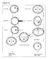
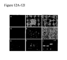
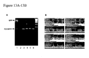

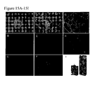
Classifications
-
- C—CHEMISTRY; METALLURGY
- C12—BIOCHEMISTRY; BEER; SPIRITS; WINE; VINEGAR; MICROBIOLOGY; ENZYMOLOGY; MUTATION OR GENETIC ENGINEERING
- C12N—MICROORGANISMS OR ENZYMES; COMPOSITIONS THEREOF; PROPAGATING, PRESERVING, OR MAINTAINING MICROORGANISMS; MUTATION OR GENETIC ENGINEERING; CULTURE MEDIA
- C12N5/00—Undifferentiated human, animal or plant cells, e.g. cell lines; Tissues; Cultivation or maintenance thereof; Culture media therefor
- C12N5/06—Animal cells or tissues; Human cells or tissues
- C12N5/0602—Vertebrate cells
- C12N5/0603—Embryonic cells ; Embryoid bodies
- C12N5/0606—Pluripotent embryonic cells, e.g. embryonic stem cells [ES]
-
- C—CHEMISTRY; METALLURGY
- C12—BIOCHEMISTRY; BEER; SPIRITS; WINE; VINEGAR; MICROBIOLOGY; ENZYMOLOGY; MUTATION OR GENETIC ENGINEERING
- C12N—MICROORGANISMS OR ENZYMES; COMPOSITIONS THEREOF; PROPAGATING, PRESERVING, OR MAINTAINING MICROORGANISMS; MUTATION OR GENETIC ENGINEERING; CULTURE MEDIA
- C12N5/00—Undifferentiated human, animal or plant cells, e.g. cell lines; Tissues; Cultivation or maintenance thereof; Culture media therefor
- C12N5/06—Animal cells or tissues; Human cells or tissues
- C12N5/0602—Vertebrate cells
- C12N5/0603—Embryonic cells ; Embryoid bodies
- C12N5/0604—Whole embryos; Culture medium therefor
-
- A—HUMAN NECESSITIES
- A01—AGRICULTURE; FORESTRY; ANIMAL HUSBANDRY; HUNTING; TRAPPING; FISHING
- A01K—ANIMAL HUSBANDRY; AVICULTURE; APICULTURE; PISCICULTURE; FISHING; REARING OR BREEDING ANIMALS, NOT OTHERWISE PROVIDED FOR; NEW BREEDS OF ANIMALS
- A01K67/00—Rearing or breeding animals, not otherwise provided for; New or modified breeds of animals
- A01K67/027—New or modified breeds of vertebrates
- A01K67/0273—Cloned vertebrates
-
- A—HUMAN NECESSITIES
- A01—AGRICULTURE; FORESTRY; ANIMAL HUSBANDRY; HUNTING; TRAPPING; FISHING
- A01K—ANIMAL HUSBANDRY; AVICULTURE; APICULTURE; PISCICULTURE; FISHING; REARING OR BREEDING ANIMALS, NOT OTHERWISE PROVIDED FOR; NEW BREEDS OF ANIMALS
- A01K67/00—Rearing or breeding animals, not otherwise provided for; New or modified breeds of animals
- A01K67/027—New or modified breeds of vertebrates
- A01K67/0275—Genetically modified vertebrates, e.g. transgenic
-
- A—HUMAN NECESSITIES
- A61—MEDICAL OR VETERINARY SCIENCE; HYGIENE
- A61P—SPECIFIC THERAPEUTIC ACTIVITY OF CHEMICAL COMPOUNDS OR MEDICINAL PREPARATIONS
- A61P1/00—Drugs for disorders of the alimentary tract or the digestive system
- A61P1/04—Drugs for disorders of the alimentary tract or the digestive system for ulcers, gastritis or reflux esophagitis, e.g. antacids, inhibitors of acid secretion, mucosal protectants
-
- A—HUMAN NECESSITIES
- A61—MEDICAL OR VETERINARY SCIENCE; HYGIENE
- A61P—SPECIFIC THERAPEUTIC ACTIVITY OF CHEMICAL COMPOUNDS OR MEDICINAL PREPARATIONS
- A61P1/00—Drugs for disorders of the alimentary tract or the digestive system
- A61P1/16—Drugs for disorders of the alimentary tract or the digestive system for liver or gallbladder disorders, e.g. hepatoprotective agents, cholagogues, litholytics
-
- A—HUMAN NECESSITIES
- A61—MEDICAL OR VETERINARY SCIENCE; HYGIENE
- A61P—SPECIFIC THERAPEUTIC ACTIVITY OF CHEMICAL COMPOUNDS OR MEDICINAL PREPARATIONS
- A61P13/00—Drugs for disorders of the urinary system
-
- A—HUMAN NECESSITIES
- A61—MEDICAL OR VETERINARY SCIENCE; HYGIENE
- A61P—SPECIFIC THERAPEUTIC ACTIVITY OF CHEMICAL COMPOUNDS OR MEDICINAL PREPARATIONS
- A61P13/00—Drugs for disorders of the urinary system
- A61P13/12—Drugs for disorders of the urinary system of the kidneys
-
- A—HUMAN NECESSITIES
- A61—MEDICAL OR VETERINARY SCIENCE; HYGIENE
- A61P—SPECIFIC THERAPEUTIC ACTIVITY OF CHEMICAL COMPOUNDS OR MEDICINAL PREPARATIONS
- A61P17/00—Drugs for dermatological disorders
- A61P17/02—Drugs for dermatological disorders for treating wounds, ulcers, burns, scars, keloids, or the like
-
- A—HUMAN NECESSITIES
- A61—MEDICAL OR VETERINARY SCIENCE; HYGIENE
- A61P—SPECIFIC THERAPEUTIC ACTIVITY OF CHEMICAL COMPOUNDS OR MEDICINAL PREPARATIONS
- A61P19/00—Drugs for skeletal disorders
- A61P19/08—Drugs for skeletal disorders for bone diseases, e.g. rachitism, Paget's disease
-
- A—HUMAN NECESSITIES
- A61—MEDICAL OR VETERINARY SCIENCE; HYGIENE
- A61P—SPECIFIC THERAPEUTIC ACTIVITY OF CHEMICAL COMPOUNDS OR MEDICINAL PREPARATIONS
- A61P21/00—Drugs for disorders of the muscular or neuromuscular system
- A61P21/02—Muscle relaxants, e.g. for tetanus or cramps
-
- A—HUMAN NECESSITIES
- A61—MEDICAL OR VETERINARY SCIENCE; HYGIENE
- A61P—SPECIFIC THERAPEUTIC ACTIVITY OF CHEMICAL COMPOUNDS OR MEDICINAL PREPARATIONS
- A61P21/00—Drugs for disorders of the muscular or neuromuscular system
- A61P21/04—Drugs for disorders of the muscular or neuromuscular system for myasthenia gravis
-
- A—HUMAN NECESSITIES
- A61—MEDICAL OR VETERINARY SCIENCE; HYGIENE
- A61P—SPECIFIC THERAPEUTIC ACTIVITY OF CHEMICAL COMPOUNDS OR MEDICINAL PREPARATIONS
- A61P25/00—Drugs for disorders of the nervous system
-
- A—HUMAN NECESSITIES
- A61—MEDICAL OR VETERINARY SCIENCE; HYGIENE
- A61P—SPECIFIC THERAPEUTIC ACTIVITY OF CHEMICAL COMPOUNDS OR MEDICINAL PREPARATIONS
- A61P25/00—Drugs for disorders of the nervous system
- A61P25/14—Drugs for disorders of the nervous system for treating abnormal movements, e.g. chorea, dyskinesia
- A61P25/16—Anti-Parkinson drugs
-
- A—HUMAN NECESSITIES
- A61—MEDICAL OR VETERINARY SCIENCE; HYGIENE
- A61P—SPECIFIC THERAPEUTIC ACTIVITY OF CHEMICAL COMPOUNDS OR MEDICINAL PREPARATIONS
- A61P25/00—Drugs for disorders of the nervous system
- A61P25/28—Drugs for disorders of the nervous system for treating neurodegenerative disorders of the central nervous system, e.g. nootropic agents, cognition enhancers, drugs for treating Alzheimer's disease or other forms of dementia
-
- A—HUMAN NECESSITIES
- A61—MEDICAL OR VETERINARY SCIENCE; HYGIENE
- A61P—SPECIFIC THERAPEUTIC ACTIVITY OF CHEMICAL COMPOUNDS OR MEDICINAL PREPARATIONS
- A61P3/00—Drugs for disorders of the metabolism
- A61P3/06—Antihyperlipidemics
-
- A—HUMAN NECESSITIES
- A61—MEDICAL OR VETERINARY SCIENCE; HYGIENE
- A61P—SPECIFIC THERAPEUTIC ACTIVITY OF CHEMICAL COMPOUNDS OR MEDICINAL PREPARATIONS
- A61P3/00—Drugs for disorders of the metabolism
- A61P3/08—Drugs for disorders of the metabolism for glucose homeostasis
- A61P3/10—Drugs for disorders of the metabolism for glucose homeostasis for hyperglycaemia, e.g. antidiabetics
-
- A—HUMAN NECESSITIES
- A61—MEDICAL OR VETERINARY SCIENCE; HYGIENE
- A61P—SPECIFIC THERAPEUTIC ACTIVITY OF CHEMICAL COMPOUNDS OR MEDICINAL PREPARATIONS
- A61P35/00—Antineoplastic agents
-
- A—HUMAN NECESSITIES
- A61—MEDICAL OR VETERINARY SCIENCE; HYGIENE
- A61P—SPECIFIC THERAPEUTIC ACTIVITY OF CHEMICAL COMPOUNDS OR MEDICINAL PREPARATIONS
- A61P35/00—Antineoplastic agents
- A61P35/02—Antineoplastic agents specific for leukemia
-
- A—HUMAN NECESSITIES
- A61—MEDICAL OR VETERINARY SCIENCE; HYGIENE
- A61P—SPECIFIC THERAPEUTIC ACTIVITY OF CHEMICAL COMPOUNDS OR MEDICINAL PREPARATIONS
- A61P37/00—Drugs for immunological or allergic disorders
- A61P37/02—Immunomodulators
- A61P37/04—Immunostimulants
-
- A—HUMAN NECESSITIES
- A61—MEDICAL OR VETERINARY SCIENCE; HYGIENE
- A61P—SPECIFIC THERAPEUTIC ACTIVITY OF CHEMICAL COMPOUNDS OR MEDICINAL PREPARATIONS
- A61P9/00—Drugs for disorders of the cardiovascular system
-
- C—CHEMISTRY; METALLURGY
- C07—ORGANIC CHEMISTRY
- C07K—PEPTIDES
- C07K14/00—Peptides having more than 20 amino acids; Gastrins; Somatostatins; Melanotropins; Derivatives thereof
- C07K14/435—Peptides having more than 20 amino acids; Gastrins; Somatostatins; Melanotropins; Derivatives thereof from animals; from humans
- C07K14/43504—Peptides having more than 20 amino acids; Gastrins; Somatostatins; Melanotropins; Derivatives thereof from animals; from humans from invertebrates
- C07K14/43595—Peptides having more than 20 amino acids; Gastrins; Somatostatins; Melanotropins; Derivatives thereof from animals; from humans from invertebrates from coelenteratae, e.g. medusae
-
- C—CHEMISTRY; METALLURGY
- C12—BIOCHEMISTRY; BEER; SPIRITS; WINE; VINEGAR; MICROBIOLOGY; ENZYMOLOGY; MUTATION OR GENETIC ENGINEERING
- C12N—MICROORGANISMS OR ENZYMES; COMPOSITIONS THEREOF; PROPAGATING, PRESERVING, OR MAINTAINING MICROORGANISMS; MUTATION OR GENETIC ENGINEERING; CULTURE MEDIA
- C12N5/00—Undifferentiated human, animal or plant cells, e.g. cell lines; Tissues; Cultivation or maintenance thereof; Culture media therefor
- C12N5/06—Animal cells or tissues; Human cells or tissues
- C12N5/0602—Vertebrate cells
- C12N5/0618—Cells of the nervous system
- C12N5/0619—Neurons
-
- C—CHEMISTRY; METALLURGY
- C12—BIOCHEMISTRY; BEER; SPIRITS; WINE; VINEGAR; MICROBIOLOGY; ENZYMOLOGY; MUTATION OR GENETIC ENGINEERING
- C12N—MICROORGANISMS OR ENZYMES; COMPOSITIONS THEREOF; PROPAGATING, PRESERVING, OR MAINTAINING MICROORGANISMS; MUTATION OR GENETIC ENGINEERING; CULTURE MEDIA
- C12N5/00—Undifferentiated human, animal or plant cells, e.g. cell lines; Tissues; Cultivation or maintenance thereof; Culture media therefor
- C12N5/06—Animal cells or tissues; Human cells or tissues
- C12N5/0602—Vertebrate cells
- C12N5/0618—Cells of the nervous system
- C12N5/0621—Eye cells, e.g. cornea, iris pigmented cells
-
- C—CHEMISTRY; METALLURGY
- C12—BIOCHEMISTRY; BEER; SPIRITS; WINE; VINEGAR; MICROBIOLOGY; ENZYMOLOGY; MUTATION OR GENETIC ENGINEERING
- C12N—MICROORGANISMS OR ENZYMES; COMPOSITIONS THEREOF; PROPAGATING, PRESERVING, OR MAINTAINING MICROORGANISMS; MUTATION OR GENETIC ENGINEERING; CULTURE MEDIA
- C12N5/00—Undifferentiated human, animal or plant cells, e.g. cell lines; Tissues; Cultivation or maintenance thereof; Culture media therefor
- C12N5/06—Animal cells or tissues; Human cells or tissues
- C12N5/0602—Vertebrate cells
- C12N5/0634—Cells from the blood or the immune system
-
- C—CHEMISTRY; METALLURGY
- C12—BIOCHEMISTRY; BEER; SPIRITS; WINE; VINEGAR; MICROBIOLOGY; ENZYMOLOGY; MUTATION OR GENETIC ENGINEERING
- C12N—MICROORGANISMS OR ENZYMES; COMPOSITIONS THEREOF; PROPAGATING, PRESERVING, OR MAINTAINING MICROORGANISMS; MUTATION OR GENETIC ENGINEERING; CULTURE MEDIA
- C12N5/00—Undifferentiated human, animal or plant cells, e.g. cell lines; Tissues; Cultivation or maintenance thereof; Culture media therefor
- C12N5/06—Animal cells or tissues; Human cells or tissues
- C12N5/0602—Vertebrate cells
- C12N5/0652—Cells of skeletal and connective tissues; Mesenchyme
- C12N5/0657—Cardiomyocytes; Heart cells
-
- C—CHEMISTRY; METALLURGY
- C12—BIOCHEMISTRY; BEER; SPIRITS; WINE; VINEGAR; MICROBIOLOGY; ENZYMOLOGY; MUTATION OR GENETIC ENGINEERING
- C12N—MICROORGANISMS OR ENZYMES; COMPOSITIONS THEREOF; PROPAGATING, PRESERVING, OR MAINTAINING MICROORGANISMS; MUTATION OR GENETIC ENGINEERING; CULTURE MEDIA
- C12N5/00—Undifferentiated human, animal or plant cells, e.g. cell lines; Tissues; Cultivation or maintenance thereof; Culture media therefor
- C12N5/10—Cells modified by introduction of foreign genetic material
- C12N5/12—Fused cells, e.g. hybridomas
- C12N5/16—Animal cells
-
- A—HUMAN NECESSITIES
- A01—AGRICULTURE; FORESTRY; ANIMAL HUSBANDRY; HUNTING; TRAPPING; FISHING
- A01K—ANIMAL HUSBANDRY; AVICULTURE; APICULTURE; PISCICULTURE; FISHING; REARING OR BREEDING ANIMALS, NOT OTHERWISE PROVIDED FOR; NEW BREEDS OF ANIMALS
- A01K2227/00—Animals characterised by species
- A01K2227/10—Mammal
- A01K2227/105—Murine
-
- A—HUMAN NECESSITIES
- A01—AGRICULTURE; FORESTRY; ANIMAL HUSBANDRY; HUNTING; TRAPPING; FISHING
- A01K—ANIMAL HUSBANDRY; AVICULTURE; APICULTURE; PISCICULTURE; FISHING; REARING OR BREEDING ANIMALS, NOT OTHERWISE PROVIDED FOR; NEW BREEDS OF ANIMALS
- A01K2267/00—Animals characterised by purpose
- A01K2267/03—Animal model, e.g. for test or diseases
- A01K2267/0393—Animal model comprising a reporter system for screening tests
-
- C—CHEMISTRY; METALLURGY
- C12—BIOCHEMISTRY; BEER; SPIRITS; WINE; VINEGAR; MICROBIOLOGY; ENZYMOLOGY; MUTATION OR GENETIC ENGINEERING
- C12N—MICROORGANISMS OR ENZYMES; COMPOSITIONS THEREOF; PROPAGATING, PRESERVING, OR MAINTAINING MICROORGANISMS; MUTATION OR GENETIC ENGINEERING; CULTURE MEDIA
- C12N2501/00—Active agents used in cell culture processes, e.g. differentation
- C12N2501/20—Cytokines; Chemokines
- C12N2501/23—Interleukins [IL]
- C12N2501/235—Leukemia inhibitory factor [LIF]
-
- C—CHEMISTRY; METALLURGY
- C12—BIOCHEMISTRY; BEER; SPIRITS; WINE; VINEGAR; MICROBIOLOGY; ENZYMOLOGY; MUTATION OR GENETIC ENGINEERING
- C12N—MICROORGANISMS OR ENZYMES; COMPOSITIONS THEREOF; PROPAGATING, PRESERVING, OR MAINTAINING MICROORGANISMS; MUTATION OR GENETIC ENGINEERING; CULTURE MEDIA
- C12N2501/00—Active agents used in cell culture processes, e.g. differentation
- C12N2501/998—Proteins not provided for elsewhere
-
- C—CHEMISTRY; METALLURGY
- C12—BIOCHEMISTRY; BEER; SPIRITS; WINE; VINEGAR; MICROBIOLOGY; ENZYMOLOGY; MUTATION OR GENETIC ENGINEERING
- C12N—MICROORGANISMS OR ENZYMES; COMPOSITIONS THEREOF; PROPAGATING, PRESERVING, OR MAINTAINING MICROORGANISMS; MUTATION OR GENETIC ENGINEERING; CULTURE MEDIA
- C12N2502/00—Coculture with; Conditioned medium produced by
- C12N2502/02—Coculture with; Conditioned medium produced by embryonic cells
-
- C—CHEMISTRY; METALLURGY
- C12—BIOCHEMISTRY; BEER; SPIRITS; WINE; VINEGAR; MICROBIOLOGY; ENZYMOLOGY; MUTATION OR GENETIC ENGINEERING
- C12N—MICROORGANISMS OR ENZYMES; COMPOSITIONS THEREOF; PROPAGATING, PRESERVING, OR MAINTAINING MICROORGANISMS; MUTATION OR GENETIC ENGINEERING; CULTURE MEDIA
- C12N2506/00—Differentiation of animal cells from one lineage to another; Differentiation of pluripotent cells
- C12N2506/02—Differentiation of animal cells from one lineage to another; Differentiation of pluripotent cells from embryonic cells
-
- C—CHEMISTRY; METALLURGY
- C12—BIOCHEMISTRY; BEER; SPIRITS; WINE; VINEGAR; MICROBIOLOGY; ENZYMOLOGY; MUTATION OR GENETIC ENGINEERING
- C12N—MICROORGANISMS OR ENZYMES; COMPOSITIONS THEREOF; PROPAGATING, PRESERVING, OR MAINTAINING MICROORGANISMS; MUTATION OR GENETIC ENGINEERING; CULTURE MEDIA
- C12N2517/00—Cells related to new breeds of animals
- C12N2517/04—Cells produced using nuclear transfer
-
- C—CHEMISTRY; METALLURGY
- C12—BIOCHEMISTRY; BEER; SPIRITS; WINE; VINEGAR; MICROBIOLOGY; ENZYMOLOGY; MUTATION OR GENETIC ENGINEERING
- C12N—MICROORGANISMS OR ENZYMES; COMPOSITIONS THEREOF; PROPAGATING, PRESERVING, OR MAINTAINING MICROORGANISMS; MUTATION OR GENETIC ENGINEERING; CULTURE MEDIA
- C12N2533/00—Supports or coatings for cell culture, characterised by material
- C12N2533/50—Proteins
- C12N2533/52—Fibronectin; Laminin
-
- C—CHEMISTRY; METALLURGY
- C12—BIOCHEMISTRY; BEER; SPIRITS; WINE; VINEGAR; MICROBIOLOGY; ENZYMOLOGY; MUTATION OR GENETIC ENGINEERING
- C12N—MICROORGANISMS OR ENZYMES; COMPOSITIONS THEREOF; PROPAGATING, PRESERVING, OR MAINTAINING MICROORGANISMS; MUTATION OR GENETIC ENGINEERING; CULTURE MEDIA
- C12N5/00—Undifferentiated human, animal or plant cells, e.g. cell lines; Tissues; Cultivation or maintenance thereof; Culture media therefor
- C12N5/06—Animal cells or tissues; Human cells or tissues
- C12N5/0602—Vertebrate cells
- C12N5/0603—Embryonic cells ; Embryoid bodies
Definitions
- the present invention relates generally to the field of somatic cell nuclear transfer (SCNT) and to the generation of animals and cells.
- SCNT somatic cell nuclear transfer
- hES cells human embryonic stem cells
- somatic differentiated cell back to a totipotent state, such as that of embryonic stem cells (“ES") ( Cibelli et al., Nature Biotech 16:642-646 (1998 )) or embryo-derived (“ED”) cells.
- ES embryonic stem cells
- ED embryo-derived
- SCNT somatic cell nuclear transfer
- transgenic animals are animals that carry the gene(s) of interest that has been deliberately introduced into existing somatic cells and/or germline cells at an early stage of development. As the animals develop and grow the protein product or specific developmental change engineered into the animal becomes apparent, and is present in their genetic complement and that of their offspring.
- this invention solves long outstanding problems with efficiency, ethical dilemmas, and the problem of how to clone embryos without oocytes.
- This invention generally relates to methods of cloning somatic cells using a fertilized embryo as a recipient.
- an oocyte is the initial recipient and a fertilized embryo is a second recipient.
- the disclosure relates to a method for cloning a mammal, for obtaining pluripotent cells, or for reprogramming a mammalian cell.
- the disclosure provides a method for reprogramming a nucleus of a differentiated cell comprising the steps of providing a differentiated cell, an enucleated, MII-stage egg of an animal and an enucleated, 2-cell stage embryo of an animal, wherein said MII-stage egg and said embryo are synchronized; injecting the nucleus of said differentiated cell into said enucleated egg; activating said egg that comprises said nucleus; allowing said activated egg that comprises said nucleus to develop to the 2-cell stage; removing at least one nucleus and at least a portion of surrounding cytoplasm of said activated 2-cell stage egg from the previous step; fusing said at least one nucleus removed in the previous step into said enucleated, 2-cell stage embryo, preferably by positioning said nucleus between the 2 cells of the 2-cell stage embryo to generate a single cell containing a reprogrammed nucleus of the differentiated cell.
- the disclosure provides a method for producing an animal comprising providing a differentiated cell, an enucleated, MII-stage egg of an animal and an enucleated, 2-cell stage embryo of an animal, wherein said MII-stage egg and said embryo are synchronized; injecting the nucleus of said differentiated cell into said enucleated egg; activating said egg that comprises said nucleus; allowing said activated egg that comprises said nucleus to develop to the 2-cell stage; removing at least one nucleus and at least a portion of the surrounding cytoplasm of said 2-cell stage egg from the previous step; fusing said at least one nucleus removed in the previous step into said enucleated, 2-cell stage embryo to generate a single cell; and culturing said single cell from the previous step to allow development into an animal.
- culturing comprises implanting said cultured cells into a uterus of an animal.
- the implanted cells and the animal in which they are implanted are of the same
- the disclosure provides a method for producing embryonic stem cells, comprising the steps of providing a differentiated cell, an enucleated, MII-stage egg of an animal and an enucleated, 2-cell stage embryo of an animal, wherein said MII-stage egg and said embryo are synchronized; injecting the nucleus of said differentiated cell into said enucleated egg; activating said egg that comprises said nucleus; allowing said activated egg that comprises said nucleus to develop to the 2-cell stage; removing a nucleus and surrounding cytoplasm of said 2-cell stage egg from the previous step; fusing said nucleus removed in the previous step into said enucleated, 2-cell stage embryo, preferably by positioning said nucleus between the 2 cells of the 2-cell stage embryo to generate a single cell; and culturing said single cell from the previous step to a developmental stage where embryonic stem cells may be derived.
- said embryonic stem cell is hemizygous or homozygous for an MHC allele, wherein either said differentiated cell is hemizygous or homozygous for an MHC allele or said embryonic stem cell is engineered to be hemizygous or homozygous for an MHC allele, by homologous recombination or by loss of heterozygocity, or both, and wherein said same species is human.
- said method is repeated many times to produce a bank of embryonic stem cells, each of which is hemizygous or homozygous for a different MHC allele than the other embryonic stem cells of the bank.
- the methods of the disclosure further comprise the steps of growing said resulting single cell from the previous methods to blastomere, morula or blastocyst stage.
- the methods of the disclosure further comprise serial nuclear transfers into oocytes.
- the methods of the disclosure further comprise serial nuclear transfers into embryos.
- said egg is activated by cyclohexamide, CsCl 2 , calcium ionopore, ionomycin, sperm factors, sperm portions or components, 6-DMAP, SrCl 2 , cytochalasin B, or a combination thereof.
- said egg is activated by a combination of these agents.
- said egg is activated by a combination of ionomycin and 6-DMAP.
- said egg is activated by a combination of calcium ionopore and 6-DMAP.
- said egg is activated by a combination of SrCl 2 and cytochalasin B
- said fusion step is performed electrically. In certain embodiments, said electric fusion is performed in two steps: a first step in which said nucleus is lined up with the positive pole and electrically shocked and a second step in which the embryo and the nucleus are turned approximately 90 degrees and electrically shocked. In certain embodiment, the emryo and the nucleus are not turned before shocking. In certain embodiments, said fusion step is performed using a Sendai virus.
- said MII-stage egg is a human egg.
- said enucleated, 2-cell stage embryo is a human embryo.
- said differentiated cell is a human cell.
- the cells of the disclosure may be from any mammal.
- the mammal is selected from a mouse, rat, cat, dog, rabbit, goat, hamster, pig, sheep, non-human primate, or primate.
- said MII-stage egg and said enucleated, 2-cell stage embryo are from any animal. In certain embodiments, said MII-stage egg and said enucleated, 2-cell stage embryo are from the same species. In certain embodiments, said differentiated cell and said MII-stage egg are from the same species. In certain embodiments, said differentiated cell and said enucleated, 2-cell stage embryo are from the same species. In certain embodiments, said differentiated cell, said MII-stage egg and said enucleated, 2-cell stage embryo are from the same species. In certain embodiments, said same species is human.
- the disclosure relates to a method for cloning a mammal, for obtaining pluripotent cells, or for reprogramming a mammalian cell.
- the method consists of the following steps: (a) obtaining a donor nucleus from a mammalian cell; (b) obtaining a fertilized embryo from a mammal; (c) transferring said donor nucleus into one cell of said fertilized embryo; (d) enucleating the original nucleus of said fertilized embryo, leaving the donor nucleus inside the fertilized embryo; and (e) culturing said fertilized embryo.
- the enucleation step of the methods of the application is performed between 3 and 6 hours of the nuclear transfer step, between 4 and 6 hours of the nuclear transfer step, between 5 and 6 hours of the nuclear transfer step, between 3 and 4 hours of the nuclear transfer step, between 3 and 5 hours of the nuclear transfer step, or between 4 and 5 hours of the nuclear transfer step. In certain embodiments, the enucleation step is performed within 3 hours of the nuclear transfer step, within 2 hours of the nuclear transfer step, or within 1 hours of the nuclear transfer step.
- the enucleation step is performed between 1 and 2 hours of the nuclear transfer step, between 1 and 3 hours of the nuclear transfer step, between 1 and 4 hours of the nuclear transfer step, between 1 and 5 hours of the nuclear transfer step, between 1 and 6 hours of the nuclear transfer step, between 2 and 3 hours of the nuclear transfer step, between 2 and 4 hours of the nuclear transfer step, between 2 and 5 hours of the nuclear transfer step, or between 2 and 6 hours of the nuclear transfer step.
- culturing the cloned embryo results in the development of a blastocyst or blastocyst-like collection of cells.
- embryonic stem cells can be derived from these blastocysts or blastocyst-like collection of cells.
- culturing the cloned embryo results in the development of a 4-8 cell stage embryo or of a morula stage embryo.
- embryonic stem cells can be derived from all or a portion of such early cleavage stage or morula stage embryos.
- culturing the cloned embryo results in the development of an embryo that continues to divide beyond the two-cell stage.
- an embryonic stem cell line is derived and established.
- the fertilized embryo is from a mammal of the same species as the mammalian donor cell. In certain embodiments of the disclosure, the fertilized embryo is from a mammal of a closely related species to the mammalian donor cell. In certain embodiments of the disclosure, the fertilized embryo is a pronuclear stage embryo. In certain embodiments, said fertilized embryo is a 2-cell stage embryo. In certain embodiments of the disclosure, the mammalian donor cell is an ES cell. In certain embodiments, said mammalian cell is a differentiated cell. In certain embodiments, said differentiated mammalian cell is a cumulus cell. In certain embodiments, said mammalian cell is a murine cell.
- said mammalian cell is a bovine cell. In certain embodiments, said mammalian cell is a human cell. In certain embodiments, cells may be from other mammalian species including, but not limited to, equine, canine, porcine, ovine sources; or rodent species such as rat may be used. In certain embodiments, the fertilized embryo underwent cryogenic preservation and was thawed prior to the nuclear transfer step.
- the donor nucleus is labeled. In certain embodiments, said nucleus is labeled by expression of a fluorescent transgene.
- the disclosure relates to a method for cloning a mammalian cell comprising the steps of: (a) obtaining a donor nucleus from a mammalian cell; (b) obtaining a first fertilized embryo from a mammal; (c) transferring said donor nucleus into said first fertilized embryo; (d) enucleating the original nucleus of said first fertilized embryo, leaving the donor nucleus inside the fertilized embryo; (e) culturing said fertilized embryo; (f) enucleating a second fertilized mammalian embryo; (g) dissociating the cells of the first fertilized embryo from step (e) and transplanting at least one cell into the enucleated second fertilized embryo; (h) fusing said transplanted cells to the cells of said enucleated second fertilized embryo to form a single-cell embryo; and (i) culturing said cloned single-cell embryo.
- steps (f)-(i) are cycled through more than once with the fertilized embryo obtained in step (g) coming from the step (i) of the previous cycle.
- step (h) (fusing step) is accomplished by electrofusion.
- step (g) comprises transferring at least one nucleus of the fertilized embryo from step (e) into the enucleated second fertilized embryo.
- the second fertilized embryo of the disclosure is at the same stage of development as the first fertilized embryo. In certain embodiments, the second fertilized embryo of the disclosure is at a similar stage of development as the first fertilized embryo. A similar stage may include embryos that are in the same general stage such as blastula stage or embryos of similar cell number stage developmental time. In certain embodiments, said second fertilized embryo and said first fertilized embryo are at the 2-cell stage and only one of the two cells is transplanted.
- the disclosure relates to a method for cloning a mammal, for obtaining pluripotent cells, or for reprogramming mammalian cells comprising the steps of: (a) obtaining desired donor nuclei from mammalian cells; (b) obtaining at least one fertilized embryo of at least the 2-cell stage from a mammal; (c) transferring donor nuclei into one or more but not all of the cells of the fertilized embryo, one donor nucleus into each cell; (d) enucleating the original nucleus of each of the cells of said embryo to which a donor nucleus was transferred, leaving the donor nucleus in said cell; and (e) culturing said fertilized embryo(es).
- the fertilized embryo of the disclosure is a 2-cell stage embryo and a donor nucleus is transferred to only one of the two cells of said embryo.
- the fertilized embryo of the disclosure may be an embryo of any stage.
- the activated oocyte of the disclosure is the recipient of nuclear transfer at the two cell stage.
- the oocyte is of any stage.
- the oocyte and the embryo are in synchrony.
- the transferring of donor nuclei step is performed immediately before the enculeation step.
- the disclosure relates to a blastocyst derived from a fertilized embryo, wherein said fertilized embryo is produced by any of the methods of the disclosure. In certain embodiments, the disclosure relates to a blastula produced by any of the methods of the disclosure.
- embryonic stem cells or embryonic stem cell lines can be produced using all or a portion of a cloned embryo.
- embryonic stem cells or cell lines can be produced using all or a portion of a blastocyst stage cloned embryo or using all or a portion of an early cleavage stage or morula stage embryo.
- the disclosure relates to a method for producing embryonic stem cells, comprising the steps of: (a) providing a differentiated cell, an enucleated, MII-stage egg of an animal and providing an enucleated, 2-cell stage embryo of an animal, wherein said MII-stage egg and said embryo are synchronized; (b) injecting nucleus of said differentiated cell into said enucleated egg; (c) activating said egg that comprises said nucleus; (d) allowing said activated egg that comprises said nucleus to develop to the 2-cell stage; (e) removing a nucleus and surrounding cytoplasm of said 2-cell stage egg from step (d); (f) fusing said nucleus removed in step (e) into said enucleated, 2-cell stage embryo, preferably by positioning said nucleus between the 2 cells of the 2-cell stage embryo to generate a single cell; and (g) culturing said single cell from step (f) to a developmental stage where embryonic stem cells may be derived comprising:
- the disclosure relates to a method of producing an embryonic stem (ES) cell, comprising: (a) culturing a blastomere removed or biopsied from a mammalian parental embryo and said mammalian parental embryo together for 12 to 18 hrs; (b) transferring the blastomere to blastocyst medium further comprising laminin and seeded with mouse embryonic fibroblasts (MEF) and (c) culturing the blastomere of (b) until ES cells are produced.
- ES embryonic stem
- the disclosure relates to a method of producing an embryonic stem (ES) cell, comprising: (a) culturing a blastomere removed or biopsied from a mammalian parental embryo and said mammalian parental embryo together; (b) transferring the blastomere to blastocyst medium further comprising laminin or fibronectin and (c) culturing the blastomere of (b) until ES cells are produced.
- ES embryonic stem
- the disclosure relates to a method for producing embryonic stem cells, comprising the steps of: (a) providing an enucleated, MII-stage egg of an animal, a differentiated cell and an enucleated, 2-cell stage embryo of an animal, wherein said MII-stage egg and said embryo are synchronized; (b) injecting nucleus of said differentiated cell into said enucleated egg; (c) activating said egg that comprises said nucleus; (d) allowing said activated egg that comprises said nucleus to develop to the 2-cell stage; (e) removing a nucleus and surrounding cytoplasm of said 2-cell stage egg from step (d); (f) fusing said nucleus removed in step (e) into said enucleated, 2-cell stage embryo, preferably by positioning said nucleus between the 2 cells of the 2-cell stage embryo to generate a single cell; (g) culturing said single cell from step (f) to generate a morula; (h) isolating a blastome
- step (c) comprises culturing in conditions that reduce embryonic vesicle formation.
- the blastocyst medium comprises 2.5 ⁇ g/ml of laminin. In certain embodiments, the blastocyst medium comprises 10 ⁇ l/ml of laminin. In certain embodiments, the blastocyst medium comprises about 2.5, 5, 7.5, 10, 15, or 20 ⁇ g/ml of laminin. In certain embodiments, the medium is supplemented with 1-5, 1-10, 5-10, 10-20 or 1-20 ⁇ g/ml of laminin. In certain embodiments, the medium is supplemented with at least 1, 2.5, 5, 7.5, 10, 15 or 20 ⁇ g/ml of laminin.
- step (c) of the above methods comprises culturing in blastocyst medium seeded with MEF cells for 5 days. In certain embodiments, culturing in blastocyst medium seeded with MEF cells occurs for 1, 2, 3, 4, 5, 6, 7, 8, 9, or 10 days. In certain embodiments, step (c) of the above methods further comprises culturing until the blastomeres form cell clumps of about 20 cells and transferring the cell clumps to medium seeded with ES cells. In certain embodiments, cell clumps are about 5, 10, 15, 20, 30, 40, or 50 cells. In certain embodiments, the ES cells express a marker or are labeled. In certain embodiments, the ES cells express GFP.
- the parental embryos are transferred to blastocyst medium and allowed to develop into blastocysts.
- the blastomere is isolated from an embryo comprising: (a) immobilizing the embryo; and (b) tapping the immobilized embryo until a blastomere is isolated.
- more than one blastomere is removed or biopsied from a parental embryo.
- two blastomere may be biopsied from a parental embryo and used to derive ES cells.
- embryonic stem cells are produced using methods that do not require and/or result in the destruction of an embryo. For example, when embryonic stem cells are produced from a single blastomere of a morula stage parental embryo, the remaining portion of the parental embryo can be subsequently frozen for long term or perpetual storage, or used to generate a pregnancy.
- a blastomere removed or biopsied from a mammalian parental embryo and said mammalian parental embryo are cultured together for about 6 to 12, 6 to 18, 6 to 24, 12 to 18, 12 to 24, or 18 to 24 hrs.
- embryonic stem cells refers to cells derived from the inner cell mass of blastocysts or morulae that have been serially passaged as cell lines.
- the ES cells may be derived from fertilization of an egg cell with sperm or DNA, nuclear transfer, parthenogenesis, or by means to generate hES cells with homozygosity in the MHC region.
- human embryonic stem cells hES cells refers to human ES cells.
- pluripotent stem cells refers to animal cells capable of differentiating into more than one differentiated cell type. Such cells include hES cells, human embryo-derived cells (hEDCs), and adult-derived cells including mesenchymal stem cells, neuronal stem cells, and bone marrow-derived stem cells. Pluripotent stem cells may be genetically modified or not genetically modified. Genetically modified cells may include markers such as fluorescent proteins to facilitate their identification within the egg.
- differentiated cell refers to a any cell in the process of differentiating into a somatic cell lineage or having terminally differentiated.
- embryonic cells can differentiate into an epithelial cell lining the intestine.
- Differentiated cells can be isolated from a fetus or a live born animal, for example.
- embryos refers to impregnating a female animal with an embryo described herein. This technique is well known to a person of ordinary skill in the art. See, e.g., Seidel and Elsden, 1997, Embryo Transfer in Dairy Cattle, W. D. Hoard & Sons, Co., Hoards Dairyman .
- the embryo may be allowed to develop in utero, or alternatively, the fetus may be removed from the uterine environment before parturition.
- synchronized or “sychronous” as used herein in reference to estrus cycle, refers to assisted reproductive techniques well known to a person of ordinary skill in the art. These techniques are fully described in the reference cited in the previous paragraph. Typically, estrogen and progesterone hormones are utilized to synchronize the estrus cycle of the female animal with the developmental cycle of the embryo.
- developmental cycle refers to embryos of the invention and the time period that exists between each cell division within the embryo. This time period is predictable for embryos, and can be synchronized with the estrus cycle of a recipient animal.
- culturing refers to laboratory procedures that involve placing an embryo in a culture medium.
- the embryo can be placed in the culture medium for an appropriate amount of time to allow the embryo to remain static but functional in the medium, or to allow the embryo to grow in the medium.
- Culture media suitable for culturing embryos are well-known to those skilled in the art. See, e.g., U.S. Pat. No. 5,213,979 , entitled “In vitro Culture of Bovine Embryos," First et al., issued May 25, 1993, and U.S. Pat. No. 5,096,822 , entitled “Bovine Embryo Medium,” Rosenkrans, Jr. et al., issued Mar. 17, 1992, incorporated herein by reference in their entireties including all figures, tables, and drawings.
- suitable medium refers to any medium that allows cell proliferation.
- the suitable medium need not promote maximum proliferation, only measurable cell proliferation.
- cloned refers to a cell, embryonic cell, fetal cell, and/or animal cell having a nuclear DNA sequence that is substantially similar or identical to the nuclear DNA sequence of another cell, embryonic cell, fetal cell, and/or animal cell.
- substantially similar and “identical” are described herein.
- the cloned embryo can arise from one nuclear transfer, or alternatively, the cloned embryo can arise from a cloning process that includes at least one re-cloning step.
- the cloned embryo arises from a cloning procedure that includes at least one re-cloning step, then the cloned embryo can indirectly arise from an immortalized, totipotent cell since the re-cloning step can utilize embryonic cells isolated from an embryo that arose from an immortalized, totipotent cell.
- totipotent as used herein in reference to embryos refers to embryos that can develop into a live born animal.
- substantially similar refers to two nuclear DNA sequences that are nearly identical. The two sequences may differ by copy error differences that normally occur during the replication of a nuclear DNA. Substantially similar DNA sequences are preferably greater than 97% identical, more-preferably greater than 98% identical, and most preferably greater than 99% identical. Identity is measured by dividing the number of identical residues in the two sequences by the total number of residues and multiplying the product by 100. Thus, two copies of exactly the same sequence have 100% identity, while sequences that are less highly conserved and have deletions, additions, or replacements have a lower degree of identity. Those of ordinary skill in the art will recognize that several computer programs are available for performing sequence comparisons and determining sequence identity.
- parental embryo is used to refer to an embryo from which a single blastomere is removed or biopsied. Following biopsy, the remaining parental embryo (the parental embryo minus the biopsied blastomere) can be cultured with the blastomere to help promote proliferation of the blastomere. The remaining, viable parental embryo may subsequently be frozen for long term or perpetual storage or for future use. Alternatively, the viable parental embryo may be used to create a pregnancy. Alternatively, the viable parental embryo may be destroyed.
- a parental embryo is a cloned embryo produced by the serial transplantation methods of the invention. In other embodiments, a parental embryo is an embryo produced by fertilization.
- SCNT somatic cell nuclear transfer
- Kishigami et al. (2006) reported an improved mouse cloning technique of treating the reconstructed mouse eggs with trichostatin A, an inhibitor of histone deacetylase, which reduces abnormal DNA hypermethylation.
- Another approach was serial cloning using either pronuclear (PN) stage zygotes or 2-cell stage in vivo fertilized embryos as second cytoplast recipients.
- PN stage mouse somatic cell cloned embryos were recloned into enucleated in vivo fertilized PN stage zygotes, the development of cloned embryos in vitro and live pup rate were improved to some extent.
- Another aspect of the application uses embryos from which one or more blastomeres can be removed or biopsied and used to generate ES cells.
- An objective of the present invention is to provide a means of cloning somatic cells more efficiently and without causing ethical concerns.
- the methods of the disclosure may be used for cloning a mammal, for obtaining pluripotent cells, or for reprogramming a mammalian cell.
- Human or animal cells may be obtained and cultured by well known methods.
- Human and animal cells useful in the present invention include, by way of example, epithelial, neural cells, epidermal cells, keratinocytes, hematopoietic cells, melanocytes, chondrocytes, lymphocytes (B and T lymphocytes), other immune cells, erythrocytes, macrophages, melanocytes, monocytes, mononuclear cells, fibroblasts, cardiac muscle cells, cumulus cells and other muscle cells, etc.
- the human cells used for nuclear transfer may be obtained from different organs, e.g., skin, lung, pancreas, liver, stomach, intestine, heart, reproductive organs, bladder, kidney, urethra and other urinary organs, etc.
- suitable donor cells i.e., cells useful in the subject invention, may be obtained from any cell or organ of the body. This includes all somatic or germ cells e.g., primordial germ cells, sperm cells.
- the donor cells or nucleus can actively dividing, i.e., non-quiescent, cells as this has been reported to enhance cloning efficacy.
- Such cells include those in the G1, G2 S or M cell phase.
- quiescent cells may be used. Also preferably, such donor cells will be in the G1 cell cycle.
- donor and/or recipient cells of the application do not undergo a 2-cell block.
- donor cells or nuclei are not pretreated before nuclear transfer. In certain embodiments, donor cells or nuclei are not pretreated with spermine, protamine, or putrescine before nuclear transfer.
- recipient fertilized embryos of the invention may be from any mammalian species.
- cryopreserved fertilized embryos are used as recipient cells.
- these embryos are human. Cryogenic preservation and thawing are known to those skilled in the art (see Tucker et al., Curr Opin Obstet Gynecol. 1995 Jun;7(3):188-92 ).
- donor nuclei may be labeled.
- Cells may be genetically modified with a transgene encoding a easily visualized protein such as the Green Fluorescent protein ( Yang, M., et al., 2000, Proc. Natl. Acad. Sci. USA, 97:1206-1211 ), or one of its derivatives, or modified with a transgene constructed from the Firefly (Photinus pyralis) luciferase gene (Fluc) ( Sweeney, T.J., et al. 1999, Proc. Natl. Acad. Sci.
- a transgene encoding a easily visualized protein such as the Green Fluorescent protein ( Yang, M., et al., 2000, Proc. Natl. Acad. Sci. USA, 97:1206-1211 ), or one of its derivatives, or modified with a transgene constructed from the Firefly (Photinus pyralis) luciferase gene (Fluc)
- the reporter transgenes may be constitutively expressed using a "house-keeping gene” promoter such that the reporter genes are expressed in many or all cells at a high level, or the reporter transgenes may be expressed using a tissue specific or developmental stage specific gene promoter such that only cells that have located into particular niches and developed into specific tissues or cell types may be visualized.
- Additional labeling reagents include, but are not limited to, luminescently labeled macromolecules including fluorescent protein analogs and biosensors, luminescent macromolecular chimeras including those formed with the green fluorescent protein and mutants thereof, luminescently labeled primary or secondary antibodies that react with cellular antigens involved in a physiological response, luminescent stains, dyes, and other small molecules. Labeled cells from a mosaic blastocyst can be sorted for example by flow cytometry to isolate the cloned population.
- the microinjection device includes a piezo unit.
- the piezo unit is operably attached to the needle to impart oscillations to the needle.
- the piezo unit can assist the needle in passing into the object.
- the piezo unit may be used to transfer minimal cytoplasm with the nucleus. Any piezo unit suitable for the purpose may be used.
- a piezo unit is a Piezo micromanipulator controller PMM150 (PrimeTech, Japan).
- Enucleation may be effected by known methods, such as described in U.S. Pat. No. 4,994,384 which is incorporated by reference herein.
- metaphase II oocytes are either placed in HECM, optionally containing 7.5 micrograms per milliliter cytochalasin B, for immediate enucleation, or may be placed in a suitable medium, for example CR1aa, plus 10% estrus cow serum, and then enucleated later.
- Enucleation may be accomplished microsurgically using a micropipette to remove the polar body and the adjacent cytoplasm.
- the cells may then be screened to identify those of which have been successfully enucleated. This screening may be effected by staining the cells with 1 microgram per milliliter 33342 Hoechst dye in HECM, and then viewing the cells under ultraviolet irradiation for less than 10 seconds. Cells that have been successfully enucleated can then be placed in a suitable culture medium.
- the present invention may utilize "induced enucleation" which refers to enucleation of the oocyte by disrupting the meiotic spindle apparatus through the destabilization (e.g., depolymerization) of the microtubules of the meiotic spindle (see U.S. Patent Application No. 20060015950 ).
- Destabilization of the microtubules prevents the chromatids from separating (e.g., prevents successful karyokinesis), and induces the oocyte genome (e.g., nuclear chromatin) to segregate unequally (e.g., skew) during meiotic maturation, whereby essentially all endogenous chromatin of the oocyte collects in the second polar body.
- blastomeres may be dissociated using a glass pipette.
- dissociation may occur in the presence of 0.25% trypsin ( Collas and Robl, 43 BIOL. REPROD. 877-84, 1992 ; Stice and Robl, 39 BIOL. REPROD. 657-664, 1988 ; Kanka et al., 43 MOL. REPROD. DEV. 135-44, 1996 ).
- the NT unit may be activated by known methods. Such methods include, e.g., culturing the NT unit at sub-physiological temperature, in essence by applying a cold, or actually cool temperature shock to the NT unit. This may be most conveniently done by culturing the NT unit at room temperature, which is cold relative to the physiological temperature conditions to which embryos are normally exposed.
- activation may be achieved by application of known activation agents.
- penetration of oocytes by sperm during fertilization has been shown to activate prefusion oocytes to yield greater numbers of viable pregnancies and multiple genetically identical calves after nuclear transfer.
- treatments such as electrical and chemical shock or cycloheximide treatment may also be used to activate NT embryos after fusion.
- Suitable oocyte activation methods are the subject of U.S. Pat. No. 5,496,720, to Susko-Parrish et al. , which is herein incorporated by reference.
- oocyte activation may be effected by simultaneously or sequentially:
- divalent cations into the oocyte cytoplasm, e.g., magnesium, strontium, barium or calcium, e.g., in the form of an ionophore.
- divalent cations include the use of electric shock, treatment with ethanol and treatment with caged chelators.
- Phosphorylation may be reduced by known methods, e.g., by the addition of kinase inhibitors, e.g., serine-threonine kinase inhibitors, such as 6-dimethylamino-purine, staurosporine, 2-aminopurine, and sphingosine.
- kinase inhibitors e.g., serine-threonine kinase inhibitors, such as 6-dimethylamino-purine, staurosporine, 2-aminopurine, and sphingosine.
- phosphorylation of cellular proteins may be inhibited by introduction of a phosphatase into the oocyte, e.g., phosphatase 2A and phosphatase 2B.
- a phosphatase into the oocyte, e.g., phosphatase 2A and phosphatase 2B.
- oocytes or NT units typically about 22 to 28 hours post maturation are placed in about 2 mM DMAP for about one hour, followed by incubation for about two to twelve hours, preferably about eight hours, in 5 pg/ml of cytochalasin B and 20 ug/ml cycloheximide.
- the activation of reconstructed oocytes is carried out in Ca ++ -free CZB containing 10mM SrCl 2 and 5 ⁇ g/ml cytochalasin B for 6 hrs in a high humidified 5.5% CO 2 incubator.
- activation may be effected before, simultaneous, or after nuclear transfer.
- activation will be effected about 40 hours prior to nuclear transfer and fusion to about 40 hours after nuclear transfer and fusion, more preferably about 24 hours before to about 24 hours after nuclear transfer and fusion, and most preferably from about 4 to 9 hours before nuclear transfer and fusion to about 4 to 9 hours after nuclear transfer and fusion.
- Activation is preferably effected after or proximate to in vitro or in vivo maturation of the oocyte, e.g., approximately simultaneous or within about 40 hours of maturation, more preferably within about 24 hours of maturation.
- a step of the present invention is to fuse the cloned nuclei with enucleated cytoplasts of germ-line cells such as blastomeres, morula cells, inner cell mass cells, ES cells, including hES cells, EG cells, EC cells as is known in the art ( Do & Scholer, Stem Cells 22:941-949 (2004 )). Fusion of the cytoplasts with the nuclei is performed using a number of techniques known in the art, including polyethylene glycol (see Pontecorvo "Polyethylene Glycol (PEG) in the Production of Mammalian Somatic Cell Hybrids" Cytogenet Cell Genet.
- PEG Polyethylene glycol
- Electrofusion of cells involves bringing cells together in close proximity and exposing them to an alternating electric field. Under appropriate conditions, the cells are pushed together and there is a fusion of cell membranes and then the formation of fusate cells or hybrid cells. Electrofusion of cells and apparatus for performing same are described in, for example, U.S. Pat. Nos. 4,441,972 , 4,578,168 and 5,283,194 , International Patent Application No.
- cytoplasmic blebs of germ-line cells such as hES cells attached to a physical substrate as is well known in the art ( Wright & Hayflick, Exp. Cell Res. 96:113-121, (1975 ); & Wright & Hayflick, Proc. Natl. Acad. Sci., USA, 72:1812-1816, (1975 ) maybe combined with the present disclosure.
- the cytoplasmic volume of the germ-line cells is increased by adding 10 ⁇ M cytochalasin B for 20 hours prior to manipulation. trypsinized and replated on sterile 18 mm coverslips, cylinders, or other physical substrate coated with material promoting attachment.
- the cells are plated at a density such that after an overnight incubation at 37 °C. and one gentle wash with medium, the cells cover a portion, preferably about 90% of the surface area of the coverslip or other substrate.
- the substrates are then placed in a centrifuge tube in a position such that centrifugation will result in the removal of the nuclei from the cytoplast containing 8mL of 10% Ficoll-400 solution and centrifuged at 20,000 g at 36 °C. for 60 minutes.
- Cloned nuclei are then spread onto the coverslip or substrate with a density of at least that of the cytoplasts, preferable at least five times the density of the cytoplasts.
- Fusion of the cytoplasts with the nuclei is performed using polyethylene glycol (see Pontecorvo "Polyethylene Glycol (PEG) in the Production of Mammalian Somatic Cell Hybrids" Cytogenet Cell Genet. 16(1-5):399-400 (1976 ). Briefly, in 1 mL of prewarmed 50% polyethylene glycol 1500 (Roche) in culture medium is placed over the coverslip or substrate for one minute. 20 mL of culture medium is then added drip-wise over a five minute period to slowly remove the polyethylene glycol. The entire media is then aspirated and replaced with culture medium. Techniques other than centrifugation such as vibration or physical removal of the nuclei using a micropipette may also be used.
- PEG Polyethylene Glycol
- embryos derived by nuclear transfer are different from normal embryos and sometimes benefit from or even require culture conditions in vivo other than those in which embryos are usually cultured (at least in vivo). The reason for this is not known.
- bovine embryos reconstituted embryos (many of them at once) have been cultured in sheep oviducts for 5 to 6 days (as described by Willadsen, In Mammalian Egg Transfer (Adams, E. E., ed.) 185 CRC Press, Boca Raton, Fla. (1982 )).
- the embryo may be embedded in a protective medium such as agar before transfer and then dissected from the agar after recovery from the temporary recipient.
- the function of the protective agar or other medium is twofold: first, it acts as a structural aid for the embryo by holding the zona pellucida together; and secondly it acts as barrier to cells of the recipient animal's immune system.
- Activated NT units may be cultured in a suitable in vitro culture medium until the generation of embryonic or stem-like cells and cell colonies.
- Culture media suitable for culturing and maturation of embryos are well known in the art. Examples of known media, which may be used for bovine embryo culture and maintenance, include Ham's F-10+10% fetal calf serum (FCS), Tissue Culture Medium-199 (TCM-199)+10% fetal calf serum, Tyrodes-Albumin-Lactate-Pyruvate (TALP), Dulbecco's Phosphate Buffered Saline (PBS), Eagle's and Whitten's media.
- TCM-199 One of the most common media used for the collection and maturation of oocytes is TCM-199, and 1 to 20% serum supplement including fetal calf serum, newborn serum, estrual cow serum, lamb serum or steer serum.
- a preferred maintenance medium includes TCM-199 with Earl salts, 10% fetal calf serum, 0.2 Ma pyruvate and 50 ug/ml gentamicin sulphate. Any of the above may also involve co-culture with a variety of cell types such as granulosa cells, oviduct cells, BRL cells and uterine cells and STO cells.
- LIF leukemia inhibitory factor
- CR1 contains the nutritional substances necessary to support an embryo.
- CR1 contains hemicalcium L-lactate in amounts ranging from 1.0 mM to 10 mM, preferably 1.0 mM to 5.0 mM.
- Hemicalcium L-lactate is L-lactate with a hemicalcium salt incorporated thereon.
- the cultured NT unit or units are preferably washed and then placed in a suitable media, e.g., CR1aa medium, Ham's F-10, Tissue Culture Media-199 (TCM-199).
- a suitable media e.g., CR1aa medium, Ham's F-10, Tissue Culture Media-199 (TCM-199).
- Tyrodes-Albumin-Lactate-Pyruvate (TALP) Dulbecco's Phosphate Buffered Saline (PBS), Eagle's or Whitten's, preferably containing about 10% FCS.
- TCM-199 Tissue Culture Media-199
- Suitable feeder layers include, by way of example, fibroblasts and epithelial cells, e.g., fibroblasts and uterine epithelial cells derived from ungulates, chicken fibroblasts, murine (e.g., mouse or rat) fibroblasts, STO and SI-m220 feeder cell lines, and BRL cells.
- fibroblasts and epithelial cells e.g., fibroblasts and uterine epithelial cells derived from ungulates, chicken fibroblasts, murine (e.g., mouse or rat) fibroblasts, STO and SI-m220 feeder cell lines, and BRL cells.
- the feeder cells will comprise mouse embryonic fibroblasts.
- Means for preparation of a suitable fibroblast feeder layer are described in the example which follows and is well within the skill of the ordinary artisan.
- ES cells from blastocyst-stage embryos (or the equivalent thereof) are well known in the art. Such techniques can be used to derive ES cells from cloned embryos. Additionally or alternatively, ES cells can be derived from cloned embryos during earlier stages of development.
- the resultant blastocysts, or blastocyst-like clusters, of the disclosure may be used to obtain embryonic stem cell lines.
- Such lines can be obtained, for example, according to the culturing methods reported by Thomson et al., Science, 282:1145-1147 (1998 ) and Thomson et al., Proc. Natl. Acad. Sci., USA, 92:7544-7848 (1995 ), incorporated by reference in their entirety herein.
- Pluripotent embryonic stem cells can also be generated from a single blastomere removed from an embryo without interfering with the embryo's normal development to birth. See U.S. application nos. 60/624,827, filed November 4, 2004 ; 60/662,489, filed March 14, 2005 ; 60/687,158, filed June 3, 2005 ; 60/723,066, filed October 3, 2005 ; 60/726,775, filed October 14, 2005 ; 11/267,555 filed November 4, 2005 ; PCT application no. PCT/US05/39776, filed November 4, 2005 , the disclosures of which are incorporated by reference in their entirety; see also Chung et al., Nature, Oct 16, 2005 (electronically published ahead of print) and Chung et al., Nature V. 439, pp. 216-219 (2006 ), the entire disclosure of each of which is incorporated by reference in its entirety).
- the method comprises the utilization of cells derived from the reprogrammed cells of the present invention in research and in therapy.
- Such reprogrammed pluripotent or totipotent cells may be differentiated into any of the cells in the body including, without limitation, skin, cartilage, bone, skeletal muscle, cardiac muscle, renal, hepatic, blood and blood forming, vascular precursor and vascular endothelial, pancreatic beta, neurons, glia, retinal, inner ear follicle, intestinal, lung, cells.
- the reprogrammed cells may be differentiated into cells with a dermatological prenatal pattern of gene expression that is highly elastogenic or capable of regeneration without causing scar formation.
- Dermal fibroblasts of mammalian fetal skin especially corresponding to areas where the integument benefits from a high level of elasticity, such as in regions surrounding the joints, are responsible for synthesizing de novo the intricate architecture of elastic fibrils that function for many years without turnover.
- early embryonic skin is capable of regenerating without scar formation.
- Cells from this point in embryonic development made from the reprogrammed cells of the present invention are useful in promoting scarless regeneration of the skin including forming normal elastin architecture. This is particularly useful in treating the symptoms of the course of normal human aging, or in actinic skin damage, where there can be a profound elastolysis of the skin resulting in an aged appearance including sagging and wrinkling of the skin.
- the reprogrammed cells are exposed to one or more inducers of differentiation to yield other therapeutically-useful cells such as retinal pigment epithelium, hematopoietic precursors and hemangioblastic progenitors as well as many other useful cell types of the ectoderm, mesoderm, and endoderm.
- inducers of differentiation such as retinal pigment epithelium, hematopoietic precursors and hemangioblastic progenitors as well as many other useful cell types of the ectoderm, mesoderm, and endoderm.
- Such inducers include but are not limited to: cytokines such as interleukin-alpha A, interferon-alpha A/D, interferon-beta, interferon-gamma, interferon-gamma-inducible protein-10, interleukin-1-17, keratinocyte growth factor, leptin, leukemia inhibitory factor, macrophage colony-stimulating factor, and macrophage inflammatory protein-1 alpha, 1-beta, 2, 3 alpha, 3 beta, and monocyte chemotactic protein 1-3, 6kine, activin A, amphiregulin, angiogenin, B-endothelial cell growth factor, beta cellulin, brain-derived neurotrophic factor, C10, cardiotrophin-1, ciliary neurotrophic factor, cytokine-induced neutrophil chemoattractant-1, eotaxin, epidermal growth factor, epithelial neutrophil activating peptide-78, erythropoietin, estrogen receptor-
- inducers include cells or components derived from cells from defined tissues used to provide inductive signals to the differentiating cells derived from the reprogrammed cells of the present invention.
- inducer cells may derive from human, nonhuman mammal, or avian, such as specific pathogen-free (SPF) embryonic or adult cells.
- SPF specific pathogen-free
- cloned cells are introduced into the tissues in which they normally reside in order to exhibit therapeutic utility.
- the clonogenic populations of cells derived by methods of this invention may be introduced into the tissues.
- cloned cells are introduced systemically or at a distance from the cite at which therapeutic utility is desired. In such embodiments, the cloned cells may act at a distance or may hone to the desired cite.
- cloned cells are utilized in inducing the differentiation of other pluripotent stem cells.
- the generation of single cell-derived populations of cells capable of being propagated in vitro while maintaining an embryonic pattern of gene expression is useful in inducing the differentiation of other pluripotent stem cells.
- Cell-cell induction is a common means of directing differentiation in the early embryo. Many potentially medically-useful cell types are influenced by inductive signals during normal embryonic development including spinal cord neurons, cardiac cells, pancreatic beta cells, and definitive hematopoietic cells.
- Single cell-derived populations of cells capable of being propagated in vitro while maintaining an embryonic pattern of gene expression can be cultured in a variety of in vitro, in ovo, or in vivo culture conditions to induce the differentiation of other pluripotent stem cells to become desired cell or tissue types.
- the subject embryonic or stem-like cells may be used to obtain any desired differentiated cell type. Therapeutic usages of such differentiated human cells are unparalleled.
- human hematopoietic stem cells may be used in medical treatments requiring bone marrow transplantation. Such procedures are used to treat many diseases, e.g., late stage cancers such as ovarian cancer and leukemia, as well as diseases that compromise the immune system, such as AIDS.
- Hematopoietic stem cells can be obtained, e.g., by fusing adult somatic cells of a cancer or AIDS patient, e.g., epithelial cells or lymphocytes with an enucleated oocyte, e.g., bovine oocyte, obtaining embryonic or stem-like cells as described above, and culturing such cells under conditions which favor differentiation, until hematopoietic stem cells are obtained.
- oocyte e.g., bovine oocyte
- Such hematopoietic cells may be used in the treatment of diseases including cancer and AIDS.
- adult somatic cells from a patient with a neurological disorder may be fused with an enucleated animal oocyte, e.g., a primate or bovine oocyte, human embryonic or stem-like cells obtained therefrom, and such cells cultured under differentiation conditions to produce neural cell lines.
- an enucleated animal oocyte e.g., a primate or bovine oocyte, human embryonic or stem-like cells obtained therefrom, and such cells cultured under differentiation conditions to produce neural cell lines.
- oocyte e.g., a primate or bovine oocyte, human embryonic or stem-like cells obtained therefrom, and such cells cultured under differentiation conditions to produce neural cell lines.
- Specific diseases treatable by transplantation of such human neural cells include, by way of example, Parkinson's disease, Alzheimers disease, ALS and cerebral palsy, among others.
- Parkinson's disease it has been demonstrated that transplanted fetal brain neural cells make the proper connections with surrounding cells and produce dopamine. This can result in long
- donor cells may be transfected with selectable markers expressed via inducible promoters, thereby permitting selection or enrichment of particular cell lineages when differentiation is induced.
- CD34-neo may be used for selection of hematopoietic cells, Pw1-neo for muscle cells, Mash-1-neo for sympathetic neurons, Mal-neo for human CNS neurons of the grey matter of the cerebral cortex, etc.
- the great advantage of the subject invention is that it provides an essentially limitless supply of isogenic or synegenic human cells suitable for transplantation. Therefore, it will obviate the significant problem associated with current transplantation methods, i.e., rejection of the transplanted tissue which may occur because ofhost-vs-graft or graft-vs-host rejection. Conventionally, rejection is prevented or reduced by the administration of anti-rejection drugs such as cyclosporin. However, such drugs have significant adverse side-effects, e.g., immunosuppression, carcinogenic properties, as well as being very expensive.
- the present invention should eliminate, or at least greatly reduce, the need for anti-rejection drugs, such as cyclosporine, imulan, FK-506, glucocorticoids, and rapamycin, and derivatives thereof.
- diseases and conditions treatable by isogenic cell therapy include, byway of example, spinal cord injuries, multiple sclerosis, muscular dystrophy, diabetes, liver diseases, i.e., hypercholesterolemia, heart diseases, cartilage replacement, bums, foot ulcers, gastrointestinal diseases, vascular diseases, kidney disease, urinary tract disease, and aging related diseases and conditions.
- the methods of the invention can be used to produce cloned cleavage stage embryos or morula stage embryos that can be used as parental embryos.
- Such parental embryos can be used to generate ES cells.
- a blastomere (1, 2, 3, 4 blastomeres) can be removed or biopsied from such parental embryos and such blastomeres can be used to derive ES cells.
- the present invention is based, in part, on the discovery that stem cells can be generated from embryos without affecting viability of the embryo using novel methods disclosed herein.
- these methods utilize in vitro techniques related to those currently used in pre-implantation genetic diagnosis (PGD) to isolate single blastomeres from embryos without destroying the embryos or otherwise significantly altering their viability.
- PGD pre-implantation genetic diagnosis
- hES pluripotent human embryonic stem cells and cell lines can be generated from a single blastomere removed from an embryo without interfering with the embryo's normal development to birth.
- ES cells, ES cell lines, TS cells and cell lines, and cells differentiated therefrom can be used to study basic developmental biology, and can be used therapeutically in the treatment of numerous diseases and conditions. Additionally, these cells can be used in screening assays to identify factors and conditions that can be used to modulate the growth, differentiation, survival, or migration of these cells. Identified agents can be used to regulate cell behavior in vitro and in vivo, and may form the basis of cellular or cell-free therapies.
- an element means one element or more than one element.
- blastomere is used throughout to refer to at least one blastomere (e.g., 1, 2, 3, 4, etc) obtained from an embryo.
- cluster of two or more blastomeres is used interchangeably with “blastomere-derived outgrowths” to refer to the cells generated during the in vitro culture of a blastomere. For example, after a blastomere is obtained from an embryo and initially cultured, it generally divides at least once to produce a cluster of two or more blastomeres (also known as a blastomere-derived outgrowth). The cluster can be further cultured with embryonic or fetal cells. Ultimately, the blastomere-derived outgrowths will continue to divide. From these structures, ES cells, TS cells, and partially differentiated cell types will develop over the course of the culture method.
- the present invention provides methods for deriving ES cells, ES cell lines, and differentiated cell types from single blastomeres of an early stage embryo without necessarily destroying the embryo.
- Various features of the method a described in detail below. All of the combinations of the various aspects and embodiments of the invention detailed above and below are contemplated.
- the blastomere may be removed from an embryo at various developmental stages prior to implantation including but not limited to: before compaction of the morula, during compaction of the morula, right after compaction of the morula, before formation of the blastocoel or during the blastocyst stage.
- a blastomere (one blastomere, two blastomeres, or more than two blastomeres) is removed from an embryo at the 4-16 cell stage, or at the 4-10 cell stage, or at the 4-8 cell stage.
- the invention provides methods for biopsy of a blastocyst which will produce embryonic stem cells, and the remainder of the blastocyst is implanted and results in a pregnancy and later in a live birth.
- the zona pellucida is removed from the blastocyst by any means known to those of ordinary skill in the art and then the blastocyst is biopsied.
- the controversies associated with the derivation of human ES cells are circumvented by using a technique similar to that used in preimplantation genetic diagnosis (PGD) where a single blastomere is removed from the embryo.
- PPD preimplantation genetic diagnosis
- the single blastomere is removed before the compaction of the morula.
- the biopsied blastomere could be allowed to undergo cell division and one progeny cell is used for genetic testing and the remaining cells are used to generate human stem cells.
- the biopsied embryo may also be implanted at the blastocyst stage or frozen for implantation at a later time.
- biopsy e.g., removal of a blastomere from an embryo
- biopsy consists of two stages. The first is to make a hole in, or in some instances fully remove, the zone pellucida that surrounds the embryo. Once the hole is made, the cells (preferably one or two) may then be removed from the human embryo.
- the method involves removing or generating an extraction hole in the zona pellucida, and can be carried out by one or more techniques such as physical manipulation, chemical treatment and enzymatic digestion. Exemplary techniques that could be used include:
- the procedure is performed on 8-10 cell stage embryos.
- the embryo is placed in a drop of biopsy medium under mineral oil by holding it with a holding pipette.
- the zona pellucida is locally digested, by releasing acidified Tyrode's solution (Sigma, St. Louis, Mo. 63178) through an assistant hatching pipette. Once the hole is made, cells (blastomeres) could be aspirated through the hole.
- the zona pellucida of the blastocyst may be at least partially digested by treatment with one or more enzymes or mixture of enzymes such as pronase.
- one or more enzymes or mixture of enzymes such as pronase.
- Other types of proteases with the same or similar protease activity as pronase may also be used.
- Single blastomeres may also be obtained by disaggregating zona-denuded embryos in Ca ++ /Mg ++ free PBS.
- This invention also provides a novel and more efficient method of isolating single blastomeres.
- the embryo is immobilized and the immobilized embryo is then tapped until a single blastomere is released from the blastocyst.
- This method is not limited to human embryos and can be performed on embryos of other species including, without limitation, non-human embryos such as non-human mammals, mice, rabbits, pigs, cows, sheep, dogs and primates.
- the embryo can be immobilized by any means known to those of skill in the art.
- the embryo is immobilized using a micropipette and the micropipette holder is tapped to isolate the blastomere.
- the embryo is cultured in medium that is calcium and magnesium free.
- the embryo may be from the 2-cell stage to the 16 cell stage.
- the embryo is from the 4 cell stage to the 10 cell stage.
- the embryo is a 6-8 cell stage embryo.
- the embryo is an 8-10 cell stage embryo.
- tapping involves generating an amount of force sufficient to remove at least one blastomere without substantially decreasing the viability of the remainder of the embryo. Maintenance of viability can be shown, for example, by culturing the remaining embryo for at least one day and confirming that the remaining embryo can continue to divide in culture.
- blastomere one blastomere or more than one blastomere
- a particular method can be used alone or in combination with another method to facilitate removal of a blastomere.
- the embryo is a mammalian embryo.
- the mammalian embryo is a human embryo.
- Exemplary mammals include, but are not limited to, mice, rats, rabbits, cows, dogs, cats, sheep, hamsters, pigs, non-human primates, and humans.
- a blastomere is removed from an embryo without destroying the remainder of the embryo.
- the remaining embryo (the embryo minus the removed blastomere) can be cultured and/or cryopreserved.
- the remaining embryo is cultured for a time sufficient to confirm that the remaining embryo can continue to divide (e.g., is still viable), and then once viability is confirmed, the remaining embryo is cryopreserved. In certain other embodiments, the remaining embryo is immediately cryopreserved.
- multiple blastomeres are removed from a single embryo and the embryo is destroyed during or subsequent to the removal of multiple blastomeres.
- Multiple blastomeres can be used together in one experiment, for example, by aggregating multiple blastomeres during the initial blastomere culture.
- multiple blastomeres can be used in separate experiments in an effort to maximize the number of lines or cell types than can be generated from a single embryo.
- Embryos from which a blastomere is obtained can be generated by sexual or asexual methods.
- the embryo is produced by fertilization of an egg with a sperm.
- the embryo is produced by somatic cell nuclear transfer, parthenogenesis, androgenesis, or other asexual techniques. Note that embryos derived from asexual techniques may not look identical to embryos generated by fertilization. However, despite any differences in appearance, the term embryo is intended to encompass the products of asexual reproduction and the products of fertilization or other means of sexual reproduction.
- the isolated blastomere(s) can be initially cultured in any type of medium, e.g., embryo medium such as Quinn's cleavage medium (Cooper Surgical Inc. Cat #ART1529). Any medium that supports growth of an embryo can be used, including, without limitation, any commercial formulations.
- embryo medium is used to refer to a medium that promotes survival of blastomeres (especially human blastomeres) in culture.
- the embryo medium is a medium containing less than 5 mM glucose.
- the embryo medium is a medium that has an osmolarity of less that 310 mosm.
- the embryo medium is a medium that contains less than 5 mM glucose and has an osmolarity of less than 310 mosm.
- the medium used to initially culture blastomeres has an osmolarity of less than 300 mosm, less than 280 mosm, or less than 260 mosm, and optionally contains less than 5 mM glucose.
- the medium used to initially culture blastomeres has an osmolarity about 260-280 mosm, and optionally contains less than 5 mM glucose. Note that regardless of the osmolarity and particular concentration of glucose in the medium used to initially culture the blastomeres, the medium may also be supplemented with antibiotics, minerals, amino acids, and other factors typically found in commercial media formulations.
- the blastomeres may not initially grow well in standard ES cell medium. However, as described in detail herein, once the blastomeres have been cultured in the presence of certain embryonic or fetal cells and/or allowed to divide one or more times, the cluster of blastomeres can optionally be cultured in ES cell medium, or may be slowly transferred from embryo medium to ES cell medium by gradually replacing the medium.
- ES cell medium is used to refer to a medium that promotes maintenance of ES cells in culture and can be used to culture clusters of blastomeres as they continue to divide and produce ES cells, ED cells, etc. Such a medium is at least somewhat optimized for ES cells.
- the ES cell medium contains at least 5 mM glucose (relatively high glucose). In certain other embodiments, the ES cell medium has an osmolarity of at least 310 mosm. In certain other embodiments, the medium contains at least 5 mM glucose and has an osmolarity of at least 310 mosm. In certain embodiments, this medium has an osmolarity of at least 320 mosm, or at least 330 mosm, and optionally contains at least 5 mM glucose. In certain embodiments, this medium has an osmolarity of about 310-340 mosm, and optionally contains at least 5 mM glucose.
- ES cell medium may also be supplemented with factors known in the art to promote the growth of ES cells, and the medium may contain antibiotics, minerals, amino acids, and other factors typically found in commercial media formulations.
- pronuclear stage human embryos are cultured in Quinn's cleavage medium (Cooper Surgical).
- pronuclear stage human embryos are cultured up to the 8-cell stage.
- the pronuclear stage embryos may be cultured up to about the 2-cell stage, 4-cell stage, or 16-cell stage.
- the pronuclear stage embryos may be cultured up to between the 2-cell stage and 4-cell stage, 2-cell stage and 8-cell stage, 2-cell stage and 16-cell stage, 4-cell stage and 8-cell stage, 4-cell stage and 16-cell stage, or 8-cell stage and 16-cell stage.
- the embryos are pre-incubated in Ca ++ and Mg ++ - free phosphate buffered saline supplemented with 0.05% PVA.
- the embryos are pre-incubated for about 5, 10, 15, 20, 25, 30, 5-10, 5-15, 5-30, 10-15, 10-30, or 15-30 min at room temperature. In certain embodiments, the embryos are transferred to Quinn's hepes medium for the manipulation.
- individual blastomeres are isolated from embryos using PIEZO.
- a hole 500 ⁇ m in diameter is made on the zona pellucida.
- the hole may be made using a small (20 ⁇ m) pipette by applying several pulses of PIEZO.
- a biopsy pipette 500 ⁇ m is inserted through the hole to grasp a blastomere applying gentle negative pressure.
- the blasomeres is pulled away when 2/3 of the blastomere is inside of the pipette.
- 1/3, 1/2, or 3/4 of the blastomere is inside the pipette.
- 1/3 to 1/2, 1/3 to 2/3, 1/3 to 3/4, 1/2 to 2/3, 1/2 to 3/4, or 2/3 to 3/4 of the blastomere is inside the pipette.
- the parental embryos and blastomeres maybe returned to the original culture drops (Quinn's cleavage medium) and cultured 12 to 18 hrs together.
- the parental embryos and blastomeres may be returned to the original culture drops (Quinn's cleavage medium) and cultured about 6 to 12, 6 to 18, 6 to 24, 12 to 18, 12 to 24, or 18 to 24 hrs together.
- the parental embryos are transferred to blastocyst medium (Quinn's blastocyst medium).
- the blastomeres are transferred to a small culture drop (50 ⁇ l) containing MEFs.
- the blastomere culture medium may be supplemented with laminin, fibronectin, or Matrigel.
- the blastomeres are cultured for about 3, 4, 5, 6, 7 or 8 days.
- the blastomeres are cultured until they form cell clumps composed of approximately 20 cells in the same medium.
- GFP ES cell culture drops may be merged with the blastomere culture drops to allow the two media to mix together.
- some or all of the blastomere clumps may be removed and plated in the same culture drop about 12, 18, 24, 36 or 48 hrs later.
- a blastomere is obtained from a human or other mammalian embryo and cultured in embryo medium.
- a blastomere is cultured in embryo medium for at least one day or until the blastomere divides at least once.
- a blastomere may be cultured in embryo medium for more than 1 day (at least 2, 3, 4 days, etc.) and/or the blastomere may be cultured in contact with embryonic or fetal cells before dividing to produce a cluster of blastoemre.
- the blastomere may divide one or more times or produce a cluster of two or more blastomeres. Further culturing of the cluster of blastomeres includes culturing the blastomere along with its progeny.
- the blastomere divides and the progeny are cultured as an aggregate.
- the blastomere can be cultured in a microdrop.
- Each microdrop can contain a single blastomere or multiple blastomeres. After about at least 1 day, at least 2-3 days, or at least 4 days, the cultured blastomeres may divide and form vesicles or aggregates. The benefit of culturing the blastomere prior to direct or indirect contact with the embryonic cells is to prevent the embryonic cells from overgrowing the blastomere.
- the cultured cluster of two or more blastomeres is contacted directly or indirectly with embryonic or fetal cells, or alternatively with a medium that promotes further maturation of the blastomeres in the absence of embryonic or fetal cells.
- a medium includes medium conditioned with embryonic or fetal cells (conditioned medium) or medium supplemented with growth factors or cytokines that promote maturation of the blastomeres.
- the medium is supplemented with ACTH (adrenocorticotropic hormone).
- the embryonic or fetal cells may be derived from, for example, a mammal.
- the embryonic or fetal cells are mouse or human cells.
- Exemplary embryonic or fetal cells include, but are not limited to, embryonic stem (ES) cells (whether derived from blastocysts, blastomeres, or by other methods, and whether derived using somatic cell nuclear transfer or other asexual reproduction), embryonic germ cells, embryonic carcinoma cells, placental cells, trophoblasts/trophectoderm cells, trophoblast stem cells, primordial germ cells embryonic germ cells, amniotic fluid cells, amniotic stem cells, placental cells, placental stem cells, and umbilical cord cells.
- ES embryonic stem
- the medium in which the blastomeres are cultured is further supplemented with ACTH or other growth factors or cytokines that promote maturation of the blastomeres.
- the embryonic or fetal cells may be grown in the presence or absence of a feeder layer of cells.
- Feeder cells may be used to help maintain the embryonic or fetal cells and to prevent their differentiation.
- the specific feeder cell may be chosen based on the particular embryonic or fetal cell used.
- Exemplary feeder cells include, but are not limited to, fibroblast feeder cells. Such fibroblast feeder cells may be derived from the same species as the embryonic or fetal cells or they may be derived from a different species. Similarly, the feeder cells and the embryonic or fetal cells may be derived from the same species as the blastomere or from a different species.
- the feeder cells are irradiated or otherwise treated to prevent overgrowth relative to the embryonic or fetal cells.
- Exemplary feeder cells include, but are not limited to, mouse embryonic fibroblasts (MEF cells), human embryonic fibroblasts, human foreskin fibroblasts, human skin fibroblasts, human endometrial fibroblasts, human oviductal fibroblasts, and placental cells. Similar cell types derived from other animals (mammals, chickens, etc) are also contemplated.
- the feeder and/or embryonic cells are human cells that are autologous cells derived from the same embryo as the blastomere.
- the embryonic or fetal cells are grown in ES cell medium or any medium that supports growth of the embryonic or fetal cells, e.g., Knockout DMEM (Invitrogen Cat # 10829-018).
- exemplary embryonic or fetal cells include, but are not limited to, embryonic stem cells, such as from already established lines, embryo carcinoma cells, murine embryonic fibroblasts, other embryo-like cells, cells of embryonic origin or cells derived from embryos, many of which are known in the art and available from the American Type Culture Collection, Manassas, VA 20110-2209, USA, and other sources.
- the embryonic or fetal cells may be added directly to the cultured blastomeres or may be grown in close proximity to, but not in direct contact with, the cultured blastomere(s).
- Various direct and indirect co-culture systems are possible to facilitate providing the cultured blastomeres with factors or signals from the embryonic or fetal cells.
- "contacting the cultured cluster of two or more blastomeres” refers to any method of direct or indirect contact or co-culture.
- contacting the cluster of two or more blastomere comprises aggregating blastomere clusters with embryonic or fetal cells. In certain other embodiments, contacting comprises co-culturing the cluster of two or mores blastomeres so that the cells are in direct contact with the embryonic or fetal cells but are not aggregated to them. In other embodiments, contacting comprises co-culturing the cluster of two or more blastomeres with the embryonic or fetal cells so that the cells are in indirect contact, for example, maintained in the same culture vessel but without direct contact of the cells or maintained as contiguous microdrops.
- the method comprises the step of directly or indirectly contacting the cultured cluster of two or more blastomere(s) with embryonic or fetal cells, with the proviso that the contacting is not carried out by aggregating the cultured blastomere with embryonic cells as described in Chung et al., Nature (2006) 439:216-9 .
- the culture of blastomere(s) and the culture of embryonic or fetal cells are indirectly connected or merged. This can be achieved by any method known in the art including, for example, dragging a manipulation pipette between two drops under light mineral oil such as Cooper Surgical ACT# ART4008, paraffin oil or Squibb's oil.
- the connections can be made by using a glass capillary or similar device.
- the blastomere(s) may be co-cultured with the remaining embryo.
- the blastomere is co-cultured with the remaining embryo in a microdroplet culture system or other culture system known in the art, which does not permit cell-cell contact but could permit cell-secreted factor and/or cell-matrix contact.
- the volume of the microdrop may be reduced, e.g., from 50 microliters to about 5 microliters to intensify the signal.
- the embryonic cells may be from a species other than human, e.g., non-human primate or mouse.
- the particular media formulations used to culture a blastomere, a cluster of two or more blastomeres, and embryonic or fetal cells may vary slightly depending on the species. Additionally, whether initial blastomere culture benefits from a media formulation different from that used to culture the clusters of blastomeres or the embryonic cells may also vary slightly depending on the species.
- the medium used to separately culture a blastomere and the medium used to culture embryonic or fetal cells is not necessarily the same.
- the cluster of two or more blastomeres which has now divided multiple times to give rise to a cluster of cells and cell outgrowths, can gradually be transferred (for example by exchanging the medium) and cultured in medium having the properties of ES cell medium.
- the blastomere(s) exhibit properties of ES cells. Specifically, as the cells continue to divide and the blastomere progeny cluster, various cell types emerge and can be identified phenotypically. Amongst the emerging cell types are trophectoderm-like cells, ES cells, and partially or terminally differentiated ED cells. As such, these methods can be used to produce ES cells, TS or other trophectoderm cells, or ED cells. While not wishing to be bound by any particular theory, it is believed that over a period of days or weeks the cultured blastomeres exhibit ES cell growth perhaps as a result of factors secreted by the embryonic or fetal cells or by the extracellular matrix.
- the dividing cluster of blastomere progeny resemble, in some respects, the changes observed during development of the preimplantation blastocyst.
- the cell types emerging in these cultures recapitulate to some extent the cell types observed when whole blastocysts or ICMs are plated.
- the blastomere culture conditions may include contacting the cells with factors that can inhibit or otherwise potentiate the differentiation of the cells, e.g., prevent the differentiation of the cells into non-ES cells, trophectoderm or other cell types.
- Such conditions can include contacting the cultured cells with heparin or introducing Oct-4 into the cells (such as by including Oct-4 in the media) or activating endogenous Oct-4 in the cells.
- expression of cdx-2 is prevented by any means known in the art including, without limitation, introducing CDX-2 RNAi into blastomeres, thereby inhibiting differentiation of the blastomere into TS cells.
- the blastomere culture medium is supplemented with factors to inhibit differentiation into non-ES cells.
- laminin is added to the culture medium in order to inhibit differentiation into non-ES cells.
- the medium is supplemented with about 2.5, 5, 7.5, 10, 15, or 20 ⁇ g/ml of laminin.
- the medium is supplemented with 1-5, 1-10, 5-10, 10-20 or 1-20 ⁇ g/ml of laminin.
- the medium is supplemented with factors to disrupt tight junctions.
- laminin is added to the medium in order to disrupt tight junctions.
- the medium is supplemented with factors to inhibit the trophectoderm differentiation pathway.
- laminin is added to the medium in order to inhibit the trophectoderm differentiation pathway.
- the medium is supplemented with factors to depolarize cells.
- laminin is added to the medium in order to depolarize cells.
- depolarization is determined by a lack of microvilli on the cell surface.
- depolarization is determined by a piling of cells to form multilayered structures.
- the invention provides methodologies for producing ES cells, ED cells, and TS cells from a blastomere obtained from an embryo.
- This approach can be used to generate ES cells, ED cell, and TS cells, as well as cell line without necessarily destroying the embryo from which the blastomere is obtained.
- Blastomere-derived ED cells have broader differentiation potential than human ES cells produced using methods known in the art because the ED cells can be readily differentiated into germ-line cells using techniques known in the art, e.g. using methods to differentiate murine ES cell lines into germ-line cells.
- human ES cell lines derived from inner mass cells are not expected to be capable of differentiation into germ-line cells. This phenomenon has been observed in ES cells derived from inner mass cells in animal such as pigs, cows, chickens and rats and is likely due to the fact that germ-line is one of the first cell lineages to branch out in differentiation.
- blastomeres from embryos with at least two cells, and before the embryo enters the stage of development of a compacting morula are induced to directly differentiate into differentiated progenitor cells which are then used for cell therapy and for the generation of cells, tissues, and organs for transplantation.
- genetic modifications can be introduced, for example, into somatic cells prior to nuclear transfer to produce a morula or blastocyst or into somatic cells prior to the reprogramming of said somatic cell into undifferentiated cells through the juxtaposition of the DNA of said somatic cell with factors capable of reprogramming said somatic cells or into ES cell lines made using these methods. See US Patent Application No.
- the differentiated progenitor cells of the present invention do not possess the pluripotency of an embryonic stem cell, or an embryonic germ cell, and are, in essence, tissue-specific partially or fully differentiated cells. These differentiated progenitor cells may give rise to cells from any of three embryonic germ layers, i.e., endoderm, mesoderm, and ectoderm.
- the differentiated progenitor cells may differentiate into bone, cartilage, smooth muscle, dermis with a prenatal pattern of gene expression and capable of promoting scarless wound repair, and hematopoietic or hemangioblast cells (mesoderm), definitive endoderm, liver, primitive gut, pancreatic beta cells, and respiratory epithelium (endoderm); or neurons, glial cells, hair follicles, or eye cells including retinal neurons and retinal pigment epithelium.
- the differentiated progenitor cells of the present invention it is not necessary for the differentiated progenitor cells of the present invention to express the catalytic component of telomerase (TERT) and be immortal, or that the progenitor cells express cell surface markers found on embryonic stem cells such as the cell surface markers characteristic of primate embryonic stem cells: positive for SSEA-3, SSEA-4, TRA-1-60, TRA-1-81, alkaline phosphatase activity, and negative for SSEA-1.
- the differentiated progenitor cells of the present invention are distinct from embryoid bodies, i.e., embryoid bodies are derived from embryonic stem cells whereas the differentiated stem cells of the present invention are derived from blastomeres.
- the differentiated cells of the present invention are produced by culturing blastomere-derived cells in the absence of embryonic stem cells. Growth of undifferentiated embryonic stem cells can be prevented, for example, by culturing blastomeres in the presence of differentiation-inducing agents or by introducing genetic modifications into the cells such that the growth of embryonic stem cells is prevented.
- Any vertebrate embryo may be used as a source of blastomeres or cells equivalent in development to a mammalian blastomere.
- Human blastomeres in particular, have important utility in the generation of human cell-based therapies.
- the original embryo may have been produced by in vitro-fertilization, derived by fertilization within the reproductive tract by normal sexual reproduction, artificial insemination, or gamete intrafallopian transfer (GIFT), and subsequently retrieved, derived by somatic cell nuclear transfer.
- GIFT gamete intrafallopian transfer
- Isolated blastomeres can be induced directly or via ES cells or cell lines to differentiate in the presence of differentiation-inducing conditions including various combinations of growth factors, sera, hormones, extracellular matrices useful in making the particular desired differentiated cell type as known in the art (see Table 1 for list of exemplary molecules), or as disclosed in the pending applications PCT/US2006/013573 filed April 11, 2006 , US Application No. 60/835,779, filed August 3, 2006 , 60/792,224 filed April 14, 2006 , 60/801,993 filed May 19, 2006 , PCT/US2006/013519 filed April 11, 2006 , US Application No.
- blastomeres or ES cells may be cultured on various inducer cell types such as those isolated as single cell-derived populations of cells, or on particular extracellular matrix components and other differentiation-inducing factors such as factors or combinations of factors shown in Table 1 below.
- EGF Ligands 1) Amphiregulin 2) Betacellulin 3) EGF 4) Epigen 5) Epiregulin 6) HB-EGF 7) Neuregulin-3 8) NRG1 isoform GGF2 9) NRG1 Isoform SMDF 10) NRG 1-alpha/HRG1-alpha 11) TGF-alpha 12) TMEFF1/Tomoregulin-1 13) TMEFF2 14) EGF Ligands pooled (1-13 above) EGF R/ErbB Receptor Family 15) EGF Receptor 16) ErbB2 17) ErbB3 18) ErbB4 19) EGF/ErbB Receptors pooled (15-18 above) FGF Ligands 20) FGF acidic 21) FGF basic 22) FGF-3 23) FGF-4 24) FGF-5 25) FGF-6 26) KGF/FGF-7 27) FGF-8 28) FGF-9 29) FGF-10 30) FGF-11 31) FGF-12 32) FGF-13 33
- Partially or terminally differentiated endodermal, mesodermal, or ectodermal cell types can be used in screening assays, to study developmental and stem cell biology, or to produce therapeutics.
- Partially or terminally differentiated cell types can optionally be substantially purified, formulated as pharmaceutical preparations, and/or cryopreserved.
- Pluripotency of the human ES cells or cell lines produced by any of the methods of this invention can be determined by detecting expression of human ES cell marker proteins.
- human ES cell marker proteins include but are not limited to octamer binding protein 4 (Oct-4), stage-specific embryonic antigen (SSEA)-3, SSEA-4, TRA-1-60, TRA-1-81 and alkaline phosphatase.
- the putative ES cell lines maintain pluripotency after more than 13, 20, 30, 40, 50, 60, 70, 80, 90 or 100 passages.
- the ES cells may also be assayed for maintenance of normal karyotype.
- Pluripotency may also be confirmed by differentiating the ES cell produced by the methods of this invention into cells of ectoderm, endoderm and mesoderm lineage using methods known in the art. Pluripotency may also be tested by transplanting ES cells in vivo, for example into an immunodeficient mouse (such as a SCID mouse), and evaluating teratoma formation.
- an immunodeficient mouse such as a SCID mouse
- the ES cells or cell lines produced from a blastomere express one or more ES cell marker protein. Additionally or alternatively, in certain embodiments, the cells maintain a normal karyotype. Additionally or alternatively, in certain embodiments, the cells maintain pluripotency after more than 13, 20, 30, 40, 50, 60, 70, 80, 90 or 100 passages.
- the ES cell or cell line produced from a blastomere can be generated without destroying the embryo from which the blastomere used to generate the cell or line is obtained. This characteristic of the cells distinguishes these cells from currently available ES cells and lines which were generated using methods that necessarily destroyed the underlying embryo.
- TS cells are produced by contacting blastomere outgrowths, which morphologically resemble trophoblast and/or extraembryonic endoderm, but which do not resemble ES cells, with FGF-4.
- FGF-4 is added to the culture media of the outgrowths.
- TS cells can be detected by assaying expression of proteins such as cdx-2, fgfr2, PL-1 and human chorionic gonadotropin (hCG) using procedures standard in the art.
- hCG human chorionic gonadotropin
- cell lines can be produced.
- a particular cell type is identified in a culture comprising a cluster of two or more blastomeres (blastomere-derived outgrowths)
- blastomere-derived outgrowths that cell can be separated from the remainder of the culture for further use. Once separated, the desired cell can be propagated as a purified or substantially purified population, or it can be maintained as a cell line.
- an ES cell produced from culturing a blastomere obtained from an embryo is separated from the culture of blastomere-derived outgrowths, and an ES cell line is established using standard techniques developed when establishing ES cell lines from blastocyst stage embryos.
- a partially differentiated ED cell of interest can be select based on, for example, morphology and that cell can be separated from the culture and purified or otherwise further analyzed.
- Exemplary cell lines include stable cell lines.
- ES cell lines established in this way may have the properties of existing ES cell lines, for example, differentiation potential, protein expression, karyotype, and the like.
- ES cell lines established in this way may differ from existing ES cell lines in one or more ways.
- the ES or ED cells of this invention are suitable for any use for which ES cells are useful.
- the present invention provides a method of treating a disorder amenable to cell therapy comprising administering to the affected subject a therapeutically effective amount of the ES cells of the invention.
- the methods of the invention are used to remove a blastomere preceding implantation of a human embryo after which the blastomere would be cultured as described above in order to derive and store human ES cells for therapeutic uses using cell therapy should the child resulting from the human embryo require, for example, disease therapy, tissue repair, transplantation, treatment of a cellular debilitation, or treatment of cellular dysfunctions in the future.
- cells derived from a blastomere, precompaction morula, compacting morula, or sectioned blastocyst are directly differentiated in vitro or in vivo to generate differentiating or differentiated cells without generating an embryonic stem cell line.
- a blastomere precompaction morula, compacting morula, or sectioned blastocyst
- the cells of the invention are useful in medical, veterinary and biological research and in the treatment of disease by providing cells for use in cell therapy, e.g., allogeneic cell therapy.
- an ES cell or cell line is derived from a blastomere and the ES cell or cell line is induced to differentiate to produce one or more mesodermal, endodermal, or ectodermal cell types.
- Exemplary cell types include, but are not limited to, RPE cells, hematopoietic stem cells, hematopoietic cell types (e.g., RBCs, platelets, etc.), pancreatic beta cells, skin cells, cardiomyocytes, smooth muscle cells, endothelial cells, hepatocytes, neurons, glia, skeletal muscle cells, vascular cells, and the like.
- RPE cells hematopoietic stem cells
- hematopoietic cell types e.g., RBCs, platelets, etc.
- pancreatic beta cells pancreatic beta cells
- skin cells cardiomyocytes, smooth muscle cells, endothelial cells, hepatocytes, neurons, glia, skeletal muscle cells, vascular cells, and the like.
- the methods of the present invention may be used to generate stem cells from blastomeres wherein the stem cells are hemizygous or homozygous for MHC antigens. These cells are useful for reduced immunogenicity during transplantation and cell therapy.
- the stem cells so produced may be assembled into a bank with reduced complexity in the MHC genes.
- the blastomeres of this invention could be derived from embryos that are hemizygous or homozygous for MHC antigens. These embryos may be either selected to be hemizygous or homozygous for MHC antigens or made, by any methods known in the art, to be hemizygous or homozygous for MHC antigens.
- stem cells derived from blastomeres may be made hemizygous or homozygous for MHC antigens, e.g., by gene targeting. See, e.g., WO 03/018760 published March 6, 2003 and US provisional patent application no. 60/729, 173 the disclosures of which are incorporated herein in their entirety.
- the ES cells and human embryo-derived cells generated by the above-mentioned novel techniques are utilized in research relating to cell biology, drug discovery, and in cell therapy, including but not limited to, production of hematopoietic and hemangioblastic cells for the treatment of blood disorders, vascular disorders, heart disease, cancer, and wound healing, pancreatic beta cells useful in the treatment of diabetes, retinal cells such as neural cells and retinal pigment epithelial cells useful in the treatment of retinal disease such as retinitis pigmentosa and macular degeneration, neurons useful in treating Parkinson's disease, Alzheimer's disease, chronic pain, stroke, psychiatric disorders, and spinal cord injury, heart muscle cells useful in treating heart disease such as heart failure, skin cells useful in treating wounds for scarless wound repair, burns, promoting wound repair, and in treating skin aging, liver cells for the treatment of liver disease such as cirrhotic liver disease, kidney cells for the treatment of kidney disease such as renal failure, cartilage for the treatment of arthritis, lung cells for the treatment of lung disease
- Such cell therapy methods may involve use of the ES cells of this invention in combination with proliferation factors, lineage-commitment factors, or gene or proteins of interest.
- Treatment methods may include providing stem or appropriate precursor cells directly for transplantation where the tissue is regenerated in vivo or recreating the desired tissue in vitro and then providing the tissue to the affected subject.
- the invention provides methods of generating ES cells, ES cell lines, TS cells, and various partially and terminally differentiated cells and cell lines.
- Cells and cell lines so produced can be studied in vitro and in vivo.
- the study of these cells provides information about basic developmental biology and stem cell biology.
- the study of these cells and/or the factors that can be used to manipulate the proliferation, differentiation, and survival of these cells can be used to develop stem-cell based therapies to treat or ameliorate any of a variety of diseases or conditions.
- stem-cell based therapies to treat or ameliorate any of a variety of diseases or conditions.
- cells and cell lines produced by these methods can be used in screening assays to identify agents and conditions that can be used therapeutically. Identified therapeutics may be used to develop cellular therapies or may themselves be useful when delivered to patients.
- ES cells, ES cell lines, TS cells, TS cell lines, or partially or terminally differentiated cells may be formulated as pharmaceutical preparations by combining the cells with a pharmaceutically acceptable carrier or excipient.
- the pharmaceutical preparation contains a certain number of cells per unit volume of carrier so that cellular therapies can be administered to deliver a particular dosage of cells.
- pharmaceutical preparations can be formulated to permit delivery of, for example, 1x10 5 , 1x10 6 , 2x10 6 , 3x10 6 , 4x10 6 , 5x10 6 , 1x10 7 , or greater than 1x10 7 cells in a volume of carrier appropriate for the condition being treated and the route of administration.
- the present invention not only provides an alternative method for efficiently generating cells and cell lines, including ES cells and cell lines, the present invention also provides a method that does not require that new embryos be destroyed as part of the process of ES cell derivation. Remaining embryos can be cryopreserved and perpetually preserved or reserved for additional, future research use.
- the ability to derive ES cells and cell lines (or partially or terminally differentiated cell types differentiated from ES cells or directly differentiated from embryos) without necessarily destroying new embryos will provide substantial benefits beyond the significant technical advanced reflected in these methods.
- the invention provides novel methods of conducting embryonic stem cell research without destroying a human embryo.
- the method entails obtaining a human ES cell or ES cell line derived from a human embryo but without destroying that human embryo.
- the ES cell or cell line can be generated from a blastomere obtained from a human embryo using any of the methodologies disclosed herein.
- the method further entails conducting embryonic stem cell research using the human ES cell or ES cell line.
- the method provides an avenue for conducting ES cell research without the need to destroy new embryos.
- the embryonic stem cell research involves research examining the differentiation potential of ES cells or cell lines.
- the research may involve contacting the human ES cell or ES cell line with one or more factors, and identifying factors that promote differentiation of the ES cell or ES cell line to one or more mesodermal, endodermal, or ectodermal cell types.
- the embryonic stem cell research involves the study of possible therapeutic uses of ES cells or cell differentiated there from.
- this method may expand the opportunities for collaboration with researchers around the world, particularly researchers working in countries with laws regulating embryo destruction.
- Example 2 Serial Cloning using PN stage zygotes and 2-cell stage embryos.
- a transplantation of dissociated individual cloned embryo cells into normal fertilized 2-cell stage mouse embryos was done at 18 hrs after the first cloning.
- the recipient embryos were enucleated prior to the nuclear transfer.
- Individual cloned blastomere cells were transplanted into the perivitellin space of the enucleated 2-cell stage embryos. Electrofusion of the transplanted blastomere and the enucleated embryo was performed by giving a single pulse of 150V DC for 15 microseconds.
- serially cloned embryos were cultured in KSOM medium and monitored for further development. Two of six embryos developed into blastocysts ( Fig.1A-B ). As a control, PN stage zygotes were injected with mouse ES cell nuclei (GFP positive), and their nuclei were enucleated 3 hrs later and cultured in 5% CO2. None of these embryos went on to develop into blastocysts. Table 3. The effect of serial cloning on cloned embryo development. Treatments Total # of zygotes 2C 4C 8C-M Blastocysts Serial cloning 6 5 4 2 2 2 Control 6 4 0 0 0
- blastomeres in a mosaic embryo might be stimulated to develop further by non-cloned cells.
- One of the two blastomeres of a 2-cell stage mouse embryo was enucleated, and immediately after enucleation, an ES cell nuclei was injected into the enucleated blastomere.
- Embryos were cultured without any further manipulation in KSOM.
- the cloned blastomeres divided the next day and contributed to GFP positive cells forming mosaic embryos ( Fig.2A-C ). When these embryos developed to the 8-cell stage at least 3 blastomeres originated from the cloned blastomeres ( Fig.2B ). Four of these cloned embryos developed into blastocyts (see Table 4). GFP positive blastomeres integrated into part of the blastocysts. Table 4. The effect of helper cells inside of the same zona pellucida with cloned blastomeres in 2-cell stage cloned embryos. Total # of 2 cells # survived injection 4C 8C-M Blastocyst 14 8 4 4 4
- Example 4 Materials & methods for Examples 1-3.
- Tables 5 and 6 document the preimplantation development of F2GFP NT embryos derived from single cloning versus serial nuclear transfer using in vivo 2-cell stage embryos. The most significant difference in development was found at the 2-cell to 4-cell transition: 59% versus 97% development in single versus serial cloning, respectively. The attrition was observed in inbred stains as well. Moreover almost all the F2GFP cleaved serial clone embryos developed to expanded or hatched blastocyst stage within 4 days after initial cloning. This rate of development was the same as the in vivo fertilized B6D2 F1 embryos (95%) cultured in KSOM.
- DBA2 and C57BL/6 inbred mice were used. A total of 2 pups (1.6%) were recovered by caesarean section at 19.5 days of gestation from DBA2 serial clones, but no viable pups were found in C57BL/6 clones regardless of cloning methods used. Of the two DBA2 pups, one pup died from respiratory deficiency within few minutes after recovery. The remaining pup did not show any sign of breathing difficulty but was deserted and partially eaten few hours after introduction to the foster mother (Table 7).
- mice cloned using nuclear retransplantation did not express the obese phenotype that has been documented in adult cloned mice.
- the obesity of adult cloned mice has been attributed to gene expression abnormalities and epigenetic modifications during NT and embryo culture, and reflects an increase of adipose tissue in addition to larger body size beginning at 8-10 weeks of age.
- Mean weight ( ⁇ SD) of mice cloned using nuclear retransplantation were 34.9 ⁇ 0.8 grams at six months, which did not differ (P>0.1) from normal control animals (33.6 ⁇ 1.9).
- mice cloned using traditional SCNT weighed 54.8 ⁇ 2.6 grams at three to six months of age (Table 8).
- Table 8 Weight of mice cloned using traditional SCNT versus nuclear retransplantation Mouse months Number of animals weight (gm) at 3-6 Mean ⁇ SD BDF1
- Example 7 Gene expression profile of cloned blastocyst stage embryos.
- Example 8 Cell number counts of blastocyst-stage embryos.
- Blastocysts generated via nuclear retransplantation contained a total of 49.8 ⁇ 6.9 cells and 16.2 ⁇ 7.1 ICM cells, representing an approximately 1.5- and two-fold improvement, respectively.
- the mean ICM/trophectoderm (TE) cell ratio also increased from 0.33 to 0.48 (45 %) (P ⁇ 0.01) (Table 9).
- the higher ICM/TE ratio and cell numbers may account, at least in part, for the marked improvement in postimplantation development and survive to birth after transfer into surrogate mothers.
- Table 9 Blastocyst quality analysis by differential staining of inner cell mass (ICM) and trophectoderm(TE) cells of blastocysts after single or serial nuclear transfer. No.
- mice Female DBA2, C57BL/6, and F2 GFP hybrid mice were used as nuclear donors.
- Female 129/SV mice were crossed with a 129/ CD F1 male (129/Sv x CD-1) carrying and expressing the gene for green fluorescent protein (GFP).
- GFP green fluorescent protein
- enucleated BDF1 C57BL/6 x DBA/2
- oocytes enucleated 2 cell stage BDF1 embryos, respectively
- CD-1 females were used as surrogate mothers to gestate the cloned embryos.
- Oocytes and embryos were cultured in KSOM (Specialty Media, USA) containing amino acids, glucose, and 1 mg/ml bovine serum albumin (BSA) at 37°C in a high humidity incubator with 5% CO 2 in air.
- Oocyte enucleation was performed in M2 medium (Specialty Media, USA), and cell injections were performed in Hepes-buffered CZB medium at room temperature for initial cloning.
- the activation of reconstructed oocytes was carried out in Ca 2+ - free CZB containing 10 mM SrCl 2 and 5 u g/ml cytochalasin B to prevent polar body extrusion.
- Nuclear retransplantation was performed at 37°C in M2 medium (Specialty Media, USA) supplemented with 7.5 ug /ml cytochalasin B and 0.4 ug /ml Nocodazole, a microtubule polymerase inhibitor, to facilitate micromanipulation.
- the reconstructed embryos were pulsed in manitol-based fusion medium composed of 0.27mM manitol, 100 ⁇ m MgSO 4 and 50 ⁇ m CaCl 2 , supplemented with 0.3% bovine serum albumin.
- the dispersed cumulus cells were washed in fresh M2 and resuspended in 3% polyvinylpyrrolidone (PVP; Mr 360,000, ICN Biochemicals, USA) and kept in a refrigerator (4°C) until use.
- the 2-cell stage embryos were collected 36 h after hCG injection from plugged females in M2 medium by washing the oviduct with a 1 ml syringe attached to a 27G blunt needle.
- Nuclear transfer was performed according to the method reported by Chung et al.(29). The enucleation was performed using a Nikon inverted microscope (TE300, Japan) equipped with a Narshige injector (Narshige, Japan). The metaphase II spindle of B2DF1 oocytes was removed in a drop of M2 medium containing 5ug/ml of cytochalasin B using a 10-12 um pipette by aspiration using a Piezo micromanipulator controller PMM150 (PrimeTech, Japan). The enucleated oocytes were washed thoroughly in CZB medium and kept in an incubator until use.
- the nuclei of cumulus cells were injected individually after cytoplasm was removed in 3% PVP using a small bore injector pipette (inner diameter 7um). The removal of cytoplasm was performed such that only small amount of cytoplasm remained around the denuded nucleus.
- Activation of reconstructed oocytes were carried out in Ca 2+ -free CZB containing 10mM SrCl 2 and 5 ⁇ g/ml cytochalasin B for 6 hrs in a high humidified 5.5% CO 2 incubator. After activation, reconstructed oocytes were cultured in KSOM medium.
- Fusion of the 2-cell stage cytoplasts and the cloned nucleus was performed using a BTX 2001 electrofusion machine with 2 DC pulses of 2.4 kV/cm for 15 usec in mannitol-based fusion medium described above.
- the 1 st pulse was given after alignment of the reconstructed eggs such that the transferred nucleus faced the negative wire and both of the two blastomeres faced the positive wire.
- the reconstructed eggs were turned 90 degrees by alignment pulse of 5 volts AC so that the two blastomeres faced the opposite poles, and a second pulse was administered.
- the pulsed reconstructed embryos were cultured in KSOM after thorough washing, and their fusion was checked 30 min after the 2 nd pulse. We typically observed over 98% fusion after the first pulse. Only fused embryos were cultured for another three days.
- the total cell number, and TE and ICM cell numbers of blastocysts were counted after differential staining with polynucleotide-specific fluorochromes as described previously. Briefly, embryos that had developed to expanded blastocysts 4.5 days after the initial cloning were exposed to acidic Tyrode's solution (pH 2.1) to remove the zona pellucida. The denuded blastocysts were washed in M2 medium and then labeled with trinitrobenzene sulfonic acid (TNBS; Sigma P-2297) in M2 at 4°C for 10 min.
- TNBS trinitrobenzene sulfonic acid
- the blastocysts were exposed to anti-dinitrophenol in M2 at 37°C for 10 min. Then excess antibodies were removed by thorough washing before exposure to guinea pig complement diluted 1:4 in M2 with 2ug/ml propidium iodide (PI) for 10 min at 37°C. Then the blastocysts were quickly washed in protein free Hepes CZB medium supplemented with 5 ug/ml propidium iodide and then fixed in ice-cold absolute ethanol for 5 min. The blastocysts were then moved to 10ug/ml Hoechst 33258 in ethanol for at least 10 min at 4°C.
- PI propidium iodide
- the stained blastocysts were mounted in 100% glycerol and evaluated by fluorescence microscopy (Nikon TE200, Japan). Blue nuclei were counted as inner cell mass (ICM) while red nuclei were considered trophoblast (TE) cells.
- ICM inner cell mass
- TE trophoblast
- F2 GFP cloned embryos had developed to the blastocyst stage, they were used to establish ntES cell lines as described previously with minor modifications. Briefly, zonae pellucidae were removed before plating by brief exposure to acidic Tyrode's solution and vigorous washings. A group of 3 or 4 denuded blastocysts were placed on the monolayer of mitomycin-C treated mouse embryonic fibroblasts (MEF) grown in one well of a 4-well dish (Nunclon, USA) with 500 ul mouse ES cell culture medium supplemented with 2000 units/ml leukemia inhibiting factor (Chemicon. USA) and 50 ⁇ M MEK-1 inhibitor (Cell Signaling Tech, USA).
- MEF mitomycin-C treated mouse embryonic fibroblasts
- this clump of cells was dissected to smaller pieces by treating them with 0.05% trypsin/EDTA and pipetting with a small bore pipette.
- the dissected cell clumps and dispersed cells originating from same embryo were transferred to fresh MEFs grown in 50 ul drops of mouse ES cell culture medium covered with tissue culture mineral oil. The culture drops were observed daily for the presence of ES cell outgrowth. The outgrowths were then passed to wells of a 4-well dish containing fresh MEFs.
- the Nd3 C9461T polymorphism was confirmed by restriction fragment length polymorphism (RFLP) analysis as previously described (19). Briefly, a 204 bp fragment containing the 9461 site was amplifed by PCR. The primer-generated mutation together with the C9461 wild-type version produces a recognition site for BelI. As a result, the presence of the T9461 polymorphism disrupts the restriction site. Fragments were analyzed by electrophoresis in a 4% agarose gel.
- RFLP restriction fragment length polymorphism
- Genomic DNA from F2 GFP clones was isolated from tail tips as described above, and 100 ng per reaction was used for GFP gene amplification by PCR.
- PCR products were separated on 1.5% agarose gel and visualized by ethidium bromide staining.
- the D1Mit46 simple sequence repeat (SSR) polymorphism was used to genotype nuclear DNA from tail tip DNA by using MapPairs assay B219 forward and reverse unlabelled primers (Invitrogen, USA).
- the reaction parameters included 20 ng of template, 1X FailSafe Premix K (EpiCentre, USA), 0.2 ⁇ M of each primer, 0.5 units of FailSafe PCR Enzyme mix (EpiCentre, USA), and cycling conditions of 96° C for 2 min (1 cycle), 94° C for 45 s, 55° C for 45 s, 72° C for 1 min for 30 cycles, and 72° C for 7 min.
- PCR products were separated on a 4% agarose gel and visualized by ethidium bromide staining. The PCR was carried out for 30 cycles, and the product was separated by 3% agrose gels and visualized by ethidium bromide staing.
- Example 11 Human embryonic stem cell lines generated without embryo destruction.
- microdrops were merged with microdrops containing green fluoresecent protein (GFP)-expressing hES cells as previously described.
- GFP green fluoresecent protein
- blastomere-derived (hES cell-like) colonies reached approximately 50 cells or more, they were mechanically dispersed and the pieces plated next to the initial outgrowths. Secondary colonies were also allowed to grow to a similar size, and mechanically passaged onto fresh MEF every 3-5 days until they adapted to routine passaging with trypsin and could be frozen (usually after 7-10 passages) as previously described ( Klimanskaya and McMahon. Handbook of Stem Cells. San Diego: Elsevier Academic Press; 2004 p. 437-449 ). At each passage, the colonies were screened under a fluorescent microscope for the absence of GFP-positive cells.
- Example 12 Blastomere biopsy and culture.
- Pronuclear stage human embryos were cultured in Quinn's cleavage medium (Cooper Surgical) up to the 8-cell stage in an incubator with 5% CO 2 .
- Individual blastomeres were isolated from embryos as described previously using PIEZO. Briefly, the 8-cell embryos were pre-incubated in Ca ++ and Mg ++ - free phosphate buffered saline supplemented with 0.05% PVA for 15 min at room temperature to facilitate individual blastomere isolation. The embryos were then, transferred to Quinn's hepes medium for the manipulation.
- a hole 500 ⁇ m in diameter was made on the zona pellucida using a small (20 ⁇ m) pipette by applying several pulses of PIEZO.
- a biopsy pipette 500 ⁇ m was inserted through the hole and grasped a blasomere applying gentle negative pressure.
- the blasomeres were pulled away.
- the parental embryos and blastomeres were returned to the original culture drops (Quinn's cleavage medium) and cultured 12 to 18 hrs together.
- blastomeres and parental embryos were separated: the parental embryos were transferred to blastocyst medium (Quinn's blastocyst medium) to allow them to develop into blastocysts, meanwhile the blastomeres were transferred to a small culture drop (50 ⁇ l) containing MEFs.
- the blastomere culture medium was supplemented with laminin (10 ⁇ l/ml), fibronectin (10 ⁇ l/ml), or Matrigel (10 ⁇ l/ml). They were cultured for 5 days or until they formed cell clumps composed of approximately 20 cells in the same medium. Then the adjacent GFP ES cell culture drops were merged with the blastomere culture drops to allow the two media to mix together.
- Example 13 hESC co culture is not necessary for hESC line derivation.
- GFP-hESC co-culture was examined to determine if co-culture is necessary for successful derivation.
- An experiment was carried out using 2 frozen cleavage-stage embryos that were thawed and cultured in blastocyst medium for two hours prior to blastomere biopsy. A single blastomere was removed from one embryo, and two blastomeres were removed from the second embryo. The remaining biopsied embryos were allowed to continue development and were frozen at the blastocyst stage. Extracted blastomeres were cultured under the same conditions as described for the second set of experiments of example 11 except that no GFP-hESCs were present.
- the newly established hESC line had a normal male (46 XY) karyotype ( Figure 14 ) and differentiated into derivatives from all three germ layers, including immunostaining with antibodies to tubulin ⁇ III (ectoderm), smooth muscle actin (mesoderm), ⁇ fetoprotein (primitive endoderm).
- Example 14 Laminin directs blastomere differentiation toward ICM.
- Example 15 Materials and Methods for Examples 11-14.
- the blastomere biopsy was performed at 37°C in Quinn's Hepes medium supplemented with 5% SPS (serum protein substitute, Cooper Surgical) using a PIEZO drill as described previously. Only a single blastomere (or in a small number of case, two blastomeres) was removed from each embryo.
- SPS serum protein substitute, Cooper Surgical
- the parental embryos and biopsied blastomeres were cocultured for 12 - 24 h and then transferred to Quinn's blastocyst medium and cocultured for another 48 hrs. After the coculture parental embryos were frozen, the blastomere clumps were moved to MEF drops for further outgrowth as described below.
- Experimental Group 2 the parental embryos and blastomeres were cocultured for less than 12 hrs.
- the parental embryos were frozen using a Blastocyst Freezing kit (Cooper Surgical, Cat# ART-8015) according to manufacturer's directions. All the procedures were performed at room temperature. Briefly, the embryos were rinsed and cultured in diluent medium for 5 min, then transferred to 5% glycerol freezing medium for 10 min before being moved to the final 9% glycerol plus 0.2 M sucrose freezing solution. Then each embryo was loaded in a 0.25ml embryo freezing straw (IMV-ZA475, France) before freezing. The embryo freezing was performed using an embryo freezer (Freeze Control CL-869, Australia).
- the embryos were taken from a starting temperature of 25°C to - 6.5°C at 2°C/min. Then they were seeded manually and held at -6.5°C for 10 min before being cooled at 0.3°C/min to -45°C and transferred to a liquid nitrogen storage tank.
- hESC culture was performed as previously described (Klimanskaya et al., 2006; Klimanskaya et al., 2007; Klimanskaya and McMahon, 2004).
- MEF cells were plated on gelatin-treated 60 mm cell culture dishes in rows of 50 ul drops.
- the MEF cell drops were arranged as 2 or 3 drops ("auxiliary drops") surrounded one "blastomere drop” - the drop) designated for the blastomere outgrowth
- small clumps of GFP+ hES cells were transferred into the "auxiliary drops” and cultured overnight, and the MEF plating medium in these drops was replaced with hESC medium.
- each vesicle culture drop was connected to 2 or 3 surrounding GFP-hESC drops by dragging medium with a small glass pipette between two drops.
- the connecting channels were widened with a pipette, and the blastocyst medium was replaced with derivation medium as described above two days later.
- the blastomeres were cultured in 50 ul drops of Quinn's blastocyst medium supplemented with 5 ug/ml fibronectin and 5 ug/ml laminin containing MEF cells (as prepared in Experimental Group 1) after the first 12 hours of coculture with the parental embryos.
- the blastomere culture drops and the surrounding GFP ESC culture drops were not connected for 5 days. During this time most blastomeres formed clumps of cells comprising 20-30 cells resembling inner cell mass.
- the blastomere culture drops and surrounding GFP ESC culture drops were connected as in Experimental Group 1, and the blastocyst medium was replaced with derivation medium on day 7.
- Immunostaining was performed using standard protocols. Briefly, the samples were fixed with 2% paraformaldehyde for 10 minutes (cells) or 40 minutes (vesicles), permeabilized with 0.1% NP-40, blocked for 1 h with PBS containing 10% each goat and donkey serum, and incubated with the primary antibodies overnight at 4°C. Three washes of 10 minutes each were performed after each antibody incubation. Fluorescently labeled or biotinilated secondary antibodies (Jackson Immunoresearch or Molecular Probes) were added for 1 h, and fluorescently labeled Streptavidin (Molecular Probes) was added for 15 minutes to visualize biotinylated secondary antibodies.
- NOD-SCID male mice 6-8 weeks of age were used (Jackson Laboratories, Bar Harbor, ME). Small clumps of 50-100 cells were injected under the kidney capsule, and 7-12 weeks after transplantation the mice were sacrificed, the kidneys fixed in 4% paraformaldehyde overnight, embedded in paraffin, sectioned, immunostained or stained with hematoxylin-eosin and analyzed for the presence of the derivatives of all three germ layers.
- blast cells were plated on fibronectin-coated plates (BD Bioscience) in EGM-2 complete media (Lonza) for 3-5 days.
- EGM-2 complete media Lionza
- hES-BC cells were cultured for 3-5 days on fibronectin-coated wells and incubated for 6-8 h with 10 ⁇ g/ml of Alexa Fluor 594-labeled Ac-LDL (Invitrogen). Then cells were washed 3 times with 1X PBS and fixed with 4% paraformaldehyde for 30 min. The uptake of Ac-LDL was visualized under a fluorescent microscope.
- vWF Density Factor
- PECAM-1 CD31
- VE-cadherin R&D Systems
- KDR Cell Signalling Systems
- the cells were plated on gelatin at 1:6 ratio. When the cells were approximately 50% confluent, 0.12 ug/ml colcemid (Invitrogen) was added to the culture for 40 min. The cells were harvested by trypsin, incubated in 0.075M KCl for 12 minutes at 37oC, fixed with 3:1 Methanol and acetic acid. The spread analysis was performed by Cell Line Genetics, Inc. (for the hESC lines NEDI-NED4) and by the Cytogenetics Laboratory at the Children's Hospital, Oakland (NED5) using G-banding technique.
- Colcemid Invitrogen
Landscapes
- Health & Medical Sciences (AREA)
- Life Sciences & Earth Sciences (AREA)
- Engineering & Computer Science (AREA)
- Chemical & Material Sciences (AREA)
- Organic Chemistry (AREA)
- Biomedical Technology (AREA)
- Bioinformatics & Cheminformatics (AREA)
- General Health & Medical Sciences (AREA)
- Zoology (AREA)
- Genetics & Genomics (AREA)
- Biotechnology (AREA)
- Wood Science & Technology (AREA)
- Animal Behavior & Ethology (AREA)
- Veterinary Medicine (AREA)
- Medicinal Chemistry (AREA)
- Public Health (AREA)
- Pharmacology & Pharmacy (AREA)
- Nuclear Medicine, Radiotherapy & Molecular Imaging (AREA)
- General Chemical & Material Sciences (AREA)
- Chemical Kinetics & Catalysis (AREA)
- Biochemistry (AREA)
- General Engineering & Computer Science (AREA)
- Microbiology (AREA)
- Cell Biology (AREA)
- Reproductive Health (AREA)
- Gynecology & Obstetrics (AREA)
- Developmental Biology & Embryology (AREA)
- Neurology (AREA)
- Environmental Sciences (AREA)
- Neurosurgery (AREA)
- Immunology (AREA)
- Hematology (AREA)
- Biodiversity & Conservation Biology (AREA)
- Animal Husbandry (AREA)
- Gastroenterology & Hepatology (AREA)
- Diabetes (AREA)
- Physical Education & Sports Medicine (AREA)
- Tropical Medicine & Parasitology (AREA)
- Proteomics, Peptides & Aminoacids (AREA)
- Toxicology (AREA)
Priority Applications (1)
| Application Number | Priority Date | Filing Date | Title |
|---|---|---|---|
| EP16183074.0A EP3190178A1 (fr) | 2007-02-23 | 2008-02-22 | Procedes hautement efficaces pour reprogrammer des cellules differenciees et pour produire des animaux et des cellules souches embryonnaires a partir de cellules reprogrammees |
Applications Claiming Priority (5)
| Application Number | Priority Date | Filing Date | Title |
|---|---|---|---|
| US90297007P | 2007-02-23 | 2007-02-23 | |
| US91854307P | 2007-03-16 | 2007-03-16 | |
| US99377207P | 2007-09-14 | 2007-09-14 | |
| US943207P | 2007-12-28 | 2007-12-28 | |
| EP08725969A EP2121902A4 (fr) | 2007-02-23 | 2008-02-22 | Procedes hautement efficaces pour reprogrammer des cellules differenciees et pour produire des animaux et des cellules souches embryonnaires a partir de cellules reprogrammees |
Related Parent Applications (1)
| Application Number | Title | Priority Date | Filing Date |
|---|---|---|---|
| EP08725969A Division EP2121902A4 (fr) | 2007-02-23 | 2008-02-22 | Procedes hautement efficaces pour reprogrammer des cellules differenciees et pour produire des animaux et des cellules souches embryonnaires a partir de cellules reprogrammees |
Related Child Applications (1)
| Application Number | Title | Priority Date | Filing Date |
|---|---|---|---|
| EP16183074.0A Division EP3190178A1 (fr) | 2007-02-23 | 2008-02-22 | Procedes hautement efficaces pour reprogrammer des cellules differenciees et pour produire des animaux et des cellules souches embryonnaires a partir de cellules reprogrammees |
Publications (1)
| Publication Number | Publication Date |
|---|---|
| EP2982744A1 true EP2982744A1 (fr) | 2016-02-10 |
Family
ID=39710674
Family Applications (3)
| Application Number | Title | Priority Date | Filing Date |
|---|---|---|---|
| EP15164113.1A Withdrawn EP2982744A1 (fr) | 2007-02-23 | 2008-02-22 | Procédés hautement efficaces pour reprogrammer des cellules différenciées et pour produire des animaux et des cellules souches embryonnaires à partir de cellules reprogrammées |
| EP16183074.0A Withdrawn EP3190178A1 (fr) | 2007-02-23 | 2008-02-22 | Procedes hautement efficaces pour reprogrammer des cellules differenciees et pour produire des animaux et des cellules souches embryonnaires a partir de cellules reprogrammees |
| EP08725969A Withdrawn EP2121902A4 (fr) | 2007-02-23 | 2008-02-22 | Procedes hautement efficaces pour reprogrammer des cellules differenciees et pour produire des animaux et des cellules souches embryonnaires a partir de cellules reprogrammees |
Family Applications After (2)
| Application Number | Title | Priority Date | Filing Date |
|---|---|---|---|
| EP16183074.0A Withdrawn EP3190178A1 (fr) | 2007-02-23 | 2008-02-22 | Procedes hautement efficaces pour reprogrammer des cellules differenciees et pour produire des animaux et des cellules souches embryonnaires a partir de cellules reprogrammees |
| EP08725969A Withdrawn EP2121902A4 (fr) | 2007-02-23 | 2008-02-22 | Procedes hautement efficaces pour reprogrammer des cellules differenciees et pour produire des animaux et des cellules souches embryonnaires a partir de cellules reprogrammees |
Country Status (10)
| Country | Link |
|---|---|
| US (2) | US8796021B2 (fr) |
| EP (3) | EP2982744A1 (fr) |
| JP (4) | JP2010518857A (fr) |
| KR (1) | KR20100014504A (fr) |
| CN (2) | CN101679942A (fr) |
| AU (1) | AU2008218998A1 (fr) |
| CA (1) | CA2679109C (fr) |
| HK (1) | HK1222887A1 (fr) |
| IL (1) | IL200522A (fr) |
| WO (1) | WO2008103462A2 (fr) |
Families Citing this family (31)
| Publication number | Priority date | Publication date | Assignee | Title |
|---|---|---|---|---|
| EP2479256A1 (fr) | 2004-11-04 | 2012-07-25 | Advanced Cell Technology, Inc. | Dérivation de cellules de souche embryonnaire |
| US7893315B2 (en) | 2004-11-04 | 2011-02-22 | Advanced Cell Technology, Inc. | Derivation of embryonic stem cells and embryo-derived cells |
| US20100167404A1 (en) | 2005-08-03 | 2010-07-01 | Advanced Cell Technology, Inc. | Methods of Reprogramming Animal Somatic Cells |
| CN101679942A (zh) * | 2007-02-23 | 2010-03-24 | 先进细胞技术公司 | 重编程分化细胞和从重编程的细胞产生动物和胚胎干细胞的高效方法 |
| EP2283118A4 (fr) | 2008-05-06 | 2012-11-21 | Agency Science Tech & Res | Procédé de réalisation de la dédifférenciation d une cellule |
| CN101798569B (zh) * | 2009-02-09 | 2012-11-21 | 北京瑞途之星科技有限公司 | 以卵裂后的发育胚胎替代卵母细胞的治疗性克隆新方法 |
| AU2011235212B2 (en) * | 2010-03-31 | 2014-07-31 | The Scripps Research Institute | Reprogramming cells |
| CN101921808B (zh) * | 2010-08-18 | 2012-05-30 | 东北农业大学 | 一种提高猪体细胞核移植效率的方法 |
| US20120184035A1 (en) * | 2011-01-13 | 2012-07-19 | Sadhana Agarwal | Methods and Compositions For Reprogramming Cells |
| US10478460B2 (en) * | 2011-06-14 | 2019-11-19 | J.R. Simplot Company | Method for identifying a mammal receptive to embryo transfer |
| WO2013010045A1 (fr) | 2011-07-12 | 2013-01-17 | Biotime Inc. | Nouveaux procédés et formulations pour thérapie cellulaire orthopédique |
| CN102321571A (zh) * | 2011-09-21 | 2012-01-18 | 王跃嗣 | 从分化细胞制备自体造血干细胞的方法 |
| DK2785359T3 (en) | 2011-11-30 | 2018-10-29 | Astellas Inst For Regenerative Medicine | MESENKYMAL STROMACELLES AND APPLICATIONS RELATED |
| US9428732B2 (en) * | 2011-12-05 | 2016-08-30 | Primorigen Biosciences, Inc. | Compositions and methods for differentiating pluripotent stem cells into primitive blood cells and uses thereof |
| EP2802872A4 (fr) * | 2012-01-13 | 2015-09-09 | Univ Leland Stanford Junior | Biomarqueurs mécaniques pour la viabilité d'un ovocyte et d'un embryon |
| CN102608323A (zh) * | 2012-03-26 | 2012-07-25 | 西北农林科技大学 | 一种基于哺乳动物囊胚的分子免疫检测的胚胎质量分析方法 |
| WO2014144754A1 (fr) | 2013-03-15 | 2014-09-18 | The New York Stem Cell Foundation | Procédés de transfert nucléaire de cellules somatiques |
| US9822336B2 (en) * | 2013-04-16 | 2017-11-21 | Universiteit Maastricht | Blastoid, cell line based artificial blastocyst |
| JP2016073207A (ja) * | 2014-10-02 | 2016-05-12 | 株式会社リプロセル | 細胞凍結保存方法、細胞解凍方法、及び細胞 |
| US11422125B2 (en) | 2015-03-23 | 2022-08-23 | Astellas Institute For Regenerative Medicine | Assays for potency of human retinal pigment epithelium (RPE) cells and photoreceptor progenitors |
| CN109219658A (zh) * | 2016-03-31 | 2019-01-15 | 赛百奥公司 | 用于干细胞培养和治疗的培养基、方法、细胞和分泌的因子 |
| EP3555262A4 (fr) * | 2016-11-07 | 2020-05-13 | K2 Research Holdings, LLC | Production et utilisations thérapeutiques de cellules pluripotentes d'epinul et cellules différenciées dérivées de celles-ci |
| CN107099553A (zh) * | 2017-06-19 | 2017-08-29 | 内蒙古大学 | 一种小鼠体细胞核移植方法 |
| CA3076664C (fr) | 2017-09-21 | 2023-10-10 | EMULATE, Inc. | Physiologie et pathophysiologie de l'intestin humain : intestin-sur-puce |
| CN108624621B (zh) * | 2018-01-17 | 2019-04-12 | 中国科学院上海生命科学研究院 | 非人灵长类的体细胞克隆动物的制备方法 |
| CN110819585B (zh) * | 2018-08-09 | 2022-12-06 | 山东大学 | 含有igf2的胚胎体外培育方法和培养基 |
| TWI715874B (zh) * | 2018-09-27 | 2021-01-11 | 台灣粒線體應用技術股份有限公司 | 無血清培養液以及使用此無血清培養液分離和放大培養幹細胞方法 |
| JP7282411B2 (ja) * | 2018-10-23 | 2023-05-29 | ザ ボード オブ トラスティーズ オブ ザ レランド スタンフォード ジュニア ユニバーシティー | 交流電場を使用する、幹細胞に基づく療法におけるテラトーマ形成の予防及び処置 |
| WO2022272042A2 (fr) * | 2021-06-25 | 2022-12-29 | The J. David Gladstone Institutes, A Testamentary Trust Established Under The Will Of J. David Gladstone | Cellules germinales primordiales |
| CN113925878B (zh) * | 2021-10-20 | 2024-03-08 | 上海交通大学医学院附属仁济医院 | 一种细胞外囊泡在治疗神经损伤性勃起功能障碍中的应用 |
| CN114128668A (zh) * | 2021-11-24 | 2022-03-04 | 华中农业大学 | 一种提高水牛夏季同期发情-定时输精效果的处理方法 |
Citations (23)
| Publication number | Priority date | Publication date | Assignee | Title |
|---|---|---|---|---|
| US4441972A (en) | 1983-04-08 | 1984-04-10 | D.E.P. Systems, Inc. | Apparatus for electrofusion of biological particles |
| US4578168A (en) | 1984-07-27 | 1986-03-25 | Biotronics | Apparatus for fusing live cells with electric fields |
| US4664097A (en) | 1984-05-09 | 1987-05-12 | The Wistar Institute Of Anatomy & Biology | Nuclear transplantation in the mammalian embryo by microsurgery and cell fusion |
| WO1990003432A1 (fr) | 1988-09-21 | 1990-04-05 | Animal Biotechnology Cambridge Limited | Derivation de souches de cellules embryonnaires pluripotentielles a partir d'animaux domestiques |
| US4944384A (en) | 1989-01-30 | 1990-07-31 | Hay & Forage Industries | Trash discharge apparatus for crop transferring conveyor mechanism |
| US4994384A (en) | 1986-12-31 | 1991-02-19 | W. R. Grace & Co.-Conn. | Multiplying bovine embryos |
| US5057420A (en) | 1987-06-05 | 1991-10-15 | Granada Biosciences, Inc. | Bovine nuclear transplantation |
| US5096822A (en) | 1990-07-26 | 1992-03-17 | W. R. Grace & Co.- Conn. | Bovine embryo medium |
| WO1993005166A1 (fr) | 1991-09-05 | 1993-03-18 | Fucell Pty. Limited | Procede de selection, de separation et de fusion de cellules |
| US5213979A (en) | 1987-12-30 | 1993-05-25 | W. R. Grace & Co.-Conn. | In vitro culture of bovine embryos |
| US5283194A (en) | 1991-07-22 | 1994-02-01 | Schmukler Robert E | Apparatus and methods for electroporation and electrofusion |
| WO1994024274A1 (fr) | 1993-04-21 | 1994-10-27 | The University Of Edinburgh | Isolation, selection et propagation de cellules souches d'animaux transgeniques |
| WO1994026884A1 (fr) | 1993-05-14 | 1994-11-24 | Biotechnology Research And Development Corporation | Cellules souches embryonnaires destinees a la reproduction d'ongules chimeres et transgeniques |
| US5496720A (en) | 1993-02-10 | 1996-03-05 | Susko-Parrish; Joan L. | Parthenogenic oocyte activation |
| US5712156A (en) | 1990-07-09 | 1998-01-27 | Amrad Corporation Limited | Enhanced implantation, development and maintenance of embryos using leukaemia inhibitory factor |
| WO2001029206A1 (fr) | 1999-10-15 | 2001-04-26 | Advanced Cell Technology, Inc. | Procedes de production de cellules souches et de cellules embryonnaires a lignee defectueuse differenciees |
| WO2003018760A2 (fr) | 2001-08-24 | 2003-03-06 | Advanced Cell Technology, Inc. | Essais de criblage pour l'identification d'agents induisant la differenciation, et production de cellules differenciees pour la therapie cellulaire |
| WO2003046141A2 (fr) * | 2001-11-26 | 2003-06-05 | Advanced Cell Technology, Inc. | Procedes de production et d'utilisation de noyaux de cellules somatiques humaines reprogrammees et de cellules souches humaines autologues et isogeniques |
| US20040199935A1 (en) | 1999-06-30 | 2004-10-07 | Chapman Karen B. | Cytoplasmic transfer to de-differentiate recipient cells |
| WO2005070011A2 (fr) | 2004-01-23 | 2005-08-04 | Advanced Cell Technology, Inc. | Modalites ameliorees du traitement des maladies degeneratives de la retine |
| US20060015950A1 (en) | 2002-07-29 | 2006-01-19 | Trustees Of Tufts College | Nuclear transfer embryo formation method |
| WO2006052646A2 (fr) * | 2004-11-04 | 2006-05-18 | Advanced Cell Technology, Inc. | Derivation de cellules souches embryonnaires |
| WO2006080952A2 (fr) | 2005-01-24 | 2006-08-03 | Advanced Cell Technology, Inc. | Modalities ameliorees permettant de traiter les maladies degeneratives de la retine |
Family Cites Families (40)
| Publication number | Priority date | Publication date | Assignee | Title |
|---|---|---|---|---|
| AU1439295A (en) | 1993-12-17 | 1995-07-03 | Abs Global, Inc. | Ungulate preblastocyst derived embryonic stem cells and use thereof to produce cloned transgenic and chimeric ungulates |
| WO1995017500A1 (fr) | 1993-12-23 | 1995-06-29 | Abs Global, Inc. | Cellules souches d'embryons utilisees comme donneuses de noyaux et procedes de transfert de noyaux pour la production d'animaux chimeriques et transgeniques |
| US5843780A (en) | 1995-01-20 | 1998-12-01 | Wisconsin Alumni Research Foundation | Primate embryonic stem cells |
| US7696404B2 (en) * | 1996-08-19 | 2010-04-13 | Advanced Cell Technology, Inc. | Embryonic or stem-like cell lines produced by cross species nuclear transplantation and methods for enhancing embryonic development by genetic alteration of donor cells or by tissue culture conditions |
| US5945577A (en) * | 1997-01-10 | 1999-08-31 | University Of Massachusetts As Represented By Its Amherst Campus | Cloning using donor nuclei from proliferating somatic cells |
| US20090092588A1 (en) * | 1997-01-10 | 2009-04-09 | University Of Massachusetts As Represented By Its Amherst Campus | Cloned ungulate embryos and animals, use of cells, tissues and organs thereof for transplantation therapies including parkinson's disease |
| US6011197A (en) * | 1997-03-06 | 2000-01-04 | Infigen, Inc. | Method of cloning bovines using reprogrammed non-embryonic bovine cells |
| WO1998039416A1 (fr) | 1997-03-06 | 1998-09-11 | Infigen, Inc. | Methode de clonage d'animaux |
| US6667176B1 (en) * | 2000-01-11 | 2003-12-23 | Geron Corporation | cDNA libraries reflecting gene expression during growth and differentiation of human pluripotent stem cells |
| US6372494B1 (en) | 1999-05-14 | 2002-04-16 | Advanced Tissue Sciences, Inc. | Methods of making conditioned cell culture medium compositions |
| AU782286B2 (en) * | 1999-06-30 | 2005-07-14 | Advanced Cell Technology, Inc. | Cytoplasmic transfer to de-differentiate recipient cells |
| EP1225800A1 (fr) * | 1999-10-14 | 2002-07-31 | University of Massachusetts, a Public Institution of Higher Education of The Commonwealth of Massachusetts, | Preparation et selection de cellules donneuses pour transplantation nucleaire |
| NZ519347A (en) * | 1999-12-20 | 2004-07-30 | Anthony C | A method to produce cloned embryos and adults from cultured cells particulary from embryonic stem (ES) cell lines |
| BR0016561A (pt) | 1999-12-20 | 2002-09-10 | Univ Massachusetts Public Inst | Células embrionárias ou similares a células tronco produzidas por transplantes nuclear em espécie cruzadas |
| WO2001050848A2 (fr) * | 2000-01-07 | 2001-07-19 | Oregon Health And Science University | Propagation clonale de descendance de primates par sectionnement d'embryons |
| DE10108412B4 (de) * | 2001-02-21 | 2006-03-09 | Cevec Pharmaceuticals Gmbh | Pigmentepithelzelle des Auges, ihre Herstellung und Verwendung in der Therapie einer Augen- oder Nervenerkrankung |
| JP2003067271A (ja) * | 2001-08-27 | 2003-03-07 | Hitachi Ltd | 統合管理システム |
| US20030044976A1 (en) * | 2001-08-27 | 2003-03-06 | Advanced Cell Technology | De-differentiation and re-differentiation of somatic cells and production of cells for cell therapies |
| WO2003018767A2 (fr) | 2001-08-27 | 2003-03-06 | Advanced Cell Technology, Inc. | Transdifferenciation et redifferenciation de cellules somatiques et production de cellules pour therapies cellulaires |
| WO2003057836A2 (fr) | 2001-12-27 | 2003-07-17 | Advanced Cell Technology, Inc. | Lignees de cellules de type souche ou embryonnaires produites par transplantation nucleaire heterospecifique et techniques permettant de renforcer le developpement embryonnaire par modification genetique de cellules donneuses ou par des conditions de culture tissulaire |
| US7247477B2 (en) | 2002-04-16 | 2007-07-24 | Technion Research & Development Foundation Ltd. | Methods for the in-vitro identification, isolation and differentiation of vasculogenic progenitor cells |
| WO2003100011A2 (fr) | 2002-05-23 | 2003-12-04 | Advanced Cell Technology, Inc. | Creation de tissus histocompatibles par transplantation nucleaire |
| CN1708582A (zh) | 2002-10-31 | 2005-12-14 | 独立行政法人理化学研究所 | 多能干细胞培养用的组合物及其使用 |
| US20060014278A1 (en) * | 2002-12-10 | 2006-01-19 | Khillan Jaspal S | Method for programmed differentiation of stem cells |
| PT2457999T (pt) * | 2002-12-16 | 2019-01-28 | Technion Res & Dev Foundation | Sistema de cultura para células estaminais pluripotentes |
| US20040229350A1 (en) * | 2003-05-12 | 2004-11-18 | Nikolai Strelchenko | Morula derived embryonic stem cells |
| US20050118713A1 (en) * | 2003-05-12 | 2005-06-02 | Nikolai Strelchenko | Morula derived embryonic stem cells |
| TW200521236A (en) * | 2003-12-22 | 2005-07-01 | Animal Technology Inst Taiwan | Method for generating non-human mammalian chimeric embryo |
| GB2441530B (en) | 2004-02-12 | 2009-09-23 | Univ Newcastle | Stem Cells |
| WO2006013519A1 (fr) | 2004-07-26 | 2006-02-09 | Koninklijke Philips Electronics N.V. | Procede de stockage de donnees dans un support d'information ; systeme de lecture d'un tel support d'information |
| WO2006013573A2 (fr) | 2004-08-06 | 2006-02-09 | Mendy Erad Ltd. | Procede, systeme et kit de detection precoce d'agents dangereux |
| US7893315B2 (en) | 2004-11-04 | 2011-02-22 | Advanced Cell Technology, Inc. | Derivation of embryonic stem cells and embryo-derived cells |
| US20100167404A1 (en) | 2005-08-03 | 2010-07-01 | Advanced Cell Technology, Inc. | Methods of Reprogramming Animal Somatic Cells |
| WO2007047894A2 (fr) | 2005-10-20 | 2007-04-26 | Advanced Cell Technology, Inc. | Cellules de mammifères homozygotes ou hémizygotes totipotentes, presque totipotentes ou pluripotentes pour un ou plusieurs gènes d’antigène d’histocompatibilité |
| US20080070303A1 (en) | 2005-11-21 | 2008-03-20 | West Michael D | Methods to accelerate the isolation of novel cell strains from pluripotent stem cells and cells obtained thereby |
| WO2007058671A1 (fr) | 2005-11-21 | 2007-05-24 | West Michael D | Nouvelles utilisations de cellules a motifs prenatals de l'expression genique |
| WO2007130664A2 (fr) | 2006-05-03 | 2007-11-15 | Advanced Cell Technology, Inc. | Dérivation de cellules souches embryonnaires et cellules dérivées d'embryons |
| CN100465268C (zh) * | 2006-05-17 | 2009-03-04 | 北京大学 | 人胚胎干细胞的培养方法及其专用培养基 |
| US20070298496A1 (en) * | 2006-06-23 | 2007-12-27 | Hung-Chih Kuo | Method of deriving pluripotent stem cells from a single blastomere |
| CN101679942A (zh) | 2007-02-23 | 2010-03-24 | 先进细胞技术公司 | 重编程分化细胞和从重编程的细胞产生动物和胚胎干细胞的高效方法 |
-
2008
- 2008-02-22 CN CN200880013190A patent/CN101679942A/zh active Pending
- 2008-02-22 JP JP2009550931A patent/JP2010518857A/ja active Pending
- 2008-02-22 AU AU2008218998A patent/AU2008218998A1/en not_active Abandoned
- 2008-02-22 CN CN201510660811.9A patent/CN105441385A/zh active Pending
- 2008-02-22 EP EP15164113.1A patent/EP2982744A1/fr not_active Withdrawn
- 2008-02-22 EP EP16183074.0A patent/EP3190178A1/fr not_active Withdrawn
- 2008-02-22 EP EP08725969A patent/EP2121902A4/fr not_active Withdrawn
- 2008-02-22 CA CA2679109A patent/CA2679109C/fr active Active
- 2008-02-22 WO PCT/US2008/002380 patent/WO2008103462A2/fr active Application Filing
- 2008-02-22 KR KR1020097019691A patent/KR20100014504A/ko not_active Application Discontinuation
- 2008-02-22 US US12/528,212 patent/US8796021B2/en active Active
-
2009
- 2009-08-20 IL IL200522A patent/IL200522A/en active IP Right Grant
-
2013
- 2013-05-07 JP JP2013097811A patent/JP5955809B2/ja active Active
-
2014
- 2014-07-23 US US14/338,596 patent/US10584313B2/en active Active
-
2016
- 2016-06-15 JP JP2016119245A patent/JP6423386B2/ja active Active
- 2016-09-22 HK HK16111146.2A patent/HK1222887A1/zh unknown
-
2018
- 2018-10-18 JP JP2018196337A patent/JP2019004912A/ja active Pending
Patent Citations (24)
| Publication number | Priority date | Publication date | Assignee | Title |
|---|---|---|---|---|
| US4441972A (en) | 1983-04-08 | 1984-04-10 | D.E.P. Systems, Inc. | Apparatus for electrofusion of biological particles |
| US4664097A (en) | 1984-05-09 | 1987-05-12 | The Wistar Institute Of Anatomy & Biology | Nuclear transplantation in the mammalian embryo by microsurgery and cell fusion |
| US4578168A (en) | 1984-07-27 | 1986-03-25 | Biotronics | Apparatus for fusing live cells with electric fields |
| US4994384A (en) | 1986-12-31 | 1991-02-19 | W. R. Grace & Co.-Conn. | Multiplying bovine embryos |
| US5057420A (en) | 1987-06-05 | 1991-10-15 | Granada Biosciences, Inc. | Bovine nuclear transplantation |
| US5213979A (en) | 1987-12-30 | 1993-05-25 | W. R. Grace & Co.-Conn. | In vitro culture of bovine embryos |
| WO1990003432A1 (fr) | 1988-09-21 | 1990-04-05 | Animal Biotechnology Cambridge Limited | Derivation de souches de cellules embryonnaires pluripotentielles a partir d'animaux domestiques |
| US4944384A (en) | 1989-01-30 | 1990-07-31 | Hay & Forage Industries | Trash discharge apparatus for crop transferring conveyor mechanism |
| US5712156A (en) | 1990-07-09 | 1998-01-27 | Amrad Corporation Limited | Enhanced implantation, development and maintenance of embryos using leukaemia inhibitory factor |
| US5096822A (en) | 1990-07-26 | 1992-03-17 | W. R. Grace & Co.- Conn. | Bovine embryo medium |
| US5283194A (en) | 1991-07-22 | 1994-02-01 | Schmukler Robert E | Apparatus and methods for electroporation and electrofusion |
| WO1993005166A1 (fr) | 1991-09-05 | 1993-03-18 | Fucell Pty. Limited | Procede de selection, de separation et de fusion de cellules |
| US5496720A (en) | 1993-02-10 | 1996-03-05 | Susko-Parrish; Joan L. | Parthenogenic oocyte activation |
| WO1994024274A1 (fr) | 1993-04-21 | 1994-10-27 | The University Of Edinburgh | Isolation, selection et propagation de cellules souches d'animaux transgeniques |
| WO1994026884A1 (fr) | 1993-05-14 | 1994-11-24 | Biotechnology Research And Development Corporation | Cellules souches embryonnaires destinees a la reproduction d'ongules chimeres et transgeniques |
| US20040199935A1 (en) | 1999-06-30 | 2004-10-07 | Chapman Karen B. | Cytoplasmic transfer to de-differentiate recipient cells |
| WO2001029206A1 (fr) | 1999-10-15 | 2001-04-26 | Advanced Cell Technology, Inc. | Procedes de production de cellules souches et de cellules embryonnaires a lignee defectueuse differenciees |
| US20050265976A1 (en) | 1999-10-15 | 2005-12-01 | Advanced Cell Technology, Inc. | Method of differentiation of morula or inner cell mass cells and method of making lineage-defective embryonic stem cells |
| WO2003018760A2 (fr) | 2001-08-24 | 2003-03-06 | Advanced Cell Technology, Inc. | Essais de criblage pour l'identification d'agents induisant la differenciation, et production de cellules differenciees pour la therapie cellulaire |
| WO2003046141A2 (fr) * | 2001-11-26 | 2003-06-05 | Advanced Cell Technology, Inc. | Procedes de production et d'utilisation de noyaux de cellules somatiques humaines reprogrammees et de cellules souches humaines autologues et isogeniques |
| US20060015950A1 (en) | 2002-07-29 | 2006-01-19 | Trustees Of Tufts College | Nuclear transfer embryo formation method |
| WO2005070011A2 (fr) | 2004-01-23 | 2005-08-04 | Advanced Cell Technology, Inc. | Modalites ameliorees du traitement des maladies degeneratives de la retine |
| WO2006052646A2 (fr) * | 2004-11-04 | 2006-05-18 | Advanced Cell Technology, Inc. | Derivation de cellules souches embryonnaires |
| WO2006080952A2 (fr) | 2005-01-24 | 2006-08-03 | Advanced Cell Technology, Inc. | Modalities ameliorees permettant de traiter les maladies degeneratives de la retine |
Non-Patent Citations (112)
| Title |
|---|
| AMANO T ET AL.: "Mouse cloned from embryonic stem cells synchronized in metaphase with Nocodazole", J EXP ZOOLOGY, vol. 287, 2001, pages 139 - 145 |
| BAYONA-BAFALUY MP ET AL.: "Revisiting the mouse mitochondrial DNA sequence", NUCLEIC ACIDS RES., vol. 31, 2003, pages 5349 - 5355 |
| BHAUMIK, S.; GHAMBHIR, S.S., PROC. NATL. ACAD. SCI. USA, vol. 99, 2002, pages 377 - 382 |
| BOIANI M ET AL.: "Oct4 distribution and level in mouse clones: consequences for pluripotency", GENES DEV, vol. 16, 2002, pages 1209 - 1219 |
| BOIANI M ET AL.: "Pluripotency deficit in clones overcome by clone-clone aggregation: epigenetic complementation?", EMBO J, vol. 22, 2003, pages 5304 - 5312 |
| CAMPBELL ET AL., THERIOGENOLOGY, vol. 43, 1995, pages 181 |
| CAMPBELL K ET AL.: "Cell cycle co-ordination in embryo cloning by nuclear transfer", REVIEWS REPROD, vol. 1, 1996, pages 40 - 46 |
| CAMPBELL, K. H. ET AL., NATURE, vol. 380, 1996, pages 64 - 66 |
| CHARTOT C ET AL.: "An improved culture medium supports development of random-bred 1-cell mouse embryos in vitro", J REPROD FERTIL, vol. 86, 1989, pages 679 - 688 |
| CHUNG ET AL., NATURE, 16 October 2005 (2005-10-16) |
| CHUNG ET AL., NATURE, vol. 439, 2006, pages 216 - 219 |
| CHUNG ET AL., NATURE, vol. 439, 2006, pages 216 - 9 |
| CHUNG ET AL.: "Optimization of procedure for cloning by somatic cell nuclear transfer in mice", METHOD MOL BIOL, vol. 348, 2006, pages 111 - 124 |
| CHUNG Y ET AL.: "Abnormal regulation of DNA methyltransferase expression in cloned mouse embryos", BIOL REPROD, vol. 69, 2003, pages 146 - 153 |
| CHUNG Y ET AL.: "Nuclear-cytoplasmic ''tug of war''during cloning: effects of somatic cell nuclei on culture medium preferences of preimplantation cloned mouse embryos", BIOL REPROD, vol. 66, 2002, pages 1178 - 84 |
| CHUNG Y; BECKER S: "Embryonic stem cells using nuclear transfer", METHOD ENZ, vol. 418, 2006, pages 135 - 147 |
| CHUNG YOUNG ET AL: "EMBRYONIC AND EXTRAEMBRYONIC STEM CELL LINES DERIVED FROM SINGLE MOUSE BLASTOMERES", NATURE, NATURE PUBLISHING GROUP, UNITED KINGDOM, vol. 439, no. 7073, 12 January 2006 (2006-01-12), pages 216 - 219, XP007901044, ISSN: 0028-0836, DOI: 10.1038/NATURE04277 * |
| CHUNG YOUNG ET AL: "Human embryonic stem cell lines generated without embryo destruction.", CELL STEM CELL, vol. 2, no. 2, 7 February 2008 (2008-02-07), pages 113 - 117, XP002604696, ISSN: 1875-9777 * |
| CHUNG YOUNG ET AL:: "Supplemental Data:Human embryonic stem cell lines generated without embryo destruction.", CELL STEM CELL, 7 February 2008 (2008-02-07), XP002608204, Retrieved from the Internet <URL:http://www.sciencedirect.com/science/MiamiMultiMediaURL/B8G3V-4RJK0CG-1/B8G3V-4RJK0CG-1-5/41804/html/S193459090700330X/33dae331c629755a9ae4032774548fc4/mmc1.pdf> [retrieved on 20101013] * |
| CIBELLI ET AL., SCIENCE, vol. 280, 1998, pages 1256 - 1258 |
| CIBELLI, NATURE BIOTECH, vol. 16, 1998, pages 642 - 646 |
| COLLAS ET AL., MOL. REPORT DEV., vol. 38, 1994, pages 264 - 267 |
| COWAN CA; KLIMANSKAYA I; MCMAHON J ET AL.: "Derivation of embryonic stem-cell lines from human blastocysts", N ENGL J MED, vol. 350, no. 13, 2004, pages 1353 - 1356 |
| DEAN W ET AL.: "Conservation of methylation reprogramming in mammalian development: aberrant reprogramming in cloned embryos", PNAS, vol. 98, 2001, pages 13734 - 13738 |
| DICKEY-WICKER, PUBLICATION L NO. 104-99, 1996 |
| DO; SCHOLER, STEM CELLS, vol. 22, 2004, pages 941 - 949 |
| E. FINKEL: "Spare the Embryo, Save the Stem Cell", 19 July 2007 (2007-07-19), XP002604694, Retrieved from the Internet <URL:http://news.sciencemag.org/sciencenow/2007/06/19-03.html> [retrieved on 20101012] * |
| EGGAN K ET AL., HYBRID VIGOR, FETAL OVERGROWTH, AND VIABILITY OF MICE DERIVED BY NUCLEAR CLONING AND TETRAPLOID EMBRYO COMPLEMENTATION, 2001 |
| ENRIGHT B ET AL.: "Epigenetic characteristics and development of embryos cloned from donor cells treated by trichostatin A or 5-aza-2'-deoxycytidine", BIOL REPROD, vol. 69, 2003, pages 896 - 901 |
| FANG Z F ET AL: "Rabbit embryonic stem cell lines derived from fertilized, parthenogenetic or somatic cell nuclear transfer embryos", EXPERIMENTAL CELL RESEARCH, ACADEMIC PRESS, US, vol. 312, no. 18, 1 November 2006 (2006-11-01), pages 3669 - 3682, XP024945251, ISSN: 0014-4827, [retrieved on 20061101], DOI: 10.1016/J.YEXCR.2006.08.013 * |
| GAO S ET AL.: "Somatic cell-like features of cloned mouse embryos prepared with cultured myoblast nuclei", BIOL REPROD, vol. 69, 2003, pages 48 - 56 |
| GEBER S ET AL: "PROLIFERATION OF BLASTOMERES FROM BIOPSIED CLEAVAGE STAGE HUMAN EMBRYOS IN VITRO: AN ALTERNATIVE TO BLASTOCYST BIOPSY FOR PREIMPLANTATION DIAGNOSIS", HUMAN REPRODUCTION, vol. 10, no. 6, 1 January 1995 (1995-01-01), OXFORD UNIVERSITY PRESS, GB, pages 1492 - 1496, XP008061952, ISSN: 0268-1161 * |
| GEBER S. ET AL., HUM. REPROD., vol. 10, 1995, pages 1492 - 1496 |
| GEBER S; SAMPAIO M.: "Blastomere development after embryo biopsy: a new model to predict embryo development and to select for transfer", HUM REPROD, vol. 14, no. 3, 1999, pages 782 - 786 |
| GEBER; SAMPAIO, HUM REPROD, vol. 14, no. 3, 1999, pages 782 - 786 |
| GIBBONS J ET AL.: "Enhanced survivability of cloned calves derived from roscovitine-treated adult somatic cells", BIOL REPROD, vol. 66, 2002, pages 895 - 900 |
| GRAHAM WISTAR INST. SYMP. MONOGR. 9, 1969, pages 19 |
| GURDON Q. J., MICROSC. SOC., vol. 101, 1960, pages 299 - 311 |
| HEINDRYCKX B ET AL.: "Effects of culture media on invitro deveopelment of cloned mouse embryos", CLONING, vol. 3, 2001, pages 41 - 50 |
| HEINDRYCKX B ET AL.: "Serial nuclear transfer increases the developmental potential of in vitro-matured oocytes in mouse cloning", BIOLO REPROD, vol. 67, 2002, pages 1790 - 1795 |
| HOVATTA O; MIKKOLA M; GERTOW K ET AL.: "A culture system using human foreskin fibroblasts as feeder cells allows production of human embryonic stem cells", HUM REPROD, vol. 18, no. 7, 2003, pages 1404 - 1409 |
| IOUE K ET AL.: "Effects of donor cell type and genotype on the efficiency of mouse somatic cell cloning", BIOL REPROD, vol. 69, 2003, pages 1394 - 4000 |
| KATO Y ET AL: "Developmental potential of mouse follicular epithelial cells and cumulus cells after nuclear transfer.", BIOLOGY OF REPRODUCTION, vol. 61, no. 4, October 1999 (1999-10-01), pages 1110 - 1114, XP002604811, ISSN: 0006-3363 * |
| KEEFER ET AL., BIOL. REPROD., vol. 50, 1994, pages 935 - 939 |
| KIM J ET AL.: "Analysis of the mechanism for chromatin remodeling in embryos reconstructed by somatic cell nuclear transfer", BIOL REPROD, vol. 67, 2002, pages 760 - 766 |
| KISHIGAMI S ET AL.: "Significant improvement of mouse cloning technique by treatment with trichostatin A after somatic nuclear transfer", BIOCHEM BIOPHYS RES COMMUN, vol. 340, 2006, pages 183 - 189 |
| KISHIKAWA H ET AL.: "Comparison of oocyte activating agents for mouse cloning", CLONING, vol. 1, 1999, pages 153 - 159 |
| KLIMANSKAYA ET AL., NATURE, vol. 444, no. 7118, 2006, pages 481 - 485 |
| KLIMANSKAYA I; CHUNG Y; BECKER S; LU SJ; LANZA R: "Human embryonic stem cell lines derived from single blastomeres", NATURE PROTOCOLS |
| KLIMANSKAYA I; CHUNG Y; BECKER S; LU SJ; LANZA R: "Human embryonic stem cell lines derived from single blastomeres", NATURE, vol. 444, no. 7118, 2006, pages 481 - 485 |
| KLIMANSKAYA I; CHUNG Y; MEISNER L; JOHNSON J; WEST MD; LANZA R.: "Human embryonic stem cells derived without feeder cells", LANCET, vol. 365, no. 9471, 2005, pages 1636 - 1641 |
| KLIMANSKAYA I; MCMAHON J: "Handbook of Stem Cells", 2004, ELSEVIER ACADEMIC PRESS, article "Approaches for Derivation and Maintenance of human ES cells: detailed procedures and alternatives", pages: 437 - 449 |
| KLIMANSKAYA IRINA ET AL: "Derivation of human embryonic stem cells from single blastomeres.", NATURE PROTOCOLS, vol. 2, no. 8, 2007, pages 1963 - 1972, XP002604695, ISSN: 1750-2799 * |
| KLIMANSKAYA IRINA ET AL: "Human embryonic stem cell lines derived from single blastomeres", NATURE, vol. 444, no. 7118, 1 November 2006 (2006-11-01), NATURE PUBLISHING GROUP, LONDON, GB, pages 481 - 485, XP002490129, ISSN: 0028-0836, DOI: 10.1038/NATURE05142 * |
| KLIMANSKAYA; MCMAHON: "Handbook of Stem Cells", 2004, ELSEVIER ACADEMIC PRESS, pages: 437 - 449 |
| KRTOLICA A; GENBACEV O; ESCOBEDO C ET AL.: "Disruption of Apical-Basal Polarity of Human Embryonic Stem Cells Enhances Hematoendothelial Differentiation", STEM CELLS, 2007 |
| L. J. WILTON ET AL: "Biopsy of preimplantation mouse embryos: development of micromanipulated embryos and proliferation of single blastomeres in vitro", BIOLOGY OF REPRODUCTION, vol. 40, no. 1, 1 January 1989 (1989-01-01), pages 145 - 152, XP055214659, ISSN: 0006-3363, DOI: 10.1095/biolreprod40.1.145 * |
| LANZA ET AL., NATURE BIOTECH, vol. 20, 2002, pages 689 - 696 |
| LANZA ET AL., NATURE MEDICINE, vol. 5, 1999, pages 975 - 977 |
| LANZA ET AL., SCIENCE, vol. 288, 2000, pages 665 - 669 |
| LANZA R; THOMAS ED; THOMSON JA; PEDERSEN R: "Essentials of Stem Cell Biology", 2006, ELSEVIER ACADEMIC PRESS |
| LANZENDORFSE; BOYD CA; WRIGHT DL; MUASHER S; OEHNINGER S; HODGEN GD: "Use of human gametes obtained from anonymous donors for the production of human embryonic stem cell lines", FERTIL STERIL, vol. 76, no. 1, 2001, pages 132 - 137 |
| LATHAM K ET AL.: "Acquisition of transcriptionally permissive state during the 1-cell stage of mouse embryogenesis", DEV BIOL, vol. 149, 1992, pages 457 - 462 |
| LI ET AL.: "Enhanced development of 8-cell stage blastomeres in vitro by intact mouse embryos", THERIOGENOLOGY, vol. 37, no. 1, 1 January 1992 (1992-01-01), pages 246, XP023192200 * |
| LIVAK K; SCHMITTGEN T: "Analysis of relative gene expression data using real-time quantitive PCR and the -AAC Method", METHODS, vol. 25, 2001, pages 402 - 408 |
| LU SJ; FENG Q; CABALLERO S ET AL.: "Generation of functional hemangioblasts from human embryonic stem cells", NAT METHODS, vol. 4, no. 6, 2007, pages 501 - 509 |
| MANN M ET AL.: "Disruption of imprinted gene methylation and expression in cloned preimplantation stage mouse embryos", BIOL REPROD, vol. 69, 2003, pages 902 - 914 |
| MIYOSHI K ET AL.: "Improvements in cloning efficiencies may be possible by increasing uniformity in recipeient oocytes and donor cells", BIOL REPROD, vol. 68, 2003, pages 1079 - 1086 |
| NEUBER E; RINAUDO P; TRIMARCHI JR; SAKKAS D: "Sequential assessment of individually cultured human embryos as an indicator of subsequent good quality blastocyst development", HUM REPROD, vol. 18, no. 6, 2003, pages 1307 - 1312 |
| ONO Y ET AL.: "Cloned mice from fetal fibroblast cells arrested at metaphase by a serial nuclear transfer", BIOL REPROD, vol. 64, 2001, pages 44 - 50 |
| ONO Y ET AL.: "Cloned mice from fetal fibroblast cells arrested at metaphase by a serial nuclear transfer", BIOL REPROD., vol. 64, 2001, pages 44 - 50 |
| PALMER ET AL., HUM REPROD, vol. 17, no. 1, 2002, pages 25 - 31 |
| PALMER GA; TRAEGER-SYNODINOS J; DAVIES S ET AL.: "Pregnancies following blastocyst stage transfer in PGD cycles at risk for beta-thalassaemic haemoglobinopathies", HUM REPROD, vol. 17, no. 1, 2002, pages 25 - 31 |
| POHL: "Dielectrophoresis", 1978, CAMBRIDGE UNIVERSITY PRESS |
| POLEJEVA I ET AL.: "Cloned pigs produced by nuclear transfer from adult somatic cells", NATURE, vol. 407, 2000, pages 86 - 90 |
| PONTECORVO: "Polyethylene Glycol (PEG) in the Production of Mammalian Somatic Cell Hybrids", CYTOGENET CELL GENET., vol. 16, no. 1-5, 1976, pages 399 - 400 |
| PROC. NATL. ACAD. SCI., USA, vol. 92, 1995, pages 7844 - 7848 |
| REUBINOFF BE; PERA MF; FONG CY; TROUNSON A; BONGSO A: "Embryonic stem cell lines from human blastocysts: somatic differentiation in vitro", NAT BIOTECHNOL, vol. 18, no. 4, 2000, pages 399 - 404 |
| RIDEOUT III W ET AL.: "Nuclear cloning and epigenetic reprogramming of the geneome", SCIENCE, vol. 293, 2001, pages 1093 - 1098 |
| ROUDEBUSH W E ET AL: "SURVIVAL AND CELL ACQUISITION RATES AFTER PREIMPLANTATION EMBRYO BIOPSY: USE OF TWO MECHANICAL TECHNIQUES AND TWO MOUSE STRAINS", AMERICAN JOURNAL OF OBSTETRICS & GYNECOLOGY, vol. 162, no. 4, 1 April 1990 (1990-04-01), MOSBY, ST LOUIS, MO, US, pages 1084 - 1090, XP001057759, ISSN: 0002-9378 * |
| RYBOUCHKIN A ET AL.: "Developmental potential of cloned mouse embryos reconstructed by a conventional technique of nuclear injection", REPRODUCTION, vol. 124, 2002, pages 197 - 207 |
| SAYAKA WAKAYAMA ET AL: "Efficient establishment of mouse embryonic stem cell lines from single blastomeres and polar bodies", STEM CELLS, ALPHAMED PRESS, DAYTON, OH, US, vol. 25, no. 4, 21 December 2006 (2006-12-21), pages 986 - 993, XP002635116, ISSN: 1066-5099, [retrieved on 20061221], DOI: 10.1634/STEMCELLS.2006-0615 * |
| SEBASTIANO V ET AL.: "Cloned pre-implantation mouse embryos show correct timing but altered levels of gene expression", MOL REPROD DEV, vol. 70, 2005, pages 146 - 154 |
| SEIDEL; ELSDEN: "Embryo Transfer in Dairy Cattle", 1997, W. D. HOARD & SONS, CO. |
| SHI W ET AL.: "Methylation reprogramming and chromosomal aneuploidy in in vitro fertilized and cloned rabbit preimplantation embryos", BIOL REPROD, vol. 71, 2004, pages 340 - 347 |
| SIMERLY C ET AL.: "Molecular correlates of primate nuclear transfer failures", SCIENCE, vol. 300, 2003, pages 297 |
| SIMON C; ESCOBEDO C; VALBUENA D ET AL.: "First derivation in Spain of human embryonic stem cell lines: use of long-term cryopreserved embryos and animal-free conditions", FERTIL STERIL, vol. 83, no. 1, 2005, pages 246 - 249 |
| SIMS ET AL., PROC. NATL. ACAD. SCI., USA, vol. 90, 1993, pages 6143 - 6147 |
| STRELCHENKO N; VERLINSKY O; KUKHARENKO V; VERLINSKY Y: "Morula-derived human embryonic stem cells", REPROD BIOMED ONLINE, vol. 9, no. 6, 2004, pages 623 - 629 |
| SULLIVAN E ET AL.: "Cloned calved from chromtin remodeled in vitro", BIOL REPROD, vol. 70, 2004, pages 146 - 153 |
| SWEENEY, T.J. ET AL., PROC. NATL. ACAD. SCI. USA, vol. 96, 1999, pages 12044 - 12049 |
| THOMSON ET AL., PROC. NATL. ACAD. SCI., USA, vol. 92, 1995, pages 7544 - 7848 |
| THOMSON ET AL., SCIENCE, vol. 282, 1998, pages 1145 - 1147 |
| THOMSON JA; ITSKOVITZ-ELDOR J; SHAPIRO SS ET AL.: "Embryonic stem cell lines derived from human blastocysts", SCIENCE, vol. 282, no. 5391, 1998, pages 1145 - 1147 |
| TONG G ET AL.: "Aberrant profile of gene expression in cloned mouse embryos derived from donor cumulus nuclei", CELL TISSUE RES, vol. 325, 2006, pages 231 - 243 |
| TSUNODA ET AL., J. REPROD. FERTIL., vol. 82, 1988, pages 173 |
| TUCKER ET AL., CURR OPIN OBSTET GYNECOL., vol. 7, no. 3, June 1995 (1995-06-01), pages 188 - 92 |
| VAN THUAN N ET AL.: "Injection of somatic cell cytoplasm into oocytes before intracytoplasmic sperm injection impaires full-term development and increases placental weight in mice", BIOL REPROD, 2006 |
| VEECK LL: "An Atlas of Human Gametes and Conceptuses", 1999, PARTHENON PUBLISHING GROUP |
| VRANA K E ET AL: "Nonhuman primate parthenogenetic stem cells", PROCEEDINGS OF THE NATIONAL ACADEMY OF SCIENCES, NATIONAL ACADEMY OF SCIENCES, US, vol. 100, no. SUPPL. 1, 30 September 2003 (2003-09-30), pages 11911 - 11916, XP003001993, ISSN: 0027-8424, DOI: 10.1073/PNAS.2034195100 * |
| WAKAYAMA S ET AL.: "Establishment of male and female nuclear transfer embryonic stem cell lines from different mouse strains and tissues", BIOL REPROD, vol. 72, 2005, pages 932 - 936 |
| WAKAYAMA T ET AL.: "Full term development of mice from enucleated oocytes injected with cumulus cell nuclei", NATURE, vol. 394, 1998, pages 369 - 374 |
| WAKAYAMA T ET AL.: "Full-term development of mice from enucleated oocytes injected with cumulus cell nuclei", NATURE, vol. 394, 1998, pages 369 - 374 |
| WAKAYAMA, T. ET AL., NATURE, vol. 394, 1998, pages 369 - 374 |
| WEBER DJ: "Manufacturing considerations for clinical uses of therapies derived from stem cells", METHODS ENZYMOL, vol. 420, 2006, pages 410 - 430 |
| WILLADSEN: "Mammalian Egg Transfer", vol. 185, 1982, CRC PRESS |
| WRIGHT; HAYFLICK, EXP. CELL RES., vol. 96, 1975, pages 113 - 121 |
| WRIGHT; HAYFLICK, PROC. NATL. ACAD. SCI., USA, vol. 72, 1975, pages 1812 - 1816 |
| XU CHUNHUI ET AL: "Feeder-free growth of undifferentiated human embryonic stem cells", NATURE BIOTECHNOLOGY, NATURE PUBLISHING GROUP, US, vol. 19, no. 10, 1 October 2001 (2001-10-01), pages 971 - 974, XP002282070, ISSN: 1087-0156, DOI: 10.1038/NBT1001-971 * |
| YABUCHI A ET AL.: "Nuclear transfer of mouse follicular epithelial cells pretreated with spermine, protamine, or putrescine", J EXP ZOOLOG, vol. 289, 2001, pages 208 - 212 |
| YANG, M. ET AL., PROC. NATL. ACAD. SCI. USA, vol. 97, 2000, pages 1206 - 1211 |
| ZIMMERMAN ET AL., BIOCHIMICA ET BIOPLZYSICA ACTA, vol. 641, 1981, pages 160 - 165 |
Also Published As
| Publication number | Publication date |
|---|---|
| AU2008218998A1 (en) | 2008-08-28 |
| US20100240132A1 (en) | 2010-09-23 |
| IL200522A (en) | 2016-06-30 |
| JP6423386B2 (ja) | 2018-11-14 |
| CA2679109C (fr) | 2016-07-19 |
| CN105441385A (zh) | 2016-03-30 |
| JP2016163593A (ja) | 2016-09-08 |
| JP5955809B2 (ja) | 2016-07-20 |
| US8796021B2 (en) | 2014-08-05 |
| US20150175960A1 (en) | 2015-06-25 |
| EP3190178A1 (fr) | 2017-07-12 |
| HK1222887A1 (zh) | 2017-07-14 |
| US10584313B2 (en) | 2020-03-10 |
| JP2010518857A (ja) | 2010-06-03 |
| CA2679109A1 (fr) | 2008-08-28 |
| CN101679942A (zh) | 2010-03-24 |
| KR20100014504A (ko) | 2010-02-10 |
| JP2019004912A (ja) | 2019-01-17 |
| EP2121902A4 (fr) | 2010-12-22 |
| EP2121902A2 (fr) | 2009-11-25 |
| IL200522A0 (en) | 2011-08-01 |
| WO2008103462A3 (fr) | 2008-11-27 |
| JP2013146280A (ja) | 2013-08-01 |
| WO2008103462A2 (fr) | 2008-08-28 |
Similar Documents
| Publication | Publication Date | Title |
|---|---|---|
| US10584313B2 (en) | Method of producing a differentiated mammalian cell comprising culturing a single mammalian blastomere | |
| US10072243B2 (en) | Derivation of embryonic stem cells and embryo-derived cells | |
| WO1999001163A1 (fr) | Clonage a l'aide de noyaux donneurs a partir de cellules differentiees ne presentant pas de carence serique | |
| KR101602951B1 (ko) | 배아 줄기 세포 및 배-유도 세포의 유도 | |
| AU2018220083A1 (en) | Highly efficient methods for reprogramming differentiated cells and for generating animals and embryonic stem cells from reprogammed cells | |
| AU2014203668A1 (en) | Highly efficient methods for reprogramming differentiated cells and for generating animals and embryonic stem cells from reprogammed cells | |
| AU2018220130A1 (en) | Derivation of Embryonic Stem Cells and Embryo-Derived Cells | |
| Soszyńska et al. | Exposure of chimaeric embryos to exogenous FGF4 leads to the production of pure ESC-derived mice | |
| AU2014201421A1 (en) | Derivation of Embryonic Stem Cells and Embryo-Derived Cells | |
| Edwards | Carnegie Stage Comment 1 Maternal mRNA transcribed 2 Embryonic mRNA transcribed Transcripts of HCGB |
Legal Events
| Date | Code | Title | Description |
|---|---|---|---|
| PUAI | Public reference made under article 153(3) epc to a published international application that has entered the european phase |
Free format text: ORIGINAL CODE: 0009012 |
|
| AC | Divisional application: reference to earlier application |
Ref document number: 2121902 Country of ref document: EP Kind code of ref document: P |
|
| AK | Designated contracting states |
Kind code of ref document: A1 Designated state(s): AT BE BG CH CY CZ DE DK EE ES FI FR GB GR HR HU IE IS IT LI LT LU LV MC MT NL NO PL PT RO SE SI SK TR |
|
| RAP1 | Party data changed (applicant data changed or rights of an application transferred) |
Owner name: ASTELLAS INSTITUTE FOR REGENERATIVE MEDICINE |
|
| STAA | Information on the status of an ep patent application or granted ep patent |
Free format text: STATUS: THE APPLICATION IS DEEMED TO BE WITHDRAWN |
|
| 18D | Application deemed to be withdrawn |
Effective date: 20160811 |