EP2956552B1 - Markergene der eizellenkompetenz - Google Patents
Markergene der eizellenkompetenz Download PDFInfo
- Publication number
- EP2956552B1 EP2956552B1 EP14707352.2A EP14707352A EP2956552B1 EP 2956552 B1 EP2956552 B1 EP 2956552B1 EP 14707352 A EP14707352 A EP 14707352A EP 2956552 B1 EP2956552 B1 EP 2956552B1
- Authority
- EP
- European Patent Office
- Prior art keywords
- exon
- genes
- camk1d
- oocyte
- hsph1
- Prior art date
- Legal status (The legal status is an assumption and is not a legal conclusion. Google has not performed a legal analysis and makes no representation as to the accuracy of the status listed.)
- Active
Links
Images


Classifications
-
- C—CHEMISTRY; METALLURGY
- C12—BIOCHEMISTRY; BEER; SPIRITS; WINE; VINEGAR; MICROBIOLOGY; ENZYMOLOGY; MUTATION OR GENETIC ENGINEERING
- C12Q—MEASURING OR TESTING PROCESSES INVOLVING ENZYMES, NUCLEIC ACIDS OR MICROORGANISMS; COMPOSITIONS OR TEST PAPERS THEREFOR; PROCESSES OF PREPARING SUCH COMPOSITIONS; CONDITION-RESPONSIVE CONTROL IN MICROBIOLOGICAL OR ENZYMOLOGICAL PROCESSES
- C12Q1/00—Measuring or testing processes involving enzymes, nucleic acids or microorganisms; Compositions therefor; Processes of preparing such compositions
- C12Q1/68—Measuring or testing processes involving enzymes, nucleic acids or microorganisms; Compositions therefor; Processes of preparing such compositions involving nucleic acids
- C12Q1/6876—Nucleic acid products used in the analysis of nucleic acids, e.g. primers or probes
- C12Q1/6881—Nucleic acid products used in the analysis of nucleic acids, e.g. primers or probes for tissue or cell typing, e.g. human leukocyte antigen [HLA] probes
-
- C—CHEMISTRY; METALLURGY
- C12—BIOCHEMISTRY; BEER; SPIRITS; WINE; VINEGAR; MICROBIOLOGY; ENZYMOLOGY; MUTATION OR GENETIC ENGINEERING
- C12N—MICROORGANISMS OR ENZYMES; COMPOSITIONS THEREOF; PROPAGATING, PRESERVING, OR MAINTAINING MICROORGANISMS; MUTATION OR GENETIC ENGINEERING; CULTURE MEDIA
- C12N5/00—Undifferentiated human, animal or plant cells, e.g. cell lines; Tissues; Cultivation or maintenance thereof; Culture media therefor
- C12N5/06—Animal cells or tissues; Human cells or tissues
- C12N5/0602—Vertebrate cells
- C12N5/0608—Germ cells
- C12N5/0609—Oocytes, oogonia
-
- C—CHEMISTRY; METALLURGY
- C12—BIOCHEMISTRY; BEER; SPIRITS; WINE; VINEGAR; MICROBIOLOGY; ENZYMOLOGY; MUTATION OR GENETIC ENGINEERING
- C12Q—MEASURING OR TESTING PROCESSES INVOLVING ENZYMES, NUCLEIC ACIDS OR MICROORGANISMS; COMPOSITIONS OR TEST PAPERS THEREFOR; PROCESSES OF PREPARING SUCH COMPOSITIONS; CONDITION-RESPONSIVE CONTROL IN MICROBIOLOGICAL OR ENZYMOLOGICAL PROCESSES
- C12Q2600/00—Oligonucleotides characterized by their use
- C12Q2600/124—Animal traits, i.e. production traits, including athletic performance or the like
-
- C—CHEMISTRY; METALLURGY
- C12—BIOCHEMISTRY; BEER; SPIRITS; WINE; VINEGAR; MICROBIOLOGY; ENZYMOLOGY; MUTATION OR GENETIC ENGINEERING
- C12Q—MEASURING OR TESTING PROCESSES INVOLVING ENZYMES, NUCLEIC ACIDS OR MICROORGANISMS; COMPOSITIONS OR TEST PAPERS THEREFOR; PROCESSES OF PREPARING SUCH COMPOSITIONS; CONDITION-RESPONSIVE CONTROL IN MICROBIOLOGICAL OR ENZYMOLOGICAL PROCESSES
- C12Q2600/00—Oligonucleotides characterized by their use
- C12Q2600/158—Expression markers
Definitions
- the present invention relates to a novel method of identifying biomarker genes for evaluating the competence of a mammalian oocyte in giving rise to a viable pregnancy after fertilization, based on the use of live birth and embryonic development as endpoint criteria for the oocytes to be used in an exon level analysis of potential biomarker genes.
- the invention further provides CC-expressed biomarker genes thus identified, as well as prognostic models based on the biomarker genes identified using the methods of the present invention.
- Wathlet et al. 2012 discloses a method for evaluating the competence of mammalian oocytes by determining gene expression levels.
- Bitton et al. 2008 and Pohl et al. 2009 are directed to exon level determination using microarray/affymetrix analyses.
- HAS2 hyaluronan synthase 2
- EFNB2 ephrin-B2
- CAMK1D calcium/calmodulin-dependent protein kinase ID
- STC1 stanniocalcin 1
- the present application is directed to a method for detecting a biomarker gene or its splice variant for evaluating the competence of a mammalian oocyte to lead to childbirth, to implantation, to form a pre-implantation blastocyst or embryo, and/or to lead to fertilization; said method comprising
- a gene is considered a biomarker gene in evaluating the competence of an oocyte in case of differential expression of said exon level gene analysis, wherein said differential expression is at least 20% different from the exon level expression of said gene in control or reference standard.
- the method of the present invention differs in the selection endpoint for the oocytes, in the exon level analysis of the genes, and the intra-patient based comparison.
- live birth and embryo development as the selection endpoint for the oocytes to be used in the identification of biomarker genes does indeed differentiate the results of the present finding over the art in which other endpoints, also referred to as 'intermediate endpoints', like fertilisation, morphology based embryo quality, or blastocyst development have been used.
- the relevance of the intermediate endpoints for full oocyte competence is limited as for example oocytes with a good (morphology based) embryo development capacity on day 3 will only result in a pregnancy in 33% of the cases.
- Choice of the proper endpoint has accordingly been key to come to the present set of biomarkers genes shown to give Positive Predictive Values (PPV's) and Negative Predictive Values (NPV's) of at least 60%.
- a further differentiating feature is based on the fact that the gene selection was based on an intra-patient comparison. Doing for example a retrospective cumulus cell analysis of patients that became pregnant or not, the data will be biased by the inter-patient variance influencing the expression of the genes. Cumulus cell gene expression levels are influenced by: patient specific characteristics (e.g.: age, BMI, pretreatment), oocyte quality, and by expression of other genes (Adriaenssens et al. 2010). The competence of an oocyte is also determined by the ability to succeed in different processes. By using intra-patient samples instead, said inter-patient variabilty is suppressed, adding to the identification of the biomarker genes of the present invention shown to be applicable genes that can be used for live birth prediction for patients with different pretreatments.
- the oocyte gene expression analysis is performed as an exon level analysis. As demonstrated in the examples hereinafter, this added to the resolving power of the predictive models. Where the overall expression for some of the genes was shown not to be predictive, looking at the expression level of exons within these genes showed to have a predictive value. When looking at the overall expression of a gene some of the signals associated at exon level will be leveled out with possible loss of interesting markers.
- the present application provides the foregoing method wherein the exon level analysis of gene expression is performed on the genes of Table 13.
- This list of genes is to be seen as a reservoir of oocyte competence marker genes that can be used in multiparametric analyses to select the combinations of independent genes with the strongest prediction capacity for live birth.
- the present inventors successfully applied combinations of these genes in predictive models of oocyte competence after different types of Assisted Reproduction Technology (ART) treatment.
- ART Assisted Reproduction Technology
- the present application provides a method to detect a biomarker gene combination model of genes or splice variants using cumulus cell gene expression from oocytes in a screening experiment comprising:
- the foregoing screening experiment is a micro array experiment or a QPCR experiment.
- biomarker genes of the genes as presented in Table 13 are selected from the 11 genes listed in Table 8 below.
- the present application further provides the use of said genes and gene combinations in an in vitro method of evaluating the competence of a mammalian oocyte to to lead to childbirth, to implantation, to form a pre-implantation blastocyst or embryo, and/or to lead to fertilization.
- the present application provides an in vitro method for evaluating the competence of a mammalian oocyte to lead to childbirth, to implantation, to form a pre-implantation blastocyst or embryo, and/or to lead to fertilization; said method comprising the steps of:
- evaluating the competence of the oocyte is done using a biomarker gene combination model determined using the method of the present invention.
- the expression levels of the genes will be used in a mathematical formula (infra) providing the probability of pregnancy (P) of said oocyte.
- the expression levels used in evaluating the competence of the oocyte are normalized expression levels.
- the in vitro methods for evaluating the competence of a mammalian oocyte as provided herein further comprise the step of normalizing the expression levels.
- it comprises the step of normalizing the expression levels, wherein expression levels of the biomarker genes are normalized by correcting the absolute expression level of a said marker by comparing its expression to the expression of a gene that is not a marker, e.g., a housekeeping gene that is constitutively expressed.
- a housekeeping gene used in normalizing the biomarker gene expression levels is selected from the group consisting UBC, B2M, actin, GAPDH, HPRT, CPB, G6PD, histone H2A, and mitochondrial ribosomal protein S 18C gene (also known as RNA18S5); in particular UBC or B2M.
- the competence of a mammalian oocyte is evaluated by comparing the level of marker gene expression with a control of which the competence is known. Differential expression of said gene is indicative for the competence of the oocyte when there is at least 20% difference in expression level.
- the competence of a mammalian oocyte is evaluated using one of the biomarker gene combination models as described herein.
- the in vitro methods may further comprise the step of normalizing the exon level gene expression of said genes; and evaluating the competence of the oocyte to lead to childbirth, to implantation, to form a pre-implantation blastocyst or embryo, and/or to lead to fertilization; based on said normalized expression levels.
- the oocyte is evaluated by comparing the level of marker gene expression with a control of which the competence is known, optionally using normalized expression levels, and wherein said oocyte is capable to lead to childbirth, to implantation, to form a pre-implantation blastocyst or embryo, and/or to lead to fertilization, when there is at least 20% difference in expression level for said gene.
- the skilled artisan is well aware of the methodologies available to determine the level of marker gene expression of said one or more biomarker genes.
- it comprises measuring polynucleotide levels of said genes by means of biological assays using primers and/or probes capable of specifically hybridizing to said polynucleotides or to one or more regions within said polynucleotides.
- it comprises measuring protein levels of related gene products by means of biological assays using binders, antibodies or fragments thereof, for said proteins, their pro-forms, their substrates, or their metabolisation products.
- Samples to be used in the present invention evidently include granulosa or cumulus originating from an oocytes, but may as well be based on follicular fluid, or from culture medium, comprising at least one granulosa or cumulus cell associated with an oocyte.
- the competence of the oocyte is known, either samples with known competent or known non-competent oocytes can be used, wherein said samples could be obtained from the same or a different subject than the sample to be tested.
- the control or reference sample comprises at least one granulosa or cumulus cell associated with a known non-competent oocyte, either or not obtained from the same subject as the sample to be tested.
- the exon level analysis of the following genes i.e. CAMK1D, PTGS2, EFNB2, VCAN, STC1, STC2, PGR and GPX3 allows to establish biomarker gene expression models to predict the competence of an oocyte in a sample.
- biomarker genes to be used in the in vitro methods according to the application are selected from the group comprising SASH1, MROH9, NCOA7, DNAH3, HSPH1 exon 2, HSPH1exon 6, GALNTL6, SPTBN5, CAMK1D exon 1, CAMK1D exon 9, and EFNB2.
- the present application thus provides an in vitro method for evaluating the competence of a mammalian oocyte to lead to childbirth, to implantation, to form a pre-implantation blastocyst or embryo, and/or to lead to fertilization; said method comprising the steps of:
- the exon level analysis of gene expression is performed on the splice variant of one or more genes selected from SASH1, MROH9, NCOA7, DNAH3, HSPH1 exon 2, HSPH1exon 6, GALNTL6, SPTBN5, CAMK1D exon 1, CAMK1D exon 9, and EFNB2.
- Preferred combinations of biomarkers within the foregoing set of genes are selected from the list comprising;
- the present application provides an in vitro method for evaluating the competence of a mammalian oocyte to lead to childbirth, to implantation, to form a pre-implantation blastocyst or embryo, and/or to lead to fertilization; said method comprising the steps of:
- the present application provides an in vitro method for evaluating the competence of a mammalian oocyte to lead to childbirth, to implantation, to form a pre-implantation blastocyst or embryo, and/or to lead to fertilization; said method comprising the steps of:
- the present invention provides the use of the in vitro methods according to invention for identifying oocytes that are capable of giving rise to a viable pregnancy after fertilization, optionally in combination with another in vitro oocyte, sperm or embryo evaluation method.
- 'oocyte competence' or 'competence' is meant to be the ability of an oocyte to resume meiosis, cleave after fertilization, help promote embryonic development and pregnancy establishment, and bring a pregnancy to term in good health.
- it represents the ability of a mammalian oocyte to lead to childbirth, to implantation, to form a pre-implantation blastocyst or embryo, and/or to lead to fertilization.
- the oocyte may result from a natural cycle, a modified natural cycle or a stimulated cycle for ART (Assisted reproduction techniques) comprising e.g. In Vitro Fertilization (IVF) or intracytoplasmic sperm injection (ICSI).
- ART Assisted reproduction techniques
- IVF In Vitro Fertilization
- ICSI intracytoplasmic sperm injection
- the oocytes used in the methods of the present invention are typically obtained between 20 to 44 hours after ovulation induction, wherein the ovulation induction could be obtained in a natural or modified natural cycle.
- natural cycle refers to the natural cycle by which the female or woman produces an oocyte.
- modified natural cycle also referred to as a "stimulated cycle” refers to the process by which, multiple follicle growth and/or ovulation of the oocytes is induced by treatment of the female or woman.
- Ovulation triggers that can be used in such stimulated cycle include for example Luteinizing hormone or analogs, Chorionic Gonadotrophins and analogs, FSH and agonists, GnRH and analogs, Epidermal growth factor (EGF) and analogs, EGF-like proteins (peptides) amphiregulin, epiregulin, betacellulin and their analogs, Interleukin-6, Interleukin-1, Leukemia Inhibitory Factor (LIF) Phosphodiesterase type 4 Inhibitors, Low Molecular weight compounds activating any of the foregoing, and Any combinations of the foregoing.
- Particular treatments include ovarian stimulation with GnRH analogs (agonist or antagonists) associated with recombinant FSH and/or hMG; or with clomi
- the oocytes used in the methods of the present invention are obtained and/or collected between 20 to 40 hours after the ovulatory trigger. In another embodiment the oocytes used in the methods of the present invention are obtained and/or collected between 34 to 38 hours after ovulation induction; in particular 36 hours after ovulation induction.
- the ovulatory trigger to the oocytes can be performed in vitro or in vivo.
- a 'granulosa cell' also referred to as a 'follicular cell', is a somatic cell of the sex cord that is closely associated with the developing female gamete (oocyte) in the ovary of mammals.
- a 'cumulus cell' is a cell as present in the discus proligerus (cluster of cells) that surrounds the oocyte both in the ovarian follicle and after ovulation.
- oocytes used refer to cumulus cells, wherein said cumulus cells can be analyzed using the methods of the present invention prior or after the ovulatory trigger.
- determining the level of marker expression is meant an assessment of the degree of expression of a marker in a sample at the nucleic acid or protein level, using technology available to the skilled artisan to detect a sufficient portion of any marker expression product (including nucleic acids and proteins).
- the 'marker gene expression level' may for example be determined using nucleic acid microarray, Northern blot, reverse transcription PCR, Western blot, enzyme-linked immunosorbent assay, protein microarray or FACS analysis.
- the term 'marker gene expression' is meant to include expression of the full-length gene or variants thereof, in particular splice variants containing specific exons.
- Particularly interesting splice variants in the context of the present invention are any of the following splice variants or combinations thereof comprising:
- the combination of splice-variants used in the methods of the present application is selected from the list comprising: exon 2 of HSPH1 and exon 6 of HSPH1, exon 2 of HSPH1 and exon 1 or exon 9 of CAMK1D, exon 2 of HSPH1 and exon 1 or exon 2 of NCOA7; exon 2 of HSPH1 and exon 12 of SASH1, exon 2 of HSPH1 and exon 14 or MROH9, exon 2 of HSPH1 and exon 8 of SPTBN5, exon 2 of HSPH1 and GALNTL6, exon 2 of HSPH1 and exon 21 of DNAH3, exon 6 of HSPH1 and exon 1 or exon 9 of CAMK1D, exon 6 of HSPH1 and exon 1 or exon 2 of NCOA7; exon 6 of HSPH1 and exon 12 of SASH1, exon 6 of HSPH1 and exon 14 or MROH9, exon 6 of
- the combination of splice-variants used in the methods of the present application is selected from the list comprising: exon 1 of CAMK1D and exon 9 of CAMK1D, exon 1 of CAMK1D and exon 2 or exon 6 of HSPH1, exon 1 of CAMK1D and exon 1 or exon 2 of NCOA7, exon 1 of CAMK1D and exon 12 of SASH1, exon 1 of CAMK1D and exon 14 of MROH9, exon 1 of CAMK1D and exon 8 of SPTBN5, exon 1 of CAMK1D and exon 16 of GALNTL6, exon 1 of CAMK1D and exon 21 of DNAH3, exon 9 of CAMK1D and exon 2 or exon 6 of HSPH1, exon 9 of CAMK1D and exon 1 or exon 2 of NCOA7, exon 9 of CAMK1D and exon 12 of SASH1, exon 9 of CAMK1
- the combination of splice-variants used in the methods of the present application is selected from the list comprising: EFNB2 and exon 9 or exon 1 of CAMK1D, EFNB2 and exon 2 or exon 6 of HSPH1, EFNB2 and exon 1 or exon 2 of NCOA7, EFNB2 and exon 12 of SASH1, EFNB2 and exon 14 of MROH9, EFNB2 and exon 8 of SPTBN5, EFNB2 and exon 16 of GALNTL6, EFNB2 and exon 21 of DNAH3, EFNB2 and exon 9 or exon 1 of CAMK1D and exon 2 or exon 6 of HSPH1, EFNB2 and exon 9 or exon 1 of CAMK1D and exon 1 or exon 2 of NCOA7, EFNB2 and exon 9 or exon 1 of CAMK1D and exon 12 of SASH1, EFNB2 and exon 2 or exon 6 of HSPH1 and exon
- combinations of splice variants used in the methods of the present application is selected from the list comprising exon 12 of SASH1 and HSPH1 or exon 2 or exon 6 of HSPH1; exon 12 of SASH1 and MROH9 or exon 14 of MROH9; exon 12 of SASH1 and SPTBN5 or exon 8 of SPTBN5; exon 12 of SASH1 and CAMK1D or exon 1 or exon 9 of CAMK1D; exon 12 of SASH1 and GALNTL6 or exon 16 of GALNTL6; exon 12 of SASH1 and NCOA7 or exon 1 or exon 2 of NCOA7; exon 12 of SASH1 and DNAH3 or exon 21 of DNAH3; exon 12 of SASH1 and EFNB2; exon 12 of SASH1 and HSPH1 or exon 2 or exon 6 of HSPH1 and SPTBN5 or exon 8 of SPTBN5; exon 12 of SASH1 and
- the models used in the in vitro models according to the application is based on the determination of either EFNB2, or exon 1 or exon 9 of CAMK1D in combination with one or more marker genes selected from STC1, SASH1, PGR, GSTA4, GSTA3, GPX, NCOA7, HSPH1, MROH9, DNAH3, GALNTL6 and SPTBN5; in particular with one or more genes selected from STC1, SASH1, PGR, GSTA4, GSTA3, GPX, NCOA7, and HSPH1.
- the models used are based on the determination of either EFNB2, or exon 1 or exon 9 of CAMK1D in combination with one or more splice variants selected from exon 12 of SASH1, exon 1 of NCOA7, exon 2 of NCOA7, exon 2 of HSPH1, exon 6 of HSPH1, exon 14 of MROH9, exon 21 of DNAH3, exon 16 of GALNTL6 and exon 8 of SPTBN5; in particular with one or more splice variants selected from exon 12 of SASH1, exon 1 of NCOA7, exon 2 of NCOA7, exon 2 of HSPH1, and exon 6 of HSPH1.
- the control is at least one granulosa or cumulus cell associated with an oocyte, of which the competence is known.
- a positive control is at least one granulosa or cumulus cell associated with an oocyte competent to lead to childbirth, to implantation, to form a pre-implantation blastocyst or embryo, and/or to lead to fertilization.
- a positive control may be a chromosomally normal oocyte.
- the level of expression of at least 2 marker genes of Table 13 is determined.
- the level of biomarker gene expression is determined for one of the following combinations;
- the in vitro models of the present application may further comprise patient and/or cycle parameters such as for example age, days of stimulation and relative estradiol (E2), Anti-Mullerian hormone levels (AMH), day 3 follicle stimulating hormone levels (Day 3 FSH) and the like; in a particular example the patient and cycle parameters used in the in vitro models of the present application are days of stimulation, relative E2 and age.
- patient and cycle parameters such as for example age, days of stimulation and relative estradiol (E2), Anti-Mullerian hormone levels (AMH), day 3 follicle stimulating hormone levels (Day 3 FSH) and the like; in a particular example the patient and cycle parameters used in the in vitro models of the present application are days of stimulation, relative E2 and age.
- the present application provides an in vitro model for predicting the competence of a mammalian oocyte to lead to preganacy and live birth; said method comprising the steps of:
- a is a number from and between 2,00 to 3,00 ; b is a number from and between 0,00 and 1,00 ; c is a number from and between 0,00 and 1,00; d is a number from between 0,00 and 1,00, in the foregoing equation.
- a is 2,26; b is 0,79; c is 0,095; and d is 0,096 in the foregoing equation.
- the in vitro method further includes determining the level of marker gene expression of GPX3 and GSTA3 in a sample comprising at least one granulosa or cumulus cell associated with the oocyte, and using said further marker gene expression in evaluating the competence of the oocyte.
- a is a number from and between 1,00 to 2,00 ; b is a number from and between 0,00 and 1,00 ; c is a number from and between 0,00 and 1,00; d is a number from between 0,00 and 1,00; e is a number from and between 0,00 and 1,00; and f is a number from between 0,00 and 1,00 in the foregoing equation. More in particular a is 1,02, b is 0,63, c is 0,27, d is 0,11, e is 0,43 and f is 0,51 in the foregoing equation.
- the in vitro method further includes determining the patient and cycle characteristics age, days of stimulation and Rel E2 (Relative E2), and using said further patient and cycle characteristics in evaluating the competence of the oocyte.
- a is 11,27, b is 1,35, c is 0,46, d is 0,24, e is 0,66, f is 0,86, g is 0,0,50, h is 0,009 and i is 0,14 in the foregoing equation.
- the present application provides an in vitro model for predicting the competence of a mammalian oocyte to lead to preganacy and live birth; said method comprising the steps of:
- the present application provides an in vitro model for predicting the competence of a mammalian oocyte to lead to preganacy and live birth in a subject pretreated with a GnRH (Gonadotropin-releasing Hormone) antagonist and rFSH (recombinant Follicle Stimulating Hormone) ; said method comprising the steps of:
- a is -1,37; b is 1,79; c is 0,89 and d is 0,74.
- the present application provides an in vitro model for predicting the competence of a mammalian oocyte to lead to preganacy and live birth in a subject pretreated with a GnRH (Gonadotropin-releasing Hormone) antagonist and HP-hMG (highly purified human menopausal gonadotropin) ; said method comprising the steps of:
- a is 0,47; b is 0,91; and c is 0,0,23.
- Comparison of gene expression levels according to the methods of the present application is preferably performed after the gene expression levels obtained have been corrected for both differences in the amount of sample assayed and variability in the quality of the sample used (e.g., amount and quality of mRNA tested). Normalizing the levels against reference genes in the same sample may carry out correction.
- “housekeeping genes”, such as UBC, B2M, actin, GAPDH, HPRT, CPB, G6PD, histone H2A, or mitochondrial ribosomal protein S 18C gene, in particular UBC or B2M are used for this normalization.
- Expression levels of a marker gene may be normalized by correcting the absolute expression level of a marker by comparing its expression to the expression of a gene that is not a marker, e.g., a housekeeping gene that is constitutively expressed.
- Suitable genes for normalization include housekeeping genes such as UBC, B2M, actin, GAPDH, HPRT, CPB, G6PD, histone H2A, or mitochondrial ribosomal protein S18C gene. This normalization allows amongst others the comparison of the expression level in one sample, e.g., a test sample to a control sample.
- a plurality of oocytes from an individual, or group of individuals are grouped and screened in a single assay; the oocytes characterized with the highest quality probability scores are then selected for fertilization and/or implantation.
- kits based on either mRNA or protein expression.
- Basic materials and reagents required for determination of oocyte quality according to the invention may be assembled in a kit.
- the kit comprises at least one reagent that specifically detects expression levels of at least one gene as disclosed herein, and instructions for using the kit according to one or more methods of the invention.
- Each kit necessarily comprises reagents which render the procedure specific.
- the reagent will comprise a nucleic acid probe complementary to mRNA, such as, for example, a cDNA or an oligonucleotide.
- the nucleic acid probe may or may not be immobilized on a substrate surface (e.g., a microarray).
- the reagent will comprise an antibody that specifically binds to the polypeptide.
- the kit may further comprise one or more of: extraction buffer and/or reagents, amplification buffer and/or reagents, hybridization buffer and/or reagents, immunodetection buffer and/or reagents, labeling buffer and/or reagents, and detection means. Protocols for using these buffers and reagents for performing different steps of the procedure may also be included in the kit.
- kits of the present application may optionally comprise different containers (e.g., vial, ampoule, test tube, flask or bottle) for each individual buffer and/or reagent. Other containers suitable for conducting certain steps for the disclosed methods may also be provided.
- the kits of the present application further comprise control samples. Instructions for using the kit according to one or more methods of the application may comprise instructions for processing the cumulus cell or granulosa cells samples, and/or performing the test, and instructions for interpreting the results, i.e.
- kits for determining mRNA expression of marker genes by quantitative RT-PCR would include standard primers and other reagents for performing quantitative RT-PCR, with reaction components formulated for optimized success in detection for each gene to be assayed.
- the amplification primers may be selected based on the nucleotide sequence of the relevant marker gene(s), depending on the species of mammalian oocyte being assessed. Any single or combination of the marker genes could be incorporated into the kit. To provide users options to choose balance between coverage and cost, different kits could provide different collections of primers for the marker genes targeted for analysis.
- the inventive methods may also be used to identify women subjects who produce or do not produce pregnancy competent oocytes based on the levels of expression of a set of differentially expressed genes.
- the inventive methods are applicable to non-human animals as well, e.g., other mammals, avians, amphibians, reptiles, et al.
- the subject invention may be used to derive animal models for the study of putative female fertility treatments.
- the present invention may be used to identify female subjects who have an abnormality that precludes or inhibits their ability to produce pregnancy competent oocytes, e.g., exposure to medication, exposure to toxicants, environmental factors, ovarian dysfunction, ovarian cyst, pre-menopausal or menopausal condition, cancer, autoimmune disorder, hormonal dysfunction, cell proliferation disorder, or another health condition that inhibits or precludes the development of pregnancy competent oocytes.
- an abnormality that precludes or inhibits their ability to produce pregnancy competent oocytes
- subjects who do not express specific pregnancy signature genes at characteristic expression levels may be screened to assess whether they have an underlying health condition that precludes them from producing pregnancy competent oocytes.
- Such subjects may be screened to assess whether they are exhibiting signs of menopause, whether they have a cancer, autoimmune disease or ovarian abnormality, e.g., ovarian cyst, or whether they have another health condition, e.g., hormonal disorder, allergic disorder, etc., that may preclude the development of "pregnancy competent" oocytes.
- a cancer e.g., ovarian cyst
- another health condition e.g., hormonal disorder, allergic disorder, etc.
- the subject methods may be used to assess the efficacy of putative female fertility treatments in humans or non-human female subjects.
- such methods may comprise treating a female subject, preferably a woman, with a putative fertility enhancing treatment, isolating at least one oocyte and associated surrounding follicular or granulosa cells from said woman after treatment, optionally further isolating at least one oocyte and associated surrounding cells prior to treatment, isolating at least one cumulus cell from each of said isolated oocytes; detecting the levels of expression of at least one gene that is expressed or not expressed at characteristic levels by cumulus cells that are associated with (surround) pregnancy competent oocytes; and assessing the efficacy of said putative fertility treatment based on whether it results in cumulus cells that express at least one pregnancy signature gene at levels more characteristic of cumulus cells that surround pregnancy competent oocytes (than without treatment).
- the subject methods may be used to assess the efficacy of putative fertility treatments in non-
- the present invention may be used to enhance the efficacy of in vitro or in vivo fertility treatments.
- oocytes that are found to be "pregnancy incompetent", or are immature may be cultured in a medium containing one or more gene products that are encoded by genes identified as being "pregnancy signature" genes, e.g., hormones, growth factors, differentiation factors, and the like, prior to, during, or after in vivo, or in vitro fertilization.
- the presence of these gene products should supplement for a deficiency in nutritional gene products that are ordinarily expressed by cumulus and/or granulosa cells that surround "pregnancy competent" oocytes, and which normally nurture oocytes and thereby facilitate the capability of these oocytes to yield viable pregnancies upon fertilization.
- one or more gene products encoded by said pregnancy signature genes may be administered to a subject who is discovered not to produce pregnancy competent oocytes according to the methods of the invention.
- Such administration may be parenteral, e.g., by intravenous, intramuscular, intravaginal, subcutaneous injection or by oral or transdermal administration.
- these gene products may be administered locally to a target site, e.g., a female ovarian or uterine environment.
- a female subject may have her uterus or ovary implanted with a drug delivery device that provides for the sustained delivery of one or more gene products encoded by "pregnancy signature" genes.
- differences in marker gene expression in cumulus cells or granulosa cells associated with a given oocyte compared to the expression level of cumulus or granulosa cells associated with other oocytes in a group, permits ranking of oocytes in a group of oocytes according to relative quality.
- a plurality of oocytes from an individual, or group of human or non-human female subjects are grouped and screened in a single assay; the oocytes characterized with the highest quality probability scores are then selected for fertilization and/or implantation.
- Vaginal ultrasound was used to monitor follicular development.
- the endocrine profile was monitored by analysis of serum 17 ⁇ -estradiol (E2), progesterone, FSH and LH by electrochemiluminescence on a COBAS 6001 immunoanalysisr (Roche, Roche Diagnostics, Mannheim, Germany) using validated assays with respectively sensitivities of 5 ng/l, 0.03 ⁇ g/l, ⁇ 0.1 IU/I, 0.1 IU/I and total imprecisions (%CV) of respectively ⁇ 6, ⁇ 7, ⁇ 6 and ⁇ 6.
- Final follicular maturation was induced with a dose of 10 000 IU hCG when at least three follicles of 17 mm in diameter were observed by transvaginal ultrasound.
- Oocyte retrieval was done 36 h later.
- CC collection was done as described in (Wathlet et al. 2011). Briefly, individual oocyte denudation was performed in 40 ⁇ l droplets of HTF-SSS containing 80 IU/ml Cumulase (MediCult, Lyon, France) for not longer than 30 s and washed sequentially in droplets without enzyme. At any time, oocytes were handled individually from this point onwards in order to allow retrospective analysis of the CC per oocyte. After denudation, the CC were plunged directly in liquid nitrogen. ICSI was performed as described previously (Van Landuyt et al.
- This study is the 3rd one in a row to evaluate the predictive value of CC gene expression for oocyte quality using QPCR. Over the 3 studies, we followed a precise strategy to choose which genes to analyze regarding to oocyte quality in ICSI patients.
- the first study identified 4 top genes, 2 predictive for embryo morphology (inositol-trisphosphate 3-kinase A (ITPKA) and transient receptor potential cation channel, subfamily M, member 7 (TRPM7)) and 2 for pregnancy outcome (syndecan 4 (SDC4) and versican (VCAN)). It was chosen to include those 4 genes in the next study (Wathlet et al. 2012).
- Table 1 Genes analyzed in cumulus cells for pregnancy prediction.
- Gene symbol (name) General Function Previously described as oocyte quality marker in human CC References EFNB2 (ephrin-B2)
- B-Class Ephrins are transmembrane proteins possibly involved in luteinization events Higher in the CC of pregnant ICSI patients (Egawa et al. 2003)(A) (Wathlet et al. 2012)(B) CAMK1D (calcium/calmodulin-dependent protein kinase ID Member of the Ca2+/calmodulin-dependent protein kinase 1 subfamily of serine/threonine kinases Higher in the CC of pregnant ICSI patients (Verploegen et al.
- ITPR1 inositol 1,4,5-trisphosphate receptor, type 1 Receptor for inositol 1,4,5-triphsopahte, releasing calcium from the endoplasmatic reticulum Up-regulated in non-early cleavage embryos (array results) (Berridge 2009)(A) (van Montfoort et al.
- SLC2A1 (solute carrier family 2 (facilitated glucose transporter), member 1) Glucose transporter responsible for the facilitated transport of glucose through the plasma membrane of mammalian cells Not yet described (Olson and Pessin 1996)(A) THBS1 (thrombospondin 1) Can mediate cell-cell and cell-matrix interactions. Can activate TGFB1 Not yet described (Adams 1997; Hayashi al. 2012)(A) et SASH1 (SAM and SH3 domain containing 1) 1.1. Acts downstream of TLR4, the Not yet described (Dauphinee et al.
- Embryo morphology prediction ITPKA and TRPM7 Pregnancy prediction: CAMK1D , EFNB2 and STC1
- CC cumulus cells
- RT Reverse transcriptase
- Primer sequences for CAMK1D , STC1 and EFNB2 are listed in Wathlet et al 2012. Primers for GPX3, GSTA3, GSTA4, PGR, THB1, ITPRA, SLC2A1, GSR and TGFb1 can be found in Table 3. Both beta-2-microglobulin ( B2M ) and ubiquitin C (UBC) were validated and used before as normalization factor (Wathlet et al. 2011). Cycling conditions, negative controls, standard curves and normalization (with B2M and UBC) are as described earlier (Wathlet et al. 2011), but all PCR reactions were adapted to 10 ⁇ l reactions. All values mentioned hereafter are the normalized values to the mean of both B2M and UBC for each sample.
- B2M beta-2-microglobulin
- UBC ubiquitin C
- a backwards regression step was performed to exclude redundant variables.
- Four different models were built to predict pregnancy. In two models only gene expression values were allowed (first the model was restricted to 3 genes, next all genes were allowed into the models as long as they improved the P-value of the model). In two other models, the need for correction by patient and cycle characteristics was assessed by allowing all patient and cycle characteristics to the models only composed of genes, when those extra variables could improve the model. By introducing those extra factors, possible inter-patient variability on gene expression could be levelled out and increase the differences related to oocyte quality.
- the study allowed comparing 2 (or 3) oocytes originating from one oocyte retrieval cycle with known pregnancy outcome per oocyte, as all embryos were transferred individually in consecutive cycles.
- the CC of the embryos that were replaced in a subsequent frozen single embryo transfer cycle resulting in pregnancy were compared to those transferred in the fresh cycle.
- two consecutive frozen embryo replacement cycles were analyzed as the first embryo did not end in a pregnancy.
- Seven genes were chosen for this analysis based on their presence in one of the above mentioned models or their P-value of addition, when first added to a pregnancy model.
- a paired t-test was performed for each gene and the chance to pregnancy was calculated with the earlier defined models from the inter-patient analysis, containing only genes and built on the 47 CC samples (19 live birth), but excluding the 8 CC samples from the frozen cycles.
- the P-value of addition when added as first variable was calculated for all genes and can be found in Table 4.
- Table 4 P-value of addition for the different genes tested.
- Variable P-value of addition Variable P-value of addition EFNB2 0.01 STG1 0.38 GSTA4 0.01 TGFB1 0.43 GPX3 0.04 ITPR1 0.50 CAMKID 0.05 SLC2A1 0.61 GSR 0.06 THBS1 0.68 PGR 0.16 GSTA3 0.75
- the P-value of addition is obtained when each gene is inserted as first variable in a pregnancy model.
- the genes are ordered with increasing P-value.
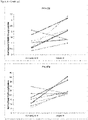
- the model was restricted to the inclusion of 3 of the 12 genes (Model 1).
- EFNB2, GSTA4 and PGR were retained and gave a model with a P-value of 0.0015, a PPV of 68%, a NPV of 79% an accuracy of 73% and an AUC of 0.82.
- a P-value of 0.0015 When trying to improve this model by also allowing patient and cycle characteristics (from Table 3), no improvement on the previous model was found (Model 1 bis).
- more than 3 genes were allowed into the model if improving the P-value (Model 2). Five of the 12 genes were retained in this model (i.e.
- cryostored CC samples related to cryopreserved embryos which had led to a clinical pregnancy after transfer in a subsequent transfer cycle were analyzed.
- This material was used to analyze the genes present in the above obtained multiparametric models (Table 6) and/or the 5 genes with the smallest P-value of addition (Table 4) i.e. CAMK1D, EFNB2, GPX3, GSR, GSAT4, GSTA3 and PGR.
- CAMK1D CAMK1D
- EFNB2 GPX3, GSR, GSAT4, GSTA3 and PGR.
- Table 7 Comparison of gene expression levels of fresh cycles not resulting in pregnancy to frozen transfer cycles resulting in pregnancy ( Intra -patient analysis).
- the 3 most predictive genes were GSTA4, PGR and EFNB2 and resulted in a similar accuracy as the previous model with EFNB2, CAMK1D and STC1 (73% versus 72%).
- Using three genes none of the patient or cycle characteristics could improve the model, suggesting that the expression of the three genes was minimally influenced by patient and cycle factors.
- the live birth model could be improved by including two more genes (GPX3 and GSTA3), which increased the accuracy up to 81% and resulted in an AUC of 0.93.
- This 5 gene model was improved with three patient and cycle characteristics (age, relative E2 and days of stimulation) and resulted in an optimized model with a PPV, NPV and accuracy of 93%, but similar AUC as the model only containing genes.
- pregnancy prediction based on gene expression should be possible with a limited set of genes to reduce analysis time and cost.
- the 2 most recurrent genes are GSTA4 and EFNB2 (see Table 5). These 2 genes are present in all 3 pregnancy models and have respectively in 3 out of 3 and in 2 out of 3 predictive models a type-III P-value ⁇ 0.01.
- the models also showed that the gene expression values are always more important than the patient and cycle characteristics, as the type-III P-values in the models are only significant for the genes and not for the patient and cycle characteristics. Furthermore, the AUC results are comparable between the model containing only genes and the model combining genes and patient and cycle characteristics.
- CAMK1D also strongly correlated with a steroid related gene i.e. PGR next to EFNB2 and GSTA4 (all P ⁇ 0.0001 with Pearson correlation analysis) still leaving the possibilities open for more than one pathway to which CAMK1D could be associated with.
- PGR steroid related gene
- ROS reactive oxygen species
- micro array analysis 1 and 2 were used for data-mining, (for finding genes or exons of genes that are predictive for oocyte competence) and array experiment 3 was used for filtering the recurrent predictive genes (finding genes or exons that are more general applicable as they are predictive in different fertility centers and after slightly different treatments).
- the most novel analysis is based on micro array experiment 2 which comprises an intra patient and intra-cycle (oocytes from the same stimulation cohort) analysis for live birth.
- amplification was performed by starting from 100ng total-RNA and a 1 round T7 based amplification (Message Amp II-Biotin Enhanced, Ambion). Good Quality aRNA (size distribution, 3'/5'ratio & %present calls) was verified and Affymetrix Human Genome U133 Plus 2.0 Arrays were performed at the Salk Institute (USA, San Diego). Further analysis of the gene readouts was performed using Genesifter (Perkin Elmer). For Array experiment 2 and 3 RNA was extracted, measured, and evaluated as mentioned above but amplification was performed using the Nugen V2 kit. Arrays were, both for array experiment 2 and 3, the Affymetrix Human Gene ST arrays. Data were mainly analyzed in Microsoft Excel.
- Intra-patient comparison comprises that both the positive sample (CC of an oocyte resulting in a pregnancy in a single embryo transfer) and the negative sample (CC of an oocyte resulting in no pregnancy after single embryo transfer) originate from the same patient and the same stimulation and pick up cycle.
- Array experiment 1 performed in 2006, comprised an inter patient comparison for live birth and an intra-patient comparison for embryo development.
- Array analysis 2 was performed in January 2012 and consisted of an intra-patient analysis for live birth and delayed development for 5 patients and 15 arrays and resulted in several lists with differentially regulated genes. Patients were in this case pretreated with GnRH Antagonist and recombinant FSH. Array experiment 3 was only used as a confirmation dataset in micro array analysis 4 and is further described below. Table 14: Micro Array experiments by FOBI Analysis Time Setting Patient crosstreatment Parameters Outcome 1 2006 intra - and inter-patient comp. GnRH Agonist + HP-hMG no development > ⁇ Live birth > ⁇ No Pregn. Q1 and Q3 2 Jan. 2012 intra -patient comparison GnRH Antagonist + rFSH deleayed development > ⁇ Live birth > ⁇ No Pregn.
- Array analysis 4 Reanalysis of the micro array data of array experiment 2+3 on an exon level
- RT-QPCR quantitative PCR after reversed transcription
- splice variants could be coding for tissue specific (e.g. cumulus cell specific) or secreted or more active forms of the protein.
- tissue specific e.g. cumulus cell specific
- Alternative splice variants can also have a regulatory function and actually down-regulate the amount or the effect of the protein. Therefore combining the information of different splice variants for a specific gene holds more information but requires also more time and resources to investigate and is not commonly done.
- CC originated from oocytes that resulted in 3 outcome categories: 1. an embryo with a high morphological score (Embryo quality 1) that was transferred and resulted in a pregnancy (P-CC) 2. an embryo with a high morphological score (Embryo quality 1) that was transferred but did not result in a pregnancy (NP-CC) and 3. an embryo that developed too slow to be considered for transfer (NT-CC).
- the patients considered for this study were stimulated with GnRH antagonist and rFSH and were not pregnant from the first single embryo transfer and came back for the transfer of a single frozen embryo which did result in a pregnancy.
- the CC samples from category 1, 2 or 3 originated from 1 pick up cycle and this thus allowed an intra patient analysis.
- Genes were considered potentially interesting if they were: a) differentially expressed between P-CC and both NP-CC and NT-CC, b) in the same direction different (e.g. down in the 2 negative control samples), c) at least one of the difference was >1.5 with a q ⁇ 0.1 (False discovery rate) and d) more than one exon of that gene was retained.
- the "11 genes list” consists of the most predictive exon level genes combinations and are therefore the core of the current application.
- the original "45 gene list” contains other genes that are related to them and could eventually replace one or more of the 11 genes if needed. An example of this is given below and discussed in more detail in Table 11.
- CC of morphologic identical embryos resulting in a pregnant and not is considered more important than the distinction between the CC of oocytes leading to a pregnancy or to an embryo delayed during in vitro development (later also referred to as "pregnant versus delayed embryo development") as the latter information is already available to labs performing extended embryo culture.
- This first intra-patient analysis thus confirms an upregulation of a known oocyte quality marker gene, CAMK1D, in the CC of oocytes resulting in a pregnancy compared to the CC of oocytes that will result in good morphology embryos that do not lead to a pregnancy or to delayed embryos of the same patient.
- CAMK1D oocyte quality marker gene
- CAMK1D exon 9 and EFNB2 both chosen as positive control genes are in the current dataset confirmed both in the intra- and inter-patient analysis.
- HSPH1 exon 2 and NCOA7 are retained in the final model for antagonist/rFSH stimulated patients.
- SASH1 is a powerful predictive gene for antagonist/HP-hMG stimulated patients.
- HSPH1exon 6 and EFNB2 are not retained in the final model does not imply that these markers are not related to oocyte competence; as they are correlated with other markers (HSPH1 exon 6 is correlated with HSPH1 exon 2, and EFNB2 is correlated with CAMK1D expression) and hence are rejected from the regression analysis.
- a case-control assessor blinded prospective study was performed by FOBI using 2 of the oocyte competence predictive models described higher ( Figure 6 and 7 ).
- the study comprised 3 groups of patients: the experimental group with morphological embryo grading (as described in Wathlet et al 2012) and CC gene analysis and transfer of a day 3 embryo and the 2 control groups with morphological embryo grading alone and transfer of a day 3 or a day 5 embryo.
- the 2 controls are included as pregnancy rates differ between the two transfer regimes. Day 5 transfer slightly increases clinical pregnancy rates in the first cycle but cumulative pregnancy rates are higher in in day 3 transfer cycles (Glujovsky et al. Cochrane review 2012).
- the aim was to identify within the pool of oocytes obtained from each patient the oocyte with the highest competence by comparing the expression of specific genes in the CC that surrounded each oocyte.
- the embryo originating from this oocyte was transferred back to the patient.
- the CC gene expression analysis combined with morpghological grading is expected to increase the chance on pregnancy for these patients compared to matched patients without the CC evaluation (i.e. choice of embryo to transfer only decided on routine morphological embryo grading).
- This study comprised 17 patients.
- the patients included are patients from the ART clinic scheduled for fertility treatment with intra-cytoplasmatic sperm injection (ICSI) and single embryo transfer after 3 days of embryo culture (generally an 8 cell stage embryo).
- ICSI intra-cytoplasmatic sperm injection
- Single embryo transfer after 3 days of embryo culture generally an 8 cell stage embryo.
- Patient pretreatments allowed are GnRH antagonist plus rFSH or HP-hMG and the gene expression models described in figure 5 and 6 are used for these patients respectively.
- this step could comprise oocyte freezing for later use, eventually in specific media supplemented with nutrients to compensate for the deficiency that was observed in the CC gene expression result.
- ** the CC score could be used independently or combined with morphologic or other oocyte, sperm or embryo evaluation methods
- the expression of specific genes was quantified in the CC of each oocyte.
- the absolute expression of each gene was normalized to the expression of 2 endogenous genes in the same CC sample to compensate for cell number differences and technical variance between the samples.
- the normalized expression values were than used in the mathematical formula described in figure 6 and 7 and result in a score for each oocyte. The higher this score, the higher the chance that this oocyte would result in a live birth.
- the models used in this proof of principle experiment contained 2 and 3 genes. From earlier micro array experiments (with more genes and more patients analyzed) we know that adding more genes (up to at least 5 or 6) will further improve the predictive power of the models. This work is currently ongoing with more genes from the list in the current patent.
- the 45 oocyte quality marker gene list originates from 2 array experiments using UZBrussel patients and was cross validated using data from a 3th independent array experiment containing more than 100 patients from 3 different European fertility centers. This approach should provide solid marker genes and gene combinations/models to predict treatment outcome.
Landscapes
- Life Sciences & Earth Sciences (AREA)
- Chemical & Material Sciences (AREA)
- Health & Medical Sciences (AREA)
- Engineering & Computer Science (AREA)
- Organic Chemistry (AREA)
- Zoology (AREA)
- Wood Science & Technology (AREA)
- Proteomics, Peptides & Aminoacids (AREA)
- Genetics & Genomics (AREA)
- Biotechnology (AREA)
- Bioinformatics & Cheminformatics (AREA)
- Analytical Chemistry (AREA)
- Immunology (AREA)
- Biomedical Technology (AREA)
- Microbiology (AREA)
- General Health & Medical Sciences (AREA)
- Cell Biology (AREA)
- Biochemistry (AREA)
- General Engineering & Computer Science (AREA)
- Physics & Mathematics (AREA)
- Molecular Biology (AREA)
- Biophysics (AREA)
- Developmental Biology & Embryology (AREA)
- Measuring Or Testing Involving Enzymes Or Micro-Organisms (AREA)
- Micro-Organisms Or Cultivation Processes Thereof (AREA)
- Investigating Or Analysing Biological Materials (AREA)
- Saccharide Compounds (AREA)
- Heterocyclic Carbon Compounds Containing A Hetero Ring Having Oxygen Or Sulfur (AREA)
Claims (9)
- In-vitro-Verfahren zum Nachweis eines Biomarker-Gens oder seiner Spleißvariante zur Bewertung der Fähigkeit einer unbefruchteten Säugetier-Oozyte, zur Geburt, Einnistung, Bildung einer Präimplantations-Blastozyste oder eines Embryos und/oder zur Befruchtung zu führen; wobei das Verfahren folgende Schritte umfasst:- Durchführen einer Analyse der Genexpression auf Exonebene in einer Probe, die mindestens eine aus der unbefruchteten Oozyte isolierte Granulosa- oder Kumuluszelle umfasst;- Durchführen eines intrapatientenbasierten Vergleichs der Analyse der Genexpression auf Exonebene; und- Verwendung von Lebendgeburt und Embryoentwicklung als Selektionsendpunkt, indem nur p-Werte < 0,05 in einem gepaarten t-Test als für ein Biomarkergen signifikant betrachtet werden, um geeignet zu sein, die Fähigkeit einer unbefruchteten Säugetier-Oozyte zu bewerten, zur Geburt, Einnistung und Bildung einer Präimplantations-Blastozyste oder eines Embryos und/oder zur Befruchtung zu führen.
- In-vitro-Verfahren nach Patentanspruch 1, wobei die Analyse der Genexpression auf Exonebene an einem Experiment durchgeführt wird, das die Gene von Tabelle 13 umfasst.
- In-vitro-Verfahren zur Bewertung der Fähigkeit einer unbefruchteten Säugetier-Oozyte, zur Geburt, Einnistung, Bildung einer Präimplantations-Blastozyste oder eines Embryos und/oder zur Befruchtung zu führen; wobei das Verfahren die folgenden Schritte umfasst:- Bestimmen des Niveaus einer Biomarker-Genexpression durch die Durchführung einer Analyse der Genexpression auf Exonebene, durchgeführt an der Spleißvariante eines Gens, ausgewählt aus CAMK1D, PTGS2, EFNB2, VCAN, STC1, STC2, PGR und GPX3; oder das Niveau der Biomarker-Genexpression einer Kombination ausgewählt aus der Gruppe umfassend SASH1, MROH9, NCOA7, DNAH3, HSPH1 Exon 2, HSPH1 Exon 6, GALNTL6, SPTBN5, CAMK1D Exon 1, CAMK1D Exon 9 und EFNB2 in einer Probe, die mindestens eine aus der unbefruchteten Oozyte isolierte Granulosa- oder Kumuluszelle umfasst; und- Bewertung der Fähigkeit der unbefruchteten Oozyte, zur Geburt, Einnistung, Bildung einer Präimplantations-Blastozyste oder eines Embryos und/oder zur Befruchtung zu führen; basierend auf dem (den) Expressionsniveau(s).
- In-vitro-Verfahren nach Patentanspruch 3, wobei die Kombination von Biomarkern ausgewählt wird aus der Liste umfassend:• EFNB2 und NCOA7;• CAMK1D Exon 9 und HSPH1 Exon2 und NCOA7• CAMK1D Exon 9 und HSPH1 Exon6 und NCOA7• CAMK1D Exon 1 und SASH1; und• EFBN2 und SASH1.
- In-vitro-Verfahren zur Bewertung der Fähigkeit einer unbefruchteten Säugetier-Oozyte, zur Geburt, Einnistung, Bildung einer Präimplantations-Blastozyste oder eines Embryos und/oder zur Befruchtung zu führen; wobei das Verfahren die folgenden Schritte umfasst:- Bestimmen der Genexpression auf Exonebene von entweder CAMK1D Exon 1, CAMK1D Exon 9 oder EFNB2 in Kombination mit einem oder mehreren Genen, ausgewählt aus den in Tabelle 13 aufgeführten Genen; insbesondere mit einem oder mehreren zusätzlichen Genen ausgewählt aus den in Tabelle 8 aufgeführten Genen; insbesondere mit einem oder mehreren zusätzlichen Genen ausgewählt aus der Gruppe bestehend aus HSPH1 Exon 2, HSPH1 Exon 6, NCOA7 und SASH1 in einer Probe umfassend mindestens eine aus der unbefruchteten Oozyte isolierte Granulosa- oder Kumuluszelle; und- Bewertung der Fähigkeit der unbefruchteten Oozyte, zur Geburt, Einnistung, Bildung einer Präimplantations-Blastozyste oder eines Embryos und/oder zur Befruchtung zu führen; basierend auf den Expressionsniveaus.
- In-vitro-Verfahren nach einem der Patentansprüche 1 bis 5, ferner umfassend den Schritt des Normalisierens der Genexpression auf Exonebene der Gene.
- In-vitro-Verfahren nach einem der Patentansprüche 1 bis 5, wobei das Bestimmen des Niveaus der Markergenexpression eines Biomarkergens oder mehrerer Biomarkergene das Messen der Polynucleotidniveaus der Gene mittels biologischer Assays unter Verwendung von Primern und/oder Sonden umfasst, die spezifisch hybridisieren können mit den Polynucleotiden oder mit einer oder mehreren Region(en) innerhalb der Polynucleotide.
- In-vitro-Verfahren nach einem der Patentansprüche 1 bis 5, wobei das Bestimmen des Niveaus der Markergenexpression eines Gens oder mehrerer Gene das Messen der Proteinniveaus verwandter Genprodukte mittels biologischer Assays unter Verwendung von Bindern, Antikörpern oder Fragmenten davon für diese Proteine, ihre Vorformen oder ihre Metabolisierungsprodukte umfasst.
- In-vitro-Verfahren nach einem der Patentansprüche 1 bis 5, wobei die unbefruchtete Säugetier-Oozyte eine unbefruchtete menschliche Oozyte ist.
Applications Claiming Priority (2)
| Application Number | Priority Date | Filing Date | Title |
|---|---|---|---|
| EP13155633 | 2013-02-18 | ||
| PCT/EP2014/053164 WO2014125129A1 (en) | 2013-02-18 | 2014-02-18 | Marker genes for oocyte competence |
Publications (2)
| Publication Number | Publication Date |
|---|---|
| EP2956552A1 EP2956552A1 (de) | 2015-12-23 |
| EP2956552B1 true EP2956552B1 (de) | 2022-03-30 |
Family
ID=47715921
Family Applications (1)
| Application Number | Title | Priority Date | Filing Date |
|---|---|---|---|
| EP14707352.2A Active EP2956552B1 (de) | 2013-02-18 | 2014-02-18 | Markergene der eizellenkompetenz |
Country Status (11)
| Country | Link |
|---|---|
| US (1) | US10053733B2 (de) |
| EP (1) | EP2956552B1 (de) |
| JP (2) | JP2016509831A (de) |
| CN (2) | CN121249872A (de) |
| CA (1) | CA2901144A1 (de) |
| DK (1) | DK2956552T3 (de) |
| ES (1) | ES2920753T3 (de) |
| HK (1) | HK1218660A1 (de) |
| HU (1) | HUE059437T2 (de) |
| PL (1) | PL2956552T3 (de) |
| WO (1) | WO2014125129A1 (de) |
Families Citing this family (8)
| Publication number | Priority date | Publication date | Assignee | Title |
|---|---|---|---|---|
| GB201519944D0 (en) * | 2015-11-12 | 2015-12-30 | Univ Warwick | Preimplantation Screening |
| CN105861709B (zh) * | 2016-05-19 | 2019-10-18 | 山东大学齐鲁医院 | 利用颗粒细胞grim-19相对表达量来评价卵母细胞发育潜能的方法 |
| US11988674B2 (en) | 2018-08-07 | 2024-05-21 | University Of South Carolina | Methods for measuring gene expression levels to identify viable oocytes |
| CN110819719B (zh) * | 2018-08-09 | 2023-03-03 | 山东大学 | 母本-合子转化中imp2途径相关基因和蛋白及其应用 |
| CN109929918B (zh) * | 2019-03-26 | 2020-10-30 | 浙江大学 | 一种基于颗粒细胞基因表达情况评估卵及胚胎质量的方法 |
| CN113046323A (zh) * | 2021-04-02 | 2021-06-29 | 四川农业大学 | 一种基于miR-532-5p及其靶基因调控卵巢颗粒细胞的方法 |
| GB2607578A (en) * | 2021-06-01 | 2022-12-14 | Ntostis Panagiotis | Biomarker |
| CN118593710B (zh) * | 2024-05-28 | 2025-11-25 | 山东大学 | Ncoa7环状rna纳米脂质体在制备治疗卵巢早衰或卵巢储备功能减退相关药物中的应用 |
Family Cites Families (4)
| Publication number | Priority date | Publication date | Assignee | Title |
|---|---|---|---|---|
| US20060141499A1 (en) * | 2004-11-17 | 2006-06-29 | Geoffrey Sher | Methods of determining human egg competency |
| WO2011000805A1 (en) * | 2009-06-29 | 2011-01-06 | INSERM (Institut National de la Santé et de la Recherche Médicale) | Biomarkers of oocyte competency and method of use |
| US20120283125A1 (en) * | 2009-11-12 | 2012-11-08 | UNIVERSITé LAVAL | Ovarian Markers of Oocyte Competency and Uses Thereof |
| WO2012109326A2 (en) | 2011-02-09 | 2012-08-16 | Temple University-Of The Commonwealth System Of Higher Education | Determination of oocyte quality |
-
2014
- 2014-02-18 CN CN202511206117.XA patent/CN121249872A/zh active Pending
- 2014-02-18 ES ES14707352T patent/ES2920753T3/es active Active
- 2014-02-18 PL PL14707352.2T patent/PL2956552T3/pl unknown
- 2014-02-18 CA CA2901144A patent/CA2901144A1/en not_active Abandoned
- 2014-02-18 HK HK16106661.7A patent/HK1218660A1/zh unknown
- 2014-02-18 HU HUE14707352A patent/HUE059437T2/hu unknown
- 2014-02-18 US US14/768,578 patent/US10053733B2/en active Active
- 2014-02-18 JP JP2015557467A patent/JP2016509831A/ja active Pending
- 2014-02-18 EP EP14707352.2A patent/EP2956552B1/de active Active
- 2014-02-18 CN CN201480017795.5A patent/CN105164275A/zh active Pending
- 2014-02-18 WO PCT/EP2014/053164 patent/WO2014125129A1/en not_active Ceased
- 2014-02-18 DK DK14707352.2T patent/DK2956552T3/da active
-
2019
- 2019-02-04 JP JP2019017867A patent/JP7012676B2/ja active Active
Also Published As
| Publication number | Publication date |
|---|---|
| DK2956552T3 (da) | 2022-07-11 |
| HK1218660A1 (zh) | 2017-03-03 |
| HUE059437T2 (hu) | 2022-11-28 |
| EP2956552A1 (de) | 2015-12-23 |
| JP2019088309A (ja) | 2019-06-13 |
| CN105164275A (zh) | 2015-12-16 |
| WO2014125129A1 (en) | 2014-08-21 |
| JP2016509831A (ja) | 2016-04-04 |
| US10053733B2 (en) | 2018-08-21 |
| CN121249872A (zh) | 2026-01-02 |
| US20160017422A1 (en) | 2016-01-21 |
| JP7012676B2 (ja) | 2022-02-14 |
| CA2901144A1 (en) | 2014-08-21 |
| ES2920753T3 (es) | 2022-08-09 |
| PL2956552T3 (pl) | 2022-07-25 |
Similar Documents
| Publication | Publication Date | Title |
|---|---|---|
| EP2956552B1 (de) | Markergene der eizellenkompetenz | |
| Evian Annual Reproduction (EVAR) Workshop Group 2010 Fauser BCJM Diedrich K. Bouchard P. Domínguez F. Matzuk M. Franks S. Hamamah S. Simón C. Devroey P. Ezcurra D. Howles CM | Contemporary genetic technologies and female reproduction | |
| EP2419526B1 (de) | Verfahren zur auswahl funktionsfähiger oozyten und funktionsfähiger embryos mit hohem potential für eine schwangerschaft | |
| US20130109583A1 (en) | Methods and devices for assessing risk to a putative offspring of developing a condition | |
| EP2480883B1 (de) | Verfahren zur überprüfung von unfruchtbarkeit und/oder eierqualität | |
| JP2010503385A (ja) | 哺乳類卵母細胞発達適格性の顆粒膜マーカーおよびその使用 | |
| Lee et al. | Differentially expressed genes implicated in unexplained recurrent spontaneous abortion | |
| US20140171337A1 (en) | Methods and devices for assessing risk of female infertility | |
| US9090938B2 (en) | Methods for selecting competent oocytes and competent embryos with high potential for pregnancy outcome | |
| Kim et al. | Microarray analysis of gene expression in the uterine endometrium during the implantation period in pigs | |
| US20130053261A1 (en) | Genes differentially expressed by cumulus cells and assays using same to identify pregnancy competent oocytes | |
| CA2842839A1 (en) | Ovarian markers of follicular maturity and uses thereof | |
| Lervik et al. | Gene expression during testis development in Duroc boars | |
| US11566289B2 (en) | Marker genes for oocyte competence | |
| WO2007097741A1 (en) | Method of diagnosing intrauterine growth restriction | |
| KR102885777B1 (ko) | 고령 여성의 난임 진단을 위한 바이오마커 및 이의 용도 | |
| EP4650363A1 (de) | Rettung der chromosomen kohäsion in gealterten säugetier oozyten und eiern | |
| Jakhesara et al. | Database on spermatozoa transcriptogram of catagorised Frieswal crossbred (Holstein 2 Friesian X Sahiwal) bulls | |
| Rodriguez et al. | Identification of candidate mRNAs associated with gonadotropin-induced maturation of murine cumulus oocyte complexes using serial analysis of gene expression | |
| Ranjan | Characterization of Transition Protein Genes in Indian Cattle Breeds | |
| Kamalludin | Characterization of a Major Gene for Bovine Ovulation Rate | |
| Class et al. | Patent application title: METHODS AND DEVICES FOR ASSESSING RISK OF FEMALE INFERTILITY Inventors: Piraye Yurttas Beim (New York, NY, US) Piraye Yurttas Beim (New York, NY, US) Assignees: Celmatix, Inc. | |
| CN110819719A (zh) | 母本-合子转化中imp2途径相关基因和蛋白及其应用 |
Legal Events
| Date | Code | Title | Description |
|---|---|---|---|
| PUAI | Public reference made under article 153(3) epc to a published international application that has entered the european phase |
Free format text: ORIGINAL CODE: 0009012 |
|
| 17P | Request for examination filed |
Effective date: 20150902 |
|
| AK | Designated contracting states |
Kind code of ref document: A1 Designated state(s): AL AT BE BG CH CY CZ DE DK EE ES FI FR GB GR HR HU IE IS IT LI LT LU LV MC MK MT NL NO PL PT RO RS SE SI SK SM TR |
|
| AX | Request for extension of the european patent |
Extension state: BA ME |
|
| DAX | Request for extension of the european patent (deleted) | ||
| STAA | Information on the status of an ep patent application or granted ep patent |
Free format text: STATUS: EXAMINATION IS IN PROGRESS |
|
| 17Q | First examination report despatched |
Effective date: 20161110 |
|
| REG | Reference to a national code |
Ref country code: DE Ref legal event code: R079 Ref document number: 602014082995 Country of ref document: DE Free format text: PREVIOUS MAIN CLASS: C12Q0001680000 Ipc: C12Q0001688100 |
|
| RIC1 | Information provided on ipc code assigned before grant |
Ipc: C12Q 1/6881 20180101AFI20210906BHEP |
|
| GRAP | Despatch of communication of intention to grant a patent |
Free format text: ORIGINAL CODE: EPIDOSNIGR1 |
|
| STAA | Information on the status of an ep patent application or granted ep patent |
Free format text: STATUS: GRANT OF PATENT IS INTENDED |
|
| INTG | Intention to grant announced |
Effective date: 20211021 |
|
| GRAS | Grant fee paid |
Free format text: ORIGINAL CODE: EPIDOSNIGR3 |
|
| GRAA | (expected) grant |
Free format text: ORIGINAL CODE: 0009210 |
|
| STAA | Information on the status of an ep patent application or granted ep patent |
Free format text: STATUS: THE PATENT HAS BEEN GRANTED |
|
| AK | Designated contracting states |
Kind code of ref document: B1 Designated state(s): AL AT BE BG CH CY CZ DE DK EE ES FI FR GB GR HR HU IE IS IT LI LT LU LV MC MK MT NL NO PL PT RO RS SE SI SK SM TR |
|
| REG | Reference to a national code |
Ref country code: GB Ref legal event code: FG4D |
|
| REG | Reference to a national code |
Ref country code: CH Ref legal event code: EP |
|
| REG | Reference to a national code |
Ref country code: AT Ref legal event code: REF Ref document number: 1479235 Country of ref document: AT Kind code of ref document: T Effective date: 20220415 |
|
| REG | Reference to a national code |
Ref country code: DE Ref legal event code: R096 Ref document number: 602014082995 Country of ref document: DE |
|
| REG | Reference to a national code |
Ref country code: IE Ref legal event code: FG4D |
|
| REG | Reference to a national code |
Ref country code: FI Ref legal event code: FGE |
|
| REG | Reference to a national code |
Ref country code: DK Ref legal event code: T3 Effective date: 20220704 Ref country code: LT Ref legal event code: MG9D |
|
| REG | Reference to a national code |
Ref country code: NL Ref legal event code: FP |
|
| REG | Reference to a national code |
Ref country code: SE Ref legal event code: TRGR |
|
| PG25 | Lapsed in a contracting state [announced via postgrant information from national office to epo] |
Ref country code: RS Free format text: LAPSE BECAUSE OF FAILURE TO SUBMIT A TRANSLATION OF THE DESCRIPTION OR TO PAY THE FEE WITHIN THE PRESCRIBED TIME-LIMIT Effective date: 20220330 Ref country code: LT Free format text: LAPSE BECAUSE OF FAILURE TO SUBMIT A TRANSLATION OF THE DESCRIPTION OR TO PAY THE FEE WITHIN THE PRESCRIBED TIME-LIMIT Effective date: 20220330 Ref country code: HR Free format text: LAPSE BECAUSE OF FAILURE TO SUBMIT A TRANSLATION OF THE DESCRIPTION OR TO PAY THE FEE WITHIN THE PRESCRIBED TIME-LIMIT Effective date: 20220330 Ref country code: BG Free format text: LAPSE BECAUSE OF FAILURE TO SUBMIT A TRANSLATION OF THE DESCRIPTION OR TO PAY THE FEE WITHIN THE PRESCRIBED TIME-LIMIT Effective date: 20220630 |
|
| REG | Reference to a national code |
Ref country code: ES Ref legal event code: FG2A Ref document number: 2920753 Country of ref document: ES Kind code of ref document: T3 Effective date: 20220809 |
|
| REG | Reference to a national code |
Ref country code: GR Ref legal event code: EP Ref document number: 20220401320 Country of ref document: GR Effective date: 20220808 |
|
| REG | Reference to a national code |
Ref country code: NO Ref legal event code: T2 Effective date: 20220330 |
|
| PG25 | Lapsed in a contracting state [announced via postgrant information from national office to epo] |
Ref country code: LV Free format text: LAPSE BECAUSE OF FAILURE TO SUBMIT A TRANSLATION OF THE DESCRIPTION OR TO PAY THE FEE WITHIN THE PRESCRIBED TIME-LIMIT Effective date: 20220330 |
|
| PG25 | Lapsed in a contracting state [announced via postgrant information from national office to epo] |
Ref country code: SM Free format text: LAPSE BECAUSE OF FAILURE TO SUBMIT A TRANSLATION OF THE DESCRIPTION OR TO PAY THE FEE WITHIN THE PRESCRIBED TIME-LIMIT Effective date: 20220330 Ref country code: SK Free format text: LAPSE BECAUSE OF FAILURE TO SUBMIT A TRANSLATION OF THE DESCRIPTION OR TO PAY THE FEE WITHIN THE PRESCRIBED TIME-LIMIT Effective date: 20220330 Ref country code: RO Free format text: LAPSE BECAUSE OF FAILURE TO SUBMIT A TRANSLATION OF THE DESCRIPTION OR TO PAY THE FEE WITHIN THE PRESCRIBED TIME-LIMIT Effective date: 20220330 Ref country code: PT Free format text: LAPSE BECAUSE OF FAILURE TO SUBMIT A TRANSLATION OF THE DESCRIPTION OR TO PAY THE FEE WITHIN THE PRESCRIBED TIME-LIMIT Effective date: 20220801 Ref country code: EE Free format text: LAPSE BECAUSE OF FAILURE TO SUBMIT A TRANSLATION OF THE DESCRIPTION OR TO PAY THE FEE WITHIN THE PRESCRIBED TIME-LIMIT Effective date: 20220330 |
|
| REG | Reference to a national code |
Ref country code: HU Ref legal event code: AG4A Ref document number: E059437 Country of ref document: HU |
|
| PG25 | Lapsed in a contracting state [announced via postgrant information from national office to epo] |
Ref country code: IS Free format text: LAPSE BECAUSE OF FAILURE TO SUBMIT A TRANSLATION OF THE DESCRIPTION OR TO PAY THE FEE WITHIN THE PRESCRIBED TIME-LIMIT Effective date: 20220730 Ref country code: AL Free format text: LAPSE BECAUSE OF FAILURE TO SUBMIT A TRANSLATION OF THE DESCRIPTION OR TO PAY THE FEE WITHIN THE PRESCRIBED TIME-LIMIT Effective date: 20220330 |
|
| REG | Reference to a national code |
Ref country code: DE Ref legal event code: R097 Ref document number: 602014082995 Country of ref document: DE |
|
| PLBE | No opposition filed within time limit |
Free format text: ORIGINAL CODE: 0009261 |
|
| STAA | Information on the status of an ep patent application or granted ep patent |
Free format text: STATUS: NO OPPOSITION FILED WITHIN TIME LIMIT |
|
| 26N | No opposition filed |
Effective date: 20230103 |
|
| REG | Reference to a national code |
Ref country code: AT Ref legal event code: UEP Ref document number: 1479235 Country of ref document: AT Kind code of ref document: T Effective date: 20220330 |
|
| PG25 | Lapsed in a contracting state [announced via postgrant information from national office to epo] |
Ref country code: SI Free format text: LAPSE BECAUSE OF FAILURE TO SUBMIT A TRANSLATION OF THE DESCRIPTION OR TO PAY THE FEE WITHIN THE PRESCRIBED TIME-LIMIT Effective date: 20220330 |
|
| P01 | Opt-out of the competence of the unified patent court (upc) registered |
Effective date: 20230526 |
|
| PG25 | Lapsed in a contracting state [announced via postgrant information from national office to epo] |
Ref country code: MC Free format text: LAPSE BECAUSE OF FAILURE TO SUBMIT A TRANSLATION OF THE DESCRIPTION OR TO PAY THE FEE WITHIN THE PRESCRIBED TIME-LIMIT Effective date: 20220330 |
|
| PG25 | Lapsed in a contracting state [announced via postgrant information from national office to epo] |
Ref country code: LU Free format text: LAPSE BECAUSE OF NON-PAYMENT OF DUE FEES Effective date: 20230218 |
|
| PG25 | Lapsed in a contracting state [announced via postgrant information from national office to epo] |
Ref country code: CY Free format text: LAPSE BECAUSE OF FAILURE TO SUBMIT A TRANSLATION OF THE DESCRIPTION OR TO PAY THE FEE WITHIN THE PRESCRIBED TIME-LIMIT; INVALID AB INITIO Effective date: 20140218 |
|
| REG | Reference to a national code |
Ref country code: CH Ref legal event code: U11 Free format text: ST27 STATUS EVENT CODE: U-0-0-U10-U11 (AS PROVIDED BY THE NATIONAL OFFICE) Effective date: 20260301 |
|
| PGFP | Annual fee paid to national office [announced via postgrant information from national office to epo] |
Ref country code: NL Payment date: 20260226 Year of fee payment: 13 |
|
| PGFP | Annual fee paid to national office [announced via postgrant information from national office to epo] |
Ref country code: HU Payment date: 20260209 Year of fee payment: 13 |
|
| PGFP | Annual fee paid to national office [announced via postgrant information from national office to epo] |
Ref country code: SE Payment date: 20260227 Year of fee payment: 13 |
|
| PGFP | Annual fee paid to national office [announced via postgrant information from national office to epo] |
Ref country code: GB Payment date: 20260227 Year of fee payment: 13 |
|
| PGFP | Annual fee paid to national office [announced via postgrant information from national office to epo] |
Ref country code: ES Payment date: 20260302 Year of fee payment: 13 |
|
| PGFP | Annual fee paid to national office [announced via postgrant information from national office to epo] |
Ref country code: DE Payment date: 20260227 Year of fee payment: 13 Ref country code: NO Payment date: 20260227 Year of fee payment: 13 Ref country code: DK Payment date: 20260225 Year of fee payment: 13 Ref country code: IE Payment date: 20260227 Year of fee payment: 13 |
|
| PGFP | Annual fee paid to national office [announced via postgrant information from national office to epo] |
Ref country code: AT Payment date: 20260227 Year of fee payment: 13 |
|
| PGFP | Annual fee paid to national office [announced via postgrant information from national office to epo] |
Ref country code: BE Payment date: 20260227 Year of fee payment: 13 Ref country code: FI Payment date: 20260225 Year of fee payment: 13 Ref country code: IT Payment date: 20260219 Year of fee payment: 13 |
|
| PGFP | Annual fee paid to national office [announced via postgrant information from national office to epo] |
Ref country code: FR Payment date: 20260225 Year of fee payment: 13 |
|
| PGFP | Annual fee paid to national office [announced via postgrant information from national office to epo] |
Ref country code: TR Payment date: 20260206 Year of fee payment: 13 |
|
| PGFP | Annual fee paid to national office [announced via postgrant information from national office to epo] |
Ref country code: CH Payment date: 20260301 Year of fee payment: 13 Ref country code: CZ Payment date: 20260205 Year of fee payment: 13 |
|
| PGFP | Annual fee paid to national office [announced via postgrant information from national office to epo] |
Ref country code: GR Payment date: 20260226 Year of fee payment: 13 Ref country code: PL Payment date: 20260202 Year of fee payment: 13 |