EP2953682B1 - Biospecific agents for bone - Google Patents
Biospecific agents for bone Download PDFInfo
- Publication number
- EP2953682B1 EP2953682B1 EP14702931.8A EP14702931A EP2953682B1 EP 2953682 B1 EP2953682 B1 EP 2953682B1 EP 14702931 A EP14702931 A EP 14702931A EP 2953682 B1 EP2953682 B1 EP 2953682B1
- Authority
- EP
- European Patent Office
- Prior art keywords
- bone
- peptide
- biospecific agent
- biospecific
- agent according
- Prior art date
- Legal status (The legal status is an assumption and is not a legal conclusion. Google has not performed a legal analysis and makes no representation as to the accuracy of the status listed.)
- Active
Links
Images


Classifications
-
- A—HUMAN NECESSITIES
- A61—MEDICAL OR VETERINARY SCIENCE; HYGIENE
- A61K—PREPARATIONS FOR MEDICAL, DENTAL OR TOILETRY PURPOSES
- A61K49/00—Preparations for testing in vivo
- A61K49/06—Nuclear magnetic resonance [NMR] contrast preparations; Magnetic resonance imaging [MRI] contrast preparations
- A61K49/08—Nuclear magnetic resonance [NMR] contrast preparations; Magnetic resonance imaging [MRI] contrast preparations characterised by the carrier
- A61K49/10—Organic compounds
- A61K49/14—Peptides, e.g. proteins
-
- A—HUMAN NECESSITIES
- A61—MEDICAL OR VETERINARY SCIENCE; HYGIENE
- A61K—PREPARATIONS FOR MEDICAL, DENTAL OR TOILETRY PURPOSES
- A61K49/00—Preparations for testing in vivo
- A61K49/04—X-ray contrast preparations
- A61K49/0409—Physical forms of mixtures of two different X-ray contrast-enhancing agents, containing at least one X-ray contrast-enhancing agent which is not a halogenated organic compound
- A61K49/0414—Particles, beads, capsules or spheres
- A61K49/0423—Nanoparticles, nanobeads, nanospheres, nanocapsules, i.e. having a size or diameter smaller than 1 micrometer
- A61K49/0428—Surface-modified nanoparticles, e.g. immuno-nanoparticles
-
- A—HUMAN NECESSITIES
- A61—MEDICAL OR VETERINARY SCIENCE; HYGIENE
- A61B—DIAGNOSIS; SURGERY; IDENTIFICATION
- A61B6/00—Apparatus or devices for radiation diagnosis; Apparatus or devices for radiation diagnosis combined with radiation therapy equipment
- A61B6/02—Arrangements for diagnosis sequentially in different planes; Stereoscopic radiation diagnosis
- A61B6/03—Computed tomography [CT]
- A61B6/032—Transmission computed tomography [CT]
-
- A—HUMAN NECESSITIES
- A61—MEDICAL OR VETERINARY SCIENCE; HYGIENE
- A61K—PREPARATIONS FOR MEDICAL, DENTAL OR TOILETRY PURPOSES
- A61K38/00—Medicinal preparations containing peptides
- A61K38/16—Peptides having more than 20 amino acids; Gastrins; Somatostatins; Melanotropins; Derivatives thereof
- A61K38/17—Peptides having more than 20 amino acids; Gastrins; Somatostatins; Melanotropins; Derivatives thereof from animals; from humans
- A61K38/177—Receptors; Cell surface antigens; Cell surface determinants
- A61K38/1793—Receptors; Cell surface antigens; Cell surface determinants for cytokines; for lymphokines; for interferons
-
- A—HUMAN NECESSITIES
- A61—MEDICAL OR VETERINARY SCIENCE; HYGIENE
- A61K—PREPARATIONS FOR MEDICAL, DENTAL OR TOILETRY PURPOSES
- A61K47/00—Medicinal preparations characterised by the non-active ingredients used, e.g. carriers or inert additives; Targeting or modifying agents chemically bound to the active ingredient
- A61K47/50—Medicinal preparations characterised by the non-active ingredients used, e.g. carriers or inert additives; Targeting or modifying agents chemically bound to the active ingredient the non-active ingredient being chemically bound to the active ingredient, e.g. polymer-drug conjugates
- A61K47/69—Medicinal preparations characterised by the non-active ingredients used, e.g. carriers or inert additives; Targeting or modifying agents chemically bound to the active ingredient the non-active ingredient being chemically bound to the active ingredient, e.g. polymer-drug conjugates the conjugate being characterised by physical or galenical forms, e.g. emulsion, particle, inclusion complex, stent or kit
- A61K47/6921—Medicinal preparations characterised by the non-active ingredients used, e.g. carriers or inert additives; Targeting or modifying agents chemically bound to the active ingredient the non-active ingredient being chemically bound to the active ingredient, e.g. polymer-drug conjugates the conjugate being characterised by physical or galenical forms, e.g. emulsion, particle, inclusion complex, stent or kit the form being a particulate, a powder, an adsorbate, a bead or a sphere
- A61K47/6927—Medicinal preparations characterised by the non-active ingredients used, e.g. carriers or inert additives; Targeting or modifying agents chemically bound to the active ingredient the non-active ingredient being chemically bound to the active ingredient, e.g. polymer-drug conjugates the conjugate being characterised by physical or galenical forms, e.g. emulsion, particle, inclusion complex, stent or kit the form being a particulate, a powder, an adsorbate, a bead or a sphere the form being a solid microparticle having no hollow or gas-filled cores
- A61K47/6929—Medicinal preparations characterised by the non-active ingredients used, e.g. carriers or inert additives; Targeting or modifying agents chemically bound to the active ingredient the non-active ingredient being chemically bound to the active ingredient, e.g. polymer-drug conjugates the conjugate being characterised by physical or galenical forms, e.g. emulsion, particle, inclusion complex, stent or kit the form being a particulate, a powder, an adsorbate, a bead or a sphere the form being a solid microparticle having no hollow or gas-filled cores the form being a nanoparticle, e.g. an immuno-nanoparticle
-
- A—HUMAN NECESSITIES
- A61—MEDICAL OR VETERINARY SCIENCE; HYGIENE
- A61K—PREPARATIONS FOR MEDICAL, DENTAL OR TOILETRY PURPOSES
- A61K49/00—Preparations for testing in vivo
- A61K49/06—Nuclear magnetic resonance [NMR] contrast preparations; Magnetic resonance imaging [MRI] contrast preparations
- A61K49/18—Nuclear magnetic resonance [NMR] contrast preparations; Magnetic resonance imaging [MRI] contrast preparations characterised by a special physical form, e.g. emulsions, microcapsules, liposomes
- A61K49/1818—Nuclear magnetic resonance [NMR] contrast preparations; Magnetic resonance imaging [MRI] contrast preparations characterised by a special physical form, e.g. emulsions, microcapsules, liposomes particles, e.g. uncoated or non-functionalised microparticles or nanoparticles
- A61K49/1821—Nuclear magnetic resonance [NMR] contrast preparations; Magnetic resonance imaging [MRI] contrast preparations characterised by a special physical form, e.g. emulsions, microcapsules, liposomes particles, e.g. uncoated or non-functionalised microparticles or nanoparticles coated or functionalised microparticles or nanoparticles
- A61K49/1824—Nuclear magnetic resonance [NMR] contrast preparations; Magnetic resonance imaging [MRI] contrast preparations characterised by a special physical form, e.g. emulsions, microcapsules, liposomes particles, e.g. uncoated or non-functionalised microparticles or nanoparticles coated or functionalised microparticles or nanoparticles coated or functionalised nanoparticles
- A61K49/1827—Nuclear magnetic resonance [NMR] contrast preparations; Magnetic resonance imaging [MRI] contrast preparations characterised by a special physical form, e.g. emulsions, microcapsules, liposomes particles, e.g. uncoated or non-functionalised microparticles or nanoparticles coated or functionalised microparticles or nanoparticles coated or functionalised nanoparticles having a (super)(para)magnetic core, being a solid MRI-active material, e.g. magnetite, or composed of a plurality of MRI-active, organic agents, e.g. Gd-chelates, or nuclei, e.g. Eu3+, encapsulated or entrapped in the core of the coated or functionalised nanoparticle
- A61K49/1875—Nuclear magnetic resonance [NMR] contrast preparations; Magnetic resonance imaging [MRI] contrast preparations characterised by a special physical form, e.g. emulsions, microcapsules, liposomes particles, e.g. uncoated or non-functionalised microparticles or nanoparticles coated or functionalised microparticles or nanoparticles coated or functionalised nanoparticles having a (super)(para)magnetic core, being a solid MRI-active material, e.g. magnetite, or composed of a plurality of MRI-active, organic agents, e.g. Gd-chelates, or nuclei, e.g. Eu3+, encapsulated or entrapped in the core of the coated or functionalised nanoparticle coated or functionalised with an antibody
-
- B—PERFORMING OPERATIONS; TRANSPORTING
- B82—NANOTECHNOLOGY
- B82Y—SPECIFIC USES OR APPLICATIONS OF NANOSTRUCTURES; MEASUREMENT OR ANALYSIS OF NANOSTRUCTURES; MANUFACTURE OR TREATMENT OF NANOSTRUCTURES
- B82Y5/00—Nanobiotechnology or nanomedicine, e.g. protein engineering or drug delivery
-
- G—PHYSICS
- G01—MEASURING; TESTING
- G01R—MEASURING ELECTRIC VARIABLES; MEASURING MAGNETIC VARIABLES
- G01R33/00—Arrangements or instruments for measuring magnetic variables
- G01R33/20—Arrangements or instruments for measuring magnetic variables involving magnetic resonance
- G01R33/44—Arrangements or instruments for measuring magnetic variables involving magnetic resonance using nuclear magnetic resonance [NMR]
- G01R33/48—NMR imaging systems
- G01R33/54—Signal processing systems, e.g. using pulse sequences ; Generation or control of pulse sequences; Operator console
- G01R33/56—Image enhancement or correction, e.g. subtraction or averaging techniques, e.g. improvement of signal-to-noise ratio and resolution
- G01R33/5601—Image enhancement or correction, e.g. subtraction or averaging techniques, e.g. improvement of signal-to-noise ratio and resolution involving use of a contrast agent for contrast manipulation, e.g. a paramagnetic, super-paramagnetic, ferromagnetic or hyperpolarised contrast agent
-
- A—HUMAN NECESSITIES
- A61—MEDICAL OR VETERINARY SCIENCE; HYGIENE
- A61K—PREPARATIONS FOR MEDICAL, DENTAL OR TOILETRY PURPOSES
- A61K49/00—Preparations for testing in vivo
- A61K49/06—Nuclear magnetic resonance [NMR] contrast preparations; Magnetic resonance imaging [MRI] contrast preparations
- A61K49/18—Nuclear magnetic resonance [NMR] contrast preparations; Magnetic resonance imaging [MRI] contrast preparations characterised by a special physical form, e.g. emulsions, microcapsules, liposomes
- A61K49/1818—Nuclear magnetic resonance [NMR] contrast preparations; Magnetic resonance imaging [MRI] contrast preparations characterised by a special physical form, e.g. emulsions, microcapsules, liposomes particles, e.g. uncoated or non-functionalised microparticles or nanoparticles
- A61K49/1821—Nuclear magnetic resonance [NMR] contrast preparations; Magnetic resonance imaging [MRI] contrast preparations characterised by a special physical form, e.g. emulsions, microcapsules, liposomes particles, e.g. uncoated or non-functionalised microparticles or nanoparticles coated or functionalised microparticles or nanoparticles
- A61K49/1824—Nuclear magnetic resonance [NMR] contrast preparations; Magnetic resonance imaging [MRI] contrast preparations characterised by a special physical form, e.g. emulsions, microcapsules, liposomes particles, e.g. uncoated or non-functionalised microparticles or nanoparticles coated or functionalised microparticles or nanoparticles coated or functionalised nanoparticles
- A61K49/1827—Nuclear magnetic resonance [NMR] contrast preparations; Magnetic resonance imaging [MRI] contrast preparations characterised by a special physical form, e.g. emulsions, microcapsules, liposomes particles, e.g. uncoated or non-functionalised microparticles or nanoparticles coated or functionalised microparticles or nanoparticles coated or functionalised nanoparticles having a (super)(para)magnetic core, being a solid MRI-active material, e.g. magnetite, or composed of a plurality of MRI-active, organic agents, e.g. Gd-chelates, or nuclei, e.g. Eu3+, encapsulated or entrapped in the core of the coated or functionalised nanoparticle
- A61K49/1866—Nuclear magnetic resonance [NMR] contrast preparations; Magnetic resonance imaging [MRI] contrast preparations characterised by a special physical form, e.g. emulsions, microcapsules, liposomes particles, e.g. uncoated or non-functionalised microparticles or nanoparticles coated or functionalised microparticles or nanoparticles coated or functionalised nanoparticles having a (super)(para)magnetic core, being a solid MRI-active material, e.g. magnetite, or composed of a plurality of MRI-active, organic agents, e.g. Gd-chelates, or nuclei, e.g. Eu3+, encapsulated or entrapped in the core of the coated or functionalised nanoparticle the nanoparticle having a (super)(para)magnetic core coated or functionalised with a peptide, e.g. protein, polyamino acid
-
- A—HUMAN NECESSITIES
- A61—MEDICAL OR VETERINARY SCIENCE; HYGIENE
- A61P—SPECIFIC THERAPEUTIC ACTIVITY OF CHEMICAL COMPOUNDS OR MEDICINAL PREPARATIONS
- A61P19/00—Drugs for skeletal disorders
- A61P19/08—Drugs for skeletal disorders for bone diseases, e.g. rachitism, Paget's disease
Definitions
- the present invention relates to biospecific agents for bone, and in particular to bone biospecific agents, including nanoparticles, sub-micron particles and atomic or molecular elements, which are functionalised with peptides that are specific for bone.
- the invention is especially concerned with the use of these bone biospecific agents in diagnostic imaging techniques, such as Magnetic Resonance Imaging (MRI) and Computed Tomography (CT), and the use of the agents in imaging bone remodelling activities, detecting and treating pathological bone conditions and/or bone repair processes.
- MRI Magnetic Resonance Imaging
- CT Computed Tomography
- the invention extends to the diagnosis and/or treatment of bone disease and bone pathologies using the biospecific agents.
- Magnetic Resonance Imaging (MRI) and Computed Tomography (CT) are the methods of choice in the imaging of tissues.
- MRI Magnetic Resonance Imaging
- CT Computed Tomography
- MRI is based on the ability of large magnetic fields to produce a net magnetic vector temporarily changing the alignment of the protons in the highly hydrated tissues.
- MRI is mainly suited for the imaging of injuries in ligaments, tendons and spinal cord as well as of brain tumours.
- the technique does not allow imaging of the bony tissues as detailed as those that can be obtained by CT.
- QCT Quantitative computed tomography
- DXA Dual-energy E-ray absorptiometry
- Contrast agents are indispensable to the improvement of imaging in both techniques as they enhance the image definition. Contrast agents with no specificity for tissues are currently used in MRI, which are based on either iron oxide nanoparticles or gadolinium. However, although providing good imaging and safety for the patients, these contrast agents are unable to recognise specific tissues and cell types.
- CT contrast agents such as iodine-based compounds have several limitations, including short imaging times due to rapid renal clearance, renal toxicity, and vascular permeation.
- osteoporosis Although the pathogenesis of osteoporosis is unclear, data suggests that it is caused by an imbalance between the bone resorption activities of the osteoclasts and the bone forming activities of the osteoblasts. This imbalanced cellular activity leads to a progressive weakening of the bony tissue leading to the formation of micro-fractures that are at the origin of clinically-significant fractures. Epidemiological studies have demonstrated that these fractures tend to occur in specific anatomical sites, including vertebral bodies, sub-throcanteric femoural bone and the wrists. Although the imbalanced cellular activity plays a critical role in most osteoporotic cases, its relative contribution to bone loss (i.e.
- osteopenia may vary depending on a number of different factors including age, gender, genetic predisposition to osteoporosis, lack of exercise, medication, health and nutrition. Generally, osteoporosis is classed as either primary (i.e. senile) or secondary (i.e. non-age related).
- Type I osteoporosis occurs mainly at the age of 50 to 70 largely due to oestrogen loss at menopause and affects the trabecular bone.
- Type II is directly related to the aging process and usually occurs at the age of 70 and above, affecting both trabecular and cortical bone.
- PBM peak bone mass
- Secondary osteoporosis results from either a complication of underlying medical conditions or lifestyle (e.g. alcohol consumption, drug abuse or poor diet) and can affect people of all ages. Indeed, infants with lower than expected bone density in the early weeks of life have been reported to develop osteoporosis.
- Medical conditions that can cause secondary osteoporosis include hormone imbalances, rheumatoid arthritis, liver failure, kidney failure, impaired gastrointestinal function, multiple sclerosis, scurvy, anorexia nervosa and athlete triad. In some cases, it is not the condition that causes osteoporosis, but the drugs used. Medication such as corticosteroids, some hormones, and lithium to manage medical conditions have been linked with the development of secondary osteoporosis. Generally, osteoporosis is of more clinical significance in women than in men.
- Osteosarcoma is the most common primary sarcoma (incidence: 0.2-3/100 000/year) characterised by osteoblastic differentiation leading to production of poorly organised osteoid or bone growth that affects bone integrity. Although osteosarcomas can also be malignant, its absolute incidence among malignant tumors is low. Within strict histological definition, osteosarcoma lesions are considerably diverse in histological features and grade and its prognosis is dependent not only on these parameters, but also on its anatomic site. Another feature of osteosarcomas is the tendency to produce variable amounts of cartilage matrix and fibrous tissue which in some cases predominates the actual production of bone.
- osteosarcoma usually develop in the metaphysis of a long extremity bone, most commonly around the knee and its presence in the axial skeleton or craniofacial bones is widely observed in adults.
- osteosacorma may develop inside the bones (in the intramedullary or intracortical compartment), on the surfaces of bones, and in extraosseous sites.
- Bone metastases are characterised by osteoblastic, osteolytic or both osteoblastic and osteolytic phenotypes. Different malignancies exhibit osteotropism and higher affinity for bone with carcinomas being the most common metastatic deposits in bone. Common malignancies that end up in bone include breast, prostate and lung cancers. Thus, being widely considered a significant challenge in the field of oncology.
- metastatic cells increase osteoblastic proliferation and activity, including an increase in the expression and release of RANKL through the release of soluble mediators or via cell-to-cell contact. This then activates the differentiation of pre-osteoclast and activity of mature osteoclasts through the RANKL-RANK interaction.
- Bone resorption by osteoclasts releases cytokines and other growth factors such as TGF- ⁇ and insulin like growth factor (IGF) necessary for the tumour cell, thereby enhancing tumour growth and perpetuating the process.
- Increased bone resorption leaves behind osteolytic lesions which are detectable by X-ray, densitometric techniques and MRI.
- Osteoblastic metastases abnormally increased bone formation and are seen as dense areas of bone on X-Rays, and MRI.
- the frequency of bone resorption activation varies more between health and diseased bone than the differences between bone resorption and formation phases.
- the frequency of bone activation is characterised by the amount of the so-called bone multicellular units (BMU) on the surface of bone, which is greater in osteoporotic bone than in normal bone and is associated with increased osteoclast and resorption lacunae in the skeleton.
- BMU bone multicellular units
- Histological stain on bone biopsies in a systemic disease, such as osteoporosis may be used as diagnosis, and the stain would make it possible to microscopically localise solid particulate materials used for diagnosis and treatment (i.e. Prussian blue stain for iron).
- AU2012204100 discloses an agent comprising magnetic nanoparticles with a contrast core, which is visible using MRI.
- US2008/206146 discloses methods for detecting diseased bone tissue in an individual.
- WO02/07792 , WO2008/156637 and US7323542 disclose bone targeting peptides.
- a bone biospecific agent comprising a contrast material core, which is visible using Magnetic Resonance Imaging (MRI) or Computed Tomography (CT), wherein:
- MRI Magnetic Resonance Imaging
- CT Computed Tomography
- a bone biospecific agent comprising a contrast material core, which is visible using Magnetic Resonance Imaging (MRI) or Computed Tomography (CT), the contrast material core being surrounded by a polymeric shell, which is functionalised with a bone-targeting peptide, wherein the peptide, in use, targets the biospecific agent to bone.
- MRI Magnetic Resonance Imaging
- CT Computed Tomography
- the bone biospecific agent of the present invention is based upon the design, development and improvement of a range of different nanoparticles, submicron particles and atomic or molecular elements, which are described in detail below, and their uses in either MRI or CT imaging techniques.
- the bone biospecific agent comprises a core comprising a conventional contrast material, which has been functionalised with a peptide that can specifically recognise a bone cell (e.g. an osteoblast or an osteoclast) or mineralized bone extracellular matrix (e.g. hydroxyapatite).
- a bone cell e.g. an osteoblast or an osteoclast
- mineralized bone extracellular matrix e.g. hydroxyapatite
- the inventors have prepared a series of different bone-specific agents in which a range of different bone-targeting peptides have been used to functionalise a polymeric shell.
- Careful selection of the material used for the contrast material in the core, of the polymer forming the polymeric shell, and also of the bone-specific functionalising peptide enables the bone biospecific agents to be used in either diagnosis and/or therapy of various bone-related conditions. Therefore, the biospecific agent may be used in imaging bone remodelling activities, detecting pathological conditions (e.g. bone resorption or bone tumours) and/or tissue repair processes following fractures or surgical intervention.
- the biospecific agents of the invention have been designed to specifically interact with the elements of diseased bone that are essential for bone remodelling, and can be carefully tailored into injectable materials for less invasive early diagnosis and/or treatment of bone diseases.
- the contrast material forms or constitutes the inner core of the biospecific agents of the invention surrounded by an outer polymeric shell.
- the mean diameter of the contrast material core may be between 5nm and 30nm, or between 10nm and 20nm.
- the contrast material core which is visible using MRI or CT, may comprise a metallic or non-metallic material.
- the contrast material core may comprise a magnetic or non-magnetic material.
- the contrast material may comprise an MRI contrast material.
- the contrast material may comprise a paramagnetic or superparamagnetic material.
- the contrast material core may comprise iron, nickel, cobalt or dysprosium or a compound, such as an oxide or alloy, which contains one or more of these elements.
- the contrast material comprises magnetite (Fe 3 O 4 ).
- the contrast material may comprise both a MRI and a CT contrast material.
- the contrast material core may comprise gadolinium, gold, iodine or boro-sulphate. Each of these materials may be used as either MRI or as CT contrast materials.
- the contrast material comprises gadolinium.
- the polymeric shell of the bone biospecific agent may comprise a polymer, which may comprise a polypeptide, a charged protein, a polysaccharide or a nucleic acid.
- Suitable polymers may comprise any biocompatible natural or synthetic polymer including, but not limited to, chitosan, collagen, gelatine, hyaluronic acid, poly(ethylene glycol) poly(lactic acid), poly(glycolic acid), poly(epsilon-caprolactone) and poly(acrylic acid).
- a preferred polymer for the polymeric shell may comprise chitosan.
- Chitosan is known to be a linear polysaccharide comprising randomly distributed p-(1-4)-linked D-glucosamine (deacetyleated unit) and N-acetyl-D-glucosamine (acetylated unit).
- the polymeric shell is attached to the contrast material core by physical absorption or by covalent bonding, depending on the chemistry of the polymer and of the surface of the contrast material core. It may be desirable to derivatise the polymeric shell in order to enable its efficient functionalisation with the bone-targeting peptide.
- the polymeric shell may be derivatised by reacting the polymer with succinic anhydride. This can be carried out in order to provide a spacer between the polymer and the bone-targeting peptide, which would reduce steric hindrance. It may also improve the solubility of the polymer used (e.g. chitosan) and physiological pH.
- Succinic anhydride is also known as dihydro-2,5-furandione and has the molecular formula C 4 H 4 O 3 .
- Methods by which the polymer, for example, chitosan, may be derivatised by succinic anhydride will be known to the skilled person, and are described in Example 1.
- the amount of bone-targeting peptide that is attached to the polymeric shell depends on the amount of functional groups, type of polymer used and chemistry of attachment.
- the peptides are arranged in a spaced-apart array covering the outer surface of the polymeric shell.
- the polymeric shell may be functionalised with one species (i.e. the same sequence) of bone-targeting peptide, which targets the biospecific agent to bone.
- the shell may be functionalised with two or more species (i.e. having different sequences) of bone-targeting peptide.
- the bone-targeting peptide may target the biospecific agent to a cell present exclusively in bone, for example an osteoblast, osteocyte, osteoclast, bone cell progenitor, osteoclast progenitor or a bone lining cell.
- the bone-targeting peptide targets the biospecific agent to osteoblasts or osteoclasts.
- Peptides with sequences able to mimic GAP-junction communication e.g. connexin 43, cx43
- the bone-targeting peptide can direct the biospecific agent to the bone mineral phase; i.e. hydroxyapatite.
- Biospecific agents comprising hydroxyapatite-targeting peptides are therefore a valuable tool for monitoring the mineralization of a forming bony tissue following a traumatic event or during the progression of diseases, such as osteoporosis.
- the bone-targeting peptide may be associated with the Gap junction intercellular communication (GJIC) and the RANK-RANKL-OPG triad pathways.
- GJIC Gap junction intercellular communication
- RANK-RANKL-OPG triad pathways The fact that the biospecific agent of the invention is specific for RANK-RANKL means that it would identify and target the cell within the specimen.
- the tumour In diseases such as osteosarcoma and bone metastases, the tumour may be localised with X-RAY and MRI, and the biospecific agent may enhance the signal and improve resolution which may allow the visualisation of smaller lesions that may otherwise be missed.
- the bone-targeting peptide may comprise an amino acid sequence that mimics OPG by binding RANK such that RANKL-induced osteoclast differentiation and activity is reduced or prevented.
- the bone targeting peptide may be able to mimic proteins such as Connexin 43 participating in the inhibition of osteoclast-osteoclast and or osteoclast-osteoblast interactions.
- Bone-targeting peptides can also be used that recognise migrating osteoblasts or the mineral phase of bone (i.e. hydroxyapatite).
- the bone-targeting peptide may comprise one of the following amino acid sequences:
- connecting or spacer peptide for connecting the bone-targeting peptide to the polymeric shell, and preferably a derivatised form thereof (for example, with succinic).
- Such connecting peptides exhibit improved solubility in aqueous solution, and therefore facilitate the grafting step of the peptide to the agent, and later improve presentation of the bioligand to the cell.
- a suitable connecting peptide which may be used may comprise the amino acid sequence K-(KK).
- This peptide is designated SEQ ID No.9 or GiPL when referred to herein.
- This peptide is a polar molecule and hence improves solubility and accessibility.
- a connecting peptide may comprise the amino acid sequence K-(KK)-(KKKK). This peptide is designated SEQ ID No.10 or G2PL when referred to herein.
- a connecting peptide may comprise the amino acid sequence K-(KK)-(KKKK)-(KKKKKKKK). This peptide is designated SEQ ID No.11 or G3PL when referred to herein.
- the bone-targeting peptide may comprise any of SEQ ID No.9-11, or a functional variant or fragment thereof.
- any of the peptides designated SEQ ID No.1-8 may be conjugated to any of SEQ ID No.9-11.
- the bone-targeting peptide may comprise the following amino acid sequence, or a functional fragment or variant thereof:
- the bone-targeting peptide may comprise 1,4,7,10-tetraazacyclododecane-1,4,7,10-tetraacetic acid (i.e. DOTA). It comprises gadoteric acid, a macrocycle-structured Gd-based MRI contrast agent, consisting of the organic acid "DOTA" as a chelating agent. Accordingly, in another embodiment, the bone-targeting peptide may comprise the following amino acid sequence, or a functional fragment or variant thereof:
- the DOTA molecule is fairly large, very acidic and reactive, and so this molecule may need the user of a spacer or connecting peptide to avoid compromising the potency of the bone-targeting peptide.
- the bone-targeting peptide may be synthesised by known methods, for example solid phase peptide synthesis (SPPS) using the conventional 9-fluorenylmethyloxy carbonyl (Fmoc) protection/deprotection strategy.
- SPPS solid phase peptide synthesis
- Fmoc 9-fluorenylmethyloxy carbonyl
- the bone-targeting peptide may be cyclised.
- the cyclised form is more chemically stable and others have reported improved activity (See Shin J et al 2008), example by dimethyl sulfoxide (DMSO) oxidation to form cysteine-cysteine disulfide bonds.
- DMSO dimethyl sulfoxide
- the bone-targeting peptide may be attached to the polymeric shell of the bone biospecific agent by covalent bonding.
- the polymeric shell comprises chitosan, which may be derivatised, for example using succinic anhydride.
- the peptides may be covalently attached to the polymeric shell using carbodiimide chemistry in order to create the nanoparticles of the invention.
- the biospecific agent may comprise a bioactive compound, which may be delivered to the bone due to the presence of the bone-targeting peptide.
- the bioactive compound may be selected from a group of molecules consisting of: a dye, electrochemical mediator, protein, peptide, chemical compound (such as a drug), genetic material (such as an oligonucleotide, DNA, RNA), small molecule, antibody, enzyme, and other bioactive molecule.
- the bioactive compound may be conjugated to thebiospecific agent, for example by encapsulation during cross-linking, adsorption, ionotropic interaction or direct covalent attachment of the polymer coating.
- the bone biospecific agent may comprise a nanoparticle.
- the bone biospecific agent may comprise a sub-micron particle.
- the nanoparticle may be substantially spherical in shape.
- the mean diameter of the biospecific agent may be sub-micron, i.e. less than 1000nm.
- the mean diameter of the biospecific agent may be 100-450nm.
- the biospecific agent may be produced by initially carrying out a step of ionotropic cross-linking, followed by dissolving the polymer and cross-linker at predetermined concentrations.
- the inner contrast material core may be added to the mix.
- the polymer may be dissolved in a solution comprising the cross-linker (for example, drop-wise under continuous stirring).
- the mixture may be allowed to react (e.g. for at least 30 minutes).
- the mixture may then be centrifuged and the resultant particles (i.e. the biospecific agent) collected in a suitable solvent (e.g. ethanol or water).
- a suitable solvent e.g. ethanol or water
- a connecting peptide (or spacer) may be added, for example by carrying out a ring opening reaction, for example in the case of succinic anhydride.
- the bone-targeting peptide may be attached, for example using carbodiimide chemistry.
- biospecific agents of the invention can be effectively used in MRI or CT imaging techniques depending on the material of contrast agent that is used.
- the bone biospecific agent according to the first aspect for use in diagnosis.
- the bone biospecific agent may be used as a biosensor in a range of different biological imaging applications.
- the biospecific agent is preferably used in MRI or CT imaging techniques, as a biolabel.
- a bone biospecific agent for use as an MRI biolabel or as a CT biolabel for imaging.
- the bone biospecific agent of the first aspect as an MRI biolabel or as a CT biolabel.
- a biolabel comprising the bone biospecific agent according to the first aspect.
- the biolabel may be used in MRI or CT imaging.
- an MRI or CT imaging method comprising the use of the bone biospecific agent of the first aspect.
- the bone biospecific agent can be used in imaging bone remodelling activities, detecting pathological bone conditions (e.g. bone resorption, bone tumours, osteoporosis etc.) and/or bone tissue repair processes following fractures. Furthermore, in addition to the various imaging techniques that can harness the powerful bone-targeting properties of the bone specific agent, Example 5 also explains how the biospecific agents of the invention can be effectively used to inhibit osteoclastogenesis and osteoclast activity, and therefore prevent bone resorption. The inventors therefore believe that the biospecific agent can be used in a variety of therapeutic applications for treating bone disease.
- pathological bone conditions e.g. bone resorption, bone tumours, osteoporosis etc.
- Example 5 also explains how the biospecific agents of the invention can be effectively used to inhibit osteoclastogenesis and osteoclast activity, and therefore prevent bone resorption. The inventors therefore believe that the biospecific agent can be used in a variety of therapeutic applications for treating bone disease.
- the bone biospecific agent according to the first aspect for use in therapy, and preferably as a medicament.
- the bone biospecific agent of the invention is particularly useful for preventing or treating bone disease.
- the bone biospecific agent according to the first aspect for use in treating, preventing or ameliorating bone disease.
- a method of treating, ameliorating or preventing bone disease comprising administering, to a subject in need of such treatment, a therapeutically effective amount of a bone biospecific agent according to the first aspect.
- bone disease examples include bone resorption, treatment of bone tumour, Paget's disease, rheumatoid arthritis, osteoarthritis, osteoporosis, osteosarcoma, osteopenia and bone metastases, including osteolytic and osteoblastic phenotypes etc.
- a bone biospecific agent of the present invention may be used in a medicament, which may be used in a monotherapy.
- agents according to the invention may be used as an adjunct to, or in combination among them or in combination with, known therapies for treating bone disease.
- a biospecific agent of the present invention may be combined in compositions having a number of different forms depending, in particular, on the manner in which the composition is to be used.
- the composition may be in the form of a powder, powder suspension, tablet, capsule, liquid, gel, hydrogel, aerosol, spray, micellar solution, or any other suitable form that may be administered to a person or animal in need of treatment.
- the vehicle of medicaments according to the invention should be one which is well-tolerated by the subject to whom it is given.
- Medicaments comprising biospecific agents of the present invention may be used in a number of ways. For instance, oral administration may be required, in which case the biospecific agent may be contained within a composition that may, for example, be ingested orally in the form of a tablet, capsule or liquid. Compositions comprising biospecific agents of the present invention may be administered by inhalation (e.g. intranasally).
- a bioactive agent of the present invention may also be incorporated within a slow- or delayed-release device.
- the device may be located at least adjacent the treatment site. Such devices may be particularly advantageous when long-term treatment with a biospecific agent of the present invention is required and which would normally require frequent administration (e.g. at least daily injection).
- a biospecific agent of the present invention and compositions according to the invention may be administered to a subject by injection into the blood stream or directly into a site requiring treatment, i.e. the bone.
- Injections may be intravenous (bolus or infusion) or subcutaneous (bolus or infusion), or intradermal (bolus or infusion) or intraosseus.
- biospecific agent of the present invention that is required is determined by its biological activity and bioavailability, which in turn depends on the mode of administration, the physico-chemical properties of the agent and whether it is being used as a monotherapy or in a combined therapy.
- the frequency of administration will also be influenced by the half-life of the agent within the subject being treated.
- Optimal dosages to be administered may be determined by those skilled in the art, and will vary with the particular agent in use, the strength of the pharmaceutical composition, the mode of administration, and the advancement of the disease being diagnosed or treated. Additional factors depending on the particular subject being treated will result in a need to adjust dosages, including subject age, weight, gender, diet, and time of administration.
- a daily dose of between 0.01 ⁇ g/kg of body weight and 0.5g/kg of body weight of the biospecific agent may be used for treating, ameliorating, or preventing bone disease.
- the agent of the present invention may be administered before, during or after onset of disease.
- Daily doses may be given as a single administration (e.g. a single daily injection).
- the agent may require administration twice or more times during a day.
- the agent may be administered as two (or more depending upon the severity of the bone disease being treated) daily doses of between 25mg and 7000 mg (i.e. assuming a body weight of 70 kg).
- a patient receiving treatment may take a first dose upon waking and then a second dose in the evening (if on a two dose regime) or at 03- or 4-hourly intervals thereafter.
- a slow release device may be used to provide optimal doses of agent to a patient without the need to administer repeated doses.
- Known procedures such as those conventionally employed by the pharmaceutical industry (e.g. in vivo experimentation, clinical trials, etc.), may be used to form specific formulations comprising a biospecific agent and precise therapeutic regimes (such as daily doses of the agent and the frequency of administration).
- a pharmaceutical composition comprising the bone biospecific agent according to the first aspect, and a pharmaceutically acceptable vehicle.
- the invention also provides in a tenth aspect, a process for making the composition according to the ninth aspect, the process comprising contacting a therapeutically effective amount of the bone biospecific agent according to the first aspect, and a pharmaceutically acceptable vehicle.
- compositions and medicaments according to the invention may be used to treat any mammal, for example livestock (e.g. a horse), pets, or may be used in other veterinary applications. Most preferably, however, the subject is a human being.
- a “therapeutically effective amount” of the biospecific agent is any amount which, when administered to a subject, is the amount of medicament or drug that is needed to treat a bone disease, or produce the desired effect.
- the therapeutically effective amount of agent used may be from about 0.01 mg to about 800 mg.
- a "pharmaceutically acceptable vehicle” as referred to herein, is any known compound or combination of known compounds that are known to those skilled in the art to be useful in formulating pharmaceutical compositions.
- the pharmaceutically acceptable vehicle may be a solid, and the composition may be in the form of a powder or tablet.
- a solid pharmaceutically acceptable vehicle may include one or more substances which may also act as flavouring agents, lubricants, solubilisers, suspending agents, dyes, fillers, glidants, compression aids, inert binders, sweeteners, preservatives, dyes, coatings, or tablet-disintegrating agents.
- the vehicle may also be an encapsulating material.
- the vehicle is a finely divided solid that is in admixture with the finely divided active agents according to the invention.
- the active agent e.g.
- the biospecific agent may be mixed with a vehicle having the necessary compression properties in suitable proportions and compacted in the shape and size desired.
- the powders and tablets preferably contain up to 99% of the capsule or cell.
- Suitable solid vehicles include, for example calcium phosphate, magnesium stearate, talc, sugars, lactose, dextrin, starch, gelatine, cellulose, polyvinylpyrrolidine, low melting waxes and ion exchange resins.
- the pharmaceutical vehicle may be a gel and the composition may be in the form of a cream or the like.
- the pharmaceutical vehicle may be a liquid, and the pharmaceutical composition is in the form of a solution.
- Liquid vehicles are used in preparing solutions, suspensions, emulsions, syrups, elixirs and pressurized compositions.
- the agent of the present invention may be dissolved or suspended in a pharmaceutically acceptable liquid vehicle such as water, an organic solvent, a mixture of both or pharmaceutically acceptable oils or fats.
- the liquid vehicle can contain other suitable pharmaceutical additives such as solubilisers, emulsifiers, buffers, preservatives, sweeteners, flavouring agents, suspending agents, thickening agents, colours, viscosity regulators, stabilizers or osmo-regulators.
- liquid vehicles for oral and parenteral administration include water (partially containing additives as above, e.g. cellulose derivatives, preferably sodium carboxymethyl cellulose solution), alcohols (including monohydric alcohols and polyhydric alcohols, e.g. glycols) and their derivatives, and oils (e.g. fractionated coconut oil and arachis oil).
- additives e.g. cellulose derivatives, preferably sodium carboxymethyl cellulose solution
- alcohols including monohydric alcohols and polyhydric alcohols, e.g. glycols
- oils e.g. fractionated coconut oil and arachis oil.
- the vehicle can also be an oily ester such as ethyl oleate and isopropyl myristate.
- Sterile liquid vehicles are useful in sterile liquid form compositions for parenteral administration.
- Liquid pharmaceutical compositions which are sterile solutions or suspensions, can be utilized by, for example, intramuscular, intrathecal, epidural, intraperitoneal, intravenous and particularly subcutaneous injection.
- the agent may be prepared as a sterile solid composition that may be dissolved or suspended at the time of administration using sterile water, saline, or other appropriate sterile injectable medium.
- the biospecific agent and pharmaceutical compositions of the invention may be administered orally in the form of a sterile solution or suspension containing other solutes or suspending agents (for example, enough saline or glucose to make the solution isotonic), bile salts, acacia, gelatine, sorbitan monoleate, polysorbate 80 (oleate esters of sorbitol and its anhydrides copolymerized with ethylene oxide) and the like.
- the biospecific agent according to the invention can also be administered orally either in liquid or solid composition form.
- Compositions suitable for oral administration include solid forms, such as pills, capsules, granules, tablets, and powders, and liquid forms, such as solutions, syrups, elixirs, and suspensions.
- Forms useful for parenteral administration include sterile solutions, emulsions, and suspensions.
- novel bone specific agents 2 e.g. nanoparticles, sub-micron particles, and atomic or molecular elements
- Figure 5E novel bone specific agents 2
- a contrasting agent core 4 e.g., an ion oxide or a gold metallic core
- a polymer 6 for example, chitosan
- peptide(s) 8 which recognise bone cells (such as osteocytes, osteoblasts), or other peptides that are only present in bone, for example hydroxyapatite-specific peptides.
- the nanoparticles 2 etc. can be used in either diagnosis or therapy.
- the particles 2 may be used in imaging bone remodelling activities, detecting pathological conditions and/or tissue repair processes. The following Examples describe the results of their research.
- chitosan a polysaccharide which can be used to coat contrast agents or other active ingredients of a pharmaceutical agent
- succinic anhydride a polysaccharide which can be used to coat contrast agents or other active ingredients of a pharmaceutical agent
- Chitosan succinate conjugates are known in the art as being both a biocompatible and biodegradable drug delivery agent which may be used in tablets.
- CS was derivatised by succinic anhydride (Suc-Chi) using a known ring opening reaction ( Yan et al., 2006, Yakugaku Zasshi, 126, 789-793 ).
- a 1 % (w/v) CS solution (in 1 % v/v acetic acid) was filtered through 0.8 ⁇ m pore membrane (Millipore) and diluted (1:4) with methanol.
- Succinic anhydride ⁇ 99 % GC, Sigma Aldrich
- 5 ml acetone at 4 % (w/v) was added drop wise under magnetic stirring and left overnight under agitation at room temperature. The gel that formed was removed from excess solution, double diluted in methanol and dialyzed against ultrapure water for 3 days. The water was changed twice per day and the obtained precipitate was then collected by centrifugation and lyophilised.
- Suc-Chi beads were produced using an established ionic gelation method (Agnihotri, et al., 2004). Briefly, sodium tripolyphosphate (TPP) solution (1 mg/ml) was added drop wise to a 1 mg/ml Suc-CS solution (as described above) under magnetic stirring at a volume ratio of 1:5 and allowed to react for 45 minutes at room temperature.
- TPP sodium tripolyphosphate
- MRI magnetic resonance imaging
- CT imaging biospecific contrast agents i.e. nanoparticles 2 of the invention
- iron oxide core 4 particles Fe 3 O 4 , 10 nm mean diameter
- gold core 4 particles ⁇ 20 nm mean diameter
- the peptides 8 listed in Table 1 and their corresponding amino acid sequences were synthesised and then used to functionalise the core particles 4.
- GAP27p SRPTEKTIFII Derived from Cx43 GAP27.
- OP3-1 YCLEIEFCY Based on OPG residual 113-122.
- G1PL K-(KK) Nanosized flexible carrier of the G2PL K-(KK)-(KKKK) peptides with improved G3PL K-(KK)-(KKKK)-(KKKKKKKK) solubility in aqueous solution (Lloyd, et al.
- G2PL-OP3-1 (KKKK)-(KK)-K-YCLEIEFCY Pro-drug : novel OP3-1 tethered G2PL DOTA-OP3-4 DOTA-KGG-YCLEIEFCYLIR Novel DOTA tethered OP3-4 peptide for the chelation of MRI visibleGd 3+ DOTA-Gd-OP3-4 DOTA-KGG-YCLEIEFCYLIR Novel MRI detectable OP3-4 derivative with a Gd 3+ chelate DOTA-Gd-FHRRIKA DOTA-Gd-FHRRIKA Novel MRI detectable osteoblast migration derivative with a Gd 3+ chelate
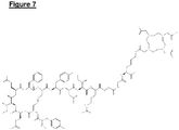
- GiPL, G2PL and G3PL are not linear molecules, but rather hyperbranched (dendritic). See Figure 10 where a G2PL molecule (vertical) is conjugated to OP3-1 (horizontal).
- the peptides 2 were synthesised by solid phase peptide synthesis (SPPS) using the conventional 9-fluorenylmethyloxy carbonyl (Fmoc) protection/deprotection strategy on Tenta Gel S NH 2 resin (0.1 mmol) and dimethylformamide (DMF) as the reaction solvent.
- SPPS solid phase peptide synthesis
- Fmoc 9-fluorenylmethyloxy carbonyl
- DMF dimethylformamide
- An acid-liable Fmoc-Rink-Amide linker (linker) was attached first to the resin for later cleavage of the peptide 8.
- the peptide 8 was then synthesised by adding the first amino acid from the C-terminal followed by sequential coupling/deprotection steps of subsequent amino acids as per the peptide sequence, as set out in Table 1.
- the coupling reactions (30 minutes, ⁇ 2) were carried out using HBTU (O-Benzotriazole-N,N,N',N'-tetramethyl-uronium-hexafluoro-phosphate), for amino group activation, and N,N-Diisopropylethylamine (DIPEA) as a tertiary base.
- HBTU O-Benzotriazole-N,N,N',N'-tetramethyl-uronium-hexafluoro-phosphate
- DIPEA N,N-Diisopropylethylamine
- OP3 is a segment on OPG protein.
- RANKL on the surface of osteoblast (sometimes release in soluble form) interacts with RANK on osteoclasts, thereby initiating a reaction cascade leading to osteoclast differentiation and increased activity.
- OPG released by osteoblasts
- OPG mimetics would act as ligands for the receptors in the bone-associated target cells.
- the peptides to be cyclised were cleaved from resin in a nitrogen atmosphere for 3 hours. After cleavage, the peptides were collected in cold diethyl ether, isolated by centrifugation and dried over a stream of nitrogen.
- the peptides were then dissolved in 60 ml of oxidising buffer (100 mM NaH 2 PO 4 and 2 mM Gdn.HCl, 5 % DMSO, pH 7.0) and shaken for 12 hours. The solution was then acidified with 1M HCO 2 H (250 ⁇ l) and purified by LC-MS. The pure fractions were combined and freeze dried. The degree of cyclisation (formation of disulfide bridges) was assessed by the conventional method for quantitation of free thiol groups using Ellman's reagent. The peptides were ultimately characterised by HPLC and MS.
- oxidising buffer 100 mM NaH 2 PO 4 and 2 mM Gdn.HCl, 5 % DMSO, pH 7.0
- Non-derivatised particles were first dispersed in 2 ml of 2-(N-morpholino) ethanesulfonic acid (MES) buffer (0.1 M MES, 0.3 M NaCl, pH 6.5) to obtain a 1 mg/ml bead concentration.
- MES 2-(N-morpholino) ethanesulfonic acid
- the carboxyl groups within the core 4 particles were then activated by addition of 1-ethyl-3-(3-dimethylaminopropyl) carbodiimide (EDC, 4 mM) and N-hydroxysuccinimide (NHS, 10 mM).
- the activation reaction was allowed to proceed at room temperature for 30 minutes. Excess EDC was deactivated by addition ⁇ -mercaptoethanol (2.8 ⁇ l) and the core 4 particles were washed through a desalting membrane. A1 mg/ml solution of the peptide chosen from Table 1 (e.g. OP3-4 peptide having the sequence, YCEIEFCYLIR) in MES buffer was then added to the solution of core 4 particles at a volume ratio of 1:1. The conjugation reaction was allowed to proceed under magnetic stirring for 3 hours at room temperature. The reaction was then quenched by the addition of hydroxylamine to give a final concentration of 5 to 10 mM.
- Table 1 e.g. OP3-4 peptide having the sequence, YCEIEFCYLIR
- FIG. 5E there is shown a schematic representation of one embodiment of the nanoparticle 2 of the invention.
- the Figure shows the nanoparticle 2 having the inner core 4 (e.g., ion oxide or gold) coated with the polymeric outer shell 6 (e.g. chitosan).
- the shell 6 is functionalised with a coating of peptides 8, which recognise bone cells, or other peptides that are only present in bone, for example hydroxyapatite.
- the resultant nanoparticles 2 were then purified using ultrafiltration spin columns (MWCO 100,000), lyophilised and stored at -20°C.
- Figure 1 shows the scheme of the derivatisation of CS into Suc-Chi and its subsequent functionalisation with the OP3-4 peptide.
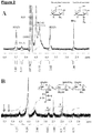
- the 1H NMR spectra of CS and Suc-Chi are shown in Figure 2 . Briefly, the signals are assigned as follows: 2.50-2.70 ppm (H-Ac) was attributed acetyl proteins of GlcNAc monomers; 2.50-2.75 ppm (H2D) was attributed to proton 2 of GlcN monomer; 3.95-4.65 ppm (H2-2) was attributed protons 2 to 6 of both GlcNAc and GlcN monomers; 4.65-4.90 ppm (HOD) corresponds to the solvent (HOD); 5.10-5.30 ppm (Hi-A) corresponds to proton 1 of GlcNAc monomers; 5.30-5.65 ppm (Hi-D) correspond to proton 1 of GlcN monomer.
- the degree of substitution as determined by titration was 25.5 % and 30.6 % by 1 HNMR. Although, less accurate, potentiometric titration analysis allowed for the determination of the molar amounts of the free -NH2 in both CS and Suc-Chi.
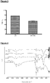
- the degree of derivatisation (DD) values calculated for CS was 79.92 % ( ⁇ 5.85) and for Suc-Chi was 54.4 % ( ⁇ 3.7), as shown in Figure 3 .
- the titrations were repeated 5 times for both polymers.
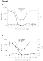
- FTIR results also showed successful derivatisation of chitosan to Suc-Chi and subsequent attachment of OP3-4, as shown in Figure 4 .
- N-H stretch of primary and secondary amines and O-H stretch are comprised in band 1 (3500 -3200 cm -1 ).
- the main contribution to band 1 in the spectra of OP3-4 is from the amines involved in the amide bond.
- Band 2 (1640-1580 cm -1 ) may correspond to the N-H deformation in primary amines, present in the four species; N-H deformation of amides and also carbonyl stretching in secondary amides for the case of OP3-4, Suc-Chi and Suc-Chi-OP3-4.
- Band 3 (1722 cm -1 ), which is present in Suc-Chi and unresolved in Suc-Chi-OP3-4 may be attributed to the carbonyl stretching as result of the linkage of the succinyl group to the polysaccharide through an ester bond, in addition to the linkage via amide bond described earlier (band 3).
- the aromatic structures present in tyrosine and phenylalanine can be confirmed in Suc-Chi-OP3-4 with band 4 (3000 cm -1 ), which originates from C-H stretch in unsaturated species.
- the inventors then set out to determine whether the protein, osteoprogeterin 3 (OP3), which is specifically expressed on bone cells, such as osteoclasts and oseoblasts, can be conjugated to Dotarem (DOTA), which is a chelator that can be used to coat various contrast agents, including gadolinium.
- Dotarem is gadoteric acid, a known macrocycle-structured GD-based MRI contrast agent. It consists of the organic acid DOTA as a chelating agent.
- a novel DOTA-OP3-4 conjugate protein was synthesised by solid phase peptide synthesis using Fmoc chemistry as described in the synthesis of OP3-1 and OP3-4. In this case, however, lysine core amino acid was added first, followed by the coupling of DOTA to the NH2 group that was protected by Mtt. Two glycine amino acids were then coupled to form a spacer followed by the subsequent assembly of OP3-4 peptide. The introduction of the lysine-glycine-glycine spacer between DOTA molecule and OP3-4 sequence was considered important to avoiding potential steric hindrance during the synthesis and any possible effect on the potency of the peptide. A structure of the DOTA-OP3-4 is shown in Figure 7 .
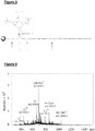
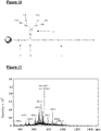
- Figures 9 and 11 show typical mass spectrometry spectra of a linear ( Figure 9 ) and a branched ( Figure 11 ) OP3-4 peptide. These prove the successful synthesis of these peptides that is necessary to the formation of stable binding and functionalisation to the core 4 particle. Purity of above 95% was achieved after purification procedure.
- the inventors next set out to determine whether a gadolinium (Gd)-based contrast agent could be created to form a nanoparticle 2 of the invention by the conjugation of DOTA-coated Gd with the bone-specific protein osteoprogeterin 3 (OP3).
- Gd gadolinium
- OP3 bone-specific protein osteoprogeterin 3
- Novel peptides were used to manufacture biospecific contrast agents (i.e. functionalised nanoparticles 2) for MRI and CT (see Table 1).
- the chelation of the core 4 particle, Gd3+ was achieved by incubating DOTA-OP3-4 with GdCl 3 in a buffer system for 15 hours.
- the DOTA moiety acted as a polydentate ligand and enveloped the metal cations, in this case complexing Gd 3+ , to give an MRI-visible peptide.
- the coordination of the DOTA ligands and metal ion in the complex depends on the conformation of the ligand and geometric tendencies of the metal cation.
- DOTA acts as an octadentate ligand, binding the metal through four amino and four carboxylate groups.
- the DOTA molecule acts as a septadentate since one of the carboxylate groups is used in the covalent with the peptide.
- a carboxylate group from the amino acid linking DOTA and the peptide provides the eighth ligand and restores the octadentate state, forming a highly stable coordination complex(Viola-Villegas, et al., 2009).
- the resultant nanoparticles 2 were obtained through the direct binding of peptides 8 with a linear or branched root to magnetic core 4 particles (e.g. iron oxide) coated with thin films of polymers 6 or ceramics (i.e. MRI contrast agents) or gold core 4 particles (i.e. CT contrast agents).
- magnetic core 4 particles e.g. iron oxide
- ceramics i.e. MRI contrast agents
- gold core 4 particles i.e. CT contrast agents
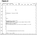
- a representative LC profile of the hydrolysis products on the Gd nanoparticle 2 functionalised with the OP3-4 peptide 8 is given in Figure 12 .
- the glucosamine units per micro gram of material was calculated to be 1.92 nmoles in chitosan-based nanoparticles (CNB), 1.40 nmoles in Gd core 4 particle (on its own) and 0.27 nmoles in Gd nanoparticle 2 functionalised with OP3-4 peptide 8, and Gd nanoparticle-DOTA-Gd-OP3-4.
- the amount of peptide 8 conjugated was calculated to be 4.2 mmoles per gram of nanoparticle 2. Individual amino acids were detected in molar ratios reflective of the amounts in OP3-4 sequence.
- Biospecific nanoparticles 2 were obtained through the entrapment of Gd core 4 into derivatised peptides 8 and by grafting onto nanoparticles previously functionalised with bioactive peptides.
- the solutions of the peptides (DOTA-OP3-4 and DOTA-Gd-OP3-4) in PBS buffer were prepared by first dissolving the peptides in a minimum amount of DMSO and then diluted out to give a 20 ⁇ g/ml peptide stock solution in PBS (1 % DMSO) and the pH adjusted to 7.2 with 0.1M HCl.
- the various nanoparticles 2 i.e. core 4 particle alone, nanoparticle-OP3-4 conjugate, nanoparticle-DOTA-Gd-OP3-4 conjugate) were suspended in the same PBS buffer solution.
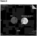
- Figure 13 shows a typical MRI scan of a negative control filter impregnated with phosphate buffered saline (PBS), a negative control consisting of DOTA-OP3-4 peptide 8 but with no contrast agent core 4, and a gadolinium-chelating DOTA-OP3-4 nanoparticle 2.
- PBS phosphate buffered saline
- DOTA-OP3-4 peptide 8 but with no contrast agent core 4
- gadolinium-chelating DOTA-OP3-4 nanoparticle 2 The scan clearly show that while the negative control showed only noise signals, the peptide 8 chelating the gadolinium core 4 provided a clear positive signal.
- a comparative analysis of filters impregnated with either gadolinium-chelating DOTA-OP3-4 and peptide-functionalised magnetic nanoparticle 2 showed the typical bright and dark images expected from the two contrast agents in the chosen mode of detection ( Figure 14 ).
- the inventor next determined whether a nanoparticle 2 comprising DOTA-coated gadolinium core 4 conjugated to osteoprogeterin 3 or 4 (OP3 or OP4) peptide 8, would inhibit osteoclastogenesis and osteoclast activity in vitro.
- Osteoclasts were obtained from mononuclear cells freshly isolated from peripheral blood from healthy human donors according to conventional methods based either on spiking of the cells with RANK and M-CSF or in osteoblast mononuclear cell co-culture systems spiked with M-CSF.
- Peptides and peptide-tethered nanoparticles 2 i.e. nanoparticle-OP3-4, magnetic nanoparticle-OP3-4, nanoparticle-OP3-DOTA and nanoparticle-OP3-4-Gd-DOTA
- the negative controls received no test materials and the positive control received rh OPG (50 ng/ml). Spiking was performed either before or after the differentiation of the mononuclear cells into osteoclasts.
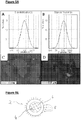
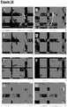
- Inhibition of osteoclastogenesis and osteoclast activity was quantitatively assessed by counting the number of TRAP positive multinucleated (MNC) cells using light microscopy and the number of MNC cells presenting F-actin rings using epifluorescence microscopy. Osteoclast activity was also assessed qualitatively by analysis of the number of resorption pits formed on the bone slices by SEM. Culture medium was replaced every 3 days with fresh medium supplemented with all the growth factors and test materials.
- peptide-functionalised magnetic nanoparticles 2 appeared to significantly reduce osteoclastogenesis when compared to non-functionalised nanoparticles (see Figure 16 ).
- the number of TRAP positive MNC in culture treated with nanoparticle-OP3-4, nanoparticle-DOTA-OP3-4 and nanoparticle-DOTA-Gd-OP3-4 was not significantly different showing that the various modifications of the peptide and its grafting to nanoparticles 2 did not alter its ability to inhibit osteoclastogenesis.
- Gd 3+ is toxic both in vitro and in vivo.
- macrocyclic chelates such as DOTA tightly trap Gd 3+ improve the ion solubility thus avoiding cytotoxic effects.
- Suc-Chi with its many free carboxyl groups and improved solubility would further contain Gd 3+ and improve the biocompatibility of DOTA-Gd-OP3-4.
- bone pathologies e.g. osteoporosis
- agents can recognise bone cells, osteoblasts and osteoclasts, as well as the mineralized bone extracellular matrix.
- These biorecognition properties were obtained through the synthesis of novel derivatised peptides with specificity for various bone cells and the mineral phase of bone.
- the derivatisation was designed to favour the stable binding with contrast agents of nanoparticulate or ionic composition without affecting their imaging properties.
- contrast agents in the form of magnetised polymeric beads mainly chitosan nanobeads
- chitosan nanobeads were obtained either through methods of coating of the magnetic core or grafting of gadolinium-modified peptides or a dispersion of ions in their crosslinked matrix. This ability to recognise cellular and structural components of the bone was coupled with the ability of controlling the cell behaviour.
- Biospecific contrast agents able to recognise mononuclear cells during their process of differentiation into osteoclasts as well as to recognise and inhibit the activity of differentiated osteoclasts could be obtained together with agents able to favour osteoblast migration.
- these biospecific agents couple the property of contrast agents with combined, built-in biorecognition and bioactivity properties capable of inducing tissue imaging and regeneration.
- osteoblast-specific peptides e.g. FHRRIKA
- these novel material can be also used as theranostic (i.e. combined therapy and diagnostic) agents in the treatment of bone deficiencies.
Landscapes
- Health & Medical Sciences (AREA)
- Chemical & Material Sciences (AREA)
- Engineering & Computer Science (AREA)
- Life Sciences & Earth Sciences (AREA)
- Nanotechnology (AREA)
- General Health & Medical Sciences (AREA)
- Veterinary Medicine (AREA)
- Animal Behavior & Ethology (AREA)
- Public Health (AREA)
- Nuclear Medicine, Radiotherapy & Molecular Imaging (AREA)
- Epidemiology (AREA)
- Medicinal Chemistry (AREA)
- Immunology (AREA)
- Radiology & Medical Imaging (AREA)
- Bioinformatics & Cheminformatics (AREA)
- Pharmacology & Pharmacy (AREA)
- Physics & Mathematics (AREA)
- Medical Informatics (AREA)
- Biophysics (AREA)
- Molecular Biology (AREA)
- Proteomics, Peptides & Aminoacids (AREA)
- High Energy & Nuclear Physics (AREA)
- Gastroenterology & Hepatology (AREA)
- Cell Biology (AREA)
- Zoology (AREA)
- Crystallography & Structural Chemistry (AREA)
- General Engineering & Computer Science (AREA)
- Biotechnology (AREA)
- Physical Education & Sports Medicine (AREA)
- Pathology (AREA)
- Heart & Thoracic Surgery (AREA)
- Surgery (AREA)
- Condensed Matter Physics & Semiconductors (AREA)
- General Physics & Mathematics (AREA)
- Biomedical Technology (AREA)
- Signal Processing (AREA)
- Optics & Photonics (AREA)
- Pulmonology (AREA)
- Theoretical Computer Science (AREA)
- Organic Chemistry (AREA)
Applications Claiming Priority (2)
| Application Number | Priority Date | Filing Date | Title |
|---|---|---|---|
| GB1302199.3A GB2510587B (en) | 2013-02-07 | 2013-02-07 | Biospecific agents for bone |
| PCT/GB2014/050265 WO2014122431A1 (en) | 2013-02-07 | 2014-01-31 | Biospecific agents for bone |
Publications (2)
| Publication Number | Publication Date |
|---|---|
| EP2953682A1 EP2953682A1 (en) | 2015-12-16 |
| EP2953682B1 true EP2953682B1 (en) | 2021-10-13 |
Family
ID=47998779
Family Applications (1)
| Application Number | Title | Priority Date | Filing Date |
|---|---|---|---|
| EP14702931.8A Active EP2953682B1 (en) | 2013-02-07 | 2014-01-31 | Biospecific agents for bone |
Country Status (9)
Families Citing this family (8)
| Publication number | Priority date | Publication date | Assignee | Title |
|---|---|---|---|---|
| US10660906B2 (en) | 2014-02-27 | 2020-05-26 | Synartro Ab | Hyaluronan conjugates with pharmaceutically active substances, methods and compositions |
| JP7156665B2 (ja) * | 2017-06-13 | 2022-10-19 | 国立研究開発法人量子科学技術研究開発機構 | 医薬、並びにその製造方法 |
| CN108187143A (zh) * | 2018-02-09 | 2018-06-22 | 福州大学 | 一种兼具磁热效应和原位诱导成骨的多功能复合材料及其制备方法 |
| CN109289119B (zh) * | 2018-09-16 | 2022-05-17 | 华北理工大学 | 一种用于脊柱康复系统的磁性纳米粒子球混合物 |
| US11944982B2 (en) * | 2019-06-05 | 2024-04-02 | Battelle Memorial Institute | Polymer-functionalized magnetic particle embodiments for solute separation, and devices and systems for using the same |
| CN111558041A (zh) * | 2020-04-30 | 2020-08-21 | 浙江理工大学 | 羟基磷灰石包裹磁性载药纳米颗粒及其制备方法和在制备骨肉瘤光疗药物中的应用 |
| CN119212686A (zh) * | 2021-09-15 | 2024-12-27 | 国家科学研究中心 | 包含至少一种金属盐和至少一种肽的纳米颗粒 |
| CN117582485B (zh) * | 2023-11-15 | 2024-06-07 | 湛江中心人民医院 | 方格星虫三肽在制备骨质疏松药物中的应用 |
Family Cites Families (9)
| Publication number | Priority date | Publication date | Assignee | Title |
|---|---|---|---|---|
| CH694935A5 (de) * | 2000-07-26 | 2005-09-30 | Straumann Holding Ag | Oberflaechenmodifizierte Implantate. |
| US7323542B2 (en) * | 2002-02-21 | 2008-01-29 | University Of Virginia Patent Foundation | Bone targeting peptides |
| US20050175584A1 (en) * | 2004-01-28 | 2005-08-11 | Paciotti Giulio F. | Functionalized colloidal metal compositions and methods |
| WO2006102377A2 (en) | 2005-03-21 | 2006-09-28 | The Regents Of The University Of California | Functionalized magnetic nanoparticles and methods of use thereof |
| AU2012204100B2 (en) * | 2005-03-21 | 2014-03-06 | The Regents Of The University Of California | Functionalized magnetic nanoparticles and methods of use thereof |
| US20090098050A1 (en) * | 2005-09-28 | 2009-04-16 | The Regents Of The Unversity Of California | Calcium binding peptides |
| US20100028387A1 (en) * | 2007-06-12 | 2010-02-04 | Ganesan Balasundaram | Biocompatible Coated Nanostructured Titanium Surfaces |
| US8063636B2 (en) * | 2009-05-29 | 2011-11-22 | The Invention Science Fund I, Llc | Systems, devices, methods, and compositions including targeted ferromagnetic structures |
| US9316645B2 (en) * | 2011-10-07 | 2016-04-19 | Brown University | Methods, compositions and kits for imaging cells and tissues using nanoparticles and spatial frequency heterodyne imaging |
-
2013
- 2013-02-07 GB GB1302199.3A patent/GB2510587B/en active Active
-
2014
- 2014-01-31 EP EP14702931.8A patent/EP2953682B1/en active Active
- 2014-01-31 JP JP2015556563A patent/JP6400026B2/ja active Active
- 2014-01-31 US US14/765,508 patent/US20150367000A1/en not_active Abandoned
- 2014-01-31 CN CN201480007874.8A patent/CN105142729A/zh active Pending
- 2014-01-31 ES ES14702931T patent/ES2904260T3/es active Active
- 2014-01-31 NZ NZ710436A patent/NZ710436A/en unknown
- 2014-01-31 CA CA2898366A patent/CA2898366C/en active Active
- 2014-01-31 WO PCT/GB2014/050265 patent/WO2014122431A1/en active Application Filing
-
2018
- 2018-08-17 JP JP2018153482A patent/JP6683773B2/ja active Active
-
2019
- 2019-11-25 US US16/693,780 patent/US11464878B2/en active Active
Non-Patent Citations (1)
| Title |
|---|
| None * |
Also Published As
| Publication number | Publication date |
|---|---|
| CN105142729A (zh) | 2015-12-09 |
| CA2898366C (en) | 2021-05-04 |
| HK1217910A1 (zh) | 2017-01-27 |
| JP6683773B2 (ja) | 2020-04-22 |
| GB2510587B (en) | 2020-05-20 |
| WO2014122431A1 (en) | 2014-08-14 |
| JP2018199693A (ja) | 2018-12-20 |
| US20150367000A1 (en) | 2015-12-24 |
| US11464878B2 (en) | 2022-10-11 |
| EP2953682A1 (en) | 2015-12-16 |
| CA2898366A1 (en) | 2014-08-14 |
| NZ710436A (en) | 2020-07-31 |
| ES2904260T3 (es) | 2022-04-04 |
| JP2016509007A (ja) | 2016-03-24 |
| GB201302199D0 (en) | 2013-03-27 |
| JP6400026B2 (ja) | 2018-10-03 |
| GB2510587A (en) | 2014-08-13 |
| US20200215207A1 (en) | 2020-07-09 |
Similar Documents
| Publication | Publication Date | Title |
|---|---|---|
| US11464878B2 (en) | Biospecific agents for bone | |
| Kielar et al. | Large relaxivity enhancement of paramagnetic lipid nanoparticles by restricting the local motions of the GdIII chelates | |
| KR101946070B1 (ko) | ε폴리라이신 결합체 및 이의 용도 | |
| EP2725053B1 (en) | Branched amphipathic block polymer and molecular aggregate and drug delivery system using same | |
| AU2006200363A1 (en) | Glycopeptide compositions | |
| Yang et al. | Redox-responsive nanoparticles from disulfide bond-linked poly-(N-ε-carbobenzyloxy-l-lysine)-grafted hyaluronan copolymers as theranostic nanoparticles for tumor-targeted MRI and chemotherapy | |
| CN110114367B (zh) | Vap多肽及其在制备靶向诊疗肿瘤药物中的应用 | |
| JP2010528122A (ja) | 白金薬剤と結合されたポリマー | |
| US20130302255A1 (en) | Novel targeted paramagnetic contrast agent | |
| EP0991430A1 (en) | Compositions and methods for x-ray imaging of tumors | |
| Wu et al. | Hyaluronic Acid‐Gadolinium Complex Nanospheres as Lymphatic System‐Specific Contrast Agent for Magnetic Resonance Imaging | |
| KR20100035062A (ko) | 질환의 진단을 위한 표적 펩타이드가 결합된 양친성 키토산나노입자 조영제 | |
| JP2010208979A (ja) | 組織撮像用mri造影剤 | |
| Xiao et al. | A new biodegradable and biocompatible gadolinium (III)-polymer for liver magnetic resonance imaging contrast agent | |
| HK1217910B (en) | Biospecific agents for bone | |
| CN117563012A (zh) | 一种多肽偶联物及制备方法和应用 | |
| CN109568597B (zh) | 靶向胎盘样硫酸软骨素a的多肽药物偶联物及其制备方法和应用 | |
| KR20160056492A (ko) | 동맥경화 진단용 키토산 나노입자 복합체, 이의 제조방법 및 이를 포함하는 조영제 조성물 | |
| CN108570094A (zh) | Ae多肽及其在制备肿瘤靶向诊治递药系统中的用途 | |
| Das et al. | Magnetic resonance imaging contrast enhancement in vitro and in vivo by octanuclear iron-oxo cluster-based agents | |
| CN111840324B (zh) | 应用于骨肉瘤细胞成像或治疗的Au DENPs-巨噬细胞复合物 | |
| JP2008280277A (ja) | 腫瘍撮像用mri造影剤 | |
| KR20220103415A (ko) | 다발성골수종(multiple myeloma)의 표적치료 및 병용화학치료가 가능한 초분지 폴리글리세롤 항암 복합약물전달체와 그의 제법 | |
| Capuana | Design and testing of novel imaging probes | |
| JP2024510327A (ja) | 医薬ポリマーコンジュゲート |
Legal Events
| Date | Code | Title | Description |
|---|---|---|---|
| PUAI | Public reference made under article 153(3) epc to a published international application that has entered the european phase |
Free format text: ORIGINAL CODE: 0009012 |
|
| 17P | Request for examination filed |
Effective date: 20150713 |
|
| AK | Designated contracting states |
Kind code of ref document: A1 Designated state(s): AL AT BE BG CH CY CZ DE DK EE ES FI FR GB GR HR HU IE IS IT LI LT LU LV MC MK MT NL NO PL PT RO RS SE SI SK SM TR |
|
| AX | Request for extension of the european patent |
Extension state: BA ME |
|
| DAX | Request for extension of the european patent (deleted) | ||
| REG | Reference to a national code |
Ref country code: HK Ref legal event code: DE Ref document number: 1217910 Country of ref document: HK |
|
| STAA | Information on the status of an ep patent application or granted ep patent |
Free format text: STATUS: EXAMINATION IS IN PROGRESS |
|
| 17Q | First examination report despatched |
Effective date: 20180228 |
|
| REG | Reference to a national code |
Ref country code: DE Ref legal event code: R079 Ref document number: 602014080626 Country of ref document: DE Free format text: PREVIOUS MAIN CLASS: A61P0019080000 Ipc: A61K0049040000 |
|
| RIC1 | Information provided on ipc code assigned before grant |
Ipc: B82Y 5/00 20110101ALI20210329BHEP Ipc: A61K 38/17 20060101ALI20210329BHEP Ipc: A61P 19/08 20060101ALI20210329BHEP Ipc: A61K 47/69 20170101ALI20210329BHEP Ipc: A61K 49/18 20060101ALI20210329BHEP Ipc: A61K 49/04 20060101AFI20210329BHEP |
|
| GRAP | Despatch of communication of intention to grant a patent |
Free format text: ORIGINAL CODE: EPIDOSNIGR1 |
|
| STAA | Information on the status of an ep patent application or granted ep patent |
Free format text: STATUS: GRANT OF PATENT IS INTENDED |
|
| INTG | Intention to grant announced |
Effective date: 20210511 |
|
| GRAS | Grant fee paid |
Free format text: ORIGINAL CODE: EPIDOSNIGR3 |
|
| GRAA | (expected) grant |
Free format text: ORIGINAL CODE: 0009210 |
|
| STAA | Information on the status of an ep patent application or granted ep patent |
Free format text: STATUS: THE PATENT HAS BEEN GRANTED |
|
| AK | Designated contracting states |
Kind code of ref document: B1 Designated state(s): AL AT BE BG CH CY CZ DE DK EE ES FI FR GB GR HR HU IE IS IT LI LT LU LV MC MK MT NL NO PL PT RO RS SE SI SK SM TR |
|
| REG | Reference to a national code |
Ref country code: GB Ref legal event code: FG4D |
|
| REG | Reference to a national code |
Ref country code: CH Ref legal event code: EP |
|
| REG | Reference to a national code |
Ref country code: DE Ref legal event code: R096 Ref document number: 602014080626 Country of ref document: DE |
|
| REG | Reference to a national code |
Ref country code: IE Ref legal event code: FG4D |
|
| REG | Reference to a national code |
Ref country code: AT Ref legal event code: REF Ref document number: 1437679 Country of ref document: AT Kind code of ref document: T Effective date: 20211115 |
|
| REG | Reference to a national code |
Ref country code: LT Ref legal event code: MG9D |
|
| REG | Reference to a national code |
Ref country code: NL Ref legal event code: MP Effective date: 20211013 |
|
| REG | Reference to a national code |
Ref country code: AT Ref legal event code: MK05 Ref document number: 1437679 Country of ref document: AT Kind code of ref document: T Effective date: 20211013 |
|
| REG | Reference to a national code |
Ref country code: ES Ref legal event code: FG2A Ref document number: 2904260 Country of ref document: ES Kind code of ref document: T3 Effective date: 20220404 |
|
| PG25 | Lapsed in a contracting state [announced via postgrant information from national office to epo] |
Ref country code: RS Free format text: LAPSE BECAUSE OF FAILURE TO SUBMIT A TRANSLATION OF THE DESCRIPTION OR TO PAY THE FEE WITHIN THE PRESCRIBED TIME-LIMIT Effective date: 20211013 Ref country code: LT Free format text: LAPSE BECAUSE OF FAILURE TO SUBMIT A TRANSLATION OF THE DESCRIPTION OR TO PAY THE FEE WITHIN THE PRESCRIBED TIME-LIMIT Effective date: 20211013 Ref country code: FI Free format text: LAPSE BECAUSE OF FAILURE TO SUBMIT A TRANSLATION OF THE DESCRIPTION OR TO PAY THE FEE WITHIN THE PRESCRIBED TIME-LIMIT Effective date: 20211013 Ref country code: BG Free format text: LAPSE BECAUSE OF FAILURE TO SUBMIT A TRANSLATION OF THE DESCRIPTION OR TO PAY THE FEE WITHIN THE PRESCRIBED TIME-LIMIT Effective date: 20220113 Ref country code: AT Free format text: LAPSE BECAUSE OF FAILURE TO SUBMIT A TRANSLATION OF THE DESCRIPTION OR TO PAY THE FEE WITHIN THE PRESCRIBED TIME-LIMIT Effective date: 20211013 |
|
| PG25 | Lapsed in a contracting state [announced via postgrant information from national office to epo] |
Ref country code: IS Free format text: LAPSE BECAUSE OF FAILURE TO SUBMIT A TRANSLATION OF THE DESCRIPTION OR TO PAY THE FEE WITHIN THE PRESCRIBED TIME-LIMIT Effective date: 20220213 Ref country code: SE Free format text: LAPSE BECAUSE OF FAILURE TO SUBMIT A TRANSLATION OF THE DESCRIPTION OR TO PAY THE FEE WITHIN THE PRESCRIBED TIME-LIMIT Effective date: 20211013 Ref country code: PT Free format text: LAPSE BECAUSE OF FAILURE TO SUBMIT A TRANSLATION OF THE DESCRIPTION OR TO PAY THE FEE WITHIN THE PRESCRIBED TIME-LIMIT Effective date: 20220214 Ref country code: PL Free format text: LAPSE BECAUSE OF FAILURE TO SUBMIT A TRANSLATION OF THE DESCRIPTION OR TO PAY THE FEE WITHIN THE PRESCRIBED TIME-LIMIT Effective date: 20211013 Ref country code: NO Free format text: LAPSE BECAUSE OF FAILURE TO SUBMIT A TRANSLATION OF THE DESCRIPTION OR TO PAY THE FEE WITHIN THE PRESCRIBED TIME-LIMIT Effective date: 20220113 Ref country code: NL Free format text: LAPSE BECAUSE OF FAILURE TO SUBMIT A TRANSLATION OF THE DESCRIPTION OR TO PAY THE FEE WITHIN THE PRESCRIBED TIME-LIMIT Effective date: 20211013 Ref country code: LV Free format text: LAPSE BECAUSE OF FAILURE TO SUBMIT A TRANSLATION OF THE DESCRIPTION OR TO PAY THE FEE WITHIN THE PRESCRIBED TIME-LIMIT Effective date: 20211013 Ref country code: HR Free format text: LAPSE BECAUSE OF FAILURE TO SUBMIT A TRANSLATION OF THE DESCRIPTION OR TO PAY THE FEE WITHIN THE PRESCRIBED TIME-LIMIT Effective date: 20211013 Ref country code: GR Free format text: LAPSE BECAUSE OF FAILURE TO SUBMIT A TRANSLATION OF THE DESCRIPTION OR TO PAY THE FEE WITHIN THE PRESCRIBED TIME-LIMIT Effective date: 20220114 |
|
| REG | Reference to a national code |
Ref country code: DE Ref legal event code: R097 Ref document number: 602014080626 Country of ref document: DE |
|
| PG25 | Lapsed in a contracting state [announced via postgrant information from national office to epo] |
Ref country code: SM Free format text: LAPSE BECAUSE OF FAILURE TO SUBMIT A TRANSLATION OF THE DESCRIPTION OR TO PAY THE FEE WITHIN THE PRESCRIBED TIME-LIMIT Effective date: 20211013 Ref country code: SK Free format text: LAPSE BECAUSE OF FAILURE TO SUBMIT A TRANSLATION OF THE DESCRIPTION OR TO PAY THE FEE WITHIN THE PRESCRIBED TIME-LIMIT Effective date: 20211013 Ref country code: RO Free format text: LAPSE BECAUSE OF FAILURE TO SUBMIT A TRANSLATION OF THE DESCRIPTION OR TO PAY THE FEE WITHIN THE PRESCRIBED TIME-LIMIT Effective date: 20211013 Ref country code: EE Free format text: LAPSE BECAUSE OF FAILURE TO SUBMIT A TRANSLATION OF THE DESCRIPTION OR TO PAY THE FEE WITHIN THE PRESCRIBED TIME-LIMIT Effective date: 20211013 Ref country code: DK Free format text: LAPSE BECAUSE OF FAILURE TO SUBMIT A TRANSLATION OF THE DESCRIPTION OR TO PAY THE FEE WITHIN THE PRESCRIBED TIME-LIMIT Effective date: 20211013 Ref country code: CZ Free format text: LAPSE BECAUSE OF FAILURE TO SUBMIT A TRANSLATION OF THE DESCRIPTION OR TO PAY THE FEE WITHIN THE PRESCRIBED TIME-LIMIT Effective date: 20211013 |
|
| PLBE | No opposition filed within time limit |
Free format text: ORIGINAL CODE: 0009261 |
|
| STAA | Information on the status of an ep patent application or granted ep patent |
Free format text: STATUS: NO OPPOSITION FILED WITHIN TIME LIMIT |
|
| PG25 | Lapsed in a contracting state [announced via postgrant information from national office to epo] |
Ref country code: MC Free format text: LAPSE BECAUSE OF FAILURE TO SUBMIT A TRANSLATION OF THE DESCRIPTION OR TO PAY THE FEE WITHIN THE PRESCRIBED TIME-LIMIT Effective date: 20211013 |
|
| 26N | No opposition filed |
Effective date: 20220714 |
|
| REG | Reference to a national code |
Ref country code: BE Ref legal event code: MM Effective date: 20220131 |
|
| PG25 | Lapsed in a contracting state [announced via postgrant information from national office to epo] |
Ref country code: LU Free format text: LAPSE BECAUSE OF NON-PAYMENT OF DUE FEES Effective date: 20220131 Ref country code: AL Free format text: LAPSE BECAUSE OF FAILURE TO SUBMIT A TRANSLATION OF THE DESCRIPTION OR TO PAY THE FEE WITHIN THE PRESCRIBED TIME-LIMIT Effective date: 20211013 |
|
| PG25 | Lapsed in a contracting state [announced via postgrant information from national office to epo] |
Ref country code: SI Free format text: LAPSE BECAUSE OF FAILURE TO SUBMIT A TRANSLATION OF THE DESCRIPTION OR TO PAY THE FEE WITHIN THE PRESCRIBED TIME-LIMIT Effective date: 20211013 Ref country code: BE Free format text: LAPSE BECAUSE OF NON-PAYMENT OF DUE FEES Effective date: 20220131 |
|
| PG25 | Lapsed in a contracting state [announced via postgrant information from national office to epo] |
Ref country code: HU Free format text: LAPSE BECAUSE OF FAILURE TO SUBMIT A TRANSLATION OF THE DESCRIPTION OR TO PAY THE FEE WITHIN THE PRESCRIBED TIME-LIMIT; INVALID AB INITIO Effective date: 20140131 |
|
| PGFP | Annual fee paid to national office [announced via postgrant information from national office to epo] |
Ref country code: ES Payment date: 20240223 Year of fee payment: 11 Ref country code: IE Payment date: 20240130 Year of fee payment: 11 |
|
| PG25 | Lapsed in a contracting state [announced via postgrant information from national office to epo] |
Ref country code: MK Free format text: LAPSE BECAUSE OF FAILURE TO SUBMIT A TRANSLATION OF THE DESCRIPTION OR TO PAY THE FEE WITHIN THE PRESCRIBED TIME-LIMIT Effective date: 20211013 Ref country code: CY Free format text: LAPSE BECAUSE OF FAILURE TO SUBMIT A TRANSLATION OF THE DESCRIPTION OR TO PAY THE FEE WITHIN THE PRESCRIBED TIME-LIMIT Effective date: 20211013 |
|
| PGFP | Annual fee paid to national office [announced via postgrant information from national office to epo] |
Ref country code: DE Payment date: 20240130 Year of fee payment: 11 Ref country code: GB Payment date: 20240126 Year of fee payment: 11 Ref country code: CH Payment date: 20240212 Year of fee payment: 11 |
|
| PGFP | Annual fee paid to national office [announced via postgrant information from national office to epo] |
Ref country code: IT Payment date: 20240130 Year of fee payment: 11 Ref country code: FR Payment date: 20240130 Year of fee payment: 11 |
|
| PG25 | Lapsed in a contracting state [announced via postgrant information from national office to epo] |
Ref country code: MT Free format text: LAPSE BECAUSE OF FAILURE TO SUBMIT A TRANSLATION OF THE DESCRIPTION OR TO PAY THE FEE WITHIN THE PRESCRIBED TIME-LIMIT Effective date: 20211013 |
|
| REG | Reference to a national code |
Ref country code: CH Ref legal event code: PL |