EP2945544B1 - Eliminating motion effects in medical images caused by physiological function - Google Patents
Eliminating motion effects in medical images caused by physiological function Download PDFInfo
- Publication number
- EP2945544B1 EP2945544B1 EP14704901.9A EP14704901A EP2945544B1 EP 2945544 B1 EP2945544 B1 EP 2945544B1 EP 14704901 A EP14704901 A EP 14704901A EP 2945544 B1 EP2945544 B1 EP 2945544B1
- Authority
- EP
- European Patent Office
- Prior art keywords
- image
- images
- motion
- intensity
- sequence
- Prior art date
- Legal status (The legal status is an assumption and is not a legal conclusion. Google has not performed a legal analysis and makes no representation as to the accuracy of the status listed.)
- Active
Links
- 230000033001 locomotion Effects 0.000 title claims description 70
- 230000000694 effects Effects 0.000 title claims description 15
- 230000035790 physiological processes and functions Effects 0.000 title description 4
- 230000000241 respiratory effect Effects 0.000 claims description 44
- 239000002872 contrast media Substances 0.000 claims description 33
- 230000003902 lesion Effects 0.000 claims description 33
- 238000000034 method Methods 0.000 claims description 28
- 238000012545 processing Methods 0.000 claims description 21
- 230000010412 perfusion Effects 0.000 claims description 17
- 238000002604 ultrasonography Methods 0.000 claims description 15
- 239000011159 matrix material Substances 0.000 claims description 6
- 230000010410 reperfusion Effects 0.000 claims description 6
- 206010028980 Neoplasm Diseases 0.000 claims description 4
- 230000000747 cardiac effect Effects 0.000 claims description 4
- 125000004122 cyclic group Chemical group 0.000 claims description 3
- 206010027476 Metastases Diseases 0.000 description 16
- 230000009401 metastasis Effects 0.000 description 16
- 210000004185 liver Anatomy 0.000 description 13
- 238000002592 echocardiography Methods 0.000 description 11
- 239000000523 sample Substances 0.000 description 11
- 210000001519 tissue Anatomy 0.000 description 9
- 238000002560 therapeutic procedure Methods 0.000 description 8
- 238000010586 diagram Methods 0.000 description 7
- 238000005259 measurement Methods 0.000 description 7
- 230000007423 decrease Effects 0.000 description 6
- 238000003384 imaging method Methods 0.000 description 6
- 230000015572 biosynthetic process Effects 0.000 description 5
- 239000008280 blood Substances 0.000 description 5
- 210000004369 blood Anatomy 0.000 description 5
- 230000006870 function Effects 0.000 description 5
- 210000002767 hepatic artery Anatomy 0.000 description 5
- 210000003484 anatomy Anatomy 0.000 description 4
- 238000004519 manufacturing process Methods 0.000 description 4
- 210000003240 portal vein Anatomy 0.000 description 4
- 238000011002 quantification Methods 0.000 description 4
- 230000029058 respiratory gaseous exchange Effects 0.000 description 4
- 230000033115 angiogenesis Effects 0.000 description 3
- 230000017531 blood circulation Effects 0.000 description 3
- 210000000038 chest Anatomy 0.000 description 3
- 238000002059 diagnostic imaging Methods 0.000 description 3
- 238000009826 distribution Methods 0.000 description 3
- 230000008569 process Effects 0.000 description 3
- 238000011282 treatment Methods 0.000 description 3
- 238000013459 approach Methods 0.000 description 2
- 210000004204 blood vessel Anatomy 0.000 description 2
- 238000000605 extraction Methods 0.000 description 2
- 238000007689 inspection Methods 0.000 description 2
- 201000007270 liver cancer Diseases 0.000 description 2
- 208000014018 liver neoplasm Diseases 0.000 description 2
- 210000004165 myocardium Anatomy 0.000 description 2
- 230000017074 necrotic cell death Effects 0.000 description 2
- 230000003534 oscillatory effect Effects 0.000 description 2
- 230000004044 response Effects 0.000 description 2
- 238000005070 sampling Methods 0.000 description 2
- 210000003462 vein Anatomy 0.000 description 2
- 206010006187 Breast cancer Diseases 0.000 description 1
- 208000026310 Breast neoplasm Diseases 0.000 description 1
- 241001331845 Equus asinus x caballus Species 0.000 description 1
- 206010020843 Hyperthermia Diseases 0.000 description 1
- 208000008839 Kidney Neoplasms Diseases 0.000 description 1
- 241001465754 Metazoa Species 0.000 description 1
- 206010061902 Pancreatic neoplasm Diseases 0.000 description 1
- 206010060862 Prostate cancer Diseases 0.000 description 1
- 208000000236 Prostatic Neoplasms Diseases 0.000 description 1
- 206010038389 Renal cancer Diseases 0.000 description 1
- 210000001015 abdomen Anatomy 0.000 description 1
- 230000002411 adverse Effects 0.000 description 1
- 239000004037 angiogenesis inhibitor Substances 0.000 description 1
- 210000001367 artery Anatomy 0.000 description 1
- 230000008901 benefit Effects 0.000 description 1
- 230000005540 biological transmission Effects 0.000 description 1
- 210000000481 breast Anatomy 0.000 description 1
- 239000003795 chemical substances by application Substances 0.000 description 1
- 230000001427 coherent effect Effects 0.000 description 1
- 238000004040 coloring Methods 0.000 description 1
- 238000010276 construction Methods 0.000 description 1
- 238000011109 contamination Methods 0.000 description 1
- 238000002607 contrast-enhanced ultrasound Methods 0.000 description 1
- 238000011393 cytotoxic chemotherapy Methods 0.000 description 1
- 238000013144 data compression Methods 0.000 description 1
- 230000001934 delay Effects 0.000 description 1
- 238000012217 deletion Methods 0.000 description 1
- 230000037430 deletion Effects 0.000 description 1
- 238000001514 detection method Methods 0.000 description 1
- 230000003292 diminished effect Effects 0.000 description 1
- 239000002961 echo contrast media Substances 0.000 description 1
- 230000001747 exhibiting effect Effects 0.000 description 1
- 210000002216 heart Anatomy 0.000 description 1
- 230000000004 hemodynamic effect Effects 0.000 description 1
- 230000002440 hepatic effect Effects 0.000 description 1
- 230000036031 hyperthermia Effects 0.000 description 1
- 230000002631 hypothermal effect Effects 0.000 description 1
- 238000001802 infusion Methods 0.000 description 1
- 238000002347 injection Methods 0.000 description 1
- 239000007924 injection Substances 0.000 description 1
- 210000003734 kidney Anatomy 0.000 description 1
- 201000010982 kidney cancer Diseases 0.000 description 1
- 208000015486 malignant pancreatic neoplasm Diseases 0.000 description 1
- 238000013178 mathematical model Methods 0.000 description 1
- 230000001394 metastastic effect Effects 0.000 description 1
- 206010061289 metastatic neoplasm Diseases 0.000 description 1
- 238000010606 normalization Methods 0.000 description 1
- 210000000056 organ Anatomy 0.000 description 1
- 210000000496 pancreas Anatomy 0.000 description 1
- 208000008443 pancreatic carcinoma Diseases 0.000 description 1
- 238000012805 post-processing Methods 0.000 description 1
- 238000000513 principal component analysis Methods 0.000 description 1
- 230000009467 reduction Effects 0.000 description 1
- 230000003595 spectral effect Effects 0.000 description 1
- 230000002123 temporal effect Effects 0.000 description 1
- 230000001225 therapeutic effect Effects 0.000 description 1
- 230000009466 transformation Effects 0.000 description 1
- 238000011269 treatment regimen Methods 0.000 description 1
- 230000005747 tumor angiogenesis Effects 0.000 description 1
Images


Classifications
-
- G—PHYSICS
- G06—COMPUTING; CALCULATING OR COUNTING
- G06T—IMAGE DATA PROCESSING OR GENERATION, IN GENERAL
- G06T7/00—Image analysis
- G06T7/0002—Inspection of images, e.g. flaw detection
- G06T7/0012—Biomedical image inspection
-
- A—HUMAN NECESSITIES
- A61—MEDICAL OR VETERINARY SCIENCE; HYGIENE
- A61B—DIAGNOSIS; SURGERY; IDENTIFICATION
- A61B8/00—Diagnosis using ultrasonic, sonic or infrasonic waves
- A61B8/06—Measuring blood flow
-
- A—HUMAN NECESSITIES
- A61—MEDICAL OR VETERINARY SCIENCE; HYGIENE
- A61B—DIAGNOSIS; SURGERY; IDENTIFICATION
- A61B8/00—Diagnosis using ultrasonic, sonic or infrasonic waves
- A61B8/48—Diagnostic techniques
- A61B8/481—Diagnostic techniques involving the use of contrast agent, e.g. microbubbles introduced into the bloodstream
-
- A—HUMAN NECESSITIES
- A61—MEDICAL OR VETERINARY SCIENCE; HYGIENE
- A61B—DIAGNOSIS; SURGERY; IDENTIFICATION
- A61B8/00—Diagnosis using ultrasonic, sonic or infrasonic waves
- A61B8/48—Diagnostic techniques
- A61B8/483—Diagnostic techniques involving the acquisition of a 3D volume of data
-
- A—HUMAN NECESSITIES
- A61—MEDICAL OR VETERINARY SCIENCE; HYGIENE
- A61B—DIAGNOSIS; SURGERY; IDENTIFICATION
- A61B8/00—Diagnosis using ultrasonic, sonic or infrasonic waves
- A61B8/52—Devices using data or image processing specially adapted for diagnosis using ultrasonic, sonic or infrasonic waves
- A61B8/5215—Devices using data or image processing specially adapted for diagnosis using ultrasonic, sonic or infrasonic waves involving processing of medical diagnostic data
- A61B8/5223—Devices using data or image processing specially adapted for diagnosis using ultrasonic, sonic or infrasonic waves involving processing of medical diagnostic data for extracting a diagnostic or physiological parameter from medical diagnostic data
-
- A—HUMAN NECESSITIES
- A61—MEDICAL OR VETERINARY SCIENCE; HYGIENE
- A61B—DIAGNOSIS; SURGERY; IDENTIFICATION
- A61B8/00—Diagnosis using ultrasonic, sonic or infrasonic waves
- A61B8/52—Devices using data or image processing specially adapted for diagnosis using ultrasonic, sonic or infrasonic waves
- A61B8/5269—Devices using data or image processing specially adapted for diagnosis using ultrasonic, sonic or infrasonic waves involving detection or reduction of artifacts
- A61B8/5276—Devices using data or image processing specially adapted for diagnosis using ultrasonic, sonic or infrasonic waves involving detection or reduction of artifacts due to motion
-
- A—HUMAN NECESSITIES
- A61—MEDICAL OR VETERINARY SCIENCE; HYGIENE
- A61B—DIAGNOSIS; SURGERY; IDENTIFICATION
- A61B8/00—Diagnosis using ultrasonic, sonic or infrasonic waves
- A61B8/52—Devices using data or image processing specially adapted for diagnosis using ultrasonic, sonic or infrasonic waves
- A61B8/5284—Devices using data or image processing specially adapted for diagnosis using ultrasonic, sonic or infrasonic waves involving retrospective matching to a physiological signal
-
- G—PHYSICS
- G06—COMPUTING; CALCULATING OR COUNTING
- G06T—IMAGE DATA PROCESSING OR GENERATION, IN GENERAL
- G06T7/00—Image analysis
- G06T7/20—Analysis of motion
- G06T7/246—Analysis of motion using feature-based methods, e.g. the tracking of corners or segments
-
- G—PHYSICS
- G16—INFORMATION AND COMMUNICATION TECHNOLOGY [ICT] SPECIALLY ADAPTED FOR SPECIFIC APPLICATION FIELDS
- G16H—HEALTHCARE INFORMATICS, i.e. INFORMATION AND COMMUNICATION TECHNOLOGY [ICT] SPECIALLY ADAPTED FOR THE HANDLING OR PROCESSING OF MEDICAL OR HEALTHCARE DATA
- G16H50/00—ICT specially adapted for medical diagnosis, medical simulation or medical data mining; ICT specially adapted for detecting, monitoring or modelling epidemics or pandemics
- G16H50/30—ICT specially adapted for medical diagnosis, medical simulation or medical data mining; ICT specially adapted for detecting, monitoring or modelling epidemics or pandemics for calculating health indices; for individual health risk assessment
-
- G—PHYSICS
- G06—COMPUTING; CALCULATING OR COUNTING
- G06T—IMAGE DATA PROCESSING OR GENERATION, IN GENERAL
- G06T2207/00—Indexing scheme for image analysis or image enhancement
- G06T2207/10—Image acquisition modality
- G06T2207/10004—Still image; Photographic image
-
- G—PHYSICS
- G06—COMPUTING; CALCULATING OR COUNTING
- G06T—IMAGE DATA PROCESSING OR GENERATION, IN GENERAL
- G06T2207/00—Indexing scheme for image analysis or image enhancement
- G06T2207/30—Subject of image; Context of image processing
- G06T2207/30004—Biomedical image processing
- G06T2207/30096—Tumor; Lesion
Definitions
- This invention relates to ultrasonic diagnostic imaging systems and, in particular, to the use of ultrasonic diagnostic imaging systems to assess the progress of therapeutic treatment of tumors.
- the inventors take advantage of the fact that the flow of blood to the liver comes from two sources, the hepatic artery and the portal vein. Since the flow of blood during the first, arterial phase of blood flow will perfuse HCC and metastatic liver lesions first, the inventors identify such lesions by detecting the times of arrival of contrast agent in the liver during the arterial and the later portal phase of blood flow. An area of early wash-in of contrast agent to the liver can be symptomatic of a lesion.
- a treatment regimen is generally prescribed by a physician.
- the therapy may involve hyper-/hypothermia, cytotoxic chemotherapy, or anti-angiogenesis agents, for example.
- the therapy is usually not performed in a single session, but in several sessions over a period of weeks or months.
- At each therapy session it is generally desirable for a physician to assess the progress of the therapy to determine its effectiveness for the patient.
- the lesion or metastasis may be imaged diagnostically to see whether it is shrinking, for instance. But often the progress of treatment is slow and only small changes in the lesion or metastasis have occurred since the previous session. In such instances it is desirable to assess the progress of therapy quantitatively by measuring certain characteristics of the tumor.
- One such measure is the regression of tumor angiogenesis.
- the microvasculature which developed to nourish the lesion will provide a smaller supply of blood for the lesion and may itself begin to shrink.
- One quantitative approach is to assess this regression of angiogenesis, the decline in performance of the lesion's microvasculature.
- a preferred technique for doing this is with a contrast agent which brightly illuminates the blood flow during the uptake of blood containing the agent, and the subsequent decline in brightness during the wash-out phase of the contrast agent. Measurement of the time-intensity curve of this reperfusion enables precise assessment of the success of the treatment therapy toward lesion necrosis.
- the acquisition of the necessary image sequence of contrast agent wash-in and wash-out requires that the imaging probe steadily image the lesion in the image plane during the wash-in and wash-out period of the contrast agent, which can last for as long as 100 seconds or more.
- the liver is adjacent to the diaphragm in the body, causing the patient's respiratory motion to cause movement of the anatomy in relation to the image acquisition plane of the probe during the imaging procedure. This movement can cause the lesion to move into and out of the image plane during the procedure.
- the brightness of the lesion and its contrast agent in the image will vary with the lesion movement rather that solely due to contrast agent wash-in and wash-out. Consequently it is desirable to eliminate these unwanted motional effects when acquiring images used for perfusion assessment.
- Document WO2009/093211 discloses a method for reducing the effect of physiological motion in which a sequence of medical images of a target region is acquired during the occurrence of physiological motion and image data of the sequence is used to identify cycles of said physiological motion.
- methods are described for processing medical images so that movement due to a certain physiological function, such as the cyclic movement due to respiration or cardiac motion, can be identified and selectively removed from subsequently processed images.
- This is preferably done by processing only those image frames which belong to the same range of phases of the motion cycle.
- the image data is more uniformly affected by the motion and disparate motional effects are largely eliminated from the data, allowing for more precise quantitative assessment of anatomical performance such as perfusion.
- Clinical applications of the methods include liver, kidney, pancreas, breast, and prostate cancer assessment.
- a sequence (loop) of images is acquired during one, and preferably more, cycles of the motion.
- the normal breathing rate of an adult at rest is 8-16 breaths per minutes, so the acquisition of a one-minute loop will acquire images over about 8-16 cycles of respiratory motion, for instance.
- the acquired images are tagged with their times of acquisition so that these times can be related to phases of the motion.
- Strong reflecting structures are identified in the images and their motion followed throughout the loop, which is identified by their cyclic variation in position and/or brightness throughout the loop. For instance, a reflector may start at one location in an image, move to another location over a first series of images, then back to its original location over a second series of images, the time to acquire the two series of images being the time of one cycle of motion.
- a reflector may start with a given brightness in an image, increase in brightness during a first series of images, then decrease in brightness back to its original intensity during a second series of images.
- the time required to acquire the two series of images is the time of a motion cycle.
- the identified time or times of motion cycles may be compared with typical cycle times of the physiological function in question to see that they correlate. For example, if an identified motion cycle is outside an expected range of 5-20 breaths per minute, the cycle measurement would be discarded as a respiratory cycle and another cycle measurement made.
- those images which were acquired during desired phases of the motion cycle are accepted for further processing while the remainder are discarded. For example, it may be desired to accept for processing only those images which were acquired during a 25% range of phases of a respiratory cycle. Images acquired within this range of the respiratory phase of each cycle are then accepted for processing for quantified measurement, such as time-intensity curve and reperfusion measurement.
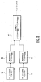
- FIGURE 1 an ultrasound system constructed in accordance with the principles of the present invention is shown in block diagram form.
- This system operates by scanning a two or three dimensional region of the body being imaged with ultrasonic transmit beams. As each beam is transmitted along its steered path through the body, the beam returns echo signals with linear and nonlinear (fundamental and harmonic frequency) components corresponding to the transmitted frequency components.
- the transmit signals are modulated by the nonlinear response of contrast agent microbubbles encountered by the beam, thereby generating echo signals with harmonic components.
- the ultrasound system of FIGURE 1 utilizes a transmitter 16 which transmits waves or pulses of a selected modulation characteristic in a desired beam direction for the return of harmonic echo components from scatterers within the body.
- the transmitter is responsive to a number of control parameters which determine the characteristics of the transmit beams, including the frequency components of the transmit beam, their relative intensities or amplitudes, and the phase or polarity of the transmit signals.
- the transmitter is coupled by a transmit/receive switch 14 to the elements of an array transducer 12 of an ultrasound probe 10.
- the array transducer can be a one dimensional array for planar (two dimensional) imaging or a two dimensional array for two dimensional or volumetric (three dimensional) imaging.
- a two dimensional matrix array can also be operated in an xMatrix mode in which a single plane (xPlane) which can be steered over a volumetric region is scanned by the matrix array probe.
- the transducer array 12 receives echoes from the body containing fundamental (linear) and harmonic (nonlinear) frequency components which are within the transducer passband. These echo signals are coupled by the switch 14 to a beamformer 18 which appropriately delays echo signals from the different transducer elements then combines them to form a sequence of linear and harmonic signals from along the beam direction from shallow to deeper depths.
- the beamformer is a digital beamformer operating on digitized echo signals to produce a sequence of discrete coherent digital echo signals from a near field to a far field depth of the image.
- the beamformer may be a multiline beamformer which produces two or more sequences of echo signals along multiple spatially distinct receive scanlines in response to a single transmit beam, which is particularly useful for 3D imaging.
- the beamformed echo signals are coupled to an ensemble memory 22.
- multiple waves or pulses are transmitted in each beam direction using different modulation techniques, resulting in the reception of multiple echoes for each scanned point in the image field.
- the echoes corresponding to a common spatial location are referred to herein as an ensemble of echoes, and are stored in the ensemble memory 22, from which they can be retrieved and processed together.
- the echoes of an ensemble are combined in various ways by the nonlinear signal separator 24 to produce the desired nonlinear or harmonic signals. For example, two pulses with different phase or polarity modulation can be transmitted to each point in the image field.
- the different modulation causes the fundamental frequency components of the echoes to cancel and the harmonic components to reinforce each other. This separates out the harmonic components of the echo signals.
- the fundamental frequency components are reinforced and the harmonic components cancel. This separates out fundamental frequencies for construction of a standard B mode image.
- This modulation is referred to as "pulse inversion,” and can be done by phase, polarity or amplitude modulation as described in US patents 5,706,819 (Hwang et al. ), 5,951,478 (Hwang et al. ), and 5,577,505 (Brock Fisher et al. )
- the separated signals are filtered by a filter 30 to further remove unwanted frequency components, then subjected to B mode or Doppler detection by a detector 32.
- the detected signals are coupled to a nonlinear signal combiner 34 to reduce image speckle content.
- the signals are then processed for the formation of two dimensional, three dimensional, spectral, parametric, or other desired image in image processor 36, and the image is then displayed on a display 38.
- Detected fundamental (linear) signals which do not need speckle reduction processing are coupled directly to the image processor 36 for image formation and display.
- the ultrasound image data is also coupled to a QLab image processor 40 for the production of time-intensity curves and contrast agent wash-in and wash-out characteristics.
- the time-intensity curves and characteristics produced by the QLab processor are coupled back to the image processor where they may be displayed numerically or graphically on the display 38 along with the ultrasound images.
- a standard QLab processor which is suitable for the production of time-intensity curves is available from Philips Healthcare of Andover, Massachusetts.
- a standard QLab processor produces the well-known time-intensity curves, also referred to as perfusion curves or reperfusion curves. See US patent 5,833,613 (Averkiou et al. ), international patent publication WO 2005/099579 (Rafter ), and international patent publication WO 2005/054898 (Garg et al. ) As these publications illustrate, the build-up of contrast agent at points in the tissue (points in the image) is monitored during the arrival of the contrast agent at locations in the body. The amount of contrast agent at a point is indicated by the intensity of echoes returned from contrast agent microbubbles at each point, and is present in a sequence of images acquired by low power (low MI) transmission as the contrast agent washes into the tissue.
- low MI low power
- a time-intensity curve can be formed of this build-up of contrast intensity and its subsequent decline during wash-out of the contrast agent for each point in the tissue which returns the time sequence of echoes frame-by-frame.
- a qualitative presentation of the time-intensity curves for the entire tissue being viewed can be formed by coloring each pixel in an anatomical image with a color that represents a parameter of the time-intensity curves at each point in the image.
- the Garg et al. application illustrates the formation of a parametric image of the myocardium where the color of each pixel in the image represents the peak level attained by the time-intensity curve at each point in the myocardium, for example.
- the slope of the time-intensity curve can be used to indicate the rate of reperfusion instead of the peak. See also US patent 6,692,438 (Skyba et al. )
- contrast agent perfusion echo data is acquired over a sequence (loop) of images as the contrast agent arrives at the location of a metastasis in the body, builds up, and then washes out.
- the intensity values of the echoes will thus start from a baseline level of no contrast agent present, then rise, plateau, and decline as the contrast agent washes out.
- the wash-in time is preferably extracted from the fitted curve rather than the noisy image data.
- the contrast agent echo data does not undergo data compression prior to this processing so that the data remains in its acquired linear relationship.
- the curve can be scaled horizontally by varying ⁇ and changed in terms of skewness by varying ⁇ .
- the area under the curve is A, t 0 is the time offset, and C is the baseline intensity offset.
- the lognormal fitted curve is used to extract the wash-in time.
- the ultrasound system of FIGURE 1 can compute a quantitative measure of the perfusion of a metastasis, a parameter referred to as the wash-in time ratio (WITR).
- the WITR is computed as shown by the block diagram of FIGURE 2 .
- contrast agent intensity values are computed for the ROI Met of the metastasis, a region of interest in the metastasis, as indicated by box 72.
- these values can be computed by combining the pixel values of the metastasis ROI for each image of the sequence acquired during wash-in.
- intensity values are computed for an ROI Par of normal parenchyma of the tissue.
- a time-intensity curve is fitted to the perfusion values of ROI MET
- a time-intensity curve is fitted to the perfusion values of ROI PAR .
- the fit is not always necessary but it gives a better estimation of the various hemodynamic parameters that may be extracted from time-intensity curves. While these parameters can be measured directly from the data, noise in the data can interfere with the accuracy of the measurement, hence the preference for curve-fitting.
- a wash-in time parameter WIT is found for each curve, for example by use of the error function or lognormal distribution described above. This determines a wash-in time parameter for both the metastasis and normal parenchyma, WIT MET and WIT PAR , respectively.
- a wash-in time ratio WITR is then computed from the two wash-in parameters by dividing WIT Met by WIT Par as shown by box 70.
- the effect of normalizing WIT Met by the wash-in time parameter of normal tissue is to reduce or eliminate the effects of variables in the procedure such as bolus size, cardiac output, and ultrasound system settings, which may differ from one therapy session to another.
- comparable quantitative measures of the growth or shrinkage of the metastasis as indicated by its angiogenesis can be produced for each therapy session over the period of weeks or months that the patient is being treated.
- FIGURE 3 Another quantified measure of metastasis angiogenesis which reduces or removes the effects of bolus injection rate, cardiac output of the patient, or variation in machine settings is illustrated in FIGURE 3 .
- a time-intensity curve is fitted for each of the ROIs of the metastasis and the parenchyma as shown in boxes 76 and 78.
- boxes 82 and 84 the range of each time-intensity curve is normalized.
- a convenient normalization scale is zero to one.
- a difference curve ⁇ T-I Curve is computed as the difference between the two normalized curves T-I Curve MET and T-I Curve PAR . Further details of this production of a time-intensity curve may be found in U.S. Pat. 8,529,453 (Averkiou et al. ) which is incorporated herein by reference.
- a typical period of contrast agent wash-in and wash-out can last for as long as 100 seconds or more. This means that a clinician acquiring the image data must maintain the same image of the lesion steadily for 100 seconds so that each intensity value is of the same region of the lesion. If the probe moves during the acquisition, for instance, the lesion can move out of the image plane and the data acquired cannot be used. Even if the probe is held steady against the body of the patient, the lesion can still move relative to the probe field of view due to the respiratory motion of the patient. This is illustrated by the ultrasound images of FIGURES 4a, 4b, and 4c .
- FIGURE 4c the vessel is barely visible as it has move almost completely out of the probe's image plane.
- the size and position of the lesion indicated by the white pointer also changes from one image to another.
- One way to overcome the adverse effect of respiratory motion is to gate the image acquisition to the respiratory cycle.
- a respiratory signal can be acquired by known means such as an elastic band with strain or pressure sensors around the chest of the patient.
- Another technique is to transmit small signals between sensors across the chest of the patient and measure the patient's chest impedance variations.
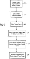
- respiratory gating is performed by image post-processing as shown by the block diagram of FIGURE 5 , which illustrates one implementation of the present invention.
- a sequence of successive images is acquired during one or more motion cycles.
- This image loop could be a sequence of images acquired during the 100+ seconds of contrast agent wash-in and wash-out mentioned above, and could comprise hundreds or even thousands of images. Obviously, manually processing this many images would be a painstaking and likely inaccurate task.
- An image loop this long would cover over 20 respiratory cycles at a nominal breathing rate of thirteen breaths per minute.
- a loop this long would cover about 100 heartbeat cycles at a nominal heart rate of one beat per second. This loop would therefore be contaminated by many cycles of motion of either physiological phenomenon.
- step 52 the cyclical motion is identified.
- the main hepatic vessels above the circle 2, filled as it is with contrast agent is a very bright reflector in the images, indeed it is the largest bright structure in the images.
- the vein is seen to be at its largest and brightest when the cut plane of the image passes through the center of the artery as it is in FIGURE 4a , and less so in the other images. Thresholding or decimation of the pixel values of these images below the hepatic artery brightness will extract the pixels of the hepatic artery from the center of the images and the pixel values summed or integrated to indicate the brightness of this structure in each image.
- the recurrence of the peak brightness of the portal vein each time it moves fully into the cut plane of the probe image plane indicates the periodicity of the respiratory motion moving the image plane, and the duration of the motional cycle is the time between the acquisition of two such images as shown by their time-tagging.
- the images acquired during a desired phase or phase range of the motion are identified in step 54. For instance, it may be decided to use each image of peak brightness ( FIGURE 4a ) and the five images preceding and succeeding the peak brightness image in every respiratory cycle. These eleven images of each respiratory cycle are then selected for perfusion assessment processing as shown in step 56. The images which were acquired during less desirable phases of the respiratory cycle are omitted from processing. As a result the perfusion measurement should be relatively unaffected by the effects of respiratory motion.
- FIGURE 6 illustrates a preferred implementation of the present invention in block diagram form.
- the motion information contained in the image sequence is extracted in what is referred to herein as a "motion information matrix.”
- FIGURES 7a-7d illustrate one way to form a motion information matrix (MIM).
- FIGURE 7a illustrates an ultrasound image of the liver
- FIGURE 7b illustrates an image which is an average of all of the image frames in the loop containing the image of FIGURE 7a .
- Each frame of the loop such as that of FIGURE 7a is subtracted from the average of the frames ( FIGURE 7b ) and pixel intensities above a certain threshold are assigned a value of 1 with all others assigned a value of zero.
- the next step 62 is the selection of a trigger frame from the loop.
- a trigger frame is one which will be included among the finally processed images. It may be one, for instance, in which the lesion is clearly defined as by a high intensity or large size in the image. Or it may be an image which appears virtually identical to previously and/or successively acquired images in the loop and thus exhibits little or no motion contamination.
- the trigger frame may be designated by the user by inspection, or may be selected by an automated method such as by subtracting each pair of successive frames and using using one of the frames which produced the most minimal difference image.
- Candidate structures for motion identification are then extracted from the trigger frame for motion curve formation as shown by step 64. This extraction may comprise a binary image produced from the trigger frame as discussed above.
- the structures extracted from the trigger image are compared with the structures of the MIM to choose one which is significantly affected by motion.
- a structure of the MIM which varies over a wide intensity range or positional range could be selected, for instance.
- the frequency of the variation can be compared with the frequency of the physiological phenomenon to ascertain that its variation corresponds to that of the physiological function. If it does not, the structure is disregarded and another structure of the MIM selected for motion identification.
- the structures of the MIM can be ranked in accordance with their likelihood of exhibiting effects of motion.
- the structure which is found to be the region of greatest interest for motion extraction is then used as a binary mask that is used to extract a curve of the respiratory or other bodily motion.
- the mask of the structure is applied to the corresponding location in each image in the loop and a series of mean intensity values within the mask extracted from the image frames which exhibit intensity variation during the loop that is representative of the cyclical motion. These mean intensity values are plotted as a function of time (or, equivalently, frame number in the sequence) to yield an approximate respiratory curve.
- the initial plot may be fitted to an average curve to produce a smoothly varying respiratory curve as shown by respiratory curve 100 in FIGURE 8 .
- Known curve fitting techniques may be used to produce a smoothly varying curve, such as a piecewise cubic Hermite interpolating polynomial fitted to the peaks and troughs of the approximate curve, for instance.
- FIGURE 8 illustrates a respiratory curve 100 formed from respiratory motion mean intensity variation of a structure over a loop of 600 image frames. It is seen that the curve 100 indicates 21 respiratory cycles occurring during acquisition of the 600 image frames.
- the small circles 110 at the top of the plot indicate some of the discrete frames of the loop. The circles are shown on the respiratory curve in correspondence to the points in the respiratory cycles.
- a threshold 102 is applied to the curve 100 to delineate the phase range of each respiratory cycle during which image frames will be accepted for perfusion processing. For instance, suppose that the lower peaks of the respiratory curve are points where the patient has fully inhaled, and the upper peaks of the curve are points where the patient has fully exhaled.
- the clinician may decide to use image frames that all were acquired around the time of full exhalation, for example, just before and just after the patient has fully exhaled.
- the system then applies a threshold 102 as shown in the drawing which delineates, in this example, this phase range of each respiratory cycle, which is seen in this example to comprise about 30% of each cycle.
- the image frames acquired during this respiratory phase range, the frames represented by the circles 110, are then selected for processing as shown in step 66 of FIGURE 6 .
- the processed image frames of step 68 are thus a set of frames which all were acquired during the same phase range of respiratory motion and should thus generally correspond spatially to each other, with the structures and lesions of the images appearing generally the same in size and position in each of the processed images.
- the data for such quantification may be selected from the accepted images using the trigger frame.
- the lesion is identified in the trigger frame as a region of interest (ROI), and data extracted from the location of the ROI in each of the accepted image frames.
- ROI region of interest
- This data is then processed to produce time-intensity curves as described above and in European patent EP 2 234 544 B1 , the contents of which are incorporated herein.
- FIGURE 9 illustrates time-intensity curves produced in one of these cases, both with and without respiratory phase gating.
- the intensity data of a region of interest in the images was fitted to a lognormal function to produce time-intensity curves of reperfusion.
- the goodness of fit was quantified using the R-squared value and the root mean squared error value.
- the time-intensity curve 120 was produced from data of all of the image frames without gating, and is seen to be affected by the highly oscillatory characteristic of the intensity values 140 sampled at the ROI of the images, which contains large spikes due to sampling of images outside the lesion caused by respiratory motion of the anatomy.
Landscapes
- Health & Medical Sciences (AREA)
- Life Sciences & Earth Sciences (AREA)
- Engineering & Computer Science (AREA)
- Physics & Mathematics (AREA)
- Medical Informatics (AREA)
- General Health & Medical Sciences (AREA)
- Public Health (AREA)
- Radiology & Medical Imaging (AREA)
- Nuclear Medicine, Radiotherapy & Molecular Imaging (AREA)
- Biomedical Technology (AREA)
- Pathology (AREA)
- Biophysics (AREA)
- Animal Behavior & Ethology (AREA)
- Veterinary Medicine (AREA)
- Heart & Thoracic Surgery (AREA)
- Molecular Biology (AREA)
- Surgery (AREA)
- Computer Vision & Pattern Recognition (AREA)
- Physiology (AREA)
- Hematology (AREA)
- General Physics & Mathematics (AREA)
- Theoretical Computer Science (AREA)
- Multimedia (AREA)
- Quality & Reliability (AREA)
- Data Mining & Analysis (AREA)
- Databases & Information Systems (AREA)
- Epidemiology (AREA)
- Primary Health Care (AREA)
- Ultra Sonic Daignosis Equipment (AREA)
- Apparatus For Radiation Diagnosis (AREA)
Applications Claiming Priority (2)
| Application Number | Priority Date | Filing Date | Title |
|---|---|---|---|
| US201361753898P | 2013-01-17 | 2013-01-17 | |
| PCT/IB2014/058293 WO2014111860A2 (en) | 2013-01-17 | 2014-01-15 | Eliminating motion effects in medical images caused by physiological function |
Publications (2)
| Publication Number | Publication Date |
|---|---|
| EP2945544A2 EP2945544A2 (en) | 2015-11-25 |
| EP2945544B1 true EP2945544B1 (en) | 2018-11-07 |
Family
ID=50114440
Family Applications (1)
| Application Number | Title | Priority Date | Filing Date |
|---|---|---|---|
| EP14704901.9A Active EP2945544B1 (en) | 2013-01-17 | 2014-01-15 | Eliminating motion effects in medical images caused by physiological function |
Country Status (7)
| Country | Link |
|---|---|
| US (1) | US9576357B2 (pt) |
| EP (1) | EP2945544B1 (pt) |
| JP (2) | JP6297593B2 (pt) |
| CN (1) | CN104968279B (pt) |
| BR (1) | BR112015016804A2 (pt) |
| RU (1) | RU2015134348A (pt) |
| WO (1) | WO2014111860A2 (pt) |
Families Citing this family (17)
| Publication number | Priority date | Publication date | Assignee | Title |
|---|---|---|---|---|
| EP3097538B1 (en) | 2014-01-23 | 2018-09-26 | Koninklijke Philips N.V. | Evaluation of carotid plaque using contrast enhanced ultrasonic imaging |
| US9519966B2 (en) * | 2015-04-22 | 2016-12-13 | King Fahd University Of Petroleum And Minerals | Method, system and computer program product for breast density classification using parts-based local features |
| KR102525616B1 (ko) * | 2015-10-08 | 2023-04-26 | 삼성메디슨 주식회사 | 조영제 초음파 진단 장치 및 방법 |
| GB201519985D0 (en) * | 2015-11-12 | 2015-12-30 | Respinor As | Ultrasonic method and apparatus for respiration monitoring |
| CN107133549B (zh) | 2016-02-29 | 2020-11-24 | 上海联影医疗科技有限公司 | Ect运动门控信号获取方法及ect图像重建方法 |
| CN109414246B (zh) * | 2016-11-09 | 2021-09-14 | 深圳市理邦精密仪器股份有限公司 | 用于多普勒频谱时间持续性的系统和方法 |
| EP3565478B1 (en) * | 2017-01-04 | 2022-09-28 | Koninklijke Philips N.V. | Time-based parametric contrast enhanced ultrasound imaging system and method |
| US11419584B2 (en) * | 2017-06-28 | 2022-08-23 | Tokitae, LLC | Devices, systems, and methods for diagnosis of pulmonary conditions through detection of b-lines in lung sonography |
| CN110019855B (zh) * | 2017-12-04 | 2023-07-21 | 沪江教育科技(上海)股份有限公司 | 根据时间轴控制动效的方法、装置、存储介质及电子设备 |
| JP7084193B2 (ja) | 2018-04-10 | 2022-06-14 | ザイオソフト株式会社 | 医用画像処理装置、医用画像処理方法、及び医用画像処理プログラム |
| US11497475B2 (en) * | 2020-01-31 | 2022-11-15 | Caption Health, Inc. | Ultrasound image acquisition optimization according to different respiration modes |
| US11816832B2 (en) | 2020-11-18 | 2023-11-14 | Canon Medical Systems Corporation | Devices, systems, and methods for medical imaging |
| US11712215B2 (en) | 2021-04-13 | 2023-08-01 | Canon Medical Systems Corporation | Devices, systems, and methods for motion-corrected medical imaging |
| WO2023025837A1 (en) * | 2021-08-27 | 2023-03-02 | Koninklijke Philips N.V. | Method for use in analysing ultrasound image data of a subject |
| CN114403924B (zh) * | 2022-01-19 | 2024-02-02 | 复旦大学附属中山医院 | 一种基于超声造影评估aip激素治疗疗效的方法 |
| WO2024067948A1 (en) * | 2022-09-26 | 2024-04-04 | University College Dublin | Method and apparatus |
| CN116306937B (zh) * | 2023-03-22 | 2023-11-10 | 中航信移动科技有限公司 | 一种基于时间序列离线数据的规则提取方法、介质及设备 |
Citations (1)
| Publication number | Priority date | Publication date | Assignee | Title |
|---|---|---|---|---|
| WO2009093211A1 (en) * | 2008-01-23 | 2009-07-30 | Michalakis Averkiou | Therapy assessment with ultrasonic contrast agents |
Family Cites Families (27)
| Publication number | Priority date | Publication date | Assignee | Title |
|---|---|---|---|---|
| US5706819A (en) | 1995-10-10 | 1998-01-13 | Advanced Technology Laboratories, Inc. | Ultrasonic diagnostic imaging with harmonic contrast agents |
| EP0770352B1 (en) * | 1995-10-10 | 2004-12-29 | Advanced Technology Laboratories, Inc. | Ultrasonic diagnostic imaging with contrast agents |
| US5833613A (en) | 1996-09-27 | 1998-11-10 | Advanced Technology Laboratories, Inc. | Ultrasonic diagnostic imaging with contrast agents |
| US5577505A (en) | 1996-02-06 | 1996-11-26 | Hewlett-Packard Company | Means for increasing sensitivity in non-linear ultrasound imaging systems |
| CN1106825C (zh) * | 1997-02-13 | 2003-04-30 | 通用电器横河医疗系统株式会社 | 识别被观察器官状态时间相位的超声波诊断装置 |
| JP2000210283A (ja) * | 1999-01-25 | 2000-08-02 | Shimadzu Corp | 医用動画像の運動周期求出装置 |
| US6692438B2 (en) | 2001-12-18 | 2004-02-17 | Koninklijke Philips Electronics Nv | Ultrasonic imaging system and method for displaying tissue perfusion and other parameters varying with time |
| JP2003210456A (ja) * | 2002-01-21 | 2003-07-29 | Toshiba Corp | 時系列画像の処理装置 |
| US7587074B2 (en) * | 2003-07-21 | 2009-09-08 | Paieon Inc. | Method and system for identifying optimal image within a series of images that depict a moving organ |
| US7731660B2 (en) * | 2003-07-25 | 2010-06-08 | Siemens Medical Solutions Usa, Inc. | Phase selection for cardiac contrast assessment |
| JP4682149B2 (ja) | 2003-12-03 | 2011-05-11 | コーニンクレッカ フィリップス エレクトロニクス エヌ ヴィ | 血流及び潅流パラメータを同時に表示するための超音波イメージングシステムおよび方法 |
| US20080281205A1 (en) * | 2004-01-16 | 2008-11-13 | Morteza Naghavi | Methods and Apparatuses For Medical Imaging |
| US20050215904A1 (en) * | 2004-03-23 | 2005-09-29 | Siemens Medical Solutions Usa, Inc. | Ultrasound breathing waveform detection system and method |
| CN1942142B (zh) | 2004-04-16 | 2012-04-25 | 皇家飞利浦电子股份有限公司 | 自动心肌造影超声心动图 |
| JP2005342006A (ja) * | 2004-05-31 | 2005-12-15 | Toshiba Corp | 超音波診断装置、超音波画像処理装置、及び超音波信号処理プログラム |
| US20060074315A1 (en) * | 2004-10-04 | 2006-04-06 | Jianming Liang | Medical diagnostic ultrasound characterization of cardiac motion |
| JP4685458B2 (ja) * | 2005-01-18 | 2011-05-18 | 株式会社東芝 | 超音波診断装置 |
| CN100509083C (zh) * | 2005-01-31 | 2009-07-08 | 重庆微海软件开发有限公司 | 利用呼吸信号控制图像同步的方法及其装置 |
| EP1855596B1 (en) | 2005-02-23 | 2015-07-01 | Koninklijke Philips N.V. | Ultrasonic diagnostic imaging system for detecting lesions of the liver |
| KR100856042B1 (ko) * | 2005-10-07 | 2008-09-03 | 주식회사 메디슨 | 웨이브렛 변환과 svm을 이용하여 초음파 영상에서대상체 볼륨을 추출하는 초음파 영상 시스템 및 방법 |
| JP4206107B2 (ja) * | 2006-07-05 | 2009-01-07 | アロカ株式会社 | 超音波診断装置 |
| JP4912807B2 (ja) * | 2006-09-22 | 2012-04-11 | 株式会社東芝 | 超音波画像診断装置 |
| WO2008107905A2 (en) * | 2007-03-08 | 2008-09-12 | Sync-Rx, Ltd. | Imaging and tools for use with moving organs |
| JP2009082181A (ja) * | 2007-09-27 | 2009-04-23 | Aloka Co Ltd | 超音波診断装置 |
| US9289191B2 (en) * | 2011-10-12 | 2016-03-22 | Seno Medical Instruments, Inc. | System and method for acquiring optoacoustic data and producing parametric maps thereof |
| US20130172730A1 (en) * | 2011-12-29 | 2013-07-04 | Amit Cohen | Motion-Compensated Image Fusion |
| JP6444302B2 (ja) * | 2012-07-16 | 2018-12-26 | ミラビリス メディカ インク | 超音波誘導治療のためのヒューマンインターフェースおよびデバイス |
-
2014
- 2014-01-15 JP JP2015553207A patent/JP6297593B2/ja active Active
- 2014-01-15 WO PCT/IB2014/058293 patent/WO2014111860A2/en active Application Filing
- 2014-01-15 US US14/760,736 patent/US9576357B2/en active Active
- 2014-01-15 CN CN201480005221.6A patent/CN104968279B/zh active Active
- 2014-01-15 BR BR112015016804A patent/BR112015016804A2/pt not_active IP Right Cessation
- 2014-01-15 RU RU2015134348A patent/RU2015134348A/ru not_active Application Discontinuation
- 2014-01-15 EP EP14704901.9A patent/EP2945544B1/en active Active
-
2018
- 2018-02-21 JP JP2018028589A patent/JP6542928B2/ja active Active
Patent Citations (1)
| Publication number | Priority date | Publication date | Assignee | Title |
|---|---|---|---|---|
| WO2009093211A1 (en) * | 2008-01-23 | 2009-07-30 | Michalakis Averkiou | Therapy assessment with ultrasonic contrast agents |
Non-Patent Citations (1)
| Title |
|---|
| YOUNGKYOO HWANG ET AL: "Robust real-time respiratory motion tracking using ultrasound image sequences", ULTRASONICS SYMPOSIUM (IUS), 2012 IEEE INTERNATIONAL, IEEE, 7 October 2012 (2012-10-07), pages 1666 - 1669, XP032434584, ISSN: 1948-5719, ISBN: 978-1-4673-4561-3, DOI: 10.1109/ULTSYM.2012.0418 * |
Also Published As
| Publication number | Publication date |
|---|---|
| JP2018118061A (ja) | 2018-08-02 |
| WO2014111860A2 (en) | 2014-07-24 |
| CN104968279B (zh) | 2018-08-10 |
| WO2014111860A3 (en) | 2014-09-18 |
| BR112015016804A2 (pt) | 2017-07-11 |
| JP2016503707A (ja) | 2016-02-08 |
| JP6542928B2 (ja) | 2019-07-10 |
| US20150371379A1 (en) | 2015-12-24 |
| EP2945544A2 (en) | 2015-11-25 |
| US9576357B2 (en) | 2017-02-21 |
| JP6297593B2 (ja) | 2018-03-20 |
| RU2015134348A (ru) | 2017-02-20 |
| CN104968279A (zh) | 2015-10-07 |
Similar Documents
| Publication | Publication Date | Title |
|---|---|---|
| EP2945544B1 (en) | Eliminating motion effects in medical images caused by physiological function | |
| US8460194B2 (en) | Therapy assessment with ultrasound contrast agents | |
| JP5680654B2 (ja) | 超音波診断装置及び超音波画像表示方法 | |
| US9380999B2 (en) | Ultrasonic diagnostic apparatus, ultrasonic image processing apparatus, and medical diagnostic imaging apparatus | |
| US20120253190A1 (en) | Contrast-enhanced ultrasound assessment of liver blood flow for monitoring liver therapy | |
| US20110208061A1 (en) | Ultrasonic lesion identification using temporal parametric contrast images | |
| JP7041147B2 (ja) | 造影剤流の肝灌流を特徴付けるシステム及び方法 | |
| JPH08308831A (ja) | 超音波診断装置 | |
| CN114245725A (zh) | 在洗入、洗出期间改变系统操作的对比度增强超声成像 | |
| EP3378404A1 (en) | Ultrasonic diagnostic system and method for contrast enhanced liver diagnosis | |
| CN110167448B (zh) | 基于时间的参数对比增强超声成像系统和方法 | |
| Barrois et al. | A multiplicative model to improve microvascular flow evaluation in the context of dynamic contrast-enhanced ultrasound (DCE-US) |
Legal Events
| Date | Code | Title | Description |
|---|---|---|---|
| PUAI | Public reference made under article 153(3) epc to a published international application that has entered the european phase |
Free format text: ORIGINAL CODE: 0009012 |
|
| 17P | Request for examination filed |
Effective date: 20150817 |
|
| AK | Designated contracting states |
Kind code of ref document: A2 Designated state(s): AL AT BE BG CH CY CZ DE DK EE ES FI FR GB GR HR HU IE IS IT LI LT LU LV MC MK MT NL NO PL PT RO RS SE SI SK SM TR |
|
| AX | Request for extension of the european patent |
Extension state: BA ME |
|
| DAX | Request for extension of the european patent (deleted) | ||
| STAA | Information on the status of an ep patent application or granted ep patent |
Free format text: STATUS: EXAMINATION IS IN PROGRESS |
|
| 17Q | First examination report despatched |
Effective date: 20170503 |
|
| GRAP | Despatch of communication of intention to grant a patent |
Free format text: ORIGINAL CODE: EPIDOSNIGR1 |
|
| STAA | Information on the status of an ep patent application or granted ep patent |
Free format text: STATUS: GRANT OF PATENT IS INTENDED |
|
| INTG | Intention to grant announced |
Effective date: 20180601 |
|
| GRAS | Grant fee paid |
Free format text: ORIGINAL CODE: EPIDOSNIGR3 |
|
| GRAA | (expected) grant |
Free format text: ORIGINAL CODE: 0009210 |
|
| STAA | Information on the status of an ep patent application or granted ep patent |
Free format text: STATUS: THE PATENT HAS BEEN GRANTED |
|
| AK | Designated contracting states |
Kind code of ref document: B1 Designated state(s): AL AT BE BG CH CY CZ DE DK EE ES FI FR GB GR HR HU IE IS IT LI LT LU LV MC MK MT NL NO PL PT RO RS SE SI SK SM TR |
|
| REG | Reference to a national code |
Ref country code: GB Ref legal event code: FG4D |
|
| REG | Reference to a national code |
Ref country code: CH Ref legal event code: EP Ref country code: AT Ref legal event code: REF Ref document number: 1061132 Country of ref document: AT Kind code of ref document: T Effective date: 20181115 |
|
| REG | Reference to a national code |
Ref country code: DE Ref legal event code: R096 Ref document number: 602014035524 Country of ref document: DE |
|
| REG | Reference to a national code |
Ref country code: IE Ref legal event code: FG4D |
|
| REG | Reference to a national code |
Ref country code: GB Ref legal event code: 746 Effective date: 20190102 |
|
| REG | Reference to a national code |
Ref country code: DE Ref legal event code: R084 Ref document number: 602014035524 Country of ref document: DE |
|
| REG | Reference to a national code |
Ref country code: NL Ref legal event code: MP Effective date: 20181107 |
|
| REG | Reference to a national code |
Ref country code: LT Ref legal event code: MG4D |
|
| REG | Reference to a national code |
Ref country code: AT Ref legal event code: MK05 Ref document number: 1061132 Country of ref document: AT Kind code of ref document: T Effective date: 20181107 |
|
| PG25 | Lapsed in a contracting state [announced via postgrant information from national office to epo] |
Ref country code: AT Free format text: LAPSE BECAUSE OF FAILURE TO SUBMIT A TRANSLATION OF THE DESCRIPTION OR TO PAY THE FEE WITHIN THE PRESCRIBED TIME-LIMIT Effective date: 20181107 Ref country code: HR Free format text: LAPSE BECAUSE OF FAILURE TO SUBMIT A TRANSLATION OF THE DESCRIPTION OR TO PAY THE FEE WITHIN THE PRESCRIBED TIME-LIMIT Effective date: 20181107 Ref country code: NO Free format text: LAPSE BECAUSE OF FAILURE TO SUBMIT A TRANSLATION OF THE DESCRIPTION OR TO PAY THE FEE WITHIN THE PRESCRIBED TIME-LIMIT Effective date: 20190207 Ref country code: LV Free format text: LAPSE BECAUSE OF FAILURE TO SUBMIT A TRANSLATION OF THE DESCRIPTION OR TO PAY THE FEE WITHIN THE PRESCRIBED TIME-LIMIT Effective date: 20181107 Ref country code: BG Free format text: LAPSE BECAUSE OF FAILURE TO SUBMIT A TRANSLATION OF THE DESCRIPTION OR TO PAY THE FEE WITHIN THE PRESCRIBED TIME-LIMIT Effective date: 20190207 Ref country code: LT Free format text: LAPSE BECAUSE OF FAILURE TO SUBMIT A TRANSLATION OF THE DESCRIPTION OR TO PAY THE FEE WITHIN THE PRESCRIBED TIME-LIMIT Effective date: 20181107 Ref country code: ES Free format text: LAPSE BECAUSE OF FAILURE TO SUBMIT A TRANSLATION OF THE DESCRIPTION OR TO PAY THE FEE WITHIN THE PRESCRIBED TIME-LIMIT Effective date: 20181107 Ref country code: IS Free format text: LAPSE BECAUSE OF FAILURE TO SUBMIT A TRANSLATION OF THE DESCRIPTION OR TO PAY THE FEE WITHIN THE PRESCRIBED TIME-LIMIT Effective date: 20190307 Ref country code: FI Free format text: LAPSE BECAUSE OF FAILURE TO SUBMIT A TRANSLATION OF THE DESCRIPTION OR TO PAY THE FEE WITHIN THE PRESCRIBED TIME-LIMIT Effective date: 20181107 |
|
| PG25 | Lapsed in a contracting state [announced via postgrant information from national office to epo] |
Ref country code: GR Free format text: LAPSE BECAUSE OF FAILURE TO SUBMIT A TRANSLATION OF THE DESCRIPTION OR TO PAY THE FEE WITHIN THE PRESCRIBED TIME-LIMIT Effective date: 20190208 Ref country code: SE Free format text: LAPSE BECAUSE OF FAILURE TO SUBMIT A TRANSLATION OF THE DESCRIPTION OR TO PAY THE FEE WITHIN THE PRESCRIBED TIME-LIMIT Effective date: 20181107 Ref country code: RS Free format text: LAPSE BECAUSE OF FAILURE TO SUBMIT A TRANSLATION OF THE DESCRIPTION OR TO PAY THE FEE WITHIN THE PRESCRIBED TIME-LIMIT Effective date: 20181107 Ref country code: PT Free format text: LAPSE BECAUSE OF FAILURE TO SUBMIT A TRANSLATION OF THE DESCRIPTION OR TO PAY THE FEE WITHIN THE PRESCRIBED TIME-LIMIT Effective date: 20190307 Ref country code: AL Free format text: LAPSE BECAUSE OF FAILURE TO SUBMIT A TRANSLATION OF THE DESCRIPTION OR TO PAY THE FEE WITHIN THE PRESCRIBED TIME-LIMIT Effective date: 20181107 Ref country code: NL Free format text: LAPSE BECAUSE OF FAILURE TO SUBMIT A TRANSLATION OF THE DESCRIPTION OR TO PAY THE FEE WITHIN THE PRESCRIBED TIME-LIMIT Effective date: 20181107 |
|
| PG25 | Lapsed in a contracting state [announced via postgrant information from national office to epo] |
Ref country code: DK Free format text: LAPSE BECAUSE OF FAILURE TO SUBMIT A TRANSLATION OF THE DESCRIPTION OR TO PAY THE FEE WITHIN THE PRESCRIBED TIME-LIMIT Effective date: 20181107 Ref country code: IT Free format text: LAPSE BECAUSE OF FAILURE TO SUBMIT A TRANSLATION OF THE DESCRIPTION OR TO PAY THE FEE WITHIN THE PRESCRIBED TIME-LIMIT Effective date: 20181107 Ref country code: CZ Free format text: LAPSE BECAUSE OF FAILURE TO SUBMIT A TRANSLATION OF THE DESCRIPTION OR TO PAY THE FEE WITHIN THE PRESCRIBED TIME-LIMIT Effective date: 20181107 Ref country code: PL Free format text: LAPSE BECAUSE OF FAILURE TO SUBMIT A TRANSLATION OF THE DESCRIPTION OR TO PAY THE FEE WITHIN THE PRESCRIBED TIME-LIMIT Effective date: 20181107 |
|
| REG | Reference to a national code |
Ref country code: DE Ref legal event code: R097 Ref document number: 602014035524 Country of ref document: DE |
|
| PG25 | Lapsed in a contracting state [announced via postgrant information from national office to epo] |
Ref country code: RO Free format text: LAPSE BECAUSE OF FAILURE TO SUBMIT A TRANSLATION OF THE DESCRIPTION OR TO PAY THE FEE WITHIN THE PRESCRIBED TIME-LIMIT Effective date: 20181107 Ref country code: SK Free format text: LAPSE BECAUSE OF FAILURE TO SUBMIT A TRANSLATION OF THE DESCRIPTION OR TO PAY THE FEE WITHIN THE PRESCRIBED TIME-LIMIT Effective date: 20181107 Ref country code: MC Free format text: LAPSE BECAUSE OF FAILURE TO SUBMIT A TRANSLATION OF THE DESCRIPTION OR TO PAY THE FEE WITHIN THE PRESCRIBED TIME-LIMIT Effective date: 20181107 Ref country code: EE Free format text: LAPSE BECAUSE OF FAILURE TO SUBMIT A TRANSLATION OF THE DESCRIPTION OR TO PAY THE FEE WITHIN THE PRESCRIBED TIME-LIMIT Effective date: 20181107 Ref country code: SM Free format text: LAPSE BECAUSE OF FAILURE TO SUBMIT A TRANSLATION OF THE DESCRIPTION OR TO PAY THE FEE WITHIN THE PRESCRIBED TIME-LIMIT Effective date: 20181107 |
|
| REG | Reference to a national code |
Ref country code: CH Ref legal event code: PL |
|
| PLBE | No opposition filed within time limit |
Free format text: ORIGINAL CODE: 0009261 |
|
| STAA | Information on the status of an ep patent application or granted ep patent |
Free format text: STATUS: NO OPPOSITION FILED WITHIN TIME LIMIT |
|
| PG25 | Lapsed in a contracting state [announced via postgrant information from national office to epo] |
Ref country code: LU Free format text: LAPSE BECAUSE OF NON-PAYMENT OF DUE FEES Effective date: 20190115 |
|
| 26N | No opposition filed |
Effective date: 20190808 |
|
| REG | Reference to a national code |
Ref country code: BE Ref legal event code: MM Effective date: 20190131 |
|
| REG | Reference to a national code |
Ref country code: IE Ref legal event code: MM4A |
|
| PG25 | Lapsed in a contracting state [announced via postgrant information from national office to epo] |
Ref country code: SI Free format text: LAPSE BECAUSE OF FAILURE TO SUBMIT A TRANSLATION OF THE DESCRIPTION OR TO PAY THE FEE WITHIN THE PRESCRIBED TIME-LIMIT Effective date: 20181107 |
|
| PG25 | Lapsed in a contracting state [announced via postgrant information from national office to epo] |
Ref country code: BE Free format text: LAPSE BECAUSE OF NON-PAYMENT OF DUE FEES Effective date: 20190131 |
|
| PG25 | Lapsed in a contracting state [announced via postgrant information from national office to epo] |
Ref country code: LI Free format text: LAPSE BECAUSE OF NON-PAYMENT OF DUE FEES Effective date: 20190131 Ref country code: CH Free format text: LAPSE BECAUSE OF NON-PAYMENT OF DUE FEES Effective date: 20190131 |
|
| PG25 | Lapsed in a contracting state [announced via postgrant information from national office to epo] |
Ref country code: IE Free format text: LAPSE BECAUSE OF NON-PAYMENT OF DUE FEES Effective date: 20190115 |
|
| PG25 | Lapsed in a contracting state [announced via postgrant information from national office to epo] |
Ref country code: TR Free format text: LAPSE BECAUSE OF FAILURE TO SUBMIT A TRANSLATION OF THE DESCRIPTION OR TO PAY THE FEE WITHIN THE PRESCRIBED TIME-LIMIT Effective date: 20181107 |
|
| PG25 | Lapsed in a contracting state [announced via postgrant information from national office to epo] |
Ref country code: MT Free format text: LAPSE BECAUSE OF NON-PAYMENT OF DUE FEES Effective date: 20190115 |
|
| PG25 | Lapsed in a contracting state [announced via postgrant information from national office to epo] |
Ref country code: CY Free format text: LAPSE BECAUSE OF FAILURE TO SUBMIT A TRANSLATION OF THE DESCRIPTION OR TO PAY THE FEE WITHIN THE PRESCRIBED TIME-LIMIT Effective date: 20181107 |
|
| PG25 | Lapsed in a contracting state [announced via postgrant information from national office to epo] |
Ref country code: HU Free format text: LAPSE BECAUSE OF FAILURE TO SUBMIT A TRANSLATION OF THE DESCRIPTION OR TO PAY THE FEE WITHIN THE PRESCRIBED TIME-LIMIT; INVALID AB INITIO Effective date: 20140115 |
|
| PG25 | Lapsed in a contracting state [announced via postgrant information from national office to epo] |
Ref country code: MK Free format text: LAPSE BECAUSE OF FAILURE TO SUBMIT A TRANSLATION OF THE DESCRIPTION OR TO PAY THE FEE WITHIN THE PRESCRIBED TIME-LIMIT Effective date: 20181107 |
|
| PGFP | Annual fee paid to national office [announced via postgrant information from national office to epo] |
Ref country code: FR Payment date: 20230124 Year of fee payment: 10 |
|
| PGFP | Annual fee paid to national office [announced via postgrant information from national office to epo] |
Ref country code: DE Payment date: 20240129 Year of fee payment: 11 Ref country code: GB Payment date: 20240123 Year of fee payment: 11 |