EP2833149B1 - Verfahren zur erkennung des malignitätsgrades einer zirkulierenden tumorzelleneinheit und kit dafür - Google Patents
Verfahren zur erkennung des malignitätsgrades einer zirkulierenden tumorzelleneinheit und kit dafür Download PDFInfo
- Publication number
- EP2833149B1 EP2833149B1 EP13770163.7A EP13770163A EP2833149B1 EP 2833149 B1 EP2833149 B1 EP 2833149B1 EP 13770163 A EP13770163 A EP 13770163A EP 2833149 B1 EP2833149 B1 EP 2833149B1
- Authority
- EP
- European Patent Office
- Prior art keywords
- cell
- circulating tumor
- binding component
- epithelial
- cancer
- Prior art date
- Legal status (The legal status is an assumption and is not a legal conclusion. Google has not performed a legal analysis and makes no representation as to the accuracy of the status listed.)
- Active
Links
Images


Classifications
-
- G01N33/5759—
-
- C—CHEMISTRY; METALLURGY
- C12—BIOCHEMISTRY; BEER; SPIRITS; WINE; VINEGAR; MICROBIOLOGY; ENZYMOLOGY; MUTATION OR GENETIC ENGINEERING
- C12N—MICROORGANISMS OR ENZYMES; COMPOSITIONS THEREOF; PROPAGATING, PRESERVING, OR MAINTAINING MICROORGANISMS; MUTATION OR GENETIC ENGINEERING; CULTURE MEDIA
- C12N5/00—Undifferentiated human, animal or plant cells, e.g. cell lines; Tissues; Cultivation or maintenance thereof; Culture media therefor
- C12N5/06—Animal cells or tissues; Human cells or tissues
- C12N5/0602—Vertebrate cells
- C12N5/0693—Tumour cells; Cancer cells
-
- G01N33/575—
-
- G—PHYSICS
- G01—MEASURING; TESTING
- G01N—INVESTIGATING OR ANALYSING MATERIALS BY DETERMINING THEIR CHEMICAL OR PHYSICAL PROPERTIES
- G01N33/00—Investigating or analysing materials by specific methods not covered by groups G01N1/00 - G01N31/00
- G01N33/48—Biological material, e.g. blood, urine; Haemocytometers
- G01N33/50—Chemical analysis of biological material, e.g. blood, urine; Testing involving biospecific ligand binding methods; Immunological testing
- G01N33/58—Chemical analysis of biological material, e.g. blood, urine; Testing involving biospecific ligand binding methods; Immunological testing involving labelled substances
- G01N33/582—Chemical analysis of biological material, e.g. blood, urine; Testing involving biospecific ligand binding methods; Immunological testing involving labelled substances with fluorescent label
Definitions
- the present invention relates to a method for detecting the degree of malignancy of each of the circulating tumor cells.
- CTC circulating tumor cell
- CTCs When CTCs are detected in the peripheral blood of cancer patients, extremely-low concentrated CTCs which are one-billionth of high concentrated blood cells, should be detected in the high concentrated blood cells. Thus, a count loss of CTCs or a contamination between samples of patients leads to a seriously wrong diagnosis.
- the measurement of CTCs is carried out using CellSearch System (U.S.A).
- U.S.A CellSearch System
- the cells are subjected to nuclear staining and cytokeratin staining, and then the cells are reacted with CD326 antibody-immobilized magnetic beads. The resulting cells are then allowed to float through a magnetic field.
- Fluorescent imaging of cells are obtained by scanning by laser beam and then a human determines whether or not each of the cells is CTC, on the basis of the fluorescent imaging. Further, there is a technique called CTC-Chip as a method for measuring CTCs. As disclosed in Patent reference 2, in the above technique, a blood sample is passed through a chip in which 80,000 micro posts are formed on a silicon wafer about the size of a business card. Then, all of the 80,000 micro posts immobilized with anti-EpCAM antibody are image-recognized so as to identify and measure CTCs.
- Patent reference 3 discloses that CTCs detected by the method disclosed in Patent reference 1, are recovered, and then genes of the recovered CTCs are analyzed by fluorescence in situ hybridization (hereinafter sometimes referred to as FISH).
- Patent reference 4 and 9 discloses a flowcytometer using a disposable chip. A measurement without a cross contamination of samples can be conducted with the above flowcytometer.
- Patent reference 5 discloses a conventional flow cytometer wherein a solution sending system including a flow cell is fixed.
- Patent reference 6 discloses that CTCs detected by the method disclosed in Patent reference 1, are recovered, and then an abnormality of IGF-1R gene of CTCs is analyzed by FISH.
- Patent reference 8 discloses that CTCs are concentrated by a microfilter on the basis of cell size.
- Non-patent reference 1 discloses that a fluorescent protein i.e. GFP is expressed in cancer cells by using a virus capable of infecting cancer cells only, and the expressed cell is detected by a fluorescence microscope.
- Non-patent reference 1 discloses that when an epithelial-mesenchymal transition (hereinafter sometimes referred to as EMT) in cancer cells derived from an epithelial cell is induced, the cancer cells easily become detached and easily migrate to other regions.
- EMT epithelial-mesenchymal transition
- the EMT is proposed by Elizabeth Hay et al. in the early 1980's. In the EMT, the epithelial cell is morphologically altered to a mesenchymal-like cell.
- Non-patent reference 1 discloses that a degree of EMT is related to a metastasis of cancer.
- Non-patent reference 2 discloses that an EMT is induced in CTC derived from epithelial cell also.
- Non-patent reference 7 discloses findings on EMT of culture cells by protein analysis using an electrophoresis. It is reported that a ratio between an expression amount of cytokeratin and an expression amount of vimentin is not altered, but constant during the cell cycle. The ratio between expressions is measured by data of electrophoresis, and therefore the ratio is the average of a number of cells.
- Non-patent reference 4 a method for separating CTCs derived from prostate cancer from leucocytes and concentrating CTCs by means of CPT Vacutainer tubes manufactured by Becton, Dickinson and Company is disclosed.
- Non-patent reference 5 discloses a CYTOTRACK system manufactured by CYTOTRACK (U.S.A). The CYTOTRACK system does not comprise a specific concentration step before detection. That is to say, cells containing CTCs are separately fixed on a disc after labelling with a fluorescent antibody. Consequently, the CTCs on the disc are detected by laser-scanning using a compact disc turntable system.
- Non-patent reference 6 a method for magnetically-concentrating CTCs using an anti-EpCAM antibody, and a method for detecting CTCs using the apparatus disclosed in Patent reference 4, are disclosed.
- a reagent kit relevant to CTCs there may be mentioned a basic research reagent kit for magnetically-concentrating CTCs from peripheral blood (D326(EpCAM)Tumor Cell Enrichment and Detection Kit; Miltenyi Biotec, catalog number 130-090-500); a reagent kit for magnetically-concentrating epithelial cells(HUMAN EpCAM POSITIVE SELECTION KIT; STEMCELL Technologies, catalog number 18356); a reagent kit for negatively-selecting CTCs from bloods by cross-linking other cells such as erythrocytes and leukocytes other than CTCs and density-gradiently centrifuging the cells (Tumor Cell Enrichment Cocktail; STEMCELL Technologies, catalog number 15167); a reagent kit for concentrating CTCs by using antibody-immobilized magnetic beads and negatively-selecting CTCs using magnets (Tumor Cell Enrichment Cocktail; STEMCELL Technologies, catalog number 14152).
- EMT epithelial-mesenchymal transition
- the first problem is related to a detection of CTCs wherein an epithelial-mesenchymal transition is induced, by using EpCAM, as mentioned below.
- Non-Patent reference 2 it is reported that epithelial-mesenchymal transition (EMT) is induced in the CTCs derived from epithelial cancer cells. If the epithelial-mesenchymal transition is induced, it is considered that a single cancer cell which forms a mass of cancer can migrate, and thus the cancer cell can easily metastasize. Further, an expression of EpCAM on cancer cells is reduced by EMT. Therefore, in the method for detecting CTCs by concentrating CTCs using an anti-EpCAM antibody, it is difficult to detect CTCs having a high malignancy and a high metastatic property, wherein EMT is progressively induced.
- EMT epithelial-mesenchymal transition
- the second problem is related to an evaluation of the stages of cancer progression according to a ratio of an induction of epithelial-mesenchymal transition, as mentioned below.
- Non-patent reference 2 discloses that a rate of EMT-induced CTC is high in patients with metastatic cancer compared to patients with early-stage cancer. Thus, it is considered that the rate of EMT-induced CTC is related to the patient's prognosis, in addition to the number (concentration) of CTC. That is, it is important to analyze an EMT induction in each of the CTCs.
- Non-Patent reference 2 in the method for analyzing the EMT induction in each of the CTCs, cytokeratin (CK) and vimentin on CTC are doubly, fluorescently-stained. When it is confirmed from an image of fluorescence microscope that vimentin on CTC is fluorescently-stained in addition to cytokeratin (CK), it is determined that the EMT is induced. According to this report, EMT is induced in 70% or more of the CTCs of patients with early-stage cancer, and EMT is induced in 100% of the CTCs of patients with metastatic cancer.
- EMT is induced in 70% of circulating tumor cells of the patients with early-stage cancer, and thus it is very difficult to evaluate a stage of cancer progression or a malignancy of cancer from the rate of EMT induction in circulating tumor cells. Further, it is impossible to adopt the method for evaluating EMT disclosed in Non-patent reference 7, as one for evaluating EMT of the circulating tumor cells wherein the number thereof is low.
- the object of the present invention is to provide an evaluation method capable of accurately determining metastasis of cancer, stage of cancer progression, or malignancy of cancer.
- the present inventors have conducted intensive studies into the evaluation method of circulating tumor cells. As a result, the present inventors, surprisingly, found that the metastasis of cancer, stage of cancer progression, or malignancy of cancer can be accurately diagnosed by measuring an amount of a marker (such as cytokeratin) expressed on a epithelial cell and an amount of a marker (such as vimentin) expressed on a mesenchymal cell, and expressing the same as an EMT index.
- a marker such as cytokeratin
- the present invention is based on the above findings.
- the present invention relates to
- the metastasis of cancer, stage of cancer progression, or malignancy of cancer can be accurately diagnosed.
- the detecting method of the present invention can be used in an occurrence prediction of metastasis after extirpation of the cancer, or the monitoring of the stages of cancer progression.
- the present invention can be used in monitoring therapeutic effects of anticancer drugs by measuring CTCs after administration of said anticancer drugs and evaluating the degree of EMT in the CTCs.
- the method for detecting the degree of malignancy of each of the circulating tumor cells of the present invention comprises the steps of: (a) bringing an epithelial cell-binding component which specifically binds to a marker molecule expressed on epithelial cells and is fluorescently-labeled or luminescent enzyme-labeled, and a mesenchymal cell-binding component which specifically binds to a marker molecule expressed on mesenchymal cells and is fluorescently-labeled or luminescent enzyme-labeled, into contact with a sample that may possibly contain circulating tumor cells, (b) detecting a fluorescence signal or a luminescence signal of the epithelial cell-binding component and a fluorescence signal or a luminescence signal of the mesenchymal cell-binding component of each of the cells, wherein the fluorescence signal or the luminescence signal is detected by a flow cytometer or an image analyzer and (c) determining the degree of epithelial-mesenchymal transition of circulating
- the method for detecting the degree of malignancy of each of the circulating tumor cells of the present invention further comprises the step of (d) removing erythrocytes and/or leucocytes from the sample, before or after step (a).
- the detecting method of the present invention can be used as a method for detecting (diagnosing) the metastasis of cancer, a method for detecting (diagnosing) the stage of cancer progression, or a method for monitoring the therapeutic effects in cancer, in particular, for monitoring the therapeutic effects of an anticancer drug.
- a sample that may possibly contain circulating tumor cells is brought into contact with an epithelial cell-binding component which specifically binds to a marker molecule expressed on epithelial cells and is fluorescently-labeled or luminescent enzyme-labeled, and a mesenchymal cell-binding component which specifically binds to a marker molecule expressed on mesenchymal cells and is fluorescently-labeled or luminescent enzyme-labeled.
- the marker expressed on an epithelial cell is not particularly limited as long as it was expressed on the circulating tumor cells, but includes, for example, a surface protein and a sugar chain of an epithelial cell.
- cytokeratin, EpCAM, or E-cadherin can be used as the epithelial cell marker, and in particular, cytokeratin is preferabl.
- the cytokeratin at least one cytokeratin selected from a group consisting of CK1, CK4, CK5, CK8, CK10, CK14, CK15, CK16, CK18, and CK19 may be used, but the mixture of two or more thereof is preferable.
- cytokeratin, EpCAM, and E-cadherin are labeled with the same fluorescence, and the total expression amount thereof can be used as the expression amount of the marker expressed on an epithelial cell.
- a combination of the above markers is not limited, but contains cytokeratin, preferably.
- the epithelial cell-binding component which specifically binds to the epithelial cell marker is not limited, so long as it can be bound to the epithelial cell marker, but it includes, for example, antibody, antibody fragment, antigen, DNA, RNA, receptor, ligand for receptor, enzyme, ligand for enzyme, enzyme analog, substrate for the enzyme to be based on enzyme analog, lectin, or sugar chain.
- the epithelial cell marker is protein, antibody, antibody fragment, ligand for receptor, ligand for enzyme, DNA (such as aptamer), or RNA may be used.
- DNA such as aptamer
- the epithelial cell marker is sugar chain, antibody, antibody fragment, or lectin may be used.
- mesenchymal cell marker Marker expressed on mesenchymal cell
- a mesenchymal cell marker is not particularly limited as long as it is expressed on the circulating tumor cells, but includes, for example, a surface protein and a sugar chain of mesenchymal cell.
- vimentin, Twist, and N-cadherin can be used as the mesenchymal cell marker.
- vimentin, Twist and N-cadherin are labeled by the same fluorescence, and the total expression amount thereof can be used as the expression amount of the marker expressed on the mesenchymal cell.
- a combination of the above markers is not limited, but contains vimentin, preferably.
- the mesenchymal cell-binding component which specifically binds to the mesenchymal cell marker is not limited so long as it can be bound to the mesenchymal cell marker, but includes, for example, antibody, antibody fragment, antigen, DNA, RNA, receptor, ligand for receptor, enzyme, ligand for enzyme, enzyme analog, substrate for the enzyme to be based on enzyme analog, lectin, or sugar chain.
- the mesenchymal cell marker is protein
- DNA such as aptamer
- the mesenchymal cell marker is sugar chain
- antibody, antibody fragment, or lectin may be used.
- the antibody used as the epithelial cell-binding component or the mesenchymal cell-binding component is not limited, but there may be mentioned, for example, polyclonal antibody, monoclonal antibody, recombinant antibody, or antibody fragment having the antigen-binding site thereof.
- As the antibody fragment there may be mentioned, for example, F(ab') 2 , Fab', Fab, Fv, or the like.
- the fragment of the antibody may be obtained by conventional methods, for example, by digesting the antibody using a protease (such as pepsin, papain, or the like) and purifying the resulting fragments by standard polypeptide isolation and purification methods.
- the epithelial cell-binding component and mesenchymal cell-binding component are preferably fluorescently-labeled, or luminescent enzyme-labeled.
- a fluorescent substance used for fluorescent labeling is not particularly limited, but there may be mentioned, for example, AMCA, Alexa Flour 350, Murina Blue, Cascade Blue, Cascade Yellow, Pacific Blue, Alexa Flour 405, Alexa Flour 488, Qdot(R)605, FITC, PE/RD1, ECD/PE-TexasRed, PC5/SPRD/PE-Cy5, PC5.5/PE-Cy5.5, PE Alexa Flour 750, PC7/PE-Cy7, TRITC, Cy3, Texas Red, Alexa Flour 647, Alexa Flour 700, Cy5, Cy5.5, APC, APC7/APC-Cy7, APC Alexa Flour 750.
- the luminescent enzyme is not particularly limited, but includes luciferase.
- An origin of the luciferase is not particularly limited, but there may be mentioned a luciferase derived from firefly, or a luciferase derived from bacteria.
- the luminescent enzyme becomes luminous by reacting with a substrate specific to the luminescent enzyme. Thus, it may be preferably used in the analysis by the image analyzer, as mentioned below.
- the substrate of luminescent enzyme a substrate specific to each luminescent enzyme may be used, and thus, the substrate can be appropriately selected. For example, if the luciferase is used, a luciferin having substrate-specificity to each luciferase may be used.
- the contact step (a) may comprise the procedure of bringing a sample that may possibly contain circulating tumor cells into contact with the leucocyte-binding component which specifically binds to a marker molecule expressed on leucocytes and is fluorescently-labeled or luminescent enzyme-labeled.
- the circulating tumor cells can surely be separated from leucocytes using a flow cytometer or an image analyzer in the following detection step. Thus, an accuracy of detection of circulating tumor cells can be increased.
- a leucocyte marker is not particularly limited, so long as it is specifically expressed on leucocytes, but includes, for example, CD45, CD2, CD3, CD4, CD5, CD8, CD10, CD11b, CD14, CD15, CD16, CD19, CD20, CD24, CD25, CD27, CD29, CD33, CD36, CD38, CD41, CD45, CD45RA, CD45RO, CD56, CD66b, CD66e, CD69, or CD124.
- CD45 is preferable. This is because most leucocytes have a CD45 marker.
- the leucocyte-binding component which specifically binds to the leucocytes is not particularly limited, but includes, for example, antibody, antibody fragment, antigen, DNA, RNA, receptor, ligand for receptor, enzyme, ligand for enzyme, enzyme analog, substrate for the enzyme to be based on enzyme analog, lectin, or sugar chain.
- antibody or antibody fragment is preferable.
- an antibody used as the leucocyte-binding component is not limited, but the antibody described in the above item "(Antibody or antibody fragment)" can be used.
- a fluorescent substance or luminescent enzyme for labeling a leucocyte-binding component is not limited, but the fluorescent substance or luminescent enzyme described in the above item "(Fluorescent label or luminescent enzyme label)” can be used.
- the nuclear portion of the cells in the sample may be stained.
- the circulating tumor cells can be surely separated from the cell membrane fragments or the like by nuclear staining.
- a sample used in the method for detecting the degree of malignancy of circulating tumor cells of the present invention is not particularly limited, so long as it is a sample obtained from a patient suspected of having cancer. That is, the sample is not limited, so long as the sample may possibly contain circulating tumor cells.
- the liquid sample that may possibly contain circulating tumor cells for example, there may be mentioned blood, urine, lymph fluid, tissue fluid, spinal fluid, ascites fluid, or pleural effusion.
- the peripheral blood is preferable because it is easily collected by drawing blood.
- the cancer in the patient in which the sample is collected is an epithelial cancer.
- epithelial cancer there may be mentioned bladder cancer, breast cancer, colorectal cancer, rectal cancer, kidney cancer, hepatic cancer, lung cancer, small cell lung cancer, esophageal cancer, gallbladder cancer, ovarian cancer, pancreatic cancer, stomach cancer, cervical cancer, thyroid cancer, prostate cancer, squamous cancer, skin cancer, duodenal cancer, vaginal cancer, or brain cancer.
- circulating tumor cells means extremely-low concentrated cancer cells detected in the bloods of cancer patients. It is sometimes referred to as “circulating tumor cells in blood” or “circulating tumor cells in peripheral blood”.
- the circulating tumor cells detected in the present invention are not particularly limited.
- the circulating tumor cells are derived from epithelial cancer, preferably.
- a circulating tumor cell capable of inducing epithelial-mesenchymal transition (EMT) is preferable.
- EMT epithelial-mesenchymal transition
- a fluorescence signal or a luminescence signal of epithelial cell-binding component or mesenchymal cell-binding component bound to circulating tumor cells is detected.
- the fluorescence or luminescence signal bound to the epithelial cell marker or mesenchymal cell marker is measured on each of the circulating tumor cells.
- a cell having a certain threshold amount or more of fluorescence or luminescence signal of the epithelial cell marker is detected as the circulating tumor cells.
- an apparatus capable of identifying a single cell is required.
- a flow cytometer, or an image analyzer can be used.
- a flow cytometer using a disposable micro flow path chip is preferable, in view of the prevention of cross-contamination between samples. Further, a fluorescence signal of each of the low-concentrated CTCs can be detected with high-speed in the flow cytometer. Therefore, it is preferable to measure CTCs by using the above flow cytometer disclosed in Patent references 4 and 9, wherein the measurement without a cross-contamination of samples can be achieved through the disposable micro flow path chip. In the flow cytometer, the fluorescently-stained cells are measured. Further, the measured data is the fluorescent signal intensity of each of the cells, and therefore the signal intensity can be used to quantify a degree of epithelial-mesenchymal transition of each of the cells. This feature is different from the following measurement using the image analyzer.
- the fluorescently-stained cells or self-luminously-stained cells are measured. That is, a distribution of fluorescent intensity or luminescence intensity of cells is measured. Therefore, the measured data of the image analyzer is different from that of a flow cytometer. In particular, it is an image of light intensity distribution showing several cells in a defined area.
- a function for tracing an outline of the cell by image recognition is required. Specifically, an integral quantity of the fluorescent intensity distribution of an epithelial cell-binding component within the outline of the cell, and an integral quantity of fluorescent intensity distribution of the mesenchymal cell-binding component within the outline of the cell are calculated. Then, when the degree of epithelial-mesenchymal transition is quantified in the next step, the above integral quantities are essentially used.
- the degree of epithelial-mesenchymal transition is quantified by the signal amount of the epithelial cell-binding component (E) and the signal amount of the mesenchymal cell-binding component (M).
- the signal amount of the epithelial cell-binding component (E) means the expression amount of the epithelial cell marker on a circulating tumor cell
- the signal amount of the mesenchymal cell-binding component (M) means the expression amount of the mesenchymal cell marker on a circulating tumor cell.
- the degree of epithelial-mesenchymal transition is quantified using the expression amount of the epithelial cell marker and the expression amount of the mesenchymal cell marker.
- the index of degree of EMT is not limited, as long as it is calculated using the signal amount (E) of the epithelial cell-binding component and the signal amount (M) of the mesenchymal cell-binding component and a ratio between the signal amount (M) and the signal amount (E) changes.
- an index wherein the more the signal amount (M) with respect to the signal amount (E) increases, the more the degree of EMT increases is referred to as the "EMT index (P)" herein.
- EMT index (P) is advantageous from the viewpoint of defining the scope of the upper limit and lower limit from 0 to 1.
- an EMT index (P) is "M/E”
- an obtained value may vary from zero to infinity.
- the closer the value of E is to zero the more the EMT index (P) increases. That is, when the value of E is close to a detection limit that is not accurate, the EMT index (P) increases.
- the EMT index (P) of "M/E” is unfavorable for an index of diagnosis.
- a value of 1 means that the EMT is not induced.
- the above EMT index (P) is advantageous from the viewpoint of defining the scope of the upper limit and lower limit.
- the above EMT index (P) may be expressed as a logarithm thereof.
- the above P When the EMT is not induced, the above P is zero. When the Vimentin expression amount increases, the above P is close to 1. Thus, the lower limit of zero and the upper limit of 1 are defined as values showing the degree of the EMT. Therefore, the above P can be suitably used as an index for a common diagnostic standard even in different apparatuses.
- a concrete procedure for measuring the EMT index is as follows. CK on CTC is stained by FITC labeled anti CK antibody. Then a fluorescence of FITC is measured at a wavelength range of 510nm to 550nm, and the resulting signal intensity is referred to as FL1. Further, Vimentin on CTC is stained by a PE labeled anti Vimentin antibody. Then a fluorescence of said PE is measured at a wavelength range of 550nm to 600nm, and the resulting signal intensity is referred to as FL2.
- the EMT index (P) can be shown as a formula of [A(FL2/(FL1+FL2))+B] wherein A and B are apparatus constants.
- the A is the apparatus constant based on variability between apparatuses of detection sensitivity of fluorescence of FL1 and FL2, and the B is the apparatus constant defined by the zero level of the fluorescence signal.
- the constants are defined in such a way that P is 0 in the case of the Vimentin expression amount of 0, and P is 1 in the case of the CK expression amount of 0 and only the Vimentin expression.
- the type of fluorescence substance is not limited to the above types.
- the apparatus constants of A and B can be determined as follows.
- the line of "100%CK (FITC)" drawn in a scatter plot of Figure 3(A) is defined by a ratio between FL1 of the fluorescence spectrum of FITC and FL2, and data of particles or cells only having fluorescence of FITC are distributed thereon. Similarly, data of particles or cells only having fluorescence of PE are distributed on the line of "100%Vimentin (PE)".
- the apparatus constants A and B are defined in such a way that the EMT indexes of two kinds of data, i.e. "100%CK (FITC)" and "100%Vimentin (PE)" are 0 and 1 respectively.
- the epithelial-mesenchymal transition When the epithelial-mesenchymal transition is induced, a cell-to-cell adhesiveness is decreased by a transition from epithelial cell to mesenchymal cell. If the EMT is induced in cancer cells, the cancer cells are removed from cancer tissue. Thus, it is considered that the induced EMT leads to an increase of metastatic potential of cancer cells, and therefore the malignancy of cancer cells progresses.
- the signal amount (E) of the epithelial cell-binding component if the signal amount (E) of the epithelial cell-binding component is major, the stage of cancer progression is low. If the signal amount (M) of the mesenchymal cell-binding component is major, the stage of cancer progression is high.
- the signal amount (E) of the epithelial cell-binding component means the expression amount of the epithelial cell marker, and the signal amount (M) of the mesenchymal cell-binding component means the expression amount of the mesenchymal cell
- Non-Patent reference 2 discloses that EMT is induced in 70% or more of the CTCs of patients with early-stage cancer, and EMT is induced in 100% of the CTCs of patients with metastatic cancer. However, Non-Patent reference 2 does not disclose that a stage of cancer progression in a single cell varies by the expression amount of an epithelial cell marker such as cytokeratin, and the expression amount of a mesenchymal cell marker such as vimentin.
- an epithelial cell marker such as cytokeratin
- a mesenchymal cell marker such as vimentin.
- the malignancy of circulating tumor cells In the method for detecting the degree of malignancy of the circulating tumor cells, the malignancy of circulating tumor cells, the prediction of metastasis of cancer, or the stage of cancer progression can be determined by using the EMT index, i.e. the ratio between the signal amount (E) of the epithelial cell-binding component and the signal amount (M) of the mesenchymal cell-binding component. Further, the occurrence prediction of metastasis after the extirpation of cancer, monitoring the stages of cancer progression, or the monitoring therapeutic effects of anticancer drugs can be performed by using the EMT index.
- the method for detecting the degree of malignancy of the circulating tumor cells further comprises the step of (d) removing erythrocytes and/or leucocytes from the sample.
- the removal step (d) is not an essential step in the detection method of the present invention. However, as about several to several dozen of the circulating tumor cells merely exist in 10 9 /mL of blood cells, it is preferable to remove erythrocytes and/or leucocytes before or after the contact step (a). In particular, the removal step (d) is preferably carried out before the contact step (a).
- the epithelial cell marker and the mesenchymal cell marker on the CTCs are effectively stained by conducting the removal step (d) before the contact step (a).
- the protocol for removing erythrocytes is not particularly limited. However, ammonium chloride solution for removing erythrocytes, or commercially available buffers for removing erythrocytes can be used.
- the protocol for removing leucocytes is not particularly limited, but there may be mentioned the negative selection of leucocytes using antibody-immobilized magnetic beads against a surface marker of leucocytes and a magnet. This protocol is preferable because it is an EpCAM-independent, enrichment method.
- the method for detecting the degree of malignancy of the circulating tumor cells of the present invention further comprises the step of (e) setting a reference of state before an onset of epithelial-mesenchymal transition (0) and a reference of state after a termination of epithelial-mesenchymal transition (1) by measuring particles to which a fluorescence substance labeling the epithelial cell-binding component is bound, and particles to which a fluorescence substance labeling the mesenchymal cell-binding component is bound.
- the "particles to which a fluorescence substance labeling the epithelial cell-binding component is bound” corresponds to the particles wherein FITC is bound thereto
- the “particles to which a fluorescence substance labeling the mesenchymal cell-binding component is bound” corresponds to the particles wherein PE is bound thereto.
- the fluorescence substance is not limited to FITC or PE, but, for example, the fluorescence substances described in the item "(Fluorescent label or luminescent enzyme label)" can be used.
- a particle is not particularly limited as long as it can be used in a flow cytometer or image analyzer, but includes cells or beads having a uniform size.
- the reference setting step (e) may be carried out independently. Further, it may be carried out together with detection of particles in the detection step (b). Furthermore, it may be carried out simultaneously in the degree determination step (c). However, if it is carried out in step (b) or (c), beads having uniform and smaller size than cells are preferably used, in order to distinguish beads from the circulating tumor cells.
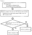
- Figure 3 shows a method for determining threshold value, in order to use the analysis results of malignancy of CTCs in addition to the conventional measured number of CTCs for diagnosis.
- the detection and measurement of CTCs are carried out by the contact step (a) and the detection step (b). Further, the EMT index is calculated by the degree determination step(c), and thereby the degree of epithelial-mesenchymal transition of each of the circulating tumor cells is determined. Then, "when the number of circulating tumor cells having a certain EMT index or more is more than the predetermined number (threshold value)" or "when an average of EMT index is more than the predetermined value (threshold value)", it can be determined that, for example, "the patient has metastasis of cancer at the high frequency".
- the predetermined value threshold value
- the predetermined threshold value cutoff value
- the number of CTCs or EMT index are not more than the predetermined threshold value (cutoff value) to the contrary, it can be determined that "malignancy of cancer is low”, “stage of cancer is not progressed”, “effects of anticancer drug is high”, or the like.
- the predetermined value can be appropriately determined according to each situation.
- the predetermined value can be determined without limitation, as long as the metastasis of cancer, the malignancy of cancer, the stage of cancer progression, or the like is diagnosed. That is to say, the various predetermined value (threshold value: cutoff value) can be used according to each situation. However, the predetermined value (threshold value: cutoff value) is preferably determined by a controlled clinical trial.
- the detection kit of the present invention may be used as a kit for the above methods.
- the epithelial cell-binding antibody (a) can specifically bind to the epithelial cell marker described in the detection method of the present invention.
- the epithelial cell marker there may be mentioned one stated in the above item "(Epithelial cell marker)", more preferably cytokeratin, EpCAM, or E-cadherin.
- the antibody is not particularly limited, as long as it can bind to the epithelial cell marker, but includes polyclonal antibody, monoclonal antibody, recombinant antibody, or antibody fragment having the antigen-binding site thereof.
- the antibody fragment there may be mentioned, for example, F(ab') 2 , Fab', Fab, Fv, or the like.
- the fragment of the antibody may be obtained by conventional methods, for example, by digesting the antibody using a protease (such as pepsin, papain, or the like) and purifying the resulting fragments by standard polypeptide isolation and purification methods.
- the mesenchymal cell-binding antibody (b) can specifically bind to the mesenchymal cell marker described in the detection method of the present invention.
- the mesenchymal cell marker there may be mentioned one stated in the above item "(Mesenchymal cell marker)", more preferably vimentin, or N-cadherin.
- the antibody is not particularly limited, as long as it can bind to the mesenchymal cell marker, but includes polyclonal antibody, monoclonal antibody, recombinant antibody, or antibody fragment having the antigen-binding site thereof.
- the antibody fragment there may be mentioned, for example, F(ab') 2 , Fab', Fab, Fv, or the like.
- the fragment of the antibody may be obtained by conventional methods, for example, by digesting the antibody using a protease (such as pepsin, papain, or the like) and purifying the resulting fragments by standard polypeptide isolation and purification methods.
- the leucocyte-binding antibody (c) can specifically bind to the leucocyte marker described in the detection method of the present invention.
- the leucocyte marker there may be mentioned markers stated in the above item "(Fluorescence or luminescence stain of leucocyte)", more preferably CD45.
- the antibody is not particularly limited, as long as it can bind to the leucocytes marker, but includes polyclonal antibody, monoclonal antibody, recombinant antibody, or antibody fragment having the antigen-binding site thereof.
- the antibody fragment there may be mentioned, for example, F(ab') 2 , Fab', Fab, Fv, or the like.
- the fragment of the antibody may be obtained by conventional methods, for example, by digesting the antibody using a protease (such as pepsin, papain, or the like) and purifying the resulting fragments by standard polypeptide isolation and purification methods.
- the epithelial cell-binding antibody (a), mesenchymal cell-binding antibody (b), and leucocyte-binding antibody (c) are fluorescently-labeled or luminescent enzyme-labeled. Further, as the fluorescent substance or luminescent enzyme, one generally known in this field can be used without limitation. For example, the fluorescent substance or luminescent enzyme described in the above item "(Fluorescent label or luminescent enzyme label)" can be used.
- the particles, to which the fluorescence substance or luminescent enzyme is bound, are not particularly limited.
- the particles for example, cells may be used, or polystyrene beads containing a single fluorescence substance may be used.
- the diameter of the particle is not particularly limited, but preferably a bit smaller than the diameter of the cell.
- the range of diameter is preferably 3 to 6 ⁇ m, and a uniform diameter thereof is preferable.
- A549 cell line derived from lung cancer was mixed with the peripheral blood of the volunteer, as a substitute for CTC. Then, cytokeratin and vimentin were detected and the EMT index was calculated.
- lysing buffer 45mL of lysing buffer was added to 4mL of peripheral blood in which A549 cells (100 cells) were mixed, and the whole was mixed and allowed to stand on ice so as to lyse erythrocytes.
- T-buffer PBS buffer containing 0.5%BSA and 2mM EDTA
- the resulting cell pellet was resuspended in 200 ⁇ L of T-buffer, and 100 ⁇ L of Fc Blocking Reagent was added thereto. The whole was incubated at 4°C for 15 minutes. After the incubation, 200 ⁇ L of T-buffer was added to the whole so as to obtain a cell suspension.
- the beads immobilized with a CD45 antibody for removing leucocytes were prepared as follows. 400 ⁇ L of magnetic beads immobilized with a CD45 antibody (Dynabeads, Life technologies) was poured into an Eppendorf tube. 400 ⁇ L of T-buffer was added to the beads and the whole was gently mixed by moving the tube up and down. Then a magnet was brought close to the tube, and the tube was allowed to stand for about 1 minute so that the beads were completely trapped on the wall of the tube. The supernatant was gently removed, and 400 ⁇ L of T-buffer was added to the beads. Then the mixture was stirred by a tapping to obtain a Dynabeads mixture.
- the cells were centrifuged at 600g, for 5 minutes. Then the cells were resuspended with 30uL of antibody reaction liquid containing 4 ⁇ L of antibodies i.e. Alexa700-labelled anti-CD45 antibody, PE-labelled anti-vimentin antibody, FITC-labelled anti-cytokeratin antibody, and 26 ⁇ L of T-buffer, and were reacted with antibodies at 4°C. 1mL of T- buffer was added thereto, and then the cells were centrifuged at 600g for 5 minutes. The cells were resuspended in 200 ⁇ L of T-buffer so as to obtain a sample stained with Alexa700-labelled anti-CD45 antibody, PE-labelled anti-vimentin antibody, and FITC-labelled anti-cytokeratin antibody.
- antibody reaction liquid containing 4 ⁇ L of antibodies i.e. Alexa700-labelled anti-CD45 antibody, PE-labelled anti-vimentin antibody, FITC-labelled anti-cytokeratin antibody, and 26 ⁇ L of T-buffer, and
- a measurement without cross contamination between samples by the flow cytometry was conducted.
- the measurement was carried out using a flow cytometer (FISHMAN-R, On-chip Biotechnologies Co.,Ltd) wherein the disposable micro flow path chip made of acrylic resin is used as the flow cell.
- FISHMAN-R On-chip Biotechnologies Co.,Ltd
- the detected signals are shown as FL1, FL2, FL3, and FL4, respectively.
- the wavelength regions of FL1, FL2, FL3, and FL4 are 510nm to 550nm, 565nm to 605nm, 656nm to 696nm, and 700nm to 850nm, respectively.
- the resulting sample was applied to the flow cytometer, and FITC-labelled anti-cytokeratin antibody, PE-labelled anti-vimentin antibody, nuclear staining agent i.e. 7-AAD, and Alexa700-labelled anti-CD45 antibody were detected by FL1, FL2, FL3, and FL4, respectively.
- the EMT index was calculated according to the following procedures.
- FIG 1(a) the scatter plot between the forward scattered signal (FS) and the sideward scattering signal (SS) is shown.
- One dot in the scatter plot corresponds to one cell.
- Many of the cells are leucocytes which were supposed to be removed by the magnetic beads. It was found that an enormous number of leucocytes other than CTCs are contained therein. Firstly, small cellular fragments were removed from the scatter plot.
- the scatter plot FL1 corresponding to FITC and FL4 corresponding to ALEXA700 is shown. The CK positive cells labelled with FITC were selected from the scatter plot.
- Figure 1(c) the scatter plot showing FL3 corresponding to the spectrum of 7-AAD and FL4 corresponding to the spectrum of ALEXA700 is described. Leucocytes were completely removed by selecting the CD45 negative cells from the scatter plot.
- Figure 1(d) is a graph showing the EMT index of CK-positive CTC cells not containing leucocytes, which is calculated using FL1 and FL2.
- the EMT index on a longitudinal axis and the FS on an abscissa axis are plotted. It was found that the EMT indexes of A549 cells were distributed on about 10%.
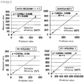
- the histogram of the EMT index values of CTCs is shown in Figure 2 .
- the apparatus constants A and B are calculated as follows.
- FIG. 2(A) the relationship between an EMT value and a fluorescence signal value is shown.
- the dashed line “100% CK (FITC)” described in Figure 2 is a standard linear of 100% FITC(cytokeratin), which is obtained by the measurement values of FITC-bound particles.
- the dashed line “100% Vimentin (PE)” described in Figure 2 is a standard linear of 100% PE(vimentin), which is obtained by the measurement values of PE-bound particles. If the cytokeratin or vimentin is singly expressed on a cell, the dot of the cell is on the standard linear of either of them.
- the cytokeratin is labelled with FITC, and thus, the CTC data, wherein the cytokeratin is only expressed thereon (100%) and the EMT is not induced, is distributed on the standard linear of "100% CK (FITC)" corresponding to the FITC fluorescence spectrum.
- FITC FITC fluorescence spectrum
- an EMT index value thereof is zero (0).
- the vimentin is only expressed thereon (100%)
- the CTC data is distributed on the standard linear of "100% Vimentin (PE)" corresponding to the PE fluorescence spectrum.
- an EMT index value thereof is 1.
- FIG. 3(B) is a set of histograms showing the data of particles bound with PE and the data of particles bound with FITC.
- the apparatus constants A and B are initially set in such a way that the modes thereof are 1 and 0 (zero), respectively. Therefore, the distributions of EMT indexes between various apparatuses can be compared. Generally, raw data of fluorescence signal intensity in flow cytometry may not be compared between various apparatuses with differences or the like. However, an index for medical diagnosis which can be used for comparison between various apparatuses is required. The above EMT index satisfies this request.
- recovery rates in the detection method of the present invention were examined by using A549 cells or cancer cells other than A549 cells.
- Example 1 The procedures described in Example 1 were repeated using A549 cells. Further, the procedures described in Example 1 were repeated except that KATO-III cells, PC-9 cells, or PC-14 cells were used instead of A549 cells. Then, EMT indexes thereof were calculated, and further detection rates of these cells were calculated simultaneously.
- the detection rates of KATO-III cells, A549 cells, PC-9 cells, and PC-14 cells were approximately 100%, 89%, 75%, and 100%, respectively.
- the detection rate of PC-14 cells in which EpCAM was not expressed was 100%. Therefore, it was revealed that the detection method of the present invention can effectively detect CTCs of metastatic cancer in which the EMT was induced.
- an analog value showing a cell characteristic feature (degree of EMT of CTC in the present invention), which is defined by multiple signal intensities and an apparatus constant, was calculated, and an evaluation of the analog value corrected by the apparatus constant was achieved. That is to say, the measurement data of each of the cells between various apparatuses can be compared.
- the analog information of each of the cells can be used on medical diagnosis in the method of the present invention.
- numbers of cells detected in a certain range of signal intensity are used as information for diagnosis.
- the conventional information is not the analog information of each of the cells, but merely the numbers of detected cells. That is, it is impossible to express the analog value showing a characteristic feature of each of the cells in the conventional flow cytometer.
- the analog value of each of the cells present in the samples of the patients can be used for medical diagnosis.
- the present invention has advantages in that (1) CTCs are detected by only a blood drawing from patients so that cancer can be found. Further, the present invention has advantages in that (2) in the patients wherein the cancer tissue was removed by surgery, a recurrence can be found early by only a blood drawing after surgery.
Landscapes
- Health & Medical Sciences (AREA)
- Life Sciences & Earth Sciences (AREA)
- Engineering & Computer Science (AREA)
- Biomedical Technology (AREA)
- Chemical & Material Sciences (AREA)
- Immunology (AREA)
- Molecular Biology (AREA)
- Hematology (AREA)
- Urology & Nephrology (AREA)
- Biotechnology (AREA)
- Cell Biology (AREA)
- General Health & Medical Sciences (AREA)
- Biochemistry (AREA)
- Microbiology (AREA)
- Physics & Mathematics (AREA)
- Medicinal Chemistry (AREA)
- Analytical Chemistry (AREA)
- Food Science & Technology (AREA)
- General Physics & Mathematics (AREA)
- Pathology (AREA)
- Genetics & Genomics (AREA)
- Organic Chemistry (AREA)
- Zoology (AREA)
- Wood Science & Technology (AREA)
- Bioinformatics & Cheminformatics (AREA)
- Oncology (AREA)
- General Engineering & Computer Science (AREA)
- Measuring Or Testing Involving Enzymes Or Micro-Organisms (AREA)
- Investigating Or Analysing Biological Materials (AREA)
- Hospice & Palliative Care (AREA)
- Investigating, Analyzing Materials By Fluorescence Or Luminescence (AREA)
- Apparatus Associated With Microorganisms And Enzymes (AREA)
- Micro-Organisms Or Cultivation Processes Thereof (AREA)
- Peptides Or Proteins (AREA)
Claims (7)
- Ein Verfahren zum Detektieren des Grades der Bösartigkeit einer jeden der zirkulierenden Tumorzellen, gekennzeichnet durch die Stufen:a) In-Berührung-Bringen einer an Epithelzellen bindenden Komponente, die spezifisch an ein Markermolekül bindet, das in Epithelzellen exprimiert und Fluoreszenz-markiert oder mit einem lumineszierenden Enzym markiert worden ist, und einer an Mesenchymzellen bindenden Komponente, die spezifisch an ein Markermolekül bindet, das in Mesenchymzellen exprimiert und Fluoreszenz-markiert oder mit einem lumineszierenden Enzym markiert worden ist, mit einer Probe, die möglicherweise zirkulierende Tumorzellen enthält,b) Detektieren eines Fluoreszenzsignals oder Lumineszenzsignals der an Epithelzellen bindenden Komponente und eines Fluoreszenzsignals oder Lumineszenzsignals der an Mesenchymzellen bindenden Komponente jeder der Zellen, wobei das Fluoreszenzsignal oder Lumineszenzsignal von einem Durchflusszytometer oder einem Bildanalysiergerät detektiert wird, undc) Bestimmen des Grades des Epithel-Mesenchym-Übergangs von zirkulierenden Tumorzellen, basierend auf der Signalhöhe der an Epithelzellen bindenden Komponente (E) und der Signalhöhe der an Mesenchymzellen bindenden Komponente (M).
- Das Verfahren zum Detektieren des Bösartigkeitsgrades jeder der zirkulierenden Tumorzellen nach Anspruch 1, das weiterhin die Stufe d) Entfernen der Erythrocyten und/oder Leukocyten aus der Probe vor oder nach Stufe a) umfasst.
- Das Verfahren zum Detektieren des Bösartigkeitsgrades jeder der zirkulierenden Tumorzellen nach Anspruch 1 oder 2, wobei eine an Leukocyten bindende Komponente, die spezifisch an ein Markermolekül bindet, das in Leukocyten exprimiert und Fluoreszenz-markiert oder mit einem lumineszierenden Enzym markiert worden ist, mit einer Probe in Berührung gebracht wird, die in Stufe a) möglicherweise zirkulierende Tumorzellen enthält.
- Das Verfahren zum Detektieren des Bösartigkeitsgrades jeder der zirkulierenden Tumorzellen nach einem der Ansprüche 1 bis 3, wobei der Grad des Epithel-Mesenchym-Übergangs von einer Formel angegeben wird, die aus der Gruppe ausgewählt ist, die aus:
Formel (1):
Formel (2):
Formel (3):
Formel (4):
- Das Verfahren zum Detektieren des Bösartigkeitsgrades jeder der zirkulierenden Tumorzellen nach einem der Ansprüche 1 bis 4, das ferner die Stufe e) Aufstellen einer Statusreferenz vor Beginn des Epithel-Mesenchym-Übergangs (0) und einer Statusreferenz nach Beendigung des Epithel-Mesenchym-Übergangs (1) durch Vermessen von Teilchen, an welche eine fluoreszierende Substanz, mit welcher die an Epithelzellen bindende Komponente markiert ist, gebunden ist, und von Teilchen, an welche eine fluoreszierende Substanz, mit welcher die an Mesenchymzellen bindende Komponente markiert ist, gebunden ist, umfasst.
- Das Verfahren zum Detektieren des Bösartigkeitsgrades jeder der zirkulierenden Tumorzellen nach Anspruch 5, wobei die Stufe e) zusammen mit den Stufen b) und c) durchgeführt wird.
- Das Verfahren zum Detektieren des Bösartigkeitsgrades jeder der zirkulierenden Tumorzellen nach einem der Ansprüche 1 bis 6, wobei die an Epithelzellen bindende Komponente ein Antikörper oder Aptamer ist, der/das spezifisch an Cytokeratin, EpCAM oder E-Cadherin bindet, und die an Mesenchymzellen bindende Komponente ein Antikörper oder Aptamer ist, der/das spezifisch an Vimentin oder N-Cadherin bindet.
Priority Applications (1)
| Application Number | Priority Date | Filing Date | Title |
|---|---|---|---|
| EP18193730.1A EP3441767B1 (de) | 2012-03-28 | 2013-03-28 | Vorrichtung zur erkennung des malignitätsgrades einer zirkulierenden tumorzelleneinheit und kit dafür |
Applications Claiming Priority (2)
| Application Number | Priority Date | Filing Date | Title |
|---|---|---|---|
| JP2012074030 | 2012-03-28 | ||
| PCT/JP2013/059202 WO2013146993A1 (ja) | 2012-03-28 | 2013-03-28 | 末梢循環腫瘍細胞単位の悪性度の検出方法及びそのキット |
Related Child Applications (1)
| Application Number | Title | Priority Date | Filing Date |
|---|---|---|---|
| EP18193730.1A Division EP3441767B1 (de) | 2012-03-28 | 2013-03-28 | Vorrichtung zur erkennung des malignitätsgrades einer zirkulierenden tumorzelleneinheit und kit dafür |
Publications (3)
| Publication Number | Publication Date |
|---|---|
| EP2833149A1 EP2833149A1 (de) | 2015-02-04 |
| EP2833149A4 EP2833149A4 (de) | 2016-03-23 |
| EP2833149B1 true EP2833149B1 (de) | 2018-10-03 |
Family
ID=49260241
Family Applications (2)
| Application Number | Title | Priority Date | Filing Date |
|---|---|---|---|
| EP13770163.7A Active EP2833149B1 (de) | 2012-03-28 | 2013-03-28 | Verfahren zur erkennung des malignitätsgrades einer zirkulierenden tumorzelleneinheit und kit dafür |
| EP18193730.1A Not-in-force EP3441767B1 (de) | 2012-03-28 | 2013-03-28 | Vorrichtung zur erkennung des malignitätsgrades einer zirkulierenden tumorzelleneinheit und kit dafür |
Family Applications After (1)
| Application Number | Title | Priority Date | Filing Date |
|---|---|---|---|
| EP18193730.1A Not-in-force EP3441767B1 (de) | 2012-03-28 | 2013-03-28 | Vorrichtung zur erkennung des malignitätsgrades einer zirkulierenden tumorzelleneinheit und kit dafür |
Country Status (4)
| Country | Link |
|---|---|
| US (2) | US20150064722A1 (de) |
| EP (2) | EP2833149B1 (de) |
| JP (2) | JP6198717B2 (de) |
| WO (1) | WO2013146993A1 (de) |
Families Citing this family (11)
| Publication number | Priority date | Publication date | Assignee | Title |
|---|---|---|---|---|
| US10871491B2 (en) * | 2014-08-25 | 2020-12-22 | Creatv Microtech, Inc. | Use of circulating cell biomarkers in the blood for detection and diagnosis of diseases and methods of isolating them |
| EP3252469A1 (de) * | 2015-01-29 | 2017-12-06 | Konica Minolta, Inc. | Verfahren zur gleichzeitigen erkennung von blutzellen mit interagierenden molekülen |
| JP6582486B2 (ja) * | 2015-03-27 | 2019-10-02 | コニカミノルタ株式会社 | 血液中の稀少細胞検出方法 |
| JP6502224B2 (ja) * | 2015-09-29 | 2019-04-17 | 富士フイルム株式会社 | 細胞評価装置および方法並びにプログラム |
| JP6860919B2 (ja) * | 2015-11-19 | 2021-04-21 | 国立大学法人金沢大学 | 間葉系kras変異型がん治療剤 |
| JP7494460B2 (ja) * | 2018-11-02 | 2024-06-04 | 東ソー株式会社 | 細胞に結合可能な担体の製造方法 |
| CN110389219B (zh) * | 2019-06-12 | 2023-04-14 | 杭州华得森生物技术有限公司 | 一种上皮间质混合型及pd-l1阳性循环肿瘤细胞的富集检测方法 |
| CN110726838A (zh) * | 2019-09-29 | 2020-01-24 | 武汉纺织大学 | 一种基于免疫微球的循环肿瘤细胞明场负向鉴定方法 |
| JP7545202B2 (ja) * | 2019-11-29 | 2024-09-04 | シスメックス株式会社 | 細胞解析方法、細胞解析装置、細胞解析システム、及び細胞解析プログラム |
| US20230323431A1 (en) * | 2020-08-31 | 2023-10-12 | World Biotech Regenerative Medical Group Limited | Personalized Immunogenic Compositions and Methods for Producing and Using Same |
| EP4603837A3 (de) * | 2023-12-26 | 2025-12-17 | Fullhope Biomedical Co., Ltd. | Verfahren zur isolierung und analyse von zirkulierenden tumorzellen |
Family Cites Families (18)
| Publication number | Priority date | Publication date | Assignee | Title |
|---|---|---|---|---|
| US6235723B1 (en) * | 1992-03-16 | 2001-05-22 | Isis Pharmaceuticals , Inc. | Antisense oligonucleotide modulation of human protein kinase C-δ expression |
| US20020172987A1 (en) | 1998-02-12 | 2002-11-21 | Terstappen Leon W.M.M. | Methods and reagents for the rapid and efficient isolation of circulating cancer cells |
| US6566063B1 (en) * | 1999-05-14 | 2003-05-20 | Chiron Corporation | Methods for determining metastatic potential of breast cancer cells by detecting GSEF gene product expression |
| JP2006029921A (ja) | 2004-07-14 | 2006-02-02 | Institute Of Physical & Chemical Research | フローサイトメーター |
| ATE477495T1 (de) * | 2005-03-16 | 2010-08-15 | Osi Pharm Inc | Biologische marker prediktiv für das ansprechen von krebs auf inhibitoren der kinase des rezeptors für epidermalen wachstumsfaktor |
| US7846393B2 (en) | 2005-04-21 | 2010-12-07 | California Institute Of Technology | Membrane filter for capturing circulating tumor cells |
| US8921102B2 (en) | 2005-07-29 | 2014-12-30 | Gpb Scientific, Llc | Devices and methods for enrichment and alteration of circulating tumor cells and other particles |
| JP4986449B2 (ja) | 2005-12-27 | 2012-07-25 | 株式会社エスアールエル | 浮遊細胞の検査方法 |
| HUE028487T2 (en) * | 2007-02-27 | 2016-12-28 | Sentoclone Int Ab | Multiplex detection of tumour cells using a panel of agents binding to extracellular markers |
| US20090258365A1 (en) | 2008-03-25 | 2009-10-15 | Terstappen Leon W M M | METHOD FOR DETECTING IGF1R/Chr 15 in CIRCULATING TUMOR CELLS USING FISH |
| JP4358888B1 (ja) | 2008-07-01 | 2009-11-04 | 株式会社オンチップ・バイオテクノロジーズ | フローサイトメーターおよびそのフローセル |
| JP5382852B2 (ja) | 2009-02-06 | 2014-01-08 | 株式会社オンチップ・バイオテクノロジーズ | 使い捨てチップ型フローセルとそれを用いたフローサイトメーター |
| CA2755341A1 (en) * | 2009-03-13 | 2010-09-16 | Bergen Teknologioverforing As | Methods using axl as biomarker of epithelial-to-mesnchymal transition |
| JP2013504064A (ja) * | 2009-09-03 | 2013-02-04 | ザ スクリプス リサーチ インスティチュート | 循環腫瘍細胞を分類する方法 |
| EP2525209B1 (de) | 2010-01-15 | 2020-03-11 | On-chip Biotechnologies Co., Ltd. | Einweg-chip-flusszelle und zellsortiermaschine damit |
| WO2011093927A1 (en) * | 2010-01-27 | 2011-08-04 | Duke University | Biomarkers for circulating tumor cells |
| US9341550B2 (en) * | 2010-11-19 | 2016-05-17 | On-Chip Biotechnologies Co., Ltd. | Method for detecting low concentrations of specific cell from high concentrations of cell populations, and method for collecting and analyzing detected cell |
| WO2012149014A1 (en) * | 2011-04-25 | 2012-11-01 | OSI Pharmaceuticals, LLC | Use of emt gene signatures in cancer drug discovery, diagnostics, and treatment |
-
2013
- 2013-03-28 EP EP13770163.7A patent/EP2833149B1/de active Active
- 2013-03-28 JP JP2014508010A patent/JP6198717B2/ja active Active
- 2013-03-28 EP EP18193730.1A patent/EP3441767B1/de not_active Not-in-force
- 2013-03-28 WO PCT/JP2013/059202 patent/WO2013146993A1/ja not_active Ceased
- 2013-03-28 US US14/388,445 patent/US20150064722A1/en not_active Abandoned
-
2017
- 2017-08-22 JP JP2017159092A patent/JP6485759B2/ja active Active
-
2018
- 2018-09-06 US US16/123,444 patent/US11280792B2/en active Active
Non-Patent Citations (1)
| Title |
|---|
| None * |
Also Published As
| Publication number | Publication date |
|---|---|
| WO2013146993A1 (ja) | 2013-10-03 |
| US20190011450A1 (en) | 2019-01-10 |
| EP3441767B1 (de) | 2021-02-24 |
| EP2833149A4 (de) | 2016-03-23 |
| JPWO2013146993A1 (ja) | 2015-12-14 |
| EP2833149A1 (de) | 2015-02-04 |
| EP3441767A1 (de) | 2019-02-13 |
| JP2017207516A (ja) | 2017-11-24 |
| JP6485759B2 (ja) | 2019-03-20 |
| JP6198717B2 (ja) | 2017-09-20 |
| US20150064722A1 (en) | 2015-03-05 |
| US11280792B2 (en) | 2022-03-22 |
Similar Documents
| Publication | Publication Date | Title |
|---|---|---|
| US11280792B2 (en) | Apparatus for detecting the degree of malignancy of each of circulating tumor cells | |
| de Graaf et al. | Flow cytometric characterization of cerebrospinal fluid cells | |
| US9506927B2 (en) | Method for detecting low concentrations of specific cell from high concentrations of cell populations, and method for collecting and analyzing detected cell | |
| CA2516795C (en) | Circulating tumor cells (ctc's): early assessment of time to progression,_survival and response to therapy in metastatic cancer patients | |
| Li et al. | Strategies for enrichment of circulating tumor cells | |
| US9671407B2 (en) | Devices and methods of cell capture and analysis | |
| Lu et al. | Isolation and characterization of living circulating tumor cells in patients by immunomagnetic negative enrichment coupled with flow cytometry | |
| EP1861509B1 (de) | Verfahren zur vorhersage des überlebens insgesamt und ohne krankheitsfortschreitung zu jedem nachsorgezeitpunkt bei der therapie von metastatischen brustkrebspatienten unter verwendung zirkulierender tumorzellen | |
| Soh et al. | Diagnosis of plasma cell dyscrasias and monitoring of minimal residual disease by multiparametric flow cytometry | |
| CN113933511B (zh) | 检测急性b淋巴细胞白血病微小残留的抗体组合物及方法 | |
| US20080113350A1 (en) | Blood test to monitor the genetic changes of progressive cancer using immunomagnetic enrichment and fluorescence in situ hybridization (FISH) | |
| Cherian et al. | How I diagnose minimal/measurable residual disease in B lymphoblastic leukemia/lymphoma by flow cytometry | |
| US20150233932A1 (en) | Methods, Systems, and Compositions for Enrichment of Rare Cells | |
| CN108572255A (zh) | 检测pd-l1+循环肿瘤细胞的试剂在筛选癌症风险个体以及在癌症患者预后中的应用 | |
| Karimi et al. | Circulating tumor cells detection in patients with early breast cancer using MACS immunomagnetic flow cytometry | |
| Kim et al. | Detection of circulating tumor cells and their potential use as a biomarker for advanced renal cell carcinoma | |
| CA2322282A1 (en) | Method and compositions for differential detection of primary tumor cells and metastatic cells | |
| Carneiro et al. | Minimizing false positives for CTC identification | |
| CN111830249A (zh) | 用于纯化分离及分析非典型循环肿瘤细胞的方法的用途及非典型循环肿瘤细胞的用途 | |
| WO2005116264A2 (en) | A blood test to monitor the genetic changes of progressive cancer using immunomagnetic enrichment and fluorescence in situ hybridization (fish) | |
| EP2551673B1 (de) | Verfahren zur Erfassung der Krebsinfiltration in das zentrale Nervensystem | |
| Slack et al. | Flow cytometric detection of ZAP-70 in chronic lymphocytic leukemia: correlation with immunocytochemistry and Western blot analysis | |
| WO2013003898A1 (en) | Method for detection of cancer in a patient | |
| Paietta | How to optimize multiparameter flow cytometry for leukaemia/lymphoma diagnosis | |
| 김성민 | Minimal Residual Disease Detection Using Next-Generation Flow Analysis And Next-Generation Sequencing In Multiple Myeloma Patients: Comparative analysis with IMW treatment response and verification of peripheral blood applicability |
Legal Events
| Date | Code | Title | Description |
|---|---|---|---|
| PUAI | Public reference made under article 153(3) epc to a published international application that has entered the european phase |
Free format text: ORIGINAL CODE: 0009012 |
|
| 17P | Request for examination filed |
Effective date: 20141023 |
|
| AK | Designated contracting states |
Kind code of ref document: A1 Designated state(s): AL AT BE BG CH CY CZ DE DK EE ES FI FR GB GR HR HU IE IS IT LI LT LU LV MC MK MT NL NO PL PT RO RS SE SI SK SM TR |
|
| AX | Request for extension of the european patent |
Extension state: BA ME |
|
| DAX | Request for extension of the european patent (deleted) | ||
| RIN1 | Information on inventor provided before grant (corrected) |
Inventor name: NISHIO, KAORI Inventor name: KOH, YASUHIRO Inventor name: KOIZUMI, FUMIAKI Inventor name: FUJIMURA, YUU Inventor name: UEHARA, YURI Inventor name: YAMASHITA, NAMIKO Inventor name: WATANABE, MASARU Inventor name: TAKEDA, KAZUO |
|
| RIC1 | Information provided on ipc code assigned before grant |
Ipc: G01N 33/543 20060101ALI20151030BHEP Ipc: G01N 33/536 20060101ALI20151030BHEP Ipc: C07K 14/47 20060101ALI20151030BHEP Ipc: G01N 33/574 20060101AFI20151030BHEP Ipc: C12N 5/07 20100101ALI20151030BHEP |
|
| RA4 | Supplementary search report drawn up and despatched (corrected) |
Effective date: 20160223 |
|
| RIC1 | Information provided on ipc code assigned before grant |
Ipc: G01N 33/543 20060101ALI20160217BHEP Ipc: C07K 14/47 20060101ALI20160217BHEP Ipc: C12N 5/07 20100101ALI20160217BHEP Ipc: G01N 33/574 20060101AFI20160217BHEP Ipc: G01N 33/536 20060101ALI20160217BHEP |
|
| STAA | Information on the status of an ep patent application or granted ep patent |
Free format text: STATUS: EXAMINATION IS IN PROGRESS |
|
| 17Q | First examination report despatched |
Effective date: 20170201 |
|
| GRAP | Despatch of communication of intention to grant a patent |
Free format text: ORIGINAL CODE: EPIDOSNIGR1 |
|
| STAA | Information on the status of an ep patent application or granted ep patent |
Free format text: STATUS: GRANT OF PATENT IS INTENDED |
|
| RIC1 | Information provided on ipc code assigned before grant |
Ipc: G01N 33/574 20060101AFI20180327BHEP Ipc: G01N 33/536 20060101ALI20180327BHEP Ipc: G01N 33/543 20060101ALI20180327BHEP |
|
| INTG | Intention to grant announced |
Effective date: 20180412 |
|
| GRAS | Grant fee paid |
Free format text: ORIGINAL CODE: EPIDOSNIGR3 |
|
| GRAA | (expected) grant |
Free format text: ORIGINAL CODE: 0009210 |
|
| STAA | Information on the status of an ep patent application or granted ep patent |
Free format text: STATUS: THE PATENT HAS BEEN GRANTED |
|
| AK | Designated contracting states |
Kind code of ref document: B1 Designated state(s): AL AT BE BG CH CY CZ DE DK EE ES FI FR GB GR HR HU IE IS IT LI LT LU LV MC MK MT NL NO PL PT RO RS SE SI SK SM TR |
|
| REG | Reference to a national code |
Ref country code: GB Ref legal event code: FG4D |
|
| REG | Reference to a national code |
Ref country code: CH Ref legal event code: EP Ref country code: AT Ref legal event code: REF Ref document number: 1049179 Country of ref document: AT Kind code of ref document: T Effective date: 20181015 |
|
| REG | Reference to a national code |
Ref country code: DE Ref legal event code: R096 Ref document number: 602013044528 Country of ref document: DE Ref country code: IE Ref legal event code: FG4D |
|
| REG | Reference to a national code |
Ref country code: GB Ref legal event code: 746 Effective date: 20181026 |
|
| REG | Reference to a national code |
Ref country code: NL Ref legal event code: MP Effective date: 20181003 |
|
| REG | Reference to a national code |
Ref country code: LT Ref legal event code: MG4D |
|
| REG | Reference to a national code |
Ref country code: AT Ref legal event code: MK05 Ref document number: 1049179 Country of ref document: AT Kind code of ref document: T Effective date: 20181003 |
|
| PG25 | Lapsed in a contracting state [announced via postgrant information from national office to epo] |
Ref country code: NL Free format text: LAPSE BECAUSE OF FAILURE TO SUBMIT A TRANSLATION OF THE DESCRIPTION OR TO PAY THE FEE WITHIN THE PRESCRIBED TIME-LIMIT Effective date: 20181003 |
|
| PG25 | Lapsed in a contracting state [announced via postgrant information from national office to epo] |
Ref country code: IS Free format text: LAPSE BECAUSE OF FAILURE TO SUBMIT A TRANSLATION OF THE DESCRIPTION OR TO PAY THE FEE WITHIN THE PRESCRIBED TIME-LIMIT Effective date: 20190203 Ref country code: FI Free format text: LAPSE BECAUSE OF FAILURE TO SUBMIT A TRANSLATION OF THE DESCRIPTION OR TO PAY THE FEE WITHIN THE PRESCRIBED TIME-LIMIT Effective date: 20181003 Ref country code: BG Free format text: LAPSE BECAUSE OF FAILURE TO SUBMIT A TRANSLATION OF THE DESCRIPTION OR TO PAY THE FEE WITHIN THE PRESCRIBED TIME-LIMIT Effective date: 20190103 Ref country code: NO Free format text: LAPSE BECAUSE OF FAILURE TO SUBMIT A TRANSLATION OF THE DESCRIPTION OR TO PAY THE FEE WITHIN THE PRESCRIBED TIME-LIMIT Effective date: 20190103 Ref country code: LT Free format text: LAPSE BECAUSE OF FAILURE TO SUBMIT A TRANSLATION OF THE DESCRIPTION OR TO PAY THE FEE WITHIN THE PRESCRIBED TIME-LIMIT Effective date: 20181003 Ref country code: CZ Free format text: LAPSE BECAUSE OF FAILURE TO SUBMIT A TRANSLATION OF THE DESCRIPTION OR TO PAY THE FEE WITHIN THE PRESCRIBED TIME-LIMIT Effective date: 20181003 Ref country code: AT Free format text: LAPSE BECAUSE OF FAILURE TO SUBMIT A TRANSLATION OF THE DESCRIPTION OR TO PAY THE FEE WITHIN THE PRESCRIBED TIME-LIMIT Effective date: 20181003 Ref country code: ES Free format text: LAPSE BECAUSE OF FAILURE TO SUBMIT A TRANSLATION OF THE DESCRIPTION OR TO PAY THE FEE WITHIN THE PRESCRIBED TIME-LIMIT Effective date: 20181003 Ref country code: HR Free format text: LAPSE BECAUSE OF FAILURE TO SUBMIT A TRANSLATION OF THE DESCRIPTION OR TO PAY THE FEE WITHIN THE PRESCRIBED TIME-LIMIT Effective date: 20181003 Ref country code: PL Free format text: LAPSE BECAUSE OF FAILURE TO SUBMIT A TRANSLATION OF THE DESCRIPTION OR TO PAY THE FEE WITHIN THE PRESCRIBED TIME-LIMIT Effective date: 20181003 Ref country code: LV Free format text: LAPSE BECAUSE OF FAILURE TO SUBMIT A TRANSLATION OF THE DESCRIPTION OR TO PAY THE FEE WITHIN THE PRESCRIBED TIME-LIMIT Effective date: 20181003 |
|
| PG25 | Lapsed in a contracting state [announced via postgrant information from national office to epo] |
Ref country code: PT Free format text: LAPSE BECAUSE OF FAILURE TO SUBMIT A TRANSLATION OF THE DESCRIPTION OR TO PAY THE FEE WITHIN THE PRESCRIBED TIME-LIMIT Effective date: 20190203 Ref country code: AL Free format text: LAPSE BECAUSE OF FAILURE TO SUBMIT A TRANSLATION OF THE DESCRIPTION OR TO PAY THE FEE WITHIN THE PRESCRIBED TIME-LIMIT Effective date: 20181003 Ref country code: SE Free format text: LAPSE BECAUSE OF FAILURE TO SUBMIT A TRANSLATION OF THE DESCRIPTION OR TO PAY THE FEE WITHIN THE PRESCRIBED TIME-LIMIT Effective date: 20181003 Ref country code: GR Free format text: LAPSE BECAUSE OF FAILURE TO SUBMIT A TRANSLATION OF THE DESCRIPTION OR TO PAY THE FEE WITHIN THE PRESCRIBED TIME-LIMIT Effective date: 20190104 Ref country code: RS Free format text: LAPSE BECAUSE OF FAILURE TO SUBMIT A TRANSLATION OF THE DESCRIPTION OR TO PAY THE FEE WITHIN THE PRESCRIBED TIME-LIMIT Effective date: 20181003 |
|
| REG | Reference to a national code |
Ref country code: DE Ref legal event code: R097 Ref document number: 602013044528 Country of ref document: DE |
|
| PG25 | Lapsed in a contracting state [announced via postgrant information from national office to epo] |
Ref country code: IT Free format text: LAPSE BECAUSE OF FAILURE TO SUBMIT A TRANSLATION OF THE DESCRIPTION OR TO PAY THE FEE WITHIN THE PRESCRIBED TIME-LIMIT Effective date: 20181003 Ref country code: DK Free format text: LAPSE BECAUSE OF FAILURE TO SUBMIT A TRANSLATION OF THE DESCRIPTION OR TO PAY THE FEE WITHIN THE PRESCRIBED TIME-LIMIT Effective date: 20181003 |
|
| PLBE | No opposition filed within time limit |
Free format text: ORIGINAL CODE: 0009261 |
|
| STAA | Information on the status of an ep patent application or granted ep patent |
Free format text: STATUS: NO OPPOSITION FILED WITHIN TIME LIMIT |
|
| PG25 | Lapsed in a contracting state [announced via postgrant information from national office to epo] |
Ref country code: EE Free format text: LAPSE BECAUSE OF FAILURE TO SUBMIT A TRANSLATION OF THE DESCRIPTION OR TO PAY THE FEE WITHIN THE PRESCRIBED TIME-LIMIT Effective date: 20181003 Ref country code: SM Free format text: LAPSE BECAUSE OF FAILURE TO SUBMIT A TRANSLATION OF THE DESCRIPTION OR TO PAY THE FEE WITHIN THE PRESCRIBED TIME-LIMIT Effective date: 20181003 Ref country code: RO Free format text: LAPSE BECAUSE OF FAILURE TO SUBMIT A TRANSLATION OF THE DESCRIPTION OR TO PAY THE FEE WITHIN THE PRESCRIBED TIME-LIMIT Effective date: 20181003 Ref country code: SK Free format text: LAPSE BECAUSE OF FAILURE TO SUBMIT A TRANSLATION OF THE DESCRIPTION OR TO PAY THE FEE WITHIN THE PRESCRIBED TIME-LIMIT Effective date: 20181003 |
|
| 26N | No opposition filed |
Effective date: 20190704 |
|
| PG25 | Lapsed in a contracting state [announced via postgrant information from national office to epo] |
Ref country code: SI Free format text: LAPSE BECAUSE OF FAILURE TO SUBMIT A TRANSLATION OF THE DESCRIPTION OR TO PAY THE FEE WITHIN THE PRESCRIBED TIME-LIMIT Effective date: 20181003 Ref country code: MC Free format text: LAPSE BECAUSE OF FAILURE TO SUBMIT A TRANSLATION OF THE DESCRIPTION OR TO PAY THE FEE WITHIN THE PRESCRIBED TIME-LIMIT Effective date: 20181003 |
|
| REG | Reference to a national code |
Ref country code: CH Ref legal event code: PL |
|
| PG25 | Lapsed in a contracting state [announced via postgrant information from national office to epo] |
Ref country code: LU Free format text: LAPSE BECAUSE OF NON-PAYMENT OF DUE FEES Effective date: 20190328 |
|
| REG | Reference to a national code |
Ref country code: BE Ref legal event code: MM Effective date: 20190331 |
|
| PG25 | Lapsed in a contracting state [announced via postgrant information from national office to epo] |
Ref country code: IE Free format text: LAPSE BECAUSE OF NON-PAYMENT OF DUE FEES Effective date: 20190328 Ref country code: CH Free format text: LAPSE BECAUSE OF NON-PAYMENT OF DUE FEES Effective date: 20190331 Ref country code: LI Free format text: LAPSE BECAUSE OF NON-PAYMENT OF DUE FEES Effective date: 20190331 |
|
| PG25 | Lapsed in a contracting state [announced via postgrant information from national office to epo] |
Ref country code: BE Free format text: LAPSE BECAUSE OF NON-PAYMENT OF DUE FEES Effective date: 20190331 |
|
| PG25 | Lapsed in a contracting state [announced via postgrant information from national office to epo] |
Ref country code: TR Free format text: LAPSE BECAUSE OF FAILURE TO SUBMIT A TRANSLATION OF THE DESCRIPTION OR TO PAY THE FEE WITHIN THE PRESCRIBED TIME-LIMIT Effective date: 20181003 |
|
| PG25 | Lapsed in a contracting state [announced via postgrant information from national office to epo] |
Ref country code: MT Free format text: LAPSE BECAUSE OF NON-PAYMENT OF DUE FEES Effective date: 20190328 |
|
| PG25 | Lapsed in a contracting state [announced via postgrant information from national office to epo] |
Ref country code: CY Free format text: LAPSE BECAUSE OF FAILURE TO SUBMIT A TRANSLATION OF THE DESCRIPTION OR TO PAY THE FEE WITHIN THE PRESCRIBED TIME-LIMIT Effective date: 20181003 |
|
| PG25 | Lapsed in a contracting state [announced via postgrant information from national office to epo] |
Ref country code: HU Free format text: LAPSE BECAUSE OF FAILURE TO SUBMIT A TRANSLATION OF THE DESCRIPTION OR TO PAY THE FEE WITHIN THE PRESCRIBED TIME-LIMIT; INVALID AB INITIO Effective date: 20130328 |
|
| PG25 | Lapsed in a contracting state [announced via postgrant information from national office to epo] |
Ref country code: MK Free format text: LAPSE BECAUSE OF FAILURE TO SUBMIT A TRANSLATION OF THE DESCRIPTION OR TO PAY THE FEE WITHIN THE PRESCRIBED TIME-LIMIT Effective date: 20181003 |
|
| PGFP | Annual fee paid to national office [announced via postgrant information from national office to epo] |
Ref country code: DE Payment date: 20250319 Year of fee payment: 13 |
|
| PGFP | Annual fee paid to national office [announced via postgrant information from national office to epo] |
Ref country code: FR Payment date: 20250325 Year of fee payment: 13 |
|
| PGFP | Annual fee paid to national office [announced via postgrant information from national office to epo] |
Ref country code: GB Payment date: 20250324 Year of fee payment: 13 |
|
| REG | Reference to a national code |
Ref country code: DE Ref legal event code: R079 Ref document number: 602013044528 Country of ref document: DE Free format text: PREVIOUS MAIN CLASS: G01N0033574000 Ipc: G01N0033575000 |