EP2769321B1 - Methods for improving inflammatory bowel disease diagnosis - Google Patents
Methods for improving inflammatory bowel disease diagnosis Download PDFInfo
- Publication number
- EP2769321B1 EP2769321B1 EP12841790.4A EP12841790A EP2769321B1 EP 2769321 B1 EP2769321 B1 EP 2769321B1 EP 12841790 A EP12841790 A EP 12841790A EP 2769321 B1 EP2769321 B1 EP 2769321B1
- Authority
- EP
- European Patent Office
- Prior art keywords
- sample
- ibd
- antibody
- markers
- disease
- Prior art date
- Legal status (The legal status is an assumption and is not a legal conclusion. Google has not performed a legal analysis and makes no representation as to the accuracy of the status listed.)
- Active
Links
- 208000022559 Inflammatory bowel disease Diseases 0.000 title claims description 469
- 238000000034 method Methods 0.000 title claims description 260
- 238000003745 diagnosis Methods 0.000 title description 59
- 206010009900 Colitis ulcerative Diseases 0.000 claims description 240
- 201000006704 Ulcerative Colitis Diseases 0.000 claims description 240
- 239000003550 marker Substances 0.000 claims description 197
- 238000003556 assay Methods 0.000 claims description 120
- 238000007637 random forest analysis Methods 0.000 claims description 102
- 101000866526 Homo sapiens Extracellular matrix protein 1 Proteins 0.000 claims description 91
- 102100031758 Extracellular matrix protein 1 Human genes 0.000 claims description 89
- 230000002068 genetic effect Effects 0.000 claims description 83
- 108010017324 STAT3 Transcription Factor Proteins 0.000 claims description 80
- 102100024040 Signal transducer and activator of transcription 3 Human genes 0.000 claims description 80
- 101000964092 Homo sapiens Autophagy-related protein 16-1 Proteins 0.000 claims description 79
- 102100040355 Autophagy-related protein 16-1 Human genes 0.000 claims description 78
- 101000632181 Homo sapiens Homeobox protein Nkx-2.3 Proteins 0.000 claims description 72
- 206010061218 Inflammation Diseases 0.000 claims description 70
- 238000007619 statistical method Methods 0.000 claims description 70
- 102100027890 Homeobox protein Nkx-2.3 Human genes 0.000 claims description 69
- 230000000405 serological effect Effects 0.000 claims description 67
- 239000002773 nucleotide Substances 0.000 claims description 63
- 125000003729 nucleotide group Chemical group 0.000 claims description 63
- 208000011231 Crohn disease Diseases 0.000 claims description 61
- 238000002965 ELISA Methods 0.000 claims description 52
- 108010019530 Vascular Endothelial Growth Factors Proteins 0.000 claims description 47
- 102000005789 Vascular Endothelial Growth Factors Human genes 0.000 claims description 46
- 230000004054 inflammatory process Effects 0.000 claims description 41
- 238000003066 decision tree Methods 0.000 claims description 35
- 238000003018 immunoassay Methods 0.000 claims description 28
- 210000002966 serum Anatomy 0.000 claims description 24
- 102200132076 rs2241880 Human genes 0.000 claims description 23
- 102210007381 rs744166 Human genes 0.000 claims description 22
- 108010040721 Flagellin Proteins 0.000 claims description 21
- 230000003321 amplification Effects 0.000 claims description 21
- 238000003199 nucleic acid amplification method Methods 0.000 claims description 21
- 102210007296 rs10883365 Human genes 0.000 claims description 21
- 238000009396 hybridization Methods 0.000 claims description 20
- 238000003205 genotyping method Methods 0.000 claims description 16
- 230000002055 immunohistochemical effect Effects 0.000 claims description 15
- 230000035945 sensitivity Effects 0.000 claims description 15
- 210000004369 blood Anatomy 0.000 claims description 12
- 239000008280 blood Substances 0.000 claims description 12
- 102220583772 Extracellular matrix protein 1_T130M_mutation Human genes 0.000 claims description 9
- XGWFJBFNAQHLEF-UHFFFAOYSA-N 9-anthroic acid Chemical compound C1=CC=C2C(C(=O)O)=C(C=CC=C3)C3=CC2=C1 XGWFJBFNAQHLEF-UHFFFAOYSA-N 0.000 claims 18
- 239000000523 sample Substances 0.000 description 308
- 108700028369 Alleles Proteins 0.000 description 166
- 208000037265 diseases, disorders, signs and symptoms Diseases 0.000 description 117
- 201000010099 disease Diseases 0.000 description 115
- 108090000623 proteins and genes Proteins 0.000 description 108
- 102000004169 proteins and genes Human genes 0.000 description 73
- 235000018102 proteins Nutrition 0.000 description 65
- 101001125026 Homo sapiens Nucleotide-binding oligomerization domain-containing protein 2 Proteins 0.000 description 63
- 102100029441 Nucleotide-binding oligomerization domain-containing protein 2 Human genes 0.000 description 60
- 238000001514 detection method Methods 0.000 description 59
- 238000004422 calculation algorithm Methods 0.000 description 57
- 238000012549 training Methods 0.000 description 51
- 102000054727 Serum Amyloid A Human genes 0.000 description 47
- 108700028909 Serum Amyloid A Proteins 0.000 description 46
- OPTASPLRGRRNAP-UHFFFAOYSA-N cytosine Chemical compound NC=1C=CNC(=O)N=1 OPTASPLRGRRNAP-UHFFFAOYSA-N 0.000 description 46
- 239000003814 drug Substances 0.000 description 46
- 238000012360 testing method Methods 0.000 description 45
- 102100032752 C-reactive protein Human genes 0.000 description 43
- 108010073929 Vascular Endothelial Growth Factor A Proteins 0.000 description 43
- 102000004196 processed proteins & peptides Human genes 0.000 description 43
- 108090000765 processed proteins & peptides Proteins 0.000 description 43
- 108010074051 C-Reactive Protein Proteins 0.000 description 42
- 208000027138 indeterminate colitis Diseases 0.000 description 42
- 108010029485 Protein Isoforms Proteins 0.000 description 41
- 102000001708 Protein Isoforms Human genes 0.000 description 41
- 229920001184 polypeptide Polymers 0.000 description 39
- 239000000427 antigen Substances 0.000 description 38
- 108091007433 antigens Proteins 0.000 description 38
- 102000036639 antigens Human genes 0.000 description 38
- 239000012634 fragment Substances 0.000 description 36
- 108700028353 OmpC Proteins 0.000 description 35
- 238000004458 analytical method Methods 0.000 description 35
- 150000007523 nucleic acids Chemical class 0.000 description 34
- 150000001413 amino acids Chemical group 0.000 description 33
- -1 IL-1β Proteins 0.000 description 32
- 102000039446 nucleic acids Human genes 0.000 description 32
- 108020004707 nucleic acids Proteins 0.000 description 32
- 102000000905 Cadherin Human genes 0.000 description 31
- 108050007957 Cadherin Proteins 0.000 description 31
- 229940124597 therapeutic agent Drugs 0.000 description 31
- 238000011282 treatment Methods 0.000 description 27
- UYTPUPDQBNUYGX-UHFFFAOYSA-N guanine Chemical compound O=C1NC(N)=NC2=C1N=CN2 UYTPUPDQBNUYGX-UHFFFAOYSA-N 0.000 description 25
- 208000024891 symptom Diseases 0.000 description 24
- RWQNBRDOKXIBIV-UHFFFAOYSA-N thymine Chemical compound CC1=CNC(=O)NC1=O RWQNBRDOKXIBIV-UHFFFAOYSA-N 0.000 description 24
- 102000004127 Cytokines Human genes 0.000 description 23
- 108090000695 Cytokines Proteins 0.000 description 23
- 229940104302 cytosine Drugs 0.000 description 23
- 108020004999 messenger RNA Proteins 0.000 description 22
- 238000013528 artificial neural network Methods 0.000 description 21
- 230000000670 limiting effect Effects 0.000 description 21
- 230000014509 gene expression Effects 0.000 description 20
- 102000011767 Acute-Phase Proteins Human genes 0.000 description 19
- 108010062271 Acute-Phase Proteins Proteins 0.000 description 19
- 101150083031 Nod2 gene Proteins 0.000 description 19
- 108010000134 Vascular Cell Adhesion Molecule-1 Proteins 0.000 description 19
- 102100023543 Vascular cell adhesion protein 1 Human genes 0.000 description 19
- 238000006467 substitution reaction Methods 0.000 description 19
- 102000000541 Defensins Human genes 0.000 description 18
- 108010002069 Defensins Proteins 0.000 description 18
- 102220583790 Extracellular matrix protein 1_G415S_mutation Human genes 0.000 description 18
- 108010064593 Intercellular Adhesion Molecule-1 Proteins 0.000 description 18
- 102100037877 Intercellular adhesion molecule 1 Human genes 0.000 description 18
- 238000005259 measurement Methods 0.000 description 18
- 102000007592 Apolipoproteins Human genes 0.000 description 17
- 108010071619 Apolipoproteins Proteins 0.000 description 17
- 108020004414 DNA Proteins 0.000 description 17
- 241000282414 Homo sapiens Species 0.000 description 17
- 239000003102 growth factor Substances 0.000 description 17
- 238000003752 polymerase chain reaction Methods 0.000 description 17
- 102210021500 rs6584283 Human genes 0.000 description 17
- 238000012300 Sequence Analysis Methods 0.000 description 16
- 210000001072 colon Anatomy 0.000 description 16
- 239000002552 dosage form Substances 0.000 description 16
- 238000001356 surgical procedure Methods 0.000 description 16
- 239000012472 biological sample Substances 0.000 description 15
- 210000004027 cell Anatomy 0.000 description 15
- 230000001413 cellular effect Effects 0.000 description 15
- 230000008859 change Effects 0.000 description 15
- 210000000440 neutrophil Anatomy 0.000 description 15
- 102210007411 rs3828309 Human genes 0.000 description 15
- WQZGKKKJIJFFOK-QTVWNMPRSA-N D-mannopyranose Chemical compound OC[C@H]1OC(O)[C@@H](O)[C@@H](O)[C@@H]1O WQZGKKKJIJFFOK-QTVWNMPRSA-N 0.000 description 14
- 235000001014 amino acid Nutrition 0.000 description 14
- 239000000975 dye Substances 0.000 description 14
- 210000002381 plasma Anatomy 0.000 description 14
- 102200146742 rs2066844 Human genes 0.000 description 14
- 102200146596 rs2066845 Human genes 0.000 description 14
- 238000013179 statistical model Methods 0.000 description 14
- 206010009887 colitis Diseases 0.000 description 13
- 238000012774 diagnostic algorithm Methods 0.000 description 13
- 102000054766 genetic haplotypes Human genes 0.000 description 13
- 230000004044 response Effects 0.000 description 13
- 230000001086 cytosolic effect Effects 0.000 description 12
- 230000000694 effects Effects 0.000 description 12
- 230000001965 increasing effect Effects 0.000 description 12
- 210000001519 tissue Anatomy 0.000 description 12
- 102100038326 Beta-defensin 4A Human genes 0.000 description 11
- 101100460499 Homo sapiens NKX2-3 gene Proteins 0.000 description 11
- 229940079593 drug Drugs 0.000 description 11
- 230000006870 function Effects 0.000 description 11
- 239000000203 mixture Substances 0.000 description 11
- 210000000664 rectum Anatomy 0.000 description 11
- 210000002700 urine Anatomy 0.000 description 11
- 238000010200 validation analysis Methods 0.000 description 11
- 229930024421 Adenine Natural products 0.000 description 10
- GFFGJBXGBJISGV-UHFFFAOYSA-N Adenine Chemical compound NC1=NC=NC2=C1N=CN2 GFFGJBXGBJISGV-UHFFFAOYSA-N 0.000 description 10
- 241001408449 Asca Species 0.000 description 10
- 238000008157 ELISA kit Methods 0.000 description 10
- 101000952040 Homo sapiens Beta-defensin 1 Proteins 0.000 description 10
- 108060003951 Immunoglobulin Proteins 0.000 description 10
- 108091034117 Oligonucleotide Proteins 0.000 description 10
- 229960000643 adenine Drugs 0.000 description 10
- 230000000845 anti-microbial effect Effects 0.000 description 10
- 230000007614 genetic variation Effects 0.000 description 10
- 102000018358 immunoglobulin Human genes 0.000 description 10
- 208000002551 irritable bowel syndrome Diseases 0.000 description 10
- 210000003296 saliva Anatomy 0.000 description 10
- 238000002560 therapeutic procedure Methods 0.000 description 10
- 102100033350 ATP-dependent translocase ABCB1 Human genes 0.000 description 9
- 102100037437 Beta-defensin 1 Human genes 0.000 description 9
- 102000004190 Enzymes Human genes 0.000 description 9
- 108090000790 Enzymes Proteins 0.000 description 9
- 101000578932 Homo sapiens Membrane-associated guanylate kinase, WW and PDZ domain-containing protein 2 Proteins 0.000 description 9
- 108010063045 Lactoferrin Proteins 0.000 description 9
- 102100032241 Lactotransferrin Human genes 0.000 description 9
- 108010047230 Member 1 Subfamily B ATP Binding Cassette Transporter Proteins 0.000 description 9
- 102100028328 Membrane-associated guanylate kinase, WW and PDZ domain-containing protein 2 Human genes 0.000 description 9
- 239000004599 antimicrobial Substances 0.000 description 9
- 239000000090 biomarker Substances 0.000 description 9
- 238000001962 electrophoresis Methods 0.000 description 9
- 229940088598 enzyme Drugs 0.000 description 9
- 230000000149 penetrating effect Effects 0.000 description 9
- 230000008569 process Effects 0.000 description 9
- 238000004393 prognosis Methods 0.000 description 9
- 239000000243 solution Substances 0.000 description 9
- 238000010186 staining Methods 0.000 description 9
- 229940113082 thymine Drugs 0.000 description 9
- 208000004998 Abdominal Pain Diseases 0.000 description 8
- 108010016200 Zinc Finger Protein GLI1 Proteins 0.000 description 8
- 102100035535 Zinc finger protein GLI1 Human genes 0.000 description 8
- 230000001580 bacterial effect Effects 0.000 description 8
- 239000002585 base Substances 0.000 description 8
- 230000027455 binding Effects 0.000 description 8
- 238000012217 deletion Methods 0.000 description 8
- 230000037430 deletion Effects 0.000 description 8
- 238000003780 insertion Methods 0.000 description 8
- 230000037431 insertion Effects 0.000 description 8
- CSSYQJWUGATIHM-IKGCZBKSSA-N l-phenylalanyl-l-lysyl-l-cysteinyl-l-arginyl-l-arginyl-l-tryptophyl-l-glutaminyl-l-tryptophyl-l-arginyl-l-methionyl-l-lysyl-l-lysyl-l-leucylglycyl-l-alanyl-l-prolyl-l-seryl-l-isoleucyl-l-threonyl-l-cysteinyl-l-valyl-l-arginyl-l-arginyl-l-alanyl-l-phenylal Chemical compound C([C@H](N)C(=O)N[C@@H](CCCCN)C(=O)N[C@@H](CS)C(=O)N[C@@H](CCCNC(N)=N)C(=O)N[C@@H](CCCNC(N)=N)C(=O)N[C@@H](CC=1C2=CC=CC=C2NC=1)C(=O)N[C@@H](CCC(N)=O)C(=O)N[C@@H](CC=1C2=CC=CC=C2NC=1)C(=O)N[C@@H](CCCNC(N)=N)C(=O)N[C@@H](CCSC)C(=O)N[C@@H](CCCCN)C(=O)N[C@@H](CCCCN)C(=O)N[C@@H](CC(C)C)C(=O)NCC(=O)N[C@@H](C)C(=O)N1CCC[C@H]1C(=O)N[C@@H](CO)C(=O)N[C@@H]([C@@H](C)CC)C(=O)N[C@@H]([C@@H](C)O)C(=O)N[C@@H](CS)C(=O)N[C@@H](C(C)C)C(=O)N[C@@H](CCCNC(N)=N)C(=O)N[C@@H](CCCNC(N)=N)C(=O)N[C@@H](C)C(=O)N[C@@H](CC=1C=CC=CC=1)C(O)=O)C1=CC=CC=C1 CSSYQJWUGATIHM-IKGCZBKSSA-N 0.000 description 8
- 229940078795 lactoferrin Drugs 0.000 description 8
- 235000021242 lactoferrin Nutrition 0.000 description 8
- 210000004901 leucine-rich repeat Anatomy 0.000 description 8
- 108010075437 phosphopeptidomannan Proteins 0.000 description 8
- 238000012545 processing Methods 0.000 description 8
- 238000012207 quantitative assay Methods 0.000 description 8
- 230000001225 therapeutic effect Effects 0.000 description 8
- 102000016911 Deoxyribonucleases Human genes 0.000 description 7
- 108010053770 Deoxyribonucleases Proteins 0.000 description 7
- 206010012735 Diarrhoea Diseases 0.000 description 7
- 108010057466 NF-kappa B Proteins 0.000 description 7
- 102000003945 NF-kappa B Human genes 0.000 description 7
- 238000012952 Resampling Methods 0.000 description 7
- 238000013459 approach Methods 0.000 description 7
- 210000001035 gastrointestinal tract Anatomy 0.000 description 7
- 210000003917 human chromosome Anatomy 0.000 description 7
- 230000002757 inflammatory effect Effects 0.000 description 7
- 238000007477 logistic regression Methods 0.000 description 7
- 239000000047 product Substances 0.000 description 7
- 238000012163 sequencing technique Methods 0.000 description 7
- 235000002639 sodium chloride Nutrition 0.000 description 7
- 239000007787 solid Substances 0.000 description 7
- 241000894006 Bacteria Species 0.000 description 6
- 108091026890 Coding region Proteins 0.000 description 6
- 102000053602 DNA Human genes 0.000 description 6
- 238000012286 ELISA Assay Methods 0.000 description 6
- 206010018691 Granuloma Diseases 0.000 description 6
- 102100032277 Serum amyloid A-1 protein Human genes 0.000 description 6
- 238000006243 chemical reaction Methods 0.000 description 6
- 230000000295 complement effect Effects 0.000 description 6
- 230000001186 cumulative effect Effects 0.000 description 6
- 238000002405 diagnostic procedure Methods 0.000 description 6
- 230000008030 elimination Effects 0.000 description 6
- 238000003379 elimination reaction Methods 0.000 description 6
- 238000009472 formulation Methods 0.000 description 6
- 239000010410 layer Substances 0.000 description 6
- HQKMJHAJHXVSDF-UHFFFAOYSA-L magnesium stearate Chemical compound [Mg+2].CCCCCCCCCCCCCCCCCC([O-])=O.CCCCCCCCCCCCCCCCCC([O-])=O HQKMJHAJHXVSDF-UHFFFAOYSA-L 0.000 description 6
- 239000011159 matrix material Substances 0.000 description 6
- 230000000813 microbial effect Effects 0.000 description 6
- 230000037361 pathway Effects 0.000 description 6
- 239000000546 pharmaceutical excipient Substances 0.000 description 6
- 102000040430 polynucleotide Human genes 0.000 description 6
- 108091033319 polynucleotide Proteins 0.000 description 6
- 239000002157 polynucleotide Substances 0.000 description 6
- 102210011296 rs11190140 Human genes 0.000 description 6
- 241000894007 species Species 0.000 description 6
- 108091032973 (ribonucleotides)n+m Proteins 0.000 description 5
- 102100025805 Cadherin-1 Human genes 0.000 description 5
- 108010037645 Cytokine TWEAK Proteins 0.000 description 5
- 101000884714 Homo sapiens Beta-defensin 4A Proteins 0.000 description 5
- 108700002045 Nod2 Signaling Adaptor Proteins 0.000 description 5
- 238000012408 PCR amplification Methods 0.000 description 5
- 108060008682 Tumor Necrosis Factor Proteins 0.000 description 5
- 102000000852 Tumor Necrosis Factor-alpha Human genes 0.000 description 5
- 102100024584 Tumor necrosis factor ligand superfamily member 12 Human genes 0.000 description 5
- 102100024587 Tumor necrosis factor ligand superfamily member 15 Human genes 0.000 description 5
- 102220485438 Zinc finger protein GLI1_G933D_mutation Human genes 0.000 description 5
- 206010000269 abscess Diseases 0.000 description 5
- 210000001789 adipocyte Anatomy 0.000 description 5
- 239000003124 biologic agent Substances 0.000 description 5
- 239000002158 endotoxin Substances 0.000 description 5
- 239000000499 gel Substances 0.000 description 5
- 229940099472 immunoglobulin a Drugs 0.000 description 5
- 229940027941 immunoglobulin g Drugs 0.000 description 5
- 229920006008 lipopolysaccharide Polymers 0.000 description 5
- 239000002502 liposome Substances 0.000 description 5
- 239000012528 membrane Substances 0.000 description 5
- 210000004379 membrane Anatomy 0.000 description 5
- 108091070501 miRNA Proteins 0.000 description 5
- 238000007894 restriction fragment length polymorphism technique Methods 0.000 description 5
- 238000012216 screening Methods 0.000 description 5
- 210000000813 small intestine Anatomy 0.000 description 5
- 239000007790 solid phase Substances 0.000 description 5
- 101150102803 ATG16L1 gene Proteins 0.000 description 4
- 102100021943 C-C motif chemokine 2 Human genes 0.000 description 4
- 102100032366 C-C motif chemokine 7 Human genes 0.000 description 4
- DHMQDGOQFOQNFH-UHFFFAOYSA-N Glycine Chemical compound NCC(O)=O DHMQDGOQFOQNFH-UHFFFAOYSA-N 0.000 description 4
- 101000984015 Homo sapiens Cadherin-1 Proteins 0.000 description 4
- 101000853000 Homo sapiens Interleukin-26 Proteins 0.000 description 4
- 101001117509 Homo sapiens Prostaglandin E2 receptor EP4 subtype Proteins 0.000 description 4
- 108010001336 Horseradish Peroxidase Proteins 0.000 description 4
- MHAJPDPJQMAIIY-UHFFFAOYSA-N Hydrogen peroxide Chemical compound OO MHAJPDPJQMAIIY-UHFFFAOYSA-N 0.000 description 4
- 102100038249 Immunity-related GTPase family M protein Human genes 0.000 description 4
- 102100037850 Interferon gamma Human genes 0.000 description 4
- 102100036679 Interleukin-26 Human genes 0.000 description 4
- 108010006444 Leucine-Rich Repeat Proteins Proteins 0.000 description 4
- 102000001109 Leukocyte L1 Antigen Complex Human genes 0.000 description 4
- 108010069316 Leukocyte L1 Antigen Complex Proteins 0.000 description 4
- 101150111724 MEP1 gene Proteins 0.000 description 4
- 102000002356 Nectin Human genes 0.000 description 4
- 108060005251 Nectin Proteins 0.000 description 4
- 108091092724 Noncoding DNA Proteins 0.000 description 4
- 102100024450 Prostaglandin E2 receptor EP4 subtype Human genes 0.000 description 4
- 101710190759 Serum amyloid A protein Proteins 0.000 description 4
- FAPWRFPIFSIZLT-UHFFFAOYSA-M Sodium chloride Chemical compound [Na+].[Cl-] FAPWRFPIFSIZLT-UHFFFAOYSA-M 0.000 description 4
- 229920002472 Starch Polymers 0.000 description 4
- 108091036066 Three prime untranslated region Proteins 0.000 description 4
- 102100024333 Toll-like receptor 2 Human genes 0.000 description 4
- 102100035284 Tumor necrosis factor receptor superfamily member 6B Human genes 0.000 description 4
- 208000025865 Ulcer Diseases 0.000 description 4
- 230000001154 acute effect Effects 0.000 description 4
- 102000012265 beta-defensin Human genes 0.000 description 4
- 108050002883 beta-defensin Proteins 0.000 description 4
- 238000001574 biopsy Methods 0.000 description 4
- 230000015556 catabolic process Effects 0.000 description 4
- 239000003593 chromogenic compound Substances 0.000 description 4
- 238000006731 degradation reaction Methods 0.000 description 4
- 238000003935 denaturing gradient gel electrophoresis Methods 0.000 description 4
- 238000011161 development Methods 0.000 description 4
- 230000018109 developmental process Effects 0.000 description 4
- 239000000839 emulsion Substances 0.000 description 4
- 239000003925 fat Substances 0.000 description 4
- 102000054767 gene variant Human genes 0.000 description 4
- 238000010166 immunofluorescence Methods 0.000 description 4
- 208000001234 inflammatory bowel disease 5 Diseases 0.000 description 4
- 230000000968 intestinal effect Effects 0.000 description 4
- 208000003243 intestinal obstruction Diseases 0.000 description 4
- 150000002632 lipids Chemical class 0.000 description 4
- 210000004185 liver Anatomy 0.000 description 4
- 238000010801 machine learning Methods 0.000 description 4
- 239000000463 material Substances 0.000 description 4
- 238000002493 microarray Methods 0.000 description 4
- 230000004048 modification Effects 0.000 description 4
- 238000012986 modification Methods 0.000 description 4
- 238000012544 monitoring process Methods 0.000 description 4
- 208000014965 pancolitis Diseases 0.000 description 4
- 239000002245 particle Substances 0.000 description 4
- 238000002823 phage display Methods 0.000 description 4
- 239000012071 phase Substances 0.000 description 4
- 102000054765 polymorphisms of proteins Human genes 0.000 description 4
- 238000000746 purification Methods 0.000 description 4
- 238000012205 qualitative assay Methods 0.000 description 4
- 238000003127 radioimmunoassay Methods 0.000 description 4
- 150000003839 salts Chemical class 0.000 description 4
- 235000019698 starch Nutrition 0.000 description 4
- 238000012706 support-vector machine Methods 0.000 description 4
- 239000000725 suspension Substances 0.000 description 4
- 230000000007 visual effect Effects 0.000 description 4
- 210000005253 yeast cell Anatomy 0.000 description 4
- 102000002260 Alkaline Phosphatase Human genes 0.000 description 3
- 108020004774 Alkaline Phosphatase Proteins 0.000 description 3
- GUBGYTABKSRVRQ-XLOQQCSPSA-N Alpha-Lactose Chemical compound O[C@@H]1[C@@H](O)[C@@H](O)[C@@H](CO)O[C@H]1O[C@@H]1[C@@H](CO)O[C@H](O)[C@H](O)[C@H]1O GUBGYTABKSRVRQ-XLOQQCSPSA-N 0.000 description 3
- 108010033760 Amphiregulin Proteins 0.000 description 3
- 239000004475 Arginine Substances 0.000 description 3
- 102100023995 Beta-nerve growth factor Human genes 0.000 description 3
- 102100024153 Cadherin-15 Human genes 0.000 description 3
- 102100022480 Cadherin-20 Human genes 0.000 description 3
- 241000283707 Capra Species 0.000 description 3
- 102000019034 Chemokines Human genes 0.000 description 3
- 108010012236 Chemokines Proteins 0.000 description 3
- 101150077445 Ecm1 gene Proteins 0.000 description 3
- 241000792859 Enema Species 0.000 description 3
- LFQSCWFLJHTTHZ-UHFFFAOYSA-N Ethanol Chemical compound CCO LFQSCWFLJHTTHZ-UHFFFAOYSA-N 0.000 description 3
- 108010010803 Gelatin Proteins 0.000 description 3
- 108700039691 Genetic Promoter Regions Proteins 0.000 description 3
- WQZGKKKJIJFFOK-GASJEMHNSA-N Glucose Natural products OC[C@H]1OC(O)[C@H](O)[C@@H](O)[C@@H]1O WQZGKKKJIJFFOK-GASJEMHNSA-N 0.000 description 3
- PEDCQBHIVMGVHV-UHFFFAOYSA-N Glycerine Chemical compound OCC(O)CO PEDCQBHIVMGVHV-UHFFFAOYSA-N 0.000 description 3
- 101000797758 Homo sapiens C-C motif chemokine 7 Proteins 0.000 description 3
- 101000942118 Homo sapiens C-reactive protein Proteins 0.000 description 3
- 101000899410 Homo sapiens Cadherin-19 Proteins 0.000 description 3
- 101000899459 Homo sapiens Cadherin-20 Proteins 0.000 description 3
- 101001032334 Homo sapiens Immunity-related GTPase family M protein Proteins 0.000 description 3
- 101000599940 Homo sapiens Interferon gamma Proteins 0.000 description 3
- 101000852992 Homo sapiens Interleukin-12 subunit beta Proteins 0.000 description 3
- 101001019615 Homo sapiens Interleukin-18 receptor accessory protein Proteins 0.000 description 3
- 101001005528 Homo sapiens LYR motif-containing protein 4 Proteins 0.000 description 3
- 101000869480 Homo sapiens Serum amyloid A-1 protein Proteins 0.000 description 3
- 101000831567 Homo sapiens Toll-like receptor 2 Proteins 0.000 description 3
- 101000597785 Homo sapiens Tumor necrosis factor receptor superfamily member 6B Proteins 0.000 description 3
- 102100023353 Intelectin-1 Human genes 0.000 description 3
- 102000000589 Interleukin-1 Human genes 0.000 description 3
- 108010002352 Interleukin-1 Proteins 0.000 description 3
- 102000003814 Interleukin-10 Human genes 0.000 description 3
- 108090000174 Interleukin-10 Proteins 0.000 description 3
- 102100036701 Interleukin-12 subunit beta Human genes 0.000 description 3
- 102100035010 Interleukin-18 receptor accessory protein Human genes 0.000 description 3
- 102000004889 Interleukin-6 Human genes 0.000 description 3
- 108090001005 Interleukin-6 Proteins 0.000 description 3
- 208000005016 Intestinal Neoplasms Diseases 0.000 description 3
- 108091092195 Intron Proteins 0.000 description 3
- 102100025154 LYR motif-containing protein 4 Human genes 0.000 description 3
- GUBGYTABKSRVRQ-QKKXKWKRSA-N Lactose Natural products OC[C@H]1O[C@@H](O[C@H]2[C@H](O)[C@@H](O)C(O)O[C@@H]2CO)[C@H](O)[C@@H](O)[C@H]1O GUBGYTABKSRVRQ-QKKXKWKRSA-N 0.000 description 3
- 108091026898 Leader sequence (mRNA) Proteins 0.000 description 3
- 108010020246 Leucine-Rich Repeat Serine-Threonine Protein Kinase-2 Proteins 0.000 description 3
- 102100032693 Leucine-rich repeat serine/threonine-protein kinase 2 Human genes 0.000 description 3
- 241000208202 Linaceae Species 0.000 description 3
- 235000004431 Linum usitatissimum Nutrition 0.000 description 3
- 108010025020 Nerve Growth Factor Proteins 0.000 description 3
- 108010079246 OMPA outer membrane proteins Proteins 0.000 description 3
- 102100030120 ORM1-like protein 3 Human genes 0.000 description 3
- 102100035846 Pigment epithelium-derived factor Human genes 0.000 description 3
- 108010013381 Porins Proteins 0.000 description 3
- 206010036774 Proctitis Diseases 0.000 description 3
- 206010036783 Proctitis ulcerative Diseases 0.000 description 3
- 206010037660 Pyrexia Diseases 0.000 description 3
- 206010038063 Rectal haemorrhage Diseases 0.000 description 3
- 229920002125 Sokalan® Polymers 0.000 description 3
- 238000012896 Statistical algorithm Methods 0.000 description 3
- 229930006000 Sucrose Natural products 0.000 description 3
- CZMRCDWAGMRECN-UGDNZRGBSA-N Sucrose Chemical compound O[C@H]1[C@H](O)[C@@H](CO)O[C@@]1(CO)O[C@@H]1[C@H](O)[C@@H](O)[C@H](O)[C@@H](CO)O1 CZMRCDWAGMRECN-UGDNZRGBSA-N 0.000 description 3
- 241000251539 Vertebrata <Metazoa> Species 0.000 description 3
- 230000003044 adaptive effect Effects 0.000 description 3
- ODKSFYDXXFIFQN-UHFFFAOYSA-N arginine Natural products OC(=O)C(N)CCCNC(N)=N ODKSFYDXXFIFQN-UHFFFAOYSA-N 0.000 description 3
- 230000004900 autophagic degradation Effects 0.000 description 3
- 239000011324 bead Substances 0.000 description 3
- 230000008901 benefit Effects 0.000 description 3
- 239000011230 binding agent Substances 0.000 description 3
- 238000005251 capillar electrophoresis Methods 0.000 description 3
- 239000002775 capsule Substances 0.000 description 3
- 239000001913 cellulose Substances 0.000 description 3
- 235000010980 cellulose Nutrition 0.000 description 3
- 229920002678 cellulose Polymers 0.000 description 3
- 239000003795 chemical substances by application Substances 0.000 description 3
- 238000003776 cleavage reaction Methods 0.000 description 3
- 230000000112 colonic effect Effects 0.000 description 3
- 239000002299 complementary DNA Substances 0.000 description 3
- 239000007920 enema Substances 0.000 description 3
- 230000002255 enzymatic effect Effects 0.000 description 3
- 210000003238 esophagus Anatomy 0.000 description 3
- 239000000284 extract Substances 0.000 description 3
- 210000003495 flagella Anatomy 0.000 description 3
- 239000007850 fluorescent dye Substances 0.000 description 3
- 239000006260 foam Substances 0.000 description 3
- 239000008273 gelatin Substances 0.000 description 3
- 229920000159 gelatin Polymers 0.000 description 3
- 235000019322 gelatine Nutrition 0.000 description 3
- 235000011852 gelatine desserts Nutrition 0.000 description 3
- 238000007825 histological assay Methods 0.000 description 3
- 210000000987 immune system Anatomy 0.000 description 3
- 201000002313 intestinal cancer Diseases 0.000 description 3
- 239000008101 lactose Substances 0.000 description 3
- 210000002429 large intestine Anatomy 0.000 description 3
- 210000004698 lymphocyte Anatomy 0.000 description 3
- 235000019359 magnesium stearate Nutrition 0.000 description 3
- 238000004949 mass spectrometry Methods 0.000 description 3
- 238000001840 matrix-assisted laser desorption--ionisation time-of-flight mass spectrometry Methods 0.000 description 3
- 239000011859 microparticle Substances 0.000 description 3
- 210000004877 mucosa Anatomy 0.000 description 3
- 210000003097 mucus Anatomy 0.000 description 3
- 210000004940 nucleus Anatomy 0.000 description 3
- 238000010647 peptide synthesis reaction Methods 0.000 description 3
- 102000007739 porin activity proteins Human genes 0.000 description 3
- 239000000843 powder Substances 0.000 description 3
- 239000002243 precursor Substances 0.000 description 3
- 239000012474 protein marker Substances 0.000 description 3
- 102000005962 receptors Human genes 0.000 description 3
- 108020003175 receptors Proteins 0.000 description 3
- 238000003259 recombinant expression Methods 0.000 description 3
- 230000000306 recurrent effect Effects 0.000 description 3
- 230000002829 reductive effect Effects 0.000 description 3
- 102200012755 rs2032582 Human genes 0.000 description 3
- 102220001819 rs2066847 Human genes 0.000 description 3
- 102210015708 rs4613763 Human genes 0.000 description 3
- 102210007409 rs762421 Human genes 0.000 description 3
- 102210007407 rs7927894 Human genes 0.000 description 3
- 230000007017 scission Effects 0.000 description 3
- 239000011780 sodium chloride Substances 0.000 description 3
- 239000008107 starch Substances 0.000 description 3
- 239000000126 substance Substances 0.000 description 3
- 239000000758 substrate Substances 0.000 description 3
- 239000005720 sucrose Substances 0.000 description 3
- 239000013589 supplement Substances 0.000 description 3
- 239000003826 tablet Substances 0.000 description 3
- 238000011285 therapeutic regimen Methods 0.000 description 3
- 230000009466 transformation Effects 0.000 description 3
- 238000012384 transportation and delivery Methods 0.000 description 3
- 230000036269 ulceration Effects 0.000 description 3
- VBEQCZHXXJYVRD-GACYYNSASA-N uroanthelone Chemical compound C([C@@H](C(=O)N[C@H](C(=O)N[C@@H](CS)C(=O)N[C@@H](CC(N)=O)C(=O)N[C@@H](CS)C(=O)N[C@H](C(=O)N[C@@H]([C@@H](C)CC)C(=O)NCC(=O)N[C@@H](CC=1C=CC(O)=CC=1)C(=O)N[C@@H](CO)C(=O)NCC(=O)N[C@@H](CC(O)=O)C(=O)N[C@@H](CCCNC(N)=N)C(=O)N[C@@H](CS)C(=O)N[C@@H](CCC(N)=O)C(=O)N[C@@H]([C@@H](C)O)C(=O)N[C@@H](CCCNC(N)=N)C(=O)N[C@@H](CC(O)=O)C(=O)N[C@@H](CC(C)C)C(=O)N[C@@H](CCCNC(N)=N)C(=O)N[C@@H](CC=1C2=CC=CC=C2NC=1)C(=O)N[C@@H](CC=1C2=CC=CC=C2NC=1)C(=O)N[C@@H](CCC(O)=O)C(=O)N[C@@H](CC(C)C)C(=O)N[C@@H](CCCNC(N)=N)C(O)=O)C(C)C)[C@@H](C)O)NC(=O)[C@H](CO)NC(=O)[C@H](CC(O)=O)NC(=O)[C@H](CC(C)C)NC(=O)[C@H](CO)NC(=O)[C@H](CCC(O)=O)NC(=O)[C@@H](NC(=O)[C@H](CC=1NC=NC=1)NC(=O)[C@H](CCSC)NC(=O)[C@H](CS)NC(=O)[C@@H](NC(=O)CNC(=O)CNC(=O)[C@H](CC(N)=O)NC(=O)[C@H](CC(C)C)NC(=O)[C@H](CS)NC(=O)[C@H](CC=1C=CC(O)=CC=1)NC(=O)CNC(=O)[C@H](CC(O)=O)NC(=O)[C@H](CC=1C=CC(O)=CC=1)NC(=O)[C@H](CO)NC(=O)[C@H](CO)NC(=O)[C@H]1N(CCC1)C(=O)[C@H](CS)NC(=O)CNC(=O)[C@H]1N(CCC1)C(=O)[C@H](CC=1C=CC(O)=CC=1)NC(=O)[C@H](CO)NC(=O)[C@@H](N)CC(N)=O)C(C)C)[C@@H](C)CC)C1=CC=C(O)C=C1 VBEQCZHXXJYVRD-GACYYNSASA-N 0.000 description 3
- 230000004580 weight loss Effects 0.000 description 3
- 208000016261 weight loss Diseases 0.000 description 3
- 101150084750 1 gene Proteins 0.000 description 2
- KSXTUUUQYQYKCR-LQDDAWAPSA-M 2,3-bis[[(z)-octadec-9-enoyl]oxy]propyl-trimethylazanium;chloride Chemical compound [Cl-].CCCCCCCC\C=C/CCCCCCCC(=O)OCC(C[N+](C)(C)C)OC(=O)CCCCCCC\C=C/CCCCCCCC KSXTUUUQYQYKCR-LQDDAWAPSA-M 0.000 description 2
- JVTIXNMXDLQEJE-UHFFFAOYSA-N 2-decanoyloxypropyl decanoate 2-octanoyloxypropyl octanoate Chemical compound C(CCCCCCC)(=O)OCC(C)OC(CCCCCCC)=O.C(=O)(CCCCCCCCC)OCC(C)OC(=O)CCCCCCCCC JVTIXNMXDLQEJE-UHFFFAOYSA-N 0.000 description 2
- KUWPCJHYPSUOFW-YBXAARCKSA-N 2-nitrophenyl beta-D-galactoside Chemical compound O[C@@H]1[C@@H](O)[C@@H](O)[C@@H](CO)O[C@H]1OC1=CC=CC=C1[N+]([O-])=O KUWPCJHYPSUOFW-YBXAARCKSA-N 0.000 description 2
- 244000215068 Acacia senegal Species 0.000 description 2
- 235000006491 Acacia senegal Nutrition 0.000 description 2
- 102100021636 Actin-related protein 2/3 complex subunit 2 Human genes 0.000 description 2
- 206010048998 Acute phase reaction Diseases 0.000 description 2
- 102000014777 Adipokines Human genes 0.000 description 2
- 108010078606 Adipokines Proteins 0.000 description 2
- 102100038778 Amphiregulin Human genes 0.000 description 2
- 102000007299 Amphiregulin Human genes 0.000 description 2
- 102100026887 Beta-defensin 103 Human genes 0.000 description 2
- 102100037909 Beta-defensin 107 Human genes 0.000 description 2
- 101710125298 Beta-defensin 2 Proteins 0.000 description 2
- 101710176951 Beta-defensin 4A Proteins 0.000 description 2
- 102100026189 Beta-galactosidase Human genes 0.000 description 2
- 102000004219 Brain-derived neurotrophic factor Human genes 0.000 description 2
- 108090000715 Brain-derived neurotrophic factor Proteins 0.000 description 2
- 102100036302 C-C chemokine receptor type 6 Human genes 0.000 description 2
- 102100023702 C-C motif chemokine 13 Human genes 0.000 description 2
- 102100023705 C-C motif chemokine 14 Human genes 0.000 description 2
- 102100023703 C-C motif chemokine 15 Human genes 0.000 description 2
- 102100023700 C-C motif chemokine 16 Human genes 0.000 description 2
- 102100023701 C-C motif chemokine 18 Human genes 0.000 description 2
- 102100036846 C-C motif chemokine 21 Human genes 0.000 description 2
- 102100036850 C-C motif chemokine 23 Human genes 0.000 description 2
- 102100036849 C-C motif chemokine 24 Human genes 0.000 description 2
- 102100021933 C-C motif chemokine 25 Human genes 0.000 description 2
- 102100021935 C-C motif chemokine 26 Human genes 0.000 description 2
- 102100021936 C-C motif chemokine 27 Human genes 0.000 description 2
- 102100021942 C-C motif chemokine 28 Human genes 0.000 description 2
- 102100032367 C-C motif chemokine 5 Human genes 0.000 description 2
- 102100034871 C-C motif chemokine 8 Human genes 0.000 description 2
- 102100025248 C-X-C motif chemokine 10 Human genes 0.000 description 2
- 102100025279 C-X-C motif chemokine 11 Human genes 0.000 description 2
- 102100025250 C-X-C motif chemokine 14 Human genes 0.000 description 2
- 102100039435 C-X-C motif chemokine 17 Human genes 0.000 description 2
- 102100039398 C-X-C motif chemokine 2 Human genes 0.000 description 2
- 102100036189 C-X-C motif chemokine 3 Human genes 0.000 description 2
- 102100036150 C-X-C motif chemokine 5 Human genes 0.000 description 2
- 102100036153 C-X-C motif chemokine 6 Human genes 0.000 description 2
- 102100036170 C-X-C motif chemokine 9 Human genes 0.000 description 2
- 102100024158 Cadherin-10 Human genes 0.000 description 2
- 102100024155 Cadherin-11 Human genes 0.000 description 2
- 102100024156 Cadherin-12 Human genes 0.000 description 2
- 102100024154 Cadherin-13 Human genes 0.000 description 2
- 102100024151 Cadherin-16 Human genes 0.000 description 2
- 102100022527 Cadherin-18 Human genes 0.000 description 2
- 102100022529 Cadherin-19 Human genes 0.000 description 2
- 102100022509 Cadherin-23 Human genes 0.000 description 2
- 102100029758 Cadherin-4 Human genes 0.000 description 2
- 102100029756 Cadherin-6 Human genes 0.000 description 2
- 102100025331 Cadherin-8 Human genes 0.000 description 2
- 102100025332 Cadherin-9 Human genes 0.000 description 2
- OYPRJOBELJOOCE-UHFFFAOYSA-N Calcium Chemical compound [Ca] OYPRJOBELJOOCE-UHFFFAOYSA-N 0.000 description 2
- 241000222122 Candida albicans Species 0.000 description 2
- 102000021350 Caspase recruitment domains Human genes 0.000 description 2
- 108091011189 Caspase recruitment domains Proteins 0.000 description 2
- 108091007854 Cdh1/Fizzy-related Proteins 0.000 description 2
- 241000193469 Clostridium pasteurianum Species 0.000 description 2
- 206010009944 Colon cancer Diseases 0.000 description 2
- FBPFZTCFMRRESA-KVTDHHQDSA-N D-Mannitol Chemical compound OC[C@@H](O)[C@@H](O)[C@H](O)[C@H](O)CO FBPFZTCFMRRESA-KVTDHHQDSA-N 0.000 description 2
- 239000012626 DNA minor groove binder Substances 0.000 description 2
- 238000001712 DNA sequencing Methods 0.000 description 2
- FEWJPZIEWOKRBE-JCYAYHJZSA-N Dextrotartaric acid Chemical compound OC(=O)[C@H](O)[C@@H](O)C(O)=O FEWJPZIEWOKRBE-JCYAYHJZSA-N 0.000 description 2
- 206010061818 Disease progression Diseases 0.000 description 2
- 102100040324 E3 ubiquitin-protein ligase RNF186 Human genes 0.000 description 2
- 241000196324 Embryophyta Species 0.000 description 2
- 102100023688 Eotaxin Human genes 0.000 description 2
- 102000003951 Erythropoietin Human genes 0.000 description 2
- 108090000394 Erythropoietin Proteins 0.000 description 2
- 108010008165 Etanercept Proteins 0.000 description 2
- 102000003974 Fibroblast growth factor 2 Human genes 0.000 description 2
- 108090000379 Fibroblast growth factor 2 Proteins 0.000 description 2
- 208000018522 Gastrointestinal disease Diseases 0.000 description 2
- 208000012671 Gastrointestinal haemorrhages Diseases 0.000 description 2
- 239000004471 Glycine Substances 0.000 description 2
- 102000003886 Glycoproteins Human genes 0.000 description 2
- 108090000288 Glycoproteins Proteins 0.000 description 2
- 102000004269 Granulocyte Colony-Stimulating Factor Human genes 0.000 description 2
- 108010017080 Granulocyte Colony-Stimulating Factor Proteins 0.000 description 2
- 108010017213 Granulocyte-Macrophage Colony-Stimulating Factor Proteins 0.000 description 2
- 102100039620 Granulocyte-macrophage colony-stimulating factor Human genes 0.000 description 2
- 102000004858 Growth differentiation factor-9 Human genes 0.000 description 2
- 108090001086 Growth differentiation factor-9 Proteins 0.000 description 2
- 102100034221 Growth-regulated alpha protein Human genes 0.000 description 2
- 102100039939 Growth/differentiation factor 8 Human genes 0.000 description 2
- 229920000084 Gum arabic Polymers 0.000 description 2
- 102000015779 HDL Lipoproteins Human genes 0.000 description 2
- 108010010234 HDL Lipoproteins Proteins 0.000 description 2
- 102000001554 Hemoglobins Human genes 0.000 description 2
- 108010054147 Hemoglobins Proteins 0.000 description 2
- 208000032843 Hemorrhage Diseases 0.000 description 2
- 108090000100 Hepatocyte Growth Factor Proteins 0.000 description 2
- 102100021866 Hepatocyte growth factor Human genes 0.000 description 2
- 102100032813 Hepatocyte growth factor-like protein Human genes 0.000 description 2
- 102220581199 Hepatocyte growth factor-like protein_R689C_mutation Human genes 0.000 description 2
- 108010033040 Histones Proteins 0.000 description 2
- 101000754220 Homo sapiens Actin-related protein 2/3 complex subunit 2 Proteins 0.000 description 2
- 101000912247 Homo sapiens Beta-defensin 103 Proteins 0.000 description 2
- 101000951635 Homo sapiens Beta-defensin 107 Proteins 0.000 description 2
- 101000716068 Homo sapiens C-C chemokine receptor type 6 Proteins 0.000 description 2
- 101000978381 Homo sapiens C-C motif chemokine 14 Proteins 0.000 description 2
- 101000978376 Homo sapiens C-C motif chemokine 15 Proteins 0.000 description 2
- 101000978375 Homo sapiens C-C motif chemokine 16 Proteins 0.000 description 2
- 101000713085 Homo sapiens C-C motif chemokine 21 Proteins 0.000 description 2
- 101000897486 Homo sapiens C-C motif chemokine 25 Proteins 0.000 description 2
- 101000897477 Homo sapiens C-C motif chemokine 28 Proteins 0.000 description 2
- 101000858068 Homo sapiens C-X-C motif chemokine 14 Proteins 0.000 description 2
- 101000889048 Homo sapiens C-X-C motif chemokine 17 Proteins 0.000 description 2
- 101000889128 Homo sapiens C-X-C motif chemokine 2 Proteins 0.000 description 2
- 101000947193 Homo sapiens C-X-C motif chemokine 3 Proteins 0.000 description 2
- 101000947186 Homo sapiens C-X-C motif chemokine 5 Proteins 0.000 description 2
- 101000762229 Homo sapiens Cadherin-10 Proteins 0.000 description 2
- 101000762238 Homo sapiens Cadherin-12 Proteins 0.000 description 2
- 101000762242 Homo sapiens Cadherin-15 Proteins 0.000 description 2
- 101000899405 Homo sapiens Cadherin-18 Proteins 0.000 description 2
- 101000935111 Homo sapiens Cadherin-7 Proteins 0.000 description 2
- 101000935095 Homo sapiens Cadherin-8 Proteins 0.000 description 2
- 101000935098 Homo sapiens Cadherin-9 Proteins 0.000 description 2
- 101000922020 Homo sapiens Cysteine and glycine-rich protein 1 Proteins 0.000 description 2
- 101000893424 Homo sapiens Glucokinase regulatory protein Proteins 0.000 description 2
- 101001069921 Homo sapiens Growth-regulated alpha protein Proteins 0.000 description 2
- 101001066435 Homo sapiens Hepatocyte growth factor-like protein Proteins 0.000 description 2
- 101001050473 Homo sapiens Intelectin-1 Proteins 0.000 description 2
- 101000960946 Homo sapiens Interleukin-19 Proteins 0.000 description 2
- 101000853012 Homo sapiens Interleukin-23 receptor Proteins 0.000 description 2
- 101001059982 Homo sapiens Mitogen-activated protein kinase kinase kinase kinase 5 Proteins 0.000 description 2
- 101001133059 Homo sapiens Mucin-19 Proteins 0.000 description 2
- 101001112222 Homo sapiens Neural cell adhesion molecule L1-like protein Proteins 0.000 description 2
- 101000586232 Homo sapiens ORM1-like protein 3 Proteins 0.000 description 2
- 101000947178 Homo sapiens Platelet basic protein Proteins 0.000 description 2
- 101000871708 Homo sapiens Proheparin-binding EGF-like growth factor Proteins 0.000 description 2
- 101001080401 Homo sapiens Proteasome assembly chaperone 1 Proteins 0.000 description 2
- 101000880431 Homo sapiens Serine/threonine-protein kinase 4 Proteins 0.000 description 2
- 101000997832 Homo sapiens Tyrosine-protein kinase JAK2 Proteins 0.000 description 2
- 102100026018 Interleukin-1 receptor antagonist protein Human genes 0.000 description 2
- 101710144554 Interleukin-1 receptor antagonist protein Proteins 0.000 description 2
- 102100039879 Interleukin-19 Human genes 0.000 description 2
- 102100036672 Interleukin-23 receptor Human genes 0.000 description 2
- 108010066979 Interleukin-27 Proteins 0.000 description 2
- 229920000057 Mannan Polymers 0.000 description 2
- 229930195725 Mannitol Natural products 0.000 description 2
- 241001465754 Metazoa Species 0.000 description 2
- 102100028195 Mitogen-activated protein kinase kinase kinase kinase 5 Human genes 0.000 description 2
- 102100034257 Mucin-19 Human genes 0.000 description 2
- 108010056852 Myostatin Proteins 0.000 description 2
- 108010012255 Neural Cell Adhesion Molecule L1 Proteins 0.000 description 2
- 102000001068 Neural Cell Adhesion Molecules Human genes 0.000 description 2
- 108010069196 Neural Cell Adhesion Molecules Proteins 0.000 description 2
- 102100024964 Neural cell adhesion molecule L1 Human genes 0.000 description 2
- 102100023616 Neural cell adhesion molecule L1-like protein Human genes 0.000 description 2
- 108090000742 Neurotrophin 3 Proteins 0.000 description 2
- 102100029268 Neurotrophin-3 Human genes 0.000 description 2
- 102000003683 Neurotrophin-4 Human genes 0.000 description 2
- 108090000099 Neurotrophin-4 Proteins 0.000 description 2
- 108091028043 Nucleic acid sequence Proteins 0.000 description 2
- 206010030113 Oedema Diseases 0.000 description 2
- 108020005187 Oligonucleotide Probes Proteins 0.000 description 2
- 108010061952 Orosomucoid Proteins 0.000 description 2
- 102000012404 Orosomucoid Human genes 0.000 description 2
- 108010035042 Osteoprotegerin Proteins 0.000 description 2
- 238000010222 PCR analysis Methods 0.000 description 2
- 108010022233 Plasminogen Activator Inhibitor 1 Proteins 0.000 description 2
- 102100039418 Plasminogen activator inhibitor 1 Human genes 0.000 description 2
- 108010069381 Platelet Endothelial Cell Adhesion Molecule-1 Proteins 0.000 description 2
- 102000037602 Platelet Endothelial Cell Adhesion Molecule-1 Human genes 0.000 description 2
- 102100036154 Platelet basic protein Human genes 0.000 description 2
- 102100030304 Platelet factor 4 Human genes 0.000 description 2
- 108010038512 Platelet-Derived Growth Factor Proteins 0.000 description 2
- 102000010780 Platelet-Derived Growth Factor Human genes 0.000 description 2
- 239000002202 Polyethylene glycol Substances 0.000 description 2
- 102100033762 Proheparin-binding EGF-like growth factor Human genes 0.000 description 2
- 102100027583 Proteasome assembly chaperone 1 Human genes 0.000 description 2
- 238000010240 RT-PCR analysis Methods 0.000 description 2
- 102100038246 Retinol-binding protein 4 Human genes 0.000 description 2
- 101710137011 Retinol-binding protein 4 Proteins 0.000 description 2
- 108091006766 SLC22A23 Proteins 0.000 description 2
- 108020004682 Single-Stranded DNA Proteins 0.000 description 2
- 102100023100 Solute carrier family 22 member 23 Human genes 0.000 description 2
- 102100021669 Stromal cell-derived factor 1 Human genes 0.000 description 2
- 208000033809 Suppuration Diseases 0.000 description 2
- 108010008038 Synthetic Vaccines Proteins 0.000 description 2
- 108010006785 Taq Polymerase Proteins 0.000 description 2
- AYFVYJQAPQTCCC-UHFFFAOYSA-N Threonine Natural products CC(O)C(N)C(O)=O AYFVYJQAPQTCCC-UHFFFAOYSA-N 0.000 description 2
- 239000004473 Threonine Substances 0.000 description 2
- 102000036693 Thrombopoietin Human genes 0.000 description 2
- 108010041111 Thrombopoietin Proteins 0.000 description 2
- 102000040945 Transcription factor Human genes 0.000 description 2
- 108091023040 Transcription factor Proteins 0.000 description 2
- 108090001012 Transforming Growth Factor beta Proteins 0.000 description 2
- 102000004887 Transforming Growth Factor beta Human genes 0.000 description 2
- 102400001320 Transforming growth factor alpha Human genes 0.000 description 2
- 101800004564 Transforming growth factor alpha Proteins 0.000 description 2
- 101000638142 Treponema pallidum (strain Nichols) Membrane lipoprotein TmpC Proteins 0.000 description 2
- 108090000138 Tumor necrosis factor ligand superfamily member 15 Proteins 0.000 description 2
- 102100032236 Tumor necrosis factor receptor superfamily member 11B Human genes 0.000 description 2
- 102100033444 Tyrosine-protein kinase JAK2 Human genes 0.000 description 2
- 108010046334 Urease Proteins 0.000 description 2
- 102220485457 Zinc finger protein GLI1_E1100Q_mutation Human genes 0.000 description 2
- 230000002159 abnormal effect Effects 0.000 description 2
- 235000010489 acacia gum Nutrition 0.000 description 2
- 239000002671 adjuvant Substances 0.000 description 2
- 230000002776 aggregation Effects 0.000 description 2
- 238000004220 aggregation Methods 0.000 description 2
- 235000004279 alanine Nutrition 0.000 description 2
- 125000003295 alanine group Chemical group N[C@@H](C)C(=O)* 0.000 description 2
- 238000000137 annealing Methods 0.000 description 2
- 230000000356 anti-lactoferrin effect Effects 0.000 description 2
- 230000006907 apoptotic process Effects 0.000 description 2
- 230000006399 behavior Effects 0.000 description 2
- WQZGKKKJIJFFOK-VFUOTHLCSA-N beta-D-glucose Chemical compound OC[C@H]1O[C@@H](O)[C@H](O)[C@@H](O)[C@@H]1O WQZGKKKJIJFFOK-VFUOTHLCSA-N 0.000 description 2
- 108010005774 beta-Galactosidase Proteins 0.000 description 2
- 238000001815 biotherapy Methods 0.000 description 2
- 208000034158 bleeding Diseases 0.000 description 2
- 230000000740 bleeding effect Effects 0.000 description 2
- 210000001124 body fluid Anatomy 0.000 description 2
- 229940077737 brain-derived neurotrophic factor Drugs 0.000 description 2
- 239000011575 calcium Substances 0.000 description 2
- 229910052791 calcium Inorganic materials 0.000 description 2
- 239000001506 calcium phosphate Substances 0.000 description 2
- 238000004364 calculation method Methods 0.000 description 2
- 229940095731 candida albicans Drugs 0.000 description 2
- 239000000969 carrier Substances 0.000 description 2
- 125000002091 cationic group Chemical group 0.000 description 2
- 210000004534 cecum Anatomy 0.000 description 2
- 210000001175 cerebrospinal fluid Anatomy 0.000 description 2
- 210000003467 cheek Anatomy 0.000 description 2
- HVYWMOMLDIMFJA-DPAQBDIFSA-N cholesterol Chemical compound C1C=C2C[C@@H](O)CC[C@]2(C)[C@@H]2[C@@H]1[C@@H]1CC[C@H]([C@H](C)CCCC(C)C)[C@@]1(C)CC2 HVYWMOMLDIMFJA-DPAQBDIFSA-N 0.000 description 2
- 210000000349 chromosome Anatomy 0.000 description 2
- 208000019902 chronic diarrheal disease Diseases 0.000 description 2
- 238000004891 communication Methods 0.000 description 2
- 238000010276 construction Methods 0.000 description 2
- 239000006071 cream Substances 0.000 description 2
- 238000007405 data analysis Methods 0.000 description 2
- 230000007423 decrease Effects 0.000 description 2
- 238000013461 design Methods 0.000 description 2
- 239000008121 dextrose Substances 0.000 description 2
- 235000013367 dietary fats Nutrition 0.000 description 2
- 239000003085 diluting agent Substances 0.000 description 2
- 230000005750 disease progression Effects 0.000 description 2
- 208000035475 disorder Diseases 0.000 description 2
- 238000009826 distribution Methods 0.000 description 2
- 210000002889 endothelial cell Anatomy 0.000 description 2
- 210000003989 endothelium vascular Anatomy 0.000 description 2
- 229940079360 enema for constipation Drugs 0.000 description 2
- 238000006911 enzymatic reaction Methods 0.000 description 2
- 238000003094 enzyme-multiplied immunoassay technique Methods 0.000 description 2
- 210000002919 epithelial cell Anatomy 0.000 description 2
- 229940105423 erythropoietin Drugs 0.000 description 2
- 230000007717 exclusion Effects 0.000 description 2
- 238000002474 experimental method Methods 0.000 description 2
- 230000002550 fecal effect Effects 0.000 description 2
- 230000003890 fistula Effects 0.000 description 2
- 239000012530 fluid Substances 0.000 description 2
- 230000037433 frameshift Effects 0.000 description 2
- 230000002496 gastric effect Effects 0.000 description 2
- 208000030304 gastrointestinal bleeding Diseases 0.000 description 2
- 230000036541 health Effects 0.000 description 2
- 238000004128 high performance liquid chromatography Methods 0.000 description 2
- 102000047933 human CDH1 Human genes 0.000 description 2
- 102000051143 human CRP Human genes 0.000 description 2
- 102000046975 human DEFB1 Human genes 0.000 description 2
- 210000003405 ileum Anatomy 0.000 description 2
- 230000001900 immune effect Effects 0.000 description 2
- 230000028993 immune response Effects 0.000 description 2
- 201000008638 inflammatory bowel disease 1 Diseases 0.000 description 2
- 210000004969 inflammatory cell Anatomy 0.000 description 2
- 208000027866 inflammatory disease Diseases 0.000 description 2
- 230000028709 inflammatory response Effects 0.000 description 2
- 239000003112 inhibitor Substances 0.000 description 2
- 210000004347 intestinal mucosa Anatomy 0.000 description 2
- 210000000936 intestine Anatomy 0.000 description 2
- 238000001990 intravenous administration Methods 0.000 description 2
- XMBWDFGMSWQBCA-YPZZEJLDSA-N iodane Chemical compound [125IH] XMBWDFGMSWQBCA-YPZZEJLDSA-N 0.000 description 2
- 229940044173 iodine-125 Drugs 0.000 description 2
- 150000002500 ions Chemical class 0.000 description 2
- 210000003734 kidney Anatomy 0.000 description 2
- 238000001499 laser induced fluorescence spectroscopy Methods 0.000 description 2
- 210000000265 leukocyte Anatomy 0.000 description 2
- 239000007788 liquid Substances 0.000 description 2
- 239000008297 liquid dosage form Substances 0.000 description 2
- 208000019423 liver disease Diseases 0.000 description 2
- 239000006210 lotion Substances 0.000 description 2
- 239000000314 lubricant Substances 0.000 description 2
- 210000002540 macrophage Anatomy 0.000 description 2
- 230000014759 maintenance of location Effects 0.000 description 2
- 239000000594 mannitol Substances 0.000 description 2
- 235000010355 mannitol Nutrition 0.000 description 2
- 238000013507 mapping Methods 0.000 description 2
- 238000002844 melting Methods 0.000 description 2
- 230000008018 melting Effects 0.000 description 2
- GLVAUDGFNGKCSF-UHFFFAOYSA-N mercaptopurine Chemical compound S=C1NC=NC2=C1NC=N2 GLVAUDGFNGKCSF-UHFFFAOYSA-N 0.000 description 2
- 239000002679 microRNA Substances 0.000 description 2
- 238000000386 microscopy Methods 0.000 description 2
- 230000035772 mutation Effects 0.000 description 2
- 239000002105 nanoparticle Substances 0.000 description 2
- 238000004848 nephelometry Methods 0.000 description 2
- 229940053128 nerve growth factor Drugs 0.000 description 2
- 229940032018 neurotrophin 3 Drugs 0.000 description 2
- 229940097998 neurotrophin 4 Drugs 0.000 description 2
- 238000007899 nucleic acid hybridization Methods 0.000 description 2
- XXUPLYBCNPLTIW-UHFFFAOYSA-N octadec-7-ynoic acid Chemical compound CCCCCCCCCCC#CCCCCCC(O)=O XXUPLYBCNPLTIW-UHFFFAOYSA-N 0.000 description 2
- 239000002674 ointment Substances 0.000 description 2
- 239000002751 oligonucleotide probe Substances 0.000 description 2
- 229920001542 oligosaccharide Polymers 0.000 description 2
- 150000002482 oligosaccharides Chemical class 0.000 description 2
- 210000000056 organ Anatomy 0.000 description 2
- 230000007170 pathology Effects 0.000 description 2
- 239000013610 patient sample Substances 0.000 description 2
- 238000012302 perinuclear staining Methods 0.000 description 2
- 230000002085 persistent effect Effects 0.000 description 2
- 229940124531 pharmaceutical excipient Drugs 0.000 description 2
- 108090000102 pigment epithelium-derived factor Proteins 0.000 description 2
- 239000006187 pill Substances 0.000 description 2
- 229920001223 polyethylene glycol Polymers 0.000 description 2
- 229920000036 polyvinylpyrrolidone Polymers 0.000 description 2
- 239000001267 polyvinylpyrrolidone Substances 0.000 description 2
- 235000013855 polyvinylpyrrolidone Nutrition 0.000 description 2
- 239000013641 positive control Substances 0.000 description 2
- OXCMYAYHXIHQOA-UHFFFAOYSA-N potassium;[2-butyl-5-chloro-3-[[4-[2-(1,2,4-triaza-3-azanidacyclopenta-1,4-dien-5-yl)phenyl]phenyl]methyl]imidazol-4-yl]methanol Chemical compound [K+].CCCCC1=NC(Cl)=C(CO)N1CC1=CC=C(C=2C(=CC=CC=2)C2=N[N-]N=N2)C=C1 OXCMYAYHXIHQOA-UHFFFAOYSA-N 0.000 description 2
- 206010036784 proctocolitis Diseases 0.000 description 2
- 208000017048 proctosigmoiditis Diseases 0.000 description 2
- 210000004915 pus Anatomy 0.000 description 2
- 238000013139 quantization Methods 0.000 description 2
- 230000005855 radiation Effects 0.000 description 2
- 230000002285 radioactive effect Effects 0.000 description 2
- 230000009257 reactivity Effects 0.000 description 2
- 238000011084 recovery Methods 0.000 description 2
- 230000002787 reinforcement Effects 0.000 description 2
- 102210021745 rs10045431 Human genes 0.000 description 2
- 102210015745 rs10758669 Human genes 0.000 description 2
- 102210007417 rs10995271 Human genes 0.000 description 2
- 102200050485 rs11209026 Human genes 0.000 description 2
- 102210007413 rs11747270 Human genes 0.000 description 2
- 102210033192 rs13003464 Human genes 0.000 description 2
- 102210007380 rs2301436 Human genes 0.000 description 2
- 102200061973 rs2476601 Human genes 0.000 description 2
- 102210007318 rs2542151 Human genes 0.000 description 2
- 102210007414 rs4263839 Human genes 0.000 description 2
- 102200098764 rs4986790 Human genes 0.000 description 2
- 102200098513 rs4986791 Human genes 0.000 description 2
- 102210033146 rs5743293 Human genes 0.000 description 2
- 102210010943 rs660895 Human genes 0.000 description 2
- 102210013135 rs6908425 Human genes 0.000 description 2
- 102210007219 rs780094 Human genes 0.000 description 2
- 102210021297 rs9268480 Human genes 0.000 description 2
- 238000003118 sandwich ELISA Methods 0.000 description 2
- 210000000952 spleen Anatomy 0.000 description 2
- 230000000638 stimulation Effects 0.000 description 2
- 210000002784 stomach Anatomy 0.000 description 2
- 239000000829 suppository Substances 0.000 description 2
- 208000011580 syndromic disease Diseases 0.000 description 2
- 239000006188 syrup Substances 0.000 description 2
- 235000020357 syrup Nutrition 0.000 description 2
- 239000000454 talc Substances 0.000 description 2
- 235000012222 talc Nutrition 0.000 description 2
- 229910052623 talc Inorganic materials 0.000 description 2
- ZRKFYGHZFMAOKI-QMGMOQQFSA-N tgfbeta Chemical compound C([C@H](NC(=O)[C@H](C(C)C)NC(=O)CNC(=O)[C@H](CCC(O)=O)NC(=O)[C@H](CCCNC(N)=N)NC(=O)[C@H](CC(N)=O)NC(=O)[C@H](CC(C)C)NC(=O)[C@H]([C@@H](C)O)NC(=O)[C@H](CCC(O)=O)NC(=O)[C@H]([C@@H](C)O)NC(=O)[C@H](CC(C)C)NC(=O)CNC(=O)[C@H](C)NC(=O)[C@H](CO)NC(=O)[C@H](CCC(N)=O)NC(=O)[C@@H](NC(=O)[C@H](C)NC(=O)[C@H](C)NC(=O)[C@@H](NC(=O)[C@H](CC(C)C)NC(=O)[C@@H](N)CCSC)C(C)C)[C@@H](C)CC)C(=O)N[C@@H]([C@@H](C)O)C(=O)N[C@@H](C(C)C)C(=O)N[C@@H](CC=1C=CC=CC=1)C(=O)N[C@@H](C)C(=O)N1[C@@H](CCC1)C(=O)N[C@@H]([C@@H](C)O)C(=O)N[C@@H](CC(N)=O)C(=O)N[C@@H](CCC(O)=O)C(=O)N[C@@H](C)C(=O)N[C@@H](CC=1C=CC=CC=1)C(=O)N[C@@H](CCCNC(N)=N)C(=O)N[C@@H](C)C(=O)N[C@@H](CC(C)C)C(=O)N1[C@@H](CCC1)C(=O)N1[C@@H](CCC1)C(=O)N[C@@H](CCCNC(N)=N)C(=O)N[C@@H](CCC(O)=O)C(=O)N[C@@H](CCCNC(N)=N)C(=O)N[C@@H](CO)C(=O)N[C@@H](CCCNC(N)=N)C(=O)N[C@@H](CC(C)C)C(=O)N[C@@H](CC(C)C)C(O)=O)C1=CC=C(O)C=C1 ZRKFYGHZFMAOKI-QMGMOQQFSA-N 0.000 description 2
- 238000011200 topical administration Methods 0.000 description 2
- 230000000699 topical effect Effects 0.000 description 2
- 238000012546 transfer Methods 0.000 description 2
- 230000007704 transition Effects 0.000 description 2
- 102000035160 transmembrane proteins Human genes 0.000 description 2
- 108091005703 transmembrane proteins Proteins 0.000 description 2
- 210000003384 transverse colon Anatomy 0.000 description 2
- 238000011144 upstream manufacturing Methods 0.000 description 2
- 230000002792 vascular Effects 0.000 description 2
- XLYOFNOQVPJJNP-UHFFFAOYSA-N water Substances O XLYOFNOQVPJJNP-UHFFFAOYSA-N 0.000 description 2
- 238000001262 western blot Methods 0.000 description 2
- LNAZSHAWQACDHT-XIYTZBAFSA-N (2r,3r,4s,5r,6s)-4,5-dimethoxy-2-(methoxymethyl)-3-[(2s,3r,4s,5r,6r)-3,4,5-trimethoxy-6-(methoxymethyl)oxan-2-yl]oxy-6-[(2r,3r,4s,5r,6r)-4,5,6-trimethoxy-2-(methoxymethyl)oxan-3-yl]oxyoxane Chemical compound CO[C@@H]1[C@@H](OC)[C@H](OC)[C@@H](COC)O[C@H]1O[C@H]1[C@H](OC)[C@@H](OC)[C@H](O[C@H]2[C@@H]([C@@H](OC)[C@H](OC)O[C@@H]2COC)OC)O[C@@H]1COC LNAZSHAWQACDHT-XIYTZBAFSA-N 0.000 description 1
- MZOFCQQQCNRIBI-VMXHOPILSA-N (3s)-4-[[(2s)-1-[[(2s)-1-[[(1s)-1-carboxy-2-hydroxyethyl]amino]-4-methyl-1-oxopentan-2-yl]amino]-5-(diaminomethylideneamino)-1-oxopentan-2-yl]amino]-3-[[2-[[(2s)-2,6-diaminohexanoyl]amino]acetyl]amino]-4-oxobutanoic acid Chemical compound OC[C@@H](C(O)=O)NC(=O)[C@H](CC(C)C)NC(=O)[C@H](CCCN=C(N)N)NC(=O)[C@H](CC(O)=O)NC(=O)CNC(=O)[C@@H](N)CCCCN MZOFCQQQCNRIBI-VMXHOPILSA-N 0.000 description 1
- NMWKYTGJWUAZPZ-WWHBDHEGSA-N (4S)-4-[[(4R,7S,10S,16S,19S,25S,28S,31R)-31-[[(2S)-2-[[(1R,6R,9S,12S,18S,21S,24S,27S,30S,33S,36S,39S,42R,47R,53S,56S,59S,62S,65S,68S,71S,76S,79S,85S)-47-[[(2S)-2-[[(2S)-4-amino-2-[[(2S)-2-[[(2S)-2-[[(2S)-2-[[(2S)-2-[[(2S)-2-amino-3-methylbutanoyl]amino]-3-methylbutanoyl]amino]-3-hydroxypropanoyl]amino]-3-(1H-imidazol-4-yl)propanoyl]amino]-3-phenylpropanoyl]amino]-4-oxobutanoyl]amino]-3-carboxypropanoyl]amino]-18-(4-aminobutyl)-27,68-bis(3-amino-3-oxopropyl)-36,71,76-tribenzyl-39-(3-carbamimidamidopropyl)-24-(2-carboxyethyl)-21,56-bis(carboxymethyl)-65,85-bis[(1R)-1-hydroxyethyl]-59-(hydroxymethyl)-62,79-bis(1H-imidazol-4-ylmethyl)-9-methyl-33-(2-methylpropyl)-8,11,17,20,23,26,29,32,35,38,41,48,54,57,60,63,66,69,72,74,77,80,83,86-tetracosaoxo-30-propan-2-yl-3,4,44,45-tetrathia-7,10,16,19,22,25,28,31,34,37,40,49,55,58,61,64,67,70,73,75,78,81,84,87-tetracosazatetracyclo[40.31.14.012,16.049,53]heptaoctacontane-6-carbonyl]amino]-3-methylbutanoyl]amino]-7-(3-carbamimidamidopropyl)-25-(hydroxymethyl)-19-[(4-hydroxyphenyl)methyl]-28-(1H-imidazol-4-ylmethyl)-10-methyl-6,9,12,15,18,21,24,27,30-nonaoxo-16-propan-2-yl-1,2-dithia-5,8,11,14,17,20,23,26,29-nonazacyclodotriacontane-4-carbonyl]amino]-5-[[(2S)-1-[[(2S)-1-[[(2S)-3-carboxy-1-[[(2S)-1-[[(2S)-1-[[(1S)-1-carboxyethyl]amino]-4-methyl-1-oxopentan-2-yl]amino]-4-methyl-1-oxopentan-2-yl]amino]-1-oxopropan-2-yl]amino]-1-oxopropan-2-yl]amino]-3-(1H-imidazol-4-yl)-1-oxopropan-2-yl]amino]-5-oxopentanoic acid Chemical compound CC(C)C[C@H](NC(=O)[C@H](CC(C)C)NC(=O)[C@H](CC(O)=O)NC(=O)[C@H](C)NC(=O)[C@H](Cc1c[nH]cn1)NC(=O)[C@H](CCC(O)=O)NC(=O)[C@@H]1CSSC[C@H](NC(=O)[C@@H](NC(=O)[C@@H]2CSSC[C@@H]3NC(=O)[C@H](Cc4ccccc4)NC(=O)[C@H](CCC(N)=O)NC(=O)[C@@H](NC(=O)[C@H](Cc4c[nH]cn4)NC(=O)[C@H](CO)NC(=O)[C@H](CC(O)=O)NC(=O)[C@@H]4CCCN4C(=O)[C@H](CSSC[C@H](NC(=O)[C@@H](NC(=O)CNC(=O)[C@H](Cc4c[nH]cn4)NC(=O)[C@H](Cc4ccccc4)NC3=O)[C@@H](C)O)C(=O)N[C@@H](CCCNC(N)=N)C(=O)N[C@@H](Cc3ccccc3)C(=O)N[C@@H](CC(C)C)C(=O)N[C@@H](C(C)C)C(=O)N[C@@H](CCC(N)=O)C(=O)N[C@@H](CCC(O)=O)C(=O)N[C@@H](CC(O)=O)C(=O)N[C@@H](CCCCN)C(=O)N3CCC[C@H]3C(=O)N[C@@H](C)C(=O)N2)NC(=O)[C@H](CC(O)=O)NC(=O)[C@H](CC(N)=O)NC(=O)[C@H](Cc2ccccc2)NC(=O)[C@H](Cc2c[nH]cn2)NC(=O)[C@H](CO)NC(=O)[C@@H](NC(=O)[C@@H](N)C(C)C)C(C)C)[C@@H](C)O)C(C)C)C(=O)N[C@@H](Cc2c[nH]cn2)C(=O)N[C@@H](CO)C(=O)NCC(=O)N[C@@H](Cc2ccc(O)cc2)C(=O)N[C@@H](C(C)C)C(=O)NCC(=O)N[C@@H](C)C(=O)N[C@@H](CCCNC(N)=N)C(=O)N1)C(=O)N[C@@H](C)C(O)=O NMWKYTGJWUAZPZ-WWHBDHEGSA-N 0.000 description 1
- UCTWMZQNUQWSLP-VIFPVBQESA-N (R)-adrenaline Chemical compound CNC[C@H](O)C1=CC=C(O)C(O)=C1 UCTWMZQNUQWSLP-VIFPVBQESA-N 0.000 description 1
- 229930182837 (R)-adrenaline Natural products 0.000 description 1
- NWUYHJFMYQTDRP-UHFFFAOYSA-N 1,2-bis(ethenyl)benzene;1-ethenyl-2-ethylbenzene;styrene Chemical group C=CC1=CC=CC=C1.CCC1=CC=CC=C1C=C.C=CC1=CC=CC=C1C=C NWUYHJFMYQTDRP-UHFFFAOYSA-N 0.000 description 1
- LDGWQMRUWMSZIU-LQDDAWAPSA-M 2,3-bis[(z)-octadec-9-enoxy]propyl-trimethylazanium;chloride Chemical compound [Cl-].CCCCCCCC\C=C/CCCCCCCCOCC(C[N+](C)(C)C)OCCCCCCCC\C=C/CCCCCCCC LDGWQMRUWMSZIU-LQDDAWAPSA-M 0.000 description 1
- NBGAYCYFNGPNPV-UHFFFAOYSA-N 2-aminooxybenzoic acid Chemical class NOC1=CC=CC=C1C(O)=O NBGAYCYFNGPNPV-UHFFFAOYSA-N 0.000 description 1
- KZMAWJRXKGLWGS-UHFFFAOYSA-N 2-chloro-n-[4-(4-methoxyphenyl)-1,3-thiazol-2-yl]-n-(3-methoxypropyl)acetamide Chemical compound S1C(N(C(=O)CCl)CCCOC)=NC(C=2C=CC(OC)=CC=2)=C1 KZMAWJRXKGLWGS-UHFFFAOYSA-N 0.000 description 1
- AUVALWUPUHHNQV-UHFFFAOYSA-N 2-hydroxy-3-propylbenzoic acid Chemical class CCCC1=CC=CC(C(O)=O)=C1O AUVALWUPUHHNQV-UHFFFAOYSA-N 0.000 description 1
- FWBHETKCLVMNFS-UHFFFAOYSA-N 4',6-Diamino-2-phenylindol Chemical compound C1=CC(C(=N)N)=CC=C1C1=CC2=CC=C(C(N)=N)C=C2N1 FWBHETKCLVMNFS-UHFFFAOYSA-N 0.000 description 1
- XZKIHKMTEMTJQX-UHFFFAOYSA-N 4-Nitrophenyl Phosphate Chemical compound OP(O)(=O)OC1=CC=C([N+]([O-])=O)C=C1 XZKIHKMTEMTJQX-UHFFFAOYSA-N 0.000 description 1
- YRNWIFYIFSBPAU-UHFFFAOYSA-N 4-[4-(dimethylamino)phenyl]-n,n-dimethylaniline Chemical compound C1=CC(N(C)C)=CC=C1C1=CC=C(N(C)C)C=C1 YRNWIFYIFSBPAU-UHFFFAOYSA-N 0.000 description 1
- RBCCQATUVPNPGQ-UHFFFAOYSA-N 4-hexadecylaniline Chemical compound CCCCCCCCCCCCCCCCC1=CC=C(N)C=C1 RBCCQATUVPNPGQ-UHFFFAOYSA-N 0.000 description 1
- 206010060921 Abdominal abscess Diseases 0.000 description 1
- 206010000060 Abdominal distension Diseases 0.000 description 1
- 102000011690 Adiponectin Human genes 0.000 description 1
- 108010076365 Adiponectin Proteins 0.000 description 1
- 229920000936 Agarose Polymers 0.000 description 1
- 102100027211 Albumin Human genes 0.000 description 1
- 108010088751 Albumins Proteins 0.000 description 1
- 102100022524 Alpha-1-antichymotrypsin Human genes 0.000 description 1
- 102100033312 Alpha-2-macroglobulin Human genes 0.000 description 1
- 102000044503 Antimicrobial Peptides Human genes 0.000 description 1
- 108700042778 Antimicrobial Peptides Proteins 0.000 description 1
- 208000019901 Anxiety disease Diseases 0.000 description 1
- 101150104773 Apoh gene Proteins 0.000 description 1
- 108010059886 Apolipoprotein A-I Proteins 0.000 description 1
- 102000005666 Apolipoprotein A-I Human genes 0.000 description 1
- 108010087614 Apolipoprotein A-II Proteins 0.000 description 1
- 102000009081 Apolipoprotein A-II Human genes 0.000 description 1
- 102100037320 Apolipoprotein A-IV Human genes 0.000 description 1
- 108010061118 Apolipoprotein A-V Proteins 0.000 description 1
- 102000011936 Apolipoprotein A-V Human genes 0.000 description 1
- 101800001976 Apolipoprotein B-48 Proteins 0.000 description 1
- 108010076807 Apolipoprotein C-I Proteins 0.000 description 1
- 102100036451 Apolipoprotein C-I Human genes 0.000 description 1
- 108010024284 Apolipoprotein C-II Proteins 0.000 description 1
- 102100039998 Apolipoprotein C-II Human genes 0.000 description 1
- 108010056301 Apolipoprotein C-III Proteins 0.000 description 1
- 102000030169 Apolipoprotein C-III Human genes 0.000 description 1
- 102100037322 Apolipoprotein C-IV Human genes 0.000 description 1
- 101710086822 Apolipoprotein C-IV Proteins 0.000 description 1
- 108010027004 Apolipoproteins A Proteins 0.000 description 1
- 102000018619 Apolipoproteins A Human genes 0.000 description 1
- 108010027006 Apolipoproteins B Proteins 0.000 description 1
- 102000018616 Apolipoproteins B Human genes 0.000 description 1
- 108010027070 Apolipoproteins C Proteins 0.000 description 1
- 102000018655 Apolipoproteins C Human genes 0.000 description 1
- 108010025614 Apolipoproteins D Proteins 0.000 description 1
- 108010025628 Apolipoproteins E Proteins 0.000 description 1
- 102000013918 Apolipoproteins E Human genes 0.000 description 1
- 241000893512 Aquifex aeolicus Species 0.000 description 1
- 208000002109 Argyria Diseases 0.000 description 1
- 241000416162 Astragalus gummifer Species 0.000 description 1
- 102000016614 Autophagy-Related Protein 5 Human genes 0.000 description 1
- 108010092776 Autophagy-Related Protein 5 Proteins 0.000 description 1
- 238000012935 Averaging Methods 0.000 description 1
- 102100038080 B-cell receptor CD22 Human genes 0.000 description 1
- 241000606125 Bacteroides Species 0.000 description 1
- 101710125314 Beta-defensin 1 Proteins 0.000 description 1
- 102100039829 Beta-defensin 110 Human genes 0.000 description 1
- 102100029541 Beta-defensin 133 Human genes 0.000 description 1
- BVKZGUZCCUSVTD-UHFFFAOYSA-M Bicarbonate Chemical compound OC([O-])=O BVKZGUZCCUSVTD-UHFFFAOYSA-M 0.000 description 1
- 241000212384 Bifora Species 0.000 description 1
- 102000007350 Bone Morphogenetic Proteins Human genes 0.000 description 1
- 108010007726 Bone Morphogenetic Proteins Proteins 0.000 description 1
- 238000009010 Bradford assay Methods 0.000 description 1
- 101000680848 Bryonia dioica Ribosome-inactivating protein bryodin I Proteins 0.000 description 1
- 241000605900 Butyrivibrio fibrisolvens Species 0.000 description 1
- 102100025429 Butyrophilin-like protein 2 Human genes 0.000 description 1
- 102100035875 C-C chemokine receptor type 5 Human genes 0.000 description 1
- 101710149870 C-C chemokine receptor type 5 Proteins 0.000 description 1
- 101710112613 C-C motif chemokine 13 Proteins 0.000 description 1
- 102100023698 C-C motif chemokine 17 Human genes 0.000 description 1
- 102100036842 C-C motif chemokine 19 Human genes 0.000 description 1
- 101710155857 C-C motif chemokine 2 Proteins 0.000 description 1
- 102100036848 C-C motif chemokine 20 Human genes 0.000 description 1
- 101710112538 C-C motif chemokine 27 Proteins 0.000 description 1
- 101710155834 C-C motif chemokine 7 Proteins 0.000 description 1
- 101710155833 C-C motif chemokine 8 Proteins 0.000 description 1
- 101710098275 C-X-C motif chemokine 10 Proteins 0.000 description 1
- 101710098272 C-X-C motif chemokine 11 Proteins 0.000 description 1
- 102100039396 C-X-C motif chemokine 16 Human genes 0.000 description 1
- 101710085504 C-X-C motif chemokine 6 Proteins 0.000 description 1
- 101710085500 C-X-C motif chemokine 9 Proteins 0.000 description 1
- 102100024080 CASP8-associated protein 2 Human genes 0.000 description 1
- 108010018054 CBir1 flagellin Proteins 0.000 description 1
- 108700012434 CCL3 Proteins 0.000 description 1
- 101150049756 CCL6 gene Proteins 0.000 description 1
- 101150011672 CCL9 gene Proteins 0.000 description 1
- 101710196874 Cadherin-16 Proteins 0.000 description 1
- 102100024152 Cadherin-17 Human genes 0.000 description 1
- 102100036364 Cadherin-2 Human genes 0.000 description 1
- 101710196902 Cadherin-23 Proteins 0.000 description 1
- 102100029761 Cadherin-5 Human genes 0.000 description 1
- 102100025329 Cadherin-7 Human genes 0.000 description 1
- 101100537937 Caenorhabditis elegans arc-1 gene Proteins 0.000 description 1
- 108010045403 Calcium-Binding Proteins Proteins 0.000 description 1
- 102100025465 Calpain-10 Human genes 0.000 description 1
- 241000222120 Candida <Saccharomycetales> Species 0.000 description 1
- 241000282465 Canis Species 0.000 description 1
- KXDHJXZQYSOELW-UHFFFAOYSA-M Carbamate Chemical compound NC([O-])=O KXDHJXZQYSOELW-UHFFFAOYSA-M 0.000 description 1
- 101150075117 Ccl12 gene Proteins 0.000 description 1
- 102000016289 Cell Adhesion Molecules Human genes 0.000 description 1
- 108010067225 Cell Adhesion Molecules Proteins 0.000 description 1
- 102100024649 Cell adhesion molecule 1 Human genes 0.000 description 1
- 102100024646 Cell adhesion molecule 2 Human genes 0.000 description 1
- 102100024046 Cell adhesion molecule 3 Human genes 0.000 description 1
- 102100024045 Cell adhesion molecule 4 Human genes 0.000 description 1
- 102100033187 Centrosomal protein of 72 kDa Human genes 0.000 description 1
- 101710202346 Centrosomal protein of 72 kDa Proteins 0.000 description 1
- 108010075016 Ceruloplasmin Proteins 0.000 description 1
- 102100023321 Ceruloplasmin Human genes 0.000 description 1
- 108010082155 Chemokine CCL18 Proteins 0.000 description 1
- 108010082161 Chemokine CCL19 Proteins 0.000 description 1
- 102000003805 Chemokine CCL19 Human genes 0.000 description 1
- 108010083647 Chemokine CCL24 Proteins 0.000 description 1
- 108010083698 Chemokine CCL26 Proteins 0.000 description 1
- 102000000013 Chemokine CCL3 Human genes 0.000 description 1
- 102000001326 Chemokine CCL4 Human genes 0.000 description 1
- 108010055165 Chemokine CCL4 Proteins 0.000 description 1
- 108010055166 Chemokine CCL5 Proteins 0.000 description 1
- 108010014419 Chemokine CXCL1 Proteins 0.000 description 1
- 102000016950 Chemokine CXCL1 Human genes 0.000 description 1
- 102100030099 Chloride anion exchanger Human genes 0.000 description 1
- KRKNYBCHXYNGOX-UHFFFAOYSA-K Citrate Chemical compound [O-]C(=O)CC(O)(CC([O-])=O)C([O-])=O KRKNYBCHXYNGOX-UHFFFAOYSA-K 0.000 description 1
- 208000002881 Colic Diseases 0.000 description 1
- 206010009895 Colitis ischaemic Diseases 0.000 description 1
- 206010056979 Colitis microscopic Diseases 0.000 description 1
- 208000001333 Colorectal Neoplasms Diseases 0.000 description 1
- 238000011537 Coomassie blue staining Methods 0.000 description 1
- 102100039193 Cullin-2 Human genes 0.000 description 1
- 229920000858 Cyclodextrin Polymers 0.000 description 1
- FBPFZTCFMRRESA-FSIIMWSLSA-N D-Glucitol Natural products OC[C@H](O)[C@H](O)[C@@H](O)[C@H](O)CO FBPFZTCFMRRESA-FSIIMWSLSA-N 0.000 description 1
- 150000008574 D-amino acids Chemical class 0.000 description 1
- FBPFZTCFMRRESA-JGWLITMVSA-N D-glucitol Chemical compound OC[C@H](O)[C@@H](O)[C@H](O)[C@H](O)CO FBPFZTCFMRRESA-JGWLITMVSA-N 0.000 description 1
- KHMVXSQLPUNRCF-UHFFFAOYSA-N DL-Adalin Natural products C1CCC2CC(=O)CC1(CCCCC)N2 KHMVXSQLPUNRCF-UHFFFAOYSA-N 0.000 description 1
- 108020003215 DNA Probes Proteins 0.000 description 1
- 238000000018 DNA microarray Methods 0.000 description 1
- 239000003298 DNA probe Substances 0.000 description 1
- 102100037799 DNA-binding protein Ikaros Human genes 0.000 description 1
- 101100457345 Danio rerio mapk14a gene Proteins 0.000 description 1
- 101100457347 Danio rerio mapk14b gene Proteins 0.000 description 1
- 108010019063 Desmocollins Proteins 0.000 description 1
- 102000006375 Desmocollins Human genes 0.000 description 1
- 108050002238 Desmoglein Proteins 0.000 description 1
- 102000011799 Desmoglein Human genes 0.000 description 1
- 229920002307 Dextran Polymers 0.000 description 1
- 235000019739 Dicalciumphosphate Nutrition 0.000 description 1
- 206010067671 Disease complication Diseases 0.000 description 1
- 102100022258 Disks large homolog 5 Human genes 0.000 description 1
- 102100023401 Dual specificity mitogen-activated protein kinase kinase 6 Human genes 0.000 description 1
- 102100039577 ETS translocation variant 5 Human genes 0.000 description 1
- 102100031780 Endonuclease Human genes 0.000 description 1
- 108010042407 Endonucleases Proteins 0.000 description 1
- 101710139422 Eotaxin Proteins 0.000 description 1
- 101800003838 Epidermal growth factor Proteins 0.000 description 1
- 102400001368 Epidermal growth factor Human genes 0.000 description 1
- 241000283073 Equus caballus Species 0.000 description 1
- 241000588724 Escherichia coli Species 0.000 description 1
- 239000001856 Ethyl cellulose Substances 0.000 description 1
- ZZSNKZQZMQGXPY-UHFFFAOYSA-N Ethyl cellulose Chemical compound CCOCC1OC(OC)C(OCC)C(OCC)C1OC1C(O)C(O)C(OC)C(CO)O1 ZZSNKZQZMQGXPY-UHFFFAOYSA-N 0.000 description 1
- 108700024394 Exon Proteins 0.000 description 1
- 108010037362 Extracellular Matrix Proteins Proteins 0.000 description 1
- 102000010834 Extracellular Matrix Proteins Human genes 0.000 description 1
- 206010015943 Eye inflammation Diseases 0.000 description 1
- 108010054218 Factor VIII Proteins 0.000 description 1
- 102000001690 Factor VIII Human genes 0.000 description 1
- 241000282324 Felis Species 0.000 description 1
- 206010061149 Female genital tract fistula Diseases 0.000 description 1
- 102000008857 Ferritin Human genes 0.000 description 1
- 108050000784 Ferritin Proteins 0.000 description 1
- 238000008416 Ferritin Methods 0.000 description 1
- 108010049003 Fibrinogen Proteins 0.000 description 1
- 102000008946 Fibrinogen Human genes 0.000 description 1
- 206010016654 Fibrosis Diseases 0.000 description 1
- 206010016717 Fistula Diseases 0.000 description 1
- 101710151190 Flagellin A Proteins 0.000 description 1
- 241000233866 Fungi Species 0.000 description 1
- 101710115997 Gamma-tubulin complex component 2 Proteins 0.000 description 1
- 102100026825 Group IIE secretory phospholipase A2 Human genes 0.000 description 1
- 241000147041 Guaiacum officinale Species 0.000 description 1
- 108010066705 H-cadherin Proteins 0.000 description 1
- 102100036242 HLA class II histocompatibility antigen, DQ alpha 2 chain Human genes 0.000 description 1
- 102100040485 HLA class II histocompatibility antigen, DRB1 beta chain Human genes 0.000 description 1
- 108010086786 HLA-DQA1 antigen Proteins 0.000 description 1
- 108010039343 HLA-DRB1 Chains Proteins 0.000 description 1
- 108050005077 Haptoglobin Proteins 0.000 description 1
- 102000014702 Haptoglobin Human genes 0.000 description 1
- 241001503513 Helicobacter bilis Species 0.000 description 1
- 241000590002 Helicobacter pylori Species 0.000 description 1
- 108091027305 Heteroduplex Proteins 0.000 description 1
- 102000006947 Histones Human genes 0.000 description 1
- 108010048671 Homeodomain Proteins Proteins 0.000 description 1
- 102000009331 Homeodomain Proteins Human genes 0.000 description 1
- 241000282412 Homo Species 0.000 description 1
- 101000722210 Homo sapiens ATP-dependent DNA helicase DDX11 Proteins 0.000 description 1
- 101000912243 Homo sapiens Beta-defensin 104 Proteins 0.000 description 1
- 101000885654 Homo sapiens Beta-defensin 110 Proteins 0.000 description 1
- 101000917473 Homo sapiens Beta-defensin 133 Proteins 0.000 description 1
- 101000934738 Homo sapiens Butyrophilin-like protein 2 Proteins 0.000 description 1
- 101000978379 Homo sapiens C-C motif chemokine 13 Proteins 0.000 description 1
- 101000978362 Homo sapiens C-C motif chemokine 17 Proteins 0.000 description 1
- 101000978371 Homo sapiens C-C motif chemokine 18 Proteins 0.000 description 1
- 101000713106 Homo sapiens C-C motif chemokine 19 Proteins 0.000 description 1
- 101000713099 Homo sapiens C-C motif chemokine 20 Proteins 0.000 description 1
- 101000713081 Homo sapiens C-C motif chemokine 23 Proteins 0.000 description 1
- 101000713078 Homo sapiens C-C motif chemokine 24 Proteins 0.000 description 1
- 101000897493 Homo sapiens C-C motif chemokine 26 Proteins 0.000 description 1
- 101000897494 Homo sapiens C-C motif chemokine 27 Proteins 0.000 description 1
- 101000797762 Homo sapiens C-C motif chemokine 5 Proteins 0.000 description 1
- 101000946794 Homo sapiens C-C motif chemokine 8 Proteins 0.000 description 1
- 101000858088 Homo sapiens C-X-C motif chemokine 10 Proteins 0.000 description 1
- 101000858060 Homo sapiens C-X-C motif chemokine 11 Proteins 0.000 description 1
- 101000889133 Homo sapiens C-X-C motif chemokine 16 Proteins 0.000 description 1
- 101000947177 Homo sapiens C-X-C motif chemokine 6 Proteins 0.000 description 1
- 101000947172 Homo sapiens C-X-C motif chemokine 9 Proteins 0.000 description 1
- 101000910382 Homo sapiens CASP8-associated protein 2 Proteins 0.000 description 1
- 101100005560 Homo sapiens CCL23 gene Proteins 0.000 description 1
- 101100382253 Homo sapiens CDH1 gene Proteins 0.000 description 1
- 101000762236 Homo sapiens Cadherin-11 Proteins 0.000 description 1
- 101000762243 Homo sapiens Cadherin-13 Proteins 0.000 description 1
- 101000762246 Homo sapiens Cadherin-16 Proteins 0.000 description 1
- 101000762247 Homo sapiens Cadherin-17 Proteins 0.000 description 1
- 101000714537 Homo sapiens Cadherin-2 Proteins 0.000 description 1
- 101000899442 Homo sapiens Cadherin-23 Proteins 0.000 description 1
- 101000714553 Homo sapiens Cadherin-3 Proteins 0.000 description 1
- 101000794580 Homo sapiens Cadherin-4 Proteins 0.000 description 1
- 101000794587 Homo sapiens Cadherin-5 Proteins 0.000 description 1
- 101000794604 Homo sapiens Cadherin-6 Proteins 0.000 description 1
- 101000984149 Homo sapiens Calpain-10 Proteins 0.000 description 1
- 101000746072 Homo sapiens Cullin-2 Proteins 0.000 description 1
- 101000599038 Homo sapiens DNA-binding protein Ikaros Proteins 0.000 description 1
- 101000902114 Homo sapiens Disks large homolog 5 Proteins 0.000 description 1
- 101000624426 Homo sapiens Dual specificity mitogen-activated protein kinase kinase 6 Proteins 0.000 description 1
- 101001104289 Homo sapiens E3 ubiquitin-protein ligase RNF186 Proteins 0.000 description 1
- 101001048716 Homo sapiens ETS domain-containing protein Elk-4 Proteins 0.000 description 1
- 101000813745 Homo sapiens ETS translocation variant 5 Proteins 0.000 description 1
- 101000920800 Homo sapiens Endoplasmic reticulum-Golgi intermediate compartment protein 2 Proteins 0.000 description 1
- 101000978392 Homo sapiens Eotaxin Proteins 0.000 description 1
- 101000983148 Homo sapiens Group IIE secretory phospholipase A2 Proteins 0.000 description 1
- 101001019455 Homo sapiens ICOS ligand Proteins 0.000 description 1
- 101000599852 Homo sapiens Intercellular adhesion molecule 1 Proteins 0.000 description 1
- 101001057504 Homo sapiens Interferon-stimulated gene 20 kDa protein Proteins 0.000 description 1
- 101001003142 Homo sapiens Interleukin-12 receptor subunit beta-1 Proteins 0.000 description 1
- 101001003138 Homo sapiens Interleukin-12 receptor subunit beta-2 Proteins 0.000 description 1
- 101001010600 Homo sapiens Interleukin-12 subunit alpha Proteins 0.000 description 1
- 101000961065 Homo sapiens Interleukin-18 receptor 1 Proteins 0.000 description 1
- 101001055144 Homo sapiens Interleukin-2 receptor subunit alpha Proteins 0.000 description 1
- 101001010626 Homo sapiens Interleukin-22 Proteins 0.000 description 1
- 101000971638 Homo sapiens Kinesin-like protein KIF1A Proteins 0.000 description 1
- 101001027634 Homo sapiens Kinesin-like protein KIF21B Proteins 0.000 description 1
- 101000798114 Homo sapiens Lactotransferrin Proteins 0.000 description 1
- 101001008568 Homo sapiens Laminin subunit beta-1 Proteins 0.000 description 1
- 101000917826 Homo sapiens Low affinity immunoglobulin gamma Fc region receptor II-a Proteins 0.000 description 1
- 101000950695 Homo sapiens Mitogen-activated protein kinase 8 Proteins 0.000 description 1
- 101000918983 Homo sapiens Neutrophil defensin 1 Proteins 0.000 description 1
- 101000830386 Homo sapiens Neutrophil defensin 3 Proteins 0.000 description 1
- 101000929203 Homo sapiens Neutrophil defensin 4 Proteins 0.000 description 1
- 101000973997 Homo sapiens Nucleosome assembly protein 1-like 4 Proteins 0.000 description 1
- 101000721382 Homo sapiens OTU domain-containing protein 3 Proteins 0.000 description 1
- 101001073422 Homo sapiens Pigment epithelium-derived factor Proteins 0.000 description 1
- 101000583156 Homo sapiens Pituitary homeobox 1 Proteins 0.000 description 1
- 101000582950 Homo sapiens Platelet factor 4 Proteins 0.000 description 1
- 101000586618 Homo sapiens Poliovirus receptor Proteins 0.000 description 1
- 101001092930 Homo sapiens Prosaposin Proteins 0.000 description 1
- 101000788757 Homo sapiens Protein ZNF365 Proteins 0.000 description 1
- 101000602015 Homo sapiens Protocadherin gamma-B4 Proteins 0.000 description 1
- 101000826373 Homo sapiens Signal transducer and activator of transcription 3 Proteins 0.000 description 1
- 101000617130 Homo sapiens Stromal cell-derived factor 1 Proteins 0.000 description 1
- 101000946860 Homo sapiens T-cell surface glycoprotein CD3 epsilon chain Proteins 0.000 description 1
- 101000738413 Homo sapiens T-cell surface glycoprotein CD3 gamma chain Proteins 0.000 description 1
- 101000657265 Homo sapiens Talanin Proteins 0.000 description 1
- 101000669447 Homo sapiens Toll-like receptor 4 Proteins 0.000 description 1
- 101000830596 Homo sapiens Tumor necrosis factor ligand superfamily member 15 Proteins 0.000 description 1
- 101001135572 Homo sapiens Tyrosine-protein phosphatase non-receptor type 2 Proteins 0.000 description 1
- 101001135589 Homo sapiens Tyrosine-protein phosphatase non-receptor type 22 Proteins 0.000 description 1
- 101000622304 Homo sapiens Vascular cell adhesion protein 1 Proteins 0.000 description 1
- 101000666295 Homo sapiens X-box-binding protein 1 Proteins 0.000 description 1
- 101000919269 Homo sapiens cAMP-responsive element modulator Proteins 0.000 description 1
- 108010048209 Human Immunodeficiency Virus Proteins Proteins 0.000 description 1
- 101100273566 Humulus lupulus CCL10 gene Proteins 0.000 description 1
- VEXZGXHMUGYJMC-UHFFFAOYSA-N Hydrochloric acid Chemical compound Cl VEXZGXHMUGYJMC-UHFFFAOYSA-N 0.000 description 1
- 102100034980 ICOS ligand Human genes 0.000 description 1
- 101150106931 IFNG gene Proteins 0.000 description 1
- 206010022489 Insulin Resistance Diseases 0.000 description 1
- 108010041012 Integrin alpha4 Proteins 0.000 description 1
- 108010064600 Intercellular Adhesion Molecule-3 Proteins 0.000 description 1
- 102100037872 Intercellular adhesion molecule 2 Human genes 0.000 description 1
- 101710148794 Intercellular adhesion molecule 2 Proteins 0.000 description 1
- 102100037871 Intercellular adhesion molecule 3 Human genes 0.000 description 1
- 102100037874 Intercellular adhesion molecule 4 Human genes 0.000 description 1
- 101710148793 Intercellular adhesion molecule 4 Proteins 0.000 description 1
- 102100039919 Intercellular adhesion molecule 5 Human genes 0.000 description 1
- 101710148796 Intercellular adhesion molecule 5 Proteins 0.000 description 1
- 102100026720 Interferon beta Human genes 0.000 description 1
- 108010047761 Interferon-alpha Proteins 0.000 description 1
- 102000006992 Interferon-alpha Human genes 0.000 description 1
- 108090000467 Interferon-beta Proteins 0.000 description 1
- 108010074328 Interferon-gamma Proteins 0.000 description 1
- 108010065805 Interleukin-12 Proteins 0.000 description 1
- 102100020790 Interleukin-12 receptor subunit beta-1 Human genes 0.000 description 1
- 102100020792 Interleukin-12 receptor subunit beta-2 Human genes 0.000 description 1
- 102100030698 Interleukin-12 subunit alpha Human genes 0.000 description 1
- 108090000176 Interleukin-13 Proteins 0.000 description 1
- 108090000172 Interleukin-15 Proteins 0.000 description 1
- 108050003558 Interleukin-17 Proteins 0.000 description 1
- 102000013691 Interleukin-17 Human genes 0.000 description 1
- 102000003810 Interleukin-18 Human genes 0.000 description 1
- 108090000171 Interleukin-18 Proteins 0.000 description 1
- 102100039340 Interleukin-18 receptor 1 Human genes 0.000 description 1
- 108010002350 Interleukin-2 Proteins 0.000 description 1
- 102100026878 Interleukin-2 receptor subunit alpha Human genes 0.000 description 1
- 102100030703 Interleukin-22 Human genes 0.000 description 1
- 108010065637 Interleukin-23 Proteins 0.000 description 1
- 108090000978 Interleukin-4 Proteins 0.000 description 1
- 108010002616 Interleukin-5 Proteins 0.000 description 1
- 108010038501 Interleukin-6 Receptors Proteins 0.000 description 1
- 102000010781 Interleukin-6 Receptors Human genes 0.000 description 1
- 108010002586 Interleukin-7 Proteins 0.000 description 1
- 108090001007 Interleukin-8 Proteins 0.000 description 1
- 108010002335 Interleukin-9 Proteins 0.000 description 1
- 208000008081 Intestinal Fistula Diseases 0.000 description 1
- 206010022971 Iron Deficiencies Diseases 0.000 description 1
- UETNIIAIRMUTSM-UHFFFAOYSA-N Jacareubin Natural products CC1(C)OC2=CC3Oc4c(O)c(O)ccc4C(=O)C3C(=C2C=C1)O UETNIIAIRMUTSM-UHFFFAOYSA-N 0.000 description 1
- 108010024121 Janus Kinases Proteins 0.000 description 1
- 102000015617 Janus Kinases Human genes 0.000 description 1
- 108010056045 K cadherin Proteins 0.000 description 1
- 208000000913 Kidney Calculi Diseases 0.000 description 1
- 102100021527 Kinesin-like protein KIF1A Human genes 0.000 description 1
- 102100037690 Kinesin-like protein KIF21B Human genes 0.000 description 1
- FBOZXECLQNJBKD-ZDUSSCGKSA-N L-methotrexate Chemical compound C=1N=C2N=C(N)N=C(N)C2=NC=1CN(C)C1=CC=C(C(=O)N[C@@H](CCC(O)=O)C(O)=O)C=C1 FBOZXECLQNJBKD-ZDUSSCGKSA-N 0.000 description 1
- QIVBCDIJIAJPQS-VIFPVBQESA-N L-tryptophane Chemical compound C1=CC=C2C(C[C@H](N)C(O)=O)=CNC2=C1 QIVBCDIJIAJPQS-VIFPVBQESA-N 0.000 description 1
- 102100027448 Laminin subunit beta-1 Human genes 0.000 description 1
- 108090001090 Lectins Proteins 0.000 description 1
- 102000004856 Lectins Human genes 0.000 description 1
- 102000016267 Leptin Human genes 0.000 description 1
- 108010092277 Leptin Proteins 0.000 description 1
- 238000012897 Levenberg–Marquardt algorithm Methods 0.000 description 1
- 102000004895 Lipoproteins Human genes 0.000 description 1
- 108090001030 Lipoproteins Proteins 0.000 description 1
- 102100029204 Low affinity immunoglobulin gamma Fc region receptor II-a Human genes 0.000 description 1
- 108010018562 M-cadherin Proteins 0.000 description 1
- 108700012928 MAPK14 Proteins 0.000 description 1
- 241000124008 Mammalia Species 0.000 description 1
- 102000009112 Mannose-Binding Lectin Human genes 0.000 description 1
- 108010087870 Mannose-Binding Lectin Proteins 0.000 description 1
- 101150003941 Mapk14 gene Proteins 0.000 description 1
- 102000001776 Matrix metalloproteinase-9 Human genes 0.000 description 1
- 108010015302 Matrix metalloproteinase-9 Proteins 0.000 description 1
- 229920000168 Microcrystalline cellulose Polymers 0.000 description 1
- 108091092878 Microsatellite Proteins 0.000 description 1
- 102000054819 Mitogen-activated protein kinase 14 Human genes 0.000 description 1
- 102100037808 Mitogen-activated protein kinase 8 Human genes 0.000 description 1
- 241001529936 Murinae Species 0.000 description 1
- 101000978374 Mus musculus C-C motif chemokine 12 Proteins 0.000 description 1
- 101100222387 Mus musculus Cxcl15 gene Proteins 0.000 description 1
- 241000187479 Mycobacterium tuberculosis Species 0.000 description 1
- 101100454133 Mycobacterium tuberculosis (strain ATCC 25618 / H37Rv) kstR2 gene Proteins 0.000 description 1
- 208000031888 Mycoses Diseases 0.000 description 1
- 108010013731 Myelin-Associated Glycoprotein Proteins 0.000 description 1
- 102000017099 Myelin-Associated Glycoprotein Human genes 0.000 description 1
- 102100025243 Myeloid cell surface antigen CD33 Human genes 0.000 description 1
- HRNLUBSXIHFDHP-UHFFFAOYSA-N N-(2-aminophenyl)-4-[[[4-(3-pyridinyl)-2-pyrimidinyl]amino]methyl]benzamide Chemical compound NC1=CC=CC=C1NC(=O)C(C=C1)=CC=C1CNC1=NC=CC(C=2C=NC=CC=2)=N1 HRNLUBSXIHFDHP-UHFFFAOYSA-N 0.000 description 1
- 108050000637 N-cadherin Proteins 0.000 description 1
- 102100022691 NACHT, LRR and PYD domains-containing protein 3 Human genes 0.000 description 1
- 229910002651 NO3 Inorganic materials 0.000 description 1
- 206010028813 Nausea Diseases 0.000 description 1
- 206010028851 Necrosis Diseases 0.000 description 1
- 206010029148 Nephrolithiasis Diseases 0.000 description 1
- 102000007072 Nerve Growth Factors Human genes 0.000 description 1
- 102100029494 Neutrophil defensin 1 Human genes 0.000 description 1
- 102100024761 Neutrophil defensin 3 Human genes 0.000 description 1
- 102100036348 Neutrophil defensin 4 Human genes 0.000 description 1
- 102000015532 Nicotinamide phosphoribosyltransferase Human genes 0.000 description 1
- 108010064862 Nicotinamide phosphoribosyltransferase Proteins 0.000 description 1
- NHNBFGGVMKEFGY-UHFFFAOYSA-N Nitrate Chemical compound [O-][N+]([O-])=O NHNBFGGVMKEFGY-UHFFFAOYSA-N 0.000 description 1
- 108020004485 Nonsense Codon Proteins 0.000 description 1
- 101710163270 Nuclease Proteins 0.000 description 1
- 108091005461 Nucleic proteins Chemical group 0.000 description 1
- 239000004677 Nylon Substances 0.000 description 1
- 102100025196 OTU domain-containing protein 3 Human genes 0.000 description 1
- 108700026244 Open Reading Frames Proteins 0.000 description 1
- 101710203379 Outer membrane porin C Proteins 0.000 description 1
- 101710160101 Outer membrane protein C Proteins 0.000 description 1
- 102000016387 Pancreatic elastase Human genes 0.000 description 1
- 108010067372 Pancreatic elastase Proteins 0.000 description 1
- 208000031481 Pathologic Constriction Diseases 0.000 description 1
- KJWMGLBVDNMNQW-VWTMXFPPSA-N Pectenotoxin 1 Chemical compound O[C@@H]1[C@H](C)CCO[C@]1(O)[C@H]1O[C@@H]2/C=C/C(/C)=C/[C@H](C)C[C@](C)(O3)CC[C@@H]3[C@](O3)(O4)CC[C@@]3(CO)C[C@@H]4[C@@H](O3)C(=O)C[C@]3(C)[C@@H](O)[C@@H](O3)CC[C@@]3(O3)CCC[C@H]3[C@@H](C)C(=O)O[C@@H]2C1 KJWMGLBVDNMNQW-VWTMXFPPSA-N 0.000 description 1
- 208000008469 Peptic Ulcer Diseases 0.000 description 1
- 102000004160 Phosphoric Monoester Hydrolases Human genes 0.000 description 1
- 108090000608 Phosphoric Monoester Hydrolases Proteins 0.000 description 1
- 108010051456 Plasminogen Proteins 0.000 description 1
- 102000013566 Plasminogen Human genes 0.000 description 1
- 108090000778 Platelet factor 4 Proteins 0.000 description 1
- 102100029740 Poliovirus receptor Human genes 0.000 description 1
- 239000004698 Polyethylene Substances 0.000 description 1
- 108010071690 Prealbumin Proteins 0.000 description 1
- 108010015078 Pregnancy-Associated alpha 2-Macroglobulins Proteins 0.000 description 1
- 241000288906 Primates Species 0.000 description 1
- 206010036772 Proctalgia Diseases 0.000 description 1
- 206010036790 Productive cough Diseases 0.000 description 1
- 108010094028 Prothrombin Proteins 0.000 description 1
- 102100027378 Prothrombin Human genes 0.000 description 1
- 108700040559 Protocadherins Proteins 0.000 description 1
- 108010001946 Pyrin Domain-Containing 3 Protein NLR Family Proteins 0.000 description 1
- 108010086890 R-cadherin Proteins 0.000 description 1
- 206010036775 Rectal inflammations Diseases 0.000 description 1
- 208000003776 Rectovaginal Fistula Diseases 0.000 description 1
- 102000007156 Resistin Human genes 0.000 description 1
- 108010047909 Resistin Proteins 0.000 description 1
- 108010083644 Ribonucleases Proteins 0.000 description 1
- 102000006382 Ribonucleases Human genes 0.000 description 1
- 241000283984 Rodentia Species 0.000 description 1
- 108091006504 SLC26A3 Proteins 0.000 description 1
- 101150099493 STAT3 gene Proteins 0.000 description 1
- 102000005886 STAT4 Transcription Factor Human genes 0.000 description 1
- 108010019992 STAT4 Transcription Factor Proteins 0.000 description 1
- 241000235070 Saccharomyces Species 0.000 description 1
- 240000004808 Saccharomyces cerevisiae Species 0.000 description 1
- 241000582914 Saccharomyces uvarum Species 0.000 description 1
- 108010045517 Serum Amyloid P-Component Proteins 0.000 description 1
- 102100036202 Serum amyloid P-component Human genes 0.000 description 1
- 108010029176 Sialic Acid Binding Ig-like Lectin 1 Proteins 0.000 description 1
- 108010029157 Sialic Acid Binding Ig-like Lectin 2 Proteins 0.000 description 1
- 108010029180 Sialic Acid Binding Ig-like Lectin 3 Proteins 0.000 description 1
- 108010047827 Sialic Acid Binding Immunoglobulin-like Lectins Proteins 0.000 description 1
- 102000007073 Sialic Acid Binding Immunoglobulin-like Lectins Human genes 0.000 description 1
- 102100032855 Sialoadhesin Human genes 0.000 description 1
- 206010040943 Skin Ulcer Diseases 0.000 description 1
- 101710088580 Stromal cell-derived factor 1 Proteins 0.000 description 1
- QAOWNCQODCNURD-UHFFFAOYSA-L Sulfate Chemical compound [O-]S([O-])(=O)=O QAOWNCQODCNURD-UHFFFAOYSA-L 0.000 description 1
- 102100035794 T-cell surface glycoprotein CD3 epsilon chain Human genes 0.000 description 1
- 102100037911 T-cell surface glycoprotein CD3 gamma chain Human genes 0.000 description 1
- 102100034772 Talanin Human genes 0.000 description 1
- 102000008235 Toll-Like Receptor 9 Human genes 0.000 description 1
- 102100039360 Toll-like receptor 4 Human genes 0.000 description 1
- 102220642274 Toll-like receptor 4_S400N_mutation Human genes 0.000 description 1
- 229920001615 Tragacanth Polymers 0.000 description 1
- 102000014034 Transcortin Human genes 0.000 description 1
- 108010011095 Transcortin Proteins 0.000 description 1
- 102000000887 Transcription factor STAT Human genes 0.000 description 1
- 108050007918 Transcription factor STAT Proteins 0.000 description 1
- 102000004338 Transferrin Human genes 0.000 description 1
- 108090000901 Transferrin Proteins 0.000 description 1
- 102000009190 Transthyretin Human genes 0.000 description 1
- QIVBCDIJIAJPQS-UHFFFAOYSA-N Tryptophan Natural products C1=CC=C2C(CC(N)C(O)=O)=CNC2=C1 QIVBCDIJIAJPQS-UHFFFAOYSA-N 0.000 description 1
- 241000364021 Tulsa Species 0.000 description 1
- 108010047933 Tumor Necrosis Factor alpha-Induced Protein 3 Proteins 0.000 description 1
- 102100024596 Tumor necrosis factor alpha-induced protein 3 Human genes 0.000 description 1
- 102100033141 Tyrosine-protein phosphatase non-receptor type 2 Human genes 0.000 description 1
- 102100033138 Tyrosine-protein phosphatase non-receptor type 22 Human genes 0.000 description 1
- LUEWUZLMQUOBSB-UHFFFAOYSA-N UNPD55895 Natural products OC1C(O)C(O)C(CO)OC1OC1C(CO)OC(OC2C(OC(OC3C(OC(O)C(O)C3O)CO)C(O)C2O)CO)C(O)C1O LUEWUZLMQUOBSB-UHFFFAOYSA-N 0.000 description 1
- 206010046851 Uveitis Diseases 0.000 description 1
- 102000008790 VE-cadherin Human genes 0.000 description 1
- 206010047700 Vomiting Diseases 0.000 description 1
- 102100038151 X-box-binding protein 1 Human genes 0.000 description 1
- JLCPHMBAVCMARE-UHFFFAOYSA-N [3-[[3-[[3-[[3-[[3-[[3-[[3-[[3-[[3-[[3-[[3-[[5-(2-amino-6-oxo-1H-purin-9-yl)-3-[[3-[[3-[[3-[[3-[[3-[[5-(2-amino-6-oxo-1H-purin-9-yl)-3-[[5-(2-amino-6-oxo-1H-purin-9-yl)-3-hydroxyoxolan-2-yl]methoxy-hydroxyphosphoryl]oxyoxolan-2-yl]methoxy-hydroxyphosphoryl]oxy-5-(5-methyl-2,4-dioxopyrimidin-1-yl)oxolan-2-yl]methoxy-hydroxyphosphoryl]oxy-5-(6-aminopurin-9-yl)oxolan-2-yl]methoxy-hydroxyphosphoryl]oxy-5-(6-aminopurin-9-yl)oxolan-2-yl]methoxy-hydroxyphosphoryl]oxy-5-(6-aminopurin-9-yl)oxolan-2-yl]methoxy-hydroxyphosphoryl]oxy-5-(6-aminopurin-9-yl)oxolan-2-yl]methoxy-hydroxyphosphoryl]oxyoxolan-2-yl]methoxy-hydroxyphosphoryl]oxy-5-(5-methyl-2,4-dioxopyrimidin-1-yl)oxolan-2-yl]methoxy-hydroxyphosphoryl]oxy-5-(4-amino-2-oxopyrimidin-1-yl)oxolan-2-yl]methoxy-hydroxyphosphoryl]oxy-5-(5-methyl-2,4-dioxopyrimidin-1-yl)oxolan-2-yl]methoxy-hydroxyphosphoryl]oxy-5-(5-methyl-2,4-dioxopyrimidin-1-yl)oxolan-2-yl]methoxy-hydroxyphosphoryl]oxy-5-(6-aminopurin-9-yl)oxolan-2-yl]methoxy-hydroxyphosphoryl]oxy-5-(6-aminopurin-9-yl)oxolan-2-yl]methoxy-hydroxyphosphoryl]oxy-5-(4-amino-2-oxopyrimidin-1-yl)oxolan-2-yl]methoxy-hydroxyphosphoryl]oxy-5-(4-amino-2-oxopyrimidin-1-yl)oxolan-2-yl]methoxy-hydroxyphosphoryl]oxy-5-(4-amino-2-oxopyrimidin-1-yl)oxolan-2-yl]methoxy-hydroxyphosphoryl]oxy-5-(6-aminopurin-9-yl)oxolan-2-yl]methoxy-hydroxyphosphoryl]oxy-5-(4-amino-2-oxopyrimidin-1-yl)oxolan-2-yl]methyl [5-(6-aminopurin-9-yl)-2-(hydroxymethyl)oxolan-3-yl] hydrogen phosphate Polymers Cc1cn(C2CC(OP(O)(=O)OCC3OC(CC3OP(O)(=O)OCC3OC(CC3O)n3cnc4c3nc(N)[nH]c4=O)n3cnc4c3nc(N)[nH]c4=O)C(COP(O)(=O)OC3CC(OC3COP(O)(=O)OC3CC(OC3COP(O)(=O)OC3CC(OC3COP(O)(=O)OC3CC(OC3COP(O)(=O)OC3CC(OC3COP(O)(=O)OC3CC(OC3COP(O)(=O)OC3CC(OC3COP(O)(=O)OC3CC(OC3COP(O)(=O)OC3CC(OC3COP(O)(=O)OC3CC(OC3COP(O)(=O)OC3CC(OC3COP(O)(=O)OC3CC(OC3COP(O)(=O)OC3CC(OC3COP(O)(=O)OC3CC(OC3COP(O)(=O)OC3CC(OC3COP(O)(=O)OC3CC(OC3COP(O)(=O)OC3CC(OC3CO)n3cnc4c(N)ncnc34)n3ccc(N)nc3=O)n3cnc4c(N)ncnc34)n3ccc(N)nc3=O)n3ccc(N)nc3=O)n3ccc(N)nc3=O)n3cnc4c(N)ncnc34)n3cnc4c(N)ncnc34)n3cc(C)c(=O)[nH]c3=O)n3cc(C)c(=O)[nH]c3=O)n3ccc(N)nc3=O)n3cc(C)c(=O)[nH]c3=O)n3cnc4c3nc(N)[nH]c4=O)n3cnc4c(N)ncnc34)n3cnc4c(N)ncnc34)n3cnc4c(N)ncnc34)n3cnc4c(N)ncnc34)O2)c(=O)[nH]c1=O JLCPHMBAVCMARE-UHFFFAOYSA-N 0.000 description 1
- HMNZFMSWFCAGGW-XPWSMXQVSA-N [3-[hydroxy(2-hydroxyethoxy)phosphoryl]oxy-2-[(e)-octadec-9-enoyl]oxypropyl] (e)-octadec-9-enoate Chemical compound CCCCCCCC\C=C\CCCCCCCC(=O)OCC(COP(O)(=O)OCCO)OC(=O)CCCCCCC\C=C\CCCCCCCC HMNZFMSWFCAGGW-XPWSMXQVSA-N 0.000 description 1
- 210000001015 abdomen Anatomy 0.000 description 1
- 208000019790 abdominal distention Diseases 0.000 description 1
- 230000005856 abnormality Effects 0.000 description 1
- 238000010521 absorption reaction Methods 0.000 description 1
- 239000002253 acid Substances 0.000 description 1
- 150000007513 acids Chemical class 0.000 description 1
- 230000009471 action Effects 0.000 description 1
- 230000004913 activation Effects 0.000 description 1
- 239000004480 active ingredient Substances 0.000 description 1
- 230000004658 acute-phase response Effects 0.000 description 1
- 229960002964 adalimumab Drugs 0.000 description 1
- 239000000478 adipokine Substances 0.000 description 1
- 230000002411 adverse Effects 0.000 description 1
- 239000000443 aerosol Substances 0.000 description 1
- 238000000246 agarose gel electrophoresis Methods 0.000 description 1
- 238000007818 agglutination assay Methods 0.000 description 1
- 238000011256 aggressive treatment Methods 0.000 description 1
- 235000010443 alginic acid Nutrition 0.000 description 1
- 229920000615 alginic acid Polymers 0.000 description 1
- 108010091628 alpha 1-Antichymotrypsin Proteins 0.000 description 1
- 102000015395 alpha 1-Antitrypsin Human genes 0.000 description 1
- 108010050122 alpha 1-Antitrypsin Proteins 0.000 description 1
- 229940024142 alpha 1-antitrypsin Drugs 0.000 description 1
- 102000018568 alpha-Defensin Human genes 0.000 description 1
- 108050007802 alpha-defensin Proteins 0.000 description 1
- 230000006229 amino acid addition Effects 0.000 description 1
- 210000004381 amniotic fluid Anatomy 0.000 description 1
- 239000003708 ampul Substances 0.000 description 1
- 206010002022 amyloidosis Diseases 0.000 description 1
- 208000007502 anemia Diseases 0.000 description 1
- 230000033115 angiogenesis Effects 0.000 description 1
- 230000002391 anti-complement effect Effects 0.000 description 1
- 230000003092 anti-cytokine Effects 0.000 description 1
- 230000001188 anti-phage Effects 0.000 description 1
- 230000000692 anti-sense effect Effects 0.000 description 1
- 230000000259 anti-tumor effect Effects 0.000 description 1
- 108010008730 anticomplement Proteins 0.000 description 1
- 210000000436 anus Anatomy 0.000 description 1
- 230000036506 anxiety Effects 0.000 description 1
- 108010073614 apolipoprotein A-IV Proteins 0.000 description 1
- 206010003246 arthritis Diseases 0.000 description 1
- 230000002886 autophagic effect Effects 0.000 description 1
- 230000005033 autophagosome formation Effects 0.000 description 1
- LMEKQMALGUDUQG-UHFFFAOYSA-N azathioprine Chemical compound CN1C=NC([N+]([O-])=O)=C1SC1=NC=NC2=C1NC=N2 LMEKQMALGUDUQG-UHFFFAOYSA-N 0.000 description 1
- 229960002170 azathioprine Drugs 0.000 description 1
- 230000010065 bacterial adhesion Effects 0.000 description 1
- 210000002469 basement membrane Anatomy 0.000 description 1
- 229960004669 basiliximab Drugs 0.000 description 1
- 210000003651 basophil Anatomy 0.000 description 1
- 230000009286 beneficial effect Effects 0.000 description 1
- WPYMKLBDIGXBTP-UHFFFAOYSA-N benzoic acid Chemical group OC(=O)C1=CC=CC=C1 WPYMKLBDIGXBTP-UHFFFAOYSA-N 0.000 description 1
- LUEWUZLMQUOBSB-MHJOMNRISA-N beta-D-Manp-(1->4)-beta-D-Manp-(1->4)-beta-D-Manp-(1->4)-D-Manp Chemical compound O[C@H]1[C@@H](O)[C@H](O)[C@@H](CO)O[C@H]1O[C@@H]1[C@@H](CO)O[C@@H](O[C@@H]2[C@H](O[C@@H](O[C@@H]3[C@H](OC(O)[C@@H](O)[C@H]3O)CO)[C@@H](O)[C@H]2O)CO)[C@@H](O)[C@H]1O LUEWUZLMQUOBSB-MHJOMNRISA-N 0.000 description 1
- SQVRNKJHWKZAKO-UHFFFAOYSA-N beta-N-Acetyl-D-neuraminic acid Natural products CC(=O)NC1C(O)CC(O)(C(O)=O)OC1C(O)C(O)CO SQVRNKJHWKZAKO-UHFFFAOYSA-N 0.000 description 1
- 210000000941 bile Anatomy 0.000 description 1
- 239000003150 biochemical marker Substances 0.000 description 1
- 229920002988 biodegradable polymer Polymers 0.000 description 1
- 239000004621 biodegradable polymer Substances 0.000 description 1
- 230000004071 biological effect Effects 0.000 description 1
- 230000015572 biosynthetic process Effects 0.000 description 1
- 229960002685 biotin Drugs 0.000 description 1
- 239000011616 biotin Substances 0.000 description 1
- 230000006583 body weight regulation Effects 0.000 description 1
- 229940112869 bone morphogenetic protein Drugs 0.000 description 1
- 102100029387 cAMP-responsive element modulator Human genes 0.000 description 1
- 108010018828 cadherin 5 Proteins 0.000 description 1
- 229910000389 calcium phosphate Inorganic materials 0.000 description 1
- 235000011010 calcium phosphates Nutrition 0.000 description 1
- 239000000378 calcium silicate Substances 0.000 description 1
- 229910052918 calcium silicate Inorganic materials 0.000 description 1
- 229960003340 calcium silicate Drugs 0.000 description 1
- 235000012241 calcium silicate Nutrition 0.000 description 1
- 230000017484 calcium-dependent cell-cell adhesion Effects 0.000 description 1
- OYACROKNLOSFPA-UHFFFAOYSA-N calcium;dioxido(oxo)silane Chemical compound [Ca+2].[O-][Si]([O-])=O OYACROKNLOSFPA-UHFFFAOYSA-N 0.000 description 1
- 229940075510 carbopol 981 Drugs 0.000 description 1
- 230000021164 cell adhesion Effects 0.000 description 1
- 230000024245 cell differentiation Effects 0.000 description 1
- 230000010261 cell growth Effects 0.000 description 1
- 210000000170 cell membrane Anatomy 0.000 description 1
- 230000009087 cell motility Effects 0.000 description 1
- 230000004663 cell proliferation Effects 0.000 description 1
- 230000033077 cellular process Effects 0.000 description 1
- 230000004700 cellular uptake Effects 0.000 description 1
- 229960003115 certolizumab pegol Drugs 0.000 description 1
- 210000003756 cervix mucus Anatomy 0.000 description 1
- 238000012512 characterization method Methods 0.000 description 1
- 235000012000 cholesterol Nutrition 0.000 description 1
- 150000001841 cholesterols Chemical class 0.000 description 1
- 239000012504 chromatography matrix Substances 0.000 description 1
- 208000037976 chronic inflammation Diseases 0.000 description 1
- 230000006020 chronic inflammation Effects 0.000 description 1
- 238000007635 classification algorithm Methods 0.000 description 1
- 238000010367 cloning Methods 0.000 description 1
- 239000011248 coating agent Substances 0.000 description 1
- 238000000576 coating method Methods 0.000 description 1
- 208000008609 collagenous colitis Diseases 0.000 description 1
- 238000002052 colonoscopy Methods 0.000 description 1
- 238000007398 colorimetric assay Methods 0.000 description 1
- 230000002860 competitive effect Effects 0.000 description 1
- 238000002967 competitive immunoassay Methods 0.000 description 1
- 150000001875 compounds Chemical class 0.000 description 1
- 238000007906 compression Methods 0.000 description 1
- 230000006835 compression Effects 0.000 description 1
- 238000004590 computer program Methods 0.000 description 1
- 238000005094 computer simulation Methods 0.000 description 1
- 230000001268 conjugating effect Effects 0.000 description 1
- 239000012059 conventional drug carrier Substances 0.000 description 1
- 210000004087 cornea Anatomy 0.000 description 1
- 239000003246 corticosteroid Substances 0.000 description 1
- 229960001334 corticosteroids Drugs 0.000 description 1
- 230000008878 coupling Effects 0.000 description 1
- 238000010168 coupling process Methods 0.000 description 1
- 238000005859 coupling reaction Methods 0.000 description 1
- 238000011461 current therapy Methods 0.000 description 1
- 235000018417 cysteine Nutrition 0.000 description 1
- XUJNEKJLAYXESH-UHFFFAOYSA-N cysteine Natural products SCC(N)C(O)=O XUJNEKJLAYXESH-UHFFFAOYSA-N 0.000 description 1
- 125000000151 cysteine group Chemical group N[C@@H](CS)C(=O)* 0.000 description 1
- 230000016396 cytokine production Effects 0.000 description 1
- 238000007822 cytometric assay Methods 0.000 description 1
- 210000005220 cytoplasmic tail Anatomy 0.000 description 1
- 229960002806 daclizumab Drugs 0.000 description 1
- 230000006378 damage Effects 0.000 description 1
- 230000007547 defect Effects 0.000 description 1
- 230000018044 dehydration Effects 0.000 description 1
- 238000006297 dehydration reaction Methods 0.000 description 1
- 239000003398 denaturant Substances 0.000 description 1
- 238000003936 denaturing gel electrophoresis Methods 0.000 description 1
- 238000000326 densiometry Methods 0.000 description 1
- 230000001419 dependent effect Effects 0.000 description 1
- NEFBYIFKOOEVPA-UHFFFAOYSA-K dicalcium phosphate Chemical compound [Ca+2].[Ca+2].[O-]P([O-])([O-])=O NEFBYIFKOOEVPA-UHFFFAOYSA-K 0.000 description 1
- 229910000390 dicalcium phosphate Inorganic materials 0.000 description 1
- 229940038472 dicalcium phosphate Drugs 0.000 description 1
- 230000029087 digestion Effects 0.000 description 1
- 208000010643 digestive system disease Diseases 0.000 description 1
- 230000009266 disease activity Effects 0.000 description 1
- 239000007884 disintegrant Substances 0.000 description 1
- BFMYDTVEBKDAKJ-UHFFFAOYSA-L disodium;(2',7'-dibromo-3',6'-dioxido-3-oxospiro[2-benzofuran-1,9'-xanthene]-4'-yl)mercury;hydrate Chemical compound O.[Na+].[Na+].O1C(=O)C2=CC=CC=C2C21C1=CC(Br)=C([O-])C([Hg])=C1OC1=C2C=C(Br)C([O-])=C1 BFMYDTVEBKDAKJ-UHFFFAOYSA-L 0.000 description 1
- 201000008243 diversion colitis Diseases 0.000 description 1
- 210000001198 duodenum Anatomy 0.000 description 1
- 239000012636 effector Substances 0.000 description 1
- 230000005684 electric field Effects 0.000 description 1
- 239000003995 emulsifying agent Substances 0.000 description 1
- 229940073621 enbrel Drugs 0.000 description 1
- 230000003511 endothelial effect Effects 0.000 description 1
- 229940095399 enema Drugs 0.000 description 1
- 238000005516 engineering process Methods 0.000 description 1
- 239000003623 enhancer Substances 0.000 description 1
- 210000003979 eosinophil Anatomy 0.000 description 1
- 229940116977 epidermal growth factor Drugs 0.000 description 1
- 229960005139 epinephrine Drugs 0.000 description 1
- 210000000981 epithelium Anatomy 0.000 description 1
- 229960000403 etanercept Drugs 0.000 description 1
- 235000019325 ethyl cellulose Nutrition 0.000 description 1
- 229920001249 ethyl cellulose Polymers 0.000 description 1
- 230000005713 exacerbation Effects 0.000 description 1
- 238000000605 extraction Methods 0.000 description 1
- 229960000301 factor viii Drugs 0.000 description 1
- 206010016256 fatigue Diseases 0.000 description 1
- 210000003608 fece Anatomy 0.000 description 1
- 108010052295 fibrin fragment D Proteins 0.000 description 1
- 229940012952 fibrinogen Drugs 0.000 description 1
- 230000004761 fibrosis Effects 0.000 description 1
- 108010003342 flaB flagellin Proteins 0.000 description 1
- 239000000796 flavoring agent Substances 0.000 description 1
- 238000000684 flow cytometry Methods 0.000 description 1
- 238000004401 flow injection analysis Methods 0.000 description 1
- GNBHRKFJIUUOQI-UHFFFAOYSA-N fluorescein Chemical compound O1C(=O)C2=CC=CC=C2C21C1=CC=C(O)C=C1OC1=CC(O)=CC=C21 GNBHRKFJIUUOQI-UHFFFAOYSA-N 0.000 description 1
- 238000002875 fluorescence polarization Methods 0.000 description 1
- 238000002866 fluorescence resonance energy transfer Methods 0.000 description 1
- 238000001215 fluorescent labelling Methods 0.000 description 1
- 235000013355 food flavoring agent Nutrition 0.000 description 1
- 235000003599 food sweetener Nutrition 0.000 description 1
- 231100000221 frame shift mutation induction Toxicity 0.000 description 1
- 239000012458 free base Substances 0.000 description 1
- 108020001507 fusion proteins Proteins 0.000 description 1
- 102000037865 fusion proteins Human genes 0.000 description 1
- 238000001502 gel electrophoresis Methods 0.000 description 1
- 229940014259 gelatin Drugs 0.000 description 1
- 238000001415 gene therapy Methods 0.000 description 1
- 239000008103 glucose Substances 0.000 description 1
- 235000001727 glucose Nutrition 0.000 description 1
- 210000002175 goblet cell Anatomy 0.000 description 1
- 210000003714 granulocyte Anatomy 0.000 description 1
- 229940091561 guaiac Drugs 0.000 description 1
- 210000004837 gut-associated lymphoid tissue Anatomy 0.000 description 1
- 150000003278 haem Chemical class 0.000 description 1
- 229940037467 helicobacter pylori Drugs 0.000 description 1
- 208000035861 hematochezia Diseases 0.000 description 1
- 230000011132 hemopoiesis Effects 0.000 description 1
- 102000054638 human ATG16L1 Human genes 0.000 description 1
- 102000054414 human ECM1 Human genes 0.000 description 1
- 102000050459 human LTF Human genes 0.000 description 1
- 102000046415 human NKX2-3 Human genes 0.000 description 1
- 102000050354 human NOD2 Human genes 0.000 description 1
- 102000051841 human STAT3 Human genes 0.000 description 1
- 235000020256 human milk Nutrition 0.000 description 1
- 210000004251 human milk Anatomy 0.000 description 1
- 229940048921 humira Drugs 0.000 description 1
- 125000004435 hydrogen atom Chemical group [H]* 0.000 description 1
- 239000001866 hydroxypropyl methyl cellulose Substances 0.000 description 1
- 235000010979 hydroxypropyl methyl cellulose Nutrition 0.000 description 1
- 229920003088 hydroxypropyl methyl cellulose Polymers 0.000 description 1
- UFVKGYZPFZQRLF-UHFFFAOYSA-N hydroxypropyl methyl cellulose Chemical compound OC1C(O)C(OC)OC(CO)C1OC1C(O)C(O)C(OC2C(C(O)C(OC3C(C(O)C(O)C(CO)O3)O)C(CO)O2)O)C(CO)O1 UFVKGYZPFZQRLF-UHFFFAOYSA-N 0.000 description 1
- 210000003767 ileocecal valve Anatomy 0.000 description 1
- 210000002865 immune cell Anatomy 0.000 description 1
- 208000026278 immune system disease Diseases 0.000 description 1
- 230000000984 immunochemical effect Effects 0.000 description 1
- 230000000951 immunodiffusion Effects 0.000 description 1
- 229940124541 immunological agent Drugs 0.000 description 1
- 238000010348 incorporation Methods 0.000 description 1
- 230000001939 inductive effect Effects 0.000 description 1
- 230000001524 infective effect Effects 0.000 description 1
- 230000008595 infiltration Effects 0.000 description 1
- 238000001764 infiltration Methods 0.000 description 1
- 230000004968 inflammatory condition Effects 0.000 description 1
- 229960000598 infliximab Drugs 0.000 description 1
- 230000010365 information processing Effects 0.000 description 1
- 238000001802 infusion Methods 0.000 description 1
- 230000005732 intercellular adhesion Effects 0.000 description 1
- 206010022694 intestinal perforation Diseases 0.000 description 1
- 230000006525 intracellular process Effects 0.000 description 1
- 238000007917 intracranial administration Methods 0.000 description 1
- 238000007918 intramuscular administration Methods 0.000 description 1
- 238000007912 intraperitoneal administration Methods 0.000 description 1
- 238000007914 intraventricular administration Methods 0.000 description 1
- 239000003456 ion exchange resin Substances 0.000 description 1
- 229920003303 ion-exchange polymer Polymers 0.000 description 1
- 201000008222 ischemic colitis Diseases 0.000 description 1
- 238000002955 isolation Methods 0.000 description 1
- 235000015110 jellies Nutrition 0.000 description 1
- 230000002147 killing effect Effects 0.000 description 1
- 239000002523 lectin Substances 0.000 description 1
- 229940039781 leptin Drugs 0.000 description 1
- NRYBAZVQPHGZNS-ZSOCWYAHSA-N leptin Chemical compound O=C([C@H](CO)NC(=O)[C@H](CC(C)C)NC(=O)[C@H](CC(O)=O)NC(=O)[C@H](CC(C)C)NC(=O)[C@H](CCC(N)=O)NC(=O)[C@H](CC=1C2=CC=CC=C2NC=1)NC(=O)[C@H](CC(C)C)NC(=O)[C@@H](NC(=O)[C@H](CC(O)=O)NC(=O)[C@H](CCC(N)=O)NC(=O)[C@H](CC(C)C)NC(=O)[C@H](CO)NC(=O)CNC(=O)[C@H](CCC(N)=O)NC(=O)[C@@H](N)CC(C)C)CCSC)N1CCC[C@H]1C(=O)NCC(=O)N[C@@H](CS)C(O)=O NRYBAZVQPHGZNS-ZSOCWYAHSA-N 0.000 description 1
- 230000003902 lesion Effects 0.000 description 1
- 239000003446 ligand Substances 0.000 description 1
- 238000012417 linear regression Methods 0.000 description 1
- 230000008604 lipoprotein metabolism Effects 0.000 description 1
- 230000004807 localization Effects 0.000 description 1
- 239000007937 lozenge Substances 0.000 description 1
- 210000002751 lymph Anatomy 0.000 description 1
- 208000004341 lymphocytic colitis Diseases 0.000 description 1
- 239000008176 lyophilized powder Substances 0.000 description 1
- 125000003588 lysine group Chemical class [H]N([H])C([H])([H])C([H])([H])C([H])([H])C([H])([H])C([H])(N([H])[H])C(*)=O 0.000 description 1
- 210000003712 lysosome Anatomy 0.000 description 1
- 230000001868 lysosomic effect Effects 0.000 description 1
- ZLNQQNXFFQJAID-UHFFFAOYSA-L magnesium carbonate Chemical compound [Mg+2].[O-]C([O-])=O ZLNQQNXFFQJAID-UHFFFAOYSA-L 0.000 description 1
- 239000001095 magnesium carbonate Substances 0.000 description 1
- 229910000021 magnesium carbonate Inorganic materials 0.000 description 1
- VZCYOOQTPOCHFL-UPHRSURJSA-N maleic acid Chemical compound OC(=O)\C=C/C(O)=O VZCYOOQTPOCHFL-UPHRSURJSA-N 0.000 description 1
- 210000004962 mammalian cell Anatomy 0.000 description 1
- LUEWUZLMQUOBSB-GFVSVBBRSA-N mannan Chemical class O[C@H]1[C@@H](O)[C@H](O)[C@@H](CO)O[C@H]1O[C@@H]1[C@@H](CO)O[C@@H](O[C@@H]2[C@H](O[C@@H](O[C@H]3[C@H](O[C@@H](O)[C@@H](O)[C@H]3O)CO)[C@@H](O)[C@H]2O)CO)[C@H](O)[C@H]1O LUEWUZLMQUOBSB-GFVSVBBRSA-N 0.000 description 1
- 125000000311 mannosyl group Chemical group C1([C@@H](O)[C@@H](O)[C@H](O)[C@H](O1)CO)* 0.000 description 1
- 238000013178 mathematical model Methods 0.000 description 1
- 238000000816 matrix-assisted laser desorption--ionisation Methods 0.000 description 1
- AEUKDPKXTPNBNY-XEYRWQBLSA-N mcp 2 Chemical compound C([C@@H](C(=O)N[C@@H](CS)C(=O)N[C@@H](CCCNC(N)=N)C(=O)N[C@@H]([C@@H](C)CC)C(=O)N[C@@H](CCCNC(N)=N)C(=O)NCC(=O)N[C@@H](CCCNC(N)=N)C(=O)N[C@@H]([C@@H](C)CC)C(=O)N[C@@H](CC=1NC=NC=1)C(=O)N1[C@@H](CCC1)C(=O)N[C@@H](CC(C)C)C(=O)N[C@@H](CS)C(=O)N[C@@H](CS)C(=O)N[C@@H](CCCNC(N)=N)C(=O)N[C@@H](CCCNC(N)=N)C(O)=O)NC(=O)CNC(=O)[C@H](C)NC(=O)[C@H](CCCNC(N)=N)NC(=O)[C@H](CCCNC(N)=N)NC(=O)[C@H](CCC(O)=O)NC(=O)[C@H](CC(C)C)NC(=O)[C@H]1N(CCC1)C(=O)[C@H](CC(C)C)NC(=O)[C@H](CS)NC(=O)[C@H](CC(C)C)NC(=O)[C@H](C)NC(=O)[C@H](CCCNC(N)=N)NC(=O)[C@H](CCCNC(N)=N)NC(=O)[C@H](CS)NC(=O)[C@H](C)NC(=O)[C@H](CS)NC(=O)[C@@H](NC(=O)[C@@H](N)C(C)C)C(C)C)C1=CC=CC=C1 AEUKDPKXTPNBNY-XEYRWQBLSA-N 0.000 description 1
- 229960001428 mercaptopurine Drugs 0.000 description 1
- KBOPZPXVLCULAV-UHFFFAOYSA-N mesalamine Chemical compound NC1=CC=C(O)C(C(O)=O)=C1 KBOPZPXVLCULAV-UHFFFAOYSA-N 0.000 description 1
- 229960004963 mesalazine Drugs 0.000 description 1
- 229910052751 metal Inorganic materials 0.000 description 1
- 239000002184 metal Substances 0.000 description 1
- 150000002739 metals Chemical class 0.000 description 1
- 229960000485 methotrexate Drugs 0.000 description 1
- 229920000609 methyl cellulose Polymers 0.000 description 1
- 239000001923 methylcellulose Substances 0.000 description 1
- 235000010981 methylcellulose Nutrition 0.000 description 1
- 235000019813 microcrystalline cellulose Nutrition 0.000 description 1
- 239000008108 microcrystalline cellulose Substances 0.000 description 1
- 229940016286 microcrystalline cellulose Drugs 0.000 description 1
- 230000005012 migration Effects 0.000 description 1
- 238000013508 migration Methods 0.000 description 1
- 239000002480 mineral oil Substances 0.000 description 1
- 235000010446 mineral oil Nutrition 0.000 description 1
- 150000007522 mineralic acids Chemical class 0.000 description 1
- 239000002829 mitogen activated protein kinase inhibitor Substances 0.000 description 1
- 238000010369 molecular cloning Methods 0.000 description 1
- 238000000329 molecular dynamics simulation Methods 0.000 description 1
- 210000001616 monocyte Anatomy 0.000 description 1
- 210000000214 mouth Anatomy 0.000 description 1
- 210000001087 myotubule Anatomy 0.000 description 1
- OHDXDNUPVVYWOV-UHFFFAOYSA-N n-methyl-1-(2-naphthalen-1-ylsulfanylphenyl)methanamine Chemical compound CNCC1=CC=CC=C1SC1=CC=CC2=CC=CC=C12 OHDXDNUPVVYWOV-UHFFFAOYSA-N 0.000 description 1
- 229960005027 natalizumab Drugs 0.000 description 1
- 230000008693 nausea Effects 0.000 description 1
- 239000006199 nebulizer Substances 0.000 description 1
- 230000017074 necrotic cell death Effects 0.000 description 1
- 230000001537 neural effect Effects 0.000 description 1
- 210000002569 neuron Anatomy 0.000 description 1
- 239000003900 neurotrophic factor Substances 0.000 description 1
- 230000003448 neutrophilic effect Effects 0.000 description 1
- 244000309711 non-enveloped viruses Species 0.000 description 1
- 230000036963 noncompetitive effect Effects 0.000 description 1
- 235000015097 nutrients Nutrition 0.000 description 1
- 229920001778 nylon Polymers 0.000 description 1
- 229940126701 oral medication Drugs 0.000 description 1
- 150000007524 organic acids Chemical class 0.000 description 1
- 235000005985 organic acids Nutrition 0.000 description 1
- 210000000963 osteoblast Anatomy 0.000 description 1
- 108010000953 osteoblast cadherin Proteins 0.000 description 1
- 230000003647 oxidation Effects 0.000 description 1
- 238000007254 oxidation reaction Methods 0.000 description 1
- 239000003002 pH adjusting agent Substances 0.000 description 1
- 239000000123 paper Substances 0.000 description 1
- 238000007911 parenteral administration Methods 0.000 description 1
- 230000008506 pathogenesis Effects 0.000 description 1
- 230000001575 pathological effect Effects 0.000 description 1
- 239000008188 pellet Substances 0.000 description 1
- 102000041715 pentraxin family Human genes 0.000 description 1
- 108091075331 pentraxin family Proteins 0.000 description 1
- 230000000737 periodic effect Effects 0.000 description 1
- 238000003322 phosphorimaging Methods 0.000 description 1
- INAAIJLSXJJHOZ-UHFFFAOYSA-N pibenzimol Chemical compound C1CN(C)CCN1C1=CC=C(N=C(N2)C=3C=C4NC(=NC4=CC=3)C=3C=CC(O)=CC=3)C2=C1 INAAIJLSXJJHOZ-UHFFFAOYSA-N 0.000 description 1
- 230000003169 placental effect Effects 0.000 description 1
- 238000013439 planning Methods 0.000 description 1
- 230000036470 plasma concentration Effects 0.000 description 1
- 229920003023 plastic Polymers 0.000 description 1
- 239000004033 plastic Substances 0.000 description 1
- 229920000729 poly(L-lysine) polymer Polymers 0.000 description 1
- 229920002401 polyacrylamide Polymers 0.000 description 1
- 238000002264 polyacrylamide gel electrophoresis Methods 0.000 description 1
- 230000034190 positive regulation of NF-kappaB transcription factor activity Effects 0.000 description 1
- XOFYZVNMUHMLCC-ZPOLXVRWSA-N prednisone Chemical compound O=C1C=C[C@]2(C)[C@H]3C(=O)C[C@](C)([C@@](CC4)(O)C(=O)CO)[C@@H]4[C@@H]3CCC2=C1 XOFYZVNMUHMLCC-ZPOLXVRWSA-N 0.000 description 1
- 229960004618 prednisone Drugs 0.000 description 1
- 238000002360 preparation method Methods 0.000 description 1
- 238000004321 preservation Methods 0.000 description 1
- 239000003755 preservative agent Substances 0.000 description 1
- 230000002265 prevention Effects 0.000 description 1
- 229950003700 priliximab Drugs 0.000 description 1
- 125000002924 primary amino group Chemical group [H]N([H])* 0.000 description 1
- 230000000770 proinflammatory effect Effects 0.000 description 1
- 235000004252 protein component Nutrition 0.000 description 1
- 229940039716 prothrombin Drugs 0.000 description 1
- 238000013138 pruning Methods 0.000 description 1
- 238000004445 quantitative analysis Methods 0.000 description 1
- 239000010453 quartz Substances 0.000 description 1
- 238000000611 regression analysis Methods 0.000 description 1
- 229940116176 remicade Drugs 0.000 description 1
- 238000011160 research Methods 0.000 description 1
- 238000002271 resection Methods 0.000 description 1
- 210000002345 respiratory system Anatomy 0.000 description 1
- 230000004043 responsiveness Effects 0.000 description 1
- 108091008146 restriction endonucleases Proteins 0.000 description 1
- 230000002207 retinal effect Effects 0.000 description 1
- 108091000053 retinol binding Proteins 0.000 description 1
- 102000029752 retinol binding Human genes 0.000 description 1
- PYWVYCXTNDRMGF-UHFFFAOYSA-N rhodamine B Chemical compound [Cl-].C=12C=CC(=[N+](CC)CC)C=C2OC2=CC(N(CC)CC)=CC=C2C=1C1=CC=CC=C1C(O)=O PYWVYCXTNDRMGF-UHFFFAOYSA-N 0.000 description 1
- 102220090100 rs1045642 Human genes 0.000 description 1
- 102220172355 rs1053004 Human genes 0.000 description 1
- 102210008563 rs10889677 Human genes 0.000 description 1
- 102210007410 rs11465804 Human genes 0.000 description 1
- 102200157270 rs12720356 Human genes 0.000 description 1
- 102210033232 rs12949918 Human genes 0.000 description 1
- 102210007312 rs13361189 Human genes 0.000 description 1
- 102220103490 rs138764713 Human genes 0.000 description 1
- 102210007404 rs2274910 Human genes 0.000 description 1
- 102210009242 rs2836878 Human genes 0.000 description 1
- 102210008564 rs2870946 Human genes 0.000 description 1
- 102210009770 rs3024493 Human genes 0.000 description 1
- 102210027776 rs3024505 Human genes 0.000 description 1
- 102220147371 rs3212227 Human genes 0.000 description 1
- 102220185844 rs3744483 Human genes 0.000 description 1
- 102200080720 rs4077515 Human genes 0.000 description 1
- 102210009692 rs6556416 Human genes 0.000 description 1
- 102210007434 rs6887695 Human genes 0.000 description 1
- 102210019010 rs7517847 Human genes 0.000 description 1
- 102210021294 rs9858542 Human genes 0.000 description 1
- CVHZOJJKTDOEJC-UHFFFAOYSA-N saccharin Chemical compound C1=CC=C2C(=O)NS(=O)(=O)C2=C1 CVHZOJJKTDOEJC-UHFFFAOYSA-N 0.000 description 1
- YGSDEFSMJLZEOE-UHFFFAOYSA-M salicylate Chemical compound OC1=CC=CC=C1C([O-])=O YGSDEFSMJLZEOE-UHFFFAOYSA-M 0.000 description 1
- 229960001860 salicylate Drugs 0.000 description 1
- 210000003079 salivary gland Anatomy 0.000 description 1
- 238000005070 sampling Methods 0.000 description 1
- 238000007480 sanger sequencing Methods 0.000 description 1
- HFHDHCJBZVLPGP-UHFFFAOYSA-N schardinger α-dextrin Chemical compound O1C(C(C2O)O)C(CO)OC2OC(C(C2O)O)C(CO)OC2OC(C(C2O)O)C(CO)OC2OC(C(O)C2O)C(CO)OC2OC(C(C2O)O)C(CO)OC2OC2C(O)C(O)C1OC2CO HFHDHCJBZVLPGP-UHFFFAOYSA-N 0.000 description 1
- 210000004739 secretory vesicle Anatomy 0.000 description 1
- 230000035807 sensation Effects 0.000 description 1
- 238000000926 separation method Methods 0.000 description 1
- 230000035939 shock Effects 0.000 description 1
- SQVRNKJHWKZAKO-OQPLDHBCSA-N sialic acid Chemical compound CC(=O)N[C@@H]1[C@@H](O)C[C@@](O)(C(O)=O)OC1[C@H](O)[C@H](O)CO SQVRNKJHWKZAKO-OQPLDHBCSA-N 0.000 description 1
- 210000001599 sigmoid colon Anatomy 0.000 description 1
- 230000019491 signal transduction Effects 0.000 description 1
- 230000011664 signaling Effects 0.000 description 1
- VYPSYNLAJGMNEJ-UHFFFAOYSA-N silicon dioxide Inorganic materials O=[Si]=O VYPSYNLAJGMNEJ-UHFFFAOYSA-N 0.000 description 1
- 229940115586 simulect Drugs 0.000 description 1
- 239000002356 single layer Substances 0.000 description 1
- 210000002027 skeletal muscle Anatomy 0.000 description 1
- 210000003491 skin Anatomy 0.000 description 1
- 231100000019 skin ulcer Toxicity 0.000 description 1
- 150000003384 small molecules Chemical class 0.000 description 1
- 238000002415 sodium dodecyl sulfate polyacrylamide gel electrophoresis Methods 0.000 description 1
- 235000021055 solid food Nutrition 0.000 description 1
- 239000002904 solvent Substances 0.000 description 1
- 238000000638 solvent extraction Methods 0.000 description 1
- 239000000600 sorbitol Substances 0.000 description 1
- 235000010356 sorbitol Nutrition 0.000 description 1
- 238000001228 spectrum Methods 0.000 description 1
- 238000012421 spiking Methods 0.000 description 1
- 239000007921 spray Substances 0.000 description 1
- 210000003802 sputum Anatomy 0.000 description 1
- 208000024794 sputum Diseases 0.000 description 1
- 238000010561 standard procedure Methods 0.000 description 1
- 239000012058 sterile packaged powder Substances 0.000 description 1
- 150000003431 steroids Chemical class 0.000 description 1
- 230000004936 stimulating effect Effects 0.000 description 1
- 238000013530 stochastic neural network Methods 0.000 description 1
- 238000003860 storage Methods 0.000 description 1
- 238000007920 subcutaneous administration Methods 0.000 description 1
- KDYFGRWQOYBRFD-UHFFFAOYSA-L succinate(2-) Chemical compound [O-]C(=O)CCC([O-])=O KDYFGRWQOYBRFD-UHFFFAOYSA-L 0.000 description 1
- NCEXYHBECQHGNR-QZQOTICOSA-N sulfasalazine Chemical compound C1=C(O)C(C(=O)O)=CC(\N=N\C=2C=CC(=CC=2)S(=O)(=O)NC=2N=CC=CC=2)=C1 NCEXYHBECQHGNR-QZQOTICOSA-N 0.000 description 1
- 229960001940 sulfasalazine Drugs 0.000 description 1
- NCEXYHBECQHGNR-UHFFFAOYSA-N sulfasalazine Natural products C1=C(O)C(C(=O)O)=CC(N=NC=2C=CC(=CC=2)S(=O)(=O)NC=2N=CC=CC=2)=C1 NCEXYHBECQHGNR-UHFFFAOYSA-N 0.000 description 1
- 238000011477 surgical intervention Methods 0.000 description 1
- 239000000375 suspending agent Substances 0.000 description 1
- 238000013268 sustained release Methods 0.000 description 1
- 239000012730 sustained-release form Substances 0.000 description 1
- 239000003765 sweetening agent Substances 0.000 description 1
- 229920002994 synthetic fiber Polymers 0.000 description 1
- 230000009885 systemic effect Effects 0.000 description 1
- 238000004885 tandem mass spectrometry Methods 0.000 description 1
- 229940095064 tartrate Drugs 0.000 description 1
- 210000004876 tela submucosa Anatomy 0.000 description 1
- 230000002123 temporal effect Effects 0.000 description 1
- MPLHNVLQVRSVEE-UHFFFAOYSA-N texas red Chemical compound [O-]S(=O)(=O)C1=CC(S(Cl)(=O)=O)=CC=C1C(C1=CC=2CCCN3CCCC(C=23)=C1O1)=C2C1=C(CCC1)C3=[N+]1CCCC3=C2 MPLHNVLQVRSVEE-UHFFFAOYSA-N 0.000 description 1
- 230000004797 therapeutic response Effects 0.000 description 1
- 230000008719 thickening Effects 0.000 description 1
- 238000004448 titration Methods 0.000 description 1
- 210000002105 tongue Anatomy 0.000 description 1
- 235000010487 tragacanth Nutrition 0.000 description 1
- 239000000196 tragacanth Substances 0.000 description 1
- 229940116362 tragacanth Drugs 0.000 description 1
- VZCYOOQTPOCHFL-UHFFFAOYSA-N trans-butenedioic acid Natural products OC(=O)C=CC(O)=O VZCYOOQTPOCHFL-UHFFFAOYSA-N 0.000 description 1
- 238000013518 transcription Methods 0.000 description 1
- 230000035897 transcription Effects 0.000 description 1
- 108091006106 transcriptional activators Proteins 0.000 description 1
- 230000002103 transcriptional effect Effects 0.000 description 1
- 108091008023 transcriptional regulators Proteins 0.000 description 1
- 239000012581 transferrin Substances 0.000 description 1
- 230000032258 transport Effects 0.000 description 1
- QORWJWZARLRLPR-UHFFFAOYSA-H tricalcium bis(phosphate) Chemical compound [Ca+2].[Ca+2].[Ca+2].[O-]P([O-])([O-])=O.[O-]P([O-])([O-])=O QORWJWZARLRLPR-UHFFFAOYSA-H 0.000 description 1
- 230000007306 turnover Effects 0.000 description 1
- 208000001072 type 2 diabetes mellitus Diseases 0.000 description 1
- 229940079023 tysabri Drugs 0.000 description 1
- 231100000397 ulcer Toxicity 0.000 description 1
- 208000025835 ulcerative proctosigmoiditis Diseases 0.000 description 1
- 230000003827 upregulation Effects 0.000 description 1
- 239000005526 vasoconstrictor agent Substances 0.000 description 1
- 239000003981 vehicle Substances 0.000 description 1
- 229950004393 visilizumab Drugs 0.000 description 1
- 230000008673 vomiting Effects 0.000 description 1
- 108010047303 von Willebrand Factor Proteins 0.000 description 1
- 102100036537 von Willebrand factor Human genes 0.000 description 1
- 229960001134 von willebrand factor Drugs 0.000 description 1
- 238000005406 washing Methods 0.000 description 1
- 239000000080 wetting agent Substances 0.000 description 1
- 230000029663 wound healing Effects 0.000 description 1
- 108010088577 zinc-binding protein Proteins 0.000 description 1
Images

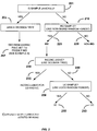
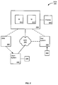
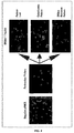
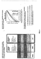
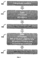


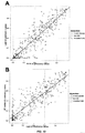
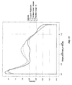


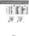


Classifications
-
- G—PHYSICS
- G01—MEASURING; TESTING
- G01N—INVESTIGATING OR ANALYSING MATERIALS BY DETERMINING THEIR CHEMICAL OR PHYSICAL PROPERTIES
- G01N33/00—Investigating or analysing materials by specific methods not covered by groups G01N1/00 - G01N31/00
- G01N33/48—Biological material, e.g. blood, urine; Haemocytometers
- G01N33/50—Chemical analysis of biological material, e.g. blood, urine; Testing involving biospecific ligand binding methods; Immunological testing
- G01N33/53—Immunoassay; Biospecific binding assay; Materials therefor
- G01N33/543—Immunoassay; Biospecific binding assay; Materials therefor with an insoluble carrier for immobilising immunochemicals
- G01N33/54306—Solid-phase reaction mechanisms
-
- G—PHYSICS
- G16—INFORMATION AND COMMUNICATION TECHNOLOGY [ICT] SPECIALLY ADAPTED FOR SPECIFIC APPLICATION FIELDS
- G16B—BIOINFORMATICS, i.e. INFORMATION AND COMMUNICATION TECHNOLOGY [ICT] SPECIALLY ADAPTED FOR GENETIC OR PROTEIN-RELATED DATA PROCESSING IN COMPUTATIONAL MOLECULAR BIOLOGY
- G16B20/00—ICT specially adapted for functional genomics or proteomics, e.g. genotype-phenotype associations
- G16B20/20—Allele or variant detection, e.g. single nucleotide polymorphism [SNP] detection
-
- C—CHEMISTRY; METALLURGY
- C12—BIOCHEMISTRY; BEER; SPIRITS; WINE; VINEGAR; MICROBIOLOGY; ENZYMOLOGY; MUTATION OR GENETIC ENGINEERING
- C12Q—MEASURING OR TESTING PROCESSES INVOLVING ENZYMES, NUCLEIC ACIDS OR MICROORGANISMS; COMPOSITIONS OR TEST PAPERS THEREFOR; PROCESSES OF PREPARING SUCH COMPOSITIONS; CONDITION-RESPONSIVE CONTROL IN MICROBIOLOGICAL OR ENZYMOLOGICAL PROCESSES
- C12Q1/00—Measuring or testing processes involving enzymes, nucleic acids or microorganisms; Compositions therefor; Processes of preparing such compositions
- C12Q1/68—Measuring or testing processes involving enzymes, nucleic acids or microorganisms; Compositions therefor; Processes of preparing such compositions involving nucleic acids
- C12Q1/6876—Nucleic acid products used in the analysis of nucleic acids, e.g. primers or probes
- C12Q1/6883—Nucleic acid products used in the analysis of nucleic acids, e.g. primers or probes for diseases caused by alterations of genetic material
-
- G—PHYSICS
- G01—MEASURING; TESTING
- G01N—INVESTIGATING OR ANALYSING MATERIALS BY DETERMINING THEIR CHEMICAL OR PHYSICAL PROPERTIES
- G01N33/00—Investigating or analysing materials by specific methods not covered by groups G01N1/00 - G01N31/00
- G01N33/48—Biological material, e.g. blood, urine; Haemocytometers
- G01N33/50—Chemical analysis of biological material, e.g. blood, urine; Testing involving biospecific ligand binding methods; Immunological testing
- G01N33/68—Chemical analysis of biological material, e.g. blood, urine; Testing involving biospecific ligand binding methods; Immunological testing involving proteins, peptides or amino acids
- G01N33/6854—Immunoglobulins
-
- G—PHYSICS
- G01—MEASURING; TESTING
- G01N—INVESTIGATING OR ANALYSING MATERIALS BY DETERMINING THEIR CHEMICAL OR PHYSICAL PROPERTIES
- G01N33/00—Investigating or analysing materials by specific methods not covered by groups G01N1/00 - G01N31/00
- G01N33/48—Biological material, e.g. blood, urine; Haemocytometers
- G01N33/50—Chemical analysis of biological material, e.g. blood, urine; Testing involving biospecific ligand binding methods; Immunological testing
- G01N33/68—Chemical analysis of biological material, e.g. blood, urine; Testing involving biospecific ligand binding methods; Immunological testing involving proteins, peptides or amino acids
- G01N33/6893—Chemical analysis of biological material, e.g. blood, urine; Testing involving biospecific ligand binding methods; Immunological testing involving proteins, peptides or amino acids related to diseases not provided for elsewhere
-
- G—PHYSICS
- G16—INFORMATION AND COMMUNICATION TECHNOLOGY [ICT] SPECIALLY ADAPTED FOR SPECIFIC APPLICATION FIELDS
- G16B—BIOINFORMATICS, i.e. INFORMATION AND COMMUNICATION TECHNOLOGY [ICT] SPECIALLY ADAPTED FOR GENETIC OR PROTEIN-RELATED DATA PROCESSING IN COMPUTATIONAL MOLECULAR BIOLOGY
- G16B20/00—ICT specially adapted for functional genomics or proteomics, e.g. genotype-phenotype associations
- G16B20/40—Population genetics; Linkage disequilibrium
-
- G—PHYSICS
- G16—INFORMATION AND COMMUNICATION TECHNOLOGY [ICT] SPECIALLY ADAPTED FOR SPECIFIC APPLICATION FIELDS
- G16B—BIOINFORMATICS, i.e. INFORMATION AND COMMUNICATION TECHNOLOGY [ICT] SPECIALLY ADAPTED FOR GENETIC OR PROTEIN-RELATED DATA PROCESSING IN COMPUTATIONAL MOLECULAR BIOLOGY
- G16B40/00—ICT specially adapted for biostatistics; ICT specially adapted for bioinformatics-related machine learning or data mining, e.g. knowledge discovery or pattern finding
-
- G—PHYSICS
- G16—INFORMATION AND COMMUNICATION TECHNOLOGY [ICT] SPECIALLY ADAPTED FOR SPECIFIC APPLICATION FIELDS
- G16H—HEALTHCARE INFORMATICS, i.e. INFORMATION AND COMMUNICATION TECHNOLOGY [ICT] SPECIALLY ADAPTED FOR THE HANDLING OR PROCESSING OF MEDICAL OR HEALTHCARE DATA
- G16H50/00—ICT specially adapted for medical diagnosis, medical simulation or medical data mining; ICT specially adapted for detecting, monitoring or modelling epidemics or pandemics
- G16H50/30—ICT specially adapted for medical diagnosis, medical simulation or medical data mining; ICT specially adapted for detecting, monitoring or modelling epidemics or pandemics for calculating health indices; for individual health risk assessment
-
- C—CHEMISTRY; METALLURGY
- C12—BIOCHEMISTRY; BEER; SPIRITS; WINE; VINEGAR; MICROBIOLOGY; ENZYMOLOGY; MUTATION OR GENETIC ENGINEERING
- C12Q—MEASURING OR TESTING PROCESSES INVOLVING ENZYMES, NUCLEIC ACIDS OR MICROORGANISMS; COMPOSITIONS OR TEST PAPERS THEREFOR; PROCESSES OF PREPARING SUCH COMPOSITIONS; CONDITION-RESPONSIVE CONTROL IN MICROBIOLOGICAL OR ENZYMOLOGICAL PROCESSES
- C12Q2600/00—Oligonucleotides characterized by their use
- C12Q2600/112—Disease subtyping, staging or classification
-
- C—CHEMISTRY; METALLURGY
- C12—BIOCHEMISTRY; BEER; SPIRITS; WINE; VINEGAR; MICROBIOLOGY; ENZYMOLOGY; MUTATION OR GENETIC ENGINEERING
- C12Q—MEASURING OR TESTING PROCESSES INVOLVING ENZYMES, NUCLEIC ACIDS OR MICROORGANISMS; COMPOSITIONS OR TEST PAPERS THEREFOR; PROCESSES OF PREPARING SUCH COMPOSITIONS; CONDITION-RESPONSIVE CONTROL IN MICROBIOLOGICAL OR ENZYMOLOGICAL PROCESSES
- C12Q2600/00—Oligonucleotides characterized by their use
- C12Q2600/156—Polymorphic or mutational markers
-
- C—CHEMISTRY; METALLURGY
- C12—BIOCHEMISTRY; BEER; SPIRITS; WINE; VINEGAR; MICROBIOLOGY; ENZYMOLOGY; MUTATION OR GENETIC ENGINEERING
- C12Q—MEASURING OR TESTING PROCESSES INVOLVING ENZYMES, NUCLEIC ACIDS OR MICROORGANISMS; COMPOSITIONS OR TEST PAPERS THEREFOR; PROCESSES OF PREPARING SUCH COMPOSITIONS; CONDITION-RESPONSIVE CONTROL IN MICROBIOLOGICAL OR ENZYMOLOGICAL PROCESSES
- C12Q2600/00—Oligonucleotides characterized by their use
- C12Q2600/158—Expression markers
-
- G—PHYSICS
- G01—MEASURING; TESTING
- G01N—INVESTIGATING OR ANALYSING MATERIALS BY DETERMINING THEIR CHEMICAL OR PHYSICAL PROPERTIES
- G01N2333/00—Assays involving biological materials from specific organisms or of a specific nature
- G01N2333/435—Assays involving biological materials from specific organisms or of a specific nature from animals; from humans
- G01N2333/46—Assays involving biological materials from specific organisms or of a specific nature from animals; from humans from vertebrates
- G01N2333/47—Assays involving proteins of known structure or function as defined in the subgroups
- G01N2333/4701—Details
- G01N2333/4737—C-reactive protein
-
- G—PHYSICS
- G01—MEASURING; TESTING
- G01N—INVESTIGATING OR ANALYSING MATERIALS BY DETERMINING THEIR CHEMICAL OR PHYSICAL PROPERTIES
- G01N2333/00—Assays involving biological materials from specific organisms or of a specific nature
- G01N2333/435—Assays involving biological materials from specific organisms or of a specific nature from animals; from humans
- G01N2333/475—Assays involving growth factors
- G01N2333/49—Platelet-derived growth factor [PDGF]
-
- G—PHYSICS
- G01—MEASURING; TESTING
- G01N—INVESTIGATING OR ANALYSING MATERIALS BY DETERMINING THEIR CHEMICAL OR PHYSICAL PROPERTIES
- G01N2333/00—Assays involving biological materials from specific organisms or of a specific nature
- G01N2333/435—Assays involving biological materials from specific organisms or of a specific nature from animals; from humans
- G01N2333/705—Assays involving receptors, cell surface antigens or cell surface determinants
- G01N2333/70503—Immunoglobulin superfamily, e.g. VCAMs, PECAM, LFA-3
- G01N2333/70525—ICAM molecules, e.g. CD50, CD54, CD102
-
- G—PHYSICS
- G01—MEASURING; TESTING
- G01N—INVESTIGATING OR ANALYSING MATERIALS BY DETERMINING THEIR CHEMICAL OR PHYSICAL PROPERTIES
- G01N2333/00—Assays involving biological materials from specific organisms or of a specific nature
- G01N2333/435—Assays involving biological materials from specific organisms or of a specific nature from animals; from humans
- G01N2333/705—Assays involving receptors, cell surface antigens or cell surface determinants
- G01N2333/70503—Immunoglobulin superfamily, e.g. VCAMs, PECAM, LFA-3
- G01N2333/70542—CD106
-
- G—PHYSICS
- G01—MEASURING; TESTING
- G01N—INVESTIGATING OR ANALYSING MATERIALS BY DETERMINING THEIR CHEMICAL OR PHYSICAL PROPERTIES
- G01N2333/00—Assays involving biological materials from specific organisms or of a specific nature
- G01N2333/435—Assays involving biological materials from specific organisms or of a specific nature from animals; from humans
- G01N2333/775—Apolipopeptides
-
- G—PHYSICS
- G01—MEASURING; TESTING
- G01N—INVESTIGATING OR ANALYSING MATERIALS BY DETERMINING THEIR CHEMICAL OR PHYSICAL PROPERTIES
- G01N2800/00—Detection or diagnosis of diseases
- G01N2800/06—Gastro-intestinal diseases
- G01N2800/065—Bowel diseases, e.g. Crohn, ulcerative colitis, IBS
-
- G—PHYSICS
- G01—MEASURING; TESTING
- G01N—INVESTIGATING OR ANALYSING MATERIALS BY DETERMINING THEIR CHEMICAL OR PHYSICAL PROPERTIES
- G01N2800/00—Detection or diagnosis of diseases
- G01N2800/60—Complex ways of combining multiple protein biomarkers for diagnosis
-
- G—PHYSICS
- G16—INFORMATION AND COMMUNICATION TECHNOLOGY [ICT] SPECIALLY ADAPTED FOR SPECIFIC APPLICATION FIELDS
- G16B—BIOINFORMATICS, i.e. INFORMATION AND COMMUNICATION TECHNOLOGY [ICT] SPECIALLY ADAPTED FOR GENETIC OR PROTEIN-RELATED DATA PROCESSING IN COMPUTATIONAL MOLECULAR BIOLOGY
- G16B20/00—ICT specially adapted for functional genomics or proteomics, e.g. genotype-phenotype associations
-
- Y—GENERAL TAGGING OF NEW TECHNOLOGICAL DEVELOPMENTS; GENERAL TAGGING OF CROSS-SECTIONAL TECHNOLOGIES SPANNING OVER SEVERAL SECTIONS OF THE IPC; TECHNICAL SUBJECTS COVERED BY FORMER USPC CROSS-REFERENCE ART COLLECTIONS [XRACs] AND DIGESTS
- Y02—TECHNOLOGIES OR APPLICATIONS FOR MITIGATION OR ADAPTATION AGAINST CLIMATE CHANGE
- Y02A—TECHNOLOGIES FOR ADAPTATION TO CLIMATE CHANGE
- Y02A90/00—Technologies having an indirect contribution to adaptation to climate change
- Y02A90/10—Information and communication technologies [ICT] supporting adaptation to climate change, e.g. for weather forecasting or climate simulation
Definitions
- IBD Inflammatory bowel disease
- CD Crohn's disease
- UC ulcerative colitis
- IC indeterminate colitis
- IBS irritable bowel syndrome
- Inflammatory bowel disease has many symptoms in common with irritable bowel syndrome, including abdominal pain, chronic diarrhea, weight loss, and cramping, making definitive diagnosis extremely difficult. Of the 5 million people suspected of suffering from IBD in the United States, only 1 million are diagnosed as having IBD. The difficulty in differentially diagnosing IBD and determining its outcome hampers early and effective treatment of these diseases. Thus, there is a need for rapid and sensitive testing methods for determining IBD and its subtypes.
- WO2010/120814 relates to inflammatory bowel disease prognostics.
- WO2010/056682 relates to methods for prediction of inflammatory bowel disease using serologic markers.
- US2008/182280 , US 2008/131439 and US2011/218956 relates to methods of diagnosing inflammatory bowel disease.
- the present invention provides methods and systems for the diagnosis of inflammatory bowel disease (IBD) and/or the determination of subtypes including ulcerative colitis (UC) and Crohn's Disease (CD) or categorizing the sample as inconclusive (e.g ., IBD sample that is inconclusive for CD and UC).
- IBD inflammatory bowel disease
- UC ulcerative colitis
- CD Crohn's Disease
- categorizing the sample as inconclusive e.g ., IBD sample that is inconclusive for CD and UC.
- the present invention provides a diagnostic model that comprises two separate random forests, one for IBD vs. non-IBD and the other for UC vs. CD.
- the present invention provides a model comprising four class outcomes (non-IBD, CD, UC, Inconclusive).
- the present invention provides a single model comprising a 3 class outcome (non-IBD, CD, UC).
- the present invention provides methods and systems to diagnose IBD or nonIBD and/or to differentiate between clinical subtypes of IBD such as UC and CD or categorizing the sample as inconclusive by analyzing a sample to determine the presence or absence of one, two, three, four, or more variant alleles (e.g., single nucleotide polymorphisms or SNPs) in the ATG16L1 (e.g., rs2241880, rs3828309), ECM1 (e.g., rs7511649, rs3737240 and rs13294), NKX2-3 (e.g.
- ATG16L1 e.g., rs2241880, rs3828309
- ECM1 e.g., rs7511649, rs3737240 and rs13294
- NKX2-3 e.g.
- the present invention may further include analyzing a sample to determine the presence (or absence) or concentration level of one or more serological and inflammation markers such as, e.g., ASCA-A, ASCA-G, anti-OmpC, anti-CBirl, anti-Fla2, anti-FlaX, VEGF, CRP, SAA, ICAM, VCAM, ANCA (e.g., by ELISA) and/or pANCA (e.g., by one or more indirect fluorescent antibody (IFA) assays to detect an IBD-specific pANCA perinuclear pattern and/or pANCA DNase sensitivity), pANCA2 to diagnose IBD or non-IBD, and/or to differentiate between clinical subtypes of IBD such as IC, UC and CD or categori
- serological and inflammation markers such as, e.g., ASCA-A, ASCA-G, anti-OmpC, anti-CBirl, anti-Fla2, anti-FlaX, VE
- the present invention provides a method for diagnosing inflammatory bowel disease (IBD) and/or a clinical subtype thereof in an individual, said method comprising:
- the IBD diagnostic assay comprises an initial node for determining whether a sample is pANCA2 negative as a first step to direct individual specimens to either a Decision Tree Method or the Random Forest Algorithm depending on their pANCA2 value.
- pANCA2 samples with either a negative (0 or "not detected") or a weak (+1) pANCA determination are removed and subjected to a decision matrix or set of rules for making a clinical prediction of IBD.
- the present invention provides for diagnosing inflammatory bowel disease (IBD) and/or a clinical subtype thereof in an individualsaid method comprising:
- the present invention is based, in part, upon the surprising discovery that the accuracy of diagnosing inflammatory bowel disease (IBD) and subtypes thereof can be substantially improved by detecting the presence, level, or genotype of certain markers in a biological sample from an individual.
- the present invention provides diagnostic platforms based on a serological and/or genetic panel of markers.
- the present invention provides methods and systems to improve the diagnosis of IBD and subgroups thereof such as, e.g., indeterminate colitis (IC), ulcerative colitis (UC), Crohn's disease (CD), and inconclusive for CD and UC ("Inconclusive").
- IBD indeterminate colitis
- UC ulcerative colitis
- CD Crohn's disease
- Inconclusive inconclusive for CD and UC
- the methods described herein accurately predict IBD, a disease which is known to be very difficult to diagnose and predict outcome.
- the methods described herein utilize multiple serological, protein, and/or genetic markers, alone or in combination with one or more algorithms or other types of statistical analysis, to provide physicians valuable diagnostic or prognostic insight.
- the methods and systems of the present invention provide an indication of a patient's projected response to biological therapy.
- the methods and systems of the present invention utilize multiple markers (e.g., serological, protein, and/or genetic) in conjunction with statistical analysis (e.g., quartile analysis) to provide prognostic value by identifying patients with complicated disease or a risk of developing disease complications (e.g., internal stricturing or internal penetrating disease) and/or a need for surgical intervention, while also assisting in assessing the rate of disease progression.
- the methods enable classification of disease severity along a continuum of IBD subgroups including IC, UC and CD.
- the methods guide therapeutic decisions of patients with advanced disease.
- the use of multiple markers provides the ability to distinguish responders from non-responders and guides initial therapeutic options (e.g., whether or not to prescribe aggressive treatment), with the potential to change disease behavior.
- Figure 1 illustrates a flowchart for an exemplary embodiment of an IBD diagnostic assay of the present invention comprising a two-step random forest algorithm.
- the IBD diagnostic assay applies the measurements from 17 sero-genetic-inflammation (sgi) biological markers (e.g., ANCA, ASCA-A, ASCA-G, anti-FlaX, anti-A4-Fla2, pANCA, anti-OmpC, anti-CBirl, ATG16L1, CRP, SAA, ICAM, VCAM, ECM1, STAT3, VEGF, NKX2-3, and combinations thereof) and computes a score based on a first random forest model for predicting IBD vs. non-IBD (110).
- sero-genetic-inflammation e.g., ANCA, ASCA-A, ASCA-G, anti-FlaX, anti-A4-Fla2, pANCA, anti-OmpC, anti-CBirl, ATG16L1,
- the first random forest model determines if a patient has IBD. If the score is less than the IBD vs. non-IBD cut-off (e.g., ⁇ 0.64), the sample is predicted to be from a patient having IBD (125). Otherwise, the sample is predicted to be from a patient having non-IBD (120). Samples predicted to have IBD proceed to the next step of the algorithm, which is a decision tree or set of rules designed to rule out categorizing the sample as inconclusive (130). If a sample matches the pattern for either of the "indeterminate” rules, the algorithm predicts the sample as having IBD, but is inconclusive for UC and CD (135).
- the algorithm predicts the sample as having IBD, but is inconclusive for UC and CD (135).
- the sample proceeds to the next step of the algorithm (140), which is a second random forest model for predicting UC vs. CD (150).
- the IBD diagnostic assay applies the measurements from 11 sgi biological markers (e.g., ANCA, ASCA-A, ASCA-G, anti-FlaX, anti-A4-Fla2, pANCA, anti-OmpC, anti-CBirl, ECM1, STAT3, VEGF, and combinations thereof) to compute a model score based on the second random forest model for predicting UC vs. CD. If the score is less than the UC vs. CD cut-off (e.g., 0.35), the algorithm predicts the sample as having CD (153). Or else, the algorithm predicts the sample as having UC (155).
- 11 sgi biological markers e.g., ANCA, ASCA-A, ASCA-G, anti-FlaX, anti-A4-Fla2, pANCA, anti-OmpC, anti
- Figure 2 illustrates a flowchart for another exemplary embodiment of an IBD diagnostic assay of the invention comprising a two-step random forest algorithm (right pathway) and a separate pANCA decision tree (left pathway).
- the IBD diagnostic assay comprises an initial node (201) for determining whether a sample is pANCA2 negative as a first step to direct individual specimens to either the Decision Tree Method (205) or the Random Forest Algorithm (210), depending on their pANCA2 value.
- pANCA2 samples with either a negative (0 or "not detected") or a weak (+1) pANCA determination are removed from the sgi algorithm queue (right pathway) and subjected to a decision matrix or set of rules for making a clinical prediction of IBD (207).
- An exemplary decision matrix for predicting IBD following pANCA decision tree analysis is described in Example 9.
- the pANCA decision tree (205) can be implemented as an alternate path for making predictive IBD diagnoses in clinical specimens with pANCA2 (-) assay results.
- Figure 2 also illustrates an exemplary path for analyzing pANCA2 positive samples (right pathway).
- pANCA2 samples with a positive (+2, +3, or +4) pANCA determination are processed using an IBD diagnostic assay and subjected to a two-step random forest algorithm analogous to the two-step random forest algorithm described in Figure 1 .
- the IBD diagnostic assay applies the measurements from 17 sero-genetic-inflammation (sgi) biological markers (e.g., ANCA, ASCA-A, ASCA-G, anti-FlaX, anti-A4-Fla2, pANCA, anti-OmpC, anti-CBirl, ATG16L1, CRP, SAA, ICAM, VCAM, ECM1, STAT3, VEGF, NKX2-3, and combinations thereof) and computes a score based on a first random forest model for predicting IBD vs. non-IBD (210). The first random forest model determines if a patient has IBD. If the score is less than the IBD vs.
- sero-genetic-inflammation e.g., ANCA, ASCA-A, ASCA-G, anti-FlaX, anti-A4-Fla2, pANCA, anti-OmpC, anti-CBirl, ATG16L1, CRP, SAA, ICAM,
- non-IBD cut-off e.g., ⁇ 0.64
- the sample is predicted to be from a patient having IBD (225). Otherwise, the sample is predicted to be from a patient having non-IBD (220).
- Samples predicted to have IBD proceed to the next step of the algorithm, which is a decision tree or set of rules designed to rule out categorizing the sample as inconclusive (230). If a sample matches the pattern for either of the "indeterminate" rules, the algorithm predicts the sample as having IBD, but is inconclusive for UC and CD (235). Otherwise, the sample proceeds to the next step of the algorithm (240), which is a second random forest model for predicting UC vs. CD (250).
- the IBD diagnostic assay applies the measurements from 11 sgi biological markers (e.g., ANCA, ASCA-A, ASCA-G, anti-FlaX, anti-A4-Fla2, pANCA, anti-OmpC, anti-CBirl, ECM1, STAT3, VEGF, and combinations thereof) to compute a model score based on the second random forest model for predicting UC vs. CD. If the score is less than the UC vs. CD cut-off ( e.g. , 0.35), the algorithm predicts the sample as having CD (253). Or else, the algorithm predicts the sample as having UC (255).
- 11 sgi biological markers e.g., ANCA, ASCA-A, ASCA-G, anti-FlaX, anti-A4-Fla2, pANCA, anti-OmpC, anti-CBirl, ECM1, STAT3, VEGF, and combinations thereof.
- the methods and systems of the present invention comprise a step having a "transformation" or “machine” associated therewith.
- an ELISA technique may be performed to measure the presence or concentration level of many of the markers described herein.
- An ELISA includes transformation of the marker, e.g., an auto-antibody, into a complex between the marker (e.g., auto-antibody) and a binding agent (e.g., antigen), which then can be measured with a labeled secondary antibody.
- the label is an enzyme which transforms a substrate into a detectable product.
- the detectable product measurement can be performed using a plate reader such as a spectrophotometer.
- genetic markers are determined using various amplification techniques such as PCR.
- Method steps including amplification result in the transformation of single or double strands of nucleic acid into multiple strands for detection.
- the detection can include the use of a fluorophore, which is performed using a machine such as a fluorometer.
- the methods further comprise comparing the results from the statistical analysis (i.e., diagnostic profile) to a reference (i.e., diagnostic model) to aid in the diagnosis of IBD.
- the methods utilize multiple serological, protein, and/or genetic markers to provide physicians with valuable diagnostic insight.
- the assay methods and systems of the present invention provide diagnostic value by identifying patients having IBD and IBD subgroups such as IC, UC, CD, and inconclusive.
- IBD subgroups such as IC, UC, CD, and inconclusive.
- the present invention enables classification of disease severity along a continuum of IBD subgroups including, but not limited to, IC, UC, CD, and inconclusive.
- classifying includes “associating” or “categorizing” a sample or an individual with a disease state or prognosis. In certain instances, “classifying” is based on statistical evidence, empirical evidence, or both. In certain embodiments, the methods and systems of classifying use a so-called training set of samples from individuals with known disease states or prognoses. Once established, the training data set serves as a basis, model, or template against which the features of an unknown sample from an individual are compared, in order to classify the unknown disease state or provide a prognosis of the disease state in the individual. In some instances, "classifying” is akin to diagnosing the disease state and/or differentiating the disease state from another disease state. In other instances, “classifying” is akin to providing a prognosis of the disease state in an individual diagnosed with the disease state.
- IBD inflammatory bowel disease
- gastrointestinal disorders such as, e.g. , Crohn's disease (CD), ulcerative colitis (UC), indeterminate colitis (IC), and IBD that is inconclusive for CD vs. UC ("Inconclusive").
- Inflammatory bowel diseases e.g. , CD, UC, IC, and Inconclusive
- IBS irritable bowel syndrome
- sample includes any biological specimen obtained from an individual. Suitable samples for use in the present invention include, without limitation, whole blood, plasma, serum, saliva, urine, stool, tears, any other bodily fluid, tissue samples ( e.g., biopsy), and cellular extracts thereof ( e.g. , red blood cellular extract).
- tissue samples e.g., biopsy
- cellular extracts thereof e.g. , red blood cellular extract
- the sample is a serum sample.
- samples such as serum, saliva, and urine is well known in the art ( see, e.g., Hashida et al., J. Clin. Lab. Anal., 11:267-86 (1997 )).
- samples such as serum samples can be diluted prior to the analysis of marker levels.
- marker includes any biochemical marker, serological marker, genetic marker, or other clinical or echographic characteristic that can be used in the diagnosis of IBD, in the prediction of the probable course and outcome of IBD, and/or in the prediction of the likelihood of recovery from the disease.
- Non-limiting examples of such markers include serological markers such as an anti-neutrophil antibody (e.g., ANCA, pANCA, and the like), an anti- Saccharomyces cerevisiae antibody (e.g., ASCA-IgA, ASCA-IgG), an antimicrobial antibody (e.g., anti-OmpC antibody, anti-I2 antibody, anti-Fla2 antibody, anti-FlaX antibody, anti-CBirl antibody), an acute phase protein (e.g., CRP), an apolipoprotein (e.g., SAA), a defensin (e.g., ⁇ defensin), a growth factor (e.g., EGF, VEGF), a cytokine (e.g., TWEAK, IL-1 ⁇ , IL-6), a cadherin (e.g., E-cadherin), a cellular adhesion molecule (e.g., ICAM-1, VCAM-1,
- Non-limiting examples of genetic markers include variant alleles in the GLI1 (e.g., rs2228224 and/or rs2228226), MDR1 (e.g., rs2032582), ATG16L1 (e.g., rs2241880, rs3828309), ECM1 (e.g., rs7511649, rs373240, rs13294), NKX2-3 (e.g., rs1190140, rs10883365, rs6584283), STAT3 (e.g., rs744166), and NOD2/CARD15 (e.g.
- GLI1 e.g., rs2228224 and/or rs2228226
- MDR1 e.g., rs2032582
- ATG16L1 e.g., rs2241880, rs3828309
- ECM1 e.g., r
- the markers are utilized in combination with one or more (e.g. , a plurality of) statistical analyses to aid or provide a diagnosis or prognosis of IBD in an individual.
- the diagnosis can be IBD or a clinical subtype thereof such as Crohn's disease (CD), ulcerative colitis (UC), indeterminate colitis (IC), or inconclusive IBD.
- the prognosis can be the need for surgery (e.g., the likelihood or risk of needing small bowel surgery), development of a clinical subtype of CD or UC (e.g., the likelihood or risk of being susceptible to a particular clinical subtype CD or UC such as the stricturing, penetrating, or inflammatory CD subtype), development of one or more clinical factors (e.g., the likelihood or risk of being susceptible to a particular clinical factor), development of intestinal cancer (e.g., the likelihood or risk of being susceptible to intestinal cancer), or recovery from the disease (e.g., the likelihood of remission).
- a clinical subtype of CD or UC e.g., the likelihood or risk of being susceptible to a particular clinical subtype CD or UC such as the stricturing, penetrating, or inflammatory CD subtype
- development of one or more clinical factors e.g., the likelihood or risk of being susceptible to a particular clinical factor
- development of intestinal cancer e.g., the likelihood or risk of being susceptible to intestinal cancer
- the present invention relies, in part, on determining the presence (or absence) or level (e.g., concentration) of at least one marker in a sample obtained from an individual.
- detecting the presence of at least one marker includes determining the presence of each marker of interest by using any quantitative or qualitative assay known to one of skill in the art.
- qualitative assays that determine the presence or absence of a particular trait, variable, genotype, and/or biochemical or serological substance (e.g., protein or antibody) are suitable for detecting each marker of interest.
- quantitative assays that determine the presence or absence of DNA, RNA, protein, antibody, or activity are suitable for detecting each marker of interest.
- the term "detecting the level of at least one marker” includes determining the level of each marker of interest by using any direct or indirect quantitative assay known to one of skill in the art. In certain instances, quantitative assays that determine, for example, the relative or absolute amount of DNA, RNA, protein, antibody, or activity are suitable for detecting the level of each marker of interest. One skilled in the art will appreciate that any assay useful for detecting the level of a marker is also useful for detecting the presence or absence of the marker.
- the term "marker profile” includes 1, 2, 3, 4, 5, 6, 7, 8, 9, 10, 11, 12, 13, 14, 15, 16, 17, 18, 19, 20, 21, 22, 23, 24, 25, 30, 35, 40, 45, 50, or more diagnostic and/or prognostic marker(s), wherein the markers can be a serological marker, a protein marker, a genetic marker, and the like.
- the marker profile together with a statistical analysis can provide physicians and caregivers valuable diagnostic and prognostic insight.
- the marker profile with optionally a statistical analysis provides a projected response to biological therapy.
- the assays described herein provide diagnostic, prognostic and therapeutic value by identifying patients with IBD or a clinical subtype thereof, predicting risk of developing complicated disease, assisting in assessing the rate of disease progression (e.g., rate of progression to complicated disease or surgery), and assisting in the selection of therapy.
- markers e.g., serological, protein, genetic, etc.
- the term "diagnostic profile" includes 1, 2, 3, 4, 5, 6, 7, 8, 9, 10, 11, 12, 13, 14, 15, 16, 17, 18, 19, 20, 21, 22, 23, 24, 25, 30, 35, 40, 45, 50, or more marker(s) of an individual, wherein the marker(s) can be a serological marker, a protein marker, a genetic marker, and the like.
- a statistical analysis transforms the marker profile into a diagnostic profile.
- a preferred statistical analysis is a quartile score and the quartile score for each of the markers can be summed to generate a quartile sum score.
- the term "diagnostic model” includes serological models, genetic models, sero-genetic-inflammatory models, and a combination thereof.
- a retrospective analysis is done on a cohort of known disease outcomes with known complications and surgical procedures performed.
- a regression analysis e.g., logistic regression
- the model can be illustrated or depicted in, e.g., a look-up table, graph or other display.
- a diagnostic profile of an individual can then be compared to a diagnostic model and the diagnosis determined (e.g., the risk or probability of having a disease).
- the term "individual,” “subject,” or “patient” typically includes humans, but also includes other animals such as, e.g., other primates, rodents, canines, felines, equines, ovines, porcines, and the like.
- substantially the same amino acid sequence includes an amino acid sequence that is similar, but not identical to, the naturally-occurring amino acid sequence.
- an amino acid sequence, i.e., polypeptide that has substantially the same amino acid sequence as an 12 protein can have one or more modifications such as amino acid additions, deletions, or substitutions relative to the amino acid sequence of the naturally-occurring 12 protein, provided that the modified polypeptide retains substantially at least one biological activity of 12 such as immunoreactivity.
- Comparison for substantial similarity between amino acid sequences is usually performed with sequences between about 6 and 100 residues, preferably between about 10 and 100 residues, and more preferably between about 25 and 35 residues.
- a particularly useful modification of a polypeptide of the present invention, or a fragment thereof, is a modification that confers, for example, increased stability.
- Incorporation of one or more D-amino acids is a modification useful in increasing stability of a polypeptide or polypeptide fragment.
- deletion or substitution of lysine residues can increase stability by protecting the polypeptide or polypeptide fragment against degradation.
- clinical factor includes a symptom in an individual that is associated with IBD.
- examples of clinical factors include, without limitation, diarrhea, abdominal pain, cramping, fever, anemia, weight loss, anxiety, depression, and combinations thereof.
- a diagnosis or prognosis of IBD is based upon a combination of analyzing a sample obtained from an individual to determine the presence, level, or genotype of one or more markers by applying one or more statistical analyses and determining whether the individual has one or more clinical factors.
- symptom or “symptoms” and variants thereof includes any sensation, change or perceived change in bodily function that is experienced by an individual and is associated with a particular diseases or that accompanies a disease and is regarded as an indication of the disease.
- Disease for which symptoms in the context of the present invention can be associated with include inflammatory bowel disease (IBD), ulcerative colitis (UC) or Crohn's disease (CD).
- the methods of invention are used after an individual has been diagnosed with IBD.
- the methods can be used to diagnose IBD or can be used as a "second opinion" if, for example, IBD is suspected or has been previously diagnosed using other methods.
- the methods can be used to diagnose UC or differentiate between UC and CD.
- diagnosis IBD and variants thereof includes the use of the methods and systems described herein to determine the presence or absence of IBD.
- diagnosis UC includes the use of the methods and systems described herein to determine the presence or absence of UC, as well as to differentiate between UC and CD. The terms can also include assessing the level of disease activity in an individual.
- a statistical analysis is used to diagnose a mild, moderate, severe, or fulminant form of IBD or UC based upon the criteria developed by Truelove et al., Br. Med. J., 12:1041-1048 (1955 ). In other embodiments, a statistical analysis is used to diagnose a mild to moderate, moderate to severe, or severe to fulminant form of IBD or UC based upon the criteria developed by Hanauer et al., Am. J. Gastroenterol., 92:559-566 (1997 ). One skilled in the art will know of other methods for evaluating the severity of IBD or UC in an individual.
- the methods of the invention are used in order to diagnose IBD, diagnose UC or differentiate IC from UC and/or CD and differentiate between UC and CD or categorizing the sample as inconclusive.
- the methods can be used to monitor the disease, both progression and regression.
- the term "monitoring the progression or regression of IBD or UC” includes the use of the methods and marker profiles to determine the disease state (e.g., presence or severity of IBD or the presence of UC) of an individual.
- the results of a statistical analysis are compared to those results obtained for the same individual at an earlier time.
- the methods of the present invention can also be used to predict the progression of IBD or UC, e.g., by determining a likelihood for IBD or UC to progress either rapidly or slowly in an individual based on the presence or level of at least one marker in a sample.
- the methods of the present invention can also be used to predict the regression of IBD or UC, e.g., by determining a likelihood for IBD or UC to regress either rapidly or slowly in an individual based on the presence or level of at least one marker in a sample.
- gene refers to the segment of DNA involved in producing a polypeptide chain; it includes regions preceding and following the coding region, such as the promoter and 3'-untranslated region, respectively, as well as intervening sequences (introns) between individual coding segments (exons).
- genotyp refers to the genetic composition of an organism, including, for example, whether a diploid organism is heterozygous or homozygous for one or more variant alleles of interest.
- polymorphism refers to the occurrence of two or more genetically determined alternative sequences or alleles in a population.
- a "polymorphic site” refers to the locus at which divergence occurs. Preferred polymorphic sites have at least two alleles, each occurring at a particular frequency in a population.
- a polymorphic locus may be as small as one base pair (i.e., single nucleotide polymorphism or SNP).
- Polymorphic markers include restriction fragment length polymorphisms, variable number of tandem repeats (VNTR's), hypervariable regions, minisatellites, dinucleotide repeats, trinucleotide repeats, tetranucleotide repeats, simple sequence repeats, and insertion elements such as Alu.
- the first identified allele is arbitrarily designated as the reference allele, and other alleles are designated as alternative alleles, "variant alleles," or “variances.”
- the allele occurring most frequently in a selected population is sometimes referred to as the "wild-type" allele.
- Diploid organisms may be homozygous or heterozygous for the variant alleles.
- the variant allele may or may not produce an observable physical or biochemical characteristic ("phenotype") in an individual carrying the variant allele.
- a variant allele may alter the enzymatic activity of a protein encoded by a gene of interest.
- single nucleotide polymorphism and variants thereof refers to a change of a single nucleotide with a polynucleotide, including within an allele. This can include the replacement of one nucleotide by another, as well as deletion or insertion of a single nucleotide. Most typically, SNPs are biallelic markers although tri- and tetra-allelic markers can also exist. By way of non-limiting example, a nucleic acid molecule comprising SNP A ⁇ C may include a C or A at the polymorphic position. For combinations of SNPs, the term “haplotype" is used, e.g.
- haplotype can be used to describe a combination of SNP alleles, e.g., the alleles of the SNPs found together on a single DNA molecule.
- the SNPs in a haplotype can be in linkage disequilibrium with one another.
- specific or “specificity” and variants thereof when used in the context of polynucleotides capable of detecting variant alleles (e.g., polynucleotides that are capable of discriminating between different alleles), includes the ability to bind or hybridize or detect one variant allele without binding or hybridizing or detecting the other variant allele.
- specificity can refer to the ability of a polynucleotide to detect the wild-type and not the mutant or variant allele. In other embodiments, specificity can refer to the ability of a polynucleotide to detect the mutant or variant allele and not the wild-type allele.
- antibody includes a population of immunoglobulin molecules, which can be polyclonal or monoclonal and of any isotype, or an immunologically active fragment of an immunoglobulin molecule.
- an immunologically active fragment contains the heavy and light chain variable regions, which make up the portion of the antibody molecule that specifically binds an antigen.
- an immunologically active fragment of an immunoglobulin molecule known in the art as Fab, Fab' or F(ab') 2 is included within the meaning of the term antibody.
- quartile analysis there are three numbers (values) that divide a range of data into four equal parts.
- the first quartile also called the 'lower quartile'
- the second quartile (the 'median') divides the range in the middle and has 50 percent of the data below it.
- the third quartile (also called the 'upper quartile') has 75 percent of the data below it and the top 25 percent of the data above it.
- quartile analysis can be applied to the concentration level of a marker such as an antibody or other protein marker described herein, such that a marker level in the first quartile ( ⁇ 25%) is assigned a value of 1, a marker level in the second quartile (25-50%) is assigned a value of 2, a marker level in the third quartile (51%- ⁇ 75%) is assigned a value of 3, and a marker level in the fourth quartile (75%-100%) is assigned a value of 4.
- a marker level in the first quartile ⁇ 25%)
- a marker level in the second quartile 25-50%)
- a marker level in the third quartile 51%- ⁇ 75%)
- a marker level in the fourth quartile (75%-100%) is assigned a value of 4.
- quartile sum score or "QSS” includes the sum of quartile scores for all of the markers of interest.
- a quartile sum score for a panel of 6 markers may range from 6-24, wherein each of the individual markers is assigned a quartile score (Q) of 1-4 based upon the presence or absence of the marker, the concentration level of the marker, or the genotype of the marker.
- the present invention provides a method for diagnosing inflammatory bowel disease (IBD) and/or a clinical subtype thereof in an individual, the method comprising:
- the present invention provides for diagnosing inflammatory bowel disease (IBD) and/or a clinical subtype thereof in an individual, the method comprising:
- the methods further comprise comparing the results from the statistical analysis (i.e., diagnostic profile) to a reference (i.e., diagnostic model) to aid in the diagnosis of IBD.
- the methods utilize multiple serological, protein, and/or genetic markers to provide physicians with valuable diagnostic insight.
- the methods and systems herein measure a first serological marker of pANCA to determine a value of pANCA2.
- pANCA is assigned a value of 0 when it is not detected or when it is DNAse cytoplasmic sensitive.
- pANCA is assigned a value of 1 when it is DNAse Sensitive 1+P, DNAse Sensitive 2+P, DNAse Sensitive 3+P, or DNAse Sensitive 4+P, wherein 1+P through 4+P indicate increasing amounts and intensity of fluorescence label based on visual assessment. For example, if the pANCA values are +2, +3 or +4 in a sample, then the pANCA2 value is positive.
- pANCA2 is assigned a value of 0 (negative) when pANCA is not detected, pANCA is DNAse cytoplasmic sensitive, or DNAse Sensitive 1+P. pANCA2 is given a value of 1 (positive), when pANCA is DNAse Sensitive 2+P, DNAse Sensitive 3+P or DNAse Sensitive 4+P.
- the methods and systems further comprise:
- ASCA-IgA in part a above is greater than or equal to 69 EU/mL, then the sample is consistent with Crohn's disease.
- ASCA-IgG in part a above is greater than or equal to 40 EU/mL, then the sample is consistent with Crohn's disease.
- the systems and methods provide assigning a Crohn's disease (CD) count value.
- CD Crohn's disease
- the CD count value is determined using the following rules.
- the reference value for CBirl-IgG is greater than or equal to 78.4 EU/mL.
- the reference value for A4-Fla2-IgG is greater than or equal to 44.8 EU/mL.
- the reference value for FlaX-IgG is greater than or equal to 33.4 EU/mL.
- the methods and systems herein further comprise for part c above, if pANCA is zero (not detected), then:
- the methods and systems herein further comprise that when pANCA is one:
- the present invention provides methods and systems for improved diagnosis of inflammatory bowel disease (IBD) and to differentiate ulcerative colitis (UC) and Crohn's disease (CD).
- IBD inflammatory bowel disease
- UC ulcerative colitis
- CD Crohn's disease
- the methods and systems enable classification of disease severity along a continuum of IBD subgroups rather than merely as CD or UC.
- the methods categorize the sample as being inconclusive. By identifying patients with complicated disease and assisting in assessing the specific disease type, the methods and systems described herein provide invaluable information to assess the severity of the disease and treatment options.
- applying a statistical analysis to a profile of serological, protein, and/or genetic markers improves the accuracy of predicting IBD, UC and CD, and also enables the selection of appropriate treatment options, including therapy such as biological, conventional, surgery, or some combination thereof.
- the present invention provides methods and systems improved for diagnosis of ulcerative colitis (UC) and to differentiate between UC and Crohn's disease (CD).
- the methods and systems enable classification of disease severity along a continuum of IBD subgroups rather than merely as CD or UC.
- the methods and systems described herein provide invaluable information to assess the severity of the disease and treatment options.
- applying a statistical analysis to a profile of serological, protein, and/or genetic markers improves the accuracy of predicting UC, and also enables the selection of appropriate treatment options, including therapy such as biological, conventional, surgery, or some combination thereof.
- the present invention provides methods and systems for improved diagnosis of Crohn's disease (CD) and to differentiate between UC and CD.
- the methods and systems enable classification of disease severity along a continuum of IBD subgroups rather than merely as CD or UC.
- the methods and systems described herein provide invaluable information to assess the severity of the disease and treatment options.
- applying a statistical analysis to a profile of serological, protein, and/or genetic markers improves the accuracy of predicting CD, and also enables the selection of appropriate treatment options, including therapy such as biological, conventional, surgery, or some combination thereof.
- the present invention provides methods and systems for improved diagnosis of inflammatory bowel disease (IBD) and to differentiate it from non-IBD.
- the present invention also provides methods and systems to diagnose non-inflammatory bowel disease (non-IBD) and to differentiate it from IBD.
- the methods and systems enable classification of disease severity along a continuum of IBD subgroups rather than merely as CD or UC. By identifying patients with complicated disease and assisting in assessing the specific disease type, the methods and systems described herein provide invaluable information to assess the severity of the disease and treatment options.
- applying a statistical analysis to a profile of serological, protein, and/or genetic markers improves the accuracy of predicting IBD and non-IBD, and also enables the selection of appropriate treatment options, including therapy such as biological, conventional, surgery, or some combination thereof.
- the present invention provides methods and systems for improved diagnosis of inconclusive as well as differentiate it from UC and CD.
- the methods and systems enable classification of disease severity along a continuum of IBD subgroups rather than merely as CD or UC.
- the methods and systems described herein provide invaluable information to assess the severity of the disease and treatment options.
- applying a statistical analysis to a profile of serological, protein, and/or genetic markers improves the accuracy of predicting subtypes, and also enables the selection of appropriate treatment options, including therapy such as biological, conventional, surgery, or some combination thereof.
- the method of the present invention is performed in an individual with symptoms of UC.
- the symptoms of UC include, but are not limited to, rectal inflammation, rectal bleeding, rectal pain, diarrhea, abdominal cramps, abdominal pain, fatigue, weight loss, fever, colon rupture, and combinations thereof.
- the method of the present invention entails analysis of a biological sample selected from the group consisting of whole blood, tissue, saliva, cheek cells, hair, fluid, plasma, serum, cerebrospinal fluid, buccal swabs, mucus, urine, stools, spermatozoids, vaginal secretions, lymph, amniotic fluid, pleural liquid, tears, and combinations thereof.
- the present invention provides methods and systems to diagnose IBD and to differentiate between subtypes of IBD such as UC and CD.
- the methods and systems of the present invention utilize one or a plurality of (e.g. , multiple) genetic markers, alone or in combination with one or a plurality of serological and/or protein markers, and alone or in combination with one or a plurality of algorithms (e.g., random forest model and decision tree or set of rules) or other types of statistical analysis (e.g., quartile analysis), to aid or assist in identifying patients with IBD, CD and/or UC and providing physicians with valuable diagnostic insight.
- algorithms e.g., random forest model and decision tree or set of rules
- other types of statistical analysis e.g., quartile analysis
- the method of diagnosing IBD comprises detection of the NKX2-3 (rs1190140) variant allele. In other embodiments, the method of diagnosing IBD preferably employs detection of the NKX2-3 (rs10883365) variant allele. In yet other embodiments, the method of diagnosing IBD employs detection of the NKX2-3 (rs6584283) variant allele. In some preferred embodiments, the method of diagnosing IBD comprises detection of the ATG16L1 (rs2241880) variant allele. In further embodiments, the method of diagnosing IBD comprises detection of the ATG16L1 (rs3828309) variant allele.
- the method of diagnosing IBD employs detection of the ECM1 (rs7511649) variant allele. In other preferred embodiments, the method of diagnosing IBD comprises detection of the ECM1 (rs373240) variant allele. In yet other embodiments, the method of diagnosing IBD comprises detection of the ECM1 (rs13294) variant allele. In some preferred embodiments, the method of diagnosing IBD comprises detection of the STAT3 (rs744166) variant allele.
- the method of diagnosing UC comprises detection of the NKX2-3 (rs1190140) variant allele. In other embodiments, the method of diagnosing UC comprises detection of the NKX2-3 (rs10883365) variant allele. In yet other embodiments, the method of diagnosing UC comprises detection of the NKX2-3 (rs6584283) variant allele. In some embodiments, the method of diagnosing UC comprises detection of the ATG16L1 (rs2241880) variant allele. In further embodiments, the method of diagnosing UC comprises detection of the ATG16L1 (rs3828309) variant allele. In some embodiments, the method of diagnosing UC comprises detection of the ECM1 (rs7511649) variant allele.
- the method of diagnosing UC comprises detection of the ECM1 (rs373240) variant allele. In yet other embodiments, the method of diagnosing UC comprises detection of the ECM1 (rs13294) variant allele. In some embodiments, the method of diagnosing UC comprises detection of the STAT3 (rs744166) variant allele.
- the method of diagnosing CD comprises detection of the NKX2-3 (rs1190140) variant allele. In other embodiments, the method of diagnosing CD comprises detection of the NKX2-3 (rs10883365) variant allele. In yet other embodiments, the method of diagnosing CD comprises detection of the NKX2-3 (rs6584283) variant allele. In some embodiments, the method of diagnosing CD employs detection of the ATG16L1 (rs2241880) variant allele. In further embodiments, the method of diagnosing CD comprises detection of the ATG16L1 (rs3828309) variant allele. In some embodiments, the method of diagnosing CD comprises detection of the ECM1 (rs7511649) variant allele.
- the method of diagnosing CD comprises detection of the ECM1 (rs373240) variant allele. In yet other embodiments, the method of diagnosing CD comprises detection of the ECM1 (rs13294) variant allele. In some embodiments, the method of diagnosing CD comprises detection of the STAT3 (rs744166) variant allele.
- the method of diagnosing IBD comprises detection of one or more variant alleles selected from the group consisting of ATG16L1 (e.g. , rs2241880, rs3828309), ECM1 (e.g., rs7511649, rs373240, rs13294), NKX2-3 (e.g., rs1190140, rs10883365, rs6584283), and STAT3 (e.g. , rs744166).
- ATG16L1 e.g. , rs2241880, rs3828309
- ECM1 e.g., rs7511649, rs373240, rs13294
- NKX2-3 e.g., rs1190140, rs10883365, rs6584283
- STAT3 e.g. , rs744166
- the method of diagnosing IC and/or categorizing a sample as being inconclusive comprises detection of one or more variant alleles selected from the group consisting of ATG16L1 (e.g. , rs2241880, rs3828309), ECM1 (e.g., rs7511649, rs373240, rs13294), NKX2-3 (e.g., rs1190140, rs10883365, rs6584283), and STAT3 (e.g., rs744166).
- ATG16L1 e.g. , rs2241880, rs3828309
- ECM1 e.g., rs7511649, rs373240, rs13294
- NKX2-3 e.g., rs1190140, rs10883365, rs6584283
- STAT3 e.g., rs7441
- the method of diagnosing UC comprises detection of one or more variant alleles selected from the group consisting of ATG16L1 (e.g. , rs2241880, rs3828309), ECM1 (e.g., rs7511649, rs373240, rs13294), NKX2-3 (e.g., rs1190140, rs10883365, rs6584283), and STAT3 (e.g., rs744166).
- ATG16L1 e.g. , rs2241880, rs3828309
- ECM1 e.g., rs7511649, rs373240, rs13294
- NKX2-3 e.g., rs1190140, rs10883365, rs6584283
- STAT3 e.g., rs744166
- the method of diagnosing CD comprises detection of one or more variant alleles selected from the group consisting of ATG16L1 (e.g., rs2241880, rs3828309), ECM1 (e.g., rs7511649, rs373240, rs13294), NKX2-3 (e.g., rs1190140, rs10883365, rs6584283), and STAT3 (e.g., rs744166).
- ATG16L1 e.g., rs2241880, rs3828309
- ECM1 e.g., rs7511649, rs373240, rs13294
- NKX2-3 e.g., rs1190140, rs10883365, rs6584283
- STAT3 e.g., rs744166
- the presence or absence of a variant allele in a genetic marker can be determined using an assay described herein.
- Assays that can be used to determine variant allele status include, but are not limited to, electrophoretic analysis assays, restriction length polymorphism analysis assays, sequence analysis assays, hybridization analysis assays, PCR analysis assays, allele-specific hybridization, oligonucleotide ligation allele-specific elongation/ligation, allele-specific amplification, single-base extension, molecular inversion probe, invasive cleavage, selective termination, restriction length polymorphism, sequencing, single strand conformation polymorphism (SSCP), single strand chain polymorphism, mismatch-cleaving, denaturing gradient gel electrophoresis, and combinations thereof.
- electrophoretic analysis assays restriction length polymorphism analysis assays
- sequence analysis assays sequence analysis assays
- hybridization analysis assays hybridization analysis assays
- PCR analysis assays
- the method described herein improves the diagnosis of UC compared to ANCA and/or pANCA-based methods of diagnosing UC.
- the method of diagnosing IBD and/or subgroups including UC and CD comprises an additional step of analyzing the biological sample for the presence or level of a serological marker, wherein detection of the presence or level of the serological marker in conjunction with the presence of one or more variant alleles further improves the diagnosis of IBD and/or subgroups including UC and CD.
- the method of diagnosing IBD and/or subgroups including UC and CD comprises detection of a serological marker selected from an anti-neutrophil antibody, an anti- Saccharomyces cerevisiae antibody, an antimicrobial antibody, an acute phase protein, an apolipoprotein, a defensin, a growth factor, a cytokine, a cadherin, a cellular adhesion molecule, or any combination of the markers described herein.
- a serological marker selected from an anti-neutrophil antibody, an anti- Saccharomyces cerevisiae antibody, an antimicrobial antibody, an acute phase protein, an apolipoprotein, a defensin, a growth factor, a cytokine, a cadherin, a cellular adhesion molecule, or any combination of the markers described herein.
- the method of diagnosing IBD and/or subgroups including UC and CD utilizes an anti-neutrophil antibody that is selected from one of ANCA and pANCA, or a combination of ANCA and pANCA.
- the anti-neutrophil antibody comprises an anti-neutrophil cytoplasmic antibody (ANCA) such as ANCA detected by an immunoassay (e.g., ELISA), a perinuclear anti-neutrophil cytoplasmic antibody (pANCA) such as pANCA detected by an immunohistochemical assay (e.g., IFA) or a DNAse-sensitive immunohistochemical assay, or a combination thereof.
- ANCA anti-neutrophil cytoplasmic antibody
- pANCA perinuclear anti-neutrophil cytoplasmic antibody
- IFA immunohistochemical assay
- DNAse-sensitive immunohistochemical assay e.g., DNAse-sensitive immunohistochemical assay
- the method of diagnosing IBD and/or subgroups including UC and CD utilizes an anti- Saccharomyces cerevisiae antibody that is selected from the group consisting of anti- Saccharomyces cerevisiae immunoglobulin A (ASCA-IgA), anti- Saccharomyces cerevisiae immunoglobulin G (ASCA-IgG), and a combination thereof.
- ASCA-IgA anti- Saccharomyces cerevisiae immunoglobulin A
- ASCA-IgG anti- Saccharomyces cerevisiae immunoglobulin G
- the method of diagnosing IBD and/or subgroups including UC and CD utilizes an antimicrobial antibody that is selected from the group consisting of an anti-outer membrane protein C (anti-OmpC) antibody, an anti-I2 antibody, an anti-flagellin antibody (e.g., anti-CBirl antibody, anti-Fla2 antibody (i.e., anti-A4-Fla2), and anti-FlaX antibody), and a combination thereof.
- an antimicrobial antibody that is selected from the group consisting of an anti-outer membrane protein C (anti-OmpC) antibody, an anti-I2 antibody, an anti-flagellin antibody (e.g., anti-CBirl antibody, anti-Fla2 antibody (i.e., anti-A4-Fla2), and anti-FlaX antibody), and a combination thereof.
- an antimicrobial antibody that is selected from the group consisting of an anti-outer membrane protein C (anti-OmpC) antibody, an anti-I2 antibody,
- the method of diagnosing IBD and/or subgroups including UC and CD utilizes an acute phase protein such as, but not limited to C-Reactive protein (CRP).
- an acute phase protein such as, but not limited to C-Reactive protein (CRP).
- the method of diagnosing IBD and/or subgroups including UC and CD utilizes an apolipoprotein such as, but not limited to serum amyloid A (SAA).
- SAA serum amyloid A
- the method of diagnosing IBD and/or subgroups including UC and CD utilizes a defensin that is selected from the group consisting of ⁇ defensin (BD1), ⁇ defensin-2 (BD2), or combination thereof.
- BD1 ⁇ defensin
- BD2 ⁇ defensin-2
- the method of diagnosing IBD and/or subgroups including UC and CD utilizes a growth factor that is selected from the group consisting of epidermal growth factor (EGF), vascular endothelial growth factor (VEGF), or combination thereof.
- EGF epidermal growth factor
- VEGF vascular endothelial growth factor
- the method of diagnosing IBD and/or subgroups including UC and CD utilizes a cytokine that is selected from the group consisting of TNF-related weak inducer of apoptosis (TWEAK), IL-1 ⁇ , IL-6, or a combination thereof.
- TWEAK TNF-related weak inducer of apoptosis
- the method of diagnosing IBD and/or subgroups including UC and CD utilizes a cadherin such as, but not limited to, E-cadherin.
- the method of diagnosing IBD and/or subgroups including UC and CD utilizes a cellular adhesion molecule selected from the group consisting of inter-cellular adhesion molecule 1 (ICAM-1 or ICAM), vascular cellular adhesion molecule 1 (VCAM-1 or VCAM), or combination thereof.
- a cellular adhesion molecule selected from the group consisting of inter-cellular adhesion molecule 1 (ICAM-1 or ICAM), vascular cellular adhesion molecule 1 (VCAM-1 or VCAM), or combination thereof.
- the serological marker comprises or consists of ANCA, pANCA (e.g., pANCA IFA and/or DNAse-sensitive pANCA IFA), ASCA-IgA, ASCA-IgG, anti-OmpC antibody (OmpC), anti-CBir-1 antibody (CBirl), anti-Fla2 antibody (Fla2 or A4-Fla2), anti-FlaX antibody (FlaX), or a combination thereof.
- ANCA e.g., pANCA IFA and/or DNAse-sensitive pANCA IFA
- ASCA-IgA ASCA-IgG
- anti-OmpC antibody OmpC
- CBir-1 antibody CBirl
- anti-Fla2 antibody Fla2 or A4-Fla2
- FaX anti-FlaX antibody
- the inflammation marker comprises or consists of VEGF, CRP, SAA, ICAM, VCAM, or a combination thereof
- the presence or (concentration) level of the serological marker or the inflammation marker can be detected (e.g., determined, measured, analyzed, etc.) with a hybridization assay, amplification-based assay, immunoassay, immunohistochemical assay, or a combination thereof.
- a hybridization assay e.g., determined, measured, analyzed, etc.
- amplification-based assay e.g., assays, techniques, and kits for detecting or determining the presence or level of one or more serological markers in a sample.
- assays, techniques, and kits for detecting or determining the presence or level of one or more inflammation markers in a sample are described herein.
- the methods and systems of the present invention utilize one or a pluralitiy of (e.g., multiple) genetic markers, alone or in combination with one or a plurality of serological and/or inflammation markers.
- the present invention provides a method for diagnosing ulcerative colitis (UC) in an individual diagnosed with inflammatory bowel disease (IBD) and/or suspected of having UC.
- the method comprises: (i) analyzing a biological sample obtained from the individual to determine the presence, absence or level of a plurality of sero-genetic-inflammation markers in a biological sample, wherein the gene is one or more of ATG16L1, ECM1, STAT3, or NKX2-3, the serological marker is one or more of ANCA, pANCA ( e.g., pANCA IFA and/or DNAse-sensitive pANCA IFA), ASCA-IgA, ASCA-IgG, anti-OmpC antibody, anti-CBir-1 antibody, anti-Fla2 antibody, anti-FlaX antibody, and the inflammation marker is one or more of VEGF, CRP, SAA, ICAM-1, or VCAM-1, in the sample to obtain a marker profile; and (ii) applying
- the present invention provides a method for diagnosing Crohn's disease (CD) in an individual diagnosed with inflammatory bowel disease (IBD) and/or suspected of having CD.
- the method comprises: (i) analyzing a biological sample obtained from the individual to determine the presence, absence or level of a plurality of sero-genetic-inflammation markers in a biological sample, wherein the gene is one or more of ATG16L1, ECM1, STAT3, or NKX2-3, the serological marker is one or more of ANCA, pANCA ( e.g., pANCA IFA and/or DNAse-sensitive pANCA IFA), ASCA-IgA, ASCA-IgG, anti-OmpC antibody, anti-CBir-1 antibody, anti-Fla2 antibody, anti-FlaX antibody, and the inflammation marker is one or more of VEGF, CRP, SAA, ICAM-1, or VCAM-1, in the sample to obtain a marker profile; and (ii) applying
- the present invention provides a method for diagnosing indeterminate colitis (IC) in an individual diagnosed with inflammatory bowel disease (IBD) and/or suspected of having IC.
- the method comprises: (i) analyzing a biological sample obtained from the individual to determine the presence, absence or level of a plurality of sero-genetic-inflammation markers in a biological sample, wherein the gene is one or more of ATG16L1, ECM1, STAT3, or NKX2-3, the serological marker is one or more of ANCA, pANCA ( e.g., pANCA IFA and/or DNAse-sensitive pANCA IFA), ASCA-IgA, ASCA-IgG, anti-OmpC antibody, anti-CBir-1 antibody, anti-Fla2 antibody, anti-FlaX antibody, and the inflammation marker is one or more of VEGF, CRP, SAA, ICAM-1, or VCAM-1, in the sample to obtain a marker profile; and (ii)
- the method of the present invention includes an additional step comprising associated the diagnostic profile for the individual to a diagnostic model to aid in the diagnosis of UC, CD or IC.
- the method of differentiating between IBD and non-IBD involves detection of the presence, absence, or level of a plurality of sero-genetic-inflammation markers in a biological sample.
- the detection of the presence, absence or level of a plurality of sero-genetic-inflammation markers is indicative of IBD and not indicative of non-IBD.
- the method of differentiating between UC and CD involves detection of the presence, absence, or level of a plurality of sero-genetic-inflammation markers in a biological sample from a patient determined to have IBD.
- the detection of the presence, absence or level of a plurality of sero-genetic-inflammation markers is indicative of UC and not indicative of CD.
- the detection of the presence, absence or level of a plurality of sero-genetic- inflammation markers is indicative of CD and not indicative of UC.
- the present invention provides methods for detecting the association of at least one sero-genetic-inflammation marker selected from the group consisting of a serological marker such as an anti-neutrophil antibody, an anti- Saccharomyces cerevisiae antibody, an antimicrobial antibody, an acute phase protein, an apolipoprotein, a defensin, a growth factor, a cytokine, a cadherin; a genetic marker such as gene variants of ATG16L1 (e.g., rs2241880, rs3828309), ECM1 (e.g., rs7511649, rs373240, rs13294), NKX2-3 (e.g., rs1190140, rs10883365, rs6584283), and STAT3 (e.g., rs744166); and any combination of the markers with the presence of ulcerative colitis (UC) in a group of individuals.
- a serological marker such
- the method comprises: (i) obtaining biological samples from a group of individuals diagnosed with IBD and/or suspected of having UC; (ii) screening the biological samples to determine the presence, absence or level of at least one sero-genetic-inflammation marker selected from the group consisting of a serological marker such as an anti-neutrophil antibody, an anti- Saccharomyces cerevisiae antibody, an antimicrobial antibody, an acute phase protein, an apolipoprotein, a defensin, a growth factor, a cytokine, a cadherin; a genetic marker such as gene variants of ATG16L1 (e.g.
- rs2241880, rs3828309 ECM1 (e.g., rs7511649, rs373240, rs13294), NKX2-3 (e.g., rs1190140, rs10883365, rs6584283), and STAT3 (e.g. , rs744166); and any combination thereof; and (iii) evaluating whether one or more of the serological markers show a statistically significant distribution that is skewed towards a group of individuals diagnosed with IBD and/or suspected of having UC, wherein the comparison is between a group of individuals diagnosed with IBD and/or suspected of having UC and a group of control individuals.
- ECM1 e.g., rs7511649, rs373240, rs13294
- NKX2-3 e.g., rs1190140, rs10883365, rs6584283
- STAT3 e.g.
- the present invention provides methods for detecting the association of at least one sero-genetic-inflammation marker selected from the group consisting of a serological marker such as an anti-neutrophil antibody, an anti- Saccharomyces cerevisiae antibody, an antimicrobial antibody, an acute phase protein, an apolipoprotein, a defensin, a growth factor, a cytokine, a cadherin; a genetic marker such as gene variants of ATG16L1 (e.g., rs2241880, rs3828309), ECM1 (e.g., rs7511649, rs373240, rs13294), NKX2-3 (e.g., rs1190140, rs10883365, rs6584283), and STAT3 (e.g., rs744166); and any combination of the markers with the presence of Crohn's disease (CD) in a group of individuals.
- a serological marker
- the method comprises: (i) obtaining biological samples from a group of individuals diagnosed with IBD and/or suspected of having CD; (ii) screening the biological samples to determine the presence, absence or level of at least one sero-genetic-inflammation marker selected from the group consisting of a serological marker such as an anti-neutrophil antibody, an anti- Saccharomyces cerevisiae antibody, an antimicrobial antibody, an acute phase protein, an apolipoprotein, a defensin, a growth factor, a cytokine, a cadherin; a genetic marker such as gene variants of ATG16L1 (e.g.
- rs2241880, rs3828309 ECM1 (e.g., rs7511649, rs373240, rs13294), NKX2-3 (e.g., rs1190140, rs10883365, rs6584283), and STAT3 ( e.g. , rs744166); and any combination thereof; and (iii) evaluating whether one or more of the serological markers show a statistically significant distribution that is skewed towards a group of individuals diagnosed with IBD and/or suspected of having CD, wherein the comparison is between a group of individuals diagnosed with IBD and/or suspected of having CD and a group of control individuals.
- ECM1 e.g., rs7511649, rs373240, rs13294
- NKX2-3 e.g., rs1190140, rs10883365, rs6584283
- STAT3 e.g. ,
- the genetic marker is at least one of the genes set forth in Tables A-E (e.g. , Table A, B, C, D, and/or E).
- the genetic marker is NKX2-3.
- the genetic marker is ATG16L1.
- the genetic marker is ECM1.
- the genetic marker is STAT3.
- the genotype of the genetic marker can be detected (e.g., determined, analyzed, etc.) by genotyping an individual for the presence or absence of one or more variant alleles such as, for example, one or more single nucleotide polymorphisms (SNPs) in one or more genetic markers.
- SNPs single nucleotide polymorphisms
- the SNP is at least one of the SNPs set forth in Tables B-E (e.g., Table B, C, D, and/or E).
- Tables B-E e.g., Table B, C, D, and/or E.
- Non-limiting examples of techniques for detecting or determining the genotype of one or more genetic markers in a sample are described herein.
- the genetic marker is ATG16L1, ECM1, NKX2-3 and/or STAT3.
- the presence or absence of one or more ATG16L1, ECM1, NKX2-3 and/or STAT3 SNPs is determined in combination with the presence or level of one or more markers selected from the group consisting of an anti-neutrophil antibody, an anti- Saccharomyces cerevisiae antibody, an antimicrobial antibody, an acute phase protein, an apolipoprotein, a defensin, a growth factor, a cytokine, a cadherin, a cellular adhesion molecule, and a combination thereof.
- Non-limiting examples include ASCA-IgA, ASCA-IgG, anti-OmpC antibody, anti-CBir-1 antibody, anti-flagellin antibody, pANCA (e.g., pANCA IFA and/or DNAse-sensitive pANCA IFA), CRP, SAA, ANCA, VEGF, ICAM, VCAM, or a combination thereof.
- pANCA e.g., pANCA IFA and/or DNAse-sensitive pANCA IFA
- CRP e.g., pANCA IFA and/or DNAse-sensitive pANCA IFA
- SAA e.g., pANCA IFA and/or DNAse-sensitive pANCA IFA
- ANCA e.g., pANCA IFA and/or DNAse-sensitive pANCA IFA
- a panel for measuring one or more of the markers described herein may be constructed to provide relevant information related to the approach of the invention for diagnosing IBD and/or differentiating UC and CD.
- a panel may be constructed to detect or determine the presence (or absence) or level of at least 1, 2, 3, 4, 5, 6, 7, 8, 9, 10, 11, 12, 13, 14, 15, 16, 17, 18, 19, 20, 25, 30, 35, 40, or more individual markers such as the genetic, biochemical, serological, protein, inflammatory, or other markers described herein.
- a marker profile of the present invention comprises the measurement of one or more of the markers described herein.
- the analysis of a single marker or subsets of markers can also be carried out by one skilled in the art in various clinical settings. These include, but are not limited to, ambulatory, urgent care, critical care, intensive care, monitoring unit, inpatient, outpatient, physician office, medical clinic, and health screening settings.
- the analysis of markers could be carried out in a variety of physical formats.
- microtiter plates or automation could be used to facilitate the processing of large numbers of test samples.
- single sample formats could be developed to facilitate treatment, diagnosis, and prognosis in a timely fashion.
- the methods further comprise comparing the results from the statistical analysis (i.e., diagnostic profile) to a reference (i.e., diagnostic model) to aid in the diagnosis of IBD.
- the methods utilize multiple serological, protein, and/or genetic markers to provide physicians with valuable diagnostic insight.
- the marker profile can be determined by detecting the presence, level, or genotype of at least 2, 3, 4, 5, 6, 7, 8, 9, 10, 11, 12, 13, 14, 15, 16, 17, 18, 19, or 20 markers.
- the sample is serum, plasma, whole blood, and/or stool.
- the individual is diagnosed with Crohn's disease (CD), ulcerative colitis (UC), or inconclusive.
- the statistical analysis applied to the marker profile can comprise any of a variety of statistical methods, models, and algorithms described herein.
- the statistical analysis is a quartile analysis.
- the quartile analysis converts the presence, level or genotype of each marker into a quartile score.
- the diagnosis of IBD can be made based upon a quartile sum score (QSS) for the individual that is obtained by summing the quartile score for each of the markers.
- the statistical analysis comprises one or more learning statistical classifier systems as described herein.
- the statistical analysis comprises a combination of at least two learning statistical classifier systems.
- a non-limiting example of such a combination includes a decision/classification tree (e.g., a classification and regression tree (C&RT), a random forest, etc.) and a neural network, e.g., applied in tandem.
- the methods comprise applying a first statistical analysis (e.g., a decision/classification tree or a random forest) to the presence, level, or genotype determined for each of the markers to generate a prediction or probability value, and then applying a second statistical analysis (e.g., e.g., a decision/classification tree or a random forest) to the prediction or probability value and the presence, level, or genotype determined for each of the markers to aid in the diagnosis of IBD (e.g., by classifying the sample as an IBD sample or non-IBD sample)and/or differentiating UC and CD.
- a first random forest is applied to a marker profile, followed by a decision tree or set of rules, followed by yet anther random forest.
- the diagnostic profile is a quartile sum score (QSS) for the individual and the QSS is compared to a diagnostic model (e.g., a serological model, a sero-genetic-inflammatory model, standardized risk scale, etc.).
- a diagnostic model e.g., a serological model, a sero-genetic-inflammatory model, standardized risk scale, etc.
- the individual is predicted to have a higher probability of disease when the QSS is greater than 5, 6, 7, 8, 9, 10, 11, 12, 13, 14, 15, 16, 17, 18, 19, 20, 21, 22, 23, 24, etc. (e.g., preferably greater than 10).
- the diagnostic model is established using a retrospective cohort with known outcomes of a clinical subtype of IBD (e.g., CD, or UC).
- the diagnostic model is selected from the group consisting of a serological model, a sero-genetic-inflammatory model, a genetic model, and a combination thereof.
- the serological model is derived by applying logistic regression analysis to the presence or level of one or more serological markers determined in the retrospective cohort.
- the sero-genetic-inflammatory model is derived by applying logistic regression analysis to the presence or level of one or more serological markers and the genotype of one or more genetic markers determined in the retrospective cohort.
- the sero-genetic-inflammatory model is derived by applying a random forest model to the presence or level of one or more serological markers and the genotype of one or more genetic markers determined in the retrospective cohort.
- the methods described herein provide a prediction of UC or CD diagnosis at a rate of (at least) about 5%, 10%, 15%, 20%, 25%, 30%, 35%, 40%, 45%, 50%, 55%, 60%, 65%, 70%, 75%, 80%, 81%, 82%, 83%, 84%, 85%, 86%, 87%, 88%, 89%, 90%, 91%, 92%, 93%, 94%, 95%, 96%, 97%, 98%, 99%, or 100% (or any range therein) based on an individual's diagnostic profile, e.g., the individual's QSS, optionally in combination with the presence or absence of one or more variant alleles in one or more genetic markers, e.g., ATG16L1, STAT3, ECM1, and/or NKX2-3, and the level of one or more serological markers, e.g., ASCA-IgA, ASCA-IgG, anti-OmpC antibody, anti-CBir-1 antibody, anti-
- the reference (concentration) level corresponds to a (concentration) level of one of the markers in a sample from an individual not having CD (e.g., healthy individual, non-CD individual, non-IBD individual, UC individual, etc.).
- the reference genotype corresponds to a wild-type genotype (e.g., non-variant allele or SNP) of one of the genetic markers.
- the methods described herein provide a prediction of disease at a rate of (at least) about 5%, 10%, 15%, 20%, 25%, 30%, 35%, 40%, 45%, 50%, 55%, 60%, 65%, 70%, 75%, 80%, 81%, 82%, 83%, 84%, 85%, 86%, 87%, 88%, 89%, 90%, 91%, 92%, 93%, 94%, 95%, 96%, 97%, 98%, 99%, or 100% (or any range therein) based on an individual's diagnostic profile, such as, e.g., the individual's QSS, optionally in combination with the presence or absence of one or more variant alleles in one or more genetic markers, e.g., ATG16L1, ECM1, NKX2-3 and/or STAT3, and the level of one or more serological markers, e.g., ASCA-IgA, ASCA-IgG, anti-OmpC antibody, anti-CBir-1 antibody, anti-f
- the reference (concentration) level corresponds to a (concentration) level of one of the markers in a sample from an individual not having IBD (e.g., healthy individual, non-IBD individual, non-CD individual, non-UC individual, etc.).
- the reference genotype corresponds to a wild-type genotype (e.g., non-variant allele or SNP) of one of the genetic markers.
- the methods described herein provide a probability of IBD (or a clinical subtype thereof) of (at least) about 5%, 10%, 15%, 20%, 25%, 30%, 35%, 40%, 45%, 50%, 55%, 60%, 65%, 70%, 75%, 80%, 81%, 82%, 83%, 84%, 85%, 86%, 87%, 88%, 89%, 90%, 91%, 92%, 93%, 94%, 95%, 96%, 97%, 98%, 99%, or 100% (or any range therein) based on an individual's marker levels and/or genotypes.
- the methods further comprise comparing the results from the statistical analysis (i.e., diagnostic profile) to a reference (i.e., diagnostic model) to aid in the diagnosis of IBD.
- the diagnostic model comprises a display, print-out, and/or report such as a look-up table or graph.
- the diagnostic profile is a quartile sum score (QSS) for the individual and the QSS is compared to a diagnostic model.
- the individual is predicted to have a higher probability of having IBD when the QSS is greater than 5, 6, 7, 8, 9, 10, 11, 12, 13, 14, 15, 16, 17, 18, 19, 20, 21, 22, 23, 24, etc.
- the IBD diagnostic algorithm of the present invention uses measurements from 17 sero-genetic-inflammatory biological markers (e.g., ANCA, ASCA-A, ASCA-G, pANCA, FlaX, SAA, Fla2, ICAM, OmpC, CBirl, VCAM, CRP, NKX2-3, ATG16L1, STAT3, ECM1, and VEGF) to compute a model score based on the first random forest model for predicting IBD vs. non-IBD (110).
- the first random model determines if a patient's sample is an IBD or a nonIBD sample. If the score is less than the IBD vs.
- the sample is predicted to be from a patient having IBD i.e., an IBD sample (125). Otherwise, the sample is predicted to be from a patient having non-IBD (120).
- Samples predicted to have IBD proceed to the next step of the algorithm, which is a decision tree or set of rules designed to rule out Indeterminate Colitis (IC) or rule out categorizing the sample as inconclusive (130). If a sample matches the pattern for either of the IC rules, the algorithm predicts that the IBD sample is IC or it categorizes the sample as inconclusive (135).
- the sample proceeds to the next step of the algorithm (140), which is a second random forest model for predicting UC or CD (150).
- the diagnostic algorithm of the present invention uses measurements from 11 sero-genetic-inflammatory biological markers (e.g., ANCA, ASCA-A, ASCA-G, FlaX, Fla2, pANCA, OmpC, CBirl, ECM1, STAT3, VEGF and combinations thereof) to compute a model score based on the second random forest model for predicting UC or CD. If the score is less than the UC vs. CD cut-off (e.g., 0.35), the algorithm predicts the sample as having CD (153). If the score is greater than the cut-off, the algorithm predicts the sample as having UC (155).
- 11 sero-genetic-inflammatory biological markers e.g., ANCA, ASCA-A, ASCA-G, FlaX, Fla2, pANCA, OmpC, CBirl, ECM1, STAT3, VEGF and combinations thereof
- an IC rule or inconclusive rule can be ANCA ⁇ Q3, pANCA2 positive, and either anti-Cbirl antibody or anti-Fla2 antibody or anti-FlaX antibody ⁇ Q3.
- an IC rule can be pANCA2 positive, and any two of the serological anti-flagellin antibody (e.g., anti-Cbirl antibody or anti-Fla2 antibody or anti-FlaX antibody) markers ⁇ Q3.
- an IC rule is also an inconclusive rule.
- the methods of the present invention comprise using 2, 3, 4, 5, 6, 7, 8, 9, 10, 11, 12, 13, 14, 15, 16, or 17 sero-genetic-inflammation markers for an IBD diagnostic test, wherein the diagnostic test comprises a diagnostic algorithm, wherein the algorithm is developed from a sample set ( e.g. training set) comprising 2, 3, 4, 5, 6, 7, 8, 9, 10, 11, 12, 13, 14, 15, 16, or 17 sero-genetic-inflammatory markers.
- serogenetic markers include e.g.
- ANCA ANCA
- ASCA-A ASCA-G
- pANCA pANCA
- FlaX SCA
- SAA Session Initiation Protocol
- Fla2 SCA
- Fla2 SCA
- Fla2 SCA
- Fla2 SCA
- Fla2 SCA
- Fla2 SCA
- Fla2 SCA
- Fla2 SCA
- Fla2 SCA
- Fla2 SCA
- Fla2 SCA
- Fla2 SBA
- ICAM OmpC
- CBirl VCAM
- CRP CRP
- the diagnostic model comprises a display, print-out, and/or report such as a look-up table or graph.
- the look-up table or graph provides a cumulative probability of the individual having or not having IBD.
- the look-up table or graph provides a cumulative probability of the individual having or not having IBD.
- the look-up table or graph provides a cumulative probability of the individual having or not having IC.
- the look-up table or graph provides a cumulative probability of the individual having or not having CD.
- the look-up table or graph can also provide a cumulative probability of the individual having or not having UC.
- IBD Inflammatory bowel disease
- CD Crohn's disease
- UC ulcerative colitis
- Other less common forms of IBD include, e.g., indeterminate colitis (IC), collagenous colitis, lymphocytic colitis, ischemic colitis, diversion colitis, Sehçet's syndrome, infective colitis, and the like.
- IBD that is inconclusive for CD and UC is also included herein as a form of IBD.
- CD Crohn's disease
- CD Crohn's disease
- the variable clinical manifestations of CD are, in part, a result of the varying anatomic localization of the disease.
- the most frequent symptoms of CD are abdominal pain, diarrhea, and recurrent fever.
- CD is commonly associated with intestinal obstruction or fistula, an abnormal passage between diseased loops of bowel.
- CD also includes complications such as inflammation of the eye, joints, and skin, liver disease, kidney stones, and amyloidosis.
- CD is associated with an increased risk of intestinal cancer.
- the inflammation associated with CD involves all layers of the bowel wall. Thickening and edema, for example, typically also appear throughout the bowel wall, with fibrosis present in long-standing forms of the disease.
- the inflammation characteristic of CD is discontinuous in that segments of inflamed tissue, known as "skip lesions," are separated by apparently normal intestine.
- linear ulcerations, edema, and inflammation of the intervening tissue lead to a "cobblestone" appearance of the intestinal mucosa, which is distinctive of CD.
- CD Crohn's disease
- Crohn's disease may be categorized by the behavior of disease as it progresses. This was formalized in the Vienna classification of Crohn's disease. See, Gasche et al., Inflamm. Bowel Dis., 6:8-15 (2000 ). There are three categories of disease presentation in Crohn's disease: (1) stricturing, (2) penetrating, and (3) inflammatory. Stricturing disease causes narrowing of the bowel which may lead to bowel obstruction or changes in the caliber of the feces. Penetrating disease creates abnormal passageways (fistulae) between the bowel and other structures such as the skin. Inflammatory disease (also known as non-stricturing, non-penetrating disease) causes inflammation without causing strictures or fistulae.
- Crohn's disease represents a number of heterogeneous disease subtypes that affect the gastrointestinal tract and may produce similar symptoms.
- the term "clinical subtype" includes a classification of CD defined by a set of clinical criteria that distinguish one classification of CD from another.
- subjects with CD can be classified as having stricturing (e.g. , internal stricturing), penetrating (e.g., internal penetrating), or inflammatory disease as described herein, or these subjects can additionally or alternatively be classified as having fibrostenotic disease, small bowel disease, internal perforating disease, perianal fistulizing disease, UC-like disease, the need for small bowel surgery, the absence of features of UC, or combinations thereof.
- subjects with CD can be classified as having complicated CD, which is a clinical subtype characterized by stricturing or penetrating phenotypes.
- subjects with CD can be classified as having a form of CD characterized by one or more of the following complications: fibrostenosis, internal perforating disease, and the need for small bowel surgery.
- subjects with CD can be classified as having an aggressive form of fibrostenotic disease requiring small bowel surgery. Criteria relating to these subtypes have been described, for example, in Gasche et al., Inflamm.
- the "fibrostenotic subtype" of CD is a classification of CD characterized by one or more accepted characteristics of fibrostenosing disease. Such characteristics of fibrostenosing disease include, but are not limited to, documented persistent intestinal obstruction or an intestinal resection for an intestinal obstruction.
- the fibrostenotic subtype of CD can be accompanied by other symptoms such as perforations, abscesses, or fistulae, and can further be characterized by persistent symptoms of intestinal blockage such as nausea, vomiting, abdominal distention, and inability to eat solid food. Intestinal X-rays of patients with the fibrostenotic subtype of CD can show, for example, distention of the bowel before the point of blockage.
- CD The requirement for small bowel surgery in a subject with the fibrostenotic subtype of CD can indicate a more aggressive form of this subtype.
- Additional subtypes of CD are also known in the art and can be identified using defined clinical criteria.
- internal perforating disease is a clinical subtype of CD defined by current or previous evidence of entero-enteric or entero-vesicular fistulae, intra-abdominal abscesses, or small bowel perforation.
- Perianal perforating disease is a clinical subtype of CD defined by current or previous evidence of either perianal fistulae or abscesses or rectovaginal fistula.
- the UC-like clinical subtype of CD can be defined by current or previous evidence of left-sided colonic involvement, symptoms of bleeding or urgency, and crypt abscesses on colonic biopsies. Disease location can be classified based on one or more endoscopic, radiologic, or pathologic studies.
- a subject having CD can have more than one clinical subtype of CD.
- a subject having CD can have the fibrostenotic subtype of CD and can also meet clinical criteria for a clinical subtype characterized by the need for small bowel surgery or the internal perforating disease subtype.
- the markers described herein can be associated with more than one clinical subtype of CD.
- Ulcerative colitis is a disease of the large intestine characterized by chronic diarrhea with cramping, abdominal pain, rectal bleeding, loose discharges of blood, pus, and mucus.
- the manifestations of UC vary widely.
- a pattern of exacerbations and remissions typifies the clinical course for about 70% of UC patients, although continuous symptoms without remission are present in some patients with UC.
- Local and systemic complications of UC include arthritis, eye inflammation such as uveitis, skin ulcers, and liver disease.
- UC and especially the long-standing, extensive form of the disease is associated with an increased risk of colon carcinoma.
- UC ulcerative colitis
- left-sided colitis describes an inflammation that involves the distal portion of the colon, extending as far as the splenic flexure. Sparing of the rectum or involvement of the right side (proximal portion) of the colon alone is unusual in UC.
- the inflammatory process of UC is limited to the colon and does not involve, for example, the small intestine, stomach, or esophagus.
- UC is distinguished by a superficial inflammation of the mucosa that generally spares the deeper layers of the bowel wall. Crypt abscesses, in which degenerated intestinal crypts are filled with neutrophils, are also typical of UC (Rubin and Farber, supra ) .
- the variability of symptoms reflect differences in the extent of disease (i.e., the amount of the colon and rectum that are inflamed) and the intensity of inflammation. Disease starts at the rectum and moves "up" the colon to involve more of the organ.
- UC can be categorized by the amount of colon involved. Typically, patients with inflammation confined to the rectum and a short segment of the colon adjacent to the rectum have milder symptoms and a better prognosis than patients with more widespread inflammation of the colon.
- UC ulcerative colitis
- CD Crohn's disease
- ulcerative colitis The different types are classified according to the location and the extent of inflammation.
- the term "clinical subtype" includes a classification of UC defined by a set of clinical criteria that distinguish one classification of UC from another.
- subjects with UC can be classified as having ulcerative proctitis, proctosigmoiditis, left-sided colitis, pancolitis, fulminant colitis, and combinations thereof Criteria relating to these subtypes have been described, for example, in Kornbluth et al., Am. J. Gastroenterol., 99: 1371-85 (2004 ).
- Ulcerative proctitis is a clinical subtype of UC defined by inflammation that is limited to the rectum.
- Proctosigmoiditis is a clinical subtype of UC which affects the rectum and the sigmoid colon.
- Left-sided colitis is a clinical subtype ofUC which affects the entire left side of the colon, from the rectum to the place where the colon bends near the spleen and begins to run across the upper abdomen (the splenic flexure).
- Pancolitis is a clinical subtype of UC which affects the entire colon.
- Fulminant colitis is a rare, but severe form of pancolitis. Patients with fulminant colitis are extremely ill with dehydration, severe abdominal pain, protracted diarrhea with bleeding, and even shock.
- classification of the clinical subtype of UC is important in planning an effective course of treatment. While ulcerative proctitis, proctosigmoiditis, and left-sided colitis can be treated with local agents introduced through the anus, including steroid-based or other enemas and foams, pancolitis must be treated with oral medication so that active ingredients can reach all of the affected portions of the colon.
- Indeterminate colitis is a clinical subtype of IBD that includes both features of CD and UC. Such an overlap in the symptoms of both diseases can occur temporarily (e.g., in the early stages of the disease) or persistently (e.g., throughout the progression of the disease) in patients with IC.
- Clinically, IC is characterized by abdominal pain and diarrhea with or without rectal bleeding.
- colitis with intermittent multiple ulcerations separated by normal mucosa is found in patients with the disease. Histologically, there is a pattern of severe ulceration with transmural inflammation. The rectum is typically free of the disease and the lymphoid inflammatory cells do not show aggregation.
- the set of rules or decision tree or set of rules categorizes the sample as inconclusive or IC.
- IBD markers including biochemical markers, serological markers, protein markers, genetic markers, and other clinical or echographic characteristics, are suitable for use in the methods of the present invention for diagnosing or predicting IBD, or for aiding or improving the diagnosis or prediction of IBD. See, e.g., U.S. Publication No. 20110045476 and WO2011/153501 .
- the diagnostic methods described herein utilize the application of an algorithm (e.g., statistical analysis) to the presence, concentration level, or genotype determined for one or more of the IBD markers to aid or assist in diagnosis or prediction of IBD.
- an algorithm e.g., statistical analysis
- IBD markers include: (i) biochemical, serological, and protein markers such as, e.g. , cytokines, growth factors, anti-neutrophil antibodies, anti- Saccharomyces cerevisiae antibodies, antimicrobial antibodies, acute phase proteins, apolipoproteins, defensins, cadherins, cellular adhesion molecules, and combinations thereof; (ii) genetic markers such as, e.g. , any one or a combination of the genes set forth in Tables A-E; and (iii) a combination of serological and genetic markers.
- biochemical, serological, and protein markers such as, e.g. , cytokines, growth factors, anti-neutrophil antibodies, anti- Saccharomyces cerevisiae antibodies, antimicrobial antibodies, acute phase proteins, apolipoproteins, defensins, cadherins, cellular adhesion molecules, and combinations thereof.
- genetic markers such as, e.g. , any one
- cytokine includes any of a variety of polypeptides or proteins secreted by immune cells that regulate a range of immune system functions and encompasses small cytokines such as chemokines.
- cytokine also includes adipocytokines, which comprise a group of cytokines secreted by adipocytes that function, for example, in the regulation of body weight, hematopoiesis, angiogenesis, wound healing, insulin resistance, the immune response, and the inflammatory response.
- the presence or level of at least one cytokine including, but not limited to, TNF- ⁇ , TNF-related weak inducer of apoptosis (TWEAK), osteoprotegerin (OPG), IFN- ⁇ , IFN- ⁇ , IFN- ⁇ , IL-1 ⁇ , IL-1 ⁇ , IL-1 receptor antagonist (IL-1ra), IL-2, IL-4, IL-5, IL-6, soluble IL-6 receptor (sIL-6R), IL-7, IL-8, IL-9, IL-10, IL-12, IL-13, IL-15, IL-17, IL-23, and IL-27 is determined in a sample.
- TWEAK TNF-related weak inducer of apoptosis
- OPG osteoprotegerin
- IFN- ⁇ IFN- ⁇
- IFN- ⁇ IFN- ⁇
- IFN- ⁇ IFN- ⁇
- IL-1 ⁇ IL-1 receptor antagonist
- the presence or level of at least one chemokine such as, for example, CXCL1/GRO1/GRO ⁇ , CXCL2/GRO2, CXCL3/GRO3, CXCL4/PF-4, CXCL5/ENA-78, CXCL6/GCP-2, CXCL7/NAP-2, CXCL9/MIG, CXCL10/IP-10, CXCL11/I-TAC, CXCL12/SDF-1, CXCL13BCA-1, CXCL14/BRAK, CXCL15, CXCL16, CXCL17/DMC, CCL1, CCL2/MCP-1, CCL3/MIP-1 ⁇ , CCL4/MIP-1 ⁇ , CCL5/RANTES, CCL6/C10, CCL7/MCP-3, CCL8/MCP-2, CCL9/CCL10, CCL11/Eotaxin, CCL12/MCP-5, CCL13/MCP-4, CCL14/HCC-1, CCL15/
- the presence or level of at least one adipocytokine including, but not limited to, leptin, adiponectin, resistin, active or total plasminogen activator inhibitor-1 (PAI-1), visfatin, and retinol binding protein 4 (RBP4) is determined in a sample.
- at least one adipocytokine including, but not limited to, leptin, adiponectin, resistin, active or total plasminogen activator inhibitor-1 (PAI-1), visfatin, and retinol binding protein 4 (RBP4) is determined in a sample.
- the presence or level of a particular cytokine is detected at the level of mRNA expression with an assay such as, for example, a hybridization assay or an amplification-based assay.
- an assay such as, for example, a hybridization assay or an amplification-based assay.
- the presence or level of a particular cytokine is detected at the level of protein expression using, for example, an immunoassay (e.g., ELISA) or an immunohistochemical assay.
- ELISA kits for determining the presence or level of a cytokine in a sample such as a serum, plasma, saliva, or urine sample are available from, e.g., R&D Systems, Inc. (Minneapolis, MN), Neogen Corp.
- growth factor includes any of a variety of peptides, polypeptides, or proteins that are capable of stimulating cellular proliferation and/or cellular differentiation.
- the presence or level of at least one growth factor including, but not limited to, epidermal growth factor (EGF), heparin-binding epidermal growth factor (HB-EGF), vascular endothelial growth factor (VEGF), pigment epithelium-derived factor (PEDF; also known as SERPINF1), amphiregulin (AREG; also known as schwannoma-derived growth factor (SDGF)), basic fibroblast growth factor (bFGF), hepatocyte growth factor (HGF), transforming growth factor- ⁇ (TGF- ⁇ ), transforming growth factor- ⁇ (TGF- ⁇ ), bone morphogenetic proteins (e.g., BMP1-BMP15), platelet-derived growth factor (PDGF), nerve growth factor (NGF), ⁇ -nerve growth factor ⁇ -NGF), neurotrophic factors (e.g., brain-derived neurotrophic factor (BDNF), neurotrophin 3 (NT3), neurotrophin 4 (NT4), etc.), growth differentiation factor-9 (GDF-9), neurotrophic factors (
- VEGF vascular endothelial growth factor gene
- VEGF vascular endothelial growth factor gene
- Genbank Accession No. NM_001025366.2 isoform a
- NM_003376.5 isoform b
- NM_001025367.2 isoform c
- NM_001025368.2 isoform d
- NM_001025369.2 isoform e
- NM_001025370.2 isoform f
- NM_001033756.2 isoform g
- NM_001171622.1 isoform h
- NM_001171623.1 isoform i
- NM_001171624.1 isoform j
- NM_001171625.1 isoform k
- NM_001171626.1 isoform 1
- NM_001171627.1 isoform m
- NM_00001171626.1 isoform 1
- NM_001171627.1 is
- NP_001020537.2 isoform a
- NP_003367.4 isoform b
- NP_001020538.2 isoform c
- NP_001020539.2 isoform d
- NP_001020540.2 isoform e
- NP_001020541.2 isoform f
- NP_001028928.1 isoform g
- 001165093.1 isoform h
- NP_001165094.1 isoform i
- NP_001165095.1 isoform j
- NP_001165096.1 isoform k
- NP_001165097.1 isoform 1
- NP_001165098.1 isoform m
- NP_001165099.1 isoform n
- NP_001165100.1 isoform o
- NP_001165101.1 isoform p
- NP_001191313.1 isoform q
- NP_00191314.1 is
- the presence or level of a particular growth factor is detected at the level of mRNA expression with an assay such as, for example, a hybridization assay or an amplification-based assay.
- an assay such as, for example, a hybridization assay or an amplification-based assay.
- the presence or level of a particular growth factor is detected at the level of protein expression using, for example, an immunoassay (e.g., ELISA) or an immunohistochemical assay.
- ELISA kits for determining the presence or level of a growth factor in a sample such as a serum, plasma, saliva, or urine sample are available from, e.g. , Antigenix America Inc. (Huntington Station, NY), Promega (Madison, WI), R&D Systems, Inc.
- ANCA anti-neutrophil cytoplasmic antibody
- ANCA activity can be divided into several broad categories based upon the ANCA staining pattern in neutrophils: (1) cytoplasmic neutrophil staining without perinuclear highlighting (cANCA); (2) perinuclear staining around the outside edge of the nucleus (pANCA); (3) perinuclear staining around the inside edge of the nucleus (NSNA); and (4) diffuse staining with speckling across the entire neutrophil (SAPPA).
- pANCA staining is sensitive to DNase treatment.
- the term ANCA encompasses all varieties of anti-neutrophil reactivity, including, but not limited to, cANCA, pANCA, NSNA, and SAPPA.
- the term ANCA encompasses all immunoglobulin isotypes including, without limitation, immunoglobulin A and G.
- the presence or level of ANCA IgG is determined.
- both the presence or absence of a perinuclear pANCA staining pattern and the presence or absence of pANCA staining that is sensitive to DNase treatment are detected, e.g. , using separate indirect fluorescent antibody (IFA) assays.
- IFA indirect fluorescent antibody
- ANCA levels in a sample from an individual can be determined, for example, using an immunoassay such as an enzyme-linked immunosorbent assay (ELISA) with alcohol-fixed neutrophils.
- the presence or absence of a particular category of ANCA such as pANCA can be determined, for example, using an immunohistochemical assay such as an indirect fluorescent antibody (IFA) assay.
- IFA indirect fluorescent antibody
- the presence or absence of pANCA in a sample is determined using an immunofluorescence assay with DNase-treated, fixed neutrophils.
- antibodies directed against human antibodies can be used for detection.
- Antigens specific for ANCA are also suitable for determining ANCA levels, including, without limitation, unpurified or partially purified neutrophil extracts; purified proteins, protein fragments, or synthetic peptides such as histone H1 or ANCA-reactive fragments thereof ( see, e.g., U.S. Patent No. 6,074,835 ); histone H1-like antigens, porin antigens, Bacteroides antigens, or ANCA-reactive fragments thereof ( see, e.g., U.S. Patent No. 6,033,864 ); secretory vesicle antigens or ANCA-reactive fragments thereof; and anti-ANCA idiotypic antibodies.
- additional antigens specific for ANCA is within the scope of the present invention.
- the pANCA biomarker is a binary rather than a numerical variable since its value is either positive or negative.
- a pANCA-positive status is associated with a lower rate and/or risk of complications (e.g. , internal stricturing disease, internal penetrating disease, and/or surgery).
- the quartile scoring for pANCA is inverted, such that a positive status is scored as "1" and a negative status is scored as "4".
- ASCA e.g., ASCA-IgA, ASCA-IgG, ASCA-IgM, etc.
- ASCA-IgA antibodies of the immunoglobulin A isotype that react specifically with S. cerevisiae.
- ASCA-IgG antibodies of the immunoglobulin G isotype that react specifically with S. cerevisiae.
- ASCA-IgA or ASCA-IgG The determination of whether a sample is positive for ASCA-IgA or ASCA-IgG is made using an antibody specific for human antibody sequences or an antigen specific for ASCA.
- an antigen can be any antigen or mixture of antigens that is bound specifically by ASCA-IgA and/or ASCA-IgG.
- ASCA antibodies were initially characterized by their ability to bind S. cerevisiae, those of skill in the art will understand that an antigen that is bound specifically by ASCA can be obtained from S. cerevisiae or from a variety of other sources so long as the antigen is capable of binding specifically to ASCA antibodies.
- exemplary sources of an antigen specific for ASCA which can be used to determine the levels of ASCA-IgA and/or ASCA-IgG in a sample, include, without limitation, whole killed yeast cells such as Saccharomyces or Candida cells; yeast cell wall mannan such as phosphopeptidomannan (PPM); oligosachharides such as oligomannosides; neoglycolipids; anti-ASCA idiotypic antibodies; and the like.
- yeast such as S. cerevisiae strain Su1, Su2, CBS 1315, or BM 156, or Candida albicans strain VW32, are suitable for use as an antigen specific for ASCA-IgA and/or ASCA-IgG.
- Purified and synthetic antigens specific for ASCA are also suitable for use in determining the levels of ASCA-IgA and/or ASCA-IgG in a sample.
- purified antigens include, without limitation, purified oligosaccharide antigens such as oligomannosides.
- synthetic antigens include, without limitation, synthetic oligomannosides such as those described in U.S. Patent Publication No. 20030105060 , e.g.
- D-Man ⁇ (1-2) D-Man ⁇ (1-2) D-Man ⁇ (1-2) D-Man-OR D-Man ⁇ (1-2) D-Man ⁇ (1-2) D-Man-OR
- D-Man ⁇ (1-2) D-Man ⁇ (1-2) D-Man ⁇ (1-2) D-Man-OR and D-Man ⁇ (1-3) D-Man ⁇ (1-2) D-Man ⁇ (1-2) D-Man-OR, wherein R is a hydrogen atom, a C 1 to C 20 alkyl, or an optionally labeled connector group.
- Preparations of yeast cell wall mannans can be used in determining the levels of ASCA-IgA and/or ASCA-IgG in a sample.
- Such water-soluble surface antigens can be prepared by any appropriate extraction technique known in the art, including, for example, by autoclaving, or can be obtained commercially ( see, e.g., Lindberg et al., Gut, 33:909-913 (1992 )).
- the acid-stable fraction of PPM is also useful in the statistical algorithms of the present invention ( Sendid et al., Clin. Diag. Lab. Immunol., 3:219-226 (1996 )).
- An exemplary PPM that is useful in determining ASCA levels in a sample is derived from S . uvarum strain ATCC #38926.
- Purified oligosaccharide antigens such as oligomannosides can also be useful in determining the levels of ASCA-IgA and/or ASCA-IgG in a sample.
- the purified oligomannoside antigens are preferably converted into neoglycolipids as described in, for example, Faille et al., Eur. J. Microbiol. Infect. Dis., 11:438-446 (1992 ).
- One skilled in the art understands that the reactivity of such an oligomannoside antigen with ASCA can be optimized by varying the mannosyl chain length ( Frosh et al., Proc Natl. Acad. Sci.
- Suitable oligomannosides for use in the methods of the present invention include, without limitation, an oligomannoside having the mannotetraose Man(1-3) Man(1-2) Man(1-2) Man.
- Such an oligomannoside can be purified from PPM as described in, e.g., Faille et al., supra.
- An exemplary neoglycolipid specific for ASCA can be constructed by releasing the oligomannoside from its respective PPM and subsequently coupling the released oligomannoside to 4-hexadecylaniline or the like.
- an anti-OmpC antibody includes antibodies directed to a bacterial outer membrane porin as described in, e.g., U.S. Patent No. 7,138,237 and PCT Patent Publication No. WO 01/89361 .
- the term "outer membrane protein C” or “OmpC” refers to a bacterial porin that is immunoreactive with an anti-OmpC antibody.
- the term anti-OmpC antibody encompasses all immunoglobulin isotypes including, without limitation, immunoglobulin A and G.
- the presence or level of anti-OmpC IgA is determined.
- the level of anti-OmpC antibody present in a sample from an individual can be determined using an OmpC protein or a fragment thereof such as an immunoreactive fragment thereof.
- Suitable OmpC antigens useful in determining anti-OmpC antibody levels in a sample include, without limitation, an OmpC protein, an OmpC polypeptide having substantially the same amino acid sequence as the OmpC protein, or a fragment thereof such as an immunoreactive fragment thereof.
- an OmpC polypeptide generally describes polypeptides having an amino acid sequence with greater than about 50% identity, preferably greater than about 60% identity, more preferably greater than about 70% identity, still more preferably greater than about 80%, 85%, 90%, 95%, 96%, 97%, 98%, or 99% amino acid sequence identity with an OmpC protein, with the amino acid identity determined using a sequence alignment program such as CLUSTALW.
- Such antigens can be prepared, for example, by purification from enteric bacteria such as E. coli, by recombinant expression of a nucleic acid such as Genbank Accession No. K00541, by synthetic means such as solution or solid phase peptide synthesis, or by using phage display. Determination of anti-OmpC antibody levels in a sample can be done by using an ELISA assay or a histological assay.
- anti-I2 antibody includes antibodies directed to a microbial antigen sharing homology to bacterial transcriptional regulators as described in, e.g., U.S. Patent No. 6,309,643 .
- the term “12” refers to a microbial antigen that is immunoreactive with an anti-I2 antibody.
- the microbial 12 protein is a polypeptide of 100 amino acids sharing some similarity weak homology with the predicted protein 4 from C. pasteurianum , Rv3557c from Mycobacterium tuberculosis, and a transcriptional regulator from Aquifex aeolicus.
- the nucleic acid and protein sequences for the I2 protein are described in, e.g., U.S. Patent No. 6,309,643 .
- the level of anti-I2 antibody present in a sample from an individual can be determined using an I2 protein or a fragment thereof such as an immunoreactive fragment thereof.
- Suitable 12 antigens useful in determining anti-I2 antibody levels in a sample include, without limitation, an 12 protein, an 12 polypeptide having substantially the same amino acid sequence as the 12 protein, or a fragment thereof such as an immunoreactive fragment thereof. Such 12 polypeptides exhibit greater sequence similarity to the 12 protein than to the C. pasteurianum protein 4 and include isotype variants and homologs thereof.
- an 12 polypeptide generally describes polypeptides having an amino acid sequence with greater than about 50% identity, preferably greater than about 60% identity, more preferably greater than about 70% identity, still more preferably greater than about 80%, 85%, 90%, 95%, 96%, 97%, 98%, or 99% amino acid sequence identity with a naturally-occurring 12 protein, with the amino acid identity determined using a sequence alignment program such as CLUSTALW.
- Such 12 antigens can be prepared, for example, by purification from microbes, by recombinant expression of a nucleic acid encoding an I2 antigen, by synthetic means such as solution or solid phase peptide synthesis, or by using phage display. Determination of anti-I2 antibody levels in a sample can be done by a histological assay or an ELISA assay as described in in, e.g., U.S. Patent No: 7,873,479 .
- anti-flagellin antibody includes antibodies directed to a protein component of bacterial flagella as described in, e.g., U.S. Patent No. 7,361,733 and PCT Patent Publication No. WO 03/053220 .
- flagellin refers to a bacterial flagellum protein that is immunoreactive with an anti-flagellin antibody.
- Microbial flagellins are proteins found in bacterial flagellum that arrange themselves in a hollow cylinder to form the filament.
- the level of anti-flagellin antibody present in a sample from an individual can be determined using a flagellin protein or a fragment thereof such as an immunoreactive fragment thereof.
- Suitable flagellin antigens useful in determining anti-flagellin antibody levels in a sample include, without limitation, a flagellin protein such as CBirl flagellin, A4-Fla2 flagellin (Fla2), flagellin X (FlaX), flagellin A, flagellin B, fragments thereof, and combinations thereof, a flagellin polypeptide having substantially the same amino acid sequence as the flagellin protein, or a fragment thereof such as an immunoreactive fragment thereof.
- a flagellin polypeptide generally describes polypeptides having an amino acid sequence with greater than about 50% identity, preferably greater than about 60% identity, more preferably greater than about 70% identity, still more preferably greater than about 80%, 85%, 90%, 95%, 96%, 97%, 98%, or 99% amino acid sequence identity with a naturally-occurring flagellin protein, with the amino acid identity determined using a sequence alignment program such as CLUSTALW.
- flagellin antigens can be prepared, e.g., by purification from bacterium such as Helicobacter Bilis, Hedicobacter mustelae , Helicobacter pylori, Butyrivibrio fibrisolvens, and bacterium found in the cecum, by recombinant expression of a nucleic acid encoding a flagellin antigen, by synthetic means such as solution or solid phase peptide synthesis, or by using phage display.
- Determination of anti-flagellin (e.g., anti-CBirl, anti-Fla2, and/or anti-FlaX) antibody levels in a sample can be done by using an ELISA assay or a histological assay.
- the presence or level of anti-CBirl IgG, anti-Fla2 IgG, and/or anti-FlaX IgG is determined.
- Acute-phase proteins are a class of proteins whose plasma concentrations increase (positive acute-phase proteins) or decrease (negative acute-phase proteins) in response to inflammation. This response is called the acute-phase reaction (also called acute-phase response).
- positive acute-phase proteins include, but are not limited to, C-reactive protein (CRP), D-dimer protein, mannose-binding protein, alpha 1-antitrypsin, alpha 1-antichymotrypsin, alpha 2-macroglobulin, fibrinogen, prothrombin, factor VIII, von Willebrand factor, plasminogen, complement factors, ferritin, serum amyloid P component, serum amyloid A (SAA), orosomucoid (alpha 1-acid glycoprotein, AGP), ceruloplasmin, haptoglobin, and combinations thereof.
- Non-limiting examples of negative acute-phase proteins include albumin, transferrin, transthyretin, transcortin, retinol-binding protein, and combinations thereof.
- the presence or level of CRP and/or SAA is determined.
- the presence or level of a particular acute-phase protein is detected at the level of mRNA expression with an assay such as, for example, a hybridization assay or an amplification-based assay.
- an assay such as, for example, a hybridization assay or an amplification-based assay.
- the presence or level of an acute-phase protein such as CRP or SAA is detected at the level of protein expression using, for example, an immunoassay (e.g., ELISA or an immuno electrochemiluminescence assay) or an immunohistochemical assay.
- an immunoassay e.g., ELISA or an immuno electrochemiluminescence assay
- an immunohistochemical assay e.g., a sandwich colorimetric ELISA assay available from Alpco Diagnostics (Salem, NH) can be used to determine the level of CRP in a serum, plasma, urine, or stool sample.
- an ELISA kit available from Biomeda Corporation can be used to detect CRP levels in a sample.
- Other methods for determining CRP levels in a sample are described in, e.g., U.S. Patent Nos. 6,838,250 and 6,406,862 ; and U.S. Patent Publication Nos. 20060024682 and 20060019410 .
- Additional methods for determining CRP levels include, e.g., immunoturbidimetry assays, rapid immunodiffusion assays, and visual agglutination assays.
- CRP C-reactive protein
- Apolipoproteins are proteins that bind to fats (lipids). They form lipoproteins, which transport dietary fats through the bloodstream. Dietary fats are digested in the intestine and carried to the liver. Fats are also synthesized in the liver itself. Fats are stored in fat cells (adipocytes). Fats are metabolized as needed for energy in the skeletal muscle, heart, and other organs and are secreted in breast milk. Apolipoproteins also serve as enzyme co-factors, receptor ligands, and lipid transfer carriers that regulate the metabolism of lipoproteins and their uptake in tissues.
- apolipoproteins include, but are not limited to, ApoA (e.g., ApoA-I, ApoA-II, ApoA-IV, ApoA-V), ApoB (e.g., ApoB48, ApoB100), ApoC (e.g., ApoC-I, ApoC-II, ApoC-III, ApoC-IV), ApoD, ApoE, ApoH, serum amyloid A (SAA), and combinations thereof.
- ApoA e.g., ApoA-I, ApoA-II, ApoA-IV, ApoA-V
- ApoB e.g., ApoB48, ApoB100
- ApoC e.g., ApoC-I, ApoC-II, ApoC-III, ApoC-IV
- ApoD ApoE
- ApoH serum amyloid A
- SAA serum amyloid A
- the presence or level of a particular apolipoprotein is detected at the level of mRNA expression with an assay such as, for example, a hybridization assay or an amplification-based assay.
- an assay such as, for example, a hybridization assay or an amplification-based assay.
- the presence or level of a particular apolipoprotein such as SAA is detected at the level of protein expression using, for example, an immunoassay (e.g., ELISA or an immuno electrochemiluminescence assay) or an immunohistochemical assay.
- Suitable ELISA kits for determining the presence or level of SAA in a sample such as serum, plasma, saliva, urine, or stool are available from, e.g., Antigenix America Inc. (Huntington Station, NY), Abazyme (Needham, MA), USCN Life (Missouri City, TX), and/or U.S. Biological (Swampscott, MA).
- Serum amyloid A (SAA) proteins are a family of apolipoproteins associated with high-density lipoprotein (HDL) in plasma. Different isoforms of SAA are expressed constitutively (constitutive SAAs) at different levels or in response to inflammatory stimuli (acute phase SAAs). These proteins are predominantly produced by the liver. The conservation of these proteins throughout invertebrates and vertebrates suggests SAAs play a highly essential role in all animals. Acute phase serum amyloid A proteins (A-SAAs) are secreted during the acute phase of inflammation.
- the human SAA polypeptide sequence is set forth in, e.g., Genbank Accession No. NP_000322.
- the human SAA mRNA (coding) sequence is set forth in, e.g., Genbank Accession No. NM_000331.
- SAA is also known as PIG4, TP53I4, MGC111216, and SAA1.
- defensins are small cysteine-rich cationic proteins found in both vertebrates and invertebrates. They are active against bacteria, fungi, and many enveloped and nonenveloped viruses. They typically consist of 18-45 amino acids, including 6 (in vertebrates) to 8 conserved cysteine residues. Cells of the immune system contain these peptides to assist in killing phagocytized bacteria, for example, in neutrophil granulocytes and almost all epithelial cells.
- defensins function by binding to microbial cell membranes, and once embedded, forming pore-like membrane defects that allow efflux of essential ions and nutrients.
- defensins include ⁇ -defensins (e.g., DEFA1, DEFA1A3, DEFA3, DEFA4), ⁇ -defensins (e.g. , ⁇ defensin-1 (DEFB1), ⁇ defensin-2 (DEFB2), DEFB103A/DEFB103B to DEFB107A/DEFB107B, DEFB110 to DEFB133), and combinations thereof.
- the presence or level of a particular defensin is detected at the level of mRNA expression with an assay such as, for example, a hybridization assay or an amplification-based assay.
- an assay such as, for example, a hybridization assay or an amplification-based assay.
- the presence or level of a particular defensin is detected at the level of protein expression using, for example, an immunoassay (e.g., ELISA or an immuno electrochemiluminescence assay) or an immunohistochemical assay.
- Suitable ELISA kits for determining the presence or level of defensins in a sample such as serum, plasma, saliva, urine, or stool are available from, e.g., Alpco Diagnostics (Salem, NH), Antigenix America Inc. (Huntington Station, NY), PeproTech (Rocky Hill, NJ), and/or Alpha Diagnostic Intl. Inc. (San Antonio, TX).
- ⁇ -defensins are antimicrobial peptides implicated in the resistance of epithelial surfaces to microbial colonization. They are the most widely distributed of all defensins, being secreted by leukocytes and epithelial cells of many kinds. For example, they can be found on the tongue, skin, cornea, salivary glands, kidneys, esophagus, and respiratory tract.
- the human DEFB1 polypeptide sequence is set forth in, e.g., Genbank Accession No. NP_005209.
- the human DEFB1 mRNA (coding) sequence is set forth in, e.g., Genbank Accession No. NM_005218.
- DEFB1 is also known as BD1, HBD1, DEFB-1, DEFB101, and MGC51822.
- the human DEFB2 polypeptide sequence is set forth in, e.g., Genbank Accession No. NP_004933.
- the human DEFB2 mRNA (coding) sequence is set forth in, e.g., Genbank Accession No. NM_004942.
- DEFB2 is also known as SAP1, HBD-2, DEFB-2, DEFB102, and DEFB4.
- Cadherins are a class of type-1 transmembrane proteins which play important roles in cell adhesion, ensuring that cells within tissues are bound together. They are dependent on calcium (Ca 2+ ) ions to function.
- the cadherin superfamily includes cadherins, protocadherins, desmogleins, and desmocollins, and more. In structure, they share cadherin repeats, which are the extracellular Ca 2+ -binding domains.
- Cadherins suitable for use in the present invention include, but are not limited to, CDH1 - E-cadherin (epithelial), CDH2 - N-cadherin (neural), CDH12 - cadherin 12, type 2 (N-cadherin 2), CDH3 - P-cadherin (placental),CDH4 - R-cadherin (retinal), CDH5 - VE-cadherin (vascular endothelial), CDH6 - K-cadherin (kidney), CDH7 - cadherin 7, type 2, CDH8 - cadherin 8, type 2, CDH9 - cadherin 9, type 2 (T1-cadherin), CDH10 - cadherin 10, type 2 (T2-cadherin), CDH11 - OB-cadherin (osteoblast), CDH 13 - T-cadherin - H-cadherin (heart), CDH15 - M-
- the presence or level of a particular cadherin is detected at the level of mRNA expression with an assay such as, for example, a hybridization assay or an amplification-based assay.
- an assay such as, for example, a hybridization assay or an amplification-based assay.
- the presence or level of a particular cadherin is detected at the level of protein expression using, for example, an immunoassay (e.g., ELISA or an immuno electrochemiluminescence assay) or an immunohistochemical assay.
- Suitable ELISA kits for determining the presence or level of cadherins in a sample such as serum, plasma, saliva, urine, or stool are available from, e.g., R&D Systems, Inc. (Minneapolis, MN) and/or GenWay Biotech, Inc. (San Diego, CA).
- E-cadherin is a classical cadherin from the cadherin superfamily. It is a calcium dependent cell-cell adhesion glycoprotein comprised of five extracellular cadherin repeats, a transmembrane region, and a highly conserved cytoplasmic tail. The ectodomain of E-cadherin mediates bacterial adhesion to mammalian cells and the cytoplasmic domain is required for internalization.
- the human E-cadherin polypeptide sequence is set forth in, e.g., Genbank Accession No. NP_004351.
- the human E-cadherin mRNA (coding) sequence is set forth in, e.g., Genbank Accession No. NM_004360.
- E-cadherin is also known as UVO, CDHE, ECAD, LCAM, Arc-1, CD324, and CDH1.
- IgSF CAMs J. Cellular Adhesion Molecules
- immunoglobulin superfamily cellular adhesion molecule includes any of a variety of polypeptides or proteins located on the surface of a cell that have one or more immunoglobulin-like fold domains, and which function in intercellular adhesion and/or signal transduction. In many cases, IgSF CAMs are transmembrane proteins.
- IgSF CAMs include Neural Cell Adhesion Molecules (NCAMs; e.g., NCAM-120, NCAM-125, NCAM-140, NCAM-145, NCAM-180, NCAM-185, etc.), Intercellular Adhesion Molecules (ICAMs, e.g., ICAM-1, ICAM-2, ICAM-3, ICAM-4, and ICAM-5), Vascular Cell Adhesion Molecule-1 (VCAM-1), Platelet-Endothelial Cell Adhesion Molecule-1 (PECAM-1), L1 Cell Adhesion Molecule (L1CAM), cell adhesion molecule with homology to L1CAM (close homolog of L1) (CHL1), sialic acid binding Ig-like lectins (SIGLECs; e.g., SIGLEC-1, SIGLEC-2, SIGLEC-3, SIGLEC-4, etc.), Nectins (e.g., Nectin-1, Ne
- ICM-1 Intercellular Adhesion Molecule-1
- ICAM-1 is a transmembrane cellular adhesion protein that is continuously present in low concentrations in the membranes of leukocytes and endothelial cells. Upon cytokine stimulation, the concentrations greatly increase. ICAM-1 can be induced by IL-1 and TNF ⁇ and is expressed by the vascular endothelium, macrophages, and lymphocytes. In IBD, proinflammatory cytokines cause inflammation by upregulating expression of adhesion molecules such as ICAM-1 and VCAM-1.
- ICAM-1 is encoded by the intercellular adhesion molecule 1 gene (ICAM1; Entrez GeneID:3383; Genbank Accession No. NM_000201) and is produced after processing of the intercellular adhesion molecule 1 precursor polypeptide (Genbank Accession No. NP_000192).
- VCAM-1 Vascular Cell Adhesion Molecule-1
- VCAM-1 is a transmembrane cellular adhesion protein that mediates the adhesion of lymphocytes, monocytes, eosinophils, and basophils to vascular endothelium. Upregulation of VCAM-1 in endothelial cells by cytokines occurs as a result of increased gene transcription (e.g., in response to Tumor necrosis factor-alpha (TNF ⁇ ) and Interleukin-1 (IL-1)). VCAM-1 is encoded by the vascular cell adhesion molecule 1 gene (VCAM1; Entrez GeneID:7412) and is produced after differential splicing of the transcript (Genbank Accession No.
- NM_001078 variant 1 or NM_080682 (variant 2)
- processing of the precursor polypeptide splice isoform Genbank Accession No. NP_001069 (isoform a) or NP_542413 (isoform b)).
- the presence or level of an IgSF CAM is detected at the level of mRNA expression with an assay such as, e.g., a hybridization assay or an amplification-based assay.
- an assay such as, e.g., a hybridization assay or an amplification-based assay.
- the presence or level of an IgSF CAM such as ICAM-1 or VCAM-1 is detected at the level of protein expression using, for example, an immunoassay (e.g., ELISA or an immuno electrochemiluminescence assay) or an immunohistochemical assay.
- Suitable antibodies and/or ELISA kits for determining the presence or level of ICAM-1 and/or VCAM-1 in a sample such as a tissue sample, biopsy, serum, plasma, saliva, urine, or stool are available from, e.g., Invitrogen (Camarillo, CA), Santa Cruz Biotechnology, Inc. (Santa Cruz, CA), and/or Abcam Inc. (Cambridge, MA).
- allelic variants in one or more genetic markers in a sample is also useful in the present invention.
- genetic markers include, but are not limited to, any of the genes set forth in Tables A-E or a combination thereof.
- SNP single nucleotide polymorphism
- Table A provides an exemplary list of IBD, UC, and CD genes wherein genotyping for the presence or absence of one or more allelic variants (e.g., SNPs) therein is useful in the diagnostic methods of the invention.
- Table B provides additional exemplary genetic markers and corresponding SNPs that can be genotyped in accordance with the diagnostic methods of the invention.
- Tables C-E provide additional exemplary IBD, UC and CD genetic markers and corresponding SNPs that can be genotyped in accordance with the diagnostic methods described herein. Table A. IBD, CD & UC Genes.
- IBD Genes (CD & UC) Colonic IBD Genes UC Genes CD Genes IL23R HLA regions ECM1 NOD2 IL12B/p40 IL10 ATG16L1 JAK2 IFNg IRGM STAT3 IL22 NLRP3 NKX2.3 IL26 5p13/PTGER4 3p21/MSTl OTUD3 PTPN2 CCNY PLA2G2E TNFSF15 (TL1A) IL18RAP ARPC2 IBD5/5q31 LYRM4 ZNF365 CDKAL4 PTPN22 TNFRSF6B CCR6 PSMG1 LRRK2 ICOSLG ITLN1 ORMDL3 Table B.
- Additional SNPs useful in the present invention include, e.g., rs2188962, rs9286879, rs11584383, rs7746082, rs1456893, rs1551398, rs17582416, rs3764147, rs1736135, rs4807569, rs7758080, and rs8098673. See, e.g., Barrett et al., Nat. Genet., 40:955-62 (2008 ).
- ATG16L1 also known as autophagy related 16-like 1
- autophagy is a protein involved the intracellular process of delivering cytoplasmic components to lysosomes, a process called autophagy.
- Autophagy is a process used by cells to recycle cellular components. Autophagy processes are also involved in the inflammatory response and facilitates immune system destruction of bacteria.
- the ATG16L1 protein is a WD repeated containing component of a large protein complex and associates with the autophagic isolation membrane throughout autophagosome formation ( Mizushima et al., Journal of Cell Science 116(9):1679-1688 (2003 ) and Hampe et al., Nature Genetics 39:207-211 (2006 )).
- ATG16L1 has been implicated in Crohn's Disease ( Rioux et al., Nature Genetics 39(5):596-604 (2007 )). See also, e.g., Marquez et al., lnflamm. Bowel Disease 15(11):1697-1704 (2009 ); Mizushima et al., J. Cell Science 116:1679-1688 (2003 ); and Zheng et al., DNA Sequence: The J of DNA Sequencing and Mapping 15(4): 303-5 (2004 )).
- allelic variants such as SNPs in the ATG16L1 gene
- ATG16L1 variant includes a nucleotide sequence of an ATG16L1 gene containing one or more changes as compared to the wild-type ATG16L1 gene or an amino acid sequence of an ATG16L1 polypeptide containing one or more changes as compared to the wild-type ATG16L1 polypeptide sequence.
- ATG16L1 also known as autophagy related 16-like 1, has been localized to human chromosome 2.
- Gene location information for ATG16L1 is set forth in, e.g., GeneID:55054.
- the mRNA (coding) and polypeptide sequences of human ATG16L1 are set forth in, e.g., NM_017974.3 or NM_030803.6 and NP_060444.3 or NP_110430.5 respectively.
- the complete sequence of human chromosome 2 (2q37.1), GRCh37 primary reference assembly, which includes ATG16L1 is set forth in, e.g., GenBank Accession No. NT_005120.16.
- the sequence of ATG16L1 from other species can be found in the GenBank database.
- the rs2241880 SNP that finds use in the methods of the present invention is located at nucleotide position 1098 of NM_017974.3, as an A to G transition, corresponding to a change from threonine to alanine at position 281 of NP_060444.3 or at position 1155 of NM_030803.6, as an A to G transition, corresponding to a change from threonine to alanine at position 300 of NP_110430.5.
- the presence of the ATG16L1 rs2241880 SNP and other variants can be detected, for example, by allelic discrimination assays or sequence analysis.
- ECM1 also known as extracellular matrix protein, is a glycoprotein expressed in the small and large intestines. It interacts with the basement membrane and inhibits matrix metalloproteinase 9. ECM1 also strongly activates NF- ⁇ B signaling, which is a key regulator of immune response.
- allelic variants such as SNPs in the ECM1 gene is particularly useful in the present invention.
- ECM1 variant or variants thereof includes a nucleotide sequence of an ECM 1 gene containing one or more changes as compared to the wild-type ECM 1 gene or an amino acid sequence of an ECM1 polypeptide containing one or more changes as compared to the wild-type ECM1 polypeptide sequence.
- the ECM1 gene has been localized to on human chromosome 1q21 ( Fisher et al., Nature Genetics., 40:710-712 (2008 )).
- Gene location information for ECM1 is set forth in, e.g., GeneID:1893.
- the mRNA (coding) sequences of human ECM1 are set forth in, e.g., NM_001202858.1, NM_022664.2, and NM_004425.3.
- the respective polypeptide sequences are set forth NP_001189787.1, NP_004416.2 and NP_073155.2.
- the complete sequence of human chromosome 1 (1q21) GRCh37.p5 primary reference assembly, which includes ECM1 is set forth in, e.g., GenBank Accession No. NC_000001.10.
- the sequence of ECM1 from other species can be found in the GenBank database.
- ECM1 SNPs near and within the ECM1 gene have been previously described.
- ECM1 SNPs identified in a nonsynonymous SNP scan for ulcerative colitis, have been associated with UC ( Fisher et al., Nature Genetics., 40:710-712 (2008 )).
- Non-limiting examples of ECM1 SNPs include rs3737240, rs7511649, rs13294, rs12724450, rs11205386, rs11205387, rs9661040, rs875514, and rs11810419.
- the presence of the ECM1 rs3737240 SNP and other variants can be detected, for example, by allelic discrimination assays or sequence analysis.
- NKX2-3, a member of the NKX family of homeodomain-containing transcription factors, has been implicated in the development of the gut, spleen and gut-associated lymphoid tissue.
- NKX2-3 variant includes a nucleotide sequence of a NKX2-3 gene containing one or more changes as compared to the wild-type NKX2-3 gene or an amino acid sequence of a NKX2-3 polypeptide containing one or more changes as compared to the wild-type NKX2-3 polypeptide sequence.
- the NKX2-3 gene has been localized to on human chromosome 10q24.2.
- Gene location information for NKX2-3 is set forth in, e.g., GeneID: 159296.
- the mRNA (coding) sequence and the corresponding polypeptide sequence of human NKX2-3 are set forth in, e.g., NM_145285.2 and NP_660328.2 respectively.
- the complete sequence of human 10 chromosome (10q24.2) GRCh37.p5 primary reference assembly, which includes NKX2-3, is set forth in, e.g., GenBank Accession No. NC_000010.10.
- sequence of NKX2-3 from other species can be found in the GenBank database.
- NKX2-3 SNP rs10883365 has been associated with UC and CD in the Japanese population ( Arai et al., Hum. Immunol., 72:587-91 (2011 )) and with susceptibility of CD in the Western European population ( Meggyesi et al., World J. Gastroenterol., 16:5233-5240 (2010 )).
- Non-limiting examples of NKX2-3 SNPs include rs10883365, rs6584283, rs11190140, rs888208, rs11596008, rs7911680, rs7908704, rs41290504, rs7893840, rs884144, rs10082511, rs60505509 and rs59754112.
- the presence of the NKX2-3 SNP rs10883365 and other variants can be detected, for example, by allelic discrimination assays or sequence analysis.
- STAT3 Signal transducer and activator of transcription 3
- STAT3 is a transcription factor that regulates the expression of a variety of genes involved in many cellular processes such as cell growth, apoptosis, cell motility, and cytokine production.
- STAT3 is activated by JAK kinases and translocates to the nucleus to act as a transcriptional activator.
- JAK kinases JAK kinases and translocates to the nucleus to act as a transcriptional activator.
- Gene location information for STAT3 is set forth in, e.g., GeneID:6774.
- the mRNA (coding) sequences and the corresponding polypeptide sequences of human STAT3 are set forth in, e.g., NM_139276.2, NM_003150.3 and NM_213662.1, and NP_644805.1, NP_003141.2, and NP_998827.1, respectively.
- the complete sequence of human chromosome 17q21.31 GRCh37.p5 primary assembly, which includes STAT3 is set forth in e.g., GenBank Accession No. NC_000017.10.
- the sequence of STAT3 from other species can be found in the Genbank database.
- Non-limiting examples of STAT3 SNPs include rs744166, rs11547455, rs957970, rs2291281, rs1064111, rs10775, rs9912773, rs2306580, rs1905340, rs12947808, rs12949918, rs34460718, rs1064110, rs1053004, rs3736164, rs7211777, rs1064118, rs12721576, rs35499754, rs4796644, rs3736163, rs3809758, rs3744483, rs17885291, rs6503698 and rs77479856.
- the presence of the STAT3 SNP rs744166 and other variants can be detected, for example, by allelic discrimination assays or sequence analysis.
- NOD2/CARD15 variant or "NOD2 variant” includes a nucleotide sequence of a NOD2 gene containing one or more changes as compared to the wild-type NOD2 gene or an amino acid sequence of a NOD2 polypeptide containing one or more changes as compared to the wild-type NOD2 polypeptide sequence.
- NOD2 also known as CARD 15, has been localized to the IBD1 locus on chromosome 16 and identified by positional-cloning ( Hugot et al., Nature, 411:599-603 (2001 )) as well as a positional candidate gene strategy ( Ogura et al., Nature, 411:603-606 (2001 ); Hampe et al., Lancet, 357:1925-1928 (2001 )).
- the mRNA (coding) and polypeptide sequences of human NOD2 are set forth in, e.g., Genbank Accession Nos. NM_022162 and NP_071445, respectively.
- the complete sequence of human chromosome 16 clone RP11-327F22, which includes NOD2 is set forth in, e.g. , Genbank Accession No. AC007728.
- the sequence of NOD2 from other species can be found in the GenBank database.
- the NOD2 protein contains amino-terminal caspase recruitment domains (CARDs), which can activate NF-kappa B (NF-kB), and several carboxy-terminal leucine-rich repeat domains ( Ogura et al., J. Biol. Chem., 276:4812-4818 (2001 )).
- CARDs caspase recruitment domains
- NF-kB NF-kappa B
- NF-kB carboxy-terminal leucine-rich repeat domains
- LRRs leucine rich repeats
- Wild-type NOD2 activates nuclear factor NF-kappa B, making it responsive to bacterial lipopolysaccharides (LPS; Ogura et al. , supra ; Inohara et al., J. Biol. Chem., 276:2551-2554 (2001 ).
- LPS bacterial lipopolysaccharides
- NOD2 can function as an intercellular receptor for LPS, with the leucine rich repeats required for responsiveness.
- SNP 8 SNP 8
- G908R SNP 12
- 1007fs SNP 13
- SNP 8 SNP 8
- SNP 12 G908R
- SNP 13 SNP 13
- SNP 8 SNP 12
- SNP 13 SNP 13
- a further description of SNP 8, SNP 12, and SNP 13, as well as additional SNPs in the NOD2 gene suitable for use in the invention, can be found in, e.g., U.S. Patent Nos. 6,835,815 ; 6,858,391 ; and 7,592,437 ; and U.S. Patent Publication Nos. 20030190639 , 20050054021 , and 20070072180 .
- a NOD2 variant is located in a coding region of the NOD2 locus, for example, within a region encoding several leucine-rich repeats in the carboxy-terminal portion of the NOD2 polypeptide.
- NOD2 variants located in the leucine-rich repeat region of NOD2 include, without limitation, R702W ("SNP 8") and G908R ("SNP 12").
- a NOD2 variant useful in the invention can also encode a NOD2 polypeptide with reduced ability to activate NF-kappa B as compared to NF-kappa B activation by a wild-type NOD2 polypeptide.
- the NOD2 variant 1007fs (“SNP 13") results in a truncated NOD2 polypeptide which has reduced ability to induce NF-kappa B in response to LPS stimulation ( Ogura et al., Nature, 411:603-606 (2001 )).
- a NOD2 variant useful in the invention can be, for example, R702W, G908R, or 1007fs.
- R702W, G908R, and 1007fs are located within the coding region of NOD2.
- a method of the invention is practiced with the R702W NOD2 variant.
- R702W includes a single nucleotide polymorphism within exon 4 of the NOD2 gene, which occurs within a triplet encoding amino acid 702 of the NOD2 protein.
- the wild-type NOD2 allele contains a cytosine (c) residue at position 138,991 of the AC007728 sequence, which occurs within a triplet encoding an arginine at amino acid702.
- the R702W NOD2 variant contains a thymine (t) residue at position 138,991 of the AC007728 sequence, resulting in an arginine (R) to tryptophan (W) substitution at amino acid 702 of the NOD2 protein. Accordingly, this NOD2 variant is denoted “R702W” or “702W” and can also be denoted "R675W” based on the earlier numbering system of Hugot et al. , supra.
- R702W variant is also known as the "SNP 8" allele or a "2" allele at SNP 8.
- the NCBI SNP ID number for R702W or SNP 8 is rs2066844.
- the presence of the R702W NOD2 variant and other NOD2 variants can be conveniently detected, for example, by allelic discrimination assays or sequence analysis.
- G908R includes a single nucleotide polymorphism within exon 8 of the NOD2 gene, which occurs within a triplet encoding amino acid 908 of the NOD2 protein. Amino acid 908 is located within the leucine rich repeat region of the NOD2 gene.
- the wild-type NOD2 allele contains a guanine (g) residue at position 128,377 of the AC007728 sequence, which occurs within a triplet encoding glycine at amino acid 908.
- the G908R NOD2 variant contains a cytosine (c) residue at position 128,377 of the AC007728 sequence, resulting in a glycine (G) to arginine (R) substitution at amino acid 908 of the NOD2 protein. Accordingly, this NOD2 variant is denoted “G908R” or “908R” and can also be denoted “G881R” based on the earlier numbering system of Hugot et al. , supra. In addition, the G908R variant is also known as the "SNP 12" allele or a "2" allele at SNP 12.
- the NCBI SNP ID number for G908R SNP 12 is rs2066845.
- a method of the invention can also be practiced with the 1007fs NOD2 variant.
- This variant is an insertion of a single nucleotide that results in a frame shift in the tenth leucine-rich repeat of the NOD2 protein and is followed by a premature stop codon.
- the resulting truncation of the NOD2 protein appears to prevent activation of NF-kappaB in response to bacterial lipopolysaccharides (Ogura et al. , supra ) .
- the term "1007fs” includes a single nucleotide polymorphism within exon 11 of the NOD2 gene, which occurs in a triplet encoding amino acid 1007 of the NOD2 protein.
- the 1007fs variant contains a cytosine which has been added at position 121,139 of the AC007728 sequence, resulting in a frame shift mutation at amino acid 1007. Accordingly, this NOD2 variant is denoted “1007fs” and can also be denoted “3020insC” or "980fs” based on the earlier numbering system of Hugot et al. , supra.
- the 1007fs NOD2 variant is also known as the "SNP 13" allele or a "2" allele at SNP 13.
- the NCBI SNP ID number for 1007fs or SNP 13 is rs2066847.
- NOD2 variant allele or other polymorphic allele can be conveniently defined, for example, in comparison to a Centre d'Etude du Polymorphisme Humain (CEPH) reference individual such as the individual designated 1347-02 ( Dib et al., Nature, 380:152-154 (1996 )), using commercially available reference DNA obtained, for example, from PE Biosystems (Foster City, CA).
- CEPH Centre d'Etude du Polymorphisme Humain
- specific information on SNPs can be obtained from the dbSNP of the National Center for Biotechnology Information (NCBI).
- a NOD2 variant can also be located in a non-coding region of the NOD2 locus.
- Non-coding regions include, for example, intron sequences as well as 5' and 3' untranslated sequences.
- a non-limiting example of a NOD2 variant allele located in a non-coding region of the NOD2 gene is the JW1 variant, which is described in Sugimura et al., Am. J. Hum. Genet., 72:509-518 (2003 ) and U.S. Patent Publication No. 20070072180 .
- Examples of NOD2 variant alleles located in the 3' untranslated region of the NOD2 gene include, without limitation, the JW15 and JW16 variant alleles, which are described in U.S. Patent Publication No.
- NOD2 variant alleles located in the 5' untranslated region (e.g., promoter region) of the NOD2 gene include, without limitation, the JW17 and JW18 variant alleles, which are described in U.S. Patent Publication No. 20070072180 .
- JW1 variant allele includes a genetic variation at nucleotide 158 of intervening sequence 8 (intron 8) of the NOD2 gene. In relation to the AC007728 sequence, the JW1 variant allele is located at position 128,143.
- the genetic variation at nucleotide 158 of intron 8 can be, but is not limited to, a single nucleotide substitution, multiple nucleotide substitutions, or a deletion or insertion of one or more nucleotides.
- the wild-type sequence of intron 8 has a cytosine at position 158.
- a JW1 variant allele can have a cytosine (c) to adenine (a), cytosine (c) to guanine (g), or cytosine (c) to thymine (t) substitution at nucleotide 158 of intron 8.
- the JW1 variant allele is a change from a cytosine (c) to a thymine (t) at nucleotide 158 of NOD2 intron 8.
- JW15 variant allele includes a genetic variation in the 3' untranslated region of NOD2 at nucleotide position 118,790 of the AC007728 sequence.
- the genetic variation at nucleotide 118,790 can be, but is not limited to, a single nucleotide substitution, multiple nucleotide substitutions, or a deletion or insertion of one or more nucleotides.
- the wild-type sequence has an adenine (a) at position 118,790.
- a JW15 variant allele can have an adenine (a) to cytosine (c), adenine (a) to guanine (g), or adenine (a) to thymine (t) substitution at nucleotide 118,790.
- the JW15 variant allele is a change from an adenine (a) to a cytosine (c) at nucleotide 118,790.
- JW16 variant allele includes a genetic variation in the 3' untranslated region of NOD2 at nucleotide position 118,031 of the AC007728 sequence.
- the genetic variation at nucleotide 118,031 can be, but is not limited to, a single nucleotide substitution, multiple nucleotide substitutions, or a deletion or insertion of one or more nucleotides.
- the wild-type sequence has a guanine (g) at position 118,031.
- a JW16 variant allele can have a guanine (g) to cytosine (c), guanine (g) to adenine (a), or guanine (g) to thymine (t) substitution at nucleotide 118,031.
- the JW16 variant allele is a change from a guanine (g) to an adenine (a) at nucleotide 118,031.
- JW17 variant allele includes a genetic variation in the 5' untranslated region of NOD2 at nucleotide position 154,688 of the AC007728 sequence.
- the genetic variation at nucleotide 154,688 can be, but is not limited to, a single nucleotide substitution, multiple nucleotide substitutions, or a deletion or insertion of one or more nucleotides.
- the wild-type sequence has a cytosine (c) at position 154,688.
- a JW17 variant allele can have a cytosine (c) to guanine (g), cytosine (c) to adenine (a), or cytosine (c) to thymine (t) substitution at nucleotide 154,688.
- the JW17 variant allele is a change from a cytosine (c) to a thymine (t) at nucleotide 154,688.
- JW18 variant allele includes a genetic variation in the 5' untranslated region ofNOD2 at nucleotide position 154,471 of the AC007728 sequence.
- the genetic variation at nucleotide 154,471 can be, but is not limited to, a single nucleotide substitution, multiple nucleotide substitutions, or a deletion or insertion of one or more nucleotides.
- the wild-type sequence has a cytosine (c) at position 154,471.
- a JW18 variant allele can have a cytosine (c) to guanine (g), cytosine (c) to adenine (a), or cytosine (c) to thymine (t) substitution at nucleotide 154,471.
- the JW18 variant allele is a change from a cytosine (c) to a thymine (t) at nucleotide 154,471.
- the methods of the invention can be practiced with these or other NOD2 variant alleles located in a coding region or non-coding region (e.g., intron or promoter region) of the NOD2 locus. It is further understood that the methods of the invention can involve determining the presence of one, two, three, four, or more NOD2 variants, including, but not limited to, the SNP 8, SNP 12, and SNP 13 alleles, and other coding as well as non-coding region variants.
- Additional diagnostic markers suitable for use in the present invention include, but are not limited to, lactoferrin, anti-lactoferrin antibodies, elastase, calprotectin, hemoglobin, and combinations thereof.
- the determination of the presence or level of lactoferrin in a sample can be useful in the present invention.
- the presence or level of lactoferrin is detected at the level of mRNA expression with an assay such as, for example, a hybridization assay or an amplification-based assay.
- the presence or level of lactoferrin is detected at the level of protein expression using, for example, an immunoassay (e.g., ELISA) or an immunohistochemical assay.
- An ELISA kit available from Calbiochem can be used to detect human lactoferrin in a plasma, urine, bronchoalveolar lavage, or cerebrospinal fluid sample.
- an ELISA kit available from U.S. Biological can be used to determine the level of lactoferrin in a plasma sample.
- ELISA kits available from TECHLAB, Inc. can be used to determine the level of lactoferrin in a stool sample.
- U.S. Patent Publication No. 20040137536 describes an ELISA assay for determining the presence of elevated lactoferrin levels in a stool sample
- U.S. Patent Publication No. 20040033537 describes an ELISA assay for determining the concentration of endogenous lactoferrin in a stool, mucus, or bile sample.
- presence or level of anti-lactoferrin antibodies can be detected in a sample using, e.g., lactoferrin protein or a fragment thereof.
- hemoccult fecal occult blood
- various kits have been developed to monitor gastrointestinal bleeding.
- Hemoccult SENSA a Beckman Coulter product
- This particular assay is based on the oxidation of guaiac by hydrogen peroxide to produce a blue color.
- a similar colorimetric assay is commercially available from Helena Laboratories (Beaumont, TX) for the detection of blood in stool samples.
- Calprotectin is a calcium and zinc-binding protein found in all cells, tissues, and fluids in the body. Calprotectin is a major protein in neutrophilic granulocytes and macrophages and accounts for as much as 60% of the total protein in the cytosolic fraction of these cells. It is therefore a surrogate marker of neutrophil turnover. Its concentration in stool correlates with the intensity of neutrophil infiltration of the intestinal mucosa and with the severity of inflammation. Calprotectin can be measured with an ELISA using small (50-100 mg) fecal samples (see, e.g., Johne et al., Scand J Gastroenterol., 36:291-296 (2001 )).
- any of a variety of assays, techniques, and kits known in the art can be used to detect or determine the presence or level of one or more IBD markers in a sample to diagnose IBD and to classify the diagnosis of IBD (e.g., IC, CD or UC).
- the present invention relies, in part, on determining the presence or level of at least one marker in a sample obtained from an individual.
- the term "detecting the presence of at least one marker” includes determining the presence of each marker of interest by using any quantitative or qualitative assay known to one of skill in the art. In certain instances, qualitative assays that determine the presence or absence of a particular trait, variable, or biochemical or serological substance (e.g., protein or antibody) are suitable for detecting each marker of interest. In certain other instances, quantitative assays that determine the presence or absence of RNA, protein, antibody, or activity are suitable for detecting each marker of interest.
- the term "detecting the level of at least one marker” includes determining the level of each marker of interest by using any direct or indirect quantitative assay known to one of skill in the art. In certain instances, quantitative assays that determine, for example, the relative or absolute amount of RNA, protein, antibody, or activity are suitable for detecting the level of each marker of interest. One skilled in the art will appreciate that any assay useful for detecting the level of a marker is also useful for detecting the presence or absence of the marker.
- antibody includes a population of immunoglobulin molecules, which can be polyclonal or monoclonal and of any isotype, or an immunologically active fragment of an immunoglobulin molecule.
- an immunologically active fragment contains the heavy and light chain variable regions, which make up the portion of the antibody molecule that specifically binds an antigen.
- an immunologically active fragment of an immunoglobulin molecule known in the art as Fab, Fab' or F(ab') 2 is included within the meaning of the term antibody.
- Flow cytometry can be used to detect the presence or level of one or more markers in a sample.
- Such flow cytometric assays including bead based immunoassays, can be used to determine, e.g., antibody marker levels in the same manner as described for detecting serum antibodies to Candida albicans and HIV proteins (see, e.g., Bishop and Davis, J. Immunol. Methods, 210:79-87 (1997 ); McHugh et al., J. Immunol. Methods, 116:213 (1989 ); Scillian et al., Blood, 73:2041 (1989 )).
- Phage display technology for expressing a recombinant antigen specific for a marker can also be used to detect the presence or level of one or more markers in a sample.
- Phage particles expressing an antigen specific for, e.g., an antibody marker can be anchored, if desired, to a multi-well plate using an antibody such as an anti-phage monoclonal antibody ( Felici et al., "Phage-Displayed Peptides as Tools for Characterization of Human Sera" in Abelson (Ed.), Methods in Enzymol., 267, San Diego: Academic Press, Inc. (1996 )).
- immunoassay techniques including competitive and non-competitive immunoassays, can be used to detect the presence or level of one or more markers in a sample (see, e.g., Self and Cook, Curr. Opin. Biotechnol., 7:60-65 (1996 )).
- immunoassay encompasses techniques including, without limitation, enzyme immunoassays (EIA) such as enzyme multiplied immunoassay technique (EMIT), enzyme-linked immunosorbent assay (ELISA), antigen capture ELISA, sandwich ELISA, IgM antibody capture ELISA (MAC ELISA), and microparticle enzyme immunoassay (MEIA); capillary electrophoresis immunoassays (CEIA); radioimmunoassays (RIA); immunoradiometric assays (IRMA); fluorescence polarization immunoassays (FPIA); and chemiluminescence assays (CL). If desired, such immunoassays can be automated.
- EIA enzyme multiplied immunoassay technique
- ELISA enzyme-linked immunosorbent assay
- MAC ELISA enzyme-linked immunosorbent assay
- MEIA microparticle enzyme immunoassay
- CEIA capillary electrophoresis immunoassays
- RIA radioimm
- Immunoassays can also be used in conjunction with laser induced fluorescence (see, e.g., Schmalzing and Nashabeh, Electrophoresis, 18:2184-2193 (1997 ); Bao, J. Chromatogr. B. Biomed. Sci., 699:463-480 (1997 )).
- Liposome immunoassays such as flow-injection liposome immunoassays and liposome immunosensors, are also suitable for use in the present invention (see, e.g., Rongen et al., J. Immunol. Methods, 204:105-133 (1997 )).
- nephelometry assays in which the formation of protein/antibody complexes results in increased light scatter that is converted to a peak rate signal as a function of the marker concentration, are suitable for use in the present invention.
- Nephelometry assays are commercially available from Beckman Coulter (Brea, CA; Kit #449430) and can be performed using a Behring Nephelometer Analyzer ( Fink et al., J. Clin. Chem. Clin. Biol. Chem., 27:261-276 (1989 )).
- Antigen capture ELISA can be useful for detecting the presence or level of one or more markers in a sample.
- an antibody directed to a marker of interest is bound to a solid phase and sample is added such that the marker is bound by the antibody. After unbound proteins are removed by washing, the amount of bound marker can be quantitated using, e.g., a radioimmunoassay (see, e.g., Harlow and Lane, Antibodies: A Laboratory Manual, Cold Spring Harbor Laboratory, New York, 1988 )).
- Sandwich ELISA can also be suitable for use in the present invention.
- a first antibody is bound to a solid support, and the marker of interest is allowed to bind to the first antibody.
- the amount of the marker is quantitated by measuring the amount of a second antibody that binds the marker.
- the antibodies can be immobilized onto a variety of solid supports, such as magnetic or chromatographic matrix particles, the surface of an assay plate ( e.g., microtiter wells), pieces of a solid substrate material or membrane ( e.g., plastic, nylon, paper), and the like.
- An assay strip can be prepared by coating the antibody or a plurality of antibodies in an array on a solid support. This strip can then be dipped into the test sample and processed quickly through washes and detection steps to generate a measurable signal, such as a colored spot.
- a radioimmunoassay using, for example, an iodine-125 ( 125 I) labeled secondary antibody is also suitable for detecting the presence or level of one or more markers in a sample.
- a secondary antibody labeled with a chemiluminescent marker can also be suitable for use in the present invention.
- a chemiluminescence assay using a chemiluminescent secondary antibody is suitable for sensitive, non-radioactive detection of marker levels.
- Such secondary antibodies can be obtained commercially from various sources, e.g. , Amersham Lifesciences, Inc. (Arlington Heights, IL).
- the immunoassays described above are particularly useful for detecting the presence or level of one or more markers in a sample.
- a fixed neutrophil ELISA is useful for determining whether a sample is positive for ANCA or for determining ANCA levels in a sample.
- an ELISA using yeast cell wall phosphopeptidomannan is useful for determining whether a sample is positive for ASCA-IgA and/or ASCA-IgG, or for determining ASCA-IgA and/or ASCA-IgG levels in a sample.
- An ELISA using OmpC protein or a fragment thereof is useful for determining whether a sample is positive for anti-OmpC antibodies, or for determining anti-OmpC antibody levels in a sample.
- An ELISA using 12 protein or a fragment thereof is useful for determining whether a sample is positive for anti-I2 antibodies, or for determining anti-I2 antibody levels in a sample.
- An ELISA using flagellin protein e.g., Cbir-1 flagellin, Fla2 flagellin, FlaX flagellin
- flagellin protein e.g., Cbir-1 flagellin, Fla2 flagellin, FlaX flagellin
- the immunoassays described above are particularly useful for detecting the presence or level of other markers in a sample.
- Direct labels include fluorescent or luminescent tags, metals, dyes, radionuclides, and the like, attached to the antibody.
- An antibody labeled with iodine-125 ( 125 I) can be used for determining the levels of one or more markers in a sample.
- a chemiluminescence assay using a chemiluminescent antibody specific for the marker is suitable for sensitive, non-radioactive detection of marker levels.
- An antibody labeled with fluorochrome is also suitable for determining the levels of one or more markers in a sample.
- fluorochromes examples include, without limitation, DAPI, fluorescein, Hoechst 33258, R-phycocyanin, B-phycoerythrin, R-phycoerythrin, rhodamine, Texas red, and lissamine.
- Secondary antibodies linked to fluorochromes can be obtained commercially, e.g. , goat F(ab') 2 anti-human IgG-FITC is available from Tago Immunologicals (Burlingame, CA).
- Indirect labels include various enzymes well-known in the art, such as horseradish peroxidase (HRP), alkaline phosphatase (AP), ⁇ -galactosidase, urease, and the like.
- HRP horseradish peroxidase
- AP alkaline phosphatase
- ⁇ -galactosidase urease, and the like.
- a horseradish-peroxidase detection system can be used, for example, with the chromogenic substrate tetramethylbenzidine (TMB), which yields a soluble product in the presence of hydrogen peroxide that is detectable at 450 nm.
- TMB tetramethylbenzidine
- An alkaline phosphatase detection system can be used with the chromogenic substrate p-nitrophenyl phosphate, for example, which yields a soluble product readily detectable at 405 nm.
- a ⁇ -galactosidase detection system can be used with the chromogenic substrate o-nitrophenyl- ⁇ -D-galactopyranoside (ONPG), which yields a soluble product detectable at 410 nm.
- An urease detection system can be used with a substrate such as urea-bromocresol purple (Sigma Immunochemicals; St. Louis, MO).
- a useful secondary antibody linked to an enzyme can be obtained from a number of commercial sources, e.g. , goat F(ab') 2 anti-human IgG-alkaline phosphatase can be purchased from Jackson ImmunoResearch (West Grove, PA.).
- a signal from the direct or indirect label can be analyzed, for example, using a spectrophotometer to detect color from a chromogenic substrate; a radiation counter to detect radiation such as a gamma counter for detection of 125 I; or a fluorometer to detect fluorescence in the presence of light of a certain wavelength.
- a quantitative analysis of the amount of marker levels can be made using a spectrophotometer such as an EMAX Microplate Reader (Molecular Devices; Menlo Park, CA) in accordance with the manufacturer's instructions.
- the assays described herein can be automated or performed robotically, and the signal from multiple samples can be detected simultaneously.
- Quantitative Western blotting can also be used to detect or determine the presence or level of one or more markers in a sample.
- Western blots can be quantitated by well-known methods such as scanning densitometry or phosphorimaging.
- protein samples are electrophoresed on 10% SDS-PAGE Laemmli gels.
- Primary murine monoclonal antibodies are reacted with the blot, and antibody binding can be confirmed to be linear using a preliminary slot blot experiment.
- immunohistochemical assay encompasses techniques that utilize the visual detection of fluorescent dyes or enzymes coupled ( i.e., conjugated) to antibodies that react with the marker of interest using fluorescent microscopy or light microscopy and includes, without limitation, direct fluorescent antibody assay, indirect fluorescent antibody (IFA) assay, anticomplement immunofluorescence, avidin-biotin immunofluorescence, and immunoperoxidase assays.
- direct fluorescent antibody assay indirect fluorescent antibody (IFA) assay
- anticomplement immunofluorescence avidin-biotin immunofluorescence
- immunoperoxidase assays immunoperoxidase assays.
- An IFA assay is useful for determining whether a sample is positive for ANCA, the level of ANCA in a sample, whether a sample is positive for pANCA, the level of pANCA in a sample, and/or an ANCA staining pattern (e.g., cANCA, pANCA, NSNA, and/or SAPPA staining pattern).
- concentration of ANCA in a sample can be quantitated, e.g. , through endpoint titration or through measuring the visual intensity of fluorescence compared to a known reference standard.
- the presence or level of a marker of interest can be determined by detecting or quantifying the amount of the purified marker.
- Purification of the marker can be achieved, for example, by high pressure liquid chromatography (HPLC), alone or in combination with mass spectrometry (e.g., MALDI/MS, MALDI-TOF/MS, SELDI-TOF/MS, tandem MS, etc.).
- mass spectrometry e.g., MALDI/MS, MALDI-TOF/MS, SELDI-TOF/MS, tandem MS, etc.
- Qualitative or quantitative detection of a marker of interest can also be determined by well-known methods including, without limitation, Bradford assays, Coomassie blue staining, silver staining, assays for radiolabeled protein, and mass spectrometry.
- suitable apparatuses include clinical laboratory analyzers such as the ElecSys (Roche), the AxSym (Abbott), the Access (Beckman), the ADVIA ® , the CENTAUR ® (Bayer), and the NICHOLS ADVANTAGE ® (Nichols Institute) immunoassay systems.
- Preferred apparatuses or protein chips perform simultaneous assays of a plurality of markers on a single surface.
- Particularly useful physical formats comprise surfaces having a plurality of discrete, addressable locations for the detection of a plurality of different markers.
- each discrete surface location may comprise antibodies to immobilize one or more markers for detection at each location.
- Surfaces may alternatively comprise one or more discrete particles (e.g., microparticles or nanoparticles) immobilized at discrete locations of a surface, where the microparticles comprise antibodies to immobilize one or more markers for detection.
- RNA samples are also within the scope of the present invention.
- RT-PCR reverse-transcriptase polymerase chain reaction
- Applicable PCR amplification techniques are described in, e.g. , Ausubel et al., Current Protocols in Molecular Biology, John Wiley & Sons, Inc.
- Microarray methods are generally described in Hardiman, “Microarrays Methods and Applications: Nuts & Bolts,” DNA Press, 2003 ; and Baldi et al., “DNA Microarrays and Gene Expression: From Experiments to Data Analysis and Modeling,” Cambridge University Press, 2002 .
- markers of interest may be combined into one test for efficient processing of a multiple of samples.
- one skilled in the art would recognize the value of testing multiple samples (e.g., at successive time points, etc.) from the same subject.
- Such testing of serial samples can allow the identification of changes in marker levels over time. Increases or decreases in marker levels, as well as the absence of change in marker levels, can also provide useful prognostic and predictive information to facilitate in the treatment of IBD.
- a panel for measuring one or more of the markers described above may be constructed to provide relevant information related to the approach of the invention for diagnosing IBD, for predicting the probable course and outcome of IBD, and for predicting the likelihood of response to IBD therapy.
- Such a panel may be constructed to detect or determine the presence or level of 1, 2, 3, 4, 5, 6, 7, 8, 9, 10, 11, 12, 13, 14, 15, 16, 17, 18, 19, 20, 25, 30, 35, 40, or more individual markers.
- the analysis of a single marker or subsets of markers can also be carried out by one skilled in the art in various clinical settings. These include, but are not limited to, ambulatory, urgent care, critical care, intensive care, monitoring unit, inpatient, outpatient, physician office, medical clinic, and health screening settings.
- markers could be carried out in a variety of physical formats as well.
- the use of microtiter plates or automation could be used to facilitate the processing of large numbers of test samples.
- single sample formats could be developed to facilitate treatment, diagnosis, and prognosis in a timely fashion.
- the methods of the invention for providing diagnostic information regarding IBD or clinical subtypes thereof, for providing prognostic and predictive information regarding the outcome and course of progression of IBD, and for providing information regarding the selection of a suitable therapeutic regimen for the treatment of IBD can be practiced using one or any combination of the well-known assays described above or other assays known in the art.
- a variety of means can be used to genotype an individual at a polymorphic site in a gene or any other genetic marker described herein to determine whether a sample (e.g., a nucleic acid sample) contains a specific variant allele or haplotype.
- a sample e.g., a nucleic acid sample
- enzymatic amplification of nucleic acid from an individual can be conveniently used to obtain nucleic acid for subsequent analysis.
- the presence or absence of a specific variant allele or haplotype in one or more genetic markers of interest can also be determined directly from the individual's nucleic acid without enzymatic amplification.
- an individual is genotyped at the ATG16L1 locus.
- an individual is genotyped at the NKX2-3 locus.
- an individual is genotyped at the STAT3 locus.
- an individual is genotyped at the ECM1 locus.
- nucleic acid means a polynucleotide such as a single- or double-stranded DNA or RNA molecule including, for example, genomic DNA, cDNA and mRNA. This term encompasses nucleic acid molecules of both natural and synthetic origin as well as molecules of linear, circular, or branched configuration representing either the sense or antisense strand, or both, of a native nucleic acid molecule. It is understood that such nucleic acids can be unpurified, purified, or attached, for example, to a synthetic material such as a bead or column matrix.
- Material containing nucleic acid is routinely obtained from individuals. Such material is any biological matter from which nucleic acid can be prepared. As non-limiting examples, material can be whole blood, serum, plasma, saliva, cheek swab, sputum, or other bodily fluid or tissue that contains nucleic acid. In one embodiment, a method of the present invention is practiced with whole blood, which can be obtained readily by non-invasive means and used to prepare genomic DNA. In another embodiment, genotyping involves amplification of an individual's nucleic acid using the polymerase chain reaction (PCR). Use of PCR for the amplification of nucleic acids is well known in the art (see, e.g., Mullis et al.
- PCR polymerase chain reaction
- PCR amplification is performed using one or more fluorescently labeled primers.
- PCR amplification is performed using one or more labeled or unlabeled primers that contain a DNA minor groove binder.
- any of a variety of different primers can be used to amplify an individual's nucleic acid by PCR in order to determine the presence or absence of a variant allele in one or more genes or other genetic marker in a method of the invention.
- genes include ATG16L1, STAT3, ECM1 and NKX2-3.
- the PCR primers can be used to amplify specific regions of the ATG16L1 locus.
- additional primers for PCR analysis can be designed based on the sequence flanking the polymorphic site(s) of interest in the ATG16L1 gene or other genetic marker.
- a sequence primer can contain from about 15 to about 30 nucleotides of a sequence upstream or downstream of the polymorphic site of interest in the ATG16L1gene or other genetic marker.
- Such primers generally are designed to have sufficient guanine and cytosine content to attain a high melting temperature which allows for a stable annealing step in the amplification reaction.
- Several computer programs, such as Primer Select, are available to aid in the design of PCR primers.
- a Taqman ® allelic discrimination assay available from Applied Biosystems can be useful for genotyping an individual at a polymorphic site and thereby determining the presence or absence of a particular variant allele or haplotype in the one or more of the ATG16L1, ECM1, STAT3, NKX2-3 genes, or other genetic marker described herein.
- a Taqman ® allelic discrimination assay a specific fluorescent dye-labeled probe for each allele is constructed.
- the probes contain different fluorescent reporter dyes such as FAM and VIC to differentiate amplification of each allele.
- each probe has a quencher dye at one end which quenches fluorescence by fluorescence resonance energy transfer.
- each probe anneals specifically to complementary sequences in the nucleic acid from the individual.
- the 5' nuclease activity of Taq polymerase is used to cleave only probe that hybridizes to the allele. Cleavage separates the reporter dye from the quencher dye, resulting in increased fluorescence by the reporter dye.
- the fluorescence signal generated by PCR amplification indicates which alleles are present in the sample. Mismatches between a probe and allele reduce the efficiency of both probe hybridization and cleavage by Taq polymerase, resulting in little to no fluorescent signal.
- allelic discrimination assays can be achieved by conjugating a DNA minor groove binder (MGB) group to a DNA probe as described, e.g., in Kutyavin et al., Nuc. Acids Research 28:655-661 (2000 ).
- Minor groove binders include, but are not limited to, compounds such as dihydrocyclopyrroloindole tripeptide (DPI3).
- DPI3 dihydrocyclopyrroloindole tripeptide
- Exemplary Taqman ® probes suitable for detecting the allelic variants in the ATG16L1, STAT3, NKX2-3 and ECM 1 genes are commercially available from Applied Biosystems.
- Non-limiting examples include catalog numbers: C_9095577_20 and C_11530586_10 (ATG16L1); C_29560274_10, C_25803068_10, and C_8690827_20 (ECM1); C_3140282_10 (STAT3); C_3018644_10, C_31657361_10 and C_3018642_10 (NKX2-3).
- the probes for detecting ATG16L1 SNP rs2241880 comprise the following sequences:
- the probes for detecting the STAT3 SNP rs744166 comprise the following sequences:
- the probes for detecting ECM1 SNP rs3737240 comprise the following sequences:
- the probes for detecting ECM1 SNP rs13294 comprise the following sequences:
- the probes for detecting NKX2-3 SNP rs10883365 comprise the following sequences:
- the probes for detecting NKX2-3 SNP rs6584283 comprise the following sequences:
- the probes for detecting ECM1 SNP rs7511649 comprise the following sequences:
- the probes for detecting NKX2-3 SNP rs11190140 comprise the following sequences:
- Sequence analysis can also be useful for genotyping an individual according to the methods described herein to determine the presence or absence of a particular variant allele or haplotype in the NOD2 gene or other genetic marker.
- a variant allele of interest can be detected by sequence analysis using the appropriate primers, which are designed based on the sequence flanking the polymorphic site of interest in one or more of the ATG16L1, ECM1, STAT3, NKX2-3 genes, or another genetic marker.
- Additional or alternative sequence primers can contain from about 15 to about 30 nucleotides of a sequence that corresponds to a sequence about 40 to about 400 base pairs upstream or downstream of the polymorphic site of interest in one or more of the ATG16L1, ECM1, STAT3, NKX2-3 genes, or another genetic marker.
- Such primers are generally designed to have sufficient guanine and cytosine content to attain a high melting temperature which allows for a stable annealing step in the sequencing reaction.
- sequence analysis includes any manual or automated process by which the order of nucleotides in a nucleic acid is determined.
- sequence analysis can be used to determine the nucleotide sequence of a sample of DNA.
- sequence analysis encompasses, without limitation, chemical and enzymatic methods such as dideoxy enzymatic methods including, for example, Maxam-Gilbert and Sanger sequencing as well as variations thereof.
- sequence analysis further encompasses, but is not limited to, capillary array DNA sequencing, which relies on capillary electrophoresis and laser-induced fluorescence detection and can be performed using instruments such as the MegaBACE 1000 or ABI 3700.
- sequence analysis encompasses thermal cycle sequencing (see, Sears et al., Biotechniques 13:626-633 (1992 )); solid-phase sequencing ( see, Zimmerman et al., Methods Mol. Cell Biol. 3:39-42 (1992 ); and sequencing with mass spectrometry, such as matrix-assisted laser desorption/ionization time-of-flight mass spectrometry (see, MALDI-TOF MS; Fu et al., Nature Biotech. 16:381-384 (1998 )).
- sequence analysis further includes, but is not limited to, sequencing by hybridization (SBH), which relies on an array of all possible short oligonucleotides to identify a segment of sequence (see, Chee et al., Science 274:610-614 (1996 ); Drmanac et al., Science 260:1649-1652 (1993 ); and Drmanac et al., Nature Biotech. 16:54-58 (1998 )).
- SBH sequencing by hybridization
- Electrophoretic analysis also can be useful in genotyping an individual according to the methods of the present invention to determine the presence or absence of a particular variant allele or haplotype in one or more of the ATG16L1, ECM1, STAT3, NKX2-3 genes, or another genetic marker.
- "Electrophoretic analysis” as used herein in reference to one or more nucleic acids such as amplified fragments includes a process whereby charged molecules are moved through a stationary medium under the influence of an electric field. Electrophoretic migration separates nucleic acids primarily on the basis of their charge, which is in proportion to their size, with smaller molecules migrating more quickly.
- electrophoretic analysis includes, without limitation, analysis using slab gel electrophoresis, such as agarose or polyacrylamide gel electrophoresis, or capillary electrophoresis.
- Capillary electrophoretic analysis generally occurs inside a small-diameter (50-100 m) quartz capillary in the presence of high (kilovolt-level) separating voltages with separation times of a few minutes.
- nucleic acids are conveniently detected by UV absorption or fluorescent labeling, and single-base resolution can be obtained on fragments up to several hundred base pairs.
- Restriction fragment length polymorphism (RFLP) analysis can also be useful for genotyping an individual according to the methods of the present invention to determine the presence or absence of a particular variant allele or haplotype in the NOD2 gene or other genetic marker (see, Jarcho et al. in Dracopoli et al., Current Protocols in Human Genetics pages 2.7.1-2.7.5, John Wiley & Sons, New York ; Innis et al.,(Ed.), PCR Protocols, San Diego: Academic Press, Inc. (1990 )).
- RFLP Restriction fragment length polymorphism
- restriction fragment length polymorphism analysis includes any method for distinguishing polymorphic alleles using a restriction enzyme, which is an endonuclease that catalyzes degradation of nucleic acid following recognition of a specific base sequence, generally a palindrome or inverted repeat.
- a restriction enzyme which is an endonuclease that catalyzes degradation of nucleic acid following recognition of a specific base sequence, generally a palindrome or inverted repeat.
- RFLP analysis depends upon an enzyme that can differentiate a variant allele from a wild-type or other allele at a polymorphic site.
- allele-specific oligonucleotide hybridization can be useful for genotyping an individual in the methods described herein to determine the presence or absence of a particular variant allele or haplotype in one or more of the ATG16L1, ECM1, STAT3, NKX2-3 genes, or another genetic marker r. Allele-specific oligonucleotide hybridization is based on the use of a labeled oligonucleotide probe having a sequence perfectly complementary, for example, to the sequence encompassing the variant allele.
- the variant allele-specific probe hybridizes to a nucleic acid containing the variant allele but does not hybridize to the one or more other alleles, which have one or more nucleotide mismatches as compared to the probe.
- a second allele-specific oligonucleotide probe that matches an alternate (e.g., wild-type) allele can also be used.
- the technique of allele-specific oligonucleotide amplification can be used to selectively amplify, for example, a variant allele by using an allele-specific oligonucleotide primer that is perfectly complementary to the nucleotide sequence of the variant allele but which has one or more mismatches as compared to other alleles (Mullis et al., supra ) .
- an allele-specific oligonucleotide primer to be used in PCR amplification generally contains the one or more nucleotide mismatches that distinguish between the variant and other alleles at the 3' end of the primer.
- a heteroduplex mobility assay is another well-known assay that can be used for genotyping in the methods of the present invention to determine the presence or absence of a particular variant allele or haplotype in one or more of the ATG16L1, ECM1, STAT3, NKX2-3 genes, or another genetic marker.
- HMA is useful for detecting the presence of a variant allele since a DNA duplex carrying a mismatch has reduced mobility in a polyacrylamide gel compared to the mobility of a perfectly base-paired duplex ( see, Delwart et al., Science, 262:1257-1261 (1993 ); White et al., Genomics, 12:301-306 (1992 )).
- SSCP single strand conformational polymorphism
- This technique is used to detect variant alleles based on differences in the secondary structure of single-stranded DNA that produce an altered electrophoretic mobility upon non-denaturing gel electrophoresis. Variant alleles are detected by comparison of the electrophoretic pattern of the test fragment to corresponding standard fragments containing known alleles.
- Denaturing gradient gel electrophoresis can also be useful in the methods of the invention to determine the presence or absence of a particular variant allele or haplotype in one or more of the ATG16L1, ECM1, STAT3, NKX2-3 genes, or another genetic marker.
- double-stranded DNA is electrophoresed in a gel containing an increasing concentration of denaturant; double-stranded fragments made up of mismatched alleles have segments that melt more rapidly, causing such fragments to migrate differently as compared to perfectly complementary sequences ( see, Sheffield et al., "Identifying DNA Polymorphisms by Denaturing Gradient Gel Electrophoresis” in Innis et al., supra, 1990 ).
- the methods of the invention for providing diagnostic information regarding IBD or clinical subtypes thereof, for providing prognostic and predictive information regarding the outcome and course of progression of IBD, and for providing information regarding the selection of a suitable therapeutic regimen for the treatment of IBD can be practiced using one or any combination of the well-known genotyping assays described above or other assays known in the art.
- the present invention provides methods and systems for diagnosing IBD or non-IBD, for classifying the diagnosis of IBD (e.g., CD, UC, or inconclusive for CD or UC), for classifying the subtype of IBD as UC, CD, or IC, or for differentiating between UC and CD.
- one or more learning statistical classifier systems are applied to the presence, level, and/or genotype of one or more IBD markers determined by any of the assays described herein to diagnose IBD or to determine the subtype thereof.
- quantile analysis is applied to the presence, level, and/or genotype of one or more IBD markers determined by any of the assays described herein to diagnose IBD or to determine the subtype thereof.
- the statistical analyses of the invention advantageously provide improved sensitivity, specificity, negative predictive value, positive predictive value, and/or overall accuracy for diagnosing IBD or for determining the subtype thereof.
- the term "statistical analysis” or “statistical algorithm” or “statistical process” includes any of a variety of statistical methods and models used to determine relationships between variables.
- the variables are the presence, level, or genotype of at least one marker of interest. Any number of markers can be analyzed using a statistical analysis described herein. For example, the presence or level of 1, 2, 3, 4, 5, 6, 7, 8, 9, 10, 11, 12, 13, 14, 15, 16, 17, 18, 19, 20, 25, 30, 35, 40, 45, 50, 55, 60, or more markers can be included in a statistical analysis.
- logistic regression is used.
- linear regression is used.
- the statistical analyses of the present invention comprise a quantile measurement of one or more markers, e.g., within a given population, as a variable.
- Quantiles are a set of "cut points" that divide a sample of data into groups containing (as far as possible) equal numbers of observations. For example, quartiles are values that divide a sample of data into four groups containing (as far as possible) equal numbers of observations. The lower quartile is the data value a quarter way up through the ordered data set; the upper quartile is the data value a quarter way down through the ordered data set.
- Quintiles are values that divide a sample of data into five groups containing (as far as possible) equal numbers of observations.
- the present invention can also include the use of percentile ranges of marker levels (e.g., tertiles, quartile, quintiles, etc.), or their cumulative indices (e.g., quartile sums of marker levels to obtain quartile sum scores (QSS), etc.) as variables in the statistical analyses (just as with continuous variables).
- percentile ranges of marker levels e.g., tertiles, quartile, quintiles, etc.
- cumulative indices e.g., quartile sums of marker levels to obtain quartile sum scores (QSS), etc.
- the present invention involves detecting or determining the presence, level (e.g., magnitude), and/or genotype of one or more markers of interest using quartile analysis.
- level e.g., magnitude
- genotype of one or more markers of interest
- the level of a marker of interest is defined as being in the first quartile ( ⁇ 25%), second quartile (25-50%), third quartile (51%- ⁇ 75%), or fourth quartile (75-100%) in relation to a reference database of samples.
- quartiles may be assigned a quartile score of 1, 2, 3, and 4, respectively.
- a marker that is not detected in a sample is assigned a quartile score of 0 or 1, while a marker that is detected ( e.g., present) in a sample (e.g., sample is positive for the marker) is assigned a quartile score of 4.
- quartile 1 represents samples with the lowest marker levels
- quartile 4 represent samples with the highest marker levels.
- quartile 1 represents samples with a particular marker genotype ( e.g., wild-type allele), while quartile 4 represent samples with another particular marker genotype (e.g., allelic variant).
- the reference database of samples can include a large spectrum of IBD (e.g., CD and/or UC) patients.
- quartile cut-offs can be established.
- a non-limiting example of quartile analysis suitable for use in the present invention is described in, e.g., Mow et al., Gastroenterology, 126:414-24 (2004 ).
- the statistical analyses of the invention comprise one or more learning statistical classifier systems.
- learning statistical classifier system includes a machine learning algorithmic technique capable of adapting to complex data sets (e.g., panel of markers of interest) and making decisions based upon such data sets.
- a single learning statistical classifier system such as a decision/classification tree (e.g., random forest (RF) or classification and regression tree (C&RT)) is used.
- RF random forest
- C&RT classification and regression tree
- a combination of 2, 3, 4, 5, 6, 7, 8, 9, 10, or more learning statistical classifier systems are used, preferably in tandem.
- Examples of learning statistical classifier systems include, but are not limited to, those using inductive learning (e.g., decision/classification trees such as random forests, classification and regression trees (C&RT), boosted trees, etc.), Probably Approximately Correct (PAC) learning, connectionist learning (e.g., neural networks (NN), artificial neural networks (ANN), neuro fuzzy networks (NFN), network structures, perceptrons such as multi-layer perceptrons, multi-layer feed-forward networks, applications of neural networks, Bayesian learning in belief networks, etc.
- inductive learning e.g., decision/classification trees such as random forests, classification and regression trees (C&RT), boosted trees, etc.
- PAC Probably Approximately Correct
- connectionist learning e.g., neural networks (NN), artificial neural networks (ANN), neuro fuzzy networks (NFN), network structures, perceptrons such as multi-layer perceptrons, multi-layer feed-forward networks, applications of neural networks, Bayesian learning in belief networks, etc.
- reinforcement learning e.g., passive learning in a known environment such as naive learning, adaptive dynamic learning, and temporal difference learning, passive learning in an unknown environment, active learning in an unknown environment, learning action-value functions, applications of reinforcement learning, etc.
- Other learning statistical classifier systems include support vector machines (e.g., Kernel methods), multivariate adaptive regression splines (MARS), Levenberg-Marquardt algorithms, Gauss-Newton algorithms, mixtures of Gaussians, gradient descent algorithms, and learning vector quantization (LVQ).
- Random forests are learning statistical classifier systems that are constructed using an algorithm developed by Leo Breiman and Adele Cutler. Random forests use a large number of individual decision trees and decide the class by choosing the mode ( i.e., most frequently occurring) of the classes as determined by the individual trees. Random forest analysis can be performed, e.g., using the RandomForests software available from Salford Systems (San Diego, CA). See, e.g., Breiman, Machine Learning, 45:5-32 (2001); and http://stat-www.berkeley.edu/users/breiman/RandomForests/cc_home.htm , for a description of random forests.
- Classification and regression trees represent a computer intensive alternative to fitting classical regression models and are typically used to determine the best possible model for a categorical or continuous response of interest based upon one or more predictors.
- Classification and regression tree analysis can be performed, e.g., using the C&RT software available from Salford Systems or the Statistica data analysis software available from StatSoft, Inc. (Tulsa, OK).
- C&RT software available from Salford Systems
- Statistica data analysis software available from StatSoft, Inc. (Tulsa, OK).
- a description of classification and regression trees is found, e.g., in Breiman et al. "Classification and Regression Trees," Chapman and Hall, New York (1984 ); and Steinberg et al., “CART: Tree-Structured Non-Parametric Data Analysis,” Salford Systems, San Diego, (1995 ).
- Neural networks are interconnected groups of artificial neurons that use a mathematical or computational model for information processing based on a connectionist approach to computation.
- neural networks are adaptive systems that change their structure based on external or internal information that flows through the network.
- Specific examples of neural networks include feed-forward neural networks such as perceptrons, single-layer perceptrons, multi-layer perceptrons, backpropagation networks, ADALINE networks, MADALINE networks, Learnmatrix networks, radial basis function (RBF) networks, and self-organizing maps or Kohonen self-organizing networks; recurrent neural networks such as simple recurrent networks and Hopfield networks; stochastic neural networks such as Boltzmann machines; modular neural networks such as committee of machines and associative neural networks; and other types of networks such as instantaneously trained neural networks, spiking neural networks, dynamic neural networks, and cascading neural networks.
- feed-forward neural networks such as perceptrons, single-layer perceptrons, multi-layer perceptrons, backpropagation networks, ADALINE networks
- Neural network analysis can be performed, e.g., using the Statistica data analysis software available from StatSoft, Inc. See, e.g., Freeman et al., In “Neural Networks: Algorithms, Applications and Programming Techniques," Addison-Wesley Publishing Company (1991 ); Zadeh, Information and Control, 8:338-353 (1965 ); Zadeh, "IEEE Trans.
- Support vector machines are a set of related supervised learning techniques used for classification and regression and are described, e.g., in Cristianini et al. , "An Introduction to Support Vector Machines and Other Kernel-Based Learning Methods," Cambridge University Press (2000). Support vector machine analysis can be performed, e.g., using the SVM light software developed by Thorsten Joachims (Cornell University) or using the LIBSVM software developed by Chih-Chung Chang and Chih-Jen Lin (National Taiwan University).
- samples e.g., serological and/or genomic samples
- IBD e.g., CD and/or UC
- samples from patients diagnosed by a physician, and preferably by a gastroenterologist, as having IBD or a clinical subtype thereof using a biopsy, colonoscopy, or an immunoassay as described in, e.g., U.S. Patent No. 6,218,129 are suitable for use in training and testing the statistical methods and models of the present invention.
- Samples from patients diagnosed with IBD can also be stratified into Crohn's disease or ulcerative colitis using an immunoassay as described in, e.g., U.S.
- Patent Nos. 5,750,355 and 5,830,675 Samples from healthy individuals can include those that were not identified as IBD samples.
- One skilled in the art will know of additional techniques and diagnostic criteria for obtaining a cohort of patient samples that can be used in training and testing the statistical methods and models of the present invention.
- sensitivity refers to the probability that a diagnostic or predictive method, system, or code of the present invention gives a positive result when the sample is positive, e.g., having the predicted diagnosis. Sensitivity is calculated as the number of true positive results divided by the sum of the true positives and false negatives. Sensitivity essentially is a measure of how well the present invention correctly identifies those who have the predicted diagnosis from those who do not have the predicted diagnosis.
- the statistical methods and models can be selected such that the sensitivity is at least about 60%, and can be, e.g., at least about 65%, 70%, 75%, 76%, 77%, 78%, 79%, 80%, 81%, 82%, 83%, 84%, 85%, 86%, 87%, 88%, 89%, 90%, 91%, 92%, 93%, 94%, 95%, 96%, 97%, 98%, or 99%.
- the term "specificity" refers to the probability that a diagnostic or predictive method, system, or code of the present invention gives a negative result when the sample is not positive, e.g., not having the predicted diagnosis. Specificity is calculated as the number of true negative results divided by the sum of the true negatives and false positives. Specificity essentially is a measure of how well the present invention excludes those who do not have the predicted diagnosis from those who do have the predicted diagnosis.
- the statistical methods and models can be selected such that the specificity is at least about 60%, and can be, e.g., at least about 65%, 70%, 75%, 76%, 77%, 78%, 79%, 80%, 81%, 82%, 83%, 84%, 85%, 86%, 87%, 88%, 89%, 90%, 91%, 92%, 93%, 94%, 95%, 96%, 97%, 98%, or 99%.
- negative predictive value refers to the probability that an individual identified as not having the predicted diagnosis actually does not have the predicted diagnosis. Negative predictive value can be calculated as the number of true negatives divided by the sum of the true negatives and false negatives. Negative predictive value is determined by the characteristics of the diagnostic or predictive method, system, or code as well as the prevalence of the disease in the population analyzed.
- the statistical methods and models can be selected such that the negative predictive value in a population having a disease prevalence is in the range of about 70% to about 99% and can be, for example, at least about 70%, 75%, 76%, 77%, 78%, 79%, 80%, 81%, 82%, 83%, 84%, 85%, 86%, 87%, 88%, 89%, 90%, 91%, 92%, 93%, 94%, 95%, 96%, 97%, 98%, or 99%.
- Positive predictive value refers to the probability that an individual identified as having the predicted diagnosis actually has the predicted diagnosis. Positive predictive value can be calculated as the number of true positives divided by the sum of the true positives and false positives. Positive predictive value is determined by the characteristics of the diagnostic or predictive method, system, or code as well as the prevalence of the disease in the population analyzed.
- the statistical methods and models can be selected such that the positive predictive value in a population having a disease prevalence is in the range of about 70% to about 99% and can be, for example, at least about 70%, 75%, 76%, 77%, 78%, 79%, 80%, 81%, 82%, 83%, 84%, 85%, 86%, 87%, 88%, 89%, 90%, 91%, 92%, 93%, 94%, 95%, 96%, 97%, 98%, or 99%.
- Predictive values are influenced by the prevalence of the disease in the population analyzed.
- the statistical methods and models can be selected to produce a desired clinical parameter for a clinical population with a particular IBD prevalence.
- IBD prevalence of up to about 1%, 2%, 3%, 4%, 5%, 6%, 7%, 8%, 9%, 10%, 15%, 20%, 25%, 30%, 35%, 40%, 45%, 50%, 55%, 60%, 65%, or 70%, which can be seen, e.g., in a clinician's office such as a gastroenterologist's office or a general practitioner's office.
- the term "overall agreement” or “overall accuracy” refers to the accuracy with which a diagnostic or predictive method, system, or code of the invention diagnoses or predicts IBD. Overall accuracy is calculated as the sum of the true positives and true negatives divided by the total number of sample results and is affected by the prevalence of the disease in the population analyzed.
- the statistical methods and models of the invention can be selected such that the overall accuracy in a patient population having a disease prevalence is at least about 40%, and can be, e.g., at least about 40%, 41%, 42%, 43%, 44%, 45%, 46%, 47%, 48%, 49%, 50%, 51%, 52%, 53%, 54%, 55%, 56%, 57%, 58%, 59%, 60%, 61%, 62%, 63%, 64%, 65%, 66%, 67%, 68%, 69%, 70%, 71%, 72%, 73%, 74%, 75%, 76%, 77%, 78%, 79%, 80%, 81%, 82%, 83%, 84%, 85%, 86%, 87%, 88%, 89%, 90%, 91%, 92%, 93%, 94%, 95%, 96%, 97%, 98%, or 99%.
- the statistical methods and models described herein for predictive modeling of diagnostic markers related to IBD utilize the random forest method of model construction.
- a random forest is generally a classifier made up of many decision trees with a random component to building each tree. Each decision tree addresses the same classification problem, but with a different collection of examples and a different subset of features randomly selected from the dataset of examples provided.
- a single tree might be built using a random 2/3 of the available samples (e.g., training set), with a random 2/3 of the features selected to make a decision split at each node of the tree.
- new examples are classified by taking a vote across all the decision trees.
- the cutoff for a winning number of votes, as established during training is preset to optimize performance measures. In some instances, if false positives are more costly than false negatives, the cutoff might be set higher.
- the method is very robust for modeling this type of statistical data because it is highly accurate. Since each decision tree is built from a different subset of the data, it is less prone to over-fitting of the data than other machine learning approaches.
- the method is also inherently resistant to over-training due to its ensemble nature. Typically, a large number of trees are grown and trained to random subsets of training data and predictions are made by averaging the results of individual trees. Any over-fitting or underperforming by an individual tree tends to cancel out when the ensemble average is computed.
- the advantages of random forest modeling include minimal over-training due to cancellation, creation of arbitrary-shaped regions in the predictor space, creation of arbitrary non-linear mappings, variable importance, and outlyingness.
- the method has the ability to rank importance of variables (markers) and has computational efficiency when re-sampling.
- random forest modeling of the present invention comprises an ensemble method, wherein the forest comprises thousands of decision trees.
- a decision tree can be used to predict by generating a sequence of questions (e.g., splits). At a given node in the sequence, the question (e.g. , split) that is asked depends upon the answers to the previous questions.
- a tree chooses the best split at each node based on how well it can separates classes. The trees are produced by using different random subsamples of training data. In other instances, trees are produced using different random subsets of markers at each node.
- the probability of each class is a fraction of the trees in the algorithm predicting it.
- the general steps of a random forest include: (1) constructing a conventional recursive partitioning tree from roughly 2/3 of the data available for training, wherein no subsequent pruning is employed; (2) considering only a small and different subset of available variables at each node, wherein the subset is no larger than the square root of the number of variables; (3) repeating steps 1 and 2 a plurality of times to create multiple trees (a forest); and (4) during the prediction mode, reporting a score which is the fraction of trees in the forest that predict a given class.
- a cut-off can then be employed to produce a class prediction (e.g., IBD, non-IBD, IC, UC or CD diagnosis or categorizing the sample as inconclusive for CD and UC).
- the IBD diagnostic algorithm is established using a retrospective cohort of predicted diagnoses with known presence, absence and/or levels of sero-genetic-inflammation markers (e.g., examples).
- the examples used e.g., training and test sets) to build the random forest are from studies using the methods described herein.
- text-based measurements of the presence, absence and/or levels of sero-genetic-inflammation markers and the known disease diagnosis are assigned a binary value based on determined rules.
- pANCA measurements determined to be not detected or DNAse sensitive (cytoplasmic) are assigned a value of 0.
- pANCA measurements determined to be DNase sensitive 1+P, 2+P, 3+P or 4+P are assigned a value of 1.
- pANCA2 measurements determined to be not detected, DNAse sensitive (cytoplasmic) or DNase sensitive 1+P are assigned a value of 0.
- pANCA2 measurements determined to be DNase sensitive 2+P, 3+P or 4+P are assigned a value of 1.
- correlations and associations for all possible pairwise combinations of the variables e.g., genetic markers, serology markers and known disease diagnoses
- variables e.g., genetic markers, serology markers and known disease diagnoses
- Pearson correlation coefficients and their p-values can be calculated.
- association can be evaluated with Chi-squared analysis. All calculations can also be performed using the software program Matlab.
- the IBD diagnostic algorithm comprises a random forest model for predicting IBD vs. non-IBD using all training data after sample removal, wherein samples meeting a determined criteria are removed from the training data, a random forest for predicting UC vs. CD uses training data from only known IBD patients that are not IC, and a decision tree or set of rules is based on the "indeterminate" rules.
- the IBD diagnostic algorithm when the IBD diagnostic algorithm is in the prediction mode, it can predict IBD vs. non-IBD with the first model (e.g., random forest for IBD vs. non-IBD). If non-IBD is predicted, the algorithm is done. Otherwise, the algorithm applies the "indeterminate" rules at the decision tree or set of rules.
- Samples that are not categorized as inconclusive for CD and UC based on the "indeterminate" rules are processed by applying a second model (e.g., random forest for UC vs. CD).
- the IBD diagnostic algorithm can classify an individual as a non-IBD, CD or UC patient, or can categorize an individual with IBD as inconclusive for CD and UC.
- the "indeterminate" rules or set of rules can be ANCA ⁇ Q3, pANCA2 positive, and either anti-Cbir1 antibody or anti-Fla2 antibody or anti-FlaX antibody ⁇ Q3.
- the "indeterminate” rules or set of rules can be pANCA2 positive, and any two of the serological anti-flagellin antibody (e.g., anti-Cbir1 antibody or anti-Fla2 antibody or anti-FlaX antibody) markers ⁇ Q3.
- the IBD diagnostic algorithm includes a method of internal validation, wherein both training and testing iterations (e.g., resampling) are repeated. In certain instances, resampling is repeated about 100 times to obtain statistics.
- Internal validation comprises dividing a training set into 2/3 to make a model-training set and 1/3 to make a test set; constructing an algorithm ( e.g, a pair of random forest models) using the model-training set; and computing performance using the test set.
- internal validation includes applying the same sample removal rules and "indeterminate" rules to a validation set as applied to a training set and computing the performance of the algorithm. In certain instances, failure to apply the same culling rules can result in a performance degradation of ⁇ 2%.
- a sample is removed from random forest models for predicting either IBD vs. non-IBD or UC vs. CD: if a sample is from a patient who last experienced symptoms more than 6 months prior to a doctors visit; if a sample is from a patient whose last symptom was described as "not available" and who enrolled in the study prior to a certain date; if the patient did not meet inclusion/exclusion criteria of the study; and if information is lacking regarding the patient's duration of disease.
- a sample is removed: if a sample is from a patient who last experienced symptoms more than 6 months prior to a doctors visit; if a sample is from a patient whose last symptom was described as "not available" and who enrolled in the study prior to a certain date; if the patient did not meet inclusion/exclusion criteria of the study; and if information is lacking regarding the patient's duration of disease.
- a sample is removed from the random forest model for UC vs. CD diagnosis.
- a sample with the following characteristics is removed from the training dataset used to construct the UC vs. CD random forest model.
- the characteristics include: (1) UC diagnosis; (2) ANCA level at ⁇ quartile score (Q) 3, pANCA2 negative; and (3) two or more markers (e.g., ASCA-A, ASCA-G, anti-OmpC, anti-CBir1, anti-Fla2, anti-FlaX) with a Q ⁇ 3. Typically, 90% of these samples are from CD patients.
- a sample meeting the following criteria is eliminated: (1) known CD diagnosis; (2) ANCA ⁇ 16.8 EU/ml, pANCA2 negative; and (3) two or more of these conditions satisfied: ASCA-A ⁇ 10.675 EU/ml, ASCA-G ⁇ 14.175 EU/ml, anti-OmpC ⁇ 9.4 EU/ml, anti-CBir1 ⁇ 29.475 EU/ml, anti-Fla2 ⁇ 34.5 EU/ml, or anti-FlaX ⁇ 28.875 EU/ml.
- a subject's sample is removed from the training dataset used to construct the UC vs.
- CD random forest model if (1) the subject has a UC diagnosis; (2) ANCA ⁇ 16.8 EU/ml; pANCA2 positive; and (3) all of the following conditions are satisfied: ASCA-A ⁇ 20.675 EU/ml, ASCA-G ⁇ 14.175 EU/ml, anti-OmpC ⁇ 9.4 EU/ml, anti-CBir1 ⁇ 29.475 EU/ml, anti-Fla2 ⁇ 34.5 EU/ml and anti-FlaX ⁇ 28.875 EU/ml. 90% of these subjects are typically from UC patients.
- the disease classification algorithm of the present invention uses the entire training set to build one or a plurality of random forest models. It establishes a cut-off for classifying disease from the average cut-offs from the resampling phase.
- the algorithm internally validates by dividing a training set repeatedly into 1/3 for a validation set and 2/3 for both model training and test sets; creating one algorithm on the model-training set and the test set using average cut-off values; computing performance on the validation set and comparing it to the average performance on the test set; and comparing the average probabilities on test sets to final probabilites on the validation set.
- performance of the test set is computed from an algorithm based on a pair of random forest models created from a model-training set and a test set which was a subset of the model-training/test set that was a subset of the entire training set.
- the IBD diagnostic algorithm of the present invention uses measurements from 17 sero-genetic-inflammatory biological markers (e.g., ANCA, ASCA-A, ASCA-G, pANCA, anti-FlaX, SAA, anti-Fla2, ICAM, anti-OmpC, anti-CBir1, VCAM, CRP, NKX2-3, ATG16L1, STAT3, ECM1, VEGF, and a combination thereof) to compute a model score based on the first random forest model for predicting IBD vs. non-IBD.
- the first random model determines if a patient has IBD. If the score is less than the IBD vs.
- non-IBD cut-off e.g ., ⁇ 0.64
- the sample is predicted to be from a patient having IBD. Otherwise, the sample is predicted to be from a patient having non-IBD.
- Samples predicted to have IBD proceed to the next step of the algorithm, which is a decision tree or set of rules designed to rule out inconclusives based on marker patterns. If a sample matches the pattern for either of the "indeterminate" rules, the algorithm predicts the sample as having IBD, but categorizes the sample as inconclusive for CD and UC. Otherwise, the sample proceeds to the next step of the algorithm, which is a second random forest model for predicting UC vs. CD.
- the diagnostic algorithm uses measurements from 11 sero-genetic-inflammatory biological markers (e.g., ANCA, ASCA-A, ASCA-G, anti-FlaX, anti-Fla2, pANCA, anti-OmpC, anti-CBir1, ECM1, STAT3, VEGF, and combinations thereof) to compute a model score based on the second random forest model for predicting UC vs. CD. If the score is less than the UC vs. CD cut-off ( e.g., 0.35), the algorithm predicts the sample as having CD. Otherwise, the algorithm predicts the sample as having UC.
- 11 sero-genetic-inflammatory biological markers e.g., ANCA, ASCA-A, ASCA-G, anti-FlaX, anti-Fla2, pANCA, anti-OmpC, anti-CBir1, ECM1, STAT3, VEGF, and combinations thereof
- the sero-genetic-inflammation markers used in the IBD vs. non-IBD model are ranked by an importance measure.
- the markers listed in descending order of importance in the training set are: ANCA, ASCA-A, ASCA-G, pANCA, anti-FlaX, SAA, anti-Fla2, ICAM, anti-OmpC, anti-CBir1, VCAM, CRP, NKX2-3, ATG16L1, STAT3, ECM1, and VEGF.
- CD model are ranked by an importance measure and are listed in descending order of importance in the training set: ANCA, ASCA-A, ASCA-G, anti-FlaX, anti-Fla2, pANCA, anti-OmpC, anti-CBir1, ECM1, STAT3, and VEGF.
- the importance measure of a marker is described herein, see Example 3.
- the IBD diagnostic algorithm of the present invention comprises an initial step of determining whether a sample is pANCA2 negative to direct a sample to either the first random forest model for predicting IBD vs. non-IBD or to a separate pANCA decision tree analysis (see, Figure 2 and Example 9).
- pANCA2 samples with either a negative (0 or "not detected") or a weak (+1) pANCA determination are removed from the two-step random forest algorithm queue and subjected to a decision matrix or set of rules for making a clinical prediction of IBD.
- a decision matrix for predicting IBD following pANCA decision tree analysis is described in Example 9.
- the pANCA decision tree can be implemented as an alternate path for making predictive IBD diagnoses in samples with pANCA2 (-) assay results.
- FIG. 3 illustrates a disease classification system (DCS) (300) according to one embodiment of the present invention.
- a DCS includes a DCS intelligence module (305), such as a computer, having a processor (315) and memory module (310).
- the intelligence module also includes communication modules (not shown) for transmitting and receiving information over one or more direct connections (e.g., USB, Firewire, or other interface) and one or more network connections (e.g., including a modem or other network interface device).
- the memory module may include internal memory devices and one or more external memory devices.
- the intelligence module also includes a display module (325), such as a monitor or printer.
- the intelligence module receives data such as patient test results from a data acquisition module such as a test system (350), either through a direct connection or over a network (340).
- a data acquisition module such as a test system (350)
- the test system may be configured to run multianalyte tests on one or more patient samples (355) and automatically provide the test results to the intelligence module.
- the data may also be provided to the intelligence module via direct input by a user or it may be downloaded from a portable medium such as a compact disk (CD) or a digital versatile disk (DVD).
- the test system may be integrated with the intelligence module, directly coupled to the intelligence module, or it may be remotely coupled with the intelligence module over the network.
- the intelligence module may also communicate data to and from one or more client systems (330) over the network as is well known. For example, a requesting physician or healthcare provider may obtain and view a report from the intelligence module, which may be resident in a laboratory or hospital, using a client system (330).
- the network can be a LAN (local area network), WAN (wide area network), wireless network, point-to-point network, star network, token ring network, hub network, or other configuration.
- TCP/IP Transfer Control Protocol and Internet Protocol
- IP Transfer Control Protocol and Internet Protocol
- the Internet often referred to as the "Internet” with a capital "I,” that will be used in many of the examples herein, but it should be understood that the networks that the present invention might use are not so limited, although TCP/IP is the currently preferred protocol.
- the intelligence module could be implemented as a desktop personal computer, workstation, mainframe, laptop, etc.
- Each client system could include a desktop personal computer, workstation, laptop, PDA, cell phone, or any WAP-enabled device or any other computing device capable of interfacing directly or indirectly to the Internet or other network connection.
- a client system typically runs an HTTP client, e.g., a browsing program, such as Microsoft's Internet ExplorerTM browser, Netscape's NavigatorTM browser, Opera's browser, or a WAP-enabled browser in the case of a cell phone, PDA or other wireless device, or the like, allowing a user of the client system to access, process, and view information and pages available to it from the intelligence module over the network.
- HTTP client e.g., a browsing program, such as Microsoft's Internet ExplorerTM browser, Netscape's NavigatorTM browser, Opera's browser, or a WAP-enabled browser in the case of a cell phone, PDA or other wireless device, or the like
- Each client system also typically includes one or more user interface devices, such as a keyboard, a mouse, touch screen, pen or the like, for interacting with a graphical user interface (GUI) provided by the browser on a display (e.g., monitor screen, LCD display, etc. ) (335) in conjunction with pages, forms, and
- the present invention is suitable for use with the Internet, which refers to a specific global internetwork of networks.
- the Internet refers to a specific global internetwork of networks.
- other networks can be used instead of the Internet, such as an intranet, an extranet, a virtual private network (VPN), a non-TCP/IP based network, any LAN or WAN, or the like.
- VPN virtual private network
- non-TCP/IP based network any LAN or WAN, or the like.
- each client system and all of its components are operator configurable using applications, such as a browser, including computer code run using a central processing unit such as an Intel ® Pentium ® processor or the like.
- applications such as a browser, including computer code run using a central processing unit such as an Intel ® Pentium ® processor or the like.
- the intelligence module and all of its components might be operator configurable using application(s) including computer code run using a central processing unit (315) such as an Intel Pentium processor or the like, or multiple processor units.
- Computer code for operating and configuring the intelligence module to process data and test results as described herein is preferably downloaded and stored on a hard disk, but the entire program code, or portions thereof, may also be stored in any other volatile or non-volatile memory medium or device as is well known, such as a ROM or RAM, or provided on any other computer readable medium (360) capable of storing program code, such as a compact disk (CD) medium, digital versatile disk (DVD) medium, a floppy disk, ROM, RAM, and the like.
- the computer code for implementing various aspects and embodiments of the present invention can be implemented in any programming language that can be executed on a computer system such as, for example, in R, C, C++, C#, HTML, Java, JavaScript, or any other scripting language, such as VBScript.
- the R programming language is preferred.
- the entire program code, or portions thereof may be embodied as a carrier signal, which may be transmitted and downloaded from a software source (e.g., server) over the Internet, or over any other conventional network connection as is well known ( e.g. , extranet, VPN, LAN, etc.) using any communication medium and protocols (e.g., TCP/I P, HTTP, HTTPS, Ethernet, etc.) as are well known.
- a software source e.g., server
- any other conventional network connection e.g. , extranet, VPN, LAN, etc.
- any communication medium and protocols e.g., TCP/I P, HTTP, HTTPS, Ethernet, etc.
- the intelligence module implements a disease classification process for analyzing patient test results to determine a diagnosis of IBD or a clinical subtype thereof such as CD or UC.
- the data may be stored in one or more data tables or other logical data structures in memory (310) or in a separate storage or database system coupled with the intelligence module.
- One or more statistical analyses or processes are typically applied to a data set including test data for a particular patient.
- the test data might include a diagnostic marker profile, which comprises data indicating the presence, level, and/or genotype of at least one or a panel of markers in a sample from the patient.
- a statistical analysis such as a random forest is applied to test data for a particular patient, wherein the test data comprises the presence, level, and/or genotype of at least one or a panel of markers determined in a sample from the patient.
- the statistically derived decision(s) may be displayed on a display device associated with or coupled to the intelligence module, or the decision(s) may be provided to and displayed at a separate system, e.g., a client system (330).
- the statistically derived decision(s) may be displayed in the form of a report or print-out, which can optionally include a look-up table, chart, graph, or model to enable a physician to compare and interpret the displayed results to make a reasoned IBD diagnosis or prediction.
- the present invention may further comprise recommending a course of therapy based upon the classification or prediction.
- the present invention may further comprise administering to the individual a therapeutically effective amount of an IBD therapeutic agent useful for treating one or more symptoms associated with IBD, CD, UC, IC, or inconclusive IBD.
- the IBD therapeutic agent can be administered alone or co-administered in combination with one or more additional IBD therapeutic agents and/or one or more drugs that reduce the side-effects associated with the IBD therapeutic agent.
- IBD therapeutic agents include, but are not limited to, biologic agents, conventional drugs, and combinations thereof.
- the present invention advantageously enables a clinician to practice "personalized medicine" by guiding treatment decisions and informing therapy selection for IBD such that the right drug is given to the right patient at the right time.
- IBD therapeutic agents can be administered with a suitable pharmaceutical excipient as necessary and can be carried out via any of the accepted modes of administration.
- administration can be, for example, intravenous, topical, subcutaneous, transcutaneous, transdermal, intramuscular, oral, buccal, sublingual, gingival, palatal, intra-joint, parenteral, intra-arteriole, intradermal, intraventricular, intracranial, intraperitoneal, intralesional, intranasal, rectal, vaginal, or by inhalation.
- co-administer it is meant that an IBD therapeutic agent is administered at the same time, just prior to, or just after the administration of a second drug (e.g., another IBD therapeutic agent, a drug useful for reducing the side-effects of the IBD therapeutic agent, etc.).
- a second drug e.g., another IBD therapeutic agent, a drug useful for reducing the side-effects of the IBD therapeutic agent, etc.
- a therapeutically effective amount of an IBD therapeutic agent may be administered repeatedly, e.g., at least 2, 3, 4, 5, 6, 7, 8, or more times, or the dose may be administered by continuous infusion.
- the dose may take the form of solid, semi-solid, lyophilized powder, or liquid dosage forms, such as, for example, tablets, pills, pellets, capsules, powders, solutions, suspensions, emulsions, suppositories, retention enemas, creams, ointments, lotions, gels, aerosols, foams, or the like, preferably in unit dosage forms suitable for simple administration of precise dosages.
- unit dosage form includes physically discrete units suitable as unitary dosages for human subjects and other mammals, each unit containing a predetermined quantity of an IBD therapeutic agent calculated to produce the desired onset, tolerability, and/or therapeutic effects, in association with a suitable pharmaceutical excipient (e.g., an ampoule).
- a suitable pharmaceutical excipient e.g., an ampoule
- more concentrated dosage forms may be prepared, from which the more dilute unit dosage forms may then be produced.
- the more concentrated dosage forms thus will contain substantially more than, e.g., at least 1, 2, 3, 4, 5, 6, 7, 8, 9, 10, or more times the amount of the IBD therapeutic agent.
- the dosage forms typically include a conventional pharmaceutical carrier or excipient and may additionally include other medicinal agents, carriers, adjuvants, diluents, tissue permeation enhancers, solubilizers, and the like.
- Appropriate excipients can be tailored to the particular dosage form and route of administration by methods well known in the art (see, e.g., REMINGTON'S PHARMACEUTICAL SCIENCES, supra ) .
- excipients include, but are not limited to, lactose, dextrose, sucrose, sorbitol, mannitol, starches, gum acacia, calcium phosphate, alginates, tragacanth, gelatin, calcium silicate, microcrystalline cellulose, polyvinylpyrrolidone, cellulose, water, saline, syrup, methylcellulose, ethylcellulose, hydroxypropylmethylcellulose, and polyacrylic acids such as Carbopols, e.g., Carbopol 941, Carbopol 980, Carbopol 981, etc.
- Carbopols e.g., Carbopol 941, Carbopol 980, Carbopol 981, etc.
- the dosage forms can additionally include lubricating agents such as talc, magnesium stearate, and mineral oil; wetting agents; emulsifying agents; suspending agents; preserving agents such as methyl-, ethyl-, and propyl-hydroxy-benzoates (i.e., the parabens); pH adjusting agents such as inorganic and organic acids and bases; sweetening agents; and flavoring agents.
- lubricating agents such as talc, magnesium stearate, and mineral oil
- wetting agents such as talc, magnesium stearate, and mineral oil
- emulsifying agents such as methyl-, ethyl-, and propyl-hydroxy-benzoates (i.e., the parabens)
- pH adjusting agents such as inorganic and organic acids and bases
- sweetening agents and flavoring agents.
- the dosage forms may also comprise biodegradable polymer beads, dextran, and cyclodextrin inclusion complexes.
- the therapeutically effective dose can be in the form of tablets, capsules, emulsions, suspensions, solutions, syrups, sprays, lozenges, powders, and sustained-release formulations.
- Suitable excipients for oral administration include pharmaceutical grades of mannitol, lactose, starch, magnesium stearate, sodium saccharine, talcum, cellulose, glucose, gelatin, sucrose, magnesium carbonate, and the like.
- the therapeutically effective dose takes the form of a pill, tablet, or capsule, and thus, the dosage form can contain, along with an IBD therapeutic agent, any of the following: a diluent such as lactose, sucrose, dicalcium phosphate, and the like; a disintegrant such as starch or derivatives thereof; a lubricant such as magnesium stearate and the like; and a binder such a starch, gum acacia, polyvinylpyrrolidone, gelatin, cellulose and derivatives thereof.
- An IBD therapeutic agent can also be formulated into a suppository disposed, for example, in a polyethylene glycol (PEG) carrier.
- PEG polyethylene glycol
- Liquid dosage forms can be prepared by dissolving or dispersing an IBD therapeutic agent and optionally one or more pharmaceutically acceptable adjuvants in a carrier such as, for example, aqueous saline (e.g., 0.9% w/v sodium chloride), aqueous dextrose, glycerol, ethanol, and the like, to form a solution or suspension, e.g., for oral, topical, or intravenous administration.
- a carrier such as, for example, aqueous saline (e.g., 0.9% w/v sodium chloride), aqueous dextrose, glycerol, ethanol, and the like, to form a solution or suspension, e.g., for oral, topical, or intravenous administration.
- An IBD therapeutic agent can also be formulated into a retention enema.
- the therapeutically effective dose can be in the form of emulsions, lotions, gels, foams, creams, jellies, solutions, suspensions, ointments, and transdermal patches.
- an IBD therapeutic agent can be delivered as a dry powder or in liquid form via a nebulizer.
- the therapeutically effective dose can be in the form of sterile injectable solutions and sterile packaged powders.
- injectable solutions are formulated at a pH of from about 4.5 to about 7.5.
- the therapeutically effective dose can also be provided in a lyophilized form.
- dosage forms may include a buffer, e.g., bicarbonate, for reconstitution prior to administration, or the buffer may be included in the lyophilized dosage form for reconstitution with, e.g., water.
- the lyophilized dosage form may further comprise a suitable vasoconstrictor, e.g., epinephrine.
- the lyophilized dosage form can be provided in a syringe, optionally packaged in combination with the buffer for reconstitution, such that the reconstituted dosage form can be immediately administered to an individual.
- an IBD therapeutic agent can be administered at the initial dosage of from about 0.001 mg/kg to about 1000 mg/kg daily.
- the dosages may be varied depending upon the requirements of the individual, the severity of IBD symptoms, and the IBD therapeutic agent being employed. For example, dosages can be empirically determined considering the type and severity of IBD symptoms in an individual classified as having a particular clinical subtype of IBD according to the methods described herein.
- the dose administered to an individual should be sufficient to affect a beneficial therapeutic response in the individual over time.
- the size of the dose can also be determined by the existence, nature, and extent of any adverse side-effects that accompany the administration of a particular IBD therapeutic agent in an individual. Determination of the proper dosage for a particular situation is within the skill of the practitioner. Generally, treatment is initiated with smaller dosages which are less than the optimum dose of the IBD therapeutic agent. Thereafter, the dosage is increased by small increments until the optimum effect under circumstances is reached. For convenience, the total daily dosage may be divided and administered in portions during the day, if desired.
- the term "IBD therapeutic agent” includes all pharmaceutically acceptable forms of a drug that is useful for treating one or more symptoms associated with IBD.
- the IBD therapeutic agent can be in a racemic or isomeric mixture, a solid complex bound to an ion exchange resin, or the like.
- the IBD therapeutic agent can be in a solvated form.
- the term is also intended to include all pharmaceutically acceptable salts, derivatives, and analogs of the IBD therapeutic agent being described, as well as combinations thereof.
- the pharmaceutically acceptable salts of an IBD therapeutic agent include, without limitation, the tartrate, succinate, tartarate, bitartarate, dihydrochloride, salicylate, hemisuccinate, citrate, maleate, hydrochloride, carbamate, sulfate, nitrate, and benzoate salt forms thereof, as well as combinations thereof and the like.
- Any form of an IBD therapeutic agent is suitable for use in the methods of the present invention, e.g., a pharmaceutically acceptable salt of an IBD therapeutic agent, a free base of an IBD therapeutic agent, or a mixture thereof.
- suitable IBD therapeutic agents include, but are not limited to, biologic agents, conventional drugs, and combinations thereof.
- Biologic agents include, e.g., anti-cytokine and chemokine antibodies such as antitumor necrosis factor alpha (TNF ⁇ ) antibodies.
- anti-TNF ⁇ antibodies include: chimeric monoclonal antibodies such as infliximab (Remicade ® ) (Centocor, Inc.; Horsham, PA), which is a chimeric IgG1 anti-TNFa monoclonal antibody; humanized monoclonal antibodies such as CDP571 and the PEGylated CDP870; fully human monoclonal antibodies such as adalimumab (Humira ® ) (Abbott Laboratories; Abbott Park, IL); p75 fusion proteins such as etanercept (Enbrel ® ) (Amgen; Thousand Oaks, CA; Wyeth Pharmaceuticals Inc.; Collegeville, PA), small molecules (e.g., MAP kinase inhibitors); and combinations thereof. See, Ghosh, Novot al
- biologic agents include, e.g., anti-cell adhesion antibodies such as natalizumab (Tysabri ® ) (Elan Pharmaceuticals, Inc.; Dublin, Ireland; Biogen Idec; Cambridge, MA), which is a humanized monoclonal antibody against the cellular adhesion molecule ⁇ 4-integrin, and MLN-02 (Millennium Pharmaceuticals; Cambridge, MA), which is a humanized IgG1 anti- ⁇ 4 ⁇ 7-integrin monoclonal antibody; anti-T cell agents; anti-CD3 antibodies such as visilizumab (Nuvion ® ) (PDL BioPharma; Incline Village, NV), which is a humanized IgG2M3 anti-CD3 onoclonal antibody; anti-CD4 antibodies such as priliximab (cM-T412) (Centocor, Inc.; Horsham, PA), which is a chimeric anti-CD4 monoclonal antibody; anti-IL-2 receptor alpha (CD25
- the present invention provides treatment or prevention of IBD by introducing into or providing to a patient with IBD an effective amount of i) an miRNA inhibitor molecule or ii) a miRNA molecule.
- Liposomes and emulsions are well-known examples of delivery vehicles that may be used to deliver nucleic acids of the invention.
- a nucleic acid of the invention can be administered in combination with a carrier or lipid to increase cellular uptake.
- the oligonucleotide may be administered in combination with a cationic lipid.
- cationic lipids include, but are not limited to, lipofectin, DOTMA, DOPE, and DOTAP.
- WO0071096 describes different formulations, such as a DOTAP:cholesterol or cholesterol derivative formulation that can effectively be used for gene therapy.
- lipid or liposomal formulations including nanoparticles and methods of administration; these include, but are not limited to, U.S. Patent Publication 20030203865 , 20020150626 , 20030032615 , and 20040048787 , which disclose formulations and other related aspects of administration and delivery of nucleic acids.
- Methods used for forming particles are also disclosed in U.S. Pat. Nos. 5,844,107 , 5,877,302 , 6,008,336 , 6,077,835 , 5,972,901 , 6,200,801 , and 5,972,900 .
- the nucleic acids may also be administered in combination with a cationic amine such as poly (L-lysine).
- Examples of conventional drugs include, without limitation, aminosalicylates (e.g. , mesalazine, sulfasalazine, and the like), corticosteroids (e.g., prednisone), thiopurines (e.g., azathioprine, 6-mercaptopurine, and the like), methotrexate, free bases thereof, pharmaceutically acceptable salts thereof, derivatives thereof, analogs thereof, and combinations thereof.
- aminosalicylates e.g. , mesalazine, sulfasalazine, and the like
- corticosteroids e.g., prednisone
- thiopurines e.g., azathioprine, 6-mercaptopurine, and the like
- methotrexate free bases thereof, pharmaceutically acceptable salts thereof, derivatives thereof, analogs thereof, and combinations thereof.
- IBD therapeutic agents suitable for use in the present invention (see, e.g., Sands, Surg. Clin. North Am., 86:1045-1064 (2006 ); Danese et al., Mini Rev. Med. Chem., 6:771-784 (2006 ); Domenech, Digestion, 73 (Suppl. 1):67-76 (2006 ); Nakamura et al., World J. Gastroenterol., 12:4628-4635 (2006 ); and Gionchetti et al., World J. Gastroenterol., 12:3306-3313 (2006 )).
- An individual can also be monitored at periodic time intervals to assess the efficacy of a certain therapeutic regimen once diagnostic and/or predictive information has been obtained from the individual's sample. For example, the presence or level of certain markers may change based on the therapeutic effect of a treatment such as a drug. In certain embodiments, the patient can be monitored to assess response and understand the effects of certain drugs or treatments in an individualized approach. Additionally, patients may not respond to a drug, but the markers may change, suggesting that these patients belong to a special population (not responsive) that can be identified by their marker levels. These patients can be discontinued on their current therapy and alternative treatments prescribed.
- This example illustrates a method of training and validating a learning statistical classifier system comprising one or a plurality of random forest models.
- the method of creating a random forest model for diagnosing disease comprises a method of converted text-based measurements of sero-genetic-inflammation (sgi) markers into binary variables.
- a method of creating a random forest model for diagnosing disease comprises a method of step-wise variable elimination with incomplete sample reclamation and performance error estimation through resampling.
- This example also illustrates a method of creating a random forest model for diagnosing disease comprising determining the importance of various sgi markers to the predictability of the model.
- the statistical methods and models described herein for predictive modeling of diagnostic markers related to IBD utilize the random forest method of model construction.
- a random forest is a classifier made up of many decision trees with a random component to building each tree. Each decision tree addresses the same classification problem, but with a different collection of examples and a different subset of features randomly selected from the dataset of examples provided. For instance, a single tree might be built using a random 2/3 of the available examples, with a random 2/3 of the features selected to make a decision split at each node of the tree.
- Once the forest is built during training new examples are classified by taking a vote across all the decision trees. In the simplest case for a 2-class random forest classifier, the class with the most votes wins. In other instances, the cutoff for a winning number of votes, as established during training, is preset to optimize performance measures. In some instances, if false positives are more costly than false negatives, the cutoff might be set higher.
- Random forest analysis is very robust for modeling this type of statistical data because it is highly accurate. Since each decision tree is built from a different subset of the data, random forests are less prone to over-fitting of data than other machine learning approaches. Any over-fitting or underperforming by an individual tree tends to cancel out when the ensemble average is computed.
- an IBD diagnostic algorithm is established using a retrospective cohort of predicted diagnoses with known presence, absence and/or levels of sero-genetic-inflammation (sgi) markers.
- the samples used e.g., training and validation datasets
- a training dataset is used for training the algorithm and a validation dataset is used to determine the performance of the trained algorithm.
- the method of random forest modeling allows for comparisons of markers and thus, marker importance to predicting diagnosis can be determined.
- binary values are defined based on the dataset of samples (see, Table 1).
- the datasets comprise data of subjects predicted to have IBD, CD, UC or Irritable Bowel Syndrome (IBS), and healthy control subjects.
- Table 1 Defined Binary Columns Added to Dataset.
- Diagnosis column is IBD CD or UC 0 If healthy, HC, or IBS DxUC 1 If Diagnosis is UC 0 If CD pANCA 1 If "ANCA IFA M3 (pANCA)" column is "Not Detected” or "DNAse Resistant” 0 If anything else (except blank)
- GLI1 1 If "GLI1- G933D - rs2228224" is TT 0 If BOTH or FAM (blank) If UNDETERMINED MDR1 1 If "MDR1 Triallelic - rs2032582" is TT 0 If AA, GA, GG, GT, or TA (blank) If UNDETERMINED ATG 1 If "ATG16LI - T300A - rs2241880" is FAM 0 If BOTH or VIC (blank) If UNDETERMINED ECM1 1 If "ECM1 - rs3737240"
- a method of creating a random forest model for diagnosing disease comprises a method of step-wise variable elimination with incomplete sample reclamation and performance error estimation through resampling.
- methods are used to determine whether markers could improve diagnostic predictions. In some instances when there is limited data with complete entries for all markers, all markers are used during training. And then, after eliminating less important markers, samples are reclaimed that were missing the less important marker and used in the training dataset.
- the protocol listed above is used for determining which sero-genetic-inflammation (sgi) markers can improve the prediction of IBD vs. non-IBD diagnosis.
- Table 2 illustrates the variable importance for IBD vs. non-IBD diagnosis using 23 sero-genetic-inflammation markers, such as ASCA-A, IRGM47, IRGM89, MDR1, ECM1, VEGF, CRP, NKX2-3, STAT3, ATG16L1, SAA, GLI1, MAGI2, anti-OmpA, ICAM, anti-Fla2, ASCA-G, ANCA, anti-FlaX, VCAM-1, anti-Cbir1, anti-OmpC, and pANCA.
- sero-genetic-inflammation markers such as ASCA-A, IRGM47, IRGM89, MDR1, ECM1, VEGF, CRP, NKX2-3, STAT3, ATG16L1, SAA, GLI1, MAGI2, anti-OmpA, ICAM, anti-F
- NTRAIN0 refers to the number of training samples with DxIBD equal to 0 (i.e., non-IBD).
- NTRAIN1 refers to the number of training samples with DxIBD equal to 1 (i.e., IBD).
- NVAR refers to the determined importance of a sero-genetic-inflammation marker to IBD diagnostics. Table 2 shows that pANCA has the most importance and ASCA-A has less importance in the algorithm. Table 2.
- NVAR OOB_ROC TEST_ROC SENS SPEC LEAST_IMP NTRAIN0 NTRAIN1 NTEST0 NTEST1 23 0.919+-0.007 0.937+-0.010 0.897+-0.018 0.913+-0.014 ASCAA 59.7+-1.4 53.3+-1.4 30.3+-1.4 26.7+-1.4 2 22 0.903+-0.006 0.943+-0.010 0.846+-0.018 0.951+-0.020 IRGM47 59.8+-1.9 53.2+-1.9 30.2+-1.9 26.8+-1.9 3 21 0.918+-0.005 0.914+-0.011 0.855+-0.017 0.889+-0.012 IRGM89 60.5+-0.7 54.5+-0.7 29.5+-0.7 28.5+-0.7 4 20 0.908+-0.004 0.955+-0.006 0.910+-0.007
- the size of the training and test datasets increased as less important variables were deleted and rows were restored.
- a ROC score on the test dataset was as high as 0.95.
- two markers i.e., VCAM and anti-FlaX
- two serological markers i.e., ASCA-G and ASCA-A
- Table 3 shows that pANCA was the most important marker for predicting IBD vs. non-IBD when using only serology markers, and ASCA-G was less important in this cohort.
- the best test ROC score was 0.8, which indicates that the markers tested can considerably reduce error.
- NVAR OOB_ROC TEST_ROC SENS SPEC LEAST_IMP NTRAIN0 NTRAIN1 NTEST0 NTEST1 6 0.846+-0.007 0.850+-0.010 0.699+-0.019 0.893+-0.010 ASCAG 248.3+-3.1 448.7+-3.1 126.7+-3.1 222.3+-3.1 2 5 0.845+-0.005 0.843+-0.008 0.707+-0.013 0.853+-0.010 ANCA 254.4+-3.9 442.6+-3.9 120.6+-3.9 228.4+-3.9 3 4 0.843+-0.005 0.852+-0.010 0.714+-0.010 0.877+-0.013 ASCAA 246.2+-3.8 450.8+-3.8 128.8+-3.8 220.2+-3.8 4 3 0.817+-0.006 0.815+-0.008 0.669+-0.014 0.870+-0.007 CBir1 248.8+-1.9 4
- the protocol described above can also be used for determining the sero-genetic-inflammation markers that can help improve the prediction of UC vs. CD.
- Table 2 illustrates the marker importance for UC vs. CD diagnosis using 23 sero-genetic-inflammation markers, such as ASCA-A, IRGM47, IRGM89, MDR1, ECM1, VEGF, CRP, NKX2-3, STAT3, ATG16L1, SAA, GLI1, MAGI2, anti-OmpA, ICAM, anti-Fla2, ASCA-G, ANCA, anti-FlaX, VCAM-1, anti-Cbirl, anti-OmpC, and pANCA.
- sero-genetic-inflammation markers such as ASCA-A, IRGM47, IRGM89, MDR1, ECM1, VEGF, CRP, NKX2-3, STAT3, ATG16L1, SAA, GLI1, MAGI2, anti-OmpA, ICAM, anti-Fla2, ASCA-G,
- the data was limited wherein the cohort contains no samples with all 23 markers and a diagnosis ofUC.
- each marker can be compared to the number of diagnoses in the cohort and then it can be determined whether a variable (e.g., marker) can be eliminated (see, Table 4).
- Table 4 Number of UC Diagnoses (NUC) and Number of CD Diagnoses (NCD) for Each Marker.
- the marker ECM1 was associated with the fewest number of UC diagnoses and thereby eliminated from the step-wise Variable Elimination Protocol.
- the methods of the present invention and a dataset of 22 sero-genetic-inflammation markers for predicting UC vs. CD can be used to build a random forest classifier (see, Table 5). Table 5. Marker Importance for UC vs. CD Prediction for 22 Markers Except ECM1.
- NVAR OOB_ROC TEST_ROC SENS SPEC LEAST_IMP NTRAIN0 NTRAIN1 NTEST0 NTEST1 1 22 0.932+-0.005 0.944+-0.007 0.877+- 0.013 0.913+-0.012 STAT3 84.8+-2.7 191.2+-2.7 47.2+-2.7 91.8+-2.7 2 21 0.940+-0.004 0.919+-0.010 0.850+-0.015 0.869+-0.022 MDR1 88.3+-0.8 189.7+-0.8 46.7+-0.8 93.3+-0.8 3 20 0.939+-0.004 0.928+-0.009 0.838+-0.012 0.901+-0.011 GLI1 89.0+-1.1 189.0+-1.1 46.0+-1.1 94.0+-1.1 4 19 0.941+-0.003 0.930+-0.003 0.865+-0.010 0.895+-0.016 MAGI2 89.6+-1.5 189.4+-1.5 45.4+-1.5 9
- Table 5 shows that ASCA-G was the most important sero-genetic-inflammation marker for predicting UC vs. CD, and STAT3 was less important when using the random forest model for the cohort.
- a ROC score on the test set was as high as 0.95 with the 7 markers determined to have the highest marker importance (e.g. , ASCA-G, ICAM, ASCA-A, anti-FlaX, ANCA and SAA).
- two markers i.e., VCAM and anti-FlaX
- Table 7 lists pairwise correlations between continuous variables and between continuous and binary variables, along with their p-values. Note that associations between binary variables transformed from text-based data are not shown here.
- Table 8 lists statistically significant pairwise correlations. 42 significant correlations were found. None involved associations between genetic markers, pANCA, or pANCA2. All but one (ANCA and ASCA-G) showed positive correlations.
- Correlations were calculated using all pairwise combinations of continuous variables, and of continuous and binary variables. Pearson correlation coefficients and the p-value for each correlation coefficient are shown. The p-value represents statistical significance relative to a correlation coefficient of 0; p-values of 0.0000 are less than 10 -4 . Table 7. Correlations for All Unique Pairwise Combinations.
- this example demonstrates that random forest modeling in accordance with the present invention is particularly useful in diagnosing IBD vs. non-IBD and distinguishing between UC and CD.
- This example illustrates a method of random forest modeling of IBD diagnostics wherein the training set does not contain samples from healthy controls.
- This example illustrates training an algorithm with a dataset that excludes healthy control samples.
- This example also illustrates random forest modeling of 7 serological markers for predicting IBD vs. non-IBD, wherein the training cohort comprises IBD and irritable bowel syndrome (IBS) patients.
- IBS irritable bowel syndrome
- This example further illustrates random forest modeling for predicting three disease classifications, such as non-IBD, CD and UC.
- This example also illustrates a random forest model for predicting IBS vs. healthy subjects.
- an IBD diagnostic algorithm of a plurality of random forest models is trained and validated using dataset from retrospective cohorts of predicted diagnoses with known presence, absence and/or levels of sero-genetic-inflammation markers.
- the datasets comprise data of subjects predicted to have IBD, CD, UC or Irritable Bowel Syndrome (IBS) ( see, Table 9).
- IBS Irritable Bowel Syndrome
- datasets include 23 markers such as, but not limited to, ASCA-A, ASCA-G, IRGM47, IRGM89, MAGI2, MDR1, anti-OmpA, anti-OmpC, anti-CBirl, anti-Fla2, anti-FlaX, VEGF, CRP, SAA, ICAM, VCAM, ANCA, pANCA, pANCA2, ATG16L1, ECM1, STAT3, and NKX2-3.
- Table 9 illustrates the variable (e.g., marker) importance for IBD vs. non-IBD diagnosis for 23 sero-genetic-inflammation markers using only IBD and IBS patients (excluding healthy controls).
- NTRAIN0 refers to the number of training samples with DxIBD equal to 0 ( i.e ., non-IBD).
- NTRAIN1 refers to the number of training samples with DxIBD equal to 1 ( i.e ., IBD).
- non-IBD is pANCA and that a less important marker is ASCA-A, when the random forest model is built without samples from the cohort.
- Table 9 Modeling for IBD vs. Non-IBD for 23 Sero-Genetic-Inflammation Markers Using Only IBD and IBS Patients.
- a random forest model for IBD vs. non-IBD was determined for only serology markers using only samples from IBD and IBS patients.
- Table 10 illustrates the variable importance for IBD vs. non-IBD diagnosis for the serology markers.
- pANCA has the most importance and ASCA-A has less importance for predicting IBD vs. non-IBD.
- Table 10 Modeling for IBD vs. Non-IBD Using Only Seroly Markers and Only IBP and IBS Patients.
- a single model with 3-class predictions (e.g., non-IBD, CD and UC) was developed.
- the method of variable elimination and resampling as described in the "Variable Elimination Protocol" was adjusted such that two cut-off values for prediction were established.
- One cut-off value was used for IBD vs. non-IBD, and the other was for UC vs. CD given that IBD was determined.
- the full test dataset can be used for the IBD vs. non-IBD model to find a non-IBD probability cut-off, above which is predicted as non-IBD and below which the algorithm continues to the second cut-off.
- the first cut-off value can be used to determine the ROC value for IBD vs.
- the test dataset of known IBD patients can be used to find a cut-off for p(UC)/(p(CD)+(UC)), where p(UC) and p(CD) are the model probabilities for UC and CD, respectively.
- the second cut-off value can be used to determine the ROC value for UC vs. CD.
- a single confusion matrix for the 3 classes e.g., non-IBD, CD and UC
- based on the two cut-offs were used to predict the diagnosis of each test sample.
- a step-wise variable elimination was performed on the single 3-class model, wherein the "least important" variable is selected and eliminated for the entire diagnostic domain.
- a 3-class random forest model was built from 7 serology markers for predicting non-IBD, UC and CD for each sample ( see, Table 11). In Table 11, the first row includes all 7 markers, the second row includes all but the least important marker, and so on.
- IBD_TEST_ROC refers to using the non-IBD test vs/ IBD cut-off (e.g., first cut-off) and the entire test dataset.
- UC_TEST_ROC refers to using the UC vs. CD cut-off and only samples for the test dataset known to be IBD.
- the remaining sensitivities and selectivities are determined on the entire test dataset.
- the IBD performance in the last row of Table 11 is an artifact in the statistical program R for the Random Forest method on a single binary variable and should not be considered further.
- Table 11 3-class Random Forest Model for Predicating non-IBD, UC or CD for 7 Sero-Genetic-Inflammation Markers.
- a 3-class random forest model was also built using 22 sero-genetic-inflammation markers for predicting non-IBD, UC and CD for each sample. Table 12 shows that in this model, the most important marker is pANCA and a less important marker is MDR1. Table 12. 3-class Random Forest Model Predicating non-IBD, UC or CD for 22 Sero-Genetic-Inflammation Markers.
- Table 14 shows yet another random forest model by modifying the random forest model for IBS vs. healthy controls.
- a marker was incorporated in turn into the model, starting with the most important marker. The model was determined to be complete when there is no longer complete data for one of the classes.
- Table 14 shows the predictions of the random forest model for predicting IBS vs. healthy subjects using 17 sero-genetic-inflammation markers.
- Table 14 Random Forest Model for IBS vs. Healthy Controls Using 17 Sero-Genetic-Inflammation Markers.
- the algorithm shows that the most important markers for predicting IBS vs. healthy patients are ICAM, anti-OmpC, anti-OmpA, and anti-FlaX.
- random forest modeling in accordance with the present invention can be useful in diagnosing IBS by predicting patients as having IBS versus healthy control patients.
- the diagnostic algorithm is a component of an IBD sgi diagnostic test which can be used to diagnose inflammatory bowel disease (IBD) versus non-inflammatory bowel disease (non-IBD) and/or subtypes thereof including ulcerative colitis (UC), Crohn's disease (CD), indeterminate colitis (IC), and IBD inconclusive for CD and UC.
- IBD inflammatory bowel disease
- non-IBD non-inflammatory bowel disease
- IC indeterminate colitis
- IBD inconclusive for CD and UC IBD inconclusive for CD and UC.
- This example also illustrates methods for predicting IBD, non-IBD, UC, CD, IC, and/or IBD inconclusive using algorithms described herein. This example further illustrates methods for creating diagnostic algorithms.
- the IBD sgi diagnostic algorithm utilizes measurements of 17 markers (18 values as pANCA is measured twice). Table 15 describes the markers and exemplary assay methods for measuring the markers. Table 15. Sero-genetic-inflammatory (sgi) markers of the IBD diagnostic algorithm.
- the IBD sgi diagnostic test also comprises a data analysis computational algorithm.
- the final test result is a probability score reflecting a prediction of the patient's IBD status.
- the IBD sgi diagnostic test of the present invention advantageously uses serologic, genetic and inflammatory data to diagnose or help physicians diagnose whether a patient has IBD and/or a subtype such as UC, CD, IC, and/or IBD that is inconclusive for CD and UC.
- the diagnostic test uses 13 serological and inflammatory biomarkers and 4 genetic markers to assess a patient's risk profile.
- the test takes the measurements of the biological markers and uses a computational algorithm to predict a patient's IBD status. In particular, the test can predict whether a patient has IBD or non-IBD.
- the test can also examine the marker measurements from the patient predicted to have IBD and excluded from having indeterminate colitis (IC) and/or IBD inconclusive for CD and UC, and can predict whether the patient has UC or CD.
- the diagnostic test provides comprehensive results that help physicians, optionally in combination with additional clinical findings, make the most informed decisions for management of their patients.
- the IBD sgi diagnostic test takes the measurement of a plurality of sero-genetic-inflammation (sgi) markers and uses a computational algorithm to predict a patient's IBD status.
- sero-genetic-inflammation (sgi) markers are described, for example, in U.S. Patent Nos. 5,750,479 ; 5,830,675 ; and 7,873,479 , U.S. Patent Publication No. 2011/0045476 , and PCT Application No. PCT/US2011/039174
- the IBD sgi diagnostic algorithm takes advantage of a computational algorithm (termed s ero-genetic- i nflammation (sgi) algorithm) to predict a patient's IBD status.
- a computational algorithm termed s ero-genetic- i nflammation (sgi) algorithm
- the steps of one embodiment of the algorithm are illustrated in Figure 1 .
- the algorithm uses a random forest model based on measurement data of 17 biological markers (serologic, genetic, and inflammation markers) and predicts whether a patient has IBD or does not have IBD (non-IBD).
- a decision tree or a set of rules is used to determine whether a patient predicted to have IBD has a biomarker pattern consistent with an inconclusive diagnosis (e.g. , a diagnosis that is inconclusive for CD versus UC).
- the IBD sgi diagnostic algorithm of the invention classifies a patient as having non-IBD, UC, CD, or IBD that is inconclusive for CD and UC.
- Components of the sgi algorithm utilize a type of computational classifier known as a random forest model, which is made up of many decision trees with a random component to building each tree.
- a random forest model which is made up of many decision trees with a random component to building each tree.
- forests with 10,000 trees were built for each of the IBD vs. non-IBD and UC vs. CD classifications.
- Each decision tree in the forest addresses the same classification problem, but with a different collection of examples and a different subset of features randomly selected from the dataset of examples provided. For instance, a single tree might be built using a random two-thirds of the available examples, with a random two-thirds of the features selected to make a decision split at each node of the tree.
- a 2-class classifier the class with the most votes wins.
- the cutoff for a winning number of votes is preset to optimize performance measures. For example, in instances when false positives are more costly than false negatives, the cutoff is set at a higher value.
- the random forest classifiers of the sgi algorithm were built from training data which is a collection of samples (dataset of samples from cohorts of patient having known diagnoses) based on biological data from known non-IBD, CD and UC patients.
- the samples were provided in a structured format that includes data about features (e.g. , biological markers) suspected to be linked to the classification, along with the true classification for each example.
- This computational classifier approach is referred to as "supervised machine learning", in that (1) the true classes of the examples are known beforehand (supervised); (2) the computer does the processing (machine); and (3) the algorithm discovers and recognizes feature patterns that distinguish the different classes (learning). After feature patterns are learned, the classifier can predict the classifications of new examples.
- a 2-class random forest classifier to predict IBD vs. non-IBD diagnosis was built from a collection of data of sero-genetic-inflammatory biological markers from known non-IBD, CD and UC patients. 2420 samples with measurements for 26 markers were originally considered.
- the sero-genetic-inflammatory biological markers include, but are not limited to, anti -Saccharomyces cerevisiae antibodies (e.g., ASCA-IgA, ASCA-IgG), anti-neutrophil antibodies (e.g. , ANCA, pANCA such as IFA perinuclear pattern and DNase sensitivity), anti-microbial and-flagellin antibodies (e.g.
- antibodies to OmpA, OmpC, CBir1, A4-Fla2, FlaX growth factors (e.g., VEGF), acute phase proteins (e.g., CRP), apolipoproteins (e.g., SAA), cellular adhesion molecules (e.g., ICAM-1, VCAM-1), and SNPs (e.g., ATG16L1 1155 (A>G), GLI1 2876 (G>A), IL10 (A>G), IRGM (A>G), IRGM (C>T), MAGI2 (C>G), TL1A (C>T) XBP1 (A>G), MDR1 2677 (G>T/A), ECM1 588 (C>T), NKX2-3 (A>G) and STAT3 (A>G)).
- growth factors e.g., VEGF
- acute phase proteins e.g., CRP
- apolipoproteins e.g., SAA
- the modeling strategy uses available data from samples (patients) of a known diagnosis to create, test and validate an algorithm. Initially the available data was divided into a training dataset and a validation dataset. In order to give a fair account of the ability of the resultant algorithm to predict new samples, all experimentation with basic modeling technique, variable selection, and model architecture was restricted to only using the training dataset. During evaluations of effectiveness of different approaches, the training dataset was further divided at random into two-thirds model training used to create an algorithm, and one-third test dataset used to measure performance. All experiments during modeling of the algorithm were repeated from about 50-100 times by re-dividing the training and test datasets at random (a process termed "resampling"), comparing mean values, and ensuring that the standard error in the means were smaller than the difference in performance.
- resampling a process termed "resampling”
- the available samples included data of 26 biological markers.
- a variable elimination protocol was followed. Briefly, the protocol includes determining mean and standard error importance for each marker through re-sampling, deleting the least important marker, and repeating until all markers have a positive importance greater than its standard error.
- the final set of markers e.g., 17 markers
- the final set of markers used to build the IBD vs. non-IBD model was selected by considering variable importance, assay stability, and availability. Not all of these markers proved to be significant for the UC vs. CD model and thus, only a subset of the markers (e.g. , 11 markers) was used for this model.
- a IBD vs. non-IBD model was created using all the training data. Then, a UC vs. CD model was created on an appropriate subset of this data.
- the known CD and UC samples (e.g. , samples from patients previous diagnosed as having CD or UC) in the training dataset were stratified as follows: diagnosis as CD or UC, ethnicity/race as Jewish or all others, sample source as community or academic, time from diagnosis as ⁇ 5 years or ⁇ 5 years.
- the disease control samples were stratified by diagnosis of Hepatitis, GERD or all others, ethnicity/race; and sample source. In addition, two other constraints were applied. In certain instances, no African-American or pediatric samples were used in either the training or validation sets. And, 174 CD samples from a particular cohort were not used in the validation set as they were used as part of another protocol.
- text-based data for any biological marker is transformed into binary variables by correlating a value of 0 or 1 to a set of rules (see, Table 16). Table 16. Rules for transforming text-based data into discrete numeric data (e.g. , binary variables) for 6 sero-genetic-inflammatory biological markers.
- the pANCA and pANCA2 data refers to the amount and location of the fluorescence label for ANCA detected in the patient sample.
- the term "not detected” indicates that no label was detected, and thus the sample was considered negative and assigned a value of 0.
- Samples referred to as "DNAse resistant” indicate that the label fails to disappear after DNase treatment and were considered “not detected”. It should be noted that this label pattern was not observed in the data sample set used to establish the sgi algorithm used in this example. However, "DNAse resistant" samples usually occur in 2% of the sample set.
- DNase sensitive (cytoplasmic) indicates that after DNase treatment, the label was distributed with the cytoplasm of cells, and the sample was considered negative. It was also assigned a value of 0.
- DNase sensitive 1+P indicates that label was observed by visual assessment to be surrounding the nuclei of cells (perinuclear pattern) with increasing intensity and amount of label seen from 1+P through 4+P, wherein 4+P correlates to the largest amount of label.
- these labels are considered detected and positive, and thus assigned a value of 1.
- pANCA2 only labels from 2+P through 4+P are considered positive and given a value of 1.
- pANCA and pANCA2 are two independently determined measurements of a single marker.
- Figure 4 shows examples of different pANCA staining patterns, including a perinuclear pattern, a DNase sensitive cytoplasmic pattern, and a DNAse resistant pattern.
- VIC and FAM refer to fluorescent reporter dyes used to label different alleles of each gene. Either the VIC or the FAM dye is used to label the disease allele for each gene.
- the text data "VIC” "FAM”, or “Both” indicates whether the normal allele, the disease allele, or both alleles are seen in the sample. In some embodiments, if only the dye for the normal allele ("VIC” or “FAM”, depending on the gene) is detected, the patient is homozygous for the normal allele and the marker data is assigned a value of 0.
- VIC dye for the disease allele
- the available data for the samples were reviewed and samples were eliminated from the data set if they were in accordance with a defined criteria.
- a defined criteria include: if a sample is from a patient who last experienced symptoms more than 6 months prior to a doctor's visit, or if a sample is from a patient whose last symptom was described as "not available" and who enrolled in the study prior.
- Other examples of patients that were excluded are those that failed to meet the inclusion and exclusion criteria for the study and those in which information regarding the length of disease was lacking.
- Patient samples having pANCA2 positive and an ANCA ⁇ 11.9 EU/ml were eliminated from the data. Likewise, those with pANCA2 negative and ANCA>11.9 EU/ml were also excluded. This left 1,083 samples for the training set and 437 samples for the validation set.
- anti-microbial and -flagellin antibodies e.g., anti-OmpC, anti-CBir1, anti-Fla2 and anti-FlaX
- growth factors e.g., VEGF
- acute phase proteins CPP
- apolipoproteins e.g., SAA
- cellular adhesion molecules e.g., ICAM and VCAM
- SNPs e.g ., ATG16L1, ECM1, NKX2-3 and STAT3
- the sgi algorithm uses scores from 17 sero-genetic-inflammation biological markers to compute a first model score for a new sample in the diagnostic test. If the score is ⁇ 0.64, the sample is predicted to be from a patient having IBD. Otherwise, the sample is predicted to be from a patient having non-IBD. Samples predicted to have IBD proceed to the next step of the algorithm, wherein a decision tree or a set of rules is utilized to predict an inconclusive sample.
- a decision tree or set of rules is used to determine if a sample, classified in the first random forest model as having IBD, also has ambiguous marker patterns, such that the sample can be classified as inconclusive. If ANCA is ⁇ 16.8 EU/ml, the pANCA2 score is positive, and one or more of the following conditions are true: CBir1 ⁇ 29.475 EU/ml, Fla2 ⁇ 34.5 EU/ml or FlaX ⁇ 28.875 EU/ml, then the sample is determined to be "indeterminate” and the patient is "inconclusive" for CD and UC.
- a second random forest model for predicting UC vs. CD was created from a subset of the original training dataset. Samples determined to be known non-IBD samples, indeterminate samples, and samples meeting the Rule 1 (also referred to as Group 1) or Rule 2 (also referred to as Group 2) violation criteria were excluded from the training dataset. A detailed description of Rule 1 and Rule 2 violations is described in Table 17. Table 17. Violation criteria used to exclude samples used in building the second random forest classifier to predict UC vs. CD.
- 540 samples were used to determine a 2-class random forest classifier for predicting UC vs. CD diagnosis.
- the same methods of marker selection used in the IBD vs. non-IBD random forest model were used.
- the sero-genetic-inflammation markers selected were ASCA-IgA, ASCA-IgG, pANCA, ANCA, OmpC, CBir1, Fla2 and FlaX, VEGF, ECM1 and STAT3.
- the score cutoff for the second random forest classifier was set to 0.35.
- the sgi diagnostic algorithm applies scores from 11 sero-genetic-inflammatory biological markers to compute a second model score for a new sample. If the score is ⁇ 0.35, the sample is predicted to be from a patient having CD. Otherwise, the sample is predicted to be from a patient having UC.
- the IBD sgi diagnostic algorithm of the present invention uses measurements from 17 sero-genetic-inflammatory (sgi) biological markers (e.g., ANCA, ASCA-A, ASCA-G, anti-FlaX, anti-A4-Fla2, pANCA, anti-OmpC, anti-CBir1, ATG16L1, CRP, SAA, ICAM, VCAM, ECM1, STAT3, VEGF, NKX2-3, and combinations thereof) and computes a score based on the first random forest model for predicting IBD vs. non-IBD (see, Figure 1 , 110 ). The first random forest model determines if a patient has IBD. If the score is less than the IBD vs.
- sero-genetic-inflammatory e.g., ANCA, ASCA-A, ASCA-G, anti-FlaX, anti-A4-Fla2, pANCA, anti-OmpC, anti-CBir1, ATG16L1, CRP,
- non-IBD cut-off e.g. , ⁇ 0.64
- the sample is predicted to be from a patient having IBD (125). Otherwise, the sample is predicted to be from a patient having non-IBD (120).
- Samples predicted to have IBD proceed to the next step of the algorithm, which is a decision tree or set of rules designed to rule out categorizing the sample as inconclusive (130). If a sample matches the pattern for either of the "indeterminate" rules, the algorithm predicts that the sample as having IBD, but is inconclusive for UC and CD (135). Otherwise, the sample proceeds to the next step of the algorithm (140), which is a second random forest model for predicting UC vs. CD (150).
- the diagnostic algorithm of the present invention uses measurements from 11 sero-genetic-inflammatory biological markers (e.g., ANCA, ASCA-A, ASCA-G, anti-FlaX, anti-A4-Fla2, pANCA, anti-OmpC, anti-CBir1, ECM1, STAT3, VEGF, and combinations thereof) to compute a model score based on the second random forest model for predicting UC vs. CD. If the score is less than the UC vs. CD cut-off ( e.g. , 0.35), the algorithm predicts the sample as having CD (153). Or else, the algorithm predicts the sample as having UC (155).
- 11 sero-genetic-inflammatory biological markers e.g., ANCA, ASCA-A, ASCA-G, anti-FlaX, anti-A4-Fla2, pANCA, anti-OmpC, anti-CBir1, ECM1, STAT3, VEGF, and combinations thereof
- the performance for the sgi algorithm was measured against a separate validation set that satisfied the same exclusion criteria as used during training. After applying the same set of exclusions used for the training of the first random forest model, the validation set consisted of 437 samples. The same rules for exclusion that were applied during training of the second random forest model (e.g. , indeterminate patients and also "Rule 1/Rule 2" violations) were used prior to determining the performance of UC vs. CD calls. This left 199 samples as known UC or known CD. The results are presented in Table 18. The data show that the sgi algorithm predicts IBD, CD and UC with high sensitivity and specificity.
- Figure 5 provides training and validation cohort characteristics and illustrates that the area under the ROC curve is greater for the sgi algorithm than for individual markers alone.
- the sgi algorithm of the invention advantageously (i) aids in distinguishing between IBD, non-IBD, CD, and UC, (ii) has better performance than ASCA/ANCA cutoff testing, and (iii) identifies patients who have low levels of IBD markers but are still disease-positive. Table 18. Performance results for sgi algorithm on a separate validation set.
- Random forest modeling provides a method for determining the importance of variables (e.g. , markers). This is done by scrambling the measurements for one marker among all samples and then determining the average drop in accuracy in trees in the forest. In this way any significant correlation between the marker and the predicted outcome is likely to be broken, while at the same time preserving all the features of the statistical distribution of the marker. Only samples not used in the training of a particular tree ("out-of-bag") are used for the accuracy determination. The procedure is then repeated for each of the other markers. The reported score is the average decrease in accuracy divided by its variance. Since two random forest models are in the sgi algorithm, two sets of importance are needed. These are shown in Tables 19 and 20. Note that all of the markers kept for each model have at least some importance.
- variables e.g. , markers
- ANCA has the most importance and VEGF has less importance in the IBD vs. non-IBD model.
- ANCA has the highest importance value and VEGF has the lowest importance value in the UC vs. CD model.
- an algorithm used to determine or predict a diagnosis of IBD vs. non-IBD can include one or a plurality of the markers listed in Table 19.
- an algorithm used to determine or predict a diagnosis of UC vs. CD can include one or a plurality of the markers listed in Table 20.
- This example illustrates a method of creating an algorithm for IBD diagnostics based on logistic regression modeling. This example further illustrates a method of building an algorithm comprising two logistic regression models, wherein one model can predict an IBD vs. non-IBD diagnosis, and another model can predict a CD vs. UC diagnosis.
- a logistic regression model (also referred to as a logistic model or a logit model) is used for prediction of the probability of occurrence of an event by fitting data to a logit function logistic curve. In some instances, it is a generalized linear model used for binomial regression.
- the logistic regression models use the same sero-genetic-inflammation markers and the cohort composition as those used to build the random forest models described in Example 3.
- 17 sero-genetic-inflammation markers include, but are not limited to, anti -Saccharomyces cerevisiae antibodies (e.g., ASCA-IgA and ASCA-IgG), anti-neutrophil anibodies (e.g.
- anti-microbial and anti-flagellin antibodies e.g., antibodies to OmpC, CBir1, Fla2 and FlaX
- growth factors e.g., VEGF
- acute phase proteins CRP
- apolipoproteins e.g., SAA
- cellular adhesion molecules e.g., ICAM and VCAM
- SNPs e.g., ATG16L1, ECM1, NKX2-3 and STAT3
- Samples were selected for exclusion from training and validation sets following similar rules as described in Example 3.
- the logistic regression models were built from 1083 samples in the training set. The models were then tested for prediction performance using a validation set of 437 samples of known diagnoses.
- the logit models were created using the same sero-genetic-inflammation markers and the cohort composition as those used to build the random forest models described in Example 3.
- the method of developing the algorithm is illustrated in Figure 6 .
- 17 sero-genetic-inflammation markers consisting of anti- Saccharomyces cerevisiae antibodies (e.g., ASCA-IgA and ASCA-IgG), anti-neutrophil anibodies (e.g. , pANCA and ANCA), anti-microbial and anti-flagellin antibodies (e.g.
- antibodies to OmpC, CBir1, Fla2 and FlaX antibodies to OmpC, CBir1, Fla2 and FlaX), growth factors (e.g., VEGF), acute phase proteins (CRP), apolipoproteins (e.g., SAA), cellular adhesion molecules (e.g., ICAM and VCAM), and SNPs (e.g., ATG16L1, ECM1, NKX2-3 and STAT3) were used.
- growth factors e.g., VEGF
- CCP acute phase proteins
- SAA apolipoproteins
- cellular adhesion molecules e.g., ICAM and VCAM
- SNPs e.g., ATG16L1, ECM1, NKX2-3 and STAT3
- Samples predicted to have IBD are used to create the second logit model after the samples determined to be IBD indeterminate samples are removed (630). Similar to the decision tree analysis described in Example 3, indeterminate samples are defined as those meeting one of the following two criteria: 1) an ANCA score ⁇ 16.8 EU/ml, pANCA2 score positive, and one or more of the following conditions as being true: CBir1 ⁇ 29.475 EU/ml, Fla2 ⁇ 34.5 EU/ml or FlaX ⁇ 28.875 EU/ml; or pANCA2 positive and any 2 or more of these conditions as being true: CBir1 ⁇ 29.475 EU/ml, Fla2 ⁇ 34.5 EU/ml or FlaX ⁇ 28.875 EU/ml.
- a sample was predicted to have IBD according to the logistic regression model for IBD vs. non-IBD, and was determined to be an indeterminate sample or inconclusive, the sample is predicted to have indeterminate colitis (IC) or is inconclusive.
- 11 sero-genetic-inflammation markers e.g. , ANCA, ASCA-A, ASCA-G, anti-FlaX, anti-Fla2, pANCA, anti-OmpC, anti-CBir1, ECM1, STAT3, VEGF, and combinations thereof
- the trained and validated algorithm can predict whether a patient has CD vs. UC (640).
- the performance of the logit model algorithm can be analyzed by testing the algorithm on a validation set of sample of known diagnoses.
- a comparison of the performance of the random forest algorithm described in Example 3 and the logistic regression algorithm described above shows that the random forest demonstrates more IBD specificity, CD sensitivity and specificity, and UC specificity (Table 21).
- Table 22 shows the performance of the algorithm for predicting IBD, CD and UC.
- Sensitivity is calculated using the following equation: true positives/ (true positives + false negatives). Specificity is calculated using the equation: true negative/ (true negative + false positive). Accuracy is calculated with the formula: (true positive + true negative)/ (true positive + true negative + false positive + false negative). Precision is calculated from the equation: true positive/ (true positive + false positive).
- the algorithm has high sensitivity and accuracy for predicting UC. Table 22. Performance of Logistic Regression Algorithm.
- Sensitivity of the algorithm can be analyzed and plotted as two receiver operating characteristic (ROC) curves, one representing the IBD logit model and the other representing the UC logic model.
- ROC receiver operating characteristic
- Figure 7A illustrates that the IBD model is a good classifier system for predicting IBD.
- Figure 7B illustrates that the UC model is a good classifier system for predicting UC.
- Example 5 Improved IBD Diagnostics Using sgi Algorithm Based on Two Random Forest Models.
- the sgi algorithm comprises one or a plurality of random forest models that uses known sero-genetic-inflammation marker measurements from samples from patients classified with non-IBD, IC, UC or CD to build a computer-based classifier.
- the sgi algorithm can have a random forest model that predicts IBD vs. non-IBD.
- the sgi algorithm can have a random forest model that predicts UC vs. CD.
- the steps of one embodiment of the IBD sgi algorithm of the present invention are illustrated in Figure 1 .
- the sgi algorithm uses a random forest classifier based on measurement data of 17 biological markers (18 values as pANCA is measured twice) and predicts whether a patient is non-IBD or has IBD (110).
- a decision tree or set of rules is used to determine whether a patient predicted to have IBD has a biomarker pattern that is consistent with an inconclusive diagnosis (e.g. , a diagnosis that is inconclusive for CD versus UC) (130).
- another random forest-based algorithm that uses 11 marker measurements predicts whether the patient predicted to have IBD has either UC or CD (150).
- the sgi algorithm test classifies a sample as from a non-IBD (120), inconclusive (135), CD (153) or UC (155) patient.
- the sgi algorithm is constructed through a series of training, testing and resampling steps that utilize a training dataset (e.g. , training set) which contains the known marker measurement from samples from non-IBD, IC, UC and/or CD patients.
- the training set is divided such that two-thirds comprise the model-training set and one-third comprises the test set.
- a sample of the training dataset is removed from further analysis if it meets any of the following criteria: (1) subject's last experience of symptoms was more than 6 months prior to a doctor's visit; (2) subject's last symptom was described as "N/A" and subject enrolled in study; (3) subject did not meet the inclusion/exclusion criteria of the study; and (4) subject history lacking information regarding length of disease affliction.
- a sample is removed from the training data set for the UC vs. CD random forest model.
- a sample meeting the following criteria is eliminated: 1) known CD diagnosis; 2) ANCA ⁇ 16.8 EU/ml; pANCA2 negative; and 3) two or more of these conditions satisfied: ASCA-A ⁇ 10.675 EU/ml, ASCA-G ⁇ 14.175 EU/ml, OmpC ⁇ 9.4 EU/ml, CBir1 ⁇ 29.475 EU/ml, Fla2 ⁇ 34.5 EU/ml, or FlaX ⁇ 28.875 EU/ml.
- a subject's sample is removed from the training dataset used to construct the UC vs.
- CD random forest model if: 1) the subject has a UC diagnosis; 2) ANCA ⁇ 16.8 EU/ml; pANCA2 positive; and 3) all of the following conditions are satisfied: ASCA-A ⁇ 20.675 EU/ml, ASCA-G ⁇ 14.175 EU/ml, OmpC ⁇ 9.4 EU/ml, CBir1 ⁇ 29.475 EU/ml, Fla2 ⁇ 34.5 EU/ml and FlaX ⁇ 28.875 EU/ml.
- a sample is excluded from the training set if it is a known non-IBD sample, e.g. , previously diagnosed to have non-IBD.
- a sample determined to be indeterminate, wherein it meets either of the following conditions is also excluded from the training set.
- One condition includes: ANCA >16.8 EU/ml, pANCA2 positive, and one or more of these criteria, wherein CBir1 ⁇ 29.475 EU/ml, Fla2 ⁇ 34.5 EU/ml or FlaX ⁇ 28.875 EU/ml.
- Another condition includes: pANCA2 positive, and any two or more of these criteria, wherein CBir1 ⁇ 29.475 EU/ml, Fla2 ⁇ 34.5 EU/ml or FlaX ⁇ 28.875 EU/ml.
- This example illustrates a method for building an IBD sgi algorithm using a training set to model. This example also illustrates a method of validating an IBD sgi algorithm.
- initial cut-offs are set as the average cut-offs from the resampling phase of the algorithm. Probabilities and predictions on artificial marker data can be made such that a final model can be compared to average resampled models.
- artificial marker data is a dataset that efficiently spans the clinical relevant space of markers with similar distributions.
- an algorithm creates a statistical value (e.g.
- octile or a quantile with 8 regions of the continuous markers and generates a random marker value by picking an octile at random or by picking a uniformly distributed random number within that octile.
- a sample is classified as from a patient with indeterminate colitis (IC).
- IC indeterminate colitis
- a sample that is determined to have IBD, an ANCA level ⁇ Q3, pANCA2 positive, and anti-CBir1 or anti-Fla2 or anti-FlaX ⁇ Q3, is predicted to be from a patient with IC.
- an IC patient can be a patient determined to have IBD who is also pANCA2 positive and expresses two out of three markers (e.g. , anti-CBir1, anti-Fla2 and anti-FlaX) ⁇ Q3.
- Table 23 shows the performance of the sgi algorithm compared to the "Serology 7" assay (Prometheus Laboratories Inc., San Diego, CA). Notably, Table 23 shows that the performance of the sgi algorithm is improved over the "Serology 7" assay. Table 23. Performance of sgi Algorithm. Performance sgi "Serology 7" IBD/Non ROC 0.871 0.831 UC/CD ROC 0.929 0.902 IBD sensitivity 0.736 0.717 IBD specificity 0.896 0.825 CD sensitivity 0.889 0.871 CD specificity 0.810 0.686 UC sensitivity 0.977 0.815 UC specificity 0.835 0.778
- Table 24 shows the performance of the SGI algorithm with a validation set (e.g., test set) compared to the performance with two cohorts not used for training or testing the algorithm. It should be noted that the pediatrics cohort is relatively small with 61 samples after sample exclusion rules are followed. The performance of the SGI algorithm on a validation set is better than on sample sets from cohorts not used in the training. Table 24. Performance of sgi Algorithm with Validation Set.
- Figure 12 illustrates a scatter plot of the p-values of an IBD logistic regression model (see, Example 4) along the y-axis and the p-values of an IBD random forest model created in R along the x-axis.
- Figure 13 illustrates a scatter plot of the p-values of a UC vs. CD logistic regression model (see, Example 4) along the y-axis and the p-values of a UC vs. CD random forest model along the x-axis.
- Figures 14 and 15 illustrate the performance differences between the two algorithms.
- the data show a comparison of the sgi algorithm using two different statistical analysis programs (e.g. , R and Matlab).
- Figure 14 shows a comparison of the IBD vs. non-IBD model.
- Figure 15 shows a comparison between the UC vs. CD model. The data indicate that the software programs generated similar results.
- Example 6 IBD sgi Diagnostic Algorithm and Datasets to Predict IBD Diagnostics.
- a random forest modeling algorithm is created through a process of training the computational model using a training set of data of samples from a cohort.
- the trained algorithm is validated using a validation set of data of samples from a cohort. This example illustrates the use of a training set and a validation set in random forest modeling.
- This example illustrates a method of using samples with measurements for biological makers to train and validate a diagnostic algorithm of random forest machine learning modeling.
- This example shows that the methods described herein (see, e.g., Examples 1-5) can be used to predict or diagnose non-IBD, IBD, IC, CD, UC, and/or IBD inconclusive for UC and CD.
- the example also illustrates that rules for the exclusion of samples can be applied in the algorithm.
- the training and validation sets comprise measurements of sero-genetic-inflammation (sgi) markers (e.g. , ASCA-A, ASCA-G, pANCA, ANCA, anti-OmpC, anti-Fla2, anti-FlaX, VEGF, CRP, SAA, ICAM, VCAM, ATG16L1, ECM1, NKX2-3, and STAT3) from samples with known diagnoses, such as non-IBD, CD and UC.
- sero-genetic-inflammation markers e.g. , ASCA-A, ASCA-G, pANCA, ANCA, anti-OmpC, anti-Fla2, anti-FlaX, VEGF, CRP, SAA, ICAM, VCAM, ATG16L1, ECM1, NKX2-3, and STAT3
- the term "known diagnosis” refers to the diagnosis of a patient using a method not comprising random forest modeling. In some instances, the known diagnosis is determined.
- the detection of sero-genetic-inflammatory biological markers in samples from different cohorts was performed as described herein.
- Table 25 shows measurements of samples #1-14 of the training set (DataSet 1). DataSet 1 having 14 samples is merely a representative subset of a much larger data set having an n value of about 1490 samples.
- Text-based biological marker data were transformed into discrete numeric variables (see, e.g. Example 3 and Table 16.
- Table 26 below shows the transformed measurements of markers such as pANCA, ATG16L1, ECM1, NKX2-3 and STAT3.
- Table 25 Biological Markers in Training Set. Sample #.
- the trained random forest model was validated using the validation set of samples listed in Table 27 and Table 28. Again, the validation set shown here (samples 15-28) is merely a representative subset of a much larger data set having an n value of about 679 samples. Table 27. Biological Markers in Validation Set.
- Table 29 illustrates that the diagnostic algorithm can predict diagnoses of the samples in the training set and validation set.
- the values in column "RF Dx4" describe the predicted diagnosis as such: "0” refers to non-IBD; “1” refers to CD; “2” refers to UC; and “3” refers to IC.
- Table 29 also shows that samples were excluded or not excluded ( e.g. , included) in the datasets of the algorithm based on sample elimination criteria, indeterminate sample determination, Group 1 violations and Group 2 violations ( see, e.g., Examples 1-4).
- Table 29 also shows the known diagnoses of the samples as determined prior to training and validating the diagnostic algorithm of the present invention (see, column “Dx3" in Table 29). Table 29.
- Samples #1 and 2 were excluded from the training set used to construct both the random forest models of the sgi algorithm because these samples met an exclusion rule.
- Samples #7 and 8 were used to construct the IBD vs. non-IBD model and were determined to be indeterminate samples because they met the appropriate conditions.
- Samples #9, 10, 11 and 12 were excluded from the training set for constructing the UC vs. CD model because they satisfied the conditions for a Group 1 violation (#9 and 10) or Group 2 violation (#11 and 12).
- Samples #5 and 6 were in the training set and predicted to have non-IBD.
- Samples # 13 and 14 were predicted to have IBD in the first random forest model and CD in the second model.
- Samples #15-28 are of the validation set.
- Samples #15-18 were excluded because they met exclusion conditions.
- Sample #19 and 20 were predicted to have non-IBD by the sgi algorithm, which corresponds to the known or prior diagnosis.
- Samples #21 and 22 were predicted to have IC, while their known diagnoses were UC and CD, respectively.
- Samples #27 and 28 have a known diagnosis of CD, yet the sgi algorithm predicted that #27 has non-IBD and #28 has CD.
- This example demonstrates that the sgi algorithm is a valid model to predict a non-IBD, IC, UC, or CD diagnosis. This example also demonstrates that combinatorial use of serological, genetic, and inflammatory markers provides an improved diagnostic test to distinguish between IBD subtypes such as IC, UC and CD and to identify IBD inconclusive for UC and CD.
- Example 7 IBD sgi Diagnostic Algorithm Training and Validation Sets.
- This example illustrates the use of a training set and a validation set in random forest modeling.
- a random forest modeling algorithm is created through a process of training the computational model using a training set of data of samples from a cohort.
- the trained algorithm is validated using a validation set of data of samples from a cohort.
- Figure 16 shows the percent of each of the serological markers present in the training and/or validation cohorts.
- the Venn diagram on the left shows the overlap of CBirl, Fla2 and FlaX antibodies in CD.
- the Venn diagram on the right shows the overlap of CBirl, Fla2 and FlaX antibodies in non-IBD.
- Figure 17 shows the percent of each of the inflammatory markers present in the training and/or validation cohorts.
- the left Venn diagram shows the overlap of CRP and SAA in IBD.
- the middle Venn diagram shows the overlap of CRP and SAA in Non-IBD.
- the right Venn diagram shows the overlap of CRP and SAA in healthy controls.
- Figure 18 shows the number of patients positive for two or more genetic markers in the training and/or validation cohorts. The results show that the percent of IBD patients with two or more genetic markers is higher than in non-IBD patients. The trend is statistically significant.
- results presented herein show that the pathogenesis of IBD reflects a dysregulated immunologic response to diverse antigens (e.g. , yeast, bacteria, flagellins, auto-antibodies) in a genetically susceptible person resulting in inflammation and tissue injury.
- diverse antigens e.g. , yeast, bacteria, flagellins, auto-antibodies
- a random forest model that analyzes the complex interactions between the three classes of markers is more accurate at detecting patterns that differentiate IBD vs. non-IBD patients and CD vs. UC patients than using a single class of markers (e.g. , serology).
- Example 8 Combined Serologic, Genetic, and Inflammatory (sgi) Markers Accurately Differentiates non-IBD, Crohn's Disease, and Ulcerative Colitis Patients.
- This example illustrates the use of the methods of the present invention to accurately differentiate between non-IBD, Crohn's disease and ulcerative colitis. This example also describes a study that identified a combination of established and new biomarkers that can improve the identification and stratification of IBD.
- a diagnosis of IBD is complex, based on a combination of clinical exam, imaging, endoscopy with histopathology, and laboratory testing.
- Serologic markers can provide important adjunct information.
- the use of serology to assist in IBD diagnosis has been extensively described in the literature.
- genetic variants and angiogenesis and inflammation molecules may help better identify IBD.
- An ideal tool for IBD diagnosis would (1) distinguish IBD patients from patients with other gastrointestinal (GI) disorders, and (2) differentiate UC from CD.
- GI gastrointestinal
- ASCA-A, ASCA-G, ANCA, pANCA, anti-OmpC, and anti-CBirl was compared to a panel of 17 serologic, genetic, and inflammatory (sgi) biomarkers, including 4 gene variants (e.g., ATG16L1 rs2241880, NKX2-3 rs10883365, ECM1 rs3737240, and STAT3 rs744166), inflammatory markers (e.g. , CRP, SAA, ICAM, VCAM, and VEGF), and serologic markers (e.g. , ASCA-A, ASCA-G, ANCA, pANCA, anti-OmpC, anti-CBirl, anti-A4-Fla2, and anti-FlaX).
- 4 gene variants e.g., ATG16L1 rs2241880, NKX2-3 rs10883365, ECM1 rs3737240, and STAT3 rs
- ROC analysis was used to compare the diagnostic accuracy of the panel of serologic biomakers alone to the 17 marker panel that also included four gene variants (e.g., ATG16L1 rs2241880, NKX2-3 rs10883365, ECM1 rs3737240, STAT3 rs744166), five inflammatory markers (e.g., CRP, SAA, ICAM, VCAM, VEGF), and two other anti-microbial serologic markers (e.g., antibodies to A4-Fla2 and FlaX). The additional markers increased the IBD vs.
- four gene variants e.g., ATG16L1 rs2241880, NKX2-3 rs10883365, ECM1 rs3737240, STAT3 rs744166
- five inflammatory markers e.g., CRP, SAA, ICAM, VCAM, VEGF
- two other anti-microbial serologic markers e.g., antibodies to A4-Fla2 and
- Example 9 pANCA Decision Tree.
- This example illustrates a decision tree that was developed to improve diagnostic predictions in a population of patient specimens negative for pANCA2, e.g., to minimize the false positive IBD prediction rate.
- the pANCA2 definition includes all specimens with a negative (0 or "not detected") or weak (+1) pANCA determination. The methodology for developing that decision tree and its performance characteristics on a large group of clinically-characterized, biopsy-confirmed patient specimens are described herein.
- the laboratory data from the original pANCA2(-) training and validation specimens that met inclusion/exclusion criteria was parsed into a new file for the purpose of developing, testing and validating the new decision tree.
- Laboratory data from the original pANCA2(-), ANCA ELISA >11.9 EU/mL training only sub-group was used to develop the new decision tree, while the remainder were reserved for validation and calculation of performance metrics.
- Common confusion matrices comparing disease predictions against biopsy (reference) results were used to assess the effect of potential rules within the Decision Tree.
- ANCA neutrophil anti-nuclear antibodies
- any flagellin is found to have an assay value ⁇ 100 EU/mL, then an additional +1 is added to the CD counter. Regardless of the number of flagellins that meet this ⁇ 100 EU/mL rule, the total count only increases by +1. Any specimen with a final CD count ⁇ 2 is designated as CD.
- Tables 30 and 32 The cumulative effect of application of the rules described above on the intact data sets from pANCA2 (-) specimens are depicted in Tables 30 and 32.
- Table 30 demonstrates the effect of these rules on validation specimens with ANCA ELISA values ⁇ 11.9 EU/mL, while Table 32 contains data from those validation specimens with ANCA ELISA values ⁇ 11.9 EU/mL.
- the reason for this distinction is that predictions from the specimens with ANCA ELISA values ⁇ 11.9 EU/mL were included in calculating performance metrics for the IBD sgi algorithm because the algorithm performance was deemed adequate on those specimens, while it was agreed to remove the specimens with ANCA ELISA values ⁇ 11.9 EU/mL from the performance calculations and develop alternate methods for making predictions in that group.
- the performance characteristics derived from the validation specimens does not include a subset of the pANCA2 (-) validation specimens
- the actual overall accuracy of the sgi algorithm in the two pANCA (-) groups are very similar ( see, performance for the Random Forest Algorithm in Tables 31 and 33).
- the calculated IBD sgi sensitivities and false positive rates between these two groups are virtually identical.
- Table 30 contains confusion matrices that demonstrate the effect of the Decision Tree prediction (Table 30) vs. the prediction made by the Random Forest algorithm (Table 31) on pANCA2 (-) specimens with ANCA ELISA assay values ⁇ 11.9 EU/mL. Table 30. Decision Tree Performance.
- the sensitivity of the Decision Tree is slightly lower than the Random Forest Algorithm, the overall accuracy of the Decision Tree is substantially higher, 70.2% vs. 60.9%, respectively and the false positive rate is also substantially reduced in the Decision Tree method compared to the Random Forest Algorithm, 43% vs. 65.2%, respectively.
- Table 32 contains confusion matrices that demonstrate the effect of the Decision Tree prediction vs. the prediction made by the Random Forest Algorithm (Table 33) on pANCA2(-) specimens with ANCA ELISA assay values ⁇ 11.9 EU/mL. Table 32. Decision Tree Performance.
- the sensitivity of the Decision Tree is slightly higher than the Random Forest Algorithm, while the specificity and overall accuracy of the Decision Tree Method is substantially higher compared to the Random Forest Algorithm.
- the Decision Tree Method effectively balances the true negative predictions against false positives, dramatically improving specificity while minimizing the false positive rate.
- the Decision Tree Method described herein can be implemented as an alternate path for making predictive diagnoses in clinical specimens with pANCA2 (-) assay results.
- the modified algorithm would comprise an initial node as a first step to direct individual specimens to either the Decision Tree Method or the Random Forest Algorithm, depending on their pANCA2 value.
- This example describes exemplary assays for detecting or measuring the sero-genetic-inflammation (sgi) markers used in the IBD diagnostic algorithms of the present invention.
- the SNP genotyping assays in the IBD sgi Diagnostic panel are based on TaqMan SNP genotyping.
- the qualitative genotyping assays can use human blood genomic DNA (gDNA) or lysate samples to aid in the diagnosis and/or differential diagnosis of IBD; specifically CD and UC.
- These allelic discrimination PCR assays use two specific oligonucleotide sequences with two different fluorescent dyes in the 5' of the sequence (e.g., fluorogenic probe with FAM dye or VIC dye), each of them having a non-fluorescent quencher in the 3' of the sequence linked with minor groove binder (melting temperature enhancer).
- each probe anneals specifically to its complementary sequence between a forward and reverse primer on the target DNA. Because the DNA polymerase has an intrinsic 5' nuclease activity, a selective cleavage of the probes that hybridized to the genomic sequence occurs. This results in an increased fluorescence due to the separation of the reporter dye from the quencher. Therefore, the selective increase of one dye versus another (FAM vs. VIC) indicates the alleles that are present in the genomic DNA studied.
- a sample genotype can be determined by the examination of the relative fluorescent intensity of each probe's dye. Using ABI's SDS software, a graphic plot of the two dyes intensities can be created. The homozygous wild type genotype is determined by a relatively high intensity of FAM (y-axis) and very low intensity of VIC (x-axis). The homozygous mutant genotype exhibits a similar but opposite pattern with high VIC (x-axis), low FAM (y-axis). Once the data has been analyzed, the allele "Call" is made with the ABI software to determine the genotype for each SNP.
- the SNP genotyping assays employ a workflow which combines the use of ABI 7500 Fast for pre- and post-read data collection with offline PCR amplification step on Applied Biosystems Veriti 96-Well Fast Thermal Cycler (ABI Veriti). This combination enables one lab operator to simultaneously perform multiple genotyping runs (up to 96 samples per plate) on multiple ABI Veriti machines followed by a quick post-read of the each reaction plate on one ABI 7500 Fast instrument.
- the reaction plate is then transferred to the ABI Veriti for offline amplification steps. When the amplification is complete, the reaction plate is transferred back to the ABI 7500 Fast instrument for post-read and data analysis.
- This workflow enables multiple plates to be amplified simultaneously using a single ABI 7500 Fast instrument for pre- and post-amplification measurements.
- the IBD sgi diagnostic test includes four SNP genotyping assays (see, Table 34) to evaluate the genetic susceptibility which influences immune responses. Table 34. SNP Assay Data Output. Assay Name SNP ID ABI Readout (csv file ) Genotype (Call) ATG16L1 SNP rs2241880 VIC_1 Homozygous AA Both Homozygous AG FAM_1 Homozygous GG ECM1 SNP rs3737240 VIC_2 Homozygous CC Both Homozygous CT FAM_2 Homozygous TT NKX2-3 SNP rs10883365 VIC_10 Homozygous AA Both Homozygous AG FAM_10 Homozygous GG STAT3 SNP rs744166 VIC_11 Homozygous AA Both Homozygous AG FAM_11 Homozygous GG
- the ATG16L1 gene is located on chromosome 2 and encodes a protein known to be involved in the formation of autophagosomes during autophagy.
- the ATG16L1 1155 (A>G) SNP (SNP ID: rs2241880) is located within the coding region position 1155 A>G for cDNA sequence which is T300A for AA position.
- the ATG16L1 SNP rs2241880 genotyping assay is a laboratory developed test (LDT) for detecting the ATG16L1 1155 (A>G) SNP in IBD patients using genomic DNA (gDNA) or lysate samples prepared from whole blood samples. This qualitative genotyping assay aids in the diagnosis of IBD.
- NTC Non-Template Control
- gDNA Controls are prepared from DNA isolated from whole blood samples, or blood lysates are prepared from the same whole blood samples. Three controls are run in each assay:
- the ECM1 gene is located on chromosome 1 and encodes extra cellular matrix protein 1 which is implicated in maintaining the barrier function of the gut wall and actives the NF- ⁇ B inflammatory pathway.
- the ECM1 558 (C>T) SNP (SNP ID: rs3737240) is located within coding region position 588 C>T for cDNA sequence, which is T130M for AA position.
- the ECM1 SNP rs3737240 genotyping assay is a laboratory developed test (LDT) for detecting the ECM1 558 (C>T) SNP genotype in IBD patients using gDNA or lysate samples prepared from whole blood samples. This qualitative genotyping assay aids in the diagnosis of IBD.
- LDT laboratory developed test
- gDNA controls are prepared from whole blood samples or blood lysates are prepared from the same whole blood samples:
- the NKX2-3 gene is located on chromosome 10.
- NKX2-3 (A>G) SNP (SNP ID: rs10883365) is located in the intron (position 101287764).
- the NKX2-3 rs10883365 genotyping assay is a laboratory developed test (LDT) for detecting the NKX2-3 (A>G) SNP genotype in IBD patients using genomic DNA (gDNA) or lysate samples prepared from whole blood samples. This qualitative genotyping assay aids in the diagnosis of IBD.
- NTC Non-Template Control
- the STAT3 gene is located on chromosome 17, and it plays an important role in various autoimmune disorders including inflammatory bowel diseases (IBDs).
- the STAT3 (A>G) SNP SNP ID: rs744166
- the STAT3 rs744166 genotyping assay is a laboratory developed test (LDT) for detecting the STAT3 (A>G) SNP genotype in IBD patients using gDNA or lysate samples prepared from whole blood samples. This qualitative genotyping assay aids in the diagnosis of IBD.
- the FlaX assay employs recombinant FlaX bacterial antigen to measure anti-FlaX IgG antibodies in human serum.
- the assay aids in the diagnosis and differential diagnosis of IBD, especially CD and UC.
- the assay is an automated indirect ELISA run on a standard 96-well plate format.
- the recombinant FlaX antigen is coated on the well surface of the plate, followed by blocking.
- Diluted patient serum (1/100) is added to the well and incubated.
- the detection antibody labeled with Alkaline Phosphatase is incubated in the well.
- the substrate is added for development of a colorimetric signal.
- the plate is read for optical density (OD).
- OD optical density
- the anti-FlaX antibody concentration is calculated from a standard curve and reported in ELISA unit per milliliter serum (EU/mL).
- the Fla2 assay employs recombinant Fla2 bacterial antigen to measure anti-Fla2 IgG antibodies in human serum.
- the Fla2 assay aids in the diagnosis and differential diagnosis of IBD, especially CD and UC.
- the assay is an automated indirect ELISA run on a standard 96-well plate format.
- the recombinant Fla2 antigen is coated on the well surface of the plate, followed by blocking.
- Diluted patient serum (1/100) is added to the well and incubated.
- the detection antibody labeled with Alkaline Phosphatase is incubated in the well.
- the substrate is added for development of a colorimetric signal.
- the plate is read for optical density (OD).
- OD optical density
- the anti-Fla2 antibody concentration is calculated from a standard curve and reported in ELISA unit per milliliter serum EU/mL).
- the Human VEGF kit is sandwich antigen detection ELISA assay for measuring human VEGF in serum (Thermo Scientific Inc.). The assay aids in the diagnosis and differential diagnosis of IBD, especially CD and UC.
- the assay runs on a standard 96-well plate format.
- the plate is coated with anti-VEGF antibody (capture antibody) by the manufacturer.
- anti-VEGF antibody capture antibody
- the diluted patient serum (1/2) is added to the well for VEGF capture binding incubation.
- the biotinylated detecting antibody is added and binds to a second site of the VEGF.
- streptavidin-horseradish peroxidase is added that reacts with 3,3',5,5'- tetramethylbenzidine (TMB) to produce colorimetric signal measured by optical density (OD) reader.
- TMB 3,3',5,5'- tetramethylbenzidine
- OD optical density
- this assay was automated by employing the Tecan Freedom EVO150 system.
- the Human Vascular Injury II assay is a multiplexed sandwich antigen detection assay based on MULTI-ARRAY technology.
- the assay is used for measuring CRP, SAA, ICAM1, and VCAM1 in human serum (Meso Scale Discovery Inc.).
- the assay aids in the diagnosis and differential diagnosis of IBD, especially CD and UC.
- the assay employs unique electrochemiluminescence detection and patterned array on a 96-well plate format.
- the plate is coated with four capture antibodies against each analyte of CRP, SAA, ICAM1, and VCAM1 on the surface electrode well.
- the diluted patient serum (1/1000) is added to the well for antigen capture incubation.
- the chemiluminescence (SULFO-TAG) labeled detecting antibody is added and binds to a second site of the analytes.
- the plate is read by applying electroexcitation which converts energy to luminescence emission light signal measured by Relative Luminescence Units (RLU).
- the RLU value is calculated against the standard curve to generate value for the concentration reported in milligram per liter serum (mg/L) for CRP and SAA, or microgram per milliliter ( ⁇ g/mL) for VCAM1 and ICAM1.
- a reference range or call corresponds to a cut-off value, a cut point, the presence or absence of a mutation, or the presence or absence of an IFA pattern for a particular sgi marker.
- the reference range and reference call for the IBD sgi test were determined based on each individual assays ( see, Table 35). Table 35 .
- the reference ranges are the 95% lower confidence intervals (LCI) across 10,000 estimates of the upper reference range limits, where an upper reference range limit is the upper boundary of the central 95% of marker measurements.
- LCI 95% lower confidence intervals
- Genetic assays are qualitative genotyping assays.
- ATG16L1 SNP rs2241880 a reference value/call of A/A or A/G is equivalent to no mutation detected.
- ECM1 SNP rs3737240 a reference value/call of C/C or C/T is equivalent to no mutation detected.
- NKX2-3 SNP rs10883365 a reference value/call of A/A or A/G is equivalent to no mutation detected.
- STAT3 SNP rs744166 a reference value/call of G/G or A/G is equivalent to mutation detected.
Landscapes
- Health & Medical Sciences (AREA)
- Life Sciences & Earth Sciences (AREA)
- Engineering & Computer Science (AREA)
- Chemical & Material Sciences (AREA)
- Physics & Mathematics (AREA)
- Molecular Biology (AREA)
- Immunology (AREA)
- Proteomics, Peptides & Aminoacids (AREA)
- General Health & Medical Sciences (AREA)
- Analytical Chemistry (AREA)
- Biotechnology (AREA)
- Bioinformatics & Cheminformatics (AREA)
- Biomedical Technology (AREA)
- Genetics & Genomics (AREA)
- Medical Informatics (AREA)
- Urology & Nephrology (AREA)
- Hematology (AREA)
- Pathology (AREA)
- Biophysics (AREA)
- Organic Chemistry (AREA)
- Biochemistry (AREA)
- Microbiology (AREA)
- Evolutionary Biology (AREA)
- Bioinformatics & Computational Biology (AREA)
- Theoretical Computer Science (AREA)
- Public Health (AREA)
- Spectroscopy & Molecular Physics (AREA)
- Zoology (AREA)
- Wood Science & Technology (AREA)
- Cell Biology (AREA)
- General Physics & Mathematics (AREA)
- Medicinal Chemistry (AREA)
- Food Science & Technology (AREA)
- Data Mining & Analysis (AREA)
- Databases & Information Systems (AREA)
- Epidemiology (AREA)
- General Engineering & Computer Science (AREA)
- Ecology (AREA)
- Physiology (AREA)
- Primary Health Care (AREA)
Applications Claiming Priority (5)
| Application Number | Priority Date | Filing Date | Title |
|---|---|---|---|
| US201161550293P | 2011-10-21 | 2011-10-21 | |
| US201161553853P | 2011-10-31 | 2011-10-31 | |
| US201161567096P | 2011-12-05 | 2011-12-05 | |
| US201161570271P | 2011-12-13 | 2011-12-13 | |
| PCT/US2012/061202 WO2013059732A1 (en) | 2011-10-21 | 2012-10-19 | Methods for improving inflammatory bowel disease diagnosis |
Publications (3)
| Publication Number | Publication Date |
|---|---|
| EP2769321A1 EP2769321A1 (en) | 2014-08-27 |
| EP2769321A4 EP2769321A4 (en) | 2015-05-13 |
| EP2769321B1 true EP2769321B1 (en) | 2016-06-01 |
Family
ID=48141435
Family Applications (1)
| Application Number | Title | Priority Date | Filing Date |
|---|---|---|---|
| EP12841790.4A Active EP2769321B1 (en) | 2011-10-21 | 2012-10-19 | Methods for improving inflammatory bowel disease diagnosis |
Country Status (9)
Families Citing this family (51)
| Publication number | Priority date | Publication date | Assignee | Title |
|---|---|---|---|---|
| US20110229471A1 (en) | 2008-11-26 | 2011-09-22 | Cedars-Sinai Medical Center | Methods of determining responsiveness to anti-tnf alpha therapy in inflammatory bowel disease |
| EP2960342A1 (en) | 2009-04-14 | 2015-12-30 | Nestec S.A. | Inflammatory bowel disease prognostics |
| WO2015067913A1 (en) | 2013-11-07 | 2015-05-14 | Diagnodus Limited | Biomarkers |
| CN103649336A (zh) | 2011-05-10 | 2014-03-19 | 雀巢产品技术援助有限公司 | 用于个体化治疗管理的疾病活动性表征方法 |
| US10633449B2 (en) | 2013-03-27 | 2020-04-28 | Cedars-Sinai Medical Center | Treatment and reversal of fibrosis and inflammation by inhibition of the TL1A-DR3 signaling pathway |
| WO2014182689A1 (en) * | 2013-05-06 | 2014-11-13 | Yacyshyn Bruce R | Method of using biomarkers in predicting inflammatory bowel disease |
| EP3988673A3 (en) | 2013-05-17 | 2022-08-03 | Cedars-Sinai Medical Center | Variants of tnfsf15 and dcr3 associated with crohn's disease |
| KR20160010621A (ko) | 2013-05-24 | 2016-01-27 | 네스텍 소시에테아노님 | 과민성 대장 증후군의 진단을 위한 경로 특이적 마커 |
| EP4105236A1 (en) | 2013-07-19 | 2022-12-21 | Cedars-Sinai Medical Center | Anti-tl1a (tnfsf15) antibody for treatment of inflammatory bowel disease |
| US11568982B1 (en) | 2014-02-17 | 2023-01-31 | Health at Scale Corporation | System to improve the logistics of clinical care by selectively matching patients to providers |
| US11227667B2 (en) * | 2014-07-11 | 2022-01-18 | Matatu, Inc. | Use of a gut microbiome as a predictor of animal growth or health |
| CN104131102B (zh) * | 2014-08-07 | 2016-06-15 | 马飞 | 一种判断nsclc患者对吉非替尼治疗反应性的试剂盒 |
| US11809501B2 (en) * | 2014-08-28 | 2023-11-07 | Ebay Inc. | Systems, apparatuses, and methods for providing a ranking based recommendation |
| MX2017006959A (es) * | 2014-12-02 | 2017-08-10 | Nestec Sa | Metodos para establecer un regimen de dosificacion de vedolizumab para tratar pacientes con enfermedad del intestino irritable. |
| US11101038B2 (en) | 2015-01-20 | 2021-08-24 | Nantomics, Llc | Systems and methods for response prediction to chemotherapy in high grade bladder cancer |
| KR101974769B1 (ko) * | 2015-03-03 | 2019-05-02 | 난토믹스, 엘엘씨 | 앙상블-기반 연구 추천 시스템 및 방법 |
| US11037070B2 (en) * | 2015-04-29 | 2021-06-15 | Siemens Healthcare Gmbh | Diagnostic test planning using machine learning techniques |
| EP4235173A3 (en) * | 2016-02-02 | 2023-09-13 | Maximus Diagnostic Technologies LLC | Assessment of intestinal barrier function to improve treatment of inflammatory bowel disease |
| US11946927B2 (en) | 2016-03-14 | 2024-04-02 | Musidora Biotechnology Llc | Process and system for identifying individuals having a high risk of inflammatory bowel disease and a method of treatment |
| US10295527B2 (en) | 2016-03-14 | 2019-05-21 | Bruce Yacyshyn | Process and system for predicting responders and non-responders to mesalamine treatment of ulcerative colitis |
| CN109462996A (zh) | 2016-03-17 | 2019-03-12 | 西达-赛奈医疗中心 | 通过rnaset2诊断炎性肠病的方法 |
| WO2017201461A1 (en) * | 2016-05-20 | 2017-11-23 | Cedars-Sinai Medical Center | Diagnosis of inflammatory bowel disease based on genes |
| JP7111322B2 (ja) | 2016-12-02 | 2022-08-02 | 国立大学法人 東京大学 | 線維化関連分子に対する抗体およびその医療応用 |
| EP3554362A4 (en) * | 2016-12-14 | 2020-08-12 | Warren, Tracy | SYSTEM AND METHODS FOR DEVELOPING AND USING A MICROBIOME-BASED ACTION COMPONENT FOR PATIENT HEALTH |
| US11978543B2 (en) | 2016-12-14 | 2024-05-07 | Astarte Medical Partners Inc. | System and methods for developing and using a microbiome-based action component |
| US10409734B1 (en) * | 2017-03-27 | 2019-09-10 | Symantec Corporation | Systems and methods for controlling auxiliary device access to computing devices based on device functionality descriptors |
| CN108784666B (zh) * | 2017-05-04 | 2023-01-13 | 深圳市生码医疗科技有限公司 | 连续监测心血管的精准医疗系统及数据处理方法 |
| JP2020523560A (ja) * | 2017-05-31 | 2020-08-06 | プロメテウス バイオサイエンシーズ インコーポレイテッド | クローン病患者の粘膜治癒を評価する方法 |
| US11244454B2 (en) | 2018-04-03 | 2022-02-08 | Boston Scientific Scimed, Inc. | Systems and methods for diagnosing and/or monitoring disease |
| CN120093222A (zh) | 2018-07-05 | 2025-06-06 | 波士顿科学国际有限公司 | 确定炎症和/或纤维化的设备、系统和方法 |
| CA3108719A1 (en) * | 2018-08-21 | 2020-02-27 | Bruce R. Yacyshyn | Process and system for identifying individuals having a high risk of inflammatory bowel disease and a method of treatment |
| WO2020113116A1 (en) * | 2018-11-29 | 2020-06-04 | Cedars-Sinai Medical Center | Methods of stratifying and treating a sub-population of inflammatory bowel disease patients |
| WO2020117795A1 (en) * | 2018-12-04 | 2020-06-11 | Prometheus Biosciences, Inc. | Assessment and monitoring of mucosal healing in children and adults with crohn's disease |
| WO2020180424A1 (en) | 2019-03-04 | 2020-09-10 | Iocurrents, Inc. | Data compression and communication using machine learning |
| CA3137728A1 (en) * | 2019-04-23 | 2020-10-29 | Cedars-Sinai Medical Center | Methods and systems for assessing inflammatory disease with deep learning |
| EP3969906A1 (en) * | 2019-05-16 | 2022-03-23 | ProciseDx Inc. | Assay detection methods for vcam-1 and calprotectin |
| CN110373457B (zh) * | 2019-06-20 | 2023-03-14 | 镇江市第一人民医院 | 一种用于溃疡性结肠炎诊断的mRNA标志物及其应用 |
| CN111417124A (zh) * | 2019-06-28 | 2020-07-14 | 西南交通大学 | 在认知无线网络环境下频谱感知的方法 |
| AU2021230583A1 (en) * | 2020-03-06 | 2022-09-29 | Laboratory Corporation Of America Holdings | Assay for Hemoglobin A (HBA) detection and genotyping |
| US11610679B1 (en) * | 2020-04-20 | 2023-03-21 | Health at Scale Corporation | Prediction and prevention of medical events using machine-learning algorithms |
| US20230279491A1 (en) * | 2020-06-03 | 2023-09-07 | Cedars-Sinai Medical Center | Treatments for a sub-population of inflammatory bowel disease patients |
| US12094582B1 (en) | 2020-08-11 | 2024-09-17 | Health at Scale Corporation | Intelligent healthcare data fabric system |
| US12080428B1 (en) | 2020-09-10 | 2024-09-03 | Health at Scale Corporation | Machine intelligence-based prioritization of non-emergent procedures and visits |
| WO2022061346A1 (en) * | 2020-09-16 | 2022-03-24 | Northwestern University | Classification of functional lumen imaging probe data |
| CA3194324A1 (en) * | 2020-10-05 | 2022-04-14 | Dermot MCGOVERN | Methods and systems for stratifying inflammatory bowel disease patients |
| WO2022125867A1 (en) * | 2020-12-11 | 2022-06-16 | Icahn School Of Medicine At Mount Sinai | Methods of monitoring inflammatory bowel diseases |
| JP7655720B2 (ja) * | 2020-12-18 | 2025-04-02 | 花王株式会社 | ストレスマーカー及びそれを用いた慢性ストレスレベルの検出方法 |
| JP7655721B2 (ja) * | 2020-12-18 | 2025-04-02 | 花王株式会社 | 疲労マーカー及びそれを用いた疲労の検出方法 |
| CN117255802A (zh) * | 2021-02-17 | 2023-12-19 | 普罗米修斯生物科学公司 | 抗cd30l抗体和其用途 |
| EP4404817A4 (en) * | 2021-09-22 | 2025-06-18 | Janssen Research & Development, LLC | AUTOMATED ESTIMATION OF ULCERATIVE COLITIS SEVERITY FROM ENDOSCOPY VIDEOS USING MULTI-INSTANCE ORDINARY LEARNING |
| WO2023147139A1 (en) * | 2022-01-28 | 2023-08-03 | University Of Southern California | Identifying non-disease patients using a disease related assay and analysis in the liquid biopsy |
Family Cites Families (60)
| Publication number | Priority date | Publication date | Assignee | Title |
|---|---|---|---|---|
| US4277250A (en) | 1979-12-10 | 1981-07-07 | Baylor College Of Medicine | Methods for detecting and quantifying occult blood |
| JPH0684970B2 (ja) | 1987-03-31 | 1994-10-26 | 株式会社京都医科学研究所 | 糞便中の潜血検出方法 |
| US5081040A (en) | 1987-06-29 | 1992-01-14 | Helena Laboratories Corporation | Composition and kit for testing for occult blood in human and animal excretions, fluids, or tissue matrixes |
| US6867195B1 (en) | 1989-03-21 | 2005-03-15 | Vical Incorporated | Lipid-mediated polynucleotide administration to reduce likelihood of subject's becoming infected |
| JPH0736013B2 (ja) | 1991-04-27 | 1995-04-19 | 一男 田畑 | 潜血検出用試薬 |
| US6019944A (en) | 1992-05-21 | 2000-02-01 | Biosite Diagnostics, Inc. | Diagnostic devices and apparatus for the controlled movement of reagents without membranes |
| US5830675A (en) | 1993-03-10 | 1998-11-03 | Cedars-Sinai Medical Center | Methods for selectively detecting perinuclear anti-neutrophil cytoplasmic antibody of ulcerative colitis, primary sclerosing cholangitis, or type 1 autoimmune hepatitis |
| DK0615129T3 (da) | 1993-03-10 | 2000-08-07 | Cedars Sinai Medical Center | Fremgangsmåder til selektiv påvisning af perinukleært anti-neutrophilt cytoplasmatisk antistof fra ulcerativ colitis eller |
| US5972901A (en) | 1994-03-23 | 1999-10-26 | Case Western Reserve University | Serpin enzyme complex receptor--mediated gene transfer |
| US5844107A (en) | 1994-03-23 | 1998-12-01 | Case Western Reserve University | Compacted nucleic acids and their delivery to cells |
| DE69535853D1 (de) | 1994-03-23 | 2008-11-20 | Univ Case Western Reserve | Kompaktnukleinsäure und ihre verabreichung in zellen |
| US6077835A (en) | 1994-03-23 | 2000-06-20 | Case Western Reserve University | Delivery of compacted nucleic acid to cells |
| US6074835A (en) | 1996-04-12 | 2000-06-13 | Regents Of The Univ. Of California | Diagnosis, prevention and treatment of ulcerative colitis, and clinical subtypes thereof, using histone H1 |
| US5916748A (en) | 1996-04-12 | 1999-06-29 | Cedars-Sinai Medical Center | Method of diagnosing a clinical subtype of crohn's disease with features of ulcerative colitis |
| US5932429A (en) | 1996-04-12 | 1999-08-03 | Cedars-Sinai Medical Center | Methods of diagnosing clinical subtypes of crohn's disease |
| US6033864A (en) | 1996-04-12 | 2000-03-07 | The Regents Of The University Of California | Diagnosis, prevention and treatment of ulcerative colitis, and clinical subtypes thereof, using microbial UC pANCA antigens |
| US5750479A (en) | 1997-02-14 | 1998-05-12 | Bandon Corp. | Enclosed fluid system conditioner and process therefor |
| WO1998037415A1 (en) | 1997-02-20 | 1998-08-27 | The Regents Of The University Of California | ULCERATIVE COLITIS pANCA SECRETORY VESICLE ANTIGEN AND METHODS OF USING SAME |
| US6218129B1 (en) | 1998-05-15 | 2001-04-17 | Prometheus Laboratories, Inc. | Inflammatory bowel disease first step assay system |
| US6406862B1 (en) | 1998-10-06 | 2002-06-18 | The United States Of America As Represented By The Secretary Of The Army | Dip-stick assay for C-reactive protein |
| US6309643B1 (en) | 1999-04-30 | 2001-10-30 | The Regents Of The University Of California | IBD-associated microbial antigens and methods of using same |
| ES2273699T3 (es) | 1999-05-24 | 2007-05-16 | Introgen Therapeutics, Inc. | Metodos y composiciones para terapia genetica no viral para el tratamiento de enfermedades hiperproliferativas. |
| FR2802536B1 (fr) | 1999-11-23 | 2003-06-13 | Chru Lille | Oligomannosides de synthese, leur preparation et leur utilisation a la detection d'anticorps et a la prevention d'infections |
| FR2806739B1 (fr) | 2000-03-27 | 2005-02-18 | Fond Jean Dausset Ceph | Genes impliques dans les maladies inflammatoires de l'intestin et leur utilisation |
| US6838250B2 (en) | 2000-03-31 | 2005-01-04 | Ortho-Clinical Diagnostics, Inc. | Immunoassay for C-reactive protein |
| US7138237B1 (en) | 2000-05-19 | 2006-11-21 | Cedars-Sinai Medical Center | Diagnosis, prevention and treatment of Crohn's disease using the OmpC antigen |
| US20020042388A1 (en) | 2001-05-01 | 2002-04-11 | Cooper Mark J. | Lyophilizable and enhanced compacted nucleic acids |
| US6821739B2 (en) | 2000-10-13 | 2004-11-23 | The Regents Of The University Of California | Methods of diagnosing and treating Crohn's disease using Pseudomonas antigens |
| WO2002032396A2 (en) | 2000-10-16 | 2002-04-25 | Massachusetts Institute Of Technology | Lipid-protein-sugar particles for delivery of nucleic acids |
| AU2002243415B2 (en) | 2000-10-30 | 2007-05-24 | Regents Of The University Of Michigan | Nod2 nucleic acids and proteins |
| US7060442B2 (en) | 2000-10-30 | 2006-06-13 | Regents Of The University Of Michigan | Modulators on Nod2 signaling |
| WO2002036773A2 (en) | 2000-10-30 | 2002-05-10 | Regents Of The University Of Michigan | Nod2 nucleic acids and proteins |
| US20050164929A1 (en) | 2000-11-06 | 2005-07-28 | Lupine Logic, Inc. | Methods of preventing and treating inflammatory bowel disease |
| EP1340078B1 (en) | 2000-11-14 | 2009-05-27 | Techlab, Inc. | Method for differentiating irritable bowel syndrome from inflammatory bowel disease (ibd) and for monitoring persons with ibd using total endogenous lactoferrin as a marker |
| US20030203865A1 (en) | 2001-04-30 | 2003-10-30 | Pierrot Harvie | Lipid-comprising drug delivery complexes and methods for their production |
| CA2860702C (en) | 2001-12-17 | 2019-02-26 | Corixa Corporation | Compositions and methods for the therapy and diagnosis of inflammatory bowel disease |
| US20030204507A1 (en) | 2002-04-25 | 2003-10-30 | Li Jonathan Qiang | Classification of rare events with high reliability |
| US20040053263A1 (en) | 2002-08-30 | 2004-03-18 | Abreu Maria T. | Mutations in NOD2 are associated with fibrostenosing disease in patients with Crohn's disease |
| CA2502976C (en) | 2002-10-25 | 2012-05-22 | Techlab, Inc. | Inflammatory bowel disease and irritable bowel syndrome ibd-first chek diagnostic panel |
| US20070275424A1 (en) | 2002-11-22 | 2007-11-29 | Andrew Gewirtz | Diagnostic Tests and Methods for Diagnosing Inflammatory Bowel Disease |
| US7662569B2 (en) | 2003-04-11 | 2010-02-16 | Cedars-Sinai Medical Center | Methods of assessing Crohn's disease patient phenotype by I2 serologic response |
| CN1777806B (zh) | 2003-04-23 | 2010-09-01 | 卫材R&D管理有限公司 | 疾病预后模型的制作方法、使用该模型的疾病预后预测方法、利用该模型的预后预测装置以及其程序·存储介质 |
| US20040242972A1 (en) | 2003-05-28 | 2004-12-02 | General Electric Company | Method, system and computer product for prognosis of a medical disorder |
| US20050060295A1 (en) | 2003-09-12 | 2005-03-17 | Sensory Networks, Inc. | Statistical classification of high-speed network data through content inspection |
| EP1687447A4 (en) | 2003-11-03 | 2008-04-02 | Univ Duke | METHOD FOR IDENTIFYING INDIVIDUALS IN THE HAZARD OF PERIOPERATIVE BLOODING, A KIDNEY PROBLEM OR A STROKE |
| EP1723432A1 (en) | 2004-02-05 | 2006-11-22 | Medtronic, Inc. | Methods and apparatus for identifying patients at risk for life threatening arrhythmias |
| US7759079B2 (en) | 2004-05-13 | 2010-07-20 | Prometheus Laboratories Inc. | Methods of diagnosing inflammatory bowel disease |
| US20060154276A1 (en) * | 2004-05-13 | 2006-07-13 | Prometheus Laboratories Inc. | Methods of diagnosing inflammatory bowel disease |
| US20060019410A1 (en) | 2004-07-21 | 2006-01-26 | Qualyst, Inc. | Apparatus, kits and methods for evaluating binding interactions, for detecting and quantifying binding molecules, and for sample preparation |
| ATE477497T1 (de) | 2004-12-08 | 2010-08-15 | Cedars Sinai Medical Center | Verfahren zur diagnose von morbus crohn |
| WO2008109782A2 (en) | 2007-03-06 | 2008-09-12 | Cedars-Sinai Medical Center | Diagnosis of inflammatory bowel disease in children |
| WO2008141148A2 (en) | 2007-05-10 | 2008-11-20 | Cedars-Sinai Medical Center | Characterization of the cbir1 antigenic response for diagnosis and treatment of crohn's disease |
| US7873479B2 (en) | 2005-12-01 | 2011-01-18 | Prometheus Laboratories Inc. | Methods of diagnosing inflammatory bowel disease |
| US20080085524A1 (en) * | 2006-08-15 | 2008-04-10 | Prometheus Laboratories Inc. | Methods for diagnosing irritable bowel syndrome |
| US8557463B2 (en) | 2008-03-27 | 2013-10-15 | JX Nippon Oil Energy Corporation | Fuel cell system and method for load following operation of the same |
| WO2010056682A2 (en) * | 2008-11-11 | 2010-05-20 | Prometheus Laboratories Inc. | Methods for prediction of inflammatory bowel disease (ibd) using serologic markers |
| EP2960342A1 (en) * | 2009-04-14 | 2015-12-30 | Nestec S.A. | Inflammatory bowel disease prognostics |
| AU2010266028B2 (en) * | 2009-06-25 | 2015-04-30 | Société des Produits Nestlé S.A. | Methods for diagnosing irritable bowel syndrome |
| BR112012012513A2 (pt) * | 2009-11-25 | 2017-01-10 | Prometheus Lab Inc | método para diagnosticar síndrome do intestino irritável em um paciente, método para monitorar a progressão ou regressão da síndrome do intestino irritável em um paciente, e para designar uma terapia contra síndrome do intestino irritável a um paciente em necessidade do mesmo |
| MX2012014096A (es) | 2010-06-04 | 2013-04-19 | Nestec Sa | Métodos para mejorar el diagnóstico de enfermedad intestinal inflamatoria. |
-
2012
- 2012-10-19 JP JP2014537335A patent/JP2015502740A/ja active Pending
- 2012-10-19 WO PCT/US2012/061202 patent/WO2013059732A1/en active Application Filing
- 2012-10-19 SG SG11201401536QA patent/SG11201401536QA/en unknown
- 2012-10-19 AU AU2012325798A patent/AU2012325798B2/en not_active Ceased
- 2012-10-19 CA CA2852954A patent/CA2852954A1/en not_active Abandoned
- 2012-10-19 EP EP12841790.4A patent/EP2769321B1/en active Active
- 2012-10-19 MX MX2014004712A patent/MX352274B/es active IP Right Grant
-
2013
- 2013-03-15 US US13/840,779 patent/US8715943B2/en active Active
-
2014
- 2014-03-28 US US14/229,715 patent/US20150072879A1/en not_active Abandoned
- 2014-04-22 IL IL232167A patent/IL232167A0/en unknown
Also Published As
| Publication number | Publication date |
|---|---|
| MX352274B (es) | 2017-11-16 |
| AU2012325798B2 (en) | 2015-11-26 |
| IL232167A0 (en) | 2014-06-30 |
| MX2014004712A (es) | 2014-11-14 |
| EP2769321A1 (en) | 2014-08-27 |
| JP2015502740A (ja) | 2015-01-29 |
| CA2852954A1 (en) | 2013-04-25 |
| HK1200933A1 (zh) | 2015-08-14 |
| AU2012325798A1 (en) | 2013-05-16 |
| US20130225439A1 (en) | 2013-08-29 |
| US8715943B2 (en) | 2014-05-06 |
| EP2769321A4 (en) | 2015-05-13 |
| WO2013059732A1 (en) | 2013-04-25 |
| SG11201401536QA (en) | 2014-05-29 |
| US20150072879A1 (en) | 2015-03-12 |
Similar Documents
| Publication | Publication Date | Title |
|---|---|---|
| EP2769321B1 (en) | Methods for improving inflammatory bowel disease diagnosis | |
| US9732385B2 (en) | Method for determining the risk of crohn's disease-related complications | |
| US20120171672A1 (en) | Inflammatory bowel disease prognostics | |
| JP5634023B2 (ja) | 試料が炎症性腸疾患と関連しているか分類する方法 | |
| US20130203053A1 (en) | Methods for improving inflammatory bowel disease diagnosis | |
| US7873479B2 (en) | Methods of diagnosing inflammatory bowel disease | |
| JP5749652B2 (ja) | 血清学的マーカーを用いた炎症性腸疾患(ibd)の予測方法 | |
| EP2710383B1 (en) | Performance of a biomarker panel for irritable bowel syndrome | |
| HK1200933B (en) | Methods for improving inflammatory bowel disease diagnosis | |
| WO2011060098A1 (en) | Methods for predicting post-surgery risk associated with ileal pouch-anal anastomosis |
Legal Events
| Date | Code | Title | Description |
|---|---|---|---|
| PUAI | Public reference made under article 153(3) epc to a published international application that has entered the european phase |
Free format text: ORIGINAL CODE: 0009012 |
|
| 17P | Request for examination filed |
Effective date: 20140417 |
|
| AK | Designated contracting states |
Kind code of ref document: A1 Designated state(s): AL AT BE BG CH CY CZ DE DK EE ES FI FR GB GR HR HU IE IS IT LI LT LU LV MC MK MT NL NO PL PT RO RS SE SI SK SM TR |
|
| AX | Request for extension of the european patent |
Extension state: BA ME |
|
| RA4 | Supplementary search report drawn up and despatched (corrected) |
Effective date: 20150414 |
|
| RIC1 | Information provided on ipc code assigned before grant |
Ipc: G06F 19/00 20110101ALI20150408BHEP Ipc: G01N 33/53 20060101ALI20150408BHEP Ipc: C12Q 1/68 20060101AFI20150408BHEP Ipc: G01N 33/48 20060101ALI20150408BHEP |
|
| REG | Reference to a national code |
Ref country code: HK Ref legal event code: DE Ref document number: 1200933 Country of ref document: HK |
|
| REG | Reference to a national code |
Ref country code: DE Ref legal event code: R079 Ref document number: 602012019275 Country of ref document: DE Free format text: PREVIOUS MAIN CLASS: G06F0019000000 Ipc: C12Q0001680000 |
|
| GRAP | Despatch of communication of intention to grant a patent |
Free format text: ORIGINAL CODE: EPIDOSNIGR1 |
|
| RIC1 | Information provided on ipc code assigned before grant |
Ipc: G01N 33/543 20060101ALI20151223BHEP Ipc: C12Q 1/68 20060101AFI20151223BHEP Ipc: G06F 19/18 20110101ALI20151223BHEP Ipc: G06F 19/24 20110101ALI20151223BHEP |
|
| INTG | Intention to grant announced |
Effective date: 20160128 |
|
| GRAS | Grant fee paid |
Free format text: ORIGINAL CODE: EPIDOSNIGR3 |
|
| GRAA | (expected) grant |
Free format text: ORIGINAL CODE: 0009210 |
|
| AK | Designated contracting states |
Kind code of ref document: B1 Designated state(s): AL AT BE BG CH CY CZ DE DK EE ES FI FR GB GR HR HU IE IS IT LI LT LU LV MC MK MT NL NO PL PT RO RS SE SI SK SM TR |
|
| AX | Request for extension of the european patent |
Extension state: BA ME |
|
| REG | Reference to a national code |
Ref country code: GB Ref legal event code: FG4D |
|
| REG | Reference to a national code |
Ref country code: CH Ref legal event code: EP Ref country code: AT Ref legal event code: REF Ref document number: 803925 Country of ref document: AT Kind code of ref document: T Effective date: 20160615 |
|
| REG | Reference to a national code |
Ref country code: IE Ref legal event code: FG4D |
|
| REG | Reference to a national code |
Ref country code: DE Ref legal event code: R082 Ref document number: 602012019275 Country of ref document: DE Representative=s name: MITSCHERLICH, PATENT- UND RECHTSANWAELTE PARTM, DE |
|
| REG | Reference to a national code |
Ref country code: DE Ref legal event code: R096 Ref document number: 602012019275 Country of ref document: DE |
|
| REG | Reference to a national code |
Ref country code: FR Ref legal event code: PLFP Year of fee payment: 5 |
|
| REG | Reference to a national code |
Ref country code: LT Ref legal event code: MG4D |
|
| REG | Reference to a national code |
Ref country code: NL Ref legal event code: MP Effective date: 20160601 |
|
| PG25 | Lapsed in a contracting state [announced via postgrant information from national office to epo] |
Ref country code: NO Free format text: LAPSE BECAUSE OF FAILURE TO SUBMIT A TRANSLATION OF THE DESCRIPTION OR TO PAY THE FEE WITHIN THE PRESCRIBED TIME-LIMIT Effective date: 20160901 Ref country code: FI Free format text: LAPSE BECAUSE OF FAILURE TO SUBMIT A TRANSLATION OF THE DESCRIPTION OR TO PAY THE FEE WITHIN THE PRESCRIBED TIME-LIMIT Effective date: 20160601 Ref country code: LT Free format text: LAPSE BECAUSE OF FAILURE TO SUBMIT A TRANSLATION OF THE DESCRIPTION OR TO PAY THE FEE WITHIN THE PRESCRIBED TIME-LIMIT Effective date: 20160601 |
|
| REG | Reference to a national code |
Ref country code: AT Ref legal event code: MK05 Ref document number: 803925 Country of ref document: AT Kind code of ref document: T Effective date: 20160601 |
|
| PG25 | Lapsed in a contracting state [announced via postgrant information from national office to epo] |
Ref country code: LV Free format text: LAPSE BECAUSE OF FAILURE TO SUBMIT A TRANSLATION OF THE DESCRIPTION OR TO PAY THE FEE WITHIN THE PRESCRIBED TIME-LIMIT Effective date: 20160601 Ref country code: HR Free format text: LAPSE BECAUSE OF FAILURE TO SUBMIT A TRANSLATION OF THE DESCRIPTION OR TO PAY THE FEE WITHIN THE PRESCRIBED TIME-LIMIT Effective date: 20160601 Ref country code: NL Free format text: LAPSE BECAUSE OF FAILURE TO SUBMIT A TRANSLATION OF THE DESCRIPTION OR TO PAY THE FEE WITHIN THE PRESCRIBED TIME-LIMIT Effective date: 20160601 Ref country code: GR Free format text: LAPSE BECAUSE OF FAILURE TO SUBMIT A TRANSLATION OF THE DESCRIPTION OR TO PAY THE FEE WITHIN THE PRESCRIBED TIME-LIMIT Effective date: 20160902 Ref country code: SE Free format text: LAPSE BECAUSE OF FAILURE TO SUBMIT A TRANSLATION OF THE DESCRIPTION OR TO PAY THE FEE WITHIN THE PRESCRIBED TIME-LIMIT Effective date: 20160601 Ref country code: RS Free format text: LAPSE BECAUSE OF FAILURE TO SUBMIT A TRANSLATION OF THE DESCRIPTION OR TO PAY THE FEE WITHIN THE PRESCRIBED TIME-LIMIT Effective date: 20160601 Ref country code: ES Free format text: LAPSE BECAUSE OF FAILURE TO SUBMIT A TRANSLATION OF THE DESCRIPTION OR TO PAY THE FEE WITHIN THE PRESCRIBED TIME-LIMIT Effective date: 20160601 |
|
| PG25 | Lapsed in a contracting state [announced via postgrant information from national office to epo] |
Ref country code: EE Free format text: LAPSE BECAUSE OF FAILURE TO SUBMIT A TRANSLATION OF THE DESCRIPTION OR TO PAY THE FEE WITHIN THE PRESCRIBED TIME-LIMIT Effective date: 20160601 Ref country code: RO Free format text: LAPSE BECAUSE OF FAILURE TO SUBMIT A TRANSLATION OF THE DESCRIPTION OR TO PAY THE FEE WITHIN THE PRESCRIBED TIME-LIMIT Effective date: 20160601 Ref country code: CZ Free format text: LAPSE BECAUSE OF FAILURE TO SUBMIT A TRANSLATION OF THE DESCRIPTION OR TO PAY THE FEE WITHIN THE PRESCRIBED TIME-LIMIT Effective date: 20160601 Ref country code: IT Free format text: LAPSE BECAUSE OF FAILURE TO SUBMIT A TRANSLATION OF THE DESCRIPTION OR TO PAY THE FEE WITHIN THE PRESCRIBED TIME-LIMIT Effective date: 20160601 Ref country code: IS Free format text: LAPSE BECAUSE OF FAILURE TO SUBMIT A TRANSLATION OF THE DESCRIPTION OR TO PAY THE FEE WITHIN THE PRESCRIBED TIME-LIMIT Effective date: 20161001 Ref country code: SK Free format text: LAPSE BECAUSE OF FAILURE TO SUBMIT A TRANSLATION OF THE DESCRIPTION OR TO PAY THE FEE WITHIN THE PRESCRIBED TIME-LIMIT Effective date: 20160601 |
|
| PG25 | Lapsed in a contracting state [announced via postgrant information from national office to epo] |
Ref country code: PL Free format text: LAPSE BECAUSE OF FAILURE TO SUBMIT A TRANSLATION OF THE DESCRIPTION OR TO PAY THE FEE WITHIN THE PRESCRIBED TIME-LIMIT Effective date: 20160601 Ref country code: BE Free format text: LAPSE BECAUSE OF NON-PAYMENT OF DUE FEES Effective date: 20160601 Ref country code: AT Free format text: LAPSE BECAUSE OF FAILURE TO SUBMIT A TRANSLATION OF THE DESCRIPTION OR TO PAY THE FEE WITHIN THE PRESCRIBED TIME-LIMIT Effective date: 20160601 Ref country code: PT Free format text: LAPSE BECAUSE OF FAILURE TO SUBMIT A TRANSLATION OF THE DESCRIPTION OR TO PAY THE FEE WITHIN THE PRESCRIBED TIME-LIMIT Effective date: 20161003 Ref country code: SM Free format text: LAPSE BECAUSE OF FAILURE TO SUBMIT A TRANSLATION OF THE DESCRIPTION OR TO PAY THE FEE WITHIN THE PRESCRIBED TIME-LIMIT Effective date: 20160601 |
|
| REG | Reference to a national code |
Ref country code: DE Ref legal event code: R097 Ref document number: 602012019275 Country of ref document: DE |
|
| PLBE | No opposition filed within time limit |
Free format text: ORIGINAL CODE: 0009261 |
|
| STAA | Information on the status of an ep patent application or granted ep patent |
Free format text: STATUS: NO OPPOSITION FILED WITHIN TIME LIMIT |
|
| 26N | No opposition filed |
Effective date: 20170302 |
|
| REG | Reference to a national code |
Ref country code: HK Ref legal event code: GR Ref document number: 1200933 Country of ref document: HK |
|
| PG25 | Lapsed in a contracting state [announced via postgrant information from national office to epo] |
Ref country code: DK Free format text: LAPSE BECAUSE OF FAILURE TO SUBMIT A TRANSLATION OF THE DESCRIPTION OR TO PAY THE FEE WITHIN THE PRESCRIBED TIME-LIMIT Effective date: 20160601 Ref country code: SI Free format text: LAPSE BECAUSE OF FAILURE TO SUBMIT A TRANSLATION OF THE DESCRIPTION OR TO PAY THE FEE WITHIN THE PRESCRIBED TIME-LIMIT Effective date: 20160601 |
|
| REG | Reference to a national code |
Ref country code: IE Ref legal event code: MM4A |
|
| PG25 | Lapsed in a contracting state [announced via postgrant information from national office to epo] |
Ref country code: LU Free format text: LAPSE BECAUSE OF NON-PAYMENT OF DUE FEES Effective date: 20161019 |
|
| REG | Reference to a national code |
Ref country code: FR Ref legal event code: PLFP Year of fee payment: 6 |
|
| PG25 | Lapsed in a contracting state [announced via postgrant information from national office to epo] |
Ref country code: IE Free format text: LAPSE BECAUSE OF NON-PAYMENT OF DUE FEES Effective date: 20161019 |
|
| PG25 | Lapsed in a contracting state [announced via postgrant information from national office to epo] |
Ref country code: HU Free format text: LAPSE BECAUSE OF FAILURE TO SUBMIT A TRANSLATION OF THE DESCRIPTION OR TO PAY THE FEE WITHIN THE PRESCRIBED TIME-LIMIT; INVALID AB INITIO Effective date: 20121019 |
|
| PG25 | Lapsed in a contracting state [announced via postgrant information from national office to epo] |
Ref country code: CY Free format text: LAPSE BECAUSE OF FAILURE TO SUBMIT A TRANSLATION OF THE DESCRIPTION OR TO PAY THE FEE WITHIN THE PRESCRIBED TIME-LIMIT Effective date: 20160601 Ref country code: MC Free format text: LAPSE BECAUSE OF FAILURE TO SUBMIT A TRANSLATION OF THE DESCRIPTION OR TO PAY THE FEE WITHIN THE PRESCRIBED TIME-LIMIT Effective date: 20160601 Ref country code: MK Free format text: LAPSE BECAUSE OF FAILURE TO SUBMIT A TRANSLATION OF THE DESCRIPTION OR TO PAY THE FEE WITHIN THE PRESCRIBED TIME-LIMIT Effective date: 20160601 Ref country code: MT Free format text: LAPSE BECAUSE OF NON-PAYMENT OF DUE FEES Effective date: 20161031 |
|
| PG25 | Lapsed in a contracting state [announced via postgrant information from national office to epo] |
Ref country code: BG Free format text: LAPSE BECAUSE OF FAILURE TO SUBMIT A TRANSLATION OF THE DESCRIPTION OR TO PAY THE FEE WITHIN THE PRESCRIBED TIME-LIMIT Effective date: 20160601 |
|
| REG | Reference to a national code |
Ref country code: FR Ref legal event code: PLFP Year of fee payment: 7 |
|
| PG25 | Lapsed in a contracting state [announced via postgrant information from national office to epo] |
Ref country code: TR Free format text: LAPSE BECAUSE OF FAILURE TO SUBMIT A TRANSLATION OF THE DESCRIPTION OR TO PAY THE FEE WITHIN THE PRESCRIBED TIME-LIMIT Effective date: 20160601 Ref country code: AL Free format text: LAPSE BECAUSE OF FAILURE TO SUBMIT A TRANSLATION OF THE DESCRIPTION OR TO PAY THE FEE WITHIN THE PRESCRIBED TIME-LIMIT Effective date: 20160601 |
|
| REG | Reference to a national code |
Ref country code: CH Ref legal event code: PFUS Owner name: SOCIETE DES PRODUITS NESTLE S.A., CH Free format text: FORMER OWNER: NESTEC S.A., CH |
|
| REG | Reference to a national code |
Ref country code: GB Ref legal event code: 732E Free format text: REGISTERED BETWEEN 20190801 AND 20190807 |
|
| PGFP | Annual fee paid to national office [announced via postgrant information from national office to epo] |
Ref country code: DE Payment date: 20191029 Year of fee payment: 8 |
|
| REG | Reference to a national code |
Ref country code: DE Ref legal event code: R082 Ref document number: 602012019275 Country of ref document: DE Representative=s name: MITSCHERLICH, PATENT- UND RECHTSANWAELTE PARTM, DE Ref country code: DE Ref legal event code: R081 Ref document number: 602012019275 Country of ref document: DE Owner name: SOCIETE DES PRODUITS NESTLE S.A., CH Free format text: FORMER OWNER: NESTEC S.A., VEVEY, CH Ref country code: DE Ref legal event code: R081 Ref document number: 602012019275 Country of ref document: DE Owner name: PROMETHEUS BIOSCIENCES, INC., SAN DIEGO, US Free format text: FORMER OWNER: NESTEC S.A., VEVEY, CH |
|
| PGFP | Annual fee paid to national office [announced via postgrant information from national office to epo] |
Ref country code: FR Payment date: 20191025 Year of fee payment: 8 |
|
| PGFP | Annual fee paid to national office [announced via postgrant information from national office to epo] |
Ref country code: CH Payment date: 20191015 Year of fee payment: 8 |
|
| PGFP | Annual fee paid to national office [announced via postgrant information from national office to epo] |
Ref country code: GB Payment date: 20191018 Year of fee payment: 8 |
|
| REG | Reference to a national code |
Ref country code: DE Ref legal event code: R082 Ref document number: 602012019275 Country of ref document: DE Representative=s name: MITSCHERLICH, PATENT- UND RECHTSANWAELTE PARTM, DE Ref country code: DE Ref legal event code: R081 Ref document number: 602012019275 Country of ref document: DE Owner name: PROMETHEUS BIOSCIENCES, INC., SAN DIEGO, US Free format text: FORMER OWNER: SOCIETE DES PRODUITS NESTLE S.A., VEVEY, CH |
|
| REG | Reference to a national code |
Ref country code: GB Ref legal event code: 732E Free format text: REGISTERED BETWEEN 20200618 AND 20200624 |
|
| REG | Reference to a national code |
Ref country code: DE Ref legal event code: R119 Ref document number: 602012019275 Country of ref document: DE |
|
| REG | Reference to a national code |
Ref country code: CH Ref legal event code: PL |
|
| GBPC | Gb: european patent ceased through non-payment of renewal fee |
Effective date: 20201019 |
|
| PG25 | Lapsed in a contracting state [announced via postgrant information from national office to epo] |
Ref country code: DE Free format text: LAPSE BECAUSE OF NON-PAYMENT OF DUE FEES Effective date: 20210501 Ref country code: FR Free format text: LAPSE BECAUSE OF NON-PAYMENT OF DUE FEES Effective date: 20201031 |
|
| PG25 | Lapsed in a contracting state [announced via postgrant information from national office to epo] |
Ref country code: CH Free format text: LAPSE BECAUSE OF NON-PAYMENT OF DUE FEES Effective date: 20201031 Ref country code: LI Free format text: LAPSE BECAUSE OF NON-PAYMENT OF DUE FEES Effective date: 20201031 Ref country code: GB Free format text: LAPSE BECAUSE OF NON-PAYMENT OF DUE FEES Effective date: 20201019 |