EP2751279B1 - Procédés et compositions pour détecter le niveau d'activité d'exocytose lysosomale et procédés d'utilisation - Google Patents
Procédés et compositions pour détecter le niveau d'activité d'exocytose lysosomale et procédés d'utilisation Download PDFInfo
- Publication number
- EP2751279B1 EP2751279B1 EP12761845.2A EP12761845A EP2751279B1 EP 2751279 B1 EP2751279 B1 EP 2751279B1 EP 12761845 A EP12761845 A EP 12761845A EP 2751279 B1 EP2751279 B1 EP 2751279B1
- Authority
- EP
- European Patent Office
- Prior art keywords
- neu1
- activity
- lysosomal
- level
- protein
- Prior art date
- Legal status (The legal status is an assumption and is not a legal conclusion. Google has not performed a legal analysis and makes no representation as to the accuracy of the status listed.)
- Active
Links
- 230000000694 effects Effects 0.000 title claims description 364
- 238000000034 method Methods 0.000 title claims description 97
- 239000000203 mixture Substances 0.000 title claims description 56
- 230000002132 lysosomal effect Effects 0.000 title description 198
- 230000028023 exocytosis Effects 0.000 title description 154
- 102100028760 Sialidase-1 Human genes 0.000 claims description 450
- 101001123859 Homo sapiens Sialidase-1 Proteins 0.000 claims description 377
- 108090000623 proteins and genes Proteins 0.000 claims description 165
- 102000004169 proteins and genes Human genes 0.000 claims description 158
- 208000024827 Alzheimer disease Diseases 0.000 claims description 76
- 102100028524 Lysosomal protective protein Human genes 0.000 claims description 60
- 206010012289 Dementia Diseases 0.000 claims description 49
- 108090000765 processed proteins & peptides Proteins 0.000 claims description 48
- 102000004196 processed proteins & peptides Human genes 0.000 claims description 44
- 229920001184 polypeptide Polymers 0.000 claims description 38
- 239000003814 drug Substances 0.000 claims description 35
- 229940079593 drug Drugs 0.000 claims description 33
- 241000282414 Homo sapiens Species 0.000 claims description 24
- 239000012634 fragment Substances 0.000 claims description 24
- 230000002255 enzymatic effect Effects 0.000 claims description 20
- 125000003275 alpha amino acid group Chemical group 0.000 claims description 18
- 108010006232 Neuraminidase Proteins 0.000 claims description 15
- 102000005348 Neuraminidase Human genes 0.000 claims description 15
- 102000005572 Cathepsin A Human genes 0.000 claims description 9
- 108010059081 Cathepsin A Proteins 0.000 claims description 9
- 230000001681 protective effect Effects 0.000 claims description 8
- FWMNVWWHGCHHJJ-SKKKGAJSSA-N 4-amino-1-[(2r)-6-amino-2-[[(2r)-2-[[(2r)-2-[[(2r)-2-amino-3-phenylpropanoyl]amino]-3-phenylpropanoyl]amino]-4-methylpentanoyl]amino]hexanoyl]piperidine-4-carboxylic acid Chemical compound C([C@H](C(=O)N[C@H](CC(C)C)C(=O)N[C@H](CCCCN)C(=O)N1CCC(N)(CC1)C(O)=O)NC(=O)[C@H](N)CC=1C=CC=CC=1)C1=CC=CC=C1 FWMNVWWHGCHHJJ-SKKKGAJSSA-N 0.000 claims description 6
- ASUTZQLVASHGKV-JDFRZJQESA-N galanthamine Chemical compound O1C(=C23)C(OC)=CC=C2CN(C)CC[C@]23[C@@H]1C[C@@H](O)C=C2 ASUTZQLVASHGKV-JDFRZJQESA-N 0.000 claims description 4
- ADEBPBSSDYVVLD-UHFFFAOYSA-N donepezil Chemical compound O=C1C=2C=C(OC)C(OC)=CC=2CC1CC(CC1)CCN1CC1=CC=CC=C1 ADEBPBSSDYVVLD-UHFFFAOYSA-N 0.000 claims description 3
- XSVMFMHYUFZWBK-NSHDSACASA-N Rivastigmine Chemical compound CCN(C)C(=O)OC1=CC=CC([C@H](C)N(C)C)=C1 XSVMFMHYUFZWBK-NSHDSACASA-N 0.000 claims description 2
- 229960003980 galantamine Drugs 0.000 claims description 2
- ASUTZQLVASHGKV-UHFFFAOYSA-N galanthamine hydrochloride Natural products O1C(=C23)C(OC)=CC=C2CN(C)CCC23C1CC(O)C=C2 ASUTZQLVASHGKV-UHFFFAOYSA-N 0.000 claims description 2
- BUGYDGFZZOZRHP-UHFFFAOYSA-N memantine Chemical compound C1C(C2)CC3(C)CC1(C)CC2(N)C3 BUGYDGFZZOZRHP-UHFFFAOYSA-N 0.000 claims description 2
- 229960004640 memantine Drugs 0.000 claims description 2
- 229960004136 rivastigmine Drugs 0.000 claims description 2
- 229960001685 tacrine Drugs 0.000 claims description 2
- YLJREFDVOIBQDA-UHFFFAOYSA-N tacrine Chemical compound C1=CC=C2C(N)=C(CCCC3)C3=NC2=C1 YLJREFDVOIBQDA-UHFFFAOYSA-N 0.000 claims description 2
- 229960003530 donepezil Drugs 0.000 claims 1
- 210000004027 cell Anatomy 0.000 description 193
- 206010028980 Neoplasm Diseases 0.000 description 164
- 230000009450 sialylation Effects 0.000 description 146
- 239000000758 substrate Substances 0.000 description 127
- 201000011510 cancer Diseases 0.000 description 103
- 102100022166 E3 ubiquitin-protein ligase NEURL1 Human genes 0.000 description 98
- 101150024252 Neu1 gene Proteins 0.000 description 94
- 239000000523 sample Substances 0.000 description 93
- 230000001965 increasing effect Effects 0.000 description 91
- 101710128612 Sialidase-1 Proteins 0.000 description 81
- 102100035133 Lysosome-associated membrane glycoprotein 1 Human genes 0.000 description 75
- AOJJSUZBOXZQNB-TZSSRYMLSA-N Doxorubicin Chemical compound O([C@H]1C[C@@](O)(CC=2C(O)=C3C(=O)C=4C=CC=C(C=4C(=O)C3=C(O)C=21)OC)C(=O)CO)[C@H]1C[C@H](N)[C@H](O)[C@H](C)O1 AOJJSUZBOXZQNB-TZSSRYMLSA-N 0.000 description 66
- 239000003550 marker Substances 0.000 description 65
- 210000003712 lysosome Anatomy 0.000 description 50
- 230000001868 lysosomic effect Effects 0.000 description 50
- 238000013459 approach Methods 0.000 description 49
- 210000001519 tissue Anatomy 0.000 description 45
- 101710137189 Amyloid-beta A4 protein Proteins 0.000 description 44
- 102100022704 Amyloid-beta precursor protein Human genes 0.000 description 44
- 101710151993 Amyloid-beta precursor protein Proteins 0.000 description 44
- DZHSAHHDTRWUTF-SIQRNXPUSA-N amyloid-beta polypeptide 42 Chemical compound C([C@@H](C(=O)N[C@@H](C)C(=O)N[C@@H](CCC(O)=O)C(=O)N[C@@H](CC(O)=O)C(=O)N[C@H](C(=O)NCC(=O)N[C@@H](CO)C(=O)N[C@@H](CC(N)=O)C(=O)N[C@@H](CCCCN)C(=O)NCC(=O)N[C@@H](C)C(=O)N[C@H](C(=O)N[C@@H]([C@@H](C)CC)C(=O)NCC(=O)N[C@@H](CC(C)C)C(=O)N[C@@H](CCSC)C(=O)N[C@@H](C(C)C)C(=O)NCC(=O)NCC(=O)N[C@@H](C(C)C)C(=O)N[C@@H](C(C)C)C(=O)N[C@@H]([C@@H](C)CC)C(=O)N[C@@H](C)C(O)=O)[C@@H](C)CC)C(C)C)NC(=O)[C@H](CC=1C=CC=CC=1)NC(=O)[C@@H](NC(=O)[C@H](CC(C)C)NC(=O)[C@H](CCCCN)NC(=O)[C@H](CCC(N)=O)NC(=O)[C@H](CC=1N=CNC=1)NC(=O)[C@H](CC=1N=CNC=1)NC(=O)[C@@H](NC(=O)[C@H](CCC(O)=O)NC(=O)[C@H](CC=1C=CC(O)=CC=1)NC(=O)CNC(=O)[C@H](CO)NC(=O)[C@H](CC(O)=O)NC(=O)[C@H](CC=1N=CNC=1)NC(=O)[C@H](CCCNC(N)=N)NC(=O)[C@H](CC=1C=CC=CC=1)NC(=O)[C@H](CCC(O)=O)NC(=O)[C@H](C)NC(=O)[C@@H](N)CC(O)=O)C(C)C)C(C)C)C1=CC=CC=C1 DZHSAHHDTRWUTF-SIQRNXPUSA-N 0.000 description 44
- 241000699670 Mus sp. Species 0.000 description 42
- 101710116782 Lysosome-associated membrane glycoprotein 1 Proteins 0.000 description 41
- 102000004190 Enzymes Human genes 0.000 description 36
- 108090000790 Enzymes Proteins 0.000 description 36
- 108010009254 Lysosomal-Associated Membrane Protein 1 Proteins 0.000 description 36
- 201000009410 rhabdomyosarcoma Diseases 0.000 description 36
- 210000000170 cell membrane Anatomy 0.000 description 35
- 230000014509 gene expression Effects 0.000 description 34
- 101100468589 Arabidopsis thaliana RH30 gene Proteins 0.000 description 33
- 210000004556 brain Anatomy 0.000 description 33
- 229960004679 doxorubicin Drugs 0.000 description 33
- 230000008569 process Effects 0.000 description 32
- 238000003556 assay Methods 0.000 description 29
- 238000003745 diagnosis Methods 0.000 description 29
- 101100301828 Arabidopsis thaliana RH41 gene Proteins 0.000 description 28
- 210000001175 cerebrospinal fluid Anatomy 0.000 description 28
- 230000001225 therapeutic effect Effects 0.000 description 28
- 230000004075 alteration Effects 0.000 description 27
- 108010008707 Mucin-1 Proteins 0.000 description 25
- 102100034256 Mucin-1 Human genes 0.000 description 25
- 238000002512 chemotherapy Methods 0.000 description 24
- 230000003828 downregulation Effects 0.000 description 24
- 102000040430 polynucleotide Human genes 0.000 description 24
- 108091033319 polynucleotide Proteins 0.000 description 24
- 239000002157 polynucleotide Substances 0.000 description 24
- 238000004393 prognosis Methods 0.000 description 24
- 102000010834 Extracellular Matrix Proteins Human genes 0.000 description 23
- 108010037362 Extracellular Matrix Proteins Proteins 0.000 description 23
- 239000013068 control sample Substances 0.000 description 23
- 210000002744 extracellular matrix Anatomy 0.000 description 23
- 230000006870 function Effects 0.000 description 22
- 230000007423 decrease Effects 0.000 description 20
- 238000011282 treatment Methods 0.000 description 20
- 238000009825 accumulation Methods 0.000 description 19
- 230000007812 deficiency Effects 0.000 description 19
- 230000001105 regulatory effect Effects 0.000 description 18
- SQVRNKJHWKZAKO-UHFFFAOYSA-N beta-N-Acetyl-D-neuraminic acid Natural products CC(=O)NC1C(O)CC(O)(C(O)=O)OC1C(O)C(O)CO SQVRNKJHWKZAKO-UHFFFAOYSA-N 0.000 description 17
- SQVRNKJHWKZAKO-OQPLDHBCSA-N sialic acid Chemical compound CC(=O)N[C@@H]1[C@@H](O)C[C@@](O)(C(O)=O)OC1[C@H](O)[C@H](O)CO SQVRNKJHWKZAKO-OQPLDHBCSA-N 0.000 description 16
- 101150048357 Lamp1 gene Proteins 0.000 description 15
- 241001465754 Metazoa Species 0.000 description 15
- 230000000875 corresponding effect Effects 0.000 description 15
- 239000013598 vector Substances 0.000 description 15
- 208000008955 Mucolipidoses Diseases 0.000 description 14
- 206010072927 Mucolipidosis type I Diseases 0.000 description 14
- 150000001875 compounds Chemical class 0.000 description 14
- 238000009472 formulation Methods 0.000 description 14
- 201000008977 glycoproteinosis Diseases 0.000 description 14
- 230000007246 mechanism Effects 0.000 description 14
- 239000002773 nucleotide Substances 0.000 description 14
- 125000003729 nucleotide group Chemical group 0.000 description 14
- 125000005629 sialic acid group Chemical group 0.000 description 14
- 208000011985 sialidosis Diseases 0.000 description 14
- 238000006467 substitution reaction Methods 0.000 description 14
- 230000003827 upregulation Effects 0.000 description 14
- 102000003886 Glycoproteins Human genes 0.000 description 13
- 108090000288 Glycoproteins Proteins 0.000 description 13
- 102000035195 Peptidases Human genes 0.000 description 13
- 108091005804 Peptidases Proteins 0.000 description 13
- 239000004365 Protease Substances 0.000 description 13
- 241000700605 Viruses Species 0.000 description 13
- 108090000141 Sialyltransferases Proteins 0.000 description 12
- 102000003838 Sialyltransferases Human genes 0.000 description 12
- 201000010099 disease Diseases 0.000 description 12
- 208000037265 diseases, disorders, signs and symptoms Diseases 0.000 description 12
- -1 for example Proteins 0.000 description 12
- 206010061289 metastatic neoplasm Diseases 0.000 description 12
- 230000002159 abnormal effect Effects 0.000 description 11
- 230000015556 catabolic process Effects 0.000 description 11
- 230000000670 limiting effect Effects 0.000 description 11
- 230000001575 pathological effect Effects 0.000 description 11
- 241000701161 unidentified adenovirus Species 0.000 description 11
- 238000011818 5xFAD mouse Methods 0.000 description 10
- 239000002246 antineoplastic agent Substances 0.000 description 10
- 238000012217 deletion Methods 0.000 description 10
- 230000037430 deletion Effects 0.000 description 10
- 238000011161 development Methods 0.000 description 10
- 230000018109 developmental process Effects 0.000 description 10
- 210000001320 hippocampus Anatomy 0.000 description 10
- 238000011813 knockout mouse model Methods 0.000 description 10
- 239000002609 medium Substances 0.000 description 10
- 210000002241 neurite Anatomy 0.000 description 10
- 102000004225 Cathepsin B Human genes 0.000 description 9
- 108090000712 Cathepsin B Proteins 0.000 description 9
- 108020004414 DNA Proteins 0.000 description 9
- FAPWRFPIFSIZLT-UHFFFAOYSA-M Sodium chloride Chemical compound [Na+].[Cl-] FAPWRFPIFSIZLT-UHFFFAOYSA-M 0.000 description 9
- 150000001413 amino acids Chemical class 0.000 description 9
- 230000003247 decreasing effect Effects 0.000 description 9
- 108010082117 matrigel Proteins 0.000 description 9
- 230000001394 metastastic effect Effects 0.000 description 9
- 238000003032 molecular docking Methods 0.000 description 9
- 230000002829 reductive effect Effects 0.000 description 9
- 230000032258 transport Effects 0.000 description 9
- 102100033350 ATP-dependent translocase ABCB1 Human genes 0.000 description 8
- 108010047230 Member 1 Subfamily B ATP Binding Cassette Transporter Proteins 0.000 description 8
- 229940044683 chemotherapy drug Drugs 0.000 description 8
- 238000006731 degradation reaction Methods 0.000 description 8
- 230000000971 hippocampal effect Effects 0.000 description 8
- 230000009545 invasion Effects 0.000 description 8
- 239000000463 material Substances 0.000 description 8
- RXWNCPJZOCPEPQ-NVWDDTSBSA-N puromycin Chemical compound C1=CC(OC)=CC=C1C[C@H](N)C(=O)N[C@H]1[C@@H](O)[C@H](N2C3=NC=NC(=C3N=C2)N(C)C)O[C@@H]1CO RXWNCPJZOCPEPQ-NVWDDTSBSA-N 0.000 description 8
- 238000012360 testing method Methods 0.000 description 8
- 239000013603 viral vector Substances 0.000 description 8
- SGTNSNPWRIOYBX-UHFFFAOYSA-N 2-(3,4-dimethoxyphenyl)-5-{[2-(3,4-dimethoxyphenyl)ethyl](methyl)amino}-2-(propan-2-yl)pentanenitrile Chemical compound C1=C(OC)C(OC)=CC=C1CCN(C)CCCC(C#N)(C(C)C)C1=CC=C(OC)C(OC)=C1 SGTNSNPWRIOYBX-UHFFFAOYSA-N 0.000 description 7
- 108010090849 Amyloid beta-Peptides Proteins 0.000 description 7
- 102000013455 Amyloid beta-Peptides Human genes 0.000 description 7
- 206010059866 Drug resistance Diseases 0.000 description 7
- 102000014944 Lysosome-Associated Membrane Glycoproteins Human genes 0.000 description 7
- 108010064171 Lysosome-Associated Membrane Glycoproteins Proteins 0.000 description 7
- 206010027476 Metastases Diseases 0.000 description 7
- 230000008859 change Effects 0.000 description 7
- 210000001723 extracellular space Anatomy 0.000 description 7
- 230000001771 impaired effect Effects 0.000 description 7
- 108010045758 lysosomal proteins Proteins 0.000 description 7
- 210000000274 microglia Anatomy 0.000 description 7
- 210000004303 peritoneum Anatomy 0.000 description 7
- 229920001223 polyethylene glycol Polymers 0.000 description 7
- 238000012545 processing Methods 0.000 description 7
- 230000007115 recruitment Effects 0.000 description 7
- 238000007634 remodeling Methods 0.000 description 7
- 238000011160 research Methods 0.000 description 7
- 230000004044 response Effects 0.000 description 7
- 210000002784 stomach Anatomy 0.000 description 7
- 231100000331 toxic Toxicity 0.000 description 7
- 230000002588 toxic effect Effects 0.000 description 7
- 229960001722 verapamil Drugs 0.000 description 7
- 108010048112 Amyloidogenic Proteins Proteins 0.000 description 6
- 102000009091 Amyloidogenic Proteins Human genes 0.000 description 6
- OYPRJOBELJOOCE-UHFFFAOYSA-N Calcium Chemical compound [Ca] OYPRJOBELJOOCE-UHFFFAOYSA-N 0.000 description 6
- 229930186217 Glycolipid Natural products 0.000 description 6
- 108090001090 Lectins Proteins 0.000 description 6
- 102000004856 Lectins Human genes 0.000 description 6
- 239000002202 Polyethylene glycol Substances 0.000 description 6
- DNIAPMSPPWPWGF-UHFFFAOYSA-N Propylene glycol Chemical compound CC(O)CO DNIAPMSPPWPWGF-UHFFFAOYSA-N 0.000 description 6
- 229920002472 Starch Polymers 0.000 description 6
- 238000007792 addition Methods 0.000 description 6
- 125000000539 amino acid group Chemical group 0.000 description 6
- 238000004458 analytical method Methods 0.000 description 6
- 230000004071 biological effect Effects 0.000 description 6
- 239000011575 calcium Substances 0.000 description 6
- 229910052791 calcium Inorganic materials 0.000 description 6
- 238000013270 controlled release Methods 0.000 description 6
- 230000001419 dependent effect Effects 0.000 description 6
- 230000004927 fusion Effects 0.000 description 6
- 239000001963 growth medium Substances 0.000 description 6
- 238000001727 in vivo Methods 0.000 description 6
- 230000003834 intracellular effect Effects 0.000 description 6
- 239000002523 lectin Substances 0.000 description 6
- 230000036210 malignancy Effects 0.000 description 6
- 230000009401 metastasis Effects 0.000 description 6
- 238000010172 mouse model Methods 0.000 description 6
- 150000007523 nucleic acids Chemical class 0.000 description 6
- 239000011780 sodium chloride Substances 0.000 description 6
- 239000008107 starch Substances 0.000 description 6
- 235000019698 starch Nutrition 0.000 description 6
- 229940032147 starch Drugs 0.000 description 6
- 208000024891 symptom Diseases 0.000 description 6
- 239000003826 tablet Substances 0.000 description 6
- 210000004881 tumor cell Anatomy 0.000 description 6
- 229920002134 Carboxymethyl cellulose Polymers 0.000 description 5
- 102000005600 Cathepsins Human genes 0.000 description 5
- 108010084457 Cathepsins Proteins 0.000 description 5
- 206010009944 Colon cancer Diseases 0.000 description 5
- 102000002268 Hexosaminidases Human genes 0.000 description 5
- 108010000540 Hexosaminidases Proteins 0.000 description 5
- 241000282412 Homo Species 0.000 description 5
- 208000015439 Lysosomal storage disease Diseases 0.000 description 5
- 102000001696 Mannosidases Human genes 0.000 description 5
- 108010054377 Mannosidases Proteins 0.000 description 5
- 108010063954 Mucins Proteins 0.000 description 5
- 102000015728 Mucins Human genes 0.000 description 5
- 241000699666 Mus <mouse, genus> Species 0.000 description 5
- 102000012338 Poly(ADP-ribose) Polymerases Human genes 0.000 description 5
- 108010061844 Poly(ADP-ribose) Polymerases Proteins 0.000 description 5
- 229920000776 Poly(Adenosine diphosphate-ribose) polymerase Polymers 0.000 description 5
- 230000003942 amyloidogenic effect Effects 0.000 description 5
- 210000004369 blood Anatomy 0.000 description 5
- 239000008280 blood Substances 0.000 description 5
- 239000001768 carboxy methyl cellulose Substances 0.000 description 5
- 210000002987 choroid plexus Anatomy 0.000 description 5
- 238000000576 coating method Methods 0.000 description 5
- 230000002596 correlated effect Effects 0.000 description 5
- 229940127089 cytotoxic agent Drugs 0.000 description 5
- 230000002074 deregulated effect Effects 0.000 description 5
- 239000007884 disintegrant Substances 0.000 description 5
- 230000005713 exacerbation Effects 0.000 description 5
- 238000002474 experimental method Methods 0.000 description 5
- 238000003119 immunoblot Methods 0.000 description 5
- 238000000338 in vitro Methods 0.000 description 5
- 238000002347 injection Methods 0.000 description 5
- 239000007924 injection Substances 0.000 description 5
- 239000002502 liposome Substances 0.000 description 5
- 239000006166 lysate Substances 0.000 description 5
- 210000004379 membrane Anatomy 0.000 description 5
- 239000012528 membrane Substances 0.000 description 5
- 210000003463 organelle Anatomy 0.000 description 5
- 230000001717 pathogenic effect Effects 0.000 description 5
- 230000035790 physiological processes and functions Effects 0.000 description 5
- 230000000750 progressive effect Effects 0.000 description 5
- 230000009467 reduction Effects 0.000 description 5
- 238000012552 review Methods 0.000 description 5
- 150000003839 salts Chemical group 0.000 description 5
- 239000000126 substance Substances 0.000 description 5
- 230000009885 systemic effect Effects 0.000 description 5
- 238000002560 therapeutic procedure Methods 0.000 description 5
- 230000004797 therapeutic response Effects 0.000 description 5
- 238000012546 transfer Methods 0.000 description 5
- 238000001262 western blot Methods 0.000 description 5
- 208000037259 Amyloid Plaque Diseases 0.000 description 4
- 206010006187 Breast cancer Diseases 0.000 description 4
- 208000026310 Breast neoplasm Diseases 0.000 description 4
- 229940127291 Calcium channel antagonist Drugs 0.000 description 4
- 102000003908 Cathepsin D Human genes 0.000 description 4
- 108090000258 Cathepsin D Proteins 0.000 description 4
- 241000702421 Dependoparvovirus Species 0.000 description 4
- 208000006168 Ewing Sarcoma Diseases 0.000 description 4
- 108010010803 Gelatin Proteins 0.000 description 4
- WQZGKKKJIJFFOK-GASJEMHNSA-N Glucose Natural products OC[C@H]1OC(O)[C@H](O)[C@@H](O)[C@@H]1O WQZGKKKJIJFFOK-GASJEMHNSA-N 0.000 description 4
- 101000973232 Homo sapiens E3 ubiquitin-protein ligase NEURL1 Proteins 0.000 description 4
- 206010039491 Sarcoma Diseases 0.000 description 4
- 230000005856 abnormality Effects 0.000 description 4
- 239000004480 active ingredient Substances 0.000 description 4
- 108010064539 amyloid beta-protein (1-42) Proteins 0.000 description 4
- 230000006907 apoptotic process Effects 0.000 description 4
- 210000002469 basement membrane Anatomy 0.000 description 4
- 230000008901 benefit Effects 0.000 description 4
- 102000007478 beta-N-Acetylhexosaminidases Human genes 0.000 description 4
- 108010085377 beta-N-Acetylhexosaminidases Proteins 0.000 description 4
- 230000015572 biosynthetic process Effects 0.000 description 4
- 239000000480 calcium channel blocker Substances 0.000 description 4
- 230000009460 calcium influx Effects 0.000 description 4
- 235000010948 carboxy methyl cellulose Nutrition 0.000 description 4
- 239000008112 carboxymethyl-cellulose Substances 0.000 description 4
- 230000003436 cytoskeletal effect Effects 0.000 description 4
- 230000002950 deficient Effects 0.000 description 4
- 230000008021 deposition Effects 0.000 description 4
- 238000001514 detection method Methods 0.000 description 4
- 239000008121 dextrose Substances 0.000 description 4
- 229930182830 galactose Natural products 0.000 description 4
- 239000008273 gelatin Substances 0.000 description 4
- 229920000159 gelatin Polymers 0.000 description 4
- 229940014259 gelatin Drugs 0.000 description 4
- 235000019322 gelatine Nutrition 0.000 description 4
- 235000011852 gelatine desserts Nutrition 0.000 description 4
- 230000002068 genetic effect Effects 0.000 description 4
- 229920003132 hydroxypropyl methylcellulose phthalate Polymers 0.000 description 4
- 229940031704 hydroxypropyl methylcellulose phthalate Drugs 0.000 description 4
- 230000006872 improvement Effects 0.000 description 4
- 230000002401 inhibitory effect Effects 0.000 description 4
- 238000003780 insertion Methods 0.000 description 4
- 230000037431 insertion Effects 0.000 description 4
- 210000000936 intestine Anatomy 0.000 description 4
- 238000001990 intravenous administration Methods 0.000 description 4
- 238000010253 intravenous injection Methods 0.000 description 4
- 230000033001 locomotion Effects 0.000 description 4
- 230000002080 lysosomotropic effect Effects 0.000 description 4
- 230000003211 malignant effect Effects 0.000 description 4
- 108020004999 messenger RNA Proteins 0.000 description 4
- 102000039446 nucleic acids Human genes 0.000 description 4
- 108020004707 nucleic acids Proteins 0.000 description 4
- 230000002018 overexpression Effects 0.000 description 4
- 230000002093 peripheral effect Effects 0.000 description 4
- 239000001267 polyvinylpyrrolidone Substances 0.000 description 4
- 229920000036 polyvinylpyrrolidone Polymers 0.000 description 4
- 235000013855 polyvinylpyrrolidone Nutrition 0.000 description 4
- 238000002360 preparation method Methods 0.000 description 4
- 229950010131 puromycin Drugs 0.000 description 4
- 239000000243 solution Substances 0.000 description 4
- 238000010186 staining Methods 0.000 description 4
- 235000000346 sugar Nutrition 0.000 description 4
- 239000004094 surface-active agent Substances 0.000 description 4
- 201000009030 Carcinoma Diseases 0.000 description 3
- 102000008186 Collagen Human genes 0.000 description 3
- 108010035532 Collagen Proteins 0.000 description 3
- 101150029662 E1 gene Proteins 0.000 description 3
- LFQSCWFLJHTTHZ-UHFFFAOYSA-N Ethanol Chemical compound CCO LFQSCWFLJHTTHZ-UHFFFAOYSA-N 0.000 description 3
- LYCAIKOWRPUZTN-UHFFFAOYSA-N Ethylene glycol Chemical compound OCCO LYCAIKOWRPUZTN-UHFFFAOYSA-N 0.000 description 3
- PEDCQBHIVMGVHV-UHFFFAOYSA-N Glycerine Chemical compound OCC(O)CO PEDCQBHIVMGVHV-UHFFFAOYSA-N 0.000 description 3
- 108010085895 Laminin Proteins 0.000 description 3
- 102000007547 Laminin Human genes 0.000 description 3
- 241000124008 Mammalia Species 0.000 description 3
- 108091028043 Nucleic acid sequence Proteins 0.000 description 3
- 208000017460 Sialidosis type 2 Diseases 0.000 description 3
- BQCADISMDOOEFD-UHFFFAOYSA-N Silver Chemical compound [Ag] BQCADISMDOOEFD-UHFFFAOYSA-N 0.000 description 3
- 108010040002 Tumor Suppressor Proteins Proteins 0.000 description 3
- 102000001742 Tumor Suppressor Proteins Human genes 0.000 description 3
- 239000002253 acid Substances 0.000 description 3
- 230000002378 acidificating effect Effects 0.000 description 3
- 239000011149 active material Substances 0.000 description 3
- 230000003941 amyloidogenesis Effects 0.000 description 3
- 239000007864 aqueous solution Substances 0.000 description 3
- 230000006399 behavior Effects 0.000 description 3
- WQZGKKKJIJFFOK-VFUOTHLCSA-N beta-D-glucose Chemical compound OC[C@H]1O[C@@H](O)[C@H](O)[C@@H](O)[C@@H]1O WQZGKKKJIJFFOK-VFUOTHLCSA-N 0.000 description 3
- 239000000090 biomarker Substances 0.000 description 3
- 239000002775 capsule Substances 0.000 description 3
- 230000010001 cellular homeostasis Effects 0.000 description 3
- 239000003153 chemical reaction reagent Substances 0.000 description 3
- 239000003795 chemical substances by application Substances 0.000 description 3
- 229960004316 cisplatin Drugs 0.000 description 3
- DQLATGHUWYMOKM-UHFFFAOYSA-L cisplatin Chemical compound N[Pt](N)(Cl)Cl DQLATGHUWYMOKM-UHFFFAOYSA-L 0.000 description 3
- 230000001149 cognitive effect Effects 0.000 description 3
- 229920001436 collagen Polymers 0.000 description 3
- 239000003636 conditioned culture medium Substances 0.000 description 3
- 230000006378 damage Effects 0.000 description 3
- 239000003599 detergent Substances 0.000 description 3
- 238000002405 diagnostic procedure Methods 0.000 description 3
- 235000014113 dietary fatty acids Nutrition 0.000 description 3
- 239000003085 diluting agent Substances 0.000 description 3
- 239000003937 drug carrier Substances 0.000 description 3
- 238000005538 encapsulation Methods 0.000 description 3
- 239000000284 extract Substances 0.000 description 3
- 239000000194 fatty acid Substances 0.000 description 3
- 229930195729 fatty acid Natural products 0.000 description 3
- 239000012530 fluid Substances 0.000 description 3
- 239000000499 gel Substances 0.000 description 3
- 238000001415 gene therapy Methods 0.000 description 3
- 150000004676 glycans Chemical class 0.000 description 3
- 230000007062 hydrolysis Effects 0.000 description 3
- 238000006460 hydrolysis reaction Methods 0.000 description 3
- 238000003384 imaging method Methods 0.000 description 3
- 238000010166 immunofluorescence Methods 0.000 description 3
- 238000003364 immunohistochemistry Methods 0.000 description 3
- 238000001802 infusion Methods 0.000 description 3
- 239000003112 inhibitor Substances 0.000 description 3
- 238000007918 intramuscular administration Methods 0.000 description 3
- 238000007912 intraperitoneal administration Methods 0.000 description 3
- 238000002372 labelling Methods 0.000 description 3
- 239000011159 matrix material Substances 0.000 description 3
- 230000001404 mediated effect Effects 0.000 description 3
- 229920000609 methyl cellulose Polymers 0.000 description 3
- 235000010981 methylcellulose Nutrition 0.000 description 3
- 239000001923 methylcellulose Substances 0.000 description 3
- 230000004048 modification Effects 0.000 description 3
- 238000012986 modification Methods 0.000 description 3
- 229940051875 mucins Drugs 0.000 description 3
- CAMWVBRDIKKGII-UHFFFAOYSA-M n,n-dimethyl-4-(1-methylpyridin-1-ium-4-yl)aniline;iodide Chemical compound [I-].C1=CC(N(C)C)=CC=C1C1=CC=[N+](C)C=C1 CAMWVBRDIKKGII-UHFFFAOYSA-M 0.000 description 3
- 239000006225 natural substrate Substances 0.000 description 3
- 230000004770 neurodegeneration Effects 0.000 description 3
- 210000002569 neuron Anatomy 0.000 description 3
- 150000002482 oligosaccharides Chemical class 0.000 description 3
- 210000000056 organ Anatomy 0.000 description 3
- 239000008188 pellet Substances 0.000 description 3
- 230000035479 physiological effects, processes and functions Effects 0.000 description 3
- 230000007505 plaque formation Effects 0.000 description 3
- 229920000642 polymer Polymers 0.000 description 3
- 239000000843 powder Substances 0.000 description 3
- 210000001176 projection neuron Anatomy 0.000 description 3
- 230000004952 protein activity Effects 0.000 description 3
- 238000002864 sequence alignment Methods 0.000 description 3
- 210000002966 serum Anatomy 0.000 description 3
- 239000011257 shell material Substances 0.000 description 3
- 229910052709 silver Inorganic materials 0.000 description 3
- 239000004332 silver Substances 0.000 description 3
- 210000002027 skeletal muscle Anatomy 0.000 description 3
- 239000007909 solid dosage form Substances 0.000 description 3
- 239000000725 suspension Substances 0.000 description 3
- 230000036962 time dependent Effects 0.000 description 3
- 230000005945 translocation Effects 0.000 description 3
- 230000004614 tumor growth Effects 0.000 description 3
- 230000007306 turnover Effects 0.000 description 3
- 239000003981 vehicle Substances 0.000 description 3
- VBICKXHEKHSIBG-UHFFFAOYSA-N 1-monostearoylglycerol Chemical compound CCCCCCCCCCCCCCCCCC(=O)OCC(O)CO VBICKXHEKHSIBG-UHFFFAOYSA-N 0.000 description 2
- 101150034941 AURKB gene Proteins 0.000 description 2
- GUBGYTABKSRVRQ-XLOQQCSPSA-N Alpha-Lactose Chemical compound O[C@@H]1[C@@H](O)[C@@H](O)[C@@H](CO)O[C@H]1O[C@@H]1[C@@H](CO)O[C@H](O)[C@H](O)[C@H]1O GUBGYTABKSRVRQ-XLOQQCSPSA-N 0.000 description 2
- 102100031969 Alpha-N-acetylgalactosaminide alpha-2,6-sialyltransferase 1 Human genes 0.000 description 2
- 101710183133 Alpha-N-acetylgalactosaminide alpha-2,6-sialyltransferase 1 Proteins 0.000 description 2
- 241000416162 Astragalus gummifer Species 0.000 description 2
- 241000283690 Bos taurus Species 0.000 description 2
- BHPQYMZQTOCNFJ-UHFFFAOYSA-N Calcium cation Chemical group [Ca+2] BHPQYMZQTOCNFJ-UHFFFAOYSA-N 0.000 description 2
- 241000282472 Canis lupus familiaris Species 0.000 description 2
- 229920000623 Cellulose acetate phthalate Polymers 0.000 description 2
- 229920002307 Dextran Polymers 0.000 description 2
- 201000010374 Down Syndrome Diseases 0.000 description 2
- 238000002965 ELISA Methods 0.000 description 2
- 239000001856 Ethyl cellulose Substances 0.000 description 2
- ZZSNKZQZMQGXPY-UHFFFAOYSA-N Ethyl cellulose Chemical compound CCOCC1OC(OC)C(OCC)C(OCC)C1OC1C(O)C(O)C(OC)C(CO)O1 ZZSNKZQZMQGXPY-UHFFFAOYSA-N 0.000 description 2
- 241000282326 Felis catus Species 0.000 description 2
- 108010049003 Fibrinogen Proteins 0.000 description 2
- 102000008946 Fibrinogen Human genes 0.000 description 2
- 208000017462 Galactosialidosis Diseases 0.000 description 2
- 102000004157 Hydrolases Human genes 0.000 description 2
- 108090000604 Hydrolases Proteins 0.000 description 2
- DGAQECJNVWCQMB-PUAWFVPOSA-M Ilexoside XXIX Chemical compound C[C@@H]1CC[C@@]2(CC[C@@]3(C(=CC[C@H]4[C@]3(CC[C@@H]5[C@@]4(CC[C@@H](C5(C)C)OS(=O)(=O)[O-])C)C)[C@@H]2[C@]1(C)O)C)C(=O)O[C@H]6[C@@H]([C@H]([C@@H]([C@H](O6)CO)O)O)O.[Na+] DGAQECJNVWCQMB-PUAWFVPOSA-M 0.000 description 2
- 101000668058 Infectious salmon anemia virus (isolate Atlantic salmon/Norway/810/9/99) RNA-directed RNA polymerase catalytic subunit Proteins 0.000 description 2
- 101710162021 Lysosomal protective protein Proteins 0.000 description 2
- 108010006519 Molecular Chaperones Proteins 0.000 description 2
- OVRNDRQMDRJTHS-CBQIKETKSA-N N-Acetyl-D-Galactosamine Chemical compound CC(=O)N[C@H]1[C@@H](O)O[C@H](CO)[C@H](O)[C@@H]1O OVRNDRQMDRJTHS-CBQIKETKSA-N 0.000 description 2
- MBLBDJOUHNCFQT-UHFFFAOYSA-N N-acetyl-D-galactosamine Natural products CC(=O)NC(C=O)C(O)C(O)C(O)CO MBLBDJOUHNCFQT-UHFFFAOYSA-N 0.000 description 2
- 241001494479 Pecora Species 0.000 description 2
- 241000276498 Pollachius virens Species 0.000 description 2
- 229920000037 Polyproline Polymers 0.000 description 2
- 239000004372 Polyvinyl alcohol Substances 0.000 description 2
- 108010071690 Prealbumin Proteins 0.000 description 2
- 240000006028 Sambucus nigra Species 0.000 description 2
- 235000003142 Sambucus nigra Nutrition 0.000 description 2
- 241000700584 Simplexvirus Species 0.000 description 2
- DBMJMQXJHONAFJ-UHFFFAOYSA-M Sodium laurylsulphate Chemical compound [Na+].CCCCCCCCCCCCOS([O-])(=O)=O DBMJMQXJHONAFJ-UHFFFAOYSA-M 0.000 description 2
- 229930006000 Sucrose Natural products 0.000 description 2
- 241000282887 Suidae Species 0.000 description 2
- 229920001615 Tragacanth Polymers 0.000 description 2
- 102000009190 Transthyretin Human genes 0.000 description 2
- 108090000848 Ubiquitin Proteins 0.000 description 2
- 102000044159 Ubiquitin Human genes 0.000 description 2
- 108700005077 Viral Genes Proteins 0.000 description 2
- 230000001594 aberrant effect Effects 0.000 description 2
- 238000002679 ablation Methods 0.000 description 2
- 210000001642 activated microglia Anatomy 0.000 description 2
- 239000000654 additive Substances 0.000 description 2
- 230000001476 alcoholic effect Effects 0.000 description 2
- 206010065867 alveolar rhabdomyosarcoma Diseases 0.000 description 2
- 239000000427 antigen Substances 0.000 description 2
- 108091007433 antigens Proteins 0.000 description 2
- 102000036639 antigens Human genes 0.000 description 2
- 102000015736 beta 2-Microglobulin Human genes 0.000 description 2
- 108010081355 beta 2-Microglobulin Proteins 0.000 description 2
- 239000011230 binding agent Substances 0.000 description 2
- 238000001574 biopsy Methods 0.000 description 2
- 210000004899 c-terminal region Anatomy 0.000 description 2
- 150000001720 carbohydrates Chemical class 0.000 description 2
- 125000003178 carboxy group Chemical group [H]OC(*)=O 0.000 description 2
- 125000002091 cationic group Chemical group 0.000 description 2
- 230000003915 cell function Effects 0.000 description 2
- 230000012292 cell migration Effects 0.000 description 2
- 230000001413 cellular effect Effects 0.000 description 2
- 229940081734 cellulose acetate phthalate Drugs 0.000 description 2
- 238000003776 cleavage reaction Methods 0.000 description 2
- 239000011248 coating agent Substances 0.000 description 2
- 239000003086 colorant Substances 0.000 description 2
- 230000000295 complement effect Effects 0.000 description 2
- 238000007906 compression Methods 0.000 description 2
- 230000006835 compression Effects 0.000 description 2
- 230000001276 controlling effect Effects 0.000 description 2
- 229920001577 copolymer Polymers 0.000 description 2
- 230000006735 deficit Effects 0.000 description 2
- 230000002939 deleterious effect Effects 0.000 description 2
- 238000002716 delivery method Methods 0.000 description 2
- 238000013461 design Methods 0.000 description 2
- 210000001198 duodenum Anatomy 0.000 description 2
- 238000013399 early diagnosis Methods 0.000 description 2
- 239000012636 effector Substances 0.000 description 2
- 239000000839 emulsion Substances 0.000 description 2
- 238000001952 enzyme assay Methods 0.000 description 2
- 230000002327 eosinophilic effect Effects 0.000 description 2
- 235000019325 ethyl cellulose Nutrition 0.000 description 2
- 229920001249 ethyl cellulose Polymers 0.000 description 2
- 235000008995 european elder Nutrition 0.000 description 2
- 150000004665 fatty acids Chemical class 0.000 description 2
- 229940012952 fibrinogen Drugs 0.000 description 2
- 239000000796 flavoring agent Substances 0.000 description 2
- 235000013355 food flavoring agent Nutrition 0.000 description 2
- 210000004602 germ cell Anatomy 0.000 description 2
- 239000008187 granular material Substances 0.000 description 2
- 230000012010 growth Effects 0.000 description 2
- 102000046216 human NEU1 Human genes 0.000 description 2
- 235000010979 hydroxypropyl methyl cellulose Nutrition 0.000 description 2
- 229920003088 hydroxypropyl methyl cellulose Polymers 0.000 description 2
- 239000001866 hydroxypropyl methyl cellulose Substances 0.000 description 2
- UFVKGYZPFZQRLF-UHFFFAOYSA-N hydroxypropyl methyl cellulose Chemical compound OC1C(O)C(OC)OC(CO)C1OC1C(O)C(O)C(OC2C(C(O)C(OC3C(C(O)C(O)C(CO)O3)O)C(CO)O2)O)C(CO)O1 UFVKGYZPFZQRLF-UHFFFAOYSA-N 0.000 description 2
- 230000028993 immune response Effects 0.000 description 2
- 230000002055 immunohistochemical effect Effects 0.000 description 2
- 230000001976 improved effect Effects 0.000 description 2
- 229940060367 inert ingredients Drugs 0.000 description 2
- 230000005764 inhibitory process Effects 0.000 description 2
- 239000003446 ligand Substances 0.000 description 2
- 150000002632 lipids Chemical class 0.000 description 2
- 239000007788 liquid Substances 0.000 description 2
- 238000010859 live-cell imaging Methods 0.000 description 2
- 210000004185 liver Anatomy 0.000 description 2
- 239000007937 lozenge Substances 0.000 description 2
- 239000000314 lubricant Substances 0.000 description 2
- 210000002540 macrophage Anatomy 0.000 description 2
- 238000012423 maintenance Methods 0.000 description 2
- 210000004962 mammalian cell Anatomy 0.000 description 2
- 230000006510 metastatic growth Effects 0.000 description 2
- MYWUZJCMWCOHBA-VIFPVBQESA-N methamphetamine Chemical compound CN[C@@H](C)CC1=CC=CC=C1 MYWUZJCMWCOHBA-VIFPVBQESA-N 0.000 description 2
- 238000002493 microarray Methods 0.000 description 2
- 239000004005 microsphere Substances 0.000 description 2
- 230000009456 molecular mechanism Effects 0.000 description 2
- 230000000877 morphologic effect Effects 0.000 description 2
- 230000035772 mutation Effects 0.000 description 2
- 210000000653 nervous system Anatomy 0.000 description 2
- 210000003061 neural cell Anatomy 0.000 description 2
- 230000000626 neurodegenerative effect Effects 0.000 description 2
- 210000005044 neurofilament Anatomy 0.000 description 2
- 229920001542 oligosaccharide Polymers 0.000 description 2
- 239000002245 particle Substances 0.000 description 2
- 230000007170 pathology Effects 0.000 description 2
- 230000037361 pathway Effects 0.000 description 2
- 239000008194 pharmaceutical composition Substances 0.000 description 2
- 239000000546 pharmaceutical excipient Substances 0.000 description 2
- 239000006187 pill Substances 0.000 description 2
- 210000002381 plasma Anatomy 0.000 description 2
- 239000013612 plasmid Substances 0.000 description 2
- 238000003752 polymerase chain reaction Methods 0.000 description 2
- 108010026466 polyproline Proteins 0.000 description 2
- 229920001343 polytetrafluoroethylene Polymers 0.000 description 2
- 239000004810 polytetrafluoroethylene Substances 0.000 description 2
- 229940100467 polyvinyl acetate phthalate Drugs 0.000 description 2
- 229920002451 polyvinyl alcohol Polymers 0.000 description 2
- 238000010837 poor prognosis Methods 0.000 description 2
- 230000004481 post-translational protein modification Effects 0.000 description 2
- 230000002265 prevention Effects 0.000 description 2
- 238000000159 protein binding assay Methods 0.000 description 2
- 230000009257 reactivity Effects 0.000 description 2
- 238000003753 real-time PCR Methods 0.000 description 2
- 102000005962 receptors Human genes 0.000 description 2
- 108020003175 receptors Proteins 0.000 description 2
- 230000004043 responsiveness Effects 0.000 description 2
- 230000001177 retroviral effect Effects 0.000 description 2
- 230000007017 scission Effects 0.000 description 2
- 230000003248 secreting effect Effects 0.000 description 2
- 230000028327 secretion Effects 0.000 description 2
- 230000035945 sensitivity Effects 0.000 description 2
- 239000011734 sodium Substances 0.000 description 2
- 229910052708 sodium Inorganic materials 0.000 description 2
- 235000019333 sodium laurylsulphate Nutrition 0.000 description 2
- 210000004872 soft tissue Anatomy 0.000 description 2
- UCSJYZPVAKXKNQ-HZYVHMACSA-N streptomycin Chemical compound CN[C@H]1[C@H](O)[C@@H](O)[C@H](CO)O[C@H]1O[C@@H]1[C@](C=O)(O)[C@H](C)O[C@H]1O[C@@H]1[C@@H](NC(N)=N)[C@H](O)[C@@H](NC(N)=N)[C@H](O)[C@H]1O UCSJYZPVAKXKNQ-HZYVHMACSA-N 0.000 description 2
- 239000005720 sucrose Substances 0.000 description 2
- 229960004793 sucrose Drugs 0.000 description 2
- 230000004083 survival effect Effects 0.000 description 2
- 235000010487 tragacanth Nutrition 0.000 description 2
- 239000000196 tragacanth Substances 0.000 description 2
- 229940116362 tragacanth Drugs 0.000 description 2
- GETQZCLCWQTVFV-UHFFFAOYSA-N trimethylamine Chemical compound CN(C)C GETQZCLCWQTVFV-UHFFFAOYSA-N 0.000 description 2
- 241001430294 unidentified retrovirus Species 0.000 description 2
- 210000002700 urine Anatomy 0.000 description 2
- 210000003934 vacuole Anatomy 0.000 description 2
- 235000015112 vegetable and seed oil Nutrition 0.000 description 2
- 239000008158 vegetable oil Substances 0.000 description 2
- 230000028973 vesicle-mediated transport Effects 0.000 description 2
- 238000012800 visualization Methods 0.000 description 2
- XLYOFNOQVPJJNP-UHFFFAOYSA-N water Substances O XLYOFNOQVPJJNP-UHFFFAOYSA-N 0.000 description 2
- 239000000080 wetting agent Substances 0.000 description 2
- 239000002676 xenobiotic agent Substances 0.000 description 2
- LNAZSHAWQACDHT-XIYTZBAFSA-N (2r,3r,4s,5r,6s)-4,5-dimethoxy-2-(methoxymethyl)-3-[(2s,3r,4s,5r,6r)-3,4,5-trimethoxy-6-(methoxymethyl)oxan-2-yl]oxy-6-[(2r,3r,4s,5r,6r)-4,5,6-trimethoxy-2-(methoxymethyl)oxan-3-yl]oxyoxane Chemical compound CO[C@@H]1[C@@H](OC)[C@H](OC)[C@@H](COC)O[C@H]1O[C@H]1[C@H](OC)[C@@H](OC)[C@H](O[C@H]2[C@@H]([C@@H](OC)[C@H](OC)O[C@@H]2COC)OC)O[C@@H]1COC LNAZSHAWQACDHT-XIYTZBAFSA-N 0.000 description 1
- WRIDQFICGBMAFQ-UHFFFAOYSA-N (E)-8-Octadecenoic acid Natural products CCCCCCCCCC=CCCCCCCC(O)=O WRIDQFICGBMAFQ-UHFFFAOYSA-N 0.000 description 1
- WNXJIVFYUVYPPR-UHFFFAOYSA-N 1,3-dioxolane Chemical compound C1COCO1 WNXJIVFYUVYPPR-UHFFFAOYSA-N 0.000 description 1
- IXPNQXFRVYWDDI-UHFFFAOYSA-N 1-methyl-2,4-dioxo-1,3-diazinane-5-carboximidamide Chemical compound CN1CC(C(N)=N)C(=O)NC1=O IXPNQXFRVYWDDI-UHFFFAOYSA-N 0.000 description 1
- MIJDSYMOBYNHOT-UHFFFAOYSA-N 2-(ethylamino)ethanol Chemical compound CCNCCO MIJDSYMOBYNHOT-UHFFFAOYSA-N 0.000 description 1
- LQJBNNIYVWPHFW-UHFFFAOYSA-N 20:1omega9c fatty acid Natural products CCCCCCCCCCC=CCCCCCCCC(O)=O LQJBNNIYVWPHFW-UHFFFAOYSA-N 0.000 description 1
- HVCOBJNICQPDBP-UHFFFAOYSA-N 3-[3-[3,5-dihydroxy-6-methyl-4-(3,4,5-trihydroxy-6-methyloxan-2-yl)oxyoxan-2-yl]oxydecanoyloxy]decanoic acid;hydrate Chemical compound O.OC1C(OC(CC(=O)OC(CCCCCCC)CC(O)=O)CCCCCCC)OC(C)C(O)C1OC1C(O)C(O)C(O)C(C)O1 HVCOBJNICQPDBP-UHFFFAOYSA-N 0.000 description 1
- CDOUZKKFHVEKRI-UHFFFAOYSA-N 3-bromo-n-[(prop-2-enoylamino)methyl]propanamide Chemical compound BrCCC(=O)NCNC(=O)C=C CDOUZKKFHVEKRI-UHFFFAOYSA-N 0.000 description 1
- FGYNZFHVGOFCMD-KHVHPYDTSA-N 4-[(e)-2-[4-[(e)-2-(4-hydroxyphenyl)ethenyl]-3-methoxyphenyl]ethenyl]phenol Chemical compound C=1C=C(\C=C\C=2C=CC(O)=CC=2)C(OC)=CC=1\C=C\C1=CC=C(O)C=C1 FGYNZFHVGOFCMD-KHVHPYDTSA-N 0.000 description 1
- QSBYPNXLFMSGKH-UHFFFAOYSA-N 9-Heptadecensaeure Natural products CCCCCCCC=CCCCCCCCC(O)=O QSBYPNXLFMSGKH-UHFFFAOYSA-N 0.000 description 1
- 239000013607 AAV vector Substances 0.000 description 1
- 108010006533 ATP-Binding Cassette Transporters Proteins 0.000 description 1
- 102000005416 ATP-Binding Cassette Transporters Human genes 0.000 description 1
- 206010052747 Adenocarcinoma pancreas Diseases 0.000 description 1
- 229920001817 Agar Polymers 0.000 description 1
- HJCMDXDYPOUFDY-WHFBIAKZSA-N Ala-Gln Chemical compound C[C@H](N)C(=O)N[C@H](C(O)=O)CCC(N)=O HJCMDXDYPOUFDY-WHFBIAKZSA-N 0.000 description 1
- 102100031971 Alpha-N-acetylgalactosaminide alpha-2,6-sialyltransferase 3 Human genes 0.000 description 1
- 102100029229 Alpha-N-acetylgalactosaminide alpha-2,6-sialyltransferase 5 Human genes 0.000 description 1
- 108030003364 Alpha-N-acetylgalactosaminide alpha-2,6-sialyltransferases Proteins 0.000 description 1
- 102100029233 Alpha-N-acetylneuraminide alpha-2,8-sialyltransferase Human genes 0.000 description 1
- 101710115567 Alpha-N-acetylneuraminide alpha-2,8-sialyltransferase Proteins 0.000 description 1
- 241000710929 Alphavirus Species 0.000 description 1
- 208000022099 Alzheimer disease 2 Diseases 0.000 description 1
- QGZKDVFQNNGYKY-UHFFFAOYSA-O Ammonium Chemical compound [NH4+] QGZKDVFQNNGYKY-UHFFFAOYSA-O 0.000 description 1
- 239000004382 Amylase Substances 0.000 description 1
- 102000013142 Amylases Human genes 0.000 description 1
- 108010065511 Amylases Proteins 0.000 description 1
- 102100026337 BAI1-associated protein 3 Human genes 0.000 description 1
- 101710049498 BAIAP3 Proteins 0.000 description 1
- 102100029963 Beta-galactoside alpha-2,6-sialyltransferase 2 Human genes 0.000 description 1
- 101710136188 Beta-galactoside alpha-2,6-sialyltransferase 2 Proteins 0.000 description 1
- 102100029962 CMP-N-acetylneuraminate-beta-1,4-galactoside alpha-2,3-sialyltransferase Human genes 0.000 description 1
- 241000178270 Canarypox virus Species 0.000 description 1
- 241000282465 Canis Species 0.000 description 1
- 241000283707 Capra Species 0.000 description 1
- VEXZGXHMUGYJMC-UHFFFAOYSA-M Chloride anion Chemical compound [Cl-] VEXZGXHMUGYJMC-UHFFFAOYSA-M 0.000 description 1
- 208000028698 Cognitive impairment Diseases 0.000 description 1
- 206010055114 Colon cancer metastatic Diseases 0.000 description 1
- 208000035473 Communicable disease Diseases 0.000 description 1
- 102000000989 Complement System Proteins Human genes 0.000 description 1
- 108010069112 Complement System Proteins Proteins 0.000 description 1
- 241000209020 Cornus Species 0.000 description 1
- FBPFZTCFMRRESA-KVTDHHQDSA-N D-Mannitol Chemical compound OC[C@@H](O)[C@@H](O)[C@H](O)[C@H](O)CO FBPFZTCFMRRESA-KVTDHHQDSA-N 0.000 description 1
- 102000053602 DNA Human genes 0.000 description 1
- 206010012218 Delirium Diseases 0.000 description 1
- 208000008743 Desmoplastic Small Round Cell Tumor Diseases 0.000 description 1
- 206010064581 Desmoplastic small round cell tumour Diseases 0.000 description 1
- 208000030453 Drug-Related Side Effects and Adverse reaction Diseases 0.000 description 1
- 239000006144 Dulbecco’s modified Eagle's medium Substances 0.000 description 1
- 238000012286 ELISA Assay Methods 0.000 description 1
- LVGKNOAMLMIIKO-UHFFFAOYSA-N Elaidinsaeure-aethylester Natural products CCCCCCCCC=CCCCCCCCC(=O)OCC LVGKNOAMLMIIKO-UHFFFAOYSA-N 0.000 description 1
- 201000005231 Epithelioid sarcoma Diseases 0.000 description 1
- 241000283086 Equidae Species 0.000 description 1
- 241000283073 Equus caballus Species 0.000 description 1
- 229920003136 Eudragit® L polymer Polymers 0.000 description 1
- 229920003137 Eudragit® S polymer Polymers 0.000 description 1
- 206010070246 Executive dysfunction Diseases 0.000 description 1
- 241000282324 Felis Species 0.000 description 1
- 241001076388 Fimbria Species 0.000 description 1
- 206010064571 Gene mutation Diseases 0.000 description 1
- 206010071602 Genetic polymorphism Diseases 0.000 description 1
- 206010018338 Glioma Diseases 0.000 description 1
- 102000005744 Glycoside Hydrolases Human genes 0.000 description 1
- 108010031186 Glycoside Hydrolases Proteins 0.000 description 1
- 241000175212 Herpesvirales Species 0.000 description 1
- 101001046686 Homo sapiens Integrin alpha-M Proteins 0.000 description 1
- 101000935040 Homo sapiens Integrin beta-2 Proteins 0.000 description 1
- 101001133056 Homo sapiens Mucin-1 Proteins 0.000 description 1
- 101001123851 Homo sapiens Sialidase-2 Proteins 0.000 description 1
- 101001123847 Homo sapiens Sialidase-3 Proteins 0.000 description 1
- 101000601384 Homo sapiens Sialidase-4 Proteins 0.000 description 1
- 102000006496 Immunoglobulin Heavy Chains Human genes 0.000 description 1
- 108010019476 Immunoglobulin Heavy Chains Proteins 0.000 description 1
- 206010071176 Impaired reasoning Diseases 0.000 description 1
- 208000026350 Inborn Genetic disease Diseases 0.000 description 1
- 102100025390 Integrin beta-2 Human genes 0.000 description 1
- 102100023972 Keratin, type II cytoskeletal 8 Human genes 0.000 description 1
- 108010070511 Keratin-8 Proteins 0.000 description 1
- HNDVDQJCIGZPNO-YFKPBYRVSA-N L-histidine Chemical compound OC(=O)[C@@H](N)CC1=CN=CN1 HNDVDQJCIGZPNO-YFKPBYRVSA-N 0.000 description 1
- 239000002147 L01XE04 - Sunitinib Substances 0.000 description 1
- 102100030928 Lactosylceramide alpha-2,3-sialyltransferase Human genes 0.000 description 1
- 235000010643 Leucaena leucocephala Nutrition 0.000 description 1
- 240000007472 Leucaena leucocephala Species 0.000 description 1
- OYHQOLUKZRVURQ-HZJYTTRNSA-N Linoleic acid Chemical compound CCCCC\C=C/C\C=C/CCCCCCCC(O)=O OYHQOLUKZRVURQ-HZJYTTRNSA-N 0.000 description 1
- 102000004882 Lipase Human genes 0.000 description 1
- 108090001060 Lipase Proteins 0.000 description 1
- 239000004367 Lipase Substances 0.000 description 1
- 206010025323 Lymphomas Diseases 0.000 description 1
- 101000962498 Macropis fulvipes Macropin Proteins 0.000 description 1
- FYYHWMGAXLPEAU-UHFFFAOYSA-N Magnesium Chemical compound [Mg] FYYHWMGAXLPEAU-UHFFFAOYSA-N 0.000 description 1
- 229930195725 Mannitol Natural products 0.000 description 1
- 238000007807 Matrigel invasion assay Methods 0.000 description 1
- 102000012750 Membrane Glycoproteins Human genes 0.000 description 1
- 108010090054 Membrane Glycoproteins Proteins 0.000 description 1
- 102000011279 Multidrug resistance protein 1 Human genes 0.000 description 1
- OVRNDRQMDRJTHS-KEWYIRBNSA-N N-acetyl-D-galactosamine Chemical compound CC(=O)N[C@H]1C(O)O[C@H](CO)[C@H](O)[C@@H]1O OVRNDRQMDRJTHS-KEWYIRBNSA-N 0.000 description 1
- 108010015197 N-acetyllactosaminide alpha-2,3-sialyltransferase Proteins 0.000 description 1
- ZDZOTLJHXYCWBA-VCVYQWHSSA-N N-debenzoyl-N-(tert-butoxycarbonyl)-10-deacetyltaxol Chemical compound O([C@H]1[C@H]2[C@@](C([C@H](O)C3=C(C)[C@@H](OC(=O)[C@H](O)[C@@H](NC(=O)OC(C)(C)C)C=4C=CC=CC=4)C[C@]1(O)C3(C)C)=O)(C)[C@@H](O)C[C@H]1OC[C@]12OC(=O)C)C(=O)C1=CC=CC=C1 ZDZOTLJHXYCWBA-VCVYQWHSSA-N 0.000 description 1
- 238000011887 Necropsy Methods 0.000 description 1
- 206010061309 Neoplasm progression Diseases 0.000 description 1
- 208000008636 Neoplastic Processes Diseases 0.000 description 1
- 108010052465 Neu5Ac N-acetylgalactosamine 2,6-sialyltransferase Proteins 0.000 description 1
- 208000009668 Neurobehavioral Manifestations Diseases 0.000 description 1
- 101710163270 Nuclease Proteins 0.000 description 1
- RMINQIRDFIBNLE-NNRWGFCXSA-N O-[N-acetyl-alpha-neuraminyl-(2->6)-N-acetyl-alpha-D-galactosaminyl]-L-serine Chemical compound O1[C@H](OC[C@H](N)C(O)=O)[C@H](NC(=O)C)[C@@H](O)[C@@H](O)[C@H]1CO[C@@]1(C(O)=O)O[C@@H]([C@H](O)[C@H](O)CO)[C@H](NC(C)=O)[C@@H](O)C1 RMINQIRDFIBNLE-NNRWGFCXSA-N 0.000 description 1
- 239000005642 Oleic acid Substances 0.000 description 1
- ZQPPMHVWECSIRJ-UHFFFAOYSA-N Oleic acid Natural products CCCCCCCCC=CCCCCCCCC(O)=O ZQPPMHVWECSIRJ-UHFFFAOYSA-N 0.000 description 1
- 241000283973 Oryctolagus cuniculus Species 0.000 description 1
- 108010087367 P-glycoprotein 2 Proteins 0.000 description 1
- 229930182555 Penicillin Natural products 0.000 description 1
- JGSARLDLIJGVTE-MBNYWOFBSA-N Penicillin G Chemical compound N([C@H]1[C@H]2SC([C@@H](N2C1=O)C(O)=O)(C)C)C(=O)CC1=CC=CC=C1 JGSARLDLIJGVTE-MBNYWOFBSA-N 0.000 description 1
- 229940049937 Pgp inhibitor Drugs 0.000 description 1
- 229920001363 Polidocanol Polymers 0.000 description 1
- 229920003171 Poly (ethylene oxide) Polymers 0.000 description 1
- 229920002701 Polyoxyl 40 Stearate Polymers 0.000 description 1
- 229920001219 Polysorbate 40 Polymers 0.000 description 1
- 229920001214 Polysorbate 60 Polymers 0.000 description 1
- 229920002642 Polysorbate 65 Polymers 0.000 description 1
- ZLMJMSJWJFRBEC-UHFFFAOYSA-N Potassium Chemical compound [K] ZLMJMSJWJFRBEC-UHFFFAOYSA-N 0.000 description 1
- 241000288906 Primates Species 0.000 description 1
- 108010029485 Protein Isoforms Proteins 0.000 description 1
- 102000001708 Protein Isoforms Human genes 0.000 description 1
- 108010076504 Protein Sorting Signals Proteins 0.000 description 1
- 241000125945 Protoparvovirus Species 0.000 description 1
- 241001115903 Raphus cucullatus Species 0.000 description 1
- 241000700159 Rattus Species 0.000 description 1
- 208000008938 Rhabdoid tumor Diseases 0.000 description 1
- 101150108975 Rhd gene Proteins 0.000 description 1
- 241000283984 Rodentia Species 0.000 description 1
- 239000006146 Roswell Park Memorial Institute medium Substances 0.000 description 1
- 108010069296 ST6GalNAc V brain-specific GD1alpha synthase Proteins 0.000 description 1
- 241000710961 Semliki Forest virus Species 0.000 description 1
- 229920001800 Shellac Polymers 0.000 description 1
- 102100028755 Sialidase-2 Human genes 0.000 description 1
- 102100028756 Sialidase-3 Human genes 0.000 description 1
- 102100037729 Sialidase-4 Human genes 0.000 description 1
- 241000710960 Sindbis virus Species 0.000 description 1
- 108091027967 Small hairpin RNA Proteins 0.000 description 1
- 235000021355 Stearic acid Nutrition 0.000 description 1
- 235000015125 Sterculia urens Nutrition 0.000 description 1
- 240000001058 Sterculia urens Species 0.000 description 1
- 238000000692 Student's t-test Methods 0.000 description 1
- CZMRCDWAGMRECN-UGDNZRGBSA-N Sucrose Chemical compound O[C@H]1[C@H](O)[C@@H](CO)O[C@@]1(CO)O[C@@H]1[C@H](O)[C@@H](O)[C@H](O)[C@@H](CO)O1 CZMRCDWAGMRECN-UGDNZRGBSA-N 0.000 description 1
- 241000906446 Theraps Species 0.000 description 1
- 101710183280 Topoisomerase Proteins 0.000 description 1
- 206010070863 Toxicity to various agents Diseases 0.000 description 1
- 108700019146 Transgenes Proteins 0.000 description 1
- 102000004243 Tubulin Human genes 0.000 description 1
- 108090000704 Tubulin Proteins 0.000 description 1
- 241000700618 Vaccinia virus Species 0.000 description 1
- 241000251539 Vertebrata <Metazoa> Species 0.000 description 1
- 102000013127 Vimentin Human genes 0.000 description 1
- 108010065472 Vimentin Proteins 0.000 description 1
- JXLYSJRDGCGARV-WWYNWVTFSA-N Vinblastine Natural products O=C(O[C@H]1[C@](O)(C(=O)OC)[C@@H]2N(C)c3c(cc(c(OC)c3)[C@]3(C(=O)OC)c4[nH]c5c(c4CCN4C[C@](O)(CC)C[C@H](C3)C4)cccc5)[C@@]32[C@H]2[C@@]1(CC)C=CCN2CC3)C JXLYSJRDGCGARV-WWYNWVTFSA-N 0.000 description 1
- 108020005202 Viral DNA Proteins 0.000 description 1
- DPXJVFZANSGRMM-UHFFFAOYSA-N acetic acid;2,3,4,5,6-pentahydroxyhexanal;sodium Chemical compound [Na].CC(O)=O.OCC(O)C(O)C(O)C(O)C=O DPXJVFZANSGRMM-UHFFFAOYSA-N 0.000 description 1
- 101150063416 add gene Proteins 0.000 description 1
- 239000000443 aerosol Substances 0.000 description 1
- 239000008272 agar Substances 0.000 description 1
- 235000010419 agar Nutrition 0.000 description 1
- 230000002776 aggregation Effects 0.000 description 1
- 238000004220 aggregation Methods 0.000 description 1
- 235000010443 alginic acid Nutrition 0.000 description 1
- 229920000615 alginic acid Polymers 0.000 description 1
- 239000000783 alginic acid Substances 0.000 description 1
- 229960001126 alginic acid Drugs 0.000 description 1
- 150000004781 alginic acids Chemical class 0.000 description 1
- WQZGKKKJIJFFOK-PHYPRBDBSA-N alpha-D-galactose Chemical compound OC[C@H]1O[C@H](O)[C@H](O)[C@@H](O)[C@H]1O WQZGKKKJIJFFOK-PHYPRBDBSA-N 0.000 description 1
- 102000019199 alpha-Mannosidase Human genes 0.000 description 1
- 108010012864 alpha-Mannosidase Proteins 0.000 description 1
- DTOSIQBPPRVQHS-PDBXOOCHSA-N alpha-linolenic acid Chemical compound CC\C=C/C\C=C/C\C=C/CCCCCCCC(O)=O DTOSIQBPPRVQHS-PDBXOOCHSA-N 0.000 description 1
- 235000020661 alpha-linolenic acid Nutrition 0.000 description 1
- VREFGVBLTWBCJP-UHFFFAOYSA-N alprazolam Chemical compound C12=CC(Cl)=CC=C2N2C(C)=NN=C2CN=C1C1=CC=CC=C1 VREFGVBLTWBCJP-UHFFFAOYSA-N 0.000 description 1
- 125000003277 amino group Chemical group 0.000 description 1
- 235000019418 amylase Nutrition 0.000 description 1
- 108010064397 amyloid beta-protein (1-40) Proteins 0.000 description 1
- 230000007450 amyloidogenic pathway Effects 0.000 description 1
- 230000003943 amyloidogenic processing Effects 0.000 description 1
- 229960004977 anhydrous lactose Drugs 0.000 description 1
- 229940045799 anthracyclines and related substance Drugs 0.000 description 1
- 230000003466 anti-cipated effect Effects 0.000 description 1
- 230000000118 anti-neoplastic effect Effects 0.000 description 1
- 239000003831 antifriction material Substances 0.000 description 1
- 239000004599 antimicrobial Substances 0.000 description 1
- 229940041181 antineoplastic drug Drugs 0.000 description 1
- 239000003963 antioxidant agent Substances 0.000 description 1
- 230000001640 apoptogenic effect Effects 0.000 description 1
- 230000005735 apoptotic response Effects 0.000 description 1
- 239000008365 aqueous carrier Substances 0.000 description 1
- 239000003125 aqueous solvent Substances 0.000 description 1
- 238000003491 array Methods 0.000 description 1
- 238000002820 assay format Methods 0.000 description 1
- 210000001130 astrocyte Anatomy 0.000 description 1
- 230000003190 augmentative effect Effects 0.000 description 1
- 230000002886 autophagic effect Effects 0.000 description 1
- 230000004888 barrier function Effects 0.000 description 1
- 230000006736 behavioral deficit Effects 0.000 description 1
- 208000013404 behavioral symptom Diseases 0.000 description 1
- 239000000440 bentonite Substances 0.000 description 1
- 229910000278 bentonite Inorganic materials 0.000 description 1
- SVPXDRXYRYOSEX-UHFFFAOYSA-N bentoquatam Chemical compound O.O=[Si]=O.O=[Al]O[Al]=O SVPXDRXYRYOSEX-UHFFFAOYSA-N 0.000 description 1
- 229960000686 benzalkonium chloride Drugs 0.000 description 1
- CADWTSSKOVRVJC-UHFFFAOYSA-N benzyl(dimethyl)azanium;chloride Chemical compound [Cl-].C[NH+](C)CC1=CC=CC=C1 CADWTSSKOVRVJC-UHFFFAOYSA-N 0.000 description 1
- 102000005936 beta-Galactosidase Human genes 0.000 description 1
- 108010005774 beta-Galactosidase Proteins 0.000 description 1
- 108010057005 beta-galactoside alpha-2,3-sialyltransferase Proteins 0.000 description 1
- 235000013361 beverage Nutrition 0.000 description 1
- 230000000975 bioactive effect Effects 0.000 description 1
- 230000004791 biological behavior Effects 0.000 description 1
- 239000013060 biological fluid Substances 0.000 description 1
- 239000012472 biological sample Substances 0.000 description 1
- 230000001851 biosynthetic effect Effects 0.000 description 1
- 210000001124 body fluid Anatomy 0.000 description 1
- 239000010839 body fluid Substances 0.000 description 1
- 230000037396 body weight Effects 0.000 description 1
- 210000001185 bone marrow Anatomy 0.000 description 1
- 210000005013 brain tissue Anatomy 0.000 description 1
- 210000000481 breast Anatomy 0.000 description 1
- 201000008275 breast carcinoma Diseases 0.000 description 1
- 230000001488 breeding effect Effects 0.000 description 1
- 159000000007 calcium salts Chemical class 0.000 description 1
- 229910000394 calcium triphosphate Inorganic materials 0.000 description 1
- BPKIGYQJPYCAOW-FFJTTWKXSA-I calcium;potassium;disodium;(2s)-2-hydroxypropanoate;dichloride;dihydroxide;hydrate Chemical compound O.[OH-].[OH-].[Na+].[Na+].[Cl-].[Cl-].[K+].[Ca+2].C[C@H](O)C([O-])=O BPKIGYQJPYCAOW-FFJTTWKXSA-I 0.000 description 1
- 244000309466 calf Species 0.000 description 1
- 235000014633 carbohydrates Nutrition 0.000 description 1
- JJWKPURADFRFRB-UHFFFAOYSA-N carbonyl sulfide Chemical compound O=C=S JJWKPURADFRFRB-UHFFFAOYSA-N 0.000 description 1
- 231100000504 carcinogenesis Toxicity 0.000 description 1
- 239000004359 castor oil Substances 0.000 description 1
- 235000019438 castor oil Nutrition 0.000 description 1
- 230000001925 catabolic effect Effects 0.000 description 1
- 230000003197 catalytic effect Effects 0.000 description 1
- 230000021164 cell adhesion Effects 0.000 description 1
- 238000004113 cell culture Methods 0.000 description 1
- 230000004709 cell invasion Effects 0.000 description 1
- 230000004663 cell proliferation Effects 0.000 description 1
- 230000009134 cell regulation Effects 0.000 description 1
- 230000003833 cell viability Effects 0.000 description 1
- 230000007248 cellular mechanism Effects 0.000 description 1
- 230000033077 cellular process Effects 0.000 description 1
- 230000005754 cellular signaling Effects 0.000 description 1
- 229920002678 cellulose Polymers 0.000 description 1
- 239000001913 cellulose Substances 0.000 description 1
- 229920002301 cellulose acetate Polymers 0.000 description 1
- WZNRVWBKYDHTKI-UHFFFAOYSA-N cellulose, acetate 1,2,4-benzenetricarboxylate Chemical compound OC1C(O)C(O)C(CO)OC1OC1C(CO)OC(O)C(O)C1O.OC1C(O)C(O)C(CO)OC1OC1C(CO)OC(O)C(O)C1O.CC(=O)OCC1OC(OC(C)=O)C(OC(C)=O)C(OC(C)=O)C1OC1C(OC(C)=O)C(OC(C)=O)C(OC(C)=O)C(COC(C)=O)O1.CC(=O)OCC1OC(OC(C)=O)C(OC(C)=O)C(OC(C)=O)C1OC1C(OC(C)=O)C(OC(C)=O)C(OC(C)=O)C(COC(C)=O)O1.OC(=O)C1=CC(C(=O)O)=CC=C1C(=O)OCC1C(OC2C(C(OC(=O)C=3C(=CC(=CC=3)C(O)=O)C(O)=O)C(OC(=O)C=3C(=CC(=CC=3)C(O)=O)C(O)=O)C(COC(=O)C=3C(=CC(=CC=3)C(O)=O)C(O)=O)O2)OC(=O)C=2C(=CC(=CC=2)C(O)=O)C(O)=O)C(OC(=O)C=2C(=CC(=CC=2)C(O)=O)C(O)=O)C(OC(=O)C=2C(=CC(=CC=2)C(O)=O)C(O)=O)C(OC(=O)C=2C(=CC(=CC=2)C(O)=O)C(O)=O)O1 WZNRVWBKYDHTKI-UHFFFAOYSA-N 0.000 description 1
- 238000012512 characterization method Methods 0.000 description 1
- 229920001429 chelating resin Polymers 0.000 description 1
- 230000009920 chelation Effects 0.000 description 1
- 238000007385 chemical modification Methods 0.000 description 1
- 239000007795 chemical reaction product Substances 0.000 description 1
- 210000001728 clone cell Anatomy 0.000 description 1
- 238000011278 co-treatment Methods 0.000 description 1
- 230000019771 cognition Effects 0.000 description 1
- 208000010877 cognitive disease Diseases 0.000 description 1
- 210000001072 colon Anatomy 0.000 description 1
- 201000010897 colon adenocarcinoma Diseases 0.000 description 1
- 208000029742 colonic neoplasm Diseases 0.000 description 1
- 239000002299 complementary DNA Substances 0.000 description 1
- 230000000536 complexating effect Effects 0.000 description 1
- 239000000306 component Substances 0.000 description 1
- 230000001143 conditioned effect Effects 0.000 description 1
- 238000004624 confocal microscopy Methods 0.000 description 1
- 239000000470 constituent Substances 0.000 description 1
- 239000000599 controlled substance Substances 0.000 description 1
- 230000008878 coupling Effects 0.000 description 1
- 238000010168 coupling process Methods 0.000 description 1
- 238000005859 coupling reaction Methods 0.000 description 1
- 238000012258 culturing Methods 0.000 description 1
- WZHCOOQXZCIUNC-UHFFFAOYSA-N cyclandelate Chemical compound C1C(C)(C)CC(C)CC1OC(=O)C(O)C1=CC=CC=C1 WZHCOOQXZCIUNC-UHFFFAOYSA-N 0.000 description 1
- 210000000805 cytoplasm Anatomy 0.000 description 1
- 210000004292 cytoskeleton Anatomy 0.000 description 1
- 231100000433 cytotoxic Toxicity 0.000 description 1
- 230000001472 cytotoxic effect Effects 0.000 description 1
- 238000007405 data analysis Methods 0.000 description 1
- 230000002498 deadly effect Effects 0.000 description 1
- 230000034994 death Effects 0.000 description 1
- 230000007547 defect Effects 0.000 description 1
- 230000007850 degeneration Effects 0.000 description 1
- 230000003413 degradative effect Effects 0.000 description 1
- 230000003111 delayed effect Effects 0.000 description 1
- 238000000326 densiometry Methods 0.000 description 1
- 230000003831 deregulation Effects 0.000 description 1
- 238000009795 derivation Methods 0.000 description 1
- 230000001687 destabilization Effects 0.000 description 1
- 230000001627 detrimental effect Effects 0.000 description 1
- 230000029087 digestion Effects 0.000 description 1
- 230000003292 diminished effect Effects 0.000 description 1
- 235000019329 dioctyl sodium sulphosuccinate Nutrition 0.000 description 1
- 229940042399 direct acting antivirals protease inhibitors Drugs 0.000 description 1
- 238000004090 dissolution Methods 0.000 description 1
- 238000009826 distribution Methods 0.000 description 1
- 229960003668 docetaxel Drugs 0.000 description 1
- 231100000673 dose–response relationship Toxicity 0.000 description 1
- 230000002222 downregulating effect Effects 0.000 description 1
- 239000000890 drug combination Substances 0.000 description 1
- 238000009510 drug design Methods 0.000 description 1
- 230000036267 drug metabolism Effects 0.000 description 1
- 229940126534 drug product Drugs 0.000 description 1
- 230000009977 dual effect Effects 0.000 description 1
- 230000004064 dysfunction Effects 0.000 description 1
- 239000003792 electrolyte Substances 0.000 description 1
- 230000008030 elimination Effects 0.000 description 1
- 238000003379 elimination reaction Methods 0.000 description 1
- 239000003995 emulsifying agent Substances 0.000 description 1
- 210000001163 endosome Anatomy 0.000 description 1
- 239000003623 enhancer Substances 0.000 description 1
- 230000002708 enhancing effect Effects 0.000 description 1
- 239000002702 enteric coating Substances 0.000 description 1
- 238000009505 enteric coating Methods 0.000 description 1
- 230000006353 environmental stress Effects 0.000 description 1
- 210000002919 epithelial cell Anatomy 0.000 description 1
- LVGKNOAMLMIIKO-QXMHVHEDSA-N ethyl oleate Chemical compound CCCCCCCC\C=C/CCCCCCCC(=O)OCC LVGKNOAMLMIIKO-QXMHVHEDSA-N 0.000 description 1
- 229940093471 ethyl oleate Drugs 0.000 description 1
- GDCRSXZBSIRSFR-UHFFFAOYSA-N ethyl prop-2-enoate;2-methylprop-2-enoic acid Chemical compound CC(=C)C(O)=O.CCOC(=O)C=C GDCRSXZBSIRSFR-UHFFFAOYSA-N 0.000 description 1
- SFNALCNOMXIBKG-UHFFFAOYSA-N ethylene glycol monododecyl ether Chemical compound CCCCCCCCCCCCOCCO SFNALCNOMXIBKG-UHFFFAOYSA-N 0.000 description 1
- 230000008295 exocytic mechanism Effects 0.000 description 1
- 230000002349 favourable effect Effects 0.000 description 1
- 239000000945 filler Substances 0.000 description 1
- 238000011049 filling Methods 0.000 description 1
- 238000000799 fluorescence microscopy Methods 0.000 description 1
- 239000012737 fresh medium Substances 0.000 description 1
- 229910021485 fumed silica Inorganic materials 0.000 description 1
- 150000002270 gangliosides Chemical class 0.000 description 1
- 230000002496 gastric effect Effects 0.000 description 1
- 238000012224 gene deletion Methods 0.000 description 1
- 230000009395 genetic defect Effects 0.000 description 1
- 208000016361 genetic disease Diseases 0.000 description 1
- 238000009650 gentamicin protection assay Methods 0.000 description 1
- YQEMORVAKMFKLG-UHFFFAOYSA-N glycerine monostearate Natural products CCCCCCCCCCCCCCCCCC(=O)OC(CO)CO YQEMORVAKMFKLG-UHFFFAOYSA-N 0.000 description 1
- SVUQHVRAGMNPLW-UHFFFAOYSA-N glycerol monostearate Natural products CCCCCCCCCCCCCCCCC(=O)OCC(O)CO SVUQHVRAGMNPLW-UHFFFAOYSA-N 0.000 description 1
- ZEMPKEQAKRGZGQ-XOQCFJPHSA-N glycerol triricinoleate Natural products CCCCCC[C@@H](O)CC=CCCCCCCCC(=O)OC[C@@H](COC(=O)CCCCCCCC=CC[C@@H](O)CCCCCC)OC(=O)CCCCCCCC=CC[C@H](O)CCCCCC ZEMPKEQAKRGZGQ-XOQCFJPHSA-N 0.000 description 1
- 125000003827 glycol group Chemical group 0.000 description 1
- 239000002748 glycoprotein P inhibitor Substances 0.000 description 1
- 108010076477 haematoside synthetase Proteins 0.000 description 1
- 230000036541 health Effects 0.000 description 1
- HNDVDQJCIGZPNO-UHFFFAOYSA-N histidine Natural products OC(=O)C(N)CC1=CN=CN1 HNDVDQJCIGZPNO-UHFFFAOYSA-N 0.000 description 1
- 230000013632 homeostatic process Effects 0.000 description 1
- 238000002744 homologous recombination Methods 0.000 description 1
- 230000006801 homologous recombination Effects 0.000 description 1
- 239000005556 hormone Substances 0.000 description 1
- 229940088597 hormone Drugs 0.000 description 1
- 238000009396 hybridization Methods 0.000 description 1
- 235000011167 hydrochloric acid Nutrition 0.000 description 1
- 150000004679 hydroxides Chemical class 0.000 description 1
- 210000003405 ileum Anatomy 0.000 description 1
- 238000003018 immunoassay Methods 0.000 description 1
- 238000003125 immunofluorescent labeling Methods 0.000 description 1
- 230000005847 immunogenicity Effects 0.000 description 1
- 238000002991 immunohistochemical analysis Methods 0.000 description 1
- 238000001114 immunoprecipitation Methods 0.000 description 1
- 238000012744 immunostaining Methods 0.000 description 1
- 230000003116 impacting effect Effects 0.000 description 1
- 238000011534 incubation Methods 0.000 description 1
- 230000006882 induction of apoptosis Effects 0.000 description 1
- 239000011261 inert gas Substances 0.000 description 1
- 208000015181 infectious disease Diseases 0.000 description 1
- 239000004615 ingredient Substances 0.000 description 1
- 150000007529 inorganic bases Chemical class 0.000 description 1
- 230000003993 interaction Effects 0.000 description 1
- 230000004068 intracellular signaling Effects 0.000 description 1
- 238000007917 intracranial administration Methods 0.000 description 1
- 238000007914 intraventricular administration Methods 0.000 description 1
- UWKQSNNFCGGAFS-XIFFEERXSA-N irinotecan Chemical compound C1=C2C(CC)=C3CN(C(C4=C([C@@](C(=O)OC4)(O)CC)C=4)=O)C=4C3=NC2=CC=C1OC(=O)N(CC1)CCC1N1CCCCC1 UWKQSNNFCGGAFS-XIFFEERXSA-N 0.000 description 1
- 229960004768 irinotecan Drugs 0.000 description 1
- QXJSBBXBKPUZAA-UHFFFAOYSA-N isooleic acid Natural products CCCCCCCC=CCCCCCCCCC(O)=O QXJSBBXBKPUZAA-UHFFFAOYSA-N 0.000 description 1
- JJWLVOIRVHMVIS-UHFFFAOYSA-N isopropylamine Chemical compound CC(C)N JJWLVOIRVHMVIS-UHFFFAOYSA-N 0.000 description 1
- 210000001630 jejunum Anatomy 0.000 description 1
- 210000002429 large intestine Anatomy 0.000 description 1
- 229950006462 lauromacrogol 400 Drugs 0.000 description 1
- 235000020778 linoleic acid Nutrition 0.000 description 1
- OYHQOLUKZRVURQ-IXWMQOLASA-N linoleic acid Natural products CCCCC\C=C/C\C=C\CCCCCCCC(O)=O OYHQOLUKZRVURQ-IXWMQOLASA-N 0.000 description 1
- KQQKGWQCNNTQJW-UHFFFAOYSA-N linolenic acid Natural products CC=CCCC=CCC=CCCCCCCCC(O)=O KQQKGWQCNNTQJW-UHFFFAOYSA-N 0.000 description 1
- 229960004488 linolenic acid Drugs 0.000 description 1
- 235000019421 lipase Nutrition 0.000 description 1
- 229940057995 liquid paraffin Drugs 0.000 description 1
- 239000006193 liquid solution Substances 0.000 description 1
- 239000006194 liquid suspension Substances 0.000 description 1
- 230000004807 localization Effects 0.000 description 1
- 230000007774 longterm Effects 0.000 description 1
- 210000002751 lymph Anatomy 0.000 description 1
- 239000011777 magnesium Substances 0.000 description 1
- 229910052749 magnesium Inorganic materials 0.000 description 1
- ZLNQQNXFFQJAID-UHFFFAOYSA-L magnesium carbonate Chemical compound [Mg+2].[O-]C([O-])=O ZLNQQNXFFQJAID-UHFFFAOYSA-L 0.000 description 1
- 239000001095 magnesium carbonate Substances 0.000 description 1
- 229910000021 magnesium carbonate Inorganic materials 0.000 description 1
- 229940037627 magnesium lauryl sulfate Drugs 0.000 description 1
- HBNDBUATLJAUQM-UHFFFAOYSA-L magnesium;dodecyl sulfate Chemical compound [Mg+2].CCCCCCCCCCCCOS([O-])(=O)=O.CCCCCCCCCCCCOS([O-])(=O)=O HBNDBUATLJAUQM-UHFFFAOYSA-L 0.000 description 1
- 239000000594 mannitol Substances 0.000 description 1
- 235000010355 mannitol Nutrition 0.000 description 1
- 238000004519 manufacturing process Methods 0.000 description 1
- 238000005259 measurement Methods 0.000 description 1
- 230000004066 metabolic change Effects 0.000 description 1
- 230000002503 metabolic effect Effects 0.000 description 1
- 230000004060 metabolic process Effects 0.000 description 1
- 235000020938 metabolic status Nutrition 0.000 description 1
- 239000002207 metabolite Substances 0.000 description 1
- 208000011645 metastatic carcinoma Diseases 0.000 description 1
- 238000010208 microarray analysis Methods 0.000 description 1
- 238000007431 microscopic evaluation Methods 0.000 description 1
- 238000000386 microscopy Methods 0.000 description 1
- 230000003278 mimic effect Effects 0.000 description 1
- 150000007522 mineralic acids Chemical class 0.000 description 1
- 239000007932 molded tablet Substances 0.000 description 1
- 238000001823 molecular biology technique Methods 0.000 description 1
- 230000004660 morphological change Effects 0.000 description 1
- 230000036457 multidrug resistance Effects 0.000 description 1
- 238000002703 mutagenesis Methods 0.000 description 1
- 231100000350 mutagenesis Toxicity 0.000 description 1
- 231100000219 mutagenic Toxicity 0.000 description 1
- 230000003505 mutagenic effect Effects 0.000 description 1
- 210000001989 nasopharynx Anatomy 0.000 description 1
- 229930014626 natural product Natural products 0.000 description 1
- 230000017074 necrotic cell death Effects 0.000 description 1
- 230000023837 negative regulation of proteolysis Effects 0.000 description 1
- 230000009826 neoplastic cell growth Effects 0.000 description 1
- 230000001613 neoplastic effect Effects 0.000 description 1
- 230000010309 neoplastic transformation Effects 0.000 description 1
- 230000001537 neural effect Effects 0.000 description 1
- 208000015122 neurodegenerative disease Diseases 0.000 description 1
- 230000000926 neurological effect Effects 0.000 description 1
- 230000002981 neuropathic effect Effects 0.000 description 1
- 210000002511 neuropil thread Anatomy 0.000 description 1
- 239000002858 neurotransmitter agent Substances 0.000 description 1
- 239000002547 new drug Substances 0.000 description 1
- 239000012457 nonaqueous media Substances 0.000 description 1
- 239000000346 nonvolatile oil Substances 0.000 description 1
- 235000015097 nutrients Nutrition 0.000 description 1
- QIQXTHQIDYTFRH-UHFFFAOYSA-N octadecanoic acid Chemical compound CCCCCCCCCCCCCCCCCC(O)=O QIQXTHQIDYTFRH-UHFFFAOYSA-N 0.000 description 1
- OQCDKBAXFALNLD-UHFFFAOYSA-N octadecanoic acid Natural products CCCCCCCC(C)CCCCCCCCC(O)=O OQCDKBAXFALNLD-UHFFFAOYSA-N 0.000 description 1
- 239000003921 oil Substances 0.000 description 1
- 235000019198 oils Nutrition 0.000 description 1
- ZQPPMHVWECSIRJ-KTKRTIGZSA-N oleic acid Chemical compound CCCCCCCC\C=C/CCCCCCCC(O)=O ZQPPMHVWECSIRJ-KTKRTIGZSA-N 0.000 description 1
- 235000021313 oleic acid Nutrition 0.000 description 1
- 239000004006 olive oil Substances 0.000 description 1
- 235000008390 olive oil Nutrition 0.000 description 1
- 239000006186 oral dosage form Substances 0.000 description 1
- 239000008184 oral solid dosage form Substances 0.000 description 1
- 150000007524 organic acids Chemical class 0.000 description 1
- 235000005985 organic acids Nutrition 0.000 description 1
- 150000007530 organic bases Chemical class 0.000 description 1
- 150000002895 organic esters Chemical class 0.000 description 1
- 230000003204 osmotic effect Effects 0.000 description 1
- 239000006179 pH buffering agent Substances 0.000 description 1
- 201000002094 pancreatic adenocarcinoma Diseases 0.000 description 1
- 230000036961 partial effect Effects 0.000 description 1
- 230000008506 pathogenesis Effects 0.000 description 1
- 231100000255 pathogenic effect Toxicity 0.000 description 1
- 229940049954 penicillin Drugs 0.000 description 1
- RFWLACFDYFIVMC-UHFFFAOYSA-D pentacalcium;[oxido(phosphonatooxy)phosphoryl] phosphate Chemical compound [Ca+2].[Ca+2].[Ca+2].[Ca+2].[Ca+2].[O-]P([O-])(=O)OP([O-])(=O)OP([O-])([O-])=O.[O-]P([O-])(=O)OP([O-])(=O)OP([O-])([O-])=O RFWLACFDYFIVMC-UHFFFAOYSA-D 0.000 description 1
- 239000000137 peptide hydrolase inhibitor Substances 0.000 description 1
- 239000000825 pharmaceutical preparation Substances 0.000 description 1
- 230000000144 pharmacologic effect Effects 0.000 description 1
- 239000008363 phosphate buffer Substances 0.000 description 1
- 235000011007 phosphoric acid Nutrition 0.000 description 1
- 150000003016 phosphoric acids Chemical class 0.000 description 1
- 230000004983 pleiotropic effect Effects 0.000 description 1
- 239000002574 poison Substances 0.000 description 1
- 231100000614 poison Toxicity 0.000 description 1
- 229920001983 poloxamer Polymers 0.000 description 1
- 229920001987 poloxamine Polymers 0.000 description 1
- 235000010482 polyoxyethylene sorbitan monooleate Nutrition 0.000 description 1
- 239000000244 polyoxyethylene sorbitan monooleate Substances 0.000 description 1
- 235000010483 polyoxyethylene sorbitan monopalmitate Nutrition 0.000 description 1
- 239000000249 polyoxyethylene sorbitan monopalmitate Substances 0.000 description 1
- 235000010989 polyoxyethylene sorbitan monostearate Nutrition 0.000 description 1
- 239000001818 polyoxyethylene sorbitan monostearate Substances 0.000 description 1
- 235000010988 polyoxyethylene sorbitan tristearate Nutrition 0.000 description 1
- 239000001816 polyoxyethylene sorbitan tristearate Substances 0.000 description 1
- 229940099429 polyoxyl 40 stearate Drugs 0.000 description 1
- 229920001451 polypropylene glycol Polymers 0.000 description 1
- 229920001282 polysaccharide Polymers 0.000 description 1
- 239000005017 polysaccharide Substances 0.000 description 1
- 229940101027 polysorbate 40 Drugs 0.000 description 1
- 229940113124 polysorbate 60 Drugs 0.000 description 1
- 229940099511 polysorbate 65 Drugs 0.000 description 1
- 229940068968 polysorbate 80 Drugs 0.000 description 1
- 229920000053 polysorbate 80 Polymers 0.000 description 1
- 229920002744 polyvinyl acetate phthalate Polymers 0.000 description 1
- 239000011591 potassium Substances 0.000 description 1
- 229910052700 potassium Inorganic materials 0.000 description 1
- 101150024698 ppcA gene Proteins 0.000 description 1
- 239000003755 preservative agent Substances 0.000 description 1
- MFDFERRIHVXMIY-UHFFFAOYSA-N procaine Chemical compound CCN(CC)CCOC(=O)C1=CC=C(N)C=C1 MFDFERRIHVXMIY-UHFFFAOYSA-N 0.000 description 1
- 229960004919 procaine Drugs 0.000 description 1
- 239000000047 product Substances 0.000 description 1
- 230000001737 promoting effect Effects 0.000 description 1
- 239000011253 protective coating Substances 0.000 description 1
- 230000002797 proteolythic effect Effects 0.000 description 1
- 238000000575 proteomic method Methods 0.000 description 1
- 208000020016 psychiatric disease Diseases 0.000 description 1
- 230000002685 pulmonary effect Effects 0.000 description 1
- 238000005086 pumping Methods 0.000 description 1
- 238000010926 purge Methods 0.000 description 1
- 150000003254 radicals Chemical class 0.000 description 1
- 238000003127 radioimmunoassay Methods 0.000 description 1
- 238000009790 rate-determining step (RDS) Methods 0.000 description 1
- 230000008707 rearrangement Effects 0.000 description 1
- 238000003259 recombinant expression Methods 0.000 description 1
- 230000006798 recombination Effects 0.000 description 1
- 238000005215 recombination Methods 0.000 description 1
- 238000002271 resection Methods 0.000 description 1
- 229920005989 resin Polymers 0.000 description 1
- 239000011347 resin Substances 0.000 description 1
- 230000008261 resistance mechanism Effects 0.000 description 1
- 230000000452 restraining effect Effects 0.000 description 1
- 201000006402 rhabdoid cancer Diseases 0.000 description 1
- 238000007423 screening assay Methods 0.000 description 1
- 230000018448 secretion by cell Effects 0.000 description 1
- 210000002955 secretory cell Anatomy 0.000 description 1
- 210000004739 secretory vesicle Anatomy 0.000 description 1
- 239000004208 shellac Substances 0.000 description 1
- 229940113147 shellac Drugs 0.000 description 1
- ZLGIYFNHBLSMPS-ATJNOEHPSA-N shellac Chemical compound OCCCCCC(O)C(O)CCCCCCCC(O)=O.C1C23[C@H](C(O)=O)CCC2[C@](C)(CO)[C@@H]1C(C(O)=O)=C[C@@H]3O ZLGIYFNHBLSMPS-ATJNOEHPSA-N 0.000 description 1
- 235000013874 shellac Nutrition 0.000 description 1
- 125000005630 sialyl group Chemical group 0.000 description 1
- 238000002741 site-directed mutagenesis Methods 0.000 description 1
- 239000004055 small Interfering RNA Substances 0.000 description 1
- 210000000813 small intestine Anatomy 0.000 description 1
- 150000003384 small molecules Chemical class 0.000 description 1
- 235000010413 sodium alginate Nutrition 0.000 description 1
- 239000000661 sodium alginate Substances 0.000 description 1
- 229940005550 sodium alginate Drugs 0.000 description 1
- 235000019812 sodium carboxymethyl cellulose Nutrition 0.000 description 1
- 229920001027 sodium carboxymethylcellulose Polymers 0.000 description 1
- APSBXTVYXVQYAB-UHFFFAOYSA-M sodium docusate Chemical group [Na+].CCCCC(CC)COC(=O)CC(S([O-])(=O)=O)C(=O)OCC(CC)CCCC APSBXTVYXVQYAB-UHFFFAOYSA-M 0.000 description 1
- 159000000000 sodium salts Chemical class 0.000 description 1
- 239000008109 sodium starch glycolate Substances 0.000 description 1
- 229940079832 sodium starch glycolate Drugs 0.000 description 1
- 229920003109 sodium starch glycolate Polymers 0.000 description 1
- 239000007787 solid Substances 0.000 description 1
- 210000001082 somatic cell Anatomy 0.000 description 1
- 241000894007 species Species 0.000 description 1
- 230000002269 spontaneous effect Effects 0.000 description 1
- 230000007480 spreading Effects 0.000 description 1
- 238000003892 spreading Methods 0.000 description 1
- 230000000087 stabilizing effect Effects 0.000 description 1
- 238000010561 standard procedure Methods 0.000 description 1
- 238000007619 statistical method Methods 0.000 description 1
- 239000008117 stearic acid Substances 0.000 description 1
- 229960005322 streptomycin Drugs 0.000 description 1
- 230000004960 subcellular localization Effects 0.000 description 1
- 238000007920 subcutaneous administration Methods 0.000 description 1
- 238000009495 sugar coating Methods 0.000 description 1
- BDHFUVZGWQCTTF-UHFFFAOYSA-M sulfonate Chemical compound [O-]S(=O)=O BDHFUVZGWQCTTF-UHFFFAOYSA-M 0.000 description 1
- 229960001796 sunitinib Drugs 0.000 description 1
- WINHZLLDWRZWRT-ATVHPVEESA-N sunitinib Chemical compound CCN(CC)CCNC(=O)C1=C(C)NC(\C=C/2C3=CC(F)=CC=C3NC\2=O)=C1C WINHZLLDWRZWRT-ATVHPVEESA-N 0.000 description 1
- 230000008093 supporting effect Effects 0.000 description 1
- 230000003319 supportive effect Effects 0.000 description 1
- 238000001356 surgical procedure Methods 0.000 description 1
- 230000002459 sustained effect Effects 0.000 description 1
- 238000013268 sustained release Methods 0.000 description 1
- 239000012730 sustained-release form Substances 0.000 description 1
- 230000002195 synergetic effect Effects 0.000 description 1
- 238000003786 synthesis reaction Methods 0.000 description 1
- 238000011521 systemic chemotherapy Methods 0.000 description 1
- 239000000454 talc Substances 0.000 description 1
- 229910052623 talc Inorganic materials 0.000 description 1
- 230000008685 targeting Effects 0.000 description 1
- ABZLKHKQJHEPAX-UHFFFAOYSA-N tetramethylrhodamine Chemical compound C=12C=CC(N(C)C)=CC2=[O+]C2=CC(N(C)C)=CC=C2C=1C1=CC=CC=C1C([O-])=O ABZLKHKQJHEPAX-UHFFFAOYSA-N 0.000 description 1
- 229940124597 therapeutic agent Drugs 0.000 description 1
- 238000011285 therapeutic regimen Methods 0.000 description 1
- 230000025366 tissue development Effects 0.000 description 1
- 230000030968 tissue homeostasis Effects 0.000 description 1
- 238000000204 total internal reflection microscopy Methods 0.000 description 1
- 230000009466 transformation Effects 0.000 description 1
- 238000012301 transgenic model Methods 0.000 description 1
- 125000005591 trimellitate group Chemical group 0.000 description 1
- 239000000439 tumor marker Substances 0.000 description 1
- 230000005751 tumor progression Effects 0.000 description 1
- 238000011144 upstream manufacturing Methods 0.000 description 1
- 239000012178 vegetable wax Substances 0.000 description 1
- 210000003501 vero cell Anatomy 0.000 description 1
- 210000005048 vimentin Anatomy 0.000 description 1
- 229960003048 vinblastine Drugs 0.000 description 1
- JXLYSJRDGCGARV-XQKSVPLYSA-N vincaleukoblastine Chemical compound C([C@@H](C[C@]1(C(=O)OC)C=2C(=CC3=C([C@]45[C@H]([C@@]([C@H](OC(C)=O)[C@]6(CC)C=CCN([C@H]56)CC4)(O)C(=O)OC)N3C)C=2)OC)C[C@@](C2)(O)CC)N2CCC2=C1NC1=CC=CC=C21 JXLYSJRDGCGARV-XQKSVPLYSA-N 0.000 description 1
- 229960004528 vincristine Drugs 0.000 description 1
- OGWKCGZFUXNPDA-UHFFFAOYSA-N vincristine Natural products C1C(CC)(O)CC(CC2(C(=O)OC)C=3C(=CC4=C(C56C(C(C(OC(C)=O)C7(CC)C=CCN(C67)CC5)(O)C(=O)OC)N4C=O)C=3)OC)CN1CCC1=C2NC2=CC=CC=C12 OGWKCGZFUXNPDA-UHFFFAOYSA-N 0.000 description 1
- OGWKCGZFUXNPDA-XQKSVPLYSA-N vincristine Chemical compound C([N@]1C[C@@H](C[C@]2(C(=O)OC)C=3C(=CC4=C([C@]56[C@H]([C@@]([C@H](OC(C)=O)[C@]7(CC)C=CCN([C@H]67)CC5)(O)C(=O)OC)N4C=O)C=3)OC)C[C@@](C1)(O)CC)CC1=C2NC2=CC=CC=C12 OGWKCGZFUXNPDA-XQKSVPLYSA-N 0.000 description 1
- 230000003612 virological effect Effects 0.000 description 1
- 229920003169 water-soluble polymer Polymers 0.000 description 1
- 239000001993 wax Substances 0.000 description 1
- 238000009736 wetting Methods 0.000 description 1
- 230000002034 xenobiotic effect Effects 0.000 description 1
Images

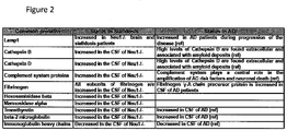
Classifications
-
- G—PHYSICS
- G16—INFORMATION AND COMMUNICATION TECHNOLOGY [ICT] SPECIALLY ADAPTED FOR SPECIFIC APPLICATION FIELDS
- G16H—HEALTHCARE INFORMATICS, i.e. INFORMATION AND COMMUNICATION TECHNOLOGY [ICT] SPECIALLY ADAPTED FOR THE HANDLING OR PROCESSING OF MEDICAL OR HEALTHCARE DATA
- G16H20/00—ICT specially adapted for therapies or health-improving plans, e.g. for handling prescriptions, for steering therapy or for monitoring patient compliance
- G16H20/40—ICT specially adapted for therapies or health-improving plans, e.g. for handling prescriptions, for steering therapy or for monitoring patient compliance relating to mechanical, radiation or invasive therapies, e.g. surgery, laser therapy, dialysis or acupuncture
-
- A—HUMAN NECESSITIES
- A61—MEDICAL OR VETERINARY SCIENCE; HYGIENE
- A61K—PREPARATIONS FOR MEDICAL, DENTAL OR TOILETRY PURPOSES
- A61K38/00—Medicinal preparations containing peptides
- A61K38/16—Peptides having more than 20 amino acids; Gastrins; Somatostatins; Melanotropins; Derivatives thereof
- A61K38/43—Enzymes; Proenzymes; Derivatives thereof
- A61K38/46—Hydrolases (3)
- A61K38/47—Hydrolases (3) acting on glycosyl compounds (3.2), e.g. cellulases, lactases
-
- A—HUMAN NECESSITIES
- A61—MEDICAL OR VETERINARY SCIENCE; HYGIENE
- A61K—PREPARATIONS FOR MEDICAL, DENTAL OR TOILETRY PURPOSES
- A61K38/00—Medicinal preparations containing peptides
- A61K38/16—Peptides having more than 20 amino acids; Gastrins; Somatostatins; Melanotropins; Derivatives thereof
- A61K38/43—Enzymes; Proenzymes; Derivatives thereof
- A61K38/46—Hydrolases (3)
- A61K38/48—Hydrolases (3) acting on peptide bonds (3.4)
- A61K38/4813—Exopeptidases (3.4.11. to 3.4.19)
-
- A—HUMAN NECESSITIES
- A61—MEDICAL OR VETERINARY SCIENCE; HYGIENE
- A61P—SPECIFIC THERAPEUTIC ACTIVITY OF CHEMICAL COMPOUNDS OR MEDICINAL PREPARATIONS
- A61P25/00—Drugs for disorders of the nervous system
- A61P25/28—Drugs for disorders of the nervous system for treating neurodegenerative disorders of the central nervous system, e.g. nootropic agents, cognition enhancers, drugs for treating Alzheimer's disease or other forms of dementia
-
- A—HUMAN NECESSITIES
- A61—MEDICAL OR VETERINARY SCIENCE; HYGIENE
- A61P—SPECIFIC THERAPEUTIC ACTIVITY OF CHEMICAL COMPOUNDS OR MEDICINAL PREPARATIONS
- A61P35/00—Antineoplastic agents
-
- C—CHEMISTRY; METALLURGY
- C07—ORGANIC CHEMISTRY
- C07B—GENERAL METHODS OF ORGANIC CHEMISTRY; APPARATUS THEREFOR
- C07B59/00—Introduction of isotopes of elements into organic compounds ; Labelled organic compounds per se
- C07B59/005—Sugars; Derivatives thereof; Nucleosides; Nucleotides; Nucleic acids
-
- C—CHEMISTRY; METALLURGY
- C12—BIOCHEMISTRY; BEER; SPIRITS; WINE; VINEGAR; MICROBIOLOGY; ENZYMOLOGY; MUTATION OR GENETIC ENGINEERING
- C12N—MICROORGANISMS OR ENZYMES; COMPOSITIONS THEREOF; PROPAGATING, PRESERVING, OR MAINTAINING MICROORGANISMS; MUTATION OR GENETIC ENGINEERING; CULTURE MEDIA
- C12N15/00—Mutation or genetic engineering; DNA or RNA concerning genetic engineering, vectors, e.g. plasmids, or their isolation, preparation or purification; Use of hosts therefor
- C12N15/09—Recombinant DNA-technology
- C12N15/11—DNA or RNA fragments; Modified forms thereof; Non-coding nucleic acids having a biological activity
-
- C—CHEMISTRY; METALLURGY
- C12—BIOCHEMISTRY; BEER; SPIRITS; WINE; VINEGAR; MICROBIOLOGY; ENZYMOLOGY; MUTATION OR GENETIC ENGINEERING
- C12N—MICROORGANISMS OR ENZYMES; COMPOSITIONS THEREOF; PROPAGATING, PRESERVING, OR MAINTAINING MICROORGANISMS; MUTATION OR GENETIC ENGINEERING; CULTURE MEDIA
- C12N15/00—Mutation or genetic engineering; DNA or RNA concerning genetic engineering, vectors, e.g. plasmids, or their isolation, preparation or purification; Use of hosts therefor
- C12N15/09—Recombinant DNA-technology
- C12N15/63—Introduction of foreign genetic material using vectors; Vectors; Use of hosts therefor; Regulation of expression
- C12N15/79—Vectors or expression systems specially adapted for eukaryotic hosts
- C12N15/85—Vectors or expression systems specially adapted for eukaryotic hosts for animal cells
- C12N15/86—Viral vectors
-
- C—CHEMISTRY; METALLURGY
- C12—BIOCHEMISTRY; BEER; SPIRITS; WINE; VINEGAR; MICROBIOLOGY; ENZYMOLOGY; MUTATION OR GENETIC ENGINEERING
- C12N—MICROORGANISMS OR ENZYMES; COMPOSITIONS THEREOF; PROPAGATING, PRESERVING, OR MAINTAINING MICROORGANISMS; MUTATION OR GENETIC ENGINEERING; CULTURE MEDIA
- C12N9/00—Enzymes; Proenzymes; Compositions thereof; Processes for preparing, activating, inhibiting, separating or purifying enzymes
- C12N9/14—Hydrolases (3)
- C12N9/24—Hydrolases (3) acting on glycosyl compounds (3.2)
- C12N9/2402—Hydrolases (3) acting on glycosyl compounds (3.2) hydrolysing O- and S- glycosyl compounds (3.2.1)
-
- C—CHEMISTRY; METALLURGY
- C12—BIOCHEMISTRY; BEER; SPIRITS; WINE; VINEGAR; MICROBIOLOGY; ENZYMOLOGY; MUTATION OR GENETIC ENGINEERING
- C12N—MICROORGANISMS OR ENZYMES; COMPOSITIONS THEREOF; PROPAGATING, PRESERVING, OR MAINTAINING MICROORGANISMS; MUTATION OR GENETIC ENGINEERING; CULTURE MEDIA
- C12N9/00—Enzymes; Proenzymes; Compositions thereof; Processes for preparing, activating, inhibiting, separating or purifying enzymes
- C12N9/14—Hydrolases (3)
- C12N9/48—Hydrolases (3) acting on peptide bonds (3.4)
- C12N9/485—Exopeptidases (3.4.11-3.4.19)
-
- C—CHEMISTRY; METALLURGY
- C12—BIOCHEMISTRY; BEER; SPIRITS; WINE; VINEGAR; MICROBIOLOGY; ENZYMOLOGY; MUTATION OR GENETIC ENGINEERING
- C12Q—MEASURING OR TESTING PROCESSES INVOLVING ENZYMES, NUCLEIC ACIDS OR MICROORGANISMS; COMPOSITIONS OR TEST PAPERS THEREFOR; PROCESSES OF PREPARING SUCH COMPOSITIONS; CONDITION-RESPONSIVE CONTROL IN MICROBIOLOGICAL OR ENZYMOLOGICAL PROCESSES
- C12Q1/00—Measuring or testing processes involving enzymes, nucleic acids or microorganisms; Compositions therefor; Processes of preparing such compositions
- C12Q1/34—Measuring or testing processes involving enzymes, nucleic acids or microorganisms; Compositions therefor; Processes of preparing such compositions involving hydrolase
-
- C—CHEMISTRY; METALLURGY
- C12—BIOCHEMISTRY; BEER; SPIRITS; WINE; VINEGAR; MICROBIOLOGY; ENZYMOLOGY; MUTATION OR GENETIC ENGINEERING
- C12Y—ENZYMES
- C12Y302/00—Hydrolases acting on glycosyl compounds, i.e. glycosylases (3.2)
- C12Y302/01—Glycosidases, i.e. enzymes hydrolysing O- and S-glycosyl compounds (3.2.1)
- C12Y302/01018—Exo-alpha-sialidase (3.2.1.18), i.e. trans-sialidase
-
- C—CHEMISTRY; METALLURGY
- C12—BIOCHEMISTRY; BEER; SPIRITS; WINE; VINEGAR; MICROBIOLOGY; ENZYMOLOGY; MUTATION OR GENETIC ENGINEERING
- C12Y—ENZYMES
- C12Y304/00—Hydrolases acting on peptide bonds, i.e. peptidases (3.4)
- C12Y304/16—Serine-type carboxypeptidases (3.4.16)
- C12Y304/16005—Carboxypeptidase C (3.4.16.5), i.e. carboxypeptidase Y
-
- G—PHYSICS
- G01—MEASURING; TESTING
- G01N—INVESTIGATING OR ANALYSING MATERIALS BY DETERMINING THEIR CHEMICAL OR PHYSICAL PROPERTIES
- G01N33/00—Investigating or analysing materials by specific methods not covered by groups G01N1/00 - G01N31/00
- G01N33/48—Biological material, e.g. blood, urine; Haemocytometers
- G01N33/50—Chemical analysis of biological material, e.g. blood, urine; Testing involving biospecific ligand binding methods; Immunological testing
- G01N33/53—Immunoassay; Biospecific binding assay; Materials therefor
- G01N33/573—Immunoassay; Biospecific binding assay; Materials therefor for enzymes or isoenzymes
-
- G—PHYSICS
- G01—MEASURING; TESTING
- G01N—INVESTIGATING OR ANALYSING MATERIALS BY DETERMINING THEIR CHEMICAL OR PHYSICAL PROPERTIES
- G01N33/00—Investigating or analysing materials by specific methods not covered by groups G01N1/00 - G01N31/00
- G01N33/48—Biological material, e.g. blood, urine; Haemocytometers
- G01N33/50—Chemical analysis of biological material, e.g. blood, urine; Testing involving biospecific ligand binding methods; Immunological testing
- G01N33/53—Immunoassay; Biospecific binding assay; Materials therefor
- G01N33/574—Immunoassay; Biospecific binding assay; Materials therefor for cancer
- G01N33/57484—Immunoassay; Biospecific binding assay; Materials therefor for cancer involving compounds serving as markers for tumor, cancer, neoplasia, e.g. cellular determinants, receptors, heat shock/stress proteins, A-protein, oligosaccharides, metabolites
- G01N33/57496—Immunoassay; Biospecific binding assay; Materials therefor for cancer involving compounds serving as markers for tumor, cancer, neoplasia, e.g. cellular determinants, receptors, heat shock/stress proteins, A-protein, oligosaccharides, metabolites involving intracellular compounds
-
- G—PHYSICS
- G16—INFORMATION AND COMMUNICATION TECHNOLOGY [ICT] SPECIALLY ADAPTED FOR SPECIFIC APPLICATION FIELDS
- G16H—HEALTHCARE INFORMATICS, i.e. INFORMATION AND COMMUNICATION TECHNOLOGY [ICT] SPECIALLY ADAPTED FOR THE HANDLING OR PROCESSING OF MEDICAL OR HEALTHCARE DATA
- G16H20/00—ICT specially adapted for therapies or health-improving plans, e.g. for handling prescriptions, for steering therapy or for monitoring patient compliance
-
- C—CHEMISTRY; METALLURGY
- C12—BIOCHEMISTRY; BEER; SPIRITS; WINE; VINEGAR; MICROBIOLOGY; ENZYMOLOGY; MUTATION OR GENETIC ENGINEERING
- C12N—MICROORGANISMS OR ENZYMES; COMPOSITIONS THEREOF; PROPAGATING, PRESERVING, OR MAINTAINING MICROORGANISMS; MUTATION OR GENETIC ENGINEERING; CULTURE MEDIA
- C12N15/00—Mutation or genetic engineering; DNA or RNA concerning genetic engineering, vectors, e.g. plasmids, or their isolation, preparation or purification; Use of hosts therefor
-
- C—CHEMISTRY; METALLURGY
- C12—BIOCHEMISTRY; BEER; SPIRITS; WINE; VINEGAR; MICROBIOLOGY; ENZYMOLOGY; MUTATION OR GENETIC ENGINEERING
- C12N—MICROORGANISMS OR ENZYMES; COMPOSITIONS THEREOF; PROPAGATING, PRESERVING, OR MAINTAINING MICROORGANISMS; MUTATION OR GENETIC ENGINEERING; CULTURE MEDIA
- C12N2320/00—Applications; Uses
- C12N2320/30—Special therapeutic applications
-
- C—CHEMISTRY; METALLURGY
- C12—BIOCHEMISTRY; BEER; SPIRITS; WINE; VINEGAR; MICROBIOLOGY; ENZYMOLOGY; MUTATION OR GENETIC ENGINEERING
- C12N—MICROORGANISMS OR ENZYMES; COMPOSITIONS THEREOF; PROPAGATING, PRESERVING, OR MAINTAINING MICROORGANISMS; MUTATION OR GENETIC ENGINEERING; CULTURE MEDIA
- C12N2710/00—MICROORGANISMS OR ENZYMES; COMPOSITIONS THEREOF; PROPAGATING, PRESERVING, OR MAINTAINING MICROORGANISMS; MUTATION OR GENETIC ENGINEERING; CULTURE MEDIA dsDNA viruses
- C12N2710/00011—Details
- C12N2710/10011—Adenoviridae
- C12N2710/10031—Uses of virus other than therapeutic or vaccine, e.g. disinfectant
-
- C—CHEMISTRY; METALLURGY
- C12—BIOCHEMISTRY; BEER; SPIRITS; WINE; VINEGAR; MICROBIOLOGY; ENZYMOLOGY; MUTATION OR GENETIC ENGINEERING
- C12N—MICROORGANISMS OR ENZYMES; COMPOSITIONS THEREOF; PROPAGATING, PRESERVING, OR MAINTAINING MICROORGANISMS; MUTATION OR GENETIC ENGINEERING; CULTURE MEDIA
- C12N2740/00—Reverse transcribing RNA viruses
- C12N2740/00011—Details
- C12N2740/10011—Retroviridae
- C12N2740/10041—Use of virus, viral particle or viral elements as a vector
-
- C—CHEMISTRY; METALLURGY
- C12—BIOCHEMISTRY; BEER; SPIRITS; WINE; VINEGAR; MICROBIOLOGY; ENZYMOLOGY; MUTATION OR GENETIC ENGINEERING
- C12N—MICROORGANISMS OR ENZYMES; COMPOSITIONS THEREOF; PROPAGATING, PRESERVING, OR MAINTAINING MICROORGANISMS; MUTATION OR GENETIC ENGINEERING; CULTURE MEDIA
- C12N2750/00—MICROORGANISMS OR ENZYMES; COMPOSITIONS THEREOF; PROPAGATING, PRESERVING, OR MAINTAINING MICROORGANISMS; MUTATION OR GENETIC ENGINEERING; CULTURE MEDIA ssDNA viruses
- C12N2750/00011—Details
- C12N2750/14011—Parvoviridae
- C12N2750/14111—Dependovirus, e.g. adenoassociated viruses
- C12N2750/14141—Use of virus, viral particle or viral elements as a vector
-
- G—PHYSICS
- G01—MEASURING; TESTING
- G01N—INVESTIGATING OR ANALYSING MATERIALS BY DETERMINING THEIR CHEMICAL OR PHYSICAL PROPERTIES
- G01N2333/00—Assays involving biological materials from specific organisms or of a specific nature
- G01N2333/90—Enzymes; Proenzymes
- G01N2333/914—Hydrolases (3)
- G01N2333/924—Hydrolases (3) acting on glycosyl compounds (3.2)
-
- G—PHYSICS
- G01—MEASURING; TESTING
- G01N—INVESTIGATING OR ANALYSING MATERIALS BY DETERMINING THEIR CHEMICAL OR PHYSICAL PROPERTIES
- G01N2800/00—Detection or diagnosis of diseases
- G01N2800/28—Neurological disorders
- G01N2800/2814—Dementia; Cognitive disorders
- G01N2800/2821—Alzheimer
-
- G—PHYSICS
- G01—MEASURING; TESTING
- G01N—INVESTIGATING OR ANALYSING MATERIALS BY DETERMINING THEIR CHEMICAL OR PHYSICAL PROPERTIES
- G01N2800/00—Detection or diagnosis of diseases
- G01N2800/56—Staging of a disease; Further complications associated with the disease
-
- Y—GENERAL TAGGING OF NEW TECHNOLOGICAL DEVELOPMENTS; GENERAL TAGGING OF CROSS-SECTIONAL TECHNOLOGIES SPANNING OVER SEVERAL SECTIONS OF THE IPC; TECHNICAL SUBJECTS COVERED BY FORMER USPC CROSS-REFERENCE ART COLLECTIONS [XRACs] AND DIGESTS
- Y02—TECHNOLOGIES OR APPLICATIONS FOR MITIGATION OR ADAPTATION AGAINST CLIMATE CHANGE
- Y02A—TECHNOLOGIES FOR ADAPTATION TO CLIMATE CHANGE
- Y02A90/00—Technologies having an indirect contribution to adaptation to climate change
- Y02A90/10—Information and communication technologies [ICT] supporting adaptation to climate change, e.g. for weather forecasting or climate simulation
Definitions
- the present invention relates to the field of molecular biology and Alzheimer's disease therapeutics and diagnostics.
- the prognosis of a disease or pathological condition in a subject can be greatly improved with an early diagnosis.
- reliable prognostic and diagnostic methods are lacking for managing disease states.
- the only definitive diagnostic test is to determine whether amyloid plaques are present in a subject's brain tissue, a determination that can only be made after death.
- diagnosis and prognosis of a cancer are important for choosing the best treatment options in order to improve outcome.
- diagnostic and prognostic tests to predict the efficacy of a particular chemotherapy regime to determine the best treatment options for a subject.
- Methods are disclosed for the prognosis, diagnosis and treatment of various pathological states, including cancer, chemotherapy resistance and dementia associated with Alzheimer's disease.
- the methods provided herein are based on the discovery that various proteins with a high level of sialylation are shown herein to be associated with disease states, such as, cancer, chemotherapy resistance and dementia associated with Alzheimer's disease.
- Such methods provide a lysosomal exocytosis activity profile comprising one or more values representing lysosomal exocytosis activity.
- low lysosomal sialidase activity is associated with various pathological states.
- the methods also provide a lysosomal sialidase activity profile, comprising one or more values representing lysosomal sialidase activity.
- a lysosomal sialidase activity profile is one example of a lysosomal exocytosis activity profile.
- the level af lysosomal exocytosis activity and/or lysosomal sialidase activity is predictive of a diagnosis and/or prognosis of cancer, chemotherapy resistance or dementia associated with Alzheimer's disease.
- lysosomal exocytosis activity can serve as a marker for the diagnosis and/or prognosis of pathological conditions including, for example, cancer, chemotherapy resistance and dementia associated with Alzheimer's disease.
- Various activity profiles are provided herein for the diagnosis and/or prognosis of cancer, chemotherapy resistance, and dementia associated with Alzheimer's disease.
- a “profile” comprises one or more values corresponding to a measurement of a marker(s) representing an activity in a sample.
- Various profiles are disclosed herein which can be used for the prognosis and/or diagnosis of a given pathological state. Such profiles include: a lysosomal exocytosis activity profile, a sialylation activity profile, a lysosomal sialidase activity profile, a NEU1 substrate sialylation activity profile and a NEU1 level activity profile.
- lysosomal exocytosis activity profile is meant a profile of one or more values representing lysosomal exocytosis activity.
- lysosomal exocytosis activity is meant a measure of the level of exocytosis in a sample. Various markers can be used to determine the lysosomal exocytosis activity of a sample.
- Such markers include one or more of the following: (1) the level of NEU1 protein or direct enzymatic activity of NEU1; (2) the protein level of one or more NEU1 substrates; (3) the protein level of one or more lysosomal proteins; (4) the protein level of one or more lysosomal proteases; (5) the protein level of LAMP-1; (6) the protein level of hexosaminidase beta; (7) the protein level of mannosidase alpha; or (8) the protein level of one or more cathepsins; (9) any marker for a sialylation activity profile provided herein; or (10) any marker for a lysosomal sialidase activity profile provided herein.
- the lysosomal exocytosis activity profile can comprise 1, 2, 3, 4, 5, 6, 7, 8, 9, 10, 11, 12, 13, 14, 15 or more lysosomal exocytosis activity values of the various lysosomal exocytosis activity markers provided herein.
- sialylation activity profile is meant a profile of one or more values representing sialylation activity.
- sialylation activity is meant a measure of the sialylation level of a population of proteins in a sample or the sialylation level of one or more proteins in a sample.
- markers can be used to determine sialylation activity.
- Such markers include one or more of the following: (1) the overall level of sialylation in a sample; (2) the level of NEU1 protein or direct enzymatic activity of NEU1; (3) the level of sialylation of one or more NEU1 substrates; (4) the protein level of one or more NEU1 substrates; or (5) any marker for a lysosomal sialidase activity profile, as discussed in further detail elsewhere herein or outlined in Table 1.
- the sialylation activity profile can comprise 1, 2, 3, 4, 5, 6, 7, 8, 9, 10, 11, 12, 13, 14, 15 or more sialylation activity values of the various sialylation activity markers provided herein.
- lysosomal sialidase activity profile is meant a profile of one or more values representing lysosomal sialidase activity.
- lysosomal sialidase activity is meant a direct or indirect measure of lysosomal sialidase activity.
- markers can be used to determine lysosomal sialidase activity in a sample.
- the various markers representing the lysosomal sialidase activity in a sample include any one or more of the following: (1) the level of NEU1 protein or the level of direct enzymatic activity of NEU1; (2) the protein level of one or more NEU1 substrate: (3) the level of sialylation of one or more NEU1 substrate; or (4) the activity level of one or more NEU1 substrate.
- the lysosomal sialidase activity profile can comprise any combination of the lysosomal sialidase activity markers provided herein.
- the lysosomal sialidase activity profile can comprise 1, 2, 3, 4, 5, 6, 7, 8, 9, 10, 11, 12, 13, 14, 15 or more lysosomal sialidase activity values of the various lysosomal sialidase activity markers provided herein.
- NEU1 substrate sialylation activity profile is meant measuring lysosomal sialidase activity in a sample by determining the level of sialylation of one or more NEU1 substrates.
- the various markers representing lysosomal sialidase activity that are encompassed in a NEU1 substrate sialylation activity profile include: (1) the level of sialylation of one or more NEU1 substrate; (2) the level of sialylation of LAMP-1; (3) The level of sialylation of MUC-1; or (4) the level of sialylation of NEU1 and MUC-1.
- the NEU1 substrate sialylation activity profile can comprise 1, 2, 3, 4, 5, 6, 7, 8, 9, 10, 11, 12, 13, 14, 15 or more marker values provided by the sialylation level of the various NEU1 substrates.
- NEU1 level activity profile is meant measuring lysosomal sialidase activity in a sample by determining the protein level of any non-MUC-1 NEU1 substrate or of NEU1 itself.
- the various markers representing lysosomal sialidase activity that are encompassed in a NEU1 level activity profile include: (1) the level of NEU1 protein; (2) the protein level of any one or more non-MUC-1 NEU1 substrate; or (3) the protein level of LAMP-1.
- the NEU1 level activity profile can comprise 1, 2, 3, 4, 5, 6, 7, 8, 9, 10, 11, 12, 13, 14, 15 or more marker values provided by the protein level of the various NEU1 substrates.
- markers are present in a given profile, not all the markers must show an altered activity as compared to the marker value in a corresponding control or reference profile in order to produce the prognosis and/or diagnosis provided herein.
- the alteration of a single marker may be sufficient for a diagnosis and/or prognosis.
- an alteration in 2, 3, 4, 5, 6, 7, 8, 9, 10 or more marker values in a given profile as compared to the values in corresponding control or reference profile is sufficient for a diagnosis and/or prognosis.
- Table 1 Summary of various markers employed to establish a specific type of activity profile.
- NEU1 Substrate Sialylation Activity Profile NEU1 Level Activity Profile the level of NEU1 protein + + + + the level of direct enzymatic activity of NEU1 + + + the protein level of one or more NEU1 substrates + + + + + At least one NEU1 substrate other than MUC-1 must be detected the protein level of LAMP-1 + + + + the protein level of MUC-1 + + + + Only in combination with another NEU1 substrate the protein level of LAMP-1 and MUC-1 + + + + the level of any one or more lysosomal proteins + the protein level of one or more lysosomal proteases + the protein level of one or more cathepsins + the protein level of Hexosaminidase beta + the protein level of mannosidase alpha + the activity level of one or more NEU1 substrates + + + the activity level of LAMP-1 + + + the activity level of
- the methods for diagnosis and/or prognosis provided herein are based on analyzing a sample for lysosomal exocytosis activity, sialylation activity, lysosomal sialidase activity, NEU1 substrate sialylation activity and/or NEU1 level activity and comparing it to a reference value for lysosomal exocytosis activity, sialylation activity, lysosomal sialidase activity, NEU1 substrate sialylation activity and/or NEU1 level activity from a control sample.
- Measuring the "level” or “amount” of a protein, sialylation, or an activity in a sample means quantifying the lysosomal exocytosis activity, sialylation activity, lysosomal sialidase activity, NEU1 substrate sialylation activity or NEU1 level activity by determining, for example, the relative or absolute amount of protein and/or sialylation of a protein and/or the activity of a protein.
- One aspect of the methods provided herein relates to assays for detecting lysosomal exocytosis activity, sialylation activity, lysosomal sialidase activity, NEU1 substrate sialylation activity and NEU1 level activity in the context of a sample.
- These assays determine the values that make up the lysosomal exocytosis activity profile, sialylation activity profile, lysosomal sialidase activity profile, NEU1 substrate sialylation activity profile or NEU1 level activity profile of a sample.
- sample or “subject sample”, as used herein, can comprise any sample in which one desires to determine the lysosomal exocytosis activity, sialylation activity, lysosomal sialidase activity, NEU1 substrate sialylation activity and/or NEU1 level activity.
- subject is intended any animal (i.e. mammals) such as, humans, primates, rodents, agricultural and domesticated animals such as, but not limited to, dogs, cats, cattle, horses, pigs, sheep, and the like, in which one desires to determine the lysosomal exocytosis activity, sialylation activity and/or lysosomal sialidase activity.
- the sample may be derived from any cell, tissue, or biological fluid from the animal of interest.
- the sample may comprise any clinically relevant tissue, such as, but not limited to, bone marrow, cerebrospinal fluid, tumor biopsy, fine needle aspirate, or a sample of body fluid, such as blood, plasma, serum, lymph, ascetic fluid, cystic fluid or urine.
- body fluid such as blood, plasma, serum, lymph, ascetic fluid, cystic fluid or urine.
- the sample used in the methods provided herein will vary based on the assay format, nature of the detection method, and the tissues, cells or extracts which are used as the sample.
- a "reference" lysosomal exocytosis activity, sialylation activity, lysosomal sialidase activity, NEU1 substrate sialylation activity and/or NEU1 level activity as used herein is provided in a control sample.
- a “control” or “control sample” provides a reference point for measuring changes in lysosomal exocytosis activity, sialylation activity, lysosomal sialidase activity, NEU1 substrate sialylation level activity and/or NEU1 level activity of a subject sample.
- the control may be a predetermined value based on a group of samples or it may be a single value based on an individual sample.
- the control may be a sample tested in parallel with the subject sample.
- a control sample may comprise, for example: (a) any sample from healthy individual(s); (b) a normal tissue sample taken from a location adjacent to a tumor from the same subject; (b) a tissue sample from healthy individual(s) taken from the same tissue type as a subject tumor; (c) a serum or plasma sample taken from healthy individual(s); (d) a cerebrospinal fluid sample taken from healthy individual(s); or (e) a urine sample from healthy individual(s).
- a "higher” or “increased” level for a given marker is meant any significant increase in the level of the marker in a sample as compared to the level of the corresponding marker in a control sample.
- An increased or higher level for a given marker can be any statistically significant increase in the level of the marker of at least 5%, 10%, 15%, 20%, 25%, 30%, 40%, 50%, 60%, 70%, 80%, 85%, 90%, 95%, 96%, 97%, 98%, 99%, 100%, 200%, 400% or more as compared to a reference level in a control sample.
- an increase in the level for a given marker can be any fold increase of at least 1.5-fold, 2-fold, 3-fold, 4-fold, 5-fold, 6-fold, 7-fold, 8-fold, 9-fold, 10-fold, 12-fold, 14-fold, 16-fold, 20-fold or more over the value for the level of the corresponding marker in a control sample.
- an increase in the level of a given marker can result in an increase in a specific activity in the sample (i.e. the lysosomal exocytosis activity, sialylation activity, lysosomal sialidase activity, NEU1 substrate sialylation activity or NEU1 level activity).
- an increase in the level of a given marker can result in a decrease in a specific activity in a sample.
- a "decreased”, “lower” or “reduced” level for a given marker is meant any significant decrease in the level of the marker in a sample as compared to the level of the corresponding marker in a control sample.
- lower or reduced level of a marker is meant a statistically significant reduction in the level of a marker in a subject sample of at least 5%, 10%, 15%, 20%, 25%, 30%, 40%, 50%, 60%, 70%, 80%, 85%, 90%, 95%, 96%, 97%, 98%, 99% or more as compared to a reference level in a control sample.
- a decrease in the level for a given marker can be any fold decrease of at least 1.5-fold, 2-fold, 3-fold, 4-fold, 5-fold, 6-fold, 7-fold, 8-fold, 9-fold, 10-fold, 12-fold, 14-fold, 16-fold, 20-fold or more as compared to the level of the corresponding marker in a control sample.
- a decrease in the level of a given marker can result in a decrease in a specific activity in the sample (i.e. the lysosomal exocytosis activity, sialylation activity, lysosomal sialidase activity, NEU1 substrate sialylation activity or NEU1 level activity).
- a decrease in the level of a given marker can result in an increase in a specific activity in the sample.
- Table 2 provides non-limiting examples of markers for the various activity profiles provided herein and denotes if an increase or a decrease in the marker is reflective of an increase or a decrease in the activity in a sample (i.e. the lysosomal exocytosis activity, sialylation activity, lysosomal sialidase activity, NEU1 substrate sialylation activity or NEU1 level activity).
- exocytosis is a process of cellular secretion in which substances contained in vesicles are discharged from the cell by fusion of the vesicular membrane with the outer cell membrane.
- exocytosis There are two types of exocytosis, constitutive and regulated. Constitutive exocytosis is not regulated by calcium, while regulated exocytosis is dependent on calcium.
- Exocytosis involves vesicle recruitment, tethering and docking of the vesicle to the plasma membrane and fusion of the vesicle membrane with the plasma membrane thereby releasing the contents of the vesicle into the extracellular space.
- the vesicles release various components into the extracellular environment.
- Some examples of components of secretory vesicles include, but are not limited to, enzymes, proteases, extracellular matrix components, hormones, neurotransmitters and cytotoxic compounds.
- Lysosomal exocytosis is one type of exocytosis.
- lysosomal exocytosis is meant the process by which lysosomes release their contents to the extracellular space.
- Lysosomal exocytosis is a calcium dependent process that involves the recruitment and docking of lysosomes to the plasma membrane, fusion of the lysosomal membrane with the plasma membrane and the release of lysosomal luminal content into the extracellular environment.
- lysosomal contents include, but are not limited to, enzymes, such as lipases, proteases, nucleases and amylase, and other proteins related to lysosomal function, such as sialidases and proteins involved in lysosomal exocytosis.
- enzymes such as lipases, proteases, nucleases and amylase
- proteins related to lysosomal function such as sialidases and proteins involved in lysosomal exocytosis.
- a subject sample has a higher or increased lysosomal exocytosis activity as compared to a control sample.
- higher lysosomal exocytosis activity or “increased lysosomal exocytosis activity” is meant a statistically significant alteration in the level of one or more markers in the lysosomal exocytosis activity profile.
- Table 2 provides non-limiting examples of markers for the lysosomal exocytosis activity profile and denotes if an increase or a decrease in the marker is reflective of a higher lysosomal exocytosis activity.
- an increase in lysosomal exocytosis activity is denoted in a given profile by an alteration in at least 1, 2, 3, 4, 5, 6, 7, 8, 9, 10, 11, 12, 13, 14, 15 or all of the lysosomal exocytosis activity markers as compared to a control sample.
- an alteration in lysosomal exocytosis activity of one marker is sufficient for a diagnosis and/or prognosis.
- an alteration in two or more lysosomal exocytosis activity markers is sufficient for a diagnosis and/or prognosis.
- Assays to measure lysosomal exocytosis activity for an exocytosis marker are provided herein.
- One measure of lysosomal exocytosis activity is the level of a protein in a sample (i.e. NEU1, NEU1 substrates or lysosomal proteins).
- a variety of assays for detecting protein in a sample are known in the art and include direct and indirect assays for protein.
- An exemplary method for detecting the presence or absence or the quantity of a protein in a sample involves obtaining a sample and contacting the sample with a compound or agent capable of specifically binding and detecting the protein, such that the presence of the protein is detected in the sample. Results obtained with a sample from a subject may be compared to results obtained with a biological sample from a control subject.
- an agent for detecting a protein is an antibody capable of specifically binding to that protein.
- Antibodies can be polyclonal or monoclonal.
- the term "labeled”, with regard to the antibody is intended to encompass direct labeling of the antibody by coupling (i.e. physically linking) a detectable substance to the antibody as well as indirect labeling of the antibody by reactivity with another reagent that is directly labeled. Examples of indirect labeling include detection of a primary antibody using a fluorescently labeled secondary antibody.
- the level of a protein in a sample can be quantitatively measured by a variety of assays utilizing antibodies for a specific protein. These include, for example, immunoassays, radioimmunoassays, enzyme-linked immunosorbant assays and two-antibody sandwich assays. Quantitative western blotting can also be used to determine the level of protein. Western blots can be quantitated by well-known methods such as scanning densitometry. In addition, antibodies can be used to detect and quantitate the level of protein in a sample of a tissue by fluorescence or confocal microscopy by using a fluorescently labeled antibody or secondary reagent.
- a marker is the level of sialylation of a sample.
- Assays for measuring the level of sialylation in a sample are provided elsewhere herein, for example, in the section on sialylation activity.
- a marker is the level of protein activity in a sample.
- the protein activity for any protein provided herein can be measured by assaying for the activity of the specific protein in a sample.
- the protein is an enzyme
- the activity of the enzyme can be measured in an enzyme activity assay.
- Various assays are known in the art for measuring enzymatic activity.
- NEU1 enzyme activity in a sample can be measured by incubating the sample with a sialylated NEU1 substrate and detecting the amount of free sialic acid present in the sample after incubation. As such, the units of enzyme activity can be calculated (i.e. the amount of activity per milligram of protein).
- sialylation activity of one or more sialylation activity markers in a sample is provided.
- a protein or lipid is "sialylated” if a sialic acid is present on the terminal portion of a glycoprotein or glycolipid.
- sialylation is meant the transfer of sialic acid to the terminal portions of the sialylated glycolipids or to the N- or O-linked sugar chains of glycoproteins.
- Sialylation can be catalyzed by a number of different sialyltransferases, each with specificity for a particular sugar substrate.
- sialyltransferases known in the art include, for example, sialyltransferase, beta-galactosamide alpha-2,6-sialyltransferase, alpha-N-acetylgalactosaminide alpha-2,6-sialyltransferase, beta-galactoside alpha-2,3-sialyltransferase, N-acetyllactosaminide alpha-2,3-sialyltransferase, alpha-N-acetyl-neuraminide alpha-2,8-sialyltransferase and lactosylceramide alpha-2,3-sialyltransferase.
- Sialyltransferases can transfer sialic acid to a substrate by various linkages. For example, some sialyltransferases add sialic acid with an alpha-2,3 linkage to galactose, while other sialyltransferases add sialic acid with an alpha-2,6 linkage to galactose or N-acetylgalactosamine. Another group of sialyltransferases can add sialic acid to other sialic acids by an alpha-2,8 linkage. In one approach, the sialic acid is added with an alpha-2,6 linkage to a glycoprotein. In another approach, the sialic acid is added with an alpha 2,3 linkage to a glycoprotein.
- a subject sample has a higher or increased sialylation activity as compared to a control sample.
- high sialylation activity or “increased sialylation activity” is meant a statistically significant alteration in the level of one or more markers in the sialylation activity profile.
- Table 2 provides non-limiting examples of markers for the sialylation activity profile and denotes if an increase or a decrease in the marker is reflective of a higher sialylation activity.
- an increase in sialylation activity is denoted in a given profile by an alteration in at least 1, 2, 3, 4, 5, 6, 7, 8, 9, 10, 11, 12, 13, 14, 15 or all of the sialylation activity markers as compared to a control sample.
- an alteration in sialylation activity of one marker is sufficient for a diagnosis and/or prognosis.
- an alteration in two or more sialylation activity markers is sufficient for a diagnosis and/or prognosis.
- sambuscus nigra lcctin SNA
- SNA sambuscus nigra lcctin
- Other assays for sialylation include the use of Machia amurentis lectin that binds sialic acids attached with an alpha-2,3 linkage.
- sialylation activity can be measured for a population of proteins to determine global sialylation of proteins in a sample.
- sialylation in this context can be assayed for in a sample by lectin binding assays.
- the lectin binding assays can be ELISA based or can be gel based.
- the sialylation activity of a single protein can be measured.
- an ELISA based or gel based lectin assay can be coupled with a specific antibody to a protein of interest.
- the level of sialylation in a sample can be quantitated by using samples of known different sialylation levels as standards in the assay.
- the sialylation activity comprises the level or lysosomal sialidase activity.
- sialidases are enzymes that remove the terminal sialic acid from glycoproteins by a process called desialylation. In mammals, there are at least four types of sialidases including, for example, Neuraminidase 1 (NEU1), NEU2, NEU3 and NEU4 which differ in substrate specificity and subcellular localization. NEU1, for example, is localized to the lysosome and cleaves terminal sialic acid residues from substrates such as glycoproteins. As such, NEU1 is an enzyme that contributes to the overall sialylation activity of a sample.
- NEU1 Neuraminidase 1
- NEU1 is part of a heterotrimeric complex together with betagalactosidase and protective protein/cathepsin A (PPCA).
- PPCA protective protein/cathepsin A
- NEU1 substrate is any protein that is desialylated by NEU1.
- Some non-limiting examples of NEU1 substrates include LAMP-1, Cathepsin A, mucins (i.e. MUC1), cathepsin D, cathepsin B and Amyloid Precursor Protein.
- NEU1 can catalyze the hydrolysis of alpha 2-3 and alpha 2-6 sialyl linkages of terminal sialic acid residues in oligosaccharides, glycoproteins and glycolipids. Desialylation of a glycoprotein, for example, leads to the destabilization and degradation of the protein. Thus, the sialidase, NEU1, contributes to the turnover of glycoproteins.
- NEU1 In addition to its role as a sialidase, NEU1 has a related effect on the constitutive process of lysosomal exocytosis.
- lysosomal exocytosis involves the recruitment and docking of lysosomes to the plasma membrane, fusion of the lysosomal membrane with the plasma membrane and the release of lysosomal luminal content into the extracellular environment.
- the recruitment and docking step is facilitated by the lysosomal associated protein-1 (LAMP-1).
- LAMP-1 is a NEU1 substrate, and thus the stability and turnover rate of LAMP-1 can be influenced by lysosomal sialidase activity.
- a subject sample has a lower or decreased lysosomal sialidase activity as compared to a control sample.
- lower lysosomal sialidase activity or “decreased lysosomal sialidase activity” is meant a statistically significant alteration in the level of one or more markers in the lysosomal sialidase activity profile.
- Table 2 provides non-limiting examples of markers for the lysosomal sialidase activity profile and denotes if an increase or a decrease in the marker is reflective of a lower lysosomal sialidase activity.
- a decrease in lysosomal sialidase activity is denoted in a given profile by an alteration in at least 1, 2, 3, 4, 5, 6, 7, 8, 9, 10, 11, 12, 13, 14, 15 or all of the lysosomal sialidase activity markers as compared to a control sample.
- an alteration in lysosomal sialidase activity of one marker is sufficient for a diagnosis and/or prognosis.
- an alteration in two or more lysosomal sialidase activity markers is sufficient for a diagnosis and/or prognosis.
- lysosomal sialidase activity is measured by the level of NEU1 protein or enzymatic activity of NEU1.
- Assays to measure NEU1 protein level are well known in the art and include contacting a sample with an antibody to NEU1.
- NEU1 enzymatic activity can be measured directly in a sample by assaying for NEU1 enzyme activity of a sample in the presence of a sialylated NEU1 substrate.
- NEU1 substrates when NEU1 protein and/or enzyme activity levels in a sample are low or absent, NEU1 substrates will not be desialylated or will be desialylated at a lower rate resulting in an increase in sialylation of the substrate and/or an increase in the stability of the substrate and thus an increase in the protein level of the NEU1 substrate in a sample.
- NEU1 protein under conditions where NEU1 protein is not present in a sample (i.e. in a NEU1 knockout), LAMP-1 is over-sialylated, accumulates in the lysosome, recruits the lysosome to the plasma membrane and facilitates docking of the lysosome to the plasma membrane. In such cases, the loss of NEU1 protein/activity results in an increase in lysosomal exocytosis.
- an increase in sialylation of any one or more NEU1 substrates results in a lower lysosomal sialidase activity in a sample.
- an increase in the protein level of any one or more of the NEU1 substrates provided herein also results in a lower lysosomal sialidase activity.
- these values are markers for lysosomal sialidase activity and indicative of low protein and activity levels of NEU1 in a sample.
- Assays to measure for sialylation levels in a sample or the sialylation level of a specific protein in a sample are discussed elsewhere herein.
- Assays to measure the protein level of any of the various lysosomal sialidase activity markers are known in the art and are described in detail elsewhere herein.
- the enzymatic activity level of NEU1 or the activity level of any of the various NEU1 substrates are markers for lysosomal sialidase activity.
- Assays to measure the protein activity for various proteins is known in the art and described elsewhere herein.
- one type of lysosomal sialidase profile is a NEU1 substrate sialylation activity profile.
- NEU1 substrate sialylation activity profile Non-limiting examples of the various NEU1 substrate sialylation activity markers are summarized in Table 1.
- a subject sample has a higher or increased NEU1 substrate sialylation activity as compared to a control sample.
- higher NEU1 substrate sialylation activity or “increased NEU1 substrate sialylation activity” is meant a statistically significant alteration in the level of one or more markers in the NEU1 substrate sialylation activity profile.
- Table 2 provides non-limiting examples of markers for the NEU1 substrate sialylation activity profile and denotes if an increase or a decrease in the marker is reflective of a higher NEU1 substrate sialylation activity.
- an increase in NEU1 substrate sialylation activity is denoted in a given profile by an alteration in at least 1, 2, 3, 4, 5, 6, 7, 8, 9,10,11,12, 13, 14, 15 or all of the NEU1 substrate sialylation activity markers as compared to a control sample.
- an alteration in NEU1 substrate sialylation activity of one marker is sufficient for a diagnosis and/or prognosis.
- an alteration in two or more NEU1 substrate sialylation activity markers is sufficient for a diagnosis and/or prognosis.
- Assays to measure the NEU1 substrate sialylation activity of the various NEU1 substrate sialylation activity markers are known in the art and include measuring the level of sialylation of any of the various NEU1 substrates provided herein. Such assays are described elsewhere herein.
- one type of lysosomal sialidase activity profile is a NEU1 level activity profile.
- NEU1 level activity profile Non-limiting examples of the various NEU1 level activity markers are summarized in Table 1.
- a subject sample has a higher or increased NEU1 level activity as compared to a control sample.
- higher NEU1 level activity or “increased NEU1 level activity” is meant a statistically significant alteration in the level of two or more markers in the NEU1 level activity profile.
- Table 2 provides non-limiting examples of markers for the NEU1 level activity profile and denotes if an increase or a decrease in the marker is reflective of a higher NEU1 level activity.
- an increase in NEU1 level activity is denoted in a given profile by an alteration in at least 2, 3, 4, 5, 6, 7, 8, 9, 10, 11, 12, 13, 14, 15 or all of the NEU1 level activity markers as compared to a control sample.
- an alteration in NEU1 level activity of two or more markers is sufficient for a diagnosis and/or prognosis.
- NEU1 level activity of the various NEU1 level activity markers include, for example, immune-blotting using an antibody specific for a NEU1 level activity marker or ELISA assay using an antibody specific for a NEU1 level activity marker. These assays are discussed in detail elsewhere herein. Table 2. Summary of alterations in various markers employed to establish an increase or decrease in a specific type of activity.
- dementia associated with Alzheimer's disease is characterized by the standard criteria for dementia as reported in the Recommendations from the National Institute on Aging-Alzheimer's Association workgroups on diagnostic guidelines for Alzheimer's disease.
- the standard criteria for dementia include (1) cognitive or behavioral symptoms that interfere with the ability to function at usual activities or work, denote a decline from previous functioning and performing levels and cannot be explained by a major psychiatric disorder or delirium; (2) detection and diagnosis of cognitive impairment through a combination of history-taking from the patient and a knowledgeable informant and an objective cognitive assessment; and (3) the cognitive or behavioral impairment involves two or more of the following: (a) impaired ability to acquire and remember new information; (b) impaired reasoning and handling of complex tasks, poor judgment; (c) impaired visuospatial abilities; (d) impaired language functions; and (e) changes in personality, behavior or comportment.
- Dementia associated with Alzheimer's can further have one or more of the following characteristics: (1) meets all criteria for dementia as described above; (2) insidious onset; (3) a history of worsening or cognition by report or observation; (4) amnestic presentation; and (5) nonamnestic presentations, such as, language presentation, visuospatial presentation or executive dysfunction.
- the Alzheimer's disease dementia guidelines are described in detail in McKhann et al. (2011) Alzheimer's & Dementia 7:263-69 .
- NEU1 is a negative regulator of lysosomal exocytosis. In such instances when NEU1 protein levels or enzymatic activity are low, lysosomal exocytosis is enhanced.
- highly sialylated proteins can be detected in the cerebrospinal fluid (CSF). In such cases, the composition of the CSF is changed and many af the highly sialylated proteins also have increased levels in the CSF.
- amyloid precursor protein is shown herein to be a NEU1 substrate and accumulates in the brain and CSF in a highly sialylated form under low NEU1 conditions. Lysosomes also comprise proteases that can process APP to form toxic A ⁇ peptides. Thus, excessive lysosomal exocytosis (i.e. when NEU1 protein or enzymatic activity levels are low) enhances the plaque formation that is characteristic of Alzheimer's disease. See, for example, Example 3, provided elsewhere herein. Thus, an increased lysosomal exocytosis activity, as described in detail elsewhere herein, in the CSF can be predictive of dementia associated with Alzheimer's disease.
- lysosomal exocytosis activity as described in detail elsewhere herein, in the CSF can be predictive of dementia associated with Alzheimer's disease.
- proteins can have an increased sialylation level and/or have increased levels in the CSF.
- the proteins having an increased sialylation level and/or protein level are NEU1 substrates.
- the proteins having an increased sialylation level and/or protein level are lysosomal proteins.
- proteins with increased sialylation and/or protein level include LAMP-1, MUC-1, Cathepsin B, Cathepsin D, Complement system proteins, Fibrinogen, Hexosaminidase beta, Mannosidase alpha, Transthyretin, beta-2 microglobulin and Amyloid Precursor Protein. Any one or more of these proteins can be a marker for lysosomal exocytosis activity.
- a method of diagnosing dementia associated with Alzheimer's disease in a subject comprising: (a) providing a subject lysosomal exocytosis activity profile of a sample of cerebrospinal fluid from the subject; (b) providing a reference lysosomal exocytosis activity profile of a control sample of cerebrospinal fluid, wherein the subject lysosomal exocytosis activity profile and the corresponding reference lysosomal exocytosis activity profile comprise one or more values representing lysosomal exocytosis activity; and (c) comparing the subject and the reference lysosomal exocytosis activity profiles, wherein the subject is diagnosed with dementia associated with Alzheimer's disease if the subject has a higher lysosomal exocytosis activity as compared to the reference lysosomal exocytosis activity.
- the lysosomal exocytosis activity profile comprises a lysosomal sialidase activity profile.
- the lysosomal sialidase activity profile can comprise any combination of any of the various lysosomal sialidase activity markers provided herein. In such cases, a low lysosomal sialidase activity in a subject sample as compared to a reference lysosomal sialidase activity in a control sample results in a subject being diagnosed with dementia associated with Alzheimer's disease.
- the lysosomal exocytosis activity profile comprises a sialylation activity profile.
- the sialylation activity profile can comprise any combination of any of the various sialylation activity markers provided herein. In such cases, a high sialylation activity in a subject sample as compared to a reference sialylation activity in a control sample results in a subject being diagnosed with dementia associated with Alzheimer's disease.
- the lysosomal exocytosis activity profiles, the lysosomal sialidase activity profiles or the sialylation activity profiles can comprise any one or more of the various markers provided herein (i.e. see Tables 1 and 2).
- Knowledge of the sialylation activity profile of a subject sample will allow the physician to make a diagnosis of dementia associated with Alzheimer's disease in a subject. Thus, an early diagnosis can be made and the appropriate treatment options can be considered for the subject.
- the lysosomal sialidase activity profile of a sample can comprise one or more lysosomal sialidase activity markers representing lysosomal sialidase activity (i.e. any of the various markers of lysosomal sialidase activity provided herein, see Table 1)
- the lysosomal exocytosis activity profile of a sample can comprise one or more lysosomal exocytosis activity markers representing lysosomal exocytosis activity (i.e. any or the various markers of lysosomal exocytosis activity provided herein, see Table 1).
- a method of generating a lysosomal sialidase activity profile comprises: (a) obtaining a sample from a tumor from a subject; and (b) assaying for the level of LAMP-1 protein or the level of LAMP-1 sialylation.
- the method comprises assaying for one or more additional lysosomal sialidase activity markers.
- the one or more additional lysosomal sialidase activity markers comprise a NEU1 substrate. Assays for measuring the various lysosomal sialidase activity markers are provided elsewhere herein.
- a method of generating a lysosomal exocytosis activity profile from cerebrospinal fluid comprises: (a) obtaining a sample of cerebrospinal fluid from a subject; and (b) assaying for lysosomal exocytosis activity.
- assaying for lysosomal exocytosis activity comprises assaying for the level of LAMP-1 protein or the level of LAMP-1 sialylation.
- assaying for lysosomal exocytosis activity comprises assaying for the level of one or more proteins comprising LAMP-1, MUC-1, amyloid precursor protein, Cathepsin B, Cathepsin D, Fibrinogen, Hexosaminidase beta, Mannosidase alpha, Transthyretin, beta-2 microglobulin or Immunoglobulin heavy chain.
- a subject having a cancer or having dementia associated with Alzheimer's disease is intended administration of a therapeutically effective amount of NEU1 or an active variant or fragment thereof, administration of a therapeutically effective amount of protective protein/cathepsin A (PPCA) or an active variant or fragment thereof or administration of a therapeutically effective amount of a combination of NEU1 and PPCA to a subject that has cancer or dementia associated with Alzheimer's disease, where the purpose is to cure, heal, alleviate, relieve, alter, remedy, ameliorate, improve, or affect the condition or the symptoms of the cancer or dementia associated with Alzheimer's disease.
- PPCA protective protein/cathepsin A
- preventing a cancer or dementia associated with Alzheimer's disease in a subject is intended administration of a therapeutically effective amount of NEU1 or an active variant or fragment thereof, administration of a therapeutically effective amount of protective protein/cathepsin A (PPCA) or an active variant or fragment thereof or administration of a therapeutically effective amount of a combination of NEU1 and PPCA to a subject, where the purpose is to protect the subject from development of a cancer or dementia associated with Alzheimer's disease.
- PPCA protective protein/cathepsin A
- a therapeutically effective amount of NEU1 or an active variant or fragment thereof, protective protein/cathepsin A (PPCA) or an active variant or fragment thereof or a combination of NEU1 and PPCA is administered to a subject, such as a human, that is at risk for developing a cancer or dementia associated with Alzheimer's disease.
- PPCA protective protein/cathepsin A
- a “therapeutically effective amount” as used herein refers to that amount which provides a therapeutic effect for a given condition and administration regimen.
- the phrase “therapeutically effective amount” is used herein to mean an amount sufficient to cause an improvement in a clinically significant condition in the host.
- a “therapeutically effective amount” refers to an amount of NEU1, PPCA, or a combination of NEU1 and PPCA provided herein that when administered to a subject brings about a positive therapeutic response with respect to the treatment of a subject for a cancer or dementia associated with Alzheimer's disease.
- a positive therapeutic response in regard to treating a cancer includes curing or ameliorating the symptoms of the disease.
- a deficit in the response of the host can be evidenced by continuing or spreading of the cancer.
- An improvement in a clinically significant condition in the host includes a decrease in the size of a tumor, increased necrosis of a tumor, clearance of the tumor from the host tissue, reduction or amelioration of metastasis, or a reduction in any symptom associated with the cancer.
- a positive therapeutic response in regard to treating a subject with dementia associated with Alzheimer's disease includes curing or ameliorating the symptoms of the disease.
- a deficit in the response of the host can be evidenced by continuing or worsening of the dementia associated with Alzheimer's disease.
- An improvement in a clinically significant condition in the host includes a decrease in dementia (i.e. an improvement in memory, judgment, visuospatial abilities, language functions, behavior or any of the other symptoms of dementia provided elsewhere herein) in the subject.
- a "therapeutically effective amount” refers to an amount of NEU1, PPCA, or a combination of NEU1 and PPCA provided herein that when administered to a subject brings about a positive therapeutic response with respect to the prevention of a cancer or dementia associated with Alzheimer's disease in a subject.
- a positive therapeutic response with respect to preventing a cancer or dementia associated with Alzheimer's disease in a subject is the prevention of development of the disease in a subject.
- a method of treating a subject having a cancer comprises administering to a subject in need thereof a therapeutically effective amount of Neuraminidase 1 (NEU1) having an amino acid sequence with at least 85% sequence identity to SEQ ID NO: 2 or an active variant or fragment thereof.
- NEU1 Neuraminidase 1
- a method of treating a subject with dementia associated with Alzheimer's disease comprises administering to a subject in need thereof a therapeutically effective amount of Neuraminidase 1 (NEU1) having an amino acid sequence with at least 85% sequence identity to SEQ ID NO: 2 or an active variant or fragment thereof.
- NEU1 Neuraminidase 1
- the methods can further comprise administration of Protective Protein/Cathepsin A (PPCA) having an amino acid sequence with at least 85% sequence identity to SEQ ID NO: 4.
- PPCA Protective Protein/Cathepsin A
- the administration of NEU1 and PPCA can be separate or NEU1 and PPCA can be administered to a subject simultaneously.
- the administration can be by any known method of administration as described elsewhere herein.
- the administration of NEU1 and/or PPCA comprises administration of a viral vector comprising a nucleotide sequence having at least 85% sequence identity to SEQ ID NO: 1 and/or a nucleotide sequence having at least 85% sequence identity to SEQ ID NO: 3.
- Active variants and fragments of NEU1 can be used in the methods provided herein. Such active variants comprise at least 85%, 90%, 91%, 92%, 93%, 94%, 95%, 96%, 97%, 98%, 99% or more sequence identity to SEQ ID NO: 2, wherein the active variants retain biological activity and hence have sialidase activity. Sialidase activity is described in detail elsewhere herein. Active variants of NEU1 are known in the art. There are over 130 types of neuraminidases known from various species ranging from viruses to humans. See, for example, Monti et al. (2010) Adv. Carbohydr. Chem. Biochem. 64:403-79 .
- Active variants and fragments of PPCA can be used in the methods provided herein. Such active variants comprise at least 85%, 90%, 91%, 92%, 93%, 94%, 95%, 96%, 97%, 98%, 99% or more sequence identity to SEQ ID NO:4, wherein the active variants retain biological activity and hence enhances NEU1 enzymatic activity. Assays to measure for NEU1 enzymatic activity are described elsewhere herein. Active variants of PPCA are known in the art. See, for example, Galjart et al. (1988) Cell 54(6):755-64 .
- the methods of treatment for cancer and dementia associated with Alzheimer's disease disclosed herein can encompass administration of treatment via any parenteral route, including, but not limited, to intramuscular, intraperitoneal, intravenous, and the like.
- pharmaceutically acceptable carriers are well known to those skilled in the art and include, but are not limited to, 0.01-0.1 M, or 0.05M phosphate buffer or 0.8% saline. Additionally, such pharmaceutically acceptable carriers may be aqueous or non-aqueous solutions, suspensions, and emulsions. Examples of non-aqueous solvents are propylene glycol, polyethylene glycol, vegetable oils such as olive oil, and injectable organic esters such as ethyl oleate. Aqueous carriers include water, alcoholic/aqueous solutions, emulsions or suspensions, including saline and buffered media.
- Parenteral vehicles include sodium chloride solution, Ringer's dextrose, dextrose and sodium chloride, lactated Ringer's or fixed oils.
- Intravenous vehicles include fluid and nutrient replenishers, electrolyte replenishers such as those based on Ringer's dextrose, and the like. Preservatives and other additives may also be present, such as, for example, antimicrobials, antioxidants, collating agents, inert gases and the like.
- Controlled or sustained release compositions include formulation in lipophilic depots (e.g. fatty acids, waxes, oils). Also comprehended herein are particulate compositions coated with polymers (e.g. poloxamers or poloxamines) and the compound coupled to antibodies directed against tissue-specific receptors, ligands or antigens or coupled to ligands of tissue-specific receptors. Other examples of the compositions presented herein incorporate particulate forms protective coatings, protease inhibitors or permeation enhancers for various routes of administration, including parenteral, pulmonary, nasal and oral.
- lipophilic depots e.g. fatty acids, waxes, oils.
- particulate compositions coated with polymers e.g. poloxamers or poloxamines
- Other examples of the compositions presented herein incorporate particulate forms protective coatings, protease inhibitors or permeation enhancers for various routes of administration, including parenteral, pulmonary, nasal and oral.
- Such modifications may also increase the compound's solubility in aqueous solution, eliminate aggregation, enhance the physical and chemical stability of the compound, and greatly reduce the immunogenicity and reactivity of the compound.
- the desired in vivo biological activity may be achieved by the administration of such polymer-compound abducts less frequently or in lower doses than with the unmodified compound.
- the sufficient amount may include but is not limited to from about 1 ⁇ g/kg to about 100 ⁇ g/kg, from about 100 ⁇ g/kg to about 1 mg/kg, from about 1 mg/kg to about 10 mg/kg, about 10 mg/kg to about 100 mg/kg, from about 100 mg/kg to about 500 mg/kg or from about 500 mg/kg to about 1000 mg/kg.
- the amount may be 10 mg/kg.
- the pharmaceutically acceptable form of the composition includes a pharmaceutically acceptable carrier.
- compositions which contain an active component are well understood in the art.
- such compositions are prepared as an aerosol of the polypeptide delivered to the nasopharynx or as injectables, either as liquid solutions or suspensions, however, solid forms suitable for solution in, or suspension in, liquid prior to injection can also be prepared.
- the preparation can also be emulsified.
- the active therapeutic ingredient is often mixed with excipients which are pharmaceutically acceptable and compatible with the active ingredient. Suitable excipients are, for example, water, saline, dextrose, glycerol, ethanol, or the like and combinations thereof.
- the composition can contain minor amounts of auxiliary substances such as wetting or emulsifying agents, pH buffering agents which enhance the effectiveness of the active ingredient.
- An active component can be formulated into the therapeutic composition as neutralized pharmaceutically acceptable salt forms.
- Pharmaceutically acceptable salts include the acid addition salts (formed with the free amino groups of the polypeptide) and which are formed with inorganic acids such as, for example, hydrochloric or phosphoric acids, or such organic acids as acetic, oxalic, tartaric, mandelic, and the like. Salts formed from the free carboxyl groups can also be derived from inorganic bases such as, for example, sodium, potassium, ammonium, calcium, or ferric hydroxides, and such organic bases as isopropylamine, trimethylamine, 2-ethylamino ethanol, histidine, procaine, and the like.
- the component or components of a therapeutic composition provided herein may be introduced parenterally, transmucosally, e.g ., orally, nasally, pulmonarily, or rectally, or transdermally.
- administration is parenteral, e.g., via intravenous injection, and also including, but is not limited to, intra-arteriole, intramuscular, intradermal, subcutaneous, intraperitoneal, intraventricular, and intracranial administration.
- unit dose when used in reference to a therapeutic composition provided herein refers to physically discrete units suitable as unitary dosage for humans, each unit containing a predetermined quantity of active material calculated to produce the desired therapeutic effect in association with the required diluent; i.e ., carrier, or vehicle.
- the active compound can be delivered in a vesicle, in particular a liposome (see Langer (1990) Science 249:1527-1533 ; Treat et al., in Liposomes in the Therapy of Infectious Disease and Cancer, Lopez-Berestein and Fidler (eds.), Liss, New York, pp. 353-365 (1989 ); Lopez-Berestein, ibid., pp. 317-327 ; see generally ibid).
- a liposome see Langer (1990) Science 249:1527-1533 ; Treat et al., in Liposomes in the Therapy of Infectious Disease and Cancer, Lopez-Berestein and Fidler (eds.), Liss, New York, pp. 353-365 (1989 ); Lopez-Berestein, ibid., pp. 317-327 ; see generally ibid).
- the therapeutic compound can be delivered in a controlled release system.
- the protein may be administered using intravenous infusion, an implantable osmotic pump, a transdermal patch, liposomes, or other modes of administration,
- a pump may be used (see Langer, supra; Sefton (1987) CRC Crit. Ref Biomed Eng. 14:201 ; Buchwald et al. (1980) Surgery 88:507 ; Saudek et al. (1989) N. Engl. J. Med. 321:574 ).
- polymeric materials can be used (see Medical Applications of Controlled Release, Langer and Wise (eds.), CRC Pres., Boca Raton, Florida (1974 ); Controlled Drug Bioavailability, Drug Product Design and Performance, Smolen and Ball (eds.), Wiley, New York (1984 ); Ranger and Peppas (1983) J. Macromol. Sci. Rev. Macromol. Chem. 23:61 ; see also Levy et al. (1985) Science 228:190 ; During et al. (1989) Ann. Neurol 25:351 ; Howard et al. (1989) J. Neurosurg. 71:105 ).
- a controlled release system can be placed in proximity of the therapeutic target, i.e., the brain or a tumor, thus requiring only a fraction of the systemic dose (see, e.g., Goodson, in Medical Applications of Controlled Release, supra, vol. 2, pp. 115-138 (1984 )).
- Other controlled release systems are discussed in the review by Langer (1990) Science 249:1527-1533 .
- a subject in whom administration of an active component as set forth above is an effective therapeutic regimen for a cancer or dementia associated with Alzheimer's disease is preferably a human, but can be any animal.
- the methods and pharmaceutical compositions provided herein are particularly suited to administration to any animal, particularly a mammal, and including, but by no means limited to, domestic animals, such as feline or canine subjects, farm animals, such as but not limited to bovine, equine, caprine, ovine, and porcine subjects, wild animals (whether in the wild or in a zoological garden), research animals, such as mice, rats, rabbits, goats, sheep, pigs, dogs, cats, etc., i.e., for veterinary medical use.
- a therapeutically effective dosage of the active component is provided.
- a therapeutically effective dosage can be determined by the ordinary skilled medical worker based on patient characteristics (age, weight, sex, condition, complications, other diseases, etc.), as is well known in the art. Furthermore, as further routine studies are conducted, more specific information will emerge regarding appropriate dosage levels for treatment of various conditions in various patients, and the ordinary skilled worker, considering the therapeutic context, age and general health of the recipient, is able to ascertain proper dosing. Generally, for intravenous injection or infusion, dosage may be lower than for intraperitoneal, intramuscular, or other route of administration. The dosing schedule may vary, depending on the circulation half-life, and the formulation used.
- compositions are administered in a manner compatible with the dosage formulation in the therapeutically effective amount.
- Precise amounts of active ingredient required to be administered depend on the judgment of the practitioner and are peculiar to each individual.
- suitable dosages may range from about 0.1 to 20, preferably about 0.5 to about 10, and more preferably one to several, milligrams of active ingredient per kilogram body weight of individual per day and depend on the route of administration.
- Suitable regimes for initial administration and booster shots are also variable, but are typified by an initial administration followed by repeated doses at one or more hour intervals by a subsequent injection or other administration.
- continuous intravenous infusion sufficient to maintain concentrations of ten nanomolar to ten micromolar in the blood are contemplated.
- Administration with other compounds For treatment of cancer or dementia associated with Alzheimer's disease, one may administer the present active component in conjunction with one or more pharmaceutical compositions used for treating cancer or dementia associated with Alzheimer's disease, including but not limited to (1) chemotherapeutic agents; or (2) other drugs for treating symptoms of Alzheimer's including domepezil, galantamine, memantine, rivastigmine or tacrine. Administration may be simultaneous (for example, administration of a mixture of the present active component and a chemotherapeutic agent), or may be in seriatim.
- pharmaceutical compositions used for treating cancer or dementia associated with Alzheimer's disease including but not limited to (1) chemotherapeutic agents; or (2) other drugs for treating symptoms of Alzheimer's including domepezil, galantamine, memantine, rivastigmine or tacrine.
- Administration may be simultaneous (for example, administration of a mixture of the present active component and a chemotherapeutic agent), or may be in seriatim.
- dry powder formulations comprising at least one protein provided herein and another therapeutically effective drug, such as a chemotherapeutic agent or a drug for treating Alzheimer's disease.
- Solid dosage forms include tablets, capsules, pills, troches or lozenges, cachets or pellets.
- liposomal or proteinoid encapsulation may be used to formulate the present compositions (as, for example, proteinoid microspheres reported in U.S. Patent No. 4,925,673 ).
- Liposomal encapsulation may be used and the liposomes may be derivatized with various polymers ( e.g ., U.S. Patent No. 5,013,556 ).
- the formulation will include the component or components (or chemically modified forms thereof) and inert ingredients which allow for protection against the stomach environment, and release of the biologically active material in the intestine.
- oral dosage forms of the above derivatized component or components may be chemically modified so that oral delivery of the derivative is efficacious.
- the chemical modification contemplated is the attachment of at least one moiety to the component molecule itself, where the moiety permits (a) inhibition of proteolysis; and (b) uptake into the blood stream from the stomach or intestine.
- the increase in overall stability of the component or components and increase in circulation time in the body examples include: polyethylene glycol, copolymers of ethylene glycol and propylene glycol, carboxymethyl cellulose, dextran, polyvinyl alcohol, polyvinyl pyrrolidone and polyproline.
- the location of release may be the stomach, the small intestine (the duodenum, the jejunum, or the ileum), or the large intestine.
- the stomach the small intestine (the duodenum, the jejunum, or the ileum), or the large intestine.
- One skilled in the art has available formulations which will not dissolve in the stomach, yet will release the material in the duodenum or elsewhere in the intestine.
- the release will avoid the deleterious effects of the stomach environment, either by protection of the protein (or derivative) or by release of the biologically active material beyond the stomach environment, such as in the intestine.
- a coating impermeable to at least pH 5.0 is essential.
- examples of the more common inert ingredients that are used as enteric coatings are cellulose acetate trimellitate (CAT), hydroxypropylmethylcellulose phthalate (HPMCP), HPMCP 50, HPMCP 55, polyvinyl acetate phthalate (PVAP), Eudragit L30D, Aquateric, cellulose acetate phthalate (CAP), Eudragit L, Eudragit S, and Shellac. These coatings may be used as mixed films.
- a coating or mixture of coatings can also be used on tablets, which are not intended for protection against the stomach. This can include sugar coatings, or coatings which make the tablet easier to swallow.
- Capsules may consist of a hard shell (such as gelatin) for delivery of dry therapeutic i.e. powder; for liquid forms, a soft gelatin shell may be used.
- the shell material of cachets could be thick starch or other edible paper. For pills, lozenges, molded tablets or tablet triturates, moist massing techniques can be used.
- the peptide therapeutic can be included in the formulation as fine multiparticulates in the form of granules or pellets of particle size about 1mm.
- the formulation of the material for capsule administration could also be as a powder, lightly compressed plugs or even as tablets.
- the therapeutic could be prepared by compression.
- Colorants and flavoring agents may all be included.
- the protein (or derivative) may be formulated (such as by liposome or microsphere encapsulation) and then further contained within an edible product, such as a refrigerated beverage containing colorants and flavoring agents.
- diluents could include carbohydrates, especially mannitol, a-lactose, anhydrous lactose, cellulose, sucrose, modified dextran and starch.
- Certain inorganic salts may be also be used as fillers including calcium triphosphate, magnesium carbonate and sodium chloride.
- Some commercially available diluents are Fast-Flo, Emdex, STA-Rx 1500, Emcompress and Avicell.
- Disintegrants may be included in the formulation of the therapeutic into a solid dosage form.
- Materials used as disintegrates include but are not limited to starch, including the commercial disintegrant based on starch, Explotab. Sodium starch glycolate, Amberlite, sodium carboxymethylcellulose, ultramylopectin, sodium alginate, gelatin, orange peel, acid carboxymethyl cellulose, natural sponge and bentonite may all be used.
- Another form of the disintegrants are the insoluble cationic exchange resins.
- Powdered gums may be used as disintegrants and as binders and these can include powdered gums such as agar, Karaya or tragacanth. Alginic acid and its sodium salt are also useful as disintegrants.
- Binders may be used to hold the therapeutic agent together to form a hard tablet and include materials from natural products such as acacia, tragacanth, starch and gelatin. Others include methyl cellulose (MC), ethyl cellulose (EC) and carboxymethyl cellulose (CMC). Polyvinyl pyrrolidone (PVP) and hydroxypropylmethyl cellulose (HPMC) could both be used in alcoholic solutions to granulate the therapeutic.
- MC methyl cellulose
- EC ethyl cellulose
- CMC carboxymethyl cellulose
- PVP polyvinyl pyrrolidone
- HPMC hydroxypropylmethyl cellulose
- Lubricants may be used as a layer between the therapeutic and the die wall, and these can include but are not limited to; stearic acid including its magnesium and calcium salts, polytetrafluoroethylene (PTFE), liquid paraffin, vegetable oils and waxes. Soluble lubricants may also be used such as sodium lauryl sulfate, magnesium lauryl sulfate, polyethylene glycol of various molecular weights, Carbowax 4000 and 6000.
- the glidants may include starch, talc, pyrogenic silica and hydrated silicoaluminate.
- surfactant might be added as a wetting agent.
- Surfactants may include anionic detergents such as sodium lauryl sulfate, dioctyl sodium sulfosuccinate and dioctyl sodium sulfonate.
- anionic detergents such as sodium lauryl sulfate, dioctyl sodium sulfosuccinate and dioctyl sodium sulfonate.
- Cationic detergents might be used and could include benzalkonium chloride or benzethomium chloride.
- nonionic detergents that could be included in the formulation as surfactants are lauromacrogol 400, polyoxyl 40 stearate, polyoxyethylene hydrogenated castor oil 10; 50 and 60, glycerol monostearate, polysorbate 40, 60, 65 and 80, sucrose fatty acid ester, methyl cellulose and carboxymethyl cellulose. These surfactants could be present in the formulation of the protein or derivative either alone or as a mixture in different ratios.
- Additives which potentially enhance uptake of the protein (or derivative) are for instance the fatty acids oleic acid, linoleic acid and linolenic acid.
- the method comprises the use of viruses for administering NEU1 and/or PPCA to a subject.
- Administration can be by the use of viruses that express NEU1 and/or PPCA, such as recombinant retroviruses, recombinant adeno-associated viruses, recombinant adenoviruses, and recombinant Herpes simplex viruses (see, for example, Mulligan, Science 260:926 (1993 ), Rosenberg et al., Science 242:1575 (1988 ), LaSalle et al., Science 259:988 (1993 ), Wolff et al., Science 247:1465 (1990 ), Breakfield and Deluca, The New Biologist 3:203 (1991 )).
- viruses that express NEU1 and/or PPCA such as recombinant retroviruses, recombinant adeno-associated viruses, recombinant adenoviruses, and recombinant Herpes simplex viruses (see
- a NEU1 and/or PPCA gene can be delivered using recombinant viral vectors, including for example, adenoviral vectors ( e.g ., Kass-Eisler et al., Proc. Nat'l Acad. Sci. USA 90:11498 (1993 ), Kolls et al., Proc. Nat'l Acad. Sci. USA 91:215 (1994 ), Li et al., Hum. Gene Ther. 4:403 (1993 ), Vincent et al., Nat. Genet. 5:130 (1993 ), and Zabner et al., Cell 75:207 (1993 )), adenovirus-associated viral vectors ( Flotte et al., Proc.
- adenoviral vectors e.g ., Kass-Eisler et al., Proc. Nat'l Acad. Sci. USA 90:11498 (1993 ), Kolls et al., Proc. Nat'l Aca
- alphaviruses such as Semliki Forest Virus and Sindbis Virus ( Hertz and Huang, J. Vir. 66:857 (1992 ), Raju and Huang, J. Vir. 65:2501 (1991 ), and Xiong et al., Science 243:1188 (1989 )), herpes viral vectors ( e.g., U.S. Patent Nos. 4,769,331 , 4,859,587 , 5,288,641 and 5,328,688 ), parvovirus vectors ( Koering et al., Hum. Gene Therap.
- pox virus vectors Ozaki et al., Biochem. Biophys. Res. Comm. 193:653 (1993 ), Panicali and Paoletti, Proc. Nat'l Acad. Sci. USA 79:4927 (1982 )
- pox viruses such as canary pox virus or vaccinia virus ( Fisher-Hoch et al., Proc. Nat'l Acad. Sci. USA 86:317 (1989 ), and Flexner et al., Ann. N.Y. Acad. Sci.
- retroviruses e.g., Baba et al., J Neurosurg 79:729 (1993 ), Ram et al., Cancer Res. 53:83 (1993 ), Takamiya et al., J. Neurosci. Res 33:493 (1992 ), Vile and Hart, Cancer Res. 53:962 (1993 ), Vile and Hart, Cancer Res. 53:3860 (1993 ), and Anderson et al., U.S. Patent No. 5,399,346 ).
- Either the viral vector itself, or a viral particle, which contains the viral vector may be utilized in the methods described below.
- adenovirus a double-stranded DNA virus
- the adenovirus system offers several advantages including: (i) the ability to accommodate relatively large DNA inserts, (ii) the ability to be grown to high-titer, (iii) the ability to infect a broad range of mammalian cell types, and (iv) the ability to be used with many different promoters including ubiquitous, tissue specific, and regulatable promoters.
- adenoviruses can be administered by intravenous injection, because the viruses are stable in the bloodstream.
- adenovirus vectors where portions of the adenovirus genome are deleted, inserts are incorporated into the viral DNA by direct ligation or by homologous recombination with a co-transfected plasmid.
- the essential E1 gene is deleted from the viral vector, and the virus will not replicate unless the E1 gene is provided by the host cell.
- adenovirus When intravenously administered to intact animals, adenovirus primarily targets the liver. Although an adenoviral delivery system with an E1 gene deletion cannot replicate in the host cells, the host's tissue will express and process an encoded heterologous protein. Host cells will also secrete the heterologous protein if the corresponding gene includes a secretory signal sequence. Secreted proteins will enter the circulation from tissue that expresses the heterologous gene ( e.g., the highly vascularized liver).
- adenoviral vectors containing various deletions of viral genes can be used to reduce or eliminate immune responses to the vector.
- Such adenoviruses are E1-deleted, and in addition, contain deletions of E2A or E4 ( Lusky et al., J. Virol. 72:2022 (1998 ); Raper et al., Human Gene Therapy 9:671 (1998 )).
- the deletion of E2b has also been reported to reduce immune responses ( Amalfitano et al., J. Virol. 72:926 (1998 )). By deleting the entire adenovirus genome, very large inserts of heterologous DNA can be accommodated.
- High titer stocks of recombinant viruses capable of expressing a therapeutic gene can be obtained from infected mammalian cells using standard methods.
- recombinant herpes simplex virus can be prepared in Vero cells, as described by Brandt et al., J. Gen. Virol. 72:2043 (1991 ), Herold et al., J. Gen. Virol. 75:1211 (1994 ), Visalli and Brandt, Virology 185:419 (1991 ), Grau et al., Invest. Ophthalmol. Vis. Sci. 30:2474 (1989 ), Brandt et al., J. Virol. Meth. 36:209 (1992 ), and by Brown and MacLean (eds.), HSY Virus Protocols (Humana Press 1997 ).
- the therapy is preferably somatic cell gene therapy. That is, the preferred treatment of a human with a recombinant virus does not entail introducing into cells a nucleic acid molecule that can form part of a human germ line and be passed onto successive generations ( i.e., human germ line gene therapy).
- Fragments and variants of the polynucleotides encoding the NEU1 and PPCA polypeptides can be employed in the various methods and compositions of the invention.
- fragment is intended a portion of the polynucleotide and hence the protein encoded thereby or a portion of the polypeptide.
- Fragments of a polynucleotide may encode protein fragments that retain the biological activity of the native protein.
- fragments of a polynucleotide may range from at least about 20 nucleotides, about 50 nucleotides, about 100 nucleotides, about 150, about 200, about 250, about 300, about 350, about 400, about 450, about 500, about 550, about 600 and up to the full-length polynucleotide encoding the NEU1 or PPCA polypeptide.
- a fragment of a polynucleotide that encodes a biologically active portion of a NEU1 or PPCA polypeptide will encode at least 15, 25, 30, 50, 100, 150, 200, or 250 contiguous amino acids, or up to the total number of amino acids present in a full-length NEU1 or PPCA polypeptide.
- a biologically active portion of a NEU1 or PPCA polypeptide can be prepared by isolating a portion of one of the polynucleotides encoding the portion of the NEU1 or PPCA polypeptide and expressing the encoded portion of the polypeptide (e.g., by recombinant expression in vitro ), and assessing the activity of the portion of the NEU1 or PPCA polypeptide.
- Polynucleotides that encode fragments of a NEU1 or PPCA polypeptide can comprise nucleotide sequence comprising at least 16, 20, 50, 75, 100, 150, 200, 250, 300, 350, 400, 450, 500, 550, 600, 650, 700, 800, 900, 1,000, 1,100, 1,200, 1,300, or 1,400 nucleotides, or up to the number of nucleotides present in a full-length NEU1 or PPCA nucleotide sequence disclosed herein.
- Variant sequences have a high degree of sequence similarity.
- conservative variants include those sequences that, because of the degeneracy of the genetic code, encode the amino acid sequence of one of the NEU1 or PPCA polypeptides.
- Variants such as these can be identified with the use of well-known molecular biology techniques, as, for example, polymerase chain reaction (PCR) and hybridization techniques.
- Variant polynucleotides also include synthetically derived nucleotide sequences, such as those generated, for example, by using site-directed mutagenesis but which still encode a NEU1 or PPCA polypeptide.
- variants of a particular polynucleotide will have at least about 40%, 45%, 50%, 55%, 60%, 65%, 70%, 75%, 80%, 85%, 90%, 91%, 92%, 93%, 94%, 95%, 96%, 97%, 98%, 99% or more sequence identity to that particular polynucleotide as determined by sequence alignment programs and parameters described elsewhere herein.
- Variants of a particular polynucleotide can also be evaluated by comparison of the percent sequence identity between the polypeptide encoded by a variant polynucleotide and the polypeptide encoded by the reference polynucleotide.
- the percent sequence identity between the two encoded polypeptides is at least 85%, 90%, 91%, 92%, 93%, 94%, 95%, 96%, 97%, 98%, 99% or more sequence identity.
- variant protein is intended a protein derived from the native protein by deletion (so-called truncation) or addition of one or more amino acids to the N-terminal and/or C-terminal end of the native protein; deletion or addition of one or more amino acids at one or more sites in the native protein; or substitution of one or more amino acids at one or more sites in the native protein.
- variant proteins are biologically active, that is they continue to possess the desired biological activity of the native protein. Such variants may result from, for example, genetic polymorphism or from human manipulation.
- Biologically active variants of a NEU1 or PPCA polypeptides will have at least 85%, 90%, 91%, 92%, 93%, 94%, 95%, 96%, 97%, 98%, 99% or more sequence identity to the amino acid sequence for the native protein as determined by sequence alignment programs and parameters described elsewhere herein.
- a biologically active variant of a protein may differ from that protein by as few as 1-15 amino acid residues, as few as 1-10, such as 6-10, as few as 5, as few as 4, 3, 2, or even 1 amino acid residue.
- Proteins may be altered in various ways including amino acid substitutions, deletions, truncations, and insertions. Methods for such manipulations are generally known in the art. For example, amino acid sequence variants of the NEU1 or PPCA proteins can be prepared by mutations in the DNA. Methods for mutagenesis and nucleotide sequence alterations are well known in the art. See, for example, Kunkel (1985) Proc. Natl. Acad. Sci. USA 82:488-492 ; Kunkel et al. (1987) Methods in Enzymol. 154:367-382 ; U.S. Patent No. 4,873,192 ; Walker and Gaastra, eds.
- the polynucleotides used in the invention can include the naturally occurring sequences, the "native" sequences, as well as mutant forms.
- the proteins used in the methods of the invention encompass naturally occurring proteins as well as variations and modified forms thereof. Such variants will continue to possess the ability to implement a recombination event.
- the mutations made in the polynucleotide encoding the variant polypeptide should not place the sequence out of reading frame, and/or create complementary regions that could produce secondary mRNA structure. See, EP Patent Application Publication No. 75,444 .
- deletions, insertions, and substitutions of the protein sequences encompassed herein are not expected to produce radical changes in the characteristics of the protein. However, when it is difficult to predict the exact effect of the substitution, deletion, or insertion in advance of doing so, one skilled in the art will appreciate that the effect will be evaluated by routine screening assays.
- Variant polynucleotides and proteins also encompass sequences and proteins derived from a mutagenic and recombinogenic procedure such as DNA shuffling. With such a procedure, one or more different NEU1 or PPCA coding sequences can be manipulated to create new NEU1 or PPCA polypeptides possessing the desired properties. In this manner, libraries of recombinant polynucleotides are generated from a population of related sequence polynucleotides comprising sequence regions that have substantial sequence identity and can be homologously recombined in vitro or in vivo. Strategies for such DNA shuffling are known in the art. See, for example, Stemmer (1994) Proc. Natl. Acad. Sci.
- sequence identity or “identity” in the context of two polynucleotides or polypeptide sequences makes reference to the residues in the two sequences that are the same when aligned for maximum correspondence over a specified comparison window.
- sequence identity or “identity” in the context of two polynucleotides or polypeptide sequences makes reference to the residues in the two sequences that are the same when aligned for maximum correspondence over a specified comparison window.
- percentage of sequence identity is used in reference to proteins it is recognized that residue positions which are not identical often differ by conservative amino acid substitutions, where amino acid residues are substituted for other amino acid residues with similar chemical properties (e.g., charge or hydrophobicity) and therefore do not change the functional properties of the molecule.
- sequences differ in conservative substitutions the percent sequence identity may be adjusted upwards to correct for the conservative nature of the substitution.
- Sequences that differ by such conservative substitutions are said to have "sequence similarity" or "similarity”. Means for making this adjustment are well known to those of skill in the art. Typically this involves scoring a conservative substitution as a partial rather than a full mismatch, thereby increasing the percentage sequence identity. Thus, for example, where an identical amino acid is given a score of 1 and a non-conservative substitution is given a score of zero, a conservative substitution is given a score between zero and 1. The scoring of conservative substitutions is calculated, e.g., as implemented in the program PC/GENE (Intelligenetics, Mountain View, California).
- percentage of sequence identity means the value determined by comparing two optimally aligned sequences over a comparison window, wherein the portion of the polynucleotide sequence in the comparison window may comprise additions or deletions (i.e., gaps) as compared to the reference sequence (which does not comprise additions or deletions) for optimal alignment of the two sequences. The percentage is calculated by determining the number of positions at which the identical nucleic acid base or amino acid residue occurs in both sequences to yield the number of matched positions, dividing the number of matched positions by the total number of positions in the window of comparison, and multiplying the result by 100 to yield the percentage of sequence identity.
- sequence identity/similarity values provided herein refer to the value obtained using GAP Version 10 using the following parameters: % identity and % similarity for a nucleotide sequence using GAP Weight of 50 and Length Weight of 3, and the nwsgapdna.cmp scoring matrix; % identity and % similarity for an amino acid sequence using GAP Weight of 8 and Length Weight of 2, and the BLOSUM62 scoring matrix; or any equivalent program thereof.
- equivalent program is intended any sequence comparison program that, for any two sequences in question, generates an alignment having identical nucleotide or amino acid residue matches and an identical percent sequence identity when compared to the corresponding alignment generated by GAP Version 10.
- the subject matter of the present disclosure is further illustrated by the following non-limiting examples.
- the invention is defined in the claims and relates to treating or preventing dementia associated with Alzheimer's disease.
- Other subject-matter provides technical background for reference.
- NEU1 lysosomal sialidase NEU1-regulated lysosomal exocytosis and two pathological states: (1) cancer and (2) Alzheimer's disease.
- the loss of NEU1 results in accumulation of its substrate LAMP-1, which, in turn, facilitates the exocytosis of lysosomal contents.
- LAMP-1 substrate protein-1
- the physiological consequences of this depend on the affected tissue. For instance, release of active proteases into the extracellular environment may cause remodeling of tissue surrounding a tumor. In the brain, this release may result in processing of amyloidogenic proteins and formation of plaques.
- xenobiotics which accumulate in the lysosome may undergo efflux through this mechanism, altering drug metabolism. This application is of particular importance as a possible predictor of chemotherapy resistance in cancer cells and as prognostic marker of dementia related to Alzheimer's disease.
- this information can be gleaned via a panel of NEU1 substrates. Accumulation of substrate glycoproteins in their oversialylated state as well as of active lysosomal enzymes indicate a global change in processing rather than a discrete upregulation in expression, which is currently assumed. This global change could then predict outcomes and could be used as fingerprint of invasive-low NEU1 tumors
- lysosomal exocytosis can be used as a molecular fingerprint for Alzheimer's pathology.
- high levels of active lysosomal enzymes and other potential substrates of NEU1 in cerebral spinal fluid allow diagnosis of dementia related to late onset Alzheimer's disease in patients, filling a gap in patient care which currently exists.
- This invention also can lead to new cancer treatments and neurodegeneration therapies that may complement existing techniques or provide completely novel approaches.
- lysosomal sialidase NEU1 results in the lysosomal storage disease sialidosis.
- Type I sialidosis is a catastrophic pediatric disease while Type II, or adult onset sialidosis, is a relatively mild condition caused by gene mutations which preserve residual activity of NEU1.
- Our own research into NEU1 deficiency, performed in the mouse model of sialidosis, has revealed a novel function of NEU1 1 as an inhibitor of lysosomal exocytosis. In the absence of NEU1, its substrate LAMP-1 accumulates, increasing the number of lysosomes docked at the PM and ready to engage in lysosomal exocytosis.
- the invasiveness of tested cell lines was known.
- the syngeneic system of SW480 and SW620 colon cancer lines is composed of cells derived from a primary or metastatic tumor, respectively, from the same patient.
- invasive potential of the tested cell lines was determined in our hands using an ex vivo model of peritoneal invasion.
- Lysosomal exocytosis is part of constitutive cellular physiology which has particular importance for cancer cells. Translocation of lysosomal contents to the extracellular matrix (ECM) results in ECM remodeling and increased vulnerability of healthy tissue to invasion. In addition, many commonly used chemotherapeutics accumulate in the acidic lysosome due to their weakly basic nature. Lysosomal exocytosis therefore constitutes a method of xenobiotic efflux, relieving cancer cells of toxic burden.
- Neuraminidase 1 as a negative regulator of lysosome exocytosis and here demonstrate the downregulation of NEU 1 in several cancer types as well as the advantageous physiological consequences for cancer cells associated with the loss of NEU1.
- NEU 1 In addition to its canonical role as a sialidase, NEU 1 has a related and profound effect on the constitutive process of lysosomal exocytosis. This functionality is mediated by the NEU1 substrate Lysosomal Associated Membrane Protein 1 (LAMP1) which is left hyper-glycosylated in the absence of NEU1. This hyper-glycosylated state of LAMP 1 appears to facilitate lysosomal docking at the plasma membrane (PM) and subsequent exocytosis causing a range of significant physiological changes to both the affected cell and its environment.
- LAMP1 Lysosomal Associated Membrane Protein 1
- NEU1 in RH41 cells were further confirmed by archived microarray data, real time PCR, Western blot, and immunofluorescence.
- the high level of NEU1 correlates with a reduction of lysosomal exocytosis as measured by media activity assay and TIRF imaging (data not shown).
- we generated stable clones of each line with upregulation of NEU1 in RH30 cells and downregulation in RH41 cells, along with empty vector controls for each.
- Lysosomal exocytosis is the most likely method for the efflux of active lysosomal enzymes into the extracellular matrix, and the presence of enzymes such as cathepsin B in the ECM correlates with metastasis across cancer types.
- Active proteases participate in ECM remodeling and inhibit the microenvironment's ability to contain tumor spread. Therefore, we tested the stable lines for their ability to invade a matrigel substrate, primarily composed of laminin and collagen IV, both susceptible to digestion by lysosomal enzymes such as cathepsins.
- the stable clone lines for RH41 and RH30 were each plated onto matrigel plugs for two days. The plugs were then fixed, embedded, sectioned, and stained with H&E to visualize the ingress of cells into the substrate (data not shown). Regardless of parental line, those clones with low NEU1 successfully invaded the matrigel after two days whereas those cells with high NEU1 were excluded from the gel. This experiment established that the NEU1 status of cancer cells can determine the potential of cells to invade ECM.
- the second prediction for functional changes downstream of NEU1 also proved to be relevant to rhabdomyosarcoma.
- the RH30 line has a baseline resistance to doxorubicin which can be weakened by the addition of NEU1.
- RH41 cells are highly susceptible to doxorubicin and can acquire resistance upon upregulation of NEU1. This result points specifically to the efflux of the drug through the lysosome for a number of reasons.
- doxorubicin like many chemotherapeutics, is a weak base which accumulates in the acid lysosomal compartment.
- neither of these cells lines expresses p-glycoprotein, the traditionally studied method of drug efflux.
- Rh30 cells co-incubated with verapamil and doxorubicin are fully sensitized to the drug, recapitulating the phenotype of RH41 cells (data not shown).
- Doxorubicin can be visualized almost exclusively in the nucleus of the verapamil-sensitized RH30 cells, and the elimination of the mobile fraction of the drug is demonstrated (data not shown).
- NEU1 lysosomal exocytosis
- lysosomal exocytosis is a critical determinant in cancer cell phenotype
- the loss of NEU1 results in accumulation of its substrates and alterations to baseline physiology.
- Two consequences of translocation of lysosomal contents to the extracellular space are degradation of the ECM and efflux of lysosomally-accumulating chemotherapy drugs.
- downregulation of NEU1 may be an important predictor for resistance to a class of chemotherapy drugs, including the commonly used doxorubicin, cisplatin, and docetaxel, all of which known to localize at least in part to the lysosome.
- NEU1 substrates may represent a rationally designed panel of cancer markers, adding sensitivity to the growing field.
- Rhabdomyosarcoma is the most common soft tissue malignancy in children. For children diagnosed with metastatic disease, 3-year survival rates are only about 30%.
- Systemic chemotherapy is currently the predominant treatment for these patients--and several combination protocols are being used on site here at St. Jude--but drug resistance often blunts response.
- chemotherapy drugs often accumulate in lysosomes, which are multi-functional acidic organelles. Lysosomes can then be transported to the cell periphery, fuse their membrane with the plasma membrane and release their contents into the extracellular space. This process, called lysosomal exocytosis (LEX), could effectively limit intracellular exposure to drug.
- NEU1 lysosomal sialidase NEU1
- LAMP1 for Lysosome Associated Membrane Protein
- NEU1 downregulation may be advantageous for cancer cells and we have observed such a loss in pediatric RMS tumor samples. Simply administering NEU1 to patients may not be feasible.
- the NEU1 protein requires complexing with its chaperone, Protective Protein Cathepsin A (PPCA), which may prove to be a rate-limiting step.
- PPCA Protective Protein Cathepsin A
- enhancing PPCA is known to significantly boost NEU1 residual activity and this may prove to be a more tractable entry point into clinical control of LEX.
- PPCA is a lysosomal protein that guides the enzyme NEU1 into the lysosome and enhances its activity. NEU1 then helps to degrade LAMP1, one of its target substrates.
- Lysosomal exocytosis of drugs decreases effectiveness of chemotherapy but this can be reversed by upregulating PPCA or downregulating LAMP1.
- Specific Aim1 will test upregulation of PPCA using a virus delivery method.
- Specific Aim 2 will test inhibition of LAMP 1 through use of an antibody against its binding site so that it cannot facilitate lysosomal movement.
- Example 3 Early Stage Alzheimer's Disease-Phenotype Linked to Deficiency of the Lysosomal Sialidase Neu1
- Lysosomal sialidase NEU1 catalyses the hydrolysis of sialo-glycoconjugates by removing their terminal sialic acid residues.
- primary or secondary deficiency of this enzyme leads to two clinically similar neurodegenerative lysosomal storage disorders: sialidosis and galactosialidosis.
- Mice deficient in Neu1 recapitulate the early-onset severe form of sialidosis.
- loss of Neu1 activity exacerbates the process of lysosomal exocytosis in various cell types by influencing the sialic acid content of Lamp-1. This increases the ability of a pool of lysosomes to dock at the PM and engage in lysosomal exocytosis.
- Lysosomal storage diseases comprise a group of more than 50 genetic disorders of lysosomal function, mostly caused by defects in one of the glycan-cleaving lysosomal hydrolases. Enzyme deficiency usually leads to impaired substrates' degradation and to their accumulation in cells of multiple systemic organs and the nervous system.
- mice lacking the lysosomal sialidase Neu1 besides recapitulating the neurodegenerative LSD sialidosis, develop pathological and molecular changes in the brain, which are reminiscent of early-stage Alzheimer's disease (AD).
- NEU1 The fundamental role of the mammalian lysosomal sialidase NEU1 is to initiate the hydrolysis of sialoglyconjugates by removing their terminal sialic acids. This activity is crucial to cell homeostasis because genetic defects that alter NEU1 activity disrupt lysosomal metabolism and result in the LSD sialidosis.
- Neu1 the only negative regulator of the physiological process known as lysosomal exocytosis (LEX).
- the latter is a Ca 2+ -dependent, regulated mechanism that involves recruitment/docking of lysosomes to the plasma membrane (PM), a step which is facilitated by the lysosomal associated protein-1 (LAMP-1) and is followed by the fusion of the lysosomal membrane with the PM, and the release of lysosomal luminal content into the extracellular space.
- PM plasma membrane
- LAMP-1 lysosomal associated protein-1
- This phenotype was accompanied by increased expression of a long-lived oversialylated Lamp-1 (data not shown), a target substrate of Neu1.
- Lamp-1 a target substrate of Neu1.
- the APP accumulating neurites were also immunoreactive with antibodies recognizing APP/A ⁇ (data not shown), and were positive when stained with an antibody against the ⁇ -amyloid isoform ending at the 42nd amino acid (A ⁇ 42) (data not shown).
- Most importantly, almost all the APP+ dystrophic neurites were immunostained with ubiquitin, neurofilaments and tau antibodies indicating the presence of protein aggregates and extensive cytoskeletal abnormalities in these structures (data not shown).
- APP accumulation in these dystrophic neurites contributes to the formation of toxic amyloid peptides (A ⁇ ) because quantitative determination of A ⁇ 40 and A ⁇ 42(43) showed elevated A ⁇ peptides in the Neu1 - / - brain (data not shown). Based on these data, we conclude that Neu1 deficiency is directly linked to early pathogenic events observed in AD.
- the APP+ neurites may represent early events in the pathogenesis of the neuropil threads characterized by true amyloid and plaque deposition. APP overexpression in neurons or neurites may be toxic and cause degeneration with release of this protein. Exocytic microglia could then contribute to the pathogenic process by releasing into the extracellular space activated lysosomal enzymes that progressively process the APP producing toxic A ⁇ peptides.
- NEU1 functions as a bona fide tumor suppressor by restraining lysosomal exocytosis in cancer cells, precluding the development of a drug resistant and invasive phenotype.
- NEU1 The lysosomal glycosidase N-acetyl- ⁇ -neuraminidase 1 (NEU1) is the most abundant and widely expressed mammalian sialidase. Its canonical function is to remove ⁇ 2,6- or ⁇ 2,3-linked terminal sialic acids from the saccharide chains of glycoproteins, glycolipids (gangliosides), oligosaccharides, and polysaccharides (Monti et al., 2010). Genetic deficiency of NEU1 results in impaired catabolism of sialic acids on its target substrates, which in turn, affects countless cellular functions and leads to the loss of cell and tissue homeostasis.
- NEU1 loss of function is obvious in the lysosomal storage disease sialidosis, a severe neurosomatic condition in children and adolescents that affects most of the systemic organs and the nervous system (d'Azzo, 2009; Thomas, 2001).
- sialic acid content can result from two opposing processes, upregulation of the synthetic enzymes sialyltransferases that control the regulated transfer of sialic acids to nascent oligosaccharide moieties; and loss of activity of the sialidases that affect the same sugar nucleotide linkages.
- upregulation of the synthetic enzymes sialyltransferases that control the regulated transfer of sialic acids to nascent oligosaccharide moieties and loss of activity of the sialidases that affect the same sugar nucleotide linkages.
- sialyltransferase ST6GaINAc-V in the enhanced metastatic potential of breast cancer cells, most likely through abnormal sialylation of as yet unidentified proteins.
- NEU1's function in this process is mediated via the lysosomal associated membrane protein 1 (LAMP1), a natural substrate of NEU1 (Yogalingam et al., 2008), which was previously implicated in the peripheral movement of lysosomes (Reddy et al., 2001).
- LAMP1 is a heavily glycosylated and sialylated structural component of the lysosomal membrane whose regulated turnover is considerably delayed when the protein is oversialylated due to loss of NEU1 activity (Yogalingam et awl, 2008).
- LAMP1 Long-lived oversialylated LAMP1 changes the lysosomal membrane topology and trafficking, thereby increasing the number of exocytic lysosomes docked at the PM ready to fuse and secrete their contents (Yogalingam et al., 2008).
- Anthracyclines, cisplatin, and sunitinib are examples of such drugs that have been directly visualized in lysosomes, whose trafficking could greatly influence the intracellular exposure to the drug (Gotink et al., 2011; Hurwitz et al., 1997; Safaei et al., 2005).
- the degradation and remodeling of the ECM has been attributed to the presence of lysosomal proteases in the extracellular environment and has been strongly correlated with tumor invasiveness and metastasis (Khan et al., 1998a; Khan et al., 1998b; Matarrese et al.).
- Rhabdomyosarcoma which arises in skeletal muscle, is the most common soft-tissue malignancy in children and adolescents (Ognjanovic et al., 2009). This cancer is classified into two subtypes, embryonal RMS, which typically has a favorable prognosis, and alveolar RMS, which is associated with poor prognosis (Ognjanovic et al., 2009). RMS is treated primarily with surgical resection and chemotherapy; common complications are metastases and chemotherapy resistance, both of which could result from excessive LEX.
- NEU1 In regulating LEX in cancer cells, we chose two human RMS cell lines that express different amounts of NEU1.
- RH41 and RH30 cells were both derived from alveolar RMS tumors (Houghton et al., 2007).
- Affymetrix mRNA microarray analysis of NEU1 expression showed that RH41 cells express a relatively high level of NEU1 mRNA compared with that expressed by RH30 cells.
- the mRNA results correlated well with the levels of NEU1 activity (data not shown).
- Immunofluorescent labeling also revealed a typical lysosomal distribution of NEU1 in both cell lines, but expression was markedly higher in the RH41 cells (data not shown).
- LAMP1 levels were inversely proportional to the levels of NEU1 activity in the modified lines (data not shown).
- LAMP1 + immunofluorescence was notably localized to the cell periphery of RH41 shNEU1 cells and control RH30 empty cells compared to their high-NEU1 counterparts (RH30 NEU1 ; RH 41 empty) (data not shown).
- TIRF imaging confirmed the peripheral trafficking of lysosomes and their tendency to cluster at the termini of cell extensions in the RH41 shNEU1 cells (data not shown). In contrast, this feature was lost in RH30 NEU1 cells (data not shown).
- Verapamil is a p-glycoprotein inhibitor, but it also inhibits drug efflux in the absence of p-glycoprotein, which suggests an alternative efflux mechanism (Chiu et al., 2010). Because LEX depends on calcium influx, we hypothesized that this commonly used calcium channel blocker would inhibit this process.
- the lysosomes accumulated the drug and clustered in the perinuclear region (data not shown). The cells then underwent apoptosis, as measured by PARP cleavage and morphological analysis (data not shown).
- the second feature of cancer cells that we predicted would be affected by excessive LEX is invasiveness, because degradation of the ECM would compromise the tissue's ability to contain the tumor.
- NEU1 levels, and in turn extent of LEX, in tumor cells are linked to their invasive properties.
- the parental RH41 and RH30 lines as representative of high- and low-NEU1 cells, respectively.
- the ex vivo invasive potentials of these cells were measured using denucleated peritoneal basement membranes (Marshall et al., 2011) obtained from either wild-type or Neu1 -knockout mice (data not shown). Because the Neu1 -knockout mouse is a model of constitutive, excessive LEX, its tissues and ECM already have been subjected to progressive environmental stresses that mimic tumor-adjacent tissue (Yogalingam et al., 2008; Zanoteli et al., 2010).
- the low-NEU1 RH30 line more successfully invaded the wild-type substrate (data not shown).
- the peritonea from Neu1 -knockout mice were significantly more vulnerable to invasion from either RMS cell line than were the wild-type peritonea (data not shown).
- the high-NEU1 RH41 cells seeded on a Neu1 -knockout peritoneum were as invasive as the RH30 cells on a wild-type peritoneum, suggesting that intrinsic LEX had conditioned the otherwise healthy tissue for cancerous invasion (data not shown).
- the invasive properties of the RMS cell lines were further evaluated in the Neu1 -knockout peritoneum sections by immunohistochemical visualization of the basement membrane components, laminin and collagen IV, both of which are cathepsin B substrates (Buck et al., 1992). Tissue exposed to RH30 cells underwent more destruction/remodeling than did tissue exposed to RH41 cells (data not shown).
- NEU 1 activity and invasive potential of the RMS cells suggested that this is a general mechanism that may be used by other cancers.
- SW480 and SW620 are from a primary colon carcinoma; SW620 cells are from a metastatic recurrence. The latter cells were also shown to be more resistant to anti-neoplastic drugs than SW480 cells (Walker et al., 2010), and we found that they downregulated NEU1 activity. This was also paralleled by a drastic increase in LAMP1 levels compared to that in SW480 (data not shown).
- the NEU1 Status of RMS Cells Is the Primary Determinant of Differences in Their Invasive Capacity
- mice in these mice arose at the average age of 9 months and were typically focal (data not shown).
- Tumors in the Neu1- heterozygous mice were sometimes pleomorphic, not resembling those commonly seen in genetically-engineered mice.
- Two such malignancies had morphologic and immunohistochemical features of the rhabdoid/epithelioid sarcoma-like group of tumors. They contained rhabdoid-type cells positive for both vimentin and cytokeratin 8, cytoskeletal markers for mesenchymal and epithelial cells, respectively (data not shown).
- This group of tumors has not, to our knowledge, been reported in mice, and in humans is associated with aggressive biological behavior and a poor prognosis (Oda and Tsuneyoshi, 2006).
- LAMP1 immunohistochemistry revealed a strong upregulation of this protein in 7 of 12 (58.3%) samples, including all 4 of those with complete loss of NEU1 (data not shown).
- LAMP1 staining was also performed and replicated the results seen in RMS, with 7 of 12 (58.3%) carcinoma samples showing upregulation (data not shown).
- Both of these sialyltransferases catalyze the transfer of sialic acids in ⁇ 2,6-linkage to GalNAc (N-acetyl-galactosamine) residues found in glycoproteins and glycolipids, a process that may be readily reversed by NEU1.
- GalNAc N-acetyl-galactosamine residues found in glycoproteins and glycolipids
- DOXO-resistant RMS cell line effluxes DOXO, despite lacking the p-glycoprotein.
- the same cells can then be made more sensitive to DOXO by inhibiting LEX, either by upregulating NEU1 or using the calcium channel blocker verapamil.
- lysosomotropic drug efflux is under the control of NEU1, as a critical determinant of LEX.
- NEU1-regulated LEX also has a direct influence on the invasive/metastatic potential of cancer cells.
- the Arf -/- / Neu1 +/- mice developed various tumors, including highly aggressive, rare rhabdoid/epithelioid-like sarcomas of uncertain histogenesis. Because no mouse model has been identified for this type of cancer, the spontaneous generation of these tumors offers a possible window of opportunity into the study of these rare and deadly malignancies.
- neoplastic transformation may place selective pressure on tumor cells to downregulate NEU1, as demonstrated by the loss of Neu1 expression in the tumors developed by Arf single-knockout mice.
- NEU1 activity levels in the normal human population range widely, with as-yet-unknown ramifications. It is predictable that individuals with low NEU1 activity, i.e., those with the type I sialidosis, would be more vulnerable to cancer than are healthy controls. This appears to be the case; Yagi and colleagues recently reported different neoplastic malignancies in three siblings with type I sialidosis and suggested a link between these occurrences and NEU1 deficiency (Yagi et al., 2011).
- NEU1 is robustly downregulated in malignant tissues from human RMS and pancreatic adenocarcinoma compared to unmatched healthy tissue. Whether this is due to a preexisting deficiency or to a transformation-related change is unknown and deserves further attention. In either case, reduced NEU1 expression may be an informative marker, both because of the consistency of its downregulation and because of the physiological ramifications of its loss. Coupled to accumulation of substrate, NEU1 loss could function as a predictor of excessive LEX and its important consequences of increased invasiveness and drug resistance in tumors. Such information may help to shape treatment decisions and to optimize the rational design of new drug combination trials.
- Neu1 +/- mice were bred with Arf -/- mice (kindly provided by Dr. Charles Sherr). The colony was expanded for 1 year, during which time the birth rates and tumor burdens were documented. Full necropsies were performed by the St. Jude Veterinary Pathology Core. All mouse experiments were performed according to animal protocols approved by our institutional Animal Care and Use Committee and NIH guidelines.
- the RMS cell lines RH41, RH30 and Ewing sarcoma cell lines EW8, and SKNEP-1 were kindly provided by Drs. Gerard Grosveld and Andrew Davidoff. These cell lines were characterized by the Pediatric Preclinical Testing Program at St. Jude Children's Research Hospital (Houghton et al., 2007).
- SW480 and SW620 adenocarcinoma colon cancer cell lines were obtained from ATCC. Cells were maintained in DMEM or RPMI (Invitrogen) media supplemented with Glutamax (Sigma), penicillin and streptomycin (Invitrogen), and 10% cosmic calf serum (Hyclone). Puromycin-resistant clones were selected and maintained in media supplemented with puromycin (2 ⁇ g/mL, Sigma).
- anti-LAMPI Sigma
- anti-laminin Sigma
- Polyclonal anti-NEUI antibody has been previously described (Bonten et al., 2004).
- Verapamil 50 ⁇ M, Sigma
- DOXO 3 ⁇ g/mL, LKT Laboratories
- Lysotracker Green DND-26 and Lysotracker Red DND-99 were obtained from Invitrogen and applied per the manufacturer's instructions.
- NEU1 and ⁇ -hex enzymatic activities were measured with the appropriate fluorimetric substrates (Sigma) and normalized to BCA protein concentrations (Pierce Biotechnology) as described previously (Yogalingam et al., 2008).
- RH30 cells were transduced with the pBABE-puro retroviral vector (AddGene), either unmodified or containing the human NEU1 cDNA. Transduced cells were selected with puromycin (2 ⁇ g/mL), according to the predetermined sensitivity of the cell line, and were maintained in 0.5 ⁇ g/mL selection medium. RH41 cells were transfected with a panel of NEU1 shRNA plasmids (RHS4533-NM000434, Open Biosystems) or the accompanying empty vector control. The cells were selected with puromycin and maintained under selection medium.
- NEU1 shRNA plasmids RHS4533-NM000434, Open Biosystems
- Cells were plated and maintained at 50% confluency. The next day, they were treated with medium containing 3 ⁇ g/mL DOXO for 2 hours. Treated cells were then washed 3 times with PBS and cultured in DOXO-free medium for an additional 2 hours, to allow efflux of the drug into fresh medium. The conditioned medium was then centrifuged at 1000 rpm (867 x g ) for 5 minutes and a 500 ⁇ L aliquot was spun through an Ultrafree-MC 0.1 ⁇ m Eppendorf centrifugal filter (Millipore) to capture the effluxed drug. The filter was then placed on a microscopic slide and images of the red fluorescence filter were taken on a Nikon C1 microscope.
- Peritoneal sections were harvested from wild-type or Neu1 -knockout mice of matched ages and mounted over transwell inserts (Fisher), as previously described (Marshall et al., 2011). A total of 250,000 RMS cells per line were then overlaid onto the peritoneal preparation and kept in culture for 8 days.
- Example 5 Lysosomal Dysfunction and Excessive Lysosomal Exocytosis Lead to Alzheimer's-like Amyloidogenesis
- Lysosomal exocytosis is a regulated physiological process responsible for the controlled secretion of metabolites from specialized secretory cells and for the maintenance of plasma membrane homeostasis in most cell types.
- the lysosomal sialidase NEU1 is a pivotal negative regulator of this process; genetic ablation of Neu1 in the mouse model of the childhood disease sialidosis leads to exacerbated release of lysosomal content extracellularly with deleterious effects for tissue and extracellular matrix integrity.
- AD Alzheimer's disease
- Lysosomes are the major site of compartmentalized degradation of glycoproteins, glycolipids, as well as aged organelles, and in this capacity they are pivotal for the maintenance of cell homeostasis. Downregulation or deficiency of any of the lysosomal constituents, whose coordinated activities control overall lysosomal function, disrupts the balance between synthesis and degradation with detrimental effects on multiple tissues and organs. This is particularly true for brain, which is extraordinarly sensitive to metabolic changes.
- One of these fundamental lysosomal enzymes is the sialidase NEU1, which initiates the catabolism of a plethora of sialoglyconjugate substrates by removing their terminal sialic acids.
- NEU1 was recently identified as the enzyme that regulates the physiological process of lysosomal exocytosis (LEX), a function that NEU1 exerts by controlling the sialic acid content of one of its target substrates, the lysosomal associated membrane protein, LAMP1.
- LEX is a Ca 2+ -dependent regulated mechanism present in virtually all cell types. It begins with the recruitment of a subset of lysosomes along the cytoskeleton to the plasma membrane (PM), followed by their docking at the PM, and fusion with the PM, which releases the lysosomal luminal content into the extracellular space. The docking step of the pathway is mediated by LAMP1.
- AD Alzheimer's disease
- APP is a type-I membrane glycoprotein, which is glycosylated and sialylated; changes in its glycan makeup have been linked to aberrant processing of the protein leading to increased production and secretion of toxic A ⁇ peptides.
- APP could be a natural substrate of Neu1 and, if so, would accumulate in an oversialylated state in absence of Neu1 activity.
- SNA sambucus nigra lectin
- CTFs carboxy terminal fragments
Claims (10)
- Composition comprenant un polypeptide de protéine protectrice/cathepsine A (PPCA) ou un variant ou fragment actif de celui-ci, dans laquelle le polypeptide, variant ou fragment actif, possède une séquence d'acides aminés présentant au moins 85 % d'identité de séquence avec SEQ ID N0: 4 et améliore l'activité enzymatique de la neuraminidase 1 (NEU1), destinée à être utilisée dans un procédé de traitement ou de prévention de la démence associée à la maladie d'Alzheimer chez un sujet, le procédé comprenant l'administration au sujet d'une quantité thérapeutiquement efficace de la composition.
- Composition destinée à être utilisée selon la revendication 1, où la composition comprend une PPCA qui possède la séquence d'acides aminés de SEQ ID NO: 4.
- Composition destinée à être utilisée selon la revendication 1, où, dans le procédé, la composition est administrée en combinaison avec un polypeptide de neuraminidase 1 (NEU1) ou un variant ou fragment actif de celui-ci, où le polypeptide NEU1, variant ou fragment actif, possède une séquence d'acides aminés présentant au moins 85 % d'identité de séquence avec SEQ ID NO: 2 et possède une activité sialidase.
- Composition destinée à être utilisée selon la revendication 3, dans laquelle la PPCA et NEU1 sont administrées séparément.
- Composition destinée à être utilisée selon la revendication 3, dans laquelle la PPCA et NEU1 sont administrées simultanément.
- Composition destinée à être utilisée selon la revendication 3, où, dans le procédé, un polypeptide NEU1 est administré et possède la séquence d'acides aminés de SEQ ID NO: 2.
- Composition destinée à être utilisée selon l'une quelconque des revendications précédentes, où le sujet est un mammifère.
- Composition destinée à être utilisée selon la revendication 7, où le sujet est un humain.
- Composition destinée à être utilisée selon l'une quelconque des revendications 1 à 8, où, dans le procédé, la composition est administrée au sujet simultanément avec, ou in seriatim avec, un ou plusieurs médicaments supplémentaires pour traiter la maladie d'Alzheimer.
- Composition destinée à être utilisée selon la revendication 9, dans laquelle le médicament supplémentaire pour traiter la maladie d'Alzheimer est sélectionné dans le groupe consistant en le donépézil, la galantamine, la mémantine, la rivastigmine et la tacrine.
Priority Applications (3)
| Application Number | Priority Date | Filing Date | Title |
|---|---|---|---|
| SI201231157T SI2751279T1 (en) | 2011-08-31 | 2012-08-28 | Procedures and compositions for detecting the level of activity of lysosomal exocytosis and application procedures |
| PL12761845T PL2751279T3 (pl) | 2011-08-31 | 2012-08-28 | Sposoby i kompozycje do wykrywania poziomu aktywności lizosomalnej egzocytozy i sposoby zastosowania |
| HRP20171909TT HRP20171909T1 (hr) | 2011-08-31 | 2017-12-08 | Postupci i sastavi za otkrivanje razine djelovanja lizosomalne eksocitoze i načini njihove uporabe |
Applications Claiming Priority (3)
| Application Number | Priority Date | Filing Date | Title |
|---|---|---|---|
| US201161529675P | 2011-08-31 | 2011-08-31 | |
| US201161544855P | 2011-10-07 | 2011-10-07 | |
| PCT/US2012/052629 WO2013033074A2 (fr) | 2011-08-31 | 2012-08-28 | Procédés et compositions pour détecter le niveau d'activité d'exocytose lysosomale et procédés d'utilisation |
Publications (2)
| Publication Number | Publication Date |
|---|---|
| EP2751279A2 EP2751279A2 (fr) | 2014-07-09 |
| EP2751279B1 true EP2751279B1 (fr) | 2017-10-04 |
Family
ID=46881159
Family Applications (1)
| Application Number | Title | Priority Date | Filing Date |
|---|---|---|---|
| EP12761845.2A Active EP2751279B1 (fr) | 2011-08-31 | 2012-08-28 | Procédés et compositions pour détecter le niveau d'activité d'exocytose lysosomale et procédés d'utilisation |
Country Status (15)
| Country | Link |
|---|---|
| US (5) | US9399791B2 (fr) |
| EP (1) | EP2751279B1 (fr) |
| JP (1) | JP6228121B2 (fr) |
| AU (1) | AU2012300252B2 (fr) |
| CA (1) | CA2847057C (fr) |
| CY (1) | CY1119689T1 (fr) |
| DK (1) | DK2751279T3 (fr) |
| ES (1) | ES2653918T3 (fr) |
| HR (1) | HRP20171909T1 (fr) |
| HU (1) | HUE037829T2 (fr) |
| LT (1) | LT2751279T (fr) |
| PL (1) | PL2751279T3 (fr) |
| PT (1) | PT2751279T (fr) |
| SI (1) | SI2751279T1 (fr) |
| WO (1) | WO2013033074A2 (fr) |
Families Citing this family (14)
| Publication number | Priority date | Publication date | Assignee | Title |
|---|---|---|---|---|
| ES2477884T3 (es) | 2008-08-19 | 2014-07-18 | Xenoport, Inc. | Profármacos de hidrogenofumarato de metilo, composiciones farmacéuticas de los mismos y procedimientos de uso |
| CA2882713A1 (fr) | 2012-08-22 | 2014-02-27 | Xenoport, Inc. | Methodes d'administration du monomethyl fumarate et des promedicaments associes presentant des effets secondaires reduits |
| WO2014031897A1 (fr) | 2012-08-22 | 2014-02-27 | Xenoport, Inc. | Formes pharmaceutiques orales possédant une charge élevée de (n,n-diéthylcarbamoyl)méthyl méthyl(2e)but-2-ène-l,4-dioate |
| WO2014160633A1 (fr) | 2013-03-24 | 2014-10-02 | Xenoport, Inc. | Compositions pharmaceutiques de fumarate de diméthyle |
| WO2014197860A1 (fr) | 2013-06-07 | 2014-12-11 | Xenoport, Inc. | Procédé de production de monométhylfumarate |
| US9421182B2 (en) | 2013-06-21 | 2016-08-23 | Xenoport, Inc. | Cocrystals of dimethyl fumarate |
| TW201516020A (zh) | 2013-09-06 | 2015-05-01 | Xenoport Inc | (n,n-二乙基胺甲醯基)甲基(2e)丁-2-烯-1,4-二酸甲酯之晶形、合成方法及用途 |
| US9999672B2 (en) | 2014-03-24 | 2018-06-19 | Xenoport, Inc. | Pharmaceutical compositions of fumaric acid esters |
| WO2016179481A1 (fr) * | 2015-05-07 | 2016-11-10 | Cardelli James Allen | Traitement du cancer par le biais de médicaments de type anti-dépresseur tricycliques repositionnés comme agents anti-cancer et nouvelles combinaisons de tels médicaments |
| BR112018008839A8 (pt) * | 2015-10-30 | 2019-02-26 | Ultragenyx Pharmaceutical Inc | métodos e composições para o tratamento da amiloidose |
| US20210198689A1 (en) * | 2016-03-18 | 2021-07-01 | Nant Holdings Ip, Llc | Multimodal Vector for Dendritic Cell Infection |
| BR112019013843A2 (pt) * | 2017-01-04 | 2020-01-28 | Ericsson Telefon Ab L M | método e estação base para transmitir informações do sistema, método e equipamento de usuário para receber informações do sistema e meio de armazenagem legível por computador |
| CA3145622A1 (fr) | 2019-07-05 | 2021-01-14 | Tokushima University | Neuraminidase modifiee |
| WO2023147605A2 (fr) * | 2022-01-31 | 2023-08-03 | Sens Research Foundation | Marqueurs de surface de cellules sénescentes |
Citations (1)
| Publication number | Priority date | Publication date | Assignee | Title |
|---|---|---|---|---|
| WO2011049787A1 (fr) * | 2009-10-19 | 2011-04-28 | Amicus Therapeutics, Inc. | Méthode de traitement de la maladie d'alzheimer au moyen de chaperons pharmacologiques destinés à augmenter l'activité des gangliosidases |
Family Cites Families (22)
| Publication number | Priority date | Publication date | Assignee | Title |
|---|---|---|---|---|
| US4769331A (en) | 1981-09-16 | 1988-09-06 | University Patents, Inc. | Recombinant methods and materials |
| NZ201918A (en) | 1981-09-18 | 1987-04-30 | Genentech Inc | N-terminal methionyl analogues of bovine growth hormone |
| US5288641A (en) | 1984-06-04 | 1994-02-22 | Arch Development Corporation | Herpes Simplex virus as a vector |
| US4859587A (en) | 1984-06-04 | 1989-08-22 | Institut Merieux | Recombinant herpes simplex viruses, vaccines and methods |
| NL8720442A (nl) | 1986-08-18 | 1989-04-03 | Clinical Technologies Ass | Afgeefsystemen voor farmacologische agentia. |
| US4873192A (en) | 1987-02-17 | 1989-10-10 | The United States Of America As Represented By The Department Of Health And Human Services | Process for site specific mutagenesis without phenotypic selection |
| US5399346A (en) | 1989-06-14 | 1995-03-21 | The United States Of America As Represented By The Department Of Health And Human Services | Gene therapy |
| US5013556A (en) | 1989-10-20 | 1991-05-07 | Liposome Technology, Inc. | Liposomes with enhanced circulation time |
| US5328688A (en) | 1990-09-10 | 1994-07-12 | Arch Development Corporation | Recombinant herpes simplex viruses vaccines and methods |
| US5605793A (en) | 1994-02-17 | 1997-02-25 | Affymax Technologies N.V. | Methods for in vitro recombination |
| US5837458A (en) | 1994-02-17 | 1998-11-17 | Maxygen, Inc. | Methods and compositions for cellular and metabolic engineering |
| AU1115797A (en) | 1995-10-26 | 1997-05-15 | Alessandra D'azzo | Protective protein/cathepsin a and precursor: crystallization, x-ray diffraction, three-dimensional structure determination and rational drug design |
| WO1998031817A2 (fr) * | 1997-01-14 | 1998-07-23 | Hopital Sainte-Justine | Sialidase lysosomiale humaine et utilisations therapeutiques de celle-ci |
| ES2323940T3 (es) * | 2000-05-01 | 2009-07-28 | Accera, Inc. | Uso de trigliceridos de cadena media para el tratamiento y prevencion de la enfermedad de alzheimer. |
| US7232670B2 (en) | 2001-09-28 | 2007-06-19 | St. Jude Children's Research Hospital | Targeting proteins to cells expressing mannose receptors via expression in insect cells |
| AU2004290298A1 (en) | 2003-10-24 | 2005-05-26 | Gilead Sciences, Inc. | Methods and compositions for identifying therapeutic compounds |
| WO2006013013A2 (fr) | 2004-08-04 | 2006-02-09 | Bayer Healthcare Ag | Agents pour le diagnostic et le traitement de maladies associees a la proteine protectrice pour la beta-galactosidase (ppgb) |
| US20110166074A1 (en) | 2006-10-18 | 2011-07-07 | Cornell Research Foundation, Inc. | Cln2 treatment of alzheimer's disease |
| US7951367B2 (en) | 2006-11-15 | 2011-05-31 | The J. David Gladstone Institutes | Methods and compositions for reducing amyloid beta levels |
| US8420613B2 (en) | 2006-11-15 | 2013-04-16 | The J. David Gladstone Institutes | Methods and compositions for reducing amyloid beta levels |
| ES2635726T3 (es) | 2007-06-06 | 2017-10-04 | Genzyme Corporation | Terapia génica para enfermedades de almacenamiento lisosómico |
| US20140161896A1 (en) | 2011-08-04 | 2014-06-12 | Ramot At Tel-Aviv University Ltd. | Particles for the treatment of neurodegenerative diseases |
-
2012
- 2012-08-28 LT LTEP12761845.2T patent/LT2751279T/lt unknown
- 2012-08-28 AU AU2012300252A patent/AU2012300252B2/en active Active
- 2012-08-28 ES ES12761845.2T patent/ES2653918T3/es active Active
- 2012-08-28 EP EP12761845.2A patent/EP2751279B1/fr active Active
- 2012-08-28 CA CA2847057A patent/CA2847057C/fr active Active
- 2012-08-28 HU HUE12761845A patent/HUE037829T2/hu unknown
- 2012-08-28 SI SI201231157T patent/SI2751279T1/en unknown
- 2012-08-28 PL PL12761845T patent/PL2751279T3/pl unknown
- 2012-08-28 DK DK12761845.2T patent/DK2751279T3/da active
- 2012-08-28 PT PT127618452T patent/PT2751279T/pt unknown
- 2012-08-28 JP JP2014528515A patent/JP6228121B2/ja active Active
- 2012-08-28 US US14/239,728 patent/US9399791B2/en active Active
- 2012-08-28 WO PCT/US2012/052629 patent/WO2013033074A2/fr active Application Filing
-
2016
- 2016-06-20 US US15/187,349 patent/US9840727B2/en active Active
-
2017
- 2017-11-09 US US15/808,075 patent/US10533208B2/en active Active
- 2017-12-08 HR HRP20171909TT patent/HRP20171909T1/hr unknown
- 2017-12-18 CY CY20171101321T patent/CY1119689T1/el unknown
-
2019
- 2019-11-26 US US16/696,210 patent/US20200087704A1/en not_active Abandoned
-
2022
- 2022-08-18 US US17/820,755 patent/US20220406435A1/en active Pending
Patent Citations (1)
| Publication number | Priority date | Publication date | Assignee | Title |
|---|---|---|---|---|
| WO2011049787A1 (fr) * | 2009-10-19 | 2011-04-28 | Amicus Therapeutics, Inc. | Méthode de traitement de la maladie d'alzheimer au moyen de chaperons pharmacologiques destinés à augmenter l'activité des gangliosidases |
Non-Patent Citations (4)
| Title |
|---|
| ALESSANDRA D'AZZO ET AL: "Molecular mechanisms of pathogenesis in a glycosphingolipid and a glycoprotein storage disease", BIOCHEMICAL SOCIETY TRANSACTIONS, vol. 38, no. 6, 1 December 2010 (2010-12-01), pages 1453, XP055196815, ISSN: 0300-5127, DOI: 10.1042/BST0381453 * |
| HAHN C N ET AL: "Correction of murine galactosialidosis by bone marrow-derived macrophages overexpressing human protective protein/cathepsin A under control of the colony-stimulating factor-1 receptor promoter", PROCEEDINGS OF THE NATIONAL ACADEMY OF SCIENCES, NATIONAL ACADEMY OF SCIENCES, US, vol. 95, no. 25, 8 December 1998 (1998-12-08), pages 14880 - 14885, XP002402168, ISSN: 0027-8424, DOI: 10.1073/PNAS.95.25.14880 * |
| IDA ANNUNZIATA ET AL: "Lysosomal NEU1 deficiency affects amyloid precursor protein levels and amyloid-[beta] secretion via deregulated lysosomal exocytosis", NATURE COMMUNICATIONS, vol. 4, 14 November 2013 (2013-11-14), United Kingdom, XP055244858, ISSN: 2041-1723, DOI: 10.1038/ncomms3734 * |
| T. LEIMIG ET AL: "Functional amelioration of murine galactosialidosis by genetically modified bone marrow hematopoietic progenitor cells", BLOOD, vol. 99, no. 9, 1 May 2002 (2002-05-01), pages 3169 - 3178, XP055088501, ISSN: 0006-4971, DOI: 10.1182/blood.V99.9.3169 * |
Also Published As
| Publication number | Publication date |
|---|---|
| HRP20171909T1 (hr) | 2018-01-26 |
| US20160304933A1 (en) | 2016-10-20 |
| US10533208B2 (en) | 2020-01-14 |
| US20140377245A9 (en) | 2014-12-25 |
| JP6228121B2 (ja) | 2017-11-08 |
| CA2847057A1 (fr) | 2013-03-07 |
| US20140193392A1 (en) | 2014-07-10 |
| AU2012300252B2 (en) | 2017-10-19 |
| CY1119689T1 (el) | 2018-04-04 |
| SI2751279T1 (en) | 2018-01-31 |
| LT2751279T (lt) | 2017-12-11 |
| CA2847057C (fr) | 2019-06-11 |
| DK2751279T3 (da) | 2017-11-20 |
| PL2751279T3 (pl) | 2018-03-30 |
| PT2751279T (pt) | 2017-12-29 |
| US20220406435A1 (en) | 2022-12-22 |
| US9399791B2 (en) | 2016-07-26 |
| US20200087704A1 (en) | 2020-03-19 |
| US9840727B2 (en) | 2017-12-12 |
| WO2013033074A3 (fr) | 2013-04-25 |
| EP2751279A2 (fr) | 2014-07-09 |
| JP2014526904A (ja) | 2014-10-09 |
| US20180057854A1 (en) | 2018-03-01 |
| ES2653918T3 (es) | 2018-02-09 |
| WO2013033074A2 (fr) | 2013-03-07 |
| AU2012300252A1 (en) | 2014-03-20 |
| HUE037829T2 (hu) | 2018-09-28 |
Similar Documents
| Publication | Publication Date | Title |
|---|---|---|
| US20220406435A1 (en) | Methods of treating dementia associated with alzheimer's disease with protective protein/cathepsin a (ppca) | |
| Boza-Serrano et al. | Galectin-3, a novel endogenous TREM2 ligand, detrimentally regulates inflammatory response in Alzheimer’s disease | |
| US7951776B2 (en) | Methods for treatment of type 1 diabetes | |
| Liang et al. | Glycosylation of human plasma clusterin yields a novel candidate biomarker of Alzheimer’s disease | |
| WO2010081131A1 (fr) | Compositions et procédés de diagnostic et de traitement du diabète de type 2 | |
| JP2013129672A (ja) | 代謝障害の診断及び治療方法 | |
| Echaniz‐Laguna et al. | Novel Lamp‐2 gene mutation and successful treatment with heart transplantation in a large family with Danon disease | |
| Vieitez et al. | Fabry disease in the Spanish population: observational study with detection of 77 patients | |
| Muntoni et al. | Muscular dystrophies due to defective glycosylation of dystroglycan | |
| US20200397870A1 (en) | Recombinant alkaline phosphatase for use in treating sepsis-associated acute kidney injury | |
| Li et al. | Inhibition of histone deacetylation by MS-275 alleviates colitis by activating the vitamin D receptor | |
| US10597696B2 (en) | Detection of Niemann-Pick disease comprising detection of lysozyme and cathepsins | |
| EP2605763B1 (fr) | Traitement | |
| US20060286090A1 (en) | Mediators of epithelial adhesion and their role in cancer and skin disorders | |
| IL179555A (en) | Use of argonionine to prepare a medical preparation for the treatment and / or prevention of autoimmune diseases | |
| TW200831898A (en) | Treatment of insulin resistance | |
| US20200003779A1 (en) | Method and device for detecting siglec12 | |
| Hu et al. | Association between SCN5A R225Q variant and dilated cardiomyopathy: potential role of intracellular pH and WNT/β-catenin pathway | |
| Yang et al. | Innate mechanism of mucosal barrier erosion in the pathogenesis of acquired colitis | |
| Simmons | Pre-clinical Advancements in Biomarkers, Tools, and Therapeutics for a Metabolic Neurodegenerative Disease | |
| US20200225249A1 (en) | Compositions and methods for the diagnosis and treatment of diseases of the liver | |
| Tan et al. | Aqueous humor TGFβ and fibrillin-1 in Tsk mice reveal clues to POAG pathogenesis | |
| Peng et al. | Deficiency of polypeptide N-acetylgalactosamine transferase 9 contributes to a risk for Parkinson's disease via mitochondrial dysfunctions | |
| JP5830329B2 (ja) | 腎障害の新規マーカー |
Legal Events
| Date | Code | Title | Description |
|---|---|---|---|
| PUAI | Public reference made under article 153(3) epc to a published international application that has entered the european phase |
Free format text: ORIGINAL CODE: 0009012 |
|
| 17P | Request for examination filed |
Effective date: 20140331 |
|
| AK | Designated contracting states |
Kind code of ref document: A2 Designated state(s): AL AT BE BG CH CY CZ DE DK EE ES FI FR GB GR HR HU IE IS IT LI LT LU LV MC MK MT NL NO PL PT RO RS SE SI SK SM TR |
|
| AX | Request for extension of the european patent |
Extension state: BA ME |
|
| PUAG | Search results despatched under rule 164(2) epc together with communication from examining division |
Free format text: ORIGINAL CODE: 0009017 |
|
| 17Q | First examination report despatched |
Effective date: 20150702 |
|
| 17Q | First examination report despatched |
Effective date: 20150702 |
|
| B565 | Issuance of search results under rule 164(2) epc |
Effective date: 20150702 |
|
| STAA | Information on the status of an ep patent application or granted ep patent |
Free format text: STATUS: EXAMINATION IS IN PROGRESS |
|
| GRAP | Despatch of communication of intention to grant a patent |
Free format text: ORIGINAL CODE: EPIDOSNIGR1 |
|
| STAA | Information on the status of an ep patent application or granted ep patent |
Free format text: STATUS: GRANT OF PATENT IS INTENDED |
|
| INTG | Intention to grant announced |
Effective date: 20170316 |
|
| GRAS | Grant fee paid |
Free format text: ORIGINAL CODE: EPIDOSNIGR3 |
|
| GRAA | (expected) grant |
Free format text: ORIGINAL CODE: 0009210 |
|
| STAA | Information on the status of an ep patent application or granted ep patent |
Free format text: STATUS: THE PATENT HAS BEEN GRANTED |
|
| AK | Designated contracting states |
Kind code of ref document: B1 Designated state(s): AL AT BE BG CH CY CZ DE DK EE ES FI FR GB GR HR HU IE IS IT LI LT LU LV MC MK MT NL NO PL PT RO RS SE SI SK SM TR |
|
| AX | Request for extension of the european patent |
Extension state: BA ME |
|
| REG | Reference to a national code |
Ref country code: GB Ref legal event code: FG4D |
|
| REG | Reference to a national code |
Ref country code: CH Ref legal event code: EP |
|
| REG | Reference to a national code |
Ref country code: AT Ref legal event code: REF Ref document number: 934079 Country of ref document: AT Kind code of ref document: T Effective date: 20171015 |
|
| REG | Reference to a national code |
Ref country code: IE Ref legal event code: FG4D |
|
| REG | Reference to a national code |
Ref country code: RO Ref legal event code: EPE |
|
| REG | Reference to a national code |
Ref country code: DE Ref legal event code: R096 Ref document number: 602012038149 Country of ref document: DE |
|
| REG | Reference to a national code |
Ref country code: DK Ref legal event code: T3 Effective date: 20171114 |
|
| REG | Reference to a national code |
Ref country code: SE Ref legal event code: TRGR |
|
| REG | Reference to a national code |
Ref country code: HR Ref legal event code: TUEP Ref document number: P20171909 Country of ref document: HR |
|
| REG | Reference to a national code |
Ref country code: PT Ref legal event code: SC4A Ref document number: 2751279 Country of ref document: PT Date of ref document: 20171229 Kind code of ref document: T Free format text: AVAILABILITY OF NATIONAL TRANSLATION Effective date: 20171220 |
|
| REG | Reference to a national code |
Ref country code: NL Ref legal event code: FP |
|
| REG | Reference to a national code |
Ref country code: HR Ref legal event code: T1PR Ref document number: P20171909 Country of ref document: HR |
|
| REG | Reference to a national code |
Ref country code: ES Ref legal event code: FG2A Ref document number: 2653918 Country of ref document: ES Kind code of ref document: T3 Effective date: 20180209 |
|
| REG | Reference to a national code |
Ref country code: EE Ref legal event code: FG4A Ref document number: E014782 Country of ref document: EE Effective date: 20180103 |
|
| REG | Reference to a national code |
Ref country code: NO Ref legal event code: T2 Effective date: 20171004 |
|
| REG | Reference to a national code |
Ref country code: SK Ref legal event code: T3 Ref document number: E 26183 Country of ref document: SK |
|
| PG25 | Lapsed in a contracting state [announced via postgrant information from national office to epo] |
Ref country code: IS Free format text: LAPSE BECAUSE OF FAILURE TO SUBMIT A TRANSLATION OF THE DESCRIPTION OR TO PAY THE FEE WITHIN THE PRESCRIBED TIME-LIMIT Effective date: 20180204 Ref country code: RS Free format text: LAPSE BECAUSE OF FAILURE TO SUBMIT A TRANSLATION OF THE DESCRIPTION OR TO PAY THE FEE WITHIN THE PRESCRIBED TIME-LIMIT Effective date: 20171004 |
|
| REG | Reference to a national code |
Ref country code: GR Ref legal event code: EP Ref document number: 20170403472 Country of ref document: GR Effective date: 20180518 |
|
| REG | Reference to a national code |
Ref country code: DE Ref legal event code: R097 Ref document number: 602012038149 Country of ref document: DE |
|
| PLBE | No opposition filed within time limit |
Free format text: ORIGINAL CODE: 0009261 |
|
| STAA | Information on the status of an ep patent application or granted ep patent |
Free format text: STATUS: NO OPPOSITION FILED WITHIN TIME LIMIT |
|
| REG | Reference to a national code |
Ref country code: FR Ref legal event code: PLFP Year of fee payment: 7 |
|
| PG25 | Lapsed in a contracting state [announced via postgrant information from national office to epo] |
Ref country code: SM Free format text: LAPSE BECAUSE OF FAILURE TO SUBMIT A TRANSLATION OF THE DESCRIPTION OR TO PAY THE FEE WITHIN THE PRESCRIBED TIME-LIMIT Effective date: 20171004 |
|
| 26N | No opposition filed |
Effective date: 20180705 |
|
| REG | Reference to a national code |
Ref country code: HU Ref legal event code: AG4A Ref document number: E037829 Country of ref document: HU |
|
| REG | Reference to a national code |
Ref country code: HR Ref legal event code: ODRP Ref document number: P20171909 Country of ref document: HR Payment date: 20190807 Year of fee payment: 8 |
|
| PG25 | Lapsed in a contracting state [announced via postgrant information from national office to epo] |
Ref country code: AL Free format text: LAPSE BECAUSE OF FAILURE TO SUBMIT A TRANSLATION OF THE DESCRIPTION OR TO PAY THE FEE WITHIN THE PRESCRIBED TIME-LIMIT Effective date: 20171004 |
|
| REG | Reference to a national code |
Ref country code: HR Ref legal event code: ODRP Ref document number: P20171909 Country of ref document: HR Payment date: 20200806 Year of fee payment: 9 |
|
| REG | Reference to a national code |
Ref country code: HR Ref legal event code: ODRP Ref document number: P20171909 Country of ref document: HR Payment date: 20210804 Year of fee payment: 10 |
|
| REG | Reference to a national code |
Ref country code: AT Ref legal event code: UEP Ref document number: 934079 Country of ref document: AT Kind code of ref document: T Effective date: 20171004 |
|
| REG | Reference to a national code |
Ref country code: HR Ref legal event code: ODRP Ref document number: P20171909 Country of ref document: HR Payment date: 20220802 Year of fee payment: 11 |
|
| PGFP | Annual fee paid to national office [announced via postgrant information from national office to epo] |
Ref country code: LU Payment date: 20230729 Year of fee payment: 12 |
|
| REG | Reference to a national code |
Ref country code: HR Ref legal event code: ODRP Ref document number: P20171909 Country of ref document: HR Payment date: 20230726 Year of fee payment: 12 |
|
| PGFP | Annual fee paid to national office [announced via postgrant information from national office to epo] |
Ref country code: NL Payment date: 20230823 Year of fee payment: 12 |
|
| PGFP | Annual fee paid to national office [announced via postgrant information from national office to epo] |
Ref country code: TR Payment date: 20230828 Year of fee payment: 12 Ref country code: RO Payment date: 20230802 Year of fee payment: 12 Ref country code: NO Payment date: 20230831 Year of fee payment: 12 Ref country code: MC Payment date: 20230829 Year of fee payment: 12 Ref country code: IT Payment date: 20230823 Year of fee payment: 12 Ref country code: IE Payment date: 20230822 Year of fee payment: 12 Ref country code: GB Payment date: 20230822 Year of fee payment: 12 Ref country code: FI Payment date: 20230825 Year of fee payment: 12 Ref country code: ES Payment date: 20230913 Year of fee payment: 12 Ref country code: EE Payment date: 20230808 Year of fee payment: 12 Ref country code: CZ Payment date: 20230728 Year of fee payment: 12 Ref country code: CH Payment date: 20230902 Year of fee payment: 12 Ref country code: BG Payment date: 20230807 Year of fee payment: 12 Ref country code: AT Payment date: 20230825 Year of fee payment: 12 |
|
| PGFP | Annual fee paid to national office [announced via postgrant information from national office to epo] |
Ref country code: SK Payment date: 20230731 Year of fee payment: 12 Ref country code: SI Payment date: 20230807 Year of fee payment: 12 Ref country code: SE Payment date: 20230824 Year of fee payment: 12 Ref country code: PT Payment date: 20230821 Year of fee payment: 12 Ref country code: PL Payment date: 20230804 Year of fee payment: 12 Ref country code: HU Payment date: 20230728 Year of fee payment: 12 Ref country code: HR Payment date: 20230726 Year of fee payment: 12 Ref country code: GR Payment date: 20230825 Year of fee payment: 12 Ref country code: FR Payment date: 20230824 Year of fee payment: 12 Ref country code: DK Payment date: 20230724 Year of fee payment: 12 Ref country code: DE Payment date: 20230828 Year of fee payment: 12 Ref country code: BE Payment date: 20230830 Year of fee payment: 12 |
|
| PGFP | Annual fee paid to national office [announced via postgrant information from national office to epo] |
Ref country code: MT Payment date: 20230829 Year of fee payment: 12 Ref country code: LV Payment date: 20230802 Year of fee payment: 12 Ref country code: LT Payment date: 20230731 Year of fee payment: 12 |
|
| PGFP | Annual fee paid to national office [announced via postgrant information from national office to epo] |
Ref country code: MK Payment date: 20230807 Year of fee payment: 12 |
|
| PGFP | Annual fee paid to national office [announced via postgrant information from national office to epo] |
Ref country code: CY Payment date: 20230804 Year of fee payment: 12 |