EP2708246B1 - Growth factor anchoring type bone graft material, method for producing growth factor anchoring type bone graft material, kit for producing growth factor anchoring type bone graft material. - Google Patents
Growth factor anchoring type bone graft material, method for producing growth factor anchoring type bone graft material, kit for producing growth factor anchoring type bone graft material. Download PDFInfo
- Publication number
- EP2708246B1 EP2708246B1 EP12785014.7A EP12785014A EP2708246B1 EP 2708246 B1 EP2708246 B1 EP 2708246B1 EP 12785014 A EP12785014 A EP 12785014A EP 2708246 B1 EP2708246 B1 EP 2708246B1
- Authority
- EP
- European Patent Office
- Prior art keywords
- bone
- collagen
- growth factor
- bone graft
- exposing
- Prior art date
- Legal status (The legal status is an assumption and is not a legal conclusion. Google has not performed a legal analysis and makes no representation as to the accuracy of the status listed.)
- Active
Links
- 210000000988 bone and bone Anatomy 0.000 title claims description 450
- 239000000463 material Substances 0.000 title claims description 202
- 239000003102 growth factor Substances 0.000 title claims description 105
- 238000004873 anchoring Methods 0.000 title claims description 63
- 238000004519 manufacturing process Methods 0.000 title claims description 36
- 108010035532 Collagen Proteins 0.000 claims description 126
- 102000008186 Collagen Human genes 0.000 claims description 126
- 229920001436 collagen Polymers 0.000 claims description 126
- 239000000758 substrate Substances 0.000 claims description 74
- 230000027455 binding Effects 0.000 claims description 65
- 108090000765 processed proteins & peptides Proteins 0.000 claims description 62
- 239000000835 fiber Substances 0.000 claims description 39
- 108060005980 Collagenase Proteins 0.000 claims description 34
- 102000029816 Collagenase Human genes 0.000 claims description 34
- 229960002424 collagenase Drugs 0.000 claims description 33
- 108090000379 Fibroblast growth factor 2 Proteins 0.000 claims description 30
- 102000003974 Fibroblast growth factor 2 Human genes 0.000 claims description 26
- 229940124108 Growth factor receptor agonist Drugs 0.000 claims description 23
- 239000002253 acid Substances 0.000 claims description 16
- 230000001580 bacterial effect Effects 0.000 claims description 16
- 238000002360 preparation method Methods 0.000 claims description 10
- OYPRJOBELJOOCE-UHFFFAOYSA-N Calcium Chemical compound [Ca] OYPRJOBELJOOCE-UHFFFAOYSA-N 0.000 claims description 7
- 239000011575 calcium Substances 0.000 claims description 7
- 229910052791 calcium Inorganic materials 0.000 claims description 7
- 239000003153 chemical reaction reagent Substances 0.000 claims description 5
- 208000030761 polycystic kidney disease Diseases 0.000 claims description 5
- 102000037865 fusion proteins Human genes 0.000 description 87
- 108020001507 fusion proteins Proteins 0.000 description 87
- 238000000224 chemical solution deposition Methods 0.000 description 78
- 239000000243 solution Substances 0.000 description 36
- 239000002585 base Substances 0.000 description 25
- 210000003275 diaphysis Anatomy 0.000 description 25
- 238000000034 method Methods 0.000 description 25
- 239000006228 supernatant Substances 0.000 description 25
- 230000011164 ossification Effects 0.000 description 24
- 108090000623 proteins and genes Proteins 0.000 description 24
- 239000012634 fragment Substances 0.000 description 23
- 239000000203 mixture Substances 0.000 description 22
- 239000008055 phosphate buffer solution Substances 0.000 description 21
- 210000002745 epiphysis Anatomy 0.000 description 20
- 102000004196 processed proteins & peptides Human genes 0.000 description 19
- VEXZGXHMUGYJMC-UHFFFAOYSA-N Hydrochloric acid Chemical compound Cl VEXZGXHMUGYJMC-UHFFFAOYSA-N 0.000 description 18
- 238000010586 diagram Methods 0.000 description 15
- 230000000735 allogeneic effect Effects 0.000 description 14
- 229910052500 inorganic mineral Inorganic materials 0.000 description 14
- 229920001184 polypeptide Polymers 0.000 description 14
- 230000007547 defect Effects 0.000 description 13
- 230000000694 effects Effects 0.000 description 13
- FAPWRFPIFSIZLT-UHFFFAOYSA-M Sodium chloride Chemical compound [Na+].[Cl-] FAPWRFPIFSIZLT-UHFFFAOYSA-M 0.000 description 12
- 239000011324 bead Substances 0.000 description 12
- 239000001506 calcium phosphate Substances 0.000 description 12
- 235000011010 calcium phosphates Nutrition 0.000 description 12
- YBYRMVIVWMBXKQ-UHFFFAOYSA-N phenylmethanesulfonyl fluoride Chemical compound FS(=O)(=O)CC1=CC=CC=C1 YBYRMVIVWMBXKQ-UHFFFAOYSA-N 0.000 description 12
- QORWJWZARLRLPR-UHFFFAOYSA-H tricalcium bis(phosphate) Chemical compound [Ca+2].[Ca+2].[Ca+2].[O-]P([O-])([O-])=O.[O-]P([O-])([O-])=O QORWJWZARLRLPR-UHFFFAOYSA-H 0.000 description 12
- 241000700157 Rattus norvegicus Species 0.000 description 11
- 150000001413 amino acids Chemical group 0.000 description 11
- 229910000389 calcium phosphate Inorganic materials 0.000 description 11
- 238000001356 surgical procedure Methods 0.000 description 11
- 235000018102 proteins Nutrition 0.000 description 10
- 102000004169 proteins and genes Human genes 0.000 description 10
- 239000000725 suspension Substances 0.000 description 10
- 230000008468 bone growth Effects 0.000 description 9
- 239000007853 buffer solution Substances 0.000 description 9
- 210000004027 cell Anatomy 0.000 description 9
- 238000002156 mixing Methods 0.000 description 9
- 239000002245 particle Substances 0.000 description 9
- 230000001737 promoting effect Effects 0.000 description 9
- 206010017076 Fracture Diseases 0.000 description 8
- 238000006243 chemical reaction Methods 0.000 description 8
- 230000002188 osteogenic effect Effects 0.000 description 8
- 239000013612 plasmid Substances 0.000 description 8
- QKNYBSVHEMOAJP-UHFFFAOYSA-N 2-amino-2-(hydroxymethyl)propane-1,3-diol;hydron;chloride Chemical compound Cl.OCC(N)(CO)CO QKNYBSVHEMOAJP-UHFFFAOYSA-N 0.000 description 7
- 241000193755 Bacillus cereus Species 0.000 description 7
- 208000010392 Bone Fractures Diseases 0.000 description 7
- 238000011156 evaluation Methods 0.000 description 7
- 239000013613 expression plasmid Substances 0.000 description 7
- 239000000126 substance Substances 0.000 description 7
- 210000000689 upper leg Anatomy 0.000 description 7
- QTBSBXVTEAMEQO-UHFFFAOYSA-N Acetic acid Chemical compound CC(O)=O QTBSBXVTEAMEQO-UHFFFAOYSA-N 0.000 description 6
- 102000003982 Parathyroid hormone Human genes 0.000 description 6
- 108090000445 Parathyroid hormone Proteins 0.000 description 6
- 208000006735 Periostitis Diseases 0.000 description 6
- 241000700159 Rattus Species 0.000 description 6
- 239000003431 cross linking reagent Substances 0.000 description 6
- 230000001965 increasing effect Effects 0.000 description 6
- 239000000199 parathyroid hormone Substances 0.000 description 6
- 229960001319 parathyroid hormone Drugs 0.000 description 6
- 210000003460 periosteum Anatomy 0.000 description 6
- 239000011780 sodium chloride Substances 0.000 description 6
- 108010007726 Bone Morphogenetic Proteins Proteins 0.000 description 5
- 102000007350 Bone Morphogenetic Proteins Human genes 0.000 description 5
- 206010028980 Neoplasm Diseases 0.000 description 5
- 108010038512 Platelet-Derived Growth Factor Proteins 0.000 description 5
- 102000010780 Platelet-Derived Growth Factor Human genes 0.000 description 5
- 210000001124 body fluid Anatomy 0.000 description 5
- 239000010839 body fluid Substances 0.000 description 5
- 229940112869 bone morphogenetic protein Drugs 0.000 description 5
- 230000001939 inductive effect Effects 0.000 description 5
- 239000006166 lysate Substances 0.000 description 5
- 230000014759 maintenance of location Effects 0.000 description 5
- 239000004604 Blowing Agent Substances 0.000 description 4
- 241000588724 Escherichia coli Species 0.000 description 4
- 101001052035 Homo sapiens Fibroblast growth factor 2 Proteins 0.000 description 4
- KFZMGEQAYNKOFK-UHFFFAOYSA-N Isopropanol Chemical compound CC(C)O KFZMGEQAYNKOFK-UHFFFAOYSA-N 0.000 description 4
- 229920002684 Sepharose Polymers 0.000 description 4
- 108090000190 Thrombin Proteins 0.000 description 4
- 238000004132 cross linking Methods 0.000 description 4
- 238000004108 freeze drying Methods 0.000 description 4
- RWSXRVCMGQZWBV-WDSKDSINSA-N glutathione Chemical compound OC(=O)[C@@H](N)CCC(=O)N[C@@H](CS)C(=O)NCC(O)=O RWSXRVCMGQZWBV-WDSKDSINSA-N 0.000 description 4
- 239000001963 growth medium Substances 0.000 description 4
- BPHPUYQFMNQIOC-NXRLNHOXSA-N isopropyl beta-D-thiogalactopyranoside Chemical compound CC(C)S[C@@H]1O[C@H](CO)[C@H](O)[C@H](O)[C@H]1O BPHPUYQFMNQIOC-NXRLNHOXSA-N 0.000 description 4
- 239000007788 liquid Substances 0.000 description 4
- 239000003550 marker Substances 0.000 description 4
- 210000001519 tissue Anatomy 0.000 description 4
- FWMNVWWHGCHHJJ-SKKKGAJSSA-N 4-amino-1-[(2r)-6-amino-2-[[(2r)-2-[[(2r)-2-[[(2r)-2-amino-3-phenylpropanoyl]amino]-3-phenylpropanoyl]amino]-4-methylpentanoyl]amino]hexanoyl]piperidine-4-carboxylic acid Chemical compound C([C@H](C(=O)N[C@H](CC(C)C)C(=O)N[C@H](CCCCN)C(=O)N1CCC(N)(CC1)C(O)=O)NC(=O)[C@H](N)CC=1C=CC=CC=1)C1=CC=CC=C1 FWMNVWWHGCHHJJ-SKKKGAJSSA-N 0.000 description 3
- 241000193738 Bacillus anthracis Species 0.000 description 3
- 102100024506 Bone morphogenetic protein 2 Human genes 0.000 description 3
- 108020004705 Codon Proteins 0.000 description 3
- 102000018233 Fibroblast Growth Factor Human genes 0.000 description 3
- 108050007372 Fibroblast Growth Factor Proteins 0.000 description 3
- 241000282414 Homo sapiens Species 0.000 description 3
- 241000699666 Mus <mouse, genus> Species 0.000 description 3
- 235000001014 amino acid Nutrition 0.000 description 3
- 229940065181 bacillus anthracis Drugs 0.000 description 3
- 230000021164 cell adhesion Effects 0.000 description 3
- 238000003776 cleavage reaction Methods 0.000 description 3
- 230000003247 decreasing effect Effects 0.000 description 3
- 230000003111 delayed effect Effects 0.000 description 3
- 238000004520 electroporation Methods 0.000 description 3
- 230000008472 epithelial growth Effects 0.000 description 3
- 238000000605 extraction Methods 0.000 description 3
- 229940126864 fibroblast growth factor Drugs 0.000 description 3
- 238000010603 microCT Methods 0.000 description 3
- 235000010755 mineral Nutrition 0.000 description 3
- 239000011707 mineral Substances 0.000 description 3
- 239000002773 nucleotide Substances 0.000 description 3
- 125000003729 nucleotide group Chemical group 0.000 description 3
- 210000000963 osteoblast Anatomy 0.000 description 3
- 229940044601 receptor agonist Drugs 0.000 description 3
- 239000000018 receptor agonist Substances 0.000 description 3
- 230000001172 regenerating effect Effects 0.000 description 3
- 230000007017 scission Effects 0.000 description 3
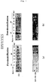
- 238000002415 sodium dodecyl sulfate polyacrylamide gel electrophoresis Methods 0.000 description 3
- 230000008467 tissue growth Effects 0.000 description 3
- 238000005406 washing Methods 0.000 description 3
- GOJUJUVQIVIZAV-UHFFFAOYSA-N 2-amino-4,6-dichloropyrimidine-5-carbaldehyde Chemical group NC1=NC(Cl)=C(C=O)C(Cl)=N1 GOJUJUVQIVIZAV-UHFFFAOYSA-N 0.000 description 2
- 241000193388 Bacillus thuringiensis Species 0.000 description 2
- 108010049931 Bone Morphogenetic Protein 2 Proteins 0.000 description 2
- 241000283690 Bos taurus Species 0.000 description 2
- 108091003079 Bovine Serum Albumin Proteins 0.000 description 2
- 102000004190 Enzymes Human genes 0.000 description 2
- 108090000790 Enzymes Proteins 0.000 description 2
- 102000016359 Fibronectins Human genes 0.000 description 2
- 108010067306 Fibronectins Proteins 0.000 description 2
- 108010024636 Glutathione Proteins 0.000 description 2
- BCCRXDTUTZHDEU-VKHMYHEASA-N Gly-Ser Chemical compound NCC(=O)N[C@@H](CO)C(O)=O BCCRXDTUTZHDEU-VKHMYHEASA-N 0.000 description 2
- 241000193159 Hathewaya histolytica Species 0.000 description 2
- 206010020649 Hyperkeratosis Diseases 0.000 description 2
- 108090000723 Insulin-Like Growth Factor I Proteins 0.000 description 2
- 102000004218 Insulin-Like Growth Factor I Human genes 0.000 description 2
- 108010013295 Microbial collagenase Proteins 0.000 description 2
- 241000283973 Oryctolagus cuniculus Species 0.000 description 2
- 102000006461 Parathyroid Hormone Receptors Human genes 0.000 description 2
- 108010058828 Parathyroid Hormone Receptors Proteins 0.000 description 2
- UIIMBOGNXHQVGW-UHFFFAOYSA-M Sodium bicarbonate Chemical compound [Na+].OC([O-])=O UIIMBOGNXHQVGW-UHFFFAOYSA-M 0.000 description 2
- QAOWNCQODCNURD-UHFFFAOYSA-N Sulfuric acid Chemical compound OS(O)(=O)=O QAOWNCQODCNURD-UHFFFAOYSA-N 0.000 description 2
- 102000004887 Transforming Growth Factor beta Human genes 0.000 description 2
- 108090001012 Transforming Growth Factor beta Proteins 0.000 description 2
- 239000007983 Tris buffer Substances 0.000 description 2
- 238000010306 acid treatment Methods 0.000 description 2
- 125000003277 amino group Chemical group 0.000 description 2
- 229960000723 ampicillin Drugs 0.000 description 2
- AVKUERGKIZMTKX-NJBDSQKTSA-N ampicillin Chemical compound C1([C@@H](N)C(=O)N[C@H]2[C@H]3SC([C@@H](N3C2=O)C(O)=O)(C)C)=CC=CC=C1 AVKUERGKIZMTKX-NJBDSQKTSA-N 0.000 description 2
- 239000007864 aqueous solution Substances 0.000 description 2
- 229940097012 bacillus thuringiensis Drugs 0.000 description 2
- 230000008901 benefit Effects 0.000 description 2
- 239000008280 blood Substances 0.000 description 2
- 210000004369 blood Anatomy 0.000 description 2
- 230000037182 bone density Effects 0.000 description 2
- 210000001185 bone marrow Anatomy 0.000 description 2
- 210000002805 bone matrix Anatomy 0.000 description 2
- OSGAYBCDTDRGGQ-UHFFFAOYSA-L calcium sulfate Chemical compound [Ca+2].[O-]S([O-])(=O)=O OSGAYBCDTDRGGQ-UHFFFAOYSA-L 0.000 description 2
- 230000008859 change Effects 0.000 description 2
- 239000007795 chemical reaction product Substances 0.000 description 2
- 229960005091 chloramphenicol Drugs 0.000 description 2
- WIIZWVCIJKGZOK-RKDXNWHRSA-N chloramphenicol Chemical compound ClC(Cl)C(=O)N[C@H](CO)[C@H](O)C1=CC=C([N+]([O-])=O)C=C1 WIIZWVCIJKGZOK-RKDXNWHRSA-N 0.000 description 2
- 239000002299 complementary DNA Substances 0.000 description 2
- 230000001054 cortical effect Effects 0.000 description 2
- 238000001035 drying Methods 0.000 description 2
- 229940088598 enzyme Drugs 0.000 description 2
- 210000002919 epithelial cell Anatomy 0.000 description 2
- 238000011049 filling Methods 0.000 description 2
- 230000006870 function Effects 0.000 description 2
- 230000004927 fusion Effects 0.000 description 2
- 229960003180 glutathione Drugs 0.000 description 2
- 230000035876 healing Effects 0.000 description 2
- 230000001976 improved effect Effects 0.000 description 2
- 238000003780 insertion Methods 0.000 description 2
- 230000037431 insertion Effects 0.000 description 2
- 230000001788 irregular Effects 0.000 description 2
- 239000011159 matrix material Substances 0.000 description 2
- BDAGIHXWWSANSR-UHFFFAOYSA-N methanoic acid Natural products OC=O BDAGIHXWWSANSR-UHFFFAOYSA-N 0.000 description 2
- QYSGYZVSCZSLHT-UHFFFAOYSA-N octafluoropropane Chemical compound FC(F)(F)C(F)(F)C(F)(F)F QYSGYZVSCZSLHT-UHFFFAOYSA-N 0.000 description 2
- 239000002504 physiological saline solution Substances 0.000 description 2
- 239000000047 product Substances 0.000 description 2
- 230000000717 retained effect Effects 0.000 description 2
- 239000011550 stock solution Substances 0.000 description 2
- 230000002459 sustained effect Effects 0.000 description 2
- ZRKFYGHZFMAOKI-QMGMOQQFSA-N tgfbeta Chemical compound C([C@H](NC(=O)[C@H](C(C)C)NC(=O)CNC(=O)[C@H](CCC(O)=O)NC(=O)[C@H](CCCNC(N)=N)NC(=O)[C@H](CC(N)=O)NC(=O)[C@H](CC(C)C)NC(=O)[C@H]([C@@H](C)O)NC(=O)[C@H](CCC(O)=O)NC(=O)[C@H]([C@@H](C)O)NC(=O)[C@H](CC(C)C)NC(=O)CNC(=O)[C@H](C)NC(=O)[C@H](CO)NC(=O)[C@H](CCC(N)=O)NC(=O)[C@@H](NC(=O)[C@H](C)NC(=O)[C@H](C)NC(=O)[C@@H](NC(=O)[C@H](CC(C)C)NC(=O)[C@@H](N)CCSC)C(C)C)[C@@H](C)CC)C(=O)N[C@@H]([C@@H](C)O)C(=O)N[C@@H](C(C)C)C(=O)N[C@@H](CC=1C=CC=CC=1)C(=O)N[C@@H](C)C(=O)N1[C@@H](CCC1)C(=O)N[C@@H]([C@@H](C)O)C(=O)N[C@@H](CC(N)=O)C(=O)N[C@@H](CCC(O)=O)C(=O)N[C@@H](C)C(=O)N[C@@H](CC=1C=CC=CC=1)C(=O)N[C@@H](CCCNC(N)=N)C(=O)N[C@@H](C)C(=O)N[C@@H](CC(C)C)C(=O)N1[C@@H](CCC1)C(=O)N1[C@@H](CCC1)C(=O)N[C@@H](CCCNC(N)=N)C(=O)N[C@@H](CCC(O)=O)C(=O)N[C@@H](CCCNC(N)=N)C(=O)N[C@@H](CO)C(=O)N[C@@H](CCCNC(N)=N)C(=O)N[C@@H](CC(C)C)C(=O)N[C@@H](CC(C)C)C(O)=O)C1=CC=C(O)C=C1 ZRKFYGHZFMAOKI-QMGMOQQFSA-N 0.000 description 2
- 229960004072 thrombin Drugs 0.000 description 2
- LENZDBCJOHFCAS-UHFFFAOYSA-N tris Chemical compound OCC(N)(CO)CO LENZDBCJOHFCAS-UHFFFAOYSA-N 0.000 description 2
- XLYOFNOQVPJJNP-UHFFFAOYSA-N water Substances O XLYOFNOQVPJJNP-UHFFFAOYSA-N 0.000 description 2
- VHYRHFNOWKMCHQ-UHFFFAOYSA-N (2,5-dioxopyrrolidin-1-yl) 4-formylbenzoate Chemical compound C1=CC(C=O)=CC=C1C(=O)ON1C(=O)CCC1=O VHYRHFNOWKMCHQ-UHFFFAOYSA-N 0.000 description 1
- MTCFGRXMJLQNBG-REOHCLBHSA-N (2S)-2-Amino-3-hydroxypropansäure Chemical compound OC[C@H](N)C(O)=O MTCFGRXMJLQNBG-REOHCLBHSA-N 0.000 description 1
- WYTZZXDRDKSJID-UHFFFAOYSA-N (3-aminopropyl)triethoxysilane Chemical compound CCO[Si](OCC)(OCC)CCCN WYTZZXDRDKSJID-UHFFFAOYSA-N 0.000 description 1
- RVJFYAXPARXKHK-UHFFFAOYSA-N 2-(2,5-dioxopyrrolidin-1-yl)-4-hydrazinylpyridine-3-carboxylic acid propan-2-ylidenehydrazine Chemical compound CC(C)=NN.NNc1ccnc(N2C(=O)CCC2=O)c1C(O)=O RVJFYAXPARXKHK-UHFFFAOYSA-N 0.000 description 1
- OSWFIVFLDKOXQC-UHFFFAOYSA-N 4-(3-methoxyphenyl)aniline Chemical compound COC1=CC=CC(C=2C=CC(N)=CC=2)=C1 OSWFIVFLDKOXQC-UHFFFAOYSA-N 0.000 description 1
- 108010088751 Albumins Proteins 0.000 description 1
- 102000009027 Albumins Human genes 0.000 description 1
- 102100022544 Bone morphogenetic protein 7 Human genes 0.000 description 1
- 241000283707 Capra Species 0.000 description 1
- 229920002134 Carboxymethyl cellulose Polymers 0.000 description 1
- 241000700199 Cavia porcellus Species 0.000 description 1
- 241000282693 Cercopithecidae Species 0.000 description 1
- 241000193155 Clostridium botulinum Species 0.000 description 1
- 241000186581 Clostridium novyi Species 0.000 description 1
- 241000193468 Clostridium perfringens Species 0.000 description 1
- 241000193466 Clostridium septicum Species 0.000 description 1
- 241000193470 Clostridium sporogenes Species 0.000 description 1
- 241000193449 Clostridium tetani Species 0.000 description 1
- 241000699800 Cricetinae Species 0.000 description 1
- CKLJMWTZIZZHCS-UHFFFAOYSA-N D-OH-Asp Natural products OC(=O)C(N)CC(O)=O CKLJMWTZIZZHCS-UHFFFAOYSA-N 0.000 description 1
- 241000283073 Equus caballus Species 0.000 description 1
- LFQSCWFLJHTTHZ-UHFFFAOYSA-N Ethanol Chemical compound CCO LFQSCWFLJHTTHZ-UHFFFAOYSA-N 0.000 description 1
- 102000010834 Extracellular Matrix Proteins Human genes 0.000 description 1
- 108010037362 Extracellular Matrix Proteins Proteins 0.000 description 1
- 241000282326 Felis catus Species 0.000 description 1
- WHUUTDBJXJRKMK-UHFFFAOYSA-N Glutamic acid Natural products OC(=O)C(N)CCC(O)=O WHUUTDBJXJRKMK-UHFFFAOYSA-N 0.000 description 1
- SXRSQZLOMIGNAQ-UHFFFAOYSA-N Glutaraldehyde Chemical compound O=CCCCC=O SXRSQZLOMIGNAQ-UHFFFAOYSA-N 0.000 description 1
- 102000009465 Growth Factor Receptors Human genes 0.000 description 1
- 108010009202 Growth Factor Receptors Proteins 0.000 description 1
- 241000186568 Hathewaya limosa Species 0.000 description 1
- 101000762366 Homo sapiens Bone morphogenetic protein 2 Proteins 0.000 description 1
- 101000899361 Homo sapiens Bone morphogenetic protein 7 Proteins 0.000 description 1
- 206010058031 Joint adhesion Diseases 0.000 description 1
- ONIBWKKTOPOVIA-BYPYZUCNSA-N L-Proline Chemical compound OC(=O)[C@@H]1CCCN1 ONIBWKKTOPOVIA-BYPYZUCNSA-N 0.000 description 1
- CKLJMWTZIZZHCS-REOHCLBHSA-N L-aspartic acid Chemical compound OC(=O)[C@@H](N)CC(O)=O CKLJMWTZIZZHCS-REOHCLBHSA-N 0.000 description 1
- WHUUTDBJXJRKMK-VKHMYHEASA-N L-glutamic acid Chemical compound OC(=O)[C@@H](N)CCC(O)=O WHUUTDBJXJRKMK-VKHMYHEASA-N 0.000 description 1
- KDXKERNSBIXSRK-YFKPBYRVSA-N L-lysine Chemical compound NCCCC[C@H](N)C(O)=O KDXKERNSBIXSRK-YFKPBYRVSA-N 0.000 description 1
- AYFVYJQAPQTCCC-GBXIJSLDSA-N L-threonine Chemical compound C[C@@H](O)[C@H](N)C(O)=O AYFVYJQAPQTCCC-GBXIJSLDSA-N 0.000 description 1
- KDXKERNSBIXSRK-UHFFFAOYSA-N Lysine Natural products NCCCCC(N)C(O)=O KDXKERNSBIXSRK-UHFFFAOYSA-N 0.000 description 1
- 239000004472 Lysine Substances 0.000 description 1
- 241000699684 Meriones unguiculatus Species 0.000 description 1
- 241001529936 Murinae Species 0.000 description 1
- GRYLNZFGIOXLOG-UHFFFAOYSA-N Nitric acid Chemical compound O[N+]([O-])=O GRYLNZFGIOXLOG-UHFFFAOYSA-N 0.000 description 1
- 208000001132 Osteoporosis Diseases 0.000 description 1
- 229910019142 PO4 Inorganic materials 0.000 description 1
- 241000193465 Paeniclostridium sordellii Species 0.000 description 1
- 208000002193 Pain Diseases 0.000 description 1
- 241000282577 Pan troglodytes Species 0.000 description 1
- 241001504519 Papio ursinus Species 0.000 description 1
- 241000193157 Paraclostridium bifermentans Species 0.000 description 1
- 241001494479 Pecora Species 0.000 description 1
- 102000035195 Peptidases Human genes 0.000 description 1
- 108091005804 Peptidases Proteins 0.000 description 1
- 102000007079 Peptide Fragments Human genes 0.000 description 1
- 108010033276 Peptide Fragments Proteins 0.000 description 1
- 241000009328 Perro Species 0.000 description 1
- 102000004160 Phosphoric Monoester Hydrolases Human genes 0.000 description 1
- 108090000608 Phosphoric Monoester Hydrolases Proteins 0.000 description 1
- 241000288906 Primates Species 0.000 description 1
- ONIBWKKTOPOVIA-UHFFFAOYSA-N Proline Natural products OC(=O)C1CCCN1 ONIBWKKTOPOVIA-UHFFFAOYSA-N 0.000 description 1
- 208000002607 Pseudarthrosis Diseases 0.000 description 1
- MTCFGRXMJLQNBG-UHFFFAOYSA-N Serine Natural products OCC(N)C(O)=O MTCFGRXMJLQNBG-UHFFFAOYSA-N 0.000 description 1
- 241000282898 Sus scrofa Species 0.000 description 1
- AYFVYJQAPQTCCC-UHFFFAOYSA-N Threonine Natural products CC(O)C(N)C(O)=O AYFVYJQAPQTCCC-UHFFFAOYSA-N 0.000 description 1
- 239000004473 Threonine Substances 0.000 description 1
- AUYYCJSJGJYCDS-LBPRGKRZSA-N Thyrolar Chemical class IC1=CC(C[C@H](N)C(O)=O)=CC(I)=C1OC1=CC=C(O)C(I)=C1 AUYYCJSJGJYCDS-LBPRGKRZSA-N 0.000 description 1
- 229920004890 Triton X-100 Polymers 0.000 description 1
- 239000013504 Triton X-100 Substances 0.000 description 1
- 208000027418 Wounds and injury Diseases 0.000 description 1
- 239000003929 acidic solution Substances 0.000 description 1
- 239000004480 active ingredient Substances 0.000 description 1
- 239000013543 active substance Substances 0.000 description 1
- 230000032683 aging Effects 0.000 description 1
- 239000003513 alkali Substances 0.000 description 1
- 150000001412 amines Chemical group 0.000 description 1
- 230000001195 anabolic effect Effects 0.000 description 1
- 208000037873 arthrodesis Diseases 0.000 description 1
- 108010045569 atelocollagen Proteins 0.000 description 1
- 239000011230 binding agent Substances 0.000 description 1
- 230000000035 biogenic effect Effects 0.000 description 1
- 239000012620 biological material Substances 0.000 description 1
- LNQHREYHFRFJAU-UHFFFAOYSA-N bis(2,5-dioxopyrrolidin-1-yl) pentanedioate Chemical compound O=C1CCC(=O)N1OC(=O)CCCC(=O)ON1C(=O)CCC1=O LNQHREYHFRFJAU-UHFFFAOYSA-N 0.000 description 1
- 230000010478 bone regeneration Effects 0.000 description 1
- 239000000316 bone substitute Substances 0.000 description 1
- 239000001768 carboxy methyl cellulose Substances 0.000 description 1
- 235000010948 carboxy methyl cellulose Nutrition 0.000 description 1
- 239000008112 carboxymethyl-cellulose Substances 0.000 description 1
- 230000015556 catabolic process Effects 0.000 description 1
- 230000003197 catalytic effect Effects 0.000 description 1
- 230000024245 cell differentiation Effects 0.000 description 1
- 230000010261 cell growth Effects 0.000 description 1
- 230000004663 cell proliferation Effects 0.000 description 1
- 230000005859 cell recognition Effects 0.000 description 1
- 238000012512 characterization method Methods 0.000 description 1
- 238000010382 chemical cross-linking Methods 0.000 description 1
- 239000011248 coating agent Substances 0.000 description 1
- 238000000576 coating method Methods 0.000 description 1
- 239000013065 commercial product Substances 0.000 description 1
- 150000001875 compounds Chemical class 0.000 description 1
- 239000000470 constituent Substances 0.000 description 1
- 238000013270 controlled release Methods 0.000 description 1
- 239000013256 coordination polymer Substances 0.000 description 1
- 230000006378 damage Effects 0.000 description 1
- 230000002950 deficient Effects 0.000 description 1
- 238000006731 degradation reaction Methods 0.000 description 1
- 238000005115 demineralization Methods 0.000 description 1
- 230000002328 demineralizing effect Effects 0.000 description 1
- 230000001419 dependent effect Effects 0.000 description 1
- 238000000502 dialysis Methods 0.000 description 1
- 239000006185 dispersion Substances 0.000 description 1
- 238000010828 elution Methods 0.000 description 1
- 239000013604 expression vector Substances 0.000 description 1
- 235000019253 formic acid Nutrition 0.000 description 1
- 239000007789 gas Substances 0.000 description 1
- 239000004220 glutamic acid Substances 0.000 description 1
- 235000013922 glutamic acid Nutrition 0.000 description 1
- 150000004676 glycans Chemical class 0.000 description 1
- 230000036571 hydration Effects 0.000 description 1
- 238000006703 hydration reaction Methods 0.000 description 1
- 229910052588 hydroxylapatite Inorganic materials 0.000 description 1
- 230000001900 immune effect Effects 0.000 description 1
- 238000002513 implantation Methods 0.000 description 1
- 238000001727 in vivo Methods 0.000 description 1
- 208000014674 injury Diseases 0.000 description 1
- 238000010253 intravenous injection Methods 0.000 description 1
- 230000009545 invasion Effects 0.000 description 1
- 230000008407 joint function Effects 0.000 description 1
- 210000005067 joint tissue Anatomy 0.000 description 1
- 210000003292 kidney cell Anatomy 0.000 description 1
- 150000002632 lipids Chemical class 0.000 description 1
- 238000011068 loading method Methods 0.000 description 1
- 230000007774 longterm Effects 0.000 description 1
- 238000013227 male C57BL/6J mice Methods 0.000 description 1
- 230000003340 mental effect Effects 0.000 description 1
- 108020004999 messenger RNA Proteins 0.000 description 1
- 230000000921 morphogenic effect Effects 0.000 description 1
- 210000003205 muscle Anatomy 0.000 description 1
- 229940105631 nembutal Drugs 0.000 description 1
- 229910017604 nitric acid Inorganic materials 0.000 description 1
- 239000003960 organic solvent Substances 0.000 description 1
- 201000008482 osteoarthritis Diseases 0.000 description 1
- 201000008968 osteosarcoma Diseases 0.000 description 1
- 230000036407 pain Effects 0.000 description 1
- 206010033675 panniculitis Diseases 0.000 description 1
- XYJRXVWERLGGKC-UHFFFAOYSA-D pentacalcium;hydroxide;triphosphate Chemical compound [OH-].[Ca+2].[Ca+2].[Ca+2].[Ca+2].[Ca+2].[O-]P([O-])([O-])=O.[O-]P([O-])([O-])=O.[O-]P([O-])([O-])=O XYJRXVWERLGGKC-UHFFFAOYSA-D 0.000 description 1
- WEXRUCMBJFQVBZ-UHFFFAOYSA-N pentobarbital Chemical compound CCCC(C)C1(CC)C(=O)NC(=O)NC1=O WEXRUCMBJFQVBZ-UHFFFAOYSA-N 0.000 description 1
- 230000000144 pharmacologic effect Effects 0.000 description 1
- NBIIXXVUZAFLBC-UHFFFAOYSA-K phosphate Chemical compound [O-]P([O-])([O-])=O NBIIXXVUZAFLBC-UHFFFAOYSA-K 0.000 description 1
- 239000010452 phosphate Substances 0.000 description 1
- 229920001282 polysaccharide Polymers 0.000 description 1
- 239000005017 polysaccharide Substances 0.000 description 1
- 239000000843 powder Substances 0.000 description 1
- 230000002028 premature Effects 0.000 description 1
- 238000004321 preservation Methods 0.000 description 1
- 238000011084 recovery Methods 0.000 description 1
- 230000006833 reintegration Effects 0.000 description 1
- 150000003839 salts Chemical class 0.000 description 1
- 238000007493 shaping process Methods 0.000 description 1
- 210000003625 skull Anatomy 0.000 description 1
- 235000017557 sodium bicarbonate Nutrition 0.000 description 1
- 229910000030 sodium bicarbonate Inorganic materials 0.000 description 1
- 229910001467 sodium calcium phosphate Inorganic materials 0.000 description 1
- 210000004872 soft tissue Anatomy 0.000 description 1
- 230000007480 spreading Effects 0.000 description 1
- 238000003892 spreading Methods 0.000 description 1
- 210000000130 stem cell Anatomy 0.000 description 1
- 230000004936 stimulating effect Effects 0.000 description 1
- 238000003756 stirring Methods 0.000 description 1
- 210000004304 subcutaneous tissue Anatomy 0.000 description 1
- 239000013589 supplement Substances 0.000 description 1
- 230000008093 supporting effect Effects 0.000 description 1
- 230000002195 synergetic effect Effects 0.000 description 1
- 238000007910 systemic administration Methods 0.000 description 1
- 230000002123 temporal effect Effects 0.000 description 1
- 238000002560 therapeutic procedure Methods 0.000 description 1
- 239000005495 thyroid hormone Substances 0.000 description 1
- 229940036555 thyroid hormone Drugs 0.000 description 1
- 230000017423 tissue regeneration Effects 0.000 description 1
- 238000002054 transplantation Methods 0.000 description 1
- GPRLSGONYQIRFK-MNYXATJNSA-N triton Chemical compound [3H+] GPRLSGONYQIRFK-MNYXATJNSA-N 0.000 description 1
Images


Classifications
-
- A—HUMAN NECESSITIES
- A61—MEDICAL OR VETERINARY SCIENCE; HYGIENE
- A61K—PREPARATIONS FOR MEDICAL, DENTAL OR TOILETRY PURPOSES
- A61K38/00—Medicinal preparations containing peptides
- A61K38/16—Peptides having more than 20 amino acids; Gastrins; Somatostatins; Melanotropins; Derivatives thereof
- A61K38/17—Peptides having more than 20 amino acids; Gastrins; Somatostatins; Melanotropins; Derivatives thereof from animals; from humans
- A61K38/18—Growth factors; Growth regulators
- A61K38/1825—Fibroblast growth factor [FGF]
-
- A—HUMAN NECESSITIES
- A61—MEDICAL OR VETERINARY SCIENCE; HYGIENE
- A61K—PREPARATIONS FOR MEDICAL, DENTAL OR TOILETRY PURPOSES
- A61K38/00—Medicinal preparations containing peptides
- A61K38/16—Peptides having more than 20 amino acids; Gastrins; Somatostatins; Melanotropins; Derivatives thereof
- A61K38/17—Peptides having more than 20 amino acids; Gastrins; Somatostatins; Melanotropins; Derivatives thereof from animals; from humans
- A61K38/18—Growth factors; Growth regulators
- A61K38/1808—Epidermal growth factor [EGF] urogastrone
-
- A—HUMAN NECESSITIES
- A61—MEDICAL OR VETERINARY SCIENCE; HYGIENE
- A61K—PREPARATIONS FOR MEDICAL, DENTAL OR TOILETRY PURPOSES
- A61K38/00—Medicinal preparations containing peptides
- A61K38/16—Peptides having more than 20 amino acids; Gastrins; Somatostatins; Melanotropins; Derivatives thereof
- A61K38/43—Enzymes; Proenzymes; Derivatives thereof
- A61K38/46—Hydrolases (3)
- A61K38/48—Hydrolases (3) acting on peptide bonds (3.4)
- A61K38/4886—Metalloendopeptidases (3.4.24), e.g. collagenase
-
- A—HUMAN NECESSITIES
- A61—MEDICAL OR VETERINARY SCIENCE; HYGIENE
- A61L—METHODS OR APPARATUS FOR STERILISING MATERIALS OR OBJECTS IN GENERAL; DISINFECTION, STERILISATION OR DEODORISATION OF AIR; CHEMICAL ASPECTS OF BANDAGES, DRESSINGS, ABSORBENT PADS OR SURGICAL ARTICLES; MATERIALS FOR BANDAGES, DRESSINGS, ABSORBENT PADS OR SURGICAL ARTICLES
- A61L27/00—Materials for grafts or prostheses or for coating grafts or prostheses
- A61L27/36—Materials for grafts or prostheses or for coating grafts or prostheses containing ingredients of undetermined constitution or reaction products thereof, e.g. transplant tissue, natural bone, extracellular matrix
- A61L27/3604—Materials for grafts or prostheses or for coating grafts or prostheses containing ingredients of undetermined constitution or reaction products thereof, e.g. transplant tissue, natural bone, extracellular matrix characterised by the human or animal origin of the biological material, e.g. hair, fascia, fish scales, silk, shellac, pericardium, pleura, renal tissue, amniotic membrane, parenchymal tissue, fetal tissue, muscle tissue, fat tissue, enamel
- A61L27/3608—Bone, e.g. demineralised bone matrix [DBM], bone powder
-
- A—HUMAN NECESSITIES
- A61—MEDICAL OR VETERINARY SCIENCE; HYGIENE
- A61L—METHODS OR APPARATUS FOR STERILISING MATERIALS OR OBJECTS IN GENERAL; DISINFECTION, STERILISATION OR DEODORISATION OF AIR; CHEMICAL ASPECTS OF BANDAGES, DRESSINGS, ABSORBENT PADS OR SURGICAL ARTICLES; MATERIALS FOR BANDAGES, DRESSINGS, ABSORBENT PADS OR SURGICAL ARTICLES
- A61L27/00—Materials for grafts or prostheses or for coating grafts or prostheses
- A61L27/50—Materials characterised by their function or physical properties, e.g. injectable or lubricating compositions, shape-memory materials, surface modified materials
- A61L27/54—Biologically active materials, e.g. therapeutic substances
-
- C—CHEMISTRY; METALLURGY
- C07—ORGANIC CHEMISTRY
- C07K—PEPTIDES
- C07K14/00—Peptides having more than 20 amino acids; Gastrins; Somatostatins; Melanotropins; Derivatives thereof
- C07K14/195—Peptides having more than 20 amino acids; Gastrins; Somatostatins; Melanotropins; Derivatives thereof from bacteria
- C07K14/33—Peptides having more than 20 amino acids; Gastrins; Somatostatins; Melanotropins; Derivatives thereof from bacteria from Clostridium (G)
-
- C—CHEMISTRY; METALLURGY
- C07—ORGANIC CHEMISTRY
- C07K—PEPTIDES
- C07K14/00—Peptides having more than 20 amino acids; Gastrins; Somatostatins; Melanotropins; Derivatives thereof
- C07K14/435—Peptides having more than 20 amino acids; Gastrins; Somatostatins; Melanotropins; Derivatives thereof from animals; from humans
- C07K14/475—Growth factors; Growth regulators
- C07K14/485—Epidermal growth factor [EGF], i.e. urogastrone
-
- C—CHEMISTRY; METALLURGY
- C12—BIOCHEMISTRY; BEER; SPIRITS; WINE; VINEGAR; MICROBIOLOGY; ENZYMOLOGY; MUTATION OR GENETIC ENGINEERING
- C12N—MICROORGANISMS OR ENZYMES; COMPOSITIONS THEREOF; PROPAGATING, PRESERVING, OR MAINTAINING MICROORGANISMS; MUTATION OR GENETIC ENGINEERING; CULTURE MEDIA
- C12N9/00—Enzymes; Proenzymes; Compositions thereof; Processes for preparing, activating, inhibiting, separating or purifying enzymes
- C12N9/14—Hydrolases (3)
- C12N9/48—Hydrolases (3) acting on peptide bonds (3.4)
- C12N9/50—Proteinases, e.g. Endopeptidases (3.4.21-3.4.25)
- C12N9/52—Proteinases, e.g. Endopeptidases (3.4.21-3.4.25) derived from bacteria or Archaea
-
- A—HUMAN NECESSITIES
- A61—MEDICAL OR VETERINARY SCIENCE; HYGIENE
- A61L—METHODS OR APPARATUS FOR STERILISING MATERIALS OR OBJECTS IN GENERAL; DISINFECTION, STERILISATION OR DEODORISATION OF AIR; CHEMICAL ASPECTS OF BANDAGES, DRESSINGS, ABSORBENT PADS OR SURGICAL ARTICLES; MATERIALS FOR BANDAGES, DRESSINGS, ABSORBENT PADS OR SURGICAL ARTICLES
- A61L2300/00—Biologically active materials used in bandages, wound dressings, absorbent pads or medical devices
- A61L2300/20—Biologically active materials used in bandages, wound dressings, absorbent pads or medical devices containing or releasing organic materials
- A61L2300/252—Polypeptides, proteins, e.g. glycoproteins, lipoproteins, cytokines
-
- A—HUMAN NECESSITIES
- A61—MEDICAL OR VETERINARY SCIENCE; HYGIENE
- A61L—METHODS OR APPARATUS FOR STERILISING MATERIALS OR OBJECTS IN GENERAL; DISINFECTION, STERILISATION OR DEODORISATION OF AIR; CHEMICAL ASPECTS OF BANDAGES, DRESSINGS, ABSORBENT PADS OR SURGICAL ARTICLES; MATERIALS FOR BANDAGES, DRESSINGS, ABSORBENT PADS OR SURGICAL ARTICLES
- A61L2300/00—Biologically active materials used in bandages, wound dressings, absorbent pads or medical devices
- A61L2300/40—Biologically active materials used in bandages, wound dressings, absorbent pads or medical devices characterised by a specific therapeutic activity or mode of action
- A61L2300/412—Tissue-regenerating or healing or proliferative agents
- A61L2300/414—Growth factors
-
- A—HUMAN NECESSITIES
- A61—MEDICAL OR VETERINARY SCIENCE; HYGIENE
- A61L—METHODS OR APPARATUS FOR STERILISING MATERIALS OR OBJECTS IN GENERAL; DISINFECTION, STERILISATION OR DEODORISATION OF AIR; CHEMICAL ASPECTS OF BANDAGES, DRESSINGS, ABSORBENT PADS OR SURGICAL ARTICLES; MATERIALS FOR BANDAGES, DRESSINGS, ABSORBENT PADS OR SURGICAL ARTICLES
- A61L2430/00—Materials or treatment for tissue regeneration
- A61L2430/02—Materials or treatment for tissue regeneration for reconstruction of bones; weight-bearing implants
-
- C—CHEMISTRY; METALLURGY
- C07—ORGANIC CHEMISTRY
- C07K—PEPTIDES
- C07K2319/00—Fusion polypeptide
-
- C—CHEMISTRY; METALLURGY
- C07—ORGANIC CHEMISTRY
- C07K—PEPTIDES
- C07K2319/00—Fusion polypeptide
- C07K2319/01—Fusion polypeptide containing a localisation/targetting motif
- C07K2319/03—Fusion polypeptide containing a localisation/targetting motif containing a transmembrane segment
-
- C—CHEMISTRY; METALLURGY
- C07—ORGANIC CHEMISTRY
- C07K—PEPTIDES
- C07K2319/00—Fusion polypeptide
- C07K2319/70—Fusion polypeptide containing domain for protein-protein interaction
Definitions
- the present invention relates to a bone graft material bound a bone graft substrate exposing at least a collagen fiber to a growth factor, more particularly, relates to a growth factor anchoring type bone graft material wherein a bone graft substrate is bound to a collagen-binding-site-containing growth factor which comprises a growth factor receptor agonist peptide and a collagen-binding peptide, a method for producing the growth factor anchoring type bone graft material, a kit for production of a growth factor anchoring type bone graft material, and a method for forming a bone.
- Bone grafting has a feature that a bone protein contained in grafted bone promotes resorption of the grafted bone and conversion to an autologous tissue, therefore it has an advantage that reconstruction of a joint function becomes possible even though reconstruction with a prosthesis is impossible. Further, bone is a tissue superior in regenerative capacity, it may be regenerated into a nearly original form by proper reintegration and fixation in case of a fracture.
- autologous bone grafting is a method which own bone is cut out from a certain part of a patient as a block, the obtained bone is transplanted to deficient part as a block, or after crushing to a granular or powder form.
- the method is an advantage of high safety because own bone is utilized although, pains are severe at the bone collecting part in the case of a large bone defect region, the recovery period after the bone grafting surgery becomes longer, and sometimes it is very difficult to find a donor supplying a bone for bone grafting.
- allogeneic bone grafting using a donor-derived bone instead of an autologous bone is conducted, and further, various bone graft materials have been also developed.
- composition used for promoting bone formation in arthrodesis which includes a platelet-derived growth factor solution, a biocompatible matrix containing polysaccharides, and a scaffold material such as calcium phosphate (Patent Literature 1).
- a platelet-derived growth factor solution a biocompatible matrix containing polysaccharides
- a scaffold material such as calcium phosphate
- 1.0 mg/mL of platelet-derived growth factor is dropped to calcium phosphate in the average diameter of 1000 to 2000 ⁇ m for preparing a composition, and the composition is coated on a bone to be fused in a joint.
- the composition exhibits bone bridging and joint adhesion equivalent to autologous bone grafting.
- Patent Literature 2 there is a bone graft material on which surf ace a cell adhesion inducing peptide having an RGD amino acid sequence, or a tissue growth factor-derived peptide is fixed (Patent Literature 2).
- the bone graft material adhering on the surface a tissue growth factor capable of obtaining a tissue regeneration effect andapeptide having active site of an extracellular matrix protein exhibits allegedly a stable and sustainable pharmacological effect, even though the concentration of the peptides is low.
- surfaces of a bovine bone-derived bone mineral particle are treated with 3-aminopropyltriethoxysilane to form an amine residue, the particles are bound with a crosslinking agent of 1, 4-bis-maleimidebutane added thereto, then reacted with a cell adhesion inducing peptide to fix the peptide, and prepare a bone graft material.
- the material exhibits allegedly superior regenerative power compared to a bone graft material without the fixed peptide.
- a bone graft fragment composition prepared by drying a fragment of a cell-free tissue substrate together with a fragment of a demineralize bone material (Patent Literature 3).
- a cell-free tissue substrate such as collagen obtained from an epithelial cell has capability for supporting cell recognition and cell association, as well as cell spreading, cell proliferation, and cell differentiation, a demineralize bone material has physiological characteristics of natural bone important for a success of bone grafting.
- new bone formation can be allegedly induced in or on a surface of an osseous tissue, or in or on a surface of a non-osseous tissue of a recipient by stimulating a bone formation stem cell.
- compositions containing a fusion protein fused a PTH/PTHrP receptor agonist with a collagen-binding polypeptide fragment drived from a collagenase (Patent Literature 4) .
- a parathyroid hormone (PTH) is used for an anabolic therapy of osteoporosis, an administration once a day is required.
- the composition can form a stable bind with collagen through a collagen-binding polypeptide fragment, and stay at an administration site for a long time period resisting body fluid circulation to enjoy longer half-life than PTH. Then, it can exert allegedly the same or higher effectiveness compared to PTH administration. In the example, it is administered intraperitoneally and increase of the bone density is observed.
- Non Patent Literature 1 a fusion protein which a basic fibroblast growth factor (bFGF) instead of a PTH/PTHrP receptor agonist is bound to a collagen-binding polypeptide fragment, has been also known (Non Patent Literature 1).
- bFGF basic fibroblast growth factor
- a fusion protein comprising a BMP-2 as growth factor receptor agonist and a collagen binding peptide is also known (Non Patent Literature 2).
- a bone formation promoting fusion protein prepared by binding a polypeptide having a collagen-binding domain derived from fibronectin with a bone formation promoting protein (Patent Literature 5).
- the bone formation promoting protein are named a growth factor belonging to a BMP (Bone Morphogenetic Proteins) subfamily, bFGF, and a thyroid hormone.
- the polypeptide is prepared by using mRNA extracted from human kidney cells as a template thereof, bound with BMP2 or BMP7 as the bone formation promoting protein to prepare the bone formation promoting fusion protein.
- composition for a treatment of a bone defect composed of a forming particle having at least 4 curved projections composed of calcium sulfate or the like and a material for a suspension
- a plurality of the projection of the forming particle can interlock each other to stabilize filling into a defect site, a binder capable of forming a gel of a collagen derivative or the like, or a bone morphogenic protein (BMP) can use as the suspension.
- BMP bone morphogenic protein
- a self-curing porous calcium phosphate composition which contains calcium phosphate, a blowing agent, and a biocompatible flocculant, and is mixed with a physiologically acceptable liquid, can releases a gas component by hydrationofthe blowing agent in the composition, gives at least 5% of porosity to the composition, and after curing the calcium phosphate composition exhibits a compressive strength of 1 MPa or more (Patent Literature 7).
- a biocompatible flocculant collagen is disclosed and it is described that the composition may contain further a collagen exposure-treated substrate.
- the invention has a feature that a porous calcium phosphate composition is formed by a blowing agent, and in the example thereof a collagen exposure-treated substrate, sodium hydrogen carbonate and calcium phosphate as a blowing agent, and carboxymethyl cellulose as a flocculant were mixed to prepare aself-curingpaste.
- a porous calcium phosphate composition is formed by a blowing agent, and in the example thereof a collagen exposure-treated substrate, sodium hydrogen carbonate and calcium phosphate as a blowing agent, and carboxymethyl cellulose as a flocculant were mixed to prepare aself-curingpaste.
- a bone growth composition containing a particulate fibrous collagen component, and a calcium phosphate component, as well as a substance selected from the group consisting of a purified bone growth factor, a recombinant bone growth factor, a bone-marrow component, and demineralized bone and autologous bone (Patent Literature 8).
- the collagen component is cross-linked collagen or porous granular or other insoluble collagen.
- a calcium phosphate gel dispersion is kneaded with complex collagen, and after a cross-linking step by freeze-drying and thermal dewatering shaped into the particulate, pasted by adding blood, then transplanted to scattered bone. A defect site could be allegedly fixed firmly with the paste.
- Bone grafting is exercised on an artificial joint revision surgery, a treatment of fracture, and a bone defect due to malignant osteosarcoma, but in some cases, even though graft bone originated from autologous bone or allogeneic bone is used, a graft bone applied to a bone occurs faulty union or delayed union to the site of application to the bone reportedly.
- Such faulty union or delayed union means prolongation of a treatment period and becomes an economical, physical, and mental burden on the patient.
- soonest bone union is desired in order to initiate rehabilitation as soon as possible.
- the bone graft material of Patent Literature 1 although calcium phosphate or the like used as a scaffold material is advantageous in terms of easy availability, bone growth or early union surpassing autologous bone is difficult.
- a cell adhesion inducing peptide or a tissue growth factor-derived peptide is fixed on a bone surface, the same can remain at an administration part at a high retention rate, and exhibit superior bone regenerative power. It, however, requires a cross-linking treatment for fixing the peptide on the bone surface, which makes the production difficult.
- Patent Literature 3 requires use of a demineralized bone material, and for demineralization extraction with 0.6 N hydrochloric acid for 3 to 24 hours is necessary, namely the treatment time becomes longer. Further, although it is advantageous that the bone graft material of Patent Literature 3 or Patent Literature 4 uses an active ingredient relevant to bone growth, such a component is easy to leave from the administrated part due to body fluid circulation, and a high retention rate may not be maintained at the administrated part.
- Patent Literature 5 a collagen-binding domain is limited to what derived from fibronectin.
- bFGF bone formation promoting protein
- Patent Literature 6 is characterized by using a forming particle having a predetermined shape, and despite a description that BMP may be added, an actual evaluation has not been conducted. Even if the component is added, it is presumed that the component will easily leave from the administrated part due to body fluid circulation and is not able to establish a high retention rate.
- Patent Literature 7 there is a description that collagen may be mixed as a biocompatible flocculant to formed porous calcium phosphate, however an actual evaluation has not been conducted.
- porous calcium phosphate and the collagen are not fixed together by a covalent bond, the same will easily leave an administrated part due to body fluid circulation, and a sustainable effect is presumed to be hardly attainable.
- cross-linked collagen shaped a particulate form is used, however preparation is not easy, and despite a disclosure that a bone growth factor can be added, an actual evaluation has not been conducted. Further, even if a bone growth factor is mixed with the cross-linked collagen, the bone growth factor easily leaves an administration part due to body fluid circulation, and presumably an effect is hardly attainable for a long period.
- an object of the present invention is to provide a bone graft material that can maintain the retention rate of a bone growth factor at an administration part, while securing an anatomical shape and mechanical strengths of a bone, and expectedly attain early bone union.
- Another object of the present invention is to provide a bone graft material having mechanical strengths and being superior in osteogenic ability, a method for producing a bone graft material and a kit for producing a bone graft material.
- the present inventors have found that a superior osteogenic ability can be expected by binding a fusion protein as defined by claim 1, in which a growth factor is bound to a collagen-binding peptide to a bone, that the fusion protein can easily bind to the bone graft substrate exposing at least a collagen fiber by mixing it with the bone graft substrate without a cross-linking reaction or the like, and further that the obtained growth factor anchoring type bone graft material can exert the osteogenic ability at an administratied part for a long time period and consequently early bone union can be expected, thereby established the present invention.
- the present invention provides a growth factor anchoring type bone graft material, wherein a bone graft substrate exposing at least a collagen fiber is bound to a collagen-binding-site-containing growth factor which comprises a growth factor receptor agonist peptide and a collagen-binding peptide (hereinafter also referred to as " CB-GF ").
- the present invention provides the growth factor anchoring type bone graft material, wherein the collagen-binding-site-containing growth factor comprises the growth factor receptor agonist peptide, the collagen-binding peptide, and a linker.
- the present invention provides the growth factor anchoring type bone graft material, wherein the bone graft substrate is a collagen-exposing bone material or a high-density collagen material.
- the growth factor anchoring type bone graft material wherein the growth factor receptor agonist peptide is a basic fibroblast growth factor.
- the present invention provides a method for producing a growth factor anchoring type bone graft material, wherein the bone graft substrate and the CB-GF are mixed.
- the bone graft substrate is a collagen-exposing bone material prepared by treating a bone with an acid and removing an inorganic mineral component dissolved by the acid.
- the present invention provides a kit for production of a growth factor anchoring type bone graft material, comprising a solution comprising the CB-GF and a bone graft substrate, as defined in claim 7.
- the present invention provides a kit for production of a growth factor anchoring type bone graft material, comprising a solution comprising the CB-GF and a collagen-exposing bone material preparation solution, as defined in claim 9.
- this description describes a method for forming a bone, where in the growth factor anchoring type bone graft material is transplanted to a bone defect region and/or a non-union region.
- the present invention provides the method for forming a bone, wherein the growth factor anchoring type bone graft material is prepared by preparing a collagen-exposing bone material by crushing a bone and treating the same with an acid for 1 to 60 min, and binding the CB-GF to the collagen-exposing bone material.
- a growth factor anchoring type bone graft material of the present invention which a growth factor receptor agonist peptide is bound to a bone graft substrate exposing at least collagen fiber through a collagen-binding peptide of the bone graft substrate, is entirely derived from biogenic substances, and has excellent affinity for an organism and safety.
- the growth factor anchoring type bone graft material of to the present invention can be produced easily by simply mixing a bone graft substrate exposing at least a collagen fiber with a CB-GF prepared in advance to be bound together.
- the growth factor anchoring type bone graft material of the present invention can utilize the bone forming activities of both the bone graft substrate exposing at least a collagen fiber and a growth factor, a good union effect can be exerted even for a case in which union is difficult at the application site of the bone.
- kit for production of a growth factor anchoring type bone graft material of the present invention can prepare a collagen-exposing bone material in a short time, it can be used easily at the time of autologous bone grafting.
- the first aspect of the present invention is a growth factor anchoring type bone graft material characterized in that a bone graft substrate exposing at least a collagen fiber is bound to a CB-GF.
- a bone is constituted with network-formed collagen fibers and hydroxyapatite deposited thereon, and most part of organic substances of a bone is collagen.
- 3 polypeptide chains are bound in a helical fashion, and a large number of the molecules associate in vivo to form insoluble fibers.
- the substances differentiate undifferentiated mesenchymal cells existing in subcutaneous tissues and muscles to osteoblasts to promote bone formation.
- a "growth factor anchoring bone graft material" of the present invention is to use a bone graft substrate exposing at least a collagen fiber.
- a bone graft substrate which at least a part of inorganic substances is removed from a bone to expose collagen fibers on the bone surface can be used.
- Such a bone graft substrate to which a CB-GF is bound retains highly its anatomical shape and excels in dynamically, because a large amount of mineral remains in the substrate.
- collagen fibers exist therein without degradation, and the CB-GF can be bound thereto simply by mixing with the bone graft substrate, and therefore production is easy.
- the growth factor anchoring type bone graft material of the present invention can be expected synergistic bone forming activity by a growth factor, in addition to the osteogenic ability owned inherently by the bone graft substrate exposing at least a collagen fiber. Furthermore, since the growth factor is bound to the bone graft substrate, it can stay long at a grafted site and promote sustained bone formation. Additionally an autologous bone is used as a source material of the bone graft substrate, it is advantageous in that an immunological rejection reaction can be avoided.
- a CB-GF is bound preferably in an amount of 0.01 to 1 nmol, preferably 0.1 to 1 nmol, and more preferably 0.5 to 1 nmol. Even if the CB-GF is bound beyond 1 nmol, the increasing rate of bone formation is not improved any more; and if it is below 0.01 nmol, the effect of the bound CB-GF may occasionally not be attainable sufficiently.
- a bone is subjected to a collagen-exposing treatment to prepared the bone graft substrate at the time to use, binding thereto a CB-GF, thereafter it is used as a bone graft material; or alternatively a growth factor anchoring bone graft material prepared in advance by binding a CB-GF to a bone graft substrate and dried for preservation can be used by suspending it in a buffer solution when needed.
- a collagen-binding peptide included in the growth factor anchoring bone graft material binds to a collagen fiber by means of its stereostructure, it is preferable to suspend it in a buffer solution that can secure the stereostructure.
- a buffer solution include a phosphate buffer solution of pH 7.4 and a Tris buffer solution.
- the growth factor anchoring bone graft material of the present invention can be administered locally for the purpose of increasing bone density, increasing bone mineral density, or increasing new bone similarly to a conventional bone graft material such as an autologous bone graft material.
- a conventional bone graft material such as an autologous bone graft material.
- bone formation can be promoted. It can be used favorably especially for cases requiring a bone graft material maintaining an anatomical shape and mechanical strengths, such as artificial joint revision surgery, and intractable fracture treatment.
- CB-GF growth factor receptor agonist peptide
- CB site collagen-binding peptide
- both of the peptides may be bound chemically, or it may be a fusion protein including a GF site and a CB site.
- the CB site may be binding directly or through a linker composed of a polypeptide fragment with the GF site.
- 2 polypeptides of the GF site and the CB site may be cross-linked by a reagent including disuccinimidyl glutarate or glutaraldehyde through an amino group.
- a polypeptide is derivatized by succinimidyl-4-hydrazinonicotinate acetone hydrazone, and the other polypeptide is derivatized by succinimidyl-4-formyl benzoate, and then two derivatized polypeptides may be mixed for cross-linking through an amino group.
- the two may be linked by a crosslinking agent other than polypeptides or other compounds to bind the GF site and the CB site.
- a "collagen-binding peptide" constituting the CB-GF to be used in the present invention is a functional site to bind a growth factor receptor agonist peptide to the bone graft substrate.
- a growth factor exerts bone forming activity as described above, it cannot be expected sustained bone forming activity because a low local residual ratio by systemic administration such as an intravenous injection.
- a bone graft substrate exposing at least a collagen fiber is used as a bone graft material, the CB-GF including a GF site and a CB site prepared in advance is mixed with the bone graft substrate to bind a growth factor receptor agonist to the bone graft substrate.
- a method for binding aGF site to a bone graft substrate As a method forbindingaGF site to a bone graft substrate, a method for binding a bone graft substrate such as a collagen-exposing bone material to a specific component by a chemical cross-linking reaction has been known, for example, as shown in Patent Literature 2.
- a crosslinking agent may occasionally remain in the collagen-exposing bone material.
- the present invention using the CB-GF the GF site can be bound to the collagen-exposing bone material through a CB site in the CB-GF, without using a crosslinking agent or other chemical components.
- the growth factor anchoring type bone graft material of the present invention can be prepared easily, and is superior in safety since a crosslinking agent is not used. Further, it is superior in retention of the mechanical strengths and the anatomical shape of the collagen-exposing bone material.
- a "CB site” may include widely what can bind at least a part of collagen fibers.
- Examples of a polypeptide bindable to a collagen fiber include a collagenase-derived collagen binding site.
- Examples of a structural gene for the collagenase-derived collagen binding site include a DNA fragment including a base sequence of base Nos. 3001 to 3366 of a gene (GenBank Accession Number D29981) of Clostridium histolyticum collagenase (hereinafter occasionally referred to as "ColH”) as set forth in SEQ ID NO: 1.
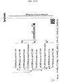
- the DNA fragment codes for an amino acid sequence specified by GenBank Accession Number BAA06251. Referring to FIG.
- a catalytic site represented by CD and a collagen binding site represented by CBD are included and the base sequence of base Nos.3001 to 3366 corresponds to a CBD.
- Clostridium histolyticum collagenase (hereinafter occasionally referred to as "ColG") specified by GenBank Accession Number BAA77453, Clostridium limosum collagenase specified by ditto BAC57532, Clostridium septicum collagenase specified by ditto BAC57535, Clostridium perfringens collagenase specified by ditto A36866, Clostridium novyi collagenase specified by ditto BAC57545, Clostridium bifermentans collagenase specified by ditto BAC57541, Clostridium sordellii collagenase specified by ditto BAC57550, Clostridium tetani collagenase specified by ditto AAO37456, Clostridium botulinum collagenase specified by di
- a "CB site" to be used in the present invention is required to bind to a collagen fiber of the bone graft substrate exposing at least a collagen fiber to the extent that the growth factor can be retained there, and therefore it is not necessary to contain the entire amino acid sequence of a collagenase-derived collagen binding site.
- the collagen-binding peptide having 90% homology with the base sequence constituting a CBD in the amino acid sequence maybe favorably used.
- There is no particular restriction on a binding method and, for example, it may be bound with an affinity for a part of collagen fibers exposing out of a surface of the collagen-exposing bone material.
- a GF site constituting a CB-GF to be used in the present invention is a site for exerting a function of a growth factor or the like by binding to a bone graft substrate.
- a growth factor include an epithelial growth factor (EGF), a fibroblast growth factor (FGF), and a platelet-derived growth factor (PDGF), and a growth factor receptor agonists exerting such actions widely may be used.
- Further growth factors such as TGF- ⁇ , IGF-1, and BMP do not exert a heterotopic bone inducing activity but exert a bone forming activity, they can promote healing of fracture when applied to a fractured part.
- a basic fibroblast growth factor As a structural gene for such a growth factor receptor agonist, especially use of a basic fibroblast growth factor is preferable.
- Examples of such a basic fibroblast growth factor include a DNA fragment composed of a base sequence of base Nos. 468 to 932 of the Homo sapiens fibroblast growth factor 2 (basic) gene (NCBI Reference Sequence Accession Number NM _002006.4) as set forth in SEQ ID NO: 2.
- a structural gene for an epithelial growth factor there is also cDNA (SEQ ID NO: 3) of preproEGF (GenBank Accession Number U04842) of Rattus norvegicus.
- the amino acid sequence of preproEGF encoded by the DNA is set forth in SEQ ID NO: 4.
- a basic fibroblast growth factor (bFGF) may be used favorably in the present invention. Since a basic fibroblast growth factor is superior in osteogenic ability, if the CB-GF bound to a basic fibroblast growth factor as a constituent growth factor (hereinafter referred to as "CB-bFGF") is bound to the bone graft substrate the uniting ability between a recipient bed bone and a grafted bone is superior.
- CB-EGF epithelial growth factor
- the CB site and the GF site are isolated by a predetermined gap width, thus each site can independently fully exert each function.
- the linker the CB-GF can be bound stronger to collagen fibers than the CB-GF without the linker.
- a linker examples include a peptide fragment which does not have a specific three-dimensional structure and is composed of amino acids, such as serine, threonine, proline, asparaginic acid, glutamic acid, and lysine. Further, as such a linker an amino acid sequence derived from the ColH may be used favorably. In the current invention a polycystic kidney disease I domain (hereinafter referred to as "PKD") of the ColH is used. Additionally, a PKD derived from another bacterial collagenase may be also used favorably as the linker. This is because the collagen binding ability of the CBD is reinforced by coexistence of the PKD.
- PKD polycystic kidney disease I domain
- FIG. 12 Such a linker derived a bacterial collagenase is depicted in FIG. 12 as PKD. Incidentally, such a linker should preferably be resistant to a peptide hydrolase or the like contained in a human circulatory liquid, the local residual performance of
- a “bone graft substrate” to be used in the present invention is the bone graft substrate exposing at least a collagen fiber.
- the bone graft substrate include a collagen-exposing bone material and a high-density collagen material.
- the collagen-exposing bone material for example, the collagen-exposing bone material such as crushed bone which is removed at least a part of an inorganic mineral component from the bone may be used favorably. It is not limited to a so-called complete decalcified bone, namely a bone from which all the contained inorganic mineral component is removed. Thereby mechanical strengths of a bone can be secured and the anatomical shape of the same can be retained. By removing a part of the inorganic mineral component, collagen fibers contained in a bone are exposed to a bone surface, and the CB-GF can be bound through the collagen-binding peptide.
- a "bone” to be used of the present invention may be any of autologous bone, allogeneic bone, and heterologous bone.
- Heterologous bone other than human may be from any of primates, such as monkey, baboon, and chimpanzee, swine, cattle, horse, goat, sheep, dog, cat, rabbit, guinea pig, mongolian gerbil, hamster, rat, and mouse.
- a "collagen-exposing bone material” contains in addition to collagen richly a growth factor, and various peptides and small proteins, maintaining the osteogenic ability. In the present invention, by using a collagen-exposing bone material a growth factor contained in the bone material can be efficiently bound, and the anatomical shape, the mechanical strengths, and the bone inducing potency of a bone can be utilized effectively.
- the collagen-exposing bone material to be used in the present invention can be prepared by immersing a bone in an acid solution to expose collagen fibers. Prior to the acid treatment a treatment for removing soft tissues, or a treatment with an organic solvent such as alcohol for removing bone marrow, blood, and lipid, may be conducted.
- a bone collected in a block form may be used after shaping into a form corresponding to a bone defect region, or crushing also. When a bone is crushed, the shape may be irregular, and the size may be not uniform.
- a treatment step for crushing a bone substrate to an appropriate particle size is not limited to before the collagen-exposing treatment, and it may be conducted simultaneously with the collagen-exposing treatment, or conducted after the collagen-exposing treatment.
- the crushing treatment can be carried out usually with a commonly used a crusher or a mixer, and in either of a wet state and a dry state of a bone substrate.
- the particle size for example, the largest diameter may be in a range of 50 to 5000 ⁇ m, preferably 50 to 1000 ⁇ m, and more preferably 50 to 2000 ⁇ m.
- a bone which is removed at least a part of an inorganic mineral component so as to expose collagen fibers out of a bone surface may be favorably used.
- Collagen fibers are required to be exposed from bone tissues to the extent that a CB-GF can bind thereto.
- a content of calcium can be used as an indicator for removal of an inorganic mineral component.
- the relative calcium content compared to the value before a collagen-exposing treatment should be reduced up to 95 to 10%, preferably 95 to 40%, more preferably 95 to 60%, and especially preferably 95 to 80%.
- a complete decalcified bone which a calcium component has been removed to the extent possible is used in general.
- an inorganic mineral component is, however, required to be removed only in the above range, the collagen-exposing treatment time can be shortened.
- Such a collagen-exposing treatment on a bone can be performed by dissolving an inorganic mineral component with hydrochloric acid, acetic acid, nitric acid, sulfuric acid, formic acid, or the like.
- concentration or treatment conditions may be appropriately selected according to an acid used.
- the temperature is from 0 to 10°C
- the time is from 30 sec to 18hours, preferably from 60 sec to 6 hours, more preferably from 60 sec to 1 hour, and especially preferably from 60 sec to 2 min.
- a collagen-exposing treatment was performed by extraction with 0.6 N hydrochloric acid for 3 to 24 hours, the target of the acid extraction was to reduce the calcium content below 5%, as described in Patent Literature 3.
- the growth factor anchoring type bone graft material of the present invention it is enough to bind the CB-GF to collagen fibers contained in crushed bone, and further to be killed viable cells to the extent that the antigene city is removed.
- collagen-exposing treatment it is found that, when a bone is crushed in the largest diameter of 50 to 5000 ⁇ m, then treated with 0.6 N hydrochloric acid within the above range, the CB-GF is efficiently bound, the mechanical strengths are kept, and viable cells are killed to reduce antigenicity even if an allogeneic bone is used.
- the collagen-exposing bone material to be used in the present invention can be used by removing an inorganic mineral component contained in the acid solution after the acid treatment. As a method for removing the inorganic mineral component, the supernatant is removed and washed with water or a phosphate buffer solution, or it may be washed with a chelating reagent.
- the collagen-exposing bone material to be used in the present invention may be prepared by using an autologous bone.
- the collagen-exposing bone material may be prepared by using a donor bone, according to the above, and preserved in a buffer solution or preserved dry.
- a high-density collagen material may be used as the bone graft substrate. Since a collagen-exposing treatment with an acid for producing a collagen-exposing bone material is not required, the growth factor anchoring type bone graft material can be prepared in a short time.
- the density of collagen fibers in the high-density collagen material is from 100 to 800 mg/cm 3 , preferably from and 300 to 800 mg/cm 3 , more preferably from 400 to 800 mg/cm 3 .
- the mechanical strengths can be superior in the range.
- the high-density collagen material may be in a sheet form, a columnar form, a spherical form, apolyhedral form, or in another irregular form. Among them the high-density collagen material in a sheet form can be used favorably for e.g. coating a bone surface.
- a collagen fiber composing the high-density collagen material there is no particular restriction on a collagen fiber composing the high-density collagen material, and it may be any of collagen types I to XI. Preferably, it is type I.
- the high-density collagen material is preferably constituted with atelocollagen which a part or all of a telopeptide is removed from a collagen.
- the high-density collagen material can be prepared by freeze-drying or otherwise drying a solution containing collagen fibers, being pressurizing to the above density and into a sheet form. A commercial product may be also used.
- both of the GF site and the CB site constituting the CB-GF to be used in the present invention are peptides, they can be prepared as a fusion protein.
- the CB-GF includes a basic fibroblast growth factor (bFGF) as a growth factor receptor agonist, and PKD-CBD derived from ColH as a linker and a CB site
- bFGF-PKD-CBD basic fibroblast growth factor
- a method for producing a bFGF-PKD-CBD is disclosed in Non Patent Literature 1, the bFGF-PKD-CBD can be produced by the method.
- a bFGF-CBD By using a basic fibroblast growth factor (bFGF) as a GF site, and a CBD derived from ColG as a CB site, a bFGF-CBD can be also produced by fusing the two.
- a gene sequence for an epithelial cell growth factor (EGF) instead of a gene sequence for a bFGF, a CB-EGF can be produced similarly as above.
- EGF epithelial cell growth factor
- a CB-GF which the growth factor receptor agonist binds to the CB can be produced.
- the CB site and the GF site may be cross-linked by a crosslinking agent.
- the growth factor anchoring type bone graft material may be produced by mixing the EGF-PKD-CBD, or other CB-GF with the above bone graft substrate. Generally, by adding predetermined amounts of the bone graft substrate and the CB-GF into a phosphate buffer solution, stirring the mixture for 60 sec to 60 min, preferably 5 to 30 min, and more preferably 15 to 30 min at a temperature of 0 to 10 °C, or leaving it standing, the CB-GF can be bound to the bone graft substrate.
- the growth factor anchoring type bone graft material of the present invention can be easily prepared and used provided that the bone graft substrate is prepared at a conventional autologous bone grafting, then the CB-GF prepared in advance is added immediately the substrate to prepare the growth factor anchoring type bone graft material.
- the bone graft substrate which is prepared by the above method in advance or preserved in a buffer solution may be used.
- a growth factor anchoring type bone graft material which is prepared by immersing a dried bone graft substrate in a buffer solution and adding the CB-GF thereto may be used as a grafting bone material.
- kit (I) composed of a CB-GF solution and the bone graft substrate
- kit (II) composed of a CB-GF solution and a collagen-exposing bone material preparation solution
- a kit (I) is composed of a CB-GF solution and the bone graft substrate.
- a bone graft substrate include a donor bone which is removed at least a part of an inorganic mineral component to expose collagen fibers and then preserved in a buffer solution, the same preserved in a dry state, and the high-density collagen material.
- the CB-GF solution in the kit (I) is a solution dissolving the CB-GF in a buffer solution in a range of 0.5 to 2.0 mg/mL.
- a buffer solution include a phosphate buffer solution of pH 7.0 to 8.0, Tris buffer solution, and a physiological saline solution. Since the bone graft substrate is included in the kit, the growth factor anchoring type bone graft material can be easily prepared by adding the CB-GF solution to the bone graft substrate before transplanting.
- a kit (II) is composed of a collagen-exposing bone material preparation solution in place of a bone graft substrate, and a CB-GF solution.
- the collagen-exposing bone material can be easily prepared by immersing an autologous bone in the collagen-exposing bone material preparation solution followed by washing.
- the growth factor anchoring type bone graft material can be prepared.
- An acid solution such as 0.6 N hydrochloric acid solution, and acetic acid, as well as an acid solution to which a chelating reagent is added, may be used as a collagen-exposing bone material preparation solution.
- a kit (II) may be used favorably for conducting an autologous bone grafting.
- the growth factor anchoring type bone graft material of the present invention is a bone graft material which the CB-GF including the GF site such as FGF, TGF- ⁇ , IGF-1, and PDGF, and the CB site is bound to the bone graft substrate.
- the osteogenic ability based on the bone graft substrate and the osteogenic effect based on the growth factor can be expected.
- crushed autologous bone as a graft bone or crushed allogeneic bone as a graft bone has been heretofore used.
- bone formation can be promoted.
- a graft bone is obtained, crushed in the range of the largest diameter 50 to 5000 ⁇ m, and stirred in 0.6 N hydrochloric acid for 1 min to perform a collagen-exposing treatment. Then the obtained collagen-exposing bone material is washed with water, rinsed with a phosphate buffer solution (pH 7.0 to 8.0), added the CB-GF thereto and mixed for approx. 1 to 30 min, thereby preparing a growth factor anchoring type autologous bone graft material.
- a phosphate buffer solution pH 7.0 to 8.0
- the growth factor anchoring type bone graft material of the present invention includes the CB-GF. Therefore excellent bone formation based on the CB-GF can be expected. On an occasion of a fracture or the like early ambulation owing to premature fusion at an affected part becomes possible, so that rehabilitation can be started early.
- an allogeneic bone grafting it is possible to prepare a growth factor anchoring type allogeneic bone graft material before the surgery. Therefore, an allogeneic bone grafting can be carried out effectively within a short operation time and with minimal invasion.
- a collagen-exposing bone material preparation solution in the kit (II) can be used for the preparation of the collagen-exposing bone material, and a CB-GF solution in the kit (II) may be used as the CB-GF.
- the Escherichia coli was precultured overnight in 50 mL of a 2xYT-G culture medium containing 50 ⁇ g/mL of ampicillin and 30 ⁇ g/mL of chloramphenicol.
- a 2xYT-G culture medium containing 50 ⁇ g/mL of ampicillin and 30 ⁇ g/mL of chloramphenicol.
- To 500 mL of the culture medium 10 mL of the obtained precultured liquid was added and shake-cultured at 37°C until the turbidity (O. D. 600) of the bacterial suspension became approx. 0.7.
- To the obtained bacterial suspension 5 mL of a 0.1 M-aqueous solution of isopropyl- ⁇ -D-thiogalactopyranoside (IPTG) was added, and cultured at 37 ° C for 2hours.
- IPTG isopropyl- ⁇ -D-thiogalactopyranoside
- phenylmethylsulfonyl fluoride (PMSF) solution containing 0.1 M isopropanol was added, and the culture solution was centrifuged at 6,000 ⁇ g, and 4°C for 10 min to collect a transformant.
- Bacterial cells were suspended in 7.5 mL of a phosphate buffered physiological saline solution (PBS) containing 1 mM PMSF, and the cells were destructed by a French press. A 20%-Triton X-100 solution equivalent to 1/19 volume of the suspension was added and stirred at 4°C for 30 min.
- PBS phosphate buffered physiological saline solution
- the lysate was centrifuged at 15,000 ⁇ g, and 4°C for 30 min to obtain a supernatant, and the resulting supernatant was then centrifuged again under the same condition.
- the supernatant was defined as a cleared lysate solution.
- glutathione-sepharose beads (2 mL)
- the cleared lysate solution was added and stirred at 4 °C for 1 hour to bind a GST-EGF-PKD -CBD fusion protein to the beads.
- the fusion protein was eluted with 50 mM Tris-HCl (pH 8.0) and 10 mM glutathione solution. Five units of thrombin per mg of the fusion protein were added and the mixture was subjected to a reaction at 25°C for 10 hours to cleave a GST tag. After that, dialysis against 300 mL of PBS at 4°C for 12 hours was repeated four times. The dialyzed cleavage product was added to a column filled with fresh glutathione-sepharose beads (2 mL) washed with PBS and directly eluted. As a result, the GST tag was removed and EGF-PKD-CBD fusion protein (SEQ ID NO: 6; 225 to 491) without the GST tag was obtained.
- a DNA fragment (PKD-CBD gene) including a base sequence of base Nos. 2719 to 3391 of the Co1H gene set forth in SEQ ID NO: 1 was inserted in an SmaI site of a pGEX-4T-2 plasmid (by GE Healthcare, Japan) in the usual manner.
- the obtained expression plasmid possesses a reading frame (SEQ ID NO: 9) coding GST-bFGF-PKD-CBD fusion protein (SEQ ID NO: 8).
- the amino acid sequence of the bFGF-PKD-CBD fusion protein is set forth in SEQ ID NO: 10
- the base sequence coding for the bFGF-PKD-CBD fusion protein is set forth in SEQ ID NO: 11.
- the N-terminal 2 amino acid residues Gly-Ser are a part of a recognition site of a GST tag cleavage enzyme (thrombin protease).
- the expression plasmid was introduced in Escherichia coli (BL21 Codon Plus RIL, by Stratagene) by an electroporation method to produce a transformant.
- the transformant was precultured overnight in 50 mL of a 2xYT-G culture medium containing 50 ⁇ g/ mL of ampicillin and 30 ⁇ g/ mL of chloramphenicol.
- Ten mL of the obtained preculture solution was added to 500 mL of the culture medium and was shake-cultured at 37°C until the turbidity (O. D. 600) of the bacterial suspension reached approx. 0.7.
- To the obtained bacterial suspension 5 mL of a 0.1 M isopropyl- ⁇ -D-thiogalacto -pyranoside (IPTG) aqueous solution was added and the mixture was cultured at 37°C for 2 hours.
- IPTG isopropyl- ⁇ -D-thiogalacto -pyranoside
- the bacterial suspension was centrifuged at 6000xg and 4°C for 10 min to collect the transformant.
- the transformant was suspended in 7.5 mL of 50mM Tris-HCl (pH 7.5), 0.5M NaCl and 1 mM PMSF, and the cells were destructed by a French press.
- 1 volume of a 20% Triton (registered trademark) X-100 was added and stirred at 4°C for 30 min.
- the obtained bacterial suspension was centrifuged at 15,000 ⁇ g and 4 °C for 30 min and the supernatant was recovered.
- the obtained supernatant was further centrifuged at 15,000 ⁇ g and 4°C for 30 min and the supernatant was recovered.
- the supernatant was defined as a clarified lysate.
- the clarified lysate was added to 2 mL of glutathione-sepharose beads and stirred at 4 °C for 1hour. After washing the beads 5 times with 12 mL of 50 mM Tris-HCl(pH 7.5) and 0.5M NaCl, the beads were suspended in small amount of 50 mM Tris-HCl(pH 7.5) and 0.5M NaCl, and filled in a column.
- the GST-bFGF-PKD-CBD fusion protein was eluted therefrom with an elution liquid (50 mM Tris-HCl (pH 8.0), 0.5M NaCl and 10 mM glutathione).
- Thrombin in an amount of 5 units per 1 mg of the fusion protein was added and allowed to react at 25 ° C for 10 hours.
- the obtained reaction solution was added to 1mL of heparin-sepharose beads and stirred at 4 °C for 3 hours allowing the bFGF-PKD-CBD fusion protein to bind to the beads.
- the beads were washed 3 times with 12 mL of 50 mM Tris-HCl (pH 7.5) with 0.5 M NaCl.
- the beads were filled in a column and the protein was eluted with 10mL of 50 mM Tris-HCl (pH 7.5) with the salt gradient of NaCl from 0.5 to 2M, to obtain the bFGF- PKD- CBD fusion protein (SEQ ID NO: 10).
- a DNA fragment including a base sequence of base Nos. 4011 to 4358 of the ColG gene set forth in SEQ ID NO: 12 was amplified by a PCR method so as to have an SmaI site at the 5'end, and an XhoI site at the 3'end. The fragment was inserted between an SmaI site and an XhoI site of a pGEX-4T-2 plasmid in the usual manner. Meanwhile, a DNA fragment (bFGF gene) consisting of a base sequence of base Nos.
- the expression plasmid possesses a reading frame coding for the GST-bFGF-CBD fusion protein (SEQ ID NO: 13).
- the amino acid sequence of the bFGF-CBD fusion protein is an amino acid sequence corresponding to base Nos. 720 to 1503 of the base sequence set forth in SEQ ID NO: 13.
- the N-terminal 2 amino acid residues Gly-Ser are a part of a recognition site of a GST tag cleavage enzyme (thrombin protease).
- the expression plasmid was introduced in Escherichia coli (BL21 Codon Plus RIL, by Stratagene) by an electroporation method to produce a transformant.
- a bFGF-CBD fusion protein was produced identically with the production example 2 , except that this transformant was used.
- a femur was obtained from a 2 months old male Wistar rat and subjected to defatting freeze-drying.
- the bone tissue was divided to epiphysis and diaphysis, and each of them was crushed to an average particle size of 50 to 300 ⁇ m.
- To 40 mg of each crushed bone 1 mL of 0.6 N hydrochloric acid was added and the mixture was stirred at a temperature of 4°C for 18 hours. Then the mixture was washed twice with a pH 7.4-phosphate buffer solution to prepare a collagen-exposing bone material of epiphysis or diaphysis.
- 1A are shown from left molecular weight marker (Marker), stock solution of the EGF-PKD-CBD fusion protein obtained in the production example 2 (con), collagen (CP) 5 mg, crushed bone (BP) 5 mg, crushed bone (BP) 10 mg, crushed bone (BP) 20 mg, crushed bone (BP) 40 mg, crushed bone (BP) 80 mg, and crushed bone (BP) 160 mg.
- Marker left molecular weight marker
- Con stock solution of the EGF-PKD-CBD fusion protein obtained in the production example 2
- collagen CP
- BP crushed bone
- BP crushed bone
- BP crushed bone
- BP crushed bone
- BP crushed bone
- BP crushed bone
- BP crushed bone
- BP crushed bone
- BP crushed bone
- BP crushed bone
- BP crushed bone
- BP crushed bone
- BP crushed bone
- BP crushed bone
- BP crushed bone
- BP crushed bone
- BP crushed bone
- BP crushed bone
- BP crushed bone
- BP crushed bone
- FIG. 1A the amount of the fusion protein in the supernatant is constant irrespective of the amount of the crushed bone
- FIG. 1B the amount of the fusion protein in the supernatant is decreased in proportion to increase in the amount of the collagen-exposing bone material. Since the EGF-PKD-CBD fusion protein not bound to the collagen-exposing bone material is present in the supernatant, it is presumed that as the amount of collagen-exposing bone material was increased, more EGF-PKD-CBD fusion protein was bound to the collagen-exposing bone material.
- the residual amount in a supernatant is decreased depending on the amount of the collagen-exposing bone material similarly to the EGF-PKD-CBD fusion protein, to indicate that the binding capability of a protein is increased by the collagen-exposing treatment.
- FIG. 1B and FIG. 2B comparing FIG. 1B and FIG. 2B with respect to the binding amount of the EGF-PKD-CBD fusion protein to the collagen-exposing bone material, the binding amounts to the collagen-exposing bone material derived from epiphysis and to the collagen-exposing bone material derived from diaphysis were nearly the same.
- the amount of BSA in the supernatant was larger for diaphysis. This means that the binding amount of albumin depends on a bone part. It is presumed that of the present invention, the EGF-PKD-CBD fusion protein could be anchored to a crushed bone irrespective of a used bone part.
- Example 2 The same procedures were carried out as in Example 1, except that the bFGF-PKD-CBD fusion protein obtained in the production example 2 was used instead of the EGF-PKD-CBD fusion protein, and the binding activities of the bFGF-PKD-CBD fusion protein to the crushed bone and the collagen-exposing bone material derived from epiphysis, and the crushed bone and the collagen-exposing bone material derived from diaphysis respectively were examined.
- the results of the binding activities of the bFGF-PKD-CBD fusion protein to the crushed bone and the collagen-exposing bone material derived from epiphysis are shown in FIG. 3A and FIG. 3B
- FIG. 4A and FIG. 4B The results of the binding activities of the bFGF-PKD-CBD fusion protein to the crushed bone and the collagen-exposing bone material derived from diaphysis.
- the amounts of the fusion protein in the supernatant were decreased with increase in the amount of the crushed bone and also of the collagen-exposing bone material.
- the dependence on the amount of bone was higher than for the crushed bone to indicate that the binding capacity of the bFGF-PKD-CBD fusion protein was improved by a collagen-exposing treatment.
- FIG. 5A The results of the transplant of the collagen-exposing bone material with the bound bFGF-PKD-CBD fusion protein are shown in FIG. 5A, and the results of the transplant of the crushed bone of epiphysis are shown in FIG. 5B.
- FIG. 5A when a growth factor anchoring type bone graft material was transplanted on the anterior femoral periosteum, after approx. 1 week from the transplant a bone tissue was observed (arrow) in the vicinity of the growth factor anchoring type bone graft material, and after approx. 2 weeks a bone tissue with certain thickness was observed in a wider range.
- the control group transplanted with a crushed bone even 2 weeks after the transplant, no bone tissue could be observed in the vicinity of the crushed bone .
- the area of a new bone tissue (callus) is shown in FIG. 6.
- the black bar is for the control group, and the white bar is for the group bound to the bFGF-PKD-CBD fusion protein.
- a growth factor anchoring type bone graft material of the present invention can form a bone tissue faster than a conventional allogeneic bone grafting.
- the diaphysis of the bone tissue was crushed to an average particle size of 50 to 300 ⁇ m.
- the crushed bone was divided to 3 groups of 40 mg each (weight before collagen-exposing treatment), and the group 1 was for a not collagen exposure-treated crushed bone (BP), and group 2 and group 3 were for a collagen exposure-treated crushed bone (DBP).
- BP not collagen exposure-treated crushed bone
- DBP collagen exposure-treated crushed bone
- FIG. 7 are shown from left molecular weight marker (Marker), stock solution (con), crushed bone (BP), crushed bone with 1 min-collagen-exposing treatment (DBP), and crushed bonewith 18 hour-collagen-exposing treatment (DBP).
- a growth factor anchoring type bone graft material was prepared by reacting 20 mg (weight before collagen-exposing treatment) of a demineralize bone material of diaphysis prepared as in Example 1, with 1.16 nmol of a bFGF, 0.29 nmol of a bFGF-PKD-CBD fusion protein, or 1.16 nmol of a bFGF-PKD-CBD fusion protein, and transplanted on the anterior periosteum of the femoral diaphysis.
- the white bar is for the control group
- the grey bar is for the 1.16 nmol bFGF group
- the black bar is for the 0.29 nmol bFGF-PKD-CBD fusion protein group
- the gradation column is for the 1.16 nmol bFGF-PKD-CBD fusion protein group.
- the "a” means significant difference to the control group
- the "b” means significant difference to the 1.16 nmol bFGF group.
- FIG. 8 shows that the new bone amount of the 1.16 nmol bFGF-PKD-CBD fusion protein group after 1 week was significantly larger than the 1.16 nmol bFGF group. After 2 weeks, the amounts of a new bone of both the 0.29 nmol bFGF-PKD-CBD fusion protein group and the 1.16 nmol bFGF-PKD-CBD fusion protein group were significantly larger than the bFGF group. It has been shown that by using the collagen-exposing bone material and thebFGF-PKD-CBD fusion proteinaccording to the present invention, bone formation can be promoted at a low dose for a long term.
- the femora of 8 rats of each group were obtained and the new bone volume was measured using a micro-CT.
- the results are shown in FIG. 9.
- the new bone amount after 2 weeks from the transplant tends to be large in the bFGF-CBD fusion protein group. It has been shown that by changing the collagen binding domain the controlled release period or the bone formation amount can be controlled according to the present invention.
- a group transplanted with a reaction product of a phosphate buffer solution (PBS) and the high-density collagen material was defined as the control.
- PBS phosphate buffer solution
- the femora of 10 rats of each group were obtained and the new bone volume was measured using a micro-CT.
- the results are shown in FIG. 10 .
- the amount of new bone after 1 week from the transplant were the same for the bFGFgroup, the bFGF-CBD (I) fusion protein, and the bFGF-PKD-CBD (II) fusion protein group, however after 2 weeks the same was significantly high for the bFGF-PKD-CBD (II) fusion protein.
- the present invention it has been shown that by using the high-density collagen material having high strengths, a graft bone substitute material that can promote bone formation for a long time period can be provided.
- a 5 mm-bone defect was prepared at the murine femur diaphysis and then a bone was grafted thereto. After bone grafting a bone graft material obtained by reacting the bFGF-PKD-CBD fusion protein prepared as in Example 7 with a sheet-formed high-density collagen material (collagen fiber density of 640 mg/cm 3 , 5 mm ⁇ 5 mm ⁇ 100 ⁇ m), was coated thereon. Meanwhile, a group coated with a reaction product of a phosphate buffer solution (PBS) and a sheet-formed high-density collagen material was defined as the control.
- PBS phosphate buffer solution
- FIG. 11 The results of temporal change of a mouse of each group are shown in FIG. 11 .
- 3 weeks from the grafting vigorous new bone formation is recognizable around the grafted bone in the group coated with a bone graft material, and further that union of the grafted bone and a recipient bed bone was recognized.
- the above has demonstrated that the bone graft material is useful as a substitute material for an allogeneic cortical bone plate requiring high mechanical strengths.
- a growth factor anchoring type bone graft material of the present invention can be produced easily, and used similarly as a conventional bone graft material. Further, since a growth factor is added, the same is superior in uniting ability of a grafted bone with a recipient bed bone, and therefore useful.
Landscapes
- Health & Medical Sciences (AREA)
- Life Sciences & Earth Sciences (AREA)
- Chemical & Material Sciences (AREA)
- Medicinal Chemistry (AREA)
- Engineering & Computer Science (AREA)
- General Health & Medical Sciences (AREA)
- Organic Chemistry (AREA)
- Gastroenterology & Hepatology (AREA)
- Zoology (AREA)
- Molecular Biology (AREA)
- Biomedical Technology (AREA)
- Genetics & Genomics (AREA)
- Epidemiology (AREA)
- Bioinformatics & Cheminformatics (AREA)
- Veterinary Medicine (AREA)
- Public Health (AREA)
- Proteomics, Peptides & Aminoacids (AREA)
- Animal Behavior & Ethology (AREA)
- Biochemistry (AREA)
- Oral & Maxillofacial Surgery (AREA)
- Dermatology (AREA)
- Wood Science & Technology (AREA)
- Transplantation (AREA)
- Biophysics (AREA)
- Immunology (AREA)
- Pharmacology & Pharmacy (AREA)
- Microbiology (AREA)
- Orthopedic Medicine & Surgery (AREA)
- Urology & Nephrology (AREA)
- Botany (AREA)
- Chemical Kinetics & Catalysis (AREA)
- Toxicology (AREA)
- General Engineering & Computer Science (AREA)
- Biotechnology (AREA)
- Materials For Medical Uses (AREA)
- Peptides Or Proteins (AREA)
Applications Claiming Priority (2)
| Application Number | Priority Date | Filing Date | Title |
|---|---|---|---|
| JP2011108650 | 2011-05-13 | ||
| PCT/JP2012/057829 WO2012157339A1 (ja) | 2011-05-13 | 2012-03-26 | 成長因子アンカーリング型骨移植材料、成長因子アンカーリング型骨移植材料の製造方法、成長因子アンカーリング型骨移植材料製造用キット、および骨形成方法 |
Publications (3)
| Publication Number | Publication Date |
|---|---|
| EP2708246A1 EP2708246A1 (en) | 2014-03-19 |
| EP2708246A4 EP2708246A4 (en) | 2014-11-19 |
| EP2708246B1 true EP2708246B1 (en) | 2017-11-01 |
Family
ID=47176680
Family Applications (1)
| Application Number | Title | Priority Date | Filing Date |
|---|---|---|---|
| EP12785014.7A Active EP2708246B1 (en) | 2011-05-13 | 2012-03-26 | Growth factor anchoring type bone graft material, method for producing growth factor anchoring type bone graft material, kit for producing growth factor anchoring type bone graft material. |
Country Status (4)
| Country | Link |
|---|---|
| US (1) | US9248164B2 (ja) |
| EP (1) | EP2708246B1 (ja) |
| JP (1) | JP5512887B2 (ja) |
| WO (1) | WO2012157339A1 (ja) |
Families Citing this family (7)
| Publication number | Priority date | Publication date | Assignee | Title |
|---|---|---|---|---|
| US8450273B2 (en) | 2007-04-09 | 2013-05-28 | The Board Of Trustees Of The University Of Arkansas | Fusion proteins of collagen-binding domain and parathyroid hormone |
| DK2790717T3 (da) | 2011-12-14 | 2018-07-30 | Univ Arkansas | Afgivelse af terapeutiske midler ved hjælp af et kollagenbindende protein |
| WO2013120060A1 (en) | 2012-02-09 | 2013-08-15 | The Board Of Trustees Of The University Of Arkansas | Delivery of therapeutic agents by a collagen binding protein |
| JP6699821B2 (ja) * | 2014-10-16 | 2020-05-27 | 学校法人北里研究所 | 神経再生用移植材料、神経再生用移植材料の製造方法、及び神経再生用移植材料製造用キット |
| CN105288752A (zh) * | 2015-10-29 | 2016-02-03 | 深圳市第二人民医院 | 磁性纳米复合生物界面材料及其制备方法 |
| WO2018148573A1 (en) * | 2017-02-10 | 2018-08-16 | The Board Of Trustees Of The University Of Arkansas | Collagen-binding agent compositions and methods of using the same |
| KR102066392B1 (ko) * | 2017-11-14 | 2020-01-16 | 한국치아은행 주식회사 | 골 이식용 복합체 제조방법 및 이에 의해 제조된 골 이식용 복합체 |
Family Cites Families (13)
| Publication number | Priority date | Publication date | Assignee | Title |
|---|---|---|---|---|
| US4950483A (en) | 1988-06-30 | 1990-08-21 | Collagen Corporation | Collagen wound healing matrices and process for their production |
| US6146420A (en) * | 1997-12-10 | 2000-11-14 | Sdgi Holdings, Inc. | Osteogenic fusion device |
| WO2001066044A2 (en) | 2000-03-03 | 2001-09-13 | Smith & Nephew, Inc. | Shaped particle and composition for bone deficiency and method of making the particle |
| JP2002058485A (ja) | 2000-08-16 | 2002-02-26 | Terumo Corp | コラーゲン結合性を有する骨形成促進融合蛋白質 |
| KR101013999B1 (ko) | 2004-03-19 | 2011-02-14 | 재단법인서울대학교산학협력재단 | 표면에 골조직 형성 증진 펩타이드가 고정된 차폐막 및임플란트 |
| US8147860B2 (en) | 2005-12-06 | 2012-04-03 | Etex Corporation | Porous calcium phosphate bone material |
| US20070248575A1 (en) | 2006-04-19 | 2007-10-25 | Jerome Connor | Bone graft composition |
| JP5552315B2 (ja) | 2006-11-03 | 2014-07-16 | バイオミメティック セラピューティクス, エルエルシー | アースロデティック術のための組成物および方法 |
| US7718616B2 (en) | 2006-12-21 | 2010-05-18 | Zimmer Orthobiologics, Inc. | Bone growth particles and osteoinductive composition thereof |
| US8450273B2 (en) * | 2007-04-09 | 2013-05-28 | The Board Of Trustees Of The University Of Arkansas | Fusion proteins of collagen-binding domain and parathyroid hormone |
| WO2009018527A1 (en) * | 2007-08-02 | 2009-02-05 | Proactive Orthopedic, Llc | Fixation and alignment device and method used in orthopaedic surgery |
| JP2010172247A (ja) * | 2009-01-29 | 2010-08-12 | Kitasato Institute | 積層型高密度培養人工組織の製造方法及び積層型高密度培養人工組織 |
| WO2011142425A1 (ja) | 2010-05-12 | 2011-11-17 | 国立大学法人香川大学 | 上皮層含有組織の再生材および再生評価方法 |
-
2012
- 2012-03-26 US US14/117,599 patent/US9248164B2/en active Active
- 2012-03-26 JP JP2013515034A patent/JP5512887B2/ja active Active
- 2012-03-26 WO PCT/JP2012/057829 patent/WO2012157339A1/ja active Application Filing
- 2012-03-26 EP EP12785014.7A patent/EP2708246B1/en active Active
Non-Patent Citations (1)
| Title |
|---|
| None * |
Also Published As
| Publication number | Publication date |
|---|---|
| EP2708246A1 (en) | 2014-03-19 |
| JP5512887B2 (ja) | 2014-06-04 |
| US9248164B2 (en) | 2016-02-02 |
| WO2012157339A1 (ja) | 2012-11-22 |
| US20140335146A1 (en) | 2014-11-13 |
| JPWO2012157339A1 (ja) | 2014-07-31 |
| EP2708246A4 (en) | 2014-11-19 |
Similar Documents
| Publication | Publication Date | Title |
|---|---|---|
| EP2708246B1 (en) | Growth factor anchoring type bone graft material, method for producing growth factor anchoring type bone graft material, kit for producing growth factor anchoring type bone graft material. | |
| US10358471B2 (en) | Fusion proteins of collagen-binding domain and parathyroid hormone | |
| US4888366A (en) | Inductive collagen-based bone repair preparations | |
| JP4121558B2 (ja) | 形態形成タンパク質および刺激因子を使用する組成物および治療方法 | |
| CN1056083C (zh) | 治疗及修复软骨缺损或损伤的方法及组合物 | |
| US10493134B2 (en) | Compositions comprising collagen and PRP for tissue regeneration | |
| JP2004041236A (ja) | 骨形成具 | |
| US20130202564A1 (en) | Systems and Methods of Cell Activated, Controlled Release Delivery of Growth Factors for Tissue Repair and Regeneration | |
| JPH09505305A (ja) | 骨形成促進生成物および骨形成方法 | |
| WO2002100426A1 (en) | Bmp binding proteins for use in bone or cartilage regeneration | |
| KR20120092632A (ko) | 경조직 재생 유도용 재료 | |
| US9981010B2 (en) | Methods and compositions for bone formation | |
| US20030235564A1 (en) | Compositions and devices comprising or encoding the Run x2 protein and method of use | |
| Sarrigiannidis et al. | Engineered dual affinity protein fragments to bind collagen and capture growth factors | |
| JP2997549B2 (ja) | 新規なタンパク質およびその製法 | |
| Ito | Engineered Bone Morphogenetic Protein for Hard Tissue Engineering |
Legal Events
| Date | Code | Title | Description |
|---|---|---|---|
| PUAI | Public reference made under article 153(3) epc to a published international application that has entered the european phase |
Free format text: ORIGINAL CODE: 0009012 |
|
| 17P | Request for examination filed |
Effective date: 20131119 |
|
| AK | Designated contracting states |
Kind code of ref document: A1 Designated state(s): AL AT BE BG CH CY CZ DE DK EE ES FI FR GB GR HR HU IE IS IT LI LT LU LV MC MK MT NL NO PL PT RO RS SE SI SK SM TR |
|
| DAX | Request for extension of the european patent (deleted) | ||
| REG | Reference to a national code |
Ref country code: DE Ref legal event code: R079 Ref document number: 602012039265 Country of ref document: DE Free format text: PREVIOUS MAIN CLASS: A61L0027000000 Ipc: A61L0027540000 |
|
| A4 | Supplementary search report drawn up and despatched |
Effective date: 20141020 |
|
| RIC1 | Information provided on ipc code assigned before grant |
Ipc: A61L 27/54 20060101AFI20141014BHEP Ipc: C12N 15/62 20060101ALI20141014BHEP |
|
| 17Q | First examination report despatched |
Effective date: 20160406 |
|
| GRAP | Despatch of communication of intention to grant a patent |
Free format text: ORIGINAL CODE: EPIDOSNIGR1 |
|
| INTG | Intention to grant announced |
Effective date: 20170630 |
|
| GRAS | Grant fee paid |
Free format text: ORIGINAL CODE: EPIDOSNIGR3 |
|
| GRAA | (expected) grant |
Free format text: ORIGINAL CODE: 0009210 |
|
| AK | Designated contracting states |
Kind code of ref document: B1 Designated state(s): AL AT BE BG CH CY CZ DE DK EE ES FI FR GB GR HR HU IE IS IT LI LT LU LV MC MK MT NL NO PL PT RO RS SE SI SK SM TR |
|
| REG | Reference to a national code |
Ref country code: GB Ref legal event code: FG4D |
|
| REG | Reference to a national code |
Ref country code: CH Ref legal event code: EP Ref country code: AT Ref legal event code: REF Ref document number: 941401 Country of ref document: AT Kind code of ref document: T Effective date: 20171115 |
|
| REG | Reference to a national code |
Ref country code: IE Ref legal event code: FG4D |
|
| REG | Reference to a national code |
Ref country code: DE Ref legal event code: R096 Ref document number: 602012039265 Country of ref document: DE |
|
| REG | Reference to a national code |
Ref country code: NL Ref legal event code: MP Effective date: 20171101 |
|
| REG | Reference to a national code |
Ref country code: LT Ref legal event code: MG4D Ref country code: FR Ref legal event code: PLFP Year of fee payment: 7 |
|
| REG | Reference to a national code |
Ref country code: AT Ref legal event code: MK05 Ref document number: 941401 Country of ref document: AT Kind code of ref document: T Effective date: 20171101 |
|
| PG25 | Lapsed in a contracting state [announced via postgrant information from national office to epo] |
Ref country code: ES Free format text: LAPSE BECAUSE OF FAILURE TO SUBMIT A TRANSLATION OF THE DESCRIPTION OR TO PAY THE FEE WITHIN THE PRESCRIBED TIME-LIMIT Effective date: 20171101 Ref country code: SE Free format text: LAPSE BECAUSE OF FAILURE TO SUBMIT A TRANSLATION OF THE DESCRIPTION OR TO PAY THE FEE WITHIN THE PRESCRIBED TIME-LIMIT Effective date: 20171101 Ref country code: LT Free format text: LAPSE BECAUSE OF FAILURE TO SUBMIT A TRANSLATION OF THE DESCRIPTION OR TO PAY THE FEE WITHIN THE PRESCRIBED TIME-LIMIT Effective date: 20171101 Ref country code: NL Free format text: LAPSE BECAUSE OF FAILURE TO SUBMIT A TRANSLATION OF THE DESCRIPTION OR TO PAY THE FEE WITHIN THE PRESCRIBED TIME-LIMIT Effective date: 20171101 Ref country code: FI Free format text: LAPSE BECAUSE OF FAILURE TO SUBMIT A TRANSLATION OF THE DESCRIPTION OR TO PAY THE FEE WITHIN THE PRESCRIBED TIME-LIMIT Effective date: 20171101 Ref country code: NO Free format text: LAPSE BECAUSE OF FAILURE TO SUBMIT A TRANSLATION OF THE DESCRIPTION OR TO PAY THE FEE WITHIN THE PRESCRIBED TIME-LIMIT Effective date: 20180201 |
|
| PG25 | Lapsed in a contracting state [announced via postgrant information from national office to epo] |
Ref country code: GR Free format text: LAPSE BECAUSE OF FAILURE TO SUBMIT A TRANSLATION OF THE DESCRIPTION OR TO PAY THE FEE WITHIN THE PRESCRIBED TIME-LIMIT Effective date: 20180202 Ref country code: LV Free format text: LAPSE BECAUSE OF FAILURE TO SUBMIT A TRANSLATION OF THE DESCRIPTION OR TO PAY THE FEE WITHIN THE PRESCRIBED TIME-LIMIT Effective date: 20171101 Ref country code: HR Free format text: LAPSE BECAUSE OF FAILURE TO SUBMIT A TRANSLATION OF THE DESCRIPTION OR TO PAY THE FEE WITHIN THE PRESCRIBED TIME-LIMIT Effective date: 20171101 Ref country code: AT Free format text: LAPSE BECAUSE OF FAILURE TO SUBMIT A TRANSLATION OF THE DESCRIPTION OR TO PAY THE FEE WITHIN THE PRESCRIBED TIME-LIMIT Effective date: 20171101 Ref country code: IS Free format text: LAPSE BECAUSE OF FAILURE TO SUBMIT A TRANSLATION OF THE DESCRIPTION OR TO PAY THE FEE WITHIN THE PRESCRIBED TIME-LIMIT Effective date: 20180301 Ref country code: BG Free format text: LAPSE BECAUSE OF FAILURE TO SUBMIT A TRANSLATION OF THE DESCRIPTION OR TO PAY THE FEE WITHIN THE PRESCRIBED TIME-LIMIT Effective date: 20180201 Ref country code: RS Free format text: LAPSE BECAUSE OF FAILURE TO SUBMIT A TRANSLATION OF THE DESCRIPTION OR TO PAY THE FEE WITHIN THE PRESCRIBED TIME-LIMIT Effective date: 20171101 |
|
| PG25 | Lapsed in a contracting state [announced via postgrant information from national office to epo] |
Ref country code: DK Free format text: LAPSE BECAUSE OF FAILURE TO SUBMIT A TRANSLATION OF THE DESCRIPTION OR TO PAY THE FEE WITHIN THE PRESCRIBED TIME-LIMIT Effective date: 20171101 Ref country code: CY Free format text: LAPSE BECAUSE OF FAILURE TO SUBMIT A TRANSLATION OF THE DESCRIPTION OR TO PAY THE FEE WITHIN THE PRESCRIBED TIME-LIMIT Effective date: 20171101 Ref country code: EE Free format text: LAPSE BECAUSE OF FAILURE TO SUBMIT A TRANSLATION OF THE DESCRIPTION OR TO PAY THE FEE WITHIN THE PRESCRIBED TIME-LIMIT Effective date: 20171101 Ref country code: CZ Free format text: LAPSE BECAUSE OF FAILURE TO SUBMIT A TRANSLATION OF THE DESCRIPTION OR TO PAY THE FEE WITHIN THE PRESCRIBED TIME-LIMIT Effective date: 20171101 Ref country code: SK Free format text: LAPSE BECAUSE OF FAILURE TO SUBMIT A TRANSLATION OF THE DESCRIPTION OR TO PAY THE FEE WITHIN THE PRESCRIBED TIME-LIMIT Effective date: 20171101 |
|
| REG | Reference to a national code |
Ref country code: DE Ref legal event code: R097 Ref document number: 602012039265 Country of ref document: DE |
|
| PG25 | Lapsed in a contracting state [announced via postgrant information from national office to epo] |
Ref country code: PL Free format text: LAPSE BECAUSE OF FAILURE TO SUBMIT A TRANSLATION OF THE DESCRIPTION OR TO PAY THE FEE WITHIN THE PRESCRIBED TIME-LIMIT Effective date: 20171101 Ref country code: RO Free format text: LAPSE BECAUSE OF FAILURE TO SUBMIT A TRANSLATION OF THE DESCRIPTION OR TO PAY THE FEE WITHIN THE PRESCRIBED TIME-LIMIT Effective date: 20171101 Ref country code: IT Free format text: LAPSE BECAUSE OF FAILURE TO SUBMIT A TRANSLATION OF THE DESCRIPTION OR TO PAY THE FEE WITHIN THE PRESCRIBED TIME-LIMIT Effective date: 20171101 Ref country code: SM Free format text: LAPSE BECAUSE OF FAILURE TO SUBMIT A TRANSLATION OF THE DESCRIPTION OR TO PAY THE FEE WITHIN THE PRESCRIBED TIME-LIMIT Effective date: 20171101 |
|
| PLBE | No opposition filed within time limit |
Free format text: ORIGINAL CODE: 0009261 |
|
| STAA | Information on the status of an ep patent application or granted ep patent |
Free format text: STATUS: NO OPPOSITION FILED WITHIN TIME LIMIT |
|
| 26N | No opposition filed |
Effective date: 20180802 |
|
| REG | Reference to a national code |
Ref country code: CH Ref legal event code: PL |
|
| PG25 | Lapsed in a contracting state [announced via postgrant information from national office to epo] |
Ref country code: SI Free format text: LAPSE BECAUSE OF FAILURE TO SUBMIT A TRANSLATION OF THE DESCRIPTION OR TO PAY THE FEE WITHIN THE PRESCRIBED TIME-LIMIT Effective date: 20171101 Ref country code: MC Free format text: LAPSE BECAUSE OF FAILURE TO SUBMIT A TRANSLATION OF THE DESCRIPTION OR TO PAY THE FEE WITHIN THE PRESCRIBED TIME-LIMIT Effective date: 20171101 |
|
| REG | Reference to a national code |
Ref country code: BE Ref legal event code: MM Effective date: 20180331 |
|
| REG | Reference to a national code |
Ref country code: IE Ref legal event code: MM4A |
|
| PG25 | Lapsed in a contracting state [announced via postgrant information from national office to epo] |
Ref country code: LU Free format text: LAPSE BECAUSE OF NON-PAYMENT OF DUE FEES Effective date: 20180326 |
|
| PG25 | Lapsed in a contracting state [announced via postgrant information from national office to epo] |
Ref country code: IE Free format text: LAPSE BECAUSE OF NON-PAYMENT OF DUE FEES Effective date: 20180326 |
|
| PG25 | Lapsed in a contracting state [announced via postgrant information from national office to epo] |
Ref country code: CH Free format text: LAPSE BECAUSE OF NON-PAYMENT OF DUE FEES Effective date: 20180331 Ref country code: BE Free format text: LAPSE BECAUSE OF NON-PAYMENT OF DUE FEES Effective date: 20180331 Ref country code: LI Free format text: LAPSE BECAUSE OF NON-PAYMENT OF DUE FEES Effective date: 20180331 |
|
| PG25 | Lapsed in a contracting state [announced via postgrant information from national office to epo] |
Ref country code: MT Free format text: LAPSE BECAUSE OF NON-PAYMENT OF DUE FEES Effective date: 20180326 |
|
| PG25 | Lapsed in a contracting state [announced via postgrant information from national office to epo] |
Ref country code: TR Free format text: LAPSE BECAUSE OF FAILURE TO SUBMIT A TRANSLATION OF THE DESCRIPTION OR TO PAY THE FEE WITHIN THE PRESCRIBED TIME-LIMIT Effective date: 20171101 |
|
| PG25 | Lapsed in a contracting state [announced via postgrant information from national office to epo] |
Ref country code: PT Free format text: LAPSE BECAUSE OF FAILURE TO SUBMIT A TRANSLATION OF THE DESCRIPTION OR TO PAY THE FEE WITHIN THE PRESCRIBED TIME-LIMIT Effective date: 20171101 Ref country code: HU Free format text: LAPSE BECAUSE OF FAILURE TO SUBMIT A TRANSLATION OF THE DESCRIPTION OR TO PAY THE FEE WITHIN THE PRESCRIBED TIME-LIMIT; INVALID AB INITIO Effective date: 20120326 |
|
| PG25 | Lapsed in a contracting state [announced via postgrant information from national office to epo] |
Ref country code: MK Free format text: LAPSE BECAUSE OF NON-PAYMENT OF DUE FEES Effective date: 20171101 |
|
| PG25 | Lapsed in a contracting state [announced via postgrant information from national office to epo] |
Ref country code: AL Free format text: LAPSE BECAUSE OF FAILURE TO SUBMIT A TRANSLATION OF THE DESCRIPTION OR TO PAY THE FEE WITHIN THE PRESCRIBED TIME-LIMIT Effective date: 20171101 |
|
| PGFP | Annual fee paid to national office [announced via postgrant information from national office to epo] |
Ref country code: FR Payment date: 20230320 Year of fee payment: 12 |
|
| REG | Reference to a national code |
Ref country code: DE Ref legal event code: R082 Ref document number: 602012039265 Country of ref document: DE Representative=s name: PUSCHMANN BORCHERT KAISER KLETTNER PATENTANWAE, DE |
|
| PGFP | Annual fee paid to national office [announced via postgrant information from national office to epo] |
Ref country code: DE Payment date: 20240327 Year of fee payment: 13 Ref country code: GB Payment date: 20240322 Year of fee payment: 13 |