EP2686661B1 - Method, composition and kit for visualization and characterization of the nervous system - Google Patents
Method, composition and kit for visualization and characterization of the nervous system Download PDFInfo
- Publication number
- EP2686661B1 EP2686661B1 EP12714976.3A EP12714976A EP2686661B1 EP 2686661 B1 EP2686661 B1 EP 2686661B1 EP 12714976 A EP12714976 A EP 12714976A EP 2686661 B1 EP2686661 B1 EP 2686661B1
- Authority
- EP
- European Patent Office
- Prior art keywords
- fixation
- paraformaldehyde
- impregnation
- visualization
- golgi
- Prior art date
- Legal status (The legal status is an assumption and is not a legal conclusion. Google has not performed a legal analysis and makes no representation as to the accuracy of the status listed.)
- Active
Links
- 238000000034 method Methods 0.000 title claims description 86
- 238000012800 visualization Methods 0.000 title claims description 28
- 239000000203 mixture Substances 0.000 title claims description 21
- 238000012512 characterization method Methods 0.000 title claims description 15
- 210000000653 nervous system Anatomy 0.000 title claims description 13
- 238000005470 impregnation Methods 0.000 claims description 68
- 229910052751 metal Inorganic materials 0.000 claims description 37
- 239000002184 metal Substances 0.000 claims description 37
- 229930040373 Paraformaldehyde Natural products 0.000 claims description 36
- 229920002866 paraformaldehyde Polymers 0.000 claims description 36
- 239000000243 solution Substances 0.000 claims description 29
- 238000010186 staining Methods 0.000 claims description 15
- 239000003153 chemical reaction reagent Substances 0.000 claims description 12
- KMUONIBRACKNSN-UHFFFAOYSA-N potassium dichromate Substances [K+].[K+].[O-][Cr](=O)(=O)O[Cr]([O-])(=O)=O KMUONIBRACKNSN-UHFFFAOYSA-N 0.000 claims description 12
- 239000007864 aqueous solution Substances 0.000 claims description 10
- 229960002523 mercuric chloride Drugs 0.000 claims description 6
- 238000003365 immunocytochemistry Methods 0.000 claims description 5
- 238000012760 immunocytochemical staining Methods 0.000 claims 1
- 210000001519 tissue Anatomy 0.000 description 36
- 210000002569 neuron Anatomy 0.000 description 29
- 238000010166 immunofluorescence Methods 0.000 description 24
- 108091000117 Tyrosine 3-Monooxygenase Proteins 0.000 description 23
- 102000048218 Tyrosine 3-monooxygenases Human genes 0.000 description 23
- 238000003364 immunohistochemistry Methods 0.000 description 18
- 239000000834 fixative Substances 0.000 description 14
- 230000010412 perfusion Effects 0.000 description 12
- 102000004169 proteins and genes Human genes 0.000 description 12
- 108090000623 proteins and genes Proteins 0.000 description 12
- XLYOFNOQVPJJNP-UHFFFAOYSA-N water Chemical compound O XLYOFNOQVPJJNP-UHFFFAOYSA-N 0.000 description 11
- 210000004556 brain Anatomy 0.000 description 10
- 238000001218 confocal laser scanning microscopy Methods 0.000 description 10
- 238000004458 analytical method Methods 0.000 description 9
- 210000001259 mesencephalon Anatomy 0.000 description 9
- WSFSSNUMVMOOMR-UHFFFAOYSA-N Formaldehyde Chemical compound O=C WSFSSNUMVMOOMR-UHFFFAOYSA-N 0.000 description 8
- 239000000427 antigen Substances 0.000 description 6
- 102000036639 antigens Human genes 0.000 description 6
- 108091007433 antigens Proteins 0.000 description 6
- 210000001787 dendrite Anatomy 0.000 description 6
- 239000000835 fiber Substances 0.000 description 6
- 238000007654 immersion Methods 0.000 description 6
- 238000012744 immunostaining Methods 0.000 description 6
- LWJROJCJINYWOX-UHFFFAOYSA-L mercury dichloride Chemical compound Cl[Hg]Cl LWJROJCJINYWOX-UHFFFAOYSA-L 0.000 description 6
- 230000001537 neural effect Effects 0.000 description 6
- 238000009877 rendering Methods 0.000 description 6
- 241001465754 Metazoa Species 0.000 description 5
- 241000699670 Mus sp. Species 0.000 description 5
- 210000003169 central nervous system Anatomy 0.000 description 5
- 210000001577 neostriatum Anatomy 0.000 description 5
- 230000035515 penetration Effects 0.000 description 5
- 238000011282 treatment Methods 0.000 description 5
- ZAINTDRBUHCDPZ-UHFFFAOYSA-M Alexa Fluor 546 Chemical compound [H+].[Na+].CC1CC(C)(C)NC(C(=C2OC3=C(C4=NC(C)(C)CC(C)C4=CC3=3)S([O-])(=O)=O)S([O-])(=O)=O)=C1C=C2C=3C(C(=C(Cl)C=1Cl)C(O)=O)=C(Cl)C=1SCC(=O)NCCCCCC(=O)ON1C(=O)CCC1=O ZAINTDRBUHCDPZ-UHFFFAOYSA-M 0.000 description 4
- 241000283707 Capra Species 0.000 description 4
- 241000283973 Oryctolagus cuniculus Species 0.000 description 4
- 241000700159 Rattus Species 0.000 description 4
- 230000001413 cellular effect Effects 0.000 description 4
- 210000003520 dendritic spine Anatomy 0.000 description 4
- 239000012153 distilled water Substances 0.000 description 4
- VYFYYTLLBUKUHU-UHFFFAOYSA-N dopamine Chemical compound NCCC1=CC=C(O)C(O)=C1 VYFYYTLLBUKUHU-UHFFFAOYSA-N 0.000 description 4
- 238000000386 microscopy Methods 0.000 description 4
- 238000002360 preparation method Methods 0.000 description 4
- SQGYOTSLMSWVJD-UHFFFAOYSA-N silver(1+) nitrate Chemical compound [Ag+].[O-]N(=O)=O SQGYOTSLMSWVJD-UHFFFAOYSA-N 0.000 description 4
- 238000005406 washing Methods 0.000 description 4
- QLOKJRIVRGCVIM-UHFFFAOYSA-N 1-[(4-methylsulfanylphenyl)methyl]piperazine Chemical compound C1=CC(SC)=CC=C1CN1CCNCC1 QLOKJRIVRGCVIM-UHFFFAOYSA-N 0.000 description 3
- FAPWRFPIFSIZLT-UHFFFAOYSA-M Sodium chloride Chemical compound [Na+].[Cl-] FAPWRFPIFSIZLT-UHFFFAOYSA-M 0.000 description 3
- 229930006000 Sucrose Natural products 0.000 description 3
- CZMRCDWAGMRECN-UGDNZRGBSA-N Sucrose Chemical compound O[C@H]1[C@H](O)[C@@H](CO)O[C@@]1(CO)O[C@@H]1[C@H](O)[C@@H](O)[C@H](O)[C@@H](CO)O1 CZMRCDWAGMRECN-UGDNZRGBSA-N 0.000 description 3
- 238000010171 animal model Methods 0.000 description 3
- 210000003050 axon Anatomy 0.000 description 3
- 210000004027 cell Anatomy 0.000 description 3
- 230000002490 cerebral effect Effects 0.000 description 3
- 230000008045 co-localization Effects 0.000 description 3
- 230000000959 cryoprotective effect Effects 0.000 description 3
- 238000005520 cutting process Methods 0.000 description 3
- 235000013681 dietary sucrose Nutrition 0.000 description 3
- 238000009826 distribution Methods 0.000 description 3
- 230000000694 effects Effects 0.000 description 3
- 238000011532 immunohistochemical staining Methods 0.000 description 3
- 230000000877 morphologic effect Effects 0.000 description 3
- 230000003287 optical effect Effects 0.000 description 3
- 238000000399 optical microscopy Methods 0.000 description 3
- 230000001242 postsynaptic effect Effects 0.000 description 3
- 230000003518 presynaptic effect Effects 0.000 description 3
- 210000003523 substantia nigra Anatomy 0.000 description 3
- 229960004793 sucrose Drugs 0.000 description 3
- QGZKDVFQNNGYKY-UHFFFAOYSA-N Ammonia Chemical compound N QGZKDVFQNNGYKY-UHFFFAOYSA-N 0.000 description 2
- VHUUQVKOLVNVRT-UHFFFAOYSA-N Ammonium hydroxide Chemical compound [NH4+].[OH-] VHUUQVKOLVNVRT-UHFFFAOYSA-N 0.000 description 2
- 108091003079 Bovine Serum Albumin Proteins 0.000 description 2
- 241001573498 Compacta Species 0.000 description 2
- 102000001435 Synapsin Human genes 0.000 description 2
- 108050009621 Synapsin Proteins 0.000 description 2
- 239000007980 Sørensen’s phosphate buffer Substances 0.000 description 2
- 210000004227 basal ganglia Anatomy 0.000 description 2
- 229940098773 bovine serum albumin Drugs 0.000 description 2
- 229960003638 dopamine Drugs 0.000 description 2
- 238000012137 double-staining Methods 0.000 description 2
- 210000005153 frontal cortex Anatomy 0.000 description 2
- 230000003993 interaction Effects 0.000 description 2
- 239000002923 metal particle Substances 0.000 description 2
- MYWUZJCMWCOHBA-VIFPVBQESA-N methamphetamine Chemical compound CN[C@@H](C)CC1=CC=CC=C1 MYWUZJCMWCOHBA-VIFPVBQESA-N 0.000 description 2
- 230000001722 neurochemical effect Effects 0.000 description 2
- 230000037361 pathway Effects 0.000 description 2
- 210000003538 post-synaptic density Anatomy 0.000 description 2
- 108010092804 postsynaptic density proteins Proteins 0.000 description 2
- 238000011160 research Methods 0.000 description 2
- 229910001961 silver nitrate Inorganic materials 0.000 description 2
- 238000012453 sprague-dawley rat model Methods 0.000 description 2
- NLXLAEXVIDQMFP-UHFFFAOYSA-N Ammonium chloride Substances [NH4+].[Cl-] NLXLAEXVIDQMFP-UHFFFAOYSA-N 0.000 description 1
- 206010002091 Anaesthesia Diseases 0.000 description 1
- 241000282693 Cercopithecidae Species 0.000 description 1
- 102000004190 Enzymes Human genes 0.000 description 1
- 108090000790 Enzymes Proteins 0.000 description 1
- JOYRKODLDBILNP-UHFFFAOYSA-N Ethyl urethane Chemical compound CCOC(N)=O JOYRKODLDBILNP-UHFFFAOYSA-N 0.000 description 1
- 241000282326 Felis catus Species 0.000 description 1
- 102000008109 Mixed Function Oxygenases Human genes 0.000 description 1
- 108010074633 Mixed Function Oxygenases Proteins 0.000 description 1
- 230000009471 action Effects 0.000 description 1
- VREFGVBLTWBCJP-UHFFFAOYSA-N alprazolam Chemical compound C12=CC(Cl)=CC=C2N2C(C)=NN=C2CN=C1C1=CC=CC=C1 VREFGVBLTWBCJP-UHFFFAOYSA-N 0.000 description 1
- 229910021529 ammonia Inorganic materials 0.000 description 1
- 235000011114 ammonium hydroxide Nutrition 0.000 description 1
- 230000003321 amplification Effects 0.000 description 1
- 230000037005 anaesthesia Effects 0.000 description 1
- 230000000890 antigenic effect Effects 0.000 description 1
- 238000004061 bleaching Methods 0.000 description 1
- 210000003850 cellular structure Anatomy 0.000 description 1
- RNFNDJAIBTYOQL-UHFFFAOYSA-N chloral hydrate Chemical compound OC(O)C(Cl)(Cl)Cl RNFNDJAIBTYOQL-UHFFFAOYSA-N 0.000 description 1
- 229960002327 chloral hydrate Drugs 0.000 description 1
- 238000004624 confocal microscopy Methods 0.000 description 1
- 238000004132 cross linking Methods 0.000 description 1
- 238000004925 denaturation Methods 0.000 description 1
- 230000036425 denaturation Effects 0.000 description 1
- 238000009792 diffusion process Methods 0.000 description 1
- 230000003291 dopaminomimetic effect Effects 0.000 description 1
- 238000002474 experimental method Methods 0.000 description 1
- 238000000799 fluorescence microscopy Methods 0.000 description 1
- 230000002518 glial effect Effects 0.000 description 1
- 230000000984 immunochemical effect Effects 0.000 description 1
- 230000002055 immunohistochemical effect Effects 0.000 description 1
- 238000011534 incubation Methods 0.000 description 1
- 238000002372 labelling Methods 0.000 description 1
- 230000004807 localization Effects 0.000 description 1
- 238000012423 maintenance Methods 0.000 description 1
- 238000004519 manufacturing process Methods 0.000 description 1
- 238000013507 mapping Methods 0.000 description 1
- 239000000463 material Substances 0.000 description 1
- 239000012528 membrane Substances 0.000 description 1
- 239000003068 molecular probe Substances 0.000 description 1
- 210000004179 neuropil Anatomy 0.000 description 1
- 238000003199 nucleic acid amplification method Methods 0.000 description 1
- 102000013415 peroxidase activity proteins Human genes 0.000 description 1
- 108040007629 peroxidase activity proteins Proteins 0.000 description 1
- 230000001376 precipitating effect Effects 0.000 description 1
- 210000000063 presynaptic terminal Anatomy 0.000 description 1
- 230000008569 process Effects 0.000 description 1
- 238000004451 qualitative analysis Methods 0.000 description 1
- 230000007441 retrograde transport Effects 0.000 description 1
- 239000012266 salt solution Substances 0.000 description 1
- 238000005070 sampling Methods 0.000 description 1
- 210000002966 serum Anatomy 0.000 description 1
- 210000003625 skull Anatomy 0.000 description 1
- 238000003756 stirring Methods 0.000 description 1
- 210000000225 synapse Anatomy 0.000 description 1
- 230000000946 synaptic effect Effects 0.000 description 1
- 238000012360 testing method Methods 0.000 description 1
- 210000004515 ventral tegmental area Anatomy 0.000 description 1
- 210000000857 visual cortex Anatomy 0.000 description 1
Images


Classifications
-
- G—PHYSICS
- G01—MEASURING; TESTING
- G01N—INVESTIGATING OR ANALYSING MATERIALS BY DETERMINING THEIR CHEMICAL OR PHYSICAL PROPERTIES
- G01N1/00—Sampling; Preparing specimens for investigation
- G01N1/28—Preparing specimens for investigation including physical details of (bio-)chemical methods covered elsewhere, e.g. G01N33/50, C12Q
- G01N1/30—Staining; Impregnating ; Fixation; Dehydration; Multistep processes for preparing samples of tissue, cell or nucleic acid material and the like for analysis
-
- G—PHYSICS
- G01—MEASURING; TESTING
- G01N—INVESTIGATING OR ANALYSING MATERIALS BY DETERMINING THEIR CHEMICAL OR PHYSICAL PROPERTIES
- G01N33/00—Investigating or analysing materials by specific methods not covered by groups G01N1/00 - G01N31/00
- G01N33/48—Biological material, e.g. blood, urine; Haemocytometers
- G01N33/50—Chemical analysis of biological material, e.g. blood, urine; Testing involving biospecific ligand binding methods; Immunological testing
- G01N33/53—Immunoassay; Biospecific binding assay; Materials therefor
- G01N33/569—Immunoassay; Biospecific binding assay; Materials therefor for microorganisms, e.g. protozoa, bacteria, viruses
- G01N33/56966—Animal cells
Definitions
- the invention is related to a method of visualization and characterization of the nervous system and, more specifically, of the central nervous system, by staining the same histological section for metal impregnation and immunocytochemistry.
- the doubly stained histological sections are subsequently subjected to a qualitative and quantitative morphological and antigenic and/or neurochemical analysis, by means of conventional optical microscopy or confocal laser scanning microscopy.
- the histological method of metal impregnation by applying potassium dichromate and silver nitrate developed by Camillo Golgi in 1873 continues, with all its subsequent variations, to still represent a method of reference today, and one essential for the histological analysis of the nervous tissue and, more specifically, of the central nervous system.
- the most used metal impregnation is the one proposed by Cox in 1891, known as mercuric impregnation or Golgi-Cox's method or black reaction, which foresees replacement of silver nitrate with mercuric chloride.
- This technique is widely used for the qualitative analysis of neuron morphology and in determining the quantity of dendritic spines, and the length and complexity of the dendrite branching.
- the international application WO9600234 A1 discloses a fixative comprising both, a precipitating fixative including Golgi-type of fixatives and cross-linking fixatives such as formaldehyde.
- a precipitating fixative including Golgi-type of fixatives and cross-linking fixatives such as formaldehyde.
- Another successful technique applied to the visualization and characterization of tissues is immunohistochemistry, a technique which exploits the ability of antibodies labeled with suitable markers, preferably fluorescent, to bind specific antigens with high affinity. This technique is widely used to localize antigens on both a cellular and sub-cellular level, and it is widely applied on the central nervous system as well.
- confocal laser scanning microscopy (CLSM) using reflection mode, the latter a technique suitable for the modeling and reconstruction in 3D of cellular structures, especially neurons.
- CLSM confocal laser scanning microscopy
- the very extensive use made recently of confocal laser scanning microscopy applied to these techniques is due to the possibility of obtaining top-quality images from specimens prepared for examination under conventional or fluorescent light.
- Confocal reflection microscopy in fact, allows, in the case of the mercuric impregnation technique, to visualize the tissues impregnated with the metal particles in an advantageous manner, because the metal particles reflect the laser light and do not display a photo-bleaching phenomenon.
- this analytical method for tissues allows to control field depth, to reduce background noise, and to collect serial optical sections from the specimen. Furthermore, confocal laser scanning microscopy allows to survey fine details which can represent substantial information and which would be lost with conventional microscopy.
- a purpose of this invention is to develop a method of visualization and characterization of nervous system tissues and, more specifically, of the central nervous system, by staining the same histological section for metal impregnation and immunocytochemistry.
- the purpose of this invention is to develop a method simple to perform and highly reproducible, which allows scientists to simultaneously obtain an amplification of the effects of the two methods for visualization of the morpho-structural conformations of nervous tissues and a synergism between them, allowing them to analyze the same histological sections by means of conventional optical microscopy or confocal laser scanning microscopy.
- the method described hereinafter is, therefore, based on combining the two histological staining techniques in the same tissue specimens to be analyzed, that is, metal impregnation and immunohistochemistry, associated to the analysis of the specimens thus prepared with conventional optical microscopy or, preferably, with confocal laser scanning microscopy.
- the object of this invention is a method of visualization and characterization of the nervous system by combining staining for metal impregnation and immunocytochemistry, comprising at least the steps of:
- the method according to the invention finds application in the experimental and clinical field for the study of the characteristics of neuronal connections in the central nervous system.
- the inventors have developed a method which foresees fixation of the tissues ex vivo using aqueous solutions of paraformaldehyde in suitable concentrations, optionally pre-fixed with the same solution comprising paraformaldehyde.
- the second that is, the ex vivo fixation
- the critical one for the immunohistochemical staining because for the staining of the tissues to be adequate, the antibodies have to penetrate in the entire depth of the tissues and to bind the specific antigens, in spite of the protein's fixation.
- the conditions for carrying out of this fixation are therefore essential to obtain an effective immunochemical staining.
- the fixing step is therefore such to allow both fixation of the proteins and an efficient penetration of the antibodies for immunohistochemistry.
- the fixation of nervous system specimens foresees a fixation of a tissue specimen to be examined with an aqueous solution comprising paraformaldehyde in a concentration between 1 and 2% w/v or a paraformaldehyde 4% w/v in Sorensen's Phosphate Buffer (PBS 0.4M, pH 7.4).
- This method can, as a matter of fact, be carried out according to two different protocols, or rather, two different embodiments of the same method, which in any case foresee the fixation of specimens to be examined by immersion thereof at a temperature of 4°C for at least 8 hours to up to 12h in a paraformaldehyde solution 4% in PBS (0.4M, pH 7.4) followed by metal impregnation or in a composition comprising both the fixative, that is, the paraformaldehyde in concentration between 1 and 2% w/v, and the reagents for the metal impregnation, preferably according to the known Golgi-Cox method.
- the fixation is carried out with an aqueous solution comprising paraformaldehyde between 1 and 2% w/v and the reagents for metal impregnation
- the metal impregnation step is already started in the fixation step, and the subsequent impregnation step is carried out to renew the reagents and maximize impregnation.
- the fixative properties of the salt solution are exploited for the preferred Golgi-Cox mercuric impregnation, fixative properties which are moreover the basis for the metal impregnation itself, combined with the fixative properties of the paraformaldehyde.
- the composition that can be used for the combined fixation and metal impregnation step consists in an aqueous solution having pH 7.4 comprising:
- the step of fixation with paraformaldehyde 4% or with the composition comprising paraformaldehyde between 1 and 2% and the reagents for metal impregnation can be preceded by a pre-fixation by perfusion with the same solution comprising paraformaldehyde of the subsequent step of fixation ex vivo.
- This perfusion and pre-fixation step is not, however, essential to achieve the purpose of the invention and can therefore be omitted, thus allowing the application of the method also in the clinical field.
- the immunohistochemistry instead, is carried out on sections of impregnated tissue specimens and then, after metal, preferably mercuric, impregnation, the method comprises the further step of cutting the specimens in slices 50-100 ⁇ m thick with a cryostat or a vibratome. The sections obtained are then subjected to the following treatments before the immuno-staining in free floating with the suitable reagents:
- the tissue is cut in slices with a cryostat, it must be treated before the slicing with a cryo-protective saccharose aqueous solution 30% by immersion for two or three days. This step can be skipped if a vibratome is used.
- the temperature of the specimen is preferably of -4°C and the temperature of the blade -14°C. In any case, the slices are collected in pits that contain H 2 O.
- the method according to the invention can comprise further steps, for example, a wash to remove the traces of fixatives used for the mercuric impregnation and for immunohistochemistry.
- the method according to the invention comprises the steps of:
- the embodiment of the method according to the invention without the pre-fixation of the tissue can be applied in clinical field.
- the metallic impregnation has been carried out with a Golgi-Cox solution composed by: 5% potassium dichromate, 5% mercuric chloride, 5% potassium chromate (pH 6.5) (Glaser and van der Loos, 1981, ref. cit .).
- mice antibodies anti-TH poly-clonal rabbit antibodies anti-TH (r_anti-TH) and anti-SynI; monoclonal mice antibodies anti-PDS95) and Sigma-Aldrich (monoclonal mice antibodies anti-TH (m_anti-TH)).
- the secondary antibodies used were: anti-mice IgM biotinylated goat and fluorescein-streptavidin (Vector Laboratories, Burlingame, CA), anti-rabbit Alexa Fluor 546 (1:200, Molecular Probes).
- fixation and staining protocol with mercuric impregnation and immunohistochemistry have been carried out according to the two ways of fixation ex vivo with paraformaldehyde 4% and mercuric impregnation in sequence or combined fixation and metal impregnation, described in detail hereinafter respectively in example 1 as method a) and in example 2 as method b).
- Example 1 fixation of cerebral tissues with paraformaldehyde 4% and mercuric impregnation carried out in sequence (method a).
- each brain was immersed in the Golgi-Cox solution (in a quantity sufficient to entirely cover the specimens) and maintained in the dark for two days.
- the solution was then replaced with a fresh Golgi-Cox solution and the brain was maintained in it for a further 14 days, always in the dark.
- the specimens were then washed with H 2 O (3x5 min.) and transferred for two or three days in a cryo-protective saccharose solution 30%.
- the brains were then cut in coronal slices 50-100 ⁇ m thick.
- the optimal temperature of the specimen is -4°C and the optimal temperature of the blade is -14°C.
- the slices obtained were collected in pits containing H 2 O and treated with an ammonia solution 30% for 40 min. in the dark at room temperature.
- the slices were rinsed with distilled water for 10 min and fixed for 10 min. with a photographic fixative (Kodak Fix for Paper) diluted 1:7. Finally, the slices were rinsed in distilled water (10 min.) and collected in PBS for the subsequent immuno-staining in free floating.
- Method b) is the method which foresees combining of the fixation with paraformaldehyde and metal impregnation and, for this method, the composition already described previously is prepared. The preparation of this composition is described hereinafter.
- the pH of the solution thus obtained is controlled and, if necessary, it is brought to pH 7.4.
- the composition is then maintained in the dark for 5 days and then filtered.
- the animals, the anesthesia, and the perfusion are as in example 1, with the difference that in this case, the pre-fixation by perfusion has been carried out with the composition comprising the paraformaldehyde and the reagents for metal impregnation prepared as described above.
- the perfusion with the composition has been followed by fixation of the brains ex vivo by immersion in the same composition. Continuation of the metal impregnation and immunofluorescence has been carried out as in example 1.
- tissue specimens (examples 1 and 2) have been examined with conventional light microscopy to evaluate whether the impregnation was successful and with fluorescence microscopy and confocal laser scanning microscopy to evaluate whether the immunofluorescence reaction was successful.
- Both method a) of example 1 and method b) of example 2 according to the invention have been tested in the meso-cortico-limbic dopaminergic pathway, which contains several cerebral nuclei which go from the mesencephalon to the basal ganglia to the frontal cortex.
- mesencephalon In the mesencephalon (ventral tegmental area and substantia nigra) are the cellular bodies of the neurons containing dopamine (DA) which project their axons predominantly in the basal nuclei and in the frontal cortex. The expected results are, therefore, for the mesencephalon:
- the neurons impregnated with Golgi-Cox and those TH-positive (fluorescence) were simultaneously visualized with the confocal laser scanning microscope (CLSM).
- CLSM confocal laser scanning microscope
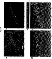
- Figure 1 shows both TH-positive neural elements and ones impregnated with Golgi-Cox in the Substantia Nigra compacta (SNc), and simultaneously visualized with the CLSM (1a).
- the figure shows that the application of these two procedures combined does not produce negative effects of one on the other but, on the contrary, supplies information on the neurochemical nature (TH-immuno-fluorescence alone, 1b) of the impregnated neurons (impregnation only, 1c).
- the vector analysis shown in panel 1e shows that the Golgi-Cox procedure provides a better representation of the morphological profile of the neuron on a whole, as for example the structures similar to dendritic spines (white arrows), as previously observed by Tepper et al. ( Tepper JM et al., 1987, J. Neurosci. 7, 2794-2806 ). More specifically, the research carried out shows ( figure 1e ) how the morphological analyses based exclusively on the immunofluorescence for the TH can show minor precision.
- FIG. 2 shows how the presence of PSD-95 can be visualized both in TH-positive neurons and in TH-negative ones, and simultaneously impregnated with the Golgi-Cox method.
- the PSD-95 in panels 2a and 2c reveals a dense and punctiform staining which also allows one to identify the profiles of neuropils and dendrites of non-stained cells (white arrow).
- the immunoreactivity with the PSD-95 and the SynI in the striatum was detected to be uniform.
- the double immuno-staining was carried out with these antibodies in sections stained with the Golgi-Cox impregnation, it was possible to survey the relations between these two antigens in the dendrites and in the dendritic spines of the MSN ( figure 4 ).
- clusters of PSD-95 were surveyed in association with the membrane of the MSN, while immunoreactivity of the SynI was always found outside the impregnated neurons ( figure 4a ).
- the results obtained show that the method of visualization and characterization of the nervous system that is the object of the invention fulfills its purposes, since it has proven to be a simple and inexpensive method, suitable for both conventional microscopy and confocal microscopy, which grants the possibility to pair two techniques that are incompatible, that is, Golgi-Cox mercuric impregnation and immunohistochemistry. It has, instead, proven possible to combine these two techniques which have long been incompatible, for them to be used simultaneously in the same sections (Lee et al. 2006, ref. cit .), by following appropriate fixation procedures according to the method that is the object of the invention and, at the same time, giving optimal results extensively correspondent to the expected results based on the available knowledge in the field.
Landscapes
- Life Sciences & Earth Sciences (AREA)
- Health & Medical Sciences (AREA)
- Immunology (AREA)
- Engineering & Computer Science (AREA)
- Molecular Biology (AREA)
- Chemical & Material Sciences (AREA)
- Biomedical Technology (AREA)
- Pathology (AREA)
- General Physics & Mathematics (AREA)
- General Health & Medical Sciences (AREA)
- Biochemistry (AREA)
- Analytical Chemistry (AREA)
- Physics & Mathematics (AREA)
- Hematology (AREA)
- Urology & Nephrology (AREA)
- Cell Biology (AREA)
- Food Science & Technology (AREA)
- Medicinal Chemistry (AREA)
- Microbiology (AREA)
- Virology (AREA)
- Biotechnology (AREA)
- Zoology (AREA)
- Tropical Medicine & Parasitology (AREA)
- Investigating Or Analysing Biological Materials (AREA)
- Analysing Materials By The Use Of Radiation (AREA)
- Investigating Materials By The Use Of Optical Means Adapted For Particular Applications (AREA)
Applications Claiming Priority (2)
| Application Number | Priority Date | Filing Date | Title |
|---|---|---|---|
| IT000081A ITPD20110081A1 (it) | 2011-03-16 | 2011-03-16 | Metodo di visualizzazione e caratterizzazione del sistema nervoso mediante colorazione combinata per impregnazione metallica ed immunoistochimica |
| PCT/EP2012/054454 WO2012123492A1 (en) | 2011-03-16 | 2012-03-14 | Method, composition and kit of visualization and characterization of the nervous system by combining the staining for metal impregnation and immunohistochemistry |
Publications (2)
| Publication Number | Publication Date |
|---|---|
| EP2686661A1 EP2686661A1 (en) | 2014-01-22 |
| EP2686661B1 true EP2686661B1 (en) | 2017-10-18 |
Family
ID=43977259
Family Applications (1)
| Application Number | Title | Priority Date | Filing Date |
|---|---|---|---|
| EP12714976.3A Active EP2686661B1 (en) | 2011-03-16 | 2012-03-14 | Method, composition and kit for visualization and characterization of the nervous system |
Country Status (5)
| Country | Link |
|---|---|
| US (1) | US20140295451A1 (zh) |
| EP (1) | EP2686661B1 (zh) |
| CN (1) | CN103534571B (zh) |
| IT (1) | ITPD20110081A1 (zh) |
| WO (1) | WO2012123492A1 (zh) |
Families Citing this family (2)
| Publication number | Priority date | Publication date | Assignee | Title |
|---|---|---|---|---|
| CN103380768B (zh) * | 2013-07-05 | 2015-07-01 | 河南科技大学 | 一种成年公羊的神经系统标本制作方法 |
| CN112113821B (zh) * | 2020-05-28 | 2021-09-03 | 王剑 | 一种细胞病理学样本的多重染色制片方法 |
Family Cites Families (10)
| Publication number | Priority date | Publication date | Assignee | Title |
|---|---|---|---|---|
| SU395746A1 (ru) * | 1968-04-12 | 1973-08-28 | Способ импрегнации нервных тканей азотнокислым серебром | |
| US4208479A (en) * | 1977-07-14 | 1980-06-17 | Syva Company | Label modified immunoassays |
| WO1996000234A1 (en) * | 1994-06-23 | 1996-01-04 | Aprogenex, Inc. | Centromere hybridization probes |
| US6426195B1 (en) * | 2000-03-28 | 2002-07-30 | Biogenex Lab | Method for silver staining a pathologic sample |
| EP1191337B1 (en) * | 2000-06-12 | 2007-10-10 | Fujirebio Inc. | Immunoassay for measuring human C-peptide and kit therefor |
| US20090220968A1 (en) * | 2006-03-10 | 2009-09-03 | President And Fellows Of Harvard College | Methods and Apparatus for Near Field Irradiation |
| EP2089029B1 (en) * | 2006-11-10 | 2012-10-03 | Massachusetts Institute of Technology | Pak inhibitors for use in treating neurodevelopmental disorders |
| JP2008237072A (ja) * | 2007-03-27 | 2008-10-09 | Kochi Univ | 遺伝子操作非ヒト動物、および向精神薬開発法 |
| CN101458182A (zh) * | 2008-12-22 | 2009-06-17 | 华中科技大学 | 一种小动物全脑标本的制备方法 |
| CN101915693B (zh) * | 2010-07-07 | 2012-07-25 | 新疆医科大学 | 基于人胚胎三叉神经利用组织切片染色进行三维重建方法 |
-
2011
- 2011-03-16 IT IT000081A patent/ITPD20110081A1/it unknown
-
2012
- 2012-03-14 CN CN201280013271.XA patent/CN103534571B/zh active Active
- 2012-03-14 WO PCT/EP2012/054454 patent/WO2012123492A1/en active Application Filing
- 2012-03-14 EP EP12714976.3A patent/EP2686661B1/en active Active
- 2012-03-14 US US14/005,318 patent/US20140295451A1/en not_active Abandoned
Also Published As
| Publication number | Publication date |
|---|---|
| EP2686661A1 (en) | 2014-01-22 |
| WO2012123492A1 (en) | 2012-09-20 |
| CN103534571A (zh) | 2014-01-22 |
| CN103534571B (zh) | 2016-02-10 |
| US20140295451A1 (en) | 2014-10-02 |
| ITPD20110081A1 (it) | 2012-09-17 |
Similar Documents
| Publication | Publication Date | Title |
|---|---|---|
| Korzhevskii et al. | Immunohistochemical demonstration of specific antigens in the human brain fixed in zinc-ethanol-formaldehyde | |
| Hoffman et al. | The importance of titrating antibodies for immunocytochemical methods | |
| Whittington et al. | Suppression of red blood cell autofluorescence for immunocytochemistry on fixed embryonic mouse tissue | |
| Halász et al. | Immunohistochemical identification of two types of dopamine neuron in the rat olfactory bulb as seen by serial sectioning | |
| Spiga et al. | Simultaneous Golgi-Cox and immunofluorescence using confocal microscopy | |
| Kosaka et al. | Catecholaminergic neurons containing GABA-like and/or glutamic acid decarboxylase-like immunoreactivities in various brain regions of the rat | |
| Schmued et al. | Fluoro-Jade B: a high affinity fluorescent marker for the localization of neuronal degeneration | |
| Freund et al. | The section-Golgi impregnation procedure. 1. Description of the method and its combination with histochemistry after intracellular iontophoresis or retrograde transport of horseradish peroxidase | |
| EP2794655B1 (de) | Verfahren zur selektiven quantifizierung von a-beta-aggregaten | |
| Shibata et al. | Large-area fluorescence and electron microscopic correlative imaging with multibeam scanning electron microscopy | |
| Tao-Cheng et al. | Optimization of protocols for pre-embedding immunogold electron microscopy of neurons in cell cultures and brains | |
| EP2686661B1 (en) | Method, composition and kit for visualization and characterization of the nervous system | |
| Takamatsu et al. | Nonspecific (“pseudo-plasmal”) dye-binding in the Feulgen nuclear stain and its blocking by azocarmin G | |
| Exbrayat | Classical methods of visualization | |
| Altman | Neuroanatomical techniques: insect nervous system | |
| Narayanan et al. | Determining factors for optimal neuronal and glial Golgi-Cox staining | |
| Wang et al. | Compartmentalization of calbindin and parvalbumin in different parts of rat rubrospinal neurons | |
| Barnerssoi et al. | Postembedding immunohistochemistry for inhibitory neurotransmitters in conjunction with neuroanatomical tracers | |
| JP2021512333A (ja) | 透明化大型組織の免疫染色用組成物、及び透明化大型生体組織の免疫染色方法 | |
| Watanabe et al. | Immunoelectron microscopic observation of chicken glucagon-like peptide (GLP)-1-containing cells in tissues derived from thin section, paraffin block and conventional method | |
| Freund et al. | Synaptic relationships of Golgi-impregnated neurons as identified by electrophysiological or immunocytochemical techniques | |
| Thornell et al. | Cryoultramicrotomy and immunocytochemistry in the analysis of muscle fine structure | |
| Bowen | The methods for the demonstration of the Golgi apparatus. II. Silver and gold methods | |
| RU2620559C1 (ru) | Способ окрашивания препаратов цельных биологических тканей и органов методом клик-гистохимии (варианты) | |
| Bates et al. | Preparation of spinal cord injured tissue for light and electron microscopy including preparation for immunostaining |
Legal Events
| Date | Code | Title | Description |
|---|---|---|---|
| PUAI | Public reference made under article 153(3) epc to a published international application that has entered the european phase |
Free format text: ORIGINAL CODE: 0009012 |
|
| 17P | Request for examination filed |
Effective date: 20131015 |
|
| AK | Designated contracting states |
Kind code of ref document: A1 Designated state(s): AL AT BE BG CH CY CZ DE DK EE ES FI FR GB GR HR HU IE IS IT LI LT LU LV MC MK MT NL NO PL PT RO RS SE SI SK SM TR |
|
| DAX | Request for extension of the european patent (deleted) | ||
| RIC1 | Information provided on ipc code assigned before grant |
Ipc: G01N 33/569 20060101ALI20161201BHEP Ipc: G01N 1/30 20060101AFI20161201BHEP |
|
| GRAP | Despatch of communication of intention to grant a patent |
Free format text: ORIGINAL CODE: EPIDOSNIGR1 |
|
| INTG | Intention to grant announced |
Effective date: 20170109 |
|
| GRAS | Grant fee paid |
Free format text: ORIGINAL CODE: EPIDOSNIGR3 |
|
| GRAA | (expected) grant |
Free format text: ORIGINAL CODE: 0009210 |
|
| AK | Designated contracting states |
Kind code of ref document: B1 Designated state(s): AL AT BE BG CH CY CZ DE DK EE ES FI FR GB GR HR HU IE IS IT LI LT LU LV MC MK MT NL NO PL PT RO RS SE SI SK SM TR |
|
| REG | Reference to a national code |
Ref country code: GB Ref legal event code: FG4D |
|
| REG | Reference to a national code |
Ref country code: CH Ref legal event code: EP |
|
| REG | Reference to a national code |
Ref country code: AT Ref legal event code: REF Ref document number: 938376 Country of ref document: AT Kind code of ref document: T Effective date: 20171115 Ref country code: IE Ref legal event code: FG4D |
|
| REG | Reference to a national code |
Ref country code: DE Ref legal event code: R096 Ref document number: 602012038634 Country of ref document: DE |
|
| REG | Reference to a national code |
Ref country code: NL Ref legal event code: MP Effective date: 20171018 |
|
| REG | Reference to a national code |
Ref country code: LT Ref legal event code: MG4D |
|
| REG | Reference to a national code |
Ref country code: FR Ref legal event code: PLFP Year of fee payment: 7 |
|
| REG | Reference to a national code |
Ref country code: AT Ref legal event code: MK05 Ref document number: 938376 Country of ref document: AT Kind code of ref document: T Effective date: 20171018 |
|
| PG25 | Lapsed in a contracting state [announced via postgrant information from national office to epo] |
Ref country code: NL Free format text: LAPSE BECAUSE OF FAILURE TO SUBMIT A TRANSLATION OF THE DESCRIPTION OR TO PAY THE FEE WITHIN THE PRESCRIBED TIME-LIMIT Effective date: 20171018 |
|
| PG25 | Lapsed in a contracting state [announced via postgrant information from national office to epo] |
Ref country code: LT Free format text: LAPSE BECAUSE OF FAILURE TO SUBMIT A TRANSLATION OF THE DESCRIPTION OR TO PAY THE FEE WITHIN THE PRESCRIBED TIME-LIMIT Effective date: 20171018 Ref country code: SE Free format text: LAPSE BECAUSE OF FAILURE TO SUBMIT A TRANSLATION OF THE DESCRIPTION OR TO PAY THE FEE WITHIN THE PRESCRIBED TIME-LIMIT Effective date: 20171018 Ref country code: FI Free format text: LAPSE BECAUSE OF FAILURE TO SUBMIT A TRANSLATION OF THE DESCRIPTION OR TO PAY THE FEE WITHIN THE PRESCRIBED TIME-LIMIT Effective date: 20171018 Ref country code: NO Free format text: LAPSE BECAUSE OF FAILURE TO SUBMIT A TRANSLATION OF THE DESCRIPTION OR TO PAY THE FEE WITHIN THE PRESCRIBED TIME-LIMIT Effective date: 20180118 Ref country code: ES Free format text: LAPSE BECAUSE OF FAILURE TO SUBMIT A TRANSLATION OF THE DESCRIPTION OR TO PAY THE FEE WITHIN THE PRESCRIBED TIME-LIMIT Effective date: 20171018 |
|
| PG25 | Lapsed in a contracting state [announced via postgrant information from national office to epo] |
Ref country code: AT Free format text: LAPSE BECAUSE OF FAILURE TO SUBMIT A TRANSLATION OF THE DESCRIPTION OR TO PAY THE FEE WITHIN THE PRESCRIBED TIME-LIMIT Effective date: 20171018 Ref country code: IS Free format text: LAPSE BECAUSE OF FAILURE TO SUBMIT A TRANSLATION OF THE DESCRIPTION OR TO PAY THE FEE WITHIN THE PRESCRIBED TIME-LIMIT Effective date: 20180218 Ref country code: RS Free format text: LAPSE BECAUSE OF FAILURE TO SUBMIT A TRANSLATION OF THE DESCRIPTION OR TO PAY THE FEE WITHIN THE PRESCRIBED TIME-LIMIT Effective date: 20171018 Ref country code: BG Free format text: LAPSE BECAUSE OF FAILURE TO SUBMIT A TRANSLATION OF THE DESCRIPTION OR TO PAY THE FEE WITHIN THE PRESCRIBED TIME-LIMIT Effective date: 20180118 Ref country code: HR Free format text: LAPSE BECAUSE OF FAILURE TO SUBMIT A TRANSLATION OF THE DESCRIPTION OR TO PAY THE FEE WITHIN THE PRESCRIBED TIME-LIMIT Effective date: 20171018 Ref country code: GR Free format text: LAPSE BECAUSE OF FAILURE TO SUBMIT A TRANSLATION OF THE DESCRIPTION OR TO PAY THE FEE WITHIN THE PRESCRIBED TIME-LIMIT Effective date: 20180119 Ref country code: LV Free format text: LAPSE BECAUSE OF FAILURE TO SUBMIT A TRANSLATION OF THE DESCRIPTION OR TO PAY THE FEE WITHIN THE PRESCRIBED TIME-LIMIT Effective date: 20171018 |
|
| REG | Reference to a national code |
Ref country code: DE Ref legal event code: R097 Ref document number: 602012038634 Country of ref document: DE |
|
| PG25 | Lapsed in a contracting state [announced via postgrant information from national office to epo] |
Ref country code: DK Free format text: LAPSE BECAUSE OF FAILURE TO SUBMIT A TRANSLATION OF THE DESCRIPTION OR TO PAY THE FEE WITHIN THE PRESCRIBED TIME-LIMIT Effective date: 20171018 Ref country code: EE Free format text: LAPSE BECAUSE OF FAILURE TO SUBMIT A TRANSLATION OF THE DESCRIPTION OR TO PAY THE FEE WITHIN THE PRESCRIBED TIME-LIMIT Effective date: 20171018 Ref country code: CZ Free format text: LAPSE BECAUSE OF FAILURE TO SUBMIT A TRANSLATION OF THE DESCRIPTION OR TO PAY THE FEE WITHIN THE PRESCRIBED TIME-LIMIT Effective date: 20171018 Ref country code: SK Free format text: LAPSE BECAUSE OF FAILURE TO SUBMIT A TRANSLATION OF THE DESCRIPTION OR TO PAY THE FEE WITHIN THE PRESCRIBED TIME-LIMIT Effective date: 20171018 |
|
| PLBE | No opposition filed within time limit |
Free format text: ORIGINAL CODE: 0009261 |
|
| STAA | Information on the status of an ep patent application or granted ep patent |
Free format text: STATUS: NO OPPOSITION FILED WITHIN TIME LIMIT |
|
| PG25 | Lapsed in a contracting state [announced via postgrant information from national office to epo] |
Ref country code: SM Free format text: LAPSE BECAUSE OF FAILURE TO SUBMIT A TRANSLATION OF THE DESCRIPTION OR TO PAY THE FEE WITHIN THE PRESCRIBED TIME-LIMIT Effective date: 20171018 Ref country code: IT Free format text: LAPSE BECAUSE OF FAILURE TO SUBMIT A TRANSLATION OF THE DESCRIPTION OR TO PAY THE FEE WITHIN THE PRESCRIBED TIME-LIMIT Effective date: 20171018 Ref country code: RO Free format text: LAPSE BECAUSE OF FAILURE TO SUBMIT A TRANSLATION OF THE DESCRIPTION OR TO PAY THE FEE WITHIN THE PRESCRIBED TIME-LIMIT Effective date: 20171018 Ref country code: PL Free format text: LAPSE BECAUSE OF FAILURE TO SUBMIT A TRANSLATION OF THE DESCRIPTION OR TO PAY THE FEE WITHIN THE PRESCRIBED TIME-LIMIT Effective date: 20171018 |
|
| 26N | No opposition filed |
Effective date: 20180719 |
|
| REG | Reference to a national code |
Ref country code: CH Ref legal event code: PL |
|
| PG25 | Lapsed in a contracting state [announced via postgrant information from national office to epo] |
Ref country code: MC Free format text: LAPSE BECAUSE OF FAILURE TO SUBMIT A TRANSLATION OF THE DESCRIPTION OR TO PAY THE FEE WITHIN THE PRESCRIBED TIME-LIMIT Effective date: 20171018 Ref country code: SI Free format text: LAPSE BECAUSE OF FAILURE TO SUBMIT A TRANSLATION OF THE DESCRIPTION OR TO PAY THE FEE WITHIN THE PRESCRIBED TIME-LIMIT Effective date: 20171018 |
|
| REG | Reference to a national code |
Ref country code: BE Ref legal event code: MM Effective date: 20180331 |
|
| REG | Reference to a national code |
Ref country code: IE Ref legal event code: MM4A |
|
| PG25 | Lapsed in a contracting state [announced via postgrant information from national office to epo] |
Ref country code: LU Free format text: LAPSE BECAUSE OF NON-PAYMENT OF DUE FEES Effective date: 20180314 |
|
| PG25 | Lapsed in a contracting state [announced via postgrant information from national office to epo] |
Ref country code: IE Free format text: LAPSE BECAUSE OF NON-PAYMENT OF DUE FEES Effective date: 20180314 |
|
| PG25 | Lapsed in a contracting state [announced via postgrant information from national office to epo] |
Ref country code: CH Free format text: LAPSE BECAUSE OF NON-PAYMENT OF DUE FEES Effective date: 20180331 Ref country code: LI Free format text: LAPSE BECAUSE OF NON-PAYMENT OF DUE FEES Effective date: 20180331 Ref country code: BE Free format text: LAPSE BECAUSE OF NON-PAYMENT OF DUE FEES Effective date: 20180331 |
|
| PG25 | Lapsed in a contracting state [announced via postgrant information from national office to epo] |
Ref country code: MT Free format text: LAPSE BECAUSE OF NON-PAYMENT OF DUE FEES Effective date: 20180314 |
|
| PG25 | Lapsed in a contracting state [announced via postgrant information from national office to epo] |
Ref country code: TR Free format text: LAPSE BECAUSE OF FAILURE TO SUBMIT A TRANSLATION OF THE DESCRIPTION OR TO PAY THE FEE WITHIN THE PRESCRIBED TIME-LIMIT Effective date: 20171018 |
|
| PG25 | Lapsed in a contracting state [announced via postgrant information from national office to epo] |
Ref country code: PT Free format text: LAPSE BECAUSE OF FAILURE TO SUBMIT A TRANSLATION OF THE DESCRIPTION OR TO PAY THE FEE WITHIN THE PRESCRIBED TIME-LIMIT Effective date: 20171018 Ref country code: HU Free format text: LAPSE BECAUSE OF FAILURE TO SUBMIT A TRANSLATION OF THE DESCRIPTION OR TO PAY THE FEE WITHIN THE PRESCRIBED TIME-LIMIT; INVALID AB INITIO Effective date: 20120314 |
|
| PG25 | Lapsed in a contracting state [announced via postgrant information from national office to epo] |
Ref country code: CY Free format text: LAPSE BECAUSE OF FAILURE TO SUBMIT A TRANSLATION OF THE DESCRIPTION OR TO PAY THE FEE WITHIN THE PRESCRIBED TIME-LIMIT Effective date: 20171018 Ref country code: MK Free format text: LAPSE BECAUSE OF NON-PAYMENT OF DUE FEES Effective date: 20171018 |
|
| PG25 | Lapsed in a contracting state [announced via postgrant information from national office to epo] |
Ref country code: AL Free format text: LAPSE BECAUSE OF FAILURE TO SUBMIT A TRANSLATION OF THE DESCRIPTION OR TO PAY THE FEE WITHIN THE PRESCRIBED TIME-LIMIT Effective date: 20171018 |
|
| PGFP | Annual fee paid to national office [announced via postgrant information from national office to epo] |
Ref country code: FR Payment date: 20240329 Year of fee payment: 13 |
|
| PGFP | Annual fee paid to national office [announced via postgrant information from national office to epo] |
Ref country code: GB Payment date: 20240404 Year of fee payment: 13 |
|
| PGFP | Annual fee paid to national office [announced via postgrant information from national office to epo] |
Ref country code: DE Payment date: 20240409 Year of fee payment: 13 |