EP2686016B1 - Anticorps de reconnaissance de domaine n-terminal de midkine - Google Patents
Anticorps de reconnaissance de domaine n-terminal de midkine Download PDFInfo
- Publication number
- EP2686016B1 EP2686016B1 EP12757658.5A EP12757658A EP2686016B1 EP 2686016 B1 EP2686016 B1 EP 2686016B1 EP 12757658 A EP12757658 A EP 12757658A EP 2686016 B1 EP2686016 B1 EP 2686016B1
- Authority
- EP
- European Patent Office
- Prior art keywords
- antibody
- seq
- isolated
- recombinant protein
- domain
- Prior art date
- Legal status (The legal status is an assumption and is not a legal conclusion. Google has not performed a legal analysis and makes no representation as to the accuracy of the status listed.)
- Active
Links
Images


Classifications
-
- C—CHEMISTRY; METALLURGY
- C07—ORGANIC CHEMISTRY
- C07K—PEPTIDES
- C07K16/00—Immunoglobulins [IGs], e.g. monoclonal or polyclonal antibodies
- C07K16/18—Immunoglobulins [IGs], e.g. monoclonal or polyclonal antibodies against material from animals or humans
- C07K16/22—Immunoglobulins [IGs], e.g. monoclonal or polyclonal antibodies against material from animals or humans against growth factors ; against growth regulators
-
- A—HUMAN NECESSITIES
- A61—MEDICAL OR VETERINARY SCIENCE; HYGIENE
- A61P—SPECIFIC THERAPEUTIC ACTIVITY OF CHEMICAL COMPOUNDS OR MEDICINAL PREPARATIONS
- A61P25/00—Drugs for disorders of the nervous system
- A61P25/28—Drugs for disorders of the nervous system for treating neurodegenerative disorders of the central nervous system, e.g. nootropic agents, cognition enhancers, drugs for treating Alzheimer's disease or other forms of dementia
-
- A—HUMAN NECESSITIES
- A61—MEDICAL OR VETERINARY SCIENCE; HYGIENE
- A61P—SPECIFIC THERAPEUTIC ACTIVITY OF CHEMICAL COMPOUNDS OR MEDICINAL PREPARATIONS
- A61P37/00—Drugs for immunological or allergic disorders
-
- A—HUMAN NECESSITIES
- A61—MEDICAL OR VETERINARY SCIENCE; HYGIENE
- A61K—PREPARATIONS FOR MEDICAL, DENTAL OR TOILETRY PURPOSES
- A61K39/00—Medicinal preparations containing antigens or antibodies
- A61K2039/505—Medicinal preparations containing antigens or antibodies comprising antibodies
-
- C—CHEMISTRY; METALLURGY
- C07—ORGANIC CHEMISTRY
- C07K—PEPTIDES
- C07K2317/00—Immunoglobulins specific features
- C07K2317/30—Immunoglobulins specific features characterized by aspects of specificity or valency
- C07K2317/34—Identification of a linear epitope shorter than 20 amino acid residues or of a conformational epitope defined by amino acid residues
-
- C—CHEMISTRY; METALLURGY
- C07—ORGANIC CHEMISTRY
- C07K—PEPTIDES
- C07K2317/00—Immunoglobulins specific features
- C07K2317/70—Immunoglobulins specific features characterized by effect upon binding to a cell or to an antigen
- C07K2317/76—Antagonist effect on antigen, e.g. neutralization or inhibition of binding
-
- C—CHEMISTRY; METALLURGY
- C07—ORGANIC CHEMISTRY
- C07K—PEPTIDES
- C07K2317/00—Immunoglobulins specific features
- C07K2317/90—Immunoglobulins specific features characterized by (pharmaco)kinetic aspects or by stability of the immunoglobulin
- C07K2317/92—Affinity (KD), association rate (Ka), dissociation rate (Kd) or EC50 value
Definitions
- the present disclosure relates to isolated or recombinant proteins, such as antibodies, which inhibit or reduce the function of midkine (hereinafter, referred to as "MK") for use in the treatment or prevention of midkine-related disorders.
- MK midkine
- MK Midkine
- EC embryonal carcinoma
- MK is known to have various biological activities. For example, it is known that MK expression is increased in human cancer cells. This increase in expression has been confirmed in various cancers such as esophageal cancer, thyroid cancer, urinary bladder cancer, colon cancer, stomach cancer, pancreatic cancer, thoracic cancer, liver cancer, lung cancer, breast cancer, neuroblastoma, glioblastoma, uterine cancer, ovarian cancer, and Wilms tumor ( Muramatsu (2002) J. Biochem. 132:359-371 ). Moreover, MK is thought to promote the survival and migration of cancer cells, promote angiogenesis, and contribute to cancer progression.
- MK is also known to play a central role in the stage of inflammation formation. For example, it is known that neointimal formation after vascular injury and nephritis onset during ischemic injury are suppressed in knockout mice deficient in MK genes. Moreover, it is also known that rheumatism models and postoperative adhesion are significantly suppressed in such knockout mice ( WO2000/10608 ; WO2004/078210 ).
- MK is known to participate in inflammatory diseases such as arthritis, autoimmune disease, rheumatic arthritis (rheumatoid arthritis (RA) or osteoarthritis (OA)), multiple sclerosis, postoperative adhesion, inflammatory bowel disease, psoriasis, lupus, asthma, and neutrophil dysfunction. Furthermore, MK is known to promote the movement (migration) of inflammatory cells such as macrophages or neutrophils. Since this movement is necessary for the establishment of inflammation, it is thought that deficiency of MK probably prevents diseases based on inflammation ( WO1999/03493 ).
- MK The three-dimensional structure of MK has been determined by NMR and reported ( Iwasaki et al. (1997) EMBO J. 16, p. 6936-6946 ).
- MK is composed of: an N-terminal fragment (hereinafter, referred to as an "N-fragment") consisting of amino acid residues 1 to 52; a C-terminal fragment (hereinafter, referred to as a "C-fragment”) consisting of amino acid residues 62 to 121; and a loop region (amino acid residues 53 to 61) (hereinafter, referred to as a "loop”) that links these fragments.
- N-fragment an N-terminal fragment
- C-fragment C-terminal fragment
- loop region amino acid residues 53 to 61
- Each of the N- and C-fragments is mainly composed of: a portion having a three-dimensional structure consisting of three antiparallel [beta]-sheets (hereinafter, referred to as a "domain”; the domain (consisting of amino acid residues 15 to 52) in the N-fragment is referred to as an "N-domain", and the domain (consisting of amino acid residues 62 to 104) in the C-fragment is referred to as a "C-domain”); and a terminally located portion devoid of the domain that does not assume a particular three-dimensional structure (hereinafter, referred to as a "tail”; the tail (consisting of amino acid residues 1 to 14) in the N-fragment is referred to as an "N-tail", and the tail (consisting of amino acid residues 105 to 121) in the C-fragment is referred to as a "C-tail”).
- domain the domain (consisting of amino acid residues 15 to 52) in the N-frag
- Basic amino acids on the C-domain surface form two clusters: a cluster consisting of lysine 79, arginine 81, and lysine 102 (cluster I) and a cluster consisting of lysine 86, lysine 87, and arginine 89 (cluster II). Both the clusters are known to participate in heparin-binding ability.
- the C-terminally located domain is usually responsible for MK activity ( Kojima et al. (1995) Biochem Biophys. Res. Comm. 206:468-473 ; Muramatsu et al. (1994) Biochem Biophys. Res. Comm. 203:1131-1139 ; Matsui et al. (2010) Int. Arch Medicine 3:12 ).
- Inoh et al. discloses an anti-MK antibody (MM964) that binds to the N-terminal domain of midkine but does not inhibit the growth of Hep-G2 cells.
- the use of anti-ML antibodies conjugated to Doxorubicin showed an inhibitory effect on Hep-G2 cells. Development of anti-MK antibodies has therefore focused on antibodies which are directed against the C-domain.
- the present inventors have prepared antibodies against the N-domain of human MK and made the surprising finding that these antibodies inhibit the cell migration function of MK to a substantially greater extent than antibodies directed against the MK C-domain.
- An exemplary antibody of the invention has also been shown to have excellent inhibitory effects on the onset of Experimental Autoimmune Encephalomyelitis (EAE) in a mouse model.
- EAE Experimental Autoimmune Encephalomyelitis
- the present invention provides the following embodiments defined under items 1-9 below:
- the present disclosure provides an isolated or recombinant protein comprising an antigen binding domain of an antibody which specifically binds to an epitope located within the N-domain of midkine (MK) as defined by amino acid residues 1-61 of SEQ ID NO:2, wherein the protein inhibits or reduces a function of MK, for use in treating or preventing a midkine-related disorder.
- MK midkine
- the present disclosure also provides an isolated or recombinant protein comprising an antigen binding domain of an antibody which specifically binds to an epitope located within the N-domain of midkine (MK) as defined by amino acid residues 1-61 of SEQ ID NO:2, for use in the treatment of prevention of a midkine-related disorder wherein the protein inhibits or reduces a function of MK.
- MK midkine
- the isolated or recombinant protein inhibits or reduces the cell migratory function of midkine.
- the isolated or recombinant protein specifically binds to a conformational epitope formed by the protein of SEQ ID NO:2, wherein the epitope includes at least two residues selected from the group consisting of 18W; 20W; 34F; 35R, 36E, 38T, 43T, 45R, 47R and 49R.
- the epitope may be defined by the following residues:
- the isolated or recombinant protein binds to the same epitope as monoclonal antibody IP-13 or to an overlapping epitope.
- the isolated or recombinant protein comprises at least one variable domain of an antibody.
- the isolated or recombinant protein may comprise a heavy chain variable domain comprising a complementarity determining region 3 (CDR3) sequence as shown in SEQ ID NO: 9 or a sequence exhibiting 95% or greater identity thereto.
- CDR3 complementarity determining region 3
- the heavy chain variable domain may further comprise a CDR1 sequence as shown in SEQ ID NO: 7 or a sequence exhibiting 95% or greater identity thereto and a CDR2 sequence as shown in SEQ ID NO:8 or a sequence exhibiting 95% or greater identity thereto.
- the heavy chain variable domain comprises the sequence as shown in SEQ ID NO: 5 or a sequence exhibiting 95% or greater identity thereto.
- the isolated or recombinant protein comprises a light chain variable domain comprising a CDR3 sequence as shown in SEQ ID NO: 12 or a sequence exhibiting 95% or greater identity thereto.
- the light chain variable domain may further comprise a CDR1 sequence as shown in SEQ ID NO: 10 or a sequence exhibiting 95% or greater identity thereto and a CDR2 sequence as shown in SEQ ID NO:11 or a sequence exhibiting 95% or greater identity thereto.
- the light chain variable domain comprises a sequence as shown in SEQ ID NO: 6 or a sequence exhibiting 95% or greater identity thereto.
- the isolated or recombinant protein comprises a heavy chain variable domain (VH) and a light chain variable domain (VL).
- the VH and the VL may be in the form of a single polypeptide chain, such as in the form of;
- VL and VH are in separate polypeptide chains, such as in the form of:
- the isolated or recombinant protein is a chimeric, de-immunized, humanized or human antibody.
- the isolated or recombinant protein comprises a human or non-human primate heavy chain immunoglobulin constant region selected from a group consisting of IgG1, IgG2, IgG3, IgG4, IgM, IgE and IgA.
- isolated or recombinant protein is in the form of an antibody comprising:
- isolated or recombinant protein is in the form of an antibody comprising:
- the isolated or recombinant protein may be conjugated to a compound.
- the compound may be selected from the group consisting of a radioisotope, a detectable label, a therapeutic compound, a colloid, a toxin, a nucleic acid, a peptide, a protein, a compound that increases the half life of the protein in a subject and mixtures thereof.
- the present disclosure also provides for administration of the isolated or recombinant protein by way of an isolated or recombinant nucleic acid molecule encoding the protein.
- the isolated or recombinant nucleic acid molecule may be operably linked to a promoter in the form of an expression construct.
- the present disclosure also provides for administration of the isolated or recombinant protein in the form of a composition comprising the protein or the nucleic acid molecule or the expression construct as described above and a suitable carrier or diluent.
- the present disclosure also encompasses the use of the protein or the nucleic acid or the expression construct or the composition of the present disclosure in medicine.
- the MK-related disorder may be, for example, an autoimmune disease, cancer, an inflammatory disease or multiple sclerosis.
- the method comprises administering between about 0.0001mg/kg and 50mg/kg of protein to the mammal.
- the method comprises administering between about 0.0005mg/kg to about 50mg/kg.
- the protein is administered at a dose of about 0.1mg/kg, or at a dose of about 1mg/kg, or at a dose of about 10mg/kg, or at a dose of about 30mg/kg.
- the present disclosure also provides a method for detecting MK in a sample, the method comprising contacting a sample with the protein of the disclosure such that a MK-protein complex forms and detecting the complex, wherein the complex is indicative of MK in the sample.
- the method for detecting MK in a sample may be used for diagnosing a MK-related condition in a subject, wherein detection of MK in the sample is indicative of the condition. For example, an increased or decreased level of MK in the sample compared to a control sample may be indicative of the condition.
- the mammal is a human.
- composition of matter, group of steps or group of compositions of matter shall be taken to encompass one and a plurality (i.e. one or more) of those steps, compositions of matter, groups of steps or groups of compositions of matter.
- variable regions and parts thereof, immunoglobulins, antibodies and fragments thereof herein may be further clarified by the discussion in Kabat Sequences of Proteins of Immunological Interest, National Institutes of Health, Bethesda, Md., 1987 and 1991 , Bork et al., J Mol. Biol. 242, 309-320, 1994 , Chothia and Lesk J. Mol Biol. 196:901 -917, 1987 , Chothia et al. Nature 342, 877-883, 1989 and/or or Al-Lazikani et al., J Mol Biol 273, 927-948, 1997 .
- derived from shall be taken to indicate that a specified integer may be obtained from a particular source albeit not necessarily directly from that source.
- an "antibody” is generally considered to be a protein that comprises a variable region made up of a plurality of polypeptide chains, e.g., a polypeptide comprising a V L and a polypeptide comprising a V H .
- An antibody also generally comprises constant domains, some of which can be arranged into a constant region or constant fragment or fragment crystallizable (Fc).
- a V H and a V L interact to form a Fv comprising an antigen binding region that is capable of specifically binding to one or a few closely related antigens.
- a light chain from mammals is either a ⁇ light chain or a ⁇ light chain and a heavy chain from mammals is ⁇ , ⁇ , ⁇ , ⁇ , or ⁇ .
- Antibodies can be of any type (e.g., IgG, IgE, IgM, IgD, IgA, and IgY), class (e.g., IgG 1 , IgG 2 , IgG 3 , IgG 4 , IgA 1 and IgA 2 ) or subclass.
- the term "antibody” also encompasses humanized antibodies, de-immunized antibodies, primatized antibodies, human antibodies and chimeric antibodies.
- full-length antibody “intact antibody” or “whole antibody” are used interchangeably to refer to an antibody in its substantially intact form, as opposed to an antigen binding fragment of an antibody.
- whole antibodies include those with heavy and light chains including an Fc region.
- the constant domains may be wild-type sequence constant domains (e.g., human wild-type sequence constant domains) or amino acid sequence variants thereof.
- the intact antibody may have one or more effector functions.
- an "antigen binding fragment” of an antibody comprises the antigen binding and/or the variable region of the intact antibody.
- antibody fragments include Fab, Fab', F(ab') 2 and Fv fragments; diabodies; linear antibodies; single-chain antibody molecules and multispecific antibodies formed from antibody fragments.
- variable region refers to the portions of the light and/or heavy chains of an antibody as defined herein that is capable of specifically binding to an antigen and, for example, includes amino acid sequences of CDRs; i.e., CDR1, CDR2, and CDR3, and framework regions (FRs).
- the variable region comprises three or four FRs (e.g., FR1, FR2, FR3 and optionally FR4) together with three CDRs.
- V H refers to the variable region of the heavy chain.
- V L refers to the variable region of the light chain.
- the amino acid positions assigned to CDRs and FRs can be defined according to Kabat (1987 and 1991, supra ) or other numbering systems in the performance of methods according to the present disclosure, e.g., the hypervariable loop numbering system of Chothia and Lesk (1987 and/or 1989, supra and/or Al-Lazikani et al., 1997, supra ).
- a V H FRs and CDRs positioned as follows residues 1-30 (FR1), 31-25 (CDR1), 36-49 (FR2), 50-65 (CDR2), 66-94 (FR3), 95-102 (CDR3) and 103- 113 (FR4), numbered according to the Kabat numbering system.
- V L FRs and CDRs are positioned as follows residues 1-23 (FR1), 24-34 (CDR1), 35-49 (FR2), 50-56 (CDR2), 57-88 (FR3), 89-97 (CDR3) and 98-107 (FR4).
- CDRs complementarity determining regions
- CDR1, CDR2, and CDR3 refers to the amino acid residues of an antibody variable domain that form loops between the FRs the sequence of which vary between antibodies. Some or all of the CDRs confer the ability to bind antigen on the antibody.
- Each variable domain typically has three CDR regions identified as CDR1, CDR2 and CDR3.
- Each complementarity determining region may comprise amino acid residues from a "complementarity determining region" as defined by Kabat et al., (1991) and/or those residues from a "hypervariable loop” Chothia and Lesk (1987), or any other known numbering technique or combination thereof, including the IMGT numbering system (Le Franc et al., 2003).
- Framework regions are those variable domain residues other than the CDR residues.
- constant region refers to a portion of an antibody comprising at least one constant domain and which is generally (though not necessarily) glycosylated and which is capable of binding to one or more Fc receptors and/or components of the complement cascade.
- the heavy chain constant region can be selected from any of the five isotypes: ⁇ , ⁇ , ⁇ , ⁇ , or ⁇ .
- heavy chains of various subclasses are responsible for different effector functions and thus, by choosing the desired heavy chain constant region, proteins with desired effector function can be produced.
- Exemplary heavy chain constant regions are gamma 1 (IgG1), gamma 2 (IgG2) and gamma 3 (IgG3), or hybrids thereof.
- a “constant domain” is a domain in an antibody the sequence of which is highly similar in antibodies/antibodies of the same type, e.g., IgG or IgM or IgE.
- a constant region of an antibody generally comprises a plurality of constant domains, e.g., the constant region of ⁇ , ⁇ and ⁇ heavy chains comprises two constant domains.
- the term “specifically binds” shall be taken to mean a protein reacts or associates more frequently, more rapidly, with greater duration and/or with greater affinity with midkine or a specified epitope thereof than it does with alternative antigens or epitopes. As such, “specific binding” does not necessarily require exclusive binding or non-detectable binding of another antigen.
- the term specifically binds is used interchangeably with “selectively binds” herein.
- the term "competitively inhibits” shall be understood to mean that a isolated or recombinant protein such as an antibody reduces or prevents binding of the monoclonal antibody designated IP-13 to human midkine. It will be apparent from the foregoing that the antibody need not completely inhibit binding of the monoclonal antibody IP-13, rather it need only reduce binding by a statistically significant amount, for example, by at least about 10% or 20% or 30% or 40% or 50% or 60% or 70% or 80% or 90% or 95%. Methods for determining competitive inhibition of binding are known in the art and/or described herein. For example, monoclonal antibody IP-13 is exposed to mdikine either in the presence or absence of the recombinant protein. If less monoclonal antibody binds in the presence of the recombinant protein than in the absence of it, the recombinant protein is considered to competitively inhibit binding of monoclonal antibody IP-13 to midkine.
- overlapping in the context of two epitopes shall be taken to mean that two epitopes share a sufficient number of amino acid residues to permit an antibody that binds to one epitope to competitively inhibit the binding of an antibody that binds to the other epitope.
- the two epitopes share at least 1 or 2 or 3 or 4 or 5 or 6 or more amino acids.
- EU numbering system of Kabat will be understood to mean the numbering of an immunoglobulin heavy chain is according to the EU index as taught in Kabat et al., 1991, Sequences of Proteins of Immunological Interest, 5th Ed., United States Public Health Service, National Institutes of Health, Bethesda .
- the EU index is based on the residue numbering of the human IgG1 EU antibody.
- treatment refers to clinical intervention designed to alter the natural course of the individual or cell being treated during the course of clinical pathology. Desirable effects of treatment include decreasing the rate of disease progression, ameliorating or palliating the disease state, and remission or improved prognosis.
- An individual is successfully "treated", for example, if one or more symptoms associated with a disease (e.g., lupus) are mitigated or eliminated.
- prevention includes providing prophylaxis with respect to occurrence or recurrence of a disease in an individual.
- An individual may be predisposed to or at risk of developing the disease or disease relapse but has not yet been diagnosed with the disease or the relapse.
- the term prevention does not require absolute prevention but includes inhibiting the progression of the disease to some extent.
- an “effective amount” refers to at least an amount effective, at dosages and for periods of time necessary, to achieve the desired therapeutic or prophylactic result.
- An effective amount can be provided in one or more administrations.
- the term "effective amount” is meant an amount necessary to effect treatment of a disease or condition as hereinbefore described.
- the effective amount may vary according to the disease or condition to be treated and also according to the weight, age, racial background, sex, health and/or physical condition and other factors relevant to the mammal being treated.
- the effective amount will fall within a relatively broad range (e.g. a "dosage" range) that can be determined through routine trial and experimentation by a medical practitioner.
- the effective amount can be administered in a single dose or in a dose repeated once or several times over a treatment period.
- a “therapeutically effective amount” is at least the minimum concentration required to effect a measurable improvement of a particular disorder (e.g., MS).
- a therapeutically effective amount herein may vary according to factors such as the disease state, age, sex, and weight of the patient, and the ability of the protein to elicit a desired response in the individual.
- a therapeutically effective amount is also one in which any toxic or detrimental effects of the protein are outweighed by the therapeutically beneficial effects.
- prophylactically effective amount refers to an amount effective, at the dosages and for periods of time necessary, to achieve the desired prophylactic result. Typically but not necessarily, since a prophylactic dose is used in mammals prior to or at an earlier stage of disease, a prophylactically effective amount may be less than a therapeutically effective amount.
- amino acid sequence of human midkine is shown in SEQ ID NO: 1 and the N-domain sequence is shown in SEQ ID NO:2.
- the "mammal” treated according to the present disclosure may be a primate, livestock (e.g. sheep, horses, cattle, pigs, donkeys), companion animal (e.g. pets such as dogs and cats), laboratory test animal (e.g. mice, rabbits, rats, guinea pigs), performance animal (e.g. racehorses, camels, greyhounds) or captive wild animal.
- livestock e.g. sheep, horses, cattle, pigs, donkeys
- companion animal e.g. pets such as dogs and cats
- laboratory test animal e.g. mice, rabbits, rats, guinea pigs
- performance animal e.g. racehorses, camels, greyhounds
- the mammal is a human.
- antibodies and functional antigen-binding fragments that are structurally and/or functionally related to IP-13, in which the heavy chain variable region sequence exhibits a degree of identity to SEQ ID NO:5 and the light chain variable region sequence exhibits a degree of identity to SEQ ID NO:6.
- antibodies and antigen-binding fragments include a heavy or a light chain variable region sequence with about 80% or more identity to a heavy or light chain sequence variable region of IP-13 or a sequence within the variable region (e.g., one or more CDRs).
- antibodies or antigen-binding fragments include a heavy or a light chain with at least 82%, 85%, 90%, 95%, or more identity to a heavy chain variable region sequence of IP-13, or a sequence within the variable region (e.g., one or more CDRs).
- antibodies or antigen-binding fragments include a heavy or a light chain variable region sequence with at least 80-85%, 85-90%, 90-95%, 95-100% identity to one or more CDRs in the IP-13.
- an antibody or antigen-binding fragment thereof includes a heavy or a light chain variable region sequence with 95-100% identity to one, two or three CDRs in each heavy or light chain variable region sequence in the IP-13 antibody.
- Antibodies and antigen-binding fragments of the invention therefore include those with at least partial sequence identity to IP-13.
- the percent identity of such antibodies and functional fragments can be as little as 80%, or can be more (e.g., 82%, 85%, 90%, 95%, 96%, 97%, 98%, 99% etc.).
- the percent identity can extend over the entire sequence length of an IP-13 variable domain, or a contiguous region or area within an IP-13 variable domain.
- the length of the sequence sharing the percent identity is 5 or more contiguous amino acids, e.g., 5, 6, 7, 8, 9, 10, 11, 12, 13, 14, 15, 16, 17, 18, 19, 20, 21, 22, 23, 24, 25, etc. contiguous amino acids.
- the length of the sequence sharing the percent identity is 25 or more contiguous amino acids, e.g., 26, 27, 28, 29, 30, 31, 32, 33, 34, 35, etc. contiguous amino acids.
- the length of the sequence sharing the percent identity is 35 or more contiguous amino acids, e.g., 35, 36, 37, 38, 39, 40, 41, 42, 43, 44, 45, 45, 47, 48, 49, 50, etc., contiguous amino acids.
- the length of the sequence sharing the percent identity is 50 or more contiguous amino acids, e.g., 50-55, 55-60, 60-65, 65-70, 70-75, 75-80, 80-85, 85-90, 90-95, 95-100, 100-110, etc. contiguous amino acids.
- the length of the sequence sharing the percent identity is equal to the length of any CDR of a variable region sequence, or a region outside the CDRs but within the variable region of a heavy or light chain sequence.
- the CDR regions of the anti-N-domain antibody will be either identical to highly homologous to the specified regions of SEQ ID NOs: 7 to 12.
- “highly homologous” it is contemplated that only a few substitutions, preferably 1, 2 or 3 substitutions may be made in the CDRs.
- the query sequence is at least 50 residues in length, and the GAP analysis aligns the two sequences over a region of at least 50 residues. Even more preferably, the query sequence is at least 100 residues in length and the GAP analysis aligns the two sequences over a region of at least 100 residues. Most preferably, the two sequences are aligned over their entire length.
- Antibodies and functional fragments of the invention include those that retain at least one or more partial activities or functions of IP-13, such as the ability to inhibit midkine induce cell migration. Such antibodies include those that have greater than, about the same or less than the binding affinity of IP-13 for binding to midkine.
- Binding affinity can be determined by association (Ka) and dissociation (Kd) rate.
- Equilibrium affinity constant, K is the ratio of Ka/Kd.
- Association (Ka) and dissociation (Kd) rates can be measured using surface plasmon resonance (SPR) ( Rich and Myszka, Curr. Opin. Biotechnol. 11:54 (2000 ); Englebienne, Analyst. 123:1599 (1998 )). Instrumentation and methods for real time detection and monitoring of binding rates are known and are commercially available (BiaCore 2000, Biacore AB, Upsala, Sweden; and Malmqvist, Biochem. Soc. Trans. 27:335 (1999 )).
- nucleic acids of the invention include, among other things, nucleic acid sequences 1) encoding antibodies that are structurally or functionally related to IP-13; 2) encoding SEQ ID NO:5 and/or SEQ ID NO: 6, or antibodies that include all or a portion of a sequence of SEQ ID NO:5 and/or SEQ ID NO: 6 (e.g., one or more CDRs); 3) that exhibit a degree of complementarity or identity with nucleic acid sequences encoding antibodies with sequence identity to IP-13; and 4) that hybridize to sequences encoding antibodies that have sequence identity to the IP-13.
- nucleic acid sequence encompassed by the present disclosure are 75-100% complementary or identical to a nucleic acid sequence as shown in SEQ ID NO: 3, SEQ ID NO: 4 or SEQ ID NO: 5.
- nucleic acid sequences that hybridize to a nucleic acid sequence as shown in any one of SEQ ID NOs:3 or 4.
- a nucleic acid sequence specifically hybridizes to a nucleic acid encoding SEQ ID NO:5 (i.e. that hybridizes to a sequence as set forth in SEQ ID NO:4) or a portion thereof.
- a nucleic acid sequence specifically hybridizes to a nucleic acid encoding SEQ ID NO:6 (i.e. that hybridizes to a sequence as set forth in SEQ ID NO:3) or a portion thereof.
- a nucleic acid sequence is at least 75-100% complementary or homologous to a nucleic acid sequence that encodes all or a subsequence or fragment of IP-13.
- Hybridize and grammatical variations thereof refer to the binding between nucleic acid sequences.
- Hybridizing sequences will generally have more than about 50% homology (e.g., 50%, 60%, 70%, 80%, 90%, or more identity) to a reference nucleic acid or a sequence complementary to a reference sequence.
- Hybridizing sequences that are 100% or fully complementary to a reference sequence for example, to a nucleic acid that encodes an amino acid sequence of a reference sequence, exhibit 100% base pairing with no mismatches.
- hybridization region between hybridizing sequences typically is at least about 12-15 nucleotides, 15-20 nucleotides, 20-30 nucleotides, 30-50 nucleotides, 50-100 nucleotides, 100 to 200 nucleotides or more, or any numerical value or range within or encompassing such lengths.
- high stringency hybridization and/or wash conditions are preferred.
- a high stringency is defined herein as being a hybridization and/or wash carried out in about 0.1 x SSC buffer and/or about 0.1% (w/v) SDS, or lower salt concentration, and/or at a temperature of at least 65°C, or equivalent conditions.
- Reference herein to a particular level of stringency encompasses equivalent conditions using wash/hybridization solutions other than SSC known to those skilled in the art.
- the stringency is increased by reducing the concentration of SSC buffer, and/or increasing the concentration of SDS and/or increasing the temperature of the hybridization and/or wash.
- the conditions for hybridization and/or wash may vary depending upon the nature of the hybridization matrix used to support the sample DNA, and/or the type of hybridization probe used and/or constituents of any buffer used in a hybridization. For example, formamide reduces the melting temperature of a probe or primer in a hybridization or an amplification reaction.
- Nucleic acid sequences further include nucleotide and nucleoside substitutions, additions and deletions, as well as derivatized forms and fusion/chimeric sequences (e.g., encoding recombinant polypeptide).
- nucleic acids include sequences and subsequences degenerate with respect to nucleic acids that encode, modified forms and variants thereof.
- nucleic acids of the invention can be of various lengths. Nucleic acid lengths typically range from about 20 nucleotides to 20 Kb, or any numerical value or range within or encompassing such lengths, 10 nucleotides to 10 Kb, 1 to 5 Kb or less, 1000 to about 500 nucleotides or less in length. Nucleic acids can also be shorter, for example, 100 to about 500 nucleotides, or from about 12 to 25, 25 to 50, 50 to 100, 100 to 250, or about 250 to 500 nucleotides in length, or any numerical value or range or value within or encompassing such lengths.
- a nucleic acid sequence has a length from about 10-20, 20-30, 30-50, 50-100, 100-150, 150-200, 200-250, 250-300, 300-400, 400-500, 500-1000, 1000-2000, nucleotides, or any numerical value or range within or encompassing such lengths.
- Nucleic acids can be produced using various standard cloning and chemical synthesis techniques. Techniques include, but are not limited to nucleic acid amplification, e.g., polymerase chain reaction (PCR), with genomic DNA or cDNA targets using primers (e.g., a degenerate primer mixture) capable of annealing to antibody encoding sequence. Nucleic acids can also be produced by chemical synthesis (e.g., solid phase phosphoramidite synthesis) or transcription from a gene.
- PCR polymerase chain reaction
- primers e.g., a degenerate primer mixture
- Nucleic acids can also be produced by chemical synthesis (e.g., solid phase phosphoramidite synthesis) or transcription from a gene.
- sequences produced can then be translated in vitro, or cloned into a plasmid and propagated and then expressed in a cell (e.g., a host cell such as yeast or bacteria, a eukaryote such as an animal or mammalian cell or in a plant).
- a cell e.g., a host cell such as yeast or bacteria, a eukaryote such as an animal or mammalian cell or in a plant.
- a midkine protein or immunogenic fragment or epitope thereof or a cell expressing and displaying same i.e., an immunogen
- an immunogen optionally formulated with any suitable or desired carrier, adjuvant, or pharmaceutically acceptable excipient, is administered to a non-human animal, for example, a mouse, chicken, rat, rabbit, guinea pig, dog, horse, cow, goat or pig.
- the immunogen may be administered intranasally, intramuscularly, sub-cutaneously, intravenously, intradermally, intraperitoneally, or by other known route.
- polyclonal antibodies may be monitored by sampling blood of the immunized animal at various points following immunization. One or more further immunizations may be given, if required to achieve a desired antibody titer. The process of boosting and titering is repeated until a suitable titer is achieved. When a desired level of immunogenicity is obtained, the immunized animal is bled and the serum isolated and stored, and/or the animal is used to generate monoclonal antibodies (Mabs).
- Mabs monoclonal antibodies
- Monoclonal antibodies are one exemplary form of antibody contemplated by the present disclosure.
- the term “monoclonal antibody” or “MAb” refers to a homogeneous antibody population capable of binding to the same antigen(s), for example, to the same epitope within the antigen. This term is not intended to be limited as regards to the source of the antibody or the manner in which it is made.
- a suitable animal is immunized with an immunogen under conditions sufficient to stimulate antibody producing cells.
- Rodents such as rabbits, mice and rats are exemplary animals.
- Mice genetically-engineered to express human immunoglobulin proteins and, for example, do not express murine immunoglobulin proteins, can also be used to generate an antibody of the present disclosure (e.g., as described in WO2002/066630 ).
- somatic cells with the potential for producing antibodies, specifically B lymphocytes (B cells), are selected for use in the MAb generating protocol. These cells may be obtained from biopsies of spleens, tonsils or lymph nodes, or from a peripheral blood sample. The B cells from the immunized animal are then fused with cells of an immortal myeloma cell, generally derived from the same species as the animal that was immunized with the immunogen.
- B lymphocytes B lymphocytes
- Hybrids are amplified by culture in a selective medium comprising an agent that blocks the de novo synthesis of nucleotides in the tissue culture media.
- agents are aminopterin, methotrexate and azaserine.
- the amplified hybridomas are subjected to a functional selection for antibody specificity and/or titer, such as, for example, by flow cytometry and/or immunohistochemstry and/or immunoassay (e.g. radioimmunoassay, enzyme immunoassay, cytotoxicity assay, plaque assay, dot immunoassay, and the like).
- a functional selection for antibody specificity and/or titer such as, for example, by flow cytometry and/or immunohistochemstry and/or immunoassay (e.g. radioimmunoassay, enzyme immunoassay, cytotoxicity assay, plaque assay, dot immunoassay, and the like).
- immunoassay e.g. radioimmunoassay, enzyme immunoassay, cytotoxicity assay, plaque assay, dot immunoassay, and the like.
- ABL-MYC technology (NeoClone, Madison WI 53713, USA) is used to produce cell lines secreting MAbs (e.g., as described in Largaespada et al, J. Immunol. Methods. 197: 85-95, 1996 ).
- Antibodies can also be produced or isolated by screening a display library, e.g., a phage display library, e.g., as described in US6300064 and/or US5885793 .
- a display library e.g., a phage display library, e.g., as described in US6300064 and/or US5885793 .
- an antibody described herein is a chimeric antibody.
- the term "chimeric antibody” refers to antibodies in which a portion of the heavy and/or light chain is identical with or homologous to corresponding sequences in antibodies derived from a particular species (e.g., murine, such as mouse) or belonging to a particular antibody class or subclass, while the remainder of the chain(s) is identical with or homologous to corresponding sequences in antibodies derived from another species (e.g., primate, such as human) or belonging to another antibody class or subclass.
- chimeric antibodies utilize rodent or rabbit variable regions and human constant regions, in order to produce an antibody with predominantly human domains. Methods for producing chimeric antibodies are described in, e.g., US4816567 ; and US5807715 .
- the present disclosure also includes a chimeric immunoglobulin, e.g., in which a variable region from one species is fused to a region of a protein from another species.
- a chimeric immunoglobulin e.g., in which a variable region from one species is fused to a region of a protein from another species.
- the disclosure contemplates an immunoglobulin comprising a variable region from a T cell receptor of one species fused to a T cell receptor constant domain from a separate species.
- the antibodies of the present disclosure may be humanized or human.
- humanized antibody shall be understood to refer to a subclass of chimeric antibodies having an antigen binding site or variable region derived from an antibody from a non-human species and the remaining antibody structure of the molecule based upon the structure and/or sequence of a human antibody.
- the antigen-binding site comprises the complementarity determining regions (CDRs) from the non-human antibody grafted onto appropriate FRs in the variable domains of a human antibody and the remaining regions from a human antibody.
- Antigen binding sites may be wild type or modified by one or more amino acid substitutions. In some instances, FR residues of the human immunoglobulin are replaced by corresponding non-human residues.
- human antibody refers to antibodies having variable regions (e.g. V H , V L ) and, optionally constant regions derived from or corresponding to sequences found in humans, e.g. in the human germline or somatic cells.
- the "human” antibodies can include amino acid residues not encoded by human sequences, e.g. mutations introduced by random or site directed mutations in vitro (in particular mutations which involve conservative substitutions or mutations in a small number of residues of the antibody, e.g. in 1, 2, 3, 4 or 5 of the residues of the antibody, e.g. in 1, 2, 3, 4 or 5 of the residues making up one or more of the CDRs of the antibody).
- human antibodies do not actually need to be produced by a human, rather, they can be produced using recombinant means and/or isolated from a transgenic animal (e.g., mouse) comprising nucleic acid encoding human antibody constant and/or variable regions (e.g., as described above). Human antibodies can be produced using various techniques known in the art, including phage display libraries (e.g., as described in US5885793 ).
- Human antibodies which recognize a selected epitope can also be generated using a technique referred to as "guided selection.”
- a selected non-human monoclonal antibody e.g., a mouse antibody
- is used to guide the selection of a completely human antibody recognizing the same epitope e.g., as described in US5565332 .
- the present disclosure also contemplates a de-immunized antibody or protein.
- De-immunized antibodies and proteins have one or more epitopes, e.g., B cell epitopes or T cell epitopes removed (i.e., mutated) to thereby reduce the likelihood that a mammal will raise an immune response against the antibody or protein.
- epitopes e.g., B cell epitopes or T cell epitopes removed (i.e., mutated) to thereby reduce the likelihood that a mammal will raise an immune response against the antibody or protein.
- Methods for producing de-immunized antibodies and proteins are known in the art and described, for example, in WO00/34317 , WO2004/108158 and WO2004/064724 .
- Heavy chain antibodies differ structurally from many other forms of antibodies, in so far as they comprise a heavy chain, but do not comprise a light chain. Accordingly, these immunoglobulins are also referred to as "heavy chain only antibodies”. Heavy chain immunoglobulins are found in, for example, camelids and cartilaginous fish (also called IgNAR).
- variable regions present in naturally occurring heavy chain antibodies are generally referred to as "V HH domains" in camelid antibodies and V-NAR in IgNAR, in order to distinguish them from the heavy chain variable regions that are present in conventional 4-chain antibodies (which are referred to as "V H domains”) and from the light chain variable regions that are present in conventional 4-chain antibodies (which are referred to as "V L domains").
- an immunoglobulin of the disclosure is or comprises a single-domain antibody (which is used interchangeably with the term "domain antibody” or "dAb”).
- a single-domain antibody is a single polypeptide chain comprising all or a portion of the heavy chain variable domain of an antibody.
- a single-domain antibody is a human single-domain antibody (Domantis, Inc., Waltham, MA; see, e.g., US6248516 ).
- an immunoglobulin of the disclosure is or comprises a diabody, triabody, tetrabody or higher order protein complex such as those described in WO98/044001 and/or WO94/007921 .
- a diabody is a protein comprising two associated polypeptide chains, each polypeptide chain comprising the structure V L -X-V H or V H -X-V L , wherein V L is an antibody light chain variable region, V H is an antibody heavy chain variable region, X is a linker comprising insufficient residues to permit the V H and V L in a single polypeptide chain to associate (or form an Fv) or is absent, and wherein the V H of one polypeptide chain binds to a V L of the other polypeptide chain to form an antigen binding site, i.e., to form a Fv molecule capable of specifically binding to one or more antigens.
- the V L and V H can be the same in each polypeptide chain or the V L and V H can be different in each polypeptide chain so as to form a bispecific diabody (i.e., comprising two Fvs having different specificity).
- a diabody, triabody, tetrabody, etc capable of inducing effector activity can be produced using an antigen binding domain capable of binding to IL-3R ⁇ and an antigen binding domain capable of binding to a cell surface molecule on an immune cell, e.g., a T cell (e.g., CD3).
- an immune cell e.g., a T cell (e.g., CD3).
- scFvs comprise V H and V L regions in a single polypeptide chain and a polypeptide linker between the V H and V L which enables the scFv to form the desired structure for antigen binding (i.e., for the V H and V L of the single polypeptide chain to associate with one another to form a Fv).
- the linker comprises in excess of 12 amino acid residues with (Gly 4 Ser) 3 being one of the more favored linkers for a scFv.
- the present disclosure also contemplates a disulfide stabilized Fv (or diFv or dsFv), in which a single cysteine residue is introduced into a FR of V H and a FR of V L and the cysteine residues linked by a disulfide bond to yield a stable Fv.
- the present disclosure encompasses a dimeric scFv, i.e., a protein comprising two scFv molecules linked by a non-covalent or covalent linkage, e.g., by a leucine zipper domain (e.g., derived from Fos or Jun).
- a leucine zipper domain e.g., derived from Fos or Jun.
- two scFvs are linked by a peptide linker of sufficient length to permit both scFvs to form and to bind to an antigen, e.g., as described in US20060263367 .
- the present disclosure also contemplates a dimeric scFv capable of inducing effector activity.
- one scFv binds to IL-3R ⁇ and another scFv binds to a cell surface molecule on an immune cell, e.g., a T cell (e.g., CD3).
- the dimeric protein is a combination of a dAb and a scFv. Examples of bispecific antibody fragments capable of inducing effector function are described, for example, in US7235641 .
- the present disclosure also contemplates other antibodies and antibody fragments, such as:
- the present disclosure encompasses immunoglobulins comprising a Fc region of an antibody, including antigen binding fragments of an immunoglobulin fused to a Fc.
- Sequences of Fc regions useful for producing the proteins of the present disclosure may be obtained from a number of different sources.
- the Fc or portion thereof of the protein is derived from a human antibody.
- the Fc or portion thereof may be derived from any antibody class, including IgM, IgG, IgD, IgA and IgE, and any antibody isotype, including IgG1, IgG2, IgG3 and IgG4.
- the Fc is human isotype IgG1 or human isotype IgG2 or human isotype IgG3 or a hybrid of any of the foregoing.
- the antibody comprises one or more amino acid substitutions that increase its half-life.
- the immunoglobulin comprises a Fc region comprising one or more amino acid substitutions that increase the affinity of the Fc region for the neonatal Fc region (FcRn).
- the Fc region has increased affinity for FcRn at lower pH, e.g., about pH 6.0, to facilitate Fc/FcRn binding in an endosome.
- the Fc region has increased affinity for FcRn at about pH 6 compared to its affinity at about pH 7.4, which facilitates the re-release of Fc into blood following cellular recycling.
- Exemplary amino acid substitutions include T250Q and/or M428L according to the EU numbering system of Kabat. Additional or alternative amino acid substitutions are described, for example, in US20070135620 .
- an antibody encompassed by the present disclosure is produced by recombinant techniques.
- nucleic acid encoding same can be cloned into expression vectors, which are then transfected into host cells, such as E. coli cells, yeast cells, insect cells, or mammalian cells, such as simian COS cells, Chinese Hamster Ovary (CHO) cells, human embryonic kidney (HEK) cells, or myeloma cells that do not otherwise produce immunoglobulin protein.
- host cells such as E. coli cells, yeast cells, insect cells, or mammalian cells, such as simian COS cells, Chinese Hamster Ovary (CHO) cells, human embryonic kidney (HEK) cells, or myeloma cells that do not otherwise produce immunoglobulin protein.
- CHO Chinese Hamster Ovary
- HEK human embryonic kidney
- myeloma cells that do not otherwise produce immunoglobulin protein.
- Exemplary cells used for expressing an immunoglobulin are CHO cells, myeloma cells or HEK cells.
- nucleic acid is inserted operably linked to a promoter in an expression construct or expression vector for further cloning (amplification of the DNA) or for expression in a cell-free system or in cells.
- promoter is to be taken in its broadest context and includes the transcriptional regulatory sequences of a genomic gene, including the TATA box or initiator element, which is required for accurate transcription initiation, with or without additional regulatory elements (e.g., upstream activating sequences, transcription factor binding sites, enhancers and silencers) that alter expression of a nucleic acid, e.g., in response to a developmental and/or external stimulus, or in a tissue specific manner.
- promoter is also used to describe a recombinant, synthetic or fusion nucleic acid, or derivative which confers, activates or enhances the expression of a nucleic acid to which it is operably linked.
- Exemplary promoters can contain additional copies of one or more specific regulatory elements to further enhance expression and/or alter the spatial expression and/or temporal expression of said nucleic acid.
- operably linked to means positioning a promoter relative to a nucleic acid such that expression of the nucleic acid is controlled by the promoter.
- the vector components generally include, but are not limited to, one or more of the following: a signal sequence, a sequence encoding an immunoglobulin (e.g., derived from the information provided herein), an enhancer element, a promoter, and a transcription termination sequence.
- a signal sequence e.g., a sequence encoding an immunoglobulin (e.g., derived from the information provided herein)
- an enhancer element e.g., derived from the information provided herein
- a promoter e.g., derived from the information provided herein
- a transcription termination sequence e.g., a sequence encoding an immunoglobulin (e.g., derived from the information provided herein)
- the skilled artisan will be aware of suitable sequences for expression of an immunoglobulin.
- Exemplary signal sequences include prokaryotic secretion signals (e.g., pelB, alkaline phosphatase, penicillinase, Ipp, or heat-stable enterotoxin II), yeast secretion signals (e.g., invertase leader, ⁇ factor leader, or acid phosphatase leader) or mammalian secretion signals (e.g., herpes simplex gD signal).
- prokaryotic secretion signals e.g., pelB, alkaline phosphatase, penicillinase, Ipp, or heat-stable enterotoxin II
- yeast secretion signals e.g., invertase leader, ⁇ factor leader, or acid phosphatase leader
- mammalian secretion signals e.g., herpes simplex gD signal.
- Exemplary promoters active in mammalian cells include cytomegalovirus immediate early promoter (CMV-IE), human elongation factor 1- ⁇ promoter (EF1), small nuclear RNA promoters (U1a and U1b), ⁇ -myosin heavy chain promoter, Simian virus 40 promoter (SV40), Rous sarcoma virus promoter (RSV), Adenovirus major late promoter, ⁇ -actin promoter; hybrid regulatory element comprising a CMV enhancer/ ⁇ -actin promoter or an immunoglobulin promoter or active fragment thereof.
- CMV-IE cytomegalovirus immediate early promoter
- EF1 human elongation factor 1- ⁇ promoter
- U1a and U1b small nuclear RNA promoters
- ⁇ -myosin heavy chain promoter ⁇ -myosin heavy chain promoter
- Simian virus 40 promoter SV40
- Rous sarcoma virus promoter RSV
- Adenovirus major late promoter ⁇
- Examples of useful mammalian host cell lines are monkey kidney CV1 line transformed by SV40 (COS-7, ATCC CRL 1651); human embryonic kidney line (293 or 293 cells subcloned for growth in suspension culture; baby hamster kidney cells (BHK, ATCC CCL 10); or Chinese hamster ovary cells (CHO).
- COS-7 monkey kidney CV1 line transformed by SV40
- human embryonic kidney line (293 or 293 cells subcloned for growth in suspension culture
- baby hamster kidney cells BHK, ATCC CCL 10
- Chinese hamster ovary cells CHO
- Typical promoters suitable for expression in yeast cells such as for example a yeast cell selected from the group comprising Pichia pastoris , Saccharomyces cerevisiae and S. pombe, include, but are not limited to, the ADH1 promoter, the GAL1 promoter, the GAL4 promoter, the CUP1 promoter, the PHO5 promoter, the nmt promoter, the RPR1 promoter, or the TEF1 promoter.
- Means for introducing the isolated nucleic acid or expression construct comprising same into a cell for expression are known to those skilled in the art. The technique used for a given cell depends on the known successful techniques. Means for introducing recombinant DNA into cells include microinjection, transfection mediated by DEAE-dextran, transfection mediated by liposomes such as by using lipofectamine (Gibco, MD, USA) and/or cellfectin (Gibco, MD, USA), PEG-mediated DNA uptake, electroporation and microparticle bombardment such as by using DNA-coated tungsten or gold particles (Agracetus Inc., WI, USA) amongst others.
- the host cells used to produce the immunoglobulin may be cultured in a variety of media, depending on the cell type used.
- Commercially available media such as Ham's F10 (Sigma), Minimal Essential Medium ((MEM), (Sigma), RPM1-1640 (Sigma), and Dulbecco's Modified Eagle's Medium ((DMEM), Sigma) are suitable for culturing mammalian cells.
- Media for culturing other cell types discussed herein are known in the art.
- supernatants from such expression systems are generally first concentrated using a commercially available protein concentration filter, for example, an Amicon or Millipore Pellicon ultrafiltration unit.
- a protease inhibitor such as PMSF may be included in any of the foregoing steps to inhibit proteolysis and antibiotics may be included to prevent the growth of adventitious contaminants.
- the immunoglobulin prepared from the cells can be purified using, for example, ion exchange, hydroxyapatite chromatography, hydrophobic interaction chromatography, gel electrophoresis, dialysis, affinity chromatography (e.g., protein A affinity chromatography or protein G chromatography), or any combination of the foregoing.
- affinity chromatography e.g., protein A affinity chromatography or protein G chromatography
- an abtibody can be modified to include a tag to facilitate purification or detection, e.g., a poly-histidine tag, e.g., a hexa-histidine tag, or a influenza virus hemagglutinin (HA) tag, or a Simian Virus 5 (V5) tag, or a FLAG tag, or a glutathione S-transferase (GST) tag.
- a tag to facilitate purification or detection e.g., a poly-histidine tag, e.g., a hexa-histidine tag, or a influenza virus hemagglutinin (HA) tag, or a Simian Virus 5 (V5) tag, or a FLAG tag, or a glutathione S-transferase (GST) tag.
- a tag to facilitate purification or detection e.g., a poly-histidine tag, e.g., a hexa-hist
- an immunoglobulin comprising a hexa-his tag is purified by contacting a sample comprising the immunoglobulin with nickel-nitrilotriacetic acid (Ni-NTA) that specifically binds a hexa-his tag immobilized on a solid or semi-solid support, washing the sample to remove unbound immunoglobulin, and subsequently eluting the bound immunoglobulin.
- Ni-NTA nickel-nitrilotriacetic acid
- a ligand or antibody that binds to a tag is used in an affinity purification method.
- IP-13 may be conjugated to a detectable label, e.g., a fluorescent label or a radioactive label.
- the labeled IP-13 and the test antibody are then mixed and contacted with midkine or an epitope thereof.
- the level of labeled IP-13 is then determined and compared to the level determined when the labeled antibody is contacted with the midkine, epitope or cells in the absence of the test antibody. If the level of labeled IP-13 is reduced in the presence of the test protein compared to the absence of the test protein, the test protein is considered to competitively inhibit binding of IP-13 to midkine.
- test antibody is conjugated to different label to IP-13.
- IP-13 conjugated to different label to IP-13. This alternate labeling permits detection of the level of binding of the test antibody to midkine or the epitope or cell.
- test antibody is permitted to bind to midkine prior to contacting the midkine with IP-13.
- a reduction in the amount of bound IP-13 in the presence of the test antibody compared to in the absence of the test antibody indicates that the immunoglobulin competitively inhibits IP-13 binding to midkine.
- a reciprocal assay can also be performed using labeled test antibody and first allowing IP-13 to bind to midkine. In this case, a reduced amount of labeled test protein bound to midkine in the presence of IP-13 compared to in the absence of IP-13 indicates that the test protein competitively inhibits binding of IP-13 to midkine.
- the epitope bound by the test antibody is mapped to determine if it is the same or overlaps with the epitope bound by IP-13.
- Epitope mapping methods will be apparent to the skilled artisan. For example, a series of overlapping peptides spanning the midkine sequence, e.g., peptides comprising 10-15 amino acids are produced. The test antibody is then contacted to each peptide and the peptide(s) to which it binds determined. This permits determination of peptide(s) comprising the epitope to which the test antibody binds. If multiple non-contiguous peptides are bound by the test antibody the test antibody may bind a conformational epitope.
- amino acid residues within midkine are mutated, e.g., by alanine scanning mutagenesis, and mutations that reduce or prevent test antibody and/or IP-13 binding are determined. Any mutation that reduces or prevents binding of the test antibody is likely to be within the epitope bound by the test protein.
- test antibody is produced using the epitope to which IP-13 binds, and thus is likely to bind to the same epitope.
- the dissociation constant (Kd) of a test antibody for midkine or an epitope thereof is determined.
- the "Kd” or "Kd value" for a midkine binding antibody is in one example measured by a radiolabeled midkine binding assay (RIA). This assay equilibrates the test amtibody with a minimal concentration of radioactive midkine in the presence of a titration series of unlabeled midkine. Following washing to remove unbound midkine, the amount of radioactivity is determined, which is indicative of the Kd of the test antibody.
- RIA radiolabeled midkine binding assay
- the Kd or Kd value is measured by using surface plasmon resonance assays, e.g., using BIAcore surface plasmon resonance (BIAcore, Inc., Piscataway, NJ) with immobilized IL-3R ⁇ .
- surface plasmon resonance assays e.g., using BIAcore surface plasmon resonance (BIAcore, Inc., Piscataway, NJ) with immobilized IL-3R ⁇ .
- test antibodies having a similar Kd or a higher Kd than IP-13 are selected, because they are likely to compete for binding to midkine.
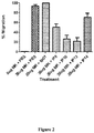
- test anti-N-domain antibodies are capable of inhibiting midkine induced cell migration.
- the efficacy of a test anti-N-domain antibody to treat a disease or condition is assessed using an in vivo assay.
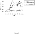
- test anti-N-domain antibody may be administered to a non-human mammal (e.g., non-human primate) model of multiple sclerosis or an inflammatory disease such as arthritis.
- a non-human mammal e.g., non-human primate
- an inflammatory disease such as arthritis.
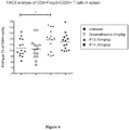
- the level of an anti-inflammatory cytokine is detected in the circulation of a mammal, e.g., using an ELISA.
- a test antibody that increases the level of the cytokine compared to the level prior to administration and/or in a control mammal to which the test antibody has not been administered is considered suitable for treating the disease or condition.
- MK-related disease refers to a disease involving MK functions.
- diseases include: diseases attributed to cell growth or angiogenesis, such as cancers (esophageal cancer, thyroid cancer, urinary bladder cancer, colon cancer, stomach cancer, pancreatic cancer, thoracic cancer, liver cancer, lung cancer, breast cancer, neuroblastoma, glioblastoma, uterine cancer, ovarian cancer, prostatic cancer, and Wilms tumor) and endometriosis; inflammatory diseases or diseases attributed to cell migration or suppression of regulatory T cell function, such as inflammatory diseases of the kidney, acute renal failure, chronic kidney diseases arthritis, autoimmune disease (organ-specific autoimmune disease, etc.), rheumatic arthritis (rheumatoid arthritis (RA) or osteoarthritis (OA)), multiple sclerosis (relapsing-remitting multiple sclerosis, etc.
- cancers esophageal cancer, thyroid cancer, urinary bladder cancer, colon cancer, stomach cancer, pancreatic cancer, thoracic cancer
- the MK-related disease is preferably cancer, arteriosclerosis, angiogenesis-related disease, angina pectoris, myocardial infarction, cerebral infarction, cerebral hemorrhage, hypertension, nephritis, chronic obstructive pulmonary disease (COPD) or multiple sclerosis.
- arteriosclerosis preferably cancer, arteriosclerosis, angiogenesis-related disease, angina pectoris, myocardial infarction, cerebral infarction, cerebral hemorrhage, hypertension, nephritis, chronic obstructive pulmonary disease (COPD) or multiple sclerosis.
- COPD chronic obstructive pulmonary disease
- the anti N-domain antibody antibodies of the present disclosure inhibit MK functions and can therefore be used for the prevention or inhibition of MK-related disorders such as post-laparotomy adhesions.
- compositions or methods for administration of the anti-N-domain antibody to a mammal the antibody is combined with a pharmaceutically acceptable carrier, diluent and/or excipient, as is understood in the art.
- a pharmaceutical composition comprising the anti-N-domain antibody combined with a pharmaceutically acceptable carrier, diluent and/or excipient.
- the disclosure provides a kit comprising a pharmaceutically acceptable carrier, diluent and/or excipient suitable for combining or mixing with the antibody prior to administration to the mammal.
- the kit may further comprise instructions for use.
- carrier diluent or excipient
- carrier diluent or excipient
- a solid or liquid filler, binder, diluent, encapsulating substance, emulsifier, wetting agent, solvent, suspending agent, coating or lubricant that may be safely administered to any mammal, e.g., a human.
- a variety of acceptable carriers, diluents or excipients, known in the art may be used, as for example described in Remington's Pharmaceutical Sciences (Mack Publishing Co. N.J. USA, 1991 ).
- the carriers, diluents or excipients may be selected from a group including sugars (e.g. sucrose, maltose, trehalose, glucose), starches, cellulose and its derivatives, malt, gelatine, talc, calcium sulphate, oils inclusive of vegetable oils, synthetic oils and synthetic mono- or di-glycerides, lower alcohols, polyols, alginic acid, phosphate buffered solutions, lubricants such as sodium or magnesium stearate, isotonic saline and pyrogen-free water.
- the carrier, diluent or excipient is compatible with, or suitable for, parenteral administration.
- Parenteral administration includes any route of administration that is not through the alimentary canal.
- Non-limiting examples of parenteral administration include injection, infusion and the like.
- administration by injection includes intravenous, intra-arterial, intramuscular and subcutaneous injection.
- delivery by a depot or slow-release formulation which may be delivered intradermally, intramuscularly and subcutaneously, for example.
- the anti-N-domain antibody is administered in combination with another compound useful for treating a disease or condition, e.g., an inflammatory disease, either as combined or additional treatment steps or as additional components of a therapeutic formulation.
- a disease or condition e.g., an inflammatory disease
- the other compound is an anti-inflammatory compound.
- the other compound is an immunosuppressant.
- the other compound is a corticosteroid, such as prednisone and/or prednisolone.
- the other compound is an antimalarial compound, such as hydroxychloroquine or chloroquinine.
- the other compound is methotrexate.
- the other compound is azathioprine.
- the other compound is cyclophosphamide.
- the appropriate dosage of an active agent i.e anti-N-domain antibody
- an active agent i.e anti-N-domain antibody
- the particular dosage regimen i.e., dose, timing, and repetition, will depend on the particular individual and that individual's medical history as assessed by a physician.
- a clinician will administer an immunoglobulin until a dosage is reached that achieves the desired result.
- normal dosage amounts may vary from about 10ng/kg up to about 100mg/kg of an individual's body weight or more per day. Exemplary dosages and ranges thereof are described herein.
- the treatment can be sustained until a desired suppression of symptoms is achieved.
- the antibody is administered at an initial (or loading) dose of between about 1mg/kg to about 30mg/kg, such as from about 1mg/kg to about 10mg/kg, or about 2mg/kg or about 3mg/kg or 4mg/kg or 5mg/kg.
- the antibody can then be administered at a maintenance dose of between about 0.0001mg/kg to about 1mg/kg, such as from about 0.0005mg/kg to about 1mg/kg, for example, from about 0.001mg/kg to about 1mg/kg, such as about 0.005mg/kg to about 1mg/kg, for example from about 0.1mg/kg to about 1mg/kg, such as about 0.2mg/kg or 0.3mg/kg or 0.4mg/kg or 0.5mg/kg.
- the maintenance doses may be administered every 7-30 days, such as, every 10-15 days, for example, every 10 or 11 or 12 or 13 or 14 or 15 days.
- Dosages for a particular antibody may be determined empirically in mammals who have been given one or more administrations of the antibody.
- a clinical symptom of a disease or condition e.g., lupus (such as SLE) can be monitored.
- Administration of an antibody according to the methods of the present disclosure can be continuous or intermittent, depending, for example, on the recipient's physiological condition, whether the purpose of the administration is therapeutic or prophylactic, and other factors known to skilled practitioners.
- the administration of an antibody may be essentially continuous over a preselected period of time or may be in a series of spaced doses, e.g., either during or after development of an inflammatory disorder.
- the anti-N-domain antibodies described herein may be used as diagnostic agents.
- the antibodies may be used in diagnostic methods such as ELISAs, radioimmunoassays, immunohistological methods, and western blotting.
- tissue samples or liquids collected as biopsies from test subjects can be used as specimens for the diagnostic agent of the present invention.
- the biopsies used are not particularly limited as long as they are targeted by the immunological measurement of MK. Examples thereof can include tissues, blood, urine, serous fluids, spinal fluids, synovial fluids, aqueous humor, lacrimal fluids, saliva or fractionated or processed products thereof. Analysis using the diagnostic agent can be conducted qualitatively, quantitatively, or semi-quantitatively.
- MK gene-knockout mice were prepared by a method known in the art (Japanese Patent Laid-Open No. 2002-85058 and Nakamura, E. et al.: Genes Cells 3, p. 811-822 ).
- Human MK mRNAs were prepared from a cultured cell line G-401 derived from Wilms tumor ( Tsutsui, J. et al., Biochem. Biophys. Res. Commun. 176, 792-797, 1991 ). Primers were designed such that they contained a sequence recognized by a restriction enzyme EcoRI (5'-GAATTC-3').
- PCR polymerase chain reaction
- the MK cDNAs and expression vectors pHIL301 (containing histidine and a neomycin resistance gene; see Japanese Patent Laid-Open No. 2-104292 and EP Patent No. 0339568 ) for yeast Pichia pastoris GS115 (hereinafter, referred to as "Pichia yeast GS115") were digested with a restriction enzyme EcoRI and then ligated using a ligation kit (manufactured by TAKARABIO INC.) to prepare recombinant expression vectors.
- a ligation kit manufactured by TAKARABIO INC.
- the recombinant expression vectors thus prepared were introduced into Pichia yeast GS115 (manufactured by Invitrogen Corp.) using electroporation.
- the vector-introduced Pichia yeast GS115 was cultured in a G418-containing medium free from histidine to obtain several clones having the MK gene of interest.
- the obtained clones were cultured, while induced with methanol.
- the culture supernatant was collected, and western blotting using rabbit anti-mouse MK polyclonal antibodies was conducted to confirm whether the clones secreted MK.
- T3L-50-4P One of the clones that secreted MK into the culture supernatant by the induction was designated as T3L-50-4P, and this clone was cultured (see Japanese Patent Laid-Open No. 7-39889 ).
- the MK secretion products were collected from the culture supernatant and subjected to purification by ion-exchange chromatography and affinity chromatography using a heparin column to obtain highly pure MK.
- the MK-knockout mice were immunized with the antigens.
- the antigens were prepared as an antigen solution in an amount of 10 ⁇ g per mouse diluted with a saline to 0.1 ml, and mixed with 0.1 ml of FCA for emulsification, and this mixture was hypodermically administered to the dorsal regions of the mice.
- the mice were immunized on a total of 8 occasions, two weeks apart.
- the 8th immunization was performed by administering a solution containing 10 ⁇ g of the antigen solution directly dissolved in 0.1 ml of a saline to the tail veins of the mice through intravenous injection.
- the ELISA was conducted by the following method: first, the antigen solution was prepared to a concentration of 1.0 ⁇ g/ml or 0.1 ⁇ g/ml with PBS (pH 7.2 to 7.4) and dispensed in an amount of 50 ⁇ l/well or 100 ⁇ l/well to a 96-well assay plate (manufactured by BD FALCON, 353912; or manufactured by NUNC, 468667), which was then left standing overnight at 4°C to immobilize the antigens thereon. The plate was washed three times with 0.05% Tween 20-PBS.
- BlockAce manufactured by Dainippon Pharmaceutical Co., Ltd.
- BSA manufactured by Wako Pure Chemical Industries, Ltd., 019-15134
- Tween 20-PBS in an amount of 300 ⁇ l/wel was added thereto, and the plate was left standing at 37°C for 2 hours or overnight at 4°C for blocking. The plate was washed three times with 0.05% Tween 20-PBS.
- HRP substrates 25 ml of a substrate solution (10.206 mg/ml citric acid monohydrate and 36.82 mg/ml disodium hydrogen phosphate dodecahydrate in distilled H2O), 10 mg of OPD, and 5 ⁇ l of 30% H2O2 in an amount of 50 ⁇ l/well or TMB+Substrate Chromogen (manufactured by DAKO, S1599) in an amount of 100 ⁇ l/well were added thereto, and the plate was left standing at room temperature for 20 minutes under shading conditions. The reaction was terminated by the addition of 1 N sulfuric acid in an amount of 50 ⁇ l/well, and the antibody titers were measured at a wavelength of 492 nm or 450 nm.
- mice were secured, and their chests were wiped with alcohol-moistened cotton.
- Blood was collected from the heart using a 2.5 ml syringe and a 23-G needle. After blood collection, the mice were placed in a beaker containing 20 ml of alcohol for disinfection for approximately 3 minutes. The collected blood was placed in a 1.5-ml tube and left at 37°C for 1 hour and then overnight at 4°C, followed by centrifugation at 3,000 rpm for 10 minutes. The sera were transferred to another 1.5-ml tube and stored at 4°C after addition of 0.05% sodium azide.
- mice after blood collection were denuded of epithelium using scissors and tweezers. Furthermore, the endothelium was picked up and incised for separation of the spleen.
- the spleen was washed five times in order with 200 ml of an RPMI1640 S.P medium dispensed in advance to five Petri dishes. The spleen thus washed was placed in a mesh, incised several times with scissors, and crushed using a glass rod. The mesh was washed with an RPMI1640 S.P medium to collect the spleen cells into a 40-ml glass centrifuge.
- the collected spleen cells were centrifuged at 1200 rpm for 10 minutes, and the supernatant was removed using a suction pipette. 40 ml of an RPMI1640 S.P medium was added to the cells, and the cells were centrifuged at 1200 rpm for 10 minutes. 40 ml of an RPMI1640 S.P medium was added to the obtained spleen cells, and the cells were well stirred. The cell count was measured using a hemocytometer.
- Myeloma cells (P3U1) placed in a Petri dish were collected into a 50-ml centrifuge by spraying several times using a pipette. The cells were centrifuged at 1000 rpm for 5 minutes, and the supernatant was removed using a suction pipette. 40 ml of an RPMI1640 S.P medium was added to the cells, and the cells were centrifuged at 1000 rpm for 5 minutes. 40 ml of an RPMI1640 S.P medium was added to the obtained myeloma cells, and the cells were well stirred. The cell count was measured using a hemocytometer.
- the myeloma cells were placed in the 50-ml glass centrifuge containing the spleen cells such that the ratio between the spleen cells and the myeloma cells was 5:1. After mixing, the cells were centrifuged at 1200 rpm for 10 minutes, and the supernatant was removed using a suction pipette. The centrifuge was then tapped.
- Antibody IP-13 (2.85mg/ml) was diluted to 5.7 ⁇ g/ml in Biacore acetate pH 4.5 buffer and amine coupled to of flow cell (FC) #3 of a CM-4 chip. Approximately 750 units were aimed to be immobilized onto Flow cells (FC) #3 of the CM-4 chip; and FC#1 was blocked using EDC/NHS followed by ethanolamine and was used as reference during the study.
- Antibody IP-9 (0.74mg/ml) was diluted to 7.4 ⁇ g/ml in Biacore acetate pH 4.5 buffer and amine coupled to of flow cell (FC) #4 of a CM-4 chip. Approximately 750 units were aimed to be immobilized onto Flow cells (FC) #4 of the CM-4 chip; and FC#1 was blocked using EDC/NHS followed by ethanolamine and was used as reference during the study
- FC flow cells
- FC#1 was blocked using EDC/NHS followed by ethanolamine and was used as reference during the study
- MK17 Recombinant human midkine was diluted to 15 ⁇ M (1 ⁇ l of 10mg/ml in 50 ⁇ l of HBS-EP buffer, the calculation was based on molecular weight of 13.4kDa).
- Various dilutions of human midkine using HBS-EP buffer were prepared (0, 1, 2, 3, 4, 5, 10 and 20nM) .
- Antibody IP-10 (2.1mg/ml) was diluted to 10 ⁇ g/ml in Biacore HBS-EP+ buffer and captured on FC#2 to ⁇ 300RU for each injection cycle. Recombinant human midkine was midkine was diluted to 746nM (1 ⁇ l of 10mg/ml in 1ml of HBS-EP+ buffer, the calculation was based on molecular weight of 13.4kDa).
- the heavy and light chains of mAb IP-13 were sequences and the amino acid sequences are shown below (with CDR sequences in bold):
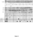
- the midkine N-domain (MK residues 1-61) gene was sequenced and cloned: "WT (1-61)”. Mutants of WT (1-61) MK N-domain were expressed with selected single amino acids mutated from WT to Alanine (A). Anti-N-domain mAbs were then used to detect mutant MK N-domain proteins by Western blot after separation under non-reducing conditions by SDS-PAGE
- the UMR-106 rat osteosarcoma cell line was originally obtained from the American Type Culture Collection, PO Box 1549, Manassas, Virginia, USA as a frozen stock (catalog no. CRL-1661; batch no. 58494148).
- DMEM+Glutamax (DMEM, catalog no. 10569-010 500 mL, lot. no. 778325, Invitrogen/Gibco) supplemented with the following items: Penicillin (10,000 U//mL stock concentration)- Streptomycin (10,000 ug/mL stock concentration) (catalog no. 15140-122 100 mL, lot no. 730849, Invitrogen/Gibco) at a rate of 1 mL/200 mL of DMEM 10% fetal bovine serum (FBS, catalog no. 100099-141, lot no. 6955347Y, Invitrogen/Gibco).
- FBS fetal bovine serum
- the DEM+Glutamax media containing 50 U/mL penicillin, 50 ug/mL streptomycin and 10% FBS is identified as Complete media in this report.
- the 10% FBS was replaced by 0.3% (w/v) bovine serum albumin (BSA) for the cell migration assays.
- BSA bovine serum albumin
- the BSA was supplied by Sigma-Aldrich, catalog no. A-3192, lot no. 31K1264 and was formulated as a 15% (w/v) stock solution in D-PBS which was sterilized by passage through a 0.22 micron filter and stored at -20oC prior to use.
- the cell cultures were maintained in T75 tissue culture-treated flasks in a humidified incubator at approximately 37oC and 5% carbon dioxide.
- Cells were sub-cultured twice weekly at a sub-culture ratio ranging from approximately 1:8 to 1:20.
- Cells were dissociated from the flask by rinsing once with Dulbecco's phosphate buffered saline (DPBS, catalog no. 14190, lot no. 783715, Invitrogen/Gibco) and then incubated with 0.25% Trypsin-EDTA solution (Invitrogen/Gibco) at 37oC for 2-5 minutes. The cells were then removed by addition of approximately 10 mL of Complete media. The experiments were completed using UMR-106 cells at -50 to 75% confluency on the day prior to assay.
- DPBS Dulbecco's phosphate buffered saline
- Trypsin-EDTA solution Invitrogen/Gibco
- the cell migration assays were performed using the following modified Boyden Chamber kits supplied by Millipore Australia: QCM Chemotaxis Cell Migration Assay, 24-well (8um pores), colormetric (catalog no. ECM 508).
- Unused stock solution of the IgG1 protein was aliquoted and stored frozen at approximately -20oC for the study duration.
- PBS vehicle control treatments were prepared by dilution of D-PBS at a 1 in 20 (v/v) ratio in DMEM/0.3% BSA.
- the cell migration assays were performed using the following modified Boyden Chamber kits supplied by Millipore Australia: QCM Chemotaxis Cell Migration Assay, 24-well (8um pores), colorimetric (catalog no. ECM 508). This assay method is based on the manufacturers' instructions and the results of the previous method development/validation study at the test facility.
- QCM Chemotaxis Cell Migration Assay 24-well (8 um pores)
- mice Ten week old female C57BL/6 mice (Taconic) were injected subcutaneously in both hind limbs with 100 ⁇ lper limb of MOG35-55 peptide (1 mg/ml) emulsified with CFA (2 mg/ml MTB). Immediately following the subcutaneous injection, mice were injected intraperitoneally with pertussis toxin (3.75x, HookeLaboratories), and again 24 hours later. IP-13 (10mg/kg) was injected intraperitoneally on days 0, 3, 7, 10, 14, 18, 21 and 24. Dextramethasone was injected intraperitoneally every second day. Mice monitored from day 8 to 24 for the parameters described below.
- spleens were isolated, cut into 1-3mm pieces and incubated in media containing collagenase for 45 minutes at 37°C. Following the incubation any remaining pieces were gently mashed through a 75 ⁇ m cell strainer and washed thoroughly in IMDM (Invitrogen) followed by MACS buffer. The resulting single cell suspensions were incubated with CD4+ MACS beads and CD4+ T cell were isolated on individual MACS columns following the manufacturers instructions. The flow through CD4-negative cells were treated with Mitomycin C at 50 ⁇ g/ml for 30 minutes at 37°C followed by washing twice in IMDM.
- CD4+ T cells (2x10⁵ total cells) and Mitomycin C treated CD4- cells (5x10⁶ total cells) were cultured together with 200ng/ml MOG35-55 peptide for 3 days. Culture supernatant was isolated on day 3 and analyzed with a custom multiplex for the indicated cytokines. Results are shown in Figure 5 .
- Anti-inflammatory splenic T reg cells were also measured at day 28.
- CD4+ T cells were cultured as described above. For the assessment of proliferation, cultures were supplemented with 2.5 ⁇ Ci/ml 3H Thymidine for the final 18 hours of the culture and 3H Thymidine uptake was determined utilizing a TopCountTM Microplate Scintillation Counter. Results are shown in Figure 6 .
- Renal ischemia/reperfusion is a common cause of acute renal failure (ARF), wherein the renal tubes become susceptible to hypoxic injury.
- Inflammatory responses to the hypoxia including renal synthesis of inflammatory cytokines, generation of chemokines and leukocyte infiltration of the kidney, contribute to resultant tissue injury.
- Therapies that target specific inflammatory cell types or effector proteins such as chemokines or adhesion molecules, can reduce ischemic ARF in animal models and recover kidney function.
- BUN serum blood urea nitrogen
- 10-12 week old male Sprague Dawley rats were assessed for baseline serum blood urea nitrogen (BUN) levels to assess kidney function, with a range of 12-18mg/dL being the normal BUN range in healthy rats of this age. Kidney damage is evident when BUN levels exceeded 18mg/dL.
- Animals were weighed and randomly assigned to either control (saline) or anti-MK mAb (IP10, 10mg/kg) treatment groups. On Day 0, animals were anesthetized., midline laparotomy was performed and the kidneys exposed. The renal pedicles were clamped to create bilateral acute ischemia. Ischemia was verified by a change in renal color.
- test or control article was administered intravenously (IV) as a bolus ( ⁇ 500 ⁇ L) through the tail vein.
- IV intravenously
- the abdominal incision was repaired and the animal recovered.
- test/control articles were once again administered IV as a bolus ( ⁇ 500 ⁇ L) through the tail vein.
Claims (9)
- Protéine isolée ou recombinante comprenant un domaine de liaison à l'antigène d'un anticorps qui se lie spécifiquement à un épitope localisé à l'intérieur du domaine N de la midkine tel que défini par les résidus d'acides aminés 1-61 de SEQ ID n° : 2, la protéine isolée ou recombinante inhibant ou réduisant la migration des cellules induites par la midkine et comprenant :un domaine variable de chaîne lourde comprenant des séquences CDR de chaîne lourde CDR1, CDR2 et CDR3 telles que montrées dans SEQ ID n° : 7, SEQ ID n° : 8 et SEQ ID n° : 9 respectivement ; etun domaine variable de chaîne légère comprenant des séquences CDR de chaîne légère CDR1, CDR2 et CDR3 telles que montrées dans SEQ ID n° : 10, SEQ ID n° : 11 et SEQ ID n° : 12 respectivement pour l'utilisation dans le traitement ou la prévention d'un trouble lié à la midkine, le trouble lié à la midkine étant une maladie auto-immune, le cancer, une maladie inflammatoire, la sclérose en plaques, ou les adhérences post-laparotomie.
- Protéine isolée ou recombinante pour l'utilisation selon la revendication 1, la protéine se liant au même épitope qu'un anticorps qui présente un domaine variable de chaîne lourde tel que montré dans SEQ ID n° : 5 et un domaine variable de chaîne légère tel que montré dans SEQ ID n° : 6
- Protéine isolée ou recombinante pour l'utilisation selon la revendication 1 ou 2, la protéine isolée ou recombinante comprenant un domaine variable de chaîne lourde (VH) et un domaine variable de chaîne légère (VL).
- Protéine isolée ou recombinante pour l'utilisation selon la revendication 3, la protéine isolée ou recombinante étant :(i) un fragment Fv de chaîne unique (scFv) ;(ii) un scFv dimère (di-scFv) ;(iii) au moins l'un de (i) et/ou (ii) lié à un Fc ou un domaine constant de chaîne lourde (CH)2 et/ou CH3 ;(iv) un diacorps ;(v) un tricorps ;(vi) a tétracorps ;(vii) un Fab ;(viii) un F(ab')2 ;(ix) un Fv ; ou(x) l'un de (iv) à (vi) lié à un Fc ou un domaine constant de chaîne lourde (CH)2 et/ou CH3.
- Protéine isolée ou recombinante pour l'utilisation selon l'une quelconque des revendications 1 à 4, la protéine isolée ou recombinante étant un anticorps chimère, désimmunisé ou humanisé.
- Protéine isolée ou recombinante pour l'utilisation selon l'une quelconque des revendications 1 à 5 sous la forme d'un anticorps comprenant
un domaine variable de chaîne lourde comprenant SEQ ID n° : 5 ou une séquence faisant preuve de 95 % d'identité ou plus vis-à-vis de celle-ci ; et
un domaine variable de chaîne légère comprenant SEQ ID n° : 6 ou une séquence faisant preuve de 95 % d'identité ou plus vis-à-vis de celle-ci. - Protéine isolée ou recombinante pour l'utilisation selon l'une quelconque des revendications 1 à 6, la protéine isolée ou recombinante étant conjuguée à un composé.
- Protéine isolée ou recombinante pour l'utilisation selon l'une quelconque des revendications 1 à 7, le trouble étant une maladie auto-immune, le cancer, une maladie inflammatoire ou la sclérose en plaques.
- Protéine isolée ou recombinante pour l'utilisation selon l'une quelconque des revendications 1 à 7, l'utilisation servant à prévenir ou inhiber les adhérences post-laparotomie.
Applications Claiming Priority (2)
| Application Number | Priority Date | Filing Date | Title |
|---|---|---|---|
| US201161452337P | 2011-03-14 | 2011-03-14 | |
| PCT/AU2012/000251 WO2012122590A1 (fr) | 2011-03-14 | 2012-03-13 | Anticorps de reconnaissance de domaine n-terminal de midkine |
Publications (3)
| Publication Number | Publication Date |
|---|---|
| EP2686016A1 EP2686016A1 (fr) | 2014-01-22 |
| EP2686016A4 EP2686016A4 (fr) | 2014-09-03 |
| EP2686016B1 true EP2686016B1 (fr) | 2019-05-01 |
Family
ID=46829949
Family Applications (1)
| Application Number | Title | Priority Date | Filing Date |
|---|---|---|---|
| EP12757658.5A Active EP2686016B1 (fr) | 2011-03-14 | 2012-03-13 | Anticorps de reconnaissance de domaine n-terminal de midkine |
Country Status (3)
| Country | Link |
|---|---|
| US (1) | US9624294B2 (fr) |
| EP (1) | EP2686016B1 (fr) |
| WO (1) | WO2012122590A1 (fr) |
Families Citing this family (6)
| Publication number | Priority date | Publication date | Assignee | Title |
|---|---|---|---|---|
| US9659437B2 (en) * | 2012-09-28 | 2017-05-23 | Bally Gaming, Inc. | System and method for cross platform persistent gaming sessions using a mobile device |
| AU2015333590B2 (en) * | 2014-10-14 | 2021-03-11 | Cellmid Limited | Improved midkine antibody |
| JP2019510080A (ja) * | 2016-03-01 | 2019-04-11 | セルミド リミティッド | 骨疾患、障害及び/または傷害の治療方法及びそのための試薬 |
| WO2019136516A1 (fr) * | 2018-01-09 | 2019-07-18 | Cellmid Limited | Procédés de traitement d'une myocardite et/ou d'une cardiomyopathie et réactifs correspondants |
| JP2020027089A (ja) * | 2018-08-17 | 2020-02-20 | 国立大学法人埼玉大学 | 癌の診断用モノクローナル抗体及び癌診断キット |
| WO2021000004A1 (fr) * | 2019-07-04 | 2021-01-07 | Cellmid Limited | Methodes de traitement d'une myocardite et/ou d'une cardiomyopathie et réactifs correspondants |
Family Cites Families (34)
| Publication number | Priority date | Publication date | Assignee | Title |
|---|---|---|---|---|
| US4196265A (en) | 1977-06-15 | 1980-04-01 | The Wistar Institute | Method of producing antibodies |
| US4816567A (en) | 1983-04-08 | 1989-03-28 | Genentech, Inc. | Recombinant immunoglobin preparations |
| US5807715A (en) | 1984-08-27 | 1998-09-15 | The Board Of Trustees Of The Leland Stanford Junior University | Methods and transformed mammalian lymphocyte cells for producing functional antigen-binding protein including chimeric immunoglobulin |
| US4676980A (en) | 1985-09-23 | 1987-06-30 | The United States Of America As Represented By The Secretary Of The Department Of Health And Human Services | Target specific cross-linked heteroantibodies |
| US5225539A (en) | 1986-03-27 | 1993-07-06 | Medical Research Council | Recombinant altered antibodies and methods of making altered antibodies |
| IL89989A0 (en) | 1988-04-25 | 1989-12-15 | Phillips Petroleum Co | Expression of human interleukin-2 in methylotrophic yeasts |
| KR0184860B1 (ko) | 1988-11-11 | 1999-04-01 | 메디칼 리써어치 카운실 | 단일영역 리간드와 이를 포함하는 수용체 및 이들의 제조방법과 이용(법) |
| US5530101A (en) | 1988-12-28 | 1996-06-25 | Protein Design Labs, Inc. | Humanized immunoglobulins |
| ES2136092T3 (es) | 1991-09-23 | 1999-11-16 | Medical Res Council | Procedimientos para la produccion de anticuerpos humanizados. |
| DK1024191T3 (da) | 1991-12-02 | 2008-12-08 | Medical Res Council | Fremstilling af autoantistoffer fremvist på fag-overflader ud fra antistofsegmentbiblioteker |
| AU3691093A (en) | 1992-04-14 | 1993-10-21 | Hybritech Incorporated | Methods of inhibiting the growth of multidrug resistant tumors |
| ES2162823T5 (es) | 1992-08-21 | 2010-08-09 | Vrije Universiteit Brussel | Inmunoglobulinas desprovistas de cadenas ligeras. |
| US5844094A (en) | 1992-09-25 | 1998-12-01 | Commonwealth Scientific And Industrial Research Organization | Target binding polypeptide |
| JPH0739889A (ja) | 1993-07-29 | 1995-02-10 | Meidensha Corp | 高濃度アンモニア廃液の処理方法 |
| US5731168A (en) | 1995-03-01 | 1998-03-24 | Genentech, Inc. | Method for making heteromultimeric polypeptides |
| AUPO591797A0 (en) | 1997-03-27 | 1997-04-24 | Commonwealth Scientific And Industrial Research Organisation | High avidity polyvalent and polyspecific reagents |
| EP0859841B1 (fr) | 1995-08-18 | 2002-06-19 | MorphoSys AG | Banques de proteines/(poly)peptides |
| CA2258518C (fr) | 1996-06-27 | 2011-11-22 | Vlaams Interuniversitair Instituut Voor Biotechnologie Vzw | Molecules de reconnaissance ayant une interaction specifique avec le site actif ou la fissure d'une molecule cible |
| DE69835016T2 (de) | 1997-07-14 | 2007-06-28 | Muramatsu, Takashi, Nagoya | Verwendung von midkin oder von anti-midkin antikörper |
| ATE321066T1 (de) | 1998-05-06 | 2006-04-15 | Genentech Inc | Anti-her2 antikörperzusammensetzung |
| WO2000010608A1 (fr) | 1998-08-24 | 2000-03-02 | Meiji Milk Products Co., Ltd. | Medicament pour le traitement et/ou la prevention de l'arteriosclerose et de la reangiostenose post-actp |
| AU776910B2 (en) | 1998-12-08 | 2004-09-23 | Merck Patent Gesellschaft Mit Beschrankter Haftung | Modifying protein immunogenicity |
| JP3887154B2 (ja) | 2000-09-07 | 2007-02-28 | 喬 村松 | ヒトmkに対するモノクローナル抗体 |
| US6596541B2 (en) | 2000-10-31 | 2003-07-22 | Regeneron Pharmaceuticals, Inc. | Methods of modifying eukaryotic cells |
| US7501494B2 (en) | 2003-01-15 | 2009-03-10 | United Biomedical, Inc. | Designed deimmunized monoclonal antibodies for protection against HIV exposure and treatment of HIV infection |
| JP4768440B2 (ja) * | 2003-03-06 | 2011-09-07 | セルミド リミテッド | 開腹手術後の癒着の予防剤 |
| CN1822857A (zh) | 2003-06-02 | 2006-08-23 | 阿莱克申药物公司 | 去免疫原性抗cd3抗体 |
| US7235641B2 (en) | 2003-12-22 | 2007-06-26 | Micromet Ag | Bispecific antibodies |
| AU2005250055B2 (en) | 2004-06-02 | 2008-09-11 | Adalta Pty Ltd | Binding moieties based on shark IgNAR domains |
| US20070135620A1 (en) | 2004-11-12 | 2007-06-14 | Xencor, Inc. | Fc variants with altered binding to FcRn |
| US20060263367A1 (en) | 2005-05-23 | 2006-11-23 | Fey Georg H | Bispecific antibody devoid of Fc region and method of treatment using same |
| EP1964574B1 (fr) | 2005-11-14 | 2016-09-07 | Cellmid Limited | Procede de traitement ou de prevention d une maladie associee a un trouble fonctionnel des lymphocytes t regulateurs |
| JP5663137B2 (ja) * | 2006-11-14 | 2015-02-04 | セルミド リミテッド | ミッドカインのc−ドメインを認識する抗体 |
| WO2010074218A1 (fr) * | 2008-12-25 | 2010-07-01 | 株式会社イーベック | Anticorps anti-humain de midkine |
-
2012
- 2012-03-13 WO PCT/AU2012/000251 patent/WO2012122590A1/fr active Application Filing
- 2012-03-13 US US14/004,548 patent/US9624294B2/en active Active
- 2012-03-13 EP EP12757658.5A patent/EP2686016B1/fr active Active
Non-Patent Citations (3)
| Title |
|---|
| FOUAD YOUSEF AHMED ET AL: "Revisiting the hallmarks of cancer", AMERICAN JOURNAL OF CANCER RESEARCH, vol. 7, no. 5, 2017, pages 1016 - 1036, ISSN: 2156-6976 * |
| HANAHAN DOUGLAS ET AL: "Hallmarks of Cancer: The Next Generation", CELL, vol. 144, no. 5, 4 March 2011 (2011-03-04), pages 646 - 674, ISSN: 0092-8674(print) * |
| MAEHARA H ET AL: "Midkine as a novel target for antibody therapy in osteosarcoma", BIOCHEMICAL AND BIOPHYSICAL RESEARCH COMMUNICATIONS, ACADEMIC PRESS INC. ORLANDO, FL, US, vol. 358, no. 3, 6 July 2007 (2007-07-06), pages 757 - 762, XP025322370, ISSN: 0006-291X, [retrieved on 20070526], DOI: 10.1016/J.BBRC.2007.04.183 * |
Also Published As
| Publication number | Publication date |
|---|---|
| US9624294B2 (en) | 2017-04-18 |
| US20140170144A1 (en) | 2014-06-19 |
| EP2686016A1 (fr) | 2014-01-22 |
| EP2686016A4 (fr) | 2014-09-03 |
| WO2012122590A1 (fr) | 2012-09-20 |
Similar Documents
| Publication | Publication Date | Title |
|---|---|---|
| JP6923658B2 (ja) | 抗cd47抗体およびその使用 | |
| US20220056136A1 (en) | Anti-pd-l1/anti-4-1bb bispecific antibodies and uses thereof | |
| JP2022166238A (ja) | 抗プロ/潜在型-ミオスタチン抗体およびその使用 | |
| EP3798230B1 (fr) | Anticorps thérapeutiques | |
| US20200115454A1 (en) | Anti-pd-1 antibodies and uses thereof | |
| US20170107280A1 (en) | Antibodies to hgf and compositions containing | |
| EP2686016B1 (fr) | Anticorps de reconnaissance de domaine n-terminal de midkine | |
| US11840573B2 (en) | CD131 binding proteins and uses thereof | |
| TW201531484A (zh) | 抗TNF-α之抗體及其用途 | |
| JP7094698B2 (ja) | 抗体、使用、及び方法 | |
| US20220127356A1 (en) | Trem2 antibodies and uses thereof | |
| US20140271464A1 (en) | Therapeutic use of antibodies to hgf | |
| US20210032350A1 (en) | Antibodies to galectin-3 and methods of use thereof | |
| US11883495B2 (en) | Anti-CD47 antibodies and uses thereof | |
| CN107922939B (zh) | 中和全部种类埃博拉病毒的感染性的单克隆抗体 | |
| WO2018017711A1 (fr) | Traitement de la démence fronto-temporale. | |
| CN110637034B (zh) | Cxcr2抗体及其用途 | |
| US20210261665A1 (en) | Anti-pd-1 antibodies, dosages and uses thereof | |
| CA3124356A1 (fr) | Anticorps fn14 et utilisations associees | |
| KR102356187B1 (ko) | 항-gm-csf 항체 및 이것의 사용 | |
| WO2021043203A1 (fr) | Anticorps anti-cxcr2 et leurs utilisations | |
| RU2773927C2 (ru) | Cd131-связывающие белки и их применения | |
| RU2787629C2 (ru) | Способы и материалы для оценки ответа на терапию против плазмобластов и плазматических клеток | |
| WO2024079074A1 (fr) | Anticorps anti-scd146 et leurs utilisations | |
| CN111072780A (zh) | 白血病干细胞抑制剂及其在治疗慢性粒细胞白血病中的应用 |
Legal Events
| Date | Code | Title | Description |
|---|---|---|---|
| PUAI | Public reference made under article 153(3) epc to a published international application that has entered the european phase |
Free format text: ORIGINAL CODE: 0009012 |
|
| 17P | Request for examination filed |
Effective date: 20131010 |
|
| AK | Designated contracting states |
Kind code of ref document: A1 Designated state(s): AL AT BE BG CH CY CZ DE DK EE ES FI FR GB GR HR HU IE IS IT LI LT LU LV MC MK MT NL NO PL PT RO RS SE SI SK SM TR |
|
| DAX | Request for extension of the european patent (deleted) | ||
| A4 | Supplementary search report drawn up and despatched |
Effective date: 20140806 |
|
| RIC1 | Information provided on ipc code assigned before grant |
Ipc: C07K 16/22 20060101ALI20140731BHEP Ipc: A61K 39/395 20060101AFI20140731BHEP Ipc: A61P 25/28 20060101ALI20140731BHEP Ipc: A61P 37/00 20060101ALI20140731BHEP |
|
| 17Q | First examination report despatched |
Effective date: 20160308 |
|
| STAA | Information on the status of an ep patent application or granted ep patent |
Free format text: STATUS: EXAMINATION IS IN PROGRESS |
|
| GRAP | Despatch of communication of intention to grant a patent |
Free format text: ORIGINAL CODE: EPIDOSNIGR1 |
|
| STAA | Information on the status of an ep patent application or granted ep patent |
Free format text: STATUS: GRANT OF PATENT IS INTENDED |
|
| INTG | Intention to grant announced |
Effective date: 20181001 |
|
| RIN1 | Information on inventor provided before grant (corrected) |
Inventor name: JONES, DARREN Inventor name: HALASZ, MARIA Inventor name: SAKUMA, SADATOSI |
|
| GRAS | Grant fee paid |
Free format text: ORIGINAL CODE: EPIDOSNIGR3 |
|
| GRAA | (expected) grant |
Free format text: ORIGINAL CODE: 0009210 |
|
| STAA | Information on the status of an ep patent application or granted ep patent |
Free format text: STATUS: THE PATENT HAS BEEN GRANTED |
|
| AK | Designated contracting states |
Kind code of ref document: B1 Designated state(s): AL AT BE BG CH CY CZ DE DK EE ES FI FR GB GR HR HU IE IS IT LI LT LU LV MC MK MT NL NO PL PT RO RS SE SI SK SM TR |
|
| REG | Reference to a national code |
Ref country code: GB Ref legal event code: FG4D |
|
| REG | Reference to a national code |
Ref country code: CH Ref legal event code: EP Ref country code: AT Ref legal event code: REF Ref document number: 1126037 Country of ref document: AT Kind code of ref document: T Effective date: 20190515 |
|
| REG | Reference to a national code |
Ref country code: DE Ref legal event code: R096 Ref document number: 602012059614 Country of ref document: DE |
|
| REG | Reference to a national code |
Ref country code: IE Ref legal event code: FG4D |
|
| REG | Reference to a national code |
Ref country code: CH Ref legal event code: NV Representative=s name: ISLER AND PEDRAZZINI AG, CH |
|
| REG | Reference to a national code |
Ref country code: NL Ref legal event code: MP Effective date: 20190501 |
|
| REG | Reference to a national code |
Ref country code: LT Ref legal event code: MG4D |
|
| PG25 | Lapsed in a contracting state [announced via postgrant information from national office to epo] |
Ref country code: NO Free format text: LAPSE BECAUSE OF FAILURE TO SUBMIT A TRANSLATION OF THE DESCRIPTION OR TO PAY THE FEE WITHIN THE PRESCRIBED TIME-LIMIT Effective date: 20190801 Ref country code: LT Free format text: LAPSE BECAUSE OF FAILURE TO SUBMIT A TRANSLATION OF THE DESCRIPTION OR TO PAY THE FEE WITHIN THE PRESCRIBED TIME-LIMIT Effective date: 20190501 Ref country code: FI Free format text: LAPSE BECAUSE OF FAILURE TO SUBMIT A TRANSLATION OF THE DESCRIPTION OR TO PAY THE FEE WITHIN THE PRESCRIBED TIME-LIMIT Effective date: 20190501 Ref country code: SE Free format text: LAPSE BECAUSE OF FAILURE TO SUBMIT A TRANSLATION OF THE DESCRIPTION OR TO PAY THE FEE WITHIN THE PRESCRIBED TIME-LIMIT Effective date: 20190501 Ref country code: HR Free format text: LAPSE BECAUSE OF FAILURE TO SUBMIT A TRANSLATION OF THE DESCRIPTION OR TO PAY THE FEE WITHIN THE PRESCRIBED TIME-LIMIT Effective date: 20190501 Ref country code: ES Free format text: LAPSE BECAUSE OF FAILURE TO SUBMIT A TRANSLATION OF THE DESCRIPTION OR TO PAY THE FEE WITHIN THE PRESCRIBED TIME-LIMIT Effective date: 20190501 Ref country code: AL Free format text: LAPSE BECAUSE OF FAILURE TO SUBMIT A TRANSLATION OF THE DESCRIPTION OR TO PAY THE FEE WITHIN THE PRESCRIBED TIME-LIMIT Effective date: 20190501 Ref country code: PT Free format text: LAPSE BECAUSE OF FAILURE TO SUBMIT A TRANSLATION OF THE DESCRIPTION OR TO PAY THE FEE WITHIN THE PRESCRIBED TIME-LIMIT Effective date: 20190901 Ref country code: NL Free format text: LAPSE BECAUSE OF FAILURE TO SUBMIT A TRANSLATION OF THE DESCRIPTION OR TO PAY THE FEE WITHIN THE PRESCRIBED TIME-LIMIT Effective date: 20190501 |
|
| PG25 | Lapsed in a contracting state [announced via postgrant information from national office to epo] |
Ref country code: BG Free format text: LAPSE BECAUSE OF FAILURE TO SUBMIT A TRANSLATION OF THE DESCRIPTION OR TO PAY THE FEE WITHIN THE PRESCRIBED TIME-LIMIT Effective date: 20190801 Ref country code: RS Free format text: LAPSE BECAUSE OF FAILURE TO SUBMIT A TRANSLATION OF THE DESCRIPTION OR TO PAY THE FEE WITHIN THE PRESCRIBED TIME-LIMIT Effective date: 20190501 Ref country code: LV Free format text: LAPSE BECAUSE OF FAILURE TO SUBMIT A TRANSLATION OF THE DESCRIPTION OR TO PAY THE FEE WITHIN THE PRESCRIBED TIME-LIMIT Effective date: 20190501 Ref country code: GR Free format text: LAPSE BECAUSE OF FAILURE TO SUBMIT A TRANSLATION OF THE DESCRIPTION OR TO PAY THE FEE WITHIN THE PRESCRIBED TIME-LIMIT Effective date: 20190802 |
|
| REG | Reference to a national code |
Ref country code: AT Ref legal event code: MK05 Ref document number: 1126037 Country of ref document: AT Kind code of ref document: T Effective date: 20190501 |
|
| PG25 | Lapsed in a contracting state [announced via postgrant information from national office to epo] |
Ref country code: IS Free format text: LAPSE BECAUSE OF FAILURE TO SUBMIT A TRANSLATION OF THE DESCRIPTION OR TO PAY THE FEE WITHIN THE PRESCRIBED TIME-LIMIT Effective date: 20190901 |
|
| PG25 | Lapsed in a contracting state [announced via postgrant information from national office to epo] |
Ref country code: SK Free format text: LAPSE BECAUSE OF FAILURE TO SUBMIT A TRANSLATION OF THE DESCRIPTION OR TO PAY THE FEE WITHIN THE PRESCRIBED TIME-LIMIT Effective date: 20190501 Ref country code: AT Free format text: LAPSE BECAUSE OF FAILURE TO SUBMIT A TRANSLATION OF THE DESCRIPTION OR TO PAY THE FEE WITHIN THE PRESCRIBED TIME-LIMIT Effective date: 20190501 Ref country code: DK Free format text: LAPSE BECAUSE OF FAILURE TO SUBMIT A TRANSLATION OF THE DESCRIPTION OR TO PAY THE FEE WITHIN THE PRESCRIBED TIME-LIMIT Effective date: 20190501 Ref country code: EE Free format text: LAPSE BECAUSE OF FAILURE TO SUBMIT A TRANSLATION OF THE DESCRIPTION OR TO PAY THE FEE WITHIN THE PRESCRIBED TIME-LIMIT Effective date: 20190501 Ref country code: CZ Free format text: LAPSE BECAUSE OF FAILURE TO SUBMIT A TRANSLATION OF THE DESCRIPTION OR TO PAY THE FEE WITHIN THE PRESCRIBED TIME-LIMIT Effective date: 20190501 Ref country code: RO Free format text: LAPSE BECAUSE OF FAILURE TO SUBMIT A TRANSLATION OF THE DESCRIPTION OR TO PAY THE FEE WITHIN THE PRESCRIBED TIME-LIMIT Effective date: 20190501 |
|
| REG | Reference to a national code |
Ref country code: DE Ref legal event code: R097 Ref document number: 602012059614 Country of ref document: DE |
|
| PG25 | Lapsed in a contracting state [announced via postgrant information from national office to epo] |
Ref country code: SM Free format text: LAPSE BECAUSE OF FAILURE TO SUBMIT A TRANSLATION OF THE DESCRIPTION OR TO PAY THE FEE WITHIN THE PRESCRIBED TIME-LIMIT Effective date: 20190501 |
|
| PLBE | No opposition filed within time limit |
Free format text: ORIGINAL CODE: 0009261 |
|
| STAA | Information on the status of an ep patent application or granted ep patent |
Free format text: STATUS: NO OPPOSITION FILED WITHIN TIME LIMIT |
|
| PG25 | Lapsed in a contracting state [announced via postgrant information from national office to epo] |
Ref country code: TR Free format text: LAPSE BECAUSE OF FAILURE TO SUBMIT A TRANSLATION OF THE DESCRIPTION OR TO PAY THE FEE WITHIN THE PRESCRIBED TIME-LIMIT Effective date: 20190501 |
|
| 26N | No opposition filed |
Effective date: 20200204 |
|
| PG25 | Lapsed in a contracting state [announced via postgrant information from national office to epo] |
Ref country code: PL Free format text: LAPSE BECAUSE OF FAILURE TO SUBMIT A TRANSLATION OF THE DESCRIPTION OR TO PAY THE FEE WITHIN THE PRESCRIBED TIME-LIMIT Effective date: 20190501 |
|
| PG25 | Lapsed in a contracting state [announced via postgrant information from national office to epo] |
Ref country code: SI Free format text: LAPSE BECAUSE OF FAILURE TO SUBMIT A TRANSLATION OF THE DESCRIPTION OR TO PAY THE FEE WITHIN THE PRESCRIBED TIME-LIMIT Effective date: 20190501 |
|
| PG25 | Lapsed in a contracting state [announced via postgrant information from national office to epo] |
Ref country code: MC Free format text: LAPSE BECAUSE OF FAILURE TO SUBMIT A TRANSLATION OF THE DESCRIPTION OR TO PAY THE FEE WITHIN THE PRESCRIBED TIME-LIMIT Effective date: 20190501 |
|
| REG | Reference to a national code |
Ref country code: BE Ref legal event code: MM Effective date: 20200331 |
|
| PG25 | Lapsed in a contracting state [announced via postgrant information from national office to epo] |
Ref country code: LU Free format text: LAPSE BECAUSE OF NON-PAYMENT OF DUE FEES Effective date: 20200313 |
|
| PG25 | Lapsed in a contracting state [announced via postgrant information from national office to epo] |
Ref country code: IE Free format text: LAPSE BECAUSE OF NON-PAYMENT OF DUE FEES Effective date: 20200313 |
|
| PG25 | Lapsed in a contracting state [announced via postgrant information from national office to epo] |
Ref country code: BE Free format text: LAPSE BECAUSE OF NON-PAYMENT OF DUE FEES Effective date: 20200331 |
|
| PG25 | Lapsed in a contracting state [announced via postgrant information from national office to epo] |
Ref country code: MT Free format text: LAPSE BECAUSE OF FAILURE TO SUBMIT A TRANSLATION OF THE DESCRIPTION OR TO PAY THE FEE WITHIN THE PRESCRIBED TIME-LIMIT Effective date: 20190501 Ref country code: CY Free format text: LAPSE BECAUSE OF FAILURE TO SUBMIT A TRANSLATION OF THE DESCRIPTION OR TO PAY THE FEE WITHIN THE PRESCRIBED TIME-LIMIT Effective date: 20190501 |
|
| PG25 | Lapsed in a contracting state [announced via postgrant information from national office to epo] |
Ref country code: MK Free format text: LAPSE BECAUSE OF FAILURE TO SUBMIT A TRANSLATION OF THE DESCRIPTION OR TO PAY THE FEE WITHIN THE PRESCRIBED TIME-LIMIT Effective date: 20190501 |
|
| P01 | Opt-out of the competence of the unified patent court (upc) registered |
Effective date: 20230529 |
|
| PGFP | Annual fee paid to national office [announced via postgrant information from national office to epo] |
Ref country code: FR Payment date: 20230629 Year of fee payment: 12 Ref country code: DE Payment date: 20230630 Year of fee payment: 12 Ref country code: CH Payment date: 20230628 Year of fee payment: 12 |
|
| PGFP | Annual fee paid to national office [announced via postgrant information from national office to epo] |
Ref country code: IT Payment date: 20230630 Year of fee payment: 12 Ref country code: GB Payment date: 20230629 Year of fee payment: 12 |