EP2676966A1 - BAG3 as biochemical serum and tissue marker - Google Patents
BAG3 as biochemical serum and tissue marker Download PDFInfo
- Publication number
- EP2676966A1 EP2676966A1 EP12172531.1A EP12172531A EP2676966A1 EP 2676966 A1 EP2676966 A1 EP 2676966A1 EP 12172531 A EP12172531 A EP 12172531A EP 2676966 A1 EP2676966 A1 EP 2676966A1
- Authority
- EP
- European Patent Office
- Prior art keywords
- bag3
- antibodies
- biological sample
- serum
- protein
- Prior art date
- Legal status (The legal status is an assumption and is not a legal conclusion. Google has not performed a legal analysis and makes no representation as to the accuracy of the status listed.)
- Withdrawn
Links
Images


Classifications
-
- C—CHEMISTRY; METALLURGY
- C07—ORGANIC CHEMISTRY
- C07K—PEPTIDES
- C07K14/00—Peptides having more than 20 amino acids; Gastrins; Somatostatins; Melanotropins; Derivatives thereof
- C07K14/435—Peptides having more than 20 amino acids; Gastrins; Somatostatins; Melanotropins; Derivatives thereof from animals; from humans
- C07K14/46—Peptides having more than 20 amino acids; Gastrins; Somatostatins; Melanotropins; Derivatives thereof from animals; from humans from vertebrates
- C07K14/47—Peptides having more than 20 amino acids; Gastrins; Somatostatins; Melanotropins; Derivatives thereof from animals; from humans from vertebrates from mammals
-
- C—CHEMISTRY; METALLURGY
- C07—ORGANIC CHEMISTRY
- C07K—PEPTIDES
- C07K16/00—Immunoglobulins [IGs], e.g. monoclonal or polyclonal antibodies
- C07K16/18—Immunoglobulins [IGs], e.g. monoclonal or polyclonal antibodies against material from animals or humans
-
- C—CHEMISTRY; METALLURGY
- C12—BIOCHEMISTRY; BEER; SPIRITS; WINE; VINEGAR; MICROBIOLOGY; ENZYMOLOGY; MUTATION OR GENETIC ENGINEERING
- C12Q—MEASURING OR TESTING PROCESSES INVOLVING ENZYMES, NUCLEIC ACIDS OR MICROORGANISMS; COMPOSITIONS OR TEST PAPERS THEREFOR; PROCESSES OF PREPARING SUCH COMPOSITIONS; CONDITION-RESPONSIVE CONTROL IN MICROBIOLOGICAL OR ENZYMOLOGICAL PROCESSES
- C12Q1/00—Measuring or testing processes involving enzymes, nucleic acids or microorganisms; Compositions therefor; Processes of preparing such compositions
- C12Q1/68—Measuring or testing processes involving enzymes, nucleic acids or microorganisms; Compositions therefor; Processes of preparing such compositions involving nucleic acids
- C12Q1/6844—Nucleic acid amplification reactions
- C12Q1/686—Polymerase chain reaction [PCR]
-
- C—CHEMISTRY; METALLURGY
- C12—BIOCHEMISTRY; BEER; SPIRITS; WINE; VINEGAR; MICROBIOLOGY; ENZYMOLOGY; MUTATION OR GENETIC ENGINEERING
- C12Q—MEASURING OR TESTING PROCESSES INVOLVING ENZYMES, NUCLEIC ACIDS OR MICROORGANISMS; COMPOSITIONS OR TEST PAPERS THEREFOR; PROCESSES OF PREPARING SUCH COMPOSITIONS; CONDITION-RESPONSIVE CONTROL IN MICROBIOLOGICAL OR ENZYMOLOGICAL PROCESSES
- C12Q1/00—Measuring or testing processes involving enzymes, nucleic acids or microorganisms; Compositions therefor; Processes of preparing such compositions
- C12Q1/68—Measuring or testing processes involving enzymes, nucleic acids or microorganisms; Compositions therefor; Processes of preparing such compositions involving nucleic acids
- C12Q1/6876—Nucleic acid products used in the analysis of nucleic acids, e.g. primers or probes
- C12Q1/6883—Nucleic acid products used in the analysis of nucleic acids, e.g. primers or probes for diseases caused by alterations of genetic material
- C12Q1/6886—Nucleic acid products used in the analysis of nucleic acids, e.g. primers or probes for diseases caused by alterations of genetic material for cancer
-
- G—PHYSICS
- G01—MEASURING; TESTING
- G01N—INVESTIGATING OR ANALYSING MATERIALS BY DETERMINING THEIR CHEMICAL OR PHYSICAL PROPERTIES
- G01N33/00—Investigating or analysing materials by specific methods not covered by groups G01N1/00 - G01N31/00
- G01N33/48—Biological material, e.g. blood, urine; Haemocytometers
- G01N33/50—Chemical analysis of biological material, e.g. blood, urine; Testing involving biospecific ligand binding methods; Immunological testing
- G01N33/53—Immunoassay; Biospecific binding assay; Materials therefor
- G01N33/574—Immunoassay; Biospecific binding assay; Materials therefor for cancer
- G01N33/57407—Specifically defined cancers
- G01N33/57434—Specifically defined cancers of prostate
-
- G—PHYSICS
- G01—MEASURING; TESTING
- G01N—INVESTIGATING OR ANALYSING MATERIALS BY DETERMINING THEIR CHEMICAL OR PHYSICAL PROPERTIES
- G01N33/00—Investigating or analysing materials by specific methods not covered by groups G01N1/00 - G01N31/00
- G01N33/48—Biological material, e.g. blood, urine; Haemocytometers
- G01N33/50—Chemical analysis of biological material, e.g. blood, urine; Testing involving biospecific ligand binding methods; Immunological testing
- G01N33/53—Immunoassay; Biospecific binding assay; Materials therefor
- G01N33/574—Immunoassay; Biospecific binding assay; Materials therefor for cancer
- G01N33/57407—Specifically defined cancers
- G01N33/57438—Specifically defined cancers of liver, pancreas or kidney
-
- G—PHYSICS
- G01—MEASURING; TESTING
- G01N—INVESTIGATING OR ANALYSING MATERIALS BY DETERMINING THEIR CHEMICAL OR PHYSICAL PROPERTIES
- G01N33/00—Investigating or analysing materials by specific methods not covered by groups G01N1/00 - G01N31/00
- G01N33/48—Biological material, e.g. blood, urine; Haemocytometers
- G01N33/50—Chemical analysis of biological material, e.g. blood, urine; Testing involving biospecific ligand binding methods; Immunological testing
- G01N33/53—Immunoassay; Biospecific binding assay; Materials therefor
- G01N33/574—Immunoassay; Biospecific binding assay; Materials therefor for cancer
- G01N33/57484—Immunoassay; Biospecific binding assay; Materials therefor for cancer involving compounds serving as markers for tumor, cancer, neoplasia, e.g. cellular determinants, receptors, heat shock/stress proteins, A-protein, oligosaccharides, metabolites
- G01N33/57496—Immunoassay; Biospecific binding assay; Materials therefor for cancer involving compounds serving as markers for tumor, cancer, neoplasia, e.g. cellular determinants, receptors, heat shock/stress proteins, A-protein, oligosaccharides, metabolites involving intracellular compounds
-
- G—PHYSICS
- G01—MEASURING; TESTING
- G01N—INVESTIGATING OR ANALYSING MATERIALS BY DETERMINING THEIR CHEMICAL OR PHYSICAL PROPERTIES
- G01N33/00—Investigating or analysing materials by specific methods not covered by groups G01N1/00 - G01N31/00
- G01N33/48—Biological material, e.g. blood, urine; Haemocytometers
- G01N33/50—Chemical analysis of biological material, e.g. blood, urine; Testing involving biospecific ligand binding methods; Immunological testing
- G01N33/68—Chemical analysis of biological material, e.g. blood, urine; Testing involving biospecific ligand binding methods; Immunological testing involving proteins, peptides or amino acids
- G01N33/6887—Chemical analysis of biological material, e.g. blood, urine; Testing involving biospecific ligand binding methods; Immunological testing involving proteins, peptides or amino acids from muscle, cartilage or connective tissue
-
- G—PHYSICS
- G01—MEASURING; TESTING
- G01N—INVESTIGATING OR ANALYSING MATERIALS BY DETERMINING THEIR CHEMICAL OR PHYSICAL PROPERTIES
- G01N33/00—Investigating or analysing materials by specific methods not covered by groups G01N1/00 - G01N31/00
- G01N33/48—Biological material, e.g. blood, urine; Haemocytometers
- G01N33/50—Chemical analysis of biological material, e.g. blood, urine; Testing involving biospecific ligand binding methods; Immunological testing
- G01N33/68—Chemical analysis of biological material, e.g. blood, urine; Testing involving biospecific ligand binding methods; Immunological testing involving proteins, peptides or amino acids
- G01N33/6893—Chemical analysis of biological material, e.g. blood, urine; Testing involving biospecific ligand binding methods; Immunological testing involving proteins, peptides or amino acids related to diseases not provided for elsewhere
-
- C—CHEMISTRY; METALLURGY
- C07—ORGANIC CHEMISTRY
- C07K—PEPTIDES
- C07K2317/00—Immunoglobulins specific features
- C07K2317/20—Immunoglobulins specific features characterized by taxonomic origin
- C07K2317/21—Immunoglobulins specific features characterized by taxonomic origin from primates, e.g. man
-
- C—CHEMISTRY; METALLURGY
- C12—BIOCHEMISTRY; BEER; SPIRITS; WINE; VINEGAR; MICROBIOLOGY; ENZYMOLOGY; MUTATION OR GENETIC ENGINEERING
- C12Q—MEASURING OR TESTING PROCESSES INVOLVING ENZYMES, NUCLEIC ACIDS OR MICROORGANISMS; COMPOSITIONS OR TEST PAPERS THEREFOR; PROCESSES OF PREPARING SUCH COMPOSITIONS; CONDITION-RESPONSIVE CONTROL IN MICROBIOLOGICAL OR ENZYMOLOGICAL PROCESSES
- C12Q2527/00—Reactions demanding special reaction conditions
- C12Q2527/101—Temperature
-
- C—CHEMISTRY; METALLURGY
- C12—BIOCHEMISTRY; BEER; SPIRITS; WINE; VINEGAR; MICROBIOLOGY; ENZYMOLOGY; MUTATION OR GENETIC ENGINEERING
- C12Q—MEASURING OR TESTING PROCESSES INVOLVING ENZYMES, NUCLEIC ACIDS OR MICROORGANISMS; COMPOSITIONS OR TEST PAPERS THEREFOR; PROCESSES OF PREPARING SUCH COMPOSITIONS; CONDITION-RESPONSIVE CONTROL IN MICROBIOLOGICAL OR ENZYMOLOGICAL PROCESSES
- C12Q2600/00—Oligonucleotides characterized by their use
- C12Q2600/118—Prognosis of disease development
-
- C—CHEMISTRY; METALLURGY
- C12—BIOCHEMISTRY; BEER; SPIRITS; WINE; VINEGAR; MICROBIOLOGY; ENZYMOLOGY; MUTATION OR GENETIC ENGINEERING
- C12Q—MEASURING OR TESTING PROCESSES INVOLVING ENZYMES, NUCLEIC ACIDS OR MICROORGANISMS; COMPOSITIONS OR TEST PAPERS THEREFOR; PROCESSES OF PREPARING SUCH COMPOSITIONS; CONDITION-RESPONSIVE CONTROL IN MICROBIOLOGICAL OR ENZYMOLOGICAL PROCESSES
- C12Q2600/00—Oligonucleotides characterized by their use
- C12Q2600/158—Expression markers
-
- G—PHYSICS
- G01—MEASURING; TESTING
- G01N—INVESTIGATING OR ANALYSING MATERIALS BY DETERMINING THEIR CHEMICAL OR PHYSICAL PROPERTIES
- G01N2333/00—Assays involving biological materials from specific organisms or of a specific nature
- G01N2333/435—Assays involving biological materials from specific organisms or of a specific nature from animals; from humans
- G01N2333/46—Assays involving biological materials from specific organisms or of a specific nature from animals; from humans from vertebrates
- G01N2333/47—Assays involving proteins of known structure or function as defined in the subgroups
-
- G—PHYSICS
- G01—MEASURING; TESTING
- G01N—INVESTIGATING OR ANALYSING MATERIALS BY DETERMINING THEIR CHEMICAL OR PHYSICAL PROPERTIES
- G01N2800/00—Detection or diagnosis of diseases
- G01N2800/32—Cardiovascular disorders
- G01N2800/324—Coronary artery diseases, e.g. angina pectoris, myocardial infarction
-
- G—PHYSICS
- G01—MEASURING; TESTING
- G01N—INVESTIGATING OR ANALYSING MATERIALS BY DETERMINING THEIR CHEMICAL OR PHYSICAL PROPERTIES
- G01N2800/00—Detection or diagnosis of diseases
- G01N2800/32—Cardiovascular disorders
- G01N2800/325—Heart failure or cardiac arrest, e.g. cardiomyopathy, congestive heart failure
-
- G—PHYSICS
- G01—MEASURING; TESTING
- G01N—INVESTIGATING OR ANALYSING MATERIALS BY DETERMINING THEIR CHEMICAL OR PHYSICAL PROPERTIES
- G01N2800/00—Detection or diagnosis of diseases
- G01N2800/70—Mechanisms involved in disease identification
- G01N2800/7095—Inflammation
Definitions
- the present invention concerns the field of diagnostic serum and tissue markers and therapy targets.
- BAG3 (RefSeq: NP_004272; Gene ID 9531) is a 74 kDa cytoplasmic protein particularly concentrated in the rough endoplasmic reticulum.
- BAG3 protein belongs to the family of co-chaperones that interact with the ATPase domain of the heat shock protein HSP70 through the structural domain known as BAG domain (110-124 amino acids).
- BAG3 contains a WW domain and a proline-rich repeat (PXXP), that can mediate binding to other proteins.
- PXXP proline-rich repeat
- bag3 gene expression is constitutive in a few normal cell types, including myocytes, and in several primary tumours or tumour cell lines. Moreover it can be induced by a variety of stressors: indeed stressful stimuli activate the heat shock transcription factor (HSF) 1, that is responsible for the expression of stress- activated genes, including bag3 ( Rosati A, Graziano V, De Laurenzi V, Pascale M, Turco MC.
- HSF heat shock transcription factor
- BAG3 a multifaceted protein that regulates major cell pathways. Cell Death Dis. 2011; 2: e141 ). Evidence indicates that BAG3 has a role in sustaining cell survival, by modulating, in either Hsp70- dependent or -independent fashion, the levels or localisation of apoptosis-regulating proteins, such as IKK ⁇ , Bax or BRAF, depending on cell context.
- BAG3 protein appears to be expressed during cardiomyoblasts differentiation and to sustain myogenin expression. These findings indicate an involvement of BAG3 in late heart development ( De Marco M, Turco MC, Rosati A. BAG3 protein is induced during cardiomyoblast differentiation and modulates myogenin expression. Cell Cycle. 2011; 10: 850-852 ). Moreover, in cardiomyocytes BAG3 has been shown to localize at Z-disc and interact with the actin capping protein, CapZ ⁇ 1, stabilizing myofibril structure and possibly preserving myofibrillar integrity during mechanical stress. BAG3 mutations can impair the Z-disc assembly and increase the sensitivity to stress-induced apoptosis.
- the present invention concerns anti-BAG3 antibodies for use as serum markers of a pathological state.
- the use of the present invention has the advantages of being specific for a pathological state selected from the group consisting of a heart disease, pancreatic cancer, bladder carcinoma, inflammation and inflammatory related diseases of the skin, nerves, bones, blood vessels and connective tissues.
- a further aspect of the present invention is method for detecting the presence of an anti-BAG3 antibody or an anti-BAG3 antibody bound to soluble BAG3 to form an immune complex in a biological sample, comprising the steps of:
- the invention further relates to a ELISA kit for the detection of anti-BAG3 antibodies or anti-BAG3 antibodies bound to soluble BAG3 to form an immune complexes in a biological sample, comprising a BAG3 recombinant protein for capturing anti-BAG3 antibodies or BAG3- specific mouse monoclonal antibodies AC-1, AC-2, AC-3, AC-4 and AC-5 for capturing soluble BAG3 and antibodies able to recognize human immunoglobulins.
- the invention still further relates to a immunohistochemistry (IHC) kit for the detection of BAG3 protein in a biological sample, wherein said biological sample is preferably a tissue sample, comprising BAG3- specific antibodies and reagents including probes needed for the staining.
- IHC immunohistochemistry
- the present invention concerns anti-BAG3 antibodies for use as serum markers of a pathological state.
- Anti-BAG3 antibodies have now been advantageously detected in serum. Until now such antibodies had never been found in serum either in physiological or pathological condition.
- the detection of anti-BAG3 antibodies in serum has the advantage of being a rapid and non-invasive technique be exploited for diagnostic, early diagnosis and prognostic purposes, risk stratification, as a tool for the identification and for monitoring therapies.
- a further advantage of the detection of antibodies is that a very small amount of serum is required for the detection.
- soluble BAG3 protein can also be detected in the serum of patients suffering from some pathologies, but the amount of serum requested for the detection of the soluble protein is much higher than that required for the detection of antibodies.
- soluble BAG3 protein levels can be much lower than those of antibodies and/or that, respect to soluble BAG3 protein, antibodies can be detectable in earlier phases of specific pathologies and/or can more efficiently predict risk of complications or monitor the effects of therapies.
- serum is intended the component of blood that is neither a blood cell nor a clotting factor; it is the blood plasma with the fibrinogens removed.
- Serum includes all proteins not used in blood clotting (coagulation) and all the electrolytes, antibodies, antigens, hormones, and any exogenous substances.
- the invention provides the use of anti-BAG3 antibodies as serum markers, wherein said anti-BAG3 antibodies are bound to soluble BAG3 to form immune complexes.
- Anti-BAG3 antibodies either free or bound to soluble BAG3 to form immune complexes have advantageously been now detected in serum, and may be used as a marker of a pathological condition.
- the detection of such antibodies and/or immune complexes in serum also has the advantage of being a rapid and non-invasive technique for diagnostic and prognostic purposes.
- immune complex or protein/antibody complex is intended the integral binding of an antibody to a soluble antigen, the bound antigen acting as a specific epitope, bound to an antibody is referred to as a singular immune complex.
- Such immune complexes have the same advantages seen as for the detection of antibodies, since a very small amount of serum is required also for the detection of the immune complex.
- the amount of serum required for the detection of soluble BAG3 is much higher also than that required for the detection of the protein/antibody (immune) complexes.
- Immune complexes as well as free antibodies can be detectable in earlier phases of specific pathologies and can more efficiently predict risk of complications or monitor the effects of therapies.
- a still further aspect of the invention is the use of anti-BAG3 antibodies or said immune complexes (formed by anti-BAG3 antibodies bound to soluble BAG3) as serum markers of a pathological state, wherein said pathological state is a heart disease, a pancreatic cancer, bladder carcinoma, inflammation and inflammatory related diseases of the skin, nerves, bones, blood vessels and connective tissues.
- said heart disease is selected from the group consisting of: angina pectoris, preinfarction angina, ischemia, myocardial infarction, heart failure, acute coronary disease, acute heart failure, chronic heart failure and iatrogenic heart disease.
- a further aspect of the present invention is method for detecting the presence of an anti-BAG3 antibody or an anti-BAG3 antibody bound to soluble BAG3 to form an immune complex in a biological sample, comprising the steps of:
- the method according to the present invention is a method wherein said determination step b. is performed by an ELISA test.
- the method according to the present invention has the advantage of allowing the rapid and non-invasive detection of the biological markers allowing the evaluation of pathologies, risks for diseases and/or their complications, and monitoring of therapies.
- the invention relates to a detection method wherein the presence of said anti-BAG3 antibody or said anti-BAG3 antibody bound to soluble BAG3 to form an immune complex is associated with a pathological condition.
- said pathological condition is chosen from the group consisting of heart disease, pancreatic cancer, bladder carcinoma, inflammation and inflammatory related diseases of the skin, nerves, bones, blood vessels and connective tissues.
- said heart disease is selected from the group consisting of: angina pectoris, pre-infarction angina, myocardial infarction, ischemia, heart failure, acute coronary disease, acute heart failure, chronic heart failure or iatrogenic heart disease.
- the invention further relates to a ELISA kit for the detection of anti-BAG3 antibodies or anti-BAG3 antibodies bound to soluble BAG3 to form an immune complexes in a biological sample.
- the ELISA kit according to the present invention comprises a BAG3 recombinant protein for capturing anti-BAG3 antibodies and antibodies able to recognize human immunoglobulins.
- Such antibodies can be enzyme-linked antibodies able to recognize human immunoglobulins.
- the invention also relates to a kit for the detection of BAG3- associated antibodies in a biological sample and is performed by ELISA with BAG3- specific mouse monoclonal antibodies AC-1, AC-2, AC-3, AC-4 and AC-5 for capturing soluble BAG3 and enzyme-linked antibodies for the detection able to recognize human immunoglobulins.
- the invention still further relates to a immunohistochemistry (IHC) kit for the detection of BAG3 protein in a biological sample, wherein said biological sample is preferably a tissue sample.
- Tissue samples can be biopsies, frozen tissues, paraffin embedded tissues.
- the IHC kit according to the present invention comprises BAG3- specific antibodies and reagents including probes needed for the staining.
- Said BAG3- specific antibodies can be mouse monoclonal antibodies AC-1, AC-2, AC-3, AC-4 and AC-5 and/or enzyme-linked antibodies for the detection able to recognize mouse immunoglobulins.
- the IHC kit advantageously allows to reveal BAG3 protein in 100% of pancreatic carcinoma tissue samples from patients that undergo pancreas resection and is expressed in most bladder carcinoma samples.
- BAG3 protein can be revealed with the kit for BAG3 detection by IHC also in normal pancreas tissue in Langerhans islets while other normal tissues result negative (such for example normal urocystis). BAG3 positivity can be also observed in synovial fibroblasts and inflammatory infiltrates in rheumatoid arthritis tissue samples.
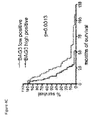
- the BAG3 IHC kit allows the identification of the prognosis of PDAC patients. It was seen that the intensity of BAG3 expression identified by IHC, correlates with patients' survival. Therefore it can be used for both prognosis and for making a choice of therapy.
- a still further aspect of the invention is a kit for the detection of BAG3 gene expression in a biological sample comprising a set of amplification primers.
- amplification primers are suitable for the detection by quantitative real-time RT-PCR.
- the RT-PCR kit for bag3 mRNA detection in a biological sample allows to correlate the levels of bag3 gene expression with patients' survival and can be used for prognosis and for choice of therapy.
- said biological sample is a tissue sample.
- a still further aspect of the invention is represented by anti-BAG3 monoclonal antibodies, their fragments, and peptides corresponding to specific aminoacidic sequences of BAG3 protein that are able to block macrophage activation and can therefore be used for therapy of inflammatory, oncologic or other diseases involving macrophage activation. See in particular Figure 3 and Table I.
- This invention relates to the use of BAG3- specific mouse monoclonal antibodies AC-1, AC-2 and AC-3 or same modified as F(ab), F(ab')2, F(ab) or humanized; or peptides comprising sequences as follows:
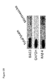
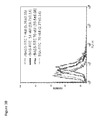
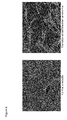
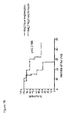
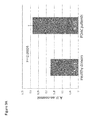
- Cardiomyocytes are known to release protective factors in mounting a response against stressful agents. Since stress- induced proteins, such as Hsp70, Hp27, Hsp90 and others, although exerting an intracellular activity, can also be secreted in response to stress, BAG3 release by cardiomyocytes was analyzed in stressful conditions. For this purpose, we analyzed the effect of serum deprivation- induced stress in cultured human primary cardiomyocytes or the rat cardiomyocyte cell line H9c2. As shown in Figure 1 , we could detect BAG3 protein in the supernatants of cardiomyocytes exposed to serum deprivation for 16 h ( Figure 1A ).
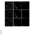
- J774 cells were incubated with 14 nM FITC-BAG3 protein and with 625 nM of BAG3 peptides (peptide 1, peptide 2, peptide 3, peptide 4 or scrambled peptide) or with 420 nM of F(ab')2 fragments from anti-BAG3 monoclonal and polyclonal antibodies (mouse monoclonal AC1, AC2 and rabbit polyclonal TOS2).
- F(ab')2 fragments from mouse IgG or F(ab')2 fragments from rabbit IgG were used as a negative control.
- FITC-rBAG3 FITC-BSA Competition assays % of positive cells ( ⁇ S.D.) % of positive cells ( ⁇ S.D.) % of positive cells ( ⁇ S.D.) % inhibition FITC-rBAG3 15.7 ( ⁇ 0.45) FITC-BSA 4.04 ( ⁇ 0.06) (FITC-rBAG3)-( FITC -BSA) 11.06 ( ⁇ 0.45) FITC -rBAG3 + Pep1 0.18 ( ⁇ 0.05) 98.4 FITC -rBAG3 + Pep2 1.21 ( ⁇ 0.63) 89.1 FITC -rBAG3 + Pep3 5.86 ( ⁇ 0.43) 47.2 FITC -rBAG3 + Pep4 0.68 ( ⁇ 0.20) 93.8 FITC -rBAG3 + Pep Scr 12.1 ( ⁇ 0.21) 0.0 FITC -rBAG3 + Mouse IgG F(ab')2 12.3 ( ⁇ 0.40) 0.0 FITC -
- BAG3-specific mouse monoclonal antibodies AC-1, AC-2 and AC-3 and/or others or same modified as F(ab), F(ab')2, F(ab) or humanized; or peptides comprising sequences PEP 1 to 4 and/or others are molecules able to bind and/or block soluble BAG3 effects to be used for therapy of inflammatory, oncologic or other diseases involving macrophage activation.
- IHC immunohistochemistry
- This kit revealed BAG3 expression in all the 346 (100%) PDAC biopsies that we analyzed.
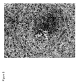
- BAG3 staining revealed a moderate positivity of Langerhans islets, while normal pancreatic ducts and pancreatic acinar cells had no BAG3 expression. This was true in both normal pancreas and non-neoplastic pancreatic tissue adjacent to the tumor mass. BAG3 staining was observed predominantly in the cytoplasm of tumor cells. The intensity of staining of BAG3 was variable as was the number of positive cancer cells.
- the first-line chemotherapy for treatment of pancreatic cancer is gemcitabine.
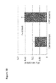
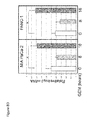
- BAG3 protein in response to therapy, we analyzed the effect of BAG3 down-modulation in human PDAC cells.
- Silencing of BAG3 enhanced cell apoptosis in response to the drug ( Figure 8 ).
- bag3 primers that detect BAG3 expression by quantitative real-time RT-PCR.
- RT-PCR kit containing specific primers for bag3 mRNA detection and quantification.
- HCMa Human Cardiac Myocytes-adult were purchased from Sciencell Research Laboratories (San Diego, CA) and grown in Cardiac Myocyte Medium (CMM, FBS 5%, Cardiac Myocyte Growth Supplement 1%, penicillin/streptomycin solution 1 %) (Sciencell Research Laboratories, San Diego, CA). All experiments were performed on low-passage cell cultures.
- Embryonic rat cardiomyoblasts (line H9c2) was purchased from the American Type Culture Collection (ATCC, Manassas, VA, USA) and grown in Dulbecco's Modified Eagle's Medium (DMEM) supplemented with 10 % fetal bovine serum (FBS), 100 U/mL penicillin and 100 ⁇ g/mL streptomycin.
- DMEM Dulbecco's Modified Eagle's Medium
- FBS fetal bovine serum
- J774A.1 murine monocyte macrophage cell line (ATCC, Manassas, VA, USA) was grown in DMEM supplemented with 10% fetal bovine serum (FBS), 25 mM HEPES, 2 mM glutamine, 100 u/mL penicillin and 100 ⁇ g/mL streptomycin.
- pancreatic cancer cell lines (MIA PaCa-2, AsPC-1, PSN1, Capan-1 and PANC-1) were received from the American Type Culture Collection (ATCC; Manassas, VA) cell bank.
- MIA PaCa-2 cells were cultured in Dulbecco's Modified Eagle's Medium (DMEM) and supplemented with 10% FBS and 2.5% horse serum.
- AsPC-1 and PSN1 cells were grown in RPMI-1640 Medium supplemented with 10% FBS.
- Capan-1 were cultured in RPMI-1640 containing 20% FBS while PANC-1 were cultured in DMEM supplemented with 10% FBS.
- Sera were diluted 1:40 with dissociation buffer (PBS with 1.5% BSA and 0.2 M glycine-acetate pH 2.5) to a 500 ⁇ l final volume and incubated for 20 min at room temperature.
- the sera were then pipetted into the sample reservoir of Microcon centrifugal filter device, YM-100 (100,000 MW cut-off; Millipore, Billerica, MA, USA) and centrifuged at 14,100 rpm for 20 min at room temperature.
- the sample reservoir was then separated from the flow through, placed inverted into a second tube and centrifuged at 5,000 rpm for 3 min at room temperature.
- the collected solution containing the antibody dissociated was adjusted to pH 7.0 with 1 M Tris buffer, pH 9.0.
- the retentate volume was reconstituted to the initial volume (500 ⁇ l) with dilution buffer (PBS with 1.5% BSA and 0.1% Tween-20).14
- dilution buffer PBS with 1.5% BSA and 0.1% Tween-20.14
- the dissociated antibodies were diluted 1:200 in TBST containing 5% bovine serum albumin overnight at 4°C.
- Cells were harvested and lysed in a buffer containing 20 mM HEPES (pH 7.5), 150 mM NaCl, 0.1% Triton (TNN buffer) supplemented with a protease inhibitors cocktail (1 mM phenylmethylsulfonyl fluoride, 1 mg/ml pepstatin A, 2 mg/ml aprotinin) by 3 cycles of freezing and thawing. Soluble proteins were collected after a centrifugation at 10,000 g for 15 min and their amount was determined by Bradford assay (Bio-Rad, Hercules, CA).
- Protein bands were excised and gel pieces were subsequently washed with MilliQ Water and Acetonitrile and the proteins were digested in situ as described in Shevchenko protocol. Briefly, gel slices were reduced in 1,4-dithiothreitol (10 mM) and alkylated with iodoacetamide (50 mM), then washed and rehydrated in trypsin solution (12 ng/ ⁇ L) on ice for 1 h. After the addition of 30 ⁇ L ammonium bicarbonate (10 mM, pH 7.5), samples were digested overnight at 25 °C. 5 ⁇ L of the obtained peptide mixture were injected onto a nano Acquity LC system (Waters Corp. Manchester, United Kingdom).
- the peptides were separated on a 1.7 ⁇ m BEH C-18 column (Waters Corp. Manchester, United Kingdom) at a flow rate of 200 nl/min.
- the gradient (Solution A: 0.1% formic acid, solution B: 0.1% formic acid, 100% ACN) started at 5% and ended at 50% B after 55 min.
- MS and MS/MS data were acquired using a Q-TOF Premier mass spectrometer (Waters Corp., Micromass, Manchester, United Kingdom). Doubly and triply charged peptide-ions were automatically chosen by the MassLynx software and fragmented.
- MS data were automatically processed and peaklists for protein identifications by database searches were generated by the ProteinLynx software. Database searches were carried out with MASCOT server using the SwissProt protein database.
- the SwissProt human database (405506 sequences; 146166984 residues) was searched allowing 1 missed cleavage, carbamidomethyl (C) as fixed modification.
- the peptide tolerance was set to 60 ppm and the MS/MS tolerance to 0.8 Da.
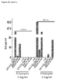
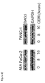
- Serum-free medium of H9c2 was cleared of cells and large debris by serial centrifugation at 4°C (2000xg for 15 min, 10,000xg for 30 min). After each of the first two centrifugations, pellets are discarded, and the supernatant is kept for the next step. The final supernatant is then ultracentrifuged at 150,000xg for 90 min at 4°C (with a SW50.1 rotor, and an Optima L-90K Ultracentrifuge, Beckman Coulter) to pellet exosomes.
- the pellet is washed in PBS to eliminate contaminating proteins and centrifuged one last time at 150,000xg for 90 min at 4°C.16 After washing, the pellet (exosomes) was resuspended in 20 ⁇ l of PBS and analyzed with the anti-BAG3 TOS-2 polyclonal antibody in comparison with a whole-cell lysate by western blot. Rab-4 was analyzed as a marker for exocytic vesicles.
- rBAG3 binding - J774 A.1 cells were blocked with 2 % FBS + 0.1 % NaN3 in PBS for 15 min on ice and incubated (2.5 x 10⁵/100 ⁇ l) with different concentration of FITC-rBAG3 protein (7, 14 and 70 nm) or FITC-BSA (70 nM) in PBS containing 2% FBS+ 0.1 % NaN3 for 30 min at 4 °C in the dark. After washing with PBS, the cells were resuspended in PBS + 2% FBS+ 0.1 % NaN3 and analyzed with a FACScan (BD Biosciences) flow cytometer.
- FACScan BD Biosciences
- IL6 was measured in supernatant of J774 A.1 cells (5x10 4 / in 96-well microplates) treated with LPS (10 ng/ml) or with rBAG3 (14 nM) or BSA (14 nM) for 10 or 20 hours in absence or presence of polymyxin B sulfate (5 ⁇ g/ml). After treatment 50 ⁇ L of cell culture medium were collected and analyzed in triplicate with a mouse IL6 Kit (eBioscience).
- Images were acquired in sequential scan mode by using the same acquisitions parameters (laser intensities, gain photomultipliers, pinhole aperture, objective 63X, zoom 2) when comparing experimental and control material.
- brightness and contrast of images were adjusted by taking care to leave a light cellular fluorescence background for visual appreciation of the lowest fluorescence intensity features and to help comparison among the different experimental groups.
- Final figures were assembled using Adobe Photoshop 7 and Adobe Illustrator 10. Leica Q9 Confocal Software and ImageJ were used for data analysis.
- NUNC Maxisorp 96 well ELISA plates were coated with recombinant BAG3 protein 1 ⁇ g/ml (50 ⁇ l/well) in PBS, pH 7 and incubated overnight at 4°C. Plates were washed 2 times with washing buffer (PBS + 0.05% Tween-20), and then blocked (150 ⁇ l/well) for one hour at room temperature with 0.5% fish gelatin in PBS. Following blocking, the plates were washed 2 times with washing buffer and sera were diluted 1:70 with 0.5% fish gelatin in washing buffer and then applied (50 ⁇ l/well) in triplicate and incubated at room temperature for two hour. The plates were then washed 6 times with washing buffer.
- washing buffer PBS + 0.05% Tween-20
- Anti-human IgG (H+L) antibody (Sigma Aldrich) was diluted 1:20,000 with 0.5% fish gelatin in washing buffer, added at 50 ⁇ l/well and incubated at 4°C for 30 minutes. After incubation, the plates were washed 6 times, developed with TMB (50 ⁇ l/well) (eBioscience), the reaction stopped with 4.5 M sulfuric acid (25 ⁇ l/well) and the plates were analyzed spectrophotometrically at 450 nm.
- Nitrite content (NO 2 -), a stable metabolite of NO released by cells in the culture supernatant, was measured18 in J774 A.1 cells (5x10⁴/ in 96-well microplates) treated with LPS (10 ng/ml) or with rBAG3 (7, 14 and 28 nM) or BSA (28 nM) for 24 hours in absence or presence of polymyxin B sulfate (Sigma-Aldrich, St. Louis, MO, USA) 5 ⁇ g/ml. NO2- amounts were measured by Griess reaction.
- NUNC Maxisorp 96 well ELISA plates were coated with anti-BAG3 monoclonal antibody AC-1, AC-2, AC-3, AC-4 or AC-5 in PBS, pH 7 and incubated overnight at 4°C. Plates were washed 2 times with washing buffer (PBS + 0.05% Tween-20), and then blocked (150 ⁇ l/well) for one hour at room temperature with 0.5% fish gelatin in PBS. Following blocking, the plates were washed 2 times with washing buffer and sera were diluted 1:70 with 0.5% fish gelatin in washing buffer and then applied (50 ⁇ l/well) in triplicate and incubated at room temperature for two hour. The plates were then washed 6 times with washing buffer.
- washing buffer PBS + 0.05% Tween-20
- Anti-human IgG (H+L) antibody (Sigma Aldrich) was diluted 1:20,000 with 0.5% fish gelatin in washing buffer, added at 50 ⁇ l/well and incubated at 4°C for 30 minutes. After incubation, the plates were washed 6 times, developed with TMB (50 ⁇ l/well) (eBioscience), the reaction stopped with 4.5 M sulfuric acid (25 ⁇ l/well) and the plates were analyzed spectrophotometrically at 450 nm.
- Immunohistochemistry protocol included: deparaffination in xylene, re-hydration through descending concentrations of alcohol up to pure water, non-enzymatic antigen retrieval in citrate buffer, pH 6.0, for 30 minutes at 95°C, and endogenous peroxidase quenching with H2O2 in methanol for 20 minutes. After rinsing with PBS, the samples were blocked with 5% normal horse serum in 0.1% PBS/BSA. To detect BAG3, samples were incubated for 1 hour at room temperature with BAG3 monoclonal antibody AC_1, AC-2 or AC-3 at the concentration of 3 microg/ml.
- RNA concentration and purity were validated by NanoDrop Spectrophotometer (Thermo Fisher, Waltham, MA, USA).
- Primers for the human bag3 gene were synthesized by Primm srl (Milano, Italy) (forward primer: (SEQ ID NO:16) CCT GTT AGC TGT GGT TG; reverse primer: (SEQ ID NO:17) AAC ATA CAG ATA TTC CTA TGG C).
Landscapes
- Health & Medical Sciences (AREA)
- Life Sciences & Earth Sciences (AREA)
- Chemical & Material Sciences (AREA)
- Engineering & Computer Science (AREA)
- Immunology (AREA)
- Molecular Biology (AREA)
- Urology & Nephrology (AREA)
- Biomedical Technology (AREA)
- Proteomics, Peptides & Aminoacids (AREA)
- Organic Chemistry (AREA)
- Hematology (AREA)
- Analytical Chemistry (AREA)
- Biochemistry (AREA)
- General Health & Medical Sciences (AREA)
- Pathology (AREA)
- Physics & Mathematics (AREA)
- Microbiology (AREA)
- Biotechnology (AREA)
- Medicinal Chemistry (AREA)
- Genetics & Genomics (AREA)
- Zoology (AREA)
- Cell Biology (AREA)
- Wood Science & Technology (AREA)
- General Physics & Mathematics (AREA)
- Food Science & Technology (AREA)
- Hospice & Palliative Care (AREA)
- Oncology (AREA)
- Biophysics (AREA)
- Bioinformatics & Cheminformatics (AREA)
- General Engineering & Computer Science (AREA)
- Gastroenterology & Hepatology (AREA)
- Toxicology (AREA)
- Chemical Kinetics & Catalysis (AREA)
- Measuring Or Testing Involving Enzymes Or Micro-Organisms (AREA)
- Peptides Or Proteins (AREA)
- Investigating Or Analysing Biological Materials (AREA)
- Preparation Of Compounds By Using Micro-Organisms (AREA)
- Apparatus Associated With Microorganisms And Enzymes (AREA)
Priority Applications (23)
| Application Number | Priority Date | Filing Date | Title |
|---|---|---|---|
| EP12172531.1A EP2676966A1 (en) | 2012-06-19 | 2012-06-19 | BAG3 as biochemical serum and tissue marker |
| CN201380032138.3A CN104507965B (zh) | 2012-06-19 | 2013-06-11 | 作为血清和组织生化标志物的bag3 |
| EP13728375.0A EP2861754A1 (en) | 2012-06-19 | 2013-06-11 | Bag3 as biochemical serum and tissue marker |
| KR20157001228A KR20150036102A (ko) | 2012-06-19 | 2013-06-11 | 혈청 및 조직 생화학 마커로서 bag3 |
| ES13728180T ES2795981T3 (es) | 2012-06-19 | 2013-06-11 | BAG3 como marcador bioquímico de suero y tejidos |
| EP13728180.4A EP2861618B1 (en) | 2012-06-19 | 2013-06-11 | Bag3 as biochemical serum and tissue marker |
| BR112014031958A BR112014031958A2 (pt) | 2012-06-19 | 2013-06-11 | bag3 como marcador de tecido e soro bioquímico |
| PL13728180T PL2861618T3 (pl) | 2012-06-19 | 2013-06-11 | BAG3 jako surowiczy i tkankowy marker biochemiczny |
| CN201380032196.6A CN104619861A (zh) | 2012-06-19 | 2013-06-11 | 作为血清和组织生化标志物的bag3 |
| KR1020157001313A KR102150903B1 (ko) | 2012-06-19 | 2013-06-11 | 혈청 및 조직 생화학 마커로서 bag3 |
| CA2876922A CA2876922C (en) | 2012-06-19 | 2013-06-11 | Bag3 as biochemical serum and tissue marker |
| JP2015517669A JP2015521475A (ja) | 2012-06-19 | 2013-06-11 | 生化学的血清マーカーおよび組織マーカーとしてのbag3 |
| AU2013279601A AU2013279601A1 (en) | 2012-06-19 | 2013-06-11 | BAG3 as biochemical serum and tissue marker |
| PCT/EP2013/061976 WO2013189778A1 (en) | 2012-06-19 | 2013-06-11 | Bag3 as biochemical serum and tissue marker |
| PCT/EP2013/061971 WO2013189775A1 (en) | 2012-06-19 | 2013-06-11 | Bag3 as biochemical serum and tissue marker |
| BR112014031957A BR112014031957A2 (pt) | 2012-06-19 | 2013-06-11 | bag3 como soro bioquímico e marcador de tecido |
| CA2876872A CA2876872A1 (en) | 2012-06-19 | 2013-06-11 | Bag3 as biochemical serum and tissue marker |
| JP2015517672A JP6654432B2 (ja) | 2012-06-19 | 2013-06-11 | 生化学的血清マーカーおよび組織マーカーとしてのbag3 |
| AU2013279604A AU2013279604B2 (en) | 2012-06-19 | 2013-06-11 | BAG3 as biochemical serum and tissue marker |
| IN10561DEN2014 IN2014DN10561A (ko) | 2012-06-19 | 2014-12-11 | |
| IN10562DEN2014 IN2014DN10562A (ko) | 2012-06-19 | 2014-12-11 | |
| US14/572,654 US10359433B2 (en) | 2012-06-19 | 2014-12-16 | BAG3 as biochemical serum and tissue marker |
| US14/572,445 US20150132764A1 (en) | 2012-06-19 | 2014-12-16 | Bag3 as biochemical serum and tissue marker |
Applications Claiming Priority (1)
| Application Number | Priority Date | Filing Date | Title |
|---|---|---|---|
| EP12172531.1A EP2676966A1 (en) | 2012-06-19 | 2012-06-19 | BAG3 as biochemical serum and tissue marker |
Publications (1)
| Publication Number | Publication Date |
|---|---|
| EP2676966A1 true EP2676966A1 (en) | 2013-12-25 |
Family
ID=48607259
Family Applications (3)
| Application Number | Title | Priority Date | Filing Date |
|---|---|---|---|
| EP12172531.1A Withdrawn EP2676966A1 (en) | 2012-06-19 | 2012-06-19 | BAG3 as biochemical serum and tissue marker |
| EP13728375.0A Ceased EP2861754A1 (en) | 2012-06-19 | 2013-06-11 | Bag3 as biochemical serum and tissue marker |
| EP13728180.4A Active EP2861618B1 (en) | 2012-06-19 | 2013-06-11 | Bag3 as biochemical serum and tissue marker |
Family Applications After (2)
| Application Number | Title | Priority Date | Filing Date |
|---|---|---|---|
| EP13728375.0A Ceased EP2861754A1 (en) | 2012-06-19 | 2013-06-11 | Bag3 as biochemical serum and tissue marker |
| EP13728180.4A Active EP2861618B1 (en) | 2012-06-19 | 2013-06-11 | Bag3 as biochemical serum and tissue marker |
Country Status (12)
| Country | Link |
|---|---|
| US (2) | US20150132764A1 (ko) |
| EP (3) | EP2676966A1 (ko) |
| JP (2) | JP6654432B2 (ko) |
| KR (2) | KR102150903B1 (ko) |
| CN (2) | CN104507965B (ko) |
| AU (2) | AU2013279601A1 (ko) |
| BR (2) | BR112014031958A2 (ko) |
| CA (2) | CA2876872A1 (ko) |
| ES (1) | ES2795981T3 (ko) |
| IN (2) | IN2014DN10561A (ko) |
| PL (1) | PL2861618T3 (ko) |
| WO (2) | WO2013189775A1 (ko) |
Cited By (1)
| Publication number | Priority date | Publication date | Assignee | Title |
|---|---|---|---|---|
| IT201600069391A1 (it) * | 2016-07-04 | 2016-10-04 | Univ Degli Studi Di Salerno | Uso della proteina bag3 e suoi frammenti peptidici per il controllo dell’omeostasi vascolare |
Families Citing this family (6)
| Publication number | Priority date | Publication date | Assignee | Title |
|---|---|---|---|---|
| ITMI20130403A1 (it) | 2013-03-18 | 2014-09-19 | Biouniversa Srl | Anticorpi anti-bag3 per uso terapeutico |
| CN105021825B (zh) * | 2015-07-09 | 2016-08-17 | 陈勇 | 一种检测胰腺癌相关多肽dap44的elisa试剂盒 |
| CN105769900A (zh) * | 2016-03-22 | 2016-07-20 | 山西大学 | Bag3基因在制备抗膀胱癌药物中的应用 |
| WO2021140173A1 (en) | 2020-01-10 | 2021-07-15 | Biouniversa S.R.L. | Methods and uses for treating fibrotic solid tumors with bags inhibitors |
| US11940450B2 (en) | 2021-03-16 | 2024-03-26 | University Of Connecticut | Biomarker panel for non-invasive diagnosis of congenital renal dysfunction |
| CN113862361B (zh) * | 2021-10-25 | 2023-08-15 | 中山大学孙逸仙纪念医院 | 一种诊断和治疗膀胱癌的分子标志物hsf1及其用途 |
Citations (2)
| Publication number | Priority date | Publication date | Assignee | Title |
|---|---|---|---|---|
| EP1323733A1 (en) * | 2001-12-28 | 2003-07-02 | Arturo Leone | BAG3 nucleotide and protein sequences to be used in research, diagnostics and therapy for cell death-involving diseases |
| WO2011067377A1 (en) * | 2009-12-04 | 2011-06-09 | Biouniversa S.R.L. | Biochemical serum marker |
Family Cites Families (7)
| Publication number | Priority date | Publication date | Assignee | Title |
|---|---|---|---|---|
| US5652223A (en) * | 1994-03-14 | 1997-07-29 | The United States Of America As Represented By The Secretary Of The Department Of Health And Human Services | DNA encoding CAI resistance proteins and uses thereof |
| EP1200618A2 (en) * | 1999-07-09 | 2002-05-02 | The Burnham Institute | A method for determining the prognosis of cancer patients by measuring levels of bag expression |
| US20030073623A1 (en) * | 2001-07-30 | 2003-04-17 | Drmanac Radoje T. | Novel nucleic acid sequences obtained from various cDNA libraries |
| JP4424987B2 (ja) * | 2001-09-20 | 2010-03-03 | ボード オブ リージェンツ, ザ ユニバーシティ オブ テキサス システム | Elisaアッセイを用いた循環する治療抗体、抗原および抗原/抗体複合体の測定 |
| US20090047689A1 (en) * | 2007-06-20 | 2009-02-19 | John Kolman | Autoantigen biomarkers for early diagnosis of lung adenocarcinoma |
| WO2011044927A1 (en) * | 2009-10-12 | 2011-04-21 | INSERM (Institut National de la Santé et de la Recherche Médicale) | A method for the diagnosis or prognosis of an advanced heart failure |
| GB201017520D0 (en) * | 2010-10-15 | 2010-12-01 | Sense Proteomic Ltd | Biomarkers |
-
2012
- 2012-06-19 EP EP12172531.1A patent/EP2676966A1/en not_active Withdrawn
-
2013
- 2013-06-11 KR KR1020157001313A patent/KR102150903B1/ko active IP Right Grant
- 2013-06-11 PL PL13728180T patent/PL2861618T3/pl unknown
- 2013-06-11 CN CN201380032138.3A patent/CN104507965B/zh not_active Expired - Fee Related
- 2013-06-11 EP EP13728375.0A patent/EP2861754A1/en not_active Ceased
- 2013-06-11 BR BR112014031958A patent/BR112014031958A2/pt not_active IP Right Cessation
- 2013-06-11 CA CA2876872A patent/CA2876872A1/en not_active Abandoned
- 2013-06-11 EP EP13728180.4A patent/EP2861618B1/en active Active
- 2013-06-11 KR KR20157001228A patent/KR20150036102A/ko not_active Application Discontinuation
- 2013-06-11 AU AU2013279601A patent/AU2013279601A1/en not_active Abandoned
- 2013-06-11 ES ES13728180T patent/ES2795981T3/es active Active
- 2013-06-11 BR BR112014031957A patent/BR112014031957A2/pt not_active IP Right Cessation
- 2013-06-11 JP JP2015517672A patent/JP6654432B2/ja not_active Expired - Fee Related
- 2013-06-11 JP JP2015517669A patent/JP2015521475A/ja active Pending
- 2013-06-11 WO PCT/EP2013/061971 patent/WO2013189775A1/en active Application Filing
- 2013-06-11 CN CN201380032196.6A patent/CN104619861A/zh active Pending
- 2013-06-11 CA CA2876922A patent/CA2876922C/en active Active
- 2013-06-11 AU AU2013279604A patent/AU2013279604B2/en not_active Ceased
- 2013-06-11 WO PCT/EP2013/061976 patent/WO2013189778A1/en active Application Filing
-
2014
- 2014-12-11 IN IN10561DEN2014 patent/IN2014DN10561A/en unknown
- 2014-12-11 IN IN10562DEN2014 patent/IN2014DN10562A/en unknown
- 2014-12-16 US US14/572,445 patent/US20150132764A1/en not_active Abandoned
- 2014-12-16 US US14/572,654 patent/US10359433B2/en active Active
Patent Citations (2)
| Publication number | Priority date | Publication date | Assignee | Title |
|---|---|---|---|---|
| EP1323733A1 (en) * | 2001-12-28 | 2003-07-02 | Arturo Leone | BAG3 nucleotide and protein sequences to be used in research, diagnostics and therapy for cell death-involving diseases |
| WO2011067377A1 (en) * | 2009-12-04 | 2011-06-09 | Biouniversa S.R.L. | Biochemical serum marker |
Non-Patent Citations (11)
| Title |
|---|
| B. FONTANELLA ET AL.: "The co-chaperone BAG3 interacts with the cytosolic CCT: New hints for actin folding.", THE INTERNATIONAL JOURNAL OF BIOCHEMISTRY & CELL BIOLOGY, vol. 42, no. 5, 1 May 2010 (2010-05-01), GB, pages 641 - 650, XP026940700 * |
| C. XU ET AL.: "Apoptotic gene expression by human periodontal ligament cells following cyclic stretch.", JOURNAL OF PERIODONTAL RESEARCH, vol. 46, no. 6, 21 July 2011 (2011-07-21), Denmark, pages 742 - 748, XP008158908 * |
| DE MARCO M; TURCO MC; ROSATI A: "BAG3 protein is induced during cardiomyoblast differentiation and modulates myogenin expression", CELL CYCLE., vol. 10, 2011, pages 850 - 852 |
| H. CHEN ET AL.: "Bag3 gene expression in chronic lymphocytic leukemia and its association with patients' prognosis.", JOURNAL OF EXPERIMENTAL HEMATOLOGY, vol. 18, no. 4, August 2010 (2010-08-01), China, pages 838 - 842, XP008157243 * |
| M. DE MARCO ET AL.: "BAG3 protein is induced during cardiomyoblast differentiation and modulates myogenin expression.", CELL CYCLE, vol. 10, no. 5, 1 March 2011 (2011-03-01), USA, pages 850 - 852, XP002685428 * |
| M. FESTA ET AL.: "BAG3 protein is overexpressed in human glioblastoma and is a potential target for therapy.", THE AMERICAN JOURNAL OF PATHOLOGY, vol. 178, no. 6, 10 May 2011 (2011-05-10), USA, pages 2504 - 2512, XP002685427 * |
| M. PAGLIUCA ET AL.: "Regulation by heavy metals and temperature of the human BAG-3 gene, a modulator of Hsp70 activity.", FEBS LETTERS, vol. 541, no. 1-3, 24 April 2003 (2003-04-24), Netherlands, pages 11 - 15, XP004421716 * |
| P. YOUNG ET AL.: "Epstein-Barr virus nuclear antigen (EBNA) 3A induces the expression of and interacts with a subset of chaperones and co-chaperones.", THE JOURNAL OF GENERAL VIROLOGY, vol. 89, no. part 4, April 2008 (2008-04-01), GB, pages 866 - 877, XP002689789 * |
| Q. LIAO ET AL.: "The anti-apoptotic protein BAG-3 is overexpressed in pancreatic cancer and induced by heat stress in pancreatic cancer cell lines.", FEBS LETTERS, vol. 503, no. 2-3, 17 August 2001 (2001-08-17), The Netherlands, pages 151 - 157, XP027305726 * |
| ROSATI A; GRAZIANO V; DE LAURENZI V; PASCALE M; TURCO MC.: "BAG3: a multifaceted protein that regulates major cell pathways", CELL DEATH DIS., vol. 2, 2011, pages E141 |
| X. JIN ET AL.: "Delineation of apoptotic genes for synergistic apoptosis of lexatumumab and anthracyclines in human renal cell carcinoma cells by polymerase chain reaction array.", ANTI-CANCER DRUGS, vol. 23, no. 4, April 2012 (2012-04-01), GB, pages 445 - 454, XP008158907 * |
Cited By (1)
| Publication number | Priority date | Publication date | Assignee | Title |
|---|---|---|---|---|
| IT201600069391A1 (it) * | 2016-07-04 | 2016-10-04 | Univ Degli Studi Di Salerno | Uso della proteina bag3 e suoi frammenti peptidici per il controllo dell’omeostasi vascolare |
Also Published As
| Publication number | Publication date |
|---|---|
| JP2015529633A (ja) | 2015-10-08 |
| IN2014DN10562A (ko) | 2015-08-28 |
| AU2013279604A1 (en) | 2014-12-18 |
| BR112014031957A2 (pt) | 2017-08-01 |
| PL2861618T3 (pl) | 2020-09-21 |
| ES2795981T3 (es) | 2020-11-25 |
| EP2861618B1 (en) | 2020-03-11 |
| KR102150903B1 (ko) | 2020-09-03 |
| CN104507965B (zh) | 2019-04-26 |
| CA2876922C (en) | 2022-09-20 |
| CN104619861A (zh) | 2015-05-13 |
| CN104507965A (zh) | 2015-04-08 |
| WO2013189778A1 (en) | 2013-12-27 |
| US10359433B2 (en) | 2019-07-23 |
| WO2013189775A1 (en) | 2013-12-27 |
| JP6654432B2 (ja) | 2020-02-26 |
| EP2861618A1 (en) | 2015-04-22 |
| US20150132764A1 (en) | 2015-05-14 |
| CA2876872A1 (en) | 2013-12-27 |
| KR20150036102A (ko) | 2015-04-07 |
| AU2013279601A1 (en) | 2015-01-15 |
| EP2861754A1 (en) | 2015-04-22 |
| JP2015521475A (ja) | 2015-07-30 |
| US20150147767A1 (en) | 2015-05-28 |
| KR20150036111A (ko) | 2015-04-07 |
| AU2013279604B2 (en) | 2018-03-29 |
| BR112014031958A2 (pt) | 2017-08-01 |
| IN2014DN10561A (ko) | 2015-08-28 |
| CA2876922A1 (en) | 2013-12-27 |
Similar Documents
| Publication | Publication Date | Title |
|---|---|---|
| US10359433B2 (en) | BAG3 as biochemical serum and tissue marker | |
| CN106461681B (zh) | 用于诊断血管疾病的生物标志物及其用途 | |
| US20210077577A1 (en) | Blockade of ccl18 signaling via ccr6 as a therapeutic option in fibrotic diseases and cancer | |
| US20190154694A1 (en) | Detection and treatment of cancer | |
| EP3637106B1 (en) | Lung cancer detection method | |
| JP7073528B2 (ja) | 同種移植片レシピエントを分類するためのバイオマーカー | |
| Lagoutte et al. | Procollagen C-Proteinase Enhancer 1 (PCPE-1) is a marker of myocardial fibrosis and impaired cardiac function in a murine model of pressure overload | |
| Jeong et al. | GCC2 is a New Biomarker for Diagnosis of Early Non-Small Cell Lung Cancer and A Potential Target to Reverse Epithelial to Mesenchymal Transition | |
| CN117106894A (zh) | Nkrf在病理性心脏重构诊治中的应用 | |
| EP2721410A2 (en) | Biomarkers for epithelial cancer diagnosis and treatment | |
| WO2024224397A1 (en) | Compositions for use in the treatment of endometriosis | |
| JP2013545992A (ja) | バイオマーカー、バイオマーカーの使用及びバイオマーカーを同定する方法 | |
| US20140335090A1 (en) | Therapeutic agent for cancer, and method for determining prognosis of cancer |
Legal Events
| Date | Code | Title | Description |
|---|---|---|---|
| PUAI | Public reference made under article 153(3) epc to a published international application that has entered the european phase |
Free format text: ORIGINAL CODE: 0009012 |
|
| AK | Designated contracting states |
Kind code of ref document: A1 Designated state(s): AL AT BE BG CH CY CZ DE DK EE ES FI FR GB GR HR HU IE IS IT LI LT LU LV MC MK MT NL NO PL PT RO RS SE SI SK SM TR |
|
| AX | Request for extension of the european patent |
Extension state: BA ME |
|
| STAA | Information on the status of an ep patent application or granted ep patent |
Free format text: STATUS: THE APPLICATION IS DEEMED TO BE WITHDRAWN |
|
| 18D | Application deemed to be withdrawn |
Effective date: 20140626 |