EP2675359B1 - Bewegungsalgorithmen für die personenstütze eines bildgebungssystems - Google Patents
Bewegungsalgorithmen für die personenstütze eines bildgebungssystems Download PDFInfo
- Publication number
- EP2675359B1 EP2675359B1 EP12709374.8A EP12709374A EP2675359B1 EP 2675359 B1 EP2675359 B1 EP 2675359B1 EP 12709374 A EP12709374 A EP 12709374A EP 2675359 B1 EP2675359 B1 EP 2675359B1
- Authority
- EP
- European Patent Office
- Prior art keywords
- motion
- algorithm
- subject
- axial
- tabletop
- Prior art date
- Legal status (The legal status is an assumption and is not a legal conclusion. Google has not performed a legal analysis and makes no representation as to the accuracy of the status listed.)
- Active
Links
- 230000033001 locomotion Effects 0.000 title claims description 204
- 238000003384 imaging method Methods 0.000 title claims description 24
- 230000010412 perfusion Effects 0.000 claims description 62
- 238000007493 shaping process Methods 0.000 claims description 18
- 238000013016 damping Methods 0.000 claims description 11
- 238000000034 method Methods 0.000 claims description 9
- 238000001914 filtration Methods 0.000 claims description 7
- 238000013519 translation Methods 0.000 claims description 4
- 238000005070 sampling Methods 0.000 claims description 3
- 241000711981 Sais Species 0.000 claims 1
- 230000001133 acceleration Effects 0.000 description 47
- 238000002591 computed tomography Methods 0.000 description 14
- 210000000056 organ Anatomy 0.000 description 12
- 230000006870 function Effects 0.000 description 10
- 230000005855 radiation Effects 0.000 description 10
- 230000036461 convulsion Effects 0.000 description 8
- 125000004122 cyclic group Chemical group 0.000 description 6
- 238000012546 transfer Methods 0.000 description 6
- 230000009189 diving Effects 0.000 description 5
- 238000002474 experimental method Methods 0.000 description 5
- 230000008859 change Effects 0.000 description 4
- 230000009467 reduction Effects 0.000 description 4
- XLYOFNOQVPJJNP-UHFFFAOYSA-N water Substances O XLYOFNOQVPJJNP-UHFFFAOYSA-N 0.000 description 4
- 230000005540 biological transmission Effects 0.000 description 3
- 230000001419 dependent effect Effects 0.000 description 3
- 230000000694 effects Effects 0.000 description 3
- 238000009499 grossing Methods 0.000 description 3
- 230000004075 alteration Effects 0.000 description 2
- 239000002131 composite material Substances 0.000 description 2
- 238000012986 modification Methods 0.000 description 2
- 230000004048 modification Effects 0.000 description 2
- 230000010363 phase shift Effects 0.000 description 2
- 238000002600 positron emission tomography Methods 0.000 description 2
- 238000012545 processing Methods 0.000 description 2
- 230000003252 repetitive effect Effects 0.000 description 2
- 230000004044 response Effects 0.000 description 2
- 210000004872 soft tissue Anatomy 0.000 description 2
- 210000001519 tissue Anatomy 0.000 description 2
- 241000251468 Actinopterygii Species 0.000 description 1
- 229910000831 Steel Inorganic materials 0.000 description 1
- 208000002173 dizziness Diseases 0.000 description 1
- 238000005516 engineering process Methods 0.000 description 1
- 230000005284 excitation Effects 0.000 description 1
- 210000004072 lung Anatomy 0.000 description 1
- 238000013507 mapping Methods 0.000 description 1
- 239000002184 metal Substances 0.000 description 1
- 230000000116 mitigating effect Effects 0.000 description 1
- 230000003534 oscillatory effect Effects 0.000 description 1
- 230000008569 process Effects 0.000 description 1
- 238000012892 rational function Methods 0.000 description 1
- 238000004088 simulation Methods 0.000 description 1
- 230000003068 static effect Effects 0.000 description 1
- 239000010959 steel Substances 0.000 description 1
- 230000009466 transformation Effects 0.000 description 1
- 210000001835 viscera Anatomy 0.000 description 1
Images




Classifications
-
- A—HUMAN NECESSITIES
- A61—MEDICAL OR VETERINARY SCIENCE; HYGIENE
- A61B—DIAGNOSIS; SURGERY; IDENTIFICATION
- A61B6/00—Apparatus or devices for radiation diagnosis; Apparatus or devices for radiation diagnosis combined with radiation therapy equipment
- A61B6/04—Positioning of patients; Tiltable beds or the like
- A61B6/0407—Supports, e.g. tables or beds, for the body or parts of the body
-
- A—HUMAN NECESSITIES
- A61—MEDICAL OR VETERINARY SCIENCE; HYGIENE
- A61B—DIAGNOSIS; SURGERY; IDENTIFICATION
- A61B6/00—Apparatus or devices for radiation diagnosis; Apparatus or devices for radiation diagnosis combined with radiation therapy equipment
- A61B6/02—Arrangements for diagnosis sequentially in different planes; Stereoscopic radiation diagnosis
- A61B6/03—Computed tomography [CT]
- A61B6/032—Transmission computed tomography [CT]
- A61B6/035—Mechanical aspects of CT
-
- A—HUMAN NECESSITIES
- A61—MEDICAL OR VETERINARY SCIENCE; HYGIENE
- A61B—DIAGNOSIS; SURGERY; IDENTIFICATION
- A61B6/00—Apparatus or devices for radiation diagnosis; Apparatus or devices for radiation diagnosis combined with radiation therapy equipment
- A61B6/04—Positioning of patients; Tiltable beds or the like
- A61B6/0487—Motor-assisted positioning
Definitions
- the following generally relates to translating a subject support of an imaging system in connection with axial and/or perfusion scans of a subject carried by the subject support, and is described with particular application to computed tomography (CT).
- CT computed tomography
- PET positron emission tomography
- x-radiology x-radiology
- other imaging modalities such as positron emission tomography (PET), x-radiology, and/or other imaging modalities.
- a computed tomography (CT) scanner includes a rotating portion rotatably supported by a stationary portion.
- the rotating portion supports an x-ray tube, which emits radiation that traverses an examination region and an object or a subject therein, and a detector array that detects radiation traversing the examination region and generates projection data indicative of the detected radiation.
- a subject support supports the object or subject in the examination region.
- a reconstructor reconstructs the projection data and generates volumetric image data indicative of the portion of the object or subject in the examination region.
- One or more images can be generated based on the volumetric image data.
- the subject support generally includes a base, which is affixed to the floor of the examination room and configured to move vertically with respect to the floor, and a tabletop, which is affixed to the base and is configured to translate horizontally, with respect to the base, in and out of the examination region.
- Both axial and perfusion scans require frequent tabletop translation; an axial scan is an imaging mode in which the tabletop moves the object or subject to each axial position for scanning, but does not move the object or subject during scanning, and a perfusion scan is an imaging mode in which the tabletop cycles the object or subject back and forth in the examination region during scanning.
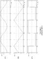
- FIGURE 1 illustrates an example s-curve motion algorithm for axial (step and shoot) scanning over three steps.
- FIGURE 1(A) a y-axis 102 represents tabletop position
- a y-axis 104 represents tabletop velocity
- a y-axis 106 represents tabletop acceleration.
- an x-axis 108 represents time.
- tabletop acceleration and deceleration for the s-curve motion algorithm includes abrupt variable ramp up and ramp down, for each step.
- FIGURES 2 and 3 respectively illustrate example trapezoidal and s-curve motion algorithms for perfusion (cyclic) scanning.
- the y-axis 102 represents tabletop position
- the y-axis 104 represents tabletop velocity
- the y-axis 106 represents tabletop acceleration
- the x-axis 108 in all six figures represents time.
- FIGURES 2(C) and 3(C) respectively show tabletop acceleration and deceleration with constant ramp up and ramp down and abrupt variable ramp up and ramp down, during each cycle.
- the acceleration and deceleration profiles of the trapezoidal and s-curve motion algorithms of FIGURES 1-3 generally will result in tabletop vibration and motion of internal organs and tissue of the patient being scanned.
- the tabletop is a lightly damped steel and composite structure with one or multiple resonant frequencies.
- the tabletop resonance can be aroused by the resonant component of the primary motion, resulting in undesired secondary motion or diving board vibration in the vertical direction.
- High acceleration and jerk due to the primary motion when used cyclically in perfusion scans, may cause excessive human organ motion, resulting in an unpleasant patient experience.
- US5598453A discloses an X-ray apparatus for collecting X-ray transmission data of a subject from a plurality of directions to generate an X-ray transmission image or X-ray CT image of the subject, which includes an X-ray generator for generating an X-ray, an X-ray detector for detecting a transmission X-ray after the X-ray generated by the X-ray generator is transmitted through the subject, a rotation unit for rotating an imaging unit including the X-ray generator and the X-ray detector around the subject, a data collector for converting an output signal of the X-ray detector to a digital signal and collecting the digital signal, a signal processor for subjecting data collected by the data collector to a signal processing operation, a display for displaying thereon as an image the data collected by the data collector and the data subjected by the signal processor to the signal processing operation, and a position change unit for moving a relative position of a rotation center of the imaging unit and the subject in a direction parallel to a rotation plane of the rotation, and
- US6381299B1 discloses that, between an X-ray generating system and an X-ray image detecting system arranged opposite to the X-ray generating system, an examination object supporting system is arranged and contains a straight movement table provided on a rotary table supported by a rotary table supporting member, and an examination object supporting member for supporting the examination object under either a standing position or a sitting position on the straight movement table. While the examination object is rotated by the rotary table, the examination object is continuously moved (reciprocating movement) by the straight movement table along a direction parallel to the rotation plane, and X-ray images of the examination object are acquired along a plurality of directions during both the rotating operation and the moving operation.
- both an X-ray tomographic image and a three-dimensional image (stereoscopic image) of a wider area than a viewing field of an X-ray II can be acquired. Since the viewing fields of the X-ray tomographic image and of the 3-dimensional image can be employed, both diagnostic performance and also a diagnostic efficiency with respect to a large organ such as a lung can be improved.
- US2010310040A1 discloses a CT system including a scintillator array having a first length in an axial direction of the CT system, the scintillator array including a plurality of scintillator cells along the first length.
- the CT system includes a controller configured to repeatedly position a subject fore and aft along the axial direction and over a second length of the CT system, the second length greater than the first length, energize an x-ray source to emit x-rays toward the subject while the subject is being repeatedly positioned, and obtain non-uniform projection data of the subject from the scintillator array, the non-uniform projection data comprising the x-rays received from the x-ray source while the subject is repeatedly positioned.;
- the CT system includes a computer programmed to reconstruct a plurality of temporally non-uniformly spaced images from the non-uniform projection data, interpolate the plurality of non-uniform images to form a plurality of temporally uniform
- US2008031407A1 discloses to easily associate X-ray projection data and scanning table z-direction coordinate information with each other. Using set parameters of the operations of a scanning gantry and a scanning table, the association of the X-ray projection data and scanning table z-direction coordinate information with each other is executed. Thereafter, image reconstruction is carried out based on the X-ray projection data to obtain a tomographic image.
- the operation set parameters are stored as part of the X-ray projection data. Alternatively, they are collectively stored even in the case of files separate from the X-ray projection data.
- the invention may take form in various components and arrangements of components, and in various steps and arrangements of steps.
- the drawings are only for purposes of illustrating the preferred embodiments and are not to be construed as limiting the invention.
- FIGURE 4 schematically illustrates an imaging system 400 such as a computed tomography (CT) scanner.
- the imaging system 400 includes a stationary gantry 402 and a rotating gantry 404, which is rotatably supported by the stationary gantry 402.
- the rotating gantry 404 rotates around an examination region 406 about a longitudinal or z-axis.
- a radiation source 408, such as an x-ray tube, is supported by and rotates with the rotating gantry 404 around the examination region 406.
- the radiation source 408 emits radiation that is collimated by a source collimator to produce a generally fan, wedge, cone, or otherwise shaped radiation beam that traverses the examination region 406.
- a radiation sensitive detector array 410 includes a one or two dimensional array of detector pixels that respectively detect radiation that traverses the examination region 406 and generates an electrical signal indicative of the detected radiation.
- a subject support 412 such as a couch, supports an object or subject in the examination region 406.
- the subject support 412 includes a base portion 414 and a tabletop 416, and is configured to position (vertically and/or horizontally) the object or subject, with respect to x, y, and/or z directions, in the examination region 406 for scanning.
- For axial (step and shoot) scans this includes moving the tabletop 416 and hence the object or subject to a predetermined position before an axial scan, maintaining a static position during the scan, and moving the tabletop 416 and hence the object or subject to next a position for a next axial when there is another axial scan to perform. This is repeated for each axial scan of the imaging procedure.
- this includes moving the tabletop 416 cyclically and hence the object or subject cyclically in coordination with scanning the object or subject.
- a subject support controller 418 controls the movement of the subject support 412.
- the illustrated subject support controller 418 includes one or more processors 420 and computer readable storage medium 422 (e.g., physical memory), which stores computer readable instructions, which, when executed by the one or more processors 420 cause the controller 418 to transmit one or more control signals to control the subject support 412.
- the one or more processors 420 can additionally or alternative execute computer readable instructions carried in a signal, carrier wave or other transitory medium.
- the illustrated storage medium 422 includes computer readable instructions such as one or more subject support motion algorithms 424.
- subject support motion algorithms 424 include, but are not limited to, a sinusoidal harmonic axial motion algorithm, a feed-forward profile shaping axial motion algorithm, a feed-forward profile filtering with time delay compensation axial motion algorithm, a sinusoidal harmonic perfusion motion algorithm, a filtered perfusion motion algorithm, traditional axial and/or perfusion motion algorithms such as a trapezoidal or s-curve axial and perfusion motion algorithms, a combination of one or more of the above-noted motion algorithms, and/or one or more other motion algorithms.
- At least one of the non-traditional trapezoidal or s-curve axial and perfusion motion algorithms may mitigate at least one of tabletop vibration or internal subject motion, which, where the subject is a patient, may improve the experience of the patient and/or image quality. In one non-limiting instance, this is achieved while maintaining move duration, move distance, average speed, etc. relative to traditional trapezoidal or s-curve motion axial and perfusion motion algorithms, while providing jerk and acceleration motion profiles that minimize the undesired motion.
- a reconstructor 426 reconstructs the signal generated by the detector array 408 and generates volumetric image data indicative of the examination region 406, including the portion of the object or subject therein.
- a general purpose computing system serves as an operator console 428, and includes an output device such as a display, an input device such as a keyboard, mouse, and/or the like, one or more processor and computer readable storage medium substantially similar to the storage medium 422.
- the console 428 allows the operator to control operation of the system 400, for example, allowing the operator to select a scan protocol (which may be associated with a default or user specified subject support motion algorithm), select or change the subject support motion algorithm, initiate scanning, etc.
- FIGURES 5 , 8 and 10 illustrate a non-limiting set of subject support motion algorithms well-suited for axial scans
- FIGURES 7 , 9 and 11 illustrate simulated vibration for the set of algorithms
- FIGURE 6 shows simulated vibration for a traditional s-curve axial motion algorithm.
- a typical axial scan involves multiple steps of rest to rest motion. While the tabletop 416 comes to rest at a particular position, e.g., after each step move in the horizontal direction, inertia force of the step move excites the tabletop 416 and the base 414, causing a diving board like vibration of the tabletop 416, and the object or subject will vibrate in the vertical direction.
- the illustrated subject support 412 is a lightly damped metal and composite apparatus so resonance is inherent. The weight of the object or subject carried by the subject support 412, when coupled with acceleration, reduces the resonant frequency, and increase the inertia force required to complete the step motion, so the object or subject increases the amplitude of the diving board vibration.
- the low frequency resonance of the subject support 412 is an internal, or system, cause of the diving board vibration.
- the dominant frequency of the axial scan motion is smaller than the resonant frequency of the subject support even with the heaviest patient load.
- FIGURE 5 an example sinusoidal motion algorithm with the identical time and distance as the s-curve motion algorithm of FIGURE 1 is illustrated.
- the y-axis 102 represents tabletop position
- the y-axis 104 represents tabletop velocity
- the y-axis 106 represents tabletop acceleration
- the x-axis 108 represents time in all three figures.
- the tabletop velocity and position profiles in FIGURE 5 are very similar to the tabletop velocity and position profiles of FIGURE 1 .
- the tabletop acceleration profiles of the sinusoidal harmonic algorithm of FIGURE 5 is much smoother than the tabletop acceleration profile of FIGURE 1 , mitigating undesired motion.
- FIGURES 6 and 7 respectively show simulated tabletop vibration for the s-curve axial motion algorithm of FIGURE 1 and the sinusoidal axial motion algorithm of FIGURE 5 .
- a y-axis 602 represents tabletop vibration and the x-axis 604 represents position.
- the tabletop vibration for the sinusoidal harmonic axial motion algorithm of FIGURE 7 during the time periods when the axial scans are acquired (i.e., between 1 and' 2, 3 and 4, and 5 and 6), has a peak residue vibration 702 (e.g., 0.0846) that is about half (50%) of a peak residue vibration 606 (e.g., 1.664) for the s-curve axial motion algorithm of FIGURE 1 .
- the peak 702 is about 25% to about 75% of the peak 606.
- Other transfer functions may also be used to characterize the tabletop.
- FIGURE 8 an example filtered compensation motion algorithm for axial scanning is discussed.
- a band-stop filter such as a notch filter or the like can be employed where the resonant frequency and damping factor of the subject support 412 are known or can be measured or estimated.
- a notch filter is a pole-zero cancelling technology that basically uses new zeros to cancel the resonant poles of the original system, and places new well damped poles to achieve a stable non-oscillatory response.
- a notch filter Compared with a low pass filter, a notch filter generally introduces less delay at least in part due to phase lag concentricity to the resonant frequency and has much better vibration squeezing due to the stable poles and cancellation of resonant poles. The time delay to the original motion planning can be accounted for in order to scan a patient in the right rest intervals.
- the y-axis 102 represents tabletop position after the time delay is factored in and the original s-curved position profile is filtered with the notch or other band pass filter
- the y-axis 104 represents tabletop velocity
- the y-axis 106 represents tabletop acceleration
- the x-axis 108 represents time in all three figures.
- the y-axis 602 represents tabletop vibration and the x-axis 604 represents position. Note that the simulated vibration has a peak residue vibration 902 of about or on the order of zero.
- T 2 ⁇ ⁇ ⁇ n of the couch vertical dynamics.
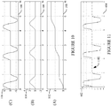
- FIGURE 10 an example profile shaping motion algorithm for axial scanning is discussed.
- feed-forward input shaping is implemented by convolving a sequence of impulses, an input shaper, with a desired motion profile to produce a shaped motion profile input that produces self-cancelling command signal to limit the residue vibration of rest to rest move pattern.
- the input is the subject support resonant frequency and damping factor (which is used in this case to compute the gain and time location of the impulse functions), and time delay is accounted for in a digital implementation.
- the input shaping filter introduces half of the notch-filter's delay, and has better residue vibration attenuation.
- the y-axis 102 represents tabletop position
- the y-axis 104 represents tabletop velocity
- the y-axis 106 represents tabletop acceleration
- the x-axis 108 represents time in all three figures.
- the y-axis 602 represents tabletop vibration and the x-axis 604 represents position.
- the simulated vibration for the input shaping axial motion algorithm has a peak residue vibration 1102 of about or on the order of zero.
- the above time domain convolution can be converted to continuous time transfer function, which is commonly used in simulation, and then the continuous time delay transfer function can be approximated with a continuous rational function.
- FIGURES 12 and 13 illustrate a non-limiting set of subject support motion algorithms well-suited for at least perfusion scans.
- a perfusion scan For a perfusion scan, a general concern is patient comfort and the impact of organ motion in horizontal direction on image quality, and since the patient lies on the tabletop 416, patient comfort is affected by the force pattern introduced by the tabletop friction, i.e., the tabletop's acceleration and jerk and its frequency pattern of the primary motion. Generally low frequency and low acceleration will result in lower jerk and better patient feeling.
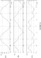
- FIGURE 12 shows a filtered point-to-point s-curve perfusion motion algorithm.
- the y-axis 102 represents tabletop position
- the y-axis 104 represents tabletop velocity
- the y-axis 106 represents tabletop acceleration
- the x-axis 108 represents time in all three figures.
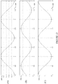
- FIGURE 13 shows a sinusoidal harmonic perfusion motion algorithm.
- the y-axis 102 represents tabletop position
- the y-axis 104 represents tabletop velocity
- the y-axis 106 represents tabletop acceleration
- the x-axis 108 represents time in all three figures.
- the tabletop acceleration of both the filtered point-to-point s-curve perfusion motion algorithm ( FIGURE 12 ) and the sinusoidal harmonic perfusion motion algorithm ( FIGURE 13 ) are smoother and thus result in less jerk and patient discomfort relative to the tabletop acceleration of the trapezoidal perfusion motion algorithm ( FIGURE 2 ) and the point-to-point s-curve perfusion motion algorithm ( FIGURE 3 ).
- a first order low pass filter e.g., ⁇ c s + ⁇ c
- ⁇ ⁇ ⁇ of the low pass filter has to be carefully chosen. Too high cut-off frequency, the low pass filter has no motion smoothing effect, while too low cut-off frequency will produce too much motion coverage reduction and phase lag.
- the phase lag is not an issue (like it is in axial scanning, as described above) as long as it is repetitive and constant, but the excessive coverage reduction has to be compensated.
- cut-off frequency of the perfusion cycle frequency can be used to the low pass filter to balance the coverage cutting and motion smoothing effect.
- the effect of the low pass filter on the point to point s-curve perfusion motion phase lag and amplitude cutting can be seen in FIGURE 12 where the cut-off frequency is 2 Hz for a 0.5 Hz perfusion cycle (i.e., a one (1) second end-to-end move, or a period of two (2) seconds).
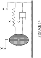
- FIGURE 14 shows an example model that can be used to model the elastic and viscous nature of soft tissue for the perfusion motion algorithms.
- the model assumes human organ as a passive, stable, and low pass systems, which may or may not have resonance, and is not sensitive to high frequency excitation.
- the relative human organ motion "X-Y" will be small if the organ position X can follow the tabletop position Y closely, where 'm' is the mass of the organ, 'k' is the stiffness of the tissue, and 'c' is the damping coefficient of the organ to external perturbation.
- the sinusoidal motion algorithms have a 90 degree phase shift between the velocity and acceleration curves, which may result in relatively smaller drive power capacity compared with the other simultaneous peak speed and peak acceleration motion profile, because speed corresponds to voltage for motor drive, acceleration means torque so drive current, and power is derived by speed times force or by voltage times current. Smaller power drive means cost saving opportunity.
- the initial and end speed of the sinusoidal motion at the beginning and end is zero, but the sinusoidal acceleration is peak at both ends. This is a unique feature of the sinusoidal perfusion motion profile.
- the sub-optimal motion profile still satisfies a 90 degree phase shift of speed and acceleration curve to get the smallest acceleration level and smoothest acceleration change to shore up patient feeling.
- the time delay is factored in first to calculate the acceleration profile, then the acceleration is integrated to get speed, which is integrated to get position, and then the profile is sampled and digitally shaped or filtered to generate the modified motion profile for couch primary motion input.
- the low pass filter cut-off frequency is calculated first, and then the original profile is digitally sampled and low pass filtered to generate the filtered motion profile.
- the harmonic motion algorithms for the axial scan ( FIGURE 5 ) and perfusion scan ( FIGURE 13 ) are similar in theory but different in implementation.
- the pattern of axial scan move includes move duration, stop duration for scan, then next move duration, and stop duration, and more dependent on the move steps.
- the pattern of perfusion scan includes move, then back move, then move, and more dependent on the perfusion cycles, and no intermittent stop duration.
- the harmonic single step point to point move of axial scan includes a full wave of acceleration cycle to begin and end the move with initial zero jerk, acceleration and speed, which is essential to minimize the secondary motion.
- the harmonic cyclic move of perfusion scan includes a half wave of acceleration and speed cycle due to the repetitive feature of the motion.
- the subject support controller 418 can generate a motion reference profile in advance, which is an array of position and time pairs. Where the controllers 418 is not capable of the computing a sinusoidal function online, a look up table and be generated and stored for the controller 418.
- the point to point harmonic move trajectory of unit distance and unit period with 100 (or some other number) samples can be determined offline dependent on the controller's memory size.
- the trajectory is an array of 100 components. In this instance, each component can be represented as two parameters, a position count parameter and a time index parameter.
- Such trajectory data can be saved into the storage 422 of the controller 418 as a lookup table.
- the desired move period is X rather than unit second, then the time index is multiplied by X. If the desired move distance is Y rather than unit length, then the position count is multiplied by Y.
- Two harmonic motion modes can be created, one for single step move, and the other for the cyclic move. Each harmonic motion function will have a set of functional variables of move distance and move time.
- the motion algorithms are primarily discussed in connection with CT, it is to be understood that one or more of them can be employed in connection with one or more other imaging modalities such as PET, X-ray, etc.
- the one or more algorithms can be used to mitigate and/or minimize the induced secondary vibration and/or human organ motion and to improve patient feeling.
- the one or more algorithms can also be used with other single step or multiple steps or back and forth point to point positioning scenarios with or without rest time in between which concern the unnecessary residue motion.
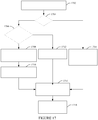
- FIGURE 17 illustrates an example method for determining a tabletop motion algorithm for a selected imaging protocol.
- a signal indicative of a user selected imaging protocol is received.
- the selected imaging protocol may be an axial or a perfusion scan.
- the selected protocol is an axial or perfusion scan protocol.
- the selected protocol is for an axial scan
- Such information may be part of the electronic patient information, entered by a clinician, and/or dynamically measured, for example, via a scale or the like incorporated into the subject support 412 or otherwise.
- a resonant frequency and damping factor of the subject support 412 are determined based at least on the patient's weight. This may include mapping the patient's weight to a look up table (LUT) that cross references patient weight with subject support resonant frequency and damping factor.
- the LUT can be stored in the storage 422 or other storage.
- the processor 420 identifies one of the feed-forward input shaping ( FIGURE 8 ) or the band pass filtered ( FIGURE 10 ) axial motion algorithms.
- the processor 420 identifies the sinusoidal harmonic axial scan motion algorithm ( FIGURE 5 ).
- the processor 420 identifies one of the feed-forward low pass filtered perfusion motion algorithm ( FIGURE 12 ) or the perfusion harmonic motion algorithm ( FIGURE 13 ).
- a profile for the identified tabletop motion algorithm is obtained or generated, and loaded by the subject support controller 418.
- the identified motion profile can dynamically generated, retrieved from storage 422 or other storage, derive, or otherwise obtained.
- the scan is performed using the profile to control the motion of the tabletop 416.
Landscapes
- Health & Medical Sciences (AREA)
- Life Sciences & Earth Sciences (AREA)
- Engineering & Computer Science (AREA)
- Medical Informatics (AREA)
- Optics & Photonics (AREA)
- Biomedical Technology (AREA)
- Biophysics (AREA)
- High Energy & Nuclear Physics (AREA)
- Veterinary Medicine (AREA)
- Nuclear Medicine, Radiotherapy & Molecular Imaging (AREA)
- Public Health (AREA)
- Pathology (AREA)
- Radiology & Medical Imaging (AREA)
- Physics & Mathematics (AREA)
- Heart & Thoracic Surgery (AREA)
- Molecular Biology (AREA)
- Surgery (AREA)
- Animal Behavior & Ethology (AREA)
- General Health & Medical Sciences (AREA)
- Pulmonology (AREA)
- Theoretical Computer Science (AREA)
- Apparatus For Radiation Diagnosis (AREA)
- Measurement Of The Respiration, Hearing Ability, Form, And Blood Characteristics Of Living Organisms (AREA)
Claims (6)
- Bildgebungssystem (400), umfassend:einen Objektträger (412), der zum Tragen eines Objekts konfiguriert ist, das in einem Untersuchungsbereich (406) des Bildgebungssystems (400) abgebildet wird, wobei der Objektträger (412)
eine Tischplatte (416) umfasst; undeine Objektträger-Steuerung (418), die konfiguriert ist, um den Objektträger (412) in dem Untersuchungsbereich (406) zu positionieren, um das Objekt basierend auf einem Bewegungsalgorithmus (424) für eine gegebene Übersetzungsdauer und eine gegebene Übersetzungsdistanz abzutasten und um weiter den Bewegungsalgorithmus basierend auf einem Abtasttyp zu identifizieren;dadurch gekennzeichnet, dass der Bewegungsalgorithmus (424) einem Axialbewegungsalgorithmus zur Vorwärtskopplungseingabeformung oder einem zeitkompensierten bandpassgefilterten S-Kurven-Axialbewegungsalgorithmus oder einem tiefpassgefilterten S-Kurven-Perfusionsbewegungsalgorithmus entspricht;wobei der Axialbewegungsalgorithmus zur Vorwärtskopplungseingabeformung implementiert wird, indem eine Sequenz von Impulsen mit einem gewünschten Bewegungsprofil konvolviert wird, um eine geformte Bewegungsprofileingabe zu erzeugen;wobei der zeitkompensierte bandpassgefilterte S-Kurven-Axialbewegungsalgorithmus unter Einsetzen eines Bandsperrfilters implementiert wird;wobei für einen der Axialbewegungsalgorithmen der Bewegungsalgorithmus auf einer Resonanzfrequenz und einem Dämpfungsfaktor des Objektträgers (412) basiert, die basierend auf einem Gewicht, das von dem Objektträger (412) getragen wird, bestimmt werden; undwobei für den tiefpassgefilterten S-Kurven-Perfusionsbewegungsalgorithmus die Objektträger-Steuerung (418) konfiguriert ist, um ein Bewegungsprofil des Bewegungsalgorithmus durch Abfragen und Tiefpassfiltern eines vorbestimmten Perfusionsbewegungsprofils basierend auf einer vorbestimmten Grenzfrequenz zu erzeugen. - Bildgebungssystem (400) nach Anspruch 1, wobei für einen der Axialbewegungsalgorithmen der Objektträger (412) eine Skala zum Bestimmen des Gewichts des Objekts aufweist, wenn das Objekt auf dem Objektträger (412) ist.
- Bildgebungssystem (400) nach einem der Ansprüche 1 bis 2, wobei für einen der Axialbewegungsalgorithmen die Objektträger-Steuerung (418) konfiguriert ist, um dynamisch ein Bewegungsprofil des Bewegungsalgorithmus (424) durch Hinzufügen einer Zeitverzögerung in ein Tischplattenpositionsprofil und Eingabeformung oder Bandpassfilterung des zeitverzögerten Tischplattenpositionsprofils zu erzeugen.
- Verfahren, umfassend:Bestimmen (1704) eines Abtasttyps für ein Objekt basierend auf einem ausgewählten Abtastprotokoll der Abtastung, wobei der Typ eine Axialabtastung oder eine Perfusionsabtastung ist;Identifizieren (1710, 1712, 1714) eines Bewegungsalgorithmus von Interesse (424) basierend auf dem Abtasttyp für eine Tischplatte (416) eines Objektträgers (412) eines Bildgebungssystems (400), das zum Durchführen der Abtastung verwendet wird;Erzeugen oder Abrufen (1716) eines Tischbewegungsprofils für den Algorithmus (424); Laden (1716) des Tischplattenbewegungsprofils in eine Objektträger-Steuerung (418);undEinsetzen (1718) der Objektträger-Steuerung (418), um die Tischplatte (416) während des Abtastens des Objekts basierend auf dem Bewegungsprofil der geladenen Tischplatte zu steuern;dadurch gekennzeichnet, dass der Bewegungsalgorithmus von Interesse (424) einen axialen Bewegungsalgorithmus zur Vorwärtskopplungseingabeformung oder einen zeitkompensierten bandpassgefilterten S-Kurven-Axialbewegungsalgorithmus oder einen tiefpassgefilterten S-Kurven-Perfusionsbewegungsalgorithmus umfasst;wobei der Axialbewegungsalgorithmus zur Vorwärtskopplungseingabeformung implementiert wird, indem eine Sequenz von Impulsen mit einem gewünschten Bewegungsprofil konvolviert wird, um eine geformte Bewegungsprofileingabe zu erzeugen;wobei der zeitkompensierte bandpassgefilterte S-Kurven-Axialbewegungsalgorithmus unter Einsetzen eines Bandsperrfilters implementiert wird;wobei für einen der Axialbewegungsalgorithmen der Bewegungsalgorithmus auf einer Resonanzfrequenz und einem Dämpfungsfaktor des Objektträgers (412) basiert, die basierend auf einem Gewicht, das von dem Objektträger (412) getragen wird, bestimmt werden; undwobei der tiefpassgefilterte S-Kurven-Perfusionsbewegungsalgorithmus durch Erzeugen eines Bewegungsprofils des Bewegungsalgorithmus durch Abfragen und Tiefpassfiltern eines vorbestimmten Perfusionsbewegungsprofils basierend auf einer vorbestimmten Grenzfrequenz implementiert wird.
- Verfahren nach Anspruch 4, wobei für einen der Axialbewegungsalgorithmen das Verfahren weiter umfasst:Empfangen (1702) eines Signals, das ein Gewicht des Objekts anzeigt; Bestimmen (1708) einer Resonanzfrequenz und eines Dämpfungsverhältnisses des Objektträgers (412) basierend auf dem empfangenen Gewicht; undErzeugen des Bewegungsalgorithmus (424) basierend auf der Resonanzfrequenz und der Dämpfung, wobei die Abtastung eine axiale Abtastung ist und der Bewegungsalgorithmus (424) ein Axialbewegungsalgorithmus mit Vorwärtskopplungseingabeformung oder ein zeitkompensierter, bandpassgefilterter S-Kurven-Axialbewegungsalgorithmus ist.
- Verfahren nach Anspruch 5, wobei der Bewegungsalgorithmus (424) eine Zeitverzögerung aufweist, die eine Zeitverzögerung kompensiert, die durch das Formen oder Filtern eingeführt wird.
Applications Claiming Priority (2)
| Application Number | Priority Date | Filing Date | Title |
|---|---|---|---|
| US201161444155P | 2011-02-18 | 2011-02-18 | |
| PCT/IB2012/050723 WO2012110982A1 (en) | 2011-02-18 | 2012-02-16 | Imaging system subject support motion algorithm(s) |
Publications (2)
| Publication Number | Publication Date |
|---|---|
| EP2675359A1 EP2675359A1 (de) | 2013-12-25 |
| EP2675359B1 true EP2675359B1 (de) | 2019-08-07 |
Family
ID=45852622
Family Applications (1)
| Application Number | Title | Priority Date | Filing Date |
|---|---|---|---|
| EP12709374.8A Active EP2675359B1 (de) | 2011-02-18 | 2012-02-16 | Bewegungsalgorithmen für die personenstütze eines bildgebungssystems |
Country Status (4)
| Country | Link |
|---|---|
| US (1) | US9161726B2 (de) |
| EP (1) | EP2675359B1 (de) |
| CN (1) | CN103379860B (de) |
| WO (1) | WO2012110982A1 (de) |
Families Citing this family (8)
| Publication number | Priority date | Publication date | Assignee | Title |
|---|---|---|---|---|
| DE102011003606A1 (de) * | 2011-02-03 | 2012-08-09 | Siemens Aktiengesellschaft | Verfahren zum Entfernen unerwünschter perfusionsbedingter Signale aus MR-Bildern, Magnetresonanzanlage, Computerprogrammprodukt und elektronisch lesbarer Datenträger |
| DE102012208037A1 (de) * | 2012-05-14 | 2013-11-14 | Siemens Aktiengesellschaft | Patientenlagerungsvorrichtung, eine medizinischen Bildgebungsvorrichtung mit der Patientenlagerungsvorrichtung sowie ein Verfahren zu einer Markierung einer maximalen Aufenthaltsfläche |
| WO2014189048A1 (ja) * | 2013-05-22 | 2014-11-27 | 株式会社 東芝 | X線コンピュータ断層撮影装置、天板制御装置、および天板制御方法 |
| JP6383416B2 (ja) | 2013-11-08 | 2018-08-29 | コーニンクレッカ フィリップス エヌ ヴェKoninklijke Philips N.V. | バンド幅可変ecgハイパスフィルタ、心電図モニタ及び除細動器 |
| JP6266574B2 (ja) * | 2015-09-10 | 2018-01-24 | 株式会社日立ハイテクサイエンス | X線検査方法及びx線検査装置 |
| US11335464B2 (en) * | 2018-01-12 | 2022-05-17 | Siemens Medical Solutions Usa, Inc. | Integrated precision medicine by combining quantitative imaging techniques with quantitative genomics for improved decision making |
| EP4122398B1 (de) * | 2021-07-23 | 2025-07-02 | Siemens Healthineers AG | Verfahren für ein bewegen einer patientenliege |
| CN120267319B (zh) * | 2025-03-05 | 2025-12-30 | 赛诺威盛医疗科技(扬州)有限公司 | 一种基于EtherCAT运动控制的CT扫描床控制方法及系统 |
Citations (1)
| Publication number | Priority date | Publication date | Assignee | Title |
|---|---|---|---|---|
| US20090252300A1 (en) * | 2008-04-02 | 2009-10-08 | General Electric Company | Medical Imaging System and Method with Integrated Weight Sensing |
Family Cites Families (14)
| Publication number | Priority date | Publication date | Assignee | Title |
|---|---|---|---|---|
| US4789929A (en) | 1987-05-14 | 1988-12-06 | Hitachi Medical Corporation | CT system for spirally scanning subject on a movable bed synchronized to X-ray tube revolution |
| US5046003A (en) | 1989-06-26 | 1991-09-03 | General Electric Company | Method for reducing skew image artifacts in helical projection imaging |
| JP3378401B2 (ja) | 1994-08-30 | 2003-02-17 | 株式会社日立メディコ | X線装置 |
| JPH0984784A (ja) | 1995-09-22 | 1997-03-31 | Hitachi Medical Corp | X線検査装置 |
| WO1999027857A1 (en) | 1997-12-04 | 1999-06-10 | Hitachi Medical Corporation | X-ray examination apparatus and imaging method of x-ray image |
| JP4319109B2 (ja) * | 2004-08-13 | 2009-08-26 | ジーイー・メディカル・システムズ・グローバル・テクノロジー・カンパニー・エルエルシー | スキャン制御方法およびx線ct装置 |
| EP1874388A4 (de) | 2005-04-13 | 2011-09-07 | Univ Maryland | Techniken zur kompensierung der bewegung eines behandlungsziels in einem patienten |
| ATE440353T1 (de) * | 2005-06-07 | 2009-09-15 | Koninkl Philips Electronics Nv | Ausfallsichere fernsteuerung |
| JP4611225B2 (ja) | 2006-03-01 | 2011-01-12 | ジーイー・メディカル・システムズ・グローバル・テクノロジー・カンパニー・エルエルシー | X線ct装置 |
| JP2007236662A (ja) * | 2006-03-09 | 2007-09-20 | Ge Medical Systems Global Technology Co Llc | X線ct装置およびそのx線ct画像再構成方法、x線ct画像撮影方法。 |
| CN101103923A (zh) * | 2006-07-14 | 2008-01-16 | Ge医疗系统环球技术有限公司 | X射线混合诊断系统 |
| CN101756709A (zh) * | 2008-12-26 | 2010-06-30 | Ge医疗系统环球技术有限公司 | X射线ct设备 |
| CN102421364B (zh) | 2009-05-08 | 2014-10-22 | 皇家飞利浦电子股份有限公司 | 电机辅助的手动控制移动组件、包括这种组件的x射线系统、方法和使用 |
| US7933377B2 (en) | 2009-06-03 | 2011-04-26 | General Electric Company | Method of CT perfusion imaging and apparatus for implementing same |
-
2012
- 2012-02-16 EP EP12709374.8A patent/EP2675359B1/de active Active
- 2012-02-16 CN CN201280009033.1A patent/CN103379860B/zh active Active
- 2012-02-16 WO PCT/IB2012/050723 patent/WO2012110982A1/en not_active Ceased
- 2012-02-16 US US13/984,858 patent/US9161726B2/en active Active
Patent Citations (1)
| Publication number | Priority date | Publication date | Assignee | Title |
|---|---|---|---|---|
| US20090252300A1 (en) * | 2008-04-02 | 2009-10-08 | General Electric Company | Medical Imaging System and Method with Integrated Weight Sensing |
Non-Patent Citations (2)
| Title |
|---|
| BOHLKE: "Using Input Shaping to minimize residual vibration in flexible space structures", 1 January 1993 (1993-01-01), XP055480374, Retrieved from the Internet <URL:http://hdl.handle.net/1721.1/36050> [retrieved on 20180531] * |
| DAVID BOWLING: "Advances in motion profiling", 1 January 2008 (2008-01-01), XP055480360, ISBN: 978-1-109-01766-3, Retrieved from the Internet <URL:https://media.proquest.com/media/pq/classic/doc/1679799961/fmt/ai/rep/SPDF?_s=iqQsXRua0v3ybsSr5NEfzT1y6IY=> [retrieved on 20180601] * |
Also Published As
| Publication number | Publication date |
|---|---|
| CN103379860A (zh) | 2013-10-30 |
| US20130315381A1 (en) | 2013-11-28 |
| CN103379860B (zh) | 2016-08-17 |
| WO2012110982A1 (en) | 2012-08-23 |
| US9161726B2 (en) | 2015-10-20 |
| EP2675359A1 (de) | 2013-12-25 |
Similar Documents
| Publication | Publication Date | Title |
|---|---|---|
| US9161726B2 (en) | Imaging system subject support motion algorithm(s) | |
| JP4524085B2 (ja) | 連続走査断層写真合成システム及び方法 | |
| US5046003A (en) | Method for reducing skew image artifacts in helical projection imaging | |
| JP5028528B2 (ja) | X線ct装置 | |
| EP2443614B1 (de) | Abbildungsprozedurplanung | |
| JP6329490B2 (ja) | X線ct装置及び画像再構成方法 | |
| CN102483853B (zh) | 用于处理投影数据的装置和方法 | |
| JP2009089760A (ja) | X線ct装置 | |
| CN101370430A (zh) | X射线摄像装置 | |
| US10247824B2 (en) | Ultrasound imaging apparatus and method for controlling the same | |
| EP1913558B1 (de) | Durch 3d-2d-adaptives formmodel unterstützte bewegungskompensierte rekonstruktion | |
| CN101461714A (zh) | X射线ct设备 | |
| NL2008400C2 (en) | Method to reduce radiation dose delivered by imaging system. | |
| JP2007021021A (ja) | 画像処理装置およびx線ct装置 | |
| WO2007072286A2 (en) | Method for movement compensation of image data | |
| JP5511189B2 (ja) | 画像再構成方法およびx線ct装置 | |
| CN100536777C (zh) | 用于计算机断层摄影的动态剂量控制 | |
| JP2009534079A (ja) | 複数の部分的な走査軌道を有するコーンビーム・コンピュータ断層撮影 | |
| JP6321405B2 (ja) | 画像生成装置、放射線断層撮影装置及び画像生成方法並びにプログラム | |
| US20050094760A1 (en) | CT image producing method and X-ray CT apparatus | |
| JP4686147B2 (ja) | 画像データ処理装置 | |
| EP1903787A2 (de) | Bildverarbeitungsvorrichtung und Bildverarbeitungsverfahren | |
| JP6386981B2 (ja) | 画像処理方法、画像処理装置及び放射線断層撮影装置並びにプログラム | |
| US7774040B1 (en) | Method and apparatus of multi-phase cardiac imaging | |
| CN101529472B (zh) | 用于对感兴趣区域成像的成像系统、成像方法和计算机程序 |
Legal Events
| Date | Code | Title | Description |
|---|---|---|---|
| PUAI | Public reference made under article 153(3) epc to a published international application that has entered the european phase |
Free format text: ORIGINAL CODE: 0009012 |
|
| 17P | Request for examination filed |
Effective date: 20130918 |
|
| AK | Designated contracting states |
Kind code of ref document: A1 Designated state(s): AL AT BE BG CH CY CZ DE DK EE ES FI FR GB GR HR HU IE IS IT LI LT LU LV MC MK MT NL NO PL PT RO RS SE SI SK SM TR |
|
| DAX | Request for extension of the european patent (deleted) | ||
| STAA | Information on the status of an ep patent application or granted ep patent |
Free format text: STATUS: EXAMINATION IS IN PROGRESS |
|
| 17Q | First examination report despatched |
Effective date: 20180607 |
|
| GRAP | Despatch of communication of intention to grant a patent |
Free format text: ORIGINAL CODE: EPIDOSNIGR1 |
|
| STAA | Information on the status of an ep patent application or granted ep patent |
Free format text: STATUS: GRANT OF PATENT IS INTENDED |
|
| INTG | Intention to grant announced |
Effective date: 20190304 |
|
| RIN1 | Information on inventor provided before grant (corrected) |
Inventor name: DONG, SHUFANG |
|
| GRAS | Grant fee paid |
Free format text: ORIGINAL CODE: EPIDOSNIGR3 |
|
| GRAA | (expected) grant |
Free format text: ORIGINAL CODE: 0009210 |
|
| STAA | Information on the status of an ep patent application or granted ep patent |
Free format text: STATUS: THE PATENT HAS BEEN GRANTED |
|
| AK | Designated contracting states |
Kind code of ref document: B1 Designated state(s): AL AT BE BG CH CY CZ DE DK EE ES FI FR GB GR HR HU IE IS IT LI LT LU LV MC MK MT NL NO PL PT RO RS SE SI SK SM TR |
|
| REG | Reference to a national code |
Ref country code: GB Ref legal event code: FG4D |
|
| REG | Reference to a national code |
Ref country code: CH Ref legal event code: EP Ref country code: AT Ref legal event code: REF Ref document number: 1162597 Country of ref document: AT Kind code of ref document: T Effective date: 20190815 |
|
| REG | Reference to a national code |
Ref country code: DE Ref legal event code: R096 Ref document number: 602012062651 Country of ref document: DE |
|
| REG | Reference to a national code |
Ref country code: IE Ref legal event code: FG4D |
|
| REG | Reference to a national code |
Ref country code: DE Ref legal event code: R084 Ref document number: 602012062651 Country of ref document: DE |
|
| REG | Reference to a national code |
Ref country code: GB Ref legal event code: 746 Effective date: 20191104 |
|
| REG | Reference to a national code |
Ref country code: NL Ref legal event code: MP Effective date: 20190807 |
|
| REG | Reference to a national code |
Ref country code: LT Ref legal event code: MG4D |
|
| PG25 | Lapsed in a contracting state [announced via postgrant information from national office to epo] |
Ref country code: LT Free format text: LAPSE BECAUSE OF FAILURE TO SUBMIT A TRANSLATION OF THE DESCRIPTION OR TO PAY THE FEE WITHIN THE PRESCRIBED TIME-LIMIT Effective date: 20190807 Ref country code: FI Free format text: LAPSE BECAUSE OF FAILURE TO SUBMIT A TRANSLATION OF THE DESCRIPTION OR TO PAY THE FEE WITHIN THE PRESCRIBED TIME-LIMIT Effective date: 20190807 Ref country code: NL Free format text: LAPSE BECAUSE OF FAILURE TO SUBMIT A TRANSLATION OF THE DESCRIPTION OR TO PAY THE FEE WITHIN THE PRESCRIBED TIME-LIMIT Effective date: 20190807 Ref country code: SE Free format text: LAPSE BECAUSE OF FAILURE TO SUBMIT A TRANSLATION OF THE DESCRIPTION OR TO PAY THE FEE WITHIN THE PRESCRIBED TIME-LIMIT Effective date: 20190807 Ref country code: PT Free format text: LAPSE BECAUSE OF FAILURE TO SUBMIT A TRANSLATION OF THE DESCRIPTION OR TO PAY THE FEE WITHIN THE PRESCRIBED TIME-LIMIT Effective date: 20191209 Ref country code: HR Free format text: LAPSE BECAUSE OF FAILURE TO SUBMIT A TRANSLATION OF THE DESCRIPTION OR TO PAY THE FEE WITHIN THE PRESCRIBED TIME-LIMIT Effective date: 20190807 Ref country code: BG Free format text: LAPSE BECAUSE OF FAILURE TO SUBMIT A TRANSLATION OF THE DESCRIPTION OR TO PAY THE FEE WITHIN THE PRESCRIBED TIME-LIMIT Effective date: 20191107 Ref country code: NO Free format text: LAPSE BECAUSE OF FAILURE TO SUBMIT A TRANSLATION OF THE DESCRIPTION OR TO PAY THE FEE WITHIN THE PRESCRIBED TIME-LIMIT Effective date: 20191107 |
|
| REG | Reference to a national code |
Ref country code: AT Ref legal event code: MK05 Ref document number: 1162597 Country of ref document: AT Kind code of ref document: T Effective date: 20190807 |
|
| PG25 | Lapsed in a contracting state [announced via postgrant information from national office to epo] |
Ref country code: IS Free format text: LAPSE BECAUSE OF FAILURE TO SUBMIT A TRANSLATION OF THE DESCRIPTION OR TO PAY THE FEE WITHIN THE PRESCRIBED TIME-LIMIT Effective date: 20191207 Ref country code: ES Free format text: LAPSE BECAUSE OF FAILURE TO SUBMIT A TRANSLATION OF THE DESCRIPTION OR TO PAY THE FEE WITHIN THE PRESCRIBED TIME-LIMIT Effective date: 20190807 Ref country code: AL Free format text: LAPSE BECAUSE OF FAILURE TO SUBMIT A TRANSLATION OF THE DESCRIPTION OR TO PAY THE FEE WITHIN THE PRESCRIBED TIME-LIMIT Effective date: 20190807 Ref country code: GR Free format text: LAPSE BECAUSE OF FAILURE TO SUBMIT A TRANSLATION OF THE DESCRIPTION OR TO PAY THE FEE WITHIN THE PRESCRIBED TIME-LIMIT Effective date: 20191108 Ref country code: LV Free format text: LAPSE BECAUSE OF FAILURE TO SUBMIT A TRANSLATION OF THE DESCRIPTION OR TO PAY THE FEE WITHIN THE PRESCRIBED TIME-LIMIT Effective date: 20190807 Ref country code: RS Free format text: LAPSE BECAUSE OF FAILURE TO SUBMIT A TRANSLATION OF THE DESCRIPTION OR TO PAY THE FEE WITHIN THE PRESCRIBED TIME-LIMIT Effective date: 20190807 |
|
| RAP2 | Party data changed (patent owner data changed or rights of a patent transferred) |
Owner name: KONINKLIJKE PHILIPS N.V. |
|
| PG25 | Lapsed in a contracting state [announced via postgrant information from national office to epo] |
Ref country code: TR Free format text: LAPSE BECAUSE OF FAILURE TO SUBMIT A TRANSLATION OF THE DESCRIPTION OR TO PAY THE FEE WITHIN THE PRESCRIBED TIME-LIMIT Effective date: 20190807 |
|
| PG25 | Lapsed in a contracting state [announced via postgrant information from national office to epo] |
Ref country code: DK Free format text: LAPSE BECAUSE OF FAILURE TO SUBMIT A TRANSLATION OF THE DESCRIPTION OR TO PAY THE FEE WITHIN THE PRESCRIBED TIME-LIMIT Effective date: 20190807 Ref country code: AT Free format text: LAPSE BECAUSE OF FAILURE TO SUBMIT A TRANSLATION OF THE DESCRIPTION OR TO PAY THE FEE WITHIN THE PRESCRIBED TIME-LIMIT Effective date: 20190807 Ref country code: EE Free format text: LAPSE BECAUSE OF FAILURE TO SUBMIT A TRANSLATION OF THE DESCRIPTION OR TO PAY THE FEE WITHIN THE PRESCRIBED TIME-LIMIT Effective date: 20190807 Ref country code: PL Free format text: LAPSE BECAUSE OF FAILURE TO SUBMIT A TRANSLATION OF THE DESCRIPTION OR TO PAY THE FEE WITHIN THE PRESCRIBED TIME-LIMIT Effective date: 20190807 Ref country code: RO Free format text: LAPSE BECAUSE OF FAILURE TO SUBMIT A TRANSLATION OF THE DESCRIPTION OR TO PAY THE FEE WITHIN THE PRESCRIBED TIME-LIMIT Effective date: 20190807 Ref country code: IT Free format text: LAPSE BECAUSE OF FAILURE TO SUBMIT A TRANSLATION OF THE DESCRIPTION OR TO PAY THE FEE WITHIN THE PRESCRIBED TIME-LIMIT Effective date: 20190807 |
|
| PG25 | Lapsed in a contracting state [announced via postgrant information from national office to epo] |
Ref country code: IS Free format text: LAPSE BECAUSE OF FAILURE TO SUBMIT A TRANSLATION OF THE DESCRIPTION OR TO PAY THE FEE WITHIN THE PRESCRIBED TIME-LIMIT Effective date: 20200224 Ref country code: SM Free format text: LAPSE BECAUSE OF FAILURE TO SUBMIT A TRANSLATION OF THE DESCRIPTION OR TO PAY THE FEE WITHIN THE PRESCRIBED TIME-LIMIT Effective date: 20190807 Ref country code: SK Free format text: LAPSE BECAUSE OF FAILURE TO SUBMIT A TRANSLATION OF THE DESCRIPTION OR TO PAY THE FEE WITHIN THE PRESCRIBED TIME-LIMIT Effective date: 20190807 Ref country code: CZ Free format text: LAPSE BECAUSE OF FAILURE TO SUBMIT A TRANSLATION OF THE DESCRIPTION OR TO PAY THE FEE WITHIN THE PRESCRIBED TIME-LIMIT Effective date: 20190807 |
|
| REG | Reference to a national code |
Ref country code: DE Ref legal event code: R097 Ref document number: 602012062651 Country of ref document: DE |
|
| PLBE | No opposition filed within time limit |
Free format text: ORIGINAL CODE: 0009261 |
|
| STAA | Information on the status of an ep patent application or granted ep patent |
Free format text: STATUS: NO OPPOSITION FILED WITHIN TIME LIMIT |
|
| PG2D | Information on lapse in contracting state deleted |
Ref country code: IS |
|
| 26N | No opposition filed |
Effective date: 20200603 |
|
| PG25 | Lapsed in a contracting state [announced via postgrant information from national office to epo] |
Ref country code: SI Free format text: LAPSE BECAUSE OF FAILURE TO SUBMIT A TRANSLATION OF THE DESCRIPTION OR TO PAY THE FEE WITHIN THE PRESCRIBED TIME-LIMIT Effective date: 20190807 |
|
| REG | Reference to a national code |
Ref country code: CH Ref legal event code: PL |
|
| REG | Reference to a national code |
Ref country code: BE Ref legal event code: MM Effective date: 20200229 |
|
| PG25 | Lapsed in a contracting state [announced via postgrant information from national office to epo] |
Ref country code: MC Free format text: LAPSE BECAUSE OF FAILURE TO SUBMIT A TRANSLATION OF THE DESCRIPTION OR TO PAY THE FEE WITHIN THE PRESCRIBED TIME-LIMIT Effective date: 20190807 Ref country code: LU Free format text: LAPSE BECAUSE OF NON-PAYMENT OF DUE FEES Effective date: 20200216 |
|
| PG25 | Lapsed in a contracting state [announced via postgrant information from national office to epo] |
Ref country code: LI Free format text: LAPSE BECAUSE OF NON-PAYMENT OF DUE FEES Effective date: 20200229 Ref country code: CH Free format text: LAPSE BECAUSE OF NON-PAYMENT OF DUE FEES Effective date: 20200229 |
|
| PG25 | Lapsed in a contracting state [announced via postgrant information from national office to epo] |
Ref country code: FR Free format text: LAPSE BECAUSE OF NON-PAYMENT OF DUE FEES Effective date: 20200229 Ref country code: IE Free format text: LAPSE BECAUSE OF NON-PAYMENT OF DUE FEES Effective date: 20200216 |
|
| PG25 | Lapsed in a contracting state [announced via postgrant information from national office to epo] |
Ref country code: BE Free format text: LAPSE BECAUSE OF NON-PAYMENT OF DUE FEES Effective date: 20200229 |
|
| PG25 | Lapsed in a contracting state [announced via postgrant information from national office to epo] |
Ref country code: MT Free format text: LAPSE BECAUSE OF FAILURE TO SUBMIT A TRANSLATION OF THE DESCRIPTION OR TO PAY THE FEE WITHIN THE PRESCRIBED TIME-LIMIT Effective date: 20190807 Ref country code: CY Free format text: LAPSE BECAUSE OF FAILURE TO SUBMIT A TRANSLATION OF THE DESCRIPTION OR TO PAY THE FEE WITHIN THE PRESCRIBED TIME-LIMIT Effective date: 20190807 |
|
| PG25 | Lapsed in a contracting state [announced via postgrant information from national office to epo] |
Ref country code: MK Free format text: LAPSE BECAUSE OF FAILURE TO SUBMIT A TRANSLATION OF THE DESCRIPTION OR TO PAY THE FEE WITHIN THE PRESCRIBED TIME-LIMIT Effective date: 20190807 |
|
| PGFP | Annual fee paid to national office [announced via postgrant information from national office to epo] |
Ref country code: GB Payment date: 20230214 Year of fee payment: 12 Ref country code: DE Payment date: 20220628 Year of fee payment: 12 |
|
| REG | Reference to a national code |
Ref country code: DE Ref legal event code: R119 Ref document number: 602012062651 Country of ref document: DE |
|
| GBPC | Gb: european patent ceased through non-payment of renewal fee |
Effective date: 20240216 |
|
| PG25 | Lapsed in a contracting state [announced via postgrant information from national office to epo] |
Ref country code: DE Free format text: LAPSE BECAUSE OF NON-PAYMENT OF DUE FEES Effective date: 20240903 |
|
| PG25 | Lapsed in a contracting state [announced via postgrant information from national office to epo] |
Ref country code: GB Free format text: LAPSE BECAUSE OF NON-PAYMENT OF DUE FEES Effective date: 20240216 |
|
| PG25 | Lapsed in a contracting state [announced via postgrant information from national office to epo] |
Ref country code: GB Free format text: LAPSE BECAUSE OF NON-PAYMENT OF DUE FEES Effective date: 20240216 Ref country code: DE Free format text: LAPSE BECAUSE OF NON-PAYMENT OF DUE FEES Effective date: 20240903 |