EP2669295A1 - Procédé de diagnostic permettant de déterminer la présence et la quantité d'interleukine-3 humaine dans un échantillon à l'aide de nouveaux anticorps IL-3 - Google Patents
Procédé de diagnostic permettant de déterminer la présence et la quantité d'interleukine-3 humaine dans un échantillon à l'aide de nouveaux anticorps IL-3 Download PDFInfo
- Publication number
- EP2669295A1 EP2669295A1 EP12169805.4A EP12169805A EP2669295A1 EP 2669295 A1 EP2669295 A1 EP 2669295A1 EP 12169805 A EP12169805 A EP 12169805A EP 2669295 A1 EP2669295 A1 EP 2669295A1
- Authority
- EP
- European Patent Office
- Prior art keywords
- antibody
- clone
- antibodies
- diagnostic method
- sample
- Prior art date
- Legal status (The legal status is an assumption and is not a legal conclusion. Google has not performed a legal analysis and makes no representation as to the accuracy of the status listed.)
- Withdrawn
Links
Images


Classifications
-
- C—CHEMISTRY; METALLURGY
- C07—ORGANIC CHEMISTRY
- C07K—PEPTIDES
- C07K16/00—Immunoglobulins [IGs], e.g. monoclonal or polyclonal antibodies
- C07K16/18—Immunoglobulins [IGs], e.g. monoclonal or polyclonal antibodies against material from animals or humans
- C07K16/24—Immunoglobulins [IGs], e.g. monoclonal or polyclonal antibodies against material from animals or humans against cytokines, lymphokines or interferons
- C07K16/244—Interleukins [IL]
-
- G—PHYSICS
- G01—MEASURING; TESTING
- G01N—INVESTIGATING OR ANALYSING MATERIALS BY DETERMINING THEIR CHEMICAL OR PHYSICAL PROPERTIES
- G01N33/00—Investigating or analysing materials by specific methods not covered by groups G01N1/00 - G01N31/00
- G01N33/48—Biological material, e.g. blood, urine; Haemocytometers
- G01N33/50—Chemical analysis of biological material, e.g. blood, urine; Testing involving biospecific ligand binding methods; Immunological testing
- G01N33/53—Immunoassay; Biospecific binding assay; Materials therefor
- G01N33/564—Immunoassay; Biospecific binding assay; Materials therefor for pre-existing immune complex or autoimmune disease, i.e. systemic lupus erythematosus, rheumatoid arthritis, multiple sclerosis, rheumatoid factors or complement components C1-C9
-
- G—PHYSICS
- G01—MEASURING; TESTING
- G01N—INVESTIGATING OR ANALYSING MATERIALS BY DETERMINING THEIR CHEMICAL OR PHYSICAL PROPERTIES
- G01N33/00—Investigating or analysing materials by specific methods not covered by groups G01N1/00 - G01N31/00
- G01N33/48—Biological material, e.g. blood, urine; Haemocytometers
- G01N33/50—Chemical analysis of biological material, e.g. blood, urine; Testing involving biospecific ligand binding methods; Immunological testing
- G01N33/68—Chemical analysis of biological material, e.g. blood, urine; Testing involving biospecific ligand binding methods; Immunological testing involving proteins, peptides or amino acids
- G01N33/6863—Cytokines, i.e. immune system proteins modifying a biological response such as cell growth proliferation or differentiation, e.g. TNF, CNF, GM-CSF, lymphotoxin, MIF or their receptors
- G01N33/6869—Interleukin
-
- C—CHEMISTRY; METALLURGY
- C07—ORGANIC CHEMISTRY
- C07K—PEPTIDES
- C07K2317/00—Immunoglobulins specific features
- C07K2317/10—Immunoglobulins specific features characterized by their source of isolation or production
- C07K2317/14—Specific host cells or culture conditions, e.g. components, pH or temperature
-
- C—CHEMISTRY; METALLURGY
- C07—ORGANIC CHEMISTRY
- C07K—PEPTIDES
- C07K2317/00—Immunoglobulins specific features
- C07K2317/30—Immunoglobulins specific features characterized by aspects of specificity or valency
- C07K2317/33—Crossreactivity, e.g. for species or epitope, or lack of said crossreactivity
-
- C—CHEMISTRY; METALLURGY
- C07—ORGANIC CHEMISTRY
- C07K—PEPTIDES
- C07K2317/00—Immunoglobulins specific features
- C07K2317/90—Immunoglobulins specific features characterized by (pharmaco)kinetic aspects or by stability of the immunoglobulin
- C07K2317/92—Affinity (KD), association rate (Ka), dissociation rate (Kd) or EC50 value
-
- G—PHYSICS
- G01—MEASURING; TESTING
- G01N—INVESTIGATING OR ANALYSING MATERIALS BY DETERMINING THEIR CHEMICAL OR PHYSICAL PROPERTIES
- G01N2333/00—Assays involving biological materials from specific organisms or of a specific nature
- G01N2333/435—Assays involving biological materials from specific organisms or of a specific nature from animals; from humans
- G01N2333/52—Assays involving cytokines
- G01N2333/54—Interleukins [IL]
- G01N2333/5403—IL-3
-
- G—PHYSICS
- G01—MEASURING; TESTING
- G01N—INVESTIGATING OR ANALYSING MATERIALS BY DETERMINING THEIR CHEMICAL OR PHYSICAL PROPERTIES
- G01N2800/00—Detection or diagnosis of diseases
- G01N2800/10—Musculoskeletal or connective tissue disorders
- G01N2800/101—Diffuse connective tissue disease, e.g. Sjögren, Wegener's granulomatosis
- G01N2800/102—Arthritis; Rheumatoid arthritis, i.e. inflammation of peripheral joints
Definitions
- the present invention relates to diagnostic methods for reliably determining the presence and the amount of human interleukin-3 (hIL-3) in a sample, preferably in the blood, plasma, serum or any other body fluid (e.g. urine, synovial fluid) of a human patient.
- hIL-3 human interleukin-3
- the invention further relates to novel anti-IL-3 antibodies used in the diagnostic method, nucleic acid sequences encoding and hybridoma cell lines producing the antibodies according to the present invention and test kits providing the essential reagents for the diagnostic method.
- Cytokines are polypeptides that influence the function of certain cells upon binding to specific cellular receptors and are divided in subclasses, i.e., interleukins, interferons, colony-stimulating factors (CSFs), lymphokines, growth factors and monokines. It is well known that cytokines play a major role in cell proliferation and, e.g., also inflammatory diseases.
- Cell proliferation is a complex process wherein growth factors bind to specific receptors on the cell surface, whereupon endocytosis occurs and the complexes of cytokine and receptor are internalized causing a cellular response.
- Such cellular responses include specific gene transcription activities as DNA synthesis and cell replication.
- most of the cytokines When tested in relatively high concentrations, most of the cytokines have several differing biological effects. Because of these effects of cytokines, there is a high interest in investigations for possible therapeutic uses of these proteins.
- Interleukins are mediators of the immune system which are produced in low concentration mostly in leukocytes. They influence the growth, differentiation and activity of cells of the immune system and thus belong to the immune modulators. They also take effect by binding to receptors on the surface of target cells and thus change the transcription rate of certain genes. They play an important role in the triggering of a multiplicity of cellular responses.
- Interleukins are, e.g., involved in the immunological cell activation cascade and subsequent inflammatory changes. Irregular and/or abnormal inflammation is a major component and factor of a wide range of human diseases, one of which is the immunological disorder rheumatoid arthritis (RA). But also other immunological diseases are influenced by interleukins.
- RA immunological disorder rheumatoid arthritis
- IL-3 also designated as Multi-CSF, is a well-known member of the interleukin family. It has a growth stimulating and differentiating effect on various hematopoietic precursor cells and acts as a growth factor for mast cells. Together with IL-5 and GM-CSF, IL-3 belongs to the family of hematopoietic cytokines with four short alpha-helical bundles. GM-CSF and IL-3 stimulate the formation of neutrophilic and eosinophilic granulocyte colonies as well as macrophages. It further stimulates the formation of mast, megakaryocyte and pure and mixed erythroid colonies ( D. Metcalf, "The hematopoietic colony-stimulating factors", 1984, Elsevier, Amsterd am).
- IL-3 consists of 133 amino acids and is known for its stimulation of colony formation by human hematopoietic progenitor cells and the stimulation of DNA synthesis by human acute myelogenous leukemia (AML) blasts.
- IL-3 binds to a unique receptor also known as CD123 antigen.
- the receptor belongs to the type I cytokine receptor family and is a heterodimer with a unique ⁇ -chain paired with a common ⁇ -subunit ( ⁇ C or CDW 131).
- IL-3 binds to the unique ⁇ -receptor subunit. Signal transduction is mediated, however, by the common ⁇ -receptor subunit ( ⁇ C) by the JAK2-STAT5 pathway.
- IL-3 is mainly produced by activated CD4+ T-cells and contributes especially to growth, differentiation and survival of CD34+ hematopoietic progenitor cells.
- IL-3 has been observed to promote the differentiation of basophiles and mast cells from bone marrow cells. It has further been observed to induce IL-6 release by murine basophils and to up-regulate MHC-II expression and IL-1 secretion in monocyte/macrophages. Further, IL-3 supports the differentiation of monocytes into dendritic cells and osteoclasts.
- RA rheumatoid arthritis
- NSAIDs non-steroidal anti-inflammatory drugs
- NSAIDs act as anti-inflammatory and analgetic agents and often only achieve an alleviation of pain.
- the drugs further interfere with a certain step in the inflammatory cascade, where prostaglandine is generated by cyclooxygenases.
- NSAIDs do not influence the underlying inflammatory process and are thus not able to retard the joint destruction, which is the most deleterious effect of RA.
- DMARDs disease-modifying anti-rheumatic drugs
- methotrexate the most commonly used anti-rheumatic, the effect of which is based on a reversible inhibition of the enzyme dihydrofolate reductase.
- leflunomide Another commonly used substance for treating RA. Both pharmaceuticals are long-acting and thus have to be administered over a longer period of time (usually 12-16 weeks) to show the desired effects. To bridge the time until DMARDs improve the disease, most patients are administered steroids.
- a further approach for treating RA are "biologicals" that block cytokines like TNF, IL-6, IL-1 or costimulatory molecules like B7 or that deplete leukocyte subsets (e.g. B cells).
- Biologicals e.g. the TNF antibody Infliximab
- TNF antibody Infliximab are mostly used for severe disease processes and after DMARDs have failed to sufficiently control disease activity.
- Biologicals influence a plurality of signal systems in the immune system and have a variety of serious side effects including bacterial and viral infections and a higher risk for development of neoplasia.
- IL-3 inhibitors can be used in treatment of early stages of rheumatoid arthritis.
- a genetic analysis found an association between a single nucleotide polymorphism in the IL-3 promoter gene and RA.
- WO 2010/063488 proposes such use of inhibitors, mainly antibodies or antibody fragments, antibody variants or antibody multimers in prophylactic RA treatment, therapeutic treatment in early stages of the disease or in maintenance treatment.
- IL-3 Apart from the outlined relevance of IL-3 in RA, another advantageous implementation of the determination of elevated levels of IL-3 could be seen in the field of coronary artery and heart diseases. Elevated levels of IL-3 have e.g. been considered relevant for the prediction of restenosis after coronary intervention ( Rudolph, T. et al. (2009) Int. J. Cardiol. 132:392 ). Further elucidation of the role of IL-3 might suggest other conditions in which IL-3 levels have medical relevance and therefore need to be monitored.
- a diagnostic method for determining the IL-3 level in a sample which preferably is a body fluid and more preferably is blood, plasma or serum of a patient.
- the method according to the present invention comprises adding an anti-IL-3 antibody, antibody fragment or antibody construct to said sample under conditions which allow for binding of said antibody, fragment or construct thereof to IL-3 and detecting the amount of antibody-bound IL-3 in said sample, wherein the anti-IL-3 antibody is clone 13 (DSM ACC3164).
- IL-3 For a reliable qualitative and quantitative determination of IL-3 in a body fluid sample, it is especially important that the antibodies used in such method show little or no cross-reactivity with other cytokines and interleukins like IL-5 an GM-CSF which are often also present in substantial amounts in body fluids of patients.
- Antibody clone 13 is a mouse anti-human IL-3 antibody which shows a high specificity and affinity for IL-3. It is considered to bind to a 3D epitope of IL-3 in its native conformation. According to the present invention, antibody clone 13 and optionally further antibodies having similar characteristics are used for the detection of the presence and/or amount of IL-3 in any suitable immunoassay format.
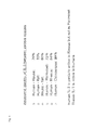
- antibody clone 13 Apart from a very high specificity for only IL-3 but not other cytokines, antibody clone 13 also shows very low levels of cross-reactivity with IL-3 molecules of other mammalian origin. Although amino acid identities for the human protein and the mouse protein is only 29%, for marmoset, rhesus or a chimpanzee proteins there are amino acid identities between 72 and 99% ( figure 1 ). Nevertheless, it could be shown that for clone 13 there was no detectable cross-reactivity with IL-3 from mouse, rat or rhesus. From the further cytokines, which may also be present at an elevated level in autoimmune diseases, IL-5 and GM-CSF are particularly important.
- a high cross-reactivity of an anti-IL-3 antibody with such cytokines in an immunoassay can lead to incorrect results regarding the fact whether an IL-3 overexpression has an important influence in the manifestation and progression of the autoimmune disease. Such results, however, have an important impact e.g. on the decision whether the application of an anti-IL-3 antibody can be considered a promising therapeutic approach.
- antibodies used in the diagnostic method of this invention to show the lowest possible cross-reactivity with human IL-5 and GM-CSF.
- it is a characterizing feature of such an antibody that it binds to IL-5 or GM-CSF to an extent of below 5%, more preferred below 2% and particularly preferred below 1% as compared to the amount of IL-3 bound by the antibody.
- clone 13 has been proven to be usefull for ELISA immunoassays using blood, plasma or serum as samples and which show a very high sensitivity as well as specificity towards hIL-3 and enable the detection of levels of IL-3 already in the pg/ml range. Further, such diagnostic tests using clone 13 reliably detect IL-3 even in samples that have been stored at room temperature, in the refrigerator or the freezer.
- the diagnostic method is conducted as an ELISA assay.
- the general test regime used for ELISA assays is well known to the skilled person.
- At least two antibodies which bind to the target molecule are used.
- One of these antibodies is bound to a solid phase allowing for the separation of the antigen to be determined from the test sample.
- a second antigen-specific and labeled antibody is added and after further removal of excess labeled antibody and optionally further washing steps the amount of label bound via the antibody-antigen-antibody complex is determined and correlated to the amount of antigen present.
- both antibodies used in such an ELISA assay are specific to the antigen to a high extent, an especially high degree of specificity is needed in at least one of the two antibodies involved.
- a very specific antibody can be used to be able to separate only the desired antigen from the liquid sample without any cross-reacting background. If only the desired antigen is coupled via the antibody to the solid surface, the specificity of the second antibody is not so decisive anymore since unspecific binding to other antigens is not an issue in such a case. Accordingly, it is possible to use a less specific antibody as the second antibody in such a context.
- the first solid phase-bound antibody it is possible to use a high affinity and high avidity antibody as the first solid phase-bound antibody and to use a highly specific antibody as the second and labeled antibody.
- a highly specific antibody such an embodiment, unspecific binding of other antigens like IL-5 or GM-CSF might take place to some extent due to a certain cross-reactivity of the first antibody, however, the actual detection by the labeled antibody is then restricted to IL-3 by using a highly specific second antibody.
- first solid-phase bound antibody / second labeled antibody preferred combinations of antibodies (first solid-phase bound antibody / second labeled antibody) are clone 13 / 11 (most preferred), clone 11 / 13 (preferred), clone 13 / 44, clone 44 / 13, clone 14 / 47, clone 47 / 13 as well as combinations of clones 11 or 44 with clone 47.
- the antibodies used in the diagnostic method according to the present invention can be of different nature and the following more detailed illustrations of possible antibodies or antibody constructs are only meant to be exemplary. That means that within the context of the present invention the term antibody is to be understood in its broadest sense. Any antibody, part thereof or construct containing antibody characteristics and retaining the specificity of the antibodies shown in the examples of the present invention, is considered as encompassed within the term antibody in the context of the present invention.
- monoclonal antibodies as well as polyclonal antibodies can be used.
- Monoclonal antibodies generally have the advantage of a higher specificity as compared to polyclonal antibodies and are thus preferred in view of the present invention.
- the term “antibody” shall also comprise fragments, bi-, tri- or multimeric or bi-, tri- or multifunctional antibodies having several antigen binding sites which preferably are IL-3-specific binding sites.
- the term “antibody” further comprises fusion proteins containing as a part of the fusion protein an antibody or antibody fragment or complement determining region (CDRs) of an antibody of the present invention, which show a corresponding specificity and which have furthermore retained their binding ability to IL-3. Further comprised are single chain antibodies.
- inventive antibodies can belong to any appropriate antibody class, it is however essential that their use in therapy and diagnostics is possible.
- the anti-IL-3 antibody or the fragment thereof according to the present invention is of the class IgG, IgA, IgE oder IgM.
- the actual form of a molecule considered to be encompassed by the term "antibody” is irrelevant as long as it specifically binds to IL-3 in diagnostic assays.
- antibody fragments or constructs as outlined above which are derived from antibody clone 13 as well as clone 11, clone 44 and clone 47 can be used in the method of the invention.
- the specific anti-IL-3 antibodies used according to the present invention can be produced by any method known to the skilled person.
- antibodies can be generated using the complete hIL-3 protein as an immunogen and lateron selecting for antibodies and antibody clones which are specific for the mentioned sequences.
- a peptide containing within its sequence the desired parts or epitopes of IL-3 can be used for immunization.
- a further possibility is the use of artificial epitopes which contain only the very epitope (conformationally discriminating epitope, CDE) integrated into an environment which allows for the generation of antibodies.
- CDE formationally discriminating epitope
- Such anti-IL-3-antibody can also be of any origin, e.g. human, mouse, goat, rabbit.
- the production of antibodies is carried out by immunizing appropriate mammals, e.g., mice, rat, hamster or rabbits.
- ELISA enzyme-linked immunosorbent assay
- ELISA plates are coated with any anti-human-IL-3 antibody which can also be a commercially available antibody (e.g., a goat IgG anti-human-IL-3 antibody).
- any anti-human-IL-3 antibody which can also be a commercially available antibody (e.g., a goat IgG anti-human-IL-3 antibody).
- 1 ug/ml of the antibody is incubated with the ELISA plates over night in a refrigerator whereupon a washing step, a blocking step and incubation with human IL-3 (250 ng/ml in PBS buffer) is performed, thus fixing human IL-3 on the solid phase.
- candidate antibodies are then added in different concentrations and detected by means of a secondary HRP- (horseradish peroxidase) labelled polyclonal antibody.
- a particularly preferred antibody which according to the present invention is used in an ELISA assay as a second or further antibody together with antibody clone 13 is clone 11.
- Clone 11 is a mouse anti-human-IL-3 antibody and shows a very high specificity and affinity to IL-3. Clone 11 specifically binds to the epitope with the amino acids SWVN according to SEQ ID NO: 2. This particularly preferred antibody was deposited at DSMZ (Braunschweig, Germany) under number DSM ACC3163.
- clone 11 has proven very superior characteristics with regard to specificity, lack of cross-reactivity but also with regard to affinity and avidity. This preferred antibody therefore is considered especially suitable for use in diagnostics as well as therapeutic measures.
- a further especially preferred antibody used in an ELISA assay according to the present invention is clone 44 which has been deposited at DSMZ under the accession number DSM ACC3166. Clone 44 is considered to bind specifically to the same epitope as clone 11.
- a further especially preferred antibody used in an ELISA assay according to the present invention is clone 47 which has been deposited at DSMZ under the accession number DSM ACC3167. Clone 47 is considered to bind to a three dimensional (3D) epitope.
- a second embodiment of the present invention is novel antibody clone 13.
- a further subject-matter and third embodiment of the present invention is a nucleic acid which encodes antibody clone 13 or an antibody fragment, an antibody construct or sequences for CDRs conveying specificity of antibody clone 13 according to the present invention.
- a further subject-matter and embodiment of the present invention is the hybridoma cell line which produces antibody clone 13, the cell line designated 13.4.4 (DSM ACC3164).
- a further and final embodiment of the present invention is a test kit to be used in the diagnostic method according to the present invention.
- a test kit contains as an essential element antibody clone 13 as well as other materials and reagents necessary and useful for the immunoassay for detection of IL-3.
- Clone 13 and other useful as well as preferred antibodies for the immunoassay and especially ELISA assay are described above.
- Further materials necessary for the assays as well as useful buffers, labels and other reagents e.g. for determining the amount of label which is indicative of the amount of IL-3 present in the sample are known to the skilled person.
- Such test kits can be provided for easy and effective use in laboratories of every size.
- Test kits according to the present invention contain in an especially preferred embodiment of the present invention a combination of antibody clones 13 and 11, wherein both antibodies can be provided either as solid phase bound antibody or as the antibody carrying the detectable label.
- IL-3 By using the clone 13 and other specific anti-IL-3 antibodies in diagnostic methods and corresponding test kits, the accurate determination of the presence of IL-3 in body fluids can be improved to a major extent and such methods and test kits, therefore, are a further subject matter of the present invention. It has surprisingly been found that using the antibodies and methods of the present invention, IL-3 could be specifically detected even if present in only picogram / ml concentrations in a sample. The relative affinity of the antibodies of the present invention towards IL-3 is thus about at least a factor 100 higher than the affinity of known antibodies.
- the present diagnostic method is especially suited and useful for the detection of IL-3 in a plasma or serum sample.
- detection of IL-3 in plasma or serum did not lead to sufficiently meaningful results especially due to cross-reactivities of known antibodies and insufficient affinities of such antibodies with regard to the low levels of IL-3 in present blood, plasma or serum in healthy and in RA patients. Therefore, often synovial fluids were used as test sample. However, obtaining such a test sample is much more difficult and cumbersome to the patient.
- the present invention via its antibodies and their use in diagnosis is a big step forward to relating IL-3 presence to the severity of RA and RA incidents in patients and thus to also determine the supposition of a patient to treatment with IL-3 antibodies.
- Anti-IL-3 antibodies were produced by immunizing Balb/c mice using at least 6 i.p. injections of human eukaryotic glycosylated IL-3 in alumn at four week intervals. Two days before cell fusion, IL-3 in PBS was injected intraperitoneally. Antibody-producing splenocytes obtained from the immunized mice (HGPRT positive, able to grow on HAT medium) were fused with the myeloma cell line X63Ag8.6.5.3 in the presence of polyethylene glycol (PEG) and a selection of hybridomas performed in an HAT-selection medium.
- PEG polyethylene glycol
- Hybridomas were cultivated in RPMI-1640 medium supplemented by 10% FCS (neat inactivated, HIA), P/S and glutamine (1:100). Obtained cells are able to grow in suspension and are splitted every three days in a ratio of 1:4.
- hybridoma cells are transferred from a cell culture bottle into 50 ml or 15 ml cell culture flasks (BD FalconTM). After centrifugation at 1400 rpm for 5 minutes at room temperature, the supernatant is completely removed. Cells are resuspended in a freezing medium (90% FCS (HIA) + 10% DMSO) and 1.5 ml aliquots are filled into vials. The cells are prefrozen in a freezing container in a freezer at -80°C and after 1-2 days transferred to a liquid nitrogen storage tank.
- a freezing medium 90% FCS (HIA) + 10% DMSO
- Cloning and recloning of the obtained hybridoma cell lines are performed using limited dilution to provide long-term stable sources for monoclonal antibodies.
- 96-well-plates are coated overnight at room temperature with anti-mouse IgG (1:100 in PBS) in a concentration of 100 ⁇ l/well. Blocking is performed by adding 100 ⁇ l per well of 2 % BSA in PBS and incubation at room temperature for two hours. After the blocking reaction, the plates are washed twice.
- the plate is washed three times and then incubated with biotinylated anti-mouse IgG1 (diluted by 1:250 in 2 % BSA in PBS) for one hour at room temperature with 100 ⁇ l per well. After washing the plate a further three times, streptavidin-HRP (1:1000 in 2 % BSA in PBS) is added for one hour at room temperature and in the dark. The concentration of the antibodies is determined after adding ABTS and incubating for further 30 minutes and measuring the signal at 405 and 490 nanometers on a spectrophotrometer. Based on this determination, a desired amount of the antibodies tested is applied for the further tests.
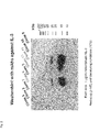
- Samples of IL-3 are prepared by mixing of recombinant human IL-3 1:1 with Laemmli buffer and heating the samples at 60°C for 5 min. An amount of 1 ⁇ g per lane of IL-3 as well as a usual standard for determining molecule sizes is loaded onto the gel. The gel is then mounted in a SDS-PAGE gel electrophoresis apparatus which already contains a running buffer. The inserted gel is then cautiously overlayed with additional running buffer and electrophoresis performed at 20 to 25 mA with voltage adjusted to infinite for approximately 1,5 hours. When the run is completed, the gel is retrieved from the apparatus and the stacking gel is removed.
- Antibody clones are incubated at a concentration of 5 ⁇ g/ml in blocking solution for 2 hours at room temperature under agitation on the shaking apparatus. After three washing steps, HRP labelled anti-mouse immune-globulin (1:1000 in blocking solution) is added and incubation is conducted for 1 hour at room temperature while shaking. After three further wahing steps, a detection solution (1:1 mixture of solutions A and B of the Western-blotting Luminal Reagent obtained from NALGENE) is added and incubated for 1 min at room temperature. Films are then adjusted on the membranes with different times of expositions and developed in the dark room.
- Fig. 2 shows the results of binding of antibody clones 2, 3, 5, 6, 7, 8, 10, 11 and 13. Binding to IL-3 at the given concentration was detected for clones 8, 11 and, to a lesser extent, for clone 13.
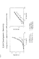
- the affinity of the obtained antibodies for IL-3 was measured in an ELISA assay.
- ELISA plates were coated overnight with 1 ⁇ g/ml of anti-human IL-3 antibody (RD, goat IgG anti-human IL-3 AF-203-NA). For each concentration, duplicates were used (2 x 12 wells). For this purpose, the first concentration (2 ⁇ g/ml) is diluted in PBS, further dilutions are made in PBS containing 2 ⁇ g/ml control goat IgG to keep the total concentration of IgG constant. Blocking with 2% BSA is performed for 2 hours at room temperature, followed by 5 washing steps using PBS.
- hIL-3 (0.25 ⁇ g/ml in PBS)
- hIL-3 (0.25 ⁇ g/ml in PBS)
- no hIL- 3 is added.
- the wells are incubated overnight at 4°C with serial (1:3) dilutions of antibodies clone 8 and 11 obtained in example 1, the antibodies being used in PBS buffer containing 2% BSA and with a starting concentration of 20 ⁇ g/ml.
- bound antibody is detected using goat-anti-mouse-HRP antibody (1:500 in PBS with 2% BSA) and incubation for 1 hour at room temperature.
- ABTS ROCHE, 1 mg/ml
- Fig. 3 shows the results of tests including antibodies clone 3, 8, 10, 11 and 13. The tests were performed in the manner as described with different concentrations/dilutions of antibodies as shown in the figure 1 .
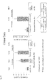
- the cytokine coated plates were washed three times and blocking performed for 2 hours at room temperature using 2% BSA in PBS. After three further washing steps, antibodies clone 3.47.20, 8.36.38, 10.12.4, 11.14.6, 13.4.4 and just medium (RPMI1640 containing 10 % FCS) as control were added at a concentration of 40 ⁇ g/ml and 1:5 and 1:25 dilutions thereof in a volume of 100 ⁇ l/well and inicubated for 1 hour at room temperature. On each plate a negative control is used.
- a secondary HRP-labelled rabbit anti-mouse IgG (DAKO-Cytomation P260 (1:2000 in 2% BSA in PBS, 100 ⁇ l/well) was added and the plates incubated at room temperature for 1 hour in the dark.
- ABTS (ROCHE, 1 mg/ml) was added and spectrometry performed at 405 and 490 nm after 30 min.
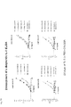
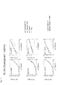
- the IL-3 coated plates were washed three times and blocking performed for 2 hours at room temperature with 2% BSA in PBS. After three washing steps, antibody clones 3.47.20, 8.36.38, 10.12.4, 11.14.6, 13.4.4 in certain concentrations as indicated in Figs 7 and 8 , and 1:5, 1:25 and 1:125 dilutions thereof were added at volumes of 100 ⁇ l/well.
- R&D monoclonal anti-IL-3 antibody (R&D Systems, Inc.) was used (100 ⁇ l/well) in concentrations of 40 ⁇ g/ml, 20 ⁇ g/ml, 10 ⁇ g/ml, 5 ⁇ g/ml and 2.5 ⁇ g/ml and, as negative control, medium (100 ⁇ l/well) without antibody (RPMI 1640 containing 10 % FCS) was used. On each plate a negative control was used.
- a secondary HRP-labelled rabbit anti-mouse IgG (DAKO-Cytomation P260 (1:2000 in 2% BSA in PBS, 100 ⁇ l/well) was added and the plates incubated at room temperature for 1 hour in the dark.
- ABTS (ROCHE, 1 mg/ml) was added and spectrometry performed at 405 and 490 nm after 30 min.
- Anti-IL-3 antibody clones 8, 11, 13 and further antibody clones 44 (44.16.16, DSM ACC3166) and 47 (47.28.15, DSM ACC3167) were analysed for their use in the development of a highly sensitive and specific ELISA assay for the determination of IL-3, especially for diagnostic purposes in blood, plasma or serum, as well as other body fluids.
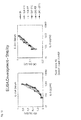
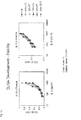
- ELISA plates were incubated with 5 ⁇ g/ml of anti-IL-3 antibody overnight at room temperature to coat the plates. After three washing steps, blocking is performed using 1% BSA in PBS at 100 ⁇ l/well for 1 hour at room temperature. After further three washing steps, samples are incubated with 60 ⁇ l/well of IL-3 in various concentrations in PBS, plasma and serum.
- detection of solid-phase bound IL-3 is performed by adding 60 ⁇ l/well of a different and HRP labelled anti-IL-3 antibody at a concentration of 400 ng/ml and incubation for 2 hours at room temperature, followed by three washing steps and addition of TMB buffer (10 ml TMB buffer, 1 tablet of TMB, 3 ⁇ l H 2 O 2 ) (0,1 mg/ml, SIGMA-ALDRICH).
- TMB buffer 10 ml TMB buffer, 1 tablet of TMB, 3 ⁇ l H 2 O 2
- the reaction is stopped by adding 100 ⁇ l/well of H 2 SO 4 (12,5 % in H 2 O).
- the results are obtained by spectrometry at 450 nm and shown in Figs. 7 to 10 .
- Labelling of the anti-IL3 antibody clones was performed using the Lightning-Link HRP Conjugation Kit (Innova Biosciences) using the following protocol: For each of the purified antibody clones 8, 11, 13, 44 and 47 100 ⁇ l solutions with a concentration of 1 ⁇ g/ ⁇ l (in PBS) were produced. To each antibody solution, 10 ⁇ l of LL-modifying reagent were added and the obtained solution mixed carefully. For each antibody solution a Lightning Link mix bottle (100 pg reagent) was opened and the antibody solution including the LL-modifying agent added directly onto the reagent powder. Mixing was performed very cautiously by up- and down-pipetting of the solution.
- the lid was readjusted on the bottle of the Lightning Link mix and the bottles incubated for 3 hours at room temperature whereupon 10 ⁇ l LL-quencher reagent were added and incubated for a further 30 min at room temperature. After this treatment the antibodies were stored at -20°C for further use.
- analogue tests were performed using the Quantikine Human IL-3 ELISA test kit provided by R&D Systems, Inc., Catalogue No. Dy 403.
- An inventive test kit with clones 13/11 retained the same sensitivity as when using PBS or PBS/BSA samples also for plasma and serum (comparison shown in Fig. 10 ). Tests performed with different plasma samples (Plasma EDTA, Plasma Citrat) and serum could also be shown to be stable for at least 24 hours at room temperature ( Fig. 11 ) and there was also no detectable signal loss after freezing and thawing of the samples ( Figs. 12-14 ).
Priority Applications (5)
| Application Number | Priority Date | Filing Date | Title |
|---|---|---|---|
| EP12169805.4A EP2669295A1 (fr) | 2012-05-29 | 2012-05-29 | Procédé de diagnostic permettant de déterminer la présence et la quantité d'interleukine-3 humaine dans un échantillon à l'aide de nouveaux anticorps IL-3 |
| EP13734345.5A EP2855525B1 (fr) | 2012-05-29 | 2013-05-29 | Procédé de diagnostic permettant de déterminer la présence et la quantité d'interleukine-3 humaine dans un échantillon à l'aide de nouveaux anticorps il-3 |
| US14/400,958 US9714286B2 (en) | 2012-05-29 | 2013-05-29 | Diagnostic method for determining the presence and amount of human interleukin-3 in a sample using novel IL-3 antibodies |
| PCT/EP2013/061122 WO2013178707A1 (fr) | 2012-05-29 | 2013-05-29 | Méthode diagnostique pour la détermination de la présence et de la quantité de l'il-3 humaine dans un échantillon à l'aide de nouveaux anticorps anti-il3 |
| US15/626,812 US9938343B2 (en) | 2012-05-29 | 2017-06-19 | Diagnostic method for determining the presence and amount of human interleukin-3 in a sample using novel IL-3 antibodies |
Applications Claiming Priority (1)
| Application Number | Priority Date | Filing Date | Title |
|---|---|---|---|
| EP12169805.4A EP2669295A1 (fr) | 2012-05-29 | 2012-05-29 | Procédé de diagnostic permettant de déterminer la présence et la quantité d'interleukine-3 humaine dans un échantillon à l'aide de nouveaux anticorps IL-3 |
Publications (1)
| Publication Number | Publication Date |
|---|---|
| EP2669295A1 true EP2669295A1 (fr) | 2013-12-04 |
Family
ID=48747516
Family Applications (2)
| Application Number | Title | Priority Date | Filing Date |
|---|---|---|---|
| EP12169805.4A Withdrawn EP2669295A1 (fr) | 2012-05-29 | 2012-05-29 | Procédé de diagnostic permettant de déterminer la présence et la quantité d'interleukine-3 humaine dans un échantillon à l'aide de nouveaux anticorps IL-3 |
| EP13734345.5A Active EP2855525B1 (fr) | 2012-05-29 | 2013-05-29 | Procédé de diagnostic permettant de déterminer la présence et la quantité d'interleukine-3 humaine dans un échantillon à l'aide de nouveaux anticorps il-3 |
Family Applications After (1)
| Application Number | Title | Priority Date | Filing Date |
|---|---|---|---|
| EP13734345.5A Active EP2855525B1 (fr) | 2012-05-29 | 2013-05-29 | Procédé de diagnostic permettant de déterminer la présence et la quantité d'interleukine-3 humaine dans un échantillon à l'aide de nouveaux anticorps il-3 |
Country Status (3)
| Country | Link |
|---|---|
| US (2) | US9714286B2 (fr) |
| EP (2) | EP2669295A1 (fr) |
| WO (1) | WO2013178707A1 (fr) |
Cited By (1)
| Publication number | Priority date | Publication date | Assignee | Title |
|---|---|---|---|---|
| EP3198007A4 (fr) * | 2014-09-26 | 2018-08-08 | National University of Singapore | Méthodes et compositions pour moduler la fonction d'un lymphocyte th-gm auxiliaire |
Families Citing this family (3)
| Publication number | Priority date | Publication date | Assignee | Title |
|---|---|---|---|---|
| EP2669295A1 (fr) | 2012-05-29 | 2013-12-04 | Universitätsklinikum Regensburg | Procédé de diagnostic permettant de déterminer la présence et la quantité d'interleukine-3 humaine dans un échantillon à l'aide de nouveaux anticorps IL-3 |
| US11078266B2 (en) | 2015-11-11 | 2021-08-03 | Universitätsklinikum Regensburg | Anti-human IL-3 antibodies, their use in treatment of a disease or malfunction associated with elevated expression or levels of IL-3, and their use in a method to detect human IL-3 |
| WO2024033522A1 (fr) * | 2022-08-11 | 2024-02-15 | Universitätsklinikum Regensburg | Anticorps ciblant il3 |
Citations (2)
| Publication number | Priority date | Publication date | Assignee | Title |
|---|---|---|---|---|
| WO2005051999A2 (fr) | 2003-11-26 | 2005-06-09 | MAX-PLANCK-Gesellschaft zur Förderung der Wissenschaften e.V. | Substance se liant au récepteur humain iib pour le fc des igg (fc$g(g)riib) |
| EP2193790A1 (fr) * | 2008-12-04 | 2010-06-09 | Klinikum der Universität Regensburg | Inhibiteurs IL-3 à utiliser pour le traitement de l'arthrite rhumatoïde à un stade précoce |
Family Cites Families (4)
| Publication number | Priority date | Publication date | Assignee | Title |
|---|---|---|---|---|
| US20100209341A1 (en) | 2009-02-18 | 2010-08-19 | Csl Limited | Treatment of chronic inflammatory conditions |
| EP2669294A1 (fr) | 2012-05-29 | 2013-12-04 | Universitätsklinikum Regensburg | Nouveaux anticorps IL-3 et leur utilisation dans le diagnostic et le traitement de maladies ou de dysfonctionnements associés à des niveaux élevés de IL-3 |
| EP2669295A1 (fr) | 2012-05-29 | 2013-12-04 | Universitätsklinikum Regensburg | Procédé de diagnostic permettant de déterminer la présence et la quantité d'interleukine-3 humaine dans un échantillon à l'aide de nouveaux anticorps IL-3 |
| EP2868328A1 (fr) | 2013-10-31 | 2015-05-06 | Universitätsklinikum Regensburg | Blocage IL-3 dans le lupus érythémateux systémique et sclérose en plaques |
-
2012
- 2012-05-29 EP EP12169805.4A patent/EP2669295A1/fr not_active Withdrawn
-
2013
- 2013-05-29 WO PCT/EP2013/061122 patent/WO2013178707A1/fr active Application Filing
- 2013-05-29 EP EP13734345.5A patent/EP2855525B1/fr active Active
- 2013-05-29 US US14/400,958 patent/US9714286B2/en active Active
-
2017
- 2017-06-19 US US15/626,812 patent/US9938343B2/en active Active
Patent Citations (3)
| Publication number | Priority date | Publication date | Assignee | Title |
|---|---|---|---|---|
| WO2005051999A2 (fr) | 2003-11-26 | 2005-06-09 | MAX-PLANCK-Gesellschaft zur Förderung der Wissenschaften e.V. | Substance se liant au récepteur humain iib pour le fc des igg (fc$g(g)riib) |
| EP2193790A1 (fr) * | 2008-12-04 | 2010-06-09 | Klinikum der Universität Regensburg | Inhibiteurs IL-3 à utiliser pour le traitement de l'arthrite rhumatoïde à un stade précoce |
| WO2010063488A1 (fr) | 2008-12-04 | 2010-06-10 | Klinikum Der Universität Regensburg | Inhibiteurs d’il-3 utilisés pour le traitement de la polyarthrite rhumatoïde à un stade précoce |
Non-Patent Citations (12)
| Title |
|---|
| ABRAMS J.: "Immunoenzymetric assay of Mouse and Human Cytokines Using NIP-Labeled Anti-Cytokine Antibodies", CURRENT PROTOCOLS IN IMMUNOLOGY, vol. Suppl. 13, 1995, pages 6.20.1 - 6.20.15, XP002679306, ISBN: 9780471142737, DOI: 10.1002/0471142735 * |
| ANONYMOUS: "Human IL-3 Antibody", 2011, XP002679305, Retrieved from the Internet <URL:http://www.rndsystems.com/pdf/mab603.pdf> [retrieved on 20120704] * |
| ANONYMOUS: "Human IL-3 ELISA Kit - User Manual", 1 March 2012 (2012-03-01), pages FP,1 - 13, XP002679308, Retrieved from the Internet <URL:http://www.raybiotech.com/manual/ELISA/ELH-IL3-001.pdf> [retrieved on 20120704] * |
| ANONYMOUS: "Technical Data Sheet Biotin Rat Anti-human-IL-3", 2007, XP002679307, Retrieved from the Internet <URL:http://www.bdbiosciences.com/external_files/pm/doc/tds/brm/live/web_enabled/20572D_554674.pdf> [retrieved on 20120704] * |
| D. METCALF: "The hematopoietic colony-stimulating factors", 1984, ELSEVIER |
| HEMMINKI K. ET AL., ARTHRITIS RHEUM., vol. 60, no. 3, 2009, pages 661 - 8 |
| JEONG KJ; JANG SH; VELMURUGAN N., BIOTECHNOL J., vol. 6, no. 1, January 2011 (2011-01-01), pages 16 - 27 |
| KNOPF H-P ET AL: "A TIME-RESOLVED FLUOROIMMUNOASSAY FOR RECOMBINANT HUMAN INTERLEUKIN-3", ANNALS OF CLINICAL BIOCHEMISTRY, vol. 30, no. 1, 1 January 1993 (1993-01-01), BRITISH MEDICAL ASSOCIATION, LONDON, GB, pages 69 - 71, XP008036485, ISSN: 0004-5632 * |
| LI J; MENZEL C; MEIER D; ZHANG C; DOBEL S; JOSTOCK T., J IMMUNOL METHODS, vol. 318, no. 1-2, 10 January 2007 (2007-01-10), pages 113 - 24 |
| PADYUKOV L. ET AL., ARTHRITIS RHEUM., vol. 50, no. 10, 2004, pages 3085 - 92 |
| PAPOIAN R ET AL: "A sensitive ELISA for measuring recombinant human interleukin-3 in human plasma or serum", JOURNAL OF IMMUNOLOGICAL METHODS, vol. 145, no. 1-2, 15 December 1991 (1991-12-15), ELSEVIER SCIENCE PUBLISHERS B.V.,AMSTERDAM, NL, pages 161 - 165, XP023975150, ISSN: 0022-1759, [retrieved on 19911215], DOI: 10.1016/0022-1759(91)90322-7 * |
| RUDOLPH, T. ET AL., INT. J. CARDIOL., vol. 132, 2009, pages 392 |
Cited By (3)
| Publication number | Priority date | Publication date | Assignee | Title |
|---|---|---|---|---|
| EP3198007A4 (fr) * | 2014-09-26 | 2018-08-08 | National University of Singapore | Méthodes et compositions pour moduler la fonction d'un lymphocyte th-gm auxiliaire |
| US10690666B2 (en) | 2014-09-26 | 2020-06-23 | National University Of Singapore | Methods and compositions for modulating TH-GM cell function |
| AU2015322125B2 (en) * | 2014-09-26 | 2021-01-07 | National University Of Singapore | Methods and compositions for modulating TH-GM cell function |
Also Published As
| Publication number | Publication date |
|---|---|
| EP2855525B1 (fr) | 2019-04-03 |
| US20170291941A1 (en) | 2017-10-12 |
| WO2013178707A1 (fr) | 2013-12-05 |
| US9714286B2 (en) | 2017-07-25 |
| US20150338420A1 (en) | 2015-11-26 |
| US9938343B2 (en) | 2018-04-10 |
| EP2855525A1 (fr) | 2015-04-08 |
Similar Documents
| Publication | Publication Date | Title |
|---|---|---|
| US9938343B2 (en) | Diagnostic method for determining the presence and amount of human interleukin-3 in a sample using novel IL-3 antibodies | |
| ES2341544T3 (es) | Metodos para producir anticuerpos. | |
| JPH09504169A (ja) | Cd40に対する抗体 | |
| JP2008532936A (ja) | インターロイキン−17f抗体及び他のil−17fシグナル伝達拮抗物質並びにそれらの使用 | |
| CN112334483B (zh) | 抗IL-23p19抗体及其用途 | |
| WO2021259199A1 (fr) | Anticorps anti-cd73 et son utilisation | |
| US20230391886A1 (en) | Compositions and methods for muc18 targeting | |
| CN113330036A (zh) | 结合pd-l1和ox40的双特异性抗体 | |
| EP2855524B1 (fr) | Nouveaux anticorps il-3 et leur utilisation dans le diagnostic et le traitement de maladies ou de dysfonctionnements associés à des niveaux élevés de il-3 | |
| Liautard et al. | Specific inhibition of IL-6 signalling with monoclonal antibodies against the gp130 receptor | |
| Varkey et al. | Discovery and characterization of potent IL-21 neutralizing antibodies via a novel alternating antigen immunization and humanization strategy | |
| Lee et al. | Interleukin-32 gamma specific monoclonal antibody and developing IL-32 specific ELISA | |
| US11078266B2 (en) | Anti-human IL-3 antibodies, their use in treatment of a disease or malfunction associated with elevated expression or levels of IL-3, and their use in a method to detect human IL-3 | |
| CN113698480B (zh) | 一组il-23单克隆抗体及其医药用途 | |
| CN113698484B (zh) | 抗il-23r抗体及其用途 | |
| CN115947854B (zh) | 抗人cd40蛋白单克隆抗体、制备方法及其应用 | |
| CN110922483B (zh) | 抗ccr5抗体及其在治疗肿瘤中的应用 | |
| CN113896793B (zh) | 一种抗人il-17rc的单克隆抗体及其应用 | |
| WO2024082383A1 (fr) | Anticorps monoclonal dirigé contre le récepteur de l'interleukine 36 humain et son utilisation | |
| WO2023213400A1 (fr) | Anticorps dirigés contre des chimiokines, procédé d'identification desdits anticorps et leurs utilisations | |
| CN117126278A (zh) | 一种单克隆抗体及其应用 | |
| Chouman | Characterization of interleukin-6 monoclonal antibodies for future applications | |
| CN115109157A (zh) | 抗体或其抗原结合片段,其制备方法及医药用途 | |
| CN113321729A (zh) | 一组il-12单克隆抗体及其医药用途 |
Legal Events
| Date | Code | Title | Description |
|---|---|---|---|
| PUAI | Public reference made under article 153(3) epc to a published international application that has entered the european phase |
Free format text: ORIGINAL CODE: 0009012 |
|
| AK | Designated contracting states |
Kind code of ref document: A1 Designated state(s): AL AT BE BG CH CY CZ DE DK EE ES FI FR GB GR HR HU IE IS IT LI LT LU LV MC MK MT NL NO PL PT RO RS SE SI SK SM TR |
|
| AX | Request for extension of the european patent |
Extension state: BA ME |
|
| STAA | Information on the status of an ep patent application or granted ep patent |
Free format text: STATUS: THE APPLICATION IS DEEMED TO BE WITHDRAWN |
|
| 18D | Application deemed to be withdrawn |
Effective date: 20140605 |