EP2601521B1 - Identifizierung von t-zell-zielantigenen - Google Patents
Identifizierung von t-zell-zielantigenen Download PDFInfo
- Publication number
- EP2601521B1 EP2601521B1 EP11739098.9A EP11739098A EP2601521B1 EP 2601521 B1 EP2601521 B1 EP 2601521B1 EP 11739098 A EP11739098 A EP 11739098A EP 2601521 B1 EP2601521 B1 EP 2601521B1
- Authority
- EP
- European Patent Office
- Prior art keywords
- cells
- cell
- primer
- artificial sequence
- antigen
- Prior art date
- Legal status (The legal status is an assumption and is not a legal conclusion. Google has not performed a legal analysis and makes no representation as to the accuracy of the status listed.)
- Not-in-force
Links
- 239000000427 antigen Substances 0.000 title claims description 141
- 108091007433 antigens Proteins 0.000 title claims description 140
- 102000036639 antigens Human genes 0.000 title claims description 140
- 210000004027 cell Anatomy 0.000 claims description 249
- 108091008874 T cell receptors Proteins 0.000 claims description 225
- 102000016266 T-Cell Antigen Receptors Human genes 0.000 claims description 221
- 210000001744 T-lymphocyte Anatomy 0.000 claims description 188
- 238000000034 method Methods 0.000 claims description 118
- 150000007523 nucleic acids Chemical group 0.000 claims description 93
- 108090000765 processed proteins & peptides Proteins 0.000 claims description 93
- 108020004707 nucleic acids Proteins 0.000 claims description 61
- 102000039446 nucleic acids Human genes 0.000 claims description 61
- 108090000623 proteins and genes Proteins 0.000 claims description 45
- 210000000612 antigen-presenting cell Anatomy 0.000 claims description 43
- 108010067902 Peptide Library Proteins 0.000 claims description 40
- 102000004169 proteins and genes Human genes 0.000 claims description 29
- 230000004913 activation Effects 0.000 claims description 21
- 230000006044 T cell activation Effects 0.000 claims description 19
- 238000012163 sequencing technique Methods 0.000 claims description 15
- 150000001875 compounds Chemical class 0.000 claims description 14
- 230000003213 activating effect Effects 0.000 claims description 10
- 238000005304 joining Methods 0.000 claims description 8
- 239000007795 chemical reaction product Substances 0.000 claims description 7
- 108020004414 DNA Proteins 0.000 description 130
- 239000013612 plasmid Substances 0.000 description 83
- 206010022000 influenza Diseases 0.000 description 62
- 102000004196 processed proteins & peptides Human genes 0.000 description 58
- 238000003199 nucleic acid amplification method Methods 0.000 description 48
- 150000001413 amino acids Chemical class 0.000 description 47
- 230000003321 amplification Effects 0.000 description 46
- 229940024606 amino acid Drugs 0.000 description 43
- 235000001014 amino acid Nutrition 0.000 description 43
- 239000013598 vector Substances 0.000 description 42
- 108700018351 Major Histocompatibility Complex Proteins 0.000 description 36
- 230000020382 suppression by virus of host antigen processing and presentation of peptide antigen via MHC class I Effects 0.000 description 36
- 230000008707 rearrangement Effects 0.000 description 33
- 239000000047 product Substances 0.000 description 29
- 238000006243 chemical reaction Methods 0.000 description 28
- 235000018102 proteins Nutrition 0.000 description 27
- 238000001890 transfection Methods 0.000 description 25
- 239000000203 mixture Substances 0.000 description 24
- 210000004408 hybridoma Anatomy 0.000 description 21
- 206010028980 Neoplasm Diseases 0.000 description 20
- 238000004458 analytical method Methods 0.000 description 18
- 210000001519 tissue Anatomy 0.000 description 17
- 238000013459 approach Methods 0.000 description 16
- 230000001580 bacterial effect Effects 0.000 description 16
- 239000008194 pharmaceutical composition Substances 0.000 description 15
- 208000023275 Autoimmune disease Diseases 0.000 description 14
- 108010074032 HLA-A2 Antigen Proteins 0.000 description 13
- 102000025850 HLA-A2 Antigen Human genes 0.000 description 13
- 230000003993 interaction Effects 0.000 description 13
- 238000007857 nested PCR Methods 0.000 description 13
- 239000002773 nucleotide Substances 0.000 description 13
- 108091032973 (ribonucleotides)n+m Proteins 0.000 description 12
- 238000004520 electroporation Methods 0.000 description 12
- 208000015181 infectious disease Diseases 0.000 description 12
- 238000012216 screening Methods 0.000 description 12
- 108010088729 HLA-A*02:01 antigen Proteins 0.000 description 11
- 238000010276 construction Methods 0.000 description 11
- 208000037265 diseases, disorders, signs and symptoms Diseases 0.000 description 11
- 239000000546 pharmaceutical excipient Substances 0.000 description 11
- 241000894006 Bacteria Species 0.000 description 10
- -1 CAT Proteins 0.000 description 10
- 201000004681 Psoriasis Diseases 0.000 description 10
- 238000001574 biopsy Methods 0.000 description 10
- 201000010099 disease Diseases 0.000 description 10
- 238000005516 engineering process Methods 0.000 description 10
- 238000002474 experimental method Methods 0.000 description 10
- 125000003729 nucleotide group Chemical group 0.000 description 10
- 108091028043 Nucleic acid sequence Proteins 0.000 description 9
- 238000010367 cloning Methods 0.000 description 9
- 239000012634 fragment Substances 0.000 description 9
- 239000005090 green fluorescent protein Substances 0.000 description 9
- 238000002955 isolation Methods 0.000 description 8
- 238000007403 mPCR Methods 0.000 description 8
- 239000000523 sample Substances 0.000 description 8
- 238000000137 annealing Methods 0.000 description 7
- 238000001514 detection method Methods 0.000 description 7
- 230000001105 regulatory effect Effects 0.000 description 7
- 239000000243 solution Substances 0.000 description 7
- 230000003612 virological effect Effects 0.000 description 7
- 102000017420 CD3 protein, epsilon/gamma/delta subunit Human genes 0.000 description 6
- 108050005493 CD3 protein, epsilon/gamma/delta subunit Proteins 0.000 description 6
- 102100034343 Integrase Human genes 0.000 description 6
- 108010092799 RNA-directed DNA polymerase Proteins 0.000 description 6
- FAPWRFPIFSIZLT-UHFFFAOYSA-M Sodium chloride Chemical compound [Na+].[Cl-] FAPWRFPIFSIZLT-UHFFFAOYSA-M 0.000 description 6
- 108091081024 Start codon Proteins 0.000 description 6
- 239000000872 buffer Substances 0.000 description 6
- 239000003795 chemical substances by application Substances 0.000 description 6
- 238000001943 fluorescence-activated cell sorting Methods 0.000 description 6
- 230000001976 improved effect Effects 0.000 description 6
- 230000003834 intracellular effect Effects 0.000 description 6
- 230000003902 lesion Effects 0.000 description 6
- 125000005647 linker group Chemical group 0.000 description 6
- 230000001404 mediated effect Effects 0.000 description 6
- 108020004999 messenger RNA Proteins 0.000 description 6
- 230000004048 modification Effects 0.000 description 6
- 238000012986 modification Methods 0.000 description 6
- 230000001717 pathogenic effect Effects 0.000 description 6
- 230000001185 psoriatic effect Effects 0.000 description 6
- 108010014303 DNA-directed DNA polymerase Proteins 0.000 description 5
- 102000016928 DNA-directed DNA polymerase Human genes 0.000 description 5
- 102000004190 Enzymes Human genes 0.000 description 5
- 108090000790 Enzymes Proteins 0.000 description 5
- 108060003393 Granulin Proteins 0.000 description 5
- 108010043121 Green Fluorescent Proteins Proteins 0.000 description 5
- 102000008949 Histocompatibility Antigens Class I Human genes 0.000 description 5
- 230000000890 antigenic effect Effects 0.000 description 5
- 230000001363 autoimmune Effects 0.000 description 5
- 201000011510 cancer Diseases 0.000 description 5
- 238000003501 co-culture Methods 0.000 description 5
- 238000009826 distribution Methods 0.000 description 5
- 230000002500 effect on skin Effects 0.000 description 5
- 230000000694 effects Effects 0.000 description 5
- 210000002615 epidermis Anatomy 0.000 description 5
- 238000000338 in vitro Methods 0.000 description 5
- 230000002757 inflammatory effect Effects 0.000 description 5
- 210000005259 peripheral blood Anatomy 0.000 description 5
- 239000011886 peripheral blood Substances 0.000 description 5
- 238000012545 processing Methods 0.000 description 5
- 238000010839 reverse transcription Methods 0.000 description 5
- 241000894007 species Species 0.000 description 5
- 238000010186 staining Methods 0.000 description 5
- XLYOFNOQVPJJNP-UHFFFAOYSA-N water Chemical compound O XLYOFNOQVPJJNP-UHFFFAOYSA-N 0.000 description 5
- 229920001817 Agar Polymers 0.000 description 4
- 102000002260 Alkaline Phosphatase Human genes 0.000 description 4
- 108020004774 Alkaline Phosphatase Proteins 0.000 description 4
- 108700028369 Alleles Proteins 0.000 description 4
- 210000001266 CD8-positive T-lymphocyte Anatomy 0.000 description 4
- 241000701022 Cytomegalovirus Species 0.000 description 4
- 102000000588 Interleukin-2 Human genes 0.000 description 4
- 108010002350 Interleukin-2 Proteins 0.000 description 4
- TWRXJAOTZQYOKJ-UHFFFAOYSA-L Magnesium chloride Chemical compound [Mg+2].[Cl-].[Cl-] TWRXJAOTZQYOKJ-UHFFFAOYSA-L 0.000 description 4
- 108010006785 Taq Polymerase Proteins 0.000 description 4
- 102100029677 Trehalase Human genes 0.000 description 4
- 108010087472 Trehalase Proteins 0.000 description 4
- 239000008272 agar Substances 0.000 description 4
- 230000030741 antigen processing and presentation Effects 0.000 description 4
- 238000003556 assay Methods 0.000 description 4
- 230000008901 benefit Effects 0.000 description 4
- 230000003115 biocidal effect Effects 0.000 description 4
- 239000000090 biomarker Substances 0.000 description 4
- 238000012761 co-transfection Methods 0.000 description 4
- 238000004925 denaturation Methods 0.000 description 4
- 230000036425 denaturation Effects 0.000 description 4
- 210000004207 dermis Anatomy 0.000 description 4
- 239000003623 enhancer Substances 0.000 description 4
- BRZYSWJRSDMWLG-CAXSIQPQSA-N geneticin Chemical compound O1C[C@@](O)(C)[C@H](NC)[C@@H](O)[C@H]1O[C@@H]1[C@@H](O)[C@H](O[C@@H]2[C@@H]([C@@H](O)[C@H](O)[C@@H](C(C)O)O2)N)[C@@H](N)C[C@H]1N BRZYSWJRSDMWLG-CAXSIQPQSA-N 0.000 description 4
- 238000001727 in vivo Methods 0.000 description 4
- 238000011534 incubation Methods 0.000 description 4
- 239000003446 ligand Substances 0.000 description 4
- 239000012528 membrane Substances 0.000 description 4
- RXWNCPJZOCPEPQ-NVWDDTSBSA-N puromycin Chemical compound C1=CC(OC)=CC=C1C[C@H](N)C(=O)N[C@H]1[C@@H](O)[C@H](N2C3=NC=NC(=C3N=C2)N(C)C)O[C@@H]1CO RXWNCPJZOCPEPQ-NVWDDTSBSA-N 0.000 description 4
- 239000011541 reaction mixture Substances 0.000 description 4
- 230000010076 replication Effects 0.000 description 4
- 230000004044 response Effects 0.000 description 4
- 230000001177 retroviral effect Effects 0.000 description 4
- 239000000126 substance Substances 0.000 description 4
- 230000001225 therapeutic effect Effects 0.000 description 4
- 230000009258 tissue cross reactivity Effects 0.000 description 4
- QTBSBXVTEAMEQO-UHFFFAOYSA-N Acetic acid Chemical compound CC(O)=O QTBSBXVTEAMEQO-UHFFFAOYSA-N 0.000 description 3
- 102000004127 Cytokines Human genes 0.000 description 3
- 108090000695 Cytokines Proteins 0.000 description 3
- 102000053602 DNA Human genes 0.000 description 3
- 238000001712 DNA sequencing Methods 0.000 description 3
- IAZDPXIOMUYVGZ-UHFFFAOYSA-N Dimethylsulphoxide Chemical compound CS(C)=O IAZDPXIOMUYVGZ-UHFFFAOYSA-N 0.000 description 3
- DHMQDGOQFOQNFH-UHFFFAOYSA-N Glycine Chemical compound NCC(O)=O DHMQDGOQFOQNFH-UHFFFAOYSA-N 0.000 description 3
- 241000238631 Hexapoda Species 0.000 description 3
- 108010088652 Histocompatibility Antigens Class I Proteins 0.000 description 3
- 101000946860 Homo sapiens T-cell surface glycoprotein CD3 epsilon chain Proteins 0.000 description 3
- ROHFNLRQFUQHCH-UHFFFAOYSA-N Leucine Natural products CC(C)CC(N)C(O)=O ROHFNLRQFUQHCH-UHFFFAOYSA-N 0.000 description 3
- 241000699666 Mus <mouse, genus> Species 0.000 description 3
- 108091034117 Oligonucleotide Proteins 0.000 description 3
- 238000012408 PCR amplification Methods 0.000 description 3
- 102100035794 T-cell surface glycoprotein CD3 epsilon chain Human genes 0.000 description 3
- KZSNJWFQEVHDMF-UHFFFAOYSA-N Valine Natural products CC(C)C(N)C(O)=O KZSNJWFQEVHDMF-UHFFFAOYSA-N 0.000 description 3
- 241000700605 Viruses Species 0.000 description 3
- JLCPHMBAVCMARE-UHFFFAOYSA-N [3-[[3-[[3-[[3-[[3-[[3-[[3-[[3-[[3-[[3-[[3-[[5-(2-amino-6-oxo-1H-purin-9-yl)-3-[[3-[[3-[[3-[[3-[[3-[[5-(2-amino-6-oxo-1H-purin-9-yl)-3-[[5-(2-amino-6-oxo-1H-purin-9-yl)-3-hydroxyoxolan-2-yl]methoxy-hydroxyphosphoryl]oxyoxolan-2-yl]methoxy-hydroxyphosphoryl]oxy-5-(5-methyl-2,4-dioxopyrimidin-1-yl)oxolan-2-yl]methoxy-hydroxyphosphoryl]oxy-5-(6-aminopurin-9-yl)oxolan-2-yl]methoxy-hydroxyphosphoryl]oxy-5-(6-aminopurin-9-yl)oxolan-2-yl]methoxy-hydroxyphosphoryl]oxy-5-(6-aminopurin-9-yl)oxolan-2-yl]methoxy-hydroxyphosphoryl]oxy-5-(6-aminopurin-9-yl)oxolan-2-yl]methoxy-hydroxyphosphoryl]oxyoxolan-2-yl]methoxy-hydroxyphosphoryl]oxy-5-(5-methyl-2,4-dioxopyrimidin-1-yl)oxolan-2-yl]methoxy-hydroxyphosphoryl]oxy-5-(4-amino-2-oxopyrimidin-1-yl)oxolan-2-yl]methoxy-hydroxyphosphoryl]oxy-5-(5-methyl-2,4-dioxopyrimidin-1-yl)oxolan-2-yl]methoxy-hydroxyphosphoryl]oxy-5-(5-methyl-2,4-dioxopyrimidin-1-yl)oxolan-2-yl]methoxy-hydroxyphosphoryl]oxy-5-(6-aminopurin-9-yl)oxolan-2-yl]methoxy-hydroxyphosphoryl]oxy-5-(6-aminopurin-9-yl)oxolan-2-yl]methoxy-hydroxyphosphoryl]oxy-5-(4-amino-2-oxopyrimidin-1-yl)oxolan-2-yl]methoxy-hydroxyphosphoryl]oxy-5-(4-amino-2-oxopyrimidin-1-yl)oxolan-2-yl]methoxy-hydroxyphosphoryl]oxy-5-(4-amino-2-oxopyrimidin-1-yl)oxolan-2-yl]methoxy-hydroxyphosphoryl]oxy-5-(6-aminopurin-9-yl)oxolan-2-yl]methoxy-hydroxyphosphoryl]oxy-5-(4-amino-2-oxopyrimidin-1-yl)oxolan-2-yl]methyl [5-(6-aminopurin-9-yl)-2-(hydroxymethyl)oxolan-3-yl] hydrogen phosphate Polymers Cc1cn(C2CC(OP(O)(=O)OCC3OC(CC3OP(O)(=O)OCC3OC(CC3O)n3cnc4c3nc(N)[nH]c4=O)n3cnc4c3nc(N)[nH]c4=O)C(COP(O)(=O)OC3CC(OC3COP(O)(=O)OC3CC(OC3COP(O)(=O)OC3CC(OC3COP(O)(=O)OC3CC(OC3COP(O)(=O)OC3CC(OC3COP(O)(=O)OC3CC(OC3COP(O)(=O)OC3CC(OC3COP(O)(=O)OC3CC(OC3COP(O)(=O)OC3CC(OC3COP(O)(=O)OC3CC(OC3COP(O)(=O)OC3CC(OC3COP(O)(=O)OC3CC(OC3COP(O)(=O)OC3CC(OC3COP(O)(=O)OC3CC(OC3COP(O)(=O)OC3CC(OC3COP(O)(=O)OC3CC(OC3COP(O)(=O)OC3CC(OC3CO)n3cnc4c(N)ncnc34)n3ccc(N)nc3=O)n3cnc4c(N)ncnc34)n3ccc(N)nc3=O)n3ccc(N)nc3=O)n3ccc(N)nc3=O)n3cnc4c(N)ncnc34)n3cnc4c(N)ncnc34)n3cc(C)c(=O)[nH]c3=O)n3cc(C)c(=O)[nH]c3=O)n3ccc(N)nc3=O)n3cc(C)c(=O)[nH]c3=O)n3cnc4c3nc(N)[nH]c4=O)n3cnc4c(N)ncnc34)n3cnc4c(N)ncnc34)n3cnc4c(N)ncnc34)n3cnc4c(N)ncnc34)O2)c(=O)[nH]c1=O JLCPHMBAVCMARE-UHFFFAOYSA-N 0.000 description 3
- 238000000246 agarose gel electrophoresis Methods 0.000 description 3
- 239000003242 anti bacterial agent Substances 0.000 description 3
- 239000011324 bead Substances 0.000 description 3
- 239000000969 carrier Substances 0.000 description 3
- 239000003153 chemical reaction reagent Substances 0.000 description 3
- 210000000172 cytosol Anatomy 0.000 description 3
- 238000013461 design Methods 0.000 description 3
- 238000011161 development Methods 0.000 description 3
- 238000003745 diagnosis Methods 0.000 description 3
- 239000003937 drug carrier Substances 0.000 description 3
- ZMMJGEGLRURXTF-UHFFFAOYSA-N ethidium bromide Chemical compound [Br-].C12=CC(N)=CC=C2C2=CC=C(N)C=C2[N+](CC)=C1C1=CC=CC=C1 ZMMJGEGLRURXTF-UHFFFAOYSA-N 0.000 description 3
- 229960005542 ethidium bromide Drugs 0.000 description 3
- 210000003527 eukaryotic cell Anatomy 0.000 description 3
- 239000013604 expression vector Substances 0.000 description 3
- 238000009472 formulation Methods 0.000 description 3
- 239000000833 heterodimer Substances 0.000 description 3
- 101150066555 lacZ gene Proteins 0.000 description 3
- 238000001001 laser micro-dissection Methods 0.000 description 3
- 239000002502 liposome Substances 0.000 description 3
- 239000007788 liquid Substances 0.000 description 3
- 239000000463 material Substances 0.000 description 3
- 230000000813 microbial effect Effects 0.000 description 3
- 238000000386 microscopy Methods 0.000 description 3
- 201000006417 multiple sclerosis Diseases 0.000 description 3
- 239000013642 negative control Substances 0.000 description 3
- 238000006366 phosphorylation reaction Methods 0.000 description 3
- 230000003252 repetitive effect Effects 0.000 description 3
- 239000011780 sodium chloride Substances 0.000 description 3
- 239000007787 solid Substances 0.000 description 3
- 238000001228 spectrum Methods 0.000 description 3
- 238000012360 testing method Methods 0.000 description 3
- 238000013518 transcription Methods 0.000 description 3
- 230000035897 transcription Effects 0.000 description 3
- 230000009466 transformation Effects 0.000 description 3
- 241001430294 unidentified retrovirus Species 0.000 description 3
- 229960005486 vaccine Drugs 0.000 description 3
- 239000004474 valine Substances 0.000 description 3
- 125000002987 valine group Chemical group [H]N([H])C([H])(C(*)=O)C([H])(C([H])([H])[H])C([H])([H])[H] 0.000 description 3
- LRFVTYWOQMYALW-UHFFFAOYSA-N 9H-xanthine Chemical compound O=C1NC(=O)NC2=C1NC=N2 LRFVTYWOQMYALW-UHFFFAOYSA-N 0.000 description 2
- QGZKDVFQNNGYKY-UHFFFAOYSA-N Ammonia Chemical compound N QGZKDVFQNNGYKY-UHFFFAOYSA-N 0.000 description 2
- CIWBSHSKHKDKBQ-JLAZNSOCSA-N Ascorbic acid Chemical compound OC[C@H](O)[C@H]1OC(=O)C(O)=C1O CIWBSHSKHKDKBQ-JLAZNSOCSA-N 0.000 description 2
- 108090000363 Bacterial Luciferases Proteins 0.000 description 2
- 101100268670 Caenorhabditis elegans acc-3 gene Proteins 0.000 description 2
- 235000014653 Carica parviflora Nutrition 0.000 description 2
- 241000243321 Cnidaria Species 0.000 description 2
- 208000035473 Communicable disease Diseases 0.000 description 2
- 108010047041 Complementarity Determining Regions Proteins 0.000 description 2
- 108020004635 Complementary DNA Proteins 0.000 description 2
- 108010017826 DNA Polymerase I Proteins 0.000 description 2
- 102000004594 DNA Polymerase I Human genes 0.000 description 2
- 102000016680 Dioxygenases Human genes 0.000 description 2
- 108010028143 Dioxygenases Proteins 0.000 description 2
- YQYJSBFKSSDGFO-UHFFFAOYSA-N Epihygromycin Natural products OC1C(O)C(C(=O)C)OC1OC(C(=C1)O)=CC=C1C=C(C)C(=O)NC1C(O)C(O)C2OCOC2C1O YQYJSBFKSSDGFO-UHFFFAOYSA-N 0.000 description 2
- 102000053187 Glucuronidase Human genes 0.000 description 2
- 108010060309 Glucuronidase Proteins 0.000 description 2
- 102000004144 Green Fluorescent Proteins Human genes 0.000 description 2
- 101001023784 Heteractis crispa GFP-like non-fluorescent chromoprotein Proteins 0.000 description 2
- 101001002508 Homo sapiens Immunoglobulin-binding protein 1 Proteins 0.000 description 2
- 241000725303 Human immunodeficiency virus Species 0.000 description 2
- 102100021042 Immunoglobulin-binding protein 1 Human genes 0.000 description 2
- SIKJAQJRHWYJAI-UHFFFAOYSA-N Indole Chemical compound C1=CC=C2NC=CC2=C1 SIKJAQJRHWYJAI-UHFFFAOYSA-N 0.000 description 2
- ZQISRDCJNBUVMM-UHFFFAOYSA-N L-Histidinol Natural products OCC(N)CC1=CN=CN1 ZQISRDCJNBUVMM-UHFFFAOYSA-N 0.000 description 2
- ZQISRDCJNBUVMM-YFKPBYRVSA-N L-histidinol Chemical compound OC[C@@H](N)CC1=CNC=N1 ZQISRDCJNBUVMM-YFKPBYRVSA-N 0.000 description 2
- 101710128836 Large T antigen Proteins 0.000 description 2
- 108010047357 Luminescent Proteins Proteins 0.000 description 2
- 102000006830 Luminescent Proteins Human genes 0.000 description 2
- 206010025323 Lymphomas Diseases 0.000 description 2
- 108091054437 MHC class I family Proteins 0.000 description 2
- 102000043131 MHC class II family Human genes 0.000 description 2
- 108091054438 MHC class II family Proteins 0.000 description 2
- 201000002481 Myositis Diseases 0.000 description 2
- 229910019142 PO4 Inorganic materials 0.000 description 2
- 201000005702 Pertussis Diseases 0.000 description 2
- 108010030688 Photinus luciferase Proteins 0.000 description 2
- 108010052090 Renilla Luciferases Proteins 0.000 description 2
- 108091027981 Response element Proteins 0.000 description 2
- 108010085671 Thermus thermophilus DNA polymerase Proteins 0.000 description 2
- 108700029229 Transcriptional Regulatory Elements Proteins 0.000 description 2
- 241000703392 Tribec virus Species 0.000 description 2
- 239000000654 additive Substances 0.000 description 2
- 230000001464 adherent effect Effects 0.000 description 2
- 239000003963 antioxidant agent Substances 0.000 description 2
- 235000006708 antioxidants Nutrition 0.000 description 2
- 239000007864 aqueous solution Substances 0.000 description 2
- 102000005936 beta-Galactosidase Human genes 0.000 description 2
- 108010005774 beta-Galactosidase Proteins 0.000 description 2
- 230000015572 biosynthetic process Effects 0.000 description 2
- 230000001413 cellular effect Effects 0.000 description 2
- 230000008859 change Effects 0.000 description 2
- 239000002738 chelating agent Substances 0.000 description 2
- 230000000295 complement effect Effects 0.000 description 2
- 231100000135 cytotoxicity Toxicity 0.000 description 2
- 230000003013 cytotoxicity Effects 0.000 description 2
- 230000001419 dependent effect Effects 0.000 description 2
- 230000029087 digestion Effects 0.000 description 2
- 238000010790 dilution Methods 0.000 description 2
- 239000012895 dilution Substances 0.000 description 2
- 238000010494 dissociation reaction Methods 0.000 description 2
- 230000005593 dissociations Effects 0.000 description 2
- 239000003814 drug Substances 0.000 description 2
- 229940079593 drug Drugs 0.000 description 2
- 238000009510 drug design Methods 0.000 description 2
- 239000000839 emulsion Substances 0.000 description 2
- 210000002472 endoplasmic reticulum Anatomy 0.000 description 2
- 108010048367 enhanced green fluorescent protein Proteins 0.000 description 2
- 230000005284 excitation Effects 0.000 description 2
- 108010038658 exo-1,4-beta-D-xylosidase Proteins 0.000 description 2
- 239000000945 filler Substances 0.000 description 2
- 238000000799 fluorescence microscopy Methods 0.000 description 2
- 230000002068 genetic effect Effects 0.000 description 2
- 230000001965 increasing effect Effects 0.000 description 2
- 230000002458 infectious effect Effects 0.000 description 2
- 230000000977 initiatory effect Effects 0.000 description 2
- 238000007918 intramuscular administration Methods 0.000 description 2
- 238000007912 intraperitoneal administration Methods 0.000 description 2
- 238000001990 intravenous administration Methods 0.000 description 2
- 229960000310 isoleucine Drugs 0.000 description 2
- AGPKZVBTJJNPAG-UHFFFAOYSA-N isoleucine Natural products CCC(C)C(N)C(O)=O AGPKZVBTJJNPAG-UHFFFAOYSA-N 0.000 description 2
- 125000000741 isoleucyl group Chemical group [H]N([H])C(C(C([H])([H])[H])C([H])([H])C([H])([H])[H])C(=O)O* 0.000 description 2
- 239000010410 layer Substances 0.000 description 2
- 125000001909 leucine group Chemical group [H]N(*)C(C(*)=O)C([H])([H])C(C([H])([H])[H])C([H])([H])[H] 0.000 description 2
- 239000012931 lyophilized formulation Substances 0.000 description 2
- 229910001629 magnesium chloride Inorganic materials 0.000 description 2
- 238000004949 mass spectrometry Methods 0.000 description 2
- MYWUZJCMWCOHBA-VIFPVBQESA-N methamphetamine Chemical compound CN[C@@H](C)CC1=CC=CC=C1 MYWUZJCMWCOHBA-VIFPVBQESA-N 0.000 description 2
- 238000007479 molecular analysis Methods 0.000 description 2
- 230000003472 neutralizing effect Effects 0.000 description 2
- 231100000252 nontoxic Toxicity 0.000 description 2
- 230000003000 nontoxic effect Effects 0.000 description 2
- YCIMNLLNPGFGHC-UHFFFAOYSA-N o-dihydroxy-benzene Natural products OC1=CC=CC=C1O YCIMNLLNPGFGHC-UHFFFAOYSA-N 0.000 description 2
- 238000004806 packaging method and process Methods 0.000 description 2
- 102000013415 peroxidase activity proteins Human genes 0.000 description 2
- 108040007629 peroxidase activity proteins Proteins 0.000 description 2
- 239000010452 phosphate Substances 0.000 description 2
- NBIIXXVUZAFLBC-UHFFFAOYSA-K phosphate Chemical compound [O-]P([O-])([O-])=O NBIIXXVUZAFLBC-UHFFFAOYSA-K 0.000 description 2
- 230000026731 phosphorylation Effects 0.000 description 2
- 239000013600 plasmid vector Substances 0.000 description 2
- 229920001184 polypeptide Polymers 0.000 description 2
- 239000003755 preservative agent Substances 0.000 description 2
- 230000008569 process Effects 0.000 description 2
- 229950010131 puromycin Drugs 0.000 description 2
- 108020003175 receptors Proteins 0.000 description 2
- 102000005962 receptors Human genes 0.000 description 2
- 230000002441 reversible effect Effects 0.000 description 2
- 229920002477 rna polymer Polymers 0.000 description 2
- 230000028327 secretion Effects 0.000 description 2
- 230000035945 sensitivity Effects 0.000 description 2
- 231100000444 skin lesion Toxicity 0.000 description 2
- 206010040882 skin lesion Diseases 0.000 description 2
- 238000010561 standard procedure Methods 0.000 description 2
- 230000000638 stimulation Effects 0.000 description 2
- 238000007920 subcutaneous administration Methods 0.000 description 2
- 238000003786 synthesis reaction Methods 0.000 description 2
- 238000012546 transfer Methods 0.000 description 2
- 230000001131 transforming effect Effects 0.000 description 2
- 230000001052 transient effect Effects 0.000 description 2
- 238000003146 transient transfection Methods 0.000 description 2
- 239000001226 triphosphate Substances 0.000 description 2
- 235000011178 triphosphate Nutrition 0.000 description 2
- 210000003171 tumor-infiltrating lymphocyte Anatomy 0.000 description 2
- 241000701447 unidentified baculovirus Species 0.000 description 2
- 238000002255 vaccination Methods 0.000 description 2
- 230000009385 viral infection Effects 0.000 description 2
- 239000000080 wetting agent Substances 0.000 description 2
- MZOFCQQQCNRIBI-VMXHOPILSA-N (3s)-4-[[(2s)-1-[[(2s)-1-[[(1s)-1-carboxy-2-hydroxyethyl]amino]-4-methyl-1-oxopentan-2-yl]amino]-5-(diaminomethylideneamino)-1-oxopentan-2-yl]amino]-3-[[2-[[(2s)-2,6-diaminohexanoyl]amino]acetyl]amino]-4-oxobutanoic acid Chemical compound OC[C@@H](C(O)=O)NC(=O)[C@H](CC(C)C)NC(=O)[C@H](CCCN=C(N)N)NC(=O)[C@H](CC(O)=O)NC(=O)CNC(=O)[C@@H](N)CCCCN MZOFCQQQCNRIBI-VMXHOPILSA-N 0.000 description 1
- 125000003088 (fluoren-9-ylmethoxy)carbonyl group Chemical group 0.000 description 1
- QRBLKGHRWFGINE-UGWAGOLRSA-N 2-[2-[2-[[2-[[4-[[2-[[6-amino-2-[3-amino-1-[(2,3-diamino-3-oxopropyl)amino]-3-oxopropyl]-5-methylpyrimidine-4-carbonyl]amino]-3-[(2r,3s,4s,5s,6s)-3-[(2s,3r,4r,5s)-4-carbamoyl-3,4,5-trihydroxy-6-(hydroxymethyl)oxan-2-yl]oxy-4,5-dihydroxy-6-(hydroxymethyl)- Chemical compound N=1C(C=2SC=C(N=2)C(N)=O)CSC=1CCNC(=O)C(C(C)=O)NC(=O)C(C)C(O)C(C)NC(=O)C(C(O[C@H]1[C@@]([C@@H](O)[C@H](O)[C@H](CO)O1)(C)O[C@H]1[C@@H]([C@](O)([C@@H](O)C(CO)O1)C(N)=O)O)C=1NC=NC=1)NC(=O)C1=NC(C(CC(N)=O)NCC(N)C(N)=O)=NC(N)=C1C QRBLKGHRWFGINE-UGWAGOLRSA-N 0.000 description 1
- SNBCLPGEMZEWLU-QXFUBDJGSA-N 2-chloro-n-[[(2r,3s,5r)-3-hydroxy-5-(5-methyl-2,4-dioxopyrimidin-1-yl)oxolan-2-yl]methyl]acetamide Chemical compound O=C1NC(=O)C(C)=CN1[C@@H]1O[C@H](CNC(=O)CCl)[C@@H](O)C1 SNBCLPGEMZEWLU-QXFUBDJGSA-N 0.000 description 1
- NLXLAEXVIDQMFP-UHFFFAOYSA-N Ammonium chloride Substances [NH4+].[Cl-] NLXLAEXVIDQMFP-UHFFFAOYSA-N 0.000 description 1
- VHUUQVKOLVNVRT-UHFFFAOYSA-N Ammonium hydroxide Chemical compound [NH4+].[OH-] VHUUQVKOLVNVRT-UHFFFAOYSA-N 0.000 description 1
- 206010002383 Angina Pectoris Diseases 0.000 description 1
- 239000004475 Arginine Substances 0.000 description 1
- 201000002909 Aspergillosis Diseases 0.000 description 1
- 208000036641 Aspergillus infections Diseases 0.000 description 1
- 241001203868 Autographa californica Species 0.000 description 1
- 241000713838 Avian myeloblastosis virus Species 0.000 description 1
- 108091008875 B cell receptors Proteins 0.000 description 1
- 210000002237 B-cell of pancreatic islet Anatomy 0.000 description 1
- 108010077805 Bacterial Proteins Proteins 0.000 description 1
- 208000035143 Bacterial infection Diseases 0.000 description 1
- 208000027496 Behcet disease Diseases 0.000 description 1
- 208000009137 Behcet syndrome Diseases 0.000 description 1
- 206010005098 Blastomycosis Diseases 0.000 description 1
- 241000283690 Bos taurus Species 0.000 description 1
- OYPRJOBELJOOCE-UHFFFAOYSA-N Calcium Chemical compound [Ca] OYPRJOBELJOOCE-UHFFFAOYSA-N 0.000 description 1
- 241000754798 Calophyllum brasiliense Species 0.000 description 1
- 241000222122 Candida albicans Species 0.000 description 1
- 206010007134 Candida infections Diseases 0.000 description 1
- 241000282465 Canis Species 0.000 description 1
- 201000006082 Chickenpox Diseases 0.000 description 1
- 108010035563 Chloramphenicol O-acetyltransferase Proteins 0.000 description 1
- 206010008631 Cholera Diseases 0.000 description 1
- KRKNYBCHXYNGOX-UHFFFAOYSA-K Citrate Chemical compound [O-]C(=O)CC(O)(CC([O-])=O)C([O-])=O KRKNYBCHXYNGOX-UHFFFAOYSA-K 0.000 description 1
- 241000223205 Coccidioides immitis Species 0.000 description 1
- 101100007328 Cocos nucifera COS-1 gene Proteins 0.000 description 1
- 108091026890 Coding region Proteins 0.000 description 1
- 241000699802 Cricetulus griseus Species 0.000 description 1
- 201000007336 Cryptococcosis Diseases 0.000 description 1
- 241000221204 Cryptococcus neoformans Species 0.000 description 1
- 206010011831 Cytomegalovirus infection Diseases 0.000 description 1
- FBPFZTCFMRRESA-FSIIMWSLSA-N D-Glucitol Natural products OC[C@H](O)[C@H](O)[C@@H](O)[C@H](O)CO FBPFZTCFMRRESA-FSIIMWSLSA-N 0.000 description 1
- FBPFZTCFMRRESA-KVTDHHQDSA-N D-Mannitol Chemical compound OC[C@@H](O)[C@@H](O)[C@H](O)[C@H](O)CO FBPFZTCFMRRESA-KVTDHHQDSA-N 0.000 description 1
- FBPFZTCFMRRESA-JGWLITMVSA-N D-glucitol Chemical compound OC[C@H](O)[C@@H](O)[C@H](O)[C@H](O)CO FBPFZTCFMRRESA-JGWLITMVSA-N 0.000 description 1
- 230000004544 DNA amplification Effects 0.000 description 1
- 208000001490 Dengue Diseases 0.000 description 1
- 206010012310 Dengue fever Diseases 0.000 description 1
- 102000016911 Deoxyribonucleases Human genes 0.000 description 1
- 108010053770 Deoxyribonucleases Proteins 0.000 description 1
- 239000004375 Dextrin Substances 0.000 description 1
- 229920001353 Dextrin Polymers 0.000 description 1
- 101100364969 Dictyostelium discoideum scai gene Proteins 0.000 description 1
- BWGNESOTFCXPMA-UHFFFAOYSA-N Dihydrogen disulfide Chemical compound SS BWGNESOTFCXPMA-UHFFFAOYSA-N 0.000 description 1
- KCXVZYZYPLLWCC-UHFFFAOYSA-N EDTA Chemical compound OC(=O)CN(CC(O)=O)CCN(CC(O)=O)CC(O)=O KCXVZYZYPLLWCC-UHFFFAOYSA-N 0.000 description 1
- 208000030820 Ebola disease Diseases 0.000 description 1
- 241000196324 Embryophyta Species 0.000 description 1
- 206010014612 Encephalitis viral Diseases 0.000 description 1
- 102000005593 Endopeptidases Human genes 0.000 description 1
- 108010059378 Endopeptidases Proteins 0.000 description 1
- 241000283073 Equus caballus Species 0.000 description 1
- 241000588724 Escherichia coli Species 0.000 description 1
- 102000018389 Exopeptidases Human genes 0.000 description 1
- 108010091443 Exopeptidases Proteins 0.000 description 1
- 241000282324 Felis Species 0.000 description 1
- 238000012413 Fluorescence activated cell sorting analysis Methods 0.000 description 1
- 206010017533 Fungal infection Diseases 0.000 description 1
- 206010017918 Gastroenteritis viral Diseases 0.000 description 1
- 108010010803 Gelatin Proteins 0.000 description 1
- 108700028146 Genetic Enhancer Elements Proteins 0.000 description 1
- 108700039691 Genetic Promoter Regions Proteins 0.000 description 1
- WQZGKKKJIJFFOK-GASJEMHNSA-N Glucose Natural products OC[C@H]1OC(O)[C@H](O)[C@@H](O)[C@@H]1O WQZGKKKJIJFFOK-GASJEMHNSA-N 0.000 description 1
- WHUUTDBJXJRKMK-UHFFFAOYSA-N Glutamic acid Natural products OC(=O)C(N)CCC(O)=O WHUUTDBJXJRKMK-UHFFFAOYSA-N 0.000 description 1
- 239000004471 Glycine Substances 0.000 description 1
- 102000001398 Granzyme Human genes 0.000 description 1
- 108060005986 Granzyme Proteins 0.000 description 1
- 108010059045 HLA-C*06 antigen Proteins 0.000 description 1
- 208000020061 Hand, Foot and Mouth Disease Diseases 0.000 description 1
- 208000025713 Hand-foot-and-mouth disease Diseases 0.000 description 1
- 201000002563 Histoplasmosis Diseases 0.000 description 1
- 241000701024 Human betaherpesvirus 5 Species 0.000 description 1
- 101000829171 Hypocrea virens (strain Gv29-8 / FGSC 10586) Effector TSP1 Proteins 0.000 description 1
- DGAQECJNVWCQMB-PUAWFVPOSA-M Ilexoside XXIX Chemical compound C[C@@H]1CC[C@@]2(CC[C@@]3(C(=CC[C@H]4[C@]3(CC[C@@H]5[C@@]4(CC[C@@H](C5(C)C)OS(=O)(=O)[O-])C)C)[C@@H]2[C@]1(C)O)C)C(=O)O[C@H]6[C@@H]([C@H]([C@@H]([C@H](O6)CO)O)O)O.[Na+] DGAQECJNVWCQMB-PUAWFVPOSA-M 0.000 description 1
- 108060003951 Immunoglobulin Proteins 0.000 description 1
- 206010061218 Inflammation Diseases 0.000 description 1
- 208000022559 Inflammatory bowel disease Diseases 0.000 description 1
- 102000015696 Interleukins Human genes 0.000 description 1
- 108010063738 Interleukins Proteins 0.000 description 1
- ODKSFYDXXFIFQN-BYPYZUCNSA-P L-argininium(2+) Chemical compound NC(=[NH2+])NCCC[C@H]([NH3+])C(O)=O ODKSFYDXXFIFQN-BYPYZUCNSA-P 0.000 description 1
- CKLJMWTZIZZHCS-REOHCLBHSA-N L-aspartic acid Chemical compound OC(=O)[C@@H](N)CC(O)=O CKLJMWTZIZZHCS-REOHCLBHSA-N 0.000 description 1
- WHUUTDBJXJRKMK-VKHMYHEASA-N L-glutamic acid Chemical compound OC(=O)[C@@H](N)CCC(O)=O WHUUTDBJXJRKMK-VKHMYHEASA-N 0.000 description 1
- AGPKZVBTJJNPAG-WHFBIAKZSA-N L-isoleucine Chemical group CC[C@H](C)[C@H](N)C(O)=O AGPKZVBTJJNPAG-WHFBIAKZSA-N 0.000 description 1
- ROHFNLRQFUQHCH-YFKPBYRVSA-N L-leucine Chemical group CC(C)C[C@H](N)C(O)=O ROHFNLRQFUQHCH-YFKPBYRVSA-N 0.000 description 1
- FBOZXECLQNJBKD-ZDUSSCGKSA-N L-methotrexate Chemical compound C=1N=C2N=C(N)N=C(N)C2=NC=1CN(C)C1=CC=C(C(=O)N[C@@H](CCC(O)=O)C(O)=O)C=C1 FBOZXECLQNJBKD-ZDUSSCGKSA-N 0.000 description 1
- COLNVLDHVKWLRT-QMMMGPOBSA-N L-phenylalanine Chemical compound OC(=O)[C@@H](N)CC1=CC=CC=C1 COLNVLDHVKWLRT-QMMMGPOBSA-N 0.000 description 1
- QIVBCDIJIAJPQS-VIFPVBQESA-N L-tryptophane Chemical compound C1=CC=C2C(C[C@H](N)C(O)=O)=CNC2=C1 QIVBCDIJIAJPQS-VIFPVBQESA-N 0.000 description 1
- 241000713666 Lentivirus Species 0.000 description 1
- 206010024641 Listeriosis Diseases 0.000 description 1
- 108060001084 Luciferase Proteins 0.000 description 1
- 239000005089 Luciferase Substances 0.000 description 1
- 206010058467 Lung neoplasm malignant Diseases 0.000 description 1
- 229930195725 Mannitol Natural products 0.000 description 1
- 206010027202 Meningitis bacterial Diseases 0.000 description 1
- 206010027260 Meningitis viral Diseases 0.000 description 1
- 206010027476 Metastases Diseases 0.000 description 1
- 208000005647 Mumps Diseases 0.000 description 1
- 101100364971 Mus musculus Scai gene Proteins 0.000 description 1
- 208000031888 Mycoses Diseases 0.000 description 1
- 229930193140 Neomycin Natural products 0.000 description 1
- 102000017954 Nuclear factor of activated T cells (NFAT) Human genes 0.000 description 1
- 108050007058 Nuclear factor of activated T cells (NFAT) Proteins 0.000 description 1
- 108091005804 Peptidases Proteins 0.000 description 1
- 102000002508 Peptide Elongation Factors Human genes 0.000 description 1
- 108010068204 Peptide Elongation Factors Proteins 0.000 description 1
- KHGNFPUMBJSZSM-UHFFFAOYSA-N Perforine Natural products COC1=C2CCC(O)C(CCC(C)(C)O)(OC)C2=NC2=C1C=CO2 KHGNFPUMBJSZSM-UHFFFAOYSA-N 0.000 description 1
- LTQCLFMNABRKSH-UHFFFAOYSA-N Phleomycin Natural products N=1C(C=2SC=C(N=2)C(N)=O)CSC=1CCNC(=O)C(C(O)C)NC(=O)C(C)C(O)C(C)NC(=O)C(C(OC1C(C(O)C(O)C(CO)O1)OC1C(C(OC(N)=O)C(O)C(CO)O1)O)C=1NC=NC=1)NC(=O)C1=NC(C(CC(N)=O)NCC(N)C(N)=O)=NC(N)=C1C LTQCLFMNABRKSH-UHFFFAOYSA-N 0.000 description 1
- 108010035235 Phleomycins Proteins 0.000 description 1
- 208000009362 Pneumococcal Pneumonia Diseases 0.000 description 1
- 206010035728 Pneumonia pneumococcal Diseases 0.000 description 1
- 206010035737 Pneumonia viral Diseases 0.000 description 1
- 208000000474 Poliomyelitis Diseases 0.000 description 1
- 241000288906 Primates Species 0.000 description 1
- 239000004365 Protease Substances 0.000 description 1
- 239000012979 RPMI medium Substances 0.000 description 1
- 238000010240 RT-PCR analysis Methods 0.000 description 1
- 206010037742 Rabies Diseases 0.000 description 1
- 108700008625 Reporter Genes Proteins 0.000 description 1
- 102100037486 Reverse transcriptase/ribonuclease H Human genes 0.000 description 1
- 239000006146 Roswell Park Memorial Institute medium Substances 0.000 description 1
- 206010039438 Salmonella Infections Diseases 0.000 description 1
- 241000242583 Scyphozoa Species 0.000 description 1
- 241000710961 Semliki Forest virus Species 0.000 description 1
- 238000012300 Sequence Analysis Methods 0.000 description 1
- 102000007562 Serum Albumin Human genes 0.000 description 1
- 108010071390 Serum Albumin Proteins 0.000 description 1
- 206010041925 Staphylococcal infections Diseases 0.000 description 1
- 241000191967 Staphylococcus aureus Species 0.000 description 1
- 241000193996 Streptococcus pyogenes Species 0.000 description 1
- 241000282898 Sus scrofa Species 0.000 description 1
- 230000005867 T cell response Effects 0.000 description 1
- 108020005038 Terminator Codon Proteins 0.000 description 1
- 206010043376 Tetanus Diseases 0.000 description 1
- 241000589500 Thermus aquaticus Species 0.000 description 1
- QIVBCDIJIAJPQS-UHFFFAOYSA-N Tryptophan Natural products C1=CC=C2C(CC(N)C(O)=O)=CNC2=C1 QIVBCDIJIAJPQS-UHFFFAOYSA-N 0.000 description 1
- 108010020713 Tth polymerase Proteins 0.000 description 1
- 108060008682 Tumor Necrosis Factor Proteins 0.000 description 1
- 102000000852 Tumor Necrosis Factor-alpha Human genes 0.000 description 1
- 241000700618 Vaccinia virus Species 0.000 description 1
- 206010046980 Varicella Diseases 0.000 description 1
- 241000700647 Variola virus Species 0.000 description 1
- 241000251539 Vertebrata <Metazoa> Species 0.000 description 1
- 208000036142 Viral infection Diseases 0.000 description 1
- 208000003152 Yellow Fever Diseases 0.000 description 1
- 108010084455 Zeocin Proteins 0.000 description 1
- 238000009825 accumulation Methods 0.000 description 1
- 230000021736 acetylation Effects 0.000 description 1
- 238000006640 acetylation reaction Methods 0.000 description 1
- 239000000443 aerosol Substances 0.000 description 1
- 238000001042 affinity chromatography Methods 0.000 description 1
- 230000004075 alteration Effects 0.000 description 1
- 229910021529 ammonia Inorganic materials 0.000 description 1
- 235000011114 ammonium hydroxide Nutrition 0.000 description 1
- 229960000723 ampicillin Drugs 0.000 description 1
- AVKUERGKIZMTKX-NJBDSQKTSA-N ampicillin Chemical compound C1([C@@H](N)C(=O)N[C@H]2[C@H]3SC([C@@H](N3C2=O)C(O)=O)(C)C)=CC=CC=C1 AVKUERGKIZMTKX-NJBDSQKTSA-N 0.000 description 1
- 230000000692 anti-sense effect Effects 0.000 description 1
- 230000006023 anti-tumor response Effects 0.000 description 1
- 230000000840 anti-viral effect Effects 0.000 description 1
- 239000004599 antimicrobial Substances 0.000 description 1
- 230000003078 antioxidant effect Effects 0.000 description 1
- ODKSFYDXXFIFQN-UHFFFAOYSA-N arginine Natural products OC(=O)C(N)CCCNC(N)=N ODKSFYDXXFIFQN-UHFFFAOYSA-N 0.000 description 1
- 235000009697 arginine Nutrition 0.000 description 1
- 229960005070 ascorbic acid Drugs 0.000 description 1
- 235000010323 ascorbic acid Nutrition 0.000 description 1
- 239000011668 ascorbic acid Substances 0.000 description 1
- 235000003704 aspartic acid Nutrition 0.000 description 1
- 230000006472 autoimmune response Effects 0.000 description 1
- 201000004982 autoimmune uveitis Diseases 0.000 description 1
- 230000005784 autoimmunity Effects 0.000 description 1
- 210000003719 b-lymphocyte Anatomy 0.000 description 1
- 208000022362 bacterial infectious disease Diseases 0.000 description 1
- 201000009904 bacterial meningitis Diseases 0.000 description 1
- 239000008228 bacteriostatic water for injection Substances 0.000 description 1
- 230000009286 beneficial effect Effects 0.000 description 1
- OQFSQFPPLPISGP-UHFFFAOYSA-N beta-carboxyaspartic acid Natural products OC(=O)C(N)C(C(O)=O)C(O)=O OQFSQFPPLPISGP-UHFFFAOYSA-N 0.000 description 1
- 239000011230 binding agent Substances 0.000 description 1
- 238000004166 bioassay Methods 0.000 description 1
- 238000002306 biochemical method Methods 0.000 description 1
- 229930189065 blasticidin Natural products 0.000 description 1
- 210000004369 blood Anatomy 0.000 description 1
- 239000008280 blood Substances 0.000 description 1
- 210000004204 blood vessel Anatomy 0.000 description 1
- 230000000981 bystander Effects 0.000 description 1
- 239000011575 calcium Substances 0.000 description 1
- 229910052791 calcium Inorganic materials 0.000 description 1
- 230000009460 calcium influx Effects 0.000 description 1
- 239000001506 calcium phosphate Substances 0.000 description 1
- 229910000389 calcium phosphate Inorganic materials 0.000 description 1
- 235000011010 calcium phosphates Nutrition 0.000 description 1
- 201000003984 candidiasis Diseases 0.000 description 1
- 150000001720 carbohydrates Chemical class 0.000 description 1
- 235000014633 carbohydrates Nutrition 0.000 description 1
- 229910052799 carbon Inorganic materials 0.000 description 1
- 125000002091 cationic group Chemical group 0.000 description 1
- 238000004113 cell culture Methods 0.000 description 1
- 239000006285 cell suspension Substances 0.000 description 1
- 239000001913 cellulose Substances 0.000 description 1
- 229920002678 cellulose Polymers 0.000 description 1
- 238000012512 characterization method Methods 0.000 description 1
- 230000001684 chronic effect Effects 0.000 description 1
- 208000037976 chronic inflammation Diseases 0.000 description 1
- 230000006020 chronic inflammation Effects 0.000 description 1
- 238000003776 cleavage reaction Methods 0.000 description 1
- 201000003486 coccidioidomycosis Diseases 0.000 description 1
- 239000002299 complementary DNA Substances 0.000 description 1
- 238000007796 conventional method Methods 0.000 description 1
- 238000001816 cooling Methods 0.000 description 1
- 230000008878 coupling Effects 0.000 description 1
- 238000010168 coupling process Methods 0.000 description 1
- 238000005859 coupling reaction Methods 0.000 description 1
- 238000004132 cross linking Methods 0.000 description 1
- 238000005520 cutting process Methods 0.000 description 1
- 231100000433 cytotoxic Toxicity 0.000 description 1
- 210000001151 cytotoxic T lymphocyte Anatomy 0.000 description 1
- 230000001472 cytotoxic effect Effects 0.000 description 1
- 230000006378 damage Effects 0.000 description 1
- 230000007123 defense Effects 0.000 description 1
- 230000003412 degenerative effect Effects 0.000 description 1
- 238000012217 deletion Methods 0.000 description 1
- 230000037430 deletion Effects 0.000 description 1
- 210000004443 dendritic cell Anatomy 0.000 description 1
- 208000025729 dengue disease Diseases 0.000 description 1
- 239000005549 deoxyribonucleoside Substances 0.000 description 1
- 230000001627 detrimental effect Effects 0.000 description 1
- 235000019425 dextrin Nutrition 0.000 description 1
- 206010012601 diabetes mellitus Diseases 0.000 description 1
- 239000003085 diluting agent Substances 0.000 description 1
- 239000000539 dimer Substances 0.000 description 1
- 206010013023 diphtheria Diseases 0.000 description 1
- 150000002016 disaccharides Chemical class 0.000 description 1
- 208000035475 disorder Diseases 0.000 description 1
- 230000007783 downstream signaling Effects 0.000 description 1
- 241001493065 dsRNA viruses Species 0.000 description 1
- 238000006911 enzymatic reaction Methods 0.000 description 1
- 208000028104 epidemic louse-borne typhus Diseases 0.000 description 1
- 210000002919 epithelial cell Anatomy 0.000 description 1
- 238000001704 evaporation Methods 0.000 description 1
- 230000008020 evaporation Effects 0.000 description 1
- 239000013613 expression plasmid Substances 0.000 description 1
- 210000002950 fibroblast Anatomy 0.000 description 1
- 238000001914 filtration Methods 0.000 description 1
- 230000002538 fungal effect Effects 0.000 description 1
- 230000004927 fusion Effects 0.000 description 1
- 239000000499 gel Substances 0.000 description 1
- 239000008273 gelatin Substances 0.000 description 1
- 229920000159 gelatin Polymers 0.000 description 1
- 235000019322 gelatine Nutrition 0.000 description 1
- 235000011852 gelatine desserts Nutrition 0.000 description 1
- 238000010353 genetic engineering Methods 0.000 description 1
- 239000008103 glucose Substances 0.000 description 1
- 235000013922 glutamic acid Nutrition 0.000 description 1
- 239000004220 glutamic acid Substances 0.000 description 1
- 230000013595 glycosylation Effects 0.000 description 1
- 238000006206 glycosylation reaction Methods 0.000 description 1
- 239000008187 granular material Substances 0.000 description 1
- 230000012010 growth Effects 0.000 description 1
- 239000001963 growth medium Substances 0.000 description 1
- 229940093915 gynecological organic acid Drugs 0.000 description 1
- 230000036541 health Effects 0.000 description 1
- 208000006454 hepatitis Diseases 0.000 description 1
- 231100000283 hepatitis Toxicity 0.000 description 1
- 229940094991 herring sperm dna Drugs 0.000 description 1
- 238000012203 high throughput assay Methods 0.000 description 1
- 238000002744 homologous recombination Methods 0.000 description 1
- 230000006801 homologous recombination Effects 0.000 description 1
- 229920001477 hydrophilic polymer Polymers 0.000 description 1
- 230000002209 hydrophobic effect Effects 0.000 description 1
- 230000007124 immune defense Effects 0.000 description 1
- 230000028993 immune response Effects 0.000 description 1
- 230000037451 immune surveillance Effects 0.000 description 1
- 230000005847 immunogenicity Effects 0.000 description 1
- 102000018358 immunoglobulin Human genes 0.000 description 1
- 229940072221 immunoglobulins Drugs 0.000 description 1
- 238000003364 immunohistochemistry Methods 0.000 description 1
- 238000001114 immunoprecipitation Methods 0.000 description 1
- 238000002513 implantation Methods 0.000 description 1
- 238000011065 in-situ storage Methods 0.000 description 1
- PZOUSPYUWWUPPK-UHFFFAOYSA-N indole Natural products CC1=CC=CC2=C1C=CN2 PZOUSPYUWWUPPK-UHFFFAOYSA-N 0.000 description 1
- RKJUIXBNRJVNHR-UHFFFAOYSA-N indolenine Natural products C1=CC=C2CC=NC2=C1 RKJUIXBNRJVNHR-UHFFFAOYSA-N 0.000 description 1
- 230000001939 inductive effect Effects 0.000 description 1
- 239000011261 inert gas Substances 0.000 description 1
- 201000006747 infectious mononucleosis Diseases 0.000 description 1
- 230000036512 infertility Effects 0.000 description 1
- 230000004054 inflammatory process Effects 0.000 description 1
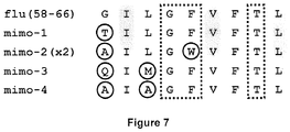
- 108010061181 influenza matrix peptide (58-66) Proteins 0.000 description 1
- 238000001802 infusion Methods 0.000 description 1
- 239000003978 infusion fluid Substances 0.000 description 1
- 238000002347 injection Methods 0.000 description 1
- 239000007924 injection Substances 0.000 description 1
- 239000012212 insulator Substances 0.000 description 1
- 229940047122 interleukins Drugs 0.000 description 1
- 230000009545 invasion Effects 0.000 description 1
- 238000011835 investigation Methods 0.000 description 1
- 230000001788 irregular Effects 0.000 description 1
- 210000002510 keratinocyte Anatomy 0.000 description 1
- 238000000370 laser capture micro-dissection Methods 0.000 description 1
- 210000000265 leukocyte Anatomy 0.000 description 1
- 238000011068 loading method Methods 0.000 description 1
- 239000000314 lubricant Substances 0.000 description 1
- 201000005202 lung cancer Diseases 0.000 description 1
- 208000020816 lung neoplasm Diseases 0.000 description 1
- 210000004324 lymphatic system Anatomy 0.000 description 1
- 210000004698 lymphocyte Anatomy 0.000 description 1
- 210000002540 macrophage Anatomy 0.000 description 1
- 210000004962 mammalian cell Anatomy 0.000 description 1
- 239000000594 mannitol Substances 0.000 description 1
- 235000010355 mannitol Nutrition 0.000 description 1
- 238000004519 manufacturing process Methods 0.000 description 1
- 239000003550 marker Substances 0.000 description 1
- 238000005259 measurement Methods 0.000 description 1
- 239000002609 medium Substances 0.000 description 1
- 201000001441 melanoma Diseases 0.000 description 1
- 239000000155 melt Substances 0.000 description 1
- 230000009401 metastasis Effects 0.000 description 1
- 229960000485 methotrexate Drugs 0.000 description 1
- 125000000325 methylidene group Chemical group [H]C([H])=* 0.000 description 1
- 244000000010 microbial pathogen Species 0.000 description 1
- 230000003278 mimic effect Effects 0.000 description 1
- 108091005601 modified peptides Proteins 0.000 description 1
- 238000012544 monitoring process Methods 0.000 description 1
- 150000002772 monosaccharides Chemical class 0.000 description 1
- 208000010805 mumps infectious disease Diseases 0.000 description 1
- 238000002703 mutagenesis Methods 0.000 description 1
- 231100000350 mutagenesis Toxicity 0.000 description 1
- AEMBWNDIEFEPTH-UHFFFAOYSA-N n-tert-butyl-n-ethylnitrous amide Chemical compound CCN(N=O)C(C)(C)C AEMBWNDIEFEPTH-UHFFFAOYSA-N 0.000 description 1
- 201000009240 nasopharyngitis Diseases 0.000 description 1
- 229960004927 neomycin Drugs 0.000 description 1
- 230000001613 neoplastic effect Effects 0.000 description 1
- 210000004498 neuroglial cell Anatomy 0.000 description 1
- 238000006386 neutralization reaction Methods 0.000 description 1
- 239000002736 nonionic surfactant Substances 0.000 description 1
- 238000006384 oligomerization reaction Methods 0.000 description 1
- 210000000056 organ Anatomy 0.000 description 1
- 150000007524 organic acids Chemical class 0.000 description 1
- 235000005985 organic acids Nutrition 0.000 description 1
- 239000003960 organic solvent Substances 0.000 description 1
- 210000001672 ovary Anatomy 0.000 description 1
- 229910052760 oxygen Inorganic materials 0.000 description 1
- 239000001301 oxygen Substances 0.000 description 1
- 210000002741 palatine tonsil Anatomy 0.000 description 1
- 238000004091 panning Methods 0.000 description 1
- 239000002245 particle Substances 0.000 description 1
- 230000008506 pathogenesis Effects 0.000 description 1
- 229930192851 perforin Natural products 0.000 description 1
- 238000002823 phage display Methods 0.000 description 1
- 239000000825 pharmaceutical preparation Substances 0.000 description 1
- COLNVLDHVKWLRT-UHFFFAOYSA-N phenylalanine Natural products OC(=O)C(N)CC1=CC=CC=C1 COLNVLDHVKWLRT-UHFFFAOYSA-N 0.000 description 1
- CWCMIVBLVUHDHK-ZSNHEYEWSA-N phleomycin D1 Chemical compound N([C@H](C(=O)N[C@H](C)[C@@H](O)[C@H](C)C(=O)N[C@@H]([C@H](O)C)C(=O)NCCC=1SC[C@@H](N=1)C=1SC=C(N=1)C(=O)NCCCCNC(N)=N)[C@@H](O[C@H]1[C@H]([C@@H](O)[C@H](O)[C@H](CO)O1)O[C@@H]1[C@H]([C@@H](OC(N)=O)[C@H](O)[C@@H](CO)O1)O)C=1N=CNC=1)C(=O)C1=NC([C@H](CC(N)=O)NC[C@H](N)C(N)=O)=NC(N)=C1C CWCMIVBLVUHDHK-ZSNHEYEWSA-N 0.000 description 1
- 239000008363 phosphate buffer Substances 0.000 description 1
- 231100000614 poison Toxicity 0.000 description 1
- 229920001983 poloxamer Polymers 0.000 description 1
- 229920000724 poly(L-arginine) polymer Polymers 0.000 description 1
- 230000008488 polyadenylation Effects 0.000 description 1
- 108010011110 polyarginine Proteins 0.000 description 1
- 229920001223 polyethylene glycol Polymers 0.000 description 1
- 229920000642 polymer Polymers 0.000 description 1
- 238000006116 polymerization reaction Methods 0.000 description 1
- 230000000379 polymerizing effect Effects 0.000 description 1
- 108091033319 polynucleotide Proteins 0.000 description 1
- 102000040430 polynucleotide Human genes 0.000 description 1
- 239000002157 polynucleotide Substances 0.000 description 1
- 229920000136 polysorbate Polymers 0.000 description 1
- 229940068965 polysorbates Drugs 0.000 description 1
- 239000001267 polyvinylpyrrolidone Substances 0.000 description 1
- 235000013855 polyvinylpyrrolidone Nutrition 0.000 description 1
- 229920000036 polyvinylpyrrolidone Polymers 0.000 description 1
- 239000000843 powder Substances 0.000 description 1
- 238000001556 precipitation Methods 0.000 description 1
- 238000002360 preparation method Methods 0.000 description 1
- 230000001915 proofreading effect Effects 0.000 description 1
- 230000001902 propagating effect Effects 0.000 description 1
- 238000000159 protein binding assay Methods 0.000 description 1
- 230000020978 protein processing Effects 0.000 description 1
- 238000000746 purification Methods 0.000 description 1
- 238000012175 pyrosequencing Methods 0.000 description 1
- 238000011084 recovery Methods 0.000 description 1
- 230000000306 recurrent effect Effects 0.000 description 1
- 238000009877 rendering Methods 0.000 description 1
- 238000012340 reverse transcriptase PCR Methods 0.000 description 1
- 238000004007 reversed phase HPLC Methods 0.000 description 1
- 238000012552 review Methods 0.000 description 1
- 206010039073 rheumatoid arthritis Diseases 0.000 description 1
- 210000003705 ribosome Anatomy 0.000 description 1
- 125000000548 ribosyl group Chemical group C1([C@H](O)[C@H](O)[C@H](O1)CO)* 0.000 description 1
- 102200076454 rs104894848 Human genes 0.000 description 1
- 206010039447 salmonellosis Diseases 0.000 description 1
- 150000003839 salts Chemical class 0.000 description 1
- 230000007017 scission Effects 0.000 description 1
- 238000002864 sequence alignment Methods 0.000 description 1
- 230000037432 silent mutation Effects 0.000 description 1
- 239000002356 single layer Substances 0.000 description 1
- 238000007860 single-cell PCR Methods 0.000 description 1
- 238000002741 site-directed mutagenesis Methods 0.000 description 1
- 210000003491 skin Anatomy 0.000 description 1
- 229910052708 sodium Inorganic materials 0.000 description 1
- 239000011734 sodium Substances 0.000 description 1
- 239000000600 sorbitol Substances 0.000 description 1
- 230000006641 stabilisation Effects 0.000 description 1
- 238000011105 stabilization Methods 0.000 description 1
- 230000000087 stabilizing effect Effects 0.000 description 1
- 238000011146 sterile filtration Methods 0.000 description 1
- 239000008174 sterile solution Substances 0.000 description 1
- 238000003860 storage Methods 0.000 description 1
- 208000022218 streptococcal pneumonia Diseases 0.000 description 1
- KDYFGRWQOYBRFD-UHFFFAOYSA-L succinate(2-) Chemical compound [O-]C(=O)CCC([O-])=O KDYFGRWQOYBRFD-UHFFFAOYSA-L 0.000 description 1
- 150000005846 sugar alcohols Chemical class 0.000 description 1
- 239000013589 supplement Substances 0.000 description 1
- 238000004114 suspension culture Methods 0.000 description 1
- 230000009885 systemic effect Effects 0.000 description 1
- 238000002560 therapeutic procedure Methods 0.000 description 1
- 206010043778 thyroiditis Diseases 0.000 description 1
- 201000004647 tinea pedis Diseases 0.000 description 1
- 230000000699 topical effect Effects 0.000 description 1
- 238000007862 touchdown PCR Methods 0.000 description 1
- 239000003440 toxic substance Substances 0.000 description 1
- 230000005030 transcription termination Effects 0.000 description 1
- 230000002103 transcriptional effect Effects 0.000 description 1
- 238000003151 transfection method Methods 0.000 description 1
- 239000012096 transfection reagent Substances 0.000 description 1
- 230000014621 translational initiation Effects 0.000 description 1
- QORWJWZARLRLPR-UHFFFAOYSA-H tricalcium bis(phosphate) Chemical compound [Ca+2].[Ca+2].[Ca+2].[O-]P([O-])([O-])=O.[O-]P([O-])([O-])=O QORWJWZARLRLPR-UHFFFAOYSA-H 0.000 description 1
- 239000013638 trimer Substances 0.000 description 1
- 125000002264 triphosphate group Chemical class [H]OP(=O)(O[H])OP(=O)(O[H])OP(=O)(O[H])O* 0.000 description 1
- UNXRWKVEANCORM-UHFFFAOYSA-N triphosphoric acid Chemical compound OP(O)(=O)OP(O)(=O)OP(O)(O)=O UNXRWKVEANCORM-UHFFFAOYSA-N 0.000 description 1
- 201000008827 tuberculosis Diseases 0.000 description 1
- 210000004881 tumor cell Anatomy 0.000 description 1
- 206010061393 typhus Diseases 0.000 description 1
- 241000701161 unidentified adenovirus Species 0.000 description 1
- 241000701366 unidentified nuclear polyhedrosis viruses Species 0.000 description 1
- 241001515965 unidentified phage Species 0.000 description 1
- 208000019206 urinary tract infection Diseases 0.000 description 1
- 210000003556 vascular endothelial cell Anatomy 0.000 description 1
- 201000002498 viral encephalitis Diseases 0.000 description 1
- 201000010044 viral meningitis Diseases 0.000 description 1
- 208000009421 viral pneumonia Diseases 0.000 description 1
- 239000013603 viral vector Substances 0.000 description 1
- 229940075420 xanthine Drugs 0.000 description 1
Images


Classifications
-
- C—CHEMISTRY; METALLURGY
- C12—BIOCHEMISTRY; BEER; SPIRITS; WINE; VINEGAR; MICROBIOLOGY; ENZYMOLOGY; MUTATION OR GENETIC ENGINEERING
- C12N—MICROORGANISMS OR ENZYMES; COMPOSITIONS THEREOF; PROPAGATING, PRESERVING, OR MAINTAINING MICROORGANISMS; MUTATION OR GENETIC ENGINEERING; CULTURE MEDIA
- C12N15/00—Mutation or genetic engineering; DNA or RNA concerning genetic engineering, vectors, e.g. plasmids, or their isolation, preparation or purification; Use of hosts therefor
- C12N15/09—Recombinant DNA-technology
- C12N15/10—Processes for the isolation, preparation or purification of DNA or RNA
- C12N15/1034—Isolating an individual clone by screening libraries
- C12N15/1037—Screening libraries presented on the surface of microorganisms, e.g. phage display, E. coli display
-
- A—HUMAN NECESSITIES
- A61—MEDICAL OR VETERINARY SCIENCE; HYGIENE
- A61P—SPECIFIC THERAPEUTIC ACTIVITY OF CHEMICAL COMPOUNDS OR MEDICINAL PREPARATIONS
- A61P31/00—Antiinfectives, i.e. antibiotics, antiseptics, chemotherapeutics
-
- A—HUMAN NECESSITIES
- A61—MEDICAL OR VETERINARY SCIENCE; HYGIENE
- A61P—SPECIFIC THERAPEUTIC ACTIVITY OF CHEMICAL COMPOUNDS OR MEDICINAL PREPARATIONS
- A61P35/00—Antineoplastic agents
-
- A—HUMAN NECESSITIES
- A61—MEDICAL OR VETERINARY SCIENCE; HYGIENE
- A61P—SPECIFIC THERAPEUTIC ACTIVITY OF CHEMICAL COMPOUNDS OR MEDICINAL PREPARATIONS
- A61P37/00—Drugs for immunological or allergic disorders
- A61P37/02—Immunomodulators
- A61P37/06—Immunosuppressants, e.g. drugs for graft rejection
-
- C—CHEMISTRY; METALLURGY
- C12—BIOCHEMISTRY; BEER; SPIRITS; WINE; VINEGAR; MICROBIOLOGY; ENZYMOLOGY; MUTATION OR GENETIC ENGINEERING
- C12Q—MEASURING OR TESTING PROCESSES INVOLVING ENZYMES, NUCLEIC ACIDS OR MICROORGANISMS; COMPOSITIONS OR TEST PAPERS THEREFOR; PROCESSES OF PREPARING SUCH COMPOSITIONS; CONDITION-RESPONSIVE CONTROL IN MICROBIOLOGICAL OR ENZYMOLOGICAL PROCESSES
- C12Q1/00—Measuring or testing processes involving enzymes, nucleic acids or microorganisms; Compositions therefor; Processes of preparing such compositions
- C12Q1/68—Measuring or testing processes involving enzymes, nucleic acids or microorganisms; Compositions therefor; Processes of preparing such compositions involving nucleic acids
- C12Q1/6876—Nucleic acid products used in the analysis of nucleic acids, e.g. primers or probes
- C12Q1/6883—Nucleic acid products used in the analysis of nucleic acids, e.g. primers or probes for diseases caused by alterations of genetic material
-
- G—PHYSICS
- G01—MEASURING; TESTING
- G01N—INVESTIGATING OR ANALYSING MATERIALS BY DETERMINING THEIR CHEMICAL OR PHYSICAL PROPERTIES
- G01N33/00—Investigating or analysing materials by specific methods not covered by groups G01N1/00 - G01N31/00
- G01N33/48—Biological material, e.g. blood, urine; Haemocytometers
- G01N33/50—Chemical analysis of biological material, e.g. blood, urine; Testing involving biospecific ligand binding methods; Immunological testing
- G01N33/5005—Chemical analysis of biological material, e.g. blood, urine; Testing involving biospecific ligand binding methods; Immunological testing involving human or animal cells
- G01N33/5008—Chemical analysis of biological material, e.g. blood, urine; Testing involving biospecific ligand binding methods; Immunological testing involving human or animal cells for testing or evaluating the effect of chemical or biological compounds, e.g. drugs, cosmetics
- G01N33/5044—Chemical analysis of biological material, e.g. blood, urine; Testing involving biospecific ligand binding methods; Immunological testing involving human or animal cells for testing or evaluating the effect of chemical or biological compounds, e.g. drugs, cosmetics involving specific cell types
- G01N33/5047—Cells of the immune system
- G01N33/505—Cells of the immune system involving T-cells
-
- G—PHYSICS
- G01—MEASURING; TESTING
- G01N—INVESTIGATING OR ANALYSING MATERIALS BY DETERMINING THEIR CHEMICAL OR PHYSICAL PROPERTIES
- G01N33/00—Investigating or analysing materials by specific methods not covered by groups G01N1/00 - G01N31/00
- G01N33/48—Biological material, e.g. blood, urine; Haemocytometers
- G01N33/50—Chemical analysis of biological material, e.g. blood, urine; Testing involving biospecific ligand binding methods; Immunological testing
- G01N33/53—Immunoassay; Biospecific binding assay; Materials therefor
- G01N33/569—Immunoassay; Biospecific binding assay; Materials therefor for microorganisms, e.g. protozoa, bacteria, viruses
- G01N33/56966—Animal cells
- G01N33/56977—HLA or MHC typing
-
- A—HUMAN NECESSITIES
- A61—MEDICAL OR VETERINARY SCIENCE; HYGIENE
- A61K—PREPARATIONS FOR MEDICAL, DENTAL OR TOILETRY PURPOSES
- A61K39/00—Medicinal preparations containing antigens or antibodies
-
- C—CHEMISTRY; METALLURGY
- C12—BIOCHEMISTRY; BEER; SPIRITS; WINE; VINEGAR; MICROBIOLOGY; ENZYMOLOGY; MUTATION OR GENETIC ENGINEERING
- C12Q—MEASURING OR TESTING PROCESSES INVOLVING ENZYMES, NUCLEIC ACIDS OR MICROORGANISMS; COMPOSITIONS OR TEST PAPERS THEREFOR; PROCESSES OF PREPARING SUCH COMPOSITIONS; CONDITION-RESPONSIVE CONTROL IN MICROBIOLOGICAL OR ENZYMOLOGICAL PROCESSES
- C12Q2600/00—Oligonucleotides characterized by their use
- C12Q2600/16—Primer sets for multiplex assays
-
- G—PHYSICS
- G01—MEASURING; TESTING
- G01N—INVESTIGATING OR ANALYSING MATERIALS BY DETERMINING THEIR CHEMICAL OR PHYSICAL PROPERTIES
- G01N2333/00—Assays involving biological materials from specific organisms or of a specific nature
- G01N2333/435—Assays involving biological materials from specific organisms or of a specific nature from animals; from humans
- G01N2333/705—Assays involving receptors, cell surface antigens or cell surface determinants
- G01N2333/70503—Immunoglobulin superfamily, e.g. VCAMs, PECAM, LFA-3
- G01N2333/7051—T-cell receptor (TcR)-CD3 complex
-
- G—PHYSICS
- G01—MEASURING; TESTING
- G01N—INVESTIGATING OR ANALYSING MATERIALS BY DETERMINING THEIR CHEMICAL OR PHYSICAL PROPERTIES
- G01N2333/00—Assays involving biological materials from specific organisms or of a specific nature
- G01N2333/435—Assays involving biological materials from specific organisms or of a specific nature from animals; from humans
- G01N2333/705—Assays involving receptors, cell surface antigens or cell surface determinants
- G01N2333/70503—Immunoglobulin superfamily, e.g. VCAMs, PECAM, LFA-3
- G01N2333/70539—MHC-molecules, e.g. HLA-molecules
Definitions
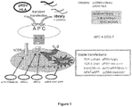
- the present invention relates to a method of identifying a target antigen of T cells as characterised in the appended claims, comprising (a) contacting (aa) cells expressing (i) a functional T cell receptor complex comprising predefined matching T cell receptor ⁇ and ⁇ chains; and (ii) a read-out system for T cell activation; with (ab) antigen-presenting cells carrying (iii) peptide libraries encoded by randomised nucleic acid sequences; and (iv) MHC molecules recognised by the T cell receptor of (i); (b) assessing T cell activation using said read-out system; (c) isolating antigen-presenting cells that are in contact with the cells in which the read-out system indicates T cell activation; (d) identifying the target antigen or the nucleic acid molecule encoding said target antigen.
- the present invention also relates to a method of identifying patient-specific T cell antigens comprising (A) isolating T cells from a sample obtained from said patient; (B) identifying matching T cell receptor ⁇ and ⁇ chains from the T cells isolated in (A); and (C) identifying T cell antigens in accordance with the method of the invention, wherein the cell comprising a functional T cell receptor and a read-out system for T cell activation expresses matching T cell receptor ⁇ and ⁇ chains from the T cells identified in (B).
- T cells play crucial roles in many infectious, tumor, and autoimmune diseases, but apart from very few exceptions, the target antigens of pathogenic human T cells have remained unknown.
- the specificity of T cells towards their target antigens is determined by their heterodimeric, hyper-variable T cell receptor (TCR) molecules, which recognize antigenic peptides that are presented by MHC molecules.
- TCR hyper-variable T cell receptor
- MHC molecules are of "self"-origin, whereas the antigenic peptides are "non-self", i.e. they are derived from viral or microbial peptides.
- class-I MHC molecules present peptides of intracellular (viral) origin to CD8+ T cells, whereas class-II MHC molecules present phagocytosed (microbial) peptides to CD4+ T cells.
- "self” MHC molecules also present “self” peptides, but these are normally ignored because of T cell tolerance. It is assumed that in autoimmune diseases the tolerance is broken and recognition of "self" peptides results in chronic inflammation, disturbed organ function or tissue destruction. Another important role of T cells is during tumor defense where T cells may mount anti-tumor responses. In this case, however, they recognize tumor-associated antigens.
- T cell antigens there is so far no simple, reliable, and unbiased method to determine T cell antigens.
- straightforward biochemical techniques such as immunoprecipitation or affinity chromatography can not be used, because the affinities of TCRs to MHC/peptide complexes are several orders of magnitude too low.
- Such techniques work well if the dissociation constant is in the nanomolar range or below, as it is typical for antibodies or conventional receptor-ligand interactions, but for TCR-MHC/peptide interactions, the dissociation constants are usually greater than 10 -6 M (Rudolph et al., 2006; J.D.Stone et al (2009).
- antigenic peptides were eluted from MHC molecules of tumor or autoimmune tissue and analyzed by mass spectrometry (Cox et al., 1994; Fissolo et al., 2009).
- a third method uses randomized synthetic peptide libraries (Nino-Vasquez et al., 2005). With this approach the idea is that TCR molecules recognize patterns rather than defined sequences or structures. In other words, the recognition of target structures is poly-specific (also termed “promiscuous” or “degenerate”) (Wucherpfennig et al., 2007).
- Such libraries contain random amino acids in all but one position and may allow identification of "recognition patterns", i.e. of mimotopes of the natural peptides, which then may be identified by database searches. These approaches are limited to some TCRs which show an appropriate balance between specificity and poly-specificity.
- cDNA libraries have been used in particular to investigate tumor T cell antigens. They were either transfected into COS cells via plasmids bearing the SV40 origin (Van der Bruggen et al., 1991; Wong et al., 1999; Boon et al., 2006), or via retroviral constructs into appropriate recipient cells (Smith et al., 2001). In both cases the plasmids or viral particles may be recovered from the transfected cells and amplified in bacteria. Usually many pools of plasmids are used, and positive pools are subjected to further rounds of transfections until a single antigen-bearing plasmid may be isolated and characterized.
- Such amplification strategies are of course advantageous as compared to biochemical or combinatorial synthetic peptide libraries. Although several tumor antigens were detected, these methods have not reached general applicability. The reasons are diverse: the libraries usually come from diseased tissue (which is often not available) and a tremendous number of clones must be screened by laborious and expensive cytotoxicity or cytokine assays, which - another limitation - require large numbers of T cells. Most importantly, however, the proteins expressed from cDNA libraries require extensive and correct processing. Hence, the processing pattern of the APCs used in vitro must be identical to the processing pattern of the original APCs. This may often not be the case. A fifth strategy is pronounced of phage display libraries.
- baculovirus infected insect cells display randomized peptide libraries in the binding groove of recombinant MHC molecules (Crawford et al., 2004; Wang et al., 2005; Crawford et al., 2006). These libraries are screened with fluorescent oligomerized soluble TCR molecules. Although this technology could reveal mimotopes of peptides known to activate class-I and class-II restricted TCRs, it has several intricacies. The most important drawback is, as discussed above, that TCRs have notoriously low affinities to their MHC/peptide ligands. This impedes detection of positive insect cell clones. TCR-oligomerization facilitates recognition by increasing avidity, but may presumably not completely overcome this limitation.
- WO2003068800 describes isolated peptides that bind to HLA molecules and stimulate cytolytic T cells specific for complexes of the peptide and the HLA molecule.
- a transfection of antigen presenting cells with recombinant combinatorial peptide libraries is not envisaged in WO2003068800 .
- US 6,037,135 discloses methods of making HLA binding peptides, but does not disclose the loading of MHC molecules with and presentation of antigens by antigen presenting cells via a peptide library encoding such peptides.
- Dornmair et al., 2003 represents a review which lists possible methods for identifying a target antigen of T cells.
- the present invention relates to a method of identifying a target antigen of T cells as characterised in the appended claims, comprising (a) contacting (aa) cells expressing (i) a functional T cell receptor complex comprising predefined matching T cell receptor ⁇ and ⁇ chains; and (ii) a read-out system for T cell activation; with (ab) antigen-presenting cells carrying (iii) peptide libraries encoded by randomised nucleic acid sequences; and (iv) MHC molecules recognised by the T cell receptor of (i); (b) assessing T cell activation using said read-out system; (c) isolating antigen-presenting cells that are in contact with the cells in which the read-out system indicates T cell activation; (d) identifying the target antigen or the nucleic acid molecule encoding said target antigen.
- target antigen of T cells relates to an antigen that is recognised and bound by T cells. The binding of the antigen to T cells subsequently results in the activation of said T cells.
- T cell target antigens are recognised by T cells via a functional T cell receptor complex consisting of T cell receptors (TCR) and the CD3 complex.
- T cell target antigens are presented by major histocompatibility complex (MHC) molecules on the surface of antigen-presenting cells (APCs).
- MHC major histocompatibility complex
- APCs antigen-presenting cells
- target antigen of T cells refers to the peptide epitope. When the peptide epitope is complexed with an MHC molecule, reference is made to the "antigen-MHC complex" herein.
- Target antigens presented by MHC class I molecules are recognized by CD8+ T cells and are typically of intracellular origin, such as for example viral target antigens.
- Target antigens presented by MHC class II molecules are recognized by CD4+ T cells.
- they are peptides, which originate from extracellular sources that were phagocytosed, such as for example peptides derived from microbes.
- Target antigens of T cells can be of an origin that is foreign to the host organism, such as for example viral or microbial peptides.
- Target antigens may also be of self-origin, such as for example tumor-associated antigens or self-antigens that trigger an autoimmune response in the host organism.
- the target antigen of T cells is a target antigen of CD8+ T cells.
- the term "functional T cell receptor complex” refers to a complex capable of eliciting activation of the T cell in which the complex is expressed.
- the T cell receptor complex is composed of six subunits.
- the T cell receptor is made up of two subunits (also referred to herein as chains), TCR ⁇ and ⁇ , which form a disulfide-linked heterodimer, which comprises the variable, hypervariable and joining region of the TCR receptor complex that interacts with the antigen/MHC-complex, thus forming one single antigen-binding site.
- TCR ⁇ and ⁇ which form a disulfide-linked heterodimer, which comprises the variable, hypervariable and joining region of the TCR receptor complex that interacts with the antigen/MHC-complex, thus forming one single antigen-binding site.
- TCR ⁇ and ⁇ subunits are also referred to herein as chains.
- TCR ⁇ and ⁇ -chains comprise conserved (constant) regions which interact with the proteins of the CD3-complex and fix the TCR in the membrane.
- the T-cell receptor complex further contains four CD3 subunits. Each CD3 complex contains one CD3y subunit, one CD3 ⁇ subunit, and two CD3 ⁇ subunit. One of the CD3 ⁇ subunits forms a heterodimer with the CD3y subunit, while the other CD3 ⁇ subunit forms a heterodimer with the CD3 ⁇ subunit.
- Antigen binding leads to the cross-linking and activation of the TCR hexamers.
- the signal is then conducted by the ⁇ -chains to further downstream intracellular compounds.
- T cell receptor complex Functionality of a T cell receptor complex can be analysed using methods well known in the art, such as for example measurements of phosphorylation and de-phosphorylation of proteins and other intracellular molecules (such as for example IP3), Ca 2+ -influx into T-cells, production of cytokines (such as for example Interferon-y, interleukins (such as IL-2, -4, -6, -17), TNF- ⁇ ), secretion of cytotoxic granules containing perforin and granzymes or killing of target cells (Smith-Garvin et al. (2009); Murphy et al. "Janeway's Immunobiology" 2008, 7th Editi on).
- cytokines such as for example Interferon-y, interleukins (such as IL-2, -4, -6, -17), TNF- ⁇
- predefined matching T cell receptor ⁇ and ⁇ chains relates to ⁇ and ⁇ chains derived from one molecular type of T cell receptor, i.e. paired ⁇ and ⁇ chains. In other words, the chains are matched to represent functional T cell receptor heterodimers.
- predefined refers to the purposive selection of the T cell receptor chains to represent a T cell receptor of interest, for which corresponding antigens are to be identified by the method of the invention.
- the T cell receptor is a vertebrate T cell receptor, more preferably it is a mammalian T cell receptor. Even more preferably, the T cell receptor is a T cell receptor from horse, bovine, swine, canine, feline or primate. Most preferably, the T cell receptor is a human T cell receptor.
- T cell receptor chains may be obtained by any method known in the art, such as for example the method described in Seitz et al. 2006, where TCR chains are cloned from morphologically characterised single cells. Such cells may be obtained from a sample obtained from a patient, for example, by laser micro-dissection of biopsy tissue. Furthermore, T cell receptors of known sequence may be employed, in which case the chains may be obtained by transfecting the cell of (aa) with a nucleic acid molecule encoding said T cell receptor chains.
- the term "read-out system for T cell activation” relates to a system that provides a measurable signal upon activation of the T cell receptor.
- T cell receptor activation normally results in the activation of the respective T cell expressing said T cell receptor.
- the signal provided by the read-out system is representative of a successful T cell activation via T cell receptor binding of the antigen presented by the antigen-presenting cell.
- Said activation of the T cell receptor expressed in the cells of (aa) results in a signal such as for example the expression of a reporter protein.
- Non-limiting examples of reporter genes suitable to provide a measurable signal upon T cell activation include enzymes such as for example ⁇ -galactosidase, CAT, ⁇ -glucuronidase, peroxidase, ⁇ -xylosidase, catechol dioxygenase (XylE), trehalase (TreA), alkaline phosphatase or secreted alkaline phosphatase as well as fluorescent compounds, bioluminescent compounds and chemiluminescent compounds.
- enzymes such as for example ⁇ -galactosidase, CAT, ⁇ -glucuronidase, peroxidase, ⁇ -xylosidase, catechol dioxygenase (XylE), trehalase (TreA), alkaline phosphatase or secreted alkaline phosphatase as well as fluorescent compounds, bioluminescent compounds and chemiluminescent compounds.
- Such read-out systems are well known in the art and are
- the cells of (aa) are characterised by the expression of a functional T cell receptor complex and a read-out system, which indicates T cell activation.
- Such cells may be any cells comprising a T cell receptor complex, such as for example cells naturally expressing a T cell receptor complex or cells transformed or transfected with a T cell receptor or a T cell receptor complex. Such cells are also referred to as T cell receptor transfectants.
- Non-limiting examples of such cells are T cells, T cell hybridomas, lymphomas and artificially immortalized T cells (Katakura et al. (1998)).
- the cells of (aa) are T cell hybridomas.
- T cell hybridomas are well known in the art and may be obtained as described in Ozaki et al. (1988); Shirahata et al. (1998); Chen et al. (2007) or as described in the examples below.
- the cells of (aa) express only one molecular type of T cell receptor.
- Such cells may be, for example, naturally occurring T cells or T cell hybridomas devoid of an endogenous T cell receptor and being transfected or transformed with a T cell receptor of interest.
- cells already expressing an endogenous T cell receptor may be transfected or transformed with a further T cell receptor of interest.
- the T cell receptor of interest is modified such as to prevent hetero-dimerisation between chains of the endogenous T cell receptor and chains of the T cell receptor of interest.
- modifications are well known in the art and include, without being limiting, the introduction of mutated amino acids at the interface of the constant regions of the T cell receptor chains of interest, or choosing T cells of very limited heterogeneity, such as oligo- or monoclonal T cell populations, as recipients (Voss et al. (2008); Cohen et al. (2007); Kuball et al. (2007); Weinhold et al.
- cells expressing different molecular types of T cell receptor are employed in the method of the invention, further rounds of screening are carried out using different cells, such as for example cells expressing the T cell receptor of interest with only one endogenous T cell receptor, wherein said endogenous T cell receptor differs from the T cell receptor(s) present in the cells of the first or previous rounds of screening.
- the cells employed in such further rounds of screening only express the T cell receptor of interest, in order to confirm that the antigen identified is an antigen of the T cell receptor of interest and not an antigen of the endogenous T cell receptor.
- antigen-presenting cells relates to cells that display antigen in complex with the major histocompatibility complex (MHC) on its surface.
- MHC major histocompatibility complex
- Any cell is suitable as an antigen-presenting cell in accordance with the present invention, as long as it expresses an MHC and presents an antigen.
- Cells that have in vivo the potential to act as antigen presenting cells include for example dendritic cells, macrophages, B-cells or activated epithelial cells but also fibroblasts, glial cells, pancreatic beta cells or vascular endothelial cells.
- Such cells may be employed in accordance with the present invention after transfection or transformation with a peptide library as defined below.
- cells not endogenously expressing MHC may be employed, in which case suitable MHC are to be transformed or transfected into said cells.
- peptide libraries encoded by randomised nucleic acid sequences relates to a collection of peptides, i.e. a library, wherein the peptides are encoded by nucleic acid sequences of arbitrary sequences.
- the peptide library is encoded by a set of nucleic acid sequences representing at least 10 3 different nucleic acid sequences, such as at least 10 4 , at least 10 5 , at least 10 6 , at least 10 7 , at least 10 8 , at least 10 9 , at least 10 10 , at least 10 11 , at least 10 12 , at least 10 13 , at least 10 14 , at least 10 15 or at least 10 16 different nucleic acid sequences.
- the peptide library is encoded by a set of nucleic acid sequences representing all possible permutations for a given peptide length.
- peptide libraries encompassing peptides of different lengths are also envisaged herein and the above defined amounts of nucleic acid sequence representation within said library apply mutatis mutandis.
- anchor positions are known to be required for binding between the antigen and the MHC.
- the randomised nucleic acid sequences may also be generated by introducing in these positions fixed amino acids and randomising solely the remaining positions.
- Anchor positions for various MHCs are known, such as for example in HLA-A2, as discussed in the examples, where anchor positions are isoleucine in position 2, valine in position 6 and leucine in position 9 (Rammensee et al., 1999). Further anchors are known in the art and are described, for example in (Rammensee et al., 1999).
- peptide as used herein describes a group of molecules consisting of up to 50 amino acids. Peptides may further form dimers, trimers and higher oligomers, i.e. consisting of more than one molecule which may be identical or non-identical. The corresponding higher order structures are, consequently, termed homo- or heterodimers, homo- or heterotrimers etc.
- peptide (wherein “polypeptide” is interchangeably used with “protein”) also refers to naturally modified peptides wherein the modification is effected e.g. by glycosylation, acetylation, phosphorylation and the like. Such modifications are well-known in the art.
- the peptides have a minimum length of at least 4 amino acids, such as for example at least 5, at least 6, at least 7, at least 8, at least 9 or at least 10 amino acids. Also preferred is that the peptides have a length of at the most 50 amino acids, such as for example at most 45, such as at most 40, at most 35, at most 30, at most 25, at most 20 amino acids. Any of the intermediate numbers not explicitly mentioned are also envisaged herein. More preferably, peptides presented by MHC class I molecules have a length of between 4 and 20 amino acids. Thus, said peptides may be 4, 5, 6, 7, 8, 9, 10, 11, 12, 13, 14, 15, 16, 17, 18, 19 or 20 amino acids in length.
- peptides presented by MHC class II molecules have a length of between 4 and 50 amino acids.
- the class II peptides may in principle be infinitely long, because they may reach out from the MHC binding groove at both sides.
- the epitope itself is normally 8 to 10 amino acids long.
- said peptides may be 4, 5, 6, 7, 8, 9, 10, 11, 12, 13, 14, 15, 16, 17, 18, 19, 20, 21, 22, 23, 24, 25, 26, 27, 28, 29, 30, 31, 32, 33, 34, 35, 36, 37, 38, 39, 40, 41, 42, 43, 44, 45, 46, 47, 48, 49 or 50 amino acids in length.
- the peptides of the present invention have a length of between 8 to 10 amino acids.
- the peptides have a length of 9 amino acids.
- Nucleic acid sequences include DNA, such as cDNA or genomic DNA, and RNA. It is understood that the term “RNA” as used herein comprises all forms of RNA and preferably refers to mRNA. Further included are nucleic acid mimicking sequences known in the art such as synthetic or semisynthetic derivatives of DNA or RNA and mixed polymers, both sense and antisense strands.
- nucleic acid mimicking molecules or nucleic acid derivatives include phosphorothioate nucleic acid, phosphoramidate nucleic acid, 2'-O-methoxyethyl ribonucleic acid, morpholino nucleic acid, hexitol nucleic acid (HNA) and locked nucleic acid (LNA) (see Braasch and Corey (2001) Chem Biol. 8, 1 ).
- LNA is an RNA derivative in which the ribose ring is constrained by a methylene linkage between the 2'-oxygen and the 4'-carbon. They may contain additional non-natural or derivatized nucleotide bases, as will be readily appreciated by those skilled in the art.
- the peptide libraries employed in the present invention differ from the prior art in that in the prior art, one would have to design the nucleic acid sequences according to a known sequence of the gene, whereas from the present library one can screen through a fully random panel of different peptides (without the need of prior knowledge of their sequences) to look for a target antigen of T cells.
- the randomised nucleic acid sequences encoding the peptide library/libraries do not encode a naturally occurring gene.
- the term "randomised nucleic acid sequences" does not include full length genes encoding a particular protein nor cDNAs, such as for example cDNAs derived from mRNA.
- the nucleic acid sequences encoding the peptides of the peptide library are incorporated into a vector.
- the vector is a plasmid, cosmid, virus, bacteriophage or another vector used e.g. conventionally in genetic engineering.
- the nucleic acid sequences of the present invention may be inserted into several commercially available vectors.
- Non-limiting examples include vectors compatible with an expression in mammalian cells like pREP (Invitrogen), pcDNA3 (Invitrogen), pCEP4 (Invitrogen), pMC1neo (Stratagene), pXT1 (Stratagene), pSG5 (Stratagene), EBO-pSV2neo, pBPV-1, pdBPVMMTneo, pRSVgpt, pRSVneo, pSV2-dhfr, plZD35, pLXIN, pSIR (Clontech), pIRES-EGFP (Clontech), pEAK-10 (Edge Biosystems) pTriEx-Hygro (
- vectors can contain one or more origin of replication (ori) and inheritance systems for cloning or expression, one or more markers for selection in the host, e. g., antibiotic resistance, and one or more expression cassettes.
- Suitable origins of replication (ori) include, for example, the Col E1, the SV40 viral and the M 13 origins of replication.
- the coding nucleic acid sequences inserted in the vector can e.g. be synthesized by standard methods, or isolated from natural sources. Ligation of the coding sequences to transcriptional regulatory elements and/or to other amino acid encoding sequences can be carried out using established methods.
- Transcriptional regulatory elements (parts of an expression cassette) ensuring expression in eukaryotic cells are well known to those skilled in the art. These elements comprise regulatory sequences ensuring the initiation of the transcription (e. g., translation initiation codon, promoters, enhancers, and/or insulators), internal ribosomal entry sites (IRES) ( Owens, Proc. Natl. Acad. Sci. USA 98 (2001), 1471-1476 ) and optionally poly-A signals ensuring termination of transcription and stabilization of the transcript. Additional regulatory elements may include transcriptional as well as translational enhancers, and/or naturally-associated or heterologous promoter regions.
- regulatory sequences ensuring the initiation of the transcription (e. g., translation initiation codon, promoters, enhancers, and/or insulators), internal ribosomal entry sites (IRES) ( Owens, Proc. Natl. Acad. Sci. USA 98 (2001), 1471-1476 ) and optionally poly-A signals ensuring
- the nucleic acid sequences of the invention are operatively linked to such expression control sequences allowing expression in eukaryotic cells.
- the vector may further comprise nucleotide sequences encoding secretion signals as further regulatory elements.
- sequences are well known to the person skilled in the art.
- leader sequences capable of directing the expressed polypeptide to a cellular compartment may be added to the coding sequence of the polynucleotide of the invention. Such leader sequences are well known in the art.
- regulatory elements ensuring the initiation of transcription comprise the cytomegalovirus (CMV) promoter, SV40-promoter, RSV-promoter (Rous sarcome virus), the lacZ promoter, the gai10 promoter, human elongation factor 1 ⁇ -promoter, CMV enhancer, CaM-kinase promoter, the Autographa californica multiple nuclear polyhedrosis virus (AcMNPV) polyhedral promoter or the SV40-enhancer.
- CMV cytomegalovirus
- SV40-promoter RSV-promoter
- RSV-promoter Rousarcome virus
- lacZ the lacZ promoter
- the gai10 promoter the gai10 promoter
- human elongation factor 1 ⁇ -promoter human elongation factor 1 ⁇ -promoter
- CMV enhancer CMV enhancer
- CaM-kinase promoter the Autographa californica multiple nuclear polyhedrosis virus
- Examples for further regulatory elements in eukaryotic cells comprise transcription termination signals, such as SV40-poly-A site or the tk-poly-A site or the SV40, lacZ and AcMNPV polyhedral polyadenylation signals, downstream of the nucleic acid sequences.
- the vector as disclosed herein comprises a selectable marker. Examples of selectable markers include neomycin, ampicillin, and hygromycin resistance and the like. Specifically-designed vectors allow the shuttling of DNA between different hosts, such as bacteria- fungal cells or bacteria-animal cells.
- An expression vector as disclosed herein is capable of directing the replication, and the expression, of the nucleic acid sequences and encoded peptides of this invention.
- Suitable expression vectors which comprise the described regulatory elements are known in the art such as Okayama-Berg cDNA expression vector pcDV1 (Pharmacia), pRc/CMV, pcDNA1, pcDNA3 (Invitrogene, as used, inter alia in the appended examples), pSPORT1 (GIBCO BRL), Gateway plasmids (Invitrogen) or pGEMHE (Promega).
- the nucleic acid sequences as described herein above may be designed for direct introduction or for introduction via electroporation (using for example Multiporator (Eppendorf) or Genepulser (BioRad)), PEI (Polysciences Inc.
- baculoviral systems or systems based on Vaccinia Virus or Semliki Forest Virus can be used as eukaryotic expression system for the nucleic acid sequences as described.
- MHC is used interchangeably with HLA herein.
- MHC molecules recognised by the T cell receptor are employed.
- the MHC expressed by the cell of (ab) has to be a class I MHC while when screening for CD4+ T cell target antigen, the MHC has to be a class II MHC.
- the specific MHC recognised by the T cell receptor expressed by the cell of (aa) needs to be employed.
- a preceding experiment may be performed to screen the complete set of all MHC (HLA) molecules expressed from all alleles of the subject, e.g.
- the cells of (aa) that express the T cell receptor of interest will be activated when in contact with those cells (ab) that present an antigen of said T cell receptor.
- T cell activation is observable due to the read-out system and allows for convenient isolation of the cell complex of (ab) with (aa) or of the activating cells of (ab) only.
- Methods of isolation of cells are well known in the art and include, without being limiting, manual picking of cells or automated picking by use of a robot, laser-capture microdissection or FACS (fluorescence activated cell sorting). Such methods are described for example in Murray (2007) or Tung et al. (2007) as well as in the examples below.
- FACS fluorescence activated cell sorting
- nucleic acid molecule Prior to identification of the target antigen or the nucleic acid molecule encoding it, said nucleic acid molecule may be amplified. Amplification can include, for example, amplification of the nucleic acid molecule via PCR technology as further detailed below and/or transfection of the nucleic acid molecule into bacterial cells and subsequent culture of the bacteria. The amplified nucleic acid molecules or the peptides encoded by these nucleic acid molecules are then identified.
- the term "identifying", as used in accordance with the present invention, refers to determining the amino acid or nucleic acid sequence of the target antigen(s). Methods of identification of nucleic acid molecules include, without being limiting, nucleic acid sequencing.
- the target antigen or the nucleic acid molecule encoding said target antigen is identified.
- an unbiased and straightforward technique for determining the target antigens of T cells is provided.
- the intricacies of protein processing are circumvented by co-transfecting a recombinant combinatorial library encoding short peptides and the appropriate MHC-I molecules into cells, such as for example COS cells.
- the library contains plasmids comprising random nucleotide sequences encoding short peptides. These plasmids can be easily recovered.
- Transfected COS cells are, as for example shown in the examples below, screened with cells which carry functional CD3 and downstream signalling molecules and are co-transfected with TCR ⁇ - and ⁇ -chains (T-cell receptor transfectants), human CD8 ⁇ - and ⁇ -chains, and sGFP under the control of the response element of the nuclear factor of activated T cells (NFAT) in cells that carry NFAT (Fiering et al., 1990; Karttunen and Shastri, 1991).
- these cells are TCR-transfected T-hybridoma cells, as this provides unlimited numbers of "revived" T cells and avoids establishing "real" T cell clones, which is often more problematic.
- a confluent layer of library-presenting COS cells is overlaid with the TCR-transfectants. Whenever a particular single TCR-transfectant is activated by a peptide from the library, it lights up green, and the underlying COS cell can be picked with a micromanipulator under a fluorescent microscope. By this high-throughput approach, several millions of library-peptides can be examined within a few hours.
- the library-plasmids from the recovered COS-cell may then be isolated, subjected to a new round of screening, and are finally sequenced. Then the T cell receptor activating motif can be analyzed and compared to existing proteins in the database. The feasibility of this technique is demonstrated in the appended examples using the well investigated TCR JM22 (Gotch et al., 1987; Stewart-Jones et al., 2003; Ishizuka et al., 2008) that is specific for HLA-A2 and the influenza matrix protein peptide flu(58-66).
- a major advantage of the inventive technology is that it does not depend on the antigen processing machinery of the antigen-presenting cells by providing plasmid vectors encoding short peptides.
- a high number of antigen-presenting cells efficiently present the encoded peptides (e.g. flu(58-66)) after transfection with plasmid pcDNA ( Fig 2c,e ).
- the examples also provide evidence that the peptides, such as flu(58-66), are efficiently transported after expression into the lumen of the endoplasmic reticulum where they are loaded onto HLA-A*0201. Strikingly, the full-length protein flu(1-252) was not recognized ( Fig. 2g ).
- COS cells can be transfected with high efficacy and they amplify the plasmid vector in their cytosol, which allows for it to be recovered and analyzed.
- the results provided herein demonstrate that it is possible to identify mimotopes that allow unequivocal identification of the parent, naturally occurring peptide by a simple database search.
- the list of identified mimotopes generated by the present technology may not be complete, because some peptides may be digested further by endo- or exopeptidases in the cytosol of the COS cells, or some may be lost due to their chemical properties, i.e. they may be hydrophobic and insert into membranes, or they may bind to proteins or other cellular components.
- TCR-oligomerisation may not be able to overcome this disadvantage, because the readout of such an assay is still a binding assay with moderate sensitivity. This is presumably the reason why, as discussed above, a priori unknown T cell antigens have not yet been discovered by this technique.
- the present invention makes use of an extremely sensitive bioassay: the activation of a cell, such as a T hybridoma cell, that just needs to be loosely attached to its antigen-presenting cell, is measured. The tremendous sensitivity of this assay compensates for the low binding affinity of the T cell receptor to its MHC/peptide complex.
- the technology of the present invention may have several implications not only for scientific purposes, but also for medical applications.
- it is a generally applicable method, i.e., it may be applied to any T cell of interest. It is not restricted to human T cells, but the antigen recognition properties of any TCR from any species may be investigated. This is of course highly interesting for many scientific questions, where T cells are involved.
- the only proviso is that the specific class-I MHC molecule is known, or that the specific class-II MHC molecule is known. If this is not the case, then the complete set of all candidate alleles has to be identified in a preceding experiment, using methods well established in the art (Robinson J, et al.
- T cells are involved in many diseases. They defeat infections and tumours, and they attack "self" tissue in autoimmunity. In most cases their precise antigens are not known. Knowing these epitopes will enable scientists to utilise this knowledge in diagnostics and therapy. Thus, the antigens may serve as biomarkers to detect infections and tumours and to improve diagnosis of many autoimmune diseases. Further, such biomarkers may be of great prognostic value for the progression of diseases.
- the antigens may also be helpful in therapeutic approaches: They could be used in vaccinations against infections or tumours (Boon et al., 2006), and they may allow to selectively delete or modulate auto-aggressive T cell clones in many autoimmune diseases, where e.g. CD8+ T cells are known to play prominent roles, i.e. in multiple sclerosis, psoriasis, inflammatory myopathies and many others more (Friese and Fugger, 2009; Dalakas, 2006; Walter and Santamaria, 2005). Further, knowledge of the antigens may allow the rational design and development of new, improved T cell receptors that are engineered for higher affinities to their target antigens.
- Such improved T cell receptors may be produced by replacement or introduction of amino acids in the complementarity determining regions by site-specific mutagenesis.
- the improved T cell receptors may be used for detection of their antigens in vitro and in vivo for scientific, diagnostic and therapeutic purposes. They may be used to detect, block, or delete (by coupling toxic substances) cells that present certain antigens. If required, T cell receptors may be oligomerized to increase avidity.
- the present invention also refers to engineered T cell receptors, wherein amino acids are altered as compared to a natural T cell receptor and wherein said alteration results in an increased affinity to the target antigen as compared to the unaltered T cell receptor.
- the knowledge of specific antigens may also allow the design of altered peptide ligands for a particular T cell receptor of interest. Such altered antigens may be of importance for inducing T cell tolerance or improving the immunogenicity of a particular antigen vaccine.
- Methods for the rational design of altered and/or improved T cell antigens are well known in the art (Fontoura et al. (2005); Bielekova and Martin (2001)).
- the method of the invention comprises repeating steps (a) to (c) of the above defined method until a single molecular type of nucleic acid molecule encoding a single type of antigen is isolated.
- steps (a) to (c) may be repeated at least one more time, such as for example two more times, three more times, four more times, five more times, six more times, seven more times, eight more time, nine more times or ten more times.
- a higher number of repeats of steps (a) to (c) is also envisaged herein.
- step (ab) Employing standard methods of introducing the peptide library into the antigen-presenting cells of (ab) may result in the presence of more than one copy of the encoding nucleic acid molecule within said cell. Furthermore, nucleic acid molecules present in the culture medium may be transferred together with the antigen-presenting cell when isolating said cell in step (c). Thus, performing the method of the invention with only one round of steps (a) to (c) may result in the isolation of an antigen-presenting cell that carries multiple diverse copies of nucleic acid molecules encoding different antigen peptides or to which multiple diverse copies of nucleic acid molecules encoding different antigen peptides are attached. By repeating steps (a) to (c), the correct target antigen may be identified in the subsequent step (d). It is understood that when repeating step (a), the antigen-presenting cell is a cell carrying the nucleic acid molecules isolated from the antigen-presenting cell isolated in step (c) in the previous round of screening.
- Figure 6 provides an overview over a preferred way of carrying out these repeated method steps in order to obtain a single molecular type of nucleic acid molecule encoding a single type of antigen.
- an activated, green fluorescent TCR-transfectant is picked together with the subjacent antigen presenting cell that carries the activating plasmid.
- the inserts of the plasmids are cloned, and transfected into bacteria.
- To count the numbers of bacterial clones a fraction of the bacterial clones is plated onto agar plates. Then 30 or more sub-pools of bacteria are created that contain 500 independent bacterial clones, each. They are transfected into COS-7 cells and tested again. From a positive sub-pool, further sub-pools are generated, which contain less clones per sub-pool.
- single bacterial clones are analyzed. Positive clones are sequenced and reveal the antigenic mimotope.
- the cells of (aa) further express CD8 chains, more preferably CD8 ⁇ and ⁇ chains.
- CD8 on the cells of (aa) serves as a co-receptor for the T cell receptor (TCR).
- TCR T cell receptor
- the extracellular IgV-like domain of CD8- ⁇ interacts with the ⁇ 3 portion of Class I MHC molecules. This interaction keeps the cell expressing the T cell receptor closely together with the target cell bound during antigen-specific activation.
- additional expression of CD8 on the cells of (aa) further enhances the interaction between the two cells in accordance with the invention.
- the identification of the target antigen in (d) comprises sequencing of the nucleic acid molecule encoding said target antigen.
- Methods for sequencing comprise, without being limiting, approaches of sequence analysis by direct sequencing, fluorescent SSCP in an automated DNA sequencer and pyro-sequencing. These methods are well known in the art, see e.g. Adams et al. (Ed.), "Automated DNA Sequencing and Analysis", Academic Press, 1994 ; Alphey, “DNA Sequencing: From Experimental Methods to Bioinformatics", Springer Verlag Publishing, 1997 ; Ramon et al., J. Transl. Med. 1 (2003) 9 ; Meng et al., J. Clin. Endocrinol. Metab. 90 (2005) 3419-3422 .
- the nucleic acid molecule encoding said target antigen is amplified prior to sequencing.
- PCR is well known in the art and is employed to make large numbers of copies of a target sequence. This is done on an automated cycler device, which can heat and cool containers with the reaction mixture in a very short time.
- the PCR generally, consists of many repetitions of a cycle which consists of: (a) a denaturing step, which melts both strands of a DNA molecule and terminates all previous enzymatic reactions; (b) an annealing step, which is aimed at allowing the primers to anneal specifically to the melted strands of the DNA molecule; and (c) an extension step, which elongates the annealed primers by using the information provided by the template strand.
- PCR can be performed for example in a 50 ⁇ l reaction mixture containing 5 ⁇ l of 10 x PCR buffer with 1.5 mM MgCI2, 200 ⁇ M of each deoxynucleoside triphosphate, 0.5 ⁇ l of each primer (10 ⁇ M), about 10 to 100ng of template DNA and 1 to 2.5 units of Taq Polymerase.
- the primers for the amplification may be labelled or be unlabelled.
- DNA amplification can be performed, e.g., with a model 2400 thermal cycler (Applied Biosystems, Foster City, CA): 2 min at 94°C, followed by 30 to 40 cycles consisting of annealing (e. g. 30 s at 50°C), extension (e. g.
- Suitable polymerases for use with a DNA template include, for example, E. coli DNA polymerase I or its Klenow fragment, T4 DNA polymerase, Tth polymerase, Taq polymerase, a heat-stable DNA polymerase isolated from Thermus aquaticus Vent, Amplitaq, iProof, Pfu and KOD, some of which may exhibit proof-reading function and/or different temperature optima.
- E. coli DNA polymerase I or its Klenow fragment T4 DNA polymerase, Tth polymerase, Taq polymerase, a heat-stable DNA polymerase isolated from Thermus aquaticus Vent, Amplitaq, iProof, Pfu and KOD, some of which may exhibit proof-reading function and/or different temperature optima.
- T4 DNA polymerase Tth polymerase
- Taq polymerase a heat-stable DNA polymerase isolated from Thermus aquaticus Vent
- Amplitaq iProof
- Pfu and KOD some of
- RT-PCR reverse transcriptase polymerase chain reaction
- the term “reverse transcriptase” refers to an enzyme that catalyzes the polymerization of deoxyribonucleoside triphosphates to form primer extension products that are complementary to a ribonucleic acid template.
- RT-PCR is particularly suitable when RNA viruses are employed in order to encode the library plasmids for use in the method of the invention. The enzyme initiates synthesis at the 3'-end of the primer and proceeds toward the 5'-end of the template until synthesis terminates.
- RNA target sequence into a complementary, copy-DNA (cDNA) sequence examples include avian myeloblastosis virus reverse transcriptase and Thermus thermophilus DNA polymerase, a thermostable DNA polymerase with reverse transcriptase activity marketed by Perkin Elmer.
- cDNA duplex template is heat denatured during the first denaturation step after the initial reverse transcription step leaving the DNA strand available as an amplification template. High-temperature RT provides greater primer specificity and improved efficiency.
- the RT Reaction can be performed, for example, in a 20 ⁇ l reaction mix containing: 4 ⁇ l of 5x AMV-RT buffer, 2 ⁇ l of Oligo dT (100 ⁇ g/ml), 2 ⁇ l of 10 mM dNTPs, 1 ⁇ l total RNA, 10 Units of AMV reverse transcriptase, and H 2 O to 20 ⁇ l final volume.
- the reaction may be, for example, performed by using the following conditions: The reaction is held at 70 C° for 15 minutes to allow for reverse transcription. The reaction temperature is then raised to 95 C° for 1 minute to denature the RNA-cDNA duplex.
- reaction temperature undergoes two cycles of 95°C for 15 seconds and 60 C° for 20 seconds followed by 38 cycles of 90 C° for 15 seconds and 60 C° for 20 seconds. Finally, the reaction temperature is held at 60 C° for 4 minutes for the final extension step, cooled to 15 C°, and held at that temperature until further processing of the amplified sample. Any of the above mentioned reaction conditions may be scaled up according to the needs of the particular case.
- primers for both the sequencing as well as the amplification of nucleic acid molecules can be derived by the skilled person using well-established methods.
- primer sequences may be derived based on the knowledge of up-stream and down-stream flanking regions surrounding the randomised nucleic acid sequences encoding the peptide libraries, such as for example the plasmid backbone into which said nucleic acid sequences are inserted to, the promoter sequence employed to express the peptides or other regulatory sequences, such as enhancer sequences.
- the identification of the target antigen in (d) comprises the identification of at least one mimotope of the antigen.
- mimotope in accordance with the present invention, relates to a molecule, which mimics the structure of an antigen epitope.
- Said molecule may, for example, be a peptide. Due to the mimicking property the mimotope causes a T cell receptor response identical to the one elicited by the naturally occurring antigen epitope.
- a T cell receptor recognizing a particular antigen epitope will thus also recognize a mimotope which mimics that antigen epitope.
- a mimotope differs from the natural target antigen epitope of a particular T cell receptor in its amino acid structure, e.g.
- amino acid sequence and composition such as for example by a difference of less than 40 amino acids in amino acids structure, such as for example less than 30 amino acids, less than 20 amino acids, less than 15 amino acids less than 10 amino acids, less than 5 amino acids, less than 4 amino acids, less than 3 amino acids and most preferably by one amino acid.
- At least one mimotope refers to any number of possible mimotopes for a given antigen. It includes, for example, at least two mimotopes, such as for example at least three, at least four, at least five, at least six, at least seven, at least eight, at least nine or at least 10 mimotopes. Also included is at least 15, such as at least 20, at least 30, at least 40, at least 50, at least 75, at least 100, at least 150, at least 200, at least 300, at least 400 or at least 500 mimotopes.
- This embodiment is based on the finding that it is possible to identify mimotopes by the method of the present invention, which may then be employed in a database search to identify the naturally occurring antigen. Thus, even if the actual antigen is not directly identified by the method of the invention, it can indirectly be identified via said mimotopes, as described in the examples below.
- the antigen-presenting cells are cells capable of amplifying the peptide libraries.
- a suitable degree of amplification is achieved when more copies of the nucleic acid sequences are present in the cell than were originally transfected or transformed into said cell.
- amplification is achieved when the amount of nucleic acid sequence copies is at least duplicated, such as for example when at least 5% more copies of one molecular species of nucleic acid sequence is present in the cell, such as for example at least 10% more copies, such as at least 20% more copies, at least 30% more copies, at least 50% more copies, at least 100% more copies, at least 200% more copies, at least 500% more copies, at least 1.000% more copies, at least 2.000% more copies, at least 3.000% more copies, at least 4.000% more copies, at least 5.000% more copies, at least 10.000% more copies, at least 15.000% more copies, at least 50.000% more copies, at least 100.000% more copies, at least 250.000% more copies or at least 500.000% more copies.
- some of said cells may be analysed immediately after transfection/transformation for the amount of copies of one molecular type of the nucleic acid sequence.
- the same analysis may be repeated with cells obtained at (a) later time point(s) and the copy numbers thus determined may be compared.
- Methods for the quantitative or semi-quantitative determination of copy numbers of any given nucleic acid sequence are well known in the art and include, without being limiting, PCR methods as described elsewhere herein.
- the antigen-presenting cells are selected from the group consisting of COS-7, HEK, Hela, H9, Jurkat, NIH3T3, C127, COS-1, CV1, QC1-3, mouse L cells, mouse C2C12 cells and Chinese hamster ovary (CHO), Wi-38, MRC-5, insect cells like Sf9, Hi-5 cells.
- the read-out system comprises the activation of a reporter protein.
- reporter protein refers to a protein that is expressed in response to a certain stimulus to be investigated and that can easily be detected. Thus, in accordance with the present invention, it is preferred that the reporter protein is expressed upon T cell activation.
- a reporter protein preferably is a protein that is easy to detect and, more preferably, it is a protein that is not present normally in the cells investigated.
- Non-limiting examples of reporter proteins include: ⁇ -galactosidase (encoded by the bacterial gene lacZ ), luciferase, such as for example bacterial luciferase (luxAB ), Photinus luciferase and Renilla luciferase, chloramphenicol acetyltransferase (CAT; from bacteria), GUS ( ⁇ -glucuronidase; commonly used in plants) as well as green fluorescent protein (GFP; from jelly fish) and variants thereof, such as CFP, YFP, EGFP, GFP+.
- luciferase such as for example bacterial luciferase (luxAB ), Photinus luciferase and Renilla luciferase
- CAT chloramphenicol acetyltransferase
- GUS ⁇ -glucuronidase
- GFP green fluorescent protein
- Non-limiting examples include alkaline phosphatase or secreted alkaline phosphatase, peroxidase, ⁇ -xylosidase, XylE (catechol dioxygenase), TreA (trehalase) as well as coral-derived photoproteins including DSRed, HcRed, AmCyan, ZsGreen, ZsYellow, AsRed.
- the reporter protein may be a protein that confers resistance to an antibiotic.
- the cells be cultivated in the presence of an antibiotic so that only clones expressing the reporter protein are capable of propagating.
- the protein can mediate resistance to an antibiotic such as hygromycin, geneticin (G418), puromycin, blasticidine, zeocin, histidinol, methotrexate, media with xanthine/hypoxynthin-aminopterin-mycophenolic (Gpt selection), HAT selection, indole media without tryptopha, or phleomycin. ( R.Vile (1991) Meth Mol.Biol. 8:49-60 )
- the reporter protein is selected from the group consisting of fluorescent compounds, bioluminescent compounds and chemiluminescent compounds.
- the reporter protein is selected from the group consisting of GFP and variants of GFP such as CFP, YFP, EGFP, GFP+, sGFP, bacterial luciferase ( luxAB ), Photinus luciferase, Renilla luciferase and coral-derived photoproteins including DSRed, HcRed, AmCyan, ZsGreen, ZsYellow, AsRed.
- GFP GFP and variants of GFP such as CFP, YFP, EGFP, GFP+, sGFP, bacterial luciferase ( luxAB ), Photinus luciferase, Renilla luciferase and coral-derived photoproteins including DSRed, HcRed, AmCyan, ZsGreen, ZsYellow, AsRed.
- the present invention further relates to a method of identifying patient-specific T cell antigens, as characterised in the appended claims, comprising (A) isolating T cells from a sample obtained from said patient; (B) identifying matching T cell receptor ⁇ and ⁇ chains from the T cells isolated in (A); and (C) identifying T cell antigens in accordance with the method of the invention, wherein the cell comprising a functional T cell receptor and a read-out system for T cell activation expresses the matching T cell receptor ⁇ and ⁇ chains identified in (B).
- T cells are isolated from a patient.
- Methods for isolating T cells from patients are well known in the art and include, without being limiting the isolation of T cells by laser-micro-dissection from frozen tissue biopsy samples, by picking of living or dead cells from fresh biopsy specimens harbouring living cells by hand or by using a micromanipulator, by isolating cells using a FACSort apparatus, by ex vivo cloning by limiting dilution, with or without previous fusion of the cell with a suitable tumor cell line to generate immortalized hybridoma cells ( Burgemeister R. (2005) J Histochem Cytochem. 53:409-12 ; Erickson HS et al.
- the heterodimeric T cell receptors expressed in said cells are identified.
- Methods for identifying T cell receptors from T cells include, without being limiting, amplification of T cell receptor ⁇ -and ⁇ -chains by PCR using clone-specific primers, e.g. in cases where the sequence of one of the chains is known, for example by CDR3-spectratyping (Pannetier et al. (1995)) or by immunohistochemistry employing antibodies that recognize epitopes on the variable regions of the TCR, or amplification of T cell receptor ⁇ -and ⁇ -chains by PCR using one or more sets of PCR primers that allow the amplification of many different, e.g.
- TCR chains Such methods include, without being limiting, the below described method for identifying nucleic acid molecules encoding variable, hypervariable and/or joining regions of T cell receptor ⁇ and ⁇ chains (for example employing the primers disclosed in Seitz et al. (2006) for ⁇ -chains, and the V ⁇ primers disclosed herein for the variable, hypervariable and/or joining regions of ⁇ -chains).
- Alternative sets of primers suitable for the identification of T cell receptors from T cells include, without being limiting, those disclosed in Genevee (1992); Monteiro et al. (1996); Roers et al. (2000); Gagne et al. (2000) or Zhou et al. (2006).
- the T cell receptor thus identified is then employed in the cell according to (aa) of the method of identifying a target antigen of T cells according to the invention and the inventive method is carried out as described above.
- the T cells isolated from the patient represent one single molecular type of T cells.
- the T cell receptor isolated from these cells represents one single molecular type of T cell receptor.
- the embodiment may also relate to two different T-cell receptors expressed in a single cell, i.e. T-cell receptors with two different V ⁇ -chains and one V ⁇ -chain and vice versa.
- the main advantage of this inventive patient-specific antigen search technology is that it allows to transfer a highly complex in vivo situation to a straightforward in vitro condition. This is of outstanding interest, because many different cell types and many different T cells are present in an inflammatory lesion, but only few of them are relevant to disease pathogenesis or progression.
- a method to analyze individual T cells of particular interest in inflammatory autoimmune lesions has recently been described (Seitz et al., 2006). This allows to distinguish relevant T cells from irrelevant T cells in various in vivo settings: For example, relevant T cells may be auto-aggressive T cells in autoimmune diseases, tumor-infiltrating T cells in neoplastic diseases, or anti-viral T cells in infectious diseases.
- Irrelevant cells may be bystander cells that were non-specifically attracted into the inflammatory milieu, cells in blood vessels, or patrolling T cells during immune surveillance.
- Individual T cells of choice can, for example, be isolated by laser-micro-dissection from frozen tissue biopsy samples or other methods for single cell isolation such as described above, their matching TCR ⁇ - and ⁇ -chains are cloned and expressed in 58 ⁇ - ⁇ - T hybridoma cells.
- this detection method in combination with the antigen search technology described herein, it will be possible to characterise the antigens of putatively pathogenic or beneficial T cells in a straightforward, quick, and cheap high-throughput assay.
- T cell receptor transfected hybridoma cells provides the advantage that unlike other strategies where "real" T cells or T cell lines are used, there are virtually no limitations in cell numbers, because the hybridoma cells grow fast and reliable.
- the matching T cell receptor ⁇ chains are identified in step (B) by identifying the nucleic acid molecules encoding variable, hypervariable and/or joining regions of said T cell receptor ⁇ chains by a method comprising amplifying said nucleic acid molecules obtained from T cells expressing ⁇ T cell receptors using a primer or a set of primers selected from the primers represented by SEQ ID NOs: 1 to 9 in combination with a primer represented by SEQ ID NO:11.
- the T cells are single T cells, i.e. individual T cells or clonal expansions of individual T cells.
- the T cells represent one molecular type of cell only.
- nucleic acid molecules are obtained from T cells using methods well known in the art, e.g. as described elsewhere herein.
- the term "set”, as used herein, relates to a combination of primers.
- the set of primers of the present invention requires that more than one molecular species of primer is present.
- at least two different primers such as at least three, at least four, at least five, at least six, at least seven, at least eight or at least nine different primers selected from the primers represented by SEQ ID NOs: 1 to 9 are comprised in the set of primers.
- T cell receptor V ⁇ -primers were identified based on sequence homologies of the various functional V ⁇ -gene families.
- the primer positions are such that direct sequencing can identify the respective TCR V ⁇ -gene.
- these primers for the V ⁇ repertoire are adjusted to each other as well as to a set of 24 V ⁇ -primers in order to minimize potential interactions during PCR amplification.
- the set of nine V ⁇ primers covers all functional V ⁇ genes. Except Vp1, which is located in the leader segment, all primers are positioned in the V ⁇ -gene segment.
- each of these primers efficiently amplifies the corresponding V ⁇ -gene rearrangements, as shown by agarose gel-electrophoresis and ethidium-bromide staining ( Fig. 8, A ).
- Table 1 V ⁇ Primers and TRBV subfamilies covered by Vp primers.
- Vp primer Sequence SEQ ID NO: TRBV family Basepairs Vp1 5'-TSY TTT GTC TCC TGG GAG CA -3' 1 5, 9, 13, 14, 19 29-48 Vp2 5'-CCT GAA GTC GCC CAG ACT CC-3' 2 2, 16 4-23 Vp3 5'-GTC ATS CAG AAC CCA AGA YAC C -3' 3 15, 18 10-31 Vp4 4 20, 29 30-53 Vp5 5'-ATG TAC TGG TAT CGA CAA GAY C -3' 5 6, 10, 24, 25, 27, 28 94-115 Vp6 5'-CAC TGT GGA AGG AAC ATC AAA CC - 3' 6 30 69-91 Vp7 5'-TCT CCA CTC TSA AGA TCC AGC -3' 7 7,11,12 221-241 Vp8 8 7 50-75 Vp9 5'-CCA GAC WCC AAR AYA CCT GGT CA - 3' 9 3, 4 15-37
- S C or G
- Y T or C
- W T or A
- M C or A
- R A or G
- K G or T
- V A or C or G
- H A or T or C
- D A or T or G
- B T or C or G
- N A or T or C or G
- the amplification further comprises the use of a C ⁇ (out)-primer (SEQ ID NO: 11).
- Table 2. C primer and universal primer (UP).
- C primer Sequence SEQ ID NO: C ⁇ out 5'-GCA GAC AGA CTT GTC ACT GG -3' 10 C ⁇ out 5'-TGG TCG GGG AAG AAG CCT GTG -3' 11 C ⁇ in 5'-AG TCT CTC AGC TGG TAC ACG -3' 12 UP 5'-ACA GCA CGA CTT CCA AGA CTC A -3' 13 C ⁇ in 5'-TCT GAT GGC TCA AAC ACA GC -3' 14
- the method further comprises identifying the nucleic acid molecules encoding the variable, hypervariable and/or joining regions of matching T cell receptor ⁇ chains by amplifying said nucleic acid molecules using a primer or a set of primers selected from the primers represented by SEQ ID NOs: 15 to 38 in combination with a primer represented by SEQ ID NO: 10.
- T cell receptor V ⁇ -primers represented by SEQ ID NOs: 15 to 38 have been recently described for the amplification of the TCR V ⁇ repertoire (Seitz, Schneider et al. 2006) and are shown in Table 3. Table 3. V ⁇ primers.
- the nomenclature of degenerate basepairs relates to the IUPAC nomenclature of mixed bases.
- S C or G
- Y T or C
- W T or A
- M C or A
- R A or G
- K G or T
- V A or C or G
- H A or T or C
- D A or T or G
- B T or C or G
- N A or T or C or G
- the amplification further comprises the use of a C ⁇ out-primer (SEQ ID NO: 10).
- the method comprises the steps: (i) amplifying nucleic acid molecules using a set of primers comprising (a) the primers represented by SEQ ID NOs: 1 to 9 in combination with a primer represented by SEQ ID NO:11; and (b) the primers represented by SEQ ID NOs: 15 to 38 in combination with a primer represented by SEQ ID NO: 10; (ii) amplifying the reaction product of (i)(a) using a set of primers comprising the primers represented by SEQ ID NOs: 39 to 47; and (iii-a) amplifying the reaction product of (ii) using (a) the primer of SEQ ID NO: 13; and (b) the primer of SEQ ID NO: 14 and (iii-b) amplifying the reaction product of (i)(b) using (a) a set of primers represented by SEQ ID NOs: 48 to 83 and (b) the primer of
- step (i) to simultaneously amplify all V ⁇ -and ⁇ - chains from cDNA isolated from cells of interest by using a pool of primers comprising 24 V ⁇ -specific and 9 V ⁇ -specific primers (Vp1 - Vp9), which cover all functional ⁇ and ⁇ TCR variable region genes.
- step (ii) the respective TCR V ⁇ -chain rearrangements are amplified from the pre-amplification product. Because the multitude of 23 different V ⁇ -gene subfamilies prohibits a V ⁇ -specific nested PCR in one sample, a universal primer sequence is introduced at the 5' end of the TCR ⁇ -chain PCR-products in step (ii).
- Vp1-9 primers referred to herein as Vp+ primers.
- Vp+ primers Parts of the pre-amplification product is subjected to a run-off PCR using these elongated primers, followed by a third semi-nested PCR, which amplifies the respective single cell TCR-V ⁇ rearrangement independent from the TCR V ⁇ -gene family using the universal primer together with a nested C ⁇ (2)-primer (SEQ ID NO: 14) (step (iii-a)).
- SEQ ID NO: 14 nested C ⁇ (2)-primer
- the corresponding TCR Va-rearrangements are amplified from the pre-amplification (i.e. step (i)) multiplex PCR in different nested PCRs (i.e. step (iii-b)).
- multiplex PCR in different nested PCRs i.e. step (iii-b)
- the amplified TCR ⁇ - and ⁇ -chain rearrangements can then be characterized by direct sequencing.
- the method described above is preceded by a step of reverse transcription of mRNA of cells, preferably a single cell or a clonally expanded single type of cell, into cDNA.
- a step of reverse transcription of mRNA of cells preferably a single cell or a clonally expanded single type of cell
- Such methods of reverse transcription are well known and may be carried out, e.g. by using a one step RT-PCR kit (such as provided by QIAGEN) and gene specific Ca- and ⁇ - primers.
- a PCR protocol was established which starts with a multiplex RT-PCR capable of pre-amplifying all TCR V ⁇ - and V ⁇ - genes in a single reaction. Subsequently, the TCR V ⁇ - and V ⁇ -PCR products are handled separately.
- the method of the present invention employing the primers or sets of primers disclosed herein enables the identification of variable, hypervariable and/or joining regions of T cell receptor ⁇ chains in combination with matching ⁇ chains.
- composition comprising a T cell antigen identified by the method of the invention and/or identified by the method of identifying a patient-specific T cell antigen of the invention.
- composition relates to a composition which comprises at least one T cell antigen identified in accordance with the present invention. It may, optionally, comprise further molecules capable of altering the characteristics of the T cell antigen of the invention thereby, for example, stabilizing the T cell antigen.
- the composition may be in solid, liquid or gaseous form and may be, inter alia, in the form of (a) powder(s), (a) tablet(s), (a) solution(s) or (an) aerosol(s).
- the T cell antigens identified by the methods of the present invention may serve as biomarkers to detect infections and tumours and to improve diagnosis of autoimmune diseases and are of great prognostic value for the progression of diseases.
- said T cell antigens can be helpful in therapeutic approaches: They can be used in vaccinations against infections or tumours, and they may allow to selectively delete or modulate auto-aggressive T cell clones in autoimmune diseases, such as for example in multiple sclerosis, psoriasis, inflammatory myopathies and many others more.
- the composition described herein may have a T cell activating activity, e.g. for use in vaccines, or it may have a T cell neutralising activity, e.g. for use in deletion of T cell clones.
- the composition is a pharmaceutical composition.
- composition relates to a composition for administration to a patient, preferably a human patient.
- the pharmaceutical composition of the disclosure comprises the compounds, i.e. T cell antigens, recited above.
- the pharmaceutical composition of the present disclosure may, optionally and additionally, comprise a pharmaceutically acceptable carrier and/or excipient.
- pharmaceutically acceptable carrier and/or excipient is meant a non-toxic solid, semisolid or liquid filler, diluent, encapsulating material or formulation auxiliary of any type.
- suitable pharmaceutical carriers and/or excipients include sodium chloride solutions, phosphate buffered sodium chloride solutions, water, emulsions, such as oil/water emulsions, various types of wetting agents, sterile solutions, organic solvents including DMSO etc.
- the carrier/excipient is a parenteral carrier/excipient, more preferably a solution that is isotonic with the blood of the recipient.
- the carrier/excipient suitably contains minor amounts of additives such as substances that enhance isotonicity and chemical stability.
- Such materials are non-toxic to recipients at the dosages and concentrations employed, and include buffers such as phosphate, citrate, succinate, acetic acid, and other organic acids or their salts; antioxidants such as ascorbic acid; low molecular weight (less than about ten residues) (poly)peptides, e.g., polyarginine or tripeptides; proteins, such as serum albumin, gelatin, or immunoglobulins; hydrophilic polymers such as polyvinylpyrrolidone; amino acids, such as glycine, glutamic acid, aspartic acid, or arginine; monosaccharides, disaccharides, and other carbohydrates including cellulose or its derivatives, glucose, manose, or dextrins; chelating agents such as EDTA; sugar alcohols such as mannitol or sorbitol; counterions such as sodium; and/or nonionic surfactants such as polysorbates, poloxamers, or PEG.
- buffers such as phosphat
- the pharmaceutical composition may comprise further agents depending on the intended use of the pharmaceutical composition. It is particularly preferred that said pharmaceutical composition comprises further agents known in the art to affect T cell activity, i.e. by either activating T cell activity for use in vaccines or by neutralising T cell activity for use in T cell neutralisation/depletion. Since the pharmaceutical preparation of the present disclosure relies on the above T cell antigens, it is preferred that the mentioned further agents are only used as a supplement, e.g. at a reduced dose as compared to the recommended dose when used as the only drug or in order to reduce the amount of T cell antigen required, so as to reduce side effects conferred by either the further agents or the T cell antigens.
- parenteral refers to modes of administration which include intravenous, intramuscular, intra-peritoneal, intra-sternal, subcutaneous and intra-articular injection and infusion.
- compositions comprising such carriers and/or excipients can be formulated by well known conventional methods.
- the formulations are prepared by contacting the components of the pharmaceutical composition uniformly and intimately with liquid carriers/excipients or finely divided solid carriers/excipients or both. Then, if necessary, the product is shaped into the desired formulation.
- compositions can be administered to the subject at a suitable dose.
- the dosage regimen will be determined by the attending physician and clinical factors. As is well known in the medical arts, dosages for any one patient depends upon many factors, including the patient's size, body surface area, age, the particular compound to be administered, sex, time and route of administration, general health, and other drugs being administered concurrently. The therapeutically effective amount for a given situation will readily be determined by routine experimentation and is within the skills and judgment of the ordinary clinician or physician. Generally, the regimen as a regular administration of the pharmaceutical composition should be in the range of 1 ⁇ g to 20 g units per day.
- a more preferred dosage might be in the range of 0.01 mg to 100 mg, even more preferably 0.01 mg to 50 mg and most preferably 0.01 mg to 10 mg per day.
- Administration of pharmaceutical compositions of the invention may be effected by different ways, e.g., by intravenous, intraperitoneal, subcutaneous, intramuscular, topical, intradermal, intranasal or intrabronchial administration.
- the components of the pharmaceutical composition to be used for therapeutic administration must be sterile. Sterility is readily accomplished for example by filtration through sterile filtration membranes (e.g., 0.2 micron membranes).
- the components of the pharmaceutical composition ordinarily will be stored in unit or multidose containers, for example, sealed ampoules or vials, as an aqueous solution or as a lyophilized formulation for reconstitution.
- a lyophilized formulation 10-ml vials are filled with 5 ml of sterile-filtered 1% (w/v) aqueous solution, and the resulting mixture is lyophilized.
- the infusion solution is prepared by reconstituting the lyophilized compound(s) using bacteriostatic water-for-injection.
- the pharmaceutical composition is for use in treating a disease selected from the group consisting of cancer, infections and autoimmune diseases.
- Cancer in accordance with the present disclosure, refers to a class of diseases or disorders characterized by uncontrolled division of cells and the ability of these to spread, either by direct growth into adjacent tissue through invasion, or by implantation into distant sites by metastasis, where cancer cells are transported through the bloodstream or lymphatic system.
- Non-limiting examples of cancer include lymphoma, melanoma, lung cancer, and other tumors with tumor infiltrating lymphocytes.
- infections as used herein, relates to the detrimental colonization of a host organism by a foreign species.
- Bacterial infections include but are not limited to bacterial meningitis, cholera, diphtheria, listeriosis, pertussis (whooping cough), pneumococcal pneumonia, salmonellosis, tetanus, typhus, tuberculosis, Streptococcus pyogenes, Staphylococcus aureus or urinary tract infections by various microbial pathogens.
- Viral infections include but are not limited to mononucleosis, human immunodeficiency virus infection (HIV), chickenpox, common cold, cytomegalovirus infection, dengue fever, ebola haemorrhagic fever, hand-foot and mouth disease, hepatitis, influenza, mumps, poliomyelitis, rabies, smallpox, viral encephalitis, viral gastroenteritis, viral meningitis, viral pneumonia or yellow fever.
- HIV human immunodeficiency virus infection
- chickenpox common cold
- cytomegalovirus infection dengue fever
- ebola haemorrhagic fever hand-foot and mouth disease
- hepatitis influenza
- mumps poliomyelitis
- rabies smallpox
- viral encephalitis viral gastroenteritis
- viral meningitis viral pneumonia or yellow fever.
- Fungal infections include but are not limited to aspergillosis, blastomycosis, candidiasis, coccidioidomycosis, cryptococcosis, histoplasmosis or tinea pedis.
- autoimmune disease in accordance with the present disclosure, refers to diseases which arise from overactive T-cell mediated immune responses of the body against substances and tissues normally present in the body.
- Autoimmune diseases are well known to the person skilled in the art and include, but are not limited to rheumatoid arthritis, multiple sclerosis, inflammatory bowel disease, diabetes mellitus type 1, psoriasis, autoimmune uveitis, autoimmune (Hashimoto) thyroiditis and Behcet's syndrome.
- the composition of the disclosure is a diagnostic composition.
- the term "diagnostic composition” relates to a composition for diagnosing individual patients for their potential response to or curability by the pharmaceutical compositions of the invention.
- the diagnostic composition of the disclosure comprises the T cell antigens recited above.
- the diagnostic composition may further comprise an appropriate carrier and/or excipient, as defined above.
- the diagnostic compositions may be packaged in a container or a plurality of containers.
- the T cell antigens may be useful in a diagnostic composition as biomarkers to detect infections and tumours and to improve the diagnosis of autoimmune diseases. Furthermore, they are of prognostic value for monitoring the progression of such diseases.
- the diagnostic composition of the disclosure is for use in diagnosing a disease selected from the group consisting of cancer, infections and autoimmune diseases.
- peptide library comprising a plurality of vectors comprising nucleic acid sequences encoding peptides, wherein the peptides are potential target antigens of T cells and wherein the nucleic acid sequences are randomised nucleic acid sequence.
- a plurality of vectors relates to any number of vectors greater than 1.
- a plurality of vectors may be, for example, at least two vectors, such as five vectors, at least 10 vectors, at least 15 vectors, at least 20 vectors, at least 30 vectors, at least 40 vectors, at least 50 vectors, at 100 vectors, at least 200 vectors, at least 300 vectors, at least 500 vectors, at least 1.000 vectors, at least 5.000 vectors, at least 10 4 vectors, at least 10 5 vectors, at least 10 6 vectors, at least 10 7 vectors, at least 10 8 vectors, at least 10 9 vectors, at least 10 10 vectors or at least 10 11 vectors.
- transfecting or transforming cells include, without being limiting, electroporation (using for example Multiporator (Eppendorf), Genepulser (BioRad)), viral transfer (e.g. using adenoviral, retroviral or lentiviral vectors), calcium-mediated transfection such as e.g. calcium phosphate precipitation, cationic lipids, PEI (Polysciences Inc. Warrington, Eppelheim), liposomes such as for example "Fugene” (Roche) or "Lipofectamine” (Invitrogen) or phage vectors.
- electroporation using for example Multiporator (Eppendorf), Genepulser (BioRad)
- viral transfer e.g. using adenoviral, retroviral or lentiviral vectors
- calcium-mediated transfection such as e.g. calcium phosphate precipitation, cationic lipids, PEI (Polysciences Inc. Warrington, Eppelheim), liposomes such as for
- This method of preparing antigen-presenting cells thus provides a library of such antigen-presenting cells.
- a library may be employed for example in the methods of the present invention.
- the present disclosure further relates to antigen-presenting cells obtainable by this method of the disclosure.
- the present disclosure also relates to a primer or a set of primers selected from the group consisting of SEQ ID NOs: 1 to 9 and/or SEQ ID NOs: 39 to 47 and/or SEQ ID NOs: 11 and 14.
- primers have been identified that are suitable for the amplification and identification of nucleic acid molecules encoding T cell receptor ⁇ chains from individual cells or cell clones. As described above in detail, these primers have been optimised to minimize potential interactions with each other or with primers for the ⁇ chains during PCR reaction.
- the term "set”, as defined herein above, relates to a combination of primers.
- the set of primers of the present disclosure requires that more than one molecular species of primer is present.
- at least two different primers such as at least three, at least four, at least five, at least six, at least seven, at least eight, at least nine, at least ten, at least 11, at least 12, at least 13, at least 14, at least 15, at least 16, at least 17, at least 18, at least 19 or at least 20 different primers selected from the primers represented by SEQ ID NOs: 1 to 9 and/or SEQ ID NOs: 39 to 47 and/or SEQ ID NOs: 11 and 14 are comprised in the set of primers.
- the present disclosure further relates to a kit comprising the peptide library of the disclosure and/or the primer or set of primers of the disclosure and/or an/the antigen-presenting cell(s) of the disclosure.
- the various components of the kit may be packaged in one or more containers such as one or more vials.
- the containers or vials may, in addition to the components, comprise preservatives or buffers for storage.
- the kit of the disclosure comprises the peptide library of the disclosure and the primer or set of primers of the disclosure.
- RNA was prepared from peripheral blood of a volunteer by the TRIzol-RS reagent (Gibco/Invitrogen), and DNA prepared using Superscript III reverse transcriptase (Gibco/Invitrogen) and oligo(dT) primer.
- the V-regions of the ⁇ - and ⁇ -chains of TCR JM22 (Lehner et al., 1995) were amplified by PCR.
- the N(D)N regions were inserted by mutagenesis.
- the PCR product was digested by BamHI, ligated, and used as template for a second PCR with JM22-Valpha-for and Calpha Pvull-rev.
- the SalI and Pvull digested product was inserted into pRSVhygro that already contained the TCR C-region as a cloning cassette.
- TCR transfected cell line 58 ⁇ - ⁇ - Both plasmids were transfected into the T hybridoma cell line 58 ⁇ - ⁇ - , (Blank et al., 1993), selected by G418 and hygromycinB (Sigma), and individual clones were isolated as described for other TCR-chains in Seitz et al. 2006. The resulting TCR transfected cell line is designated 58-JM22.
- the ⁇ - and ⁇ -chains of human CD8 molecules were amplified from cDNA using the primers: hCD8a-Not-for, hCD8a-EcoRI-rev, hCD8b-Mlul for, and hCD8b-Xho-rev.
- the ⁇ -chain was inserted into the NotI and EcoRI sites and the ⁇ -chain into the Mlul and Xhol sites of pQCXIX (Clontech).
- the pIRES sequence in pQCXIX (Clontech) was replaced by pIRES2 which was excised from pIRES2-DsRed2 (BD Biosciences).
- the fragment comprising hCD8 ⁇ -IRES2-hCD8 ⁇ was excised by Notl and EcoRV and inserted into the Notl and the blunted Clal sites of pLPCX (Clontech).
- the resulting plasmid pLPC-hCD8 ⁇ -IRES2-hCD8 ⁇ was transfected into GP+E86 packaging cells (ATCC).
- GP+E86 packaging cells ATCC
- 58-JM22 hybridoma cells which already are transfected with the CD3 ⁇ -chain under histidinol selection (Blank et al, 1993), were co-cultured with the retrovirus-expressing GP+E86-CD8 ⁇ cells for two days. Transformed cells were selected by 1.0 ⁇ g/ml puromycin (Sigma).
- CD8 ⁇ expression was analyzed by FACS using the anti-CD8 ⁇ -and anti-CD8 ⁇ - antibodies antibodies B9.11 and 2ST8-5H7 (Beckman Coulter).
- the resulting TCR and CD8 transfected cell line is designated 58-JM22-CD8.
- NFAT-response-element-hrGFP-SV40splice/pA sequence was isolated from pNFAT-hrGFP (Stratagene). We first cut with Accl, created blunt ends, and then digested with Aatll. The fragment was ligated into pcDNATM6/V5-His C which was digested with Pmel and Aatll yielding the plasmid pcDNA-NFAT-hrGFP.
- sGFP(S65T) was amplified using the primers sGFP-Eco-for and sGFP-Xho-rev and ligated into the EcoRI and Xhol sites of pcDNA-NFAT-hrGFP which yielded plasmid pcDNA-NFAT-sGFP.
- 58-JM22-CD8 cells were transfected and selected by 3 ⁇ g/ml blasticidin (Invitrogen) and individual clones were picked and analyzed for GFP expression after stimulation with the anti-CD3 antibody 145-2C11 (BD) and HLA-A2-flu(58-66).
- the resulting TCR, CD8, and NFAT-sGFP transfected cell line is designated 58-JM22-CD8-sGFP.
- the cDNA of the HLA-A2 heavy chain was amplified by PCR using the primers 5'EX1-A-6 and 3'A-ex8-1 ( M.P. Bettinotti et al. (2003) J.Immunol.Meth. 279:143-148 ) and cDNA from patient PM16488 (Seitz 2006). After re-amplification with primers extended for SalI and BamHI restriction sites (HLA-A0101-lead and HLA-A0101-end), the PCR products were inserted into the SalI and BamHI sites of pRSVneo and pHSE3' (Pircher et al., 1989). These plasmids were used to transfect COS-7 cells as described below.
- the full length sequence of matrix protein flu(1-252) was synthesized with the Kozak sequence CCACC directly before the start codon, a stop signal and Nhel and Xbal overhangs (GeneArt). The fragment was inserted into the Nhel and Xbal sites of pcDNATM6/V5-His A (Invitrogen).
- a nucleotide sequence coding for flu(58-66) was inserted into the HindIII and XhoI sites of pcDNATM3.1zeo(+) (Invitrogen) by a double stranded synthetic linker that contained sticky overhangs, the Kozak sequence CCACC directly before the start codon, and a Stop-signal.
- the forward strand flu(58-66)-COS-for and the reverse strand flu(58-66)-COS-rev were hybridized by incubation at 95 °C for 5 min, cooled slowly and ligated into the HindIII and XhoI sites of pcDNATM3.1zeo(+). From this plasmid the NheI - XbaI Fragment was ligated into the Nhel and Xbal sites of pcDNATM6/V5-His A.
- the plasmid pcDNArc-spacer was designed as recipient plasmid for complex libraries.
- pcDNATM/V5-His A The restriction sites for rare cutting enzymes AscI and Notl into the multiple cloning site of pcDNA TM 6/V5-His A (Invitrogen).
- pcDNArc-MCS-for and pcDNArc-MCS-rev into the BamHI and NotI sites of pcDNATM6/V5-His A, yielding the plasmid pcDNArc ("rc" means "rare cutter").
- the nucleotide sequence coding for flu(58-66) was inserted into the AscI and Notl sites of pcDNA6rc. We generated a double stranded synthetic linker with sticky AscI and Notl overhangs, the Kozak sequence CCACC directly before the start codon, and a Stop-signal. The forward strand N27-flu-Asc-for and the reverse strand N27-flu-Not-rev were hybridized by incubation at 95 °C for 5 min, cooled slowly and ligated into digested pcDNA6rc-spacer. pcDNA6rc-flu(58-66) behaved absolutely identical in all experiments as pcDNA6-flu(58-66).
- oligonucleotides N27-all-lib-Asc-for for a completely randomized 9 amino acid library; and N27-A2-269-lib-Asc-for: for a 9 amino acid library with 3 fixed anchor positions of flu(58-66): isoleucin at position 2, valine at position 6, and leucin at position 9. All oligonucleotides contained the restriction sites AscI and Notl, the Kozak sequence CCACC directly before the start codon, and a stop sequence.
- Double strands were generated from these plasmids by annealing the short plasmid N27-Not-rev and a subsequent fill-in reaction.
- We annealed the library coding plasmids and N27-Not-rev at 5 ⁇ M each in 10 mM Tris-HCI, 1.5 mM MgCI2, 50 mM KCI, pH 8.2 by incubation for 5 min at 100°C and cooling slowly within one hour to room temperature.
- the peptides representing influenza matrix protein amino acids 58 to 66 (flu(58-66): GILGFVFTL) and human cytomegalovirus fragment pp65 amino acids 184 to 192 (hCMV-pp65(184-192): NLVPMVATV) were synthesized by Fmoc chemistry and purified by C8 reverse phase HPLC. Their correct sequences were verified by mass spectrometry.
- the "Fugene” method is very effective, and delivers on average 200 individual plasmids into a single COS-7 cell. This method is preferred for transfecting pools of plasmids or single plasmids from individual bacterial clones, i.e. at the late stage of the antigen search. Electroporation, on the other hand, delivers about 3 to 5 plasmids into a single COS-7 cell. Therefore the heterogeneity of library plasmids is lower in a single cell when electroporation is used.
- each COS-7 cell amplifies the transfected plasmids to about 5.000 copies in total within 2 to 3 days. Consequently, after electroporation, each individual plasmid-species is amplified to a much greater extent, e.g. electroporation of e.g. 4 different transfected plasmids yields 4 different plasmid-species with 1.250 copies each while with the Fugene method, for example 200 different transfected plasmids yield 200 different plasmid-species with only 25 copies each.
- Electroporation protocol To transiently transfect COS-7 cells by electroporation, 4 ⁇ 10 6 cells were washed twice in RPMI and resuspended in 0.8 ml hypo-osmolar buffer (Eppendorf). Then 16 ⁇ g of DNA (in 10 mM Tris-HCI, pH 8.5) were added. For co-transfection plasmids pRSV-A2 and pcDNA-flu(58-66) or pcDNA-flu(1-152) or pcDNA-A2-269 or pcDNArc were used at equal molar amounts.
- Fugene protocol Transient transfection of COS-7 cells with FugeneHD transfection reagent (Roche) was performed at a ratio of 2 ⁇ g DNA : 7 ⁇ l Fugene. Plasmids pRSV-A2 and pcDNA-flu(58-66) or pcDNA-A2-269 were used at equal molar amounts. To remove residual transfection complexes, COS-7 cells were washed 4 times with PBS 24 h after transfection, incubated for one hour with 50 ⁇ g/ml herring sperm DNA to replace the plasmids in the transfection complexes, and then and for 2 hours with 0.2 mg/ml DNase and 10 mM MgCl 2 to digest remaining DNA.
- the transient transfection efficiency of COS-7 cells was determined by FACS analysis using pcDNA6 coding for-sGFP or pRSV-A2. Positive cells were analysed by intrinsic sGFP fluorescence or by staining with the anti-HLA-A2 antibody BB7.2 (Proimmune). Maximum expression was observed between 48 and 72 hours post transfection.
- library-transfected COS-7 cells were plated on tissue culture plates at a density of about 40.000 cells/cm 2 . After incubation for 2 to 3 days at 37°C, the almost confluent cell layer was washed twice with RPMI medium and overlayed with 58-JM22-CD8-sGFP T-hybridoma cells, and incubated for additional an 12 to 18 hours.
- a Cy3-Filter (excitation/emission: 545(25)/605(70) nm, Zeiss) was used to check for auto fluorescence and a GFP-Filter (excitation/emmision at 472(30)/520(35) nm, Semrock, BrightLine) to detect sGFP expression.
- a GFP-Filter excitation/emmision at 472(30)/520(35) nm, Semrock, BrightLine
- an automated scan system based on a motorized xy-stage (BioPresision2, Visitron, Puchheim, Germany) and automated picture acquisition and analysis (MetaMorph-Software, V7.7) was used.
- COS-7 cells in contact with activated green 58-JM22-CD8-sGFP hybridoma cells were then picked with a thin capillary (Eppendorf, customTips Type I, inner diameter 15 ⁇ m) and a micro manipulator (Mini 25, Luigs & Neumann). Picked cells were flushed into 7 ⁇ l of 25% ammonia solution and stored for up to 4 hours on ice to preserve plasmids and to inhibit DNases. Before the PCR-reaction, the tubes were opened for 30 min at room temperature to facilitate evaporation of ammonia.
- PCR amplification of the pcDNA inserts was performed as two rounds of nested PCR.
- a first PCR reaction was performed in 50 to 100 ⁇ l with 1 U Taq polymerase (Roche) (0.2 mM dNTP, 0.5 ⁇ M primer). In some experiments a single PCR using inner or outer primers was sufficient. Subsequently, an equivalent to 0.01 ⁇ l of the first reaction product were used as template in a nested PCR reaction with iProof polymerase (Biorad) and inner primers.
- the PCR product was purified with MinElute PCR purification kit (Qiagen) and cloned into plasmid pcDNA3.1D/V5-His-TOPO® (Invitrogen). The ligation product was transfected into DH10B ElectroMax (Invitrogen) bacteria by electroporation.
- the enriched mimotopes were subjected to further rounds of antigen screening where pools of independent bacterial clones were used ( Fig. 6 ).
- a small fraction of the transfected bacteria was grown on agar plates to determine the number of bacteria contained. The major fraction was grown in bulk culture. From this >30 pools of about 500 independent bacterial clones each were grown in suspension culture, plasmids were prepared, and then transfected into fresh COS-7 cells. These were tested again for activation of 58-JM22-CD8-sGFP cells. From positive pools again >30 subpools with 100 bacterial clones each were created, which were tested as above. Then the procedure was repeated with >30 sub-pools of 20 independent bacterial clones, which in addition were grown on agar plates. From these individual bacterial colonies were picked and tested. Plasmids that were positive in this last round of analysis were sequenced.
- Protocol step 1.3. (1 st multiplex PCR) Pooled primers for the simultaneous amplification of V ⁇ - and ⁇ - chain (Vprimer) were added to the reaction solution. The pool contains 24 V ⁇ -specific and 9 V ⁇ -specific primers (Vp1 - Vp9) (0,075 uM each) (Table 1 and 3), which cover all functional ⁇ and ⁇ TCR variable region genes.
- Protocol step 2.1 For the amplification of the V ⁇ -chain a 1ul aliquot of the pre-amplification product was subjected to 1 cycle of run-off PCR at 94°C for 5 min, 53°C for 150 sec and 68°C for 15 min using primers based on the Vp primers supplemented with a universal primer sequence at the 5' end (Vp+ primer). 10ul reaction solution contains 1*PCR Buffer, 0,25U DNA Polymerase (Roche), 0,2mM dNTP and Primer Pool (0,1uM each primer).
- Protocol step 2.2 (2 nd seminested PCR) A "semi-nested" PCR was run using a C ⁇ specific nested primer (C ⁇ -in) and a universal primer (UP). PCR consists of a 2 min denaturation step at 94°C, 50 cycles at 94°C (30 sec), 58°C (60 sec), 68°C (60 sec) and a 15 min final extension step at 68°C.
- C ⁇ -in C ⁇ specific nested primer
- UP universal primer
- Protocol step 3 V ⁇ amplification.
- the second nested amplification of the V ⁇ - chain was performed as described in a previous report with a little modification (Seitz et al. (2006)). Briefly, 1ul of the pre-amplification probe was added to a PCR solution containing a Ca-nested primer (C ⁇ -in) and the Va-nested primer pool (0,1uM each primer). After denaturation (2 min at 94°C), touch down PCR was run with each 4 cycles at 61°C, 58°C, and 56°C annealing followed by 40 cycles at 53°C annealing.
- Annealing and extension (68°C) times were 1 min each, and denaturation time 30 sec, followed by a final extension at 68°C for 15 min.
- TCR ⁇ -and ⁇ - chain were directly sequenced using C ⁇ -/C ⁇ -nested primers and 0,5 ul of the nested PCR product.
- the T hybridoma cell line 58 ⁇ - ⁇ - was stably transfected with the ⁇ - and ⁇ -chains of the TCR JM22, the human CD8 ⁇ - and ⁇ -chains, and a reporter construct where sGFP is controlled by NFAT (termed 58-JM22-CD8-sGFP cells) ( Fig. 1 ).
- COS-7 cells were used as APC that were stably transfected with HLA-A*0201 in plasmid pHSE3' (pHSE3'-A2) and the synthetic peptide flu(58-66) was added.
- COS-7 cells formed an almost confluent monolayer.
- 58-JM22-CD8-sGFP cells were added and sGFP fluorescence was observed after 16 hours under a fluorescence microscope ( Fig. 2a ) and by FACS ( Fig. 3a ). More than 50 percent of 58-JM22-CD8-sGFP cells were found to be activated as evident by their bright sGFP fluorescence. If empty COS-7 cells that have not been transfected with HLA-A2 were used ( Fig. 2b and Fig. 3b ), or if flu(58-66) was replaced by the irrelevant peptide hCMV(184-192) ( Fig. 2h and Fig.
- the protocol was modified so that all transfections into COS cells are transient.
- flu(58-66) needs to be added as a synthetic peptide, or whether it may be encoded in a plasmid that is transfected into COS cells that stably express HLA-A*0201.
- COS-7 cells that were stably transfected with pHSE3'-A2 were transiently super-transfected with the plasmid pcDNA that coded for expression of flu(58-66) (pcDNA-flu(58-66)) by electroporation ( Fig 2c and Fig.
- pcDNA carries the SV40 origin, which ensures intracellular plasmid-amplification in COS-7 cells (Gluzman, 1981).
- the yield of fluorescent 58-JM22-CD8-sGFP was 31 percent, which is only slightly lower as compared to the experiment where the synthetic peptide was added directly ( Fig. 2a and Fig. 3a ).
- this experiment shows directly that peptides encoded by pcDNA can be expressed in the cytosol of COS-7 cells and are efficiently transported into the lumen of the endoplasmic reticulum where they are loaded onto class-I MHC molecules.
- both pRSV-A2 and pcDNA-flu(58-66) were transiently co-transfected at a molar ratio of 1:1 into COS cells.
- 58-JM22-CD8-sGFP cells were added after 56 hours and their fluorescence was observed again 16 hours later ( Fig. 2e and Fig. 3e ).
- the yield of activated 58-JM22-CD8-sGFP was 13 %, i.e., it is slightly lower than for stable transfectants ( Figs 2a and 2d ).
- the HLA-A2*0201 anchor positions isoleucine in position 2, valine in position 6 and the main anchor leucine in position 9 of flu(58-66) were fixed. All other positions were randomized.
- the library was co-transfected together with pRSV-A2 into COS-7 cells and activation of a small number of 58-JM22-CD8-sGFP cells was observed ( Fig. 5b ).
- the direct contact of the fluorescent activated 58-JM22-CD8-sGFP cell with an subjacent COS-7 cell is visible ( Fig. 5a ).
- the frequency of activated 58-JM22-CD8-sGFP cells was found to be about 3 fluorescent cells per million COS-7 cells.
- the activated 58-JM22-CD8-sGFP cells were picked together with the subjacent COS-7 cells with a thin capillary under a fluorescent microscope. Although COS-7 are adherent cells, they can be recovered from the bottom of the tissue culture plate. Under co-culture conditions of 58-JM22-CD8-sGFP cells with COS-7 cells co-transfected with HLA-A2 and pcDNA-A2-2,6,9, both the fluorescent 58-JM22-CD8-sGFP cell and the subjacent COS cell can be picked together. This can be observed under fluorescent or, better, under transmitted light.
- the mimotope coding sequences were amplified by PCR, cloned and co-transfected with pRSV-A2 into fresh COS-7 cells, or transfected into COS-7 cells that previously stably transfected with HLA-A2. Then they were tested again for 58-JM22-CD8-sGFP activation. Although in most cases it was possible to pick single COS-7 cells, many different inserts were initially recovered, presumably because a single COS cell typically contains more than one library-plasmid, and because plasmids in transfection complexes may still be present in the medium outside the cells and are aspirated together with the cells.
- amino acid replacement in position five is conservative. However, it is of interest that this amino acid interacts with several loops in the TCR complementarity determining regions. Using the mimotop sequences for Blast searches under standard conditions, it was possible to unequivocally identify the parent flu(58-66) sequence.
- Example 4 Analysis ⁇ -T-cell receptor rearrangements in single T cells
- TCR V ⁇ -primers were designed utilizing sequence homologies of the various V ⁇ -gene families as identified by sequence alignments. One mismatch with the primary nucleotide sequence was allowed. Primer positions had to consider that direct sequencing could still identify the respective TCR V ⁇ -gene. To minimize potential interactions, all primers for the V ⁇ repertoire were adjusted to each other and the 24 V ⁇ -primers by an oligoanalysis software program.
- Vp1-9 Table 1
- Vp1-9 Table 1
- all primers are positioned on the V ⁇ -gene segment.
- C ⁇ (1)-primer each of them efficiently amplified the corresponding V ⁇ -gene rearrangements, as shown by agarose gel-electrophoresis and ethidium-bromide staining ( Fig. 8, A ).
- V ⁇ - and V ⁇ -primer sets to amplify the full TCR V ⁇ -and V ⁇ -gene repertoire in a single reaction when combined with each other was tested by multiplex RT-PCR with mRNA from peripheral blood T cells.
- the rearrangements of the different TCR V ⁇ -gene families were amplified from the multiplex-PCR product in 23 different nested PCR reactions using 23 different primers specific for the various TCR V ⁇ -gene families and a FAM-labelled C ⁇ (2)-primer essentially as described.
- the fragment lengths of the PCR products were analysed by spectratyping on a genetic sequencer. Amplification of each TCR V ⁇ -gene family yielded PCR products of the expected size range with Gaussian-like fragment-lengths distributions of the TCR rearrangements typical of polyclonal T-cell populations ( Fig. 8, C ).
- the TCR V ⁇ -repertoire was amplified from the multiplex-PCR product using the 24 different V ⁇ -primers in individual reactions. As shown by agarose-gel electrophoresis and ethidium bromide staining each of them yielded discrete PCR products representative of the amplified TCR V ⁇ -rearrangements ( Figure 8, B ). Thus, the multiplex RT-PCR conditions we had established for pre-amplification of the ⁇ -TCR repertoire were actually capable of amplifying both the full TCR V ⁇ - and V ⁇ -repertoire in a single reaction.
- the next step required amplifying the respective TCR V ⁇ -chain rearrangement from the pre-amplification product of single T cells. Because the multitude of 23 different V ⁇ -gene subfamilies prohibited a V ⁇ -specific nested PCR in one sample we introduced a universal primer sequence at the 5' end of the TCR ⁇ -chain PCR-products. For this purpose a unique 21-nucleotide sequence was designed lacking primer interactions or homologies with human genes and appended to the 5' end of the nine different Vp (Vp1-9) primers. 1 ⁇ l of the pre-amplification product was subjected to a run-off PCR using these elongated primers in a 10- ⁇ l PCR volume.
- peripheral blood T cells were labelled with CD4- or CD8-monoclonal antibody-coated magnetic beads, and single T cells were aspirated with a 2- ⁇ l pipette in invert microscopy and transferred into the pre-amplification vial.
- this technique easily allows identifying and isolating single rosette-forming T cells from cell suspensions of various origins ( Fig. 10 ).
- TCR V ⁇ -rearrangement could be amplified and characterized by direct DNA sequencing of the PCR product, showing an efficiency of TCR ⁇ -chain amplification of greater than 80%.
- TCR V ⁇ -gene families were represented except V ⁇ 16, which is rarely rearranged in general.
- the prevalence of the other TCR V ⁇ -gene families reflected the average TCR V ⁇ -gene usage, with those TCRBV gene families showing a predominance that are generally most often observed in single cell analysis, as well.
- T-cell mediated autoimmune diseases result from the autoantigen-specific activation and clonal expansion of autoreactive T cells.
- a pathogenic autoimmune T-cell clone should be identifiable ex vivo in the pathogenic T-cell infiltrate as multiple T cells with identical ⁇ -TCR rearrangements. Based on this assumption, we established a stepwise protocol to identify lesional psoriatic T-cell clones on a single cell level and verify their presence within the tissue lesions.
- Biopsies were taken from chronic psoriatic plaques and divided in two pieces. One half was subjected to the preparation of TCR ⁇ -chain cDNA, while the other half was seeded into culture to allow emigration of T cells from the inflammatory infiltrate, which occurs within 24 to 48 hours. T cells were harvested, labelled with CD4- or CD8-specific magnetic beads, and single CD4 + or CD8 + T cells were isolated and subjected to TCR ⁇ -chain analysis. TCR ⁇ -chain rearrangements of single T cells were only analysed in T-cell clones, i.e. if particular TCR V ⁇ -chain rearrangements were identified both in multiple T cells and within the second half of the biopsy specimen.
- CD4 + and CD8 + T cells were analysed. This patient had been tonsillectomized due to constant psoriasis flares in association with a recurrent streptococcal angina, which is the main infectious psoriasis trigger. Blood lymphocytes and fractions of the tonsils were available for analysis, as well. While one particular ⁇ -TCR rearrangement was found in two of 82 CD4 + T cells (Table 7), three repetitive ⁇ -TCR rearrangements were identified in two or three of 41 CD8 + T cells, respectively. All of these TCR-rearrangements were also identified within the corresponding second half of the biopsy.
- the repetitive TCR V ⁇ -rearrangements of both CD4 + and CD8 + T cells could also be amplified from the PBL and/or the tonsillar tissue of the patient, indicating a systemic distribution of the corresponding T-cell clones.
- the development of psoriasis is crucially dependent on the accumulation of T cells within the epidermis, the majority of them being CD8 + .
- the actually pathogenic T cells may represent CD8 + T cells, which have to enter the epidermis to promote psoriatic skin lesions.
- dermis and epidermis were dissociated from each other and seeded separately into culture.
- the paired ⁇ TCR rearrangements of the clonally expanded CD8 + T cells likely carry the antigen-specificity of the lesional psoriatic T-cell response.
- recombinant TCR hybridomas were generated with the TCR rearrangements of several lesional CD8 + T-cell clones.
- the ⁇ and ⁇ -chain rearrangements of three different CD8 + clones were cloned and stably expressed as recombinant ⁇ -TCR clones in the mouse 58 -/- T cell hybridoma cell line together with the human CD8 molecule and a green fluorescent protein under the control of NFAT.
- these hybridomas Upon activation by CD3 monoclonal antibodies these hybridomas produced ample amounts of interleukin 2 (IL-2), as measured by an IL-2 Elisa, and became green fluorescent in UV-microscopy. Furthermore, they were activated by cells of the keratinocyte cell line, HaCaT, when this cell line was transfected with HLA-Cw6, which is the main risk allele conferring susceptibility to psoriasis.
- IL-2 interleukin 2
Landscapes
- Health & Medical Sciences (AREA)
- Life Sciences & Earth Sciences (AREA)
- Chemical & Material Sciences (AREA)
- Immunology (AREA)
- Engineering & Computer Science (AREA)
- Biomedical Technology (AREA)
- Molecular Biology (AREA)
- Organic Chemistry (AREA)
- Cell Biology (AREA)
- General Health & Medical Sciences (AREA)
- Hematology (AREA)
- Urology & Nephrology (AREA)
- Biotechnology (AREA)
- Analytical Chemistry (AREA)
- Zoology (AREA)
- Bioinformatics & Cheminformatics (AREA)
- Physics & Mathematics (AREA)
- Microbiology (AREA)
- Biochemistry (AREA)
- Genetics & Genomics (AREA)
- Medicinal Chemistry (AREA)
- Pathology (AREA)
- Wood Science & Technology (AREA)
- Proteomics, Peptides & Aminoacids (AREA)
- Tropical Medicine & Parasitology (AREA)
- General Physics & Mathematics (AREA)
- Food Science & Technology (AREA)
- General Engineering & Computer Science (AREA)
- Virology (AREA)
- Biophysics (AREA)
- Chemical Kinetics & Catalysis (AREA)
- General Chemical & Material Sciences (AREA)
- Animal Behavior & Ethology (AREA)
- Nuclear Medicine, Radiotherapy & Molecular Imaging (AREA)
- Public Health (AREA)
- Pharmacology & Pharmacy (AREA)
- Veterinary Medicine (AREA)
- Toxicology (AREA)
- Plant Pathology (AREA)
- Crystallography & Structural Chemistry (AREA)
Claims (10)
- Verfahren zur Identifizierung von Zielantigenen von T-Zellen, umfassend(a) Inkontaktbringen von(aa) Zellen, die(i) einen funktionalen T-Zell-Rezeptor-Komplex exprimieren, der vordefinierte zusammenpassende T-Zell-Rezeptor α- und β-Ketten umfasst, wobei der funktionale T-Zell-Rezeptor-Komplex ein Komplex ist, der eine Aktivierung der T-Zelle bewirken kann, in der der Komplex exprimiert ist; und(ii) ein Anzeige-System ("read-out"-System) für T-Zell-Aktivierung exprimieren; mit(ab) Antigen-präsentierenden Zellen, die(iii) Peptid-Bibliotheken enthalten, die durch randomisierte Nukleinsäuresequenzen kodiert sind; und(iv) MHC-Moleküle tragen, die durch den T-Zell-Rezeptor nach (i) erkannt werden;(b) Beurteilen der T-Zell-Aktivierung anhand des Anzeige-Systems; charakterisiert durch(c) Picken einzelner, aktivierender Antigen-präsentierender Zellen, die in Kontakt mit den Zellen sind, in denen das Anzeige-System T-Zell-Aktivierung anzeigt und Isolieren des Nukleinsäuremoleküls, das das/die Peptid(e) der Peptid-Bibliothek kodiert, das in den Antigen-präsentierenden Zellen vorliegt;(d) Identifizieren des Zielantigens oder des Nukleinsäuremoleküls, das dieses Zielantigen kodiert.
- Verfahren nach Anspruch 1, wobei das Identifizieren des Zielantigens in (d) das Sequenzieren des Nukleinsäuremoleküls umfasst, das dieses Zielantigen kodiert.
- Verfahren nach Anspruch 1 oder 2, wobei das Identifizieren des Zielantigens in (d) das Identifizieren von wenigstens einem Mimotop des Antigens umfasst.
- Verfahren nach einem der Ansprüche 1 bis 3, wobei die Antigen-präsentierenden Zellen Zellen sind, die die Peptid-Bibliotheken amplifizieren können.
- Verfahren nach einem der Ansprüche 1 bis 4, wobei das Anzeige-System die Aktivierung eines Reporterproteins umfasst.
- Verfahren nach Anspruch 5, wobei das Reporterprotein ausgewählt ist aus fluoreszierenden Stoffen, biolumineszierenden Stoffen und chemilumineszierenden Stoffen.
- Verfahren zur Identifizierung von Patienten-spezifischen T-Zell-Antigenen, umfassend(A) Isolieren von T-Zellen aus einer Probe, die von dem Patienten erhalten wurde;(B) Identifizieren von passenden T-Zell-Rezeptor α- und β-Ketten aus den T-Zellen, die in (A) isoliert wurden; und(C) Identifizieren von T-Zell-Antigenen gemäß dem Verfahren nach einem der Ansprüche 1 bis 6, wobei die Zelle, die einen funktionalen T-Zell-Rezeptor und ein Anzeige-System für T-Zell-Aktivierung umfasst, die passenden T-Zell-Rezeptor α- und β-Ketten exprimiert, die in (B) identifiziert wurden.
- Verfahren nach Anspruch 7, wobei die passenden T-Zell-Rezeptor β-Ketten in Schritt (B) dadurch identifiziert werden, dass die Nukleinsäuremoleküle, die variable, hypervariable und/oder verbindende Regionen der T-Zell-Rezeptor β-Ketten kodieren, durch ein Verfahren identifiziert werden, die das Amplifizieren der Nukleinsäuremoleküle umfasst, die aus den αβ T-Zell-Rezeptorenexprimierenden T-Zellen erhalten wurden, unter Verwendung eines Sets von Primern, die durch die SEQ ID Nummern 1 bis 9 repräsentiert sind, in Kombination mit einem Primer, der durch die SEQ ID Nummer 11 repräsentiert ist.
- Verfahren nach Anspruch 8, des Weiteren umfassend das Identifizieren der Nukleinsäuremoleküle, die die variablen, hypervariablen und/oder verbindenden Regionen der passenden T-Zell-Rezeptor α-Ketten kodieren, mittels Amplifizieren der Nukleinsäuremoleküle unter Verwendung eines Sets von Primern, die durch die SEQ ID Nummern 15 bis 38 repräsentiert sind, in Kombination mit einem Primer, der durch die SEQ ID Nummer 10 repräsentiert ist.
- Verfahren nach Anspruch 8 oder 9, wobei das Verfahren die Schritte umfasst:(i) Amplifizieren von Nukleinsäuremolekülen unter Verwendung eines Sets von Primern umfassend:(a) die Primer, die durch die SEQ ID Nummern 1 bis 9 repräsentiert sind, in Kombination mit einem Primer, der durch die SEQ ID Nummer 11 repräsentiert ist; und(b) die Primer, die durch die SEQ ID Nummern 15 bis 38 repräsentiert sind, in Kombination mit einem Primer, der durch die SEQ ID Nummer 10 repräsentiert ist;(ii) Amplifizieren des Reaktionsprodukts aus (i)(a) unter Verwendung eines Sets von Primern umfassend die Primer, die durch die SEQ ID Nummern 39 bis 47 repräsentiert sind; und(iii-a) Amplifizieren des Reaktionsprodukts aus (ii) unter Verwendung:(a) des Primers der SEQ ID Nr. 13; und(b) des Primers der SEQ ID Nr. 14 und(iii-b) Amplifizieren des Reaktionsprodukts aus (i)(b) unter Verwendung(a) eines Sets von Primern, die durch die SEQ ID Nummern 48 bis 83 repräsentiert sind; und(b) des Primers der SEQ ID Nr. 12.
Priority Applications (3)
| Application Number | Priority Date | Filing Date | Title |
|---|---|---|---|
| EP11739098.9A EP2601521B1 (de) | 2010-08-06 | 2011-08-05 | Identifizierung von t-zell-zielantigenen |
| EP18165023.5A EP3376225A1 (de) | 2010-08-06 | 2011-08-05 | Identifizierung von t-zell-zielantigenen |
| EP15181740.0A EP2982766B1 (de) | 2010-08-06 | 2011-08-05 | Identifizierung von t-zell-zielantigenen |
Applications Claiming Priority (3)
| Application Number | Priority Date | Filing Date | Title |
|---|---|---|---|
| EP10008233 | 2010-08-06 | ||
| EP11739098.9A EP2601521B1 (de) | 2010-08-06 | 2011-08-05 | Identifizierung von t-zell-zielantigenen |
| PCT/EP2011/063538 WO2012017081A1 (en) | 2010-08-06 | 2011-08-05 | Identification of t cell target antigens |
Related Child Applications (4)
| Application Number | Title | Priority Date | Filing Date |
|---|---|---|---|
| EP15181740.0A Division EP2982766B1 (de) | 2010-08-06 | 2011-08-05 | Identifizierung von t-zell-zielantigenen |
| EP15181740.0A Division-Into EP2982766B1 (de) | 2010-08-06 | 2011-08-05 | Identifizierung von t-zell-zielantigenen |
| EP18165023.5A Division-Into EP3376225A1 (de) | 2010-08-06 | 2011-08-05 | Identifizierung von t-zell-zielantigenen |
| EP18165023.5A Division EP3376225A1 (de) | 2010-08-06 | 2011-08-05 | Identifizierung von t-zell-zielantigenen |
Publications (2)
| Publication Number | Publication Date |
|---|---|
| EP2601521A1 EP2601521A1 (de) | 2013-06-12 |
| EP2601521B1 true EP2601521B1 (de) | 2018-05-02 |
Family
ID=44023005
Family Applications (3)
| Application Number | Title | Priority Date | Filing Date |
|---|---|---|---|
| EP11739098.9A Not-in-force EP2601521B1 (de) | 2010-08-06 | 2011-08-05 | Identifizierung von t-zell-zielantigenen |
| EP15181740.0A Not-in-force EP2982766B1 (de) | 2010-08-06 | 2011-08-05 | Identifizierung von t-zell-zielantigenen |
| EP18165023.5A Withdrawn EP3376225A1 (de) | 2010-08-06 | 2011-08-05 | Identifizierung von t-zell-zielantigenen |
Family Applications After (2)
| Application Number | Title | Priority Date | Filing Date |
|---|---|---|---|
| EP15181740.0A Not-in-force EP2982766B1 (de) | 2010-08-06 | 2011-08-05 | Identifizierung von t-zell-zielantigenen |
| EP18165023.5A Withdrawn EP3376225A1 (de) | 2010-08-06 | 2011-08-05 | Identifizierung von t-zell-zielantigenen |
Country Status (5)
| Country | Link |
|---|---|
| US (2) | US9678061B2 (de) |
| EP (3) | EP2601521B1 (de) |
| JP (2) | JP6192537B2 (de) |
| CA (1) | CA2806440A1 (de) |
| WO (1) | WO2012017081A1 (de) |
Families Citing this family (37)
| Publication number | Priority date | Publication date | Assignee | Title |
|---|---|---|---|---|
| EP2387627B1 (de) | 2009-01-15 | 2016-03-30 | Adaptive Biotechnologies Corporation | Adaptive immunitätsprofilierung und verfahren zur erzeugung monoklonaler antikörper |
| CA2848209C (en) * | 2011-09-15 | 2021-06-01 | The United States Of America, As Represented By The Secretary, Department Of Health And Human Services | T cell receptors recognizing hla-a1- or hla-cw7-restricted mage |
| CA2858070C (en) * | 2011-12-09 | 2018-07-10 | Adaptive Biotechnologies Corporation | Diagnosis of lymphoid malignancies and minimal residual disease detection |
| JP6362543B2 (ja) * | 2012-02-23 | 2018-07-25 | ジュノ セラピューティクス ゲーエムベーハー | 細胞および他の複雑な生物学的材料のクロマトグラフィーによる単離 |
| WO2013134162A2 (en) | 2012-03-05 | 2013-09-12 | Sequenta, Inc. | Determining paired immune receptor chains from frequency matched subunits |
| CN104520440B (zh) | 2012-05-08 | 2017-10-03 | 适应生物技术公司 | 用于测量和校准多重pcr反应中的扩增偏倚的组合物和方法 |
| US20140065629A1 (en) * | 2012-08-29 | 2014-03-06 | Israel Barken | Methods of treating diseases |
| US20140065098A1 (en) * | 2012-08-29 | 2014-03-06 | Israel Barken | Method to augment immune system in response to disease or injury |
| US20140356318A1 (en) * | 2013-05-28 | 2014-12-04 | Israel Barken | Adoptive cell therapy with specific regulatory lymphocytes |
| US9708657B2 (en) | 2013-07-01 | 2017-07-18 | Adaptive Biotechnologies Corp. | Method for generating clonotype profiles using sequence tags |
| JP6164759B2 (ja) | 2013-11-21 | 2017-07-19 | Repertoire Genesis株式会社 | T細胞受容体およびb細胞受容体レパトアの解析システムならびにその治療および診断への利用 |
| CA2943569C (en) | 2014-03-27 | 2021-02-23 | British Columbia Cancer Agency Branch | T-cell epitope identification |
| US10066265B2 (en) | 2014-04-01 | 2018-09-04 | Adaptive Biotechnologies Corp. | Determining antigen-specific t-cells |
| MY184699A (en) | 2014-04-16 | 2021-04-18 | Juno Therapeutics Gmbh | Methods, kits and apparatus for expanding a population of cells |
| EP3132059B1 (de) | 2014-04-17 | 2020-01-08 | Adaptive Biotechnologies Corporation | Quantifizierung von adaptiven immunzellgenomen in einer komplexen mischung von zellen |
| WO2016014725A1 (en) | 2014-07-22 | 2016-01-28 | The University Of Notre Dame Du Lac | Molecular constructs and uses thereof |
| AU2015339191A1 (en) | 2014-10-29 | 2017-05-18 | Adaptive Biotechnologies Corp. | Highly-multiplexed simultaneous detection of nucleic acids encoding paired adaptive immune receptor heterodimers from many samples |
| US10246701B2 (en) | 2014-11-14 | 2019-04-02 | Adaptive Biotechnologies Corp. | Multiplexed digital quantitation of rearranged lymphoid receptors in a complex mixture |
| WO2016138122A1 (en) | 2015-02-24 | 2016-09-01 | Adaptive Biotechnologies Corp. | Methods for diagnosing infectious disease and determining hla status using immune repertoire sequencing |
| EP3277294B1 (de) | 2015-04-01 | 2024-05-15 | Adaptive Biotechnologies Corp. | Verfahren zur identifizierung von menschlichen kompatiblen t-zell-rezeptoren spezifisch für ein antigen-ziel |
| EP3112375A1 (de) | 2015-07-03 | 2017-01-04 | Ludwig-Maximilians-Universität München | Antigene peptide zur diagnose, therapieüberwachung und/oder behandlung von psoriasis vulgaris |
| CN113774495A (zh) * | 2015-09-25 | 2021-12-10 | 阿布维特罗有限责任公司 | 用于对天然配对t细胞受体序列进行t细胞受体靶向鉴别的高通量方法 |
| MA45489A (fr) | 2015-10-22 | 2018-08-29 | Juno Therapeutics Gmbh | Procédés de culture de cellules, kits et appareil associés |
| MA45488A (fr) | 2015-10-22 | 2018-08-29 | Juno Therapeutics Gmbh | Procédés, kits et appareil de culture de cellules |
| MA45426A (fr) | 2015-10-22 | 2019-05-01 | Juno Therapeutics Gmbh | Procédés, kits, agents et appareils de transduction |
| WO2017075141A1 (en) * | 2015-10-27 | 2017-05-04 | Arizona Board Of Regents On Behalf Of Arizona State University | High throughput identification of t-cell recognition antigens and epitopes |
| US20190227063A1 (en) * | 2016-09-16 | 2019-07-25 | Memorial Sloan Kettering Cancer Center | Methods and compositions for t-cell epitope screening |
| US10428325B1 (en) | 2016-09-21 | 2019-10-01 | Adaptive Biotechnologies Corporation | Identification of antigen-specific B cell receptors |
| KR20240010545A (ko) * | 2016-11-07 | 2024-01-23 | 제노비에 에이비 | T-세포 수용체 및 t-세포 항원의 식별 및 특징규명을 위한 조작된 다성분 시스템 |
| EP4365296A2 (de) * | 2016-11-07 | 2024-05-08 | Genovie AB | Manipulierte zweiteilige zelluläre vorrichtung zur entdeckung und charakterisierung der t-zell-rezeptorinteraktion mit einem verwandten antigen |
| KR102614328B1 (ko) | 2016-11-07 | 2023-12-14 | 제노비에 에이비 | T-세포 수용체 합성 및 tcr-제시 세포에 대한 안정적인 게놈 통합을 위한 2-부분 디바이스 |
| NZ758485A (en) | 2017-04-27 | 2024-02-23 | Juno Therapeutics Gmbh | Oligomeric particle reagents and methods of use thereof |
| WO2018213803A1 (en) * | 2017-05-19 | 2018-11-22 | Neon Therapeutics, Inc. | Immunogenic neoantigen identification |
| AU2018346719A1 (en) | 2017-10-06 | 2020-04-23 | Oslo Universitetssykehus Hf | Chimeric antigen receptors |
| US11254980B1 (en) | 2017-11-29 | 2022-02-22 | Adaptive Biotechnologies Corporation | Methods of profiling targeted polynucleotides while mitigating sequencing depth requirements |
| EP3842550A4 (de) * | 2018-08-24 | 2022-06-29 | Repertoire Genesis Incorporation | Verfahren zur analyse eines funktionellen untereinheitspaares eines t-zell-rezeptors und eines b-zell-rezeptors |
| CN114807125B (zh) * | 2022-05-20 | 2023-09-12 | 纳昂达(南京)生物科技有限公司 | 测序文库接头、测序文库及其构建方法、提升ngs建库连接效率的方法 |
Family Cites Families (16)
| Publication number | Priority date | Publication date | Assignee | Title |
|---|---|---|---|---|
| US6037135A (en) | 1992-08-07 | 2000-03-14 | Epimmune Inc. | Methods for making HLA binding peptides and their uses |
| US20010053523A1 (en) * | 1995-06-02 | 2001-12-20 | M&E Biotech A/S. | Method for identification of biologically active peptides and nucleic acids |
| AU730537B2 (en) * | 1996-02-08 | 2001-03-08 | Government Of The United States Of America, As Represented By The Secretary Of The Department Of Health And Human Services, The | Methods and compositions for transforming dendritic cells and activating T cells |
| CA2285571C (en) * | 1997-04-16 | 2008-12-02 | Juridical Foundation The Chemo-Sero-Therapeutic Research Institute | Immunomodulatory molecules and process for producing the same |
| US6872518B2 (en) * | 1997-09-22 | 2005-03-29 | University Of Rochester | Methods for selecting polynucleotides encoding T cell epitopes |
| DE10009341A1 (de) * | 2000-02-22 | 2001-09-06 | Florian Kern | Verfahren zur antigen-spezifischen Stimulation von T-Lymphozyten |
| JP4812951B2 (ja) * | 2001-03-21 | 2011-11-09 | 一般財団法人化学及血清療法研究所 | 特異性不明のt細胞のクローン化法及びその認識ペプチドリガンドの同定方法 |
| DE60225441T2 (de) * | 2001-04-13 | 2009-02-26 | Institut National De La Santé Et De La Recherche Médicale (Inserm) | Verfahren zur analyse von t-lymphozyten über die t-lymphozytrezeptoren eines organismus |
| US20030003485A1 (en) * | 2001-05-15 | 2003-01-02 | Ludwig Institute For Cancer Research | Methods for identifying antigens |
| AU2003217384A1 (en) | 2002-02-13 | 2003-09-04 | Ludwig Institute For Cancer Research | Isolated peptides which bind to hla molecules and uses thereof |
| JP2009515534A (ja) * | 2005-11-10 | 2009-04-16 | ザ ユニヴァーシティー オヴ シカゴ | Hcv−反応性t細胞受容体 |
| JP5292550B2 (ja) | 2007-03-23 | 2013-09-18 | 静岡県 | T細胞レセプターβ鎖遺伝子及びα鎖遺伝子 |
| GB0712670D0 (en) | 2007-06-29 | 2007-08-08 | King S College London | Isolated peptides and uses thereof |
| US9012148B2 (en) * | 2008-04-16 | 2015-04-21 | Jian Han | Method for evaluating and comparing immunorepertoires |
| US7892202B2 (en) | 2008-05-09 | 2011-02-22 | Merit Medical Systems, Inc. | System and method for inflation syringe with improved display |
| EP4335932A2 (de) * | 2008-11-07 | 2024-03-13 | Adaptive Biotechnologies Corporation | Verfahren zur überwachung von zuständen durch sequenzanalyse |
-
2011
- 2011-08-05 EP EP11739098.9A patent/EP2601521B1/de not_active Not-in-force
- 2011-08-05 EP EP15181740.0A patent/EP2982766B1/de not_active Not-in-force
- 2011-08-05 WO PCT/EP2011/063538 patent/WO2012017081A1/en active Application Filing
- 2011-08-05 CA CA2806440A patent/CA2806440A1/en not_active Abandoned
- 2011-08-05 EP EP18165023.5A patent/EP3376225A1/de not_active Withdrawn
- 2011-08-05 JP JP2013522265A patent/JP6192537B2/ja not_active Expired - Fee Related
- 2011-08-05 US US13/814,263 patent/US9678061B2/en not_active Expired - Fee Related
-
2016
- 2016-10-05 JP JP2016197135A patent/JP2017038609A/ja not_active Withdrawn
-
2017
- 2017-05-05 US US15/587,636 patent/US20170240884A1/en not_active Abandoned
Non-Patent Citations (4)
| Title |
|---|
| ANNE BONDGAARD TOLSTRUP ET AL: "Functional screening of a retroviral peptide library for MHC class I presentation", GENE, 1 January 2001 (2001-01-01), Netherlands, pages 77 - 84, XP055313798, Retrieved from the Internet <URL:http://ac.els-cdn.com/S0378111900005722/1-s2.0-S0378111900005722-main.pdf?_tid=10c6343c-9ab3-11e6-82ec-00000aab0f01&acdnat=1477400635_d77439b0f4c1411fcb9a9ac99bf8ddb5> [retrieved on 20161025], DOI: 10.1016/S0378-1119(00)00572-2 * |
| ANOYMOUS: "pFliTrx (TM) Peptide Display Vector A Cloning Vector for Studying Protein-Protein Interactions", INVITROGEN LIFE SCIENCES, CATALOG NO. V1126-01, 1 April 2002 (2002-04-01), XP055313590, Retrieved from the Internet <URL:http://tools.thermofisher.com/content/sfs/manuals/pflitrxvector_man.pdf> [retrieved on 20161025] * |
| KATHERINA SIEWERT ET AL: "Unbiased identification of target antigens of CD8+ T cells with combinatorial libraries coding for short peptides", NATURE MEDICINE, vol. 18, no. 5, 8 April 2012 (2012-04-08), pages 824 - 828, XP055167638, ISSN: 1078-8956, DOI: 10.1038/nm.2720 * |
| MÜLLER O J ET AL: "Random peptide libraries displayed on adeno-associated virus to select for targeted gene therapy vectors", NATURE BIOTECHNOLOGY, GALE GROUP INC, US, vol. 21, no. 9, 1 September 2003 (2003-09-01), pages 1040 - 1046, XP002596751, ISSN: 1087-0156, [retrieved on 20030803], DOI: 10.1038/NBT856 * |
Also Published As
| Publication number | Publication date |
|---|---|
| EP2982766B1 (de) | 2018-05-23 |
| US9678061B2 (en) | 2017-06-13 |
| US20170240884A1 (en) | 2017-08-24 |
| EP2601521A1 (de) | 2013-06-12 |
| EP2982766A1 (de) | 2016-02-10 |
| US20130195900A1 (en) | 2013-08-01 |
| WO2012017081A1 (en) | 2012-02-09 |
| JP2017038609A (ja) | 2017-02-23 |
| CA2806440A1 (en) | 2012-02-09 |
| JP2013535209A (ja) | 2013-09-12 |
| EP3376225A1 (de) | 2018-09-19 |
| JP6192537B2 (ja) | 2017-09-06 |
Similar Documents
| Publication | Publication Date | Title |
|---|---|---|
| EP2601521B1 (de) | Identifizierung von t-zell-zielantigenen | |
| US11668705B2 (en) | General detection and isolation of specific cells by binding of labeled molecules | |
| JP6483071B2 (ja) | ペプチド癌抗原特異的t細胞のレセプター遺伝子 | |
| TWI615469B (zh) | 癌抗原特異性t細胞之受體基因及由該基因所編碼之肽以及該等之用途 | |
| KR20180099641A (ko) | 세포의 배양 방법 및 이를 위한 키트와 장치 | |
| KR20210057730A (ko) | 조작 세포 및 이의 조성물 생성 방법 | |
| JP2002513541A (ja) | Hla−e結合 | |
| JP2007536522A (ja) | Ctlの媒介による抗原提示細胞の溶解を検出するためのmhc架橋系 | |
| JP2008503214A (ja) | ミコバクテリウム・ツベルクローシスのエピトープ、およびその使用法 | |
| JP2022520230A (ja) | 抗原特異的t細胞の同定のための組成物と方法 | |
| CA3061959A1 (en) | Utilization of cd39 and cd103 for identification of human tumor reactive t cells for treatment of cancer | |
| US20220033460A1 (en) | Identification and use of t cell epitopes in designing diagnostic and therapeutic approaches for covid-19 | |
| CN114539386B (zh) | Mage-a1特异性t细胞受体及其用途 | |
| JP2022552151A (ja) | 初代ヒト細胞における、同族t細胞及びエピトープ反応性をスクリーニングするハイスループット法 | |
| US11802303B2 (en) | Methods for multiplexed cell isolation using DNA gates | |
| KR102505263B1 (ko) | 단일 t 세포로부터 기능성 t 세포 수용체를 클로닝하는 방법 및 물질 | |
| RU2778411C2 (ru) | Способы культивирования клеток и наборы и устройство для них | |
| WO2022171611A1 (en) | New method to pronostic lung cancer | |
| Lee et al. | Peptide/MHC Tetramer Analysis |
Legal Events
| Date | Code | Title | Description |
|---|---|---|---|
| PUAI | Public reference made under article 153(3) epc to a published international application that has entered the european phase |
Free format text: ORIGINAL CODE: 0009012 |
|
| 17P | Request for examination filed |
Effective date: 20130214 |
|
| AK | Designated contracting states |
Kind code of ref document: A1 Designated state(s): AL AT BE BG CH CY CZ DE DK EE ES FI FR GB GR HR HU IE IS IT LI LT LU LV MC MK MT NL NO PL PT RO RS SE SI SK SM TR |
|
| RIN1 | Information on inventor provided before grant (corrected) |
Inventor name: HOHLFELD, REINHARD Inventor name: PRINZ, JOERG Inventor name: DORNMAIR, KLAUS Inventor name: KIM, SONG-MIN Inventor name: SIEWERT, KATHERINA |
|
| DAX | Request for extension of the european patent (deleted) | ||
| 17Q | First examination report despatched |
Effective date: 20150211 |
|
| GRAP | Despatch of communication of intention to grant a patent |
Free format text: ORIGINAL CODE: EPIDOSNIGR1 |
|
| INTG | Intention to grant announced |
Effective date: 20171117 |
|
| GRAS | Grant fee paid |
Free format text: ORIGINAL CODE: EPIDOSNIGR3 |
|
| GRAA | (expected) grant |
Free format text: ORIGINAL CODE: 0009210 |
|
| AK | Designated contracting states |
Kind code of ref document: B1 Designated state(s): AL AT BE BG CH CY CZ DE DK EE ES FI FR GB GR HR HU IE IS IT LI LT LU LV MC MK MT NL NO PL PT RO RS SE SI SK SM TR |
|
| REG | Reference to a national code |
Ref country code: GB Ref legal event code: FG4D |
|
| RIN1 | Information on inventor provided before grant (corrected) |
Inventor name: SIEWERT, KATHERINA Inventor name: HOHLFELD, REINHARD Inventor name: KIM, SONG-MIN Inventor name: DORNMAIR, KLAUS Inventor name: PRINZ, JOERG |
|
| REG | Reference to a national code |
Ref country code: CH Ref legal event code: EP Ref country code: AT Ref legal event code: REF Ref document number: 995820 Country of ref document: AT Kind code of ref document: T Effective date: 20180515 |
|
| REG | Reference to a national code |
Ref country code: DE Ref legal event code: R096 Ref document number: 602011048031 Country of ref document: DE Ref country code: IE Ref legal event code: FG4D |
|
| REG | Reference to a national code |
Ref country code: CH Ref legal event code: NV Representative=s name: VOSSIUS AND PARTNER PATENTANWAELTE RECHTSANWAE, CH |
|
| REG | Reference to a national code |
Ref country code: FR Ref legal event code: PLFP Year of fee payment: 8 |
|
| REG | Reference to a national code |
Ref country code: NL Ref legal event code: MP Effective date: 20180502 |
|
| REG | Reference to a national code |
Ref country code: LT Ref legal event code: MG4D |
|
| PG25 | Lapsed in a contracting state [announced via postgrant information from national office to epo] |
Ref country code: FI Free format text: LAPSE BECAUSE OF FAILURE TO SUBMIT A TRANSLATION OF THE DESCRIPTION OR TO PAY THE FEE WITHIN THE PRESCRIBED TIME-LIMIT Effective date: 20180502 Ref country code: NO Free format text: LAPSE BECAUSE OF FAILURE TO SUBMIT A TRANSLATION OF THE DESCRIPTION OR TO PAY THE FEE WITHIN THE PRESCRIBED TIME-LIMIT Effective date: 20180802 Ref country code: LT Free format text: LAPSE BECAUSE OF FAILURE TO SUBMIT A TRANSLATION OF THE DESCRIPTION OR TO PAY THE FEE WITHIN THE PRESCRIBED TIME-LIMIT Effective date: 20180502 Ref country code: ES Free format text: LAPSE BECAUSE OF FAILURE TO SUBMIT A TRANSLATION OF THE DESCRIPTION OR TO PAY THE FEE WITHIN THE PRESCRIBED TIME-LIMIT Effective date: 20180502 Ref country code: SE Free format text: LAPSE BECAUSE OF FAILURE TO SUBMIT A TRANSLATION OF THE DESCRIPTION OR TO PAY THE FEE WITHIN THE PRESCRIBED TIME-LIMIT Effective date: 20180502 Ref country code: BG Free format text: LAPSE BECAUSE OF FAILURE TO SUBMIT A TRANSLATION OF THE DESCRIPTION OR TO PAY THE FEE WITHIN THE PRESCRIBED TIME-LIMIT Effective date: 20180802 |
|
| PGFP | Annual fee paid to national office [announced via postgrant information from national office to epo] |
Ref country code: DE Payment date: 20180830 Year of fee payment: 8 Ref country code: FR Payment date: 20180712 Year of fee payment: 8 |
|
| PG25 | Lapsed in a contracting state [announced via postgrant information from national office to epo] |
Ref country code: NL Free format text: LAPSE BECAUSE OF FAILURE TO SUBMIT A TRANSLATION OF THE DESCRIPTION OR TO PAY THE FEE WITHIN THE PRESCRIBED TIME-LIMIT Effective date: 20180502 Ref country code: LV Free format text: LAPSE BECAUSE OF FAILURE TO SUBMIT A TRANSLATION OF THE DESCRIPTION OR TO PAY THE FEE WITHIN THE PRESCRIBED TIME-LIMIT Effective date: 20180502 Ref country code: RS Free format text: LAPSE BECAUSE OF FAILURE TO SUBMIT A TRANSLATION OF THE DESCRIPTION OR TO PAY THE FEE WITHIN THE PRESCRIBED TIME-LIMIT Effective date: 20180502 Ref country code: GR Free format text: LAPSE BECAUSE OF FAILURE TO SUBMIT A TRANSLATION OF THE DESCRIPTION OR TO PAY THE FEE WITHIN THE PRESCRIBED TIME-LIMIT Effective date: 20180803 Ref country code: HR Free format text: LAPSE BECAUSE OF FAILURE TO SUBMIT A TRANSLATION OF THE DESCRIPTION OR TO PAY THE FEE WITHIN THE PRESCRIBED TIME-LIMIT Effective date: 20180502 |
|
| PGFP | Annual fee paid to national office [announced via postgrant information from national office to epo] |
Ref country code: GB Payment date: 20180828 Year of fee payment: 8 Ref country code: CH Payment date: 20180827 Year of fee payment: 8 |
|
| REG | Reference to a national code |
Ref country code: AT Ref legal event code: MK05 Ref document number: 995820 Country of ref document: AT Kind code of ref document: T Effective date: 20180502 |
|
| PG25 | Lapsed in a contracting state [announced via postgrant information from national office to epo] |
Ref country code: PL Free format text: LAPSE BECAUSE OF FAILURE TO SUBMIT A TRANSLATION OF THE DESCRIPTION OR TO PAY THE FEE WITHIN THE PRESCRIBED TIME-LIMIT Effective date: 20180502 Ref country code: EE Free format text: LAPSE BECAUSE OF FAILURE TO SUBMIT A TRANSLATION OF THE DESCRIPTION OR TO PAY THE FEE WITHIN THE PRESCRIBED TIME-LIMIT Effective date: 20180502 Ref country code: AT Free format text: LAPSE BECAUSE OF FAILURE TO SUBMIT A TRANSLATION OF THE DESCRIPTION OR TO PAY THE FEE WITHIN THE PRESCRIBED TIME-LIMIT Effective date: 20180502 Ref country code: DK Free format text: LAPSE BECAUSE OF FAILURE TO SUBMIT A TRANSLATION OF THE DESCRIPTION OR TO PAY THE FEE WITHIN THE PRESCRIBED TIME-LIMIT Effective date: 20180502 Ref country code: SK Free format text: LAPSE BECAUSE OF FAILURE TO SUBMIT A TRANSLATION OF THE DESCRIPTION OR TO PAY THE FEE WITHIN THE PRESCRIBED TIME-LIMIT Effective date: 20180502 Ref country code: CZ Free format text: LAPSE BECAUSE OF FAILURE TO SUBMIT A TRANSLATION OF THE DESCRIPTION OR TO PAY THE FEE WITHIN THE PRESCRIBED TIME-LIMIT Effective date: 20180502 Ref country code: RO Free format text: LAPSE BECAUSE OF FAILURE TO SUBMIT A TRANSLATION OF THE DESCRIPTION OR TO PAY THE FEE WITHIN THE PRESCRIBED TIME-LIMIT Effective date: 20180502 |
|
| REG | Reference to a national code |
Ref country code: DE Ref legal event code: R097 Ref document number: 602011048031 Country of ref document: DE |
|
| PG25 | Lapsed in a contracting state [announced via postgrant information from national office to epo] |
Ref country code: SM Free format text: LAPSE BECAUSE OF FAILURE TO SUBMIT A TRANSLATION OF THE DESCRIPTION OR TO PAY THE FEE WITHIN THE PRESCRIBED TIME-LIMIT Effective date: 20180502 Ref country code: IT Free format text: LAPSE BECAUSE OF FAILURE TO SUBMIT A TRANSLATION OF THE DESCRIPTION OR TO PAY THE FEE WITHIN THE PRESCRIBED TIME-LIMIT Effective date: 20180502 |
|
| PLBE | No opposition filed within time limit |
Free format text: ORIGINAL CODE: 0009261 |
|
| STAA | Information on the status of an ep patent application or granted ep patent |
Free format text: STATUS: NO OPPOSITION FILED WITHIN TIME LIMIT |
|
| PG25 | Lapsed in a contracting state [announced via postgrant information from national office to epo] |
Ref country code: MC Free format text: LAPSE BECAUSE OF FAILURE TO SUBMIT A TRANSLATION OF THE DESCRIPTION OR TO PAY THE FEE WITHIN THE PRESCRIBED TIME-LIMIT Effective date: 20180502 |
|
| 26N | No opposition filed |
Effective date: 20190205 |
|
| PG25 | Lapsed in a contracting state [announced via postgrant information from national office to epo] |
Ref country code: LU Free format text: LAPSE BECAUSE OF NON-PAYMENT OF DUE FEES Effective date: 20180805 |
|
| REG | Reference to a national code |
Ref country code: BE Ref legal event code: MM Effective date: 20180831 |
|
| REG | Reference to a national code |
Ref country code: IE Ref legal event code: MM4A |
|
| PG25 | Lapsed in a contracting state [announced via postgrant information from national office to epo] |
Ref country code: SI Free format text: LAPSE BECAUSE OF FAILURE TO SUBMIT A TRANSLATION OF THE DESCRIPTION OR TO PAY THE FEE WITHIN THE PRESCRIBED TIME-LIMIT Effective date: 20180502 |
|
| PG25 | Lapsed in a contracting state [announced via postgrant information from national office to epo] |
Ref country code: IE Free format text: LAPSE BECAUSE OF NON-PAYMENT OF DUE FEES Effective date: 20180805 |
|
| PG25 | Lapsed in a contracting state [announced via postgrant information from national office to epo] |
Ref country code: BE Free format text: LAPSE BECAUSE OF NON-PAYMENT OF DUE FEES Effective date: 20180831 |
|
| PG25 | Lapsed in a contracting state [announced via postgrant information from national office to epo] |
Ref country code: AL Free format text: LAPSE BECAUSE OF FAILURE TO SUBMIT A TRANSLATION OF THE DESCRIPTION OR TO PAY THE FEE WITHIN THE PRESCRIBED TIME-LIMIT Effective date: 20180502 |
|
| PG25 | Lapsed in a contracting state [announced via postgrant information from national office to epo] |
Ref country code: MT Free format text: LAPSE BECAUSE OF NON-PAYMENT OF DUE FEES Effective date: 20180805 |
|
| REG | Reference to a national code |
Ref country code: DE Ref legal event code: R119 Ref document number: 602011048031 Country of ref document: DE |
|
| PG25 | Lapsed in a contracting state [announced via postgrant information from national office to epo] |
Ref country code: TR Free format text: LAPSE BECAUSE OF FAILURE TO SUBMIT A TRANSLATION OF THE DESCRIPTION OR TO PAY THE FEE WITHIN THE PRESCRIBED TIME-LIMIT Effective date: 20180502 |
|
| GBPC | Gb: european patent ceased through non-payment of renewal fee |
Effective date: 20190805 |
|
| PG25 | Lapsed in a contracting state [announced via postgrant information from national office to epo] |
Ref country code: HU Free format text: LAPSE BECAUSE OF FAILURE TO SUBMIT A TRANSLATION OF THE DESCRIPTION OR TO PAY THE FEE WITHIN THE PRESCRIBED TIME-LIMIT; INVALID AB INITIO Effective date: 20110805 Ref country code: PT Free format text: LAPSE BECAUSE OF FAILURE TO SUBMIT A TRANSLATION OF THE DESCRIPTION OR TO PAY THE FEE WITHIN THE PRESCRIBED TIME-LIMIT Effective date: 20180502 Ref country code: CH Free format text: LAPSE BECAUSE OF NON-PAYMENT OF DUE FEES Effective date: 20190831 Ref country code: LI Free format text: LAPSE BECAUSE OF NON-PAYMENT OF DUE FEES Effective date: 20190831 |
|
| PG25 | Lapsed in a contracting state [announced via postgrant information from national office to epo] |
Ref country code: CY Free format text: LAPSE BECAUSE OF FAILURE TO SUBMIT A TRANSLATION OF THE DESCRIPTION OR TO PAY THE FEE WITHIN THE PRESCRIBED TIME-LIMIT Effective date: 20180502 Ref country code: MK Free format text: LAPSE BECAUSE OF NON-PAYMENT OF DUE FEES Effective date: 20180502 |
|
| PG25 | Lapsed in a contracting state [announced via postgrant information from national office to epo] |
Ref country code: FR Free format text: LAPSE BECAUSE OF NON-PAYMENT OF DUE FEES Effective date: 20190831 Ref country code: DE Free format text: LAPSE BECAUSE OF NON-PAYMENT OF DUE FEES Effective date: 20200303 Ref country code: IS Free format text: LAPSE BECAUSE OF FAILURE TO SUBMIT A TRANSLATION OF THE DESCRIPTION OR TO PAY THE FEE WITHIN THE PRESCRIBED TIME-LIMIT Effective date: 20180902 |
|
| PG25 | Lapsed in a contracting state [announced via postgrant information from national office to epo] |
Ref country code: GB Free format text: LAPSE BECAUSE OF NON-PAYMENT OF DUE FEES Effective date: 20190805 |