EP2511369B1 - Proteasestabile Zellwand lysierende Enzyme - Google Patents
Proteasestabile Zellwand lysierende Enzyme Download PDFInfo
- Publication number
- EP2511369B1 EP2511369B1 EP12176321.3A EP12176321A EP2511369B1 EP 2511369 B1 EP2511369 B1 EP 2511369B1 EP 12176321 A EP12176321 A EP 12176321A EP 2511369 B1 EP2511369 B1 EP 2511369B1
- Authority
- EP
- European Patent Office
- Prior art keywords
- amino acid
- protease
- chap
- cbdusa
- amipitti26
- Prior art date
- Legal status (The legal status is an assumption and is not a legal conclusion. Google has not performed a legal analysis and makes no representation as to the accuracy of the status listed.)
- Not-in-force
Links
- 102000004190 Enzymes Human genes 0.000 title description 52
- 108090000790 Enzymes Proteins 0.000 title description 52
- 108090000765 processed proteins & peptides Proteins 0.000 claims description 91
- 229920001184 polypeptide Polymers 0.000 claims description 90
- 102000004196 processed proteins & peptides Human genes 0.000 claims description 90
- 108020004707 nucleic acids Proteins 0.000 claims description 16
- 102000039446 nucleic acids Human genes 0.000 claims description 16
- 150000007523 nucleic acids Chemical class 0.000 claims description 16
- 235000013305 food Nutrition 0.000 claims description 15
- 230000007613 environmental effect Effects 0.000 claims description 12
- 239000002537 cosmetic Substances 0.000 claims description 10
- 241000186781 Listeria Species 0.000 claims description 9
- 241000295644 Staphylococcaceae Species 0.000 claims description 8
- 241001112696 Clostridia Species 0.000 claims description 6
- 241000192125 Firmicutes Species 0.000 claims description 6
- 125000003729 nucleotide group Chemical group 0.000 claims description 6
- 241000186660 Lactobacillus Species 0.000 claims description 5
- 239000003814 drug Substances 0.000 claims description 5
- 239000002773 nucleotide Substances 0.000 claims description 5
- 238000002560 therapeutic procedure Methods 0.000 claims description 4
- 241000192132 Leuconostoc Species 0.000 claims description 3
- 201000010099 disease Diseases 0.000 claims description 3
- 208000037265 diseases, disorders, signs and symptoms Diseases 0.000 claims description 3
- 239000013604 expression vector Substances 0.000 claims description 3
- 230000002265 prevention Effects 0.000 claims description 3
- 239000000032 diagnostic agent Substances 0.000 claims 2
- 229940039227 diagnostic agent Drugs 0.000 claims 2
- 241000304886 Bacilli Species 0.000 claims 1
- 230000002401 inhibitory effect Effects 0.000 claims 1
- 102000035195 Peptidases Human genes 0.000 description 117
- 108091005804 Peptidases Proteins 0.000 description 117
- 239000004365 Protease Substances 0.000 description 107
- 108010062010 N-Acetylmuramoyl-L-alanine Amidase Proteins 0.000 description 73
- 235000019419 proteases Nutrition 0.000 description 66
- KDXKERNSBIXSRK-UHFFFAOYSA-N lysine Chemical compound NCCCCC(N)C(O)=O KDXKERNSBIXSRK-UHFFFAOYSA-N 0.000 description 65
- 125000000539 amino acid group Chemical group 0.000 description 59
- 125000003275 alpha amino acid group Chemical group 0.000 description 57
- 235000001014 amino acid Nutrition 0.000 description 53
- 229940088598 enzyme Drugs 0.000 description 51
- 102000004169 proteins and genes Human genes 0.000 description 51
- 108090000623 proteins and genes Proteins 0.000 description 51
- 108090000190 Thrombin Proteins 0.000 description 49
- 108010051815 Glutamyl endopeptidase Proteins 0.000 description 48
- 235000018102 proteins Nutrition 0.000 description 48
- 229960004072 thrombin Drugs 0.000 description 46
- 210000002421 cell wall Anatomy 0.000 description 39
- 241000191940 Staphylococcus Species 0.000 description 38
- 238000006467 substitution reaction Methods 0.000 description 37
- 229940024606 amino acid Drugs 0.000 description 35
- 230000002934 lysing effect Effects 0.000 description 35
- 230000029087 digestion Effects 0.000 description 33
- 150000001413 amino acids Chemical class 0.000 description 31
- 238000003776 cleavage reaction Methods 0.000 description 30
- 230000007017 scission Effects 0.000 description 30
- 239000000284 extract Substances 0.000 description 27
- 230000000694 effects Effects 0.000 description 24
- 241000894006 Bacteria Species 0.000 description 23
- 108700023418 Amidases Proteins 0.000 description 22
- 102000005922 amidase Human genes 0.000 description 22
- 239000004410 anthocyanin Substances 0.000 description 22
- 210000004027 cell Anatomy 0.000 description 22
- 239000000499 gel Substances 0.000 description 21
- 239000012634 fragment Substances 0.000 description 20
- 230000001580 bacterial effect Effects 0.000 description 18
- 210000000056 organ Anatomy 0.000 description 18
- 230000009089 cytolysis Effects 0.000 description 16
- 238000012360 testing method Methods 0.000 description 16
- 230000004048 modification Effects 0.000 description 13
- 238000012986 modification Methods 0.000 description 13
- 241001515965 unidentified phage Species 0.000 description 13
- 230000015556 catabolic process Effects 0.000 description 12
- 108090001092 clostripain Proteins 0.000 description 12
- 238000006731 degradation reaction Methods 0.000 description 12
- 210000003734 kidney Anatomy 0.000 description 12
- 230000035772 mutation Effects 0.000 description 12
- 108010088842 Fibrinolysin Proteins 0.000 description 11
- 102000011727 Caspases Human genes 0.000 description 10
- 108010076667 Caspases Proteins 0.000 description 10
- 241000191967 Staphylococcus aureus Species 0.000 description 10
- 238000007792 addition Methods 0.000 description 10
- 235000019833 protease Nutrition 0.000 description 10
- 244000052616 bacterial pathogen Species 0.000 description 9
- 238000011534 incubation Methods 0.000 description 9
- 229940012957 plasmin Drugs 0.000 description 9
- 101710148087 Amidase 2 Proteins 0.000 description 8
- 101710089972 Ochratoxinase Proteins 0.000 description 8
- 239000000872 buffer Substances 0.000 description 8
- 238000004458 analytical method Methods 0.000 description 7
- 238000001514 detection method Methods 0.000 description 7
- 239000004403 ethyl p-hydroxybenzoate Substances 0.000 description 7
- 108010062877 Bacteriocins Proteins 0.000 description 6
- 102100035904 Caspase-1 Human genes 0.000 description 6
- 108090000426 Caspase-1 Proteins 0.000 description 6
- FAPWRFPIFSIZLT-UHFFFAOYSA-M Sodium chloride Chemical compound [Na+].[Cl-] FAPWRFPIFSIZLT-UHFFFAOYSA-M 0.000 description 6
- 239000007983 Tris buffer Substances 0.000 description 6
- 210000004899 c-terminal region Anatomy 0.000 description 6
- 208000015181 infectious disease Diseases 0.000 description 6
- 239000007788 liquid Substances 0.000 description 6
- 239000012528 membrane Substances 0.000 description 6
- 229920002401 polyacrylamide Polymers 0.000 description 6
- LENZDBCJOHFCAS-UHFFFAOYSA-N tris Chemical compound OCC(N)(CO)CO LENZDBCJOHFCAS-UHFFFAOYSA-N 0.000 description 6
- 206010041925 Staphylococcal infections Diseases 0.000 description 5
- 206010052428 Wound Diseases 0.000 description 5
- 208000027418 Wounds and injury Diseases 0.000 description 5
- 238000003556 assay Methods 0.000 description 5
- 230000006037 cell lysis Effects 0.000 description 5
- 238000004949 mass spectrometry Methods 0.000 description 5
- 208000015688 methicillin-resistant staphylococcus aureus infectious disease Diseases 0.000 description 5
- 241000193830 Bacillus <bacterium> Species 0.000 description 4
- 101000870242 Bacillus phage Nf Tail knob protein gp9 Proteins 0.000 description 4
- 108010090665 Mannosyl-Glycoprotein Endo-beta-N-Acetylglucosaminidase Proteins 0.000 description 4
- 102100030397 N-acetylmuramoyl-L-alanine amidase Human genes 0.000 description 4
- 241001132697 Staphylococcus aureus subsp. aureus JH9 Species 0.000 description 4
- 230000006229 amino acid addition Effects 0.000 description 4
- 230000004071 biological effect Effects 0.000 description 4
- 102220359469 c.76C>G Human genes 0.000 description 4
- 239000004248 saffron Substances 0.000 description 4
- 239000000243 solution Substances 0.000 description 4
- 230000000087 stabilizing effect Effects 0.000 description 4
- 108090000145 Bacillolysin Proteins 0.000 description 3
- 241000194033 Enterococcus Species 0.000 description 3
- 102100029727 Enteropeptidase Human genes 0.000 description 3
- 108010013369 Enteropeptidase Proteins 0.000 description 3
- 108010074860 Factor Xa Proteins 0.000 description 3
- 102000001398 Granzyme Human genes 0.000 description 3
- 108060005986 Granzyme Proteins 0.000 description 3
- 241001465754 Metazoa Species 0.000 description 3
- 241001430197 Mollicutes Species 0.000 description 3
- 108091028043 Nucleic acid sequence Proteins 0.000 description 3
- 239000002033 PVDF binder Substances 0.000 description 3
- 241000246205 Phage phiCD119 Species 0.000 description 3
- 238000012300 Sequence Analysis Methods 0.000 description 3
- 241000191963 Staphylococcus epidermidis Species 0.000 description 3
- 241000194017 Streptococcus Species 0.000 description 3
- 108090000794 Streptopain Proteins 0.000 description 3
- 238000000149 argon plasma sintering Methods 0.000 description 3
- 238000009835 boiling Methods 0.000 description 3
- 230000006870 function Effects 0.000 description 3
- 210000002216 heart Anatomy 0.000 description 3
- 239000012160 loading buffer Substances 0.000 description 3
- 210000004072 lung Anatomy 0.000 description 3
- 238000004519 manufacturing process Methods 0.000 description 3
- 239000000203 mixture Substances 0.000 description 3
- 210000004898 n-terminal fragment Anatomy 0.000 description 3
- 239000012460 protein solution Substances 0.000 description 3
- 238000012163 sequencing technique Methods 0.000 description 3
- 239000011780 sodium chloride Substances 0.000 description 3
- 210000000952 spleen Anatomy 0.000 description 3
- 230000006641 stabilisation Effects 0.000 description 3
- 238000011105 stabilization Methods 0.000 description 3
- 239000011550 stock solution Substances 0.000 description 3
- FWMNVWWHGCHHJJ-SKKKGAJSSA-N 4-amino-1-[(2r)-6-amino-2-[[(2r)-2-[[(2r)-2-[[(2r)-2-amino-3-phenylpropanoyl]amino]-3-phenylpropanoyl]amino]-4-methylpentanoyl]amino]hexanoyl]piperidine-4-carboxylic acid Chemical compound C([C@H](C(=O)N[C@H](CC(C)C)C(=O)N[C@H](CCCCN)C(=O)N1CCC(N)(CC1)C(O)=O)NC(=O)[C@H](N)CC=1C=CC=CC=1)C1=CC=CC=C1 FWMNVWWHGCHHJJ-SKKKGAJSSA-N 0.000 description 2
- 229920001817 Agar Polymers 0.000 description 2
- 102000004092 Amidohydrolases Human genes 0.000 description 2
- 108090000531 Amidohydrolases Proteins 0.000 description 2
- 235000014469 Bacillus subtilis Nutrition 0.000 description 2
- 208000035143 Bacterial infection Diseases 0.000 description 2
- 102000004068 Caspase-10 Human genes 0.000 description 2
- 108090000572 Caspase-10 Proteins 0.000 description 2
- 241000193403 Clostridium Species 0.000 description 2
- 108010065152 Coagulase Proteins 0.000 description 2
- 101000976889 Haemophilus phage HP1 (strain HP1c1) Uncharacterized 19.2 kDa protein in cox-rep intergenic region Proteins 0.000 description 2
- 101000786896 Haemophilus phage HP1 (strain HP1c1) Uncharacterized 19.2 kDa protein in rep-hol intergenic region Proteins 0.000 description 2
- 101000882911 Hathewaya histolytica Clostripain Proteins 0.000 description 2
- WHUUTDBJXJRKMK-VKHMYHEASA-N L-glutamic acid Chemical compound OC(=O)[C@@H](N)CCC(O)=O WHUUTDBJXJRKMK-VKHMYHEASA-N 0.000 description 2
- MSFSPUZXLOGKHJ-UHFFFAOYSA-N Muraminsaeure Natural products OC(=O)C(C)OC1C(N)C(O)OC(CO)C1O MSFSPUZXLOGKHJ-UHFFFAOYSA-N 0.000 description 2
- 101000805098 Orgyia pseudotsugata multicapsid polyhedrosis virus Uncharacterized 73.1 kDa protein Proteins 0.000 description 2
- 108010013639 Peptidoglycan Proteins 0.000 description 2
- 102400000831 Saposin-C Human genes 0.000 description 2
- 101800001701 Saposin-C Proteins 0.000 description 2
- 241000438283 Staphylococcus aureus RF122 Species 0.000 description 2
- 241000344861 Staphylococcus aureus subsp. aureus NCTC 8325 Species 0.000 description 2
- 241000192097 Staphylococcus sciuri Species 0.000 description 2
- 241001186872 Staphylococcus virus 44AHJD Species 0.000 description 2
- 238000002835 absorbance Methods 0.000 description 2
- 239000008272 agar Substances 0.000 description 2
- 239000003242 anti bacterial agent Substances 0.000 description 2
- 229940088710 antibiotic agent Drugs 0.000 description 2
- 238000013459 approach Methods 0.000 description 2
- 208000022362 bacterial infectious disease Diseases 0.000 description 2
- 239000007621 bhi medium Substances 0.000 description 2
- 210000004900 c-terminal fragment Anatomy 0.000 description 2
- -1 capsules Natural products 0.000 description 2
- 239000002738 chelating agent Substances 0.000 description 2
- 230000007423 decrease Effects 0.000 description 2
- 239000007857 degradation product Substances 0.000 description 2
- 229940042399 direct acting antivirals protease inhibitors Drugs 0.000 description 2
- 230000002255 enzymatic effect Effects 0.000 description 2
- 238000002474 experimental method Methods 0.000 description 2
- 229940001501 fibrinolysin Drugs 0.000 description 2
- 238000001502 gel electrophoresis Methods 0.000 description 2
- 238000002523 gelfiltration Methods 0.000 description 2
- 210000004185 liver Anatomy 0.000 description 2
- 239000011159 matrix material Substances 0.000 description 2
- 229910052751 metal Inorganic materials 0.000 description 2
- 239000002184 metal Substances 0.000 description 2
- 229930182817 methionine Natural products 0.000 description 2
- 125000001360 methionine group Chemical group N[C@@H](CCSC)C(=O)* 0.000 description 2
- 238000000034 method Methods 0.000 description 2
- 239000000137 peptide hydrolase inhibitor Substances 0.000 description 2
- 238000012510 peptide mapping method Methods 0.000 description 2
- 239000008194 pharmaceutical composition Substances 0.000 description 2
- 229920002981 polyvinylidene fluoride Polymers 0.000 description 2
- 239000003755 preservative agent Substances 0.000 description 2
- 238000011160 research Methods 0.000 description 2
- 239000000523 sample Substances 0.000 description 2
- 238000002415 sodium dodecyl sulfate polyacrylamide gel electrophoresis Methods 0.000 description 2
- 239000000126 substance Substances 0.000 description 2
- 210000001519 tissue Anatomy 0.000 description 2
- 238000012546 transfer Methods 0.000 description 2
- 239000013598 vector Substances 0.000 description 2
- 108091032973 (ribonucleotides)n+m Proteins 0.000 description 1
- GOJUJUVQIVIZAV-UHFFFAOYSA-N 2-amino-4,6-dichloropyrimidine-5-carbaldehyde Chemical group NC1=NC(Cl)=C(C=O)C(Cl)=N1 GOJUJUVQIVIZAV-UHFFFAOYSA-N 0.000 description 1
- HRPVXLWXLXDGHG-UHFFFAOYSA-N Acrylamide Chemical compound NC(=O)C=C HRPVXLWXLXDGHG-UHFFFAOYSA-N 0.000 description 1
- 102220534735 Angiopoietin-related protein 4_E40A_mutation Human genes 0.000 description 1
- 244000063299 Bacillus subtilis Species 0.000 description 1
- 101000588395 Bacillus subtilis (strain 168) Beta-hexosaminidase Proteins 0.000 description 1
- 101100171060 Caenorhabditis elegans div-1 gene Proteins 0.000 description 1
- 108090000397 Caspase 3 Proteins 0.000 description 1
- 102000004018 Caspase 6 Human genes 0.000 description 1
- 108090000425 Caspase 6 Proteins 0.000 description 1
- 108090000567 Caspase 7 Proteins 0.000 description 1
- 102000004046 Caspase-2 Human genes 0.000 description 1
- 108090000552 Caspase-2 Proteins 0.000 description 1
- 102100029855 Caspase-3 Human genes 0.000 description 1
- 102100025597 Caspase-4 Human genes 0.000 description 1
- 101710090338 Caspase-4 Proteins 0.000 description 1
- 102100038916 Caspase-5 Human genes 0.000 description 1
- 101710090333 Caspase-5 Proteins 0.000 description 1
- 102100038902 Caspase-7 Human genes 0.000 description 1
- 102100026548 Caspase-8 Human genes 0.000 description 1
- 108090000538 Caspase-8 Proteins 0.000 description 1
- 102100026550 Caspase-9 Human genes 0.000 description 1
- 108090000566 Caspase-9 Proteins 0.000 description 1
- 240000001817 Cereus hexagonus Species 0.000 description 1
- 241000193163 Clostridioides difficile Species 0.000 description 1
- 108700010070 Codon Usage Proteins 0.000 description 1
- 108010073254 Colicins Proteins 0.000 description 1
- 108020004414 DNA Proteins 0.000 description 1
- 108020001019 DNA Primers Proteins 0.000 description 1
- 239000003155 DNA primer Substances 0.000 description 1
- 101100229963 Drosophila melanogaster grau gene Proteins 0.000 description 1
- 238000002965 ELISA Methods 0.000 description 1
- GDSYPXWUHMRTHT-UHFFFAOYSA-N Epidermin Natural products N#CCC(C)(C)OC1OC(CO)C(O)C(O)C1O GDSYPXWUHMRTHT-UHFFFAOYSA-N 0.000 description 1
- 241000588724 Escherichia coli Species 0.000 description 1
- 108010071289 Factor XIII Proteins 0.000 description 1
- 102220622487 GTP-binding protein 1_R92K_mutation Human genes 0.000 description 1
- 102000004157 Hydrolases Human genes 0.000 description 1
- 108090000604 Hydrolases Proteins 0.000 description 1
- 108060003951 Immunoglobulin Proteins 0.000 description 1
- 102220465587 Insulin-like growth factor II_R92A_mutation Human genes 0.000 description 1
- DWPCPZJAHOETAG-IMJSIDKUSA-N L-lanthionine Chemical compound OC(=O)[C@@H](N)CSC[C@H](N)C(O)=O DWPCPZJAHOETAG-IMJSIDKUSA-N 0.000 description 1
- 240000001046 Lactobacillus acidophilus Species 0.000 description 1
- 235000013956 Lactobacillus acidophilus Nutrition 0.000 description 1
- 240000001929 Lactobacillus brevis Species 0.000 description 1
- 244000199866 Lactobacillus casei Species 0.000 description 1
- 241000186673 Lactobacillus delbrueckii Species 0.000 description 1
- 240000002605 Lactobacillus helveticus Species 0.000 description 1
- 241000123769 Listeria virus A511 Species 0.000 description 1
- 239000004472 Lysine Substances 0.000 description 1
- 102220644287 Lysophosphatidic acid phosphatase type 6_R62A_mutation Human genes 0.000 description 1
- 108090000988 Lysostaphin Proteins 0.000 description 1
- 241000579835 Merops Species 0.000 description 1
- 108010013295 Microbial collagenase Proteins 0.000 description 1
- 102000016943 Muramidase Human genes 0.000 description 1
- 108010014251 Muramidase Proteins 0.000 description 1
- 125000003047 N-acetyl group Chemical group 0.000 description 1
- XUYPXLNMDZIRQH-LURJTMIESA-N N-acetyl-L-methionine Chemical compound CSCC[C@@H](C(O)=O)NC(C)=O XUYPXLNMDZIRQH-LURJTMIESA-N 0.000 description 1
- 101710097941 N-acetylmuramoyl-L-alanine amidase CwlA Proteins 0.000 description 1
- 108010053775 Nisin Proteins 0.000 description 1
- NVNLLIYOARQCIX-MSHCCFNRSA-N Nisin Chemical compound N1C(=O)[C@@H](CC(C)C)NC(=O)C(=C)NC(=O)[C@@H]([C@H](C)CC)NC(=O)[C@@H](NC(=O)C(=C/C)/NC(=O)[C@H](N)[C@H](C)CC)CSC[C@@H]1C(=O)N[C@@H]1C(=O)N2CCC[C@@H]2C(=O)NCC(=O)N[C@@H](C(=O)N[C@H](CCCCN)C(=O)N[C@@H]2C(NCC(=O)N[C@H](C)C(=O)N[C@H](CC(C)C)C(=O)N[C@H](CCSC)C(=O)NCC(=O)N[C@H](CS[C@@H]2C)C(=O)N[C@H](CC(N)=O)C(=O)N[C@H](CCSC)C(=O)N[C@H](CCCCN)C(=O)N[C@@H]2C(N[C@H](C)C(=O)N[C@@H]3C(=O)N[C@@H](C(N[C@H](CC=4NC=NC=4)C(=O)N[C@H](CS[C@@H]3C)C(=O)N[C@H](CO)C(=O)N[C@H]([C@H](C)CC)C(=O)N[C@H](CC=3NC=NC=3)C(=O)N[C@H](C(C)C)C(=O)NC(=C)C(=O)N[C@H](CCCCN)C(O)=O)=O)CS[C@@H]2C)=O)=O)CS[C@@H]1C NVNLLIYOARQCIX-MSHCCFNRSA-N 0.000 description 1
- 229910019142 PO4 Inorganic materials 0.000 description 1
- 241000700159 Rattus Species 0.000 description 1
- 108090000783 Renin Proteins 0.000 description 1
- 102100028255 Renin Human genes 0.000 description 1
- 102000012479 Serine Proteases Human genes 0.000 description 1
- 108010022999 Serine Proteases Proteins 0.000 description 1
- 206010072170 Skin wound Diseases 0.000 description 1
- 241001033898 Staphylococcus equorum Species 0.000 description 1
- 241000191984 Staphylococcus haemolyticus Species 0.000 description 1
- 241001480830 Staphylococcus phage SAP-2 Species 0.000 description 1
- 241001147691 Staphylococcus saprophyticus Species 0.000 description 1
- 241000724233 Staphylococcus virus 11 Species 0.000 description 1
- 241001613917 Staphylococcus virus 29 Species 0.000 description 1
- 241001613930 Staphylococcus virus 37 Species 0.000 description 1
- 241001613923 Staphylococcus virus 52a Species 0.000 description 1
- 241001613919 Staphylococcus virus 55 Species 0.000 description 1
- 241001613943 Staphylococcus virus 69 Species 0.000 description 1
- 241000520053 Staphylococcus virus 80alpha Species 0.000 description 1
- 241001613926 Staphylococcus virus EW Species 0.000 description 1
- 241000554962 Staphylococcus virus phiNM2 Species 0.000 description 1
- 244000057717 Streptococcus lactis Species 0.000 description 1
- 235000014897 Streptococcus lactis Nutrition 0.000 description 1
- 108010023197 Streptokinase Proteins 0.000 description 1
- 101710172711 Structural protein Proteins 0.000 description 1
- 239000006180 TBST buffer Substances 0.000 description 1
- 238000010521 absorption reaction Methods 0.000 description 1
- 230000002378 acidificating effect Effects 0.000 description 1
- 230000002411 adverse Effects 0.000 description 1
- 235000004279 alanine Nutrition 0.000 description 1
- 125000003295 alanine group Chemical group N[C@@H](C)C(=O)* 0.000 description 1
- 150000001295 alanines Chemical class 0.000 description 1
- 230000003385 bacteriostatic effect Effects 0.000 description 1
- 230000003115 biocidal effect Effects 0.000 description 1
- 239000000337 buffer salt Substances 0.000 description 1
- 102220364412 c.185G>A Human genes 0.000 description 1
- VSGNNIFQASZAOI-UHFFFAOYSA-L calcium acetate Chemical compound [Ca+2].CC([O-])=O.CC([O-])=O VSGNNIFQASZAOI-UHFFFAOYSA-L 0.000 description 1
- 239000001639 calcium acetate Substances 0.000 description 1
- 235000011092 calcium acetate Nutrition 0.000 description 1
- 229960005147 calcium acetate Drugs 0.000 description 1
- 239000002775 capsule Substances 0.000 description 1
- 210000000170 cell membrane Anatomy 0.000 description 1
- 239000006285 cell suspension Substances 0.000 description 1
- 210000003850 cellular structure Anatomy 0.000 description 1
- 239000003638 chemical reducing agent Substances 0.000 description 1
- 239000006071 cream Substances 0.000 description 1
- 235000018417 cysteine Nutrition 0.000 description 1
- 150000001945 cysteines Chemical class 0.000 description 1
- 239000008380 degradant Substances 0.000 description 1
- 230000001419 dependent effect Effects 0.000 description 1
- 230000014670 detection of bacterium Effects 0.000 description 1
- 239000003599 detergent Substances 0.000 description 1
- 238000003745 diagnosis Methods 0.000 description 1
- 229940079593 drug Drugs 0.000 description 1
- 239000002158 endotoxin Substances 0.000 description 1
- 230000009088 enzymatic function Effects 0.000 description 1
- 108010064962 epidermin Proteins 0.000 description 1
- CXTXHTVXPMOOSW-JUEJINBGSA-N epidermin Chemical compound C([C@H]1C(=O)N[C@H](C(=O)N[C@@H](CSC[C@H](C(N[C@@H](CCCCN)C(=O)N1)=O)NC(=O)[C@H](C)NC(=O)[C@@H](N)[C@@H](C)CC)C(=O)N[C@H]1C(N2CCC[C@H]2C(=O)NCC(=O)N[C@@H](CS[C@H]1C)C(=O)N[C@@H](C)C(=O)N[C@@H](CCCCN)C(=O)N\C(=C/C)C(=O)NCC(=O)N[C@H]1C(N[C@@H](CC=2C=CC=CC=2)C(=O)N[C@@H](CC(N)=O)C(=O)N[C@H]2C(=O)N[C@@H](CC=3C=CC(O)=CC=3)C(=O)N[C@H](C(N\C=C/SC2)=O)CSC1)=O)=O)[C@@H](C)CC)C1=CC=CC=C1 CXTXHTVXPMOOSW-JUEJINBGSA-N 0.000 description 1
- 210000002615 epidermis Anatomy 0.000 description 1
- 239000003797 essential amino acid Substances 0.000 description 1
- 235000020776 essential amino acid Nutrition 0.000 description 1
- 230000020764 fibrinolysis Effects 0.000 description 1
- 238000013467 fragmentation Methods 0.000 description 1
- 238000006062 fragmentation reaction Methods 0.000 description 1
- 229930195712 glutamate Natural products 0.000 description 1
- 150000002306 glutamic acid derivatives Chemical class 0.000 description 1
- 125000003147 glycosyl group Chemical group 0.000 description 1
- 239000011544 gradient gel Substances 0.000 description 1
- 230000005802 health problem Effects 0.000 description 1
- 235000014304 histidine Nutrition 0.000 description 1
- 125000000487 histidyl group Chemical class [H]N([H])C(C(=O)O*)C([H])([H])C1=C([H])N([H])C([H])=N1 0.000 description 1
- 238000009396 hybridization Methods 0.000 description 1
- 230000002209 hydrophobic effect Effects 0.000 description 1
- 102000018358 immunoglobulin Human genes 0.000 description 1
- 229940072221 immunoglobulins Drugs 0.000 description 1
- 239000003112 inhibitor Substances 0.000 description 1
- 229940039695 lactobacillus acidophilus Drugs 0.000 description 1
- 229920006008 lipopolysaccharide Polymers 0.000 description 1
- 229940040511 liver extract Drugs 0.000 description 1
- 125000003588 lysine group Chemical group [H]N([H])C([H])([H])C([H])([H])C([H])([H])C([H])([H])C([H])(N([H])[H])C(*)=O 0.000 description 1
- 229960000274 lysozyme Drugs 0.000 description 1
- 239000004325 lysozyme Substances 0.000 description 1
- 235000010335 lysozyme Nutrition 0.000 description 1
- 238000005259 measurement Methods 0.000 description 1
- 229940127554 medical product Drugs 0.000 description 1
- 108091007169 meprins Proteins 0.000 description 1
- DWPCPZJAHOETAG-UHFFFAOYSA-N meso-lanthionine Natural products OC(=O)C(N)CSCC(N)C(O)=O DWPCPZJAHOETAG-UHFFFAOYSA-N 0.000 description 1
- 229910021645 metal ion Inorganic materials 0.000 description 1
- 108010079904 microcin Proteins 0.000 description 1
- 238000010369 molecular cloning Methods 0.000 description 1
- 210000003097 mucus Anatomy 0.000 description 1
- 229940051921 muramidase Drugs 0.000 description 1
- 239000004309 nisin Substances 0.000 description 1
- 235000010297 nisin Nutrition 0.000 description 1
- 239000002674 ointment Substances 0.000 description 1
- 239000008188 pellet Substances 0.000 description 1
- NBIIXXVUZAFLBC-UHFFFAOYSA-K phosphate Chemical compound [O-]P([O-])([O-])=O NBIIXXVUZAFLBC-UHFFFAOYSA-K 0.000 description 1
- 239000010452 phosphate Substances 0.000 description 1
- 229920000136 polysorbate Polymers 0.000 description 1
- 230000017854 proteolysis Effects 0.000 description 1
- 102000005962 receptors Human genes 0.000 description 1
- 230000010076 replication Effects 0.000 description 1
- 102200062902 rs139401671 Human genes 0.000 description 1
- 102200082891 rs334 Human genes 0.000 description 1
- 102200082893 rs33930165 Human genes 0.000 description 1
- 239000012723 sample buffer Substances 0.000 description 1
- 239000012488 sample solution Substances 0.000 description 1
- 238000010845 search algorithm Methods 0.000 description 1
- 230000035945 sensitivity Effects 0.000 description 1
- 238000000926 separation method Methods 0.000 description 1
- 235000004400 serine Nutrition 0.000 description 1
- 150000003355 serines Chemical class 0.000 description 1
- 238000002741 site-directed mutagenesis Methods 0.000 description 1
- 239000011734 sodium Substances 0.000 description 1
- 239000001488 sodium phosphate Substances 0.000 description 1
- 229910000162 sodium phosphate Inorganic materials 0.000 description 1
- 238000001228 spectrum Methods 0.000 description 1
- 229940037649 staphylococcus haemolyticus Drugs 0.000 description 1
- 238000004659 sterilization and disinfection Methods 0.000 description 1
- 229960005202 streptokinase Drugs 0.000 description 1
- 239000000758 substrate Substances 0.000 description 1
- 230000001225 therapeutic effect Effects 0.000 description 1
- 229960003766 thrombin (human) Drugs 0.000 description 1
- 230000000699 topical effect Effects 0.000 description 1
- 239000003053 toxin Substances 0.000 description 1
- 231100000765 toxin Toxicity 0.000 description 1
- 108700012359 toxins Proteins 0.000 description 1
- 230000005945 translocation Effects 0.000 description 1
- RYFMWSXOAZQYPI-UHFFFAOYSA-K trisodium phosphate Chemical compound [Na+].[Na+].[Na+].[O-]P([O-])([O-])=O RYFMWSXOAZQYPI-UHFFFAOYSA-K 0.000 description 1
Images





Classifications
-
- A—HUMAN NECESSITIES
- A01—AGRICULTURE; FORESTRY; ANIMAL HUSBANDRY; HUNTING; TRAPPING; FISHING
- A01N—PRESERVATION OF BODIES OF HUMANS OR ANIMALS OR PLANTS OR PARTS THEREOF; BIOCIDES, e.g. AS DISINFECTANTS, AS PESTICIDES OR AS HERBICIDES; PEST REPELLANTS OR ATTRACTANTS; PLANT GROWTH REGULATORS
- A01N63/00—Biocides, pest repellants or attractants, or plant growth regulators containing microorganisms, viruses, microbial fungi, animals or substances produced by, or obtained from, microorganisms, viruses, microbial fungi or animals, e.g. enzymes or fermentates
- A01N63/50—Isolated enzymes; Isolated proteins
-
- A—HUMAN NECESSITIES
- A61—MEDICAL OR VETERINARY SCIENCE; HYGIENE
- A61P—SPECIFIC THERAPEUTIC ACTIVITY OF CHEMICAL COMPOUNDS OR MEDICINAL PREPARATIONS
- A61P31/00—Antiinfectives, i.e. antibiotics, antiseptics, chemotherapeutics
- A61P31/04—Antibacterial agents
-
- C—CHEMISTRY; METALLURGY
- C12—BIOCHEMISTRY; BEER; SPIRITS; WINE; VINEGAR; MICROBIOLOGY; ENZYMOLOGY; MUTATION OR GENETIC ENGINEERING
- C12N—MICROORGANISMS OR ENZYMES; COMPOSITIONS THEREOF; PROPAGATING, PRESERVING, OR MAINTAINING MICROORGANISMS; MUTATION OR GENETIC ENGINEERING; CULTURE MEDIA
- C12N9/00—Enzymes; Proenzymes; Compositions thereof; Processes for preparing, activating, inhibiting, separating or purifying enzymes
-
- C—CHEMISTRY; METALLURGY
- C12—BIOCHEMISTRY; BEER; SPIRITS; WINE; VINEGAR; MICROBIOLOGY; ENZYMOLOGY; MUTATION OR GENETIC ENGINEERING
- C12N—MICROORGANISMS OR ENZYMES; COMPOSITIONS THEREOF; PROPAGATING, PRESERVING, OR MAINTAINING MICROORGANISMS; MUTATION OR GENETIC ENGINEERING; CULTURE MEDIA
- C12N9/00—Enzymes; Proenzymes; Compositions thereof; Processes for preparing, activating, inhibiting, separating or purifying enzymes
- C12N9/14—Hydrolases (3)
- C12N9/48—Hydrolases (3) acting on peptide bonds (3.4)
- C12N9/50—Proteinases, e.g. Endopeptidases (3.4.21-3.4.25)
- C12N9/503—Proteinases, e.g. Endopeptidases (3.4.21-3.4.25) derived from viruses
-
- C—CHEMISTRY; METALLURGY
- C12—BIOCHEMISTRY; BEER; SPIRITS; WINE; VINEGAR; MICROBIOLOGY; ENZYMOLOGY; MUTATION OR GENETIC ENGINEERING
- C12N—MICROORGANISMS OR ENZYMES; COMPOSITIONS THEREOF; PROPAGATING, PRESERVING, OR MAINTAINING MICROORGANISMS; MUTATION OR GENETIC ENGINEERING; CULTURE MEDIA
- C12N9/00—Enzymes; Proenzymes; Compositions thereof; Processes for preparing, activating, inhibiting, separating or purifying enzymes
- C12N9/14—Hydrolases (3)
- C12N9/78—Hydrolases (3) acting on carbon to nitrogen bonds other than peptide bonds (3.5)
- C12N9/80—Hydrolases (3) acting on carbon to nitrogen bonds other than peptide bonds (3.5) acting on amide bonds in linear amides (3.5.1)
Definitions
- the present invention relates to a polypeptide variant of the polypeptide according to SEQ ID NO: 1, wherein the polypeptide variant is selected from the group consisting of: SEQ ID NO: 2, 3, 11, 12 and 13.
- the invention further relates to nucleic acids having a sequence coding for the polypeptides of the invention.
- Pathogenic bacteria occur in many places and cause enormous health problems due to infections as well as high economic costs, e.g. also by unwanted bacterial attack in the food, cosmetics or environmental industry.
- the problems due to antibiotic-resistant germs are increasing, so that urgent looking for alternatives to antibiotics.
- the food and cosmetics industry is increasingly trying to do without the traditional preservatives, which are becoming less and less popular among the population.
- As a solution to these problems offers the use of cell wall lysing enzymes that naturally prevent the growth of unwanted bacteria and kill existing germs.
- cell wall lysing enzymes examples are endolysins isolated from bacteriophages. Bacteriophages use these enzymes at the end of their multiplication cycle to release the bacteriophages formed within the bacterial cell. The bacterial envelope is lysed thereby destroying the host bacterium in this way.
- Another example is enzymes that the bacteriophage requires at the beginning of its propagation cycle and is usually localized in bacteriophage tail proteins. These enzymes are needed to break up the bacterial wall during bacterial infection.
- a third example is enzymes that have a similar function and often a sequential similarity to the endolysins. These enzymes are the autolysins formed by bacteria in certain circumstances, leading to self-lysis of the bacteria.
- a fourth example is enzymes, which are also formed by bacteria and as Bacteriocins are known. These four groups of cell wall lysing enzymes are already being used technically and are likely to gain importance in the future.
- One field of application is medical use in the prevention and therapy as well as the diagnosis of bacterial infections in humans and animals.
- Another field of application is the application in the food, environmental and cosmetics industries to prevent unwanted bacterial growth and to kill the germs, for example by disinfection and the detection of bacteria in food, environmental and cosmetic samples.
- protease inhibitors can only be used to a limited extent for the stability of cell wall-lysing enzymes because they also disturb other essential components at the site of action.
- specific inhibitors are also very expensive.
- the selection of other environmental conditions in which proteases are less active, is usually not possible because the cell wall lysing enzymes need very similar conditions for their activity as the proteases.
- cell wall lysing enzymes work best in an aqueous environment and at moderate pH levels, among which the protease activity is greatest.
- the known stabilization approaches are not suitable for use in cell wall lysing enzymes.
- the object of the present invention is to provide stable cell wall lysing enzymes.
- polypeptide or "protein” as used herein refers to peptides of at least eight amino acids.
- the polypeptides may be pharmacologically or immunologically active polypeptides or polypeptides used for diagnostic purposes.
- cell wall lysing enzymes refers to enzymes capable of at least partially disrupting or damaging the bacterial cell wall to subsequently result in cell lysis or at least a bacteriostatic effect.
- the term refers to endolysins, autolysins, bacteriophage tail proteins and bacteriocins.
- endolysin refers to enzymes that are naturally encoded by bacteriophages and that produce them at the end of their host cycle to lyse the host cell and thereby release the bacteriophages newly produced in the host cell. Endolysins are composed of at least one enzymatically active domain (EAD) and one non-enzymatically active cell-binding domain (CBD).
- EAD enzymatically active domain
- CBD non-enzymatically active cell-binding domain
- the individual EADs have different activities, with endolysins having at least one EAD selected from the following group: N-acetyl-muramoyl-L-alanine amidase (amidase, eg Ami_2, Ami_5), (endo) -peptidase (eg CHAP, ie c- stones, h- isidine-dependent a midohydrolases / p- peptidases), transglycosylase, glycosyl hydrolase, (N-acetyl) -muramidase (lysozyme) and N-acetyl-glucosaminidase.
- aminodase eg Ami_2, Ami_5
- endo -peptidase
- transglycosylase eg CHAP, ie c- stones, h- isidine-dependent a midohydrolases / p- peptidases
- transglycosylase glycosyl hydrolase
- autolysin refers to bacterial peptidoglycan hydrolases which in certain situations lead to self-lysis of the bacteria. Functionally and structurally, autolysins and endolysins are often related. Different domains of endolysins and autolysins can often be modularly combined to form active chimeras.
- bacteriophage tail protein refers to phage structural proteins that, at the beginning of the bacteriophage replication cycle, function in the infection to bind to receptors on the bacterial surface, thereby lysing components of the cell surface through their enzymatic function.
- the corresponding bacteriophage proteins do not always have to be located in the phage tail, but can also sit directly on the phage head in phages without a tail.
- bacteriocin refers to proteinogenic toxins secreted by bacteria to inhibit the size growth of similar bacterial genera. They consist of a cell wall binding domain, a translocation domain and the killing factor. The group of bacteriocins contained in this context attacks bacterial membranes. Examples are colicin, microcin, nisin, epidermin and lantibiotics (lanthionine-containing antibiotics).
- bacterial "cell wall” refers to all components which build up the outer cell envelope of the bacteria and thus guarantee their integrity.
- it means the peptidoglycan, the outer membrane of the gram-negative Bacteria with the lipopolysaccharide, the bacterial cell membrane, but also additional layers deposited on the peptidoglycan, such as capsules, mucus or outer protein layers.
- protease refers to an enzyme that is capable of hydrolytically cleaving proteins and / or peptides.
- domain refers to a portion of an amino acid sequence that is functionally and / or structurally defined. Domains can often be predicted very well through homology based on corresponding databases with conserved domains (eg conserveed Domain Database (CDD) at the NCBI ( Marchler-Bauer et al., 2005, Nucleic Acids Res. 33, D192-6 ), Pfam ( Finn et al., 2006, Nucleic Acids Research 34, D247-D251 ) or SMART ( Schultz et al., 1998, Proc. Natl. Acad. Sci. USA 95, 5857-5864 . Letunic et al., 2006, Nucleic Acids Res 34, D257-D260 ).
- CDD conserved Domain Database
- wild type refers to the amino acid sequence found in naturally occurring proteins and whose sequence has not been modified with respect to protease cleavage sites. These may be amino acid sequences as they occur naturally in a bacteriophage, prophage or bacterium. In addition, the term refers to all of the amino acid sequences occurring in the databases of cell wall lysing enzymes that still contain certain protease cleavage sites, even if the amino acid sequences have already been altered compared to naturally occurring proteins. In the context of nucleotide sequences, the term wild-type also refers to the nucleotide sequences which code for a polypeptide having an amino acid sequence as described above.
- the present invention relates to a modified polypeptide having the biological activity of lysing cell walls of bacteria, wherein the polypeptide does not contain caspase, enterokinase, factor Xa, granzyme B, Staphylococcus peptidase I (V8 protease), plasmin, streptopain, bacillolysin or thrombin interface and other protease cleavage sites that were specifically determined for the respective polypeptide by protease digestion with subsequent analysis of the resulting fragments and then modified in the manner described in the invention.
- the polypeptide does not contain caspase, enterokinase, factor Xa, granzyme B, Staphylococcus peptidase I (V8 protease), plasmin, streptopain, bacillolysin or thrombin interface and other protease cleavage sites that were specifically determined for the respective polypeptide by protease digestion with subsequent
- the present invention relates modified polypeptides of the invention which lyse gram-positive bacteria, which bacteria may be selected from the group consisting of clostridia, bacilli, listeria, staphylococci, lactobacilli, enterococci, aerococci, pediococci, streptococci, mycoplasmas and / or leucostok.
- the polypeptide of the invention is an endolysin, a bacteriophage tail protein, an autolysin or a bacteriocin.
- the present invention further relates to a recombinantly produced polypeptide having the biological activity of lysing cell walls of bacteria, wherein the amino acid sequence of the polypeptide against the wild-type sequence at amino acid positions have modifications recognized by proteases and / or after which the proteases cleave the polypeptides.
- the cell wall lysing enzymes can be changed in their amino acid sequence so that increased protease stability can be achieved without having to add stabilizing substances. It is well known that various proteases at a particular amino acid residue cut the polypeptide chain or require amino acid sequence motifs for their enzymatic activity. Protease cleavage sites in proteins can be predicted in existing amino acid sequence with appropriate sequence analysis programs (eg ExPASy Peptide Cutter, Gasteiger et al., Protein Identification and Analysis Tools on the ExPASy Server in John M. Walker (ed): The Proteomics Protocols Handbook, Humana Press (2005 )).
- sequence analysis programs eg ExPASy Peptide Cutter, Gasteiger et al., Protein Identification and Analysis Tools on the ExPASy Server in John M. Walker (ed): The Proteomics Protocols Handbook, Humana Press (2005 )).
- proteases isolated from organisms or samples can be used for this purpose.
- the samples containing proteases such as food samples, environmental samples, organ extracts, medical samples can be used without previously isolating the proteases.
- sequence determination eg N- or C-terminal Sequencing, peptide mapping in conjunction with mass spectrometry
- size determination eg mass spectrometry, band analysis of SDS gels, analytical gel filtration.
- protease cleavage sites are changed which are recognized by certain proteases which are potentially present at the site of action of the cell wall lysing enzymes.
- the amino acid sequences of the cell wall lysing enzymes are modified so that they are no longer recognized by the specific proteases and therefore no longer cut. This leads to an increased stability of the cell wall lysing enzymes to proteases present at the site of action. Thus, a protease-induced loss of cell wall-lysing enzyme activity is prevented and longer efficacy is achieved.
- proteases that require a specific recognition sequence in their substrate proteins for their enzymatic activity. Some examples are listed in the following table. The skilled worker is familiar with a large number of further proteases, as described, for example, in the Merops Peptidase Database ( Rawlings et al., 2008, Nucleic Acids Res 36, D320-D325 ) are described. However, the following also applies to protease cleavage sites of unknown proteases, which can be determined experimentally.
- P1 indicates the position of the amino acid cut
- P4 and P2 are the positions N-terminal in front of the P1 interface.
- the designations P1 'and P2' are the positions C-terminal following P1.
- the present invention relates to a modified polypeptide having the biological activity of lysing cell walls of bacteria, the polypeptide according to the invention having one or more modifications in its amino acid sequence at one or more of the positions shown in Table 1 or in experimentally determined protease cleavage sites, wherein the one or more modifications to the listed in Table 1 or experimentally determined amino acids of protease cleavage occur.
- This increases the stability of the polypeptide to the protease (s) whose recognition sequence has been changed accordingly.
- the amino acid residue at the position to be cut (P1) is replaced by another suitable amino acid residue such that the polypeptide of the invention is no longer cut by the protease whose recognition site has been correspondingly modified.
- other amino acid residues in the recognition site preferably the positions listed in Table 1 at P4, P3, P2, P1 'and / or P2', can be replaced by other amino acid residues ,
- 6, 5, 4, 3, 2, 1 institutions are made per recognition site, more preferably in particular 1 to 3 substitutions.
- one or more protease cleavage sites may be modified in a polypeptide as described above.
- the protein variant according to the invention after the mutation not only has a higher stability against a protease degradation, but also their Retains cell wall lysing activity.
- the amino acid substitution is therefore carried out as described below.
- the prior art discloses a number of essential amino acid residues known for the activity of the cell wall lysing enzymes, eg conserved cysteines and histidines at various amidases or CHAP domains.
- the corresponding essential residues can also be found with sequence analysis programs that specifically search for conserved domains (eg, conserveed Domain Database (CDD) via NCBI ( Marchler-Bauer et al., 2005, Nucleic Acids Res. 33, D192-6 ), Pfam ( Finn et al., 2006, Nucleic Acids Research 34, D247-D251 ) or SMART ( Schultz et al., 1998, Proc. Natl. Acad. Sci. USA 95, 5857-5864 . Letunic et al., 2006, Nucleic Acids Res 34, D257-D260 ).
- CDD conserved Domain Database
- amino acid residues of the protease recognition sequence to be exchanged are replaced by amino acid residues which are as closely related as possible in their structure and biochemistry and yet are not recognized by the protease.
- amino acid residues which are as closely related as possible in their structure and biochemistry and yet are not recognized by the protease.
- substitutions for the amino acid residues in the polypeptides of the invention are summarized in Table 2. The substitutions listed in column 2 represent conservative amino acid alternatives which, either structurally and / or functionally, are particularly similar to the wild-type amino acid residue.
- sequence analysis programs eg, the BLAST program, see Altschul et al., 1990, J. Mol. Biol., 215 , 403-410. It is thus possible to search for related proteins to the cell wall to be mutated lysing enzymes become.
- the search algorithm finds similarities at the sequence level.
- the program "Protein BLAST" is suitable for demonstrating functional and / or evolutionary relationships.
- amino acid variants with at least about 60% or at least about 80% or at least about 90% identity are found in a region of about 100 or 50 or 20 amino acid residues as the border around the potential protease cleavage site in closely related proteins, the amino acid sequence which differs from the protease recognition sequence may be found , are incorporated into the polypeptide of the invention.
- the amino acid substitutions may differ from the exchange possibilities listed in Table 2.
- the polypeptide of the invention is modified in one or more thrombin recognition sequences.
- the amino acid residue designated R at position P1 is preferably exchanged for the amino acid residue designated K.
- An exchange for S, Q or A is also preferred.
- the two Gs or only one of them can be exchanged for preferably A.
- the second variant of the thrombin recognition sequence P; R; not D, E, not D, E
- the P at position P2 may be replaced with an A, an S or a Q.
- one or both positions P1 'and P2' may be substituted with a D or E.
- the amino acid residue D is exchanged for N or A at the P1 position of a caspase interface.
- the amino acid residue R at the P1 position of a clostripainic interface is preferably replaced with K or A, the amino acid residue E at the P1 position of a V8 protease cleavage site against Q or A. Further preferred substitutions are summarized in Table 2, stabilizing substitutions in the recognition site outside the P1 position can also be done.
- the present invention further relates to a recombinantly produced polypeptide having the biological activity of lysing cell walls of bacteria, wherein the amino acid sequence of the polypeptide relative to the wild-type sequence has amino acid additions.
- the polypeptide of the invention has one, two, three or four additions of amino acid residues, wherein the amino acid residues can be introduced in a contiguous or independent manner.
- the addition of one or two amino acid residues more preferred is the addition of one amino acid residue, most preferred is the addition of one amino acid residue at position two.
- amino acid additions can be inserted into polypeptides having amino acid positions which are recognized by proteases and / or after which the proteases cleave the polypeptides, or also into polypeptides in which such modifications have not been incorporated. Particularly preferred embodiments are listed in SEQ ID NO: 12-17, 19 and 22.
- polypeptide of the invention is modified in one or more V8 protease recognition sequences.
- amino acid residue denoted by E at position P1 with another amino acid residue is preferably exchanged for the amino acid residue denoted by A or Q.
- a particularly preferred embodiment is listed in SEQ ID NO: 6.
- the polypeptide according to the invention is modified at one or more clostripain interfaces.
- the amino acid residue designated R at position P1 with another amino acid residue is preferably exchanged for the amino acid residue designated K or A.
- Particularly preferred embodiments are listed in SEQ ID NO: 7 and 8.
- polypeptide according to the invention is modified in one or more caspase recognition sequences.
- amino acid residue designated D at position P1 with another amino acid residue is preferred exchanged for the designated with E or A amino acid residue.
- Particularly preferred embodiments are listed in SEQ ID NO: 9 and 10.
- the polypeptide of the invention is modified in one or more V8 protease recognition sequences and one or more thrombin recognition sequences.
- the amino acid residue designated E at position P1 with another amino acid residue is preferably exchanged for the amino acid residue designated Q or A
- the amino acid residue designated R at position P1 with another amino acid residue is preferably exchanged for the amino acid residue designated K or A.
- a particularly preferred embodiment is listed in SEQ ID NO: 11.
- the polypeptide of the invention is modified in one or more protease recognition sequences identified after digestion with kidney extract.
- the amino acid residue designated Q at position P1 with another amino acid residue is preferably exchanged for the amino acid residue designated N or A
- the amino acid residue designated at S at position P1 with another amino acid residue is preferably replaced by the amino acid residue designated T or A.
- Particularly preferred embodiments are listed in SEQ ID NO: 12 and 13.
- amino acid substitutions introduced in the context of the present invention can be carried out with the aid of standard molecular biological techniques which are known to the person skilled in the art and are described, for example, in US Pat Sambrook et al., Molecular cloning. A laboratory manual; 2nd ed. Cold Spring Harbor Laboratory Press 1989 , are listed.
- DNA primers in the nucleotide sequence encoding the polypeptides of the invention the desired mutations can be incorporated.
- the modified nucleotide sequences thus obtained can then be expressed in a suitable host, preferably E. coli, and the polypeptides purified.
- the nucleotide sequence encoding the polypeptides of the invention may be further adapted with the so-called codon Usage to the corresponding host cell in order to achieve better expression.
- the present invention thus relates to the nucleotide sequence encoding the polypeptides of the invention, corresponding expression vectors and host cells for the production of polypeptides of the invention.
- polypeptides of the invention can be tested by various assays for protease sensitivity and / or enzyme activity. Examples of such tests are protease digestion and subsequent separation of the fragments by SDS gel electrophoresis, mass spectrometry, cell lysis test on agar plates, liquid lysis test by measuring the light scattering in the photometer, zymogram assay for the activity test on gels.
- polypeptides of the invention can be used in the medical, therapeutic, diagnostic, environmental, cosmetic or food sector.
- the present invention further relates to a nucleic acid molecule comprising a nucleotide sequence encoding a polypeptide of the invention. Furthermore, the present invention relates to a vector comprising a nucleic acid molecules according to the invention.
- a suitable host cell for expression of the polypeptides of the invention comprises a nucleic acid molecule or a vector according to the invention.
- a suitable host cell for expression of the polypeptides of the invention is transformed with a nucleic acid molecule of the invention.
- the recognition site of such proteases is preferably modified. It is known, for example, that caspases, thrombin, granzyme B, enterokinase, factor Xa occur in humans or animals.
- tissue-specific proteases for example the proteases meprin and renin, which occur in the kidney.
- proteases may also be present from pathogenic bacteria which trigger an infection in humans or animals or are present as an accompanying flora. These include, for example, the V8 protease from Staphylococcus or Clostridiopeptidase B from Clostridia.
- the polypeptides of the invention are to be used in the therapy of such infections, it is preferable to modify the recognition sites for the proteases present in the bacteria responsible for an infection to be treated.
- the protease-sensitive regions of the cell wall lysing enzymes are determined experimentally by digesting the proteins with commercially available proteases, proteases isolated from organisms or samples, or samples containing proteases, such as food samples, environmental samples, organ extracts, medical samples, and subsequent analysis of the resulting degradation products by means of sequence determination (eg N- or C-terminal sequencing, peptide mapping in combination with mass spectrometry) and size determination (eg mass spectrometry, band analysis of SDS gels, analytical gel filtration).
- sequence determination eg N- or C-terminal sequencing, peptide mapping in combination with mass spectrometry
- size determination eg mass spectrometry, band analysis of SDS gels, analytical gel filtration.
- cell wall lysing enzymes are topical application in wounds in the form of ointments, creams, tinctures or wound coverings such as dressings or bandages, for example in the case of infection by staphylococci or clostridia.
- Additional stability problems for the cell wall lysing enzymes can occur in that in the wound care partially drugs or medical products are used, which also contain proteases.
- skin wounds are attempting to achieve gentle wound cleansing by fibrinolysis.
- proteases used in wound care are fibrinolysin, plasmin, streptokinase, clostridiopeptidase A and Bacillus subtilis protease.
- the polypeptides of the invention also have modifications of the recognition sites for these proteases.
- proteases in the food, environmental and cosmetics industries that are either produced by bacteria used in, for example, food production, such as lactobacilli, lactococci or various bacilli strains, or those of unwanted germs that are to be lysed. excreted (eg B. cereus, B. subtilis, Cl. perfringens, Cl. botulinum ).
- food production such as lactobacilli, lactococci or various bacilli strains, or those of unwanted germs that are to be lysed. excreted (eg B. cereus, B. subtilis, Cl. perfringens, Cl. botulinum ).
- proteases are also present in the food itself.
- the polypeptides of the invention have such modifications of the recognition sites of proteases found in the food, environmental and cosmetics industries.
- the present invention further relates to the polypeptide of the invention as a pharmaceutical or pharmaceutical composition and the use of the inventive Polypeptide as a pharmaceutical or pharmaceutical composition for the prevention or therapy of diseases caused by Gram positive bacteria, in particular Clostridia, Bacillus, Listeria, Staphylococci, Lactobacilli, Enterococci, Aerococci, Pediococci, Streptococci, Mycoplasmas and / or Leuconostoc.
- Gram positive bacteria in particular Clostridia, Bacillus, Listeria, Staphylococci, Lactobacilli, Enterococci, Aerococci, Pediococci, Streptococci, Mycoplasmas and / or Leuconostoc.
- the present invention furthermore relates to the polypeptide according to the invention as a diagnostic or diagnostic composition and to the use of the polypeptide according to the invention as a diagnostic or diagnostic composition in medicine for the detection of diseases, in particular by gram-positive bacteria, in particular clostridia, bacilli, listeria, staphylococci, lactobacilli, enterococci , Aerococci, Pediococci, Streptococci, Mycoplasmas and / or Leuconostoc.
- diseases in particular by gram-positive bacteria, in particular clostridia, bacilli, listeria, staphylococci, lactobacilli, enterococci , Aerococci, Pediococci, Streptococci, Mycoplasmas and / or Leuconostoc.
- the polypeptide of the invention is used in diagnostics to specifically lyse the bacteria to be detected, so that one or more detection steps may follow which detect specific cell components of the bacteria, e.g. DNA, RNA, enzymes, cell wall components.
- detection steps may follow which detect specific cell components of the bacteria, e.g. DNA, RNA, enzymes, cell wall components.
- the appropriate methods e.g. PCR, NASBA, hybridization, antibody-based detection such as ELISA, biochemical detection of certain specific enzymes, colorimetric detection are known in the art.
- the present invention relates to the use of the polypeptide of the invention for suppressing the growth or detection of gram positive bacteria in the environmental, food or cosmetic industries.
- the samples were tested in the liquid lysis test for the lysis activity against Staphylococcus cells.
- the liquid lysis test is an activity test for cell wall lysing enzymes in which bacterial cell lysis is measured in real time via light scattering in the photometer. The absorption at a wavelength of 600 nm is a measure of the number of existing bacterial cells. When the bacterial cells lyse, the sample solution gradually becomes clear and the measured absorbance ideally decreases to a value of zero.
- Staphylococcus bacteria were cultured in BHI medium to an OD 600 of approximately 0.8.
- the non-heat-inactivated cells were harvested and resuspended in TBST buffer (20 mM Tris / HCl pH 7.5, 60 mM NaCl, 0.1% Tween, 2 mM CaCl 2 ) to an OD 600 of approximately 1. 10 ml of enzyme solution were added to 990 ⁇ l of cell suspension (enzyme concentration in the assay 10 ⁇ g / ml) and the decrease in absorbance at 600 nm was monitored at 30 ° C.
- a staphylococcal synthetically produced endolysin variant consisting of the enzymatically active domains CHAP and Ami of the endolysin of PlyPitti26 and of the cell wall binding domain (CBD) of the endolysin of the prophage ⁇ SA2USA derived from the genome of the MRSA strain USA300 ( Diep et al., The Lancet, 2006, 367, 731-739 ; Database accession number NC_007793) is referred to as CHAP-AmiPitti26-CBDUSA.
- both the thrombin interface and the V8 linkers located in the linker between CHAP and amidase domains were modified by amino acid substitutions by site-directed mutagenesis.
- the single, double, triple and quadruple mutations according to the invention carried out for this purpose are summarized in Table 3.
- endolysin activity against staphylococci in the plate lysis test was demonstrated after the mutation as described below.
- the amino acid positions listed in the table refer to the respective positions of the wild-type sequence or the positions in modified polypeptides in which no amino acid residues have been added.
- E163 is E164, and position E167 is then E168. If more than one amino acid addition, the position shifts accordingly.
- position E163 remains the same, but position E189 changes to E 190.
- V8 protease T thrombin 1 E163 (V8) 163Q R167 (T) 167K 2 E163 (V8) 163Q R167 (T) 167A 3 E163 (V8) 163A R167 (T) 167K 4 E163 (V8) 163A R167 (T) 167A 5 E179 (V8) 179Q 6 E179 (V8) 179A 7 E189 (V8) 189Q 8th E189 (V8) 179A 9 E163 (V8) 163Q R167 (T) 167A E179 (V8) 179Q E189 (V8) 189Q 10 E163 (V8) 163Q R167 (T) 167A E179 (V8) 179A E189 (V8) 189Q 11 E163 (V8) 163A R167 (T) 167A E179 (V8) 179Q E189 (V8) 189Q 12 E163 (V8) 163A R167 (T) 167A E179 (V8) 163A R167 (T)
- the endolysins were used in the digestion at a concentration of 1 mg / ml.
- Thrombinverdau 90 ⁇ l protein and 10 ⁇ l human thrombin (stock solution with 50 ⁇ g / ml) were used.
- the samples were incubated overnight at 25 ° C. in buffer (20 mM Tris / HCl pH 7.5, 10 mM DTE, 0.1 mM ZnSO 4 ).
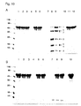
- a portion of the samples were spiked with SDS gel loading buffer and analyzed after boiling on SDS-polyacrylamide gels. Mutation of R at position 167 to A leads to complete insensitization to thrombin. The same was true for the mutation of R at position 168 to A, after addition of A to amino acid position two (CHAP-Ami_Pitti26-CBD-USA-Add2).
- polypeptides according to the invention still have the desired activity after the amino acid substitutions, they were tested in a plate lysis test with various Staphylococcus strains.
- the bacterial strains were grown overnight in 5 ml cultures in BHI medium and the bacterial cells were centrifuged for 5 min at 4000 rpm in the bench top centrifuge. The cell pellet was then resuspended in 200 ⁇ l of PBS buffer (10 mM Na phosphate, 144 mM NaCl, 50 mM KCl, pH 7.4) and the cells heat-inactivated at 80 ° C for 30 minutes.
- the heat-inactivated cells were poured into petri dishes with 15 ml of Topagar and dried.
- Table 4 Staphylococcus species PlyPitti26 (not modified) Mutant 10 from Table 3 (E163Q, R 167A, E179A, E189Q) Mutant 4 from Table 3 (E163A, R167A) CHAP-AmiPitti26-CBDUSA (not modified) Staphylococcus aureus MRSA +++ +++ +++ +++ Staphylococcus aureus MRSA +++ +++ +++ +++ +++ Staphylococcus aureus MRSA ++ +++ +++ +++ Staphylococcus aureus MRSA ++ +++ +++ +++ Staphylococcus epidermis +++ +++ +++ +++ Staphylococcus equorum + ++ ++ ++ Staphylococcus sciuri - ++ + ++ Staphylococcus saprophyticus - +++ ++ +++ Staphylococcus sciuri - - - - Staphylococcus aureus MRSA +++
- the two modified polypeptides mutant 4 and mutant 10 according to the invention with the amino acid exchanges at the thrombin or different V8 protease cleavage sites shown in the table had a virtually identical activity to the unmodified protein CHAP-AmiPitti26-CBDUSA. This showed that the mutations according to the invention did not negatively influence the activity of the cell wall lysing enzymes. The same was true for unmodified CHAP-Ami_Pitti26-CBD-USA-Add2 and the corresponding modified CHAP-Ami_Pitti26-CBD-USA-Add2 polypeptides.
- Example 5 Modified cell wall binding enzymes against Clostridium difficile
- Example 6 Determination of protease-sensitive regions within the Ply511 sequence.
- Ply511 Due to its amino acid sequence, Ply511 has 6 potential sites for Staphylococcus peptidase I (6 glutamates). To find out which regions of the endolysin Ply511 are particularly protease sensitive, protease digestion experiments with Staphylococcus peptidase I were performed. For Staphylococcus peptidase I degradation, 99 ⁇ l of protein solution (concentration about 1 mg / ml) and 1 ⁇ l of protease solution (concentration about 0.5 mg / ml) were used.
- substitutions were made either as a single substitution or in combination so that in the resulting mutants, amino acids other than glutamate were placed at the respective positions that are no longer cut by Staphylococcus peptidase I.
- the mutants thus obtained were used in the protease digestion described in Example 6 and then analyzed on SDS gels, whether the corresponding degradants still occurred or not.
- Particularly suitable were the mutations E7A and E7Q and E40A and E40Q.
- Example 8 Resistance to thrombin and V8 protease of various endolysin variants of Plypitti26 and CHAP-AmiPitti26-CBDUSA.
- CHAP-Amipitti26_CBDUSA E163A, R167A, Y200H
- CHAP-Amipitti26_CBDUSA E163A, R167A, E179A, E189Q, Y200H
- CHAP-Amipitti26_CBDUSA L55H, L56T, E163A, R167A, Y200H
- CHAP -Amipitti26_CBDUSA L55H, L56T, E163A, R167A, E179A, E189Q, Y200H
- CHAP-Amipitti26_CBDUSA E163A, R167A
- CHAP-Amipitti26_CBDUSA E163A, R167A
- CHAP-Amipitti26_CBDUSA-Add2 E164A, R168A, Y201H
- CHAP-Amipitti26_CBDUSA-Add2 E164A, R168A, E180A, E190Q,
- CHAP-Amipitti26_CBDUSA L55H, L56T, E163A, R167A, Y200H
- CHAP-Amipitti26_CBDUSA L55H, L56T, E163A, R167A, E179A, E189Q, Y200H
- CHAP-Amipitti26_CBDUSA-Add2 L56H, L57T, E164A, R168A, Y201H
- CHAP-Amipitti26_CBDUSA-Add2 L56H, L57T, E164A, R168A, E180A, E190Q, Y201H
- CHAP-Amipitti26_CBDUSA E163A, R167A, E179A, E189Q, Y200H
- CHAP-Amipitti26_CBDUSA L55H, L56T, E163A, R167A, E179A, E189Q, Y200H
- CHAP-Amipitti26_CBDUSA-Add2 E164A, R168A, E180A, E190Q, Y201H
- CHAP-Amipitti26_CBDUSA-Add2 L56H, L57T, E164A, R168A, E180A, E190Q, Y201H
- the substitutions E179A and E189Q or E180A and E190Q proved to be advantageous here.
- Ply511 (0.1 mg / ml) was digested for 3 h or overnight at room temperature with 5 units of clostripain (unit definition according to manufacturer's instructions, Sigma) in 60 ⁇ l sample volume with the following composition: 25 mM sodium phosphate, 1 mM calcium acetate, 2 , 5 mM DTT, pH 7.6.
- the resulting protein fragments were separated by SDS gel electrophoresis (gradient gels 10-20% acrylamide). There were 3 bands (molecular weights about 25 kDa, about 14 kDa, about 10 kDa), blotted on PVDF membranes, then cut out and N-terminal were sequenced by Edman degradation.
- Example 11 Protease degradation of a therapeutically useful protein in various organs and determination of the protease-sensitive regions
- the CHAP and amidase 2 domains are the EADs, while the SH3_5 domain forms the CBD.
- a domain linker region (amino acids 159 to 198) and a second linker between the amidase 2 domain and the CBD (amino acids 347 to 412). All three interfaces found were in the middle of the domain linker regions, which proved to be less stable than the conserved domains.
- the protease from the kidney responsible for the degradation could not be identified, but it was noted that at least one protease needed a Q at position P1 and that all three protease recognition sites were rich in serines and alanines.
Landscapes
- Life Sciences & Earth Sciences (AREA)
- Health & Medical Sciences (AREA)
- Chemical & Material Sciences (AREA)
- Zoology (AREA)
- Engineering & Computer Science (AREA)
- Wood Science & Technology (AREA)
- Organic Chemistry (AREA)
- Genetics & Genomics (AREA)
- Bioinformatics & Cheminformatics (AREA)
- General Health & Medical Sciences (AREA)
- Microbiology (AREA)
- Biotechnology (AREA)
- Medicinal Chemistry (AREA)
- Biomedical Technology (AREA)
- General Engineering & Computer Science (AREA)
- Biochemistry (AREA)
- Molecular Biology (AREA)
- Virology (AREA)
- Agronomy & Crop Science (AREA)
- Dentistry (AREA)
- Environmental Sciences (AREA)
- Plant Pathology (AREA)
- Pest Control & Pesticides (AREA)
- Proteomics, Peptides & Aminoacids (AREA)
- Animal Behavior & Ethology (AREA)
- General Chemical & Material Sciences (AREA)
- Pharmacology & Pharmacy (AREA)
- Communicable Diseases (AREA)
- Nuclear Medicine, Radiotherapy & Molecular Imaging (AREA)
- Oncology (AREA)
- Public Health (AREA)
- Chemical Kinetics & Catalysis (AREA)
- Veterinary Medicine (AREA)
- Enzymes And Modification Thereof (AREA)
- Medicines That Contain Protein Lipid Enzymes And Other Medicines (AREA)
Description
- Die vorliegende Erfindung betrifft eine Polypeptidvariante des Polypeptids gemäß SEQ ID NO: 1, wobei die Polypeptidvariante ausgewählt ist aus der Gruppe bestehend aus: SEQ ID NO: 2, 3, 11, 12 und 13. Die Erfindung betrifft ferner Nukleinsäuren mit einer Sequenz codierend für die erfindungsgemäßen Polypeptide.
- Pathogene Bakterien treten an vielen Stellen auf und verursachen enorme gesundheitliche Probleme durch Infektionen sowie hohe wirtschaftliche Kosten z.B. auch durch unerwünschten Bakterienbefall in der Nahrungsmittel-, Kosmetik- oder Umweltindustrie. Im Gesundheitswesen nehmen die Probleme aufgrund Antibiotika-resistenter Keime immer mehr zu, so dass händeringend nach Alternativen zu Antibiotika gesucht wird. In der Lebensmittel-und Kosmetikindustrie wird zunehmend versucht, ohne die klassischen Konservierungsmittel auszukommen, die in der Bevölkerung immer weniger Akzeptanz finden. Als Lösung für diese Probleme bietet sich der Einsatz von Zellwand lysierenden Enzymen an, die auf natürliche Weise das Wachstum der unerwünschten Bakterien unterbinden und vorhandene Keime abtöten.
- Beispiele von Zellwand lysierenden Enzymen sind Endolysine, die aus Bakteriophagen, isoliert wurden. Bakteriophagen benutzen diese Enzyme am Ende ihres Vermehrungszyklus, um die innerhalb der Bakterienzelle gebildeten Bakteriophagen freizusetzen. Die Bakterienhülle wird dabei lysiert und das Wirtsbakterium auf diese Weise zerstört. Ein weiteres Beispiel sind Enzyme, die der Bakteriophage am Anfang seines Vermehrungzyklus benötigt und in der Regel in Bakteriophagenschwanzproteinen lokalisiert sind. Diese Enzyme werden für den Aufbruch der Bakterienwand bei der Bakterieninfektion benötigt. Ein drittes Beispiel sind Enzyme, die eine ähnliche Funktion und häufig auch eine sequenzielle Ähnlichkeit zu den Endolysinen aufweisen. Diese Enzyme sind die von Bakterien unter bestimmten Umständen gebildeten Autolysine, die zur Selbstlyse der Bakterien führen. Ein viertes Beispiel sind Enzyme, die ebenfalls von Bakterien gebildet werden und als Bakteriocine bekannt sind. Diese vier Gruppen von Zellwand lysierenden Enzyme werden bereits technisch eingesetzt und in Zukunft wohl noch an Bedeutung gewinnen.
- Ein Anwendungsgebiet ist der medizinische Einsatz in der Prävention und Therapie sowie die Diagnostik von Bakterieninfektionen bei Mensch und Tier. Ein weiteres Anwendungsgebiet ist die Anwendung in der Lebensmittel-, Umwelt- und Kosmetikindustrie zur Verhinderung eines unerwünschten Bakterienwachstums und zur Abtötung der Keime beispielsweise durch Desinfektion sowie der Nachweis von Bakterien in Lebensmittel-, Umwelt- und Kosmetikproben.
- Praktisch alle Anwendungen in denen Bakterien lysierende Enzyme verwendet werden stellen hohe Anforderungen an die Stabilität der eingesetzten Enzyme, damit sich ihr Einsatz wirtschaftlich lohnt. In der
US 6,432,444 B1 wird zur Stabilisierung der Zellwand lysierenden Enzyme vorgeschlagen, z.B. einen stabilisierenden Puffer zu verwenden, um die optimale Enzymaktivität zu gewährleisten. Weiterhin wird der Zusatz von stabilisierenden Substanzen wie Reduktionsmittel, Metallchelatoren, Immuglobulinen, spezifischen Puffersalzen, physiologischen pH-Werten, Konservierungsmittel oder milden Detergentien vorgeschlagen. - Generell wird die Stabilität von Proteinen häufig durch vorhandene Proteasen verringert. Strategien zur Erhöhung der Stabilität von Proteinen gegenüber einem Proteaseabbau sind z.B. der Zusatz von Metallchelatoren, um Proteasen zu hemmen, die für ihre Aktivität Metallionen benötigen oder der Zusatz von speziellen Proteaseinhibitoren, wie sie zum Beispiel für Serinproteasen kommerziell erhältlich sind. Proteaseinhibitoren können zur Stabilität von Zellwand lysierenden Enzymen jedoch nur bedingt eingesetzt werden, weil sie auch andere essentielle Komponenten am Wirkort stören. Darüber hinaus sind spezifische Inhibitoren auch sehr teuer. Auch die Auswahl anderer Umgebungsbedingungen, in denen Proteasen weniger aktiv sind, ist in der Regel nicht möglich, da die Zellwand lysierenden Enzyme ganz ähnliche Bedingungen für ihre Aktivität brauchen wie die Proteasen. So arbeiten Zellwand lysierende Enzyme in einer wässrigen Umgebung und unter moderaten pH-Werten am besten, unter denen auch die Proteaseaktivität am größten ist. Die bekannten Stabilisierungsansätze sind für den Einsatz bei Zellwand lysierenden Enzymen nicht geeignet.
- Daher liegt der vorliegenden Erfindung die Aufgabe zu Grunde, stabile Zellwand lysierende Enzyme zur Verfügung zu stellen.
- Die Aufgabe wird durch den in den Patentansprüchen definierten Gegenstand gelöst.
- Die nachfolgenden Abbildungen dienen der Erläuterung der Erfindung.
-
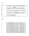
Figur 1 zeigt die Wildtyp-Aminosäuresequenz der CHAP-Domäne (Aminosäure 1-154), der Linkerregion (unterstrichen, Aminosäure 155-193) und der Amidase-Domäne (Aminosäure 194-393) eines Endolysins aus PlyPitti26. Potentielle Schnittstellen für V8 Protease, die nach dem Aminosäurerest E schneidet, sind fett hervorgehoben und befinden sich an den Aminosäurepositionen 163, 179 und 189. Die Thrombinschnittstelle ist grau unterlegt und befindet sich an Position R167. -
Figur 2 zeigt das Ergebnis eines Proteaseverdaus und anschließendem Aktivitätstest von nicht modifiziertem PlyPitti26 mit den Proteasen Thrombin und V8 Protease.Figur 2A zeigt ein SDS-Polyacrylamidgel mit Proben von nicht modifizeirtem PlyPitti26 nach einen Verdau mit Thrombin (T), V8 Protease (V8) und einem Kontrollverdau mit Plasmin (P), für das keine Schnittstelle existiert. Unverdautes PlyPitti26 ist mit "-" gekennzeichnet. M bezeichnet Molekulargewichtsstandards mit den am Rand angegebenen Proteingrößen in kDa. Am rechten Rand angegeben sind die Positionen der Banden für das Fragment N-terminal von der Thrombinschnittstelle (CHAP-Domäne), das Fragment C-terminal von der Schnittstelle (Ami-CBD) und die Bande vom zugesetzten Thrombin.Figur 2B zeigt das Ergebnis eines Flüssiglysetests zur Bestimmung der spezifischen Aktivitäten von verdautem und nicht verdautem nicht modifiziertem PlyPitti26. Die Bakterienzelllyse wird über eine Lichtstreuungsmessung im Photometer verfolgt. Es sind die Lysekurven für unverdautes PlyPitti26 (____), Plasmin verdautes (-----) sowie mit Thrombin (_._.) oder V8 Protease (_.._..) verdautes Endolysin dargestellt. -
Figur 3 zeigt das Ergebnis eines Proteaseverdaus von modifiziertem und nicht modifiziertem CHAP-AmiPitti26-CBDUSA.Figur 3A zeigt das Ergebnis eines Thrombinverdaus des nicht modifizierten CHAP-AmiPitti26-CBDUSA und eines modifizierten Staphylococcus Endolysins CHAP-AmiPitti26-CBDUSA (Mutante 4 auf Tabelle 3). Das nicht modifizierte CHAP-AmiPitti26-CBDUSA besitzt eine singuläre Thrombinschnittstelle zwischen der CHAP- und der Amidase-Domäne an Position R167, das modifizierte Polypeptid einen Austausch von R nach A an Position 167. DieFigur 3A zeigt ein SDS-Polyacrylamidgel der beiden Enzymvarianten vor und nach dem Thrombinverdau: M - Molekulargewichtsmarker (Molekulargewichte in kDa am Rand angegeben); 1- nicht modifiziertes CHAP-AmiPitti26-CBDUSA ohne Thrombin; 2 - nicht modifiziertes CHAP-AmiPitti26-CBDUSA nach Thrombinverdau; 3 - modifiziertes CHAP-AmiPitti26-CBDUSA ohne Thrombin; 4-modifiziertes CHAP-AmiPitti26-CBDUSA nach Thrombinzugabe. -
Figur 3B zeigt das Ergebnis eines V8 Protease Verdaus des nicht modifizierten CHAP-AmiPitti26-CBDUSA und eines modifizierten Staphylococcus Endolysins CHAP-AmiPitti26-CBDUSA (Mutante 4 auf Tabelle 3). Das nicht modifizierte CHAP-AmiPitti26-CBDUSA besitzt eine V8 Protease Schnittstelle zwischen der CHAP- und der Amidase-Domäne an Position E163, das modifizierte Polypeptid einen Austausch von E nach A an Position 163. DieFigur 3B zeigt ein SDS-Polyacrylamidgel der beiden Enzymvarianten vor und nach dem Thrombinverdau: M - Molekulargewichtsmarker (Molekulargewichte in kDa am Rand angegeben); 1- nicht modifiziertes CHAP-AmiPitti26-CBDUSA ohne V8 Protease; 2 - nicht modifiziertes CHAP-AmiPitti26-CBDUSA 15 min V8 Protease Verdau; 3 - nicht modifiziertes CHAP-AmiPitti26-CBDUSA 60 min V8 Protease Verdau; 4 - modifiziertes CHAP-AmiPitti26-CBDUSA ohne V8 Protease; 5 - modifiziertes CHAP-AmiPitti26-CBDUSA 15 min V8 Protease Verdau; 6 - modifiziertes CHAP-AmiPitti26-CBDUSA 60 min V8 Protease Verdau. -
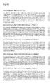
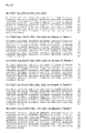
Figur 4 zeigt einen Aminosäuresequenzvergleich des modifizierten Endolysins aus dem Cl. difficile Phagen Φ CD 119 (CD 119) mit dem modifizierten Endolysin aus dem Cl. difficile Phagen Φ 630. Die im Φ CD 119 und Φ 630 Wildtyp vorhandene singuläre Caspase 1 Schnittstelle an Aminosäure D214 wurde zu E214 modifiziert, die im Φ 630 Wildtyp vorhandene singuläre Thrombinschnittstelle R87 zu K87 modifiziert. -
Figur 5 zeigt einen Aminosäuresequenzvergleich der Region der Thrombinschnittstelle von verschiedenen Staplaylococcus aureus Endolysinen. Der fett hervorgehobene Aminosäurerest R ist Position R167 in der Referenzsequenz ply_pitti26, bezeichnet als P26A. Die weiteren aufgeführten Aminosäuresequenzen sind an R167 von ply_pitti26 ausgerichtget. Der Aminosäuresequenzvergleich wurde mit dem Programm BLAST durchgeführt; Altschul et al., 1990, J. Mol. Biol., 215, 403-410. Die Zahl rechts neben dem Aminosäuresequenzvergleich bezeichnet die Aminosäureposition der letzen Aminosäure im dargestellten Sequenzabschnitt des jeweiligen Endolysins. Die Zahlen am linken Rand stehen für verschiedene homologe Proteine aus dem Aminosäuresequenzvergleich. P26A steht für ply_pitti26, 15 ein Autolysin (N-Acetylmuramoyl-L-Alanin Amidase) aus Staphylococcus aureus (Datenbankzugangsnummer P24556), 16 steht für eine Amidase aus dem Bakteriophagen 80 alpha (Datenbankzugangsnummer AAB39699), 3 steht für das Endolysin des Staphylococcus Phagen phi MR11 (Datenbankzugangsnummer YP 001604156), 4 steht für ORF006 aus dem Staphylococcus aureus Phagen 88 (Datenbankzugangsnummer YP 240699), 5 steht für ORF21 aus dem Staphylococcus aureus Phagen 85 (Datenbankzugangsnummer YP 239752), 9 steht für die Amidase aus dem Staphylococcus Phagen 11 (Datenbankzugangsnummer NP803306), 10 steht für ORF007 aus dem Staphylococcus Phagen 52A (Datenbankzugangsnummer YP 240634), 11 steht für die N-Acetylmuramoyl-L-Alanin Amidase aus dem Stamm Staphylococcus aureus subsp. aureus JH9 (Datenbankzugangsnummer YP 001246457), 12 steht für die putative Zellwandhydrolase des Phagen phiMR25 (Datenbankzugangsnummer YP 001949866), 13 steht für ORF007 aus dem Staphylococcus Phagen 69 (Datenbankzugangsnummer YP 239596), 14 steht für ORF007 aus dem Staphylococcus Phagen 55 (Datenbankzugangsnummer YP 240484), 2 steht für die N-Acetylmuramoyl-L-Alanin Amidase aus dem Stamm Staphylococcus aureus subsp. aureus JH9 (Datenbankzugangsnummer YP 001245749), 8 steht für ORF007 aus dem Staphylococcus Phagen 29 (Datenbankzugangsnummer YP 240560), 7 steht für das Autolysin aus dem Stamm Staphylococcus aureus subsp. aureus NCTC 8325 (Datenbankzugangsnummer YP 500516), 1 steht für die Amidase aus dem Staphylococcus Phagen phiNM2 (Datenbankzugangsnummer ABF73160), 6 steht für eine Phagen-verwandte Amidase aus dem Stamm Staphylococcus aureus RF122 (Datenbankzugangsnummer YP 417165), 17 steht für ORF006 aus dem Staphylococcus Phagen 37 (Datenbankzugangsnummer YP 240103) und 18 steht für ORF007 aus dem Staphylococcus Phagen EW (Datenbankzugangsnummer YP 240182). -
Figur 6 gibt Aminosäuresequenzen von modifiziertem und nicht modifiziertem CHAP-AmiPitti26-CBDUSA wieder.Figur 6A bis D zeigt die Aminosäuresequenz von CHAP-AmiPitti26-CBDUSA ohne Aminosäuresubstitutionen an Proteaseschnittstellen (A) und CHAP-AmiPitti26-CBDUSA mit Aminosäuresubstitutionen an Proteaseschnittstellen (B bis E).Figur 6F bis K zeigt die Aminosäuresequenz von CHAP-AmiPitti26-CBDUSA-Add2 ohne Aminosäuresubstitutionen an Proteaseschnittstellen und einem zusätzlichen Aminosäurerest an Position zwei (F) und CHAP-AmiPitti26-CBDUSA-Add2 mit Aminosäuresubstitutionen an Proteaseschnittstellen und einem zusätzlichen Aminosäurerest an Position zwei (G bis K). -
Figur 7A zeigt die Aminosäuresequenz des modifizieten Endolysins aus dem Cl. difficile Stamm 630 mit einem Austausch von D214 zu E214.Figur 7B zeigt die Aminosäuresequenz des modifizieten Cl. difficile Endolysins aus dem Phagen phi CD119 mit einem Austausch von R87 zu K87 und D214 zu E214. -
Figur 8 zeigt die Aminosäuresequenz des Endolysins Ply511. Die fett gedruckten Aminosäurepositionen E7, E40 und E89 können mit jedem anderen Aminosäurerest substituiert werden. -
Figur 9 zeigt die Aminosäuresequenz des Endolysins Ply511. Die potentiellen Schnittstellen (R in P1 Position) für die Protease Clostripain sind unterstrichen. Die beiden experimentell ermittelten besonders sensitiven Schnittstellen (R92 und R221) sind unterstrichen und fett gedruckt. -
Figur 10 zeigt den Verdau des therapeutisch einsetzbaren Endolysins CHAP-Amipitti26_CBDUSA-Add2 (L56H, L57T, E164A, R168A, Y201H) durch in Organextrakten vorhandene Proteasen.Figur 10A zeigt ein SDS-Gel, bei dem die Proteinbanden nach Inkubation mit Leber-, Nieren- und Lungenextrakt aufgetrennt wurden. Links aufgetragen ist ein Molekulargewichtsstandard. Spuren 1 bis 3 zeigen Kontrollen (nur Endolysin, kein Organextrakt), die gar nicht (Spur 1), 18 h bei Raumtemperatur (Spur 2) oder 18 h bei 4 °C inkubiert (Spur 3) wurden. In den Spuren 4 bis 6 wurde Leberextrakt zugegeben, bei Spur 7 bis 9 Nierenextrakt und in den Spuren 10 bis 12 Lungenextrakt. Spuren 4, 7 und 10 enthalten nur Organextrakt, Spuren 5, 8 und 11 Organextrakt und Endolysin nach 18 h Inkubation bei Raumtemperatur, Spuren 6, 9 und 12 Organextrakt und Endolysin nach 18 h Inkubation bei 4 °C. Die Pfeile mit den Nummern 1 bis 7 neben Spur 9 markieren die Banden, die nach Transfer auf eine Blotmembran N-terminal ansequenziert wurden.Figur 10B zeigt ein SDS-Gel, bei dem die Proteinbanden nach Inkubation mit Herz- oder Milzextrakt aufgetrennt wurden. Links aufgetragen ist ein Molekulargewichtsstandard. Spuren 1 bis 3 sowie 10 zeigen Kontrollen (nur Endolysin, kein Organextrakt), die gar nicht (Spur 1 und 10), 18 h bei Raumtemperatur (Spur 2) oder 18 h bei 4 °C inkubiert (Spur 3) wurden. In den Spuren 4 bis 6 wurde Herzextrakt zugegeben, bei Spur 7 bis 9 Milzextrakt. Spuren 4 und 7 enthalten nur Organextrakt, Spuren 5 und 8 Organextrakt und Endolysin nach 18 h Inkubation bei Raumtemperatur, Spuren 6 und 9 Organextrakt und Endolysin nach 18 h Inkubation bei 4 °C. -
Fig. 11 zeigt die Sequenz des Endolysins CHAP-Amipitti26_CBDUSA-Add2 (L56H, L57T, E164A, R168A, Y201H) mit proteasesensitiven Bereichen. Die Schnittstellen (P1 Position), die sich aus der Inkubation mit Nierenextrakt ergaben, sind fett markiert. Aminosäuresequenzen, die sich bei der N-terminalen Sequenzierung der entstandenen Fragmente ergaben, sind unterstrichen. Aminosäurebereiche, die die Domänenlinker definieren, sind kursiv dargestellt. Dies ist ein Domänenlinkerbereich zwischen der CHAP und Amidase 2 Domäne (Aminosäuren 159 bis 198) sowie ein zweiter Linker zwischen der Amidase 2 Domäne und der CBD (Aminosäuren 347 bis 412). -
Fig. 12 zeigt einen Aminosäuresequenzvergleich der Region der Caspase I Schnittstelle von verschiedenen Staphylococcus Endolysinen. Es handelt sich um Amidasen aus den Staphylococcus Phagen SAP-2, 44AHJD und Phage 66. Die NCBI Datenbankzugangsnummern für die Proteine sind YP_001491539, 44AHJD_15 amidase und YP_239469. Die Position D42, nach der geschnitten wird ist fett hervorgehoben, die konservierte Erkennungssequenz für die Caspase I ist jeweils kursiv und umrandet dargestellt. Die 3 Proteine sind auf ihre gesamte Länge zu 89 bis 90 % identisch, nicht konservierte Aminosäuren für den Bereich der Aminosäuren 1 bis 60 sind mit "-" markiert. Der Aminosäuresequenzvergleich wurde mit dem Programm BLAST durchgeführt. - Der Begriff "Polypeptid" oder "Protein" wie hier verwendet bezeichnet Peptide aus mindestens acht Aminosäuren. Die Polypeptide können pharmakologisch oder immunologisch aktive Polypeptide oder für diagnostische Zwecke verwendete Polypeptide sein.
- Der Begriff "Zellwand lysierende Enzyme" wie hier verwendet bezeichnet Enzyme, die in der Lage sind, die bakterielle Zellwand zumindest teilweise aufzubrechen oder so zu beschädigen, dass in der Folge eine Zelllyse oder zumindest ein bakteriostatischer Effekt auftritt. Insbesondere bezeichnet der Begriff Endolysine, Autolysine, Bakteriophagenschwanzproteine und Bakteriocine.
- Der Begriff "Endolysin" wie hier verwendet bezeichnet Enzyme, die natürlicherweise von Bakteriophagen kodiert werden und die diese am Ende ihres Wirtszyklus herstellen, um die Wirtszelle zu lysieren und damit die in der Wirtszelle neu hergestellten Bakteriophagen freizusetzen. Endolysine sind aus mindestens einer enzymatisch aktiven Domäne (EAD) und einer nicht enzymatisch aktiven zellbindenden Domäne (CBD) aufgebaut. Die einzelnen EADs weisen unterschiedliche Aktivitäten auf, wobei Endolysine mindestens eine EAD ausgewählt aus der folgenden Gruppe aufweisen: N-Acetyl-Muramoyl-L-Alanin-Amidase (Amidase, z.B. Ami_2, Ami_5), (Endo)-Peptidase (z.B. CHAP, i.e. cysteine, histidine-dependent amidohydrolases/peptidases), Transglykosylase, Glykosylhydrolase, (N-Acetyl)-Muramidase (Lysozyme) und N-Acetyl-Glukosaminidase.
- Der Begriff "Autolysin" wie hier verwendet bezeichnet bakterielle Peptidoglykanhydrolasen, die in bestimmten Situationen zu einer Selbstlyse der Bakterien führen. Funktionell und auch strukturell sind Autolysine und Endolysine häufig verwandt. Verschiedene Domänen aus Endolysinen und Autolysinen können häufig modular kombiniert werden und aktive Chimären bilden.
- Der Begriff "Bakteriophagenschwanzprotein" wie hier verwendet bezeichnet Phagenstrukturproteine, die am Anfang des Replikationszyklus des Bakteriophagen bei der Infektion die Funktion erfüllen, an Rezeptoren auf der Bakterienoberfläche zu binden und dabei über ihre enzymatische Funktion Komponenten der Zelloberfläche lysieren. Die entsprechenden Bakteriophagenproteine müssen nicht immer im Phagenschwanz lokalisiert sein, sondern können bei Phagen ohne Schhwanz auch direkt am Phagenkopf sitzen.
- Der Begriff "Bakteriocin" wie hier verwendet bezeichnet proteinogene Toxine, die von Bakterien abgesondert werden, um das Größenwachstum ähnlicher Bakteriengattungen zu inhibieren. Sie bestehen aus einer Zellwand bindenden Domäne, einer Translokationsdomäne und aus dem "killing factor". Die in diesem Zusammenhang enthaltene Gruppe der Bakteriocine greift dabei bakterielle Membranen an. Beispiele sind Colicin, Microcin, Nisin, Epidermin und Lantibiotika (lanthioninhaltige Antibiotika).
- Der Begriff bakterielle "Zellwand" wie hier verwendet bezeichnet alle Komponenten, die die äußere Zellhülle der Bakterien aufbauen und so deren Unversehrtheit garantieren. Insbesondere ist darunter zu verstehen das Peptidoglykan, die äußere Membran der gramnegativen Bakterien mit dem Lipopolysaccharid, die Bakterienzellmembran, aber auch zusätzliche auf das Peptidoglykan aufgelagerte Schichten wie Kapseln, Schleime oder äußere Proteinschichten.
- Der Begriff "Protease" wie hier verwendet bezeichnet ein Enzym, das in der Lage ist, Proteine und/oder Peptide hydrolytisch zu spalten.
- Der Begriff "Domäne" wie hier verwendet bezeichnet einen Teilbereich einer Aminosäuresequenz, der funktionell und/oder strukturell definiert ist. Domänen können aufgrund von Homologien häufig sehr gut über entsprechende Suchprogramme vorhergesagt werden, die sich auf entsprechende Datenbanken mit konservierten Domänen stützen (z.B. Conserved Domain Database (CDD) an der NCBI (Marchler-Bauer et al., 2005, Nucleic Acids Res. 33, D192-6), Pfam (Finn et al., 2006, Nucleic Acids Research 34, D247-D251) oder SMART (Schultz et al., 1998, Proc. Natl. Acad. Sci. USA 95, 5857-5864, Letunic et al., 2006, Nucleic Acids Res 34, D257-D260).
- Der Begriff "Wildtyp" wie hier verwendet bezeichnet die Aminosäuresequenz, wie sie in natürlicherweise vorkommenden Proteinen vorkommt und deren Sequenz nicht in Bezug auf Proteaseschnittstellen modifiziert wurde. Es kann sich dabei um Aminosäuresequenzen handeln so wie sie natürlicherweise in einem Bakteriophagen, Prophagen oder Bakterium vorkommen. Zusätzlich bezeichnet der Begriff alle in den Datenbanken vorkommenden Aminosäuresequenzen von Zellwand lysierenden Enzymen, die noch bestimmte Proteaseschnittstellen enthalten, auch wenn die Aminosäuresequenzen bereits gegenüber natürlich vorkommenden Proteinen verändert wurden. Im Zusammenhang mit Nukleotidsequenzen bezeichnet der Begriff Wildtyp auch die Nukleotidsequenzen, die für ein Polypeptid kodieren, die eine vorstehend beschriebene Aminosäuresequenz aufweisen.
- Die vorliegende Erfindung betrifft ein modifiziertes Polypeptid mit der biologischen Aktivität Zellwände von Bakterien zu lysieren, wobei das Polypeptid keine Caspase-, Enterokinase-, Faktor Xa, Granzyme B, Staphylococcus Peptidase I (V8 Protease), Plasmin, Streptopain, Bacillolysin oder Thrombinschnittstelle enthält sowie weitere Proteaseschnittstellen, die spezifisch für das jeweilige Polypeptid durch Proteaseverdau mit anschließender Analyse der entstandenen Fragmente bestimmt wurden und anschließend auf die in der Erfindung beschriebene Weise modifiziert wurden. Vorzugsweise betrifft die vorliegende Erfindung erfindungsgemäße modifizierte Polypeptide, die gram positive Bakterien lysieren, wobei die Bakterien aus der Gruppe bestehend aus Clostridien, Bazillen, Listerien, Staphylokokken, Lactobazillen, Enterokokken, Aerokokken, Pediokokken, Streptokokken, Mycoplasmen und/oder Leuconostoc stammen können. Vorzugsweise ist das erfindungsgemäße Polypeptid ein Endolysin, ein Bakteriophagenschwanzprotein, ein Autolysin oder ein Bakteriocin.
- Die vorliegende Erfindung betrifft ferner ein rekombinant hergestelltes Polypeptid mit der biologischen Aktivität Zellwände von Bakterien zu lysieren, wobei die Aminosäuresequenz des Polypeptids gegenüber der Wildtyp-Sequenz an Aminosäurepositionen Modifikationen aufweist, die von Proteasen erkannt und/oder nach der die Proteasen die Polypeptide spalten.
- Überraschenderweise stellte sich heraus, dass die Zellwand lysierenden Enzyme in ihrer Aminosäuresequenz so verändert werden können, dass eine erhöhte Proteasestabilität erreicht werden kann, ohne dass man stabilisierende Substanzen zusetzen muss. Es ist allgemein bekannt, dass verschiedene Proteasen an einem bestimmten Aminosäurerest die Polypeptidkette schneiden oder Aminosäuresequenzmotive für ihre enzymatische Aktivität benötigen. Proteaseschnittstellen in Proteinen können bei vorhandener Aminosäuresequenz mit geeigneten Sequenzanalyseprogrammen vorausgesagt werden (z.B. ExPASy PeptideCutter, Gasteiger et al., Protein Identification and Analysis Tools on the ExPASy Server in John M. Walker (ed): The Proteomics Protocols Handbook, Humana Press (2005)). So ergeben sich für ein Protein mittlerer Größe mit etwa 300 bis 400 Aminosäureresten in der Regel weit mehr als 100 potentielle Proteaseschnittstellen, so dass eine Stabilisierung von Proteinen nur dadurch zu erreichen ist, dass die Schnittstellen einer Vielzahl von Proteasen verändert werden müssten. Das ist jedoch unpraktikabel, da das Protein seine Wirkung und Aktivität verliert, wenn zu viele Aminosäuremodifikationen eingeführt werden. Eine weitere Möglichkeit zur Bestimmung von proteasesensitiven Bereichen neben der Vorhersage anhand der Aminosäuresequenz, vor allem für Proteasen, bei denen keine Erkennungssequenz bekannt ist oder deren Identität man nicht kennt, ist die experimentelle Bestimmung der tatsächlichen Schnittstellen durch einen Verdau der Proteine mit kommerziell erhältlichen Proteasen. Ferner können hierzu aus Organismen oder Proben isolierte Proteasen verwendet werden. Insbesondere können die Proben, die Proteasen enthalten wie z.B. Lebensmittelproben, Umweltproben, Organextrakte, medizinische Proben verwendet werden, ohne die Proteasen vorher zu isolieren. Anschliessend werden die Versuchsansätze einer Analyse der entstehenden Abbauprodukte mit Hilfe von Sequenzbestimmung (z.B. N- oder C-terminale Ansequenzierung, Peptidmapping in Verbindung mit Massenspektrometrie) und Größenbestimmung (z.B. Massenspektrometrie, Bandenanalyse von SDS-Gelen, analytische Gelfiltration) unterzogen. Tatsächlich haben die Erfinder in vielen Zellwand lysierenden Enzymen Schnittstellen und Aminosäuresequenzmotive für Proteasen gefunden. Das erscheint überraschend, wenn man davon ausgeht, dass die Zellwand lysierenden Enzyme eigentlich auf eine hohe Stabilität hin evolviert sein müssten. Überraschenderweise zeigte sich jedoch, dass die Schnittstellen und Aminosäuresequenzmotive für die Proteasen in geringer Anzahl, häufig sogar in singulärer Form in den Zellwand lysierenden Enzymen vorhanden und häufig auch in homologen Proteinen konserviert sind. Dadurch ist eine Modifikation einiger weniger oder sogar nur einer Proteaseschnittstelle ausreichend, um die Stabilität der Zellwand lysierenden Enzyme zu erhöhen.
- In der vorliegenden Erfindung werden vorzugsweise nur solche Proteaseschnittstellen verändert, die von bestimmten, potentiell am Wirkort der Zellwand lysierenden Enzyme vorhandenen Proteasen erkannt werden. Dabei werden die Aminosäuresequenzen der Zellwand lysierenden Enzyme so modifiziert, dass sie von den spezifischen Proteasen nicht mehr erkannt und folglich nicht mehr geschnitten werden. Das führt zu einer erhöhten Stabilität der Zellwand lysierenden Enzyme gegenüber am Wirkort vorhandenen Proteasen. Somit wird ein proteasebedingter Verlust der Aktivität der Zellwand lysierenden Enzyme verhindert und eine längere Wirksamkeit erzielt.
- Es gibt eine Reihe von Proteasen, die für ihre enzymatische Aktivität eine spezifische Erkennungssequenz in ihren Substratproteinen benötigen. Einige Beispiele dafür sind in der folgenden Tabelle aufgeführt. Dem Fachmann sind eine Vielzahl weiterer Proteasen bekannt wie sie beispielsweise in der Merops Peptidase Datenbank (Rawlings et al., 2008, Nucleic Acids Res 36, D320-D325) beschrieben sind. Das Nachfolgende gilt jedoch auch für Proteaseschnittstellen unbekannter Proteasen, die experimentell bestimmt werden können. P1 bezeichnet die Position der Aminosäure, nach der geschnitten wird, P4, P3 und P2 sind die Positionen N-terminal vor der Schnittstelle P1. Die Bezeichnung P1' und P2' sind die Positionen C-terminal im Anschluss an P1. Das bedeutet, dass die Proteasen die Polypeptidkette zwischen P1 und P1' spalten. Die anstelle der Aminosäurereste aufgeführten Großbuchstaben stellen die international verwendeten Bezeichnungen der Aminosäurereste dar und sind allgemein bekannt. Werden in der Beschreibung der vorliegenden Erfindung die Großbuchstaben verwendet, sind die entsprechenden Aminosäurerest gemeint.
Tabelle 1 Protease Schnittstelle P4 P3 P2 P1 P1' P2' Caspase 1 F, W, Y oder L - H, A oder T D nicht P, E, D, Q, K oder R - Caspase 2 D V A D nicht P, E, D, Q, K oder R - Caspase 3 D M Q D nicht P, E, D, Q, K oder R - Caspase 4 L E V D nicht P, E, D, Q, K oder R - Caspase 5 L oder W E H D - - Caspase 6 V E H oder I D nicht P, E, D, Q, K oder R - Caspase 7 D E V D nicht P, E, D, Q, K oder R - Caspase 8 I oder L E T D nicht P, E, D, Q, K oder R - Caspase 9 L E H D - - Caspase 10 I E A D - - Clostripain (Clostridiopeptidase B) - - - R - Enterokinase D oder N D oder N D oder N K - - Faktor Xa A, F, G, I, L, T, V oder M D oder E G R - - Plasmin (Fibrinolysin, Fibrinase) - - - K or R - - Streptopain (Streptococcus Peptidase A) - - F F Y - Bacillolysin (Bacillus subtilis neutrale Protease) - G oder F oder A G oder F L oder F A oder G oder L - Granzyme B I E P D - - Staphylococcus Peptidase I (V8 Protease) - - nicht E E - - Thrombin - - G R G - A, F, G, I, L, T, V oder M A, F, G, I, L, T, V, W or A P R nicht D, E nicht D, E - Vorzugsweise betrifft die vorliegende Erfindung ein modifiziertes Polypeptid mit der biologischen Aktivität Zellwände von Bakterien zu lysieren, wobei das erfindungsgemäße Polypeptid gegenüber der natürlicherweise vorkommenden eine oder mehrere Modifikationen in seiner Aminosäuresequenz an einer oder mehreren der in Tabelle 1 aufgezeigten Positionen oder in experimentell bestimmten Proteaseschnittstellen aufweist, wobei die eine Modifikation oder die mehreren Modifikationen an den in Tabelle 1 aufgeführten oder experimentell bestimmten Aminosäuren von Proteaseschnittstellen vorkommen. Dadurch erhöht sich die Stabilität des Polypeptids gegenüber der oder den Proteasen, deren Erkennungssequenz entsprechend geändert wurde.
- In einer bevorzugten Ausführungsform der vorliegenden Erfindung wird der Aminosäurerest an der Position, nach der geschnitten wird (P1) durch einen anderen geeigneten Aminosäurerest ersetzt, so dass das erfindungsgemäße Polypeptid nicht mehr von der Protease geschnitten wird, dessen Erkennungsstelle entsprechend modifiziert wurde. Statt eines Austausches des Aminosäurerestes an Position P1 oder auch zusätzlich zu einem Austausch an Position P1 können weitere Aminosäurereste in der Erkennungsstelle vorzugsweise die in Tabelle 1 aufgeführten Positonen an P4, P3, P2, P1' und/oder P2', durch andere Aminosäurereste ausgetauscht werden. Vorzugsweise werden pro Erkennungsstelle 6, 5, 4, 3, 2, 1 Sustitutionen vorgenommen, besonders bevorzugt insbesondere 1 bis 3 Substitutionen.
- In einer weiteren Ausführungsform der vorliegenden Erfindung können in einem Polypeptid eine oder mehrere Proteaseschnittstellen wie zuvor beschrieben modifiziert werden.
- Wichtig ist, dass die erfindungsgemäße Proteinvariante nach der Mutation nicht nur eine höhere Stabilität gegenüber einem Proteaseabbau aufweist, sondern auch weiterhin ihre Zellwand lysierende Aktivität behält. Die Aminosäuresubstitution wird daher wie nachfolgend beschrieben durchgeführt.
- Im Stand der Technik sind eine Reihe von essentiellen Aminosäureresten bekannt, die für die Aktivität der Zellwand lysierenden Enzyme bekannt sind z.B. konservierte Cysteine und Histidine bei verschiedenen Amidasen oder CHAP-Domänen. Die entsprechenden essentiellen Reste können auch mit Sequenzanalyseprogrammen gefunden werden, die speziell nach konservierten Domänen suchen (z.B. Conserved Domain Database (CDD) über NCBI (Marchler-Bauer et al., 2005, Nucleic Acids Res. 33, D192-6), Pfam (Finn et al., 2006, Nucleic Acids Research 34, D247-D251) oder SMART (Schultz et al., 1998, Proc. Natl. Acad. Sci. USA 95, 5857-5864, Letunic et al., 2006, Nucleic Acids Res 34, D257-D260).
- Um die Stabilität, Struktur und Funktion der erfindungsgemäßen Polypeptide möglichst wenig zu destabilisieren, werden die auszutauschenden Aminosäurereste der Proteaseerkennungssequenz durch Aminosäurereste ersetzt, die von ihrer Struktur und Biochemie her möglichst nah verwandt sind und dennoch von der Protease nicht erkannt werden. Es ist mittlerweile allgemeines Fachwissen die 20 natürlich vorkommenden Aminosäuren in bestimmte Gruppen einzuteilen, die von ihrer Biochemie her unterschiedlich sind (z.B. saure, basische, hydrophobe, polare, aromatische Aminosäuren), innerhalb derer die Aminosäuren jedoch zueinander verwandt sind. Bevorzugte Austausche für die Aminosäurereste in den erfindungsgemäßen Polypeptiden sind in Tabelle 2 zusammengefasst. Die in Spalte 2 genannten Austauschmöglichkeiten stellen konservative Aminosäurealternativen dar, die entweder von ihrer Struktur her und/oder von ihrer Funktion her besonders ähnlich zum Aminosäurerest im Wildtyp sind. Dies sind besonders bevorzugte Austauschvarianten. Möglich sind jedoch für die erfindungsgemäßen Zellwand lysierenden Polypeptide auch weitere Austauschvarianten, die in Spalte 3 genannt sind.
Tabelle 2 Aminosäure im Wildtyp Konservativer Austausch Weitere Austauschmöglichkeiten A (Ala) S T, G, V, E, D N (Asn) Q, D K, H, I, T, S, A D (Asp) E, N A, G, S R (Arg) K S, Q, A C (Cys) S M, G, A E (Glu) D, Q A, G, S, A Q (Gln) N, E H, L, R, A G (Gly) A S, N, D H (His) Q, N R, A I (Ile) V, L T, A L (Leu) V, I M,F,A K (Lys) R N, T, S, Q, A M (Met) L K, V, I, C, A F (Phe) Y L, I, M, A P (Pro) A S, Q S (Ser) A, T G, N, K T (Thr) S, A V, K, N W (Trp) Y F, S, R, A Y (Tyr) F H, N, C, A V (Val) I, A L, T - Eine weitere Möglichkeit zu bestimmen, welcher Aminosäurerest anstelle des zu substituierenden eingebaut wird, stellt die Dayhoff-Matrix dar; siehe z.B. in Creighton (Proteins: Structure and Molecular Properties, 2nd Ed., 1984, Freeman, New York). Aus Untersuchungen homologer Proteine ist bekannt, dass die rein statistisch zufällige Mutation auf Nukleinsäureebene sich nicht in der Aminosäuresequenz der entsprechenden Proteine widerspiegelt. Es scheint somit für bestimmte Aminosäurepositionen in den homologen Proteinen ein Selektionsdruck auf bestimmte Aminosäurereste zu bestehen, was in der Dayhoff-Matrix angegeben ist.
- Eine weitere Möglichkeit zu bestimmen, welcher Aminosäurerest anstelle des zu substituierenden eingebaut wird, bedient sich der Hilfe von Sequenzanalyseprogrammen z.B. des Programms BLAST, siehe Altschul et al., 1990, J. Mol. Biol., 215, 403-410. Damit kann nach verwandten Proteinen zu den zu mutierenden Zellwand lysierenden Enzymen gesucht werden. Der Suchalgorithmus findet dabei Ähnlichkeiten auf Sequenzebene. Wird auf Aminosäuresequenzebene gesucht, ist das Programm "Protein BLAST" geeignet, funktionale und/oder evolutionäre Verwandtschaften aufzeigen. Findet man in einem Bereich von etwa 100 oder 50 oder 20 Aminosäureresten als Grenze um die potentielle Proteaseschnittstelle in nah Verwandten Proteinen Aminosäurevarianten mit mindestens etwa 60% oder mindestens etwa 80% oder mindestens etwa 90 % Identität, kann die Aminosäuresequenz, die von der Proteaseerkennungssequenz abweicht, in das erfindungsgemäße Polypeptid eingebaut werden. Die Aminosäuresubstitutionen können dabei von den in Tabelle 2 aufgeführten Austauschmöglichkeiten abweichen.
- Es gibt eine Reihe von bekannten Endolysinen, die Proteaseschnittstellen, wie sie in Tabelle 1 dargestellt sind, aufweisen, insbesondere Schnittstellen für die Proteasen Thrombin, Caspase, Clostripain oder V8 Protease. Insbesondere Thrombin- oder Caspase-Schnittstellen sind häufig singulär und können erfindungsgemäß substituiert werden, so dass sich ein stablilisiertes Endolysin ergibt. In einer bevorzugten Ausführungsform wird das erfindungsgemäße Polypeptid in einer oder mehreren Thrombinerkennungssequenzen modifiziert. Dabei wird der mit R bezeichnete Aminosäurerest an Position P1 vorzugsweise gegen den mit K bezeichneten Aminosäurerest ausgetauscht. Ein Austausch gegen S, Q oder A ist ebenfalls bevorzugt. In der ersten Variante der Thrombinerkennungssequenz (G; R; G) können zusätzlich oder alternativ zum Austausch des R die beiden G oder nur eines davon gegen vorzugsweise A ausgetauscht werden. In der zweiten Variante der Thrombinerkennungssequenz (P; R; nicht D, E; nicht D, E) wird ebenfalls vorzugsweise das R gegen ein K oder gegen ein S, Q oder A ausgetauscht. Zusätzlich oder als Alternative kann das P an Position P2 gegen ein A, ein S oder Q ausgetauscht werden. Ferner können zusätzlich oder als Alternative eine oder beide Positionen P1' und P2' mit einem D oder E substituiert werden.
- In einer weiteren bevorzugten Ausführungsform wird der Aminosäurerest D an der P1 Position einer Caspaseschnittstelle gegen N oder A ausgetauscht. Der Aminosäurerest R an der P1 Position einer Clostripainschnittstelle wird vorzugsweise gegen K oder A ausgetauscht, der Aminosäurerest E an der P1 Position einer V8 Proteaseschnittstelle gegen Q oder A. Weitere bevorzugte Austausche sind in Tabelle 2 zusammengefasst, stabilisierende Austausche in der Erkennungsstelle außerhalb der P1 Position können ebenfalls durchgeführt werden.
- Besonders bevorzugte Ausführungsformen sind aufgeführt in SEQ ID NO:1, 2, 3, 4 und 5.
- Die vorliegende Erfindung betrifft ferner ein rekombinant hergestelltes Polypeptid mit der biologischen Aktivität Zellwände von Bakterien zu lysieren, wobei die Aminosäuresequenz des Polypeptids gegenüber der Wildtyp-Sequenz Aminosäureadditionen aufweist. Vorzugsweise weist das erfindungsgemäße Polypeptid ein, zwei, drei oder vier Additionen von Aminosäureresten auf, wobei die Aminosäurereste zusammenhängend oder unabhängig voneinander eingefügt werden können. Bevorzugt ist die Addition von einer oder zwei Aminosäuresten, insbesondere bevorzugt ist die Addition von einem Aminosäurerest, ganz besonders bevorzugt ist die Addition von einem Aminosäurerest an Position zwei. Die Aminosäureadditionen können in Polypeptide eingefügt werden, die an Aminosäurepositionen Modifikationen aufweisen, die von Proteasen erkannt und/oder nach der die Proteasen die Polypeptide spalten, oder auch in Polypeptide, in die derartige Modifikationen nicht eingebaut wurden. Besonders bevorzugte Ausführungsformen sind aufgeführt in SEQ ID NO:12-17, 19 und 22.
- In einer weiteren bevorzugten Ausführungsform wird das erfindungsgemäße Polypeptid in einer oder mehreren V8 Protease-Erkennungssequenzen modifiziert. Dabei wird der mit E bezeichnete Aminosäurerest an Position P1 mit einem anderen Aminosäurerest vorzugsweise gegen den mit A oder Q bezeichneten Aminosäurerest ausgetauscht. Eine besonders bevorzugte Ausführungsform ist aufgeführt in SEQ ID NO:6.
- In einer weiteren bevorzugten Ausführungsform wird das erfindungsgemäße Polypeptid an einer oder mehreren Clostripainschnittstellen modifiziert. Dabei wird der mit R bezeichnete Aminosäurerest an Position P1 mit einem anderen Aminosäurerest vorzugsweise gegen den mit K oder A bezeichneten Aminosäurerest ausgetauscht. Besonders bevorzugte Ausführungsformen sind in SEQ ID NO:7 und 8 aufgeführt.
- In einer weiteren bevorzugten Ausführungsform wird das erfindungsgemäße Polypeptid in einer oder mehreren Caspase Erkennungssequenzen modifiziert. Dabei wird der mit D bezeichnete Aminosäurerest an Position P1 mit einem anderen Aminosäurerest vorzugsweise gegen den mit E oder A bezeichneten Aminosäurerest ausgetauscht. Besonders bevorzugte Ausführungsformen sind in SEQ ID NO:9 und 10 aufgeführt.
- In einer weiteren bevorzugten Ausführungsform wird das erfindungsgemäße Polypeptid in einer oder mehreren V8 Protease-Erkennungssequenzen und einer oder mehreren ThrombinErkennungssequenzen modifiziert. Dabei wird der mit E bezeichnete Aminosäurerest an Position P1 mit einem anderen Aminosäurerest vorzugsweise gegen den mit Q oder A bezeichneten Aminosäurerest ausgetauscht und der mit R bezeichnete Aminosäurerest an Position P1 mit einem anderen Aminosäurerest vorzugsweise gegen den mit K oder A bezeichneten Aminosäurerest ausgetauscht. Eine besonders bevorzugte Ausführungsform ist in SEQ ID NO:11 aufgeführt.
- In einer weiteren bevorzugten Ausführungsform wird das erfindungsgemäße Polypeptid in einer oder mehreren Protease-Erkennungssequenzen, die nach einem Verdau mit Nierenextrakt identifiziert wurden modifiziert. Dabei wird der mit Q bezeichnete Aminosäurerest an Position P1 mit einem anderen Aminosäurerest vorzugsweise gegen den mit N oder A bezeichneten Aminosäurerest ausgetauscht und der mit S bezeichnete Aminosäurerest an Position P1 mit einem anderen Aminosäurerest vorzugsweise gegen den mit T oder A bezeichneten Aminosäurerest ausgetauscht. Besonders bevorzugte Ausführungsformen sind in SEQ ID NO:12 und 13 aufgeführt.
- Die im Rahmen der vorliegenden Erfindung eingeführten Aminosäuresubstitutionen können mit Hilfe von molekularbiologischen Standardtechniken erfolgen, die dem Fachmann bekannt sind und z.B. in Sambrook et al., Molecular cloning. A laboratory manual; 2nd ed. Cold Spring Harbor Laboratory Press 1989, aufgeführt sind. So können mit Hilfe von DNA-Primem in die Nukleotidsequenz, die die erfindungsgemäßen Polypeptide codieren, die gewünschten Mutationen eingebaut werden. Die so erhaltenen modifizierten Nukleotidsequenzen können dann in einem geeigneten Wirt, vorzugsweise E.coli, exprimiert und die Polypeptide aufgereinigt werden. Die Nukleotidsequenz codierend die erfindungsgemäßen Polypeptide kann ferner mit dem sogenannten Codon-Usage an die entsprechende Wirtszelle angepasst werden, um eine bessere Expression zu erzielen. Die vorliegende Erfindung betrifft somit die Nukleotidsequenz codierend die erfindungsgemäßen Polypeptide, entsprechende Expressionsvektoren und Wirtszellen zur Herstellung der erfindungsgemäßen Polypeptide.
- Die erfindungsgemäßen Polypeptide können mit verschiedenen Tests auf Proteaseempfindlichkeit und/oder Enzymaktivität untersucht werden. Beispiele für solche Test sind Proteaseverdau und anschließende Auftrennung der Fragmente über SDS-Gelelektrophorese, Massenspektrometrie, Zelllysetest auf Agarplatten, Flüssiglysetest durch Messung der Lichtstreuung im Photometer, Zymogrammassay zum Aktivitätstest auf Gelen.
- Die erfindungsgemäßen Polypeptide können im medizinischen, therapeutischen, diagnostischen, umwelttechnischen, kosmetischen oder Lebensmittelbereich eingesetzt werden.
- Die vorliegende Erfindung betrifft ferner ein Nukleinsäuremolekül, umfassend eine Nukleotidsequenz, die ein erfindungsgemäßes Polypeptid kodiert. Ferner betrifft die vorliegende Erfindung einen Vektor, umfassend ein erfindungsgemäßes Nukleinsäuremoleküle.
- Ferner betrifft die vorliegende Erfindung geeignete Wirtszellen zur Expression der erfindungsgemäßen Polypeptide. Vorzugsweise umfasst eine geeignete Wirtszelle zur Expression der erfindungsgemäßen Polypeptide ein erfindungsgemäßes Nukleinsäuremolekül oder einen erfindungsgemäßen Vektor. Vorzugsweise ist eine geeignete Wirtszelle zur Expression der erfindungsgemäßen Polypeptide mit einem erfindungsgemäßen Nukleinsäuremolekül transformiert.
- Bei der Modifikation der Erkennungsstellen wird dabei vorzugsweise die Erkennungsstelle solcher Proteasen modifiziert, die in dem gewünschten Anwendungsgebiet erfahrungsgemäß vorkommen. So ist bekannt, dass im Menschen oder im Tier z.B. die Caspasen, Thrombin, Granzyme B, Enterokinase, Faktor Xa vorkommen. Dem Fachmann sind auch gewebsspezifische Proteasen bekannt, z.B. die Proteasen Meprin und Renin, die in der Niere vorkommen. Ferner können darüber hinaus Proteasen aus pathogenen Bakterien vorliegen, die eine Infektion im Mensch oder Tier auslösen oder als Begleitflora mit vorhanden sind. Dazu zählen z.B. die V8 Protease aus Staphylococcus oder Clostridiopeptidase B aus Clostridien. Sollen die erfindungsgemäßen Polypeptide in der Therapie solcher Infektionen verwendet werden, werden vorzugsweise die Erkennungsstellen für die Proteasen modifiziert, die in den Bakterien vorliegen, die für eine zu therapierende Infektion verantwortlich sind. Vorzugsweise werden die proteasesensitiven Bereiche der Zellwand lysierenden Enzyme experimentell bestimmt durch einen Verdau der Proteine mit kommerziell erhältlichen Proteasen, aus Organismen oder Proben isolierten Proteasen oder Proben, die Proteasen enthalten wie z.B. Lebensmittelproben, Umweltproben, Organextrakte, medizinische Proben und einer anschliessenden Analyse der entstehenden Abbauprodukte mit Hilfe von Sequenzbestimmung (z.B. N- oder C-terminale Ansequenzierung, Peptidmapping in Verbindung mit Massenspektrometrie) und Größenbestimmung (z.B. Massenspektrometrie, Bandenanalyse von SDS-Gelen, analytische Gelfiltration). Ein Anwendungsgebiet für Zellwand lysierende Enzyme ist die topische Anwendung in Wunden in Form von Salben, Cremes, Tinkturen oder Wundabdeckungen wie Verbänden oder Bandagen, z.B. bei einer Infektion durch Staphylokokken oder Clostridien. Zusätzliche Stabilitätsprobleme für die Zellwand lysierenden Enzyme können dadurch auftreten, dass in der Wundversorgung teilweise Medikamente oder medizinische Produkte verwendet werden, die ebenfalls Proteasen enthalten. Beispielsweise wird bei Wunden der Haut versucht, eine sanfte Wundreinigung durch Fibrinolyse zu erreichen. Beispiele für Proteasen, die in der Wundversorgung eingesetzt werden sind Fibrinolysin, Plasmin, Streptokinase, Clostridiopeptidase A und Bacillus subtilis Protease. Vorzugsweise weisen die erfindungsgemäßen Polypeptide ebenfalls Modifikationen der Erkennungsstellen für diese Proteasen auf.
- In der Lebensmittel-, Umwelt- und Kosmetikindustrie können ebenfalls eine Vielzahl von Proteasen vorliegen, die entweder von Bakterien produziert werden, die z.B. in der Lebensmittelherstellung verwendet werden wie Laktobazillen, Lactococcen oder verschiedene Bazillenstämme, oder die von unerwünschten Keimen, die lysiert werden sollen, ausgeschieden werden (z.B. B. cereus, B. subtilis, Cl. perfringens, Cl. botulinum). In Bakterien, die in der Lebensmittelherstellung verwendet werden, wie z.B. Lactobacillus acidophilus, L. casei, L. delbrueckii, L. brevis, L. helveticus oder Lactococcus lactis sind beispielsweise jeweils zwischen 60 und 130 Proteasen bekannt. Zusätzlich sind auch in den Lebensmitteln selber Proteasen vorhanden. Vorzugsweise weisen die erfindungsgemäßen Polypeptide solche Modifikationen der Erkennungsstellen von Proteasen auf, die in der Lebensmittel-, Umwelt- und Kosmetikindustrie vorkommen.
- Die vorliegende Erfindung betrifft ferner das erfindungsgemäße Polypeptid als Arzneimittel oder pharmazeutische Zusammensetzung sowie die Verwendung des erfindungsgemäßen Polypeptids als Arzneimittel oder pharmazeutische Zusammensetzung zur Prävention oder Therapie von Erkrankungen, die durch gram positive Bakterien insbesondere Clostridien, Bazillen, Listerien, Staphylokokken, Lactobazillen, Enterokokken, Aerokokken, Pediokokken, Streptokokken, Mycoplasmen und/oder Leuconostoc hervorgerufen werden.
- Die vorliegende Erfindung betrifft ferner das erfindungsgemäße Polypeptid als Diagnostikum oder diagnotische Zusammensetzung sowie die Verwendung des erfindungsgemäßen Polypeptids als Diagnostikum oder diagnotische Zusammensetzung in der Medizin zum Nachweis von Erkrankungen, insbesondere die durch gram positive Bakterien insbesondere Clostridien, Bazillen, Listerien, Staphylokokken, Lactobazillen, Enterokokken, Aerokokken, Pediokokken, Streptokokken, Mycoplasmen und/oder Leuconostoc hervorgerufen werden.
- Das erfindungsgemäße Polypeptid wird in der Diagnostik dazu verwendet, um die nachzuweisenden Bakterien spezifisch zu lysieren, so dass sich ein oder mehrere Detektionsschritte anschließen können, die spezifische Zellbestandteile der Bakterien nachweisen wie z.B. DNA, RNA, Enzyme, Zellwandkomponenten. Die dafür geeigneten Verfahren z.B. PCR, NASBA, Hybridisierung, Antikörper basierte Nachweise wie ELISA, biochemischer Nachweise für bestimmte spezifische Enzyme, colorimetrische Nachweise sind aus dem Stand der Technik bekannt.
- Darüber hinaus betrifft die vorliegende Erfindung die Verwendung des erfindungsgemäßen Polypeptids zur Unterdrückung des Wachstums oder des Nachweises von gram positiven Bakterien in der Umwelt-, Lebensmittel- oder Kosmetikindustrie.
- Die folgenden Ausführungsbeispiele dienen der Erläuterung der Erfindung, sind jedoch nicht als einschränkend aufzufassen.
- Es wurde ein Verdau von PlyPitti26 mit den Proteasen Thrombin und V8 durchgeführt. Jeweils 90 µl Proteinlösung (Proteinkonzentration 1,35 mg/ml) wurden mit 10 µl Thrombin und in Kontrollansätzen nur mit Plasmin versetzt. Für den V8 Protease Verdau wurden 99 µl Proteinlösung und 1 µl Proteaselösung verwendet. Die Konzentration der Proteasestammlösungen betrug jeweils 500 µg/ml. Die Probenansätze wurden über Nacht bei 25 °C in einem 20 mM Tris/HCl pH 7.5, 10 mM DTE, 0.1 mM ZnSO4 Puffer inkubiert. Ein Teil der Proben wurde mit SDS-Gel-Auftragspuffer versetzt und nach Aufkochen auf SDS-Polyacrylamidgelen analysiert.
- Es wurde festgestellt, dass PlyPitti26 durch Thrombin vollständig abgebaut wird, wobei zwei relativ große Proteinfragmente entstanden, von denen das eine die CHAP-Domäne enthielt und das andere die Amidase-Domäne und die CBD. Bei einem Verdau mit V8 Protease entstanden mehrere kleinere, aber ebenfalls definierte Proteinfragmente. Bei einem Kontrollverdau mit Plasmin erfolgte unter den gegebenen Bedingungen keine Fragmentierung des Proteins. Die Ergebnisse sind in
Fig. 2A gezeigt. - In weiteren Versuchen wurden die Proben im Flüssiglysetest auf die Lyseaktivität gegenüber Staphylococcus Zellen getestet. Der Flüssiglysetest ist ein Aktivitätstest für Zellwand lysierende Enzyme, bei dem die bakterielle Zelllyse in Echtzeit über Lichtstreuung im Photometer gemessen wird. Die Absorption bei einer Wellenlänge von 600 nm ist dabei ein Maß für die Anzahl an vorhandenen Bakterienzellen. Lysieren die Bakterienzellen wird die Probenlösung nach und nach klar und die gemessene Absorption nimmt im Idealfall bis auf einen Wert von Null ab. Staphylococcus Bakterien wurden in BHI-Medium bis zu einer OD600 von ungefähr 0,8 kultiviert. Die nicht hitzeinaktivierten Zellen wurden geerntet und in TBST Puffer (20 mM Tris/HCl pH 7.5, 60 mM NaCl, 0.1 % Tween, 2 mM CaCl2) zu einer OD600 von ungefähr 1 resuspendiert. 990 µl Zellsuspension wurde jeweils mit 10 µl Enzymlösung versetzt (Enzymkonzentration im Test 10 µg/ml) und bei 30 °C die Abnahme der Absorption bei 600 nm verfolgt.
- Es wurde festgestellt, dass bei nicht verdautem PlyPitti26 und bei der Plasminkontrolle nach etwa 1000 s eine vollständige Lyse der Staphylokokken eingetreten ist, während bei den mit Thrombin und V8 Protease verdauten Proben keine sichtbare Zelllyse mehr stattgefunden hat, das Zellwand lysierende Enzym also vollständig inaktiviert wurde. Die Ergebnisse sind in
Fig. 2B gezeigt. - Eine gegen Staphylokokken wirksame synthetisch hergestellte Endolysinvariante, die aus den enzymatisch aktiven Domänen CHAP und Ami des Endolysins von PlyPitti26 besteht und aus der Zellwand bindenden Domäne (CBD) des Endolysins des Prophagen ΦSA2USA, der aus dem Genom des MRSA Stamms USA300 stammt (Diep et al., The Lancet, 2006, 367, 731-739; Datenbank Zugangsnummer NC_007793) wird als CHAP-AmiPitti26-CBDUSA bezeichnet.
- Um dieses Protein gegen Thrombinverdau und Verdau durch V8 Protease zu stabilisieren wurden mittels zielgerichteter Mutagenese sowohl die Thrombin-Schnittstelle als auch die im Linker zwischen CHAP- und Amidase-Domäne liegenden V8-Schnittstellen durch Aminosäuresubstitutionen modifiziert. Die zu diesem Zweck durchgeführten erfindungsgemäßen Einzel-, Doppel-, Dreifach und Vierfachmutationen sind in Tabelle 3 zusammengefasst. Für alle in Tabelle 3 aufgeführten Mutanten wurde nach der Mutation noch Endolysinaktivität gegen Staphylokokken im Plattenlysetest wie nachstehend beschrieben nachgewiesen. Die in der Tabelle aufgeführten Aminosäurepositionen beziehen sich auf die jeweiligen Positionen der Wildtypsequenz bzw. der Positionen in modifizierten Polypeptiden, in denen keine Aminosäurereste zugefügt wurden. Beispielsweise ist die Position E163 in einer Ausführungsform eines erfindungsgemäßen Polypeptids mit einer Aminosäureaddition an Position zwei E164, die Position E167 ist dann E168. Bei mehr als einer Aminosäureaddition verschiebt sich die Position entsprechend. Wird ein Aminosäurerest erst an Position 174 eingefügt, so bleibt die Position E163 gleich, aber die Position E189 verändert sich zu E 190.
Tabelle 3 Mutante Schnittstelle Austausch V8: V8 Protease T: Thrombin 1 E163 (V8) 163Q R167 (T) 167K 2 E163 (V8) 163Q R167 (T) 167A 3 E163 (V8) 163A R167 (T) 167K 4 E163 (V8) 163A R167 (T) 167A 5 E179 (V8) 179Q 6 E179 (V8) 179A 7 E189 (V8) 189Q 8 E189 (V8) 179A 9 E163 (V8) 163Q R167 (T) 167A E179 (V8) 179Q E189 (V8) 189Q 10 E163 (V8) 163Q R167 (T) 167A E179 (V8) 179A E189 (V8) 189Q 11 E163 (V8) 163A R167 (T) 167A E179 (V8) 179Q E189 (V8) 189Q 12 E163 (V8) 163A R167 (T) 167A E179 (V8) 179A E189 (V8) 189Q 13 R167 (T) 167A 14 E163 (V8) 163A 15 E163 (V8) 163Q 16 R167 (T) 167K 17 E179 (V8) 179A E189 (V8) 189Q 18 E179 (V8) 179Q E189 (V8) 189Q 19 E163 (V8) 163Q R167 (T) 167A E189 (V8) 189Q 20 E163 (V8) 163A R167 (T) 167A E189 (V8) 189Q - Die Endolysine wurden in einer Konzentration von 1mg/ml in den Verdau eingesetzt. Für den Thrombinverdau wurden 90µl Protein und 10µl humanes Thrombin (Stammlösung mit 50µg/ml) eingesetzt. Die Probenansätze wurden über Nacht bei 25 °C in Puffer (20 mM Tris/HCl pH 7.5, 10 mM DTE, 0.1 mM ZnSO4) inkubiert. Ein Teil der Proben wurde mit SDS-Gel-Auftragspuffer versetzt und nach Aufkochen auf SDS-Polyacrylamidgelen analysiert. Die Mutation von R an Position 167 gegen A führt zu einer vollständigen Insensibilisierung gegen Thrombin. Gleiches galt für die Mutation von R an Position 168 gegen A, nach Addition von A an Aminosäureposition zwei (CHAP-Ami_Pitti26-CBD-USA-Add2).
- Nicht modifiziertes CHAP-Ami_Pitti26-CBD-USA, CHAP-Ami_Pitti26-CBD-USA-Add2 und die jeweilige Mutante 4 (E163A, R167A, bzw. E164A, R1678) wurden ebenfalls mit V8 Protease verdaut. Die Endolysine wurden in einer Konzentration von 1mg/ml in den Verdau eingesetzt. Für den V8 Protease Verdau wurden 99 µl Protein und 1 µl V8 Protease (Stammlösung mit 500 µg/ml) eingesetzt. Die Probenansätze wurden für 15 min bzw. 60 min in 20 mM Tris/HCl pH 7.5, 10 mM DTE, 0.1 mM ZnSO4 Puffer inkubiert. Ein Teil der Proben wurde mit SDS-Gel-Auftragspuffer versetzt und nach Aufkochen auf SDS-Polyacrylamidgelen analysiert. Die Mutation von E an Position 163, bzw. 164 gegen A führt zwar nicht zu einer vollständigen Insensibilisierung gegenüber V8 Protease, jedoch ist die erfindungsgemäße Mutante gegenüber dem Wildtyp, bei dem ein vollständiger Verdau eintritt, deutlich stabilisiert. Die modifizierte Thrombinschnittstelle an Position 167/168 spielte bei dem durchgeführten V8 Protease-Verdau keine Rolle. Bei den modifizierten Polypeptiden sind sowohl nach 15 min als auch nach 60 min noch zwei Fragmente von etwa 40 kDa vorhanden, die bei den nicht modifizeirten Polypeptiden nach 15 min nur noch schwach und nach 60 min gar nicht mehr vorhanden sind.
- Um zu untersuchen, ob die erfindungsgemäßen Polypeptide nach den Aminosäureaustauschen noch die gewünschte Aktivität besitzen, wurden sie in einem Plattenlysetest mit verschiedenen Staphylococcus Stämmen getestet. Die Bakterienstämme wurden über Nacht in 5 ml Kulturen in BHI-Medium angezogen und die Bakterienzellen 5 min bei 4000 UpM in der Tischzentrifuge zentrifugiert. Das Zellpellet wurde anschließend in 200 µl PBS-Puffer (10 mM NaPhosphat, 144 mM NaCl, 50 mM KCl, pH 7,4) resuspendiert und die Zellen 30 min bei 80 °C hitzeinaktiviert. Die hitzeinaktivierten Zellen wurden mit 15 ml Topagar in Petrischalen gegossen und getrocknet. Anschließend wurden jeweils 5 µg Protein aufgetüpfelt und die Agarplatten für 1 h bei 30 °C inkubiert. Enstehende Zelllysehöfe an den Stellen der aufgetüpfelten Proteine wurden visuell ausgewertet und je nach Größe mit +++, ++ und + bewertet oder mit -, falls keine Lyse auftrat.
- Das Testergebnis für einige erfindungsgemäße Polypeptide ist in Tabelle 4 zusammengefaßt. Tabelle 4
Staphylococcus Spezies PlyPitti26 (nicht modifiziert) Mutante 10 aus Tabelle 3 (E163Q, R 167A, E179A, E189Q) Mutante 4 aus Tabelle 3 (E163A, R167A) CHAP-AmiPitti26-CBDUSA (nicht modifiziert) Staphylococcus aureus MRSA +++ +++ +++ +++ Staphylococcus aureus MRSA +++ +++ +++ +++ Staphylococcus aureus MRSA ++ +++ +++ +++ Staphylococcus epidermis +++ +++ +++ +++ Staphylococcus equorum + ++ ++ ++ Staphylococcus sciuri - ++ + ++ Staphylococcus saprophyticus - +++ ++ +++ Staphylococcus sciuri - - - - Staphylococcus aureus MRSA +++ +++ +++ +++ Staphylococcus epidermidis +++ +++ +++ +++ Staphylococcus epidermidis koagulase-negativ ++ ++ ++ ++ Staphylococcus haemolyticus koapulase-nepativ ++ ++ ++ ++ Staphylococcus epidermidis koagulase-negativ +++ +++ +++ +++ - Sowohl die nicht modifizierten PlyPitti26 als auch CHAP-AmiPitti26-CBDUSA wiesen ein breites Lysespektrum gegenüber verschiedenen Staphylococcus Stämmen auf, wobei das CHAP-AmiPitti26-CBDUSA eine etwas bessere Lyseaktivität zeigte. Die beiden in der Tabelle gezeigten erfindungsgemäßen modifizierten Polypeptide Mutante 4 und Mutante 10 mit den Aminosäureaustauschen an der Thrombin- bzw. verschiedenen V8 Proteaseschnittstellen wiesen eine praktisch identische Aktivität wie das nicht modifizierte Protein CHAP-AmiPitti26-CBDUSA auf. Dies zeigte, dass die erfindungsgemäßen Mutationen die Aktivität der Zellwand lysierenden Enzyme nicht negativ beeinflussten. Gleiches galt für nicht modifiziertes CHAP-Ami_Pitti26-CBD-USA-Add2 sowie die entsprechenden modifizierten CHAP-Ami_Pitti26-CBD-USA-Add2 Polypeptide.
- Zwei Endolysine aus verschiedenen Cl. difficile Phagen (Zugangsnummern YP_529586 für das Endolysin aus dem Phagen Φ CD119 und YP_001087453 für das Endolysin aus dem Stamm Cl. difficile 630) wiesen eine singuläre Caspase 1 Schnittstelle nach Aminosäure D214 auf, wohingegen eine singuläre Thrombinschnittstelle nur bei dem Endolysin aus dem Phagen Φ CD119 an Position 87 auftrat. Aufgrund der hohen Sequenzähnlichkeit der beiden Endolysine, wurde neben den Modifikationen der Caspase 1 Schnittstellen von D214 zu E214 beim Φ CD119 Endolysin R an Position 87 durch K ausgetauscht, das in dem homologen Endolysin aus dem Stamm Cl. difficile 630 an dieser Position vorhanden war und von Thrombin nicht als Schnittstelle erkannt wird. Weitere erfindungsgemäße Modifikationen sind in Tabelle 2 dargestellt.
- Ply511 besitzt aufgrund seiner Aminosäuresequenz 6 potentielle Schnittstellen für Staphylococcus Peptidase I (6 Glutamate). Um herauszufinden, welche Bereiche des Endolysins Ply511 besonders proteaseempfindlich sind, wurden Proteaseverdauexperimente mit Staphylococcus Peptidase I durchgeführt. Für den Staphylococcus Peptidase I Abbau wurden 99 µl Proteinlösung (Konzentration ca. 1 mg/ml) und 1 µl Proteaselösung (Konzentration ca. 0,5 mg/ml) verwendet. Die Probenansätze wurden für verschiedene Zeitintervalle (Minuten bis mehrere Stunden) bei 37 °C in einem Puffer mit 20 mM Tris/HCl, 100 mM NaCl, pH 8,0 inkubiert. Die Proteinbanden wurden auf SDS-Gelen aufgetrennt. Die entstandenen Abbaufragmente wurden auf PVDF (Polyvinylidenfluorid) Membranen geblottet, gut separierbare Banden ausgeschnitten und N-terminal ansequenziert. Es ergaben sich drei bevorzugte Schnittstellen für Staphylococcus Peptidase I nach den Aminosäuren E7, E40 und E89.
- An den Positionen E7, E40 und E89 wurden Substitutionen entweder als Einzelsubstitution oder in Kombination durchgeführt, so dass in den entstehenden Mutanten andere Aminosäuren außer Glutamat an die jeweiligen Positionen traten, die nicht mehr von Staphylococcus Peptidase I geschnitten werden. Die so erhaltenen Mutanten wurden in den in Beispiel 6 beschriebenen Proteaseverdau eingesetzt und anschließend auf SDS-Gelen analysiert, ob die entsprechenden Abbaubanden noch auftraten oder nicht. Als besonders geeignet erwiesen sich die Mutationen E7A und E7Q sowie E40A und E40Q.
- Verschiedene erfindungsgemäße Mutantenkombinationen aus Tabelle 3 wurden in die Plypitti26 Variante CHAP-AmiPitti26-CBDUSA und CHAP-AmiPitti26-CBDUSA-Add2 eingeführt und zum Teil mit weiteren Mutationen kombiniert. Anschließend wurden diese Endolysinvarianten auf ihre Resistenz gegenüber Thrombin und V8 Protease sowie die Enzymaktivität getestet (Nachweis wie in Beispiel 3 beschrieben). Die Ergebnisse sind in Tabelle 5 für CHAP-AmiPitti26-CBDUSA und dessen Modifikationen zusammengefasst. Die Ergebnisse für CHAP-AmiPitti26-CBDUSA-Add2 und dessen Modifikationen (Aminosäurepositionen sind gegenüber den Positionen in der Tabelle jeweils um eine Position weiter Richtung C-Terminus) sind identisch und in Tabelle 5 nicht nochmals wiedergegeben..
Tabelle 5: Resistenz gegenüber Thrombin und V8 Protease verschiedener Mutanten von CHAP-Amipitti26_CBDUSA(in der Tabelle EADpitti26_CBDUSA bezeichnet) im Vergleich zu Plypitti26-Wildtyp Endolysin-Konstrukt plypitti26 EADpitti 26_CBDUSA EADpitti 26_CBDUSA EADpitti26 _CBDUSA EADpitti26 _CBDUSA EADpitti26 _CBDUSA EADpitti26 _CBDUSA EADpitti26 _CBDUSA Mutationen E163A, R167A, Y200H E163A, R167A, E179A, E189Q, Y200H L55H, L56T, E163A, R167A, Y200H L55H, L56T, E163A, R167A, E179A, E189Q, Y200H L55H, L56T, E163A, R167A Eigenschaft Assay Thrombin-resistenz SDS-PAGE - +++ ++ +++ +++ - +++ + Thrombinresistenz Flüssig lyse - - - ++ ++ - - + V8 Proteaseresistenz SDS-PAGE - - + - + - - - - Es zeigte sich, dass die erfindungsgemäßen Endolysinvarianten CHAP-Amipitti26_CBDUSA(E163A, R167A, Y200H), CHAP-Amipitti26_CBDUSA(E163A, R167A, E179A, E189Q, Y200H), CHAP-Amipitti26_CBDUSA(L55H, L56T, E163A, R167A, Y200H), CHAP-Amipitti26_CBDUSA(L55H, L56T, E163A, R167A, E179A, E189Q, Y200H) und CHAP-Amipitti26_CBDUSA(E163A, R167A) sowie CHAP-Amipitti26_CBDUSA-Add2 (E164A, R168A, Y201H), CHAP-Amipitti26_CBDUSA-Add2 (E164A, R168A, E180A, E190Q, Y201H), CHAP-Amipitti26_CBDUSA-Add2 (L56H, L57T, E164A, R168A, Y201H), CHAP-Amipitti26_CBDUSA-Add2 (L56H, L57T, E164A, R168A, E180A, E190Q, Y201H) und CHAP-Amipitti26_CBDUSA-Add2 (E164A, R168A) eine gute bis sehr gute Resistenz gegenüber Thrombinverdau aufwiesen. Die Endolysinvarianten CHAP-Amipitti26_CBDUSA(L55H, L56T, E163A, R167A, Y200H) und CHAP-Amipitti26_CBDUSA(L55H, L56T, E163A, R167A, E179A, E189Q, Y200H) sowie CHAP-Amipitti26_CBDUSA-Add2 (L56H, L57T, E164A, R168A, Y201H) und CHAP-Amipitti26_CBDUSA-Add2 (L56H, L57T, E164A, R168A, E180A, E190Q, Y201H) zeigten auch im Flüssiglysetest eine hohe Enzymaktivität nach Thrombinverdau. Die Endolysinvarianten CHAP-Amipitti26_CBDUSA(E163A,R167A, E179A, E189Q, Y200H) und CHAP-Amipitti26_CBDUSA(L55H, L56T, E163A, R167A, E179A, E189Q, Y200H) sowie CHAP-Amipitti26_CBDUSA-Add2 (E164A, R168A, E180A, E190Q, Y201H) und CHAP-Amipitti26_CBDUSA-Add2 (L56H, L57T, E164A, R168A, E180A, E190Q, Y201H) wiesen eine verbesserte Resistenz gegenüber V8 Proteaseverdau auf. Besonders die Substitutionen E179A und E189Q bzw. E180A und E190Q erwiesen sich hier als vorteilhaft.
- Ob sich eine erfindungsgemäße Substitution eines Aminosäurerestes als vorteilhaft erweist, lässt sich anhand der weiter oben beschriebenen Nachweise für Proteasestabilität und Endolysinaktivität nachweisen. Eine Reihe von Beispielen für Endolysine, die Vertreter aus verschiedenen Gattungen von Bakterien lysieren und die sich erfindungsgemäß gegen Proteaseabbau stabilisieren lassen, sind in Tabelle 6 aufgezeigt. Die Proteaseschnittstellen wurden mit dem Programm PeptideCutter (Gasteiger et al., Protein Identification and Analysis Tools on the ExPASy Server in John M. Walker (ed): The Proteomics Protocols Handbook, Humana Press (2005)) bestimmt.
- Tabelle 6: Endolysine mit Proteaseschnittstellen nach Tabelle 1
Proteaseschnittstellen für Endolysin (Lyse von Bakterien der Gattung) Quelle für die Aminosäures equenz (NCBI Zugangsnummer) Thrombin; Nummer von Aminosäure P1 und Erkennungssequenz Caspase1; Nummer von Aminosäure P1 und Erkennungssequenz Clostripain; Anzahl V8 Protease; Anzahl PlyPitti26 (Staphylococcus) Neu 167 (TAP R) 20 24 CHAP-AmiPitti26-CBDUSA (Staphylococcus) Neu 167 (TAP R) 21 23 Amidase of Staphylococcus phage SAP-2 (YP_0014915 39) 39) 42 (YITDG) 7 9 E1 Endolysin (Enterococcus) (NP_814147) 106 (FLP R) 358 (YGT DY) 6 15 E2 Endolysin (Enterococcus) (NP_815016) 330 (YIA DG) und Caspase 10 31 (IEAD ) 13 13 E5 Endolysin (Enterococcus) (NP_815749) 147 (WLA DY) 8 13 Fab25 (Enterococus) Neu 112 (LLH DL) 14 9 B30-PlyGBS (Streptococcus) (AAR99416) 388 (LST DY) 12 18 Pal amidase (Streptococcus) (CAB07986) 14 13 Cp1 Lysin (Streptococcus) (2IU_A) 133 (FTH DN) 7 12 LysK (Staphylococcus) (YP_024461) 60 (LIT DY) und 67 (WLT DN) 16 18 Ply187 (Staphylococcus) (CAA69022) 596 (YAT DI) 23 20 Lysostaphin (Staphylococcus) (AAB53783) 9 51 Ply511 (Listeria) (Q38653) 6 6 Ply118 (Listeria) EP0781349 B1 ; SEQ ID NO: 4262(GTP R) 129 (YGT DT) 8 6 PlyA500 (Listeria) (AAY52812) 12 12 Ply21 (Bacillus) (CAA72267) 82 (GRG) 15 13 PlyBA (Bacillus) (CAA72266) 11 25 Ply12 (Bacillus) (CAA72264) 14 10 PlyCD119 (Clostridium) Neu 87 (GRG) 214 (LVT DI) 7 13 Ply3626 (Clostridium) US 7,371,375 B2 ; SEQ ID NO:217 27 - Wie aus Tabelle 6 ersichtlich ist, sind potentielle Schnittstellen für Clostripain in Endolysinen häufig in größerer Anzahl vorhanden (6 bis 23 in den beschriebenen Beispielen). Da eine Substitution aller potentiellen Schnittstellen die Aktivität des Endolysins negativ beeinflussen kann, ist es sinnvoll, die für die Protease zugänglichen Schnittstellen zu bestimmen und nur diese zu modifizieren. Das Listeriaendolysin Ply511 enthält sechs potentielle Schnittstellen für Clostripain. Es wurde ein Clostripainverdau von Ply511 durchgeführt, um die für Clostripain sensitiven Bereiche des Endolysins zu bestimmen. Ply511 (0,1 mg/ml) wurde für 3 h bzw. über Nacht bei Raumtemperatur mit 5 Units Clostripain (Unitdefinition nach Herstellerangaben, Sigma) verdaut in 60 µl Probenvolumen mit folgender Zusammensetzung: 25 mM Natrium Phosphate, 1 mM Calcium acetate, 2,5 mM DTT, pH 7,6. Die entstandenen Proteinfragmente wurden per SDS-Gelelektrophorese aufgetrennt (Gradientengele 10 - 20 % Acrylamid). Es entstanden 3 Banden (Molkulargewichte ca. 25 kDa, ca. 14 kDa, ca. 10 kDa), die auf PVDF-Membranen geblottet, danach ausgeschnitten und mittels Edman-Abbau N-terminal ansequenziert wurden.
- Es ergaben sich folgende N-terminale Sequenzen für die Fragmente:
- 1. (M)V K Y T V E N K; das N-terminale Methionin war zum Teil abgespalten
- 2. DKLAK
- 3. TSNATTF
- Dieses Ergebnis zeigt, dass von den sechs potentiellen Clostripainschnittstellen (R46, R62, R92, R221, R312, R326) zwei von der Protease erkannt werden, nämlich R92 und R221. Erfindungsgemäß stabilisierte Varianten von Ply511 besitzen an diesen Positionen Austausche von R zu anderen Aminosäureresten, insbesondere R62K oder R62A sowie R22 1 K oder R221A.
- Bei Pharmakokinetik Studien wurde festgestellt, dass das gegen Staphylokokken eingesetzte Endolysin CHAP-Amipitti26_CBDUSA-Add2 (L56H, L57T, E164A, R168A, Y201H) in unterschiedlichen Organen unterschiedlich schnell nicht mehr nachweisbar war. Um zu testen, ob dies auf Proteaseabbau zurückzuführen war, wurde das Endolysin mit Organextrakten inkubiert und potentielle Abbaubanden auf SDS-Gelen analysiert. Gewebeproben aus Leber, Herz, Milz, Niere und Lunge von Ratten wurden tiefgefroren, wieder aufgetaut und im gleichen Volumen PBS-Puffer homogenisiert. Pro Testansatz wurden 40 µl CHAP-Amipitti26_CBDUSA-Add2 (L56H, L57T, E164A, R168A, Y201H) (1 mg/ml in 20 mM Tris, pH 8,0) und 10 µl Organextrakt (1:20 verdünnt mit PBS) gemischt und für 18 h bei Raumtemperatur bzw. bei 4 ° C inkubiert. Bei den Proteinkontrollen wurde statt Organextrakt nur PBS-Puffer zugesetzt und für dieselben Zeiten inkubiert sowie eine Proteinkontrolle ohne Inkubationszeit angesetzt. Als weitere Kontrollen wurde jeweils Organextrakt ohne Zusatz von Endolysin analysiert. Die Proben wurden mit Probenpuffer versetzt und auf 12 % igen SDS-Gelen aufgetragen.
- Es zeigte sich, dass nur bei den Proben, denen Nierenextrakt zugesetzt wurde, über den Versuchszeitraum ein signifikanter Proteinabbau des Endolysins stattfand, bei allen anderen Organextrakten sowie den Kontrollen blieb das Endolysin stabil. Bei Inkubation mit dem Nierenextrakt entstanden größere Proteinfragmente mit definierter Größe, so dass es wahrscheinlich war, dass das Endolysin von einer oder wenigen nierenspezifischer Proteasen abgebaut wurde. Um die sensitiven Bereiche für diese Protease(n) zu bestimmen, wurden die entstandenen Banden (markiert mit Pfeilen und den Nummern 1 bis 7) auf eine PVDF-Membran geblottet und die Polypeptide mittels Edman-Abbau N-terminal ansequenziert. Die ungefähre Größe der entstandenen Fragmente wurde anhand einer Standardkurve abgeleitet, die aus den Laufstrecken der Banden des Molekulargewichtsstandards, bei denen das Molekulargewicht bekannt war, erstellt wurde. Es ergaben sich folgende Fragmente, die in Tabelle 7 zusammengefasst sind. Die entstandenen Proteinabschnitte wurden ausgehend von den bestimmten N-terminalen Sequenzen in Verbindung mit den Fragmentgrößen bestimmt.
Tabelle 7: Fragmente von Endolysin CHAP-Amipitti26_CBDUSA-Add2 (L56H, L57T, E164A, R168A, Y201H) nach Abbau durch nierenspezifische Protease(n) Bande Nr. Größe (berechnet aus Laufstrecke im Gel) kDa N-terminale Sequenz Proteinabschnitt Größe (berechnet aus Aminosäuresequenz) kDa 1 58 ASIIMEV Komplettes Protein, N-terminales M abgespalten 55 2 43 ASIIM N-terminales Fragment. CHAP-und Amidase Domäne. Komplette EAD bis zur Schnittstelle vor Bande 5 (ASSNTV) 44 3 25 ASKKETAPQ Amidasedomäne. Bis zur Schnittstelle vor Bande 5 (ASSNTV). 24 4 19 ASIIMEVAT MQ N-terminales Fragment. CHAP-Domäne bis zur Schnittstelle vor Bande 3 (ASKKETAPQ). 19 5 15 ASSNTV C-terminales Fragment. CBD. 12 6 20 ASIIMEVAT N-terminales Fragment. CHAP-Domäne bis zur Schnittstelle vor Bande 7 (XPTQA). 20 7 41 XPTQA C-terminales Fragment. Amidasedomäne und CBD. 36 X: Aminosäure, die nicht definiert werden konnte. - Aus der Bestimmung der N-terminalen Sequenzen in Verbindung mit der Größenbestimmung der entstandenen Fragmente ergab sich, dass die im Nierenextrakt vorhandenen Proteasen das Endolysin CHAP-Amipitti26_CBDUSA-Add2 an 3 Positionen schneiden, nämlich nach den Aminosäuren Q171, Q175 und S384. Eine Suche nach konservierten Domänen innerhalb des Endolysins CHAP-Amipitti26_CBDUSA-Add2 in der CDD (conserved domain database) ergab 3 konservierte Domänen, nämlich N-terminal eine CHAP-Domäne (Aminosäuren 28 bis 158), gefolgt von einer Amidase_2 Domäne (Aminosäuren 199 bis 346) sowie einer C-terminal gelegenen SH3_5 Domäne (Aminosäuren 413 bis 478). Die CHAP- und Amidase 2 Domäne sind die EADs, während die SH3_5 Domäne die CBD bildet. Zwischen der CHAP und Amidase 2 Domäne liegt ein Domänenlinkerbereich (Aminosäuren 159 bis 198) sowie ein zweiter Linker zwischen der Amidase 2 Domäne und der CBD (Aminosäuren 347 bis 412). Alle drei gefundenen Schnittstellen lagen mitten in den Domänenlinkerbereichen, die sich als weniger stabil erwiesen als die konservierten Domänen. Die Proteasen aus der Niere, die für den Abbau verantwortlich waren, konnten nicht identifiziert werden, jedoch fiel auf, dass zumindest eine Protease ein Q an Position P1 benötigt sowie dass alle drei Proteaseerkennungsstellen reich an Serinen und Alaninen waren.
-
- <110> Biomerieux S.A. Hyglos Invest GmbH
- <120> Proteasestabile Zellwand lysierende Enzyme
- <130> PRO-029 PCT/EP DIV 1
- <140> unknown
<141> 2012-07-13 - <150>
EP 07 114 785.4
<151> 2007-08-22 - <150>
US 60/957 351
<151> 2007-08-22 - <150>
DE 10 2007 061 929.6
<151> 2007-12-21 - <150>
EP 08 152 096.7
<151> 2008-02-28 - <150>
US 61/032 211
<151> 2008-02-28 - <150>
DE 10 2008 023 448.6
<151> 2008-05-14 - <160> 22
- <170> PatentIn version 3.3
- <210> 1
<211> 496
<212> PRT
<213> Artificial - <220>
<223> Polypetide comprising substitution at R167 - <400> 1


- <210> 2
<211> 496
<212> PRT
<213> Artificial - <220>
<223> Polypetide comprising substitution at E163 and R167 - <400> 2
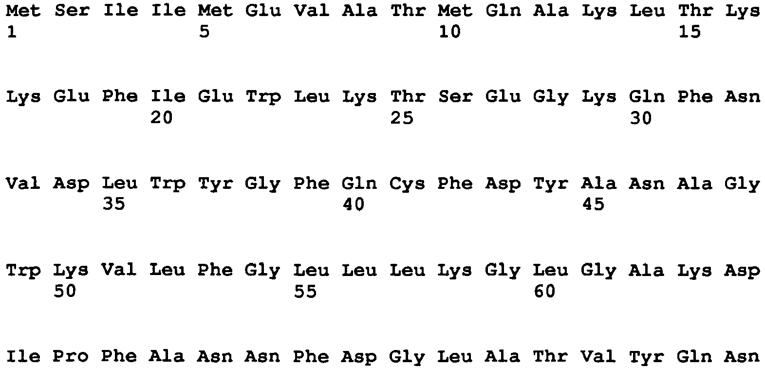


- <210> 3
<211> 496
<212> PRT
<213> Artificial - <220>
<223> Polypetide comprising substitution at E163, R167, E179, E189


- <210> 4
<211> 271
<212> PRT
<213> Artificial - <220>
<223> Polypetide comprising substitution at D214 - <400> 4


- <210> 5
<211> 271
<212> PRT
<213> Artificial - <220>
<223> Polypetide comprising substitution at R87 and D214 - <400> 5


- <210> 6
<211> 341
<212> PRT
<213> Artificial - <220>
<223> Polypeptide comprising substitution at E7 and E40 - <400> 6


- <210> 7
<211> 341
<212> PRT
<213> artificial - <220>
<223> Polypeptide comprising substitution at R92K, R221K - <400> 7


- <210> 8
<211> 341
<212> PRT
<213> artificial - <220>
<223> Polypeptide comprising substitution at R92A, R221A - <400> 8


- <210> 9
<211> 317
<212> PRT
<213> artificial - <220>
<223> Polypeptide comprising substitution at D112E - <400> 9

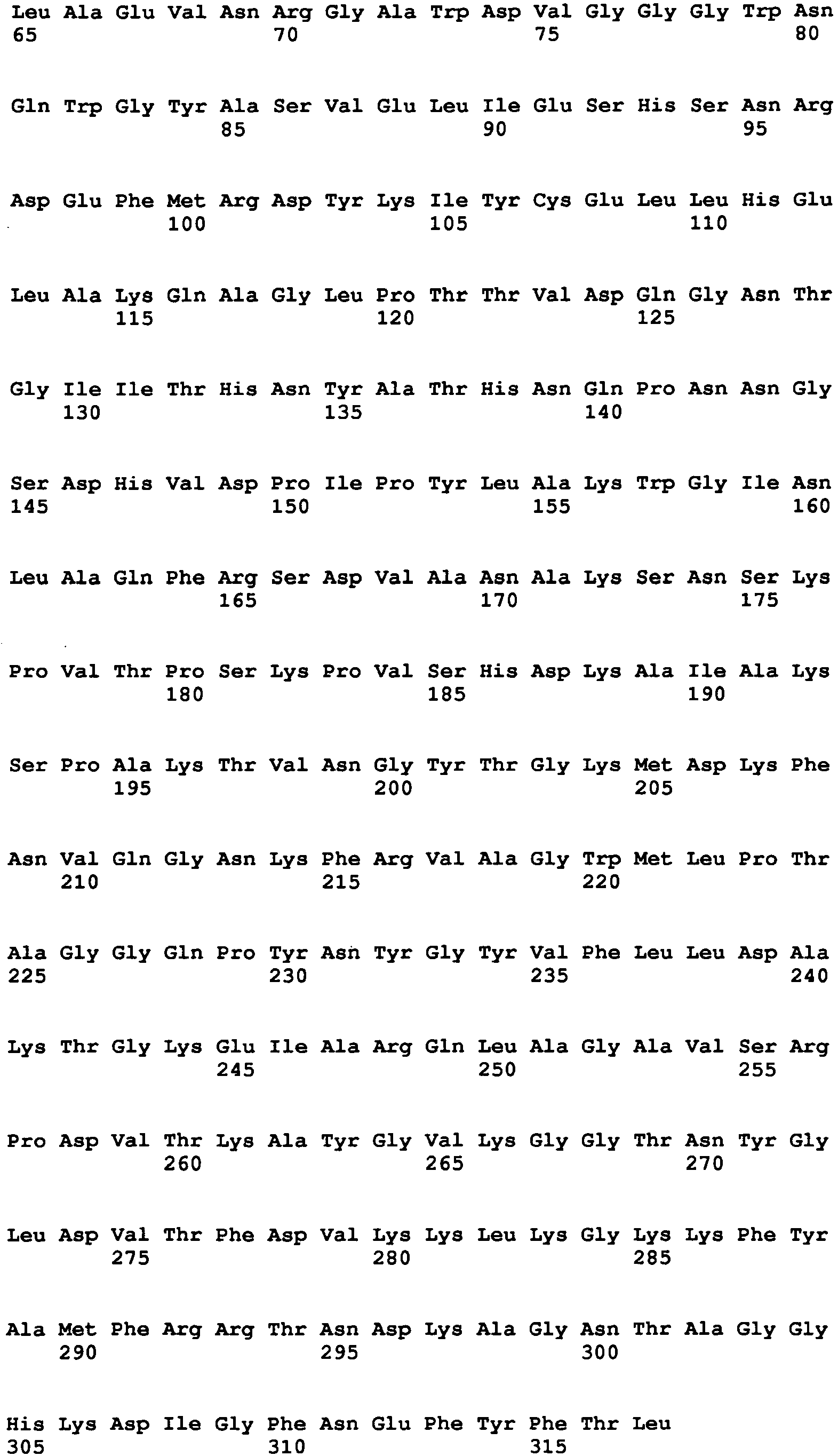
- <210> 10
<211> 317
<212> PRT
<213> artificial - <220>
<223> Polypeptide comprising substitution at D112A - <400> 10


- <210> 11
<211> 496
<212> PRT
<213> artificial - <220>
<223> Polypetide comprising substitution at E163, R167 - <400> 11


- <210> 12
<211> 497
<212> PRT
<213> artificial - <220>
<223> Polypeptide comprising substitutions at L56, L57, E164, R168, Y201, Q171, Q175, S384 - <400> 12


- <210> 13
<211> 497
<212> PRT
<213> artificial - <220>
<223> Polypeptide comprising substitutions at L56, L57, E164, R168, Y201, Q171, Q175, S384 - <400> 13


- <210> 14
<211> 497
<212> PRT
<213> artificial - <220>
<223> Polypeptide comprising substitution at R168 - <400> 14


- <210> 15
<211> 497
<212> PRT
<213> artificial - <220>
<223> Polypetide comprising substitution at E163 and R167 - <400> 15


- <210> 16
<211> 497
<212> PRT
<213> artificial - <220>
<223> Polypetide comprising substitution at E163, R167, E179, E189 - <400> 16
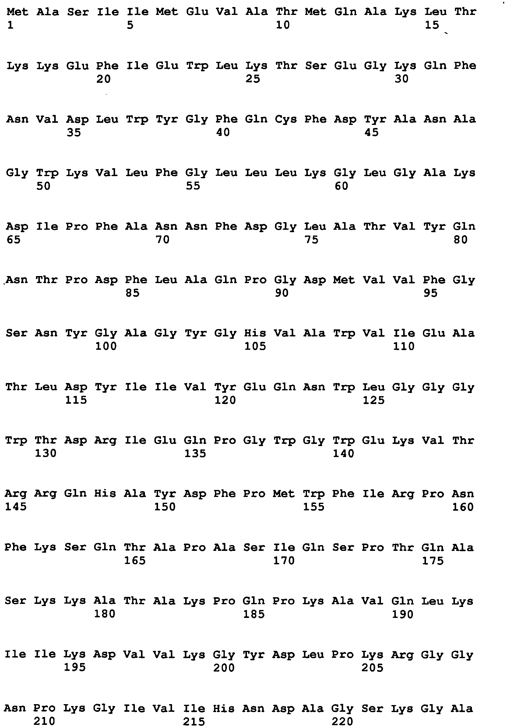


- <210> 17
<211> 497
<212> PRT
<213> artificial - <220>
<223> Polypetide comprising substitution at E163, R167 - <400> 17


- <210> 18
<211> 496
<212> PRT
<213> artificial - <220>
<223> CHAP-AMIpitti26-CBDUSA - <400> 18


- <210> 19
<211> 497
<212> PRT
<213> artificial - <220>
<223> CHAP-AMIpitti26-CBDUSA-Add2 - <400> 19


- <210> 20
<211> 393
<212> PRT
<213> artificial - <220>
<223> CHAP-AMIpitti26 - <400> 20


- <210> 21
<211> 341
<212> PRT
<213> Bacteriophage A511 - <400> 21


- <210> 22
<211> 497
<212> PRT
<213> artificial - <220>
<223> Polypeptide comprising substitutions at L56, L57, E164, R168, Y201 - <400> 22


Claims (8)
- Polypeptidvariante des Polypeptids gemäß SEQ ID NO: 1, wobei die Polypeptidvariante ausgewählt ist aus der Gruppe bestehend aus: SEQ ID NO: 2, 3, 11, 12 und 13.
- Nukleinsäuremolekül, umfassend eine Nukleotidsequenz codierend für ein Polypeptid nach Anspruch 1.
- Expressionsvektor, umfassend ein Nukleinsäuremolekül nach Anspruch 2.
- Wirtszelle enthaltend ein Nukleinsäuremolekül nach Anspruch 2 oder einen Expressionsvektor nach Anspruch 3.
- Polypeptid nach Anspruch 1 als Arzneimittel oder als Diagnostikum.
- Polypeptid nach Anspruch 1 zur Verwendung als Arzneimittel zur Prävention oder Therapie von Erkrankungen, die durch gram positive Bakterien insbesondere Clostridien, Bazillen, Listerien, Staphylokokken, Lactobazillen, Enterokokken, Aerokokken, Pediokokken, Streptokokken, Mycoplasmen und/oder Leuconostoc hervorgerufen werden.
- Verwendung des Polypeptids nach Anspruch 1 zur Unterdrückung des Wachstums von gram positiven Bakterien in der Umwelt-, Lebensmittel- oder Kosmetikindustrie.
- Polypeptid nach Anspruch 1 zur Verwendung als Diagnostikum in der Medizin, Umwelt-, Lebensmittel- oder Kosmetikindustrie.
Priority Applications (1)
| Application Number | Priority Date | Filing Date | Title |
|---|---|---|---|
| EP12176321.3A EP2511369B1 (de) | 2007-08-22 | 2008-08-19 | Proteasestabile Zellwand lysierende Enzyme |
Applications Claiming Priority (8)
| Application Number | Priority Date | Filing Date | Title |
|---|---|---|---|
| US95735107P | 2007-08-22 | 2007-08-22 | |
| EP07114785A EP2028271A1 (de) | 2007-08-22 | 2007-08-22 | Neue Proteine gegen Staphylokken-Infektionen in Menschen oder Tieren. |
| DE102007061929A DE102007061929A1 (de) | 2007-12-21 | 2007-12-21 | Proteasestabile Zellwand lysierende Enzyme |
| US3221108P | 2008-02-28 | 2008-02-28 | |
| EP08152096 | 2008-02-28 | ||
| DE102008023448 | 2008-05-14 | ||
| EP08801201A EP2188373B1 (de) | 2007-08-22 | 2008-08-19 | Proteasestabile zellwand lysierende enzyme |
| EP12176321.3A EP2511369B1 (de) | 2007-08-22 | 2008-08-19 | Proteasestabile Zellwand lysierende Enzyme |
Related Parent Applications (2)
| Application Number | Title | Priority Date | Filing Date |
|---|---|---|---|
| EP08801201.8 Division | 2008-08-19 | ||
| EP08801201A Division EP2188373B1 (de) | 2007-08-22 | 2008-08-19 | Proteasestabile zellwand lysierende enzyme |
Publications (2)
| Publication Number | Publication Date |
|---|---|
| EP2511369A1 EP2511369A1 (de) | 2012-10-17 |
| EP2511369B1 true EP2511369B1 (de) | 2013-10-23 |
Family
ID=40052086
Family Applications (2)
| Application Number | Title | Priority Date | Filing Date |
|---|---|---|---|
| EP12176321.3A Not-in-force EP2511369B1 (de) | 2007-08-22 | 2008-08-19 | Proteasestabile Zellwand lysierende Enzyme |
| EP08801201A Not-in-force EP2188373B1 (de) | 2007-08-22 | 2008-08-19 | Proteasestabile zellwand lysierende enzyme |
Family Applications After (1)
| Application Number | Title | Priority Date | Filing Date |
|---|---|---|---|
| EP08801201A Not-in-force EP2188373B1 (de) | 2007-08-22 | 2008-08-19 | Proteasestabile zellwand lysierende enzyme |
Country Status (5)
| Country | Link |
|---|---|
| US (2) | US8492519B2 (de) |
| EP (2) | EP2511369B1 (de) |
| DK (2) | DK2188373T3 (de) |
| ES (2) | ES2442667T3 (de) |
| WO (1) | WO2009024142A2 (de) |
Families Citing this family (8)
| Publication number | Priority date | Publication date | Assignee | Title |
|---|---|---|---|---|
| WO2009138475A1 (en) * | 2008-05-14 | 2009-11-19 | Profos Ag | Modified endolysin ply511 |
| US20090304673A1 (en) * | 2008-06-10 | 2009-12-10 | Bernd Buchberger | Method of Treatment of Inflammatory Diseases |
| US20120171188A1 (en) * | 2008-08-19 | 2012-07-05 | Hyglos Invest Gmbh | Artificial Peptidoglycan Lysing Enzymes and Peptidoglycan Binding Proteins |
| EP2157100A1 (de) * | 2008-08-19 | 2010-02-24 | Profos AG | Künstliche Peptidoglykan-lysierende Enzyme und Peptidoglykan-bindende Proteine |
| AU2012228407B2 (en) | 2011-03-11 | 2016-10-13 | Merz Pharma Gmbh & Co. Kgaa | Method for the determination of botulinum neurotoxin biological activity |
| US10383338B2 (en) | 2014-10-29 | 2019-08-20 | University Of Wyoming | Compositions and methods for making and using hydrolytic enzymes for degrading polysaccharides made by foodborne pathogenic bacteria |
| CN106811416B (zh) * | 2017-02-24 | 2020-06-12 | 中国农业科学院植物保护研究所 | 水解酶CwlC在芽胞杆菌母细胞裂解中的应用 |
| AU2024313255A1 (en) | 2023-06-20 | 2026-01-08 | Topaz Biosciences, Inc. | Recombinant cell wall hydrolases |
Family Cites Families (2)
| Publication number | Priority date | Publication date | Assignee | Title |
|---|---|---|---|---|
| US6432444B1 (en) | 1997-10-31 | 2002-08-13 | New Horizons Diagnostics Corp | Use of bacterial phage associated lysing enzymes for treating dermatological infections |
| EP1862080A1 (de) | 2006-05-31 | 2007-12-05 | Ajinomoto Co., Inc. | Durch Milchsäurebakterien hergestellte Bakteriocine mit Protease-Resistenz und ihre Verwendung bei Vieh |
-
2008
- 2008-08-19 ES ES12176321.3T patent/ES2442667T3/es active Active
- 2008-08-19 EP EP12176321.3A patent/EP2511369B1/de not_active Not-in-force
- 2008-08-19 WO PCT/DE2008/001378 patent/WO2009024142A2/de not_active Ceased
- 2008-08-19 US US12/674,369 patent/US8492519B2/en not_active Expired - Fee Related
- 2008-08-19 EP EP08801201A patent/EP2188373B1/de not_active Not-in-force
- 2008-08-19 ES ES08801201T patent/ES2402337T3/es active Active
- 2008-08-19 DK DK08801201.8T patent/DK2188373T3/da active
- 2008-08-19 DK DK12176321.3T patent/DK2511369T3/da active
-
2013
- 2013-07-18 US US13/945,606 patent/US20130295070A1/en not_active Abandoned
Also Published As
| Publication number | Publication date |
|---|---|
| DK2511369T3 (da) | 2014-01-27 |
| US20100216711A1 (en) | 2010-08-26 |
| DK2188373T3 (da) | 2013-04-08 |
| ES2402337T3 (es) | 2013-04-30 |
| WO2009024142A2 (de) | 2009-02-26 |
| US20130295070A1 (en) | 2013-11-07 |
| WO2009024142A3 (de) | 2009-06-18 |
| EP2188373B1 (de) | 2013-01-02 |
| EP2511369A1 (de) | 2012-10-17 |
| US8492519B2 (en) | 2013-07-23 |
| EP2188373A2 (de) | 2010-05-26 |
| ES2442667T3 (es) | 2014-02-12 |
Similar Documents
| Publication | Publication Date | Title |
|---|---|---|
| EP2188373B1 (de) | Proteasestabile zellwand lysierende enzyme | |
| James et al. | Purification and characterization of a novel antibacterial protein from the marine bacterium D2 | |
| Ibrahim et al. | Genetic evidence that antibacterial activity of lysozyme is independent of its catalytic function | |
| Baranger et al. | The antibacterial and antifungal properties of trappin‐2 (pre‐elafin) do not depend on its protease inhibitory function | |
| DE60036456T2 (de) | Antimikrobiell wirksames oder endotoxin neutralisierendes polypeptid | |
| EP2391636B1 (de) | Antibiotische peptide | |
| DE3850180T2 (de) | Bakteriozide und/oder bakteriostatische Peptide, Verfahren zu ihrer Isolierung, Herstellung und Verwendung. | |
| JP3459314B2 (ja) | 新規ペプチド、抗菌剤、新規ペプチド遺伝子、新規な組み換え体dna及び新規ペプチドの製造法 | |
| Cabrefiga et al. | Lysozyme enhances the bactericidal effect of BP100 peptide against Erwinia amylovora, the causal agent of fire blight of rosaceous plants | |
| Shekh et al. | Biochemical characterization of an anti-Candida factor produced by Enterococcus faecalis | |
| US8735097B2 (en) | Human antibiotic proteins | |
| KR102097128B1 (ko) | 황색포도상구균 제어에 효과적인 키메릭 엔도라이신 Lys109 | |
| Ortiz-Rodríguez et al. | Thurincin H is a nonhemolytic bacteriocin of Bacillus thuringiensis with potential for applied use | |
| EP2905288B1 (de) | Synthetische artifizielle Peptide mit antimikrobieller Wirkung | |
| Alahyaribeik et al. | Peptide recovery from chicken feather keratin and their anti-biofilm properties against methicillin-resistant Staphylococcus aureus (MRSA) | |
| DE102008002972A1 (de) | Neues Endolysin PlyP40 | |
| US9872893B2 (en) | Endolysins active against Staphylococcus bacteria, pharmaceutical compositions, and methods relating thereto | |
| DE10129983A1 (de) | Antimikrobiell wirkendes Peptid | |
| Fontoura et al. | Purification and characterization of an antimicrobial peptide produced by Pseudomonas sp. strain 4B | |
| DE69836500T2 (de) | Antifungale und antibakterielle peptide | |
| EP1068301A2 (de) | Bakterielle transglutaminasen | |
| DE102007061929A1 (de) | Proteasestabile Zellwand lysierende Enzyme | |
| KR101865782B1 (ko) | 바실러스 아밀로리퀴파시엔스 k14로부터 단리된 신규 항균 펩타이드 및 그의 용도 | |
| DE102007059891A1 (de) | C-terminale lfapsoriasinfragmente als antimikrobielle Peptide, deren Herstellung und Verwendung | |
| DE19905128A1 (de) | Humane antibiotische Proteine |
Legal Events
| Date | Code | Title | Description |
|---|---|---|---|
| PUAI | Public reference made under article 153(3) epc to a published international application that has entered the european phase |
Free format text: ORIGINAL CODE: 0009012 |
|
| AC | Divisional application: reference to earlier application |
Ref document number: 2188373 Country of ref document: EP Kind code of ref document: P |
|
| AK | Designated contracting states |
Kind code of ref document: A1 Designated state(s): AT BE BG CH CY CZ DE DK EE ES FI FR GB GR HR HU IE IS IT LI LT LU LV MC MT NL NO PL PT RO SE SI SK TR |
|
| RIN1 | Information on inventor provided before grant (corrected) |
Inventor name: FORCHHEIM, MICHAEL Inventor name: GRALLERT, HOLGER |
|
| 17P | Request for examination filed |
Effective date: 20130308 |
|
| GRAP | Despatch of communication of intention to grant a patent |
Free format text: ORIGINAL CODE: EPIDOSNIGR1 |
|
| INTG | Intention to grant announced |
Effective date: 20130606 |
|
| GRAS | Grant fee paid |
Free format text: ORIGINAL CODE: EPIDOSNIGR3 |
|
| GRAA | (expected) grant |
Free format text: ORIGINAL CODE: 0009210 |
|
| AC | Divisional application: reference to earlier application |
Ref document number: 2188373 Country of ref document: EP Kind code of ref document: P |
|
| AK | Designated contracting states |
Kind code of ref document: B1 Designated state(s): AT BE BG CH CY CZ DE DK EE ES FI FR GB GR HR HU IE IS IT LI LT LU LV MC MT NL NO PL PT RO SE SI SK TR |
|
| REG | Reference to a national code |
Ref country code: GB Ref legal event code: FG4D Free format text: NOT ENGLISH |
|
| REG | Reference to a national code |
Ref country code: CH Ref legal event code: EP |
|
| REG | Reference to a national code |
Ref country code: AT Ref legal event code: REF Ref document number: 637635 Country of ref document: AT Kind code of ref document: T Effective date: 20131115 |
|
| REG | Reference to a national code |
Ref country code: IE Ref legal event code: FG4D Free format text: LANGUAGE OF EP DOCUMENT: GERMAN |
|
| RAP2 | Party data changed (patent owner data changed or rights of a patent transferred) |
Owner name: HYGLOS INVEST GMBH |
|
| REG | Reference to a national code |
Ref country code: DE Ref legal event code: R096 Ref document number: 502008010869 Country of ref document: DE Effective date: 20131219 |
|
| REG | Reference to a national code |
Ref country code: CH Ref legal event code: NV Representative=s name: WAGNER + HELBIG PATENTANWAELTE, CH |
|
| REG | Reference to a national code |
Ref country code: DK Ref legal event code: T3 Effective date: 20140121 |
|
| REG | Reference to a national code |
Ref country code: NL Ref legal event code: T3 |
|
| REG | Reference to a national code |
Ref country code: SE Ref legal event code: TRGR |
|
| REG | Reference to a national code |
Ref country code: NL Ref legal event code: T3 |
|
| REG | Reference to a national code |
Ref country code: NL Ref legal event code: SD Effective date: 20140205 Ref country code: ES Ref legal event code: FG2A Ref document number: 2442667 Country of ref document: ES Kind code of ref document: T3 Effective date: 20140212 |
|
| REG | Reference to a national code |
Ref country code: LT Ref legal event code: MG4D |
|
| REG | Reference to a national code |
Ref country code: GB Ref legal event code: 732E Free format text: REGISTERED BETWEEN 20140327 AND 20140402 |
|
| PG25 | Lapsed in a contracting state [announced via postgrant information from national office to epo] |
Ref country code: IS Free format text: LAPSE BECAUSE OF FAILURE TO SUBMIT A TRANSLATION OF THE DESCRIPTION OR TO PAY THE FEE WITHIN THE PRESCRIBED TIME-LIMIT Effective date: 20140223 Ref country code: NO Free format text: LAPSE BECAUSE OF FAILURE TO SUBMIT A TRANSLATION OF THE DESCRIPTION OR TO PAY THE FEE WITHIN THE PRESCRIBED TIME-LIMIT Effective date: 20140123 Ref country code: HR Free format text: LAPSE BECAUSE OF FAILURE TO SUBMIT A TRANSLATION OF THE DESCRIPTION OR TO PAY THE FEE WITHIN THE PRESCRIBED TIME-LIMIT Effective date: 20131023 Ref country code: FI Free format text: LAPSE BECAUSE OF FAILURE TO SUBMIT A TRANSLATION OF THE DESCRIPTION OR TO PAY THE FEE WITHIN THE PRESCRIBED TIME-LIMIT Effective date: 20131023 Ref country code: LT Free format text: LAPSE BECAUSE OF FAILURE TO SUBMIT A TRANSLATION OF THE DESCRIPTION OR TO PAY THE FEE WITHIN THE PRESCRIBED TIME-LIMIT Effective date: 20131023 |
|
| PG25 | Lapsed in a contracting state [announced via postgrant information from national office to epo] |
Ref country code: CY Free format text: LAPSE BECAUSE OF FAILURE TO SUBMIT A TRANSLATION OF THE DESCRIPTION OR TO PAY THE FEE WITHIN THE PRESCRIBED TIME-LIMIT Effective date: 20131023 Ref country code: LV Free format text: LAPSE BECAUSE OF FAILURE TO SUBMIT A TRANSLATION OF THE DESCRIPTION OR TO PAY THE FEE WITHIN THE PRESCRIBED TIME-LIMIT Effective date: 20131023 |
|
| PG25 | Lapsed in a contracting state [announced via postgrant information from national office to epo] |
Ref country code: PT Free format text: LAPSE BECAUSE OF FAILURE TO SUBMIT A TRANSLATION OF THE DESCRIPTION OR TO PAY THE FEE WITHIN THE PRESCRIBED TIME-LIMIT Effective date: 20140224 |
|
| REG | Reference to a national code |
Ref country code: DE Ref legal event code: R097 Ref document number: 502008010869 Country of ref document: DE |
|
| PG25 | Lapsed in a contracting state [announced via postgrant information from national office to epo] |
Ref country code: EE Free format text: LAPSE BECAUSE OF FAILURE TO SUBMIT A TRANSLATION OF THE DESCRIPTION OR TO PAY THE FEE WITHIN THE PRESCRIBED TIME-LIMIT Effective date: 20131023 |
|
| REG | Reference to a national code |
Ref country code: DE Ref legal event code: R082 Ref document number: 502008010869 Country of ref document: DE Representative=s name: DEHMEL & BETTENHAUSEN PATENT- UND RECHTSANWAEL, DE Effective date: 20140710 Ref country code: DE Ref legal event code: R081 Ref document number: 502008010869 Country of ref document: DE Owner name: HYGLOS INVEST GMBH, DE Free format text: FORMER OWNER: BIOMERIEUX S.A., HYGLOS INVEST GMBH, , FR Effective date: 20140710 Ref country code: DE Ref legal event code: R082 Ref document number: 502008010869 Country of ref document: DE Representative=s name: DEHMEL & BETTENHAUSEN PATENTANWAELTE PARTMBB, DE Effective date: 20140710 Ref country code: DE Ref legal event code: R081 Ref document number: 502008010869 Country of ref document: DE Owner name: HYGLOS INVEST GMBH, DE Free format text: FORMER OWNERS: BIOMERIEUX S.A., MARCY L'ETOILE, FR; HYGLOS INVEST GMBH, 82347 BERNRIED, DE Effective date: 20140710 |
|
| PG25 | Lapsed in a contracting state [announced via postgrant information from national office to epo] |
Ref country code: SK Free format text: LAPSE BECAUSE OF FAILURE TO SUBMIT A TRANSLATION OF THE DESCRIPTION OR TO PAY THE FEE WITHIN THE PRESCRIBED TIME-LIMIT Effective date: 20131023 Ref country code: RO Free format text: LAPSE BECAUSE OF FAILURE TO SUBMIT A TRANSLATION OF THE DESCRIPTION OR TO PAY THE FEE WITHIN THE PRESCRIBED TIME-LIMIT Effective date: 20131023 Ref country code: CZ Free format text: LAPSE BECAUSE OF FAILURE TO SUBMIT A TRANSLATION OF THE DESCRIPTION OR TO PAY THE FEE WITHIN THE PRESCRIBED TIME-LIMIT Effective date: 20131023 Ref country code: PL Free format text: LAPSE BECAUSE OF FAILURE TO SUBMIT A TRANSLATION OF THE DESCRIPTION OR TO PAY THE FEE WITHIN THE PRESCRIBED TIME-LIMIT Effective date: 20131023 |
|
| PLBE | No opposition filed within time limit |
Free format text: ORIGINAL CODE: 0009261 |
|
| STAA | Information on the status of an ep patent application or granted ep patent |
Free format text: STATUS: NO OPPOSITION FILED WITHIN TIME LIMIT |
|
| 26N | No opposition filed |
Effective date: 20140724 |
|
| REG | Reference to a national code |
Ref country code: DE Ref legal event code: R097 Ref document number: 502008010869 Country of ref document: DE Effective date: 20140724 |
|
| PG25 | Lapsed in a contracting state [announced via postgrant information from national office to epo] |
Ref country code: SI Free format text: LAPSE BECAUSE OF FAILURE TO SUBMIT A TRANSLATION OF THE DESCRIPTION OR TO PAY THE FEE WITHIN THE PRESCRIBED TIME-LIMIT Effective date: 20131023 |
|
| REG | Reference to a national code |
Ref country code: CH Ref legal event code: PCAR Free format text: NEW ADDRESS: 48, ROUTE DE DIVONNE CASE POSTALE 2666, 1260 NYON (CH) |
|
| PG25 | Lapsed in a contracting state [announced via postgrant information from national office to epo] |
Ref country code: MC Free format text: LAPSE BECAUSE OF FAILURE TO SUBMIT A TRANSLATION OF THE DESCRIPTION OR TO PAY THE FEE WITHIN THE PRESCRIBED TIME-LIMIT Effective date: 20131023 Ref country code: LU Free format text: LAPSE BECAUSE OF FAILURE TO SUBMIT A TRANSLATION OF THE DESCRIPTION OR TO PAY THE FEE WITHIN THE PRESCRIBED TIME-LIMIT Effective date: 20140819 |
|
| PG25 | Lapsed in a contracting state [announced via postgrant information from national office to epo] |
Ref country code: BG Free format text: LAPSE BECAUSE OF FAILURE TO SUBMIT A TRANSLATION OF THE DESCRIPTION OR TO PAY THE FEE WITHIN THE PRESCRIBED TIME-LIMIT Effective date: 20131023 |
|
| PG25 | Lapsed in a contracting state [announced via postgrant information from national office to epo] |
Ref country code: MT Free format text: LAPSE BECAUSE OF FAILURE TO SUBMIT A TRANSLATION OF THE DESCRIPTION OR TO PAY THE FEE WITHIN THE PRESCRIBED TIME-LIMIT Effective date: 20131023 Ref country code: GR Free format text: LAPSE BECAUSE OF FAILURE TO SUBMIT A TRANSLATION OF THE DESCRIPTION OR TO PAY THE FEE WITHIN THE PRESCRIBED TIME-LIMIT Effective date: 20140124 |
|
| PG25 | Lapsed in a contracting state [announced via postgrant information from national office to epo] |
Ref country code: TR Free format text: LAPSE BECAUSE OF FAILURE TO SUBMIT A TRANSLATION OF THE DESCRIPTION OR TO PAY THE FEE WITHIN THE PRESCRIBED TIME-LIMIT Effective date: 20131023 Ref country code: HU Free format text: LAPSE BECAUSE OF FAILURE TO SUBMIT A TRANSLATION OF THE DESCRIPTION OR TO PAY THE FEE WITHIN THE PRESCRIBED TIME-LIMIT; INVALID AB INITIO Effective date: 20080819 |
|
| REG | Reference to a national code |
Ref country code: FR Ref legal event code: PLFP Year of fee payment: 9 |
|
| REG | Reference to a national code |
Ref country code: AT Ref legal event code: PC Ref document number: 637635 Country of ref document: AT Kind code of ref document: T Owner name: HYGLOS INVEST GMBH, DE Effective date: 20170331 |
|
| REG | Reference to a national code |
Ref country code: FR Ref legal event code: PLFP Year of fee payment: 10 |
|
| REG | Reference to a national code |
Ref country code: FR Ref legal event code: PLFP Year of fee payment: 11 |
|
| REG | Reference to a national code |
Ref country code: DE Ref legal event code: R082 Ref document number: 502008010869 Country of ref document: DE Representative=s name: GRUENECKER PATENT- UND RECHTSANWAELTE PARTG MB, DE |
|
| REG | Reference to a national code |
Ref country code: DE Ref legal event code: R082 Ref document number: 502008010869 Country of ref document: DE Representative=s name: GRUENECKER PATENT- UND RECHTSANWAELTE PARTG MB, DE Ref country code: DE Ref legal event code: R081 Ref document number: 502008010869 Country of ref document: DE Owner name: HYPHARM GMBH, DE Free format text: FORMER OWNER: HYGLOS INVEST GMBH, 82347 BERNRIED, DE |
|
| REG | Reference to a national code |
Ref country code: NL Ref legal event code: PD Owner name: HYPHARM GMBH; DE Free format text: DETAILS ASSIGNMENT: CHANGE OF OWNER(S), ASSIGNMENT; FORMER OWNER NAME: HYGLOS INVEST GMBH Effective date: 20190715 |
|
| REG | Reference to a national code |
Ref country code: BE Ref legal event code: PD Owner name: HYPHARM GMBH; DE Free format text: DETAILS ASSIGNMENT: CHANGE OF OWNER(S), CESSION; FORMER OWNER NAME: HYGLOS INVEST GMBH Effective date: 20190701 |
|
| REG | Reference to a national code |
Ref country code: GB Ref legal event code: 732E Free format text: REGISTERED BETWEEN 20191003 AND 20191009 |
|
| REG | Reference to a national code |
Ref country code: AT Ref legal event code: PC Ref document number: 637635 Country of ref document: AT Kind code of ref document: T Owner name: HYPHARM GMBH, DE Effective date: 20191011 |
|
| REG | Reference to a national code |
Ref country code: CH Ref legal event code: PUE Owner name: HYPHARM GMBH, DE Free format text: FORMER OWNER: HYGLOS INVEST GMBH, DE |
|
| REG | Reference to a national code |
Ref country code: CH Ref legal event code: NV Representative=s name: WAGNER + HELBIG PATENTANWAELTE CONSEILS EN PRO, CH |
|
| PGFP | Annual fee paid to national office [announced via postgrant information from national office to epo] |
Ref country code: NL Payment date: 20200826 Year of fee payment: 13 |
|
| PGFP | Annual fee paid to national office [announced via postgrant information from national office to epo] |
Ref country code: IE Payment date: 20200821 Year of fee payment: 13 Ref country code: FR Payment date: 20200826 Year of fee payment: 13 Ref country code: GB Payment date: 20200826 Year of fee payment: 13 Ref country code: DK Payment date: 20200820 Year of fee payment: 13 Ref country code: ES Payment date: 20200902 Year of fee payment: 13 Ref country code: DE Payment date: 20200827 Year of fee payment: 13 |
|
| PGFP | Annual fee paid to national office [announced via postgrant information from national office to epo] |
Ref country code: IT Payment date: 20200831 Year of fee payment: 13 Ref country code: BE Payment date: 20200825 Year of fee payment: 13 Ref country code: AT Payment date: 20200903 Year of fee payment: 13 Ref country code: SE Payment date: 20200826 Year of fee payment: 13 Ref country code: CH Payment date: 20200826 Year of fee payment: 13 |
|
| REG | Reference to a national code |
Ref country code: DE Ref legal event code: R119 Ref document number: 502008010869 Country of ref document: DE |
|
| REG | Reference to a national code |
Ref country code: DK Ref legal event code: EBP Effective date: 20210831 |
|
| REG | Reference to a national code |
Ref country code: SE Ref legal event code: EUG |
|
| REG | Reference to a national code |
Ref country code: CH Ref legal event code: PL |
|
| REG | Reference to a national code |
Ref country code: NL Ref legal event code: MM Effective date: 20210901 |
|
| REG | Reference to a national code |
Ref country code: AT Ref legal event code: MM01 Ref document number: 637635 Country of ref document: AT Kind code of ref document: T Effective date: 20210819 |
|
| REG | Reference to a national code |
Ref country code: BE Ref legal event code: MM Effective date: 20210831 |
|
| GBPC | Gb: european patent ceased through non-payment of renewal fee |
Effective date: 20210819 |
|
| PG25 | Lapsed in a contracting state [announced via postgrant information from national office to epo] |
Ref country code: LI Free format text: LAPSE BECAUSE OF NON-PAYMENT OF DUE FEES Effective date: 20210831 Ref country code: CH Free format text: LAPSE BECAUSE OF NON-PAYMENT OF DUE FEES Effective date: 20210831 Ref country code: AT Free format text: LAPSE BECAUSE OF NON-PAYMENT OF DUE FEES Effective date: 20210819 |
|
| PG25 | Lapsed in a contracting state [announced via postgrant information from national office to epo] |
Ref country code: SE Free format text: LAPSE BECAUSE OF NON-PAYMENT OF DUE FEES Effective date: 20210820 |
|
| PG25 | Lapsed in a contracting state [announced via postgrant information from national office to epo] |
Ref country code: NL Free format text: LAPSE BECAUSE OF NON-PAYMENT OF DUE FEES Effective date: 20210901 |
|
| PG25 | Lapsed in a contracting state [announced via postgrant information from national office to epo] |
Ref country code: IT Free format text: LAPSE BECAUSE OF NON-PAYMENT OF DUE FEES Effective date: 20210819 Ref country code: IE Free format text: LAPSE BECAUSE OF NON-PAYMENT OF DUE FEES Effective date: 20210819 Ref country code: GB Free format text: LAPSE BECAUSE OF NON-PAYMENT OF DUE FEES Effective date: 20210819 Ref country code: FR Free format text: LAPSE BECAUSE OF NON-PAYMENT OF DUE FEES Effective date: 20210831 Ref country code: DK Free format text: LAPSE BECAUSE OF NON-PAYMENT OF DUE FEES Effective date: 20210831 Ref country code: DE Free format text: LAPSE BECAUSE OF NON-PAYMENT OF DUE FEES Effective date: 20220301 Ref country code: BE Free format text: LAPSE BECAUSE OF NON-PAYMENT OF DUE FEES Effective date: 20210831 |
|
| REG | Reference to a national code |
Ref country code: ES Ref legal event code: FD2A Effective date: 20221027 |
|
| PG25 | Lapsed in a contracting state [announced via postgrant information from national office to epo] |
Ref country code: ES Free format text: LAPSE BECAUSE OF NON-PAYMENT OF DUE FEES Effective date: 20210820 |