EP2510878A1 - Procédé de génération d'un enregistrement de tomographie volumétrique numérique tridimensionnelle radiologique d'une partie du corps d'un patient - Google Patents
Procédé de génération d'un enregistrement de tomographie volumétrique numérique tridimensionnelle radiologique d'une partie du corps d'un patient Download PDFInfo
- Publication number
- EP2510878A1 EP2510878A1 EP11162043A EP11162043A EP2510878A1 EP 2510878 A1 EP2510878 A1 EP 2510878A1 EP 11162043 A EP11162043 A EP 11162043A EP 11162043 A EP11162043 A EP 11162043A EP 2510878 A1 EP2510878 A1 EP 2510878A1
- Authority
- EP
- European Patent Office
- Prior art keywords
- body part
- marker
- generating
- patient
- marker elements
- Prior art date
- Legal status (The legal status is an assumption and is not a legal conclusion. Google has not performed a legal analysis and makes no representation as to the accuracy of the status listed.)
- Granted
Links
- 238000000034 method Methods 0.000 title claims abstract description 25
- 238000011983 digital volume tomography Methods 0.000 title claims abstract description 16
- 239000003550 marker Substances 0.000 claims abstract description 80
- 210000001847 jaw Anatomy 0.000 claims description 18
- 210000002455 dental arch Anatomy 0.000 claims description 8
- 239000004053 dental implant Substances 0.000 description 3
- 238000001228 spectrum Methods 0.000 description 3
- 210000000988 bone and bone Anatomy 0.000 description 2
- 238000003384 imaging method Methods 0.000 description 2
- 239000007943 implant Substances 0.000 description 2
- 230000005855 radiation Effects 0.000 description 2
- 210000001519 tissue Anatomy 0.000 description 2
- 239000000969 carrier Substances 0.000 description 1
- 230000000295 complement effect Effects 0.000 description 1
- 238000002059 diagnostic imaging Methods 0.000 description 1
- 230000004069 differentiation Effects 0.000 description 1
- 238000003908 quality control method Methods 0.000 description 1
- 238000003325 tomography Methods 0.000 description 1
Images
Classifications
-
- A—HUMAN NECESSITIES
- A61—MEDICAL OR VETERINARY SCIENCE; HYGIENE
- A61B—DIAGNOSIS; SURGERY; IDENTIFICATION
- A61B6/00—Apparatus for radiation diagnosis, e.g. combined with radiation therapy equipment
- A61B6/46—Apparatus for radiation diagnosis, e.g. combined with radiation therapy equipment with special arrangements for interfacing with the operator or the patient
- A61B6/461—Displaying means of special interest
- A61B6/466—Displaying means of special interest adapted to display 3D data
-
- A—HUMAN NECESSITIES
- A61—MEDICAL OR VETERINARY SCIENCE; HYGIENE
- A61B—DIAGNOSIS; SURGERY; IDENTIFICATION
- A61B6/00—Apparatus for radiation diagnosis, e.g. combined with radiation therapy equipment
- A61B6/12—Devices for detecting or locating foreign bodies
-
- A61B6/51—
-
- A—HUMAN NECESSITIES
- A61—MEDICAL OR VETERINARY SCIENCE; HYGIENE
- A61B—DIAGNOSIS; SURGERY; IDENTIFICATION
- A61B6/00—Apparatus for radiation diagnosis, e.g. combined with radiation therapy equipment
- A61B6/58—Testing, adjusting or calibrating apparatus or devices for radiation diagnosis
- A61B6/582—Calibration
- A61B6/583—Calibration using calibration phantoms
-
- G—PHYSICS
- G06—COMPUTING; CALCULATING OR COUNTING
- G06T—IMAGE DATA PROCESSING OR GENERATION, IN GENERAL
- G06T11/00—2D [Two Dimensional] image generation
- G06T11/003—Reconstruction from projections, e.g. tomography
- G06T11/008—Specific post-processing after tomographic reconstruction, e.g. voxelisation, metal artifact correction
-
- G—PHYSICS
- G06—COMPUTING; CALCULATING OR COUNTING
- G06T—IMAGE DATA PROCESSING OR GENERATION, IN GENERAL
- G06T3/00—Geometric image transformation in the plane of the image
- G06T3/40—Scaling the whole image or part thereof
Definitions
- the invention relates to a method for generating a radiological three-dimensional digital volume tomography image of a patient's body part, in particular a patient's head, and in this case particularly preferably a patient's jaw.
- digital volume tomography is often used.
- a cone-shaped x-ray beam is directed at a two-dimensional detector which is opposed at a constant distance.
- the assembly consisting of the X-ray source and the detector, is moved around the patient's head during the recording, taking 10-30 seconds to record.
- Digital volume tomography has the advantage of relatively low radiation exposure to the patient compared to other three-dimensional radiographic imaging techniques.
- the quality of the resulting three-dimensional images of the patient's head is highly variable and difficult to assess.
- the reasons for the fluctuating image quality are on the one hand inherent in principle and physical nature, and are on the other hand in the long recording duration, because of unwanted intrinsic movements of the patient's head, the X-ray source and the detector during the recording can not be excluded. Due to the proper movements, the quality of the images is considerably worsened.
- the size relationships and relationships are often represented with an error of up to several millimeters. Although this is not always problematic in diagnostics, digital volume tomography becomes particularly used in dentistry for the planning of dental implants in the patient's jaw. In this case, a high geometric accuracy is required in order to ensure a high quality of the implant supply can.
- the object of the invention is in contrast to provide a method for generating a radiological three-dimensional recording, in particular a digital volume tomography recording of a patient's body part with the possibility of quality control and / or improved accuracy.
- a label carrier is fixed to the patient's body part, and preferably in the patient's mouth.
- the marker carrier can have an individual molded part shaped from the patient's jaw, with which the marker carrier can be placed on the patient's jaw in a form-fitting and immovable manner. The fixation can be done by the bite of the patient.
- the marker carrier has a non-radiopaque carrier body which absorbs virtually no X-ray radiation and is therefore largely invisible to an X-ray machine.
- a plurality of radio-opaque marker elements are fixed, the actual size S and / or actual distance D is known to each other.
- a recording of the relevant body part is made with the digital volume tomography device to produce a three-dimensional raw image of the body part, as is already known from the prior art.
- the digital volume tomography device to produce a three-dimensional raw image of the body part, as is already known from the prior art.
- their size S 'and / or the distances D' of the marker elements are measured relative to one another in the raw image.
- the measured values for the marker element size S 'or the marker element distances D' are compared with the respective actual values S, D, and from this a matching value QS, QD is generated.
- this adjustment value QS, QD can be the quotient of the respective actual value S, D and the measured values S ', D'.
- the raw image is corrected on the basis of the obtained matching values QS, QD.
- the raw image can be normalized, for example, by being stretched or compressed by the value of the quotient of the actual value and the measured value.
- the thus obtained corrected three-dimensional image of the body part or the patient's jaw is considerably more accurate than the raw image, in particular with regard to the size ratios and distances.
- an exact three-dimensional image of the patient's jaw is made available in this way, which forms the basis for the subsequent precise planning and realization of the implantological dental restoration.
- the correction is a size correction of the raw image based on the raw image distances D 'in relation to the actual distances D of the marker elements to each other. From the quotient of the raw image distances D 'and the actual distances D of the marker elements to one another, a correction value can be generated directly by which the raw image is to be corrected.
- a quality indicator can be output whose value results from the deviation of the raw image size S 'in relation to the actual size S of the marker elements.
- a quotient can also be used here.
- the size-quality indicator provides information about the quality of the raw image at the location of the respective marker element. In case of blurring or blurring in the raw image, the marker element in the raw image, for example, represented considerably larger than it actually is. Although no immediate correction of the raw image can be performed with the help of the quality indicator, it does give the user
- the raw image provides information about the extent to which the user can trust the raw image.
- each marker element being arranged to its neighboring marker elements in each case at a constant actual distance of at most 3.0 cm.
- the mesh size over the entire network is always the same, and the mesh size is at most 3.0 cm.
- the marker elements can each be distributed flat or in space and arranged on perpendicular lines to each other. However, three adjacent marker elements particularly preferably form an equilateral triangle. With this arrangement, with relatively few marker elements, which in principle can also be the source of artifacts because of their radio opacity, a network with a sufficient density of marker elements in the relevant space can be spanned.
- the marker elements are arranged in at least two transverse planes, ie in two horizontal planes. This ensures that the size correction of the raw image on the basis of the distances D, D 'of the marker elements to each other can be made with respect to all three spatial axes.
- the radioopacity of the marker elements is different. Since the marker elements also have reflexive properties due to their radio-opacity with respect to the X-ray radiation, artifacts in these areas can be reduced or avoided, for example, by reducing the radioopacity of the marker elements in particularly sensitive areas.
- the radioopotential values of the marker elements may have three to six different values, so that approximately the entire radioopotential spectrum of the part of the body to be photographed is covered.
- This provides an objective measure of radio opacity in the raw image.
- the raw image can be automatically calibrated, i.
- the structures of the body part depicted in the raw image can be classified objectively with respect to their radioopacity. This facilitates the interpretation of the three-dimensional image.
- bone and tissue can be distinguished easier and safer from each other, which is of essential importance in dental implantology.
- the marker carrier can have a plurality of radioopacity spectra distributed over the body part or the image space, each consisting of a plurality of marker elements of different radio opacity, so that the calibration can be carried out locally. This can take into account the fact that the imaging quality in the digital volume tomography principle can turn out locally very different.
- the label carrier is designed as an intraoral marker carrier, and a plurality of marker elements are arranged within the dental arch.
- a plurality of marker elements are arranged within the dental arch.
- at least one marker element is arranged outside the dental arch, and particularly preferably outside the patient's mouth.
- the arrangement outside the dental arch or the patient's mouth allows the generation of adjustment values, for example in the region of the jawbone of the patient's jaw. Especially in this area, high accuracy is desirable for implant planning.
- the embodiments illustrated in the figures relate to a dental application, wherein the body part in the present case is a patient's jaw.
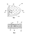
- an intraoral marker carrier 10 is shown, as it is suitable for generating a radiological three-dimensional recording with a digital volume tomography device.
- the marker carrier 10 consists of a radiologically invisible support body 12 made of a suitable plastic.
- On the support body 12, an individual molded part 16 is attached, for example glued, the tooth recesses 18 which are complementary to the patient teeth 24 formed.
- the individual molded part 16 which represents an impression of the respective patient's jaw 22, 23, a firm, immovable and defined seat of the marker carrier 10 on the patient's jaw 22, 23 in the patient's mouth is ensured.
- marker elements 14 lying in two planes and evenly distributed. Three mutually adjacent marker elements 14 each form an equilateral triangle, and are each spaced with a same distance for all marker elements 14 actual distance D, which is not greater than 3.0 cm. All marker elements 14 have an identical size S, which corresponds to the diameter, for example, in the case of spherical marker elements 14. The size S is a maximum of a few millimeters.
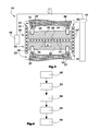
- a second embodiment of a more complex marker carrier 20 is shown, which is inserted into the patient's mouth.
- a rotatable about a vertical axis rotor 44 of a digital volume tomography device which carries an X-ray source 40 which generates a cone-shaped X-ray beam, and opposite a two-dimensional detector 42.
- the marker carrier 20 is H-shaped in cross-section and has a horizontal carrier body part 26 and on its outer circumference each upwardly and downwardly projecting vertical carrier body parts 30,32.
- the marker elements 14 are distributed in a planar manner.
- an individual shaped part 28, 29 is respectively fixed, which is shaped by the upper jaw 22 and the lower jaw 23.
- the arrangement of the marker elements 14 corresponds in principle to that in the marker carrier 10 of Figures 1 and 2 ,
- the label carriers 14 in the horizontal carrier body portion 26 are disposed within the dental arches 36, 37 spanned by the patient teeth 24 of the patient upper jaw 22 and lower jaw 23.
- the marker elements 14 'in the vertical support body parts 30, 32 are arranged outside the dental arches 36, 37 but within the patient's mouth.
- a recording of the patient's jaw for generating a three-dimensional raw image of the patient's head or jaw is made in the next method step 52 with the digital volume tomography device.
- a further method step 54 in FIG Measure the raw image with respect to all marker elements of their size S 'and the distances D' of the marker elements to each other.
- the measured values for the marker element distances D ' are compared with the known actual value D, or a quotient is generated therefrom.
- a correction value QD is generated directly in order to locally correct the raw image in a subsequent method step 58.
- the correction is in each case a size correction of the raw image on the basis of the raw image distances D 'in relation to the actual stocks D of the marker elements to each other.
- the thus obtained three-dimensional corrected image of the patient's head or the patient's jaw is considerably more accurate, in particular with regard to the size ratios and distances, than the raw image.
- an exact three-dimensional image of the patient's jaw is made available in this way, which forms the basis for the subsequent precise planning and realization of the implantological dental restoration.
- the size-quality indicator QS provides information about the quality of the raw image at the location concerned, and in particular provides information on the extent to which the raw image or the corrected raw image can be trusted.
- the radioopacity of the marker elements 14,14 ' is different.
- a spectrum of three to six mutually adjacent marker elements 14 'of different radio opacity can be provided on the vertical support body part 30 at four positions of the dental arch. These are used in the raw image, the raw image in terms of its brightness and its contrast each to be calibrated locally. This calibration can be quite different in the four areas. The resulting image significantly facilitates the differentiation of bone and tissue.
Priority Applications (2)
| Application Number | Priority Date | Filing Date | Title |
|---|---|---|---|
| EP11162043.1A EP2510878B1 (fr) | 2011-04-12 | 2011-04-12 | Procédé de génération d'un enregistrement de tomographie volumétrique numérique tridimensionnelle radiologique d'une partie du corps d'un patient |
| US13/432,355 US8831322B2 (en) | 2011-04-12 | 2012-03-28 | Method of generating a three-dimensional digital radiological volume topography recording of a patient's body part |
Applications Claiming Priority (1)
| Application Number | Priority Date | Filing Date | Title |
|---|---|---|---|
| EP11162043.1A EP2510878B1 (fr) | 2011-04-12 | 2011-04-12 | Procédé de génération d'un enregistrement de tomographie volumétrique numérique tridimensionnelle radiologique d'une partie du corps d'un patient |
Publications (2)
| Publication Number | Publication Date |
|---|---|
| EP2510878A1 true EP2510878A1 (fr) | 2012-10-17 |
| EP2510878B1 EP2510878B1 (fr) | 2014-02-26 |
Family
ID=44454675
Family Applications (1)
| Application Number | Title | Priority Date | Filing Date |
|---|---|---|---|
| EP11162043.1A Active EP2510878B1 (fr) | 2011-04-12 | 2011-04-12 | Procédé de génération d'un enregistrement de tomographie volumétrique numérique tridimensionnelle radiologique d'une partie du corps d'un patient |
Country Status (2)
| Country | Link |
|---|---|
| US (1) | US8831322B2 (fr) |
| EP (1) | EP2510878B1 (fr) |
Families Citing this family (12)
| Publication number | Priority date | Publication date | Assignee | Title |
|---|---|---|---|---|
| US10568720B2 (en) | 2012-01-10 | 2020-02-25 | Estetic Implant Solutions, LLC | Dental implants with markers for determining three-dimensional positioning |
| US10595970B2 (en) | 2012-01-10 | 2020-03-24 | Esthetic Implant Solutions, Llc | Bonding of soft gingival tissues with anatomical and other dental prostheses |
| US11253345B2 (en) | 2012-01-10 | 2022-02-22 | Esthetic Implant Solutions, Llc | Methods for integrating scans including 3D cone beam scan for positioning of implant and fabrication of dental prosthesis |
| US10016260B2 (en) | 2012-01-10 | 2018-07-10 | Mark H. Blaisdell | Anatomical healing abutments, kits, and methods |
| US9895209B2 (en) | 2012-01-10 | 2018-02-20 | Mark H. Blaisdell | Casting jig including elongate handle for chair-side manufacture of customizable sculptable anatomical healing caps, and method for forming bis-acrylic crown |
| US10709525B2 (en) | 2012-01-10 | 2020-07-14 | Esthetic Implant Solutions, Llc | Methods for taking an oral scan without requiring removal of a temporary healing abutment |
| US10507081B2 (en) | 2012-01-10 | 2019-12-17 | Esthetic Implant Solutions, Llc | Methods for taking an impression or scanning without requiring removal of a temporary healing abutment |
| EP3386432A4 (fr) * | 2015-12-11 | 2019-09-04 | Timothy Hillukka | Détermination de mouvements de mâchoire et de mouvements faciaux |
| US11301964B2 (en) * | 2016-03-29 | 2022-04-12 | Sony Corporation | Image processing apparatus, image processing method, and medical system to correct blurring without removing a screen motion caused by a biological body motion |
| US11559379B2 (en) | 2018-04-12 | 2023-01-24 | Esthetic Implant Solutions, Llc | Dental implants with markers for determining three-dimensional positioning |
| JP2022526086A (ja) * | 2019-03-12 | 2022-05-23 | ケアストリーム デンタル エルエルシー | 口腔内トモシンセシスのための可動及び固定コリメータとx線源配置 |
| ES2807700A1 (es) * | 2019-08-23 | 2021-02-23 | Accurate Fit Sl | Sistema, método y programas de ordenador para ubicación de implantes dentales |
Citations (11)
| Publication number | Priority date | Publication date | Assignee | Title |
|---|---|---|---|---|
| WO1999044503A1 (fr) * | 1998-03-05 | 1999-09-10 | Wake Forest University | Technique de formation d'images tridimensionnelles par tomographie par tomosynthese assistee par ordinateur et appareillage correspondant |
| WO2001033511A2 (fr) * | 1999-11-03 | 2001-05-10 | Case Western Reserve University | Systeme et procede de production de modele en trois dimensions |
| WO2002032307A1 (fr) * | 2000-10-18 | 2002-04-25 | Paieon Inc. | Procede et systeme pour mesurer les dimensions d"un organe |
| US20030086535A1 (en) * | 2001-11-08 | 2003-05-08 | Pierre Teppaz | Multimodality imaging phantom and process for manufacturing said phantom |
| WO2004100767A2 (fr) * | 2003-05-09 | 2004-11-25 | Vanderbilt University | Systeme porte-repere pour imagerie chirurgicale |
| WO2006094156A2 (fr) * | 2005-03-02 | 2006-09-08 | Calypso Medical Technologies, Inc. | Systemes et procedes de traitement d'un patient par intervention chirurgicale ou radiotherapie guidee |
| US20070122020A1 (en) * | 2005-11-29 | 2007-05-31 | General Electric Company | Method and device for geometry analysis and calibration of volumetric imaging systems |
| GB2449113A (en) * | 2007-05-11 | 2008-11-12 | Cameron Nigel Glenvi Carpenter | Apparatus For Measurement Accuracy Testing Of Radiological Imaging Modalities And Networked Digital Viewing Platforms |
| US20090018437A1 (en) * | 2007-07-12 | 2009-01-15 | Cooke T Derek | Radiographic imaging method and apparatus |
| US20090086887A1 (en) * | 2007-09-28 | 2009-04-02 | Fujifilm Corp | Radiation image capturing apparatus |
| US20100278311A1 (en) * | 2008-10-28 | 2010-11-04 | Kevin Hammerstrom | Device and method for scaling medical images |
Family Cites Families (23)
| Publication number | Priority date | Publication date | Assignee | Title |
|---|---|---|---|---|
| WO1995020343A1 (fr) * | 1994-01-28 | 1995-08-03 | Schneider Medical Technologies, Inc. | Procede et dispositif d'imagerie |
| US6259943B1 (en) * | 1995-02-16 | 2001-07-10 | Sherwood Services Ag | Frameless to frame-based registration system |
| EP1042993B1 (fr) * | 1996-03-06 | 2003-06-04 | Rainer Dr. Hahn | Dispositif pour l' obtention d'empreintes dentaires |
| US6640127B1 (en) * | 1999-06-10 | 2003-10-28 | Olympus Optical Co., Ltd. | Surgical operation navigating system using a reference frame |
| KR100608997B1 (ko) * | 2002-08-26 | 2006-08-09 | 한국과학기술연구원 | 턱 운동 측정장치 및 측정방법 |
| US20040121282A1 (en) * | 2002-10-09 | 2004-06-24 | Sildve Peter O. | Apparatus and method for positioning dental arch to dental articulator |
| US20060281991A1 (en) * | 2003-05-09 | 2006-12-14 | Fitzpatrick J M | Fiducial marker holder system for surgery |
| WO2005088520A1 (fr) * | 2004-03-11 | 2005-09-22 | University Of Cincinnati | Technique (assistance) de balayage iteratif d'analyse vertebrale automatisee |
| EP1744670A2 (fr) * | 2004-03-22 | 2007-01-24 | Vanderbilt University | Systeme et procedes pour la neutralisation d'instruments chirurgicaux par retroaction de la position guidee par image |
| JP2008513094A (ja) * | 2004-09-14 | 2008-05-01 | オラティオ ビー.ブイ. | 審美的インプラント・アバットメントを備えたセラミックの歯科用インプラントの製造及び装着方法 |
| EP3135238B1 (fr) * | 2005-04-18 | 2019-05-29 | Image Navigation Ltd | Procédés et appareil d'implantation dentaire |
| US8215957B2 (en) * | 2005-05-12 | 2012-07-10 | Robert Shelton | Dental implant placement locator and method of use |
| US20070141525A1 (en) * | 2005-12-16 | 2007-06-21 | Cinader Jr David K | Registering banded appliances for digital orthodontics treatment planning |
| JP4670658B2 (ja) * | 2006-01-25 | 2011-04-13 | 富士ゼロックス株式会社 | 画像処理装置、画像処理方法及びプログラム |
| DE102006004197A1 (de) * | 2006-01-26 | 2007-08-09 | Klett, Rolf, Dr.Dr. | Verfahren und Vorrichtung zur Aufzeichnung von Körperbewegungen |
| WO2008051129A1 (fr) * | 2006-10-27 | 2008-05-02 | Nobel Biocare Services Ag | Porte-empreinte dentaire permettant d'obtenir une empreinte d'une structure dentaire |
| US20080286715A1 (en) * | 2007-05-16 | 2008-11-20 | Woncheol Choi | System and method for providing an image guided implant surgical guide |
| JP5250251B2 (ja) * | 2007-12-17 | 2013-07-31 | イマグノーシス株式会社 | 医用撮影用マーカーおよびその活用プログラム |
| EP2119397B1 (fr) * | 2008-05-15 | 2013-12-18 | Brainlab AG | Détermination d'une information de calibrage pour un appareil de radiologie |
| JP2010025759A (ja) * | 2008-07-18 | 2010-02-04 | Fuji Xerox Co Ltd | 位置計測システム |
| US20120028211A1 (en) * | 2009-01-30 | 2012-02-02 | Ady Palti | Occlusion template |
| US8363919B2 (en) * | 2009-11-25 | 2013-01-29 | Imaging Sciences International Llc | Marker identification and processing in x-ray images |
| US20110230755A1 (en) * | 2010-03-04 | 2011-09-22 | Macfarlane Duncan | Single camera motion measurement and monitoring for magnetic resonance applications |
-
2011
- 2011-04-12 EP EP11162043.1A patent/EP2510878B1/fr active Active
-
2012
- 2012-03-28 US US13/432,355 patent/US8831322B2/en active Active
Patent Citations (11)
| Publication number | Priority date | Publication date | Assignee | Title |
|---|---|---|---|---|
| WO1999044503A1 (fr) * | 1998-03-05 | 1999-09-10 | Wake Forest University | Technique de formation d'images tridimensionnelles par tomographie par tomosynthese assistee par ordinateur et appareillage correspondant |
| WO2001033511A2 (fr) * | 1999-11-03 | 2001-05-10 | Case Western Reserve University | Systeme et procede de production de modele en trois dimensions |
| WO2002032307A1 (fr) * | 2000-10-18 | 2002-04-25 | Paieon Inc. | Procede et systeme pour mesurer les dimensions d"un organe |
| US20030086535A1 (en) * | 2001-11-08 | 2003-05-08 | Pierre Teppaz | Multimodality imaging phantom and process for manufacturing said phantom |
| WO2004100767A2 (fr) * | 2003-05-09 | 2004-11-25 | Vanderbilt University | Systeme porte-repere pour imagerie chirurgicale |
| WO2006094156A2 (fr) * | 2005-03-02 | 2006-09-08 | Calypso Medical Technologies, Inc. | Systemes et procedes de traitement d'un patient par intervention chirurgicale ou radiotherapie guidee |
| US20070122020A1 (en) * | 2005-11-29 | 2007-05-31 | General Electric Company | Method and device for geometry analysis and calibration of volumetric imaging systems |
| GB2449113A (en) * | 2007-05-11 | 2008-11-12 | Cameron Nigel Glenvi Carpenter | Apparatus For Measurement Accuracy Testing Of Radiological Imaging Modalities And Networked Digital Viewing Platforms |
| US20090018437A1 (en) * | 2007-07-12 | 2009-01-15 | Cooke T Derek | Radiographic imaging method and apparatus |
| US20090086887A1 (en) * | 2007-09-28 | 2009-04-02 | Fujifilm Corp | Radiation image capturing apparatus |
| US20100278311A1 (en) * | 2008-10-28 | 2010-11-04 | Kevin Hammerstrom | Device and method for scaling medical images |
Also Published As
| Publication number | Publication date |
|---|---|
| US8831322B2 (en) | 2014-09-09 |
| US20120263363A1 (en) | 2012-10-18 |
| EP2510878B1 (fr) | 2014-02-26 |
Similar Documents
| Publication | Publication Date | Title |
|---|---|---|
| EP2510878B1 (fr) | Procédé de génération d'un enregistrement de tomographie volumétrique numérique tridimensionnelle radiologique d'une partie du corps d'un patient | |
| DE102007008118B4 (de) | Verfahren zur Erzeugung tomographischer Darstellungen mit einem Röntgen-Computertomographie-System mit Streustrahlungskorrektur | |
| DE60018394T2 (de) | Megavolt-computertomographie während der strahlentherapie | |
| DE102009039486B4 (de) | Röntgen-CT-Abbildungsvorrichtung und Abbildungssteuerverfahren dafür | |
| DE2730324C2 (de) | Computer-Tomograph | |
| EP1764040A2 (fr) | Procédé pour l'imagerie 3D radiologique avec des artefacts reduits, outil d'imagerie médicale et procédé pour établir un plan de traitement | |
| EP3389496B1 (fr) | Procédé pour calibrer un cliché radiographique | |
| DE102011055616A1 (de) | System und Verfahren zur Brustbildgebung mittels Röntgen-Computertomographie | |
| WO2015128108A1 (fr) | Réglage d'une unité d'émission de rayons x | |
| DE102007026801A1 (de) | Röntgen-CT-Gerät | |
| DE102006045769A1 (de) | Röntgen-CT-Vorrichtung | |
| DE112017002369T5 (de) | Stationäre intraorale Tomosynthesebildgebungssysteme und-Verfahren sowie computerlesbare Medien für die dreidimensionale Dentalbildgebung | |
| EP1922013A1 (fr) | Procede pour realiser des organes de guidage pour guider un instrument chirurgical, et organe de guidage realise grace a ce procede | |
| DE102009057066A1 (de) | Bildgebungsvorrichtung, Strahlentherapiegerät mit einer derartigen Bildgebungsvorrichtung, Verfahren zur Erzeugung eines Bildes und Computerprogrammprodukt | |
| DE10224315B4 (de) | Verfahren zum Bestimmung von Korrektur-Koeffizienten für Detektorkanäle eines Computertomographen | |
| DE102015222821A1 (de) | Verfahren und Vorrichtung zum Betreiben eines dentaldiagnostischen Bilderzeugungssystems | |
| EP1325471A2 (fr) | Procede et dispositif pour la representation d'un objet au moyen d'une irradiation ainsi que pour la reconstruction | |
| DE10141346A1 (de) | Verfahren zur Aufnahme von Messdaten mit einem Computertormographen | |
| DE102012212774A1 (de) | Verfahren zur Korrektur von Metallartefakten und Röntgeneinrichtung | |
| DE102007014829B3 (de) | Verfahren zur Streustrahlungskorrektur in bildgebenden Röntgengeräten sowie Röntgenbildgebungssystem | |
| DE60214022T2 (de) | Verfahren zur verringerung von artefakten in objektbildern | |
| DE202020104200U1 (de) | Gerät zur Aufnahme von Panoramaschicht-Röntgenbildern, CBCT-Volumen-Röntgenbildern und Fernröntgenbildern | |
| DE102004033989B4 (de) | Verfahren zur Messung der dreidimensionalen Dichteverteilung in Knochen | |
| DE102017125671B4 (de) | Haltevorrichtung für Röntgenfilme | |
| EP3552547A1 (fr) | Procédé de fourniture d'informations de conversion à un ensemble de données d'image, dispositif à rayons x, programme informatique et support de données lisible électroniquement |
Legal Events
| Date | Code | Title | Description |
|---|---|---|---|
| PUAI | Public reference made under article 153(3) epc to a published international application that has entered the european phase |
Free format text: ORIGINAL CODE: 0009012 |
|
| AK | Designated contracting states |
Kind code of ref document: A1 Designated state(s): AL AT BE BG CH CY CZ DE DK EE ES FI FR GB GR HR HU IE IS IT LI LT LU LV MC MK MT NL NO PL PT RO RS SE SI SK SM TR |
|
| AX | Request for extension of the european patent |
Extension state: BA ME |
|
| 17P | Request for examination filed |
Effective date: 20130328 |
|
| GRAP | Despatch of communication of intention to grant a patent |
Free format text: ORIGINAL CODE: EPIDOSNIGR1 |
|
| RIC1 | Information provided on ipc code assigned before grant |
Ipc: G06T 11/00 20060101ALI20130809BHEP Ipc: A61B 6/00 20060101AFI20130809BHEP Ipc: A61B 6/14 20060101ALI20130809BHEP Ipc: A61B 6/12 20060101ALI20130809BHEP Ipc: G06T 3/40 20060101ALI20130809BHEP |
|
| INTG | Intention to grant announced |
Effective date: 20130910 |
|
| GRAS | Grant fee paid |
Free format text: ORIGINAL CODE: EPIDOSNIGR3 |
|
| GRAA | (expected) grant |
Free format text: ORIGINAL CODE: 0009210 |
|
| AK | Designated contracting states |
Kind code of ref document: B1 Designated state(s): AL AT BE BG CH CY CZ DE DK EE ES FI FR GB GR HR HU IE IS IT LI LT LU LV MC MK MT NL NO PL PT RO RS SE SI SK SM TR |
|
| REG | Reference to a national code |
Ref country code: GB Ref legal event code: FG4D Free format text: NOT ENGLISH |
|
| REG | Reference to a national code |
Ref country code: CH Ref legal event code: EP |
|
| REG | Reference to a national code |
Ref country code: AT Ref legal event code: REF Ref document number: 653082 Country of ref document: AT Kind code of ref document: T Effective date: 20140315 |
|
| REG | Reference to a national code |
Ref country code: IE Ref legal event code: FG4D Free format text: LANGUAGE OF EP DOCUMENT: GERMAN |
|
| REG | Reference to a national code |
Ref country code: DE Ref legal event code: R096 Ref document number: 502011002199 Country of ref document: DE Effective date: 20140410 |
|
| REG | Reference to a national code |
Ref country code: NL Ref legal event code: VDEP Effective date: 20140226 |
|
| REG | Reference to a national code |
Ref country code: LT Ref legal event code: MG4D |
|
| PG25 | Lapsed in a contracting state [announced via postgrant information from national office to epo] |
Ref country code: IS Free format text: LAPSE BECAUSE OF FAILURE TO SUBMIT A TRANSLATION OF THE DESCRIPTION OR TO PAY THE FEE WITHIN THE PRESCRIBED TIME-LIMIT Effective date: 20140626 Ref country code: LT Free format text: LAPSE BECAUSE OF FAILURE TO SUBMIT A TRANSLATION OF THE DESCRIPTION OR TO PAY THE FEE WITHIN THE PRESCRIBED TIME-LIMIT Effective date: 20140226 Ref country code: NO Free format text: LAPSE BECAUSE OF FAILURE TO SUBMIT A TRANSLATION OF THE DESCRIPTION OR TO PAY THE FEE WITHIN THE PRESCRIBED TIME-LIMIT Effective date: 20140526 |
|
| PG25 | Lapsed in a contracting state [announced via postgrant information from national office to epo] |
Ref country code: PT Free format text: LAPSE BECAUSE OF FAILURE TO SUBMIT A TRANSLATION OF THE DESCRIPTION OR TO PAY THE FEE WITHIN THE PRESCRIBED TIME-LIMIT Effective date: 20140626 Ref country code: CY Free format text: LAPSE BECAUSE OF FAILURE TO SUBMIT A TRANSLATION OF THE DESCRIPTION OR TO PAY THE FEE WITHIN THE PRESCRIBED TIME-LIMIT Effective date: 20140226 Ref country code: SE Free format text: LAPSE BECAUSE OF FAILURE TO SUBMIT A TRANSLATION OF THE DESCRIPTION OR TO PAY THE FEE WITHIN THE PRESCRIBED TIME-LIMIT Effective date: 20140226 Ref country code: FI Free format text: LAPSE BECAUSE OF FAILURE TO SUBMIT A TRANSLATION OF THE DESCRIPTION OR TO PAY THE FEE WITHIN THE PRESCRIBED TIME-LIMIT Effective date: 20140226 Ref country code: NL Free format text: LAPSE BECAUSE OF FAILURE TO SUBMIT A TRANSLATION OF THE DESCRIPTION OR TO PAY THE FEE WITHIN THE PRESCRIBED TIME-LIMIT Effective date: 20140226 |
|
| PG25 | Lapsed in a contracting state [announced via postgrant information from national office to epo] |
Ref country code: HR Free format text: LAPSE BECAUSE OF FAILURE TO SUBMIT A TRANSLATION OF THE DESCRIPTION OR TO PAY THE FEE WITHIN THE PRESCRIBED TIME-LIMIT Effective date: 20140226 Ref country code: LV Free format text: LAPSE BECAUSE OF FAILURE TO SUBMIT A TRANSLATION OF THE DESCRIPTION OR TO PAY THE FEE WITHIN THE PRESCRIBED TIME-LIMIT Effective date: 20140226 |
|
| PG25 | Lapsed in a contracting state [announced via postgrant information from national office to epo] |
Ref country code: CZ Free format text: LAPSE BECAUSE OF FAILURE TO SUBMIT A TRANSLATION OF THE DESCRIPTION OR TO PAY THE FEE WITHIN THE PRESCRIBED TIME-LIMIT Effective date: 20140226 Ref country code: RO Free format text: LAPSE BECAUSE OF FAILURE TO SUBMIT A TRANSLATION OF THE DESCRIPTION OR TO PAY THE FEE WITHIN THE PRESCRIBED TIME-LIMIT Effective date: 20140226 Ref country code: DK Free format text: LAPSE BECAUSE OF FAILURE TO SUBMIT A TRANSLATION OF THE DESCRIPTION OR TO PAY THE FEE WITHIN THE PRESCRIBED TIME-LIMIT Effective date: 20140226 Ref country code: EE Free format text: LAPSE BECAUSE OF FAILURE TO SUBMIT A TRANSLATION OF THE DESCRIPTION OR TO PAY THE FEE WITHIN THE PRESCRIBED TIME-LIMIT Effective date: 20140226 |
|
| REG | Reference to a national code |
Ref country code: DE Ref legal event code: R097 Ref document number: 502011002199 Country of ref document: DE |
|
| PG25 | Lapsed in a contracting state [announced via postgrant information from national office to epo] |
Ref country code: ES Free format text: LAPSE BECAUSE OF FAILURE TO SUBMIT A TRANSLATION OF THE DESCRIPTION OR TO PAY THE FEE WITHIN THE PRESCRIBED TIME-LIMIT Effective date: 20140226 Ref country code: PL Free format text: LAPSE BECAUSE OF FAILURE TO SUBMIT A TRANSLATION OF THE DESCRIPTION OR TO PAY THE FEE WITHIN THE PRESCRIBED TIME-LIMIT Effective date: 20140226 Ref country code: SK Free format text: LAPSE BECAUSE OF FAILURE TO SUBMIT A TRANSLATION OF THE DESCRIPTION OR TO PAY THE FEE WITHIN THE PRESCRIBED TIME-LIMIT Effective date: 20140226 Ref country code: MC Free format text: LAPSE BECAUSE OF FAILURE TO SUBMIT A TRANSLATION OF THE DESCRIPTION OR TO PAY THE FEE WITHIN THE PRESCRIBED TIME-LIMIT Effective date: 20140226 Ref country code: LU Free format text: LAPSE BECAUSE OF FAILURE TO SUBMIT A TRANSLATION OF THE DESCRIPTION OR TO PAY THE FEE WITHIN THE PRESCRIBED TIME-LIMIT Effective date: 20140412 |
|
| REG | Reference to a national code |
Ref country code: CH Ref legal event code: PL |
|
| PLBE | No opposition filed within time limit |
Free format text: ORIGINAL CODE: 0009261 |
|
| STAA | Information on the status of an ep patent application or granted ep patent |
Free format text: STATUS: NO OPPOSITION FILED WITHIN TIME LIMIT |
|
| REG | Reference to a national code |
Ref country code: IE Ref legal event code: MM4A |
|
| PG25 | Lapsed in a contracting state [announced via postgrant information from national office to epo] |
Ref country code: LI Free format text: LAPSE BECAUSE OF NON-PAYMENT OF DUE FEES Effective date: 20140430 Ref country code: CH Free format text: LAPSE BECAUSE OF NON-PAYMENT OF DUE FEES Effective date: 20140430 |
|
| 26N | No opposition filed |
Effective date: 20141127 |
|
| REG | Reference to a national code |
Ref country code: DE Ref legal event code: R097 Ref document number: 502011002199 Country of ref document: DE Effective date: 20141127 |
|
| PG25 | Lapsed in a contracting state [announced via postgrant information from national office to epo] |
Ref country code: IT Free format text: LAPSE BECAUSE OF FAILURE TO SUBMIT A TRANSLATION OF THE DESCRIPTION OR TO PAY THE FEE WITHIN THE PRESCRIBED TIME-LIMIT Effective date: 20140226 |
|
| PG25 | Lapsed in a contracting state [announced via postgrant information from national office to epo] |
Ref country code: IE Free format text: LAPSE BECAUSE OF NON-PAYMENT OF DUE FEES Effective date: 20140412 |
|
| PG25 | Lapsed in a contracting state [announced via postgrant information from national office to epo] |
Ref country code: SI Free format text: LAPSE BECAUSE OF FAILURE TO SUBMIT A TRANSLATION OF THE DESCRIPTION OR TO PAY THE FEE WITHIN THE PRESCRIBED TIME-LIMIT Effective date: 20140226 |
|
| PG25 | Lapsed in a contracting state [announced via postgrant information from national office to epo] |
Ref country code: MT Free format text: LAPSE BECAUSE OF FAILURE TO SUBMIT A TRANSLATION OF THE DESCRIPTION OR TO PAY THE FEE WITHIN THE PRESCRIBED TIME-LIMIT Effective date: 20140226 |
|
| REG | Reference to a national code |
Ref country code: FR Ref legal event code: PLFP Year of fee payment: 6 |
|
| PG25 | Lapsed in a contracting state [announced via postgrant information from national office to epo] |
Ref country code: SM Free format text: LAPSE BECAUSE OF FAILURE TO SUBMIT A TRANSLATION OF THE DESCRIPTION OR TO PAY THE FEE WITHIN THE PRESCRIBED TIME-LIMIT Effective date: 20140226 |
|
| PG25 | Lapsed in a contracting state [announced via postgrant information from national office to epo] |
Ref country code: GR Free format text: LAPSE BECAUSE OF FAILURE TO SUBMIT A TRANSLATION OF THE DESCRIPTION OR TO PAY THE FEE WITHIN THE PRESCRIBED TIME-LIMIT Effective date: 20140527 Ref country code: BG Free format text: LAPSE BECAUSE OF FAILURE TO SUBMIT A TRANSLATION OF THE DESCRIPTION OR TO PAY THE FEE WITHIN THE PRESCRIBED TIME-LIMIT Effective date: 20140226 Ref country code: RS Free format text: LAPSE BECAUSE OF NON-PAYMENT OF DUE FEES Effective date: 20140226 |
|
| PG25 | Lapsed in a contracting state [announced via postgrant information from national office to epo] |
Ref country code: BE Free format text: LAPSE BECAUSE OF FAILURE TO SUBMIT A TRANSLATION OF THE DESCRIPTION OR TO PAY THE FEE WITHIN THE PRESCRIBED TIME-LIMIT Effective date: 20140430 Ref country code: TR Free format text: LAPSE BECAUSE OF FAILURE TO SUBMIT A TRANSLATION OF THE DESCRIPTION OR TO PAY THE FEE WITHIN THE PRESCRIBED TIME-LIMIT Effective date: 20140226 Ref country code: HU Free format text: LAPSE BECAUSE OF FAILURE TO SUBMIT A TRANSLATION OF THE DESCRIPTION OR TO PAY THE FEE WITHIN THE PRESCRIBED TIME-LIMIT; INVALID AB INITIO Effective date: 20110412 |
|
| REG | Reference to a national code |
Ref country code: FR Ref legal event code: PLFP Year of fee payment: 7 |
|
| REG | Reference to a national code |
Ref country code: AT Ref legal event code: MM01 Ref document number: 653082 Country of ref document: AT Kind code of ref document: T Effective date: 20160412 |
|
| PG25 | Lapsed in a contracting state [announced via postgrant information from national office to epo] |
Ref country code: AT Free format text: LAPSE BECAUSE OF NON-PAYMENT OF DUE FEES Effective date: 20160412 |
|
| REG | Reference to a national code |
Ref country code: FR Ref legal event code: PLFP Year of fee payment: 8 |
|
| PG25 | Lapsed in a contracting state [announced via postgrant information from national office to epo] |
Ref country code: MK Free format text: LAPSE BECAUSE OF FAILURE TO SUBMIT A TRANSLATION OF THE DESCRIPTION OR TO PAY THE FEE WITHIN THE PRESCRIBED TIME-LIMIT Effective date: 20140226 |
|
| PG25 | Lapsed in a contracting state [announced via postgrant information from national office to epo] |
Ref country code: AL Free format text: LAPSE BECAUSE OF FAILURE TO SUBMIT A TRANSLATION OF THE DESCRIPTION OR TO PAY THE FEE WITHIN THE PRESCRIBED TIME-LIMIT Effective date: 20140226 |
|
| REG | Reference to a national code |
Ref country code: DE Ref legal event code: R082 Ref document number: 502011002199 Country of ref document: DE Representative=s name: DENNEMEYER & ASSOCIATES S.A., DE Ref country code: DE Ref legal event code: R082 Ref document number: 502011002199 Country of ref document: DE Representative=s name: TERPATENT PATENTANWAELTE TER SMITTEN EBERLEIN-, DE |
|
| PGFP | Annual fee paid to national office [announced via postgrant information from national office to epo] |
Ref country code: GB Payment date: 20230928 Year of fee payment: 13 |
|
| PGFP | Annual fee paid to national office [announced via postgrant information from national office to epo] |
Ref country code: FR Payment date: 20230929 Year of fee payment: 13 Ref country code: DE Payment date: 20230929 Year of fee payment: 13 |
|
| REG | Reference to a national code |
Ref country code: DE Ref legal event code: R082 Ref document number: 502011002199 Country of ref document: DE Representative=s name: DENNEMEYER & ASSOCIATES S.A., DE |