EP2454588B1 - Nukleinsäureanalyse - Google Patents
Nukleinsäureanalyse Download PDFInfo
- Publication number
- EP2454588B1 EP2454588B1 EP10800649.5A EP10800649A EP2454588B1 EP 2454588 B1 EP2454588 B1 EP 2454588B1 EP 10800649 A EP10800649 A EP 10800649A EP 2454588 B1 EP2454588 B1 EP 2454588B1
- Authority
- EP
- European Patent Office
- Prior art keywords
- rna
- microvesicles
- nucleic acid
- extraction
- sample
- Prior art date
- Legal status (The legal status is an assumption and is not a legal conclusion. Google has not performed a legal analysis and makes no representation as to the accuracy of the status listed.)
- Active
Links
- 102000039446 nucleic acids Human genes 0.000 title description 199
- 108020004707 nucleic acids Proteins 0.000 title description 199
- 150000007523 nucleic acids Chemical class 0.000 title description 189
- 238000004458 analytical method Methods 0.000 title description 19
- 238000000605 extraction Methods 0.000 claims description 130
- 238000000034 method Methods 0.000 claims description 121
- 108091032973 (ribonucleotides)n+m Proteins 0.000 claims description 119
- 210000002700 urine Anatomy 0.000 claims description 79
- 102000016911 Deoxyribonucleases Human genes 0.000 claims description 57
- 108010053770 Deoxyribonucleases Proteins 0.000 claims description 57
- 108091005804 Peptidases Proteins 0.000 claims description 45
- 239000004365 Protease Substances 0.000 claims description 45
- 102100037486 Reverse transcriptase/ribonuclease H Human genes 0.000 claims description 42
- 239000003161 ribonuclease inhibitor Substances 0.000 claims description 41
- 102000006382 Ribonucleases Human genes 0.000 claims description 40
- 108010083644 Ribonucleases Proteins 0.000 claims description 40
- 238000001914 filtration Methods 0.000 claims description 30
- 238000002123 RNA extraction Methods 0.000 claims description 24
- 238000005199 ultracentrifugation Methods 0.000 claims description 15
- 238000007781 pre-processing Methods 0.000 claims description 12
- 238000001514 detection method Methods 0.000 claims description 11
- 238000005406 washing Methods 0.000 claims description 10
- 108020004463 18S ribosomal RNA Proteins 0.000 claims description 8
- 102000040650 (ribonucleotides)n+m Human genes 0.000 claims description 4
- 239000011148 porous material Substances 0.000 claims 1
- 239000012472 biological sample Substances 0.000 description 79
- 239000000523 sample Substances 0.000 description 67
- 230000002485 urinary effect Effects 0.000 description 67
- 210000002966 serum Anatomy 0.000 description 60
- 241000282414 Homo sapiens Species 0.000 description 54
- 108020004414 DNA Proteins 0.000 description 50
- 210000004027 cell Anatomy 0.000 description 50
- 108090000623 proteins and genes Proteins 0.000 description 36
- 230000029087 digestion Effects 0.000 description 33
- 108020004418 ribosomal RNA Proteins 0.000 description 28
- 108020004999 messenger RNA Proteins 0.000 description 26
- 210000001124 body fluid Anatomy 0.000 description 24
- 210000001808 exosome Anatomy 0.000 description 23
- 239000008188 pellet Substances 0.000 description 23
- 238000011282 treatment Methods 0.000 description 21
- 230000014509 gene expression Effects 0.000 description 20
- 102000004169 proteins and genes Human genes 0.000 description 19
- 230000002441 reversible effect Effects 0.000 description 19
- 108010036221 Aquaporin 2 Proteins 0.000 description 18
- 102100034414 Aquaporin-2 Human genes 0.000 description 18
- 108091032955 Bacterial small RNA Proteins 0.000 description 17
- 239000000872 buffer Substances 0.000 description 17
- 239000003795 chemical substances by application Substances 0.000 description 17
- 238000002955 isolation Methods 0.000 description 16
- 230000002411 adverse Effects 0.000 description 15
- 210000003734 kidney Anatomy 0.000 description 15
- 239000012528 membrane Substances 0.000 description 15
- 238000012360 testing method Methods 0.000 description 15
- 239000000090 biomarker Substances 0.000 description 14
- 238000003199 nucleic acid amplification method Methods 0.000 description 14
- 206010028980 Neoplasm Diseases 0.000 description 13
- 238000003752 polymerase chain reaction Methods 0.000 description 13
- 201000001441 melanoma Diseases 0.000 description 12
- 239000002773 nucleotide Substances 0.000 description 12
- 210000002381 plasma Anatomy 0.000 description 12
- 239000000725 suspension Substances 0.000 description 12
- 210000002487 multivesicular body Anatomy 0.000 description 11
- 210000000885 nephron Anatomy 0.000 description 11
- 125000003729 nucleotide group Chemical group 0.000 description 11
- 102100031096 Cubilin Human genes 0.000 description 10
- 241001465754 Metazoa Species 0.000 description 10
- 241000699670 Mus sp. Species 0.000 description 10
- 102100036037 Podocin Human genes 0.000 description 10
- 230000003321 amplification Effects 0.000 description 10
- 239000000427 antigen Substances 0.000 description 10
- 108091007433 antigens Proteins 0.000 description 10
- 102000036639 antigens Human genes 0.000 description 10
- 210000000349 chromosome Anatomy 0.000 description 10
- 108010003082 intrinsic factor-cobalamin receptor Proteins 0.000 description 10
- 210000003292 kidney cell Anatomy 0.000 description 10
- 238000005119 centrifugation Methods 0.000 description 9
- 230000000694 effects Effects 0.000 description 9
- 239000000499 gel Substances 0.000 description 9
- 238000003757 reverse transcription PCR Methods 0.000 description 9
- 238000010804 cDNA synthesis Methods 0.000 description 8
- 230000002255 enzymatic effect Effects 0.000 description 8
- 239000012530 fluid Substances 0.000 description 8
- 239000002679 microRNA Substances 0.000 description 8
- 239000000203 mixture Substances 0.000 description 8
- 102000040430 polynucleotide Human genes 0.000 description 8
- 108091033319 polynucleotide Proteins 0.000 description 8
- 239000002157 polynucleotide Substances 0.000 description 8
- 238000011045 prefiltration Methods 0.000 description 8
- 238000003753 real-time PCR Methods 0.000 description 8
- 210000005084 renal tissue Anatomy 0.000 description 8
- 102100022531 Amiloride-sensitive sodium channel subunit delta Human genes 0.000 description 7
- 102000004888 Aquaporin 1 Human genes 0.000 description 7
- 108090001004 Aquaporin 1 Proteins 0.000 description 7
- 102000017916 BDKRB1 Human genes 0.000 description 7
- 102100038520 Calcitonin receptor Human genes 0.000 description 7
- 108010001498 Galectin 1 Proteins 0.000 description 7
- 102100021736 Galectin-1 Human genes 0.000 description 7
- 102100031181 Glyceraldehyde-3-phosphate dehydrogenase Human genes 0.000 description 7
- 102100034471 H(+)/Cl(-) exchange transporter 5 Human genes 0.000 description 7
- 101710135667 H(+)/Cl(-) exchange transporter 5 Proteins 0.000 description 7
- 102100021922 Low-density lipoprotein receptor-related protein 2 Human genes 0.000 description 7
- 241000124008 Mammalia Species 0.000 description 7
- 102000056430 Member 1 Solute Carrier Family 12 Human genes 0.000 description 7
- 102000056548 Member 3 Solute Carrier Family 12 Human genes 0.000 description 7
- 108091006621 SLC12A1 Proteins 0.000 description 7
- 108091006623 SLC12A3 Proteins 0.000 description 7
- 108020004445 glyceraldehyde-3-phosphate dehydrogenase Proteins 0.000 description 7
- 108010085238 Actins Proteins 0.000 description 6
- 230000009089 cytolysis Effects 0.000 description 6
- 201000010099 disease Diseases 0.000 description 6
- 208000037265 diseases, disorders, signs and symptoms Diseases 0.000 description 6
- 108091070501 miRNA Proteins 0.000 description 6
- 210000000512 proximal kidney tubule Anatomy 0.000 description 6
- 210000001519 tissue Anatomy 0.000 description 6
- XLYOFNOQVPJJNP-UHFFFAOYSA-N water Chemical compound O XLYOFNOQVPJJNP-UHFFFAOYSA-N 0.000 description 6
- 108020004635 Complementary DNA Proteins 0.000 description 5
- 101000595193 Homo sapiens Podocin Proteins 0.000 description 5
- 101000670953 Homo sapiens V-type proton ATPase subunit B, kidney isoform Proteins 0.000 description 5
- 241000699666 Mus <mouse, genus> Species 0.000 description 5
- 101710162479 Podocin Proteins 0.000 description 5
- 102100039468 V-type proton ATPase subunit B, kidney isoform Human genes 0.000 description 5
- 230000001174 ascending effect Effects 0.000 description 5
- 238000003556 assay Methods 0.000 description 5
- 239000002299 complementary DNA Substances 0.000 description 5
- 238000005516 engineering process Methods 0.000 description 5
- 230000006870 function Effects 0.000 description 5
- 108010038082 heparin proteoglycan Proteins 0.000 description 5
- 230000004048 modification Effects 0.000 description 5
- 238000012986 modification Methods 0.000 description 5
- 230000035772 mutation Effects 0.000 description 5
- 238000000926 separation method Methods 0.000 description 5
- 241000894007 species Species 0.000 description 5
- 239000006228 supernatant Substances 0.000 description 5
- IJGRMHOSHXDMSA-UHFFFAOYSA-N Atomic nitrogen Chemical compound N#N IJGRMHOSHXDMSA-UHFFFAOYSA-N 0.000 description 4
- 108060003359 BDKRB1 Proteins 0.000 description 4
- 208000003174 Brain Neoplasms Diseases 0.000 description 4
- 102100031940 Epithelial cell adhesion molecule Human genes 0.000 description 4
- LFQSCWFLJHTTHZ-UHFFFAOYSA-N Ethanol Chemical class CCO LFQSCWFLJHTTHZ-UHFFFAOYSA-N 0.000 description 4
- 101000822355 Homo sapiens Amiloride-sensitive sodium channel subunit delta Proteins 0.000 description 4
- 101000741435 Homo sapiens Calcitonin receptor Proteins 0.000 description 4
- 101001043562 Homo sapiens Low-density lipoprotein receptor-related protein 2 Proteins 0.000 description 4
- 108700011259 MicroRNAs Proteins 0.000 description 4
- 108020004459 Small interfering RNA Proteins 0.000 description 4
- 210000004369 blood Anatomy 0.000 description 4
- 239000008280 blood Substances 0.000 description 4
- 201000011510 cancer Diseases 0.000 description 4
- 230000015556 catabolic process Effects 0.000 description 4
- 108091092356 cellular DNA Proteins 0.000 description 4
- 238000006731 degradation reaction Methods 0.000 description 4
- 239000003623 enhancer Substances 0.000 description 4
- 239000000706 filtrate Substances 0.000 description 4
- 230000004077 genetic alteration Effects 0.000 description 4
- 238000000464 low-speed centrifugation Methods 0.000 description 4
- 239000000463 material Substances 0.000 description 4
- 230000000116 mitigating effect Effects 0.000 description 4
- 239000000047 product Substances 0.000 description 4
- 102000005962 receptors Human genes 0.000 description 4
- 108020003175 receptors Proteins 0.000 description 4
- 238000010839 reverse transcription Methods 0.000 description 4
- 239000004055 small Interfering RNA Substances 0.000 description 4
- 239000000126 substance Substances 0.000 description 4
- 238000004627 transmission electron microscopy Methods 0.000 description 4
- 102000007469 Actins Human genes 0.000 description 3
- 101710138041 Amiloride-sensitive sodium channel subunit delta Proteins 0.000 description 3
- 108091023037 Aptamer Proteins 0.000 description 3
- 108010044231 Bradykinin B1 Receptor Proteins 0.000 description 3
- 108010001789 Calcitonin Receptors Proteins 0.000 description 3
- 102000007260 Deoxyribonuclease I Human genes 0.000 description 3
- 108010008532 Deoxyribonuclease I Proteins 0.000 description 3
- 108010066687 Epithelial Cell Adhesion Molecule Proteins 0.000 description 3
- 108010015372 Low Density Lipoprotein Receptor-Related Protein-2 Proteins 0.000 description 3
- 108010006519 Molecular Chaperones Proteins 0.000 description 3
- 102000005431 Molecular Chaperones Human genes 0.000 description 3
- 102000035195 Peptidases Human genes 0.000 description 3
- 206010060862 Prostate cancer Diseases 0.000 description 3
- 208000000236 Prostatic Neoplasms Diseases 0.000 description 3
- 239000013614 RNA sample Substances 0.000 description 3
- 230000004570 RNA-binding Effects 0.000 description 3
- 230000001640 apoptogenic effect Effects 0.000 description 3
- 230000005540 biological transmission Effects 0.000 description 3
- 239000007978 cacodylate buffer Substances 0.000 description 3
- 210000000170 cell membrane Anatomy 0.000 description 3
- 108091092328 cellular RNA Proteins 0.000 description 3
- 238000006243 chemical reaction Methods 0.000 description 3
- 239000003638 chemical reducing agent Substances 0.000 description 3
- 230000003247 decreasing effect Effects 0.000 description 3
- 238000012350 deep sequencing Methods 0.000 description 3
- 238000012217 deletion Methods 0.000 description 3
- 230000037430 deletion Effects 0.000 description 3
- 238000001085 differential centrifugation Methods 0.000 description 3
- 238000001493 electron microscopy Methods 0.000 description 3
- 230000002708 enhancing effect Effects 0.000 description 3
- 239000012634 fragment Substances 0.000 description 3
- 210000003917 human chromosome Anatomy 0.000 description 3
- 230000002401 inhibitory effect Effects 0.000 description 3
- 210000001439 intercalated cell Anatomy 0.000 description 3
- 238000011813 knockout mouse model Methods 0.000 description 3
- 108091027963 non-coding RNA Proteins 0.000 description 3
- 102000042567 non-coding RNA Human genes 0.000 description 3
- IYDGMDWEHDFVQI-UHFFFAOYSA-N phosphoric acid;trioxotungsten Chemical compound O=[W](=O)=O.O=[W](=O)=O.O=[W](=O)=O.O=[W](=O)=O.O=[W](=O)=O.O=[W](=O)=O.O=[W](=O)=O.O=[W](=O)=O.O=[W](=O)=O.O=[W](=O)=O.O=[W](=O)=O.O=[W](=O)=O.OP(O)(O)=O IYDGMDWEHDFVQI-UHFFFAOYSA-N 0.000 description 3
- 102000054765 polymorphisms of proteins Human genes 0.000 description 3
- 238000003196 serial analysis of gene expression Methods 0.000 description 3
- 239000000243 solution Substances 0.000 description 3
- 239000000758 substrate Substances 0.000 description 3
- 238000013518 transcription Methods 0.000 description 3
- 230000035897 transcription Effects 0.000 description 3
- 210000005239 tubule Anatomy 0.000 description 3
- 210000004881 tumor cell Anatomy 0.000 description 3
- 102100022900 Actin, cytoplasmic 1 Human genes 0.000 description 2
- 102100024644 Carbonic anhydrase 4 Human genes 0.000 description 2
- 101710167916 Carbonic anhydrase 4 Proteins 0.000 description 2
- 108010022366 Carcinoembryonic Antigen Proteins 0.000 description 2
- 102100025475 Carcinoembryonic antigen-related cell adhesion molecule 5 Human genes 0.000 description 2
- 201000009030 Carcinoma Diseases 0.000 description 2
- HEDRZPFGACZZDS-UHFFFAOYSA-N Chloroform Chemical compound ClC(Cl)Cl HEDRZPFGACZZDS-UHFFFAOYSA-N 0.000 description 2
- 108091027305 Heteroduplex Proteins 0.000 description 2
- OUYCCCASQSFEME-QMMMGPOBSA-N L-tyrosine Chemical compound OC(=O)[C@@H](N)CC1=CC=C(O)C=C1 OUYCCCASQSFEME-QMMMGPOBSA-N 0.000 description 2
- 108010085220 Multiprotein Complexes Proteins 0.000 description 2
- 102000007474 Multiprotein Complexes Human genes 0.000 description 2
- 229920002274 Nalgene Polymers 0.000 description 2
- 239000000020 Nitrocellulose Substances 0.000 description 2
- 101710163270 Nuclease Proteins 0.000 description 2
- 108020002230 Pancreatic Ribonuclease Proteins 0.000 description 2
- 102000005891 Pancreatic ribonuclease Human genes 0.000 description 2
- QGMRQYFBGABWDR-UHFFFAOYSA-M Pentobarbital sodium Chemical compound [Na+].CCCC(C)C1(CC)C(=O)NC(=O)[N-]C1=O QGMRQYFBGABWDR-UHFFFAOYSA-M 0.000 description 2
- 108010039518 Proton-Translocating ATPases Proteins 0.000 description 2
- 102000015176 Proton-Translocating ATPases Human genes 0.000 description 2
- 238000010802 RNA extraction kit Methods 0.000 description 2
- 238000011530 RNeasy Mini Kit Methods 0.000 description 2
- 102000039471 Small Nuclear RNA Human genes 0.000 description 2
- 102000042773 Small Nucleolar RNA Human genes 0.000 description 2
- 108020003224 Small Nucleolar RNA Proteins 0.000 description 2
- COQLPRJCUIATTQ-UHFFFAOYSA-N Uranyl acetate Chemical compound O.O.O=[U]=O.CC(O)=O.CC(O)=O COQLPRJCUIATTQ-UHFFFAOYSA-N 0.000 description 2
- 241000700605 Viruses Species 0.000 description 2
- FJWGYAHXMCUOOM-QHOUIDNNSA-N [(2s,3r,4s,5r,6r)-2-[(2r,3r,4s,5r,6s)-4,5-dinitrooxy-2-(nitrooxymethyl)-6-[(2r,3r,4s,5r,6s)-4,5,6-trinitrooxy-2-(nitrooxymethyl)oxan-3-yl]oxyoxan-3-yl]oxy-3,5-dinitrooxy-6-(nitrooxymethyl)oxan-4-yl] nitrate Chemical compound O([C@@H]1O[C@@H]([C@H]([C@H](O[N+]([O-])=O)[C@H]1O[N+]([O-])=O)O[C@H]1[C@@H]([C@@H](O[N+]([O-])=O)[C@H](O[N+]([O-])=O)[C@@H](CO[N+]([O-])=O)O1)O[N+]([O-])=O)CO[N+](=O)[O-])[C@@H]1[C@@H](CO[N+]([O-])=O)O[C@@H](O[N+]([O-])=O)[C@H](O[N+]([O-])=O)[C@H]1O[N+]([O-])=O FJWGYAHXMCUOOM-QHOUIDNNSA-N 0.000 description 2
- 238000007844 allele-specific PCR Methods 0.000 description 2
- 150000001413 amino acids Chemical class 0.000 description 2
- 238000013459 approach Methods 0.000 description 2
- 230000008901 benefit Effects 0.000 description 2
- 230000008436 biogenesis Effects 0.000 description 2
- 210000004556 brain Anatomy 0.000 description 2
- 210000000481 breast Anatomy 0.000 description 2
- 238000003776 cleavage reaction Methods 0.000 description 2
- 210000005237 collecting duct principal cell Anatomy 0.000 description 2
- 238000011109 contamination Methods 0.000 description 2
- 210000000805 cytoplasm Anatomy 0.000 description 2
- 238000004925 denaturation Methods 0.000 description 2
- 230000036425 denaturation Effects 0.000 description 2
- 239000003599 detergent Substances 0.000 description 2
- 239000000104 diagnostic biomarker Substances 0.000 description 2
- 239000012153 distilled water Substances 0.000 description 2
- 230000006862 enzymatic digestion Effects 0.000 description 2
- 108010087914 epidermal growth factor receptor VIII Proteins 0.000 description 2
- 238000002474 experimental method Methods 0.000 description 2
- 239000000284 extract Substances 0.000 description 2
- 239000010200 folin Substances 0.000 description 2
- 238000003205 genotyping method Methods 0.000 description 2
- 208000005017 glioblastoma Diseases 0.000 description 2
- 238000003384 imaging method Methods 0.000 description 2
- 238000000338 in vitro Methods 0.000 description 2
- 239000003112 inhibitor Substances 0.000 description 2
- 208000017169 kidney disease Diseases 0.000 description 2
- 239000007788 liquid Substances 0.000 description 2
- 210000004072 lung Anatomy 0.000 description 2
- 238000002826 magnetic-activated cell sorting Methods 0.000 description 2
- 230000003211 malignant effect Effects 0.000 description 2
- 230000001404 mediated effect Effects 0.000 description 2
- 230000011987 methylation Effects 0.000 description 2
- 238000007069 methylation reaction Methods 0.000 description 2
- 238000000386 microscopy Methods 0.000 description 2
- 229920000344 molecularly imprinted polymer Polymers 0.000 description 2
- 229920001220 nitrocellulos Polymers 0.000 description 2
- 229910052757 nitrogen Inorganic materials 0.000 description 2
- 230000020477 pH reduction Effects 0.000 description 2
- 239000002245 particle Substances 0.000 description 2
- 210000000557 podocyte Anatomy 0.000 description 2
- 230000008569 process Effects 0.000 description 2
- 102000004196 processed proteins & peptides Human genes 0.000 description 2
- 108090000765 processed proteins & peptides Proteins 0.000 description 2
- 210000002307 prostate Anatomy 0.000 description 2
- 230000009467 reduction Effects 0.000 description 2
- 230000002829 reductive effect Effects 0.000 description 2
- 230000010076 replication Effects 0.000 description 2
- 238000007894 restriction fragment length polymorphism technique Methods 0.000 description 2
- 230000007017 scission Effects 0.000 description 2
- 230000035945 sensitivity Effects 0.000 description 2
- -1 small RNA Chemical class 0.000 description 2
- 108091029842 small nuclear ribonucleic acid Proteins 0.000 description 2
- IHQKEDIOMGYHEB-UHFFFAOYSA-M sodium dimethylarsinate Chemical compound [Na+].C[As](C)([O-])=O IHQKEDIOMGYHEB-UHFFFAOYSA-M 0.000 description 2
- 238000000123 temperature gradient gel electrophoresis Methods 0.000 description 2
- 238000002560 therapeutic procedure Methods 0.000 description 2
- 230000002103 transcriptional effect Effects 0.000 description 2
- 238000012546 transfer Methods 0.000 description 2
- OUYCCCASQSFEME-UHFFFAOYSA-N tyrosine Natural products OC(=O)C(N)CC1=CC=C(O)C=C1 OUYCCCASQSFEME-UHFFFAOYSA-N 0.000 description 2
- 238000000108 ultra-filtration Methods 0.000 description 2
- 239000013598 vector Substances 0.000 description 2
- DGVVWUTYPXICAM-UHFFFAOYSA-N β‐Mercaptoethanol Chemical compound OCCS DGVVWUTYPXICAM-UHFFFAOYSA-N 0.000 description 2
- 208000010444 Acidosis Diseases 0.000 description 1
- 101710159080 Aconitate hydratase A Proteins 0.000 description 1
- 101710159078 Aconitate hydratase B Proteins 0.000 description 1
- 108700028369 Alleles Proteins 0.000 description 1
- 102100036597 Basement membrane-specific heparan sulfate proteoglycan core protein Human genes 0.000 description 1
- 108091016585 CD44 antigen Proteins 0.000 description 1
- 102100025221 CD70 antigen Human genes 0.000 description 1
- 241000700199 Cavia porcellus Species 0.000 description 1
- 102100025064 Cellular tumor antigen p53 Human genes 0.000 description 1
- 108091026890 Coding region Proteins 0.000 description 1
- 102000053602 DNA Human genes 0.000 description 1
- 230000007067 DNA methylation Effects 0.000 description 1
- 230000007023 DNA restriction-modification system Effects 0.000 description 1
- 101150084967 EPCAM gene Proteins 0.000 description 1
- 102000004190 Enzymes Human genes 0.000 description 1
- 108090000790 Enzymes Proteins 0.000 description 1
- 241000206602 Eukaryota Species 0.000 description 1
- 101710190709 Eukaryotic translation initiation factor 4 gamma 2 Proteins 0.000 description 1
- 108700024394 Exon Proteins 0.000 description 1
- 102000004678 Exoribonucleases Human genes 0.000 description 1
- 108010002700 Exoribonucleases Proteins 0.000 description 1
- 102000009442 Exosome Multienzyme Ribonuclease Complex Human genes 0.000 description 1
- 108010034229 Exosome Multienzyme Ribonuclease Complex Proteins 0.000 description 1
- 241000282326 Felis catus Species 0.000 description 1
- 208000022461 Glomerular disease Diseases 0.000 description 1
- SXRSQZLOMIGNAQ-UHFFFAOYSA-N Glutaraldehyde Chemical compound O=CCCCC=O SXRSQZLOMIGNAQ-UHFFFAOYSA-N 0.000 description 1
- 102000003886 Glycoproteins Human genes 0.000 description 1
- 108090000288 Glycoproteins Proteins 0.000 description 1
- 108010027814 HSP72 Heat-Shock Proteins Proteins 0.000 description 1
- 102100040352 Heat shock 70 kDa protein 1A Human genes 0.000 description 1
- 102000008055 Heparan Sulfate Proteoglycans Human genes 0.000 description 1
- 229920002971 Heparan sulfate Polymers 0.000 description 1
- 101000934356 Homo sapiens CD70 antigen Proteins 0.000 description 1
- 101000764216 Homo sapiens Mitochondrial import receptor subunit TOM40 homolog Proteins 0.000 description 1
- 101000884271 Homo sapiens Signal transducer CD24 Proteins 0.000 description 1
- 101000914484 Homo sapiens T-lymphocyte activation antigen CD80 Proteins 0.000 description 1
- 101100369992 Homo sapiens TNFSF10 gene Proteins 0.000 description 1
- 208000002582 Imerslund-Grasbeck syndrome Diseases 0.000 description 1
- 102000003960 Ligases Human genes 0.000 description 1
- 108090000364 Ligases Proteins 0.000 description 1
- 239000000232 Lipid Bilayer Substances 0.000 description 1
- 102100026905 Mitochondrial import receptor subunit TOM40 homolog Human genes 0.000 description 1
- 101100372521 Mus musculus Atp6v1b2 gene Proteins 0.000 description 1
- 101100013786 Mus musculus Gapdh gene Proteins 0.000 description 1
- 206010065673 Nephritic syndrome Diseases 0.000 description 1
- 108091028043 Nucleic acid sequence Proteins 0.000 description 1
- 108091034117 Oligonucleotide Proteins 0.000 description 1
- 102000043276 Oncogene Human genes 0.000 description 1
- 108700020796 Oncogene Proteins 0.000 description 1
- 101710160107 Outer membrane protein A Proteins 0.000 description 1
- 206010033128 Ovarian cancer Diseases 0.000 description 1
- 206010061535 Ovarian neoplasm Diseases 0.000 description 1
- 238000012408 PCR amplification Methods 0.000 description 1
- 238000010222 PCR analysis Methods 0.000 description 1
- 229930040373 Paraformaldehyde Natural products 0.000 description 1
- 241000577979 Peromyscus spicilegus Species 0.000 description 1
- ISWSIDIOOBJBQZ-UHFFFAOYSA-N Phenol Chemical compound OC1=CC=CC=C1 ISWSIDIOOBJBQZ-UHFFFAOYSA-N 0.000 description 1
- 241000514450 Podocarpus latifolius Species 0.000 description 1
- 241000288906 Primates Species 0.000 description 1
- 206010036790 Productive cough Diseases 0.000 description 1
- 102000044126 RNA-Binding Proteins Human genes 0.000 description 1
- 101710105008 RNA-binding protein Proteins 0.000 description 1
- 238000010240 RT-PCR analysis Methods 0.000 description 1
- 241000700159 Rattus Species 0.000 description 1
- 108091028664 Ribonucleotide Proteins 0.000 description 1
- 241000283984 Rodentia Species 0.000 description 1
- 102100038081 Signal transducer CD24 Human genes 0.000 description 1
- 229930006000 Sucrose Natural products 0.000 description 1
- CZMRCDWAGMRECN-UGDNZRGBSA-N Sucrose Chemical compound O[C@H]1[C@H](O)[C@@H](CO)O[C@@]1(CO)O[C@@H]1[C@H](O)[C@@H](O)[C@H](O)[C@@H](CO)O1 CZMRCDWAGMRECN-UGDNZRGBSA-N 0.000 description 1
- 108090000054 Syndecan-2 Proteins 0.000 description 1
- 102100027222 T-lymphocyte activation antigen CD80 Human genes 0.000 description 1
- 238000003917 TEM image Methods 0.000 description 1
- 108700012411 TNFSF10 Proteins 0.000 description 1
- 108010033576 Transferrin Receptors Proteins 0.000 description 1
- 102100026144 Transferrin receptor protein 1 Human genes 0.000 description 1
- 102100026145 Transitional endoplasmic reticulum ATPase Human genes 0.000 description 1
- 101710132062 Transitional endoplasmic reticulum ATPase Proteins 0.000 description 1
- 239000007984 Tris EDTA buffer Substances 0.000 description 1
- 108700025716 Tumor Suppressor Genes Proteins 0.000 description 1
- 102000044209 Tumor Suppressor Genes Human genes 0.000 description 1
- 102100024598 Tumor necrosis factor ligand superfamily member 10 Human genes 0.000 description 1
- 102100031988 Tumor necrosis factor ligand superfamily member 6 Human genes 0.000 description 1
- 108050002568 Tumor necrosis factor ligand superfamily member 6 Proteins 0.000 description 1
- 102100026383 Vasopressin-neurophysin 2-copeptin Human genes 0.000 description 1
- 230000021736 acetylation Effects 0.000 description 1
- 238000006640 acetylation reaction Methods 0.000 description 1
- 239000002253 acid Substances 0.000 description 1
- 230000007950 acidosis Effects 0.000 description 1
- 208000026545 acidosis disease Diseases 0.000 description 1
- 230000004913 activation Effects 0.000 description 1
- 230000004075 alteration Effects 0.000 description 1
- 210000004381 amniotic fluid Anatomy 0.000 description 1
- 238000005571 anion exchange chromatography Methods 0.000 description 1
- 238000000137 annealing Methods 0.000 description 1
- 239000007864 aqueous solution Substances 0.000 description 1
- 210000003567 ascitic fluid Anatomy 0.000 description 1
- 210000003719 b-lymphocyte Anatomy 0.000 description 1
- 230000009286 beneficial effect Effects 0.000 description 1
- 238000001574 biopsy Methods 0.000 description 1
- 230000015572 biosynthetic process Effects 0.000 description 1
- HOQPTLCRWVZIQZ-UHFFFAOYSA-H bis[[2-(5-hydroxy-4,7-dioxo-1,3,2$l^{2}-dioxaplumbepan-5-yl)acetyl]oxy]lead Chemical compound [Pb+2].[Pb+2].[Pb+2].[O-]C(=O)CC(O)(CC([O-])=O)C([O-])=O.[O-]C(=O)CC(O)(CC([O-])=O)C([O-])=O HOQPTLCRWVZIQZ-UHFFFAOYSA-H 0.000 description 1
- 230000037396 body weight Effects 0.000 description 1
- 230000034303 cell budding Effects 0.000 description 1
- 238000004113 cell culture Methods 0.000 description 1
- 239000006143 cell culture medium Substances 0.000 description 1
- 230000010261 cell growth Effects 0.000 description 1
- 230000009134 cell regulation Effects 0.000 description 1
- 230000001413 cellular effect Effects 0.000 description 1
- 210000001175 cerebrospinal fluid Anatomy 0.000 description 1
- 230000008859 change Effects 0.000 description 1
- 239000003153 chemical reaction reagent Substances 0.000 description 1
- 230000002759 chromosomal effect Effects 0.000 description 1
- 230000008711 chromosomal rearrangement Effects 0.000 description 1
- 238000005352 clarification Methods 0.000 description 1
- 239000000356 contaminant Substances 0.000 description 1
- 210000002726 cyst fluid Anatomy 0.000 description 1
- 230000001086 cytosolic effect Effects 0.000 description 1
- 239000007857 degradation product Substances 0.000 description 1
- 238000003935 denaturing gradient gel electrophoresis Methods 0.000 description 1
- 239000005547 deoxyribonucleotide Substances 0.000 description 1
- 125000002637 deoxyribonucleotide group Chemical group 0.000 description 1
- 201000010064 diabetes insipidus Diseases 0.000 description 1
- 238000003745 diagnosis Methods 0.000 description 1
- 238000001962 electrophoresis Methods 0.000 description 1
- 238000010828 elution Methods 0.000 description 1
- 238000001976 enzyme digestion Methods 0.000 description 1
- 108700015053 epidermal growth factor receptor activity proteins Proteins 0.000 description 1
- 102000052116 epidermal growth factor receptor activity proteins Human genes 0.000 description 1
- 210000003238 esophagus Anatomy 0.000 description 1
- 210000003527 eukaryotic cell Anatomy 0.000 description 1
- 238000011156 evaluation Methods 0.000 description 1
- 230000028023 exocytosis Effects 0.000 description 1
- 210000003754 fetus Anatomy 0.000 description 1
- 230000004927 fusion Effects 0.000 description 1
- 238000001502 gel electrophoresis Methods 0.000 description 1
- 238000005227 gel permeation chromatography Methods 0.000 description 1
- 230000002068 genetic effect Effects 0.000 description 1
- 230000030414 genetic transfer Effects 0.000 description 1
- 231100000852 glomerular disease Toxicity 0.000 description 1
- PCHJSUWPFVWCPO-UHFFFAOYSA-N gold Chemical compound [Au] PCHJSUWPFVWCPO-UHFFFAOYSA-N 0.000 description 1
- 239000010931 gold Substances 0.000 description 1
- 229910052737 gold Inorganic materials 0.000 description 1
- 210000003128 head Anatomy 0.000 description 1
- 230000002489 hematologic effect Effects 0.000 description 1
- 230000002440 hepatic effect Effects 0.000 description 1
- 210000005260 human cell Anatomy 0.000 description 1
- 210000004251 human milk Anatomy 0.000 description 1
- 235000020256 human milk Nutrition 0.000 description 1
- 238000009396 hybridization Methods 0.000 description 1
- 238000007654 immersion Methods 0.000 description 1
- 238000011065 in-situ storage Methods 0.000 description 1
- 238000011534 incubation Methods 0.000 description 1
- 238000003780 insertion Methods 0.000 description 1
- 230000037431 insertion Effects 0.000 description 1
- 230000000968 intestinal effect Effects 0.000 description 1
- 210000000936 intestine Anatomy 0.000 description 1
- 230000003834 intracellular effect Effects 0.000 description 1
- 238000002032 lab-on-a-chip Methods 0.000 description 1
- 238000007834 ligase chain reaction Methods 0.000 description 1
- 150000002632 lipids Chemical class 0.000 description 1
- 210000004185 liver Anatomy 0.000 description 1
- 210000004880 lymph fluid Anatomy 0.000 description 1
- 210000004324 lymphatic system Anatomy 0.000 description 1
- 239000012139 lysis buffer Substances 0.000 description 1
- 108010026228 mRNA guanylyltransferase Proteins 0.000 description 1
- 238000012423 maintenance Methods 0.000 description 1
- 238000004519 manufacturing process Methods 0.000 description 1
- 238000004949 mass spectrometry Methods 0.000 description 1
- 238000005259 measurement Methods 0.000 description 1
- 230000007246 mechanism Effects 0.000 description 1
- 230000002503 metabolic effect Effects 0.000 description 1
- 238000002493 microarray Methods 0.000 description 1
- 238000010208 microarray analysis Methods 0.000 description 1
- 238000012544 monitoring process Methods 0.000 description 1
- 239000004570 mortar (masonry) Substances 0.000 description 1
- 230000000869 mutational effect Effects 0.000 description 1
- YOHYSYJDKVYCJI-UHFFFAOYSA-N n-[3-[[6-[3-(trifluoromethyl)anilino]pyrimidin-4-yl]amino]phenyl]cyclopropanecarboxamide Chemical compound FC(F)(F)C1=CC=CC(NC=2N=CN=C(NC=3C=C(NC(=O)C4CC4)C=CC=3)C=2)=C1 YOHYSYJDKVYCJI-UHFFFAOYSA-N 0.000 description 1
- 210000003739 neck Anatomy 0.000 description 1
- 229940105631 nembutal Drugs 0.000 description 1
- 210000002445 nipple Anatomy 0.000 description 1
- 210000004940 nucleus Anatomy 0.000 description 1
- 231100000590 oncogenic Toxicity 0.000 description 1
- 230000002246 oncogenic effect Effects 0.000 description 1
- 210000003463 organelle Anatomy 0.000 description 1
- 229910000489 osmium tetroxide Inorganic materials 0.000 description 1
- 239000012285 osmium tetroxide Substances 0.000 description 1
- 210000001672 ovary Anatomy 0.000 description 1
- 230000002018 overexpression Effects 0.000 description 1
- 210000000496 pancreas Anatomy 0.000 description 1
- 229920002866 paraformaldehyde Polymers 0.000 description 1
- 230000001575 pathological effect Effects 0.000 description 1
- 230000037361 pathway Effects 0.000 description 1
- 229960002275 pentobarbital sodium Drugs 0.000 description 1
- 230000010412 perfusion Effects 0.000 description 1
- 230000002093 peripheral effect Effects 0.000 description 1
- 108010049224 perlecan Proteins 0.000 description 1
- 230000026731 phosphorylation Effects 0.000 description 1
- 238000006366 phosphorylation reaction Methods 0.000 description 1
- 210000002826 placenta Anatomy 0.000 description 1
- 210000004910 pleural fluid Anatomy 0.000 description 1
- 210000005238 principal cell Anatomy 0.000 description 1
- 238000012545 processing Methods 0.000 description 1
- 238000004393 prognosis Methods 0.000 description 1
- 201000001474 proteinuria Diseases 0.000 description 1
- 238000004451 qualitative analysis Methods 0.000 description 1
- 238000003908 quality control method Methods 0.000 description 1
- 238000011002 quantification Methods 0.000 description 1
- 238000004445 quantitative analysis Methods 0.000 description 1
- 230000008707 rearrangement Effects 0.000 description 1
- 238000011160 research Methods 0.000 description 1
- 238000012827 research and development Methods 0.000 description 1
- 239000011347 resin Substances 0.000 description 1
- 229920005989 resin Polymers 0.000 description 1
- 230000000241 respiratory effect Effects 0.000 description 1
- 108091008146 restriction endonucleases Proteins 0.000 description 1
- 239000002336 ribonucleotide Substances 0.000 description 1
- 125000002652 ribonucleotide group Chemical group 0.000 description 1
- 210000003705 ribosome Anatomy 0.000 description 1
- 229920002477 rna polymer Polymers 0.000 description 1
- 210000003296 saliva Anatomy 0.000 description 1
- 210000000582 semen Anatomy 0.000 description 1
- 238000012163 sequencing technique Methods 0.000 description 1
- 210000003491 skin Anatomy 0.000 description 1
- 210000003802 sputum Anatomy 0.000 description 1
- 208000024794 sputum Diseases 0.000 description 1
- 150000003431 steroids Chemical class 0.000 description 1
- 210000002784 stomach Anatomy 0.000 description 1
- 239000005720 sucrose Substances 0.000 description 1
- 238000001356 surgical procedure Methods 0.000 description 1
- 210000001550 testis Anatomy 0.000 description 1
- 230000032258 transport Effects 0.000 description 1
- 239000000107 tumor biomarker Substances 0.000 description 1
- 230000004614 tumor growth Effects 0.000 description 1
- 238000000539 two dimensional gel electrophoresis Methods 0.000 description 1
- 230000009452 underexpressoin Effects 0.000 description 1
- 210000003932 urinary bladder Anatomy 0.000 description 1
- 230000035899 viability Effects 0.000 description 1
Images


Classifications
-
- C—CHEMISTRY; METALLURGY
- C07—ORGANIC CHEMISTRY
- C07D—HETEROCYCLIC COMPOUNDS
- C07D249/00—Heterocyclic compounds containing five-membered rings having three nitrogen atoms as the only ring hetero atoms
- C07D249/02—Heterocyclic compounds containing five-membered rings having three nitrogen atoms as the only ring hetero atoms not condensed with other rings
- C07D249/08—1,2,4-Triazoles; Hydrogenated 1,2,4-triazoles
-
- C—CHEMISTRY; METALLURGY
- C12—BIOCHEMISTRY; BEER; SPIRITS; WINE; VINEGAR; MICROBIOLOGY; ENZYMOLOGY; MUTATION OR GENETIC ENGINEERING
- C12Q—MEASURING OR TESTING PROCESSES INVOLVING ENZYMES, NUCLEIC ACIDS OR MICROORGANISMS; COMPOSITIONS OR TEST PAPERS THEREFOR; PROCESSES OF PREPARING SUCH COMPOSITIONS; CONDITION-RESPONSIVE CONTROL IN MICROBIOLOGICAL OR ENZYMOLOGICAL PROCESSES
- C12Q1/00—Measuring or testing processes involving enzymes, nucleic acids or microorganisms; Compositions therefor; Processes of preparing such compositions
- C12Q1/68—Measuring or testing processes involving enzymes, nucleic acids or microorganisms; Compositions therefor; Processes of preparing such compositions involving nucleic acids
- C12Q1/6806—Preparing nucleic acids for analysis, e.g. for polymerase chain reaction [PCR] assay
-
- C—CHEMISTRY; METALLURGY
- C12—BIOCHEMISTRY; BEER; SPIRITS; WINE; VINEGAR; MICROBIOLOGY; ENZYMOLOGY; MUTATION OR GENETIC ENGINEERING
- C12Q—MEASURING OR TESTING PROCESSES INVOLVING ENZYMES, NUCLEIC ACIDS OR MICROORGANISMS; COMPOSITIONS OR TEST PAPERS THEREFOR; PROCESSES OF PREPARING SUCH COMPOSITIONS; CONDITION-RESPONSIVE CONTROL IN MICROBIOLOGICAL OR ENZYMOLOGICAL PROCESSES
- C12Q1/00—Measuring or testing processes involving enzymes, nucleic acids or microorganisms; Compositions therefor; Processes of preparing such compositions
- C12Q1/68—Measuring or testing processes involving enzymes, nucleic acids or microorganisms; Compositions therefor; Processes of preparing such compositions involving nucleic acids
- C12Q1/6876—Nucleic acid products used in the analysis of nucleic acids, e.g. primers or probes
- C12Q1/6883—Nucleic acid products used in the analysis of nucleic acids, e.g. primers or probes for diseases caused by alterations of genetic material
-
- A—HUMAN NECESSITIES
- A61—MEDICAL OR VETERINARY SCIENCE; HYGIENE
- A61P—SPECIFIC THERAPEUTIC ACTIVITY OF CHEMICAL COMPOUNDS OR MEDICINAL PREPARATIONS
- A61P13/00—Drugs for disorders of the urinary system
- A61P13/12—Drugs for disorders of the urinary system of the kidneys
-
- A—HUMAN NECESSITIES
- A61—MEDICAL OR VETERINARY SCIENCE; HYGIENE
- A61P—SPECIFIC THERAPEUTIC ACTIVITY OF CHEMICAL COMPOUNDS OR MEDICINAL PREPARATIONS
- A61P7/00—Drugs for disorders of the blood or the extracellular fluid
- A61P7/12—Antidiuretics, e.g. drugs for diabetes insipidus
-
- C—CHEMISTRY; METALLURGY
- C12—BIOCHEMISTRY; BEER; SPIRITS; WINE; VINEGAR; MICROBIOLOGY; ENZYMOLOGY; MUTATION OR GENETIC ENGINEERING
- C12N—MICROORGANISMS OR ENZYMES; COMPOSITIONS THEREOF; PROPAGATING, PRESERVING, OR MAINTAINING MICROORGANISMS; MUTATION OR GENETIC ENGINEERING; CULTURE MEDIA
- C12N15/00—Mutation or genetic engineering; DNA or RNA concerning genetic engineering, vectors, e.g. plasmids, or their isolation, preparation or purification; Use of hosts therefor
- C12N15/09—Recombinant DNA-technology
- C12N15/10—Processes for the isolation, preparation or purification of DNA or RNA
- C12N15/1003—Extracting or separating nucleic acids from biological samples, e.g. pure separation or isolation methods; Conditions, buffers or apparatuses therefor
- C12N15/1017—Extracting or separating nucleic acids from biological samples, e.g. pure separation or isolation methods; Conditions, buffers or apparatuses therefor by filtration, e.g. using filters, frits, membranes
-
- C—CHEMISTRY; METALLURGY
- C12—BIOCHEMISTRY; BEER; SPIRITS; WINE; VINEGAR; MICROBIOLOGY; ENZYMOLOGY; MUTATION OR GENETIC ENGINEERING
- C12Q—MEASURING OR TESTING PROCESSES INVOLVING ENZYMES, NUCLEIC ACIDS OR MICROORGANISMS; COMPOSITIONS OR TEST PAPERS THEREFOR; PROCESSES OF PREPARING SUCH COMPOSITIONS; CONDITION-RESPONSIVE CONTROL IN MICROBIOLOGICAL OR ENZYMOLOGICAL PROCESSES
- C12Q1/00—Measuring or testing processes involving enzymes, nucleic acids or microorganisms; Compositions therefor; Processes of preparing such compositions
- C12Q1/34—Measuring or testing processes involving enzymes, nucleic acids or microorganisms; Compositions therefor; Processes of preparing such compositions involving hydrolase
-
- C—CHEMISTRY; METALLURGY
- C12—BIOCHEMISTRY; BEER; SPIRITS; WINE; VINEGAR; MICROBIOLOGY; ENZYMOLOGY; MUTATION OR GENETIC ENGINEERING
- C12Q—MEASURING OR TESTING PROCESSES INVOLVING ENZYMES, NUCLEIC ACIDS OR MICROORGANISMS; COMPOSITIONS OR TEST PAPERS THEREFOR; PROCESSES OF PREPARING SUCH COMPOSITIONS; CONDITION-RESPONSIVE CONTROL IN MICROBIOLOGICAL OR ENZYMOLOGICAL PROCESSES
- C12Q2600/00—Oligonucleotides characterized by their use
- C12Q2600/158—Expression markers
-
- C—CHEMISTRY; METALLURGY
- C12—BIOCHEMISTRY; BEER; SPIRITS; WINE; VINEGAR; MICROBIOLOGY; ENZYMOLOGY; MUTATION OR GENETIC ENGINEERING
- C12Q—MEASURING OR TESTING PROCESSES INVOLVING ENZYMES, NUCLEIC ACIDS OR MICROORGANISMS; COMPOSITIONS OR TEST PAPERS THEREFOR; PROCESSES OF PREPARING SUCH COMPOSITIONS; CONDITION-RESPONSIVE CONTROL IN MICROBIOLOGICAL OR ENZYMOLOGICAL PROCESSES
- C12Q2600/00—Oligonucleotides characterized by their use
- C12Q2600/178—Oligonucleotides characterized by their use miRNA, siRNA or ncRNA
Definitions
- the present invention relates to the general fields of nucleic acid analysis in human or other animal subjects, particularly the procurement and analysis of high quality nucleic acids from a biological sample, and in particular, from microvesicles.
- Exosomes Small microvesicles shed by cells are known as "exosomes" (Thery et al., 2002). Exosomes are reported as having a diameter of approximately 30-100 nm and are shed from many different cell types under both normal and pathological conditions (Thery et al., 2002). Exosomes are classically formed from the inward invagination and pinching off of the late endosomal membrane. This results in the formation of a multivesicular body (MVB) laden with small lipid bilayer vesicles ( ⁇ 40-100 nm in diameter), each of which contains a sample of the parent cell's cytoplasm (Stoorvogel et al., 2002). Fusion of the MVB with the cell membrane results in the release of these exosomes from the cell, and their delivery into the blood, urine or other bodily fluids.
- MVB multivesicular body
- microvesicles Another category of cell-derived vesicles are known as "shedding microvesicles” (Cocucci et al., 2009). These microvesicles, formed by directly budding off of the cell's plasma membrane, are more heterogeneous in size than exosomes, and like exosomes, also contain a sample of the parent cell's cytoplasm. Exosomes and shedding microvesicles co-isolate using ultracentrifugation and ultrafiltration isolation techniques and will, therefore, be collectively referred to here as microvesicles.
- nucleic acids within microvesicles have a role as biomarkers.
- Skog et. al. describes, among other things, the use of nucleic acids extracted from microvesicles in GBM patient serum for medical diagnosis, prognosis and therapy evaluation (Skog et al., 2008).
- the use of nucleic acids extracted from microvesicles is considered to potentially circumvent the need for biopsies, highlighting the enormous diagnostic potential of microvesicle biology (Skog et al., 2008).
- Urine exosomes may be used for isolating biomarkers for prostate cancer (Milsson et al, 2009, 100, p. 1603-1607).
- nucleic acid biomarkers In research and development, as well as commercial applications of nucleic acid biomarkers, it is desirable to extract high quality nucleic acids from biological samples in a consistent and reliable manner. Described herein are compositions of high quality nucleic acid extractions from microvesicles and other biological samples, methods of making such extractions, and methods of using these high quality nucleic acids in various applications.
- the invention is a method of obtaining and analyzing RNA from a urine sample, comprising the steps of:
- a novel nucleic acid extraction from one or more microvesicles isolated from a eukaryotic biological sample wherein 18S rRNA and 28S rRNA are detectable in the extraction.
- the quantitative ratio of 18S rRNA to 28S rRNA detectable in the novel extractions is within the range of approximately 1:1 to approximately 1:2; and is preferably approximately 1:2.
- Biological samples from which the novel extraction may be obtained include, among other things, any bodily fluid, preferably urine, serum or plasma, and preferably, are from a mammal, particularly a human.
- the novel nucleic acid extraction may further comprise a nucleic acid extraction having an RNA Integrity Number (in all cases, as obtained on an Agilent BioAnalyzer or an equivalent thereof) of greater than or equal to 5 and/or may further comprise a nucleic acid yield from 20 ml of biological sample of greater than or equal to 50 pg/ml.
- the novel nucleic acid extraction may further comprise an RNA Integrity Number of greater than or equal to 3 and/or may further comprise a nucleic acid yield from 1 ml of biological sample is greater than or equal to 50 pg/ml.
- novel profile of nucleic acid from one or more microvesicles isolated from a eukaryotic biological sample wherein 18S rRNA and 28S rRNA are detectable in the profile.
- the quantitative ratio of 18S rRNA to 28S rRNA detectable in the novel profile is within the range of approximately 1:1 to approximately 1:2; and is preferably approximately 1:2.
- Biological samples from which the novel profile may be obtained include, among other things, any bodily fluid, preferably urine, serum or plasma, and preferably, is from a mammal, particularly a human.
- the novel profile may further comprise an RNA Integrity Number of greater than or equal to 5 and/or may further comprise a nucleic acid yield from 20 ml of biological sample of greater than or equal to 50 pg/ml.
- the novel profile may further comprise an RNA Integrity Number of greater than or equal to 3 and/or may further comprise a nucleic acid yield from 1 ml of biological sample is greater than or equal to 50 pg/ml.
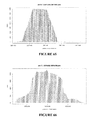
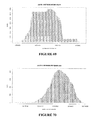
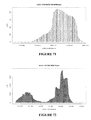
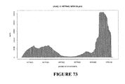
- a method of evaluating the quality of a nucleic acid extraction from microvesicles isolated from a eukaryotic biological sample comprising the steps of: (a) extracting RNA from microvesicles; and (b) measuring the quality of the RNA by determining the quantity of 18S and 28S rRNA in the extraction.
- the quantitative ratio of 18S rRNA to 28S rRNA determined in the novel method is within the range of approximately 1:1 to approximately 1:2; and is preferably approximately 1:2.
- Biological samples on which the novel method may be performed include, among other things, any bodily fluid, preferably urine, serum or plasma, and preferably, is from a mammal, particularly a human.
- the novel method may further result in the extraction of nucleic acid having an RNA Integrity Number of greater than or equal to 5 and/or may further result in a nucleic acid yield from 20 ml of biological sample of greater than or equal to 50 pg/ml.
- the novel method may further result in the extraction of nucleic acid having an RNA Integrity Number of greater than or equal to 3 and/or may further result in a nucleic acid yield from 1 ml of biological sample is greater than or equal to 50 pg/ml.
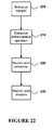
- a method of obtaining nucleic acid from a biological sample comprising the steps of: (a) obtaining a biological sample; (b) performing an extraction enhancement operation on the biological sample; and (c) extracting nucleic acid from the biological sample.
- the extraction enhancement operation is comprised of: (a) the addition of one or more enhancement agents to the biological sample; or (b) the performance of one or more enhancement steps prior to nucleic acid extraction; or (c) a combination of enhancement agents and enhancement steps.
- the enhancement agents may include: (i) RNase inhibitor; (ii) protease; (iii) reducing agent; (iv) decoy substrate, such as synthetic RNA; (v) soluble receptor; (vi) small interfering RNA; (vii) RNA binding molecule, such as anti-RNA antibody, chaperone protein, or an RNase inhibitory protein; (ix) RNase denaturing substance, such as high osmolarity solution or detergent.
- the extraction enhancement steps may include: (x) washing; (xi) size-separating RNase from the sample; (xii) effecting RNase denaturation through a physical change, such as by decreasing temperature, or executing a freeze/thaw cycle.
- the novel method may be performed on a biological sample including, among other things, any bodily fluid, preferably urine, serum or plasma, and preferably, is from a mammal, particularly a human.
- a derivative may be obtained from the biological sample and subjected to the extraction enhancement operation before extracting nucleic acid.
- the derivative is a microvesicle fraction from the biological sample.
- the microvesicle fraction may be obtained by a filtration concentration technique, however other known isolation techniques may be utilized as well.
- the derivative may be treated with a ribonuclease, deoxyribonuclease, or a combination thereof, prior to performance of the enhancement extraction operation.
- the extraction enhancement operation may include the addition of an RNase inhibitor to the biological sample, or to the derivative, prior to extracting nucleic acid; preferably the RNase inhibitor has a concentration of greater than 0.027 AU (1X) for a sample equal to or more than 1 ⁇ l; alternatively, greater than or equal to 0.135 AU (5X) for a sample equal to or more than 1 ⁇ l; alternatively, greater than or equal to 0.27 AU (10X) for a sample equal to or more than 1 ⁇ l; alternatively, greater than or equal to 0.675 AU (25X) for a sample equal to or more than 1 ⁇ l; and alternatively, greater than or equal to 1.35 AU (50X) for a sample equal to or more than 1 ⁇ l, wherein the 1X protease concentration refers to an enzymatic condition wherein 0.027 AU or more protease is used to treat microvesicles isolated from 1 ⁇ l or more bodily fluid; the 5X protease concentration refers to an enzymatic condition wherein
- the novel kit for obtaining nucleic acids from microvesicles, comprising in one or more containers: (a) a nucleic acid extraction enhancement agent; (b) DNase, RNase, or both; and (c) a lysis buffer.
- the novel kit may further comprise instructions for using the kit.
- the nucleic acid extraction enhancing agent may include: (a) RNase inhibitor; (b) protease; (c) reducing agent; (d) decoy substrate; (e) soluble receptor; (f) small interfering RNA; (g) RNA binding molecule; (h) RNase denaturing substance; or (i) any combination of any of the foregoing agents as a mixture or individually.
- RNA from microvesicles comprising the steps of: (a) obtaining a sample of microvesicles; (b) treating the sample with DNase to eliminate all or substantially all of any DNA located outside of or on the surface of the microvesicles in the sample; (c) extracting RNA from the sample; and (d) analyzing the extracted RNA.
- the novel method may be performed on a biological sample including, among other things, any bodily fluid, preferably urine, serum or plasma, and preferably, is from a mammal, particularly a human.
- a novel method for diagnosing, monitoring, or treating a subject comprising the steps of: (a) isolating a microvesicle fraction from a urine sample from a subject; (b) detecting the presence or absence of a biomarker within the microvesicle fraction; wherein the biomarker is selected from the group consisting of (i) a species of nucleic acid, (ii) the level of expression of a nucleic acid, (iii) a nucleic acid variant, and (iv) any combination of any of the foregoing; and wherein the biomarker is associated with the presence or absence of a disease or other medical condition, or the viability of a treatment option.
- the biomarker may be an mRNA transcript; for instance, the mRNA transcript may be selected from the group consisting of: NPHS2 (podocin), LGALS1 (Galectin-1), HSPG2 (heparin sulfate proteoglycan); CUBN (cubilin), LRP2 (megalin), AQP1 (aquaporin 1), CA4 (carbonic anydrase 4), CLCN5 (chloride channel protein 5), BDKRB1 (bradykinin B1 receptor), CALCR (calcitonin receptor), SCNN1D (amiloride-sensitive sodium channel subunit delta), SLC12A3 (thiazide-sensitive sodium-chloride cotransporter), AQP2 (aquaporin 2), ATP6V1B1 (V-ATPase B1 subunit), SLC12A1 (kidney-specific Na-K-Cl symporter via RT-PCR of RiboAmped mRNA); more preferably, the
- the biomarker and disease or other medical condition may be selected from the group consisting of: (a) NPHS2 (podocin) and glomerular disease, such as steroid-resistant nephritic syndrome; (b) CUBN (cubilin) and proteinuria , such as in Imerslund-Gräsbeck syndrome; and (c) AQP2 (aquaporin 2) and diabetes insipidus.
- NPHS2 podocin
- CUBN cubilin
- proteinuria such as in Imerslund-Gräsbeck syndrome
- AQP2 aquaporin 2
- diabetes insipidus may be selected from the group consisting of: (a) NPHS2 (podocin) and glomerular disease, such as steroid-resistant nephritic syndrome; (b) CUBN (cubilin) and proteinuria , such as in Imerslund-Gräsbeck syndrome; and (c) AQP2 (aqua
- an isolated polynucleotide molecule comprising a first nucleotide sequence that is at least 90% identical to a second nucleotide sequence selected from the group consisting of SEQ ID NOS: 1-29; an isolated polynucleotide comprising a segment of a nucleotide sequence selected from SEQ ID NOS: 1-29; or an isolated polynucleotide comprising a sequence of at least 13 nucleotides that are the same as any 13-nucleotide sequence in any one of SEQ ID NOS: 1-29.
- the foregoing polynucleotide molecules may be a deoxyribonucleotide or a ribonucleotide.
- a vector comprising any of the foregoing isolated nucleic acid molecules.
- a host cell comprising any of the foregoing vectors or any of the foregoing isolated nucleic acid molecules.
- a novel method of assessing the quality of a nucleic acid extraction from a biological sample comprising: (a) providing a biological sample; (b) obtaining an extraction of nucleic acids from the biological sample; (c) measuring the amount of a polynucleotide molecule comprising a segment having a nucleotide sequence selected from SEQ NOS: 1-29 in the extraction; and (d) comparing the amount of the polynucleotide molecule against a standard to assess the quality of the nucleic acid extraction.
- the novel method may be performed on any biological sample, for example, a bodily fluid, in particular, urine, serum or plasma, preferably from a mammal such as a human.
- the standard used to assess the quality of the nucleic acid extraction may be derived by measuring the amount of a polynucleotide molecule comprising a segment having the nucleotide sequence selected from SEQ NOS: 1-29 in nucleic acid extractions from more than 5 biological samples.
- Microvesicles are shed by eukaryotic cells, or budded off of the plasma membrane, to the exterior of the cell. These membrane vesicles are heterogeneous in size with diameters ranging from about 10 nm to about 5000 nm.
- the small microvesicles (approximately 10 to 1000 nm, and more often approximately 10 to 200 nm in diameter) that are released by exocytosis of intracellular multivesicular bodies are referred to in the art as "exosomes.”
- the compositions, methods and uses described herein are equally applicable to microvesicles of all sizes; preferably 10 to 800 nm; and more preferably 10 to 200 nm.
- exosome also refers to protein complexes containing exoribonucleases which are involved in mRNA degradation and the processing of small nucleolar RNAs (snoRNAs), small nuclear RNAs (snRNAs) and ribosomal RNAs (rRNA) (Liu et al., 2006; van Dijk et al., 2007).
- snoRNAs small nucleolar RNAs
- snRNAs small nuclear RNAs
- rRNA ribosomal RNAs
- the present invention is partly based on the discovery that adverse factors can prevent an effective extraction of nucleic acids from a biological sample and that novel and unexpected agents and steps may be used to mitigate or remove the adverse factors, thereby dramatically improving the quality of the extracted nucleic acids.
- described herein are novel methods for extracting high quality nucleic acids from a biological sample.
- the high quality extractions obtained by the novel methods described herein are characterized by high yield and high integrity, making the extracted nucleic acids useful for various applications in which high quality nucleic acid extractions are preferred.
- the novel methods include, for example, the steps of obtaining a biological sample, mitigating or removing the adverse factors that prevent an effective extraction of nucleic acids from a biological sample, and extracting nucleic acids from the biological sample followed, optionally, by nucleic acid analysis.
- Applicable biological samples include, for example, a cell, a group of cells, fragments of cells, cell products including for example microvesicles, cell cultures, bodily tissues from a subject, or bodily fluids.
- the bodily fluids can be fluids isolated from anywhere in the body of the subject, preferably a peripheral location, including but not limited to, for example, blood, plasma, serum, urine, sputum, spinal fluid, pleural fluid, nipple aspirates, lymph fluid, fluid of the respiratory, intestinal, and genitourinary tracts, tear fluid, saliva, breast milk, fluid from the lymphatic system, semen, cerebrospinal fluid, intra-organ system fluid, ascitic fluid, tumor cyst fluid, amniotic fluid and combinations thereof.
- a biological sample may sometimes come from a subject.
- subject is intended to include all animals shown to or expected to have microvesicles.
- the subject may be a mammal, a human or nonhuman primate, a dog, a cat, a horse, a cow, other farm animals, or a rodent (e.g. mouse, rat, guinea pig, etc.).
- rodent e.g. mouse, rat, guinea pig, etc.
- a biological sample may optionally be processed to obtain a biological sample derivative before, after, or at the same time as, carrying out the step of mitigating or removing the adverse effects.
- the biological sample derivative may be a cell, cell debris, a membrane vesicle, or a microvesicle.
- a biological sample is sometimes pre-processed before a biological sample derivative such as a microvesicle is obtained.
- the pre-processing step is preferred.
- a urine sample may be pre-processed to obtain urinary microvesicles.
- the pre-processing may be achieved by techniques known in the art such as low speed centrifugation and pre-filtration.
- urine samples may undergo a first centrifugation step of 300g to get rid of large particles in the samples.
- Urine samples may undergo a second centrifugation step of 17,000g to get rid of smaller particles in the samples.
- urine samples may further undergo a pre-filtration step, e.g., a 0.8um pre-filtration step.
- urine samples may be pre-processed by a - pre-filtration step without first undergoing the one or more of the centrifugation steps.
- Membrane vesicles may be isolated from a biological sample. In some instances, such isolation may be carried out without pre-processing the biological sample. In other instances, such isolation may be carried out after the biological sample is pre-processed. The isolation step may be advantageous for high quality nucleic acid extraction from a biological sample.
- the isolation may give rise to advantages such as: 1) the opportunity to selectively analyze disease- or tumor-specific nucleic acids, which may be obtained by isolating disease- or tumor-specific microvesicles apart from other microvesicles within the fluid sample; 2) significantly higher yield of nucleic acid species with higher integrity as compared to the yield/integrity obtained by extracting nucleic acids directly from the fluid sample; 3) scalability, e.g.
- the sensitivity can be increased by pelleting more microvesicles from a larger volume of serum; 4) purer nucleic acids in that protein and lipids, debris from dead cells, and other potential contaminants and PCR inhibitors are excluded from the microvesicle pellets before the nucleic acid extraction step; and 5) more choices in nucleic acid extraction methods as microvesicle pellets are of much smaller volume than that of the starting serum, making it possible to extract nucleic acids from these microvesicle pellets using small volume column filters.
- Methods of isolating microvesicles from a biological sample are known in the art. For example, a method of differential centrifugation is described in a paper by Raposo et al. (Raposo et al., 1996), a paper by Skog et. al.(Skog et al., 2008) and a paper by Nilsson et. al.(Nilsson et al., 2009). Methods of anion exchange and/or gel permeation chromatography are described in US Patent Nos. 6,899,863 and 6,812,023 . Methods of sucrose density gradients or organelle electrophoresis are described in U.S. Patent No. 7,198,923 .
- microvesicles can be identified and isolated from bodily fluid of a subject by a newly developed microchip technology that uses a unique microfluidic platform to efficiently and selectively separate tumor-derived microvesicles (Chen et al.).
- the microvesicles isolated from a bodily fluid may be enriched for those originating from a specific cell type, for example, lung, pancreas, stomach, intestine, bladder, kidney, ovary, testis, skin, colorectal, breast, prostate, brain, esophagus, liver, placenta, fetus cells. Because the microvesicles often carry surface molecules such as antigens from their donor cells, surface molecules may be used to identify, isolate and/or enrich for microvesicles from a specific donor cell type (Al-Nedawi et al., 2008; Taylor and Gercel-Taylor, 2008).
- microvesicles originating from distinct cell populations can be analyzed for their nucleic acid content.
- tumor (malignant and non-malignant) microvesicles carry tumor-associated surface antigens and may be detected, isolated and/or enriched via these specific tumor-associated surface antigens.
- the surface antigen is epithelial-cell-adhesion-molecule (EpCAM), which is specific to microvesicles from carcinomas of lung, colorectal, breast, prostate, head and neck, and hepatic origin, but not of hematological cell origin (Balzar et al., 1999; Went et al., 2004).
- the surface antigen is CD24, which is a glycoprotein specific to urine microvesicles (Keller et al., 2007).
- the surface antigen is selected from a group of molecules such as CD70, carcinoembryonic antigen (CEA), EGFR, EGFRvIII and other variants, Fas ligand, TRAIL, transferrin receptor, p38.5, p97 and HSP72.
- tumor specific microvesicles may be characterized by the lack of surface markers, such as CD80 and CD86.
- the isolation of microvesicles from specific cell types can be accomplished, for example, by using antibodies, aptamers, aptamer analogs or molecularly imprinted polymers specific for a desired surface antigen.
- the surface antigen may be specific for a cancer type.
- the surface antigen may be specific for a cell type which is not necessarily cancerous.
- One example of a method of microvesicle separation based on cell surface antigen is provided in U.S. Patent No. 7,198,923 . As described in, e.g., U.S. Patent Nos. 5,840,867 and 5,582,981 , WO/2003/050290 and a publication by Johnson et al.
- aptamers and their analogs specifically bind surface molecules and can be used as a separation tool for retrieving cell type-specific microvesicles.
- Molecularly imprinted polymers also specifically recognize surface molecules as described in, e.g., US Patent Nos. 6,525,154 , 7,332,553 and 7,384,589 and a publication by Bossi et al. (Bossi et al., 2007) and are a tool for retrieving and isolating cell type-specific microvesicles.
- a step of removing nucleic acids that are not inside the microvesicle is sometimes performed.
- Methods of removing nucleic acids are well known in the art.
- an enzyme digestion step may be performed.
- Such enzymes may be a type of ribonuclease that catalyzes the enzymatic digestion of ribonucleic acids or a type of deoxyribonuclease that catalyzes the enzymatic digestion of deoxyribonucleic acids.
- the novel nucleic acid extraction methods may include a step of removing or mitigating adverse factors that prevent high quality nucleic acid extraction from a biological sample.
- adverse factors are heterogeneous in that different biological samples may contain various species of such adverse factors.
- factors such as excessive extra-microvesicle DNA may affect the quality of nucleic acid extractions from such samples and contaminate DNA extracted from within microvesicle.
- factors such as excessive endogenous RNase may affect the quality of nucleic acid extractions from such samples.
- Many agents and methods may be used to remove these adverse factors. These methods and agents are referred to collectively as an "extraction enhancement operation.”
- the extraction enhancement operation may involve the addition of nucleic acid extraction enhancement agents to the biological sample or derivative.
- extraction enhancement agents as defined here may include, but are not limited to, a commercially available RNase inhibitor such as Superase-In (Ambion Inc.), RNaseIN (Promega Corp.), or other agents that function in a similar fashion; a protease; a reducing agent; a decoy substrate such as a synthetic RNA; a soluble receptor that can bind RNase; a small interfering RNA (siRNA); a RNA binding molecule, such as an anti-RNA antibody, or a chaperone protein; a RNase denaturing substance, such as a high osmolarity solution, a detergent, or a combination thereof.
- a commercially available RNase inhibitor such as Superase-In (Ambion Inc.), RNaseIN (Promega Corp.), or other agents that function in a similar fashion
- a protease such as Superase-In (
- enhancement agents may exert their functions in various ways, for example, but not limited to, through inhibiting RNase activity (e.g., RNase inhibitors), through a ubiquitous degradation of proteins (e.g., proteases), or through a chaperone protein (e.g., a RNA-binding protein) that binds and protects RNAs.
- RNase activity e.g., RNase inhibitors
- a ubiquitous degradation of proteins e.g., proteases
- a chaperone protein e.g., a RNA-binding protein
- the extraction enhancement operation may involve the performance of one or more process steps.
- processes include extensive or substantially thorough washing of nucleic acid-containing components of the sample, such as microvesicles; size separation of RNases from the biological sample; denaturation of proteins in the biological sample by various techniques including, but not limited to, generating a particular pH condition, a temperature condition, (e.g., the maintenance of a decreasing or lower temperature), freeze/thaw cycles, and combinations thereof.
- rRNA ribosomal RNA
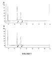
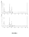
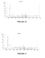
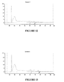
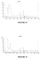
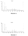
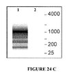
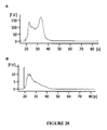
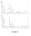
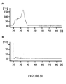
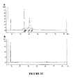
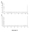
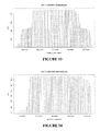
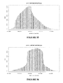
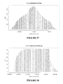
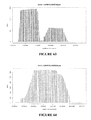
- RNA integrity number is the product of a software tool designed to estimate the integrity of total RNA samples.
- the software automatically assigns an integrity number to an eukaryote total RNA sample. Using this tool, sample integrity is not determined by the ratio of the 18S and 28S ribosomal bands, but by the entire electrophoretic trace of the RNA sample. This includes the presence or absence of degradation products.
- the assigned RIN is independent of sample concentration, instrument, and analyst, and can serve as a standard for RNA integrity.
- an extraction enhancement operation will improve the quantity or yield of extracted nucleic acid. For example, using an extraction enhancement operation, as described herein, one may obtain a nucleic acid yield of greater than or equal to 50 pg/ml from a 20 ml low protein biological sample such as urine. Alternatively, one may obtain a nucleic acid yield of greater than or equal to 50 pg/ml from 1 ml of a high protein biological sample, such as serum or plasma.
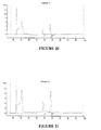
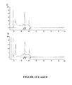
- Novel high quality nucleic acid extractions obtained by the methods described herein may display a combination of the detection of 18S and 28S rRNA, preferably in a ratio of approximately 1:1 to approximately 1:2; and more preferably, approximately 1:2; a RNA integrity number of greater than or equal to 5 for a low protein biological sample, or greater than or equal to 3 for a high protein biological sample; and a nucleic acid yield of greater than or equal to 50 pg/ml from a 20 ml low protein biological sample or a 1 ml high protein biological sample.
- RNA degradation can seriously affect downstream assessment of the extracted RNA, such as in gene expression and mRNA analysis, as well as analysis of non-coding RNA such as small RNA and micro RNA.
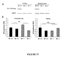
- the novel methods described herein enable one to extract high quality nucleic acids from a biological sample such as microvesicles so that an accurate analysis of gene expression and mutational level within the exosomes can be carried out. For example, when increased concentrations of protease (5X, 10X) are used as an extraction enhancing agent, the amount and integrity of RNA isolated from urinary microvesicles is increased significantly.
- RNA such as miRNA
- a method to extract nucleic acid, particularly small RNA may use 25X and 50X protease as extraction enhancing agent and is able to obtain significantly increased amounts of small RNA.
- expressions such as 5X, 10X, 25X and 50X mean 5 times, 10 times, etc. the activity level of protease currently used or recommended in commercially available nucleic acid extraction kits such as the QIAamp MinElute Virus Spin Kit.
- nucleic acid molecules can be isolated from a biological sample using any number of procedures that are well-known in the art. Persons of skill will select a particular isolation procedure as being appropriate for the particular biological sample. Examples of methods for extraction are provided in the Examples section herein. In some instances, with some techniques, it may also be possible to analyze the nucleic acid without extraction from the microvesicle.
- the extracted nucleic acids may be analyzed directly without an amplification step.
- Direct analysis may be performed with different methods including, but not limited to, nanostring technology.
- NanoString technology enables identification and quantification of individual target molecules in a biological sample by attaching a color coded fluorescent reporter to each target molecule. This approach is similar to the concept of measuring inventory by scanning barcodes. Reporters can be made with hundreds or even thousands of different codes allowing for highly multiplexed analysis. The technology is described in a publication by Geiss et al. (Geiss et al., 2008).
- nucleic acid amplification it may be beneficial or otherwise desirable to amplify the nucleic acid of the microvesicle prior to analyzing it.
- Methods of nucleic acid amplification are commonly used and generally known in the art, many examples of which are described herein. If desired, the amplification can be performed such that it is quantitative. Quantitative amplification will allow quantitative determination of relative amounts of the various nucleic acids, to generate a profile as described below.
- the extracted nucleic acid is RNA.
- the RNA is then preferably reverse-transcribed into complementary DNA (cDNA) before further amplification.
- cDNA complementary DNA
- Such reverse transcription may be performed alone or in combination with an amplification step.
- a method combining reverse transcription and amplification steps is reverse transcription polymerase chain reaction (RT-PCR), which may be further modified to be quantitative, e.g., quantitative RT-PCR as described in US Patent No. 5,639,606 .
- Nucleic acid amplification methods include, without limitation, polymerase chain reaction (PCR) ( US Patent No. 5,219,727 ) and its variants such as in situ polymerase chain reaction ( US Patent No. 5,538,871 ), quantitative polymerase chain reaction ( US Patent No. 5,219,727 ), nested polymerase chain reaction ( US Patent No.
- PCR polymerase chain reaction
- US Patent No. 5,538,871 in situ polymerase chain reaction
- quantitative polymerase chain reaction US Patent No. 5,219,727
- nested polymerase chain reaction US Patent No.
- nucleic acids present in the microvesicles is quantitative and/or qualitative.
- amounts (expression levels), either relative or absolute, of specific nucleic acids of interest within the microvesicles are measured with methods known in the art (described below).
- species of specific nucleic acids of interest within the microvesicles, whether wild type or variants, are identified with methods known in the art.
- a nucleic acid extraction from microvesicles in which 18S and 28S rRNA is detectable in the extraction may be achieved using the novel nucleic acid extraction method described herein.
- a high quality nucleic acid extraction from microvesicles in a biological sample is desirable in many instances.
- a tissue sample is not easily accessible.
- a brain tumor sample can not usually be obtained without brain surgery. Instead, a microvesicle sample from the brain tumor patient serum is easily accessible.
- nucleic acids in microvesicles secreted by tissue cells are used to substitute nucleic acids from tissue cells
- it is desirable to obtain high quality nucleic acids which, like those obtained from tissue cells directly, contain detectable quality controls, such as 18S and 28S rRNA.
- high quality small RNA is desirable.
- Nucleic acid extractions disclosed herein contain such high quality small RNA together with 18S and 28S rRNA.
- Such high quality small RNA is important for the accurate assessment of nucleic acids for various purposes, e.g., the expression level of a particular miRNA.
- RNAs are generated by analyzing nucleic acid extractions that contain 18S and 28S rRNA. Such profiles may be obtained with the novel methods disclosed herein.
- High quality nucleic acid profiles are highly desirable for many uses, such as for use as a biomarker for a medical condition or therapy selection. It is desirable in that such profiles are consistent between samples. Such consistency can hardly be achieved without high quality nuclei acid extractions.
- a profile of nucleic acids can be obtained by analyzing nucleic acids in microvesicles that are secreted by those cells of origin. Such microvesicles can be isolated from an easily accessible biological sample, e.g., urine, serum or plasma.
- Such profiles of nucleic acids many include small RNAs, messenger RNA, microRNA, non-coding RNAs or a combination thereof. Such profiles of nucleic acids may be combined with other biomarkers to more accurately achieve certain results.
- the profile of nucleic acids for instance can be a collection of genetic aberrations, which is used herein to refer to the nucleic acid amounts as well as nucleic acid variants within the microvesicles.
- genetic aberrations include, without limitation, over-expression of a gene (e.g., oncogenes) or a panel of genes, under-expression of a gene (e.g., tumor suppressor genes such as p53 or RB) or a panel of genes, alternative production of splice variants of a gene or a panel of genes, gene copy number variants (CNV) (e.g.
- DNA double minutes DNA double minutes
- nucleic acid modifications e.g., methylation, acetylation and phosphorylations
- single nucleotide polymorphisms SNPs
- chromosomal rearrangements e.g., inversions, deletions and duplications
- mutations insertions, deletions, duplications, missense, nonsense, synonymous or any other nucleotide changes
- nucleic acid modifications can be assayed by methods described in, e.g., US Patent No.
- methylation profiles may be determined by Illumina DNA Methylation OMA003 Cancer Panel.
- SNPs and mutations can be detected by hybridization with allele-specific probes, enzymatic mutation detection, chemical cleavage of mismatched heteroduplex (Cotton et al., 1988), ribonuclease cleavage of mismatched bases (Myers et al., 1985), mass spectrometry ( US Patent Nos.
- nucleic acid sequencing single strand conformation polymorphism (SSCP) (Orita et al., 1989), denaturing gradient gel electrophoresis (DGGE)(Fischer and Lerman, 1979a; Fischer and Lerman, 1979b), temperature gradient gel electrophoresis (TGGE) (Fischer and Lerman, 1979a; Fischer and Lerman, 1979b), restriction fragment length polymorphisms (RFLP) (Kan and Dozy, 1978a; Kan and Dozy, 1978b), oligonucleotide ligation assay (OLA), allele-specific PCR (ASPCR) ( US Patent No.
- DGGE denaturing gradient gel electrophoresis
- TGGE temperature gradient gel electrophoresis
- RFLP restriction fragment length polymorphisms
- OLA oligonucleotide ligation assay
- ASPCR allele-specific PCR
- gene expression levels may be determined by the serial analysis of gene expression (SAGE) technique (Velculescu et al., 1995).
- SAGE serial analysis of gene expression
- Example 1 Renal cells contain multivesicular bodies
- TEM Transmission Electron Microscopy
- the sample slices were rinsed in 0.1 M sodium cacodylate buffer, post-fixed in 1.0% osmium tetroxide in cacodylate buffer for 1 h at room temperature, rinsed in buffer again, then rinsed in distilled water (dH 2 O) and stained, en bloc, in an aqueous solution of 2.0% uranyl acetate for 1 hour at room temperature.
- the samples were rinsed in distilled water and dehydrated through a graded series of ethanol to 100%.
- the samples were infiltrated with Epon resin (Ted Pella, CA) by overnight immersion in a 1:1 solution of Epon:ethanol. The following day samples were placed in fresh Epon for several hours and embedded in Epon overnight at 60°C.
- TEM transmission electron microscope
- Example 2 Microvesicles exist in urine
- the filtrate then underwent ultracentrifugation at 118,000g for 70 min at 4°C, the supernatant was removed and the microvesicle-containing pellet was washed in PBS and re-pelleted at 118,000g for 70 min at 4°C.
- filtration concentration was also used to isolate microvesicles from pre-processed samples.
- the filtrate concentrator 100kDa MWCO (Millipore, MA) was prepared according to the manufacturer's instructions. Pre-processed filtrate was added to the filtration concentrator and centrifuged at 4,000g for 4 min at RT. A 15 ml PBS wash step was included.
- Microvesicle pellets were fixed, 1:1 with 4% paraformaldehyde in dH 2 O. Ten (10) ⁇ l drops were pipetted onto formvar-coated 200 mesh gold grids and drawn off after one minute. Samples were rinsed 2 times with drops of dH 2 O. Aqueous 2.0% phosphotungstic acid (PTA) was applied (10 ⁇ l) for 10 sec, drawn off and rinsed once with dH 2 O Samples were examined in a JEOL JEM 1011 transmission electron microscope at 80 kV. Images were collected using an AMT (Advanced Microscopy Techniques, MA) digital imaging system. As shown in Figure 2 , the pellet was indeed rich in microvesicles. The microvesicles sometimes aggregate together or remain singular in the TEM image.
- AMT Advanced Microscopy Techniques, MA
- Example 3 Ultracentrifugation is replaceable with filtration concentrator for purposes of microvesicle isolation
- filtration concentrators can yield viable microvesicles for RNA extraction similar to the ultracentrifugation method.
- the ultracentrifugation method and filtration concentration method yielded similar RNA concentrations from the 75 ml urine samples with ultracentrifugation ( Figure 4) at 410 ⁇ 28 pg/ ⁇ l, and filtration concentrator ( Figure 3) at 381 ⁇ 47 pg/ ⁇ l (Mean ⁇ SD). There is no statistically significant difference between the two yields.
- Example 4 Sample pre - processing with only a 0.8um pre-filtration step is sufficient for purpose of microvesicle isolation
- the low speed centrifugation steps at 300g and 17,000g could be eliminated because urine pre-processing with just a 0.8 ⁇ m pre-filtration step was as effective as methods including the low speed centrifugation steps.
- the nucleic acid profile using a method of low speed centrifugation coupled with 0.8 ⁇ m pre-filtration ( A ) is the same as the profile using a method of only 0.8 ⁇ m pre-filtration ( B ).
- Example 5 Nucleic acid extraction from urinary microvesicles with methods that include removal or mitigation of adverse factors
- the microvesicles were resuspended in 1 ⁇ l/ml RNase A (DNase and protease free) (Fermentas, MD) in PBS and incubated for 1 hr at 37°C.
- the samples were re-pelleted at 118,000g for 70 min in PBS.
- DNase I digestion the pellet was resuspended in 500 ⁇ l PBS and DNase I (RNase free)(Qiagen, CA) diluted in RDD buffer (according to manufacturer's instructions) and incubated at room temperature for 10 min.
- the samples were re-pelleted at 118,000g for 70 min in PBS.
- RNA extraction enhancement 140 was then performed, e.g., three resuspension/wash steps with 15 ml of PBS alone, or coupled with a protease treatment. After microvesicles were isolated, digested with nucleases and treated with protease, nucleic acids were extracted 150. The RNA extraction was performed using the RNeasy Micro kit (Qiagen, CA) according to the manufacturer's instructions.
- RNA isolation method was followed according to the manufacturer's instructions.
- RNA Pico 6000 chip Agilent, CA
- Agilent BioAnalyzer Agilent BioAnalyzer
- RNA extraction from urine microvesicles increases as more protease is used to treat microvesicles before breaking the membrane of microvesicles.
- nucleic acids from microvesicles in 20 ml urine were extracted using the above improved method except that the RNA extraction was performed using the Qiagen Qiamp minelute virus spin kit. The urine sample was concentrated with filtration concentration and eluted in 200 ⁇ l PBS.
- 1X protease as 0.027 AU; 5X protease as 0.135 AU; 10X protease as 0.27 AU; 25X protease as 0.675 AU; and 50X as 1.35 AU.
- 18S and 28S rRNA peaks of greater integrity were observed when the concentration of protease was increased from 5X ( A ) to 10X ( B ).
- 25X ( A ) and 50X ( B ) we observed 18S and 28S rRNA of higher integrity, in addition to increased small/miRNA levels.
- protease can increase the yield and integrity of 18S and 28S rRNA as well as small RNA and microRNA. We suspect that the effect of protease may be due to its ability to digest away RNases and other inhibitory factors.
- Urine samples of 20 ml each are used for four groups of nucleic acid extraction tests following the above method with except some modifications.
- group 1 isolated microvesicles were directly used for nucleic acid extraction without any intervening steps.
- group 2 microvesicles were treated with RNase inhibitors before nucleic acid extraction.
- group 3 microvesicles were washed without any RNase inhibitor treatment before nucleic acid extraction.
- group 4 microvesicles were washed and treated with RNase inhibitors.
- nucleic acid extractions can also be obtained from serum microvesicles.
- serum from both melanoma and normal patient sera and used RNase inhibitor cocktail SUPERase-InTM (Ambion, Inc.) to treat microvesicle pellets by resuspension.
- RNase inhibitor cocktail SUPERase-InTM Ambion, Inc.
- the microvesicle isolation method was ultracentrifugation and the microvesicle pellets were treated with DNase for 20 minutes at room temperature.
- RNA yield 543 pg/ ⁇ l, 607 pg/ ⁇ l, 1084 pg/ ⁇ l, 1090 pg/ ⁇ l
- RNA integrity assessed by the 28s/18s ratio being 1.7, 1.8, 1.3, and 0.6, respectively.
- RNA yield for the two melanoma serum samples is 3433pg/ ⁇ l and 781pg/ ⁇ l and the 28S/18S ratio is 1.4 and 1.5, respectively.
- Example 7 Use of extraction enhancement agents for nucleic acid extraction from a biological sample
- RNA extraction enhancers can increase the quality of RNA extraction from microvesicles. It is expected that such extraction enhancers will have similar effects on other biological samples.
- a novel method of nucleic acid extraction requires a step of performing an extraction enhancement operation on the biological sample. Such method may be exemplified in the following conceived nucleic acid extraction experiment. A doctor prescribes a test of a tumor biomarker for a patient. A 5 ml blood is then drawn from the patient. The blood sample 200 is sometimes pre-processed to get the blood serum.
- An enhancement operation 210 is then performed, e.g., an appropriate amount of extraction enhancer is added to the serum and the mixture is incubated for 30 minutes at 37°C.
- Nucleic acids from the treated serum are then extracted using regular extraction methods such as detailed in Example 5 220 and analyzed using Agilent BioAnalyzer 230 . Such extraction is expected to produce high quality nucleic acids from the biological sample.
- Example 8 Urinary microvesicles are contaminated by free, extra-microvesicle, non-cellular DNA
- the extra nucleic acids from the untreated sample were likely from DNase susceptible "apoptotic bodies” because DNase susceptible "apoptotic bodies”-like ladders were seen as shown in the pseudo gel in Figure 24 C .
- the amount of the free, extra-microvesicle, non-cellular DNA varied between subjects but the size of this DNA was in the range of approximately 25 to 1500 base pairs.
- Urinary microvesicles are mostly not contaminated by free, extra-microvesicle, non-cellular RNA
- the curve of the RNase untreated sample ( A ) mostly overlapped with the curve of the RNase treated sample ( B ), suggesting there is no free extra-microvesicle, non-cellular RNA associated with the isolated microvesicles. This may be due to the presence of ribonucleases in urine.
- Example 10 Nucleic acid profiles are similar in urinary microvesicles and renal cells measured by Agilent BioAnalyzer.
- urinary microvesicles also contained similar small RNA profiles to those obtained from renal cells. As shown in Figure 27 , both urinary microvesicles ( B ) and renal tissue ( A ) contained small RNAs (about 25-200 base pairs) and shared similar patterns.
- the profiles in urinary microvesicles may be used to examine the profiles in the renal cells from which the microvesicles originated.
- Example 11 RNA profiles in urinary microvesicles are different from those from whole urine.
- RNA profiles in urinary microvesicles are different from those from whole urine.
- RNA from whole urine was isolated using the ZR urine RNA isolation kit (Zymo Research, CA) according to the manufacturer's instructions.
- the eluted RNA was resuspended in 350 ⁇ l RLT buffer and processed via the RNeasy Plus Micro kit, which used DNase to eliminate associated DNA, and eluted in 16 ⁇ l nuclease free water.
- RNA profile from microvesicles from the same urine sample was generally very different from the profile from the whole urine extraction. In the microvesicle profile, there were 18S and 28S peaks. In addition, RNA from microvesicles was more abundant than that from the whole urine.
- RNA profiles from urinary microvesicles is more similar to renal cell profiles than to profiles from whole urine. Further, the integrity of RNA extraction from microvesicles was at least 10 times better than that from whole urine.
- Example 12 Urinary microvesicles contain both RNA and DNA.
- RNA represents the most abundant nucleic acid within microvesicles.
- Figure 33 after on-column digestion of the RNase treated samples with DNase ( B ), the peak just after 20s is reduced after further DNase digestion inside in comparison with the peak before further DNase digestion ( A ). This reduction demonstrated that a small amount of DNase-digestible material was present within microvesicles, probably DNA.
- Urinary microvesicles contain mRNA transcripts encoding specific genes from various regions of the nephron and collecting duct.
- nucleic acid profiles are similar in urinary microvesicles and renal cells measured by Agilent BioAnalyzer.
- microvesicles contain mRNA transcripts encoding specific genes from various regions of the nephron and collecting duct.
- Urinary microvesicles were isolated from 200 ml urine from four human subjects (23 to 32 years of age) and were digested with RNase and DNase prior to exosome lysis and RNA extraction as detailed in Example 5. The extracted RNA underwent two rounds of mRNA amplification using RiboAmp (Molecular Devices, CA).
- Figure 34B We examined 15 transcripts characteristic of various regions of the nephron and collecting duct ( Figure 34B ). These included proteins and receptors implicated in various renal diseases including podocin from the glomerulus, cubilin from the proximal tubule and aquaporin 2 from the collecting duct.
- the PCR primers used were: ACTB UTR, forward 5'-GAAGTCCCTTGCCATCCTAA-3', reverse '5-GCTATCACCTCCCCTGTGTG-3'; GAPDH EX, forward 5'-ACACCCACTCCTCCACCTTT-3', reverse 5'-TGCTGTAGCCAAATTCGTTG-3'; NPHS2 UTR, forward 5'-AACTTGGTTCAGATGTCCCTTT-3', reverse 5'-CAATGATAGGTGCTTGTAGGAAG-3'; LGALS1 EX, forward 5'-GGAAGTGTTGCAGAGGTGTG-3', reverse 5'-TTGATGGCCTCCAGGTTG-3'; HSPG2 UTR, 5'-AAGGCAGGACTCACGACTGA-3', reverse 5'-ATGGCACTTGAGCTGGATCT-3'; CUBN EX, forward 5'-CAGCTCTCCATCCTCTGGAC-3', reverse 5'-CCGTGCATAATCAGCATGAA-3';
- UTR refers to primers designed in the UTR and "EX” refers primers designed across exons.
- the PCR protocol was 5 min at 94°C; 40 s at 94°C; 30 s at 55°C; 1 min at 65°C for 30 cycles; and 68°C for 4 min.
- the primers used were: AQP2: forward 5'-GCCACCTCCTTGGGATCTATT-3', reverse 5'-TCATCAAACTTGCCAGTGACAAC-3'; V-ATPase B1 subunit: forward 5'-CTGGCACTGACCACGGCTGAG -3', reverse 5'-CCAGCCTGTGACTGAGCCCTG -3'.
- the PCR protocol was 5 min at 94°C; 40 s at 94°C, 30 s at 55°C, 1 min at 65°C for 30 cycles; and 68°C for 4 min.
- region 1 Glomerulus NPHS2 - podocin, LGALS1 - Galectin-1 , and HSPG2 - heparan sulfate proteoglycan
- region 2 Proximal Tubule CUBN - cubilin, LRP2 - megalin, AQP1 - aquaporin 1, CA4 - carbonic anhydrase 4, and CLCN5 - chloride channel protein 5
- region 3 Thin Descending Limb BDKRB1 - bradykinin B1 receptor
- region 4 Medullary Thick Ascending Limb CALCR - calcitonin receptor, and SCNN1D - amiloride-sensitive sodium channel subunit delta
- region 5 Distal Convoluted Tubule SLC12
- microvesicles containing mRNA are released from all regions of the nephron and collecting duct, and microvesicles can be a novel non-invasive source of nucleic acid biomarkers for renal diseases.
- Example 14 Some mRNA transcripts inside urinary microvesicles are specific to renal cells
- the transcripts in microvesicles should be specific to renal cells.
- mRNA transcripts are specific to renal cells.
- knockout mice in which the V-ATPase B1 subunit is absent.
- the absence of V-ATPase B1 subunit leads to renal acidosis in the mice ( Finberg KE, Wagner CA, Bailey MA, et al., The B1-subunit of the H(+) ATPase is required for maximal urinary acidification. Proc Natl Acad Sci USA 102:13616-13621, 2005 ).
- RNAlater Qiagen, CA
- RNAlater Qiagen, CA
- Mouse kidney samples were processed via the RNeasy Mini kit (Qiagen, CA) with the inclusion of the DNA digestion step.
- RNA extracted from mouse urinary microvesicles was denatured for 5 minutes at 65 °C and subjected to first strand cDNA synthesis as described in the Qiagen Sensiscript protocol (Qiagen, MD).
- Sensiscript reverse transcription oligo-dT primers were used at a final concentration of 1 ⁇ M (Applied Biosystems, CA).
- the resulting cDNA was used in the TaqMan PreAmp Master Mix Kit according to manufactures guide using 14 preamplification cycles (Applied Biosystems, CA).
- the preamplification product was then diluted 1:20 with 1X TE buffer (Promega, WI).
- the resulting cDNA was then used as a template for real-time PCR according to Taqman Preamplification guide (Applied Biosystems, CA).
- Mouse kidney RNA concentration was measured on a SmartSpec 3000 (Bio-Rad, CA) and all samples were diluted to 90 ng/ ⁇ l.
- Mouse kidney RNA was denatured for 5 minutes at 65 °C and subjected to first strand cDNA synthesis as described in the Qiagen Omniscript protocol (Qiagen, MD). In the Omniscript reverse transcription oligo-dT primers were used at a final concentration of 1 ⁇ M (Applied Biosystems, CA) and 1 ⁇ l of the resulting cDNA was then used per well in the subsequent real-time PCR reaction.
- the real-time PCR reaction was carried out using TaqMan Gene Expression Master Mix and Expression Assays (Mouse GAPD Part Number 4352339E and mouse Atp6v1b2 assay id Mm00431996_mH) on an ABI 7300 Real Time PCR System (Applied Biosystems, CA).
- V-ATPase B1 subunit and aquaporin 2 (AQP2) mRNA using RT-PCR.
- Panel A V-ATPase B1 subunit transcript was not detected in both the kidney and urinary microvesicle samples from double mutant mice (B1-/-), which is consistent with the fact that V-ATPase B1 subunit gene was knocked out in these mice.
- V-ATPase B1 subunit transcript was present in the kidney and microvesicle samples from the wild-type mice (B1/+/+).
- AQP2 mRNA was readily detected in both the kidney and the microvesicle sample from mice with both the B1 knockout or wild-type mice, which is expected because the V-ATPase B1 subunit deletion does not affect the expression of AQP2.
- Panel B the expression level of V-ATPase B2 subunit in microvesicles from the B1 knockout was not statistically different from the level in microvesicles from the wild-type mice. Such was also true for the expression level of V-ATPase B2 subunit in kidney cell from the knockout mice in comparison with the level in kidney cells from the wild-type mice.
- transcripts present in kidney cells can be detected in urinary microvesicles secreted by the kidney cells, and transcripts absent in kidney cells can not be detected in urinary microvesicles secreted by the kidney cell. Accordingly, transcripts in urinary microvesicles are specific to renal cells and are noninvasive biomarkers for renal cells transcripts.
- Urinary microvesicles contain non-coding RNA transcripts
- Urinary microvesicles were isolated and nucleic acids were extracted according to the above method detailed in Example 5. We performed a deep-sequencing of urinary microvesicle RNAs and found that there were random areas on certain chromosomes that exhibited extreme transcription. When plotted transcript number versus position on the chromosome, these transcripts appeared as "Spikes.” These transcripts were more abundantly expressed than well-known endogenous markers such as GAPDH or actin, and were generally in non-coding regions of the chromosome. The relatively high expression levels of these spike sequences suggest that these sequences may also serve important roles in chromosome activation and cellular regulation.
- the 29 regions are shown in Figure 42 and correspond to SEQ ID NOS. 1-29.
- the plotted spikes in these 29 regions are shown in Figures 45-73 .
- PCR analysis of the sequences of the most highly expressed spike transcripts demonstrated that they were indeed present within both human urinary microvesicles and human kidney cells, suggesting that these sequences were not an artifact of deep sequencing.
- the primers used to amply the 10 such spike abundant regions are shown in Figure 43 .
- PCR was performed according to the following program: initial denaturing at 95°C for 8 minutes; 30 cycles of three steps of denaturing at 95°C for 40 seconds, annealing at 55°C for 30 seconds, and elongation at 65°C for 1 minute; final elongation at 68 °C for 5 minutes; and on hold at 4°C before BioAnalyzer analysis of the reaction.
- initial denaturing at 95°C for 8 minutes 30 cycles of three steps of denaturing at 95°C for 40 seconds, annealing at 55°C for 30 seconds, and elongation at 65°C for 1 minute; final elongation at 68 °C for 5 minutes; and on hold at 4°C before BioAnalyzer analysis of the reaction.
- Figure 44 the amplification of each of these 10 regions gave positive results using templates in both human urinary microvesicles ( A ) and human kidney cells ( B ), suggesting that these spike transcripts were indeed present within microvesicles and human kidney cells.
- spike transcripts can be used to assess the quality of an nucleic acid extraction from a biological sample.
- the amount of any of the spike transcripts can be used to assess the quality of nucleic acids from urinary microvesicles in place of common markers such as GAPDH or ACTIN polynucleotide molecules.
- the amount of GAPDH or ACTIN RNA in urinary microvesicles was so low that an extra-amplification step, e.g., a RiboAMP, was required for measuring their amount.
- the amount of any one of the spike transcripts was so high that no extra-amplification step was necessary. Therefore, the use of these spike transcripts can make the assessment of nucleic acid extraction quality more efficient and simpler.
- a novel method of assessing the quality of a nucleic acid extraction from a biological sample e.g., a human urine sample.
- the method can be accomplished by extracting nucleic acids from a biological sample, measuring the amount of any of the spike transcripts, and compare the amount to a standard that has been establish for the particular biological sample.
- the establishment of such standard can be, for example, an average amount of such spike transcript extracted from 10 normal human urine samples performed by an experienced biotechnology professional.
Claims (11)
- Verfahren zum Erhalten und Analysieren von RNA aus einer Urinprobe, die folgenden Schritte umfassend:(a) Erhalten einer Urinprobe und Vorverarbeiten der Urinprobe durch Filtrieren durch einen 0,8-µm-Filter;(b) Erhalten einer Mikrovesikelfraktion aus der vorverarbeiteten Urinprobe durch Ultrazentrifugieren oder Filtrationskonzentrieren;(c) Waschen der Mikrovesikelfraktion;(d) Extrahieren von RNA aus der Mikrovesikelfraktion;(e) Messen der Qualität der RNA aus der Mikrovesikelfraktion durch Ermitteln der Menge von 18S- und 28S-rRNA in der Extraktion, um die Qualität der RNA-Extraktion auszuwerten, wobei das Feststellen von 18S- und 28S-rRNA in der RNA-Extraktion darauf hindeutet, dass die RNA-Extraktion einen hohen Ertrag und / oder eine hohe Integrität aufweist; undf) Analysieren einer oder mehrerer RNAs aus der RNA-Extraktion aus Schritt e), die als solche mit hohem Ertrag oder hoher Integrität bestimmt worden sind.
- Verfahren nach Anspruch 1, wobei die Mikrovesikelfraktion durch Filtrationskonzentrieren erhalten wird.
- Verfahren nach Anspruch 2, wobei das Filtrationskonzentrieren das Durchleiten der Urinprobe durch einen Filter mit einem Porendurchmesser von höchstens 0,8 µm umfasst.
- Verfahren nach einem der vorhergehenden Ansprüche, wobei die Urinprobe oder die Mikrovesikelfraktion vor dem Waschen in Schritt c) mit einer Ribonuklease, einer Desoxyribonuklease oder einer Kombination aus diesen behandelt wird.
- Verfahren nach einem der vorhergehenden Ansprüche, wobei Schritt c) vor dem Extrahieren der RNA das Hinzufügen eines RNase-Hemmers umfasst.
- Verfahren nach Anspruch 5, wobei der RNase-Hemmer eine Konzentration von über [1X]; alternativ dazu von wenigstens [5X]; alternativ dazu von wenigstens [10X]; alternativ dazu von wenigstens [25X]; und alternativ dazu von wenigstens [50X] aufweist.
- Verfahren nach Anspruch 5 oder 6, wobei der RNase-Hemmer eine Protease ist.
- Verfahren nach einem der vorhergehenden Ansprüche, ferner umfassend das Bestimmen eines Mengenverhältnisses zwischen 18S-rRNA und 28S-rRNA in der Extraktion.
- Verfahren nach Anspruch 8, wobei das Mengenverhältnis zwischen 18S-rRNA und 28S-rRNA zwischen etwa 1:1 und etwa 1:2 liegt.
- Verfahren nach Anspruch 8 oder 9, wobei das Mengenverhältnis zwischen 18S-rRNA und 28S-rRNA etwa 1:2 beträgt.
- Verfahren nach Anspruch 4, Folgendes umfassend:Behandeln der Mikrovesikelfraktion mit DNase, um die gesamte oder im Wesentlichen die gesamte DNA, die sich außerhalb oder auf der Oberfläche der Mikrovesikel in der Fraktion befindet, zu eliminieren; Extrahieren der RNA aus der Probe; und Analysieren der extrahierten RNA.
Applications Claiming Priority (3)
| Application Number | Priority Date | Filing Date | Title |
|---|---|---|---|
| US22610609P | 2009-07-16 | 2009-07-16 | |
| US22602509P | 2009-07-16 | 2009-07-16 | |
| PCT/US2010/042365 WO2011009104A1 (en) | 2009-07-16 | 2010-07-16 | Nucleic acid analysis |
Publications (3)
| Publication Number | Publication Date |
|---|---|
| EP2454588A1 EP2454588A1 (de) | 2012-05-23 |
| EP2454588A4 EP2454588A4 (de) | 2012-12-12 |
| EP2454588B1 true EP2454588B1 (de) | 2015-07-15 |
Family
ID=43449850
Family Applications (1)
| Application Number | Title | Priority Date | Filing Date |
|---|---|---|---|
| EP10800649.5A Active EP2454588B1 (de) | 2009-07-16 | 2010-07-16 | Nukleinsäureanalyse |
Country Status (14)
| Country | Link |
|---|---|
| US (3) | US20110195426A1 (de) |
| EP (1) | EP2454588B1 (de) |
| JP (3) | JP5909444B2 (de) |
| KR (2) | KR20180029105A (de) |
| CN (2) | CN102498397B (de) |
| AU (1) | AU2010273928A1 (de) |
| BR (1) | BR112012001077A2 (de) |
| CA (1) | CA2768371A1 (de) |
| DK (1) | DK2454588T3 (de) |
| ES (1) | ES2547715T3 (de) |
| HK (2) | HK1173213A1 (de) |
| IN (1) | IN2012DN00866A (de) |
| SG (2) | SG177677A1 (de) |
| WO (1) | WO2011009104A1 (de) |
Families Citing this family (46)
| Publication number | Priority date | Publication date | Assignee | Title |
|---|---|---|---|---|
| ES2736726T3 (es) * | 2006-03-09 | 2020-01-07 | Aethlon Medical Inc | Eliminación extracorpórea de partículas microvesiculares |
| US11266736B2 (en) | 2008-04-17 | 2022-03-08 | Vin De Bona Trading Company Pte Ltd | Method of painting micro vesicles |
| EP3461912B1 (de) * | 2009-09-09 | 2022-07-13 | The General Hospital Corporation | Verwendung von mikrovesikeln bei der analyse von nukleinsäureprofilen |
| WO2011031892A1 (en) | 2009-09-09 | 2011-03-17 | The General Hospital Corporation | Use of microvesicles in analyzing kras mutations |
| JP5755326B2 (ja) * | 2010-06-11 | 2015-07-29 | 日立化成株式会社 | 腎機能の特徴を決定する方法 |
| US20140045915A1 (en) | 2010-08-31 | 2014-02-13 | The General Hospital Corporation | Cancer-related biological materials in microvesicles |
| JP6219721B2 (ja) * | 2010-11-10 | 2017-10-25 | エキソソーム ダイアグノスティックス インコーポレイテッド | 核酸含有粒子の単離および該粒子からの核酸の抽出のための方法 |
| JP2014508290A (ja) * | 2011-01-31 | 2014-04-03 | エソテリックス ジェネティック ラボラトリーズ, エルエルシー | 結合分子を用いて微粒子または核酸を富化するための方法 |
| WO2012155014A1 (en) * | 2011-05-11 | 2012-11-15 | Exosome Diagnostics, Inc. | Nucleic acid extraction from heterogeneous biological materials |
| US9719129B2 (en) | 2011-06-10 | 2017-08-01 | Hitachi Chemical Co., Ltd. | Methods for isolating vesicles from biological fluids |
| EP2748335B1 (de) | 2011-08-22 | 2018-10-03 | Exosome Diagnostics, Inc. | Urinbiomarker |
| EP2776587A4 (de) * | 2011-11-10 | 2015-07-15 | Exosome Diagnostics Inc | Liquor-assay |
| CN104995311A (zh) | 2012-08-30 | 2015-10-21 | 外来体诊断公司 | 用于核酸测定的对照 |
| US20150252428A1 (en) * | 2012-10-03 | 2015-09-10 | Exosome Diagnostics, Inc. | Use of microvesicles in diagnosis, prognosis, and treatment of medical diseases and conditions |
| JP6106277B2 (ja) * | 2012-10-05 | 2017-03-29 | 日立化成株式会社 | 尿中エキソソームmRNAおよびそれを用いて糖尿病性腎症を検出する方法 |
| EP2906717B8 (de) | 2012-10-11 | 2019-03-27 | Vin-de-Bona Trading Company Pte Ltd | Verfahren zum anstreichen von mikrovesikeln |
| EP3677271B1 (de) | 2013-03-13 | 2023-04-19 | University Of Miami | Verfahren zur isolation und reinigung von mikrovesikeln aus zellkulturüberständen und biologischen flüssigkeiten |
| JP6297676B2 (ja) | 2013-05-06 | 2018-03-20 | 日立化成株式会社 | 標的分子を捕捉するためのデバイス及び方法 |
| CN106029900B (zh) | 2013-08-06 | 2020-02-28 | 外来体诊断公司 | 尿生物标志物群、基因表达特征及其使用方法 |
| EP3594359B1 (de) | 2013-11-06 | 2022-05-18 | The United States of America, as represented by the Secretary, Department of Health and Human Services | Methode zur lymphoma typisierung mit hilfe von expressionsmarker bestimmung |
| EP3077528B1 (de) * | 2013-12-02 | 2018-08-22 | Albert-Ludwigs-Universität Freiburg | Verfahren zur diagnose von prostatakrebs durch verwendung von exosomen |
| US10844436B2 (en) | 2014-04-01 | 2020-11-24 | Cornell University | Use of double-stranded DNA in exosomes: a novel biomarker in cancer detection |
| CN106232815A (zh) * | 2014-04-22 | 2016-12-14 | 特罗瓦基因公司 | 浓缩尿中的核酸 |
| CN104031915A (zh) * | 2014-06-17 | 2014-09-10 | 复旦大学附属肿瘤医院 | 用于结直肠癌预后的基因片段及其应用 |
| US10266895B2 (en) | 2014-11-05 | 2019-04-23 | Hitachi Chemical Company Ltd. | Exosomes and microvesicles in intestinal luminal fluids and stool and use of same for the assessment of inflammatory bowel disease |
| WO2016077537A1 (en) | 2014-11-12 | 2016-05-19 | Hitachi Chemical Co., Ltd. | Method and device for diagnosing organ injury |
| WO2016172598A1 (en) * | 2015-04-22 | 2016-10-27 | The Broad Institute Inc. | Exosomes and uses thereof |
| GB2539497A (en) | 2015-06-19 | 2016-12-21 | Airbus Operations Ltd | A Rotational Joint For An Aircraft Folding Wing |
| WO2017040520A1 (en) | 2015-08-31 | 2017-03-09 | Hitachi Chemical Co., Ltd. | Molecular methods for assessing urothelial disease |
| WO2017181161A1 (en) | 2016-04-15 | 2017-10-19 | Predicine, Inc. | Systems and methods for detecting genetic alterations |
| WO2018057820A1 (en) | 2016-09-21 | 2018-03-29 | Predicine, Inc. | Systems and methods for combined detection of genetic alterations |
| EP3455355B1 (de) * | 2016-05-13 | 2022-04-13 | Exosome Diagnostics, Inc. | Automatisiertes und manuelles verfahren zur isolierung von extrazellulären vesikeln und zur co-isolierung von zellfreier dna aus biofluiden |
| EP3635139A1 (de) | 2017-05-17 | 2020-04-15 | Exosome Diagnostics, Inc. | Mikrovesikel-nukleinsäuren und / oder proteine und deren verwendungen als marker für nierentransplantatabstossung |
| US11899024B2 (en) | 2017-07-12 | 2024-02-13 | Exosome Diagnostics, Inc. | Treatment and diagnosis of parkinson's disease using isolated and enriched populations of biofluid-derived extracellular vesicles |
| JP7069587B2 (ja) * | 2017-07-31 | 2022-05-18 | 東ソー株式会社 | 分析機器の洗浄方法 |
| WO2019116234A1 (en) * | 2017-12-12 | 2019-06-20 | Nestec S.A. | Methods for determining microbiome ribosomal rna composition changes |
| JP7444386B2 (ja) | 2018-06-07 | 2024-03-06 | 株式会社Lsiメディエンス | ヒト尿からのマイクロベシクルの分離方法及び分析方法 |
| US20210403881A1 (en) | 2018-11-20 | 2021-12-30 | Exosome Diagnostics, Inc. | Compositions and methods for internal controls of microvesicle isolations |
| CN109856259A (zh) * | 2018-12-29 | 2019-06-07 | 南通大学附属医院 | 一种血清胰腺癌外泌体特异蛋白及其再验证过程 |
| CN114958834A (zh) * | 2020-03-09 | 2022-08-30 | 中山大学 | 一种adar蛋白细胞内高效结合底物及应用 |
| CA3180572A1 (en) | 2020-05-29 | 2021-12-02 | Johan Skog | Use of microvesicle signature for the diagnosis and treatment of kidney transplant rejection |
| JP2023538557A (ja) | 2020-08-15 | 2023-09-08 | リジェネロン・ファーマシューティカルズ・インコーポレイテッド | カルシトニン受容体(calcr)をコードするバリアント核酸分子を有する対象における肥満の治療 |
| WO2023288130A1 (en) | 2021-07-16 | 2023-01-19 | Exosome Diagnostics, Inc. | Methods of detecting sjögren's syndrome using salivary exosomes |
| WO2023158869A1 (en) | 2022-02-18 | 2023-08-24 | Exosome Diagnostics, Inc. | Use of microvesicle signatures in the identification and treatment of renal disorders |
| CN114480656B (zh) * | 2022-03-07 | 2023-12-26 | 温州医科大学附属眼视光医院 | Slc12a3和/或slc12a9在作为葡萄膜黑色素瘤治疗和预后检测指标中的应用 |
| WO2024054572A1 (en) | 2022-09-07 | 2024-03-14 | Exosome Diagnostics, Inc. | Methods of detecting sjögren's syndrome using salivary exosomes |
Family Cites Families (44)
| Publication number | Priority date | Publication date | Assignee | Title |
|---|---|---|---|---|
| US5639611A (en) | 1988-12-12 | 1997-06-17 | City Of Hope | Allele specific polymerase chain reaction |
| US5219727A (en) | 1989-08-21 | 1993-06-15 | Hoffmann-Laroche Inc. | Quantitation of nucleic acids using the polymerase chain reaction |
| US5840867A (en) | 1991-02-21 | 1998-11-24 | Gilead Sciences, Inc. | Aptamer analogs specific for biomolecules |
| AU645915B2 (en) | 1991-07-23 | 1994-01-27 | F. Hoffmann-La Roche Ag | Improvements in the in situ PCR |
| US5582981A (en) | 1991-08-14 | 1996-12-10 | Gilead Sciences, Inc. | Method for identifying an oligonucleotide aptamer specific for a target |
| US5605798A (en) | 1993-01-07 | 1997-02-25 | Sequenom, Inc. | DNA diagnostic based on mass spectrometry |
| US5639606A (en) | 1993-04-06 | 1997-06-17 | The University Of Rochester | Method for quantitative measurement of gene expression using multiplex competitive reverse transcriptase-polymerase chain reaction |
| US5556773A (en) | 1993-08-06 | 1996-09-17 | Yourno; Joseph | Method and apparatus for nested polymerase chain reaction (PCR) with single closed reaction tubes |
| EP0938320B2 (de) * | 1996-03-26 | 2014-06-18 | Michael S. Kopreski | Methoden aus plasma oder serum extrahierte extrazelluraere rna zur diagnoseüberwachung oder evaluation von krebs verwenden |
| EP1164203B1 (de) | 1996-11-06 | 2007-10-10 | Sequenom, Inc. | DNA-Diagnostik mittels Massenspektrometrie |
| EP0985148A4 (de) | 1997-05-28 | 2004-03-10 | Inst Medical W & E Hall | Diagnose von nukleinsäuren durch massenspektrometrie, massentrennung und basenspezifischer spaltung |
| US6004755A (en) | 1998-04-07 | 1999-12-21 | Incyte Pharmaceuticals, Inc. | Quantitative microarray hybridizaton assays |
| US6204375B1 (en) * | 1998-07-31 | 2001-03-20 | Ambion, Inc. | Methods and reagents for preserving RNA in cell and tissue samples |
| FR2788780B1 (fr) | 1999-01-27 | 2001-03-30 | Ap Cells Inc | Procede de preparation de vesicules membranaires |
| GB9927320D0 (en) | 1999-11-18 | 2000-01-12 | Chiron Spa | Exosome separation |
| US6870021B2 (en) | 2000-01-28 | 2005-03-22 | Mip Technologies Ab | Functional monomers for molecular recognition and catalysis |
| US20040241176A1 (en) * | 2000-04-27 | 2004-12-02 | Ap Cells. Inc. | Method of producing membrane vesicles |
| US6812023B1 (en) | 2000-04-27 | 2004-11-02 | Anosys, Inc. | Methods of producing membrane vesicles |
| US6913879B1 (en) | 2000-07-10 | 2005-07-05 | Telechem International Inc. | Microarray method of genotyping multiple samples at multiple LOCI |
| US6525154B1 (en) | 2000-07-20 | 2003-02-25 | The Regents Of The University Of California | Molecular imprinting for the recognition of peptides in aqueous solution |
| JP4162494B2 (ja) * | 2001-05-11 | 2008-10-08 | リージェンツ オブ ザ ユニバーシティ オブ ミネソタ | 筋緊張性ジストロフィー2型に関連するイントロン、および使用方法 |
| WO2002099064A2 (en) * | 2001-06-05 | 2002-12-12 | Gene Logic, Inc. | Rna purification methods |
| US6696271B2 (en) | 2001-08-23 | 2004-02-24 | The Regents Of The University Of California | Frozen tissue microarray technology for analysis of RNA, DNA, and proteins |
| WO2003023065A1 (en) | 2001-09-06 | 2003-03-20 | Syngenta Participations Ag | Dna methylation patterns |
| US20030162190A1 (en) | 2001-11-15 | 2003-08-28 | Gorenstein David G. | Phosphoromonothioate and phosphorodithioate oligonucleotide aptamer chip for functional proteomics |
| EP1534865A4 (de) | 2002-06-26 | 2005-12-21 | Cold Spring Harbor Lab | Verfahren und zusammensetzungen zur bestimmung von methylierungsprofilen |
| JP2005537002A (ja) | 2002-09-02 | 2005-12-08 | パムジーン ベー.ベー. | 新規な一体化したマイクロアレイ分析 |
| US7141371B2 (en) | 2002-09-06 | 2006-11-28 | State Of Oregon Acting By And Through The State Board Of Higher Education On Behalf Of The University Of Oregon | Methods for detecting and localizing DNA mutations by microarray |
| EP1498144A1 (de) * | 2003-07-15 | 2005-01-19 | Universite Pierre Et Marie Curie Paris Vi | Extrazelluläre Vesikel von nicht-pathogenen Amöben verwendbar für den Transfer eines Moleküles von Interesse in eine eukaryotische Zelle |
| US7384589B2 (en) | 2003-08-01 | 2008-06-10 | Lawrence Livermore National Security, Llc | Nanoscale molecularly imprinted polymers and method thereof |
| EP1766053A4 (de) * | 2004-06-02 | 2007-12-12 | Sourcepharm Inc | Rna-haltige mikrovesikel und verfahren dafür |
| US8021847B2 (en) * | 2004-06-02 | 2011-09-20 | Proxy Life Science Holdings, Inc. | Microvesicle-based compositions and methods |
| CA2572065A1 (en) * | 2004-07-01 | 2006-01-19 | University Of Pittsburgh Of The Commonwealth System Of Higher Education | Immunosuppressive exosomes |
| US20060223072A1 (en) * | 2005-03-31 | 2006-10-05 | Boyes Barry E | Methods of using a DNase I-like enzyme |
| US20080187923A1 (en) | 2005-04-15 | 2008-08-07 | Cedars-Sinai Medical Center | Flow-Cytometric Heteroduplex Analysis for Detection of Genetic Alterations |
| JP5280841B2 (ja) * | 2005-05-03 | 2013-09-04 | アルセア・ディーエックス,インク. | 分解核酸の分析用組成物及び方法 |
| EP1991204A2 (de) * | 2006-02-24 | 2008-11-19 | Novartis AG | Mikropartikel mit biologisch abbaubarem polymer und kationischem polysaccharid zur verwendung für immunogene zusammensetzungen |
| JP2008035779A (ja) * | 2006-08-07 | 2008-02-21 | Mitsubishi Rayon Co Ltd | 核酸の分解度を測定する方法と核酸アレイ |
| US20080287669A1 (en) * | 2007-05-16 | 2008-11-20 | Braman Jeffrey C | Methods and compositions for identifying compounds useful in nucleic acid purification |
| ES2420973T3 (es) * | 2007-07-25 | 2013-08-28 | University Of Louisville Research Foundation, Inc. | Micro-ARN asociado a exosoma como marcador de diagnóstico |
| WO2009021322A1 (en) * | 2007-08-16 | 2009-02-19 | The Royal Institution For The Advancement Of Learning/Mcgill University | Tumor cell-derived microvesicles |
| JP5602018B2 (ja) * | 2007-09-05 | 2014-10-08 | ローレンシャン ユニバーシティー | 癌患者における化学療法に対する応答を測定するために腫瘍のrna完全性を用いる方法 |
| ES2419129T3 (es) * | 2007-09-14 | 2013-08-19 | The Ohio State University Research Foundation | Expresión de miARN en microvesículas de sangre periférica humana y usos del mismo |
| EP3708682B1 (de) * | 2008-02-01 | 2022-11-23 | The General Hospital Corporation | Verwendung von mikrovesikeln bei der diagnose und prognose von medizinischen krankheiten und leiden |
-
2010
- 2010-07-16 WO PCT/US2010/042365 patent/WO2011009104A1/en active Application Filing
- 2010-07-16 BR BR112012001077-8A patent/BR112012001077A2/pt not_active IP Right Cessation
- 2010-07-16 CN CN201080041496.7A patent/CN102498397B/zh active Active
- 2010-07-16 JP JP2012520829A patent/JP5909444B2/ja active Active
- 2010-07-16 SG SG2012003141A patent/SG177677A1/en unknown
- 2010-07-16 US US12/838,413 patent/US20110195426A1/en not_active Abandoned
- 2010-07-16 IN IN866DEN2012 patent/IN2012DN00866A/en unknown
- 2010-07-16 CA CA2768371A patent/CA2768371A1/en not_active Abandoned
- 2010-07-16 KR KR1020187006847A patent/KR20180029105A/ko not_active Application Discontinuation
- 2010-07-16 KR KR1020127003940A patent/KR20120037992A/ko active Application Filing
- 2010-07-16 EP EP10800649.5A patent/EP2454588B1/de active Active
- 2010-07-16 ES ES10800649.5T patent/ES2547715T3/es active Active
- 2010-07-16 AU AU2010273928A patent/AU2010273928A1/en not_active Abandoned
- 2010-07-16 DK DK10800649.5T patent/DK2454588T3/en active
- 2010-07-16 SG SG2014014617A patent/SG2014014617A/en unknown
- 2010-07-16 CN CN201610128465.4A patent/CN105779436A/zh active Pending
-
2012
- 2012-11-19 HK HK12111737.1A patent/HK1173213A1/xx unknown
- 2012-11-20 HK HK12111815.6A patent/HK1171083A1/zh unknown
-
2015
- 2015-12-17 JP JP2015246006A patent/JP2016116512A/ja active Pending
-
2016
- 2016-07-11 US US15/206,747 patent/US20160348095A1/en not_active Abandoned
- 2016-12-09 US US15/374,766 patent/US20170114389A1/en not_active Abandoned
-
2017
- 2017-04-25 JP JP2017086218A patent/JP2017131244A/ja active Pending
Also Published As
| Publication number | Publication date |
|---|---|
| HK1173213A1 (en) | 2013-05-10 |
| CA2768371A1 (en) | 2011-01-20 |
| US20170114389A1 (en) | 2017-04-27 |
| BR112012001077A2 (pt) | 2020-08-11 |
| JP2012533308A (ja) | 2012-12-27 |
| EP2454588A1 (de) | 2012-05-23 |
| JP2017131244A (ja) | 2017-08-03 |
| EP2454588A4 (de) | 2012-12-12 |
| JP2016116512A (ja) | 2016-06-30 |
| SG2014014617A (en) | 2014-05-29 |
| US20110195426A1 (en) | 2011-08-11 |
| HK1171083A1 (zh) | 2013-03-15 |
| CN102498397A (zh) | 2012-06-13 |
| CN105779436A (zh) | 2016-07-20 |
| CN102498397B (zh) | 2016-04-06 |
| WO2011009104A1 (en) | 2011-01-20 |
| JP5909444B2 (ja) | 2016-04-26 |
| DK2454588T3 (en) | 2015-09-28 |
| AU2010273928A1 (en) | 2012-02-09 |
| KR20180029105A (ko) | 2018-03-19 |
| KR20120037992A (ko) | 2012-04-20 |
| ES2547715T3 (es) | 2015-10-08 |
| IN2012DN00866A (de) | 2015-07-10 |
| SG177677A1 (en) | 2012-02-28 |
| US20160348095A1 (en) | 2016-12-01 |
| AU2010273928A2 (en) | 2012-02-09 |
Similar Documents
| Publication | Publication Date | Title |
|---|---|---|
| EP2454588B1 (de) | Nukleinsäureanalyse | |
| EP3455355B1 (de) | Automatisiertes und manuelles verfahren zur isolierung von extrazellulären vesikeln und zur co-isolierung von zellfreier dna aus biofluiden | |
| EP3529374B1 (de) | Sequenzierung und analyse von exosomalen nukleinsäuren | |
| US20210207124A1 (en) | Method for isolation of nucleic acid containing particles and extraction of nucleic acids therefrom | |
| KR101810799B1 (ko) | 의학적 질환 및 병태의 진단, 예후, 및 치료에 있어서 미세소포체의 용도 | |
| EP3030675B1 (de) | Urinbiomarkerkohorten, genexpressionssignaturen und verfahren zur verwendung davon | |
| US20120142001A1 (en) | Method for isolation of nucleic acid containing particles and extraction of nucleic acids therefrom | |
| US20200149036A1 (en) | Methods to distinguish rna and dna in a combined preparation | |
| AU2017219022A1 (en) | Nucleic acid analysis | |
| RUSSO et al. | Patent 2768371 Summary |
Legal Events
| Date | Code | Title | Description |
|---|---|---|---|
| PUAI | Public reference made under article 153(3) epc to a published international application that has entered the european phase |
Free format text: ORIGINAL CODE: 0009012 |
|
| 17P | Request for examination filed |
Effective date: 20120216 |
|
| AK | Designated contracting states |
Kind code of ref document: A1 Designated state(s): AL AT BE BG CH CY CZ DE DK EE ES FI FR GB GR HR HU IE IS IT LI LT LU LV MC MK MT NL NO PL PT RO SE SI SK SM TR |
|
| DAX | Request for extension of the european patent (deleted) | ||
| REG | Reference to a national code |
Ref country code: DE Ref legal event code: R079 Ref document number: 602010025952 Country of ref document: DE Free format text: PREVIOUS MAIN CLASS: G01N0033480000 Ipc: C12Q0001680000 |
|
| A4 | Supplementary search report drawn up and despatched |
Effective date: 20121114 |
|
| RIC1 | Information provided on ipc code assigned before grant |
Ipc: G01N 33/48 20060101ALI20121106BHEP Ipc: C12Q 1/68 20060101AFI20121106BHEP |
|
| REG | Reference to a national code |
Ref country code: HK Ref legal event code: DE Ref document number: 1173213 Country of ref document: HK |
|
| 17Q | First examination report despatched |
Effective date: 20130719 |
|
| GRAP | Despatch of communication of intention to grant a patent |
Free format text: ORIGINAL CODE: EPIDOSNIGR1 |
|
| INTG | Intention to grant announced |
Effective date: 20150212 |
|
| GRAS | Grant fee paid |
Free format text: ORIGINAL CODE: EPIDOSNIGR3 |
|
| GRAA | (expected) grant |
Free format text: ORIGINAL CODE: 0009210 |
|
| AK | Designated contracting states |
Kind code of ref document: B1 Designated state(s): AL AT BE BG CH CY CZ DE DK EE ES FI FR GB GR HR HU IE IS IT LI LT LU LV MC MK MT NL NO PL PT RO SE SI SK SM TR |
|
| REG | Reference to a national code |
Ref country code: CH Ref legal event code: EP Ref country code: GB Ref legal event code: FG4D |
|
| RIN1 | Information on inventor provided before grant (corrected) |
Inventor name: SKOG, JOHAN Inventor name: MIRANDA, KEVIN C. Inventor name: RUSSO, LEILEATA M. |
|
| REG | Reference to a national code |
Ref country code: IE Ref legal event code: FG4D |
|
| REG | Reference to a national code |
Ref country code: AT Ref legal event code: REF Ref document number: 736831 Country of ref document: AT Kind code of ref document: T Effective date: 20150815 |
|
| REG | Reference to a national code |
Ref country code: DE Ref legal event code: R096 Ref document number: 602010025952 Country of ref document: DE |
|
| REG | Reference to a national code |
Ref country code: DK Ref legal event code: T3 Effective date: 20150921 |
|
| REG | Reference to a national code |
Ref country code: CH Ref legal event code: NV Representative=s name: MICHELI AND CIE SA, CH |
|
| REG | Reference to a national code |
Ref country code: ES Ref legal event code: FG2A Ref document number: 2547715 Country of ref document: ES Kind code of ref document: T3 Effective date: 20151008 |
|
| REG | Reference to a national code |
Ref country code: NL Ref legal event code: FP |
|
| REG | Reference to a national code |
Ref country code: AT Ref legal event code: MK05 Ref document number: 736831 Country of ref document: AT Kind code of ref document: T Effective date: 20150715 |
|
| REG | Reference to a national code |
Ref country code: LT Ref legal event code: MG4D |
|
| PG25 | Lapsed in a contracting state [announced via postgrant information from national office to epo] |
Ref country code: FI Free format text: LAPSE BECAUSE OF FAILURE TO SUBMIT A TRANSLATION OF THE DESCRIPTION OR TO PAY THE FEE WITHIN THE PRESCRIBED TIME-LIMIT Effective date: 20150715 Ref country code: LT Free format text: LAPSE BECAUSE OF FAILURE TO SUBMIT A TRANSLATION OF THE DESCRIPTION OR TO PAY THE FEE WITHIN THE PRESCRIBED TIME-LIMIT Effective date: 20150715 Ref country code: GR Free format text: LAPSE BECAUSE OF FAILURE TO SUBMIT A TRANSLATION OF THE DESCRIPTION OR TO PAY THE FEE WITHIN THE PRESCRIBED TIME-LIMIT Effective date: 20151016 Ref country code: LV Free format text: LAPSE BECAUSE OF FAILURE TO SUBMIT A TRANSLATION OF THE DESCRIPTION OR TO PAY THE FEE WITHIN THE PRESCRIBED TIME-LIMIT Effective date: 20150715 Ref country code: NO Free format text: LAPSE BECAUSE OF FAILURE TO SUBMIT A TRANSLATION OF THE DESCRIPTION OR TO PAY THE FEE WITHIN THE PRESCRIBED TIME-LIMIT Effective date: 20151015 |
|
| PG25 | Lapsed in a contracting state [announced via postgrant information from national office to epo] |
Ref country code: HR Free format text: LAPSE BECAUSE OF FAILURE TO SUBMIT A TRANSLATION OF THE DESCRIPTION OR TO PAY THE FEE WITHIN THE PRESCRIBED TIME-LIMIT Effective date: 20150715 Ref country code: SE Free format text: LAPSE BECAUSE OF FAILURE TO SUBMIT A TRANSLATION OF THE DESCRIPTION OR TO PAY THE FEE WITHIN THE PRESCRIBED TIME-LIMIT Effective date: 20150715 Ref country code: AT Free format text: LAPSE BECAUSE OF FAILURE TO SUBMIT A TRANSLATION OF THE DESCRIPTION OR TO PAY THE FEE WITHIN THE PRESCRIBED TIME-LIMIT Effective date: 20150715 Ref country code: PL Free format text: LAPSE BECAUSE OF FAILURE TO SUBMIT A TRANSLATION OF THE DESCRIPTION OR TO PAY THE FEE WITHIN THE PRESCRIBED TIME-LIMIT Effective date: 20150715 Ref country code: PT Free format text: LAPSE BECAUSE OF FAILURE TO SUBMIT A TRANSLATION OF THE DESCRIPTION OR TO PAY THE FEE WITHIN THE PRESCRIBED TIME-LIMIT Effective date: 20151116 |
|
| REG | Reference to a national code |
Ref country code: DE Ref legal event code: R097 Ref document number: 602010025952 Country of ref document: DE |
|
| PG25 | Lapsed in a contracting state [announced via postgrant information from national office to epo] |
Ref country code: EE Free format text: LAPSE BECAUSE OF FAILURE TO SUBMIT A TRANSLATION OF THE DESCRIPTION OR TO PAY THE FEE WITHIN THE PRESCRIBED TIME-LIMIT Effective date: 20150715 Ref country code: CZ Free format text: LAPSE BECAUSE OF FAILURE TO SUBMIT A TRANSLATION OF THE DESCRIPTION OR TO PAY THE FEE WITHIN THE PRESCRIBED TIME-LIMIT Effective date: 20150715 Ref country code: MC Free format text: LAPSE BECAUSE OF FAILURE TO SUBMIT A TRANSLATION OF THE DESCRIPTION OR TO PAY THE FEE WITHIN THE PRESCRIBED TIME-LIMIT Effective date: 20150715 Ref country code: SK Free format text: LAPSE BECAUSE OF FAILURE TO SUBMIT A TRANSLATION OF THE DESCRIPTION OR TO PAY THE FEE WITHIN THE PRESCRIBED TIME-LIMIT Effective date: 20150715 |
|
| PLBE | No opposition filed within time limit |
Free format text: ORIGINAL CODE: 0009261 |
|
| STAA | Information on the status of an ep patent application or granted ep patent |
Free format text: STATUS: NO OPPOSITION FILED WITHIN TIME LIMIT |
|
| REG | Reference to a national code |
Ref country code: HK Ref legal event code: GR Ref document number: 1173213 Country of ref document: HK |
|
| REG | Reference to a national code |
Ref country code: DE Ref legal event code: R082 Ref document number: 602010025952 Country of ref document: DE Representative=s name: HERNANDEZ, YORCK, DIPL.-ING., DE |
|
| PG25 | Lapsed in a contracting state [announced via postgrant information from national office to epo] |
Ref country code: RO Free format text: LAPSE BECAUSE OF FAILURE TO SUBMIT A TRANSLATION OF THE DESCRIPTION OR TO PAY THE FEE WITHIN THE PRESCRIBED TIME-LIMIT Effective date: 20150715 |
|
| 26N | No opposition filed |
Effective date: 20160418 |
|
| PG25 | Lapsed in a contracting state [announced via postgrant information from national office to epo] |
Ref country code: IS Free format text: LAPSE BECAUSE OF FAILURE TO SUBMIT A TRANSLATION OF THE DESCRIPTION OR TO PAY THE FEE WITHIN THE PRESCRIBED TIME-LIMIT Effective date: 20150715 |
|
| REG | Reference to a national code |
Ref country code: FR Ref legal event code: PLFP Year of fee payment: 7 |
|
| PG25 | Lapsed in a contracting state [announced via postgrant information from national office to epo] |
Ref country code: SI Free format text: LAPSE BECAUSE OF FAILURE TO SUBMIT A TRANSLATION OF THE DESCRIPTION OR TO PAY THE FEE WITHIN THE PRESCRIBED TIME-LIMIT Effective date: 20150715 |
|
| PG25 | Lapsed in a contracting state [announced via postgrant information from national office to epo] |
Ref country code: MT Free format text: LAPSE BECAUSE OF FAILURE TO SUBMIT A TRANSLATION OF THE DESCRIPTION OR TO PAY THE FEE WITHIN THE PRESCRIBED TIME-LIMIT Effective date: 20150715 |
|
| PG25 | Lapsed in a contracting state [announced via postgrant information from national office to epo] |
Ref country code: HU Free format text: LAPSE BECAUSE OF FAILURE TO SUBMIT A TRANSLATION OF THE DESCRIPTION OR TO PAY THE FEE WITHIN THE PRESCRIBED TIME-LIMIT; INVALID AB INITIO Effective date: 20100716 Ref country code: BG Free format text: LAPSE BECAUSE OF FAILURE TO SUBMIT A TRANSLATION OF THE DESCRIPTION OR TO PAY THE FEE WITHIN THE PRESCRIBED TIME-LIMIT Effective date: 20150715 Ref country code: SM Free format text: LAPSE BECAUSE OF FAILURE TO SUBMIT A TRANSLATION OF THE DESCRIPTION OR TO PAY THE FEE WITHIN THE PRESCRIBED TIME-LIMIT Effective date: 20150715 |
|
| PG25 | Lapsed in a contracting state [announced via postgrant information from national office to epo] |
Ref country code: CY Free format text: LAPSE BECAUSE OF FAILURE TO SUBMIT A TRANSLATION OF THE DESCRIPTION OR TO PAY THE FEE WITHIN THE PRESCRIBED TIME-LIMIT Effective date: 20150715 |
|
| REG | Reference to a national code |
Ref country code: FR Ref legal event code: PLFP Year of fee payment: 8 |
|
| PG25 | Lapsed in a contracting state [announced via postgrant information from national office to epo] |
Ref country code: TR Free format text: LAPSE BECAUSE OF FAILURE TO SUBMIT A TRANSLATION OF THE DESCRIPTION OR TO PAY THE FEE WITHIN THE PRESCRIBED TIME-LIMIT Effective date: 20150715 |
|
| PG25 | Lapsed in a contracting state [announced via postgrant information from national office to epo] |
Ref country code: LU Free format text: LAPSE BECAUSE OF NON-PAYMENT OF DUE FEES Effective date: 20150716 |
|
| PG25 | Lapsed in a contracting state [announced via postgrant information from national office to epo] |
Ref country code: MK Free format text: LAPSE BECAUSE OF FAILURE TO SUBMIT A TRANSLATION OF THE DESCRIPTION OR TO PAY THE FEE WITHIN THE PRESCRIBED TIME-LIMIT Effective date: 20150715 |
|
| REG | Reference to a national code |
Ref country code: FR Ref legal event code: PLFP Year of fee payment: 9 |
|
| PG25 | Lapsed in a contracting state [announced via postgrant information from national office to epo] |
Ref country code: AL Free format text: LAPSE BECAUSE OF FAILURE TO SUBMIT A TRANSLATION OF THE DESCRIPTION OR TO PAY THE FEE WITHIN THE PRESCRIBED TIME-LIMIT Effective date: 20150715 |
|
| P01 | Opt-out of the competence of the unified patent court (upc) registered |
Effective date: 20230524 |
|
| PGFP | Annual fee paid to national office [announced via postgrant information from national office to epo] |
Ref country code: NL Payment date: 20230726 Year of fee payment: 14 |
|
| PGFP | Annual fee paid to national office [announced via postgrant information from national office to epo] |
Ref country code: IT Payment date: 20230720 Year of fee payment: 14 Ref country code: IE Payment date: 20230727 Year of fee payment: 14 Ref country code: GB Payment date: 20230727 Year of fee payment: 14 Ref country code: ES Payment date: 20230804 Year of fee payment: 14 Ref country code: CH Payment date: 20230802 Year of fee payment: 14 |
|
| PGFP | Annual fee paid to national office [announced via postgrant information from national office to epo] |
Ref country code: FR Payment date: 20230725 Year of fee payment: 14 Ref country code: DK Payment date: 20230727 Year of fee payment: 14 Ref country code: DE Payment date: 20230727 Year of fee payment: 14 Ref country code: BE Payment date: 20230727 Year of fee payment: 14 |