EP2385839B1 - Therapeutic compositions for treatment of corneal disorders - Google Patents
Therapeutic compositions for treatment of corneal disorders Download PDFInfo
- Publication number
- EP2385839B1 EP2385839B1 EP10700459.0A EP10700459A EP2385839B1 EP 2385839 B1 EP2385839 B1 EP 2385839B1 EP 10700459 A EP10700459 A EP 10700459A EP 2385839 B1 EP2385839 B1 EP 2385839B1
- Authority
- EP
- European Patent Office
- Prior art keywords
- corneal
- nerve
- compositions
- receptor
- loss
- Prior art date
- Legal status (The legal status is an assumption and is not a legal conclusion. Google has not performed a legal analysis and makes no representation as to the accuracy of the status listed.)
- Active
Links
Images





Classifications
-
- A—HUMAN NECESSITIES
- A61—MEDICAL OR VETERINARY SCIENCE; HYGIENE
- A61K—PREPARATIONS FOR MEDICAL, DENTAL OR TOILETRY PURPOSES
- A61K38/00—Medicinal preparations containing peptides
- A61K38/16—Peptides having more than 20 amino acids; Gastrins; Somatostatins; Melanotropins; Derivatives thereof
- A61K38/17—Peptides having more than 20 amino acids; Gastrins; Somatostatins; Melanotropins; Derivatives thereof from animals; from humans
- A61K38/1703—Peptides having more than 20 amino acids; Gastrins; Somatostatins; Melanotropins; Derivatives thereof from animals; from humans from vertebrates
- A61K38/1709—Peptides having more than 20 amino acids; Gastrins; Somatostatins; Melanotropins; Derivatives thereof from animals; from humans from vertebrates from mammals
-
- A—HUMAN NECESSITIES
- A61—MEDICAL OR VETERINARY SCIENCE; HYGIENE
- A61K—PREPARATIONS FOR MEDICAL, DENTAL OR TOILETRY PURPOSES
- A61K38/00—Medicinal preparations containing peptides
- A61K38/16—Peptides having more than 20 amino acids; Gastrins; Somatostatins; Melanotropins; Derivatives thereof
- A61K38/17—Peptides having more than 20 amino acids; Gastrins; Somatostatins; Melanotropins; Derivatives thereof from animals; from humans
- A61K38/177—Receptors; Cell surface antigens; Cell surface determinants
- A61K38/1793—Receptors; Cell surface antigens; Cell surface determinants for cytokines; for lymphokines; for interferons
-
- A—HUMAN NECESSITIES
- A61—MEDICAL OR VETERINARY SCIENCE; HYGIENE
- A61K—PREPARATIONS FOR MEDICAL, DENTAL OR TOILETRY PURPOSES
- A61K38/00—Medicinal preparations containing peptides
- A61K38/16—Peptides having more than 20 amino acids; Gastrins; Somatostatins; Melanotropins; Derivatives thereof
- A61K38/17—Peptides having more than 20 amino acids; Gastrins; Somatostatins; Melanotropins; Derivatives thereof from animals; from humans
- A61K38/19—Cytokines; Lymphokines; Interferons
- A61K38/20—Interleukins [IL]
- A61K38/2006—IL-1
-
- A—HUMAN NECESSITIES
- A61—MEDICAL OR VETERINARY SCIENCE; HYGIENE
- A61K—PREPARATIONS FOR MEDICAL, DENTAL OR TOILETRY PURPOSES
- A61K9/00—Medicinal preparations characterised by special physical form
- A61K9/0012—Galenical forms characterised by the site of application
- A61K9/0048—Eye, e.g. artificial tears
-
- A—HUMAN NECESSITIES
- A61—MEDICAL OR VETERINARY SCIENCE; HYGIENE
- A61P—SPECIFIC THERAPEUTIC ACTIVITY OF CHEMICAL COMPOUNDS OR MEDICINAL PREPARATIONS
- A61P27/00—Drugs for disorders of the senses
- A61P27/02—Ophthalmic agents
-
- A—HUMAN NECESSITIES
- A61—MEDICAL OR VETERINARY SCIENCE; HYGIENE
- A61P—SPECIFIC THERAPEUTIC ACTIVITY OF CHEMICAL COMPOUNDS OR MEDICINAL PREPARATIONS
- A61P37/00—Drugs for immunological or allergic disorders
-
- C—CHEMISTRY; METALLURGY
- C12—BIOCHEMISTRY; BEER; SPIRITS; WINE; VINEGAR; MICROBIOLOGY; ENZYMOLOGY; MUTATION OR GENETIC ENGINEERING
- C12N—MICROORGANISMS OR ENZYMES; COMPOSITIONS THEREOF; PROPAGATING, PRESERVING, OR MAINTAINING MICROORGANISMS; MUTATION OR GENETIC ENGINEERING; CULTURE MEDIA
- C12N15/00—Mutation or genetic engineering; DNA or RNA concerning genetic engineering, vectors, e.g. plasmids, or their isolation, preparation or purification; Use of hosts therefor
- C12N15/09—Recombinant DNA-technology
- C12N15/11—DNA or RNA fragments; Modified forms thereof; Non-coding nucleic acids having a biological activity
- C12N15/113—Non-coding nucleic acids modulating the expression of genes, e.g. antisense oligonucleotides; Antisense DNA or RNA; Triplex- forming oligonucleotides; Catalytic nucleic acids, e.g. ribozymes; Nucleic acids used in co-suppression or gene silencing
- C12N15/1136—Non-coding nucleic acids modulating the expression of genes, e.g. antisense oligonucleotides; Antisense DNA or RNA; Triplex- forming oligonucleotides; Catalytic nucleic acids, e.g. ribozymes; Nucleic acids used in co-suppression or gene silencing against growth factors, growth regulators, cytokines, lymphokines or hormones
-
- C—CHEMISTRY; METALLURGY
- C12—BIOCHEMISTRY; BEER; SPIRITS; WINE; VINEGAR; MICROBIOLOGY; ENZYMOLOGY; MUTATION OR GENETIC ENGINEERING
- C12N—MICROORGANISMS OR ENZYMES; COMPOSITIONS THEREOF; PROPAGATING, PRESERVING, OR MAINTAINING MICROORGANISMS; MUTATION OR GENETIC ENGINEERING; CULTURE MEDIA
- C12N2310/00—Structure or type of the nucleic acid
- C12N2310/10—Type of nucleic acid
- C12N2310/14—Type of nucleic acid interfering nucleic acids [NA]
Definitions
- This invention relates generally to the field of ophthalmology.
- Corneal epithelial damage can lead to chronic ocular surface disease.
- the mechanisms by which this occurs have not been elucidated, making the development of treatments that address the cause rather than the symptoms of chronic ocular surface disease difficult, if not impossible.
- WO2009/089036 discloses compositions comprising IL-17 inhibitors for reducing corneal nerve damage. It is further disclosed that the IL-17 inhibitors can be administered together further compounds including cytokine antagonists.
- the invention is based on the surprising discovery that IL-1 inhibition leads to corneal nerve regeneration.
- the invention provides compositions and methods for treating neurotrophic dry eye disease by reducing damage to and regenerating corneal nerves. Nerve damage and increased immune activity within the cornea complete a vicious cycle of events, along with corneal epithelial damage, that would perpetuate itself and lead to chronic ocular surface disorders, but for the intervention of the treatments described herein.
- Neurotrophic dry (a neuropathic condition) eye is distinguished from other types of dry eye by a reduction or loss of corneal nerve tissue.
- neurotrophic dry eye is characterized by a reduction or loss of at least about 10%, 25%, 50%, 75%, or more of corneal nerve tissue or corneal nerve fiber length compared to a normal condition.
- the invention provides a composition comprising a compound that inhibits binding of an inflammatory interleukin-1 (IL-1) cytokine to an IL-1 receptor and a composition comprising a compound that inhibits binding of an inflammatory interleukin-17 (IL-17) cytokine to an IL-17 receptor for use in protecting or regenerating corneal nerves, wherein said compound that inhibits binding of an IL-1 cytokine to an IL-1 receptor is a receptor based polypeptide including the amino acid sequence of SEQ ID NO: 15 or SEQ ID NO: 16; or a blocking antibody selected from IL-1 TRAP, anti-IL1 ⁇ , anti-IL-1 ⁇ or humanized monoclonal anti-IL-1R, and wherein said compound that inhibits binding of an IL-17 cytokine to an IL-17 receptor is selected from a neutralizing or function blocking antibody against IL-17 and/or a receptor complex; an intrabody that binds to the IL-17 receptor complex or a synthetic intermediate of IL-17 or the IL-17 receptor complex
- the subject has not been diagnosed as having meibomian gland dysfunction (MGD), e.g., posterior blepharitis.
- MMD meibomian gland dysfunction
- the subject is identified as having corneal nerve damage or loss that results from a congenital defect, disease, trauma, medical or surgical procedure.
- the subject is identified as having corneal nerve damage or loss that results from neurotrophic keratitis, herpes simplex, zoster keratitis, diabetes mellitus, trigeminal nerve damage, orbital or head surgery, head trauma, aneurysm, intracranial neurologic disease, keratorefractive procedures, photorefractive keratectomy (PRK), laser in situ keratomileusis (LASIK), congenital defect, ocular surface disease, dry eye syndrome, a non-ophthalmic disorder, a non-ophthalmic procedure, peripheral neuropathy, or diabetic neuropathy.
- neurotrophic keratitis herpes simplex, zoster keratitis, diabetes mellitus, trigeminal nerve damage, orbital or head surgery, head trauma, aneurysm, intracranial neurologic disease, keratorefractive procedures, photorefractive keratectomy (PRK), laser in situ keratomileusis (LASIK), congenital defect, o
- composition for use optionally further comprises the step of identifying the subject at risk of developing corneal nerve damage or loss.
- the subject is identified as being at risk of exposure to corneal nerve damage or loss that could result from disease, trauma, or a medical procedure.
- the subject is identified as being at risk of exposure to corneal nerve damage or loss that could result from neurotrophic keratitis, herpes simplex keratitis, herpes zoster keratitis, diabetes mellitus, trigeminal nerve damage, orbital or head surgery, head trauma, aneurysm, intracranial neurologic disease, keratorefractive procedures, photorefractive keratectomy (PRK), laser in situ keratomileusis (LASIK), ocular surface disease, dry eye syndrome, a non-ophthalmic disorder, a non-ophthalmic procedure, peripheral neuropathy, or diabetic neuropathy.
- PRK photorefractive keratectomy
- LASIK laser in situ keratomileusis
- the invention further includes the step of identifying a subject with a sign or symptom of corneal nerve damage or loss.
- a sign of corneal nerve damage or loss is a decrease of corneal innervation or sensation, a reduction in the number of nerve fibers or bundles innervating the cornea, death of neurons innervating the cornea, a decrease or loss of neurotransmitter release, a decrease or loss of nerve growth factor release, abnormal tearing reflexes, abnormal blink reflexes, abnormal nerve morphology, appearance of abnormal nerve sprouts, abnormal tortuosity, increased bead- like nerve formations, thinning of nerve fiber bundles, or thickening of nerve fiber bundles.
- a symptom of corneal nerve damage or loss is abnormal tear production or dryness, abnormal blinking, and difficulty or loss of ability to focus, decreased or lost visual acuity, or decreased or lost corneal sensitivity.
- Compound that inhibit binding of an inflammatory IL-1 cytokine to an IL-I receptor include a receptor-based polypeptide including the amino acid sequence of SEQ ID NO: 15 or SEQ ID NO: 16.
- Compositions include inhibitors of IL- 17 activity, i.e., compounds that inhibit IL- 17 binding to its receptor.
- the compositions do not include generic, broad spectrum immunosuppressive agents, such as cyclosporine A (CsA), as such non-specific suppressors of inflammation do not regenerate corneal nerves.
- CsA cyclosporine A
- the composition is present in a concentration of 0.1-10 % (weight/volume or w/v). Alternatively, the composition is present in a concentration of 1.0 % (mg/ml), 1.5 % (mg/ml), 2.0 % (mg/ml), 2.5 % (mg/ml), 3.0 % (mg/ml), 3.5 % (mg/ml), 4.0 % (mg/ml), 4.5 % (mg/ml), 5.0 % (mg/ml), 5.5 % (mg/ml), 6.0 % (mg/ml), 6.5 % (mg/ml), 7.0 % (mg/ml), 7.5 % (mg/ml), 8.0 % (mg/ml), 8.5 % (mg/ml), 9.0 %(mg/ml), 9.5 % (mg/ml), 10.0 %(mg/ml), or any percentage point in between.
- the composition is present in a concentration of 2.5 % (mg/ml) or 5 % (mg/ml).
- the composition is present in a concentration of 25 mg/ml or 50 mg/ml.
- Exemplary formulations contain an inhibitory composition present in a concentration of 2.5 % (25 mg/ml) or 5 % (50 mg/ml).
- compositions of the above methods are a solid, a paste, an ointment, a gel, a liquid, an aerosol, a mist, a polymer, a film, an emulsion, or a suspension.
- the composition is administered topically.
- the above methods do not include systemic administration or substantial dissemination to non-ocular tissue.
- the composition further includes a compound selected from the group consisting of a physiological acceptable salt, poloxamer analogs with carbopol, carbopol/HPMC, carbopol-methyl cellulose, a mucolytic agent, carboxymethylcellulose (CMC), hyaluronic acid, cyclodextrin, and petroleum.
- An exemplary mucolytic agent is N-acetyl cysteine.
- the composition further includes carboxymethylcellulose (CMC).
- the subject has a dry-eye associated ocular surface disease.
- the subject is at risk of developing a dry-eye associated ocular surface disease.
- lymphatic vessels permit the transport of immune cells
- exemplary immune cells is include, but are not limited to, T cells, B cells, dendritic cells, macrophages, monocytes, and natural killer (NK) cells.
- composition for use comprises a compound that inhibits binding of an inflammatory IL-I cytokine to an IL-1 receptor.
- Compositions that inhibit binding of an inflammatory IL-1 cytokine to an IL-1 receptor include a receptor- based polypeptide comprising an amino acid sequence of SEQ ID NO: 15 or SEQ ID NO: 16.
- composition is a solid, a paste, an ointment, a gel, a liquid, an aerosol, a mist, a polymer, a film, an emulsion, or a suspension.
- the composition is administered topically.
- the above methods do not include systemic administration or substantial dissemination to non-ocular tissue.
- the composition further includes a compound selected from the group consisting of a physiological acceptable salt, poloxamer analogs with carbopol, carbopol/HPMC, carbopol-methyl cellulose, a mucolytic agent, carboxymethylcellulose (CMC), hyaluronic acid, cyclodextrin, and petroleum.
- An exemplary mucolytic agent is N-acetyl cysteine.
- the composition further includes carboxymethylcellulose (CMC).
- composition for use comprises an inhibitor of binding of an inflammatory IL-1 cytokine to an IL-1 receptor.
- Compounds that inhibit binding of an inflammatory IL-1 cytokine to an IL-1 receptor include the amino acid sequence of SEQ ID NO: 15 or SEQ ID NO: 16.
- compositions of the above methods are present in a concentration of 0.1-10 % (mg/ml).
- the composition is present in a concentration of 1.0 % (mg/ml), 1.5 % (mg/ml), 2.0 % (mg/ml), 2.5 % (mg/ml), 3.0 % (mg/ml), 3.5 % (mg/ml), 4.0 % (mg/ml), 4.5 % (mg/ml), 5.0 % (mg/ml), 5.5 % (mg/ml), 6.0 % (mg/ml), 6.5 % (mg/ml), 7.0 % (mg/ml), 7.5 % (mg/ml), 8.0 % (mg/ml), 8.5 % (mg/ml), 9.0 %(mg/ml), 9.5 % (mg/ml), 10.0 %(mg/ml), or any percentage point in between.
- the composition is present in a concentration of 2.5 % (mg/ml) or 5 % (mg/ml). In another preferred embodiment, the composition is present in a concentration of 25 mg/ml or 50 mg/ml. In a further preferred embodiment, the composition is present in a concentration of 2.5 % (25 mg/ml) or 5 % (50 mg/ml).
- the form of the compositions is a solid, a paste, an ointment, a gel, a liquid, an aerosol, a mist, a polymer, a film, an emulsion, or a suspension.
- compositions are preferably administered topically.
- the above methods do not include systemic administration or substantial dissemination of the composition to non-ocular tissue.
- compositions further include a compound selected from the group consisting of a physiological acceptable salt, poloxamer analogs with carbopol, carbopol/HPMC, carbopol-methyl cellulose, N-acetyl cysteine, carboxymethylcellulose (CMC), hyaluronic acid, cyclodextrin, and petroleum.
- the composition further includes N-acetyl cysteine or carboxymethylcellulose (CMC).
- Antibodies to be adapted for topical administration to the cornea are IL-1 TRAP (inline fusion double chain protein of ILIR-gp130 with hIgGFc, Regeneron, U.S. Issued Patent No. 6,927,044 ), anti-IL-1 ⁇ ( U.S. Patent No. 20030026806 ), anti-IL-1 ⁇ ( U.S. Patent No. 20030026806 and Yamasaki et al. Stroke. 1995; 26:676-681 ), and humanized monoclonal anti-IL-IR (Amgen, U.S. Patent No. 2004022718 and Roche, U.S. Patent No. 2005023872 ).
- IL-1 TRAP inline fusion double chain protein of ILIR-gp130 with hIgGFc, Regeneron, U.S. Issued Patent No. 6,927,044
- anti-IL-1 ⁇ U.S. Patent No. 20030026806
- anti-IL-1 ⁇ U.S. Patent No. 2003
- Compositions include one or more intracellular antibodies, also called intrabodies, raised against the IL-17 receptor complex or a synthetic intermediate of IL-17 or the IL-17 receptor complex.
- intracellular antibodies prevent intracellular signaling events upon receptor engagement
- the invention includes a composition with means to inhibit receptor binding of IL-1 ⁇ , IL-1 ⁇ , or a combination of both cytokines.
- polynucleotides and polypeptides of the invention are purified and/or isolated.
- an "isolated” or “purified” nucleic acid molecule, polynucleotide, polypeptide, or protein is substantially free of other cellular material, or culture medium when produced by recombinant techniques, or chemical precursors or other chemicals when chemically synthesized.
- Purified compounds are at least 60% by weight (dry weight) the compound of interest.
- the preparation is at least 75%, more preferably at least 90%, and most preferably at least 99%, by weight the compound of interest. Purity is measured by any appropriate standard method, for example, by column chromatography, polyacrylamide gel electrophoresis, or HPLC analysis.
- IVCM in vivo confocal microscopy
- exemplary devices for IVCM include, but are not limited to the Heidelberg Retina Tomograph 3 with the Rostock Cornea Module (HRT3/RCM)(Heidelberg Engineering GMBH) and the Confoscan 4 Confocal Microscope (Nidek, Inc.).
- HRT3/RCM Rostock Cornea Module
- CMR Rostock Cornea Module
- Confoscan 4 Confocal Microscope Nidek, Inc.
- IVCM is used to detect, analyze, examine, and evaluate the form, and number of nerve fibers in the various corneal layers, as well as to discriminate between parallel running, bifurcating, branching, and interconnecting nerve fiber bundles.
- IVCM is used to detect, analyze, examine, and evaluate changes in the total number of nerves, changes in the length of nerves, nerve density, the presence or absence of abnormal nerve sprouts, the presence or absence of abnormal nerve fiber tortuosity, changes in number or morphology of bead-like nerve formations, and thinning versus thickening of nerve fiber bundles.
- IVCM is used to detect, analyze, examine, and evaluate nerve regeneration.
- IVCM is used to detect, analyze, examine, and evaluate nerve degeneration. For instance, IVCM has been used to show an average of 6-8 corneal nerve bundles per image within the subbasal area of healthy individuals and nerve regeneration in patients who experienced nerve damage as a result of photoreceptive keratectomy.
- IL-1 has been reported to promote nerve regeneration.
- IL-1 ⁇ was upregulated or stimulated production of the neurotrophin, nerve growth factor (NGF) ( Pons et al., 2002, Eur. Respir. J. 20:458-463 ; Akeda et al., 2007, Spine 32:635-642 ).
- NGF nerve growth factor
- IL-1 ⁇ inhibition using IL-1Ra was found to suppress a neurotrophin response in injured brain tissue.
- An increase in nerve growth factor (NGF) was found to be directly mediated through IL-1 ⁇ and blocking IL-1 ⁇ with IL-1Ra led to suppression of the NGF-mediated reparative response ( DeKosky et al., 1996, Ann. Neurol. 39:123-127 ).
- the data reported herein indicate that IL-1 blockade stimulates corneal nerve regeneration, an unexpected and surprising finding that contradicts these earlier reports.
- IL-1 blockade was used to treat patients characterized by complaints of chronic ocular irritation and discomfort. Using an animal model and clinical studies, compositions and methods of the invention demonstrate that corneal nerves are protected and indeed regenerated by inhibiting the action of IL-1. Specifically, IL-1 blockade through topical administration of IL-1 Receptor antagonist (IL-1Ra), which acts as an antagonist to IL-1, protects corneal nerves, enhances corneal nerve regeneration, and reduces the abnormalities in subbasal nerve morphology.
- IL-1Ra IL-1 Receptor antagonist
- Exemplary abnormalities in subbasal nerve morphology include, but are not limited to the presence of abnormal nerve sprouts, abnormal tortuosity, increased bead-like formation, and thinning or thickening of nerve fiber bundles.
- IL-1 inhibitors are protective in neuropathic conditions such as herpes simplex or zoster keratitis, diabetes mellitus, dry eye, exposure keratopathy, trigeminal nerve damage associated with orbital or head surgery, head trauma, aneurysms, or intracranial neurologic disease, and corneal nerve damage associated with keratorefractive procedures such as PRK and LASIK.
- Corneal nerves are characterized by unique anatomical location, structural features, and functions, compared to other nerves, e.g., the cornea is an avascular location and has unmyelinated nerve endings sensitive to touch, temperature and chemicals. A touch of the cornea or other stimulus causes an involuntary reflex to close the eyelid.
- US6,623,736 refers to interleukins and retinal and optic nerve disorders, but not the cornea or ocular surface. Optic neuritis, macular degeneration, retinitis pigmentosa, and diabetic retinopathy have entirely different pathophysiologic mechanisms, natural histories, epidemiologies, treatments and clinical presentations, as compared to the corneal and ocular surface disorders discussed herein.
- US20030083301 refers to treatment of spinal cord injuries. The spinal cord, part of the central nervous system, has a physiology, pathobiology, and anatomy distinct from the peripheral nerves of the cornea.
- US20090136453 and US20080242634 refer to methods of administering an IL-1 antagonist for treating pain, but do not describe corneal nerves or degeneration thereof.
- the invention described herein relates to damage of corneal nerves that can occur without surgical trauma, such as natural disease processes including chronic ocular surface disorders.
- IL-1 induces expression of nerve growth factor (NGF), which is involved in nerve regeneration and survival.
- NGF nerve growth factor
- Temporin Temporin et al., 2008 Neurosci Lett., 440(2): 130-3
- Ryoke Ryoke et al., 2000 Biochem Biophys Res Commun., 267(3): 715-8
- IL-1 expression is associated with in vivo nerve regeneration.
- Guenard Guenard et al., 1991 J Neurosci Res., 29(3): 396-400
- the cornea is the transparent front part of the eye that covers the iris, pupil, and anterior chamber. Together with the lens, the cornea refracts light, and as a result helps the eye to focus, accounting for approximately two-thirds of the eye's total optical power.
- the cornea has unmyelinated nerve endings sensitive to touch, temperature and chemicals; a touch of the cornea causes an involuntary reflex to close the eyelid.
- the cornea does not have blood vessels; it receives nutrients via diffusion from the tear fluid at the outside and the aqueous humor at the inside and also from neurotrophins supplied by nerve fibers that innervate it.
- the cornea has a diameter of about 11.5 mm and a thickness of 0.5-0.6 mm in the center and 0.6-0.8 mm at the periphery.
- Transparency, avascularity, the presence of highly immature resident immune cells, and immunologic privilege makes the cornea a unique tissue. Immune privilege is meant to describe certain sites in the body that are able to tolerate the introduction of an antigen without eliciting an inflammatory immune response.
- the cornea has no blood supply, but rather, the cornea it gets oxygen directly through the air and the tears that bathe it.
- the human cornea like that of other primates, has five layers. From the anterior to posterior they are the corneal epithelium, Bowman's layer, the corneal stroma, Descemet's membrane, and the corneal endothelium.
- the corneal epithelium is a thin epithelial multicellular tissue layer, stratified squamous epithelium, of continuously regenerating cells, kept moist with tears. Irregularity or edema of the corneal epithelium disrupts the smoothness of the air-tear film interface, the most significant component of the total refractive power of the eye, thereby reducing visual acuity.
- Bowman's layer also known as the anterior limiting membrane, is a condensed layer of irregularly-arranged collagen, about 8-14 microns thick, that protects the corneal stroma.
- the corneal stroma also known as the substantia basement, is a thick and transparent middle layer, consisting of regularly-arranged collagen fibers along with sparsely populated keratocytes.
- the corneal stroma consists of approximately 200 layers of type 1 collagen fibrils. Ninety percent of the corneal thickness is composed of the stroma.
- Descemet's membrane also known as the posterior limiting membrane, is a thin and acellular layer that serves as the modified basement membrane of the corneal endothelium.
- the corneal endothelium is a simple squamous or low cuboidal monolayer of mitochondria-rich cells responsible for regulating fluid and solute transport between the aqueous and corneal stromal compartments.
- the corneal endothelium is bathed by aqueous humour, not by blood or lymph, and has a very different origin, function, and appearance from vascular endothelia.
- the cells of the endothelium do not regenerate. Instead, corneal endothelial cells expand or spread to compensate for dead cells which reduces the overall cell density of the endothelium and impacts fluid regulation.
- the cornea is one of the most sensitive tissues of the body, it is densely innervated with sensory nerve fibers via the ophthalmic division of the trigeminal nerve by way of 70 - 80 long and short ciliary nerves. Nerves enter the cornea via three levels, scleral, episcleral and conjunctival. Most of the bundles subdivide and form a network in the stroma, from which fibers supply different regions of the cornea. Three exemplary networks are midstromal, subepithelial/Bowman's layer, and epithelium. Corneal nerves of the subepithelial layer converge and terminate near the apex of the cornea.
- the cornea is one of the most densely innervated tissues in the body and is abundantly supplied by different types of nerve fibers. Rabbit studies have revealed that the nerve density of the corneal epithelium is about 300-600 times as much as that of skin and 20-40 times that of the dental pulp. It is estimated that there are approximately 7000 sensory receptors per mm 2 in the human corneal epithelium, implying that injuries to individual epithelial cells may be adequate to give a pain perception ( Müller et al., Exp Eye Res 2003;76:521-42 ).
- corneal nerve fibers are sensory in origin and are derived from the ophthalmic branch of the trigeminal nerve. Nerve bundles enter the peripheral mid-stromal cornea in a radial fashion parallel to the corneal surface. Soon after entering the cornea, the main stromal bundles branch repeatedly and dichotomously into smaller fascicles that ascended into progressively more superficial layers of the stroma. Eventually, the stromal nerve fibers turn abruptly 90°, penetrate Bowman's layer and proceed towards the corneal surface. After penetrating Bowman's layer, bundles divide and run parallel to the corneal surface between Bowman's layer and the basal epithelium, forming the subbasal nerve plexus.
- the density and number of nerves in the subbasal epithelial nerve plexus are significantly greater than the density and number of nerves in the remaining corneal layers.
- Subbasal fibers subsequently form branches that turn upward and enter the corneal epithelium between the basal cells to reach the wing cells, where they terminate ( Müller et al., Invest Ophthalmol Vis Sci 1996;37:476-88 ).
- Corneal nerve fibers mediate not only sensation but also exert critical trophic influences on the corneal epithelium and play a vital role to the preservation of a healthy ocular surface. Corneal sensation is a key mechanism in preventing injury through the blink reflex and reflex tearing.
- Neuropathy e.g. , degeneration of corneal nerves, leads to changes in sensation. Patients diagnosed with neuropathy of the corneal nerve experience diminished sensation and/or increased pain (hyperalgesia), characterized by chronic discomfort and irritation. Since lacrimation is regulated by the corneal nerves, corneal neuropathy (loss or damaged corneal nerve tissue or decreased length of corneal nerve fibers) leads to tear deficiency. Thus, the methods are useful to reduce the symptoms of tear-deficient dry eye.
- Neurotrophic keratitis Dysfunction of corneal innervation and related neuropathic pathology produces a degenerative condition known clinically as "neurotrophic keratitis", which therefore renders the corneal surface vulnerable to occult injury and delayed healing of established corneal epithelial injuries.
- Most clinical cases of neurotrophic keratitis are caused by herpes simplex or zoster keratitis, diabetes mellitus, or by trigeminal nerve damage associated with orbital or head surgery, head trauma, aneurysms, or intracranial neurologic disease. Absent or reduced corneal sensation may be congenital in origin.
- Keratorefractive procedures such as photorefractive keratectomy (PRK) and laser in situ keratomileusis (LASIK) can sever stromal and subbasal corneal nerves plexus and produce a transient mild to severe neuropathologic condition, or neurotrophic dry eye.
- PRK photorefractive keratectomy
- LASIK laser in situ keratomileusis
- This form of "neurotrophic dry eye” characterized by nerve loss and associated dryness, is also seen in non-surgical conditions such as severe forms of dry eye that develop in subjects.
- Intact corneal innervation is also mandatory for tearing reflexes.
- sensory nerves in the cornea transmit an afferent stimulation signal to the brain stem and then, after a series of interneurons, the efferent signal is transmitted to the lacrimal gland through the parasympathetic and sympathetic nerves that innervate the gland and drive tear production and secretion ( Dartt, DA Ocul Surf 2004;2:76-91 ). Damage to this neural circuit interrupts the normal regulation of lacrimal gland secretion and causes dry eye disease.
- a reduction in neural drive from the cornea favors the occurrence of dry eye-associated ocular surface disease in two ways; first, by decreasing reflex-induced lacrimal secretion and by reducing the blink rate and, consequently, increasing evaporative loss; second, by decreasing the trophic factors to the epithelial layer.
- Damage to the sensory nerves in the ocular surface, particularly the cornea as a consequence of refractive surgery and normal aging, prevents the normal reflex arc to the lacrimal gland and can result in decreased tear secretion and dry eye syndromes.
- Evidence for this mechanism comes from the clinical observation that dry eye syndrome frequently occurs after corneal refractive surgery (e.g., surgery in which the nerve is transected).
- MBD meibomian gland disease
- posterior blepharitis are generally not characterized as having clinical neuropathy (clinically significant corneal nerve damage), and hence do not have "neurotrophic" dry eye.
- corneal diseases that affect the corneal epithelium such as dry eye, exposure keratopathy, and other ocular surface diseases cause corneal nerve degeneration.
- normal neural drive is an essential requirement for corneal epithelium to heal and maintain its homeostasis. Therefore, corneal nerve alterations, either as a primary reason (refractive surgery) or just as the outcome of dryness and other corneal epithelial or ocular surface diseases, have crucial effects on the homeostasis of corneal epithelium, thus neatly contributing to the increase of the vicious cycle of epithelial disease and nerve damage.
- Interleukin-1 IL-1
- the IL-1 family is a group of cytokines that function as major mediators of inflammation and immune response ( Dinarello, C.A. 1996. Blood. 15:2095-2147 ). This family is composed of three forms: two proinflammatory forms, IL-1 ⁇ and IL-1ß, each having a precursor form, and an anti-inflammatory form, IL-1 receptor antagonist (IL-1Ra).
- the proinflammatory cytokine IL-1 plays an important role in inflammation and immunity by increasing chemokine production, adhesion factors, macrophage infiltration and activity, and lymphocyte proliferation.
- IL-1 has been implicated in the pathogenesis of human inflammatory diseases, such as rheumatoid arthritis, septic shock, and periodontitis ( Jiang, Y. et al. 2000.
- compositions and methods described herein inhibit the activity of human IL-1 ⁇ and/or IL-1 ⁇ , as defined by the ability to induce signal transduction or initiate/activate a downstream signaling cascade from an IL-1 receptor.
- Compositions that contain an inhibitor of human IL-1 ⁇ or IL-1 ⁇ function antagonize the activity of an IL-1 receptor.
- the composition comprises a polynucleotide, a polypeptide, an antibody, a compound, or a small molecule with means to inhibit the transcription, transcript stability, translation, modification, localization, secretion, or function of a polynucleotide or polypeptide encoding human IL-1 ⁇ or IL-1 ⁇ .
- the inhibitory polynucleotide or polypeptide composition binds to one or more region(s) of IL-1 ⁇ or IL-1 ⁇ comprised by SEQ ID NO: 1 and SEQ ID NO: 2 (IL-1 ⁇ ) or SEQ ID NO: 3 and SEQ ID NO: 4 (IL-1 ⁇ ).
- the inhibitory polynucleotide or polypeptide composition binds to one or more fragments of IL-1 ⁇ or IL-1 ⁇ comprised by SEQ ID NO: 1 and SEQ ID NO: 2 (IL-1 ⁇ ) or SEQ ID NO: 3 and SEQ ID NO: 4 (IL-1 ⁇ ).
- a fragment in the case of these sequences and all others described herein, is defined as a part of the whole that is less than the whole. Moreover, a fragment ranges in size from a single nucleotide or amino acid within a polynucleotide or polypeptide sequence to one fewer nucleotide or amino acid than the entire polynucleotide or polypeptide sequence. Finally, a fragment is defined as any portion of a complete polynucleotide or polypeptide sequence that is intermediate between the extremes defined above.
- Human IL-1 ⁇ is encoded by the following mRNA sequence (NCBI Accession No. NM_000575 and SEQ ID NO: 1): (For all mRNA transcripts incorporated into the present application, the initiator methionine, encoded by the codon "atg,” is bolded and capitalized to delineate the start of the coding region.)
- Human IL-1 ⁇ is encoded by the following amino acid sequence (NCBI Accession No. NM_000575 and SEQ ID NO: 2):
- Human IL-1 ⁇ is encoded by the following mRNA sequence (NCBI Accession No. NM_000576 and SEQ ID NO: 3):
- Human IL-1 ⁇ is encoded by the following amino acid sequence (NCBI Accession No. NM_000576 and SEQ ID NO: 4):
- IL-1Ra is an endogenous receptor antagonist which is primarily produced by activated monocytes and tissue macrophages, inhibits the activities of the proinflammatory forms of IL-1 by competitively binding to IL-1 receptor. (Gabay, C. et al. 1997. 159: 5905-5913). IL-1Ra is an inducible gene that is typically upregulated in inflammatory conditions ( Arend, W.P. 1993. Adv Immunol. 54: 167-223 ).
- compositions described herein may comprise one or more regions of IL-1Ra transcripts I, 2, 3, or 4, intacellular IL-1Ra (icIL-lRa), or their corresponding polypeptide isoforms.
- compositions comprise the entirety of IL-1Ra transcripts 1, 2, 3, or 4, intacellular IL-1 Ra (icIL-1Ra), or their corresponding polypeptide isoforms.
- Compositions comprising any form of human IL-1Ra, or fragments thereof, inhibit the function of IL-1R1.
- Human IL-1Ra, transcript 1 is encoded by the following mRNA sequence (NCBI Accession No. NM_173842 and SEQ ID NO: 5):
- Human IL-1Ra, transcript 1 is encoded by the following amino acid sequence (NCBI Accession No. NM_173842 and SEQ ID NO: 6):
- Human IL-1Ra, transcript 2 is encoded by the following mRNA sequence (NCBI Accession No. NM_173841 and SEQ ID NO: 7):
- Human IL-1Ra, transcript 2 is encoded by the following amino acid sequence (NCBI Accession No. NM_173841 and SEQ ID NO: 8):
- Human IL-1Ra, transcript 3 is encoded by the following mRNA sequence (NCBI Accession No. NM_000577 and SEQ ID NO: 9):
- Human IL-1Ra, transcript 3 is encoded by the following amino acid sequence (NCBI Accession No. NM_000577 and SEQ ID NO: 10):
- Human IL-1Ra, transcript 4 is encoded by the following mRNA sequence (NCBI Accession No. NM_173843 and SEQ ID NO: 11):
- Human IL-1Ra, transcript 4 is encoded by the following amino acid sequence (NCBI Accession No. NM_173843 and SEQ ID NO: 12):
- Human intracellular IL-1Ra is encoded by the following mRNA sequence (NCBI Accession No. M55646 and SEQ ID NO: 13):
- IL-1Ra Human intracellular IL-1Ra, icIL-1Ra, is encoded by the following amino acid sequence (NCBI Accession No. M55646 and SEQ ID NO: 14):
- rHuIL-1Ra human IL-1Ra
- This form of rHuIL-1Ra is also known as Anakinra or Kineret® differs from the native nonglycosylated IL-1Ra by the addition of an N-terminal methionine. It binds to IL-1R type I with the same affinity as IL-1ß.
- Kineret® consists of 153 amino acids and has a molecular weight of 17.3 kilodaltons. It is produced by recombinant DNA technology using an E. coli bacterial expression system.
- Anakinra has been approved by the FDA for the reduction in signs and symptoms of moderately to severely active rheumatoid arthritis in patients 18 years of age or older who have failed one or more disease-modifying antirheumatic drugs. Considering its high safety profile, administration of Anakinra has also been used in the treatment of arthritis in patients with Juvenile rheumatoid arthritis ( Reiff, A. 2005. Curr Rheumatol Rep. 7:434-40 ). Other indications include prevention of graft-versus-host disease (GVHD) after bone marrow transplantation ( Antin, J.H. et al. 1994. Blood.84:1342-8 ), uveitis ( Teoh, S.C. et al. 2007.
- GVHD graft-versus-host disease
- Topical human recombinant IL-1Ra has been successfully used for prevention of corneal transplant rejection ( Yamada, J. et al. 2000. Invest Opthalmol Vis Sci. 41:4203-8 ) and allergic conjunctivitis ( Keane-Myers, A.M. et al. 1999. Invest Opthalmol Vis Sci. 40:3041-6 ) in experimental animal models.
- using topical IL-1Ra significantly decreases corneal inflammation and leads to enhanced corneal transparency in the rat model of corneal alkali injury ( Yamada, J. et al. 2003. Exp Eye Res. 76:161-7 ).
- rHuIL-1Ra human IL-1Ra
- This form of rHuIL-1Ra also known as Anakinra or Kineret® (Amgen Inc.), differs from the native nonglycosylated IL-1Ra by the addition of an N-terminal methionine. It binds to human IL-1R, type 1, (IL-1R1) with the same affinity as IL-1 ß.
- Kineret® consists of 153 amino acids (see SEQ ID NO: 16) and has a molecular weight of 17.3 kilodaltons. It is produced by recombinant DNA technology using an E. coli bacterial expression system.
- Compositions for use of the invention comprise SEQ ID NO: 15 or SEQ ID NO: 16. Furthermore, compositions of the invention comprise the entire sequence of either SEQ ID NO: 15 or SEQ ID NO: 16.
- Anakinra/Kineret® is encoded by the following mRNA sequence (NCBI Accession No. M55646 and SEQ ID NO: 15):
- Anakinra/Kineret® is encoded by the following polypeptide sequence (DrugBank Accession No. BTD00060 and SEQ ID NO: 16):
- compositions disclosed herein comprise a polynucleotide, a polypeptide, an antibody, a compound, or a small molecule with means to inhibit the transcription, transcript stability, translation, modification, localization, secretion, or function of a polynucleotide or polypeptide encoding the IL-1 receptor, either type 1 or 2.
- the IL-1 Receptor, type 1 (IL-1 R1), is defined by the polynucleotide sequence of SEQ ID NO: 17 or the polypeptide sequence of SEQ ID NO: 18.
- the IL-1 Receptor, type 2 (IL-1R2), transcript variants 1 and 2 are defined by the polynucleotide sequences of
- IL-1R2 can function as a "decoy" receptor which binds IL-1 cytokines and inhibits IL-1R1.
- Polynucleotide or polypeptide compositions bind to one or more region(s) of IL-1R1 or IL-1R2, and associated isoforms, comprised by SEQ ID NO: 17-21.
- IL-1R1 is encoded by the following mRNA sequence (NCBI Accession No. NM_000877 and SEQ ID NO: 17):
- IL-1R1 is encoded by the following amino acid sequence (NCBI Accession No. NM_000877 and SEQ ID NO: 18):
- transcript variant 1 is encoded by the following mRNA sequence (NCBI Accession No. NM_004633 and SEQ ID NO: 19):
- IL-1R2, transcript variant 2 is encoded by the following mRNA sequence (NCBI Accession No. NM_173343 and SEQ ID NO: 20):
- IL-IR2 transcript variants 1 and 2 are encoded by the following amino acid sequence (NCBI Accession No. NM_004633, NM_173343, and SEQ ID NO: 21):
- Interleukin-1 Receptor (type 2) antagonist (IL-1Ra3) INF-1 Receptor (type 2) antagonist (IL-1Ra3):
- compositions with means to inhibit or enhance the activity of the human IL-1R2.
- Compositions that comprise the IL-1R2 antagonist, IL-1Ra3, have either agonist or antagonist activity regarding the efficacy of IL-1R1 function.
- the composition comprises a polynucleotide, a polypeptide, an antibody, a compound, or a small molecule with means to inhibit the transcription, transcript stability, translation, modification, localization, secretion, or function of a polynucleotide or polypeptide encoding IL-1Ra3.
- the inhibitory polynucleotide or polypeptide composition binds to one or more region(s) of IL-IRa3 comprised by SEQ ID NO: 22 and SEQ ID NO: 23.
- IL-1Ra3 is encoded by a region or the entirety of the following mRNA sequence (NCBI Accession No. AF_057168 and SEQ ID NO: 22): (for this sequence, the bolded and capitalized codon does not encode methionine, but rather represents the codon that encodes the first amino acid of the corresponding polypeptide)
- One or more isoforms of IL-1Ra3 comprise the following amino acid sequence (NCBI Accession No. AF_057168 and SEQ ID NO: 23):
- Interleukin-1 Receptor Accessory Protein IL-1RAP
- compositions that inhibit the activity of human IL-1RAP inhibit IL-1RAP binding to an IL-1 cytokine or an IL-1 receptor, and subsequent transduction of downstream intracellular signals.
- Compositions that comprise an inhibitor of IL-1 RAP function antagonize the activity of an IL-1 receptor.
- the composition comprises a polynucleotide, a polypeptide, an antibody, a compound, or a small molecule with means to inhibit the transcription, transcript stability, translation, modification, localization, secretion, or function of a polynucleotide or polypeptide encoding IL-1RAP.
- the inhibitory polynucleotide or polypeptide composition binds to one or more region(s) of IL-1RAP, and associated isoforms, comprised by SEQ ID NO: 24-27.
- IL-1RAP transcript variant 1
- mRNA sequence NCBI Accession No. NM_002182 and SEQ ID NO: 24
- IL-1RAP transcript variant 1
- NBI Accession No. NM_002182 and SEQ ID NO: 25 amino acid sequence (NCBI Accession No. NM_002182 and SEQ ID NO: 25):
- IL-1RAP transcript variant 2
- mRNA sequence NCBI Accession No. NM_134470 and SEQ ID NO: 26
- IL-1RAP transcript variant 2
- NBI Accession No. NM_134470 and SEQ ID NO: 27 amino acid sequence (NCBI Accession No. NM_134470 and SEQ ID NO: 27):
- Interleukin-1 Receptor Associated Kinase 1 IRAK1
- the composition comprises a polynucleotide, a polypeptide, an antibody, a compound, or a small molecule with means to inhibit the transcription, transcript stability, translation, modification, localization, secretion, or function of a polynucleotide or polypeptide encoding IRAKI.
- the inhibitory polynucleotide or polypeptide composition binds to one or more region(s) of IRAK1, and associated isoforms, comprised by SEQ ID NO: 28-33.
- IRAK1, transcript variant 1 is encoded by the following mRNA sequence (NCBI Accession No. NM_001569 and SEQ ID NO: 28):
- IRAKI, transcript variant 1 is encoded by the following amino acid sequence (NCBI Accession No. NM_001569 and SEQ ID NO: 29):
- IRAK1, transcript variant 2 is encoded by the following mRNA sequence (NCBI Accession No. NM_001025242 and SEQ ID NO: 30):
- IRAK1, transcript variant 2 is encoded by the following amino acid sequence (NCBI Accession No. NM_001025242 and SEQ ID NO: 31):
- IRAK1, transcript variant 3 is encoded by the following mRNA sequence (NCBI Accession No. NM_001025243 and SEQ ID NO: 32):
- IRAK1, transcript variant 3 is encoded by the following amino acid sequence (NCBI Accession No. NM_001025243 and SEQ ID NO: 33):
- compositions with means to inhibit the activity of IL-1 ⁇ , IL-1b, IL-1R1, IL-1R2, IL-1Ra3, IL-1RAP, or IRAK1 by delivering microRNA (miRNA) molecules to an ocular or adnexal tissue with an appropriate pharmaceutical carrier.
- miRNA microRNA
- compositions that comprise a miRNA targeted to either IL-1 ⁇ , IL-1b, IL-1R1, IL-1R2, IL-IRa3, IL-1RAP, or IRAK1 antagonize the function of IL-1R1.
- the composition comprises one or more miRNA(s) that bind to one or more regions of IL-1 ⁇ , IL-1b, IL-1R1, IL-1R2, IL-IRa3, IL-1RAP, or IRAK1.
- the following table contains exemplary miRNAs that have been shown to partially or completely silence the expression of human IL-1 ⁇ or IL-1R1.
- Table 1 Summary of miRNAs, their human target genes, nucleotide sequences, and their sequence identifier numbers.
- the phrase "inhibit an activity of an inflammatory interleukin-1 cytokine” is meant to describe the inhibition, prevention, diminution, reduction, decrease, repression, or interruption intracellular signaling initiated, communicated, or transduced from an IL-1 receptor.
- inhibition, prevention, diminution, reduction, decreases, repression, or interruption of intracellular signaling initiated, communicated, or transduced from an IL-1 receptor is achieved by preventing or decreasing binding of an IL-1 cytokine to an IL-1R.
- transduction of intracellular signaling from an IL-1R is prevented by removing, silencing, or mutating a downstream effector or target within a signaling cascade.
- downstream effectors and/or targets are removed (e.g. , deleted, knocked-out, sequestered, denatured, degraded, etc.), silenced (degraded, transcriptionally or translationally repressed), or mutated (nucleotide or amino acid sequence encoding the active product is altered to encode a non-functional product) by genetic modification or administration of a therapeutic compound.
- Exemplary downstream effectors and/or targets include, but are not limited to, one or more isoforms or homologs of an IL-1 (interleukin 1), an IL-1 ⁇ (interleukin 1 alpha), an IL-1 ⁇ (interleukin 1 beta), an IL-1R (interleukin 1 receptor, type I), an IL-1Ra (IIL-1R antagonist), an IL-1RAcP (IL-1R accessory protein), a TOLLIP (TOLL interacting protein), an IRAK1 (IL-1R associated kinase 1), an IRAK2 (IL-1R associated kinase 2), an IRAK 3 (IL-1R associated kinase 3), a MYD88 (myeloid differentiation primary response gene 88), an ECSIT (evolutionarily conserved signaling intermediate in Toll pathways), a TRAF6 (TNF-receptor associated factor 6), a MEKK1 (MAP ERK kinase kinase 1), a TAB1 (
- the inhibition of an activity of an interleukin-1 cytokine is determined by sampling the cornea and determining the abundance of a polynucleotide or polypeptide which encodes for component of an IL-1 R signaling cascade.
- An increase or decrease in the abundance of a polynucleotide or polypeptide which encodes for component of an IL-1R signaling cascade following administration of a therapeutic composition of the invention compared to the abundance of the component of an IL-1R signaling cascade prior to the administration indicates inhibition of an activity of an inflammatory interleukin-1 cytokine.
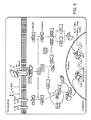
- Figure 5 shows the functional interrelationships between components of two exemplary signaling cascades.
- the arrows between components in this figure indicate that the component preceding the arrow activates the component following the arrow.
- the blunted lines indicated that the component preceding the blunted line inhibits the activity or function of the component following the blunted line.
- IL-1R IL-1 receptor accessory protein
- IL-1RAcP IL-1 receptor accessory protein
- IL-1 binding causes activation of two kinases, IRAK-1 and IRAK-2, associated with the IL-1 receptor complex.
- IRAK-1 IL-1 Receptor Associated Kinase
- TRAF6 activates and recruits TRAF6 to the IL-1 receptor complex.
- TRAF6 activates two pathways, one leading to NF-kB activation and another leading to c-jun activation.
- the TRAF associated protein ECSIT leads to c-Jun activation through the Map kinase/JNK signaling system.
- TRAF6 also signals through the TAB1/TAK1 kinases to trigger the degradation of 1-kB, and activation of NF-kB.
- a decrease in the abundance or absence of the processed form of MEKK1 a decrease in the abundance or absence of phosphorylated I ⁇ B ⁇ , a decrease in the abundance or absence of phosphorylated c-JUN, a decrease in the abundance or absence of ICAM-1, or a decrease in the abundance or absence of IL-6, TNF ⁇ , IFN ⁇ , IFN ⁇ , TGF ⁇ is indicative of inhibition of an interleukin-1 cytokine.
- a decrease or absence of activity or function of any of the above-listed components is indicative of inhibition of an interleukin-1 cytokine.
- Exemplary compounds incorporated to facilitate and expedite local delivery of topical compositions into ocular or adnexal tissues include, but are not limited to, alcohol (ethanol, propanol, and nonanol), fatty alcohol (lauryl alcohol), fatty acid (valeric acid, caproic acid and capric acid), fatty acid ester (isopropyl myristate and isopropyl n-hexanoate), alkyl ester (ethyl acetate and butyl acetate), polyol (propylene glycol, propanedione and hexanetriol), sulfoxide (dimethylsulfoxide and decylmethylsulfoxide), amide (urea, dimethylacetamide and pyrrolidone derivatives), surfactant (sodium lauryl sulfate, cetyltrimethylammonium bromide, polaxamers, spans, tweens, bile salts and lecithin),
- the composition further contains a compound selected from the group consisting of a physiological acceptable salt, poloxamer analogs with carbopol, carbopol/hydroxypropyl methyl cellulose (HPMC), carbopol-methyl cellulose, N-acetyl cysteine, carboxymethylcellulose (CMC), hyaluronic acid, cyclodextrin, and petroleum.
- a physiological acceptable salt poloxamer analogs with carbopol, carbopol/hydroxypropyl methyl cellulose (HPMC), carbopol-methyl cellulose, N-acetyl cysteine, carboxymethylcellulose (CMC), hyaluronic acid, cyclodextrin, and petroleum.
- mice were anesthetized, and a 2-mm circular corneal epithelial defect was made in one eye of each anesthetized mouse using a scalpel blade.
- mice were euthanized, and corneas were removed and fixed in 4% Para formaldehyde for 40 minutes at room temperature. Corneas were washed with PBS. Permeabilization and blocking was achieved with 2-hour incubation in 2% BSA in PBS and 0.2% Triton X-100. Corneas were incubated with rabbit anti-mouse ß III tubulin primary antibody at a dilution of 1:200 for 16 hours at 4°. After three washes in PBS corneas were incubated in goat anti-rabbit secondary antibody conjugated to rhodamine at a dilution of 1:200 for 2 hours at room temperature. The corneas were coverslipped with mounting medium and imaged.
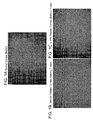
- the corneas were photographed at the level of the subbasal nerve plexus and terminal branching in cornea paracentral regions in the area of the wound ( Figure 1 ).
- nerve density at the level of terminal branching was normalized to the contralateral normal cornea.
- the results of this experiment showed that in the IL-1Ra-treated group, the percent density of regenerated nerve at the level of basal epithelial cell was 80% compared to 15% in the vehicle-treated group.
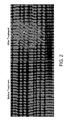
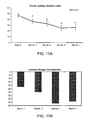
- corneal nerve morphology at the level of subbasal corneal nerve was assessed before and after one-month treatment with topical IL-1Ra 2.5% three times a day ( Figure 2 ).
- the sum of the length of the nerves per image was increased by 25% compared to baseline.
- the improvement in corneal nerve density was correlated with the reduction of signs and symptoms of dry eye in these patients.
- IL-1 blockade therefore, helps the epithelium improve its maintenance of corneal health by increasing nerve regeneration, which in turn breaks the vicious cycle of epithelial disease and nerve damage.
- IL-1 blockade can be used in all ophthalmologic conditions that affect the corneal epithelial health, including all forms of dry eye syndrome, all form of ocular surface diseases, corneal surgeries such as refractive surgeries (PRK, LASEK, Epi-LASIK, LASIK), corneal transplantation, epithelial debridement, ocular surface reconstruction, herpetic keratitis, neurotrophic keratitis, exposure keratopathy, diabetes mellitus, trigeminal nerve damage.
- Non-ophthalmic applications of IL-1 blockade would be all forms of peripheral neuropathy including diabetic peripheral neuropathy.
- animals After 5 days of treatment, animals were euthanized, and corneas were removed and fixed in 4% Para formaldehyde for 40 minutes at room temperature. Immunohistochemical staining for lymphatic vessels and blood vessels was performed on corneal flat mounts. Immunohistochemical staining against LYVE-1 (lymphatic endothelium-specific hyaluronic acid receptor) was performed with purified antibody followed by rhodamine conjugated secondary antibody. Immunohistochemical staining was also performed with FITC-conjugated CD31.
- IL-1Ra-treated corneas showed on average 60% less lymphangiogenesis activity compared to vehicle-treated corneas.
- blockade of IL-1 led to the immune cells having a lower level of maturity, which renders them less able to sensitize T cells.
- mice were divided in 5 groups.
- corneal fluorescein staining was performed by applying 0.5 ⁇ L of 1% fluorescein by micropipette into the inferior conjunctival sac of the mouse eye.
- the cornea was examined with a slit lamp biomicroscope in cobalt blue light 3 minutes after fluorescein instillation. Punctuate staining was recorded in a masked fashion with a standardized (National Eye Institute) grading system of 0 to 3 for each of the five areas in which the corneal surface was divided.
- corneal fluorescein staining was performed by applying 0.5 ⁇ L of 1% fluorescein by micropipette into the inferior conjunctival sac of the mouse eye.
- the cornea was examined with a slit lamp biomicroscope in cobalt blue light 3 minutes after fluorescein instillation. Punctuate staining was recorded in a masked fashion with a standardized (National Eye Institute) grading system of 0 to 3 for each of the five areas in which the corneal surface was divided. This experiment showed that percent reduction of corneal fluorescein staining was significantly higher in the group received topical IL-1Ra 5% mixed with N-acetyl cysteine 10% and the group received topical IL-1Ra 5% mixed with carboxymethylcellulose 1%.
- mice To examine the role of IL-1 blockade in corneal nerve homeostasis, minimization and prevention of nerve degeneration of the corneal subbasal nerve plexus and terminal epithelial branches was measured.
- Mice were then anesthetized, and a 2-mm circular corneal epithelial defect was made in one eye of each anesthetized mouse using a scalpel blade.
- mice were euthanized, and corneas were removed and fixed in 4% Para formaldehyde for 40 minutes at room temperature. Corneas were washed with PBS. Permeabilization and blocking was achieved with 2-hour incubation in 2% BSA in PBS and 0.2% Triton X-100. Corneas were incubated with rabbit anti-mouse ß III tubulin primary antibody at a dilution of 1:200 for 16 hours at 4°. After three washes in PBS corneas were incubated in goat anti-rabbit secondary antibody conjugated to rhodamine at a dilution of 1:200 for 2 hours at room temperature. The corneas were coverslipped with mounting medium and imaged.
- the corneas are photographed at the level of the subbasal nerve plexus and terminal branching in cornea paracentral regions in the area of the wound.
- nerve density at the level of terminal branching is normalized to the contralateral normal cornea.
- the results of this experiment show that that in the IL-1Ra-treated group, the percent density of degenerated nerve at the level of basal epithelial cell was decreased compared to the vehicle-treated group.
- IL-1 blockade therefore, can help the epithelium improve its maintenance of corneal health by minimizing or preventing nerve degeneration which in turn can break the vicious cycle of epithelial disease and nerve damage.
- IL-1 blockade can be used in all ophthalmologic conditions that affect the corneal epithelial health including all forms of dry eye syndrome, all form of ocular surface diseases, corneal surgeries such as refractive surgeries (PRK, LASEK, Epi-LASIK, LASIK), corneal transplantation, epithelial debridement, ocular surface reconstruction, herpetic keratitis, neurotrophic keratitis, exposure keratopathy, diabetes mellitus, trigeminal nerve damage.
- Non-ophthalmic applications of IL-1 blockade would be all forms of peripheral neuropathy including diabetic peripheral neuropathy.
- Example 6 IL-1 and IL-17 blockade provides unexpected protection of corneal nerves
- the method comprises administration of a compound that inhibits binding of an inflammatory IL-17 cytokine to the IL- 17 receptor complex.
- a method for regenerating corneal nerves is also carried out by locally administering to an eye of a subject a composition comprising a polynucleotide, a polypeptide, an antibody, a compound, or a small molecule that inhibits or modifies the transcription, transcript stability, translation, modification, localization, secretion, or function of a polynucleotide or polypeptide encoding an inflammatory interleukin-17 cytokine or any component of the IL-17 receptor complex.
- the composition may comprise a neutralizing or function-blocking antibody against IL- 17 and/or a receptor complex.
- the neutralizing or function-blocking antibody against IL-17 may be a reformulated or humanized derivative of or bind to the epitope of human IL- 17 affinity purified polyclonal antibody (Catalog # AF-317-NA, R&D Systems), human IL- 17 allophycocyanin monoclonal antibody (clone 41802)(Catalog # IC3171A, R&D Systems), human IL- 17 biotinylated affinity purified polyclonal antibody (Catalog # BAF317, R&D Systems), human IL- 17 monoclonal antibody (clone 41802)(Catalog # MAB3171 , R&D Systems), human IL- 17 monoclonal antibody (clone 41809)(Catalog # MAB317, R&D Systems), human IL- 17 phycoerythrin monoclonal antibody (clone 4180
- the neutralizing or function-blocking antibody against IL- 17 may be a reformulated or humanized derivative of or bind to the epitope of monoclonal anti-human IL- 17 antibody, (Clone: 41809, Catalog # MAB317, R&D Systems), anti-human IL- 17 antibody, polyclonal raised in Goat, (Catalog # AF-317-NA, R&D Systems), or recombinant human IL- 17 RTFc chimera (Catalog # 177-IR, R&D Systems).
- formulations are in the form of a solid, a paste, an ointment, a gel, a liquid, an aerosol, a mist, a polymer, a contact lens, a film, an emulsion, or a suspension.
- the formulations are administered topically, e . g ., the composition is delivered and directly contacts the eye.
- the compositions are administered with a pharmaceutically acceptable liquid carrier, e.g., a liquid carrier, which is aqueous or partly aqueous.
- the compositions are associated with a liposome (e.g., a cationic or anionic liposome).
- the neutralizing or function-blocking antibody against an IL-17 receptor may be a reformulated or humanized derivative of or bind to the epitope of human IL- 17R affinity purified polyclonal antibody (Catalog # AF 177, R&D Systems), human IL- 17R allophycocyanin monoclonal antibody (clone 133617)(Catalog # FAB177A, R&D Systems), human IL- 17R biotinylated affinity purified polyclonal antibody (Catalog # BAF 177, R&D Systems), human IL-17R fluorescein monoclonal antibody (clone 133617) (Catalog # FAB177F, R&D Systems), human IL-17R monoclonal antibody (clone 133617)(Catalog # MAB 177, R&D Systems), human IL- 17R monoclonal antibody (clone 133621)(Catalog # MAB 1771 , R&D Systems), human IL-
- the neutralizing or function-blocking antibody against an IL- 17 may be a reformulated or humanized derivative of, or bind to the epitope of, one or more mouse anti-IL-17A (SKU #s including but not limited to, 7172, 7173, 7175, 7177, 8171, 7371 , 7971, and 7370, eBioscience) or mouse anti-IL-17F (SKU #s including, but not limited to, 7471 and 8471 , eBioscience).
- the neutralizing or function-blocking antibody against an IL-17 may be a reformulated or humanized derivative of one or more human anti-1L- 17A (SKU #s including, but not limited to, 7178, 7179, 8179, 7176, 7976, and 7876 or human anti-IL-17F SKU #s including, but not limited to, 8479, eBioscience).
- the neutralizing or function-blocking antibody against an IL- 17 may be a reformulated or humanized derivative of, or bind to the epitope of functional grade purified anti-human IL-17A antibody (Clone: eBio64CAP17, Catalog # 16-7178. eBioscience).
- the composition may comprise an intrabody that binds to the IL- 17 receptor complex or any synthetic intermediate of IL- 17 or the IL- 17 receptor complex.
- the composition may alternatively, or in addition, comprise a soluble fragment of the IL- 17 receptor complex which binds IL-17.
- Exemplary polypeptides include, but are not limited to, fusion and/or chimeric proteins capable of disrupting IL- 17 function.
- the composition comprises morpholino antisense oligonucleotides, microRNAs (miRNAs), short hairpin RNA (shRNA), or short interfering RNA (siRNA) to silence gene expression.
- miRNAs microRNAs
- shRNA short hairpin RNA
- siRNA short interfering RNA
- Contemplated function-blocking antibodies targeted against an IL- 17 cytokine or an IL- 17 receptor are monoclonal or polyclonal.
- the contemplated antibody binds to one or more sequences within an IL- 17 or IL- 17 receptor polypeptide.
- the antibody is alternatively an intrabody.
- the antibody comprises a single chain, a humanized, a recombinant, or a chimeric antibody.
- One or more compounds are directly or indirectly conjugated onto this antibody.
- Antagonists of IL- 17 and/or its receptor complex are administered either simultaneously or sequentially with an antagonist of IL-1 and/or its receptor.
- Human IL-17 is encoded by the mRNA sequence of NCBI Accession No. NM 002190, alternatively called IL-17A. Human IL-17 is encoded by the amino acid sequence of NCBI Accession No. NM_002190, alternatively called IL-17A. Human IL-17B is encoded by the mRNA sequence of NCBI Accession No. NM_014443. Human IL- 17B is encoded by the amino acid sequence of NCBI Accession No. NM_014443. Human IL-17C is encoded by the mRNA sequence of NCBI Accession No. NM_013278. Human IL- 17C is encoded by the amino acid sequence of NCBI Accession No. NM_013278.
- Human IL- 17D is encoded by the mRNA sequence of NCBI Accession No. NM_138284. Human IL-17D is encoded by the amino acid sequence of NCBI Accession No. NM_138284. Human IL- 17E is encoded by the mRNA sequence of NCBI Accession No. AF305200. Human IL- 17E is encoded by the amino acid sequence of NCBI Accession No. AF305200. Human IL- 17F is encoded by the mRNA sequence of NCBI Accession No. NM 052872. Human IL-17F is encoded by the amino acid sequence of NCBI Accession No. NM_052872.
- IL-17 receptor antagonist (IL-17RA) is encoded by the mRNA sequence ofNCBI Accession No. NM 014339.
- IL- 17RA is encoded by the amino acid sequence of NCBI Accession No. NMJ4339.
- IL- 17RB is encoded by the mRNA sequence of NCBI Accession No. NM_018725.
- IL-17RB is encoded by the amino acid sequence of NCBI Accession No. NM_018725.
- IL-17RC, transcript variant 1 is encoded by the mRNA sequence of (NCBI Accession No. NM 1 53461.
- IL- 17RC transcript variant 1, is encoded by the amino acid sequence ofNCBI Accession No. NM_153461.
- IL-17RC transcript variant 2
- IL- 17RC transcript variant 2
- IL- 17RC transcript variant 2
- IL- 17RC transcript variant 3
- IL-17RC transcript variant 3
- IL-17RC transcript variant 3
- IL- 17RD transcript 1
- IL-17RD transcript 1
- IL-17RD transcript 2 is encoded by the mRNA sequence of NCBI Accession No. NM O1 7563.
- IL-17RD, transcript 2 is encoded by the amino acid sequence of NCBI Accession No. NM_017563.
- IL- 17RE, transcript variant 1 is encoded by the mRNA sequence of NCBI Accession No. NM 1 53480.
- IL- 17RE, transcript variant 1 is encoded by the amino acid sequence of NCBI Accession No. NM_153480.
- IL- 17RE, transcript variant 2 is encoded by the mRNA sequence of NCBI Accession No. NM_153481.
- IL- 17RE, transcript variant 2 is encoded by the amino acid sequence of NCBI Accession No. NM_153481.
- IL- 17RE, transcript variant 5 is encoded by the mRNA sequence of NCBI Accession No. NM_153483.
- IL- 17RE, transcript variant 5, is encoded by the amino acid sequence of NCBI Accession No. NM_15348
- DED dry eye disease
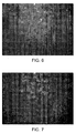
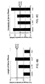
- both the anti-cytokine therapies including IL-1Ra and anti-IL 17 prevented loss of nerve fibers in DED corneas ( Figure 8B ).
- use of a lubricating drop ("Vehicle") or CsA showed ⁇ 2-fold decrease in the fiber length compared to those in normal corneas, suggesting that standard treatments against DED (lubrication or topical cyclosporine) do not protect significantly against nerve damage.
- IL-IRa and anti-IL17 therapies maintain the nerve fiber tortuosity levels similar to that seen in normal corneas, whereas DED corneas treated with vehicle or CsA show ⁇ 2-fold increase in the nerve fiber tortuosity as compared to the normals ( Figure 8C ).
- the nerve tortuosity data and the nerve fiber length data described above indicate that conventional therapies for DED (topical lubricating tear ointment or topical cyclosporine-A) do not protect significantly against nerve damage.
- DED topical lubricating tear ointment or topical cyclosporine-A
- ocular surface inflammation associated with DED is often correlated with elevated levels of tear nerve growth factor, which is typically reduced with steroids such as 0.1% prednisolone
- the results above indicate that such therapy are ineffective at inducing corneal nerve regeneration.
- administration of such immunosuppressive agents e . g ., macrolides such as cyclosporin A (e.g., Restasis ® ), or corticosteroids such as prednisolone
- Example 7 Combination therapy blockade of IL-1 and IL-17 enhances corneal nerve regeneration IL-1 is a major initiator of the inflammatory process upstream of T cell activation and plays a crucial role in precipitating the inflammation by promoting the induction or expansion of IL-17-secreting T cells.
- IL-1 and IL-17 work synergistically and enhance secretion of each other in vivo.
- An IL-17 blocking treatment is applied to a cornea, along with an IL-1 blocking treatment.
- the co-administration of IL-1 and IL-17 blocking treatment enhances corneal nerve regeneration such that the amount of nerve fibers associated with damaged corneas is increased compared to untreated corneas.
- the co-administration of IL-1 and IL-17 blocking treatment synergistically enhances corneal nerve regeneration in the case of immune-mediated corneal nerve damage.
- Example 8 Efficacy of topical application of IL-1 Ra in ocular surface inflammatory disorders in a human
- IL-1Ra open label study was utilized to determine the efficacy of IL-1Ra in treating ocular surface inflammatory disorders in a human. Seventy patients were enrolled in the study and various numbers of patients were available for monthly checkups as follows: month 1 (44 patients), month 3 (37 patients), month 5 (22 patients), and month 7 (17 patients). None of these patients had severe meibomian gland dysfunction (MGD, also called posterior blepharitis) defined as grade 3 meibomian secretions (secretions retain shape after expression), or grade 4 lid margin disease (marked diffuse redness of both lid margins and skin), or grade 4 conjunctival hyperemia (marked dark redness of the palpebral and/or bulbar conjunctiva). Of note, patients with MGD/meibomian gland dysfunction alone do not develop significant corneal neuropathy (Foulks and Bron, 2003 Ocul Surf, 107-26).
- corneal staining was utilized to measure ocular surface disease treated with topical IL-1 receptor antagonist.
- the application of fluorescein was used to detect epithelial damage in the cornea, and each patient's disease severity was graded using the Oxford scale.
- the data demonstrate a significant reduction in staining (severity of disease) with topical blockade of IL-1 using the IL-1 receptor antagonist. Specifically, a significant change was observed from baseline to 7 months follow-up after treatment with IL-1 receptor antagonist.
- OSDI Ocular surface disease index
- the Schirmer test measures the amount of tear produced by a patient's eye. The test is performed by placing a special scaled filter paper strip between the lower eyelid and the eyeball for 5 minutes. As shown in Figure 12 , tear volume increased from below 6 mm at baseline to ⁇ 9 mm at 7 months of follow-up. The data suggest an overall trend toward higher tear secretion with topical blockade of IL-1 with IL-1 receptor antagonist.
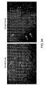
- Figure 13 shows confocal micrographs of corneas from patients with dry eye disease before and after treatment with topical vehicle or topical IL-IRa.
- One month of IL-Ra treatment promoted corneal nerve regeneration (marked with white arrows).
- one month of vehicle treatment did not promote corneal nerve regeneration.
Landscapes
- Health & Medical Sciences (AREA)
- Life Sciences & Earth Sciences (AREA)
- Engineering & Computer Science (AREA)
- Chemical & Material Sciences (AREA)
- Bioinformatics & Cheminformatics (AREA)
- General Health & Medical Sciences (AREA)
- Zoology (AREA)
- Animal Behavior & Ethology (AREA)
- Pharmacology & Pharmacy (AREA)
- Medicinal Chemistry (AREA)
- Public Health (AREA)
- Veterinary Medicine (AREA)
- Epidemiology (AREA)
- Genetics & Genomics (AREA)
- Immunology (AREA)
- Proteomics, Peptides & Aminoacids (AREA)
- Gastroenterology & Hepatology (AREA)
- Biomedical Technology (AREA)
- Organic Chemistry (AREA)
- Wood Science & Technology (AREA)
- General Engineering & Computer Science (AREA)
- Biotechnology (AREA)
- Molecular Biology (AREA)
- Ophthalmology & Optometry (AREA)
- Marine Sciences & Fisheries (AREA)
- Plant Pathology (AREA)
- Endocrinology (AREA)
- Physics & Mathematics (AREA)
- Biophysics (AREA)
- Microbiology (AREA)
- Cell Biology (AREA)
- Biochemistry (AREA)
- Nuclear Medicine, Radiotherapy & Molecular Imaging (AREA)
- General Chemical & Material Sciences (AREA)
- Chemical Kinetics & Catalysis (AREA)
- Medicines That Contain Protein Lipid Enzymes And Other Medicines (AREA)
- Pharmaceuticals Containing Other Organic And Inorganic Compounds (AREA)
- Medicinal Preparation (AREA)
- Medicines Containing Antibodies Or Antigens For Use As Internal Diagnostic Agents (AREA)
Applications Claiming Priority (2)
| Application Number | Priority Date | Filing Date | Title |
|---|---|---|---|
| US14356109P | 2009-01-09 | 2009-01-09 | |
| PCT/US2010/020646 WO2010081091A2 (en) | 2009-01-09 | 2010-01-11 | Therapeutic compositions for treatment of corneal disorders |
Publications (2)
| Publication Number | Publication Date |
|---|---|
| EP2385839A2 EP2385839A2 (en) | 2011-11-16 |
| EP2385839B1 true EP2385839B1 (en) | 2014-11-26 |
Family
ID=42144303
Family Applications (1)
| Application Number | Title | Priority Date | Filing Date |
|---|---|---|---|
| EP10700459.0A Active EP2385839B1 (en) | 2009-01-09 | 2010-01-11 | Therapeutic compositions for treatment of corneal disorders |
Country Status (6)
| Country | Link |
|---|---|
| US (1) | US10117906B2 (cg-RX-API-DMAC7.html) |
| EP (1) | EP2385839B1 (cg-RX-API-DMAC7.html) |
| JP (1) | JP2012515164A (cg-RX-API-DMAC7.html) |
| AU (1) | AU2010203425B2 (cg-RX-API-DMAC7.html) |
| CA (1) | CA2749537C (cg-RX-API-DMAC7.html) |
| WO (1) | WO2010081091A2 (cg-RX-API-DMAC7.html) |
Families Citing this family (17)
| Publication number | Priority date | Publication date | Assignee | Title |
|---|---|---|---|---|
| HRP20230776T1 (hr) * | 2010-07-29 | 2023-10-27 | Buzzard Pharmaceuticals AB | Himerni antagonisti receptora il-1 tipa i |
| WO2012103240A2 (en) * | 2011-01-25 | 2012-08-02 | Eleven Biotherapeutics, Inc. | Receptor binding agents |
| WO2013126599A1 (en) | 2012-02-21 | 2013-08-29 | Massachusetts Eye & Ear Infirmary | Meibomian gland dysfunction |
| WO2013126602A1 (en) | 2012-02-21 | 2013-08-29 | Massachusetts Eye & Ear Infirmary | Inflammatory eye disorders |
| WO2014036445A2 (en) * | 2012-08-31 | 2014-03-06 | Johnson Michael R | Novel mucolytic agents |
| JP6450364B2 (ja) | 2013-03-13 | 2019-01-09 | セセン バイオ, インコーポレイテッド | 眼送達のためのキメラサイトカイン製剤 |
| SG11201601291QA (en) | 2013-08-23 | 2016-03-30 | Parion Sciences Inc | Dithiol mucolytic agents |
| JP6377618B2 (ja) * | 2013-08-28 | 2018-08-22 | 千寿製薬株式会社 | 角膜上皮障害治療剤 |
| AR103173A1 (es) | 2014-12-22 | 2017-04-19 | Novarits Ag | Productos farmacéuticos y composiciones líquidas estables de anticuerpos il-17 |
| BR112017016067A2 (pt) | 2015-01-30 | 2018-04-03 | Parion Sciences Inc | agentes mucolíticos de monotiol |
| JP2018520986A (ja) | 2015-04-30 | 2018-08-02 | パリオン・サイエンシィズ・インコーポレーテッド | ジチオール粘液溶解剤の新規なプロドラッグ |
| US20180312838A1 (en) * | 2015-10-21 | 2018-11-01 | The Regents Of The University Of California | Modulation of microrna 184 to treat pathological lymphangiogenesis |
| WO2018231827A1 (en) | 2017-06-12 | 2018-12-20 | Bluefin Biomedicine, Inc. | Anti-il1rap antibodies and antibody drug conjugates |
| WO2020218488A1 (ja) * | 2019-04-26 | 2020-10-29 | 千寿製薬株式会社 | 眼疾患マーカー |
| CN113350326B (zh) * | 2021-07-28 | 2023-03-17 | 爱尔眼科医院集团股份有限公司 | 化合物lm22b-10在制备角膜上皮及神经损伤治疗药物中的应用 |
| KR102883890B1 (ko) * | 2021-11-30 | 2025-11-07 | 한림대학교 산학협력단 | miR-30c-1 발현 수준을 증가시키기 위한 제제를 사용하는 각막 내피 세포 배양 방법 및 각막 내피 세포 질환 치료용 조성물 |
| CN118304388B (zh) * | 2024-04-03 | 2025-02-11 | 南通大学 | Tak1在制备治疗周围神经损伤药物中的应用 |
Family Cites Families (41)
| Publication number | Priority date | Publication date | Assignee | Title |
|---|---|---|---|---|
| EP0610336B1 (en) | 1991-10-31 | 2002-03-27 | The Victoria University Of Manchester | Treatment of neurological conditions by an interleukin-1 inhibiting compound |
| US5900407A (en) * | 1997-02-06 | 1999-05-04 | Inspire Pharmaceuticals, Inc. | Method of treating dry eye disease with uridine triphosphates and related compounds |
| US6974682B1 (en) | 1996-08-26 | 2005-12-13 | Human Genome Sciences, Inc. | Soluble interleukin-1 receptor accessory molecule |
| WO1998022130A1 (en) * | 1996-11-19 | 1998-05-28 | The Schepens Eye Research Institute, Inc. | Local use of il-1ra in corneal transplant rejection or disorders of the eye |
| US6337072B1 (en) | 1998-04-03 | 2002-01-08 | Hyseq, Inc. | Interleukin-1 receptor antagonist and recombinant production thereof |
| US6927044B2 (en) | 1998-09-25 | 2005-08-09 | Regeneron Pharmaceuticals, Inc. | IL-1 receptor based cytokine traps |
| JP3363832B2 (ja) | 1999-05-13 | 2003-01-08 | アルプス電気株式会社 | 薄膜構造体およびその製造方法 |
| US20030104041A1 (en) | 1999-12-16 | 2003-06-05 | Tsung-Min Hsu | Transdermal and topical administration of drugs using basic permeation enhancers |
| EP1252895A4 (en) | 2000-01-31 | 2003-04-16 | Santen Pharmaceutical Co Ltd | REMEDIES FOR OPHTHALMIC DISORDERS |
| WO2001057082A2 (en) | 2000-02-03 | 2001-08-09 | Regeneration Technologies, Inc. | Extraction of growth factors from tissue |
| US20010047204A1 (en) * | 2000-03-20 | 2001-11-29 | Medennium, Inc. | Method for determination of a properly sized posterior chamber phakic refractive lens |
| US20020131954A1 (en) | 2000-05-02 | 2002-09-19 | Tobinick Edward L. | Interleukin antagonists for the treatment of neurological, retinal and muscular disorders |
| US20030026806A1 (en) | 2000-10-27 | 2003-02-06 | Amgen Inc. | Antibodies and other selective IL-1 binding agents that allow binding to IL-1 receptor but not activation thereof |
| US20050143333A1 (en) | 2001-05-18 | 2005-06-30 | Sirna Therapeutics, Inc. | RNA interference mediated inhibition of interleukin and interleukin receptor gene expression using short interfering nucleic acid (SINA) |
| WO2003022213A2 (en) | 2001-09-06 | 2003-03-20 | Board Of Regents, The University Of Texas System | Therapeutic treatments for spinal cord injury via blockade of interleukin-1 receptor |
| US7683165B2 (en) | 2002-02-20 | 2010-03-23 | Sirna Therapeutics, Inc. | RNA interference mediated inhibition of interleukin and interleukin receptor gene expression using short interfering nucleic acid (siNA) |
| US6890654B2 (en) | 2002-04-18 | 2005-05-10 | Northwestern University | Encapsulation of nanotubes via self-assembled nanostructures |
| WO2004060911A2 (en) * | 2002-12-30 | 2004-07-22 | Amgen Inc. | Combination therapy with co-stimulatory factors |
| US20050196370A1 (en) * | 2003-03-18 | 2005-09-08 | Zhi-Jian Yu | Stable ophthalmic oil-in-water emulsions with sodium hyaluronate for alleviating dry eye |
| US20050033694A1 (en) * | 2003-06-03 | 2005-02-10 | United States Postal Service. | System and method for fleet card management |
| US7261882B2 (en) | 2003-06-23 | 2007-08-28 | Reagents Of The University Of Colorado | Methods for treating neuropathic pain by administering IL-10 polypeptides |
| US20050023872A1 (en) * | 2003-07-28 | 2005-02-03 | Hetzel Thomas R. | Modular seat cushion with interlocking human support and base portions and method of creating and using a seat cushion |
| KR100543466B1 (ko) * | 2003-11-13 | 2006-01-20 | 삼성전자주식회사 | 영상 보간 장치 및 방법 |
| GB0404693D0 (en) | 2004-03-02 | 2004-04-07 | Univ London | Pharmaceutical preparations for the treatment of ocular surface and other disorders |
| US8618054B2 (en) | 2004-05-05 | 2013-12-31 | Valorisation-Rechereche Société en Commandite | Interleukin-1 receptor antagonists, compositions, and methods of treatment |
| US8541413B2 (en) | 2004-10-01 | 2013-09-24 | Ramscor, Inc. | Sustained release eye drop formulations |
| CA2588449A1 (en) * | 2004-11-24 | 2006-06-01 | Therakine Corporation | An implant for intraocular drug delivery |
| JP4815808B2 (ja) | 2005-01-24 | 2011-11-16 | 三菱電機株式会社 | ロータリ圧縮機 |
| CN102293774A (zh) | 2005-03-31 | 2011-12-28 | 旭硝子株式会社 | 前列腺素F2α衍生物的应用 |
| EP1870099A4 (en) | 2005-04-13 | 2008-07-16 | Ube Industries | PROTECTION AGAINST RETINAL NERVOUS CELLS WITH INDAZOLE DERIVATIVE AS ACTIVE SUBSTANCE |
| US7670849B2 (en) | 2005-04-29 | 2010-03-02 | Henkin Robert I | Method for diagnosing insulin resistance from nasal secretions |
| DE602006009834D1 (de) * | 2005-09-01 | 2009-11-26 | Schering Corp | Verwendung von il-23 und il-17-antagonisten zur behandlung von autoimmuner entzündlicher augenerkrankung |
| KR20110079922A (ko) * | 2006-04-14 | 2011-07-11 | 노파르티스 아게 | 안과 장애 치료를 위한 il-1 항체의 용도 |
| US20080026485A1 (en) * | 2006-04-18 | 2008-01-31 | Wolfgang Hueber | Antibody profiling for determination of patient responsiveness |
| CN101489569A (zh) | 2006-06-12 | 2009-07-22 | 席拉坎有限责任公司 | 眼表面疾病的局部治疗 |
| CA2679588A1 (en) * | 2007-03-26 | 2008-10-02 | Zymogenetics, Inc. | Soluble il-17ra/rc fusion proteins and related methods |
| US8399421B2 (en) | 2007-03-30 | 2013-03-19 | The Board Of Regents Of The University Of Texas System | Treatment for neuropathic pain due to spinal cord injury |
| EP2187900B1 (en) | 2007-08-16 | 2016-11-09 | The Schepens Eye Research Institute, Inc. | Therapeutic compositions for treatment of inflammation of ocular and adnexal tissues |
| CA2711696C (en) * | 2008-01-09 | 2021-10-26 | Reza Dana | Therapeutic compositions for treatment of ocular inflammatory disorders |
| US20120014970A1 (en) * | 2009-01-09 | 2012-01-19 | Reza Dana | Therapeutic Compositions for Treatment of Corneal Disorders |
| US9011861B2 (en) * | 2010-02-25 | 2015-04-21 | Schepens Eye Research Institute | Therapeutic compositions for the treatment of dry eye disease |
-
2010
- 2010-01-11 EP EP10700459.0A patent/EP2385839B1/en active Active
- 2010-01-11 CA CA2749537A patent/CA2749537C/en not_active Expired - Fee Related
- 2010-01-11 WO PCT/US2010/020646 patent/WO2010081091A2/en not_active Ceased
- 2010-01-11 US US12/685,510 patent/US10117906B2/en active Active
- 2010-01-11 JP JP2011545490A patent/JP2012515164A/ja active Pending
- 2010-01-11 AU AU2010203425A patent/AU2010203425B2/en not_active Ceased
Also Published As
| Publication number | Publication date |
|---|---|
| WO2010081091A3 (en) | 2010-11-18 |
| US20100183587A1 (en) | 2010-07-22 |
| AU2010203425A1 (en) | 2011-07-28 |
| CA2749537A1 (en) | 2010-07-15 |
| CA2749537C (en) | 2018-11-20 |
| WO2010081091A2 (en) | 2010-07-15 |
| JP2012515164A (ja) | 2012-07-05 |
| US10117906B2 (en) | 2018-11-06 |
| AU2010203425B2 (en) | 2015-04-09 |
| EP2385839A2 (en) | 2011-11-16 |
Similar Documents
| Publication | Publication Date | Title |
|---|---|---|
| US10117906B2 (en) | Methods for reducing corneal nerves damage, corneal lymphangiogenesis or immunity to corneal antigens in dry-eye associated ocular surface diseases by IL-1Ra | |
| JP5886523B2 (ja) | 眼の炎症性障害を処置するための治療組成物 | |
| EP2187900B1 (en) | Therapeutic compositions for treatment of inflammation of ocular and adnexal tissues | |
| US10940179B2 (en) | Therapeutic compositions for the treatment of dry eye disease | |
| Lambiase et al. | Expression of nerve growth factor receptors on the ocular surface in healthy subjects and during manifestation of inflammatory diseases. | |
| JP2011516400A5 (cg-RX-API-DMAC7.html) | ||
| Chucair-Elliott et al. | IL-6 contributes to corneal nerve degeneration after herpes simplex virus type I infection | |
| JP2011517659A5 (cg-RX-API-DMAC7.html) | ||
| US20120014970A1 (en) | Therapeutic Compositions for Treatment of Corneal Disorders | |
| Gagliano et al. | Intravitreal steroids for the prevention of PVR after surgery for retinal detachment | |
| RU2564920C2 (ru) | Офтальмологические препараты на основе bdnf (нейротрофического фактора головного мозга) и их применение | |
| JP6440107B2 (ja) | 細胞シートの製造方法、組成物、細胞培養補助剤、及び細胞培養方法 | |
| AU2017205185B2 (en) | Therapeutics for ocular immunoinflammatory diseases | |
| CA2640986C (en) | Therapeutic compositions for treatment of inflammation of ocular and adnexal tissues | |
| AU2014227534B2 (en) | Therapeutic compositions for treatment of inflammation of ocular and adnexal tissues | |
| CN114206849A (zh) | 用于治疗非感染性眼部免疫炎性病症的治疗方法 | |
| Sabatino et al. | SECTION: Laboratory Science, James V. Jester, PhD, Editor The intriguing role of neuropeptides at the ocular surface |
Legal Events
| Date | Code | Title | Description |
|---|---|---|---|
| PUAI | Public reference made under article 153(3) epc to a published international application that has entered the european phase |
Free format text: ORIGINAL CODE: 0009012 |
|
| 17P | Request for examination filed |
Effective date: 20110808 |
|
| AK | Designated contracting states |
Kind code of ref document: A2 Designated state(s): AT BE BG CH CY CZ DE DK EE ES FI FR GB GR HR HU IE IS IT LI LT LU LV MC MK MT NL NO PL PT RO SE SI SK SM TR |
|
| DAX | Request for extension of the european patent (deleted) | ||
| 17Q | First examination report despatched |
Effective date: 20130102 |
|
| GRAP | Despatch of communication of intention to grant a patent |
Free format text: ORIGINAL CODE: EPIDOSNIGR1 |
|
| RIC1 | Information provided on ipc code assigned before grant |
Ipc: A61K 9/00 20060101ALI20140602BHEP Ipc: A61K 38/17 20060101AFI20140602BHEP Ipc: A61P 27/02 20060101ALI20140602BHEP Ipc: A61K 38/20 20060101ALI20140602BHEP Ipc: C12N 15/113 20100101ALI20140602BHEP |
|
| INTG | Intention to grant announced |
Effective date: 20140708 |
|
| GRAS | Grant fee paid |
Free format text: ORIGINAL CODE: EPIDOSNIGR3 |
|
| GRAA | (expected) grant |
Free format text: ORIGINAL CODE: 0009210 |
|
| AK | Designated contracting states |
Kind code of ref document: B1 Designated state(s): AT BE BG CH CY CZ DE DK EE ES FI FR GB GR HR HU IE IS IT LI LT LU LV MC MK MT NL NO PL PT RO SE SI SK SM TR |
|
| REG | Reference to a national code |
Ref country code: GB Ref legal event code: FG4D |
|
| REG | Reference to a national code |
Ref country code: CH Ref legal event code: EP |
|
| REG | Reference to a national code |
Ref country code: AT Ref legal event code: REF Ref document number: 697797 Country of ref document: AT Kind code of ref document: T Effective date: 20141215 |
|
| REG | Reference to a national code |
Ref country code: IE Ref legal event code: FG4D |
|
| REG | Reference to a national code |
Ref country code: DE Ref legal event code: R096 Ref document number: 602010020513 Country of ref document: DE Effective date: 20150108 |
|
| PGFP | Annual fee paid to national office [announced via postgrant information from national office to epo] |
Ref country code: NL Payment date: 20150126 Year of fee payment: 6 |
|
| REG | Reference to a national code |
Ref country code: NL Ref legal event code: VDEP Effective date: 20141126 |
|
| REG | Reference to a national code |
Ref country code: AT Ref legal event code: MK05 Ref document number: 697797 Country of ref document: AT Kind code of ref document: T Effective date: 20141126 |
|
| REG | Reference to a national code |
Ref country code: LT Ref legal event code: MG4D |
|
| PG25 | Lapsed in a contracting state [announced via postgrant information from national office to epo] |
Ref country code: IS Free format text: LAPSE BECAUSE OF FAILURE TO SUBMIT A TRANSLATION OF THE DESCRIPTION OR TO PAY THE FEE WITHIN THE PRESCRIBED TIME-LIMIT Effective date: 20150326 Ref country code: PT Free format text: LAPSE BECAUSE OF FAILURE TO SUBMIT A TRANSLATION OF THE DESCRIPTION OR TO PAY THE FEE WITHIN THE PRESCRIBED TIME-LIMIT Effective date: 20150326 Ref country code: NO Free format text: LAPSE BECAUSE OF FAILURE TO SUBMIT A TRANSLATION OF THE DESCRIPTION OR TO PAY THE FEE WITHIN THE PRESCRIBED TIME-LIMIT Effective date: 20150226 Ref country code: NL Free format text: LAPSE BECAUSE OF FAILURE TO SUBMIT A TRANSLATION OF THE DESCRIPTION OR TO PAY THE FEE WITHIN THE PRESCRIBED TIME-LIMIT Effective date: 20141126 Ref country code: ES Free format text: LAPSE BECAUSE OF FAILURE TO SUBMIT A TRANSLATION OF THE DESCRIPTION OR TO PAY THE FEE WITHIN THE PRESCRIBED TIME-LIMIT Effective date: 20141126 Ref country code: FI Free format text: LAPSE BECAUSE OF FAILURE TO SUBMIT A TRANSLATION OF THE DESCRIPTION OR TO PAY THE FEE WITHIN THE PRESCRIBED TIME-LIMIT Effective date: 20141126 Ref country code: LT Free format text: LAPSE BECAUSE OF FAILURE TO SUBMIT A TRANSLATION OF THE DESCRIPTION OR TO PAY THE FEE WITHIN THE PRESCRIBED TIME-LIMIT Effective date: 20141126 |
|
| PGFP | Annual fee paid to national office [announced via postgrant information from national office to epo] |
Ref country code: ES Payment date: 20150126 Year of fee payment: 6 |
|
| PG25 | Lapsed in a contracting state [announced via postgrant information from national office to epo] |
Ref country code: GR Free format text: LAPSE BECAUSE OF FAILURE TO SUBMIT A TRANSLATION OF THE DESCRIPTION OR TO PAY THE FEE WITHIN THE PRESCRIBED TIME-LIMIT Effective date: 20150227 Ref country code: HR Free format text: LAPSE BECAUSE OF FAILURE TO SUBMIT A TRANSLATION OF THE DESCRIPTION OR TO PAY THE FEE WITHIN THE PRESCRIBED TIME-LIMIT Effective date: 20141126 Ref country code: CY Free format text: LAPSE BECAUSE OF FAILURE TO SUBMIT A TRANSLATION OF THE DESCRIPTION OR TO PAY THE FEE WITHIN THE PRESCRIBED TIME-LIMIT Effective date: 20141126 Ref country code: SE Free format text: LAPSE BECAUSE OF FAILURE TO SUBMIT A TRANSLATION OF THE DESCRIPTION OR TO PAY THE FEE WITHIN THE PRESCRIBED TIME-LIMIT Effective date: 20141126 Ref country code: AT Free format text: LAPSE BECAUSE OF FAILURE TO SUBMIT A TRANSLATION OF THE DESCRIPTION OR TO PAY THE FEE WITHIN THE PRESCRIBED TIME-LIMIT Effective date: 20141126 Ref country code: LV Free format text: LAPSE BECAUSE OF FAILURE TO SUBMIT A TRANSLATION OF THE DESCRIPTION OR TO PAY THE FEE WITHIN THE PRESCRIBED TIME-LIMIT Effective date: 20141126 |
|
| PGFP | Annual fee paid to national office [announced via postgrant information from national office to epo] |
Ref country code: AT Payment date: 20150102 Year of fee payment: 6 |
|
| PG25 | Lapsed in a contracting state [announced via postgrant information from national office to epo] |
Ref country code: CZ Free format text: LAPSE BECAUSE OF FAILURE TO SUBMIT A TRANSLATION OF THE DESCRIPTION OR TO PAY THE FEE WITHIN THE PRESCRIBED TIME-LIMIT Effective date: 20141126 Ref country code: SK Free format text: LAPSE BECAUSE OF FAILURE TO SUBMIT A TRANSLATION OF THE DESCRIPTION OR TO PAY THE FEE WITHIN THE PRESCRIBED TIME-LIMIT Effective date: 20141126 Ref country code: RO Free format text: LAPSE BECAUSE OF FAILURE TO SUBMIT A TRANSLATION OF THE DESCRIPTION OR TO PAY THE FEE WITHIN THE PRESCRIBED TIME-LIMIT Effective date: 20141126 Ref country code: DK Free format text: LAPSE BECAUSE OF FAILURE TO SUBMIT A TRANSLATION OF THE DESCRIPTION OR TO PAY THE FEE WITHIN THE PRESCRIBED TIME-LIMIT Effective date: 20141126 Ref country code: EE Free format text: LAPSE BECAUSE OF FAILURE TO SUBMIT A TRANSLATION OF THE DESCRIPTION OR TO PAY THE FEE WITHIN THE PRESCRIBED TIME-LIMIT Effective date: 20141126 |
|
| REG | Reference to a national code |
Ref country code: DE Ref legal event code: R097 Ref document number: 602010020513 Country of ref document: DE |
|
| PG25 | Lapsed in a contracting state [announced via postgrant information from national office to epo] |
Ref country code: PL Free format text: LAPSE BECAUSE OF FAILURE TO SUBMIT A TRANSLATION OF THE DESCRIPTION OR TO PAY THE FEE WITHIN THE PRESCRIBED TIME-LIMIT Effective date: 20141126 |
|
| PLBE | No opposition filed within time limit |
Free format text: ORIGINAL CODE: 0009261 |
|
| STAA | Information on the status of an ep patent application or granted ep patent |
Free format text: STATUS: NO OPPOSITION FILED WITHIN TIME LIMIT |
|
| 26N | No opposition filed |
Effective date: 20150827 |
|
| PG25 | Lapsed in a contracting state [announced via postgrant information from national office to epo] |
Ref country code: IT Free format text: LAPSE BECAUSE OF FAILURE TO SUBMIT A TRANSLATION OF THE DESCRIPTION OR TO PAY THE FEE WITHIN THE PRESCRIBED TIME-LIMIT Effective date: 20141126 |
|
| REG | Reference to a national code |
Ref country code: FR Ref legal event code: PLFP Year of fee payment: 7 |
|
| PG25 | Lapsed in a contracting state [announced via postgrant information from national office to epo] |
Ref country code: SI Free format text: LAPSE BECAUSE OF FAILURE TO SUBMIT A TRANSLATION OF THE DESCRIPTION OR TO PAY THE FEE WITHIN THE PRESCRIBED TIME-LIMIT Effective date: 20141126 |
|
| PGFP | Annual fee paid to national office [announced via postgrant information from national office to epo] |
Ref country code: BE Payment date: 20160127 Year of fee payment: 7 |
|
| REG | Reference to a national code |
Ref country code: FR Ref legal event code: PLFP Year of fee payment: 8 |
|
| PGFP | Annual fee paid to national office [announced via postgrant information from national office to epo] |
Ref country code: MC Payment date: 20161222 Year of fee payment: 8 |
|
| PGFP | Annual fee paid to national office [announced via postgrant information from national office to epo] |
Ref country code: CH Payment date: 20170127 Year of fee payment: 8 |
|
| PG25 | Lapsed in a contracting state [announced via postgrant information from national office to epo] |
Ref country code: HU Free format text: LAPSE BECAUSE OF FAILURE TO SUBMIT A TRANSLATION OF THE DESCRIPTION OR TO PAY THE FEE WITHIN THE PRESCRIBED TIME-LIMIT; INVALID AB INITIO Effective date: 20100111 Ref country code: BG Free format text: LAPSE BECAUSE OF FAILURE TO SUBMIT A TRANSLATION OF THE DESCRIPTION OR TO PAY THE FEE WITHIN THE PRESCRIBED TIME-LIMIT Effective date: 20141126 Ref country code: SM Free format text: LAPSE BECAUSE OF FAILURE TO SUBMIT A TRANSLATION OF THE DESCRIPTION OR TO PAY THE FEE WITHIN THE PRESCRIBED TIME-LIMIT Effective date: 20141126 |
|
| PGFP | Annual fee paid to national office [announced via postgrant information from national office to epo] |
Ref country code: IE Payment date: 20170127 Year of fee payment: 8 Ref country code: LU Payment date: 20170130 Year of fee payment: 8 |
|
| PG25 | Lapsed in a contracting state [announced via postgrant information from national office to epo] |
Ref country code: TR Free format text: LAPSE BECAUSE OF FAILURE TO SUBMIT A TRANSLATION OF THE DESCRIPTION OR TO PAY THE FEE WITHIN THE PRESCRIBED TIME-LIMIT Effective date: 20141126 |
|
| REG | Reference to a national code |
Ref country code: FR Ref legal event code: PLFP Year of fee payment: 9 |
|
| PG25 | Lapsed in a contracting state [announced via postgrant information from national office to epo] |
Ref country code: BE Free format text: LAPSE BECAUSE OF NON-PAYMENT OF DUE FEES Effective date: 20160131 Ref country code: MK Free format text: LAPSE BECAUSE OF FAILURE TO SUBMIT A TRANSLATION OF THE DESCRIPTION OR TO PAY THE FEE WITHIN THE PRESCRIBED TIME-LIMIT Effective date: 20141126 |
|
| REG | Reference to a national code |
Ref country code: CH Ref legal event code: PL |
|
| PGFP | Annual fee paid to national office [announced via postgrant information from national office to epo] |
Ref country code: MT Payment date: 20170111 Year of fee payment: 8 |
|
| PG25 | Lapsed in a contracting state [announced via postgrant information from national office to epo] |
Ref country code: LU Free format text: LAPSE BECAUSE OF NON-PAYMENT OF DUE FEES Effective date: 20180111 |
|
| REG | Reference to a national code |
Ref country code: IE Ref legal event code: MM4A |
|
| PG25 | Lapsed in a contracting state [announced via postgrant information from national office to epo] |
Ref country code: LI Free format text: LAPSE BECAUSE OF NON-PAYMENT OF DUE FEES Effective date: 20180131 Ref country code: CH Free format text: LAPSE BECAUSE OF NON-PAYMENT OF DUE FEES Effective date: 20180131 |
|
| PG25 | Lapsed in a contracting state [announced via postgrant information from national office to epo] |
Ref country code: IE Free format text: LAPSE BECAUSE OF NON-PAYMENT OF DUE FEES Effective date: 20180111 |
|
| PG25 | Lapsed in a contracting state [announced via postgrant information from national office to epo] |
Ref country code: MC Free format text: LAPSE BECAUSE OF NON-PAYMENT OF DUE FEES Effective date: 20180131 |
|
| PGFP | Annual fee paid to national office [announced via postgrant information from national office to epo] |
Ref country code: FR Payment date: 20191216 Year of fee payment: 11 |
|
| PGFP | Annual fee paid to national office [announced via postgrant information from national office to epo] |
Ref country code: GB Payment date: 20200102 Year of fee payment: 11 |
|
| PG25 | Lapsed in a contracting state [announced via postgrant information from national office to epo] |
Ref country code: MT Free format text: LAPSE BECAUSE OF NON-PAYMENT OF DUE FEES Effective date: 20180111 |
|
| GBPC | Gb: european patent ceased through non-payment of renewal fee |
Effective date: 20210111 |
|
| PG25 | Lapsed in a contracting state [announced via postgrant information from national office to epo] |
Ref country code: FR Free format text: LAPSE BECAUSE OF NON-PAYMENT OF DUE FEES Effective date: 20210131 |
|
| PG25 | Lapsed in a contracting state [announced via postgrant information from national office to epo] |
Ref country code: GB Free format text: LAPSE BECAUSE OF NON-PAYMENT OF DUE FEES Effective date: 20210111 |
|
| P01 | Opt-out of the competence of the unified patent court (upc) registered |
Effective date: 20230529 |
|
| PGFP | Annual fee paid to national office [announced via postgrant information from national office to epo] |
Ref country code: DE Payment date: 20260128 Year of fee payment: 17 |