EP2385142B1 - Neuartiger Marker zur pränatalen Diagnose und Überwachung - Google Patents
Neuartiger Marker zur pränatalen Diagnose und Überwachung Download PDFInfo
- Publication number
- EP2385142B1 EP2385142B1 EP11168611.9A EP11168611A EP2385142B1 EP 2385142 B1 EP2385142 B1 EP 2385142B1 EP 11168611 A EP11168611 A EP 11168611A EP 2385142 B1 EP2385142 B1 EP 2385142B1
- Authority
- EP
- European Patent Office
- Prior art keywords
- dna
- sequence
- genomic sequence
- fetus
- methylated
- Prior art date
- Legal status (The legal status is an assumption and is not a legal conclusion. Google has not performed a legal analysis and makes no representation as to the accuracy of the status listed.)
- Not-in-force
Links
Images


Classifications
-
- C—CHEMISTRY; METALLURGY
- C12—BIOCHEMISTRY; BEER; SPIRITS; WINE; VINEGAR; MICROBIOLOGY; ENZYMOLOGY; MUTATION OR GENETIC ENGINEERING
- C12Q—MEASURING OR TESTING PROCESSES INVOLVING ENZYMES, NUCLEIC ACIDS OR MICROORGANISMS; COMPOSITIONS OR TEST PAPERS THEREFOR; PROCESSES OF PREPARING SUCH COMPOSITIONS; CONDITION-RESPONSIVE CONTROL IN MICROBIOLOGICAL OR ENZYMOLOGICAL PROCESSES
- C12Q1/00—Measuring or testing processes involving enzymes, nucleic acids or microorganisms; Compositions therefor; Processes of preparing such compositions
- C12Q1/68—Measuring or testing processes involving enzymes, nucleic acids or microorganisms; Compositions therefor; Processes of preparing such compositions involving nucleic acids
- C12Q1/6844—Nucleic acid amplification reactions
- C12Q1/686—Polymerase chain reaction [PCR]
-
- C—CHEMISTRY; METALLURGY
- C12—BIOCHEMISTRY; BEER; SPIRITS; WINE; VINEGAR; MICROBIOLOGY; ENZYMOLOGY; MUTATION OR GENETIC ENGINEERING
- C12Q—MEASURING OR TESTING PROCESSES INVOLVING ENZYMES, NUCLEIC ACIDS OR MICROORGANISMS; COMPOSITIONS OR TEST PAPERS THEREFOR; PROCESSES OF PREPARING SUCH COMPOSITIONS; CONDITION-RESPONSIVE CONTROL IN MICROBIOLOGICAL OR ENZYMOLOGICAL PROCESSES
- C12Q1/00—Measuring or testing processes involving enzymes, nucleic acids or microorganisms; Compositions therefor; Processes of preparing such compositions
- C12Q1/68—Measuring or testing processes involving enzymes, nucleic acids or microorganisms; Compositions therefor; Processes of preparing such compositions involving nucleic acids
- C12Q1/6813—Hybridisation assays
- C12Q1/6827—Hybridisation assays for detection of mutation or polymorphism
-
- C—CHEMISTRY; METALLURGY
- C12—BIOCHEMISTRY; BEER; SPIRITS; WINE; VINEGAR; MICROBIOLOGY; ENZYMOLOGY; MUTATION OR GENETIC ENGINEERING
- C12Q—MEASURING OR TESTING PROCESSES INVOLVING ENZYMES, NUCLEIC ACIDS OR MICROORGANISMS; COMPOSITIONS OR TEST PAPERS THEREFOR; PROCESSES OF PREPARING SUCH COMPOSITIONS; CONDITION-RESPONSIVE CONTROL IN MICROBIOLOGICAL OR ENZYMOLOGICAL PROCESSES
- C12Q1/00—Measuring or testing processes involving enzymes, nucleic acids or microorganisms; Compositions therefor; Processes of preparing such compositions
- C12Q1/68—Measuring or testing processes involving enzymes, nucleic acids or microorganisms; Compositions therefor; Processes of preparing such compositions involving nucleic acids
- C12Q1/6876—Nucleic acid products used in the analysis of nucleic acids, e.g. primers or probes
- C12Q1/6883—Nucleic acid products used in the analysis of nucleic acids, e.g. primers or probes for diseases caused by alterations of genetic material
-
- G—PHYSICS
- G01—MEASURING; TESTING
- G01N—INVESTIGATING OR ANALYSING MATERIALS BY DETERMINING THEIR CHEMICAL OR PHYSICAL PROPERTIES
- G01N33/00—Investigating or analysing materials by specific methods not covered by groups G01N1/00 - G01N31/00
- G01N33/48—Biological material, e.g. blood, urine; Haemocytometers
- G01N33/50—Chemical analysis of biological material, e.g. blood, urine; Testing involving biospecific ligand binding methods; Immunological testing
-
- C—CHEMISTRY; METALLURGY
- C12—BIOCHEMISTRY; BEER; SPIRITS; WINE; VINEGAR; MICROBIOLOGY; ENZYMOLOGY; MUTATION OR GENETIC ENGINEERING
- C12Q—MEASURING OR TESTING PROCESSES INVOLVING ENZYMES, NUCLEIC ACIDS OR MICROORGANISMS; COMPOSITIONS OR TEST PAPERS THEREFOR; PROCESSES OF PREPARING SUCH COMPOSITIONS; CONDITION-RESPONSIVE CONTROL IN MICROBIOLOGICAL OR ENZYMOLOGICAL PROCESSES
- C12Q2523/00—Reactions characterised by treatment of reaction samples
- C12Q2523/10—Characterised by chemical treatment
- C12Q2523/125—Bisulfite(s)
-
- C—CHEMISTRY; METALLURGY
- C12—BIOCHEMISTRY; BEER; SPIRITS; WINE; VINEGAR; MICROBIOLOGY; ENZYMOLOGY; MUTATION OR GENETIC ENGINEERING
- C12Q—MEASURING OR TESTING PROCESSES INVOLVING ENZYMES, NUCLEIC ACIDS OR MICROORGANISMS; COMPOSITIONS OR TEST PAPERS THEREFOR; PROCESSES OF PREPARING SUCH COMPOSITIONS; CONDITION-RESPONSIVE CONTROL IN MICROBIOLOGICAL OR ENZYMOLOGICAL PROCESSES
- C12Q2600/00—Oligonucleotides characterized by their use
- C12Q2600/154—Methylation markers
-
- C—CHEMISTRY; METALLURGY
- C12—BIOCHEMISTRY; BEER; SPIRITS; WINE; VINEGAR; MICROBIOLOGY; ENZYMOLOGY; MUTATION OR GENETIC ENGINEERING
- C12Q—MEASURING OR TESTING PROCESSES INVOLVING ENZYMES, NUCLEIC ACIDS OR MICROORGANISMS; COMPOSITIONS OR TEST PAPERS THEREFOR; PROCESSES OF PREPARING SUCH COMPOSITIONS; CONDITION-RESPONSIVE CONTROL IN MICROBIOLOGICAL OR ENZYMOLOGICAL PROCESSES
- C12Q2600/00—Oligonucleotides characterized by their use
- C12Q2600/156—Polymorphic or mutational markers
Definitions
- PCR polymerase chain reaction
- Fetal RNA present in maternal blood has also been established as a diagnostic tool for pregnancy-associated conditions.
- U.S. Patent Application No. 09/876,005 discloses non-invasive techniques based on detection of fetal RNA in maternal blood;
- U.S. Patent Application No. 10/759,783 further discloses that the amount of certain mRNA species (e.g., hCG- ⁇ , hCRH, hPL, KISS 1, TPFI2, and PLAC1) present in maternal blood can be used as markers for diagnosing, monitoring, or predicting pregnancy-related disorders such as preeclampsia, fetal chromosomal aneuploidy, and preterm labor.
- certain mRNA species e.g., hCG- ⁇ , hCRH, hPL, KISS 1, TPFI2, and PLAC1
- genomic DNA sequences located on chromosome 21 are identified for the first time as loci containing regions differentially methylated in genomic DNA originated from a fetus or from an adult ( e.g., a pregnant women).
- these differentially methylated genomic loci allow proper identification or quantification of fetal and maternal DNA and therefore reliable diagnosis of prenatal conditions.
- a method for detecting or monitoring trisomy 21 in a woman pregnant with a fetus comprises the following steps: (a) determining the methylation status of a CpG-containing genomic sequence in a whole blood, serum, plasma, urine, or saliva: sample from the woman wherein the genomic sequence from the fetus and the genomic sequence from the woman are differentially methylated, thereby distinguishing the genomic sequence from the woman and the genomic sequence from the fetus in the sample, wherein the genomic sequence is at least 15 nucleotides in length, comprising at least one cytosine, and is within a region on chromosome 21, and wherein the region consists of (1) a genomic locus selected from the group consisting of Holocarboxylase Synthetase (HLCS) , and (2) a DNA sequence of no more than 10 kb upstream and/or downstream from the locus; (b) determining in the whole blood, serum, plasma, urine or saliva sample the
- the genomic sequence from the woman is methylated and the genomic sequence from the fetus is unmethylated. In other embodiments, the genomic sequence from the woman is unmethylated and the genomic sequence from the fetus is methylated.
- step (a) is performed by treating the sample with a reagent that differentially modifies methylated and unmethylated DNA.
- the reagent may comprise bisulfite; or the reagent may comprise one or more enzymes that preferentially cleave methylated DNA; or the reagent may comprise one or more enzymes that preferentially cleave unmethylated DNA.
- step (a) is performed by methylation-specific PCR.
- a method for detecting or monitoring trisomy 21 in a fetus carried by a pregnant woman comprises the steps of: (a) treating DNA, in a biological sample from the woman with bisulfite wherein the sample is whole blood, serum, plasma, urine or saliva and (b) performing an amplification reaction using the DNA from step (a) and two primers to amplify a CpG-containing genomic sequence, wherein the CpG-containing genomic sequence is at least 15 nucleotides in length, comprises at least one cytosine, and is within a region on chromosome 21, and wherein the region consists of (1) a genomic locus Holocarboxylase Synthetase (HLCS) ; and (2) a DNA sequence of no more than 10 kb upstream and/or downstream from the locus; and wherein at least one of the two primers binds differentially to the bisulfite-treated genomic sequence from the fetus; and (c) comparing
- the amplification reaction is a polymerase chain reaction (PCR), such as a methylation-specific PCR.
- PCR polymerase chain reaction
- the amplification reaction is a nucleic acid sequence based amplification, a strand displacement reaction, or a branched DNA amplification reaction.
- This method is suitable for detecting or monitoring conditions such as preeclampsia, preterm labor, hyperemesis gravidarum, ectopic pregnancy, a chromosomal aneuploidy ( e.g ., trisomy 21), and intrauterine growth retardation.
- a method for detecting trisomy 21 comprises the steps of: (a) treating DNA in a biological sample from the woman, wherein the sample is whole blood, serum, plasma, urine, or saliva with a reagent that differentially modifies methylated and unmethylated DNA; (b) determining the nucleotide sequence of a CpG-containing genomic sequence from a step (a), wherein the genomic sequence is at least 15 nucleotides in length, comprises at least one cytosine, and is within a region on chromosome 21, and wherein the region consists of (1) a genomic locus.
- HLCS Holocarboxylase Synthetase
- the reagent comprises bisulfite; or the reagent may comprise one or more enzymes that preferentially cleave methylated DNA; or the reagent may comprise one or more enzymes that preferentially cleave unmethylated DNA.
- the method may further comprise an amplification step of using the DNA from step (b) and two primers to amplify the genomic sequence.
- the amplification step can be performed by PCR, such as methylation-specific PCR.
- step (c) is performed by mass spectrometry.
- step (c) is performed by primer extension.
- Other possible means for carrying out step (c) includes polynucleotide hybridization, by real-time PCR, and by electrophoresis.
- a method for detecting trisomy 21 in a fetus in a pregnant woman wherein the fetus has two different alleles of chromosome 21 comprises the steps of: (a) treating a biological sample from the woman, wherein the sample is whole blood, serum, plasma, urine, or saliva; with a reagent that differentially modifies methylated and unmethylated DNA; (b) determining the levels of the two alleles of a CpG-containing genomic sequence, wherein the genomic sequence is at least 15 nucleotides in length, comprises at least one cytosine, and is within a region on chromosome 21, and wherein the region consists of (1) a genomic locus Holocarboxylase Synthetase (HLCS) ; and (2) a DNA sequence of no more than 10 kb upstream and/or downstream from the locus; and (c) determining the ratio of the two alleles, wherein a deviation from that of a woman
- HLCS Holocarboxylase Syn
- the reagent comprises bisulfite. In other embodiments, the reagent comprises one or more enzymes that preferentially cleave methylated DNA. In the alternative, the reagent may comprise one or more enzymes that preferentially cleave unmethylated DNA.
- the method further comprises an amplification step following step (a) to amplify the methylated or unmethylated genomic sequence.
- the amplification step may be performed by PCR, such as methylation-specific PCR.
- step (b) of the claimed method can be performed by mass spectrometry, by a primer extension assay, by real-time PCR, by polynucleotide hybridization, or electrophoresis.
- the two different alleles of the genomic sequence on chromosome 21 from the fetus comprise a single nucleotide polymorphism, an insertion-deletion polymorphism, or a simple tandem repeat polymorphism.
- a method for detecting or monitoring a pregnancy-associated disorder in a woman pregnant with a fetus comprises the steps of: (a) obtaining a biological sample from the woman, wherein the sample is whole blood, serum, plasma, urine, or saliva; (b) determining the level of a CpG-containing genomic sequence in the sample, wherein the genomic sequence is at least 15 nucleotides in length, comprises at least one unmethylated cytosine, and is within a region on chromosome 21, and wherein the region consists of (1) a genomic locus selected from the group consisting of CGI137, phosphodiesterase 9A (PDE9A) , homo sapiens protein phosphatase 1, regulatory (inhibitor) subunit 2 pseudogene 2 (PPP1R2P2) , and Similarity to Fem1A (Caenorhabditis elegans) , and (2) a DNA sequence of no more than 10 kb upstream and/or downstream from the locus;
- step (b) comprises treating DNA present in the blood sample with a reagent that differentially modifies methylated and unmethylated cytosine.
- This reagent may comprise bisulfite, or it may comprise one or more enzymes that preferentially cleave DNA comprising methylated cytosine, or it may comprise one or more enzymes that preferentially cleave DNA comprising unmethylated cytosine.
- step (b) comprises an amplification reaction, such as a polymerase chain reaction (PCR), especially a methylation-specific PCR.
- the amplification reaction may also be a nucleic acid sequence based amplification, a strand displacement reaction, a branched DNA amplification reaction.
- the level of the genomic DNA sequence is determined by way of electrophoresis or polynucleotide hybridization.
- the method is suitable for detecting or monitoring a number of pregnancy-associated disorders, including preeclampsia, preterm labor, hyperemesis gravidarum, ectopic pregnancy, trisomy 21, and intrauterine growth retardation.
- a method for detecting trisomy 21 in a fetus carried by a pregnant woman .
- the method comprises the steps of: (a) determining the level of a CpG-containing genomic sequence in a biological sample from the woman, wherein the sample is whole blood serum, plasma, urine or saliva, wherein the genomic sequence is at least 15 nucleotides in length, comprises at least one cytosine, and is within a region on chromosome 21, and wherein the region consists of (1) a genomic locus , Holocarboxylase Synthetase (HLCS) , and (2) a DNA sequence of no more than 10 kb upstream and/or downstream from the locus; and (c) comparing the level of the methylated genomic sequence with a standard control, wherein an increase from the standard control indicates the presence of trisomy 21.
- HLCS Holocarboxylase Synthetase
- step (a) comprises treating DNA present in the blood sample with a reagent that differentially modifies methylated and unmethylated cytosine.
- This reagent may comprise bisulfite, or it may comprise one or more enzymes that preferentially cleave DNA comprising methylated cytosine, or it may comprise one or more enzymes that preferentially cleave DNA comprising unmethylated cytosine.
- step (a) comprises an amplification reaction, such as a polymerase chain reaction (PCR), especially a methylation-specific PCR.
- the amplification reaction may also be a nucleic acid sequence based amplification, a strand displacement reaction, a branched DNA amplification reaction.
- the level of the genomic DNA sequence is determined by way of electrophoresis or polynucleotide hybridization.
- the method is suitable for detecting or monitoring a number of pregnancy-associated disorders, including preeclampsia, preterm labor, hyperemesis gravidarum, ectopic pregnancy, trisomy 21, and intrauterine growth retardation.
- a CpG island may be used as the CpG-containing genomic sequence in some cases, whereas in other cases the CpG-containing genomic sequence may not be a CpG island.
- pregnancy-associated disorder refers to any condition or disease that may affect a pregnant woman, the fetus the woman is carrying, or both the woman and the fetus. Such a condition or disease may manifest its symptoms during a limited time period, e.g. , during pregnancy or delivery, or may last the entire life span of the fetus following its birth.
- a pregnancy-associated disorder include ectopic pregnancy, preeclampsia, preterm labor, and fetal chromosomal abnormalities such as trisomy 21.
- CpG-containing genomic sequence refers to a segment of DNA sequence at a defined location in the genome of an individual such as a human fetus or a pregnant woman.
- a “CpG-containing genomic sequence” is at least 15 nucleotides in length and contains at least one cytosine.
- it can be at least 30, 50, 80, 100, 150, 200, 250, or 300 nucleotides in length and contains at least 2, 5, 10, 15, 20, 25, or 30 cytosines.
- CpG-containing genomic sequence at a given location, e.g ., within a region centering around a given genetic locus on chromosome 21 (such as a CpG island CGI137, PDE9A, CGI009, etc .), nucleotide sequence variations may exist from individual to individual and from allele to allele even for the same individual.
- a region centering around a defined genetic locus e.g. , a CpG island
- Each of the upstream or downstream sequence (counting from the 5' or 3' boundary of the genetic locus, respectively) can be as long as 10 kb, in other cases may be as long as 5 kb, 2 kb, 1 kb, 500 bp, 200 bp, or 100 bp.
- a "CpG-containing genomic sequence” may encompass a nucleotide sequence transcribed or not transcribed for protein production, and the nucleotide sequence can be a protein-coding sequence, a non protein-coding sequence (such as a transcription promoter), or a combination thereof.
- CpG island in this application describes a segment of DNA sequence found in a genome that has a minimal length, a minimal GC content, and a minimal ratio of observed C pG, f requency/expected CpG frequency (OCF/ECF).
- Yamada et al. (Genome Research 14:247-266, 2004 ) have described a set of standards for determining a CpG island: it must be at least 400 nucleotides in length, has a greater than 50% GC content, and an OCF/ECF ratio greater than 0.6.
- Others Takai et al., Proc. Natl. Acad. Sci. U.S.A.
- 99:3740-3745,2002 have defined a CpG island less stringently as a sequence at least 200 nucleotides in length, having a greater than 50% GC content, and an OCF/ECF ratio greater than 0.6.
- the concept of a "CpG island" on chromosome 21, as used in this application, is one that fits the CpG island profiles provided by any one of the currently available computational programs designed for scanning chromosomes based on the above stated criteria, encompassing results obtained when using window sizes of 100, 200, or 300 nucleotides and shift or step sizes of 1, 2, or 3 nucleotides in the screening process.
- the individual CpG islands named in this disclosure are further defined by their corresponding genomic contig accession number, version and region at GenBank, chromosomal location relative to the chromosome 21 sequence of the Human May 2004 (hg17) assembly of the UCSC Genome Browser (genome.ucsc.edu), and their capability to be amplified by PCR primers under given conditions, as indicated in Table 1 of this specification.
- epigenetic state refers to any structural feature at a molecular level of a nucleic acid (e.g., DNA or RNA) other than the primary nucleotide sequence.
- a nucleic acid e.g., DNA or RNA
- the epigenetic state of a genomic DNA may include its secondary or tertiary structure determined or influenced by, e.g., its methylation pattern or its association with cellular proteins.
- methylation profile or "methylation status,” when used in this application to describe the state of methylation of a genomic sequence, refers to the characteristics of a DNA segment at a particular genomic locus relevant to methylation. Such characteristics include, but are not limited to, whether any of the cytosine (C) residues within this DNA sequence are methylated, location of methylated C residue(s), percentage of methylated C at any particular stretch of residues, and allelic differences in methylation due to, e.g., difference in the origin of the alleles.
- methylation profile or “methylation status” also refers to the relative or absolute concentration of methylated C or unmethylated C at any particular stretch of residues in a biological sample.
- single nucleotide polymorphism refers to the polynucleotide sequence variation present at a single nucleotide residue within different alleles of the same genomic sequence. This variation may occur within the coding region or non-coding region ( i.e., in the promoter region) of a genomic sequence, if the genomic sequence is transcribed during protein production. Detection of one or more SNP allows differentiation of different alleles of a single genomic sequence.
- blood refers to a blood sample or preparation from a pregnant woman or a woman being tested for possible pregnancy.
- the term encompasses whole blood or any fractions of blood, such as serum and plasma as conventionally defined.
- bisulfite encompasses all types of bisulfites, such as sodium bisulfite, that are capable of chemically converting a cytosine (C) to a uracil (U) without chemically modifying a methylated cytosine and therefore can be used to differentially modify a DNA sequence based on the methylation status of the DNA.
- a reagent that "differentially modifies" methylated or non-methylated DNA encompasses any reagent that modifies methylated and/or unmethylated DNA in a process through which distinguishable products result from methylated and non-methylated DNA, thereby allowing the identification of the DNA methylation status.
- processes may include, but are not limited to, chemical reactions (such as a C ⁇ U conversion by bisulfite) and enzymatic treatment (such as cleavage by a methylation-dependent endonuclease).
- an enzyme that preferentially cleaves or digests methylated DNA is one capable of cleaving or digesting a DNA molecule at a much higher efficiency when the DNA is methylated, whereas an enzyme that preferentially cleaves or digests unmethylated DNA exhibits a significantly higher efficiency when the DNA is not methylated.
- nucleic acid or “polynucleotide” refers to deoxyribonucleic acids (DNA) or ribonucleic acids (RNA) and polymers thereof in either single- or double-stranded form. Unless specifically limited, the term encompasses nucleic acids containing known analogs of natural nucleotides that have similar binding properties as the reference nucleic acid and are metabolized in a manner similar to naturally occurring nucleotides.
- nucleic acid sequence also implicitly encompasses conservatively modified variants thereof (e.g., degenerate codon substitutions), alleles, orthologs, single nucleotide polymorphisms (SNPs), and complementary sequences as well as the sequence explicitly indicated.
- degenerate codon substitutions may be achieved by generating sequences in which the third position of one or more selected (or all) codons is substituted with mixed-base and/or deoxyinosine residues ( Batzer et al., Nucleic Acid Res. 19:5081 (1991 ); Ohtsuka et al., J. Biol. Chem.
- nucleic acid is used interchangeably with gene, cDNA, and mRNA encoded by a gene.
- gene means the segment of DNA involved in producing a polypeptide chain; it includes regions preceding and following the coding region (leader and trailer) involved in the transcription/translation of the gene product and the regulation of the transcription/translation, as well as intervening sequences (introns) between individual coding segments (exons).
- polypeptide polypeptide
- peptide protein
- protein polymer of amino acid residues.
- the terms apply to amino acid polymers in which one or more amino acid residue is an artificial chemical mimetic of a corresponding naturally occurring amino acid, as well as to naturally occurring amino acid polymers and non-naturally occurring amino acid polymers.
- the terms encompass amino acid chains of any length, including full-length proteins (i.e., antigens), wherein the amino acid residues are linked by covalent peptide bonds.
- amino acid refers to naturally occurring and synthetic amino acids, as well as amino acid analogs and amino acid mimetics that function in a manner similar to the naturally occurring amino acids.
- Naturally occurring amino acids are those encoded by the genetic code, as well as those amino acids that are later modified, e.g ., hydroxyproline, ⁇ -carboxyglutamate, and O-phosphoserine.
- Amino acids may be referred to herein by either the commonly known three letter symbols or by the one-letter symbols recommended by the IUPAC-IUB Biochemical Nomenclature Commission. Nucleotides, likewise, may be referred to by their commonly accepted single-letter codes.
- an “increase” or a “decrease” refers to a detectable positive or negative change in quantity from an established standard control.
- An increase is a positive change preferably at least 10%, more preferably 50%, still more preferably 2-fold, even more preferably at least 5-fold, and most preferably at least 10-fold of the control value.
- a decrease is a negative change preferably at least 10%, more preferably 50%, still more preferably at least 80%, and most preferably at least 90% of the control.
- Other terms indicating quantitative changes or differences from a comparative basis, such as “more” or “less,” are used in this application in the same fashion as described above.
- a "polynucleotide hybridization method" as used herein refers to a method for detecting the presence and/or quantity of a polynucleotide based on its ability to form Watson-Crick base-pairing, under appropriate hybridization conditions, with a polynucleotide probe of a known sequence. Examples of such hybridization methods include Southern blotting and Northern blotting.
- Primers refer to oligonucleotides that can be used in an amplification method, such as a polymerase chain reaction (PCR), to amplify a nucleotide sequence based on the polynucleotide sequence corresponding to a particular genomic sequence, e.g., one located within the CpG island CGI137, PDE9A, or CGI009 on chromosome 21, in various methylation status. At least one of the PCR primers for amplification of a polynucleotide sequence is sequence-specific for the sequence.
- PCR polymerase chain reaction
- Standard control refers to a sample comprising a genomic sequence of a predetermined amount or methylation profile (which may include multiple different and separable characteristics related to methylation) suitable for the use of a method of the present invention, in order for comparing the amount or methylation status of a particular genomic sequence, e.g., one located within the CpG island CGI137, PDE9A, or CGI009 on chromosome 21, that is present in a test sample.
- a sample serving as a standard control provides an average amount or methylation profile of a gene of interest that is typical for a defined time (e.g., first trimester) during pregnancy in the blood of an average, healthy pregnant woman carrying a normal fetus, both of who are not at risk of developing any pregnancy-associated disorders or complications.
- a defined time e.g., first trimester
- a pregnancy-associated condition e . g ., ectopic pregnancy, preeclampsia or preterm labor.
- the selected group of women generally has a similar gestational age to that of a woman whose blood is tested for indication of a potential pregnancy-associated disorder.
- the preferred gestational age for practicing the present invention may vary depends on the disorder that is being screened for. For example, a pregnant woman is screened for the risk of preeclampsia preferably during the second trimester of the pregnancy, whereas fetal chromosomal aneuploidy is preferably screened for and diagnosed as early as possible.
- the preferred gestational age for testing may also depend on the gene of interest in testing.
- preeclampsia refers to a condition that occurs during pregnancy, the main symptom of which is various forms of high blood pressure often accompanied by the presence of proteins in the urine and edema (swelling).
- Preeclampsia sometimes called toxemia of pregnancy, is related to a more serious disorder called "eclampsia,” which is preeclampsia together with seizures.
- preterm labor or "premature labor” as used herein refers to the condition where labor that begins more than three weeks before the full gestation period of about 40 weeks, which often leads to premature birth if not treated.
- hypoesthesiaesis gravidarum refers to extreme, persistent nausea and vomiting during pregnancy, particularly during the first trimester. The nausea and vomiting may lead to dehydration and prevent necessary weight gain for the pregnancy.
- an "ectopic pregnancy” refers to an abnormal pregnancy in which a fertilized egg has implanted outside the uterus. Although in most cases of ectopic pregnancy the egg settles in the fallopian tubes, this term also encompasses abnormal pregnancies where the fertilized egg is implanted in a woman's ovary, abdomen, or cervix.
- fetal DNA in maternal plasma was first reported in 1997 and offers the possibility for non-invasive prenatal diagnosis simply through the analysis of a maternal blood sample ( Lo et al., Lancet 350:485-487, 1997 ).
- numerous potential clinical applications have been developed.
- quantitative abnormalities of fetal DNA concentrations in maternal plasma have been found to be associated with a number of pregnancy-associated disorders, including preeclampsia, preterm labor, antepartum hemorrhage, invasive placentation, fetal Down syndrome, and other fetal chromosomal aneuploidies.
- fetal DNA analysis in maternal plasma has been suggested as a potential marker for the monitoring of fetomaternal well-being.
- Methylation is an epigenetic phenomenon, which refers to processes that alter a phenotype without involving changes in the DNA sequence.
- placenta-derived RNA can be detected in maternal plasma ( Ng et al., Proc. Natl. Acad. Sci. USA 100:4748-4753,2003 ).
- plasma DNA in normal individuals is predominantly derived from hematopoietic cells ( Lui et al., Clin. Chem. 48:421-427,2002 ).
- the predominant source of maternal DNA is derived from peripheral blood cells while the placenta is a possible source of fetal DNA release into maternal plasma.
- a generic fetal-specific DNA marker for detection in maternal plasma is to identify a gene that is differentially methylated between the placenta and the maternal peripheral blood cells.
- the present inventors demonstrated, for the first time, that a number of genomic sequences located at specific genomic loci on chromosome 21 are differentially methylated between the fetal DNA from the fetus (e.g., from the placenta) and the maternal DNA from the mother's peripheral blood cells. This discovery thus provides a new approach for distinguishing fetal and maternal genomic DNA and new methods for non-invasive prenatal diagnosis.
- nucleic acids sizes are given in either kilobases (kb) or base pairs (bp). These are estimates derived from agarose or acrylamide gel electrophoresis, from sequenced nucleic acids, or from published DNA sequences.
- kb kilobases
- bp base pairs
- proteins sizes are given in kilodaltons (kDa) or amino acid residue numbers. Protein sizes are estimated from gel electrophoresis, from sequenced proteins, from derived amino acid sequences, or from published protein sequences.
- Oligonucleotides that are not commercially available can be chemically synthesized, e.g ., according to the solid phase phosphoramidite triester method first described by Beaucage & Caruthers, Tetrahedron Lett. 22: 1859-1862 (1981 ), using an automated synthesizer, as described in Van Devanter et. al., Nucleic Acids Res. 12: 6159-6168 (1984 ). Purification of oligonucleotides is performed using any art-recognized strategy, e.g ., native acrylamide gel electrophoresis or anion-exchange high performance liquid chromatography (HPLC) as described in Pearson & Reanier, J. Chrom. 255: 137-149 (1983 ).
- HPLC high performance liquid chromatography
- genomic sequences of the present invention e.g., those located within the CpG islands on chromosome 21 such as CGI137, PDE9A, and CGI009, and the polynucleotide sequence of synthetic oligonucleotides can be verified using, e.g ., the chain termination method for sequencing double-stranded templates of Wallace et al., Gene 16: 21-26 (1981 ).
- the present invention relates to analyzing the epigenetic status of fetal DNA found in maternal blood as a non-invasive means to detect the presence and/or to monitor the progress of a pregnancy-associated condition or disorder.
- the first steps of practicing this invention are to obtain a blood sample from a pregnant woman and extract DNA from the sample.
- a blood sample is obtained from a pregnant woman at a gestational age suitable for testing using a method of the present invention.
- the suitable gestational age may vary depending on the disorder tested, as discussed below. Collection of blood from a woman is performed in accordance with the standard protocol hospitals or clinics generally follow. An appropriate amount of peripheral blood, e.g., typically between 5-50 ml, is collected and may be stored according to standard procedure prior to further preparation.
- the analysis of fetal DNA found in maternal blood may be performed using, e.g ., the whole blood, serum, or plasma.
- the methods for preparing serum or plasma from maternal blood are well known among those of skill in the art.
- a pregnant woman's blood can be placed in a tube containing EDTA or a specialized commercial product such as Vacutainer SST (Becton Dickinson, Franklin Lakes, NJ) to prevent blood clotting, and plasma can then be obtained from whole blood through centrifugation.
- serum may be obtained with or without centrifugation-following blood clotting.
- centrifugation is typically, though not exclusively, conducted at an appropriate speed, e.g ., 1,500-3,000 x g. Plasma or serum may be subjected to additional centrifugation steps before being transferred to a fresh tube for DNA extraction.
- DNA may also be recovered from the cellular fraction, enriched in the buffy coat portion, which can be obtained following centrifugation of a whole blood sample from the woman and removal of the plasma.
- the DNA Upon being extracted from a blood sample of a pregnant woman, the DNA is treated with a reagent capable of chemically modifying DNA in a methylation differential manner, i.e., different and distinguishable chemical structures will result from a methylated cytosine (C) residue and an unmethylated C residue following the treatment.
- a reagent capable of chemically modifying DNA in a methylation differential manner, i.e., different and distinguishable chemical structures will result from a methylated cytosine (C) residue and an unmethylated C residue following the treatment.
- a reagent reacts with the unmethylated C residue(s) in a DNA molecule and converts each unmethylated C residue to a uracil (U) residue, whereas the methylated C residues remain unchanged.
- This C ⁇ U conversion allows detection and comparison of methylation status based on changes in the primary sequence of the nucleic acid.
- An exemplary reagent suitable for this purpose is bisulfite, such as sodium bisulfite.
- bisulfite such as sodium bisulfite.
- Methods for using bisulfite for chemical modification of DNA are well known in the art (see, e.g., Herman et al., Proc. Natl. Acad. Sci. USA 93:9821-9826, 1996 ) and will not be discussed in detail here.
- methylation-specific modification of DNA may also be accomplished by methylation-sensitive restriction enzymes, some of which typically cleave an unmethylated DNA fragment but not a methylated DNA fragment, while others (e.g ., methylation-dependent endonuclease McrBC) cleave DNA containing methylated cytosines but not unmethylated DNA.
- methylation-sensitive restriction enzymes some of which typically cleave an unmethylated DNA fragment but not a methylated DNA fragment, while others (e.g ., methylation-dependent endonuclease McrBC) cleave DNA containing methylated cytosines but not unmethylated DNA.
- McrBC methylation-dependent endonuclease
- a combination of chemical modification and restriction enzyme treatment e . g ., combined bisulfite restriction analysis (COBRA) may be used for practicing the present invention.
- the treated DNA is then subjected to sequence-based analysis, such that one or more of the genomic sequences of the present invention (e.g ., those located within the CpG islands on chromosome 21 such as CGI137, PDE9A, and CGI009) from the fetal DNA may be distinguished from their counterparts from the maternal DNA, and that fetal genomic sequence methylation profile may be determined and compared to a standard control.
- the amount of this fetal genomic sequence can be determined based on its specific methylation status. Subsequently, this amount can be compared to a standard control value and serve as an indication for the potential of certain pregnancy-associated disorder.
- An amplification reaction is optional prior to sequence analysis for a genomic sequence after methylation specific modification.
- the amplification is performed to preferentially amplify a CpG-containing genomic sequence on chromosome 21 that has a particular methylation pattern, such that only the genomic sequence from one particular source, e.g ., from the placenta or other tissues of the fetus, is detected and analyzed.
- PCR polymerase chain reaction
- PCR is most usually carried out as an automated process with a thermostable enzyme. In this process, the temperature of the reaction mixture is cycled through a denaturing region, a primer annealing region, and an extension reaction region automatically. Machines specifically adapted for this purpose are commercially available.
- PCR amplification of a target polynucleotide sequence e.g ., a CpG-containing genomic sequence on chromosome 21 where the fetal and maternal sequence is differentially methylated
- amplification of a genomic sequence found in a maternal blood sample may be accomplished by any known method, such as ligase chain reaction (LCR), transcription-mediated amplification, and self-sustained sequence replication or nucleic acid sequence-based amplification (NASBA), each of which provides sufficient amplification.
- LCR ligase chain reaction
- transcription-mediated amplification transcription-mediated amplification
- NASBA nucleic acid sequence-based amplification
- branched-DNA technology may also be used to qualitatively demonstrate the presence of a particular genomic sequence of this invention, which represents a particular methylation pattern, or to quantitatively determine the amount of this particular genomic sequence in the maternal blood.
- branched-DNA signal amplification for direct quantitation of nucleic acid sequences in clinical samples, see Nolte, Adv. Clin. Chem. 33:201-235, 1998 .
- Additional means suitable for detecting changes (e.g., C ⁇ U) in a polynucleotide sequence for practicing the methods of the present invention include but are not limited to mass spectrometry, primer extension, polynucleotide hybridization, real-time PCR, and electrophoresis.
- a group of healthy pregnant women carrying healthy fetuses are first selected. These women are of similar gestational age, which is within the appropriate time period of pregnancy for screening of conditions such as preeclampsia, fetal chromosomal aneuploidy, and preterm labor using the methods of the present invention. Similarly, a standard control is established using samples from a group of healthy non-pregnant women.
- the healthy status of the selected pregnant women and the fetuses they are carrying are confirmed by well established, routinely employed methods including but not limited to monitoring blood pressure of the women, recording the onset of labor, and conducting fetal genetic analysis using CVS and amniocentesis.
- the selected group of healthy pregnant women carrying healthy fetuses must be of a reasonable size, such that the average amount of a genomic sequence of this invention that originated from the fetus in the maternal blood or the methylation profile of the fetal genomic sequence in the maternal blood obtained from the group can be reasonably regarded as representative of the normal or average amount or methylation profile among the general population of healthy women carrying healthy fetuses.
- the selected group comprises at least 10 women.
- a standard control for a fetal genomic sequence methylation profile may reflect multiple different and separable aspects of the methylation status of this particular genomic sequence. For example, one aspect of a methylation profile is whether any given C residue is methylated or not; another aspect is the number of methylated C bases within a particular genomic sequence; a further aspect of the profile is the percentage(s) of methylated C at any given locations. Additional aspects of a methylation profile may include, but are not limited to, the allelic difference in methylation, the ratio of differentially methylated alleles, and the like. Fetal genomic sequence methylation profile may also vary depending on the tissue type, e.g ., placental or other fetal tissue. Thus, separate standard controls may be established for different fetal tissues used in testing.
- this average or median or representative value or profile is considered a standard control.
- Any blood sample that contains a similar amount of the fetal genomic sequence or a similar methylation profile of the fetal genomic sequence can thus be used as a standard control.
- a solution containing a genomic DNA sequence in the average or median or representative amount or of the average or median or representative methylation profile can also be artificially assembled and serve as a standard control.
- separate standard controls may also be established for different aspects of the methylation profile of a genomic sequence of the fetal origin.
- fetal epigenetic markers the methylation profiles of genetic loci were assessed in both placental tissues and maternal blood cells with an aim to identify loci that demonstrate differential methylation between the two tissue types. Such markers can be used for prenatal diagnosis and monitoring of pregnancy-related conditions.
- DNA methylation refers to the addition of a methyl group to the fifth carbon position of cytosine residues in CpG dinucleotides.
- Clusters of such CpG dinucleotides on chromosome 21q were computationally identified through the Genome Browser of the UCSC Genome Bioinformatics Site ( genome.ucsc.edu/cgi-bin/hgGateway ) ( Yamada et al., Genome Res 14, 247-266, 2004 ).
- the CpG sites were further subselected based on the criteria: a stretch of DNA sequence of at least 400 bp in length with a minimal guanine and cytosine content of 50%, and a minimal ratio of observed C pG f requency/expected CpG frequency of at least 0.6.
- CpG containing genomic sequences we also branched out to CpG sites further up- and downstream to the previously identified genomic region, e.g ., PDE9A regions A and C.
- Placental tissues and corresponding blood samples were collected from women in the first and third trimesters of pregnancy. Placental tissues were collected from the third-trimester subjects after cesarean delivery and from the first trimester subjects after termination of pregnancy. Maternal blood (10 mL) was collected into EDTA blood tubes prior to the onset of labor or the performance of any obstetrics procedures. The blood samples were centrifuged at 1600 x g for 10 min at 4°C. The buffy coat portion was obtained after careful removal of plasma and stored separately at -20°C. The placental tissues were rinsed in phosphate buffered saline and stored in plain polypropylene tubes at -80°C.
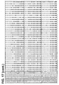
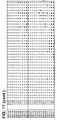
- Bisulfite sequencing DNA was extracted from the placental tissues and maternal buffy coat by using the QIAamp DNA Mini Kit and QIAamp DNA Blood Mini Kit (Qiagen, Hilden, Germany), respectively, according to the manufacturer's instructions. For each sample, 1 ⁇ g DNA was subjected to bisulfite conversion, which converts unmethylated cytosine residues to uracil but leaves methylated cytosine residues unchanged, by the CpGenome DNA Modification Kit (Intergen, Burlington, MA) according to manufacturer's instructions. The bisulfite converted DNA was then subjected to PCR amplification with pairs of primers flanking the CpG sites (Table 1).
- CpG-containing genomic sequences are investigated by two or three PCRs, namely regions A, B and C. Primers were designed not to bind to any potentially methylated cytosine residues. These primers are shown by way of illustration and should not be seen to limit the range of primers which can be used for this purpose.
- Reagents supplied in the TaqMan PCR Core Reagent Kit (Applied Biosystems, Foster City, CA) were used. Reagent compositions for each PCR are detailed in Table 1. Typically, PCRs were performed in a final reaction volume of 25 ⁇ l, with MgCl 2 , primers, TaqGold, 1X Buffer II, 200 ⁇ M of each dNTP, with or without dimethylsulfoxide (DMSO) or betaine.
- DMSO dimethylsulfoxide
- the thermal profile consisted of an initial denaturation step of 95°C for 10 min followed by 40 cycles of 95°C for 1 min, a range of annealing temperatures between 55 to 65 °C for 1 min (Table 1), 72°C for 1 min, and a final extension of 72°C for 10 min.
- the PCR product was TA-cloned into a plasmid vector using the pGEM-T Easy Vector System (Promega, Madison, WI).
- the inserts from the positive recombinant clones were analyzed by cycle sequencing using the BigDye Terminator Cycle Sequencing v1.1 kit (Applied Biosystems) as per the manufacturer's instructions.
- a CpG site was scored as methylated if the sequence was cytosine; scored as unmethylated if it was occupied by a thymine residue (deoxy counterpart of uracil).
- the proportion of methylated cytosine residue for each CpG site was determined among the placental tissues as well as the maternal blood samples.
- the distribution of methylated and unmethylated cytosines was compared between the placental tissues and maternal buffy coat for each CpG site by chi-square analysis. P-value of ⁇ 0.05 is considered as statistically significantly different.
- SPSS Sigma Stat 3.0 software
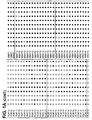
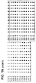
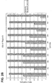
- CGI137 The methylation profile of CGI137 was studied among placental tissues and the corresponding maternal blood cells collected from 5 third-trimester pregnancies.
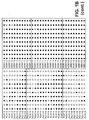
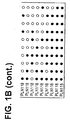
- the bisulfite sequencing data for regions A and B of all the 5 cases are shown in Figures 1A and 1B , respectively. Each row in the respective panels represents the bisulfite sequencing result for one clone while each column represents an individual CpG dinucleotide within the genomic sequence.
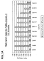
- the proportion of methylated clones among all of the sequenced clones, the methylation index, at each CpG site was determined for all 5 placental tissue and maternal blood cell samples.
- the data are summarized in Figures 2A and 2B . It can be seen that the placenta is hypomethylated compared with maternal blood cells.
- Chi-square analysis was performed to compare the distribution of methylated and unmethylated clones between the placental tissues and maternal blood cells at each CpG site. The differences in the methylation indices between the placenta and maternal blood cells are statistically significant for all 21 CpG sites (Chi-square, P ⁇ 0.0001, Tables 2A and 2B).
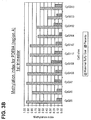
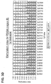
- Phosphodiesterase 9A The methylation profile of PDE9A was studied among placental tissues and the corresponding maternal blood cells collected from 5 third-trimester and 5 first-trimester pregnancies. Cloning and bisulfite sequencing were performed and the methylation indices of the studied CpG sites in the placental tissues and maternal blood cells for the region A amplicon are summarized in Figures 3A and 3B . The corresponding data for region B are summarized in Figures 3C and 3D . Comparisons between third-trimester placental tissues and maternal blood cells for region C are shown in Figure 3E . In general, the placenta is hypomethylated when compared to maternal blood cells.
- PPP1R2P2 Homo sapiens protein phosphatase 1, regulatory (inhibitor) subunit 2 pseudogene 2
- PPP1R2P2 The methylation profile of PPP1R2P2 region A was studied among placental tissues and the corresponding maternal blood cells collected from 5 third-trimester and 5 first-trimester pregnancies. Cloning and bisulfite sequencing were performed and the methylation indices of the studied CpG sites for region A in the placental tissues and maternal blood cells are summarized in Figures 4A and 4B . In general, the placenta is hypomethylated when compared to maternal blood cells. Chi-square analysis was performed as described above to assess for statistically significant differences between the two tissues.
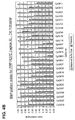
- CGI009 The methylation profiles of CGI009 from 2 maternal blood cell samples were compared with those of 2 first-trimester and 2 third-trimester placental tissue samples collected from normal pregnancies as well as 2 first-trimester placental tissue samples from pregnancies where the fetus had trisomy 21. Cloning and bisulfite sequencing were performed and the methylation indices of the studied CpG sites in the placental tissues and maternal blood cells are summarized in Figure 6 . In general, the placenta is hypermethylated when compared to maternal blood cells.
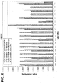
- Carbonyl reductase 1(CBR1) The profiles of CBR1 from 2 maternal blood cell samples were compared with those of 2 first-trimester placental tissue samples collected from normal pregnancies as well as 2 first-trimester placental tissue samples from pregnancies where the fetus had trisomy 21. Cloning and bisulfite sequencing were performed and the methylation indices of the studied CpG sites in the placental tissues and maternal blood cells are summarized in Figure 7 . In general, the placenta is hypermethylated when compared to maternal blood cells. Among the 20 studied CpG sites, all are statistically significantly different between the maternal blood cells and first-trimester placental tissues of both normal and trisomy 21 pregnancies (Chi-square analysis, Table 6).
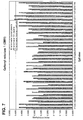
- Down syndrome cell adhesion molecule The methylation profiles of DSCAM from 2 maternal blood cell samples were compared with those of 2 first-trimester and 2 third-trimester placental tissue samples collected from normal pregnancies as well as 2 first-trimester placental tissue samples from pregnancies where the fetus had trisomy 21. Cloning and bisulfite sequencing were performed and the methylation indices of the studied CpG sites in the placental tissues and maternal blood cells are summarized in Figure 8 . In general, the placenta is hypermethylated when compared to maternal blood cells. Among the 27 studied CpG sites, all are statistically significantly different between the maternal blood cells and first-trimester placental tissues of both normal and trisomy 21 pregnancies (Chi-square analysis, Table 7).
- Chromosome 21 open reading frame 29 (C21orf29) The methylation profiles of C21orf29 from 2 maternal blood cell samples were compared with those of 1 first-trimester and 2 third-trimester placental tissue samples collected from normal pregnancies as well as 2 first-trimester placental tissue samples from pregnancies where the fetus had trisomy 21. Cloning and bisulfite sequencing were performed and the methylation indices of the studied CpG sites in the placental tissues and maternal blood cells are summarized in Figure 9 . In general, the placenta is hypermethylated when compared to maternal blood cells.
- HSF2BP Heat shock transcription factor 2 binding protein
- COL6A1 Heat shock transcription factor 2 binding protein
- the CG-containing genomic sequences with differential methylation profile between placental tissues and maternal blood cells are useful as fetal epigenetic markers for maternal blood detection.
- assays should target the detection of the placental-specific epigenetic form of each differentially methylated locus.
- An assay has been developed to target the placental-specific form of CGI137 ( Figure 1 ), namely the unmethylated molecules, and assessed its specificity.
- a third-trimester placental tissue sample and corresponding maternal blood sample were collected from a subject undergoing delivery by elective cesarean section.
- Maternal blood (10 mL) was collected into EDTA tubes prior to as well as after the delivery.
- the blood samples were centrifuged at 1600 x g for 10 min at 4 °C.
- the buffy coat portion was obtained after careful removal of plasma and stored separately at -20 °C.
- the plasma was further centrifuged at 16,000 x g for 10 min and 1.6 mL plasma was transferred to clean polypropylene tubes with care not to disturb the underlying cell pellet.
- Placental tissue biopsies were rinsed in phosphate buffered saline and stored in plain polypropylene tubes at -80 °C.
- DNA was extracted from the placental tissues using the QIAamp DNA Mini Kit (Qiagen, Hilden, Germany). DNA from the maternal plasma and buffy coat was extracted by the QIAamp DNA Blood Mini Kit (Qiagen, Hilden, Germany) according to the manufacturer's instructions.
- the amplicon spans between coordinates: 2742998-2743130 for the GenBank Accession: NT_011515.
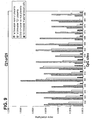
- a primer extension assay ( Figure 12 ) based on the homogeneous MassEXTEND (hME) protocol is designed to interrogate the methylation status of the CpG site 541. Basically, the primer extension primer spans up to the nucleotide 3' to the CpG site 541 (Table 10).
- Each reaction contained 1X PCR buffer (Applied Biosystems, Foster City, CA) with 2.0 mM MgCl 2 (Applied Biosystems), 200 ⁇ M each of dATP, dGTP and dCTP, 400 ⁇ M of dUTP (Applied Biosystems), 200 nM each of forward and reverse primers (Integrated DNA Technologies, Coralville, IA) and 1 U of AmpliTaq Gold® DNA Polymerase (Applied Biosystems).
- the PCR reaction was initiated at 95°C for 10 min, followed by denaturation at 94°C for 30 sec, annealing at 60°C for 40 sec, extension at 72°C for 40 sec for 40 cycles, and a final incubation at 72°C for 5 min.
- PCR products were subjected to shrimp alkaline phosphatase treatment to dephosphorylate any remaining dNTPs and prevent their incorporation in the subsequent primer extension assay.
- hME buffer Sequenom
- shrimp alkaline phosphatase Sequenom
- 3.06 ⁇ L of water were added. The reaction mixture was incubated at 37°C for 40 min, followed by heat inactivation at 85°C for 5 min.
- primer extension reaction 4 ⁇ L of base extension reaction cocktail containing 1500 nM of extension primer (Integrated DNA Technologies), 1.15 U of Thermosequenase (Sequenom) and 64 ⁇ M each of ddATP, ddCTP, dTTP and dGTP (Sequenom) were added to 10 ⁇ L of the PCR products.
- the reaction condition was 94°C for 2 min, followed by 94°C for 5 sec, 52°C for 5 sec, and 72°C for 5 sec for 75 cycles.
- the final base extension product was cleaned up with the SpectroCLEAN (Sequenom) resin to remove salts that may interfere with the mass spectrometry analysis. Twenty-four microliters of water and 12 mg of resin were added into each base extension product. The final mixtures were mixed in a rotator for 20 min. After centrifugation at 361 g for 5 min, approximately 10 nL of reaction solution was dispensed onto a 384-format SpectroCHIP (Sequenom) pre-spotted with a matrix of 3-hydroxypicolinic acid by using a SpectroPoint nanodispenser (Sequenom).
- a MassARRAYTM Analyzer Compact Mass Spectrometer (Sequenom) was used for data acquisition from the SpectroCHIP. Mass spectrometric data were automatically imported into SpectroTYPER (Sequenom) database for analysis.
- CGI137 due to the differences in the methylation profile of CGI137 between placental tissues and maternal blood cells, sensitive and specific assays could be developed to target the detection of the placenta-derived form of CGI137 among a background of CGI137 methylated molecules derived from maternal blood cells.
- placenta is a tissue source of fetal DNA release into the maternal circulation (Chim et al., supra ), in which the latter contains a high background of DNA derived from maternal blood cells (Liu et al., supra )
- assays targeting placenta-specific epigenetic markers are useful for the detection of fetal-specific nucleic acid molecules in maternal blood.
- genomic contigs accession number, version, start and end nucleotide numbers
- GenBank accession number, version, start and end nucleotide numbers
- chromosome start and end nucleotide numbers
- PCR was performed in a final reaction volume of 20 ⁇ l, with MgCl 2 , primers, HotStar Taq, 1X PCR Buffer, 50 ⁇ M of each dNTP, and 2X PCRx Enhancer (Invitrogen, Carlsbad, CA).
- the thermal profile consisted of an initial denaturation step of 95°C for 15 min, followed by 50-55 cycles of 95°C for 20 sec, 58 or 60 °C for 30 sec (Table 11), 72°C for 1.5 min, and a final extension of 72°C for 3 min.
- PCR products were then subjected to restriction enzyme digestion.
- the restriction enzyme to be used for each respective locus was chosen for its ability to distinguish between the methylated and unmethylated sequence after bisulfite conversion.
- restriction sites are only present in either the methylated or unmethylated sequence but not both, so that one of the sequences would be digested while the other would remain intact (Table 12). Restriction enzyme digestions were performed in a final reaction volume of 20 ⁇ l, with 5 ⁇ l PCR products, 1X appropriate buffer, and 10U restriction enzyme (or none for mock digestion), under the manufacturer's recommended temperatures for 2 hr. All enzymes were purchased from New England Biolabs (Beverly, MA). Digested products were then analyzed by gel electrophoresis. Table 12.
- HLCS Holocarboxylase Synthetase
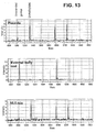
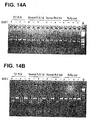
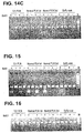
- CGI009 The methylation profiles of CGI009 from 2 maternal blood cell samples were compared with those of 2 first-trimester and 2 third-trimester placental tissue samples collected from normal pregnancies, as well as 2 first-trimester placental tissue samples from trisomy 21 pregnancies. COBRA assay was performed and the gel electrophoresis data are shown in Figure 15 . In general, the placenta is hypermethylated when compared to maternal blood cells.
- CGI132 The methylation profiles of CGI132 from 2 maternal blood cell samples were compared with those of 2 first-trimester and 2 third-trimester placental tissue samples collected from normal pregnancies, as well as 2 first-trimester placental tissue samples from trisomy 21 pregnancies. COBRA assay was performed and the gel electrophoresis data are shown in Figure 16 . In general, the placenta is hypermethylated when compared to maternal blood cells.
- Methylation sensitive restriction enzyme digestion DNA was extracted from the placental tissues using the QIAamp DNA Mini Kit (Qiagen, Hilden, Germany). DNA from the maternal buffy coat and plasma was extracted by the QIAamp DNA Blood Mini Kit (Qiagen, Hilden, Germany) according to the manufacturer's instructions. For each placental and buffy coat DNA sample, 100 ng DNA was subjected to methylation sensitive restriction enzyme digestion. Restriction enzyme digestions were performed in a final reaction volume of 50 ⁇ l, with DNA, 1X appropriate buffer, and 25U of Hpa II and 50U of Bst U I (or none for mock digestion), under the manufacturer's recommended temperatures for at least 16 hr.
- 1.6 ml plasma was used for DNA extraction, and was eluted in 50 ⁇ l of deionized water, 21 ⁇ l of which was subjected to restriction enzyme digestion. Enzyme digestions were performed in a final reaction volume of 30 ⁇ l, with DNA, 1X appropriate buffer, and 20U of Hpa II and 30U of BstU I (or none for mock digestion), under the manufacturer's recommended temperatures for at least 16 hr. All enzymes were purchased from New England Biolabs (Beverly, MA). Digested products were then analyzed by real time quantitative PCR. The selected restriction enzymes only digest the unmethylated DNA but not the methylated DNA.
- Real time quantitative PCR Real time quantitative PCR.
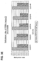
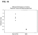
- Real time PCR assay was developed for quantitative analysis of HLCS genomic DNA with and without restriction enzyme digestion. 4 ⁇ l of restriction enzyme treated DNA or mock digestion sample was used in the real time PCR assay. Each reaction contains 1X TaqMan Universal PCR Master Mix (Applied Biosystems, Foster City, CA), 300 nM of forward primer (5'-CCGTGTGGCCAGAGGTG-3'), 300 nM of reverse primer (5'-TGGGAGCCGGAACCTACC-3'), and 100 nM of TaqMan probe (5'-6FAM- TCCCGACCTGGCCCTTTGCC-TAMRA-3').
- the thermal profile was 50 °C for 2 min, 95 °C for 10 min, 50 cycles of 95 °C for 15 sec, and 60 °C for 1 min. All reactions were run in duplicate, and the mean quantity was taken. Serially diluted human genomic DNA originally quantified by optical density measurement was used as the quantitative standard for the assay. As the detectable HLCS DNA after restriction enzyme digestion represented the methylated fraction, we expressed the real-time quantitative PCR as a methylation index. The methylation index of a sample is calculated by dividing the HLCS DNA concentration after enzyme digestion with that obtained with mock digestion.
- methylation profiles of the putative promoter region of HLCS from 8 maternal blood cell samples were compared with those from 2 first-trimester and 2 third-trimester placental tissue samples collected from normal pregnancies. Restriction enzyme digestion followed by real time PCR analysis was performed and the results are shown in Figure 18 . DNA from all maternal blood cell samples were mostly digested by restriction enzymes, resulting in methylation indices approaching 0; while that from placental tissues were partially digested, resulting in methylation indices ranging from 0.567 to 0.966.
- placenta is the predominant tissue source of fetal DNA while maternal blood cells are the main contributor of the background maternal DNA that are detectable in maternal plasma.
- placental-specific (with reference to maternal blood cells) fraction of HLCS DNA namely the methylated or non-digestable fraction can be detected in maternal plasma and is pregnancy-specific.
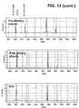
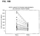
- Paired pre- and post-delivery 3 rd trimester plasma samples from 25 normal pregnant individuals were recruited. Restriction enzyme digestion followed by real time PCR assay was performed and the results are presented in Figure 19 .
- the HLCS signal is shown to be positively detected in the enzyme digested pre-delivery 3 rd trimester plasma samples and reduced after delivery.
- the median of HLCS concentration in post-delivery plasma samples after enzyme digestion was 8.1% of that the median HLCS concentration in pre-delivery plasma samples after enzyme digestion ( Figure 19A ).
- the clearance pattern from enzyme treated pre- and post-delivery plasma indicates that the detected HLCS signal is pregnancy-specific.
- the median HLCS DNA concentration in post-delivery maternal plasma with mock restriction enzyme digestion was 83.8% of that in the mock-digested pre-delivery samples ( Figure 19B ).
Landscapes
- Chemical & Material Sciences (AREA)
- Life Sciences & Earth Sciences (AREA)
- Organic Chemistry (AREA)
- Health & Medical Sciences (AREA)
- Proteomics, Peptides & Aminoacids (AREA)
- Engineering & Computer Science (AREA)
- Zoology (AREA)
- Wood Science & Technology (AREA)
- Analytical Chemistry (AREA)
- Genetics & Genomics (AREA)
- Immunology (AREA)
- Molecular Biology (AREA)
- General Health & Medical Sciences (AREA)
- Biotechnology (AREA)
- Microbiology (AREA)
- Physics & Mathematics (AREA)
- Biochemistry (AREA)
- Bioinformatics & Cheminformatics (AREA)
- Biophysics (AREA)
- General Engineering & Computer Science (AREA)
- Pathology (AREA)
- Hematology (AREA)
- Biomedical Technology (AREA)
- Urology & Nephrology (AREA)
- Chemical Kinetics & Catalysis (AREA)
- General Physics & Mathematics (AREA)
- Cell Biology (AREA)
- Food Science & Technology (AREA)
- Medicinal Chemistry (AREA)
- Measuring Or Testing Involving Enzymes Or Micro-Organisms (AREA)
- Investigating Or Analysing Biological Materials (AREA)
Claims (13)
- Verfahren zur Detektierung von Trisomie 21 in einem Fötus, der von einer schwangeren Frau ausgetragen wird, das die folgenden Schritte umfasst:(a) das Bestimmen des Methylierungsstatus einer CpG-enthaltenden genomischen Sequenz in einer Vollblut-, Serum-, Plasma-, Urin- oder Speichelprobe von der Frau, worin die genomische Sequenz zumindest 15 Nucleotide lang ist, umfassend zumindest ein Cytosin, und innerhalb von einer Region auf Chromosom 21 liegt, wobei die Region aus (1) dem genomischen Locus Holocarboxylase Synthetase (HLCS) und (2) einer DNA-Sequenz von nicht mehr als 10 kb stromauf und/oder stromab vom Locus besteht, und worin die genomische Sequenz von dem Fötus und die genomische Sequenz von der Frau unterschiedlich methyliert sind, wobei die genomische Sequenz von der Frau und die genomische Sequenz von dem Fötus in der Probe unterschieden werden;(b) das Bestimmen des Ausmaßes der genomischen Sequenz von dem Fötus in der Vollblut-, Serum-, Plasma-, Urin- oder Speichelprobe; und(c) das Vergleichen des Ausmaßes der genomischen Sequenz von dem Fötus mit einer Standardkontrolle, worin eine Steigerung von der Standardkontrolle das Vorliegen von Trisomie 21 anzeigt.
- Verfahren zur Detektierung von Trisomie 21 in einem Fötus, der von einer schwangeren Frau ausgetragen wird, das die folgenden Schritte umfasst:(a) das Behandeln von DNA in einer biologischen Probe von der Frau mit Bisulfit, worin die Probe Vollblut, Serum, Plasma, Urin oder Speichel ist;(b) das Durchführen einer Amplifikationsreaktion unter Verwendung der DNA von Schritt (a) und zwei Primern, um eine CpG-enthaltende Sequenz zu amplifizieren, worin die CpG-enthaltende genomische Sequenz zumindest 15 Nucleotide lang ist, umfassend zumindest ein Cytosin, und innerhalb von einer Region auf Chromosom 21 liegt, wobei die Region aus (1) dem genomischen Locus Holocarboxylase Synthetase (HLCS) und (2) einer DNA-Sequenz von nicht mehr als 10 kb stromauf und/oder stromab vom Locus besteht, und worin zumindest einer der zwei Primer vorzugsweise an die mit Bisulfit behandelte genomische Sequenz von dem Fötus bindet; und(c) das Vergleichen des Ausmaßes des amplifizierten Teils der genomischen Sequenz von Schritt (b) mit einer Standardkontrolle, worin eine Steigerung von der Standardkontrolle das Vorliegen von Trisomie 21 anzeigt.
- Verfahren nach Anspruch 2, worin die Amplifikationsreaktion eine Polymerase-Kettenreaktion ist.
- Verfahren nach Anspruch 2, worin die Amplifikationsreaktion eine methylierungsspezifische PCR ist.
- Verfahren zur Detektierung von Trisomie 21 in einem Fötus, der von einer schwangeren Frau ausgetragen wird, das die folgenden Schritte umfasst:(a) das Behandeln von DNA in einer biologischen Probe von der Frau mit einem Reagens, das methylierte und nicht methylierte DNA unterschiedlich modifiziert, worin die Probe Vollblut, Serum, Plasma, Urin oder Speichel ist;(b) das Bestimmen der Nucleotidsequenz einer CpG-enthaltenden genomischen Sequenz von der behandelten DNA von Schritt (a), worin die genomische Sequenz zumindest 15 Nucleotide lang ist, umfassend zumindest ein Cytosin, und innerhalb von einer Region auf Chromosom 21 liegt, wobei die Region aus (1) dem genomischen Locus Holocarboxylase Synthetase (HLCS) und (2) einer DNA-Sequenz von nicht mehr als 10 kb stromauf und/oder stromab vom Locus besteht; und(c) das Vergleichen der Mengen der unterschiedlichen Nucleotidsequenzen von Schritt (b) mit einer Standardkontrolle, worin eine Veränderung der Mengen der unterschiedlichen Nucleotidsequenzen von der Standardkontrolle das Vorliegen von Trisomie 21 anzeigt.
- Verfahren zur Detektierung von Trisomie 21 in einem Fötus in einer schwangeren Frau, worin der Fötus zwei unterschiedliche Allele von Chromosom 21 aufweist, das die folgenden Schritte umfasst:(a) das Behandeln einer biologischen Probe von der Frau mit einem Reagens, das methylierte und nicht methylierte DNA unterschiedlich modifiziert, worin die Probe Vollblut, Serum, Plasma, Urin oder Speichel ist;(b) das Bestimmen der Ausmaße der zwei Allele einer CpG-enthaltenden genomischen Sequenz, worin die genomische Sequenz zumindest 15 Nucleotide lang ist, umfassend zumindest ein Cytosin, und innerhalb von einer Region auf Chromosom 21 liegt, wobei die Region aus (1) dem genomischen Locus Holocarboxylase Synthetase (HLCS) und (2) einer DNA-Sequenz von nicht mehr als 10 kb stromauf und/oder stromab vom Locus besteht, worin die zwei Allele der genomischen Sequenz sich in der Polynucleotidsequenz voneinander unterscheiden; und(c) das Bestimmen des Verhältnisses der zwei Allele, worin eine Abweichung von jener von einer Frau, die einen Fötus austrägt, der Trisomie 21 nicht aufweist, Trisomie 21 in dem Fötus anzeigt.
- Verfahren nach Anspruch 5 oder 6, worin das Reagens Bisulfit umfasst.
- Verfahren nach Anspruch 5 oder 6, worin das Reagens ein Enzym umfasst, das vorzugsweise methylierte DNA spaltet.
- Verfahren zur Detektierung von Trisomie 21 in einem Fötus, der von einer schwangeren Frau ausgetragen wird, das die folgenden Schritte umfasst:(a) das Bestimmen des Ausmaßes einer methylierten CpG-enthaltenden genomischen Sequenz in einer Vollblut-, Serum-, Plasma-, Urin- oder Speichelprobe von der Frau, worin die genomische Sequenz zumindest 15 Nucleotide lang ist, umfassend zumindest ein Cytosin, und innerhalb von einer Region auf Chromosom 21 liegt, wobei die Region aus (1) dem genomischen Locus Holocarboxylase Synthetase (HLCS) und (2) einer DNA-Sequenz von nicht mehr als 10 kb stromauf und/oder stromab vom Locus besteht; und(b) das Vergleichen des Ausmaßes der methylierten genomischen Sequenz mit einer Standardkontrolle, worin eine Steigerung von der Standardkontrolle das Vorliegen von Trisomie 21 anzeigt.
- Verfahren nach Anspruch 9, worin Schritt (a) das Behandeln von DNA, die in der Probe vorliegt, mit einem Reagens umfasst, das methyliertes und nicht methyliertes Cytosin unterschiedlich modifiziert.
- Verfahren nach Anspruch 10, worin das Reagens Bisulfit umfasst.
- Verfahren nach Anspruch 10, worin das Reagens ein Enzym umfasst, das vorzugsweise DNA spaltet, die methyliertes Cytosin umfasst.
- Verfahren nach einem der Ansprüche 1-12, worin die biologische Probe eine Plasmaprobe ist.
Priority Applications (2)
| Application Number | Priority Date | Filing Date | Title |
|---|---|---|---|
| EP19214593.6A EP3722443A1 (de) | 2006-05-03 | 2007-05-03 | Neuartiger marker zur pränatalen diagnose und überwachung |
| EP16156063.6A EP3085792B1 (de) | 2006-05-03 | 2007-05-03 | Neuartiger marker zur pränatalen diagnose und überwachung |
Applications Claiming Priority (3)
| Application Number | Priority Date | Filing Date | Title |
|---|---|---|---|
| US79745606P | 2006-05-03 | 2006-05-03 | |
| US11/784,499 US7901884B2 (en) | 2006-05-03 | 2007-04-06 | Markers for prenatal diagnosis and monitoring |
| EP07732660A EP2016191B1 (de) | 2006-05-03 | 2007-05-03 | Neue fötale marker für pränatale diagnose und überwachung |
Related Parent Applications (2)
| Application Number | Title | Priority Date | Filing Date |
|---|---|---|---|
| EP07732660A Division EP2016191B1 (de) | 2006-05-03 | 2007-05-03 | Neue fötale marker für pränatale diagnose und überwachung |
| EP07732660.1 Division | 2007-05-03 |
Related Child Applications (3)
| Application Number | Title | Priority Date | Filing Date |
|---|---|---|---|
| EP16156063.6A Division-Into EP3085792B1 (de) | 2006-05-03 | 2007-05-03 | Neuartiger marker zur pränatalen diagnose und überwachung |
| EP16156063.6A Division EP3085792B1 (de) | 2006-05-03 | 2007-05-03 | Neuartiger marker zur pränatalen diagnose und überwachung |
| EP19214593.6A Division EP3722443A1 (de) | 2006-05-03 | 2007-05-03 | Neuartiger marker zur pränatalen diagnose und überwachung |
Publications (2)
| Publication Number | Publication Date |
|---|---|
| EP2385142A1 EP2385142A1 (de) | 2011-11-09 |
| EP2385142B1 true EP2385142B1 (de) | 2016-04-13 |
Family
ID=38694260
Family Applications (4)
| Application Number | Title | Priority Date | Filing Date |
|---|---|---|---|
| EP19214593.6A Withdrawn EP3722443A1 (de) | 2006-05-03 | 2007-05-03 | Neuartiger marker zur pränatalen diagnose und überwachung |
| EP11168611.9A Not-in-force EP2385142B1 (de) | 2006-05-03 | 2007-05-03 | Neuartiger Marker zur pränatalen Diagnose und Überwachung |
| EP16156063.6A Active EP3085792B1 (de) | 2006-05-03 | 2007-05-03 | Neuartiger marker zur pränatalen diagnose und überwachung |
| EP07732660A Active EP2016191B1 (de) | 2006-05-03 | 2007-05-03 | Neue fötale marker für pränatale diagnose und überwachung |
Family Applications Before (1)
| Application Number | Title | Priority Date | Filing Date |
|---|---|---|---|
| EP19214593.6A Withdrawn EP3722443A1 (de) | 2006-05-03 | 2007-05-03 | Neuartiger marker zur pränatalen diagnose und überwachung |
Family Applications After (2)
| Application Number | Title | Priority Date | Filing Date |
|---|---|---|---|
| EP16156063.6A Active EP3085792B1 (de) | 2006-05-03 | 2007-05-03 | Neuartiger marker zur pränatalen diagnose und überwachung |
| EP07732660A Active EP2016191B1 (de) | 2006-05-03 | 2007-05-03 | Neue fötale marker für pränatale diagnose und überwachung |
Country Status (12)
| Country | Link |
|---|---|
| US (1) | US7901884B2 (de) |
| EP (4) | EP3722443A1 (de) |
| JP (1) | JP2009535050A (de) |
| KR (5) | KR101864921B1 (de) |
| CN (3) | CN107354218B (de) |
| AU (1) | AU2007251351B2 (de) |
| CA (1) | CA2651051C (de) |
| EA (1) | EA014274B1 (de) |
| IL (2) | IL194984A (de) |
| MX (1) | MX2008013930A (de) |
| NZ (2) | NZ572581A (de) |
| WO (1) | WO2007132167A2 (de) |
Families Citing this family (130)
| Publication number | Priority date | Publication date | Assignee | Title |
|---|---|---|---|---|
| US6927028B2 (en) * | 2001-08-31 | 2005-08-09 | Chinese University Of Hong Kong | Non-invasive methods for detecting non-host DNA in a host using epigenetic differences between the host and non-host DNA |
| US8515679B2 (en) | 2005-12-06 | 2013-08-20 | Natera, Inc. | System and method for cleaning noisy genetic data and determining chromosome copy number |
| US8532930B2 (en) | 2005-11-26 | 2013-09-10 | Natera, Inc. | Method for determining the number of copies of a chromosome in the genome of a target individual using genetic data from genetically related individuals |
| US9424392B2 (en) | 2005-11-26 | 2016-08-23 | Natera, Inc. | System and method for cleaning noisy genetic data from target individuals using genetic data from genetically related individuals |
| US10083273B2 (en) | 2005-07-29 | 2018-09-25 | Natera, Inc. | System and method for cleaning noisy genetic data and determining chromosome copy number |
| US10081839B2 (en) | 2005-07-29 | 2018-09-25 | Natera, Inc | System and method for cleaning noisy genetic data and determining chromosome copy number |
| US11111543B2 (en) | 2005-07-29 | 2021-09-07 | Natera, Inc. | System and method for cleaning noisy genetic data and determining chromosome copy number |
| US11111544B2 (en) | 2005-07-29 | 2021-09-07 | Natera, Inc. | System and method for cleaning noisy genetic data and determining chromosome copy number |
| TR201910868T4 (tr) * | 2006-02-02 | 2019-08-21 | Univ Leland Stanford Junior | Dijital analizle invazif olmayan fetal genetik tarama. |
| BRPI0709545A2 (pt) * | 2006-03-06 | 2011-07-19 | Univ Columbia | amplificação especìfica de seqüência de dna fetal de uma mistura de origem fetal-maternal |
| WO2007147074A2 (en) | 2006-06-14 | 2007-12-21 | Living Microsystems, Inc. | Use of highly parallel snp genotyping for fetal diagnosis |
| US8137912B2 (en) | 2006-06-14 | 2012-03-20 | The General Hospital Corporation | Methods for the diagnosis of fetal abnormalities |
| EP2589668A1 (de) | 2006-06-14 | 2013-05-08 | Verinata Health, Inc | Analyse seltener Zellen mittels Probentrennung und DNA-Etiketten |
| US20080050739A1 (en) | 2006-06-14 | 2008-02-28 | Roland Stoughton | Diagnosis of fetal abnormalities using polymorphisms including short tandem repeats |
| TWI335354B (en) | 2006-09-27 | 2011-01-01 | Univ Hong Kong Chinese | Methods for the detection of the degree of the methylation of a target dna and kits |
| LT2557517T (lt) | 2007-07-23 | 2023-01-10 | The Chinese University Of Hong Kong | Nukleino rūgščių sekos disbalanso nustatymas |
| US12180549B2 (en) | 2007-07-23 | 2024-12-31 | The Chinese University Of Hong Kong | Diagnosing fetal chromosomal aneuploidy using genomic sequencing |
| US8748100B2 (en) * | 2007-08-30 | 2014-06-10 | The Chinese University Of Hong Kong | Methods and kits for selectively amplifying, detecting or quantifying target DNA with specific end sequences |
| DK2240781T3 (en) | 2008-01-18 | 2018-03-26 | Harvard College | METHODS FOR DETECTING DISEASE OR STATE SIGNATURES IN BODY FLUIDS |
| CA2717320A1 (en) | 2008-03-11 | 2009-09-17 | Sequenom, Inc. | Nucleic acid-based tests for prenatal gender determination |
| WO2010003153A2 (en) * | 2008-07-03 | 2010-01-07 | Life Technologies Corporation | Methylation analysis of mate pairs |
| CN102171565B (zh) | 2008-08-04 | 2015-04-29 | 纳特拉公司 | 等位基因调用和倍性调用的方法 |
| US8962247B2 (en) | 2008-09-16 | 2015-02-24 | Sequenom, Inc. | Processes and compositions for methylation-based enrichment of fetal nucleic acid from a maternal sample useful for non invasive prenatal diagnoses |
| US8476013B2 (en) * | 2008-09-16 | 2013-07-02 | Sequenom, Inc. | Processes and compositions for methylation-based acid enrichment of fetal nucleic acid from a maternal sample useful for non-invasive prenatal diagnoses |
| EP3770255A1 (de) * | 2008-09-16 | 2021-01-27 | Sequenom, Inc. | Verfahren und zusammensetzungen zur methylierungsbasierten anreicherung fetaler nukleinsäure aus einer mütterlichen probe für nichtinvasive pränatale diagnosen |
| HUE031848T2 (en) | 2008-09-20 | 2017-08-28 | Univ Leland Stanford Junior | Non-invasive diagnosis of fetal aneuploidy by sequencing |
| EP2391730B1 (de) * | 2009-01-30 | 2016-04-13 | University of Southampton | Verwendung von cpg-methylierung zur vorhersage |
| US9458503B2 (en) | 2009-07-02 | 2016-10-04 | Nucleix | Methods for distinguishing between natural and artificial DNA samples |
| US8563242B2 (en) | 2009-08-11 | 2013-10-22 | The Chinese University Of Hong Kong | Method for detecting chromosomal aneuploidy |
| AU2015252141A1 (en) * | 2009-09-16 | 2015-12-03 | Sequenom Center For Molecular Medicine | Processes and compositions for methylation-based enrichment of fetal nucleic acid from a maternal sample useful for non invasive prenatal diagnoses |
| US20120185176A1 (en) | 2009-09-30 | 2012-07-19 | Natera, Inc. | Methods for Non-Invasive Prenatal Ploidy Calling |
| US9752187B2 (en) * | 2009-12-11 | 2017-09-05 | Nucleix | Categorization of DNA samples |
| US9783850B2 (en) * | 2010-02-19 | 2017-10-10 | Nucleix | Identification of source of DNA samples |
| EP3088532B1 (de) * | 2009-12-22 | 2019-10-30 | Sequenom, Inc. | Verfahren und kits zur identifizierung von aneuploidie |
| US20110312503A1 (en) | 2010-01-23 | 2011-12-22 | Artemis Health, Inc. | Methods of fetal abnormality detection |
| DK2529032T3 (en) * | 2010-01-26 | 2017-05-01 | Nipd Genetics Ltd | METHODS AND COMPOSITIONS FOR NON-INVASIVE PRE-NATIONAL DIAGNOSTICATION OF Fetal ANEUPLOIDITIES |
| US12152275B2 (en) | 2010-05-18 | 2024-11-26 | Natera, Inc. | Methods for non-invasive prenatal ploidy calling |
| US9677118B2 (en) | 2014-04-21 | 2017-06-13 | Natera, Inc. | Methods for simultaneous amplification of target loci |
| US11326208B2 (en) | 2010-05-18 | 2022-05-10 | Natera, Inc. | Methods for nested PCR amplification of cell-free DNA |
| US10316362B2 (en) | 2010-05-18 | 2019-06-11 | Natera, Inc. | Methods for simultaneous amplification of target loci |
| US11939634B2 (en) | 2010-05-18 | 2024-03-26 | Natera, Inc. | Methods for simultaneous amplification of target loci |
| US20190010543A1 (en) | 2010-05-18 | 2019-01-10 | Natera, Inc. | Methods for simultaneous amplification of target loci |
| US12545960B2 (en) | 2010-05-18 | 2026-02-10 | Natera, Inc. | Methods for simultaneous amplification of target loci |
| US11322224B2 (en) | 2010-05-18 | 2022-05-03 | Natera, Inc. | Methods for non-invasive prenatal ploidy calling |
| US11408031B2 (en) | 2010-05-18 | 2022-08-09 | Natera, Inc. | Methods for non-invasive prenatal paternity testing |
| US11339429B2 (en) | 2010-05-18 | 2022-05-24 | Natera, Inc. | Methods for non-invasive prenatal ploidy calling |
| US11332793B2 (en) | 2010-05-18 | 2022-05-17 | Natera, Inc. | Methods for simultaneous amplification of target loci |
| US11332785B2 (en) | 2010-05-18 | 2022-05-17 | Natera, Inc. | Methods for non-invasive prenatal ploidy calling |
| US12221653B2 (en) | 2010-05-18 | 2025-02-11 | Natera, Inc. | Methods for simultaneous amplification of target loci |
| EP2572003A4 (de) | 2010-05-18 | 2016-01-13 | Natera Inc | Verfahren zur nichtinvasiven pränatalen ploidiezuordnung |
| EP2596134B1 (de) | 2010-07-23 | 2020-04-08 | President and Fellows of Harvard College | Verfahren zur erkennung von erkrankungen oder leiden mit phagozytischen zellen |
| AU2011280997A1 (en) | 2010-07-23 | 2013-02-28 | President And Fellows Of Harvard College | Methods of detecting autoimmune or immune-related diseases or conditions |
| WO2012012693A2 (en) | 2010-07-23 | 2012-01-26 | President And Fellows Of Harvard College | Methods for detecting signatures of disease or conditions in bodily fluids |
| CA2806304A1 (en) | 2010-07-23 | 2012-01-26 | President And Fellows Of Harvard College | Methods of detecting prenatal or pregnancy-related diseases or conditions |
| US10167508B2 (en) | 2010-08-06 | 2019-01-01 | Ariosa Diagnostics, Inc. | Detection of genetic abnormalities |
| US20140342940A1 (en) | 2011-01-25 | 2014-11-20 | Ariosa Diagnostics, Inc. | Detection of Target Nucleic Acids using Hybridization |
| US8700338B2 (en) | 2011-01-25 | 2014-04-15 | Ariosa Diagnosis, Inc. | Risk calculation for evaluation of fetal aneuploidy |
| US20130040375A1 (en) | 2011-08-08 | 2013-02-14 | Tandem Diagnotics, Inc. | Assay systems for genetic analysis |
| US11203786B2 (en) | 2010-08-06 | 2021-12-21 | Ariosa Diagnostics, Inc. | Detection of target nucleic acids using hybridization |
| US20120034603A1 (en) | 2010-08-06 | 2012-02-09 | Tandem Diagnostics, Inc. | Ligation-based detection of genetic variants |
| US11031095B2 (en) | 2010-08-06 | 2021-06-08 | Ariosa Diagnostics, Inc. | Assay systems for determination of fetal copy number variation |
| US10533223B2 (en) | 2010-08-06 | 2020-01-14 | Ariosa Diagnostics, Inc. | Detection of target nucleic acids using hybridization |
| US20130261003A1 (en) | 2010-08-06 | 2013-10-03 | Ariosa Diagnostics, In. | Ligation-based detection of genetic variants |
| WO2012088456A2 (en) | 2010-12-22 | 2012-06-28 | Natera, Inc. | Methods for non-invasive prenatal paternity testing |
| US8756020B2 (en) | 2011-01-25 | 2014-06-17 | Ariosa Diagnostics, Inc. | Enhanced risk probabilities using biomolecule estimations |
| US9994897B2 (en) | 2013-03-08 | 2018-06-12 | Ariosa Diagnostics, Inc. | Non-invasive fetal sex determination |
| US11270781B2 (en) | 2011-01-25 | 2022-03-08 | Ariosa Diagnostics, Inc. | Statistical analysis for non-invasive sex chromosome aneuploidy determination |
| US10131947B2 (en) | 2011-01-25 | 2018-11-20 | Ariosa Diagnostics, Inc. | Noninvasive detection of fetal aneuploidy in egg donor pregnancies |
| AU2011358564B9 (en) | 2011-02-09 | 2017-07-13 | Natera, Inc | Methods for non-invasive prenatal ploidy calling |
| WO2012149339A2 (en) | 2011-04-29 | 2012-11-01 | Sequenom, Inc. | Quantification of a minority nucleic acid species |
| US8712697B2 (en) | 2011-09-07 | 2014-04-29 | Ariosa Diagnostics, Inc. | Determination of copy number variations using binomial probability calculations |
| US9127317B2 (en) | 2012-03-02 | 2015-09-08 | Winthrop-University Hospital | Method for using probe based PCR detection to measure the levels of circulating demethylated β cell derived DNA as a measure of β cell loss in diabetes |
| HK1206055A1 (en) | 2012-03-02 | 2015-12-31 | Sequenom, Inc. | Methods and processes for non-invasive assessment of genetic variations |
| JP6411995B2 (ja) | 2012-03-13 | 2018-10-24 | ザ チャイニーズ ユニバーシティー オブ ホンコン | 非侵襲的出生前診断のために大量並列シークエンシング・データを分析する方法 |
| ES2718905T3 (es) * | 2012-04-24 | 2019-07-05 | Siemens Healthcare Diagnostics Inc | Métodos de examen de preeclampsia |
| RU2507269C2 (ru) * | 2012-05-11 | 2014-02-20 | ЗАО "Геноаналитика" | Технология определения трисомии хромосом методом секвенирования |
| US10289800B2 (en) | 2012-05-21 | 2019-05-14 | Ariosa Diagnostics, Inc. | Processes for calculating phased fetal genomic sequences |
| US10504613B2 (en) | 2012-12-20 | 2019-12-10 | Sequenom, Inc. | Methods and processes for non-invasive assessment of genetic variations |
| US9920361B2 (en) | 2012-05-21 | 2018-03-20 | Sequenom, Inc. | Methods and compositions for analyzing nucleic acid |
| US20140093873A1 (en) | 2012-07-13 | 2014-04-03 | Sequenom, Inc. | Processes and compositions for methylation-based enrichment of fetal nucleic acid from a maternal sample useful for non-invasive prenatal diagnoses |
| JP2015522293A (ja) | 2012-07-19 | 2015-08-06 | アリオサ ダイアグノスティックス インコーポレイテッドAriosa Diagnostics,Inc. | 多重化連続ライゲーションに基づく遺伝子変異体の検出 |
| WO2014019275A1 (en) * | 2012-07-31 | 2014-02-06 | Bgi Shenzhen Co., Limited | Noninvasive detection of fetal health status |
| US20140100126A1 (en) | 2012-08-17 | 2014-04-10 | Natera, Inc. | Method for Non-Invasive Prenatal Testing Using Parental Mosaicism Data |
| BR122021021823B1 (pt) * | 2012-09-20 | 2023-04-25 | The Chinese University Of Hong Kong | Método para determinar um primeiro perfil de metilação de uma amostra biológica de um organismo, e, meio de armazenamento de memória |
| US9732390B2 (en) | 2012-09-20 | 2017-08-15 | The Chinese University Of Hong Kong | Non-invasive determination of methylome of fetus or tumor from plasma |
| US10706957B2 (en) | 2012-09-20 | 2020-07-07 | The Chinese University Of Hong Kong | Non-invasive determination of methylome of tumor from plasma |
| JP6525894B2 (ja) * | 2013-02-28 | 2019-06-05 | ザ チャイニーズ ユニバーシティ オブ ホンコン | 超並列rna配列決定による母体血漿のトランスクリプトーム解析 |
| US11585814B2 (en) | 2013-03-09 | 2023-02-21 | Immunis.Ai, Inc. | Methods of detecting prostate cancer |
| EP2965077B1 (de) | 2013-03-09 | 2022-07-13 | Harry Stylli | Verfahren zur krebserkennung |
| HK1216655A1 (zh) | 2013-03-13 | 2016-11-25 | Sequenom, Inc. | 用於dna甲基化分析的引物 |
| US10577655B2 (en) | 2013-09-27 | 2020-03-03 | Natera, Inc. | Cell free DNA diagnostic testing standards |
| US9499870B2 (en) | 2013-09-27 | 2016-11-22 | Natera, Inc. | Cell free DNA diagnostic testing standards |
| US10262755B2 (en) | 2014-04-21 | 2019-04-16 | Natera, Inc. | Detecting cancer mutations and aneuploidy in chromosomal segments |
| GB2520763A (en) | 2013-12-02 | 2015-06-03 | Vanadis Diagnostics Ab | Nucleic acid probe and method of detecting genomic fragments |
| US11365447B2 (en) | 2014-03-13 | 2022-06-21 | Sequenom, Inc. | Methods and processes for non-invasive assessment of genetic variations |
| EP3957749A1 (de) | 2014-04-21 | 2022-02-23 | Natera, Inc. | Nachweis von tumorspezifischen mutationen in biopsien mittels sequenzierung des gesamten exoms und in zellfreien proben |
| US12492429B2 (en) | 2014-04-21 | 2025-12-09 | Natera, Inc. | Detecting mutations and ploidy in chromosomal segments |
| WO2015169947A1 (en) | 2014-05-09 | 2015-11-12 | Lifecodexx Ag | Detection of dna that originates from a specific cell-type and related methods |
| EP2942400A1 (de) | 2014-05-09 | 2015-11-11 | Lifecodexx AG | Multiplex-Nachweis von DNA, die aus einem spezifischen Zelltyp herrührt |
| EP2942401A1 (de) | 2014-05-09 | 2015-11-11 | Lifecodexx AG | Nachweis von aus einem spezifischen Zelltyp stammender DNA |
| US20180173845A1 (en) | 2014-06-05 | 2018-06-21 | Natera, Inc. | Systems and Methods for Detection of Aneuploidy |
| MA40636A (fr) | 2014-09-11 | 2015-09-11 | Colleen Kelly | Procédés pour détecter le cancer de la prostate |
| US10364467B2 (en) | 2015-01-13 | 2019-07-30 | The Chinese University Of Hong Kong | Using size and number aberrations in plasma DNA for detecting cancer |
| WO2016183106A1 (en) | 2015-05-11 | 2016-11-17 | Natera, Inc. | Methods and compositions for determining ploidy |
| PL3666902T3 (pl) | 2015-05-22 | 2025-02-03 | Medicover Public Co Ltd | Multipleksowa równoległa analiza docelowych regionów genomowych do nieinwazyjnych badań prenatalnych |
| US9476100B1 (en) | 2015-07-06 | 2016-10-25 | Nucleix Ltd. | Methods for diagnosing bladder cancer |
| KR101678962B1 (ko) | 2015-08-21 | 2016-12-06 | 이승재 | 대규모 병렬형 게놈서열분석 방법을 이용한 비침습적 산전검사 장치 및 방법 |
| HUE050491T2 (hu) | 2015-11-10 | 2020-12-28 | Eurofins Lifecodexx Gmbh | Magzati kromoszomális aneuploidiák kimutatása olyan DNS régiókat alkalmazva, amelyek különbözõképpen vannak metilezve a magzat és a terhes nõstény között |
| WO2017181202A2 (en) | 2016-04-15 | 2017-10-19 | Natera, Inc. | Methods for lung cancer detection |
| RU2642622C2 (ru) * | 2016-05-27 | 2018-01-25 | Общество С Ограниченной Ответственностью "Тестген" | Набор олигонуклеотидных праймеров и зондов и способ количественного определения фетальной днк в кровотоке беременной женщины на основе анализа гиперметилированных участков днк плода |
| US11485996B2 (en) | 2016-10-04 | 2022-11-01 | Natera, Inc. | Methods for characterizing copy number variation using proximity-litigation sequencing |
| GB201618485D0 (en) | 2016-11-02 | 2016-12-14 | Ucl Business Plc | Method of detecting tumour recurrence |
| US10011870B2 (en) | 2016-12-07 | 2018-07-03 | Natera, Inc. | Compositions and methods for identifying nucleic acid molecules |
| US10894976B2 (en) | 2017-02-21 | 2021-01-19 | Natera, Inc. | Compositions, methods, and kits for isolating nucleic acids |
| JP2021506342A (ja) | 2017-12-14 | 2021-02-22 | ティーエーアイ ダイアグノスティックス インコーポレイテッドTai Diagnostics,Inc. | 移植のための移植片適合性の評価 |
| US12398389B2 (en) | 2018-02-15 | 2025-08-26 | Natera, Inc. | Methods for isolating nucleic acids with size selection |
| CN111989407B (zh) | 2018-03-13 | 2024-10-29 | 格里尔公司 | 异常的片段检测及分类 |
| CN108504653A (zh) * | 2018-04-12 | 2018-09-07 | 东莞博奥木华基因科技有限公司 | 一种母体外周血血浆快速分离方法及其应用 |
| CA3090426A1 (en) | 2018-04-14 | 2019-10-17 | Natera, Inc. | Methods for cancer detection and monitoring by means of personalized detection of circulating tumor dna |
| US12234509B2 (en) | 2018-07-03 | 2025-02-25 | Natera, Inc. | Methods for detection of donor-derived cell-free DNA |
| WO2020132572A1 (en) | 2018-12-21 | 2020-06-25 | Grail, Inc. | Source of origin deconvolution based on methylation fragments in cell-free-dna samples |
| IL265451B (en) | 2019-03-18 | 2020-01-30 | Frumkin Dan | Methods and systems for the detection of methylation changes in DNA samples |
| US20220228217A1 (en) * | 2019-05-29 | 2022-07-21 | Hkg Epitherapeutics Limited | Epiaging ; novel ecosystem for managing healthy aging |
| EP3980559A1 (de) | 2019-06-06 | 2022-04-13 | Natera, Inc. | Verfahren zur detektion von immunzellen-dna und zur überwachung des immunsystems |
| KR20220148196A (ko) * | 2020-02-10 | 2022-11-04 | 팔로젠 인코포레이티드 | 나노포어 디바이스 및 이를 사용하여 하전된 입자들을 검출하고 분류하는 방법 |
| KR102332539B1 (ko) * | 2020-07-02 | 2021-11-29 | 의료법인 성광의료재단 | 다운증후군 진단용 후성학적 바이오마커 조성물 및 이의 용도 |
| KR102332540B1 (ko) * | 2020-07-02 | 2021-11-29 | 의료법인 성광의료재단 | 다운증후군 특이적인 후성학적 마커를 이용한 다운증후군 진단 방법 |
| CN112034180B (zh) * | 2020-08-18 | 2021-09-24 | 四川大学华西第二医院 | 角蛋白1在异位妊娠中的应用和产品 |
| WO2023014597A1 (en) | 2021-08-02 | 2023-02-09 | Natera, Inc. | Methods for detecting neoplasm in pregnant women |
| KR102738536B1 (ko) * | 2021-12-10 | 2024-12-05 | 한양대학교 에리카산학협력단 | 산모의 카드뮴 노출에 의한 조산 발생 가능성 예측을 위한 cg21010642 메틸화 바이오마커 |
Family Cites Families (10)
| Publication number | Priority date | Publication date | Assignee | Title |
|---|---|---|---|---|
| GB9704444D0 (en) | 1997-03-04 | 1997-04-23 | Isis Innovation | Non-invasive prenatal diagnosis |
| US6555358B1 (en) * | 1999-04-13 | 2003-04-29 | Merck & Co., Inc. | Polynucleotide encoding eimeria tenella cGMP dependent protein kinase |
| US6927028B2 (en) * | 2001-08-31 | 2005-08-09 | Chinese University Of Hong Kong | Non-invasive methods for detecting non-host DNA in a host using epigenetic differences between the host and non-host DNA |
| US20030211522A1 (en) | 2002-01-18 | 2003-11-13 | Landes Gregory M. | Methods for fetal DNA detection and allele quantitation |
| US20040153635A1 (en) * | 2002-12-30 | 2004-08-05 | Kaushik Shivnandan D. | Privileged-based qualification of branch trace store data |
| WO2004065629A1 (en) | 2003-01-17 | 2004-08-05 | The Chinese University Of Hong Kong | Circulating mrna as diagnostic markers for pregnancy-related disorders |
| WO2004064629A1 (en) | 2003-01-21 | 2004-08-05 | Ehrfeld Miktotechnik Ag | Sensor system for detecting analytes in tear fluid |
| CN1882700A (zh) * | 2003-09-22 | 2006-12-20 | 特莱索根生物科技有限合伙公司 | 用于检测基因座拷贝数改变的方法和试剂盒 |
| US20050282213A1 (en) | 2003-09-22 | 2005-12-22 | Trisogen Biotechnology Limited Partnership | Methods and kits useful for detecting an alteration in a locus copy number |
| US7709194B2 (en) * | 2004-06-04 | 2010-05-04 | The Chinese University Of Hong Kong | Marker for prenatal diagnosis and monitoring |
-
2007
- 2007-04-06 US US11/784,499 patent/US7901884B2/en active Active
- 2007-05-03 EA EA200802272A patent/EA014274B1/ru unknown
- 2007-05-03 CN CN201710692457.7A patent/CN107354218B/zh active Active
- 2007-05-03 EP EP19214593.6A patent/EP3722443A1/de not_active Withdrawn
- 2007-05-03 CN CN2007800232878A patent/CN101535502B/zh not_active Expired - Fee Related
- 2007-05-03 CN CN201210228385.8A patent/CN102758014B/zh active Active
- 2007-05-03 KR KR1020147020590A patent/KR101864921B1/ko not_active Expired - Fee Related
- 2007-05-03 AU AU2007251351A patent/AU2007251351B2/en not_active Ceased
- 2007-05-03 KR KR1020187015010A patent/KR102129690B1/ko not_active Expired - Fee Related
- 2007-05-03 MX MX2008013930A patent/MX2008013930A/es active IP Right Grant
- 2007-05-03 NZ NZ572581A patent/NZ572581A/en not_active IP Right Cessation
- 2007-05-03 WO PCT/GB2007/001627 patent/WO2007132167A2/en not_active Ceased
- 2007-05-03 KR KR1020087029578A patent/KR101456306B1/ko active Active
- 2007-05-03 EP EP11168611.9A patent/EP2385142B1/de not_active Not-in-force
- 2007-05-03 NZ NZ596951A patent/NZ596951A/xx not_active IP Right Cessation
- 2007-05-03 JP JP2009508462A patent/JP2009535050A/ja active Pending
- 2007-05-03 EP EP16156063.6A patent/EP3085792B1/de active Active
- 2007-05-03 KR KR1020207009262A patent/KR102221745B1/ko not_active Expired - Fee Related
- 2007-05-03 CA CA2651051A patent/CA2651051C/en active Active
- 2007-05-03 EP EP07732660A patent/EP2016191B1/de active Active
- 2007-05-03 KR KR1020197012577A patent/KR102166398B1/ko active Active
-
2008
- 2008-10-29 IL IL194984A patent/IL194984A/en active IP Right Grant
-
2012
- 2012-05-28 IL IL220019A patent/IL220019A/en active IP Right Grant
Also Published As
Similar Documents
| Publication | Publication Date | Title |
|---|---|---|
| EP2385142B1 (de) | Neuartiger Marker zur pränatalen Diagnose und Überwachung | |
| US11597977B2 (en) | Marker for prenatal diagnosis and monitoring | |
| HK1161748A (en) | Novel markers for prenatal diagnosis and monitoring | |
| HK1126250B (en) | Novel fetal markers for prenatal diagnosis and monitoring | |
| HK1102196B (en) | A marker for prenatal diagnosis and monitoring |
Legal Events
| Date | Code | Title | Description |
|---|---|---|---|
| AC | Divisional application: reference to earlier application |
Ref document number: 2016191 Country of ref document: EP Kind code of ref document: P |
|
| AK | Designated contracting states |
Kind code of ref document: A1 Designated state(s): AT BE BG CH CY CZ DE DK EE ES FI FR GB GR HU IE IS IT LI LT LU LV MC MT NL PL PT RO SE SI SK TR |
|
| PUAI | Public reference made under article 153(3) epc to a published international application that has entered the european phase |
Free format text: ORIGINAL CODE: 0009012 |
|
| RIN1 | Information on inventor provided before grant (corrected) |
Inventor name: LUN, FIONA MIU FUN Inventor name: LEE, TRACY YUEN HAN Inventor name: CHIM, STEPHEN SIU CHUNG Inventor name: JIN, SHENGNAN Inventor name: CHIU, ROSSA WAI KWUN Inventor name: LO, YUK MING DENNIS Inventor name: DING, CHUNMING |
|
| 17P | Request for examination filed |
Effective date: 20120323 |
|
| REG | Reference to a national code |
Ref country code: HK Ref legal event code: DE Ref document number: 1161748 Country of ref document: HK |
|
| 17Q | First examination report despatched |
Effective date: 20141027 |
|
| GRAP | Despatch of communication of intention to grant a patent |
Free format text: ORIGINAL CODE: EPIDOSNIGR1 |
|
| RIN1 | Information on inventor provided before grant (corrected) |
Inventor name: DING, CHUNMING Inventor name: JIN, SHENGNAN Inventor name: CHIM, STEPHEN SIU CHUNG Inventor name: LEE, TRACY YUEN HAN Inventor name: LO, YUK MING DENNIS Inventor name: LUN, FIONA MIU FUN Inventor name: CHIU, ROSSA WAI KWUN |
|
| INTG | Intention to grant announced |
Effective date: 20151016 |
|
| RAP1 | Party data changed (applicant data changed or rights of an application transferred) |
Owner name: THE CHINESE UNIVERSITY OF HONG KONG |
|
| GRAS | Grant fee paid |
Free format text: ORIGINAL CODE: EPIDOSNIGR3 |
|
| GRAA | (expected) grant |
Free format text: ORIGINAL CODE: 0009210 |
|
| AC | Divisional application: reference to earlier application |
Ref document number: 2016191 Country of ref document: EP Kind code of ref document: P |
|
| AK | Designated contracting states |
Kind code of ref document: B1 Designated state(s): AT BE BG CH CY CZ DE DK EE ES FI FR GB GR HU IE IS IT LI LT LU LV MC MT NL PL PT RO SE SI SK TR |
|
| REG | Reference to a national code |
Ref country code: GB Ref legal event code: FG4D |
|
| REG | Reference to a national code |
Ref country code: AT Ref legal event code: REF Ref document number: 790220 Country of ref document: AT Kind code of ref document: T Effective date: 20160415 Ref country code: CH Ref legal event code: EP |
|
| REG | Reference to a national code |
Ref country code: FR Ref legal event code: PLFP Year of fee payment: 10 |
|
| REG | Reference to a national code |
Ref country code: IE Ref legal event code: FG4D |
|
| REG | Reference to a national code |
Ref country code: DE Ref legal event code: R096 Ref document number: 602007045902 Country of ref document: DE |
|
| REG | Reference to a national code |
Ref country code: LT Ref legal event code: MG4D |
|
| PG25 | Lapsed in a contracting state [announced via postgrant information from national office to epo] |
Ref country code: BE Free format text: LAPSE BECAUSE OF NON-PAYMENT OF DUE FEES Effective date: 20160531 |
|
| REG | Reference to a national code |
Ref country code: AT Ref legal event code: MK05 Ref document number: 790220 Country of ref document: AT Kind code of ref document: T Effective date: 20160413 |
|
| REG | Reference to a national code |
Ref country code: NL Ref legal event code: MP Effective date: 20160413 |
|
| PG25 | Lapsed in a contracting state [announced via postgrant information from national office to epo] |
Ref country code: FI Free format text: LAPSE BECAUSE OF FAILURE TO SUBMIT A TRANSLATION OF THE DESCRIPTION OR TO PAY THE FEE WITHIN THE PRESCRIBED TIME-LIMIT Effective date: 20160413 Ref country code: LT Free format text: LAPSE BECAUSE OF FAILURE TO SUBMIT A TRANSLATION OF THE DESCRIPTION OR TO PAY THE FEE WITHIN THE PRESCRIBED TIME-LIMIT Effective date: 20160413 Ref country code: NL Free format text: LAPSE BECAUSE OF FAILURE TO SUBMIT A TRANSLATION OF THE DESCRIPTION OR TO PAY THE FEE WITHIN THE PRESCRIBED TIME-LIMIT Effective date: 20160413 Ref country code: PL Free format text: LAPSE BECAUSE OF FAILURE TO SUBMIT A TRANSLATION OF THE DESCRIPTION OR TO PAY THE FEE WITHIN THE PRESCRIBED TIME-LIMIT Effective date: 20160413 |
|
| PG25 | Lapsed in a contracting state [announced via postgrant information from national office to epo] |
Ref country code: SE Free format text: LAPSE BECAUSE OF FAILURE TO SUBMIT A TRANSLATION OF THE DESCRIPTION OR TO PAY THE FEE WITHIN THE PRESCRIBED TIME-LIMIT Effective date: 20160413 Ref country code: LV Free format text: LAPSE BECAUSE OF FAILURE TO SUBMIT A TRANSLATION OF THE DESCRIPTION OR TO PAY THE FEE WITHIN THE PRESCRIBED TIME-LIMIT Effective date: 20160413 Ref country code: GR Free format text: LAPSE BECAUSE OF FAILURE TO SUBMIT A TRANSLATION OF THE DESCRIPTION OR TO PAY THE FEE WITHIN THE PRESCRIBED TIME-LIMIT Effective date: 20160714 Ref country code: ES Free format text: LAPSE BECAUSE OF FAILURE TO SUBMIT A TRANSLATION OF THE DESCRIPTION OR TO PAY THE FEE WITHIN THE PRESCRIBED TIME-LIMIT Effective date: 20160413 Ref country code: AT Free format text: LAPSE BECAUSE OF FAILURE TO SUBMIT A TRANSLATION OF THE DESCRIPTION OR TO PAY THE FEE WITHIN THE PRESCRIBED TIME-LIMIT Effective date: 20160413 Ref country code: PT Free format text: LAPSE BECAUSE OF FAILURE TO SUBMIT A TRANSLATION OF THE DESCRIPTION OR TO PAY THE FEE WITHIN THE PRESCRIBED TIME-LIMIT Effective date: 20160816 |
|
| PG25 | Lapsed in a contracting state [announced via postgrant information from national office to epo] |
Ref country code: BE Free format text: LAPSE BECAUSE OF FAILURE TO SUBMIT A TRANSLATION OF THE DESCRIPTION OR TO PAY THE FEE WITHIN THE PRESCRIBED TIME-LIMIT Effective date: 20160413 Ref country code: IT Free format text: LAPSE BECAUSE OF FAILURE TO SUBMIT A TRANSLATION OF THE DESCRIPTION OR TO PAY THE FEE WITHIN THE PRESCRIBED TIME-LIMIT Effective date: 20160413 |
|
| REG | Reference to a national code |
Ref country code: CH Ref legal event code: PL |
|
| REG | Reference to a national code |
Ref country code: DE Ref legal event code: R097 Ref document number: 602007045902 Country of ref document: DE |
|
| PG25 | Lapsed in a contracting state [announced via postgrant information from national office to epo] |
Ref country code: LI Free format text: LAPSE BECAUSE OF NON-PAYMENT OF DUE FEES Effective date: 20160531 Ref country code: RO Free format text: LAPSE BECAUSE OF FAILURE TO SUBMIT A TRANSLATION OF THE DESCRIPTION OR TO PAY THE FEE WITHIN THE PRESCRIBED TIME-LIMIT Effective date: 20160413 Ref country code: CZ Free format text: LAPSE BECAUSE OF FAILURE TO SUBMIT A TRANSLATION OF THE DESCRIPTION OR TO PAY THE FEE WITHIN THE PRESCRIBED TIME-LIMIT Effective date: 20160413 Ref country code: MC Free format text: LAPSE BECAUSE OF FAILURE TO SUBMIT A TRANSLATION OF THE DESCRIPTION OR TO PAY THE FEE WITHIN THE PRESCRIBED TIME-LIMIT Effective date: 20160413 Ref country code: SK Free format text: LAPSE BECAUSE OF FAILURE TO SUBMIT A TRANSLATION OF THE DESCRIPTION OR TO PAY THE FEE WITHIN THE PRESCRIBED TIME-LIMIT Effective date: 20160413 Ref country code: CH Free format text: LAPSE BECAUSE OF NON-PAYMENT OF DUE FEES Effective date: 20160531 Ref country code: EE Free format text: LAPSE BECAUSE OF FAILURE TO SUBMIT A TRANSLATION OF THE DESCRIPTION OR TO PAY THE FEE WITHIN THE PRESCRIBED TIME-LIMIT Effective date: 20160413 Ref country code: DK Free format text: LAPSE BECAUSE OF FAILURE TO SUBMIT A TRANSLATION OF THE DESCRIPTION OR TO PAY THE FEE WITHIN THE PRESCRIBED TIME-LIMIT Effective date: 20160413 |
|
| PLBE | No opposition filed within time limit |
Free format text: ORIGINAL CODE: 0009261 |
|
| STAA | Information on the status of an ep patent application or granted ep patent |
Free format text: STATUS: NO OPPOSITION FILED WITHIN TIME LIMIT |
|
| REG | Reference to a national code |
Ref country code: IE Ref legal event code: MM4A |
|
| 26N | No opposition filed |
Effective date: 20170116 |
|
| REG | Reference to a national code |
Ref country code: FR Ref legal event code: PLFP Year of fee payment: 11 |
|
| PG25 | Lapsed in a contracting state [announced via postgrant information from national office to epo] |
Ref country code: SI Free format text: LAPSE BECAUSE OF FAILURE TO SUBMIT A TRANSLATION OF THE DESCRIPTION OR TO PAY THE FEE WITHIN THE PRESCRIBED TIME-LIMIT Effective date: 20160413 Ref country code: IE Free format text: LAPSE BECAUSE OF NON-PAYMENT OF DUE FEES Effective date: 20160503 |
|
| REG | Reference to a national code |
Ref country code: FR Ref legal event code: PLFP Year of fee payment: 12 |
|
| PG25 | Lapsed in a contracting state [announced via postgrant information from national office to epo] |
Ref country code: CY Free format text: LAPSE BECAUSE OF FAILURE TO SUBMIT A TRANSLATION OF THE DESCRIPTION OR TO PAY THE FEE WITHIN THE PRESCRIBED TIME-LIMIT Effective date: 20160413 Ref country code: HU Free format text: LAPSE BECAUSE OF FAILURE TO SUBMIT A TRANSLATION OF THE DESCRIPTION OR TO PAY THE FEE WITHIN THE PRESCRIBED TIME-LIMIT; INVALID AB INITIO Effective date: 20070503 |
|
| PG25 | Lapsed in a contracting state [announced via postgrant information from national office to epo] |
Ref country code: IS Free format text: LAPSE BECAUSE OF FAILURE TO SUBMIT A TRANSLATION OF THE DESCRIPTION OR TO PAY THE FEE WITHIN THE PRESCRIBED TIME-LIMIT Effective date: 20160413 Ref country code: LU Free format text: LAPSE BECAUSE OF NON-PAYMENT OF DUE FEES Effective date: 20160503 Ref country code: MT Free format text: LAPSE BECAUSE OF NON-PAYMENT OF DUE FEES Effective date: 20160531 Ref country code: TR Free format text: LAPSE BECAUSE OF FAILURE TO SUBMIT A TRANSLATION OF THE DESCRIPTION OR TO PAY THE FEE WITHIN THE PRESCRIBED TIME-LIMIT Effective date: 20160413 |
|
| PG25 | Lapsed in a contracting state [announced via postgrant information from national office to epo] |
Ref country code: BG Free format text: LAPSE BECAUSE OF FAILURE TO SUBMIT A TRANSLATION OF THE DESCRIPTION OR TO PAY THE FEE WITHIN THE PRESCRIBED TIME-LIMIT Effective date: 20160413 |
|
| REG | Reference to a national code |
Ref country code: FR Ref legal event code: PLFP Year of fee payment: 17 |
|
| P01 | Opt-out of the competence of the unified patent court (upc) registered |
Effective date: 20230527 |
|
| PGFP | Annual fee paid to national office [announced via postgrant information from national office to epo] |
Ref country code: GB Payment date: 20240404 Year of fee payment: 18 |
|
| PGFP | Annual fee paid to national office [announced via postgrant information from national office to epo] |
Ref country code: DE Payment date: 20240403 Year of fee payment: 18 |
|
| PGFP | Annual fee paid to national office [announced via postgrant information from national office to epo] |
Ref country code: FR Payment date: 20240408 Year of fee payment: 18 |
|
| REG | Reference to a national code |
Ref country code: DE Ref legal event code: R119 Ref document number: 602007045902 Country of ref document: DE |
|
| GBPC | Gb: european patent ceased through non-payment of renewal fee |
Effective date: 20250503 |
|
| PG25 | Lapsed in a contracting state [announced via postgrant information from national office to epo] |
Ref country code: GB Free format text: LAPSE BECAUSE OF NON-PAYMENT OF DUE FEES Effective date: 20250503 |
|
| PG25 | Lapsed in a contracting state [announced via postgrant information from national office to epo] |
Ref country code: DE Free format text: LAPSE BECAUSE OF NON-PAYMENT OF DUE FEES Effective date: 20251202 |
|
| PG25 | Lapsed in a contracting state [announced via postgrant information from national office to epo] |
Ref country code: FR Free format text: LAPSE BECAUSE OF NON-PAYMENT OF DUE FEES Effective date: 20250531 |