EP2363022A2 - Verwendung von Sauerstoff-Antagonisten für die therapeutische Behandlung von Saugetieren - Google Patents
Verwendung von Sauerstoff-Antagonisten für die therapeutische Behandlung von Saugetieren Download PDFInfo
- Publication number
- EP2363022A2 EP2363022A2 EP11157706A EP11157706A EP2363022A2 EP 2363022 A2 EP2363022 A2 EP 2363022A2 EP 11157706 A EP11157706 A EP 11157706A EP 11157706 A EP11157706 A EP 11157706A EP 2363022 A2 EP2363022 A2 EP 2363022A2
- Authority
- EP
- European Patent Office
- Prior art keywords
- oxygen
- oxygen antagonist
- gas
- antagonist
- stasis
- Prior art date
- Legal status (The legal status is an assumption and is not a legal conclusion. Google has not performed a legal analysis and makes no representation as to the accuracy of the status listed.)
- Ceased
Links
Images


Classifications
-
- C—CHEMISTRY; METALLURGY
- C12—BIOCHEMISTRY; BEER; SPIRITS; WINE; VINEGAR; MICROBIOLOGY; ENZYMOLOGY; MUTATION OR GENETIC ENGINEERING
- C12N—MICROORGANISMS OR ENZYMES; COMPOSITIONS THEREOF; PROPAGATING, PRESERVING, OR MAINTAINING MICROORGANISMS; MUTATION OR GENETIC ENGINEERING; CULTURE MEDIA
- C12N5/00—Undifferentiated human, animal or plant cells, e.g. cell lines; Tissues; Cultivation or maintenance thereof; Culture media therefor
-
- A—HUMAN NECESSITIES
- A01—AGRICULTURE; FORESTRY; ANIMAL HUSBANDRY; HUNTING; TRAPPING; FISHING
- A01N—PRESERVATION OF BODIES OF HUMANS OR ANIMALS OR PLANTS OR PARTS THEREOF; BIOCIDES, e.g. AS DISINFECTANTS, AS PESTICIDES OR AS HERBICIDES; PEST REPELLANTS OR ATTRACTANTS; PLANT GROWTH REGULATORS
- A01N1/00—Preservation of bodies of humans or animals, or parts thereof
- A01N1/02—Preservation of living parts
- A01N1/0205—Chemical aspects
- A01N1/021—Preservation or perfusion media, liquids, solids or gases used in the preservation of cells, tissue, organs or bodily fluids
- A01N1/0221—Freeze-process protecting agents, i.e. substances protecting cells from effects of the physical process, e.g. cryoprotectants, osmolarity regulators like oncotic agents
-
- C—CHEMISTRY; METALLURGY
- C12—BIOCHEMISTRY; BEER; SPIRITS; WINE; VINEGAR; MICROBIOLOGY; ENZYMOLOGY; MUTATION OR GENETIC ENGINEERING
- C12N—MICROORGANISMS OR ENZYMES; COMPOSITIONS THEREOF; PROPAGATING, PRESERVING, OR MAINTAINING MICROORGANISMS; MUTATION OR GENETIC ENGINEERING; CULTURE MEDIA
- C12N2500/00—Specific components of cell culture medium
- C12N2500/02—Atmosphere, e.g. low oxygen conditions
Definitions
- the present invention relates generally to the field of cell biology. More particularly, it concerns methods and apparatuses for inducing stasis in cells, tissues, organs, and organisms using a substance that competes with oxygen. In certain embodiments, there are methods and apparatuses for treating, preventing, and diagnosing diseases and conditions in a subject exposed to an oxygen antagonist.
- Stasis is a latin term meaning "standstill.”
- the most common forms of stasis relate to the preservation of tissues for transplant or reattachment.
- tissue are immersed in a physiologic fluid, such as saline, and placed in the cold to reduce biochemical processes leading to cellular damage.
- This stasis is incomplete and cannot be relied upon for extended periods.
- the success of organ transplant and limb reattachments is inversely related to the time the organ or limb is out of contact with the intact organism.
- a more extreme version of stasis involves placing an entire organism into what is known colloquially as "suspended animation.” Though still considered largely within the realm of science fiction, some notoriety has been achieved when wealthy individuals have sought to be cryopreserved after death in the hopes that future medical breakthroughs will permit their revival, and cure of their fatal ailments. Allegedly, more than one hundred people have been cryopreserved since the first attempt in 1967, and more than one thousand people have made legal and financial arrangements for cryonics with one of several organizations, for example, Alcor Life Extension Foundation. Such methods involve the administration of anti-ischemic drugs, low temperature preservation, and methods to perfuse whole organisms with cryosuspension fluids. It has not yet been substantiated that this form of organismal stasis is reversible.
- inducing stasis in biological matter is characterized by induction or onset of stasis followed by a period of time in which the stasis is maintained, followed then by reversion to a normal or near normal physiological state, or a state that one skilled in the art would recognize as a state that is better than the state of the biological matter had it never undergone stasis, in whole or in part.
- Stasis can also be defined as what it is not .
- Organismal stasis is not any of the following states: sleep, comatose, death, anesthetized, or grand mal seizure.
- This reflex is believed to stimulate the vagal nervous system, which controls the lungs, heart, larynx and esophagus, in order to protect vital organs.
- cold-water stimulation of nerve receptors on the skin causes shunting of blood to the brain and to the heart, and away from the skin, the gastro-intestinal tract and extremities.
- a protective reflex bradycardia, or slowing the heart beat conserves the dwindling oxygen supplies within the body.
- the expression of this reflex is not the same in all people, and is believed to be a factor in only 10-20% percent of cold-water immersion cases.
- compositions and methods that do not rely fully or at all on hypothermia and/or oxygen may be useful in the context of organ preservation, as well as for tissue or cell preservation.
- Cells and tissue are currently preserved using hypothermia, frequently at temperatures substantially below freezing, such as in liquid nitrogen.
- dependence on temperature can be problematic, as apparatuses and agents for producing such low temperatures may not be readily available when needed or they may require replacement.
- tissue culture cells are often stored for periods of time in tanks that hold liquid nitrogen; however, these tanks frequently require that the liquid nitrogen in the unit be periodically replaced, otherwise it becomes depleted and the temperature is not maintained.
- damage to cells and tissue occurs as a result of the freeze/thaw process.
- improved techniques are needed.
- the present invention provides methods, compositions, articles of manufacture, and apparatuses to induce stasis in cells, tissues and organs located within or derived from an organism, as well as in the organism itself
- Such methods compositions, articles of manufacture, and apparatuses can be employed to protect biological matter, as well as to prevent, treat, or diagnose diseases and conditions in the organism. Details of such applications and other uses are described below.
- the invention is based on studies with compounds that were determined to have a protective function, and thus, serve as protective agents.
- the overall results of studies involving different compounds indicate that compounds with an available electron donor center are particularly effective in inducing stasis.
- these compounds induce reversible stasis, meaning they are not so toxic to the particular biologic matter that the matter dies or decomposes.
- the present invention involves exposing biological matter to an amount of an agent, so as to achieve stasis of the biological matter.
- the present invention concerns methods for inducing stasis in in vivo biological matter comprising: a) identifying an organism in which stasis is desired; and, b) exposing the organism to an effective amount of an oxygen antagonist to induce stasis in the in vivo biological matter.
- stasis in biological matter means that the matter is alive but is characterized by one or more of the following: at least a two-fold reduction in the rate or amount of carbon dioxide production by the biological matter; at least a two-fold reduction in the rate or amount of oxygen consumption by the biological matter; and at least a 10% decrease in movement or motility (applies only to cells or tissue that move, such as sperm cells or a heart or a limb, or when stasis is induced in the entire organism) (collectively referred to as "cellular respiration indicators").
- stasis is temporary and/or reversible, meaning that the biological matter no longer exhibits the characteristics of stasis at some later point in time.
- biological matter refers to any living biological material (mammalian biological material in preferred embodiments) including cells, tissues, organs, and/or organisms, and any combination thereof. It is contemplated that stasis may be induced in a part of an organism (such as in cells, in tissue, and/or in one or more organs), whether that part remains within the organism or is removed from the organism, or the whole organism will be placed in a state of stasis.
- in vivo biological matter refers to biological matter that is in vivo, i.e., still within or attached to an organism.
- biological matter will be understood as synonymous with the term “biological material.”
- An organism in need of stasis is an organism in which stasis of all or part of the organism may yield direct or indirect physiological benefits.
- a patient at risk for hemorrhagic shock may be considered in need of stasis, or a patient who will undergo coronary artery bypass surgery may benefit from protecting the heart from ischemia/reperfusion injury.
- Other applications are discussed throughout the application.
- an organism is identified or determined to be in need of stasis based on one or more tests, screens, or evaluations that indicate a condition or disease, or the risk of a condition or disease that can be prevented or treated by undergoing stasis.
- the taking of a patient medical or family medical history may yield information that an organism is in need of stasis.
- oxygen antagonist refers to a substance that competes with oxygen insofar as it used by a biological matter that requires oxygen for it to be alive ("oxygen-utilizing biological matter"). Oxygen is typically used or needed for various cellular processes that create the biological matter's primary source of readily utilizable energy. An oxygen antagonist effectively reduces or eliminates the amount of oxygen that is available to the oxygen-utilizing biological matter, and/or the amount of oxygen that can be used by the oxygen-utilizing biological matter. Thus, in some embodiments an oxygen antagonist inhibits or reduces the amount of cellular respiration occurring in the cells, for instance, by binding sites on cytochrome c oxidase that would otherwise bind to oxygen.
- Cytochrome c oxidase specifically binds oxygen and then converts it to water.
- the binding to cytochrome oxidase c by the oxygen antagonist is specific.
- such binding to cytochrome c oxidase is preferably releasable and reversible binding (e.g., has an in vitro dissociation constant, K d , of at least 10 -2 , 10 -3 , or 10 -4 M, and has an in vitro dissociation constant, K d , not greater than 10 -6 , 10 -7 , 10 -8 , 10 -9 , 10 -10 , or 10 -11 M).
- an oxygen antagonist is evaluated by measuring ATP and/or carbon dioxide output.
- an "effective amount” means an amount that can achieve the stated result.
- an "effective amount” is, for example, an amount that induces stasis in the biological matter in need of stasis. It will be understood that when inducing stasis in a tissue or organ, an effective amount is one that induces stasis in the tissue or organ as determined by the collective amount of cellular respiration of the tissue or organ. Accordingly, for example, if the level of oxygen consumption by a heart (collectively with respect to cells of the heart) is decreased at least about 2-fold after exposure to a particular amount of a certain oxygen antagonist, it will be understood that that was an effective amount to induce stasis in the heart.
- an effective amount of an agent that induces stasis in an organism is one that is evaluated with respect to the collective or aggregate level of a particular parameter of stasis. It will be also understood that when inducing stasis in an organism, an effective amount is one that induces stasis generally of the whole organism, unless a particular part of the organism was targeted.
- an effective amount of a particular compound is related to how much utilizable oxygen there is available to the biological matter.
- stasis can be induced when there is about 100,000 ppm or less of oxygen in the absence of any oxygen antagonist (room air has about 210,000 ppm oxygen).
- the oxygen antagonist serves to alter how much oxygen is effectively available.
- stasis can be induced because of the competitive effect of an oxygen antagonist with oxygen for binding to essential oxygen metabolizing proteins in the biological matter.
- an effective amount of an oxygen antagonist reduces the effective oxygen concentration to a point where the oxygen that is present cannot be used.
- an oxygen antagonist reduces the effective concentration of oxygen by about 2-, 3-, 4-, 5-, 6-, 7-, 8-, 9-, 10-, 15-, 20-, 25-, 30-, 35-, 40-, 45-, 50-, 60-, 70-, 80-, 90-, 100-, 150-, 200-, 250-, 300-, 350-, 400-, 450-, 500-, 600-, 700-, 800-, 900-, 1000-, 1100-, 1200-, 1300-, 1400-, 1500-, 1600-, 1700-, 1800-, 1900-, 2000-, 2100-, 2200-, 2300-, 2400-, 2500-, 2600-, 2700-, 2800-, 2900-, 3000 - , 3100-, 3200-, 3300, 3400-, 3500-
- the effective amount can be expressed as a concentration with or without a qualification on length of time of exposure.
- the biological matter is exposed to an oxygen antagonist for about, at least about, or at most about 5, 10, 15, 20, 25, 30, 35, 40, 45, 50, 55, 60 seconds, 1, 2, 3, 4, 5, 6, 7, 8, 9, 10, 11, 12, 13, 14, 15, 16, 17, 18, 19, 20, 21, 22, 23, 24, 25, 23, 27, 28, 29, 30, 31, 32, 33, 34, 35, 36, 37, 38, 39, 40, 41, 42, 43, 44, 45, 46, 47, 48, 49, 50, 51, 52, 53, 54, 55, 56, 57, 58, 59, 60 minutes, 1, 2, 3, 4, 5, 6, 7, 8, 9,10, 11, 12, 13, 14, 15, 16, 17, 18, 19, 20, 21, 22, 23, 24 hours, 1, 2, 3, 4, 5, 6, 7 days, 1, 2, 3, 4, 5 weeks, 1, 2, 3, 4, 5, 6, 7, 8, 9,10,11,12 months, 1, 2, 3, 4, 5, 6, 7, 8, 9, 10, 11, 12, 13, 14, 15, 16, 17, 18, 19, 20
- biological matter may continue to be exposed to an oxygen antagonist, or, in other embodiments of the invention, the biological matter may no longer be exposed to the oxygen antagonist.
- This latter step can be achieved either by removing or effectively removing the oxygen antagonist from the presence of the biological matter in which stasis was desired, or the biological matter may be removed from an environment containing the oxygen antagonist.
- stasis is induced, and a further step in methods of the invention is to maintain the relevant biological matter in a state of stasis. This can be accomplished by continuing to expose the biological matter to an oxygen antagonist and/or exposing the biological matter to a nonphysiological temperature. Alternatively, the biological matter may be placed in a preservation agent or solution, or be exposed to normoxic or hypoxic conditions.
- biological matter may be maintained in stasis for about, at least about, or at most about 30 seconds, 1, 2, 3, 4, 5, 10, 15, 20, 25, 30, 35, 40, 45, 50, 55 minutes, 1, 2, 3, 4, 5, 6, 7, 8, 9, 10, 11, 12, 13, 14, 15, 16, 17, 18, 19, 20, 21, 22, 23, 24 hours, 1, 2, 3, 4, 5, 6, 7 days, 1, 2, 3, 4, 5 weeks, 1, 2, 3, 4, 5, 6, 7, 8, 9, 10, 11, 12 months, 1, 2, 3, 4, 5, 6, 7, 8, 9, 10, 11, 12, 13, 14, 15, 16, 17, 18, 19, 20 or more years, and any combination or range derivable therein.
- stasis with respect to a whole animal and “stasis” with respect to cells or tissues may require different lengths of time in stasis.
- a time of stasis of up to 12, 18, or 24 hours is generally contemplated.
- non-human animal subjects e.g. non-human animals shipped or stored for commercial purposes, stasis for a period of 2 or 4 days, 2 or 4 weeks, or longer is contemplated.
- Expose is used according to its ordinary meaning to indicate that biological matter is subjected to an oxygen antagonist. This can be achieved in some embodiments by contacting biological matter with an oxygen antagonist. In the case of in vivo cells, tissues, or organs, “expose” may further mean “to lay open” these materials so that it can be contacted with an oxygen antagonist. This can be done, for example, surgically. Exposing biological matter to an oxygen antagonist can be by incubation in or with (includes immersion) the antagonist, perfusion or infusion with the antagonist, injection of biological matter with an oxygen antagonist, or applying an oxygen antagonist to the biological matter. In addition, if stasis of the entire organism is desirable, inhalation or ingestion of the oxygen antagonist, or any other route of pharmaceutical administration is contemplated for use with oxygen antagonists.
- an effective amount is characterized as a sublethal dose of the oxygen antagonist.
- a "sublethal dose” means a single administration of the oxygen antagonist that is less than half of the amount of the oxygen antagonist that would cause at least a majority of cells in a biological matter to die within 24 hours of the administration. If stasis of the entire organism is desired, then a "sublethal dose” means a single administration of the oxygen antagonist that is less than half of the amount of the oxygen antagonist that would cause the organism to die within 24 hours of the administration.
- an effective amount is characterized as a near-lethal dose of the oxygen antagonist.
- a “near lethal dose” means a single administration of the oxygen antagonist that is within 25% of the amount of the inhibitor that would cause at least a majority of cell(s) to die within 24 hours of the administration. If stasis of the entire organism is desired, then a “near lethal dose” means a single administration of the oxygen antagonist that is within 25% of the amount of the inhibitor that would cause the organism to die within 24 hours of the administration. In some embodiments a sublethal dose is administered by administering a predetermined amount of the oxygen antagonist to the biological material.
- an effective amount is administered by monitoring, alone or in combination, the amount of oxygen antagonist administered, monitoring the duration of administration of the oxygen antagonist, monitoring a physiological response (e.g ., pulse, respiration, pain response, movement or motility, etc.) of the biological material to the administration of the oxygen antagonist and reducing, interrupting or ceasing administration of the oxygen antagonist when a predetermined floor or ceiling for a change in that response is measured, etc.
- a physiological response e.g ., pulse, respiration, pain response, movement or motility, etc.
- these steps can be employed additionally in any method of the invention.
- biological matter is exposed to an amount of an oxygen antagonist that reduces the rate or amount of carbon dioxide production by the biological matter at least 2-fold, but also by about, at least about, or at most about 3-, 4-, 5-, 6-, 7-, 8-, 9-, 10-, 15-, 20-, 25-, 30-, 35-, 40-, 45-, 50-, 100-, 200-, 300-, 400-, 500-fold of more, or any range derivable therein.
- biological matter is exposed to an amount of an oxygen antagonist that reduces the rate or amount of oxygen consumption by the biological matter at least 2-fold, but also by about, at least about, or at most about 3-, 4-, 5-, 6-, 7-, 8-, 9-, 10-, 15-, 20-, 25-, 30-, 35-, 40-, 45-, 50-, 100-, 200-, 300-, 400-, 500-fold of more, or any range derivable therein.
- biological matter is exposed to an amount of an oxygen antagonist that decreases movement or motility by at least 10%, but also by about, at least about, or at most about 15,20,25,30, 35, 40, 45, 50, 55, 60, 65, 70, 75, 80, 85, 90, 95, 99, or 100%, or any range derivable therein.
- these characteristics and parameters are in the context of whichever biological matter is induced into a state of stasis. Thus, if stasis is induced in an organism's heart, these parameters would be evaluated for the heart, and not the whole organism.
- a reduction in oxygen consumption on the order of roughly 8-fold is a kind of stasis referred to as "hibernation.”
- a reduction in oxygen consumption on the order of around 1000-fold can be considered “suspended animation.”
- embodiments of the invention concerning stasis can be achieved at the level of hibernation or suspended animation, if appropriate.
- methods are provided for reducing cellular respiration, which may or may not be as high as that needed to reach stasis.
- a reduction in oxygen consumption by about, at least about, or at most about 20, 25, 30, 35, 40, 45, 50, 55, 60, 65, 70, 75, 80, 85, 90, 95, or 100% is provided in methods of the invention. This can also be expressed and assessed in terms of any cellular respiration indicator.
- biological matter may be exposed to one or more oxygen antagonists more than one time. It is contemplated that biological matter may be exposed to one or more oxygen antagonists 1, 2, 3, 4, 5, 6, 7, 8, 9, 10 or more times, meaning when a biological matter is exposed multiple times that there are periods of respite (with respect to exposure to the oxygen antagonist) in between.
- a sublethal collective dose or a nonlethal collective dose is administered to the biological matter.
- a "sublethal collective dose” means an amount of multiple administrations of the oxygen antagonist that collectively is less than half of the amount of the oxygen antagonist that would cause at least a majority of cell(s) to die within 24 hours of one of the administrations.
- an effective amount is characterized as a near-lethal dose of the oxygen antagonist.
- a “near lethal collective dose” means an amount of multiple administrations of the oxygen antagonist that is within 25% of the amount of the oxygen antagonist that would cause at least a majority of cell(s) to die within 24 hours of the one of the administrations.
- sub-lethal collective dose and “near-Iethal collective dose” can be extrapolated based on the individual doses discussed earlier for stasis in whole organisms.
- the biological matter for use in the context of the invention involves any biological matter comprising an oxygen-utilizing cell.
- the cell may be eukaryotic or prokaryotic.
- the cell is eukaryotic. More particularly, in some embodiments, the cell is a mammalian cell. Mammalian cells contemplated for use with the invention include, but are not limited to those that are from a: human, monkey, mouse, rat, rabbit, hamster, goat, pig, dog, cat, ferret, cow, sheep, and horse.
- cells of the invention may be diploid, but in some cases, the cells are haploid (sex cells). Additionally, cells may be polyploid, aneuploid, or anucleate.
- the cell can be from a particular tissue or organ, such as one from the group consisting of: heart, lung, kidney, liver, bone marrow, pancreas, skin, bone, vein, artery, cornea, blood, small intestine, large intestine, brain, spinal cord, smooth muscle, skeletal muscle, ovary, testis, uterus, and umbilical cord.
- the cell can also be characterized as one of the following cell types: platelet, myelocyte, erythrocyte, lymphocyte, adipocyte, fibroblast, epithelial cell, endothelial cell, smooth muscle cell, skeletal muscle cell, endocrine cell, glial cell, neuron, secretory cell, barrier function cell, contractile cell, absorptive cell, mucosal cell, limbus cell (from cornea), stem cell (totipotent, pluripotent or multipotent), unfertilized or fertilized oocyte, or sperm.
- cell types platelet, myelocyte, erythrocyte, lymphocyte, adipocyte, fibroblast, epithelial cell, endothelial cell, smooth muscle cell, skeletal muscle cell, endocrine cell, glial cell, neuron, secretory cell, barrier function cell, contractile cell, absorptive cell, mucosal cell, limbus cell (from cornea), stem cell (totipotent, pl
- Biological matter may be exposed to or contacted with more than one oxygen antagonist.
- Biological matter may be exposed to at least one oxygen antagonist, including 2, 3, 4, 5, 6, 7, 8, 9, 10 or more oxygen antagonists, or any range derivable therein.
- the term "effective amount" refers to the collective amount of oxygen antagonists.
- the biological matter may be exposed to a first oxygen antagonist and then exposed to a second oxygen antagonist.
- biological matter may be exposed to more than one oxygen antagonists at the same time or in an overlapping manner.
- more than one oxygen antagonist may be comprised or mixed together, such as in a single composition to which biological matter is exposed.
- Methods and apparatuses of the invention involve a protective agent, that in some embodiments is an oxygen antagonist.
- the oxygen antagonist is a reducing agent.
- the oxygen antagonist can be characterized as a chalcogenide compound.
- the chalcogenide compound comprises sulfur, while in others, it comprises selenium, tellurium, or polonium.
- a chalcogenide compound contains one or more exposed sulfide groups. It is contemplated that this chalcogenide compounds contains 1, 2, 3, 4, 5, 6 or more exposed sulfide groups, or any range derivable therein. In particular embodiments, such a sulfide-containing compound is CS 2 (carbon disulfide).
- stasis is induced in cell(s) by exposing the cell(s) to a reducing agent that has a chemical structure of
- the reducing agent structure compound can be a chalcogenide compound in some cases.
- the chalcogenide compound has an alkyl chain with an exposed chalcogenide.
- the chalcogenide compound has a chalcogenide that becomes exposed once it is taken up by the biological matter.
- the chalcogenide compound is similar to a prodrug as an oxygen antagonist. Therefore, one or more sulfur, selenide, oxygen, tellurium, polonium, or ununhexium molecules on the compound becomes available subsequent to exposure of the biological matter to the chalcogenide compound.
- available means that the sulfur, selenide, oxygen, tellurium, polonium, or ununhexium will retain an electron.
- the reducing agent structure compound is selected from the group consisting of H 2 S, H 2 Se, H 2 Te, and H 2 Po.
- the reducing agent structure has an X that is an S.
- X is Se, or X is Te, or X is Po, or X is O.
- k in the reducing agent structure is 0 or 1 in some embodiments.
- the reducing agent structure compound is dimethylsulfoxide (DMSO), dimethylsulfide (DMS), carbon monoxide, methylmercaptan (CH 3 SH), mercaptoethanol, thiocyanate, hydrogen cyanide, methanethiol (MeSH), or CS 2 .
- the oxygen antagonist is H 2 S, H 2 Se, CS 2 , MeSH, or DMS. Compounds on the order of the size of these molecules are particularly contemplated (that is, within 50% of the average of their molecular weights).
- any compound discussed herein as an oxygen antagonist can be provided in prodrug form to the biological matter, meaning that the biological matter or other substance(s) in the environment of the biological matter alters the prodrug into its active form, that is, into an oxygen antagonist.
- the oxygen antagonist is provided to the biological matter in a state that allows it to compete with oxygen.
- the oxygen antagonist may be a gas, semi-solid liquid (such as a gel or paste), liquid, or gas. It is contemplated that biological matter may be exposed to more than one oxygen antagonist and/or to an oxygen antagonist in more than one state.
- the oxygen antagonist is a gas.
- the gaseous oxygen antagonist includes carbon monoxide, nitrogen, sulfur, selenium, tellurium, or polonium, or a mixture thereof
- an oxygen antagonist is a chalcogenide compound as a gas.
- the oxygen antagonist is in a gas mixture comprising more than one gas.
- the other gas(es) is a non-toxic and/or a non-reactive gas in some embodiments.
- the other gas is a noble gas (helium, neon, argon, krypton, xenon, radon, or ununoctium), nitrogen, nitrous oxide, hydrogen, or a mixture thereof.
- the gas mixture also contains oxygen.
- An oxygen antagonist gas is mixed with oxygen to form an oxygen gas mixture in other embodiments of the invention.
- an oxygen gas mixture in which the amount of oxygen in the oxygen gas mixture is less than the total amount of all other gas or gases in the mixture.
- the oxygen antagonist gas is carbon monoxide and the amount of carbon monoxide is about the same or exceeds any amount of oxygen in the oxygen gas mixture.
- carbon monoxide is employed with blood-free biological matter.
- blood-free biological matter refers to cells and organs whose oxygenation is not dependent, or no longer dependent, on the vasculature, such as an organ for transplant.
- the atmosphere will be 100% CO, but as will be evident to one skilled in the art, the amount of CO may be balanced with gases other than oxygen providing that the amount of usable oxygen is reduced to a level that prevents cellular respiration.
- the ratio of carbon monoxide-to-oxygen is preferably 85:15 or greater, 199:1 or greater or 399:1 or greater. In certain embodiments, the ratio is about, at least about, or at most about 1:1, 2:1, 2.5:1, 3:1, 4:1, 5:1, 6:1, 7:1, 8:1, 9:1, 10:1, 15:1, 20:1, 25:1, 30:1.
- the above numbers pertain to the ratio of carbon monoxide to a mixture of oxygen and one or more other gases.
- the other gas is a nonreactive gas such as nitrogen (N 2 ).
- N 2 nitrogen
- the above numbers apply to ratios of carbon monoxide to a combination of oxygen and nitrogen (O 2 /N 2 ) that can be used in methods and apparatuses of the invention. Accordingly, it will be understood that other gases may or may not be present.
- the CO:oxygen ratio is balanced with one or more other gases (non-carbon monoxide and non-oxygen gases).
- the CO:oxygen ratio is balanced with nitrogen.
- the amount of CO is a ratio of CO compared to room air, as is described by the numbers above.
- the amount of carbon monoxide is relative to the amount of oxygen, while in others, it is an absolute amount.
- the amount of oxygen is in terms of "parts per million (ppm)" which is a measure of the parts in volume of oxygen in a million parts of air at standard temperature and pressure of 20°C and one atmosphere pressure and the balance of the gas volume is made up with carbon monoxide.
- ppm parts per million
- the amount of carbon monoxide to oxygen is related in terms of parts per million of oxygen balanced with carbon monoxide.
- the atmosphere to which the biological material is exposed or incubated may be at least 0, 50, 100, 200, 300, 400, 500, 1000, or 2000 parts per million (ppm) of oxygen balanced with carbon monoxide and in some cases carbon monoxide mixed with a non-toxic and/or non-reactive gas
- the term "environment" refers to the immediate environment of the biological matter, that is, the environment with which it is in direct contact.
- the biological material must be directly exposed to carbon monoxide, and it is insufficient that a sealed tank of carbon monoxide be in the same room as the biological matter and be considered to be incubated an "environment" according to the invention.
- the atmosphere may be expressed in terms of kPa.
- the environment in which a biological material is incubated or exposed to is about, at least about, or at most about 0.001, 0.005, 0.01, 0.02, 0.03, 0.04, 0.05, 0.06, 0.07, 0.08, 0.09, 0.10, 0.11, 0.12, 0.13, 0.14, 0.15, 0.16, 0.17, 0.18, 0.19, 0.20, 0.21, 0.22, 0.23, 0.24, 0.25, 0.26, 0.27, 0.28, 0.29, 0.30, 0.35, 0.40, 0.45, 0.50, 0.55, 0.60, 0.65; 0.70, 0.75, 0.80, 0.5, 0.90, 0.95, 1.0 kPa or more O 2 , or any range derivable therein.
- the atmosphere may be defined in terms of CO levels in kPa units.
- the atmosphere is about, at least about, or at most about 1, 5, 10, 15, 20, 25, 30, 35, 40, 45, 50, 55, 60, 65, 70, 75, 80, 85, 90, 95, 101, 101.3 kPa CO, or any range derivable therein.
- the partial pressure is about or at least about 85, 90, 95, 101, 101.3 kPa CO, or any range derivable therein.
- the amount of time the sample is incubated or exposed to carbon monoxide can also vary in embodiments of the invention.
- the sample is incubated or exposed to carbon monoxide for about, for at least about, or for at most about 5, 6, 7, 8,9,10,11,12,13,14, 15, 16, 17, 18, 19, 20, 21, 22, 23, 24, 25, 26, 27, 28, 29, 30, 31, 32, 33, 34, 35, 36, 37, 38, 39, 40, 41, 42, 43, 44, 45, 46, 47, 48, 49, 50, 51, 52, 53, 54, 55, 56, 57, 58, 59, 60 or more minutes and/or, 1, 2, 3, 4, 5, 6, 7, 8, 9, 10, 11, 12, 13, 14, 15, 16, 17, 18, 19, 20, 21, 22, 23, 24, hours, and/or 1, 2, 3, 4, 5, 6,7, 8, 9, 10 or more days.
- Biological matter is exposed to the gas in a closed container in some embodiments of the invention.
- the closed container can maintain a particular environment or modulate the environment as is desired.
- the environment refers to the amount of oxygen antagonist that the biological matter is exposed and/or the temperature of the environment.
- the biological matter is placed under a vacuum before, during, or after exposure to an oxygen antagonist.
- the biological matter is exposed to a normoxic environment after being exposed to an oxygen antagonist.
- the environment containing the biological matter cycles at least once to a different amount or concentration of the oxygen antagonist, wherein the difference in amount or concentration is by at least one percentage difference.
- the environment may cycle back and forth between one or more amounts or concentrations of the oxygen antagonist, or it may gradually increase or decrease the amount or concentrations of an oxygen antagonist.
- the different amount or concentration is between about 0 and 99.9% of the amount or concentration of the oxygen antagonist to which the biological matter was initially exposed. It is contemplated that the difference in amount and/or concentration is about, at least about, or at most about 0.
- Methods of the invention can also include a step of subjecting biological matter to a controlled temperature environment.
- the biological matter is exposed to a temperature that is a "nonphysiological temperature environment,” which refers to a temperature in which the biological matter cannot live in for more than 96 hours.
- the controlled temperature environment can have a temperature of about, at least about, or at most about -210, - 200, -190, -180, -170, -160, -150, -140, -130, -120, -110, -100, -90, -80, -70, -60, -50, -40, -30, - 20, -10, -5, 0, 1, 2, 3, 4, 5, 6, 7, 8, 9, 10, 11, 12, 13, 14, 15, 16, 17, 18, 19, 20, 21, 22, 23, 24, 25, 26, 27, 28, 29, 0, 31, 32, 33, 34, 35, 36, 37, 8, 39, 40, 41, 42, 43, 44, 45, 46, 47, 48, 49, 50, 51, 52, 53, 54, 55, 56, 57, 58, 59, 60, 61, 62, 63, 64, 65, 66, 67, 68, 69, 70, 71, 72, 73, 74, 75, 76, 77, 78, 79, 80, 81, 82,
- the biological matter can be subjected to a nonphysiological temperature environment or a controlled temperature environment during or after exposure to the oxygen antagonist(s). Furthermore, in some embodiments, the biological matter is subjected to a nonphysiological temperature environment or a controlled temperature environment for a period of time between about one minute and about one year.
- the amount of time may be about, at least about, or at most about 30 seconds, 1, 2, 3, 4, 5, 10, 15, 20, 25, 30, 35, 40, 45, 50, 55 minutes, 1, 2, 3, 4, 5, 6, 7, 8, 9, 10, 11, 12, 13, 14, 15, 16, 17, 18, 19, 20, 21, 22, 23, 24 hours, 1, 2, 3, 4, 5, 6, 7 days, 1, 2, 3, 4, 5 weeks, 1, 2, 3, 4, 5, 6, 7, 8, 9, 10, 11, 12 months, 1, 2, 3, 4, 5, 6, 7, 8, 9, 10, 11, 12, 13, 14, 15, 16, 17, 18, 19, 20 or more years, and any combination or range derivable therein.
- the temperature may be altered or cycled during the process.
- the temperature of the biological matter may first be reduced before it is placed in the environment that has the oxygen antagonist, while in others, the biological matter may be cooled by placing it in the oxygen antagonist environment, that is below the temperature of the biological matter.
- the biological matter and/or environment may be cooled or heated gradually, such that the temperature of the biological matter or environment starts at one temperature but then reaches another temperature.
- methods include modulating environmental oxygen levels or removing the biological material from an environment having oxygen.
- exposing biological material to an environment in which oxygen is diminished or absent may mimic exposure of the biological material to an oxygen antagonist.
- compositions, methods, and articles of manufacture of the invention can be used on biological matter that will be transferred back into the donor organism from which it was derived (autologous) or a different recipient (heterologous) subject.
- biological matter is obtained directly from a donor organism.
- the biological matter is placed in culture prior to exposure to an oxygen antagonist.
- the biological matter is obtained from a donor organism administered extracorporeal membrane oxygenation prior to retrieval of the biological matter, which is a technique implemented to aid in the preservation of biological matter.
- methods include administering or implanting the biological matter in which stasis was induced to a live recipient organism.
- Methods of the invention also concern inducing stasis in in vivo biological matter comprising incubating the biological matter with an oxygen antagonist that creates hypoxic conditions for an effective amount of time for the biological matter to enter stasis.
- embodiments of the invention include methods of reducing oxygen demand in in vivo biological matter comprising contacting the biological matter with an effective amount of an oxygen antagonist to reduce their oxygen demand. It is contemplated that oxygen demands is reduced about, at least about, or at most about 1, 2, 3, 4, 5, 6, 7, 8, 9, 10,11, 12, 13, 14, 15, 16, 17, 18, 19, 20, 21, 22, 23, 24, 25, 26, 27, 28, 29, 30, 31, 32, 33, 34, 35, 36, 37, 38, 39, 40, 41, 42, 43, 44, 45, 46, 47, 48, 49, 50, 51, 52, 53, 54, 55, 56, 57, 58, 59, 60, 61, 62, 63, 64, 65, 66, 67, 68, 69, 70, 71, 72, 73, 74, 75, 76, 77, 78, 79, 80, 81, 82, 83, 84, 85, 86, 87, 88, 89, 90, 91, 92, 93, 94, 95, 96,
- aspects of the invention concern methods for preserving in vivo biological matter comprising exposing the in vivo biological matter to an effective amount of an oxygen antagonist to preserve the biological matter in vivo.
- the present invention also concerns a method of delaying the effects of trauma on or in an organism comprising exposing biological matter at risk for trauma to an effective amount of an oxygen antagonist.
- there are methods for treating or preventing hemorrhagic shock in a patient comprising exposing the patient to an effective amount of an oxygen antagonist.
- Methods for reducing heart rate in an organism are also included as part of the invention. Such methods involve contacting the biological sample or organism with an effective amount of an oxygen antagonist.
- One embodiment of the invention relates to a method of inducing hibernation in a mammal comprising contacting the mammal with an effective amount of an oxygen antagonist.
- anesthesia in another embodiment, there is a method of anesthetizing an organism comprising exposing biological matter in which anesthesia is desired to an effective amount of an oxygen antagonist. It is contemplated that the anesthesia may be similar to local or general anesthesia.
- the present invention further includes methods of protecting a mammal from radiation therapy or chemotherapy comprising contacting the mammal with an effective amount of an oxygen antagonist prior to or during radiation therapy or chemotherapy.
- an oxygen antagonist may also be administered locally to the affected organ, tissue, and/or cells.
- a hyperproliferative disease e.g., cancer
- contacting the mammal with an effective amount of an oxygen antagonist comprising contacting the mammal with an effective amount of an oxygen antagonist and subjecting the mammal to hyperthermia therapy.
- methods of the invention may be applied to preserving organs for transplant, other aspects of the invention concern the recipient organism.
- methods of inhibiting rejection of an organ transplant in a mammal comprising providing the mammal with an effective amount of an oxygen antagonist.
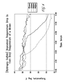
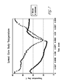
- Temperature regulation can be achieved in organisms by employing oxygen antagonists.
- there is a method of treating a subject with hypothermia comprising (a) contacting the subject with an effective amount of an oxygen antagonist, and then (b) subjecting the subject to an environmental temperature above that of the subject.
- the present invention includes a method of treating a subject with hyperthermia comprising (a) contacting the subject with an effective amount of an oxygen antagonist.
- treatment of hyperthermia also includes (b) subjecting the subject to an environmental temperature that is at least about 20°C below that of the subject. As discussed above, exposing the subject to nonphysiological or a controlled temperature environment can be used in additional embodiments.
- the invention concerns a method for inducing cardioplegia in a patient undergoing bypass surgery comprising administering to the patient an effective amount of an oxygen antagonist. It is contemplated that administration may be local to the heart so as to protect it.
- aspects of the invention relate to a method for preventing hematologic shock in a patient comprising administering to the patient an effective amount of an oxygen antagonist.
- the present invention covers a method for preventing or treating neurodegeneration in a mammal comprising administering to the mammal an effective amount of an oxygen antagonist.
- the biological matter may be exposed to an oxygen antagonist within about, within at least about, or within at most about 30 seconds, 1, 2, 3, 4, 5, 10, 15, 20, 25, 30, 35, 40, 45, 50, 55 minutes, 1, 2, 3, 4, 5, 6, 7, 8, 9, 10, 11, 12, 13, 14, 15, 16, 17, 18, 19, 20, 21, 22, 23, 24 hours, 1, 2, 3, 4, 5, 6, 7 days, 1, 2, 3, 4, 5 weeks, 1, 2, 3, 4, 5, 6, 7, 8, 9, 10, 11, 12 months, 1, 2, 3, 4, 5, 6, 7, 8, 9, 10, 11, 12, 13, 14, 15, 16, 17, 18, 19, 20 or more years, and any combination or range derivable therein, after initial damage (trauma or wound or degeneration) occurs.
- methods include an initial assessment of any damage, trauma, a wound, or degeneration.
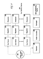
- Methods of the invention can involve employing an apparatus or system that maintains the environment in which biological matter is placed or exposed to.
- the invention includes an apparatus in which an oxygen antagonist, particularly as a gas, is supplied.
- the apparatus includes a container with a sample chamber for holding the biological matter, wherein the container is connected to a supply of gas comprising the oxygen antagonist(s).
- the container may be a solid container or it may flexible, such as a bag.
- the invention is an apparatus for preserving cell(s), the apparatus comprising: a container having a sample chamber with a volume of no greater than 775 liters; and a first gas supply in fluid communication with the sample chamber, the first gas supply including carbon monoxide.
- the apparatus also includes a cooling unit that regulates the temperature inside the sample chamber and/or a gas regulator that regulates the amount of oxygen antagonist in the chamber or the amount of oxygen antagonist in a solution that is in the chamber.
- the second gas supply may be connected with the sample chamber or it may be connected with the first gas supply.
- the additional gas as discussed above, may be a non-toxic and/or non-reactive gas.
- a gas regulator is part of the apparatus in some embodiments of the invention.
- One, two, three, or more gas regulators may be employed.
- the gas regulator regulates the gas supplied to the sample chamber from the first gas supply.
- it regulates the gas supplied to the sample chamber or first gas supply from the second gas supply, or there may be a regulator for both the first and second gas supplies.
- any gas regulator can be programmed to control the amount of gas supplied to the sample chamber and/or to another gas supply. The regulation may or may not be for a specified period of time.
- the gas regulator is electronically programmable.
- the pressure and/or the temperature inside the chamber can be regulated with either a pressure regulator or temperature regulator, respectively. As with the gas regulator, these regulators may be electronically programmable.
- the apparatus of the invention may also have a cooling and/or heating unit to achieve the temperatures discussed above. The unit may or may not be electronically programmable.
- the apparatus includes a wheeled cart on which the container rests or it may have one or more handles.
- the invention includes an apparatus for cell(s), tissues, organs, and even whole organisms, in which the apparatus has: a container having a sample chamber; a first gas supply in fluid communication with the sample chamber, the first gas supply including the oxygen antagonist(s); and an electronically-programmable gas regulator that regulates gas supplied to the sample chamber from the first gas supply.
- the apparatus also has a structure configured to provide a vacuum within the sample chamber.
- any oxygen antagonist described in this application is contemplated for use with apparatuses of the invention.
- carbon monoxide can be administered using this apparatus.
- a chalcogenide compound can be administered or a compound having the reducing agent structure.
- a candidate substance is screened for the ability to act as an oxygen antagonist. This can be done using any assay described herein, such as by measuring carbon dioxide output. Any substance identified as having exhibiting characteristics of an oxygen antagonist can be further characterized or tested. Moreover, it is contemplated that such a substance can be administered to biological matter to induce stasis or manufactured thereafter.
- any method of treatment can be used in the context of a preparation of a medicament for the treatment of or protection against the specified disease or condition.
- This includes, but is not limited to, the preparation of a medicament for the treatment of hemorrhagic or hematologic shock, wounds and tissue damage, hyperthermia, hypothermia, neurodegeneration, sepsis, cancer, and trauma.
- the invention includes, but is not limited to, the preparation of a medicament for a treatment to prevent shock, trauma, organ or tissue rejection, damage from cancer therapy, neurodegeneration, and wound or tissue damage.
- Stasis a cell, tissue or organ, or organism (collectively referred to as “biological material”) is living, but cellular functions necessary for cell division, developmental progression, metabolic state are slowed or even stopped. This state is desirable in a number of contexts.
- Stasis can be used as a method of preservation by itself, or it may be induced as part of a cryopreservation regimen.
- Biological materials may be preserved for research use, for transportation, for transplantation, for therapeutic treatment (such as ex vivo therapy), and to prevent the onset of trauma, for example. Stasis with respect to entire organisms have similar uses. For instance, transportation of organisms could be facilitated if they had entered stasis.
- Stasis may be beneficial by decreasing the need of the biological material for oxygen and, therefore, bloodflow. It may extend the period of time that biological material can be isolated from a life-sustaining environment and exposed to a death-inducing environment.
- An organism in stasis is distinguishable from an organism under general anesthesia. For example, an organism in mild stasis (between about 2- and about 5-fold decrease in cellular respiration) that is exposed to room air will begin to shiver, while an organism under anesthesia will not. Also, an organism in mild stasis is anticipated to respond to a toe squeeze, while an organism under anesthesia usually does not. Consequently, stasis is not the same thing as being under anesthesia as it is commonly practiced.
- the present invention is based on the observation that certain types of compounds effectively induce reversible stasis in biological matter.
- thermoregulation is a characteristic of so-called "warm-blooded” animals, which permits the organism to maintain a relatively constant core body temperature even when exposed to significantly altered (cold or hot) environmental temperatures.
- the ability to control thermoregulation by induction of stasis is one aspect of the invention, and permits uses similar to those discussed above.
- Thermal regulation may be a facilitated by placing of organisms, limbs or isolated organs or tissues into chambers/devices, the temperature of which can be controlled.
- warm rooms or chamber-like devices similar to hyperbaric chambers may encompass an entire organism and be connected to thermo-regulatory apparti.
- Smaller devices such as blankets, sleeves, cuffs or gloves (e.g., CORE CONTROL cooling system by AVAcore Technologies, Palo Alto, CA, U.S. Patent 6,602,277 ) are also contemplated.
- Such chambers/devices may be used both to increase or reduce ambient temperatures.
- Bio material contemplated for use with the present invention include material derived from invertebrates and vertebrates, including mammals; biological materials includes organisms.
- the invention can be employed with respect to mammals of veterinary or agricultural importance including those from the following classes: canine, feline, equine, bovine, ovine, murine, porcine, caprine, rodent, lagomorph, lupine, and ursine.
- the invention also extends to fish and birds. Other examples are disclosed below.
- the mammal is of the Order Monotremata, Marsupialia, Insectivora, Macroscelidia, Dermoptera, Chiroptera, Scandentia, Primates, Xenarthra, Pholidota, Tubulidentata, Lagomorpha, Rodentia, Cetacea, Carnivora, Proboscidea, Hyracoidea, Sirenia, Perissodactyla, or Artiodactyla.
- Monotremata examples include the Families Tachyglossidae (e.g., Echidnas) and Ornithorhynchidae (e.g., Platypus).
- Examples of Marsupialia include the Families Didelphidae (e.g., Opossums), Microbiotheriidae (e.g., Monito del Monte), Caenolestidae ( e.g., Rat Oppossums), Dasyuridae (e.g., Marsupial mice), Myrmecobiidae (e.g., Numbat), Thylacinidae (e.g., Thylacine), Peramelidae ( e.g., Bandicoots), Thylacomyidae ( e.g., Rabbit Bandicoots), Notoryctidae ( e.g., Marsupial Moles), Phalangeridae (e.
- Insectivora includes, for example, the Families Solenodontidae (e.g., Solenodons), Tenrecidae (e.g., Tenrecs, Otter Shrews), Chrysochloridae (e.g., Golden Moles), Erinaceidae ( e.g., Hedgehogs, Moonrats), Soricidae ( e.g., Shrews), and Talpidae ( e.g., Moles, Desmans).
- the Order Macroscelidia includes the Family Macroscelidia ( e.g., Elephant Shrews).
- the Order Scandentia includes Tupaiidae ( e.g., Tree Shrews).
- the Order Dermoptera includes the Family Cynocephalidea (e.g., Flying Lemurs).
- Chiroptera includes the Families Pteropodidae (e.g., Fruit Bats, Flying Foxes), Rhinopomatidae (e.g., Mouse-Tailed Bats), Craseonycteridae (e.g., Hog-Nosed or Bumblebee Bat), Emballonuridae (e.g., Sheath-Tailed Bats), Nycteridae ( e.g., Slit-Faced Bats), Megadermatidae ( e.g., False Vampire Bats), Rhinolophidae ( e.g., Horshoe Bats), Noctilionidae ( e.g., Bulldog Bats, Fisherman Bats), Mormoopidae, Phyllostomidae ( e.g., New World Leaf-Nosed Bat
- the Order Primates includes the Families Lemuridae (e.g., Lemurs), Cheirogaleidae ( e.g., Mouse Lemurs), Indriidae ( e.g., Indri, Woolly Lemur), Daubentoniidae (e.g., Aye-Aye), Lorisidae ( e.g., Lorises, Bushbabies, Galagos), Tarsiidae (e.g., Tarsiers), Cebidae ( e.g., New World Monkeys, Marmosets, Tamarins), Hylobatidae ( e.g., Gibbons), Ponlgidae ( e.g., Apes), and Hominidae (e.g., Man).
- Lemuridae e.g., Lemurs
- Cheirogaleidae e.g., Mouse Lemurs
- Indriidae e
- Examples of Xenarthra include Myrmecophagidae (e.g., Anteaters), Bradypodidae (e.g., Three-Toed Sloths), Megalonychidae (e.g., Two-Toed Sloths), and Dasypodidae ( e.g., Armadillos).
- Examples of Pholidota include Manidae ( e.g., Pangolins).
- Examples of Tubulidentata include Orycteropodidae (e.g., Aardvarks).
- Examples of Lagomorpha include Ochotonidae (e.g., Pikas) and Leporidae ( e.g., Hares and Rabbits).
- the Order Rodentia includes the Families Aplodontidae (e.g., Mountain Beavers), Sciuridae (e.g., Squirrels, Marmots, Chipmunks), Geomyidae (e.g., Pocket Gophers), Heteromyidae ( e.g., Pocket Mice, Kangaroo Rats), Castoridae ( e.g., Beaver), Anomaluridae (e.g., Scaly-Tailed Squirrels), Pedetidae ( e.g., Springhare), Muridae ( e.g., Rats and Mice), Gliridae ( e.g., Dormice), Seleviniidae ( e.g., Desert Dormouse), Zapodidae (e.g., Jumping Mice), Dipodidae (e.g., Jerboas), Hystricidae (e.g.,
- the Order Cetacea includes the Families Iniidae (e.g., Amazon Popoise), Lipotidae, Platanistidae, Pontoporiidae, Ziphiidae ( e.g., Beaked Whales), Physeteridae (e.g., Sperm Whales), Monodontidae (e.g., Beluga Whale, Narwhal), Delphinidae ( e.g., Marine Dolphins, Killer Whales), Phocoenidae ( e.g., Porpoises), Balaenopteridae ( e.g., Rorquals), Balaenidae (e.g., Right Whales), and Eschrichtiidae ( e.g., Gray Whales).
- Families Iniidae e.g., Amazon Popoise
- Lipotidae Lipotidae
- Platanistidae Platanistid
- the Order Carnivora includes the Families Canidae (e.g., Dogs, Foxes, Wolves, Jackals, Coyotes), Ursidae ( e.g., Bears), Procyonidae ( e.g., Raccoons, Coatis, Kinkajous, Lesser Pandas), Ailuropodidae ( e.g., Giant Pandas), Mustelidae ( e.g., Weasels, Skunks, Badgers, Otters), Viverridae ( e.g., Civets, Genets), Herpestidae ( e.g., Mongooses), Protelidae ( e.g., Aardwolf), Hyaenidae (e.g., Hyenas), Felidae (e.g., Cats), Otariidae (e.g., Eared Seals, Sea Lions), Odobenida
- the Order Proboscidea includes the Family Elephantidae (e.g., Elephants).
- Hyracoidea includes the Family Procaviidae (e.g., Hyraxes).
- Sirenia includes the Families Dugongidae (e.g., Dugong) and Trichechidae (e.g., Manatees).
- the Order Perissodactyla includes the Families Equidae (e.g., Horses, Asses, Zebras), Tapiridae ( e.g., Tapirs), and Rhinocerotidae ( e.g., Rhinoceroses).
- the Order Artiodactyla includes the Families Suidae (e.g., Pigs, Babirusa), Tayassuidae (e.g., Peccaries), Hippopotamidae (e.g., Hippopotamuses), Camelidae (e.g., Camels, Llamas, Vicunas), Tragulidae ( e.g., Chevrotains), Moschidae (e.g., Musk Deer), Cervidae (e.g., Deer, Elk, Moose), Giraffidae ( e.g., Giraffe, Okapi), Antilocapridae (e.g., Pronghorn), and Bovidae ( e.g., Cattle, Sheep, Antelope, Goats).
- Families Suidae e.g., Pigs, Babirusa
- Tayassuidae e.g., Pe
- the biological material is a reptile or is derived from a reptile.
- the reptile may be of the Order Chelonia, Pleurodira, Squamata, Rhynchocephalia, or Crocodylia.
- a reptile of the Order Chelonia may be, for example, a Carettochelyidae, Chelydridae ( e.g., Snapping Turtles), Cheloniidae (e.g., Loggerhead Turtles, Green Turtles), Dermatemydidae ( e.g., Leatherback Turtles), Emydidae (e.g., Paitned Turtles, Pond Sliders, Pond Turtles, Snail-Eating Turtles, Box Turtles), Kinosternidae ( e.g., Stinkpot Turtles), Saurotypidae, Testudinidae (e.g., Galapagos Tortoises, Desert Tortoises, Aldabra Turtles, Spu
- a reptile of the Order Squamata may be, for example, an Agamidae (e.g., Rainbow Lizards, Bearded Dragons, Indian Bloodsuckers, Spiny-Tailed Lizards), Chamaeleontdidae (e.g., Chameleons), Iguanidae ( e.g., Anoles, Basilisks, Collared Lizards, Iguanas, Homed Lizards, Chuckwallas, Sagebrush Lizards, Side-Blotched Lizards), Gekkonidae ( e.g., Geckos), Pygopodidae, Teiidae (e.g., Race Runners, Tegus), Lacertidae ( e.g., Sand Lizards, Ocellated Lizards, Viviparous Lizards, Wall Lizards, Long-Tailed Lizards), Xantuslidae, Scincidae (e.g., Skinks), Cordylida
- a reptile of the Order Rhynchocephalia may be, for example, a Sphenodontidae (e.g., Tuataras).
- a reptile of the Order Crocodylia may be, for example, an Alligatoridae (e.g., Alligators, Caiman), Crocodylidae (e.g., Crocodiles), or a Gavialidae (e.g., Gharials).
- the biological material of the present invention may be an amphibian or may be derived from an amphibian.
- the amphibian may be, for example, a frog or a toad.
- the frog or toad may be, for example, an Arthroleptidae (e.g., screeching frogs), Ascaphidae (e.g., tailed frogs), Brachycephalidae (e.g., gold frogs and shield toads), Bufonidae ( e.g., true toads), Centrolenidae (e.g., glass frogs and leaf frogs), Dendrobatidae (e.g., poison-dart frogs), Discoglossidae ( e.g., fire-bellied toads), Heleophrynidae ( e.g., ghost frogs), Hemisotidae (
- the amphibian may be a salamander.
- the salamander may be, for example, an Ambystomatidae (e.g., mole salamanders), Amphiumidae ( e.g., amphiumas), Cryptobranchidae (e.g., giant salamanders and hellbenders), Dicamptodontidae (e.g., Pacific giant salamanders), Hynobiidae (e.g., Asiatic salamanders), Plethodontidae (e.g., lungless salamanders), Proteidae (e.g., mudpuppies and waterdogs), Rhyacotritonidae (e.g., torrent salamanders), Salamandridae (e.g., newts and salamanders), or a Sirenidae (e.g., sirens).
- Ambystomatidae e.g., mole sal
- the amphibian may be a Caecilian.
- the Caecilian may be, for example, a Caeciliidae (e.g., caecilians), Ichthyophiidae (e.g., Asiatic tailed caecilians), Rhinatrematidae ( e.g., neotropical tailed caecilians), Scolecomorphidae ( e.g., African caecilians), Typhlonectidae ( e.g., aquatic caecilians), or an Uraeotyphlidae ( e.g., Indian caecilians).
- a Caeciliidae e.g., caecilians
- Ichthyophiidae e.g., Asiatic tailed caecilians
- Rhinatrematidae e.g., neotropical tailed caecilians
- Scolecomorphidae
- the biological material of the present invention may be a bird or may be derived from a bird.
- the bird may be, for example, an Anseriforme (e.g., waterfowl), Apodiforme (e.g., hummingbirds and swifts), Caprimulgiforme (e.g., nightbirds), Charadriiforme (e.g., shorebirds), Ciconiiforme (e.g., storks), Coliiforme ( e.g., mousebirds), Columbiforme ( e.g., doves and pigeons), Coraciiforme (e.g., kingfishers), Craciforme ( e.g., chacalacas, curassows, guans, megapodes), Cuculiforme (e.g., cuckoos, hoatzin, turacos), Falconiforme (e.g., diurnal birds of prey), Galli
- the biological material of the present invention may be a fish or may be derived from a fish.
- the fish may be, for example, an Acipenseriforme (e.g., paddlefishes, spoonfishes, and sturgeons), Polypteriforme (e.g., bichirs, birchers, lobed-finned pike, and reed fishes), Atheriniforme (e.g., rainbow fishes and silversides), Beloniforme, (e.g., halfbeeks and needlefishes), Beryciforme, Channiforme, Cyprinodontiforme (e.g., killifishes), Dactylapteriforme (e.g., flying gurnards), Gasterosteiforme (e.g., pipefishes and sticklebacks), Mugiliforme (e.g., mullets), Pegasiforme (e.g., dragonfishes and sea moths),
- the biological material maybe an invertebrate or derived from an invertebrate.
- the invertebrate may be, for example, a Porifera (e.g., sponges), Cnidaria (e.g ., jellyfish, hydras, sea anemones, Portuguese man-of-wars, and corals), Platyhelminthe (e.g., flatworms, including planaria, flukes, and tapeworms), Nematoda ( e.g., roundworms, including rotifers and nematodes), Mollusca ( e.g., mollusks, snails, slugs, octopuses, squids), Annelida ( e.g., segmented worms, including earthworms, leeches, and marine worms), Echinodermata (e.g., sea stars, sea cucumbers, sand dollars, sea urchins), Phoronida ( e.g .
- An Arthropod may be, for example, a Coleoptera (e.g ., beetles), Diptera (e.g ., true flies), Hymenoptera ( e.g., ants, bees, wasps), Lepidoptera (e.g., butterflies, moths), Mecoptera (e.g., scorpion flies), Megaloptera, Neuroptera ( e.g ., lacewings and relatives), Siphonaptera ( e.g ., fleas), Strepsiptera ( e.g., parasitic insects and twisted-winged parasites), Trichoptera ( e.g ., caddisflies), Anoplura (e.g., sucking lice), Hemiptera ( e.g., true bugs and their relatives), Mallophaga ( e.g ., biting lice), Psocoptera ( e.g ., psocids
- the biological material of the present invention may be a fungi or may be derived from a fungi.
- the fungi may be, for example, an Ascomycota (sac fungi), Basidiomycota (club fungi), Chytridiomycota (chytrids), Deuteromycota, or a Zygomycota.
- the fungi may be a Rhizopus, Pilobolus, Arthrobotrys, Aspergillus, Allomyces, Chytridium, Agaricus, Amanita, Cortinarius, Neurospora, Morchella, Saccharomyces, Pichia, Candida, Schizosaccharomyces, or Ergot.
- the fungi may be Saccharomyces cerevisiae, Schizosaccharomyces pombe, Candida albicans, or Pichia pastoris.
- the biological material of the present invention may be a plant or may be derived from a plant.
- the plant may be a Bryophyte (e.g., mosses, liverworts, hornworts), Lycophyte ( e.g., club mosses, ground pine), Sphenophyte ( e.g ., horsetails), Pterophyte ( e.g., ferns), Cycadophyte (e.g., cycads), Gnetophyte (e.g., gnetum, ephedra, welwitschia), Coniferophyte (e.g., conifers), Ginkophyte (e.g., ginko), or Anthophyte ( e.g ., flowering plants).
- Bryophyte e.g., mosses, liverworts, hornworts
- Lycophyte e.g., club mosses
- the Anthophyte may be a monocot or a dicot.
- Non-limiting examples of monocotyledonous plants include wheat, maize, rye, rice, turfgrass, sorghum, millet, sugarcane, lily, iris, agave, aloe, orchids, bromeliads, and palms.
- Non-limiting examples of dicotyledonous plants include tobacco, tomato, potato, soybean, sunflower, alfalfa, canola, rose, Arabidopsis, coffee, citrus fruits, beans, alfalfa, and cotton.
- the biological material of the present invention may be a Protist or may be derived from a Protist.
- the Protist may be a Rhodophyte (e.g., red algea), Phaeophyte (e.g., brown algea, kelp), Chlorophyte ( e.g., green algea),.
- Euglenophyte e.g., euglenoids
- Myxomycot e.g., slime molds
- Oomycot e.g., water molds, downy mildews, potato blight
- Bacillariophyte e.g., diatoms
- the biological material is a prokaryote or is derived from a prokaryote.
- the prokaryote is an Archaea (archaebacteria).
- the archaebacteria may be, for example, a Crenarchaeota, Euryarchaeota, Korarchaeota or Nanoarchaeota.
- the Euryarchaeota is a Halobacteria, Methanobacteria, Methanococci, Methanomicrobia, Methanosarcinae, Methanopyri, Archeoglobi, Thermoplasmata, or a Thermococci.
- Specific, non-limiting examples of archaebacteria include: Aeropyrum pernix, Methanococcus jannaschii, Halobacterium marismortui, and Thermoplasma acidophilum.
- the prokaryote is an Eubacteria.
- the Eubacteria may be, for example, an Actinobacteria, Aquificae, Bacteroidetes, Green sulfur bacteria, Chlamydiae, Verrucomicrobia, Chloroflexi, Chrysiogenetes, Cyanobacteria, Deferribacteres, Deinococcus-Thennus, Dictyoglomi, Fibrobacteres/Acidobacteria, Firmicutes, Fusobacteria, Gemmnatimonadetes, Nitrospirae, Omnibacteria, Planctomycetes, Proteobacteria, Spirochaetes, Thermodesulfobacteria, or Thermotogae.
- Actinobacteria include bacteria of the genera Actinomyces, Arthrobacter, Corynebacterium, Frankia, Micrococcus, Micromonospora, Mycobacterium, Propionibacterium, and Streptomyces.
- Specific examples of Actinobacteria include Mycobacterium leprae, Mycobacterium tuberculosis, Mycobacterium avium, Corynebacterium glutamicum, Propionibacterium acnes, andRhodococcus equi.
- Non-limiting examples of Aquificae include bacteria of the genera Aquifex, Hydrogenivirga, Hydrogenobacter, Hydrogenobaculum, Thermocrinis, Hydrogenothermus, Persephonella, Sulfurihydrogenibium, Balnearium, Desulfurobacterium, and Thermovibrio.
- Non-limiting examples of Firmicutes include bacteria of the genera Bacilli, Clostridia, and Molecutes.
- Firmicutes include: Listeria innocua, Listeria monocytogenes, Bacillus subtilis, Bacillus anthracis, Bacillus thuringiensis, Staphylococcus aureus, Clostridium acetobutylicum, Clostridium difficile, Clostridium perfringens, Mycoplasma genitalium, Mycoplasma pneumoniae, Mycoplasma pulmonis, Streptococcus pneumoniae, Streptococcus pyogenes, Streptococcus mutans, Lactococcus lactis, and Enterococcus faecalis.
- Non-limiting examples of Chlamydiae/Verrucomicrobia include bacteria such as Chlamydia trachomatis, Chlamydia pneumoniae, I Chlamydia psittaci.
- Non-limiting examples of Deinococcus-Thermus include bacteria of the genera Deinococcus and Thermus.
- Proteobacteria are gram-negative bacteria.
- Non-limiting examples of Proteobacteria include bacteria of the genera Escherichia, Salmonella, Vibrio, Rickettsia, Agrobacterium, Brucella, Rhizobium, Neisseria, Bordetella, Burkholderi, Buchnera, Yersinia, Klebsiella, Proteus, Shigella, Haemophilus, Pasteurella, Actinobacillus, Legionella, Mannheimia, Coxiella, Aeromonas, Francisella, Moraxella, Pseudomonas, Campylobacter, and Helicobacter.
- Proteobacteria include: Rickettsia conorii, Rickettsia prowazekii, Rickettsia typhi, Ehrlichia bovis, Agrobacterium tumefaciens, Brucella melitensis, Rhizobium rhizogenes, Neisseria meningitides, Bordetella parapertussis, Bordetella pertussis, Burkholderi mallei, Burkholderi pseudomallei, Neisseria gonorrhoeae, Escherichia coli , Salmonella enterica, Salmonella typhimurium, Yersinia pestis, Klebsiella pneumoniae, Yersinia enterocolitica, Proteus vulgaris, Shigella flexneri, Shigella sonnei, Shigella dysenterica, Haemophilus influenzae, Pasteurella multocida, Actinobacillus actinomycetem
- Non-limiting examples of Spixochaetes include bacteria of the families Brachyspiraceae, Leptospiraceae, and Spirochaetaceae. Specific examples of Spirochaetes include Borrelia burgdorferi, and Treponema pallidum.
- Stasis of the organism can be induced or stasis within cells, tissues, and/or organs of the organism can be induced.
- Biological matter in which stasis can be induced that are contemplated for use with methods and apparatuses of the invention are limited only insofar as the comprise cells utilizing oxygen to produce energy.
- Stasis can be induced in cells, tissues, or organs involving the heart, lung, kidney, liver, bone marrow, pancreas, skin, bone, vein, artery, cornea, blood, small intestine, large intestine, brain, spinal cord, smooth muscle, skeletal muscle, ovary, testis, uterus, and umbilical cord.
- stasis can be induced in cells of the following type: platelet, myelocyte, erythrocyte, lymphocyte, adipocyte, fibroblast, epithelial cell, endothelial cell, smooth muscle cell, skeletal muscle cell, endocrine cell, glial cell, neuron, secretory cell, barrier function cell, contractile cell, absorptive cell, mucosal cell, limbus cell (from cornea), stem cell (totipotent, pluripotent or multipotent), unfertilized or fertilized oocyte, or sperm.
- cells of the following type platelet, myelocyte, erythrocyte, lymphocyte, adipocyte, fibroblast, epithelial cell, endothelial cell, smooth muscle cell, skeletal muscle cell, endocrine cell, glial cell, neuron, secretory cell, barrier function cell, contractile cell, absorptive cell, mucosal cell, limbus cell (from cornea), stem cell (totipotent
- stasis can be induced in plants or parts of plants, including fruit, flowers, leaves, stems, seeds, cuttings. Plants can be agricultural, medicinal, or decorative. Induction of stasis in plants may enhance the shelf life or pathogen resistance of the whole or part of the plant.
- Methods and apparatuses of the invention can be used to induce stasis in in vivo biological matter. This can serve to protect and/or preserve the biological matter or the organism itself or to prevent damage or injury (or further damage or injury) to them or the organism overall.
- Stasis can be measured by a number of ways, including by quantifying the amount of oxygen consumed by a biological sample, the amount of carbon dioxide produced by the sample (indirect measurement of cellular respiration), or characterizing motility.
- the biological matter is placed into a chamber that is sealed with two openings; for gas import and export.
- Gas room air or other gases
- gas is passed into the chamber at a given flow rate and out of the exit port to maintain approximately 1 atmosphere of pressure in the chamber.
- the gas is passed through a carbon dioxide detector and or an oxygen detector to measure (every second) the amount of each compound in the gas mixture. Comparison of these values over time gives the rate of oxygen consumption or carbon dioxide production.
- Oxygen metabolism is a fundamental requirement for life in aerobic metazoans. Aerobic respiration accounts for the vast majority of energy production in most animals and also serves to maintain the redox potential necessary to carry out important cellular reactions. In hypoxia, decreased oxygen availability results in inefficient transfer of electrons to molecular oxygen in the final step of the electron transport chain. This inefficiency results in both a decrease in aerobic energy production and an increase in the production of damaging free radicals, mainly due to the premature release of electrons at complex III and the formation of O 2 - by cytochrome oxidase (Semenza, 1999). Limited energy supplies and free radical damage can interfere with essential cellular processes such as protein synthesis and maintenance of membrane polarities (Hochachka et al., 1996), and will ultimately lead to cell death.
- Carbon monoxide (CO) is a colorless, odorless, and tasteless gas that can be toxic to animals, including humans. According to the Center for Disease Control, more than 450 people unintentionally die from carbon monoxide each year.
- carbon monoxide presents no symptoms to humans exposed to it. However, at 200 ppm, within two-three hours the carbon monoxide can cause a slight headache; at 400 ppm, within one to two hours it can cause a frontal headache that may become widespread within three hours; and, at 800 ppm it can cause dizziness, nausea, and/or convulsions within 45 minutes, and render the subject insensible within two hours. At levels of around 1000 ppm, an organism can expire after exposure for more than around 1-2 minutes.
- carbon monoxide can be used to induce stasis of and/or help preserve live biological samples. It is thus contemplated that carbon monoxide can be used for inducing stasis in isolated biological matter, such as blood-free biological matter (because of the effects that carbon monoxide has with respect to hemoglobin, which is a separate pathway than the one involved in inducing stasis).
- carbon monoxide may be used in combination with agents or methods that assist in the preservation and/or transplantation/grafting process of biological materials.
- chalcogenides Compounds containing a chalcogen element; those in Group 6 of the periodic table, but excluding oxides, are commonly termed “chalcogenides” or “chalcogenide compounds (used interchangeably herein). These elements are sulfur (S), selenium (Se), tellurium (Te) and polonium (Po). Common chalcogenides contain one or more of S, Se and Te, in addition to other elements. Chalcogenide compounds can be employed as reducing agents.
- chalcogenides inhibit or reduce the activity of oxidative phosphorylation.
- the ability of chalcogenides to block autonomous thermoregulation, i.e., to permit core body temperatures of "warm-blooded" animals to be manipulated through control of environmental temperatures, is believed to stem from the same mechanism as set forth above - binding to cytochrome oxidase, and blocking or reducing the activity of oxidative phosphorylation.
- Chalcogenides may be provided in liquid as well as gaseous forms.
- Chalcogenides can be toxic, and at some levels lethal, to mammals. In accordance with the present invention, it is anticipated that the levels of chalcogenide should not exceed lethal levels in the appropriate environment. Lethal levels of chalcogenides may be found, for example in Material Safety Data Sheets for each chalcogenide or from information sheets available from the Occupational Safety and Health Administration (OSHA) of the US Government.
- cytochrome oxidase While carbon monoxide and chalcogenide compounds can both induce stasis by acting as an oxygen antagonist, they have different toxic effects that are separate from their abilities to induce stasis. Moreover, the concentrations needed to mediate a stasis effect are different because of the different affinities of cytochrome oxidase. While the affinity of cytochrome oxidase for oxygen is about 1:1 as compared to carbon monoxide, the affinity for H 2 S appears on the order of about 300:1 as compared to oxygen. This impacts what toxic effects are observed with a stasis-inducing concentration. Thus, it is contemplated that chalcogenide compounds are particularly suited for inducing stasis of biological matter in whole organisms and of whole organisms.
- Hydrogen sulfide is a potentially toxic gas that is often associated with petrochemical and natural gas, sewage, paper pulp, leather tanning, and food processing.
- Post-exposure effects may persist for years, and include loss of coordination, memory loss, motor dysfunction, personality changes, hallucination and insomnia.
- H 2 S Most contact with H 2 S, however, occurs well below such acute toxicity levels. Nonetheless, there is general concern over longterm contact at sub-acute levels. Some reports exist indicating persistent impairments in balance and memory, as well as altered sensory motor functions may occur in humans following chronic low-level H 2 S exposure. Kilburn and Warshaw (1995); Kilburn (1999). Others have reported that perinatal exposure of rats to low (20 or 50 ppm) H 2 S for 7 hours per day from gestation through post-natal day 21 resulted in longer dendritic branches with reduced aborization of cerebellar Purkinje cells. Other neurologic defects associated with relatively low levels of H 2 S include altered brain neurotransmitter concentrations and altered neurologic responses, such as increased hippocampal theta EEG activity.
- H 2 S Behavioral toxicity was studied in rats exposed to moderate levels of H 2 S. The results showed that H 2 S inhibits discriminated avoidance responses immediately after the end of the exposure (Higuchi and Fukamachi, 1997), and also interferes with the ability of rats to learn a baited radial arm maze task (Partlo et al., 2001). In another perinatal study using 80 ppm H 2 S, no neuropathological effects or altered motor activity, passive avoidance, or acoustic startle response in exposed rat pups was seen. Dorman et al. (2000). Finally, Struve et al. (2001) exposed rats to H 2 S by gas at various levels for 3 hours per day on five consecutive days.
- Typical levels of hydrogen sulfide contemplated for use in accordance with the present invention include values of about 1 to about 150 ppm, about 10 to about 140 ppm, about 20 to about 130 ppm, and about 40 to about 120 ppm, or the equivalent oral, intravenous or transdermal dosage thereof.
- Other relevant ranges include about 10 to about 80 ppm, about 20 to about 80 ppm, about 10 to about 70 ppm, about 20 to about 70 ppm, about 20 to about 60 ppm, and about 30 to about 60 ppm, or the equivalent oral, intravenous or transdermal thereof.
- the chalcogenide atmosphere should be reduced to avoid a potentially lethal build up of chalcogenide in the subject.
- an initial environmental concentration of 80 ppm may be reduced after 30 min to 60 ppm, followed by further reductions at 1 hr (40 ppm) and 2 hrs (20 ppm).
- Hydrogen selenide is a key metabolite, formed from inorganic sodium selenite (oxidation state +4) via selenodiglutathione (GSSeSG), through reduction by thiols and NADPH-dependent reductases, and released from selenocysteine by lyase action (Ganther, 1999). Hydrogen selenide provides Se for synthesis of selenoproteins after activation to selenophosphate.
- Hydrogen telluride (H 2 Te) exists as an unstable gas.
- the reducing agent structure compound is dimethylsulfoxide (DMSO), dimethylsulfide (DMS), methylmercaptan (CH 3 SH), mercaptoethanol, thiocyanate, hydrogen cyanide, methanethiol (MeSH), or CS 2 .
- the oxygen antagonist is CS 2 , MeSH, or DMS. Compounds on the order of the size of these molecules are particularly contemplated (that is, within about 50% of their molecular weights).
- Additional compounds that are envisioned as useful for inducing stasis include, but are not limited to, the following structures, many of which are readily available and known to those of skill in the art (identified by CAS number): 104376-79-6 (Ceftriaxone Sodium Salt); 105879-42-3; 1094-08-2 (Ethopropatine HCl); 1098-60-8 (Triflupromazine HCl); 111974-72-2; 113-59-7; 113-98-4 (Penicillin GK + );115-55-9; 1179-69-7; 118292-40-3; 119478-56-7; 120138-50-3; 121123-17-9; 121249-14-7; 1229-35-2; 1240-15-9; 1257-78-9 (Prochlorperazine Edisylate Salt); 128345-62-0; 130-61-0 (Thioridazine HCl) 132-98-9 (Penicillin V K + ); 13412-64-1 (Dicloxacillin Na + Hydrate);
- hypoxia is a common natural stress and several well conserved responses exist that facilitate cellular adaptation to hypoxic environments. To compensate for the decrease in the capacity for aerobic energy production in hypoxia, the cell must either increase anaerobic energy production or decrease energy demand (Hochachka et al., 1996). Examples of both of these responses are common in metazoans and the particular response used depends, in general, on the amount of oxygen available to the cell.
- hypoxia-inducible transcription factor 1 HIF-1
- HIF-1 hypoxia-inducible transcription factor
- anoxia defined here as ⁇ 0.001 kPa O 2 -- oxidative phosphorylation ceases and thus the capacity to generate energy is drastically reduced.
- the cell In order to survive in this environment, the cell must decrease energy demand by reducing cellular activity (Hochachka et al., 2001). For example, in turtle hepatocytes deprived of oxygen, a directed effort by the cell to limit activities such as protein synthesis, ion channel activity, and anabolic pathways results in a 94% reduction in demand for ATP (Hochachka et al, 1996).
- C . elegans respond to anoxia by entering into suspended animation, in which all observable movement, including cell division and developmental progression, ceases (Padilla et al., 2002; Van Voorhies et al., 2000). C . elegans can remain suspended for 24 hours or more and, upon return to normoxia, will recover with high viability. This response allows C .
- hypoxia is still often a damaging stress.
- mammals have both heme oxygenase-1 and HIF-1, and some evidence suggests that suspended animation is possible in mammals as well (Bellamy et al., 1996; Alam et al., 2002).
- hypoxic damage due to trauma such as heart attack, stroke or blood loss is a major cause of death.
- the understanding of the limitations of the two fundamental strategies for surviving hypoxic stress, remaining animated or suspending animation is hampered by the fact that it has been based on studies in a variety of systems under a variety of conditions.
- “Hypoxia” occurs when the normal physiologic levels of oxygen are not supplied to a cell or tissue.
- Normoxia refers to normal physiologic levels of oxygen for the particular cell type, cell state or tissue in question.
- Amoxia is the absence of oxygen.
- Hapoxic conditions are those leading to cellular hypoxia. These conditions depend on cell type, and on the specific architecture or position of a cell within a tissue or organ, as well as the metabolic status of the cell.
- hypoxic conditions include conditions in which oxygen concentration is at or less than normal atmospheric conditions, that is less that 20.8, 20, 19,18,17,16,15,14,13,12,11,10,9, 8,7,6, 5,4,3,2,1,0.5, 0%; alternatively, these numbers could represent the percent of atmosphere at 1 atmosphere of pressure (101.3 kPa).
- An oxygen concentration of zero percent defines anoxic conditions.
- hypoxic conditions include anoxic conditions, although in some embodiments, hypoxic conditions of not less than 0.5% are implemented.
- “normoxic conditions” constitute oxygen concentrations of around 20.8% or higher.
- Standard methods of achieving hypoxia or anoxia are well established and include using environmental chambers that rely on chemical catalysts to remove oxygen from the chamber.
- Such chambers are available commercially from, for example, BD Diagnostic Systems (Sparks, MD) as GASPAK Disposable Hydrogen + Carbon Dioxide Envelopes or BIO-BAG Environmental Chambers.
- oxygen may be depleted by exchanging the air in a chamber with a non-oxygen gas, such as nitrogen.
- Oxygen concentration may be determined, for example using a FYRITE Oxygen Analyzer (Bacharach, Pittsburgh PA).
- the oxygen concentration of the environment containing biological matter can be about, at least about, or at most about 1, 2, 3, 4, 5, 6, 7, 8, 9, 10, 11, 12, 13, 14, 15, 16, 17, 18, 19, 24, 21, 22, 23, 24, 25, 26, 27, 28, 29, 30, 31, 32, 33, 34, 35, 36, 37, 38, 39, 40, 41, 42, 43, 44, 45, 46, 47, 48, 49, 50, 51, 52, 53, 54, 55, 56, 57, 58, 59, 60, 61, 62, 63, 64, 65, 66, 67, 68, 69, 70, 71, 72, 73, 74, 75, 76, 77, 78, 79, 80, 81, 82, 83, 84, 85, 86, 87, 88, 89, 90, 91, 92, 93, 94, 95, 96, 97, 98,
- the present invention may find use in the treatment of patients who are undergoing, or who are susceptible to trauma. Trauma may be caused by external insults, such as burns, wounds, amputations, gunshot wounds, or surgical trauma, internal insults, such as stroke or heart attack that result in the acute reduction in circulation, or reductions in circulation due to non-invasive stress, such as exposure to cold or radiation.
- Trauma may be caused by external insults, such as burns, wounds, amputations, gunshot wounds, or surgical trauma, internal insults, such as stroke or heart attack that result in the acute reduction in circulation, or reductions in circulation due to non-invasive stress, such as exposure to cold or radiation.
- trauma On a cellular level, trauma often results in exposure of cells, tissues and/or organs to hypoxia, thereby resulting in induction of programmed cell death, or "apoptosis.”
- trauma leads to the induction of a series of biochemical processes, such as clotting, inflammation, hypotension, and may give rise to shock, which if it persists may lead to organ dysfunction, irreversible cell damage and death.
- Biological processes are designed to defend the body against traumatic insult; however they may lead to a sequence of events that proves harmful and, in some instances, fatal.
- the present invention contemplates the placement of tissues, organs, limbs and even whole organisms into stasis as a way of protecting them from the detrimental effects of trauma.
- induction of stasis in vivo or ex vivo alternatively in conjunction with reduction in the temperature of the tissue, organ or organism, can "buy time" for the subject, either by bringing medical attention to the subject, or by transporting the subject to the medical attention.
- the present invention also contemplates methods for inducing tissue regeneration and wound healing by prevention/delay of biological processes that may result in delayed wound healing and tissue regeneration.
- the induction of stasis induction of stasis in vivo or ex vivo can aid in the wound healing and tissue regeneration process by managing the biological processes that inhibit healing and regeneration.
- methods of the invention can be implemented to prevent or treat trauma such as cardiac arrest or stroke.
- trauma such as cardiac arrest or stroke.
- the invention has particular importance with respect to the risk of trauma from emergency surgical procedures, such as thoractomy, laparotomy, and splenic transection.
- wounds and tissue damage are intractable or take excessive periods of time to heal. Examples are chronic open wounds (diabetic foot ulcers and stage 3 & 4 pressure ulcers), acute and traumatic wounds, flaps and grafts, and subacute wounds ( i.e. , dehisced incisions). This may also apply to other tissue damage, for example bums and lung damage from smoke/hot air inhalation.
- biological matter is exposed to an oxygen antagonist for about, at least about, or at most about 30 seconds, 1, 2, 3, 4, 5, 10, 15, 20, 25, 30, 35, 40, 45, 50, 55 minutes, 1, 2, 3, 4, 5, 6, 7, 8, 9, 10, 11, 12, 13, 14, 15, 16, 17, 18, 19, 20, 21, 22, 23, 24 hours, 1, 2, 3, 4, 5, 6, 7 days, 1, 2, 3, 4, 5 weeks, 1, 2, 3, 4, 5, 6, 7, 8, 9, 10, 11, 12 months or more.
- the invention concerns inducing a whole body hibernetic state using H 2 S (or other oxygen antagonist), to preserve the patient's vital organs and life. This will allow for transport to a controlled environment ( e.g., surgery), where the initial cause of the shock can be addressed, and then the patient brought back to normal function in a controlled manner.
- the first hour after injury referred to as the "golden hour” is crucial to a successful outcome.
- Stabilizing the patient in this time period is the major goal, and transport to a critical care facility (e.g., emergency room, surgery, etc.) where the injury can be properly addressed.
- a critical care facility e.g., emergency room, surgery, etc.
- biological matter is exposed to an oxygen antagonist for about, at least about, or at most about 30 seconds, 1, 2, 3, 4, 5, 10, 15, 20, 25, 30, 35, 40, 45, 50, 55 minutes, 1, 2, 3, 4, 5, 6, 7, 8, 9, 10, 11, 12, 13, 14, 15, 16, 17, 18, 19, 20, 21, 22, 23, 24 hours, 1, 2, 3, 4, 5, 6, 7 days or more, and any range or combination therein.
- the present inventor proposes use of the present invention to treat people with extreme hypothermia.
- the method provides that patients with extreme hypothermia are administered or exposed to an oxygen antagonist and then gradually restored to normal temperature while withdrawing, in a controlled fashion, the oxygen antagonist.
- the oxygen antagonist buffers the biological systems within the subject so that they may be initiated gradually without shock (or harm) to the subject.
- a subject suffering from hypothermia with be given an oral or intravenous dose of an oxygen antagonist.
- Intravenous provision may be preferred because of the potential non-responsiveness of the subject and the ability to provide a controlled dosage over a period of time.
- the oxygen antagonist may be provide in a gaseous state, for example, using a mask for inhalation or even a sealed chamber that can house the entire subject.
- the patient will be stabilized in terms of heart rate, respiration and temperature prior to effecting any change.
- the ambient environmental temperature will be increased, again gradually. This may be accomplished simply by removing the subject from the hypothermic conditions.
- a more regulated increase in temperature may be effected by adding successive layers of clothing or blankets, by use of a thermal wrap with gradual increase in heat, or if possible, by placing the subject in chamber whose temperature may be gradually increased.
- the vital signs of the subject are monitored over the course of the temperature increase.
- the oxygen antagonist is removed from the subject's environment. Both heat and oxygen antagonist treatment are ceased at the appropriate endpoint, judged by the medical personnel monitoring the situation, but in any event at the time the subject's temperature and other vital signs return to a normal range. Continued monitoring following cessation of treatment is recommended for a period of at least 24 hrs.
- mice inhaled H 2 S at 80 ppm immediately underwent hibernation. This included an inability to regulate their body temperature when ambient temperatures were dropped below room temperature. Accordingly, this technology could be used to control whole body temperature in certain states of hyperthermia. This would likely involve administration of H 2 S (or other oxygen antagonist) through inhalation or perfused into the blood supply to induce a hibernation state. It would be useful to have the patient to be in stasis for between about 6 and about 24 hours, during which time the source of the fever can be addressed.
- a patient is exposed to an oxygen antagonist for about, at least about, or at most about 30 seconds, 1, 2, 3, 4, 5, 10, 15, 20, 25, 30, 35, 40, 45, 50, 55 minutes, 1, 2, 3, 4, 5, 6, 7, 8, 9, 10, 11, 12, 13, 14, 15, 16, 17, 18, 19, 20, 21, 22, 23, 24 hours, 1, 2, 3, 4, 5, 6, 7 days or more, and any range or combination therein.
- the present invention may find use as solutions for cardioplegia for cardiac bypass surgery (CABG).
- Cardioplegic solutions are perfused through the vessels and chambers of the heart and cause its intrinsic beating to cease, while maintaining the viability of the organ.
- Cardioplegia paralysis of the heart
- cardioplegia is desirable during open-heart surgery and during the procurement, transportation, and storage of donor hearts for use in heart transplantation procedures.
- cardioplegic solutions are available and different techniques for using cardioplegia solutions are known in the art.
- cardioplegic solutions often have varying amounts of potassium, magnesium, and several other minor components.
- drugs are added to the cardioplegic solution to aid in muscle relaxation and protection from ischemia. Varying the temperature at which the cardioplegic solution is used may also have beneficial effects.
- Ischemic-reperfusion injury during cardiac bypass surgery results in poor outcomes (both morbidity and mortality), especially due to an already weakened state of the heart.
- Myocardial ischemia results in anaerobic myocardial metabolism. The end products of anaerobic metabolism rapidly lead to acidosis, mitochondrial dysfunction, and myocyte necrosis. High-energy phosphate depletion occurs almost immediately, with a 50 percent loss of ATP stores within 10 minutes. Reduced contractility occurs within 1 to 2 minutes, with development of ischemic contracture and irreversible injury after 30 to 40 minutes of normothermic (37°C) ischemia.
- Reperfusion injury is a well-known phenomenon following restoration of coronary circulation.
- Reperfusion injury is characterized by abnormal myocardial oxidative metabolism.
- reperfusion may produce cytotoxic oxygen free radicals.
- oxygen free radicals play a significant role in the pathogenesis of reperfusion injury by oxidizing sarcolemmal phospholipids and thus disrupting membrane integrity.
- Oxidized free fatty acids are released into the coronary venous blood and are a marker of myocardial membrane phospholipid peroxidation.
- Protamine induces complement activation, which activates neutrophils.
- Activated neutrophils and other leukocytes are an additional source of oxygen free radicals and other cytotoxic substances.
- the present invention provides methods and compositions for inducing cardioplegia that will provide greater protection to the heart during bypass surgery.
- the present invention provides a cardioplegic solution comprising H 2 S dissolved in solution (or another oxygen antagonist).
- the invention further comprises at least a first device, such as a catheter or cannula, for introducing an appropriate dose of the cardioplegic solution to the heart.
- the invention further comprises at least a second device, such as a catheter or cannula, for removing the cardioplegic solution from the heart.
- the heart is exposed to an oxygen antagonist for about, at least about, or at most about 30 seconds, 1, 2, 3, 4, 5, 10, 15, 20, 25, 30, 35, 40, 45, 50, 55 minutes, 1, 2, 3, 4, 5, 6, 7, 8, 9, 10, 11, 12, 13, 14, 15, 16, 17, 18 hours or more, and any range or combination therein.
- Cancer is a leading cause of mortality in industrialized countries around the world.
- the most conventional approach to the treatment of cancer is by administering a cytotoxic agent to the cancer patient (or treatment ex vivo of a tissue) such that the agent has a more lethal effect on the cancer cells than normal cells.
- chemo- and radiotherapy are often characterized by severe side effects, some of which are life threatening, e.g., sores in the mouth, difficulty swallowing, dry mouth, nausea, diarrhea, vomiting, fatigue, bleeding, hair loss and infection, skin irritation and loss of energy (Curran, 1998; Brizel, 1998).
- hypothermia transient and reversible lowering of the core body temperature, or "hypothermia,” may lead to improvements in the fight against cancer.
- Hypothermia of 28°C was recently found to reduce radiation, doxorubicin-and cisplatin-induced toxicity in mice.
- the cancer fighting activity of these drugs/treatments was not compromised when administered to cooled animals; rather, it was enhanced, particularly for cisplatin (Lundgren-Eriksson et al., 2001). Based on this and other published work, the inventor proposes a further reduction in core temperature will provide benefit to cancer patients.
- the present invention contemplates the use of oxygen antagonists to induce stasis in normal tissues of a cancer patient, thereby reducing the potential impact of chemo- or radiotherapy on those tissues. It also permits the use of higher doses of chemo- and radiotherapy, thereby increasing the anti-cancer effects of these treatments.
- hyperproliferative disorder including benign and malignant neoplasias, non-neoplastic hyperproliferative conditions, pre-neoplastic conditions, and precancerous lesions.
- disorders include restenosis, cancer, multi-drug resistant cancer, primary psoriasis and metastatic tumors, angiogenesis, rheumatoid arthritis, inflammatory bowel disease, psoriasis, eczema, and secondary cataracts, as well as oral hairy leukoplasia, bronchial dysplasia, carcinomas in situ, and intraepithelial hyperplasia.
- the present invention is directed at the treatment of human cancers including cancers of the prostate, lung, brain, skin, liver, breast, lymphoid system, stomach, testicles, ovaries, pancreas, bone, bone marrow, gastrointestine, head and neck, cervix, esophagus, eye, gall bladder, kidney, adrenal glands, heart, colon and blood. Cancers involving epithelial and endothelial cells are also contemplated for treatment.
- chemo- and radiotherapy are designed to reduce tumor size, reduce tumor cell growth, induce apoptosis in tumor cells, reduce tumor vasculature, reduce or prevent metastasis, reduce tumor growth rate, accelerate tumor cell death, and kill tumor cells.
- oxygen antagonist compositions of the present invention with secondary anti-cancer agents (secondary agents) effective in the treatment of hyperproliferative disease.
- an "anti-cancer” agent is capable of negatively affecting cancer in a subject, for example, by killing cancer cells, inducing apoptosis in cancer cells, reducing the growth rate of cancer cells, reducing the incidence or number of metastases, reducing tumor size, inhibiting tumor growth, reducing the blood supply to a tumor or cancer cells, promoting an immune response against cancer cells or a tumor, preventing or inhibiting the progression of cancer, or increasing the lifespan of a subject with cancer.
- Secondary anti-cancer agents include biological agents (biotherapy), chemotherapy agents, and radiotherapy agents. More generally, these other compositions are provided in a combined amount effective to kill or inhibit proliferation of the cancer or tumor cells, while at the same time reducing or minimizing the impact of the secondary agents on normal cells.
- This process may involve contacting or exposing the cells with an oxygen antagonist and the secondary agent(s) at the same time. This may be achieved by contacting the cell with a single composition or pharmacological formulation that includes both agents, or by contacting or exposing the cell with two distinct compositions or formulations, at the same time, wherein one composition includes an oxygen antagonist and the other includes the second agent(s).
- the oxygen antagonist therapy may precede or follow the secondary agent treatment by intervals ranging from minutes to weeks.
- the other agent and expression construct are applied separately to the cell, one would generally ensure that a significant period of time did not expire between the time of each delivery, such that the agent and expression construct would still be able to exert an advantageously combined effect on the cell.
- biological matter will be kept in stasis for between about 2 and about 4 hours while the cancer treatment is being administered.
- biological matter is exposed to an oxygen antagonist for about, at least about, or at most about 30 seconds, 1, 2, 3, 4, 5, 10, 15, 20, 25, 30, 35, 40, 45, 50, 55 minutes, 1, 2, 3, 4, 5, 6 hours or more, and any range or combination therein.
- the oxygen antagonist is "A” and the secondary anti-cancer agent, such as radio- or chemotherapy, is "B":
- Administration of the oxygen antagonist compounds of the present invention to a patient will follow general protocols for the administration of chemotherapeutics, taking into account the toxicity, if any, of the compound. It is expected that the treatment cycles would be repeated as necessary. It also is contemplated that various standard therapies, as well as surgical intervention, may be applied in combination with the above-described anti-cancer therapy.
- Cancer therapies also include a variety of combination therapies with both chemical and radiation based treatments.
- Combination chemotherapies include, for example, cisplatin (CDDP), carboplatin, procarbazine, mechlorethamine, cyclophosphamide, camptothecin, ifosfamide, melphalan, chlorambucil, busulfan, nitrosurea, dactinomycin, daunorubicin, doxorubicin, bleomycin, plicomycin, mitomycin, etoposide (VP16), tamoxifen, raloxifene, estrogen receptor binding agents, taxol, gemcitabien, navelbine, farnesyl-protein transferase inhibitors, transplatinum, 5-fluorouracil, vincristine, vinblastine and methotrexate, Temazolomide (an aqueous form of DTIC), or any analog or derivative variant of the foregoing.
- Dosage ranges for X-rays range from daily doses of 50 to 200 roentgens for prolonged periods of time (3 to 4 wk), to single doses of 2000 to 6000 roentgens.
- Dosage ranges for radioisotopes vary widely, and depend on the half-life of the isotope, the strength and type of radiation emitted, and the uptake by the neoplastic cells.
- compositions of the invention for example, a hypoxic antitumor compound
- chemotherapeutic or radiotherapeutic agent is delivered to a target cell or are placed in direct juxtaposition with the target cell.
- both agents may be delivered to a cell in a combined amount effective to kill the cell or prevent it from dividing.
- Immunotherapeutics generally, rely on the use of immune effector cells and molecules to target and destroy cancer cells.
- the immune effector may be, for example, an antibody specific for some marker on the surface of a tumor cell.
- the antibody alone may serve as an effector of therapy or it may recruit other cells to actually effect cell killing.
- the antibody also may be conjugated to a drug or toxin (chemotherapeutic, radionuclide, ricin A chain, cholera toxin, pertussis toxin, etc.) and serve merely as a targeting agent.
- the effector may be a lymphocyte carrying a surface molecule that interacts, either directly or indirectly, with a tumor cell target.
- Various effector cells include cytotoxic T cells and NK cells.
- Immunotherapy could also be used as part of a combined therapy.
- the general approach for combined therapy is discussed below.
- the tumor cell must bear some marker that is amenable to targeting, i.e., is not present on the majority of other cells.
- Common tumor markers include carcinoembryonic antigen, prostate specific antigen, urinary tumor associated antigen, fetal antigen, tyrosinase (p97), gp68, TAG-72, HMFG, Sialyl Lewis Antigen, MucA, MucB, PLAP, estrogen receptor, laminin receptor, erb B and p155.
- Immune stimulating molecules also exist including: cytokines such as IL-2, IL-4, IL-12, GM-CSF, gamma-IFN, chemokines such as MIP-1, MCP-1, IL-8 and growth factors such as FLT3 ligand.
- cytokines such as IL-2, IL-4, IL-12, GM-CSF, gamma-IFN
- chemokines such as MIP-1, MCP-1, IL-8 and growth factors such as FLT3 ligand.
- Combining immune stimulating molecules, either as proteins or using gene delivery in combination with a tumor suppressor such as mda-7 has been shown to enhance anti-tumor effects (Ju et al., 2000)
- immune adjuvants e.g., Mycobacterium bovis, Plasmodium falciparum,. dinitrochlorobenzene and aromatic compounds
- U.S. Patent 5,801,005 U.S.
- Patent 5,739,169 Hui and Hashimoto, 1998; Christodoulides et al., 1998), cytokine therapy (e.g., interferons ⁇ , ⁇ and ⁇ ; IL-1, GM-CSF and TNF) (Bukowski et al., 1998; Davidson et al., 1998; Hellstrand et al., 1998) gene therapy (e.g., TNF, IL-, IL-2, p53) (Qin et al., 1998; Austin-Ward and Villaseca, 1998; U.S. Patent 5,830,880 and U.S.
- cytokine therapy e.g., interferons ⁇ , ⁇ and ⁇ ; IL-1, GM-CSF and TNF
- gene therapy e.g., TNF, IL-, IL-2, p53
- Patent 5,846,945 and monoclonal antibodies (e.g ., anti-ganglioside GM2, anti-HER-2, anti-p185) (Pietras et al., 1998; Hanibuchi et al., 1998).
- Herceptin trastuzumab
- Herceptin is a chimeric (mouse-human) monoclonal antibody that blocks the HER2-neu receptor. It possesses anti-tumor activity and has been approved for use in the treatment of malignant tumors (Dillman, 1999). Combination therapy of cancer with herceptin and chemotherapy has been shown to be more effective than the individual therapies.
- one or more anti-cancer therapies may be employed with the anti-tumor therapies described herein.
- the present invention may be used to treat neurodegenerative diseases.
- Neurodegenerative diseases are characterized by degeneration of neuronal tissue, and are often accompanied by loss of memory, loss of motor function, and dementia. With dementing diseases, intellectual and higher integrative cognitive faculties become more and more impaired over time. It is estimated that approximately 15% of people 65 years or older are mildly to moderately demented.
- Neurodegenerative diseases include Parkinson's disease; primary neurodegenerative disease; Huntington's Chorea; stroke and other hypoxic or ischemic processes; neurotrauma; metabolically induced neurological damage; sequelae from cerebral seizures; hemorrhagic shock; secondary neurodegenerative disease (metabolic or toxic); Alzheimer's disease, other memory disorders; or vascular dementia, multi-infarct dementia, Lewy body dementia, or neurodegenerative dementia.
- the present invention can be used to regulate and control the cycling between the oxidative and reduced states to prevent or reverse the effects of neurodegenerative diseases and processes. Furthermore, reduced metabolic activity overall has been shown to correlate with health in aged animals and humans. Therefore, the present invention would also be useful to suppress overall metabolic function to increase longevity and health in old age. It is contemplated that this type of treatment would likely be administered at night, during sleep for period of approximately 6 to 10 hours each day. This could require daily treatment for extended periods of time from months to years.
- the present invention can be used to preserve or store whole organisms for transport and/or storage purposes. Such organisms could be used for research purposes, such as laboratory mice (mouse banking), or for consumption, such as fish. In these situation, it is contemplated that stasis can be maintained indefinitely. Moreover, stasis can be induced in plants or parts of plants, including fruit, flowers, leaves, stems, seeds, cuttings. Plants can be agricultural, medicinal, or decorative. Induction of stasis in plants may enhance the shelf life or pathogen resistance of the whole or part of the plant.
- an organism or part thereof can be exposed to an oxygen antagonist for about, at least about, or at most about 30 seconds, 1, 2, 3, 4, 5, 10, 15, 20, 25, 30, 35, 40, 45, 50, 55 minutes, 1, 2, 3, 4, 5, 6, 7, 8, 9, 10, 11, 12, 13, 14, 15, 16, 17, 18, 19, 20, 21, 22, 23, 24 hours, 1, 2, 3, 4, 5, 6, 7 days, 1, 2, 3, 4, 5 weeks, 1, 2, 3, 4, 5, 6, 7, 8, 9, 10, 11, 12 months, 1, 2, 3, 4, 5, 6, 7, 8, 9, 10, 11, 12, 13, 14, 15, 16, 17, 18, 19, 20 or more years, and any combination or range derivable therein.
- a variety of preservation solutions have been disclosed in which the organ is surrounded or perfused with the preservation solution while it is transported.
- One of the most commonly used solution is ViaSpan®(Belzer UW), which employed with cold storage.
- Other examples of such solutions or components of such solutions include the St. Thomas solution ( Ledingham et al., J. Thorac. Cardiobasc. Surg. 93:240-246, 1987 ), Broussais solution, UW solution ( Ledingham et al., Circulation 82 (Part 2)IV351-8, 1990 ), Celsior solution ( Menasche et al., Eur. J. Cardio. Thorax. Surg. 8:207-213, 1994 ), Stanford University solution, and solution B20 ( Bernard et al., J.
- Some of the systems and solutions for organ preservation specifically involve oxygen perfusion in the solution or system to expose the organ to oxygen because it is believed that maintaining the organ or tissue in an oxygenated environment improves viability.
- Kuroda et al. (Transplantation 46(3):457-460, 1988 ) and U.S. Patents 6,490,880 ; 6,046,046 ; 5,476,763 ; 5,285,657 ; 3,995,444 ; 3,881,990 ; and, 3,777,507 .
- Isolated hearts that are deprived of oxygen for more than four hours are believed to lose vigor and not be useful in the recipient because of ischemic/reperfusion injury. See U.S. Patent 6,054,261 .
- hypothermia temperature below room temperature, often near but not below 0°C
- bed rock of all useful methods of organ and tissue preservation U.S. Patent 6,492,103 .
- hypothermia temperature below room temperature, often near but not below 0°C
- bed rock of all useful methods of organ and tissue preservation U.S. Patent 6,492,103 .
- Cells, tissue/organs, or cadavers can be given compounds that enhance or maintain the condition of organs for transplantation.
- Such methods and compositions include those described in U.S. Patents 5,752,929 and 5,395,314 .
- methods of the present invention can include exposing biological matter to preservation solutions, such as those discussed, in addition to exposure to an oxygen antagonist.
- any agent or solution used with a biological sample that is living and that will be used as a living material will be pharmaceutically acceptable or pharmacologically acceptable.
- pharmaceutically-acceptable or “pharmacologically-acceptable” refers to molecular entities and compositions that do not produce an allergic or similar untoward reaction when administered to a human.
- aqueous composition that contains a protein as an active ingredient is well understood in the art.
- Such compositions are prepared either as liquid solutions or suspensions; solid forms suitable for solution in, or suspension in, liquid prior to use can also be prepared.
- Organs for transplants may be monitored to assess their condition, particularly with respect to use as a transplant. Such methods are described in U.S. Patent 5,699,793 .
- a number of drugs can be administered to a patient after receiving an organ transplant to assist in the recovery process.
- Such drugs include compounds and agents that reduce or inhibit an immune response against the donated organ.
- Some of the systems and solutions for organ preservation specifically involve oxygen perfusion in the solution or system to expose the organ to oxygen because it is believed that maintaining the organ or tissue in an oxygenated environment improves viability.
- Kuroda et al. (Transplantation 46(3):457-460, 1988 ) and U.S. Patents 6,490,880 ; 6,046,046 ; 5,476,763 ; 5,285,657 ; 3,995,444 ; 3,881,990 ; and, 3,777,507 .
- Isolated hearts that are deprived of oxygen for more than four hours are believed to lose vigor and not be useful in the recipient because of ischemic/reperfusion injury. See U.S. Patent 6,054,261 .
- Sulfites are produced by all cells in the body during normal metabolism of sulfur containing amino acids. Sulfite oxidase, removes, and thus regulates the levels of sulfites. Differential activities of these enzymes would lead to different levels of sulfites evolved in tissue specific manner. In the example described above, for solid tumors in hypoxic conditions, sulfites may be produced at higher levels to provide local protective state to the tumor cells through the reduction of metabolic state as well as the inhibition of immune surveillance. Therefore, it would be beneficial to measure sulfite levels and incorporate this as part of diagnosis for several disease states such as solid tumors. Furthermore, since we propose using sulfites for various applications, it would be useful to follow this using some sort of imaging or other monitoring process.
- the present invention provides methods for identifying oxygen antagonists and molecules that act in a like fashion with respect to inducing stasis.
- the oxygen antagonist being sought works like a chalcogenide compound in reducing core body temperature or preserving viability in hypoxic or anoxic environments that would otherwise kill the biological matter if it did't for the presence of the oxygen antagonist.
- These assays may comprise random screening of large libraries of candidate substances; alternatively, the assays may be used to focus on particular classes of compounds selected with an eye towards attributes that are believed to make them more likely to act as oxygen antagonists.
- a method generally comprises:
- a modulator may be identified as an effective modulator according to one or more assays, meaning that the modulator appears to have some ability to act as an oxygen antagonist, such as by inducing stasis in a biological matter. Screening, in some embodiments, involves using an assay described in the Examples to identify a modulator.
- an effective modulator may be further characterized or assayed. Moreover, the effective modulator may be used in an in vivo animal or animal model (as discussed below) or be used in further in vivo animals or animal models, which may involve the same species of animals or in different animal species.
- modulator identified according to embodiments of the invention may also be manufactured after screening. Also, biological matter may be exposed to or contacted with an effective modulator according to methods of the invention, particularly with respect to therapeutic or preservation embodiments.
- candidate substance refers to any molecule that may induce stasis in biological matter by, for example, altering core body temperature.
- the candidate substance may be a protein or fragment thereof, a small molecule, or even a nucleic acid molecule.
- small molecule libraries that are believed to meet the basic criteria for useful drugs in an effort to "brute force" the identification of useful compounds. Screening of such libraries, including combinatorially generated libraries (e.g ., peptide libraries), is a rapid and efficient way to screen large number of related (and unrelated) compounds for activity. Combinatorial approaches also lend themselves to rapid evolution of potential drugs by the creation of second, third and fourth generation compounds modeled of active, but otherwise undesirable compounds.
- Candidate compounds may include fragments or parts of naturally-occurring compounds, or may be found as active combinations of known compounds, which are otherwise inactive. It is proposed that compounds isolated from natural sources, such as animals, bacteria, fungi, plant sources, including leaves and bark, and marine samples may be assayed as candidates for the presence of potentially useful pharmaceutical agents. It will be understood that the pharmaceutical agents to be screened could also be derived or synthesized from chemical compositions or man-made compounds. Thus, it is understood that the candidate substance identified by the present invention may be peptide, polypeptide, polynucleotide, small molecule inhibitors or any other compounds that may be designed through rational drug design starting from known inhibitors or stimulators.
- modulators include antisense molecules, siRNAs, ribozymes, and antibodies (including single chain antibodies), each of which would be specific for the target molecule.
- antisense molecules siRNAs, ribozymes, and antibodies (including single chain antibodies), each of which would be specific for the target molecule.
- Such compounds are described in greater detail elsewhere in this document.
- an antisense molecule that bound to a translational or transcriptional start site, or splice junctions would be ideal candidate inhibitors.
- the inventor also contemplates that other structurally similar compounds may be formulated to mimic the key portions of the structure of the modulators.
- Such compounds which may include peptidomimetics of peptide modulators, may be used in the same manner as the initial modulators.
- mice are a preferred embodiment.
- other animals are suitable as well, including rats, rabbits, hamsters, guinea pigs, gerbils, woodchucks, mice, cats, dogs, sheep, goats, pigs, cows, horses and monkeys (including chimps, gibbons and baboons).
- Fish are also contemplated for use with in vivo assays, as are nematodes.
- Assays for modulators may be conducted using an animal model derived from any of these species.
- one or more candidate substances are administered to an animal, and the ability of the candidate substance(s) to induce stasis, reduce core body temperature, or endow on the biological material the ability to survive hypoxic or anoxic environmental conditions, as compared to an inert vehicle (negative control) and H 2 S (positive control), identifies a modulator.
- Treatment of animals with test compounds will involve the administration of the compound, in an appropriate form, to the animal.
- Administration of the candidate compound (gas or liquid) will be by any route that could be utilized for clinical or non-clinical purposes, including but not limited to oral, nasal (inhalation or aerosol), buccal, or even topical.
- administration may be by intratracheal instillation, bronchial instillation, intradermal, subcutaneous, intramuscular, intraperitoneal or intravenous injection.
- routes are systemic intravenous injection, regional administration via blood or lymph supply, or directly to an affected site.
- An effective amount of a chalcogenide pharmaceutical composition is defined as that amount sufficient to detectably ameliorate, reduce, minimize or limit the extent of the condition of interest. More rigorous definitions may apply, including elimination, eradication or cure of disease.
- routes of administration of a chalcogenide will vary, naturally, with the location and nature of the condition to be treated, and include, e.g., inhalation, intradermal, transdermal, parenteral, intravenous, intramuscular, intranasal, subcutaneous, percutaneous, intratracheal, intraperitoneal, intratumoral, perfusion, lavage, direct injection, and oral administration and formulation.
- the present invention may be used pre- and or post-operatively to render host or graft materials quiescent.
- a surgical site may be injected or perfused with a formulation comprising a chalcogenide.
- the perfusion may be continued post-surgery, for example, by leaving a catheter implanted at the site of the surgery.
- compositions disclosed herein may alternatively be administered parenterally, intradermally, intramuscularly, transdermally or even intraperitoneally as described in U.S. Patent 5,543,158 ; U.S. Patent 5,641,515 and U.S. Patent 5,399,363 (each specifically incorporated herein by reference in its entirety).
- Solutions of the active compounds may be prepared in water suitably mixed with a surfactant, such as hydroxypropylcellulose.
- Dispersions may also be prepared in glycerol, liquid polyethylene glycols, and mixtures thereof and in oils. Under ordinary conditions of storage and use, these preparations contain a preservative to prevent the growth of microorganisms.
- the pharmaceutical forms suitable for injectable use include sterile aqueous solutions or dispersions and sterile powders for the extemporaneous preparation of sterile injectable solutions or dispersions ( U.S. Patent 5,466,468 , specifically incorporated herein by reference in its entirety). In all cases the form must be sterile and must be fluid to the extent that easy syringability exists.
- the carrier can be a solvent or dispersion medium containing, for example, water, ethanol, polyol (e.g ., glycerol, propylene glycol, and liquid polyethylene glycol, and the like), suitable mixtures thereof, and/or vegetable oils.
- a coating such as lecithin
- surfactants for example
- the prevention of the action of microorganisms can be brought about by various antibacterial and antifungal agents, for example, parabens, chlorobutanol, phenol, sorbic acid, thimerosal, and the like.
- isotonic agents for example, sugars or sodium chloride.
- Prolonged absorption of the injectable compositions can be brought about by the use in the compositions of agents delaying absorption, for example, aluminum monostearate and gelatin.
- aqueous solutions For parenteral administration in an aqueous solution, for example, the solution should be suitably buffered if necessary and the liquid diluent first rendered isotonic with sufficient saline or glucose.
- aqueous solutions are especially suitable for intravenous, intramuscular, subcutaneous, intratumoral and intraperitoneal administration.
- sterile aqueous media that can be employed will be known to those of skill in the art in light of the present disclosure.
- one dosage may be dissolved in 1 ml of isotonic NaCl solution and either added to 1000 ml of hypodermoclysis fluid or injected at the proposed site of infusion, (see for example, " Remington's Pharmaceutical Sciences” 15th Edition, pages 1035-1038 and 1570-1580 ).
- Some variation in dosage will necessarily occur depending on the condition of the subject being treated.
- the person responsible for administration will, in any event, determine the appropriate dose for the individual subject.
- preparations should meet sterility, pyrogenicity, general safety and purity standards as required by FDA Office of Biologics standards.
- Sterile injectable solutions are prepared by incorporating the active compounds in the required amount in the appropriate solvent with various of the other ingredients enumerated above, as required, followed by filtered sterilization.
- dispersions are prepared by incorporating the various sterilized active ingredients into a sterile vehicle which contains the basic dispersion medium and the required other ingredients from those enumerated above.
- the preferred methods of preparation are vacuum-drying and freeze-drying techniques which yield a powder of the active ingredient plus any additional desired ingredient from a previously sterile-filtered solution thereof.
- carrier includes any and all solvents, dispersion media, vehicles, coatings, diluents, antibacterial and antifungal agents, isotonic and absorption delaying agents, buffers, carrier solutions, suspensions, colloids, and the like.
- carrier includes any and all solvents, dispersion media, vehicles, coatings, diluents, antibacterial and antifungal agents, isotonic and absorption delaying agents, buffers, carrier solutions, suspensions, colloids, and the like.
- the use of such media and agents for pharmaceutical active substances is well known in the art. Except insofar as any conventional media or agent is incompatible with the active ingredient, its use in the therapeutic compositions is contemplated. Supplementary active ingredients can also be incorporated into the compositions.
- phrases "pharmaceutically-acceptable” or “pharmacologically-acceptable” refers to molecular entities and compositions that do not produce an allergic or similar untoward reaction when administered to a human.
- the preparation of an aqueous composition that contains a protein as an active ingredient is well understood in the art.
- such compositions are prepared as injectables, either as liquid solutions or suspensions; solid forms suitable for solution in, or suspension in, liquid prior to injection can also be prepared.
- a catheter is used to provide a protective agent to an organism.
- a catheter is used for this purpose.
- Yaffe et al., 2004 discusses catheters particularly in the context of suspended animation, though the use of catheters were generally known prior to this publication.
- the delivery system 100 is suited for delivering breathable gases, including an active agent, to the respiration system of a subject.
- the gas delivery system 100 includes one or more gas sources 102 . Each of the gas sources 102 is connected to a regulator 104 and a flowmeter 106.
- the gas delivery system 100 also includes an active agent source 107 , an optional vaporizer 108 , an outlet controller 110 , a scavenger 112 , and an alarm/monitoring system 114.
- the delivery system 100 may include certain elements generally used in an anesthesia delivery machine.
- anesthesia delivery machines generally include a high pressure circuit, a low pressure circuit, a breathing circuit, and a scavenging circuit.
- one or more of the gas sources 102 , the vaporizer 108 , the outlet controller 110 , the scavenger 112 , and/or the alarm/monitoring system 114 may be provided as part of a device having a high pressure, low pressure, breathing, and/or scavenging circuit, and these elements may be similar to those generally used in an anesthesia delivery machine.
- Anesthesia delivery machines are described, for example, in U.S. Patents 4,034,753 ; 4,266,573 ; 4,442,856 ; and 5,568,910 , the contents of which are hereby incorporated by reference in their entireties.
- the gas sources 102 may be provided by tanks of compressed gas; however, it should be understood that the gas sources 102 can be either a gas or a liquid source that is converted to a gas.
- the vaporizer 108 can be used to vaporize a liquid gas source.
- the regulators 104 include valves that reduce the pressure of each of the gas sources 102 .
- the decompressed gas then passes through one of the flowmeters 106 , which measures and controls the flow of gas from each of the respective gas sources 102 .
- the gas sources 102 may be carrier gases that are used to deliver the active agent 107 .
- the carrier gases may be selected to provide a desired environment for a subject to which the active agent from the source 107 is delivered.
- the carrier gases can include oxygen, nitrous oxide, or air in sufficient quantities to satisfy the needs of the patient.
- Other inert or active gases may be used.
- one of the gas sources 102 includes the active agent source 107 .
- the active agent from the source 107 may be a liquid gas source that is vaporized by the vaporizer 108 or the active agent may be a gaseous source, such as a compressed gas under high pressure.
- the active agent can be mixed with one or more of the gas sources 102 .
- the outlet controller 110 controls the amount of the gas mixture that is provided to the subject.
- the scavenger 112 is a device or system that scavenges and/or ventilates the gases that are provided to the subject.
- the active agent from the source 107 is provided as a breathable gas to a patient, the scavenger 112 can be used to remove the waste gases of the inhalant (such as the active agent), unused oxygen, and exhaled carbon dioxide.
- the alarm/monitoring system 114 includes sensors that monitor the gas flow and/or gas content at one or more locations within the delivery system 100 . For example, the flow or amount of oxygen may be monitored when the active agent from the source 107 is provided as a breathable gas to a patient to ensure that the carrier gases include sufficient oxygen for the patient.
- the alarm/monitoring system 114 also includes a user interface that is configured to provide an audio or visual alarm or monitoring information to a user of the delivery system 100 , such as a visual display, a light, or audio alarm.
- the alarm/monitoring system 114 can be configured to notify the user when a predetermined condition is met and/or to provide information regarding gas levels.
- a system 100A includes a high pressure circuit 116 , a low pressure circuit 118 , a breathing circuit 120 , and a scavenging circuit 122 .
- the high pressure circuit 116 includes the compressed gas sources 102 , which are connected to regulator valves 104b , 104a .
- the regulator valves 104a control the amount of gas that flows from each of the gas sources 102 , and the regulator valves 104b may be opened to increase the pressure of the gas, for example, by providing an opening to the surrounding atmosphere.
- the low pressure circuit 118 includes the flowmeters 106 , the active agent source 107 , and the vaporizer 108 .
- a gas mixture from the gas sources 102 is provided by the flowmeters 106 , which control the amount of each of the gases from the gas sources 102 .
- the active agent source 107 is a liquid.
- the active agent source 107 is vaporized by the vaporizer 108 and added to the gas mixture.
- the breathing circuit 120 includes the outlet controller 110, two one-way valves 124, 126 and an absorber 128 .
- the scavenger circuit 122 includes a valve 112a, a reservoir 112b, and an outlet 112c .
- a subject 130 receives the gas mixture from the outlet controller 110 and the resulting gas is ventilated by the scavenger circuit 122 .
- the outlet controller 110 controls the amount of the gas mixture that is delivered to the subject 130 via the one-way valve 124 .
- Expired gases flow through the one-way valve 126 to the valve 112a and to the reservoir 112b. Excess gases exit through the outlet 112c of the scavenger 112 .
- the absorber 128 may be a carbon dioxide absorbing canister for reducing carbon dioxide gases from exhaled gases. In this configuration, expired oxygen and/or active agent may be re-circulated and reused.
- One or more sensors S may be added at various positions in the system 100A .
- the sensors S sense and/or monitor the gases in the system 100A .
- one of the gas sources 102 is oxygen
- one of the sensors S may be an oxygen sensor configured and positioned to monitor the oxygen in the system 100A so that the patient receives a suitable amount of oxygen.
- the sensors S are in communication with the alarm/monitoring system 114 (see FIG . 9 ). If undesirable or dangerous gas levels are present in the system 100 , the alarm/monitoring system 114 may alert a user of the system 100A so that appropriate action may be taken, such as increasing the oxygen levels given to the subject 130 or disconnecting the subject 130 from the delivery system 100A .
- a system 100B is shown in which the active agent source 107 is connected to two of the regulator valves 104b,104a. If the active agent source 107 is a liquid gas source, an optional vaporizer 108 is provided to vaporize the liquid gas source. If the active agent source 107 is gaseous ( e . g ., a high pressure gas), then the vaporizer 108 may be omitted. The active agent from the source 107 is mixed with the other gas sources 102 in the low pressure circuit 118 in amounts that are controlled by the flowmeters 106 .
- the low pressure circuit 118 includes a gas reservoir 109 that contains any overflow of the gas mixture as it flows to the breathing circuit 120.
- the active agent source 107 and/or any of the gas sources 102 may be provided as a liquid gas source with a vaporizer.
- the elements of the system 100B illustrated in FIG. 11 are essentially the same as those described above with respect to FIG.10 and will not be described further.
- FIG. 12 Methods according to embodiments of the present invention which may be carried out using the systems 100, 100A, 100B are illustrated in FIG. 12 .
- a mixture of one or more breathable gas sources is provided (Block 202 ).
- the breathable gas sources may be obtained from the gas sources 102 as described with respect to FIGS. 9-11 .
- a predetermined amount of the active agent is added to the gas mixture (Block 204 ), such as is shown with respect to the active agent source 107 in FIGS. 9-11 .
- the gas mixture is administered to the subject 120 (Block 306 ). Exhaled gases are ventilated and/or recycled (Block 208 ), for example, by the scavenger 112 .
- the methods of F1G.12 are described with respect to the systems 100 , 100A , 100B of FIG. 9-11 , it should be understood that any suitable system or device may be used to carry out the steps in FIG.12 .
- Embodiments of a gas delivery system 300 are illustrated with respect to FIG. 13 .
- the gas delivery system 300 is positioned on a subject 302 .
- the gas delivery system 300 is particularly suited to deliver an active agent in a gas mixture to the tissue of a subject 302 , for example, wound tissue.
- the system 300 includes a reduced pressure chamber 304 having a screen 306 that covers the treatment area of the subject 302 .
- the reduced pressure chamber 304 is connected to a vacuum pump 310 by a pump outlet 310a.
- the reduced pressure chamber 304 includes an inlet 308a and an outlet 308b , which are in turn connected to an active agent source 307.
- a controller 320 is connected to the active agent source 307 and the vacuum pump 310 .
- Reduced pressure chambers and vacuum pump systems are discussed in U.S. Patents 5,645,081 and 5,636,643 , the contents of which are hereby incorporated by reference in their entireties.
- the reduced pressure chamber 304 is configured to enclose an area of the subject 302 to provide a fluid-tight or gas-tight enclosure to effect treatment of the area with reduced or negative pressure and the active agent source 307 .
- the pressure chamber 304 can be affixed to the subject 302 with a cover (not shown), such as a flexible, adhesive, fluid impermeable polymer sheet.
- the cover can have an adhesive backing that functions to cover the skin around the periphery of the area being treated and to provide a generally gas-tight or fluid-tight seal and to hold the chamber 304 in position.
- the screen 306 is positioned over the treatment area of the subject 302 .
- the screen 306 can be positioned over the wound to prevent its overgrowth.
- the size and configuration of the screen 306 can be adjusted to fit the individual treatment area, and maybe formed from a variety of porous materials.
- the material should be sufficiently porous to allow oxygen any other gases, such as gases from the active agent source 307 , to reach the treatment area.
- the screen 306 can be in the form of an open-cell polymer foam, such as a polyurethane foam, which is sufficiently porous to allow gas flow to and/or from the treatment area.
- Foams may be used that vary in thickness and rigidity, although it may be desirable to use a spongy material for the patient's comfort if the patient must lie upon the appliance during treatment.
- the foam may also be perforated to enhance gas flow and to reduce the weight of the system 300 .
- the screen 306 may be cut to an appropriate shape and size to fit within the treatment area, or alternatively, the screen 306 may be sufficiently large to overlap the surrounding skin.
- the vacuum pump 310 provides a source of suction within the reduced pressure chamber 304 .
- the active agent source 307 provides an amount of the active agent to the reduced pressure chamber 304 .
- the controller 320 controls the amount of vacuum applied to the reduced pressure chamber 304 by the vacuum pump 310 and the amount of the active agent that is supplied to the chamber 304 by the active agent source 307 .
- the controller 320 can apply a vacuum and/or the active agent in a substantially constant manner, cyclically, or using various fluctuations or patterns or any combination thereof.
- the active agent is supplied by the active agent source 307 alternatively with the vacuum pumping action of the vacuum pump 310. That is, the controller 320 alternatively activates the vacuum pump 310 while deactivating the active agent source 307 and then activates the active agent source 307 while deactivating the vacuum pump 310 .
- the pressure in the reduced pressure chamber 304 is allowed to fluctuate.
- a substantially constant pressure is maintained by the vacuum pump 310 and the active agent source 307 provides a substantially constant amount of active agent to the chamber 304 in the reduced pressure environment.
- a substantially constant pressure is maintained by the vacuum pump 310 and the amount of the active agent varies in a cyclical manner.
- the pressure in the reduced pressure chamber 304 is made to fluctuate by the vacuum pump 310 , and the amount of active agent supplied by the source 307 also fluctuate.
- the fluctuations of either the vacuum pump 310 and the resulting pressure in the chamber 304 or the amount of active agent supplied by the source 307 may be cyclical or not cyclical.
- FIG. 14 Methods according to embodiments of the present invention which may be carried out using the system 300 are illustrated in FIG. 14 .
- the chamber 304 is positioned over the treatment area of the subject 302 (Block 402 ). Pressure is reduced in the chamber 304 by the vacuum pump 310 (Block 404 ). A predetermined amount of active agent from the active agent source 307 is applied to the chamber (Block 406 ).
- the methods of FIG. 6 are described with respect to the system 300 of FIG. 5 , it should be understood that any suitable system or device may be used to carry out the steps in FIG. 14 .
- the outlet 308b may be omitted and the active agent may be supplied to the chamber 304 by the single inlet 308a .
- gases may also be added to the chamber 304 , for example, using a single inlet or an inlet and an outlet, such as is illustrated with respect to the active agent source 307 and the inlet 308a and the outlet 308b .
- the vacuum pump 310 is attached to an additional collection container between the pump 310 and the chamber 304 for collecting exudates from the treatment area, for example, as described in U.S. Patent 5,636,643 .
- Negative pressure gas delivery systems 300 as illustrated in FIG. 13 are useful for treating a variety of areas for treatment, and, in particular, for treating wounds.
- Wounds that may be treated using the system 300 include infected open wounds, decubitus ulcers, dehisced incisions, partial thickness burns, and various lesions to which flaps or grafts have been attached.
- Treatment of a wound can be carried out by securing a gas delivery system to the treatment site as previously shown and described, maintaining a substantially continuous or cyclical reduced pressure within the reduced pressure chamber 304 and supplying the active agent to the chamber 304 in a substantially continuous or cyclical fashion until the wound has reached a desired improved condition.
- a selected state of improved condition may include formation of granulation tissue sufficient for the attachment of a flap or graft, reduction of microbial infection in the wound, arrest or reversal of burn penetration, closure of the wound, integration of a flap or graft with the underlying wounded tissue, complete healing of the wound, or other stages of improvement or healing appropriate to a given type of wound or wound complex.
- the gas delivery system may be changed periodically, such as at 48 hrs intervals, during treatment, particularly when using a gas delivery system incorporating a screen on or in the wound.
- the method may be practiced using a negative or reduced pressure ranging from 0.01 to 0.99 atmospheres, or the method may be practiced using a negative or reduced pressure ranging between 0.5 to 0.8 atmospheres.
- the time period for use of the method on a wound may be at least 12 hrs, but can be, for example, extended for one or more days. There is no upper limit beyond which use of the method is no longer beneficial; the method can increase the rate of closure up to the time the wound actually closes. Satisfactory treatment of various types of wounds may be obtained via the use of reduced pressures equivalent to about 2 to 7 in. Hg below atmospheric pressure.
- Supplying reduced pressure to the gas delivery system in an intermittent or cyclic manner, such as described above, may be useful for treating wounds in the presence of the active agent.
- Intermittent or cyclic supply of reduced pressure to a gas delivery system may be achieved by manual or automatic control of the vacuum system.
- a cycle ratio, the ratio of "on" time to "off” time, in such an intermittent reduced pressure treatment may be as low as 1:10 or as high as 10:1.
- a typical ratio is approximately 1:1 which is usually accomplished in alternating 5 minute intervals of reduced pressure supply and non-supply.
- a suitable vacuum system includes any suction pump capable of providing at least 0.1 pounds of suction to the wound, or up to three pounds suction, or up to fourteen (14) pounds suction.
- the pump can be any ordinary suction pump suitable for medical purposes that is capable of providing the necessary suction.
- the dimension of the tubing interconnecting the pump and the reduced pressure appliance is controlled by the pump's ability to provide the suction level needed for operation.
- a 1/4 inch diameter tube may be suitable.
- Embodiments of the present invention also include methods of treating damaged tissue which include the steps of applying negative pressure to a wound and the active agent for a selected time and at a selected magnitude sufficient to reduce bacterial density in the wound.
- Open wounds are almost always contaminated with harmful bacteria. Generally a bacterial density of 10 5 bacterial organisms per gram of tissue is regarded as infected. It is generally accepted that at this level of infection, grafted tissue will not adhere to a wound. These bacteria must be killed, either through the wound host's natural immune response or through some external method, before a wound will close. The application of negative pressure and active agent to a wound may reduce the bacterial density of the wound.
- Methods according to embodiments of the present invention can be used to reduce bacterial density in a wound by at least half. In some embodiments, it can be used to reduce bacterial density by at least 1,000-fold or by at least 1,000,000-fold.
- Embodiments of the present invention also include methods of treating a burn which include the steps of applying negative pressure and the active agent to the burn over an area with predetermined reduced pressure and for a time sufficient to inhibit formation of a full thickness burn.
- a partial thickness burn one which has a surface layer of dead tissue and an underlying zone of stasis, is often sufficiently infected so that it will transform within 24-48 hrs into a full thickness bum, one in which all epidermal structures are destroyed.
- the application of negative pressure and an amount of the active agent to the wound may prevent the infection from becoming sufficiently severe to cause destruction of the underlying epidermal structures.
- the magnitude, pattern, and duration of pressure application can vary with the individual wound.
- Embodiments of the present invention also include methods for enhancing the attachment of living tissue to a wound which comprises the steps of first joining the living tissue to the wound to form a wound-tissue complex, then applying a negative or reduced pressure of selected magnitude and an amount of the active agent to the wound-tissue complex over an area sufficient to promote migration of epithelia and subcutaneous tissue toward the complex, with the negative pressure and exposure to the active agent being maintained for a selected time period sufficient to facilitate closure of the wound.
- Attachment of living tissue to a wound is a common procedure that can take many forms. For example, one common technique is the use of a "flap,” a technique in which skin tissue from an area adjacent to the wound is detached on three sides but remains attached on the fourth, then is moved onto the wound.
- Another frequently used technique is an open skin graft in which skin is fully detached from another skin surface and grafted onto the wound.
- the application of negative pressure and active agent to the wound-graft complex reduces bacterial density in the complex and improves blood flow to the wound, thereby improving the attachment of the grafted tissue.
- the core body temperature of a patient may be, in combination with the methods of the present invention, manipulated by invasive or non-invasive routes.
- Invasive methods for the manipulation of core body temperature include, for example, the use of a heart-lung pump to heat or cool the patient's blood thus raising or cooling the patient's core body temperature.
- Non-invasive routes to manipulate core body temperature include systems and apparatuses that transfer heat into or out of the patient's body.
- the compounds and methods of the present invention may be used in the context of a number of therapeutic and diagnostic applications.
- a treatment with the compositions of the present invention such as oxygen antagonists
- the treatment of stroke typically involves an antiplatelet (aspirin, clopidogrel, dipyridamole, ticlopidine), an anticoagulant (heparin, warfarin), or a thrombolytic (tissue plasminogen activator).
- an oxygen antagonist such as H 2 S
- the secondary therapy is "B":
- oxygen antagonists of the present invention to biological matter will follow general protocols for the administration of that particular secondary therapy, taking into account the toxicity, if any, of the oxygen antagonist treatment. It is expected that the treatment cycles would be repeated as necessary. It also is contemplated that various standard therapies, as well as surgical intervention, may be applied in combination with the described therapies.
- the atmosphere contains 210,000 ppm oxygen. Exposure to low levels of oxygen, or hypoxia, results in cellular damage and death in humans. In the nematode, C. elegans , oxygen concentrations between 100 ppm and 1000 ppm are also lethal. By critically studying the response of nematodes to a range of oxygen tensions, it was found that oxygen concentrations below 10 ppm and above 5000 ppm are not lethal. In 10 ppm oxygen balanced with nitrogen, nematodes enter into a state of reversible suspended animation in which all aspects of animation observable under the light microscope ceases (Padilla et al., 2002). In oxygen concentrations of 5000 ppm (balanced with nitrogen) and above, nematodes progress through their life cycle normally. In a search for drugs that protect nematodes against hypoxic damage, carbon monoxide was tested.
- a glass syringe barrel having a tip with a locking device such as a LUER-LOK with the large opening of the barrel sealed with a custom-machined steel and rubber fitting to make an airtight seal was locked to via locking device to the inlet port of an environmental chamber having an inlet and an outlet port each fitted with a locking devices such as a LUER-LOK fitting.
- a defined gas was humidified and provided to the environmental chamber by first venting the gas from a compressed tank (Byrne Specialty Gas, Seattle, WA) through a gas washing bottle (500 ml Kimex) filled with double distilled water. The gas washing bottle was connected to the environmental chamber past a gas-flow meter. A gas flow meter was used to provide a regulated 70 cc/min flow through the environmental chamber throughout the 24 hr incubation.
- 2-cell C. elegans embryos, L3 larvae or adult nematodes were collected and exposed to either an environment of effectively 100% CO, an environment of 100% N 2 , an environment comprising 500 ppm oxygen balanced with carbon monoxide, or to environments comprising 100, 500 or 1000 ppm oxygen balanced with nitrogen at room temperature.
- Nematodes were visualized using differential interference contrast microscopy (also known as Nomarski optics). Images were collected and analyzed using NIH image and Adobe Photoshop 5.5. Embryos are approximately 50 ⁇ m in length.
- Carbon monoxide is extraordinarily toxic to humans because it strongly competes with oxygen for binding to hemoglobin, the primary molecule that distributes oxygen to tissues.
- hemoglobin the primary molecule that distributes oxygen to tissues.
- nematodes which do not have hemoglobin, are resistant to carbon monoxide and even protected against hypoxic damage by this drug suggested the possibility that carbon monoxide would protect against hypoxic damage in human tissue in situations where blood is not present, such as in tissue transplant or blood free surgical fields. To tested this hypothesis using human skin.
- the foreskin tissue was preserved in keratinocyte growth medium (KGM) containing insulin, EGF (0.1 ng/ml), hydrocortisone (0.5 mg/ml) and bovine pituitary extract (approx. 50 micrograms/ml of protein).
- KGM keratinocyte growth medium
- EGF 0.1 ng/ml
- hydrocortisone 0.5 mg/ml
- bovine pituitary extract approximately 50 micrograms/ml of protein.
- Foreskins were rinsed in PBS, and excess fatty tissue was removed.
- Each foreskin sample was divided into 2 equal pieces. Each piece was placed into a separate container containing a solution of PBS with 24 mg/ml of Dispase II (from Bacillus Polymyxa EC 3.4.24.4:Roche Diagnostics Corp., Indianapolis, IN).
- keratinocytes were isolated from the foreskins according to the method described by Boyce et al . (1983; 1985; each of which is incorporated herein by reference in its entirety). Briefly, the epidermis from each foreskin sample was removed to a fresh dish containing PBS. The epidermis was minced and homogenized prior to incubation in 3 ml of 0.05% Trypsin, 1 mM EDTA for 5 minutes, at room temperature, to separate basal cells from the epidermis.
- Example 1 contains information that overlaps and extends the information disclosed in Example 1.
- Oxygen deprivation experiments were carried out using a custom atmospheric chamber designed by W. Van Voorhies (Van Voorhies et al ., 2000).
- the chamber is a 30 mL glass syringe (Fisher #14-825-10B) fitted with a custom steel stopper that is lined with two viton o-rings to ensure a tight seal.
- the stopper is bored through and has a steel lure lock on the exterior face so that a hose carrying compressed gas can be attached.
- a defined gas mixture is delivered to the chamber at a constant pressure and flow rate from compressed tanks by passing first through a rotometer (Aalborg, flow-tube number 032-41ST) or mass flow controller (Sierra Instruments #810) to monitor flow rate and then through a 500 ml gas washing bottle (Fisher #K28220-5001) containing 250 ml water to hydrate the gas.
- 1/4" OD nylon Cold-Parmer #P-06489-06
- FEP Cold-Parmer #A-06450-05 tubing was used and connections between tubing and the regulators and between the tubing and the rotometers were made with brass John-Guest-type fittings (Byrne Gas). All other connections were made with either microflow quick-connect fittings (Cole-Parmer #A-06363-57, #A-06363-52) or standard lure fittings (Cole-Parmer #A-06359-37, #A-06359-17).
- Viability of nematodes in carbon monoxide based atmospheres 30-60 embryos were harvested from continuously maintained Bristol N2 and hif-2(ia04) strains as described above. Environmental chambers were continuously perfused at room temperature with pure CO (grade CP) or 500 ppm O 2 /CO at 70 cc/min for 24 hrs. To achieve 2500 ppm O 2 /CO or 2500 ppm O 2 /N 2 , 5000 ppm O 2 /N 2 was mixed at a 1:1 ratio with either pure CO or pure N 2 using two mass flow controllers (Sierra Instruments 810) to precisely monitor flow.
- Each gas was delivered into a 3-way valve (Cole-Parmer #A-30600-23) at 50 cc/min and the resulting mixture was then passed through a gas washing bottle and into an environmental chamber throughout the 24 hour exposure. All gases were supplied by Byme Gas (Seattle, WA).
- the 500 ppm O 2 /CO mixture was certified to ⁇ 2% of the oxygen content and contained 7000 ppm N 2 to ensure a consistent O 2 /CO ratio throughout the use of the tank.
- HIF-1 has been previously reported to be required in C. elegans in mild hypoxia (0.5 kPa O 2 (Padilla et al., 2002) and 1 kPa O 2 (Jiang et al ., 2001)) and suspended animation is known to be possible in anoxia (>0.001 kPa O 2 ) (Padilla et al., 2002).
- anoxia >0.001 kPa O 2
- the viability of wild-type C. elegans embryos was determined following exposure to various oxygen tensions between mild hypoxia and anoxia for 24 hrs. Embryos exposed to anoxia entered suspended animation as previously reported, and thus survived the exposure with high viability.
- Embryos in 0.5 kPa O 2 remained animated throughout the exposure and also survived with high viability. However, embryos exposed to an intermediate range of oxygen tensions between mild hypoxia and anoxia (0.1 kPa O 2 to 0.01 kPa O 2 ) surprisingly did not survive ( FIG. 2 ).
- Embryos did not hatch during exposure to this intermediate range of hypoxia, indicating that they did not successfully execute the HIF-1 mediated response. To determine if they appeared suspended, it was examined whether embryos in this intermediate range arrested embryogenesis during the exposure. Embryos in lethal oxygen tensions did not arrest embryogenesis, and increased amounts of oxygen correlated with an increase in the extent of developmental progression in the embryo (Table 2). Upon reoxygenation, the majority of these embryos failed to hatch and many of those that did hatch arrested as abnormal L1s. These data show that this intermediate range of hypoxia is a unique stress in which oxygen levels are neither sufficiently high to facilitate continued animation nor sufficiently low to induce suspended animation.
- C. elegans embryos in various concentrations of carbon monoxide was first determined. Despite the toxic effects that high levels of carbon monoxide can have in some systems, C. elegans embryos was found to be remarkably tolerant to a wide range of carbon monoxide tensions. In fact, C. elegans embryos can withstand a continuous exposure to 101 kPa CO (100% CO) for 24 hrs with high viability (81.5% survival to adulthood, FIG. 3 ).
- Wild-type 2-cell embryos were placed into various degrees of hypoxia for 24 hrs and scored for the extent to which they progressed through embryogenesis. Exposure to atmospheres containing increased amounts of oxygen resulted in increased progression through embryogenesis. The percent of embryos that arrested within a given 20-40 minute range of embryogenesis was determined. Data are the result of 3 independent experiments. Table 3 - Carbon monoxide protects hif-1 embryos against mild hypoxia 0.25 kPa O 2 /N 2 n 0.25 kPa O 2 /CO N N2 94.2% ⁇ 1.2 49 78.7% ⁇ 21.9 109 hif-1(ia04) 0.0% ⁇ 0.0 68 83.9% ⁇ 13.8 108
- Viabilities to adulthood were assayed following exposure to 24 hrs of 0.25 kPa O 2 /N 2 or 0.25 kPa O 2 /CO in wild-type and hif-1(ia04) embryos. All data points are the result of at least 3 independent experiments and worms that could not be accounted for were dropped from the total.
- Viability of nematodes is also temperature sensitive, with 100% of a population being dead after a 24hr exposure to cold temperature (4°C; FIG 15 ). However, if the nematodes are induced into stasis by equilibration into anoxic conditions ( ⁇ 10ppm) oxygen) for 1hr prior to the temperature drop, a substantial proportion of them survive after a 24hr exposure to 4°C ( FIG 15 ). In this experiment, the nematodes were kept in stasis during the period of hypothermia, and for one hour after they have been returned to room temperature. Anoxic conditions (pure N 2 ), growth conditions, and viability measurements are described below.
- mice Female C57BL/6J mice (Jackson Laboratories ⁇ Bar Harbor, Maine) were implanted with telemetry devices (PDT-4000 HR E-Mitter - MiniMitter Inc. - Bend, OR) according to standard protocol provided by the manufacturer. Mice were allowed to recover for several weeks to permit body temperature and heart rate signals to stabilize. Core body temperature, heart rate, and movement of the mice were continuously monitored via the telemetry devices and recorded using VitalView software (provided by MiniMitter). Ambient temperature was monitored using a HOBO (Onset Computer Corp. - Pocasset, MA) and the data analyzed using BoxCar software (provided by Onset Computer Corp.).
- HOBO Onset Computer Corp. - Pocasset, MA
- mice Exposure of Mice to Regulated Atmosphere .
- Each mouse was exposed to 1 L/min of either (a) an atmosphere containing 500 ppm H 2 S balanced nitrogen (Byrne Specialty Gas - Seattle, Washington) mixed with room air (using a 3 channel gas proportioner meter from Aalborg ⁇ Orangeburg, New York) to give a final concentration of 80 ppm H 2 S and 17% O 2 , or (b) an atmosphere of nitrogen mixed with room air to give a final concentration of 17% O 2 .
- H 2 S and O 2 measurements were taken using an Innova GasTech GT series portable gas monitor (Thermo Gas Tench ⁇ Newark, California).
- mice Prior to and during exposure to testing in regulated and unregulated atmospheres, the mice were placed in a gassing chamber comprising a glass cage (with drinking water and no food) fitted with import and export tubes of FEP tubing from Cole-Parmer (Vernon Hills, Illinois) for introduction and venting of the atmosphere.
- the cage was sealed with a lid using Dow Corning silicone vacuum grease (Sigma - St. Louis, Missouri.).
- the gas from each cage was vented through the export tube into the chemical hood.
- a GasTech GT portable monitor was used to detect leaks. Respirometry.
- the consumption of oxygen was measured by use of a PA-10a O 2 analyzer (Sable Systems) which was used according to manufacturers instructions.
- the carbon dioxide being produced by the animals was monitored using a LI-7000 CO 2 /H 2 O analyzer (Li-Cor company) used according to the manufacturers instructions. These instruments were placed in line with the environmental chambers such that they sample the gas import and export tubing.
- mice were housed in a Shel Lab low temperature diurnal illumination incubator (Sheldon Manufacturing Inc. - Cornelius, Oregon) to regulate both temperature and light cycle (8 AM lights on, 8 PM lights off) for the mice.
- Mice were exposed to regulated atmosphere as described above. When the mice were exposed to the regulated atmosphere, the temperature inside the incubator was dropped to the desired temperature, for example, to 10°C or 15°C. The mice were maintained in the regulated atmosphere and at the lowered temperature for six hours. The atmosphere in the gassing chamber was replaced with room air and the the mice were returned to normal room temperature (22°C) and allowed to recover.
- Baseline Data To determine the response of mice to sub-lethal doses of hydrogen sulfide, the inventor first established baselines of core temperature, heart rate and movement by recording data over a one-week period from four mice with implanted transceivers in the incubator held at ambient temperature and perfused with room air. The baseline data demonstrated that the mice have a circadian rhythm with peak of activity in the evening just after the lights are turned off, and in the early morning just before the lights are turned on. The core temperature varied from a high of 37°C during their active periods to a low of 33.5°C during their inactive periods. The heart rate varied from 750 bpm (beats per minute) during their active periods to 250 bpm during their inactive periods. Heart rate is likely to be correlated with core temperature (higher temp higher heart rate). Likewise gross motor movement was highest during the evening and just before dawn.
- the first trial of the exposure of a mouse to hydrogen sulfide involved first placing the mouse into the gassing chamber held at 27°C in the incubator for one hour. After the hour, the chamber was perfused with 80 ppm as generally described above and the temperature of the incubator was lowered to 18°C for the duration of the experiment. While no immediate changes in heart rate and gross motor movement were detected, a dramatic decrease in core temperature was observed. The experiment was allowed to proceed for 90 min. during which time the core temperature dropped to 28.6°C - five degrees below the lowest recording for any of the four mice in the baseline study described above.
- a mouse that had core temperature and carbon dioxide production measured simultaneously demonstrated a rapid reduction in carbon dioxide production preceding the drop in core temperature of the animal ( FIG. 4A ).
- the approximately three-fold reduction in carbon dioxide production established a new baseline in approximately 5 minutes after the exposure to H 2 S.
- Table 4 shows results from an experiment with concurrent measurements of O 2 and CO 2 concentrations from mice exposed to room air that had had the CO 2 scrubbed (hence the 0 values for controls), with or without H 2 S (80ppm). Measurements were over a period of 15 minutes, with the mice in a 0.5 L sealed environmental chamber with flow rates of 500cc/min. Consumption of oxygen is obtained by subtracting the oxygen concentration when the mouse is present, from the control when the mouse is absent. Likewise, production of carbon dioxide is obtained by subtracting the carbon dioxide concentration when the mouse is present from the control when the mouse is absent. RQ stands for respiratory quotient, and is equal to the ratio of carbon dioxide produced to oxygen produced.
- the different parameters of stasis can be assessed by a variety of assays and techniques. For example, probably the easiest way to measure the induction of stasis in mice administered H 2 S is through observation of their breathing. Indeed, this encompasses all three parameters in that it is indicative of decreased oxygen consumption, carbon dioxide production and motility.
- a normal mouse in room air at standard conditions will take approximately 200 breaths per minute. If H 2 S is administered to the mouse at 80 ppm, and the core temperature is dropped to 15°C, breathing is decreased at least an order of magnitude to somewhere between 1-10 breaths per minute.
- the inventor conducted several experiments in which a non-telemetry mouse was used, followed by exposure of a mouse bearing telemetry to acquire the data.
- the first experiment was to subject a non-telemetry mouse to a regulated atmosphere of H 2 S at 80 ppm in a reduced cabinet temperature of 10°C essentially as described in Materials and Methods were as above except that the mouse was placed in the gassing chamber for one hour at 27°C prior to exposure to the gas and reduction in ambient temperature.
- the non-telemetry mouse did well in this treatment, and recovered activity within approximately 90 min. after removal from the gassing chamber.
- the telemetry mouse was subjected to the same conditions also did well, and showed decreased core temperature to approximately 12.5°C.
- the inventor was unable to accurately determine this temperature because the electronics failed at 15.3°C.
- the temperature drop to 12.5°C is therefore an estimation based on the slope of the drop prior to failure and the time the animal remained in the chamber after failure of the electronics.
- each of the four telemetry mice for a 6 hr period in the gassing chamber with a regulated atmosphere containing approximately 80 ppm hydrogen sulfide or with room air essentially as described above.
- the temperature of the incubator was reduced at initiation of the experiment (exposure to the regulated atmosphere, or time 0 for the mice exposed to room air) to a constant 15°C.
- the mice were returned to an atmosphere of room air and an ambient temperature of 22°C as generally described above.
- the mice were maintained for 4 weeks with no apparent change in the behavior of the animals.
- mice While aspects of the radiation injury model can and have been evaluated in cell culture, to test the ability of an experimental drug to affect the injury and healing process requires inclusion of all of the response systems that are affected. At this point in time, the only way to achieve that is in a whole animal. The inventor is proposing the use of mice for such studies as the most appropriate model.
- the C57BL/6 mice have been selected for study because this strain of mouse is readily susceptible to radiation lung injury, the level of radiation that is tolerated in this strain has been established, and the inventor has recently shown that H 2 S decreases the core temperature of this mouse strain.
- mice per group will be exposed to one of four test conditions (H 2 S/17.5 Gy thoracic irradiation, H 2 S/no thoracic irradiation, no H 2 S/17.5 Gy thoracic irradiation, or no H 2 S/no thoracic irradiation), then followed for 13 weeks. Twelve animals per group will be similarly exposed and followed for 26 weeks (the increased n is required to compensate for the increased mortality that occurs late in the course of the disease).
- ANOVA analysis of variance
- H 2 S exposure and thoracic irradiation will be done in SLU AHR in a linear accelerator suite. Bronchoalveolar lavage and lung procurement at necropsy will be performed in the AHR mouse necropsy room. Bronchoalveolar lavage cell counts and protein concentrations and lung hydroxyproline content measurements will be performed in the another lab (D3-255). Wild genotype C57BL/6 mice will receive 17.5 Gy of thoracic irradiation.
- mice will be anesthetized with intraperitoneal Avertin, placed into individual cloth mouse restrains and irradiated via the linear accelerator with 8.5 Gy at a dose rate of 3 Gy/min through two lateral fields collimated to target the thorax only (total thoracic dose 17.5 Gy).
- mice Wild genotype C57BL/6 mice will be anesthetized for intratracheal dosing with Isoflurane. The depth of anesthesia will be monitored by respiratory rate for response to tactile stimulation. Intraperitoneal injection of Avertin (0.4-0.7ml/mouse i.p.) will be used to anesthetize animals for the thoracic irradiation procedure. The depth of anesthesia will be monitored by respiratory rate and response to tactile stimulation.
- Avertin 0.4-0.7ml/mouse i.p.
- mice Exposure to hydrogen sulfide .
- Mice will be placed into a closed plexiglass gassing chamber similar to the one used previously for mice (IR1606).
- the chamber will have two ports (import and export).
- a gas containing H2S (80 ppm) balanced with room air will be vented through the chamber at a rate of 1 liter per minute.
- the gas will be vented from the room using the house ventilation system with a hose that extends from the export vent to the exhaust vent for the room.
- mice will be irradiated while they are in the gassing chamber with a total dose of 17.5 Gray using the linear accelerator. This radiation dose will induce an subacute pulmonary injury in the mice which progresses to fibrosis. The mice will not be radioactive or otherwise provide a hazard to personnel or other animals. No special monitoring, containment or disposal is required due to the irradiation.
- Moribund animals Thoracic radiation is associated with a finite mortality rate in mice, with 15% dying by week 10 and 50% by week 22 post irradiation. The investigators will monitor the animals daily for adverse effects (2-3 times per day initially, until they appear stable, then once daily until disease begins to progress, at which point the inventor will return to multiple daily observations). If an animal is losing weight, failing to groom, exhibiting severe respiratory distress, and/or awkward or significantly diminished movement, it will be euthanized with an avertin overdose. When practical, bronchoalveolar lavage and tissue collection for histology will be performed for these unscheduled euthanasias.
- Thoracic irradiation should produce a lung injury which itself is not painful, but may manifest itself (week 10) by increased respiratory rate, mild appetite loss, mild weight loss and/or failure to groom.
- the investigators and animal facility staff will monitor the animals daily for such adverse effects. If an animal does not seem to be eating, soft food and fluid support will be provided. If the animal is perceived to be in pain, analgesia with Butorphanol (0.2 mg/kg i.p.) or Buphrenorphine (1.0 mg/kg bid s.q.) will be administered as needed. If an animal appears to be suffering and palliative measures do not lead to improvement, it will be euthanized immediately. Lung and esophagus tissue will be collected for histopathologic evaluation and collagen content analysis at the scheduled necropsies.
- Post-irradiation Husbandry To minimize the risk of transmitting any pathogens to the rest of the facility, and to protect these animals while they are somewhat immunocompromised, all husbandry work on these animals will be done first thing each day (before any other animals in the facility) and will be done in a biosafety cabinet. To minimize the risk of adventitious infections, the mice will have autoclaved cages and bedding. In addition, they will be fed standard rodent food that has been irradiated to kill pathogens.
- Wild genotype C57BL/6 mice will receive 17.5 Gy of thoracic irradiation. Mice will be anesthetized with intraperitoneal Avertin, placed into individual cloth mouse restraints and moved into a closed plexiglass gassing chamber similar to the one used previously for mice (IR1606). The chamber will have two ports (import and export). A gas containing H2S (80 ppm) balanced with room air will be vented through the chamber at a rate of 1 liter per minute. The gas will be vented from the room using the house ventilation system with a hose that extends from the export vent to the exhaust vent for the room.
- mice Once in the gassing chamber the mice will be irradiated via the linear accelerator with 8.5 Gy at a dose rate of 3 Gy/min through two lateral fields collimated to target the thorax only (total thoracic dose 17.5 Gy). After completion of thoracic irradiation the animals will be returned to their micro-isolater cages monitored until recovered from anesthesia.
- Thoracic irradiation should produce a lung injury which itself is not painful but may manifest itself (week 10) by increased respiratory rate, mild appetite loss, mild weight loss and/or failure to groom.
- the investigators and animal facility staff will monitor the animals daily for such adverse effects. If an animal does not seem to be eating, soft food and fluid support will be provided. If the animal is perceived to be in pain, analgesia with Batorphanol (0.2 mg/kg i.p.) or Buphrenorphine (1.0 mg/kg bid s.q.) will be administered as needed. If an animal appears to be suffering and palliative measures don't lead to improvement, it will be euthanized immediately by CO 2 asphyxiation.
- the primary problems are likely to be esophagitis (resulting in decreased food and water intake) and respiratory insufficiency (reducing oxygen uptake).
- the inventor will be checking these animals 2-3 times per day until they are convinced that they are stable and doing well, at which point the inventor may reduce the frequency of checks to once daily, until the disease begins to progress, at which point they return to multiple daily checks. Supportive care will be provided in several ways. If an animal is not eating or drinking well (evidenced by weight loss and grooming problems), the inventor will provide soft food and try fluid supplementation (Lactated Ringer's solution, 1-2 ml/mouse, sc using a small bore needle (>20 G), 1-2 times daily).
- analgesia with Batorphanol (0.2 mg/kg i.p.) or Buphrenorphine (1.0 mg/kg bid s.q.) will be administered as needed. If an animal appears to be suffering and palliative measures do not lead to improvement, it will be euthanized immediately by CO 2 asphyxiation. In the event that an animal experiences significant pain or distress at the time of thoracic irradiation, the animal will be euthanized by CO 2 asphyxiation.
- a third experiment was to subject a telemetry mouse to a regulated atmosphere of H 2 S at 80 ppm in a reduced cabinet temperature of 10.5°C essentially as described above.
- the mouse was visually observed and its movements were recorded by web camera, and telemetry measurements were recorded as described above.
- the mouse was exposed to a regulated atmosphere of 80 ppm H 2 S, and the temperature of the cabinet was reduced to a constant 10.5°C.
- heat was applied to the cabinet by setting the cabinet temperature to 25°C.
- the mouse was allowed to warm up in the regulated H 2 S atmosphere until the core temperature of the mouse was between 17°C and 18°C after which time the regulated atmosphere was replaced with room air.
- Canine studies will be conducted with dogs surgically implanted with telemetry devices to monitor their core body temperature.
- the animals will be studied in the presence or absence of a sub-lethal dose of hydrogen sulfide for 10 hrs. During this time, they will be continuously monitored for vital signs by telemetry.
- the temperature of the environment will also be reduced to 15°C for 30 min to determine whether this has any effect on the core body temperature of the animals.
- the procedure will be conducted with 2 groups of 2 dogs (four total). Because of the expense of the telemetry equipment the inventor will do these experiments in succession. If the results from the first group indicate that the hypothesis is incorrect, the study will be repeated with the second group of two dogs. If the results from the second group do not support the hypothesis, the project will be discontinued.
- a time line for the protocol is given in Table5.
- Table 5 Study Time Line Day Activity Detail -1
- Pre-surgery A CBC/Chemistry will be performed; dog will be fasted in p.m., but allowed free access to water.
- Surgery Fentanyl transdermal patch placed p.m. of day before surgery for preemptive analgesia.
- Dog will be placed in dorsal recumbancy and the abdomen clipped/prepped and draped.
- the inventor induced a state of suspended animation in human tissues to protect them from lethal exposure to oxygen.
- the inventor placed human skin in an environment of 100% CO. The inventor observes that after 24 hrs skin cells survive 100-fold better in CO than those in room air. These results are very exciting; they provide evidence that inhibitors of oxidative phosphorylation can be effective in human tissues.
- Another set of experiments demonstrates the protective effects of induced suspended animation on platelets.
- a unit of platelets was split in half. The first half was kept at standard storage conditions, which involves keeping the platelets at room temperature (22-25°C) with constant shaking. The other half was placed inside an anoxic environment ( ⁇ 10 ppm oxygen) using standard methods to remove the oxygen. The two sets of platelets were compared on days 0, 5 and 8. The platelets kept in anoxic conditions performed as well or better than those kept at standard conditions over a panel of five different in vitro tests, including the ability to aggregate, cell morphology, Annexin-V staining (phosphatidyl-serine flipping to the outer membrane as an early apoptotic marker), and so on. This indicates that controlling metabolic activity, specificially oxidative phosphorylation, can be accomplished by the removal of oxygen and has a protective effect on cellular function over long periods of stasis.
- Hydrogen sulfide is able to bind cytochrome C oxidase as well as CO and stop oxidative phosphorylation on demand. It is so potent at impeding oxidative phosphorylation, that should a person take a single breath in an atmosphere with 0.1% hydrogen sulfide, they will not take another. Instead, they immediately collapse to the floor -- an event commonly referred to in industrial settings as a "knock down.” It also appears to be reversible because, if rapidly removed to fresh air (and uninjured from the fall) these individuals can sometimes reanimate and go on to live without neurological problems.
- an agent that is not only common in our world, indeed, is produced even in our own cells, but is also a potent reversible inhibitor of oxidative phosphorylation that does not effect oxygen delivery.
- mice a homeothermic animal
- Telemetry devices implanted into the peritonea of mice, can do both of these things and have the advantage of not introducing bias to the readings due to the handling of the mice (Briese, 1998). Additionally, they can remotely monitor the mice during the exposure to the hydrogen sulfide gas.
- a dose of 80 parts per million (ppm) hydrogen sulfide has been previously shown to be innocuous to mice for exposures lasting up to ten weeks (CIIT 1983; Hays, 1972). Therefore, for these experiments the inventor used a dose of 80 ppm hydrogen sulfide to test our hypothesis.
- FIG. 5A Exposing a mouse to 80 ppm H 2 S dropped its core temperature to approximately two degrees Celsius above ambient ( FIG. 5A ). This effect was highly reproducible as the average core body temperature of seven mice exposed to 80 ppm of hydrogen sulfide for 6 hrs followed a similar pattern ( FIG. 5A ). The lowest average core body temperature of these seven mice was 15°C in an ambient temperature of 13°C. All of these mice successfully recovered after rewarming when the atmosphere was switched to one containing only room air. As a control, the inventor substituted nitrogen for the hydrogen sulfide and did not see the substantial drop in core body temperature.
- mice appear superficially normal despite temporary decrease in both core body temperature and breathing rate, the inventor conducted a battery of behavior tests to rule out the possibility that neurological damage was incurred by either the exposure to hydrogen sulfide gas, the extreme reduction in core body temperature, the reduction in breathing rate, or the combination of these effects. All of the tests were performed on the mice both before and after exposure to hydrogen sulfide. These behavior tests were selected from the SHIRPA protocol developed by the Mouse Models for Human Disease consortium (Rogers et al., 1997). There were no detectable behavioral differences in the mice after gas exposure. From this, the inventor concluded that entry into a hibernation-like state is not detrimental.
- mice 8A shows that there is a significant amount of hydrogen sulfide within the mouse. Additionally, the levels of hydrogen sulfide appear to be dependent upon the ambient temperature of the mouse. Specifically, when mice are in the cold, they have reduced endogenous sulfide levels and, when mice are at warm ambient temperatures, they have increased endogenous sulfide levels. From this, the inventor concludes that mice regulate their sulfide levels in response to the ambient temperature.
- H 2 S protects mice from hypoxia. Normal room air contains approximately 21% oxygen.
- a mouse exposed to 80 ppm of hydrogen sulfide survived 11 minutes of 5.2% oxygen and 3 weeks later, it was still doing well.
- Previously published work shows that 90% of these animals (C57B1) exposed in this way without hydrogen sulfide do not survive (Zhang et al., 2004).
- This experiment involved pre-equilibrating the mouse to 80 ppm H 2 S for 3 hours, then dropping the oxygen tension in the chamber as described in experiments above. The same flow rates were used as described above ( i.e., 500 cc/mL in a 0.5L chamber).
- mice in which H 2 S is administered during periods when the oxygen tension is reduced to 4% remain viable, even for extended periods (up to an hour) in these hypoxic conditions.
- the mice appear to be unaffected by these conditions after recovery, and are viable and normally responsive when tested 24 hours later.
- This experiment differs from the one above in that the mice were retained in the H 2 S at the end of the hypoxic exposure until the oxygen tensions were returned to normal levels (21 % O 2 ).
- compositions and methods disclosed and claimed herein can be made and executed without undue experimentation in light of the present disclosure. While the compositions and methods of this invention have been described in terms of preferred embodiments, it will be apparent to those of skill in the art that variations may be applied to the compositions and methods, and in the steps or in the sequence of steps of the methods described herein without departing from the concept, spirit and scope of the invention. More specifically, it will be apparent that certain agents which are both chemically and physiologically related may be substituted for the agents described herein while the same or similar results would be achieved. All such similar substitutes and modifications apparent to those skilled in the art are deemed to be within the spirit, scope and concept of the invention as defined by the appended claims.
Priority Applications (1)
| Application Number | Priority Date | Filing Date | Title |
|---|---|---|---|
| EP15165996.8A EP2949212A1 (de) | 2003-10-22 | 2004-10-22 | Verwendung von sauerstoffantagonisten zur ex vivo konservierung von geweben oder organen |
Applications Claiming Priority (4)
| Application Number | Priority Date | Filing Date | Title |
|---|---|---|---|
| US51345803P | 2003-10-22 | 2003-10-22 | |
| US54815004P | 2004-02-26 | 2004-02-26 | |
| US57794204P | 2004-06-08 | 2004-06-08 | |
| EP04796043A EP1684579A1 (de) | 2003-10-22 | 2004-10-22 | Verfahren, zusammensetzungen und geräte für die induktion von stase in zellen, geweben, organen und organismen |
Related Parent Applications (1)
| Application Number | Title | Priority Date | Filing Date |
|---|---|---|---|
| EP04796043.0 Division | 2004-10-22 |
Related Child Applications (1)
| Application Number | Title | Priority Date | Filing Date |
|---|---|---|---|
| EP15165996.8A Division EP2949212A1 (de) | 2003-10-22 | 2004-10-22 | Verwendung von sauerstoffantagonisten zur ex vivo konservierung von geweben oder organen |
Publications (2)
| Publication Number | Publication Date |
|---|---|
| EP2363022A2 true EP2363022A2 (de) | 2011-09-07 |
| EP2363022A3 EP2363022A3 (de) | 2011-12-21 |
Family
ID=34557350
Family Applications (5)
| Application Number | Title | Priority Date | Filing Date |
|---|---|---|---|
| EP15165996.8A Withdrawn EP2949212A1 (de) | 2003-10-22 | 2004-10-22 | Verwendung von sauerstoffantagonisten zur ex vivo konservierung von geweben oder organen |
| EP11157706A Ceased EP2363022A3 (de) | 2003-10-22 | 2004-10-22 | Verwendung von Sauerstoff-Antagonisten für die therapeutische Behandlung von Saugetieren |
| EP04796300A Withdrawn EP1689228A2 (de) | 2003-10-22 | 2004-10-22 | Verfahren, zusammensetzungen und geräte zur induktion von stasis in zellen |
| EP04796091A Withdrawn EP1684580A2 (de) | 2003-10-22 | 2004-10-22 | Verfahren, zusammensetzungen und vorrichtungen zur induktion von stasen geweben und organen |
| EP04796043A Ceased EP1684579A1 (de) | 2003-10-22 | 2004-10-22 | Verfahren, zusammensetzungen und geräte für die induktion von stase in zellen, geweben, organen und organismen |
Family Applications Before (1)
| Application Number | Title | Priority Date | Filing Date |
|---|---|---|---|
| EP15165996.8A Withdrawn EP2949212A1 (de) | 2003-10-22 | 2004-10-22 | Verwendung von sauerstoffantagonisten zur ex vivo konservierung von geweben oder organen |
Family Applications After (3)
| Application Number | Title | Priority Date | Filing Date |
|---|---|---|---|
| EP04796300A Withdrawn EP1689228A2 (de) | 2003-10-22 | 2004-10-22 | Verfahren, zusammensetzungen und geräte zur induktion von stasis in zellen |
| EP04796091A Withdrawn EP1684580A2 (de) | 2003-10-22 | 2004-10-22 | Verfahren, zusammensetzungen und vorrichtungen zur induktion von stasen geweben und organen |
| EP04796043A Ceased EP1684579A1 (de) | 2003-10-22 | 2004-10-22 | Verfahren, zusammensetzungen und geräte für die induktion von stase in zellen, geweben, organen und organismen |
Country Status (9)
| Country | Link |
|---|---|
| US (2) | US7993681B2 (de) |
| EP (5) | EP2949212A1 (de) |
| JP (3) | JP2007526241A (de) |
| CN (1) | CN1901795B (de) |
| AU (4) | AU2004283729A1 (de) |
| BR (1) | BRPI0415752A (de) |
| CA (3) | CA2542426A1 (de) |
| HK (1) | HK1216284A1 (de) |
| WO (3) | WO2005041655A1 (de) |
Cited By (1)
| Publication number | Priority date | Publication date | Assignee | Title |
|---|---|---|---|---|
| US11141309B2 (en) | 2019-06-03 | 2021-10-12 | Cooler Heads Care, Inc. | Cooling cap assembly and cooling unit |
Families Citing this family (40)
| Publication number | Priority date | Publication date | Assignee | Title |
|---|---|---|---|---|
| US7645739B2 (en) | 2001-02-21 | 2010-01-12 | Alavita Pharmaceuticals, Inc. | Modified annexin compositions and methods of using same |
| US7635676B2 (en) * | 2001-02-21 | 2009-12-22 | Alavita Pharmaccuticals, Inc. | Modified annexin proteins and methods for their use in organ transplantation |
| US7635680B2 (en) * | 2001-02-21 | 2009-12-22 | Alavita Pharmaceuticals, Inc. | Attenuation of reperfusion injury |
| US20090291086A1 (en) * | 2001-02-21 | 2009-11-26 | Alavita Pharmaceuticals, Inc. | Compositions and Methods for Treating Cerebral Thrombosis and Global Cerebral Ischemia |
| US20050053912A1 (en) * | 2001-06-11 | 2005-03-10 | Roth Mark B. | Methods for inducing reversible stasis |
| US20050170019A1 (en) * | 2003-10-22 | 2005-08-04 | Fred Hutchinson Cancer Research Center | Methods, compositions and devices for inducing stasis in cells |
| US20050136125A1 (en) * | 2003-10-22 | 2005-06-23 | Roth Mark B. | Methods, compositions and devices for inducing stasis in cells, tissues, organs, and organisms |
| WO2005041655A1 (en) | 2003-10-22 | 2005-05-12 | Fred Hutchinson Cancer Research Center | Methods, compositions and devices for inducing stasis in cells, tissues, organs, and organisms |
| PT1879599E (pt) * | 2005-04-20 | 2014-01-23 | Hutchinson Fred Cancer Res | Métodos, composições e artigos de fabrico para aumentar a capacidade de sobrevivência de células, tecidos, órgãos, e organismos |
| US20070003528A1 (en) * | 2005-06-29 | 2007-01-04 | Paul Consigny | Intracoronary device and method of use thereof |
| AU2006291134C1 (en) | 2005-09-12 | 2013-08-15 | Abela Pharmaceuticals, Inc. | Systems for removing dimethyl sulfoxide (DMSO) or related compounds, or odors associated with same |
| US8480797B2 (en) | 2005-09-12 | 2013-07-09 | Abela Pharmaceuticals, Inc. | Activated carbon systems for facilitating use of dimethyl sulfoxide (DMSO) by removal of same, related compounds, or associated odors |
| WO2007033180A1 (en) | 2005-09-12 | 2007-03-22 | Abela Pharmaceuticals, Inc. | Materials for facilitating administration of dimethyl sulfoxide (dmso) and related compounds |
| US9427419B2 (en) | 2005-09-12 | 2016-08-30 | Abela Pharmaceuticals, Inc. | Compositions comprising dimethyl sulfoxide (DMSO) |
| GB0520176D0 (en) * | 2005-10-04 | 2005-11-09 | Imp College Innovations Ltd | Use |
| WO2007124447A2 (en) * | 2006-04-20 | 2007-11-01 | Fred Hutchinson Cancer Research Center | Use of chalcogenides for treating shock and other adverse conditions |
| ATE548621T1 (de) * | 2006-08-01 | 2012-03-15 | Jscd Holding L P | Verbessertes trocknungssystem |
| WO2008040002A2 (en) * | 2006-09-28 | 2008-04-03 | Fred Hutchinson Cancer Research Center | Methods, compositions and articles of manufacture for hif modulating compounds |
| US7923037B2 (en) | 2006-10-05 | 2011-04-12 | Ikaria, Inc. | Liquid chalcogenide compositions and methods of manufacturing and using the same |
| NZ575671A (en) | 2006-10-05 | 2011-04-29 | Ikaria Inc | Liquid chalcogenide compositions and methods of manufacturing and using the same |
| US20080318864A1 (en) * | 2007-06-25 | 2008-12-25 | Fred Hutchinson Cancer Research Center | Methods and compositions regarding polychalcogenide compositions |
| ES2329329B1 (es) * | 2008-05-23 | 2010-09-17 | Institut Quimic De Sarria Cets, Fundacio Privada | Pasta termoplastica para la reparacion tejidos vivos. |
| CA2755238C (en) | 2008-07-07 | 2017-06-27 | Rich Products Corporation | Method for treatment and storage of platelets |
| WO2010005968A1 (en) * | 2008-07-07 | 2010-01-14 | Rich Products Corporation | Method for storage of live crustaceans |
| BRPI0921494A2 (pt) | 2008-11-03 | 2018-10-30 | Prad Reasearch And Development Ltd | método de planejamento de uma operação de amostragem para uma formação subterrãnea, método de contolar uma operação de amostragem de formação subterrânea, método de controlar uma operação de perfuração para uma formação subterrãnea, e método de realizar uma amostragem durante a operação de perfuração. |
| WO2010146547A1 (en) * | 2009-06-16 | 2010-12-23 | Biomas Ltd. | Tellurium-containing compounds for facilitating transplantation |
| KR20120093993A (ko) | 2009-10-30 | 2012-08-23 | 아벨라 파마슈티칼스, 인코포레이티드 | 퇴행성 관절염 치료를 위한 디메틸설폭사이드 및 메틸설포닐메탄 제제 |
| JP2012046470A (ja) * | 2010-08-30 | 2012-03-08 | Japan Science & Technology Agency | Gapdhの酵素活性阻害剤 |
| RU2583179C2 (ru) * | 2011-02-07 | 2016-05-10 | ЭДВАНСТ ПРЕЗЕРВЕЙШНЗ ТЕКНОЛОДЖИЗ, ЭлЭлСи | Способ консервации клеток и культур клеток |
| JP6046727B2 (ja) | 2011-09-26 | 2016-12-21 | アドバンスド プリザベイションズ テクノロジーズ リミテッド ライアビリティー カンパニー | 生体組織の保存方法 |
| US10433538B2 (en) * | 2012-10-12 | 2019-10-08 | University of Pittsburgh—of the Commonwealth System of Higher Education | Compositions and methods for organ preservation |
| CN102960335B (zh) * | 2012-10-22 | 2013-12-25 | 同济大学附属上海市肺科医院 | 应用于无心跳供体肺移植中肺脏低温、通气序贯保存的装置 |
| EP2934111B1 (de) * | 2012-12-19 | 2020-03-25 | Rich Technologies Holding Company, LLC | Konservierungsverfahren für thrombozytenkonzentrat |
| WO2014094139A1 (en) | 2012-12-22 | 2014-06-26 | Dmf Medical Incorporated | An anesthetic circuit having a hollow fiber membrane |
| RU2728790C2 (ru) | 2014-02-10 | 2020-07-31 | Фред Хатчинсон Кансэр Рисёч Сентер | Лечение сердечного приступа и ишемического повреждения соединениями галогена |
| WO2016154452A2 (en) | 2015-03-24 | 2016-09-29 | Osiris Therapeutics, Inc. | Compositions comprising meniscal tissues and uses thereof |
| KR101755255B1 (ko) * | 2015-04-30 | 2017-07-07 | 연세대학교 원주산학협력단 | 저대사 유도용 조성물, 저대사 유도방법 및 그 기술을 이용한 어류 운송방법 |
| CA3082358A1 (en) * | 2017-11-16 | 2019-05-23 | Safetin Ltd | System and method for carbon monoxide atmosphere stored blood components |
| WO2020186206A1 (en) * | 2019-03-13 | 2020-09-17 | Membrane Protective Technologies, Inc. | Methods and systems for protective supplementation during temperature depression |
| JPWO2022210833A1 (de) * | 2021-03-31 | 2022-10-06 |
Citations (63)
| Publication number | Priority date | Publication date | Assignee | Title |
|---|---|---|---|---|
| US3777507A (en) | 1971-11-24 | 1973-12-11 | Waters Instr Inc | Renal preservation system |
| US3881990A (en) | 1971-11-24 | 1975-05-06 | Waters Instr Inc | Method of transporting and storing organs while retaining the organs in a viable condition |
| US3995444A (en) | 1974-11-08 | 1976-12-07 | American Hospital Supply Corporation | Organ perfusion system |
| US4034753A (en) | 1975-09-29 | 1977-07-12 | Connel Allan A | Gas anesthesia machine |
| US4186565A (en) | 1978-05-19 | 1980-02-05 | Henry Ford Hospital | Perfusion system for organ preservation |
| US4266573A (en) | 1979-10-19 | 1981-05-12 | Airco, Inc. | Anesthesia machine having proportion limiting control system |
| US4292817A (en) | 1980-05-12 | 1981-10-06 | The Mead Corporation | Controlled temperature shipping assembly |
| US4442856A (en) | 1981-08-18 | 1984-04-17 | Puritan-Bennett | Oxygen regulator and alarm system for an anesthesia machine |
| US4447415A (en) | 1982-11-01 | 1984-05-08 | Rock Gail A | Plasma-free medium for platelet storage |
| US4473637A (en) | 1982-11-10 | 1984-09-25 | Guibert, Colman & Associates | System for processing an organ preparatory to transplant |
| US4502295A (en) | 1984-02-21 | 1985-03-05 | Mount Carmel Research And Education Corporation | Organ hypothermic storage unit |
| US4559258A (en) | 1982-10-01 | 1985-12-17 | Ichikawa Woolen Textile Co., Ltd. | Pressure belt for use with extended nip press in paper making machine |
| US4723974A (en) | 1985-07-26 | 1988-02-09 | Ammerman Stephen W | Transporting container for an amputated extremity |
| US4745759A (en) | 1986-12-23 | 1988-05-24 | Bauer Dan O | Kidney preservation machine |
| US4798824A (en) | 1985-10-03 | 1989-01-17 | Wisconsin Alumni Research Foundation | Perfusate for the preservation of organs |
| US4828976A (en) | 1983-12-29 | 1989-05-09 | Thomas Jefferson University | Glucose free media for storing blood platelets |
| US4938961A (en) | 1989-04-28 | 1990-07-03 | Geoffrey Collins | Organ preservation solution containing pokyethylene gycol and method of performing cardioplegia |
| US4951482A (en) | 1988-12-21 | 1990-08-28 | Gilbert Gary L | Hypothermic organ transport apparatus |
| US5066578A (en) | 1989-12-21 | 1991-11-19 | The Regents Of The University Of California | Long-term preservation of organs for transplantation |
| US5157930A (en) | 1991-04-22 | 1992-10-27 | Mcghee Samuel C | Organ preservation apparatus |
| US5217860A (en) | 1991-07-08 | 1993-06-08 | The American National Red Cross | Method for preserving organs for transplantation by vitrification |
| US5231025A (en) | 1989-11-03 | 1993-07-27 | The United States Of America As Represented By The Department Of Health And Human Services | Anti-platelet monoclonal antibody |
| US5285657A (en) | 1990-09-28 | 1994-02-15 | Electrolux S.A.R.L. | Controlled-environment medical container |
| US5326706A (en) | 1989-07-17 | 1994-07-05 | Research Foundation Of State University Of New York | Homeostatic organ preservation system |
| US5370989A (en) | 1992-04-03 | 1994-12-06 | The Trustees Of Columbia University In The City Of New York | Solution for prolonged organ preservation |
| US5395314A (en) | 1990-10-10 | 1995-03-07 | Life Resuscitation Technologies, Inc. | Brain resuscitation and organ preservation device and method for performing the same |
| US5399363A (en) | 1991-01-25 | 1995-03-21 | Eastman Kodak Company | Surface modified anticancer nanoparticles |
| US5405742A (en) | 1993-07-16 | 1995-04-11 | Cyromedical Sciences, Inc. | Solutions for tissue preservation and bloodless surgery and methods using same |
| US5434045A (en) | 1989-07-27 | 1995-07-18 | Jost Leonora I | Biological support system container |
| US5466468A (en) | 1990-04-03 | 1995-11-14 | Ciba-Geigy Corporation | Parenterally administrable liposome formulation comprising synthetic lipids |
| US5470738A (en) | 1987-07-08 | 1995-11-28 | The Scripps Research Institute | Antibodies that bind to a ligand-induced binding site on GPIIIa |
| US5543158A (en) | 1993-07-23 | 1996-08-06 | Massachusetts Institute Of Technology | Biodegradable injectable nanoparticles |
| US5552267A (en) | 1992-04-03 | 1996-09-03 | The Trustees Of Columbia University In The City Of New York | Solution for prolonged organ preservation |
| US5569579A (en) | 1991-04-01 | 1996-10-29 | Thomas Jefferson University | Synthetic-based platelet storage media |
| US5568910A (en) | 1995-03-02 | 1996-10-29 | Delmarva Laboratories, Inc. | Anesthesia machine |
| US5580781A (en) | 1986-04-18 | 1996-12-03 | Advanced Tissue Sciences, Inc. | Three-dimensional tumor cell and tissue culture system |
| US5599659A (en) | 1993-03-11 | 1997-02-04 | Breonics, Inc. | Preservation solution for ex vivo, warm preservation of tissues, explants,organs and vascular endothelial cells comprising retinal-derived fibroblast growth factor, cyclodextrin and chondroitin sulfate |
| US5636643A (en) | 1991-11-14 | 1997-06-10 | Wake Forest University | Wound treatment employing reduced pressure |
| US5641515A (en) | 1995-04-04 | 1997-06-24 | Elan Corporation, Plc | Controlled release biodegradable nanoparticles containing insulin |
| US5645081A (en) | 1991-11-14 | 1997-07-08 | Wake Forest University | Method of treating tissue damage and apparatus for same |
| US5693462A (en) | 1994-12-12 | 1997-12-02 | Charlotte-Mecklenburg Hospital Authority | Organ transplant solutions and method for transplanting an organ |
| US5699793A (en) | 1994-05-20 | 1997-12-23 | Breonics Inc. | Diagnostic methods for monitoring functional charcteristics of an organ intended for transplantation |
| US5719174A (en) | 1993-07-16 | 1998-02-17 | Astra Aktiebolag | Indenoindole compounds for use in organ preservation |
| US5736397A (en) | 1994-12-07 | 1998-04-07 | Garcia; David B. | Method of use for semi-solid shipping medium for organ-derived cells |
| US5739169A (en) | 1996-05-31 | 1998-04-14 | Procept, Incorporated | Aromatic compounds for inhibiting immune response |
| US5752929A (en) | 1990-10-10 | 1998-05-19 | Life Resuscitation Technologies, Inc. | Method of preserving organs other than the brain |
| US5801005A (en) | 1993-03-17 | 1998-09-01 | University Of Washington | Immune reactivity to HER-2/neu protein for diagnosis of malignancies in which the HER-2/neu oncogene is associated |
| US5830880A (en) | 1994-08-26 | 1998-11-03 | Hoechst Aktiengesellschaft | Gene therapy of tumors with an endothelial cell-specific, cell cycle-dependent active compound |
| US5846945A (en) | 1993-02-16 | 1998-12-08 | Onyx Pharmaceuticals, Inc. | Cytopathic viruses for therapy and prophylaxis of neoplasia |
| US5912019A (en) | 1997-02-07 | 1999-06-15 | Musc Foundation For Research Development | Compounds for reducing ischemia/reperfusion injury |
| US5952168A (en) | 1996-11-07 | 1999-09-14 | 21St Century Medicine, Inc. | Method for vitrification of biological materials using alkoxylated compounds |
| US6013256A (en) | 1996-09-24 | 2000-01-11 | Protein Design Labs, Inc. | Method of preventing acute rejection following solid organ transplantation |
| US6046046A (en) | 1997-09-23 | 2000-04-04 | Hassanein; Waleed H. | Compositions, methods and devices for maintaining an organ |
| US6054261A (en) | 1998-05-20 | 2000-04-25 | Q-Pharma, Inc. | Coenzyme Q10 compositions for organ protection during perfusion |
| US6057148A (en) | 1995-03-07 | 2000-05-02 | Menicon Co., Ltd. | Apparatus for preparing skin cell culture |
| US6187529B1 (en) | 1991-07-08 | 2001-02-13 | The American National Red Cross | Method for preparing organs for transplantation after cryopreservation |
| US6365338B1 (en) | 1999-04-27 | 2002-04-02 | David A. Bull | Organ preservative solution containing trehalose, anti-oxidant, cations and an energy source |
| US6490880B1 (en) | 2000-10-26 | 2002-12-10 | Islet Technology Inc. | Regulated organ containment shipping system using dual-layer preservation liquid |
| US6492103B1 (en) | 2000-01-31 | 2002-12-10 | Organ Recovery Systems, Inc. | System for organ and tissue preservation and hypothermic blood substitution |
| US6524785B1 (en) | 1998-11-10 | 2003-02-25 | Centre National De La Recherche Scientifique | Perfusion and/or preservation and/or re-perfusion solution during organ transplant |
| US6552083B1 (en) | 1999-07-16 | 2003-04-22 | Kissei Pharmaceutical Co., Ltd. | Agents inhibiting chronic rejection reactions after organ transplantation |
| US6602277B2 (en) | 2000-06-09 | 2003-08-05 | The Board Of Trustees Of The Leland Stanford Junior University | Methods and devices for manipulating the themoregulatory status of a mammal |
| US6790603B2 (en) | 1999-12-21 | 2004-09-14 | Bioenergy, Inc. | Compositions for the storage of platelets |
Family Cites Families (64)
| Publication number | Priority date | Publication date | Assignee | Title |
|---|---|---|---|---|
| US1379855A (en) * | 1921-05-31 | Joseph donneb | ||
| US2031489A (en) * | 1932-04-15 | 1936-02-18 | Parfumerie St Denis | Stable depilatory |
| US2435854A (en) * | 1945-05-07 | 1948-02-10 | Nasa | Apparatus for the freezing-drying of tissues |
| US2487558A (en) * | 1948-02-24 | 1949-11-08 | Kamlet Jonas | Cream depilatory and process for preparing same |
| US3587899A (en) * | 1967-04-10 | 1971-06-28 | Charles E Bender | Freeze drying container |
| EP0061277B1 (de) * | 1981-03-16 | 1986-09-03 | Leonora I. Jost | Anaerobisches Verfahren zur Konservierung von vollem Blut, Geweben und lebenden Zellmaterialien von Säugetieren |
| US4377554A (en) * | 1981-08-26 | 1983-03-22 | Becton, Dickinson And Company | Generation of microaerophilic atmosphere |
| EP0120875A4 (de) * | 1982-09-27 | 1985-12-05 | Baxter Travenol Lab | Verfahren und behälter zum lagern von blutplättchen. |
| FR2559648B1 (fr) | 1984-02-16 | 1988-07-08 | Air Liquide | Procede de conservation de denrees alimentaires vegetales perissables |
| US4562051A (en) * | 1984-02-27 | 1985-12-31 | Becton, Dickinson And Company | Generation of anaerobic or microaerophilic atmosphere |
| DE3688604T2 (de) | 1985-04-30 | 1993-10-28 | Toray Industries | Optisches Gerät und Verfahren zu dessen Herstellung. |
| US4807442A (en) * | 1986-12-03 | 1989-02-28 | Board Of Regents The University Of Texas System | Cryo-slamming apparatus and method for ultrarapid cooling of biological samples |
| US4923442A (en) * | 1988-05-02 | 1990-05-08 | Cryomedical Sciences Inc. | Blood substitute |
| JPH0251404A (ja) * | 1988-05-31 | 1990-02-21 | Sankyo Kasei Kk | 無水硫化ナトリウム結晶の製造法 |
| US4920044A (en) * | 1988-11-08 | 1990-04-24 | The Cleveland Clinic Foundation | Intracellular flush solution for preserving organs |
| US5328821A (en) * | 1991-12-12 | 1994-07-12 | Robyn Fisher | Cold and cryo-preservation methods for human tissue slices |
| WO1994017178A1 (en) | 1993-01-29 | 1994-08-04 | New Brunswick Scientific Co., Inc. | Method and apparatus for anchorage and suspension cell culture |
| DE69404281T2 (de) * | 1993-01-29 | 1998-01-15 | Schering Plough Healthcare | Stabile sulfidepräperate, deren anwendung und herstellung |
| US5464768A (en) * | 1993-02-24 | 1995-11-07 | The United States Of America As Represented By The Secretary Of The Air Force | Enhanced nitrite production in transfected murine cells |
| US5508012A (en) | 1993-03-05 | 1996-04-16 | Quaker Chemical Corporation | Methods for recovering sodium sulfides from a sulfur scavenging reaction |
| US5770583A (en) * | 1994-01-25 | 1998-06-23 | Schering-Plough Healthcare Products, Inc. | Stable sulfide compositions uses and process for preparation thereof |
| US5476764A (en) * | 1994-09-16 | 1995-12-19 | The Regents Of The University Of California | Method using CO for extending the useful shelf-life of refrigerated red blood cells |
| US5791151A (en) * | 1995-06-07 | 1998-08-11 | Verhaag; Hubert | Method of preserving tissues and organs |
| DK0951292T3 (da) | 1996-09-27 | 2007-08-13 | Univ Columbia | Behandling af en iskæmisk lidelse og forbedring af udfald af slagtilfælde |
| US6164276A (en) * | 1997-05-16 | 2000-12-26 | Datex-Ohmeda, Inc. | Accurate dose nitric oxide pulse delivery device with monitoring and alarms |
| US6109260A (en) * | 1998-02-18 | 2000-08-29 | Datex-Ohmeda, Inc. | Nitric oxide administration device with timed pulse |
| US7678390B2 (en) * | 1999-04-01 | 2010-03-16 | Yale University | Carbon monoxide as a biomarker and therapeutic agent |
| RU2177774C2 (ru) * | 1999-04-29 | 2002-01-10 | Научно-исследовательский центр курортологии и реабилитации (г.Сочи) Черноморского зонального управления специализированных санаториев МЗ РФ | Способ лечения ишемической болезни сердца, стенокардии напряжения i-ii функциональных классов |
| FR2793651B1 (fr) * | 1999-05-18 | 2003-05-16 | Cair L G L | Solution aqueuse de conservation de tissus et d'organes |
| RU2177299C2 (ru) * | 1999-07-29 | 2001-12-27 | Научно-исследовательский центр курортологии и реабилитации (г.Сочи) Черноморского зонального управления специализированных санаториев | Способ лечения ишемической болезни сердца, стенокардии напряжения i-ii функциональных классов |
| AU2001232337A1 (en) * | 2000-02-18 | 2001-08-27 | Kanagawa Academy Of Science And Technology | Pharmaceutical composition, reagent and method for intracerebral delivery of pharmaceutically active ingredient or labeling substance |
| AU2001259169A1 (en) | 2000-04-26 | 2001-11-07 | Massachusetts Institute Of Technology | Cesert genes, proteins, and modulatory compounds |
| NZ523247A (en) | 2000-05-27 | 2004-06-25 | Exelixis Deutschland Gmbh | Use of the teleost alevin system for the screening of antiarrythmic drugs |
| US6557492B1 (en) * | 2002-07-19 | 2003-05-06 | Sea Chick, Inc. | System for transporting and storing live fish, components thereof and methods based thereon |
| AUPQ906300A0 (en) * | 2000-07-28 | 2000-08-24 | Monash University | Method of preserving cells and uses thereof |
| FR2816212A1 (fr) * | 2000-11-03 | 2002-05-10 | Air Liquide Sante Int | Utilisation de monoxyde de carbone (co) dans le traitement des inflammations du systeme cardiovasculaire |
| GB0111872D0 (en) * | 2001-05-15 | 2001-07-04 | Northwick Park Inst For Medica | Therapeutic agents and methods |
| US7122027B2 (en) | 2001-05-25 | 2006-10-17 | Medtronic, Inc. | Implantable medical device with controllable gaseous agent release system |
| US20050053912A1 (en) * | 2001-06-11 | 2005-03-10 | Roth Mark B. | Methods for inducing reversible stasis |
| SK15942003A3 (sk) * | 2001-06-21 | 2004-08-03 | Beth Israel Deaconess Medical Center, Inc. | Farmaceutická kompozícia s obsahom oxidu uhoľnatého zlepšujúca prežívanie alebo funkciu orgánov po transplantácii |
| US6472390B1 (en) * | 2001-11-13 | 2002-10-29 | Duke University | Use of therapeutic dosages for nitric oxide donors which do not significantly lower blood pressure or pulmonary artery pressure |
| CA2467689C (en) * | 2001-12-06 | 2013-10-01 | Fibrogen, Inc. | Stabilization of hypoxia inducible factor (hif) alpha using inhibitors of hif prolyl hydroxylase |
| JP2005525848A (ja) * | 2002-02-13 | 2005-09-02 | ベス イスラエル ディコネス メディカル センター | 血管疾患の治療方法 |
| GB0206711D0 (en) * | 2002-03-21 | 2002-05-01 | Isis Innovation | HIF Inhibitor |
| AU2003223562B2 (en) | 2002-04-15 | 2009-04-09 | University Of Pittsburgh Of The Commonwealth System Of Higher Education | Methods of treating necrotizing enterocolitis |
| IL149611A (en) * | 2002-05-13 | 2011-07-31 | Safetin Ltd | Method for extended storage of viable and pathogen-safe blood and blood components using carbon monoxide |
| US20030235571A1 (en) * | 2002-06-19 | 2003-12-25 | Gabriel Gojon-Romanillos | Systemic treatment of pathological conditions resulting from oxidative stress and/or redox imbalance |
| US20040228930A1 (en) | 2002-11-07 | 2004-11-18 | Billiar Timothy R. | Treatment for hemorrhagic shock |
| AU2003300435A1 (en) | 2002-12-31 | 2004-07-29 | The Board Of Trustees Of The University Of Illinois | Tissue and organ preservation, protection and resuscitation |
| DE10306766A1 (de) * | 2003-02-18 | 2004-08-26 | Ino Therapeutics Gmbh | Dosierte Abgabe eines therapeutischen Gases |
| US6857443B2 (en) * | 2003-02-24 | 2005-02-22 | George A. Volgyesi | Electronic gas blender and gas flow control mechanism therefor |
| DK1644336T3 (da) * | 2003-06-06 | 2011-05-09 | Fibrogen Inc | Nitrogenholdige heteroarylforbindelser og deres anvendelse til forøgelse af endogent erythropoietin |
| US20050136125A1 (en) * | 2003-10-22 | 2005-06-23 | Roth Mark B. | Methods, compositions and devices for inducing stasis in cells, tissues, organs, and organisms |
| US20050170019A1 (en) * | 2003-10-22 | 2005-08-04 | Fred Hutchinson Cancer Research Center | Methods, compositions and devices for inducing stasis in cells |
| US20080085329A1 (en) * | 2003-10-22 | 2008-04-10 | Fred Hutchinson Cancer Research Center, Inc. | Methods, Compositions and Devices for Inducing Stasis in Cells, Tissues, Organs, and Organisms |
| WO2005041655A1 (en) | 2003-10-22 | 2005-05-12 | Fred Hutchinson Cancer Research Center | Methods, compositions and devices for inducing stasis in cells, tissues, organs, and organisms |
| US20070213312A1 (en) | 2003-11-07 | 2007-09-13 | Harris Wayne B | Hif-1 Inhibitors and Methods of Use Thereof |
| US20050217667A1 (en) * | 2004-03-30 | 2005-10-06 | Sunil Dhuper | Aerosol deliver apparatus IV |
| EP1751096A4 (de) * | 2004-05-27 | 2009-01-07 | Antibe Therapeutics Inc | Salz von 4- oder 5-aminosalicylsäure |
| WO2006085127A1 (en) | 2005-02-08 | 2006-08-17 | Antibe Therapeutics, Inc. | Topical compositions |
| WO2006102536A2 (en) | 2005-03-23 | 2006-09-28 | University Of Southern California | Treatment of disease conditions through modulation of hydrogen sulfide produced by small intestinal bacterial overgrowth |
| PT1879599E (pt) * | 2005-04-20 | 2014-01-23 | Hutchinson Fred Cancer Res | Métodos, composições e artigos de fabrico para aumentar a capacidade de sobrevivência de células, tecidos, órgãos, e organismos |
| US8092838B2 (en) | 2005-04-29 | 2012-01-10 | The University Of Houston System | Use of hydrogen sulfide in the treatment of eye diseases |
| US20060270635A1 (en) * | 2005-05-27 | 2006-11-30 | Wallace John L | Derivatives of 4- or 5-aminosalicylic acid |
-
2004
- 2004-10-22 WO PCT/US2004/034980 patent/WO2005041655A1/en active Application Filing
- 2004-10-22 EP EP15165996.8A patent/EP2949212A1/de not_active Withdrawn
- 2004-10-22 WO PCT/US2004/035034 patent/WO2005041656A2/en active Application Filing
- 2004-10-22 WO PCT/US2004/035289 patent/WO2005039291A2/en active Application Filing
- 2004-10-22 CA CA002542426A patent/CA2542426A1/en not_active Abandoned
- 2004-10-22 EP EP11157706A patent/EP2363022A3/de not_active Ceased
- 2004-10-22 JP JP2006536886A patent/JP2007526241A/ja not_active Withdrawn
- 2004-10-22 EP EP04796300A patent/EP1689228A2/de not_active Withdrawn
- 2004-10-22 EP EP04796091A patent/EP1684580A2/de not_active Withdrawn
- 2004-10-22 AU AU2004283729A patent/AU2004283729A1/en not_active Abandoned
- 2004-10-22 US US10/972,063 patent/US7993681B2/en not_active Expired - Fee Related
- 2004-10-22 AU AU2004285477A patent/AU2004285477A1/en not_active Abandoned
- 2004-10-22 JP JP2006536822A patent/JP2007509163A/ja not_active Withdrawn
- 2004-10-22 BR BRPI0415752-4A patent/BRPI0415752A/pt not_active Application Discontinuation
- 2004-10-22 JP JP2006536810A patent/JP4914216B2/ja not_active Expired - Fee Related
- 2004-10-22 CA CA002542810A patent/CA2542810A1/en not_active Abandoned
- 2004-10-22 CN CN200480036061.8A patent/CN1901795B/zh not_active Expired - Fee Related
- 2004-10-22 EP EP04796043A patent/EP1684579A1/de not_active Ceased
- 2004-10-22 AU AU2004285468A patent/AU2004285468B2/en not_active Ceased
- 2004-10-22 CA CA002542806A patent/CA2542806A1/en not_active Abandoned
-
2006
- 2006-12-05 HK HK16104058.3A patent/HK1216284A1/zh unknown
-
2011
- 2011-05-23 AU AU2011202385A patent/AU2011202385B9/en not_active Ceased
- 2011-06-21 US US13/165,600 patent/US20120052479A1/en not_active Abandoned
Patent Citations (66)
| Publication number | Priority date | Publication date | Assignee | Title |
|---|---|---|---|---|
| US3777507A (en) | 1971-11-24 | 1973-12-11 | Waters Instr Inc | Renal preservation system |
| US3881990A (en) | 1971-11-24 | 1975-05-06 | Waters Instr Inc | Method of transporting and storing organs while retaining the organs in a viable condition |
| US3995444A (en) | 1974-11-08 | 1976-12-07 | American Hospital Supply Corporation | Organ perfusion system |
| US4034753A (en) | 1975-09-29 | 1977-07-12 | Connel Allan A | Gas anesthesia machine |
| US4186565A (en) | 1978-05-19 | 1980-02-05 | Henry Ford Hospital | Perfusion system for organ preservation |
| US4266573A (en) | 1979-10-19 | 1981-05-12 | Airco, Inc. | Anesthesia machine having proportion limiting control system |
| US4292817A (en) | 1980-05-12 | 1981-10-06 | The Mead Corporation | Controlled temperature shipping assembly |
| US4442856A (en) | 1981-08-18 | 1984-04-17 | Puritan-Bennett | Oxygen regulator and alarm system for an anesthesia machine |
| US4559258A (en) | 1982-10-01 | 1985-12-17 | Ichikawa Woolen Textile Co., Ltd. | Pressure belt for use with extended nip press in paper making machine |
| US4559258B1 (en) | 1982-10-01 | 1990-05-08 | Ichikawa Woolen Textile | Pressure belt for use with extended nip press in paper making machine |
| US4447415A (en) | 1982-11-01 | 1984-05-08 | Rock Gail A | Plasma-free medium for platelet storage |
| US4473637A (en) | 1982-11-10 | 1984-09-25 | Guibert, Colman & Associates | System for processing an organ preparatory to transplant |
| US4828976A (en) | 1983-12-29 | 1989-05-09 | Thomas Jefferson University | Glucose free media for storing blood platelets |
| US4502295A (en) | 1984-02-21 | 1985-03-05 | Mount Carmel Research And Education Corporation | Organ hypothermic storage unit |
| US4723974A (en) | 1985-07-26 | 1988-02-09 | Ammerman Stephen W | Transporting container for an amputated extremity |
| US4798824A (en) | 1985-10-03 | 1989-01-17 | Wisconsin Alumni Research Foundation | Perfusate for the preservation of organs |
| US5580781A (en) | 1986-04-18 | 1996-12-03 | Advanced Tissue Sciences, Inc. | Three-dimensional tumor cell and tissue culture system |
| US4745759A (en) | 1986-12-23 | 1988-05-24 | Bauer Dan O | Kidney preservation machine |
| US5470738A (en) | 1987-07-08 | 1995-11-28 | The Scripps Research Institute | Antibodies that bind to a ligand-induced binding site on GPIIIa |
| US4951482A (en) | 1988-12-21 | 1990-08-28 | Gilbert Gary L | Hypothermic organ transport apparatus |
| US4938961A (en) | 1989-04-28 | 1990-07-03 | Geoffrey Collins | Organ preservation solution containing pokyethylene gycol and method of performing cardioplegia |
| US5326706A (en) | 1989-07-17 | 1994-07-05 | Research Foundation Of State University Of New York | Homeostatic organ preservation system |
| US5434045A (en) | 1989-07-27 | 1995-07-18 | Jost Leonora I | Biological support system container |
| US5231025A (en) | 1989-11-03 | 1993-07-27 | The United States Of America As Represented By The Department Of Health And Human Services | Anti-platelet monoclonal antibody |
| US5066578A (en) | 1989-12-21 | 1991-11-19 | The Regents Of The University Of California | Long-term preservation of organs for transplantation |
| US5466468A (en) | 1990-04-03 | 1995-11-14 | Ciba-Geigy Corporation | Parenterally administrable liposome formulation comprising synthetic lipids |
| US5476763A (en) | 1990-09-28 | 1995-12-19 | Electrolux S.A.R.L. | Medical transport assembly |
| US5285657A (en) | 1990-09-28 | 1994-02-15 | Electrolux S.A.R.L. | Controlled-environment medical container |
| US5752929A (en) | 1990-10-10 | 1998-05-19 | Life Resuscitation Technologies, Inc. | Method of preserving organs other than the brain |
| US5395314A (en) | 1990-10-10 | 1995-03-07 | Life Resuscitation Technologies, Inc. | Brain resuscitation and organ preservation device and method for performing the same |
| US5399363A (en) | 1991-01-25 | 1995-03-21 | Eastman Kodak Company | Surface modified anticancer nanoparticles |
| US5569579A (en) | 1991-04-01 | 1996-10-29 | Thomas Jefferson University | Synthetic-based platelet storage media |
| US5157930A (en) | 1991-04-22 | 1992-10-27 | Mcghee Samuel C | Organ preservation apparatus |
| US5217860A (en) | 1991-07-08 | 1993-06-08 | The American National Red Cross | Method for preserving organs for transplantation by vitrification |
| US6187529B1 (en) | 1991-07-08 | 2001-02-13 | The American National Red Cross | Method for preparing organs for transplantation after cryopreservation |
| US5636643A (en) | 1991-11-14 | 1997-06-10 | Wake Forest University | Wound treatment employing reduced pressure |
| US5645081A (en) | 1991-11-14 | 1997-07-08 | Wake Forest University | Method of treating tissue damage and apparatus for same |
| US5370989A (en) | 1992-04-03 | 1994-12-06 | The Trustees Of Columbia University In The City Of New York | Solution for prolonged organ preservation |
| US5552267A (en) | 1992-04-03 | 1996-09-03 | The Trustees Of Columbia University In The City Of New York | Solution for prolonged organ preservation |
| US5846945A (en) | 1993-02-16 | 1998-12-08 | Onyx Pharmaceuticals, Inc. | Cytopathic viruses for therapy and prophylaxis of neoplasia |
| US5599659A (en) | 1993-03-11 | 1997-02-04 | Breonics, Inc. | Preservation solution for ex vivo, warm preservation of tissues, explants,organs and vascular endothelial cells comprising retinal-derived fibroblast growth factor, cyclodextrin and chondroitin sulfate |
| US5801005A (en) | 1993-03-17 | 1998-09-01 | University Of Washington | Immune reactivity to HER-2/neu protein for diagnosis of malignancies in which the HER-2/neu oncogene is associated |
| US5719174A (en) | 1993-07-16 | 1998-02-17 | Astra Aktiebolag | Indenoindole compounds for use in organ preservation |
| US5405742A (en) | 1993-07-16 | 1995-04-11 | Cyromedical Sciences, Inc. | Solutions for tissue preservation and bloodless surgery and methods using same |
| US5543158A (en) | 1993-07-23 | 1996-08-06 | Massachusetts Institute Of Technology | Biodegradable injectable nanoparticles |
| US5699793A (en) | 1994-05-20 | 1997-12-23 | Breonics Inc. | Diagnostic methods for monitoring functional charcteristics of an organ intended for transplantation |
| US5830880A (en) | 1994-08-26 | 1998-11-03 | Hoechst Aktiengesellschaft | Gene therapy of tumors with an endothelial cell-specific, cell cycle-dependent active compound |
| US5736397A (en) | 1994-12-07 | 1998-04-07 | Garcia; David B. | Method of use for semi-solid shipping medium for organ-derived cells |
| US5693462A (en) | 1994-12-12 | 1997-12-02 | Charlotte-Mecklenburg Hospital Authority | Organ transplant solutions and method for transplanting an organ |
| US5568910A (en) | 1995-03-02 | 1996-10-29 | Delmarva Laboratories, Inc. | Anesthesia machine |
| US6057148A (en) | 1995-03-07 | 2000-05-02 | Menicon Co., Ltd. | Apparatus for preparing skin cell culture |
| US5641515A (en) | 1995-04-04 | 1997-06-24 | Elan Corporation, Plc | Controlled release biodegradable nanoparticles containing insulin |
| US5739169A (en) | 1996-05-31 | 1998-04-14 | Procept, Incorporated | Aromatic compounds for inhibiting immune response |
| US6013256A (en) | 1996-09-24 | 2000-01-11 | Protein Design Labs, Inc. | Method of preventing acute rejection following solid organ transplantation |
| US5952168A (en) | 1996-11-07 | 1999-09-14 | 21St Century Medicine, Inc. | Method for vitrification of biological materials using alkoxylated compounds |
| US5912019A (en) | 1997-02-07 | 1999-06-15 | Musc Foundation For Research Development | Compounds for reducing ischemia/reperfusion injury |
| US6046046A (en) | 1997-09-23 | 2000-04-04 | Hassanein; Waleed H. | Compositions, methods and devices for maintaining an organ |
| US6100082A (en) | 1997-09-23 | 2000-08-08 | Hassanein; Waleed H. | Perfusion apparatus and method including chemical compositions for maintaining an organ |
| US6054261A (en) | 1998-05-20 | 2000-04-25 | Q-Pharma, Inc. | Coenzyme Q10 compositions for organ protection during perfusion |
| US6524785B1 (en) | 1998-11-10 | 2003-02-25 | Centre National De La Recherche Scientifique | Perfusion and/or preservation and/or re-perfusion solution during organ transplant |
| US6365338B1 (en) | 1999-04-27 | 2002-04-02 | David A. Bull | Organ preservative solution containing trehalose, anti-oxidant, cations and an energy source |
| US6552083B1 (en) | 1999-07-16 | 2003-04-22 | Kissei Pharmaceutical Co., Ltd. | Agents inhibiting chronic rejection reactions after organ transplantation |
| US6790603B2 (en) | 1999-12-21 | 2004-09-14 | Bioenergy, Inc. | Compositions for the storage of platelets |
| US6492103B1 (en) | 2000-01-31 | 2002-12-10 | Organ Recovery Systems, Inc. | System for organ and tissue preservation and hypothermic blood substitution |
| US6602277B2 (en) | 2000-06-09 | 2003-08-05 | The Board Of Trustees Of The Leland Stanford Junior University | Methods and devices for manipulating the themoregulatory status of a mammal |
| US6490880B1 (en) | 2000-10-26 | 2002-12-10 | Islet Technology Inc. | Regulated organ containment shipping system using dual-layer preservation liquid |
Non-Patent Citations (68)
| Title |
|---|
| "Remington's Pharmaceutical Sciences", 1980, MACK PUBLISHING COMPANY, pages: 1035 - 1038,157 |
| "Remington's Pharmaceutical Sciences", pages: 1035 - 1038,157 |
| "Toxigenics", 1983, CHEMICAL INDUSTRY INSTITUTE OF TOXICOLOGY, article "90 day vapor inhalation toxicity study of hydrogen sulfide", pages: 420 - 0710 |
| ALAM, ANTIOXID REDOX SIGNAL, vol. 4, no. 4, 2002, pages 559 - 62 |
| AMERSI, HEPATOLOGY, vol. 35, no. 4, 2002, pages 815 - 823 |
| AUSTIN-WARD; VILLASECA, REVISTA MEDICA DE CHILE, vol. 126, no. 7, 1998, pages 838 - 845 |
| BEHRINGER ET AL., CRIT. CARE MED., vol. 31, no. 5, 2003, pages 1523 - 1531 |
| BELLAMY ET AL.: "24", CRIT. CARE MED., no. 2, 1996, pages 24 - 47 |
| BERNARD ET AL., J. THORAC. CARDIOVASC. SURG., vol. 90, 1985, pages 235 - 242 |
| BOYCE; HAM, J. INVEST. DERMATOL., vol. 81, 1983, pages 335 - 405 |
| BOYCE; HAM, J. TISSUE CULTURE METHODS, vol. 9, 1985, pages 83 - 93 |
| BRIESE, NEUROSCI. BIOBEHAV. REV., vol. 22, no. 3, 1998, pages 427 - 436 |
| BRIZEL, SEMINARS RADIATION ONCOL., vol. 8, no. 4, 1998, pages 17 - 20 |
| BROUARD ET AL., J BIOL. CHEM., vol. 277, no. 20, 2002, pages 17950 - 17961 |
| BUKOWSKI ET AL., CLINICAL CANCER RES., vol. 4, no. 10, 1998, pages 2337 - 2347 |
| CHRISTODOULIDES ET AL., MICROBIOLOGY, vol. 144, 1998, pages 3027 - 3037 |
| CURRAN, SEMINARS RADIATION ONCOL., vol. 8, no. 4, 1998, pages 2 - 4 |
| DAVIDSON ET AL., J. IMMUNOTHER., vol. 21, no. 5, 1998, pages 389 - 398 |
| DILLMAN, CANCER BIOTHER. RADIOPHARM., vol. 14, no. 1, 1999, pages 5 - 10 |
| DORMAN ET AL., NEUROTOXICOL. TERATOL., vol. 22, no. 1, 2000, pages 71 - 84 |
| DULAK ET AL., ANTIOXID. REDOX SIGNAL, vol. 4, no. 2, 2002, pages 229 - 240 |
| ETO ET AL., BIOCHEM. BIPHYS. RES. COMMUN., vol. 293, 2002, pages 1483 - 1488 |
| GANTHER, CARCINOGENESIS, vol. 20, no. 9, 1999, pages 1657 - 66 |
| GILBERT ET AL., LANCET, vol. 355, 2000, pages 375 - 376 |
| GORMAN ET AL., TOXICOLOGY, vol. 187, no. 1, 2003, pages 25 - 38 |
| GUILLEMIN ET AL., CELL, vol. 89, no. 1, 1997, pages 9 - 12 |
| HAASE ET AL., ANNALS OF SURGERY, vol. 240, no. 2, 2004, pages 364 - 373 |
| HANIBUCHI ET AL., INTL. J. CANCER, vol. 78, no. 4, 1998, pages 480 - 45 |
| HAYS: "Studies of the Effects of Atmospheric Hydrogen Sulfide in Animals", THESIS DISSERTATION, 1972 |
| HELLSTRAND ET AL., ACTA ONCOLOGICA, vol. 37, no. 4, 1998, pages 347 - 353 |
| HIGUCHI; FUKAMACHI, FOLIA PHARMACOLOGICA JAPONICA, vol. 73, no. 3, 1977, pages 307 - 319 |
| HOCHACHKA ET AL., COMP. BIOCHEM. PHYSIOL. B BIOCHEM. MOL. BIOL., vol. 130, no. 4, 2001, pages 435 - 459 |
| HOCHACHKA ET AL., PROC. NATL. ACAD. SCI. USA, vol. 93, no. 18, 1996, pages 9493 - 94938 |
| HUI; HASHIMOTO, INFECTION IMMUN., vol. 66, no. 11, 1998, pages 5329 - 5336 |
| HYSPLER ET AL., J. CHROMATOGRAPHY, vol. 770, 2002, pages 255 - 259 |
| JIANG ET AL., AM. J. PHYSIOL. CELL PHYSIOL., vol. 280, 2001, pages 1140 - 1150 |
| JU ET AL., J. NEUROPATHOL. EXP. NEUROL., vol. 59, no. 3, 2000, pages 241 - 50 |
| KHAN ET AL., TOXICOL. APPLIED PHARMACOL., vol. 103, 1990, pages 482 - 490 |
| KILBURN, ENVIRON. HEALTH, vol. 54, no. 3, 1999, pages 150 |
| KILBURN, ENVIRON. RES., vol. 81, no. 2, 1999, pages 92 - 99 |
| KILBURN; WARSHAW, TOXICOLOGY INDUST. HEALTH, vol. 11, no. 2, 1995, pages 185 - 197 |
| KUBULUS ET AL.: "The mechanism of the delay phenomenon: tissue protection is mediated by heme oxygenase-1", INSTITUTE FOR CLINICAL EXPERIMENTAL SURGERY, 2004, pages 1 - 21 |
| KURODA ET AL., TRANSPLANTATION, vol. 46, no. 3, 1988, pages 457 - 460 |
| LEDINGHAM ET AL., CIRCULATION, vol. 82, 1990, pages IV351 - 8 |
| LEDINGHAM ET AL., CIRCULATION, vol. 82, no. 2, 1990, pages V351 - 358 |
| LEDINGHAM ET AL., J. THORAC. CARDIOBASC. SURG., vol. 93, 1987, pages 240 - 246 |
| LUNDGREN-ERIKSSON ET AL., ANTICANCER RES., vol. 21, no. 5, September 2001 (2001-09-01), pages 3269 - 74 |
| MENASCHE ET AL., EUR. J. CARDIO. THORAX. SURG., vol. 8, 1994, pages 207 - 213 |
| MENASCHE, J., EUR. J. CARDIO. THORAX. SURG., vol. 8, 1994, pages 207 - 213 |
| NYSTUL ET AL., SCIENCE, vol. 302, no. 5647, 2003, pages 1038 - 1041 |
| OTTERBEIN ET AL., AM. J. PHYSIOL. LUNG CELL MOL. PHYSIOL., vol. 279, no. 6, 2000, pages L1029 - L1037 |
| OTTERBEIN ET AL., TRENDS IMMUNOL., vol. 24, no. 8, 2003, pages 449 - 455 |
| PADILLA ET AL., MOLEC. BIOLOGY OFTHE CELL, vol. 13, 2002, pages 1473 - 1483 |
| PADILLA ET AL., PROC. NATL. ACAD. SCI. USA, vol. 98, no. 13, 2001, pages 7331 - 7335 |
| PARTLO ET AL., NEUROTOXICOLOGY, vol. 22, no. 2, 2001, pages 177 - 189 |
| PETERSEN, BIOCHEMICA ET BIOPHYSICA ACTA, vol. 460, 1977, pages 299 - 307 |
| PIETRAS ET AL., ONCOGENE, vol. 17, no. 17, 1998, pages 2235 - 2249 |
| QIN ET AL., PROC. NATL. ACAD. SCI. USA, vol. 95, no. 24, 1998, pages 14411 - 14416 |
| ROGER ET AL., GENOME, vol. 8, 1997, pages 711 - 713 |
| RYTER; OTTERBEIN, BIOESSAYS, vol. 26, 2004, pages 270 - 280 |
| SEMENZA, CELL, vol. 98, no. 3, 1999, pages 281 - 284 |
| SEMENZA, TRENDS MOL. MED., vol. 7, no. 8, 2001, pages 345 - 350 |
| STRUVE ET AL., NEUROTOXICOLOGY, vol. 22, no. 3, 2001, pages 375 - 385 |
| TEODORO; OFARRELL, EMBO J., vol. 22, no. 3, 2003, pages 580 - 587 |
| TISHERMAN, CRIT. CARE MED., vol. 32, no. 2, 2004, pages S46 - S50 |
| VAN VOORHIES ET AL., J. EXP. BIOL., vol. 203, 2000, pages 2467 - 2478 |
| WANG, FASEB J., vol. 16, no. 13, 2002, pages 1792 - 1798 |
| ZHANG ET AL., J. APPL. PHYSIOL., vol. 96, no. 1, 2004, pages 392 - 397 |
Cited By (2)
| Publication number | Priority date | Publication date | Assignee | Title |
|---|---|---|---|---|
| US11141309B2 (en) | 2019-06-03 | 2021-10-12 | Cooler Heads Care, Inc. | Cooling cap assembly and cooling unit |
| US11622881B2 (en) | 2019-06-03 | 2023-04-11 | Cooler Heads Care, Inc. | Cooling cap assembly and cooling unit |
Also Published As
| Publication number | Publication date |
|---|---|
| AU2011202385A1 (en) | 2011-06-09 |
| JP4914216B2 (ja) | 2012-04-11 |
| AU2004285468A1 (en) | 2005-05-12 |
| EP2949212A1 (de) | 2015-12-02 |
| EP2363022A3 (de) | 2011-12-21 |
| AU2004283729A1 (en) | 2005-05-06 |
| CA2542426A1 (en) | 2005-05-06 |
| BRPI0415752A (pt) | 2006-12-19 |
| JP2007526241A (ja) | 2007-09-13 |
| US20050147692A1 (en) | 2005-07-07 |
| AU2011202385B9 (en) | 2013-09-19 |
| HK1216284A1 (zh) | 2016-11-04 |
| US7993681B2 (en) | 2011-08-09 |
| US20120052479A1 (en) | 2012-03-01 |
| JP2007509162A (ja) | 2007-04-12 |
| AU2004285477A1 (en) | 2005-05-12 |
| WO2005041656A3 (en) | 2005-08-25 |
| EP1684579A1 (de) | 2006-08-02 |
| CA2542806A1 (en) | 2005-05-12 |
| WO2005041656A2 (en) | 2005-05-12 |
| EP1684580A2 (de) | 2006-08-02 |
| EP1689228A2 (de) | 2006-08-16 |
| CN1901795A (zh) | 2007-01-24 |
| WO2005041655A1 (en) | 2005-05-12 |
| JP2007509163A (ja) | 2007-04-12 |
| CN1901795B (zh) | 2014-03-26 |
| AU2011202385B2 (en) | 2013-05-16 |
| AU2004285468B2 (en) | 2011-02-24 |
| WO2005039291A3 (en) | 2005-08-25 |
| WO2005039291A2 (en) | 2005-05-06 |
| CA2542810A1 (en) | 2005-05-12 |
Similar Documents
| Publication | Publication Date | Title |
|---|---|---|
| AU2004285468B2 (en) | Methods, compositions and devices for inducing stasis in cells, tissues, organs, and organisms | |
| EP1879599B1 (de) | Verfahren, zusammensetzungen und hergestellte artikel zur verbesserten überlebensfähigkeit von zellen, geweben, organen und organismen | |
| US20150164064A1 (en) | Methods, compositions and devices for inducing stasis in cells, tissues, organs, and organisms | |
| US20050136125A1 (en) | Methods, compositions and devices for inducing stasis in cells, tissues, organs, and organisms | |
| US20050170019A1 (en) | Methods, compositions and devices for inducing stasis in cells | |
| US20080226750A1 (en) | Methods, Compositions and Articles of Manufacture for Treating Shock and Other Adverse Conditions | |
| CN101203231A (zh) | 增强细胞、组织、器官和生物的存活力的方法、组合物和制造品 | |
| AU2016201755A1 (en) | Methods, compositions and devices for inducing stasis in cells, tissues, organs, and organisms | |
| AU2013216643B2 (en) | Methods, compositions and devices for inducing stasis in cells, tissues, organs, and organisms | |
| MXPA06004527A (en) | Methods, compositions and devices for inducing stasis in cells, tissues, organs, and organisms |
Legal Events
| Date | Code | Title | Description |
|---|---|---|---|
| PUAI | Public reference made under article 153(3) epc to a published international application that has entered the european phase |
Free format text: ORIGINAL CODE: 0009012 |
|
| 17P | Request for examination filed |
Effective date: 20110407 |
|
| AC | Divisional application: reference to earlier application |
Ref document number: 1684579 Country of ref document: EP Kind code of ref document: P |
|
| AK | Designated contracting states |
Kind code of ref document: A2 Designated state(s): AT BE BG CH CY CZ DE DK EE ES FI FR GB GR HU IE IT LI LU MC NL PL PT RO SE SI SK TR |
|
| PUAL | Search report despatched |
Free format text: ORIGINAL CODE: 0009013 |
|
| AK | Designated contracting states |
Kind code of ref document: A3 Designated state(s): AT BE BG CH CY CZ DE DK EE ES FI FR GB GR HU IE IT LI LU MC NL PL PT RO SE SI SK TR |
|
| REG | Reference to a national code |
Ref country code: HK Ref legal event code: DE Ref document number: 1159424 Country of ref document: HK |
|
| 17Q | First examination report despatched |
Effective date: 20130129 |
|
| APBK | Appeal reference recorded |
Free format text: ORIGINAL CODE: EPIDOSNREFNE |
|
| APBN | Date of receipt of notice of appeal recorded |
Free format text: ORIGINAL CODE: EPIDOSNNOA2E |
|
| APAF | Appeal reference modified |
Free format text: ORIGINAL CODE: EPIDOSCREFNE |
|
| APBT | Appeal procedure closed |
Free format text: ORIGINAL CODE: EPIDOSNNOA9E |
|
| STAA | Information on the status of an ep patent application or granted ep patent |
Free format text: STATUS: THE APPLICATION HAS BEEN REFUSED |
|
| 18R | Application refused |
Effective date: 20160203 |
|
| REG | Reference to a national code |
Ref country code: HK Ref legal event code: WD Ref document number: 1159424 Country of ref document: HK |