EP2289413B1 - Hautbildgebungssystem mit einer Sonde - Google Patents
Hautbildgebungssystem mit einer Sonde Download PDFInfo
- Publication number
- EP2289413B1 EP2289413B1 EP10180273.4A EP10180273A EP2289413B1 EP 2289413 B1 EP2289413 B1 EP 2289413B1 EP 10180273 A EP10180273 A EP 10180273A EP 2289413 B1 EP2289413 B1 EP 2289413B1
- Authority
- EP
- European Patent Office
- Prior art keywords
- skin
- probe
- camera
- subject
- image
- Prior art date
- Legal status (The legal status is an assumption and is not a legal conclusion. Google has not performed a legal analysis and makes no representation as to the accuracy of the status listed.)
- Ceased
Links
Images


Classifications
-
- A—HUMAN NECESSITIES
- A61—MEDICAL OR VETERINARY SCIENCE; HYGIENE
- A61B—DIAGNOSIS; SURGERY; IDENTIFICATION
- A61B5/00—Measuring for diagnostic purposes; Identification of persons
- A61B5/44—Detecting, measuring or recording for evaluating the integumentary system, e.g. skin, hair or nails
- A61B5/441—Skin evaluation, e.g. for skin disorder diagnosis
- A61B5/445—Evaluating skin irritation or skin trauma, e.g. rash, eczema, wound, bed sore
-
- A—HUMAN NECESSITIES
- A61—MEDICAL OR VETERINARY SCIENCE; HYGIENE
- A61B—DIAGNOSIS; SURGERY; IDENTIFICATION
- A61B5/00—Measuring for diagnostic purposes; Identification of persons
- A61B5/0059—Measuring for diagnostic purposes; Identification of persons using light, e.g. diagnosis by transillumination, diascopy, fluorescence
- A61B5/0062—Arrangements for scanning
- A61B5/0064—Body surface scanning
-
- A—HUMAN NECESSITIES
- A61—MEDICAL OR VETERINARY SCIENCE; HYGIENE
- A61B—DIAGNOSIS; SURGERY; IDENTIFICATION
- A61B5/00—Measuring for diagnostic purposes; Identification of persons
- A61B5/0059—Measuring for diagnostic purposes; Identification of persons using light, e.g. diagnosis by transillumination, diascopy, fluorescence
- A61B5/0071—Measuring for diagnostic purposes; Identification of persons using light, e.g. diagnosis by transillumination, diascopy, fluorescence by measuring fluorescence emission
-
- A—HUMAN NECESSITIES
- A61—MEDICAL OR VETERINARY SCIENCE; HYGIENE
- A61B—DIAGNOSIS; SURGERY; IDENTIFICATION
- A61B5/00—Measuring for diagnostic purposes; Identification of persons
- A61B5/0059—Measuring for diagnostic purposes; Identification of persons using light, e.g. diagnosis by transillumination, diascopy, fluorescence
- A61B5/0075—Measuring for diagnostic purposes; Identification of persons using light, e.g. diagnosis by transillumination, diascopy, fluorescence by spectroscopy, i.e. measuring spectra, e.g. Raman spectroscopy, infrared absorption spectroscopy
-
- A—HUMAN NECESSITIES
- A61—MEDICAL OR VETERINARY SCIENCE; HYGIENE
- A61B—DIAGNOSIS; SURGERY; IDENTIFICATION
- A61B5/00—Measuring for diagnostic purposes; Identification of persons
- A61B5/44—Detecting, measuring or recording for evaluating the integumentary system, e.g. skin, hair or nails
- A61B5/441—Skin evaluation, e.g. for skin disorder diagnosis
- A61B5/442—Evaluating skin mechanical properties, e.g. elasticity, hardness, texture, wrinkle assessment
-
- A—HUMAN NECESSITIES
- A61—MEDICAL OR VETERINARY SCIENCE; HYGIENE
- A61B—DIAGNOSIS; SURGERY; IDENTIFICATION
- A61B5/00—Measuring for diagnostic purposes; Identification of persons
- A61B5/44—Detecting, measuring or recording for evaluating the integumentary system, e.g. skin, hair or nails
- A61B5/441—Skin evaluation, e.g. for skin disorder diagnosis
- A61B5/443—Evaluating skin constituents, e.g. elastin, melanin, water
-
- A—HUMAN NECESSITIES
- A61—MEDICAL OR VETERINARY SCIENCE; HYGIENE
- A61B—DIAGNOSIS; SURGERY; IDENTIFICATION
- A61B5/00—Measuring for diagnostic purposes; Identification of persons
- A61B5/44—Detecting, measuring or recording for evaluating the integumentary system, e.g. skin, hair or nails
- A61B5/441—Skin evaluation, e.g. for skin disorder diagnosis
- A61B5/444—Evaluating skin marks, e.g. mole, nevi, tumour, scar
-
- H—ELECTRICITY
- H04—ELECTRIC COMMUNICATION TECHNIQUE
- H04N—PICTORIAL COMMUNICATION, e.g. TELEVISION
- H04N23/00—Cameras or camera modules comprising electronic image sensors; Control thereof
- H04N23/60—Control of cameras or camera modules
- H04N23/66—Remote control of cameras or camera parts, e.g. by remote control devices
- H04N23/663—Remote control of cameras or camera parts, e.g. by remote control devices for controlling interchangeable camera parts based on electronic image sensor signals
-
- H—ELECTRICITY
- H04—ELECTRIC COMMUNICATION TECHNIQUE
- H04N—PICTORIAL COMMUNICATION, e.g. TELEVISION
- H04N23/00—Cameras or camera modules comprising electronic image sensors; Control thereof
- H04N23/70—Circuitry for compensating brightness variation in the scene
- H04N23/74—Circuitry for compensating brightness variation in the scene by influencing the scene brightness using illuminating means
-
- A—HUMAN NECESSITIES
- A61—MEDICAL OR VETERINARY SCIENCE; HYGIENE
- A61B—DIAGNOSIS; SURGERY; IDENTIFICATION
- A61B5/00—Measuring for diagnostic purposes; Identification of persons
- A61B5/44—Detecting, measuring or recording for evaluating the integumentary system, e.g. skin, hair or nails
- A61B5/441—Skin evaluation, e.g. for skin disorder diagnosis
-
- Y—GENERAL TAGGING OF NEW TECHNOLOGICAL DEVELOPMENTS; GENERAL TAGGING OF CROSS-SECTIONAL TECHNOLOGIES SPANNING OVER SEVERAL SECTIONS OF THE IPC; TECHNICAL SUBJECTS COVERED BY FORMER USPC CROSS-REFERENCE ART COLLECTIONS [XRACs] AND DIGESTS
- Y02—TECHNOLOGIES OR APPLICATIONS FOR MITIGATION OR ADAPTATION AGAINST CLIMATE CHANGE
- Y02A—TECHNOLOGIES FOR ADAPTATION TO CLIMATE CHANGE
- Y02A90/00—Technologies having an indirect contribution to adaptation to climate change
- Y02A90/10—Information and communication technologies [ICT] supporting adaptation to climate change, e.g. for weather forecasting or climate simulation
Definitions
- the present invention relates to an apparatus and method for taking and viewing images of the skin, and more particularly for analyzing specific areas of the skin by obtaining measurements indicative of various condition states.
- Polarized light photography has also been developed to selectively enhance either surface or subsurface features of the skin. These results are accomplished by placing a polarizing filter (typically a linear polarizing filter) both in front of the flash unit, and in front of the camera.
- a polarizing filter typically a linear polarizing filter
- surface features of the skin such as scales, wrinkles, fine lines, pores, and hairs are visually enhanced.
- the polarizing filters are aligned perpendicular to each other, subsurface features of the skin such as erythema, pigmentation, blood vessels, and hair, are visually enhanced.
- Ultraviolet photography where the flash unit is filtered to produce ultraviolet A light and the camera is filtered so that only visible light enters the lens, has been used to visually enhance the appearance of pigmentation, the bacteria p . acnes, and horns.
- a variation of ultraviolet photography has been termed the "sun camera" where ultraviolet A light is used to illuminate the skin and an ultraviolet A sensitive film or a digital camera is used to record the reflected ultraviolet light from the skin. In this arrangement, both the pigment distribution and the surface features of the skin are visually enhanced.
- the present invention provides people with access to one or more of these improved means of viewing and testing their skin, in order to provide them additional insight into the condition of their skin. Such insight allows them to make more informed decisions regarding the purchase of skin care products.
- skin care products can be suggested to such potential customers by retailers or professionals based upon such customer's enhanced perception of their skin.
- Document EP1433418 A1 discloses a method of providing a booth for housing a console in a private setting which includes a chair to serve as a positioning device.
- the console includes a camera.
- a pair of ultraviolet wavelength generating lights and a set of visible wavelength generating lights fitted with red, green and blue filters are arrayed, one of each kind flanking left and right sides of the camera.
- a face of the customer based upon the camera information is displayed on a monitor screen.
- EP1433418 A1 discloses that aligning of the camera with the customer's face is desirable for an accurate and repeatable assessment of facial skin using digital imaging. Additional sensors or probes can be provided for use in conjunction with, or incorporated into, this system for spot measurements on the skin.
- the present invention includes a camera for capturing the images of the person, a display for displaying images captured by the camera, at least one probe for measuring a predetermined parameter of the skin in a specific area; a controller for controlling the camera, the probe and the display for selectively capturing images of the person and skin data from the probe and displaying the captured images and skin data on the display.
- an imaging station having a camera, a light source, a display mounted for displaying images captured by the camera, at least one probe for measuring a parameter of the skin in a specific area and a controller with an operator interface is provided.
- the person whose image is to be captured is positioned relative to the camera and given the option of capturing images with the camera and/or obtaining skin data with the probe.
- the captured images and/or skin data are stored and selectively displayed on the display.
- a digital camera is used as it provides fast access to the images taken of the subject. It also allows the image to be displayed on a large monitor, enables the subject to easily enlarge areas of skin that are of particular interest (e.g., areas of the face), and allows the image to be printed in a report which can also include suggestions for products addressing any concerns the subject noticed upon examining the images.
- suitable digital cameras include, but are not limited to, those which take images of at least 1 million pixels, preferable at least 4 million pixels. Examples of such digital cameras include, but are not limited to, the Nikon D1X (Nikon, Tokyo, Japan) and the Fuji S1 (Fuji, Tokyo, Japan).
- One or more cameras may also be used in the methods of the present invention, e.g., separate cameras having a distinct light filtering lens may be used for each type of photograph taken and/or separate cameras used to photograph different areas or angles of the subject.
- only one camera is used since having more than one camera would require that the cameras be calibrated to have the same color and intensity response.
- a mechanical filter wheel or arm containing a filter(s) may be placed in front of the camera to selectively filter the light prior to or after entering the camera's lens and/or the respective filter(s) may be placed at the light source(s) to filter the light as it leaves the light source(s).
- the camera can communicate with each of the respective light sources via hard wiring or a radio transceiver.
- the camera(s) are mounted at the same level as the area of skin that the subject desires to be photographed, e.g., the face of the subject.
- the camera is set such that such area of skin substantially fills the frame area of the photograph, e.g., to ensure the greatest amount of detail in the image.
- the images are preferably acquired in less than about 30 seconds, e.g., less than about 10 seconds.
- the method includes the step of taking a standard photograph of the subject.
- standard photograph is a photograph that is taken of the subject using visible light (e.g., light having a wavelength from about 400 to about 700 nm).
- the subject is illuminated with one or more, preferably two, flash units that emit visible light.
- the flash unit(s) are further equipped with a diffusing filter that is placed in front of each flash unit.
- a diffusing filter is a filter, which assists in uniformly dispersing light (e.g., to help eliminate "hot spots").
- diffusing filters include, but are not limited to, frosted glass filters such as a Broncolor Diffuser (Sinar Bron, Allschwil, Switzerland), metal grids which may be printed on glass substrates, or a diffusing reflective umbrella for indirect lighting.
- frosted glass filters such as a Broncolor Diffuser (Sinar Bron, Allschwil, Switzerland)
- metal grids which may be printed on glass substrates
- diffusing reflective umbrella for indirect lighting.
- the flash unit(s) are angled at the subject's skin to generate a gradient across the surface of the skin.
- the flash units are mounted higher than the skin area of the subject and aimed at such skin area in order to give a gradient of light on the skin from the top to the bottom.
- the angle of the flash units is from about 5 to about 30 degrees, such as about 10 degrees, from horizontal. This gradient visually enhances various features of the skin such as the fine lines and wrinkles in the subject, e.g., the crow's feet around the eye and forehead or mouth area wrinkles.
- the method includes the step of taking a polarized photograph of the subject.
- polarized photograph is a photograph of the subject taken (i) with a light source that emits light through a polarizing filter and/or (ii) through a polarized filter that filters light prior to or after entering the camera's lens.
- the camera and one or more flash units are on about the same plane as the subject's skin to be photographed, and the flash units are placed so that the angle formed by each flash unit(s), subject's skin, and camera is about 35 to 55 degrees, such as about 45 degrees.
- a polarizing filter is placed in front of each flash unit. What is meant by a “polarizing filter” is a filter that filters incoming light to emit substantially only polarized light. What is meant by the term “substantially,” as used herein, is at least 75 percent, preferably 90 percent, and most preferably at least 95 percent.
- polarizing filters examples include, but are not limited to, polarizing plates such as those available from Edmund Scientific (Barrington, NJ USA), polarizing prisms such as Glan Thomson polarizing prisms, or a polarizing reflector that reflects light at about the Brewster angle.
- Polarizing filters may be linear or circular polarizing filters.
- a light diffuser is placed between the flash unit and the polarizing filter.
- a linear polarizing filter is used at the light source and the linear polarizing filter is arranged such that the electric field of the emitted light is about perpendicular to the plane formed by the light source, the person's skin, and the camera.
- a linear polarizing filter is used at the light source and the linear polarizing filter is arranged such that the electric field of the emitted light is about parallel to the plane formed by the light source, the person's skin, and the camera.
- the flash unit(s) are positioned on a horizontal plane with the camera and the subject's skin and the polarizing filter is a linear polarizing filter oriented so that the electric field of the transmitted light is in the vertical direction (e.g., perpendicular to the plane).
- the critical angle for total internal reflection from within the top corneocytes is 45 degrees, thereby resulting in an image that is dominated by the light thus reflected from the corneocytes.
- the resulting image has a high degree of glare, which is further enhanced when an optical coupling medium, such as sebum or "oils," is present on the surface of the corneocytes.
- the polarized image thereby, allows an estimate to be made as to the oiliness of the subject's skin. It also provides insight into the number and severity of pores on the cheek and forehead areas of the facial skin.
- Other desired outcomes of polarized photography include, but are not limited to, an enhanced image of surface features such as fine lines, skin texture, scales and vellous hair.
- the flash unit(s) are positioned on a vertical plane above the camera and the subject's skin so that the angle formed by the flash unit, subject's skin, and camera is about 35 to 55 degrees such as about 45 degrees and flash unit(s) are filtered with linear polarizing filter that is placed with the transmitted electric field in the vertical direction (e.g., parallel to the plane).
- linear polarizing filter that is placed with the transmitted electric field in the vertical direction (e.g., parallel to the plane).
- Polarized light sources on both on the horizontal and vertical planes with the camera and the subject's skin can be used to enhance specific aspects of the skin (e.g., the face) that are partially shaded with the use of polarized light sources only on the horizontal or vertical planes alone.
- the photograph of the subject is taken both with a light source that emits lights through a polarizing filter and through a polarizing filter that filters the light prior to or after entering the camera's lens.
- a polarizing filter that filters the light prior to or after entering the camera's lens.
- the polarizing filters are in the same orientation with each other (e.g., both horizontal or both vertical), surface features of the skin such as scales, wrinkles, fine lines, pores, and hairs are visually enhanced.
- the polarizing filters are aligned perpendicular to each other (e.g., one horizontal and one vertical), subsurface features of the skin such as erythema, pigmentation, blood vessels, and hair, are visually enhanced.
- the method includes the step of taking an "ultraviolet photograph" of the subject.
- "ultraviolet photograph” is a photograph of the subject taken (i) with a light source that either emits substantially only ultraviolet light (radiation) or emits light through an ultraviolet filter and/or (ii) through an ultraviolet filter that filters the light prior to or after entering the camera's lens.
- an ultraviolet filter is a filter that filters incoming light to emit substantially only ultraviolet light (e.g., light having a wavelength from about 200 to about 400 nm). Examples of light sources that can emit substantially only ultraviolet light are light emitting diodes. Examples of ultraviolet imaging include, but are not limited to, ultraviolet A imaging or ultraviolet B imaging.
- the method includes the step of taking an "ultraviolet A photograph" of the subject.
- "ultraviolet A photograph” is a photograph of the subject taken (i) with a light source that emits substantially only ultraviolet A light or emits light through an ultraviolet A filter and/or (ii) through an ultraviolet A filter that filters the light prior to or after entering the camera's lens.
- one or more, preferably two, flash units are filtered with an ultraviolet A filter ("UVA filter").
- UVA filter is a filter that filters incoming light to emit substantially only light having a wavelength of from about 320 to about 400 nm.
- UVA filters include, but are not limited to, the ultraviolet UG-11 filter (Schott Glass Technologies, Duryea, Pennsylvania USA).
- the resulting image may be rich in red color because of the long wavelength pass of UVA filter.
- either the blue or green channel, preferably the blue channel, of the RGB image is selected for viewing, resulting in a black and white image.
- Benefits of an ultraviolet A photograph include, but are not limited to, enhanced appearance of pigmented macules on the skin and surface features such as bumps and wrinkles.
- Ultraviolet A imaging may be used to determine the uniformity of application of topical products, such as sunscreens and of make-ups, that contain materials that absorb ultraviolet radiation.
- topical products such as sunscreens and of make-ups
- melanin pigmentation more strongly absorbs UVA radiation than visible light
- illuminating the skin with UVA radiation gives an enhanced contrast between normal skin and hyper pigmented skin.
- the pigmented macules are visualized as dark spots on a bright background due to the scattering and the fluorescence of the dermal collagen matrix.
- the image recorded by the camera includes both the reflection of ultraviolet A radiation and fluorescence of the collagen.
- the resulting black and white image obtained by the blue or green channel from a digital camera provides an enhanced view of the distribution of pigmented macules on the skin (e.g., the face). For subjects with deeply pigment
- the flash units are further filtered with a red blocking filter.
- red blocking filter include, but is not limited to, a KG-5 filter (Schott Glass Technologies). Such filters may assist in correcting the red appearance of the image.
- the method includes the step of taking a "blue fluorescence photograph” of the subject.
- blue fluorescence photograph is a photograph of the subject taken with a light source that emits substantially only blue light or emits light through a blue filter.
- blue light is light having a wavelength from about 380 to about 430 nm.
- one or more, preferably two, flash units are filtered with a blue filter.
- a blue filter is a filter that filters incoming light to emit substantially only blue light. Examples of such blue filters include, but are not limited to, interference filters such as those available from Melles Griot (Irvine, CA USA) or dielectric filters.
- the light entering the camera is also filtered (e.g., prior to or after entering the lens of the camera) with a long pass filter to substantially eliminate light having a wavelength below about 400 nm.
- long pass filters include, but are not limited to, GG-420 or GG-440 filters (Schott Glass Technologies) and Kodak Wratten No. 8 (Eastman Kodak, Rochester, NY USA).
- the flash units and filters are placed on either side of the camera at approximately the same horizontal plane as the skin sample of the subject.
- This type of imaging produces bright images of the distribution of coproporphyrin produced by the bacteria P . acnes and of horns.
- a "horn” is a mixture of sebaceous lipids, keratinocytes, and possibly sebocytes impacted in open comedones and blackheads on the skin.
- the color image when utilizing a digital camera, may be viewed showing the distribution of coproporphyrin and therefore the sites of maximum p. acnes concentration, which appears red in the image.
- the image also contains bright white spots, which correspond to clogged pores or open comedones.
- the green channel of the RGB image is selected to enhance the horn fluorescence emission and the red channel may be selected to enhance the fluorescence emission of porphyrins from p. acnes.
- the resulting black and white images thus, provide excellent imaging of small vessels because hemoglobin has its Soret band in the same wavelength range as porphyrins. In one embodiment, these vessels are visualized using either the blue or the green channel of the RGB image.
- these images are presented to the subject.
- the means of presenting the photographs depends in part on the type of photograph taken (e.g., using standard film, instant developing film, or a digital image).
- the prints of the images are provided to the subject.
- the prints may also be scanned and presented to the subject on a computer monitor (e.g., a LCD or CRT monitor).
- a digital camera the image may also be presented on such a monitor.
- skin care products can be suggested to the person based upon his/her review of the images.
- the method comprises presenting the subject with one or more questions relating to the presented images. Based upon the answers to such questions, one or more skin care products can be suggested to the subject. These products can be associated with responses to the questionnaire, made by a person reviewing the subject's answers, or made by a computer based upon the answers of the subject. The review of the various images by the subject facilitates more informed answers to the questions.
- the suggestions of skin care products is made by a computer program that recommends products based upon the answers provided by the subject.
- a list of skin care products are maintained on a relational database. These products are associated with answers to certain questions. Thus, based upon the answers provided by the subject, certain products are selected by the computer program. For example, if the subject answers that he/she has wrinkles, the computer program will search the data based for skin care products effective for treating wrinkles (e.g., products containing retinol) and/or if the subject answers that he/she has acne, the computer program will search the database for skin care products effective for treating acne (e.g., products containing benzoyl peroxide or salicylic acid).
- wrinkles e.g., products containing retinol
- acne e.g., products containing benzoyl peroxide or salicylic acid
- these suggestions are limited to a set number of products, e.g., the program will not recommend more than five products.
- the computer program will prioritize skin care product suggestions based upon either the importance of the skin disorders identified by the subject or the database's ranking of importance of the skin disorder to be addressed. For example, if the subject responds that he/she has severe acne and moderate fine lines, the computer program will recommend acne product(s).
- a skin care product e.g., one suggested by the present method
- a period of time e.g., one week, one month, or one year
- the recommended products may be available at the location where the photographs are taken, e.g., the photographs are taken in a store or kiosk that sells skin care products.
- skin care product(s) can be suggested to the subject to address any perceived problems identified following such analysis.
- a skin care product is a topical composition comprising cosmetically active agent.
- a cosmetically active agent is a compound (e.g., a synthetic compound or a compound isolated from a natural source) that has a cosmetic or therapeutic effect on the skin, including, but not limiting to, anti-aging agents, lightening agents, darkening agents such as self-tanning agents, anti-acne agents, shine control agents, antimicrobial agents, anti-inflammatory agents, anti-mycotic agents, anti-parasite agents, sunscreens such as UVA/UVB blocking or absorbing agents, photo protectors, antioxidants, keratolytic agents, detergents/surfactants, astringents, moisturizers, nutrients, amino acids, amino acid derivatives, minerals, plant extracts, animal-derived substances, vitamins, energy enhancers, anti-perspiration agents, astringents, deodorants, hair removers, hair growth stimulators, hair growth retarding agents, firming agents, anti-
- the cosmetically-active agent is selected from, but not limited to, the group consisting of hydroxy acids, benzoyl peroxide, sulfur resorcinol, ascorbic acid, D-panthenol, hydroquinone, octyl methoxycinnimate, titanium dioxide, octyl salicylate, homosalate, avobenzone, polyphenolics, carotenoids, free radical scavengers, retinoids such as retinoic acid, retinol, and retinyl palmitate, ceramides, polyunsaturated fatty acids, essential fatty acids, enzymes, enzyme inhibitors, minerals, hormones such as estrogens, steroids such as hydrocortisone, 2-dimethylaminoethanol, copper salts such as copper chloride, peptides containing copper such as Cu:Gly-His-Lys, coenzyme Q10, lipoic acid, amino acids such a proline and tyrosine
- vitamins include, but are not limited to, vitamin A, vitamin Bs such as vitamin B3, vitamin B5, and vitamin B12, vitamin C, vitamin K, and vitamin E and derivatives thereof.
- hydroxy acids include, but are not limited, to glycolic acid, lactic acid, malic acid, salicylic acid, citric acid, and tartaric acid.
- antioxidants include but are not limited to, water-soluble antioxidants such as sulfhydryl compounds and their derivatives (e.g., sodium metabisulfite and N-acetyl-cysteine), lipoic acid and dihydrolipoic acid, resveratrol, lactoferrin, glutathione, and ascorbic acid and ascorbic acid derivatives (e.g., ascorbyl palmitate and ascorbyl polypeptide).
- water-soluble antioxidants such as sulfhydryl compounds and their derivatives (e.g., sodium metabisulfite and N-acetyl-cysteine), lipoic acid and dihydrolipoic acid, resveratrol, lactoferrin, glutathione, and ascorbic acid and ascorbic acid derivatives (e.g., ascorbyl palmitate and ascorbyl polypeptide).
- Oil-soluble antioxidants suitable for use in the compositions of this invention include, but are not limited to, butylated hydroxytoluene, retinoids (e.g., retinol and retinyl palmitate), tocopherols (e.g., tocopherol acetate), tocotrienols, and ubiquinone.
- Natural extracts containing antioxidants suitable for use in the compositions of this invention include, but not limited to, extracts containing flavonoids and isoflavonoids and their derivatives (e.g., genistein and diadzein), extracts containing resveratrol and the like. Examples of such natural extracts include grape seed, green tea, pine bark, and propolis.
- Various other cosmetically-active agents may also be present in the skin care products. These include, but are not limited to, skin protectants, humectants, and emollients.
- the skin care products may also comprise chelating agents (e.g., EDTA), preservatives (e.g., parabens), pigments, dyes, opacifiers (e.g., titanium dioxide), and fragrances.
- One embodiment of the present invention utilizes a kiosk that is intended to be an interactive tool from which subjects (e.g., potential customers) can evaluate their facial skin and decide upon a course of action to improve the appearance of the skin.
- the kiosk is designed such that a subject will have a series of images acquired of their face and the images will be presented to them one at a time along with questions relating to the displayed images.
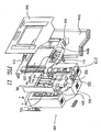
- the kiosk comprises apparatus 100, as set forth in Fig. 1 (not to scale).
- Apparatus 100 is enclosed above and on three side (the side containing chin rest 6 in open for the subject to enter his/her head) with a frosted plastic glass (not shown).
- Apparatus 100 which is capable of taking four types of photographs of the subject, is set-up on table 15 having dimensions of 30" by 36".
- the height of chin rest 6 is about 12" above table 15.
- Camera 11 Across from chin rest 6 and exactly half way along the opposite end of the table 15 is camera 11 (Nikon D1X). Camera 11 is mounted so that the center of the camera lens of camera 11 is about 17" above the top of table 15. The distance between chin rest 6 and the front end of the lens of camera 11 is adjusted so that the subject's face substantially fills the camera frame of camera 11.
- flash units 30a, 30b, 40a, 40b, 50a, and 50b (Broncolor Picolites, Sinar Bron, Allschwil, Switzerland) which are powered, respectively, by power packs 92, 92, 93, 93, 91, and 91.
- the standard flash units 50a and 50b which are used for taking a standard photograph, are mounted above camera 10 and angled down about 20 degrees. Flash units 50a and 50b are directed toward the center of the subject's face. Diffusing filters 51a and 51b (Broncolor Diffuser, Sinar Bron) are placed, respectively, in front of flash units 50a and 50b.
- UVA flash units 30a and 30b which are used for the ultraviolet A photography, are mounted on either side of camera 11 at about 14" from the edge of table 15 and at a height of about 20" from the top of table 15.
- UVA filters 31a and 31b (UG-11 filters, Schott Glass Technologies, Duryea, Pennsylvania USA) are placed, respectively, in front of UVA flash units 30a and 30b.
- Blue flash units 40a and 40b which are used for blue fluorescence photography, are also mounted on either side of the camera at about 14" from the edge of the table top and at a height of about 13" from the top of table 15.
- Blue filters 41a and 41b (Melles Griot, Irvine, California USA) are placed, respectively, in front of blue flash units 40a and 40b.
- the UVA flash units 30a and 30b and the blue flash units 40a and 40b are directed to the center of the face of the subject.
- the polarized flash units 20a and 20b (Broncolor Picolites), which are used for polarized light photography, are powered by power pack 90.
- Diffusing filters 21a and 21b (Broncolor Diffuser, Sinar Bron) are placed, respectively, in front of polarized flash units 20a and 20b, respectively.
- Linear polarizing filters 22a and 22b (Edmund Scientific, Barrington, New Jersey USA) are placed, respectively, in front of diffusing filters 21a and 21b in a vertical orientation.
- Polarized flash unit 20a is positioned at about 4 1/2" from the left edge and about 14" in from the proximal edge of table 15
- polarized flash unit 20b is positioned at about 4 1/2" from the right edge and about 14" in from the proximal edge of table 15.
- the angle between either flash units 20a or 20b, the chin rest 6, and the camera 11 is about 45 degrees.
- the method begins when the subject enters the kiosk image acquisition area and enters basic demographic information into a facial skin-care evaluation computer program (Microsoft Visual Basic, Microsoft Corporation, Redmond, WA USA) using a touch-screen monitor 70 (SecurePoint, SeePoint Technologies, Torrance, CA), which is mounted under table 15 and connected to the same computer running the computer program.
- the subject enters data into the computer program via monitor 70 (computer program runs MountFocus Runtime Keyboard and the keyboard present on monitor 70 was designed using MountFocus Keyboard designer programs (MountFocus. Information Systems, Wilmington, DE USA)), however, other input device such as a keyboard, a track ball, and a computer mouse may be used.
- Examples of such information include, but are not limited to, age and gender of the subject. Following the input of such demographic information, the computer program then instructs the subject to place their chin on chin rest 6 and indicates to the subject to close his/her eyes since apparatus 100 is ready to take photographs.
- the software Upon touching monitor 70, the software makes a function call to an image acquisition and display software ("IDL software”; IDL, Research Systems, Inc., Boulder, CO) running on the same computer.
- IDL software then triggers camera 11 to acquire a blue fluorescence photograph, a standard photograph, a polarized photograph, and an ultraviolet A photograph.
- the flash units 40a, 40b, 50a, 50b, 20a, 20b, 30a, and 30b are triggered sequentially through the use of a radio transceiver (Pocket Wizard Multimax, LPA Design, South Burlington, VT USA) using transceivers 95 (attached to power pack 90 and operating in receiver mode), 96 (attached to power pack 91 and operating in receiver mode), 97 (attached to power pack 92 and operating in receiver mode), 98 (attached to power pack 93 and operating in receiver mode), and 99 (attached to the hot shoe of camera 11 and operating in transmitter mode).
- a radio transceiver Pocket Wizard Multimax, LPA Design, South Burlington, VT USA
- a Topas A2 power pack (Sinar Bron) is used for power packs 90 and 91 and a Primo 4 power pack (Sinar Bron) is used for power packs 92 and 93.
- the radio transceiver causes the activation of the pairs of flash units in response to the shutter release of camera 11.
- the IDL software Prior to taking the blue fluorescence photograph, the IDL software makes a call to servo motor 80, using a Mini SSC II circuit board (Scott Edwards Electronics, Sierra Vista, AZ USA), to move long pass filter 81 (Kodak Wratten No. 8, Eastman Kodak, Rochester, NY USA) in front of the lens of camera 11. After this movement, the blue fluorescence photograph is taken. Following the taking of this photograph, the IDL software then makes another call to servo motor 80 to move long pass filter 81 away from the lens of camera 11. The IDL software then instructs the camera to take the remaining three images. Apparatus 100 is able to acquire these four images in about 10 seconds.
- the four images just acquired are stored in the memory of camera 11 as separate data files.
- the IDL software then makes function calls to these data files and requests these files be transferred to the computer running the computer software and saved to its hard disk with a file name that indicates the apparatus used, subject identifier, and the type of image.
- the subject is then presented on monitor 70 with a registration form.
- Examples of such questions include e-mail address, places where they buy skin-care products, ethnic background, and amount and type of skin-care products that they have purchased in the past (e.g., the past year).
- the subject then begins reviewing each of the four images on monitor 70 and answers questions, presented on monitor 70, about each image.
- the computer program calls the IDL software and requests that a particular saved image be loaded from the hard disk and resized to fit in the screen display area. Once the image is displayed, the IDL software then returns control to the computer program.
- the facial image display area is roughly half of monitor 70. The other half of the screen of monitor 70 displays a series of questions relating to the particular displayed image.
- the computer program may also display on monitor 70 images of other people as comparisons.
- the computer program calls IDL program and requests that a particular saved image be loaded from the hard disk and resized to fit in the screen of monitor 70 area as discussed above. This procedure continues until all four images have been displayed and all questions have been answered by the subject.
- the subject Upon viewing the standard photograph, the subject is then presented with questions regarding the surface of his/her skin. Examples of such questions include, but are not limited to, whether they have any fine lines, wrinkles, loss of elasticity or firmness, large visible pores, sensitive skin, and rough or smooth skin, as well as the severity and location of such conditions. Other questions include, but are not limited to, the frequency and severity of irritation from skin care products.
- the subject Upon viewing the polarized photograph, the subject is asked questions regarding the oiliness of their skin. Examples of such questions include, but are not limited to, whether they have normal, dry, oily, or selective oily skin
- the subject Upon viewing the ultraviolet A photograph, the subject is asked questions regarding the visualization of pigmentation of the face (e.g., brown spots). Examples of such questions include, but are not limited to, the amount and location of such pigmented spots.
- the subject Upon viewing the blue fluorescence photograph, the subject is asked questions regarding acne. Examples of such questions include, but are not limited to, the severity and frequency of his/her breakouts.
- the images from camera 11 are displayed on the computer monitor 70.
- the number of available screen pixels are less than the number of actual image pixels taken by the digital photograph, only a small percentage of the original image can actually be displayed if the image is to be shown on the screen in its entirety while maintaining aspect ratio.
- display of the digital image in a portrait orientation on a computer screen having a resolution of 1024 X 768 results in display of only 1 out of every 18 pixels. In such a down-sampled image presentation, fine detail of the skin taken by camera 11 is not fully displayed.
- the solution to this problem is to use a technique whereby a 256 x 256 box (display kernel) appears directly over the area of interest and shows all the image pixels actually acquired by camera 11 for such area.
- the effect is an in-place magnification of a small area of the image on monitor 70.
- the apparent magnification, shown as this display kernel can be passed over various areas of the image selected by the subject.
- this is an example of 1:1 image display (where every image pixel is shown on the screen within a small display kernel).
- True magnification of the image can also be accomplished by interpolating the data between pixels and creating additional pixels, thereby providing magnification of select areas of the image. The subject selects the magnification of such area by touching the area of interest displayed on the monitor 70.
- each question in the computer program is associated with a skin condition.
- the question "How often does your skin breakout?" is associated with acne.
- the corresponding skin condition is assigned a degree.
- the acne question has four degrees corresponding to the four answer choices: Always, Weekly, Monthly and None.
- the skin conditions questioned by the computer program are ranked according to severity. This ranking is accomplished by passing all of the degree values entered by the subject to a relational database stored procedure contained within a relational database (Microsoft SQL, Microsoft Corporation, Redmond, WA USA) that is on the same computer.
- a relational database Microsoft SQL, Microsoft Corporation, Redmond, WA USA
- a database table contained with the relational database contains a record for each skin problem type and degree.
- a "degree weight” is assigned to each record, which facilitates the ranking of the skin conditions. For example, if the subject answers “Always” for the above acne question, and “Yes” to the question “Do you notice any loss of firmness on your face?" the acne skin condition may have a higher rank than the loss of firmness condition. However, if the subject answers “Weekly” to the above acne question and "Yes" to the loss of firmness question, the loss of firmness condition may be ranked higher.
- the top three conditions, along with the subject's skin type, e.g., normal, dry, or oily, are passed to another relational database stored procedure contained within the relational database.
- this procedure queries a second database table that contains all of the possible combinations of skin conditions along with skin type. Each such record in the table contains a list of recommended products based on these values. This corresponding list of recommended products for that subject's condition is then passed back to the computer program.
- the computer program creates a printout using Crystal Reports (Seagate Corporation, Scotts Valley, CA USA) for the subject including the suggested skin care products and pictures of the subject with information about the various skin conditions.
- Crystal Reports Seagate Corporation, Scotts Valley, CA USA
- the subject will indicate whether or not the images should be kept or deleted.
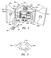
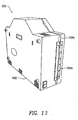
- FIGs. 2 and 3 show an alternative embodiment of the present invention wherein imaging station 200 is provided in the form of a cabinet unit with a central housing 202 flanked by a pair of hingedly connected door assemblies 204a, 204b.
- the door assemblies 204a, 204b can be opened and closed relative to the central housing 202.
- FIG. 3 shows that the angle between the door assemblies 204a, 204b and the central housing 202 can be adjusted to adjust the angle of incident light cast on subjects by flash units mounted to the door assemblies 204a, 204b.
- the angular orientation ⁇ 1, ⁇ 2 of door assemblies 204a, 204b relative to the central housing 202 results in light L1, L2 emanating from flash units 220a, 220b, respectively, having an angle of incidence of ⁇ 1 and ⁇ 2, relative to the subject S.
- the incidence angle of the light illuminating a subject S can be selected to aid in capturing images which are more probative of skin condition, e.g., when capturing polarized light images.
- angles ⁇ 1, ⁇ 2 can be controlled by conventional means, such as an adjustable, threaded stop or a slotted, pivoting bracket coacting with a limit pin, as are commonly known for controlling the extent of opening of hingedly mounted doors and lids. In this manner an optimal angle for one or both door assemblies 204a, 204b is repeatable.
- the embodiment of the present invention shown in FIG. 2 shares many features of the previously described kiosk embodiment and includes a camera 211 (preferably digital) placed within a camera niche 212 for capturing the images of a subject S.
- a number of different illuminating lights (flashes) are available for capturing images.
- the imaging station 200 has fluorescent lights 217a, 217b, polarized flashes 220a, 220b, a pair of UVA flashes 230a, 230b having a wavelength of, e.g., 405 nanometers.
- a pair of UV flashes 240a, 240b having a wavelength, e.g., of 385 nanometers are positioned below the 405 nanometer flashes 230a, 230b.
- the flashes 230a, 230b and 220a, 220b may optionally be operated in conjunction with filters 281a, 281b that can be rotated in front of the camera lens by solenoids 280a, 280b to further select the wavelengths of light received by the camera 211.
- filters 281a, 281b that can be rotated in front of the camera lens by solenoids 280a, 280b to further select the wavelengths of light received by the camera 211.
- a yellow filter 281a may be rotated in front of the camera 211 by solenoid 280b when taking pictures with the 405 nanometer flashes 230a, 230b.
- a polarizing filter 281b may be used in conjunction with polarized flashes 220a, 220b.
- illuminating light(s) and filter(s) may be used for image capture.
- the present invention is not limited to the number and placement of flashes and filters shown. More particularly, a greater or lesser number of filters and flashes may be employed, e.g., see FIG. 9 . While rotating filters 281a, 281b are described above, non-moving filters can be placed over one or more flashes to control the light wavelengths that are used to capture an image. Multiple standard flashes may be employed in conjunction with a variety of filters to achieve illumination under a variety of selected wavelengths of light. Combinations of flashes may be activated simultaneously to acquire a single image or sequentially to acquire multiple, sequential images.
- the use of multiple flashes with different filter options may be preferable under certain circumstances than utilizing a moveable filter. That is, by simplifying the imagining station 200 and reducing the number of moving parts, the imagining station 200, may be made more cheaply and function more reliably.
- a touch screen monitor 270 is disposed below the camera 211 and displays the photographic images recently obtained by the camera 211 and/or previously obtained images that reside in memory or on a storage device, such as a computer disk or memory stick.
- a speaker 218 is provided in the imaging station 200 for announcing instructions to the user (subject S). The speaker 218 is particularly useful in giving instructions to the subjects when their eyes are closed, e.g., immediately prior to, during and immediately after a photographic session, when flashes are activated to capture images of the subject's face.
- a plurality of sockets 232 are provided at the front of the imaging station 200 for receiving plug-in probes 234.
- the probes 234 will have a jack end 236, a cable portion 238 and a testing end 230.
- the testing end 230 would be held by a user in proximity to the area of skin to be tested.
- all probes 234 may remain connected and stored within the housing 202, e.g., with the cable 234 stored on a spring-wound reel and the probe held within a recessed well, accessible to the user who can withdraw it for use and allow it to retract when finished.
- numerous probes suitable for analyzing various states and conditions of the skin are known.
- an apparatus for conducting diffuse reflectance spectroscopy is described in the article, Non-Invasive Measurements of Skin Pigmentation in situ, Stamatas, et al., Pigment Cell Res 17:618-126, 2004 , which is incorporated by reference herein in its entirety.
- An apparatus and method for conducting fluorescence spectroscopy of the skin is described in Fluorescence Spectroscopy of the Skin, Kollias, et al., Vibrational Spectroscopy 28, 17-23, (2002 ), which is incorporated by reference herein in its entirety.
- Probes for measuring the elasticity of the skin can be obtained from Courage & Khazaka Electronic GmbH, Koln, Germany.
- the conductivity of the skin can be measured by a NovaMeter ® or Nova Petite ® sold by Nova Technology Corporation of Portsmouth, New Hampshire or the SkiCon hygrometer by IBS, Inc. of Hamamatsu, Japan.
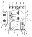
- FIG. 4 shows an enlarged view of the monitor 270 which is preferably a touch screen monitor allowing the simultaneous display of information and the processing of control inputs from the operator/user/subject S who touches the screen to select control options. In this manner, the operator may control the images displayed to review and study them for the purposes of diagnosing and evaluating skin condition.
- a plurality of thumbnail images 224a through 224e may be displayed.
- the first thumbnail image 224a may be utilized to display (both in thumbnail and in enlarged format) a real time moving image captured by the camera 211 in video mode which permits the user to interactively view and position their face in front of the camera for centering and focusing and optimally capturing their facial image.
- the real time image is therefore, in some respects, like a mirror.
- a computer program may be used to inform the user, either through visually displayed instructions or auditory instructions, to position their face such that it substantially fills the display frame and is centered therein.
- the remaining thumbnail images 224b through 224e may be used to display images that have been captured during an imaging session utilizing different wavelengths of illuminating light.
- the imaging station 200 preferably takes a sequence of images utilizing different wavelengths of light, via different flashes and filters, etc. in order to capture images which reveal different attributes of the subject's skin. Selecting one of the thumbnail images 224b through 224e by touching it will cause that particular image to be displayed in the main display area 225 on a larger scale
- the thumbnail images 224a-224e may be labeled with text indicating the type of image that it is, e.g., "Blue Light” or "White Light” and/or the time and date when taken.
- Selecting a thumbnail image of a particular type may invoke instructions and observations from the computer in the imaging station 200 which relate to that particular type of image.
- the computer program may announce a particular type of image has been selected and provide tips on what is shown by this type of image - "You have selected a blue light image.
- This type of image is particularly useful in assessing condition(s) X, Y, Z. In making this assessment, look for A, B, C in the image, which are indicative of D, E, F." This type of message can be delivered in text on the display and/or in audio form over speaker 218.
- the delivery of this type of message is preferably enabled/disabled under operator control, e.g., by touching a control button, "Enable/Disable Instructional Messaging" or an equivalent control means.
- operator control e.g., by touching a control button, "Enable/Disable Instructional Messaging" or an equivalent control means.
- the observer of the captured image is preferably educated in the technique of observing and interpreting features apparent in the captured images.
- the pertinent information for effectively using the imaging station 200 may be communicated to the user/subject S by the above-noted messaging, but also by written and/or recorded materials provided with the station 200 (e.g. in CD ROM or video form).
- the imaging station is connected to the Internet
- information can be provided to the user/subject S interactively by retrieving the information from a website and presenting it to the user/subject S at appropriate times or by presenting hyper links that the user/subject may activate based upon interest.
- the user can ascertain the presence or absence of certain skin conditions revealed by the particular light that was used in capturing the image.
- the thumbnail images therefore provide an intuitive and transparent means for identifying and reviewing a plurality of images taken during an imaging session. While the imaging method described above suggests that multiple images are captured by multiple sequential exposures of a static subject S with different wavelengths of light, the present invention may also be utilized to take one or more pictures after the subject S had varied their position.
- a series of polarized images may be captured with the subject being instructed to rotate their face from left silhouette, to a forward facing position, to a right silhouette position, or in varying degrees of looking up, down and straight ahead.
- images of other surfaces of the body may also be captured, recorded and analyzed. For example, the status and progress of a burn, abrasion, rash, melanoma, mole or other skin condition on any part of the body may be observed and analyzed using the present invention.
- Control button 228a controls the fluorescent lighting 217a, 217b (on/off and/or dim down/brighten), which illuminates the subject S in a darkened room and allows the subject S to position his or her face before the camera (as shown in thumbnail 224a).
- fluorescent lighting 217a, 217b on/off and/or dim down/brighten
- Control button 228b is utilized to review old images taken at a prior photo session. By reviewing images taken at different times, a user can compare the progress of their skin condition over time.
- the imaging station 200 permits the selection of a plurality of images to be displayed, e.g., two or three, in large scale, juxtaposed next to one another. This permits side-by-side comparison to observe trends in skin condition over time or to compare images taken under different lighting conditions, e.g., comparing a polarized light image to a white light image.
- Control button 228c may be used to initiate a photo session after the user has positioned themselves properly before the camera 211.
- a prompt may be provided instructing the user/subject S to press control button 228c after they have positioned themselves.
- An enlarged (magnified kernel) display area 227 displays a portion of the main image area 225 at a greater level of magnification.
- the portion that is magnified is determined by focus box 226, which is overlaid on the image displayed in the main display area 225.
- the purpose of the enlarged display area 227 is to focus upon a specific area of the image shown in the main display area 225 for closer examination, at a greater magnification. Accordingly, the user may position the focus box 226 over any area of the image shown in display area 225 by touching the monitor/screen 270 in the area delimited by the focus box 226 and "dragging" the focus box 226 to the area of interest on the image in the main display area 225.
- the corresponding portion of the image bounded by the focus box 226 is then displayed in the enlarged display area 227.
- the image portion shown in enlarged display area 227 may then be further enlarged or reduced (zoom in and out) by the control buttons 228d and 228e ("+" indicating zoom in for greater magnification and "-” indicating zoom out for lesser magnification) .
- the enlarged display area 227 may also be cleared by touching control button 228f.
- the comparison of side-by-side images may at times be instructive.
- This comparative process using, e.g., a pair of side-by-side images of a person's face taken under the same lighting conditions but at different times or at the same time but using different lighting can be further enhanced by performing zooming and focusing functions on one or both images.
- One embodiment involves coordinated or synchronized focusing and zooming. More particularly, when viewing a pair of images, the focus box graphic appearing in each of the images may be located on the same place in each image (e.g., on the tip of the nose). When the focus box is moved on one of the images, an equal displacement of the focus box on the other image is effected.
- zooming can also be coordinated in the same manner, such that when comparing side-by-side images, the focus area and magnification are the same, promoting comparison.
- Button 228g may be used to select a probe for examination/analysis of the skin using a probe as described above.
- a menu of available probes along with a description of the function of each would be displayed to aid the user in selecting a particular probe. Following selection, the user would be given instructions on use on -screen and/or over the speaker 18, as described more fully below.
- Figure 5 shows an exemplary state of the monitor 270 as it would appear after selection of a specific probe 234, with the subject S interactively positioning the probe over the area of interest.
- One of the benefits of the present invention is that it provides a means for focused analysis of a particular area of the skin surface, i.e., by means of a probe 234. More particularly, the imaging capability of the imaging station 200 may indicate a particular area of the skin which exhibits a condition of concern or interest, e.g., an area of redness, irritation, pigmentation, etc. This area of interest can then be examined by positioning the probe over it and activating the probe to initiate testing and recording of the test results.
- a condition of concern or interest e.g., an area of redness, irritation, pigmentation, etc.
- the imaging station 200 facilitates this process, e.g., by allowing the user to select the live video thumbnail which then causes the subject S to appear on the main display area 225 in real time.
- the enlarged display area 227 defined by the focus box 226 displays the area of interest on the skin in a magnified image to aid the Subject to place the probe in the correct position for testing.
- One benefit of conducting spot checks in this manner is to isolate data for areas that are pertinent while ignoring other areas that do not need to be considered.
- Another benefit realized by the invention, is that the probe data may be quantified and stored. Quantification of skin condition is useful for conducting objective evaluations and also to monitor a skin condition more precisely based upon the comparison of quantified results taken from two or more tests conducted at different times.
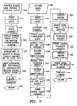
- FIG. 6 shows exemplary processing steps that an exemplary embodiment of the present invention would utilize in obtaining and viewing the images of a subject S.
- the processing flow begins 300 by the operator entering an access code or an I.D. password 302. This is an optional step in the event that the imaging station 200 is utilized by more than one person. For example, if the imaging station were a public facility, the individuals who utilize it would prefer that their images would remain private and accessible only by them upon entry of a suitable password/access code. In the event that the imaging station 200 is a private facility, for example, located in the home of the user, then it would not be necessary for a password to be utilized.
- the imaging station 200 provides instructions to the subject S to position themselves and to prepare to capture the images 306. For example, the subject may be instructed to approach the camera such that their face fills the field of view as shown in thumbnail 224a. Once positioned, the subject is instructed to turn off the fluorescent lights and/or to close their eyes to prevent the flashes from being seen.
- the image set can be captured 308 by sequentially taking pictures utilizing different flashes and filters.
- the images are then prepared to be displayed and downloaded 310 to the computer of the imaging station 200. Once the images have been captured and downloaded onto the system computer, the images may be displayed 312 on the monitor 270 and the operator is provided with various control options. When the operator is finished 314 reviewing the images, the operation is at an end 316, otherwise, the user may obtain additional new images or review old images.
- the procedures necessary to capture and store the probe data 313 and display the probe data 315 are initiated.
- the user may then be given the option of repeating the probe test such that a sequence of test results are obtained.
- the camera may take a picture at the same time the probe test is conducted in order to create a correlation between the test result data and the position of the probe, i.e., by momentarily changing the mode of the camera from video mode to camera mode, taking the picture and then returning to video mode.
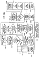
- FIGS. 7 and 8 show the basic process shown in FIG. 6 , in greater detail, namely, the step of positioning and preparing 306 to capture images includes powering 318 the fluorescent lights, powering 320 the flash charger and displaying 322 the camera video output on the monitor 270 so that the subject S may ascertain their position relative to the camera 322. As described above, this image is shown in thumbnail 224a or elsewhere as determined by the programmer. The program announces and/or displays 324 instructions to the user/subject to place themselves at the correct position relative to the camera and to close their eyes.
- a delay is provided 326, to give the subject S sufficient time to obey the commands for positioning and closing of the eyes and/or the computer program enables 326 a subject-ready signal, such as the subject pressing control button 228C to initiate taking pictures.
- the display monitor 270 is blanked 328 to avoid interfering with the illuminating light, and a return of control 330 to the main processing flow is executed.
- the fluorescent lights 217a, 217b are powered 332 and the camera 211 is triggered 334.
- the fluorescent lights 217a, 217b are then powered down 336 and the 385 nanometer UV flashes 240a, 240b are enabled 338.
- the camera is then triggered 340 causing the 385 nanometer UV flashes 240a, 240b to flash.
- the polarized flashes 220a, 220b are enabled 342 and the camera 211 is triggered, triggering 344 the polarized flashes 220a, 22b.
- the 405 nanometer flashes 230a, 230b are enabled 346 and the camera filter solenoid 280b is powered 348, thereby moving the filter 281a in front of the camera 211.
- the camera 211 is then triggered 350 along with the flashes 230a, 230b.
- the filter 281a is then removed from a position in front of the camera 211 by powering down 352 the filter solenoid 280b.
- the polarized flashes 220a, 220b are enabled 354 and the camera polarizer solenoid 280a is powered 356 thereby moving the polarizing filter 281b in front of the camera 211.
- the camera 211 is then triggered, flashing 358 the polarized flashes 220a, 220b.
- the camera polarizer solenoid 280a is powered down 360 to remove the polarizing filter 281b away from the front of the camera 211.
- the display screen 270 is unblanked 362 and control returns 364 to the main processing flow.
- the computer program announces 366 that the image capture is complete and then the image data is downloaded 368 from the camera 211 to the computer 552, where it is stored in memory or on a memory storage device. Control returns 370 to the main processing flow.
- FIG. 9 shows exemplary processing steps associated with capturing and storing probe data 313.
- a menu of available probes is displayed 371, including a diffuse reflectance spectrum 373, fluorescent spectrum 375, infrared spectrum 377, conductivity 379 and elastic modulus 381.
- Other probes that are known in the art for measuring and testing skin condition could readily be employed and would fall within the scope of the present invention.
- Power is then applied to the selected probe/sensor 383.
- the camera is placed in video mode and the image is displayed in the main display area as well as the enlarged image area 385. Instructions for use of the particular probe selected are then either displayed and/or announced over the speaker. These instructions would cover the positioning of the probe on the person and how to enable/activate the probe for taking testing in the area on which it is applied.
- the probe After the probe has been powered and reached a ready-for-testing state, the subject has been instructed on use of the probe and has indicated a readiness for testing by touching a button on the monitor or depressing a control button on the probe, the probe is enabled 389 and the activation signal is received triggering the probe into testing. The test data is then captured and stored to the computer memory 391. Control then returns 393 to the user via the touch screen display.
- FIG. 10 shows exemplary processing associated with displaying 312 images and providing operator control options to the operator/subject S.
- the display 270 has a plurality of thumbnail images 224a through 224e. Any number of thumbnail images may be displayed 372 as indicated by the dotted lines and reference to display 374 image #1, display 376 image #2 and display 378 image #n. Because the display monitor 270 is preferably a touch screen monitor, the thumbnail display areas 224a through 224e can be used to sense on 380 operator input (touching the particular thumbnail image 224a-224b that the operator wishes to display in the main display area 225,).
- a default image is displayed 384 which would typically be the first image taken, e.g., an image taken in white light or fluorescent light.
- the image associated with that particular thumbnail 224b through 224e is retrieved 388 from memory or from a storage device and the image is displayed in the main image area 225.
- a focus box 226 is displayed 390 in the main image area 225.
- a default focus box 226 is displayed 392 first and then the program senses 394 upon operator input to the focus box 226 for changes.
- the focus box 226 can be dragged to the area of the image of interest to the operator/subject S.
- Operator inputs to the focus box location are monitored at step 396. If there has been a change, then the pixel set associated with the focus box 226 is updated 398.
- the magnified image associated with the focus box 226 is displayed 400 and a default area associated with a default position of the focus box 226 is displayed 402 first.
- the program continually senses 404 upon changes in focus or zoom or for "clear" signals that are generated by the operator touching one of the control buttons 228d, 228e, 228f.
- any of these inputs causes the pixel set associated with the focus box to be reselected 406 and displayed 408 in order to implement a focus or zoom change and/or for clearing the display.
- the control buttons "+”, “-” and “clear” are displayed and sensed upon. As noted above, touching 412 "+” results in enlargement of the image displayed in the enlarged display area 227. Pressing 414 "-” results in a lower magnification and pressing 416 "clear” results in the enlarged display area 227 being blanked.
- add control buttons e.g., 228a-228g are displayed and sensed or to invoke the various processing choices described above.
- FIG. 11 shows an exploded view of an embodiment of the present invention in which, imaging station 500 has center housing 502 containing a plurality of power supplies 532, 533, 534 for powering the various system elements, including the monitor 570 and the system computer 552, as well as the camera 511.
- the camera 511 is mounted on an intermediate panel 543 which also serves to provide a structure for mounting the monitor 570.
- the intermediate panel 543 is received in and attached to the housing 502 by conventional fasteners, such as threaded fasteners or rivets.
- Front panel 542 is attached to the housing 502 and/or intermediate panel 543.
- the front panel 542 has apertures for camera 511, speaker 518 and monitor 570. Only one door assembly 504a is shown. Either single or plural door configurations are within the scope of the present invention, as are configurations where the door(s) open up or down.
- the spectrometers and other apparatus 573, 575, 577, 579 and 583 supporting the probes 234 for conducting testing and sampling based upon the diffuse reflectance spectrum, fluorescent spectrum, infrared spectrum, conductivity, and elastic modulus, respectively, are contained in the housing 502.
- FIG. 12 shows the imaging station 500 of FIG. 9 in a closed condition with door panels 504a and 504b closed, such that the monitor 570 is not visible.
- This embodiment of the imaging station 500 has some similarities to a conventional medicine cabinet in that it can be mounted on a wall and the doors 504a, 504b closed to protect the interior contents of the housing 502.
- the front surfaces of the door assemblies 504a, 504b may be mirrored.
- FIG. 13 is a rear view of the imaging station 500 illustrating that the door assemblies 504a, 504b are hingedly attached to the housing 502 by hinges e.g., 505b.
- FIG. 14 is a block diagram illustrating the electrical components of the imaging station 600.
- AC input 637 is routed to a power distribution box 638 for spreading the AC input to a plurality of power supplies 632, 633, 634, 635, which power control board 655, computer 652, monitor 670 and camera 611, respectively.
- Camera 611 communicates with the computer 652 via a U.S.B. data input/output 654 and thereby to transfer images to the control board 655.
- the control board 655 controls filter solenoids 681a, 681b, flash unit polarizers 620a, 620b, CCFLs and inverters 656a, 656b and flash units 630a, 630b, 640a and 640b, as well as, diffuse reflectance spectrometer 673, fluorescence spectrometer 675, infrared spectrometer 677, conductivity meter 679 and elasticity meter 683. Output from the various probe units 673-683 are sent to system computer 652 for storage and/or analysis.
- the present invention has been explained above in terms of an apparatus and method employing multiple images captured under different lighting conditions e.g., different wavelengths of light, the present invention would still be applicable to the capture and analysis of even a single image taken under selected lighting conditions.
- the imaging station 200 could be utilized to capture and analyze a single image taken with blue light.
- the present invention 200 is intended to be operated by any person and therefore can be utilized in a private setting, such as the home, by the subject S.
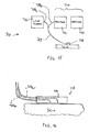
- FIG. 15 shows a spectrometer apparatus 710 for performing diffuse reflectance spectroscopy (DRS) on skin.
- DRS diffuse reflectance spectroscopy
- light is delivered onto the skin and the resultant remitted light is collected and analyzed with a spectrometer 714.
- Many manufacturers provide portable spectrometers, e.g., OceanOptics (Boca Raton, FL, USA), Newport (Irvine, CA, USA), B&W Tek (Newark, DE,USA), CDI (South Bend,IN,USA) and Minolta (Ramsey, NJ, USA).
- the components that are required for a DRS system are a light source 716, a probe 718, an analyzer 720, and a detector 722.
- a variety of light sources 716 have been used, such as tungsten halogen lamps, xenon arc lamps, broad-band white LEDs, etc. The choice depends on the wavelength range of interest.
- the illumination light can be delivered to the skin site of interest through a bifurcated fiber optic bundle 724.
- Several designs have been used in the literature including randomly mixed fibers or particular geometries for illumination and collection.
- the fiber geometry is important as the fiber size and distance between illumination and collection fibers affect the sampling volume and the sampling depth. For example, if the illumination and collection fibers are adjacent one another, a fiber diameter of about 50 ⁇ m will result in a high sampling volume of light collected from a depth of about 80-120 ⁇ m. Smaller diameter fibers correspond to light collection from shallower depths below the skin surface. As shown in FIG.
- probe 718 with the fibers 724a, 724b bent to 90° at the common terminal 726 of the bundle 724 that comes in contact with the skin. This facilitates the use of the probe 718 with minimal pressure to the skin so that capillary constriction and skin blanching are avoided.
- Another way of collecting spectra is through an integrating sphere, which allows the collection of remitted light from all possible angles, but has the disadvantage that the probe size is limited by the aperture of the sphere. Probe size limitations prohibit the use of integrating spheres in cases where small skin areas are measured (e.g., small lesions).
- the analyzer-detector system 714 can be in the form of a circular variable wavelength interference filter coupled to a silicon photodiode (32) or a diffraction grating coupled to a detector array, e.g., a linear charge-couple device (CCD) array or a photodiode array.
- a circular variable wavelength interference filter coupled to a silicon photodiode (32) or a diffraction grating coupled to a detector array, e.g., a linear charge-couple device (CCD) array or a photodiode array.
- CCD linear charge-couple device
- the detector dark current D is recorded and a spectrum is acquired from a reflectance standard S ref ( ⁇ ) (defined as 100% at all wavelengths) where ⁇ represents wavelengths.
- S ref ⁇
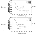
- FIGS. 17 and 18 are graphs 730, 732 showing examples of DRS data obtained from skin in vivo on day 1 and 7 after exposure to 1.0, 2.1, and 3.0 times the minimal erythemogenic dose (MED) from FS-20 lamps. DRS measurements were taken with an Ocean Optics USB-2000 spectrophotometer. Graphs 730 and 732 show absorption spectra calculated based on the preceding equation.
- FIG. 19 shows an apparatus 810 for performing fluorescence spectroscopy of the skin.
- the spectrofluorimeter 812 may be obtained from SPEX Industries (Horiba Group, Edison, NJ) and includes a double monochromator-based fluorescence spectrometer with a xenon lamp as the excitation source 814.
- the radiation from the excitation source 814 is filtered by a double monochromator 816 and is focused into a quartz fiber bundle 818 with individual fiber diameters between 100 and 200 um.
- the distal end of the fiber bundle 818 is brought into contact with the skin sample under study.
- the delivery and collection fibers 818a, 818b are packed together in a random manner.
- Emitted radiation is collected by optical fibers of the bundle 818 and is analyzed by a double monochromator in the emission compartment 820.
- the intensity of the emitted radiation is measured by a photomultiplier (PMT) 822.
- the output of the PMT 822 is processed by a computer 824, which also controls the instrument.
- An important feature of the spectrofluorimeter 812 is the double monochromator design, which significantly improves the signal to noise ratio at short wavelengths ( ⁇ ⁇ 300nm).
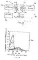
- FIG. 20 shows a graph 830 of serial excitation spectra from the right cheek of a 27-year-old volunteer.
- the excitation wavelengths range from 250 to 470 nm in increments of 10nm.
- the excitation monochromator 816 was scanned from 250 to within 20nm of the emission monochromator 820 setting.
- the emission wavelength of each scan was incremented by 20nm form the previous starting at 280nm.
- the major fluorescence peaks have been labeled with the corresponding excitation wavelength: (i) 295nm excitation, due to trytophan fluorescence; (ii) 335nm excitation, due to PDCCL; (iii) 350nm excitation, possibly due to NADH; and (iv) 370nm excitation, due to CDCCL.
Landscapes
- Health & Medical Sciences (AREA)
- Life Sciences & Earth Sciences (AREA)
- Engineering & Computer Science (AREA)
- Physics & Mathematics (AREA)
- Surgery (AREA)
- Public Health (AREA)
- Biophysics (AREA)
- Pathology (AREA)
- Biomedical Technology (AREA)
- Heart & Thoracic Surgery (AREA)
- Medical Informatics (AREA)
- Molecular Biology (AREA)
- Veterinary Medicine (AREA)
- Animal Behavior & Ethology (AREA)
- General Health & Medical Sciences (AREA)
- Dermatology (AREA)
- Signal Processing (AREA)
- Multimedia (AREA)
- Nuclear Medicine, Radiotherapy & Molecular Imaging (AREA)
- Radiology & Medical Imaging (AREA)
- Spectroscopy & Molecular Physics (AREA)
- Measuring And Recording Apparatus For Diagnosis (AREA)
- Measurement Of The Respiration, Hearing Ability, Form, And Blood Characteristics Of Living Organisms (AREA)
- Investigating, Analyzing Materials By Fluorescence Or Luminescence (AREA)
- Investigating Or Analysing Materials By Optical Means (AREA)
Claims (10)
- Verfahren zum Erfassen von Daten über die Haut einer Person, das die folgenden Schritte umfasst:(A) Bereitstellen einer Bildgebungsstation (200, 500, 600) mit einer Kamera (211), einer Lichtquelle (716, 814), einem Monitor (270) zum Anzeigen der von der Kamera erfassten Bilder, mindestens einer Sonde (234, 718) zum Messen eines Parameters der Haut in einem spezifischen Bereich, einem Controller mit einer Bedienerschnittstelle;(B) Positionieren der Person relativ zur Kamera; und gekennzeichnet dadurch, dass das Verfahren weiterhin folgende Schritte umfasst:(C) Wahlmöglichkeit für die Person zwischen den Optionen des Erfassens von Bildern mit der Kamera (211) oder der Gewinnung von Hautdaten mit der Sonde (234, 718);(D) Ausführen der in Schritt (C) gewählten Option;(E) wenn die Option Gewinnung von Hautdaten mit der Sonde (234, 718) gewählt wurde, Erfassen eines statischen Bildes der Person gleichzeitig mit der Ausführung von Schritt (D), wobei das statische Bild die Position der Sonde während der Ausführung von Schritt (D) registriert;(F) Speichern der mit der Kamera und der Sonde erfassten Daten; und(G) selektive Anzeige der erfassten Daten auf dem Monitor.
- Verfahren nach Anspruch 1, wobei Schritt (D) die Anzeige eines Echtzeitbildes der Person einschließt, um es der Person zu gestatten, die Position der Sonde zu visualisieren und interaktiv einzustellen, so dass die Sonde freigeschaltet und zum Erfassen der Hautdaten aktiviert wird.
- Verfahren nach Anspruch 2, wobei die Bildgebungsstation eine Mehrzahl von Lichtquellen verschiedener Wellenlängen aufweist und die von der Kamera erfassten Bilder auch Bilder einschließen, die mit unterschiedlichen Lichtwellenlängen erfasst wurden.
- Verfahren nach Anspruch 3, weiterhin umfassend den Schritt einer gewählten Anzahl von Wiederholungen des Schrittes der Freischaltung und Aktivierung der Sonde, um weitere Daten zu erfassen.
- Verfahren nach Anspruch 4, wobei bei mindestens einer Aktivierung die Position der Sonde in Bezug auf ein fotografisches Bild der Person bestimmt wird, welches in zeitlichem Abstand vom Zeitpunkt der Aktivierung aufgenommen wurde.
- Verfahren nach Anspruch 4, wobei die Sonde zwischen aufeinanderfolgenden Aktivierungsschritten zu mindestens einer anderen Position auf der Haut der Person bewegt wird.
- Verfahren nach einem der vorgenannten Ansprüche, weiterhin umfassend den Schritt der Ausgabe von Anweisungen zum Selbstgebrauch der Bildgebungs-bzw. Sondenstation für deren Benutzer in mindestens einer der drei Arten: textliche, grafische und akustische Anweisungen.
- Verfahren nach einem der vorgenannten Ansprüche, umfassend die Durchführung von Remissionsspektroskopie auf der Haut mittels eines Spektrometers (714).
- Verfahren nach Anspruch 8, umfassend die Verwendung einer über ein zweiarmiges Lichtleiterbündel (742) mit der Lichtquelle (716) verbundenen Sonde (718), wobei das zweiarmige Lichtleiterbündel eine Beleuchtungsfaser und eine Erfassungsfaser umfasst.
- Verfahren nach einem der Ansprüche 1 bis 7, umfassend die Durchführung von Fluoreszenzspektroskopie auf der Haut.
Applications Claiming Priority (2)
| Application Number | Priority Date | Filing Date | Title |
|---|---|---|---|
| US11/169,813 US8026942B2 (en) | 2004-10-29 | 2005-06-29 | Skin imaging system with probe |
| EP06253363.3A EP1738687B1 (de) | 2005-06-29 | 2006-06-28 | Hautbildgebungssystem mit einer Sonde |
Related Parent Applications (3)
| Application Number | Title | Priority Date | Filing Date |
|---|---|---|---|
| EP06253363.3 Division | 2006-06-28 | ||
| EP06253363.3A Division EP1738687B1 (de) | 2005-06-29 | 2006-06-28 | Hautbildgebungssystem mit einer Sonde |
| EP06253363.3A Division-Into EP1738687B1 (de) | 2005-06-29 | 2006-06-28 | Hautbildgebungssystem mit einer Sonde |
Publications (2)
| Publication Number | Publication Date |
|---|---|
| EP2289413A1 EP2289413A1 (de) | 2011-03-02 |
| EP2289413B1 true EP2289413B1 (de) | 2016-11-16 |
Family
ID=43127735
Family Applications (2)
| Application Number | Title | Priority Date | Filing Date |
|---|---|---|---|
| EP10180273.4A Ceased EP2289413B1 (de) | 2005-06-29 | 2006-06-28 | Hautbildgebungssystem mit einer Sonde |
| EP06253363.3A Ceased EP1738687B1 (de) | 2005-06-29 | 2006-06-28 | Hautbildgebungssystem mit einer Sonde |
Family Applications After (1)
| Application Number | Title | Priority Date | Filing Date |
|---|---|---|---|
| EP06253363.3A Ceased EP1738687B1 (de) | 2005-06-29 | 2006-06-28 | Hautbildgebungssystem mit einer Sonde |
Country Status (4)
| Country | Link |
|---|---|
| US (1) | US8026942B2 (de) |
| EP (2) | EP2289413B1 (de) |
| BR (1) | BRPI0605795B8 (de) |
| CA (1) | CA2551311C (de) |
Families Citing this family (118)
| Publication number | Priority date | Publication date | Assignee | Title |
|---|---|---|---|---|
| US7738032B2 (en) * | 2001-11-08 | 2010-06-15 | Johnson & Johnson Consumer Companies, Inc. | Apparatus for and method of taking and viewing images of the skin |
| US6922523B2 (en) * | 2001-11-08 | 2005-07-26 | Johnson & Johnson Consumer Companies, Inc. | Method of promoting skin care products |
| US20040146290A1 (en) * | 2001-11-08 | 2004-07-29 | Nikiforos Kollias | Method of taking images of the skin using blue light and the use thereof |
| FR2878424B1 (fr) * | 2004-11-26 | 2008-02-01 | Oreal | Procede d'observation d'un tissu biologique, notamment de la peau humaine |
| CA2595239C (en) * | 2005-01-19 | 2016-12-20 | Dermaspect Llc | Devices and methods for identifying and monitoring changes of a suspect area on a patient |
| KR101269533B1 (ko) * | 2005-04-28 | 2013-06-04 | 가부시키가이샤 시세이도 | 피부 상태 해석 방법, 피부 상태 해석 장치, 및 피부 상태해석 프로그램을 기록한 기록 매체 |
| JP5254790B2 (ja) | 2005-08-12 | 2013-08-07 | ティーシーエムエス トランスペアレント ビューティー エルエルシー | ヒトの皮膚の視覚的魅力を改善するために、反射変性剤を塗布するためのシステムおよび方法 |
| US8861863B2 (en) * | 2005-09-20 | 2014-10-14 | Brightex Bio-Photonics Llc | Method and system for analyzing lip conditions using digital images |
| WO2007043899A1 (en) | 2005-10-14 | 2007-04-19 | Applied Research Associates Nz Limited | A method of monitoring a surface feature and apparatus therefor |
| US20070200337A1 (en) * | 2006-02-27 | 2007-08-30 | Aaron Henry Johnson | Method for creating a decoy exhibiting realistic spectral reflectance |
| US20120075577A1 (en) | 2006-03-20 | 2012-03-29 | Ishak Andrew W | High performance selective light wavelength filtering providing improved contrast sensitivity |
| US8882267B2 (en) | 2006-03-20 | 2014-11-11 | High Performance Optics, Inc. | High energy visible light filter systems with yellowness index values |
| US8184901B2 (en) * | 2007-02-12 | 2012-05-22 | Tcms Transparent Beauty Llc | System and method for applying a reflectance modifying agent to change a person's appearance based on a digital image |
| US7764303B2 (en) * | 2006-10-02 | 2010-07-27 | Johnson & Johnson Consumer Companies, Inc. | Imaging apparatus and methods for capturing and analyzing digital images of the skin |
| US8107696B2 (en) | 2006-10-02 | 2012-01-31 | Johnson & Johnson Consumer Companies, Inc. | Calibration apparatus and method for fluorescent imaging |
| US8189887B2 (en) * | 2006-10-02 | 2012-05-29 | Johnson & Johnson Consumer Companies, Inc. | Imaging standard apparatus and method |
| USD564663S1 (en) | 2006-10-02 | 2008-03-18 | Johnson & Johnson Consumer Companies, Inc. | Imaging station |
| US7558416B2 (en) * | 2006-10-02 | 2009-07-07 | Johnson & Johnson Consumer Companies, Inc. | Apparatus and method for measuring photodamage to skin |
| US20080080766A1 (en) * | 2006-10-02 | 2008-04-03 | Gregory Payonk | Apparatus and Method for Analyzing Skin Using L*a*b* Colorspace |
| US8103061B2 (en) * | 2006-10-02 | 2012-01-24 | Johnson & Johnson Consumer Companies, Inc. | Method and apparatus for identifying facial regions |
| KR20100015318A (ko) | 2007-02-12 | 2010-02-12 | 릭 비. 예거 | 인간 피부에 정전기적으로 약제를 인가하기 위한 시스템 및 방법 |
| US8081355B2 (en) * | 2007-03-05 | 2011-12-20 | Ricoh Company, Ltd. | Illumination unit, image read apparatus, image formation apparatus |
| US8098900B2 (en) * | 2007-03-06 | 2012-01-17 | Honeywell International Inc. | Skin detection sensor |
| US10092082B2 (en) | 2007-05-29 | 2018-10-09 | Tcms Transparent Beauty Llc | Apparatus and method for the precision application of cosmetics |
| US20080312952A1 (en) * | 2007-06-12 | 2008-12-18 | Gulfo Joseph V | Regulating Use Of A Device To Perform A Procedure On A Subject |
| US7929050B2 (en) * | 2007-06-29 | 2011-04-19 | Epson America, Inc. | Document camera |
| WO2009005748A1 (en) * | 2007-06-29 | 2009-01-08 | The Trustees Of Columbia University In The City Ofnew York | Optical imaging or spectroscopy systems and methods |
| JP5361159B2 (ja) | 2007-09-06 | 2013-12-04 | キヤノン株式会社 | 画像表示制御装置、その制御方法、プログラム、および記録媒体 |
| JP5479345B2 (ja) * | 2007-09-24 | 2014-04-23 | コーニンクレッカ フィリップス エヌ ヴェ | 試料の表面を観察するための装置 |
| US20090102933A1 (en) * | 2007-09-24 | 2009-04-23 | Global Device Management | Target Acquisition System |
| EP2040061A1 (de) * | 2007-09-24 | 2009-03-25 | Koninklijke Philips Electronics N.V. | Vorrichtung und Verfahren zur Beobachtung der Oberfläche einer Probe |
| US20090118600A1 (en) * | 2007-11-02 | 2009-05-07 | Ortiz Joseph L | Method and apparatus for skin documentation and analysis |
| US8849380B2 (en) * | 2007-11-26 | 2014-09-30 | Canfield Scientific Inc. | Multi-spectral tissue imaging |
| US8427274B2 (en) * | 2007-12-27 | 2013-04-23 | Saje Holdings, Inc. | Lighting system and control method thereof |
| ES2989708T3 (es) | 2008-05-20 | 2024-11-27 | Univ Health Network | Dispositivo y método para formación de imágenes y supervisión por fluorescencia |
| US10469738B2 (en) | 2008-09-05 | 2019-11-05 | Telefonaktiebolaget Lm Ericsson (Publ) | Mobile terminal and method of performing multi-focusing and photographing image including plurality of objects using the same |
| US8913176B2 (en) * | 2008-09-05 | 2014-12-16 | Lg Electronics Inc. | Mobile terminal and method of performing multi-focusing and photographing image including plurality of objects using the same |
| FR2935888B1 (fr) * | 2008-09-17 | 2010-09-24 | Dataderm Internat Gmbh | Procede et systeme d'analyse de la peau |
| EP2223650A1 (de) * | 2009-02-25 | 2010-09-01 | The Provost, Fellows and Scholars of the College of the Holy and Undivided Trinity of Queen Elizabeth near Dublin | Verfahren und Vorrichtung zur Abbildung von Gewebetopographie |
| US8319857B2 (en) * | 2009-02-26 | 2012-11-27 | Access Business Group International Llc | Apparatus and method for correcting digital color photographs |
| US8823934B2 (en) * | 2009-03-27 | 2014-09-02 | Brightex Bio-Photonics Llc | Methods and systems for imaging and modeling skin using polarized lighting |
| US8373859B2 (en) * | 2009-03-27 | 2013-02-12 | Brightex Bio-Photonics Llc | Methods and systems for imaging skin using polarized lighting |
| US8330807B2 (en) * | 2009-05-29 | 2012-12-11 | Convergent Medical Solutions, Inc. | Automated assessment of skin lesions using image library |
| JP5429460B2 (ja) * | 2009-06-10 | 2014-02-26 | 株式会社ニコン | 肌診断装置 |
| US8606345B2 (en) * | 2009-08-31 | 2013-12-10 | Gsm Of Kansas, Inc. | Medical dual lens camera for documentation of dermatological conditions with laser distance measuring |
| US8452063B2 (en) * | 2009-11-03 | 2013-05-28 | Mela Sciences, Inc. | Showing skin lesion information |
| US8433116B2 (en) * | 2009-11-03 | 2013-04-30 | Mela Sciences, Inc. | Showing skin lesion information |
| US8490321B1 (en) | 2009-11-04 | 2013-07-23 | Scott A. Butz | UV reflective fishing lure system |
| CN101719984B (zh) * | 2009-11-09 | 2012-06-06 | 无锡中星微电子有限公司 | 一种图像处理的装置 |
| GB2484656A (en) * | 2010-10-12 | 2012-04-25 | Ovie Ltd | Image capture method and apparatus for recording images of a patients eye, ear and/or tongue |
| FR2969915B1 (fr) * | 2011-01-04 | 2014-01-03 | Ergylink | Systeme et procede pour analyser au moins une caracteristique de la peau |
| CA2836472C (en) * | 2011-05-18 | 2022-11-22 | Nextgenid, Inc. | Multi-biometric enrollment kiosk including biometric enrollment and verification, face recognition and fingerprint matching systems |
| US8804122B2 (en) | 2011-09-22 | 2014-08-12 | Brightex Bio-Photonics Llc | Systems and methods for determining a surface profile using a plurality of light sources |
| US9179844B2 (en) | 2011-11-28 | 2015-11-10 | Aranz Healthcare Limited | Handheld skin measuring or monitoring device |
| US9155473B2 (en) * | 2012-03-21 | 2015-10-13 | Korea Electrotechnology Research Institute | Reflection detection type measurement apparatus for skin autofluorescence |
| US9173570B2 (en) * | 2012-04-12 | 2015-11-03 | Thomas Nathan Millikan | Viewing and processing multispectral images |
| US20150078642A1 (en) * | 2012-04-24 | 2015-03-19 | The General Hospital Corporation | Method and system for non-invasive quantification of biologial sample physiology using a series of images |
| NZ702690A (en) | 2012-05-07 | 2017-08-25 | DermSpectra LLC | System and apparatus for automated total body imaging |
| WO2014021005A1 (ja) * | 2012-07-31 | 2014-02-06 | 日本電気株式会社 | 画像処理システム、画像処理方法及びプログラム |
| WO2014035793A1 (en) * | 2012-08-27 | 2014-03-06 | Tcms Transparent Beauty Llc | Printer head with prism for sensing |
| JP2014061057A (ja) * | 2012-09-20 | 2014-04-10 | Sony Corp | 情報処理装置、情報処理方法、プログラム、及び測定システム |
| US9060674B2 (en) | 2012-10-11 | 2015-06-23 | Karl Storz Imaging, Inc. | Auto zoom for video camera |
| US20140243684A1 (en) * | 2013-02-27 | 2014-08-28 | DermSpectra LLC | System and method for creating, processing, and displaying total body image |
| USD743553S1 (en) | 2013-02-28 | 2015-11-17 | DermSpectra LLC | Imaging booth |
| US20150201840A1 (en) * | 2013-03-18 | 2015-07-23 | Korea Electrotechnology Research Institute | Reflection detection type measurement apparatus for skin autofluorescence |
| US9888855B2 (en) * | 2013-03-18 | 2018-02-13 | Korea Electrotechnology Research Institute | Reflection detection type measurement apparatus and method for skin autofluorescence |
| US9256963B2 (en) | 2013-04-09 | 2016-02-09 | Elc Management Llc | Skin diagnostic and image processing systems, apparatus and articles |
| US9101320B2 (en) | 2013-04-09 | 2015-08-11 | Elc Management Llc | Skin diagnostic and image processing methods |
| TWI494068B (zh) * | 2013-04-23 | 2015-08-01 | Genesis Photonics Inc | 保護套 |
| US9662061B2 (en) * | 2013-12-16 | 2017-05-30 | International Business Machines Corporation | Ultraviolet camera and display station for capturing reflected ultraviolet light |
| TWI696450B (zh) * | 2014-04-16 | 2020-06-21 | 日商新力股份有限公司 | 資訊處理裝置、資訊處理系統、及資訊處理程式 |
| KR20150120774A (ko) * | 2014-04-18 | 2015-10-28 | 삼성전자주식회사 | 관심영역 검출 시스템 및 방법 |
| US10368795B2 (en) * | 2014-06-30 | 2019-08-06 | Canfield Scientific, Incorporated | Acne imaging methods and apparatus |
| PT3171765T (pt) | 2014-07-24 | 2021-10-27 | Univ Health Network | Recolha e análise de dados para fins diagnósticos |
| WO2016057633A1 (en) | 2014-10-08 | 2016-04-14 | Revealix, Inc. | Automated systems and methods for skin assessment and early detection of a latent pathogenic bio-signal anomaly |
| EP3017755A1 (de) | 2014-11-04 | 2016-05-11 | Samsung Electronics Co., Ltd. | Elektronische vorrichtung und verfahren zur analyse von gesichtsinformationen in elektronischen vorrichtungen |
| EP3262451A1 (de) * | 2015-02-27 | 2018-01-03 | FotoNation Limited | Optisches system für eine bilderfassungsvorrichtung |
| WO2016183075A1 (en) | 2015-05-12 | 2016-11-17 | Profoot, Inc. | Real time ultraviolet light reflectance imaging |
| AU2017217455C1 (en) | 2016-02-08 | 2019-01-17 | Sephora USA Inc. | Apparatus and method for formulation and dispensing of visually customized cosmetics |
| BR112018072107A2 (pt) | 2016-04-27 | 2019-02-12 | Voxelight, Llc | dispositivo de imageamento para avaliação de cobertura de protetor solar |
| US10013527B2 (en) | 2016-05-02 | 2018-07-03 | Aranz Healthcare Limited | Automatically assessing an anatomical surface feature and securely managing information related to the same |
| RU2745612C2 (ru) * | 2016-06-27 | 2021-03-29 | Конинклейке Филипс Н.В. | Устройство и способ для обнаружения блеска кожи |
| US10511777B2 (en) | 2016-11-08 | 2019-12-17 | Thomas Nichols | Personal care device with camera |
| US11122206B2 (en) | 2016-11-08 | 2021-09-14 | Preh Holding, Llc | Personal care device with camera |
| US11116407B2 (en) | 2016-11-17 | 2021-09-14 | Aranz Healthcare Limited | Anatomical surface assessment methods, devices and systems |
| HU4811U (hu) * | 2016-12-14 | 2018-01-29 | Handinscan Kft | Berendezés kézfertõtlenítés minõségének ellenõrzésére |
| EP3606410B1 (de) | 2017-04-04 | 2022-11-02 | Aranz Healthcare Limited | Anatomische oberflächenbeurteilungsverfahren, vorrichtungen und systeme |
| US10938950B2 (en) * | 2017-11-14 | 2021-03-02 | General Electric Company | Hierarchical data exchange management system |
| DE102017222530A1 (de) * | 2017-12-12 | 2019-06-13 | Henkel Ag & Co. Kgaa | Anordnung zum Ermitteln von Stoffwechselendprodukten in der Haut |
| JP7406688B2 (ja) | 2018-01-29 | 2023-12-28 | ファンクション インコーポレイテッド | 個人化スキンケア製品を処方するためのシステムと方法 |
| US10575623B2 (en) | 2018-06-29 | 2020-03-03 | Sephora USA, Inc. | Color capture system and device |
| CA3106578A1 (en) * | 2018-07-16 | 2020-01-23 | Swift Medical Inc. | Apparatus for visualization of tissue |
| CN112233064B (zh) * | 2018-07-16 | 2022-05-20 | 荣耀终端有限公司 | 一种色素检测方法及电子设备 |
| CN109381165B (zh) * | 2018-09-12 | 2022-05-03 | 维沃移动通信有限公司 | 一种皮肤检测方法及移动终端 |
| GB201815799D0 (en) * | 2018-09-27 | 2018-11-14 | Iiaa Ltd | Skin analysis device |
| US12039726B2 (en) | 2019-05-20 | 2024-07-16 | Aranz Healthcare Limited | Automated or partially automated anatomical surface assessment methods, devices and systems |
| KR102331320B1 (ko) * | 2019-12-06 | 2021-11-26 | 엘지전자 주식회사 | 피부 진단 기기 |
| USD1000624S1 (en) | 2019-12-27 | 2023-10-03 | Thomas Nichols | Personal care device with camera |
| US20240245346A1 (en) * | 2020-01-23 | 2024-07-25 | Precision Healing, Inc. | Image analysis using skin wound factors |
| US20220117491A1 (en) * | 2020-01-23 | 2022-04-21 | Precision Healing, Inc. | Multispectral sample analysis using fluorescence signatures |
| WO2021150812A1 (en) * | 2020-01-23 | 2021-07-29 | Precision Healing, Inc. | Exudate analysis using optical signatures |
| US20240389931A1 (en) * | 2020-01-23 | 2024-11-28 | Precision Healing LLC | Wound care diagnosis with fluorescence signatures |
| US20220120676A1 (en) * | 2020-01-23 | 2022-04-21 | Precision Healing, Inc. | Multispectral sample analysis using absorption signatures |
| KR20210152179A (ko) * | 2020-06-08 | 2021-12-15 | 주식회사 휴먼피부임상시험센터 | 시리얼 피부 유형 분석과 맞춤형 화장품 조제 장치 |
| US20240242343A1 (en) * | 2020-08-14 | 2024-07-18 | Hygenius, Inc. | Sanitization Analysis and Training Devices, Systems, and Methods |
| WO2022043975A1 (en) * | 2020-08-31 | 2022-03-03 | Trieye Ltd. | Systems, methods and computer program products for skin liquid levels estimation |
| FR3115602B1 (fr) * | 2020-10-22 | 2024-10-25 | Oreal | Procede et appareil de detection d’ecran solaire sur de la peau ayant diverses couches de produit cosmetique |
| JP2022055830A (ja) * | 2020-09-29 | 2022-04-08 | ロレアル | 様々な化粧品層を有する皮膚上の日焼け止め剤を検出するための方法および装置 |
| CN116209893A (zh) * | 2020-09-29 | 2023-06-02 | 莱雅公司 | 用于检测具有各种化妆品层的皮肤上的防晒霜的方法和装置 |
| KR102378195B1 (ko) * | 2020-10-27 | 2022-03-28 | 주식회사 케이씨테크놀러지 | 피부 및 모발 측정 및 분석 시스템 |
| JP7613897B2 (ja) * | 2020-11-26 | 2025-01-15 | マクセル株式会社 | 肌状態測定システム |
| JP7635025B2 (ja) * | 2021-03-10 | 2025-02-25 | マクセル株式会社 | 肌状態測定システム |
| US20220399123A1 (en) | 2021-06-14 | 2022-12-15 | Preh Holding, Llc | Connected body surface care module |
| KR102838092B1 (ko) * | 2022-03-14 | 2025-07-24 | 주식회사 엘지생활건강 | 피부 특성 데이터 시각화 장치 및 시스템 |
| WO2024075064A1 (en) * | 2022-10-05 | 2024-04-11 | Eyecare Spa | Methods and apparatus for detection of optical diseases |
| WO2024256008A1 (en) * | 2023-06-15 | 2024-12-19 | Hexagon Technology Center Gmbh | Measuring system for measuring a surface of an object or skin of a person |
| US11837019B1 (en) | 2023-09-26 | 2023-12-05 | Dauntless Labs, Llc | Evaluating face recognition algorithms in view of image classification features affected by smart makeup |
| CN117542127B (zh) * | 2024-01-09 | 2024-05-24 | 南方科技大学 | 基于多光谱偏振光的皮肤检测方法及装置 |
Family Cites Families (95)
| Publication number | Priority date | Publication date | Assignee | Title |
|---|---|---|---|---|
| US3353884A (en) | 1966-03-08 | 1967-11-21 | William H Chaffee | Compact multi-purpose display module |
| JPS564889B2 (de) | 1972-09-22 | 1981-02-02 | ||
| US3904293A (en) | 1973-12-06 | 1975-09-09 | Sherman Gee | Optical method for surface texture measurement |
| US4170987A (en) | 1977-11-28 | 1979-10-16 | California Institute Of Technology | Medical diagnosis system and method with multispectral imaging |
| US4398541A (en) | 1978-05-25 | 1983-08-16 | Xienta, Inc. | Method and apparatus for measuring moisture content of skin |
| GB2106241B (en) | 1981-09-25 | 1985-06-12 | Welsh Nat School Med | Measuring surface roughness |
| ATE32570T1 (de) * | 1981-12-10 | 1988-03-15 | Revlon | Verfahren zur herstellung metallischer blattformender pigmente. |
| US5311293A (en) | 1983-07-18 | 1994-05-10 | Chromatics Color Sciences International, Inc. | Method and instrument for selecting personal compatible colors |
| GB8514052D0 (en) | 1985-06-04 | 1985-07-10 | Geistlich Soehne Ag | Compositions |
| FR2587181B1 (fr) | 1985-09-16 | 1987-12-18 | Bourdier Jean Claude | Procede et dispositif de maquillage |
| JPS6459145A (en) | 1987-08-31 | 1989-03-06 | Kao Corp | Analyzer of surface nature |
| US4911544A (en) | 1987-11-13 | 1990-03-27 | Walsh John P | Skin condition analyzer for cosmetologists |
| US5016173A (en) | 1989-04-13 | 1991-05-14 | Vanguard Imaging Ltd. | Apparatus and method for monitoring visually accessible surfaces of the body |
| US5241468A (en) | 1989-04-13 | 1993-08-31 | Vanguard Imaging Ltd. | Apparatus and method for spectral enhancement of body-surface images to improve sensitivity of detecting subtle color features |
| FR2665959B1 (fr) | 1990-08-16 | 1994-01-14 | Oreal | Appareil destine a permettre d'evaluer la brillance d'une surface, en particulier de la peau. |
| DE4026821A1 (de) | 1990-08-24 | 1992-03-05 | Philips Patentverwaltung | Verfahren zur erfassung von anomalien der haut, insbesondere von melanomen, sowie vorrichtung zur durchfuehrung des verfahrens |
| US5408996A (en) * | 1993-03-25 | 1995-04-25 | Salb; Jesse | System and method for localization of malignant tissue |
| DE4313258A1 (de) | 1993-04-23 | 1994-10-27 | Beiersdorf Ag | Verfahren und Vorrichtung zur meßtechnischen quantitativen Bestimmung der Textur der menschlichen Hautoberfläche durch Registrierung, Wiedergabe und Analysierung von Bildinformationen |
| JPH0775629A (ja) | 1993-09-07 | 1995-03-20 | Kao Corp | 皮膚表面観察方法及び皮膚表面観察装置 |
| US5640957A (en) | 1993-09-29 | 1997-06-24 | Instruments Sa, Inc. | Ultraviolet radiation protection evaluator |
| US5556612A (en) | 1994-03-15 | 1996-09-17 | The General Hospital Corporation | Methods for phototherapeutic treatment of proliferative skin diseases |
| US5590660A (en) | 1994-03-28 | 1997-01-07 | Xillix Technologies Corp. | Apparatus and method for imaging diseased tissue using integrated autofluorescence |
| US5456260A (en) | 1994-04-05 | 1995-10-10 | The General Hospital Corporation | Fluorescence detection of cell proliferation |
| JPH07323014A (ja) | 1994-06-01 | 1995-12-12 | Kao Corp | 皮膚表面観察装置 |
| GB2293648B (en) | 1994-09-27 | 1998-10-28 | Atomic Energy Authority Uk | Pulsed ultra-violet fluoroscopy |
| AT403654B (de) * | 1994-12-01 | 1998-04-27 | Binder Michael Dr | Einrichtung zur optischen untersuchung von human-haut sowie derselben zugeordnete auswertungs-einrichtung |
| JP2776294B2 (ja) | 1995-04-12 | 1998-07-16 | 日本電気株式会社 | 皮膚紋様画像の画像特徴抽出装置および画像処理装置 |
| US5666159A (en) * | 1995-04-24 | 1997-09-09 | Eastman Kodak Company | Electronic camera system with programmable transmission capability |
| AU6645796A (en) | 1995-08-01 | 1997-02-26 | Medispectra, Inc. | Optical microprobes and methods for spectral analysis of materials |
| ZA967500B (en) * | 1995-12-21 | 1998-03-05 | Unilever Plc | Device for the identification of acne, microcomedones, and bacteria on human skin. |
| US5973779A (en) * | 1996-03-29 | 1999-10-26 | Ansari; Rafat R. | Fiber-optic imaging probe |
| US5742392A (en) | 1996-04-16 | 1998-04-21 | Seymour Light, Inc. | Polarized material inspection apparatus |
| US5828793A (en) | 1996-05-06 | 1998-10-27 | Massachusetts Institute Of Technology | Method and apparatus for producing digital images having extended dynamic ranges |
| JPH09308619A (ja) | 1996-05-23 | 1997-12-02 | Kao Corp | 皮膚表面分析方法及び装置 |
| IL118634A0 (en) | 1996-06-11 | 1996-10-16 | J M I Ltd | Dermal imaging diagnostic analysis system and method |
| JPH104531A (ja) | 1996-06-14 | 1998-01-06 | Nikon Corp | 情報処理装置 |
| CA2192036A1 (en) | 1996-12-04 | 1998-06-04 | Harvey Lui | Fluorescence scope system for dermatologic diagnosis |
| US6441854B2 (en) * | 1997-02-20 | 2002-08-27 | Eastman Kodak Company | Electronic camera with quick review of last captured image |
| WO1998037811A1 (en) | 1997-02-28 | 1998-09-03 | Electro-Optical Sciences, Inc. | Systems and methods for the multispectral imaging and characterization of skin tissue |
| US6081612A (en) | 1997-02-28 | 2000-06-27 | Electro Optical Sciences Inc. | Systems and methods for the multispectral imaging and characterization of skin tissue |
| US6317624B1 (en) * | 1997-05-05 | 2001-11-13 | The General Hospital Corporation | Apparatus and method for demarcating tumors |
| GB9709737D0 (en) | 1997-05-14 | 1997-07-02 | Chase Advanced Technologies | Portable presentation device |
| US6134011A (en) | 1997-09-22 | 2000-10-17 | Hdi Instrumentation | Optical measurement system using polarized light |
| EP1026999B1 (de) * | 1997-10-08 | 2006-06-07 | The General Hospital Corporation | Phototherapeutische systeme |
| US6597392B1 (en) | 1997-10-14 | 2003-07-22 | Healthcare Vision, Inc. | Apparatus and method for computerized multi-media data organization and transmission |
| US6619860B1 (en) * | 1997-11-14 | 2003-09-16 | Eastman Kodak Company | Photobooth for producing digitally processed images |
| US6148092A (en) | 1998-01-08 | 2000-11-14 | Sharp Laboratories Of America, Inc | System for detecting skin-tone regions within an image |
| US6177984B1 (en) | 1998-01-23 | 2001-01-23 | Providence Health System | Video imaging of superficial biological tissue layers using polarized light |
| JP2002500907A (ja) | 1998-01-26 | 2002-01-15 | マサチユセツツ・インスチチユート・オブ・テクノロジイ | 蛍光画像化用内視鏡 |
| CA2230736C (en) | 1998-03-03 | 2007-02-06 | Donald G. Coburn | Dispensing cabinet |
| US6721582B2 (en) * | 1999-04-06 | 2004-04-13 | Argose, Inc. | Non-invasive tissue glucose level monitoring |
| US6505059B1 (en) * | 1998-04-06 | 2003-01-07 | The General Hospital Corporation | Non-invasive tissue glucose level monitoring |
| US6728560B2 (en) | 1998-04-06 | 2004-04-27 | The General Hospital Corporation | Non-invasive tissue glucose level monitoring |
| IL124616A0 (en) | 1998-05-24 | 1998-12-06 | Romedix Ltd | Apparatus and method for measurement and temporal comparison of skin surface images |
| US6278999B1 (en) | 1998-06-12 | 2001-08-21 | Terry R. Knapp | Information management system for personal health digitizers |
| DE59812335D1 (de) | 1998-09-30 | 2005-01-05 | Courage Brewing Ltd | Vorrichtung zur messtechnischen Erfassung von Parametern der Haut |
| US6507747B1 (en) * | 1998-12-02 | 2003-01-14 | Board Of Regents, The University Of Texas System | Method and apparatus for concomitant structural and biochemical characterization of tissue |
| US6352502B1 (en) * | 1998-12-03 | 2002-03-05 | Lightouch Medical, Inc. | Methods for obtaining enhanced spectroscopic information from living tissue, noninvasive assessment of skin condition and detection of skin abnormalities |
| US6224542B1 (en) * | 1999-01-04 | 2001-05-01 | Stryker Corporation | Endoscopic camera system with non-mechanical zoom |
| US6592882B2 (en) | 1999-05-26 | 2003-07-15 | Color Access, Inc. | Cosmetic compositions containing fluorescent minerals |
| US6571003B1 (en) | 1999-06-14 | 2003-05-27 | The Procter & Gamble Company | Skin imaging and analysis systems and methods |
| US6293284B1 (en) | 1999-07-07 | 2001-09-25 | Division Of Conopco, Inc. | Virtual makeover |
| WO2001022741A2 (en) | 1999-09-23 | 2001-03-29 | Nadeau Richard G | Medical applications of orthogonal polarization spectral imaging |
| JP4396873B2 (ja) | 1999-10-01 | 2010-01-13 | 株式会社資生堂 | 口紅又はアイシャドーの選択方法 |
| US6993167B1 (en) | 1999-11-12 | 2006-01-31 | Polartechnics Limited | System and method for examining, recording and analyzing dermatological conditions |
| US6624843B2 (en) * | 1999-12-10 | 2003-09-23 | Jerry W. Lennon | Customer image capture and use thereof in a retailing system |
| US6603552B1 (en) * | 1999-12-22 | 2003-08-05 | Xillix Technologies Corp. | Portable system for detecting skin abnormalities based on characteristic autofluorescence |
| JP4727886B2 (ja) | 1999-12-22 | 2011-07-20 | ノバダック テクノロジーズ インコーポレイテッド | 皮膚異常検出用可搬型システム |
| GB0004312D0 (en) | 2000-02-23 | 2000-04-12 | Oxagen Limited | Test and model for inflammatory disease |
| AU5116401A (en) * | 2000-03-28 | 2001-10-08 | Univ Texas | Methods and apparatus for diagnositic multispectral digital imaging |
| US6889075B2 (en) | 2000-05-03 | 2005-05-03 | Rocky Mountain Biosystems, Inc. | Optical imaging of subsurface anatomical structures and biomolecules |
| JP2002011106A (ja) * | 2000-06-28 | 2002-01-15 | Nidek Co Ltd | レーザ治療装置 |
| AU2001273486A1 (en) * | 2000-07-17 | 2002-01-30 | Labnetics, Inc. | Method and apparatus for the processing of remotely collected electronic information characterizing properties of biological entities |
| JP3884226B2 (ja) * | 2000-10-10 | 2007-02-21 | オリンパス株式会社 | 撮像システム |
| CA2798508A1 (en) | 2000-10-18 | 2002-04-25 | Johnson & Johnson Consumer Companies, Inc. | Intelligent performance-based product recommendation system |
| FR2821152B3 (fr) | 2001-02-22 | 2003-03-14 | Hertsel Adhoute | Dispositif photographique de mesure sans contact et de representation 3d du micro relief cutane |
| US20020181752A1 (en) * | 2001-03-14 | 2002-12-05 | Warren Wallo | Method for measuring changes in portions of a human body |
| US6600947B2 (en) * | 2001-03-16 | 2003-07-29 | Nymox Corporation | Method of detecting amyloid-containing lesions by autofluorescence |
| WO2002099369A2 (en) * | 2001-04-11 | 2002-12-12 | Cynovad, Inc. | Methods and systems for management of information related to the appearance of an object |
| US6790179B2 (en) | 2001-08-01 | 2004-09-14 | Johnson & Johnson Consumer Companies, Inc. | Method of examining and diagnosing skin health |
| US6926904B2 (en) | 2001-05-30 | 2005-08-09 | Unilever Home & Personal Care Usa, Division Of Conopco, Inc. | Cosmetic product with proof device |
| FR2829344B1 (fr) * | 2001-08-29 | 2004-10-01 | Oreal | Dispositif d'acquisition d'au moins une image d'au moins une partie du visage ou de la chevelure d'une personne |
| US7324668B2 (en) | 2001-10-01 | 2008-01-29 | L'oreal S.A. | Feature extraction in beauty analysis |
| FR2831789B1 (fr) * | 2001-11-07 | 2004-01-23 | Oreal | Procede et dispositif pour l'evaluation de la secheresse cutanee notamment |
| US6961517B2 (en) * | 2001-11-08 | 2005-11-01 | Johnson & Johnson Consumer Companies, Inc. | Method of promoting skin care products |
| US20040146290A1 (en) | 2001-11-08 | 2004-07-29 | Nikiforos Kollias | Method of taking images of the skin using blue light and the use thereof |
| US6907193B2 (en) | 2001-11-08 | 2005-06-14 | Johnson & Johnson Consumer Companies, Inc. | Method of taking polarized images of the skin and the use thereof |
| US6922523B2 (en) * | 2001-11-08 | 2005-07-26 | Johnson & Johnson Consumer Companies, Inc. | Method of promoting skin care products |
| US6711426B2 (en) * | 2002-04-09 | 2004-03-23 | Spectros Corporation | Spectroscopy illuminator with improved delivery efficiency for high optical density and reduced thermal load |
| US20040077951A1 (en) * | 2002-07-05 | 2004-04-22 | Wei-Chiang Lin | Apparatus and methods of detection of radiation injury using optical spectroscopy |
| US20040125996A1 (en) * | 2002-12-27 | 2004-07-01 | Unilever Home & Personal Care Usa, Division Of Conopco, Inc. | Skin diagnostic imaging method and apparatus |
| JP3984175B2 (ja) * | 2003-01-31 | 2007-10-03 | 富士フイルム株式会社 | 写真画像選別装置およびプログラム |
| WO2004077020A2 (en) * | 2003-02-25 | 2004-09-10 | Spectragenics, Inc. | Skin sensing method and apparatus |
| US9750449B2 (en) * | 2003-12-12 | 2017-09-05 | Johnson & Johnson Consumer Inc. | Method of assessing skin |
| US7004599B2 (en) | 2004-01-02 | 2006-02-28 | 3Gen, Llc. | Illuminated mirror employing cross and parallel polarization |
-
2005
- 2005-06-29 US US11/169,813 patent/US8026942B2/en active Active
-
2006
- 2006-06-28 EP EP10180273.4A patent/EP2289413B1/de not_active Ceased
- 2006-06-28 EP EP06253363.3A patent/EP1738687B1/de not_active Ceased
- 2006-06-28 CA CA2551311A patent/CA2551311C/en active Active
- 2006-06-29 BR BRPI0605795A patent/BRPI0605795B8/pt not_active IP Right Cessation
Also Published As
| Publication number | Publication date |
|---|---|
| EP2289413A1 (de) | 2011-03-02 |
| US20060092315A1 (en) | 2006-05-04 |
| EP1738687B1 (de) | 2015-01-14 |
| EP1738687A1 (de) | 2007-01-03 |
| CA2551311C (en) | 2016-04-12 |
| US8026942B2 (en) | 2011-09-27 |
| CA2551311A1 (en) | 2006-12-29 |
| BRPI0605795A (pt) | 2007-05-08 |
| BRPI0605795B1 (pt) | 2018-09-25 |
| BRPI0605795B8 (pt) | 2023-03-07 |
Similar Documents
| Publication | Publication Date | Title |
|---|---|---|
| EP2289413B1 (de) | Hautbildgebungssystem mit einer Sonde | |
| US7738032B2 (en) | Apparatus for and method of taking and viewing images of the skin | |
| US6961517B2 (en) | Method of promoting skin care products | |
| US6907193B2 (en) | Method of taking polarized images of the skin and the use thereof | |
| US6922523B2 (en) | Method of promoting skin care products | |
| US20040146290A1 (en) | Method of taking images of the skin using blue light and the use thereof | |
| AU2005302482B2 (en) | Apparatus for and method of taking and viewing images of the skin | |
| EP1433418A1 (de) | Bilderzeugungsverfahren und Gerät zur Diagnostik der Haut | |
| JP3984912B2 (ja) | 目の周りの領域を評価する方法及びかかる方法を実施する器械 | |
| EP1617761B1 (de) | Verfahren zur feststellung der wirksamkeit von hautpflegeprodukten | |
| WO2006050104A1 (en) | Apparatus for and method of taking and viewing images of the skin | |
| Stephens et al. | Novel, Compelling, Noninvasive Techniques for Evaluating Cosmetic Products | |
| JP5367267B2 (ja) | 生物組織、特には人間の皮膚を観察する方法 | |
| Ardigò et al. | Non-invasive methods for vitiligo evaluation | |
| Grishanov et al. | Estimation of the advanced glycation end products accumulated by sclera using autofluorescence | |
| Galzote et al. | 28 Noninvasive Evaluation of Skin in the Cosmetic Industry | |
| Matts et al. | Digital imaging as an effective means of recording and measuring the visual signs of skin aging | |
| Wang | Multimodality microscopy and micro-Raman spectroscopy for in vivo skin characterization and diagnosis | |
| Walters et al. | Part VI: Skin Assessment | |
| Miyamoto | 32 Digital Imaging as an Effective Means of Recording and Measuring the Visual Signs of Skin Aging | |
| Ardigò et al. | In Vivo Data: Electron Microscopy |
Legal Events
| Date | Code | Title | Description |
|---|---|---|---|
| PUAI | Public reference made under article 153(3) epc to a published international application that has entered the european phase |
Free format text: ORIGINAL CODE: 0009012 |
|
| AC | Divisional application: reference to earlier application |
Ref document number: 1738687 Country of ref document: EP Kind code of ref document: P |
|
| AK | Designated contracting states |
Kind code of ref document: A1 Designated state(s): DE FR GB IT |
|
| 17P | Request for examination filed |
Effective date: 20110728 |
|
| 17Q | First examination report despatched |
Effective date: 20120405 |
|
| GRAP | Despatch of communication of intention to grant a patent |
Free format text: ORIGINAL CODE: EPIDOSNIGR1 |
|
| INTG | Intention to grant announced |
Effective date: 20150724 |
|
| GRAJ | Information related to disapproval of communication of intention to grant by the applicant or resumption of examination proceedings by the epo deleted |
Free format text: ORIGINAL CODE: EPIDOSDIGR1 |
|
| GRAP | Despatch of communication of intention to grant a patent |
Free format text: ORIGINAL CODE: EPIDOSNIGR1 |
|
| INTG | Intention to grant announced |
Effective date: 20160601 |
|
| RAP1 | Party data changed (applicant data changed or rights of an application transferred) |
Owner name: JOHNSON & JOHNSON CONSUMER INC. |
|
| GRAS | Grant fee paid |
Free format text: ORIGINAL CODE: EPIDOSNIGR3 |
|
| GRAA | (expected) grant |
Free format text: ORIGINAL CODE: 0009210 |
|
| AC | Divisional application: reference to earlier application |
Ref document number: 1738687 Country of ref document: EP Kind code of ref document: P |
|
| AK | Designated contracting states |
Kind code of ref document: B1 Designated state(s): DE FR GB IT |
|
| REG | Reference to a national code |
Ref country code: GB Ref legal event code: FG4D |
|
| REG | Reference to a national code |
Ref country code: DE Ref legal event code: R096 Ref document number: 602006050970 Country of ref document: DE |
|
| REG | Reference to a national code |
Ref country code: FR Ref legal event code: PLFP Year of fee payment: 12 |
|
| REG | Reference to a national code |
Ref country code: DE Ref legal event code: R097 Ref document number: 602006050970 Country of ref document: DE |
|
| PLBE | No opposition filed within time limit |
Free format text: ORIGINAL CODE: 0009261 |
|
| STAA | Information on the status of an ep patent application or granted ep patent |
Free format text: STATUS: NO OPPOSITION FILED WITHIN TIME LIMIT |
|
| 26N | No opposition filed |
Effective date: 20170817 |
|
| REG | Reference to a national code |
Ref country code: FR Ref legal event code: PLFP Year of fee payment: 13 |
|
| REG | Reference to a national code |
Ref country code: GB Ref legal event code: 732E Free format text: REGISTERED BETWEEN 20220428 AND 20220504 |
|
| REG | Reference to a national code |
Ref country code: GB Ref legal event code: 732E Free format text: REGISTERED BETWEEN 20220505 AND 20220512 |
|
| REG | Reference to a national code |
Ref country code: GB Ref legal event code: 732E Free format text: REGISTERED BETWEEN 20220513 AND 20220518 |
|
| REG | Reference to a national code |
Ref country code: DE Ref legal event code: R081 Ref document number: 602006050970 Country of ref document: DE Owner name: JOHNSON & JOHNSON CONSUMER INC., SKILLMAN, US Free format text: FORMER OWNER: CHENANGO TWO LLC, SKILLMAN, NJ, US Ref country code: DE Ref legal event code: R081 Ref document number: 602006050970 Country of ref document: DE Owner name: JOHNSON & JOHNSON CONSUMER INC., SKILLMAN, US Free format text: FORMER OWNER: CURRAHEE HOLDING COMPANY INC., SKILLMAN, NJ, US Ref country code: DE Ref legal event code: R081 Ref document number: 602006050970 Country of ref document: DE Owner name: JOHNSON & JOHNSON CONSUMER INC., SKILLMAN, US Free format text: FORMER OWNER: CHENANGO ZERO LLC, SKILLMAN, NJ, US Ref country code: DE Ref legal event code: R081 Ref document number: 602006050970 Country of ref document: DE Owner name: JOHNSON & JOHNSON CONSUMER INC., SKILLMAN, US Free format text: FORMER OWNER: JOHNSON & JOHNSON CONSUMER INC., SKILLMAN, N.J., US Ref country code: DE Ref legal event code: R081 Ref document number: 602006050970 Country of ref document: DE Owner name: JOHNSON & JOHNSON CONSUMER INC., WILMINGTON, US Free format text: FORMER OWNER: CHENANGO TWO LLC, SKILLMAN, NJ, US Ref country code: DE Ref legal event code: R081 Ref document number: 602006050970 Country of ref document: DE Owner name: JOHNSON & JOHNSON CONSUMER INC., WILMINGTON, US Free format text: FORMER OWNER: CURRAHEE HOLDING COMPANY INC., SKILLMAN, NJ, US Ref country code: DE Ref legal event code: R081 Ref document number: 602006050970 Country of ref document: DE Owner name: JOHNSON & JOHNSON CONSUMER INC., WILMINGTON, US Free format text: FORMER OWNER: CHENANGO ZERO LLC, SKILLMAN, NJ, US Ref country code: DE Ref legal event code: R081 Ref document number: 602006050970 Country of ref document: DE Owner name: JOHNSON & JOHNSON CONSUMER INC., WILMINGTON, US Free format text: FORMER OWNER: JOHNSON & JOHNSON CONSUMER INC., SKILLMAN, N.J., US |
|
| REG | Reference to a national code |
Ref country code: GB Ref legal event code: 732E Free format text: REGISTERED BETWEEN 20230406 AND 20230412 |
|
| REG | Reference to a national code |
Ref country code: GB Ref legal event code: 732E Free format text: REGISTERED BETWEEN 20230413 AND 20230419 |
|
| PGFP | Annual fee paid to national office [announced via postgrant information from national office to epo] |
Ref country code: IT Payment date: 20230510 Year of fee payment: 18 Ref country code: FR Payment date: 20230510 Year of fee payment: 18 Ref country code: DE Payment date: 20230502 Year of fee payment: 18 |
|
| PGFP | Annual fee paid to national office [announced via postgrant information from national office to epo] |
Ref country code: GB Payment date: 20230504 Year of fee payment: 18 |
|
| REG | Reference to a national code |
Ref country code: DE Ref legal event code: R081 Ref document number: 602006050970 Country of ref document: DE Owner name: JOHNSON & JOHNSON CONSUMER INC., WILMINGTON, US Free format text: FORMER OWNER: JOHNSON & JOHNSON CONSUMER INC. (N.D.GES.D. STAATES NEVADA), SKILLMAN, NJ, US Ref country code: DE Ref legal event code: R081 Ref document number: 602006050970 Country of ref document: DE Owner name: JOHNSON & JOHNSON CONSUMER INC., WILMINGTON, US Free format text: FORMER OWNER: JOHNSON & JOHNSON CONSUMER INC., SKILLMAN, NJ, US |
|
| REG | Reference to a national code |
Ref country code: DE Ref legal event code: R119 Ref document number: 602006050970 Country of ref document: DE |
|
| GBPC | Gb: european patent ceased through non-payment of renewal fee |
Effective date: 20240628 |
|
| PG25 | Lapsed in a contracting state [announced via postgrant information from national office to epo] |
Ref country code: DE Free format text: LAPSE BECAUSE OF NON-PAYMENT OF DUE FEES Effective date: 20250101 |
|
| PG25 | Lapsed in a contracting state [announced via postgrant information from national office to epo] |
Ref country code: FR Free format text: LAPSE BECAUSE OF NON-PAYMENT OF DUE FEES Effective date: 20240630 |
|
| PG25 | Lapsed in a contracting state [announced via postgrant information from national office to epo] |
Ref country code: GB Free format text: LAPSE BECAUSE OF NON-PAYMENT OF DUE FEES Effective date: 20240628 Ref country code: IT Free format text: LAPSE BECAUSE OF NON-PAYMENT OF DUE FEES Effective date: 20240628 |