EP2254535B1 - Devices for opening fluid passageways - Google Patents
Devices for opening fluid passageways Download PDFInfo
- Publication number
- EP2254535B1 EP2254535B1 EP09711060.5A EP09711060A EP2254535B1 EP 2254535 B1 EP2254535 B1 EP 2254535B1 EP 09711060 A EP09711060 A EP 09711060A EP 2254535 B1 EP2254535 B1 EP 2254535B1
- Authority
- EP
- European Patent Office
- Prior art keywords
- implant
- canal
- schlemm
- penetrating
- penetrating members
- Prior art date
- Legal status (The legal status is an assumption and is not a legal conclusion. Google has not performed a legal analysis and makes no representation as to the accuracy of the status listed.)
- Active
Links
- 239000012530 fluid Substances 0.000 title description 15
- 239000007943 implant Substances 0.000 claims description 230
- 230000000149 penetrating effect Effects 0.000 claims description 156
- 210000001742 aqueous humor Anatomy 0.000 claims description 24
- 239000003814 drug Substances 0.000 claims description 11
- 238000004140 cleaning Methods 0.000 claims description 6
- 229940079593 drug Drugs 0.000 claims description 6
- 238000000034 method Methods 0.000 description 43
- 210000001508 eye Anatomy 0.000 description 28
- 210000002159 anterior chamber Anatomy 0.000 description 24
- 230000004410 intraocular pressure Effects 0.000 description 17
- 210000001585 trabecular meshwork Anatomy 0.000 description 12
- 210000005252 bulbus oculi Anatomy 0.000 description 11
- 210000003786 sclera Anatomy 0.000 description 10
- 208000010412 Glaucoma Diseases 0.000 description 9
- 210000001519 tissue Anatomy 0.000 description 8
- 239000003190 viscoelastic substance Substances 0.000 description 8
- 210000004087 cornea Anatomy 0.000 description 6
- 238000002513 implantation Methods 0.000 description 6
- 230000037361 pathway Effects 0.000 description 5
- 229940124597 therapeutic agent Drugs 0.000 description 5
- 206010016717 Fistula Diseases 0.000 description 4
- 230000003890 fistula Effects 0.000 description 4
- 239000012781 shape memory material Substances 0.000 description 4
- 238000002604 ultrasonography Methods 0.000 description 4
- 229920002385 Sodium hyaluronate Polymers 0.000 description 3
- 238000003384 imaging method Methods 0.000 description 3
- 208000015181 infectious disease Diseases 0.000 description 3
- 238000002347 injection Methods 0.000 description 3
- 239000007924 injection Substances 0.000 description 3
- 230000002262 irrigation Effects 0.000 description 3
- 238000003973 irrigation Methods 0.000 description 3
- 230000007246 mechanism Effects 0.000 description 3
- 239000012528 membrane Substances 0.000 description 3
- 230000037390 scarring Effects 0.000 description 3
- 229940010747 sodium hyaluronate Drugs 0.000 description 3
- YWIVKILSMZOHHF-QJZPQSOGSA-N sodium;(2s,3s,4s,5r,6r)-6-[(2s,3r,4r,5s,6r)-3-acetamido-2-[(2s,3s,4r,5r,6r)-6-[(2r,3r,4r,5s,6r)-3-acetamido-2,5-dihydroxy-6-(hydroxymethyl)oxan-4-yl]oxy-2-carboxy-4,5-dihydroxyoxan-3-yl]oxy-5-hydroxy-6-(hydroxymethyl)oxan-4-yl]oxy-3,4,5-trihydroxyoxane-2- Chemical compound [Na+].CC(=O)N[C@H]1[C@H](O)O[C@H](CO)[C@@H](O)[C@@H]1O[C@H]1[C@H](O)[C@@H](O)[C@H](O[C@H]2[C@@H]([C@@H](O[C@H]3[C@@H]([C@@H](O)[C@H](O)[C@H](O3)C(O)=O)O)[C@H](O)[C@@H](CO)O2)NC(C)=O)[C@@H](C(O)=O)O1 YWIVKILSMZOHHF-QJZPQSOGSA-N 0.000 description 3
- XKRFYHLGVUSROY-UHFFFAOYSA-N Argon Chemical compound [Ar] XKRFYHLGVUSROY-UHFFFAOYSA-N 0.000 description 2
- 208000024304 Choroidal Effusions Diseases 0.000 description 2
- 210000004027 cell Anatomy 0.000 description 2
- 210000004240 ciliary body Anatomy 0.000 description 2
- 239000011248 coating agent Substances 0.000 description 2
- 238000000576 coating method Methods 0.000 description 2
- 201000010099 disease Diseases 0.000 description 2
- 208000037265 diseases, disorders, signs and symptoms Diseases 0.000 description 2
- 238000001914 filtration Methods 0.000 description 2
- 238000011866 long-term treatment Methods 0.000 description 2
- 239000000463 material Substances 0.000 description 2
- 230000035699 permeability Effects 0.000 description 2
- 239000004033 plastic Substances 0.000 description 2
- 210000001747 pupil Anatomy 0.000 description 2
- 239000012858 resilient material Substances 0.000 description 2
- 210000001525 retina Anatomy 0.000 description 2
- 238000001356 surgical procedure Methods 0.000 description 2
- 238000011282 treatment Methods 0.000 description 2
- 229910000851 Alloy steel Inorganic materials 0.000 description 1
- 206010003694 Atrophy Diseases 0.000 description 1
- 201000004569 Blindness Diseases 0.000 description 1
- 229910000684 Cobalt-chrome Inorganic materials 0.000 description 1
- 208000003098 Ganglion Cysts Diseases 0.000 description 1
- HTTJABKRGRZYRN-UHFFFAOYSA-N Heparin Chemical compound OC1C(NC(=O)C)C(O)OC(COS(O)(=O)=O)C1OC1C(OS(O)(=O)=O)C(O)C(OC2C(C(OS(O)(=O)=O)C(OC3C(C(O)C(O)C(O3)C(O)=O)OS(O)(=O)=O)C(CO)O2)NS(O)(=O)=O)C(C(O)=O)O1 HTTJABKRGRZYRN-UHFFFAOYSA-N 0.000 description 1
- 206010061323 Optic neuropathy Diseases 0.000 description 1
- XUIMIQQOPSSXEZ-UHFFFAOYSA-N Silicon Chemical compound [Si] XUIMIQQOPSSXEZ-UHFFFAOYSA-N 0.000 description 1
- 208000005400 Synovial Cyst Diseases 0.000 description 1
- RTAQQCXQSZGOHL-UHFFFAOYSA-N Titanium Chemical compound [Ti] RTAQQCXQSZGOHL-UHFFFAOYSA-N 0.000 description 1
- 230000002159 abnormal effect Effects 0.000 description 1
- 230000009471 action Effects 0.000 description 1
- 239000000956 alloy Substances 0.000 description 1
- 238000004873 anchoring Methods 0.000 description 1
- 230000001384 anti-glaucoma Effects 0.000 description 1
- 229910052786 argon Inorganic materials 0.000 description 1
- 230000037444 atrophy Effects 0.000 description 1
- 230000003115 biocidal effect Effects 0.000 description 1
- 239000000560 biocompatible material Substances 0.000 description 1
- 208000002352 blister Diseases 0.000 description 1
- 239000010952 cobalt-chrome Substances 0.000 description 1
- 230000007547 defect Effects 0.000 description 1
- 230000006866 deterioration Effects 0.000 description 1
- 239000000890 drug combination Substances 0.000 description 1
- 238000004945 emulsification Methods 0.000 description 1
- 208000030533 eye disease Diseases 0.000 description 1
- 239000000835 fiber Substances 0.000 description 1
- 230000006870 function Effects 0.000 description 1
- 229920000669 heparin Polymers 0.000 description 1
- 229960002897 heparin Drugs 0.000 description 1
- 230000007257 malfunction Effects 0.000 description 1
- 238000004519 manufacturing process Methods 0.000 description 1
- 230000005012 migration Effects 0.000 description 1
- 238000013508 migration Methods 0.000 description 1
- 238000012986 modification Methods 0.000 description 1
- 230000004048 modification Effects 0.000 description 1
- 229910001000 nickel titanium Inorganic materials 0.000 description 1
- 210000001328 optic nerve Anatomy 0.000 description 1
- 208000020911 optic nerve disease Diseases 0.000 description 1
- 230000035515 penetration Effects 0.000 description 1
- 238000005325 percolation Methods 0.000 description 1
- 230000002980 postoperative effect Effects 0.000 description 1
- 230000000750 progressive effect Effects 0.000 description 1
- 230000002207 retinal effect Effects 0.000 description 1
- 229910052710 silicon Inorganic materials 0.000 description 1
- 239000010703 silicon Substances 0.000 description 1
- 239000000243 solution Substances 0.000 description 1
- 229910001220 stainless steel Inorganic materials 0.000 description 1
- 239000010935 stainless steel Substances 0.000 description 1
- 229910052719 titanium Inorganic materials 0.000 description 1
- 239000010936 titanium Substances 0.000 description 1
- 210000005166 vasculature Anatomy 0.000 description 1
- 230000000007 visual effect Effects 0.000 description 1
Images


Classifications
-
- A—HUMAN NECESSITIES
- A61—MEDICAL OR VETERINARY SCIENCE; HYGIENE
- A61F—FILTERS IMPLANTABLE INTO BLOOD VESSELS; PROSTHESES; DEVICES PROVIDING PATENCY TO, OR PREVENTING COLLAPSING OF, TUBULAR STRUCTURES OF THE BODY, e.g. STENTS; ORTHOPAEDIC, NURSING OR CONTRACEPTIVE DEVICES; FOMENTATION; TREATMENT OR PROTECTION OF EYES OR EARS; BANDAGES, DRESSINGS OR ABSORBENT PADS; FIRST-AID KITS
- A61F9/00—Methods or devices for treatment of the eyes; Devices for putting-in contact lenses; Devices to correct squinting; Apparatus to guide the blind; Protective devices for the eyes, carried on the body or in the hand
- A61F9/007—Methods or devices for eye surgery
- A61F9/00781—Apparatus for modifying intraocular pressure, e.g. for glaucoma treatment
Definitions
- the invention relates to devices for assisting drainage of aqueous humor from the eye to treat glaucoma.
- Glaucoma is an eye condition typically characterized by an increase in the intraocular pressure (IOP) of the eye to an abnormal level.
- IOP intraocular pressure
- a normal eye maintains a proper IOP by the circulation within the eye of aqueous humor.
- Aqueous humor is secreted from the ciliary body, passes through the pupil into the anterior chamber of the eyeball, and is filtered out of the eyeball via the trabeculum and the Canal of Schlemm (or Schlemm's Canal).
- Schlemm's Canal Canal of Schlemm
- Glaucoma treatment if initiated early in the course of the disease, can prevent further deterioration and preserve most of the ocular functions.
- the goal of glaucoma treatment is to reduce the IOP to a level which is considered safe for a particular eye, but which is not so low as to cause ocular malfunction or retinal complications.
- a fistula is created through the limbal sclera, connecting directly the anterior chamber of the eyeball and the sub-conjunctival space. This provides an alternate route allowing the aqueous humor to exit the anterior chamber of the eyeball through the fistula in the limbal sclera and to pass into the sub-conjunctival space.
- guarded filtration surgery trabeculectomy
- a fistula created through the limbal sclera is protected by an overlying partial thickness sutured scleral flap.
- this provides an alternate route allowing the aqueous humor to exit the anterior chamber of the eyeball, through the fistula in the limbal sclera, and allowing the aqueous humor to pass into the sub-cojnuctival space.
- Drainage implant devices have also been developed and implemented. For example, some implants have a tube that is inserted through the limbal sclera. The tube provides an alternate route for the aqueous humor to leave the eye.
- a superficial flap is made in the sclera and then a second deep scleral flap is created and excised leaving a scleral reservoir under the first flap.
- a thin permeable membrane is exposed between the anterior chamber and the scleral reservoir. The procedure is non-penetrating in that no penetration is made into the anterior chamber.
- the aqueous humor percolates from the anterior chamber through the thin membrane into the scleral reservoir and into the Schlemm's Canal. This procedure can be difficult to perform and has not been shown to be fully effective in reducing IOP.
- Trabeculoplasty procedures are a group of procedures where a physician uses a laser to create holes in the trabecular meshwork to allow flow from the anterior chamber into the Schlemm's Canal.
- the two primary types of trabeculoplasty are argon laser trabeculoplasty (ALT) and selective laser trabeculoplasty (SLT).
- ALT argon laser trabeculoplasty
- SLT selective laser trabeculoplasty
- Trabeculoplasty may not be a suitable long-term treatment as the meshwork may close again, for example due to scarring.
- the TRABECTOME® device ofNeoMedix, Inc. has been proposed as another method for providing passage through the trabecular meshwork.
- the device is passed through a corneal incision and across the anterior chamber.
- the device's tip has a bipolar micro-electrocautery electrode that ablates and removes a strip of trabecular meshwork.
- this procedure may not be a suitable long-term treatment as the meshwork may close again.
- the viscocanalostomy procedure uses a viscoelastic material in a procedure similar to the deep sclerectomy procedure.
- the physician injects a viscoelastic material, such as sodium hyaluronate, into the Schlemm's Canal from the scleral reservoir.
- the viscoelastic material opens the Schlemm's Canal and helps to insure the patency of the passage from the scleral reservoir to the Schlemm's Canal.
- the viscoelastic material is claimed to increase the permeability into the Schlemm's Canal and to help prevent closure of the passage due to fibrongen migration and scarring.
- the viscocanalostomy procedure can be difficult to perform and has not been proven to be fully effective in reducing IOP.
- Canaloplasty is a procedure similar to viscocanalostomy with the primary difference being that viscocanalostomy attempts to open only portions of the Schlemm's Canal adjacent the scleral reservoir, while canaloplasty attempts to open the entire length of the Schlemm's Canal.
- a microcannula is inserted into the Schlemm's Canal at the sceral reservoir and passed all the way around the Schlemm's Canal, in conjunction with the injection of a viscoelastic material around the Schlemm's Canal.
- a suture is then tied to the microcannula, and the microcannula is withdrawn back around the Sclemm's Canal, pulling the suture through the Schlemm's Canal.
- the suture is tied together at its ends to apply pressure, stretching the trabecular meshwork inwards and helping open the Schlemm's Canal.
- the canaloplasty procedure can be difficult to perform and has not been proven to be fully effective in reducing IOP.
- the iStent device of Glaukos Corp. and the EyePass device of GMP Companies, Inc.
- the iStent device is inserted into the Schlemm's Canal by an abinterno procedure, while the EyePass is inserted into the Schlemm's Canal by an abexterno procedure.
- the iStent device of Glaukos Corp. is a small L-sbaped titanium tube that is implanted through the trabecular meshwork into the Schlemm's Canal. Multiple implants may be used around the circumference of the Schlemm's Canal. The iStent device does not appear to be fully effective in reducing IOP without the need for several implants.
- the EyePass device of GMP Companies, Inc. is a small, generally Y-shaped silicon tube that is used in a procedure similar to deep sclerectomy, without the need for the creation of the thin membrane for percolation of aqueous humor from the anterior chamber to the scleral reservoir.
- the EyePass device is placed in the scleral reservoir with its inlet branch entering the anterior chamber and its two outlet branches passing into the Schlemm's Canal.
- the EyePass device does not appear to be fully effective in consistently reducing IOP.
- WO 2007/087061 discloses a shunt that is passed along a pathway from the anterior chamber of the eye through the scleral spur and into the suprachoroidal space, to provide a fluid passageway between the anterior chamber and the suprachoroidal space.
- US 2004/147870 discloses intraocular stents configured to extend between the anterior chamber and Schlemm's Canal.
- WO 2008/005873 discloses an implantable channel having a bioabsorbable body defining an interior flow path.
- US 2005/266047 discloses stents configured to extend between the anterior chamber and Schlemm's Canal, wherein the stents can have features for anchoring into Schlemm's Canal.
- the invention provides innovative devices for assisting drainage of aqueous humor from the eye to treat glaucoma.
- an implant for assisting drainage of aqueous humor from the eye, the implant being sized to fit within the Schlemm's Canal of the eye, the implant having a longitudinal axis and comprising a plurality of penetrating members.
- Each of the penetrating members has a first unextended position in which said penetrating member lies generally close to the longitudinal axis of the implant and a second extended position in which the penetrating member is extended outward to be farther away from the longitudinal axis of the implant.
- the implant In use, the implant is inserted into the Schlemm's Canal through an access opening and advanced around at least a substantial portion of the Schlemm's Canal with the penetrating members in the first unextended position. After the implant is positioned within the Schlemm's Canal, the penetrating members are caused to move from the first unextended position to the second extended position, wherein the penetrating members penetrate the wall of the Schlemm's Canal. This creates fluid passageways in the wall of the Schlemm's Canal, which may be in the direction of the anterior chamber and/or in the direction of the scleral spur. The implant may also serve to keep the Schlemm's Canal open in its axial direction and in its radial direction.
- the implant may comprises a longitudinally flexible body.
- the longitudinally flexible body may comprise one or more longitudinally flexible rods.
- the penetrating members may lie substantially in a single plane when the penetrating members are in the first unextended position.
- Each penetrating member may comprise at least one penetrating tip.
- Each penetrating member may further comprise at least one supporting arm connecting the penetrating tip to the longitudinally flexible body at a hinge.
- the penetrating members and the longitudinally flexible body may lie substantially in a single plane when the penetrating members are in the first unextended position.
- the penetrating members may be configured to self-expand from the first unextended position to the second extended position.
- the penetrating members may comprise a resilient material, wherein in an unconstrained state the penetrating members are in the second extended position, and wherein in a constrained state the penetrating members are held in the first unextended position.
- a sheath may be provided around the implant wherein the sheath holds the penetrating members in the first unextended position. Withdrawing the sheath releases the penetrating members allowing them to move from the first unextended position to the second extended position.
- the penetrating members may comprise a shape memory material, wherein the heat from the eye causes the penetrating members to move from the first unextended position to the second extended position.
- the implant comprises a deployment rod attached to the penetrating members, wherein actuation of the deployment rod causes the penetrating members to move from the first unextended position to the second extended position. Actuation of the deployment rod may be performed by moving the deployment rod relative to the longitudinally flexible body.
- the implant may be configured such that when the penetrating members are in the first unextended position, the penetrating members generally point in a proximal direction along the longitudinal axis of the implant. In this configuration, advancing the implant in a distal direction does not cause the penetrating members to become extended. However, retraction the implant in a proximal direction back through the Schlemm's Canal causes the penetrating members to catch on the wall of the Schlemm's Canal. Further retraction of the implant in the proximal direction causes the penetrating members to move from the first unextended position to the second extended position.
- the implant may be coated with a drug that elutes from the surface of the implant.
- the drug may be used, for example, for treating glaucoma, local infections, or another eye disease.
- a delivery device for implanting an implant for assisting drainage of aqueous humor from the eye.
- the delivery device may comprise a generally longitudinally extending shaft, a hooked end with a piercing tip extending generally perpendicular to the shaft, and a lumen passing through the longitudinally extending shaft and the hooked end for accommodating an implant.
- the delivery device may further comprises one or more lumens for viscoelastic injection, an imaging device, a light source, an operating instrument such as a blade, scissors, coagulator, forceps and/or needle, and/or irrigation.
- a method for implanting an implant for assisting drainage of aqueous humor from the eye comprises providing an implant sized to fit within the Schlemm's Canal of the eye, the implant having a longitudinal axis and a plurality of penetrating members. Each of said penetrating members has a first unextended position and a second extended position.
- the method includes forming an access opening into the Schlemm's Canal, inserting the implant into the Schlemm's Canal through the access opening, advancing the implant around at least a substantial portion of the Schlemm's Canal with the penetrating members in the first unextended position, and causing the penetrating members to move from the first unextended position to the second extended position, wherein the penetrating members penetrate the wall of the Schlemm's Canal.
- the access opening into the Schlemm's Canal may be formed ab externo under a scleral flap or ab interno by forming an incision in the cornea and accessing the Schlemm's canal from through the anterior chamber of the eye.
- the implant may be delivered to the opening in the Schlemm's canal by a delivery device as described above.
- the penetrating members may be caused to move from the first unextended position to the second extended position by one or more of the mechanisms described above.
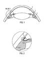
- FIG. 1 illustrates a schematic view of an anterior portion of an eye.
- aqueous humor is secreted from the ciliary body 1, passes through the pupil into the anterior chamber 2, and is filtered out of the eyeball via the trabeculum and the Schlemm's Canal 3.
- the aqueous humor passes through the trabecular meshwork 4.
- a common cause of glaucoma is blockage of flow through the trabecular meshwork 4.
- the aqueous humor excretory pathway is blocked, the aqueous humor cannot pass out of the eyeball at an adequate rate, the IOP rises, and glaucoma can develop.
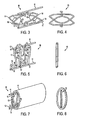
- FIGS. 3-13 illustrate an implant designed to be implanted inside the Schlemm's Canal to open fluid passageways through the wall of the Schlemm's Canal.
- the implant 10 has a longitudinally flexible body comprising one or more longitudinally flexible rods 11.
- the implant 10 has two longitudinally flexible rods 11.
- the longitudinally flexible rods 11 may be constructed of wire or may be wire-like.
- the rods 11 are longitudinally flexible to allow the implant to be threaded through the Schlemm's Canal, as described in further detail below.
- the implant 10 as illustrated in FIGS. 3-13 has a series of penetrating members attached to the longitudinally flexible body.
- Each penetrating member comprises at least one supporting arm 13 and at least one penetrating tip 14.
- each penetrating tip 14 is connected to the longitudinally flexible body by two supporting arms 13, with each of the two supporting arms 13 extending between the penetrating tip 14 and the longitudinally flexible body and connected to the longitudinally flexible body at a hinge 12.
- FIGS. 3 and 4 show the implant 10 with the penetrating members in an extended position.
- FIGS. 5 and 6 show the implant 10 with the penetrating members in an unextended position.
- the penetrating members and the longitudinally flexible body lie substantially in a single plane.
- the supporting arms 13 and penetrating tips 14 are in the unextended position, the supporting arms 13 and penetrating tips 14 and the longitudinally flexible rods 11 all lie substantially in a single plane. This can be seen in the end view of FIG. 6 .
- the flexible rods may be closer together or further apart than illustrated.
- the angles formed by the supporting arms may be smaller, allowing the flexible rods to be closer together. If the angle is small enough, when the supporting arms are unextended the supporting arms and flexible rods may all lie close together and extend in substantially the same direction. Thus, the end view of the implant may resemble a single rod.
- the penetrating members may be configured to self-expand from the unextended position to the extended position.
- the penetrating members may comprise a resilient material, wherein in an unconstrained, relaxed state the penetrating members are in the extended position as shown in FIGS. 3 and 4 .
- the implant 10 may be manufactured in this condition.
- a guiding sheath 15 may be provided to hold the penetrating members in a constrained state, holding the penetrating members in the first unextended position.
- FIGS. 7 and 8 show the guiding sheath 15 only schematically. It will be appreciated that the guiding sheath 15 may be configured to fit snugly around the implant 10 so as to hold the penetrating members in the unextended condition as shown in FIGS. 5 and 6 .
- the implant 10 within the guiding sheath 15 is inserted into the Schlemm's Canal through an access opening.
- the access opening into the Schlemm's Canal may be formed ab externo under a scleral flap or ab interno by forming an incision in the cornea and accessing the Schlemm's canal from through the anterior chamber of the eye.
- the ab externo procedure may be similar to a deep sclerectomy.
- a superficial flap may be made in the sclera and then a second deep scleral flap may be created and excised leaving a scleral reservoir under the first flap.
- An access opening to the Schlemm's Canal can be exposed at the scleral reservoir.
- a single scleral flap may be made to access the Schlemm's Canal, with or without excising any portion of the sclera.
- FIG. 9 is a perspective view of the implant 10 and guiding sheath 15 being inserted within the Schlemm's Canal 16.
- the implant 10 and guiding sheath 15 are advanced around at least a substantial portion of the Schlemm's Canal, with the penetrating members being held in the unextended position.
- Implant 10 and guiding sheath 15 are advanced substantially 360 degrees around the Schlemm's Canal so that the distal end of the implant 10 reaches the area of the opening into which the implant 10 was inserted.
- the implant 10 and guiding sheath 15 are advanced only part way around the Schlemm's Canal. In these instances, other implants may be used in other areas of the Schlemm's Canal.
- a guide wire or strand may first be inserted into the Schlemm's Canal.
- the guide wire or strand may be advanced around the Schlemm's Canal such that its leading end emerges again from the Schlemm's Canal at the area of the access opening.
- the implant may then be attached to the guide wire or strand.
- the guide wire or strand may then be withdrawn back through the Schlemm's Canal, thereby pulling the implant into the Schlemm's Canal.
- a viscoelastic material such as sodium hyaluronate, may be injected into the Schlemm's Canal as is known in the art. The viscoelastic material can help open the Schlemm's Canal and help to insure the patency of the Schlemm's Canal.
- the guiding sheath 15 is withdrawn. This removes the constraint from the penetrating members, allowing them to expand to their expanded position, as shown in FIGS. 10 and 11 . As shown in FIGS. 10 and 11 , when the penetrating members move from the unextended position to the extended position, the penetrating members penetrate the wall of the Schlemm's Canal 16. This creates fluid passageways or openings 17 in the wall of the Schlemm's Canal 16.
- the placement of the implant within the Schlemm's Canal can serve to keep the Schlemm's Canal open.
- the implant can serve as a support scaffolding to maintain the patency of the Schlemm's Canal.
- the deployed implant may apply a tension force to the tissue and increase its permeability for fluid flow.
- the implant can have penetrating members on both sides of the Schlemm's Canal.
- the penetrating members can create fluid passageways both into the Schlemm's Canal through the trabecular meshwork and also out of the Schlemm's Canal to the collector channels and into the sclera.
- the penetrating members may differ from those illustrated.
- Each penetrating member may have only a single supporting arm.
- the supporting arm may have an enlarged or hooked end, such as a hooked end forming a V-shape similar to the ends of the penetrating members as illustrated.
- the supporting arms alternatively may be in the form of tubes providing a conduit for fluid flow, as discussed further below in conjunction with FIGS. 28 and 29 .
- the sceral flap may be sutured closed. If an ab interno procedure is used, the opening in the cornea may be sutured closed at the conclusion of the procedure. The procedure also may be performed without suturing.
- An alternative self-expansion mechanism that may be used for the implant involves shape memory material.
- the implant, or portions of the implant may be made of a shape memory material.
- the implant may be manufactured such that the heat from the eye causes the penetrating members to move from the first unextended position to the second extended position.
- the implant For use in the Schlemm's Canal as described, the implant must be sized to fit within the Schlemm's Canal.
- the cross-sectional profile of the implant may have a height from about 0.05 mm to about 1 mm and a width from about 0.015 mm to about 0.25 mm, but other dimensions are of course possible.
- the cross-sectional profile of the implant When the penetrating members are in the extended position, the cross-sectional profile of the implant may have a width from about 0.03 mm to about 1 mm, again with other dimensions of course being possible.
- the implant may in certain examples have a length of about 35 mm or greater. In other instances, the length may be shorter. As just one of many possible examples, the length may be from about 1 mm to about 10 mm such that the implant extends only part way around the Schlemm's Canal.
- the implant can be described as having a longitudinal axis extended generally down the geometric center of the implant.
- the longitudinal axis is an imaginary line midway between the two longitudinally flexible rods 11.
- the penetrating members can therefore be described relative to the longitudinal axis.
- the penetrating members have an unextended position in which the penetrating members lie generally close to the longitudinal axis of the implant and an extended position in which the penetrating members are extended outward to be farther away from the longitudinal axis of the implant.
- FIG. 12 is a perspective view of the implant 10 and guiding sheath 15 inside an ultrasonic guide tube 18.
- the ultrasonic guide tube 18 uses ultrasound to emulsify tissue in the Schlemm's Canal ahead of the advancing implant in order to help facilitate the implantation procedure and/or open the Schlemm's Canal.
- the ultrasonic guide tube 18 has two layers, an inner layer which leads the ultrasonic signal from the ultrasound transducer outside the eye to the tissue on the inner wall of Schlemm's Canal, and an outer layer made of a covering material that blocks the ultrasound signal from reaching the wall of Schlemm's Canal at any place other than the leading end of the inner layer.
- the inner layer may be slightly longer than the outer layer to provide an exposed area for the leading end of the inner layer to be in contact with the tissue to be emulsified.
- FIG. 13 is a perspective view of the implant 10 and guiding sheath 15 inside a cleaning guide tube 19.
- the cleaning guide tube 19 has a coarse area on its outer surface at its distal end. When the surgeon pushes the cleaning guide tube into the Schlemm's Canal, the coarse end mechanically removes cells from the inner wall of the Schlemm's Canal. Similar to the ultrasonic guide tube 18, the cleaning guide tube 19 helps facilitate the implantation procedure and/or open the Schlemm's Canal.
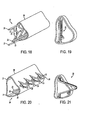
- FIGS. 14-21 illustrate an embodiment of an implant, designated as implant 20, designed to be implanted inside the Schlemm's Canal to open fluid passageways through the wall of the Schlemm's Canal.
- the implant 20 has a longitudinally flexible body comprising two longitudinally flexible rods 21.
- the longitudinally flexible rods 21 may be constructed of wire or may be wire-like and are longitudinally flexible to allow the implant to be threaded through the Schlemm's Canal.
- the implant 20 has a series of penetrating members, with each penetrating member having two supporting arms 23 and a penetrating tip 24.
- the supporting arms 23 are hingedly connected to the flexible rods 21 at hinges 22.
- Each supporting arm 23 extends between its respective penetrating tip 24 and its respective longitudinally flexible rod 21 and is connected to its respective the longitudinally flexible rod at the hinge 22.
- the implant 20 has a deployment rod 25 attached to the penetrating members.
- the deployment rod 25 may be constructed of wire or may be wire-like and is longitudinally flexible to allow the implant to be threaded through the Schlemm's Canal.
- the deployment rod 25 may also be in the form of a suture.
- FIGS. 14 and 15 show the implant 20 with the penetrating members in an unextended position.
- FIGS. 16 and 17 show the implant 20 with the penetrating members in an extended position.
- the penetrating members and the longitudinally flexible body lie substantially in a single plane.
- the supporting arms 23 and penetrating tips 24 are in the unextended position, the supporting arms 23, the penetrating tips 24, the longitudinally flexible rods 21, and the deployment rod 25 all lie substantially in a single plane. This can be seen in the end view of FIG. 15 .
- the flexible rods may be closer together or further apart than illustrated, and the angles formed by the supporting arms may differ.
- the supporting arms and flexible rods may all lie close together and extend in substantially the same direction, resulting in an end view of the implant resembling a single rod.
- the penetrating members are not self-expanding but instead are designed to be deployed by the deployment rod 25.
- the implant 20 is inserted into the Schlemm's Canal through an access opening, which may be formed ab externo or ab interno as described above.
- FIGS. 18 and 19 illustrate the implant 20 being inserted within the Schlemm's Canal 26.
- the implant 20 is advanced around at least a substantial portion of the Schlemm's Canal.
- the penetrating members are in the unextended position in their relaxed state as manufactured, so there is no need for a sheath to hold the penetrating members in the unextended position.
- implant 20 may be advanced substantially 360 degrees around the Schlemm's Canal or only part way around the Schlemm's Canal, in which case multiple implants may be used.
- the penetrating members are moved from the unextended position to the extended position by actuation of the deployment rod 25.
- Actuation of the deployment rod 25 is performed by moving the deployment rod 25 relative to the longitudinally flexible body.
- the longitudinally flexible rods 21 are held in place while the deployment rod is pulled proximally relative to the longitudinally flexible rods 21.
- this action causes the penetrating members to rotate at hinges 22 to move to the extended position shown in FIGS. 16-17 .
- the hinging motion causes plastic deformation of the implant 20 so that the penetrating members remain in the extended position after the deployment rod 25 is released.
- the placement of the implant 20 within the Schlemm's Canal can serve to keep the Schlemm's Canal open.
- the implant can serve as a support scaffolding to maintain the patency of the Schlemm's Canal.
- the size of the implant 20 may be similar to that of implant 10.
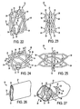
- FIGS. 22-27 illustrate an implant, designated as implant 30, designed to be implanted inside the Schlemm's Canal to open fluid passageways through the wall of the Schlemm's Canal.
- the implant 30 has a longitudinally flexible body comprising two longitudinally flexible rods 31.
- the longitudinally flexible rods 31 may be constructed of wire or may be wire-like and are longitudinally flexible to allow the implant to be threaded through the Schlemm's Canal.
- the implant 30 has a series of penetrating members, with each penetrating member having two supporting arms 33 and a penetrating tip 34.
- the supporting arms 33 are hingedly connected to the flexible rods 31 at hinges 32.
- Each supporting arm 33 extends between its respective penetrating tip 34 and its respective longitudinally flexible rod 31 and is connected to its respective the longitudinally flexible rod at the hinge 32.
- the supporting arms 33 need not be straight and may be bent or otherwise geometrically shaped to provide the desired result.
- the implant 30 is illustrated with its longitudinally flexible rods 31 joined at the distal end of the implant 30 at end 35 and joined at the proximal end of the implant 30 at end 36.
- the implant 30 is configured such that in the as-manufactured, relaxed stated of the implant, the penetrating members are in the unextended position, and the penetrating members generally point in a proximal direction along the longitudinal axis of the implant Thus, as shown in FIGS. 22 and 23 , the penetrating members generally point toward the end 36 at the proximal end of the implant 30.
- advancing the implant 30 in a distal direction through the Schlemm's Canal does not cause the penetrating members to become extended.
- retracting the implant 30 in a proximal direction back through the Sclilemm's Canal causes the penetrating members to get caught by the tissue of the walls of the Schlemm's Canal.
- Further retraction of the implant 30 by pulling on end 36 causes the penetrating members to rotate about hinges 32, thereby moving the penetrating members from the unextended position to the extended position.
- FIGS. 22 and 23 show the implant 30 with the penetrating members in an unextended position.
- FIGS. 24 and 25 show the implant 30 with the penetrating members in an extended position.
- the penetrating members and the longitudinally flexible body lie substantially in a single plane, although the profile is slightly larger than that of the implants 10 and 20. This is because the penetrating members in implant 30 stick out a little bit so as to catch on the Schlemm's Canal wall upon retraction on the implant body.
- the supporting arms 33 and penetrating tips 34 are in the unextended position, the supporting arms 33, the penetrating tips 34, and the longitudinally flexible rods 31 all lie substantially in a single plane, as can be seen in the end view of FIG. 23 .
- the implant 30 is inserted into the Schlemm's Canal through an access opening, which may be formed ab externo or ab interno as described above.
- FIG. 26 illustrates the implant 30 being inserted within the Schlemm's Canal 37.
- the implant 30 is advanced around at least a substantial portion of the Schlemm's Canal.
- the penetrating members are in the unextended position in their relaxed state as manufactured, so there is no need for a sheath to hold the penetrating members in the unextended position. Because of the orientation of the penetrating members of implant 30, advancing the implant 30 distally, with end 35 as the leading end in the direction of travel, allows the penetrating members to simply pass against the wall of the Schlemm's Canal without getting caught.
- implant 30 may be advanced substantially 360 degrees around the Schlemm's Canal or only part way around the Schlemm's Canal, in which case multiple implants may be used.
- the implant 30 is pulled proximally back through the Schlemm's Canal by pulling end 36, with end 36 as the leading end in the direction of travel. In this direction, the penetrating tips 34 get caught on the wall of the Schlemm's Canal. Further proximal pulling of end 36 causes the penetrating members to rotate at hinges 32, causing the penetrating members to move from the unextended position to the extended position. The hinging motion causes plastic deformation of the implant 30 so that the penetrating members remain in the extended position after the implant is released.
- the penetrating members when the penetrating members move from the unextended position to the extended position, the penetrating members penetrate the wall of the Schlemm's Canal 37. This creates fluid passageways or openings 38 in the wall of the Schlemm's Canal 37.
- the placement of the implant 30 within the Schlemm's Canal can also serve to keep the Schlemm's Canal open.
- the implant can serve as a support scaffolding to maintain the patency of the Schlemm's Canal.
- the size of the implant 30 may be similar to that of implants 10 and 20.
- FIGS. 28-29 illustrate a further implant.
- the implant 40 has a longitudinally flexible body which may be in the form, for example, of generally tubular shaped structure 41.
- Each penetrating member comprises one supporting arm 43 terminating in a penetrating tip 44.
- each supporting arm 43 is in the form of a tube providing a conduit for fluid flow.
- the tube opening provides an opening through the tissue for fluid flow.
- the supporting arm 43 is connected to the longitudinally flexible body at a hinge 42.
- FIG. 28 shows the implant 40 with the penetrating member in an unextended position.
- the end view of the implant 40 resembles a single rod.
- FIG. 29 shows the implant 40 with the penetrating member in an extended position.
- the penetrating members may be deployed to the extended position by mechanical means, such as a wire or strand, or the penetrating members may be configured to self-expand from the unextended position to the extended position.
- the implant 40 may be sized similarly to the implants described above and may be configured to extend part or all of the way around the Sclemm's Canal.
- the implant 40 is implanted in a similar manner as discussed above.
- the implant 40 can create fluid passageways into the Schlemm's Canal through the trabecular meshwork and/or out of the Schlemm's Canal to the collector channels and into the sclera.
- an implant in accordance with embodiments of the invention may be made of any of a number of biocompatible materials.
- the implant may be made of a type of nickel-titanium alloy, cobalt-chrome alloy or stainless steel such as SS316L.
- the implant may also be coated with a therapeutic agent such that the therapeutic agent elutes from the implant after implantation.
- Various therapeutic agents may be used.
- some therapeutic agents that may be desirable for certain applications include heparin, antibiotic drugs, anti-glaucoma drugs, or any other suitable drug or drug combination.
- the method of coating the implant may be any method as generally known in the art for applying coating to an implant, such as employed in the field of coronary stents.
- the implant may be made of a biodegradable material, which may be coated or impregnated with a therapeutic agent.
- FIG. 30 is a perspective view of a delivery device 50 that may be used to insert an implant, such as an implant as described above, within the Schlemm's Canal 3.
- the delivery device may comprise a generally longitudinally extending shaft 51, a hooked end 52 with a piercing tip 53 extending generally perpendicular to the shaft 51, and a lumen 61 passing through the longitudinally extending shaft and the hooked end for accommodating an implant.
- the delivery device may further comprises one or more lumens 62, 63, 64 for viscoelastic injection, an imaging device, a light source, an operating instrument, and/or irrigation.
- a small slit or opening is made in the cornea 5.
- the delivery device 50 is advanced through the slit or opening in the cornea 5 and the piercing tip is used to create an access passageway from the anterior chamber through the trabecular meshwork to the Schlemm's Canal 3.
- An imaging device and light source may be used to visualize the area. Irrigation may be used to wash the area.
- the implant is advanced from a lumen of the delivery device and passed into the Schlemm's Canal. Further implantation proceeds as described above. Before, during or after threading the implant through the Schlemm's Canal, a viscoelastic material, such as sodium hyaluronate, may be injected into the Schlemm's Canal as is known in the art.
- the delivery device is withdrawn from the eye. Then, the cornea can be sutured to close the slit or opening.
- the implant may be used in other body lumens where opening and/or scaffolding the lumen is desired.
- the implant may be used as a stent in the vasculature or other body lumens.
- the implant may be modified. For example, its size will be tailored to the desired application.
- the penetrating members may be designed as support members such that they hold a lumen open but do not penetrate the lumen wall.
Landscapes
- Health & Medical Sciences (AREA)
- Ophthalmology & Optometry (AREA)
- Heart & Thoracic Surgery (AREA)
- Surgery (AREA)
- Engineering & Computer Science (AREA)
- Biomedical Technology (AREA)
- Nuclear Medicine, Radiotherapy & Molecular Imaging (AREA)
- Vascular Medicine (AREA)
- Life Sciences & Earth Sciences (AREA)
- Animal Behavior & Ethology (AREA)
- General Health & Medical Sciences (AREA)
- Public Health (AREA)
- Veterinary Medicine (AREA)
- Prostheses (AREA)
Applications Claiming Priority (2)
| Application Number | Priority Date | Filing Date | Title |
|---|---|---|---|
| US12/029,255 US8109896B2 (en) | 2008-02-11 | 2008-02-11 | Devices and methods for opening fluid passageways |
| PCT/US2009/033049 WO2009102594A2 (en) | 2008-02-11 | 2009-02-04 | Devices and methods for opening fluid passageways |
Publications (2)
| Publication Number | Publication Date |
|---|---|
| EP2254535A2 EP2254535A2 (en) | 2010-12-01 |
| EP2254535B1 true EP2254535B1 (en) | 2015-06-03 |
Family
ID=40532503
Family Applications (1)
| Application Number | Title | Priority Date | Filing Date |
|---|---|---|---|
| EP09711060.5A Active EP2254535B1 (en) | 2008-02-11 | 2009-02-04 | Devices for opening fluid passageways |
Country Status (11)
| Country | Link |
|---|---|
| US (1) | US8109896B2 (ja) |
| EP (1) | EP2254535B1 (ja) |
| JP (1) | JP5438031B2 (ja) |
| KR (1) | KR101522549B1 (ja) |
| CN (1) | CN101998846B (ja) |
| AU (1) | AU2009215039B2 (ja) |
| BR (1) | BRPI0908775A2 (ja) |
| CA (1) | CA2714557C (ja) |
| ES (1) | ES2544428T3 (ja) |
| IL (1) | IL207496A (ja) |
| WO (1) | WO2009102594A2 (ja) |
Families Citing this family (68)
| Publication number | Priority date | Publication date | Assignee | Title |
|---|---|---|---|---|
| ES2609594T3 (es) | 1999-04-26 | 2017-04-21 | Glaukos Corporation | Dispositivo de drenaje para tratar el glaucoma |
| US7867186B2 (en) | 2002-04-08 | 2011-01-11 | Glaukos Corporation | Devices and methods for treatment of ocular disorders |
| US6638239B1 (en) | 2000-04-14 | 2003-10-28 | Glaukos Corporation | Apparatus and method for treating glaucoma |
| US7431710B2 (en) | 2002-04-08 | 2008-10-07 | Glaukos Corporation | Ocular implants with anchors and methods thereof |
| EP2982354A1 (en) | 2001-04-07 | 2016-02-10 | Glaukos Corporation | System for treating ocular disorders |
| US7331984B2 (en) | 2001-08-28 | 2008-02-19 | Glaukos Corporation | Glaucoma stent for treating glaucoma and methods of use |
| US7909789B2 (en) | 2006-06-26 | 2011-03-22 | Sight Sciences, Inc. | Intraocular implants and methods and kits therefor |
| US8974511B2 (en) | 2010-11-15 | 2015-03-10 | Aquesys, Inc. | Methods for treating closed angle glaucoma |
| US8801766B2 (en) | 2010-11-15 | 2014-08-12 | Aquesys, Inc. | Devices for deploying intraocular shunts |
| US10085884B2 (en) | 2006-06-30 | 2018-10-02 | Aquesys, Inc. | Intraocular devices |
| US8721702B2 (en) | 2010-11-15 | 2014-05-13 | Aquesys, Inc. | Intraocular shunt deployment devices |
| US8663303B2 (en) | 2010-11-15 | 2014-03-04 | Aquesys, Inc. | Methods for deploying an intraocular shunt from a deployment device and into an eye |
| US8852256B2 (en) | 2010-11-15 | 2014-10-07 | Aquesys, Inc. | Methods for intraocular shunt placement |
| US9095411B2 (en) | 2010-11-15 | 2015-08-04 | Aquesys, Inc. | Devices for deploying intraocular shunts |
| US8308701B2 (en) | 2010-11-15 | 2012-11-13 | Aquesys, Inc. | Methods for deploying intraocular shunts |
| US20120123316A1 (en) | 2010-11-15 | 2012-05-17 | Aquesys, Inc. | Intraocular shunts for placement in the intra-tenon's space |
| JP5748407B2 (ja) | 2006-11-10 | 2015-07-15 | グローコス コーポレーション | ブドウ膜強膜シャント |
| US8734377B2 (en) | 2007-09-24 | 2014-05-27 | Ivantis, Inc. | Ocular implants with asymmetric flexibility |
| US8425449B2 (en) | 2009-07-09 | 2013-04-23 | Ivantis, Inc. | Ocular implants and methods for delivering ocular implants into the eye |
| US20090082862A1 (en) | 2007-09-24 | 2009-03-26 | Schieber Andrew T | Ocular Implant Architectures |
| US20170360609A9 (en) | 2007-09-24 | 2017-12-21 | Ivantis, Inc. | Methods and devices for increasing aqueous humor outflow |
| US8808222B2 (en) * | 2007-11-20 | 2014-08-19 | Ivantis, Inc. | Methods and apparatus for delivering ocular implants into the eye |
| JP2011513002A (ja) | 2008-03-05 | 2011-04-28 | イバンティス インコーポレイテッド | 緑内障を治療する方法及び器具 |
| US9693899B2 (en) | 2009-07-09 | 2017-07-04 | Ivantis, Inc. | Single operator device for delivering an ocular implant |
| US20110098809A1 (en) | 2009-10-23 | 2011-04-28 | John Wardle | Ocular Implant System and Method |
| JP5856569B2 (ja) | 2010-02-05 | 2016-02-10 | サイト サイエンシーズ, インコーポレイテッド | 眼内圧を低減するためのデバイスと、それを含むキット |
| US8545430B2 (en) | 2010-06-09 | 2013-10-01 | Transcend Medical, Inc. | Expandable ocular devices |
| US9510973B2 (en) | 2010-06-23 | 2016-12-06 | Ivantis, Inc. | Ocular implants deployed in schlemm's canal of the eye |
| US20160256317A1 (en) | 2010-11-15 | 2016-09-08 | Aquesys, Inc. | Methods for implanting intraocular shunts |
| US8657776B2 (en) | 2011-06-14 | 2014-02-25 | Ivantis, Inc. | Ocular implants for delivery into the eye |
| CN102824238B (zh) * | 2011-06-16 | 2014-07-09 | 王宁利 | 一种施莱姆氏管(Schlemm)扩张支架及其组合体 |
| US8771220B2 (en) | 2011-12-07 | 2014-07-08 | Alcon Research, Ltd. | Glaucoma active pressure regulation shunt |
| US8852136B2 (en) | 2011-12-08 | 2014-10-07 | Aquesys, Inc. | Methods for placing a shunt into the intra-scleral space |
| US10080682B2 (en) | 2011-12-08 | 2018-09-25 | Aquesys, Inc. | Intrascleral shunt placement |
| US9808373B2 (en) | 2013-06-28 | 2017-11-07 | Aquesys, Inc. | Intraocular shunt implantation |
| US8765210B2 (en) | 2011-12-08 | 2014-07-01 | Aquesys, Inc. | Systems and methods for making gelatin shunts |
| US9610195B2 (en) | 2013-02-27 | 2017-04-04 | Aquesys, Inc. | Intraocular shunt implantation methods and devices |
| US8663150B2 (en) | 2011-12-19 | 2014-03-04 | Ivantis, Inc. | Delivering ocular implants into the eye |
| AU2012374034B2 (en) | 2012-03-20 | 2017-10-19 | Sight Sciences, Inc. | Ocular delivery systems and methods |
| US9554940B2 (en) | 2012-03-26 | 2017-01-31 | Glaukos Corporation | System and method for delivering multiple ocular implants |
| US9358156B2 (en) | 2012-04-18 | 2016-06-07 | Invantis, Inc. | Ocular implants for delivery into an anterior chamber of the eye |
| US8888734B2 (en) | 2012-06-05 | 2014-11-18 | Alcon Research, Ltd. | Functionally graded material tube and method for use of the same in implantation |
| US20140066833A1 (en) * | 2012-08-28 | 2014-03-06 | Clearlix Ltd. | Expandable fluid drainage implants and associated delivery devices and methods |
| WO2014085450A1 (en) | 2012-11-28 | 2014-06-05 | Ivantis, Inc. | Apparatus for delivering ocular implants into an anterior chamber of the eye |
| US9125723B2 (en) | 2013-02-19 | 2015-09-08 | Aquesys, Inc. | Adjustable glaucoma implant |
| US10159600B2 (en) | 2013-02-19 | 2018-12-25 | Aquesys, Inc. | Adjustable intraocular flow regulation |
| US10517759B2 (en) | 2013-03-15 | 2019-12-31 | Glaukos Corporation | Glaucoma stent and methods thereof for glaucoma treatment |
| US9592151B2 (en) | 2013-03-15 | 2017-03-14 | Glaukos Corporation | Systems and methods for delivering an ocular implant to the suprachoroidal space within an eye |
| EP3068354B1 (en) | 2013-11-14 | 2023-06-28 | Aquesys, Inc. | Intraocular shunt inserter |
| JP6655610B2 (ja) | 2014-05-29 | 2020-02-26 | グローコス コーポレーション | 制御された薬物送達機能を備えるインプラント及びそれを使用する方法 |
| US10709547B2 (en) | 2014-07-14 | 2020-07-14 | Ivantis, Inc. | Ocular implant delivery system and method |
| US10299958B2 (en) | 2015-03-31 | 2019-05-28 | Sight Sciences, Inc. | Ocular delivery systems and methods |
| BR112017025859A2 (pt) | 2015-06-03 | 2018-08-14 | Aquesys, Inc. | colocação de shunt intraocular ab externo |
| US11197779B2 (en) | 2015-08-14 | 2021-12-14 | Ivantis, Inc. | Ocular implant with pressure sensor and delivery system |
| WO2017040853A1 (en) | 2015-09-02 | 2017-03-09 | Glaukos Corporation | Drug delivery implants with bi-directional delivery capacity |
| US11938058B2 (en) | 2015-12-15 | 2024-03-26 | Alcon Inc. | Ocular implant and delivery system |
| MX2018014763A (es) | 2016-06-02 | 2019-04-29 | Aquesys Inc | Suministro intraocular de farmacos. |
| EP3668460A4 (en) * | 2017-08-17 | 2021-05-05 | Aspip Inc. | METHOD, DEVICE AND SYSTEM FOR TREATING HIGH INTRAOCULAR PRESSURE |
| US11116625B2 (en) | 2017-09-28 | 2021-09-14 | Glaukos Corporation | Apparatus and method for controlling placement of intraocular implants |
| EP3691586A2 (en) | 2017-10-06 | 2020-08-12 | Glaukos Corporation | Systems and methods for delivering multiple ocular implants |
| USD846738S1 (en) | 2017-10-27 | 2019-04-23 | Glaukos Corporation | Implant delivery apparatus |
| US11246753B2 (en) | 2017-11-08 | 2022-02-15 | Aquesys, Inc. | Manually adjustable intraocular flow regulation |
| US11135089B2 (en) | 2018-03-09 | 2021-10-05 | Aquesys, Inc. | Intraocular shunt inserter |
| US10952898B2 (en) | 2018-03-09 | 2021-03-23 | Aquesys, Inc. | Intraocular shunt inserter |
| CA3110310A1 (en) | 2018-09-04 | 2020-03-12 | University Hospitals Health System, Inc. | Ocular device for treating glaucoma and related minimally invasive glaucoma surgery method |
| US11504270B1 (en) | 2019-09-27 | 2022-11-22 | Sight Sciences, Inc. | Ocular delivery systems and methods |
| AU2022205382A1 (en) | 2021-01-11 | 2023-06-22 | Alcon Inc. | Systems and methods for viscoelastic delivery |
| CN113576749B (zh) * | 2021-07-30 | 2024-03-15 | 温州医科大学 | 一种青光眼引流器 |
Family Cites Families (212)
| Publication number | Priority date | Publication date | Assignee | Title |
|---|---|---|---|---|
| US15192A (en) * | 1856-06-24 | Tubular | ||
| US274447A (en) * | 1883-03-20 | William-kentish | ||
| US733152A (en) * | 1902-08-30 | 1903-07-07 | Murdoch Chisholm | Empyema drainage device. |
| US1388172A (en) * | 1920-03-18 | 1921-08-23 | Simon M Craddock | Veterinary surgical appliance |
| US2431587A (en) * | 1945-02-19 | 1947-11-25 | Charles F Schnee | Cannula button for surgical operations and method of use |
| US2555076A (en) * | 1947-11-17 | 1951-05-29 | Elijah R Crossley | Instrument for use in performing surgical eye operations |
| US2867213A (en) * | 1957-06-12 | 1959-01-06 | Jr Paul A Thomas | Flutter valve for drainage of the pleural cavity |
| US3159161A (en) * | 1962-11-14 | 1964-12-01 | Ness Richard Alton | Fistulizing canaliculus |
| US3310051A (en) * | 1963-12-10 | 1967-03-21 | Rudolf R Schulte | Surgical reservoir for implantation beneath the skin |
| US3333588A (en) * | 1964-07-06 | 1967-08-01 | Rudolf R Schulte | Brain ventricle cannula |
| US3272204A (en) * | 1965-09-22 | 1966-09-13 | Ethicon Inc | Absorbable collagen prosthetic implant with non-absorbable reinforcing strands |
| US3421509A (en) * | 1965-12-17 | 1969-01-14 | John M Fiore | Urethral catheter |
| US3530860A (en) * | 1967-01-09 | 1970-09-29 | Ponce De Leon Ear | Method and apparatus for inserting a tube through the ear drum |
| US3589401A (en) * | 1969-07-18 | 1971-06-29 | Case Co J I | Pressure modulating valve |
| US3915172A (en) * | 1970-05-27 | 1975-10-28 | Ceskoslovenska Akademie Ved | Capillary drain for glaucoma |
| US3788327A (en) * | 1971-03-30 | 1974-01-29 | H Donowitz | Surgical implant device |
| US3884238A (en) * | 1972-06-19 | 1975-05-20 | Malley Conor C O | Apparatus for intraocular surgery |
| US3957035A (en) * | 1972-09-08 | 1976-05-18 | Jean Chassaing | Ophthalmological device useful for eye surgery |
| US3890976A (en) * | 1972-10-26 | 1975-06-24 | Medical Products Corp | Catheter tip assembly |
| US3913584A (en) * | 1974-06-28 | 1975-10-21 | Xomox Corp | Combination myringotomy scalpel, aspirator and otological vent tube inserter |
| US3938529A (en) * | 1974-07-22 | 1976-02-17 | Gibbons Robert P | Indwelling ureteral catheter |
| US4142526A (en) * | 1974-12-23 | 1979-03-06 | Alza Corporation | Osmotic releasing system with means for changing release therefrom |
| US3976077A (en) * | 1975-02-03 | 1976-08-24 | Kerfoot Jr Franklin W | Eye surgery device |
| US4037604A (en) * | 1976-01-05 | 1977-07-26 | Newkirk John B | Artifical biological drainage device |
| US4153058A (en) * | 1977-07-05 | 1979-05-08 | Nehme Alexander E | Pleural decompression catheter |
| US4175563A (en) * | 1977-10-05 | 1979-11-27 | Arenberg Irving K | Biological drainage shunt |
| US4290426A (en) * | 1978-05-04 | 1981-09-22 | Alza Corporation | Dispenser for dispensing beneficial agent |
| US4299227A (en) * | 1979-10-19 | 1981-11-10 | Lincoff Harvey A | Ophthalmological appliance |
| US4303063A (en) * | 1980-03-06 | 1981-12-01 | Stahl Norman O | Ocular massage device |
| US4808183A (en) | 1980-06-03 | 1989-02-28 | University Of Iowa Research Foundation | Voice button prosthesis and method for installing same |
| US4402681A (en) * | 1980-08-23 | 1983-09-06 | Haas Joseph S | Artificial implant valve for the regulation of intraocular pressure |
| US4457757A (en) * | 1981-07-20 | 1984-07-03 | Molteno Anthony C B | Device for draining aqueous humour |
| FR2514852B1 (fr) * | 1981-10-19 | 1985-09-27 | Snecma | Organe de distribution de fluide et procede de realisation du corps de cet organe |
| US4474569A (en) * | 1982-06-28 | 1984-10-02 | Denver Surgical Developments, Inc. | Antenatal shunt |
| US4554918A (en) * | 1982-07-28 | 1985-11-26 | White Thomas C | Ocular pressure relief device |
| US4521210A (en) * | 1982-12-27 | 1985-06-04 | Wong Vernon G | Eye implant for relieving glaucoma, and device and method for use therewith |
| IT8352816V0 (it) * | 1983-01-07 | 1983-01-07 | Ferrando Ugo Gardi Giovanni E | Catetere di applicazione medico chirurgica |
| HU187011B (en) * | 1983-01-14 | 1985-10-28 | Orvosi Mueszer Sz | Trachea canula |
| SU1191227A1 (ru) | 1983-04-28 | 1985-11-15 | Всесоюзный Научно-Исследовательский И Проектный Институт Технологии Химического И Нефтяного Аппаратостроения | Лини дл производства оребренных биметаллических труб |
| US4538611A (en) * | 1983-06-13 | 1985-09-03 | Kelman Charles D | Surgical instrument and method of cutting a lens of an eye |
| NL8302541A (nl) * | 1983-07-15 | 1985-02-01 | Philips Nv | Werkwijze ter vervaardiging van een halfgeleiderinrichting, en halfgeleiderinrichting vervaardigd volgens de werkwijze. |
| US4587954A (en) * | 1983-12-29 | 1986-05-13 | Habley Medical Technology Corporation | Elastomeric prosthetic sphincter |
| US4563779A (en) * | 1984-01-27 | 1986-01-14 | Kelman Charles D | Corneal implant and method of making the same |
| US4578058A (en) * | 1984-04-02 | 1986-03-25 | Grandon Stanley C | Intraocular catheter apparatus and method of use |
| US4634418A (en) * | 1984-04-06 | 1987-01-06 | Binder Perry S | Hydrogel seton |
| US4787885A (en) | 1984-04-06 | 1988-11-29 | Binder Perry S | Hydrogel seton |
| US4660546A (en) * | 1984-11-07 | 1987-04-28 | Robert S. Herrick | Method for treating for deficiency of tears |
| US4604087A (en) * | 1985-02-26 | 1986-08-05 | Joseph Neil H | Aqueous humor drainage device |
| US4781675A (en) | 1985-11-27 | 1988-11-01 | White Thomas C | Infusion cannula |
| EP0228185B1 (en) | 1985-11-27 | 1990-07-25 | Thomas C. White | Tissue-implantable fluid-dissipating device |
| US4692142A (en) * | 1986-02-24 | 1987-09-08 | Dignam Bernard J | Sutureless infusion cannula for ophthalmic surgery |
| US4792336A (en) | 1986-03-03 | 1988-12-20 | American Cyanamid Company | Flat braided ligament or tendon implant device having texturized yarns |
| NZ215409A (en) * | 1986-03-07 | 1989-02-24 | Anthony Christopher Be Molteno | Implant for drainage of aqueous humour in glaucoma |
| US4964850A (en) | 1986-05-07 | 1990-10-23 | Vincent Bouton | Method for treating trans-nasal sinus afflictions using a double t-shaped trans-nasal aerator |
| US4826478A (en) | 1986-06-23 | 1989-05-02 | Stanley Schocket | Anterior chamber tube shunt to an encircling band, and related surgical procedure |
| US4722724A (en) * | 1986-06-23 | 1988-02-02 | Stanley Schocket | Anterior chamber tube shunt to an encircling band, and related surgical procedure |
| US4909783A (en) | 1986-07-16 | 1990-03-20 | Morrison David P | Intra-ocular pressure apparatus |
| US4751926A (en) * | 1986-09-12 | 1988-06-21 | Dow Corning Wright Corporation | Instrument for subcutaneous insertion of an injection reservoir |
| US4863457A (en) | 1986-11-24 | 1989-09-05 | Lee David A | Drug delivery device |
| US4886488A (en) | 1987-08-06 | 1989-12-12 | White Thomas C | Glaucoma drainage the lacrimal system and method |
| EP0375676A1 (en) | 1987-08-19 | 1990-07-04 | BERG, Olle | A drainage tube for sinus maxillaris, a means for its insertion and a means for making a hole for its positioning |
| US4813941A (en) | 1987-09-03 | 1989-03-21 | Leslie Shea | Pneumothorax treatment device |
| US4934363A (en) | 1987-12-15 | 1990-06-19 | Iolab Corporation | Lens insertion instrument |
| US4888016A (en) | 1988-02-10 | 1989-12-19 | Langerman David W | "Spare parts" for use in ophthalmic surgical procedures |
| US4936825A (en) | 1988-04-11 | 1990-06-26 | Ungerleider Bruce A | Method for reducing intraocular pressure caused by glaucoma |
| US5098393A (en) | 1988-05-31 | 1992-03-24 | Kurt Amplatz | Medical introducer and valve assembly |
| US4915684A (en) | 1988-06-21 | 1990-04-10 | Mackeen Donald L | Method and apparatus for modulating the flow of lacrimal fluid through a punctum and associated canaliculus |
| US5071408A (en) | 1988-10-07 | 1991-12-10 | Ahmed Abdul Mateen | Medical valve |
| US5616118A (en) * | 1988-10-07 | 1997-04-01 | Ahmed; Abdul M. | Uniquely shaped ophthalmological device |
| IT1227176B (it) | 1988-10-11 | 1991-03-20 | Co Pharma Corp Srl | Dispositivo per il fissaggio di un catetere alla teca cranica per derivazione liquorale esterna |
| US4959048A (en) | 1989-01-17 | 1990-09-25 | Helix Medical, Inc. | Lacrimal duct occluder |
| US5000731A (en) | 1989-03-30 | 1991-03-19 | Tai-Ting Wong | Shunting device adopted in the intracranial shunting surgical operation for the treatment of hydrocephalus |
| US5053040A (en) | 1989-11-09 | 1991-10-01 | Goldsmith Iii Manning M | Method of performing a myringotomy |
| US4946436A (en) | 1989-11-17 | 1990-08-07 | Smith Stewart G | Pressure-relieving device and process for implanting |
| US5106367A (en) | 1989-11-28 | 1992-04-21 | Alexander Ureche | Eye surgery apparatus with vacuum surge suppressor |
| US5167620A (en) | 1989-11-28 | 1992-12-01 | Alexandar Ureche | Eye surgery methods |
| RU1797884C (ru) | 1989-11-30 | 1993-02-28 | Туркменский Научно-Исследовательский Институт Глазных Болезней | Искусственный имплантируемый клапан дл регулировани внутриглазного давлени |
| US5092837A (en) | 1989-12-20 | 1992-03-03 | Robert Ritch | Method for the treatment of glaucoma |
| US4968296A (en) * | 1989-12-20 | 1990-11-06 | Robert Ritch | Transscleral drainage implant device for the treatment of glaucoma |
| US5073163A (en) | 1990-01-29 | 1991-12-17 | Lippman Myron E | Apparatus for treating glaucoma |
| US5171270A (en) | 1990-03-29 | 1992-12-15 | Herrick Robert S | Canalicular implant having a collapsible flared section and method |
| US5127901A (en) | 1990-05-18 | 1992-07-07 | Odrich Ronald B | Implant with subconjunctival arch |
| US5041081A (en) | 1990-05-18 | 1991-08-20 | Odrich Ronald B | Ocular implant for controlling glaucoma |
| US5178604A (en) | 1990-05-31 | 1993-01-12 | Iovision, Inc. | Glaucoma implant |
| US5397300A (en) | 1990-05-31 | 1995-03-14 | Iovision, Inc. | Glaucoma implant |
| US5476445A (en) | 1990-05-31 | 1995-12-19 | Iovision, Inc. | Glaucoma implant with a temporary flow restricting seal |
| WO1992000112A1 (en) | 1990-06-25 | 1992-01-09 | Ungerleider Bruce A | Apparatus for reducing intraocular pressure |
| US5098438A (en) | 1990-08-23 | 1992-03-24 | Siepser Steven B | Procedures for intraocular surgery |
| CA2060635A1 (en) | 1991-02-12 | 1992-08-13 | Keith D'alessio | Bioabsorbable medical implants |
| US5454796A (en) | 1991-04-09 | 1995-10-03 | Hood Laboratories | Device and method for controlling intraocular fluid pressure |
| US5242449A (en) | 1991-04-23 | 1993-09-07 | Allergan, Inc. | Ophthalmic instrument |
| US5207660A (en) | 1991-04-26 | 1993-05-04 | Cornell Research Foundation, Inc. | Method for the delivery of compositions to the ocular tissues |
| US5358492A (en) | 1991-05-02 | 1994-10-25 | Feibus Miriam H | Woven surgical drain and method of making |
| US6007511A (en) | 1991-05-08 | 1999-12-28 | Prywes; Arnold S. | Shunt valve and therapeutic delivery system for treatment of glaucoma and methods and apparatus for its installation |
| US5300020A (en) | 1991-05-31 | 1994-04-05 | Medflex Corporation | Surgically implantable device for glaucoma relief |
| US5147370A (en) | 1991-06-12 | 1992-09-15 | Mcnamara Thomas O | Nitinol stent for hollow body conduits |
| US5326345A (en) | 1991-08-14 | 1994-07-05 | Price Jr Francis W | Eye filtration prostheses |
| US5171213A (en) | 1991-08-14 | 1992-12-15 | Price Jr Francis W | Technique for fistulization of the eye and an eye filtration prosthesis useful therefor |
| US5360399A (en) | 1992-01-10 | 1994-11-01 | Robert Stegmann | Method and apparatus for maintaining the normal intraocular pressure |
| JPH08504106A (ja) | 1992-01-29 | 1996-05-07 | スミス,スチユワート・グレゴリー | 水晶体乳化のための方法および装置 |
| US5283063A (en) | 1992-01-31 | 1994-02-01 | Eagle Vision | Punctum plug method and apparatus |
| US5190552A (en) | 1992-02-04 | 1993-03-02 | Kelman Charles D | Slotted tube injector for an intraocular lens |
| US5217486A (en) | 1992-02-18 | 1993-06-08 | Mitek Surgical Products, Inc. | Suture anchor and installation tool |
| US5346464A (en) | 1992-03-10 | 1994-09-13 | Camras Carl B | Method and apparatus for reducing intraocular pressure |
| US5221278A (en) | 1992-03-12 | 1993-06-22 | Alza Corporation | Osmotically driven delivery device with expandable orifice for pulsatile delivery effect |
| WO1993020783A1 (en) | 1992-04-13 | 1993-10-28 | Iovision, Inc. | Glaucoma implant |
| US5380290A (en) | 1992-04-16 | 1995-01-10 | Pfizer Hospital Products Group, Inc. | Body access device |
| US5322504A (en) | 1992-05-07 | 1994-06-21 | United States Surgical Corporation | Method and apparatus for tissue excision and removal by fluid jet |
| WO1994002081A1 (en) | 1992-07-16 | 1994-02-03 | Wong Vernon G | Eye implant suitable for relief of glaucoma |
| US5355871A (en) | 1992-09-11 | 1994-10-18 | Dexide, Inc. | Elastomeric controller for endoscopic surgical instruments |
| IL106946A0 (en) | 1992-09-22 | 1993-12-28 | Target Therapeutics Inc | Detachable embolic coil assembly |
| CA2144741C (en) | 1992-09-30 | 1997-04-08 | Vladimir Feingold | Intraocular lens insertion system |
| US5370607A (en) | 1992-10-28 | 1994-12-06 | Annuit Coeptis, Inc. | Glaucoma implant device and method for implanting same |
| EP0596314B1 (de) | 1992-11-06 | 1999-12-15 | GRIESHABER & CO. AG | Ophthalmologisches Aspirations- und Irrigationssystem |
| WO1994013234A1 (en) | 1992-12-17 | 1994-06-23 | Michael Andrew Coote | Implant device and method for treatment of glaucoma |
| FR2700265B1 (fr) | 1993-01-08 | 1995-03-31 | France Chirurgie Instr | Bouchon méatique pour pathologie lacrymale. |
| US5338291A (en) | 1993-02-03 | 1994-08-16 | Pudenz-Schulte Medical Research Corporation | Glaucoma shunt and method for draining aqueous humor |
| USD356867S (en) | 1993-03-10 | 1995-03-28 | Hood Laboratories | Device for controlling intraocular fluid pressure |
| US5342370A (en) | 1993-03-19 | 1994-08-30 | University Of Miami | Method and apparatus for implanting an artifical meshwork in glaucoma surgery |
| US5433714A (en) | 1993-04-06 | 1995-07-18 | Bloomberg; Leroy | Topical anesthesia method for eye surgery, and applicator therefor |
| DE4313245C2 (de) | 1993-04-23 | 1997-03-27 | Geuder Hans Gmbh | Hohlnadel für ein augenchirurgisches Instrument |
| IL109499A (en) | 1994-05-02 | 1998-01-04 | Univ Ramot | Implant device for draining excess intraocular fluid |
| FR2721499B1 (fr) | 1994-06-22 | 1997-01-03 | Opsia | Implant de trabéculectomie. |
| US5520631A (en) | 1994-07-22 | 1996-05-28 | Wound Healing Of Oklahoma | Method and apparatus for lowering the intraocular pressure of an eye |
| US6102045A (en) | 1994-07-22 | 2000-08-15 | Premier Laser Systems, Inc. | Method and apparatus for lowering the intraocular pressure of an eye |
| US5704907A (en) | 1994-07-22 | 1998-01-06 | Wound Healing Of Oklahoma | Method and apparatus for lowering the intraocular pressure of an eye |
| US5522845A (en) | 1994-09-27 | 1996-06-04 | Mitek Surgical Products, Inc. | Bone anchor and bone anchor installation |
| US5601094A (en) | 1994-11-22 | 1997-02-11 | Reiss; George R. | Ophthalmic shunt |
| US5660205A (en) | 1994-12-15 | 1997-08-26 | Epstein; Alan B. | One-way valve |
| US5433701A (en) | 1994-12-21 | 1995-07-18 | Rubinstein; Mark H. | Apparatus for reducing ocular pressure |
| US5558630A (en) | 1994-12-30 | 1996-09-24 | Fisher; Bret L. | Intrascleral implant and method for the regulation of intraocular pressure |
| WO1996020742A1 (en) | 1995-01-06 | 1996-07-11 | Wong Vernon G | Improve eye implant for relief of glaucoma |
| US5626558A (en) | 1995-05-05 | 1997-05-06 | Suson; John | Adjustable flow rate glaucoma shunt and method of using same |
| EP0957949B1 (en) | 1995-05-14 | 2004-08-04 | Optonol Ltd. | Intraocular implant, delivery device, and method of implantation |
| IL113723A (en) | 1995-05-14 | 2002-11-10 | Optonol Ltd | Intraocular implant |
| US5968058A (en) | 1996-03-27 | 1999-10-19 | Optonol Ltd. | Device for and method of implanting an intraocular implant |
| US5662600A (en) | 1995-09-29 | 1997-09-02 | Pudenz-Schulte Medical Research Corporation | Burr-hole flow control device |
| US5741292A (en) | 1995-10-26 | 1998-04-21 | Eagle Vision | Punctum dilating and plug inserting instrument with push-button plug release |
| US5709698A (en) | 1996-02-26 | 1998-01-20 | Linvatec Corporation | Irrigating/aspirating shaver blade assembly |
| US5665101A (en) | 1996-04-01 | 1997-09-09 | Linvatec Corporation | Endoscopic or open lipectomy instrument |
| US5865831A (en) | 1996-04-17 | 1999-02-02 | Premier Laser Systems, Inc. | Laser surgical procedures for treatment of glaucoma |
| US5980480A (en) | 1996-07-11 | 1999-11-09 | Cs Fluids, Inc. | Method and apparatus for treating adult-onset dementia of the alzheimer's type |
| US5807240A (en) | 1996-09-24 | 1998-09-15 | Circon Corporation | Continuous flow endoscope with enlarged outflow channel |
| US6007510A (en) | 1996-10-25 | 1999-12-28 | Anamed, Inc. | Implantable devices and methods for controlling the flow of fluids within the body |
| AUPO394496A0 (en) * | 1996-11-29 | 1997-01-02 | Lions Eye Institute | Biological microfistula tube and implantation method and apparatus |
| FR2757068B1 (fr) | 1996-12-13 | 1999-04-23 | Jussmann Alberto | Drain auto-fixant |
| GB9700390D0 (en) * | 1997-01-10 | 1997-02-26 | Biocompatibles Ltd | Device for use in the eye |
| US5713844A (en) | 1997-01-10 | 1998-02-03 | Peyman; Gholam A. | Device and method for regulating intraocular pressure |
| US5893837A (en) | 1997-02-28 | 1999-04-13 | Staar Surgical Company, Inc. | Glaucoma drain implanting device and method |
| US6050970A (en) | 1997-05-08 | 2000-04-18 | Pharmacia & Upjohn Company | Method and apparatus for inserting a glaucoma implant in an anterior and posterior segment of the eye |
| US6004302A (en) | 1997-08-28 | 1999-12-21 | Brierley; Lawrence A. | Cannula |
| US6007578A (en) | 1997-10-08 | 1999-12-28 | Ras Holding Corp | Scleral prosthesis for treatment of presbyopia and other eye disorders |
| US6203513B1 (en) * | 1997-11-20 | 2001-03-20 | Optonol Ltd. | Flow regulating implant, method of manufacture, and delivery device |
| US6168575B1 (en) | 1998-01-29 | 2001-01-02 | David Pyam Soltanpour | Method and apparatus for controlling intraocular pressure |
| ES2609594T3 (es) * | 1999-04-26 | 2017-04-21 | Glaukos Corporation | Dispositivo de drenaje para tratar el glaucoma |
| US20050119601A9 (en) * | 1999-04-26 | 2005-06-02 | Lynch Mary G. | Shunt device and method for treating glaucoma |
| US6558342B1 (en) | 1999-06-02 | 2003-05-06 | Optonol Ltd. | Flow control device, introducer and method of implanting |
| US6221078B1 (en) | 1999-06-25 | 2001-04-24 | Stephen S. Bylsma | Surgical implantation apparatus |
| US7008396B1 (en) * | 1999-09-03 | 2006-03-07 | Restorvision, Inc. | Ophthalmic device and method of manufacture and use |
| JP3292953B2 (ja) | 1999-10-15 | 2002-06-17 | エヌイーシーインフロンティア株式会社 | 伝送路二重化装置および二重化伝送路方式 |
| US6245077B1 (en) | 2000-01-21 | 2001-06-12 | Exmoor Plastics Ltd. | Universal myringotomy tube/aural grommet inserter and methods |
| US6471666B1 (en) * | 2000-02-24 | 2002-10-29 | Steven A. Odrich | Injectable glaucoma device |
| US20030060752A1 (en) * | 2000-04-14 | 2003-03-27 | Olav Bergheim | Glaucoma device and methods thereof |
| US20040111050A1 (en) * | 2000-04-14 | 2004-06-10 | Gregory Smedley | Implantable ocular pump to reduce intraocular pressure |
| US20050277864A1 (en) | 2000-04-14 | 2005-12-15 | David Haffner | Injectable gel implant for glaucoma treatment |
| US7867186B2 (en) | 2002-04-08 | 2011-01-11 | Glaukos Corporation | Devices and methods for treatment of ocular disorders |
| US6533768B1 (en) * | 2000-04-14 | 2003-03-18 | The Regents Of The University Of California | Device for glaucoma treatment and methods thereof |
| US6638239B1 (en) | 2000-04-14 | 2003-10-28 | Glaukos Corporation | Apparatus and method for treating glaucoma |
| US7708711B2 (en) * | 2000-04-14 | 2010-05-04 | Glaukos Corporation | Ocular implant with therapeutic agents and methods thereof |
| US20020143284A1 (en) * | 2001-04-03 | 2002-10-03 | Hosheng Tu | Drug-releasing trabecular implant for glaucoma treatment |
| US20050049578A1 (en) | 2000-04-14 | 2005-03-03 | Hosheng Tu | Implantable ocular pump to reduce intraocular pressure |
| EP1292256A1 (en) * | 2000-06-19 | 2003-03-19 | Glaukos Corporation | Stented trabecular shunt and methods thereof |
| US6730056B1 (en) * | 2000-09-21 | 2004-05-04 | Motorola, Inc. | Eye implant for treating glaucoma and method for manufacturing same |
| US6682523B2 (en) * | 2001-02-21 | 2004-01-27 | John H. Shadduck | Devices and techniques for treating trabecular meshwork |
| US6989007B2 (en) * | 2001-02-21 | 2006-01-24 | Solx, Inc. | Devices and techniques for treating glaucoma |
| US20020133168A1 (en) * | 2001-03-16 | 2002-09-19 | Smedley Gregory T. | Applicator and methods for placing a trabecular shunt for glaucoma treatment |
| US7488303B1 (en) | 2002-09-21 | 2009-02-10 | Glaukos Corporation | Ocular implant with anchor and multiple openings |
| US6981958B1 (en) * | 2001-05-02 | 2006-01-03 | Glaukos Corporation | Implant with pressure sensor for glaucoma treatment |
| US6666841B2 (en) * | 2001-05-02 | 2003-12-23 | Glaukos Corporation | Bifurcatable trabecular shunt for glaucoma treatment |
| EP2982354A1 (en) | 2001-04-07 | 2016-02-10 | Glaukos Corporation | System for treating ocular disorders |
| US7431710B2 (en) * | 2002-04-08 | 2008-10-07 | Glaukos Corporation | Ocular implants with anchors and methods thereof |
| US7678065B2 (en) | 2001-05-02 | 2010-03-16 | Glaukos Corporation | Implant with intraocular pressure sensor for glaucoma treatment |
| US7094225B2 (en) * | 2001-05-03 | 2006-08-22 | Glaukos Corporation | Medical device and methods of use of glaucoma treatment |
| US7331984B2 (en) | 2001-08-28 | 2008-02-19 | Glaukos Corporation | Glaucoma stent for treating glaucoma and methods of use |
| US20030097151A1 (en) * | 2001-10-25 | 2003-05-22 | Smedley Gregory T. | Apparatus and mitochondrial treatment for glaucoma |
| US7163543B2 (en) * | 2001-11-08 | 2007-01-16 | Glaukos Corporation | Combined treatment for cataract and glaucoma treatment |
| US6939298B2 (en) * | 2002-02-28 | 2005-09-06 | Gmp Vision Solutions, Inc | Device and method for monitoring aqueous flow within the eye |
| US20060200113A1 (en) | 2002-03-07 | 2006-09-07 | David Haffner | Liquid jet for glaucoma treatment |
| US7186232B1 (en) | 2002-03-07 | 2007-03-06 | Glaukoa Corporation | Fluid infusion methods for glaucoma treatment |
| US20030229303A1 (en) * | 2002-03-22 | 2003-12-11 | Haffner David S. | Expandable glaucoma implant and methods of use |
| US9301875B2 (en) | 2002-04-08 | 2016-04-05 | Glaukos Corporation | Ocular disorder treatment implants with multiple opening |
| US20040147870A1 (en) * | 2002-04-08 | 2004-07-29 | Burns Thomas W. | Glaucoma treatment kit |
| US20040024345A1 (en) * | 2002-04-19 | 2004-02-05 | Morteza Gharib | Glaucoma implant with valveless flow bias |
| CN101791258A (zh) * | 2002-07-19 | 2010-08-04 | 耶鲁大学 | 色素层巩膜引流装置 |
| US7192412B1 (en) * | 2002-09-14 | 2007-03-20 | Glaukos Corporation | Targeted stent placement and multi-stent therapy |
| US20040154946A1 (en) | 2002-11-06 | 2004-08-12 | Kenneth Solovay | Storage apparatus for surgical implant device |
| US20040216749A1 (en) | 2003-01-23 | 2004-11-04 | Hosheng Tu | Vasomodulation during glaucoma surgery |
| USD490152S1 (en) * | 2003-02-28 | 2004-05-18 | Glaukos Corporation | Surgical handpiece |
| US20040225250A1 (en) | 2003-05-05 | 2004-11-11 | Michael Yablonski | Internal shunt and method for treating glaucoma |
| EP1638508B1 (en) * | 2003-06-16 | 2014-08-13 | Solx, Inc. | Shunt for the treatment of glaucoma |
| US20060069340A1 (en) | 2003-06-16 | 2006-03-30 | Solx, Inc. | Shunt for the treatment of glaucoma |
| US7291125B2 (en) | 2003-11-14 | 2007-11-06 | Transcend Medical, Inc. | Ocular pressure regulation |
| WO2005072294A2 (en) | 2004-01-22 | 2005-08-11 | Solx, Inc. | Glaucoma treatment method |
| US20050250788A1 (en) | 2004-01-30 | 2005-11-10 | Hosheng Tu | Aqueous outflow enhancement with vasodilated aqueous cavity |
| US20060032507A1 (en) | 2004-08-11 | 2006-02-16 | Hosheng Tu | Contrast-enhanced ocular imaging |
| US20060173397A1 (en) | 2004-11-23 | 2006-08-03 | Hosheng Tu | Ophthalmology implants and methods of manufacture |
| KR20070092279A (ko) * | 2004-12-16 | 2007-09-12 | 아이싸이언스 인터벤셔날 코포레이션 | 녹내장 치료를 위한 안과 이식물 |
| WO2007087061A2 (en) | 2006-01-17 | 2007-08-02 | Transcend Medical, Inc. | Glaucoma treatment device |
| US20070293807A1 (en) | 2006-05-01 | 2007-12-20 | Lynch Mary G | Dual drainage pathway shunt device and method for treating glaucoma |
| AU2007269259B2 (en) | 2006-06-30 | 2012-05-31 | Aquesys Inc. | Methods, systems and apparatus for relieving pressure in an organ |
| JP5748407B2 (ja) | 2006-11-10 | 2015-07-15 | グローコス コーポレーション | ブドウ膜強膜シャント |
| EP2173289A4 (en) | 2007-07-17 | 2010-11-24 | Transcend Medical Inc | EYE IMPLANT WITH HYDROGEL EXPANSION CAPABILITIES |
-
2008
- 2008-02-11 US US12/029,255 patent/US8109896B2/en active Active
-
2009
- 2009-02-04 WO PCT/US2009/033049 patent/WO2009102594A2/en active Application Filing
- 2009-02-04 ES ES09711060.5T patent/ES2544428T3/es active Active
- 2009-02-04 JP JP2010545964A patent/JP5438031B2/ja active Active
- 2009-02-04 CN CN200980112705.XA patent/CN101998846B/zh not_active Expired - Fee Related
- 2009-02-04 KR KR1020107018210A patent/KR101522549B1/ko active IP Right Grant
- 2009-02-04 AU AU2009215039A patent/AU2009215039B2/en not_active Ceased
- 2009-02-04 EP EP09711060.5A patent/EP2254535B1/en active Active
- 2009-02-04 CA CA2714557A patent/CA2714557C/en not_active Expired - Fee Related
- 2009-02-04 BR BRPI0908775-3A patent/BRPI0908775A2/pt not_active Application Discontinuation
-
2010
- 2010-08-09 IL IL207496A patent/IL207496A/en not_active IP Right Cessation
Also Published As
| Publication number | Publication date |
|---|---|
| ES2544428T3 (es) | 2015-08-31 |
| CA2714557C (en) | 2016-11-29 |
| IL207496A0 (en) | 2010-12-30 |
| US20090204053A1 (en) | 2009-08-13 |
| CN101998846B (zh) | 2014-02-26 |
| CA2714557A1 (en) | 2009-08-20 |
| AU2009215039A1 (en) | 2009-08-20 |
| EP2254535A2 (en) | 2010-12-01 |
| IL207496A (en) | 2014-02-27 |
| CN101998846A (zh) | 2011-03-30 |
| US8109896B2 (en) | 2012-02-07 |
| WO2009102594A3 (en) | 2009-11-05 |
| JP2011511666A (ja) | 2011-04-14 |
| KR101522549B1 (ko) | 2015-05-26 |
| JP5438031B2 (ja) | 2014-03-12 |
| WO2009102594A2 (en) | 2009-08-20 |
| BRPI0908775A2 (pt) | 2015-07-28 |
| KR20100103877A (ko) | 2010-09-28 |
| AU2009215039B2 (en) | 2013-10-31 |
Similar Documents
| Publication | Publication Date | Title |
|---|---|---|
| EP2254535B1 (en) | Devices for opening fluid passageways | |
| US11786402B2 (en) | Glaucoma treatment device | |
| US20230285191A1 (en) | Intraocular shunt implantation methods and devices | |
| AU2018200325B2 (en) | Glaucoma treatment device | |
| AU2014200171B2 (en) | Glaucoma treatment device |
Legal Events
| Date | Code | Title | Description |
|---|---|---|---|
| PUAI | Public reference made under article 153(3) epc to a published international application that has entered the european phase |
Free format text: ORIGINAL CODE: 0009012 |
|
| 17P | Request for examination filed |
Effective date: 20100910 |
|
| AK | Designated contracting states |
Kind code of ref document: A2 Designated state(s): AT BE BG CH CY CZ DE DK EE ES FI FR GB GR HR HU IE IS IT LI LT LU LV MC MK MT NL NO PL PT RO SE SI SK TR |
|
| AX | Request for extension of the european patent |
Extension state: AL BA RS |
|
| DAX | Request for extension of the european patent (deleted) | ||
| 17Q | First examination report despatched |
Effective date: 20120228 |
|
| GRAP | Despatch of communication of intention to grant a patent |
Free format text: ORIGINAL CODE: EPIDOSNIGR1 |
|
| INTG | Intention to grant announced |
Effective date: 20140828 |
|
| GRAP | Despatch of communication of intention to grant a patent |
Free format text: ORIGINAL CODE: EPIDOSNIGR1 |
|
| INTG | Intention to grant announced |
Effective date: 20141209 |
|
| GRAS | Grant fee paid |
Free format text: ORIGINAL CODE: EPIDOSNIGR3 |
|
| GRAA | (expected) grant |
Free format text: ORIGINAL CODE: 0009210 |
|
| AK | Designated contracting states |
Kind code of ref document: B1 Designated state(s): AT BE BG CH CY CZ DE DK EE ES FI FR GB GR HR HU IE IS IT LI LT LU LV MC MK MT NL NO PL PT RO SE SI SK TR |
|
| REG | Reference to a national code |
Ref country code: GB Ref legal event code: FG4D |
|
| REG | Reference to a national code |
Ref country code: CH Ref legal event code: EP |
|
| REG | Reference to a national code |
Ref country code: AT Ref legal event code: REF Ref document number: 729539 Country of ref document: AT Kind code of ref document: T Effective date: 20150715 Ref country code: IE Ref legal event code: FG4D |
|
| REG | Reference to a national code |
Ref country code: DE Ref legal event code: R096 Ref document number: 602009031537 Country of ref document: DE |
|
| REG | Reference to a national code |
Ref country code: ES Ref legal event code: FG2A Ref document number: 2544428 Country of ref document: ES Kind code of ref document: T3 Effective date: 20150831 |
|
| REG | Reference to a national code |
Ref country code: AT Ref legal event code: MK05 Ref document number: 729539 Country of ref document: AT Kind code of ref document: T Effective date: 20150603 |
|
| PG25 | Lapsed in a contracting state [announced via postgrant information from national office to epo] |
Ref country code: FI Free format text: LAPSE BECAUSE OF FAILURE TO SUBMIT A TRANSLATION OF THE DESCRIPTION OR TO PAY THE FEE WITHIN THE PRESCRIBED TIME-LIMIT Effective date: 20150603 Ref country code: HR Free format text: LAPSE BECAUSE OF FAILURE TO SUBMIT A TRANSLATION OF THE DESCRIPTION OR TO PAY THE FEE WITHIN THE PRESCRIBED TIME-LIMIT Effective date: 20150603 Ref country code: LT Free format text: LAPSE BECAUSE OF FAILURE TO SUBMIT A TRANSLATION OF THE DESCRIPTION OR TO PAY THE FEE WITHIN THE PRESCRIBED TIME-LIMIT Effective date: 20150603 Ref country code: NO Free format text: LAPSE BECAUSE OF FAILURE TO SUBMIT A TRANSLATION OF THE DESCRIPTION OR TO PAY THE FEE WITHIN THE PRESCRIBED TIME-LIMIT Effective date: 20150903 |
|
| REG | Reference to a national code |
Ref country code: NL Ref legal event code: MP Effective date: 20150603 |
|
| REG | Reference to a national code |
Ref country code: LT Ref legal event code: MG4D |
|
| PG25 | Lapsed in a contracting state [announced via postgrant information from national office to epo] |
Ref country code: AT Free format text: LAPSE BECAUSE OF FAILURE TO SUBMIT A TRANSLATION OF THE DESCRIPTION OR TO PAY THE FEE WITHIN THE PRESCRIBED TIME-LIMIT Effective date: 20150603 Ref country code: LV Free format text: LAPSE BECAUSE OF FAILURE TO SUBMIT A TRANSLATION OF THE DESCRIPTION OR TO PAY THE FEE WITHIN THE PRESCRIBED TIME-LIMIT Effective date: 20150603 Ref country code: GR Free format text: LAPSE BECAUSE OF FAILURE TO SUBMIT A TRANSLATION OF THE DESCRIPTION OR TO PAY THE FEE WITHIN THE PRESCRIBED TIME-LIMIT Effective date: 20150904 Ref country code: BG Free format text: LAPSE BECAUSE OF FAILURE TO SUBMIT A TRANSLATION OF THE DESCRIPTION OR TO PAY THE FEE WITHIN THE PRESCRIBED TIME-LIMIT Effective date: 20150903 |
|
| REG | Reference to a national code |
Ref country code: FR Ref legal event code: PLFP Year of fee payment: 8 |
|
| PG25 | Lapsed in a contracting state [announced via postgrant information from national office to epo] |
Ref country code: EE Free format text: LAPSE BECAUSE OF FAILURE TO SUBMIT A TRANSLATION OF THE DESCRIPTION OR TO PAY THE FEE WITHIN THE PRESCRIBED TIME-LIMIT Effective date: 20150603 |
|
| PG25 | Lapsed in a contracting state [announced via postgrant information from national office to epo] |
Ref country code: RO Free format text: LAPSE BECAUSE OF NON-PAYMENT OF DUE FEES Effective date: 20150603 Ref country code: CZ Free format text: LAPSE BECAUSE OF FAILURE TO SUBMIT A TRANSLATION OF THE DESCRIPTION OR TO PAY THE FEE WITHIN THE PRESCRIBED TIME-LIMIT Effective date: 20150603 Ref country code: PL Free format text: LAPSE BECAUSE OF FAILURE TO SUBMIT A TRANSLATION OF THE DESCRIPTION OR TO PAY THE FEE WITHIN THE PRESCRIBED TIME-LIMIT Effective date: 20150603 Ref country code: PT Free format text: LAPSE BECAUSE OF FAILURE TO SUBMIT A TRANSLATION OF THE DESCRIPTION OR TO PAY THE FEE WITHIN THE PRESCRIBED TIME-LIMIT Effective date: 20151006 Ref country code: SK Free format text: LAPSE BECAUSE OF FAILURE TO SUBMIT A TRANSLATION OF THE DESCRIPTION OR TO PAY THE FEE WITHIN THE PRESCRIBED TIME-LIMIT Effective date: 20150603 Ref country code: IS Free format text: LAPSE BECAUSE OF FAILURE TO SUBMIT A TRANSLATION OF THE DESCRIPTION OR TO PAY THE FEE WITHIN THE PRESCRIBED TIME-LIMIT Effective date: 20151003 |
|
| REG | Reference to a national code |
Ref country code: DE Ref legal event code: R097 Ref document number: 602009031537 Country of ref document: DE |
|
| PLBE | No opposition filed within time limit |
Free format text: ORIGINAL CODE: 0009261 |
|
| STAA | Information on the status of an ep patent application or granted ep patent |
Free format text: STATUS: NO OPPOSITION FILED WITHIN TIME LIMIT |
|
| PG25 | Lapsed in a contracting state [announced via postgrant information from national office to epo] |
Ref country code: DK Free format text: LAPSE BECAUSE OF FAILURE TO SUBMIT A TRANSLATION OF THE DESCRIPTION OR TO PAY THE FEE WITHIN THE PRESCRIBED TIME-LIMIT Effective date: 20150603 |
|
| 26N | No opposition filed |
Effective date: 20160304 |
|
| PG25 | Lapsed in a contracting state [announced via postgrant information from national office to epo] |
Ref country code: BE Free format text: LAPSE BECAUSE OF NON-PAYMENT OF DUE FEES Effective date: 20160229 Ref country code: SI Free format text: LAPSE BECAUSE OF FAILURE TO SUBMIT A TRANSLATION OF THE DESCRIPTION OR TO PAY THE FEE WITHIN THE PRESCRIBED TIME-LIMIT Effective date: 20150603 |
|
| PG25 | Lapsed in a contracting state [announced via postgrant information from national office to epo] |
Ref country code: LU Free format text: LAPSE BECAUSE OF FAILURE TO SUBMIT A TRANSLATION OF THE DESCRIPTION OR TO PAY THE FEE WITHIN THE PRESCRIBED TIME-LIMIT Effective date: 20160204 Ref country code: MC Free format text: LAPSE BECAUSE OF FAILURE TO SUBMIT A TRANSLATION OF THE DESCRIPTION OR TO PAY THE FEE WITHIN THE PRESCRIBED TIME-LIMIT Effective date: 20150603 |
|
| REG | Reference to a national code |
Ref country code: CH Ref legal event code: PL |
|
| PG25 | Lapsed in a contracting state [announced via postgrant information from national office to epo] |
Ref country code: LI Free format text: LAPSE BECAUSE OF NON-PAYMENT OF DUE FEES Effective date: 20160229 Ref country code: CH Free format text: LAPSE BECAUSE OF NON-PAYMENT OF DUE FEES Effective date: 20160229 |
|
| REG | Reference to a national code |
Ref country code: IE Ref legal event code: MM4A |
|
| PG25 | Lapsed in a contracting state [announced via postgrant information from national office to epo] |
Ref country code: BE Free format text: LAPSE BECAUSE OF FAILURE TO SUBMIT A TRANSLATION OF THE DESCRIPTION OR TO PAY THE FEE WITHIN THE PRESCRIBED TIME-LIMIT Effective date: 20150603 |
|
| REG | Reference to a national code |
Ref country code: FR Ref legal event code: PLFP Year of fee payment: 9 |
|
| PG25 | Lapsed in a contracting state [announced via postgrant information from national office to epo] |
Ref country code: IE Free format text: LAPSE BECAUSE OF NON-PAYMENT OF DUE FEES Effective date: 20160204 |
|
| PG25 | Lapsed in a contracting state [announced via postgrant information from national office to epo] |
Ref country code: NL Free format text: LAPSE BECAUSE OF FAILURE TO SUBMIT A TRANSLATION OF THE DESCRIPTION OR TO PAY THE FEE WITHIN THE PRESCRIBED TIME-LIMIT Effective date: 20150603 Ref country code: SE Free format text: LAPSE BECAUSE OF FAILURE TO SUBMIT A TRANSLATION OF THE DESCRIPTION OR TO PAY THE FEE WITHIN THE PRESCRIBED TIME-LIMIT Effective date: 20150603 |
|
| PG25 | Lapsed in a contracting state [announced via postgrant information from national office to epo] |
Ref country code: MT Free format text: LAPSE BECAUSE OF FAILURE TO SUBMIT A TRANSLATION OF THE DESCRIPTION OR TO PAY THE FEE WITHIN THE PRESCRIBED TIME-LIMIT Effective date: 20150603 |
|
| REG | Reference to a national code |
Ref country code: FR Ref legal event code: PLFP Year of fee payment: 10 |
|
| PG25 | Lapsed in a contracting state [announced via postgrant information from national office to epo] |
Ref country code: HU Free format text: LAPSE BECAUSE OF FAILURE TO SUBMIT A TRANSLATION OF THE DESCRIPTION OR TO PAY THE FEE WITHIN THE PRESCRIBED TIME-LIMIT; INVALID AB INITIO Effective date: 20090204 Ref country code: CY Free format text: LAPSE BECAUSE OF FAILURE TO SUBMIT A TRANSLATION OF THE DESCRIPTION OR TO PAY THE FEE WITHIN THE PRESCRIBED TIME-LIMIT Effective date: 20150603 |
|
| PG25 | Lapsed in a contracting state [announced via postgrant information from national office to epo] |
Ref country code: MT Free format text: LAPSE BECAUSE OF FAILURE TO SUBMIT A TRANSLATION OF THE DESCRIPTION OR TO PAY THE FEE WITHIN THE PRESCRIBED TIME-LIMIT Effective date: 20160229 Ref country code: MK Free format text: LAPSE BECAUSE OF FAILURE TO SUBMIT A TRANSLATION OF THE DESCRIPTION OR TO PAY THE FEE WITHIN THE PRESCRIBED TIME-LIMIT Effective date: 20150603 Ref country code: TR Free format text: LAPSE BECAUSE OF FAILURE TO SUBMIT A TRANSLATION OF THE DESCRIPTION OR TO PAY THE FEE WITHIN THE PRESCRIBED TIME-LIMIT Effective date: 20150603 |
|
| PGFP | Annual fee paid to national office [announced via postgrant information from national office to epo] |
Ref country code: IT Payment date: 20190326 Year of fee payment: 11 Ref country code: IT Payment date: 20190221 Year of fee payment: 11 |
|
| REG | Reference to a national code |
Ref country code: DE Ref legal event code: R082 Ref document number: 602009031537 Country of ref document: DE Representative=s name: VOSSIUS & PARTNER PATENTANWAELTE RECHTSANWAELT, DE Ref country code: DE Ref legal event code: R081 Ref document number: 602009031537 Country of ref document: DE Owner name: ALCON INC., CH Free format text: FORMER OWNER: OPTONOL LTD., NEVE ILAN, IL |
|
| REG | Reference to a national code |
Ref country code: GB Ref legal event code: 732E Free format text: REGISTERED BETWEEN 20200312 AND 20200318 |
|
| REG | Reference to a national code |
Ref country code: ES Ref legal event code: PC2A Owner name: ALCON INC. Effective date: 20200406 |
|
| PG25 | Lapsed in a contracting state [announced via postgrant information from national office to epo] |
Ref country code: ES Free format text: LAPSE BECAUSE OF NON-PAYMENT OF DUE FEES Effective date: 20200205 |
|
| PG25 | Lapsed in a contracting state [announced via postgrant information from national office to epo] |
Ref country code: IT Free format text: LAPSE BECAUSE OF NON-PAYMENT OF DUE FEES Effective date: 20200204 |
|
| PGFP | Annual fee paid to national office [announced via postgrant information from national office to epo] |
Ref country code: FR Payment date: 20230123 Year of fee payment: 15 |
|
| PGFP | Annual fee paid to national office [announced via postgrant information from national office to epo] |
Ref country code: DE Payment date: 20240116 Year of fee payment: 16 Ref country code: GB Payment date: 20240118 Year of fee payment: 16 |