EP2186821A1 - Protein mit Wirkung zur Förderung des axonalen Wachstums von Neuronen des zentralen Nervensystems - Google Patents
Protein mit Wirkung zur Förderung des axonalen Wachstums von Neuronen des zentralen Nervensystems Download PDFInfo
- Publication number
- EP2186821A1 EP2186821A1 EP08019676A EP08019676A EP2186821A1 EP 2186821 A1 EP2186821 A1 EP 2186821A1 EP 08019676 A EP08019676 A EP 08019676A EP 08019676 A EP08019676 A EP 08019676A EP 2186821 A1 EP2186821 A1 EP 2186821A1
- Authority
- EP
- European Patent Office
- Prior art keywords
- dna
- ring finger
- rnf
- finger domain
- rna
- Prior art date
- Legal status (The legal status is an assumption and is not a legal conclusion. Google has not performed a legal analysis and makes no representation as to the accuracy of the status listed.)
- Withdrawn
Links
- 210000003169 central nervous system Anatomy 0.000 title claims description 9
- 230000001737 promoting effect Effects 0.000 title claims description 6
- 108090000623 proteins and genes Proteins 0.000 title description 45
- 102000004169 proteins and genes Human genes 0.000 title description 37
- 210000002569 neuron Anatomy 0.000 title description 8
- 230000003376 axonal effect Effects 0.000 title description 6
- 239000013598 vector Substances 0.000 claims abstract description 49
- 108090000765 processed proteins & peptides Proteins 0.000 claims abstract description 42
- 229920001184 polypeptide Polymers 0.000 claims abstract description 38
- 102000004196 processed proteins & peptides Human genes 0.000 claims abstract description 38
- 238000011282 treatment Methods 0.000 claims abstract description 21
- 238000000034 method Methods 0.000 claims abstract description 13
- 210000003994 retinal ganglion cell Anatomy 0.000 claims description 68
- 210000004027 cell Anatomy 0.000 claims description 64
- 210000003050 axon Anatomy 0.000 claims description 57
- 210000001328 optic nerve Anatomy 0.000 claims description 29
- 150000001413 amino acids Chemical class 0.000 claims description 26
- 201000010099 disease Diseases 0.000 claims description 22
- 208000037265 diseases, disorders, signs and symptoms Diseases 0.000 claims description 22
- 208000014674 injury Diseases 0.000 claims description 22
- 108010025020 Nerve Growth Factor Proteins 0.000 claims description 16
- 102000015336 Nerve Growth Factor Human genes 0.000 claims description 15
- 229940053128 nerve growth factor Drugs 0.000 claims description 15
- 241000282414 Homo sapiens Species 0.000 claims description 13
- 102100034117 RING finger protein 122 Human genes 0.000 claims description 13
- 101710089995 RING finger protein 122 Proteins 0.000 claims description 13
- 239000013612 plasmid Substances 0.000 claims description 13
- 239000003814 drug Substances 0.000 claims description 12
- 239000002773 nucleotide Substances 0.000 claims description 12
- 210000000653 nervous system Anatomy 0.000 claims description 11
- 125000003729 nucleotide group Chemical group 0.000 claims description 11
- 241000700605 Viruses Species 0.000 claims description 10
- 208000006011 Stroke Diseases 0.000 claims description 9
- 241001465754 Metazoa Species 0.000 claims description 8
- 206010061218 Inflammation Diseases 0.000 claims description 7
- 230000004054 inflammatory process Effects 0.000 claims description 7
- 208000022873 Ocular disease Diseases 0.000 claims description 6
- 230000028600 axonogenesis Effects 0.000 claims description 6
- 239000007924 injection Substances 0.000 claims description 6
- 238000002347 injection Methods 0.000 claims description 6
- 210000000278 spinal cord Anatomy 0.000 claims description 6
- 241000702421 Dependoparvovirus Species 0.000 claims description 5
- 208000010412 Glaucoma Diseases 0.000 claims description 5
- 208000030768 Optic nerve injury Diseases 0.000 claims description 5
- 238000004519 manufacturing process Methods 0.000 claims description 5
- 230000008736 traumatic injury Effects 0.000 claims description 5
- 206010008190 Cerebrovascular accident Diseases 0.000 claims description 4
- 230000001537 neural effect Effects 0.000 claims description 4
- 208000028591 pheochromocytoma Diseases 0.000 claims description 4
- 208000017442 Retinal disease Diseases 0.000 claims description 3
- 239000013604 expression vector Substances 0.000 claims description 3
- 241000124008 Mammalia Species 0.000 claims description 2
- 210000002308 embryonic cell Anatomy 0.000 claims description 2
- 125000003275 alpha amino acid group Chemical group 0.000 claims 4
- 108010005939 Ciliary Neurotrophic Factor Proteins 0.000 claims 2
- 102100031614 Ciliary neurotrophic factor Human genes 0.000 claims 2
- 108091028043 Nucleic acid sequence Proteins 0.000 abstract description 2
- 230000014509 gene expression Effects 0.000 description 38
- 235000018102 proteins Nutrition 0.000 description 36
- 230000008929 regeneration Effects 0.000 description 36
- 238000011069 regeneration method Methods 0.000 description 36
- 108091032973 (ribonucleotides)n+m Proteins 0.000 description 35
- 108020004414 DNA Proteins 0.000 description 32
- 210000001525 retina Anatomy 0.000 description 31
- 241000700159 Rattus Species 0.000 description 28
- 230000002018 overexpression Effects 0.000 description 25
- 230000014511 neuron projection development Effects 0.000 description 19
- 208000027418 Wounds and injury Diseases 0.000 description 17
- 230000001172 regenerating effect Effects 0.000 description 17
- 230000006378 damage Effects 0.000 description 15
- 238000001727 in vivo Methods 0.000 description 14
- 238000003757 reverse transcription PCR Methods 0.000 description 14
- 229940024606 amino acid Drugs 0.000 description 13
- 235000001014 amino acid Nutrition 0.000 description 13
- 239000005090 green fluorescent protein Substances 0.000 description 10
- 238000010186 staining Methods 0.000 description 10
- 238000001262 western blot Methods 0.000 description 10
- 108010043121 Green Fluorescent Proteins Proteins 0.000 description 9
- 102000004144 Green Fluorescent Proteins Human genes 0.000 description 9
- 241000699666 Mus <mouse, genus> Species 0.000 description 9
- 102000005781 Nogo Receptor Human genes 0.000 description 8
- 108020003872 Nogo receptor Proteins 0.000 description 8
- 210000004556 brain Anatomy 0.000 description 7
- 210000005056 cell body Anatomy 0.000 description 7
- 230000002207 retinal effect Effects 0.000 description 7
- 230000002441 reversible effect Effects 0.000 description 7
- 230000004083 survival effect Effects 0.000 description 7
- 101100356682 Caenorhabditis elegans rho-1 gene Proteins 0.000 description 6
- 101150111584 RHOA gene Proteins 0.000 description 6
- 210000001508 eye Anatomy 0.000 description 6
- 230000002779 inactivation Effects 0.000 description 6
- 230000003902 lesion Effects 0.000 description 6
- 239000012528 membrane Substances 0.000 description 6
- 238000001890 transfection Methods 0.000 description 6
- 230000000694 effects Effects 0.000 description 5
- 238000007901 in situ hybridization Methods 0.000 description 5
- 210000002241 neurite Anatomy 0.000 description 5
- 238000001356 surgical procedure Methods 0.000 description 5
- 210000003412 trans-golgi network Anatomy 0.000 description 5
- 108091060211 Expressed sequence tag Proteins 0.000 description 4
- 208000003098 Ganglion Cysts Diseases 0.000 description 4
- 229930006000 Sucrose Natural products 0.000 description 4
- CZMRCDWAGMRECN-UGDNZRGBSA-N Sucrose Chemical compound O[C@H]1[C@H](O)[C@@H](CO)O[C@@]1(CO)O[C@@H]1[C@H](O)[C@@H](O)[C@H](O)[C@@H](CO)O1 CZMRCDWAGMRECN-UGDNZRGBSA-N 0.000 description 4
- 208000005400 Synovial Cyst Diseases 0.000 description 4
- 102000004243 Tubulin Human genes 0.000 description 4
- 108090000704 Tubulin Proteins 0.000 description 4
- 230000006907 apoptotic process Effects 0.000 description 4
- 210000001638 cerebellum Anatomy 0.000 description 4
- 239000002299 complementary DNA Substances 0.000 description 4
- 238000010790 dilution Methods 0.000 description 4
- 239000012895 dilution Substances 0.000 description 4
- 238000009396 hybridization Methods 0.000 description 4
- 238000000338 in vitro Methods 0.000 description 4
- 239000003550 marker Substances 0.000 description 4
- 230000037361 pathway Effects 0.000 description 4
- 239000000523 sample Substances 0.000 description 4
- UCSJYZPVAKXKNQ-HZYVHMACSA-N streptomycin Chemical compound CN[C@H]1[C@H](O)[C@@H](O)[C@H](CO)O[C@H]1O[C@@H]1[C@](C=O)(O)[C@H](C)O[C@H]1O[C@@H]1[C@@H](NC(N)=N)[C@H](O)[C@@H](NC(N)=N)[C@H](O)[C@H]1O UCSJYZPVAKXKNQ-HZYVHMACSA-N 0.000 description 4
- 239000005720 sucrose Substances 0.000 description 4
- 210000001519 tissue Anatomy 0.000 description 4
- 230000003612 virological effect Effects 0.000 description 4
- 102100031181 Glyceraldehyde-3-phosphate dehydrogenase Human genes 0.000 description 3
- 101000979249 Homo sapiens Neuromodulin Proteins 0.000 description 3
- DCXYFEDJOCDNAF-REOHCLBHSA-N L-asparagine Chemical compound OC(=O)[C@@H](N)CC(N)=O DCXYFEDJOCDNAF-REOHCLBHSA-N 0.000 description 3
- OKKJLVBELUTLKV-UHFFFAOYSA-N Methanol Chemical compound OC OKKJLVBELUTLKV-UHFFFAOYSA-N 0.000 description 3
- 102100023206 Neuromodulin Human genes 0.000 description 3
- 108700019146 Transgenes Proteins 0.000 description 3
- VREFGVBLTWBCJP-UHFFFAOYSA-N alprazolam Chemical compound C12=CC(Cl)=CC=C2N2C(C)=NN=C2CN=C1C1=CC=CC=C1 VREFGVBLTWBCJP-UHFFFAOYSA-N 0.000 description 3
- 230000030833 cell death Effects 0.000 description 3
- 238000010367 cloning Methods 0.000 description 3
- 210000001787 dendrite Anatomy 0.000 description 3
- LOKCTEFSRHRXRJ-UHFFFAOYSA-I dipotassium trisodium dihydrogen phosphate hydrogen phosphate dichloride Chemical compound P(=O)(O)(O)[O-].[K+].P(=O)(O)([O-])[O-].[Na+].[Na+].[Cl-].[K+].[Cl-].[Na+] LOKCTEFSRHRXRJ-UHFFFAOYSA-I 0.000 description 3
- 239000000499 gel Substances 0.000 description 3
- 108020004445 glyceraldehyde-3-phosphate dehydrogenase Proteins 0.000 description 3
- 210000002216 heart Anatomy 0.000 description 3
- 210000004295 hippocampal neuron Anatomy 0.000 description 3
- 230000004807 localization Effects 0.000 description 3
- 210000004072 lung Anatomy 0.000 description 3
- 230000001404 mediated effect Effects 0.000 description 3
- 210000005036 nerve Anatomy 0.000 description 3
- 230000000324 neuroprotective effect Effects 0.000 description 3
- 239000002953 phosphate buffered saline Substances 0.000 description 3
- 238000003753 real-time PCR Methods 0.000 description 3
- 238000002415 sodium dodecyl sulfate polyacrylamide gel electrophoresis Methods 0.000 description 3
- 210000003594 spinal ganglia Anatomy 0.000 description 3
- 239000006228 supernatant Substances 0.000 description 3
- 230000003827 upregulation Effects 0.000 description 3
- QKNYBSVHEMOAJP-UHFFFAOYSA-N 2-amino-2-(hydroxymethyl)propane-1,3-diol;hydron;chloride Chemical compound Cl.OCC(N)(CO)CO QKNYBSVHEMOAJP-UHFFFAOYSA-N 0.000 description 2
- DCXYFEDJOCDNAF-UHFFFAOYSA-N Asparagine Natural products OC(=O)C(N)CC(N)=O DCXYFEDJOCDNAF-UHFFFAOYSA-N 0.000 description 2
- 108091026890 Coding region Proteins 0.000 description 2
- 102000014824 Crystallins Human genes 0.000 description 2
- 108010064003 Crystallins Proteins 0.000 description 2
- 239000006144 Dulbecco’s modified Eagle's medium Substances 0.000 description 2
- 101000837849 Homo sapiens Trans-Golgi network integral membrane protein 2 Proteins 0.000 description 2
- 102000006386 Myelin Proteins Human genes 0.000 description 2
- 108010083674 Myelin Proteins Proteins 0.000 description 2
- 108010077641 Nogo Proteins Proteins 0.000 description 2
- 102000010410 Nogo Proteins Human genes 0.000 description 2
- 241000283973 Oryctolagus cuniculus Species 0.000 description 2
- 229930040373 Paraformaldehyde Natural products 0.000 description 2
- 229930182555 Penicillin Natural products 0.000 description 2
- JGSARLDLIJGVTE-MBNYWOFBSA-N Penicillin G Chemical compound N([C@H]1[C@H]2SC([C@@H](N2C1=O)C(O)=O)(C)C)C(=O)CC1=CC=CC=C1 JGSARLDLIJGVTE-MBNYWOFBSA-N 0.000 description 2
- 229940124158 Protease/peptidase inhibitor Drugs 0.000 description 2
- 108091030071 RNAI Proteins 0.000 description 2
- 238000011529 RT qPCR Methods 0.000 description 2
- 238000000692 Student's t-test Methods 0.000 description 2
- 102100028621 Trans-Golgi network integral membrane protein 2 Human genes 0.000 description 2
- 238000004458 analytical method Methods 0.000 description 2
- 229960001230 asparagine Drugs 0.000 description 2
- 235000009582 asparagine Nutrition 0.000 description 2
- 210000001130 astrocyte Anatomy 0.000 description 2
- 230000003532 cataractogenesis Effects 0.000 description 2
- 230000008859 change Effects 0.000 description 2
- 230000001419 dependent effect Effects 0.000 description 2
- 238000001514 detection method Methods 0.000 description 2
- 238000002474 experimental method Methods 0.000 description 2
- 230000009368 gene silencing by RNA Effects 0.000 description 2
- 238000001415 gene therapy Methods 0.000 description 2
- ZDXPYRJPNDTMRX-UHFFFAOYSA-N glutamine Natural products OC(=O)C(N)CCC(N)=O ZDXPYRJPNDTMRX-UHFFFAOYSA-N 0.000 description 2
- 230000013595 glycosylation Effects 0.000 description 2
- 238000003365 immunocytochemistry Methods 0.000 description 2
- 238000011532 immunohistochemical staining Methods 0.000 description 2
- 238000003364 immunohistochemistry Methods 0.000 description 2
- 239000003112 inhibitor Substances 0.000 description 2
- 230000002401 inhibitory effect Effects 0.000 description 2
- 210000003734 kidney Anatomy 0.000 description 2
- 210000004185 liver Anatomy 0.000 description 2
- 239000012139 lysis buffer Substances 0.000 description 2
- 210000003712 lysosome Anatomy 0.000 description 2
- 230000001868 lysosomic effect Effects 0.000 description 2
- 239000002609 medium Substances 0.000 description 2
- 108020004999 messenger RNA Proteins 0.000 description 2
- 239000003068 molecular probe Substances 0.000 description 2
- 210000005012 myelin Anatomy 0.000 description 2
- 229920002866 paraformaldehyde Polymers 0.000 description 2
- 229940049954 penicillin Drugs 0.000 description 2
- 239000000137 peptide hydrolase inhibitor Substances 0.000 description 2
- 210000000578 peripheral nerve Anatomy 0.000 description 2
- 210000002824 peroxisome Anatomy 0.000 description 2
- YBYRMVIVWMBXKQ-UHFFFAOYSA-N phenylmethanesulfonyl fluoride Chemical compound FS(=O)(=O)CC1=CC=CC=C1 YBYRMVIVWMBXKQ-UHFFFAOYSA-N 0.000 description 2
- 239000000047 product Substances 0.000 description 2
- 239000000243 solution Substances 0.000 description 2
- 210000000952 spleen Anatomy 0.000 description 2
- 229960005322 streptomycin Drugs 0.000 description 2
- 230000001225 therapeutic effect Effects 0.000 description 2
- 230000000472 traumatic effect Effects 0.000 description 2
- FWBHETKCLVMNFS-UHFFFAOYSA-N 4',6-Diamino-2-phenylindol Chemical compound C1=CC(C(=N)N)=CC=C1C1=CC2=CC=C(C(N)=N)C=C2N1 FWBHETKCLVMNFS-UHFFFAOYSA-N 0.000 description 1
- 102000007469 Actins Human genes 0.000 description 1
- 108010085238 Actins Proteins 0.000 description 1
- 241000702423 Adeno-associated virus - 2 Species 0.000 description 1
- 239000012103 Alexa Fluor 488 Substances 0.000 description 1
- 239000012110 Alexa Fluor 594 Substances 0.000 description 1
- 239000012583 B-27 Supplement Substances 0.000 description 1
- 108091003079 Bovine Serum Albumin Proteins 0.000 description 1
- 241000238366 Cephalopoda Species 0.000 description 1
- 108020004635 Complementary DNA Proteins 0.000 description 1
- 241000701022 Cytomegalovirus Species 0.000 description 1
- 108020003215 DNA Probes Proteins 0.000 description 1
- 239000003298 DNA probe Substances 0.000 description 1
- 102100031250 Disks large-associated protein 1 Human genes 0.000 description 1
- 101710165567 Extracellular signal-regulated kinase 1 Proteins 0.000 description 1
- 101710165576 Extracellular signal-regulated kinase 2 Proteins 0.000 description 1
- 101000993347 Gallus gallus Ciliary neurotrophic factor Proteins 0.000 description 1
- 102000005744 Glycoside Hydrolases Human genes 0.000 description 1
- 108010031186 Glycoside Hydrolases Proteins 0.000 description 1
- HVLSXIKZNLPZJJ-TXZCQADKSA-N HA peptide Chemical compound C([C@@H](C(=O)N[C@@H](CC(O)=O)C(=O)N[C@@H](C(C)C)C(=O)N1[C@@H](CCC1)C(=O)N[C@@H](CC(O)=O)C(=O)N[C@@H](CC=1C=CC(O)=CC=1)C(=O)N[C@@H](C)C(O)=O)NC(=O)[C@H]1N(CCC1)C(=O)[C@@H](N)CC=1C=CC(O)=CC=1)C1=CC=C(O)C=C1 HVLSXIKZNLPZJJ-TXZCQADKSA-N 0.000 description 1
- HTTJABKRGRZYRN-UHFFFAOYSA-N Heparin Chemical compound OC1C(NC(=O)C)C(O)OC(COS(O)(=O)=O)C1OC1C(OS(O)(=O)=O)C(O)C(OC2C(C(OS(O)(=O)=O)C(OC3C(C(O)C(O)C(O3)C(O)=O)OS(O)(=O)=O)C(CO)O2)NS(O)(=O)=O)C(C(O)=O)O1 HTTJABKRGRZYRN-UHFFFAOYSA-N 0.000 description 1
- 101000844784 Homo sapiens Disks large-associated protein 1 Proteins 0.000 description 1
- YQEZLKZALYSWHR-UHFFFAOYSA-N Ketamine Chemical compound C=1C=CC=C(Cl)C=1C1(NC)CCCCC1=O YQEZLKZALYSWHR-UHFFFAOYSA-N 0.000 description 1
- ONIBWKKTOPOVIA-BYPYZUCNSA-N L-Proline Chemical compound OC(=O)[C@@H]1CCCN1 ONIBWKKTOPOVIA-BYPYZUCNSA-N 0.000 description 1
- KDXKERNSBIXSRK-YFKPBYRVSA-N L-lysine Chemical compound NCCCC[C@H](N)C(O)=O KDXKERNSBIXSRK-YFKPBYRVSA-N 0.000 description 1
- 239000012097 Lipofectamine 2000 Substances 0.000 description 1
- 102100024193 Mitogen-activated protein kinase 1 Human genes 0.000 description 1
- 102100024192 Mitogen-activated protein kinase 3 Human genes 0.000 description 1
- 108090000143 Mouse Proteins Proteins 0.000 description 1
- 108010013731 Myelin-Associated Glycoprotein Proteins 0.000 description 1
- 102000017099 Myelin-Associated Glycoprotein Human genes 0.000 description 1
- 102100026933 Myelin-associated neurite-outgrowth inhibitor Human genes 0.000 description 1
- 101710147378 Myelin-associated neurite-outgrowth inhibitor Proteins 0.000 description 1
- 229910020700 Na3VO4 Inorganic materials 0.000 description 1
- 102000007072 Nerve Growth Factors Human genes 0.000 description 1
- 239000000020 Nitrocellulose Substances 0.000 description 1
- 108010005298 Oligodendrocyte-Myelin Glycoprotein Proteins 0.000 description 1
- 102100026746 Oligodendrocyte-myelin glycoprotein Human genes 0.000 description 1
- 108091034117 Oligonucleotide Proteins 0.000 description 1
- 229940123680 Oncomodulin Drugs 0.000 description 1
- 102100031945 Oncomodulin-1 Human genes 0.000 description 1
- 229920001213 Polysorbate 20 Polymers 0.000 description 1
- ONIBWKKTOPOVIA-UHFFFAOYSA-N Proline Natural products OC(=O)C1CCCN1 ONIBWKKTOPOVIA-UHFFFAOYSA-N 0.000 description 1
- 108010029485 Protein Isoforms Proteins 0.000 description 1
- 102000001708 Protein Isoforms Human genes 0.000 description 1
- 108010076504 Protein Sorting Signals Proteins 0.000 description 1
- 101000881112 Rattus norvegicus Dual specificity protein phosphatase 12 Proteins 0.000 description 1
- 108010064582 SAP90-PSD95 Associated Proteins Proteins 0.000 description 1
- 102000015266 SAP90-PSD95 Associated Proteins Human genes 0.000 description 1
- 102100030680 SH3 and multiple ankyrin repeat domains protein 2 Human genes 0.000 description 1
- 102100030681 SH3 and multiple ankyrin repeat domains protein 3 Human genes 0.000 description 1
- 101710101741 SH3 and multiple ankyrin repeat domains protein 3 Proteins 0.000 description 1
- 101710067890 SHANK2 Proteins 0.000 description 1
- 238000012300 Sequence Analysis Methods 0.000 description 1
- 108010034546 Serratia marcescens nuclease Proteins 0.000 description 1
- 239000007983 Tris buffer Substances 0.000 description 1
- 239000013504 Triton X-100 Substances 0.000 description 1
- 229920004890 Triton X-100 Polymers 0.000 description 1
- 229920000392 Zymosan Polymers 0.000 description 1
- ZHAFUINZIZIXFC-UHFFFAOYSA-N [9-(dimethylamino)-10-methylbenzo[a]phenoxazin-5-ylidene]azanium;chloride Chemical compound [Cl-].O1C2=CC(=[NH2+])C3=CC=CC=C3C2=NC2=C1C=C(N(C)C)C(C)=C2 ZHAFUINZIZIXFC-UHFFFAOYSA-N 0.000 description 1
- 238000009825 accumulation Methods 0.000 description 1
- 230000004913 activation Effects 0.000 description 1
- 230000006978 adaptation Effects 0.000 description 1
- 210000001943 adrenal medulla Anatomy 0.000 description 1
- 238000001042 affinity chromatography Methods 0.000 description 1
- 230000003321 amplification Effects 0.000 description 1
- 210000002159 anterior chamber Anatomy 0.000 description 1
- 239000000427 antigen Substances 0.000 description 1
- 108091007433 antigens Proteins 0.000 description 1
- 102000036639 antigens Human genes 0.000 description 1
- 239000000074 antisense oligonucleotide Substances 0.000 description 1
- 238000012230 antisense oligonucleotides Methods 0.000 description 1
- 238000013459 approach Methods 0.000 description 1
- 210000005252 bulbus oculi Anatomy 0.000 description 1
- 210000004899 c-terminal region Anatomy 0.000 description 1
- 239000001506 calcium phosphate Substances 0.000 description 1
- 229910000389 calcium phosphate Inorganic materials 0.000 description 1
- 235000011010 calcium phosphates Nutrition 0.000 description 1
- 239000002775 capsule Substances 0.000 description 1
- 230000024245 cell differentiation Effects 0.000 description 1
- 210000000170 cell membrane Anatomy 0.000 description 1
- 230000001413 cellular effect Effects 0.000 description 1
- 238000005119 centrifugation Methods 0.000 description 1
- 238000006243 chemical reaction Methods 0.000 description 1
- RNFNDJAIBTYOQL-UHFFFAOYSA-N chloral hydrate Chemical compound OC(O)C(Cl)(Cl)Cl RNFNDJAIBTYOQL-UHFFFAOYSA-N 0.000 description 1
- 229960002327 chloral hydrate Drugs 0.000 description 1
- 230000001886 ciliary effect Effects 0.000 description 1
- 239000013599 cloning vector Substances 0.000 description 1
- 230000008045 co-localization Effects 0.000 description 1
- 238000004891 communication Methods 0.000 description 1
- 210000002808 connective tissue Anatomy 0.000 description 1
- 238000010276 construction Methods 0.000 description 1
- 210000004748 cultured cell Anatomy 0.000 description 1
- 230000001086 cytosolic effect Effects 0.000 description 1
- 230000034994 death Effects 0.000 description 1
- 230000003247 decreasing effect Effects 0.000 description 1
- 230000003111 delayed effect Effects 0.000 description 1
- 230000003297 denaturating effect Effects 0.000 description 1
- 229960003964 deoxycholic acid Drugs 0.000 description 1
- KXGVEGMKQFWNSR-LLQZFEROSA-N deoxycholic acid Chemical compound C([C@H]1CC2)[C@H](O)CC[C@]1(C)[C@@H]1[C@@H]2[C@@H]2CC[C@H]([C@@H](CCC(O)=O)C)[C@@]2(C)[C@@H](O)C1 KXGVEGMKQFWNSR-LLQZFEROSA-N 0.000 description 1
- KXGVEGMKQFWNSR-UHFFFAOYSA-N deoxycholic acid Natural products C1CC2CC(O)CCC2(C)C2C1C1CCC(C(CCC(O)=O)C)C1(C)C(O)C2 KXGVEGMKQFWNSR-UHFFFAOYSA-N 0.000 description 1
- 238000011161 development Methods 0.000 description 1
- 230000018109 developmental process Effects 0.000 description 1
- 239000000839 emulsion Substances 0.000 description 1
- 108010048367 enhanced green fluorescent protein Proteins 0.000 description 1
- 239000012894 fetal calf serum Substances 0.000 description 1
- 239000000835 fiber Substances 0.000 description 1
- 238000013467 fragmentation Methods 0.000 description 1
- 238000006062 fragmentation reaction Methods 0.000 description 1
- 108020001507 fusion proteins Proteins 0.000 description 1
- 102000037865 fusion proteins Human genes 0.000 description 1
- 239000011521 glass Substances 0.000 description 1
- 125000000404 glutamine group Chemical group N[C@@H](CCC(N)=O)C(=O)* 0.000 description 1
- 238000006206 glycosylation reaction Methods 0.000 description 1
- 210000000020 growth cone Anatomy 0.000 description 1
- 239000003102 growth factor Substances 0.000 description 1
- 239000000185 hemagglutinin Substances 0.000 description 1
- 229960002897 heparin Drugs 0.000 description 1
- 229920000669 heparin Polymers 0.000 description 1
- 229910052739 hydrogen Inorganic materials 0.000 description 1
- 238000012760 immunocytochemical staining Methods 0.000 description 1
- 238000012744 immunostaining Methods 0.000 description 1
- 238000011534 incubation Methods 0.000 description 1
- 208000015181 infectious disease Diseases 0.000 description 1
- 230000002458 infectious effect Effects 0.000 description 1
- 230000002757 inflammatory effect Effects 0.000 description 1
- 230000003834 intracellular effect Effects 0.000 description 1
- 238000007912 intraperitoneal administration Methods 0.000 description 1
- 239000007928 intraperitoneal injection Substances 0.000 description 1
- 238000002955 isolation Methods 0.000 description 1
- 229960003299 ketamine Drugs 0.000 description 1
- 238000002372 labelling Methods 0.000 description 1
- 239000003446 ligand Substances 0.000 description 1
- 230000007774 longterm Effects 0.000 description 1
- 210000002540 macrophage Anatomy 0.000 description 1
- 210000001161 mammalian embryo Anatomy 0.000 description 1
- 238000011880 melting curve analysis Methods 0.000 description 1
- 238000002493 microarray Methods 0.000 description 1
- 238000010208 microarray analysis Methods 0.000 description 1
- 230000003278 mimic effect Effects 0.000 description 1
- 230000004048 modification Effects 0.000 description 1
- 238000012986 modification Methods 0.000 description 1
- 210000003205 muscle Anatomy 0.000 description 1
- 230000035772 mutation Effects 0.000 description 1
- 238000003522 neurite outgrowth assay Methods 0.000 description 1
- 230000004770 neurodegeneration Effects 0.000 description 1
- 208000015122 neurodegenerative disease Diseases 0.000 description 1
- 239000003900 neurotrophic factor Substances 0.000 description 1
- 229920001220 nitrocellulos Polymers 0.000 description 1
- 238000003199 nucleic acid amplification method Methods 0.000 description 1
- 108010079918 oncomodulin Proteins 0.000 description 1
- 210000001636 ophthalmic artery Anatomy 0.000 description 1
- 230000003287 optical effect Effects 0.000 description 1
- 210000000056 organ Anatomy 0.000 description 1
- 230000036961 partial effect Effects 0.000 description 1
- 239000002245 particle Substances 0.000 description 1
- CCTIOCVIZPCTGO-BYPYZUCNSA-N phosphoarginine Chemical compound OC(=O)[C@@H](N)CCCNC(=N)NP(O)(O)=O CCTIOCVIZPCTGO-BYPYZUCNSA-N 0.000 description 1
- 229920000729 poly(L-lysine) polymer Polymers 0.000 description 1
- 239000000256 polyoxyethylene sorbitan monolaurate Substances 0.000 description 1
- 229920002451 polyvinyl alcohol Polymers 0.000 description 1
- 235000008476 powdered milk Nutrition 0.000 description 1
- 230000008569 process Effects 0.000 description 1
- 210000001176 projection neuron Anatomy 0.000 description 1
- 238000011002 quantification Methods 0.000 description 1
- 238000004445 quantitative analysis Methods 0.000 description 1
- 230000002285 radioactive effect Effects 0.000 description 1
- 238000011084 recovery Methods 0.000 description 1
- 230000002829 reductive effect Effects 0.000 description 1
- 238000011160 research Methods 0.000 description 1
- 230000004044 response Effects 0.000 description 1
- 210000003705 ribosome Anatomy 0.000 description 1
- 231100000241 scar Toxicity 0.000 description 1
- 230000003248 secreting effect Effects 0.000 description 1
- 210000004739 secretory vesicle Anatomy 0.000 description 1
- 238000000926 separation method Methods 0.000 description 1
- 210000002966 serum Anatomy 0.000 description 1
- 210000002027 skeletal muscle Anatomy 0.000 description 1
- 238000013223 sprague-dawley female rat Methods 0.000 description 1
- 230000000638 stimulation Effects 0.000 description 1
- 230000004960 subcellular localization Effects 0.000 description 1
- 239000000126 substance Substances 0.000 description 1
- 238000006467 substitution reaction Methods 0.000 description 1
- 239000000758 substrate Substances 0.000 description 1
- 210000000225 synapse Anatomy 0.000 description 1
- 230000000946 synaptic effect Effects 0.000 description 1
- 238000010361 transduction Methods 0.000 description 1
- 230000026683 transduction Effects 0.000 description 1
- 238000012546 transfer Methods 0.000 description 1
- QORWJWZARLRLPR-UHFFFAOYSA-H tricalcium bis(phosphate) Chemical compound [Ca+2].[Ca+2].[Ca+2].[O-]P([O-])([O-])=O.[O-]P([O-])([O-])=O QORWJWZARLRLPR-UHFFFAOYSA-H 0.000 description 1
- YNJBWRMUSHSURL-UHFFFAOYSA-N trichloroacetic acid Chemical compound OC(=O)C(Cl)(Cl)Cl YNJBWRMUSHSURL-UHFFFAOYSA-N 0.000 description 1
- LENZDBCJOHFCAS-UHFFFAOYSA-N tris Chemical compound OCC(N)(CO)CO LENZDBCJOHFCAS-UHFFFAOYSA-N 0.000 description 1
- IHIXIJGXTJIKRB-UHFFFAOYSA-N trisodium vanadate Chemical compound [Na+].[Na+].[Na+].[O-][V]([O-])([O-])=O IHIXIJGXTJIKRB-UHFFFAOYSA-N 0.000 description 1
- 230000002792 vascular Effects 0.000 description 1
- 210000004127 vitreous body Anatomy 0.000 description 1
- XLYOFNOQVPJJNP-UHFFFAOYSA-N water Substances O XLYOFNOQVPJJNP-UHFFFAOYSA-N 0.000 description 1
- BPICBUSOMSTKRF-UHFFFAOYSA-N xylazine Chemical compound CC1=CC=CC(C)=C1NC1=NCCCS1 BPICBUSOMSTKRF-UHFFFAOYSA-N 0.000 description 1
- 229960001600 xylazine Drugs 0.000 description 1
- DGVVWUTYPXICAM-UHFFFAOYSA-N β‐Mercaptoethanol Chemical compound OCCS DGVVWUTYPXICAM-UHFFFAOYSA-N 0.000 description 1
Images


Classifications
-
- C—CHEMISTRY; METALLURGY
- C07—ORGANIC CHEMISTRY
- C07K—PEPTIDES
- C07K14/00—Peptides having more than 20 amino acids; Gastrins; Somatostatins; Melanotropins; Derivatives thereof
- C07K14/435—Peptides having more than 20 amino acids; Gastrins; Somatostatins; Melanotropins; Derivatives thereof from animals; from humans
- C07K14/46—Peptides having more than 20 amino acids; Gastrins; Somatostatins; Melanotropins; Derivatives thereof from animals; from humans from vertebrates
- C07K14/47—Peptides having more than 20 amino acids; Gastrins; Somatostatins; Melanotropins; Derivatives thereof from animals; from humans from vertebrates from mammals
- C07K14/4701—Peptides having more than 20 amino acids; Gastrins; Somatostatins; Melanotropins; Derivatives thereof from animals; from humans from vertebrates from mammals not used
- C07K14/4702—Regulators; Modulating activity
- C07K14/4705—Regulators; Modulating activity stimulating, promoting or activating activity
-
- A—HUMAN NECESSITIES
- A61—MEDICAL OR VETERINARY SCIENCE; HYGIENE
- A61K—PREPARATIONS FOR MEDICAL, DENTAL OR TOILETRY PURPOSES
- A61K38/00—Medicinal preparations containing peptides
- A61K38/16—Peptides having more than 20 amino acids; Gastrins; Somatostatins; Melanotropins; Derivatives thereof
- A61K38/17—Peptides having more than 20 amino acids; Gastrins; Somatostatins; Melanotropins; Derivatives thereof from animals; from humans
- A61K38/1703—Peptides having more than 20 amino acids; Gastrins; Somatostatins; Melanotropins; Derivatives thereof from animals; from humans from vertebrates
- A61K38/1709—Peptides having more than 20 amino acids; Gastrins; Somatostatins; Melanotropins; Derivatives thereof from animals; from humans from vertebrates from mammals
-
- A—HUMAN NECESSITIES
- A61—MEDICAL OR VETERINARY SCIENCE; HYGIENE
- A61P—SPECIFIC THERAPEUTIC ACTIVITY OF CHEMICAL COMPOUNDS OR MEDICINAL PREPARATIONS
- A61P25/00—Drugs for disorders of the nervous system
-
- A—HUMAN NECESSITIES
- A61—MEDICAL OR VETERINARY SCIENCE; HYGIENE
- A61P—SPECIFIC THERAPEUTIC ACTIVITY OF CHEMICAL COMPOUNDS OR MEDICINAL PREPARATIONS
- A61P27/00—Drugs for disorders of the senses
- A61P27/02—Ophthalmic agents
-
- A—HUMAN NECESSITIES
- A61—MEDICAL OR VETERINARY SCIENCE; HYGIENE
- A61P—SPECIFIC THERAPEUTIC ACTIVITY OF CHEMICAL COMPOUNDS OR MEDICINAL PREPARATIONS
- A61P27/00—Drugs for disorders of the senses
- A61P27/02—Ophthalmic agents
- A61P27/06—Antiglaucoma agents or miotics
-
- A—HUMAN NECESSITIES
- A61—MEDICAL OR VETERINARY SCIENCE; HYGIENE
- A61P—SPECIFIC THERAPEUTIC ACTIVITY OF CHEMICAL COMPOUNDS OR MEDICINAL PREPARATIONS
- A61P29/00—Non-central analgesic, antipyretic or antiinflammatory agents, e.g. antirheumatic agents; Non-steroidal antiinflammatory drugs [NSAID]
Definitions
- This invention comprises a polypeptide, a recombinant vector, a recombinant organism as well as RNA- and DNA-sequences. Furthermore, the use of polypeptides and recombinant vectors is described. Additionally the invention comprises methods for medical treatment.
- Nogo-A is a myelin-associated neurite outgrowth inhibitor and an antigen for monoclonal antibody IN-1. Nature 403:434-439 ; GrandPre T, Nakamura F, Vartanian T, Strittmatter SM (2000) Identification of the Nogo inhibitor of axon regeneration as a Reticulon protein.
- Retinal ganglion cells are typical projection neurons of the CNS and are therefore also insufficient to regenerate axons after optic nerve injury. Instead, more than 90% of RGCs undergo apoptosis a few days after an intraorbital optic nerve crush ( Berkelaar M, Clarke DB, Wang YC, Bray GM, Aguayo AJ (1994) Axotomy results in delayed death and apoptosis of retinal ganglion cells in adult rats. J Neurosci 14:4368-4374 ).
- RGCs can regrow injured axons at higher growth rates from retinal explants, regenerate up to 40% of axons into an peripheral nerve graft and even distal to the lesion site in the inhibitory milieu of the optic nerve ( Fischer D, Pavlidis M, Thanos S (2000) Cataractogenic lens injury prevents traumatic ganglion cell death and promotes axonal regeneration both in vivo and in culture. Invest Ophthalmol Vis Sci 41:3943-3954 ; Fischer D, Heiduschka P, Thanos S (2001) Lens-injury-stimulated axonal regeneration throughout the optic pathway of adult rats. Exp Neurol 172:257-272 ).
- Astrocyte-derived ciliary neurotrophic factors whose expression is upregulated and continuously released after the before mentioned treatments is one of the major contributing factors mediating this phenomenon ( Muller A, Hauk TG, Fischer D (2007) Astrocyte-derived CNTF switches mature RGCs to a regenerative state following inflammatory stimulation. Brain 130:3308-3320 ; Hauk TG, Müller A, Lee J, Schillerer R, Fischer D. Neuroprotective and axon growth promoting effects of intraocular inflammation do not depend on oncomodulin or the presence of large numbers of activated macrophages. Exp Neurol. 2008 Feb; 209(2):469-82. Epub 2007 Sep 29 ; Fischer D. CNTF, a key factor mediating the beneficialal effects of inflammatory reactions in the eye. Brain. 2008 Jun; 131 (Pt 6):e97. Epub 2008 Feb 20 .).
- the problem for the present invention is that until now no method as well as medical treatment or medicine is available to treat diseases regarding the nervous system which sufficiently stimulate axon growth of injured neurons and delay apoptosis. Thus, a treatment is needed which can induce regeneration or growth of e.g. neurons and protect these neurons from cell death.
- the identification of a so far uncharacterized ring finger protein is reported.
- Overexpression of the inventive protein promotes neurite outgrowth in PC12 cells (pheochromocytoma of a rat adrenal medulla) and gene therapeutic overexpression spurs axon regeneration of RGCs in vitro and in vivo.
- the invention comprises the use of a polypeptide comprising at least the ring finger domain of RING finger protein 122, of a recombinant vector comprising DNA or RNA coding at least for the ring finger domain of RNF 122 and/or of a recombinant organism comprising DNA or RNA coding at least for the ring finger domain of RNF 122 in medicine.
- a further preferred embodiment is the use of a polypeptide comprising at least the ring finger domain of RING finger protein 122, of a recombinant vector comprising DNA or RNA coding at least for the ring finger domain of RNF 122 and/or of a recombinant organism comprising DNA or RNA coding at least for the ring finger domain of RNF 122 for manufacturing of a medicinal drug for treatment of at least one of the diseases of the group comprising diseases of the nervous system, in particular of the central nervous system; ocular diseases, in particular retinal diseases, glaucoma, intraocular inflammation, optic nerve injuries, in particular crushed optic nerves; neuronal diseases, traumatic injuries of the nervous system; stroke; diseases of spinal cord and diseases after an apoplexy.
- a polypeptide comprising at least the ring finger domain of RING finger protein 122 of a recombinant vector comprising DNA or RNA coding at least for the ring finger domain of RNF 122 and/or of a recombinant organism comprising DNA or RNA coding at least for the ring finger domain of RNF 122 for manufacturing of a medicinal drug for treatment of ocular diseases by intravitreal injection of the medicinal drug is described.
- This method allows a direct application to the organism, where the medicinal drug is needed. So it is possible to use low doses and herewith the probability for appearing of side effects is reduced.
- a recombinant vector comprising DNA or RNA coding at least for the ring finger domain of RNF 122 and/or of a recombinant organism comprising DNA or RNA coding at least for the ring finger domain of RNF 122 according to one of the previous claims, characterized in that the recombinant vector and/or the recombinant organism comprises DNA or RNA of mouse origin or DNA or RNA of rat origin or DNA or RNA of human origin. Depending on further application the most suitable possibility can be used.
- An other preferred embodiment comprises the use of a recombinant vector comprising DNA or RNA coding at least for the ring finger domain of RNF 122 and/or of a recombinant organism comprising DNA or RNA coding at least for the ring finger domain of RNF 122 as described previously, characterized in that the recombinant vector and/or the recombinant organism comprises at least the following RNA nucleotide sequence coding for at least the ring finger domain of RNF 122:
- a recombinant vector comprising DNA or RNA coding at least for the ring finger domain of RNF 122 and/or of a recombinant organism comprising DNA or RNA coding at least for the ring finger domain of RNF 122 according to claim 6, characterized in that the recombinant vector and/or the recombinant organism comprises at least the following RNA nucleotide sequence: is preferred.
- An other embodiment describes the use of a recombinant vector comprising DNA or RNA coding at least for the ring finger domain of RNF 122 and/or of a recombinant organism comprising DNA or RNA coding at least for the ring finger domain of RNF 122 as described previously, wherein the recombinant vector and/or the recombinant organism comprises at least the following DNA nucleotide sequence:
- a recombinant vector comprising DNA or RNA coding at least for the ring finger domain of RNF 122 and/or of a recombinant organism comprising DNA or RNA coding at least for the ring finger domain of RNF 122 according to claim 8, characterized in that the recombinant vector and/or the recombinant organism comprises at least the following DNA nucleotide sequence: is comprised by an other preferred embodiment of the invention.
- polypeptide as described above, wherein the polypeptide comprises at least or consists of the following amino acid sequence:
- polypeptide as described before, which comprises or consists of the following amino acid sequence: wherein * indicates a not specified amino acid.
- amino acids are preferably chosen as follows: as amino acid 139 G or S and / or as amino acid 141 T or S and / or as amino acid 143 T or A and / or as amino acid 144 S or T and / or as amino acid 146 S or N.
- the mentioned combinations show excellent properties for application and in effectiveness.
- NNF nerve growth factor
- the combination of the before mentioned substances allow a higher variety in treatment of diseases, preferably of the nervous system, and therefore allow an individual adaptation to given factors.
- the invention comprises RNA with the following nucleotide sequence:
- inventive polypeptide is produced by using the previous described RNA or DNA.
- rat polypeptide comprising or consisting of the following amino acid sequence:
- the recombinant vector contains RNA or DNA or DNA or RNA which codes for polypeptides described previously.
- the recombinant vector can be a plasmid, whereas an AAV-expression vector is preferred.
- the Recombinant organism according to an embodiment of the invention is characterized in that it has at least one of the following characteristics a) to d)
- Organisms as described allow a vast variety of application, which can be optimized according to parameters given by the surrounding.
- the invention comprises additionally a method of a medical treatment, characterized in that a polypeptide comprising at least the ring finger domain of RING finger protein 122, a recombinant vector comprising DNA or RNA coding at least for the ring finger domain of RNF 122 and/or a recombinant organism comprising DNA or RNA coding at least for the ring finger domain of RNF 122 is applied to an animal, in particular a mammal, in particular a human being.
- These methods can be used for the treatment of at least one of the diseases of the group comprising diseases of the nervous system, in particular of the central nervous system; ocular diseases, in particular retinal diseases, intraocular inflammation, optic nerve injuries, in particular glaucoma; neuronal diseases, in particular injured axons; traumatic injuries of the nervous system; stroke; diseases of spinal cord and diseases after an apoplexy.
- LINA Lins injury induced factor with axon growth promoting activity
- the sequences provided on the microarray revealed a significant homology to the 3' end of an uncharacterized mouse protein (ring finger protein 122; RNF 122).
- the sequence of the mouse gene to rat genome databases was blasted. Based on the sequences which were found, primers flanking the putative coding sequence of the corresponding protein were designed.
- the following primers were used for RT-PCR: forward: 5'-CATTGATGCACCCATTCCAGTGGTGTAA-3' and reverse: 5'-GCAAGGAGCGCTTACACCAATTCAT-3'.
- the PCR products were cloned into a TOPO-cloning vector (Invitrogen). The cDNAs of several clones were sequenced.
- Transcripts encoding LINA were detected with antisense oligonucleotide purchased from Thermo Electron Corporation (Ulm, Germany) directed against the 5' and 3' end of the mRNA: 5'-GCCACTTCACCAGACACTTGCGGTGAAAGGCATGCTGGC-3' and 5'-GCAATGGGCTTGTTGCACATGGGGCAGACGCAACGCAC-3.
- 5'-GCCACTTCACCAGACACTTGCGGTGAAAGGCATGCTGGC-3' and 5'-GCAATGGGCTTGTTGCACATGGGGCAGACGCAACGCAC-3 After hybridization sections were either exposed to Amersham beta-max Hyperfilm for 1-3 weeks or dipped in NTB3 nuclear track emulsion (Kodak; diluted 1:1 with water), stored for 4-8 weeks in the dark at 4 °C, developed and counterstained with cresyl violet.
- RT-PCR Semi-quantitative reverse-transcription polymerase chain reaction
- Real-time PCR Real-time PCR
- Quantitation of LINA was performed with the Quanti-Tect SYBR ® Green RT-PCR Kit (Qiagen, No 204243), using a LightCycler Instrument (LightCycler System, Roche Diagnostics, Germany). The amplification signals were detected in real-time permitting accurate quantification of the amounts of the initial RNA template.
- Secondary antibodies included an anti-rabbit IgG conjugated to Alexa Fluor 594 (1:1000, molecular probes) and anti-mouse IgG conjugated to Alexa Fluor 488 (1:1000, molecular probes). Fluorescent sections were dyed at last with DAPI and covered using mowiol and analyzed under a fluorescent microscope.
- Rats were killed with 14 % chloral hydrate (i.p.) and their eyeballs were enucleated and dissected. The vitreous body was removed and the retinas were collected in lysis buffer (1 % Triton-X-100, 0.1% SDS) with 1/100 protease inhibitor (Calbiochem, California, USA). Retinas were homogenized and centrifuged at 14,000 rpm for 20 minutes. The supernatant was used for Western blot analysis.
- Bound antibodies were visualized with an anti-sheep, anti-rabbit or anti-mouse IgG secondary antibodies diluted 1:80,000 (all Sigma, St. Louis, USA).
- the antigen-antibody complex was detected by enhanced chemoluminescence (ECL, Amersham).
- HEK293 cells expressing wild type HA-tagged LINA for 24 h were collected in lysis buffer (20 mM Tris-HCl, pH 7.5, 10 mM KC1, 250 mM sucrose, 10 mM NaF, 1 mM DTT, 0.1 mM Na 3 VO 4 and 1 mM PMSF & 1/100 protease inhibitor cocktail). The cells were broken by repeated passage through a 21-G needle and after removal of intact cells and nuclei (500 x g, 10 min) sucrose was added to the postnuclear supernatant to 2 M.
- lysis buffer (20 mM Tris-HCl, pH 7.5, 10 mM KC1, 250 mM sucrose, 10 mM NaF, 1 mM DTT, 0.1 mM Na 3 VO 4 and 1 mM PMSF & 1/100 protease inhibitor cocktail.
- PC12 cells were grown on poly-L-lysine in four-well plates (Nunc/Wiesbaden, Germany) in DMEM plus 5 % fetal calf serum FCS (Invitrogen), 10 % heat-inactivated horse serum (Invitrogen), and 100 U penicillin/ streptomycin (Sigma). Cells were transfected by using Lipofectamine 2000 (Invitrogen). After transfection nerv growth factor NGF (Sigma) was added to the medium to initiate the differentiation of cells. After three days in culture pictures of fluorescent cells were taken by means of a fluorescent microscope equipped with a digital camera (Zeiss, Germany). Neurite outgrowth was assessed in 300 consecutive cells per well by an observer blind to the experimental conditions in each well.
- the length of neurites was measured by using Axiovison software (Zeiss). Neurite outgrowth was defined as the fraction of cells with neurites of more than 37 ⁇ m in length averaged over four replicate wells. All results are presented ⁇ SEM and are representative of at least three independent experiments.
- cDNA encoding wild-type LINA was inserted into AAV-MCS2-IGFP plasmid, kindly provided by Harvard Gene Therapy Initiative. Gene expression was driven by a cytomegalovirus promotor; constructs also expressed enhanced green fluorescent protein (GFP) from an internal ribosomal entry site (IRES). Controls were transfected with viruses expressing GFP alone.
- GFP enhanced green fluorescent protein
- HEK293 cells were cotransfected with the AAV-MCS2-IGFP plasmid, pHGTI-Adeno-1 plasmid and pLT-AAVhelp plasmid using the calcium phosphate method.
- Transfected cells were harvested after 56 h and repeatedly frozen and thawn.
- Benzonase was added to a final concentration of 250 U/ml and incubated for 30 min at 37 °C.
- After centrifugation at 8000 rpm for 10 min at 4 °C the supernatant was transferred to a new tube and deoxycholic acid (Sigma) was added to a final concentration of 0.5 % and incubated at 37 °C for 30 min.
- Viruses were then affinity purified and concentrated using heparin columns as reported elsewhere ( Auricchio A, Hildinger M, O'Connor E, Gao GP, Wilson JM (2001) Isolation of highly infectious and pure adeno-associated virus type 2 vectors with a single-step gravity-flow column. Human Gene Therapy 12:71-76 ). Finally viral titers were estimated by means of AAV-HT1080 cells (Stratagene).
- Lens injury was induced through a retrolenticular approach, puncturing the lens capsule with the narrow tip of a microcapillary tube; inflammation was enhanced by injecting 10 ⁇ l of phosphate buffered saline (PBS) intravitreally after retrieving the same volume from the anterior chamber of the eye.
- PBS phosphate buffered saline
- explants were fixed with a paraformaldehyde solution (4 %) and methanol. Axons were then stained with a monoclonal antibody against ⁇ III-tubulin (TUJ-1) (Babco, Richmond, CA; 1:2000). The number of axons extending ⁇ 50 ⁇ m from each explant was counted using a fluorescent microscope (x 200, Axiovert, Carl Zeiss, Germany) and a calibrated ocular micrometer. The results from individual explants were averaged within each experimental group and differences between the groups were evaluated using the Student's t-test.
- TUJ-1 monoclonal antibody against ⁇ III-tubulin
- the anti-LINA antibody was generated in rabbits against the peptide NKPIAGPTETSQS at Invitrogen.
- the antibody was purified by affinity chromatography against the peptide according to standard protocols.
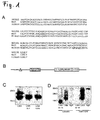
- Rat RNF 122 that was named LINA consists of 155 amino acids was generated and sequenced. Amino acid sequences of rat and mouse RNF 122 were identical and human RNF 122 differed just in five amino acids at the c-terminus of the protein ( Figure 1 A) . Bioinformatic sequence analysis predicted a ring finger domain (C 3 H 2 C 3 type) between amino acids 93 and 133, a putative transmembrane domain between amino acids 37 and 59 and a glycosylation site at N20 ( Figure 1A, B ) .
- Exogenously expressed HA-tagged-LINA ran in gels under denaturating conditions in two bands at 19 and 23 kDa ( Figure 1 C) , pointing to a posttranslational glycosylation of the protein.
- Incubation of LINA with N-glycosidase or a substitution of amino acid asparagine (N20) with glutamine (Q) eliminated the upper band, revealing that LINA is glycosylated at N20 ( Fig. 1 C) and that the two bands represent the glycosylated and unglycosylated form of the protein.
- Both HA-tagged and GFP-tagged LINA-fusionproteins showed a granular staining pattern when expressed in HEK293 or PC12 cells with a partial colocalization with the Trans-Golgi-network marker TGN38 ( Figure 2-I , C-F).
- TGN38 Trans-Golgi-network marker
- LINA-fusionproteins LINA-fusionproteins have been overexpressed in PC12 cells and subcellular markers have been used. LINA did not co-lolocalize with lysosomes, detected by lysotracker (Invitrogen) ( Figure 2-II, A, B, C ) or peroxisomes, which became detectable after overexpression of SKL-GFP fusionprotein ( Figure 2-II, D, E, F ). LINA partially colocalized with BACE1-GFP-fusionproteins, indicating that LINA is expressed in vesicles of the secretory pathway ( Figure 2-II, G, H, I ) .
- LINA is widely expressed in the nervous system
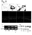
- LINA-mRNA was detected in retina, cerebellum, spinal cord, brain cortex, dorsal root ganglia and spleen.
- Low expression levels of LINA were detected in skeletal muscle, heart, kidney and liver ( Figure 3 , B, C).
- In situ -hybridisation of embryonic rats (E16) showed LINA expression in the subventricular zone of the brain, cerebellum, retina and lung. LINA-probes did not significantly detect other embryonic organs ( Figure 3, A ).
- LINA expression is upregulated in regenerating RGCs
- Retinal LINA expression was assessed by RT-PCR, in-situ -hybridization, Western-blot and immunohistochemistry:
- RT-PCR detected LINA in naive retinas.
- Levels of the ring finger protein were slightly increased after ONC and elevated further after additional LI treatment confirming the expression results of our microarray analysis ( Fischer D, Petkova V, Thanos S, Benowitz LI (2004b) Switching mature retinal ganglion cells to a robust growth state in vivo: gene expression and synergy with RhoA inactivation. J Neurosci 24:8726-8740 ) ( Figure 4, L ).
- LI without ONC increased LINA expression, too.
- In situ -hybridization using two distinct DNA-probes detected LINA in retinas that had been prior exposed to LI, whereas signals in untreated retinas were low to absent. Signals from radioactive probes accumulated in the RGC layer as determined by dipped sections ( Figure 4 , A, B, C).
- RGCs showed a vesicular staining pattern with a pronounced staining of the cell body and dendrites. Axons in the fiber layer were only weakly stained with the antibody.
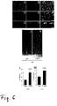
- LINA expression is essential for neurite outgrowth in PC12 cells and overexpression promotes outgrowth
- PC12 cells In order to assess a potential a role of LINA in neurite outgrowth we initially used PC12 cells (pheochromocytoma cells). Untreated PC12 cells express LINA as determined by RT-PCR, immunocytochemistry and Western-blot analysis ( Figure 5 , A-D). PC12 cells differentiate and extended neurites in response to the nerve growth factor (NGF) that was added to the medium (50 ng/ml). Expression levels of LINA increased in differentiating PC12 cells after exposure to NGF about five-fold after 24 h as measured by quantitative RT-PCR. The increase of LINA expression in PC12 cells was verified on the protein level by immunocytochemistry and Western-blotting using an affinity purified peptide-antibody against LINA ( Figure 5 , A-E). Next to the cell body of PC12 cells LINA was also detected in the neurites and growth cones.
- NGF nerve growth factor
- Adeno Associated Viruses AAV
- LINA WT Adeno Associated Viruses
- LINA overexpressing enhances RGC survival and promotes axon regeneration of into the injured optic nerve
- LINA WT overexpression enhanced axon regeneration much stronger than RGC survival it was tested next whether LINA WT could increase axon regeneration further when combined with the strong neuroprotective effects of LI.
- Two weeks after nerve crush and LI animals expressing LINA WT extended 2.1 times more axons > 1 mm beyond the injury site than LI treated controls expressing GFP alone.
- the survival of RGCs in LINA WT overexpressing retinas exposed to LI was about 23 % increased compared to controls expressing just GFP and treated with LI only ( Figure 7 , C-G).
Landscapes
- Health & Medical Sciences (AREA)
- Life Sciences & Earth Sciences (AREA)
- Chemical & Material Sciences (AREA)
- General Health & Medical Sciences (AREA)
- Medicinal Chemistry (AREA)
- Organic Chemistry (AREA)
- Veterinary Medicine (AREA)
- Public Health (AREA)
- Animal Behavior & Ethology (AREA)
- Pharmacology & Pharmacy (AREA)
- Engineering & Computer Science (AREA)
- Bioinformatics & Cheminformatics (AREA)
- Zoology (AREA)
- General Chemical & Material Sciences (AREA)
- Chemical Kinetics & Catalysis (AREA)
- Proteomics, Peptides & Aminoacids (AREA)
- Nuclear Medicine, Radiotherapy & Molecular Imaging (AREA)
- Gastroenterology & Hepatology (AREA)
- Ophthalmology & Optometry (AREA)
- Genetics & Genomics (AREA)
- Molecular Biology (AREA)
- Toxicology (AREA)
- Epidemiology (AREA)
- Immunology (AREA)
- Biochemistry (AREA)
- Biophysics (AREA)
- Marine Sciences & Fisheries (AREA)
- Biomedical Technology (AREA)
- Rheumatology (AREA)
- Pain & Pain Management (AREA)
- Neurosurgery (AREA)
- Neurology (AREA)
- Medicines That Contain Protein Lipid Enzymes And Other Medicines (AREA)
- Micro-Organisms Or Cultivation Processes Thereof (AREA)
Priority Applications (3)
| Application Number | Priority Date | Filing Date | Title |
|---|---|---|---|
| EP08019676A EP2186821A1 (de) | 2008-11-11 | 2008-11-11 | Protein mit Wirkung zur Förderung des axonalen Wachstums von Neuronen des zentralen Nervensystems |
| US13/128,790 US20120114619A1 (en) | 2008-11-11 | 2009-11-11 | Protein with promoting effects for axonal growth of neurons of central nervous system |
| PCT/EP2009/008040 WO2010054807A1 (en) | 2008-11-11 | 2009-11-11 | Protein with promoting effects for axonal growth of neurons of central nervous system |
Applications Claiming Priority (1)
| Application Number | Priority Date | Filing Date | Title |
|---|---|---|---|
| EP08019676A EP2186821A1 (de) | 2008-11-11 | 2008-11-11 | Protein mit Wirkung zur Förderung des axonalen Wachstums von Neuronen des zentralen Nervensystems |
Publications (1)
| Publication Number | Publication Date |
|---|---|
| EP2186821A1 true EP2186821A1 (de) | 2010-05-19 |
Family
ID=40512479
Family Applications (1)
| Application Number | Title | Priority Date | Filing Date |
|---|---|---|---|
| EP08019676A Withdrawn EP2186821A1 (de) | 2008-11-11 | 2008-11-11 | Protein mit Wirkung zur Förderung des axonalen Wachstums von Neuronen des zentralen Nervensystems |
Country Status (3)
| Country | Link |
|---|---|
| US (1) | US20120114619A1 (de) |
| EP (1) | EP2186821A1 (de) |
| WO (1) | WO2010054807A1 (de) |
Cited By (1)
| Publication number | Priority date | Publication date | Assignee | Title |
|---|---|---|---|---|
| CN104592352A (zh) * | 2015-01-09 | 2015-05-06 | 东南大学 | 与缺血性脑卒中组织特异性结合的hgg多肽及其应用 |
Citations (1)
| Publication number | Priority date | Publication date | Assignee | Title |
|---|---|---|---|---|
| WO2008016356A2 (en) * | 2006-08-02 | 2008-02-07 | Genizon Biosciences | Genemap of the human genes associated with psoriasis |
Family Cites Families (1)
| Publication number | Priority date | Publication date | Assignee | Title |
|---|---|---|---|---|
| JP4867018B2 (ja) * | 2006-03-22 | 2012-02-01 | 富士フイルム株式会社 | 癌の検出方法および抑制方法 |
-
2008
- 2008-11-11 EP EP08019676A patent/EP2186821A1/de not_active Withdrawn
-
2009
- 2009-11-11 WO PCT/EP2009/008040 patent/WO2010054807A1/en active Application Filing
- 2009-11-11 US US13/128,790 patent/US20120114619A1/en not_active Abandoned
Patent Citations (1)
| Publication number | Priority date | Publication date | Assignee | Title |
|---|---|---|---|---|
| WO2008016356A2 (en) * | 2006-08-02 | 2008-02-07 | Genizon Biosciences | Genemap of the human genes associated with psoriasis |
Non-Patent Citations (27)
| Title |
|---|
| AURICCHIO A ET AL.: "Isolation of highly infectious and pure adeno-associated virus type 2 vectors with a single-step gravity-flow column", HUMAN GENE THERAPY, vol. 12, 2001, pages 71 - 76 |
| BAHR M; VANSELOW J; THANOS S.: "In vitro regeneration of adult rat ganglion cell axons from retinal explants", EXP BRAIN RES., vol. 73, no. 2, 1988, pages 393 - 401 |
| BERKELAAR M ET AL.: "Axotomy results in delayed death and apoptosis of retinal ganglion cells in adult rats", J NEUROSCI, vol. 14, 1994, pages 4368 - 4374 |
| BOECKERS TM ET AL.: "Proline-rich syn- apse-associated proteins ProSAP1 and ProSAP2 interact with synaptic proteins of the SAPAP/GKAP family", BIO CHEMICAL & BIOPHYSICAL RESEARCH COMMUNICATIONS, vol. 264, 1999, pages 247 - 252 |
| CHEN MS ET AL.: "Nogo-A is a myelin-associated neurite outgrowth inhibitor and an antigen for monoclonal antibody IN-1", NATURE, vol. 403, 2000, pages 434 - 439 |
| DATABASE EMBL [online] 14 January 2004 (2004-01-14), "AGENCOURT_17636391 NIH_MGC_235 Rattus norvegicus cDNA clone IMAGE:7108026 5', mRNA sequence.", XP002524146, retrieved from EBI accession no. EMBL:CK483344 Database accession no. CK483344 * |
| DATABASE Geneseq [online] 21 August 2008 (2008-08-21), "Psoriasis associated human protein SEQ ID NO: 18507.", XP002524143, retrieved from EBI accession no. GSP:ARY77640 Database accession no. ARY77640 * |
| DATABASE UniProt [online] 1 March 2003 (2003-03-01), "RecName: Full=RING finger protein 122;", XP002524144, retrieved from EBI accession no. UNIPROT:Q8BP31 Database accession no. Q8BP31 * |
| DATABASE UniProt [online] 25 July 2006 (2006-07-25), "RecName: Full=RING finger protein 122;", XP002524145, retrieved from EBI accession no. UNIPROT:Q9H9V4 Database accession no. Q9H9V4 * |
| DIPOLO R; BEAUGE L: "Differential up-regulation of Na+-Ca2+ exchange by phosphoarginine and ATP in dialysed squid axons", J PHYSIOL, vol. 507, no. 3, 1998, pages 737 - 747 |
| FISCHER D ET AL.: "Crystallins of the beta/gamma-superfamily mimic the effects of lens injury and promote axon regeneration", MOLECULAR CELLULAR NEUROSCIENCE, 2008, Retrieved from the Internet <URL:doi:10.1016/j.mcn.2007.11.-002> |
| FISCHER D ET AL.: "Switching mature retinal ganglion cells to a robust growth state in vivo: gene expression and synergy with RhoA inactivation", J NEUROSCI, vol. 24, 2004, pages 8726 - 8740 |
| FISCHER D.: "CNTF, a key factor mediating the benefical effects of inflammatory reactions in the eye", BRAIN, vol. 131, no. 6, 20 February 2008 (2008-02-20), pages E97 |
| FISCHER D; HE Z; BENOWITZ LI: "Counteracting the Nogo receptor enhances optic nerve regeneration if retinal ganglion cells are in an active growth state", J NEUROSCI, vol. 24, 2004, pages 1646 - 1651 |
| FISCHER D; HEIDUSCHKA P; THANOS S: "Lens-injury-stimulated axonal regeneration throughout the optic pathway of adult rats", EXP NEUROL, vol. 172, 2001, pages 257 - 272 |
| FISCHER D; PAVLIDIS M; THANOS S: "Cataractogenic lens injury prevents traumatic ganglion cell death and promotes axonal regeneration both in vivo and in culture", INVEST OPHTHALMOL VIS SCI, vol. 41, 2000, pages 3943 - 3954 |
| GRANDPRE T ET AL.: "Identification of the Nogo inhibitor of axon regeneration as a Reticulon protein", NATURE, vol. 403, 2000, pages 439 - 444 |
| HAUK TG ET AL.: "Neuroprotective and axon growth promoting effects of intraocular inflammation do not depend on oncomodulin or the presence of large numbers of activated macrophages", EXP NEUROL., vol. 209, no. 2, 29 September 2007 (2007-09-29), pages 469 - 82 |
| LEAVER SG ET AL.: "AAV-mediated expression of CNTF promotes long-term survival and regeneration of adult rat retinal ganglion cells", GENE THER, vol. 13, 2006, pages 1328 - 1341 |
| LEE J ET AL.: "LINA: a new ring finger protein promoting axon regeneration", ABSTRACT FOR ANNUAL MEETING OF THE SOCIETY OF NEUROSCIENC, 2006 |
| LEON S ET AL.: "Lens injury stimulates axon regeneration in the mature rat optic nerve", J NEUROSCI, vol. 20, 2000, pages 4615 - 4626 |
| MCKERRACHER L ET AL.: "Identification of myelin-associated glycoprotein as a major myelin- derived inhibitor of neurite growth", NEURON, vol. 13, 1994, pages 805 - 811 |
| MULLER A; HAUK TG; FISCHER D: "Astro cyte-derived CNTF switches mature RGCs to a regenerative state following inflammatory stimulation", BRAIN, vol. 130, 2007, pages 3308 - 3320 |
| SAITO SAKAE ET AL: "Gene expression profiling of cerebellar development with high-throughput functional analysis", PHYSIOLOGICAL GENOMICS, vol. 22, no. 1, June 2005 (2005-06-01), pages 8 - 13, XP002524142, ISSN: 1094-8341 * |
| WANG KC ET AL.: "Oli- godendrocyte-myelin glycoprotein is a Nogo receptor ligand that inhibits neurite outgrowth", NATURE, vol. 417, 2002, pages 941 - 944 |
| WANG L ET AL: "CELL-BASED SCREENING AND VALIDATION OF HUMAN NOVEL GENES ASSOCIATED WITH CELL VIABILITY", JOURNAL OF BIOMOLECULAR SCREENING, LARCHMONT, NY, US, vol. 11, no. 4, 1 January 2006 (2006-01-01), pages 369 - 376, XP009084149, ISSN: 1087-0571 * |
| ZHOU Y ET AL.: "Activation of the extracellular signal- regulated kinase 1/2 pathway by AAV gene transfer protects retinal ganglion cells in glaucoma", MOL THER, vol. 12, 2005, pages 402 - 412 |
Cited By (2)
| Publication number | Priority date | Publication date | Assignee | Title |
|---|---|---|---|---|
| CN104592352A (zh) * | 2015-01-09 | 2015-05-06 | 东南大学 | 与缺血性脑卒中组织特异性结合的hgg多肽及其应用 |
| CN104592352B (zh) * | 2015-01-09 | 2017-08-15 | 东南大学 | 与缺血性脑卒中组织特异性结合的hgg多肽及其应用 |
Also Published As
| Publication number | Publication date |
|---|---|
| US20120114619A1 (en) | 2012-05-10 |
| WO2010054807A1 (en) | 2010-05-20 |
Similar Documents
| Publication | Publication Date | Title |
|---|---|---|
| Eisenstat et al. | DLX‐1, DLX‐2, and DLX‐5 expression define distinct stages of basal forebrain differentiation | |
| Wenz et al. | RETRACTED: mTERF2 Regulates Oxidative Phosphorylation by Modulating mtDNA Transcription | |
| Hu et al. | Identification and characterization of a novel Nogo‐interacting mitochondrial protein (NIMP) | |
| CN110809476B (zh) | 用于神经退行性病症或中风的治疗的基因构建体 | |
| Tsai et al. | Recombinant adeno-associated virus vector expressing glial cell line-derived neurotrophic factor reduces ischemia-induced damage | |
| EA010055B1 (ru) | ВЫДЕЛЕННАЯ НУКЛЕИНОВАЯ КИСЛОТА, КОДИРУЮЩАЯ ПОЛИПЕПТИД Sp35, ПОЛИПЕПТИД Sp35 И СПОСОБЫ ПРИМЕНЕНИЯ НУКЛЕИНОВОЙ КИСЛОТЫ И ПОЛИПЕПТИДА | |
| WO2005087268A1 (ja) | 軸索再生促進剤 | |
| CA2613028A1 (en) | Peptidic antagonists of class iii semaphorins/neuropilins complexes | |
| He et al. | GFP-tagged expression and immunohistochemical studies to determine the subcellular localization of the tubby gene family members | |
| US20060159681A1 (en) | Compositions and methods to inhibit cell loss by using inhibitors of BAG | |
| Adamus et al. | Anti-apoptotic effects of CNTF gene transfer on photoreceptor degeneration in experimental antibody-induced retinopathy | |
| US20220040236A1 (en) | Compositions and methods of treatment of vision loss through generation of rod photoreceptors from müller glial cells | |
| EP2186821A1 (de) | Protein mit Wirkung zur Förderung des axonalen Wachstums von Neuronen des zentralen Nervensystems | |
| CA2239753C (en) | Novel semaphorin z and gene encoding the same | |
| CN115475247B (zh) | β2-微球蛋白或其抑制剂的制药用途 | |
| CN112384248A (zh) | 预防或治疗突触核蛋白病的包含c-src的表达或活性抑制剂的药物组合物 | |
| US20100016221A1 (en) | Method of degrading protein by chaperone-mediated autophagy | |
| US20050153887A1 (en) | Compositions for inducing cell growth and differentiation and methods of using same | |
| HU230294B1 (hu) | IL-18-inhibitorok alkalmazása központi idegrendszeri sérülések kezelésére vagy megelőzésére | |
| EP1130096A1 (de) | Proliferationshemmender Transkriptionsfaktor (PATF) und seine Verwendung | |
| US20020119944A1 (en) | Use of Ulip-and/or Ulip2 in the treatment of myelin disorders | |
| JP2001511344A (ja) | 転写因子Brn−3aの使用 | |
| Reis et al. | Prospects of TAT-mediated protein therapy for fragile X syndrome | |
| KR20170035089A (ko) | Tim-3을 표적으로 하는 뇌손상 질환 치료용 조성물 및 이의 스크리닝 방법 | |
| WO1998015628A1 (fr) | Nouveau gene de semaphorine: la semaphorine w |
Legal Events
| Date | Code | Title | Description |
|---|---|---|---|
| PUAI | Public reference made under article 153(3) epc to a published international application that has entered the european phase |
Free format text: ORIGINAL CODE: 0009012 |
|
| AK | Designated contracting states |
Kind code of ref document: A1 Designated state(s): AT BE BG CH CY CZ DE DK EE ES FI FR GB GR HR HU IE IS IT LI LT LU LV MC MT NL NO PL PT RO SE SI SK TR |
|
| AX | Request for extension of the european patent |
Extension state: AL BA MK RS |
|
| 17P | Request for examination filed |
Effective date: 20101119 |
|
| AKX | Designation fees paid |
Designated state(s): AT BE BG CH CY CZ DE DK EE ES FI FR GB GR HR HU IE IS IT LI LT LU LV MC MT NL NO PL PT RO SE SI SK TR |
|
| 17Q | First examination report despatched |
Effective date: 20110124 |
|
| STAA | Information on the status of an ep patent application or granted ep patent |
Free format text: STATUS: THE APPLICATION IS DEEMED TO BE WITHDRAWN |
|
| 18D | Application deemed to be withdrawn |
Effective date: 20150602 |