EP2171043B1 - Treatment of diseases and disorders using self-renewing colony forming cells cultured and expanded in vitro - Google Patents
Treatment of diseases and disorders using self-renewing colony forming cells cultured and expanded in vitro Download PDFInfo
- Publication number
- EP2171043B1 EP2171043B1 EP08768506.1A EP08768506A EP2171043B1 EP 2171043 B1 EP2171043 B1 EP 2171043B1 EP 08768506 A EP08768506 A EP 08768506A EP 2171043 B1 EP2171043 B1 EP 2171043B1
- Authority
- EP
- European Patent Office
- Prior art keywords
- cells
- cell
- human
- abm
- tissue
- Prior art date
- Legal status (The legal status is an assumption and is not a legal conclusion. Google has not performed a legal analysis and makes no representation as to the accuracy of the status listed.)
- Active
Links
- 238000011282 treatment Methods 0.000 title claims description 57
- 238000000338 in vitro Methods 0.000 title claims description 42
- 230000001332 colony forming effect Effects 0.000 title claims description 17
- 208000037765 diseases and disorders Diseases 0.000 title claims description 8
- 210000004027 cell Anatomy 0.000 claims description 403
- 241000282414 Homo sapiens Species 0.000 claims description 116
- 210000001519 tissue Anatomy 0.000 claims description 76
- 208000037265 diseases, disorders, signs and symptoms Diseases 0.000 claims description 36
- 210000000056 organ Anatomy 0.000 claims description 30
- 210000001082 somatic cell Anatomy 0.000 claims description 29
- 210000001185 bone marrow Anatomy 0.000 claims description 27
- 230000004069 differentiation Effects 0.000 claims description 25
- 238000002347 injection Methods 0.000 claims description 25
- 239000007924 injection Substances 0.000 claims description 25
- 230000006378 damage Effects 0.000 claims description 19
- 208000035475 disorder Diseases 0.000 claims description 18
- 210000002901 mesenchymal stem cell Anatomy 0.000 claims description 18
- QVGXLLKOCUKJST-UHFFFAOYSA-N atomic oxygen Chemical compound [O] QVGXLLKOCUKJST-UHFFFAOYSA-N 0.000 claims description 17
- 201000010099 disease Diseases 0.000 claims description 17
- 229910052760 oxygen Inorganic materials 0.000 claims description 17
- 239000001301 oxygen Substances 0.000 claims description 17
- 230000033115 angiogenesis Effects 0.000 claims description 15
- 230000010412 perfusion Effects 0.000 claims description 15
- 239000003814 drug Substances 0.000 claims description 13
- 230000000451 tissue damage Effects 0.000 claims description 12
- 210000000130 stem cell Anatomy 0.000 claims description 11
- 231100000827 tissue damage Toxicity 0.000 claims description 11
- OYPRJOBELJOOCE-UHFFFAOYSA-N Calcium Chemical compound [Ca] OYPRJOBELJOOCE-UHFFFAOYSA-N 0.000 claims description 10
- 239000011575 calcium Substances 0.000 claims description 10
- 229910052791 calcium Inorganic materials 0.000 claims description 10
- 230000000694 effects Effects 0.000 claims description 10
- 210000002950 fibroblast Anatomy 0.000 claims description 9
- 238000010899 nucleation Methods 0.000 claims description 9
- 230000002138 osteoinductive effect Effects 0.000 claims description 8
- 101000994378 Homo sapiens Integrin alpha-3 Proteins 0.000 claims description 7
- 101000800116 Homo sapiens Thy-1 membrane glycoprotein Proteins 0.000 claims description 7
- 102100032819 Integrin alpha-3 Human genes 0.000 claims description 7
- 102100033523 Thy-1 membrane glycoprotein Human genes 0.000 claims description 7
- 230000001464 adherent effect Effects 0.000 claims description 7
- 210000003958 hematopoietic stem cell Anatomy 0.000 claims description 6
- 210000004409 osteocyte Anatomy 0.000 claims description 6
- 102100031573 Hematopoietic progenitor cell antigen CD34 Human genes 0.000 claims description 5
- 101000777663 Homo sapiens Hematopoietic progenitor cell antigen CD34 Proteins 0.000 claims description 5
- 230000000926 neurological effect Effects 0.000 claims description 5
- 101000691214 Haloarcula marismortui (strain ATCC 43049 / DSM 3752 / JCM 8966 / VKM B-1809) 50S ribosomal protein L44e Proteins 0.000 claims description 4
- 210000001789 adipocyte Anatomy 0.000 claims description 4
- 210000001612 chondrocyte Anatomy 0.000 claims description 4
- 210000001671 embryonic stem cell Anatomy 0.000 claims description 4
- 102100032912 CD44 antigen Human genes 0.000 claims description 3
- 101000868273 Homo sapiens CD44 antigen Proteins 0.000 claims description 3
- 102000003729 Neprilysin Human genes 0.000 claims description 3
- 108090000028 Neprilysin Proteins 0.000 claims description 3
- 230000004888 barrier function Effects 0.000 claims description 3
- 210000002798 bone marrow cell Anatomy 0.000 claims description 3
- 206010012601 diabetes mellitus Diseases 0.000 claims description 3
- 101000738771 Homo sapiens Receptor-type tyrosine-protein phosphatase C Proteins 0.000 claims description 2
- 102100033467 L-selectin Human genes 0.000 claims description 2
- 102100037422 Receptor-type tyrosine-protein phosphatase C Human genes 0.000 claims description 2
- 210000003041 ligament Anatomy 0.000 claims description 2
- 230000008816 organ damage Effects 0.000 claims description 2
- 230000008817 pulmonary damage Effects 0.000 claims description 2
- 208000018522 Gastrointestinal disease Diseases 0.000 claims 2
- 208000029578 Muscle disease Diseases 0.000 claims 2
- 208000010643 digestive system disease Diseases 0.000 claims 2
- 208000018685 gastrointestinal system disease Diseases 0.000 claims 2
- 102100022749 Aminopeptidase N Human genes 0.000 claims 1
- 101000757160 Homo sapiens Aminopeptidase N Proteins 0.000 claims 1
- 101001018097 Homo sapiens L-selectin Proteins 0.000 claims 1
- 101000622304 Homo sapiens Vascular cell adhesion protein 1 Proteins 0.000 claims 1
- 208000029549 Muscle injury Diseases 0.000 claims 1
- 208000021642 Muscular disease Diseases 0.000 claims 1
- 102100023543 Vascular cell adhesion protein 1 Human genes 0.000 claims 1
- 210000004504 adult stem cell Anatomy 0.000 claims 1
- 230000003683 cardiac damage Effects 0.000 claims 1
- 230000005779 cell damage Effects 0.000 claims 1
- 230000001900 immune effect Effects 0.000 claims 1
- 230000006749 inflammatory damage Effects 0.000 claims 1
- 238000001990 intravenous administration Methods 0.000 claims 1
- 230000003239 periodontal effect Effects 0.000 claims 1
- 210000002027 skeletal muscle Anatomy 0.000 claims 1
- 230000037380 skin damage Effects 0.000 claims 1
- 108010035532 Collagen Proteins 0.000 description 73
- 102000008186 Collagen Human genes 0.000 description 73
- 229920001436 collagen Polymers 0.000 description 73
- 239000000203 mixture Substances 0.000 description 60
- 238000000034 method Methods 0.000 description 59
- 208000027418 Wounds and injury Diseases 0.000 description 51
- 206010052428 Wound Diseases 0.000 description 48
- IJGRMHOSHXDMSA-UHFFFAOYSA-N Atomic nitrogen Chemical compound N#N IJGRMHOSHXDMSA-UHFFFAOYSA-N 0.000 description 40
- 239000000243 solution Substances 0.000 description 39
- 241000700159 Rattus Species 0.000 description 38
- 108060008682 Tumor Necrosis Factor Proteins 0.000 description 37
- 102000000852 Tumor Necrosis Factor-alpha Human genes 0.000 description 37
- 239000000499 gel Substances 0.000 description 37
- 108090000623 proteins and genes Proteins 0.000 description 35
- 102000004169 proteins and genes Human genes 0.000 description 34
- 210000003491 skin Anatomy 0.000 description 33
- 235000018102 proteins Nutrition 0.000 description 32
- 102000004127 Cytokines Human genes 0.000 description 31
- 108090000695 Cytokines Proteins 0.000 description 31
- 210000003819 peripheral blood mononuclear cell Anatomy 0.000 description 31
- 239000000047 product Substances 0.000 description 30
- 239000003981 vehicle Substances 0.000 description 29
- 239000003636 conditioned culture medium Substances 0.000 description 26
- 230000004217 heart function Effects 0.000 description 26
- 239000007787 solid Substances 0.000 description 26
- 230000000735 allogeneic effect Effects 0.000 description 25
- 210000002808 connective tissue Anatomy 0.000 description 25
- ZDXPYRJPNDTMRX-VKHMYHEASA-N L-glutamine Chemical compound OC(=O)[C@@H](N)CCC(N)=O ZDXPYRJPNDTMRX-VKHMYHEASA-N 0.000 description 23
- 230000000747 cardiac effect Effects 0.000 description 23
- 239000011159 matrix material Substances 0.000 description 23
- 241001465754 Metazoa Species 0.000 description 21
- 238000003556 assay Methods 0.000 description 21
- 239000006285 cell suspension Substances 0.000 description 21
- 210000002216 heart Anatomy 0.000 description 21
- 239000007788 liquid Substances 0.000 description 21
- 238000004519 manufacturing process Methods 0.000 description 21
- 206010061216 Infarction Diseases 0.000 description 20
- 229910052757 nitrogen Inorganic materials 0.000 description 20
- 239000000126 substance Substances 0.000 description 20
- 210000004369 blood Anatomy 0.000 description 19
- 239000008280 blood Substances 0.000 description 19
- 229930182816 L-glutamine Natural products 0.000 description 18
- 230000006872 improvement Effects 0.000 description 18
- 230000007574 infarction Effects 0.000 description 18
- 239000002609 medium Substances 0.000 description 18
- 108010002350 Interleukin-2 Proteins 0.000 description 16
- 102000000588 Interleukin-2 Human genes 0.000 description 16
- 238000004113 cell culture Methods 0.000 description 16
- 239000006228 supernatant Substances 0.000 description 16
- 238000001727 in vivo Methods 0.000 description 15
- 239000012528 membrane Substances 0.000 description 15
- 230000001172 regenerating effect Effects 0.000 description 15
- 230000008929 regeneration Effects 0.000 description 15
- 238000011069 regeneration method Methods 0.000 description 15
- 230000002861 ventricular Effects 0.000 description 15
- 239000006144 Dulbecco’s modified Eagle's medium Substances 0.000 description 14
- 206010016654 Fibrosis Diseases 0.000 description 14
- 206010000891 acute myocardial infarction Diseases 0.000 description 14
- 210000000988 bone and bone Anatomy 0.000 description 14
- 230000004761 fibrosis Effects 0.000 description 14
- 208000010125 myocardial infarction Diseases 0.000 description 14
- 102000045246 noggin Human genes 0.000 description 14
- 108700007229 noggin Proteins 0.000 description 14
- 230000008439 repair process Effects 0.000 description 14
- 239000001963 growth medium Substances 0.000 description 13
- 238000013507 mapping Methods 0.000 description 13
- 238000005259 measurement Methods 0.000 description 13
- 239000013642 negative control Substances 0.000 description 13
- 230000002829 reductive effect Effects 0.000 description 13
- 230000001225 therapeutic effect Effects 0.000 description 13
- QTBSBXVTEAMEQO-UHFFFAOYSA-N Acetic acid Chemical compound CC(O)=O QTBSBXVTEAMEQO-UHFFFAOYSA-N 0.000 description 12
- IAZDPXIOMUYVGZ-UHFFFAOYSA-N Dimethylsulphoxide Chemical compound CS(C)=O IAZDPXIOMUYVGZ-UHFFFAOYSA-N 0.000 description 12
- HEMHJVSKTPXQMS-UHFFFAOYSA-M Sodium hydroxide Chemical compound [OH-].[Na+] HEMHJVSKTPXQMS-UHFFFAOYSA-M 0.000 description 12
- 238000004458 analytical method Methods 0.000 description 12
- 230000001143 conditioned effect Effects 0.000 description 12
- 230000002500 effect on skin Effects 0.000 description 12
- 238000012360 testing method Methods 0.000 description 12
- JKMHFZQWWAIEOD-UHFFFAOYSA-N 2-[4-(2-hydroxyethyl)piperazin-1-yl]ethanesulfonic acid Chemical compound OCC[NH+]1CCN(CCS([O-])(=O)=O)CC1 JKMHFZQWWAIEOD-UHFFFAOYSA-N 0.000 description 11
- 239000007995 HEPES buffer Substances 0.000 description 11
- 206010061218 Inflammation Diseases 0.000 description 11
- 102100026018 Interleukin-1 receptor antagonist protein Human genes 0.000 description 11
- 101710144554 Interleukin-1 receptor antagonist protein Proteins 0.000 description 11
- 239000012980 RPMI-1640 medium Substances 0.000 description 11
- 230000006052 T cell proliferation Effects 0.000 description 11
- 239000000306 component Substances 0.000 description 11
- 239000003102 growth factor Substances 0.000 description 11
- 230000004054 inflammatory process Effects 0.000 description 11
- 208000014674 injury Diseases 0.000 description 11
- 230000001404 mediated effect Effects 0.000 description 11
- 230000002107 myocardial effect Effects 0.000 description 11
- 239000008194 pharmaceutical composition Substances 0.000 description 11
- 102000005962 receptors Human genes 0.000 description 11
- 108020003175 receptors Proteins 0.000 description 11
- 238000002560 therapeutic procedure Methods 0.000 description 11
- 230000001228 trophic effect Effects 0.000 description 11
- MZOFCQQQCNRIBI-VMXHOPILSA-N (3s)-4-[[(2s)-1-[[(2s)-1-[[(1s)-1-carboxy-2-hydroxyethyl]amino]-4-methyl-1-oxopentan-2-yl]amino]-5-(diaminomethylideneamino)-1-oxopentan-2-yl]amino]-3-[[2-[[(2s)-2,6-diaminohexanoyl]amino]acetyl]amino]-4-oxobutanoic acid Chemical compound OC[C@@H](C(O)=O)NC(=O)[C@H](CC(C)C)NC(=O)[C@H](CCCN=C(N)N)NC(=O)[C@H](CC(O)=O)NC(=O)CNC(=O)[C@@H](N)CCCCN MZOFCQQQCNRIBI-VMXHOPILSA-N 0.000 description 10
- 102100023995 Beta-nerve growth factor Human genes 0.000 description 10
- 108090001005 Interleukin-6 Proteins 0.000 description 10
- 102000004889 Interleukin-6 Human genes 0.000 description 10
- 210000001744 T-lymphocyte Anatomy 0.000 description 10
- 102000005789 Vascular Endothelial Growth Factors Human genes 0.000 description 10
- 108010019530 Vascular Endothelial Growth Factors Proteins 0.000 description 10
- 239000012141 concentrate Substances 0.000 description 10
- 239000012737 fresh medium Substances 0.000 description 10
- NOESYZHRGYRDHS-UHFFFAOYSA-N insulin Chemical compound N1C(=O)C(NC(=O)C(CCC(N)=O)NC(=O)C(CCC(O)=O)NC(=O)C(C(C)C)NC(=O)C(NC(=O)CN)C(C)CC)CSSCC(C(NC(CO)C(=O)NC(CC(C)C)C(=O)NC(CC=2C=CC(O)=CC=2)C(=O)NC(CCC(N)=O)C(=O)NC(CC(C)C)C(=O)NC(CCC(O)=O)C(=O)NC(CC(N)=O)C(=O)NC(CC=2C=CC(O)=CC=2)C(=O)NC(CSSCC(NC(=O)C(C(C)C)NC(=O)C(CC(C)C)NC(=O)C(CC=2C=CC(O)=CC=2)NC(=O)C(CC(C)C)NC(=O)C(C)NC(=O)C(CCC(O)=O)NC(=O)C(C(C)C)NC(=O)C(CC(C)C)NC(=O)C(CC=2NC=NC=2)NC(=O)C(CO)NC(=O)CNC2=O)C(=O)NCC(=O)NC(CCC(O)=O)C(=O)NC(CCCNC(N)=N)C(=O)NCC(=O)NC(CC=3C=CC=CC=3)C(=O)NC(CC=3C=CC=CC=3)C(=O)NC(CC=3C=CC(O)=CC=3)C(=O)NC(C(C)O)C(=O)N3C(CCC3)C(=O)NC(CCCCN)C(=O)NC(C)C(O)=O)C(=O)NC(CC(N)=O)C(O)=O)=O)NC(=O)C(C(C)CC)NC(=O)C(CO)NC(=O)C(C(C)O)NC(=O)C1CSSCC2NC(=O)C(CC(C)C)NC(=O)C(NC(=O)C(CCC(N)=O)NC(=O)C(CC(N)=O)NC(=O)C(NC(=O)C(N)CC=1C=CC=CC=1)C(C)C)CC1=CN=CN1 NOESYZHRGYRDHS-UHFFFAOYSA-N 0.000 description 10
- 108010070145 interleukin-18 binding protein Proteins 0.000 description 10
- 102000044166 interleukin-18 binding protein Human genes 0.000 description 10
- 239000003226 mitogen Substances 0.000 description 10
- XEPXGZZWVKNRGS-GQYPCLOQSA-N n-[(3r,4s,5s,6r)-3,4,5-trihydroxy-6-(hydroxymethyl)oxan-2-yl]octanamide Chemical compound CCCCCCCC(=O)NC1O[C@H](CO)[C@@H](O)[C@H](O)[C@H]1O XEPXGZZWVKNRGS-GQYPCLOQSA-N 0.000 description 10
- 239000008188 pellet Substances 0.000 description 10
- 210000002966 serum Anatomy 0.000 description 10
- 239000006143 cell culture medium Substances 0.000 description 9
- 230000008859 change Effects 0.000 description 9
- 230000006735 deficit Effects 0.000 description 9
- 210000005003 heart tissue Anatomy 0.000 description 9
- 230000028993 immune response Effects 0.000 description 9
- 230000002757 inflammatory effect Effects 0.000 description 9
- 230000007774 longterm Effects 0.000 description 9
- 230000017423 tissue regeneration Effects 0.000 description 9
- 241000283690 Bos taurus Species 0.000 description 8
- 108010008951 Chemokine CXCL12 Proteins 0.000 description 8
- NWIBSHFKIJFRCO-WUDYKRTCSA-N Mytomycin Chemical compound C1N2C(C(C(C)=C(N)C3=O)=O)=C3[C@@H](COC(N)=O)[C@@]2(OC)[C@@H]2[C@H]1N2 NWIBSHFKIJFRCO-WUDYKRTCSA-N 0.000 description 8
- 108010025020 Nerve Growth Factor Proteins 0.000 description 8
- UIIMBOGNXHQVGW-UHFFFAOYSA-M Sodium bicarbonate Chemical compound [Na+].OC([O-])=O UIIMBOGNXHQVGW-UHFFFAOYSA-M 0.000 description 8
- 102100021669 Stromal cell-derived factor 1 Human genes 0.000 description 8
- 239000012574 advanced DMEM Substances 0.000 description 8
- 239000000872 buffer Substances 0.000 description 8
- 230000008602 contraction Effects 0.000 description 8
- 239000012510 hollow fiber Substances 0.000 description 8
- 238000003384 imaging method Methods 0.000 description 8
- 230000001965 increasing effect Effects 0.000 description 8
- 229940053128 nerve growth factor Drugs 0.000 description 8
- 230000008569 process Effects 0.000 description 8
- 230000035755 proliferation Effects 0.000 description 8
- 239000012465 retentate Substances 0.000 description 8
- 231100000241 scar Toxicity 0.000 description 8
- 230000028327 secretion Effects 0.000 description 8
- 238000002603 single-photon emission computed tomography Methods 0.000 description 8
- PEDCQBHIVMGVHV-UHFFFAOYSA-N Glycerine Chemical compound OCC(O)CO PEDCQBHIVMGVHV-UHFFFAOYSA-N 0.000 description 7
- 102000003810 Interleukin-18 Human genes 0.000 description 7
- 108090000171 Interleukin-18 Proteins 0.000 description 7
- 208000006011 Stroke Diseases 0.000 description 7
- 108010023082 activin A Proteins 0.000 description 7
- 239000005557 antagonist Substances 0.000 description 7
- 210000004700 fetal blood Anatomy 0.000 description 7
- 238000007799 mixed lymphocyte reaction assay Methods 0.000 description 7
- 238000002360 preparation method Methods 0.000 description 7
- 230000009467 reduction Effects 0.000 description 7
- 230000037390 scarring Effects 0.000 description 7
- XLYOFNOQVPJJNP-UHFFFAOYSA-N water Substances O XLYOFNOQVPJJNP-UHFFFAOYSA-N 0.000 description 7
- 102000004190 Enzymes Human genes 0.000 description 6
- 108090000790 Enzymes Proteins 0.000 description 6
- 102000000380 Matrix Metalloproteinase 1 Human genes 0.000 description 6
- 108010016113 Matrix Metalloproteinase 1 Proteins 0.000 description 6
- 241000699666 Mus <mouse, genus> Species 0.000 description 6
- 102000035195 Peptidases Human genes 0.000 description 6
- 108091005804 Peptidases Proteins 0.000 description 6
- 239000004365 Protease Substances 0.000 description 6
- 230000001154 acute effect Effects 0.000 description 6
- 230000002411 adverse Effects 0.000 description 6
- 230000007423 decrease Effects 0.000 description 6
- 229940088598 enzyme Drugs 0.000 description 6
- 210000002615 epidermis Anatomy 0.000 description 6
- 210000003743 erythrocyte Anatomy 0.000 description 6
- 230000035876 healing Effects 0.000 description 6
- 208000026278 immune system disease Diseases 0.000 description 6
- 210000005240 left ventricle Anatomy 0.000 description 6
- 229920002521 macromolecule Polymers 0.000 description 6
- 238000002156 mixing Methods 0.000 description 6
- 210000003205 muscle Anatomy 0.000 description 6
- 210000004165 myocardium Anatomy 0.000 description 6
- -1 polysiloxane Polymers 0.000 description 6
- 238000011160 research Methods 0.000 description 6
- 230000004936 stimulating effect Effects 0.000 description 6
- 102100026802 72 kDa type IV collagenase Human genes 0.000 description 5
- 101710151806 72 kDa type IV collagenase Proteins 0.000 description 5
- 239000012591 Dulbecco’s Phosphate Buffered Saline Substances 0.000 description 5
- 102000004877 Insulin Human genes 0.000 description 5
- 108090001061 Insulin Proteins 0.000 description 5
- BUGBHKTXTAQXES-UHFFFAOYSA-N Selenium Chemical compound [Se] BUGBHKTXTAQXES-UHFFFAOYSA-N 0.000 description 5
- DBMJMQXJHONAFJ-UHFFFAOYSA-M Sodium laurylsulphate Chemical compound [Na+].CCCCCCCCCCCCOS([O-])(=O)=O DBMJMQXJHONAFJ-UHFFFAOYSA-M 0.000 description 5
- 102000004338 Transferrin Human genes 0.000 description 5
- 108090000901 Transferrin Proteins 0.000 description 5
- 239000002543 antimycotic Substances 0.000 description 5
- 230000000903 blocking effect Effects 0.000 description 5
- 230000032823 cell division Effects 0.000 description 5
- 238000009295 crossflow filtration Methods 0.000 description 5
- 230000007812 deficiency Effects 0.000 description 5
- 238000010790 dilution Methods 0.000 description 5
- 239000012895 dilution Substances 0.000 description 5
- 231100000673 dose–response relationship Toxicity 0.000 description 5
- 229940079593 drug Drugs 0.000 description 5
- 238000011156 evaluation Methods 0.000 description 5
- ZDXPYRJPNDTMRX-UHFFFAOYSA-N glutamine Natural products OC(=O)C(N)CCC(N)=O ZDXPYRJPNDTMRX-UHFFFAOYSA-N 0.000 description 5
- 230000005764 inhibitory process Effects 0.000 description 5
- 229940125396 insulin Drugs 0.000 description 5
- 238000012544 monitoring process Methods 0.000 description 5
- 239000012466 permeate Substances 0.000 description 5
- 239000002243 precursor Substances 0.000 description 5
- 229910052711 selenium Inorganic materials 0.000 description 5
- 239000011669 selenium Substances 0.000 description 5
- 239000012581 transferrin Substances 0.000 description 5
- 230000029663 wound healing Effects 0.000 description 5
- YBJHBAHKTGYVGT-ZKWXMUAHSA-N (+)-Biotin Chemical compound N1C(=O)N[C@@H]2[C@H](CCCCC(=O)O)SC[C@@H]21 YBJHBAHKTGYVGT-ZKWXMUAHSA-N 0.000 description 4
- OZFAFGSSMRRTDW-UHFFFAOYSA-N (2,4-dichlorophenyl) benzenesulfonate Chemical compound ClC1=CC(Cl)=CC=C1OS(=O)(=O)C1=CC=CC=C1 OZFAFGSSMRRTDW-UHFFFAOYSA-N 0.000 description 4
- 102100022987 Angiogenin Human genes 0.000 description 4
- 208000023275 Autoimmune disease Diseases 0.000 description 4
- 108091003079 Bovine Serum Albumin Proteins 0.000 description 4
- 102100036150 C-X-C motif chemokine 5 Human genes 0.000 description 4
- 102100027995 Collagenase 3 Human genes 0.000 description 4
- 108050005238 Collagenase 3 Proteins 0.000 description 4
- 102000010834 Extracellular Matrix Proteins Human genes 0.000 description 4
- 108010037362 Extracellular Matrix Proteins Proteins 0.000 description 4
- 108010073385 Fibrin Proteins 0.000 description 4
- 102000009123 Fibrin Human genes 0.000 description 4
- BWGVNKXGVNDBDI-UHFFFAOYSA-N Fibrin monomer Chemical compound CNC(=O)CNC(=O)CN BWGVNKXGVNDBDI-UHFFFAOYSA-N 0.000 description 4
- 108010049003 Fibrinogen Proteins 0.000 description 4
- 102000008946 Fibrinogen Human genes 0.000 description 4
- WSFSSNUMVMOOMR-UHFFFAOYSA-N Formaldehyde Chemical compound O=C WSFSSNUMVMOOMR-UHFFFAOYSA-N 0.000 description 4
- 101000947186 Homo sapiens C-X-C motif chemokine 5 Proteins 0.000 description 4
- 102000019223 Interleukin-1 receptor Human genes 0.000 description 4
- 108050006617 Interleukin-1 receptor Proteins 0.000 description 4
- 108090001007 Interleukin-8 Proteins 0.000 description 4
- 102000004890 Interleukin-8 Human genes 0.000 description 4
- 108090000581 Leukemia inhibitory factor Proteins 0.000 description 4
- 102000018697 Membrane Proteins Human genes 0.000 description 4
- 108010052285 Membrane Proteins Proteins 0.000 description 4
- GQPLMRYTRLFLPF-UHFFFAOYSA-N Nitrous Oxide Chemical compound [O-][N+]#N GQPLMRYTRLFLPF-UHFFFAOYSA-N 0.000 description 4
- 239000006146 Roswell Park Memorial Institute medium Substances 0.000 description 4
- 108090000190 Thrombin Proteins 0.000 description 4
- 238000000540 analysis of variance Methods 0.000 description 4
- 108010072788 angiogenin Proteins 0.000 description 4
- 230000009286 beneficial effect Effects 0.000 description 4
- 210000004204 blood vessel Anatomy 0.000 description 4
- 239000007853 buffer solution Substances 0.000 description 4
- 230000010261 cell growth Effects 0.000 description 4
- 230000001413 cellular effect Effects 0.000 description 4
- 230000001684 chronic effect Effects 0.000 description 4
- 239000000512 collagen gel Substances 0.000 description 4
- 150000001875 compounds Chemical class 0.000 description 4
- 238000011161 development Methods 0.000 description 4
- 230000018109 developmental process Effects 0.000 description 4
- 239000003937 drug carrier Substances 0.000 description 4
- 210000001339 epidermal cell Anatomy 0.000 description 4
- 230000007717 exclusion Effects 0.000 description 4
- 239000012091 fetal bovine serum Substances 0.000 description 4
- 229950003499 fibrin Drugs 0.000 description 4
- 229940012952 fibrinogen Drugs 0.000 description 4
- 230000003394 haemopoietic effect Effects 0.000 description 4
- 230000011132 hemopoiesis Effects 0.000 description 4
- 210000000987 immune system Anatomy 0.000 description 4
- 208000015181 infectious disease Diseases 0.000 description 4
- 208000027866 inflammatory disease Diseases 0.000 description 4
- 238000002955 isolation Methods 0.000 description 4
- 229960004857 mitomycin Drugs 0.000 description 4
- 230000004048 modification Effects 0.000 description 4
- 238000012986 modification Methods 0.000 description 4
- 210000002569 neuron Anatomy 0.000 description 4
- 230000014511 neuron projection development Effects 0.000 description 4
- 210000003924 normoblast Anatomy 0.000 description 4
- 238000001543 one-way ANOVA Methods 0.000 description 4
- 239000012071 phase Substances 0.000 description 4
- 210000002381 plasma Anatomy 0.000 description 4
- 239000013641 positive control Substances 0.000 description 4
- 230000002265 prevention Effects 0.000 description 4
- 230000000770 proinflammatory effect Effects 0.000 description 4
- 230000001737 promoting effect Effects 0.000 description 4
- 210000003289 regulatory T cell Anatomy 0.000 description 4
- 230000003716 rejuvenation Effects 0.000 description 4
- 102000014452 scavenger receptors Human genes 0.000 description 4
- 108010078070 scavenger receptors Proteins 0.000 description 4
- 150000003384 small molecules Chemical class 0.000 description 4
- 229910000030 sodium bicarbonate Inorganic materials 0.000 description 4
- 238000002415 sodium dodecyl sulfate polyacrylamide gel electrophoresis Methods 0.000 description 4
- 208000020431 spinal cord injury Diseases 0.000 description 4
- 230000000638 stimulation Effects 0.000 description 4
- 239000011550 stock solution Substances 0.000 description 4
- 239000012134 supernatant fraction Substances 0.000 description 4
- 230000009469 supplementation Effects 0.000 description 4
- 238000001356 surgical procedure Methods 0.000 description 4
- 239000000725 suspension Substances 0.000 description 4
- 229960004072 thrombin Drugs 0.000 description 4
- 239000011534 wash buffer Substances 0.000 description 4
- 208000004434 Calcinosis Diseases 0.000 description 3
- WQZGKKKJIJFFOK-GASJEMHNSA-N Glucose Natural products OC[C@H]1OC(O)[C@H](O)[C@@H](O)[C@@H]1O WQZGKKKJIJFFOK-GASJEMHNSA-N 0.000 description 3
- 208000022559 Inflammatory bowel disease Diseases 0.000 description 3
- 102000048143 Insulin-Like Growth Factor II Human genes 0.000 description 3
- 108090001117 Insulin-Like Growth Factor II Proteins 0.000 description 3
- 102000000589 Interleukin-1 Human genes 0.000 description 3
- 108010002352 Interleukin-1 Proteins 0.000 description 3
- 102100020880 Kit ligand Human genes 0.000 description 3
- 101710177504 Kit ligand Proteins 0.000 description 3
- 208000034693 Laceration Diseases 0.000 description 3
- 102100032352 Leukemia inhibitory factor Human genes 0.000 description 3
- 238000011887 Necropsy Methods 0.000 description 3
- 206010029113 Neovascularisation Diseases 0.000 description 3
- 102100027378 Prothrombin Human genes 0.000 description 3
- 108010094028 Prothrombin Proteins 0.000 description 3
- 206010052779 Transplant rejections Diseases 0.000 description 3
- 239000007983 Tris buffer Substances 0.000 description 3
- 108090000631 Trypsin Proteins 0.000 description 3
- 102000004142 Trypsin Human genes 0.000 description 3
- 238000005299 abrasion Methods 0.000 description 3
- 230000032683 aging Effects 0.000 description 3
- 230000001580 bacterial effect Effects 0.000 description 3
- 238000009835 boiling Methods 0.000 description 3
- 210000004556 brain Anatomy 0.000 description 3
- 210000000845 cartilage Anatomy 0.000 description 3
- 238000002659 cell therapy Methods 0.000 description 3
- 238000006243 chemical reaction Methods 0.000 description 3
- 208000037976 chronic inflammation Diseases 0.000 description 3
- 238000003501 co-culture Methods 0.000 description 3
- 210000004351 coronary vessel Anatomy 0.000 description 3
- 239000012228 culture supernatant Substances 0.000 description 3
- 238000002592 echocardiography Methods 0.000 description 3
- 230000007159 enucleation Effects 0.000 description 3
- 230000010437 erythropoiesis Effects 0.000 description 3
- 238000002474 experimental method Methods 0.000 description 3
- 238000000684 flow cytometry Methods 0.000 description 3
- 230000006870 function Effects 0.000 description 3
- 230000002068 genetic effect Effects 0.000 description 3
- 230000012010 growth Effects 0.000 description 3
- 210000003701 histiocyte Anatomy 0.000 description 3
- 238000007489 histopathology method Methods 0.000 description 3
- 230000001976 improved effect Effects 0.000 description 3
- 238000010348 incorporation Methods 0.000 description 3
- 230000006698 induction Effects 0.000 description 3
- 150000002632 lipids Chemical class 0.000 description 3
- 238000007726 management method Methods 0.000 description 3
- 230000007246 mechanism Effects 0.000 description 3
- 210000003716 mesoderm Anatomy 0.000 description 3
- 230000001537 neural effect Effects 0.000 description 3
- 108020004707 nucleic acids Proteins 0.000 description 3
- 150000007523 nucleic acids Chemical class 0.000 description 3
- 102000039446 nucleic acids Human genes 0.000 description 3
- 230000011164 ossification Effects 0.000 description 3
- 229920002492 poly(sulfone) Polymers 0.000 description 3
- 229920005644 polyethylene terephthalate glycol copolymer Polymers 0.000 description 3
- 229920001296 polysiloxane Polymers 0.000 description 3
- 238000002203 pretreatment Methods 0.000 description 3
- 230000001023 pro-angiogenic effect Effects 0.000 description 3
- 229940039716 prothrombin Drugs 0.000 description 3
- 238000011552 rat model Methods 0.000 description 3
- 238000000926 separation method Methods 0.000 description 3
- 238000011947 six minute walk test Methods 0.000 description 3
- 239000007790 solid phase Substances 0.000 description 3
- 238000001228 spectrum Methods 0.000 description 3
- 210000000278 spinal cord Anatomy 0.000 description 3
- 238000010186 staining Methods 0.000 description 3
- 238000003860 storage Methods 0.000 description 3
- 230000001502 supplementing effect Effects 0.000 description 3
- 230000000699 topical effect Effects 0.000 description 3
- 238000012546 transfer Methods 0.000 description 3
- 230000008733 trauma Effects 0.000 description 3
- 239000012588 trypsin Substances 0.000 description 3
- 230000003966 vascular damage Effects 0.000 description 3
- 230000035899 viability Effects 0.000 description 3
- 230000003612 virological effect Effects 0.000 description 3
- JCVDICFLPGDHAT-MLKPTWBGSA-N 1-[(2R,4S,5R)-4-hydroxy-5-(hydroxymethyl)oxolan-2-yl]-5-methyl-3-(tritritiomethyl)pyrimidine-2,4-dione Chemical compound [3H]C([3H])([3H])n1c(=O)c(C)cn([C@H]2C[C@H](O)[C@@H](CO)O2)c1=O JCVDICFLPGDHAT-MLKPTWBGSA-N 0.000 description 2
- 208000030507 AIDS Diseases 0.000 description 2
- HRPVXLWXLXDGHG-UHFFFAOYSA-N Acrylamide Chemical compound NC(=O)C=C HRPVXLWXLXDGHG-UHFFFAOYSA-N 0.000 description 2
- 108010075348 Activated-Leukocyte Cell Adhesion Molecule Proteins 0.000 description 2
- 229920000936 Agarose Polymers 0.000 description 2
- 102100036475 Alanine aminotransferase 1 Human genes 0.000 description 2
- 108010082126 Alanine transaminase Proteins 0.000 description 2
- QGZKDVFQNNGYKY-UHFFFAOYSA-N Ammonia Chemical compound N QGZKDVFQNNGYKY-UHFFFAOYSA-N 0.000 description 2
- NLXLAEXVIDQMFP-UHFFFAOYSA-N Ammonia chloride Chemical compound [NH4+].[Cl-] NLXLAEXVIDQMFP-UHFFFAOYSA-N 0.000 description 2
- 208000035143 Bacterial infection Diseases 0.000 description 2
- 101710129634 Beta-nerve growth factor Proteins 0.000 description 2
- 101800000407 Brain natriuretic peptide 32 Proteins 0.000 description 2
- 108090000715 Brain-derived neurotrophic factor Proteins 0.000 description 2
- 102000004219 Brain-derived neurotrophic factor Human genes 0.000 description 2
- 102100021943 C-C motif chemokine 2 Human genes 0.000 description 2
- 101710155857 C-C motif chemokine 2 Proteins 0.000 description 2
- 102100024210 CD166 antigen Human genes 0.000 description 2
- 241000251204 Chimaeridae Species 0.000 description 2
- 201000003874 Common Variable Immunodeficiency Diseases 0.000 description 2
- 229930105110 Cyclosporin A Natural products 0.000 description 2
- PMATZTZNYRCHOR-CGLBZJNRSA-N Cyclosporin A Chemical compound CC[C@@H]1NC(=O)[C@H]([C@H](O)[C@H](C)C\C=C\C)N(C)C(=O)[C@H](C(C)C)N(C)C(=O)[C@H](CC(C)C)N(C)C(=O)[C@H](CC(C)C)N(C)C(=O)[C@@H](C)NC(=O)[C@H](C)NC(=O)[C@H](CC(C)C)N(C)C(=O)[C@H](C(C)C)NC(=O)[C@H](CC(C)C)N(C)C(=O)CN(C)C1=O PMATZTZNYRCHOR-CGLBZJNRSA-N 0.000 description 2
- 108010036949 Cyclosporine Proteins 0.000 description 2
- 102100025621 Cytochrome b-245 heavy chain Human genes 0.000 description 2
- 208000000398 DiGeorge Syndrome Diseases 0.000 description 2
- 238000002965 ELISA Methods 0.000 description 2
- 206010015719 Exsanguination Diseases 0.000 description 2
- 229920001917 Ficoll Polymers 0.000 description 2
- 206010017533 Fungal infection Diseases 0.000 description 2
- 229940123037 Glucocorticoid antagonist Drugs 0.000 description 2
- 229920002683 Glycosaminoglycan Polymers 0.000 description 2
- 208000009329 Graft vs Host Disease Diseases 0.000 description 2
- 206010019280 Heart failures Diseases 0.000 description 2
- 206010019860 Hereditary angioedema Diseases 0.000 description 2
- 241000282412 Homo Species 0.000 description 2
- 101000987586 Homo sapiens Eosinophil peroxidase Proteins 0.000 description 2
- 101000679921 Homo sapiens Tumor necrosis factor receptor superfamily member 21 Proteins 0.000 description 2
- 108091006905 Human Serum Albumin Proteins 0.000 description 2
- 102000008100 Human Serum Albumin Human genes 0.000 description 2
- 241000725303 Human immunodeficiency virus Species 0.000 description 2
- 206010020751 Hypersensitivity Diseases 0.000 description 2
- DGAQECJNVWCQMB-PUAWFVPOSA-M Ilexoside XXIX Chemical compound C[C@@H]1CC[C@@]2(CC[C@@]3(C(=CC[C@H]4[C@]3(CC[C@@H]5[C@@]4(CC[C@@H](C5(C)C)OS(=O)(=O)[O-])C)C)[C@@H]2[C@]1(C)O)C)C(=O)O[C@H]6[C@@H]([C@H]([C@@H]([C@H](O6)CO)O)O)O.[Na+] DGAQECJNVWCQMB-PUAWFVPOSA-M 0.000 description 2
- 108090000723 Insulin-Like Growth Factor I Proteins 0.000 description 2
- 102000004218 Insulin-Like Growth Factor I Human genes 0.000 description 2
- 102000003814 Interleukin-10 Human genes 0.000 description 2
- 108090000174 Interleukin-10 Proteins 0.000 description 2
- 102000003812 Interleukin-15 Human genes 0.000 description 2
- 108090000172 Interleukin-15 Proteins 0.000 description 2
- 102100039064 Interleukin-3 Human genes 0.000 description 2
- 108010002386 Interleukin-3 Proteins 0.000 description 2
- 108010002616 Interleukin-5 Proteins 0.000 description 2
- 208000009388 Job Syndrome Diseases 0.000 description 2
- 208000005230 Leg Ulcer Diseases 0.000 description 2
- 102000016267 Leptin Human genes 0.000 description 2
- 108010092277 Leptin Proteins 0.000 description 2
- 102000004083 Lymphotoxin-alpha Human genes 0.000 description 2
- 108090000542 Lymphotoxin-alpha Proteins 0.000 description 2
- 108010046938 Macrophage Colony-Stimulating Factor Proteins 0.000 description 2
- 102100028123 Macrophage colony-stimulating factor 1 Human genes 0.000 description 2
- 241000124008 Mammalia Species 0.000 description 2
- 208000031888 Mycoses Diseases 0.000 description 2
- 239000004677 Nylon Substances 0.000 description 2
- 241000009328 Perro Species 0.000 description 2
- 229920000954 Polyglycolide Polymers 0.000 description 2
- 108010067787 Proteoglycans Proteins 0.000 description 2
- 102000016611 Proteoglycans Human genes 0.000 description 2
- 201000004681 Psoriasis Diseases 0.000 description 2
- 206010072170 Skin wound Diseases 0.000 description 2
- 241000282898 Sus scrofa Species 0.000 description 2
- 230000006044 T cell activation Effects 0.000 description 2
- 230000005867 T cell response Effects 0.000 description 2
- 208000007536 Thrombosis Diseases 0.000 description 2
- 102000004887 Transforming Growth Factor beta Human genes 0.000 description 2
- 108090001012 Transforming Growth Factor beta Proteins 0.000 description 2
- 102000005937 Tropomyosin Human genes 0.000 description 2
- 108010030743 Tropomyosin Proteins 0.000 description 2
- GLNADSQYFUSGOU-GPTZEZBUSA-J Trypan blue Chemical compound [Na+].[Na+].[Na+].[Na+].C1=C(S([O-])(=O)=O)C=C2C=C(S([O-])(=O)=O)C(/N=N/C3=CC=C(C=C3C)C=3C=C(C(=CC=3)\N=N\C=3C(=CC4=CC(=CC(N)=C4C=3O)S([O-])(=O)=O)S([O-])(=O)=O)C)=C(O)C2=C1N GLNADSQYFUSGOU-GPTZEZBUSA-J 0.000 description 2
- 102100022205 Tumor necrosis factor receptor superfamily member 21 Human genes 0.000 description 2
- 208000025865 Ulcer Diseases 0.000 description 2
- 208000036142 Viral infection Diseases 0.000 description 2
- 230000005856 abnormality Effects 0.000 description 2
- 239000002253 acid Substances 0.000 description 2
- 238000004115 adherent culture Methods 0.000 description 2
- 230000007815 allergy Effects 0.000 description 2
- 206010002026 amyotrophic lateral sclerosis Diseases 0.000 description 2
- 239000012491 analyte Substances 0.000 description 2
- 238000010171 animal model Methods 0.000 description 2
- 239000000427 antigen Substances 0.000 description 2
- 102000036639 antigens Human genes 0.000 description 2
- 108091007433 antigens Proteins 0.000 description 2
- 238000013459 approach Methods 0.000 description 2
- 210000003719 b-lymphocyte Anatomy 0.000 description 2
- 208000022362 bacterial infectious disease Diseases 0.000 description 2
- 239000003855 balanced salt solution Substances 0.000 description 2
- 230000008901 benefit Effects 0.000 description 2
- 210000002960 bfu-e Anatomy 0.000 description 2
- 230000004071 biological effect Effects 0.000 description 2
- 229960000074 biopharmaceutical Drugs 0.000 description 2
- 230000015572 biosynthetic process Effects 0.000 description 2
- 229960002685 biotin Drugs 0.000 description 2
- 235000020958 biotin Nutrition 0.000 description 2
- 239000011616 biotin Substances 0.000 description 2
- 210000001772 blood platelet Anatomy 0.000 description 2
- 210000002449 bone cell Anatomy 0.000 description 2
- 210000004413 cardiac myocyte Anatomy 0.000 description 2
- 230000022131 cell cycle Effects 0.000 description 2
- 230000003833 cell viability Effects 0.000 description 2
- 210000003169 central nervous system Anatomy 0.000 description 2
- 238000005119 centrifugation Methods 0.000 description 2
- 239000003153 chemical reaction reagent Substances 0.000 description 2
- 238000011976 chest X-ray Methods 0.000 description 2
- 208000016532 chronic granulomatous disease Diseases 0.000 description 2
- 208000037893 chronic inflammatory disorder Diseases 0.000 description 2
- 238000007906 compression Methods 0.000 description 2
- 230000006835 compression Effects 0.000 description 2
- DDRJAANPRJIHGJ-UHFFFAOYSA-N creatinine Chemical compound CN1CC(=O)NC1=N DDRJAANPRJIHGJ-UHFFFAOYSA-N 0.000 description 2
- 230000003247 decreasing effect Effects 0.000 description 2
- 230000007850 degeneration Effects 0.000 description 2
- 230000003412 degenerative effect Effects 0.000 description 2
- 230000001419 dependent effect Effects 0.000 description 2
- 230000008021 deposition Effects 0.000 description 2
- 210000004207 dermis Anatomy 0.000 description 2
- 238000001514 detection method Methods 0.000 description 2
- 239000008121 dextrose Substances 0.000 description 2
- 229910003460 diamond Inorganic materials 0.000 description 2
- 239000010432 diamond Substances 0.000 description 2
- 230000035487 diastolic blood pressure Effects 0.000 description 2
- 230000003205 diastolic effect Effects 0.000 description 2
- 238000002845 discoloration Methods 0.000 description 2
- 230000008482 dysregulation Effects 0.000 description 2
- 201000003511 ectopic pregnancy Diseases 0.000 description 2
- 210000002889 endothelial cell Anatomy 0.000 description 2
- YQGOJNYOYNNSMM-UHFFFAOYSA-N eosin Chemical compound [Na+].OC(=O)C1=CC=CC=C1C1=C2C=C(Br)C(=O)C(Br)=C2OC2=C(Br)C(O)=C(Br)C=C21 YQGOJNYOYNNSMM-UHFFFAOYSA-N 0.000 description 2
- 210000002919 epithelial cell Anatomy 0.000 description 2
- 210000000267 erythroid cell Anatomy 0.000 description 2
- 210000003013 erythroid precursor cell Anatomy 0.000 description 2
- 230000000913 erythropoietic effect Effects 0.000 description 2
- 210000002744 extracellular matrix Anatomy 0.000 description 2
- 210000000416 exudates and transudate Anatomy 0.000 description 2
- 238000011049 filling Methods 0.000 description 2
- 239000000706 filtrate Substances 0.000 description 2
- MHMNJMPURVTYEJ-UHFFFAOYSA-N fluorescein-5-isothiocyanate Chemical compound O1C(=O)C2=CC(N=C=S)=CC=C2C21C1=CC=C(O)C=C1OC1=CC(O)=CC=C21 MHMNJMPURVTYEJ-UHFFFAOYSA-N 0.000 description 2
- 239000011521 glass Substances 0.000 description 2
- 239000003635 glucocorticoid antagonist Substances 0.000 description 2
- 208000024908 graft versus host disease Diseases 0.000 description 2
- 238000003306 harvesting Methods 0.000 description 2
- 208000019622 heart disease Diseases 0.000 description 2
- 210000003709 heart valve Anatomy 0.000 description 2
- 235000012907 honey Nutrition 0.000 description 2
- 102000044890 human EPO Human genes 0.000 description 2
- 206010051040 hyper-IgE syndrome Diseases 0.000 description 2
- 239000002955 immunomodulating agent Substances 0.000 description 2
- 229940121354 immunomodulator Drugs 0.000 description 2
- 230000001939 inductive effect Effects 0.000 description 2
- 239000000954 inflammatory inducer Substances 0.000 description 2
- 239000003112 inhibitor Substances 0.000 description 2
- 230000002401 inhibitory effect Effects 0.000 description 2
- 230000003601 intercostal effect Effects 0.000 description 2
- 208000028867 ischemia Diseases 0.000 description 2
- JVTAAEKCZFNVCJ-UHFFFAOYSA-N lactic acid Chemical compound CC(O)C(O)=O JVTAAEKCZFNVCJ-UHFFFAOYSA-N 0.000 description 2
- 229940039781 leptin Drugs 0.000 description 2
- NRYBAZVQPHGZNS-ZSOCWYAHSA-N leptin Chemical compound O=C([C@H](CO)NC(=O)[C@H](CC(C)C)NC(=O)[C@H](CC(O)=O)NC(=O)[C@H](CC(C)C)NC(=O)[C@H](CCC(N)=O)NC(=O)[C@H](CC=1C2=CC=CC=C2NC=1)NC(=O)[C@H](CC(C)C)NC(=O)[C@@H](NC(=O)[C@H](CC(O)=O)NC(=O)[C@H](CCC(N)=O)NC(=O)[C@H](CC(C)C)NC(=O)[C@H](CO)NC(=O)CNC(=O)[C@H](CCC(N)=O)NC(=O)[C@@H](N)CC(C)C)CCSC)N1CCC[C@H]1C(=O)NCC(=O)N[C@@H](CS)C(O)=O NRYBAZVQPHGZNS-ZSOCWYAHSA-N 0.000 description 2
- 230000003902 lesion Effects 0.000 description 2
- 230000000670 limiting effect Effects 0.000 description 2
- 210000004185 liver Anatomy 0.000 description 2
- 206010025135 lupus erythematosus Diseases 0.000 description 2
- 210000002540 macrophage Anatomy 0.000 description 2
- 238000012423 maintenance Methods 0.000 description 2
- 230000000873 masking effect Effects 0.000 description 2
- 230000035800 maturation Effects 0.000 description 2
- 229960003248 mifepristone Drugs 0.000 description 2
- VKHAHZOOUSRJNA-GCNJZUOMSA-N mifepristone Chemical compound C1([C@@H]2C3=C4CCC(=O)C=C4CC[C@H]3[C@@H]3CC[C@@]([C@]3(C2)C)(O)C#CC)=CC=C(N(C)C)C=C1 VKHAHZOOUSRJNA-GCNJZUOMSA-N 0.000 description 2
- 239000007758 minimum essential medium Substances 0.000 description 2
- 230000001095 motoneuron effect Effects 0.000 description 2
- 210000002241 neurite Anatomy 0.000 description 2
- 239000001272 nitrous oxide Substances 0.000 description 2
- 229920001778 nylon Polymers 0.000 description 2
- 238000004806 packaging method and process Methods 0.000 description 2
- 239000012188 paraffin wax Substances 0.000 description 2
- 230000036961 partial effect Effects 0.000 description 2
- 230000037361 pathway Effects 0.000 description 2
- 210000001428 peripheral nervous system Anatomy 0.000 description 2
- 239000002953 phosphate buffered saline Substances 0.000 description 2
- 210000002826 placenta Anatomy 0.000 description 2
- 239000004033 plastic Substances 0.000 description 2
- 229920003023 plastic Polymers 0.000 description 2
- 239000002985 plastic film Substances 0.000 description 2
- 201000003144 pneumothorax Diseases 0.000 description 2
- 229920002401 polyacrylamide Polymers 0.000 description 2
- 239000004633 polyglycolic acid Substances 0.000 description 2
- 229920005597 polymer membrane Polymers 0.000 description 2
- 238000012545 processing Methods 0.000 description 2
- 230000001681 protective effect Effects 0.000 description 2
- 238000000746 purification Methods 0.000 description 2
- 229940044551 receptor antagonist Drugs 0.000 description 2
- 239000002464 receptor antagonist Substances 0.000 description 2
- 238000011084 recovery Methods 0.000 description 2
- 238000007634 remodeling Methods 0.000 description 2
- 230000008263 repair mechanism Effects 0.000 description 2
- 230000004044 response Effects 0.000 description 2
- 230000000717 retained effect Effects 0.000 description 2
- 230000000250 revascularization Effects 0.000 description 2
- 238000005070 sampling Methods 0.000 description 2
- 230000009758 senescence Effects 0.000 description 2
- 208000002491 severe combined immunodeficiency Diseases 0.000 description 2
- 238000005549 size reduction Methods 0.000 description 2
- 208000017520 skin disease Diseases 0.000 description 2
- 239000011734 sodium Substances 0.000 description 2
- 229910052708 sodium Inorganic materials 0.000 description 2
- 241000894007 species Species 0.000 description 2
- 238000012453 sprague-dawley rat model Methods 0.000 description 2
- 238000007619 statistical method Methods 0.000 description 2
- 150000003431 steroids Chemical class 0.000 description 2
- UCSJYZPVAKXKNQ-HZYVHMACSA-N streptomycin Chemical compound CN[C@H]1[C@H](O)[C@@H](O)[C@H](CO)O[C@H]1O[C@@H]1[C@](C=O)(O)[C@H](C)O[C@H]1O[C@@H]1[C@@H](NC(N)=N)[C@H](O)[C@@H](NC(N)=N)[C@H](O)[C@H]1O UCSJYZPVAKXKNQ-HZYVHMACSA-N 0.000 description 2
- 230000035882 stress Effects 0.000 description 2
- 239000000758 substrate Substances 0.000 description 2
- 239000013589 supplement Substances 0.000 description 2
- 238000004114 suspension culture Methods 0.000 description 2
- 201000000596 systemic lupus erythematosus Diseases 0.000 description 2
- 230000035488 systolic blood pressure Effects 0.000 description 2
- 210000002435 tendon Anatomy 0.000 description 2
- ZRKFYGHZFMAOKI-QMGMOQQFSA-N tgfbeta Chemical compound C([C@H](NC(=O)[C@H](C(C)C)NC(=O)CNC(=O)[C@H](CCC(O)=O)NC(=O)[C@H](CCCNC(N)=N)NC(=O)[C@H](CC(N)=O)NC(=O)[C@H](CC(C)C)NC(=O)[C@H]([C@@H](C)O)NC(=O)[C@H](CCC(O)=O)NC(=O)[C@H]([C@@H](C)O)NC(=O)[C@H](CC(C)C)NC(=O)CNC(=O)[C@H](C)NC(=O)[C@H](CO)NC(=O)[C@H](CCC(N)=O)NC(=O)[C@@H](NC(=O)[C@H](C)NC(=O)[C@H](C)NC(=O)[C@@H](NC(=O)[C@H](CC(C)C)NC(=O)[C@@H](N)CCSC)C(C)C)[C@@H](C)CC)C(=O)N[C@@H]([C@@H](C)O)C(=O)N[C@@H](C(C)C)C(=O)N[C@@H](CC=1C=CC=CC=1)C(=O)N[C@@H](C)C(=O)N1[C@@H](CCC1)C(=O)N[C@@H]([C@@H](C)O)C(=O)N[C@@H](CC(N)=O)C(=O)N[C@@H](CCC(O)=O)C(=O)N[C@@H](C)C(=O)N[C@@H](CC=1C=CC=CC=1)C(=O)N[C@@H](CCCNC(N)=N)C(=O)N[C@@H](C)C(=O)N[C@@H](CC(C)C)C(=O)N1[C@@H](CCC1)C(=O)N1[C@@H](CCC1)C(=O)N[C@@H](CCCNC(N)=N)C(=O)N[C@@H](CCC(O)=O)C(=O)N[C@@H](CCCNC(N)=N)C(=O)N[C@@H](CO)C(=O)N[C@@H](CCCNC(N)=N)C(=O)N[C@@H](CC(C)C)C(=O)N[C@@H](CC(C)C)C(O)=O)C1=CC=C(O)C=C1 ZRKFYGHZFMAOKI-QMGMOQQFSA-N 0.000 description 2
- 210000000115 thoracic cavity Anatomy 0.000 description 2
- 208000037816 tissue injury Diseases 0.000 description 2
- 230000001052 transient effect Effects 0.000 description 2
- LENZDBCJOHFCAS-UHFFFAOYSA-N tris Chemical compound OCC(N)(CO)CO LENZDBCJOHFCAS-UHFFFAOYSA-N 0.000 description 2
- 231100000397 ulcer Toxicity 0.000 description 2
- 210000003954 umbilical cord Anatomy 0.000 description 2
- 238000012800 visualization Methods 0.000 description 2
- DGVVWUTYPXICAM-UHFFFAOYSA-N β‐Mercaptoethanol Chemical compound OCCS DGVVWUTYPXICAM-UHFFFAOYSA-N 0.000 description 2
- PGOHTUIFYSHAQG-LJSDBVFPSA-N (2S)-6-amino-2-[[(2S)-5-amino-2-[[(2S)-2-[[(2S)-2-[[(2S)-2-[[(2S)-4-amino-2-[[(2S)-2-[[(2S)-2-[[(2S)-2-[[(2S)-2-[[(2S)-5-amino-2-[[(2S)-5-amino-2-[[(2S)-2-[[(2S)-2-[[(2S)-2-[[(2S,3R)-2-[[(2S)-5-amino-2-[[(2S)-2-[[(2S)-2-[[(2S,3R)-2-[[(2S)-2-[[(2S)-2-[[(2S)-2-[[(2S)-2-[[(2S)-5-amino-2-[[(2S)-1-[(2S,3R)-2-[[(2S)-2-[[(2S)-2-[[(2R)-2-[[(2S)-2-[[(2S)-2-[[2-[[(2S)-2-[[(2S)-2-[[(2S)-2-[[(2S)-1-[(2S)-2-[[(2S)-2-[[(2S)-2-[[(2S)-2-amino-4-methylsulfanylbutanoyl]amino]-3-(1H-indol-3-yl)propanoyl]amino]-5-carbamimidamidopentanoyl]amino]propanoyl]pyrrolidine-2-carbonyl]amino]-3-methylbutanoyl]amino]-4-methylpentanoyl]amino]-4-methylpentanoyl]amino]acetyl]amino]-3-hydroxypropanoyl]amino]-4-methylpentanoyl]amino]-3-sulfanylpropanoyl]amino]-4-methylsulfanylbutanoyl]amino]-5-carbamimidamidopentanoyl]amino]-3-hydroxybutanoyl]pyrrolidine-2-carbonyl]amino]-5-oxopentanoyl]amino]-3-hydroxypropanoyl]amino]-3-hydroxypropanoyl]amino]-3-(1H-imidazol-5-yl)propanoyl]amino]-4-methylpentanoyl]amino]-3-hydroxybutanoyl]amino]-3-(1H-indol-3-yl)propanoyl]amino]-5-carbamimidamidopentanoyl]amino]-5-oxopentanoyl]amino]-3-hydroxybutanoyl]amino]-3-hydroxypropanoyl]amino]-3-carboxypropanoyl]amino]-3-hydroxypropanoyl]amino]-5-oxopentanoyl]amino]-5-oxopentanoyl]amino]-3-phenylpropanoyl]amino]-5-carbamimidamidopentanoyl]amino]-3-methylbutanoyl]amino]-4-methylpentanoyl]amino]-4-oxobutanoyl]amino]-5-carbamimidamidopentanoyl]amino]-3-(1H-indol-3-yl)propanoyl]amino]-4-carboxybutanoyl]amino]-5-oxopentanoyl]amino]hexanoic acid Chemical compound CSCC[C@H](N)C(=O)N[C@@H](Cc1c[nH]c2ccccc12)C(=O)N[C@@H](CCCNC(N)=N)C(=O)N[C@@H](C)C(=O)N1CCC[C@H]1C(=O)N[C@@H](C(C)C)C(=O)N[C@@H](CC(C)C)C(=O)N[C@@H](CC(C)C)C(=O)NCC(=O)N[C@@H](CO)C(=O)N[C@@H](CC(C)C)C(=O)N[C@@H](CS)C(=O)N[C@@H](CCSC)C(=O)N[C@@H](CCCNC(N)=N)C(=O)N[C@@H]([C@@H](C)O)C(=O)N1CCC[C@H]1C(=O)N[C@@H](CCC(N)=O)C(=O)N[C@@H](CO)C(=O)N[C@@H](CO)C(=O)N[C@@H](Cc1cnc[nH]1)C(=O)N[C@@H](CC(C)C)C(=O)N[C@@H]([C@@H](C)O)C(=O)N[C@@H](Cc1c[nH]c2ccccc12)C(=O)N[C@@H](CCCNC(N)=N)C(=O)N[C@@H](CCC(N)=O)C(=O)N[C@@H]([C@@H](C)O)C(=O)N[C@@H](CO)C(=O)N[C@@H](CC(O)=O)C(=O)N[C@@H](CO)C(=O)N[C@@H](CCC(N)=O)C(=O)N[C@@H](CCC(N)=O)C(=O)N[C@@H](Cc1ccccc1)C(=O)N[C@@H](CCCNC(N)=N)C(=O)N[C@@H](C(C)C)C(=O)N[C@@H](CC(C)C)C(=O)N[C@@H](CC(N)=O)C(=O)N[C@@H](CCCNC(N)=N)C(=O)N[C@@H](Cc1c[nH]c2ccccc12)C(=O)N[C@@H](CCC(O)=O)C(=O)N[C@@H](CCC(N)=O)C(=O)N[C@@H](CCCCN)C(O)=O PGOHTUIFYSHAQG-LJSDBVFPSA-N 0.000 description 1
- UCTWMZQNUQWSLP-VIFPVBQESA-N (R)-adrenaline Chemical compound CNC[C@H](O)C1=CC=C(O)C(O)=C1 UCTWMZQNUQWSLP-VIFPVBQESA-N 0.000 description 1
- 229930182837 (R)-adrenaline Natural products 0.000 description 1
- 238000011265 2D-echocardiography Methods 0.000 description 1
- BZTDTCNHAFUJOG-UHFFFAOYSA-N 6-carboxyfluorescein Chemical compound C12=CC=C(O)C=C2OC2=CC(O)=CC=C2C11OC(=O)C2=CC=C(C(=O)O)C=C21 BZTDTCNHAFUJOG-UHFFFAOYSA-N 0.000 description 1
- 102000007469 Actins Human genes 0.000 description 1
- 108010085238 Actins Proteins 0.000 description 1
- 108010059616 Activins Proteins 0.000 description 1
- 108010088751 Albumins Proteins 0.000 description 1
- 102000009027 Albumins Human genes 0.000 description 1
- 102100038778 Amphiregulin Human genes 0.000 description 1
- 108010033760 Amphiregulin Proteins 0.000 description 1
- 102100034608 Angiopoietin-2 Human genes 0.000 description 1
- 108010048036 Angiopoietin-2 Proteins 0.000 description 1
- 206010002556 Ankylosing Spondylitis Diseases 0.000 description 1
- 108020000948 Antisense Oligonucleotides Proteins 0.000 description 1
- 208000002109 Argyria Diseases 0.000 description 1
- 108010003415 Aspartate Aminotransferases Proteins 0.000 description 1
- 102000004625 Aspartate Aminotransferases Human genes 0.000 description 1
- 206010003594 Ataxia telangiectasia Diseases 0.000 description 1
- 206010003658 Atrial Fibrillation Diseases 0.000 description 1
- 206010003671 Atrioventricular Block Diseases 0.000 description 1
- 206010003677 Atrioventricular block second degree Diseases 0.000 description 1
- 201000008162 B cell deficiency Diseases 0.000 description 1
- 206010004146 Basal cell carcinoma Diseases 0.000 description 1
- 108010081589 Becaplermin Proteins 0.000 description 1
- 208000009137 Behcet syndrome Diseases 0.000 description 1
- 102000004506 Blood Proteins Human genes 0.000 description 1
- 108010017384 Blood Proteins Proteins 0.000 description 1
- 108010049955 Bone Morphogenetic Protein 4 Proteins 0.000 description 1
- 108010049976 Bone Morphogenetic Protein 5 Proteins 0.000 description 1
- 108010049974 Bone Morphogenetic Protein 6 Proteins 0.000 description 1
- 108010049870 Bone Morphogenetic Protein 7 Proteins 0.000 description 1
- 102100024505 Bone morphogenetic protein 4 Human genes 0.000 description 1
- 102100022526 Bone morphogenetic protein 5 Human genes 0.000 description 1
- 102100022525 Bone morphogenetic protein 6 Human genes 0.000 description 1
- 102100022544 Bone morphogenetic protein 7 Human genes 0.000 description 1
- 101001027327 Bos taurus Growth-regulated protein homolog alpha Proteins 0.000 description 1
- 208000003174 Brain Neoplasms Diseases 0.000 description 1
- 201000010717 Bruton-type agammaglobulinemia Diseases 0.000 description 1
- 102100036841 C-C motif chemokine 1 Human genes 0.000 description 1
- 102100023702 C-C motif chemokine 13 Human genes 0.000 description 1
- 101710112613 C-C motif chemokine 13 Proteins 0.000 description 1
- 102100023703 C-C motif chemokine 15 Human genes 0.000 description 1
- 102100023700 C-C motif chemokine 16 Human genes 0.000 description 1
- 102100023698 C-C motif chemokine 17 Human genes 0.000 description 1
- 102100036842 C-C motif chemokine 19 Human genes 0.000 description 1
- 102100036848 C-C motif chemokine 20 Human genes 0.000 description 1
- 102100036850 C-C motif chemokine 23 Human genes 0.000 description 1
- 102100036849 C-C motif chemokine 24 Human genes 0.000 description 1
- 102100021935 C-C motif chemokine 26 Human genes 0.000 description 1
- 102100021936 C-C motif chemokine 27 Human genes 0.000 description 1
- 101710112538 C-C motif chemokine 27 Proteins 0.000 description 1
- 102100021942 C-C motif chemokine 28 Human genes 0.000 description 1
- 102100031092 C-C motif chemokine 3 Human genes 0.000 description 1
- 101710155856 C-C motif chemokine 3 Proteins 0.000 description 1
- 101710155855 C-C motif chemokine 4 Proteins 0.000 description 1
- 102100021984 C-C motif chemokine 4-like Human genes 0.000 description 1
- 102100032367 C-C motif chemokine 5 Human genes 0.000 description 1
- 102100032366 C-C motif chemokine 7 Human genes 0.000 description 1
- 101710155834 C-C motif chemokine 7 Proteins 0.000 description 1
- 102100034871 C-C motif chemokine 8 Human genes 0.000 description 1
- 101710155833 C-C motif chemokine 8 Proteins 0.000 description 1
- 102100025248 C-X-C motif chemokine 10 Human genes 0.000 description 1
- 102100025279 C-X-C motif chemokine 11 Human genes 0.000 description 1
- 101710098272 C-X-C motif chemokine 11 Proteins 0.000 description 1
- 102100036153 C-X-C motif chemokine 6 Human genes 0.000 description 1
- 101710085504 C-X-C motif chemokine 6 Proteins 0.000 description 1
- 102100025222 CD63 antigen Human genes 0.000 description 1
- 241000283707 Capra Species 0.000 description 1
- 102000003846 Carbonic anhydrases Human genes 0.000 description 1
- 108090000209 Carbonic anhydrases Proteins 0.000 description 1
- 206010007559 Cardiac failure congestive Diseases 0.000 description 1
- 102100028892 Cardiotrophin-1 Human genes 0.000 description 1
- 208000024172 Cardiovascular disease Diseases 0.000 description 1
- 102000014914 Carrier Proteins Human genes 0.000 description 1
- 102100035882 Catalase Human genes 0.000 description 1
- 108010053835 Catalase Proteins 0.000 description 1
- 102000053642 Catalytic RNA Human genes 0.000 description 1
- 108090000994 Catalytic RNA Proteins 0.000 description 1
- 229920000298 Cellophane Polymers 0.000 description 1
- 206010008190 Cerebrovascular accident Diseases 0.000 description 1
- 108010083700 Chemokine CCL20 Proteins 0.000 description 1
- 108010083647 Chemokine CCL24 Proteins 0.000 description 1
- 108010083698 Chemokine CCL26 Proteins 0.000 description 1
- 108010055166 Chemokine CCL5 Proteins 0.000 description 1
- 108010078239 Chemokine CX3CL1 Proteins 0.000 description 1
- 102000016950 Chemokine CXCL1 Human genes 0.000 description 1
- 102000006573 Chemokine CXCL12 Human genes 0.000 description 1
- 102000019034 Chemokines Human genes 0.000 description 1
- 108010012236 Chemokines Proteins 0.000 description 1
- 229920001287 Chondroitin sulfate Polymers 0.000 description 1
- 208000008818 Chronic Mucocutaneous Candidiasis Diseases 0.000 description 1
- 208000031879 Chédiak-Higashi syndrome Diseases 0.000 description 1
- 208000032544 Cicatrix Diseases 0.000 description 1
- 108010005939 Ciliary Neurotrophic Factor Proteins 0.000 description 1
- 102100031614 Ciliary neurotrophic factor Human genes 0.000 description 1
- 102000001187 Collagen Type III Human genes 0.000 description 1
- 108010069502 Collagen Type III Proteins 0.000 description 1
- 208000035473 Communicable disease Diseases 0.000 description 1
- 206010010996 Corneal degeneration Diseases 0.000 description 1
- 201000000057 Coronary Stenosis Diseases 0.000 description 1
- 206010011089 Coronary artery stenosis Diseases 0.000 description 1
- 208000000280 Cyclic neutropenia Diseases 0.000 description 1
- 102000005754 Cytokine Receptor gp130 Human genes 0.000 description 1
- 108010006197 Cytokine Receptor gp130 Proteins 0.000 description 1
- 108020004414 DNA Proteins 0.000 description 1
- 101100481404 Danio rerio tie1 gene Proteins 0.000 description 1
- 101100481408 Danio rerio tie2 gene Proteins 0.000 description 1
- 208000008960 Diabetic foot Diseases 0.000 description 1
- 206010056340 Diabetic ulcer Diseases 0.000 description 1
- 206010061818 Disease progression Diseases 0.000 description 1
- 208000019872 Drug Eruptions Diseases 0.000 description 1
- 206010013654 Drug abuse Diseases 0.000 description 1
- 108010024212 E-Selectin Proteins 0.000 description 1
- 102100023471 E-selectin Human genes 0.000 description 1
- KCXVZYZYPLLWCC-UHFFFAOYSA-N EDTA Chemical compound OC(=O)CN(CC(O)=O)CCN(CC(O)=O)CC(O)=O KCXVZYZYPLLWCC-UHFFFAOYSA-N 0.000 description 1
- 238000012286 ELISA Assay Methods 0.000 description 1
- 206010063044 Ectopic kidney Diseases 0.000 description 1
- 108010014258 Elastin Proteins 0.000 description 1
- 102000016942 Elastin Human genes 0.000 description 1
- 102100037241 Endoglin Human genes 0.000 description 1
- 108010036395 Endoglin Proteins 0.000 description 1
- 102100023688 Eotaxin Human genes 0.000 description 1
- 101710139422 Eotaxin Proteins 0.000 description 1
- 102000003951 Erythropoietin Human genes 0.000 description 1
- 108090000394 Erythropoietin Proteins 0.000 description 1
- LFQSCWFLJHTTHZ-UHFFFAOYSA-N Ethanol Chemical compound CCO LFQSCWFLJHTTHZ-UHFFFAOYSA-N 0.000 description 1
- 208000035874 Excoriation Diseases 0.000 description 1
- 208000024720 Fabry Disease Diseases 0.000 description 1
- 108010080379 Fibrin Tissue Adhesive Proteins 0.000 description 1
- 102100024785 Fibroblast growth factor 2 Human genes 0.000 description 1
- 108090000379 Fibroblast growth factor 2 Proteins 0.000 description 1
- 102100028072 Fibroblast growth factor 4 Human genes 0.000 description 1
- 108090000381 Fibroblast growth factor 4 Proteins 0.000 description 1
- 108090000382 Fibroblast growth factor 6 Proteins 0.000 description 1
- 102100028075 Fibroblast growth factor 6 Human genes 0.000 description 1
- 102100028071 Fibroblast growth factor 7 Human genes 0.000 description 1
- 108090000385 Fibroblast growth factor 7 Proteins 0.000 description 1
- 102100037665 Fibroblast growth factor 9 Human genes 0.000 description 1
- 108090000367 Fibroblast growth factor 9 Proteins 0.000 description 1
- 102000013818 Fractalkine Human genes 0.000 description 1
- 208000025499 G6PD deficiency Diseases 0.000 description 1
- 101710115997 Gamma-tubulin complex component 2 Proteins 0.000 description 1
- 102000034615 Glial cell line-derived neurotrophic factor Human genes 0.000 description 1
- 108091010837 Glial cell line-derived neurotrophic factor Proteins 0.000 description 1
- 206010018364 Glomerulonephritis Diseases 0.000 description 1
- 206010018444 Glucose-6-phosphate dehydrogenase deficiency Diseases 0.000 description 1
- 102000004269 Granulocyte Colony-Stimulating Factor Human genes 0.000 description 1
- 108010017080 Granulocyte Colony-Stimulating Factor Proteins 0.000 description 1
- 108010017213 Granulocyte-Macrophage Colony-Stimulating Factor Proteins 0.000 description 1
- 102100039620 Granulocyte-macrophage colony-stimulating factor Human genes 0.000 description 1
- 208000032843 Hemorrhage Diseases 0.000 description 1
- 208000028523 Hereditary Complement Deficiency disease Diseases 0.000 description 1
- 206010019909 Hernia Diseases 0.000 description 1
- 101000978376 Homo sapiens C-C motif chemokine 15 Proteins 0.000 description 1
- 101000978375 Homo sapiens C-C motif chemokine 16 Proteins 0.000 description 1
- 101000978362 Homo sapiens C-C motif chemokine 17 Proteins 0.000 description 1
- 101000713106 Homo sapiens C-C motif chemokine 19 Proteins 0.000 description 1
- 101000713081 Homo sapiens C-C motif chemokine 23 Proteins 0.000 description 1
- 101000897477 Homo sapiens C-C motif chemokine 28 Proteins 0.000 description 1
- 101000934368 Homo sapiens CD63 antigen Proteins 0.000 description 1
- 101001078158 Homo sapiens Integrin alpha-1 Proteins 0.000 description 1
- 101001057504 Homo sapiens Interferon-stimulated gene 20 kDa protein Proteins 0.000 description 1
- 101000998146 Homo sapiens Interleukin-17A Proteins 0.000 description 1
- 101001055144 Homo sapiens Interleukin-2 receptor subunit alpha Proteins 0.000 description 1
- 101000946889 Homo sapiens Monocyte differentiation antigen CD14 Proteins 0.000 description 1
- 101000973997 Homo sapiens Nucleosome assembly protein 1-like 4 Proteins 0.000 description 1
- 101000947178 Homo sapiens Platelet basic protein Proteins 0.000 description 1
- 101000668165 Homo sapiens RNA-binding motif, single-stranded-interacting protein 1 Proteins 0.000 description 1
- 101000617130 Homo sapiens Stromal cell-derived factor 1 Proteins 0.000 description 1
- 101000914484 Homo sapiens T-lymphocyte activation antigen CD80 Proteins 0.000 description 1
- 101000611183 Homo sapiens Tumor necrosis factor Proteins 0.000 description 1
- 101000610602 Homo sapiens Tumor necrosis factor receptor superfamily member 10C Proteins 0.000 description 1
- 101000610609 Homo sapiens Tumor necrosis factor receptor superfamily member 10D Proteins 0.000 description 1
- 101000801234 Homo sapiens Tumor necrosis factor receptor superfamily member 18 Proteins 0.000 description 1
- 101000611023 Homo sapiens Tumor necrosis factor receptor superfamily member 6 Proteins 0.000 description 1
- 241000701806 Human papillomavirus Species 0.000 description 1
- 208000023105 Huntington disease Diseases 0.000 description 1
- 208000003352 Hyper-IgM Immunodeficiency Syndrome Diseases 0.000 description 1
- 206010020772 Hypertension Diseases 0.000 description 1
- 208000029462 Immunodeficiency disease Diseases 0.000 description 1
- 108060003951 Immunoglobulin Proteins 0.000 description 1
- 102100026818 Inhibin beta E chain Human genes 0.000 description 1
- 102000004372 Insulin-like growth factor binding protein 2 Human genes 0.000 description 1
- 108090000964 Insulin-like growth factor binding protein 2 Proteins 0.000 description 1
- 102000004375 Insulin-like growth factor-binding protein 1 Human genes 0.000 description 1
- 108090000957 Insulin-like growth factor-binding protein 1 Proteins 0.000 description 1
- 102000004369 Insulin-like growth factor-binding protein 4 Human genes 0.000 description 1
- 108090000969 Insulin-like growth factor-binding protein 4 Proteins 0.000 description 1
- 102100025323 Integrin alpha-1 Human genes 0.000 description 1
- 108010064593 Intercellular Adhesion Molecule-1 Proteins 0.000 description 1
- 108010064600 Intercellular Adhesion Molecule-3 Proteins 0.000 description 1
- 102100037877 Intercellular adhesion molecule 1 Human genes 0.000 description 1
- 102100037872 Intercellular adhesion molecule 2 Human genes 0.000 description 1
- 101710148794 Intercellular adhesion molecule 2 Proteins 0.000 description 1
- 102100037871 Intercellular adhesion molecule 3 Human genes 0.000 description 1
- 102100037850 Interferon gamma Human genes 0.000 description 1
- 108010074328 Interferon-gamma Proteins 0.000 description 1
- 102100027268 Interferon-stimulated gene 20 kDa protein Human genes 0.000 description 1
- 102000003815 Interleukin-11 Human genes 0.000 description 1
- 108090000177 Interleukin-11 Proteins 0.000 description 1
- 108010065805 Interleukin-12 Proteins 0.000 description 1
- 102000013462 Interleukin-12 Human genes 0.000 description 1
- 102000004560 Interleukin-12 Receptors Human genes 0.000 description 1
- 108010017515 Interleukin-12 Receptors Proteins 0.000 description 1
- 102100033461 Interleukin-17A Human genes 0.000 description 1
- 108010038453 Interleukin-2 Receptors Proteins 0.000 description 1
- 102000010789 Interleukin-2 Receptors Human genes 0.000 description 1
- 102000013264 Interleukin-23 Human genes 0.000 description 1
- 108010065637 Interleukin-23 Proteins 0.000 description 1
- 108090000978 Interleukin-4 Proteins 0.000 description 1
- 102100026019 Interleukin-6 Human genes 0.000 description 1
- 108010002586 Interleukin-7 Proteins 0.000 description 1
- 108010002335 Interleukin-9 Proteins 0.000 description 1
- 208000032382 Ischaemic stroke Diseases 0.000 description 1
- YQEZLKZALYSWHR-UHFFFAOYSA-N Ketamine Chemical compound C=1C=CC=C(Cl)C=1C1(NC)CCCCC1=O YQEZLKZALYSWHR-UHFFFAOYSA-N 0.000 description 1
- 108010092694 L-Selectin Proteins 0.000 description 1
- 102000004058 Leukemia inhibitory factor Human genes 0.000 description 1
- 201000001779 Leukocyte adhesion deficiency Diseases 0.000 description 1
- 102100035304 Lymphotactin Human genes 0.000 description 1
- 102100027754 Mast/stem cell growth factor receptor Kit Human genes 0.000 description 1
- 101710087603 Mast/stem cell growth factor receptor Kit Proteins 0.000 description 1
- 102000002274 Matrix Metalloproteinases Human genes 0.000 description 1
- 108010000684 Matrix Metalloproteinases Proteins 0.000 description 1
- 102100030412 Matrix metalloproteinase-9 Human genes 0.000 description 1
- 108010015302 Matrix metalloproteinase-9 Proteins 0.000 description 1
- 210000002361 Megakaryocyte Progenitor Cell Anatomy 0.000 description 1
- 102100039364 Metalloproteinase inhibitor 1 Human genes 0.000 description 1
- 102100026262 Metalloproteinase inhibitor 2 Human genes 0.000 description 1
- 102100024289 Metalloproteinase inhibitor 4 Human genes 0.000 description 1
- 108050006579 Metalloproteinase inhibitor 4 Proteins 0.000 description 1
- 208000020128 Mitral stenosis Diseases 0.000 description 1
- 102100035877 Monocyte differentiation antigen CD14 Human genes 0.000 description 1
- 206010028080 Mucocutaneous candidiasis Diseases 0.000 description 1
- 102000016943 Muramidase Human genes 0.000 description 1
- 108010014251 Muramidase Proteins 0.000 description 1
- 241001529936 Murinae Species 0.000 description 1
- 101100481410 Mus musculus Tek gene Proteins 0.000 description 1
- 101100481406 Mus musculus Tie1 gene Proteins 0.000 description 1
- 101000597780 Mus musculus Tumor necrosis factor ligand superfamily member 18 Proteins 0.000 description 1
- 241000699670 Mus sp. Species 0.000 description 1
- 101710135898 Myc proto-oncogene protein Proteins 0.000 description 1
- 102100038895 Myc proto-oncogene protein Human genes 0.000 description 1
- 102000003505 Myosin Human genes 0.000 description 1
- 108060008487 Myosin Proteins 0.000 description 1
- 108010062010 N-Acetylmuramoyl-L-alanine Amidase Proteins 0.000 description 1
- 229920002274 Nalgene Polymers 0.000 description 1
- 206010028851 Necrosis Diseases 0.000 description 1
- 206010028980 Neoplasm Diseases 0.000 description 1
- 108010032605 Nerve Growth Factor Receptors Proteins 0.000 description 1
- 208000012902 Nervous system disease Diseases 0.000 description 1
- 208000025966 Neurological disease Diseases 0.000 description 1
- 102000004140 Oncostatin M Human genes 0.000 description 1
- 108090000630 Oncostatin M Proteins 0.000 description 1
- 241000283973 Oryctolagus cuniculus Species 0.000 description 1
- 108010035042 Osteoprotegerin Proteins 0.000 description 1
- 102000008108 Osteoprotegerin Human genes 0.000 description 1
- 208000018737 Parkinson disease Diseases 0.000 description 1
- 208000029082 Pelvic Inflammatory Disease Diseases 0.000 description 1
- 229930182555 Penicillin Natural products 0.000 description 1
- JGSARLDLIJGVTE-MBNYWOFBSA-N Penicillin G Chemical compound N([C@H]1[C@H]2SC([C@@H](N2C1=O)C(O)=O)(C)C)C(=O)CC1=CC=CC=C1 JGSARLDLIJGVTE-MBNYWOFBSA-N 0.000 description 1
- 208000018262 Peripheral vascular disease Diseases 0.000 description 1
- BELBBZDIHDAJOR-UHFFFAOYSA-N Phenolsulfonephthalein Chemical compound C1=CC(O)=CC=C1C1(C=2C=CC(O)=CC=2)C2=CC=CC=C2S(=O)(=O)O1 BELBBZDIHDAJOR-UHFFFAOYSA-N 0.000 description 1
- 102100036050 Phosphatidylinositol N-acetylglucosaminyltransferase subunit A Human genes 0.000 description 1
- 108010065084 Phosphorylase a Proteins 0.000 description 1
- 108010069381 Platelet Endothelial Cell Adhesion Molecule-1 Proteins 0.000 description 1
- 102100036154 Platelet basic protein Human genes 0.000 description 1
- 102100024616 Platelet endothelial cell adhesion molecule Human genes 0.000 description 1
- 239000004793 Polystyrene Substances 0.000 description 1
- 102000003946 Prolactin Human genes 0.000 description 1
- 108010057464 Prolactin Proteins 0.000 description 1
- 102000001708 Protein Isoforms Human genes 0.000 description 1
- 108010029485 Protein Isoforms Proteins 0.000 description 1
- 201000001263 Psoriatic Arthritis Diseases 0.000 description 1
- 208000036824 Psoriatic arthropathy Diseases 0.000 description 1
- 101001111436 Rattus norvegicus Beta-nerve growth factor Proteins 0.000 description 1
- 206010062237 Renal impairment Diseases 0.000 description 1
- 206010063837 Reperfusion injury Diseases 0.000 description 1
- 241001233278 Scalopus aquaticus Species 0.000 description 1
- 206010040047 Sepsis Diseases 0.000 description 1
- 102100029957 Sialic acid-binding Ig-like lectin 5 Human genes 0.000 description 1
- 101710110535 Sialic acid-binding Ig-like lectin 5 Proteins 0.000 description 1
- BQCADISMDOOEFD-UHFFFAOYSA-N Silver Chemical compound [Ag] BQCADISMDOOEFD-UHFFFAOYSA-N 0.000 description 1
- 206010040943 Skin Ulcer Diseases 0.000 description 1
- 108010090804 Streptavidin Proteins 0.000 description 1
- 241000282887 Suidae Species 0.000 description 1
- 208000002847 Surgical Wound Diseases 0.000 description 1
- 201000001322 T cell deficiency Diseases 0.000 description 1
- 230000024932 T cell mediated immunity Effects 0.000 description 1
- 102100027222 T-lymphocyte activation antigen CD80 Human genes 0.000 description 1
- 108010000499 Thromboplastin Proteins 0.000 description 1
- 102000002262 Thromboplastin Human genes 0.000 description 1
- 102000036693 Thrombopoietin Human genes 0.000 description 1
- 108010041111 Thrombopoietin Proteins 0.000 description 1
- 102100034195 Thrombopoietin Human genes 0.000 description 1
- 208000002715 Thymic aplasia Diseases 0.000 description 1
- 108010031374 Tissue Inhibitor of Metalloproteinase-1 Proteins 0.000 description 1
- 108010031372 Tissue Inhibitor of Metalloproteinase-2 Proteins 0.000 description 1
- 101710150448 Transcriptional regulator Myc Proteins 0.000 description 1
- 208000030886 Traumatic Brain injury Diseases 0.000 description 1
- 102000013394 Troponin I Human genes 0.000 description 1
- 108010065729 Troponin I Proteins 0.000 description 1
- 108060008683 Tumor Necrosis Factor Receptor Proteins 0.000 description 1
- 102100040247 Tumor necrosis factor Human genes 0.000 description 1
- 102100031988 Tumor necrosis factor ligand superfamily member 6 Human genes 0.000 description 1
- 108050002568 Tumor necrosis factor ligand superfamily member 6 Proteins 0.000 description 1
- 102100040115 Tumor necrosis factor receptor superfamily member 10C Human genes 0.000 description 1
- 102100040110 Tumor necrosis factor receptor superfamily member 10D Human genes 0.000 description 1
- 102100033725 Tumor necrosis factor receptor superfamily member 16 Human genes 0.000 description 1
- 102100033728 Tumor necrosis factor receptor superfamily member 18 Human genes 0.000 description 1
- 101710187743 Tumor necrosis factor receptor superfamily member 1A Proteins 0.000 description 1
- 102100033732 Tumor necrosis factor receptor superfamily member 1A Human genes 0.000 description 1
- 101710187830 Tumor necrosis factor receptor superfamily member 1B Proteins 0.000 description 1
- 102100033733 Tumor necrosis factor receptor superfamily member 1B Human genes 0.000 description 1
- 102100040403 Tumor necrosis factor receptor superfamily member 6 Human genes 0.000 description 1
- 102000008790 VE-cadherin Human genes 0.000 description 1
- 108010073919 Vascular Endothelial Growth Factor D Proteins 0.000 description 1
- 102100038234 Vascular endothelial growth factor D Human genes 0.000 description 1
- 206010054880 Vascular insufficiency Diseases 0.000 description 1
- 206010047115 Vasculitis Diseases 0.000 description 1
- 201000003663 Vici syndrome Diseases 0.000 description 1
- 208000006110 Wiskott-Aldrich syndrome Diseases 0.000 description 1
- 208000016349 X-linked agammaglobulinemia Diseases 0.000 description 1
- 238000010521 absorption reaction Methods 0.000 description 1
- 230000036982 action potential Effects 0.000 description 1
- 239000000488 activin Substances 0.000 description 1
- 230000009692 acute damage Effects 0.000 description 1
- 238000001042 affinity chromatography Methods 0.000 description 1
- 230000002009 allergenic effect Effects 0.000 description 1
- 208000026935 allergic disease Diseases 0.000 description 1
- XAGFODPZIPBFFR-UHFFFAOYSA-N aluminium Chemical compound [Al] XAGFODPZIPBFFR-UHFFFAOYSA-N 0.000 description 1
- 229910052782 aluminium Inorganic materials 0.000 description 1
- 229910021529 ammonia Inorganic materials 0.000 description 1
- 235000019270 ammonium chloride Nutrition 0.000 description 1
- 239000003242 anti bacterial agent Substances 0.000 description 1
- 230000003466 anti-cipated effect Effects 0.000 description 1
- 230000003178 anti-diabetic effect Effects 0.000 description 1
- 229940121363 anti-inflammatory agent Drugs 0.000 description 1
- 239000002260 anti-inflammatory agent Substances 0.000 description 1
- 230000003110 anti-inflammatory effect Effects 0.000 description 1
- 229940088710 antibiotic agent Drugs 0.000 description 1
- 239000003472 antidiabetic agent Substances 0.000 description 1
- 239000000074 antisense oligonucleotide Substances 0.000 description 1
- 238000012230 antisense oligonucleotides Methods 0.000 description 1
- 206010002906 aortic stenosis Diseases 0.000 description 1
- 239000008135 aqueous vehicle Substances 0.000 description 1
- 238000003491 array Methods 0.000 description 1
- 206010003119 arrhythmia Diseases 0.000 description 1
- 206010003246 arthritis Diseases 0.000 description 1
- 208000006673 asthma Diseases 0.000 description 1
- 230000003190 augmentative effect Effects 0.000 description 1
- 230000001363 autoimmune Effects 0.000 description 1
- 230000005784 autoimmunity Effects 0.000 description 1
- 210000003050 axon Anatomy 0.000 description 1
- 239000007640 basal medium Substances 0.000 description 1
- 239000003012 bilayer membrane Substances 0.000 description 1
- 108091008324 binding proteins Proteins 0.000 description 1
- 230000000975 bioactive effect Effects 0.000 description 1
- 230000033228 biological regulation Effects 0.000 description 1
- 238000001574 biopsy Methods 0.000 description 1
- 230000000740 bleeding effect Effects 0.000 description 1
- 238000004159 blood analysis Methods 0.000 description 1
- 239000012503 blood component Substances 0.000 description 1
- 238000009640 blood culture Methods 0.000 description 1
- 230000036770 blood supply Effects 0.000 description 1
- 210000004271 bone marrow stromal cell Anatomy 0.000 description 1
- KQNZDYYTLMIZCT-KQPMLPITSA-N brefeldin A Chemical compound O[C@@H]1\C=C\C(=O)O[C@@H](C)CCC\C=C\[C@@H]2C[C@H](O)C[C@H]21 KQNZDYYTLMIZCT-KQPMLPITSA-N 0.000 description 1
- JUMGSHROWPPKFX-UHFFFAOYSA-N brefeldin-A Natural products CC1CCCC=CC2(C)CC(O)CC2(C)C(O)C=CC(=O)O1 JUMGSHROWPPKFX-UHFFFAOYSA-N 0.000 description 1
- 108010018828 cadherin 5 Proteins 0.000 description 1
- 230000002308 calcification Effects 0.000 description 1
- 201000011510 cancer Diseases 0.000 description 1
- 210000001736 capillary Anatomy 0.000 description 1
- 150000001720 carbohydrates Chemical class 0.000 description 1
- 235000014633 carbohydrates Nutrition 0.000 description 1
- 229910052799 carbon Inorganic materials 0.000 description 1
- 231100001012 cardiac lesion Toxicity 0.000 description 1
- 108010041776 cardiotrophin 1 Proteins 0.000 description 1
- 230000003848 cartilage regeneration Effects 0.000 description 1
- 230000015556 catabolic process Effects 0.000 description 1
- 239000012578 cell culture reagent Substances 0.000 description 1
- 230000024245 cell differentiation Effects 0.000 description 1
- 230000004663 cell proliferation Effects 0.000 description 1
- 239000002458 cell surface marker Substances 0.000 description 1
- 230000010094 cellular senescence Effects 0.000 description 1
- 208000026106 cerebrovascular disease Diseases 0.000 description 1
- 238000012512 characterization method Methods 0.000 description 1
- 231100000481 chemical toxicant Toxicity 0.000 description 1
- 238000004587 chromatography analysis Methods 0.000 description 1
- 210000000349 chromosome Anatomy 0.000 description 1
- 230000006020 chronic inflammation Effects 0.000 description 1
- 210000003040 circulating cell Anatomy 0.000 description 1
- 238000005352 clarification Methods 0.000 description 1
- 230000003021 clonogenic effect Effects 0.000 description 1
- 230000015271 coagulation Effects 0.000 description 1
- 238000005345 coagulation Methods 0.000 description 1
- 239000011248 coating agent Substances 0.000 description 1
- 238000000576 coating method Methods 0.000 description 1
- 230000000052 comparative effect Effects 0.000 description 1
- 239000002299 complementary DNA Substances 0.000 description 1
- 238000002591 computed tomography Methods 0.000 description 1
- 238000012790 confirmation Methods 0.000 description 1
- 239000000356 contaminant Substances 0.000 description 1
- 229940039231 contrast media Drugs 0.000 description 1
- 239000002872 contrast media Substances 0.000 description 1
- 239000002537 cosmetic Substances 0.000 description 1
- 229940109239 creatinine Drugs 0.000 description 1
- 238000005138 cryopreservation Methods 0.000 description 1
- 210000004748 cultured cell Anatomy 0.000 description 1
- 238000012258 culturing Methods 0.000 description 1
- 230000009089 cytolysis Effects 0.000 description 1
- 238000006731 degradation reaction Methods 0.000 description 1
- 238000012217 deletion Methods 0.000 description 1
- 230000037430 deletion Effects 0.000 description 1
- 238000002716 delivery method Methods 0.000 description 1
- 238000003795 desorption Methods 0.000 description 1
- 230000001627 detrimental effect Effects 0.000 description 1
- 238000011026 diafiltration Methods 0.000 description 1
- 238000000502 dialysis Methods 0.000 description 1
- 230000005750 disease progression Effects 0.000 description 1
- ZGSPNIOCEDOHGS-UHFFFAOYSA-L disodium [3-[2,3-di(octadeca-9,12-dienoyloxy)propoxy-oxidophosphoryl]oxy-2-hydroxypropyl] 2,3-di(octadeca-9,12-dienoyloxy)propyl phosphate Chemical compound [Na+].[Na+].CCCCCC=CCC=CCCCCCCCC(=O)OCC(OC(=O)CCCCCCCC=CCC=CCCCCC)COP([O-])(=O)OCC(O)COP([O-])(=O)OCC(OC(=O)CCCCCCCC=CCC=CCCCCC)COC(=O)CCCCCCCC=CCC=CCCCCC ZGSPNIOCEDOHGS-UHFFFAOYSA-L 0.000 description 1
- 238000006073 displacement reaction Methods 0.000 description 1
- 238000009826 distribution Methods 0.000 description 1
- VHJLVAABSRFDPM-QWWZWVQMSA-N dithiothreitol Chemical compound SC[C@@H](O)[C@H](O)CS VHJLVAABSRFDPM-QWWZWVQMSA-N 0.000 description 1
- 238000011143 downstream manufacturing Methods 0.000 description 1
- 210000003981 ectoderm Anatomy 0.000 description 1
- 230000002526 effect on cardiovascular system Effects 0.000 description 1
- 239000013013 elastic material Substances 0.000 description 1
- 229920002549 elastin Polymers 0.000 description 1
- 238000001962 electrophoresis Methods 0.000 description 1
- 210000002308 embryonic cell Anatomy 0.000 description 1
- 210000001900 endoderm Anatomy 0.000 description 1
- 238000005516 engineering process Methods 0.000 description 1
- 208000010227 enterocolitis Diseases 0.000 description 1
- 229960005139 epinephrine Drugs 0.000 description 1
- 238000011067 equilibration Methods 0.000 description 1
- 230000000925 erythroid effect Effects 0.000 description 1
- 229940105423 erythropoietin Drugs 0.000 description 1
- PEBKBUOHNYJUKF-UHFFFAOYSA-N ethanol;2-sulfanylethanol Chemical compound CCO.OCCS PEBKBUOHNYJUKF-UHFFFAOYSA-N 0.000 description 1
- 210000003527 eukaryotic cell Anatomy 0.000 description 1
- 210000000646 extraembryonic cell Anatomy 0.000 description 1
- 210000001508 eye Anatomy 0.000 description 1
- 238000002676 facial rejuvenation Methods 0.000 description 1
- 230000008175 fetal development Effects 0.000 description 1
- 230000001605 fetal effect Effects 0.000 description 1
- 239000000835 fiber Substances 0.000 description 1
- 230000003176 fibrotic effect Effects 0.000 description 1
- 239000000945 filler Substances 0.000 description 1
- 238000001914 filtration Methods 0.000 description 1
- 108700014844 flt3 ligand Proteins 0.000 description 1
- 210000003953 foreskin Anatomy 0.000 description 1
- 238000009472 formulation Methods 0.000 description 1
- 238000005194 fractionation Methods 0.000 description 1
- 239000012634 fragment Substances 0.000 description 1
- 230000004927 fusion Effects 0.000 description 1
- 239000007789 gas Substances 0.000 description 1
- 238000001502 gel electrophoresis Methods 0.000 description 1
- 239000012213 gelatinous substance Substances 0.000 description 1
- 210000001654 germ layer Anatomy 0.000 description 1
- 108060003196 globin Proteins 0.000 description 1
- 102000018146 globin Human genes 0.000 description 1
- 239000008103 glucose Substances 0.000 description 1
- 208000008605 glucosephosphate dehydrogenase deficiency Diseases 0.000 description 1
- 210000003714 granulocyte Anatomy 0.000 description 1
- 238000009499 grossing Methods 0.000 description 1
- 238000010438 heat treatment Methods 0.000 description 1
- 230000002489 hematologic effect Effects 0.000 description 1
- 208000011316 hemodynamic instability Diseases 0.000 description 1
- 230000023597 hemostasis Effects 0.000 description 1
- 210000003494 hepatocyte Anatomy 0.000 description 1
- 208000014796 hyper-IgE recurrent infection syndrome 1 Diseases 0.000 description 1
- 206010066130 hyper-IgM syndrome Diseases 0.000 description 1
- 206010020718 hyperplasia Diseases 0.000 description 1
- 208000022368 idiopathic cardiomyopathy Diseases 0.000 description 1
- 238000007654 immersion Methods 0.000 description 1
- 230000002519 immonomodulatory effect Effects 0.000 description 1
- 230000005965 immune activity Effects 0.000 description 1
- 210000002865 immune cell Anatomy 0.000 description 1
- 230000008105 immune reaction Effects 0.000 description 1
- 230000006058 immune tolerance Effects 0.000 description 1
- 238000003018 immunoassay Methods 0.000 description 1
- 230000002163 immunogen Effects 0.000 description 1
- 102000018358 immunoglobulin Human genes 0.000 description 1
- 239000007943 implant Substances 0.000 description 1
- 238000002513 implantation Methods 0.000 description 1
- 238000005462 in vivo assay Methods 0.000 description 1
- 238000011534 incubation Methods 0.000 description 1
- 230000008595 infiltration Effects 0.000 description 1
- 238000001764 infiltration Methods 0.000 description 1
- 230000028709 inflammatory response Effects 0.000 description 1
- 230000004941 influx Effects 0.000 description 1
- 229910052500 inorganic mineral Inorganic materials 0.000 description 1
- 238000003780 insertion Methods 0.000 description 1
- 230000037431 insertion Effects 0.000 description 1
- 230000003993 interaction Effects 0.000 description 1
- 102000009634 interleukin-1 receptor antagonist activity proteins Human genes 0.000 description 1
- 108040001669 interleukin-1 receptor antagonist activity proteins Proteins 0.000 description 1
- 102000008640 interleukin-21 receptor activity proteins Human genes 0.000 description 1
- 108040002099 interleukin-21 receptor activity proteins Proteins 0.000 description 1
- 229940076264 interleukin-3 Drugs 0.000 description 1
- 229940100601 interleukin-6 Drugs 0.000 description 1
- 239000007925 intracardiac injection Substances 0.000 description 1
- 230000003834 intracellular effect Effects 0.000 description 1
- 238000011835 investigation Methods 0.000 description 1
- 230000005865 ionizing radiation Effects 0.000 description 1
- 230000002427 irreversible effect Effects 0.000 description 1
- 239000013010 irrigating solution Substances 0.000 description 1
- 238000001155 isoelectric focusing Methods 0.000 description 1
- 235000015110 jellies Nutrition 0.000 description 1
- 239000008274 jelly Substances 0.000 description 1
- 210000002510 keratinocyte Anatomy 0.000 description 1
- 229960003299 ketamine Drugs 0.000 description 1
- 210000003734 kidney Anatomy 0.000 description 1
- 239000004310 lactic acid Substances 0.000 description 1
- 235000014655 lactic acid Nutrition 0.000 description 1
- 238000011031 large-scale manufacturing process Methods 0.000 description 1
- 210000000265 leukocyte Anatomy 0.000 description 1
- 201000011486 lichen planus Diseases 0.000 description 1
- 239000003446 ligand Substances 0.000 description 1
- 239000006194 liquid suspension Substances 0.000 description 1
- 208000019423 liver disease Diseases 0.000 description 1
- 238000011068 loading method Methods 0.000 description 1
- 230000005923 long-lasting effect Effects 0.000 description 1
- 210000001165 lymph node Anatomy 0.000 description 1
- 210000004698 lymphocyte Anatomy 0.000 description 1
- 210000005210 lymphoid organ Anatomy 0.000 description 1
- 108010019677 lymphotactin Proteins 0.000 description 1
- 239000012139 lysis buffer Substances 0.000 description 1
- 239000004325 lysozyme Substances 0.000 description 1
- 229960000274 lysozyme Drugs 0.000 description 1
- 235000010335 lysozyme Nutrition 0.000 description 1
- 208000002780 macular degeneration Diseases 0.000 description 1
- 238000007898 magnetic cell sorting Methods 0.000 description 1
- 238000002826 magnetic-activated cell sorting Methods 0.000 description 1
- 210000001161 mammalian embryo Anatomy 0.000 description 1
- 238000004949 mass spectrometry Methods 0.000 description 1
- 239000000463 material Substances 0.000 description 1
- 238000001840 matrix-assisted laser desorption--ionisation time-of-flight mass spectrometry Methods 0.000 description 1
- AEUKDPKXTPNBNY-XEYRWQBLSA-N mcp 2 Chemical compound C([C@@H](C(=O)N[C@@H](CS)C(=O)N[C@@H](CCCNC(N)=N)C(=O)N[C@@H]([C@@H](C)CC)C(=O)N[C@@H](CCCNC(N)=N)C(=O)NCC(=O)N[C@@H](CCCNC(N)=N)C(=O)N[C@@H]([C@@H](C)CC)C(=O)N[C@@H](CC=1NC=NC=1)C(=O)N1[C@@H](CCC1)C(=O)N[C@@H](CC(C)C)C(=O)N[C@@H](CS)C(=O)N[C@@H](CS)C(=O)N[C@@H](CCCNC(N)=N)C(=O)N[C@@H](CCCNC(N)=N)C(O)=O)NC(=O)CNC(=O)[C@H](C)NC(=O)[C@H](CCCNC(N)=N)NC(=O)[C@H](CCCNC(N)=N)NC(=O)[C@H](CCC(O)=O)NC(=O)[C@H](CC(C)C)NC(=O)[C@H]1N(CCC1)C(=O)[C@H](CC(C)C)NC(=O)[C@H](CS)NC(=O)[C@H](CC(C)C)NC(=O)[C@H](C)NC(=O)[C@H](CCCNC(N)=N)NC(=O)[C@H](CCCNC(N)=N)NC(=O)[C@H](CS)NC(=O)[C@H](C)NC(=O)[C@H](CS)NC(=O)[C@@H](NC(=O)[C@@H](N)C(C)C)C(C)C)C1=CC=CC=C1 AEUKDPKXTPNBNY-XEYRWQBLSA-N 0.000 description 1
- HRLIOXLXPOHXTA-UHFFFAOYSA-N medetomidine Chemical compound C=1C=CC(C)=C(C)C=1C(C)C1=CN=C[N]1 HRLIOXLXPOHXTA-UHFFFAOYSA-N 0.000 description 1
- 229960002140 medetomidine Drugs 0.000 description 1
- 229920002529 medical grade silicone Polymers 0.000 description 1
- 230000003340 mental effect Effects 0.000 description 1
- 230000005012 migration Effects 0.000 description 1
- 238000013508 migration Methods 0.000 description 1
- 239000011707 mineral Substances 0.000 description 1
- 230000011278 mitosis Effects 0.000 description 1
- 208000006887 mitral valve stenosis Diseases 0.000 description 1
- 210000001616 monocyte Anatomy 0.000 description 1
- 210000005087 mononuclear cell Anatomy 0.000 description 1
- 210000002894 multi-fate stem cell Anatomy 0.000 description 1
- 201000006417 multiple sclerosis Diseases 0.000 description 1
- 210000003643 myeloid progenitor cell Anatomy 0.000 description 1
- 208000000638 myeloperoxidase deficiency Diseases 0.000 description 1
- 230000017074 necrotic cell death Effects 0.000 description 1
- 210000000653 nervous system Anatomy 0.000 description 1
- 230000004770 neurodegeneration Effects 0.000 description 1
- 208000015122 neurodegenerative disease Diseases 0.000 description 1
- 230000004766 neurogenesis Effects 0.000 description 1
- 210000004498 neuroglial cell Anatomy 0.000 description 1
- 238000010984 neurological examination Methods 0.000 description 1
- 230000007935 neutral effect Effects 0.000 description 1
- 230000003472 neutralizing effect Effects 0.000 description 1
- XXUPLYBCNPLTIW-UHFFFAOYSA-N octadec-7-ynoic acid Chemical compound CCCCCCCCCCC#CCCCCCC(O)=O XXUPLYBCNPLTIW-UHFFFAOYSA-N 0.000 description 1
- 230000003287 optical effect Effects 0.000 description 1
- 230000033667 organ regeneration Effects 0.000 description 1
- 210000000963 osteoblast Anatomy 0.000 description 1
- 230000000803 paradoxical effect Effects 0.000 description 1
- 201000003045 paroxysmal nocturnal hemoglobinuria Diseases 0.000 description 1
- 230000007170 pathology Effects 0.000 description 1
- 229940049954 penicillin Drugs 0.000 description 1
- 230000002093 peripheral effect Effects 0.000 description 1
- 210000001539 phagocyte Anatomy 0.000 description 1
- 238000002135 phase contrast microscopy Methods 0.000 description 1
- 229960003531 phenolsulfonphthalein Drugs 0.000 description 1
- 239000002504 physiological saline solution Substances 0.000 description 1
- 239000000902 placebo Substances 0.000 description 1
- 229940068196 placebo Drugs 0.000 description 1
- 108010017843 platelet-derived growth factor A Proteins 0.000 description 1
- 108010000685 platelet-derived growth factor AB Proteins 0.000 description 1
- 229920000642 polymer Polymers 0.000 description 1
- 229920001184 polypeptide Polymers 0.000 description 1
- 229920002223 polystyrene Polymers 0.000 description 1
- 231100000857 poor renal function Toxicity 0.000 description 1
- OXCMYAYHXIHQOA-UHFFFAOYSA-N potassium;[2-butyl-5-chloro-3-[[4-[2-(1,2,4-triaza-3-azanidacyclopenta-1,4-dien-5-yl)phenyl]phenyl]methyl]imidazol-4-yl]methanol Chemical compound [K+].CCCCC1=NC(Cl)=C(CO)N1CC1=CC=C(C=2C(=CC=CC=2)C2=N[N-]N=N2)C=C1 OXCMYAYHXIHQOA-UHFFFAOYSA-N 0.000 description 1
- 230000003389 potentiating effect Effects 0.000 description 1
- 230000035935 pregnancy Effects 0.000 description 1
- 238000009597 pregnancy test Methods 0.000 description 1
- 108090000765 processed proteins & peptides Proteins 0.000 description 1
- 102000004196 processed proteins & peptides Human genes 0.000 description 1
- 208000019585 progressive encephalomyelitis with rigidity and myoclonus Diseases 0.000 description 1
- 229940097325 prolactin Drugs 0.000 description 1
- 238000003498 protein array Methods 0.000 description 1
- 239000003237 recreational drug Substances 0.000 description 1
- 230000007115 recruitment Effects 0.000 description 1
- 230000000306 recurrent effect Effects 0.000 description 1
- 238000009877 rendering Methods 0.000 description 1
- 230000010410 reperfusion Effects 0.000 description 1
- 230000008943 replicative senescence Effects 0.000 description 1
- 230000002629 repopulating effect Effects 0.000 description 1
- 238000002271 resection Methods 0.000 description 1
- 230000008458 response to injury Effects 0.000 description 1
- 210000001995 reticulocyte Anatomy 0.000 description 1
- 238000012552 review Methods 0.000 description 1
- 206010039073 rheumatoid arthritis Diseases 0.000 description 1
- 108091092562 ribozyme Proteins 0.000 description 1
- 210000005241 right ventricle Anatomy 0.000 description 1
- 231100000279 safety data Toxicity 0.000 description 1
- 230000037387 scars Effects 0.000 description 1
- 238000012216 screening Methods 0.000 description 1
- 239000004017 serum-free culture medium Substances 0.000 description 1
- 230000011664 signaling Effects 0.000 description 1
- 229910052709 silver Inorganic materials 0.000 description 1
- 239000004332 silver Substances 0.000 description 1
- 238000004611 spectroscopical analysis Methods 0.000 description 1
- 210000000952 spleen Anatomy 0.000 description 1
- 230000000087 stabilizing effect Effects 0.000 description 1
- 238000010561 standard procedure Methods 0.000 description 1
- 230000003637 steroidlike Effects 0.000 description 1
- 229960005322 streptomycin Drugs 0.000 description 1
- 210000002536 stromal cell Anatomy 0.000 description 1
- 208000011117 substance-related disease Diseases 0.000 description 1
- 238000006467 substitution reaction Methods 0.000 description 1
- 230000000153 supplemental effect Effects 0.000 description 1
- 230000008093 supporting effect Effects 0.000 description 1
- 230000001629 suppression Effects 0.000 description 1
- 238000007892 surgical revascularization Methods 0.000 description 1
- 230000004083 survival effect Effects 0.000 description 1
- 230000002459 sustained effect Effects 0.000 description 1
- 208000024891 symptom Diseases 0.000 description 1
- 208000011580 syndromic disease Diseases 0.000 description 1
- 206010043554 thrombocytopenia Diseases 0.000 description 1
- 210000001541 thymus gland Anatomy 0.000 description 1
- 230000036962 time dependent Effects 0.000 description 1
- 230000030968 tissue homeostasis Effects 0.000 description 1
- 239000003440 toxic substance Substances 0.000 description 1
- 238000002627 tracheal intubation Methods 0.000 description 1
- 230000026683 transduction Effects 0.000 description 1
- 238000010361 transduction Methods 0.000 description 1
- 238000002054 transplantation Methods 0.000 description 1
- 230000009529 traumatic brain injury Effects 0.000 description 1
- 230000008736 traumatic injury Effects 0.000 description 1
- 229960001005 tuberculin Drugs 0.000 description 1
- 102000003298 tumor necrosis factor receptor Human genes 0.000 description 1
- 230000007306 turnover Effects 0.000 description 1
- 238000000539 two dimensional gel electrophoresis Methods 0.000 description 1
- 238000001419 two-dimensional polyacrylamide gel electrophoresis Methods 0.000 description 1
- 229910021642 ultra pure water Inorganic materials 0.000 description 1
- 238000000108 ultra-filtration Methods 0.000 description 1
- 239000012498 ultrapure water Substances 0.000 description 1
- 241001430294 unidentified retrovirus Species 0.000 description 1
- 238000011870 unpaired t-test Methods 0.000 description 1
- 230000003827 upregulation Effects 0.000 description 1
- 102000009816 urokinase plasminogen activator receptor activity proteins Human genes 0.000 description 1
- 108040001269 urokinase plasminogen activator receptor activity proteins Proteins 0.000 description 1
- 239000012808 vapor phase Substances 0.000 description 1
- 230000002792 vascular Effects 0.000 description 1
- 208000023577 vascular insufficiency disease Diseases 0.000 description 1
- 210000005166 vasculature Anatomy 0.000 description 1
- 206010047302 ventricular tachycardia Diseases 0.000 description 1
- 231100000747 viability assay Toxicity 0.000 description 1
- 238000003026 viability measurement method Methods 0.000 description 1
- 239000003190 viscoelastic substance Substances 0.000 description 1
- 239000011345 viscous material Substances 0.000 description 1
- 230000000007 visual effect Effects 0.000 description 1
- 229940088594 vitamin Drugs 0.000 description 1
- 239000011782 vitamin Substances 0.000 description 1
- 229930003231 vitamin Natural products 0.000 description 1
- 235000013343 vitamin Nutrition 0.000 description 1
- 238000005406 washing Methods 0.000 description 1
- 238000005303 weighing Methods 0.000 description 1
- 230000037314 wound repair Effects 0.000 description 1
Images
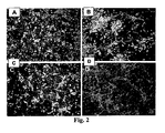
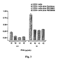
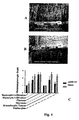
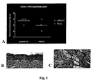


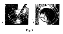

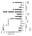
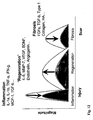


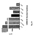
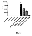
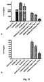
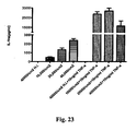
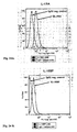
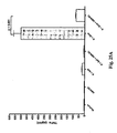


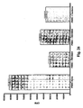
Classifications
-
- A—HUMAN NECESSITIES
- A61—MEDICAL OR VETERINARY SCIENCE; HYGIENE
- A61K—PREPARATIONS FOR MEDICAL, DENTAL OR TOILETRY PURPOSES
- A61K45/00—Medicinal preparations containing active ingredients not provided for in groups A61K31/00 - A61K41/00
- A61K45/06—Mixtures of active ingredients without chemical characterisation, e.g. antiphlogistics and cardiaca
-
- C—CHEMISTRY; METALLURGY
- C12—BIOCHEMISTRY; BEER; SPIRITS; WINE; VINEGAR; MICROBIOLOGY; ENZYMOLOGY; MUTATION OR GENETIC ENGINEERING
- C12N—MICROORGANISMS OR ENZYMES; COMPOSITIONS THEREOF; PROPAGATING, PRESERVING, OR MAINTAINING MICROORGANISMS; MUTATION OR GENETIC ENGINEERING; CULTURE MEDIA
- C12N5/00—Undifferentiated human, animal or plant cells, e.g. cell lines; Tissues; Cultivation or maintenance thereof; Culture media therefor
- C12N5/06—Animal cells or tissues; Human cells or tissues
- C12N5/0602—Vertebrate cells
- C12N5/0652—Cells of skeletal and connective tissues; Mesenchyme
- C12N5/0669—Bone marrow stromal cells; Whole bone marrow
-
- A—HUMAN NECESSITIES
- A61—MEDICAL OR VETERINARY SCIENCE; HYGIENE
- A61K—PREPARATIONS FOR MEDICAL, DENTAL OR TOILETRY PURPOSES
- A61K35/00—Medicinal preparations containing materials or reaction products thereof with undetermined constitution
- A61K35/12—Materials from mammals; Compositions comprising non-specified tissues or cells; Compositions comprising non-embryonic stem cells; Genetically modified cells
- A61K35/28—Bone marrow; Haematopoietic stem cells; Mesenchymal stem cells of any origin, e.g. adipose-derived stem cells
-
- A—HUMAN NECESSITIES
- A61—MEDICAL OR VETERINARY SCIENCE; HYGIENE
- A61K—PREPARATIONS FOR MEDICAL, DENTAL OR TOILETRY PURPOSES
- A61K38/00—Medicinal preparations containing peptides
- A61K38/16—Peptides having more than 20 amino acids; Gastrins; Somatostatins; Melanotropins; Derivatives thereof
- A61K38/17—Peptides having more than 20 amino acids; Gastrins; Somatostatins; Melanotropins; Derivatives thereof from animals; from humans
- A61K38/1703—Peptides having more than 20 amino acids; Gastrins; Somatostatins; Melanotropins; Derivatives thereof from animals; from humans from vertebrates
- A61K38/1709—Peptides having more than 20 amino acids; Gastrins; Somatostatins; Melanotropins; Derivatives thereof from animals; from humans from vertebrates from mammals
- A61K38/1722—Plasma globulins, lactoglobulins
-
- A—HUMAN NECESSITIES
- A61—MEDICAL OR VETERINARY SCIENCE; HYGIENE
- A61P—SPECIFIC THERAPEUTIC ACTIVITY OF CHEMICAL COMPOUNDS OR MEDICINAL PREPARATIONS
- A61P1/00—Drugs for disorders of the alimentary tract or the digestive system
- A61P1/02—Stomatological preparations, e.g. drugs for caries, aphtae, periodontitis
-
- A—HUMAN NECESSITIES
- A61—MEDICAL OR VETERINARY SCIENCE; HYGIENE
- A61P—SPECIFIC THERAPEUTIC ACTIVITY OF CHEMICAL COMPOUNDS OR MEDICINAL PREPARATIONS
- A61P1/00—Drugs for disorders of the alimentary tract or the digestive system
- A61P1/04—Drugs for disorders of the alimentary tract or the digestive system for ulcers, gastritis or reflux esophagitis, e.g. antacids, inhibitors of acid secretion, mucosal protectants
-
- A—HUMAN NECESSITIES
- A61—MEDICAL OR VETERINARY SCIENCE; HYGIENE
- A61P—SPECIFIC THERAPEUTIC ACTIVITY OF CHEMICAL COMPOUNDS OR MEDICINAL PREPARATIONS
- A61P1/00—Drugs for disorders of the alimentary tract or the digestive system
- A61P1/16—Drugs for disorders of the alimentary tract or the digestive system for liver or gallbladder disorders, e.g. hepatoprotective agents, cholagogues, litholytics
-
- A—HUMAN NECESSITIES
- A61—MEDICAL OR VETERINARY SCIENCE; HYGIENE
- A61P—SPECIFIC THERAPEUTIC ACTIVITY OF CHEMICAL COMPOUNDS OR MEDICINAL PREPARATIONS
- A61P1/00—Drugs for disorders of the alimentary tract or the digestive system
- A61P1/18—Drugs for disorders of the alimentary tract or the digestive system for pancreatic disorders, e.g. pancreatic enzymes
-
- A—HUMAN NECESSITIES
- A61—MEDICAL OR VETERINARY SCIENCE; HYGIENE
- A61P—SPECIFIC THERAPEUTIC ACTIVITY OF CHEMICAL COMPOUNDS OR MEDICINAL PREPARATIONS
- A61P11/00—Drugs for disorders of the respiratory system
-
- A—HUMAN NECESSITIES
- A61—MEDICAL OR VETERINARY SCIENCE; HYGIENE
- A61P—SPECIFIC THERAPEUTIC ACTIVITY OF CHEMICAL COMPOUNDS OR MEDICINAL PREPARATIONS
- A61P13/00—Drugs for disorders of the urinary system
- A61P13/02—Drugs for disorders of the urinary system of urine or of the urinary tract, e.g. urine acidifiers
-
- A—HUMAN NECESSITIES
- A61—MEDICAL OR VETERINARY SCIENCE; HYGIENE
- A61P—SPECIFIC THERAPEUTIC ACTIVITY OF CHEMICAL COMPOUNDS OR MEDICINAL PREPARATIONS
- A61P13/00—Drugs for disorders of the urinary system
- A61P13/10—Drugs for disorders of the urinary system of the bladder
-
- A—HUMAN NECESSITIES
- A61—MEDICAL OR VETERINARY SCIENCE; HYGIENE
- A61P—SPECIFIC THERAPEUTIC ACTIVITY OF CHEMICAL COMPOUNDS OR MEDICINAL PREPARATIONS
- A61P13/00—Drugs for disorders of the urinary system
- A61P13/12—Drugs for disorders of the urinary system of the kidneys
-
- A—HUMAN NECESSITIES
- A61—MEDICAL OR VETERINARY SCIENCE; HYGIENE
- A61P—SPECIFIC THERAPEUTIC ACTIVITY OF CHEMICAL COMPOUNDS OR MEDICINAL PREPARATIONS
- A61P15/00—Drugs for genital or sexual disorders; Contraceptives
-
- A—HUMAN NECESSITIES
- A61—MEDICAL OR VETERINARY SCIENCE; HYGIENE
- A61P—SPECIFIC THERAPEUTIC ACTIVITY OF CHEMICAL COMPOUNDS OR MEDICINAL PREPARATIONS
- A61P17/00—Drugs for dermatological disorders
-
- A—HUMAN NECESSITIES
- A61—MEDICAL OR VETERINARY SCIENCE; HYGIENE
- A61P—SPECIFIC THERAPEUTIC ACTIVITY OF CHEMICAL COMPOUNDS OR MEDICINAL PREPARATIONS
- A61P19/00—Drugs for skeletal disorders
-
- A—HUMAN NECESSITIES
- A61—MEDICAL OR VETERINARY SCIENCE; HYGIENE
- A61P—SPECIFIC THERAPEUTIC ACTIVITY OF CHEMICAL COMPOUNDS OR MEDICINAL PREPARATIONS
- A61P19/00—Drugs for skeletal disorders
- A61P19/04—Drugs for skeletal disorders for non-specific disorders of the connective tissue
-
- A—HUMAN NECESSITIES
- A61—MEDICAL OR VETERINARY SCIENCE; HYGIENE
- A61P—SPECIFIC THERAPEUTIC ACTIVITY OF CHEMICAL COMPOUNDS OR MEDICINAL PREPARATIONS
- A61P21/00—Drugs for disorders of the muscular or neuromuscular system
-
- A—HUMAN NECESSITIES
- A61—MEDICAL OR VETERINARY SCIENCE; HYGIENE
- A61P—SPECIFIC THERAPEUTIC ACTIVITY OF CHEMICAL COMPOUNDS OR MEDICINAL PREPARATIONS
- A61P25/00—Drugs for disorders of the nervous system
-
- A—HUMAN NECESSITIES
- A61—MEDICAL OR VETERINARY SCIENCE; HYGIENE
- A61P—SPECIFIC THERAPEUTIC ACTIVITY OF CHEMICAL COMPOUNDS OR MEDICINAL PREPARATIONS
- A61P27/00—Drugs for disorders of the senses
- A61P27/02—Ophthalmic agents
-
- A—HUMAN NECESSITIES
- A61—MEDICAL OR VETERINARY SCIENCE; HYGIENE
- A61P—SPECIFIC THERAPEUTIC ACTIVITY OF CHEMICAL COMPOUNDS OR MEDICINAL PREPARATIONS
- A61P29/00—Non-central analgesic, antipyretic or antiinflammatory agents, e.g. antirheumatic agents; Non-steroidal antiinflammatory drugs [NSAID]
-
- A—HUMAN NECESSITIES
- A61—MEDICAL OR VETERINARY SCIENCE; HYGIENE
- A61P—SPECIFIC THERAPEUTIC ACTIVITY OF CHEMICAL COMPOUNDS OR MEDICINAL PREPARATIONS
- A61P3/00—Drugs for disorders of the metabolism
- A61P3/08—Drugs for disorders of the metabolism for glucose homeostasis
- A61P3/10—Drugs for disorders of the metabolism for glucose homeostasis for hyperglycaemia, e.g. antidiabetics
-
- A—HUMAN NECESSITIES
- A61—MEDICAL OR VETERINARY SCIENCE; HYGIENE
- A61P—SPECIFIC THERAPEUTIC ACTIVITY OF CHEMICAL COMPOUNDS OR MEDICINAL PREPARATIONS
- A61P35/00—Antineoplastic agents
-
- A—HUMAN NECESSITIES
- A61—MEDICAL OR VETERINARY SCIENCE; HYGIENE
- A61P—SPECIFIC THERAPEUTIC ACTIVITY OF CHEMICAL COMPOUNDS OR MEDICINAL PREPARATIONS
- A61P37/00—Drugs for immunological or allergic disorders
-
- A—HUMAN NECESSITIES
- A61—MEDICAL OR VETERINARY SCIENCE; HYGIENE
- A61P—SPECIFIC THERAPEUTIC ACTIVITY OF CHEMICAL COMPOUNDS OR MEDICINAL PREPARATIONS
- A61P37/00—Drugs for immunological or allergic disorders
- A61P37/02—Immunomodulators
-
- A—HUMAN NECESSITIES
- A61—MEDICAL OR VETERINARY SCIENCE; HYGIENE
- A61P—SPECIFIC THERAPEUTIC ACTIVITY OF CHEMICAL COMPOUNDS OR MEDICINAL PREPARATIONS
- A61P37/00—Drugs for immunological or allergic disorders
- A61P37/02—Immunomodulators
- A61P37/06—Immunosuppressants, e.g. drugs for graft rejection
-
- A—HUMAN NECESSITIES
- A61—MEDICAL OR VETERINARY SCIENCE; HYGIENE
- A61P—SPECIFIC THERAPEUTIC ACTIVITY OF CHEMICAL COMPOUNDS OR MEDICINAL PREPARATIONS
- A61P5/00—Drugs for disorders of the endocrine system
-
- A—HUMAN NECESSITIES
- A61—MEDICAL OR VETERINARY SCIENCE; HYGIENE
- A61P—SPECIFIC THERAPEUTIC ACTIVITY OF CHEMICAL COMPOUNDS OR MEDICINAL PREPARATIONS
- A61P7/00—Drugs for disorders of the blood or the extracellular fluid
- A61P7/06—Antianaemics
-
- A—HUMAN NECESSITIES
- A61—MEDICAL OR VETERINARY SCIENCE; HYGIENE
- A61P—SPECIFIC THERAPEUTIC ACTIVITY OF CHEMICAL COMPOUNDS OR MEDICINAL PREPARATIONS
- A61P9/00—Drugs for disorders of the cardiovascular system
-
- A—HUMAN NECESSITIES
- A61—MEDICAL OR VETERINARY SCIENCE; HYGIENE
- A61K—PREPARATIONS FOR MEDICAL, DENTAL OR TOILETRY PURPOSES
- A61K35/00—Medicinal preparations containing materials or reaction products thereof with undetermined constitution
- A61K35/12—Materials from mammals; Compositions comprising non-specified tissues or cells; Compositions comprising non-embryonic stem cells; Genetically modified cells
- A61K2035/122—Materials from mammals; Compositions comprising non-specified tissues or cells; Compositions comprising non-embryonic stem cells; Genetically modified cells for inducing tolerance or supression of immune responses
-
- A—HUMAN NECESSITIES
- A61—MEDICAL OR VETERINARY SCIENCE; HYGIENE
- A61K—PREPARATIONS FOR MEDICAL, DENTAL OR TOILETRY PURPOSES
- A61K35/00—Medicinal preparations containing materials or reaction products thereof with undetermined constitution
- A61K35/12—Materials from mammals; Compositions comprising non-specified tissues or cells; Compositions comprising non-embryonic stem cells; Genetically modified cells
- A61K2035/124—Materials from mammals; Compositions comprising non-specified tissues or cells; Compositions comprising non-embryonic stem cells; Genetically modified cells the cells being hematopoietic, bone marrow derived or blood cells
Definitions
- the present invention relates generally to the generation and use of in vitro cultured self-renewing colony forming somatic cells (CF-SC) for use as a medicament to stimulate or enhance angiogenesis.
- CF-SC colony forming somatic cells
- This medicament is useful for the treatment of a variety of diseases and disorders.
- CF-SC are adult human bone marrow-derived somatic cells (hABM-SC).
- the present invention also relates to manipulation of CF-SC cell populations during cultivation to modulate (i.e ., up- or down-regulate) production of various soluble or secreted compositions produced by in vitro cultured and expanded self-renewing colony forming cells.
- the field of the invention also relates to cell-based and tissue-engineering therapies; particularly, methods of using and/or administering CF-SC including administration via incorporation in, or mixture with, pharmaceutically acceptable carriers (such as a pharmaceutically acceptable solution or a transient, permanent, or biodegradable matrix).
- pharmaceutically acceptable carriers such as a pharmaceutically acceptable solution or a transient, permanent, or biodegradable matrix.
- cell-based therapies there are two major options in using cell-based therapies to manage and treat chronic and acute tissue damage in which the overall objective is the functional and/or cosmetic restoration of damaged tissue.
- These cell based therapy options include: 1) Cell Replacement - Use of cells to replace damaged tissue by establishing long-term engraftment; and 2) Supply Trophic Factors - Use of cells and compositions produced by cells ( e.g ., growth factors) to stimulate endogenous repair mechanisms through release of factors delivered or produced by cells without long-term engraftment.
- the present invention relates to use of cell based therapies without relying on long-term cell engraftment.
- the invention relates to use of cells in the treatment of various diseases and disorders; particularly those involving tissues and organs with limited self-renewal capability (such as, for example, neurological and cardiac tissues and organs).
- the present invention relates primarily to treatments involving use of allogeneic cells. However, it would also be equally possible to perform these same treatments using autologous cells.
- a number of conditions and diseases of the central nervous system i.e ., brain and spinal cord
- peripheral nervous system and heart adversely affect humans because of the deficit of regenerative capacity in the effected tissues.
- These conditions and diseases include, for example, spinal cord injury, amyotrophic lateral sclerosis (ALS), Parkinson's disease, stroke, Huntington's disease, traumatic brain injury, brain tumors, Fabry Disease, heart diseases (such as congestive heart failure and myocardial infarction).
- Clinical management strategies for example, frequently focus on the prevention of further damage or injury rather than replacement or repair of the damaged tissue (e . g ., neurons, glial cells, cardiac muscle); include treatment with exogenous steroids and synthetic, non-cellular pharmaceutical drugs; and have varying degrees of success which may depend on the continued administration of the steroid or synthetic drug.
- spinal cord injury For example, the majority of spinal cord injuries are compression injuries with the remaining cases involving complete transection of the spinal cord.
- Current therapeutic treatments for spinal cord injury include the prevention of additional spinal cord injury by physically stabilizing the spine through surgical and non-surgical procedures and by inhibiting the inflammatory response with steroidal therapy.
- aging is a major negative component to nearly every common disease affecting mammals, and one of the principle features of aging in a degeneration of many tissue including those of skin, bone, eye, brain, liver, kidney, heart, vasculature, muscle, et cetera.
- tissue maintenance and repair mechanisms in almost every tissue decline over the course of life.
- the present invention relates in part to treatment of skin wounds.
- treatments for wounds of the skin such as epidermal replacement products, dermal replacement products, artificial skin products, and wound dressings. Examples of some of these are described briefly below.
- EPICELTM (Genzyme Corp., Cambridge, MA) is composed of autologous epidermal cells skin grown from biopsy of patients own skin for treatment of bums. Cells are co-cultured with mouse feeder cell line into sheets of autologous epidermis.
- MYSKINTM CellTran LTD, Sheffield, S1 4DP United Kingdom
- MYSKINTM contains living cells expanded from the tissue of individual patients.
- MYSKINTM comprises a layer of keratinocytes (epidermal cells) on an advanced polymer-like coating which facilitates the transfer of cells into the wound where they can initiate healing.
- MYSKINTM uses a medical grade silicone substrate layer to support cell delivery, wound coverage and allow exudate management.
- EPIDEXTM (Modex Therapeutics Ltd, Lausanne, Switzerland) is an autologous epidermal skin equivalent that is grown directly from stem and pre-cursor cells derived from hair taken directly from a patient in a non-surgical procedure.
- CELLSPRAY TM (Clinical Cell Culture Europe Ltd, Cambridge CB2 1NL, United Kingdom) is a cultured epithelial autograft suspension that is sprayed onto injured skin in order to provide a rapid epidermal cover, promote healing and optimize scar quality.
- INTEGRATM Dermal Regeneration Template (Integra LifeSciences Corporation, Plainsboro, New Jersey) is a bilayer membrane system for skin replacement.
- the dermal replacement layer is made of a porous matrix of fibers of cross-linked bovine tendon collagen and a glycosaminoglycan (chondroitin-6-sulfate) that is manufactured with a controlled porosity and defined degradation rate.
- the temporary epidermal substitute layer is made of synthetic polysiloxane polymer (silicone) and functions to control moisture loss from the wound.
- the collagen dermal replacement layer serves as a matrix for the infiltration of fibroblasts, macrophages, lymphocytes, and capillaries derived from the wound bed.
- DERMAGRAFTTM Advanced Biohealing, La Jolla, CA
- Permacol TM surgical implant is collagen-derived from porcine dermis which, when implanted in the human body, is non-allergenic and long-lasting.
- TRANSCYTETM is a human foreskin-derived fibroblast temporary skin substitute (allogeneic).
- the product consists of a polymer membrane and newborn human fibroblast cells cultured under aseptic conditions in vitro on a nylon mesh. Prior to cell growth, this nylon mesh is coated with porcine dermal collagen and bonded to a polymer membrane (silicone). This membrane provides a transparent synthetic epidermis when the product is applied to the burn.
- the human fibroblast-derived temporary skin substitute provides a temporary protective barrier.
- TRANSCYTETM is transparent and allows direct visual monitoring of the wound bed.
- RENGRANEXTM Gel is a topical wound care product made of recombinant PDGF in a gel.
- PERMADERMTM (Cambrex Bio Science Walkersville, Inc., Walkersville, MD)
- PERMADERMTM is constructed from autologous epidermal and dermal layers of the skin and is indicated for the treatment of severe burns. The product is reported to be pliable and to grow with the patient.
- ORCELTM Organic Chemical Cellular Cellular Cellular Cellular Cellular Cellular Cellular Cellular Cellular Cellular Cellular Cellular Cellular Cellular Cellular Cellular Cellular Cellular Cellular Cellular Cellular Cellular Cellular Cellular Cellular Cellular Cellular Cellular Cellular Cellular Cellular Cellular Cellular Cellular Cellular Cellular Cellular Cellular Cellular Cellular Cellular Cellular Cellular Cellular Cellular Cellular Cellular Cellular Cellular Cellular Cellular Cellular Cellular cell proliferation.
- APLIGRAFTM Smith & Nephew, London, WC2N 6LA United Kingdom
- 3MTM TEGADERMTM Transparent Film Dressing (3M, St. Paul, MN) is a breathable film that provides a bacterial and viral barrier to outside contaminants.
- TISSEELTM VH Fibrin Sealant (Baxter, Deerfield, IL) is indicated for use as an adjunct to hemostasis.
- stable cell population means an isolated, in vitro cultured, cell population that when introduced into a living mammalian organism (such as a mouse, rat, human, dog, cow, etc.) does not result in detectable production of cells which have differentiated into a specialized cell type or cell types (such as a chondrocyte, adipocyte, osteocyte, etc.) and wherein the cells in the cell population express, or maintain the ability to express or the ability to be induced to express, detectable levels of at least one therapeutically useful composition (such as membrane bound or soluble TNF-alpha receptor, IL-1R antagonists, IL-18 antagonists, compositions shown in Table 1A, 1B, 1C, etc.).
- a therapeutically useful composition such as membrane bound or soluble TNF-alpha receptor, IL-1R antagonists, IL-18 antagonists, compositions shown in Table 1A, 1B, 1C, etc.
- ectopic means "in the wrong place” or “out of place”.
- an ectopic kidney is one that is not in the usual location; or, an extrauterine pregnancy is an "ectopic pregnancy”.
- an example of ectopic differentiation would be cells that when introduced into cardiac tissue, produce bone tissue-like calcifications and/or ossifications. This phenomenon has been demonstrated to occur, for example, when mesenchymal stem cells are injected into cardiac tissue. See, Breitbach et al., "Potential Risks of Bone Marrow Cell Transplantation Into Infarcted Hearts", Blood, Vol. 110, No. 4 (Aug. 2007 ).
- the present invention relates to the generation and use of expanded, in vitro cultured, self-renewing colony forming somatic cells (hereinafter referred to as "CF-SC”) for the treatment of a variety of diseases and disorders. Further, the present invention also relates to the generation and use of extensively expanded, in vitro cultured, self-renewing colony forming somatic cells (hereinafter referred to as "exCF-SC”), for the treatment of a variety of diseases and disorders. ExCF-SC are self-renewing colony forming somatic cells (CF-SC) which have undergone at least about 30, at least about 40, or at least about 50 cell population doublings during in vitro cultivation.
- CF-SC self-renewing colony forming somatic cells which have been expanded in vitro
- CF-SC self-renewing colony forming somatic cells which have been expanded in vitro
- this term encompasses both cell populations which have undergone less than about 30 population doublings ( e.g ., less than about 5, less than about 10, less than about 15, less than about 20, less than about 25 population doublings) and also cell populations which have undergone more than about 30, more than about 40, or more than about 50 populations doublings in vitro ).
- ABM-SC adult human bone marrow-derived somatic cells
- exCF-SC adult human bone marrow-derived somatic cells which have undergone at least about 30, at least about 40, or at least about 50 cell population doublings during in vitro cultivation
- ABSM-SC adult human bone marrow-derived somatic cells which have undergone at least about 30, at least about 40, or at least about 50 cell population doublings during in vitro cultivation
- the term "ABM-SC” encompasses both ABM-SC cell populations which have undergone less than about 30 population doublings ( e.g ., less than about 5, less than about 10, less than about 15, less than about 20, less than about 25 population doublings) and also ABM-SC cell populations which have undergone more than about 30, more than about 40, or more than about 50 populations doublings in vitro ).
- exensively expanded refers to cell populations which have undergone at least about 30 or more cell population doublings and wherein the cells are non-senescent, are not immortalized, and continue to maintain the normal karyotype found in the cell species of origin.
- the term "substantial capacity for self-renewal” means having the ability to go through numerous cycles of cell division resulting in the production of multiple generations of cell progeny (thus, with each cell division, one cell produces two "daughter cells” wherein at least one daughter cell is capable of further cell division).
- One measure of “substantial capacity for self-renewal” is indicated by the ability of a cell population to undergo at least about 10, 15, 20, 25, 30, 35, 40, 45, 50 or more cell doublings.
- Another measure of "substantial capacity for self-renewal” is indicated by maintenance of the ability of a cell population to re-populate, or approach confluence in, a tissue culture vessel after cell culture passaging (when the same or similar culture conditions are maintained).
- an example of "substantial capacity for self-renewal” is demonstrated when a cell population continues to re-populate a tissue culture vessel in a period of time of at least about 25%, 50%, 60%, 70%, 80%, 90%, 95% or 100% of the time required for such re-population during early cell culture doublings (such as before a cell population has undergone more than about 10 population doublings).
- Another measure of "substantial capacity for self-renewal” is maintenance of a consistent rate of population doubling time or of a consistent and relatively rapid rate of population doubling.
- substantially no multipotent differentiation capacity means cell populations which cannot differentiate into multiple different types of cells, either in vitro or in vivo.
- An example of cells which do have substantial multipotent differentiation capacity are hematopoietic stem cells which can differentiate into red blood cells, T-cells, B-cells, platelets, etc. either in vitro or in vivo.
- Another example of cells which do have substantial multipotent differentiation capacity are mesenchymal stem cells which can differentiate, for example, into osteocytes (bone), adipocytes (fat), or chondrocytes (cartilage).
- a cell population with "substantially no multipotent differentiation capacity” is one in which at least about 80%, 90%, 95%, 98%, 99% or 100% of the cells in the cell population cannot be induced to detectably differentiate in vitro or in vivo into more than one cell type.
- a "unipotent” cell or “unipotent progenitor cell” is an example of a cell which has substantially no multipotent differentiation capacity.
- stem cell means a cell or cells possessing the following two properties: 1) capacity for self-renewal, which is the ability to go through numerous cycles of cell division while maintaining the undifferentiated state; and, 2) differentiation potency, which is the capacity to change into one or more kinds of mature cell types and, upon such change, no longer undergoing cycles of cell division (for example, capacity to change into an osteocyte, adipocyte, chondrocyte, etc.).
- differentiation potency means the cells are either totipotent, pluripotent, multipotent, or unipotent progenitor cells.
- a “mesenchymal stem cell” is a stem cell of this same definition but wherein said cell has been derived or obtained from mesenchyme tissue (such as, for example, bone marrow, adipose or cartilage). See, Horwitz et al., "Clarification of the nomenclature for MSC: The International Society for Cellular Therapy position statement", Cytotherapy, vol. 7, no. 5, pp. 393-395 (2005 ); and references cited therein.
- totipotent means cells which can become any type of cell as may be found during any stage of development in the organism of the cells origin. Totipotent cells are typically produced by the first few divisions of the fertilized egg ( i . e ., following fusion of an egg and sperm cell). Thus, totipotent cells can differentiate into embryonic and extraembryonic cell types.
- pluripotent means cells which can differentiate into cells derived from any of the three germ layers (endoderm, mesoderm, ectoderm) found in the organism of the cells origin.
- multipotent means cells which can produce multiple types (i.e ., more than one type) of differentiated cells.
- a mesenchymal stem cell is an example of a multipotent cell.
- unipotent means cells which can produce only one cell type. Unipotent cells have the property of self-renewal, but can change into only one kind of mature cell type.
- normal karyotype means having a genetic composition comprising chromosomes of the number and of the structure typically found in, and considered normal for, the species from which the cells are derived.
- connective tissue is one of the four types of tissue usually referenced in traditional classifications (the others being epithelial, muscle, and nervous tissue). Connective tissue is involved in organism and organ structure and support and is usually derived from mesoderm. As used herein, “connective tissue” includes those tissues sometimes referred to as “connective tissue proper”, “specialized connective tissues”, and “embyronic connective tissue”.
- Connective tissue proper includes areolar (or loose) connective tissue, which holds organs and epithelia in place and has a variety of proteinaceous fibres, including collagen and elastin.
- Connective tissue proper also includes dense connective tissue (or, fibrous connective tissue) which forms ligaments and tendons.
- “Specialized connective tissue” includes blood, bone, cartilage, adipose and reticular connective tissue.
- Reticular connective tissue is a network of reticular fibres (fine collagen, type III) that form a soft skeleton to support the lymphoid organs (lymph nodes, bone marrow, and spleen)
- Embryonic connective tissue includes mesenchymal connective tissue and mucous connective tissue.
- Mesenchyme also known as embryonic connective tissue
- mesoderm the middle layer of the trilaminar germ disc
- mesenchyme contains collagen bundles and fibroblasts.
- Mesenchyme later differentiates into blood vessels, blood-related organs, and connective tissues.
- Mucous connective tissue or mucous tissue is a type of connective tissue found during fetal development; it is most easily found as a component of Wharton's jelly (a gelatinous substance within the umbilical cord which serves to protect and insulate cells in the umbilical cord).
- immortalized refers to a cell or cell line which can undergo an indefinite number of cell doublings in vitro. Immortalized cells acquire such ability through genetic changes which eliminate or circumvent the natural limit on a cells ability to continually divide.
- non-immortalized cells are eukaryotic cells which, when taken directly from the organism and cultured in vitro (producing a "primary cell culture”), can undergo a limited number of cell doublings before senescencing (losing ability to divide) and dying.
- primary cultures of most types of mammalian, non-immortalized cells can usually undergo a relatively defined but reproducibly limited range of cell doublings (depending on the primary cell type) before differentiating, senescing, or dying.
- long-term engraftment means the detectable presence of donor cells residing within (or as part of) target tissue to which (or in which) said cells were delivered after more than about 4 weeks from the time of administration.
- “More than about 4 weeks” includes time periods of more than about 5, 6, 7, 8, 10, 12, 14, 16, 18, 20, and 24 weeks.
- “More than about 4 weeks” also includes time periods of more than about 6 months, 8 months, 10 months, 12 months, 18 months, 24 months, 30 months, 36 months, 42 months and 48 months.
- the present invention also relates to manipulation of CF-SC and exCF-SC cell populations during cultivation to modulate (i.e ., up- or down-regulate) production of various soluble or secreted compositions produced by the in vitro cultured and expanded self-renewing colony forming cells.
- the present invention also relates to extensively expanded cell populations which are characterized by loss of ability to differentiate into bone cells (osteocytes).
- the present invention relates to extensively expanded cell populations which are characterized by loss of ability to generate calcium deposits when cultured under osteoinductive conditions, including with or without cultivation in the presence of the supplemental bone morphogen Noggin (see Example 16 ).
- osteocytes bone cells
- the present invention relates to extensively expanded cell populations which are characterized by loss of ability to generate calcium deposits when cultured under osteoinductive conditions, including with or without cultivation in the presence of the supplemental bone morphogen Noggin (see Example 16 ).
- NP_032737 and NP_005441 see also e.g ., Valenzuela, et al., "Identification of mammalian noggin and its expression in the adult nervous system", J. Neurosci. 15 (9), 6077-6084 (1995 )).
- the present invention also relates to extensively expanded cell populations characterized by the loss of ability to differentiate into bone cells and/or loss of ability to generate calcium deposits (as described above), but wherein said cell populations continue to secrete, or maintain the ability to secrete or to be induced to secrete, at least one therapeutically useful composition.
- the present invention also relates to cell-based and tissue-engineering therapies; particularly, methods of using and/or administering CF-SC and exCF-SC including administration via incorporation in, or mixture with, pharmaceutically acceptable carriers (such as a pharmaceutically acceptable solution or a transient, permanent, or biodegradable matrix).
- pharmaceutically acceptable carriers such as a pharmaceutically acceptable solution or a transient, permanent, or biodegradable matrix.
- the present invention also relates to expanded (i.e ., in vitro cultured and passaged) and extensively expanded cell populations which are preferably negative for expression of the STRO-1 cell surface marker.
- expanded i.e ., in vitro cultured and passaged
- extensively expanded cell populations which are preferably negative for expression of the STRO-1 cell surface marker.
- STRO-1, HOP-26 (CD63), CD49a and SB-10 (CD166) markers of primitive human marrow stromal cells and their more differentiated progeny: a comparative investigation in vitro" Cell Tissue Res. 2003 Sep;313(3):281-90 ; and , Dennis et al., "The STRO-1+ marrow cell population is multipotential" Cells Tissues Organs.
- the present invention also relates to manufacture and use of pharmaceutically acceptable compositions containing CF-SC and exCF-SC (for example, ABM-SC and exABM-SC) with additional structural and/or therapeutic components.
- CF-SC or exCF-SC for example, ABM-SC or exABM-SC
- collagen may be combined in a pharmaceutically acceptable solution to generate compositions in liquid, semi-solid, or solid-like forms (matrices) for use, for example, in the treatment, repair, and regeneration of skin disorders (e.g., skin wounds such as burns, abrasions, lacerations, ulcers, infections).
- skin disorders e.g., skin wounds such as burns, abrasions, lacerations, ulcers, infections.
- the present invention relates generally to use of self-renewing cells, referred to herein as colony-forming somatic cells (CF-SC) including extensively passaged colony-forming somatic cells (exCF-SC).
- CF-SC colony-forming somatic cells
- exCF-SC extensively passaged colony-forming somatic cells
- Examples of such cells are adult human bone marrow-derived somatic cells (ABM-SC) including extensively passaged adult human bone marrow-derived somatic cells (exABM-SC), for use in treatment of various diseases and disorders.
- CF-SC colony-forming somatic cells
- ABSM-SC adult human bone marrow-derived somatic cells
- CF-SC isolated from a source population of cells are cultured under low oxygen conditions (e.g., less than atmospheric) and passaged at low cell densities such that the CF-SC maintain an essentially constant population doubling rate through numerous population doublings.
- the CF-SC may be used to generate the compositions of the present invention.
- the CF-SC and exCF-SC are derived from bone marrow (and are referred to herein as ABM-SC and exABM-SC, respectively).
- CF-SC and exCF-SC are an isolated cell population wherein the cells of the cell population co-express CD49c and CD90 and wherein the cell population maintains a doubling rate of less than about 30 hours after at least about 30, at least about 40, or at least about 50 cell population doublings.
- CF-SC and exCF-SC are an isolated cell population wherein the cells of the cell population co-express CD49c, CD90, and one or more cell surface proteins selected from the group consisting of CD44, HLA Class-1 antigen, and ⁇ (beta) 2-Microglobulin, and wherein the cell population maintains a doubling rate of less than about 30 hours after at least about 30, at least about 40, or at least about 50 cell population doublings.
- CF-SC and exCF-SC are an isolated cell population wherein the cells of the cell population co-express CD49c and CD90, but are negative for expression of cell surface protein CD10, and wherein the cell population maintains a doubling rate of less than about 30 hour after at least about 30, at least about 40, or at least about 50 cell population doublings.
- CF-SC and exCF-SC are an isolated cell population wherein the cells of the cell population co-express CD49c, CD90, and one or more cell surface proteins selected from the group consisting of CD44, HLA Class-1 antigen, and ⁇ (beta) 2-Microglobulin, but are negative for expression of cell surface protein CD10, and wherein the cell population maintains a doubling rate of less than about 30 hours after at least about 30, at least about 40, or at least about 50 cell population doublings.
- CF-SC and exCF-SC are isolated cell population wherein the cells of the cell population express one or more proteins selected from the group consisting of soluble proteins shown in Table 1A, 1B and 1C, and wherein the cell population maintains a doubling rate of less than about 30 hours after at least about 30, at least about 40, or at least about 50 cell population doublings.
- Damaged tissues and organs may result from, for example, disease (e.g ., heritable (genetic) or infectious diseases (such as bacterial, viral, and fungal infections)), physical trauma (such as burns, lacerations, abrasions, compression or invasive tissue and organ injuries), ischemia, aging, toxic chemical exposure, ionizing radiation, and dysregulation of the immune system ( e . g ., autoimmune disorders).
- the present invention encompasses the use of CF-SC and exCF-SC (such as for example, ABM-SC and exABM-SC, respectively), CF-SC and exCF-SC purified protein fractions, supernatants of CF-SC and exCF-SC conditioned media, and fractions of cell-supernatants derived from CF-SC and exCF-SC conditioned media.
- the above mentioned components may be combined with, or introduced into, physiologically compatible biodegradable matrices which contain additional components such as collagen and/or fibrin (for example, purified natural or recombinant human, bovine, or porcine collagen or fibrin), and/or polyglycolic acid (PGA), and/or additional structural or therapeutic compounds.
- Combination matrices such as these may be administered to the site of tissue or organ damage to promote, enhance, and/or result in repair and/or regeneration of the damaged tissue or organ.
- Embodiments of the invention include use of CF-SC and exCF-SC (such as for example, ABM-SC and exABM-SC, respectively), incorporated into pharmaceutically acceptable compositions which may be administered in a liquid, semi-solid, or solid-like state.
- Embodiments of the invention may be administered by methods routinely used by those skilled in the relevant art, such as for example, by topical application, as spray-on or aerosolized compositions, by injection, and implantation.
- CF-SC and exCF-SC such as for example, ABM-SC and exABM-SC, respectively
- cells as described in the present invention for tissue regenerative therapies may provide a number of benefits compared to previously described tissue regenerative therapies and products.
- use of the CF-SC and exCF-SC such as for example, ABM-SC and exABM-SC, respectively
- exABM-SC cells and compositions produced thereby provides a means of tissue regenerative therapy which may exhibit reduced adverse immune responses (such as reduced inflammation and T-cell activation; see e.g., Examples 3A, 3B, 5, 18, and 19.
- ABM-SC and exABM-SCs are immunologically silent, subjects do not need to be HLA-matched or pre-conditioned prior to treatment. See, Example 10, Part II; see also, Figure 17 .
- Another example of the field of the invention relates to the prevention and treatment of immune, and inflammatory disorders via use of such cells, cell populations, and compositions produced thereby.
- the present invention provides compositions and methods for repair and regeneration of wounds of the skin (i.e ., epidermis, dermis, hypodermis); including the manufacture and use of liquid, semi-solid, and solid-like matrices which incorporate CF-SC and exCF-SC (for example, human ABM-SC and exABM-SC), or products generated by such cells, and additional structural or therapeutic compounds.
- wounds of the skin i.e ., epidermis, dermis, hypodermis
- CF-SC and exCF-SC for example, human ABM-SC and exABM-SC
- a stem cell or other early-stage progenitor cells lose plasticity because the cells have committed to a particular differentiation pathway. At the biomolecular level, as this process begins to occur the cell loses the ability to respond to certain signaling molecules (e.g ., mitogens and morphogens) which would otherwise drive the cell to divide or become another cell type. Thus, as a cell begins to differentiate, it leaves the cell cycle ( i.e., can no longer go through mitosis) and enters an irreversible state called G0 wherein the cell can no longer divide. Entry into G0 is also associated with replicative senescence (hallmarks of which include increased expression of intracellular proteins p21 and p53).
- certain signaling molecules e.g ., mitogens and morphogens
- loss of plasticity (the ability to differentiate into a variety of cell types) is typically considered a prelude to cellular differentiation or cellular senescence. Furthermore, loss of plasticity is also typically associated with the loss of a cells capacity for continued self-renewal.
- exCF-SC of the present invention e . g ., exABM-SC
- self-renew including self-renewal at a relatively constant rate
- one embodiment of the present invention are therapeutically useful "end-stage cells" with a continued capacity for self-renewal ( e .
- the exCF-SC and exABM-SC of the present invention do not express significant quantities of p21 and/or p53, wherein a "significant quantity" of said molecules is a quantity which is indicative of cell senescence (wherein senescence may require sufficient expression levels of p21, p53, and/or other cell cycle regulators).
- the present invention is drawn, inter alia, to methods of repairing, regenerating, and/or rejuvenating tissues using self-renewing cells, referred to herein as colony-forming somatic cells (CF-SC) (an example of which are adult human bone marrow-derived somatic cells (ABM-SC)).
- CF-SC colony-forming somatic cells
- ABSM-SC adult human bone marrow-derived somatic cells
- Self-renewing colony-forming somatic cells (CF-SC) such as adult human bone marrow-derived somatic cells (ABM-SC) as used in the present invention are prepared as described in U.S. Patent Publication No. 20030059414 ( U.S. Application No. 09/260,244, filed Sept. 21, 2001 ) and U.S. Patent Publication No. 20040058412 ( U.S. Application No. 10/251,685, filed Sept.
- CF-SC isolated from a source population of cells are permitted to adhere to a cell culture surface in the presence of an appropriate media (such as for example, but not limited to, Minimal Essential Medium-Alpha ( e .
- CF-SC maintain an essentially constant population doubling rate (such as for example, but not limited to, a doubling rate of less than about 30 hours) through numerous population doublings (such as for example, but not limited to, going through 10, 15, 20, 25, 30, 35, 40, 45 and/or 50 population doublings).
- low oxygen conditions such as for example, but not limited to, O 2 at about 2-5%, CO 2 at about 5%, balanced with nitrogen
- low cell densities such as at about 30-1000 cells/cm 2
- the CF-SC maintain an essentially constant population doubling rate (such as for example, but not limited to, a doubling rate of less than about 30 hours) through numerous population doublings (such as for example, but not limited to, going through 10, 15, 20, 25, 30, 35, 40, 45 and/or 50 population doublings).
- Embodiments of the invention may be generated with CF-SC and exCF-SC (for example, ABM-SC and exABM-SC) cultured under low oxygen conditions wherein said O 2 concentrations range from about 1-20% (for example, wherein the O 2 concentration is about 1%, 2%, 3%, 4%, 5%, 6%, 7%, 8%, 9%, 10%, 11%, 12%, 13%, 14%, 15%, or 20%), plus CO 2 and balanced with nitrogen.
- CF-SC and exCF-SC for example, ABM-SC and exABM-SC
- O 2 concentrations range from about 1-20% (for example, wherein the O 2 concentration is about 1%, 2%, 3%, 4%, 5%, 6%, 7%, 8%, 9%, 10%, 11%, 12%, 13%, 14%, 15%, or 20%), plus CO 2 and balanced with nitrogen.
- ABM-SC may be cultured under low oxygen conditions wherein said O 2 concentrations are about 20%, less than about 20%, about 15%, less than about 15%, about 10%, less than about 10%, about 7%, less than about 7%, about 6%, less than about 6%, about 5%, less than about 5%, about 4%, less than about 4%, about 3%, less than about 3%, about 2%, less than about 2%, about 1%; or, wherein said low oxygen conditions are in a range from about 1% to about 20%, about 1% to about 15%, about 1% to about 10%, about 1% to about 5%, about 5% to about 20%, about 5% to about 15%, about 5% to about 10%, about 10% to about 15%, about 10% to about 20%, about 2% to about 8%, about 2% to about 7%, about 2% to about 6%, about 2% to about 5%, about 2% to about 4%, about 2% to about 3%, about 3% to about 8%, about 3% to about 7%, about 3% to about
- Embodiments of the invention may be generated with CF-SC and exCF-SC (for example, ABM-SC and exABM-SC) cultured under low oxygen conditions wherein CO 2 concentration range from about 1-15% (for example, wherein the CO 2 concentration is about 1%, 2%, 3%, 4%, 5%, 6%, 7%, 8%, 9%, 10%, 11%, 12%, 13%, 14%, or 15%), plus low O 2 and balanced with nitrogen.
- CO 2 concentration range from about 1-15% (for example, wherein the CO 2 concentration is about 1%, 2%, 3%, 4%, 5%, 6%, 7%, 8%, 9%, 10%, 11%, 12%, 13%, 14%, or 15%)
- Embodiments of the invention may be generated with CF-SC and exCF-SC (for example, ABM-SC and exABM-SC) passaged by seeding cells at low cell densities, wherein said cell density ranges from about 1-2500 cells/cm 2 (for example, wherein the cell density is about 1, 2, 5, 10, 20, 30, 40, 50, 60, 70, 80, 90, 100, 200, 300, 400, 500, 600, 700, 800, 900, 1000, 1500, 2000 or 2500 cells/cm 2 ).
- CF-SC and exCF-SC for example, ABM-SC and exABM-SC
- ABM-SC may be passaged at seeding densities of less than about 2500 cell/cm 2 , less than about 1000 cells/cm 2 , less than about 500 cells/cm 2 , less than about 100 cells/cm 2 , less than about 50 cells/cm 2 , less than about 30 cells/cm 2 , or less than about 10 cells/cm 2 .
- Embodiments of the invention may be generated with CF-SC and exCF-SC (for example, ABM-SC and exABM-SC) wherein the cell population doubling rates are maintained in a range of less than about 24-96 hours (for example, wherein the cell population doubling rate is maintained at less than about 24, 30, 36, 42, 48, 54, 60, 66, 72, 78, 84, 90, or 96 hours).
- CF-SC and exCF-SC for example, ABM-SC and exABM-SC
- Embodiments of the invention may be generated with CF-SC and exCF-SC (for example, ABM-SC and exABM-SC) wherein the cell population maintains an essentially constant doubling rate through a range of population doublings such as in a range of about 5-50 population doublings (for example, wherein the population doubling rate is maintained for about 5-10, 5-15, 5-20, 5-25, 5-30, 5-35, 5-40, 5-45, or 5-50 population doublings).
- Embodiments of the invention include use of CF-SC and exCF-SC (for example, ABM-SC and exABM-SC) incorporated into pharmaceutically acceptable compositions which may be in a liquid, semi-solid, or solid-like state.
- CF-SC and exCF-SC for example, ABM-SC and exABM-SC
- a liquid, semi-solid, or solid-like state is intended to indicate that the pharmaceutically acceptable composition in which the cells are contained can span a range of physical states from 1) a common liquid state (such as in an ordinary physiological saline solution); 2) to a wide-range of low-to-highly viscous states including jelly-like, gelatinous, or viscoelastic states (wherein the pharmaceutical composition contains from very high to very low levels of extracellular water, for example, such that the composition ranges in viscosity from a state where it "oozes" slowly like oil or honey to increasingly gelatinous or viscoelastic states which may be jelly-like, pliable, semi-elastic and
- Viscoelasticity also known as anelasticity, describes materials that exhibit both viscous and elastic characteristics when undergoing plastic deformation. Viscous materials, like honey, resist shear flow and strain linearly with time when a stress is applied. Elastic materials strain instantaneously when stretched and just as quickly return to their original state once the stress is removed. Viscoelastic materials have elements of both of these properties and, as such, exhibit time dependent strain.
- soluble matrix components such as collagens or fibrin
- exCF-SC for example, ABM-SC and exABM-SC
- ABM-SC and exABM-SC induces the cell population to up-regulate expression of important secreted proteins such as cytokines and matrix metalloproteinases.
- application of ABM-SC to surgically-induced wounds appears to facilitate wound closure and prevent scarring thereby resulting in minimal scarring ( see , Example 7 ).
- compositions and therapies incorporating these cells attractive for the treatment of immunological disorders and diseases involving the skin, such as for example, but not limited to, chronic inflammatory dermatoses, psoriasis, lichen planus, lupus erythematosus (LE), graft-versus-host disease (GVHD), and drug eruptions ( i . e ., adverse cutaneous drug reactions).
- immunological disorders and diseases involving the skin such as for example, but not limited to, chronic inflammatory dermatoses, psoriasis, lichen planus, lupus erythematosus (LE), graft-versus-host disease (GVHD), and drug eruptions ( i . e ., adverse cutaneous drug reactions).
- Secreted proteins and cell-supernatant fractions from CF-SC and exCF-SC can be manufactured from serum-free conditions, concentrated and prepared in such manner as to make them suitable for in vivo use.
- conditioned serum-free media from ABM-SC has been demonstrated to contain numerous pro-regenerative cytokines, growth factors, and matrix proteases in therapeutically effective concentrations ( see , Table 1A, 1B and 1C ).
- the complex mixture of hundreds of soluble factors produced by ABM-SC can be distinguished by 2D SDS PAGE (see, Figure 1 ).
- the desired proteins or cell supernatant fractions can be isolated, dialyzed, lyophilized and stored as a solid, or reconstituted in an appropriate vehicle for therapeutic administration.
- the proteins or cell-supernatant fractions would be reconstituted in a semi-solid collagen or fibrin-based vehicle, and applied topically to the wound bed.
- any number and type of pharmaceutically acceptable small molecules to large macromolecular compounds may be incorporated for administration with a pharmaceutically acceptable carrier such as biodegradeable matrices in which CF-SC and exCF-SC (such as, ABM-SC and exABM-SC), or products generated by such cells, are contained.
- a pharmaceutically acceptable carrier such as biodegradeable matrices in which CF-SC and exCF-SC (such as, ABM-SC and exABM-SC), or products generated by such cells, are contained.
- additional molecules may include small molecule pharmaceuticals such as anti-inflammatories, antibiotics, vitamins, and minerals (such as calcium) to name but a few categories.
- biologics may include extracellular matrix proteins, blood plasma coagulation proteins, antibodies, growth factors, chemokines, cytokines, lipids (such as cardiolipin and sphingomyelin), and nucleic acids (such as ribozymes, anti-sense oligonucleotides, or cDNA expression constructs); including therapeutically beneficial variants and derivatives of such molecules such as various isoforms, fragments, and subunits, as well as substitution, insertion, and deletion variants.
- any number of additional structural or therapeutically beneficial compounds could be included for administration with a pharmaceutically acceptable carrier such as biodegradeable matrices in which CF-SC and exCF-SC (such as, ABM-SC and exABM-SC), or products generated by such cells, are contained.
- a pharmaceutically acceptable carrier such as biodegradeable matrices in which CF-SC and exCF-SC (such as, ABM-SC and exABM-SC), or products generated by such cells, are contained.
- One embodiment of the invention encompasses a method of stimulating wound closure in a diabetic patient, such as a diabetic foot or venous leg ulcer, or a post-surgical wound.
- Stimulation of wound closure may be promoted by treatment with a pharmaceutical composition of CF-SC and exCF-SC (such as, ABM-SC and exABM-SC), or products generated by such cells, combined with naturally occurring extracellular matrix and/or blood plasma proteins components such as, for example, purified natural or recombinant human, bovine, porcine, or recombinant collagens, laminins, fibrinogen, and/or thrombin.
- the pharmaceutical composition may be administered to a mammal, including a human, at the site of tissue damage.
- a topically administered biodegradable matrix is formed from a mixture of components such as purified natural or recombinant collagen, fibrinogen, and/or thrombin, combined with allogeneic CF-SC and exCF-SC (such as, ABM-SC and exABM-SC).
- a pharmaceutical composition of allogeneic cells and matrix are cultured in vitro for an extended period of time (such as, for example, but not limited to 1 day to one month or longer), producing the de novo formation of connective tissue.
- the biodegradable matrix is bovine collagen or poylglycolic acid.
- the pharmaceutical composition is cultured in serum-free cell media under conditions of reduced oxygen tension, for example but not limited to, oxygen tension equivalent to about 4-5% O 2 , 5% CO 2 , and balanced with nitrogen.
- the invention encompasses a method of preparing a pharmaceutical composition comprising the steps:
- the above method of preparing a pharmaceutical composition may additionally comprise the step of incubating the culture for an extended period of time (such as, for example but not limited to, 1-3 days or longer) under low oxygen tension conditions equivalent to about 4-5% O 2 , 5% CO 2 , and balanced with nitrogen.
- an extended period of time such as, for example but not limited to, 1-3 days or longer
- low oxygen tension conditions equivalent to about 4-5% O 2 , 5% CO 2 , and balanced with nitrogen.
- the invention encompasses a method of preparing a pharmaceutical composition comprising the steps of:
- the invention encompasses a method of preparing a pharmaceutical composition comprising the steps of:
- the above method of preparing a pharmaceutical composition may additionally comprise the step of incubating the tissue mold, or equivalent thereof, under atmospheric oxygen tension conditions equivalent to about 18-21 % O 2 and 5% CO 2 .
- the invention encompasses a method of preparing a pharmaceutical composition comprising the steps of:
- the present invention encompasses tissue regeneration, particularly in the treatment of tissue damage caused by: immune related disorders; inflammation (including both acute and chronic inflammatory disorders); traumatic injury (such as burns, lacerations, and abrasions); infection (such as bacterial, viral, and fungal infections); and, chronic cutaneous wounds.
- the present invention encompasses treatment of a diversity of damage and disorders, for example, but not limited to, neurological damage and disorders of the central nervous system (brain) and peripheral nervous system (e.g ., spinal cord) (for example, such as may be caused by neurotrauma and neurodegenerative diseases).
- Another embodiment of the invention encompasses treatment of diseases and disorders requiring bone, connective tissue, and cartilage regeneration, chronic and acute inflammatory liver diseases, vascular insufficiency, and corneal and macular degeneration.
- Another embodiment of the invention encompasses treating cardiovascular and pulmonary damage and disorders (for example, such as repair and regeneration of blood vessels).
- Another embodiment of the invention encompasses treating damage and disorders of thymus as well as other immune cell producing and harboring organs.
- Another embodiment of the invention encompasses treating hernias and herniated tissues.
- Another embodiment of the invention encompasses treatment, repair, regeneration, and reconstruction of heart valves.
- CF-SC and exCF-SC can also be reconstituted in a solid-like collagen-base device.
- the solid-like collagen matrix is remodeled over several days, giving rise to a neotissue that has fabricated its own unique matrix.
- Such CF-SC and exCF-SC (such as, ABM-SC and exABM-SC) derived neotissues are pliable, suturable, and bioactive ( see e.g ., Figures 6 , 7 , and 10A-C ). These structures could also be sterilized, chemically cross-linked, freeze-dried, or further processed, rendering the cells non-viable and incapable of further growth.
- Such devices may be particularly beneficial in the treatment of burns, including full thickness burn wounds.
- burns including full thickness burn wounds.
- patients with severe bums are often treated with an artificial dermal replacement after surgical resection of the dead tissue. After the wound bed has healed, these patients are subsequently treated with artificial skin products or applications of epithelial cells in an attempt to re-grow host epidermis.
- compositions when used in lieu of a conventional artificial dermal products (e . g ., DERMAGRAFT TM ), may increase the longevity of subsequently grafted allogeneic skin, by inhibiting or reducing undesirable T-cell mediated immune reactions (see, Example 5 ).
- a conventional artificial dermal products e . g ., DERMAGRAFT TM
- compositions of the present invention may permit subsequent reapplication of the artificial skin for a durations adequate to stimulate re-growth of the patients own skin.
- ABM-SC have been shown to exhibit the following properties:
- a number of pro-regenerative cellular factors secreted by CF-SC and exCF-SC may be used in treatment, repair, regeneration, and/or rejuvenation of damaged tissues and organs (such as, for example, cardiac and neuronal organs and tissues damaged by, for example, heart failure due to acute myocardial infarction (AMI) or stroke).
- ABM-SC and exABM-SC pro-regenerative cellular factors secreted by CF-SC and exCF-SC
- ABM-SC cardiac and neuronal organs and tissues damaged by, for example, heart failure due to acute myocardial infarction (AMI) or stroke.
- these factors include, but are not limited to, SDF-1alpha, VEGF, ENA-78, Angiogenin, BDNF, IL-6, IL-8, ALCAM, MMP-2, Activin, MMP-1, MMP-13, MCP-1. See, Figure 11 .
- Additional factors such as those listed in Table 1A, 1B and 1C, may also be secreted by CF-SC and exCF-SC (such as, ABM-SC and exABM-SC). See e.g ., Table 1A, 1B and 1C.
- Secretion of pro-regenerative factors by CF-SC and exCF-SC may be enhanced or induced by pre-treatment with stimulatory factors (such as, for example, tumor necrosis factor-alpha (TNF-alpha)) to induce the production of conditioned cell culture media or to prime the cells before administration of cells to a patient.
- stimulatory factors such as, for example, tumor necrosis factor-alpha (TNF-alpha)
- inflammation phase there occurs a release of factors and an influx of cells to the injured site.
- regeneration phase there occurs a recruitment of circulating cells for the proper repair of functional tissue.
- fibrosis phase there occurs a deposition of fibrotic scars which potentially compromise organ function.
- cytokines and other biological molecules play a diversity of roles in each of these processes. See e.g ., Figure 12 .
- CF-SC and exCF-SC include methods of treating and preventing inflammation, methods of stimulating organ and tissue regeneration while reducing fibrosis (i.e ., tissue scarring), and methods of stimulating angiogenesis via compositions (e.g ., cytokines, proteases, extracellular matrix proteins, etc ) produced by stimulated or unstimulated CF-SC and exCF-SC (such as, ABM-SC and exABM-SC).
- compositions e.g ., cytokines, proteases, extracellular matrix proteins, etc
- angiogenesis also known as neovascularization
- Angiogenesis is increased in a desired tissue.
- Angiogenesis or the formation of new blood vessels, is a key component of regenerative medicine because newly formed tissue must have a blood supply, and angiogenesis is crucial if endothelial cells are lost during degenerative processes, disease progression, or acute injuries for which the present invention is a treatment.
- use of CF-SC and exCF-SC are useful in stimulating angiogenesis in target tissues and organs (especially, for example, in damaged cardiac tissue).
- Angiogenesis is an important component of tissue repair and can operate in conjunction with fibrosis inhibition to optimize healing of damaged tissues.
- Another exemplary use of the present invention involves the stimulation of regeneration or rejuvenation processes without the engraftment of the administered cells.
- In vivo studies have shown that long term cell engraftment or tissue-specific differentiation of human ABM-SC or exABM-SC are generally not seen, suggesting that the mechanism by which these cells incite tissue regeneration is not through cell replacement, but instead through a host response to the cells themselves and/or factors they produce. This is not surprising, however, given that the role of ABM-SC in bone marrow is to provide structural and trophic support.
- the present invention includes treatment of damaged tissues and organs wherein the administered CF-SC and exCF-SC (for example, ABM-SC and exABM-SC) do not exhibit permanent or long-term tissue or organ engraftment.
- the therapeutic CF-SC and exCF-SC for example, ABM-SC and exABM-SC
- a further example of the present invention teaches that after a period of time, the administered cells are not detected anywhere in the experimental animal, suggesting the administered cells are completely cleared from the body. This suggests that secreted factors play an essential role in the repair of damaged tissue.
- the hABM-SCs disclosed herein come from one donor source. As such, these cells will be allogeneic cell transplants in patients which might suggest that these transplanted cells could potentially stimulate an adverse immune response. However, surprisingly, we find that transplanted allogeneic cells disclosed herein actually can suppress mitogen induced T-cell proliferation in vitro and avoid induction of a T-cell-dependent immune response in vivo.
- a T-cell mediated immune response is a key factor in immune processes that are detrimental to healing, regenerative, and rejuvenation processes.
- an effective amount is an amount sufficient to produce detectable improvement in tissue, organ, or biological system (e.g ., immune system) performance, function, integrity, structure, or composition wherein said improvement is indicative of complete or partial amelioration, restoration, repair, regeneration, or healing of the damaged tissue, organ or biological system.
- Table 1A, 1B and 1C shows an extensive list of cytokines, growth factors, soluble receptors, and matrix proteases secreted by human ABM-SC when sub-cultured in serum-free cell culture media.
- Media Supernatant Concentrate #1 Advanced DMEM (GibcoTM) supplemented with 4mM L-glutamine.
- Media Supernatant Concentrate #2 RPMI-1640 containing 4mM L-glutamine and HEPES (HyClone) supplemented with Insulin-Transferrin-Selenium-A (GibcoTM).
- Cells and compositions of the present invention may be used to prevent, treat, and/or ameliorate, inter alia, immune, and inflammatory diseases and disorders. Some examples of such disorders are indicated below; these lists are exemplary only and are not intended to be comprehensive with respect to all immune, and inflammatory diseases and disorders; nor should the following be construed as limiting with respect to pathologies which may be treated with the cells and compositions of the present invention.
- Example of some inflammatory disorders allergies, ankylosing spondylitis, arthritis, asthma, autistic enterocolitis, autoimmune diseases, Behcet's disease, chronic inflammation, glomerulonephritis, inflammatory bowel disease (IBD), inflammatory bowel diseases, pelvic inflammatory disease, psoriasis, psoriatic arthritis, reperfusion injury, rheumatoid arthritis, transplant rejection, and vasculitis.
- IBD inflammatory bowel disease
- B cell deficiencies such as X-linked agammaglobulinemia and Selective Immunoglobulin Deficiency
- T cell deficiencies such as DiGeorge's syndrome (Thymic aplasia), Chronic mucocutaneous candidiasis, Hyper-IgM syndrome and, Interleukin-12 receptor deficiency
- Combined T cell and B cell abnormalities such as Severe Combined Immunodeficiency Disease (SCID), Wiskott-Aldrich syndrome, and Ataxia-telangiectasia
- Complement Deficiencies such as Hereditary Angioedema or Hereditary angioneurotic edema and Paroxysmal nocturnal hemoglobinuria
- Phagocyte deficiencies such as Leukocyte adhesion deficiency, Chronic Granulomatous Disease (CGD), Chediak-Higashi syndrome, Job's syndrome (Hyper-IgE syndrome), Cyclic neutropenia, Myeloperoxidase
- Production of serum-free conditioned media was produced as described below for use in assays, such as the solid-phase antibody capture of secreted proteins (also as described below).
- Human exABM-SC (Lot # RECB-819; at ⁇ 43 population doublings) were thawed and re-suspended in either Advanced DMEM (GIBCOTM; Catalog #12491-015, Lot # 1216032 (Invitrogen Corp., Carlsbad, CA, USA)) supplemented with 4mM L-glutamine (Catalog #SH30034.01.
- human ABM-SC (Lot # PCH627; at ⁇ 27 population doublings) were thawed and re-suspended in either HyQ® Minimum Essential Medium (MEM), Alpha Modification (HYCLONE TM ; Catalog # SH30265.01, Lot # ASA28110) supplemented with 4mM L-glutamine (HYCLONE TM ; Catalog # SH30034.01, Lot # 134-7944)) or RPMI1640 (HYCLONETM; Catalog # SH30255.01) supplemented with 4mM L-glutamine (HYCLONETM; Catalog # SH30034.01, Lot # 134-7944).
- Two-dimensional gel electrophoresis was performed according to the method of O'Farrell ( O'Farrell, P.H., J. Biol. Chem. 250: 4007-4021, 1975 ) as follows: Isoelectric focusing was first carried out in glass tubes of inner diameter 2.0 mm using 2.0% ampholines, pH 3.5-10 (Amersham Biosciences, Piscataway, NJ) for 20,000 volt-hrs. 50ng of IEF internal standard (tropomyosin) was then added to each sample. The tropomyosin standard is used as a reference point on the gel, it migrates as a doublet with a lower polypeptide spot of MW 33,000 and pI 5.2. The tube gel pH gradient for this set of ampholines was determined using a surface pH electrode.
- each tube gel was sealed to the top of a stacking gel that, itself, is placed on top of a 12% acrylamide slab gel (1.0 mm thickness). SDS slab gel electrophoresis was carried out for about 5 hours at 25mA.
- human ABM-SC can be cultured in the absence of animal serum to produce conditioned media rich in secreted proteins, and that such proteins can be individually identified and isolated.
- Conditioned media produced in such can also be processed, alternatively, by fractionating the expressed proteins based on a range of molecular weights.
- Techniques for protein concentration and fractionation are well-known and routinely used by those of ordinary skill in the art. These techniques include techniques such as affinity chromatography, hollow fiber filtration, 2D PAGE, and low-absorption ultrafiltration.
- results show that when sub-cultured, ABM-SC secrete potentially therapeutic concentrations of several growth factors and cytokines known to augment angiogenesis, inflammation and wound healing. See, Figure 11 .
- ABM-SC have been shown to consistently secrete several cytokines and growth factors in vitro ; including proangiogenic factors (e.g ., SDF-1 alpha, VEGF, ENA-78 and angiogenin), immunomodulators (e . g ., IL-6 and IL-8) and scar inhibitors/wound healing modulators ( e . g ., MMP-1, MMP-2, MMP-13 and Activin-A).
- TNF-alpha tumor necrosis factor alpha
- Conditioned media were screened for the presence of various proteins such as cytokines, proteases, and soluble receptors by solid phase antibody capture protein array, using RAYBIOTM Human Cytokine Antibody Array (RayBiotech, Inc., Norcross, GA, USA). Briefly, frozen aliquots of conditioned media were thawed and warmed to room temperature prior to use. Array membranes were placed into the well of an eight-well tray (C series 1000). To each well, 2 mL 1X Blocking Buffer (RayBiotech, Inc.) was added and then incubated at room temperature for 30 min to block the membranes.
- Blocking Buffer was then decanted from each container, and the membranes were then incubated with conditioned media (diluted 1:10 with Blocking Buffer) at room temperature for 1 hr. Fresh cell culture media were used in place of PBS as negative controls. Samples were then decanted from each container and washed 3 times with 2 mL of 1X Wash Buffer I (RayBiotech, Inc.) at room temperature, while shaking for 5 min. Array membranes were then placed into one well, with 1 mL biotin-conjugated secondary antibody prepared in 1X Blocking Buffer, and incubated at room temperature for 1 hr. Arrays were then washed several times with Wash Buffer.
- Table 1A, 1B and 1C shows an extensive list of cytokines, growth factors, soluble receptors, and matrix proteases secreted by human ABM-SC when sub-cultured in serum-free cell culture media.
- Media Supernatant Concentrate #1 Advanced DMEM (GibcoTM) supplemented with 4mM L-glutamine.
- Media Supernatant Concentrate #2 RPMI-1640 containing 4mM L-glutamine and HEPES (HyClone) supplemented with Insulin-Transferrin-Selenium-A (GibcoTM).
- values for that particular analyte greater than two standard deviations above the mean O.D. values for the respective negative control Values reported with a (-) represent mean O.D. values for that particular analyte that are not greater than two standard deviations above the mean O.D. values for the respective negative control.
- a stock solution of collagen was first prepared by re-suspending rat tail collagen (Sigma Chemical) in 0.1N acetic acid at a final concentration of 3.0mg/mL.
- the collagen-based medium then was prepared as described by Bell et al., Proc. Natl. Acad. Sci. USA, vol. 76, no. 3, pp.1274-1278 (March 1979 ) with minor modifications as described herein.
- the collagen medium was prepared by mixing the rat tail collagen solution with DMEM 5X (JRH Biosciences) supplemented with 5mM L-glutamine (CELLGROTM), Antibiotic-Antimycotic Solution (CELLGROTM), and a buffer solution (0.05N NaOH (Sigma Chemical), 2.2% NaHCO 3 (Sigma Chemical), and 60mM HEPES (JRH Biosciences) at a ratio of 4.7:2.0:3.3. Approximately 500 microL of the collagen cell suspension was added to each well of a 24-well culture plate. The 24-well plates were then placed in a 37°C humidified trigas incubator (4%O 2 , 5%CO 2 , balanced with nitrogen) for 1 hour to permit the collagen solution to congeal.
- DMEM 5X JRH Biosciences
- CELLGROTM 5mM L-glutamine
- CELLGROTM Antibiotic-Antimycotic Solution
- a buffer solution 0.05N NaOH (Sigma Chemical), 2.2% NaHCO 3 (Sigma Chemical),
- Frozen rat PC-12 were thawed, washed in RPMI-1640 supplemented with 4mM L-glutamine and HEPES (HYCLONE TM ) supplemented with Insulin-Transferrin-Selenium-A (GIBCOTM) and centrifuged at 350xg for 5 minutes at 25°C.
- RPMI-1640 supplemented with 4mM L-glutamine and HEPES (HYCLONE TM ) supplemented with Insulin-Transferrin-Selenium-A (GIBCOTM) and centrifuged at 350xg for 5 minutes at 25°C.
- HYCLONE TM Insulin-Transferrin-Selenium-A
- Cell pellets were re-suspended in same solution at a concentration of 75,000 viable cells/mL, with and without 136ng/mL rat beta-NGF ( ⁇ -NGF) (Sigma Chemical), 1:50 dilution of un conditioned concentrated RPMI-1640/ITS medium (used as a negative control), and a 1:50 dilution of conditioned concentrated RPMI-1640/Insulin-Transferrin-Selenium-A (ITS) media (media was conditioned as described in Example 1; conditioned and unconditioned, negative control media were concentrated as described in Example 1 ). Next, 1mL of cell suspension was dispensed evenly across the surface of each of 2 gels (1mL gel) for each cohort and then verified by phase contrast microscopy. The plates were then placed in a 37°C humidified trigas incubator (4%O 2 , 5%CO 2 , balanced with nitrogen). Spent culture media was replaced every 3 days with fresh media. Images were captured on Day 10.
- Human ABM-SC (Lot #RECB801 at ⁇ 18 population doublings) and exABM-SC (RECB906 at ⁇ 43 population doublings), were plated in 75cm 2 flasks at a concentration of 6000 viable cells/cm 2 in complete media (Minimal Essential Medium-Alpha (HYCLONE TM ) supplemented with 4 mM glutamine and 10% sera-lot selected, gamma-irradiated, fetal bovine serum (HYCLONE TM ) and incubated at 37°C in a humidified trigas incubator (4%O 2 , 5%CO 2 , balanced with nitrogen). After 24 hrs, spent media was aspirated and replaced with 15mL fresh media.
- HYCLONE TM Minimal Essential Medium-Alpha
- HYCLONE TM gamma-irradiated, fetal bovine serum
- Human mesenchymal stem cells (hMSC, Catalog # PT2501, Lot # 6F3837; obtained from Cambrex Research Bioproducts; now owned by Lonza Group Ltd., Basel, Switzerland) were plated in 75cm 2 flasks at a concentration of 6000 viable cells/cm 2 in 15mL Mesenchymal Stem Cell Growth Medium (MSCGM TM ; Lonza Group Ltd., Basel, Switzerland) and incubated at 37°C in a humidified incubator at atmospheric O 2 and 5%CO 2 . After 24 hrs, spent media was aspirated and replaced with 15mL fresh MSCGM TM . Both human ABM-SC (hABM-SC) and hMSC were harvested after 96 hours in culture.
- MSCGM TM Mesenchymal Stem Cell Growth Medium
- hABM-SC and hMSC were plated in 96-well round bottom plates at a concentration of 25,000 viable cells/mL in RPMI-complete media (HYCLONE TM ).
- Human peripheral blood mononuclear cells (PBMCs) were labeled in 1.25 microM CarboxyFluoroscein Succinimidyl Ester (CFSE) and cultured at 250,000 cells/well in RPMI-complete media along with hMSC, Lot #RECB801, Lot #RECB906 hABM-SC or alone.
- PBMCs Human peripheral blood mononuclear cells
- CFSE CarboxyFluoroscein Succinimidyl Ester
- To stimulate T cell proliferation cultures were inoculated with 2.5 or 10 microg/mL Phytohaemagglutinin (Sigma Chemical).
- treating an open wound first with exABM-SC, or compositions produced by such cells may act not only to help rebuild the wound bed by inciting host cells to migrate to the cite of injury, but also to provide an environment permissive to long term engraftment of allogeneic skin or skin substitutes.
- Porcine ABM-SC were seeded at 60 cells/cm 2 , refed at day 4, and grown for a total of 6 days. Cells were collected and frozen until subsequent use. Frozen aliquots of porcine ABM-SC were thawed, washed in DPBSG (Dulbecco's Phosphate Buffered Saline (CELLGROTM)) supplemented with 4.5% glucose) and centrifuged at 350xg for 5 minutes at 25°C. Cell pellets were re-suspended in DPBSG at a concentration of approximately 50,000/microL.
- DPBSG Dulbecco's Phosphate Buffered Saline
- Cell counts and viability assays were performed using a COULTERTM AcT 10 Series Analyzer (Beckman Coulter, Fullerton, CA) and by trypan blue exclusion, respectively. The cell suspension was then loaded into a 1cc tuberculin syringe.
- Two Yucatan swine, weighing between 57 kg and 78 kg were anesthetized and prepared for aseptic surgery.
- Four incisional wounds measuring approximately 50 mm in length were made with a scalpel blade on both sides of two animals (Nos. 3 and 4) for a total of eight wounds per animal along the paravertebral and thoracic area skin.
- Bleeding was stopped by inserting sterile gauze soaked with epinephrine into the lesion site. Gauze was then removed after about 10-20 minutes and each wound was treated with a single dose of porcine ABM-SC, divided into 12 separate injections evenly spaced around the incision with an additional 10-300 microL applied to the wound bed itself.
- Control wounds were injected similarly with vehicle only (DPBSG). Wounds were then closed with Steri-Strips TM (3M) and the animals were covered with protective aluminum jackets. The jackets were checked several times each day to ensure stable and proper position. The wound dressings were monitored daily and changes photographed on days 0, 1, 3, 5, and 7. Animals were euthanized on day 7 for histopathology. Formalin fixed paraffin embedded tissue sections were prepared and stained by H&E. Histomorphometric scoring was conducted by an expert veterinary pathologist blinded to the treatment group.
- the collagen medium was prepared by mixing the rat tail collagen solution with DMEM 5X (JRH Biosciences) supplemented with 5mM L-glutamine (CELLGROTM), Antibiotic-Antimycotic Solution (CELLGROTM; Catalog #30-004-C1), and a buffer solution (0.05N NaOH (Sigma Chemical), 2.2% NaHCO 3 (Sigma Chemical), and 60mM HEPES (JRH Biosciences) at a ratio of 4.7:2.0:3.3.
- DMEM 5X JRH Biosciences
- CELLGROTM 5mM L-glutamine
- CELLGROTM Antibiotic-Antimycotic Solution
- a buffer solution 0.05N NaOH (Sigma Chemical), 2.2% NaHCO 3 (Sigma Chemical), and 60mM HEPES (JRH Biosciences) at a ratio of 4.7:2.0:3.3.
- Frozen human adult bone marrow derived somatic cells (hABM-SC) were thawed, washed in DMEM 1X and centrifuged at 350xg for 5 minutes at 25°C. The cell pellets were re-suspended in DMEM 1X at concentration of approximately 72,000 total cells/microL. Fifty microliters of cell suspension was then added to 2mL collagen medium and gently triturated ( i.e., gently pipetted up and down to obtain a homogeneous suspension of cells in collagen medium), yielding a final cell concentration of approximately 1,800 cells/microL. The cell suspension was then stored at approximately 4-8°C overnight.
- the liquid cell suspension was transferred from the 15mL conical tube and dispensed into 24-well cell culture plates at approximately 500 microL/well.
- the plates were then placed in a 37°C humidified trigas incubator (4% O 2 , 5% CO 2 , balanced with nitrogen) for 1 hour to permit the collagen to solidify into a semi-solid gel.
- the gels were then removed from the 24-well plates using disposable sterile spatulas (VWR) and transferred to 12-well culture plates. The gels were then floated in 1.0mL DMEM 1X per well.
- VWR disposable sterile spatulas
- the collagen medium then was prepared as described by Bell et al. (1979) with minor modifications as described herein. Briefly, the collagen medium was prepared by mixing the rat tail collagen solution with DMEM 5X (JRH Biosciences) supplemented with 5mM L-glutamine (CELLGROTM), Antibiotic-Antimycotic Solution (CellgroTM), and a buffer solution (0.05N NaOH (Sigma Chemical), 2.2% NaHCO 3 (Sigma Chemical), and 60mM HEPES (JRH Biosciences)) at a ratio of 4.7:2.0:3.3.
- DMEM 5X JRH Biosciences
- CELLGROTM 5mM L-glutamine
- CellgroTM Antibiotic-Antimycotic Solution
- a buffer solution 0.05N NaOH (Sigma Chemical), 2.2% NaHCO 3 (Sigma Chemical), and 60mM HEPES (JRH Biosciences)
- Frozen human adult bone marrow derived somatic cells (hABM-SC) were thawed, washed in DMEM 1X and centrifuged at 350xg for 5 minutes at 25°C. The cell pellets were re-suspended in DMEM 1X at concentration of approximately 40,000, 80,000 and 200,000 viable cells/microL. Fifty microliters of each cell suspension was added to 2mL collagen medium and gently triturated. Approximately 500microL of the collagen cell suspension was added to each well of a 24-well culture plate. The plates were then placed in a humidified 37°C trigas incubator (4%O 2 , 5%CO 2 balanced with nitrogen) for 1 hour to permit the collagen solution to solidify. The gels were then removed from the plates using disposable sterile spatulas (VWR) and transferred to 12-well culture plates. The gels were floated in 1.0mL DMEM 1X per well.
- VWR disposable sterile spatulas
- enzyme-linked immunosorbant assay was performed (on day 3 of culture) on conditioned cell culture supernatants collected from the liquid media surrounding the gels ( Figure 8 ). Supernatants were transferred to sterile 15mL conical tubes and centrifuged at 1140xg for 15 minutes to remove cell debris. Supernatants were then transferred to 2mL cryovials and transferred to -80°C for short-term storage. On the day of assay, supernatants were thawed and equilibrated to room temperature before use.
- ELISA analysis was performed to detect IL-6, VEGF, Activin-A, pro-MMP-1, and MMP-2 ELISA (conducted as per manufacturer's instructions; all kits were purchased from R&D Systems, Inc. (Minneapolis, MN, USA)). Results demonstrate that therapeutically relevant levels of trophic factors can be produced by these semi-solid neotissues and that these levels can be controlled by adjusting cell concentration. Of the trophic factors measured, detectable levels were not seen in cultures containing heat inactivated cells only. Statistical comparisons between assay conditions were determined by One-way ANOVA (*** p ⁇ 0.001).
- Human ABM-SC can also be reconstituted in a collagen solution to constructs a large-format semi-solid structure that could be used as topical therapeutic ( Figure 9 ).
- a stock solution of collagen was first prepared by re-suspending rat tail collagen (Sigma Chemical) in 0.1N acetic acid at a final concentration of 3.0mg/mL.
- the aqueous collagen medium was prepared by mixing the rat tail collagen solution with DMEM 5X (JRH Biosciences) supplemented with 5mM L-glutamine (CELLGROTM), Antibiotic-Antimycotic Solution (CELLGROTM), and a buffer solution (0.286N NaOH (Sigma Chemical), 1.1% NaHCO 3 (Sigma Chemical), and 100mM HEPES (JRH Biosciences) at a ratio of 6:2:2.
- Frozen hABM-SC were thawed and washed in 1X DMEM and then centrifuged at 350xg for 5 minutes at 25°C. The cell pellet was re-suspended in 1X DMEM at a concentration of approximately 90,000 cells/microL.
- the semi-solid structure described above can be placed back into a 37°C humidified cell culture incubator (5%CO 2 ) for an additional 2 days ( Figure 10 ).
- CELLGROTM Antibiotic-Antimycotic Solution
- the semi-solid gel was then transferred to a 37°C humidified incubator (5% CO 2 ) for an additional 48 hrs to facilitate remodeling of the matrix into a solid-like tissue structure, free of the starting collagen substrate.
- the solid-like neotissue was then removed from the 15cm culture dish and photographed ( Figure 10A and 10B ). Histological analysis of the neotissue by Masson's Trichrome stain demonstrates that the matrix is rich in newly synthesize human collagens and proteoglycans ( Figure 10C ). Control collagen gels do not stain by this method. Collagens and proteoglycans stain blue.
- frozen stocks of ABM-SC can be dispensed upon thaw and reconstituted in a liquid collagen-based medium that could be used therapeutically as a liquid suspension, semi-solid construct, or solid-like neotissue.
- a liquid collagen-based medium that could be used therapeutically as a liquid suspension, semi-solid construct, or solid-like neotissue.
- the cell suspension When prepared in such a way and stored at approximately 4-10°C, the cell suspension will remain as liquid while maintaining satisfactory cell viability for greater than 24 hours.
- Employing such a method to formulate ABM-SC for clinical application then would provide considerable latitude to the clinician administering the cells.
- the suspension could be administered as a liquid injectable or, alternatively, could be applied topically to a wound bed.
- the liquid cell suspension would be anticipated to mold to the contour of the wound and then congeal into a semi-solid structure (for example, when warmed to ⁇ 37degrees C).
- the suspension could be used in such a way as to manufacture semi-solid constructs or solid-like neotissues.
- compositions each possess bioactivity important for mediating repair of various types of wounds, particularly those involving the skin.
- ExCF-SC for example, exABM-SC
- compositions produced by such cells, prepared in a liquid collagen-based medium could therefore be used topically to treat open wounds or as an injectable alternative to dermal fillers for facial rejuvenation.
- exCF-SC for example, exABM-SC
- compositions produced by such cells cold be used topically to treat severe bum patients that have had damaged full-thickness skin removed surgically, thereby acting as a dermal replacement.
- Solid neotissues produced by exCF-SC could be used surgically as an alternative to human cadaveric skin (ALLODERM TM ), porcine skin (PERMACOL TM ) and other animal-derived constructs (INTEGRA TM ). Moreover, these data also show that the potency of each of these various constructs can be controlled by altering dose of cells or compositions produced by the cells.
- CF-SC such as ABM-SC
- ABM-SC improve cardiac function and enhance repair of cardiac tissue damage by stimulating angiogenesis and reducing fibrosis. See, Figure 15 .
- a rat model for acute myocardial infarction was utilized by occluding a coronary artery thereby creating a cardiac lesion ( i.e ., damaged region of heart).
- Lesioned rats were injected intercardially with either hABM-SC or vehicle.
- This model of infarct production and pressure/time measurements of cardiac function is a standard, well characterized model by which the effects of cellular therapies on cardiac function can be assessed ( See e.g., Miiller-Ehmsen, et al., Circulation., 105(14):1720-6 (2002 )).
- the test composition was delivered using a 100 microL Hamilton syringe fitted with a 30 gauge, low dead-space needle. Five separate injections of 20 microL were performed over the course of 2-3 minutes. Four injections were performed at equal distances around the visualized infarct, while the fifth was placed directly into the center of the infarcted region as determined by area of discoloration. After injection, the incision was sutured closed, the pneumothorax was reduced, and the animals were weaned from the respirator and extubated. Four weeks after injection (5 weeks post-infarction), animals were reanesthetized, the heart was exposed through a midline sternotomy, and a Millar catheter was inserted. Dp/dt measurements were taken as described above, after which the rats were euthanized via exsanguination.
- Heart Function Results ( Figure 13 ): Four weeks after treatment, rats receiving ABM-SC demonstrated significantly higher +dp/dt (peak positive rate of pressure change) values (A). Expressing changes in cardiac function over the course of the study by subtracting 0 week +dp/dt values from 4 week values ("delta +dp/dt") demonstrated that while vehicle treated rats had decreases in cardiac function over the course of the study (negative delta), animals treated with either cell preparation showed significant improvement in cardiac function (B). Compared to vehicle treated rats, those receiving ABM-SC demonstrated significantly lower tau values (C), suggesting increased left ventricular compliance. Tau is the time constant of isovolumetric left ventricular pressure decay.
- mice Four weeks after injection (5 weeks post-infarction), animals were reanesthetized, the heart was exposed through a midline sternotomy, and cardiac function accessed. After functional measures were completed rats were euthanized via exsanguination. Rats were first deeply anesthetized using a mixture of ketamine (75mg/kg) and medetomidine (0.5mg/kg). The thoracic cavity was then surgically exposed and the heart dissected and immersion fixed in 10% neutral buffered formalin. Hearts were then grossly sectioned into three pieces, oriented into embedding molds, and processed for paraffin embedding.
- Heart tissues were then sectioned at 6 ⁇ m and stained by Hemotoxylin & Eosin (H&E) or Masson's Trichrome. At least six sections from every heart were also stained with hemotoxylin/eosin and Trichrome respectively. Specifically, trichrome staining allows for the visualization of collagen (blue) versus muscle tissue (red). Since collagen indicates the presence of scar tissue (absence of regeneration), the ratios of collagen to normal cardiac muscle were determined. A semiquantitative scoring scale was devised, with 0 as no detectable collagen and 5 as maximal/severe. Stained sections were then sent to a board certified pathologist for histomorphometric scoring.
- Each slide contained three cross-sections of the heart, demonstrating a cross-sectional view of both ventricles from the mid-ventricular area (1) distal 1/3 of the ventricle (2), and apex of the ventricle (3).
- the following grading scheme was used: Location of tissue damage: Left ventricle (LV), Right ventricle (LV), Both ventricles (BV).
- Percent of affected ventricle damaged (size of injury): Given in percent (0 - 100%) Thickness score of experimentally damaged area of ventricle: Given a grade of 1-4 based on estimated thickness in millimeters. Compared with known landmarks in the tissue sections (e.g. average erythrocyte is 7 microns in diameter; average myocardial muscle bundle is 30 microns in diameter). Grade 1 (less than .5mm); Grade 2 (.5mm to 1mm); Grade 3 (1mm to 1.5mm); Grade 4 (1.5 mm +).
- Neovascularization in area of tissue damage (Grade of 0 to 4, from normal (0) to neovascularization throughout the entire area of initial tissue damage (4).
- Initial vascular damage Includes degeneration/necrosis of pre-existing blood vessels, with thrombosis and/or inflammation resulting from removal of remaining vascular debris, expressed as a grade of 0 to 4, with 0 being no vascular damage present, and 4 being vascular damage throughout the affected area.
- Extent of fibrosis within the area of tissue damage Expressed as a grade of 0 to 4, from no fibrosis (0) to (4) in 20% graduating levels of fibrosis and scarring of the initial area of damage caused by the infarction procedure. For example, fibrosis of 20% of the ventricle would be assigned a grade of (1), and fibrosis of 40% of the ventricle would be assigned a grade of (2), 60% would receive a (3), and above 60% would receive a (4).
- Part I Suppression of mitogen-induced T-cell proliferation in one-way MLR (mixed lymphocyte reaction) assay.
- hMSC Human mesenchymal stem cells
- hABM-SC and hMSC were plated in 96-well round bottom plates at a concentration of 25,000 viable cells/mL in RPMI-complete media (Hyclone).
- Human peripheral blood mononuclear cells (PBMCs) were labeled in 1.25uM CarboxyFluoroscein Succinimidyl Ester (CFSE) and cultured at 250,000cells/well in RPMI-complete media along with hMSC, RECB801, RECB906 or alone.
- CFSE CarboxyFluoroscein Succinimidyl Ester
- cultures were inoculated with 2.5 or 10 micrograms/mL Phytohaemagglutinin (Sigma Chemical).
- CD3-PC7 antibody (Beckman Coulter), as per manufacturer's instructions, and analyzed on a Beckman FC 500 Cytometer, using Flow Jo software. Only CD3+ cells were gated analyzed for division index.
- Porcine whole blood was collected for immunoassays on Day 0 (prior to treatment) and at necropsy (Day 3 or Day 30 post-treatment) for cellular immune response analysis.
- PBMC from each animal were cultured with pABM-SC, the mitogen ConA, or media alone. Samples were analyzed by flow cytometry and the amount of proliferation calculated using FlowJo software.
- the pellets were washed in 20 mL DPBS-2% Porcine Serum and resuspended in 5 mL RPMI Complete media (RPMI-1640, 10%Porcine serum, 2mM L-glut, 20mM HEPES, 0.1mM NEAA, 1X Penn-Strep). Cells were frozen at a concentration of 20x10 6 cells/mL by centrifugation, and resuspension in ice cold freeze media (10% DMSO in Porcine Serum) and immediately added to 2 mL cryovials and placed into a cryorate freezer.
- RPMI Complete media RPMI-1640, 10%Porcine serum, 2mM L-glut, 20mM HEPES, 0.1mM NEAA, 1X Penn-Strep.
- pABM-SC were plated in 96 well culture dishes at a concentration of 10,000 cells/well in AFG-104 media according to study template for each test condition. Plates were incubated overnight at 37°C in a humidified incubator with low O 2 (4-5%), -5% CO 2 balanced with nitrogen.
- PBMC peripheral blood mononuclear cells
- CFSE carboxy-fluorescein diacetate, succinimidyl ester
- PBMC assay plate according to study template as follows: AFG-104 media was aspirated and replaced with 100microL RPMI-Complete media. 100 microL of RPMI-complete media was added to non-stimulated wells, 100microL media with 20 microg/mL ConA in RPMI-Complete media was added to stimulated wells, and 4.5% Glucose-RPMI-Complete media to vehicle cells.
- 500,000 labeled PBMC were plated per well in 96 well plates according to study template. Plates were incubated for 5 days at 37°C, atmospheric O 2 (high O 2 ), with humidity, 5% CO 2 no nitrogen. All conditions were completed in triplicate for each blood sample received. Vehicle stimulation was completed for a subset of blood samples, but was not significantly different than media alone. After 5 days of co-culture, cells were harvested for flow cytometry by transferring cells from 96 well plate to a flow tube. Indirect staining was conducted in accordance with standard protocols. The primary antibody used was Biotin Conjugated Mouse anti-Pig CD3 Monoclonal antibody; followed by exposure to Streptavidin-PE-Cy7 secondary reagent. Cells were resuspended in 200 microL flow wash buffer and analyzed on a Coulter FC500 device.
- Results A Division Index was calculated for samples collected at baseline and at 3 or 30 days post-treatment and then challenged in vitro with media, vehicle, pABM-SC or ConA.
- the average division index from all animals at Day 3 or Day 30 for CD3+ cells stimulated with ConA was significantly higher than the division index for CD3+ cells from vehicle and pABM-SC treated animals at pre-treatment and at necropsy (*p ⁇ 0.05). See, Figure 17 .
- a Phase 1, open label, dose escalation study to evaluate the safety of a single escalating dose of hABM-SC administered by endomyocardial injection to cohorts of adults 30-60 days following initial acute myocardial infarction has been undertaken.
- the primary objective of this study was investigate the safety and feasibility of single escalating doses of hABM-SC delivered via multiple endomyocardial injections using the MYOSTARTM catheter, guided by the NOGATM or NOGA XPTM electromechanical cardiac mapping system.
- a secondary objective was to investigate the preliminary efficacy of single escalating doses of hABM-SC, measured by left ventricular volume, dimension, myocardial infarction size and voltage.
- the study protocol provides that test subjects are to be followed for 12 months with frequent monitoring for safety. Efficacy assessments are to be performed at 90 day and six month follow-up visits.
- the intended study population is 30 to 75 year old consenting adults with an acute myocardial infarction (AMI) within the previous 30 days who have been successfully treated with percutaneous revascularization restoring TIMI II or higher flow, with a left ventricular ejection fraction of greater than or equal to 30% as measured by myocardial perfusion imaging (SPECT).
- AMI acute myocardial infarction
- SPECT myocardial perfusion imaging
- Inclusion criteria for the study comprises: (1) 30-75 years of age (inclusive); (2) 30-60 days since AMI (defined as the most recent MI causing a doubling in cardiac-specific troponin I (cTnI) enzyme concentrations relative to normal levels in addition to ECG changes consistent with MI with confirmation by myocardial perfusion imaging [SPECT]); (3) successful percutaneous revascularization of restoring TIMI II or higher flow to infarcted area; (4) negative pregnancy test (serum_hCG) in women of childbearing potential (within 24 hours prior to dosing); (5) left ventricular ejection fraction (LVEF) > 30% as measured by myocardial perfusion imaging (SPECT); (6) cardiac enzyme tests (CPK, CPK MB, cTnI) within the normal range at baseline; (7) must be ambulatory.
- AMI defined as the most recent MI causing a doubling in cardiac-specific troponin I (cTnI) enzyme concentrations relative to normal levels in addition to ECG changes consistent with MI with confirmation by my
- Exclusion criteria for the study comprises: (1) significant coronary artery stenosis that may require percutaneous or surgical revascularization within six months of enrollment; (2) left ventricular (LV) thrombus (mobile or mural); (3) high grade atrioventricular block (AVB); (4) frequent, recurrent, sustained (>30 seconds) or non-sustained ventricular tachycardia > 48 hours after AMI; (5) clinically significant electrocardiographic abnormalities that may interfere with subject safety during the intracardiac mapping and injection procedure; (6) atrial fibrillation with uncontrolled heart rate; (7) severe valvular disease (e.g., aortic stenosis, mitral stenosis, severe valvular insufficiency requiring valve replacement); (8) history of heart valve replacement; (9) idiopathic cardiomyopathy; (10) severe peripheral vascular disease; (11) liver enzymes (Aspartate aminotransferase [AST]/alanine aminotransferase [ALT]) > 3 times upper limit of normal
- Study Procedures Potential subjects will be consented and screened within 21 days prior to planned hABM-SC administration, which must occur within 30-60 days following AMI. Screening procedures to determine eligibility also will be used as baseline values, unless hospital SOPs require additional tests ( i.e., immediately prior to catheterization).
- Baseline testing for treatment efficacy is to consist of a Six Minute Walk Test, NYHA classification, blood analysis for B-type natriuretic peptide (BNP) concentration, echocardiography, right and left cardiac catheterization, myocardial perfusion imaging (SPECT), and NOGATM or NOGA XPTM electromechanical mapping.
- BNP B-type natriuretic peptide
- hABM-SC On the day of admission, additional baseline blood testing (including pregnancy testing [serum_hCG] for female subjects of childbearing potential) will be done, and eligibility will be verified. On the day of dosing (Study Day 0), subjects will undergo NOGATM or NOGA XPTM electromechanical cardiac mapping and a MYOSTARTM catheter will be placed into the left ventricle. The dose of hABM-SC will be administered via multiple sequential endomyocardial injections into the damaged (defined by NOGATM or NOGA XPTM) myocardial tissue.
- hABM-SC After administration of hABM-SC, echocardiography will be performed to detect possible transmural perforation, and the subject will be admitted directly to the intensive care unit (ICU) for a minimum of twenty-four hours of observation with continuous cardiac telemetric monitoring. Stable subjects without complications will be discharged from the ICU to a step down unit (with cardiac monitoring) and then discharged to home no sooner than 72 hours after the dosing procedure. Subjects with complications will remain in the ICU under optimal medical management until stable and appropriate for discharge to the step down unit. Safety evaluations will be performed 7, 14, 21, 60 and 90 days and at six and twelve months following administration of hABM-SC.
- ICU intensive care unit
- Efficacy evaluations will be performed at 90 days and six months after the procedure, and will include left ventricular volume, dimension, size of myocardial infarction and voltage, measured respectively by contrast enhanced echocardiography, myocardial reperfusion and viability analysis (SPECT), right and left cardiac catheterization (90 days only), six minute walk test, NYHA classification, and NOGATM or NOGA XPTM electromechanical mapping (90 days only).
- SPECT myocardial reperfusion and viability analysis
- SPECT myocardial reperfusion and viability analysis
- right and left cardiac catheterization 90 days only
- six minute walk test NYHA classification
- NOGATM or NOGA XPTM electromechanical mapping 90 days only.
- Safety endpoints in the study will comprise: (1) adverse events as detailed in the study protocol; (2) clinically significant changes from baseline in blood or blood components including CBC, CMP, CPK/CPK MB, cTnl, PT/PTT, and HLA antibody analysis; (3) clinically significant changes from baseline in cardiac electrical activity as assessed by electrocardiogram (ECG) or cardiac telemetry; (4) clinically significant changes from baseline in cardiac electrical activity as assessed by 24 hour Holter monitoring; (5) perioperative myocardial perforation as assessed by echocardiogram (post procedure); and, (6) clinically significant changes from baseline in physical and mental status as assessed by physical examination including a focused neurological examination. If signs and symptoms consistent with cerebrovascular accident (stroke) are observed, a neurological consult will be obtained for further evaluation
- Efficacy endpoints to be monitored comprise: (1) end systolic and/or end diastolic volume compared to baseline, as measured by myocardial perfusion imaging (SPECT); (2) myocardial infarction size compared to baseline as measured by myocardial perfusion imaging (SPECT); (3) end systolic and/or end diastolic dimensions compared to baseline as measured by contrast enhanced 2-D echocardiography; (4) action potential voltage amplitude in the area of hABM-SC injected myocardium as compared to baseline and historical controls (provided by the core laboratory) as measured by NOGATM or NOGA XPTM electromechanical mapping; (5) cardiac output and pressure gradients compared to baseline as determined by right and left cardiac catheterization; (6) quality of life compared to baseline as assessed by the Six Minute Walk Test; and, (7) functional cardiovascular disease class (NYHA functional classification scheme) compared to baseline as assessed by the physician performing scheduled physical examinations.
- SPECT myocardial perfusion imaging
- SPECT my
- hABM-SC will be delivered to the myocardium via direct catheter-guided injection from within the ventricular chamber.
- Endomyocardial delivery of hABM-SC will be accomplished with the aid of the NOGATM Cardiac Navigation System (one of the most advanced systems for three dimensional visualization of the physical, mechanical and electrical properties of intact myocardium in vivo; from Biosense-Webster, Diamond Bar, CA).
- the actual injection will be performed with the Cordis MYOSTARTM/catheter.
- the NOGATM system allows for real time viewing of left ventricular heart function, detection of heart tissue damage, observation and placement of the catheter tip.
- a relatively non-invasive delivery system compared to open heart or direct intracardiac delivery
- MYOSTARTM Injection Catheter used in conjunction with the NOGATM mapping system
- Preliminary results for 5 patients have been obtained. The first 3 patients comprised the initial dose group (30 million cells), while the last two patients received the second escalating dose (100 million cells). Overall, hABM-SC was well tolerated in all patients, with some trends to improvement in cardiac function noted in several patients. More detailed results are discussed below.
- Electromechanical Mapping Functional mapping was performed at time of treatment and at 90 days after cell treatment. Representative unipolar voltage maps were obtained from the second patient in the first dose cohort. A clear voltage deficit could be seen in the area of infarct (data not shown). Fifteen cell injections were performed at the margin of the infarct using unipolar voltage as a guide. At 90 days follow up, a clear improvement in unipolar voltage could be seen, with near normal voltages prevailing in the infarct zone. Similar degrees of improvement in voltage were noted in patients 1, 3 and 4 (data not shown).
- SPECT Myocardial Perfusion Imaging
- Ejection fraction, perfusion deficit size, and ventricular volumes were derived from this analysis, under basal and adenosine-stress conditions, along with a 24 hour-washout rescan. Results for each patient at each time point are discussed below.
- Perfusion Deficit In general, perfusion deficit sizes, which are thought to represent overall infarct sizes, either decreased or remained unchanged over the six months of follow up for treated patients. Two patients demonstrated reductions in deficit deemed "clinically significant" meaning the deficits resolved to less than 4-5% of the total ventricular wall. In both of these cases, the areas of improvement corresponded to areas of voltage improvement as measured by NOGA mapping. Although NOGA mapping is considered investigational, this data supports validity of the hypothesis that unipolar voltage may be a surrogate for infarct size measurement.
- Ejection Fraction In general, ejection fraction in study patients either improved or remained relatively unchanged. One patient experienced a significant drop in overall EF (63% to 50% over six months), but this patient experienced a serious adverse event during the course of cell treatment which renders it questionable whether or not a complete dose of cells was actually administered to the endomyocardium. Two patients demonstrated increases in EF well above the expected for this patient group. The lack of placebo controls precludes any conclusions as to the mechanism of this improvement.
- EDV End-Diastolic Volume
- Figure 18 shows the changes in cardiac fixed perfusion deficit size in three patients by comparison of a baseline (BL) measurements with measurements obtained 90 days post-treatment with hABM-SC.
- Figure 19 shows the changes in cardiac ejection fractions measured in three patients by comparison of a baseline (BL) measurements with measurements obtained 90 days post-treatment with hABM-SC.
- the bone marrow microenvironment provides the requisite combination of matrix molecules, growth factors and cytokines necessary to support and modulate hematopoiesis (Dexter at al. 1981).
- Most, if not all, of the trophic factors known to drive hematopoietic cell self-renewal and lineage restricted differentiation derive from the mesenchymal support cells (Quesenberry et al. 1985).
- Roecklein and Torok-Storb (1995) showed that even within a relatively pure population of these cells, sub-populations can be isolated that differentially support hematopoiesis.
- the hABM-SC utilized as described herein represent a pure population of CD45 negative, CD90/CD49c co-positive non-hematopoietic support cells that secrete many factors important for inducing and maintaining erythropoiesis including, but not limited to, IL-6 (Ullrich et al. 1989), LIF (Cory et al. 1991), SDF-1 (Hodohara et al. 2000), SCF (Dai et al. 1991), Activin-A (Shao et al. 1992), VEGF and IGF-II (Miharada et al. 2006) ( Figure 20 ).
- human ABM-SC and/or compositions produced by such cells can be utilized to induce, enhance, and/or maintain erythropoiesis by delivering a "cocktail" of erythropoietic factors necessary for, or to supplement, growth and differentiation of hematopoietic precursors into erythroblasts. See, Figure 20 .
- a two-step, downstream bioprocess has been developed to manufacture, collect and purify compositions such as secreted growth factors, cytokines, soluble receptors and other macromolecules produced by human ABM-SC and exABM-SC.
- This cocktail of secreted cell compositions produced as such in the stoichiometric ratios created by the cells, has tremendous potential as a pro-regenerative therapeutic, cell culture reagent and/or research tool for studying in vitro cell and tissue regeneration.
- Such compositions can also be used as an alternative to the cells themselves to support the growth and lineage-appropriate differentiation of starting erythroid progenitor cell populations in suspension cultures.
- Cryopreserved human ABM-SC (Lot no. P25-T2S1F1-5) are thawed and re-suspended in one liter of Advanced DMEM (GIBCO, catalog #12491-015, lot 284174 (Invitrogen Corp., Carlsbad, CA, USA)) supplemented with 4mM L-glutamine (HYCLONE Laboratories Inc., Logan, UT, USA catalog # SH30255.01).
- Cells are seeded in a Corning® CellBind® polystyrene CellSTACK® ten chamber (catalog number 3312, (Coming Inc., NY, USA)) at a density of 20,000 to 25,000 cells per cm 2 .
- One port of the CellSTACK® ten chamber unit is fitted with a CellSTACK® Culture chamber filling accessory (Corning® Catalog number 3333, (Coming Inc., NY, USA)) while the other port is fitted with a CellSTACK® Culture chamber filling accessory 37mm, 0.1 ⁇ M filter (Corning® Catalog number 3284, (Coming Inc., NY, USA)).
- Cultures are placed in a 37°C ⁇ 1°C incubator and aerated with a blood gas mixture (5 ⁇ 0.25% CO 2 , 4 ⁇ 0.25% O 2 , balance Nitrogen (GTS, Allentown, PA)) for 5 ⁇ 0.5 hrs. After 24 ⁇ 2 hrs post seeding, the media is removed, replaced with 1 liter of fresh media and aerated as previously described. Approximately, 72 ⁇ 2 hours later the sera-free conditioned media is aseptically removed from the CellSTACK® ten chamber unit within a biological safety cabinet and transferred to a one liter PETG bottle. The sera free conditioned media is subsequently processed by tangential flow filtration.
- a blood gas mixture 5 ⁇ 0.25% CO 2 , 4 ⁇ 0.25% O 2 , balance Nitrogen (GTS, Allentown, PA)
- Tangential flow filtration is performed on a reservoir of sera free conditioned media, recovered from a CellSTACK® ten chamber unit, as described above.
- a polysulfone hollow fiber with a molecular weight cut-off of 100 kilodaltons (kD) (Catalog number M1ABS-360-01P (Spectrum Laboratories, Inc., Collinso Dominguez, CA, USA)) is employed.
- the reservoir of sera free conditioned (the retentate) is re-circulated through the lumen of the hollow fiber tangential to the face of the lumen.
- Fraction # 1 Molecules with a molecular weight of 100kD or less pass through the lumen into a 2 liter PETG bottle; this fraction is called the permeate or filtrate.
- the retentate is continually re-circulated until the volume is reduced to approximately less than 50 mL. The retentate is subsequently discarded and the permeate is retained for further processing.
- the resulting permeate (approximately 1 liter) is a clear, sera-free solution containing small molecular weight molecules free of cellular debris and larger macromolecules, herein referred to as Fraction # 1.
- Fraction # 1 is subsequently subjected to additional TFF using a polysulfone hollow fiber with a molecular weight cut off of 10 kilodaltons (kD) (Catalog number M11S-360-01P (Spectrum Laboratories, Inc., Collinso Dominguez, CA, USA)).
- Fraction #1 is subsequently used as the retentate and re-circulated through the lumen of the hollow fiber, tangential to the face of the lumen. Smaller molecules ⁇ 10kD (i.e . ammonia, lactic acid etc .) are allowed to pass through the lumen. After the volume of the retentate is reduced to 100 mL, diafiltration of the solution is begun.
- Fraction # 2 One liter of alpha-MEM without phenol red (HYCLONE, catalog number RR11236.01 (HYCLONE Laboratories Inc., Logan, UT, USA)) is added to the retentate reservoir at the same rate that the permeate is pumped out; thus maintaining the volume of the reservoir constant.
- the resulting retentate contains small only small molecules ranging in molecular weight from 10kD to 100kD; herein referred to as Fraction # 2.
- Fraction # 2 can be further processed by subjecting it to additional TFF using a polysulfone hollow fiber with a molecular weight cut off of 50 kilodaltons (kD) (Catalog number M15S-360-01P (Spectrum Laboratories, Inc., Collinso Dominguez, CA, USA)). Fraction # 2 is thus re-circulated through the lumen of the hollow fiber, tangential to the face of the lumen. Smaller molecules ⁇ 50kD are passed through the lumen. Both processing streams are retained as product. The resulting permeate/filtrate is composed primarily of molecules 10kD to 50kD (Fraction # 3), while the retentate comprises macromolecules in the range of 50kD to 100kD (Fraction # 4).
- kD kilodaltons
- Such Isolated protein fractions can subsequently be subjected to further aseptic downstream processing and packaging, wherein such compositions can be dialyzed, lyophilized, and reconstituted into a dry, biocompatible matrix, such as LYOSPHERESTM (manufactured by BIOLYPHTM, Hopkins, Minnesota, USA).
- LYOSPHERESTM manufactured by BIOLYPHTM, Hopkins, Minnesota, USA.
- CFU-E lineage-committed erythroid cells
- stem cells or erythroblast precursors e.g . cord blood cells, embryonic stem cells, hematopoietic stem cells and BFU-E
- BFU-E hematopoietic stem cells
- Umbilical cord blood from healthy full-term newborns is collected in heparinized blood collection bags.
- a clean nucleated cell preparation is made by adding ammonium chloride lysis solution to cord blood, then centrifuging the mixture at 300Xg for 15 minutes at room temperature. The supernatant is aspirated from the cell pellet, and the cell pellet is washed in BSSD with 5% human serum albumin (wash solution). The cells are centrifuged again at 300Xg for 15 minutes at room temperature and the wash solution is removed from the cell pellet by aspiration.
- CD34+ cells are separated by magnetic cell sorting using MASC LS-columns (MACS®; Miltenyi Biotech, Gladbach, Germany) using established protocols. The CD34+ CBCs are subsequently re-suspended in CSM-55 at approximately 2million cells/mL and cryopreserved using a controlled-rate freezer.
- BSSD Breast Salt Solution with 4.5% Dextrose
- the cells are subsequently re-suspended in StemSpan® H300 (StemCell Technology) supplemented with 1.0U/mL recombinant human EPO (R&D Systems, Cat #287-TC), 10 LYOSPHERESTM/L, and inoculated into a disposable HYCLONETM, perfusion BIOPROCESS CONTAINERTM (bioreactor) or equivalent, at a cell concentration of 1.0 x 10 6 /mL. Cultures are maintained at 37°C with 5% CO 2 , 4% O 2 , and balanced with Nitrogen, for 3 weeks using continuous flow of fresh culture media.
- Cryopreserved human ABM-SC are thawed and re-suspended in Advanced RPMI Media 1640 (INVITROGENTM) supplemented with 1.0U/mL recombinant human EPO (R&D Systems, Cat #287-TC), 4mM L-Glutamine, 10% lot selected, gamma-irradiated fetal bovine serum (Hyclone), and seeded at a density of 10,000 cells/cm 2 in 40 layer cell culture factories (Coming) and maintained at 37°C under 5% CO 2 , 4% O 2 , and balanced with Nitrogen at 37°C.
- Coming layer cell culture factories
- one-half volume of spent media is removed from the cultures and replenished by adding back one-half volume of fresh media along with 1.0 x 10 6 CBC/mL.
- Discontinuous flow (on-off-on) of fresh culture media is subsequently engaged to enable the media conditions to cycle between fresh (on) to conditioned (off), and back to fresh media again (on).
- cultures are supplemented with the glucocorticoid antagonist Mifepristone to accelerate enucleation, as described by above. Co-cultures are maintained under these conditions until harvest on day 21.
- ABM-SC Secrete Scavenger Receptors and Antagonists and Reduce Tumor Necrosis Factor-Alpha Levels in a Dose Dependent Manner
- Embodiments of the present invention include methods and compositions for treating, reducing, or preventing adverse immune activity (such as inflammation or autoimmune activity) in a subject by delivering therapeutically effective amounts of exABM-SC or compositions produced by exABM-SC.
- Embodiments of the invention include utilization of exABM-SC, or compositions produced thereby, relying on the naturally occurring or basal level production of secreted compositions in vitro.
- embodiments of the invention also include utilization of exABM-SC, or compositions produced thereby, by manipulating the exABM-SC to modulate (up- or down-regulate) the quantity and kind of compositions produced (for example, by administration of pro-inflammatory factors such as TNF-alpha).
- exABM-SC produce at least one scavenger receptor for the cytokine Tumor Necrosis Factor-alpha (TNF- ⁇ ), and at least one antagonist of the Interleukin-1 Receptor (IL-1R), and at least one binding protein (antagonist) of cytokine Interleukin-18 (IL-18).
- TNF- ⁇ Tumor Necrosis Factor-alpha
- IL-1R Interleukin-1 Receptor
- IL-18 cytokine Interleukin-18
- embodiments of the invention include methods and compositions for use and administration of stable cell populations (such as exABM-SC) that consistently secrete therapeutically useful proteins in their native form.
- stable cell population means an isolated, in vitro cultured, cell population that when introduced into a living mammalian organism (such as a mouse, rat, human, dog, cow, etc.) does not result in detectable production of cells which have differentiated into a new cell type or cell types (such as a neuron(s), cardiomyocyte(s), osteocyte(s), hepatocyte(s), etc.) and wherein the cells in the cell population continue to secrete, or maintain the ability to secrete or the ability to be induced to secrete, detectable levels of at least one therapeutically useful composition (such as soluble TNF-alpha receptor, IL-1R antagonists, IL-18 antagonists, compositions shown in Table 1A, 1B and 1C , etc.).
- scavenger receptor is intended to mean any soluble or secreted receptor (whether membrane bound or free in the extracellular milieu) capable of binding to and neutralizing its cognate ligand.
- cell populations of the present invention may be treated with any number, variety, combination, and/or varying concentrations of factors now known or subsequently discovered or identified in order to manipulate the concentration and kind of compositions produced by cell populations of the present invention.
- the cell populations of the invention may preferably be treated with factors such as: IL-1alpha, IL-lbeta, IL-2, IL-12, IL-15, IL-18, IL-23, TNF-alpha, TNF-beta, and Leptin.
- compositions that can be used to treat cell populations of the present invention, nor are these compositions limited to proteins, as is it is also appreciated that many other types of compounds could also be used to manipulate the cell populations of the present invention (including, by way of brief examples, other biological macromolecules such as nucleic acids, lipids, carbohydrates, etc. and small molecules and chemicals such as dimethylsulfoxide (DMSO) and nitrous oxide (NO), etc).
- DMSO dimethylsulfoxide
- NO nitrous oxide
- Cultures were placed in a 37°C humidified trigas incubator (4% 0 2 , 5%C0 2 , balanced with nitrogen) for approximately 24 hours. Cultures were then re-fed with fresh media on same day to remove non-adherent debris and returned to the incubator. On day 3, cell culture media was concentrated using 20mL CENTRICONTM PLUS-20 Centrifugal Filter Units (Millipore Corp., Billerica, MA, USA), as per manufacturer's instructions. Briefly, concentrators were centrifuged for 45 minutes at 1140xG. Concentrated supernatants (100x final concentration) were transferred to clean 2mL cryovials and stored at -80° C until later use.
- ELISA enzyme-linked immunosorbant assays
- both sTNF-RI and sTNF-RII can bind and neutralize the biological activity of TNF-alpha. Since the measurable levels of both forms of the TNF receptor, as well as TNF-alpha itself, are each reduced significantly with each increase in cell seeding density, it is likely that the ABM-SC derived sTNF-RI and sTNF-RII are binding to and masking TNF-alpha in this assay system.
- Negative control wells were those re-fed with either MSCGM TM alone, or MSCGM TM supplemented with 5ng/mL recombinant mouse Noggin/Fc Chimer (R&D Systems, Catalog #719-NG).
- the test wells were those treated with either Osteogenesis Induction Medium (OIM; Lonza Catalog #PT-3924 and #PT-4120) alone (standard osteoinductive conditions) or OIM supplemented with 5ng/mL recombinant mouse Noggin/Fc Chimer (enhanced osteoinductive conditions).
- Cultures were then maintained in a humidified CO 2 incubator at 37°C and re-fed with fresh medium every 3-4 days for 2 weeks. After 14 days, cultures were processed for calcium determination using the Calcium Liquicolor kit (Stanbio, Catalog #0150-250), as per manufacturer's instructions. Plates were read at 550nm using a SpectraMax Plus 384 microplate reader.
- ABM-SC at about 16 population doublings exhibited a calcium deposition increase of approximately 6-fold when the OIM media was supplemented with Noggin (i.e., ABM-SC at about 16 population doublings deposited ⁇ 5 micrograms calcium/well under OIM conditions and ⁇ 30 micrograms/well under OIM + Noggin conditions).
- ABM-SC lost the capacity to deposit detectable levels of calcium beyond about 16 population doublings under standard OIM conditions, however, this could be reversed by supplementing with Noggin ( i.e ., exABM-SC at about 25 population doublings deposited no detectable calcium under OIM conditions whereas these same cells deposited ⁇ 5 micrograms calcium/well under OIM + Noggin conditions).
- exABM-SC did not deposit detectable levels of calcium under any of the conditions tested (standard or enhanced OIM).
- IL-1 receptor antagonist IL-1RA
- IL-18 binding protein IL-18BP
- FITC conjugated mouse anti-human IL-1 Receptor Antagonist IL-1RA
- eBioscience Catalog # 11-7015, clone CRM17
- IL-18BP unlabeled rabbit anti-IL-18 Binding Protein
- FITC-rabbit FITC-labeled goat anti-rabbit antibody was then used to detect the IL-18BP. Isotype matched controls were included as a negative control (Beckman Coulter).
- Human ABM-SC reduce expression of TNF-alpha and IL-13 while simultaneously increasing expression of IL-2
- PBMC Human peripheral blood mononuclear cells
- RPMI-1640 containing 5% Human Sera Albumin, 10mM HEPES, 2mM glutamine, 0.05 mM 2-mercaptoethanol, 100U/mL penicillin, and 100microg/mL streptomycin, in a 24 well plate with either 1) Mitomycin-C treated PBMC from same donor (Responder + Self) or 2) Mitomycin-C treated PBMC derived from a different donor (Responder + Stimulator).
- PBMC from each source were each seeded at 4x10 5 cells/well. For each condition, cultures were supplemented with or without human ABM-SC at a seeding density of 40,000 cells/well.
- ABM-SC induced elevated expression of IL-2 in both autologous (Responders + Self) and allogeneic (Responders + Stimulators) mixed PBMC cultures (P ⁇ 0.001) while simultaneously suppressing PBMC proliferation. While this result appears somewhat paradoxical given the importance of IL-2 in promoting T cell proliferation, recently it has been shown in mice that disruption of the IL-2 pathway results in lymphoid hyperplasia and autoimmunity rather than immune deficiency, suggesting that the major physiological role of IL-2 may be to limit or regulate, rather than enhance T cell responses (Nelson, "IL-2, Regulatory T-Cells, and Tolerance," Jour. Immunol. 172 : 3983-3988 (2004)).
- IL-2 is also critical for promoting self-tolerance by suppressing T cell responses in vivo and that the mechanism by which this occurs is through the expansion and maturation of CD4+/CD25+ regulatory T cells. It is, therefore, contemplated that ABM-SC could be employed therapeutically to induce T-cell tolerance by indirectly supporting the maturation of T regulatory cells through the induced up-regulation of IL-2.
- Human ABM-SC inhibit mitogen-induced peripheral blood mononuclear cell proliferation
- Human adult bone-marrow derived somatic cells were cultured in vitro for 96 hours in a humidified incubator under 5% CO 2 then passaged onto 96-well round bottom plates at a concentration of 25,000 viable cells/mL in RPMI-complete media (HYCLONE TM ).
- Human peripheral blood mononuclear cells were cultured either separately at 250,000 cells/mL in RPMI-complete media, or with ABM-SC Lots RECB801 (sub-cultured to about 19 population doublings) or RECB906 (sub-cultured to about 43 population doublings).
- RECB801 sub-cultured to about 19 population doublings
- RECB906 sub-cultured to about 43 population doublings.
- cultures were inoculated with 2.5microg/mL phytohaemagglutinin (Sigma Chemical Co.).
- PBMC-induced proliferation was significantly reduced when challenged with either lot of ABM-SC (P ⁇ 0.001). See, Figure 26 .
- Mesenchymal stem cells (MSC) were included as a positive control.
Landscapes
- Health & Medical Sciences (AREA)
- Life Sciences & Earth Sciences (AREA)
- Engineering & Computer Science (AREA)
- Chemical & Material Sciences (AREA)
- General Health & Medical Sciences (AREA)
- Bioinformatics & Cheminformatics (AREA)
- Organic Chemistry (AREA)
- Veterinary Medicine (AREA)
- Animal Behavior & Ethology (AREA)
- Pharmacology & Pharmacy (AREA)
- Public Health (AREA)
- Medicinal Chemistry (AREA)
- Nuclear Medicine, Radiotherapy & Molecular Imaging (AREA)
- Chemical Kinetics & Catalysis (AREA)
- General Chemical & Material Sciences (AREA)
- Immunology (AREA)
- Biomedical Technology (AREA)
- Zoology (AREA)
- Biotechnology (AREA)
- Hematology (AREA)
- Diabetes (AREA)
- Cell Biology (AREA)
- Developmental Biology & Embryology (AREA)
- Wood Science & Technology (AREA)
- Genetics & Genomics (AREA)
- Epidemiology (AREA)
- Physical Education & Sports Medicine (AREA)
- Endocrinology (AREA)
- Rheumatology (AREA)
- Urology & Nephrology (AREA)
- Gastroenterology & Hepatology (AREA)
- Microbiology (AREA)
- Neurology (AREA)
- General Engineering & Computer Science (AREA)
- Biochemistry (AREA)
- Virology (AREA)
- Proteomics, Peptides & Aminoacids (AREA)
- Marine Sciences & Fisheries (AREA)
- Obesity (AREA)
- Ophthalmology & Optometry (AREA)
Applications Claiming Priority (5)
| Application Number | Priority Date | Filing Date | Title |
|---|---|---|---|
| US92915207P | 2007-06-15 | 2007-06-15 | |
| US92915107P | 2007-06-15 | 2007-06-15 | |
| US95520407P | 2007-08-10 | 2007-08-10 | |
| US99609307P | 2007-11-01 | 2007-11-01 | |
| PCT/US2008/007488 WO2008156728A1 (en) | 2007-06-15 | 2008-06-16 | Treatment of diseases and disorders using self-renewing colony forming cells cultured and expanded in vitro |
Publications (3)
| Publication Number | Publication Date |
|---|---|
| EP2171043A1 EP2171043A1 (en) | 2010-04-07 |
| EP2171043A4 EP2171043A4 (en) | 2010-08-11 |
| EP2171043B1 true EP2171043B1 (en) | 2015-03-18 |
Family
ID=40156544
Family Applications (1)
| Application Number | Title | Priority Date | Filing Date |
|---|---|---|---|
| EP08768506.1A Active EP2171043B1 (en) | 2007-06-15 | 2008-06-16 | Treatment of diseases and disorders using self-renewing colony forming cells cultured and expanded in vitro |
Country Status (14)
| Country | Link |
|---|---|
| US (5) | US8354370B2 (enExample) |
| EP (1) | EP2171043B1 (enExample) |
| JP (1) | JP2010529987A (enExample) |
| KR (1) | KR20100035648A (enExample) |
| CN (1) | CN101835891A (enExample) |
| AP (1) | AP2010005103A0 (enExample) |
| AU (1) | AU2008266885A1 (enExample) |
| CA (1) | CA2690143A1 (enExample) |
| CO (1) | CO6251330A2 (enExample) |
| EA (1) | EA201070022A1 (enExample) |
| IL (1) | IL202445A0 (enExample) |
| MX (1) | MX2009013672A (enExample) |
| NZ (1) | NZ582616A (enExample) |
| WO (1) | WO2008156728A1 (enExample) |
Families Citing this family (19)
| Publication number | Priority date | Publication date | Assignee | Title |
|---|---|---|---|---|
| US9969980B2 (en) | 2001-09-21 | 2018-05-15 | Garnet Biotherapeutics | Cell populations which co-express CD49c and CD90 |
| US9969977B2 (en) * | 2002-09-20 | 2018-05-15 | Garnet Biotherapeutics | Cell populations which co-express CD49c and CD90 |
| EP2171043B1 (en) | 2007-06-15 | 2015-03-18 | Garnet BioTherapeutics, Inc | Treatment of diseases and disorders using self-renewing colony forming cells cultured and expanded in vitro |
| US20120028359A1 (en) * | 2009-04-06 | 2012-02-02 | In Ho Choi | Production process of gender-specific serum and biomarker using the serum |
| WO2011150398A1 (en) * | 2010-05-28 | 2011-12-01 | Garnet Biotherapeutics, Inc | Compositions and methods of using living and non-living bioactive devices with components derived from self- renewing colony forming cells cultured and expanded in vitro |
| EP2554662A1 (en) * | 2011-08-05 | 2013-02-06 | M Maria Pia Cosma | Methods of treatment of retinal degeneration diseases |
| KR101548124B1 (ko) * | 2011-10-04 | 2015-08-31 | (주) 메디컬그룹베스티안 | 피부결함의 완화 또는 치료를 위한 세포처리 및 적용 시스템 |
| US10016458B2 (en) | 2012-04-16 | 2018-07-10 | Baystate Health, Inc. | p53 silenced endothelial progenitor cells for diabetes |
| US20140134732A1 (en) * | 2012-11-14 | 2014-05-15 | Wisconsin Alumni Research Foundation | Simplified Compositions and Methods for Generating Neural Stem Cells From Human Pluripotent Stem Cells |
| GB201513996D0 (en) | 2015-08-07 | 2015-09-23 | Cell Therapy Ltd | Immuno-oncology mesodermal progenitor (IOMP) cell |
| US11253630B2 (en) | 2016-04-22 | 2022-02-22 | Vivex Biologics Group, Inc. | Malleable demineralized bone composition and method of manufacture |
| US9788950B1 (en) | 2016-04-22 | 2017-10-17 | Vivex Biomedical, Inc. | Cohesive bone composition |
| US11253629B2 (en) | 2016-04-22 | 2022-02-22 | Vivex Biologics Group, Inc. | Bone gel sheet composition and method of manufacture |
| US10596298B2 (en) | 2016-04-22 | 2020-03-24 | Vivex Biologics Group, Inc. | Malleable demineralized bone composition and method of manufacture |
| US10463767B2 (en) | 2016-04-22 | 2019-11-05 | Vivex Biologics Group, Inc. | Moldable bone composition |
| WO2018187786A1 (en) * | 2017-04-07 | 2018-10-11 | La Jolla Institute For Allergy And Immunology | Unipotent neutrophil progenitor cells, methods of preparation, and uses thereof |
| CN109251885A (zh) * | 2018-09-26 | 2019-01-22 | 北京添易医学研究院 | 医美级胎盘基蜕膜干细胞内液的制备方法 |
| CN111394306B (zh) * | 2020-04-15 | 2023-08-01 | 深圳市旷逸生物科技有限公司 | 一种提高脐带间充质干细胞的体外扩增速率的培养方法 |
| CN114591910B (zh) * | 2022-03-28 | 2023-11-21 | 重庆市铂而斐细胞生物技术有限公司 | 一种利用低氧三维环境培养的间充质干细胞富集脐血造血干细胞的方法 |
Family Cites Families (59)
| Publication number | Priority date | Publication date | Assignee | Title |
|---|---|---|---|---|
| IT1168205B (it) | 1983-07-21 | 1987-05-20 | Wellcome Italia | Derivato di monsialo ganglioside dotato di attivita' antibatterica, antifungina ed antitumorale, composizioni che lo contengono e procedimento per la loro preparazione |
| US4608199A (en) | 1984-03-20 | 1986-08-26 | Arnold Caplan | Bone protein purification process |
| US4609551A (en) | 1984-03-20 | 1986-09-02 | Arnold Caplan | Process of and material for stimulating growth of cartilage and bony tissue at anatomical sites |
| US4620327A (en) | 1984-07-05 | 1986-11-04 | Caplan Arnold I | Process of adapting soluble bone protein for use in stimulating osteoinduction |
| US5744347A (en) * | 1987-01-16 | 1998-04-28 | Ohio University Edison Biotechnology Institute | Yolk sac stem cells and their uses |
| US5061620A (en) | 1990-03-30 | 1991-10-29 | Systemix, Inc. | Human hematopoietic stem cell |
| US5733542A (en) | 1990-11-16 | 1998-03-31 | Haynesworth; Stephen E. | Enhancing bone marrow engraftment using MSCS |
| US5486359A (en) | 1990-11-16 | 1996-01-23 | Osiris Therapeutics, Inc. | Human mesenchymal stem cells |
| US5811094A (en) | 1990-11-16 | 1998-09-22 | Osiris Therapeutics, Inc. | Connective tissue regeneration using human mesenchymal stem cell preparations |
| US6010696A (en) | 1990-11-16 | 2000-01-04 | Osiris Therapeutics, Inc. | Enhancing hematopoietic progenitor cell engraftment using mesenchymal stem cells |
| US5837539A (en) | 1990-11-16 | 1998-11-17 | Osiris Therapeutics, Inc. | Monoclonal antibodies for human mesenchymal stem cells |
| US5197985A (en) | 1990-11-16 | 1993-03-30 | Caplan Arnold I | Method for enhancing the implantation and differentiation of marrow-derived mesenchymal cells |
| US5226914A (en) | 1990-11-16 | 1993-07-13 | Caplan Arnold I | Method for treating connective tissue disorders |
| US5591625A (en) | 1993-11-24 | 1997-01-07 | Case Western Reserve University | Transduced mesenchymal stem cells |
| US6719972B1 (en) * | 1994-06-03 | 2004-04-13 | Repligen Corporation | Methods of inhibiting T cell proliferation or IL-2 accumulation with CTLA4- specific antibodies |
| CA2192103C (en) | 1994-06-06 | 2002-02-05 | Arnold I. Caplan | Biomatrix for tissue regeneration |
| US6174333B1 (en) | 1994-06-06 | 2001-01-16 | Osiris Therapeutics, Inc. | Biomatrix for soft tissue regeneration using mesenchymal stem cells |
| US5736396A (en) | 1995-01-24 | 1998-04-07 | Case Western Reserve University | Lineage-directed induction of human mesenchymal stem cell differentiation |
| US5643736A (en) | 1995-02-06 | 1997-07-01 | Osiris Therapeutics, Inc. | Monoclonal antibodies for human osteogenic cell surface antigens |
| US6974571B2 (en) | 1995-03-28 | 2005-12-13 | Thomas Jefferson University | Isolated stromal cells and methods of using the same |
| US6653134B2 (en) | 1995-03-28 | 2003-11-25 | Cp Hahnemann University | Isolated stromal cells for use in the treatment of diseases of the central nervous system |
| US5716616A (en) | 1995-03-28 | 1998-02-10 | Thomas Jefferson University | Isolated stromal cells for treating diseases, disorders or conditions characterized by bone defects |
| US5728581A (en) | 1995-06-07 | 1998-03-17 | Systemix, Inc. | Method of expanding hematopoietic stem cells, reagents and bioreactors for use therein |
| CN1209836A (zh) | 1995-12-29 | 1999-03-03 | 阿尔格公司 | 骨髓基质细胞的扩展 |
| US5811297A (en) * | 1996-03-07 | 1998-09-22 | Amba Biosciences, Llc | Immortalized hematopoietic cell lines, cell system thereof with stromal cells, in vitro, ex vivo and in vivo uses, & in vitro generation of dendritic cells and macrophages |
| US6368636B1 (en) * | 1998-03-18 | 2002-04-09 | Osiris Therapeutics, Inc. | Mesenchymal stem cells for prevention and treatment of immune responses in transplantation |
| AU3888699A (en) | 1998-05-07 | 1999-11-23 | University Of South Florida | Bone marrow cells as a source of neurons for brain and spinal cord repair |
| AU766064B2 (en) | 1998-10-26 | 2003-10-09 | Philadelphia Health & Education Corporation | Stromal cell use |
| US6184035B1 (en) | 1998-11-18 | 2001-02-06 | California Institute Of Technology | Methods for isolation and activation of, and control of differentiation from, skeletal muscle stem or progenitor cells |
| US20030003572A1 (en) | 1999-03-05 | 2003-01-02 | David J. Anderson | Isolation and enrichment of neural stem cells from uncultured tissue based on cell-surface marker expression |
| ES2531592T3 (es) | 1999-05-14 | 2015-03-17 | Henry Ford Health System | Trasplante de médula ósea para el tratamiento de la apoplejía |
| US6226914B1 (en) * | 1999-06-25 | 2001-05-08 | Waymon Burton Reed | Firearm safety apparatus |
| AUPQ147799A0 (en) | 1999-07-07 | 1999-07-29 | Medvet Science Pty. Ltd. | Mesenchymal precursor cell |
| US7015037B1 (en) | 1999-08-05 | 2006-03-21 | Regents Of The University Of Minnesota | Multiponent adult stem cells and methods for isolation |
| WO2002064748A2 (en) | 2001-02-14 | 2002-08-22 | Furcht Leo T | Multipotent adult stem cells, sources thereof, methods of obtaining and maintaining same, methods of differentiation thereof, methods of use thereof and cells derived thereof |
| US8075881B2 (en) | 1999-08-05 | 2011-12-13 | Regents Of The University Of Minnesota | Use of multipotent adult stem cells in treatment of myocardial infarction and congestive heart failure |
| WO2001011011A2 (en) | 1999-08-05 | 2001-02-15 | Mcl Llc | Multipotent adult stem cells and methods for isolation |
| WO2001021767A2 (en) | 1999-09-24 | 2001-03-29 | Morphogen Pharmaceuticals, Inc. | Pluripotent embryonic-like stem cells, compositions, methods and uses thereof |
| AU781449B2 (en) | 1999-10-29 | 2005-05-26 | Philadelphia Health & Education Corporation | Isolation and expansion of human marrow stromal cells |
| FR2801979B1 (fr) | 1999-12-03 | 2002-02-08 | Oreal | Utilisation d'un anticorps specifique des fibroblastes papillaires comme marqueur de qualite d'une peau |
| US7544509B2 (en) | 2000-01-24 | 2009-06-09 | Mcgill University | Method for preparing stem cell preparations |
| JP2003521935A (ja) | 2000-02-11 | 2003-07-22 | フイラデルフイア・ヘルス・アンド・エデユケーシヨン・コーポレーシヨン | 骨髄細胞のニューロン細胞への分化およびそのための使用 |
| AU2001246894A1 (en) | 2000-04-11 | 2001-10-23 | Kyowa Hakko Kogyo Co. Ltd. | Alpha1,2-fucosyltransferase and process for producing fucose-containing complex carbohydrate |
| JP2004511266A (ja) | 2000-04-12 | 2004-04-15 | チルドレンズ ホスピタル オブ フィラデルフィア | 間葉間質細胞に対する治療的利用法 |
| AU2001285359A1 (en) | 2000-09-01 | 2002-03-13 | Neuronyx, Inc. | Novel synthetic gangliosides |
| WO2002034889A2 (en) | 2000-10-24 | 2002-05-02 | Children's Hospital Of Philadelphia | Therapeutic uses for mesenchymal stromal cells |
| US7056738B2 (en) | 2001-03-23 | 2006-06-06 | Tulane University | Early stage multipotential stem cells in colonies of bone marrow stromal cells |
| US20030003090A1 (en) | 2001-05-31 | 2003-01-02 | Prockop Darwin J. | Directed in vitro differentiation of marrow stromal cells into neural cell progenitors |
| US20030059941A1 (en) | 2001-06-14 | 2003-03-27 | Prockop Darwin J. | Transduced marrow stromal cells |
| US20030017587A1 (en) | 2001-07-18 | 2003-01-23 | Rader William C. | Embryonic stem cells, clinical applications and methods for expanding in vitro |
| US9969980B2 (en) | 2001-09-21 | 2018-05-15 | Garnet Biotherapeutics | Cell populations which co-express CD49c and CD90 |
| US9969977B2 (en) | 2002-09-20 | 2018-05-15 | Garnet Biotherapeutics | Cell populations which co-express CD49c and CD90 |
| AU2003294541A1 (en) * | 2002-12-24 | 2004-07-22 | An-Go-Gen Inc. | Encapsulated cell for therapy |
| US20060288431A1 (en) * | 2003-02-13 | 2006-12-21 | Norio Nakatsuji | Marker for undifferentiated state of cell and composition and method for separation and preparation of stem cells |
| EP3461884B1 (en) * | 2004-03-22 | 2025-05-28 | Mesoblast International Sàrl | Mesenchymal stem cells and uses therefor |
| EP1753859B1 (en) * | 2004-06-04 | 2014-07-30 | Université Pierre et Marie Curie (Paris 6) | A method for producing red blood cells |
| EP1765981A4 (en) * | 2004-06-11 | 2007-09-12 | Victorian Tissue Engineering C | WOVEN MATERIAL AND MUSCLE MATRIX |
| CA2585740A1 (en) * | 2004-10-28 | 2006-05-11 | Medivas, Llc | Bioactive wound dressings and implantable devices and methods of use |
| EP2171043B1 (en) | 2007-06-15 | 2015-03-18 | Garnet BioTherapeutics, Inc | Treatment of diseases and disorders using self-renewing colony forming cells cultured and expanded in vitro |
-
2008
- 2008-06-16 EP EP08768506.1A patent/EP2171043B1/en active Active
- 2008-06-16 CA CA2690143A patent/CA2690143A1/en not_active Abandoned
- 2008-06-16 KR KR1020107000829A patent/KR20100035648A/ko not_active Withdrawn
- 2008-06-16 WO PCT/US2008/007488 patent/WO2008156728A1/en not_active Ceased
- 2008-06-16 JP JP2010512217A patent/JP2010529987A/ja active Pending
- 2008-06-16 EA EA201070022A patent/EA201070022A1/ru unknown
- 2008-06-16 CN CN200880025039A patent/CN101835891A/zh active Pending
- 2008-06-16 AU AU2008266885A patent/AU2008266885A1/en not_active Abandoned
- 2008-06-16 AP AP2010005103A patent/AP2010005103A0/en unknown
- 2008-06-16 MX MX2009013672A patent/MX2009013672A/es active IP Right Grant
- 2008-06-16 NZ NZ582616A patent/NZ582616A/en not_active IP Right Cessation
- 2008-06-16 US US12/140,065 patent/US8354370B2/en not_active Expired - Fee Related
-
2009
- 2009-12-01 IL IL202445A patent/IL202445A0/en unknown
- 2009-12-14 CO CO09142587A patent/CO6251330A2/es not_active Application Discontinuation
-
2012
- 2012-01-12 US US13/349,385 patent/US20120148544A1/en not_active Abandoned
- 2012-01-12 US US13/349,394 patent/US20120156192A1/en not_active Abandoned
-
2013
- 2013-08-19 US US13/970,365 patent/US20140086884A1/en not_active Abandoned
- 2013-08-19 US US13/970,377 patent/US20140099329A1/en not_active Abandoned
Also Published As
| Publication number | Publication date |
|---|---|
| US20120148544A1 (en) | 2012-06-14 |
| IL202445A0 (en) | 2011-08-01 |
| US20090053183A1 (en) | 2009-02-26 |
| US20120156192A1 (en) | 2012-06-21 |
| NZ582616A (en) | 2012-07-27 |
| AU2008266885A1 (en) | 2008-12-24 |
| EP2171043A1 (en) | 2010-04-07 |
| US20140099329A1 (en) | 2014-04-10 |
| US8354370B2 (en) | 2013-01-15 |
| CN101835891A (zh) | 2010-09-15 |
| EA201070022A1 (ru) | 2010-06-30 |
| WO2008156728A1 (en) | 2008-12-24 |
| CO6251330A2 (es) | 2011-02-21 |
| KR20100035648A (ko) | 2010-04-05 |
| JP2010529987A (ja) | 2010-09-02 |
| EP2171043A4 (en) | 2010-08-11 |
| AP2010005103A0 (en) | 2010-02-28 |
| US20140086884A1 (en) | 2014-03-27 |
| MX2009013672A (es) | 2010-03-30 |
| CA2690143A1 (en) | 2008-12-24 |
Similar Documents
| Publication | Publication Date | Title |
|---|---|---|
| EP2171043B1 (en) | Treatment of diseases and disorders using self-renewing colony forming cells cultured and expanded in vitro | |
| JP2010529987A5 (enExample) | ||
| Ferraro et al. | Human adipose CD34+ CD90+ stem cells and collagen scaffold constructs grafted in vivo fabricate loose connective and adipose tissues | |
| ES2432744T3 (es) | Células del tejido adiposo extramedular y sus aplicaciones en la reconstitución del tejido cardiaco | |
| JP2013510582A (ja) | 間葉幹細胞の球状集合体 | |
| US20230092739A1 (en) | Stem cell compositions and methods of producing stem cells for therapeutic applications | |
| US20210145893A1 (en) | Compositions to amplify cardiac stem cells in vitro and in viv | |
| WO2010040302A1 (zh) | 从人或动物胚胎提取间质性干细胞及提取其分泌物的方法 | |
| JP2021530965A (ja) | ヒト誘導万能幹細胞から軟骨細胞のペレットを製造する方法およびその用途 | |
| US20130259837A1 (en) | Compositions and Methods of Using Living and Non-Living Bioactive Devices with Components Derived From Self-Renewing Colony Forming Cells Cultured and Expanded In Vitro | |
| JP2017104091A (ja) | 間葉系細胞の製造方法 | |
| Rostamzadeh et al. | The role of Wharton’s jelly mesenchymal stem cells in skin reconstruction | |
| CN106659560A (zh) | 生殖腺来源的侧群干细胞 | |
| HK1143181B (en) | Treatment of diseases and disorders using self-renewing colony forming cells cultured and expanded in vitro | |
| HK1143181A1 (en) | Treatment of disease and dysfunction with self-renewing colony forming cells in vitro | |
| AU2013202319A1 (en) | Treatment of diseases and disorders using self-renewing colony forming cells cultured and expanded in vitro | |
| US20250242080A1 (en) | Novel method to alter the proteome of a diseased heart by injection of ecm particles produced from decellularized 3d microtissues of human mesenchymal stem cells | |
| EP4667563A1 (en) | Cell population comprising mesenchymal cells, pharmaceutical composition comprising cell population, exosome obtained from cell population, and method for producing cell population | |
| US20220265901A1 (en) | Method for enhancing activity in a graft | |
| RU2742034C2 (ru) | Бесклеточные терапевтические средства для регенеративной медицины и способы их получения | |
| WO2024175738A1 (en) | Method for thymus regeneration | |
| US20190382728A1 (en) | Menstrual Blood Derived Angiogenesis Stimulatory Cells | |
| Zhang et al. | A novel population of human bone marrow multipotent mesenchymal stem cells regenerates infarcted myocardium in rats | |
| Rojphaisarn | Isolation and characterization of mesenchymal stem cells from umbilical cord, placenta and amnion |
Legal Events
| Date | Code | Title | Description |
|---|---|---|---|
| PUAI | Public reference made under article 153(3) epc to a published international application that has entered the european phase |
Free format text: ORIGINAL CODE: 0009012 |
|
| 17P | Request for examination filed |
Effective date: 20100115 |
|
| AK | Designated contracting states |
Kind code of ref document: A1 Designated state(s): AT BE BG CH CY CZ DE DK EE ES FI FR GB GR HR HU IE IS IT LI LT LU LV MC MT NL NO PL PT RO SE SI SK TR |
|
| AX | Request for extension of the european patent |
Extension state: AL BA MK RS |
|
| RIN1 | Information on inventor provided before grant (corrected) |
Inventor name: WAGNER, JOSEPH Inventor name: HEIMBACH, BARON Inventor name: RAGAGLIA, VANESSA Inventor name: GORE, RICHARD, S. Inventor name: KOPEN, GENE |
|
| RIN1 | Information on inventor provided before grant (corrected) |
Inventor name: GORE, RICHARD, S. Inventor name: KOPEN, GENE Inventor name: HEIMBACH, BARON Inventor name: WAGNER, JOSEPH Inventor name: RAGAGLIA, VANESSA |
|
| A4 | Supplementary search report drawn up and despatched |
Effective date: 20100714 |
|
| RIC1 | Information provided on ipc code assigned before grant |
Ipc: C12N 5/077 20100101AFI20100708BHEP |
|
| REG | Reference to a national code |
Ref country code: HK Ref legal event code: DE Ref document number: 1143181 Country of ref document: HK |
|
| 17Q | First examination report despatched |
Effective date: 20111021 |
|
| GRAP | Despatch of communication of intention to grant a patent |
Free format text: ORIGINAL CODE: EPIDOSNIGR1 |
|
| INTG | Intention to grant announced |
Effective date: 20141017 |
|
| GRAS | Grant fee paid |
Free format text: ORIGINAL CODE: EPIDOSNIGR3 |
|
| GRAA | (expected) grant |
Free format text: ORIGINAL CODE: 0009210 |
|
| AK | Designated contracting states |
Kind code of ref document: B1 Designated state(s): AT BE BG CH CY CZ DE DK EE ES FI FR GB GR HR HU IE IS IT LI LT LU LV MC MT NL NO PL PT RO SE SI SK TR |
|
| AX | Request for extension of the european patent |
Extension state: AL BA MK RS |
|
| REG | Reference to a national code |
Ref country code: GB Ref legal event code: FG4D |
|
| REG | Reference to a national code |
Ref country code: CH Ref legal event code: EP |
|
| REG | Reference to a national code |
Ref country code: IE Ref legal event code: FG4D |
|
| REG | Reference to a national code |
Ref country code: AT Ref legal event code: REF Ref document number: 716593 Country of ref document: AT Kind code of ref document: T Effective date: 20150415 |
|
| REG | Reference to a national code |
Ref country code: DE Ref legal event code: R096 Ref document number: 602008037213 Country of ref document: DE Effective date: 20150430 |
|
| REG | Reference to a national code |
Ref country code: NL Ref legal event code: VDEP Effective date: 20150318 |
|
| REG | Reference to a national code |
Ref country code: NL Ref legal event code: VDEP Effective date: 20150318 |
|
| PG25 | Lapsed in a contracting state [announced via postgrant information from national office to epo] |
Ref country code: FI Free format text: LAPSE BECAUSE OF FAILURE TO SUBMIT A TRANSLATION OF THE DESCRIPTION OR TO PAY THE FEE WITHIN THE PRESCRIBED TIME-LIMIT Effective date: 20150318 Ref country code: HR Free format text: LAPSE BECAUSE OF FAILURE TO SUBMIT A TRANSLATION OF THE DESCRIPTION OR TO PAY THE FEE WITHIN THE PRESCRIBED TIME-LIMIT Effective date: 20150318 Ref country code: LT Free format text: LAPSE BECAUSE OF FAILURE TO SUBMIT A TRANSLATION OF THE DESCRIPTION OR TO PAY THE FEE WITHIN THE PRESCRIBED TIME-LIMIT Effective date: 20150318 Ref country code: SE Free format text: LAPSE BECAUSE OF FAILURE TO SUBMIT A TRANSLATION OF THE DESCRIPTION OR TO PAY THE FEE WITHIN THE PRESCRIBED TIME-LIMIT Effective date: 20150318 Ref country code: NO Free format text: LAPSE BECAUSE OF FAILURE TO SUBMIT A TRANSLATION OF THE DESCRIPTION OR TO PAY THE FEE WITHIN THE PRESCRIBED TIME-LIMIT Effective date: 20150618 |
|
| REG | Reference to a national code |
Ref country code: DE Ref legal event code: R082 Ref document number: 602008037213 Country of ref document: DE Representative=s name: MUELLER-BORE & PARTNER PATENTANWAELTE PARTG MB, DE |
|
| REG | Reference to a national code |
Ref country code: AT Ref legal event code: MK05 Ref document number: 716593 Country of ref document: AT Kind code of ref document: T Effective date: 20150318 |
|
| REG | Reference to a national code |
Ref country code: LT Ref legal event code: MG4D |
|
| PG25 | Lapsed in a contracting state [announced via postgrant information from national office to epo] |
Ref country code: LV Free format text: LAPSE BECAUSE OF FAILURE TO SUBMIT A TRANSLATION OF THE DESCRIPTION OR TO PAY THE FEE WITHIN THE PRESCRIBED TIME-LIMIT Effective date: 20150318 Ref country code: GR Free format text: LAPSE BECAUSE OF FAILURE TO SUBMIT A TRANSLATION OF THE DESCRIPTION OR TO PAY THE FEE WITHIN THE PRESCRIBED TIME-LIMIT Effective date: 20150619 |
|
| PG25 | Lapsed in a contracting state [announced via postgrant information from national office to epo] |
Ref country code: NL Free format text: LAPSE BECAUSE OF FAILURE TO SUBMIT A TRANSLATION OF THE DESCRIPTION OR TO PAY THE FEE WITHIN THE PRESCRIBED TIME-LIMIT Effective date: 20150318 |
|
| PG25 | Lapsed in a contracting state [announced via postgrant information from national office to epo] |
Ref country code: RO Free format text: LAPSE BECAUSE OF FAILURE TO SUBMIT A TRANSLATION OF THE DESCRIPTION OR TO PAY THE FEE WITHIN THE PRESCRIBED TIME-LIMIT Effective date: 20150318 Ref country code: PT Free format text: LAPSE BECAUSE OF FAILURE TO SUBMIT A TRANSLATION OF THE DESCRIPTION OR TO PAY THE FEE WITHIN THE PRESCRIBED TIME-LIMIT Effective date: 20150720 Ref country code: SK Free format text: LAPSE BECAUSE OF FAILURE TO SUBMIT A TRANSLATION OF THE DESCRIPTION OR TO PAY THE FEE WITHIN THE PRESCRIBED TIME-LIMIT Effective date: 20150318 Ref country code: CZ Free format text: LAPSE BECAUSE OF FAILURE TO SUBMIT A TRANSLATION OF THE DESCRIPTION OR TO PAY THE FEE WITHIN THE PRESCRIBED TIME-LIMIT Effective date: 20150318 Ref country code: ES Free format text: LAPSE BECAUSE OF FAILURE TO SUBMIT A TRANSLATION OF THE DESCRIPTION OR TO PAY THE FEE WITHIN THE PRESCRIBED TIME-LIMIT Effective date: 20150318 Ref country code: EE Free format text: LAPSE BECAUSE OF FAILURE TO SUBMIT A TRANSLATION OF THE DESCRIPTION OR TO PAY THE FEE WITHIN THE PRESCRIBED TIME-LIMIT Effective date: 20150318 |
|
| PG25 | Lapsed in a contracting state [announced via postgrant information from national office to epo] |
Ref country code: IS Free format text: LAPSE BECAUSE OF FAILURE TO SUBMIT A TRANSLATION OF THE DESCRIPTION OR TO PAY THE FEE WITHIN THE PRESCRIBED TIME-LIMIT Effective date: 20150718 Ref country code: AT Free format text: LAPSE BECAUSE OF FAILURE TO SUBMIT A TRANSLATION OF THE DESCRIPTION OR TO PAY THE FEE WITHIN THE PRESCRIBED TIME-LIMIT Effective date: 20150318 Ref country code: PL Free format text: LAPSE BECAUSE OF FAILURE TO SUBMIT A TRANSLATION OF THE DESCRIPTION OR TO PAY THE FEE WITHIN THE PRESCRIBED TIME-LIMIT Effective date: 20150318 |
|
| REG | Reference to a national code |
Ref country code: HK Ref legal event code: GR Ref document number: 1143181 Country of ref document: HK |
|
| REG | Reference to a national code |
Ref country code: DE Ref legal event code: R097 Ref document number: 602008037213 Country of ref document: DE |
|
| PG25 | Lapsed in a contracting state [announced via postgrant information from national office to epo] |
Ref country code: IT Free format text: LAPSE BECAUSE OF FAILURE TO SUBMIT A TRANSLATION OF THE DESCRIPTION OR TO PAY THE FEE WITHIN THE PRESCRIBED TIME-LIMIT Effective date: 20150318 |
|
| PLBE | No opposition filed within time limit |
Free format text: ORIGINAL CODE: 0009261 |
|
| STAA | Information on the status of an ep patent application or granted ep patent |
Free format text: STATUS: NO OPPOSITION FILED WITHIN TIME LIMIT |
|
| PG25 | Lapsed in a contracting state [announced via postgrant information from national office to epo] |
Ref country code: MC Free format text: LAPSE BECAUSE OF FAILURE TO SUBMIT A TRANSLATION OF THE DESCRIPTION OR TO PAY THE FEE WITHIN THE PRESCRIBED TIME-LIMIT Effective date: 20150318 Ref country code: DK Free format text: LAPSE BECAUSE OF FAILURE TO SUBMIT A TRANSLATION OF THE DESCRIPTION OR TO PAY THE FEE WITHIN THE PRESCRIBED TIME-LIMIT Effective date: 20150318 |
|
| REG | Reference to a national code |
Ref country code: CH Ref legal event code: PL |
|
| 26N | No opposition filed |
Effective date: 20151221 |
|
| PG25 | Lapsed in a contracting state [announced via postgrant information from national office to epo] |
Ref country code: LU Free format text: LAPSE BECAUSE OF FAILURE TO SUBMIT A TRANSLATION OF THE DESCRIPTION OR TO PAY THE FEE WITHIN THE PRESCRIBED TIME-LIMIT Effective date: 20150616 Ref country code: SI Free format text: LAPSE BECAUSE OF FAILURE TO SUBMIT A TRANSLATION OF THE DESCRIPTION OR TO PAY THE FEE WITHIN THE PRESCRIBED TIME-LIMIT Effective date: 20150318 |
|
| REG | Reference to a national code |
Ref country code: IE Ref legal event code: MM4A |
|
| REG | Reference to a national code |
Ref country code: FR Ref legal event code: ST Effective date: 20160229 |
|
| PG25 | Lapsed in a contracting state [announced via postgrant information from national office to epo] |
Ref country code: LI Free format text: LAPSE BECAUSE OF NON-PAYMENT OF DUE FEES Effective date: 20150630 Ref country code: IE Free format text: LAPSE BECAUSE OF NON-PAYMENT OF DUE FEES Effective date: 20150616 Ref country code: CH Free format text: LAPSE BECAUSE OF NON-PAYMENT OF DUE FEES Effective date: 20150630 |
|
| PG25 | Lapsed in a contracting state [announced via postgrant information from national office to epo] |
Ref country code: FR Free format text: LAPSE BECAUSE OF NON-PAYMENT OF DUE FEES Effective date: 20150630 |
|
| PG25 | Lapsed in a contracting state [announced via postgrant information from national office to epo] |
Ref country code: BE Free format text: LAPSE BECAUSE OF FAILURE TO SUBMIT A TRANSLATION OF THE DESCRIPTION OR TO PAY THE FEE WITHIN THE PRESCRIBED TIME-LIMIT Effective date: 20150318 |
|
| PG25 | Lapsed in a contracting state [announced via postgrant information from national office to epo] |
Ref country code: MT Free format text: LAPSE BECAUSE OF FAILURE TO SUBMIT A TRANSLATION OF THE DESCRIPTION OR TO PAY THE FEE WITHIN THE PRESCRIBED TIME-LIMIT Effective date: 20150318 |
|
| PG25 | Lapsed in a contracting state [announced via postgrant information from national office to epo] |
Ref country code: BG Free format text: LAPSE BECAUSE OF FAILURE TO SUBMIT A TRANSLATION OF THE DESCRIPTION OR TO PAY THE FEE WITHIN THE PRESCRIBED TIME-LIMIT Effective date: 20150318 Ref country code: HU Free format text: LAPSE BECAUSE OF FAILURE TO SUBMIT A TRANSLATION OF THE DESCRIPTION OR TO PAY THE FEE WITHIN THE PRESCRIBED TIME-LIMIT; INVALID AB INITIO Effective date: 20080616 |
|
| PG25 | Lapsed in a contracting state [announced via postgrant information from national office to epo] |
Ref country code: CY Free format text: LAPSE BECAUSE OF FAILURE TO SUBMIT A TRANSLATION OF THE DESCRIPTION OR TO PAY THE FEE WITHIN THE PRESCRIBED TIME-LIMIT Effective date: 20150318 |
|
| PG25 | Lapsed in a contracting state [announced via postgrant information from national office to epo] |
Ref country code: TR Free format text: LAPSE BECAUSE OF FAILURE TO SUBMIT A TRANSLATION OF THE DESCRIPTION OR TO PAY THE FEE WITHIN THE PRESCRIBED TIME-LIMIT Effective date: 20150318 |
|
| PGFP | Annual fee paid to national office [announced via postgrant information from national office to epo] |
Ref country code: DE Payment date: 20190719 Year of fee payment: 12 |
|
| PGFP | Annual fee paid to national office [announced via postgrant information from national office to epo] |
Ref country code: GB Payment date: 20190719 Year of fee payment: 12 |
|
| REG | Reference to a national code |
Ref country code: DE Ref legal event code: R119 Ref document number: 602008037213 Country of ref document: DE |
|
| GBPC | Gb: european patent ceased through non-payment of renewal fee |
Effective date: 20200616 |
|
| PG25 | Lapsed in a contracting state [announced via postgrant information from national office to epo] |
Ref country code: GB Free format text: LAPSE BECAUSE OF NON-PAYMENT OF DUE FEES Effective date: 20200616 |
|
| PG25 | Lapsed in a contracting state [announced via postgrant information from national office to epo] |
Ref country code: DE Free format text: LAPSE BECAUSE OF NON-PAYMENT OF DUE FEES Effective date: 20210101 |