EP2163642A1 - Nucleic acid constructs, vascular cells transformed therewith, pharmaceutical compositions and methods utilizing same for inducing angiogenesis - Google Patents
Nucleic acid constructs, vascular cells transformed therewith, pharmaceutical compositions and methods utilizing same for inducing angiogenesis Download PDFInfo
- Publication number
- EP2163642A1 EP2163642A1 EP08156995A EP08156995A EP2163642A1 EP 2163642 A1 EP2163642 A1 EP 2163642A1 EP 08156995 A EP08156995 A EP 08156995A EP 08156995 A EP08156995 A EP 08156995A EP 2163642 A1 EP2163642 A1 EP 2163642A1
- Authority
- EP
- European Patent Office
- Prior art keywords
- cells
- ang
- vegf
- infected
- gfp
- Prior art date
- Legal status (The legal status is an assumption and is not a legal conclusion. Google has not performed a legal analysis and makes no representation as to the accuracy of the status listed.)
- Withdrawn
Links
- 239000008194 pharmaceutical composition Substances 0.000 title claims abstract description 8
- 102000039446 nucleic acids Human genes 0.000 title claims description 36
- 108020004707 nucleic acids Proteins 0.000 title claims description 36
- 150000007523 nucleic acids Chemical class 0.000 title claims description 36
- 230000001939 inductive effect Effects 0.000 title claims description 21
- 238000000034 method Methods 0.000 title description 75
- 210000005167 vascular cell Anatomy 0.000 title description 34
- 230000033115 angiogenesis Effects 0.000 title description 28
- 102000005789 Vascular Endothelial Growth Factors Human genes 0.000 claims abstract description 55
- 108010019530 Vascular Endothelial Growth Factors Proteins 0.000 claims abstract description 55
- 108010048154 Angiopoietin-1 Proteins 0.000 claims abstract description 13
- 239000003937 drug carrier Substances 0.000 claims abstract description 5
- 239000004480 active ingredient Substances 0.000 claims abstract description 4
- 230000014509 gene expression Effects 0.000 claims description 81
- 108091033319 polynucleotide Proteins 0.000 claims description 40
- 102000040430 polynucleotide Human genes 0.000 claims description 40
- 239000002157 polynucleotide Substances 0.000 claims description 40
- 229920001184 polypeptide Polymers 0.000 claims description 12
- 102000004196 processed proteins & peptides Human genes 0.000 claims description 12
- 108090000765 processed proteins & peptides Proteins 0.000 claims description 12
- 102100034594 Angiopoietin-1 Human genes 0.000 claims description 11
- 201000001320 Atherosclerosis Diseases 0.000 claims description 9
- 208000037265 diseases, disorders, signs and symptoms Diseases 0.000 claims description 8
- 201000010099 disease Diseases 0.000 claims description 7
- 230000007998 vessel formation Effects 0.000 claims description 7
- 208000018262 Peripheral vascular disease Diseases 0.000 claims description 5
- 208000023589 ischemic disease Diseases 0.000 claims description 4
- 102000009088 Angiopoietin-1 Human genes 0.000 abstract 2
- 210000004027 cell Anatomy 0.000 description 171
- 230000002491 angiogenic effect Effects 0.000 description 71
- 210000001519 tissue Anatomy 0.000 description 49
- 108010073929 Vascular Endothelial Growth Factor A Proteins 0.000 description 48
- 210000004204 blood vessel Anatomy 0.000 description 42
- 108090000623 proteins and genes Proteins 0.000 description 42
- 230000035800 maturation Effects 0.000 description 41
- 239000013598 vector Substances 0.000 description 41
- 230000002062 proliferating effect Effects 0.000 description 31
- 210000002889 endothelial cell Anatomy 0.000 description 26
- 230000001177 retroviral effect Effects 0.000 description 24
- 239000002870 angiogenesis inducing agent Substances 0.000 description 22
- 239000002609 medium Substances 0.000 description 21
- 102000004169 proteins and genes Human genes 0.000 description 21
- 238000003501 co-culture Methods 0.000 description 18
- 230000000302 ischemic effect Effects 0.000 description 18
- 230000002018 overexpression Effects 0.000 description 18
- 230000001105 regulatory effect Effects 0.000 description 17
- 210000002966 serum Anatomy 0.000 description 17
- 208000019553 vascular disease Diseases 0.000 description 16
- 230000015572 biosynthetic process Effects 0.000 description 15
- 210000000329 smooth muscle myocyte Anatomy 0.000 description 15
- 230000003612 virological effect Effects 0.000 description 15
- 241000700605 Viruses Species 0.000 description 14
- 239000013612 plasmid Substances 0.000 description 14
- 238000010361 transduction Methods 0.000 description 14
- 230000026683 transduction Effects 0.000 description 14
- 230000000694 effects Effects 0.000 description 13
- 238000004458 analytical method Methods 0.000 description 12
- 239000002299 complementary DNA Substances 0.000 description 12
- 238000001727 in vivo Methods 0.000 description 12
- 208000015181 infectious disease Diseases 0.000 description 12
- 238000001262 western blot Methods 0.000 description 12
- 238000004806 packaging method and process Methods 0.000 description 11
- 230000002792 vascular Effects 0.000 description 11
- 108020004684 Internal Ribosome Entry Sites Proteins 0.000 description 10
- 108700019146 Transgenes Proteins 0.000 description 10
- 230000001225 therapeutic effect Effects 0.000 description 10
- 108091032973 (ribonucleotides)n+m Proteins 0.000 description 9
- 101100481408 Danio rerio tie2 gene Proteins 0.000 description 9
- 101100481410 Mus musculus Tek gene Proteins 0.000 description 9
- 238000010367 cloning Methods 0.000 description 9
- 230000004069 differentiation Effects 0.000 description 9
- 108020004999 messenger RNA Proteins 0.000 description 9
- 230000035755 proliferation Effects 0.000 description 9
- 239000006228 supernatant Substances 0.000 description 9
- 241000700159 Rattus Species 0.000 description 8
- 239000013604 expression vector Substances 0.000 description 8
- 210000001105 femoral artery Anatomy 0.000 description 8
- 239000013641 positive control Substances 0.000 description 8
- 230000008569 process Effects 0.000 description 8
- UCSJYZPVAKXKNQ-HZYVHMACSA-N streptomycin Chemical compound CN[C@H]1[C@H](O)[C@@H](O)[C@H](CO)O[C@H]1O[C@@H]1[C@](C=O)(O)[C@H](C)O[C@H]1O[C@@H]1[C@@H](NC(N)=N)[C@H](O)[C@@H](NC(N)=N)[C@H](O)[C@H]1O UCSJYZPVAKXKNQ-HZYVHMACSA-N 0.000 description 8
- 230000004083 survival effect Effects 0.000 description 8
- 108020004414 DNA Proteins 0.000 description 7
- 102100034349 Integrase Human genes 0.000 description 7
- 241001465754 Metazoa Species 0.000 description 7
- 206010029113 Neovascularisation Diseases 0.000 description 7
- 108010092799 RNA-directed DNA polymerase Proteins 0.000 description 7
- 210000001185 bone marrow Anatomy 0.000 description 7
- 238000006243 chemical reaction Methods 0.000 description 7
- 239000012634 fragment Substances 0.000 description 7
- 238000003306 harvesting Methods 0.000 description 7
- 238000000338 in vitro Methods 0.000 description 7
- 238000011534 incubation Methods 0.000 description 7
- 239000000463 material Substances 0.000 description 7
- 239000013642 negative control Substances 0.000 description 7
- 241001430294 unidentified retrovirus Species 0.000 description 7
- 102000008186 Collagen Human genes 0.000 description 6
- 108010035532 Collagen Proteins 0.000 description 6
- 108090000379 Fibroblast growth factor 2 Proteins 0.000 description 6
- 102000003974 Fibroblast growth factor 2 Human genes 0.000 description 6
- 102000004887 Transforming Growth Factor beta Human genes 0.000 description 6
- 108090001012 Transforming Growth Factor beta Proteins 0.000 description 6
- 230000006907 apoptotic process Effects 0.000 description 6
- 238000003556 assay Methods 0.000 description 6
- 230000017531 blood circulation Effects 0.000 description 6
- 229920001436 collagen Polymers 0.000 description 6
- 238000001415 gene therapy Methods 0.000 description 6
- 210000003668 pericyte Anatomy 0.000 description 6
- 230000026731 phosphorylation Effects 0.000 description 6
- 238000006366 phosphorylation reaction Methods 0.000 description 6
- 239000000047 product Substances 0.000 description 6
- 238000001356 surgical procedure Methods 0.000 description 6
- ZRKFYGHZFMAOKI-QMGMOQQFSA-N tgfbeta Chemical compound C([C@H](NC(=O)[C@H](C(C)C)NC(=O)CNC(=O)[C@H](CCC(O)=O)NC(=O)[C@H](CCCNC(N)=N)NC(=O)[C@H](CC(N)=O)NC(=O)[C@H](CC(C)C)NC(=O)[C@H]([C@@H](C)O)NC(=O)[C@H](CCC(O)=O)NC(=O)[C@H]([C@@H](C)O)NC(=O)[C@H](CC(C)C)NC(=O)CNC(=O)[C@H](C)NC(=O)[C@H](CO)NC(=O)[C@H](CCC(N)=O)NC(=O)[C@@H](NC(=O)[C@H](C)NC(=O)[C@H](C)NC(=O)[C@@H](NC(=O)[C@H](CC(C)C)NC(=O)[C@@H](N)CCSC)C(C)C)[C@@H](C)CC)C(=O)N[C@@H]([C@@H](C)O)C(=O)N[C@@H](C(C)C)C(=O)N[C@@H](CC=1C=CC=CC=1)C(=O)N[C@@H](C)C(=O)N1[C@@H](CCC1)C(=O)N[C@@H]([C@@H](C)O)C(=O)N[C@@H](CC(N)=O)C(=O)N[C@@H](CCC(O)=O)C(=O)N[C@@H](C)C(=O)N[C@@H](CC=1C=CC=CC=1)C(=O)N[C@@H](CCCNC(N)=N)C(=O)N[C@@H](C)C(=O)N[C@@H](CC(C)C)C(=O)N1[C@@H](CCC1)C(=O)N1[C@@H](CCC1)C(=O)N[C@@H](CCCNC(N)=N)C(=O)N[C@@H](CCC(O)=O)C(=O)N[C@@H](CCCNC(N)=N)C(=O)N[C@@H](CO)C(=O)N[C@@H](CCCNC(N)=N)C(=O)N[C@@H](CC(C)C)C(=O)N[C@@H](CC(C)C)C(O)=O)C1=CC=C(O)C=C1 ZRKFYGHZFMAOKI-QMGMOQQFSA-N 0.000 description 6
- 102100038132 Endogenous retrovirus group K member 6 Pro protein Human genes 0.000 description 5
- 102100037362 Fibronectin Human genes 0.000 description 5
- 108010067306 Fibronectins Proteins 0.000 description 5
- 108091005804 Peptidases Proteins 0.000 description 5
- 108010038512 Platelet-Derived Growth Factor Proteins 0.000 description 5
- 102000010780 Platelet-Derived Growth Factor Human genes 0.000 description 5
- 239000004365 Protease Substances 0.000 description 5
- FAPWRFPIFSIZLT-UHFFFAOYSA-M Sodium chloride Chemical compound [Na+].[Cl-] FAPWRFPIFSIZLT-UHFFFAOYSA-M 0.000 description 5
- 238000013459 approach Methods 0.000 description 5
- 230000004663 cell proliferation Effects 0.000 description 5
- 238000010276 construction Methods 0.000 description 5
- 239000012636 effector Substances 0.000 description 5
- 239000000284 extract Substances 0.000 description 5
- 239000000499 gel Substances 0.000 description 5
- 238000002347 injection Methods 0.000 description 5
- 239000007924 injection Substances 0.000 description 5
- 239000003550 marker Substances 0.000 description 5
- 210000000107 myocyte Anatomy 0.000 description 5
- 210000000056 organ Anatomy 0.000 description 5
- 230000000149 penetrating effect Effects 0.000 description 5
- 238000002360 preparation method Methods 0.000 description 5
- 230000001023 pro-angiogenic effect Effects 0.000 description 5
- 102000005962 receptors Human genes 0.000 description 5
- 108020003175 receptors Proteins 0.000 description 5
- 210000003752 saphenous vein Anatomy 0.000 description 5
- 210000000130 stem cell Anatomy 0.000 description 5
- 238000012546 transfer Methods 0.000 description 5
- 230000009466 transformation Effects 0.000 description 5
- 241000701161 unidentified adenovirus Species 0.000 description 5
- 239000013603 viral vector Substances 0.000 description 5
- 102000007469 Actins Human genes 0.000 description 4
- 108010085238 Actins Proteins 0.000 description 4
- 206010001258 Adenoviral infections Diseases 0.000 description 4
- ZDXPYRJPNDTMRX-VKHMYHEASA-N L-glutamine Chemical compound OC(=O)[C@@H](N)CCC(N)=O ZDXPYRJPNDTMRX-VKHMYHEASA-N 0.000 description 4
- 229930182816 L-glutamine Natural products 0.000 description 4
- 229930182555 Penicillin Natural products 0.000 description 4
- JGSARLDLIJGVTE-MBNYWOFBSA-N Penicillin G Chemical compound N([C@H]1[C@H]2SC([C@@H](N2C1=O)C(O)=O)(C)C)C(=O)CC1=CC=CC=C1 JGSARLDLIJGVTE-MBNYWOFBSA-N 0.000 description 4
- 239000006180 TBST buffer Substances 0.000 description 4
- 238000002399 angioplasty Methods 0.000 description 4
- 238000010171 animal model Methods 0.000 description 4
- 230000001640 apoptogenic effect Effects 0.000 description 4
- 210000004369 blood Anatomy 0.000 description 4
- 239000008280 blood Substances 0.000 description 4
- 239000000872 buffer Substances 0.000 description 4
- 239000003636 conditioned culture medium Substances 0.000 description 4
- 108010048367 enhanced green fluorescent protein Proteins 0.000 description 4
- 238000002474 experimental method Methods 0.000 description 4
- 210000003414 extremity Anatomy 0.000 description 4
- 210000002950 fibroblast Anatomy 0.000 description 4
- 230000012010 growth Effects 0.000 description 4
- 239000003102 growth factor Substances 0.000 description 4
- 239000001963 growth medium Substances 0.000 description 4
- 238000001114 immunoprecipitation Methods 0.000 description 4
- NRYBAZVQPHGZNS-ZSOCWYAHSA-N leptin Chemical compound O=C([C@H](CO)NC(=O)[C@H](CC(C)C)NC(=O)[C@H](CC(O)=O)NC(=O)[C@H](CC(C)C)NC(=O)[C@H](CCC(N)=O)NC(=O)[C@H](CC=1C2=CC=CC=C2NC=1)NC(=O)[C@H](CC(C)C)NC(=O)[C@@H](NC(=O)[C@H](CC(O)=O)NC(=O)[C@H](CCC(N)=O)NC(=O)[C@H](CC(C)C)NC(=O)[C@H](CO)NC(=O)CNC(=O)[C@H](CCC(N)=O)NC(=O)[C@@H](N)CC(C)C)CCSC)N1CCC[C@H]1C(=O)NCC(=O)N[C@@H](CS)C(O)=O NRYBAZVQPHGZNS-ZSOCWYAHSA-N 0.000 description 4
- 238000004519 manufacturing process Methods 0.000 description 4
- 239000000203 mixture Substances 0.000 description 4
- 210000001616 monocyte Anatomy 0.000 description 4
- 210000003205 muscle Anatomy 0.000 description 4
- 239000002245 particle Substances 0.000 description 4
- 229940049954 penicillin Drugs 0.000 description 4
- 230000000861 pro-apoptotic effect Effects 0.000 description 4
- 239000011541 reaction mixture Substances 0.000 description 4
- 239000013605 shuttle vector Substances 0.000 description 4
- 229960005322 streptomycin Drugs 0.000 description 4
- 102100021943 C-C motif chemokine 2 Human genes 0.000 description 3
- 101710155857 C-C motif chemokine 2 Proteins 0.000 description 3
- 108010014303 DNA-directed DNA polymerase Proteins 0.000 description 3
- 102000016928 DNA-directed DNA polymerase Human genes 0.000 description 3
- 238000002965 ELISA Methods 0.000 description 3
- 102000012085 Endoglin Human genes 0.000 description 3
- 108010036395 Endoglin Proteins 0.000 description 3
- PEDCQBHIVMGVHV-UHFFFAOYSA-N Glycerine Chemical compound OCC(O)CO PEDCQBHIVMGVHV-UHFFFAOYSA-N 0.000 description 3
- 102100021866 Hepatocyte growth factor Human genes 0.000 description 3
- 101000898034 Homo sapiens Hepatocyte growth factor Proteins 0.000 description 3
- 101001076408 Homo sapiens Interleukin-6 Proteins 0.000 description 3
- 101000868152 Homo sapiens Son of sevenless homolog 1 Proteins 0.000 description 3
- 102000016267 Leptin Human genes 0.000 description 3
- 108010092277 Leptin Proteins 0.000 description 3
- 101710143113 Mothers against decapentaplegic homolog 5 Proteins 0.000 description 3
- 102100030610 Mothers against decapentaplegic homolog 5 Human genes 0.000 description 3
- 108091033411 PCA3 Proteins 0.000 description 3
- 238000010222 PCR analysis Methods 0.000 description 3
- 102000004022 Protein-Tyrosine Kinases Human genes 0.000 description 3
- 108090000412 Protein-Tyrosine Kinases Proteins 0.000 description 3
- 238000010240 RT-PCR analysis Methods 0.000 description 3
- HEMHJVSKTPXQMS-UHFFFAOYSA-M Sodium hydroxide Chemical compound [OH-].[Na+] HEMHJVSKTPXQMS-UHFFFAOYSA-M 0.000 description 3
- 108010090091 TIE-2 Receptor Proteins 0.000 description 3
- 102000012753 TIE-2 Receptor Human genes 0.000 description 3
- 239000004098 Tetracycline Substances 0.000 description 3
- 102000046299 Transforming Growth Factor beta1 Human genes 0.000 description 3
- 101800002279 Transforming growth factor beta-1 Proteins 0.000 description 3
- 102000008790 VE-cadherin Human genes 0.000 description 3
- 230000002378 acidificating effect Effects 0.000 description 3
- 230000003321 amplification Effects 0.000 description 3
- 210000001367 artery Anatomy 0.000 description 3
- 108010018828 cadherin 5 Proteins 0.000 description 3
- 238000001514 detection method Methods 0.000 description 3
- 230000029087 digestion Effects 0.000 description 3
- -1 ephrinB2 Proteins 0.000 description 3
- 229940039781 leptin Drugs 0.000 description 3
- 239000003446 ligand Substances 0.000 description 3
- 230000000670 limiting effect Effects 0.000 description 3
- 210000003141 lower extremity Anatomy 0.000 description 3
- 210000004962 mammalian cell Anatomy 0.000 description 3
- 230000001404 mediated effect Effects 0.000 description 3
- 239000012528 membrane Substances 0.000 description 3
- 238000010369 molecular cloning Methods 0.000 description 3
- 238000003199 nucleic acid amplification method Methods 0.000 description 3
- 239000002773 nucleotide Substances 0.000 description 3
- 125000003729 nucleotide group Chemical group 0.000 description 3
- 239000013600 plasmid vector Substances 0.000 description 3
- 230000001737 promoting effect Effects 0.000 description 3
- 238000010839 reverse transcription Methods 0.000 description 3
- 238000003757 reverse transcription PCR Methods 0.000 description 3
- 230000002441 reversible effect Effects 0.000 description 3
- 239000011780 sodium chloride Substances 0.000 description 3
- 230000009469 supplementation Effects 0.000 description 3
- 230000008685 targeting Effects 0.000 description 3
- 229930101283 tetracycline Natural products 0.000 description 3
- 229960002180 tetracycline Drugs 0.000 description 3
- 235000019364 tetracycline Nutrition 0.000 description 3
- 150000003522 tetracyclines Chemical class 0.000 description 3
- 238000013518 transcription Methods 0.000 description 3
- 230000035897 transcription Effects 0.000 description 3
- 230000001131 transforming effect Effects 0.000 description 3
- 108010047303 von Willebrand Factor Proteins 0.000 description 3
- 102100036537 von Willebrand factor Human genes 0.000 description 3
- 229960001134 von willebrand factor Drugs 0.000 description 3
- 102000009075 Angiopoietin-2 Human genes 0.000 description 2
- 108010048036 Angiopoietin-2 Proteins 0.000 description 2
- 229920002134 Carboxymethyl cellulose Polymers 0.000 description 2
- 229920002307 Dextran Polymers 0.000 description 2
- 101150021185 FGF gene Proteins 0.000 description 2
- 101150066002 GFP gene Proteins 0.000 description 2
- 101000851018 Homo sapiens Vascular endothelial growth factor receptor 1 Proteins 0.000 description 2
- 101000851007 Homo sapiens Vascular endothelial growth factor receptor 2 Proteins 0.000 description 2
- 241000124008 Mammalia Species 0.000 description 2
- 229910020700 Na3VO4 Inorganic materials 0.000 description 2
- 239000000020 Nitrocellulose Substances 0.000 description 2
- 241000283973 Oryctolagus cuniculus Species 0.000 description 2
- 241001494479 Pecora Species 0.000 description 2
- 108020004511 Recombinant DNA Proteins 0.000 description 2
- 102000007056 Recombinant Fusion Proteins Human genes 0.000 description 2
- 108010008281 Recombinant Fusion Proteins Proteins 0.000 description 2
- 238000012288 TUNEL assay Methods 0.000 description 2
- 230000000692 anti-sense effect Effects 0.000 description 2
- 238000010420 art technique Methods 0.000 description 2
- 239000011324 bead Substances 0.000 description 2
- 230000004071 biological effect Effects 0.000 description 2
- 230000033228 biological regulation Effects 0.000 description 2
- 230000036770 blood supply Effects 0.000 description 2
- 239000001768 carboxy methyl cellulose Substances 0.000 description 2
- 235000010948 carboxy methyl cellulose Nutrition 0.000 description 2
- 239000008112 carboxymethyl-cellulose Substances 0.000 description 2
- 238000004113 cell culture Methods 0.000 description 2
- 238000001516 cell proliferation assay Methods 0.000 description 2
- 230000001413 cellular effect Effects 0.000 description 2
- 210000002358 circulating endothelial cell Anatomy 0.000 description 2
- 238000003776 cleavage reaction Methods 0.000 description 2
- 239000000512 collagen gel Substances 0.000 description 2
- 238000010790 dilution Methods 0.000 description 2
- 239000012895 dilution Substances 0.000 description 2
- 230000002222 downregulating effect Effects 0.000 description 2
- 210000001671 embryonic stem cell Anatomy 0.000 description 2
- 230000003511 endothelial effect Effects 0.000 description 2
- 238000000799 fluorescence microscopy Methods 0.000 description 2
- 230000006870 function Effects 0.000 description 2
- 238000012239 gene modification Methods 0.000 description 2
- 230000005017 genetic modification Effects 0.000 description 2
- 235000013617 genetically modified food Nutrition 0.000 description 2
- 230000006801 homologous recombination Effects 0.000 description 2
- 238000002744 homologous recombination Methods 0.000 description 2
- 238000003119 immunoblot Methods 0.000 description 2
- 238000002991 immunohistochemical analysis Methods 0.000 description 2
- 238000003364 immunohistochemistry Methods 0.000 description 2
- 230000002401 inhibitory effect Effects 0.000 description 2
- 238000011835 investigation Methods 0.000 description 2
- 239000002502 liposome Substances 0.000 description 2
- 239000006166 lysate Substances 0.000 description 2
- 238000012423 maintenance Methods 0.000 description 2
- 230000014759 maintenance of location Effects 0.000 description 2
- 210000001349 mammary artery Anatomy 0.000 description 2
- 238000005259 measurement Methods 0.000 description 2
- 238000007431 microscopic evaluation Methods 0.000 description 2
- 238000002156 mixing Methods 0.000 description 2
- 230000004048 modification Effects 0.000 description 2
- 238000012986 modification Methods 0.000 description 2
- 229920001220 nitrocellulos Polymers 0.000 description 2
- YBYRMVIVWMBXKQ-UHFFFAOYSA-N phenylmethanesulfonyl fluoride Chemical compound FS(=O)(=O)CC1=CC=CC=C1 YBYRMVIVWMBXKQ-UHFFFAOYSA-N 0.000 description 2
- 210000002381 plasma Anatomy 0.000 description 2
- 229920002401 polyacrylamide Polymers 0.000 description 2
- 230000002035 prolonged effect Effects 0.000 description 2
- 238000011002 quantification Methods 0.000 description 2
- 230000003362 replicative effect Effects 0.000 description 2
- 238000011160 research Methods 0.000 description 2
- 230000007017 scission Effects 0.000 description 2
- 239000012679 serum free medium Substances 0.000 description 2
- 239000000243 solution Substances 0.000 description 2
- 239000011550 stock solution Substances 0.000 description 2
- 230000035882 stress Effects 0.000 description 2
- 238000002560 therapeutic procedure Methods 0.000 description 2
- 238000001890 transfection Methods 0.000 description 2
- 230000001052 transient effect Effects 0.000 description 2
- 238000013519 translation Methods 0.000 description 2
- IHIXIJGXTJIKRB-UHFFFAOYSA-N trisodium vanadate Chemical compound [Na+].[Na+].[Na+].[O-][V]([O-])([O-])=O IHIXIJGXTJIKRB-UHFFFAOYSA-N 0.000 description 2
- QKNYBSVHEMOAJP-UHFFFAOYSA-N 2-amino-2-(hydroxymethyl)propane-1,3-diol;hydron;chloride Chemical compound Cl.OCC(N)(CO)CO QKNYBSVHEMOAJP-UHFFFAOYSA-N 0.000 description 1
- 229920000936 Agarose Polymers 0.000 description 1
- 102000009840 Angiopoietins Human genes 0.000 description 1
- 108010009906 Angiopoietins Proteins 0.000 description 1
- 108010039627 Aprotinin Proteins 0.000 description 1
- 101100191768 Caenorhabditis elegans pbs-4 gene Proteins 0.000 description 1
- 241000283707 Capra Species 0.000 description 1
- 208000024172 Cardiovascular disease Diseases 0.000 description 1
- 102000029816 Collagenase Human genes 0.000 description 1
- 108060005980 Collagenase Proteins 0.000 description 1
- AHCYMLUZIRLXAA-SHYZEUOFSA-N Deoxyuridine 5'-triphosphate Chemical compound O1[C@H](COP(O)(=O)OP(O)(=O)OP(O)(O)=O)[C@@H](O)C[C@@H]1N1C(=O)NC(=O)C=C1 AHCYMLUZIRLXAA-SHYZEUOFSA-N 0.000 description 1
- 101150029662 E1 gene Proteins 0.000 description 1
- 102000004190 Enzymes Human genes 0.000 description 1
- 108090000790 Enzymes Proteins 0.000 description 1
- 108090000386 Fibroblast Growth Factor 1 Proteins 0.000 description 1
- 102100031706 Fibroblast growth factor 1 Human genes 0.000 description 1
- 241000712469 Fowl plague virus Species 0.000 description 1
- 108010010803 Gelatin Proteins 0.000 description 1
- 108700028146 Genetic Enhancer Elements Proteins 0.000 description 1
- 241000713813 Gibbon ape leukemia virus Species 0.000 description 1
- 101000852024 Gibbon ape leukemia virus Envelope glycoprotein Proteins 0.000 description 1
- 101710154606 Hemagglutinin Proteins 0.000 description 1
- HTTJABKRGRZYRN-UHFFFAOYSA-N Heparin Chemical compound OC1C(NC(=O)C)C(O)OC(COS(O)(=O)=O)C1OC1C(OS(O)(=O)=O)C(O)C(OC2C(C(OS(O)(=O)=O)C(OC3C(C(O)C(O)C(O3)C(O)=O)OS(O)(=O)=O)C(CO)O2)NS(O)(=O)=O)C(C(O)=O)O1 HTTJABKRGRZYRN-UHFFFAOYSA-N 0.000 description 1
- YQEZLKZALYSWHR-UHFFFAOYSA-N Ketamine Chemical compound C=1C=CC=C(Cl)C=1C1(NC)CCCCC1=O YQEZLKZALYSWHR-UHFFFAOYSA-N 0.000 description 1
- GDBQQVLCIARPGH-UHFFFAOYSA-N Leupeptin Natural products CC(C)CC(NC(C)=O)C(=O)NC(CC(C)C)C(=O)NC(C=O)CCCN=C(N)N GDBQQVLCIARPGH-UHFFFAOYSA-N 0.000 description 1
- 239000012097 Lipofectamine 2000 Substances 0.000 description 1
- 241000714177 Murine leukemia virus Species 0.000 description 1
- 108091061960 Naked DNA Proteins 0.000 description 1
- 206010028980 Neoplasm Diseases 0.000 description 1
- 101710093908 Outer capsid protein VP4 Proteins 0.000 description 1
- 101710135467 Outer capsid protein sigma-1 Proteins 0.000 description 1
- 102000003992 Peroxidases Human genes 0.000 description 1
- 108010082093 Placenta Growth Factor Proteins 0.000 description 1
- 102100035194 Placenta growth factor Human genes 0.000 description 1
- 229920001213 Polysorbate 20 Polymers 0.000 description 1
- 101710176177 Protein A56 Proteins 0.000 description 1
- 238000012181 QIAquick gel extraction kit Methods 0.000 description 1
- 238000010802 RNA extraction kit Methods 0.000 description 1
- 102000004278 Receptor Protein-Tyrosine Kinases Human genes 0.000 description 1
- 108090000873 Receptor Protein-Tyrosine Kinases Proteins 0.000 description 1
- 229920002684 Sepharose Polymers 0.000 description 1
- 102000043168 TGF-beta family Human genes 0.000 description 1
- 108091085018 TGF-beta family Proteins 0.000 description 1
- 108010000499 Thromboplastin Proteins 0.000 description 1
- 102100030859 Tissue factor Human genes 0.000 description 1
- 102000004357 Transferases Human genes 0.000 description 1
- 108090000992 Transferases Proteins 0.000 description 1
- 239000013504 Triton X-100 Substances 0.000 description 1
- 229920004890 Triton X-100 Polymers 0.000 description 1
- 108091008605 VEGF receptors Proteins 0.000 description 1
- 102000009520 Vascular Endothelial Growth Factor C Human genes 0.000 description 1
- 108010073923 Vascular Endothelial Growth Factor C Proteins 0.000 description 1
- 102000009484 Vascular Endothelial Growth Factor Receptors Human genes 0.000 description 1
- 206010071989 Vascular endothelial growth factor overexpression Diseases 0.000 description 1
- 102100033178 Vascular endothelial growth factor receptor 1 Human genes 0.000 description 1
- 102100033177 Vascular endothelial growth factor receptor 2 Human genes 0.000 description 1
- 206010054880 Vascular insufficiency Diseases 0.000 description 1
- 208000027418 Wounds and injury Diseases 0.000 description 1
- 230000001464 adherent effect Effects 0.000 description 1
- 238000013019 agitation Methods 0.000 description 1
- 210000004102 animal cell Anatomy 0.000 description 1
- 229960004405 aprotinin Drugs 0.000 description 1
- 210000002565 arteriole Anatomy 0.000 description 1
- 230000027746 artery morphogenesis Effects 0.000 description 1
- 230000003143 atherosclerotic effect Effects 0.000 description 1
- QVGXLLKOCUKJST-UHFFFAOYSA-N atomic oxygen Chemical compound [O] QVGXLLKOCUKJST-UHFFFAOYSA-N 0.000 description 1
- 230000008901 benefit Effects 0.000 description 1
- 229930189065 blasticidin Natural products 0.000 description 1
- 230000000903 blocking effect Effects 0.000 description 1
- 210000002798 bone marrow cell Anatomy 0.000 description 1
- 238000010804 cDNA synthesis Methods 0.000 description 1
- 210000004413 cardiac myocyte Anatomy 0.000 description 1
- 239000000969 carrier Substances 0.000 description 1
- 229920006317 cationic polymer Polymers 0.000 description 1
- 230000030833 cell death Effects 0.000 description 1
- 230000024245 cell differentiation Effects 0.000 description 1
- 230000011748 cell maturation Effects 0.000 description 1
- 230000012292 cell migration Effects 0.000 description 1
- 230000006041 cell recruitment Effects 0.000 description 1
- 239000006285 cell suspension Substances 0.000 description 1
- 238000005119 centrifugation Methods 0.000 description 1
- 208000026106 cerebrovascular disease Diseases 0.000 description 1
- 230000008859 change Effects 0.000 description 1
- 239000003153 chemical reaction reagent Substances 0.000 description 1
- 238000011260 co-administration Methods 0.000 description 1
- 230000004186 co-expression Effects 0.000 description 1
- 238000012761 co-transfection Methods 0.000 description 1
- 229960002424 collagenase Drugs 0.000 description 1
- 238000007398 colorimetric assay Methods 0.000 description 1
- 230000003750 conditioning effect Effects 0.000 description 1
- 239000002872 contrast media Substances 0.000 description 1
- 229940039231 contrast media Drugs 0.000 description 1
- 208000029078 coronary artery disease Diseases 0.000 description 1
- 210000004351 coronary vessel Anatomy 0.000 description 1
- 230000008878 coupling Effects 0.000 description 1
- 238000010168 coupling process Methods 0.000 description 1
- 238000005859 coupling reaction Methods 0.000 description 1
- 239000012228 culture supernatant Substances 0.000 description 1
- 238000012258 culturing Methods 0.000 description 1
- 230000000120 cytopathologic effect Effects 0.000 description 1
- 230000001086 cytosolic effect Effects 0.000 description 1
- 231100000433 cytotoxic Toxicity 0.000 description 1
- 230000001472 cytotoxic effect Effects 0.000 description 1
- 230000006378 damage Effects 0.000 description 1
- 230000034994 death Effects 0.000 description 1
- 238000011161 development Methods 0.000 description 1
- 230000018109 developmental process Effects 0.000 description 1
- 208000035475 disorder Diseases 0.000 description 1
- 230000002526 effect on cardiovascular system Effects 0.000 description 1
- 238000001962 electrophoresis Methods 0.000 description 1
- 238000004520 electroporation Methods 0.000 description 1
- 230000013020 embryo development Effects 0.000 description 1
- 210000003038 endothelium Anatomy 0.000 description 1
- 229940088598 enzyme Drugs 0.000 description 1
- ZMMJGEGLRURXTF-UHFFFAOYSA-N ethidium bromide Chemical compound [Br-].C12=CC(N)=CC=C2C2=CC=C(N)C=C2[N+](CC)=C1C1=CC=CC=C1 ZMMJGEGLRURXTF-UHFFFAOYSA-N 0.000 description 1
- 210000003527 eukaryotic cell Anatomy 0.000 description 1
- 238000011156 evaluation Methods 0.000 description 1
- 238000011124 ex vivo culture Methods 0.000 description 1
- 230000005284 excitation Effects 0.000 description 1
- 230000001605 fetal effect Effects 0.000 description 1
- 238000007667 floating Methods 0.000 description 1
- GNBHRKFJIUUOQI-UHFFFAOYSA-N fluorescein Chemical compound O1C(=O)C2=CC=CC=C2C21C1=CC=C(O)C=C1OC1=CC(O)=CC=C21 GNBHRKFJIUUOQI-UHFFFAOYSA-N 0.000 description 1
- 239000012737 fresh medium Substances 0.000 description 1
- 230000000799 fusogenic effect Effects 0.000 description 1
- 239000008273 gelatin Substances 0.000 description 1
- 229920000159 gelatin Polymers 0.000 description 1
- 235000019322 gelatine Nutrition 0.000 description 1
- 235000011852 gelatine desserts Nutrition 0.000 description 1
- 239000011521 glass Substances 0.000 description 1
- 230000003394 haemopoietic effect Effects 0.000 description 1
- 231100001261 hazardous Toxicity 0.000 description 1
- 230000036541 health Effects 0.000 description 1
- 239000000185 hemagglutinin Substances 0.000 description 1
- 210000003958 hematopoietic stem cell Anatomy 0.000 description 1
- 229960002897 heparin Drugs 0.000 description 1
- 229920000669 heparin Polymers 0.000 description 1
- 230000003054 hormonal effect Effects 0.000 description 1
- 235000003642 hunger Nutrition 0.000 description 1
- 210000001822 immobilized cell Anatomy 0.000 description 1
- 238000003018 immunoassay Methods 0.000 description 1
- 238000002513 implantation Methods 0.000 description 1
- 230000001976 improved effect Effects 0.000 description 1
- 230000006872 improvement Effects 0.000 description 1
- 238000011065 in-situ storage Methods 0.000 description 1
- 230000002779 inactivation Effects 0.000 description 1
- 238000010348 incorporation Methods 0.000 description 1
- 230000006698 induction Effects 0.000 description 1
- ZPNFWUPYTFPOJU-LPYSRVMUSA-N iniprol Chemical compound C([C@H]1C(=O)NCC(=O)NCC(=O)N[C@H]2CSSC[C@H]3C(=O)N[C@@H](CCCCN)C(=O)N[C@@H](C)C(=O)N[C@@H](CCCNC(N)=N)C(=O)N[C@H](C(N[C@H](C(=O)N[C@@H](CCCNC(N)=N)C(=O)N[C@@H](CC=4C=CC(O)=CC=4)C(=O)N[C@@H](CC=4C=CC=CC=4)C(=O)N[C@@H](CC=4C=CC(O)=CC=4)C(=O)N[C@@H](CC(N)=O)C(=O)N[C@@H](C)C(=O)N[C@@H](CCCCN)C(=O)N[C@@H](C)C(=O)NCC(=O)N[C@@H](CC(C)C)C(=O)N[C@@H](CSSC[C@H](NC(=O)[C@H](CC(O)=O)NC(=O)[C@H](CCC(O)=O)NC(=O)[C@H](C)NC(=O)[C@H](CO)NC(=O)[C@H](CCCCN)NC(=O)[C@H](CC=4C=CC=CC=4)NC(=O)[C@H](CC(N)=O)NC(=O)[C@H](CC(N)=O)NC(=O)[C@H](CCCNC(N)=N)NC(=O)[C@H](CCCCN)NC(=O)[C@H](C)NC(=O)[C@H](CCCNC(N)=N)NC2=O)C(=O)N[C@@H](CCSC)C(=O)N[C@@H](CCCNC(N)=N)C(=O)N[C@@H]([C@@H](C)O)C(=O)N[C@@H](CSSC[C@H](NC(=O)[C@H](CC=2C=CC=CC=2)NC(=O)[C@H](CC(O)=O)NC(=O)[C@H]2N(CCC2)C(=O)[C@@H](N)CCCNC(N)=N)C(=O)N[C@@H](CC(C)C)C(=O)N[C@@H](CCC(O)=O)C(=O)N2[C@@H](CCC2)C(=O)N2[C@@H](CCC2)C(=O)N[C@@H](CC=2C=CC(O)=CC=2)C(=O)N[C@@H]([C@@H](C)O)C(=O)NCC(=O)N2[C@@H](CCC2)C(=O)N3)C(=O)NCC(=O)NCC(=O)N[C@@H](C)C(O)=O)C(=O)N[C@@H](CCC(N)=O)C(=O)N[C@H](C(=O)N[C@@H](CC=2C=CC=CC=2)C(=O)N[C@H](C(=O)N1)C(C)C)[C@@H](C)O)[C@@H](C)CC)=O)[C@@H](C)CC)C1=CC=C(O)C=C1 ZPNFWUPYTFPOJU-LPYSRVMUSA-N 0.000 description 1
- 230000000977 initiatory effect Effects 0.000 description 1
- 208000014674 injury Diseases 0.000 description 1
- 230000003993 interaction Effects 0.000 description 1
- 208000028867 ischemia Diseases 0.000 description 1
- 229960003299 ketamine Drugs 0.000 description 1
- 238000002372 labelling Methods 0.000 description 1
- GDBQQVLCIARPGH-ULQDDVLXSA-N leupeptin Chemical compound CC(C)C[C@H](NC(C)=O)C(=O)N[C@@H](CC(C)C)C(=O)N[C@H](C=O)CCCN=C(N)N GDBQQVLCIARPGH-ULQDDVLXSA-N 0.000 description 1
- 108010052968 leupeptin Proteins 0.000 description 1
- 239000012139 lysis buffer Substances 0.000 description 1
- 210000001161 mammalian embryo Anatomy 0.000 description 1
- 238000013507 mapping Methods 0.000 description 1
- WSFSSNUMVMOOMR-NJFSPNSNSA-N methanone Chemical compound O=[14CH2] WSFSSNUMVMOOMR-NJFSPNSNSA-N 0.000 description 1
- 239000000693 micelle Substances 0.000 description 1
- 230000002906 microbiologic effect Effects 0.000 description 1
- 238000000386 microscopy Methods 0.000 description 1
- 210000004088 microvessel Anatomy 0.000 description 1
- 238000013508 migration Methods 0.000 description 1
- 230000004660 morphological change Effects 0.000 description 1
- 230000003387 muscular Effects 0.000 description 1
- 230000006715 negative regulation of smooth muscle cell proliferation Effects 0.000 description 1
- 210000004967 non-hematopoietic stem cell Anatomy 0.000 description 1
- 238000007899 nucleic acid hybridization Methods 0.000 description 1
- 230000000414 obstructive effect Effects 0.000 description 1
- 238000002515 oligonucleotide synthesis Methods 0.000 description 1
- 230000008520 organization Effects 0.000 description 1
- 229910052760 oxygen Inorganic materials 0.000 description 1
- 239000001301 oxygen Substances 0.000 description 1
- 230000003076 paracrine Effects 0.000 description 1
- 230000036961 partial effect Effects 0.000 description 1
- 230000001575 pathological effect Effects 0.000 description 1
- 230000007170 pathology Effects 0.000 description 1
- 230000010412 perfusion Effects 0.000 description 1
- 210000005259 peripheral blood Anatomy 0.000 description 1
- 239000011886 peripheral blood Substances 0.000 description 1
- 230000002093 peripheral effect Effects 0.000 description 1
- 108040007629 peroxidase activity proteins Proteins 0.000 description 1
- 230000009894 physiological stress Effects 0.000 description 1
- 230000007505 plaque formation Effects 0.000 description 1
- 238000002264 polyacrylamide gel electrophoresis Methods 0.000 description 1
- 238000006116 polymerization reaction Methods 0.000 description 1
- 239000000256 polyoxyethylene sorbitan monolaurate Substances 0.000 description 1
- 235000010486 polyoxyethylene sorbitan monolaurate Nutrition 0.000 description 1
- 239000002243 precursor Substances 0.000 description 1
- 238000012514 protein characterization Methods 0.000 description 1
- 239000003531 protein hydrolysate Substances 0.000 description 1
- 238000001742 protein purification Methods 0.000 description 1
- 239000011535 reaction buffer Substances 0.000 description 1
- 238000010188 recombinant method Methods 0.000 description 1
- 230000002829 reductive effect Effects 0.000 description 1
- 230000014493 regulation of gene expression Effects 0.000 description 1
- 230000022532 regulation of transcription, DNA-dependent Effects 0.000 description 1
- 102000037983 regulatory factors Human genes 0.000 description 1
- 108091008025 regulatory factors Proteins 0.000 description 1
- 238000007634 remodeling Methods 0.000 description 1
- 239000011369 resultant mixture Substances 0.000 description 1
- 210000003705 ribosome Anatomy 0.000 description 1
- 239000000523 sample Substances 0.000 description 1
- 239000012723 sample buffer Substances 0.000 description 1
- 230000028327 secretion Effects 0.000 description 1
- 238000004062 sedimentation Methods 0.000 description 1
- 238000013207 serial dilution Methods 0.000 description 1
- 235000020183 skimmed milk Nutrition 0.000 description 1
- 239000003998 snake venom Substances 0.000 description 1
- 239000001509 sodium citrate Substances 0.000 description 1
- NLJMYIDDQXHKNR-UHFFFAOYSA-K sodium citrate Chemical compound O.O.[Na+].[Na+].[Na+].[O-]C(=O)CC(O)(CC([O-])=O)C([O-])=O NLJMYIDDQXHKNR-UHFFFAOYSA-K 0.000 description 1
- 238000002415 sodium dodecyl sulfate polyacrylamide gel electrophoresis Methods 0.000 description 1
- 230000006641 stabilisation Effects 0.000 description 1
- 238000011105 stabilization Methods 0.000 description 1
- 230000037351 starvation Effects 0.000 description 1
- 230000004936 stimulating effect Effects 0.000 description 1
- 230000000638 stimulation Effects 0.000 description 1
- 230000009885 systemic effect Effects 0.000 description 1
- 230000002123 temporal effect Effects 0.000 description 1
- 238000011426 transformation method Methods 0.000 description 1
- 230000009261 transgenic effect Effects 0.000 description 1
- GPRLSGONYQIRFK-MNYXATJNSA-N triton Chemical compound [3H+] GPRLSGONYQIRFK-MNYXATJNSA-N 0.000 description 1
- 208000023577 vascular insufficiency disease Diseases 0.000 description 1
- 230000004862 vasculogenesis Effects 0.000 description 1
- 210000003462 vein Anatomy 0.000 description 1
- 238000012795 verification Methods 0.000 description 1
- 108700026220 vif Genes Proteins 0.000 description 1
- 238000012800 visualization Methods 0.000 description 1
- 238000005303 weighing Methods 0.000 description 1
- BPICBUSOMSTKRF-UHFFFAOYSA-N xylazine Chemical compound CC1=CC=CC(C)=C1NC1=NCCCS1 BPICBUSOMSTKRF-UHFFFAOYSA-N 0.000 description 1
- 229960001600 xylazine Drugs 0.000 description 1
Images


Classifications
-
- A—HUMAN NECESSITIES
- A61—MEDICAL OR VETERINARY SCIENCE; HYGIENE
- A61K—PREPARATIONS FOR MEDICAL, DENTAL OR TOILETRY PURPOSES
- A61K49/00—Preparations for testing in vivo
- A61K49/001—Preparation for luminescence or biological staining
- A61K49/0013—Luminescence
- A61K49/0017—Fluorescence in vivo
- A61K49/005—Fluorescence in vivo characterised by the carrier molecule carrying the fluorescent agent
- A61K49/0056—Peptides, proteins, polyamino acids
-
- A—HUMAN NECESSITIES
- A61—MEDICAL OR VETERINARY SCIENCE; HYGIENE
- A61K—PREPARATIONS FOR MEDICAL, DENTAL OR TOILETRY PURPOSES
- A61K48/00—Medicinal preparations containing genetic material which is inserted into cells of the living body to treat genetic diseases; Gene therapy
- A61K48/005—Medicinal preparations containing genetic material which is inserted into cells of the living body to treat genetic diseases; Gene therapy characterised by an aspect of the 'active' part of the composition delivered, i.e. the nucleic acid delivered
-
- A—HUMAN NECESSITIES
- A61—MEDICAL OR VETERINARY SCIENCE; HYGIENE
- A61K—PREPARATIONS FOR MEDICAL, DENTAL OR TOILETRY PURPOSES
- A61K49/00—Preparations for testing in vivo
- A61K49/001—Preparation for luminescence or biological staining
- A61K49/0013—Luminescence
- A61K49/0017—Fluorescence in vivo
- A61K49/0019—Fluorescence in vivo characterised by the fluorescent group, e.g. oligomeric, polymeric or dendritic molecules
- A61K49/0045—Fluorescence in vivo characterised by the fluorescent group, e.g. oligomeric, polymeric or dendritic molecules the fluorescent agent being a peptide or protein used for imaging or diagnosis in vivo
- A61K49/0047—Green fluorescent protein [GFP]
-
- A—HUMAN NECESSITIES
- A61—MEDICAL OR VETERINARY SCIENCE; HYGIENE
- A61P—SPECIFIC THERAPEUTIC ACTIVITY OF CHEMICAL COMPOUNDS OR MEDICINAL PREPARATIONS
- A61P9/00—Drugs for disorders of the cardiovascular system
-
- A—HUMAN NECESSITIES
- A61—MEDICAL OR VETERINARY SCIENCE; HYGIENE
- A61P—SPECIFIC THERAPEUTIC ACTIVITY OF CHEMICAL COMPOUNDS OR MEDICINAL PREPARATIONS
- A61P9/00—Drugs for disorders of the cardiovascular system
- A61P9/10—Drugs for disorders of the cardiovascular system for treating ischaemic or atherosclerotic diseases, e.g. antianginal drugs, coronary vasodilators, drugs for myocardial infarction, retinopathy, cerebrovascula insufficiency, renal arteriosclerosis
-
- C—CHEMISTRY; METALLURGY
- C07—ORGANIC CHEMISTRY
- C07K—PEPTIDES
- C07K14/00—Peptides having more than 20 amino acids; Gastrins; Somatostatins; Melanotropins; Derivatives thereof
- C07K14/435—Peptides having more than 20 amino acids; Gastrins; Somatostatins; Melanotropins; Derivatives thereof from animals; from humans
- C07K14/475—Growth factors; Growth regulators
- C07K14/485—Epidermal growth factor [EGF], i.e. urogastrone
-
- C—CHEMISTRY; METALLURGY
- C07—ORGANIC CHEMISTRY
- C07K—PEPTIDES
- C07K14/00—Peptides having more than 20 amino acids; Gastrins; Somatostatins; Melanotropins; Derivatives thereof
- C07K14/435—Peptides having more than 20 amino acids; Gastrins; Somatostatins; Melanotropins; Derivatives thereof from animals; from humans
- C07K14/475—Growth factors; Growth regulators
- C07K14/49—Platelet-derived growth factor [PDGF]
-
- C—CHEMISTRY; METALLURGY
- C07—ORGANIC CHEMISTRY
- C07K—PEPTIDES
- C07K14/00—Peptides having more than 20 amino acids; Gastrins; Somatostatins; Melanotropins; Derivatives thereof
- C07K14/435—Peptides having more than 20 amino acids; Gastrins; Somatostatins; Melanotropins; Derivatives thereof from animals; from humans
- C07K14/52—Cytokines; Lymphokines; Interferons
-
- C—CHEMISTRY; METALLURGY
- C12—BIOCHEMISTRY; BEER; SPIRITS; WINE; VINEGAR; MICROBIOLOGY; ENZYMOLOGY; MUTATION OR GENETIC ENGINEERING
- C12N—MICROORGANISMS OR ENZYMES; COMPOSITIONS THEREOF; PROPAGATING, PRESERVING, OR MAINTAINING MICROORGANISMS; MUTATION OR GENETIC ENGINEERING; CULTURE MEDIA
- C12N15/00—Mutation or genetic engineering; DNA or RNA concerning genetic engineering, vectors, e.g. plasmids, or their isolation, preparation or purification; Use of hosts therefor
- C12N15/09—Recombinant DNA-technology
- C12N15/63—Introduction of foreign genetic material using vectors; Vectors; Use of hosts therefor; Regulation of expression
- C12N15/79—Vectors or expression systems specially adapted for eukaryotic hosts
- C12N15/85—Vectors or expression systems specially adapted for eukaryotic hosts for animal cells
- C12N15/86—Viral vectors
-
- C—CHEMISTRY; METALLURGY
- C12—BIOCHEMISTRY; BEER; SPIRITS; WINE; VINEGAR; MICROBIOLOGY; ENZYMOLOGY; MUTATION OR GENETIC ENGINEERING
- C12N—MICROORGANISMS OR ENZYMES; COMPOSITIONS THEREOF; PROPAGATING, PRESERVING, OR MAINTAINING MICROORGANISMS; MUTATION OR GENETIC ENGINEERING; CULTURE MEDIA
- C12N2710/00—MICROORGANISMS OR ENZYMES; COMPOSITIONS THEREOF; PROPAGATING, PRESERVING, OR MAINTAINING MICROORGANISMS; MUTATION OR GENETIC ENGINEERING; CULTURE MEDIA dsDNA viruses
- C12N2710/00011—Details
- C12N2710/10011—Adenoviridae
- C12N2710/10311—Mastadenovirus, e.g. human or simian adenoviruses
- C12N2710/10341—Use of virus, viral particle or viral elements as a vector
- C12N2710/10343—Use of virus, viral particle or viral elements as a vector viral genome or elements thereof as genetic vector
-
- C—CHEMISTRY; METALLURGY
- C12—BIOCHEMISTRY; BEER; SPIRITS; WINE; VINEGAR; MICROBIOLOGY; ENZYMOLOGY; MUTATION OR GENETIC ENGINEERING
- C12N—MICROORGANISMS OR ENZYMES; COMPOSITIONS THEREOF; PROPAGATING, PRESERVING, OR MAINTAINING MICROORGANISMS; MUTATION OR GENETIC ENGINEERING; CULTURE MEDIA
- C12N2740/00—Reverse transcribing RNA viruses
- C12N2740/00011—Details
- C12N2740/10011—Retroviridae
- C12N2740/13011—Gammaretrovirus, e.g. murine leukeamia virus
- C12N2740/13041—Use of virus, viral particle or viral elements as a vector
- C12N2740/13043—Use of virus, viral particle or viral elements as a vector viral genome or elements thereof as genetic vector
-
- C—CHEMISTRY; METALLURGY
- C12—BIOCHEMISTRY; BEER; SPIRITS; WINE; VINEGAR; MICROBIOLOGY; ENZYMOLOGY; MUTATION OR GENETIC ENGINEERING
- C12N—MICROORGANISMS OR ENZYMES; COMPOSITIONS THEREOF; PROPAGATING, PRESERVING, OR MAINTAINING MICROORGANISMS; MUTATION OR GENETIC ENGINEERING; CULTURE MEDIA
- C12N2840/00—Vectors comprising a special translation-regulating system
- C12N2840/20—Vectors comprising a special translation-regulating system translation of more than one cistron
- C12N2840/203—Vectors comprising a special translation-regulating system translation of more than one cistron having an IRES
- C12N2840/206—Vectors comprising a special translation-regulating system translation of more than one cistron having an IRES having multiple IRES
Definitions
- the present invention relates to nucleic acid constructs, vascular cells transformed therewith and methods of utilizing such transformed vascular cells for inducing angiogenesis. More particularly, the present invention relates to genetically transformed vascular cells expressing at least one angiogenic proliferating factor and at least one angiogenic maturation factor and methods of utilizing the genetically transformed cells for inducing the formation of new blood vessels in mammalian tissue.
- Atherosclerosis-related diseases are second to no other known disease in terms of the economic burden imposed thereby.
- the World Health Organization (WHO) anticipates that atherosclerosis-related diseases will be the leading cause of morbidity and mortality in the world by the year 2020.
- arteriogenesis the transformation of pre-existing arterioles into small muscular arteries
- angiogenesis the sprouting of existing blood vessels (which occurs both in the embryo and in the adult) [1-5].
- physiological angiogenesis occurs during the female hormonal cycle, and in many pathological conditions, such as the formation of new blood vessels in and around a growing tumor and the formation of collateral vessels in an ischemic heart or limb [4,5].
- the process of angiogenesis is regulated by biomechanical and biochemical stimuli.
- the angiogenic process consists of three sequential stages. In the first stage, termed initiation, the connection between endothelial cells and the surrounding tissue is severed. In the second stage EC proliferate and invade the ischemic tissue, which results in formation of EC sprouts. In the final stage of the angiogenic process the newly formed EC sprout matures into a functional blood vessel.
- the maturation process is characterized by recruitment of cells that surround the endothelial cells. Peri-endothelial cells, such as pericytes in the capillaries, smooth muscle cells in larger vessels and cardiac myocytes in the heart are recruited to provide maintenance and modulatory functions to the forming vessel.
- VEGF vascular endothelial growth factor
- VEGFR1 and VEGFR2 vascular endothelial growth factor
- Ang-1 and Ang-2 Angiopoietin 1 and 2
- Tie 2 basic fibroblast growth factor
- bFGF basic fibroblast growth factor
- PDGF platelet derived growth factor
- TGF- ⁇ transforming growth factor ⁇
- VEGF receptor null heterozygous animals which do not survive the early stages of embryogenesis, either do not produce endothelial cells when heterozygous for the VEGFR1 receptor, or fail to form vessels when heterozygous for the VEGFR2 receptor.
- the rate of angiogenesis involves a change in the local equilibrium between positive and negative angiogenic factors effecting the growth of microvessels. Partial elucidation of the role of angiogenic factors and their receptors has led to several proposed gene therapy procedures. Gene therapy is thought to be advantageous in vascular therapy, since a transgene can be expressed locally with no systemic side effects, its therapeutic effect can be pronounced over a prolonged period of time, and the effect can closely imitate normal biochemical regulation of vascular tissue [21-24].
- nucleic acid expression construct including: (a) a first polynucleotide segment encoding an angiogenic proliferating factor; and (b) a second polynucleotide segment encoding an angiogenic maturation factor.
- nucleic acid expression construct system comprising (a) a first nucleic acid expression construct including a first polynucleotide segment encoding an angiogenic proliferating factor; and (b) a second nucleic acid expression construct including a second polynucleotide segment encoding an angiogenic maturation factor.
- the nucleic acid expression construct further including at least one promoter sequence being for directing the expression of at least one of the first and the second polynucleotide segments.
- the first polynucleotide segment is transcriptionally linked to the second polynucleotide segment whereas the first and the second polynucleotide segment are under the transcriptional control of a single promoter sequence of the at least one promoter sequence.
- nucleic acid construct further including a linker sequence being interposed between the first and the second polynucleotide segments.
- the linker sequence is selected from the group consisting of IRES and a protease cleavage recognition site.
- the at least one promoter is functional in eukaryotic cells.
- the at least one promoter is selected from the group consisting of a constitutive promoter, an inducible promoter and a tissue specific promoter.
- nucleic acid expression construct further including: (c) a first promoter sequence being for directing the expression of the first polynucleotide segment; and (d) a second promoter sequence being for directing the expression of the second polynucleotide segment.
- the first promoter and the second promoter are each selected from the group consisting of a constitutive promoter, an inducible promoter and a tissue specific promoter.
- expression from the first promoter and the second promoter is regulatable by one effector.
- nucleic acid expression construct further including at least one additional polynucleotide segment encoding a marker polypeptide.
- the marker polypeptide is selected from the group consisting of a selection polypeptide and a reporter polypeptide.
- the at least one additional polynucleotide segment is transcriptionally linked to the at least one of the first and the second polynucleotide segments.
- the at least one additional polynucleotide segment is transcriptionally linked to the at least one of the first and the second polynucleotide segments via linker segment.
- the linker sequence is selected from the group consisting of IRES and a protease cleavage recognition site.
- the at least one additional polynucleotide segment is translationally fused to at least one of the first and the second polynucleotide segments.
- the angiogenic proliferating factor is selected from the group consisting of VEGF, acidic or basic FGF, PlGF, leptin and HGF.
- the angiogenic maturation factor is selected from the group consisting of Angiopoietin-1, Tie-2, TGF- ⁇ 1, TGF- ⁇ receptor-2, endoglin, Smad5, VE-Cadherin, ephrinB2, PDGF, Bmx tyrosine kinase and MCP-1.
- a genetically transformed cell comprising the nucleic acid expression construct including: (a) a first polynucleotide segment encoding an angiogenic proliferating factor; and (b) a second polynucleotide segment encoding an angiogenic maturation factor.
- the transformed cell is selected from the group consisting of endothelial cells, smooth muscle cells, pericytes, myocytes, monocytes, and fibroblasts.
- the transformed cell is derived from a source selected from the group consisting of a segment of a vein or artery, bone marrow cells, peripheral blood progenitor cells, circulating endothelial cells and embryonic stem cells.
- the transformed cell is derived from a source selected from the group consisting of a human donor and an animal source.
- a population of cells being genetically transformed to express at least one angiogenic proliferating factor and at least one angiogenic maturation factor.
- the population of cells includes at least two cell types selected from the group consisting of endothelial cells, smooth muscle cells, pericytes, myocytes, monocytes, fibroblasts and bone marrow derived cells.
- a first cell type of the at least two cell types is genetically transformed to express the at least one angiogenic proliferating growth factor and further wherein a second cell type of the at least two cell types is genetically transformed to express the at least one angiogenic maturation factor.
- the first cell type is an endothelial cell and further wherein the second cell type is a smooth muscle cell.
- the endothelial cell and the smooth muscle cell are each derived from a blood vessel segment.
- each of the at least one angiogenic proliferating factor and the at least one angiogenic maturation factor is independently regulatable.
- the at least one angiogenic proliferating factor is selected from the group consisting of VEGF, acidic or basic FGF, P1GF, leptin and HGF.
- the at least one angiogenic maturation factor is selected from the group consisting of Angiopoietin-1, Tie-2, TGF- ⁇ 1, TGF- ⁇ receptor-2, endoglin, Smad5, VE-Cadherin, ephrinB2, PDGF, Bmx tyrosine kinase and MCP-1.
- a method of inducing the formation of new blood vessels in a tissue region of an individual comprising the steps of: (a) administering a first cell type being genetically transformed to express at least one angiogenic proliferating factor into the tissue region of the mammal; and (b) administering a second cell type being genetically transformed to express at least one angiogenic maturation factor into the tissue region of the mammal.
- the tissue region includes occluded or narrowed segment of a blood vessel, ischemic muscle tissue or a bypass graft.
- bypass graft is a synthetic graft or is derived from arterial or venous tissue.
- the first and the second cell types are derived from the individual.
- the first and the second cell types are each selected from the group consisting of endothelial cells, smooth muscle cells, pericytes myocytes, monocytes, fibroblasts and bone marrow stem cells.
- the method is utilized for bypassing or penetrating an occluded blood vessel segment or to establish collateral blood flow to an ischemic region.
- the first and the second cell types are co-administered into the tissue region of the individual.
- expression of the at least one angiogenic proliferating factor from the first cell type is regulatable by a first factor and further wherein expression of the at least one angiogenic maturation factor from the second cell type is regulatable by a second factor.
- the first and the second factors are a single factor capable of downregulating expression of the at least one angiogenic proliferating factor and upregulating expression of the at least one angiogenic maturation factor.
- step (b) is effected at least 12 hours following step (a).
- the first cell type and the second cell type are a single cell type.
- a pharmaceutical composition comprising, as an active ingredient, the nucleic acid constructs described hereinabove or the protein expressed thereby, and a pharmaceutically acceptable carrier.
- the present invention successfully addresses the shortcomings of the presently known configurations by providing nucleic acid constructs, cells transformed therewith and methods of utilizing such transformed cells for inducing angiogenesis in mammalian tissues.
- the present invention is of nucleic acid constructs and vascular cells transformed therewith, which can be utilized for inducing angiogenesis. More specifically, the present invention can be utilized for inducing formation of new blood vessels capable of bypassing or penetrating occluded, injured or ischemic mammalian tissue.
- the present inventors propose a novel method for promoting vascular tissue generation, which method is effected by providing two or more angiogenic factors including VEGF and Ang-1 to a site of treatment in an individual.
- the angiogenic factors provided are expressed and secreted from ex-vivo transformed endothelial and smooth muscle cells which are administered to the site of treatment.
- a nucleic acid expression construct including a first polynucleotide segment encoding an angiogenic proliferating factor such as but not limited to VEGF (GenBank Accession number AB021221), HGF (GenBank Accession number D14012), P1GF [28] (GenBank Accession number X54936), VEGF-C [30] (GenBank Accession number NM005429), bFGF [31] (GenBank Accession number J04513), aFGF (GenBank Accession number S67291) Leptin [32] (GenBank Accession number XM045426) or any other factor leading to endothelial cell proliferation and migration; and a second polynucleotide segment encoding an angiogenic maturation factor such as but not limited to Angiopoietin-1, TGF- ⁇ family (TGF- ⁇ 1, TGF- ⁇ receptor-2, endoglin, Smad5) [33], VE-
- the angiogenic proliferating factor and the angiogenic maturation factor are expressed from a single promoter sequence included in the nucleic acid construct.
- the first and second polynucleotide segments can be transcriptionally fused via a linker sequence including an internal ribosome entry site (IRES) sequence which enables the translation of the polynucleotide segment downstream of the IRES sequence.
- IRES internal ribosome entry site
- a transcribed polycistronic RNA molecule including the coding sequences of both the angiogenic proliferating factor and the angiogenic maturation factor will be translated from both the capped 5' end and the internal IRES sequence of the polycistronic RNA molecule to thereby produce both the angiogenic proliferating factor and the angiogenic maturation factor.
- the first and second polynucleotide segments can be translationally fused via a protease recognition site cleavable by a protease expressed by the cell to be transformed with the nucleic acid construct.
- a chimeric polypeptide translated will be cleaved by the cell expressed protease to thereby generate both the angiogenic proliferating factor and the angiogenic maturation factor.
- the nucleic acid construct includes two identical or different promoter sequences such that the angiogenic proliferating factor and the angiogenic maturation factor are each separately transcribed from a dedicated promoter sequence.
- angiogenic proliferating factor and the angiogenic maturation factor can be directed from two separate nucleic acid constructs.
- the first polynucleotide segment encoding an angiogenic proliferating factor is expressed from a first nucleic acid construct via a first promoter sequence
- the second polynucleotide segment encoding an angiogenic maturation factor is expressed from a second nucleic acid construct via a second promoter sequence which can be different or identical to the first promoter sequence.
- the nucleic acid constructs of the present invention are utilized for transforming mammalian cells such as, but not limited to, endothelial cells, smooth muscle cells, pericytes, myocytes, monocytes, fibroblasts, embryonic stem cells or bone marrow stem cells.
- the promoter sequences utilized by the nucleic acid constructs of the present invention are preferably constitutive, tissue specific or regulatable (e.g. inducible) promoters functional in such mammalian cell types.
- the polynucleotide segments encoding the angiogenic proliferating growth factor and/or the angiogenic maturation factor can be ligated into a commercially available expression vector system suitable for transforming mammalian cells and for directing the expression of these factors within the transformed cells.
- a commercially available expression vector system suitable for transforming mammalian cells and for directing the expression of these factors within the transformed cells.
- such commercially available vector systems can easily be modified via commonly used recombinant techniques in order to replace, duplicate or mutate existing promoter or enhancer sequences and/or introduce any additional polynucleotide sequences such as, for example, sequences encoding additional selection markers or sequences encoding reporter polypeptides.
- Suitable mammalian expression vectors for use with the present invention include, but are not limited to, pcDNA3, pcDNA3.1(+/-), pZeoSV2(+/-), pSecTag2, pDisplay, pEF/myc/cyto, pCMV/myc/cyto, pCR3.1, which are available from Invitrogen, pCI which is available from Promega, pBK-RSV and pBK-CMV which are available from Stratagene, pTRES which is available from Clontech, and their derivatives.
- the angiogenic proliferating factor and the angiogenic maturation factor are provided to an ischemic tissue region in order to induce angiogenesis therein.
- the angiogenic factors are preferably provided from harvested, ex-vivo transformed, vascular cells.
- Such transformed cells are utilized for inducing the formation of new blood vessels, by, for example, directly injecting the cells in or around the tissue region to be treated using a specially designed delivery catheters similar in principle to the perfusion catheters manufactured by Boston Scientific (USA).
- a population of cells genetically transformed to express at least one angiogenic proliferating factor and at least one angiogenic maturation factor.
- the cells are transformed ex-vivo, although in-vivo transformation of xenogeneic tissue followed by cell harvesting can also be utilized by the present invention.
- the phrase "genetically transformed” refers to a cell transiently or stabely transformed with exogenous polynucleotide sequence(s).
- the exogenous polynucleotide sequences integrate into the genome of the cell and as such are genetically inherited by daughter cells, whereas in transient transformation, the exogenous polynucleotide sequences exist in a transient manner as nuclear or cytoplasmic molecules and as such, are not genetically inherited by daughter cells.
- nucleic acid constructs of the present invention can be introduced into the population of cells via any standard mammalian transformation method. Such methods include, but are not limited to, direct DNA uptake techniques, and virus or liposome mediated transformation (for further detail see, for example, " Methods in Enzymology” Vol. 1-317, Academic Press ).
- the population of cells includes one or more cell types. Since, blood vessel generation and maturation is a stepwise process which involves several cell types expressing several angiogenic factors, the population of cells of the present invention preferably includes two distinct vascular cell types; a first cell type, such as, for example, an endothelial cell and a second cell type such as, for example, a smooth muscle cell, a pericyte cell or a myocyte cell.
- a first cell type such as, for example, an endothelial cell
- a second cell type such as, for example, a smooth muscle cell, a pericyte cell or a myocyte cell.
- each cell type of the population of cells expresses a specific angiogenic factor.
- the first cell type is transformed with a nucleic acid for expressing the angiogenic proliferating factor and the second cell type is transformed with a nucleic acid for expressing the angiogenic maturation factor.
- the population of cells according to the present invention is administered into a tissue region of an individual in order to bypass or penetrate, for example, an occlusion in a blood vessel supplying the tissue region.
- administration can be directly into the occluded blood vessel or it can be into the tissue surrounding the occluded blood vessel.
- the cell types of the population of cells are preferably derived from venous or arterial tissue or bone marrow tissue of the individual to be treated or from tissue of a syngeneic individual. It will be appreciated that xenogeneic cells can also be utilized for preparing the population of cells providing measures are taken prior to, or during administration, so as to avoid rejection of such cells by the treated individual. Numerous methods for preventing or alleviating cell rejection are known in the art and as such no further detail is given herein.
- the method of the present invention is preferably effected in a manner most suitable for generating such conditions in the tissue region to be treated.
- angiogenesis is effected by administering a first cell type genetically transformed to express at least one angiogenic proliferating factor into the tissue region of the individual followed by administering a second cell type genetically transformed to express at least one angiogenic maturation factor into the tissue region of the individual.
- the first cell type is preferably administered between 12 hours to 2 weeks prior to the administration of the second cell type. This ensures a timely provision of the angiogenic proliferation factor and the angiogenic maturation factor to the tissue to be treated thus ensuring optimal conditions for vessel formation.
- a timely provision of the angiogenic factors can also be effected by administering one or more cell types which express the angiogenic factors from a promoter sequence regulatable by an effector.
- effector or the phrase “regulatory factor” interchangeably refer to a molecule or a physical condition (e.g., light, biomechanical stress, etc.) capable of up-regulating or down-regulating the expression of a polynucleotide sequence from a promoter sequence regulated by such an effector.
- regulatable promoters which can be utilized by the present invention include chemically regulated promoters such as, for example, the tetracycline regulatable promoter system described in Agha-Mohammadi S, Lotze MT. Regulatable systems: applications in gene therapy and replicating viruses.
- biomechanical regulated promoters such as, a promoter including, for example, the shear stress responsive element described by Resnick et al., in PNAS USA 90:4591-4595, 1993 .
- one or more cell types can be transformed with a nucleic acid construct or constructs which express the angiogenic factors from regulatable promoter sequence(s).
- Such promoters are selected such that following administration of the cell type(s) into the tissue region to be treated, expression of the angiogenic factors can be upregulated or down regulated to produce the desired temporal expression pattern suitable for inducing the formation of new blood vessels.
- the expression of the angiogenic proliferating factor can be regulated by a first regulatory factor and the expression of the angiogenic maturation factor can be regulated by a second regulatory factor.
- Such regulatory factors can be utilized at different time points following administration of the cells to thereby generate a different expression pattern for each of the angiogenic factors.
- the promoter sequences can be selected such that the expression of the angiogenic maturation factor is upregulated by a factor, while the expression of the angiogenic proliferating factor is downregulated by the same factor.
- a single regulatory factor can be utilized to generate a different expression pattern for each of the angiogenic factors.
- the regulatable promoter is selected such that regulation thereof can be effected following administration of the cells into the tissue region to be treated.
- promoters which are regulatable by conditions generated during blood vessel formation such as for example, forces associated with cell-to-cell interactions, or promoters which can be regulated by externally administered factors which can safely be provided to either the tissue region or the blood stream of the individual to be treated are preferred.
- the present invention provides a novel approach for treating ischemic tissues.
- Enrichment of the ischemic organ with vascular cells genetically transformed to over express factors which improve cell survival while promoting blood vessel formation is a more rational and safe approach than prior art direct gene transfer approaches.
- the coupling of endothelial cells with smooth muscle cells and the expression of two different genes thereby ensures cooperation between the administered and recruited cells, to thereby ensure blood vessel formation and maintenance.
- angiogenic proliferating and maturation factors described hereinabove are preferably expressed from administered cells, such factors can also be administered as a polypeptide preparation which is either chemically synthesized or extracted from a transgenic prokaryotic or eukaryotic hosts expressing such factors.
- the polypeptide preparation forms a part of a pharmaceutical composition.
- a pharmaceutical composition would also include a pharmaceutically acceptable carrier which serves to stabilize and enhnace targeting of the polypeptide factors.
- suitable carriers include but are not limited to physiological solutions, liposomes, micelle bodies, viral particles and the like.
- compositions of the present invention can be administered using any method known in the art.
- the composition is injected in or around the tissue region to be treated as described hereinabove.
- the present invention provides methods which can be used to promote the generation of new blood vessels or the recanalization of occluded or narrowed vascular tissue regions.
- the present invention is substantially less invasive than bypass surgery or angioplasty and as such it traverses the risks associated with such surgical techniques.
- embodiments of the present invention utilize transformed vascular cells expressing VEGF and Ang-1 for inducing angiogenesis in ischemic, injured or occluded tissue.
- the Examples which follow outline methods for harvesting, conditioning and transforming vascular cells and methods of utilizing such cells in promoting angiogenesis.
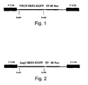
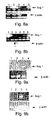
- Figure 1 illustrates a VEGF-GFP expression vector construct according to the teachings of the present invention.
- the recombinant retroviral vectors expressing the human VEGF 165 (GenBank Accession number AB021221) and/or the EGFP (Clontech, Ca, USA) genes were constructed by separately cloning two different vectors into the LXSN plasmid (Clontech, Ca, USA).
- LXSN-based retroviral vector expressing GFP alone an EcoRI-HpaI 1400 bp fragment containing an internal ribosome entry site (IRES) and the GFP gene was inserted into the specific restriction sites of the LXSN plasmid multiple cloning site (MCS).
- IVS internal ribosome entry site
- LXSN-VEGF-IRES-GFP bi-cistronic plasmid ( Figure 1 ) which co-expresses the VEGF 165 and the GFP genes, a 2.0 kB EcoRI-MunI fragment encoding VEGF 165 (600 bp), and the IRES and EGFP sequences were ligated into the EcoRI restriction site of the LXSN MCS. Expression of transgenes by these LXSN vectors is regulated by the MoMULV long terminal repeat (LTR) (Clontech, Ca, USA).
- LTR MoMULV long terminal repeat
- LXSN-GFP or LXSN-VEGF-GFP plasmid DNA were transfected into 293E3 ecotropic packaging cells and incubated for 48 hours, following which supernatant from confluent cultures of G418 (Gibco BRL, USA) resistant producer cells was collected, filtered (0.45 ⁇ m) and added to PA317 amphotropic packaging cells.
- G418 Gibco BRL, USA
- the transduced cells were exposed to G418 selection and culture supernatant was collected following 48 additional hours of culture and used to infect TEFLYGA amphotropic packaging cells which uniquely express the GALV envelope for high transduction efficiency of EC and resistance to human serum inactivation.
- Packaging cell lines were prepared according to known procedures. Detailed description of packaging cell lines can be found in the following references: Incorporation of fowl plague virus hemagglutinin into murine leukemia virus particles and analysis of the infectivity of the pseudotyped retroviruses. T. Hatziioannou, S. Valsesia-Wittmann, S.J. Russell and F.-L. Cosset. 1998. Journal of Virology, 72:5313-5317 . Inverse targeting of retroviral vectors: selective gene transfer in a mixed population of hematopoietic and non-hematopoietic cells. A.K. Fielding, M. Maurice, F.J. Morling, F.-L.
- EC are harvested from 5cm long human saphenous venin segments. Cells are harvested by collagenase digestion as previously described [27]. Cells from passages 3-9 are collected to ensure stability of cell characteristics. Cell identity is tested with immunohistochemistry for von Willebrand factor.
- HSVSMC human saphenous veins
- HLSMC internal mammary arteries
- DMEM Dulbecco's Modified Eagles Medium
- Bone marrow progenitor cell infection with VEGF encoding vectors Bone marrow progenitor cells are infected with recombinant adenoviral vectors encoding VEGF-GFP. Differentiation is tested in infected cells using morphology and immunohistochemistry for von Willebrand factor. For more details regarding cell harvesting and in vitro differentiation related to neovascularization see: Asahara T. et al., EMBO J 1999;18:3964-72 , and Huang XL, Takakura N, Suda T. Biochem Biophys Res Commun 1999, 264:133 .
- EC Transduction of human and sheep saphenous vein EC by retroviral vectors: EC were seeded (10 5 cells/35 mm dish) in fibronectin coated plates (Sigma, USA) and grown in M199 (Gibco-BRL, USA) containing 20% fetal serum. After 24 hours the cells were exposed to the cationic polymer DEAE-dextran (0.1 ⁇ g/ml) for 1 hour prior to transduction, washed 3 times with PBS and then transduced with a high titer of the recombinant virus vector encoding either the VEGF-LacZ or VEGF-GFP genes. Following incubation at 37 °C for 4 hours, virus-containing medium was replaced by fresh medium containing 20% pooled human serum.
- Figures 3a-b illustrate transgene expression from VEGF-LacZ or VEGF-GFP constructs, as indicated by LacZ activity or by GFP fluorescence, respectively.
- An effector regulatable expression system is used in order to regulate gene expression of VEGF and Ang-1 in EC and/or SMC.
- VEGF expression is downregulated via tetracycline, while Ang-1 expression is upregulated thereby.
- the administered cells are made to express VEGF in the first week when cell proliferation is needed, while in vivo administration of tetracycline 12 hours to 2 weeks following in vivo implantation of cells downregulates VEGF expression and upregulates Ang-1 expression to promote cell maturation ( Agha-Mohammadi S, Lotze MT. Regulatable systems: applications in gene therapy and replicating viruses. J Clinical Investigations 2000, 105:1177 ).
- VEGF and Ang-1 levels are measured in supernatant collected from cultures of genetically modified cells. ELISA and western blot analysis was used to measure VEGF or Ang-1 production over a 24 hour period. Following reinfusion of transformed cells in vivo into the donor, blood is collected and ELISA analysis of VEGF and Ang-1 levels is effected.
- the superficial femoral artery of each hind limb of the experimental animal is ligated immediately following bifurcation of the deep femoral artery. Blood flow is measured on both sides of the ligation using a Doppler flow meter (Transonic Animal Research Flowmeter, NY, USA). Following flow measurements, a population of homologous or heterologous vascular cells expressing VEGF and Ang-1 at various relative levels of expression are injected into one of the treated limbs, whereas a population of control cells expressing a marker polypeptide or PBS is injected into the second limb. Injection is performed proximally to the deep femoral artery. Flow is measured in both limbs 3, 7, 14 and 30 days following administration of the cells. Increased flow in the common femoral artery indicates the occurrence of angiogenesis.
- Figure 4a Three approaches can be utilized to treat an occluded blood vessel ( Figure 4a).
- Figure 4b demonstrates prior art bypass surgery for treating a single occluded blood vessel.
- Figure 4c represent treatment alternatives for bypassing or penetrating the occluded blood vessel using the teachings of the present invention.
- Figure 4c demonstrates angiogenesis in occluded tissue as a result of administration of vascular cells expressing at least one angiogenic proliferating factor and at least on angiogenic maturation factor into or around the occluded blood vessel. Such administration can lead to recanalization of the blood vessel, as indicated by 10, or to the formation of a bypass around the occlusion, as indicated by 20.
- angiogenesis according to the teachings of the present invention can also lead to the formation of a vascular bridge which interconnects the occluded vessel to an unoccluded blood vessel thus effectively traversing the occlusion and reestablishing blood flow to the target tissue fed by the occluded blood vessel.
- any of the above alternative methods can be utilized to restore blood supply to a tissue region fed by an occluded or injured blood vessel.
- the methods of the present invention can be utilized to restore blood supply even in cases of blood vessels which were previously treated via bypass surgery or angioplasty.
- the method of the present invention is designed to treat occlusive vascular disease by administration of EC genetically modified to induce optimal neovasculogenesis. Therefore, restoration of blood flow to occluded blood vessels via application of the method of the present invention was effected as follows.
- Rats Male or female Sprague-Dolly rats weighing 400-450 g (Harlan, Israel) were anesthetized using ketamine and xylazine. The femoral arteries of these rats were surgically ligated distally to a large perforating artery. Rats were then injected with recombinant adenoviral vector encoding VEGF 165 (10 9 pfu), recombinant adenoviral vector encoding GFP (10 9 pfu) or normal saline. Injection was performed directly into a side branch of the femoral artery proximal to the ligated segment ( Figure 5a ).
- Blood flow in the femoral artery was measured at 3, 7, and 14 days following injection using a Doppler flow meter. Flow measured in rats treated with recombinant adenoviral vector expressing VEGF was significantly higher than that measured in rats injected with the recombinant adenoviral vector encoding GFP or saline (see Figure 5b ). Localized expression of VEGF following injection was confirmed via RT-PCR of muscle tissue extracts and by measuring VEGF levels in blood plasma via ELISA (Data not shown).
- results demonstrate the effectiveness of the method of the present invention in treating occlusive vascular disease by inducing neovascularization of occluded blood vessels with EC genetically modified to overexpress pro-angiogenic factors.
- the genetically modified cells of the present invention can also be administered into an ischemic organ thus traversing the need for injecting potentially hazardous viral vectors into the blood stream, while enriching the ischemic organ with viable and activated vascular cells [40].
- vascular diseases such as occlusive vascular diseases
- inducement of neovascularization processes in vivo using prior art techniques remains an unfulfilled challenge.
- the method of the present invention is designed so as to induce such neovascularization via the harnessing of multiple pro-angiogenic stimuli employed by vascular cells during natural growth of blood vessels.
- pro-angiogenic stimulation is conferred by the pro-angiogenic differentiation factor Ang-1 and the EC growth factor VEGF.
- vascular cells expressing such factors thus represent an ideal therapeutic modality as these will both induce the differentiation of vascular tissues from precursor and their subsequent growth, thereby enabling the required therapeutic vessel formation.
- viral vectors for expression of Ang-1 and VEGF were constructed and expressed in EC and SMC as follows.
- Human Ang-1 cDNA (nucleotide coordinates 310-1806 of Genbank accession number U83508) was reverse transcribed using AMV reverse transcriptase (RT) (Promega, USA) from total RNA extracted from human saphenous vein SMC. Briefly, 2 ⁇ g of RNA was mixed with 500 ng random hexamers, the mixture was heated at 70°C for 10 minutes and cooled on ice. To this preheated RNA was added a reaction mixture containing 0.5 mM dNTPs, 5 units AMV-RT, 5 mM DTT, 32 units RNAse out (Gibco-BRL, USA) and 1x Promega RT buffer. The resultant mixture was incubated at 38°C for 2 hours followed by 95°C for 15 minutes.
- RT reverse transcriptase
- the cDNA product of the previous step was PCR-amplified using Expand High Fidelity PCR System (Roche, Switzerland).
- the following primers were employed for amplification: 5'- AGATCT TCAAAAATCTAAAGGTCGAAT-3' (SEQ ID NO:1) and 5'- AGATCT GCTGGCAGTACAATGACAGTT-3' (SEQ ID NO:2) (the underlined sequences represent the BglII restriction sites used for cloning).
- the PCR was performed in a volume of 20 ⁇ l containing 7 ⁇ l RT reaction mix cDNA product template, 20 pmole of each primer, 5nmole dNTPs, 1U Ex-Taq DNA Polymerase (Takara, Japan) and PCR reaction buffer provided by the manufacturer.
- the PCR reaction was performed as follows: 94°C/2' ⁇ 10 ⁇ : [94°C/30" ⁇ 50°C/30" ⁇ 72°C/1'] ⁇ 24 ⁇ : [94°C/30" ⁇ 56°C/30 ⁇ 72°C/(60" + 5"/cycle)] ⁇ 72°C/10'.
- the 1500 bp human Ang-1 cDNA (nucleotide coordinates 310-1806 of GenBank accession number U83508) was subcloned into pGEMT-easy vector (Promega). Both strands of the PCR product were sequenced and found to be 100% identical to the published sequence.
- a recombinant adenoviral vector for expression of the human Ang-1 gene was constructed in several steps, consisting of routine prokaryotic cloning and a homologous recombination procedure in the 293-cell line.
- a 1500 bp fragment containing the human Ang-1 cDNA was inserted into pCA3 plasmid, under the control of the constitutive CMV immediate early promoter.
- the resultant plasmid was co-transfected with plasmid pJM17 into 293 cells which constitutively express the adenoviral E1 gene.
- Plasmid pJM17 contains the adenovirus genome, excluding the E3 region, and including an insert (pBRX) in the E1 region of the virus. Homologous recombination between pCA3 encoding Ang-1 and pJM17 following transfection replaced the E1 region of pJM17 with the CMV-Ang-1 expression cassette from pCA3. Plaque formation occurred 2 to 4 weeks following co-transfection after which individual plaques were isolated and viral extracts were amplified therefrom by infection of 293 cells. The titer of each viral stock was determined by plaque assay in 293 cells and viral titers of ⁇ 10 11 pfu/ml were obtained.
- Ang-1 protein expressed in the growth medium of transformed cells was confirmed by Western blot analysis.
- Recombinant adenoviral vector rAdAng1-GFP encoding the human Ang-1 and GFP genes was constructed using a modified AdEasy protocol ( He TC. et al., Proc Natl Acad Sci USA 1998, 95:2509 ). Briefly, the 1500 bp BglII fragment of human Ang-1 cDNA was inserted into the BglII site in pAdTrack-CMV shuttle vector for expression under the control of CMV promoter. This shuttle vector encodes the GFP gene downstream of the transgene under the control of a second CMV promoter.
- the Ang-1-GFP encoding shuttle vector was linearized by PmeI digestion and purified by Qiaquick Gel Extraction Kit (Qiagen, Germany). Competent BJ5183 cells were co-transformed with linearized Ang-1- and GFP-encoding shuttle vector and adenoviral vector pAdEasy-1 by electroporation. Transformants expressing recombinant adenoviral vector encoding Ang-1 and GFP genes were identified by PCR analysis and restriction mapping. Recombinant adenoviral plasmid vector was linearized by PacI digestion, purified and transfected into 293 cells using Lipofectamine 2000 (Gibco BRL, USA).
- Recombinant retroviral vector pLXSN-Ang-1 expressing the human Ang-1 gene under the regulatory control of Mo-MULV 5' long terminal repeat (LTR) was constructed by cloning the 1500 bp human Ang-1 BglII cDNA fragment into the BamHI site of retroviral plasmid vector pLXSN (# K1060-B, Clontech, USA).
- a recombinant bi-cistronic retroviral vector encoding both the human Ang-1 and EGFP genes under the regulatory control of Mo-MULV 5' long terminal repeat (LTR) was constructed by cloning these genes into pLXSN retroviral plasmid vector in a two-step process, as follows. First, an internal ribosomal entry site (IRES)-EGFP EcoRI-HpaI fragment (1400 bp) excised from pIRES2-EGFP (#6029-1 Clontech) was inserted into the EcoRI-HpaI sites of pLXSN for construction of the control plasmid pLXSN-IRES-EGFP. In a second step, pLXSN-Ang-1-IRES-EGFP was constructed by cloning the human Ang-1 1361 bp EcoRI fragment into the EcoRI site of pLXSN-IRES-EGFP.
- IVS internal ribosomal entry site
- pseudotyped retroviruses for expression of the human Ang-1 gene For retroviral vector production, 10 ⁇ g of pLXSN-IRES-EGFP, pLXSN-Ang-1-IRES-EGFP or pLXSN-Ang-1 retroviral vector plasmid was transfected into 293FLYA packaging cells using Lipofectamine (Gibco BRL, USA). After 48 hours supernatant from confluent cultures of viral producer cells was collected, 0.45 ⁇ m-filtered and added to 293FLY10A or 293FLYGALV amphotropic packaging cells.
- transduced cells were subjected to G418 selection, individual colonies were collected and screened for EGFP expression by fluorescence microscopy and Ang-1 expression was confirmed by Western blot analysis of transduced cell-conditioned medium samples.
- Viral titers determined by transduction of TE671 cells, were found to range from 10 5 to 10 6 pfu/ml. Supernatant from the colonies with the highest viral titers was collected and frozen at -80°C.
- HSVEC Human saphenous vein EC
- M199 medium Gibco-BRL, USA
- Peptomycin Biological Industries, Israel
- 100 ⁇ g/ml heparin Sigma, USA
- 2 ng/ml bFGF kindly obtained from Prof. Neufeld, The Technion Institute, Haifa Israel.
- Human EC were identified by immunohistochemical analysis with anti von Willebrand factor antibodies (Zymed, USA).
- Smooth muscle cells were cultured by explant outgrowth from human saphenous vein SMC (HSVSMC) and human left internal mammary artery SMC (HLSMC). Cells were cultured in Dulbecco's Modified Eagles Medium (DMEM) (Gibco-BRL, USA) supplemented with 10% human serum, 2 mM L-glutamine, 100 units/ml penicillin, 0.1 mg/ml streptomycin and 2 ng/ml bFGF. Smooth muscle cells were identified by immunohistochemical analysis using anti-smooth muscle ⁇ -actin antibodies (Zymed, USA).
- DMEM Dulbecco's Modified Eagles Medium
- the packaging cell lines 293FLYA, 293FLY10A, 293FLYGALV and TEFLYGA were grown in DMEM supplemented with 10% FCS (Biologoical Industries, Israel), 2 mM L-glutamine, 100 units/ml penicillin, 0.1 mg/ml streptomycin, 6 ⁇ g/ml blasticidin (Sigma, USA) and 6 ⁇ g/ml phleomicin (Sigma, USA).
- the packaging cell lines PA317, 293E3 (obtained from Dr. J. Exelrod, Jerusalem) were grown in DMEM supplemented with 10% FCS, 2 mM L-glutamine, 100 units/ml penicillin, and 0.1 mg/ml streptomycin.
- Endothelial cells and SMC were infected with recombinant adenoviral vectors as follows. Briefly, cells were seeded at ⁇ 70% confluence in fibronectin (4.5 ⁇ g/ml)-pre-coated plates 20 hours prior to infection and cultured in complete medium (M20). On the day of infection the medium was replaced with fresh M199 (serum-free) medium and cells were infected with recombinant virus at 3000 viral particles/cell. The cells were incubated for 90 minutes with gentle tilting every 20 minutes after which the virus-containing medium was replaced with complete medium (M20). The infection rate was determined by visualization of GFP expression using an inverted fluorescence microscope (TE200 Nikon, Japan) equipped with a GFP fluorescence filter (GFP-LP, Nikon).
- TE200 inverted fluorescence microscope
- GFP-LP GFP fluorescence filter
- Transduction of EC and SMC with retroviral vectors was performed as follows. Briefly, endothelial cells (passage 4-9) were seeded at 10 5 cells/35 mm well in fibronectin (4.5 ⁇ g/ml)-coated plates and grown in complete medium for 24 h. One hour prior to transduction, the culture medium was replaced with serum-free M199 medium containing 0.1 mg/ml DEAE-dextran. Cells were washed three times with PBS and transduction was performed by incubating the cells for 4 hours with freshly collected and filtered (0.45 ⁇ m) supernatant from cultures of virus-producing packaging cells. At the end of the incubation the medium was replaced with fresh M20 medium.
- PURESCRIPT RNA Isolation Kit Gentra systems, USA.
- 1 ⁇ g of RNA was mixed with 500 ng random hexamers heated at 70°C for 10 minutes after which the mixture was cooled on ice.
- Reverse-transcription was performed by incubating the reaction mixture at 38°C for 2 h followed by an incubation at 95°C for 15 min.
- PCR analysis of cDNA was performed in a volume of 20 ⁇ l using 7 ⁇ l of reverse transcribed cDNA product, 20 pmol sense primer (5'-GCTGGCAGTACAATGACAGTT-3', SEQ ID NO:3), 20 pmole anti-sense primer (5'-TCAAAAATCTAAAGGTCGAAT-3', SEQ ID NO:4), 5 nmol dNTPs, 1U Ex-Taq DNA polymerase (Takara, Japan) and PCR buffer provided by the manufacturer.
- the PCR was performed using the following set of incubations: 94°C/2' ⁇ 10x: [94°C/30" ⁇ 50°C/30" ⁇ 72°C/1'] ⁇ 24x: [94°C/30" ⁇ 56°C/30” ⁇ 72°C/(60" + 5"/cycle)] ⁇ 72°C/10'.
- PCR analysis of cDNA was performed in a volume of 20 ⁇ l containing 7 ⁇ l of reverse transcribed cDNA product, 10 pmol sense and anti-sense primer (5'-GCTGGCAGTACAATGACAGTT-3', SEQ ID NO:5 and 5'-TCAAAAATCTAAAGGTCGAAT-3', SEQ ID NO:6, respectively), 5 nmol dNTPs, 1U Ex-Taq DNA polymerase (Takara, Japan) and PCR buffer provided by the manufacturer.
- PCR was performed using the following set of incubations: 94°C/2' ⁇ 10 ⁇ : [94°C/30" ⁇ 50°C/30" ⁇ 72°C/1'] ⁇ 21x: [94°C/30" ⁇ 60°C/30” ⁇ 72°C/(60" + 5"/cycle)] ⁇ 72°C/10'.
- PCR products were separated by electrophoresis in 0.8% agarose, labelled with EtBr and visualized by UV fluorescence.
- Western blot analysis Detection of Ang-1 or VEGF 165 protein expression by adenovirally or retrovirally infected EC and SMC was performed by Western blot analysis of cell-conditioned medium 24 h post-infection, as follows. Briefly, virus-containing culture medium was replaced with serum-free medium and cells were grown for an additional 24 h. Secreted proteins in 30 ⁇ l samples of cell-conditioned medium were separated by PAGE in 10% SDS-polyacrylamide gel under reducing conditions and electroblotted onto nitrocellulose membrane (Schleicher & Schuell, Germany).
- Blots were blocked by incubation in TBS containing 0.1% skim milk and 0.3% Tween-20 (TBST) at room temperature for 1 hour with gentle agitation after which blots were incubated at room temperature for 2 hours with a 1:500 dilution of polyclonal goat anti-Ang-1 antibody (#sc-6319 Santa-Cruz, USA) or a 1:700 dilution of polyclonal rabbit anti-VEGF 165 antibody (#SC 152 Santa Cruz, USA) in blocking solution.
- TBS polyclonal goat anti-Ang-1 antibody
- Tween-20 Tween-20
- Viral constructs The adenoviral and retroviral vectors constructed and employed to infect EC and SMC with Ang-1 are listed in Table 1.
- Table 1 Viral system Gene(s) encoded Virus designation Adenovirus GFP rAdGFP Adenovirus Ang-1 rAdAng1 Adenovirus Ang-1, GFP rAdAng1-GFP Adenovirus VEGF 165 , GFP rAdVEGF-GFP Pseudotyped Retrovirus GFP retroGFP Pseudotyped Retrovirus Ang-1 retroAng1 Pseudotyped Retrovirus Ang-1, GFP retroAng1-GFP Pseudotyped Retrovirus VEGF 165 , GFP retroVEGF-GFP
- Adenoviral infected or retroviral transduced EC and SMC express Ang-1-GFP transgene: Endothelial cells and SMC infected with rAdAng1-GFP were found to express high-levels of GFP ( Figures 6b and 6d , respectively). Similarly, EC and SMC transduced with retroAng1-GFP were also found to express high-levels of GFP ( Figures 7b and 7d , respectively). Expression of GFP was visualized by fluorescence microscopy.
- Endothelial cells and SMC were found to transcribe Ang-1 mRNA 48 h following adenoviral infection with rAdAng1-GFP ( Figures 8a and 8b , respectively).
- EC and SMC retrovirally transduced with retroAng1-GFP were also found to express Ang-1 mRNA 48 h following transduction ( Figures 9a and 9b , respectively).
- Endothelial cells and SMC were found to overexpress Ang-1 protein 24 h following adenoviral infection with rAdAng1 or following retroviral transduction with retroAng1 ( Figures 10 and 11 , respectively).
- adenoviral and retroviral vectors of the present invention are competent to genetically modify primary human EC and SMC and to drive expression of Ang-1 and GFP transgenes therein.
- these constructs can be employed to genetically modify human vascular cells to overexpress the vascular maturation factor Ang-1 and thus may be effectively applied towards treatment of vascular diseases aimed at inducing the formation of new blood vessels capable of bypassing or penetrating occluded, injured or ischemic mammalian tissue.
- the method of the present invention employs administration of vascular cells co-overexpressing the angiogenic factors Ang-1 and VEGF.
- vascular cells co-overexpressing the angiogenic factors Ang-1 and VEGF co-overexpressing the angiogenic factors Ang-1 and VEGF.
- the method of the present invention employs overexpression of both the vascular maturation factor Ang-1 and the angiogenic factor VEGF in genetically modified vascular cells, such as EC and SMC in order to optimize differentiation and growth of such cells so as to mediate optimal therapeutic effect when utilizing such cells for treatment of vascular disease.
- Proliferation assays Twenty-four hours prior to adenoviral infection, EC (passages 5-11) were seeded at 10 4 cells/well ( ⁇ 30% confluence) in 24-well plates pre-coated with fibronectin (4.5 mg/ml). Cells were infected with rAdAng1-GFP or rAdGFP and incubated at 37°C for 90 minutes after which serum-supplemented medium was added. After 16-18 hrs, the medium was substituted with M199 supplemented 2% human serum and 2 ng/ml bFGF. Proliferation assays were performed in triplicate and proliferation was assessed via XTT colorimetric assay on Days 3, 5, and 7 post-infection.
- the method of the present invention includes therapeutic modalities for treatment of vascular disease in which Ang-1 and VEGF are co-overexpressed by vascular cells such as EC in the same milieu.
- vascular cells such as EC in the same milieu.
- the proliferative effect of VEGF on EC is preferably required for optimal effect, the absence of inhibitory effect of Ang-1 overexpression on the proliferation of rAdAng1-GFP infected EC was examined.
- Such overexpression of Ang-1 in EC was found to have neither inhibitory nor stimulatory effect on cellular proliferation when compared to the proliferation rate of cells infected with rAdGFP ( Figure 14 ).
- proliferation of positive control infectants overexpressing VEGF was enhanced.
- the method of the present invention employs viral constructs for overexpression of Ang-1 in EC and includes modalities in which Ang-1 and VEGF are co-overexpressed to induce neovasculogenesis in the treatment of vascular diseases.
- Ang-1 expressing adenoviral vectors of the present invention to drive expression of biologically active Ang-1 protein and the capacity of the method of the present invention to optimally induce neovasculogenesis via co-overexpression of Ang-1 and VEGF in vascular cells, such as EC and SMC, were demonstrated as follows.
- Immunoprecipitation-immunoblotting analysis of Tie2 phosphorylation in EC overexpressing Ang-1 Endothelial cells were seeded in 60 mm plates pre-coated with fibronectin (4.5 ⁇ g/ml) and infected with rAdAng1-GFP, rAdGFP, or rAdVEGF-GFP. Twenty-four hours post-infection, virus-containing medium was substituted with serum-free medium and the cells were cultured for an additional 24 hours prior to immunoprecipitation of cellular protein.
- Cells were washed in cold PBS supplemented with 100 ⁇ M Na 3 VO 4 and lysed in lysis buffer containing 20 mM Tris-HCl (pH 7.5), 150 mM NaCl, 1% Triton X-100, 10% glycerol, 1 mM PMSF, 2 ⁇ g/ml leupeptin, 2 ⁇ g/ml aprotinin and 100 ⁇ M Na 3 VO 4 . Protein content of lysate supernatants was determined and lysate supernatant aliquots containing an equal quantity of protein were labelled with anti-phosphotyrosine antibody PY20 (Transduction Laboratories, USA) by incubation at 4°C overnight.
- PY20 Transduction Laboratories, USA
- Protein-A sepharose beads were added to the antibody-labelled samples and immunoprecipitation was performed by incubating the samples at 4°C for 2 h. The beads were subsequently washed 4 times in cold PBS, boiled in SDS-PAGE sample buffer for 3 min and pelleted by centrifugation. Supernatant proteins were separated electrophoretically in a 6% SDS-polyacrylamide gel. Separated proteins were electroblotted onto a nitrocellulose membrane (Schleicher and Schuell, Germany) and probed by Western immunoblotting analysis with anti-Tie2 antibodies (Santa Cruz, USA).
- the method of the present invention employs the use of both the vascular maturation and angiogenic factors Ang-1 and VEGF, respectively, in order to stimulate optimal angiogenesis for treatment of vascular diseases.
- Optimal angiogenesis by vascular cells, such as EC and SMC, overexpressing both of these factors was therefore demonstrated via analysis of three-dimensional sprouting of collagen gel-embedded co-culture spheroids containing virally transformed vascular cells as follows.
- Sprouting (the in vitro equivalent of angiogenesis) by adenovirally infected spheroids of EC or by co-cultured spheroids of adenovirally infected EC and SMC was assessed in vitro by analyzing the sprouting of such spheroids in collagen gels, as previously described ( Korff T. et al., J Cell Biol. 1998, 143:1341 ). Briefly, EC and SMC, pooled where applicable for generation of co-culture spheroids, were suspended in culture medium containing 0.25% (w/v) carboxymethylcellulose (Sigma, USA).
- EC were tagged with DiI-291 red fluorescent marker prior to mixing so as to enable differentiation of EC from SMC for subsequent fluorescence microscopic analysis of spheroids.
- Cell suspensions were seeded in non-adherent round-bottomed 96-well plates (Nunc, Denmark) so as to form a single spheroid of ⁇ 750 cells per spheroid per well following 24 h of culture. The spheroids thus generated were then embedded in collagen gels, as follows.
- Collagen stock solution was prepared freshly prior to use by mixing 8 volumes of acidic extract of rat tail collagen (equilibrated to 2 mg/ml at 4°C) with 1 volume of 10x M199 medium (Gibco BRL, USA) and 1 volume of 0.34 N NaOH to adjust the pH to 7.4. Collagen stock solution was mixed at room temperature with an equal volume of M199 medium supplemented with 40% human serum containing 0.25% (w/v) carboxymethylcellulose to prevent spheroid sedimentation prior to polymerization of the gel. Gel samples containing 20-30 spheroids were rapidly transferred into pre-warmed 24-well plates and allowed to polymerize. The gels were incubated at 37°C, 5% CO 2 and the sprouting of at least 20 spheroids from each sample group was documented by digital video camera (DXM1200 Nikon, Japan).
- Co-culture of Ang-1 and VEGF overexpressing vascular cells induces optimal angiogenesis: Spheroids of rAdGFP infected EC ( Figure 16a-b ) were observed, by fluorescent microscopic analysis, to display a low baseline sprouting activity, which was observed to be upregulated in co-culture spheroids containing rAdGFP infected and rAdVEGF-GFP infected EC ( Figure 16c-d ) as well as rAdAng1-GFP infected and rAdGFP infected EC ( Figure 16e-f ). The highest sprouting activity, however, was recorded in co-culture-spheroids containing rAdAng1-GFP and rAdVEGF-GFP infected EC ( Figure 16g-h ).
- Co-culture spheroids containing rAdGFP infected EC and retroAng1-GFP infected SMC displayed highly upregulated angiogenic sprouting activity ( Figure 17c ) however co-culture spheroids containing rAdVEGF-GFP infected EC and retroAng1-GFP infected SMC displayed synergistically increased levels of angiogenic sprouting activity ( Figure 17d ).
- the method of the present invention is capable of inducing optimal angiogenesis and cell survival.
- the method of the present invention is superior to prior art techniques employing only one type of angiogenic factor for inducing neovascularization to treat vascular diseases.
- Ang-1 overexpression mediates protection from apoptosis in genetically modified EC
- the method of the present invention employs the genetic modification of vascular cells, such as EC, to overexpress the EC differentiation factor Ang-1 in conjunction with overexpression of the EC growth factor VEGF in order to induce optimal neovasculogenesis for treatment of vascular disease.
- vascular cells such as EC
- VEGF EC growth factor
- TdT Terminal deoxynucleotide transferase
- TUNEL Terminal deoxynucleotide transferase assay of apoptotic cells
- Ang-1 overexpression mediates protection from apoptosis in genetically modified EC: Endothelial cells retrovirally transduced to overexpress Ang-1 display marked resistance to apoptosis under pro-apoptotic conditions as compared to non-transduced cells ( Figures 19h and 19d ), as visualized by microscopic fluorescent analysis of TUNEL stained cells. Numerical quantification performed on the data demonstrated microscopically above indicated that 55% of non-transduced EC were found to be apoptotic after 24 hrs of serum deprivation versus only 12% of EC retrovirally transduced to overexpress Ang-1 ( Figure 20 ). Similar effects of reduced rate of apoptosis and improved survival were also observed in mixed populations of endothelial cells over expressing VEGF and Ang-1 (data not shown).
- the present invention represents an improvement over prior art neovascularization methods to treat vascular diseases in general and in particular in pathologies involving ischemic tissues.
Landscapes
- Health & Medical Sciences (AREA)
- Life Sciences & Earth Sciences (AREA)
- Chemical & Material Sciences (AREA)
- Organic Chemistry (AREA)
- Genetics & Genomics (AREA)
- General Health & Medical Sciences (AREA)
- Molecular Biology (AREA)
- Engineering & Computer Science (AREA)
- Zoology (AREA)
- Medicinal Chemistry (AREA)
- Gastroenterology & Hepatology (AREA)
- Proteomics, Peptides & Aminoacids (AREA)
- Biophysics (AREA)
- Biochemistry (AREA)
- Toxicology (AREA)
- Animal Behavior & Ethology (AREA)
- Public Health (AREA)
- Veterinary Medicine (AREA)
- Biomedical Technology (AREA)
- Bioinformatics & Cheminformatics (AREA)
- Epidemiology (AREA)
- Biotechnology (AREA)
- General Engineering & Computer Science (AREA)
- Nuclear Medicine, Radiotherapy & Molecular Imaging (AREA)
- Pharmacology & Pharmacy (AREA)
- Wood Science & Technology (AREA)
- Virology (AREA)
- Heart & Thoracic Surgery (AREA)
- Plant Pathology (AREA)
- Radiology & Medical Imaging (AREA)
- Microbiology (AREA)
- Pathology (AREA)
- Cardiology (AREA)
- Physics & Mathematics (AREA)
- Chemical Kinetics & Catalysis (AREA)
- General Chemical & Material Sciences (AREA)
- Urology & Nephrology (AREA)
- Vascular Medicine (AREA)
- Micro-Organisms Or Cultivation Processes Thereof (AREA)
- Medicines Containing Material From Animals Or Micro-Organisms (AREA)
Applications Claiming Priority (2)
| Application Number | Priority Date | Filing Date | Title |
|---|---|---|---|
| US22372700P | 2000-08-08 | 2000-08-08 | |
| EP01958342A EP1307582B1 (en) | 2000-08-08 | 2001-08-08 | Nucleic acid constructs, vascular cells transformed therewith, pharmaceutical compositions and methods utilizing same for inducing angiogenesis |
Related Parent Applications (2)
| Application Number | Title | Priority Date | Filing Date |
|---|---|---|---|
| WOPCT/IL01/00733 Previously-Filed-Application | 2001-08-08 | ||
| EP01958342.6 Division | 2001-08-08 |
Publications (1)
| Publication Number | Publication Date |
|---|---|
| EP2163642A1 true EP2163642A1 (en) | 2010-03-17 |
Family
ID=22837748
Family Applications (2)
| Application Number | Title | Priority Date | Filing Date |
|---|---|---|---|
| EP01958342A Expired - Lifetime EP1307582B1 (en) | 2000-08-08 | 2001-08-08 | Nucleic acid constructs, vascular cells transformed therewith, pharmaceutical compositions and methods utilizing same for inducing angiogenesis |
| EP08156995A Withdrawn EP2163642A1 (en) | 2000-08-08 | 2001-08-08 | Nucleic acid constructs, vascular cells transformed therewith, pharmaceutical compositions and methods utilizing same for inducing angiogenesis |
Family Applications Before (1)
| Application Number | Title | Priority Date | Filing Date |
|---|---|---|---|
| EP01958342A Expired - Lifetime EP1307582B1 (en) | 2000-08-08 | 2001-08-08 | Nucleic acid constructs, vascular cells transformed therewith, pharmaceutical compositions and methods utilizing same for inducing angiogenesis |
Country Status (13)
| Country | Link |
|---|---|
| US (1) | US7767201B2 (ja) |
| EP (2) | EP1307582B1 (ja) |
| JP (1) | JP2004520009A (ja) |
| AT (1) | ATE397087T1 (ja) |
| AU (1) | AU2001280060A1 (ja) |
| CA (1) | CA2418936A1 (ja) |
| CZ (1) | CZ2003444A3 (ja) |
| DE (1) | DE60134234D1 (ja) |
| HU (1) | HUP0300727A3 (ja) |
| IL (1) | IL154324A0 (ja) |
| PL (1) | PL366000A1 (ja) |
| WO (1) | WO2002012539A2 (ja) |
| ZA (1) | ZA200301386B (ja) |
Cited By (1)
| Publication number | Priority date | Publication date | Assignee | Title |
|---|---|---|---|---|
| EP3995569A1 (en) * | 2020-11-10 | 2022-05-11 | Vessl Therapeutics Ltd | Methods of preconditioning vascular cells for transduction, methods of transduction and methods of preserving transduced cells |
Families Citing this family (8)
| Publication number | Priority date | Publication date | Assignee | Title |
|---|---|---|---|---|
| US7387645B2 (en) * | 2003-04-25 | 2008-06-17 | Medtronic Vascular, Inc. | Cellular therapy to heal vascular tissue |
| US20090304650A1 (en) * | 2005-05-11 | 2009-12-10 | Scott Edward W | Repair of the Bone Marrow Vasculature |
| WO2007059303A2 (en) * | 2005-11-15 | 2007-05-24 | The Board Of Trustees Of The Leland Stanford Junior University | Compositions for coordinated vegf and pdgf expression, and methods of use |
| JP5121155B2 (ja) * | 2006-03-14 | 2013-01-16 | 独立行政法人物質・材料研究機構 | 形質転換細胞集団の作成方法 |
| US9598673B2 (en) * | 2006-05-19 | 2017-03-21 | Creative Medical Health | Treatment of disc degenerative disease |
| US7943590B2 (en) * | 2007-03-16 | 2011-05-17 | Multi-Gene Vascular Systems Ltd. | Compositions and methods for treating ophthalmic disorders |
| CN102242148A (zh) * | 2010-10-09 | 2011-11-16 | 苏州大学 | 重组载体以及转基因骨髓基质细胞修饰的丝素膜及其应用 |
| EP3329779B1 (en) * | 2012-11-02 | 2019-06-05 | Cornell University | Angiogenic conditioning to enhance cardiac cellular reprogramming of fibroblasts of the infarcted myocardium |
Citations (2)
| Publication number | Priority date | Publication date | Assignee | Title |
|---|---|---|---|---|
| US4950483A (en) * | 1988-06-30 | 1990-08-21 | Collagen Corporation | Collagen wound healing matrices and process for their production |
| WO2000037642A1 (en) * | 1998-12-23 | 2000-06-29 | Regeneron Pharmaceuticals, Inc. | Method of enhancing the biological activity of ligands |
Family Cites Families (26)
| Publication number | Priority date | Publication date | Assignee | Title |
|---|---|---|---|---|
| US4900556A (en) * | 1985-04-26 | 1990-02-13 | Massachusetts Institute Of Technology | System for delayed and pulsed release of biologically active substances |
| US5674722A (en) | 1987-12-11 | 1997-10-07 | Somatix Therapy Corporation | Genetic modification of endothelial cells |
| US6001350A (en) | 1987-12-11 | 1999-12-14 | Somatix Therapy Corp | Genetic modification of endothelial cells |
| US5219739A (en) * | 1989-07-27 | 1993-06-15 | Scios Nova Inc. | DNA sequences encoding bVEGF120 and hVEGF121 and methods for the production of bovine and human vascular endothelial cell growth factors, bVEGF120 and hVEGF121 |
| US5980885A (en) * | 1991-07-08 | 1999-11-09 | Neurospheres Holdings Ltd. | Growth factor-induced proliferation of neural precursor cells in vivo |
| ATE314479T1 (de) * | 1992-05-18 | 2006-01-15 | Genentech Inc | Aktivierung von rezeptoren fähig zur oligomerisierung durch verwendung von fusionierten rezeptor liganden |
| US5618544A (en) * | 1992-08-12 | 1997-04-08 | Bays-Brown Dermatologics, Inc. | Method of decreasing cutaneous senescence |
| DE4228457A1 (de) * | 1992-08-27 | 1994-04-28 | Beiersdorf Ag | Herstellung von heterodimerem PDGF-AB mit Hilfe eines bicistronischen Vektorsystems in Säugerzellen |
| US6007987A (en) | 1993-08-23 | 1999-12-28 | The Trustees Of Boston University | Positional sequencing by hybridization |
| US5962427A (en) * | 1994-02-18 | 1999-10-05 | The Regent Of The University Of Michigan | In vivo gene transfer methods for wound healing |
| US5821919A (en) * | 1994-04-29 | 1998-10-13 | Intel Corporation | Apparatus for table-driven conversion of pixels from YVU to RGB format |
| US6090618A (en) * | 1996-10-07 | 2000-07-18 | Arch Development Corporation | DNA constructs and viral vectors comprising a smooth muscle promoter |
| US5641750A (en) * | 1995-11-29 | 1997-06-24 | Amgen Inc. | Methods for treating photoreceptors using glial cell line-derived neurotrophic factor (GDNF) protein product |
| JP2000502682A (ja) | 1995-12-22 | 2000-03-07 | ローカルメッド インコーポレイテッド | 血管新生を促進する成長因子の局所的血管内デリバリー |
| US5785965A (en) * | 1996-05-15 | 1998-07-28 | The Board Of Trustees Of The Leland Stanford Junior Univ. | VEGF gene transfer into endothelial cells for vascular prosthesis |
| CN1244772A (zh) * | 1996-10-23 | 2000-02-16 | 威克福雷大学 | 靶向性细胞毒性细胞 |
| FI970784A (fi) * | 1997-02-25 | 1998-08-26 | Nokia Telecommunications Oy | Prosessien välinen vuonvalvonta hajautetussa moniprosessoriympäristössä |
| US6099832A (en) * | 1997-05-28 | 2000-08-08 | Genzyme Corporation | Transplants for myocardial scars |
| CA2225185A1 (en) * | 1997-08-11 | 1999-02-11 | Peter K. Law | Myoblast transfer therapy for relieving pain and for treating behavioral and perceptive abnormalities |
| CA2266805C (en) * | 1998-03-27 | 2015-04-28 | An-Go-Gen Inc. | Cell-based gene therapy in the treatment of pulmonary disorders |
| US6206914B1 (en) * | 1998-04-30 | 2001-03-27 | Medtronic, Inc. | Implantable system with drug-eluting cells for on-demand local drug delivery |
| EP1016726A1 (en) * | 1998-12-30 | 2000-07-05 | Introgene B.V. | Gene therapy to promote angiogenesis |
| US6328762B1 (en) * | 1999-04-27 | 2001-12-11 | Sulzer Biologics, Inc. | Prosthetic grafts |
| US6554857B1 (en) * | 1999-07-20 | 2003-04-29 | Medtronic, Inc | Transmural concentric multilayer ingrowth matrix within well-defined porosity |
| AU2001291019A1 (en) | 2000-09-15 | 2002-03-26 | Genvec, Inc. | Method of modulating neovascularization |
| US20030027751A1 (en) * | 2001-04-10 | 2003-02-06 | Genvec, Inc. | VEGF fusion proteins |
-
2001
- 2001-08-08 CA CA002418936A patent/CA2418936A1/en not_active Abandoned
- 2001-08-08 CZ CZ2003444A patent/CZ2003444A3/cs unknown
- 2001-08-08 AU AU2001280060A patent/AU2001280060A1/en not_active Abandoned
- 2001-08-08 PL PL01366000A patent/PL366000A1/xx unknown
- 2001-08-08 DE DE60134234T patent/DE60134234D1/de not_active Expired - Lifetime
- 2001-08-08 EP EP01958342A patent/EP1307582B1/en not_active Expired - Lifetime
- 2001-08-08 HU HU0300727A patent/HUP0300727A3/hu unknown
- 2001-08-08 IL IL15432401A patent/IL154324A0/xx unknown
- 2001-08-08 JP JP2002517823A patent/JP2004520009A/ja active Pending
- 2001-08-08 WO PCT/IL2001/000733 patent/WO2002012539A2/en active IP Right Grant
- 2001-08-08 AT AT01958342T patent/ATE397087T1/de not_active IP Right Cessation
- 2001-08-08 EP EP08156995A patent/EP2163642A1/en not_active Withdrawn
-
2003
- 2003-02-20 ZA ZA200301386A patent/ZA200301386B/en unknown
- 2003-05-02 US US10/429,093 patent/US7767201B2/en not_active Expired - Fee Related
Patent Citations (2)
| Publication number | Priority date | Publication date | Assignee | Title |
|---|---|---|---|---|
| US4950483A (en) * | 1988-06-30 | 1990-08-21 | Collagen Corporation | Collagen wound healing matrices and process for their production |
| WO2000037642A1 (en) * | 1998-12-23 | 2000-06-29 | Regeneron Pharmaceuticals, Inc. | Method of enhancing the biological activity of ligands |
Non-Patent Citations (3)
| Title |
|---|
| ASAHARA T ET AL: "TIE2 RECEPTOR LIGANDS, ANGIOPOIETIN-1 AND ANGIOPOIETIN-2, MODULATE VEGF-INDUCED POSTNATAL NEOVASCULARIZATION", CIRCULATION RESEARCH, GRUNE AND STRATTON, BALTIMORE, US, vol. 83, no. 3, 1998, pages 233 - 240, XP000907290, ISSN: 0009-7330 * |
| JAIN R K ET AL: "Leaky vessels? Call Ang 1!", NATURE MEDICINE, NATURE PUBLISHING, CO, US, vol. 6, no. 2, February 2000 (2000-02-01), pages 131 - 132, XP002186836, ISSN: 1078-8956 * |
| THURSTON G ET AL: "Leakage-resistant blood vessels in mice transgenically overexpressing angiopoietin-1", SCIENCE, AMERICAN ASSOCIATION FOR THE ADVANCEMENT OF SCIENCE, WASHINGTON, DC; US, vol. 286, no. 5449, 24 December 1999 (1999-12-24), pages 2511 - 2514, XP002357065, ISSN: 0036-8075 * |
Cited By (1)
| Publication number | Priority date | Publication date | Assignee | Title |
|---|---|---|---|---|
| EP3995569A1 (en) * | 2020-11-10 | 2022-05-11 | Vessl Therapeutics Ltd | Methods of preconditioning vascular cells for transduction, methods of transduction and methods of preserving transduced cells |
Also Published As
| Publication number | Publication date |
|---|---|
| EP1307582A4 (en) | 2004-09-22 |
| IL154324A0 (en) | 2003-09-17 |
| AU2001280060A1 (en) | 2002-02-18 |
| US7767201B2 (en) | 2010-08-03 |
| HUP0300727A3 (en) | 2005-12-28 |
| ATE397087T1 (de) | 2008-06-15 |
| JP2004520009A (ja) | 2004-07-08 |
| WO2002012539A3 (en) | 2002-05-16 |
| CZ2003444A3 (cs) | 2003-10-15 |
| HUP0300727A2 (hu) | 2003-10-28 |
| EP1307582A2 (en) | 2003-05-07 |
| CA2418936A1 (en) | 2002-02-14 |
| EP1307582B1 (en) | 2008-05-28 |
| ZA200301386B (en) | 2004-03-31 |
| US20040151707A1 (en) | 2004-08-05 |
| WO2002012539A2 (en) | 2002-02-14 |
| PL366000A1 (en) | 2005-01-24 |
| DE60134234D1 (de) | 2008-07-10 |
Similar Documents
| Publication | Publication Date | Title |
|---|---|---|
| EP1311699B1 (en) | Adeno-associated virus-mediated delivery of angiogenic factors | |
| EP1968565B1 (en) | Compositions and methods for inhibiting angiogenesis | |
| JP2001501471A (ja) | 血管新生の阻害のための遺伝子治療 | |
| EP1017421A1 (en) | Method and compositions for healing tissue defects and inducing hypervascularity in mammalian tissue | |
| EP1307582B1 (en) | Nucleic acid constructs, vascular cells transformed therewith, pharmaceutical compositions and methods utilizing same for inducing angiogenesis | |
| EP1370299B1 (en) | Nucleic acid constructs expressing vegf and hgf, cells transformed therewith and their use for inducing liver regeneration and alleviation of portal hypertension | |
| US7247618B2 (en) | Methods for inhibiting macrophage colony stimulating factor and c-FMS-dependent cell signaling | |
| WO2019027299A2 (ko) | 간세포성장인자를 발현하는 중간엽줄기세포를 유효성분으로 포함하는 혈관계 질환 예방 또는 치료용 약학 조성물 | |
| EA005157B1 (ru) | Методы и составы для лечения сердечной недостаточности и вентрикулярной коррекции путем доставки in vivo ангиогенных трансгенов | |
| EP1600511A1 (en) | Method of treating ischemic disease | |
| US20070172423A1 (en) | Composition and method for modulating vasculogenesis for angiogenesis | |
| JP4736088B2 (ja) | Cd9遺伝子からなる心疾患を予防又は治療する医薬 | |
| US20040002149A1 (en) | Control of the ratio of LAP to LIP | |
| Honkonen | Adenoviral gene therapy for the stimulation of neovessel growth: new treatment strategies for lymphedema and peripheral ischemia | |
| Rauma-Pinola | Adenovirus endocytosis and adenoviral gene transfer in cardiovascular and dermatologic disease models | |
| Lukas Fischer et al. | Therapeutic Angiogenesis for Ischemic Syndromes | |
| WO2003006621A2 (en) | Super osteocalcin promoter for the treatment of calcified tumors and tissues |
Legal Events
| Date | Code | Title | Description |
|---|---|---|---|
| PUAI | Public reference made under article 153(3) epc to a published international application that has entered the european phase |
Free format text: ORIGINAL CODE: 0009012 |
|
| 17P | Request for examination filed |
Effective date: 20080527 |
|
| AC | Divisional application: reference to earlier application |
Ref document number: 1307582 Country of ref document: EP Kind code of ref document: P |
|
| AK | Designated contracting states |
Kind code of ref document: A1 Designated state(s): AT BE CH CY DE DK ES FI FR GB GR IE IT LI LU MC NL PT SE TR |
|
| AX | Request for extension of the european patent |
Extension state: AL LT LV MK RO SI |
|
| 17Q | First examination report despatched |
Effective date: 20100702 |
|
| AKX | Designation fees paid |
Designated state(s): AT BE CH CY DE DK ES FI FR GB GR IE IT LI LU MC NL PT SE TR |
|
| RIC1 | Information provided on ipc code assigned before grant |
Ipc: A01N 43/04 20060101ALI20120626BHEP Ipc: A61K 38/18 20060101ALI20120626BHEP Ipc: C07K 14/49 20060101ALI20120626BHEP Ipc: C07K 14/52 20060101ALI20120626BHEP Ipc: C12N 9/12 20060101ALI20120626BHEP Ipc: C07K 14/495 20060101ALI20120626BHEP Ipc: C07K 14/485 20060101ALI20120626BHEP Ipc: A61K 48/00 20060101ALI20120626BHEP Ipc: C07K 14/50 20060101ALI20120626BHEP Ipc: C07K 14/475 20060101ALI20120626BHEP Ipc: A61K 38/19 20060101ALI20120626BHEP Ipc: C12N 5/071 20100101ALI20120626BHEP Ipc: C12P 21/04 20060101AFI20120626BHEP Ipc: C12N 15/86 20060101ALI20120626BHEP Ipc: C07H 21/00 20060101ALI20120626BHEP Ipc: C12N 15/85 20060101ALI20120626BHEP Ipc: C07K 14/575 20060101ALI20120626BHEP Ipc: A61K 49/00 20060101ALI20120626BHEP Ipc: C07K 14/515 20060101ALI20120626BHEP |
|
| STAA | Information on the status of an ep patent application or granted ep patent |
Free format text: STATUS: THE APPLICATION IS DEEMED TO BE WITHDRAWN |
|
| 18D | Application deemed to be withdrawn |
Effective date: 20170725 |