EP2128244A1 - Procede d'induction/differenciation dans une cellule photoreceptrice - Google Patents
Procede d'induction/differenciation dans une cellule photoreceptrice Download PDFInfo
- Publication number
- EP2128244A1 EP2128244A1 EP08703169A EP08703169A EP2128244A1 EP 2128244 A1 EP2128244 A1 EP 2128244A1 EP 08703169 A EP08703169 A EP 08703169A EP 08703169 A EP08703169 A EP 08703169A EP 2128244 A1 EP2128244 A1 EP 2128244A1
- Authority
- EP
- European Patent Office
- Prior art keywords
- cells
- culture
- embryonic stem
- retinal
- photoreceptors
- Prior art date
- Legal status (The legal status is an assumption and is not a legal conclusion. Google has not performed a legal analysis and makes no representation as to the accuracy of the status listed.)
- Withdrawn
Links
Images


Classifications
-
- C—CHEMISTRY; METALLURGY
- C12—BIOCHEMISTRY; BEER; SPIRITS; WINE; VINEGAR; MICROBIOLOGY; ENZYMOLOGY; MUTATION OR GENETIC ENGINEERING
- C12N—MICROORGANISMS OR ENZYMES; COMPOSITIONS THEREOF; PROPAGATING, PRESERVING, OR MAINTAINING MICROORGANISMS; MUTATION OR GENETIC ENGINEERING; CULTURE MEDIA
- C12N5/00—Undifferentiated human, animal or plant cells, e.g. cell lines; Tissues; Cultivation or maintenance thereof; Culture media therefor
- C12N5/06—Animal cells or tissues; Human cells or tissues
- C12N5/0602—Vertebrate cells
- C12N5/0618—Cells of the nervous system
- C12N5/062—Sensory transducers, e.g. photoreceptors; Sensory neurons, e.g. for hearing, taste, smell, pH, touch, temperature, pain
-
- C—CHEMISTRY; METALLURGY
- C12—BIOCHEMISTRY; BEER; SPIRITS; WINE; VINEGAR; MICROBIOLOGY; ENZYMOLOGY; MUTATION OR GENETIC ENGINEERING
- C12N—MICROORGANISMS OR ENZYMES; COMPOSITIONS THEREOF; PROPAGATING, PRESERVING, OR MAINTAINING MICROORGANISMS; MUTATION OR GENETIC ENGINEERING; CULTURE MEDIA
- C12N5/00—Undifferentiated human, animal or plant cells, e.g. cell lines; Tissues; Cultivation or maintenance thereof; Culture media therefor
- C12N5/06—Animal cells or tissues; Human cells or tissues
- C12N5/0602—Vertebrate cells
- C12N5/0618—Cells of the nervous system
- C12N5/0621—Eye cells, e.g. cornea, iris pigmented cells
-
- C—CHEMISTRY; METALLURGY
- C12—BIOCHEMISTRY; BEER; SPIRITS; WINE; VINEGAR; MICROBIOLOGY; ENZYMOLOGY; MUTATION OR GENETIC ENGINEERING
- C12N—MICROORGANISMS OR ENZYMES; COMPOSITIONS THEREOF; PROPAGATING, PRESERVING, OR MAINTAINING MICROORGANISMS; MUTATION OR GENETIC ENGINEERING; CULTURE MEDIA
- C12N5/00—Undifferentiated human, animal or plant cells, e.g. cell lines; Tissues; Cultivation or maintenance thereof; Culture media therefor
- C12N5/06—Animal cells or tissues; Human cells or tissues
- C12N5/0602—Vertebrate cells
- C12N5/0618—Cells of the nervous system
- C12N5/0623—Stem cells
-
- A—HUMAN NECESSITIES
- A61—MEDICAL OR VETERINARY SCIENCE; HYGIENE
- A61K—PREPARATIONS FOR MEDICAL, DENTAL OR TOILETRY PURPOSES
- A61K35/00—Medicinal preparations containing materials or reaction products thereof with undetermined constitution
- A61K35/12—Materials from mammals; Compositions comprising non-specified tissues or cells; Compositions comprising non-embryonic stem cells; Genetically modified cells
-
- C—CHEMISTRY; METALLURGY
- C12—BIOCHEMISTRY; BEER; SPIRITS; WINE; VINEGAR; MICROBIOLOGY; ENZYMOLOGY; MUTATION OR GENETIC ENGINEERING
- C12N—MICROORGANISMS OR ENZYMES; COMPOSITIONS THEREOF; PROPAGATING, PRESERVING, OR MAINTAINING MICROORGANISMS; MUTATION OR GENETIC ENGINEERING; CULTURE MEDIA
- C12N2500/00—Specific components of cell culture medium
- C12N2500/30—Organic components
- C12N2500/32—Amino acids
- C12N2500/33—Amino acids other than alpha-amino carboxylic acids, e.g. beta-amino acids, taurine
-
- C—CHEMISTRY; METALLURGY
- C12—BIOCHEMISTRY; BEER; SPIRITS; WINE; VINEGAR; MICROBIOLOGY; ENZYMOLOGY; MUTATION OR GENETIC ENGINEERING
- C12N—MICROORGANISMS OR ENZYMES; COMPOSITIONS THEREOF; PROPAGATING, PRESERVING, OR MAINTAINING MICROORGANISMS; MUTATION OR GENETIC ENGINEERING; CULTURE MEDIA
- C12N2500/00—Specific components of cell culture medium
- C12N2500/90—Serum-free medium, which may still contain naturally-sourced components
-
- C—CHEMISTRY; METALLURGY
- C12—BIOCHEMISTRY; BEER; SPIRITS; WINE; VINEGAR; MICROBIOLOGY; ENZYMOLOGY; MUTATION OR GENETIC ENGINEERING
- C12N—MICROORGANISMS OR ENZYMES; COMPOSITIONS THEREOF; PROPAGATING, PRESERVING, OR MAINTAINING MICROORGANISMS; MUTATION OR GENETIC ENGINEERING; CULTURE MEDIA
- C12N2500/00—Specific components of cell culture medium
- C12N2500/99—Serum-free medium
-
- C—CHEMISTRY; METALLURGY
- C12—BIOCHEMISTRY; BEER; SPIRITS; WINE; VINEGAR; MICROBIOLOGY; ENZYMOLOGY; MUTATION OR GENETIC ENGINEERING
- C12N—MICROORGANISMS OR ENZYMES; COMPOSITIONS THEREOF; PROPAGATING, PRESERVING, OR MAINTAINING MICROORGANISMS; MUTATION OR GENETIC ENGINEERING; CULTURE MEDIA
- C12N2501/00—Active agents used in cell culture processes, e.g. differentation
- C12N2501/10—Growth factors
- C12N2501/113—Acidic fibroblast growth factor (aFGF, FGF-1)
-
- C—CHEMISTRY; METALLURGY
- C12—BIOCHEMISTRY; BEER; SPIRITS; WINE; VINEGAR; MICROBIOLOGY; ENZYMOLOGY; MUTATION OR GENETIC ENGINEERING
- C12N—MICROORGANISMS OR ENZYMES; COMPOSITIONS THEREOF; PROPAGATING, PRESERVING, OR MAINTAINING MICROORGANISMS; MUTATION OR GENETIC ENGINEERING; CULTURE MEDIA
- C12N2501/00—Active agents used in cell culture processes, e.g. differentation
- C12N2501/10—Growth factors
- C12N2501/115—Basic fibroblast growth factor (bFGF, FGF-2)
-
- C—CHEMISTRY; METALLURGY
- C12—BIOCHEMISTRY; BEER; SPIRITS; WINE; VINEGAR; MICROBIOLOGY; ENZYMOLOGY; MUTATION OR GENETIC ENGINEERING
- C12N—MICROORGANISMS OR ENZYMES; COMPOSITIONS THEREOF; PROPAGATING, PRESERVING, OR MAINTAINING MICROORGANISMS; MUTATION OR GENETIC ENGINEERING; CULTURE MEDIA
- C12N2501/00—Active agents used in cell culture processes, e.g. differentation
- C12N2501/10—Growth factors
- C12N2501/16—Activin; Inhibin; Mullerian inhibiting substance
-
- C—CHEMISTRY; METALLURGY
- C12—BIOCHEMISTRY; BEER; SPIRITS; WINE; VINEGAR; MICROBIOLOGY; ENZYMOLOGY; MUTATION OR GENETIC ENGINEERING
- C12N—MICROORGANISMS OR ENZYMES; COMPOSITIONS THEREOF; PROPAGATING, PRESERVING, OR MAINTAINING MICROORGANISMS; MUTATION OR GENETIC ENGINEERING; CULTURE MEDIA
- C12N2501/00—Active agents used in cell culture processes, e.g. differentation
- C12N2501/30—Hormones
- C12N2501/38—Hormones with nuclear receptors
- C12N2501/385—Hormones with nuclear receptors of the family of the retinoic acid recptor, e.g. RAR, RXR; Peroxisome proliferator-activated receptor [PPAR]
-
- C—CHEMISTRY; METALLURGY
- C12—BIOCHEMISTRY; BEER; SPIRITS; WINE; VINEGAR; MICROBIOLOGY; ENZYMOLOGY; MUTATION OR GENETIC ENGINEERING
- C12N—MICROORGANISMS OR ENZYMES; COMPOSITIONS THEREOF; PROPAGATING, PRESERVING, OR MAINTAINING MICROORGANISMS; MUTATION OR GENETIC ENGINEERING; CULTURE MEDIA
- C12N2501/00—Active agents used in cell culture processes, e.g. differentation
- C12N2501/40—Regulators of development
- C12N2501/415—Wnt; Frizzeled
-
- C—CHEMISTRY; METALLURGY
- C12—BIOCHEMISTRY; BEER; SPIRITS; WINE; VINEGAR; MICROBIOLOGY; ENZYMOLOGY; MUTATION OR GENETIC ENGINEERING
- C12N—MICROORGANISMS OR ENZYMES; COMPOSITIONS THEREOF; PROPAGATING, PRESERVING, OR MAINTAINING MICROORGANISMS; MUTATION OR GENETIC ENGINEERING; CULTURE MEDIA
- C12N2501/00—Active agents used in cell culture processes, e.g. differentation
- C12N2501/999—Small molecules not provided for elsewhere
-
- C—CHEMISTRY; METALLURGY
- C12—BIOCHEMISTRY; BEER; SPIRITS; WINE; VINEGAR; MICROBIOLOGY; ENZYMOLOGY; MUTATION OR GENETIC ENGINEERING
- C12N—MICROORGANISMS OR ENZYMES; COMPOSITIONS THEREOF; PROPAGATING, PRESERVING, OR MAINTAINING MICROORGANISMS; MUTATION OR GENETIC ENGINEERING; CULTURE MEDIA
- C12N2506/00—Differentiation of animal cells from one lineage to another; Differentiation of pluripotent cells
- C12N2506/02—Differentiation of animal cells from one lineage to another; Differentiation of pluripotent cells from embryonic cells
-
- C—CHEMISTRY; METALLURGY
- C12—BIOCHEMISTRY; BEER; SPIRITS; WINE; VINEGAR; MICROBIOLOGY; ENZYMOLOGY; MUTATION OR GENETIC ENGINEERING
- C12N—MICROORGANISMS OR ENZYMES; COMPOSITIONS THEREOF; PROPAGATING, PRESERVING, OR MAINTAINING MICROORGANISMS; MUTATION OR GENETIC ENGINEERING; CULTURE MEDIA
- C12N2506/00—Differentiation of animal cells from one lineage to another; Differentiation of pluripotent cells
- C12N2506/08—Differentiation of animal cells from one lineage to another; Differentiation of pluripotent cells from cells of the nervous system
-
- C—CHEMISTRY; METALLURGY
- C12—BIOCHEMISTRY; BEER; SPIRITS; WINE; VINEGAR; MICROBIOLOGY; ENZYMOLOGY; MUTATION OR GENETIC ENGINEERING
- C12N—MICROORGANISMS OR ENZYMES; COMPOSITIONS THEREOF; PROPAGATING, PRESERVING, OR MAINTAINING MICROORGANISMS; MUTATION OR GENETIC ENGINEERING; CULTURE MEDIA
- C12N2533/00—Supports or coatings for cell culture, characterised by material
Definitions
- the present invention relates to a method of producing retinal progenitor cells, photoreceptor precursors, photoreceptors and the like.
- ES cells Compared with tissue stem cells, ES cells have the capability of proliferating infinitely and permit production of sufficient numbers of cells for research and treatment, and are therefore superior. Recent studies have demonstrated that retinal progenitor cells can be efficiently produced from ES cells in vitro. By contrast, the in vitro generation of photoreceptors from ES cell-derived progenitor cells remains inefficient unless the progenitor cells are co-cultured with developing retinal tissue.
- non-patent document 6 the present inventors showed efficient induction (up to 16%) of neural retinal progenitor cells from mouse ES cells using a serum-free floating culture of embryoid body-like aggregates (SFEB) system combined with treatments with Dkk1, LeftyA, serum and Activin (SFEB/DLFA) (non-patent document 7).
- SFEB embryoid body-like aggregates
- SFEB/DLFA serum and Activin
- the present invention is directed to providing a method of efficiently producing retinal progenitor cells or photoreceptors from mammalian (particularly human) ES cells.
- the present inventors conducted extensive investigations with the aim of accomplishing the above-described object, and found that by treatment with a gamma secretase inhibitor (gamma secretase inhibitory drug), a Crx + photoreceptor precursors are efficiently induced in an aggregate culture of FACS-purified retinal progenitor cells (Rx + cells) derived from mouse ES cell. At the same time, these cells produced cone photoreceptors at high frequency, but the differentiation of rhodopsin + rod photoreceptors was not more efficient. However, further treatment with FGF, shh, taurine and/or retinoic acid significantly raised the frequency of rhodopsin + cells.
- a gamma secretase inhibitor gamma secretase inhibitory drug
- the present inventors also investigated a method of efficiently generating retinal progenitor cells and/or photoreceptors from primate (human, monkey) ES cells.
- primate human, monkey
- SFEB/DL culture Wnt and Nodal inhibitors
- the present invention relates to the following:
- a method of the present invention makes it possible to efficiently generate retinal progenitor cells or photoreceptors from ES cells without co-culture with retinal tissue.
- a method of the present invention is advantageous in that retinal progenitor cells or photoreceptors can be efficiently produced from primate ES cells under defined culture conditions.
- the present invention can largely promote the development of transplantation therapies for retinal diseases based on human ES cells.
- the present invention provides an improved method of producing retinal progenitor cells, photoreceptor precursors or photoreceptors from embryonic stem cells.
- the present invention is described in detail.
- ES cell embryonic stem cell
- embryonic stem cells for example, cells derived from a warm-blooded animal, preferably a mammal, can be used.
- a mammal primates such as humans and monkeys, rodents such as mice, rats, guinea pigs, and hamsters, rabbits, cats, dogs, sheep, pigs, bovines, horses, and goat can be mentioned.
- embryonic stem cells of a mammal or the like established by culturing a pre-implantation early embryo hereinafter, abbreviated as "embryonic stem cells I"
- embryonic stem cells established by culturing an early embryo prepared by nuclear-transplanting the nucleus of a somatic cell hereinafter, abbreviated as "embryonic stem cells II”
- embryonic stem cells prepared by modifying a gene on the chromosome of embryonic stem cells I or II using a gene engineering technique hereinafter, abbreviated as “embryonic stem cells III
- embryonic stem cells I embryonic stem cells established from an inner cell mass that constitutes an early embryo, EG cells established from a primordial germ cell, cells isolated from a cell population possessing the pluripotency of pre-implantation early embryos (for example, primordial ectoderm), or cells obtained by culturing these cells and the like can be mentioned.
- Embryonic stem cells I can be prepared by culturing a pre-implantation early embryo according to a method described in the literature (Manipulating the Mouse Embryo A Laboratory Manual, Second Edition, Cold Spring Harbor Laboratory Press (1994 )).
- Embryonic stem cells II can be prepared using methods reported by Wilmut et al. (Nature, 385, 810 (1997 )), Cibelli et al. (Science, 280, 1256 (1998 )), Akira Iritani et al. (Protein, Nucleic Acid and Enzyme, 44, 892 (1999 )), Baguisi et al. (Nature Biotechnology, 17, 456 (1999 )), Wakayama et al. (Nature, 394, 369 (1998 ); Nature Genetics, 22, 127 (1999 ); Proc. Natl. Acad. Sci. USA, 96, 14984 (1999 )), Rideout III et al. (Nature Genetics, 24, 109 (2000 )) and others, for example, as described below.
- nucleus of a mammalian cell By extracting the nucleus of a mammalian cell and then reprogramming the nucleus (an operation to return the nucleus to a state to resume development), initiating development using a method wherein the nucleus is injected into an enucleated mammalian unfertilized egg, and culturing the egg that has started development, an egg that has the nucleus of another somatic cell, and has begun normal development, is obtained.
- the nucleus can be reprogrammed by changing the medium being used to culture the nucleus donor cell from a medium containing 5 to 30%, preferably 10%, of fetal calf serum (for example, M2 medium), to an oligotrophic medium containing 0 to 1%, more preferably 0.5%, of fetal calf serum, and culturing the cell for 3 to 10 days, preferably 5 days, to induce the cell cycle into a resting phase state (G0 stage or G1 stage).
- a medium containing 5 to 30%, preferably 10%, of fetal calf serum for example, M2 medium
- an oligotrophic medium containing 0 to 1%, more preferably 0.5%, of fetal calf serum
- the nucleus can also be reprogrammed by injecting the nucleus of the nucleus donor cell into an enucleated unfertilized egg of a mammal of the same species, and culturing the cell for several hours, preferably about 1 to 6 hours.
- the reprogrammed nucleus is able to begin development in the enucleated unfertilized egg.
- methods of allowing a reprogrammed nucleus to begin development in an enucleated unfertilized egg a plurality of methods are known.
- a nucleus reprogrammed by inducing the cell cycle to a resting phase state phase G0 or phase G1
- the egg can be activated and allowed to begin development.
- a nucleus reprogrammed by injecting the nucleus into an enucleated unfertilized egg of a mammal of the same species is transplanted back to an enucleated unfertilized egg of a mammal of the same species by a method using a micromanipulator or the like, and stimulated with an egg activator (for example, strontium and the like), and thereafter treated with an inhibitor of cell division (for example, cytocalacin B and the like) to suppress the release of the second polar body, whereby development can be initiated.
- an egg activator for example, strontium and the like
- an inhibitor of cell division for example, cytocalacin B and the like
- embryonic stem cells can be acquired using publicly known methods described in Manipulating the Mouse Embryo A Laboratory Manual, Second Edition, Cold Spring Harbor Laboratory Press (1994 ); Gene Targeting, A Practical Approach, IRL Press at Oxford University Press (1993 ); Biomanual Series 8 Gene Targeting, Preparation of Mutant Mice Using ES Cells, Yodosha (1995 ) and the like.
- Embryonic stem cells III can be prepared by, for example, homologous recombination technology.
- the gene on the chromosome to be modified in preparing embryonic stem cells III histocompatibility antigen genes, genes related to diseases based on retinal cell disorders and the like can be mentioned.
- a labeling gene for example, a fluorescent protein such as GFP
- a differentiation marker of retinal progenitor cell or photoreceptor precursor for example, Rx
- a modification of the target gene on the chromosome can be performed using methods described in Manipulating the Mouse Embryo A Laboratory Manual, Second Edition, Cold Spring Harbor Laboratory Press (1994 ); Gene Targeting, A Practical Approach, IRL Press at Oxford University Press (1993 ); Biomanual Series 8 Gene Targeting, Preparation of Mutant Mice Using ES Cells, Yodosha (1995 ) and the like.
- a genomic gene for a target gene to be modified (for example, histocompatibility antigen genes, disease-related genes and the like) is isolated, and a target vector for homologous recombination of the target gene is prepared using the genomic gene isolated.
- the target vector prepared is introduced into an embryonic stem cell, and cells undergoing homologous recombination between the target gene and the target vector are selected, whereby embryonic stem cells having a modified gene on the chromosome thereof can be prepared.
- Preparation of a target vector for homologous recombination of a target gene and efficient selection of a homologous recombinant can be achieved according to methods described in Gene Targeting, A Practical Approach, IRL Press at Oxford University Press (1993 ); Biomanual Series 8 Gene Targeting, Preparation of Mutant Mice Using ES Cells, Yodosha (1995 ) and elsewhere.
- the target vector used may be any one of the replacement type and the insertion type; regarding methods of selection, positive selection, promoter selection, negative selection, poly A selection and the like can be used.
- embryonic stem cells are available from specified organizations, and commercial products may be purchased.
- the human embryonic stem cells KhES-1, KhES-2 and KhES-3 are available from the Institute for Frontier Medical Sciences, Kyoto University.
- induced pluripotent stem cells obtained by introducing into a somatic cell such as a skin cell a plurality of particular genes that are important to the maintenance of the multipotency and high proliferation capacity of ES cells can also be included in embryonic stem cells useful in a method of the present invention ( Cell, 126, 1-14, 2006 ; Cell, 131, 1-12, 2007 ; Science, 318, no.5858, 1917-1920 ; Nat Biotechnol. 2008 Jan;26(1):101-106. Epub 2007 Nov 30 .; Nature, 451,141-146, 2008.Epub 2007 Dec 23 .; others).
- fused ES cells obtained by cell fusion of an ES cell and a somatic cell can also be included in embryonic stem cells useful in a method of the present invention.
- Embryonic stem cells can be maintained in culture by a method known per se.
- embryonic stem cells can be maintained by culture without feeder cells with the addition of fetal calf serum (FCS), KnockoutTM Serum Replacement (KSR), and LIF.
- FCS fetal calf serum
- KSR KnockoutTM Serum Replacement
- LIF LIF
- the present invention provides a method of producing retinal progenitor cells from embryonic stem cells.
- a method of the present invention for producing retinal progenitor cells comprises culturing embryonic stem cells as suspended aggregates in a medium containing or not containing serum, and obtaining retinal progenitor cells from the culture.
- Production of retinal progenitor cells from embryonic stem cells can be achieved in accordance with, for example, a method described in Nat. Neurosci. 8, 288-296 (2005 ).
- culture conditions can be altered as appropriate.
- the method is described in detail.
- a retinal progenitor cell refers to a progenitor cell committed to differentiate into cells present in the retina [neural retina (inner membrane), retinal pigment epithelium (RPE, outer layer)].
- retinal progenitor cells neural retinal progenitor cells and retinal pigment epithelium progenitor cells can be mentioned.
- retinal progenitor cells can be determined by a method known per se, for example, the expression of a retinal progenitor cell marker.
- a retinal progenitor cell marker examples include Pax6 (neural retinal progenitor cells, retinal pigment epithelium progenitor cells), Rx (neural retinal progenitor cells), and Mitf (retinal pigment epithelium progenitor cells) can be mentioned.
- “Culturing embryonic stem cells as suspended aggregates” refers to culturing a group of embryonic stem cells that have gathered and formed a mass in a culture medium under conditions that are non-adhesive to the cell culture vessel.
- a culture like this is abbreviated as suspension culture as required.
- the culture is preferably performed in the absence of feeder cells.
- a medium used in the suspension culture can be prepared with a medium for animal cell culture as the basal medium.
- the basal medium is not particularly limited, as far as it is a medium that can be used for animal cell culture; for example, BME medium, BGJb medium, CMRL 1066 medium, Glasgow MEM medium, Improved MEM Zinc Option medium, IMDM medium, Medium 199 medium, Eagle MEM medium, ⁇ MEM medium, DMEM medium, Ham medium,RPMI 1640 medium, Fischer's medium, and a mixed medium thereof and the like can be mentioned.
- a serum-free medium or a serum-containing medium is used as the medium.
- a serum-free medium means a medium not containing unprepared or non-purified serum; a medium containing a purified blood-derived component or animal tissue-derived component (for example, growth factor) is to be construed as being a serum-free medium.
- a serum derived from an optionally chosen animal preferably a mammal, can be used as the serum.
- the mammal from which the serum is derived is the same as the mammal from which the embryonic stem cells are derived (described above).
- the concentration of the serum is not limited, as far as it is a concentration such that retinal progenitor cells can be efficiently differentiated; the concentration can be, for example, 0.5 to 30% (v/v), preferably about 1.0 to 20% (v/v), more preferably about 3 to 10% (v/v), and most preferably about 5% (v/v).
- serum When retinal progenitor cells are produced from embryonic stem cells of a rodent such as a mouse, serum is an essential factor. If serum is not added, efficient differentiation of retinal progenitor cells from embryonic stem cells cannot be induced.
- the timing for addition of serum to the medium containing suspended aggregates of embryonic stem cells is not particularly limited, as far as it allows differentiation into retinal progenitor cells; the timing is, for example, within 3 to 7 days from the start of suspension culture.
- the serum may be added to the medium already at the start of suspension culture.
- retinal progenitor cells are produced from embryonic stem cells of a primate such as a human or a monkey, serum is not an essential factor; primate retinal progenitor cells can be efficiently induced by performing the suspension culture in a serum-free medium over the entire period thereof.
- a serum-free medium is preferably used, taking into account the risk of serum contamination with virus and the like.
- the serum-free medium used in the suspension culture can be, for example, one containing a serum replacement.
- the serum replacement can, for example, be one containing as appropriate an albumin (for example, lipid-rich albumin), transferrin, fatty acids, insulin, collagen precursor, trace elements, 2-mercaptoethanol or 3'-thiolglycerol, or their equivalents and the like.
- albumin for example, lipid-rich albumin
- transferrin transferrin
- fatty acids for example, insulin, collagen precursor, trace elements, 2-mercaptoethanol or 3'-thiolglycerol, or their equivalents and the like.
- Such a serum replacement can be prepared by, for example, a method described in WO98/30679 .
- commercially available serum substitutes can be utilized.
- knockout Serum Replacement (KSR) Chemically-defined Lipid concentrated (produced by Gibco) and Glutamax (produced by Gibco) can be mentioned.
- the medium used in suspension culture can contain fatty acids or lipids, amino acids (for example, non-essential amino acids), vitamins, growth factors, cytokines, anti-oxidants, 2-mercaptoethanol, pyruvic acid, buffering agents, inorganic salts, additives (N2 supplement and the like) and the like.
- 2-mercaptoethanol can be used without limitations, as far as it is used at a concentration suitable for culturing embryonic stem cells, and it can be used at concentrations of, for example, about 0.05 to 1.0mM, preferably about 0.1 to 0.5mM, more preferably about 0.2mM.
- the medium used for the suspension culture is not particularly limited, as far as it is as described above.
- a serum-free medium GMEM or dMEM, 0.1mM 2-mercaptoethanol, 0.1mM non-essential amino acids Mix, 1mM sodium pyruvate
- an appropriate amount for example, 1-20% of commercially available KSR
- a serum-containing medium prepared by adding to this serum-free medium an appropriate amount (for example, 1-20%) of fetal bovine serum can be used.
- the medium used for the suspension culture preferably comprises any one inhibitor selected from the group consisting of a Nodal signal inhibitor and a Wnt signal inhibitor. Combination use of an Nodal signal inhibitor and a Wnt signal inhibitor is expected to have a still more remarkable effect.
- the Nodal signal inhibitor is not particularly limited, as far as it is capable of suppressing Nodal-mediated signal transduction.
- As examples of the Nodal signal inhibitor Lefty-A, Lefty-B, Lefty-1, Lefty-2, soluble Nodal receptor, Nodal antibody, Nodal receptor inhibitor, and SB-431242 can be mentioned.
- SB-431242 is a publicly known compound that inhibits ALK4, ALK5 and ALK7 selectively, having the structure shown below.
- the concentration of the Nodal signal inhibitor used in the suspension culture can be a concentration such that promotion of the differentiation of suspended aggregates into retinal progenitor cells or the above-described utility can be accomplished.
- the concentration can be, for example, about 0.1 to 100 ⁇ g/ml, preferably about 0.5 to 50 ⁇ g/ml, for Lefty-A.
- the concentration can be about 0.01 to 100 ⁇ M, preferably 0.1 to 10 ⁇ M.
- the Wnt signal inhibitor is not particularly limited, as far as it is capable of suppressing Wnt-mediated signal transduction.
- the Wnt signal inhibitor Dkk1, Cerberus protein, Wnt receptor inhibitor, soluble Wnt receptor, Wnt antibody, casein kinase inhibitor, dominant negative Wnt protein, CKI-7 (N-(2-aminoethyl)-5-chloro-isoquinoline-8-sulfonamide), and D4476 (4- ⁇ 4-(2,3-dihydrobenzo[1,4]dioxyn-6-yl)-5-pyridin-2-yl-1H-imidazol-2-yl ⁇ benzamide) can be mentioned.
- CKI-7 and D4476 are commonly known compounds that inhibit casein kinase 1 selectively.
- the concentration of the Wnt signal inhibitor used in the suspension culture can be a concentration such that promotion of the differentiation of suspended aggregates into retinal progenitor cells or the above-described utility can be accomplished.
- the concentration can be, for example, about 0.05 to 20 ⁇ g/ml, preferably about 0.1 to 10 ⁇ g/ml, for Dkk1.
- the concentration can be about 0.1 to 100 ⁇ M, preferably 1 to 10 ⁇ M.
- the concentration can be about 0.1 to 100 ⁇ M, preferably 1 to 10 ⁇ M.
- a Nodal signal inhibitor and a Wnt signal inhibitor are used in combination
- preferred combinations thereof include, but are not limited to, Lefty-A and Dkk1; SB-431242 and CKI-7; SB-431242 and D4476, and the like.
- the Nodal signal inhibitor and/or the Wnt signal inhibitor may be added to the medium already at the start of cultivation of embryonic stem cells, it can be added to the medium several days after the start of cultivation (for example, at a time within 10 days of cultivation). Preferably, the Nodal signal inhibitor and/or the Wnt signal inhibitor is added to the medium at a time within 5 days of cultivation.
- the medium when the suspended culture is performed in a serum-free medium, the medium preferably contains at least one (preferably both) inhibitor selected from the group consisting of a Nodal signal inhibitor and a Wnt signal inhibitor.
- a Nodal signal inhibitor preferably both
- Wnt signal inhibitor preferably both
- Suspension culture of embryonic stem cells can of course be performed in the absence of a Nodal signal inhibitor and/or a Wnt signal inhibitor. It is also possible to switch the culture conditions during the suspension culture.
- activin for example, activin A
- the concentration of activin used for the suspension culture can be a concentration such that retinal progenitor cells can be more efficiently produced.
- the concentration can be, for example, about 1 to 10000 ng/ml, preferably about 10 to 1000 ng/ml.
- the timing for addition of activin to the medium containing suspended aggregates of embryonic stem cells is not particularly limited, as far as it allows differentiation into retinal progenitor cells; the timing is, for example, within 7 days from the start of the suspension culture (for example, 3 to 7 days later).
- Suspension culture of embryonic stem cells can of course be performed in the absence of activin. It is also possible to switch this culture condition during the suspension culture.
- suspension culture is performed in a medium containing serum, a Nodal signal inhibitor, a Wnt inhibitor and activin.
- This method is suitable particularly for producing retinal progenitor cells from embryonic stem cells of a rodent such as a mouse.
- suspension culture is performed in a serum-free medium containing a Nodal signal inhibitor and a Wnt inhibitor.
- This method is suitable particularly for producing retinal progenitor cells from embryonic stem cells of a primate such as a human or monkey; use of this method makes it possible to induce retinal progenitor cells in a serum-free medium, which has been difficult for mouse embryonic stem cells, for embryonic stem cells of a primate such as a human.
- the culture vessel used for the suspension culture is not particularly limited, as far as it allows suspension culture of cells; examples include flasks, tissue culture flasks, dishes, Petri dishes, tissue culture dishes, multi-dishes, microplates, micro-well plates, multi-plates, multi-well plates, chamber slides, Petri dishes, tubes, trays, culturing bags, and roller bottles.
- the culture vessel is preferably non-adhesive to cells.
- a culture vessel whose surface has not been artificially treated for the purpose of increasing the adhesiveness to cells (for example, coating treatment with extracellular matrix and the like) can be used.
- retinal progenitor cells are dispersed, and these are again seeded into a culture vessel.
- the production efficiency for retinal progenitor cells is largely influenced by the degree of the dispersing treatment.
- retinal progenitor cells can be induced with high efficiency even when the maintenance-cultured embryonic stem cells are vigorously dispersed to single cells, or even when they are weakly dispersed to obtain a large aggregate exceeding 50 cells per aggregate.
- the stem cells are prepared so that each aggregate will be preferably configured with 2 to 50, more preferably 2 to 20, still more preferably 5 to 10 embryonic stem cells. If embryonic stem cells are dispersed to single cells, the cell viability declines, and the induction efficiency for retinal progenitor cells declines. If the number of embryonic stem cells per aggregate exceeds 50, the induction efficiency for retinal progenitor cells can decline.
- an embryonic stem cell concentration can be set as appropriate so that suspended aggregates of embryonic stem cells will be more efficiently formed.
- the embryonic stem cell concentration is not particularly limited, as far as it is a concentration that allows the formation of suspended aggregates of embryonic stem cells, and the concentration can be, for example, about 1x10 3 to about 5x10 6 cells/ml, preferably about 3x10 4 to about 1x10 5 cells/ml (based on aggregate concentration, about 1x10 3 to about 1x10 5 /ml, preferably about 3x10 3 to about 2x10 4 /ml).
- Culture temperature is not particularly limited, and is, for example, about 30 to 40°C, preferably about 37°C.
- the CO 2 concentration is, for example, about 1 to 10%, preferably about 5%.
- the period of the suspension culture can be of a length that allows retinal progenitor cells to be produced more efficiently.
- the length of the period can be, for example, about 3 days or more, preferably about 3 to 40 days, and more preferably about 5 to 25 days.
- the duration of the suspension culture is preferably 15 days or more (for example, about 16 to 25 days).
- the aggregates may be allowed to stand as they are, or subjected to a dispersing treatment (for example, trypsin/EDTA treatment), and the cells may be then further cultured under adhesive conditions (hereinafter, abbreviated as adhesion culture as required).
- adhesion culture it is preferable to use an adhesive-to-cell culture vessel, for example, a culture vessel coated with an extracellular matrix and the like (for example, poly-D lysine, laminin, fibronectin, collagen, poly-L lysine, polyethylenimine, polyornithine, Matrigel and the like).
- culture conditions such as culture temperature and CO 2 concentration in the adhesion culture can be set as appropriate as with the conditions for suspension culture.
- a medium for the adhesion culture can be chosen as appropriate as with the medium for the suspension culture, except that the medium need not contain a Nodal signal inhibitor, a Wnt signal inhibitor and activin.
- the period of the adhesion culture can be of a length that allows retinal progenitor cells to be produced more efficiently.
- the length of the period can be, for example, about 3 days or more, preferably about 3 to 70 days, and more preferably about 5 to 40 days.
- retinal progenitor cells can be isolated from the culture. This isolation can be performed using an antibody against the above-described retinal progenitor cell marker and the like, by a method known per se (cell sorter and the like). Alternatively, the isolation can be performed using a cell having a labeling gene (for example, fluorescent protein such as GFP) knocked in a gene that encodes a retinal progenitor cell marker (for example, Rx) in-frame, as the embryonic stem cell, with the expression of the labeling gene as an index, by a method known per se (cell sorter and the like) .
- a labeling gene for example, fluorescent protein such as GFP
- Rx retinal progenitor cell marker
- the culture obtained by a method of the present invention contains retinal progenitor cells at high frequency (content amount).
- Cells obtained by a method of the present invention are Rx-positive (neural retinal progenitor cell marker) at high frequency, for example, at a frequency (colony frequency) of 5% or more, preferably 10 to 50%.
- Rx-positive cells are mostly (for example, 90% or more) Pax6-positive in consistence with the profile of neural retinal progenitor cells.
- cells obtained by a method of the present invention are Mitf-positive (retinal pigment epithelium progenitor cell marker) at high frequency, for example, at a frequency (colony frequency) of 5% or more, preferably 10 to 60%.
- Mitf-positive cells are mostly (for example, 90% or more) Pax6-positive in agreement with the profile of retinal pigment epithelium of the embryo.
- a method of the present invention by performing the cultivation in a serum-free medium over the entire period thereof, it is possible to produce primate retinal progenitor cells with high efficiency.
- the present invention provides a method of producing retinal pigment epithelial cells.
- a method of producing retinal pigment epithelial cells in the same manner as the above-described method of producing retinal progenitor cells, by culturing embryonic stem cells as suspended aggregates in a medium containing or not containing serum, further culturing the cultured cells under adhesive conditions, and obtaining retinal pigment epithelial cells from the culture, retinal pigment epithelial cells can be produced.
- Retinal pigment epithelial cells are epithelial cells that constitute the retinal pigment epithelium. It can be determined whether or not the cells obtained are retinal pigment epithelial cells, by a method known per se, for example, the expression of a retinal pigment epithelial cell marker. As examples of the retinal pigment epithelial cell marker, RPE-65 can be mentioned. Besides, with cell morphology (intracellular melanin pigment deposition, polygonal and flat cell morphology, polygonal actin bundle formation and the like) as an index, using a light microscope, it is possible to determine whether or not the cells obtained are retinal pigment epithelial cells.
- cell morphology intracellular melanin pigment deposition, polygonal and flat cell morphology, polygonal actin bundle formation and the like
- the conditions for suspension culture and adhesion culture are the same as those for production of retinal progenitor cells, except for the length of the period of adhesion culture.
- the period of adhesion culture in the production of retinal pigment epithelial cells can be of a length such that retinal pigment epithelial cells can be more efficiently produced.
- the length of the period can be, for example, about 10 days or more, preferably about 20 to 120 days, more preferably about 30 to 100 days.
- retinal pigment epithelial cells can be isolated from the culture. This isolation can be performed using an antibody against the above-described retinal pigment epithelial cell marker and the like, by a method known per se (cell sorter and the like).
- the culture obtained by a method of the present invention contains retinal pigment epithelial cells at high frequency (content amount).
- Cells obtained by a method of the present invention cells are frequently, for example, at a frequency (colony frequency) of 5% or more, preferably 10 to 60%, positive for RPE-65 (retinal pigment epithelial cell marker).
- These cells have typical characteristics of mature retinal pigment epithelium, such as a flat polygonal morphology, bipolarization accompanied by terminal microvilli and basement membrane, the presence of melanin granules, and the presence of tight junctions and adhesive linkage.
- primate retinal pigment epithelial cells can be produced with high efficiency.
- the present invention provides a method of producing photoreceptors or a precursor thereof.
- a method of producing photoreceptors or a precursor thereof in the same manner as the above-described method of producing retinal progenitor cells, by culturing embryonic stem cells as suspended aggregates in a medium containing or not containing serum, further culturing the cultured cells under adhesive conditions, and obtaining photoreceptors or a precursor thereof from the culture, photoreceptors or a precursor thereof can be produced.
- a photoreceptor precursor is a progenitor cell committed to differentiate into photoreceptor. It can be determined whether or not the cells obtained are photoreceptor precursor, by a method known per se, for example, the expression of a photoreceptor precursor marker.
- a photoreceptor precursor marker Crx can be mentioned.
- Visual cells include rod photoreceptor and cone photoreceptor. It can be determined whether or not the cells obtained are photoreceptors, by a method known per se, for example, the expression of a photoreceptor marker.
- a photoreceptor marker rhodopsin (rod photoreceptors), red/green opsin (cone photoreceptors), blue opsin (cone photoreceptors), Recoverin (rod photoreceptors, cone photoreceptors) and the like can be mentioned.
- the conditions for suspension culture and adhesion culture are the same as those for production of retinal progenitor cells (2 above) except for the period of adhesion culture.
- the period of adhesion culture can be of a length such that photoreceptors or a precursor thereof can be more efficiently produced.
- the length of the period is, for example, about 10 days or more, preferably 15 days to 300 days.
- the culturing period in the production of primate photoreceptors or progenitor cells thereof can be longer than that for rodents (about 30 days or more, preferably about 40 to 300 days, more preferably about 50 to 200 days).
- adhesion culture is preferably performed in a medium containing at least one (for example, two, preferably three, more preferably four, most preferably five) factor selected from the group consisting of an FGF (aFGF, bFGF and the like), an shh signal promoter, retinoic acid and taurine.
- FGF FGF
- bFGF bFGF
- adhesion culture be performed in a medium comprising retinoic acid and/or taurine.
- the concentration of FGF used in the adhesion culture is not limited, as far as it is capable of promoting differentiation into photoreceptors or a precursor thereof, and the concentration is, for example, about 0.1 to 1000 ng/ml, preferably about 1 to 500 ng/ml, and more preferably about 5 to 200 ng/ml.
- the Shh signal promoter is not particularly limited, as far as it is capable of enhancing Shh-mediated signal transduction.
- proteins belonging to the Hedgehog family for example, Shh
- Shh receptors for example, Shh receptors
- Shh receptor agonists for example, Shh receptor agonists
- the concentration of the Shh signal promoter in the adhesion culture is not limited, as far as it is capable of promoting differentiation into photoreceptors or a precursor thereof.
- the concentration can be, for example, about 0.1 to 1000nM, preferably about 0.3 to 100nM, more preferably about 1 to 50nM.
- the concentration of the retinoic acid used in the adhesion culture is not limited, as far as it is capable of promoting differentiation into photoreceptors or a precursor thereof.
- the concentration can be, for example, about 1 to 10000nM, preferably about 10 to 2000nM, more preferably about 100 to 1000nM.
- the concentration of the taurine used in the adhesion culture is not limited, as far as it is capable of promoting differentiation into photoreceptors or a precursor thereof.
- the concentration can be, for example, about 5 to 2000 ⁇ M, more preferably about 10 to 1000 ⁇ M.
- the above-described factors may be added to the medium already at the start of the adhesion culture, it is preferable that the factors be added to the culture at a stage after the emergence of a photoreceptor precursor in the culture (for example, from 3 days, preferably from 7 days after the start of the adhesion culture). Thereby, differentiation from a photoreceptor precursor to photoreceptors is efficiently promoted.
- the adhesion culture in the production of photoreceptors or a precursor thereof is preferably performed in a medium containing additives (N2 supplement and the like).
- An additive concentration in the medium can be set as appropriate within a range of concentrations in common use by those skilled in the art.
- the adhesion culture can of course be performed in the absence of the above-described factor. It is also possible to switch these culture conditions during the adhesion culture.
- photoreceptors or a precursor thereof can be isolated from the culture.
- This isolation can be performed using an antibody against the above-described marker for photoreceptors or a precursor thereof and the like by a method known per se (cell sorter and the like).
- the isolation can be performed using a cell having a labeling gene (for example, fluorescent protein such as GFP) knocked in a gene that encodes a marker of photoreceptors or a precursor thereof (for example, Rx) in-frame, as the embryonic stem cell, with the expression of the labeling gene as an index, by a method known per se (cell sorter and the like).
- a labeling gene for example, fluorescent protein such as GFP
- the culture obtained by a method of the present invention contains photoreceptors or a precursor thereof at high frequency (content amount).
- Cells obtained by a method of the present invention are Crx-positive (photoreceptor precursor marker) at high frequency, for example, at a frequency (colony frequency) of 5% or more, preferably 10 to 40%.
- Photoreceptors obtained a method of the present invention are rhodopsin-positive (photoreceptor marker) at high frequency, for example, at a frequency (colony frequency) of 5% or more, preferably 10 to 40%. These rhodopsin-positive cells can be Recoverin-positive.
- primate photoreceptors or a precursor thereof can be produced with high efficiency.
- the present invention provides a method of producing photoreceptor precursors or cone photoreceptors, comprising culturing isolated retinal progenitor cells differentiated from an embryonic stem cell under adhesive conditions in the presence of a gamma secretase inhibitor, and obtaining a photoreceptor precursor or cone photoreceptors from the culture.
- a gamma secretase inhibitor induces differentiation from retinal progenitor cells into photoreceptor precursors, and at the same time potently promotes differentiation from photoreceptor precursors to cone photoreceptors.
- gamma secretase inhibitors do not induce differentiation from photoreceptor precursor cells to rod photoreceptors. Therefore, using this method, it is possible to selectively produce cone photoreceptors while suppressing the contamination with rod photoreceptors to the minimum extent.
- retinal progenitor cells differentiated from an embryonic stem cells for example, retinal progenitor cells (particularly, neural retinal progenitor cells) differentiated from an embryonic stem cell in vitro by the method 1 described above or a method described in Nat. Neurosci. 8, 288-296 (2005 ) can be used.
- the retinal progenitor cells used in this method need to be isolated. If non-isolated retinal progenitor cells are used, the effect of the gamma secretase inhibitor in promoting differentiation into a photoreceptor precursor and cone photoreceptors, is weakened.
- isolated means that an operation has been conducted to increase the purity (ratio) of desired cells compared with that obtained without the operation.
- the purity of isolated cells is, for example, 60% or more, preferably 70% or more, more preferably 80% or more, and most preferably 90% or more (for example, 100%).
- Isolation of retinal progenitor cells can be performed using an antibody against the above-described retinal progenitor cell marker and the like, by a method known per se (cell sorter and the like).
- the isolation can be performed using as the embryonic stem cell a cell having a labeling gene (for example, fluorescent proteins such as GFP) knocked in a gene that encodes a retinal progenitor cell marker (for example, Rx) in-frame, with the expression of the labeling gene as an index, by a method known per se (cell sorter and the like) .
- a labeling gene for example, fluorescent proteins such as GFP
- isolated retinal progenitor cells are cultured under adhesive conditions in the presence of a gamma secretase inhibitor.
- a gamma secretase inhibitor it is preferable that the isolated retinal progenitor cells be re-aggregated by centrifugation and the like, and that aggregates of retinal progenitor cells be seeded.
- the conditions for the adhesion culture are the same as those for 1 above, except that a gamma secretase inhibitor is added to the medium.
- N-[N-(3,5-difluorophenacetyl)-L-alanyl]-S-phenylglycine t-butyl ester (DAPT) can be mentioned.
- the concentration of the gamma secretase inhibitor used in the adhesion culture can be a concentration that can promote differentiation from retinal progenitor cells to photoreceptor precursors or cone photoreceptors.
- This concentration can be, for example, about 0.1 to 1000 ⁇ M, preferably about 1 to 100 ⁇ M, more preferably about 30 to 50 ⁇ M.
- the gamma secretase inhibitor may be added to the medium already at the start of the adhesion culture, it is preferably added to the culture several days after the start of the adhesion culture (for example, at a time within 10 days of the culture). Preferably, the gamma secretase inhibitor is added at a time within 5 days of the culture, more preferably within 2 days.
- the period of adhesion culture in the presence of a gamma secretase inhibitor can be of a length such that photoreceptor precursors or cone photoreceptors can be more efficiently produced.
- the length of the period can be, for example, about 3 days or more, preferably about 5 to 100 days, more preferably about 10 to 50 days.
- the cells obtained are photoreceptor precursors, by a method known per se, for example, the expression of a photoreceptor precursor marker.
- a photoreceptor precursor marker Crx can be mentioned.
- cone photoreceptors it can be determined whether or not the cells obtained are cone photoreceptors, by a method known per se, for example, the expression of a cone photoreceptor marker.
- a cone photoreceptor marker red/green opsin (cone photoreceptors), blue opsin (cone photoreceptors), Recoverin (rod photoreceptors, cone photoreceptors) and the like can be mentioned.
- a photoreceptor precursor or cone photoreceptor can be isolated from the culture. This isolation can be achieved using an antibody against the above-described photoreceptor precursor or cone photoreceptor marker and the like, by a method known per se (cell sorter and the like).
- the culture obtained by a method of the present invention contains photoreceptor precursors or cone photoreceptors at high frequency (content amount).
- Cells obtained by a method of the present invention are Crx-positive (photoreceptor precursor marker) at high frequency, for example, at a frequency (colony frequency) of 5% or more, preferably 10 to 30%.
- Cells obtained by a method of the present invention are also opsin (red/green opsin, or blue opsin)-positive (photoreceptor marker) at high frequency, for example, at a frequency (colony frequency) of 5% or more, preferably 10 to 30%.
- the present invention provides a method of producing rod photoreceptors, comprising culturing isolated retinal progenitor cells differentiated from embryonic stem cells, under adhesive conditions in the presence of a gamma secretase inhibitor, further culturing the cultured cells under adhesive conditions in the presence of at least any one factor selected from the group consisting of an FGF, an shh signal promoter, retinoic acid and taurine, and obtaining rod photoreceptors from the culture.
- a gamma secretase inhibitor As stated in 5 above, differentiation into rod photoreceptors is not promoted by the addition of a gamma secretase inhibitor. However, by treating the cultured cells with the aforementioned factor after the cultivation in the presence of a gamma secretase inhibitor, differentiation into rod photoreceptors can be promoted.
- the steps until the cultivation under adhesive conditions in the presence of a gamma secretase inhibitor can be performed in the same manner as 5 above.
- the cells obtained by this step are further cultured under adhesive conditions in a medium containing at least one (for example, two, preferably three, more preferably four, most preferably five) factor selected from the group consisting of an FGF (aFGF, bFGF and the like), an shh signal promoter, retinoic acid and taurine.
- the period of adhesion culture after addition of these factors can be of a length such that rod photoreceptors can be more efficiently produced.
- the length of the period can be, for example, about 5 days or more, preferably about 8 to 50 days.
- a gamma secretase inhibitor such as DAPT may be added to the medium.
- DAPT gamma secretase inhibitor
- the concentration of the gamma secretase inhibitor can be a concentration such that differentiation of rod photoreceptors can be promoted.
- This concentration can be, for example, about 0.1 to 1000 ⁇ M, preferably about 1 to 100 ⁇ M, more preferably about 30 to 50 ⁇ M.
- this further cultivation is preferably performed in a medium containing additives (N2 supplement and the like).
- additives N2 supplement and the like.
- the additive concentrations in the medium can be set as appropriate within a range of concentrations in common use by those skilled in the art.
- rod photoreceptors By a method known per se, for example, the expression of a rod photoreceptor marker.
- the rod photoreceptor marker rhodopsin, Recoverin and the like can be mentioned.
- rod photoreceptors can be isolated from the culture. This isolation can be achieved using an antibody against the above-described photoreceptor precursor or rod photoreceptor marker and the like by a method known per se (cell sorter and the like).
- rod photoreceptors are contained at high frequency (content).
- Cells obtained by a method of the present invention are rhodopsin-positive at high frequency, for example, at a high frequency (colony frequency) of 5% or more, preferably 10 to 30%. These rhodopsin-positive cells are co-expressing Recoverin.
- the present invention also provides a cell culture obtained by a method of the present invention.
- the cell culture of the present invention can be, for example, suspended aggregates of embryonic stem cells, cells prepared by dispersing suspended aggregates, cells obtained from a culture of dispersed cells and the like.
- the present invention also provides homogenous cells, for example, retinal progenitor cells (neural retinal progenitor cells, retinal pigment epithelium progenitor cells), photoreceptor precursor, and photoreceptors (cone photoreceptors, rod photoreceptors), that have been isolated/purified from such a culture to the extent that allows administration to a subject.
- Cells obtained by a method of the present invention can be used as therapeutic drugs for retinal diseases such as age-related macular degeneration, retinitis pigmentosa, diabetic retinopathy, and retinal detachment, or for replenishing retinal cells in a state where the cells are damaged due to other causes, and for other purposes.
- retinal diseases such as age-related macular degeneration, retinitis pigmentosa, diabetic retinopathy, and retinal detachment
- cells obtained by a method of the present invention are used as a therapeutic drug for retinal disease, it is preferable that the cells be transplanted to the subject after being made to have increased purity.
- Any method of cell separation and purification in public knowledge can be used; for example, a method using a flow cytometer (see, for example, Antibodies - A Laboratory Manual, Cold Spring Harbor Laboratory (1988 ), Monoclonal Antibodies: principles and practice, Third Edition, Acad. Press (1993 ), Int. Immunol., 10, 275 (1998 )), the panning method (see, for example, Monoclonal Antibodies: principles and practice, Third Edition, Acad. Press (1993 ), Antibody Engineering, A Practical Approach, IRL Press at Oxford University Press (1996 ), J.
- graft rejection due to histocompatibility antigen differences is often problematic, which problem, however, can be solved by using an embryonic stem cell having the nucleus of a somatic cell transplanted thereto, or an embryonic stem cell having a modified gene on the chromosome thereof.
- cells of the individual being the donor of the somatic cell, for example, photoreceptors, can be obtained.
- Cells of an individual like this are not only effective in transplantation medicine as they are, but also useful as a diagnostic material in determining whether an existing drug is effective on the individual.
- Cells for example, photoreceptors, differentiated from an embryonic stem cell can be transplanted to a diseased part of a patient's body by a method known per se (see, for example, Arch Ophthalmol. 122, 1159-1165 (2004 )).
- FACS For FACS (see below), until day 9, cell aggregates were incubated in a differentiation medium under suspension culture conditions. After FACS, 1-2x10 4 sorted cells were re-suspended in a differentiation medium containing 10% FCS, and to produce re-aggregated pellets, the suspension was centrifuged at 800g for 10 minutes. After cultivation at 37°C for 1 hour, three to five re-aggregated pellets per cm 2 were replated on a poly-D-lysine/laminin/fibronectin-coated culture slides with a differentiation medium supplemented with 10% FCS.
- the culture medium was changed to a differentiation medium not containing FCS, and containing or not containing the gamma secretase inhibitor N-[N-(3,5-difluorophenacetyl)-L-alanyl]-S-phenylglycine t-butyl ester (DAPT, 10 ⁇ M, Calbiochem).
- DAPT N-[N-(3,5-difluorophenacetyl)-L-alanyl]-S-phenylglycine t-butyl ester
- the medium was replaced with a retinal culture medium containing the factors shown in FIG.
- SFEB/DLFA-treated ES cell aggregates were dissociated with trypsin (0.25%, Invitrogen) and DNaseI (10 ⁇ g/ml, Sigma) into single cells. After neutralization, the cells were re-suspended in HBSS containing 0.1% BSA and 5 ⁇ g/ml propidium iodide (PI, Sigma), and passed through a cell strainer (BD) as described previously (reference documents A7, 21). The cells were counted and sorted using FACS Aria (BD Biosciences), and data were analyzed using the FACS Diva software (BD Biosciences). Dead cells were excluded by gating forward and side scatter and PI staining. Cells with Rx-GFP fluorescence (band-pass filter: FITC, 530 nm) were sorted and further cultured.
- trypsin 0.25%, Invitrogen
- DNaseI 10 ⁇ g/ml, Sigma
- the present inventors searched for inductive conditions that would promote the differentiation of retinal progenitor cells into a photoreceptor lineage.
- Neural retinal progenitor cells express Rx, which is essential for identification of eye field (reference documents A8, 9).
- the present inventors Using a mouse reporter ES line having GFP knocked in at the Rx locus (W.T. and Y.S), the present inventors first purified a cell population enriched for Rx + retinal progenitor cells ( FIG. 1a c ). ES cells were cultured under the SFEB/DLFA condition for 9 days, and dissociated into single cells, and the cells were sorted by FACS ( FIG. 1a ).
- the present inventors next tested to determine whether or not the sorted cells would differentiate into photoreceptors in vitro. After sorting, the present inventors made re-aggregation pellets by centrifugation, and replated the pelleted cells onto a poly-D-lysine/laminin/fibronectin-coated dishes ( FIG. 1e ). After cultivation in a differentiation medium for 11 days (i.e., on differentiation day 20), a relatively small cell population (7.8 ⁇ 2.6%) became positive for Crx, a factor of determining the fate for the photoreceptor lineage (reference documents A10, 11) ( FIG. 1f , lane 1). This reveals two facts. First, the purified Rx + cells have the capability of differentiating into photoreceptor precursors. Second, for these Rx + cells to achieve "efficient" differentiation into photoreceptor, certain promoting signals missing in this culture are required.
- the present inventors investigated the effect of gamma secretase inhibition in this step.
- the present inventors concurrently conducted using tissue culture, it was found that treatment of embryonic retinal tissue with the gamma secretase inhibitor DAPT (10 ⁇ M, see reference documents A15 and 16) considerably increased the ratio of Crx + photoreceptor precursors and suppressed the expression of the differentiation inhibitos Hes1 and Hes5 ( FIG. 4 ).
- RT-PCR analysis showed that purified Rx + cells expressed Notch (Notch1-4), Hes1, Hes5 and Hey1 ( FIG. 1d ), inhibiting that Notch signaling components were present in the purified ES-derived Rx + cells.
- the present inventors examined the effect of Notch inhibition on these cells by treating these cells with DAPT.
- the cells were purified by FACS and plated as re-aggregation pellets, after which ES-derived neural retinal progenitor cells were cultured in a differentiation medium containing or not containing 10 ⁇ M DAPT ( FIG. 5a ).
- DAPT treatment significantly raised the frequency of Crx + cell aggregates, the DAPT treatment being most effective when applied on days 10 to 20 ( FIG. 5b and FIG. 1e ).
- FIG. 5f purified Rx-GFP + cells differentiated into Crx+ photoreceptor precursors
- Photoreceptor precursors express Crx immediately after exiting the cell cycle. Consistent with this, ES-derived Crx + cells were negative for mitosis marker Ki67 ( FIG. 1i ), demonstrating that these cells were postmitotic.
- the present inventors next analyzed the effects of DAPT on Ki67 expression ( FIG. 1j ) and BrdU uptake (24 hours) ( FIG. 1k ) in the purified Rx+ cells during differentiation days 11-20. On day 14 (4 days after treatment) and thereafter, the Ki67 + cell population in DAPT-treated cells was smaller than that of non-treated cells ( FIG. 1j ). Likewise, on days 14-20, a fewer BrdU + cells compared with the non-treated control were observed among DAPT-treated cells.
- DAPT treatment reduced the number of mitotic cells in purified neural retinal progenitor cells.
- the present inventors investigated to determine whether the reduction in mitotic cells by DAPT was due to enhanced apoptosis (thereby excluding mitosis cells are removed) or due to an increase in the number of cells differentiating into their postmitotic state.
- Ki67 + mitosis cells were mostly not positive for active caspase 3 (apoptosis cell marker) (reference document A17).
- the proportion of cells positive for active caspase exhibited no significant difference between the DAPT-treated population and the non-treated population (on days 11, 12, 14, 16, 18 and 20, 0.17 ⁇ 0.04%, 0.13 ⁇ 0.04%, 0.10 ⁇ 0.07%, 0.10 ⁇ 0.01%, 0.13 ⁇ 0.02% and 0.10 ⁇ 0.15%, respectively) ( FIG. 11 ).
- DAPT-treated cells began to express Crx on differentiation day 16, the proportion of Crx + cells increased gradually during days 16-20 ( FIG. 5c ).
- ES-derived Rx + cells were cultured in a DAPT-containing differentiation medium from day 10; then on day 16 (about the time of onset of Crx expression, FIG. 2a ), the medium was switched to a retinal differentiation medium (reference document A6).
- a retinal differentiation medium reference document A6
- immunolostaining showed that 11.5 ⁇ 2.0% and 10.7 ⁇ 1.6% of the ES-derived cells expressed red/green opsin and blue opsin, respectively ( FIG. 2b, c ); these are cone-specific pigment proteins that are indispensable for color vision.
- the present study provides substantial experimental basis for applying ES cells to regenerative medicine for treatment of intractable retinal diseases.
- An important future goal is to rescue visual acuity by transplanting retinal (or photoreceptor) precursors into the retinae of an animal model of retinal degeneration.
- retinal or photoreceptor
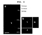
- 2 weeks after transplantation to under the rat retina sorted Rx-GFP + cells survived in the retina, expressed rhodopsin, and became integrated into the outer nuclear layer ( FIG. 6a, b ).
- CMK6 and CMK9 Two independent cell lines of cynomolgus monkey ES cells (CMK6 and CMK9) were maintained as described previously (reference documents B26, 28, 29).
- undifferentiated ES cells were maintained on a feeder layer of mitomycin C-treated mouse embryonic fibroblasts (STO cells).
- STO cells were incubated with 10 ⁇ g/mL mitomycin C (Wako, Osaka, Japan) for 2 hours and plated on gelatin-coated dishes.
- the undifferentiated monkey ES cells were incubated in a DMEM/F-12 (Sigma, St.
- ES cells were passaged every 3 days using 0.25% trypsin (GIBCO) in PBS supplemented with 1mM CaCl 2 and 20% KSR.
- the monkey ES cell lines used in this study formed colonies tightly packed with each cell showing a high nuclear/cytoplasmic ratio.
- ES colonies were partially dissociated with 0.25% trypsin (in a PBS containing 1mM CaCl 2 and 20% KSR) into clumps (5 to 10 cells per clump). It is noticeable that when this clumps of ES cells were too large (for example, exceeding 50 cells per aggregate), the neural induction efficiency declined. When ES cells dispersed to single cells were used, the cell viability declined, and retinal cells could not efficiently be induced. ES clumps were incubated on a gelatin-coated dish for 30 minutes, whereby STO feeders were removed. ES clumps were plated on a dish of bacterial grade using an ES differentiation medium at a density of 6.7x10 3 aggregate per ml.
- the composition of the differentiation medium was as follows. [G-MEM (GIBCO), 10% KSR, Lot.1139720 and 1219101, 0.1mM non-essential amino acids, 1mM pyruvate and 0.1mM 2-mercaptoethanol]. Recombinant Dkk-1 protein (100 ng/ml) and Lefty-A protein (500 ng/ml; R&D Systems, Minneapolis, MN) were added to the differentiation medium of suspension culture for 18 days. Thereafter, spontaneously formed ES cell aggregates were combined, and were plated on a poly-D-lysine/laminin/fibronectin-coated 8-well culture slide at a density of 20 to 15 aggregates per cm 2 .
- SFEB/DL-treated, differentiated cells were further incubated with RA/T/N2 medium (GMEM+5% KSR+N2 supplement (GIBCO)+retinoic acid (1 ⁇ M, Sigma)+taurine (100 ⁇ M, Sigma)+penicillin (100 units/ml)/streptomycin (100 ⁇ g/ml)) for at least 30 days.
- RA/T/N2 medium GMEM+5% KSR+N2 supplement (GIBCO)+retinoic acid (1 ⁇ M, Sigma)+taurine (100 ⁇ M, Sigma)+penicillin (100 units/ml)/streptomycin (100 ⁇ g/ml)
- Human ES cells were supplied by N. Nakatsuji and H. Suemori (Kyoto University), and used in compliance with the Human ES cell Guidelines of the Japanese government. These independent human ES cell lines (khES-1, khES-2, and khES-3) were maintained as described previously (reference document B33). In summary, undifferentiated human ES cells were maintained on a feeder layer of mitomycin C-treated mouse embryonic fibroblasts (Oriental Yeast, Tokyo, Japan).
- Human ES cells were maintained in DMEM/F-12 supplemented with 0.1mM 2-mercaptoethanol, 0.1mM non-essential amino acids, 2mM L-glutamine, 20% KSR (Lot.1219101) and 4 ng/ml basic fibroblast growth factor (Upstate Biotechnology) in a humidified atmosphere of 2% CO 2 and 98% air at 37°C.
- ES cells were passaged with 0.25% trypsin and 0.1 mg/ml collagenase IV (GIBCO) (in a PBS containing 1mM CaCl 2 and 20% KSR) every 3 to 4 days.
- GEBCO collagenase IV
- These human ES cell lines expressed the undifferentiated ES cell markers Oct-3/4 and Nanog.
- These human ES cells were immunonegative for pan-neural markers such as nestin, ⁇ III-tubulin and microtubule-associated protein-2 (a+b); consistent with human ES cells remaining undifferentiated in the cultivation of the present inventors
- ES colonies were treated with 0.25% trypsin and 0.1 mg/ml collagenase IV (in a PBS containing 1mM CaCl 2 and 20% KSR (Lot. 1219101)), and then gently disassembled for partial dissociation to obtain clumps (5 to 10 cells per clump).
- 20% KSR-containing ES differentiation medium [G-MEM, 0.1mM non-essential amino acids, 1mM pyruvate and 0.1mM 2-mercaptoethanol], at a density of 6.7x10 3 ES clumps per ml, ES clumps were plated onto a gelatin-coated dish for 1 day.
- SFEB/DL-treated ES cells were incubated in RA/T/N2 medium (GMEM+5% KSR+retinoic acid (1 ⁇ M) + taurine (100 ⁇ M)+N2 supplement +penicillin (100 units/ml)/streptomycin (100 ⁇ g/ml)) for at least 30 days.
- the medium was replaced with a fresh supply every two days.
- this method induced differentiation into retinal cells.
- SFEB serum-free floating culture of embryoid body-like aggregates
- monkey ES cells were dissociated to small clumps of 5 to 10 cells, seeded in Petri dish, and suspension-cultured in a differentiation medium (see the "Methods" section). Under these conditions, monkey ES cells formed embryoid body-like aggregates. On day 18, these aggregates were placed onto poly-D-lysine/laminin/fibronectin-coated slides.
- the optic vesicle devolves into the neural retina (inner layer) and retinal pigment epithelium (RPE, outer layer).
- neural retinal progenitor cells express Rx (reference documents B8, 12, 13)
- presumptive RPE expresses Mitf (reference documents B9, 14-16) ( FIG. 7a ). Therefore, whether or not the SFEB-treated ES cells expressed these two markers was examined. After 40 days in SFEB culture, immunostaining revealed that only a few colonies were Mitf-positive (to 10%, FIG. 7b ). To identify culture conditions that efficiently induce the formation of Mitf + cells from mouse ES cells, the effects of soluble factors on the number of Mitf + colonies in SFEB culture were examined.
- the Wnt antagonist Dkk-1 and the nodal antagonist Lefty-A both promote differentiation from mouse ES cells to Rx + retinal progenitor cells (reference document B9).
- Dkk-1 100 ng/ml
- Lefty-A 500 ng/ml
- the ratio of Mitf + colonies increased to 35.3 ⁇ 4.4% and 50.8 ⁇ 5.0%, respectively ( FIG. 7b ).
- SFEB/DL culture 16 days
- a slight improvement exceeding the results from administration of Lefty-A alone was observed (54.6 ⁇ 4.2% of all colonies, FIG. 7b, c ).
- Mitf + cells induced by SFEB/DL culture co-expressed Pax6 (90%, FIG. 7d ). This result agrees with the profile of an in vivo marker of embryo RPE (reference documents B9, 14-16).
- SFEB/DL culture for human ES cells was optimized (see "Methods" section).
- human ES cells were dissociated to cell clumps (5 to 10 cells per clump), the Wnt antagonist Dkk-1 (100 ng/ml) and the nodal antagonist Lefty-A (500 ng/ml) were applied for the first 20 days of suspension culture.
- ES cell aggregates were replated onto a poly-D-lysine/laminin/fibronectin-coated slide. Under these conditions, 73.8 ⁇ 5.5% of the colonies were nestin-positive. This does not disagree with the fact that SFEB/DL treatment induces the neural differentiation of human ES cells in the same manner as with monkey cells.
- the present inventors conducted latex beads phagocytosis assay to determine the phogocytotic activity of human ES-derived pigmented cells. Electron microscopy revealed pigmented cells taking latex beads, induced by SFEB/DL ( FIG. 12 ). From these findings, it is concluded that human ES cells differentiate into cells having typical features of RPE in SFEB/DL culture.
- Human ES cells cultured on mouse feeder cells express an immunogenic non-human sialic acid on the surface thereof (reference document B27). These xenogenic factors may cause graft rejection by the immune system following transplantation. Therefore, it is essential for the clinical application of these transplantation strategies to generate cells differentiated from human ES cells without contamination of substances derived from any other animal.
- retinal progenitor cells were induced in the presence of fetal calf serum (reference document B9).
- Lamba et al. generated retinal progenitor cells from human ES cells using Matrigel (a mouse sarcoma-derived material, not completely characterized, and may contain zenogenic components (reference document B10)).
- Mouse ES cells were cultured in the presence of SB-431542 (0.1 to 10 ⁇ M) and/or CKI-7 (1 to 100 ⁇ M) under SFEB conditions. Five days after the start of the culture, the cells were recovered and re-suspended in a fresh differentiation medium, and, to produce re-aggregated pellets, the suspension was centrifuged at 800 g for 10 minutes. After cultivation at 37°C for 1 hour, three to five re-aggregated pellets per cm 2 were replated onto poly-D-lysine/laminin/fibronectin-coated culture slides, along with differentiation medium. After cultivation for 5 days, the cells were fixed and stained with anti- ⁇ II tubulin antibody, and the ratio of ⁇ II tubulin -positive cells was determined under microscopy. The other experimental conditions were the same as those for Example 1.
- Mouse ES cells were cultured under SFEB conditions in the presence of aSB-431542 (5 ⁇ M) and CKI-7 (3 ⁇ M); or SB-431542 (5 ⁇ M) and D4476 (3 ⁇ M).
- aSB-431542 5 ⁇ M
- CKI-7 3 ⁇ M
- SB-431542 5 ⁇ M
- D4476 3 ⁇ M
- the medium was replaced with a fresh differentiation medium containing 5% FCS.
- Activin-A R & D Systems, 10 ng/ml
- the cells were recovered and re-suspended in a fresh differentiation medium, and, to produce re-aggregated pellets, the suspension was centrifuged at 800 g for 10 minutes.
- Example 1 After cultivation at 37°C for 1 hour, three to five re-aggregated pellets per cm 2 were replated onto a poly-D-lysine/laminin/fibronectin-coated culture slide, along with a differentiation medium. After 3 days of culture, in the same manner as Example 1, Rx/Pax6-positive cells were counted by immunohistological staining and FACS. The other experimental conditions were the same as those for Example 1.
- retinal progenitor cells or photoreceptors can be efficiently generated from an ES cell without co-culture with retinal tissue.
- a method of the present invention is advantageous in that retinal progenitor cells or photoreceptors can be efficiently produced from primate ES cells under the defined culture conditions.
- the present invention is expected to remarkably promote the development of a transplantation therapy for retinal diseases based on human ES cells.
Landscapes
- Health & Medical Sciences (AREA)
- Engineering & Computer Science (AREA)
- Biomedical Technology (AREA)
- Life Sciences & Earth Sciences (AREA)
- Chemical & Material Sciences (AREA)
- Bioinformatics & Cheminformatics (AREA)
- Genetics & Genomics (AREA)
- Organic Chemistry (AREA)
- Zoology (AREA)
- Biotechnology (AREA)
- Wood Science & Technology (AREA)
- Neurology (AREA)
- Cell Biology (AREA)
- General Engineering & Computer Science (AREA)
- Microbiology (AREA)
- Neurosurgery (AREA)
- General Health & Medical Sciences (AREA)
- Biochemistry (AREA)
- Acoustics & Sound (AREA)
- Physics & Mathematics (AREA)
- Analytical Chemistry (AREA)
- Developmental Biology & Embryology (AREA)
- Ophthalmology & Optometry (AREA)
- Micro-Organisms Or Cultivation Processes Thereof (AREA)
- Medicines Containing Material From Animals Or Micro-Organisms (AREA)
- Materials For Medical Uses (AREA)
- Measuring Or Testing Involving Enzymes Or Micro-Organisms (AREA)
Applications Claiming Priority (2)
| Application Number | Priority Date | Filing Date | Title |
|---|---|---|---|
| JP2007009617 | 2007-01-18 | ||
| PCT/JP2008/050305 WO2008087917A1 (fr) | 2007-01-18 | 2008-01-11 | Procédé d'induction/différenciation dans une cellule photoréceptrice |
Publications (2)
| Publication Number | Publication Date |
|---|---|
| EP2128244A1 true EP2128244A1 (fr) | 2009-12-02 |
| EP2128244A4 EP2128244A4 (fr) | 2012-05-09 |
Family
ID=39635921
Family Applications (1)
| Application Number | Title | Priority Date | Filing Date |
|---|---|---|---|
| EP08703169A Withdrawn EP2128244A4 (fr) | 2007-01-18 | 2008-01-11 | Procede d'induction/differenciation dans une cellule photoreceptrice |
Country Status (4)
| Country | Link |
|---|---|
| US (1) | US9133435B2 (fr) |
| EP (1) | EP2128244A4 (fr) |
| JP (1) | JP5441099B2 (fr) |
| WO (1) | WO2008087917A1 (fr) |
Cited By (26)
| Publication number | Priority date | Publication date | Assignee | Title |
|---|---|---|---|---|
| EP2383333A1 (fr) | 2010-04-28 | 2011-11-02 | Technische Universität Dresden | Procédé de production de cellules souches rétiniennes polarisées à partir de cellules souches pluripotentes et leur différentiation en cellules épithéliales pigmentées de la rétine |
| WO2012013936A1 (fr) * | 2010-07-30 | 2012-02-02 | Cambridge Enterprise Limited | Corticogénèse de cellules pluripotentes humaines |
| WO2012085348A1 (fr) * | 2010-12-20 | 2012-06-28 | Suomen Punainen Risti, Veripalvelu | Procédé de production de cellules épithéliales pigmentées rétiniennes humaines |
| EP2496688A1 (fr) * | 2009-11-05 | 2012-09-12 | Riken | Procédé d'induction d'une différenciation dans des cellules souches cultivées |
| US9133435B2 (en) | 2007-01-18 | 2015-09-15 | Riken | Method for induction/differentiation into photoreceptor cell |
| US9328328B2 (en) | 2009-08-24 | 2016-05-03 | Wisconsin Alumni Research Foundation | Substantially pure human retinal progenitor, forebrain progenitor, and retinal pigment epithelium cell cultures and methods of making the same |
| WO2016108240A1 (fr) | 2014-12-30 | 2016-07-07 | Cell Cure Neurosciences Ltd. | Évaluation de populations de cellules épithéliales pigmentaires rétiniennes |
| WO2016108239A1 (fr) | 2014-12-30 | 2016-07-07 | Cell Cure Neurosciences Ltd. | Populations de cellules de l'epr et leurs procédés de production |
| WO2017017686A1 (fr) | 2015-07-29 | 2017-02-02 | Hadasit Medical Research Services And Development Ltd. | Production à grande échelle de cellules de l'épithélium pigmentaire rétinien |
| WO2017021972A1 (fr) | 2015-08-05 | 2017-02-09 | Cell Cure Neurosciences Ltd. | Préparation de photorécepteurs pour le traitement de maladies rétiniennes |
| WO2017021973A1 (fr) | 2015-08-05 | 2017-02-09 | Cell Cure Neurosciences Ltd. | Préparation de cellules de l'épithélium pigmentaire rétinien |
| WO2017072763A1 (fr) | 2015-10-26 | 2017-05-04 | Cell Cure Neurosciences Ltd. | Préparation de cellules de l'épithélium pigmentaire rétinien |
| WO2018170494A1 (fr) | 2017-03-16 | 2018-09-20 | Bio Time, Inc. | Procédés de mesure d'effets thérapeutiques de traitements de maladies rétiniennes |
| CN109312306A (zh) * | 2016-04-22 | 2019-02-05 | 大日本住友制药株式会社 | 产生视网膜组织的方法 |
| US10227563B2 (en) | 2008-06-06 | 2019-03-12 | Riken | Method for culture of stem cell |
| US10519422B2 (en) | 2012-02-29 | 2019-12-31 | Riken | Method of producing human retinal pigment epithelial cells |
| WO2020058979A2 (fr) | 2018-09-20 | 2020-03-26 | Yeda Research And Development Co. Ltd. | Méthodes de traitement de la sclérose latérale amyotrophique |
| EP3754014A1 (fr) | 2019-06-21 | 2020-12-23 | Centre d'Etude des Cellules Souches (CECS) | Procédé de préparation automatique de cellules de l'épithélium pigmentaire rétinien |
| US11214771B2 (en) | 2014-10-24 | 2022-01-04 | Sumitomo Dainippon Pharma Co., Ltd. | Production method for nerve tissue |
| US11214772B2 (en) | 2014-10-24 | 2022-01-04 | Sumitomo Dainippon Pharma Co., Ltd. | Production method for retinal tissue |
| EP4000568A1 (fr) | 2017-12-29 | 2022-05-25 | Cell Cure Neurosciences Ltd. | Compositions de cellules d'épithélium pigmentaire rétinien |
| WO2022261320A1 (fr) | 2021-06-09 | 2022-12-15 | Lineage Cell Therapeutics, Inc. | Procédés et compositions pour le traitement de maladies et d'affections rétiniennes |
| WO2023009676A1 (fr) | 2021-07-28 | 2023-02-02 | Lineage Cell Therapeutics, Inc. | Multiplication de cellules de l'épithélium pigmentaire rétinien |
| US11618883B2 (en) | 2017-03-08 | 2023-04-04 | Sumitomo Pharma Co., Ltd. | Method for producing retinal pigment epithelial cells |
| US11649432B2 (en) | 2015-09-08 | 2023-05-16 | Sumitomo Pharma Co., Ltd. | Method for producing retinal pigment epithelial cells |
| WO2023211857A1 (fr) | 2022-04-25 | 2023-11-02 | Lineage Cell Therapeutics, Inc. | Procédés et compositions pour le traitement de la perte de vision |
Families Citing this family (26)
| Publication number | Priority date | Publication date | Assignee | Title |
|---|---|---|---|---|
| CN102317442B (zh) * | 2008-12-17 | 2014-08-13 | 斯克里普斯研究所 | 干细胞的产生和保持 |
| CA2802087A1 (fr) * | 2010-06-15 | 2011-12-22 | Cellular Dynamics International, Inc. | Compendium de modeles de cellules souches prefabriques pour l'interrogation d'une reponse biologique |
| JP6067232B2 (ja) * | 2011-11-25 | 2017-01-25 | 住友化学株式会社 | 網膜層特異的神経細胞を製造する方法 |
| JP5985208B2 (ja) * | 2011-11-25 | 2016-09-06 | 住友化学株式会社 | 網膜組織の製造方法 |
| JP5985207B2 (ja) * | 2011-11-25 | 2016-09-06 | 住友化学株式会社 | 網膜色素上皮細胞の製造方法 |
| JP5985209B2 (ja) * | 2011-11-25 | 2016-09-06 | 住友化学株式会社 | 眼杯様構造体の製造方法 |
| WO2013077425A1 (fr) | 2011-11-25 | 2013-05-30 | 住友化学株式会社 | Procédés pour la production d'un tissu de la rétine et de cellules associées à la rétine |
| CA2863172C (fr) * | 2012-01-31 | 2022-05-03 | Cell Cure Neurosciences Ltd. | Procede de selection de cellules de l'epithelium pigmentaire retinien |
| EP2951290B1 (fr) | 2013-02-01 | 2017-11-29 | The United States of America, as represented by The Secretary, Department of Health and Human Services | Procédé de génération de cellules épithéliales pigmentaires rétiniennes (epr) à partir de cellules souches pluripotentes induites (cspi) |
| US11241460B2 (en) | 2013-03-15 | 2022-02-08 | Astellas Institute For Regenerative Medicine | Photoreceptors and photoreceptor progenitors produced from pluripotent stem cells |
| AU2014233403B2 (en) * | 2013-03-15 | 2019-08-01 | Astellas Institute For Regenerative Medicine | Photoreceptors and photoreceptor progenitors produced from pluripotent stem cells |
| JP6580989B2 (ja) | 2013-08-23 | 2019-09-25 | 住友化学株式会社 | 網膜組織及び網膜関連細胞の製造方法 |
| CN105814192A (zh) * | 2013-10-09 | 2016-07-27 | 加利福尼亚大学董事会 | 哺乳动物视网膜干细胞产生方法和应用 |
| AU2014332857B2 (en) * | 2013-10-09 | 2020-05-07 | Healios K.K. | Method for purification of retinal pigment epithelial cells |
| AU2014363032A1 (en) | 2013-12-11 | 2016-06-09 | Pfizer Limited | Method for producing retinal pigment epithelial cells |
| WO2015109148A1 (fr) | 2014-01-16 | 2015-07-23 | The Johns Hopkins University | Procédés de formation de tissu rétinien humain tridimensionnel in vitro |
| WO2015173651A1 (fr) | 2014-05-14 | 2015-11-19 | Mark Davies | Dispositif microfluidique doté de plaques à conduits |
| CA2997763A1 (fr) * | 2015-09-08 | 2017-03-16 | Cellular Dynamics International, Inc. | Purification basee sur le tri cellulaire magnetique macs d'epithelium pigmentaire retinien derive de cellules souches |
| JP6976939B2 (ja) | 2015-10-20 | 2021-12-08 | フジフィルム セルラー ダイナミクス,インコーポレイテッド | 遺伝的プログラミングによる多系統造血前駆細胞の作製 |
| JP6980211B2 (ja) * | 2016-10-21 | 2021-12-15 | 国立大学法人群馬大学 | 末梢神経細胞の製造方法 |
| KR102638492B1 (ko) * | 2017-02-17 | 2024-02-19 | 쏘흐본느 유니베흐시테 | 망막 전구 세포, 망막 색소 상피 세포 및 신경 망막 세포를 얻기 위한 피더-비의존적(Feeder-free) 방법 |
| EP4237084A1 (fr) * | 2020-11-01 | 2023-09-06 | The Johns Hopkins University | Compositions et méthodes de thérapie par transfert de composants cellulaires |
| CN113073136A (zh) * | 2021-03-31 | 2021-07-06 | 中国科学技术大学 | 感光细胞退化作为阿尔茨海默病诊断靶点的应用 |
| KR102525093B1 (ko) * | 2021-04-16 | 2023-04-27 | 가톨릭대학교 산학협력단 | 사람 신경능 유래 코 하비갑개 줄기세포를 유효성분으로 포함하는 망막변성질환 예방 또는 치료용 약학적 조성물 |
| WO2023149565A1 (fr) * | 2022-02-07 | 2023-08-10 | 国立大学法人大阪大学 | Procédé pour induire la différenciation de cellules souches pluripotentes en cellules épithéliales pigmentaires rétiniennes |
| WO2024004960A1 (fr) * | 2022-06-27 | 2024-01-04 | 武田薬品工業株式会社 | Procédé de culture cellulaire |
Family Cites Families (9)
| Publication number | Priority date | Publication date | Assignee | Title |
|---|---|---|---|---|
| WO1998030679A1 (fr) | 1997-01-10 | 1998-07-16 | Life Technologies, Inc. | Substitut de serum pour cellules souches embryonnaires |
| US20060194315A1 (en) * | 2003-03-31 | 2006-08-31 | Condie Brian G | Compositions and methods for the control, differentiaton and/or manipulation of pluripotent cells through a gamma-secretase signaling pathway |
| KR101398356B1 (ko) * | 2004-01-23 | 2014-05-23 | 어드밴스드 셀 테크놀로지, 인코포레이티드 | 망막 변성 질환 치료를 위한 개선된 양식 |
| WO2005123902A1 (fr) | 2004-06-18 | 2005-12-29 | Riken | Méthode pour induire la différenciation de cellules souches embryonnaires en nerf à l'aide d'une culture en suspension exempte de sérum |
| JP2007009617A (ja) | 2005-07-04 | 2007-01-18 | Toray Ind Inc | 木質系板材及びそれを用いたユニット床材 |
| US7541186B2 (en) * | 2006-02-22 | 2009-06-02 | University Of Washington | Method of generating human retinal progenitors from embryonic stem cells |
| WO2008087917A1 (fr) | 2007-01-18 | 2008-07-24 | Riken | Procédé d'induction/différenciation dans une cellule photoréceptrice |
| IL292561A (en) | 2007-10-12 | 2022-06-01 | Astellas Inst For Regenerative Medicine | Improved methods of rpe cell production and rpe cell preparations |
| US10519422B2 (en) * | 2012-02-29 | 2019-12-31 | Riken | Method of producing human retinal pigment epithelial cells |
-
2008
- 2008-01-11 WO PCT/JP2008/050305 patent/WO2008087917A1/fr active Application Filing
- 2008-01-11 JP JP2008554025A patent/JP5441099B2/ja not_active Expired - Fee Related
- 2008-01-11 EP EP08703169A patent/EP2128244A4/fr not_active Withdrawn
- 2008-01-11 US US12/523,444 patent/US9133435B2/en not_active Expired - Fee Related
Non-Patent Citations (9)
| Title |
|---|
| ASHUTOSH P. JADHAV ET AL: "Notch 1 inhibits photoreceptor production in the developing mammalian retina", DEVELOPMENT, vol. 133, no. 5, 1 March 2006 (2006-03-01) , pages 913-923, XP55023108, ISSN: 0950-1991, DOI: 10.1242/dev.02245 * |
| BRANDEN R. NELSON ET AL: "Notch Activity Is Downregulated Just prior to Retinal Ganglion Cell Differentiation", DEVELOPMENTAL NEUROSCIENCE, vol. 28, no. 1-2, 1 January 2006 (2006-01-01), pages 128-141, XP55023127, ISSN: 0378-5866, DOI: 10.1159/000090759 * |
| BRANDEN R. NELSON ET AL: "Transient inactivation of Notch signaling synchronizes differentiation of neural progenitor cells", DEVELOPMENTAL BIOLOGY, vol. 304, no. 2, 15 April 2007 (2007-04-15), pages 479-498, XP55023078, ISSN: 0012-1606, DOI: 10.1016/j.ydbio.2007.01.001 * |
| CHUNG YOUNG ET AL: "Human embryonic stem cell lines generated without embryo destruction", CELL STEM CELL, ELSEVIER, CELL PRESS, AMSTERDAM, NL, vol. 2, no. 2, 7 February 2008 (2008-02-07), pages 113-117, XP002604696, ISSN: 1934-5909, DOI: 10.1016/J.STEM.2007.12.013 * |
| DATABASE BIOSIS [Online] BIOSCIENCES INFORMATION SERVICE, PHILADELPHIA, PA, US 1997 HYATT GEORGE A ET AL: 'Retinoic Acid: A key molecule for eye and photoreceptor development' Database accession no. PREV199799699757 * |
| DATABASE BIOSIS [Online] BIOSCIENCES INFORMATION SERVICE, PHILADELPHIA, PA, US 25 March 2004 YOUNG TRACY L ET AL: 'A role for ligand-gated ion channels in rod photoreceptor development' Database accession no. PREV200400429978 * |
| Mototsugu Eiraku ET AL: "Self-organizing optic-cup morphogenesis in three-dimensional culture", Nature, vol. 472, no. 7341, 7 April 2011 (2011-04-07), pages 51-56, XP055077367, ISSN: 0028-0836, DOI: 10.1038/nature09941 * |
| O. YARON: "Notch1 functions to suppress cone-photoreceptor fate specification in the developing mouse retina", DEVELOPMENT, vol. 133, no. 7, 1 April 2006 (2006-04-01) , pages 1367-1378, XP55023114, ISSN: 0950-1991, DOI: 10.1242/dev.02311 * |
| See also references of WO2008087917A1 * |
Cited By (44)
| Publication number | Priority date | Publication date | Assignee | Title |
|---|---|---|---|---|
| US9133435B2 (en) | 2007-01-18 | 2015-09-15 | Riken | Method for induction/differentiation into photoreceptor cell |
| US10934523B2 (en) | 2008-06-06 | 2021-03-02 | Riken | Method for culture of stem cell |
| US10227563B2 (en) | 2008-06-06 | 2019-03-12 | Riken | Method for culture of stem cell |
| US9328328B2 (en) | 2009-08-24 | 2016-05-03 | Wisconsin Alumni Research Foundation | Substantially pure human retinal progenitor, forebrain progenitor, and retinal pigment epithelium cell cultures and methods of making the same |
| EP2496688A1 (fr) * | 2009-11-05 | 2012-09-12 | Riken | Procédé d'induction d'une différenciation dans des cellules souches cultivées |
| US20130040330A1 (en) * | 2009-11-05 | 2013-02-14 | Riken | Method for differentiation induction in cultured stem cells |
| EP2496688A4 (fr) * | 2009-11-05 | 2013-10-09 | Riken | Procédé d'induction d'une différenciation dans des cellules souches cultivées |
| US10793830B2 (en) | 2009-11-05 | 2020-10-06 | Riken | Method for producing retinal tissue in vitro |
| US9249390B2 (en) | 2010-04-28 | 2016-02-02 | Technische Universitat Dresden | Method for producing polarized retinal progenitor cells from pluripotent stem cells and their differentiation into retinal pigment epithelium cells |
| EP2383333A1 (fr) | 2010-04-28 | 2011-11-02 | Technische Universität Dresden | Procédé de production de cellules souches rétiniennes polarisées à partir de cellules souches pluripotentes et leur différentiation en cellules épithéliales pigmentées de la rétine |
| WO2012013936A1 (fr) * | 2010-07-30 | 2012-02-02 | Cambridge Enterprise Limited | Corticogénèse de cellules pluripotentes humaines |
| WO2012085348A1 (fr) * | 2010-12-20 | 2012-06-28 | Suomen Punainen Risti, Veripalvelu | Procédé de production de cellules épithéliales pigmentées rétiniennes humaines |
| US11421203B2 (en) | 2012-02-29 | 2022-08-23 | Riken | Method of producing human retinal pigment epithelial cells |
| US10519422B2 (en) | 2012-02-29 | 2019-12-31 | Riken | Method of producing human retinal pigment epithelial cells |
| US11214772B2 (en) | 2014-10-24 | 2022-01-04 | Sumitomo Dainippon Pharma Co., Ltd. | Production method for retinal tissue |
| US11214771B2 (en) | 2014-10-24 | 2022-01-04 | Sumitomo Dainippon Pharma Co., Ltd. | Production method for nerve tissue |
| US11891622B2 (en) | 2014-12-30 | 2024-02-06 | Cell Cure Neurosciences Ltd. | RPE cell populations and methods of generating same |
| US11987810B2 (en) | 2014-12-30 | 2024-05-21 | Cell Cure Neurosciences Ltd. | RPE cell populations and methods of generating same |
| EP3916085A1 (fr) | 2014-12-30 | 2021-12-01 | Cell Cure Neurosciences Ltd. | Évaluation de populations de cellules épithéliales pigmentaires rétiniennes |
| WO2016108239A1 (fr) | 2014-12-30 | 2016-07-07 | Cell Cure Neurosciences Ltd. | Populations de cellules de l'epr et leurs procédés de production |
| WO2016108240A1 (fr) | 2014-12-30 | 2016-07-07 | Cell Cure Neurosciences Ltd. | Évaluation de populations de cellules épithéliales pigmentaires rétiniennes |
| EP3674397A1 (fr) | 2014-12-30 | 2020-07-01 | Cell Cure Neurosciences Ltd. | Populations de cellules epr et leurs procédés de génération |
| WO2017017686A1 (fr) | 2015-07-29 | 2017-02-02 | Hadasit Medical Research Services And Development Ltd. | Production à grande échelle de cellules de l'épithélium pigmentaire rétinien |
| EP3862425A1 (fr) | 2015-07-29 | 2021-08-11 | Hadasit Medical Research Services and Development Ltd. | Production à grande échelle de cellules épithéliales pigmentaires rétiniennes |
| US11230696B2 (en) | 2015-07-29 | 2022-01-25 | Hadasit Medical Research Services And Development Ltd. | Large scale production of retinal pigment epithelial cells |
| EP4155391A1 (fr) | 2015-08-05 | 2023-03-29 | Cell Cure Neurosciences Ltd. | Préparation de cellules de l'épithélium pigmentaire rétinien |
| US11066642B2 (en) | 2015-08-05 | 2021-07-20 | Cell Cure Neurosciences Ltd | Preparation of retinal pigment epithelium cells |
| WO2017021972A1 (fr) | 2015-08-05 | 2017-02-09 | Cell Cure Neurosciences Ltd. | Préparation de photorécepteurs pour le traitement de maladies rétiniennes |
| US11090337B2 (en) | 2015-08-05 | 2021-08-17 | Cell Cure Neurosciences Ltd | Preparation of photoreceptors for the treatment of retinal diseases |
| WO2017021973A1 (fr) | 2015-08-05 | 2017-02-09 | Cell Cure Neurosciences Ltd. | Préparation de cellules de l'épithélium pigmentaire rétinien |
| US11649432B2 (en) | 2015-09-08 | 2023-05-16 | Sumitomo Pharma Co., Ltd. | Method for producing retinal pigment epithelial cells |
| WO2017072763A1 (fr) | 2015-10-26 | 2017-05-04 | Cell Cure Neurosciences Ltd. | Préparation de cellules de l'épithélium pigmentaire rétinien |
| US11371016B2 (en) | 2016-04-22 | 2022-06-28 | Sumitomo Pharma Co., Ltd. | Method for producing retinal tissue |
| CN109312306A (zh) * | 2016-04-22 | 2019-02-05 | 大日本住友制药株式会社 | 产生视网膜组织的方法 |
| EP3447129A4 (fr) * | 2016-04-22 | 2019-10-30 | Sumitomo Dainippon Pharma Co., Ltd. | Procédé de production de tissu rétinien |
| US11618883B2 (en) | 2017-03-08 | 2023-04-04 | Sumitomo Pharma Co., Ltd. | Method for producing retinal pigment epithelial cells |
| WO2018170494A1 (fr) | 2017-03-16 | 2018-09-20 | Bio Time, Inc. | Procédés de mesure d'effets thérapeutiques de traitements de maladies rétiniennes |
| EP4000568A1 (fr) | 2017-12-29 | 2022-05-25 | Cell Cure Neurosciences Ltd. | Compositions de cellules d'épithélium pigmentaire rétinien |
| WO2020058979A2 (fr) | 2018-09-20 | 2020-03-26 | Yeda Research And Development Co. Ltd. | Méthodes de traitement de la sclérose latérale amyotrophique |
| WO2020254623A1 (fr) | 2019-06-21 | 2020-12-24 | Centre D'etude Des Cellules Souches (Cecs) | Procédé automatisé de préparation de cellules de l'épithélium pigmentaire rétinien |
| EP3754014A1 (fr) | 2019-06-21 | 2020-12-23 | Centre d'Etude des Cellules Souches (CECS) | Procédé de préparation automatique de cellules de l'épithélium pigmentaire rétinien |
| WO2022261320A1 (fr) | 2021-06-09 | 2022-12-15 | Lineage Cell Therapeutics, Inc. | Procédés et compositions pour le traitement de maladies et d'affections rétiniennes |
| WO2023009676A1 (fr) | 2021-07-28 | 2023-02-02 | Lineage Cell Therapeutics, Inc. | Multiplication de cellules de l'épithélium pigmentaire rétinien |
| WO2023211857A1 (fr) | 2022-04-25 | 2023-11-02 | Lineage Cell Therapeutics, Inc. | Procédés et compositions pour le traitement de la perte de vision |
Also Published As
| Publication number | Publication date |
|---|---|
| US20100105137A1 (en) | 2010-04-29 |
| JPWO2008087917A1 (ja) | 2010-05-06 |
| WO2008087917A1 (fr) | 2008-07-24 |
| US9133435B2 (en) | 2015-09-15 |
| EP2128244A4 (fr) | 2012-05-09 |
| JP5441099B2 (ja) | 2014-03-12 |
Similar Documents
| Publication | Publication Date | Title |
|---|---|---|
| US9133435B2 (en) | Method for induction/differentiation into photoreceptor cell | |
| KR102487142B1 (ko) | 만능 세포를 분화시키는 방법 | |
| US20190321414A1 (en) | Photoreceptors and photoreceptor progenitors produced from pluripotent stem cells | |
| US8492147B2 (en) | Method of inducing the differentiation of embryonic stem cells into nerve by serum-free suspension culture | |
| US20210154370A1 (en) | Method for producing retinal tissues | |
| KR101268763B1 (ko) | Wnt 신호전달 작용제를 포함하는 망막기원세포의 망막세포로의 분화 유도 또는 망막세포의 증식 유도용 조성물 | |
| US11959104B2 (en) | Methods of differentiating stem cell-derived ectodermal lineage precursors | |
| AU2016363664B2 (en) | Methods of differentiating retinal cells | |
| JP6789814B2 (ja) | 腺性下垂体又はその前駆組織の製造方法 | |
| KR102318706B1 (ko) | 모양체 주연부-유사 구조체의 제조 방법 | |
| EP3683304A1 (fr) | Procédé d'amplification de photorécepteurs de cônes ou photorécepteurs de bâtonnets à l'aide d'un transmetteur de signal de dorsalisation ou un transmetteur de signal de ventralisation | |
| AU2017202874A1 (en) | Improved methods of producing RPE cells and compositions of RPE cells | |
| WO2021201175A1 (fr) | Procédé de séparation de cellules productrices d'hormone pituitaire et de cellules progénitrices de celles-ci |
Legal Events
| Date | Code | Title | Description |
|---|---|---|---|
| PUAI | Public reference made under article 153(3) epc to a published international application that has entered the european phase |
Free format text: ORIGINAL CODE: 0009012 |
|
| 17P | Request for examination filed |
Effective date: 20090818 |
|
| AK | Designated contracting states |
Kind code of ref document: A1 Designated state(s): AT BE BG CH CY CZ DE DK EE ES FI FR GB GR HR HU IE IS IT LI LT LU LV MC MT NL NO PL PT RO SE SI SK TR |
|
| DAX | Request for extension of the european patent (deleted) | ||
| A4 | Supplementary search report drawn up and despatched |
Effective date: 20120411 |
|
| RIC1 | Information provided on ipc code assigned before grant |
Ipc: C12N 15/09 20060101ALI20120412BHEP Ipc: C12N 5/07 20100101AFI20120412BHEP |
|
| 17Q | First examination report despatched |
Effective date: 20131118 |
|
| STAA | Information on the status of an ep patent application or granted ep patent |
Free format text: STATUS: THE APPLICATION IS DEEMED TO BE WITHDRAWN |
|
| 18D | Application deemed to be withdrawn |
Effective date: 20140329 |
|
| REG | Reference to a national code |
Ref country code: DE Ref legal event code: R079 Free format text: PREVIOUS MAIN CLASS: C12N0005060000 Ipc: C12N0005070000 |
|
| REG | Reference to a national code |
Ref country code: DE Ref legal event code: R079 Free format text: PREVIOUS MAIN CLASS: C12N0005060000 Ipc: C12N0005070000 Effective date: 20141017 |