EP2090889A1 - Verfahren und Vorrichtung zur Abbildung von Zielkomponenten in einer biologischen Probe unter Verwendung von Permanentmagneten - Google Patents
Verfahren und Vorrichtung zur Abbildung von Zielkomponenten in einer biologischen Probe unter Verwendung von Permanentmagneten Download PDFInfo
- Publication number
- EP2090889A1 EP2090889A1 EP09250366A EP09250366A EP2090889A1 EP 2090889 A1 EP2090889 A1 EP 2090889A1 EP 09250366 A EP09250366 A EP 09250366A EP 09250366 A EP09250366 A EP 09250366A EP 2090889 A1 EP2090889 A1 EP 2090889A1
- Authority
- EP
- European Patent Office
- Prior art keywords
- target population
- cells
- image
- floater
- magnet
- Prior art date
- Legal status (The legal status is an assumption and is not a legal conclusion. Google has not performed a legal analysis and makes no representation as to the accuracy of the status listed.)
- Granted
Links
- 238000000034 method Methods 0.000 title claims description 34
- 238000003384 imaging method Methods 0.000 title abstract description 13
- 239000012472 biological sample Substances 0.000 title 1
- 239000006249 magnetic particle Substances 0.000 claims abstract description 8
- 239000004205 dimethyl polysiloxane Substances 0.000 claims abstract description 6
- 235000013870 dimethyl polysiloxane Nutrition 0.000 claims abstract description 6
- CXQXSVUQTKDNFP-UHFFFAOYSA-N octamethyltrisiloxane Chemical compound C[Si](C)(C)O[Si](C)(C)O[Si](C)(C)C CXQXSVUQTKDNFP-UHFFFAOYSA-N 0.000 claims abstract description 6
- 238000004987 plasma desorption mass spectroscopy Methods 0.000 claims abstract description 6
- 229920000435 poly(dimethylsiloxane) Polymers 0.000 claims abstract description 6
- 239000003550 marker Substances 0.000 claims description 15
- 238000002372 labelling Methods 0.000 claims description 7
- 230000008878 coupling Effects 0.000 claims description 6
- 238000010168 coupling process Methods 0.000 claims description 6
- 238000005859 coupling reaction Methods 0.000 claims description 6
- 229910052779 Neodymium Inorganic materials 0.000 claims description 4
- QEFYFXOXNSNQGX-UHFFFAOYSA-N neodymium atom Chemical group [Nd] QEFYFXOXNSNQGX-UHFFFAOYSA-N 0.000 claims description 4
- 229920001296 polysiloxane Polymers 0.000 claims 1
- 210000004027 cell Anatomy 0.000 abstract description 79
- 239000012530 fluid Substances 0.000 abstract description 13
- 239000011324 bead Substances 0.000 abstract description 9
- 238000004163 cytometry Methods 0.000 abstract description 4
- 210000000265 leukocyte Anatomy 0.000 abstract description 4
- 238000004422 calculation algorithm Methods 0.000 abstract description 3
- 229920002379 silicone rubber Polymers 0.000 abstract description 3
- 239000004945 silicone rubber Substances 0.000 abstract description 3
- 238000010191 image analysis Methods 0.000 abstract description 2
- 210000001124 body fluid Anatomy 0.000 abstract 2
- 239000000523 sample Substances 0.000 abstract 2
- 230000001580 bacterial effect Effects 0.000 abstract 1
- 238000011109 contamination Methods 0.000 abstract 1
- 230000007613 environmental effect Effects 0.000 abstract 1
- 235000013305 food Nutrition 0.000 abstract 1
- 239000012488 sample solution Substances 0.000 abstract 1
- 208000034699 Vitreous floaters Diseases 0.000 description 39
- 210000004369 blood Anatomy 0.000 description 23
- 239000008280 blood Substances 0.000 description 23
- 230000005291 magnetic effect Effects 0.000 description 17
- 238000004458 analytical method Methods 0.000 description 13
- 239000006285 cell suspension Substances 0.000 description 10
- 239000011554 ferrofluid Substances 0.000 description 10
- 239000002953 phosphate buffered saline Substances 0.000 description 10
- 238000012360 testing method Methods 0.000 description 10
- 238000001514 detection method Methods 0.000 description 7
- 208000005443 Circulating Neoplastic Cells Diseases 0.000 description 6
- DPKHZNPWBDQZCN-UHFFFAOYSA-N acridine orange free base Chemical compound C1=CC(N(C)C)=CC2=NC3=CC(N(C)C)=CC=C3C=C21 DPKHZNPWBDQZCN-UHFFFAOYSA-N 0.000 description 6
- DZBUGLKDJFMEHC-UHFFFAOYSA-N benzoquinolinylidene Natural products C1=CC=CC2=CC3=CC=CC=C3N=C21 DZBUGLKDJFMEHC-UHFFFAOYSA-N 0.000 description 6
- 239000011521 glass Substances 0.000 description 6
- FWBHETKCLVMNFS-UHFFFAOYSA-N 4',6-Diamino-2-phenylindol Chemical compound C1=CC(C(=N)N)=CC=C1C1=CC2=CC=C(C(N)=N)C=C2N1 FWBHETKCLVMNFS-UHFFFAOYSA-N 0.000 description 5
- 238000011534 incubation Methods 0.000 description 5
- 101150084967 EPCAM gene Proteins 0.000 description 4
- 206010028980 Neoplasm Diseases 0.000 description 4
- 201000011510 cancer Diseases 0.000 description 4
- 239000003153 chemical reaction reagent Substances 0.000 description 4
- 238000005516 engineering process Methods 0.000 description 4
- 239000000463 material Substances 0.000 description 4
- 208000037265 diseases, disorders, signs and symptoms Diseases 0.000 description 3
- 210000002919 epithelial cell Anatomy 0.000 description 3
- 230000003287 optical effect Effects 0.000 description 3
- 239000002245 particle Substances 0.000 description 3
- 230000035945 sensitivity Effects 0.000 description 3
- 210000004881 tumor cell Anatomy 0.000 description 3
- XEEYBQQBJWHFJM-UHFFFAOYSA-N Iron Chemical compound [Fe] XEEYBQQBJWHFJM-UHFFFAOYSA-N 0.000 description 2
- 102000011782 Keratins Human genes 0.000 description 2
- 108010076876 Keratins Proteins 0.000 description 2
- 239000000980 acid dye Substances 0.000 description 2
- 239000000872 buffer Substances 0.000 description 2
- 238000006243 chemical reaction Methods 0.000 description 2
- 239000012141 concentrate Substances 0.000 description 2
- 238000013461 design Methods 0.000 description 2
- 201000010099 disease Diseases 0.000 description 2
- 210000002889 endothelial cell Anatomy 0.000 description 2
- 238000000684 flow cytometry Methods 0.000 description 2
- 238000002073 fluorescence micrograph Methods 0.000 description 2
- 238000002955 isolation Methods 0.000 description 2
- 239000000644 isotonic solution Substances 0.000 description 2
- 238000007885 magnetic separation Methods 0.000 description 2
- 238000002156 mixing Methods 0.000 description 2
- 230000000877 morphologic effect Effects 0.000 description 2
- 229910001172 neodymium magnet Inorganic materials 0.000 description 2
- 150000007523 nucleic acids Chemical class 0.000 description 2
- 102000039446 nucleic acids Human genes 0.000 description 2
- 108020004707 nucleic acids Proteins 0.000 description 2
- 238000011084 recovery Methods 0.000 description 2
- 238000000926 separation method Methods 0.000 description 2
- 238000010186 staining Methods 0.000 description 2
- 239000000725 suspension Substances 0.000 description 2
- 108010066687 Epithelial Cell Adhesion Molecule Proteins 0.000 description 1
- 102000018651 Epithelial Cell Adhesion Molecule Human genes 0.000 description 1
- 102100031940 Epithelial cell adhesion molecule Human genes 0.000 description 1
- 108010043121 Green Fluorescent Proteins Proteins 0.000 description 1
- 101000920667 Homo sapiens Epithelial cell adhesion molecule Proteins 0.000 description 1
- 238000003556 assay Methods 0.000 description 1
- 210000000601 blood cell Anatomy 0.000 description 1
- 210000000170 cell membrane Anatomy 0.000 description 1
- 230000003436 cytoskeletal effect Effects 0.000 description 1
- 230000007423 decrease Effects 0.000 description 1
- 230000004069 differentiation Effects 0.000 description 1
- LOKCTEFSRHRXRJ-UHFFFAOYSA-I dipotassium trisodium dihydrogen phosphate hydrogen phosphate dichloride Chemical compound P(=O)(O)(O)[O-].[K+].P(=O)(O)([O-])[O-].[Na+].[Na+].[Cl-].[K+].[Cl-].[Na+] LOKCTEFSRHRXRJ-UHFFFAOYSA-I 0.000 description 1
- 239000000975 dye Substances 0.000 description 1
- 230000005294 ferromagnetic effect Effects 0.000 description 1
- 239000007850 fluorescent dye Substances 0.000 description 1
- 238000012632 fluorescent imaging Methods 0.000 description 1
- 210000003714 granulocyte Anatomy 0.000 description 1
- 238000003125 immunofluorescent labeling Methods 0.000 description 1
- 230000002452 interceptive effect Effects 0.000 description 1
- 229910052742 iron Inorganic materials 0.000 description 1
- 210000004698 lymphocyte Anatomy 0.000 description 1
- 230000005426 magnetic field effect Effects 0.000 description 1
- 239000002122 magnetic nanoparticle Substances 0.000 description 1
- 239000006148 magnetic separator Substances 0.000 description 1
- 238000001000 micrograph Methods 0.000 description 1
- 239000004005 microsphere Substances 0.000 description 1
- 238000012986 modification Methods 0.000 description 1
- 230000004048 modification Effects 0.000 description 1
- 210000001616 monocyte Anatomy 0.000 description 1
- 238000002360 preparation method Methods 0.000 description 1
- 239000003755 preservative agent Substances 0.000 description 1
- 230000002335 preservative effect Effects 0.000 description 1
- 238000003672 processing method Methods 0.000 description 1
- 230000009467 reduction Effects 0.000 description 1
- 238000003757 reverse transcription PCR Methods 0.000 description 1
- 239000000243 solution Substances 0.000 description 1
- 238000007619 statistical method Methods 0.000 description 1
- 230000000007 visual effect Effects 0.000 description 1
- 238000012800 visualization Methods 0.000 description 1
Images


Classifications
-
- G—PHYSICS
- G01—MEASURING; TESTING
- G01N—INVESTIGATING OR ANALYSING MATERIALS BY DETERMINING THEIR CHEMICAL OR PHYSICAL PROPERTIES
- G01N33/00—Investigating or analysing materials by specific methods not covered by groups G01N1/00 - G01N31/00
- G01N33/48—Biological material, e.g. blood, urine; Haemocytometers
- G01N33/50—Chemical analysis of biological material, e.g. blood, urine; Testing involving biospecific ligand binding methods; Immunological testing
- G01N33/58—Chemical analysis of biological material, e.g. blood, urine; Testing involving biospecific ligand binding methods; Immunological testing involving labelled substances
- G01N33/582—Chemical analysis of biological material, e.g. blood, urine; Testing involving biospecific ligand binding methods; Immunological testing involving labelled substances with fluorescent label
-
- G—PHYSICS
- G01—MEASURING; TESTING
- G01N—INVESTIGATING OR ANALYSING MATERIALS BY DETERMINING THEIR CHEMICAL OR PHYSICAL PROPERTIES
- G01N33/00—Investigating or analysing materials by specific methods not covered by groups G01N1/00 - G01N31/00
- G01N33/48—Biological material, e.g. blood, urine; Haemocytometers
- G01N33/50—Chemical analysis of biological material, e.g. blood, urine; Testing involving biospecific ligand binding methods; Immunological testing
- G01N33/53—Immunoassay; Biospecific binding assay; Materials therefor
- G01N33/543—Immunoassay; Biospecific binding assay; Materials therefor with an insoluble carrier for immobilising immunochemicals
- G01N33/54313—Immunoassay; Biospecific binding assay; Materials therefor with an insoluble carrier for immobilising immunochemicals the carrier being characterised by its particulate form
- G01N33/54326—Magnetic particles
-
- G—PHYSICS
- G01—MEASURING; TESTING
- G01N—INVESTIGATING OR ANALYSING MATERIALS BY DETERMINING THEIR CHEMICAL OR PHYSICAL PROPERTIES
- G01N33/00—Investigating or analysing materials by specific methods not covered by groups G01N1/00 - G01N31/00
- G01N33/48—Biological material, e.g. blood, urine; Haemocytometers
- G01N33/50—Chemical analysis of biological material, e.g. blood, urine; Testing involving biospecific ligand binding methods; Immunological testing
- G01N33/53—Immunoassay; Biospecific binding assay; Materials therefor
- G01N33/569—Immunoassay; Biospecific binding assay; Materials therefor for microorganisms, e.g. protozoa, bacteria, viruses
- G01N33/56966—Animal cells
- G01N33/56972—White blood cells
-
- G—PHYSICS
- G01—MEASURING; TESTING
- G01N—INVESTIGATING OR ANALYSING MATERIALS BY DETERMINING THEIR CHEMICAL OR PHYSICAL PROPERTIES
- G01N2333/00—Assays involving biological materials from specific organisms or of a specific nature
- G01N2333/435—Assays involving biological materials from specific organisms or of a specific nature from animals; from humans
- G01N2333/705—Assays involving receptors, cell surface antigens or cell surface determinants
- G01N2333/70503—Immunoglobulin superfamily, e.g. VCAMs, PECAM, LFA-3
- G01N2333/70514—CD4
-
- G—PHYSICS
- G01—MEASURING; TESTING
- G01N—INVESTIGATING OR ANALYSING MATERIALS BY DETERMINING THEIR CHEMICAL OR PHYSICAL PROPERTIES
- G01N2446/00—Magnetic particle immunoreagent carriers
Definitions
- the invention relates generally to imaging target components in a fluidic (biological) sample. More specifically, methods and apparatus are described that provide for the positive selection of target cells in a blood sample. Small permanent magnets are added directly to a blood sample containing CD4 immunomagnetic labeled fluorescently stained Acridine Orange (AO) whole blood.
- AO Acridine Orange
- immunomagnetic separation technology provides greater sensitivity and specificity in the detection of target entities in blood for example, but not limited to, intact circulating cancer cells and endothelial cells.
- This simple and sensitive diagnostic tool as described ( US6,365,362 ; US6,551,843 ; US6,623,982 ; US6,620,627 ; US6,645,731 ; WO 02/077604 ; WO03/065042 ; and WO 03/019141 ) can be used in the present invention to correlate the statistical survivability of an individual patient based on a threshold level.
- a prior diagnostic tool incorporates a blood sample from a cancer patient ( WO 03/018757 ) incubated with magnetic beads, coated with antibodies directed against an epithelial cell surface antigen as for example EpCAM. After labeling with anti-EpCAM-coated magnetic nanoparticles, the magnetically labeled cells are then isolated using a magnetic separator. The immunomagnetically enriched fraction is further processed for downstream immunocytochemical analysis or image cytometry, for example, in the CellSpotter or CellTracks ® System (Immunicon Corp., USA). The magnetic fraction can also be used for downstream immunocytochemical analysis, RT-PCR, PCR, FISH, flowcytometry, or other types of image cytometry.
- the CellSpotter or CellTracks® System utilizes immunomagnetic selection and separation to highly enrich and concentrate any epithelial cells present in whole blood samples.
- the captured cells are detectably labeled with a leukocyte specific marker and with one or more tumor cell specific fluorescent monoclonal antibodies to allow identification and enumeration of the captured CTC's as well as instrumental or visual differentiation from contaminating non-target cells.
- this assay allows tumor cell detection even in the early stages of low tumor mass.
- EasyCount® system ( PCT/US03/04468 ) is a fluorescent imaging system, designed to make a distinction between lymphocytes, granulocytes and monocytes.
- the system includes a compact electronic optical instruments, analytical methods, image acquisition, and data reduction algorithms for the detection and enumeration of magnetically labeled target cells or particles.
- whole blood as an example, blood cells are fluorescently labeled using one or more target specific fluorescent dyes, such as a DNA staining dye.
- the cells of interest or target cells in the blood sample are labeled by incubation with monoclonal antibodies conjugated to ferromagnetic particles.
- the sample is then placed into an appropriate optical detection chamber or covet, which in turn is placed into a magnetic field gradient that selectively causes the magnetically labeled cells to move towards the planar viewing surface of the chamber.
- the target cells are collected and immobilized substantially uniformly on the optically transparent surface of the chamber. A segment of this surface and the labeled target cells thereon are illuminated by means of one or more LED (light emitting diodes).
- LED light emitting diodes
- the light emitted by individual target cells is captured by a CCD (charge coupled device).
- Image acquisition methods, processing methods, and algorithms, disclosed herein, are used to count the number of captured light-emitting cells and to relate the data output to the target cells per microliter of the analysis sample in the chamber and ultimately to the original specimen.
- the present invention is a method and means for positive selecting and imaging target entities.
- This includes a coated permanent magnetic device for magnetic manipulation in the system of the present invention.
- the system immunomagnetically concentrates the target entity, fluorescently labels, identifies and quantifies target cells by positive enumeration. Subsequent statistical analysis enables the clinician to obtain potential diagnostic information.
- the present invention provides the apparatus, methods, and kits for diagnosing disease disorders after immunomagnetic imaging.
- a small permanent magnet is added to the whole blood sample.
- a small NdFeB magnet is directly added to a sample container, for example the CellTracks® cartridge US 6,861,259 and US 7,011,794 with 100 ⁇ l of CD4 immunomagnetically labeled and fluorescently stained AO whole blood.
- the small permanent magnet is pulled out of the sample using an iron rod or another magnet. The magnet is positioned within the container to allow for image analysis.
- a further embodiment of the present invention has the magnet fixed to a floatation device (floater) within the reaction chamber. After addition of the reagents, blood and floater, the immunomagnetically labeled target cells are positioned along a single imaging plane for analysis, all within the reaction chamber.
- a floatation device floater
- the invention provides a method for detecting and enumerating a target population within a biological specimen, comprising:
- the invention further provides a method for detecting and enumerating a target population within a biological specimen, comprising:
- the methods of the invention may further comprise a step of obtaining said biological specimen from a subject.
- Immunomagnetic isolation, enrichment, and analysis in blood combines immunomagnetic enrichment technology and immunofluorescent labeling technology with an appropriate analytical platform after initial blood draw.
- the associated test has the sensitivity and specificity to detect rare cells in a sample of whole blood with the utility to investigate their role in the clinical course of the disease such as malignant tumors of epithelial origin.
- CTC circulating tumor cells
- Image cytometric analysis such that the immunomagnetically enriched sample is analyzed by the CellSpotter and CellTracks® System utilizes a fluorescence-based microscope image analysis system, which in contrast with flowcytometric analysis permits the visualization of events and the assessment of morphologic features to further identify objects ( US 6,365,362 ).
- the CellSpotter and CellTracks® System refers to an automated fluorescence microscopic system for automated enumeration of isolated cells from blood.
- the system contains an integrated computer controlled fluorescence microscope and automated stage with a magnetic yoke assembly that will hold a disposable sample cartridge.
- the magnetic yoke is designed to enable ferrofluid-labeled candidate tumor cells within the sample chamber to be magnetically localized to the upper viewing surface of the sample cartridge for microscopic viewing.
- Software presents target cells, labeled with antibodies to cytokeratin and having epithelial origin, to the operator for final selection.
- Isolation of target cells can be accomplished by any means known in the art. After magnetic separation, the cells bound to the immunomagnetic-linked antibodies are magnetically held at the wall of the tube. Unbound sample is then aspirated and an isotonic solution is added to resuspend the sample. A nucleic acid dye, monoclonal antibodies to cytokeratin (a marker of epithelial cells) and CD 45 (a broad-spectrum leukocyte marker) are incubated with the sample. After magnetic separation, the unbound fraction is again aspirated and the bound and labeled cells are resuspended in 0.2 ml of an isotonic solution.
- cytokeratin a marker of epithelial cells
- CD 45 a broad-spectrum leukocyte marker
- the sample is suspended in a cell presentation chamber and placed in a magnetic device whose field orients the magnetically labeled cells for fluorescence microscopic examination.
- Cells are identified automatically and candidate target entities presented to the operator for checklist enumeration.
- An enumeration checklist consists of predetermined morphologic criteria constituting a complete cell.
- the present invention utilizes a small magnet added directly to the immunomagnetically labeled target entity in a blood sample.
- the target is further labeled with imaging nucleic acid dyes, cell membrane, and/or cytoskeletal immunofluorescent labels.
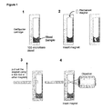
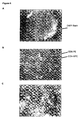
- Figure 1 depicts a method for imaging CD4 expressing target cells in a whole blood sample.
- a small neodymium (NdFeB) permanent magnet is added to a whole blood sample after immunomagnetically labeled and fluorescently labeled for CD4. After 10 minutes, the small permanent magnet is separated from the fluid sample and within the sample container to be viewed through a viewing surface.
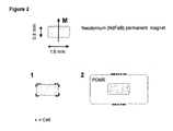
- the magnet is a disc with a diameter of 1.6 mm and a height of 0.8 mm (see Figure 2 ).
- the smaller magnets are more preferred for this invention.
- the target entity attach to only the magnets.
- the cells are not in a single focal plane and quality images are difficult to obtain.
- the same method is accomplished using encapsulated magnets with PDMS silicone rubber.
- the cells attach along a single focal plane.
- the layer of PDMS on the top of the magnet is approximately 1 mm.
- the width of the PDMS is approximately 3 mm.
- Figure 3 shows the basic steps of the present method.
- a permanent magnet is mounted on the inside of one face of a hollow tube, closed on all sides as shown in Figure 3A .
- the face, having the magnet mounted has a defined thickness (d).
- the surface can be flat or contain structures that facilitate capture and visibility of the objects of interest and limit the influence of interfering components in the fluid, i.e. free unbound magnetic particles.
- the thickness of the face determines the spread of the cells on the outside.
- the height/diameter ratio of the floater determines the magnetic field's effect on the outside area of the floater. Consequently, the ratio should limit the influence of the magnetic field to approximately the face of the outside of the floater where the magnet is mounted.
- a cell suspension is injected into a tube with a flat surface, having an optically transparent window. Immunomagnetic particles are added together with fluorescence labels. After incubation the floater containing the permanent magnet is inserted in the tube with the magnet facing the bottom of the tube and the tube is closed.
- the floater can be inserted at the same time as the other reagents.
- the test tube upside down the floater rises to the outside the fluid.
- a small layer of fluid is left in between the test tube and the floater which is neglect able to the total volume.
- Suspension is incubated with the reagents without interference of the floater or interferences of the magnetic field.
- the tube After incubation the tube is placed on a test tube rotator or similar device to cause the floater to move up and down through the cell suspension as illustrated in Figure 3B . After enough time to allow complete capture of the magnetically labeled objects (cells), the tube is taken off the rotator and placed up-side down to force the floater to rise to toward the optical window on the flat surface of the tube. Using a standard fluorescence microscope, the cells, presented on the face of the floater are imaged through the bottom of the tube.
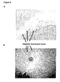
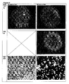
- Figure 4A displays a fluorescence image of magnetic green fluorescent beads (Bangs beads), having a diameter of 8 microns.
- the beads are collected on the face of a floater using a 0.8 mm (1/32") x 1.6 mm (1/16") neodymium permanent disc magnet with the face having a thickness of 0.17 millimeters.
- Figure 4B shows the magnetic beads imaged on the face of the floated.
- Figure 5A displays an overlay image of CD14-FF selected cells that were collected on the face of a floater with an outside diameter of 12 mm using a 1.6 mm x 6.53 mm (1/16" x 1/4") neodymium cylinder magnet and a face thickness of 2 cover slips, approximately 0.34 mm.
- the intensity of Acridine Orange can be shown as a green signal to distinguish the red color for the CD45-APC label.
- Figure 5B shows the floater, the chamber, and the cap of the chamber.
- CD-Chex with known absolute numbers of leukocytes and their phenotypes is used.
- CD-Chex (lot # 60650071):
- CD-Chex To 50 ⁇ l of CD-Chex, add 10 ⁇ l of CD3-FF (clone Cris7), 10 ⁇ l of CD4-APC and 10 ⁇ l of CD8-PE. After 25 minutes of incubation, 10 ⁇ l of this solution is injected into the chamber. PBS (1.8 ml) is added with 100 ⁇ l DAPI. The floater is then inserted. After capping, the chamber is placed on a rocker and rotated overnight (approximately 16 hrs). The chamber is inverted and the images of the floater are acquired.
- the floater surface contains:
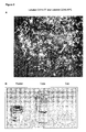
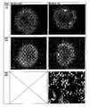
- Figure 6A displays the image acquired using a 5x NA 0.12 objective.
- Figures B and C are acquired using a 10X, NA 0.25 and a 40X, NA 0.6 objective, respectively.
- the blue color represents the DAPI, green is CD8-PE and red is CD4-APC.
- the capture efficiency will be 16%.
- control cells from the CellTracks® cartridge were transferred to a chamber similar to Figure 5B .
- the cartridge was washed several times with PBS using a pasteur pipette and all fluid used in the wash (approximately 500 microliters) was transferred to the chamber. Additional PBS was added to the volume to bring the total volume to 2 ml. Vial was placed on the tube rotator. The rotation speed was set so the floater moved through the entire fluid in one rotation. After complete mixing, 1.5 ml of control cells was injected into the vial together with 50 ⁇ l of Epcam ferrofluid and 10 ⁇ l of DAPI reagents (CellSearchTM, Veridex LLC). After 30 minutes of incubation images were acquired at multiple points in time.
- the image in Figure 7A shows results after 40 rotations. Image quality is suitable for cells to be counted easily.
- the number of green cells corresponds to highs at 556 circulating tumor cells (CTC) and low at 47 CTC. After 100 rotations most of cells become buried under a layer of ferrofluids. At this point, cells are not visible and can not be counted.
- COMPEL Magnetic Microspheres, Dragon green, 2.914 10 7 /ml, diameter 8.44 microns, lot#6548 (Bangs Laboratories Inc, Catalog code UMC4F) were diluted 1:100.
- System buffer 1.5 ml was added to the glass vial and 50 microliters containing 14570 beads were added together with 20, 40, 60 and 80 microliters of EpCam ferrofluid (20 mg/ml). Fluorescence images were acquired after 15 and 30 minutes of rotation.
- Test tube rotator was set at 10 rpm, resulting in 150 and 300 rotations.
- Floater is Corning 1.6 mm (1/16") diameter magnet.
- Images are acquired with a 5X and 40X objectives. As shown in Figure 8 , 5x and 40x objectives were used to image 20, 40, 60 and 80 microliters of EpCam. The missing images shown in Figure 8 were lost during saving.
- Magnet Floater CellTracks rescan Cap efficiency 1.6 mm x 6.35 mm 256 1 99.6% (1/16 x 1 ⁇ 4") 1.6 mm x 6.35 mm 225 na na (1/16 x 1 ⁇ 4") 1.6 mm x 12.7 mm 230 2 99.1 (1/16 x 1/2") 1.6 mm x 12.7 mm 218 na na (1/16 x 1/2 ")
- Magnet Floater CellTracks rescan Cap efficiency Low High Low High Low High 1.6 mm x 6.35 mm 13 173 0 0 100 100 (1/16 x 1 ⁇ 4") 1.6 mm x 6.35 mm 31 274 0 13 100 95 (1/16 x 1 ⁇ 4") 1.6 mm x 12.7 mm 11 136 0 2 100 98 (1/16 x 1 ⁇ 2”) 1.6 mm x 12.7 mm 13 318 0 15 100 95 (1/16 x 1 ⁇ 2”)
- the graph in Figure 9 shows the number of cells collected as a function of time
Applications Claiming Priority (1)
| Application Number | Priority Date | Filing Date | Title |
|---|---|---|---|
| US12/031,807 US7828968B2 (en) | 2007-08-30 | 2008-02-15 | Method and apparatus for imaging target components in a biological sample using permanent magnets |
Publications (2)
| Publication Number | Publication Date |
|---|---|
| EP2090889A1 true EP2090889A1 (de) | 2009-08-19 |
| EP2090889B1 EP2090889B1 (de) | 2012-01-04 |
Family
ID=40506419
Family Applications (1)
| Application Number | Title | Priority Date | Filing Date |
|---|---|---|---|
| EP09250366A Not-in-force EP2090889B1 (de) | 2008-02-15 | 2009-02-13 | Verfahren und Vorrichtung zur Abbildung von Zielkomponenten in einer biologischen Probe unter Verwendung von Permanentmagneten |
Country Status (7)
| Country | Link |
|---|---|
| US (2) | US7828968B2 (de) |
| EP (1) | EP2090889B1 (de) |
| JP (1) | JP5507092B2 (de) |
| CN (1) | CN101533012B (de) |
| AT (1) | ATE540316T1 (de) |
| ES (1) | ES2377178T3 (de) |
| HK (1) | HK1137810A1 (de) |
Cited By (1)
| Publication number | Priority date | Publication date | Assignee | Title |
|---|---|---|---|---|
| WO2015058103A1 (en) * | 2013-10-18 | 2015-04-23 | The Regents Of The University Of California | Method and device for detecting molecules or particles using fractionalized volumes |
Families Citing this family (27)
| Publication number | Priority date | Publication date | Assignee | Title |
|---|---|---|---|---|
| KR20040105717A (ko) * | 2002-02-14 | 2004-12-16 | 이뮤니베스트 코포레이션 | 저비용 세포 분석기에서의 세포 계산 방법 및 알고리즘 |
| US8189899B2 (en) * | 2004-07-30 | 2012-05-29 | Veridex, Llc | Methods and algorithms for cell enumeration in a low-cost cytometer |
| US8110101B2 (en) * | 2007-08-30 | 2012-02-07 | Veridex, Llc | Method and apparatus for imaging target components in a biological sample using permanent magnets |
| US7828968B2 (en) * | 2007-08-30 | 2010-11-09 | Veridex, Llc | Method and apparatus for imaging target components in a biological sample using permanent magnets |
| WO2010068812A1 (en) | 2008-12-10 | 2010-06-17 | Abqmr, Inc. | Nuclear magnetic resonance apparatus, methods and associated technology |
| US8790916B2 (en) | 2009-05-14 | 2014-07-29 | Genestream, Inc. | Microfluidic method and system for isolating particles from biological fluid |
| US9476812B2 (en) | 2010-04-21 | 2016-10-25 | Dna Electronics, Inc. | Methods for isolating a target analyte from a heterogeneous sample |
| US9428547B2 (en) | 2010-04-21 | 2016-08-30 | Dna Electronics, Inc. | Compositions for isolating a target analyte from a heterogeneous sample |
| US20110262989A1 (en) | 2010-04-21 | 2011-10-27 | Nanomr, Inc. | Isolating a target analyte from a body fluid |
| US8841104B2 (en) | 2010-04-21 | 2014-09-23 | Nanomr, Inc. | Methods for isolating a target analyte from a heterogeneous sample |
| US10114020B2 (en) | 2010-10-11 | 2018-10-30 | Mbio Diagnostics, Inc. | System and device for analyzing a fluidic sample |
| BR112013024148B1 (pt) * | 2011-04-27 | 2022-01-25 | Becton, Dickinson And Company | Dispositivo, método e sistema para separar partes marcadas magneticamente em uma amostra, método de separar partes marcadas magneticamente em uma amostra |
| EP2701851B1 (de) * | 2011-04-29 | 2024-04-24 | Becton Dickinson and Company | Sammelsystem zur immobilisierung fluidischer inline-teilchen sowie verfahren dafür |
| EP2911791A4 (de) | 2012-10-29 | 2016-11-02 | Mbio Diagnostics Inc | Biopartikelidentifikationssystem, kartusche und zugehörige verfahren |
| WO2014097991A1 (ja) * | 2012-12-18 | 2014-06-26 | コニカミノルタ株式会社 | 希少細胞検出装置、希少細胞検出方法、希少細胞観察システム、および細胞展開用デバイス |
| US9995742B2 (en) | 2012-12-19 | 2018-06-12 | Dnae Group Holdings Limited | Sample entry |
| US10000557B2 (en) | 2012-12-19 | 2018-06-19 | Dnae Group Holdings Limited | Methods for raising antibodies |
| US9804069B2 (en) | 2012-12-19 | 2017-10-31 | Dnae Group Holdings Limited | Methods for degrading nucleic acid |
| US9599610B2 (en) | 2012-12-19 | 2017-03-21 | Dnae Group Holdings Limited | Target capture system |
| US9551704B2 (en) | 2012-12-19 | 2017-01-24 | Dna Electronics, Inc. | Target detection |
| US9434940B2 (en) | 2012-12-19 | 2016-09-06 | Dna Electronics, Inc. | Methods for universal target capture |
| DE102013200927A1 (de) * | 2013-01-22 | 2014-07-24 | Siemens Aktiengesellschaft | Verfahren zum Anreichern und Vereinzeln von Zellen mit Konzentrationen über mehrere logarithmische Stufen |
| US20150233932A1 (en) * | 2013-02-19 | 2015-08-20 | Ching-Ping Tseng | Methods, Systems, and Compositions for Enrichment of Rare Cells |
| US10564100B1 (en) * | 2018-10-12 | 2020-02-18 | SageMedic Corporation | Analysis of viable and nonviable cells |
| CN111795919B (zh) * | 2020-05-25 | 2024-03-26 | 中国人民解放军陆军军医大学第二附属医院 | 一种骨髓细胞形态学自动检测系统及其工作方法 |
| EP3922991A1 (de) * | 2020-06-10 | 2021-12-15 | PreOmics GmbH | Dispersion mit einem beweglichen magneten |
| CN114002195A (zh) * | 2021-10-22 | 2022-02-01 | 中国科学院广州生物医药与健康研究院 | 一种辅助悬浮细胞成像的系统及其使用方法和应用 |
Citations (12)
| Publication number | Priority date | Publication date | Assignee | Title |
|---|---|---|---|---|
| GB2123706A (en) * | 1982-06-09 | 1984-02-08 | Techne Inc | Floating magnetic stirrer for culture medium |
| US4465377A (en) * | 1983-06-07 | 1984-08-14 | Techne Corporation | Magnetic stirrer apparatus with guided, floating stirrer |
| US6365362B1 (en) | 1998-02-12 | 2002-04-02 | Immunivest Corporation | Methods and reagents for the rapid and efficient isolation of circulating cancer cells |
| WO2002077604A2 (en) | 2001-03-07 | 2002-10-03 | Immunivest Corporation | Labeled cells for use as an internal functional control in rare cell detection assays |
| WO2003019141A2 (en) | 2001-08-23 | 2003-03-06 | Immunivest Corporation | Analysis of circulating tumor cells, fragments, and debris |
| US6551843B1 (en) | 1999-01-29 | 2003-04-22 | Immunivest Corporation | Methods for enhancing binding interactions between members of specific binding pairs |
| WO2003065042A1 (en) | 2001-02-16 | 2003-08-07 | Immunivest Corporation | Methods and reagents for the rapid and efficient isolation of circulating cancer cells |
| WO2003069421A2 (en) | 2002-02-14 | 2003-08-21 | Immunivest Corporation | Methods and algorithms for cell enumeration in a low-cost cytometer |
| US6620627B1 (en) | 1999-07-12 | 2003-09-16 | Immunivest Corporation | Increased separation efficiency via controlled aggregation of magnetic nanoparticles |
| US6861259B2 (en) | 2001-02-12 | 2005-03-01 | Immunivest Corporation | Method of using a cartridge for containing a specimen sample for optical analysis |
| US7011794B2 (en) | 2002-11-25 | 2006-03-14 | Immunivest Corporation | Upon a cartridge for containing a specimen sample for optical analysis |
| WO2006102233A2 (en) * | 2005-03-18 | 2006-09-28 | Immunivest Corporation | Method and apparatus for imaging target components in a biological sample using permanent magnets |
Family Cites Families (7)
| Publication number | Priority date | Publication date | Assignee | Title |
|---|---|---|---|---|
| US4166768A (en) * | 1977-11-14 | 1979-09-04 | Monsanto Company | Continuous cell culture system |
| US5374531A (en) * | 1993-03-22 | 1994-12-20 | Zynaxis, Inc. | Immunoassay for determination of cells |
| IL123210A0 (en) * | 1998-02-06 | 1998-09-24 | Gombinsky Moshe | A device and system for the collection of magnetic particles |
| AU1070600A (en) * | 1998-10-13 | 2000-05-01 | Gambro A.B. | Biocompatible polymer film |
| US7777885B2 (en) * | 2005-08-17 | 2010-08-17 | Veridex, Llc | Diagnostic imaging device for the analysis of circulating rare cells |
| CN200945454Y (zh) * | 2006-06-16 | 2007-09-12 | 复旦大学 | 一种便携式磁性分离笔 |
| US7828968B2 (en) * | 2007-08-30 | 2010-11-09 | Veridex, Llc | Method and apparatus for imaging target components in a biological sample using permanent magnets |
-
2008
- 2008-02-15 US US12/031,807 patent/US7828968B2/en active Active
-
2009
- 2009-02-13 CN CN200910130777.9A patent/CN101533012B/zh not_active Expired - Fee Related
- 2009-02-13 JP JP2009032033A patent/JP5507092B2/ja active Active
- 2009-02-13 AT AT09250366T patent/ATE540316T1/de active
- 2009-02-13 EP EP09250366A patent/EP2090889B1/de not_active Not-in-force
- 2009-02-13 ES ES09250366T patent/ES2377178T3/es active Active
-
2010
- 2010-03-11 HK HK10102561.3A patent/HK1137810A1/xx not_active IP Right Cessation
- 2010-09-23 US US12/888,562 patent/US20110014686A1/en not_active Abandoned
Patent Citations (15)
| Publication number | Priority date | Publication date | Assignee | Title |
|---|---|---|---|---|
| GB2123706A (en) * | 1982-06-09 | 1984-02-08 | Techne Inc | Floating magnetic stirrer for culture medium |
| US4465377A (en) * | 1983-06-07 | 1984-08-14 | Techne Corporation | Magnetic stirrer apparatus with guided, floating stirrer |
| US6365362B1 (en) | 1998-02-12 | 2002-04-02 | Immunivest Corporation | Methods and reagents for the rapid and efficient isolation of circulating cancer cells |
| US6645731B2 (en) | 1998-02-12 | 2003-11-11 | Immunivest Corporation | Methods and reagents for the rapid and efficient isolation of circulating cancer cells |
| US6551843B1 (en) | 1999-01-29 | 2003-04-22 | Immunivest Corporation | Methods for enhancing binding interactions between members of specific binding pairs |
| US6620627B1 (en) | 1999-07-12 | 2003-09-16 | Immunivest Corporation | Increased separation efficiency via controlled aggregation of magnetic nanoparticles |
| US6623982B1 (en) | 1999-07-12 | 2003-09-23 | Immunivest Corporation | Increased separation efficiency via controlled aggregation of magnetic nanoparticles |
| US6861259B2 (en) | 2001-02-12 | 2005-03-01 | Immunivest Corporation | Method of using a cartridge for containing a specimen sample for optical analysis |
| WO2003065042A1 (en) | 2001-02-16 | 2003-08-07 | Immunivest Corporation | Methods and reagents for the rapid and efficient isolation of circulating cancer cells |
| WO2002077604A2 (en) | 2001-03-07 | 2002-10-03 | Immunivest Corporation | Labeled cells for use as an internal functional control in rare cell detection assays |
| WO2003018757A2 (en) | 2001-08-23 | 2003-03-06 | Immunivest Corporation | Stabilization of cells and biological specimens for analysis |
| WO2003019141A2 (en) | 2001-08-23 | 2003-03-06 | Immunivest Corporation | Analysis of circulating tumor cells, fragments, and debris |
| WO2003069421A2 (en) | 2002-02-14 | 2003-08-21 | Immunivest Corporation | Methods and algorithms for cell enumeration in a low-cost cytometer |
| US7011794B2 (en) | 2002-11-25 | 2006-03-14 | Immunivest Corporation | Upon a cartridge for containing a specimen sample for optical analysis |
| WO2006102233A2 (en) * | 2005-03-18 | 2006-09-28 | Immunivest Corporation | Method and apparatus for imaging target components in a biological sample using permanent magnets |
Cited By (1)
| Publication number | Priority date | Publication date | Assignee | Title |
|---|---|---|---|---|
| WO2015058103A1 (en) * | 2013-10-18 | 2015-04-23 | The Regents Of The University Of California | Method and device for detecting molecules or particles using fractionalized volumes |
Also Published As
| Publication number | Publication date |
|---|---|
| JP5507092B2 (ja) | 2014-05-28 |
| JP2009192539A (ja) | 2009-08-27 |
| ATE540316T1 (de) | 2012-01-15 |
| CN101533012B (zh) | 2013-12-25 |
| EP2090889B1 (de) | 2012-01-04 |
| US20090061477A1 (en) | 2009-03-05 |
| ES2377178T3 (es) | 2012-03-23 |
| HK1137810A1 (en) | 2010-08-06 |
| US7828968B2 (en) | 2010-11-09 |
| US20110014686A1 (en) | 2011-01-20 |
| CN101533012A (zh) | 2009-09-16 |
Similar Documents
| Publication | Publication Date | Title |
|---|---|---|
| EP2090889B1 (de) | Verfahren und Vorrichtung zur Abbildung von Zielkomponenten in einer biologischen Probe unter Verwendung von Permanentmagneten | |
| US8110101B2 (en) | Method and apparatus for imaging target components in a biological sample using permanent magnets | |
| JP4568499B2 (ja) | 低コストで細胞計数するための方法およびアルゴリズム | |
| JP2009192539A5 (de) | ||
| US7764821B2 (en) | Methods and algorithms for cell enumeration in a low-cost cytometer | |
| US8189899B2 (en) | Methods and algorithms for cell enumeration in a low-cost cytometer | |
| EP2240778B1 (de) | Isolierung und identifizierung von zellen aus einer komplexen probenmatrix | |
| EP2240775B1 (de) | Immunomagnetische erfassung und abbildung biologischer zielmoleküle | |
| WO2006102233A2 (en) | Method and apparatus for imaging target components in a biological sample using permanent magnets | |
| EP2753924B1 (de) | Verfahren und zusammensetzungen zur zytometrischen detektion von zirkulierenden tumorzellen in einer probe | |
| US20120055854A1 (en) | Filter Method for Separating Unbound Ferrofluid from Target-bound Ferrofluid in a Biological Sample | |
| US5252460A (en) | In vitro detection of ova, parasites, and other formed elements in stool | |
| US11796542B2 (en) | Rapid test for the detecting pathogens and cells and method | |
| WO2006020936A2 (en) | A method for assessing disease states by profile analysis of isolated circulating endothelial cells | |
| WO2010091304A1 (en) | A filter method for separating ferrofluids in a biological sample |
Legal Events
| Date | Code | Title | Description |
|---|---|---|---|
| PUAI | Public reference made under article 153(3) epc to a published international application that has entered the european phase |
Free format text: ORIGINAL CODE: 0009012 |
|
| AK | Designated contracting states |
Kind code of ref document: A1 Designated state(s): AT BE BG CH CY CZ DE DK EE ES FI FR GB GR HR HU IE IS IT LI LT LU LV MC MK MT NL NO PL PT RO SE SI SK TR |
|
| AX | Request for extension of the european patent |
Extension state: AL BA RS |
|
| 17P | Request for examination filed |
Effective date: 20100120 |
|
| AKX | Designation fees paid |
Designated state(s): AT BE BG CH CY CZ DE DK EE ES FI FR GB GR HR HU IE IS IT LI LT LU LV MC MK MT NL NO PL PT RO SE SI SK TR |
|
| 17Q | First examination report despatched |
Effective date: 20100817 |
|
| GRAP | Despatch of communication of intention to grant a patent |
Free format text: ORIGINAL CODE: EPIDOSNIGR1 |
|
| GRAS | Grant fee paid |
Free format text: ORIGINAL CODE: EPIDOSNIGR3 |
|
| GRAA | (expected) grant |
Free format text: ORIGINAL CODE: 0009210 |
|
| AK | Designated contracting states |
Kind code of ref document: B1 Designated state(s): AT BE BG CH CY CZ DE DK EE ES FI FR GB GR HR HU IE IS IT LI LT LU LV MC MK MT NL NO PL PT RO SE SI SK TR |
|
| REG | Reference to a national code |
Ref country code: GB Ref legal event code: FG4D |
|
| REG | Reference to a national code |
Ref country code: CH Ref legal event code: EP Ref country code: CH Ref legal event code: NV Representative=s name: E. BLUM & CO. AG PATENT- UND MARKENANWAELTE VSP |
|
| REG | Reference to a national code |
Ref country code: AT Ref legal event code: REF Ref document number: 540316 Country of ref document: AT Kind code of ref document: T Effective date: 20120115 |
|
| REG | Reference to a national code |
Ref country code: IE Ref legal event code: FG4D |
|
| REG | Reference to a national code |
Ref country code: NL Ref legal event code: T3 |
|
| REG | Reference to a national code |
Ref country code: DE Ref legal event code: R096 Ref document number: 602009004503 Country of ref document: DE Effective date: 20120308 |
|
| REG | Reference to a national code |
Ref country code: ES Ref legal event code: FG2A Ref document number: 2377178 Country of ref document: ES Kind code of ref document: T3 Effective date: 20120323 |
|
| PG25 | Lapsed in a contracting state [announced via postgrant information from national office to epo] |
Ref country code: SI Free format text: LAPSE BECAUSE OF FAILURE TO SUBMIT A TRANSLATION OF THE DESCRIPTION OR TO PAY THE FEE WITHIN THE PRESCRIBED TIME-LIMIT Effective date: 20120104 |
|
| LTIE | Lt: invalidation of european patent or patent extension |
Effective date: 20120104 |
|
| PG25 | Lapsed in a contracting state [announced via postgrant information from national office to epo] |
Ref country code: LT Free format text: LAPSE BECAUSE OF FAILURE TO SUBMIT A TRANSLATION OF THE DESCRIPTION OR TO PAY THE FEE WITHIN THE PRESCRIBED TIME-LIMIT Effective date: 20120104 Ref country code: HR Free format text: LAPSE BECAUSE OF FAILURE TO SUBMIT A TRANSLATION OF THE DESCRIPTION OR TO PAY THE FEE WITHIN THE PRESCRIBED TIME-LIMIT Effective date: 20120104 Ref country code: IS Free format text: LAPSE BECAUSE OF FAILURE TO SUBMIT A TRANSLATION OF THE DESCRIPTION OR TO PAY THE FEE WITHIN THE PRESCRIBED TIME-LIMIT Effective date: 20120504 Ref country code: BG Free format text: LAPSE BECAUSE OF FAILURE TO SUBMIT A TRANSLATION OF THE DESCRIPTION OR TO PAY THE FEE WITHIN THE PRESCRIBED TIME-LIMIT Effective date: 20120404 Ref country code: NO Free format text: LAPSE BECAUSE OF FAILURE TO SUBMIT A TRANSLATION OF THE DESCRIPTION OR TO PAY THE FEE WITHIN THE PRESCRIBED TIME-LIMIT Effective date: 20120404 |
|
| PG25 | Lapsed in a contracting state [announced via postgrant information from national office to epo] |
Ref country code: FI Free format text: LAPSE BECAUSE OF FAILURE TO SUBMIT A TRANSLATION OF THE DESCRIPTION OR TO PAY THE FEE WITHIN THE PRESCRIBED TIME-LIMIT Effective date: 20120104 Ref country code: PT Free format text: LAPSE BECAUSE OF FAILURE TO SUBMIT A TRANSLATION OF THE DESCRIPTION OR TO PAY THE FEE WITHIN THE PRESCRIBED TIME-LIMIT Effective date: 20120504 Ref country code: LV Free format text: LAPSE BECAUSE OF FAILURE TO SUBMIT A TRANSLATION OF THE DESCRIPTION OR TO PAY THE FEE WITHIN THE PRESCRIBED TIME-LIMIT Effective date: 20120104 Ref country code: GR Free format text: LAPSE BECAUSE OF FAILURE TO SUBMIT A TRANSLATION OF THE DESCRIPTION OR TO PAY THE FEE WITHIN THE PRESCRIBED TIME-LIMIT Effective date: 20120405 Ref country code: PL Free format text: LAPSE BECAUSE OF FAILURE TO SUBMIT A TRANSLATION OF THE DESCRIPTION OR TO PAY THE FEE WITHIN THE PRESCRIBED TIME-LIMIT Effective date: 20120104 |
|
| PG25 | Lapsed in a contracting state [announced via postgrant information from national office to epo] |
Ref country code: CY Free format text: LAPSE BECAUSE OF FAILURE TO SUBMIT A TRANSLATION OF THE DESCRIPTION OR TO PAY THE FEE WITHIN THE PRESCRIBED TIME-LIMIT Effective date: 20120104 Ref country code: MC Free format text: LAPSE BECAUSE OF NON-PAYMENT OF DUE FEES Effective date: 20120229 |
|
| PG25 | Lapsed in a contracting state [announced via postgrant information from national office to epo] |
Ref country code: RO Free format text: LAPSE BECAUSE OF FAILURE TO SUBMIT A TRANSLATION OF THE DESCRIPTION OR TO PAY THE FEE WITHIN THE PRESCRIBED TIME-LIMIT Effective date: 20120104 Ref country code: EE Free format text: LAPSE BECAUSE OF FAILURE TO SUBMIT A TRANSLATION OF THE DESCRIPTION OR TO PAY THE FEE WITHIN THE PRESCRIBED TIME-LIMIT Effective date: 20120104 Ref country code: DK Free format text: LAPSE BECAUSE OF FAILURE TO SUBMIT A TRANSLATION OF THE DESCRIPTION OR TO PAY THE FEE WITHIN THE PRESCRIBED TIME-LIMIT Effective date: 20120104 Ref country code: SE Free format text: LAPSE BECAUSE OF FAILURE TO SUBMIT A TRANSLATION OF THE DESCRIPTION OR TO PAY THE FEE WITHIN THE PRESCRIBED TIME-LIMIT Effective date: 20120104 Ref country code: CZ Free format text: LAPSE BECAUSE OF FAILURE TO SUBMIT A TRANSLATION OF THE DESCRIPTION OR TO PAY THE FEE WITHIN THE PRESCRIBED TIME-LIMIT Effective date: 20120104 |
|
| PLBE | No opposition filed within time limit |
Free format text: ORIGINAL CODE: 0009261 |
|
| STAA | Information on the status of an ep patent application or granted ep patent |
Free format text: STATUS: NO OPPOSITION FILED WITHIN TIME LIMIT |
|
| PG25 | Lapsed in a contracting state [announced via postgrant information from national office to epo] |
Ref country code: SK Free format text: LAPSE BECAUSE OF FAILURE TO SUBMIT A TRANSLATION OF THE DESCRIPTION OR TO PAY THE FEE WITHIN THE PRESCRIBED TIME-LIMIT Effective date: 20120104 |
|
| 26N | No opposition filed |
Effective date: 20121005 |
|
| REG | Reference to a national code |
Ref country code: DE Ref legal event code: R097 Ref document number: 602009004503 Country of ref document: DE Effective date: 20121005 |
|
| PG25 | Lapsed in a contracting state [announced via postgrant information from national office to epo] |
Ref country code: MK Free format text: LAPSE BECAUSE OF FAILURE TO SUBMIT A TRANSLATION OF THE DESCRIPTION OR TO PAY THE FEE WITHIN THE PRESCRIBED TIME-LIMIT Effective date: 20120104 |
|
| PG25 | Lapsed in a contracting state [announced via postgrant information from national office to epo] |
Ref country code: MT Free format text: LAPSE BECAUSE OF FAILURE TO SUBMIT A TRANSLATION OF THE DESCRIPTION OR TO PAY THE FEE WITHIN THE PRESCRIBED TIME-LIMIT Effective date: 20120104 |
|
| PG25 | Lapsed in a contracting state [announced via postgrant information from national office to epo] |
Ref country code: TR Free format text: LAPSE BECAUSE OF FAILURE TO SUBMIT A TRANSLATION OF THE DESCRIPTION OR TO PAY THE FEE WITHIN THE PRESCRIBED TIME-LIMIT Effective date: 20120104 |
|
| PG25 | Lapsed in a contracting state [announced via postgrant information from national office to epo] |
Ref country code: LU Free format text: LAPSE BECAUSE OF NON-PAYMENT OF DUE FEES Effective date: 20120213 |
|
| PG25 | Lapsed in a contracting state [announced via postgrant information from national office to epo] |
Ref country code: HU Free format text: LAPSE BECAUSE OF FAILURE TO SUBMIT A TRANSLATION OF THE DESCRIPTION OR TO PAY THE FEE WITHIN THE PRESCRIBED TIME-LIMIT Effective date: 20090213 |
|
| REG | Reference to a national code |
Ref country code: FR Ref legal event code: PLFP Year of fee payment: 8 |
|
| REG | Reference to a national code |
Ref country code: FR Ref legal event code: PLFP Year of fee payment: 9 |
|
| REG | Reference to a national code |
Ref country code: FR Ref legal event code: PLFP Year of fee payment: 10 |
|
| PGFP | Annual fee paid to national office [announced via postgrant information from national office to epo] |
Ref country code: IT Payment date: 20180720 Year of fee payment: 10 Ref country code: ES Payment date: 20180727 Year of fee payment: 10 Ref country code: FR Payment date: 20180720 Year of fee payment: 10 Ref country code: IE Payment date: 20180724 Year of fee payment: 10 Ref country code: NL Payment date: 20180726 Year of fee payment: 10 |
|
| PGFP | Annual fee paid to national office [announced via postgrant information from national office to epo] |
Ref country code: CH Payment date: 20180726 Year of fee payment: 10 Ref country code: GB Payment date: 20180725 Year of fee payment: 10 Ref country code: BE Payment date: 20180725 Year of fee payment: 10 Ref country code: AT Payment date: 20180725 Year of fee payment: 10 |
|
| REG | Reference to a national code |
Ref country code: CH Ref legal event code: PL |
|
| REG | Reference to a national code |
Ref country code: NL Ref legal event code: MM Effective date: 20190301 |
|
| REG | Reference to a national code |
Ref country code: AT Ref legal event code: MM01 Ref document number: 540316 Country of ref document: AT Kind code of ref document: T Effective date: 20190213 |
|
| GBPC | Gb: european patent ceased through non-payment of renewal fee |
Effective date: 20190213 |
|
| PGFP | Annual fee paid to national office [announced via postgrant information from national office to epo] |
Ref country code: DE Payment date: 20190902 Year of fee payment: 11 |
|
| REG | Reference to a national code |
Ref country code: BE Ref legal event code: MM Effective date: 20190228 |
|
| REG | Reference to a national code |
Ref country code: IE Ref legal event code: MM4A |
|
| PG25 | Lapsed in a contracting state [announced via postgrant information from national office to epo] |
Ref country code: AT Free format text: LAPSE BECAUSE OF NON-PAYMENT OF DUE FEES Effective date: 20190213 Ref country code: CH Free format text: LAPSE BECAUSE OF NON-PAYMENT OF DUE FEES Effective date: 20190228 Ref country code: LI Free format text: LAPSE BECAUSE OF NON-PAYMENT OF DUE FEES Effective date: 20190228 |
|
| PG25 | Lapsed in a contracting state [announced via postgrant information from national office to epo] |
Ref country code: IE Free format text: LAPSE BECAUSE OF NON-PAYMENT OF DUE FEES Effective date: 20190213 Ref country code: NL Free format text: LAPSE BECAUSE OF NON-PAYMENT OF DUE FEES Effective date: 20190301 Ref country code: GB Free format text: LAPSE BECAUSE OF NON-PAYMENT OF DUE FEES Effective date: 20190213 |
|
| PG25 | Lapsed in a contracting state [announced via postgrant information from national office to epo] |
Ref country code: IT Free format text: LAPSE BECAUSE OF NON-PAYMENT OF DUE FEES Effective date: 20190213 Ref country code: BE Free format text: LAPSE BECAUSE OF NON-PAYMENT OF DUE FEES Effective date: 20190228 Ref country code: FR Free format text: LAPSE BECAUSE OF NON-PAYMENT OF DUE FEES Effective date: 20190228 |
|
| REG | Reference to a national code |
Ref country code: ES Ref legal event code: FD2A Effective date: 20200327 |
|
| PG25 | Lapsed in a contracting state [announced via postgrant information from national office to epo] |
Ref country code: ES Free format text: LAPSE BECAUSE OF NON-PAYMENT OF DUE FEES Effective date: 20190214 |
|
| REG | Reference to a national code |
Ref country code: DE Ref legal event code: R119 Ref document number: 602009004503 Country of ref document: DE |
|
| PG25 | Lapsed in a contracting state [announced via postgrant information from national office to epo] |
Ref country code: DE Free format text: LAPSE BECAUSE OF NON-PAYMENT OF DUE FEES Effective date: 20200901 |