EP2074428B1 - Beurteilung des risikos eines fortschreitens der krankheit bei einem patienten mit rheumatoider arthritis - Google Patents
Beurteilung des risikos eines fortschreitens der krankheit bei einem patienten mit rheumatoider arthritis Download PDFInfo
- Publication number
- EP2074428B1 EP2074428B1 EP07818398.5A EP07818398A EP2074428B1 EP 2074428 B1 EP2074428 B1 EP 2074428B1 EP 07818398 A EP07818398 A EP 07818398A EP 2074428 B1 EP2074428 B1 EP 2074428B1
- Authority
- EP
- European Patent Office
- Prior art keywords
- score
- markers
- marker
- disease progression
- patient
- Prior art date
- Legal status (The legal status is an assumption and is not a legal conclusion. Google has not performed a legal analysis and makes no representation as to the accuracy of the status listed.)
- Not-in-force
Links
Images
Classifications
-
- G—PHYSICS
- G01—MEASURING; TESTING
- G01N—INVESTIGATING OR ANALYSING MATERIALS BY DETERMINING THEIR CHEMICAL OR PHYSICAL PROPERTIES
- G01N33/00—Investigating or analysing materials by specific methods not covered by groups G01N1/00 - G01N31/00
- G01N33/48—Biological material, e.g. blood, urine; Haemocytometers
- G01N33/50—Chemical analysis of biological material, e.g. blood, urine; Testing involving biospecific ligand binding methods; Immunological testing
- G01N33/53—Immunoassay; Biospecific binding assay; Materials therefor
- G01N33/564—Immunoassay; Biospecific binding assay; Materials therefor for pre-existing immune complex or autoimmune disease, i.e. systemic lupus erythematosus, rheumatoid arthritis, multiple sclerosis, rheumatoid factors or complement components C1-C9
-
- G—PHYSICS
- G01—MEASURING; TESTING
- G01N—INVESTIGATING OR ANALYSING MATERIALS BY DETERMINING THEIR CHEMICAL OR PHYSICAL PROPERTIES
- G01N33/00—Investigating or analysing materials by specific methods not covered by groups G01N1/00 - G01N31/00
- G01N33/48—Biological material, e.g. blood, urine; Haemocytometers
- G01N33/50—Chemical analysis of biological material, e.g. blood, urine; Testing involving biospecific ligand binding methods; Immunological testing
- G01N33/68—Chemical analysis of biological material, e.g. blood, urine; Testing involving biospecific ligand binding methods; Immunological testing involving proteins, peptides or amino acids
- G01N33/6863—Cytokines, i.e. immune system proteins modifying a biological response such as cell growth proliferation or differentiation, e.g. TNF, CNF, GM-CSF, lymphotoxin, MIF or their receptors
- G01N33/6869—Interleukin
-
- G—PHYSICS
- G01—MEASURING; TESTING
- G01N—INVESTIGATING OR ANALYSING MATERIALS BY DETERMINING THEIR CHEMICAL OR PHYSICAL PROPERTIES
- G01N33/00—Investigating or analysing materials by specific methods not covered by groups G01N1/00 - G01N31/00
- G01N33/48—Biological material, e.g. blood, urine; Haemocytometers
- G01N33/50—Chemical analysis of biological material, e.g. blood, urine; Testing involving biospecific ligand binding methods; Immunological testing
- G01N33/68—Chemical analysis of biological material, e.g. blood, urine; Testing involving biospecific ligand binding methods; Immunological testing involving proteins, peptides or amino acids
- G01N33/6893—Chemical analysis of biological material, e.g. blood, urine; Testing involving biospecific ligand binding methods; Immunological testing involving proteins, peptides or amino acids related to diseases not provided for elsewhere
-
- G—PHYSICS
- G01—MEASURING; TESTING
- G01N—INVESTIGATING OR ANALYSING MATERIALS BY DETERMINING THEIR CHEMICAL OR PHYSICAL PROPERTIES
- G01N2333/00—Assays involving biological materials from specific organisms or of a specific nature
- G01N2333/435—Assays involving biological materials from specific organisms or of a specific nature from animals; from humans
- G01N2333/46—Assays involving biological materials from specific organisms or of a specific nature from animals; from humans from vertebrates
- G01N2333/47—Assays involving proteins of known structure or function as defined in the subgroups
- G01N2333/4701—Details
- G01N2333/4737—C-reactive protein
-
- G—PHYSICS
- G01—MEASURING; TESTING
- G01N—INVESTIGATING OR ANALYSING MATERIALS BY DETERMINING THEIR CHEMICAL OR PHYSICAL PROPERTIES
- G01N2333/00—Assays involving biological materials from specific organisms or of a specific nature
- G01N2333/435—Assays involving biological materials from specific organisms or of a specific nature from animals; from humans
- G01N2333/52—Assays involving cytokines
- G01N2333/54—Interleukins [IL]
- G01N2333/5412—IL-6
-
- G—PHYSICS
- G01—MEASURING; TESTING
- G01N—INVESTIGATING OR ANALYSING MATERIALS BY DETERMINING THEIR CHEMICAL OR PHYSICAL PROPERTIES
- G01N2800/00—Detection or diagnosis of diseases
- G01N2800/10—Musculoskeletal or connective tissue disorders
- G01N2800/101—Diffuse connective tissue disease, e.g. Sjögren, Wegener's granulomatosis
- G01N2800/102—Arthritis; Rheumatoid arthritis, i.e. inflammation of peripheral joints
Definitions
- the present invention relates to an in vitro method aiding in the further assessment of patients suffering from rheumatoid arthritis.
- the method especially is used in assessing whether an RA patient is at risk of disease progression.
- the method is for example practiced by analyzing biochemical markers, comprising measuring in a sample the concentration of at least C-reactive protein (CRP) and interleukin-6 and correlating the concentrations determined to the likelihood of an underlying rapidly progressing form of RA.
- CRP C-reactive protein
- interleukin-6 interleukin-6
- RA Rheumatoid arthritis
- RA Rheumatoid arthritis
- the onset of rheumatoid arthritis can occur slowly, ranging from a few weeks to a few months, or the condition can surface rapidly in an acute manner.
- RA has a worldwide distribution and involves all ethnic groups. Although the disease can occur at any age, the prevalence increases with age and the peak incidence is between the fourth and sixth decade. The prevalence estimates for the North American population vary from 0.3% to 1.5%. Today, over 2,500,000 individuals are diagnosed with rheumatoid arthritis in the United States alone, with some statistics indicating from 6.5 to 8 million potentially afflicted with the disease. Women are affected 2-3 times more often than men.
- rheumatoid arthritis The early symptoms of rheumatoid arthritis are mostly joint specific such as painful joints with joint swelling or tenderness, but may also include rather non-specific manifestations like stiffness, fever, subcutaneous nodules, and fatigue. Very characteristic is the symmetric involvement of joints. The joints of the hands, feet, knees and wrists are most commonly affected, with eventual involvement of the hips, elbows and shoulders. As the disease progresses, any type of motion becomes very painful and difficult leading eventually to a loss of function of the involved joints. The more severe cases of rheumatoid arthritis can lead to intense pain and joint destruction. Some 300,000 bone and joint replacement surgical procedures are performed annually in an effort to alleviate the pain and mobility loss resultant from arthritis related joint destruction.
- the histological changes in RA are not disease-specific but largely depend on the organ involved.
- the primary inflammatory joint lesion involves the synovium.
- the earliest changes are injury to the synovial microvasculature with occlusion of the lumen, swelling of endothelial cells, and gaps between endothelial cells, as documented by electron microscopy. This stage is usually associated with mild proliferation of the superficial lining cell layer.
- Two cell types constitute the synovial lining: bone marrow derived type A synoviocyte, which has macrophage features, and mesenchymal type B synoviocyte. Both cell types contribute to synovial hyperplasia, suggesting a paracrine interaction between these two cell types.
- This stage of inflammation is associated with congestion, oedema, and fibrin exudation.
- Cellular infiltration occurs in early disease and initially consists mainly of T lymphocytes.
- the synovium becomes hypertrophic from the proliferation of blood vessels and synovial fibroblasts and from multiplication and enlargement of the synovial lining layers.
- Granulation tissue extends to the cartilage and is known as pannus.
- the tissue actively invades and destroys the periarticular bone and cartilage at the margin between synovium and bone, known as erosive RA.
- RA articular manifestations of RA can be placed in two categories: reversible signs and symptoms related to inflammatory synovitis and irreversible structural damage caused by synovitis. This concept is useful not only for staging disease and determining prognosis but also for selecting medical or surgical treatment. Structural damage in the typical patient usually begins sometime between the first and second year of the disease ( van der Heijde, D.M., et al., Br. J. Rheumatol. 34, Suppl. 2 (1995) 74-78 ). Although synovitis tends to follow a fluctuating pattern, structural damage progresses as a linear function of the amount of prior synovitis. The aetiology of the early events in RA remains elusive.
- the effective treatment of rheumatoid arthritis generally comprises a combination of medication, exercise, rest and proper joint protection therapy.
- the therapy for a particular patient depends on the severity of the disease and the joints that are involved.
- Non-steroidal anti-inflammatory drugs, corticosteroids, gold salts, methotrexate and systemic immunosuppressants are widely used to reduce inflammation and joint destruction.
- steroids and immunosuppressants have significant risks and side effects both in terms of toxicity and vulnerability to potentially lethal conditions.
- More recently therapeutics based on "biologicals” have been introduced into RA-therapy. Such therapeutics, e.g., are soluble receptors or antibodies directed against TNF- ⁇ that significantly reduce inflammation. Though very promising, biologicals are still in limited use due to high costs.
- the ideal scenario for establishing a diagnosis or assessing the risk of disease progression would be a situation wherein a single event or process would cause the respective disease as, e.g., in infectious diseases. In all other cases correct diagnosis can be very difficult, especially when the etiology of the disease is not fully understood as is the case for RA. Therefore in RA, generally various clinical symptoms and biological markers are considered together for diagnosis of RA or for assessing the risk of disease progression.
- the first biochemical marker and the only one generally accepted (see the above ARA-criteria) for aiding in the diagnosis of RA is the rheumatoid factor (RF) as detected in serum.
- RF rheumatoid factor
- anti-CCP autoantibodies to cyclic citrullinated peptides
- Anti-CCPs have been intensively studied during the past years by several groups of researchers (cf., e.g., WO 98/08946 ; WO 98/22503 ; WO 99/28344 ; WO 99/35167 ; WO 01/46222 ; and WO 03/050542 ).
- Recently Schellekens and co-workers Schellekens, G.A., Arthritis Rheum. 43 (2000) 155-163 ) reported that an ELISA-test based on specific cyclic citrullinated peptides (CCP) showed superior performance characteristics with regard to diagnostic accuracy for RA as compared to the same assay using linear peptides.
- Anti-CCP Auto-antibodies against CCP, i.e., antibodies which most likely are reactive with citrullinated polypeptides circulating in a patient serum and which bind to CCP in an in vitro assay are termed "anti-CCP".
- the patent application of van Venroji et al. (WO 98/22503 ) describes certain citrullinated peptides and shows that cyclization leads to an improved reactivity of autoantibodies to the these peptides.
- improved CCPs as an antigen for detection of anti-CCP antibodies the sensitivity is increased to 63 % as compared to 36 % to the corresponding linear peptides. Since autoantibodies in patient sera have slightly different reactivity to different cyclic peptides a combination of peptides was suggested in WO 98/22503 to further improve the assay.
- Anti-CCP autoantibodies are highly specific for RA (ca. 97% specificity), with a sensitivity comparable to that of RF (65-80%) ( Lee, D.M. and Schur, P.H., Ann. Rheum. Dis. 62 (2003) 870-874 ; Pruijn, G.J.M., et al., Curr. Rheumatol. Rev. 1 (2005) 1-7 ; Vallbracht, I., et al., Ann. Rheum. Dis. 63 (2004) 1079-1084 ).
- anti-CCP can be detected in a significant percentage of seronegative RA patients ( van Paassen, P., et al., Best Pract. Res. Clin. Rheumatol. 17 (2003) 475-494 ; Vallbracht, I., et al., Ann. Rheum. Dis. 63 (2004) 1079-1084 ; Schellekens, G.A., et al., Arthritis and Rheumatism 43 (2000) 155-163 ).
- This means that anti-CCP autoantibodies are present in a significant fraction of patients (sero-)negative for RF.
- Indicators associated with a bad prognosis include e.g. cumulative joint inflammation, high ESR or CRP levels, RF positivity, early radiological erosions, poorer scores for function and adverse socio-economic circumstances.
- assessing a prognosis in RA also suffers from a lack of a clear and generally accepted definition of disease progression.
- DAS disease activity score
- SDAI disease activity score
- CDAI disease activity score
- the DAS includes tender and swollen joint counts, ESR or CRP and a global assessment of disease activity (using a VAS - visual analog scale).
- assessments of physical function do play a role in monitoring of disease states. These are based on different patient questionnaires - the most widely used in RA being the HAQ (Health Assessment Questionnaire) ( Bruce, B. and Fries, J.F., Health Qual Life Outcomes 1 (2003) 20 ) and the SF-36 (Short Form 36) ( Talamo, J., et al., Brit. J. Rheumatol. 36 (1997) 463-469 ).
- RF and anti-CCP are important tools in establishing the diagnosis of RA, they appear to be not a strong aid in predicting the future course of disease.
- Many markers or sets of markers have been proposed, however the odds ratios achieved thus far have not been sufficient or have been based on a too large variety of biochemical and clinical parameters to meet clinical routine requirements.
- the present invention is expected to at least partially overcome the problems existing in the field of assessing whether an RA patient is at risk of disease progression by providing methods and reagents for assessing whether an RA patient is at risk of disease progression in vitro.
- the present invention is directed to an in vitro-method aiding in assessing the risk of disease progression for a patient having rheumatoid arthritis (RA), the method comprising the steps of measuring in a liquid sample the concentration of both C-reactive protein (CRP) and interleukin-6, and of optionally one or more other marker, mathematically combining the values measured and correlating the concentrations determined for CRP and interleukin-6, and the optionally one or more other marker to the risk of disease progression.
- said method of mathematically combining the values is selected from DA (i.e. Linear-, Quadratic-, Regularized Discriminant Analysis), Kernel Methods (i.e. SVM), Nonparametric Methods (i.e.
- k-Nearest-Neighbor Classifiers PLS (Partial Least Squares), Tree-Based Methods (i.e. Logic Regression, CART, Random Forest Methods, Boosting Methods), or Generalized Linear Models (i.e. Logistic Regression).
- the present invention relates to an in vitro-method for aiding in assessing the risk of disease progression for a patient having rheumatoid arthritis (RA), the method comprising the steps of a) measuring in a liquid sample the concentration of both C-reactive protein (CRP) and interleukin-6, and of optionally one or more other marker, b) mathematically combining the values measured in step a) and c)correlating the combined value determined in step (b) to the risk of disease progression, wherein said method of mathematically combining the values in step (b) is selected from DA (i.e. Linear-, Quadratic-, Regularized Discriminant Analysis), Kernel Methods (i.e.
- DA i.e. Linear-, Quadratic-, Regularized Discriminant Analysis
- Kernel Methods i.e.
- Nonparametric Methods i.e. k-Nearest-Neighbor Classifiers
- PLS Partial Least Squares
- Tree-Based Methods i.e. Logic Regression, CART, Random Forest Methods, Boosting Methods
- Generalized Linear Models i.e. Logistic Regression
- a marker means one marker or more than one marker.
- marker refers to both biochemical as well as clinical markers.
- the terms marker and parameter are used interchangeable.
- a “biochemical marker” or “biomarker” as used herein refers to a biomolecule to be used as a target for analyzing patient test samples.
- Examples of such molecular targets are nucleic acids, proteins or polypeptides themselves as well as antibodies present in a sample.
- a "clinical marker” in the sense of the present invention refers to the standardized clinical assessment of an RA patient.
- Preferred clinical markers are scores like a disease activity score and/or a radiological score.
- proteins or polypeptides used as a marker in the present invention are contemplated to include any variants of said protein as well as fragments of said protein or said variant, in particular, immunologically detectable fragments as present in a patient's bodily fluid.
- proteins which are released by cells or present in the extracellular matrix which become damaged, e.g., during inflammation could become degraded or cleaved into such fragments.
- Certain markers are synthesized in an inactive form, which may be subsequently activated by proteolysis.
- proteins or fragments thereof may also be present as part of a complex. Such complex also may be used as a marker in the sense of the present invention.
- Variants of a marker polypeptide are encoded by the same gene, but differ in their PI or MW, or both (e.g., as a result of alternative mRNA or pre-mRNA processing, e.g. alternative splicing or limited proteolysis) and in addition, or in the alternative, may arise from differential post-translational modification (e.g., glycosylation, acylation, and/or phosphorylation).
- differential post-translational modification e.g., glycosylation, acylation, and/or phosphorylation
- the term marker as indicated above, according to the present invention also relates to antibodies present in a sample.
- these antibodies are autoantibodies.
- Autoantibodies are antibodies in a patient sample which bind to an antigen present in, or on, or produced by the patient's own cells.
- sample refers to a biological sample obtained for the purpose of evaluation in vitro.
- the sample or patient sample preferably may comprise any body fluid.
- Preferred test samples include blood, serum, plasma, urine, saliva, and synovial fluid.
- Preferred samples are whole blood, serum, plasma or synovial fluid, with plasma or serum being most preferred.
- the sample is merely used for the in vitro diagnostic method of the invention and the remaining material of the sample is not transferred back into the patient's body. The sample is discarded once the analysis has been performed.
- the term "aiding" in assessing the risk of disease progression is used to indicate that the method according to the present invention will (together with other variables, e.g., clinical parameters or the parameters disclosed in the dependent claims) aid the physician to assess the risk of disease progression for a patient having rheumatoid arthritis.
- the present invention relates to an in vitro method of assessing the risk of disease progression for a patient having rheumatoid arthritis (RA), the method comprising the steps of a) obtaining a liquid sample, b) measuring in said sample the concentration of both C-reactive protein (CRP) and interleukin-6, and of optionally one or more other marker, and c) correlating the concentrations determined in step (b) to the risk of disease progression.
- This method will be one of the components taken into consideration by the physician thereby helping i.e. aiding him to assess the risk of disease progression.
- assessing the risk or “or assessing the likelihood”, e.g., of disease progression, are used to indicate that when practicing the method according to the present invention, the result will always indicate a relative risk or a relative likelihood of progressive RA. The higher the result the higher the relative risk of the RA patient to undergo a progressive course of disease.
- Disease progression in the sense of the present invention is assessed by Sharp-Genant-Score.
- a patient with a progression rate > 5 per year (change of the Sharp-Genant-Score from baseline after one or two years) is classified as an RA patient with disease progression. All other patients are classified as having no disease progression.
- a "patient having rheumatoid arthritis” is a patient meeting the revised criteria developed for the classification of Rheumatoid Arthritis from the American Rheumatism Association ( Arnett, F.C., et al., Arthritis Rheum. 31 (1988) 315-324 ). These criteria are herewith included by reference.
- the inventors of the present invention have defined two sub-groups of RA patients, one showing disease progression and a reference population or sub-group of RA showing no disease progression and investigated the potential of biochemical markers for predicting disease progression based on these patient cohorts.
- At least the concentration of the biomarkers CRP and IL-6, respectively, is determined and this marker combination is correlated to the risk of disease progression for a patient diagnosed with RA.
- the step of correlating a marker level to a certain likelihood or risk can be performed and achieved in different ways.
- the values measured for the markers CRP and IL-6 are mathematically combined and the combined value is correlated to the underlying diagnostic question.
- Marker values may be combined by any appropriate state of the art mathematical method.
- the mathematical algorithm applied in the combination of markers is a logistic function.
- the result of applying such mathematical algorithm or such logistical function preferably is a single value. This value can easily be correlated to the risk of RA disease progression.
- logistic function is obtained by a) classification of RA patients into the groups of patients undergoing disease progression and the group of patients not undergoing disease progression, b) identification of markers which differ significantly between these groups by univariate analysis, c) logistic regression analysis to assess the independent discriminative values of markers useful in assessing RA disease progression and d) construction the logistic function to combine the independent discriminative values.
- the logistic function used for combining the values for CRP and IL-6 is obtained by a) classification of RA patients into the groups of patients undergoing disease progression and of patients not undergoing disease progression, respectively, b) establishing the values for CRP and interleukin-6 c) performing logistic regression analysis and d) construction the logistic function to combine the marker values for CRP and interleukin-6.
- the logistic function for combining the measurements of CRP and IL-6 with the values for one o more other marker is obtained by a) classification of RA patients into the groups of patients undergoing disease progression and the group of patients not undergoing disease progression, b) identification of one or more additional marker which differentiates significantly between these groups by univariate analysis, c) performing logistic regression analysis to assess if said marker has additive discriminative value over the combination of CRP and interleukin-6 in assessing RA disease progression and d) constructing the logistic function to combine the values measured for CRP, interleukin-6 and the one or more additional marker.
- a logistic function for correlating a marker combination to a disease preferably employs an algorithm developed and obtained by applying statistical methods like, Discriminant analysis (DA) (i.e. linear-, quadratic-, regularized-DA), Kernel Methods (i.e. SVM), Nonparametric Methods (i.e. k-Nearest-Neighbor Classifiers), PLS (Partial Least Squares), Tree-Based Methods (i.e. Logic Regression, CART, Random Forest Methods, Boosting/Bagging Methods), Generalized Linear Models (i.e. Logistic Regression), Principal Components based Methods (i.e.
- DA Discriminant analysis
- SVM Kernel Methods
- Nonparametric Methods i.e. k-Nearest-Neighbor Classifiers
- PLS Partial Least Squares
- Tree-Based Methods i.e. Logic Regression, CART, Random Forest Methods, Boosting/Bagging Methods
- the statistical method employed to obtain the mathematical algorithm used in correlating the marker combination of the invention to the risk of disease progression of RA is selected from DA (i.e. Linear-, Quadratic-, Regularized Discriminant Analysis), Kernel Methods (i.e. SVM), Nonparametric Methods (i.e. k-Nearest-Neighbor Classifiers), PLS (Partial Least Squares), Tree-Based Methods (i.e.
- state A e.g. RA disease progression from no RA disease progression
- the markers are no longer independent but form a marker panel. It could be established that combining the measurements of CRP and of IL-6 does significantly improve the diagnostic accuracy in assessing the risk of disease progression for a patient having RA.
- CRP In univariate analysis CRP, IL-6 and several other markers have an area under the curve (AUC) of about 0.7 to about 0.8. Both CRP and IL-6 are inflammation markers and they are highly correlated to each other. It is therefore quite unexpected to see that CRP and IL-6 can be combined and at the same level of specificity as the individual markers show a tremendous improvement in sensitivity.
- the AUC is an indicator of the performance or accuracy of a diagnostic procedure. Accuracy of a diagnostic method is best described by its receiver-operating characteristics (ROC) (see especially Zweig, M. H., and Campbell, G., Clin. Chem. 39 (1993) 561-577 ).
- ROC receiver-operating characteristics
- the ROC graph is a plot of all of the sensitivity/specificity pairs resulting from continuously varying the decision thresh-hold over the entire range of data observed. The area under the ROC plot is called AUC.
- the clinical performance of a laboratory test depends on its diagnostic accuracy, or the ability to correctly classify subjects into clinically relevant subgroups. Diagnostic accuracy measures the test's ability to correctly distinguish two different conditions of the subjects investigated. Such conditions are for example health and disease or disease progression versus no disease progression.
- the ROC plot depicts the overlap between the two distributions by plotting the sensitivity versus 1 - specificity for the complete range of decision thresholds.
- sensitivity or the true-positive fraction [defined as (number of true-positive test results)/(number of true-positive + number of false-negative test results)]. This has also been referred to as positivity in the presence of a disease or condition. It is calculated solely from the affected subgroup.
- false-positive fraction or 1 - specificity [defined as (number of false-positive results)/(number of true-negative + number of false-positive results)]. It is an index of specificity and is calculated entirely from the unaffected subgroup.
- the ROC plot is independent of the prevalence of disease in the sample.
- Each point on the ROC plot represents a sensitivity/1-specificity pair corresponding to a particular decision threshold.
- a test with perfect discrimination has an ROC plot that passes through the upper left corner, where the true-positive fraction is 1.0, or 100% (perfect sensitivity), and the false-positive fraction is 0 (perfect specificity).
- the theoretical plot for a test with no discrimination is a 45° diagonal line from the lower left corner to the upper right corner. Most plots fall in between these two extremes.
- AUC area under the ROC plot
- the overall assay sensitivity will depend on the specificity required for practicing the method disclosed here. In certain preferred settings a specificity of 75% may be sufficient and statistical methods and resulting algorithms can be based on this specificity requirement. In further preferred embodiments the method used to assess the risk of disease progression for a patient having RA will be based on a specificity of 80%, 85%, or especially preferred 90% or 95%. As obvious from the Examples section, the marker combination employing CRP and IL-6 at a specificity of 90% has a good sensitivity of about 50%. This compares to a total error of about 20% and is better than the total error achieved with state of the art approaches solely based on individual biochemical markers.
- Interleukin-6 is a 21 kDa secreted protein that has numerous biological activities that can be divided into those involved in hematopoiesis and into those involved in the activation of the innate immune response.
- IL-6 is an acute-phase reactant and stimulates the synthesis of a variety of proteins, including adhesion molecules. Its major function is to mediate the acute phase production of hepatic proteins, and its synthesis is induced by the cytokines IL-1 and TNF- ⁇ .
- IL-6 is normally produced by macrophages and T lymphocytes. The normal serum concentration of IL-6 is ⁇ 5 pg/ml.
- Preferred means of detecting biomarkers like CRP and IL-6 are specific binding assays, especially immunoassays.
- Immunoassays are well known to the skilled artisan. Methods for carrying out such assays as well as practical applications and procedures are summarized in related textbooks. Examples of related textbooks are Tijssen, P., In: Practice and theory of enzyme immunoassays, eds. R.H. Burdon and v.P.H. Knippenberg, Elsevier, Amsterdam (1990), pp. 221-278 , and various volumes of Methods in Enzymology, eds. Colowick, S.P., and Caplan, N.O., Academic Press, dealing with immunological detection methods, especially volumes 70, 73, 74, 84, 92 and 121 .
- IL-6 for example can be measured by a competitive type or a sandwich type immunoassay.
- IL-6 preferably is measured in a sandwich immunoassay which is essentially based on an antibody specifically binding to IL-6 which is directly or indirectly bound or capable of binding to a solid phase, an antibody specifically binding to IL-6 which is detectably labeled, and incubating these reagents under conditions allowing for binding of the anti-IL-6 antibodies to IL-6 in a sample, separating unbound detectably labeled antibody, determining the amount of labeled antibody bound via IL-6, and correlating the amount of labeled antibody bound to the concentration of IL-6 in the sample.
- CRP C-reactive protein
- CRP synthesis is induced by IL-6, and indirectly by IL-1, since IL-1 can trigger the synthesis of IL-6 by Kupffer cells in the hepatic sinusoids.
- the normal plasma concentration of CRP is ⁇ 3 ⁇ g/ml (30 nM) in 90% of the healthy population, and ⁇ 10 ⁇ g/ml (100 nM) in 99% of healthy individuals.
- Plasma CRP concentrations can, e.g. be measured by homogeneous assay formats or ELISA.
- CRP is considered a marker of systemic inflammation.
- a factor further confounding and complicating the risk assessment of disease progression for a patient having RA is the fact that patients at the time of visit may be at different stages of disease development and under various treatment regimens.
- the inventors of the present invention have been able to demonstrate that marker combination found is predictive for both patients not yet treated with an anti-rheumatic drug and for patients already under treatment with a disease modifying anti-rheumatoid drug (DMARD). Especially the later finding is of great relevance, it indicates that the method disclosed in the present invention may be of aid in identifying those patients not responding or not sufficiently responding to treatment with a DMARD.
- the method according to present invention is practiced using a sample obtained from an RA-patient who is under treatment with an anti-rheumatic drug selected from group of disease modifying anti-rheumatoid drugs (DMARDs). Also preferred, the method disclosed herein is practiced using a sample obtained from an RA-patient who has not been under treatment with an anti-rheumatic drug.
- an anti-rheumatic drug selected from group of disease modifying anti-rheumatoid drugs (DMARDs).
- DMARDs disease modifying anti-rheumatoid drugs
- the key marker combination useful in assessing the risk of disease progression for a patient having RA has no been identified.
- the method of assessing the risk of disease progression for a patient having RA can be further improved by combining the measurement of the two key markers CRP and IL-6 with further parameters.
- the present invention relates to a method comprising the steps of a) obtaining a liquid sample, b) measuring in said sample the concentration of both C-reactive protein (CRP) and interleukin-6, and of one or more other marker, and c) correlating the concentrations determined in step (b) to the risk of disease progression, wherein the optionally one or more other marker is selected from the group consisting of bone or cartilage markers, synovial fluid markers, other inflammation markers, genetic markers and radiological scores.
- CRP C-reactive protein
- interleukin-6 interleukin-6
- the optionally one or more other marker is selected from the group consisting of bone or cartilage markers, synovial fluid markers, other inflammation markers, genetic markers and radiological scores.
- the one or more other marker used in a method according to the present invention is a bone or cartilage marker, preferably said bone or cartilage marker is selected from the group consisting of PINP, ß-CrossLaps, CartiLaps, osteocalcin and ICTP also preferred the one or more bone or cartilage marker is ICTP or/and CartiLaps.
- the most prominent joint tissues are bone, cartilage and the synovium. Since rheumatoid arthritis is a destructive disease these tissues will be most affected. They are a likely source of potential biological markers in the field of RA. In principle these markers may come not only from the destruction of the respective tissue but also from a deregulated and/or ineffective repair process. The experienced artisan will understand that markers of bone, cartilage or synovium metabolism can originate either from synthesis or from destruction of these tissues.
- the various markers of bone, cartilage and/or synovium metabolism can be delineated from two different groups of proteins. They come either from the numerous types of collagen or from non-collagenous proteins. Non-collagenous proteins are often involved in the formation of the extracellular matrix. Some of these markers can be found in all three tissues in varying amounts.
- Bone and/or cartilage markers include markers of both markers of bone and/or cartilage collagen degradation as well as markers of bone and/or cartilage collagen formation.
- Preferred collagen-derived bone or cartilage markers are:
- bone sialoproteins which are major non-collagen matrix proteins of bone, such as bone sialoprotein II, now known as bone sialoprotein, which e.g., has been evaluated as marker for bone turn-over ( Saxne, T., et al., Arthritis Rheum. 38 (1995) 82-90 ).
- MMPs matrix-metalloproteinases
- TIMPs tissue inhibitors of matrix metalloproteinases
- glycosaminoglycan hyaluronic acid is one of the macromolecules essential for the function of a joint. It is synthesized by fibroblasts and other specialized connective tissue cells. Hyaluronic acid is involved in formation of the extracellular matrix and in cell to cell contacts. High concentrations are found in synovial fluid where it is responsible for the retention of water thereby contributing to the lubrication of joints. In rheumatoid arthritis the synthesis of hyaluronic acid is stimulated by the proinflammatory mediators IL-1 and TNF- ⁇ leading to increase serum/plasma levels ( Sawai, T., and Uzuki, M., Connective Tissue 33 (2001) 253-259 ).
- the one or more other marker used in a method according to the present invention is a genetic marker selected from the group consisting of an HLA-DR4 and an HLA-DRB1 allele, preferably the one ore more other genetic marker is an HLA-DRB1*01 or/and an HLA-DRB1*04 allele ( Goronzy, J.J., et al., Arthritis and Rheumatism 50 (2004) 43-54 ).
- the one or more other marker used in a method according to the present invention is a radiological score
- said radiological score is selected from the group consisting of Sharp-score, Sharp-Genant-score, van der Heijde-Sharp-score, Ratingen-score, Larsen-score, RAU-score and Herborn-score also preferred the one or more radiological score is the Sharp-Genant-score, or/and the Larsen-score.
- the "Sharp-Genant-score” is a modification of the Sharp-score as proposed by Genant in 1983 ( Genant, H.K., Am. J. Med. 75 (1983) 35-47 ).
- van der Heijde-Sharp-score is a modification of the Sharp-score as proposed by van der Heijde in 1989 ( van der Heijde, D.M.F.M., Lancet 1 (1989) 1036-1038 ).
- the "Larsen-score” has first been introduced in 1977 ( Larsen, A., et al., Acta Radiol. Diagn. 18 (1977) 481-491 ).
- the "RAU-score” sometimes also referred to as “Ratingen-score” is a modification of the Larsen-score ( Rau, R. and Wassenberg, S., Z. Rheumatol. 62 (2003) 555-565 ).
- the one or more other marker used in a method according to the present invention is a further marker of inflammation preferably said further marker of inflammation is an inflammation marker selected from the group consisting of S100-proteins, erythrocyte sedimentation rate (ESR), SAA and E-selectin preferably it is SAA or/and E-selectin.
- an inflammation marker selected from the group consisting of S100-proteins, erythrocyte sedimentation rate (ESR), SAA and E-selectin preferably it is SAA or/and E-selectin.
- Another marker of inflammation or “further marker of inflammation” is used to indicate that these marker are neither CRP nor IL-6.
- Serum amyloid A is an acute phase protein of low molecular weight of 11.7 kDa. It is predominantly synthesized by the liver in response to IL-1, IL-6 or TNF- ⁇ stimulation and is involved in the regulation of the T-cell dependent immune response. Upon acute events the concentration of SAA increases up to 1000-fold reaching one milligram per milliliter. It is used to monitor inflammation in diseases as divers as cystic fibrosis, renal graft refection, trauma or infections. In rheumatoid arthritis is has in certain cases been used as a substitute for CRP, but, SAA is not yet as widely accepted.
- S100-proteins form a constantly increasing family of Ca 2+ -binding proteins that today includes more than 20 members.
- the physiologically relevant structure of S100-proteins is a homodimer but some can also form heterodimers with each other, e.g. S100A8 and S100A9.
- the intracellular functions range from regulation of protein phosphorylation, of enzyme activities, or of the dynamics of the cytoskeleton to involvement in cell proliferation and differentiation.
- extracellular functions have been described as well, e.g., neuronal survival, astrocyte proliferation, induction of apoptosis and regulation of inflammatory processes.
- S100A8, S100A9, the heterodimer S100A8/A9 and S100A12 have been found in inflammation with S100A8 responding to chronic inflammation, while S100A9, S100A8/A9 and S100A12 are increased in acute inflammation.
- S100A8, S100A9, S100A8/A9 and S100A12 have been linked to different diseases with inflammatory components including some cancers, renal allocraft rejection, colitis and most importantly to RA ( Burmeister, G., and Gallacchi, G., Inflammopharmacology 3 (1995) 221-230 ; Foell, D., et al., Rheumathology 42 (2003) 1383-1389 ).
- the most preferred S100 markers for use in a marker panel for assessing disease progression in RA according to the present invention are S100A8, S100A9, S100A8/A9 heterodimer and S100A12.
- sE-selectin soluble endothelial leucocyte adhesion molecule-1, ELAM-1
- IL-1ß, TNF- ⁇ inflammatory cytokines
- endotoxin IL-1ß, TNF- ⁇
- Cell-surface E-selectin is a mediator of the rolling attachment of leucocytes to the endothelium, an essential step in extravasion of leucocytes at the site of inflammation, thereby playing an important role in localized inflammatory response. Soluble E-selectin is found in the blood of healthy individuals, probably arising from proteolytic cleavage of the surface-expressed molecule.
- Elevated levels of sE-selectin in serum have been reported in a variety of pathological conditions ( Gearing, A.J.H., et. al., Annals N.Y. Acad. Sci. 667 (1992) 324-331 ).
- the one or more other marker used in combination with CRP and IL-6 in order to assess the risk of disease progression in RA is a biochemical marker or a biomarker.
- the biomarker is a polypeptide or an autoantibody.
- X-rays were taken from hands and feet at baseline, after one and after 2 years following a standardized procedure. Only the baseline samples obtained from the RA-patients were used in the measurement of the different analytes and the corresponding results were used for the uni-variate and multi-variate analysis.
- Table 1 Demographic data for the study population are given in Table 1.
- Table 1 RA-patient collective Number RA-patients 237 Patients with all x-rays (BL, 1 year, 2 years) 204 Patients with x-rays at BL and year 1 33 Age (mean, (minimum/maximum)) 58.6 (18 - 87) Gender distribution (male/female) 84/153 Erosiveness at baseline (erosive/non-erosive) 155/82 Disease duration (mean, (minimum/maximum)) 4.9 (0.1 - 15.2) years
- Bone erosions and joint space narrowing in the hands and feet were scored according to a Genant-modified Sharp grading scheme as described below. This grading scheme is based on the Genant-modified Sharp scoring technique.
- Erosion Score Fourteen sites in each wrist and hand (four proximal interphalangeal and five metacarpophalangeal joints, the carpometacarpal joint of the thumb, the scaphoid bone, the distal radius and the distal ulna) and six joints in each foot (five metatarsophalangeal joints and the interphalangeal joint of digit I (i.e., the great toe)) are scored using an eight-point scale from 0 to 3.5 based on the size of erosions and the area of bone (both sides of joint) involved:
- JSN Joint space narrowing
- Hands/wrists The individual joint scores will be summed separately to create a total erosion score and a total JSN score for the hands/wrists.
- each sum is normalized to a scale of 0 - 100.
- E-score is the sum of erosion scores and J-score is the sum of JSN scores for both hands
- Feet As for the hands/wrists the individual joint scores will be summed separately to create a total erosion score and a total JSN score for the feet.
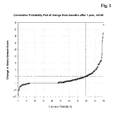
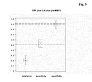
- the next important step was to define a cut-off value for the progression rates to be able to classify the patients in RA with progression and RA without progression. Therefore a cumulative probability plot of the progression rate 1 or 2 from all patients were made (see figure 1 ) ( van der Heijde et al., Arthritis Rheum. 52 (2005) 49-60 ). Laying a straight line onto the first slope of the plot, the intersection point was determined at a progression rate (1) of "5". The same results was obtained using a probability plot of the progression rate (2). To use a progression rate of "5" (i.e. an increase in SGS of more than 5 per year) as a cut-off value for classification of RA patients into patients with or without progression was supported by following two arguments:
- MTP microtiter plate
- RF and CRP were determined in a homogeneous test format on an automatic Hitachi analyzer. All marker concentrations were determined in serum samples with the exception of CartiLaps, which was measured in urine. The CartiLaps values were normalized by the creatinine results.
- Table 2 Assays and Suppliers Biomarker Assay type / format Source Anti-CCP Sandwich ELISA, MTP Axis-Shield, Dundee (UK) CRP Homogenous assay, Hitachi Roche Diagnostics, Mannheim (FRG) Hyaluronic acid Sandwich ELISA, MTP Chugai, Tokyo (J) IL-6 Sandwich ELISA, MTP Roche Diagnostics, Mannheim (FRG) RF Homogenous assay, Hitachi Roche Diagnostics, Mannheim (FRG) SAA Sandwich ELISA, MTP Biosource, Nivelles (B) pro-MMP-3 Sandwich ELISA, MTP The Binding Site, Birmingham (UK) S100 A8/A9 Sandwich ELISA, MTP Bühlmann Lab., Allschwill (CH) S100 A12 Prototype ELISA, MTP Roche Diagnostics, Penzberg (FRG) Osteocalcin Sandwich ELISA, Elecsys ® Roche Diagnostics, Mannheim (FRG) ß-Crosslaps Sandwich ELISA
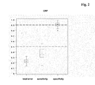
- Table 3 represents the AUC values and the sensitivity (at a specificity of 90%) for each marker.
- Table 3 Univariate analysis Biomarker AUC (%) Sensitivity (%) at a specificity of 90% Anti-CCP 59 5 CRP 75 37 Hyaluronic acid 70 20 IL-6 77 32 RF 67 24 SAA 70 27 pro-MMP-3 72 31 S100 A8/A9 70 29 S100 A12 68 32 Osteocalcin 50 8 ß-CrossLaps 57 7 PINP 55 10 sCD14 61 17 CartiLaps 71 19 ICTP 71 19 sE-selectin 67 20
- the classification algorithms were generated with the Regularized Discriminant Analysis (RDA), which is a generalization of the common Discriminant Analysis, i.e. Quadratic- and Linear Discriminant Analysis ( McLachlan, G.J., Discriminant Analysis and Statistical Pattern Recognition, Wiley Series in probability and mathematical statistics, 1992 ).
- RDA Regularized Discriminant Analysis
- plug-in maximum likelihood estimator
- the marker panels were stepwise constructed starting from the best single marker for the classification problem and ending when the total classification error do not change remarkable any more. In order to gain centralized distributions every single marker was transformed with the natural logarithmic function.
- the goal of the multivariate analysis was to find a marker panel, which shows a higher sensitivity than the best single marker.
- the specificity limit was set to 90%.
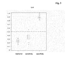
- the first marker selected was CRP with a sensitivity of 35% and the second one was IL-6 improving the sensitivity to 50%.
- the aim of the current invention is to aid the rheumatologist in his assessment whether an RA patient is at risk of disease progression.
- the diagnostic value of the identified marker panel is best reflected in Table 4 by the total error of the classification.
- CRP currently a single biological marker used for the estimation of inflammation gives a total error of 0.228. 11-6 as a single marker also reveals a similar total error of 0.247.
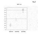
- the preferred combination of CRP and IL 6 significantly improves the classification reducing the total error to 0.203. Adding a third and a fourth marker finally helps to further minimize the misclassification (total error 0.196).
- the achieved sensitivity of 50% suggests that based on the method disclosed here about one half of the RA-patients with a progressive disease can be identified correctly by biochemical markers measure at a single time point, i.e. at baseline, which is not possible so far. This classification is expected to aid the rheumatologist in the decision process for example to start a treatment using DMARDs or to change to a better therapy scheme using a combination of different DMARDs.
- Table 4 Classification results of patients classified as RA with disease progression versus RA with no disease progression No of Markers marker or marker panel ECV (50 fold) TOTAL ERROR correct pos. Sensitivity correct neg.
Landscapes
- Health & Medical Sciences (AREA)
- Life Sciences & Earth Sciences (AREA)
- Engineering & Computer Science (AREA)
- Immunology (AREA)
- Hematology (AREA)
- Molecular Biology (AREA)
- Urology & Nephrology (AREA)
- Chemical & Material Sciences (AREA)
- Biomedical Technology (AREA)
- Cell Biology (AREA)
- General Health & Medical Sciences (AREA)
- Analytical Chemistry (AREA)
- Pathology (AREA)
- Microbiology (AREA)
- Food Science & Technology (AREA)
- Medicinal Chemistry (AREA)
- Physics & Mathematics (AREA)
- General Physics & Mathematics (AREA)
- Biochemistry (AREA)
- Biotechnology (AREA)
- Proteomics, Peptides & Aminoacids (AREA)
- Rheumatology (AREA)
- Rehabilitation Therapy (AREA)
- Investigating Or Analysing Biological Materials (AREA)
- Measuring Or Testing Involving Enzymes Or Micro-Organisms (AREA)
- Medicines That Contain Protein Lipid Enzymes And Other Medicines (AREA)
Claims (8)
- In-vitro-Verfahren zur Unterstützung beim Abschätzen des Risikos des Fortschreitens der Krankheit bei einem Patienten mit rheumatoider Arthritis (RA), wobei das Verfahren die folgenden Schritte umfasst:a) das Messen der Konzentration von sowohl C-reaktivem Protein (CRP) und Interleukin-6 und gegebenenfalls einem oder mehreren anderen Markern in einer flüssigen Probe,b) das mathematische Kombinieren der in Schritt a) gemessenen Werte undc) das Korrelieren des kombinierten, in Schritt (b) bestimmten Wertes mit dem Risiko des Fortschreitens der Krankheit,
wobei das Verfahren des mathematischen Kombinierens der Werte in Schritt (b) aus DA (d.h. linearer, quadratischer, geregelter Diskriminanzanalyse), Kernel-Methoden (d.h. SVM), parameterfreien Methoden (d.h. k-Nächste-Nachbarn-Klassifikation), PLS (Partial Least Squares, partielle kleinste Quadrate), auf Entscheidungsbäumen basierenden Methoden (d.h. logische Regression, CART, Random-Forest-Methoden, Verstärkungsmethoden) oder generalisierten linearen Modellen (d.h. logistischer Regression) ausgewählt ist. - Verfahren nach Anspruch 1, wobei der RA-Patient während der Durchführung der Untersuchung mit einem aus der aus krankheitsmodifizierenden Antirheumamitteln (Disease Modifying Anti-Rheumatoid Drugs, DMARDs) ausgewählten Antirheumamittel behandelt wird.
- Verfahren nach Anspruch 1, wobei der eine oder die mehreren gegebenenfalls gemessenen anderen Marker aus der aus Knochen- oder Knorpelmarkern, Synovialflüssigkeitsmarkern, anderen Entzündungsmarkern, genetischen Markern und radiologischen Bewertungen bestehenden Gruppe ausgewählt sind.
- Verfahren nach Anspruch 3, wobei der eine oder die mehreren Knochen- oder Knorpelmarker aus der aus PINP, β-CrossLaps, CartiLaps, Osteocalcin und ICTP bestehenden Gruppe ausgewählt sind.
- Verfahren nach Anspruch 3, wobei der eine oder die mehreren Synovialmarker aus der aus Hyaluronsäure und pro-MMP 3 bestehenden Gruppe ausgewählt sind.
- Verfahren nach Anspruch 3, wobei der eine oder die mehreren genetischen Marker aus der aus HLA-DR4 und HLA-DRB1-Allelen bestehenden Gruppe ausgewählt sind.
- Verfahren nach Anspruch 3, wobei die eine oder die mehreren radiologischen Bewertungen aus der aus den Sharp-Score, Sharp-Genant-Score, van der Heijde-Sharp-Score, Ratingen-Score, Larsen-Score und RAU-Score bestehenden Gruppe ausgewählt sind.
- Verfahren nach Anspruch 3, wobei der eine oder die mehreren anderen Entzündungsmarker aus der aus SAA und E-Selektin bestehenden Gruppe ausgewählt sind.
Priority Applications (1)
| Application Number | Priority Date | Filing Date | Title |
|---|---|---|---|
| EP07818398.5A EP2074428B1 (de) | 2006-09-29 | 2007-09-25 | Beurteilung des risikos eines fortschreitens der krankheit bei einem patienten mit rheumatoider arthritis |
Applications Claiming Priority (4)
| Application Number | Priority Date | Filing Date | Title |
|---|---|---|---|
| EP06020645 | 2006-09-29 | ||
| EP06021887 | 2006-10-19 | ||
| EP07818398.5A EP2074428B1 (de) | 2006-09-29 | 2007-09-25 | Beurteilung des risikos eines fortschreitens der krankheit bei einem patienten mit rheumatoider arthritis |
| PCT/EP2007/008313 WO2008037420A1 (en) | 2006-09-29 | 2007-09-25 | Assessing the risk of disease progression for a patient with rheumatoid arthritis |
Publications (2)
| Publication Number | Publication Date |
|---|---|
| EP2074428A1 EP2074428A1 (de) | 2009-07-01 |
| EP2074428B1 true EP2074428B1 (de) | 2015-07-01 |
Family
ID=38859738
Family Applications (1)
| Application Number | Title | Priority Date | Filing Date |
|---|---|---|---|
| EP07818398.5A Not-in-force EP2074428B1 (de) | 2006-09-29 | 2007-09-25 | Beurteilung des risikos eines fortschreitens der krankheit bei einem patienten mit rheumatoider arthritis |
Country Status (6)
| Country | Link |
|---|---|
| US (1) | US8058013B2 (de) |
| EP (1) | EP2074428B1 (de) |
| JP (1) | JP2010506147A (de) |
| CA (1) | CA2663730A1 (de) |
| ES (1) | ES2545688T3 (de) |
| WO (1) | WO2008037420A1 (de) |
Families Citing this family (16)
| Publication number | Priority date | Publication date | Assignee | Title |
|---|---|---|---|---|
| WO2010038104A1 (en) * | 2008-10-03 | 2010-04-08 | INSERM (Institut National de la Santé et de la Recherche Médicale) | Combination of cardiovascular risk factors for the diagnosis/prognosis of a cardiovascular disease/event. |
| WO2011047358A1 (en) | 2009-10-15 | 2011-04-21 | Crescendo Bioscience | Biomarkers and methods for measuring and monitoring inflammatory disease activity |
| US20130246097A1 (en) * | 2010-03-17 | 2013-09-19 | Howard M. Kenney | Medical Information Systems and Medical Data Processing Methods |
| JP5786020B2 (ja) | 2010-04-16 | 2015-09-30 | アボットジャパン株式会社 | 関節リウマチを診断する方法および試薬 |
| US8883427B2 (en) | 2010-06-30 | 2014-11-11 | New York University | Quantifying local inflammatory activity and its use to predict disease progression and tailor treatments |
| JP5924659B2 (ja) * | 2010-10-14 | 2016-05-25 | 国立大学法人 長崎大学 | 免疫複合体の網羅的解析方法および新規関節リウマチバイオマーカー |
| US20140142861A1 (en) * | 2010-11-06 | 2014-05-22 | Oklahoma Medical Research Foundation | Biomarkers For Predicting Progressive Joint Damage |
| US11531033B2 (en) * | 2013-03-15 | 2022-12-20 | Exagen Inc. | Methods for treating and diagnosing systemic lupus erythematosus |
| JP2015061592A (ja) * | 2013-08-21 | 2015-04-02 | コニカミノルタ株式会社 | 超音波診断装置、超音波画像処理方法およびコンピュータ読み取り可能な非一時的な記録媒体 |
| CA2943821A1 (en) | 2014-04-02 | 2015-10-08 | Crescendo Bioscience | Biomarkers and methods for measuring and monitoring juvenile idiopathic arthritis activity |
| EP3155439A4 (de) | 2014-06-10 | 2018-03-14 | Crescendo Bioscience | Biomarker und verfahren zur messung und überwachung der axialen spondyloarthritiserkrankungsaktivität |
| CA3207751A1 (en) | 2015-09-29 | 2017-04-06 | Laboratory Corporation Of America Holdings | Biomarkers and methods for assessing psoriatic arthritis disease activity |
| WO2017058999A2 (en) | 2015-09-29 | 2017-04-06 | Crescendo Bioscience | Biomarkers and methods for assessing response to inflammatory disease therapy withdrawal |
| GB201603571D0 (en) * | 2016-03-01 | 2016-04-13 | Univ Warwick | Markers for skeletal disorders |
| WO2021087116A1 (en) * | 2019-10-30 | 2021-05-06 | Cd Diagnostics, Inc. | Biomarkers of early osteoarthritis |
| CN114089632B (zh) * | 2021-11-15 | 2023-08-18 | 陕西师范大学 | 基于模糊逻辑的风湿免疫疾病特征识别方法及系统 |
Family Cites Families (14)
| Publication number | Priority date | Publication date | Assignee | Title |
|---|---|---|---|---|
| GB9105893D0 (en) * | 1991-03-20 | 1991-05-08 | Orion Yhtymae Oy | Bone resorption assay based on a peptide liberated during collagen degradation |
| US5888510A (en) * | 1993-07-21 | 1999-03-30 | Chugai Seiyaku Kabushiki Kaisha | Chronic rheumatoid arthritis therapy containing IL-6 antagonist as effective component |
| AU3746295A (en) * | 1994-10-17 | 1996-05-06 | Osteometer Biotech As | Estimation of the fragmentation pattern of collagen in body fluids and the diagnosis of disorders associated with the metabolism of collagen |
| FR2752842B1 (fr) | 1996-08-30 | 1998-11-06 | Biomerieux Sa | Antigenes derives des filaggrines et leur utilisation pour le diagnostic de la polyarthrite rhumatoide |
| NL1004539C2 (nl) | 1996-11-15 | 1998-05-20 | Stichting Tech Wetenschapp | Peptide afgeleid van een door auto-antilichamen van patiënten met reumatoïde artritis herkend antigeen, antilichaam daartegen en werkwijze voor het detecteren van auto-immuunantilichamen. |
| CA2309534A1 (en) | 1997-11-28 | 1999-06-10 | Innogenetics N.V. | Synthetic peptides containing citrulline recognized by rheumatoid arthritis sera as tools for diagnosis and treatment |
| FR2773157B1 (fr) | 1997-12-30 | 2001-10-05 | Bio Merieux | Epitopes peptidiques reconnus par des auto-anticorps antifilaggrine presents dans le serum des patients atteints de polyarthrite rhumatoide |
| IL136889A0 (en) * | 1998-01-09 | 2001-06-14 | Pfizer | Matrix metalloprotease inhibitors |
| EP1240180A2 (de) | 1999-12-21 | 2002-09-18 | Innogenetics N.V. | Peptide zur diagnose und behandlung rheumatischer arthritis |
| NL1019540C2 (nl) | 2001-12-11 | 2003-07-01 | Stichting Tech Wetenschapp | Werkwijze voor het detecteren van auto-antilichamen van patienten die lijden aan reumatoïde artritis, peptide en assaykit. |
| CA2552425C (en) * | 2004-02-27 | 2018-02-06 | F. Hoffmann-La Roche Ag | Method of assessing rheumatoid arthritis by measuring rheumatoid factor and interleukin-6 |
| US20060063162A1 (en) * | 2004-09-23 | 2006-03-23 | Deng David X | Biological marker for inflammation |
| WO2006098401A1 (ja) * | 2005-03-16 | 2006-09-21 | The University Of Tokushima | 関節リウマチ罹患リスクの測定方法 |
| US7713521B2 (en) * | 2005-08-12 | 2010-05-11 | Schering Corporation | MCP1 fusions |
-
2007
- 2007-09-25 JP JP2009529589A patent/JP2010506147A/ja active Pending
- 2007-09-25 WO PCT/EP2007/008313 patent/WO2008037420A1/en not_active Ceased
- 2007-09-25 EP EP07818398.5A patent/EP2074428B1/de not_active Not-in-force
- 2007-09-25 CA CA002663730A patent/CA2663730A1/en not_active Abandoned
- 2007-09-25 ES ES07818398.5T patent/ES2545688T3/es active Active
-
2009
- 2009-03-27 US US12/412,840 patent/US8058013B2/en not_active Expired - Fee Related
Also Published As
| Publication number | Publication date |
|---|---|
| JP2010506147A (ja) | 2010-02-25 |
| EP2074428A1 (de) | 2009-07-01 |
| US20090270272A1 (en) | 2009-10-29 |
| WO2008037420A1 (en) | 2008-04-03 |
| ES2545688T3 (es) | 2015-09-15 |
| US8058013B2 (en) | 2011-11-15 |
| CA2663730A1 (en) | 2008-04-03 |
Similar Documents
| Publication | Publication Date | Title |
|---|---|---|
| EP2074428B1 (de) | Beurteilung des risikos eines fortschreitens der krankheit bei einem patienten mit rheumatoider arthritis | |
| Visser | Early diagnosis of rheumatoid arthritis | |
| US8062907B2 (en) | Method to assess the severity of rheumatoid arthritis by measuring anti-CCP and serum amyloid A | |
| Hurnakova et al. | Serum calprotectin (S100A8/9): an independent predictor of ultrasound synovitis in patients with rheumatoid arthritis | |
| US7846674B2 (en) | Assessing rheumatoid arthritis by measuring anti-CCP and interleukin 6 | |
| US20070148704A1 (en) | Anti-CCPand antinuclear antibodies in diagnosis of rheumatoid arthritis | |
| US20090265116A1 (en) | Prediction of an individual's risk of developing rheumatoid arthritis | |
| CN101523218B (zh) | 评估类风湿性关节炎患者疾病进展的风险 | |
| EP1721163B1 (de) | Methode zur detektion von rheumatischer arthritis beinhaltend die messung von rheumatoidem faktor und interleukin-6 | |
| Hussein et al. | Peptidyl-arginine deiminase-type IV as a diagnostic and prognostic marker in rheumatoid arthritis patients | |
| CN104105967B (zh) | 生腱蛋白c及其在类风湿关节炎的用途 | |
| Yasin et al. | Extensive study of CCN4, VCAM-1, MMP-3, and GM-CSF as reliable markers for disease activity in rheumatoid arthritis | |
| HK1136628B (en) | Assessing the risk of disease progression for a patient with rheumatoid arthritis | |
| HK1136628A (en) | Assessing the risk of disease progression for a patient with rheumatoid arthritis | |
| Pawar et al. | INTEGRATED STUDY OF BIOMARKER IN AMAVATA WSR TO RHEUMATOID ARTHRITIS | |
| Nordal et al. | and Hilde Berner Hammer4 |
Legal Events
| Date | Code | Title | Description |
|---|---|---|---|
| PUAI | Public reference made under article 153(3) epc to a published international application that has entered the european phase |
Free format text: ORIGINAL CODE: 0009012 |
|
| 17P | Request for examination filed |
Effective date: 20090429 |
|
| AK | Designated contracting states |
Kind code of ref document: A1 Designated state(s): AT BE BG CH CY CZ DE DK EE ES FI FR GB GR HU IE IS IT LI LT LU LV MC MT NL PL PT RO SE SI SK TR |
|
| AX | Request for extension of the european patent |
Extension state: AL BA HR MK RS |
|
| 17Q | First examination report despatched |
Effective date: 20120224 |
|
| DAX | Request for extension of the european patent (deleted) | ||
| GRAP | Despatch of communication of intention to grant a patent |
Free format text: ORIGINAL CODE: EPIDOSNIGR1 |
|
| INTG | Intention to grant announced |
Effective date: 20150212 |
|
| GRAS | Grant fee paid |
Free format text: ORIGINAL CODE: EPIDOSNIGR3 |
|
| GRAA | (expected) grant |
Free format text: ORIGINAL CODE: 0009210 |
|
| RIN1 | Information on inventor provided before grant (corrected) |
Inventor name: KARL, JOHANN Inventor name: ROLLINGER, WOLFGANG Inventor name: GRUNERT, VEIT PETER Inventor name: WILD, NORBERT |
|
| AK | Designated contracting states |
Kind code of ref document: B1 Designated state(s): AT BE BG CH CY CZ DE DK EE ES FI FR GB GR HU IE IS IT LI LT LU LV MC MT NL PL PT RO SE SI SK TR |
|
| REG | Reference to a national code |
Ref country code: GB Ref legal event code: FG4D |
|
| REG | Reference to a national code |
Ref country code: AT Ref legal event code: REF Ref document number: 734265 Country of ref document: AT Kind code of ref document: T Effective date: 20150715 Ref country code: CH Ref legal event code: EP |
|
| REG | Reference to a national code |
Ref country code: IE Ref legal event code: FG4D |
|
| REG | Reference to a national code |
Ref country code: DE Ref legal event code: R096 Ref document number: 602007041967 Country of ref document: DE |
|
| REG | Reference to a national code |
Ref country code: ES Ref legal event code: FG2A Ref document number: 2545688 Country of ref document: ES Kind code of ref document: T3 Effective date: 20150915 |
|
| REG | Reference to a national code |
Ref country code: AT Ref legal event code: MK05 Ref document number: 734265 Country of ref document: AT Kind code of ref document: T Effective date: 20150701 |
|
| REG | Reference to a national code |
Ref country code: NL Ref legal event code: MP Effective date: 20150701 |
|
| REG | Reference to a national code |
Ref country code: LT Ref legal event code: MG4D |
|
| PG25 | Lapsed in a contracting state [announced via postgrant information from national office to epo] |
Ref country code: FI Free format text: LAPSE BECAUSE OF FAILURE TO SUBMIT A TRANSLATION OF THE DESCRIPTION OR TO PAY THE FEE WITHIN THE PRESCRIBED TIME-LIMIT Effective date: 20150701 Ref country code: GR Free format text: LAPSE BECAUSE OF FAILURE TO SUBMIT A TRANSLATION OF THE DESCRIPTION OR TO PAY THE FEE WITHIN THE PRESCRIBED TIME-LIMIT Effective date: 20151002 Ref country code: LV Free format text: LAPSE BECAUSE OF FAILURE TO SUBMIT A TRANSLATION OF THE DESCRIPTION OR TO PAY THE FEE WITHIN THE PRESCRIBED TIME-LIMIT Effective date: 20150701 Ref country code: LT Free format text: LAPSE BECAUSE OF FAILURE TO SUBMIT A TRANSLATION OF THE DESCRIPTION OR TO PAY THE FEE WITHIN THE PRESCRIBED TIME-LIMIT Effective date: 20150701 |
|
| PG25 | Lapsed in a contracting state [announced via postgrant information from national office to epo] |
Ref country code: PT Free format text: LAPSE BECAUSE OF FAILURE TO SUBMIT A TRANSLATION OF THE DESCRIPTION OR TO PAY THE FEE WITHIN THE PRESCRIBED TIME-LIMIT Effective date: 20151102 Ref country code: PL Free format text: LAPSE BECAUSE OF FAILURE TO SUBMIT A TRANSLATION OF THE DESCRIPTION OR TO PAY THE FEE WITHIN THE PRESCRIBED TIME-LIMIT Effective date: 20150701 Ref country code: IS Free format text: LAPSE BECAUSE OF FAILURE TO SUBMIT A TRANSLATION OF THE DESCRIPTION OR TO PAY THE FEE WITHIN THE PRESCRIBED TIME-LIMIT Effective date: 20151101 Ref country code: AT Free format text: LAPSE BECAUSE OF FAILURE TO SUBMIT A TRANSLATION OF THE DESCRIPTION OR TO PAY THE FEE WITHIN THE PRESCRIBED TIME-LIMIT Effective date: 20150701 Ref country code: SE Free format text: LAPSE BECAUSE OF FAILURE TO SUBMIT A TRANSLATION OF THE DESCRIPTION OR TO PAY THE FEE WITHIN THE PRESCRIBED TIME-LIMIT Effective date: 20150701 |
|
| REG | Reference to a national code |
Ref country code: DE Ref legal event code: R097 Ref document number: 602007041967 Country of ref document: DE |
|
| PG25 | Lapsed in a contracting state [announced via postgrant information from national office to epo] |
Ref country code: CZ Free format text: LAPSE BECAUSE OF FAILURE TO SUBMIT A TRANSLATION OF THE DESCRIPTION OR TO PAY THE FEE WITHIN THE PRESCRIBED TIME-LIMIT Effective date: 20150701 Ref country code: LU Free format text: LAPSE BECAUSE OF FAILURE TO SUBMIT A TRANSLATION OF THE DESCRIPTION OR TO PAY THE FEE WITHIN THE PRESCRIBED TIME-LIMIT Effective date: 20150925 Ref country code: MC Free format text: LAPSE BECAUSE OF FAILURE TO SUBMIT A TRANSLATION OF THE DESCRIPTION OR TO PAY THE FEE WITHIN THE PRESCRIBED TIME-LIMIT Effective date: 20150701 Ref country code: SK Free format text: LAPSE BECAUSE OF FAILURE TO SUBMIT A TRANSLATION OF THE DESCRIPTION OR TO PAY THE FEE WITHIN THE PRESCRIBED TIME-LIMIT Effective date: 20150701 Ref country code: DK Free format text: LAPSE BECAUSE OF FAILURE TO SUBMIT A TRANSLATION OF THE DESCRIPTION OR TO PAY THE FEE WITHIN THE PRESCRIBED TIME-LIMIT Effective date: 20150701 Ref country code: EE Free format text: LAPSE BECAUSE OF FAILURE TO SUBMIT A TRANSLATION OF THE DESCRIPTION OR TO PAY THE FEE WITHIN THE PRESCRIBED TIME-LIMIT Effective date: 20150701 |
|
| PLBE | No opposition filed within time limit |
Free format text: ORIGINAL CODE: 0009261 |
|
| STAA | Information on the status of an ep patent application or granted ep patent |
Free format text: STATUS: NO OPPOSITION FILED WITHIN TIME LIMIT |
|
| PG25 | Lapsed in a contracting state [announced via postgrant information from national office to epo] |
Ref country code: RO Free format text: LAPSE BECAUSE OF FAILURE TO SUBMIT A TRANSLATION OF THE DESCRIPTION OR TO PAY THE FEE WITHIN THE PRESCRIBED TIME-LIMIT Effective date: 20150701 |
|
| 26N | No opposition filed |
Effective date: 20160404 |
|
| REG | Reference to a national code |
Ref country code: IE Ref legal event code: MM4A |
|
| PG25 | Lapsed in a contracting state [announced via postgrant information from national office to epo] |
Ref country code: IE Free format text: LAPSE BECAUSE OF NON-PAYMENT OF DUE FEES Effective date: 20150925 |
|
| REG | Reference to a national code |
Ref country code: FR Ref legal event code: PLFP Year of fee payment: 10 |
|
| PG25 | Lapsed in a contracting state [announced via postgrant information from national office to epo] |
Ref country code: SI Free format text: LAPSE BECAUSE OF FAILURE TO SUBMIT A TRANSLATION OF THE DESCRIPTION OR TO PAY THE FEE WITHIN THE PRESCRIBED TIME-LIMIT Effective date: 20150701 |
|
| PG25 | Lapsed in a contracting state [announced via postgrant information from national office to epo] |
Ref country code: BE Free format text: LAPSE BECAUSE OF FAILURE TO SUBMIT A TRANSLATION OF THE DESCRIPTION OR TO PAY THE FEE WITHIN THE PRESCRIBED TIME-LIMIT Effective date: 20150701 |
|
| PG25 | Lapsed in a contracting state [announced via postgrant information from national office to epo] |
Ref country code: MT Free format text: LAPSE BECAUSE OF FAILURE TO SUBMIT A TRANSLATION OF THE DESCRIPTION OR TO PAY THE FEE WITHIN THE PRESCRIBED TIME-LIMIT Effective date: 20150701 |
|
| PG25 | Lapsed in a contracting state [announced via postgrant information from national office to epo] |
Ref country code: BG Free format text: LAPSE BECAUSE OF FAILURE TO SUBMIT A TRANSLATION OF THE DESCRIPTION OR TO PAY THE FEE WITHIN THE PRESCRIBED TIME-LIMIT Effective date: 20150701 Ref country code: HU Free format text: LAPSE BECAUSE OF FAILURE TO SUBMIT A TRANSLATION OF THE DESCRIPTION OR TO PAY THE FEE WITHIN THE PRESCRIBED TIME-LIMIT; INVALID AB INITIO Effective date: 20070925 |
|
| PG25 | Lapsed in a contracting state [announced via postgrant information from national office to epo] |
Ref country code: NL Free format text: LAPSE BECAUSE OF FAILURE TO SUBMIT A TRANSLATION OF THE DESCRIPTION OR TO PAY THE FEE WITHIN THE PRESCRIBED TIME-LIMIT Effective date: 20150701 Ref country code: CY Free format text: LAPSE BECAUSE OF FAILURE TO SUBMIT A TRANSLATION OF THE DESCRIPTION OR TO PAY THE FEE WITHIN THE PRESCRIBED TIME-LIMIT Effective date: 20150701 |
|
| REG | Reference to a national code |
Ref country code: FR Ref legal event code: PLFP Year of fee payment: 11 |
|
| PG25 | Lapsed in a contracting state [announced via postgrant information from national office to epo] |
Ref country code: TR Free format text: LAPSE BECAUSE OF FAILURE TO SUBMIT A TRANSLATION OF THE DESCRIPTION OR TO PAY THE FEE WITHIN THE PRESCRIBED TIME-LIMIT Effective date: 20150701 |
|
| REG | Reference to a national code |
Ref country code: FR Ref legal event code: PLFP Year of fee payment: 12 |
|
| PGFP | Annual fee paid to national office [announced via postgrant information from national office to epo] |
Ref country code: IT Payment date: 20210915 Year of fee payment: 15 Ref country code: FR Payment date: 20210831 Year of fee payment: 15 |
|
| PGFP | Annual fee paid to national office [announced via postgrant information from national office to epo] |
Ref country code: GB Payment date: 20210831 Year of fee payment: 15 Ref country code: CH Payment date: 20210816 Year of fee payment: 15 Ref country code: DE Payment date: 20210812 Year of fee payment: 15 |
|
| PGFP | Annual fee paid to national office [announced via postgrant information from national office to epo] |
Ref country code: ES Payment date: 20211007 Year of fee payment: 15 |
|
| REG | Reference to a national code |
Ref country code: DE Ref legal event code: R119 Ref document number: 602007041967 Country of ref document: DE |
|
| REG | Reference to a national code |
Ref country code: CH Ref legal event code: PL |
|
| GBPC | Gb: european patent ceased through non-payment of renewal fee |
Effective date: 20220925 |
|
| PG25 | Lapsed in a contracting state [announced via postgrant information from national office to epo] |
Ref country code: LI Free format text: LAPSE BECAUSE OF NON-PAYMENT OF DUE FEES Effective date: 20220930 Ref country code: FR Free format text: LAPSE BECAUSE OF NON-PAYMENT OF DUE FEES Effective date: 20220930 Ref country code: DE Free format text: LAPSE BECAUSE OF NON-PAYMENT OF DUE FEES Effective date: 20230401 Ref country code: CH Free format text: LAPSE BECAUSE OF NON-PAYMENT OF DUE FEES Effective date: 20220930 |
|
| PG25 | Lapsed in a contracting state [announced via postgrant information from national office to epo] |
Ref country code: IT Free format text: LAPSE BECAUSE OF NON-PAYMENT OF DUE FEES Effective date: 20220925 Ref country code: GB Free format text: LAPSE BECAUSE OF NON-PAYMENT OF DUE FEES Effective date: 20220925 |
|
| REG | Reference to a national code |
Ref country code: ES Ref legal event code: FD2A Effective date: 20231102 |
|
| PG25 | Lapsed in a contracting state [announced via postgrant information from national office to epo] |
Ref country code: ES Free format text: LAPSE BECAUSE OF NON-PAYMENT OF DUE FEES Effective date: 20220926 |
|
| PG25 | Lapsed in a contracting state [announced via postgrant information from national office to epo] |
Ref country code: ES Free format text: LAPSE BECAUSE OF NON-PAYMENT OF DUE FEES Effective date: 20220926 |