EP2074428B1 - Assessing the risk of disease progression for a patient with rheumatoid arthritis - Google Patents
Assessing the risk of disease progression for a patient with rheumatoid arthritis Download PDFInfo
- Publication number
- EP2074428B1 EP2074428B1 EP07818398.5A EP07818398A EP2074428B1 EP 2074428 B1 EP2074428 B1 EP 2074428B1 EP 07818398 A EP07818398 A EP 07818398A EP 2074428 B1 EP2074428 B1 EP 2074428B1
- Authority
- EP
- European Patent Office
- Prior art keywords
- score
- markers
- marker
- disease progression
- patient
- Prior art date
- Legal status (The legal status is an assumption and is not a legal conclusion. Google has not performed a legal analysis and makes no representation as to the accuracy of the status listed.)
- Not-in-force
Links
- 206010039073 rheumatoid arthritis Diseases 0.000 title claims description 109
- 230000005750 disease progression Effects 0.000 title claims description 64
- 206010061818 Disease progression Diseases 0.000 title claims description 63
- 239000003550 marker Substances 0.000 claims description 88
- 238000000034 method Methods 0.000 claims description 80
- 108010074051 C-Reactive Protein Proteins 0.000 claims description 60
- 102100032752 C-reactive protein Human genes 0.000 claims description 60
- 102000004889 Interleukin-6 Human genes 0.000 claims description 50
- 108090001005 Interleukin-6 Proteins 0.000 claims description 50
- 229940100601 interleukin-6 Drugs 0.000 claims description 50
- 210000000988 bone and bone Anatomy 0.000 claims description 28
- 210000000845 cartilage Anatomy 0.000 claims description 23
- 230000004054 inflammatory process Effects 0.000 claims description 21
- 206010061218 Inflammation Diseases 0.000 claims description 19
- 238000011282 treatment Methods 0.000 claims description 13
- 238000004458 analytical method Methods 0.000 claims description 12
- KIUKXJAPPMFGSW-DNGZLQJQSA-N (2S,3S,4S,5R,6R)-6-[(2S,3R,4R,5S,6R)-3-Acetamido-2-[(2S,3S,4R,5R,6R)-6-[(2R,3R,4R,5S,6R)-3-acetamido-2,5-dihydroxy-6-(hydroxymethyl)oxan-4-yl]oxy-2-carboxy-4,5-dihydroxyoxan-3-yl]oxy-5-hydroxy-6-(hydroxymethyl)oxan-4-yl]oxy-3,4,5-trihydroxyoxane-2-carboxylic acid Chemical compound CC(=O)N[C@H]1[C@H](O)O[C@H](CO)[C@@H](O)[C@@H]1O[C@H]1[C@H](O)[C@@H](O)[C@H](O[C@H]2[C@@H]([C@@H](O[C@H]3[C@@H]([C@@H](O)[C@H](O)[C@H](O3)C(O)=O)O)[C@H](O)[C@@H](CO)O2)NC(C)=O)[C@@H](C(O)=O)O1 KIUKXJAPPMFGSW-DNGZLQJQSA-N 0.000 claims description 8
- 239000003814 drug Substances 0.000 claims description 8
- 229920002674 hyaluronan Polymers 0.000 claims description 8
- 229960003160 hyaluronic acid Drugs 0.000 claims description 8
- 238000007477 logistic regression Methods 0.000 claims description 8
- 108010024212 E-Selectin Proteins 0.000 claims description 6
- 229940079593 drug Drugs 0.000 claims description 6
- 230000002068 genetic effect Effects 0.000 claims description 6
- 238000007637 random forest analysis Methods 0.000 claims description 6
- 210000001179 synovial fluid Anatomy 0.000 claims description 6
- 102000004067 Osteocalcin Human genes 0.000 claims description 5
- 108090000573 Osteocalcin Proteins 0.000 claims description 5
- 239000007788 liquid Substances 0.000 claims description 5
- 230000036961 partial effect Effects 0.000 claims description 5
- 239000003435 antirheumatic agent Substances 0.000 claims description 4
- 108700028369 Alleles Proteins 0.000 claims description 3
- 102100040485 HLA class II histocompatibility antigen, DRB1 beta chain Human genes 0.000 claims description 3
- 108010039343 HLA-DRB1 Chains Proteins 0.000 claims description 3
- 230000003356 anti-rheumatic effect Effects 0.000 claims description 3
- 108010046732 HLA-DR4 Antigen Proteins 0.000 claims description 2
- 208000035474 group of disease Diseases 0.000 claims description 2
- 102000015689 E-Selectin Human genes 0.000 claims 1
- 201000010099 disease Diseases 0.000 description 29
- 208000037265 diseases, disorders, signs and symptoms Diseases 0.000 description 29
- 230000035945 sensitivity Effects 0.000 description 28
- 230000003628 erosive effect Effects 0.000 description 27
- 206010003246 arthritis Diseases 0.000 description 20
- 239000000523 sample Substances 0.000 description 19
- 230000006870 function Effects 0.000 description 18
- 238000012360 testing method Methods 0.000 description 17
- 108010000684 Matrix Metalloproteinases Proteins 0.000 description 15
- 102000002274 Matrix Metalloproteinases Human genes 0.000 description 15
- 238000003556 assay Methods 0.000 description 15
- 229920002451 polyvinyl alcohol Polymers 0.000 description 15
- 108090000765 processed proteins & peptides Proteins 0.000 description 15
- 108010035532 Collagen Proteins 0.000 description 14
- 102000008186 Collagen Human genes 0.000 description 14
- 229920001436 collagen Polymers 0.000 description 14
- 210000002683 foot Anatomy 0.000 description 14
- 210000001503 joint Anatomy 0.000 description 14
- 108010052500 Calgranulin A Proteins 0.000 description 13
- 230000008859 change Effects 0.000 description 13
- 210000004247 hand Anatomy 0.000 description 13
- 241000219061 Rheum Species 0.000 description 12
- 239000000090 biomarker Substances 0.000 description 12
- 230000006378 damage Effects 0.000 description 12
- 102000004196 processed proteins & peptides Human genes 0.000 description 12
- 108090000623 proteins and genes Proteins 0.000 description 12
- 102000004169 proteins and genes Human genes 0.000 description 12
- 230000015572 biosynthetic process Effects 0.000 description 11
- 238000003118 sandwich ELISA Methods 0.000 description 11
- 210000002966 serum Anatomy 0.000 description 11
- 210000004027 cell Anatomy 0.000 description 10
- 210000000707 wrist Anatomy 0.000 description 10
- 102100032442 Protein S100-A8 Human genes 0.000 description 9
- 238000004422 calculation algorithm Methods 0.000 description 9
- 238000003745 diagnosis Methods 0.000 description 9
- 238000003786 synthesis reaction Methods 0.000 description 8
- 230000000875 corresponding effect Effects 0.000 description 7
- 210000001519 tissue Anatomy 0.000 description 7
- MZOFCQQQCNRIBI-VMXHOPILSA-N (3s)-4-[[(2s)-1-[[(2s)-1-[[(1s)-1-carboxy-2-hydroxyethyl]amino]-4-methyl-1-oxopentan-2-yl]amino]-5-(diaminomethylideneamino)-1-oxopentan-2-yl]amino]-3-[[2-[[(2s)-2,6-diaminohexanoyl]amino]acetyl]amino]-4-oxobutanoic acid Chemical compound OC[C@@H](C(O)=O)NC(=O)[C@H](CC(C)C)NC(=O)[C@H](CCCN=C(N)N)NC(=O)[C@H](CC(O)=O)NC(=O)CNC(=O)[C@@H](N)CCCCN MZOFCQQQCNRIBI-VMXHOPILSA-N 0.000 description 6
- 102000000589 Interleukin-1 Human genes 0.000 description 6
- 108010002352 Interleukin-1 Proteins 0.000 description 6
- 102100030416 Stromelysin-1 Human genes 0.000 description 6
- 108060008682 Tumor Necrosis Factor Proteins 0.000 description 6
- 102000000852 Tumor Necrosis Factor-alpha Human genes 0.000 description 6
- 238000009826 distribution Methods 0.000 description 6
- 230000002757 inflammatory effect Effects 0.000 description 6
- 229920001184 polypeptide Polymers 0.000 description 6
- 210000001258 synovial membrane Anatomy 0.000 description 6
- 201000004595 synovitis Diseases 0.000 description 6
- 238000002560 therapeutic procedure Methods 0.000 description 6
- 238000007473 univariate analysis Methods 0.000 description 6
- 102100023471 E-selectin Human genes 0.000 description 5
- 102100032420 Protein S100-A9 Human genes 0.000 description 5
- 108010061103 cyclic citrullinated peptide Proteins 0.000 description 5
- 239000012634 fragment Substances 0.000 description 5
- 238000000338 in vitro Methods 0.000 description 5
- 230000004044 response Effects 0.000 description 5
- 238000007619 statistical method Methods 0.000 description 5
- 208000024891 symptom Diseases 0.000 description 5
- VKUYLANQOAKALN-UHFFFAOYSA-N 2-[benzyl-(4-methoxyphenyl)sulfonylamino]-n-hydroxy-4-methylpentanamide Chemical compound C1=CC(OC)=CC=C1S(=O)(=O)N(C(CC(C)C)C(=O)NO)CC1=CC=CC=C1 VKUYLANQOAKALN-UHFFFAOYSA-N 0.000 description 4
- 102000018803 Calgranulin A Human genes 0.000 description 4
- 102000012422 Collagen Type I Human genes 0.000 description 4
- 108010022452 Collagen Type I Proteins 0.000 description 4
- 238000002965 ELISA Methods 0.000 description 4
- 102000010834 Extracellular Matrix Proteins Human genes 0.000 description 4
- 108010037362 Extracellular Matrix Proteins Proteins 0.000 description 4
- 206010023203 Joint destruction Diseases 0.000 description 4
- 102100029812 Protein S100-A12 Human genes 0.000 description 4
- 102000013674 S-100 Human genes 0.000 description 4
- 108700021018 S100 Proteins 0.000 description 4
- 101710108790 Stromelysin-1 Proteins 0.000 description 4
- 230000002596 correlated effect Effects 0.000 description 4
- 230000001186 cumulative effect Effects 0.000 description 4
- 210000002889 endothelial cell Anatomy 0.000 description 4
- 210000002744 extracellular matrix Anatomy 0.000 description 4
- 230000036541 health Effects 0.000 description 4
- 238000005259 measurement Methods 0.000 description 4
- 230000004048 modification Effects 0.000 description 4
- 238000012986 modification Methods 0.000 description 4
- 210000002381 plasma Anatomy 0.000 description 4
- 238000012549 training Methods 0.000 description 4
- 108060003393 Granulin Proteins 0.000 description 3
- 108010028750 Integrin-Binding Sialoprotein Proteins 0.000 description 3
- 102000016921 Integrin-Binding Sialoprotein Human genes 0.000 description 3
- 206010023232 Joint swelling Diseases 0.000 description 3
- 102000005741 Metalloproteases Human genes 0.000 description 3
- 108010006035 Metalloproteases Proteins 0.000 description 3
- 210000001744 T-lymphocyte Anatomy 0.000 description 3
- 230000001154 acute effect Effects 0.000 description 3
- 239000000654 additive Substances 0.000 description 3
- 230000000996 additive effect Effects 0.000 description 3
- 239000003150 biochemical marker Substances 0.000 description 3
- 210000004369 blood Anatomy 0.000 description 3
- 239000008280 blood Substances 0.000 description 3
- 230000001419 dependent effect Effects 0.000 description 3
- 238000002405 diagnostic procedure Methods 0.000 description 3
- 230000009266 disease activity Effects 0.000 description 3
- 210000002950 fibroblast Anatomy 0.000 description 3
- 238000003018 immunoassay Methods 0.000 description 3
- 230000006872 improvement Effects 0.000 description 3
- 230000002427 irreversible effect Effects 0.000 description 3
- 238000012544 monitoring process Methods 0.000 description 3
- 238000000491 multivariate analysis Methods 0.000 description 3
- 238000004393 prognosis Methods 0.000 description 3
- 208000037821 progressive disease Diseases 0.000 description 3
- 230000000750 progressive effect Effects 0.000 description 3
- 230000035755 proliferation Effects 0.000 description 3
- 238000001356 surgical procedure Methods 0.000 description 3
- 230000009885 systemic effect Effects 0.000 description 3
- 210000002700 urine Anatomy 0.000 description 3
- 108010062271 Acute-Phase Proteins Proteins 0.000 description 2
- 102000011767 Acute-Phase Proteins Human genes 0.000 description 2
- 208000008822 Ankylosis Diseases 0.000 description 2
- 208000006820 Arthralgia Diseases 0.000 description 2
- 206010051728 Bone erosion Diseases 0.000 description 2
- 102100026622 Cartilage intermediate layer protein 1 Human genes 0.000 description 2
- 101710176668 Cartilage oligomeric matrix protein Proteins 0.000 description 2
- 102000000503 Collagen Type II Human genes 0.000 description 2
- 108010041390 Collagen Type II Proteins 0.000 description 2
- 102000004127 Cytokines Human genes 0.000 description 2
- 108090000695 Cytokines Proteins 0.000 description 2
- ZAHDXEIQWWLQQL-IHRRRGAJSA-N Deoxypyridinoline Chemical compound OC(=O)[C@@H](N)CCCC[N+]1=CC(O)=C(C[C@H](N)C([O-])=O)C(CC[C@H](N)C(O)=O)=C1 ZAHDXEIQWWLQQL-IHRRRGAJSA-N 0.000 description 2
- 101000913767 Homo sapiens Cartilage intermediate layer protein 1 Proteins 0.000 description 2
- 101000990915 Homo sapiens Stromelysin-1 Proteins 0.000 description 2
- 206010023198 Joint ankylosis Diseases 0.000 description 2
- 108010072582 Matrilin Proteins Proteins 0.000 description 2
- 102000055008 Matrilin Proteins Human genes 0.000 description 2
- 102000000380 Matrix Metalloproteinase 1 Human genes 0.000 description 2
- 108010016113 Matrix Metalloproteinase 1 Proteins 0.000 description 2
- 101710195116 Melanoma-derived growth regulatory protein Proteins 0.000 description 2
- 102100022185 Melanoma-derived growth regulatory protein Human genes 0.000 description 2
- 102100039364 Metalloproteinase inhibitor 1 Human genes 0.000 description 2
- 206010028980 Neoplasm Diseases 0.000 description 2
- 208000002193 Pain Diseases 0.000 description 2
- LCYXYLLJXMAEMT-SAXRGWBVSA-N Pyridinoline Chemical compound OC(=O)[C@@H](N)CCC1=C[N+](C[C@H](O)CC[C@H](N)C([O-])=O)=CC(O)=C1C[C@H](N)C(O)=O LCYXYLLJXMAEMT-SAXRGWBVSA-N 0.000 description 2
- 102000058242 S100A12 Human genes 0.000 description 2
- 108010031374 Tissue Inhibitor of Metalloproteinase-1 Proteins 0.000 description 2
- 230000004913 activation Effects 0.000 description 2
- 150000001413 amino acids Chemical class 0.000 description 2
- 239000000427 antigen Substances 0.000 description 2
- 102000036639 antigens Human genes 0.000 description 2
- 108091007433 antigens Proteins 0.000 description 2
- 230000033228 biological regulation Effects 0.000 description 2
- 229960000074 biopharmaceutical Drugs 0.000 description 2
- 210000001124 body fluid Anatomy 0.000 description 2
- 230000008416 bone turnover Effects 0.000 description 2
- 210000004899 c-terminal region Anatomy 0.000 description 2
- 210000000511 carpometacarpal joint Anatomy 0.000 description 2
- 238000007635 classification algorithm Methods 0.000 description 2
- 238000003776 cleavage reaction Methods 0.000 description 2
- 230000011382 collagen catabolic process Effects 0.000 description 2
- 238000010276 construction Methods 0.000 description 2
- DDRJAANPRJIHGJ-UHFFFAOYSA-N creatinine Chemical compound CN1CC(=O)NC1=N DDRJAANPRJIHGJ-UHFFFAOYSA-N 0.000 description 2
- 238000002790 cross-validation Methods 0.000 description 2
- 238000001493 electron microscopy Methods 0.000 description 2
- 210000001255 hallux Anatomy 0.000 description 2
- 230000002440 hepatic effect Effects 0.000 description 2
- 239000000833 heterodimer Substances 0.000 description 2
- 229960003444 immunosuppressant agent Drugs 0.000 description 2
- 239000003018 immunosuppressive agent Substances 0.000 description 2
- 239000003112 inhibitor Substances 0.000 description 2
- 208000014674 injury Diseases 0.000 description 2
- 238000009533 lab test Methods 0.000 description 2
- 210000002540 macrophage Anatomy 0.000 description 2
- 239000011159 matrix material Substances 0.000 description 2
- 108020004999 messenger RNA Proteins 0.000 description 2
- 230000004060 metabolic process Effects 0.000 description 2
- 210000000811 metacarpophalangeal joint Anatomy 0.000 description 2
- 210000000878 metatarsophalangeal joint Anatomy 0.000 description 2
- 230000011164 ossification Effects 0.000 description 2
- 210000000963 osteoblast Anatomy 0.000 description 2
- 239000013610 patient sample Substances 0.000 description 2
- 230000036470 plasma concentration Effects 0.000 description 2
- 230000008569 process Effects 0.000 description 2
- 230000002250 progressing effect Effects 0.000 description 2
- 230000000770 proinflammatory effect Effects 0.000 description 2
- 230000017854 proteolysis Effects 0.000 description 2
- 230000009257 reactivity Effects 0.000 description 2
- 230000002441 reversible effect Effects 0.000 description 2
- 230000007017 scission Effects 0.000 description 2
- 230000000638 stimulation Effects 0.000 description 2
- 210000003813 thumb Anatomy 0.000 description 2
- KZMAWJRXKGLWGS-UHFFFAOYSA-N 2-chloro-n-[4-(4-methoxyphenyl)-1,3-thiazol-2-yl]-n-(3-methoxypropyl)acetamide Chemical compound S1C(N(C(=O)CCl)CCCOC)=NC(C=2C=CC(OC)=CC=2)=C1 KZMAWJRXKGLWGS-UHFFFAOYSA-N 0.000 description 1
- 108010067219 Aggrecans Proteins 0.000 description 1
- 102000016284 Aggrecans Human genes 0.000 description 1
- 108090000672 Annexin A5 Proteins 0.000 description 1
- 208000035143 Bacterial infection Diseases 0.000 description 1
- 102000014914 Carrier Proteins Human genes 0.000 description 1
- 102000055007 Cartilage Oligomeric Matrix Human genes 0.000 description 1
- 102100027473 Cartilage oligomeric matrix protein Human genes 0.000 description 1
- 102100024484 Codanin-1 Human genes 0.000 description 1
- 102000029816 Collagenase Human genes 0.000 description 1
- 108060005980 Collagenase Proteins 0.000 description 1
- 208000035473 Communicable disease Diseases 0.000 description 1
- 108010069514 Cyclic Peptides Proteins 0.000 description 1
- 102000001189 Cyclic Peptides Human genes 0.000 description 1
- 201000003883 Cystic fibrosis Diseases 0.000 description 1
- 102000004190 Enzymes Human genes 0.000 description 1
- 108090000790 Enzymes Proteins 0.000 description 1
- 206010063560 Excessive granulation tissue Diseases 0.000 description 1
- 102000009123 Fibrin Human genes 0.000 description 1
- 108010073385 Fibrin Proteins 0.000 description 1
- BWGVNKXGVNDBDI-UHFFFAOYSA-N Fibrin monomer Chemical compound CNC(=O)CNC(=O)CN BWGVNKXGVNDBDI-UHFFFAOYSA-N 0.000 description 1
- RCPOVANIIKXVTB-YPPRVYOWSA-N Galactosylhydroxylysine Chemical compound NCCCC[C@@H](C(O)=O)N(O)C1O[C@H](CO)[C@H](O)[C@H](O)[C@H]1O RCPOVANIIKXVTB-YPPRVYOWSA-N 0.000 description 1
- 229920002683 Glycosaminoglycan Polymers 0.000 description 1
- 108010033222 HLA-DRB1*04 antigen Proteins 0.000 description 1
- 101000980888 Homo sapiens Codanin-1 Proteins 0.000 description 1
- 101000741967 Homo sapiens Presequence protease, mitochondrial Proteins 0.000 description 1
- PMMYEEVYMWASQN-DMTCNVIQSA-N Hydroxyproline Chemical compound O[C@H]1CN[C@H](C(O)=O)C1 PMMYEEVYMWASQN-DMTCNVIQSA-N 0.000 description 1
- FBOZXECLQNJBKD-ZDUSSCGKSA-N L-methotrexate Chemical compound C=1N=C2N=C(N)N=C(N)C2=NC=1CN(C)C1=CC=C(C(=O)N[C@@H](CCC(O)=O)C(O)=O)C=C1 FBOZXECLQNJBKD-ZDUSSCGKSA-N 0.000 description 1
- 102000005482 Lipopolysaccharide Receptors Human genes 0.000 description 1
- 108010031801 Lipopolysaccharide Receptors Proteins 0.000 description 1
- 238000007476 Maximum Likelihood Methods 0.000 description 1
- 102100026262 Metalloproteinase inhibitor 2 Human genes 0.000 description 1
- 206010052904 Musculoskeletal stiffness Diseases 0.000 description 1
- 101000783348 Naja atra Cytotoxin 1 Proteins 0.000 description 1
- 101000761722 Naja kaouthia Cytotoxin 1 Proteins 0.000 description 1
- 108091005461 Nucleic proteins Proteins 0.000 description 1
- 206010030113 Oedema Diseases 0.000 description 1
- 241001111421 Pannus Species 0.000 description 1
- 102100038632 Presequence protease, mitochondrial Human genes 0.000 description 1
- 108010050808 Procollagen Proteins 0.000 description 1
- 206010037660 Pyrexia Diseases 0.000 description 1
- 208000009921 Rheumatoid Nodule Diseases 0.000 description 1
- 108700028909 Serum Amyloid A Proteins 0.000 description 1
- 102000054727 Serum Amyloid A Human genes 0.000 description 1
- 101710084578 Short neurotoxin 1 Proteins 0.000 description 1
- 108010032838 Sialoglycoproteins Proteins 0.000 description 1
- 102000007365 Sialoglycoproteins Human genes 0.000 description 1
- 206010067868 Skin mass Diseases 0.000 description 1
- QAOWNCQODCNURD-UHFFFAOYSA-L Sulfate Chemical compound [O-]S([O-])(=O)=O QAOWNCQODCNURD-UHFFFAOYSA-L 0.000 description 1
- 108010031372 Tissue Inhibitor of Metalloproteinase-2 Proteins 0.000 description 1
- 102100023935 Transmembrane glycoprotein NMB Human genes 0.000 description 1
- 208000036142 Viral infection Diseases 0.000 description 1
- 208000027418 Wounds and injury Diseases 0.000 description 1
- 239000012072 active phase Substances 0.000 description 1
- 230000006022 acute inflammation Effects 0.000 description 1
- 208000038016 acute inflammation Diseases 0.000 description 1
- 230000010933 acylation Effects 0.000 description 1
- 238000005917 acylation reaction Methods 0.000 description 1
- 230000002411 adverse Effects 0.000 description 1
- 238000011256 aggressive treatment Methods 0.000 description 1
- 238000013459 approach Methods 0.000 description 1
- 238000013528 artificial neural network Methods 0.000 description 1
- 238000002820 assay format Methods 0.000 description 1
- 210000001130 astrocyte Anatomy 0.000 description 1
- 230000001363 autoimmune Effects 0.000 description 1
- 230000001580 bacterial effect Effects 0.000 description 1
- 208000022362 bacterial infectious disease Diseases 0.000 description 1
- 230000027455 binding Effects 0.000 description 1
- 108091008324 binding proteins Proteins 0.000 description 1
- 230000004071 biological effect Effects 0.000 description 1
- 239000012472 biological sample Substances 0.000 description 1
- 210000004204 blood vessel Anatomy 0.000 description 1
- 239000010839 body fluid Substances 0.000 description 1
- 210000001185 bone marrow Anatomy 0.000 description 1
- -1 but Proteins 0.000 description 1
- 201000011510 cancer Diseases 0.000 description 1
- 230000024245 cell differentiation Effects 0.000 description 1
- 230000004663 cell proliferation Effects 0.000 description 1
- 230000001413 cellular effect Effects 0.000 description 1
- 239000007795 chemical reaction product Substances 0.000 description 1
- 239000003153 chemical reaction reagent Substances 0.000 description 1
- 210000001612 chondrocyte Anatomy 0.000 description 1
- 230000001684 chronic effect Effects 0.000 description 1
- 230000006020 chronic inflammation Effects 0.000 description 1
- 208000037976 chronic inflammation Diseases 0.000 description 1
- 206010009887 colitis Diseases 0.000 description 1
- 230000036569 collagen breakdown Effects 0.000 description 1
- 210000002808 connective tissue Anatomy 0.000 description 1
- 210000001608 connective tissue cell Anatomy 0.000 description 1
- 230000001054 cortical effect Effects 0.000 description 1
- 239000003246 corticosteroid Substances 0.000 description 1
- 229960001334 corticosteroids Drugs 0.000 description 1
- 229940109239 creatinine Drugs 0.000 description 1
- 238000004132 cross linking Methods 0.000 description 1
- ATDGTVJJHBUTRL-UHFFFAOYSA-N cyanogen bromide Chemical compound BrC#N ATDGTVJJHBUTRL-UHFFFAOYSA-N 0.000 description 1
- 210000004292 cytoskeleton Anatomy 0.000 description 1
- 230000002074 deregulated effect Effects 0.000 description 1
- 230000001066 destructive effect Effects 0.000 description 1
- 238000001514 detection method Methods 0.000 description 1
- 230000006866 deterioration Effects 0.000 description 1
- 238000011161 development Methods 0.000 description 1
- 230000018109 developmental process Effects 0.000 description 1
- PMMYEEVYMWASQN-UHFFFAOYSA-N dl-hydroxyproline Natural products OC1C[NH2+]C(C([O-])=O)C1 PMMYEEVYMWASQN-UHFFFAOYSA-N 0.000 description 1
- 238000013399 early diagnosis Methods 0.000 description 1
- 230000000694 effects Effects 0.000 description 1
- 230000003511 endothelial effect Effects 0.000 description 1
- 210000003038 endothelium Anatomy 0.000 description 1
- 239000002158 endotoxin Substances 0.000 description 1
- 210000003743 erythrocyte Anatomy 0.000 description 1
- 238000011156 evaluation Methods 0.000 description 1
- 206010016256 fatigue Diseases 0.000 description 1
- 229950003499 fibrin Drugs 0.000 description 1
- 230000013595 glycosylation Effects 0.000 description 1
- 238000006206 glycosylation reaction Methods 0.000 description 1
- 210000001126 granulation tissue Anatomy 0.000 description 1
- 210000002478 hand joint Anatomy 0.000 description 1
- 230000011132 hemopoiesis Effects 0.000 description 1
- 230000036732 histological change Effects 0.000 description 1
- 239000000710 homodimer Substances 0.000 description 1
- 229960002591 hydroxyproline Drugs 0.000 description 1
- 206010020718 hyperplasia Diseases 0.000 description 1
- 230000001969 hypertrophic effect Effects 0.000 description 1
- 230000028993 immune response Effects 0.000 description 1
- 238000000099 in vitro assay Methods 0.000 description 1
- 238000001727 in vivo Methods 0.000 description 1
- 230000006882 induction of apoptosis Effects 0.000 description 1
- 239000012678 infectious agent Substances 0.000 description 1
- 208000015181 infectious disease Diseases 0.000 description 1
- 230000008595 infiltration Effects 0.000 description 1
- 238000001764 infiltration Methods 0.000 description 1
- 230000028709 inflammatory response Effects 0.000 description 1
- 230000015788 innate immune response Effects 0.000 description 1
- 230000003993 interaction Effects 0.000 description 1
- 230000003834 intracellular effect Effects 0.000 description 1
- 238000002955 isolation Methods 0.000 description 1
- 208000018937 joint inflammation Diseases 0.000 description 1
- 210000005067 joint tissue Anatomy 0.000 description 1
- 210000003127 knee Anatomy 0.000 description 1
- 210000001865 kupffer cell Anatomy 0.000 description 1
- 230000003902 lesion Effects 0.000 description 1
- 231100000518 lethal Toxicity 0.000 description 1
- 230000001665 lethal effect Effects 0.000 description 1
- 239000003446 ligand Substances 0.000 description 1
- 238000012886 linear function Methods 0.000 description 1
- 210000004185 liver Anatomy 0.000 description 1
- 230000033001 locomotion Effects 0.000 description 1
- 238000005461 lubrication Methods 0.000 description 1
- 238000010801 machine learning Methods 0.000 description 1
- 229920002521 macromolecule Polymers 0.000 description 1
- 230000014759 maintenance of location Effects 0.000 description 1
- 238000004519 manufacturing process Methods 0.000 description 1
- 239000000463 material Substances 0.000 description 1
- 238000012067 mathematical method Methods 0.000 description 1
- 230000007246 mechanism Effects 0.000 description 1
- 238000010339 medical test Methods 0.000 description 1
- 229960000485 methotrexate Drugs 0.000 description 1
- 230000000877 morphologic effect Effects 0.000 description 1
- 230000006576 neuronal survival Effects 0.000 description 1
- 229940021182 non-steroidal anti-inflammatory drug Drugs 0.000 description 1
- 108020004707 nucleic acids Proteins 0.000 description 1
- 102000039446 nucleic acids Human genes 0.000 description 1
- 150000007523 nucleic acids Chemical class 0.000 description 1
- 238000011369 optimal treatment Methods 0.000 description 1
- 210000000056 organ Anatomy 0.000 description 1
- 230000003076 paracrine Effects 0.000 description 1
- 230000001575 pathological effect Effects 0.000 description 1
- 238000003909 pattern recognition Methods 0.000 description 1
- 239000012071 phase Substances 0.000 description 1
- 230000026731 phosphorylation Effects 0.000 description 1
- 238000006366 phosphorylation reaction Methods 0.000 description 1
- 230000004481 post-translational protein modification Effects 0.000 description 1
- 239000002244 precipitate Substances 0.000 description 1
- 239000002243 precursor Substances 0.000 description 1
- 238000012545 processing Methods 0.000 description 1
- 108010034596 procollagen Type III-N-terminal peptide Proteins 0.000 description 1
- 238000000159 protein binding assay Methods 0.000 description 1
- 230000006337 proteolytic cleavage Effects 0.000 description 1
- 102000005962 receptors Human genes 0.000 description 1
- 108020003175 receptors Proteins 0.000 description 1
- 230000010666 regulation of catalytic activity Effects 0.000 description 1
- 230000029892 regulation of protein phosphorylation Effects 0.000 description 1
- 230000008439 repair process Effects 0.000 description 1
- 238000011160 research Methods 0.000 description 1
- 238000007363 ring formation reaction Methods 0.000 description 1
- 238000012502 risk assessment Methods 0.000 description 1
- 238000005096 rolling process Methods 0.000 description 1
- 210000003296 saliva Anatomy 0.000 description 1
- 210000003189 scaphoid bone Anatomy 0.000 description 1
- 238000013077 scoring method Methods 0.000 description 1
- 238000004062 sedimentation Methods 0.000 description 1
- 238000000926 separation method Methods 0.000 description 1
- 230000011664 signaling Effects 0.000 description 1
- 230000009870 specific binding Effects 0.000 description 1
- 238000010972 statistical evaluation Methods 0.000 description 1
- 238000012066 statistical methodology Methods 0.000 description 1
- 150000003431 steroids Chemical class 0.000 description 1
- 239000000126 substance Substances 0.000 description 1
- 239000013589 supplement Substances 0.000 description 1
- 238000012706 support-vector machine Methods 0.000 description 1
- 230000008961 swelling Effects 0.000 description 1
- 230000008409 synovial inflammation Effects 0.000 description 1
- 210000005222 synovial tissue Anatomy 0.000 description 1
- 208000037816 tissue injury Diseases 0.000 description 1
- 230000001988 toxicity Effects 0.000 description 1
- 231100000419 toxicity Toxicity 0.000 description 1
- FGMPLJWBKKVCDB-UHFFFAOYSA-N trans-L-hydroxy-proline Natural products ON1CCCC1C(O)=O FGMPLJWBKKVCDB-UHFFFAOYSA-N 0.000 description 1
- 108091007466 transmembrane glycoproteins Proteins 0.000 description 1
- 230000008733 trauma Effects 0.000 description 1
- 238000011269 treatment regimen Methods 0.000 description 1
- 210000001692 type a synoviocyte Anatomy 0.000 description 1
- 210000002192 type b synovial cell Anatomy 0.000 description 1
- 210000000623 ulna Anatomy 0.000 description 1
- 230000009385 viral infection Effects 0.000 description 1
- 230000000007 visual effect Effects 0.000 description 1
- XLYOFNOQVPJJNP-UHFFFAOYSA-N water Substances O XLYOFNOQVPJJNP-UHFFFAOYSA-N 0.000 description 1
Images
Classifications
-
- G—PHYSICS
- G01—MEASURING; TESTING
- G01N—INVESTIGATING OR ANALYSING MATERIALS BY DETERMINING THEIR CHEMICAL OR PHYSICAL PROPERTIES
- G01N33/00—Investigating or analysing materials by specific methods not covered by groups G01N1/00 - G01N31/00
- G01N33/48—Biological material, e.g. blood, urine; Haemocytometers
- G01N33/50—Chemical analysis of biological material, e.g. blood, urine; Testing involving biospecific ligand binding methods; Immunological testing
- G01N33/53—Immunoassay; Biospecific binding assay; Materials therefor
- G01N33/564—Immunoassay; Biospecific binding assay; Materials therefor for pre-existing immune complex or autoimmune disease, i.e. systemic lupus erythematosus, rheumatoid arthritis, multiple sclerosis, rheumatoid factors or complement components C1-C9
-
- G—PHYSICS
- G01—MEASURING; TESTING
- G01N—INVESTIGATING OR ANALYSING MATERIALS BY DETERMINING THEIR CHEMICAL OR PHYSICAL PROPERTIES
- G01N33/00—Investigating or analysing materials by specific methods not covered by groups G01N1/00 - G01N31/00
- G01N33/48—Biological material, e.g. blood, urine; Haemocytometers
- G01N33/50—Chemical analysis of biological material, e.g. blood, urine; Testing involving biospecific ligand binding methods; Immunological testing
- G01N33/68—Chemical analysis of biological material, e.g. blood, urine; Testing involving biospecific ligand binding methods; Immunological testing involving proteins, peptides or amino acids
- G01N33/6863—Cytokines, i.e. immune system proteins modifying a biological response such as cell growth proliferation or differentiation, e.g. TNF, CNF, GM-CSF, lymphotoxin, MIF or their receptors
- G01N33/6869—Interleukin
-
- G—PHYSICS
- G01—MEASURING; TESTING
- G01N—INVESTIGATING OR ANALYSING MATERIALS BY DETERMINING THEIR CHEMICAL OR PHYSICAL PROPERTIES
- G01N33/00—Investigating or analysing materials by specific methods not covered by groups G01N1/00 - G01N31/00
- G01N33/48—Biological material, e.g. blood, urine; Haemocytometers
- G01N33/50—Chemical analysis of biological material, e.g. blood, urine; Testing involving biospecific ligand binding methods; Immunological testing
- G01N33/68—Chemical analysis of biological material, e.g. blood, urine; Testing involving biospecific ligand binding methods; Immunological testing involving proteins, peptides or amino acids
- G01N33/6893—Chemical analysis of biological material, e.g. blood, urine; Testing involving biospecific ligand binding methods; Immunological testing involving proteins, peptides or amino acids related to diseases not provided for elsewhere
-
- G—PHYSICS
- G01—MEASURING; TESTING
- G01N—INVESTIGATING OR ANALYSING MATERIALS BY DETERMINING THEIR CHEMICAL OR PHYSICAL PROPERTIES
- G01N2333/00—Assays involving biological materials from specific organisms or of a specific nature
- G01N2333/435—Assays involving biological materials from specific organisms or of a specific nature from animals; from humans
- G01N2333/46—Assays involving biological materials from specific organisms or of a specific nature from animals; from humans from vertebrates
- G01N2333/47—Assays involving proteins of known structure or function as defined in the subgroups
- G01N2333/4701—Details
- G01N2333/4737—C-reactive protein
-
- G—PHYSICS
- G01—MEASURING; TESTING
- G01N—INVESTIGATING OR ANALYSING MATERIALS BY DETERMINING THEIR CHEMICAL OR PHYSICAL PROPERTIES
- G01N2333/00—Assays involving biological materials from specific organisms or of a specific nature
- G01N2333/435—Assays involving biological materials from specific organisms or of a specific nature from animals; from humans
- G01N2333/52—Assays involving cytokines
- G01N2333/54—Interleukins [IL]
- G01N2333/5412—IL-6
-
- G—PHYSICS
- G01—MEASURING; TESTING
- G01N—INVESTIGATING OR ANALYSING MATERIALS BY DETERMINING THEIR CHEMICAL OR PHYSICAL PROPERTIES
- G01N2800/00—Detection or diagnosis of diseases
- G01N2800/10—Musculoskeletal or connective tissue disorders
- G01N2800/101—Diffuse connective tissue disease, e.g. Sjögren, Wegener's granulomatosis
- G01N2800/102—Arthritis; Rheumatoid arthritis, i.e. inflammation of peripheral joints
Landscapes
- Life Sciences & Earth Sciences (AREA)
- Health & Medical Sciences (AREA)
- Engineering & Computer Science (AREA)
- Immunology (AREA)
- Molecular Biology (AREA)
- Hematology (AREA)
- Chemical & Material Sciences (AREA)
- Urology & Nephrology (AREA)
- Biomedical Technology (AREA)
- Cell Biology (AREA)
- Physics & Mathematics (AREA)
- Biochemistry (AREA)
- Pathology (AREA)
- Food Science & Technology (AREA)
- Medicinal Chemistry (AREA)
- Biotechnology (AREA)
- Analytical Chemistry (AREA)
- Microbiology (AREA)
- General Health & Medical Sciences (AREA)
- General Physics & Mathematics (AREA)
- Proteomics, Peptides & Aminoacids (AREA)
- Rehabilitation Therapy (AREA)
- Rheumatology (AREA)
- Investigating Or Analysing Biological Materials (AREA)
- Medicines That Contain Protein Lipid Enzymes And Other Medicines (AREA)
- Measuring Or Testing Involving Enzymes Or Micro-Organisms (AREA)
Description
- The present invention relates to an in vitro method aiding in the further assessment of patients suffering from rheumatoid arthritis. The method especially is used in assessing whether an RA patient is at risk of disease progression. The method is for example practiced by analyzing biochemical markers, comprising measuring in a sample the concentration of at least C-reactive protein (CRP) and interleukin-6 and correlating the concentrations determined to the likelihood of an underlying rapidly progressing form of RA. A patient at high risk of a rapidly progressing disease might be a patient in need for treatment or if already treated in need for a different and more effective treatment.
- Rheumatoid arthritis ("RA") is a chronic, inflammatory, systemic disease that produces its most prominent manifestations in affected joints, particularly those of the hands and feet. The onset of rheumatoid arthritis can occur slowly, ranging from a few weeks to a few months, or the condition can surface rapidly in an acute manner.
- RA has a worldwide distribution and involves all ethnic groups. Although the disease can occur at any age, the prevalence increases with age and the peak incidence is between the fourth and sixth decade. The prevalence estimates for the North American population vary from 0.3% to 1.5%. Today, over 2,500,000 individuals are diagnosed with rheumatoid arthritis in the United States alone, with some statistics indicating from 6.5 to 8 million potentially afflicted with the disease. Women are affected 2-3 times more often than men.
- The early symptoms of rheumatoid arthritis are mostly joint specific such as painful joints with joint swelling or tenderness, but may also include rather non-specific manifestations like stiffness, fever, subcutaneous nodules, and fatigue. Very characteristic is the symmetric involvement of joints. The joints of the hands, feet, knees and wrists are most commonly affected, with eventual involvement of the hips, elbows and shoulders. As the disease progresses, any type of motion becomes very painful and difficult leading eventually to a loss of function of the involved joints. The more severe cases of rheumatoid arthritis can lead to intense pain and joint destruction. Some 300,000 bone and joint replacement surgical procedures are performed annually in an effort to alleviate the pain and mobility loss resultant from arthritis related joint destruction.
- The most widely used system to classify RA is the American College of Rheumatology 1987 revised criteria for the classification of RA (Arnett, F.C., et al., Arthritis Rheum. 31 (1988) 315-324). According to these criteria (known as ARA-criteria), a patient is said to have RA if the patient satisfies at least four of the following seven criteria, wherein criteria 1-4 must be present for at least six weeks: 1) morning stiffness for at least one hour, 2) arthritis of three or more joint areas, 3) arthritis of hand joints, 4) symmetrical arthritis, 5) rheumatoid nodules, 6) serum rheumatoid factor ("RF"), and 7) radiographic changes. These criteria have a sensitivity and specificity of approximately 90%.
- The histological changes in RA are not disease-specific but largely depend on the organ involved. The primary inflammatory joint lesion involves the synovium. The earliest changes are injury to the synovial microvasculature with occlusion of the lumen, swelling of endothelial cells, and gaps between endothelial cells, as documented by electron microscopy. This stage is usually associated with mild proliferation of the superficial lining cell layer. Two cell types constitute the synovial lining: bone marrow derived type A synoviocyte, which has macrophage features, and mesenchymal type B synoviocyte. Both cell types contribute to synovial hyperplasia, suggesting a paracrine interaction between these two cell types. This stage of inflammation is associated with congestion, oedema, and fibrin exudation. Cellular infiltration occurs in early disease and initially consists mainly of T lymphocytes. As a consequence of inflammation, the synovium becomes hypertrophic from the proliferation of blood vessels and synovial fibroblasts and from multiplication and enlargement of the synovial lining layers.
- Granulation tissue extends to the cartilage and is known as pannus. The tissue actively invades and destroys the periarticular bone and cartilage at the margin between synovium and bone, known as erosive RA.
- The articular manifestations of RA can be placed in two categories: reversible signs and symptoms related to inflammatory synovitis and irreversible structural damage caused by synovitis. This concept is useful not only for staging disease and determining prognosis but also for selecting medical or surgical treatment. Structural damage in the typical patient usually begins sometime between the first and second year of the disease (van der Heijde, D.M., et al., Br. J. Rheumatol. 34, Suppl. 2 (1995) 74-78). Although synovitis tends to follow a fluctuating pattern, structural damage progresses as a linear function of the amount of prior synovitis. The aetiology of the early events in RA remains elusive. An autoimmune component is widely accepted today but other factors are still disputed. The possibility of a bacterial or viral infection has been vigorously pursued. All efforts to associate an infectious agent with RA by isolation, electron microscopy, or molecular biology have failed. It is possible that there is no single primary cause of RA and that different mechanisms may lead to the initial tissue injury and precipitate synovial inflammation.
- Clinical signs of synovitis may be subtle and are often subjective. Warm, swollen, obviously inflamed joints are usually seen only in the most active phases of inflammatory synovitis. Cartilage loss and erosion of periarticular bone are the characteristic features of structural damage. The clinical features related to structural damage are marked by progressive deterioration functionally and anatomically. Structural damage to the joint is irreversible and additive.
- Data from longitudinal clinical and epidemiologic studies provide guidelines for treatment. These studies emphasize 1) the need for early diagnosis, 2) identification of prognostic factors, and 3) early aggressive treatment. Earlier diagnosis and treatment, preferably within the first several months after onset of symptoms, may help prevent irreversible joint damage.
- The effective treatment of rheumatoid arthritis generally comprises a combination of medication, exercise, rest and proper joint protection therapy. The therapy for a particular patient depends on the severity of the disease and the joints that are involved. Non-steroidal anti-inflammatory drugs, corticosteroids, gold salts, methotrexate and systemic immunosuppressants are widely used to reduce inflammation and joint destruction. The use of steroids and immunosuppressants, however, has significant risks and side effects both in terms of toxicity and vulnerability to potentially lethal conditions. More recently therapeutics based on "biologicals" have been introduced into RA-therapy. Such therapeutics, e.g., are soluble receptors or antibodies directed against TNF-α that significantly reduce inflammation. Though very promising, biologicals are still in limited use due to high costs.
- The ideal scenario for establishing a diagnosis or assessing the risk of disease progression would be a situation wherein a single event or process would cause the respective disease as, e.g., in infectious diseases. In all other cases correct diagnosis can be very difficult, especially when the etiology of the disease is not fully understood as is the case for RA. Therefore in RA, generally various clinical symptoms and biological markers are considered together for diagnosis of RA or for assessing the risk of disease progression.
- The first biochemical marker and the only one generally accepted (see the above ARA-criteria) for aiding in the diagnosis of RA is the rheumatoid factor (RF) as detected in serum. Recently a novel marker called anti-CCP has been introduced. It has been confirmed in many independent studies that autoantibodies to cyclic citrullinated peptides (anti-CCPs) represent a highly sensitive and specific marker for diagnosis of RA.
- Anti-CCPs have been intensively studied during the past years by several groups of researchers (cf., e.g.,
WO 98/08946 WO 98/22503 WO 99/28344 WO 99/35167 WO 01/46222WO 03/050542 - Auto-antibodies against CCP, i.e., antibodies which most likely are reactive with citrullinated polypeptides circulating in a patient serum and which bind to CCP in an in vitro assay are termed "anti-CCP". The patent application of
van Venroji et al. (WO 98/22503 WO 98/22503 - Many research groups have recently shown and confirmed that anti-CCP is an even more sensitive and specific marker for establishing the diagnosis of RA as compared to RF. Anti-CCP autoantibodies are highly specific for RA (ca. 97% specificity), with a sensitivity comparable to that of RF (65-80%) (Lee, D.M. and Schur, P.H., Ann. Rheum. Dis. 62 (2003) 870-874; Pruijn, G.J.M., et al., Curr. Rheumatol. Rev. 1 (2005) 1-7; Vallbracht, I., et al., Ann. Rheum. Dis. 63 (2004) 1079-1084). Furthermore it is of additional diagnostic value that anti-CCP can be detected in a significant percentage of seronegative RA patients (van Paassen, P., et al., Best Pract. Res. Clin. Rheumatol. 17 (2003) 475-494; Vallbracht, I., et al., Ann. Rheum. Dis. 63 (2004) 1079-1084; Schellekens, G.A., et al., Arthritis and Rheumatism 43 (2000) 155-163). This means that anti-CCP autoantibodies are present in a significant fraction of patients (sero-)negative for RF.
- As discussed above, establishing a diagnosis of RA and deciding for the optimal treatment option is not an easy task. The course of disease in individual RA patients varies significantly. No unique and generally accepted set of indicators for poor outcome in RA exists to date. Indicators associated with a bad prognosis include e.g. cumulative joint inflammation, high ESR or CRP levels, RF positivity, early radiological erosions, poorer scores for function and adverse socio-economic circumstances.
- To make things even more complicated, assessing a prognosis in RA also suffers from a lack of a clear and generally accepted definition of disease progression.
- Several scores - on the basis of clinical symptoms, radiographic changes or physical function - have been developed in order to assess treatment response in RA. However, most of these scores are used in clinical trials settings only, but rarely or not at all in rheumatology practices. Examples are the different response criteria devised by the American College of Rheumatology (ACR) and the European League against Rheumatism (EULAR) (Felson, D.T., et al., Arthritis and Rheumatism 38 (1995) 727-735; van Gestel, A.M., et al., Arthritis and Rheumatism 39 (1996) 34-40). Both - the ACR improvement criteria and the EULAR response criteria - are widely used in clinical trial settings, but not in clinical practice. The same is true for the scoring systems for assessment of radiographic changes according to Sharp or Larsen and several modifications thereof are available to date. Although X-rays are taken for monitoring of radiographic disease progression at regular intervals, they are only compared to previous X-rays but not scored.
- Furthermore, in Europe the DAS (disease activity score) and simplifications thereof (DAS28, SDAI, CDAI) are widely used for disease monitoring under therapy. The DAS includes tender and swollen joint counts, ESR or CRP and a global assessment of disease activity (using a VAS - visual analog scale). To a minor extent also assessments of physical function do play a role in monitoring of disease states. These are based on different patient questionnaires - the most widely used in RA being the HAQ (Health Assessment Questionnaire) (Bruce, B. and Fries, J.F., Health Qual Life Outcomes 1 (2003) 20) and the SF-36 (Short Form 36) (Talamo, J., et al., Brit. J. Rheumatol. 36 (1997) 463-469).
- However, the above mentioned tools are far from optimal. They are time-consuming and influenced by subjective assessments, e.g. in case of the HAQ or tender/swollen joint counts.
- Recently, attempts have been made to further assess various aspects of RA by including more biochemical markers into such assessment or to even base such assessment on biochemical markers.
- Badolato, R. and Oppenheim J. (Seminars in Arthritis and Rheumatism, Vol. 26, No 2 (October), 1996: pp 526-538) studied the "Role of cytokines, acute-phase proteins, and chemokines in the progression of rheumatoid arthritis."
- Coste, J., et al. (The Journal of Rheumatology 24 (1997) 28-34) have investigated twenty clinical and laboratory parameters for their ability to predict articular destruction in RA. Statistical significant correlations to disease progression were found for iron, CRP, ESR, and α1-acid glycoprotein. However, correlations were not very strong and only existing for the first 6 months of follow-up.
- Baum, J., Consultant 1998 United States, vol. 38, no. 5, pages 1341-1348, reports on the use of the tests for erythrocyte sedimentation rate (ESR) and C-reactive protein (CRP) in RA.
- Aman, S., et al., Rheumatology 39 (2000) 1009-1013 investigated whether disease progression in RA could be predicted by the markers ICTP, RF and CRP. They found odds ratios from 2.6 to 3.9 for the individual markers and the best marker ratio had an odds ratio of 9.1. This odds ratio translated to a specificity of 71% at a sensitivity of 77%. However, a specificity of 71% is rather low, because in clinical routine a specificity of at least 80%, or preferably even of at least 90% is required.
- Visser, H., et al., Arthritis and Rheumatism 46 (2002) 357-365, have proposed "A prediction model for persistent (erosive) arthritis". Their model consists of The developed prediction model consisted of 7 variables: symptom duration at first visit, morning stiffness for ≥1 hour, arthritis in ≥3 joints, bilateral compression pain in the metatarsophalangeal joints, rheumatoid factor positivity, anti-cyclic citrullinated peptide antibody positivity, and the presence of erosions (hands/feet). As can be seen two biochemical markers, RF and anti-CCP, formed part of their algorithm.
- Rooney P., et al., (Arthritis and Rheumatism 52:9 (2005) page S564) studied "Serum biomarkers of disease activity and radiographic progression in patients receiving biologic therapies for rheumatoid arthritis". They conclude that "serum levels of cytokines, MMPs, and their inhibitors may be employed as biomarkers of disease activity and predictors of radiographic progression in RA."
- Recently, Meyer, O., et al. (Arthritis Research and Therapy 8/2 (2006) R40), have proposed to use serial determinations of anti-CCP autoantibodies to predict the radiological outcomes after five years of follow-up. They demonstrated that the determination of anti-CCP at baseline is not a sufficient predictor of disease progression. However an aid in the prediction of progression at baseline is exactly what is needed by the practitioner.
- Whereas both RF and anti-CCP are important tools in establishing the diagnosis of RA, they appear to be not a strong aid in predicting the future course of disease. Many markers or sets of markers have been proposed, however the odds ratios achieved thus far have not been sufficient or have been based on a too large variety of biochemical and clinical parameters to meet clinical routine requirements.
- Hence there is a tremendous need for a method, especially based on biochemical parameters, aiding in assessing whether an RA patient is at risk of disease progression or not.
- It now has been surprisingly found that the two markers CRP and interleukin-6 supplement each other and thus lead to an improvement in the assessment of a patient's risk to undergo a more severe course of RA. The present invention is expected to at least partially overcome the problems existing in the field of assessing whether an RA patient is at risk of disease progression by providing methods and reagents for assessing whether an RA patient is at risk of disease progression in vitro.
- The present invention is directed to an in vitro-method aiding in assessing the risk of disease progression for a patient having rheumatoid arthritis (RA), the method comprising the steps of measuring in a liquid sample the concentration of both C-reactive protein (CRP) and interleukin-6, and of optionally one or more other marker, mathematically combining the values measured and correlating the concentrations determined for CRP and interleukin-6, and the optionally one or more other marker to the risk of disease progression. wherein said method of mathematically combining the values is selected from DA (i.e. Linear-, Quadratic-, Regularized Discriminant Analysis), Kernel Methods (i.e. SVM), Nonparametric Methods (i.e. k-Nearest-Neighbor Classifiers), PLS (Partial Least Squares), Tree-Based Methods (i.e. Logic Regression, CART, Random Forest Methods, Boosting Methods), or Generalized Linear Models (i.e. Logistic Regression).
- In a first preferred embodiment the present invention relates to an in vitro-method for aiding in assessing the risk of disease progression for a patient having rheumatoid arthritis (RA), the method comprising the steps of a) measuring in a liquid sample the concentration of both C-reactive protein (CRP) and interleukin-6, and of optionally one or more other marker, b) mathematically combining the values measured in step a) and c)correlating the combined value determined in step (b) to the risk of disease progression, wherein said method of mathematically combining the values in step (b) is selected from DA (i.e. Linear-, Quadratic-, Regularized Discriminant Analysis), Kernel Methods (i.e. SVM), Nonparametric Methods (i.e. k-Nearest-Neighbor Classifiers), PLS (Partial Least Squares), Tree-Based Methods (i.e. Logic Regression, CART, Random Forest Methods, Boosting Methods), or Generalized Linear Models (i.e. Logistic Regression).
- As used herein, each of the following terms has the meaning associated with it in this section.
- The articles "a" and "an" are used herein to refer to one or to more than one (i.e. to at least one) of the grammatical object of the article. By way of example, "a marker" means one marker or more than one marker.
- The term "marker" as used herein refers to both biochemical as well as clinical markers. The terms marker and parameter are used interchangeable.
- A "biochemical marker" or "biomarker" as used herein refers to a biomolecule to be used as a target for analyzing patient test samples. Examples of such molecular targets are nucleic acids, proteins or polypeptides themselves as well as antibodies present in a sample.
- A "clinical marker" in the sense of the present invention refers to the standardized clinical assessment of an RA patient. Preferred clinical markers are scores like a disease activity score and/or a radiological score.
- The proteins or polypeptides used as a marker in the present invention are contemplated to include any variants of said protein as well as fragments of said protein or said variant, in particular, immunologically detectable fragments as present in a patient's bodily fluid. One of skill in the art would recognize that proteins which are released by cells or present in the extracellular matrix which become damaged, e.g., during inflammation could become degraded or cleaved into such fragments. Certain markers are synthesized in an inactive form, which may be subsequently activated by proteolysis. As the skilled artisan will appreciate, proteins or fragments thereof may also be present as part of a complex. Such complex also may be used as a marker in the sense of the present invention. Variants of a marker polypeptide are encoded by the same gene, but differ in their PI or MW, or both (e.g., as a result of alternative mRNA or pre-mRNA processing, e.g. alternative splicing or limited proteolysis) and in addition, or in the alternative, may arise from differential post-translational modification (e.g., glycosylation, acylation, and/or phosphorylation).
- The term marker, as indicated above, according to the present invention also relates to antibodies present in a sample. In the present case, i.e. in RA, these antibodies are autoantibodies. Autoantibodies are antibodies in a patient sample which bind to an antigen present in, or on, or produced by the patient's own cells.
- The term "sample" as used herein refers to a biological sample obtained for the purpose of evaluation in vitro. In the methods of the present invention, the sample or patient sample preferably may comprise any body fluid. Preferred test samples include blood, serum, plasma, urine, saliva, and synovial fluid. Preferred samples are whole blood, serum, plasma or synovial fluid, with plasma or serum being most preferred. The sample is merely used for the in vitro diagnostic method of the invention and the remaining material of the sample is not transferred back into the patient's body. The sample is discarded once the analysis has been performed.
- The term "aiding" in assessing the risk of disease progression is used to indicate that the method according to the present invention will (together with other variables, e.g., clinical parameters or the parameters disclosed in the dependent claims) aid the physician to assess the risk of disease progression for a patient having rheumatoid arthritis. The present invention relates to an in vitro method of assessing the risk of disease progression for a patient having rheumatoid arthritis (RA), the method comprising the steps of a) obtaining a liquid sample, b) measuring in said sample the concentration of both C-reactive protein (CRP) and interleukin-6, and of optionally one or more other marker, and c) correlating the concentrations determined in step (b) to the risk of disease progression. This method will be one of the components taken into consideration by the physician thereby helping i.e. aiding him to assess the risk of disease progression.
- The terms "assessing the risk" or "or assessing the likelihood", e.g., of disease progression, are used to indicate that when practicing the method according to the present invention, the result will always indicate a relative risk or a relative likelihood of progressive RA. The higher the result the higher the relative risk of the RA patient to undergo a progressive course of disease.
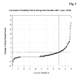
- "Disease progression" in the sense of the present invention is assessed by Sharp-Genant-Score. A patient with a progression rate > 5 per year (change of the Sharp-Genant-Score from baseline after one or two years) is classified as an RA patient with disease progression. All other patients are classified as having no disease progression.
- A "patient having rheumatoid arthritis" is a patient meeting the revised criteria developed for the classification of Rheumatoid Arthritis from the American Rheumatism Association (Arnett, F.C., et al., Arthritis Rheum. 31 (1988) 315-324). These criteria are herewith included by reference.
- The inventors of the present invention have defined two sub-groups of RA patients, one showing disease progression and a reference population or sub-group of RA showing no disease progression and investigated the potential of biochemical markers for predicting disease progression based on these patient cohorts.
- Surprisingly it could be found and established that the marker combination of CRP plus interleukin-6 is key for improving the sensitivity of prediction of the disease course for an RA patient at the clinically required high specificity.
- In a method according to the present invention at least the concentration of the biomarkers CRP and IL-6, respectively, is determined and this marker combination is correlated to the risk of disease progression for a patient diagnosed with RA.
- As the skilled artisan will appreciate the step of correlating a marker level to a certain likelihood or risk can be performed and achieved in different ways. Preferably the values measured for the markers CRP and IL-6, are mathematically combined and the combined value is correlated to the underlying diagnostic question. Marker values may be combined by any appropriate state of the art mathematical method.
- Preferably the mathematical algorithm applied in the combination of markers is a logistic function. The result of applying such mathematical algorithm or such logistical function preferably is a single value. This value can easily be correlated to the risk of RA disease progression. In a preferred way such logistic function is obtained by a) classification of RA patients into the groups of patients undergoing disease progression and the group of patients not undergoing disease progression, b) identification of markers which differ significantly between these groups by univariate analysis, c) logistic regression analysis to assess the independent discriminative values of markers useful in assessing RA disease progression and d) construction the logistic function to combine the independent discriminative values.
- In a preferred embodiment the logistic function used for combining the values for CRP and IL-6 is obtained by a) classification of RA patients into the groups of patients undergoing disease progression and of patients not undergoing disease progression, respectively, b) establishing the values for CRP and interleukin-6 c) performing logistic regression analysis and d) construction the logistic function to combine the marker values for CRP and interleukin-6.
- In a further preferred embodiment the logistic function for combining the measurements of CRP and IL-6 with the values for one o more other marker is obtained by a) classification of RA patients into the groups of patients undergoing disease progression and the group of patients not undergoing disease progression, b) identification of one or more additional marker which differentiates significantly between these groups by univariate analysis, c) performing logistic regression analysis to assess if said marker has additive discriminative value over the combination of CRP and interleukin-6 in assessing RA disease progression and d) constructing the logistic function to combine the values measured for CRP, interleukin-6 and the one or more additional marker.
- A logistic function for correlating a marker combination to a disease preferably employs an algorithm developed and obtained by applying statistical methods like, Discriminant analysis (DA) (i.e. linear-, quadratic-, regularized-DA), Kernel Methods (i.e. SVM), Nonparametric Methods (i.e. k-Nearest-Neighbor Classifiers), PLS (Partial Least Squares), Tree-Based Methods (i.e. Logic Regression, CART, Random Forest Methods, Boosting/Bagging Methods), Generalized Linear Models (i.e. Logistic Regression), Principal Components based Methods (i.e. SIMCA), Generalized Additive Models, Fuzzy Logic based Methods, Neural Networks and Genetic Algorithms based Methods. The skilled artisan will have no problem in selecting an appropriate statistical method to evaluate a marker combination of the present invention and thereby to obtain an appropriate mathematical algorithm. Preferably the statistical method employed to obtain the mathematical algorithm used in correlating the marker combination of the invention to the risk of disease progression of RA is selected from DA (i.e. Linear-, Quadratic-, Regularized Discriminant Analysis), Kernel Methods (i.e. SVM), Nonparametric Methods (i.e. k-Nearest-Neighbor Classifiers), PLS (Partial Least Squares), Tree-Based Methods (i.e. Logic Regression, CART, Random Forest Methods, Boosting Methods), or Generalized Linear Models (i.e. Logistic Regression). Details relating to these statistical methods are found in the following references: Ruczinski, I., et al., J. of Computational and Graphical Statistics 12 (2003) 475-511; Friedman, J. H., J. of the American Statistical Association 84 (1989) 165-175; Hastie, T., et al., The Elements of Statistical Learning, Springer Verlag (2001); Breiman, L., et al., Classification and regression trees, California, Wadsworth (1984); Breiman, L., Random Forests, Machine Learning 45 (2001) 5-32; Pepe, M.S., The Statistical Evaluation of Medical Tests for Classification and Prediction, Oxford Statistical Science Series, 28 (2003); and Duda, R.O., et al., Pattern Classification, Wiley Interscience, 2nd edition (2001).
- It is a preferred embodiment of the invention to use an optimized multivariate cut-off for the underlying combination of biological markers and to discriminate state A from state B, e.g. RA disease progression from no RA disease progression, respectively. In this type of analysis the markers are no longer independent but form a marker panel. It could be established that combining the measurements of CRP and of IL-6 does significantly improve the diagnostic accuracy in assessing the risk of disease progression for a patient having RA.
- In univariate analysis CRP, IL-6 and several other markers have an area under the curve (AUC) of about 0.7 to about 0.8. Both CRP and IL-6 are inflammation markers and they are highly correlated to each other. It is therefore quite unexpected to see that CRP and IL-6 can be combined and at the same level of specificity as the individual markers show a tremendous improvement in sensitivity.
- The AUC is an indicator of the performance or accuracy of a diagnostic procedure. Accuracy of a diagnostic method is best described by its receiver-operating characteristics (ROC) (see especially Zweig, M. H., and Campbell, G., Clin. Chem. 39 (1993) 561-577). The ROC graph is a plot of all of the sensitivity/specificity pairs resulting from continuously varying the decision thresh-hold over the entire range of data observed. The area under the ROC plot is called AUC.
- The clinical performance of a laboratory test depends on its diagnostic accuracy, or the ability to correctly classify subjects into clinically relevant subgroups. Diagnostic accuracy measures the test's ability to correctly distinguish two different conditions of the subjects investigated. Such conditions are for example health and disease or disease progression versus no disease progression.
- In each case, the ROC plot depicts the overlap between the two distributions by plotting the sensitivity versus 1 - specificity for the complete range of decision thresholds. On the y-axis is sensitivity, or the true-positive fraction [defined as (number of true-positive test results)/(number of true-positive + number of false-negative test results)]. This has also been referred to as positivity in the presence of a disease or condition. It is calculated solely from the affected subgroup. On the x-axis is the false-positive fraction, or 1 - specificity [defined as (number of false-positive results)/(number of true-negative + number of false-positive results)]. It is an index of specificity and is calculated entirely from the unaffected subgroup. Because the true- and false-positive fractions are calculated entirely separately, by using the test results from two different subgroups, the ROC plot is independent of the prevalence of disease in the sample. Each point on the ROC plot represents a sensitivity/1-specificity pair corresponding to a particular decision threshold. A test with perfect discrimination (no overlap in the two distributions of results) has an ROC plot that passes through the upper left corner, where the true-positive fraction is 1.0, or 100% (perfect sensitivity), and the false-positive fraction is 0 (perfect specificity). The theoretical plot for a test with no discrimination (identical distributions of results for the two groups) is a 45° diagonal line from the lower left corner to the upper right corner. Most plots fall in between these two extremes. (If the ROC plot falls completely below the 45° diagonal, this is easily remedied by reversing the criterion for "positivity" from "greater than" to "less than" or vice versa.) Qualitatively, the closer the plot is to the upper left corner, the higher the overall accuracy of the test.
- One convenient goal to quantify the diagnostic accuracy of a laboratory test is to express its performance by a single number. The most common global measure is the area under the ROC plot (AUC). By convention, this area is always ≥ 0.5 (if it is not, one can reverse the decision rule to make it so). Values range between 1.0 (perfect separation of the test values of the two groups) and 0.5 (no apparent distributional difference between the two groups of test values). The area does not depend only on a particular portion of the plot such as the point closest to the diagonal or the sensitivity at 90% specificity, but on the entire plot. This is a quantitative, descriptive expression of how close the ROC plot is to the perfect one (area = 1.0).
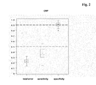
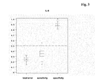
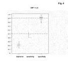
- The overall assay sensitivity will depend on the specificity required for practicing the method disclosed here. In certain preferred settings a specificity of 75% may be sufficient and statistical methods and resulting algorithms can be based on this specificity requirement. In further preferred embodiments the method used to assess the risk of disease progression for a patient having RA will be based on a specificity of 80%, 85%, or especially preferred 90% or 95%. As obvious from the Examples section, the marker combination employing CRP and IL-6 at a specificity of 90% has a good sensitivity of about 50%. This compares to a total error of about 20% and is better than the total error achieved with state of the art approaches solely based on individual biochemical markers.
- The levels given for CRP and IL-6 in the examples section have been measured and established with the assay procedures given there. It has to be understood that different assays may lead to different cut-off values. The skilled artisan will have no problems in establishing such supplier-dependent cut-off values by following the procedures outlined in the present invention.
- Interleukin-6 (IL-6) is a 21 kDa secreted protein that has numerous biological activities that can be divided into those involved in hematopoiesis and into those involved in the activation of the innate immune response. IL-6 is an acute-phase reactant and stimulates the synthesis of a variety of proteins, including adhesion molecules. Its major function is to mediate the acute phase production of hepatic proteins, and its synthesis is induced by the cytokines IL-1 and TNF-α. IL-6 is normally produced by macrophages and T lymphocytes. The normal serum concentration of IL-6 is < 5 pg/ml.
- Preferred means of detecting biomarkers like CRP and IL-6 are specific binding assays, especially immunoassays. Immunoassays are well known to the skilled artisan. Methods for carrying out such assays as well as practical applications and procedures are summarized in related textbooks. Examples of related textbooks are Tijssen, P., In: Practice and theory of enzyme immunoassays, eds. R.H. Burdon and v.P.H. Knippenberg, Elsevier, Amsterdam (1990), pp. 221-278, and various volumes of Methods in Enzymology, eds. Colowick, S.P., and Caplan, N.O., Academic Press, dealing with immunological detection methods, especially .
- IL-6 for example can be measured by a competitive type or a sandwich type immunoassay. IL-6 preferably is measured in a sandwich immunoassay which is essentially based on an antibody specifically binding to IL-6 which is directly or indirectly bound or capable of binding to a solid phase, an antibody specifically binding to IL-6 which is detectably labeled, and incubating these reagents under conditions allowing for binding of the anti-IL-6 antibodies to IL-6 in a sample, separating unbound detectably labeled antibody, determining the amount of labeled antibody bound via IL-6, and correlating the amount of labeled antibody bound to the concentration of IL-6 in the sample.
- C-reactive protein (CRP) is a homopentameric Ca2+-binding acute phase protein with 21 kDa subunits that is involved in host defense. CRP synthesis is induced by IL-6, and indirectly by IL-1, since IL-1 can trigger the synthesis of IL-6 by Kupffer cells in the hepatic sinusoids. The normal plasma concentration of CRP is < 3µg/ml (30 nM) in 90% of the healthy population, and < 10 µg/ml (100 nM) in 99% of healthy individuals. Plasma CRP concentrations can, e.g. be measured by homogeneous assay formats or ELISA. CRP is considered a marker of systemic inflammation.
- A factor further confounding and complicating the risk assessment of disease progression for a patient having RA is the fact that patients at the time of visit may be at different stages of disease development and under various treatment regimens. The inventors of the present invention have been able to demonstrate that marker combination found is predictive for both patients not yet treated with an anti-rheumatic drug and for patients already under treatment with a disease modifying anti-rheumatoid drug (DMARD). Especially the later finding is of great relevance, it indicates that the method disclosed in the present invention may be of aid in identifying those patients not responding or not sufficiently responding to treatment with a DMARD. In a preferred embodiment the method according to present invention is practiced using a sample obtained from an RA-patient who is under treatment with an anti-rheumatic drug selected from group of disease modifying anti-rheumatoid drugs (DMARDs). Also preferred, the method disclosed herein is practiced using a sample obtained from an RA-patient who has not been under treatment with an anti-rheumatic drug.
- It is believed that with the identification of the marker combination CRP and IL-6 the key marker combination useful in assessing the risk of disease progression for a patient having RA has no been identified. As has been further shown by the inventors the method of assessing the risk of disease progression for a patient having RA can be further improved by combining the measurement of the two key markers CRP and IL-6 with further parameters. In a further preferred embodiment the present invention relates to a method comprising the steps of a) obtaining a liquid sample, b) measuring in said sample the concentration of both C-reactive protein (CRP) and interleukin-6, and of one or more other marker, and c) correlating the concentrations determined in step (b) to the risk of disease progression, wherein the optionally one or more other marker is selected from the group consisting of bone or cartilage markers, synovial fluid markers, other inflammation markers, genetic markers and radiological scores.
- In a preferred embodiment the one or more other marker used in a method according to the present invention is a bone or cartilage marker, preferably said bone or cartilage marker is selected from the group consisting of PINP, ß-CrossLaps, CartiLaps, osteocalcin and ICTP also preferred the one or more bone or cartilage marker is ICTP or/and CartiLaps.
- The most prominent joint tissues are bone, cartilage and the synovium. Since rheumatoid arthritis is a destructive disease these tissues will be most affected. They are a likely source of potential biological markers in the field of RA. In principle these markers may come not only from the destruction of the respective tissue but also from a deregulated and/or ineffective repair process. The experienced artisan will understand that markers of bone, cartilage or synovium metabolism can originate either from synthesis or from destruction of these tissues. The various markers of bone, cartilage and/or synovium metabolism can be delineated from two different groups of proteins. They come either from the numerous types of collagen or from non-collagenous proteins. Non-collagenous proteins are often involved in the formation of the extracellular matrix. Some of these markers can be found in all three tissues in varying amounts.
- Bone and/or cartilage markers include markers of both markers of bone and/or cartilage collagen degradation as well as markers of bone and/or cartilage collagen formation. Preferred collagen-derived bone or cartilage markers are:
- 1. Pyridinoline (=PYD), deoxy-pyridinoline (=DPD) and Glc-Gal-PYD: Pyridinoline (=PYD) stabilizes collagen by cross-linking the strands of the collagen triple helix. The chemical structure of PYD is very stable and can be found in serum and urine as an end product of collagen degradation (Knott, L., and Bailey, A.J., Bone 22 (1998) 181-187). It has been linked to arthritis (Kaufmann, J., et al., Rheumatology 42 (2003) 314-320). PYD monitors cartilage involvement of joint destruction since it is released from cartilage and only to some degree from bone while its close cousin deoxy-pyridinoline (=DPD) originates mostly from bone. All three markers have been linked to arthritis (Kaufmann, supra). The glycosylated form Glc-Gal-PYD has mostly been found in synovial tissue (Gineyts, E., et al., Rheumatology 40 (2001) 315-323).
- 2. Cross-linked telopeptides: CTX-I, CTX-II, NTX-I and the LQ-epitope which are cross-linked telopeptides either from the C- or N-terminus of collagens type I or type II, respectively, and of which ß-CTX-I is also known as ß-CrossLaps® (Bonde, M., et al., Clin. Chem. 40 (1994) 2022-2025).
- 3. Type I collagen carboxyterminal telopeptide (=ICTP) refers to a fragment and marker of type I collagen which originally has been derived from type I collagen by cyanobromide cleavage (
US 5,538,853 ). - 4. Linear peptides derived from collagen: The assay termed Cartilaps® measures a linear peptide that is derived from the C-terminal region of collagen type II (
US 6,372,442 ). - 5. Modified amino acids: Collagen comprises modified amino acids like hydroxyproline and galactosyl hydroxylysine which may be used as a marker of collagen break-down (Al-Dehaimi, A.W., et al., Clin. Chem. 45 (1999) 676-681).
- 6. Collagen neoepitopes: Col2-3/4 and CIIN are neoepitopes generated by the initial cleavage of collagen II by collagenases (Billinghurst, R.C., et al., J. Clin. Invest. 99 (1997) 1534-1545).
- 7. Collagen markers considered reflecting bone formation: The N-terminal as well as the C-terminal pro-peptide of type I collagen (=PINP and PICP), respectively, are clipped from the precursor polypeptide (procollagen) during/after synthesis and considered markers of bone formation. PIICP is the corresponding pro-peptide from collagen type II, whereas PIIINP is derived from collagen III.
- Also preferred the bone or cartilage marker be a non-collagenous marker, like: CS846, which is a chondriotin sulfate epitope created during aggrecan synthesis; cartilage oligomeric matrix protein (=COMP) that has bridging functions in cartilage (Saxne, T., and Heinegard, D., Br. J. Rheumatol. 31 (1992) 583-591); cartilage intermediate layer protein (=CILP), which is a matrix protein of cartilage (Lorenzo, P., et al., J. Biol. Chem. 273 (1998) 23463-23468); cartilage matrix proteins 1 - 3 also known as matrilins; chondromodulins that act as signaling molecules in cartilage (Suzuki, F., Connect. Tissue Res. 35 (1996) 303-307); cartilage derived retinoic acid-sensitive protein (=CD-RAP) or MIA, which has a yet to be defined function in chondrocyte modulation (Mueller-Ladner, U., et al., Rheumatology 38 (1999) 148-154); osteocalcin, which is synthesized by osteoblasts, belongs to the major non-collagen matrix protein of bone and is used to monitor bone turnover (Gundberg, C. M., et al., J. Clin. Ligand Assay 21 (1998) 128-138); and the bone sialoproteins, which are major non-collagen matrix proteins of bone, such as bone sialoprotein II, now known as bone sialoprotein, which e.g., has been evaluated as marker for bone turn-over (Saxne, T., et al., Arthritis Rheum. 38 (1995) 82-90).
- In a preferred embodiment the one or more other marker used in a method according to the present invention is a synovial marker selected from the group consisting of matrix metalloprotease 1 (=pro-MMP-1), matrix metalloprotease 3 (=pro-MMP 3), hyaluronic acid, preferably the one ore more other synovial marker is hyaluronic acid or and pro-MMP 3.
- The family of matrix-metalloproteinases (=MMPs) degrades almost all components of the extra-cellular matrix. Hence MMPs have been related to various types of cancer but also to inflammatory processes in RA.
MMP 1 and MMP 3 are produced by fibroblasts, osteoblasts and endothelial cells upon stimulation by proinflammatory cytokines like IL-1 or TNF-α. Generally MMPs are found in the circulation as inactive pro-form, i.e.,pro-MMP 1 and pro-MMP 3, respectively. pro-MMP 1 and pro-MMP 3 have been detected in synovial fluid of RA-patients and their levels are responsive to anti-TNF-α therapy. The most preferred metalloprotease to be used in marker panel for assessing the risk of disease progression for a patient having RA is pro-MMP 3. - Instead of the metalloproteinases mentioned above it is also possible to used their corresponding inhibitors collectively referred to as tissue inhibitors of matrix metalloproteinases (=TIMPs)., e.g. MMP-1 and MMP-3 are in vivo inactivated by TIMP-1 a sialoglycoprotein of 29.5 kD that forms a 1:1 stoichiometric complex with the MMPs. The relation of TIMP-1 and TIMP-2 to the destruction of cartilage has been investigated in RA (Ishiguro, N., et al., Arthritis Rheum. 44 (2001) 2503-2511).
- The glycosaminoglycan hyaluronic acid is one of the macromolecules essential for the function of a joint. It is synthesized by fibroblasts and other specialized connective tissue cells. Hyaluronic acid is involved in formation of the extracellular matrix and in cell to cell contacts. High concentrations are found in synovial fluid where it is responsible for the retention of water thereby contributing to the lubrication of joints. In rheumatoid arthritis the synthesis of hyaluronic acid is stimulated by the proinflammatory mediators IL-1 and TNF-α leading to increase serum/plasma levels (Sawai, T., and Uzuki, M., Connective Tissue 33 (2001) 253-259).
- In a preferred embodiment the one or more other marker used in a method according to the present invention is a genetic marker selected from the group consisting of an HLA-DR4 and an HLA-DRB1 allele, preferably the one ore more other genetic marker is an HLA-DRB1*01 or/and an HLA-DRB1*04 allele (Goronzy, J.J., et al., Arthritis and Rheumatism 50 (2004) 43-54).
- In a preferred embodiment the one or more other marker used in a method according to the present invention is a radiological score, preferably said radiological score is selected from the group consisting of Sharp-score, Sharp-Genant-score, van der Heijde-Sharp-score, Ratingen-score, Larsen-score, RAU-score and Herborn-score also preferred the one or more radiological score is the Sharp-Genant-score, or/and the Larsen-score.
- The "Sharp-score" has first been introduced in 1971 (Sharp, J.T., et al., Arthritis and Rheumatism 14 (1971) 706-720) and has been further elucidated in 1985 (Sharp, J.T., et al., Arthritis and Rheumatism 28 (1985) 1326-1335).
- The "Sharp-Genant-score" is a modification of the Sharp-score as proposed by Genant in 1983 (Genant, H.K., Am. J. Med. 75 (1983) 35-47).
- The "van der Heijde-Sharp-score" is a modification of the Sharp-score as proposed by van der Heijde in 1989 (van der Heijde, D.M.F.M., Lancet 1 (1989) 1036-1038).
- The "Larsen-score" has first been introduced in 1977 (Larsen, A., et al., Acta Radiol. Diagn. 18 (1977) 481-491). The "RAU-score" sometimes also referred to as "Ratingen-score" is a modification of the Larsen-score (Rau, R. and Wassenberg, S., Z. Rheumatol. 62 (2003) 555-565).
- In a preferred embodiment the one or more other marker used in a method according to the present invention is a further marker of inflammation preferably said further marker of inflammation is an inflammation marker selected from the group consisting of S100-proteins, erythrocyte sedimentation rate (ESR), SAA and E-selectin preferably it is SAA or/and E-selectin.
- The term "other marker of inflammation" or "further marker of inflammation" is used to indicate that these marker are neither CRP nor IL-6.
- Serum amyloid A (=SAA) is an acute phase protein of low molecular weight of 11.7 kDa. It is predominantly synthesized by the liver in response to IL-1, IL-6 or TNF-α stimulation and is involved in the regulation of the T-cell dependent immune response. Upon acute events the concentration of SAA increases up to 1000-fold reaching one milligram per milliliter. It is used to monitor inflammation in diseases as divers as cystic fibrosis, renal graft refection, trauma or infections. In rheumatoid arthritis is has in certain cases been used as a substitute for CRP, but, SAA is not yet as widely accepted.
- S100-proteins form a constantly increasing family of Ca2+-binding proteins that today includes more than 20 members. The physiologically relevant structure of S100-proteins is a homodimer but some can also form heterodimers with each other, e.g. S100A8 and S100A9. The intracellular functions range from regulation of protein phosphorylation, of enzyme activities, or of the dynamics of the cytoskeleton to involvement in cell proliferation and differentiation. As some S100-proteins are also released from cells, extracellular functions have been described as well, e.g., neuronal survival, astrocyte proliferation, induction of apoptosis and regulation of inflammatory processes. S100A8, S100A9, the heterodimer S100A8/A9 and S100A12 have been found in inflammation with S100A8 responding to chronic inflammation, while S100A9, S100A8/A9 and S100A12 are increased in acute inflammation. S100A8, S100A9, S100A8/A9 and S100A12 have been linked to different diseases with inflammatory components including some cancers, renal allocraft rejection, colitis and most importantly to RA (Burmeister, G., and Gallacchi, G., Inflammopharmacology 3 (1995) 221-230; Foell, D., et al., Rheumathology 42 (2003) 1383-1389). The most preferred S100 markers for use in a marker panel for assessing disease progression in RA according to the present invention are S100A8, S100A9, S100A8/A9 heterodimer and S100A12.
- sE-selectin (soluble endothelial leucocyte adhesion molecule-1, ELAM-1) is a 115 kDa, type-I transmembrane glycoprotein expressed only on endothelial cells and only after activation by inflammatory cytokines (IL-1ß, TNF-α) or endotoxin. Cell-surface E-selectin is a mediator of the rolling attachment of leucocytes to the endothelium, an essential step in extravasion of leucocytes at the site of inflammation, thereby playing an important role in localized inflammatory response. Soluble E-selectin is found in the blood of healthy individuals, probably arising from proteolytic cleavage of the surface-expressed molecule. Elevated levels of sE-selectin in serum have been reported in a variety of pathological conditions (Gearing, A.J.H., et. al., Annals N.Y. Acad. Sci. 667 (1992) 324-331).
- Preferably the one or more other marker used in combination with CRP and IL-6 in order to assess the risk of disease progression in RA is a biochemical marker or a biomarker. Preferably the biomarker is a polypeptide or an autoantibody.
- The following examples and figures are provided to aid the understanding of the present invention.
-
- Fig. 1
- Cumulative probability plot of
progression rate 1 for all RA patients (x-axis = cumulative probabiliy %; y-axis = change in Sharp Genant Score) - Figs. 2 to 5
- show boxplots of individual markers or marker combinations. RA patients of panel I have been classified as having disease progression or as having no disease progression. The specificity (right-hand box in each Figure) has been set to about 90% (= 0.9 on the y-axis). The sensitivity for each marker or marker combination is shown in the middle box and the corresponding total error is shown by aid of the left-hand boxplot.
(boxes = 25th to 75th quartile; whiskers = 1.5 times interquartile range; - in box = median; + indicates the position of the mean; * = individual value falling outside the whiskers) - Fig. 2
- Boxplots for CRP Sensitivity = 35%; total error = 23%.
- Fig. 3
- Boxplots for IL-6 Sensitivity = 35%; total error = 25%.
- Fig. 4
- Boxplots for the marker combination CRP + IL-6 Sensitivity = 50%; total error = 20%.
- Fig. 5
- Boxplots for the marker combination CRP, IL-6 and pro-MMP3 Sensitivity = 53%; total error = 20%.
- Samples derived from 237 highly characterized RA patients with maximum disease duration of 15 years were collected in five European centers with a follow-up of one or two years. All individuals were diagnosed as RA-patients according to the 1987 revised criteria for the classification of Rheumatoid Arthritis from the American Rheumatism Association (Arnett, F.C., et al., Arthritis Rheum. 31 (1988) 315-324). All patients were documented with an extensive case report form (=CRF). The CRF included the Health Assessment Questionnaire, the SF36 Questionnaire, swollen and tender joint count, laboratory parameters, clinical history of relevant surgery, medication, co-morbidities and medication for co-morbidities. X-rays were taken from hands and feet at baseline, after one and after 2 years following a standardized procedure. Only the baseline samples obtained from the RA-patients were used in the measurement of the different analytes and the corresponding results were used for the uni-variate and multi-variate analysis.
- Demographic data for the study population are given in Table 1.
Table 1: RA-patient collective Number RA-patients 237 Patients with all x-rays (BL, 1 year, 2 years) 204 Patients with x-rays at BL and year 133 Age (mean, (minimum/maximum)) 58.6 (18 - 87) Gender distribution (male/female) 84/153 Erosiveness at baseline (erosive/non-erosive) 155/82 Disease duration (mean, (minimum/maximum)) 4.9 (0.1 - 15.2) years - From each patient x-rays were taken from hands and feet at baseline and after one and two years. The conventional film radiographs were sent to Synarc (Synarc GmbH, Hamburg, Germany), where the hard copy films were digitized using a Lumiscan 200 high resolution digitizer. After quality check each image was read by an experienced radiologist and scored according the Genant-modified Sharp scoring.
- Bone erosions and joint space narrowing in the hands and feet were scored according to a Genant-modified Sharp grading scheme as described below. This grading scheme is based on the Genant-modified Sharp scoring technique.
- Erosion Score: Fourteen sites in each wrist and hand (four proximal interphalangeal and five metacarpophalangeal joints, the carpometacarpal joint of the thumb, the scaphoid bone, the distal radius and the distal ulna) and six joints in each foot (five metatarsophalangeal joints and the interphalangeal joint of digit I (i.e., the great toe)) are scored using an eight-point scale from 0 to 3.5 based on the size of erosions and the area of bone (both sides of joint) involved:
- 0
- (normal: no erosions)
- 0.5
- (subtle loss of cortical continuity or equivocal findings of bone erosion)
- 1.0
- (mild: definite but small erosions of one or both articular bones, usually at the bare areas, involving <25% of the articular surfaces)
- 1.5
- (mild to moderate: small-medium erosions involving <25% of the articular bones of one or both articular bones)
- 2.0
- (moderate: medium-large erosions involving approx. 26% - 50% of the articular surface of both articular bones)
- 2.5
- (moderate to severe: erosions of approx. 51% - 75% of the articular surfaces)
- 3.0
- (severe: erosions of approx. 76% - 90% of the articular surfaces)
- 3.5
- (very severe: erosions of 100% of the articular surfaces (total destruction of the articular surfaces)
- Joint space narrowing (JSN) score. Thirteen sites in each wrist and hand (proximal interphalangeal joints of digits II to V, the interphalangeal joint of the thumb and five metacarpophalangeal joints, carpometacarpal joints of digits III-V as a single unit, the pericapitate (scaphoid-capitate and lunate-capitate combined) space and the radiocarpal joint) and six sites in each foot (five metatarsophalangeal joints and the interphalangeal joint of digit I (i.e., the great toe)) are scored using a nine-point scale from 0 to 4:
- 0
- (normal)
- 0.5
- (subtle joint space narrowing or equivocal findings)
- 1.0
- (mild joint space narrowing (focal or minor))
- 1.5
- (mild to moderate joint space narrowing)
- 2.0
- (moderate joint space narrowing)
- 2.5
- (moderate to severe joint space narrowing)
- 3.0
- (severe joint space narrowing)
- 3.5
- (severe joint space narrowing close to ankylosis)
- 4.0
- (definite ankylosis)
- Hands/wrists: The individual joint scores will be summed separately to create a total erosion score and a total JSN score for the hands/wrists. The maximum total erosion score for the hands/wrists is (14 x 3.5 maximum per joint) x 2 = 98. The maximum total JSN score is (13 x 4 maximum per joint) x 2 = 104. To provide equal weight to erosions and joint space narrowing, each sum is normalized to a scale of 0 - 100. If E-score is the sum of erosion scores and J-score is the sum of JSN scores for both hands, the normalized scores are calculated as follows:

and
- Feet: As for the hands/wrists the individual joint scores will be summed separately to create a total erosion score and a total JSN score for the feet. The maximum total erosion score for the feet is (6 x 3.5 maximum per joint) x 2 = 42. The maximum total JSN score is (6 x 4 maximum per joint) x 2 = 48.
- To provide equal weight to erosions and joint space narrowing, each sum will be normalized to a scale of 0 - 45. If E-score is the sum of erosion scores and J-score is the sum of JSN scores for both feet, the normalized scores are calculated as follows:

and
- Combination: The Total score for the hands/wrists and feet is the sum of the individual totals for each. Thus the maximum score achievable is 290.


- The change in total scores is calculated as:


- There are some possibilities discussed in the literature for classification of disease progression. Beside the ACR and EULAR criteria, which are mostly used in pharmaceutical studies for assessing treatment response also the HAQ score and the radiological scores can be used for classification of disease progression. The most preferred methodology is the use of the change of any radiological score after one year. We decided to use the total Sharp-Genant-Score and to determine the individual change of this score one or two years after the baseline value (= progression rate).


- The next important step was to define a cut-off value for the progression rates to be able to classify the patients in RA with progression and RA without progression. Therefore a cumulative probability plot of the
progression rate 1 or 2 from all patients were made (seefigure 1 ) (van der Heijde et al., Arthritis Rheum. 52 (2005) 49-60). Laying a straight line onto the first slope of the plot, the intersection point was determined at a progression rate (1) of "5". The same results was obtained using a probability plot of the progression rate (2). To use a progression rate of "5" (i.e. an increase in SGS of more than 5 per year) as a cut-off value for classification of RA patients into patients with or without progression was supported by following two arguments: - 1. Using a progression rate (1) of "5" as cut-off value, approx. 20% of the RA patients of this sample collective will be classified as RA patients with progression.
- 2. The value of any scoring method used to measure a clinical outcome depends on its reliability. There are described different methods for the determination of the "sensitivity to change" (Boini, S. and Guillemin, F., Ann. Rheum. Dis. 60 (2001) 817-827). The best reliability score for classification of individual patients is the smallest detectable difference (SDD). The experts evaluating the radiographs used in establishing an SGS have determined a SDD of 5.1 for the SGS. This means, an SGS-change of about 5 is the minimal difference of one patients at two time points, which can be significantly discriminated.
- Therefore the following classification was used:


- Using this definition we achieved following classification:
- RA patients with disease progression:
- 59 patients
- RA patients with no disease progression:
- 178 patients
- Table 2 represents the assays used and gives the test format as well as the suppliers of the assays. Most of the assays were manual microtiter plate (=MTP) format ELISAs. RF and CRP were determined in a homogeneous test format on an automatic Hitachi analyzer. All marker concentrations were determined in serum samples with the exception of CartiLaps, which was measured in urine. The CartiLaps values were normalized by the creatinine results.
Table 2: Assays and Suppliers Biomarker Assay type / format Source Anti-CCP Sandwich ELISA, MTP Axis-Shield, Dundee (UK) CRP Homogenous assay, Hitachi Roche Diagnostics, Mannheim (FRG) Hyaluronic acid Sandwich ELISA, MTP Chugai, Tokyo (J) IL-6 Sandwich ELISA, MTP Roche Diagnostics, Mannheim (FRG) RF Homogenous assay, Hitachi Roche Diagnostics, Mannheim (FRG) SAA Sandwich ELISA, MTP Biosource, Nivelles (B) pro-MMP-3 Sandwich ELISA, MTP The Binding Site, Birmingham (UK) S100 A8/A9 Sandwich ELISA, MTP Bühlmann Lab., Allschwill (CH) S100 A12 Prototype ELISA, MTP Roche Diagnostics, Penzberg (FRG) Osteocalcin Sandwich ELISA, Elecsys® Roche Diagnostics, Mannheim (FRG) ß-Crosslaps Sandwich ELISA, Elecsys® Roche Diagnostics, Mannheim (FRG) PINP Sandwich ELISA, Elecsys® Roche Diagnostics, Mannheim (FRG) SCD14 Sandwich ELISA, MTP IBL, Hamburg (FRG) CartiLaps Comp. ELISA, MTP Nordic Bioscience, Herlev (DK) ICTP Comp. ELISA, MTP Orion Diagnostika, Espoo (FIN) sE-Selectin Sandwich ELISA, MTP R&D Systems, Minneapolis, (USA) - The baseline samples of all 237 RA patients were measured with the 16 markers listed in table 2. Each marker value was logarithmized and a ROC analysis was carried out. Table 3 represents the AUC values and the sensitivity (at a specificity of 90%) for each marker.
Table 3: Univariate analysis Biomarker AUC (%) Sensitivity (%) at a specificity of 90% Anti-CCP 59 5 CRP 75 37 Hyaluronic acid 70 20 IL-6 77 32 RF 67 24 SAA 70 27 pro-MMP-3 72 31 S100 A8/ A9 70 29 S100 A12 68 32 Osteocalcin 50 8 ß-CrossLaps 57 7 PINP 55 10 sCD14 61 17 CartiLaps 71 19 ICTP 71 19 sE-selectin 67 20 - 8 markers achieved an AUC of 70% and higher. The best sensitivity showed CRP with 37% at a specificity of 90%. It was very surprising, that anti-CCP, which is published as a prognostic factor, showed only an AUC of 0.59. In many scientific papers biomarkers with an odds ratio of about 3.0 and higher are - rather optimistically referred to as predictors of progression. For example, Syversen, S.W., et. al. (Ann. Rheum. Dis. 65, Suppl. II (2006) 110) reported that anti-CCP (OR = 4.18), RF-IgM (OR = 3.12) ESR (OR = 3.73) and female gender (OR = 3.29) are independent predictors of 10-year radiographic progression in RA patients. If we calculate the Odds ratio of anti-CCP (Cut-off > 5 U/mL) in our RA collective, we obtain a similar odds ratio, i.e. an OR of 4.6. Nevertheless an odds ratio of "4" or "5" has merely no diagnostic value in clinical routine, where a high specificity (corresponding to a low number of false positive results) is required.
- Due to the limited number of RA patients with progression a randomly split of the patient collective into a training set and into a test set was not possible. Therefore an external cross validation (ECV) was carried out. For the ECV the training set was subdivided 50 times (ratio 2(training subsets) : 1(test subsets)) for an external Monte-Carlo cross validation (Dudoit, S. and van der Laan, M.J., Statistical Methodology 2 (2005) 131-154). On the training subsets a classification algorithm was develop and on the independent test subsets the algorithm was validated.
- The classification algorithms were generated with the Regularized Discriminant Analysis (RDA), which is a generalization of the common Discriminant Analysis, i.e. Quadratic- and Linear Discriminant Analysis (McLachlan, G.J., Discriminant Analysis and Statistical Pattern Recognition, Wiley Series in probability and mathematical statistics, 1992). In the RDA alternatives to the usual maximum likelihood (plug-in) estimates for the covariance matrices are used. These alternatives are characterized by two parameters (λ,γ), the values of which are customized to individual situations by jointly minimizing a sample-based estimate of future misclassification risk (Friedman, J.H., J. of the American Statistical Association 84 (1989) 165-175). As an alternative method Support Vector Machines algorithms (Hastie, T., et al., The Elements of Statistical Learning, Springer Series in Statistics, 2001) can be fitted with comparable classification results.
- The marker panels were stepwise constructed starting from the best single marker for the classification problem and ending when the total classification error do not change remarkable any more. In order to gain centralized distributions every single marker was transformed with the natural logarithmic function.
- The goal of the multivariate analysis was to find a marker panel, which shows a higher sensitivity than the best single marker. The specificity limit was set to 90%. The first marker selected was CRP with a sensitivity of 35% and the second one was IL-6 improving the sensitivity to 50%. There are some other combinations with different markers as the third and the fourth one, which are able to minimize the total error and/or to improve the sensitivity (Table 4). For all these marker combinations the most important two markers are CRP and IL-6, thereby representing the key markers of these marker panels.
- The aim of the current invention is to aid the rheumatologist in his assessment whether an RA patient is at risk of disease progression. The diagnostic value of the identified marker panel is best reflected in Table 4 by the total error of the classification. CRP, currently a single biological marker used for the estimation of inflammation gives a total error of 0.228. 11-6 as a single marker also reveals a similar total error of 0.247. The preferred combination of CRP and
IL 6 significantly improves the classification reducing the total error to 0.203. Adding a third and a fourth marker finally helps to further minimize the misclassification (total error 0.196). The achieved sensitivity of 50% suggests that based on the method disclosed here about one half of the RA-patients with a progressive disease can be identified correctly by biochemical markers measure at a single time point, i.e. at baseline, which is not possible so far. This classification is expected to aid the rheumatologist in the decision process for example to start a treatment using DMARDs or to change to a better therapy scheme using a combination of different DMARDs.Table 4: Classification results of patients classified as RA with disease progression versus RA with no disease progression No of Markers marker or marker panel ECV (50 fold) TOTAL ERROR correct pos. Sensitivity correct neg. Specificity 1 CRP 0.228 35.0% 91.5% 1 11-6 0.247 35.0% 88.1% 2 CRP, IL-6 0.203 50.0% 91.5% 3 CRP, IL-6, S100 A8/A9 0.203 52.5% 89.8% 3 CRP, IL-6, pro-MMP-3 0.203 55.0% 89.8% 4 CRP, IL-6, S100 A8/A9, sE-Selectin 0.196 55.0% 89.8% The Boxplots for the markers CRP and IL-6 and marker combinations (CRP IL-6 and CRP + IL-6 + pro-MMP3), respectively, of Table 4 are shown in Figures 2 to 5 .
Claims (8)
- An in vitro-method for aiding in assessing the risk of disease progression for a patient having rheumatoid arthritis (RA), the method comprising the steps ofa) measuring in a liquid sample the concentration of both C-reactive protein (CRP) and interleukin-6, and of optionally one or more other marker,b) mathematically combining the values measured in step a) andc) correlating the combined value determined in step (b) to the risk of disease progression,
wherein said method of mathematically combining the values in step (b) is selected from DA (i.e. Linear-, Quadratic-, Regularized Discriminant Analysis), Kernel Methods (i.e. SVM), Nonparametric Methods (i.e. k-Nearest-Neighbor Classifiers), PLS (Partial Least Squares), Tree-Based Methods (i.e. Logic Regression, CART, Random Forest Methods, Boosting Methods), or Generalized Linear Models (i.e. Logistic Regression). - The method according to claim 1, wherein the RA-patient is under treatment with an anti-rheumatic drug selected from group of disease modifying anti-rheumatoid drugs (DMARDs), when the assessment is performed.
- The method according to claim 1, wherein the optionally one or more other marker is selected from the group consisting of bone or cartilage markers, synovial fluid markers, other inflammation markers, genetic markers and radiological scores
- The method according to claim 3, wherein the one or more bone or cartilage markers are selected from the group consisting of PINP, ß-CrossLaps, CartiLaps, osteocalcin and ICTP.
- The method according to claim 3, wherein the one or more synovial markers are selected from the group consisting of hyaluronic acid and pro-MMP 3.
- The method according to claim 3, wherein the one or more genetic markers are selected from the group consisting of HLA-DR4 and HLA-DRB1 alleles.
- The method according to claim 3, wherein the one or more radiological scores are selected from the group consisting of Sharp-Score, Sharp-Genant-Score, van der Heijde-Sharp-Score, Ratingen Score, Larsen-Score, and RAU-Score.
- The method according to claim 3, wherein the one or more other inflammation markers are selected from the group consisting of SAA and E-selectin.
Priority Applications (1)
| Application Number | Priority Date | Filing Date | Title |
|---|---|---|---|
| EP07818398.5A EP2074428B1 (en) | 2006-09-29 | 2007-09-25 | Assessing the risk of disease progression for a patient with rheumatoid arthritis |
Applications Claiming Priority (4)
| Application Number | Priority Date | Filing Date | Title |
|---|---|---|---|
| EP06020645 | 2006-09-29 | ||
| EP06021887 | 2006-10-19 | ||
| PCT/EP2007/008313 WO2008037420A1 (en) | 2006-09-29 | 2007-09-25 | Assessing the risk of disease progression for a patient with rheumatoid arthritis |
| EP07818398.5A EP2074428B1 (en) | 2006-09-29 | 2007-09-25 | Assessing the risk of disease progression for a patient with rheumatoid arthritis |
Publications (2)
| Publication Number | Publication Date |
|---|---|
| EP2074428A1 EP2074428A1 (en) | 2009-07-01 |
| EP2074428B1 true EP2074428B1 (en) | 2015-07-01 |
Family
ID=38859738
Family Applications (1)
| Application Number | Title | Priority Date | Filing Date |
|---|---|---|---|
| EP07818398.5A Not-in-force EP2074428B1 (en) | 2006-09-29 | 2007-09-25 | Assessing the risk of disease progression for a patient with rheumatoid arthritis |
Country Status (6)
| Country | Link |
|---|---|
| US (1) | US8058013B2 (en) |
| EP (1) | EP2074428B1 (en) |
| JP (1) | JP2010506147A (en) |
| CA (1) | CA2663730A1 (en) |
| ES (1) | ES2545688T3 (en) |
| WO (1) | WO2008037420A1 (en) |
Families Citing this family (16)
| Publication number | Priority date | Publication date | Assignee | Title |
|---|---|---|---|---|
| WO2010038104A1 (en) * | 2008-10-03 | 2010-04-08 | INSERM (Institut National de la Santé et de la Recherche Médicale) | Combination of cardiovascular risk factors for the diagnosis/prognosis of a cardiovascular disease/event. |
| EP2488659B1 (en) | 2009-10-15 | 2019-12-11 | Crescendo Bioscience, Inc. | Biomarkers and methods for measuring and monitoring inflammatory disease activity |
| US20130246097A1 (en) * | 2010-03-17 | 2013-09-19 | Howard M. Kenney | Medical Information Systems and Medical Data Processing Methods |
| JP5786020B2 (en) * | 2010-04-16 | 2015-09-30 | アボットジャパン株式会社 | Methods and reagents for diagnosing rheumatoid arthritis |
| US8883427B2 (en) | 2010-06-30 | 2014-11-11 | New York University | Quantifying local inflammatory activity and its use to predict disease progression and tailor treatments |
| JP5924659B2 (en) * | 2010-10-14 | 2016-05-25 | 国立大学法人 長崎大学 | Methods for comprehensive analysis of immune complexes and novel rheumatoid arthritis biomarkers |
| US20140142861A1 (en) * | 2010-11-06 | 2014-05-22 | Oklahoma Medical Research Foundation | Biomarkers For Predicting Progressive Joint Damage |
| EP2972365B1 (en) * | 2013-03-15 | 2019-11-20 | Exagen Diagnostics, Inc. | Methods for treating and diagnosing systemic lupus erythematosus |
| JP2015061592A (en) * | 2013-08-21 | 2015-04-02 | コニカミノルタ株式会社 | Ultrasonic diagnostic equipment, ultrasonic image processing method, and computer-readable non-temporary recording medium |
| CA2943821A1 (en) | 2014-04-02 | 2015-10-08 | Crescendo Bioscience | Biomarkers and methods for measuring and monitoring juvenile idiopathic arthritis activity |
| EP3155439A4 (en) | 2014-06-10 | 2018-03-14 | Crescendo Bioscience | Biomarkers and methods for measuring and monitoring axial spondyloarthritis disease activity |
| CA3000192C (en) | 2015-09-29 | 2023-09-26 | Crescendo Bioscience | Biomarkers and methods for assessing psoriatic arthritis disease activity |
| WO2017058999A2 (en) | 2015-09-29 | 2017-04-06 | Crescendo Bioscience | Biomarkers and methods for assessing response to inflammatory disease therapy withdrawal |
| GB201603571D0 (en) * | 2016-03-01 | 2016-04-13 | Univ Warwick | Markers for skeletal disorders |
| WO2021087116A1 (en) * | 2019-10-30 | 2021-05-06 | Cd Diagnostics, Inc. | Biomarkers of early osteoarthritis |
| CN114089632B (en) * | 2021-11-15 | 2023-08-18 | 陕西师范大学 | Rheumatism immune disease feature recognition method and system based on fuzzy logic |
Family Cites Families (14)
| Publication number | Priority date | Publication date | Assignee | Title |
|---|---|---|---|---|
| GB9105893D0 (en) | 1991-03-20 | 1991-05-08 | Orion Yhtymae Oy | Bone resorption assay based on a peptide liberated during collagen degradation |
| US5888510A (en) * | 1993-07-21 | 1999-03-30 | Chugai Seiyaku Kabushiki Kaisha | Chronic rheumatoid arthritis therapy containing IL-6 antagonist as effective component |
| ES2154739T3 (en) | 1994-10-17 | 2001-04-16 | Osteometer Biotech As | ESTIMATION OF THE COLLAGEN FRAGMENTATION MODEL IN BODY FLUIDS AND THE DIAGNOSIS OF DISORDERS ASSOCIATED WITH THE COLLAGEN METABOLISM. |
| FR2752842B1 (en) | 1996-08-30 | 1998-11-06 | Biomerieux Sa | ANTIGENS DERIVED FROM FILAGGRINS AND THEIR USE FOR THE DIAGNOSIS OF RHUMATOID POLYARTHRITIS |
| NL1004539C2 (en) | 1996-11-15 | 1998-05-20 | Stichting Tech Wetenschapp | Peptide derived from an antigen recognized by autoantibodies of patients with rheumatoid arthritis, antibody to it and method for detecting autoimmune antibodies. |
| PL344534A1 (en) | 1997-11-28 | 2001-11-05 | Innogenetics Nv | Synthetic peptides containing citrulline recognized by rheumatoid arthritis sera as tools for diagnosis and treatment |
| FR2773157B1 (en) | 1997-12-30 | 2001-10-05 | Bio Merieux | PEPTIDE EPITOPES RECOGNIZED BY ANTIFILAGGRIN AUTOANTIBODIES PRESENT IN THE SERUM OF PATIENTS WITH RHUMATOID POLYARTHRITIS |
| ES2209244T3 (en) * | 1998-01-09 | 2004-06-16 | Pfizer Inc. | INHIBITORS OF THE METALOPROTEASES OF THE MATRIX. |
| CA2391356A1 (en) | 1999-12-21 | 2001-06-28 | Innogenetics N.V. | Peptides designed for the diagnosis and treatment of rheumatoid arthritis |
| NL1019540C2 (en) | 2001-12-11 | 2003-07-01 | Stichting Tech Wetenschapp | Method for detecting autoantibodies from patients suffering from rheumatoid arthritis, peptide and assay kit. |
| ES2541683T3 (en) * | 2004-02-27 | 2015-07-23 | F. Hoffmann-La Roche Ag | Method to evaluate rheumatoid arthritis by measuring rheumatoid factor and interleukin-6 |
| US20060063162A1 (en) * | 2004-09-23 | 2006-03-23 | Deng David X | Biological marker for inflammation |
| WO2006098401A1 (en) * | 2005-03-16 | 2006-09-21 | The University Of Tokushima | Method for determination of risk of rheumatoid arthritis |
| MX2008002101A (en) * | 2005-08-12 | 2008-04-19 | Schering Corp | Mcp1 fusions. |
-
2007
- 2007-09-25 CA CA002663730A patent/CA2663730A1/en not_active Abandoned
- 2007-09-25 WO PCT/EP2007/008313 patent/WO2008037420A1/en active Application Filing
- 2007-09-25 EP EP07818398.5A patent/EP2074428B1/en not_active Not-in-force
- 2007-09-25 ES ES07818398.5T patent/ES2545688T3/en active Active
- 2007-09-25 JP JP2009529589A patent/JP2010506147A/en active Pending
-
2009
- 2009-03-27 US US12/412,840 patent/US8058013B2/en not_active Expired - Fee Related
Also Published As
| Publication number | Publication date |
|---|---|
| EP2074428A1 (en) | 2009-07-01 |
| JP2010506147A (en) | 2010-02-25 |
| WO2008037420A1 (en) | 2008-04-03 |
| US8058013B2 (en) | 2011-11-15 |
| US20090270272A1 (en) | 2009-10-29 |
| ES2545688T3 (en) | 2015-09-15 |
| CA2663730A1 (en) | 2008-04-03 |
Similar Documents
| Publication | Publication Date | Title |
|---|---|---|
| EP2074428B1 (en) | Assessing the risk of disease progression for a patient with rheumatoid arthritis | |
| US7846674B2 (en) | Assessing rheumatoid arthritis by measuring anti-CCP and interleukin 6 | |
| Visser | Early diagnosis of rheumatoid arthritis | |
| EP1721163B1 (en) | Method of assessing rheumatoid arthritis by measuring rheumatoid factor and interleukin-6 | |
| Hurnakova et al. | Serum calprotectin (S100A8/9): an independent predictor of ultrasound synovitis in patients with rheumatoid arthritis | |
| EP1721162B1 (en) | Method of assessing rheumatoid arthritis by measuring anti-ccp and serum amyloid a | |
| US20070148704A1 (en) | Anti-CCPand antinuclear antibodies in diagnosis of rheumatoid arthritis | |
| Mullan et al. | Early changes in serum type II collagen biomarkers predict radiographic progression at one year in inflammatory arthritis patients after biologic therapy | |
| US20090265116A1 (en) | Prediction of an individual's risk of developing rheumatoid arthritis | |
| CN101523218B (en) | Assessing the risk of disease progression for a patient with rheumatoid arthritis | |
| Hussein et al. | Peptidyl-arginine deiminase-type IV as a diagnostic and prognostic marker in rheumatoid arthritis patients | |
| Nordal et al. | and Hilde Berner Hammer4 |
Legal Events
| Date | Code | Title | Description |
|---|---|---|---|
| PUAI | Public reference made under article 153(3) epc to a published international application that has entered the european phase |
Free format text: ORIGINAL CODE: 0009012 |
|
| 17P | Request for examination filed |
Effective date: 20090429 |
|
| AK | Designated contracting states |
Kind code of ref document: A1 Designated state(s): AT BE BG CH CY CZ DE DK EE ES FI FR GB GR HU IE IS IT LI LT LU LV MC MT NL PL PT RO SE SI SK TR |
|
| AX | Request for extension of the european patent |
Extension state: AL BA HR MK RS |
|
| 17Q | First examination report despatched |
Effective date: 20120224 |
|
| DAX | Request for extension of the european patent (deleted) | ||
| GRAP | Despatch of communication of intention to grant a patent |
Free format text: ORIGINAL CODE: EPIDOSNIGR1 |
|
| INTG | Intention to grant announced |
Effective date: 20150212 |
|
| GRAS | Grant fee paid |
Free format text: ORIGINAL CODE: EPIDOSNIGR3 |
|
| GRAA | (expected) grant |
Free format text: ORIGINAL CODE: 0009210 |
|
| RIN1 | Information on inventor provided before grant (corrected) |
Inventor name: KARL, JOHANN Inventor name: ROLLINGER, WOLFGANG Inventor name: GRUNERT, VEIT PETER Inventor name: WILD, NORBERT |
|
| AK | Designated contracting states |
Kind code of ref document: B1 Designated state(s): AT BE BG CH CY CZ DE DK EE ES FI FR GB GR HU IE IS IT LI LT LU LV MC MT NL PL PT RO SE SI SK TR |
|
| REG | Reference to a national code |
Ref country code: GB Ref legal event code: FG4D |
|
| REG | Reference to a national code |
Ref country code: AT Ref legal event code: REF Ref document number: 734265 Country of ref document: AT Kind code of ref document: T Effective date: 20150715 Ref country code: CH Ref legal event code: EP |
|
| REG | Reference to a national code |
Ref country code: IE Ref legal event code: FG4D |
|
| REG | Reference to a national code |
Ref country code: DE Ref legal event code: R096 Ref document number: 602007041967 Country of ref document: DE |
|
| REG | Reference to a national code |
Ref country code: ES Ref legal event code: FG2A Ref document number: 2545688 Country of ref document: ES Kind code of ref document: T3 Effective date: 20150915 |
|
| REG | Reference to a national code |
Ref country code: AT Ref legal event code: MK05 Ref document number: 734265 Country of ref document: AT Kind code of ref document: T Effective date: 20150701 |
|
| REG | Reference to a national code |
Ref country code: NL Ref legal event code: MP Effective date: 20150701 |
|
| REG | Reference to a national code |
Ref country code: LT Ref legal event code: MG4D |
|
| PG25 | Lapsed in a contracting state [announced via postgrant information from national office to epo] |
Ref country code: FI Free format text: LAPSE BECAUSE OF FAILURE TO SUBMIT A TRANSLATION OF THE DESCRIPTION OR TO PAY THE FEE WITHIN THE PRESCRIBED TIME-LIMIT Effective date: 20150701 Ref country code: GR Free format text: LAPSE BECAUSE OF FAILURE TO SUBMIT A TRANSLATION OF THE DESCRIPTION OR TO PAY THE FEE WITHIN THE PRESCRIBED TIME-LIMIT Effective date: 20151002 Ref country code: LV Free format text: LAPSE BECAUSE OF FAILURE TO SUBMIT A TRANSLATION OF THE DESCRIPTION OR TO PAY THE FEE WITHIN THE PRESCRIBED TIME-LIMIT Effective date: 20150701 Ref country code: LT Free format text: LAPSE BECAUSE OF FAILURE TO SUBMIT A TRANSLATION OF THE DESCRIPTION OR TO PAY THE FEE WITHIN THE PRESCRIBED TIME-LIMIT Effective date: 20150701 |
|
| PG25 | Lapsed in a contracting state [announced via postgrant information from national office to epo] |
Ref country code: PT Free format text: LAPSE BECAUSE OF FAILURE TO SUBMIT A TRANSLATION OF THE DESCRIPTION OR TO PAY THE FEE WITHIN THE PRESCRIBED TIME-LIMIT Effective date: 20151102 Ref country code: PL Free format text: LAPSE BECAUSE OF FAILURE TO SUBMIT A TRANSLATION OF THE DESCRIPTION OR TO PAY THE FEE WITHIN THE PRESCRIBED TIME-LIMIT Effective date: 20150701 Ref country code: IS Free format text: LAPSE BECAUSE OF FAILURE TO SUBMIT A TRANSLATION OF THE DESCRIPTION OR TO PAY THE FEE WITHIN THE PRESCRIBED TIME-LIMIT Effective date: 20151101 Ref country code: AT Free format text: LAPSE BECAUSE OF FAILURE TO SUBMIT A TRANSLATION OF THE DESCRIPTION OR TO PAY THE FEE WITHIN THE PRESCRIBED TIME-LIMIT Effective date: 20150701 Ref country code: SE Free format text: LAPSE BECAUSE OF FAILURE TO SUBMIT A TRANSLATION OF THE DESCRIPTION OR TO PAY THE FEE WITHIN THE PRESCRIBED TIME-LIMIT Effective date: 20150701 |
|
| REG | Reference to a national code |
Ref country code: DE Ref legal event code: R097 Ref document number: 602007041967 Country of ref document: DE |
|
| PG25 | Lapsed in a contracting state [announced via postgrant information from national office to epo] |
Ref country code: CZ Free format text: LAPSE BECAUSE OF FAILURE TO SUBMIT A TRANSLATION OF THE DESCRIPTION OR TO PAY THE FEE WITHIN THE PRESCRIBED TIME-LIMIT Effective date: 20150701 Ref country code: LU Free format text: LAPSE BECAUSE OF FAILURE TO SUBMIT A TRANSLATION OF THE DESCRIPTION OR TO PAY THE FEE WITHIN THE PRESCRIBED TIME-LIMIT Effective date: 20150925 Ref country code: MC Free format text: LAPSE BECAUSE OF FAILURE TO SUBMIT A TRANSLATION OF THE DESCRIPTION OR TO PAY THE FEE WITHIN THE PRESCRIBED TIME-LIMIT Effective date: 20150701 Ref country code: SK Free format text: LAPSE BECAUSE OF FAILURE TO SUBMIT A TRANSLATION OF THE DESCRIPTION OR TO PAY THE FEE WITHIN THE PRESCRIBED TIME-LIMIT Effective date: 20150701 Ref country code: DK Free format text: LAPSE BECAUSE OF FAILURE TO SUBMIT A TRANSLATION OF THE DESCRIPTION OR TO PAY THE FEE WITHIN THE PRESCRIBED TIME-LIMIT Effective date: 20150701 Ref country code: EE Free format text: LAPSE BECAUSE OF FAILURE TO SUBMIT A TRANSLATION OF THE DESCRIPTION OR TO PAY THE FEE WITHIN THE PRESCRIBED TIME-LIMIT Effective date: 20150701 |
|
| PLBE | No opposition filed within time limit |
Free format text: ORIGINAL CODE: 0009261 |
|
| STAA | Information on the status of an ep patent application or granted ep patent |
Free format text: STATUS: NO OPPOSITION FILED WITHIN TIME LIMIT |
|
| PG25 | Lapsed in a contracting state [announced via postgrant information from national office to epo] |
Ref country code: RO Free format text: LAPSE BECAUSE OF FAILURE TO SUBMIT A TRANSLATION OF THE DESCRIPTION OR TO PAY THE FEE WITHIN THE PRESCRIBED TIME-LIMIT Effective date: 20150701 |
|
| 26N | No opposition filed |
Effective date: 20160404 |
|
| REG | Reference to a national code |
Ref country code: IE Ref legal event code: MM4A |
|
| PG25 | Lapsed in a contracting state [announced via postgrant information from national office to epo] |
Ref country code: IE Free format text: LAPSE BECAUSE OF NON-PAYMENT OF DUE FEES Effective date: 20150925 |
|
| REG | Reference to a national code |
Ref country code: FR Ref legal event code: PLFP Year of fee payment: 10 |
|
| PG25 | Lapsed in a contracting state [announced via postgrant information from national office to epo] |
Ref country code: SI Free format text: LAPSE BECAUSE OF FAILURE TO SUBMIT A TRANSLATION OF THE DESCRIPTION OR TO PAY THE FEE WITHIN THE PRESCRIBED TIME-LIMIT Effective date: 20150701 |
|
| PG25 | Lapsed in a contracting state [announced via postgrant information from national office to epo] |
Ref country code: BE Free format text: LAPSE BECAUSE OF FAILURE TO SUBMIT A TRANSLATION OF THE DESCRIPTION OR TO PAY THE FEE WITHIN THE PRESCRIBED TIME-LIMIT Effective date: 20150701 |
|
| PG25 | Lapsed in a contracting state [announced via postgrant information from national office to epo] |
Ref country code: MT Free format text: LAPSE BECAUSE OF FAILURE TO SUBMIT A TRANSLATION OF THE DESCRIPTION OR TO PAY THE FEE WITHIN THE PRESCRIBED TIME-LIMIT Effective date: 20150701 |
|
| PG25 | Lapsed in a contracting state [announced via postgrant information from national office to epo] |
Ref country code: BG Free format text: LAPSE BECAUSE OF FAILURE TO SUBMIT A TRANSLATION OF THE DESCRIPTION OR TO PAY THE FEE WITHIN THE PRESCRIBED TIME-LIMIT Effective date: 20150701 Ref country code: HU Free format text: LAPSE BECAUSE OF FAILURE TO SUBMIT A TRANSLATION OF THE DESCRIPTION OR TO PAY THE FEE WITHIN THE PRESCRIBED TIME-LIMIT; INVALID AB INITIO Effective date: 20070925 |
|
| PG25 | Lapsed in a contracting state [announced via postgrant information from national office to epo] |
Ref country code: NL Free format text: LAPSE BECAUSE OF FAILURE TO SUBMIT A TRANSLATION OF THE DESCRIPTION OR TO PAY THE FEE WITHIN THE PRESCRIBED TIME-LIMIT Effective date: 20150701 Ref country code: CY Free format text: LAPSE BECAUSE OF FAILURE TO SUBMIT A TRANSLATION OF THE DESCRIPTION OR TO PAY THE FEE WITHIN THE PRESCRIBED TIME-LIMIT Effective date: 20150701 |
|
| REG | Reference to a national code |
Ref country code: FR Ref legal event code: PLFP Year of fee payment: 11 |
|
| PG25 | Lapsed in a contracting state [announced via postgrant information from national office to epo] |
Ref country code: TR Free format text: LAPSE BECAUSE OF FAILURE TO SUBMIT A TRANSLATION OF THE DESCRIPTION OR TO PAY THE FEE WITHIN THE PRESCRIBED TIME-LIMIT Effective date: 20150701 |
|
| REG | Reference to a national code |
Ref country code: FR Ref legal event code: PLFP Year of fee payment: 12 |
|
| PGFP | Annual fee paid to national office [announced via postgrant information from national office to epo] |
Ref country code: IT Payment date: 20210915 Year of fee payment: 15 Ref country code: FR Payment date: 20210831 Year of fee payment: 15 |
|
| PGFP | Annual fee paid to national office [announced via postgrant information from national office to epo] |
Ref country code: GB Payment date: 20210831 Year of fee payment: 15 Ref country code: CH Payment date: 20210816 Year of fee payment: 15 Ref country code: DE Payment date: 20210812 Year of fee payment: 15 |
|
| PGFP | Annual fee paid to national office [announced via postgrant information from national office to epo] |
Ref country code: ES Payment date: 20211007 Year of fee payment: 15 |
|
| REG | Reference to a national code |
Ref country code: DE Ref legal event code: R119 Ref document number: 602007041967 Country of ref document: DE |
|
| REG | Reference to a national code |
Ref country code: CH Ref legal event code: PL |
|
| GBPC | Gb: european patent ceased through non-payment of renewal fee |
Effective date: 20220925 |
|
| PG25 | Lapsed in a contracting state [announced via postgrant information from national office to epo] |
Ref country code: LI Free format text: LAPSE BECAUSE OF NON-PAYMENT OF DUE FEES Effective date: 20220930 Ref country code: FR Free format text: LAPSE BECAUSE OF NON-PAYMENT OF DUE FEES Effective date: 20220930 Ref country code: DE Free format text: LAPSE BECAUSE OF NON-PAYMENT OF DUE FEES Effective date: 20230401 Ref country code: CH Free format text: LAPSE BECAUSE OF NON-PAYMENT OF DUE FEES Effective date: 20220930 |
|
| PG25 | Lapsed in a contracting state [announced via postgrant information from national office to epo] |
Ref country code: IT Free format text: LAPSE BECAUSE OF NON-PAYMENT OF DUE FEES Effective date: 20220925 Ref country code: GB Free format text: LAPSE BECAUSE OF NON-PAYMENT OF DUE FEES Effective date: 20220925 |
|
| REG | Reference to a national code |
Ref country code: ES Ref legal event code: FD2A Effective date: 20231102 |
|
| PG25 | Lapsed in a contracting state [announced via postgrant information from national office to epo] |
Ref country code: ES Free format text: LAPSE BECAUSE OF NON-PAYMENT OF DUE FEES Effective date: 20220926 |
|
| PG25 | Lapsed in a contracting state [announced via postgrant information from national office to epo] |
Ref country code: ES Free format text: LAPSE BECAUSE OF NON-PAYMENT OF DUE FEES Effective date: 20220926 |