EP2054089B1 - Devices and bandages for the treatment or prevention of scars and/or keloids and methods and kits therefor - Google Patents
Devices and bandages for the treatment or prevention of scars and/or keloids and methods and kits therefor Download PDFInfo
- Publication number
- EP2054089B1 EP2054089B1 EP07836471.8A EP07836471A EP2054089B1 EP 2054089 B1 EP2054089 B1 EP 2054089B1 EP 07836471 A EP07836471 A EP 07836471A EP 2054089 B1 EP2054089 B1 EP 2054089B1
- Authority
- EP
- European Patent Office
- Prior art keywords
- bandage
- configuration
- wound
- devices
- skin
- Prior art date
- Legal status (The legal status is an assumption and is not a legal conclusion. Google has not performed a legal analysis and makes no representation as to the accuracy of the status listed.)
- Active
Links
- 231100000241 scar Toxicity 0.000 title claims description 69
- 238000000034 method Methods 0.000 title claims description 40
- 208000002260 Keloid Diseases 0.000 title claims description 22
- 210000001117 keloid Anatomy 0.000 title claims description 22
- 208000032544 Cicatrix Diseases 0.000 title description 41
- 230000037387 scars Effects 0.000 title description 41
- 230000002265 prevention Effects 0.000 title description 3
- 229920000642 polymer Polymers 0.000 claims description 42
- 210000001519 tissue Anatomy 0.000 claims description 35
- 230000007246 mechanism Effects 0.000 claims description 30
- 239000000853 adhesive Substances 0.000 claims description 25
- 230000001070 adhesive effect Effects 0.000 claims description 25
- 229920001296 polysiloxane Polymers 0.000 claims description 25
- 239000012790 adhesive layer Substances 0.000 claims description 18
- 230000015572 biosynthetic process Effects 0.000 claims description 18
- 238000012360 testing method Methods 0.000 claims description 10
- 229920005573 silicon-containing polymer Polymers 0.000 claims description 9
- 239000000523 sample Substances 0.000 claims description 7
- 206010023330 Keloid scar Diseases 0.000 claims description 5
- 239000013464 silicone adhesive Substances 0.000 claims 1
- 208000027418 Wounds and injury Diseases 0.000 description 206
- 206010052428 Wound Diseases 0.000 description 193
- 210000003491 skin Anatomy 0.000 description 122
- 230000035882 stress Effects 0.000 description 100
- 229910052751 metal Inorganic materials 0.000 description 26
- 239000002184 metal Substances 0.000 description 26
- 230000029663 wound healing Effects 0.000 description 21
- 230000001969 hypertrophic effect Effects 0.000 description 20
- 230000008859 change Effects 0.000 description 18
- 239000010410 layer Substances 0.000 description 18
- 239000004820 Pressure-sensitive adhesive Substances 0.000 description 17
- 210000004027 cell Anatomy 0.000 description 17
- 238000011084 recovery Methods 0.000 description 16
- 230000007704 transition Effects 0.000 description 16
- 241001529936 Murinae Species 0.000 description 15
- 208000014674 injury Diseases 0.000 description 14
- 230000035752 proliferative phase Effects 0.000 description 14
- 230000006378 damage Effects 0.000 description 13
- 239000013543 active substance Substances 0.000 description 12
- 239000000835 fiber Substances 0.000 description 11
- 210000004207 dermis Anatomy 0.000 description 9
- 210000002615 epidermis Anatomy 0.000 description 9
- 239000000463 material Substances 0.000 description 9
- 230000037390 scarring Effects 0.000 description 9
- 230000001965 increasing effect Effects 0.000 description 8
- 150000002739 metals Chemical class 0.000 description 8
- 230000008569 process Effects 0.000 description 8
- 230000036573 scar formation Effects 0.000 description 8
- 229920000431 shape-memory polymer Polymers 0.000 description 8
- 108010035532 Collagen Proteins 0.000 description 7
- 102000008186 Collagen Human genes 0.000 description 7
- 229920001436 collagen Polymers 0.000 description 7
- 230000002500 effect on skin Effects 0.000 description 7
- 238000010172 mouse model Methods 0.000 description 7
- 108010037362 Extracellular Matrix Proteins Proteins 0.000 description 6
- 102000010834 Extracellular Matrix Proteins Human genes 0.000 description 6
- 210000002744 extracellular matrix Anatomy 0.000 description 6
- 230000001605 fetal effect Effects 0.000 description 6
- 230000035876 healing Effects 0.000 description 6
- 239000000203 mixture Substances 0.000 description 6
- 108090000623 proteins and genes Proteins 0.000 description 6
- 238000007634 remodeling Methods 0.000 description 6
- 238000001816 cooling Methods 0.000 description 5
- 238000009472 formulation Methods 0.000 description 5
- 238000010438 heat treatment Methods 0.000 description 5
- 230000033001 locomotion Effects 0.000 description 5
- 239000012781 shape memory material Substances 0.000 description 5
- 210000000434 stratum corneum Anatomy 0.000 description 5
- GVJHHUAWPYXKBD-UHFFFAOYSA-N (±)-α-Tocopherol Chemical compound OC1=C(C)C(C)=C2OC(CCCC(C)CCCC(C)CCCC(C)C)(C)CCC2=C1C GVJHHUAWPYXKBD-UHFFFAOYSA-N 0.000 description 4
- 241000699666 Mus <mouse, genus> Species 0.000 description 4
- 230000009471 action Effects 0.000 description 4
- 150000001875 compounds Chemical class 0.000 description 4
- 230000000694 effects Effects 0.000 description 4
- 210000000416 exudates and transudate Anatomy 0.000 description 4
- 210000002950 fibroblast Anatomy 0.000 description 4
- 210000000245 forearm Anatomy 0.000 description 4
- 239000003102 growth factor Substances 0.000 description 4
- 230000002757 inflammatory effect Effects 0.000 description 4
- 239000011159 matrix material Substances 0.000 description 4
- 210000003205 muscle Anatomy 0.000 description 4
- 238000000879 optical micrograph Methods 0.000 description 4
- 230000004044 response Effects 0.000 description 4
- 230000000284 resting effect Effects 0.000 description 4
- 239000012780 transparent material Substances 0.000 description 4
- 108010067787 Proteoglycans Proteins 0.000 description 3
- 102000016611 Proteoglycans Human genes 0.000 description 3
- 210000001142 back Anatomy 0.000 description 3
- 210000000170 cell membrane Anatomy 0.000 description 3
- 238000004132 cross linking Methods 0.000 description 3
- 230000003247 decreasing effect Effects 0.000 description 3
- 239000011888 foil Substances 0.000 description 3
- 230000006870 function Effects 0.000 description 3
- 230000001939 inductive effect Effects 0.000 description 3
- 208000011379 keloid formation Diseases 0.000 description 3
- 238000004519 manufacturing process Methods 0.000 description 3
- 230000037311 normal skin Effects 0.000 description 3
- 206010033675 panniculitis Diseases 0.000 description 3
- 239000013047 polymeric layer Substances 0.000 description 3
- 230000002062 proliferating effect Effects 0.000 description 3
- 102000004169 proteins and genes Human genes 0.000 description 3
- 230000002829 reductive effect Effects 0.000 description 3
- 230000028327 secretion Effects 0.000 description 3
- 239000007787 solid Substances 0.000 description 3
- 239000000126 substance Substances 0.000 description 3
- 230000008961 swelling Effects 0.000 description 3
- 102000004190 Enzymes Human genes 0.000 description 2
- 108090000790 Enzymes Proteins 0.000 description 2
- 102000005741 Metalloproteases Human genes 0.000 description 2
- 108010006035 Metalloproteases Proteins 0.000 description 2
- 241000699670 Mus sp. Species 0.000 description 2
- 229920002367 Polyisobutene Polymers 0.000 description 2
- 108010003894 Protein-Lysine 6-Oxidase Proteins 0.000 description 2
- 102100026858 Protein-lysine 6-oxidase Human genes 0.000 description 2
- 229930003427 Vitamin E Natural products 0.000 description 2
- 238000004458 analytical method Methods 0.000 description 2
- 230000006907 apoptotic process Effects 0.000 description 2
- 230000006399 behavior Effects 0.000 description 2
- 239000003795 chemical substances by application Substances 0.000 description 2
- 230000001684 chronic effect Effects 0.000 description 2
- 239000003086 colorant Substances 0.000 description 2
- 230000008602 contraction Effects 0.000 description 2
- 239000003431 cross linking reagent Substances 0.000 description 2
- 239000013013 elastic material Substances 0.000 description 2
- 238000005516 engineering process Methods 0.000 description 2
- WIGCFUFOHFEKBI-UHFFFAOYSA-N gamma-tocopherol Natural products CC(C)CCCC(C)CCCC(C)CCCC1CCC2C(C)C(O)C(C)C(C)C2O1 WIGCFUFOHFEKBI-UHFFFAOYSA-N 0.000 description 2
- 206010020718 hyperplasia Diseases 0.000 description 2
- 239000003112 inhibitor Substances 0.000 description 2
- 230000006662 intracellular pathway Effects 0.000 description 2
- 238000005259 measurement Methods 0.000 description 2
- 239000004033 plastic Substances 0.000 description 2
- 229920000058 polyacrylate Polymers 0.000 description 2
- 238000002360 preparation method Methods 0.000 description 2
- 230000035755 proliferation Effects 0.000 description 2
- 239000011347 resin Substances 0.000 description 2
- 229920005989 resin Polymers 0.000 description 2
- 230000036558 skin tension Effects 0.000 description 2
- 230000036555 skin type Effects 0.000 description 2
- 230000003068 static effect Effects 0.000 description 2
- 238000012546 transfer Methods 0.000 description 2
- 229930003231 vitamin Natural products 0.000 description 2
- 239000011782 vitamin Substances 0.000 description 2
- 229940088594 vitamin Drugs 0.000 description 2
- 235000013343 vitamin Nutrition 0.000 description 2
- 229940046009 vitamin E Drugs 0.000 description 2
- 235000019165 vitamin E Nutrition 0.000 description 2
- 239000011709 vitamin E Substances 0.000 description 2
- -1 vitamin E) Chemical class 0.000 description 2
- 150000003722 vitamin derivatives Chemical class 0.000 description 2
- MTCFGRXMJLQNBG-REOHCLBHSA-N (2S)-2-Amino-3-hydroxypropansäure Chemical compound OC[C@H](N)C(O)=O MTCFGRXMJLQNBG-REOHCLBHSA-N 0.000 description 1
- XBIUWALDKXACEA-UHFFFAOYSA-N 3-[bis(2,4-dioxopentan-3-yl)alumanyl]pentane-2,4-dione Chemical compound CC(=O)C(C(C)=O)[Al](C(C(C)=O)C(C)=O)C(C(C)=O)C(C)=O XBIUWALDKXACEA-UHFFFAOYSA-N 0.000 description 1
- FWBHETKCLVMNFS-UHFFFAOYSA-N 4',6-Diamino-2-phenylindol Chemical compound C1=CC(C(=N)N)=CC=C1C1=CC2=CC=C(C(N)=N)C=C2N1 FWBHETKCLVMNFS-UHFFFAOYSA-N 0.000 description 1
- NIXOWILDQLNWCW-UHFFFAOYSA-M Acrylate Chemical compound [O-]C(=O)C=C NIXOWILDQLNWCW-UHFFFAOYSA-M 0.000 description 1
- 102100025672 Angiopoietin-related protein 2 Human genes 0.000 description 1
- 102000015827 Asporin Human genes 0.000 description 1
- 108050004044 Asporin Proteins 0.000 description 1
- 101150035467 BDNF gene Proteins 0.000 description 1
- 241000894006 Bacteria Species 0.000 description 1
- 101000898643 Candida albicans Vacuolar aspartic protease Proteins 0.000 description 1
- 101000898783 Candida tropicalis Candidapepsin Proteins 0.000 description 1
- 102000019034 Chemokines Human genes 0.000 description 1
- 108010012236 Chemokines Proteins 0.000 description 1
- 102000004420 Creatine Kinase Human genes 0.000 description 1
- 108010042126 Creatine kinase Proteins 0.000 description 1
- 101000898784 Cryphonectria parasitica Endothiapepsin Proteins 0.000 description 1
- 102000004127 Cytokines Human genes 0.000 description 1
- 108090000695 Cytokines Proteins 0.000 description 1
- 101150086096 Eif2ak3 gene Proteins 0.000 description 1
- 239000004593 Epoxy Substances 0.000 description 1
- 241000512668 Eunectes Species 0.000 description 1
- 206010016654 Fibrosis Diseases 0.000 description 1
- 102100021243 G-protein coupled receptor 182 Human genes 0.000 description 1
- 241000282412 Homo Species 0.000 description 1
- 101000693081 Homo sapiens Angiopoietin-related protein 2 Proteins 0.000 description 1
- 101000599951 Homo sapiens Insulin-like growth factor I Proteins 0.000 description 1
- 101000584590 Homo sapiens Receptor activity-modifying protein 2 Proteins 0.000 description 1
- 206010020880 Hypertrophy Diseases 0.000 description 1
- 102100037852 Insulin-like growth factor I Human genes 0.000 description 1
- CKLJMWTZIZZHCS-REOHCLBHSA-N L-aspartic acid Chemical compound OC(=O)[C@@H](N)CC(O)=O CKLJMWTZIZZHCS-REOHCLBHSA-N 0.000 description 1
- 101150032862 LEF-1 gene Proteins 0.000 description 1
- 101150022636 MAFB gene Proteins 0.000 description 1
- 108010052285 Membrane Proteins Proteins 0.000 description 1
- 102000018697 Membrane Proteins Human genes 0.000 description 1
- 206010028980 Neoplasm Diseases 0.000 description 1
- 239000004677 Nylon Substances 0.000 description 1
- 102000016387 Pancreatic elastase Human genes 0.000 description 1
- 108010067372 Pancreatic elastase Proteins 0.000 description 1
- 206010033799 Paralysis Diseases 0.000 description 1
- 108091005804 Peptidases Proteins 0.000 description 1
- 102000035195 Peptidases Human genes 0.000 description 1
- 108010050808 Procollagen Proteins 0.000 description 1
- 239000004365 Protease Substances 0.000 description 1
- 102000007987 Proto-Oncogene Proteins c-maf Human genes 0.000 description 1
- 108010089507 Proto-Oncogene Proteins c-maf Proteins 0.000 description 1
- 102100030696 Receptor activity-modifying protein 2 Human genes 0.000 description 1
- 101000933133 Rhizopus niveus Rhizopuspepsin-1 Proteins 0.000 description 1
- 101000910082 Rhizopus niveus Rhizopuspepsin-2 Proteins 0.000 description 1
- 101000910079 Rhizopus niveus Rhizopuspepsin-3 Proteins 0.000 description 1
- 101000910086 Rhizopus niveus Rhizopuspepsin-4 Proteins 0.000 description 1
- 101000910088 Rhizopus niveus Rhizopuspepsin-5 Proteins 0.000 description 1
- 101150086605 Runx2 gene Proteins 0.000 description 1
- 101000898773 Saccharomyces cerevisiae (strain ATCC 204508 / S288c) Saccharopepsin Proteins 0.000 description 1
- MTCFGRXMJLQNBG-UHFFFAOYSA-N Serine Natural products OCC(N)C(O)=O MTCFGRXMJLQNBG-UHFFFAOYSA-N 0.000 description 1
- 102000012479 Serine Proteases Human genes 0.000 description 1
- 108010022999 Serine Proteases Proteins 0.000 description 1
- 206010040880 Skin irritation Diseases 0.000 description 1
- 229920002472 Starch Polymers 0.000 description 1
- 101150017815 TCF4 gene Proteins 0.000 description 1
- 102100030951 Tissue factor pathway inhibitor Human genes 0.000 description 1
- RTAQQCXQSZGOHL-UHFFFAOYSA-N Titanium Chemical compound [Ti] RTAQQCXQSZGOHL-UHFFFAOYSA-N 0.000 description 1
- 102000040945 Transcription factor Human genes 0.000 description 1
- 108091023040 Transcription factor Proteins 0.000 description 1
- 208000025865 Ulcer Diseases 0.000 description 1
- 108010000134 Vascular Cell Adhesion Molecule-1 Proteins 0.000 description 1
- 102100023543 Vascular cell adhesion protein 1 Human genes 0.000 description 1
- HZEWFHLRYVTOIW-UHFFFAOYSA-N [Ti].[Ni] Chemical compound [Ti].[Ni] HZEWFHLRYVTOIW-UHFFFAOYSA-N 0.000 description 1
- 238000009825 accumulation Methods 0.000 description 1
- 108010063640 adrenomedullin receptors Proteins 0.000 description 1
- 229910045601 alloy Inorganic materials 0.000 description 1
- 239000000956 alloy Substances 0.000 description 1
- 230000033115 angiogenesis Effects 0.000 description 1
- 238000010171 animal model Methods 0.000 description 1
- 239000003242 anti bacterial agent Substances 0.000 description 1
- 229940088710 antibiotic agent Drugs 0.000 description 1
- 229940121375 antifungal agent Drugs 0.000 description 1
- 229940009098 aspartate Drugs 0.000 description 1
- QVGXLLKOCUKJST-UHFFFAOYSA-N atomic oxygen Chemical compound [O] QVGXLLKOCUKJST-UHFFFAOYSA-N 0.000 description 1
- 230000003305 autocrine Effects 0.000 description 1
- 230000004888 barrier function Effects 0.000 description 1
- 230000009286 beneficial effect Effects 0.000 description 1
- 230000008512 biological response Effects 0.000 description 1
- 239000008280 blood Substances 0.000 description 1
- 210000004369 blood Anatomy 0.000 description 1
- 230000023555 blood coagulation Effects 0.000 description 1
- 210000004204 blood vessel Anatomy 0.000 description 1
- 230000036760 body temperature Effects 0.000 description 1
- 230000030833 cell death Effects 0.000 description 1
- 230000024245 cell differentiation Effects 0.000 description 1
- 230000004663 cell proliferation Effects 0.000 description 1
- 230000006041 cell recruitment Effects 0.000 description 1
- 230000001413 cellular effect Effects 0.000 description 1
- 230000005754 cellular signaling Effects 0.000 description 1
- 238000006243 chemical reaction Methods 0.000 description 1
- 239000000356 contaminant Substances 0.000 description 1
- 238000010924 continuous production Methods 0.000 description 1
- 238000000315 cryotherapy Methods 0.000 description 1
- 238000001514 detection method Methods 0.000 description 1
- 238000006073 displacement reaction Methods 0.000 description 1
- 238000009826 distribution Methods 0.000 description 1
- 238000012377 drug delivery Methods 0.000 description 1
- 210000004177 elastic tissue Anatomy 0.000 description 1
- 230000002124 endocrine Effects 0.000 description 1
- 210000003195 fascia Anatomy 0.000 description 1
- 230000004761 fibrosis Effects 0.000 description 1
- 210000003780 hair follicle Anatomy 0.000 description 1
- 230000002390 hyperplastic effect Effects 0.000 description 1
- 208000015181 infectious disease Diseases 0.000 description 1
- 230000028709 inflammatory response Effects 0.000 description 1
- 102000006495 integrins Human genes 0.000 description 1
- 108010044426 integrins Proteins 0.000 description 1
- 238000002955 isolation Methods 0.000 description 1
- 108010013555 lipoprotein-associated coagulation inhibitor Proteins 0.000 description 1
- 239000007788 liquid Substances 0.000 description 1
- 238000002493 microarray Methods 0.000 description 1
- 238000012544 monitoring process Methods 0.000 description 1
- 229910001000 nickel titanium Inorganic materials 0.000 description 1
- 238000012758 nuclear staining Methods 0.000 description 1
- 229920001778 nylon Polymers 0.000 description 1
- 238000012261 overproduction Methods 0.000 description 1
- 229910052760 oxygen Inorganic materials 0.000 description 1
- 239000001301 oxygen Substances 0.000 description 1
- 230000003076 paracrine Effects 0.000 description 1
- 230000036961 partial effect Effects 0.000 description 1
- 230000001575 pathological effect Effects 0.000 description 1
- 230000007310 pathophysiology Effects 0.000 description 1
- 230000000704 physical effect Effects 0.000 description 1
- 230000009894 physiological stress Effects 0.000 description 1
- 102000005962 receptors Human genes 0.000 description 1
- 108020003175 receptors Proteins 0.000 description 1
- 238000003303 reheating Methods 0.000 description 1
- 230000003248 secreting effect Effects 0.000 description 1
- 238000000926 separation method Methods 0.000 description 1
- 238000007493 shaping process Methods 0.000 description 1
- 239000002356 single layer Substances 0.000 description 1
- 230000037394 skin elasticity Effects 0.000 description 1
- 230000036556 skin irritation Effects 0.000 description 1
- 231100000475 skin irritation Toxicity 0.000 description 1
- 239000002904 solvent Substances 0.000 description 1
- 239000008107 starch Substances 0.000 description 1
- 235000019698 starch Nutrition 0.000 description 1
- 150000003431 steroids Chemical class 0.000 description 1
- 238000003860 storage Methods 0.000 description 1
- 210000004304 subcutaneous tissue Anatomy 0.000 description 1
- 238000001356 surgical procedure Methods 0.000 description 1
- 230000002522 swelling effect Effects 0.000 description 1
- 239000010936 titanium Substances 0.000 description 1
- 229910052719 titanium Inorganic materials 0.000 description 1
- 239000003053 toxin Substances 0.000 description 1
- 231100000765 toxin Toxicity 0.000 description 1
- 230000008733 trauma Effects 0.000 description 1
- 231100000397 ulcer Toxicity 0.000 description 1
Images


Classifications
-
- A—HUMAN NECESSITIES
- A61—MEDICAL OR VETERINARY SCIENCE; HYGIENE
- A61L—METHODS OR APPARATUS FOR STERILISING MATERIALS OR OBJECTS IN GENERAL; DISINFECTION, STERILISATION OR DEODORISATION OF AIR; CHEMICAL ASPECTS OF BANDAGES, DRESSINGS, ABSORBENT PADS OR SURGICAL ARTICLES; MATERIALS FOR BANDAGES, DRESSINGS, ABSORBENT PADS OR SURGICAL ARTICLES
- A61L15/00—Chemical aspects of, or use of materials for, bandages, dressings or absorbent pads
- A61L15/16—Bandages, dressings or absorbent pads for physiological fluids such as urine or blood, e.g. sanitary towels, tampons
- A61L15/42—Use of materials characterised by their function or physical properties
-
- A—HUMAN NECESSITIES
- A61—MEDICAL OR VETERINARY SCIENCE; HYGIENE
- A61L—METHODS OR APPARATUS FOR STERILISING MATERIALS OR OBJECTS IN GENERAL; DISINFECTION, STERILISATION OR DEODORISATION OF AIR; CHEMICAL ASPECTS OF BANDAGES, DRESSINGS, ABSORBENT PADS OR SURGICAL ARTICLES; MATERIALS FOR BANDAGES, DRESSINGS, ABSORBENT PADS OR SURGICAL ARTICLES
- A61L15/00—Chemical aspects of, or use of materials for, bandages, dressings or absorbent pads
- A61L15/07—Stiffening bandages
- A61L15/12—Stiffening bandages containing macromolecular materials
-
- A—HUMAN NECESSITIES
- A61—MEDICAL OR VETERINARY SCIENCE; HYGIENE
- A61F—FILTERS IMPLANTABLE INTO BLOOD VESSELS; PROSTHESES; DEVICES PROVIDING PATENCY TO, OR PREVENTING COLLAPSING OF, TUBULAR STRUCTURES OF THE BODY, e.g. STENTS; ORTHOPAEDIC, NURSING OR CONTRACEPTIVE DEVICES; FOMENTATION; TREATMENT OR PROTECTION OF EYES OR EARS; BANDAGES, DRESSINGS OR ABSORBENT PADS; FIRST-AID KITS
- A61F13/00—Bandages or dressings; Absorbent pads
- A61F13/00051—Accessories for dressings
- A61F13/00059—Accessories for dressings provided with visual effects, e.g. printed or colored
-
- A—HUMAN NECESSITIES
- A61—MEDICAL OR VETERINARY SCIENCE; HYGIENE
- A61F—FILTERS IMPLANTABLE INTO BLOOD VESSELS; PROSTHESES; DEVICES PROVIDING PATENCY TO, OR PREVENTING COLLAPSING OF, TUBULAR STRUCTURES OF THE BODY, e.g. STENTS; ORTHOPAEDIC, NURSING OR CONTRACEPTIVE DEVICES; FOMENTATION; TREATMENT OR PROTECTION OF EYES OR EARS; BANDAGES, DRESSINGS OR ABSORBENT PADS; FIRST-AID KITS
- A61F13/00—Bandages or dressings; Absorbent pads
- A61F13/02—Adhesive plasters or dressings
- A61F13/023—Adhesive plasters or dressings wound covering film layers without a fluid handling layer
- A61F13/0233—Adhesive plasters or dressings wound covering film layers without a fluid handling layer characterised by the oclusive layer skin contacting layer
-
- A—HUMAN NECESSITIES
- A61—MEDICAL OR VETERINARY SCIENCE; HYGIENE
- A61F—FILTERS IMPLANTABLE INTO BLOOD VESSELS; PROSTHESES; DEVICES PROVIDING PATENCY TO, OR PREVENTING COLLAPSING OF, TUBULAR STRUCTURES OF THE BODY, e.g. STENTS; ORTHOPAEDIC, NURSING OR CONTRACEPTIVE DEVICES; FOMENTATION; TREATMENT OR PROTECTION OF EYES OR EARS; BANDAGES, DRESSINGS OR ABSORBENT PADS; FIRST-AID KITS
- A61F13/00—Bandages or dressings; Absorbent pads
- A61F13/02—Adhesive plasters or dressings
- A61F13/023—Adhesive plasters or dressings wound covering film layers without a fluid handling layer
- A61F13/0243—Adhesive plasters or dressings wound covering film layers without a fluid handling layer characterised by the properties of the skin contacting layer, e.g. air-vapor permeability
-
- A—HUMAN NECESSITIES
- A61—MEDICAL OR VETERINARY SCIENCE; HYGIENE
- A61K—PREPARATIONS FOR MEDICAL, DENTAL OR TOILETRY PURPOSES
- A61K47/00—Medicinal preparations characterised by the non-active ingredients used, e.g. carriers or inert additives; Targeting or modifying agents chemically bound to the active ingredient
- A61K47/50—Medicinal preparations characterised by the non-active ingredients used, e.g. carriers or inert additives; Targeting or modifying agents chemically bound to the active ingredient the non-active ingredient being chemically bound to the active ingredient, e.g. polymer-drug conjugates
- A61K47/51—Medicinal preparations characterised by the non-active ingredients used, e.g. carriers or inert additives; Targeting or modifying agents chemically bound to the active ingredient the non-active ingredient being chemically bound to the active ingredient, e.g. polymer-drug conjugates the non-active ingredient being a modifying agent
- A61K47/62—Medicinal preparations characterised by the non-active ingredients used, e.g. carriers or inert additives; Targeting or modifying agents chemically bound to the active ingredient the non-active ingredient being chemically bound to the active ingredient, e.g. polymer-drug conjugates the non-active ingredient being a modifying agent the modifying agent being a protein, peptide or polyamino acid
- A61K47/64—Drug-peptide, drug-protein or drug-polyamino acid conjugates, i.e. the modifying agent being a peptide, protein or polyamino acid which is covalently bonded or complexed to a therapeutically active agent
-
- A—HUMAN NECESSITIES
- A61—MEDICAL OR VETERINARY SCIENCE; HYGIENE
- A61K—PREPARATIONS FOR MEDICAL, DENTAL OR TOILETRY PURPOSES
- A61K47/00—Medicinal preparations characterised by the non-active ingredients used, e.g. carriers or inert additives; Targeting or modifying agents chemically bound to the active ingredient
- A61K47/50—Medicinal preparations characterised by the non-active ingredients used, e.g. carriers or inert additives; Targeting or modifying agents chemically bound to the active ingredient the non-active ingredient being chemically bound to the active ingredient, e.g. polymer-drug conjugates
- A61K47/51—Medicinal preparations characterised by the non-active ingredients used, e.g. carriers or inert additives; Targeting or modifying agents chemically bound to the active ingredient the non-active ingredient being chemically bound to the active ingredient, e.g. polymer-drug conjugates the non-active ingredient being a modifying agent
- A61K47/62—Medicinal preparations characterised by the non-active ingredients used, e.g. carriers or inert additives; Targeting or modifying agents chemically bound to the active ingredient the non-active ingredient being chemically bound to the active ingredient, e.g. polymer-drug conjugates the non-active ingredient being a modifying agent the modifying agent being a protein, peptide or polyamino acid
- A61K47/66—Medicinal preparations characterised by the non-active ingredients used, e.g. carriers or inert additives; Targeting or modifying agents chemically bound to the active ingredient the non-active ingredient being chemically bound to the active ingredient, e.g. polymer-drug conjugates the non-active ingredient being a modifying agent the modifying agent being a protein, peptide or polyamino acid the modifying agent being a pre-targeting system involving a peptide or protein for targeting specific cells
- A61K47/67—Enzyme prodrug therapy, e.g. gene directed enzyme drug therapy [GDEPT] or VDEPT
-
- A—HUMAN NECESSITIES
- A61—MEDICAL OR VETERINARY SCIENCE; HYGIENE
- A61L—METHODS OR APPARATUS FOR STERILISING MATERIALS OR OBJECTS IN GENERAL; DISINFECTION, STERILISATION OR DEODORISATION OF AIR; CHEMICAL ASPECTS OF BANDAGES, DRESSINGS, ABSORBENT PADS OR SURGICAL ARTICLES; MATERIALS FOR BANDAGES, DRESSINGS, ABSORBENT PADS OR SURGICAL ARTICLES
- A61L15/00—Chemical aspects of, or use of materials for, bandages, dressings or absorbent pads
- A61L15/16—Bandages, dressings or absorbent pads for physiological fluids such as urine or blood, e.g. sanitary towels, tampons
- A61L15/42—Use of materials characterised by their function or physical properties
- A61L15/44—Medicaments
-
- A—HUMAN NECESSITIES
- A61—MEDICAL OR VETERINARY SCIENCE; HYGIENE
- A61L—METHODS OR APPARATUS FOR STERILISING MATERIALS OR OBJECTS IN GENERAL; DISINFECTION, STERILISATION OR DEODORISATION OF AIR; CHEMICAL ASPECTS OF BANDAGES, DRESSINGS, ABSORBENT PADS OR SURGICAL ARTICLES; MATERIALS FOR BANDAGES, DRESSINGS, ABSORBENT PADS OR SURGICAL ARTICLES
- A61L15/00—Chemical aspects of, or use of materials for, bandages, dressings or absorbent pads
- A61L15/16—Bandages, dressings or absorbent pads for physiological fluids such as urine or blood, e.g. sanitary towels, tampons
- A61L15/42—Use of materials characterised by their function or physical properties
- A61L15/58—Adhesives
-
- B—PERFORMING OPERATIONS; TRANSPORTING
- B82—NANOTECHNOLOGY
- B82Y—SPECIFIC USES OR APPLICATIONS OF NANOSTRUCTURES; MEASUREMENT OR ANALYSIS OF NANOSTRUCTURES; MANUFACTURE OR TREATMENT OF NANOSTRUCTURES
- B82Y5/00—Nanobiotechnology or nanomedicine, e.g. protein engineering or drug delivery
-
- C—CHEMISTRY; METALLURGY
- C07—ORGANIC CHEMISTRY
- C07K—PEPTIDES
- C07K5/00—Peptides containing up to four amino acids in a fully defined sequence; Derivatives thereof
- C07K5/04—Peptides containing up to four amino acids in a fully defined sequence; Derivatives thereof containing only normal peptide links
- C07K5/06—Dipeptides
- C07K5/06008—Dipeptides with the first amino acid being neutral
- C07K5/06017—Dipeptides with the first amino acid being neutral and aliphatic
- C07K5/0606—Dipeptides with the first amino acid being neutral and aliphatic the side chain containing heteroatoms not provided for by C07K5/06086 - C07K5/06139, e.g. Ser, Met, Cys, Thr
-
- C—CHEMISTRY; METALLURGY
- C07—ORGANIC CHEMISTRY
- C07K—PEPTIDES
- C07K5/00—Peptides containing up to four amino acids in a fully defined sequence; Derivatives thereof
- C07K5/04—Peptides containing up to four amino acids in a fully defined sequence; Derivatives thereof containing only normal peptide links
- C07K5/06—Dipeptides
- C07K5/06104—Dipeptides with the first amino acid being acidic
- C07K5/06113—Asp- or Asn-amino acid
-
- C—CHEMISTRY; METALLURGY
- C07—ORGANIC CHEMISTRY
- C07K—PEPTIDES
- C07K5/00—Peptides containing up to four amino acids in a fully defined sequence; Derivatives thereof
- C07K5/04—Peptides containing up to four amino acids in a fully defined sequence; Derivatives thereof containing only normal peptide links
- C07K5/08—Tripeptides
- C07K5/0802—Tripeptides with the first amino acid being neutral
- C07K5/0804—Tripeptides with the first amino acid being neutral and aliphatic
- C07K5/0806—Tripeptides with the first amino acid being neutral and aliphatic the side chain containing 0 or 1 carbon atoms, i.e. Gly, Ala
-
- C—CHEMISTRY; METALLURGY
- C07—ORGANIC CHEMISTRY
- C07K—PEPTIDES
- C07K5/00—Peptides containing up to four amino acids in a fully defined sequence; Derivatives thereof
- C07K5/04—Peptides containing up to four amino acids in a fully defined sequence; Derivatives thereof containing only normal peptide links
- C07K5/08—Tripeptides
- C07K5/0802—Tripeptides with the first amino acid being neutral
- C07K5/0804—Tripeptides with the first amino acid being neutral and aliphatic
- C07K5/081—Tripeptides with the first amino acid being neutral and aliphatic the side chain containing O or S as heteroatoms, e.g. Cys, Ser
-
- C—CHEMISTRY; METALLURGY
- C07—ORGANIC CHEMISTRY
- C07K—PEPTIDES
- C07K5/00—Peptides containing up to four amino acids in a fully defined sequence; Derivatives thereof
- C07K5/04—Peptides containing up to four amino acids in a fully defined sequence; Derivatives thereof containing only normal peptide links
- C07K5/10—Tetrapeptides
- C07K5/1002—Tetrapeptides with the first amino acid being neutral
- C07K5/1005—Tetrapeptides with the first amino acid being neutral and aliphatic
- C07K5/1008—Tetrapeptides with the first amino acid being neutral and aliphatic the side chain containing 0 or 1 carbon atoms, i.e. Gly, Ala
-
- C—CHEMISTRY; METALLURGY
- C07—ORGANIC CHEMISTRY
- C07K—PEPTIDES
- C07K7/00—Peptides having 5 to 20 amino acids in a fully defined sequence; Derivatives thereof
- C07K7/02—Linear peptides containing at least one abnormal peptide link
-
- C—CHEMISTRY; METALLURGY
- C07—ORGANIC CHEMISTRY
- C07K—PEPTIDES
- C07K9/00—Peptides having up to 20 amino acids, containing saccharide radicals and having a fully defined sequence; Derivatives thereof
- C07K9/001—Peptides having up to 20 amino acids, containing saccharide radicals and having a fully defined sequence; Derivatives thereof the peptide sequence having less than 12 amino acids and not being part of a ring structure
- C07K9/005—Peptides having up to 20 amino acids, containing saccharide radicals and having a fully defined sequence; Derivatives thereof the peptide sequence having less than 12 amino acids and not being part of a ring structure containing within the molecule the substructure with m, n > 0 and m+n > 0, A, B, D, E being heteroatoms; X being a bond or a chain, e.g. muramylpeptides
-
- C—CHEMISTRY; METALLURGY
- C08—ORGANIC MACROMOLECULAR COMPOUNDS; THEIR PREPARATION OR CHEMICAL WORKING-UP; COMPOSITIONS BASED THEREON
- C08F—MACROMOLECULAR COMPOUNDS OBTAINED BY REACTIONS ONLY INVOLVING CARBON-TO-CARBON UNSATURATED BONDS
- C08F20/00—Homopolymers and copolymers of compounds having one or more unsaturated aliphatic radicals, each having only one carbon-to-carbon double bond, and only one being terminated by only one carboxyl radical or a salt, anhydride, ester, amide, imide or nitrile thereof
-
- C—CHEMISTRY; METALLURGY
- C08—ORGANIC MACROMOLECULAR COMPOUNDS; THEIR PREPARATION OR CHEMICAL WORKING-UP; COMPOSITIONS BASED THEREON
- C08L—COMPOSITIONS OF MACROMOLECULAR COMPOUNDS
- C08L25/00—Compositions of, homopolymers or copolymers of compounds having one or more unsaturated aliphatic radicals, each having only one carbon-to-carbon double bond, and at least one being terminated by an aromatic carbocyclic ring; Compositions of derivatives of such polymers
-
- G—PHYSICS
- G01—MEASURING; TESTING
- G01N—INVESTIGATING OR ANALYSING MATERIALS BY DETERMINING THEIR CHEMICAL OR PHYSICAL PROPERTIES
- G01N33/00—Investigating or analysing materials by specific methods not covered by groups G01N1/00 - G01N31/00
- G01N33/48—Biological material, e.g. blood, urine; Haemocytometers
- G01N33/50—Chemical analysis of biological material, e.g. blood, urine; Testing involving biospecific ligand binding methods; Immunological testing
- G01N33/53—Immunoassay; Biospecific binding assay; Materials therefor
- G01N33/574—Immunoassay; Biospecific binding assay; Materials therefor for cancer
- G01N33/57407—Specifically defined cancers
- G01N33/57419—Specifically defined cancers of colon
-
- A—HUMAN NECESSITIES
- A61—MEDICAL OR VETERINARY SCIENCE; HYGIENE
- A61F—FILTERS IMPLANTABLE INTO BLOOD VESSELS; PROSTHESES; DEVICES PROVIDING PATENCY TO, OR PREVENTING COLLAPSING OF, TUBULAR STRUCTURES OF THE BODY, e.g. STENTS; ORTHOPAEDIC, NURSING OR CONTRACEPTIVE DEVICES; FOMENTATION; TREATMENT OR PROTECTION OF EYES OR EARS; BANDAGES, DRESSINGS OR ABSORBENT PADS; FIRST-AID KITS
- A61F13/00—Bandages or dressings; Absorbent pads
- A61F2013/00089—Wound bandages
- A61F2013/00119—Wound bandages elastic
- A61F2013/00123—Wound bandages elastic with elastic indicator
-
- A—HUMAN NECESSITIES
- A61—MEDICAL OR VETERINARY SCIENCE; HYGIENE
- A61F—FILTERS IMPLANTABLE INTO BLOOD VESSELS; PROSTHESES; DEVICES PROVIDING PATENCY TO, OR PREVENTING COLLAPSING OF, TUBULAR STRUCTURES OF THE BODY, e.g. STENTS; ORTHOPAEDIC, NURSING OR CONTRACEPTIVE DEVICES; FOMENTATION; TREATMENT OR PROTECTION OF EYES OR EARS; BANDAGES, DRESSINGS OR ABSORBENT PADS; FIRST-AID KITS
- A61F13/00—Bandages or dressings; Absorbent pads
- A61F2013/00089—Wound bandages
- A61F2013/00119—Wound bandages elastic
- A61F2013/00127—Wound bandages elastic fixation means
-
- A—HUMAN NECESSITIES
- A61—MEDICAL OR VETERINARY SCIENCE; HYGIENE
- A61F—FILTERS IMPLANTABLE INTO BLOOD VESSELS; PROSTHESES; DEVICES PROVIDING PATENCY TO, OR PREVENTING COLLAPSING OF, TUBULAR STRUCTURES OF THE BODY, e.g. STENTS; ORTHOPAEDIC, NURSING OR CONTRACEPTIVE DEVICES; FOMENTATION; TREATMENT OR PROTECTION OF EYES OR EARS; BANDAGES, DRESSINGS OR ABSORBENT PADS; FIRST-AID KITS
- A61F13/00—Bandages or dressings; Absorbent pads
- A61F2013/00089—Wound bandages
- A61F2013/00153—Wound bandages coloured or with decoration pattern or printing
-
- A—HUMAN NECESSITIES
- A61—MEDICAL OR VETERINARY SCIENCE; HYGIENE
- A61F—FILTERS IMPLANTABLE INTO BLOOD VESSELS; PROSTHESES; DEVICES PROVIDING PATENCY TO, OR PREVENTING COLLAPSING OF, TUBULAR STRUCTURES OF THE BODY, e.g. STENTS; ORTHOPAEDIC, NURSING OR CONTRACEPTIVE DEVICES; FOMENTATION; TREATMENT OR PROTECTION OF EYES OR EARS; BANDAGES, DRESSINGS OR ABSORBENT PADS; FIRST-AID KITS
- A61F13/00—Bandages or dressings; Absorbent pads
- A61F2013/00089—Wound bandages
- A61F2013/00182—Wound bandages with transparent part
-
- A—HUMAN NECESSITIES
- A61—MEDICAL OR VETERINARY SCIENCE; HYGIENE
- A61F—FILTERS IMPLANTABLE INTO BLOOD VESSELS; PROSTHESES; DEVICES PROVIDING PATENCY TO, OR PREVENTING COLLAPSING OF, TUBULAR STRUCTURES OF THE BODY, e.g. STENTS; ORTHOPAEDIC, NURSING OR CONTRACEPTIVE DEVICES; FOMENTATION; TREATMENT OR PROTECTION OF EYES OR EARS; BANDAGES, DRESSINGS OR ABSORBENT PADS; FIRST-AID KITS
- A61F13/00—Bandages or dressings; Absorbent pads
- A61F2013/00089—Wound bandages
- A61F2013/0028—Wound bandages applying of mechanical pressure; passive massage
-
- A—HUMAN NECESSITIES
- A61—MEDICAL OR VETERINARY SCIENCE; HYGIENE
- A61F—FILTERS IMPLANTABLE INTO BLOOD VESSELS; PROSTHESES; DEVICES PROVIDING PATENCY TO, OR PREVENTING COLLAPSING OF, TUBULAR STRUCTURES OF THE BODY, e.g. STENTS; ORTHOPAEDIC, NURSING OR CONTRACEPTIVE DEVICES; FOMENTATION; TREATMENT OR PROTECTION OF EYES OR EARS; BANDAGES, DRESSINGS OR ABSORBENT PADS; FIRST-AID KITS
- A61F13/00—Bandages or dressings; Absorbent pads
- A61F2013/00361—Plasters
- A61F2013/00365—Plasters use
- A61F2013/0037—Plasters use for cosmesis
-
- A—HUMAN NECESSITIES
- A61—MEDICAL OR VETERINARY SCIENCE; HYGIENE
- A61K—PREPARATIONS FOR MEDICAL, DENTAL OR TOILETRY PURPOSES
- A61K38/00—Medicinal preparations containing peptides
-
- A—HUMAN NECESSITIES
- A61—MEDICAL OR VETERINARY SCIENCE; HYGIENE
- A61L—METHODS OR APPARATUS FOR STERILISING MATERIALS OR OBJECTS IN GENERAL; DISINFECTION, STERILISATION OR DEODORISATION OF AIR; CHEMICAL ASPECTS OF BANDAGES, DRESSINGS, ABSORBENT PADS OR SURGICAL ARTICLES; MATERIALS FOR BANDAGES, DRESSINGS, ABSORBENT PADS OR SURGICAL ARTICLES
- A61L2300/00—Biologically active materials used in bandages, wound dressings, absorbent pads or medical devices
- A61L2300/40—Biologically active materials used in bandages, wound dressings, absorbent pads or medical devices characterised by a specific therapeutic activity or mode of action
- A61L2300/412—Tissue-regenerating or healing or proliferative agents
-
- A—HUMAN NECESSITIES
- A61—MEDICAL OR VETERINARY SCIENCE; HYGIENE
- A61L—METHODS OR APPARATUS FOR STERILISING MATERIALS OR OBJECTS IN GENERAL; DISINFECTION, STERILISATION OR DEODORISATION OF AIR; CHEMICAL ASPECTS OF BANDAGES, DRESSINGS, ABSORBENT PADS OR SURGICAL ARTICLES; MATERIALS FOR BANDAGES, DRESSINGS, ABSORBENT PADS OR SURGICAL ARTICLES
- A61L2300/00—Biologically active materials used in bandages, wound dressings, absorbent pads or medical devices
- A61L2300/40—Biologically active materials used in bandages, wound dressings, absorbent pads or medical devices characterised by a specific therapeutic activity or mode of action
- A61L2300/428—Vitamins, e.g. tocopherol, riboflavin
-
- G—PHYSICS
- G01—MEASURING; TESTING
- G01N—INVESTIGATING OR ANALYSING MATERIALS BY DETERMINING THEIR CHEMICAL OR PHYSICAL PROPERTIES
- G01N2333/00—Assays involving biological materials from specific organisms or of a specific nature
- G01N2333/90—Enzymes; Proenzymes
- G01N2333/914—Hydrolases (3)
- G01N2333/948—Hydrolases (3) acting on peptide bonds (3.4)
- G01N2333/95—Proteinases, i.e. endopeptidases (3.4.21-3.4.99)
-
- G—PHYSICS
- G01—MEASURING; TESTING
- G01N—INVESTIGATING OR ANALYSING MATERIALS BY DETERMINING THEIR CHEMICAL OR PHYSICAL PROPERTIES
- G01N2500/00—Screening for compounds of potential therapeutic value
Definitions
- the devices, kits and methods described herein are in the field of wound healing, and in particular, relate to scar treatment and the amelioration of scar formation.
- the devices, kits and methods described herein may be used for the treatment, amelioration, or prevention of scars and/or keloids.
- Wound healing is a lengthy and continuous process, although it is typically recognized as occurring in stages. The process begins immediately after injury, with an inflammatory stage. During this stage, which typically lasts from two days to one week (depending on the wound), damaged tissues and foreign matter are removed from the wound.
- the proliferative stage occurs at a time after the inflammatory stage and is characterized by fibroblast proliferation and collagen and proteoglycan production. It is during the proliferative stage that the extracellular matrix is synthesized in order to provide structural integrity to the wound.
- the proliferative stage usually lasts about four days to several weeks, depending on the nature of the wound, and it is during this stage when hypertrophic scars usually form.
- the last stage is called the remodeling stage. During the remodeling stage the previously constructed and randomly organized matrix is remodeled into an organized structure that is highly cross-linked and aligned to increase mechanical strength.
- Hypertrophic scars are a side effect of excessive wound healing, and generally result in the overproduction of cells, collagen, and proteoglycans. Typically, these scars are raised and are characterized by the random distribution of tissue bundles. The appearance (i.e., size, shape, and color) of these scars varies depending on the part of the body in which they form, and the underlying ethnicity of the person affected. Hypertrophic scars are very common, and can occur following any full thickness injury to the skin. Recently, it has been shown in U.S. Patent Application Publication 2006/0037091 ( U.S.
- Patent Application Serial No.11/135,992 entitled “Method for Producing Hypertrophic Scarring Animal Model for Identification of Agents for Prevention and Treatment of Human Hypertrophic Scarring,” filed May 24, 2005) that mechanical stress can increase hypertrophic scarring in a murine model.
- Keloids are typically characterized as tumors consisting of highly hyperplastic masses that occur in the dermis and adjacent subcutaneous tissue in susceptible individuals, most commonly following trauma. Keloids are often more severe than hypertrophic scars, since they tend to invade normal adjacent tissue, while hypertrophic scars tend to remain confined within the original scar border.
- the devices are removably secured to a skin surface in proximity to the wound site.
- the devices are configured to shield the wound from endogenous (i.e., dermal) or exogenous (i.e., physiological) stress, and in some variations, the devices are configured to shield the wound from both endogenous and exogenous stress.
- the devices comprise biocompatible silicone polymers. At least a portion of the devices may be made from a transparent material or at least a portion of the devices may be porous.
- the devices may or may not be occlusive, and in some variations, the devices are occlusive.
- the devices may or may not comprise an aperture, and in some variations, the devices comprise at least one aperture.
- the devices may be removably secured to the skin surface in a variety of ways.
- the devices may be removably secured to the skin surface with an adhesive, with a skin piercing device, or the like.
- Suitable adhesives include pressure sensitive adhesives, such as polyacrylate-based, polyisobutylene-based, and silicone-based pressure sensitive adhesives.
- Suitable skin-piercing devices include microneedles, sutures, anchors, staples, microtines and the like.
- the devices may have any suitable or desirable shape or size.
- the devices may have a shape selected from the group consisting of rectangles, circles, squares, trapezoids, toroids, ovals, or segments and combinations thereof.
- some devices may be substantially circular, others may be substantially toroidal, and still others may be substantially rectangular.
- the devices are configured to actively shield the wound from endogenous and/or exogenous stress. In other variations, the devices are configured to passively shield the wound from endogenous and/or exogenous stress. The devices may be configured to shield the wound from endogenous and/or exogenous stress in a dynamic fashion or static fashion.
- the devices may also comprise an active agent.
- the active agent may be any suitable agent that may be useful in aiding in some aspect of the wound healing process.
- the active agent may be a pharmaceutical compound, a protein (e.g., a growth factor), a vitamin (e.g., vitamin E), or combinations thereof.
- the devices may comprise more than one active agent, and the devices may deliver one or more active agents.
- the device may comprise a mechanism for altering the temperature at the skin surface.
- the mechanism may be electrical, chemical, mechanical, or combinations thereof.
- the devices may include a mechanism to induce a color change in at least a portion of the device.
- the color change can correspond to a change in device stiffness, device efficacy, or the like.
- bandages to ameliorate the formation of a scar and/or keloid at a wound site are also described.
- the bandages are configured to be removably secured to a skin surface and have a first tensile-stressed configuration and a second relaxed configuration.
- the first configuration is tensile stressed by at least 40% relative to its relaxed configuration.
- the bandages comprise a biocompatible silicone polymer.
- the bandages may be removably secured to the skin surface in any desirable fashion, may include one or more active agents, may include a mechanism for altering the temperature at the skin surface, or may include a mechanism for inducing a color change in at least a portion of the bandage.
- the bandages may have any suitable shape or size. At least a portion of the bandage may be made from a transparent material, and the bandages may or may not be occlusive.
- bandages for ameliorating the formation of a scar and/or keloid at a wound site
- the bandages comprise at least first, second, and third configurations.
- the second configuration is strained relative to the first configuration.
- the bandages are removably secured to a skin surface while in the second configuration and are capable of being activated while in the second configuration to adopt the third configuration.
- the second configuration is thermally activated (e.g., by body temperature, a heating pad, an air blower, a heat gun, or the like) to adopt the third configuration.
- the second configuration is strained by about 5% relative to the first configuration. In other variations, the second configuration is strained by at least 40%.
- the third configuration may or may not be the same as the first configuration, and in some variations, the third configuration is substantially the same as the first configuration. In other variations, the third configuration differs from the second configuration in at least one direction. In still other variations, the third configuration differs from the second configuration in at least two directions. In some variations, the third configuration differs from the first configuration by less than about 10% in at least one direction. In other variations, the third configuration differs from the first configuration by less than about 10% in at least two directions. In some variations, the third configuration is at least partially determined by the constraint placed on the bandage, which may or may not be affected by skin compliance.
- kits for ameliorating the formation of scars or keloids comprise in packaged combination at least two devices. Each device is configured to be removably secured to a skin surface in proximity to a wound site and to shield the wound from endogenous and/or exogenous stress. In some variations of the kits, the devices have different colors or shapes. The devices may also have different sizes or thicknesses. The at least two devices may be configured to shield the wound from endogenous and/or exogenous stress by different amounts.
- the kits may also comprise instructions on how to use the devices, an air blower, a heat gun, a heating pad, a wound dressing, at least one wound cleanser, and other suitable or useful materials.
- the methods comprise applying to skin, in the proximity of a wound, a device configured to shield the wound from endogenous and/or exogenous stress.
- the device may be applied at any appropriate time during the wound healing process, and in some variations, the device is applied during the proliferative phase of wound healing.
- the device may be applied to the wound for any suitable length of time.
- the device may be applied to the wound for at least about 10 days, at least about 20 days, at least about 30 days, at least about 40 days, at least about 50 days, at least about 60 days, at least about 70 days, at least about 80 days, at least about 90 days, or at least about 100 days.
- the device may be applied to the wound for longer periods, e.g., for about 6 months, about 9 months, about 12 months, or about 15 months.
- the methods comprise removing the device after a period of time, which may or may not be predetermined.
- the methods may also comprise applying to the skin, in the proximity of the wound, a second device.
- the second device may be configured to shield the wound from endogenous and/or exogenous stress or may be configured to be removably secured to a skin surface and configured to reduce stress on the wound in at least one direction.
- the mechanical environment of an injury can be an important factor in tissue response to that injury.
- the mechanical environment includes exogenous stress (i.e., physiological stress which includes stress transferred to the wound via muscle action or physical body movement) and endogenous stress (i.e., dermal stress originating from the physical properties of the skin itself, including stress induced at the wound site due to swelling or contraction of the skin).
- the skin includes the outer stratum corneum, the epidermis and dermis.
- the devices, bandages, kits and methods described herein can control or regulate the mechanical environment of a wound to ameliorate scar and/or keloid formation.
- the mechanical environment of a wound includes stress, strain, and any combination of stress and strain.
- the control of a wound's mechanical environment can be active or passive, dynamic (e.g., by applying an oscillating stress) or static.
- Devices and methods described here can shield a wound from its mechanical environment.
- the term "shield” is meant to encompass the unloading of stress experienced by the wound as well as providing a physical barrier against contact, contaminants, and the like.
- the devices and methods described here can shield a wound by unloading the wound and surrounding tissues from endogenous stress and/or exogenous stress.
- devices and methods described here can reduce the stress experienced by a wound and surrounding tissues to a lower level than that experienced by normal skin and tissue. Unloading of exogenous and/or endogenous stress in the vicinity of the wound can ameliorate the formation of scars, hypertrophic scars, or keloids.
- a cell's external mechanical environment can trigger biological responses inside the cells and change cell behavior.
- Cells can sense and respond to changes in their mechanical environment using integrin, an integral membrane protein in the plasma membrane of cells, and intracellular pathways.
- the intracellular pathways are initiated by receptors attached to cell membranes and the cell membrane that can sense mechanical forces.
- mechanical forces can induce secretion of cytokines, chemokines, growth factors, and other biologically active compounds that can increase or trigger the inflammatory response.
- Such secretions can act in the cells that secrete them (intracrine), on the cells that secrete them (autocrine), on cells surrounding the cells that secrete them (paracrine), or act at a distance from the point of secretion (endocrine).
- Intracrine interference can alter cell signaling, which can in turn alter cell behavior and biology including the recruitment of cells to the wound, proliferation of cells at the wound, and cell death in the wound.
- the extracellular matrix may be affected.
- the wound healing process occurs in three stages: early inflammatory phase, the proliferative phase, and remodeling.
- the inflammatory phase occurs immediately after injury and typically lasts about two days to one week. Blood clotting takes place to halt blood loss and factors are released to attract cells that can remove debris, bacteria and damaged tissue from the wound. In addition, factors are released to initiate the proliferative phase of wound healing.
- the proliferative phase which lasts about four days to several weeks, fibroblasts grow and build a new extracellular matrix by secreting collagen and proteoglycans. At the end of the proliferative phase, fibroblasts can act to contract the wound further.
- the remodeling phase randomly oriented collagen is organized and crosslinked along skin tension lines.
- Cells that are no longer needed can undergo apoptosis.
- the remodeling phase can continue for many weeks or months, or indefinitely after injury.
- Scars typically reach about 75-80% of normal skin breaking strength 6-8 weeks after injury.
- scars typically have a triangular cross-section. That is, a scar is usually smallest in volume near the skin surface (i.e., stratum corneum and epidermis) and increases in volume as it progresses into the deeper layers of the dermis.
- a normal scar can result.
- a pathologic increase in scar formation can result, such as formation of a hypertrophic scar or a keloid.
- the wound may not heal completely and become a chronic wound or ulcer.
- the devices, kits and methods described herein can ameliorate the formation of any type of scar.
- the devices, kits and methods described here can be adapted for a variety of wound sizes, and for different thicknesses of skin, e.g., the devices may be configured for use in different areas of the body.
- the devices, kits and methods described here can be adapted to ameliorate scar formation in any type of skin, e.g., body location, age, race, or condition.
- the scars may be any type of scar, e.g., a normal scar, a hypertrophic scar, etc.
- the devices are configured to be removably secured to a skin surface near a wound.
- the devices can shield the wound from endogenous stress originating from the skin itself (e.g., stress transferred to the wound via the stratum corneum, epidermal or dermal tissue), and/or exogenous stress (e.g., stress transferred to the wound via physical body movement or muscle action).
- the devices shield the wound from endogenous stress without affecting exogenous stress on the wound, e.g., devices that modify the elastic properties of the skin, etc.
- the devices shield the wound from exogenous stress without affecting endogenous stress on the wound.
- Such variations can include situations where the musculature and surrounding wound tissue has been paralyzed, e.g., through the use of botulinim toxin or the like.
- the devices shield the wound from both endogenous and exogenous stress.
- the devices and bandages described here may ameliorate the formation of scars at wound sites by controllably stressing or straining the epidermis and deeper layers of dermal tissue around the wound, thereby reducing tensile or compressive stress at the wound site itself.
- the stress at the wound site can be reduced to levels below that experienced by normal skin and tissue.
- the stress or strain can be applied to surrounding tissue in one, two, or three directions to reduce endogenous or exogenous stress at the wound in one, two or three directions.
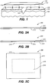
- device or bandage 100 comprises a body 112, that is removably secured to skin surface 135 near wound site 120, as indicated by arrows 116.
- the device 100 can be removably secured to the skin surface (e.g., stratum corneum and epidermis) 135 by an adhesive, or by using one or more skin piercing devices (e.g., sutures, anchors, microneedles, staples, etc.), or the like.
- the devices are removably secured to the tissue below the skin surface, e.g., sutures, anchors, staples, and the like can be used to removably secure the devices to the deepest layers of the dermis down to the fascia.
- wound 120 extends beneath the epidermis 135 through dermis 130 to reach the hypodermis or subcutis 140.
- device 100 is depicted as a single layer in FIG. 1 for simplicity, the devices described here can comprise multiple layers and have any number of different configurations. In some variations, the devices comprise multiple layers that remain separate. In other variations, the devices comprise multiple layers in an overlay configuration. In still other variations, the devices comprise multiple layers that are joined or welded together, e.g., in a laminate.
- the device 200 may include an adhesive layer 214 for removably attaching device 200 to the skin.
- the adhesive layer can be applied in any suitable fashion to surface 213 of body 212 that is intended to contact the skin.
- adhesive layer 214 can be a continuous layer around the periphery of surface 213.
- adhesive layer 214 can be a continuous layer substantially covering surface 213.
- Adhesive layer 214 may be a contiguous or noncontiguous layer on surface 213.
- adhesive layer 214 comprises a pressure sensitive adhesive, e.g., polyacrylate-based, polyisobutylene-based, silicone-based pressure sensitive adhesives, and the like. As shown in FIG.
- device 200 can include an optional wound dressing 218 to be applied to a wound (not shown).
- the surface 213 of device 200 that is intended to contact the skin is shown in FIG. 2C .
- adhesive layer 214 and wound dressing 218 in combination substantially cover surface 213.
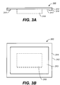
- a wound dressing can be placed over at least a portion of an adhesive layer.
- adhesive layer 314 of device 300 can partially cover surface 313 of body 312, for example by forming a frame around the periphery of surface 313.
- Optional wound dressing 318 can be located centrally within the frame formed by adhesive 314.
- device 400 can comprise a body 412 that includes at least one aperture 422.
- Aperture 422 can be positioned to surround wound 120 as shown by arrows 416.
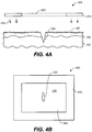
- device 500 can comprise a body 512 that includes multiple apertures 524.
- FIG. 5 depicts apertures 524 arranged in a lattice fashion, the apertures can be arranged randomly or in any suitable fashion, e.g., in rows, columns, in a circle, an oval, or on a diagonal.
- the apertures e.g., apertures 422, 524 in FIGS.
- an aperture such as aperture 422 in FIG. 4 can have an elongate shape, with the long axis of the aperture approximately parallel to the long axis of the wound.
- an aperture such as aperture 422 in FIG. 4 can have an elongate shape, with the long axis of the aperture approximately orthogonal to the long axis of the wound.
- the apertures may be cut by the user or attending physician, immediately prior to use.
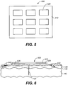
- device 600 is removably secured to the stratum corneum (not shown) and epidermis 135 by a securing mechanism 626.
- securing mechanism 626 may be anything suitable for removably securing the device 600 to the skin surface near a wound site, e.g., an adhesive, a staple, a suture, a microneedle, an anchor, or the like. If an adhesive is used as the securing mechanism, the adhesive can be selected to exhibit minimal creep over time. For example, the rheological properties of adhesives can be tuned.
- One method of tuning rheological properties of adhesives includes the addition of cross linking agents to increase the cross link density of the adhesive, e.g., a pressure sensitive adhesive.
- Suitable cross linking agents can include highly functionalized molecules such as aluminum acetylacetonate.
- the cross linking density of an adhesive can be adjusted to achieve desirable adhesion values while minimizing the amount of creep the adhesive will demonstrate over time.
- the devices When the devices are sutured, anchored or stapled to the skin, the devices may be attached to the dermis 130 or subcutis 140 as well as epidermis 135. This may help improve isolation or unloading of the wound from exogenous and/or endogenous stress.
- the devices may be applied to a wound site at any suitable time. For example, in some variations, it is desirable to apply the devices to the wound site from about one to about three days following injury, i.e., during an initial period such as the early part of the proliferative phase. It should be understood that the devices may or may not be applied to a wound site where the wound has already initially been closed (e.g., by suturing, adhesives, bandages or the like). Similarly, the devices may be applied to a fresh wound caused by a scar removal procedure. In some instances, the device will be applied up to seven days following injury, i.e., later in the proliferative phase. For example, swelling and wound exudates may indicate that the devices be applied later than three days following injury.
- a first bandage can be applied within an initial period following injury, e.g., within the first three days, and then removed, and a second bandage can be applied thereafter.
- the second bandage can be adapted to changes in the skin and tissue surrounding the wound that can occur after the initial period, e.g., decreased swelling and exudates.
- device 600 is contracted as body 612 is contracted in at least one direction.
- tension is transferred to the skin at or external to securing mechanisms 626 as indicated by arrows 642, thereby reducing stress at the wound site.
- wound 120 can be effectively isolated from exogenous and/or endogenous stress in many instances. That is, device 600 can operate to unload wound 120 and surrounding tissue from endogenous forces from skin tension as well as exogenous forces from muscle action and body movement. In this manner, scar formation at wound 120 may be reduced.
- the devices and bandages described herein may have any suitable shape.
- the devices or bandages may be rectangular, square, circular, oval, toroidal, or segments or combinations thereof.
- the devices will be flexible and planar to allow conformal placement against skin.
- the devices and bandages may also be of any suitable size, to deal with a variety of wounds.
- the devices and bandages may be cut immediately prior to use from a roll or sheet of bandage to ensure appropriate coverage of the wound site.
- Devices and bandages can extend out to about 20cm (about 8 inches) from the wound in some instances, and in other instances the devices or bandages can extend about 2, 4, 6, 8, 10, 12, 14, 16, or 18cm from the wound, where "about" qualifies each of the distances.
- the bandages can extend about 22cm, about 24cm, about 26cm, or even more, from the wound.
- the devices and bandages may or may not be occlusive, and in some variations, the devices and bandages are occlusive. At least a portion of the devices and bandages may also be made of a transparent material. The transparent material can be placed over the wound to allow monitoring of the wound (e.g., to monitor infection or healing progress).
- the devices or bandages described herein can be perforated, partially perforated, or at least partially porous. For example, some variations of the devices and bandages allow oxygen and/or moisture exchange with the environment.
- the devices and bandages may also include a mechanism for increasing the temperature at the skin surface where the device or bandage is applied. This may be beneficial, for example, to aid in the healing process.
- the mechanism may be electrical, e.g., a resistive heating element, chemical, e.g., an exothermic chemical reaction, or mechanical, e.g., the creation of an element that friction rubs, e.g., against the skin.
- the bandages and devices described here may also comprise a mechanism to induce a color change in at least a portion of the bandage. This may be helpful, for example, to alert the user to the device's decreasing efficacy, stiffness or the like.
- a color change in a device or bandage may correspond to a change in bandage stiffness. For example, if a device or bandage is strained or stressed, at least a portion of the deice or bandage may have a different color than when it is relaxed.
- a color change in a device or bandage may correspond to a change in bandage efficacy. For example, at least a portion of the device or bandage may change color as its moisture content changes. In other variations, a device or bandage may change color after a predetermined period of time.
- the devices and bandages described here may also comprise or deliver one or more active agents.
- Active agents can assist in wound healing, and may therefore include any suitable compound.
- the active agent may be a pharmaceutical compound, a protein, a vitamin, or the like.
- Illustrative active agents that may be desirable for use with the bandages and devices described here include, but are not limited to growth factors, enzymes such as elastase to degrade the extra cellular matrix, proteases such as aspartate, serine, and metalloproteases that are capable of digesting and remodeling tissue, inhibitors of enzymes such as tissue inhibitors of metalloproteases, antibiotics, antifungals, vitamin E, and combinations thereof.
- delivery of active agents can be controlled by time-release, e.g., by encapsulating or embedding the active agents in a time-release formulation, such as a drug delivery polymer or depot.
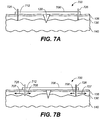
- the bandages for ameliorating the formation of a scar and/or keloid at a wound site have a first tensile stressed configuration (e.g., as shown in FIG. 7A ) and a second relaxed configuration (e.g., as shown in FIG. 7B ).
- first tensile stressed configuration e.g., as shown in FIG. 7A
- second relaxed configuration e.g., as shown in FIG. 7B
- device or bandage 700 having body 712 can be removably secured to the skin surface or epidermis 135 near wound 120 via securing mechanisms 726 while in first configuration 706'.
- configuration 706' is tensile stressed in at least one direction.
- device 700 is removably secured to dermis 130 as well as epidermis 135 via securing mechanisms 726, e.g., using sutures, anchors, staples, microneedles or the like.
- device 700 is removably secured to the tissue deeper than dermis 130.
- an adhesive can be used as a securing mechanism, the adhesive can be selected to exhibit minimized creep properties over time, e.g., by adjusting the cross link density in the adhesive. The cross linking density of an adhesive can be adjusted to achieve desirable adhesion values while minimizing the amount of creep demonstrated by the adhesive. When tensile stress is removed from device 700, it will adopt a relaxed configuration 706 shown in FIG.
- the first tensile stressed configuration 706' may be stressed relative to relaxed configuration 706 by at least 40%. It should be understood that the term "about" qualifies each of these percentages.
- stress at the wound site 120 may be minimized. That is, the device can shield the wound and tissue from endogenous and/or exogenous stress. In some instances, the device can reduce stress at the wound site such that it is lower than stress experienced by typical, unscarred skin.
- the stress in tensile stressed configuration 706' may be adjusted for different skin types and thicknesses to shield, i.e., unload, wounds from endogenous stress.
- the stress in tensile stressed configuration 706' may be adjusted to accommodate different ranges of motion to shield, e.g., unload, wounds from exogenous stress.
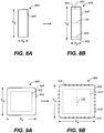
- a bandage 800 in its relaxed configuration 806 has an X-direction (width) X 8 and a Y-direction (length) Yg.
- bandage 800 can be tensile stressed, i.e., stretched, in at least one direction to form tensile stressed configuration 806'.
- bandage 800 is tensile stressed in the Y-direction to length Y 8 ', but remains substantially unstressed in the X-direction to approximately retain width Xg.
- Bandage 800 in its stressed configuration 806' can then be placed over wound 120 and removably secured to the skin surface via securing mechanisms 826.
- securing mechanisms 826 are located proximate to bandage edges 802. The tensile stress on bandage 800 may isolate and shield wound 120 from endogenous and/or exogenous stress in the Y-direction.
- a bandage 900 in its relaxed configuration 906 has width X 9 and length Y 9 .
- bandage 900 with body 912 and optional aperture 922 can be tensile stressed in at least two directions to form tensile stressed configuration 906' having width X 9 ' and length Y 9 '.
- Stressed configuration 906' can then be applied over wound 120, e.g., to frame wound 120 in stressed aperture 922', and removably secured to the skin surface via securing mechanisms 926.
- Bandages such as bandage 900 that are tensile stressed in at least two directions may shield, i.e., unload, wounds from endogenous and/or exogenous stress in at least two directions.
- the first tensile stressed configuration can be mechanically induced.
- devices or bandages can include at least one spring element.
- the spring element can be extended to form a tensile stressed configuration, and the spring element can be released to form a relaxed configuration.
- the devices or bandages comprise an elastic material., such as a biocompatible silicone polymer.
- the elastic material may be stretched to form a tensile stressed configuration.
- the first tensile stressed configuration may be at least partially induced by at least one piezoelectric element.
- the first tensile stressed configuration may be induced electrostatically.
- the devices or bandages may be tensile-stressed in a dynamic fashion, e.g., by applying an oscillating force to the bandages or devices.
- an oscillating force e.g., if a bandage includes a piezoelectric element, an alternating potential can be applied to the piezoelectric element, causing the device to alternately expand and contract in at least one direction.
- a bandage includes an electrostatic element
- an alternating potential can be applied to the electrostatic element to cause it to alternately expand and contract in at least one direction.
- Some bandages comprise at least first, second and third configurations.
- the second configuration is typically strained relative to the first configuration.
- the bandages are configured to be removably secured to a skin surface while they are in the second configuration and are capable of being activated while in the second configuration to adopt a third configuration.
- the second configuration can be thermally activated to adopt the third configuration.
- body heat, a heating pad, an air blower, a heat gun, or the like may be used activate the second configuration to adopt the third configuration.
- the first configuration may be "stored" in the bandages.
- the silicone polymer sheet the first configuration may be stored by stretching the polymer sheet and then clamping the sheet along its edges to a stiffer polymer sheet using any suitable attachment device.
- the bandage may or may not be allowed to relax for a period of time (e.g., about 5 minutes, about 10 minutes, about 20 minutes, etc.) before application to skin.
- the third configuration of the bandages can differ from the strained second configuration in at least one dimension or direction. In other variations, the third configuration can differ from the strained second configuration in at least two directions. In some variations, the third configuration differs from the initial first configuration by less than about 10%, about 20%, about 30%, about 40%, about 50% or about 60% in at least one direction. In other variations, the third configuration differs from the initial configuration by less than about 10%, about 20%, about 30%, about 40%, about 50% or about 60% in at least two directions.
- the second configuration is strained by about 5%, 10%, 15%, 20%, 25%, 30%, 35%, 40%, 45%, 50%, 55%, 60%, 65%, 70%, 75%, 80%, 85%, 90%, 95%, or 100% relative to the first configuration. It should be understood that the term "about" qualifies each of these percentages.
- stress at a wound site may be minimized. That is, the amount and directionality of strain in prestrained devices or bandages can be adjusted for different skin types, thicknesses and conditions to shield, i.e., unload, wounds from endogenous stress.
- the amount and directionality of strain in prestrained devices or bandages can be adjusted to accommodate different ranges of motion or muscle action to shield wounds from exogenous stress.
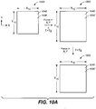
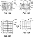
- a device or bandage 1000 includes a polymeric backing layer 1048 that has a first configuration 1006 that is approximately the shape of a planar rectangular sheet having width X 10 and length Y 10 .
- First configuration 1006 has been stored into polymeric layer 1048, e.g., by crosslinking or by pre-stretching.
- the polymeric layer 1048 can be heated above the polymer T g and deformed, e.g., by applying force in both X- and Y-directions, to adopt a second configuration 1006'.
- Strained configuration 1006' can also have an approximately planar rectangular sheet conformation, but with width X 10 ' and length Y 10 '.
- Second configuration 1006' can be stabilized, e.g., by subsequently cooling the polymeric layer 1048 to a temperature below T g while still under strain. Strain can then be removed, and strained configuration 1006' can be stable if held at a temperature sufficiently below the polymer T g , e.g., about 10°C, about 15°C, about 20°C, or more, below the polymer T g .
- the stabilized strained configuration 1006' may generally recover its initial configuration 1006 with a width of approximately X 10 and a length of approximately Y 10 , as illustrated in FIG. 10B .
- the devices or bandages described here are typically removably secured to a skin surface while in a strained configuration. If a bandage or device in its strained stabilized configuration experiences a load or constraint as it is reheated above T g , its ability to recover to its initial configuration may be limited by that constraint.
- strained configuration 1006' is removably secured to skin (not shown) via securing mechanisms 1026 and is therefore subject to constraint. Strained configuration 1006' recovers to a third configuration 1006" instead of initial configuration 1006.
- device 1000 is constrained primarily in the X-direction only.
- Device 1000 can reach third configuration 1006" that has recovered its initial length Y 10 to a substantial degree, but has recovered its width only partially along the X-direction, resulting in a recovered X-direction X 10 " that is between X 10 ' and X 10 .
- the polymer may be of any suitable thickness.
- the polymer thickness may be from about 100 or 200 microns to a few millimeters.
- the thickness of polymer sheets e.g., silicone polymer sheets or shape memory polymer sheets, can be selected to provide the devices or bandages with sufficient load carrying capacity to achieve desired recoverable strains, and to prevent undesired amounts of creep deformation of the bandages or devices over time.
- the thickness across devices or bandages is not uniform, e.g., the thickness across the device can be varied to change the stiffness, the load carrying capacity, or recovery strains in selected orientations and/or locations.
- the devices or bandages are tapered near the edges to reduce thickness. Devices or bandages having tapered edges may increase the flexibility of the devices or reduce the likelihood that the devices or bandages can debond over time. In addition, devices with tapered edges may have increased comfort for the wearer.
- the bandages or devices comprise a mesh or wire frame.
- elements 1160, 1162 forming at least part of mesh or frame 1130 can be incorporated into bandage or device 1100.
- elements 1160, 1162 comprise a shape memory metal. That is, in some variations, the bandages or devices comprise a shape memory metal formed as a mesh or wire frame.
- elements 1160, 1162 comprise shape memory polymers.
- elements 1160 or elements 1162 can be elastic fibers having a first tensile stressed configuration (not shown). Device 1100 can be removably secured to skin in the proximity of a wound site with elements 1160 or 1162 in their tensile stressed configuration.
- the elements 1160, 1162 may have any suitable configuration, e.g., linear stripes, diagonals, circles, ovals, or various three-dimensional configurations such as a three-dimensional mesh.
- elements 1160, 1162 are shape memory metals, any suitable shape memory metal may be used, e.g., nickel titanium alloys and the like.
- the elements 1160, 1162 of the devices or bandages may also include polymers in addition to shape memory metals.
- the first unstrained configuration may be formed by shaping the metal in its high strength austenitic phase to a desired configuration.
- the second strained configuration may be stored by first plastically deforming the shape memory metal while the metal is heated above M f , the temperature at which the metal fully adopts its soft martensic phase, and then cooling the metal while still under strain to a temperature below A f , the temperature at which the metal recovers its high strength austenitic phase. If the strained second configuration of shape memory metal is heated above M f while not constrained, the shape memory metal can recover to essentially the dimensions of the first configuration.
- the shape memory metal may be a wire, mesh, or foil, e.g., a thin wire, a thin mesh, or a thin foil. Any combination of wire, mesh, or foil shape memory metals may also be used. Of course, combinations of different shape memory materials, e.g., more than one shape memory metal or a shape memory metal and a shape memory polymer, may be used in devices or bandages. In still other variations, shape memory metals are covered at least partially with plastic or fibers, either while in their first unstrained configuration or in their second strained configuration.
- the phase transition temperature of shape memory metals used in bandages or devices described herein may be chosen to be compatible with use on skin, e.g., between about 35°C and about 55°C.
- the devices may include one or more thermally insulating layers that can allow the use of shape memory metals having transition temperatures higher than about 55°C.
- body heat can be sufficient to increase the temperature of shape memory metals used in the devices above M f .
- FIG. 12A shows a first configuration 1206 of a device 1200 having a rectangular shape with width X 12 and length Y 12 .
- Device 1200 is heated above a transition temperature, e.g. T>T g or T>M f , to adopt a strained second configuration 1206'.
- Strained second configuration 1206' is stabilized by cooling device 1200 below its transition temperature.
- strained configuration 1206' is strained relative to initial configuration 1206 primarily by expansion along the Y-direction and is substantially unstrained in the X-direction, having width X 12 and length Y 12 ' ( FIG. 12B ).
- strained configuration 1206' is applied proximate the wound 120 and removably secured to the skin (not shown) via securing mechanisms 1226 proximate the device edges 1253 ( FIG. 12C ). Heat is applied to strained configuration 1206' such that its temperature exceeds its transition temperature and it will attempt to adopt its original first configuration 1206.
- the device 1200 may not recover its original Y-direction Y 12 . Instead, the device may recover to an intermediate third configuration 1206" ( FIG. 12D ) having length Y 12 ", where Y 12 " is a length between the original length Y 12 of the first configuration 1206 and strained length Y 12 ' of strained configuration 1206'.
- the secured configuration 1206" transfers stress along direction Y to the skin at and external to the securing mechanisms 1226, indicated by arrows 1252. Therefore, device 1200 attached in configuration 1206" can shield, i.e., unload, wound 120 from endogenous and/or exogenous stress in at least direction Y, thereby ameliorating scar formation at wound 120.
- FIG. 13A illustrates a variation of a device which is not claimed 1300 having a first configuration 1306.
- First configuration 1306 is approximately rectangular with width X 13 and length Y 13 and includes aperture 1322.
- the device is heated above its transition temperature, e.g., T g or M f , and strained in both X- and Y-directions.
- device 1300 in strained configuration 1306' is also approximately rectangular with width X 13 ' and length Y 13 '.
- the size and/or shape of aperture 1322 can change to form aperture 1322' in strained configuration 1306'.
- Device 1300 is stabilized in strained configuration 1306' by cooling below its transition temperature while still under strain. As shown in FIG.
- device 1300 in its deformed configuration 1306' is removably secured to the skin surface via securing mechanism 1326 to frame wound 120.
- securing mechanism 1326 is illustrated as approximately following the shape of device 1300, other variations may be used as well.
- the device can be removably secured to the skin at spaced apart securing sites, or removably secured to the skin around the periphery of the device. After device 1300 is secured to skin, heat is applied to increase the temperature of the device above its transition temperature so that the device will attempt to recover to its original configuration 1306.
- Third configuration 1306 can be intermediate between first configuration 1306 and second configuration 1306', having width X 13 " between X 13 and X 13 ' and length Y 13 " between Y 13 and Y 13 '.
- device 1300 can shield, i.e., unload, wound 120 from endogenous and/or exogenous stress in at least two directions, thereby ameliorating scar formation at wound 120.
- FIGS. 14A-D Another variation of a device or bandage is illustrated in FIGS. 14A-D .
- device 1400 has a first configuration 1406 that is approximately circular or oval having cross-sectional diameters D 1 and D 2 .
- Device 1400 is strained while heated above its transition temperature to form second configuration 1406' ( FIG. 14B ).
- device 1400 is strained in both X- and Y-directions resulting in a circular or oval second configuration 1406' with cross-sectional diameters D 1 ' and D 2 '.
- the temperature of device 1400 is then lowered below the transition temperature while device 1400 is still under strain so that strained configuration 1406' is stable.
- Device 1400 is removably secured to skin (not shown) with securing mechanisms 1426 over wound 120 while in deformed configuration 1206' ( FIG. 14C ).
- securing mechanism 1426 are shown here as sutures, staples, microneedles, or anchors, or the like device 1400 can be secured to the skin surface in any suitable fashion, as described above.
- Heat is applied to increase the temperature of device 1400 above its transition temperature, e.g., T g or M f .
- Deformed state 1406' can then adopt its original configuration 1406, subject to constraint. As illustrated in FIG.
- the resulting configuration 1406" can be approximately circular or oval with cross-sectional diameters D 1 " and D 2 ", where D 1 " is approximately between D 1 and D 1 ' and D 2 " is approximately between D 2 and D 2 '.
- the constraint in one or both directions will be small enough such that D 1 " is approximately equal to D 1 and/or D 2 " is approximately equal to D 2 .
- constraint in one direction will be greater than in another direction.
- skin compliance constrains the recovery of strained configuration 1406'.
- Device 1400 may transfer stress from the wound site to skin at or external to securing mechanisms 1426, thereby shielding wound 120 from endogenous and/or exogenous stress and ameliorating scar formation. If either D 1 " or D 2 " is substantially changed from D 1 ' or D 2 ', respectively, then device 1400 can shield wound 120 from endogenous and/or exogenous stress in at least one direction. If both D 1 " and D 2 " are substantially different D 1 ' and D 2 ', respectively, device 1400 can protect wound 120 from endogenous and/or exogenous stress in at least two directions.
- the devices and bandages may comprise or be made out of more than one type of material, e.g., more than one type of polymer or more than one type of shape memory material.
- a device can comprise two different silicone polymers or two different shape memory materials, e.g., two different shape memory polymers, two different shape memory metals, or a shape memory polymer and a shape memory metal. If more than one type of material is used in a device, the materials selected can have different transition temperatures, different amounts of strain that can be incorporated into a strained configuration, or different abilities to recover an initial configuration against constraint, i.e., different load carrying capacities when heated above a transition temperature.
- FIG. 15A A variation of a device 1500, also not claimed, comprising two shape memory polymers is illustrated in FIG. 15A .
- Device 1500 forms a rectangle with width X 15 ' and length Y 15 ' in its stable, strained configuration 1506'.
- Device 1500 in its strained configuration 1506' comprises strained bands or fibers 1574' extending in the X-direction made of a first shape memory material and strained bands or fibers 1576' extending in the Y-direction made of a second shape memory material.
- Initial configuration (1506) is not shown.
- FIG. 15A depicts bands or fibers 1574, 1576' as being interwoven, they may or may not be interwoven.
- Device 1500 in second strained configuration 1506' is attached to skin (not shown).
- strained bands or fibers 1576' relax to state 1576"and strained bands or fibers 1574' relax to state 1574" to result in third configuration 1506" having width X 15 " and length Y 15 ".
- the percentage change between relaxed bands 1574" and strained bands 1574' is less than the percentage change between relaxed bands 1576" and strained bands 1576'.
- devices can comprise elements having different dimensions to shield a wound preferentially in one or more directions from stress.
- a device 1600 in second strained stable configuration 1606' can be removably secured to skin in the proximity of a wound site.
- Device 1600 in second configuration 1606' has thick bands or fibers 1686' extending in the Y-direction and thin bands or fibers 1684' extending in the X-direction.
- Bands or fibers 1686'and 1684' may be made of the same or different materials, and may or may not be interwoven.
- third configuration 1606" in some variations can preferentially recover in the Y-direction because the recovered thicker bands 1686" in the Y-direction may have increased ability over the recovered thinner bands 1684" in the X-direction to overcome constraint resisting recovery to an initial configuration (not shown). That is, if a device 1600 is expanded in both X- and Y-directions in its strained second configuration 1606', then recovered third configuration 1606"can have the characteristic Y 16 "/Y 16 ' ⁇ X 16 "/X 16 '.
- kits for ameliorating the formation of scars and/or keloids are also described here.
- the kits comprise in packaged combination at least two devices, where each device is configured to be removably secured to a skin surface in proximity to a wound site.
- Each device shields, i.e., unloads, the wound from endogenous and/or exogenous stress.
- kits in a kit have different colors. Variations of kits can include devices that are color-coded for different scheduling regimes. For example, one color of a device may be provided for use at the beginning of the proliferative phase of wound healing and another color of a device may be provided for use at a later phase of wound healing.
- devices in a kit have different shapes. For example, the shapes may be independently selected from the group consisting of rectangles, circles, squares, trapezoids, toroids, ovals, and segments and combinations thereof. In some variations, the devices in a kit may have different sizes or different thicknesses. The devices in a kit may also be configured to shield a wound from different amounts of endogenous and/or exogenous stress.
- kits may be designed to be applied in parallel, e.g., where more than one device is secured in proximity to the wound at the same time.
- Parallel application of devices encompasses scenarios in which the securing of the devices occurs at the same time, and in which a second device is secured while a first device remains secured.
- Multiple devices may also be applied in a serial fashion, where a first device is removed before a second device is secured.
- some kits may include one device to be applied during an initial period such as the early part of the proliferative phase of wound healing, e.g., up to three days after injury, and then removed and a second device to be applied thereafter.
- kits may include an air blower, a heat gun, a heating pad, or the like, to raise the temperature of one or more devices.
- Some kits may contain at least one wound dressing, or at least one wound cleanser, or other components desirable or suitable for wound healing applications.
- the kits may also comprise instructions for using the devices and/or other components contained therein.
- the methods typically comprise applying a device configured to shield a wound from endogenous and/or exogenous stress in the proximity of the wound site.
- the device is configured to shield the wound from both endogenous and exogenous stress.
- the device may be applied during the proliferative phase of wound healing, which as described above, may be during the proliferative phase of wound healing after an old scar has been excised.
- the device may be applied and worn for any suitable length of time.
- the device may be applied and worn for a period of at least about 10 days, at least about 20 days, at least about 30 days, at least about 40 days, at least about 50 days, at least about 60 days, at least about 70 days, at least about 80 days, at least about 90 days, at least about 100 days, and the like.
- the devices may be applied for a period over 100 days, e.g., for periods of about 6 months, about 9 months, about 12 months, or about 15 months, or even longer.
- the devices are typically removed after a period of time, which may or may not be predetermined.
- the period of time may be predetermined based on the type of wound.
- the period of time may be actively monitored and therefore variable depending on the progress of the wound healing process.
- the methods may also comprise applying to the skin in the proximity of a wound site, a second device configured to shield the wound from endogenous and/or exogenous stress.
- the methods may comprise applying to the skin in the proximity of a wound site, a second device configured to be removably secured to a skin surface and to reduce wound stress in at least one direction.
- the second device is configured to reduce wound stress in at least two directions. If a second device is applied, it may be applied in parallel manner with the first device. That is, the second device may be applied before the first device is removed. In some variations of the methods, the second device may be applied in a serial fashion, i.e., after the first device is removed.
- a first device may be applied in an initial period such as the early part of the proliferative phase when tissue is swollen and wound exudates is high, and then removed.
- a second device can be applied thereafter, where the second device has been selected to have properties reflecting reduced swelling and/or wound exudates.
- several devices may be applied in a parallel or serial fashion to reflect the wound environment as healing progresses.
- a murine model system for scarring in human dermal tissue can be created by inducing hypertrophic scarring on murine dorsal dermis by increasing mechanical forces on murine wounds to approximate those found in human wounds.
- Typical adult human skin is under about 0.4-1 N/mm 2 of stress while at rest.
- Healing adult human wounds are generally under about 0.6-2 N/mm 2 of stress.
- Mechanical strain devices 1700 as illustrated schematically in FIG. 17 were constructed by securing 22mm expansion screws 1790, available from Great Lakes Orthodontic Products, Tonawanda, NY, to titanium surgical Luhr plate supports 1792, available from Stryker-Leibinger Co., Freiburg, Germany. Expansion screws 1790 were secured to the plate supports 1792 using plastic interfaces and clear epoxy (not shown), available from Devcon Scientific, Riviera Beach, FL, which was allowed to dry overnight. As illustrated schematically in FIGS.
- strain devices 1800, 1800' were attached to a mouse's dorsum 1810 by suturing over wounds 1802, 1802', respectively. Strain devices 1800, 1800' did not physically contact wounds 1802 or 1802'. Strain devices 1800, 1800' were oriented relative to incisions 1802, 1802' to apply tension to the wounds in a direction approximately orthogonal to the incision direction. Analogously, as shown in FIG. 19 , two strain devices 1900, 1900' were attached to a mouse's dorsum 1910 by suturing over wounds 1902, 1902', respectively.
- strain devices 1900, 1900' were oriented relative to incisions 1902, 1902' to apply tension to the wounds in a direction approximately parallel to the incision direction.
- the strain devices 1800, 1900 were adjusted such that wounds 1802, 1902 experienced no additional strain.
- the strain devices 1800', 1900' were adjusted to apply tension to wounds 1802', 1902', respectively.
- approximately uniaxial tension on wounds 1802' was increased by expanding the distance between Luhr plates 1892' by 2mm using expansion screw 1890', to generate an estimated stress of 1.5N/mm 2 , and by 4mm every second day thereafter for a total of 7 days to increase the applied stress to about 2.7N/mm 2 .
- Tissue collected from unstressed wounds 1802, 1902 did not exhibit significant amounts of fibrosis after 3 weeks.
- tissue collected from wounds 1802' stressed in a direction approximately orthogonal to the incision direction as illustrated in FIG. 18 was characteristic of tissue from hypertrophic scars, having approximately 15 times greater cross-sectional areas than tissue from unstressed wounds.
- tissue collected from wounds 1902' stressed in a direction approximately parallel to the incision direction as illustrated in FIG. 19 was about 5 times greater in cross-sectional area than tissue collected from unstressed scars.
- the murine stressed scars displayed many characteristics of human hypertrophic scars. The murine stressed scars were raised, and demonstrated loss of rete pegs, adnexae and hair follicles.
- Stressed murine scars showed cellular hyperplasia, and fibroblasts oriented approximately parallel to collagen fibers and the direction of the strain. In addition, blood vessels in stressed wounds were approximately perpendicular to the wound. Stressed murine scars also showed collagen whorls, which are often observed in chronic human hypertrophic scars. Scar tissue from stressed murine wounds also demonstrated at least double cell density (cells per mm 2 ) by Dapi nuclear staining. In addition, stressed murine scars averaged about 3mm of height extending above the skin surface, but unstressed scars remained substantially flat.
- SAM Significance Analysis of Microarrays
- FIG. 20 illustrates a qualitative effect of mechanical load on scar volume.
- Human fetal skin typically is almost completely relaxed and therefore under very low endogenous load.
- the dynamic resting tension as a measure of skin elasticity for human fetal skin is below the currently available limits of detection.
- Human fetal skin heals with little or no scar volume, as illustrated by point 2002.
- Murine skin is under a higher endogenous load, having a dynamic resting tension of about 0.06N/mm 2 .
- the baseline scarring volume for murine skin is above that for human fetal skin, as indicated by point 2004.
- the data in this Example showed a positive correlation between mechanical load and scar volume for a murine model, as indicated by solid trend line 1 in FIG. 20 .
- the sheets made of polymer having T g of 20°C had thicknesses of approximately 200 microns and 500 microns.
- the sheets made of polymer having T g of 30°C had a thickness of approximately 500 microns, and the sheets made of polymer with T g of 40°C had a thickness of approximately 1000 microns.
- Each of a first set of 8 bandages was individually fixed between gripping fixtures and heated to about 60°C to about 90°C above T g for the polymer used in that bandage.
- the bandages were stretched along the longer dimension of the rectangle while heated above T g by translating the gripping fixtures apart at about 1mm/minute to reach strains of about 8% to about 12%.
- Each bandage was cooled to well below T g for the polymer used in that bandage by inserting the straining stage with the bandage between the gripping fixtures into a freezer at -10°C while the strains were imposed.
- each bandage was removed from the freezer and gripping fixtures and stored at laboratory ambient temperature of approximately 24°C, except for the bandages made from a polymer having T g of 20°C, which were kept in a refrigerator at approximately 5°C.
- Duro-Tak 87-4287 pressure sensitive adhesive available from National Starch and Chemical Company, Bridgewater, NJ, was applied to one side of the bandage.
- the pressure sensitive adhesive was in the form of an approximately 60 micron thick layer contained between two polymer release layers.
- One of the release layers was removed and the adhesive layer was then pressure applied to the polymer bandage using a hand roller to remove air bubbles at the interface between the adhesive and polymer.
- the excess adhesive layer was trimmed to the edges of the polymer bandage.
- the remaining release layer was then carefully removed.
- a wound dressing having approximate dimensions of 10mm x 5mm obtained from commercially available bandages was affixed to the center of selected exemplary polymer bandages.
- a polymer release layer was subsequently reapplied over the adhesive and each bandage was stored at the storage temperatures noted above prior to use.
- the polymer release layers were removed from selected bandages. Some bandages were heated approximately 20°C to approximately 50°C above T g for the polymer used in the respective bandages without being subject to constraint. When unconstrained, the bandages were generally observed to recover their approximate original unstrained size upon being heated above the polymer T g . Other bandages made from polymers having T g of 30°C were affixed to human skin in locations on the inside forearm using finger pressure. After affixing the bandages to skin, the bandages were heated above T g using a hot air blower. Temperatures were estimated to reach about 45°C for a period of about 15 seconds. Partial recovery of the bandages was noted, achieving approximately 50% of the initially imposed strain. These results are summarized in Table 1 below.
- the CSM82-4032-20 sheet had a Durometer value of 30 and had a thickness of approximately 490 microns.
- MED1356 pressure sensitive adhesive available from NUSIL TECHNOLOGY LLC, 1050 Cindy Lane, Carpinteria, CA 93013, USA, was applied to one side of the bandage.
- the pressure sensitive adhesive was in the form of a viscous liquid and was applied with a metal spatula to a layer thickness of approximately 60 microns directly on the silicone bandages.
- the solvents in the pressure sensitive adhesive layer were allowed to evaporate according to directions from the manufacturer for a period of 30 minutes at 25°C in a laboratory air environment.
- the MED1356 pressure sensitive adhesive is made by the manufacturer with a range of allowable polymer-to-resin ratios, molecular weights of the resin, and viscosity of the polymer. These determine important properties of the pressure sensitive adhesive including the release force in a T-peel test and the force in a blunt probe tack test. The manufacturer has allowable ranges of these values (approximately 125 kg/m to 286 kg/m for the force in a T-peel test). Our studies revealed that it was important to maintain high values of the release force and blunt tack probe test in order to prevent relaxation of the silicone bandage once applied to the skin.
- the skin was marked in the location where the bandage would be applied with parallel pen lines that were separated by approximately 10 mm. In some cases, orthogonal sets of lines were created. All of the silicone bandages employed were optically transparent and the lines were visible through the bandages after being affixed to the skin. This allowed the strains in the skin to be directly measured by measurement of the displacement of the lines before and after application of the bandage. By successive optical micrographs taken over a period of time the strains in the skin could be continually monitored. Optical micrographs showing initial pen lines on the skin (i.e., before application of the bandage), and after application of the bandage are shown in FIGS. 21A and 21B respectively. By measurement of the change in line separation, the strain imposed on the skin could be directly calculated.
- the silicone bandages were initially stretched to predetermined engineering strains of 10%, 20%, 30%, 40%, 50% and 60% before being affixed to human skin in locations on the inside forearm.
- the strains were imposed in only one orientation, and in others the strains were imposed biaxially in two orthogonal directions in the plane of the bandage.
- the strains were imposed by stretching the bandages and clamping them at the edges to an elastically stiffer polymer sheet using stiff paper clips. By varying the size of the stiffer polymer sheet, the initial strains in the bandage could be systematically varied.
- the bandages After stretching the bandages to the predetermined strains, the bandages were allowed to relax for a period of approximately 10 min. This resulted in some stress relaxation in the bandage.
- the bandages were then affixed to human skin in locations on the inside forearm using finger pressure. After affixing the bandages to skin, the clamps were released and the bandages exhibited immediate elastic recovery. The extent of recovery strains depended on the initial strain in the bandage, the Durometer value and the thickness of the silicone polymer bandage.
- the final strain in the bandage and the strain imposed on the skin are determined by mechanical equilibrium involving both force and moment equilibrium being achieved between the bandage and the underlying skin.
- the bi-directional strain and associated stress state in the skin under the affixed bandage could be systematically controlled by selection of the thickness, mechanical properties, and initial elastic strain of the silicone bandage.
- a set of curves of the initial tension strain in the device as a function of the resulting compressive strain in the skin was generated for different silicone polymer bandages having different polymer backing thicknesses.
- FIG. 23 shows the effect of pressure sensitive adhesive formulation on skin strain imposed by the devices.
- the Type 1 formulation was that for Lot n°36232 of the MED1356 with the higher values of release force in T-peel and blunt probe tack tests. This batch resulted in no observed relaxation of the silicone or skin strains once applied to skin for periods in excess of 3 days at two different strain levels.
- the Type 2 formulation was that of Lot n°39395 of the MED1356, which had lower values of release force in T-peel and blunt probe tack tests. This pressure sensitive adhesive exhibited creep relaxation that resulted in almost complete relaxation of the initial silicone strains in the first 24 hours after application.
- the mechanical strain and stress state of the wound region can be controlled underneath a region in the centre of the bandage that may be unbonded to the underlying skin and contain, for example, a wound dressing.
- FIG. 25 shows a polymer device with a central region approximately 35 x 20 mm in size that does not contain any adhesive layer bonding the device to the skin.
- Analysis of the underlying markings on the skin revealed the identical strains compared to the bonded regions (the curvature of the lines observed is associated with refraction through the transparent polymer layer in the presence of the air gap in the unbonded region).
Priority Applications (1)
| Application Number | Priority Date | Filing Date | Title |
|---|---|---|---|
| EP18215491.4A EP3536350A1 (en) | 2006-08-03 | 2007-08-03 | Devices and bandages for the treatment or prevention of scars and/or keloids and methods and kits therefor |
Applications Claiming Priority (2)
| Application Number | Priority Date | Filing Date | Title |
|---|---|---|---|
| US83565406P | 2006-08-03 | 2006-08-03 | |
| PCT/US2007/017320 WO2008019051A2 (en) | 2006-08-03 | 2007-08-03 | Devices and bandages for the treatment or prevention of scars and/or keloids and methods and kits therefor |
Related Child Applications (1)
| Application Number | Title | Priority Date | Filing Date |
|---|---|---|---|
| EP18215491.4A Division EP3536350A1 (en) | 2006-08-03 | 2007-08-03 | Devices and bandages for the treatment or prevention of scars and/or keloids and methods and kits therefor |
Publications (2)
| Publication Number | Publication Date |
|---|---|
| EP2054089A2 EP2054089A2 (en) | 2009-05-06 |
| EP2054089B1 true EP2054089B1 (en) | 2018-12-26 |
Family
ID=38920552
Family Applications (2)
| Application Number | Title | Priority Date | Filing Date |
|---|---|---|---|
| EP18215491.4A Pending EP3536350A1 (en) | 2006-08-03 | 2007-08-03 | Devices and bandages for the treatment or prevention of scars and/or keloids and methods and kits therefor |
| EP07836471.8A Active EP2054089B1 (en) | 2006-08-03 | 2007-08-03 | Devices and bandages for the treatment or prevention of scars and/or keloids and methods and kits therefor |
Family Applications Before (1)
| Application Number | Title | Priority Date | Filing Date |
|---|---|---|---|
| EP18215491.4A Pending EP3536350A1 (en) | 2006-08-03 | 2007-08-03 | Devices and bandages for the treatment or prevention of scars and/or keloids and methods and kits therefor |
Country Status (11)
| Country | Link |
|---|---|
| US (4) | US7683234B2 (ja) |
| EP (2) | EP3536350A1 (ja) |
| JP (4) | JP5508851B2 (ja) |
| KR (2) | KR101499080B1 (ja) |
| CN (2) | CN101563113B (ja) |
| AU (1) | AU2007282013B2 (ja) |
| BR (1) | BRPI0715075B1 (ja) |
| CA (1) | CA2659772C (ja) |
| HK (1) | HK1193331A1 (ja) |
| IL (1) | IL196748A (ja) |
| WO (1) | WO2008019051A2 (ja) |
Families Citing this family (98)
| Publication number | Priority date | Publication date | Assignee | Title |
|---|---|---|---|---|
| EP1635858A4 (en) | 2003-05-29 | 2009-08-26 | Scripps Research Inst | TARGETED RELEASE ON CELLS EXPRESSING LEGUMINUM |
| AU2007282013B2 (en) | 2006-08-03 | 2013-07-11 | The Board Of Trustees Of The Leland Stanford Junior University | Devices and bandages for the treatment or prevention of scars and/or keloids and methods and kits therefor |
| US9358009B2 (en) | 2007-08-03 | 2016-06-07 | Neodyne Biosciences, Inc. | Skin straining devices and methods |
| US9241835B2 (en) | 2007-08-03 | 2016-01-26 | Neodyne Biosciences, Inc. | Wound or skin treatment devices with variable edge geometries |
| US9668922B2 (en) | 2007-08-03 | 2017-06-06 | Neodyne Biosciences, Inc. | Wound or skin treatment devices with variable edge geometries |
| WO2009049232A1 (en) * | 2007-10-11 | 2009-04-16 | Spiracur, Inc. | Closed incision negative pressure wound therapy device and methods of use |
| US10292484B1 (en) | 2007-12-26 | 2019-05-21 | Penilopee Lee LaRosa | Apparatus and method for non-invasive repositioning of sagging skin, cellulite and fat tissue on thighs, arms, stomach and buttocks |
| GB0811874D0 (en) * | 2008-06-30 | 2008-07-30 | Nemaura Pharma Ltd | Patches for reverse iontophoresis |
| CA2742962C (en) | 2008-11-07 | 2018-09-25 | Kci Licensing, Inc. | Reduced-pressure, wound-treatment dressings and systems |
| JP5424380B2 (ja) * | 2008-12-12 | 2014-02-26 | 学校法人日本大学 | 脈波測定装置 |
| WO2010118316A1 (en) * | 2009-04-10 | 2010-10-14 | Spiracur, Inc. | Methods and devices for applying closed incision negative pressure wound therapy |
| US8444614B2 (en) | 2009-04-10 | 2013-05-21 | Spiracur, Inc. | Methods and devices for applying closed incision negative pressure wound therapy |
| EP3207904B1 (en) | 2009-08-11 | 2020-12-30 | Neodyne Biosciences, Inc. | Devices and methods for dressing applicators |
| BR112012003804B1 (pt) * | 2009-08-21 | 2019-02-19 | Novan, Inc. | Curativo para ferimentos, método para formar um curativo para ferimentos, e, kit de curativo para ferimento |
| CN102711729B (zh) | 2009-08-21 | 2015-04-01 | 诺万公司 | 局部用凝胶 |
| WO2011043786A1 (en) | 2009-09-17 | 2011-04-14 | Zipline Medical, Inc. | Rapid closing surgical closure device |
| US10159825B2 (en) | 2009-09-17 | 2018-12-25 | Zipline Medical, Inc. | Rapid closing surgical closure device |
| US8821497B2 (en) * | 2009-10-27 | 2014-09-02 | Howard D. Stupak | Method and apparatus for maxillo-mandibular fixation |
| US20110178375A1 (en) * | 2010-01-19 | 2011-07-21 | Avery Dennison Corporation | Remote physiological monitoring |
| US8439945B2 (en) | 2010-05-03 | 2013-05-14 | Zipline Medical, Inc. | Methods for biopsying tissue |
| WO2011159623A1 (en) * | 2010-06-14 | 2011-12-22 | Zipline Medical, Inc. | Methods and apparatus for inhibiting scar formation |
| WO2011163673A2 (en) * | 2010-06-25 | 2011-12-29 | Purdue Research Foundation | System and method for prevention of hypertrophic scars by actuable patch |
| JP4790091B1 (ja) | 2010-07-29 | 2011-10-12 | 有限会社ちょうりゅう | 創離開防止用補助具 |
| JP5810418B2 (ja) * | 2010-12-28 | 2015-11-11 | アルケア株式会社 | 貼付材 |
| WO2012092306A2 (en) | 2010-12-29 | 2012-07-05 | Mednet Gmbh | Device for preventing excessive skin tension |
| US8637726B2 (en) * | 2011-01-02 | 2014-01-28 | Omnitek Partners Llc | Shape and pressure adjustable dressing |
| CA2824045C (en) | 2011-01-07 | 2019-04-09 | Neodyne Biosciences, Inc. | Wound or skin treatment devices and methods |
| IT1403996B1 (it) * | 2011-02-11 | 2013-11-08 | Baroni | Dispositivo di mantenimento morfologico applicabile ad una regione corporea sottoposta ad espansione tissutale |
| EP2680800B1 (en) * | 2011-03-03 | 2016-04-27 | Neodyne Biosciences, Inc. | Devices for skin tightening |
| KR102256092B1 (ko) | 2011-03-31 | 2021-05-24 | 이엠알지이, 엘엘씨. | 힘 조정형 조직 브릿지 |
| JP5095846B1 (ja) * | 2011-07-07 | 2012-12-12 | 有限会社ちょうりゅう | 処置対応型創離開防止用補助具 |
| US20130110026A1 (en) * | 2011-07-27 | 2013-05-02 | Neodyne Biosciences, Inc. | Strained skin treatment devices and methods |
| KR20190107760A (ko) | 2011-10-20 | 2019-09-20 | 이엠알지이, 엘엘씨. | 착탈가능한 피복재 |
| US10123801B2 (en) | 2011-11-01 | 2018-11-13 | Zipline Medical, Inc. | Means to prevent wound dressings from adhering to closure device |
| US8323313B1 (en) | 2011-11-01 | 2012-12-04 | Zipline Medical, Inc. | Surgical incision and closure apparatus with integrated force distribution |
| US9561034B2 (en) | 2011-11-01 | 2017-02-07 | Zipline Medical, Inc. | Surgical incision and closure apparatus |
| US9050086B2 (en) | 2011-11-01 | 2015-06-09 | Zipline Medical, Inc. | Surgical incision and closure apparatus |
| US10123800B2 (en) | 2011-11-01 | 2018-11-13 | Zipline Medical, Inc. | Surgical incision and closure apparatus with integrated force distribution |
| US10561359B2 (en) * | 2011-12-02 | 2020-02-18 | Neodyne Biosciences, Inc. | Elastic devices, methods, systems and kits for selecting skin treatment devices |
| US9827447B1 (en) * | 2011-12-02 | 2017-11-28 | Neodyne Biosciences, Inc. | Devices, methods, systems and kits for selecting skin treatment devices |
| US9655967B2 (en) | 2011-12-09 | 2017-05-23 | The Board Of Trustees Of The Leland Stanford Junior University | Inhibition of focal adhesion kinase for control of scar tissue formation |
| GB2501055B (en) | 2012-02-01 | 2017-08-30 | Banwell Paul | Scar reduction apparatus |
| US10213350B2 (en) | 2012-02-08 | 2019-02-26 | Neodyne Biosciences, Inc. | Radially tensioned wound or skin treatment devices and methods |
| KR102107459B1 (ko) * | 2012-02-08 | 2020-05-07 | 네오다인 바이오사이언시스, 인코포레이티드 | 방사상으로 인장되는 상처 또는 피부 치료 장치 및 방법 |
| NZ702677A (en) | 2012-06-15 | 2016-05-27 | Univ Washington Ct Commerciali | Microstructure-based wound closure devices |
| US10888375B2 (en) * | 2012-07-06 | 2021-01-12 | The General Hospital Corporation | Method and apparatus for dermatological treatment |
| EP2872222B1 (en) * | 2012-07-06 | 2023-05-31 | The General Hospital Corporation | Method and apparatus for dermatological treatment |
| WO2014047429A1 (en) * | 2012-09-21 | 2014-03-27 | 3M Innovative Properties Company | Incision protection |
| US10315175B2 (en) | 2012-11-15 | 2019-06-11 | Smith International, Inc. | Method of making carbonate PCD and sintering carbonate PCD on carbide substrate |
| WO2014130359A1 (en) | 2013-02-20 | 2014-08-28 | Cytrellis Biosystems, Inc. | Methods and devices for skin tightening |
| CA2905832C (en) * | 2013-03-15 | 2021-09-07 | Neodyne Biosciences, Inc. | Wound or skin treatment devices with variable edge geometries |
| US9517163B2 (en) | 2013-07-03 | 2016-12-13 | 3K Anesthesia Innovations, Llp | Pre-stressed pressure device |
| KR102349218B1 (ko) | 2013-08-09 | 2022-01-10 | 사이트렐리스 바이오시스템즈, 인크. | 비-열적 조직 절제를 사용한 피부 치료를 위한 방법 및 기구 |
| US9675504B2 (en) * | 2013-08-28 | 2017-06-13 | Lisa Ann Myers | Disposable water resistant protective cover cast and wound sites |
| WO2015072517A1 (ja) * | 2013-11-15 | 2015-05-21 | ニチバン株式会社 | 貼付材 |
| US9226890B1 (en) | 2013-12-10 | 2016-01-05 | Englewood Lab, Llc | Polysilicone base for scar treatment |
| US10953143B2 (en) | 2013-12-19 | 2021-03-23 | Cytrellis Biosystems, Inc. | Methods and devices for manipulating subdermal fat |
| EP3089677B1 (en) | 2014-01-05 | 2020-04-01 | Zipline Medical, Inc. | Instrumented wound closure device |
| WO2015136436A1 (en) * | 2014-03-11 | 2015-09-17 | Kast Silicon Ltd. | Silicone strip |
| CN104188705B (zh) * | 2014-09-11 | 2016-10-05 | 黄成� | 一种止血塑形片 |
| WO2016072060A1 (ja) * | 2014-11-05 | 2016-05-12 | 凸版印刷株式会社 | マイクロニードルセット |
| CA2967636A1 (en) | 2014-11-14 | 2016-05-19 | Cytrellis Biosystems, Inc. | Devices and methods for ablation of the skin |
| CN107106333B (zh) * | 2014-12-19 | 2020-07-31 | 3M创新有限公司 | 粘弹性伤口闭合敷料 |
| US10709809B2 (en) | 2014-12-30 | 2020-07-14 | Michel HEYMANS | Multi-layer adhesive tape to compress and contract a scar |
| KR102611420B1 (ko) | 2015-02-19 | 2023-12-07 | 캔디샌트 바이오메디컬 인코포레이티드 | 열 생성용 의료 장치 및 이를 이용한 치료 방법 |
| US10265433B2 (en) | 2015-03-18 | 2019-04-23 | Lawrence Livermore National Security, Llc | Hemorrhage management system |
| KR102503512B1 (ko) | 2015-05-20 | 2023-02-23 | 니치반 가부시키가이샤 | 첩부재 및 그것에 사용하는 첩부재용 지지체 |
| US10939912B2 (en) | 2016-03-01 | 2021-03-09 | Kitotech Medical, Inc. | Microstructure-based systems, apparatus, and methods for wound closure |
| WO2017172920A1 (en) | 2016-03-29 | 2017-10-05 | Cytrellis Biosystems, Inc. | Devices and methods for cosmetic skin resurfacing |
| US9536758B1 (en) | 2016-05-26 | 2017-01-03 | Anand Deo | Time-varying frequency powered semiconductor substrate heat source |
| WO2018057630A1 (en) | 2016-09-21 | 2018-03-29 | Cytrellis Biosystems, Inc. | Devices and methods for cosmetic skin resurfacing |
| CH712998B1 (de) * | 2016-09-30 | 2021-12-30 | Giger Adrian | Narbenabbaupflaster. |
| JP7296028B2 (ja) | 2016-10-21 | 2023-06-22 | ブリジット メディカル, インコーポレイテッド | 力変調組織ブリッジ、関連するツール、キット、および方法 |
| WO2018081795A1 (en) | 2016-10-31 | 2018-05-03 | Zipline Medical, Inc. | Systems and methods for monitoring physical therapy of the knee and other joints |
| WO2018144461A1 (en) * | 2017-01-31 | 2018-08-09 | The Board Of Regents Of The University Of Oklahoma | Animal wound model and methods of use |
| US10765426B2 (en) * | 2017-02-17 | 2020-09-08 | Ethicon Llc | Systems for coupling adjuncts to an end effector |
| WO2018227262A1 (pt) * | 2017-06-13 | 2018-12-20 | Reno Waldir | Aparato auxiliar para pós-operatório |
| USD876641S1 (en) | 2017-10-20 | 2020-02-25 | Emrge, Llc. | Medical article for treating a wound and/or scar tissue |
| US20190262184A1 (en) * | 2018-02-26 | 2019-08-29 | Wesley G. Wilson | Therapy Tape |
| JP7096135B2 (ja) * | 2018-03-27 | 2022-07-05 | 花王株式会社 | 温熱具 |
| KR101948476B1 (ko) * | 2018-06-05 | 2019-05-02 | 이정윤 | 장력 감쇄식 비후성 반흔 감소필름 |
| GB2574074B (en) | 2018-07-27 | 2020-05-20 | Mclaren Applied Tech Ltd | Time synchronisation |
| CN109394286A (zh) * | 2018-12-07 | 2019-03-01 | 中国人民解放军陆军军医大学第附属医院 | 基于3d打印技术的异形手术创口缝合装置及其制作方法 |
| US20220211547A1 (en) * | 2019-02-25 | 2022-07-07 | William T. MCCLELLAN | Statically charged and rechargeable scar modulation dressing |
| CA3153899A1 (en) | 2019-10-11 | 2021-04-15 | Felmont F. Eaves | Medical device for applying force on biological tissue, or the like |
| GB2588236B (en) | 2019-10-18 | 2024-03-20 | Mclaren Applied Ltd | Gyroscope bias estimation |
| EP4106638A4 (en) * | 2020-02-19 | 2024-03-27 | Kitotech Medical Inc | WOUND CLOSURE DEVICE FOR WIDE TREATMENT AREAS |
| CN112336400B (zh) * | 2020-11-03 | 2022-10-21 | 河南洁利康医疗用品有限公司 | 手术伤口免缝合促愈合装置 |
| USD980434S1 (en) | 2021-01-14 | 2023-03-07 | Emrge, Llc | Medical article for treating a wound and/or scar tissue |
| WO2023114561A1 (en) * | 2021-07-06 | 2023-06-22 | Monroe Mary Beth | Bacteria-responsive shape memory polymers |
| WO2023064873A1 (en) | 2021-10-13 | 2023-04-20 | Deo Anand | Conformable polymer for frequency-selectable heating locations |
| US11957346B2 (en) | 2022-02-18 | 2024-04-16 | Kitotech Medical, Inc. | Force modulating deep skin staples and instruments |
| BR202022010723U2 (pt) * | 2022-06-01 | 2023-12-12 | Gustavo Braz Thá | Disposição construtiva aplicada em curativo compressivo |
| WO2024062325A1 (en) * | 2022-09-20 | 2024-03-28 | Solventum Intellectual Properties Company | Dressing having an integral closure device |
| CN116447962B (zh) * | 2023-03-09 | 2023-09-15 | 中国人民解放军海军第九七一医院 | 一种基于压电效应的瘢痕厚度测量设备 |
| CN116098669B (zh) * | 2023-03-22 | 2024-02-20 | 北京麦迪益康商贸有限公司 | 一种伤口闭合器 |
| CN117379362A (zh) * | 2023-10-11 | 2024-01-12 | 首都医科大学附属北京潞河医院 | 负载血管生成素样蛋白3的水凝胶及其制备方法与应用 |
| CN117679101B (zh) * | 2024-02-04 | 2024-04-05 | 苏州大学附属第二医院 | 一种预埋式无痕皮肤缝合器 |
Family Cites Families (194)
| Publication number | Priority date | Publication date | Assignee | Title |
|---|---|---|---|---|
| US335353A (en) * | 1886-02-02 | Geoegb hayes | ||
| US114750A (en) * | 1871-05-16 | Improvement in sticking or adhesive plasters | ||
| US363538A (en) * | 1887-05-24 | Suegical | ||
| US633050A (en) | 1899-04-21 | 1899-09-12 | Marie Louise Spenard | Garment-stretcher. |
| US1074413A (en) * | 1913-03-06 | 1913-09-30 | Harry C De Baun | Suture appliance. |
| US1774489A (en) * | 1927-12-20 | 1930-08-26 | Sarason David | Strip of sticking plaster |
| US1969188A (en) * | 1932-10-21 | 1934-08-07 | George W Spicer | Means for drawing and holding wounds |
| US2018517A (en) * | 1933-04-01 | 1935-10-22 | Fetter Edward | Elastic adhesive bandage |
| US2303131A (en) * | 1941-04-25 | 1942-11-24 | Eva M Morgan | Suturable surgical adhesive material and method of using same |
| US2371978A (en) * | 1941-12-13 | 1945-03-20 | Roy G Perham | Clamp for retaining the edges of a wound in apposition |
| US2421193A (en) | 1943-08-02 | 1947-05-27 | Cleveland Clinic Foundation | Surgical dressing |
| US2472009A (en) | 1945-08-01 | 1949-05-31 | Cleveland Clinic Foundation | Surgical dressing |
| US2714382A (en) | 1952-07-21 | 1955-08-02 | Alcala Maximino Solis | Emergency bandages |
| US2722220A (en) * | 1953-06-17 | 1955-11-01 | Maxime A Mestrand | Suture bandage |
| US2762371A (en) | 1955-01-31 | 1956-09-11 | Truby B Guio | Draw-type twin bandage |
| US3103218A (en) * | 1962-07-11 | 1963-09-10 | Edward P Ajemian | Dressing retainer or strap |
| US3402716A (en) * | 1964-01-30 | 1968-09-24 | Surgical Devices | Adhesive strip suture |
| AT295031B (de) * | 1967-01-10 | 1971-12-27 | Radivoje Dr Vukojevic | Klebeband als Ersatz für eine Hautnaht |
| US3575782A (en) * | 1967-05-19 | 1971-04-20 | Minnesota Mining & Mfg | Elastic shirred web product |
| NO134790C (no) * | 1968-07-09 | 1984-03-22 | Smith & Nephew | Klebende,; trykkfoelsomt, vanndamp-permeabelt produkt for bruk paa hud hos mennesker. |
| US3487836A (en) * | 1968-07-16 | 1970-01-06 | Benjamin W Niebel | Surgical strip stitch |
| US3525426A (en) * | 1968-08-21 | 1970-08-25 | James P Miller | Drain cleaning cartridge |
| US3613679A (en) | 1969-10-01 | 1971-10-19 | Patricia W Bijou | Elastic bandage with tension indicator |
| US3698395A (en) * | 1971-03-12 | 1972-10-17 | Harrith M Hasson | Surgical closure |
| US3926193A (en) * | 1971-12-17 | 1975-12-16 | Harrith M Hasson | Surgical closure having ease of assembly |
| US3863640A (en) * | 1973-04-27 | 1975-02-04 | Charles B Haverstock | Bandage construction |
| US3983878A (en) * | 1973-12-10 | 1976-10-05 | Claude Edward Kawchitch | Surgical appliance |
| US3933158A (en) * | 1974-02-15 | 1976-01-20 | Haverstock Charles B | Skin closure means |
| CA1044980A (en) * | 1975-07-04 | 1978-12-26 | Canadian Patents And Development Limited | Therapeutic device for skin lesions |
| US4141363A (en) * | 1975-11-20 | 1979-02-27 | James Jesse L | Interlocking suture |
| US4073298A (en) * | 1976-08-03 | 1978-02-14 | New Research & Development Lab., Inc. | Wound clip |
| US4173131A (en) * | 1977-08-30 | 1979-11-06 | The Kendall Co. | Porous elastic bandage |
| DE2912129C2 (de) * | 1979-03-28 | 1986-06-12 | Karl Otto Braun Kg, 6759 Wolfstein | Verfahren zur Herstellung einer kohäsiven, starren oder elastischen Binde für Fixierverbände, Kompressions- und Stützverbände und dauerelastische Kompressions- und Stützverbände für medizinische Zwecke |
| US4222383A (en) * | 1978-08-07 | 1980-09-16 | Schossow George W | Surgical drape and suture |
| GB2039785B (en) | 1978-11-09 | 1983-04-13 | Sanyo Kokusaku Pulp Co | Pressure sensitive adhesive products and the method for preparation of the same |
| JPS55126817U (ja) * | 1979-03-05 | 1980-09-08 | ||
| EP0028452A1 (en) * | 1979-09-27 | 1981-05-13 | Smith and Nephew Associated Companies p.l.c. | Adhesive skin closure |
| US4346700A (en) | 1979-11-16 | 1982-08-31 | Minnesota Mining & Manufacturing Company | Pressure-sensitive adhesive sheet material |
| US4423731A (en) * | 1980-09-09 | 1984-01-03 | Roomi Reyadh K | Surgical dressings |
| US4496535A (en) * | 1980-11-12 | 1985-01-29 | Tyndale Plains-Hunter, Ltd. | Polyurethane polyene compositions |
| US4413621A (en) | 1981-12-11 | 1983-11-08 | Johnson & Johnson Products, Inc. | Film dressing |
| US4447482A (en) | 1982-01-21 | 1984-05-08 | Shur Medical Corporation | Wound closure tape and applicator therefor |
| US4702251A (en) * | 1982-04-12 | 1987-10-27 | Kells Medical, Incorporated | Wound closure device |
| US4605005A (en) * | 1982-04-12 | 1986-08-12 | Kells Medical, Inc. | Wound closure device and method for using same |
| US4531521A (en) * | 1983-03-03 | 1985-07-30 | Haverstock Charles B | Skin closure means |
| US4535772A (en) | 1983-03-10 | 1985-08-20 | Kells Medical, Incorporated | Skin closure device |
| US4549653A (en) | 1983-09-06 | 1985-10-29 | Johnson & Johnson Products, Inc. | Adhesive bandage and package |
| US4539990A (en) * | 1983-09-16 | 1985-09-10 | Stivala Oscar G | Sutureless closure system |
| GB2157958A (en) | 1984-05-03 | 1985-11-06 | Ernest Edward Austen Bedding | Ball game net support |
| FR2566266B1 (fr) | 1984-06-25 | 1986-09-19 | Beghin Say Sa | Pansement adhesif permettant une application aisee sur la peau |
| US4780168A (en) * | 1984-10-22 | 1988-10-25 | Genetic Laboratories, Inc. | Wound closure strips |
| US4950282A (en) * | 1984-10-22 | 1990-08-21 | Genetic Laboratories, Inc. | Wound closure strips |
| USRE33353E (en) | 1985-01-24 | 1990-09-25 | Minnesota Mining And Manufacturing Company | Thin film surgical dressing with delivery system |
| US4598004A (en) | 1985-01-24 | 1986-07-01 | Minnesota Mining And Manufacturing Company | Thin film surgical dressing with delivery system |
| US4653492A (en) | 1985-02-13 | 1987-03-31 | Dorothy Parsons | Elastic bandage |
| US4646731A (en) | 1985-05-20 | 1987-03-03 | Brower Arthur B | Self adhesive suture and bandage |
| US4787381A (en) * | 1986-01-21 | 1988-11-29 | Tecnol, Inc. | Abdominal binder |
| US4742826A (en) * | 1986-04-07 | 1988-05-10 | Mclorg Anthony B | Cicatrisive strip with bias |
| US4696301A (en) * | 1986-07-16 | 1987-09-29 | Barabe David J | Wound closing method |
| US4706661A (en) | 1986-10-31 | 1987-11-17 | Barrett David M | Wound closure and observation device having a no-contact protective covering |
| DE3706599A1 (de) | 1986-12-16 | 1988-07-07 | Kaessmann Hans Juergen Prof Dr | Vorrichtung zum verschliessen einer wunde und verfahren zur applikation dieser vorrichtung |
| US4807613A (en) | 1986-12-22 | 1989-02-28 | Koehnke Raechelle K | Bandage and shield |
| US4815468A (en) * | 1987-01-09 | 1989-03-28 | Annand David S | Sutureless closure |
| US4984584A (en) * | 1987-01-16 | 1991-01-15 | Riker Laboratories, Inc. | High elastic modulus bandage |
| US4732146A (en) | 1987-08-14 | 1988-03-22 | Fasline Ronald J | Wound dressing retention apparatus |
| US4825866A (en) * | 1987-08-27 | 1989-05-02 | Robert Pierce | Wound closure device |
| DE3809348A1 (de) * | 1987-10-23 | 1989-05-03 | Beiersdorf Ag | Selbstklebendes wundnahtpflaster |
| US5026389A (en) | 1988-07-28 | 1991-06-25 | Thieler William R | Method and apparatus for opening and closing surgical wounds |
| US4915102A (en) | 1988-09-01 | 1990-04-10 | Bertek, Inc. | Hinged dermal applicator |
| US5047047A (en) * | 1988-10-26 | 1991-09-10 | Inbae Yoon | Wound closing device |
| US4924866A (en) | 1988-10-26 | 1990-05-15 | Inbae Yoon | Wound-closing device |
| US4917929A (en) | 1989-01-18 | 1990-04-17 | Minnesota Mining And Manufacturing Company | One piece adhesive bandage and package unit |
| US5106629A (en) | 1989-10-20 | 1992-04-21 | Ndm Acquisition Corp. | Transparent hydrogel wound dressing |
| US5127412A (en) | 1990-03-14 | 1992-07-07 | Cosmetto Aristodeme J | Skin tensioning |
| US5066299A (en) * | 1990-06-29 | 1991-11-19 | Bellingham Medical, Inc. | Quick use suture package |
| US5058579A (en) * | 1990-08-09 | 1991-10-22 | Terry Deborah A | Tracheostomy dressing |
| US5263970A (en) | 1990-08-10 | 1993-11-23 | Preller Siegfried F | Surgical dressing for closing a wound |
| RU2019138C1 (ru) * | 1991-07-15 | 1994-09-15 | Научно-производственное объединение "Мединструмент" | Инструмент для сведения и разведения краев раны |
| US5234462A (en) | 1991-08-29 | 1993-08-10 | Tufts University | Method and kit for accelerating the closing of open skin wounds |
| US5259835A (en) * | 1991-08-29 | 1993-11-09 | Tri-Point Medical L.P. | Wound closure means and method using flowable adhesive |
| WO1993007841A1 (en) | 1991-10-23 | 1993-04-29 | Gary Wayne Cummings | Wound dressing system |
| US5176703A (en) * | 1991-10-30 | 1993-01-05 | Peterson Meldon L | Sutureless closure for a skin wound or incision |
| AU3057792A (en) | 1991-11-06 | 1993-06-07 | Bioderm, Inc. | Occlusive wound dressing and applicator |
| US5522879A (en) | 1991-11-12 | 1996-06-04 | Ethicon, Inc. | Piezoelectric biomedical device |
| DK168420B1 (da) * | 1992-03-27 | 1994-03-28 | Coloplast As | Varmebandage |
| FR2690840B1 (fr) * | 1992-05-07 | 1994-08-19 | Patrick Frechet | Dispositif d'extension de tissu vivant. |
| CA2094230A1 (en) | 1992-05-15 | 1993-11-16 | James J. Krejci | Primer coating, polyester film having a primer coating, and a process for coating film |
| US5779659A (en) * | 1992-12-01 | 1998-07-14 | Convatec Limited | Elastic bandage with tension indicator |
| US5552162A (en) * | 1993-02-09 | 1996-09-03 | Arch Development Corporation | Method for improvement of scar size and appearance |
| GB9310185D0 (en) | 1993-05-18 | 1993-06-30 | Giltech Ltd | Wound dressing |
| US5441540A (en) | 1993-09-20 | 1995-08-15 | Kim; Paul S. | Method and apparatus for skin tissue expansion |
| US5333753A (en) | 1993-12-06 | 1994-08-02 | The Kendall Company | Finger bandage package and dispenser |
| US5520762A (en) | 1993-12-23 | 1996-05-28 | Wilshire Technologies, Inc. (Wilshire Medical Products Division) | Method of manufucturing a wound dressing delivery system |
| US5662714A (en) * | 1994-01-21 | 1997-09-02 | M.X.M. | Device for extending living tissues |
| US5507775A (en) | 1994-01-21 | 1996-04-16 | Progressive Surgical Products Inc. | Tissue expansion and approximation device |
| US5998694A (en) | 1994-03-02 | 1999-12-07 | Jensen; Ole R. | Occlusive dressing with release sheet having extended tabs |
| US5383900A (en) | 1994-06-01 | 1995-01-24 | Medi-Flex Hospital Products | Tape applicator |
| SE503384C2 (sv) * | 1994-09-20 | 1996-06-03 | Moelnlycke Ab | Förband innefattande en silikongel i vilken ett bärarmaterial är inneslutet |
| US5628724A (en) | 1994-12-29 | 1997-05-13 | Deroyal Industries, Inc. | Wound dressing and delivery system therefor |
| US5885254A (en) | 1995-01-03 | 1999-03-23 | Matyas; Melanie E. | Replaceable flexible protective cover for an infusion device |
| US5649960A (en) | 1995-02-03 | 1997-07-22 | Pavletic; Michael M. | Apparatus and method for accelerating the stretching of skin |
| US5662717A (en) | 1995-05-05 | 1997-09-02 | Rayovac Corporation | Metal-air cathode can having reduced corner radius and electrochemical cells made therewith |
| US5562705A (en) * | 1995-05-23 | 1996-10-08 | Whiteford; Carlton L. | Sutureless wound closure device |
| US5759560A (en) * | 1995-07-27 | 1998-06-02 | Bio Med Sciences, Inc. | Silicone thermoplastic sheeting for scar treatment and useful article thereof; process of manufacture and use |
| AU726713B2 (en) | 1995-10-13 | 2000-11-16 | Transvascular, Inc. | Methods and apparatus for bypassing arterial obstructions and/or performing other transvascular procedures |
| US8084051B1 (en) | 1995-11-13 | 2011-12-27 | Bio Med Sciences, Inc. | Therapeutic medical garments with silicone sheeting component for scar treatment, process of manufacture and use |
| BE1009950A6 (nl) | 1996-02-21 | 1997-11-04 | Den Heede Marie Therese Van | Composietmateriaal voor medische, cosmetische en sport doeleinden en werkwijzen ter vervaardiging hiervan. |
| US5830224A (en) | 1996-03-15 | 1998-11-03 | Beth Israel Deaconess Medical Center | Catheter apparatus and methodology for generating a fistula on-demand between closely associated blood vessels at a pre-chosen anatomic site in-vivo |
| US7022891B2 (en) | 1996-05-10 | 2006-04-04 | Wallace J Beaudry | Dressing and an epidermal positioning mechanism and method for using same |
| ATE291891T1 (de) | 1996-05-10 | 2005-04-15 | Wallace J Beaudry | Nasal epidermaler hebemechanismus |
| DE69707280T2 (de) | 1996-05-24 | 2002-07-18 | Coloplast As | Dauerhaft deformierbarer verband |
| US5758662A (en) * | 1996-08-02 | 1998-06-02 | Hall; J. Rodney | Device for shielding an incision |
| US5919476A (en) | 1997-09-29 | 1999-07-06 | Pmt Corporation | Reinforced gel sheeting for scar treatment |
| US5947998A (en) | 1997-09-30 | 1999-09-07 | Technical Alternatives, Ltd. | Wound closure strips and packages thereof |
| US20050245966A1 (en) | 1997-12-17 | 2005-11-03 | Hammerslag Julius G | Controlled viscosity tissue adhesive |
| US6043406A (en) | 1998-02-27 | 2000-03-28 | Ferris Mfg, Corp. | Thin film wound dressing and method for making same |
| US6007564A (en) * | 1998-03-05 | 1999-12-28 | Haverstock; Charles B. | Skin closure device for surgical procedures |
| JPH11319065A (ja) | 1998-05-18 | 1999-11-24 | Fuji Yakuhin:Kk | シリコーンシート及びこれを用いた外科用包帯 |
| DE19841550A1 (de) | 1998-09-11 | 2000-03-23 | Beiersdorf Ag | Verbandmaterial auf Folienbasis |
| US6343224B1 (en) | 1998-10-15 | 2002-01-29 | Sensidyne, Inc. | Reusable pulse oximeter probe and disposable bandage apparatus |
| US6469066B1 (en) * | 1998-12-18 | 2002-10-22 | Palladin Healthcare International, Ltd. | Composition for pain mediation and apparatus and method of use thereof |
| US6284941B1 (en) * | 1999-03-09 | 2001-09-04 | Craig M. Cox | Bandage having a scar treatment pad for scar management and scar repair |
| CA2375093C (en) | 1999-07-02 | 2006-11-07 | The Procter & Gamble Company | Delivery system for oral care compositions comprising organosiloxane resins using a removable backing strip |
| US6120525A (en) | 1999-07-14 | 2000-09-19 | Westcott; Mitchell S. | Skin tensioning device |
| US6297420B1 (en) | 1999-10-29 | 2001-10-02 | Arno J. Heincke | Method of selecting an item to match a person's skin tone |
| US6831205B2 (en) * | 1999-11-29 | 2004-12-14 | Clozex Medical, Llc | Bandage for wound or incision closure |
| US6329564B1 (en) * | 1999-11-29 | 2001-12-11 | Michael Lebner | Bandage for wound or incision closure |
| US6264976B1 (en) | 1999-11-29 | 2001-07-24 | 3M Innovative Properties Company | Absorbent pad dressing frame delivery system |
| DE60128673D1 (de) * | 2000-03-17 | 2007-07-12 | Avocet Polymer Technologies In | Verfahren zur verbesserung der grösse und des aussehens einer wunde |
| US7361185B2 (en) | 2001-05-09 | 2008-04-22 | Canica Design, Inc. | Clinical and surgical system and method for moving and stretching plastic tissue |
| US6485503B2 (en) | 2000-05-19 | 2002-11-26 | Coapt Systems, Inc. | Multi-point tissue tension distribution device, a brow and face lift variation, and a method of tissue approximation using the device |
| IL138099A0 (en) * | 2000-08-25 | 2001-10-31 | Naimer Richard | Bandage |
| US6572878B1 (en) | 2000-09-07 | 2003-06-03 | Robert Blaine | Method and device for treating scars |
| US20090287179A1 (en) | 2003-10-01 | 2009-11-19 | Ample Medical, Inc. | Devices, systems, and methods for reshaping a heart valve annulus, including the use of magnetic tools |
| US7066182B1 (en) | 2000-09-27 | 2006-06-27 | 3M Innovative Properties Company | Conformable adhesive wound closures |
| AU2002215714A1 (en) | 2000-12-04 | 2002-06-18 | Bernard Hoste | Silicone gel sheet for treatment of scar tissue |
| US6410818B1 (en) * | 2000-12-12 | 2002-06-25 | Michael F. Oyaski | Device and method for promoting healing of topical stress cracks |
| EP1373339B1 (en) * | 2001-01-24 | 2006-01-18 | Johnson & Johnson Vision Care, Inc. | Shape memory styrene copolymer |
| US6986855B1 (en) * | 2001-01-24 | 2006-01-17 | Cornerstone Research Group | Structural and optical applications for shape memory polymers (SMP) |
| EP1377227B1 (en) | 2001-04-06 | 2005-11-23 | Sherwood Services AG | Vessel sealing instrument |
| US6726696B1 (en) | 2001-04-24 | 2004-04-27 | Advanced Catheter Engineering, Inc. | Patches and collars for medical applications and methods of use |
| ATE438418T1 (de) * | 2001-05-01 | 2009-08-15 | Av Topchiev Inst Petrochemical | Hydrogel-zusammensetzungen |
| AU2002315027A1 (en) * | 2001-05-15 | 2002-11-25 | Children's Medical Center Corporation | Methods and apparatus for application of micro-mechanical forces to tissues |
| US20020193723A1 (en) * | 2001-06-13 | 2002-12-19 | Batdorf David B. | Compressible foam wound dressing |
| US6634653B2 (en) * | 2001-07-17 | 2003-10-21 | Probir Chatterjea & Associates, Inc. | Ride control system for construction equipment |
| US7066934B2 (en) * | 2001-07-19 | 2006-06-27 | Loma Linda University | Adhesive including medicament and device and method for applying same |
| US7004915B2 (en) | 2001-08-24 | 2006-02-28 | Kci Licensing, Inc. | Negative pressure assisted tissue treatment system |
| US7063887B2 (en) * | 2002-02-04 | 2006-06-20 | 3M Innovative Properties Company | Stretch releasable foams, articles including same and methods for the manufacture thereof |
| DE10212866A1 (de) * | 2002-03-22 | 2003-10-09 | Beiersdorf Ag | Narbenreduktionspflaster |
| CN1457769A (zh) * | 2002-05-17 | 2003-11-26 | 中国纺织科学研究院 | 一种自粘性医用药贴 |
| US8187326B2 (en) * | 2002-05-22 | 2012-05-29 | Advanced Technologies And Regenerative Medicine, Llc. | Attachment of absorbable tissue scaffolds to fixation devices |
| US6870074B2 (en) * | 2002-05-23 | 2005-03-22 | Hollister Incorporated | Thin film scar dressing and use thereof |
| US7122712B2 (en) | 2002-12-02 | 2006-10-17 | Lutri Thomas P | Surgical bandage and methods for treating open wounds |
| US7612248B2 (en) * | 2002-12-19 | 2009-11-03 | 3M Innovative Properties Company | Absorbent medical articles |
| US7411109B2 (en) | 2002-12-31 | 2008-08-12 | Ossur Hf | Method for producing a wound dressing |
| JP4413500B2 (ja) * | 2003-01-24 | 2010-02-10 | 株式会社ドクターモア | 傷痕修復用シート |
| US6838589B2 (en) | 2003-02-19 | 2005-01-04 | 3M Innovative Properties Company | Conformable wound dressing |
| US20040204740A1 (en) | 2003-04-14 | 2004-10-14 | Weiser Leslie Philipp | Method and apparatus for closing wounds without sutures |
| EP1635858A4 (en) | 2003-05-29 | 2009-08-26 | Scripps Research Inst | TARGETED RELEASE ON CELLS EXPRESSING LEGUMINUM |
| US7414168B2 (en) * | 2003-07-24 | 2008-08-19 | Clozex Medical Llc | Device for laceration or incision closure |
| US7354446B2 (en) * | 2003-07-24 | 2008-04-08 | Clozex Medical, Llc | Device for laceration or incision closure |
| US20050034731A1 (en) | 2003-08-13 | 2005-02-17 | Rousseau Robert A. | Surgical wound closure device |
| US8187627B2 (en) | 2003-09-05 | 2012-05-29 | Loma Linda University Medical Center | Dressing delivery system for internal wounds |
| US7232454B2 (en) * | 2003-09-29 | 2007-06-19 | Ethicon, Inc. | Surgical wound closure/transfer marking device |
| US7332641B2 (en) * | 2003-10-10 | 2008-02-19 | Clozex Medical Llc | Interlaced compositions and methods of production |
| CN1608604A (zh) * | 2003-10-23 | 2005-04-27 | 强生消费者公司 | 具有改进背衬材料的胶粘绷带 |
| NL1024793C1 (nl) * | 2003-11-17 | 2005-05-18 | Therese Van Den Eede | Silicone producten en werkwijze voor het maken van silicone producten. |
| US8157855B2 (en) * | 2003-12-05 | 2012-04-17 | Boston Scientific Scimed, Inc. | Detachable segment stent |
| US20050182443A1 (en) * | 2004-02-18 | 2005-08-18 | Closure Medical Corporation | Adhesive-containing wound closure device and method |
| US20060037091A1 (en) | 2004-05-24 | 2006-02-16 | Gurtner Geoffrey C | Method for producing hypertrophic scarring animal model for identification of agents for prevention and treatment of human hypertrophic scarring |
| US20050274453A1 (en) | 2004-06-10 | 2005-12-15 | Anvar Bardia A | Adhesive strip applicator |
| US20060009099A1 (en) * | 2004-07-12 | 2006-01-12 | Closure Medical Corporation | Adhesive-containing wound closure device and method |
| US20060282135A1 (en) | 2004-07-12 | 2006-12-14 | Stcmedica Cell Technologies, Inc. | Radiation separating shield for skin treatment |
| US7135606B1 (en) | 2004-07-15 | 2006-11-14 | Terrance Dozier | Wound dressing |
| EP1778142A4 (en) * | 2004-07-16 | 2011-02-02 | Polyremedy Inc | DRESSING AND APPARATUS FOR MANUFACTURING THE SAME |
| US8157839B2 (en) | 2004-08-31 | 2012-04-17 | Wadsworth Medical Technologies, Inc. | Systems and methods for closing a tissue opening |
| US7563941B2 (en) * | 2004-09-10 | 2009-07-21 | Clozex Medical, Llc | Modular wound dressing system |
| US20060246802A1 (en) * | 2005-04-29 | 2006-11-02 | Hughes Janis W | Color change laminate material |
| CA2607768A1 (en) | 2005-05-12 | 2006-11-23 | Canica Design Inc. | Dynamic tensioning system and method |
| US20070129776A1 (en) | 2005-10-20 | 2007-06-07 | Light Sciences Llc | External wearable light therapy treatment systems |
| US8586818B2 (en) | 2005-12-15 | 2013-11-19 | Aalnex, Inc. | Wound shield |
| US7837706B2 (en) | 2006-05-31 | 2010-11-23 | Boston Scientific Scimed, Inc. | Tissue attachment device, system, and method |
| AU2007282013B2 (en) | 2006-08-03 | 2013-07-11 | The Board Of Trustees Of The Leland Stanford Junior University | Devices and bandages for the treatment or prevention of scars and/or keloids and methods and kits therefor |
| US7696399B2 (en) * | 2006-08-23 | 2010-04-13 | Rogers Gary S | Sutureless wound closure and method of application |
| US7834232B2 (en) | 2006-12-02 | 2010-11-16 | Omnitek Partners Llc | Shape and pressure adjustable dressing |
| US20080208098A1 (en) | 2007-02-28 | 2008-08-28 | Lynda Rennix | Methods, apparatus, and products for wound care |
| US9358009B2 (en) | 2007-08-03 | 2016-06-07 | Neodyne Biosciences, Inc. | Skin straining devices and methods |
| US8388631B2 (en) | 2009-01-23 | 2013-03-05 | Restoration Robotics, Inc. | Skin tensioner for hair transplantation |
| US20100280428A1 (en) | 2009-04-25 | 2010-11-04 | Widgerow Alan D | Medical device for shrinking open wounds |
| EP3207904B1 (en) | 2009-08-11 | 2020-12-30 | Neodyne Biosciences, Inc. | Devices and methods for dressing applicators |
| US8591447B2 (en) | 2010-06-29 | 2013-11-26 | Jennifer DiGrazia | Wound and bandage protection system and method |
| CA2824045C (en) | 2011-01-07 | 2019-04-09 | Neodyne Biosciences, Inc. | Wound or skin treatment devices and methods |
| EP2680800B1 (en) | 2011-03-03 | 2016-04-27 | Neodyne Biosciences, Inc. | Devices for skin tightening |
| US10307135B2 (en) | 2013-11-20 | 2019-06-04 | Advanced Access Solutions, Inc. | Intravascular ultrasound needle guide |
-
2007
- 2007-08-03 AU AU2007282013A patent/AU2007282013B2/en active Active
- 2007-08-03 US US11/888,978 patent/US7683234B2/en active Active
- 2007-08-03 KR KR1020147005383A patent/KR101499080B1/ko active IP Right Grant
- 2007-08-03 EP EP18215491.4A patent/EP3536350A1/en active Pending
- 2007-08-03 KR KR1020097003220A patent/KR101419730B1/ko active IP Right Grant
- 2007-08-03 BR BRPI0715075-0A patent/BRPI0715075B1/pt active IP Right Grant
- 2007-08-03 CN CN2007800362886A patent/CN101563113B/zh active Active
- 2007-08-03 JP JP2009522879A patent/JP5508851B2/ja active Active
- 2007-08-03 WO PCT/US2007/017320 patent/WO2008019051A2/en active Application Filing
- 2007-08-03 CN CN201310474149.9A patent/CN103494620B/zh active Active
- 2007-08-03 CA CA2659772A patent/CA2659772C/en active Active
- 2007-08-03 EP EP07836471.8A patent/EP2054089B1/en active Active
-
2009
- 2009-01-22 US US12/358,162 patent/US8168850B2/en active Active
- 2009-01-22 US US12/358,164 patent/US8183428B2/en active Active
- 2009-01-27 IL IL196748A patent/IL196748A/en active IP Right Grant
-
2013
- 2013-02-27 JP JP2013037053A patent/JP5690361B2/ja active Active
-
2014
- 2014-06-16 JP JP2014123100A patent/JP2014168715A/ja active Pending
- 2014-07-04 HK HK14106809.2A patent/HK1193331A1/zh unknown
-
2015
- 2015-11-18 JP JP2015225404A patent/JP6314124B2/ja active Active
-
2018
- 2018-02-06 US US15/890,217 patent/US11246763B2/en active Active
Non-Patent Citations (1)
| Title |
|---|
| None * |
Also Published As
Similar Documents
| Publication | Publication Date | Title |
|---|---|---|
| US11246763B2 (en) | Skin treatment devices and methods with pre-stressed configurations | |
| US9889046B2 (en) | Skin treatment devices and methods with pre-stressed configurations | |
| US11357674B2 (en) | Radially tensioned wound or skin treatment devices and methods | |
| US20130110026A1 (en) | Strained skin treatment devices and methods | |
| EP2811956A2 (en) | Radially tensioned wound or skin treatment devices and methods |
Legal Events
| Date | Code | Title | Description |
|---|---|---|---|
| PUAI | Public reference made under article 153(3) epc to a published international application that has entered the european phase |
Free format text: ORIGINAL CODE: 0009012 |
|
| 17P | Request for examination filed |
Effective date: 20090303 |
|
| AK | Designated contracting states |
Kind code of ref document: A2 Designated state(s): AT BE BG CH CY CZ DE DK EE ES FI FR GB GR HU IE IS IT LI LT LU LV MC MT NL PL PT RO SE SI SK TR |
|
| AX | Request for extension of the european patent |
Extension state: AL BA HR MK RS |
|
| DAX | Request for extension of the european patent (deleted) | ||
| 17Q | First examination report despatched |
Effective date: 20100713 |
|
| STAA | Information on the status of an ep patent application or granted ep patent |
Free format text: STATUS: EXAMINATION IS IN PROGRESS |
|
| REG | Reference to a national code |
Ref country code: DE Ref legal event code: R079 Ref document number: 602007057264 Country of ref document: DE Free format text: PREVIOUS MAIN CLASS: A61L0015120000 Ipc: A61L0015420000 |
|
| GRAP | Despatch of communication of intention to grant a patent |
Free format text: ORIGINAL CODE: EPIDOSNIGR1 |
|
| STAA | Information on the status of an ep patent application or granted ep patent |
Free format text: STATUS: GRANT OF PATENT IS INTENDED |
|
| RIC1 | Information provided on ipc code assigned before grant |
Ipc: A61L 15/42 20060101AFI20180425BHEP Ipc: A61L 15/58 20060101ALI20180425BHEP Ipc: A61L 15/44 20060101ALI20180425BHEP |
|
| INTG | Intention to grant announced |
Effective date: 20180529 |
|
| GRAJ | Information related to disapproval of communication of intention to grant by the applicant or resumption of examination proceedings by the epo deleted |
Free format text: ORIGINAL CODE: EPIDOSDIGR1 |
|
| STAA | Information on the status of an ep patent application or granted ep patent |
Free format text: STATUS: EXAMINATION IS IN PROGRESS |
|
| INTC | Intention to grant announced (deleted) | ||
| GRAR | Information related to intention to grant a patent recorded |
Free format text: ORIGINAL CODE: EPIDOSNIGR71 |
|
| GRAS | Grant fee paid |
Free format text: ORIGINAL CODE: EPIDOSNIGR3 |
|
| STAA | Information on the status of an ep patent application or granted ep patent |
Free format text: STATUS: GRANT OF PATENT IS INTENDED |
|
| GRAA | (expected) grant |
Free format text: ORIGINAL CODE: 0009210 |
|
| STAA | Information on the status of an ep patent application or granted ep patent |
Free format text: STATUS: THE PATENT HAS BEEN GRANTED |
|
| INTG | Intention to grant announced |
Effective date: 20181115 |
|
| AK | Designated contracting states |
Kind code of ref document: B1 Designated state(s): AT BE BG CH CY CZ DE DK EE ES FI FR GB GR HU IE IS IT LI LT LU LV MC MT NL PL PT RO SE SI SK TR |
|
| REG | Reference to a national code |
Ref country code: GB Ref legal event code: FG4D |
|
| REG | Reference to a national code |
Ref country code: CH Ref legal event code: EP |
|
| REG | Reference to a national code |
Ref country code: DE Ref legal event code: R096 Ref document number: 602007057264 Country of ref document: DE |
|
| REG | Reference to a national code |
Ref country code: AT Ref legal event code: REF Ref document number: 1080519 Country of ref document: AT Kind code of ref document: T Effective date: 20190115 |
|
| REG | Reference to a national code |
Ref country code: IE Ref legal event code: FG4D |
|
| REG | Reference to a national code |
Ref country code: SE Ref legal event code: TRGR |
|
| PG25 | Lapsed in a contracting state [announced via postgrant information from national office to epo] |
Ref country code: FI Free format text: LAPSE BECAUSE OF FAILURE TO SUBMIT A TRANSLATION OF THE DESCRIPTION OR TO PAY THE FEE WITHIN THE PRESCRIBED TIME-LIMIT Effective date: 20181226 Ref country code: LV Free format text: LAPSE BECAUSE OF FAILURE TO SUBMIT A TRANSLATION OF THE DESCRIPTION OR TO PAY THE FEE WITHIN THE PRESCRIBED TIME-LIMIT Effective date: 20181226 Ref country code: LT Free format text: LAPSE BECAUSE OF FAILURE TO SUBMIT A TRANSLATION OF THE DESCRIPTION OR TO PAY THE FEE WITHIN THE PRESCRIBED TIME-LIMIT Effective date: 20181226 Ref country code: BG Free format text: LAPSE BECAUSE OF FAILURE TO SUBMIT A TRANSLATION OF THE DESCRIPTION OR TO PAY THE FEE WITHIN THE PRESCRIBED TIME-LIMIT Effective date: 20190326 |
|
| REG | Reference to a national code |
Ref country code: NL Ref legal event code: MP Effective date: 20181226 |
|
| REG | Reference to a national code |
Ref country code: LT Ref legal event code: MG4D |
|
| PG25 | Lapsed in a contracting state [announced via postgrant information from national office to epo] |
Ref country code: GR Free format text: LAPSE BECAUSE OF FAILURE TO SUBMIT A TRANSLATION OF THE DESCRIPTION OR TO PAY THE FEE WITHIN THE PRESCRIBED TIME-LIMIT Effective date: 20190327 |
|
| REG | Reference to a national code |
Ref country code: AT Ref legal event code: MK05 Ref document number: 1080519 Country of ref document: AT Kind code of ref document: T Effective date: 20181226 |
|
| PG25 | Lapsed in a contracting state [announced via postgrant information from national office to epo] |
Ref country code: NL Free format text: LAPSE BECAUSE OF FAILURE TO SUBMIT A TRANSLATION OF THE DESCRIPTION OR TO PAY THE FEE WITHIN THE PRESCRIBED TIME-LIMIT Effective date: 20181226 |
|
| PG25 | Lapsed in a contracting state [announced via postgrant information from national office to epo] |
Ref country code: PT Free format text: LAPSE BECAUSE OF FAILURE TO SUBMIT A TRANSLATION OF THE DESCRIPTION OR TO PAY THE FEE WITHIN THE PRESCRIBED TIME-LIMIT Effective date: 20190426 Ref country code: CZ Free format text: LAPSE BECAUSE OF FAILURE TO SUBMIT A TRANSLATION OF THE DESCRIPTION OR TO PAY THE FEE WITHIN THE PRESCRIBED TIME-LIMIT Effective date: 20181226 Ref country code: ES Free format text: LAPSE BECAUSE OF FAILURE TO SUBMIT A TRANSLATION OF THE DESCRIPTION OR TO PAY THE FEE WITHIN THE PRESCRIBED TIME-LIMIT Effective date: 20181226 Ref country code: IT Free format text: LAPSE BECAUSE OF FAILURE TO SUBMIT A TRANSLATION OF THE DESCRIPTION OR TO PAY THE FEE WITHIN THE PRESCRIBED TIME-LIMIT Effective date: 20181226 Ref country code: PL Free format text: LAPSE BECAUSE OF FAILURE TO SUBMIT A TRANSLATION OF THE DESCRIPTION OR TO PAY THE FEE WITHIN THE PRESCRIBED TIME-LIMIT Effective date: 20181226 |
|
| PG25 | Lapsed in a contracting state [announced via postgrant information from national office to epo] |
Ref country code: EE Free format text: LAPSE BECAUSE OF FAILURE TO SUBMIT A TRANSLATION OF THE DESCRIPTION OR TO PAY THE FEE WITHIN THE PRESCRIBED TIME-LIMIT Effective date: 20181226 Ref country code: SK Free format text: LAPSE BECAUSE OF FAILURE TO SUBMIT A TRANSLATION OF THE DESCRIPTION OR TO PAY THE FEE WITHIN THE PRESCRIBED TIME-LIMIT Effective date: 20181226 Ref country code: RO Free format text: LAPSE BECAUSE OF FAILURE TO SUBMIT A TRANSLATION OF THE DESCRIPTION OR TO PAY THE FEE WITHIN THE PRESCRIBED TIME-LIMIT Effective date: 20181226 Ref country code: IS Free format text: LAPSE BECAUSE OF FAILURE TO SUBMIT A TRANSLATION OF THE DESCRIPTION OR TO PAY THE FEE WITHIN THE PRESCRIBED TIME-LIMIT Effective date: 20190426 |
|
| RAP2 | Party data changed (patent owner data changed or rights of a patent transferred) |
Owner name: THE BOARD OF TRUSTEES OF THE LELAND STANFORD JUNIO |
|
| REG | Reference to a national code |
Ref country code: DE Ref legal event code: R097 Ref document number: 602007057264 Country of ref document: DE |
|
| PG25 | Lapsed in a contracting state [announced via postgrant information from national office to epo] |
Ref country code: AT Free format text: LAPSE BECAUSE OF FAILURE TO SUBMIT A TRANSLATION OF THE DESCRIPTION OR TO PAY THE FEE WITHIN THE PRESCRIBED TIME-LIMIT Effective date: 20181226 Ref country code: DK Free format text: LAPSE BECAUSE OF FAILURE TO SUBMIT A TRANSLATION OF THE DESCRIPTION OR TO PAY THE FEE WITHIN THE PRESCRIBED TIME-LIMIT Effective date: 20181226 |
|
| PLBE | No opposition filed within time limit |
Free format text: ORIGINAL CODE: 0009261 |
|
| STAA | Information on the status of an ep patent application or granted ep patent |
Free format text: STATUS: NO OPPOSITION FILED WITHIN TIME LIMIT |
|
| 26N | No opposition filed |
Effective date: 20190927 |
|
| PG25 | Lapsed in a contracting state [announced via postgrant information from national office to epo] |
Ref country code: SI Free format text: LAPSE BECAUSE OF FAILURE TO SUBMIT A TRANSLATION OF THE DESCRIPTION OR TO PAY THE FEE WITHIN THE PRESCRIBED TIME-LIMIT Effective date: 20181226 |
|
| PG25 | Lapsed in a contracting state [announced via postgrant information from national office to epo] |
Ref country code: TR Free format text: LAPSE BECAUSE OF FAILURE TO SUBMIT A TRANSLATION OF THE DESCRIPTION OR TO PAY THE FEE WITHIN THE PRESCRIBED TIME-LIMIT Effective date: 20181226 |
|
| PG25 | Lapsed in a contracting state [announced via postgrant information from national office to epo] |
Ref country code: CH Free format text: LAPSE BECAUSE OF NON-PAYMENT OF DUE FEES Effective date: 20190831 Ref country code: LU Free format text: LAPSE BECAUSE OF NON-PAYMENT OF DUE FEES Effective date: 20190803 Ref country code: MC Free format text: LAPSE BECAUSE OF FAILURE TO SUBMIT A TRANSLATION OF THE DESCRIPTION OR TO PAY THE FEE WITHIN THE PRESCRIBED TIME-LIMIT Effective date: 20181226 Ref country code: LI Free format text: LAPSE BECAUSE OF NON-PAYMENT OF DUE FEES Effective date: 20190831 |
|
| REG | Reference to a national code |
Ref country code: BE Ref legal event code: MM Effective date: 20190831 |
|
| PG25 | Lapsed in a contracting state [announced via postgrant information from national office to epo] |
Ref country code: BE Free format text: LAPSE BECAUSE OF NON-PAYMENT OF DUE FEES Effective date: 20190831 |
|
| PG25 | Lapsed in a contracting state [announced via postgrant information from national office to epo] |
Ref country code: CY Free format text: LAPSE BECAUSE OF FAILURE TO SUBMIT A TRANSLATION OF THE DESCRIPTION OR TO PAY THE FEE WITHIN THE PRESCRIBED TIME-LIMIT Effective date: 20181226 |
|
| PG25 | Lapsed in a contracting state [announced via postgrant information from national office to epo] |
Ref country code: MT Free format text: LAPSE BECAUSE OF FAILURE TO SUBMIT A TRANSLATION OF THE DESCRIPTION OR TO PAY THE FEE WITHIN THE PRESCRIBED TIME-LIMIT Effective date: 20181226 Ref country code: HU Free format text: LAPSE BECAUSE OF FAILURE TO SUBMIT A TRANSLATION OF THE DESCRIPTION OR TO PAY THE FEE WITHIN THE PRESCRIBED TIME-LIMIT; INVALID AB INITIO Effective date: 20070803 |
|
| P01 | Opt-out of the competence of the unified patent court (upc) registered |
Effective date: 20230527 |
|
| PGFP | Annual fee paid to national office [announced via postgrant information from national office to epo] |
Ref country code: IE Payment date: 20230606 Year of fee payment: 17 Ref country code: FR Payment date: 20230620 Year of fee payment: 17 |
|
| PGFP | Annual fee paid to national office [announced via postgrant information from national office to epo] |
Ref country code: SE Payment date: 20230613 Year of fee payment: 17 |
|
| PGFP | Annual fee paid to national office [announced via postgrant information from national office to epo] |
Ref country code: GB Payment date: 20230615 Year of fee payment: 17 |
|
| PGFP | Annual fee paid to national office [announced via postgrant information from national office to epo] |
Ref country code: DE Payment date: 20230607 Year of fee payment: 17 |