EP2032166B1 - Compositions et procedes de diagnostic et de traitement du cancer - Google Patents
Compositions et procedes de diagnostic et de traitement du cancer Download PDFInfo
- Publication number
- EP2032166B1 EP2032166B1 EP07777332.3A EP07777332A EP2032166B1 EP 2032166 B1 EP2032166 B1 EP 2032166B1 EP 07777332 A EP07777332 A EP 07777332A EP 2032166 B1 EP2032166 B1 EP 2032166B1
- Authority
- EP
- European Patent Office
- Prior art keywords
- antibody
- antibodies
- cells
- tumor
- cancer
- Prior art date
- Legal status (The legal status is an assumption and is not a legal conclusion. Google has not performed a legal analysis and makes no representation as to the accuracy of the status listed.)
- Active
Links
Images


Classifications
-
- C—CHEMISTRY; METALLURGY
- C07—ORGANIC CHEMISTRY
- C07K—PEPTIDES
- C07K16/00—Immunoglobulins [IGs], e.g. monoclonal or polyclonal antibodies
- C07K16/18—Immunoglobulins [IGs], e.g. monoclonal or polyclonal antibodies against material from animals or humans
- C07K16/28—Immunoglobulins [IGs], e.g. monoclonal or polyclonal antibodies against material from animals or humans against receptors, cell surface antigens or cell surface determinants
- C07K16/30—Immunoglobulins [IGs], e.g. monoclonal or polyclonal antibodies against material from animals or humans against receptors, cell surface antigens or cell surface determinants from tumour cells
-
- A—HUMAN NECESSITIES
- A61—MEDICAL OR VETERINARY SCIENCE; HYGIENE
- A61K—PREPARATIONS FOR MEDICAL, DENTAL OR TOILETRY PURPOSES
- A61K39/00—Medicinal preparations containing antigens or antibodies
- A61K39/395—Antibodies; Immunoglobulins; Immune serum, e.g. antilymphocytic serum
- A61K39/39533—Antibodies; Immunoglobulins; Immune serum, e.g. antilymphocytic serum against materials from animals
- A61K39/39558—Antibodies; Immunoglobulins; Immune serum, e.g. antilymphocytic serum against materials from animals against tumor tissues, cells, antigens
-
- A—HUMAN NECESSITIES
- A61—MEDICAL OR VETERINARY SCIENCE; HYGIENE
- A61K—PREPARATIONS FOR MEDICAL, DENTAL OR TOILETRY PURPOSES
- A61K45/00—Medicinal preparations containing active ingredients not provided for in groups A61K31/00 - A61K41/00
- A61K45/06—Mixtures of active ingredients without chemical characterisation, e.g. antiphlogistics and cardiaca
-
- A—HUMAN NECESSITIES
- A61—MEDICAL OR VETERINARY SCIENCE; HYGIENE
- A61N—ELECTROTHERAPY; MAGNETOTHERAPY; RADIATION THERAPY; ULTRASOUND THERAPY
- A61N5/00—Radiation therapy
- A61N5/10—X-ray therapy; Gamma-ray therapy; Particle-irradiation therapy
-
- A—HUMAN NECESSITIES
- A61—MEDICAL OR VETERINARY SCIENCE; HYGIENE
- A61P—SPECIFIC THERAPEUTIC ACTIVITY OF CHEMICAL COMPOUNDS OR MEDICINAL PREPARATIONS
- A61P35/00—Antineoplastic agents
-
- C—CHEMISTRY; METALLURGY
- C07—ORGANIC CHEMISTRY
- C07K—PEPTIDES
- C07K16/00—Immunoglobulins [IGs], e.g. monoclonal or polyclonal antibodies
- C07K16/18—Immunoglobulins [IGs], e.g. monoclonal or polyclonal antibodies against material from animals or humans
-
- C—CHEMISTRY; METALLURGY
- C07—ORGANIC CHEMISTRY
- C07K—PEPTIDES
- C07K16/00—Immunoglobulins [IGs], e.g. monoclonal or polyclonal antibodies
- C07K16/18—Immunoglobulins [IGs], e.g. monoclonal or polyclonal antibodies against material from animals or humans
- C07K16/28—Immunoglobulins [IGs], e.g. monoclonal or polyclonal antibodies against material from animals or humans against receptors, cell surface antigens or cell surface determinants
- C07K16/30—Immunoglobulins [IGs], e.g. monoclonal or polyclonal antibodies against material from animals or humans against receptors, cell surface antigens or cell surface determinants from tumour cells
- C07K16/3015—Breast
-
- C—CHEMISTRY; METALLURGY
- C07—ORGANIC CHEMISTRY
- C07K—PEPTIDES
- C07K16/00—Immunoglobulins [IGs], e.g. monoclonal or polyclonal antibodies
- C07K16/18—Immunoglobulins [IGs], e.g. monoclonal or polyclonal antibodies against material from animals or humans
- C07K16/28—Immunoglobulins [IGs], e.g. monoclonal or polyclonal antibodies against material from animals or humans against receptors, cell surface antigens or cell surface determinants
- C07K16/30—Immunoglobulins [IGs], e.g. monoclonal or polyclonal antibodies against material from animals or humans against receptors, cell surface antigens or cell surface determinants from tumour cells
- C07K16/3046—Stomach, Intestines
-
- A—HUMAN NECESSITIES
- A61—MEDICAL OR VETERINARY SCIENCE; HYGIENE
- A61K—PREPARATIONS FOR MEDICAL, DENTAL OR TOILETRY PURPOSES
- A61K39/00—Medicinal preparations containing antigens or antibodies
- A61K2039/505—Medicinal preparations containing antigens or antibodies comprising antibodies
-
- A—HUMAN NECESSITIES
- A61—MEDICAL OR VETERINARY SCIENCE; HYGIENE
- A61K—PREPARATIONS FOR MEDICAL, DENTAL OR TOILETRY PURPOSES
- A61K39/00—Medicinal preparations containing antigens or antibodies
- A61K2039/505—Medicinal preparations containing antigens or antibodies comprising antibodies
- A61K2039/507—Comprising a combination of two or more separate antibodies
-
- C—CHEMISTRY; METALLURGY
- C07—ORGANIC CHEMISTRY
- C07K—PEPTIDES
- C07K2317/00—Immunoglobulins specific features
- C07K2317/20—Immunoglobulins specific features characterized by taxonomic origin
- C07K2317/21—Immunoglobulins specific features characterized by taxonomic origin from primates, e.g. man
-
- C—CHEMISTRY; METALLURGY
- C07—ORGANIC CHEMISTRY
- C07K—PEPTIDES
- C07K2317/00—Immunoglobulins specific features
- C07K2317/20—Immunoglobulins specific features characterized by taxonomic origin
- C07K2317/24—Immunoglobulins specific features characterized by taxonomic origin containing regions, domains or residues from different species, e.g. chimeric, humanized or veneered
-
- C—CHEMISTRY; METALLURGY
- C07—ORGANIC CHEMISTRY
- C07K—PEPTIDES
- C07K2317/00—Immunoglobulins specific features
- C07K2317/30—Immunoglobulins specific features characterized by aspects of specificity or valency
- C07K2317/31—Immunoglobulins specific features characterized by aspects of specificity or valency multispecific
-
- C—CHEMISTRY; METALLURGY
- C07—ORGANIC CHEMISTRY
- C07K—PEPTIDES
- C07K2317/00—Immunoglobulins specific features
- C07K2317/30—Immunoglobulins specific features characterized by aspects of specificity or valency
- C07K2317/34—Identification of a linear epitope shorter than 20 amino acid residues or of a conformational epitope defined by amino acid residues
-
- C—CHEMISTRY; METALLURGY
- C07—ORGANIC CHEMISTRY
- C07K—PEPTIDES
- C07K2317/00—Immunoglobulins specific features
- C07K2317/70—Immunoglobulins specific features characterized by effect upon binding to a cell or to an antigen
- C07K2317/73—Inducing cell death, e.g. apoptosis, necrosis or inhibition of cell proliferation
- C07K2317/732—Antibody-dependent cellular cytotoxicity [ADCC]
-
- C—CHEMISTRY; METALLURGY
- C07—ORGANIC CHEMISTRY
- C07K—PEPTIDES
- C07K2317/00—Immunoglobulins specific features
- C07K2317/70—Immunoglobulins specific features characterized by effect upon binding to a cell or to an antigen
- C07K2317/73—Inducing cell death, e.g. apoptosis, necrosis or inhibition of cell proliferation
- C07K2317/734—Complement-dependent cytotoxicity [CDC]
-
- C—CHEMISTRY; METALLURGY
- C07—ORGANIC CHEMISTRY
- C07K—PEPTIDES
- C07K2317/00—Immunoglobulins specific features
- C07K2317/70—Immunoglobulins specific features characterized by effect upon binding to a cell or to an antigen
- C07K2317/76—Antagonist effect on antigen, e.g. neutralization or inhibition of binding
-
- C—CHEMISTRY; METALLURGY
- C07—ORGANIC CHEMISTRY
- C07K—PEPTIDES
- C07K2317/00—Immunoglobulins specific features
- C07K2317/90—Immunoglobulins specific features characterized by (pharmaco)kinetic aspects or by stability of the immunoglobulin
- C07K2317/92—Affinity (KD), association rate (Ka), dissociation rate (Kd) or EC50 value
Definitions
- compositions that may be used for characterizing, diagnosing, and treating cancer.
- Composition according to the invention may be used in methods for diagnosis, characterization, prognosis, and treatment of cancer and specifically targeting cancer stem cells.
- Cancer is one of the leading causes of death in the developed world, resulting in over 500,000 deaths per year in the United States alone. Over one million people are diagnosed with cancer in the U.S. each year, and overall it is estimated that more than 1 in 3 people will develop some form of cancer during their lifetime. Though there are more than 200 different types of cancer, four of them-breast, lung, colorectal, and prostate-account for over half of all new cases ( Jemal et al., Cancer J. Clin. 53:5-26 (2003 )).
- breast cancer is the most common cancer in woman, with an estimate 12% of women at risk of developing the disease during their lifetime. Although mortality rates have decreased due to earlier detection and improved treatments, breast cancer remains a leading cause of death in middle-aged women. Furthermore, metastatic breast cancer is still an incurable disease. On presentation, most patients with metastatic breast cancer have only one or two organ systems affected, but as the disease progresses, multiple sites usually become involved. The most common sites of metastatic involvement are locoregional recurrences in the skin and soft tissues of the chest wall, as well as in axilla and supraclavicular areas. The most common site for distant metastasis is the bone (30 40% of distant metastasis), followed by the lungs and liver. And although only approximately 1-5% of women with newly diagnosed breast cancer have distant metastasis at the time of diagnosis, approximately 50% of patients with local disease eventually relapse with metastasis within five years. At present the median survival from the manifestation of distant metastases is about three years.
- TAM tumor-node-metastasis
- Current methods of diagnosing and staging breast cancer include the tumor-node-metastasis (TNM) system that relies on tumor size, tumor presence in lymph nodes, and the presence of distant metastases as described in the American Joint Committee on Cancer: AJCC Cancer Staging Manual. Philadelphia, Pa.: Lippincott-Raven Publishers, 5th ed., 1997, pp 171-180 , and in Harris, J R: "Staging of breast carcinoma” in Harris, J. R., Hellman, S., Henderson, I. C., Kinne D. W. (eds.): Breast Diseases. Philadelphia, Lippincott, 1991 . These parameters are used to provide a prognosis and select an appropriate therapy.
- ER-positive breast cancers typically respond more readily to hormonal therapies such as tamoxifen or aromatase inhibitors than ER-negative tumors.
- hormonal therapies such as tamoxifen or aromatase inhibitors than ER-negative tumors.
- Prostate cancer is the most common cancer in men in the developed world, representing an estimated 33% of all new cancer cases in the U.S., and is the second most frequent cause of death ( Jemal et al., 2003, CA Cancer J. Clin. 53:5-26 ). Since the introduction of the prostate specific antigen (PSA) blood test, early detection of prostate cancer has dramatically improved survival rates, and the five year survival rate for patients with local and regional stage prostate cancers at the time of diagnosis is nearing 100%. Yet more than 50% of patients will eventually develop locally advanced or metastatic disease ( Muthuramalingam et al., 2004, Clin. Oncol. 16:505-16 ).
- PSA prostate specific antigen
- Colorectal cancer is the third most common cancer and the fourth most frequent cause of cancer deaths worldwide ( Weitz et al., 2005, Lancet 365:153-65 ). Approximately 5-10% of all colorectal cancers are hereditary with one of the main forms being familial adenomatous polyposis (FAP), an autosomal dominant disease in which about 80% of affected individuals contain a germline mutation in the adenomatous polyposis coli (APC) gene. Colorectal carcinoma has a tendency to invade locally by circumferential growth and elsewhere by lymphatic, hematogenous, transperitoneal, and perineural spread. The most common site of extralymphatic involvement is the liver, with the lungs the most frequently affected extra-abdominal organ. Other sites of hematogenous spread include the bones, kidneys, adrenal glands, and brain.
- FAP familial adenomatous polyposis
- APC adenomatous polyposis coli
- the current staging system for colorectal cancer is based on the degree of tumor penetration through the bowel wall and the presence or absence of nodal involvement.
- This staging system is defined by three major Duke's classifications: Duke's A disease is confined to submucosa layers of colon or rectum; Duke's B disease has tumors that invade through the muscularis intestinal and may penetrate the wall of the colon or rectum; and Duke's C disease includes any degree of bowel wall invasion with regional lymph node metastasis.
- Lung cancer is the most common cancer worldwide, the third most commonly diagnosed cancer in the United States, and by far the most frequent cause of cancer deaths ( Spiro et al., 2002, Am. J. Respir. Crit. Care Med. 166:1166-96 ; Jemal et al., 2003, CA Cancer J. Clin. 53:5-26 ). Cigarette smoking is believed responsible for an estimated 87% of all lung cancers, making it the most deadly preventable disease. Lung cancer is divided into two major types that account for over 90% of all lung cancers: small cell lung cancer (SCLC) and non-small cell lung cancer (NSCLC). SCLC accounts for 15-20% of cases and is characterized by its origin in large central airways and histological composition of sheets of small cells with little cytoplasm.
- SCLC small cell lung cancer
- NSCLC non-small cell lung cancer
- SCLC is more aggressive than NSCLC, growing rapidly and metastasizing early and often.
- NSCLC accounts for 80-85% of all cases and is further divided into three major subtypes based on histology: adenocarcinoma, squamous cell carcinoma (epidermoid carcinoma), and large cell undifferentiated carcinoma.
- Lung cancer typically presents late in its course, and thus has a median survival of only 6-12 months after diagnosis and an overall 5 year survival rate of only 5-10%. Although surgery offers the best chance of a cure, only a small fraction of lung cancer patients are eligible with the majority relying on chemotherapy and radiotherapy. Despite attempts to manipulate the timing and dose intensity of these therapies, survival rates have increased little over the last 15 years ( Spiro et al., 2002, Am. J. Respir. Crit. Care Med. 166:1166-96 ).
- stem cells arises from dysregulation of the mechanisms that control normal tissue development and maintenance, and increasingly stem cells are thought to play a central role ( Beachy et al., 2004, Nature 432:324 ).
- stem cells Morrison et al., 1997, Cell 88:287-98 ; Morrison et al.,1997, Curr. Opin. Immunol. 9:216-31 ; Morrison et al., 1995, Annu. Rev. Cell. Dev. Biol. 11:35-71 ).
- Stem cells are cell that: (1) have extensive proliferative capacity 2) are capable of asymmetric cell division to generate one or more kinds of progeny with reduced proliferative and/or developmental potential; and (3) are capable of symmetric cell divisions for self-renewal or self-maintenance.
- the best-known example of adult cell renewal by the differentiation of stem cells is the hematopoietic system where developmentally immature precursors (hematopoietic stem and progenitor cells) respond to molecular signals to form the varied blood and lymphoid cell types.
- Other cells, including cells of the gut, breast ductal system, and skin are constantly replenished from a small population of stem cells in each tissue, and recent studies suggest that most other adult tissues also harbor stem cells, including the brain.
- Solid tumors are composed of heterogeneous cell populations.
- breast cancers are a mixture of cancer cells and normal cells, including mesenchymal (stromal) cells, inflammatory cells, and endothelial cells.
- stromal mesenchymal
- inflammatory cells e.g., IL-12, IL-12, and IL-12.
- endothelial cells e.g., IL-12, IL-12, and others.
- Classic models of cancer hold that phenotypically distinct cancer cell populations all have the capacity to proliferate and give rise to a new tumor.
- tumor cell heterogeneity results from environmental factors as well as ongoing mutations within cancer cells resulting in a diverse population of tumorigenic cells. This model rests on the idea that all populations of tumor cells would have some degree of tumorigenic potential.
- solid tumors result from a "solid tumor stem cell” (or “cancer stem cell” from a solid tumor) that subsequently undergoes chaotic development through both symmetric and asymmetric rounds of cell divisions.
- solid tumors contain a distinct and limited (possibly even rare) subset of cells that share the properties of normal "stem cells", in that they extensively proliferate and efficiently give rise both to additional solid tumor stem cells (self-renewal) and to the majority of tumor cells of a solid tumor that lack tumorigenic potential.
- mutations within a long-lived stem cell population may initiate the formation of cancer stem cells that underlie the growth and maintenance of tumors and whose presence contributes to the failure of current therapeutic approaches.
- the present Invention provides an antibody that specifically binds to a non-ligand binding region of the extracellular domain of a human NOTCH receptor wherein the non-ligand binding region is EGF repeats 1-10, and wherein the antibody inhibits growth of tumor cells expressing the Notch receptor.
- the antibody may be used In a method of treating cancer, the method comprising administering a therapeutically effective amount of the antibody that specifically binds to a non-ligand binding region of the extracellular domain of a human NOTCH receptor protein wherein the non-ligand binding region is EGF repeats 1-10, and wherein the antibody Inhibits growth of tumor cells expressing the Notch receptor.
- Notch receptor family members or the lingands to those Notch receptors to treat such cancer are contemplated herein.
- certain Notch receptors are highly expressed in certain solid tumors, for example, breast and colon, and this provides a sink for active drug where the drug binds to the Notch receptor.
- Antibodies that bind overexpressed Notch receptors are anticipated to have a better safety profile than currently available chemotherapeutic drugs.
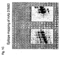
- FIG. 1 Epitope mapping of anti-NOTCH1 monoclonal antibodies that bind to non-ligand binding domains.
- Fc fusion proteins containing a deletion series of NOTCH1 EGF domains 1-5 (A, B) or 10-15 (C) were separated by SDS-PAGE and blotted with monoclonal antibody 13M57 (A, B) or 31M80 (C).
- antibody 13M57 only detected fusion proteins containing EGF repeat 4 including EGF 1-4 and EGF 1-5 but not EGF 1-3 (A); and EGF 4-5 but not EGF 5 alone (B).
- antibody 31M80 only detected fusion proteins containing EGF repeat 13 (C).
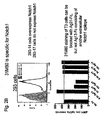
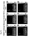
- Figure 2 Binding of anti-NOTCH1 monoclonal antibody 13M57 and 31M80 to cell surface expressed NOTCH1.
- A FACS analysis of cells co-expressing full length NOTCH1 receptor and GFP incubated with, from left to right, IgG1 control antibodies, 13M57 and anti-human NOTCH1 EGF 1-5 antisera.
- B FAC analysis of mock transfected cells or cells expressing full length NOTCH1 receptor incubated with anti-NOTCH1 receptor antibody 31M80 demonstrates that 31M80 specifically recognizes NOTCH1 receptor.
- Antibody binding relative to an IgG control antibody is inhibited by increasing amounts of antigen protein containing EGF repeats 10-16 linked to human Fc (0.5x, 3x, and 10x Ag31) but not antigen protein containing EGF repeats 1-5 linked to Fc (0.5x, 3x, and 10x Ag13).
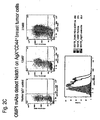
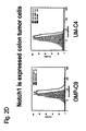
- C Anti-NOTCH1 antibodies 13M57 and 31M80 showed increased binding to PE13 breast tumor cells compared to an isotype control antibody (bottom) and this binding corresponded to cells that express high levels of ESA and CD44 (top: CD44 x-axis and NOTCH1 y-axis). Cells isolated for a tumorigenicity study are indicated in the flow cytometry results for antibody 31M80.
- Anti-NOTCH1 antibodies 13M57 and 31M80 showed increased binding to T3 breast tumor cells compared to an isotype control antibody (bottom) and this binding corresponded to cells that express high levels of ESA and CD44 (top: CD44 x-axis and NOTCH1 y-axis).
- Anti-NOTCH1 antibody 31M80 showed increased binding to dissociated colon tumor cells from two different patients compared to an isotype antibody control.
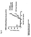
- Figure 3 Antibodies Against NOTCH1 EGF13 and EGF4 Fail to Effectively Block Ligand Binding.
- NOTCH1 expressing HEK 293 cells were incubated with either DLL4-Fc (left) or JAG1-Fc (right) in the presence of anti-NOTCH1 antibodies (13M57, 31M103, 31M106, or 31M108) or control anti-DLL4 (21M18) or anti-JAG1 (64M14) antibodies.
- Binding of Fc fusion proteins to NOTCH1-expressing cells was detected by a PE-conjugated goat anti-Fc antibody and flow cytometry. Inhibition of ligand binding by anti-NOTCH1 antibodies was expressed as a percentage of inhibition by the control ligand antibodies.
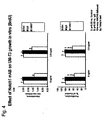
- FIG. 4 Effect of NOTCH1 Monoclonal Antibodies against EGF4 on Breast Tumor Cell Growth In Vitro.
- Breast tumor cells were cultured in the presence of 2.5 ug/mL or 5 ug/mL anti-NOTCH1 antibody, control murine IgG or no antibody for three days followed by 18 hours of BrdU labeling.
- breast tumor cells cultured in the presence of anti-NOTCH1 antibody 13M57 showed a decreased 450nm/690nm absorbance ratio compared to controls.
- Figure 5 Effect of NOTCH1 Monoclonal Antibody 13M57 on PE-13 Tumor Cells In Vivo.
- NOD/SCID mice injected with PE-13 tumor cells were treated with PBS or anti-NOTCH1 antibodies 3 days after cell injection, and the growth of tumor cells was determine twice a week. Total tumor volume was significantly reduced by 49% (p ⁇ 0.05) in animals treated with anti-NOTCH antibodies 13M57 compared to PBS injected controls.
- FIG. 6 Effect of NOTCH1 Monoclonal Antibodies on Colon Tumor Cells In Vivo.
- NOD/SCID mice injected with OMP-C9 (A) or OMP-C8 (B) colon tumor cells were treated with PBS or anti-NOTCH1 13M57 antibodies (A) or 13M57, 31M106, and 31M103 (B) three days after cell injection, and the growth of tumor cells was determine twice a week.
- Total tumor volume was significantly reduced in animals treated with anti-NOTCH antibodies compared to PBS injected controls in both tumor models.
- Antibodies against EGF4 and EGF13 were all equally effective against C8 colon tumors (B).
- Figure 7 Effect of NOTCH1 Monoclonal Antibody 13M57 on PE-13 Tumor Cells Expressing Luciferase.
- NOD/SCID mice injected with PE-13 tumor cells were treated with anti-NOTCH1 antibodies, control 5M108 control antibodies, or PBS.
- a scale of luciferase activity is provided at the right of each picture with upper dark indicating the highest activity (100 or higher X 10 6 ) and lower levels ( ⁇ 30 x 10 6 ) indicating low luciferase signal.
- Animals treated with PBS or 5M108 controls antibodies have tumors detected in the upper dark region of the scale. In contrast, tumors in animals treated with anti-Notch antibodies show luciferase activity mainly in the lower region of the scale.
- Figure 8 Effect of NOTCH2 Monoclonal Antibody 59M07 on Colon Tumor Growth.
- NOD/SCID mice injected with C6 colon tumor cells were treated with anti-NOTCH2 antibodies or PBS vehicle as a control.
- Animals treated with anti-NOTCH2 59M07 showed significant reduction in tumor growth over 48 days compared to control treated animals (diamonds).
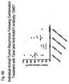
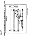
- Figure 9 Effect of anti-NOTCH1 Antibody 13M57 and Chemotherapy Combination Therapy on Breast Tumor Reoccurrence.
- A Graph of the response of UM-PE13 tumors to four different treatment regimes: Group 1: paclitaxel followed by PBS (squares); Group 2: paclitaxel followed by 13M57 (inverted triangles); Group 3 paclitaxel + 13M57 followed by PBS (circles); and Group 4: paclitaxel + 13M57 followed by 13M57 (diamonds).
- Paclitaxel (or Paclitaxel + 13M57) treatments were stopped at day 52 after the tumors had regressed and were undetectable (Arrow: Stop Paclitaxel).
- Arrow: Stop Paclitaxel Both the individual animal tumor volumes (dots) and average (lines) tumor volumes for each treatment group are graphed. Concurrent combination treatment followed by continual treatment with anti-NOTCH1 13M57 antibodies (far right) had the greatest effect on inhibiting tumor recurrence following the cessation of paclitaxel treatment.
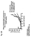
- FIG. 10 Effect of anti-NOTCH1 Antibody 13M57 and Chemotherapy Combination Therapy on Colon Tumor Growth.
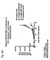
- FIG. 11 Effect of anti-NOTCH1 Antibody 13M57 and Chemotherapy Combination Therapy on Established Colon Tumor Growth.
- a graph shows C8 colon tumor volume over the course of treatment with either oxaliplatin (triangles), 10 mg/kg 13M57 (diamonds), a combination of oxaliplatin and 13M57 (circles), or a control antibody (squares).
- Figure 12 Effect of NOTCH1 and NOTCH2 Antibody Combination Therapy on Breast Tumor Growth.
- A A graph of bioluminescence imaging of animals treated either with 10 mg/kg anti-NOTCH1 31M108 (open triangles), 10 mg/kg 59M07 anti-NOTCH2 (filled triangles), a combination of anti-NOTCH1 and NOTCH2 antibodies (filled inverted triangles), or a control antibody (open circles). Animals were imaged twice weekly. Combination treatment with anti-NOTCH1 and NOTCH2 antibodies significantly reduced growth of luciferase expressing PE13 tumor cells.
- antagonist includes any molecule that partially or fully blocks, inhibits, or neutralizes a biological activity of the Notch pathway. Suitable antagonist molecules specifically include antagonist antibodies or antibody fragments, fragments or amino acid sequence variants of native Notch receptors.
- antagonist is used herein to include any molecule that partially or fully blocks, Inhibits, or neutralizes the expression of or the biological activity of a cancer stem cell marker disclosed herein and such biological activity includes, but is not limited to, inhibition of tumor growth.
- antibody is used to mean an immunoglobulin molecule that recognizes and specifically binds to a target, such as a protein, polypeptide, peptide, carbohydrate, polynucleotide, lipid, or combinations of the foregoing etc., through at least one antigen recognition site within the variable region of the immunoglobulin molecule.
- the term encompasses intact polyclonal antibodies, intact monoclonal antibodies, antibody fragments (such as Fab, Fab', F(ab')2, and Fv fragments), single chain Fv (scFv) mutants, multispecific antibodies such as bispecific antibodies generated from at least two intact antibodies, fusion proteins comprising an antibody portion, and any other modified immunoglobulin molecule comprising an antigen recognition site so long as the antibodies exhibit the desired biological activity.
- An antibody can be of any the five major classes of immunoglobulins: IgA, IgD, IgE, IgG, and IgM, or subclasses (isotypes) thereof (e.g.
- IgG1, IgG2, IgG3, IgG4, IgA1 and IgA2) based on the identity of their heavy-chain constant domains referred to as alpha, delta, epsilon, gamma, and mu, respectively.
- the different classes of immunoglobulins have different and well known subunit structures and three-dimensional configurations.
- Antibodies can be naked or conjugated to other molecules such as toxins, radioisotopes, etc.
- antibody fragment refers to a portion of an intact antibody and refers to the antigenic determining variable regions of an intact antibody.
- antibody fragments include, but are not limited to Fab, Fab', F(ab')2, and Fv fragments, linear antibodies, single chain antibodies, and multispecific antibodies formed from antibody fragments.
- an “Fv antibody” refers to the minimal antibody fragment that contains a complete antigen-recognition and -binding site either as two-chains, in which one heavy and one light chain variable domain form a non-covalent dimer, or as a single-chain (scFv), in which one heavy and one light chain variable domain are covalently linked by a flexible peptide linker so that the two chains associate in a similar dimeric structure.
- scFv single-chain
- the complementary determining regions (CDRs) of each variable domain interact to define the antigen-binding specificity of the Fv dimer.
- a single variable domain or half of an Fv can be used to recognize and bind antigen, although generally with lower affinity.
- a “monoclonal antibody” as used herein refers to homogenous antibody population involved in the highly specific recognition and binding of a single antigenic determinant, or epitope. This is in contrast to polyclonal antibodies that typically include different antibodies directed against different antigenic determinants.
- the term “monoclonal antibody” encompasses both intact and full-length monoclonal antibodies as well as antibody fragments (such as Fab, Fab', F(ab')2, Fv), single chain (scFv) mutants, fusion proteins comprising an antibody portion, and any other modified immunoglobulin molecule comprising an antigen recognition site.
- “monoclonal antibody” refers to such antibodies made in any number of manners including but not limited to by hybridoma, phage selection, recombinant expression, and transgenic animals.
- humanized antibody refers to forms of non-human (e.g. murine) antibodies that are specific immunoglobulin chains, chimeric immunoglobulins, or fragments thereof that contain minimal non-human sequences.
- humanized antibodies are human immunoglobulins in which residues from the complementary determining region (CDR) are replaced by residues from the CDR of a non-human species (e.g. mouse, rat, rabbit, hamster, etc.) that have the desired specificity, affinity, and capability.
- CDR complementary determining region
- FR Fv framework region
- the humanized antibody can be further modified by the substitution of additional residue either in the Fv framework region and/or within the replaced non-human residues to refine and optimize antibody specificity, affinity, and/or capability.
- the humanized antibody will comprise substantially all of at least one, and typically two or three, variable domains containing all or substantially all of the CDR regions that correspond to the non-human immunoglobulin whereas all or substantially all of the FR regions are those of a human immunoglobulin consensus sequence.
- the humanized antibody can also comprise at least a portion of an immunoglobulin constant region or domain (Fc), typically that of a human immunoglobulin. Examples of methods used to generate humanized antibodies are described in U.S. Pat. 5,225,539 .
- human antibody as used herein means an antibody produced by a human or an antibody having an amino acid sequence corresponding to an antibody produced by a human made using any of the techniques known in the art. This definition of a human antibody includes intact or full-length antibodies, fragments thereof, and/or antibodies comprising at least one human heavy and/or light chain polypeptide such as, for example, an antibody comprising murine light chain and human heavy chain polypeptides.
- Hybrid antibodies are immunoglobulin molecules in which pairs of heavy and light chains from antibodies with different antigenic determinant regions are assembled together so that two different epitopes or two different antigens can be recognized and bound by the resulting tetramer.
- chimeric antibodies refers to antibodies wherein the amino acid sequence of the immunoglobulin molecule is derived from two or more species.
- the variable region of both light and heavy chains corresponds to the variable region of antibodies derived from one species of mammals (e.g. mouse, rat, rabbit, etc.) with the desired specificity, affinity, and capability while the constant regions are homologous to the sequences in antibodies derived from another (usually human) to avoid eliciting an immune response in that species.
- epitopes or "antigenic determinant” are used interchangeably herein and refer to that portion of an antigen capable of being recognized and specifically bound by a particular antibody.
- the antigen is a polypeptide
- epitopes can be formed both from contiguous amino acids and noncontiguous amino acids juxtaposed by tertiary folding of a protein. Epitopes formed from contiguous amino acids are typically retained upon protein denaturing, whereas epitopes formed by tertiary folding are typically lost upon protein denaturing.
- An epitope typically includes at least 3, and more usually, at least 5 or 8-10 amino acids in a unique spatial conformation.
- Competition between antibodies is determined by an assay in which the immunoglobulin under test inhibits specific binding of a reference antibody to a common antigen.
- Numerous types of competitive binding assays are known, for example: solid phase direct or indirect radioimmunoassay (RIA), solid phase direct or indirect enzyme immunoassay (EIA), sandwich competition assay (see Stahli et al., Methods in Enzymology 9:242-253 (1983 )); solid phase direct biotin-avidin EIA (see Kirkland et al., J. Immunol.
- solid phase direct labeled assay solid phase direct labeled sandwich assay (see Harlow and Lane, "Antibodies, A Laboratory Manual,” Cold Spring Harbor Press (1988 )); solid phase direct label RIA using I-125 label (see Morel et al., Molec. Immunol. 25(1):7-15 (1988 )); solid phase direct biotin-avidin EIA ( Cheung et al., Virology 176:546-552 (1990 )); and direct labeled RIA ( Moldenhauer et al., Scand. J. Immunol. 32:77-82 (1990 )).
- such an assay involves the use of purified antigen bound to a solid surface or cells bearing either of these, an unlabeled test immunoglobulin and a labeled reference immunoglobulin.
- Competitive inhibition is measured by determining the amount of label bound to the solid surface or cells in the presence of the test immunoglobulin.
- the test immunoglobulin is present in excess.
- Antibodies identified by competition assay include antibodies binding to the same epitope as the reference antibody and antibodies binding to an adjacent epitope sufficiently proximal to the epitope bound by the reference antibody for steric hindrance to occur.
- a competing antibody is present in excess, it will inhibit specific binding of a reference antibody to a common antigen by at least 50 or 75%.
- an antibody “selectively binds” or “specifically binds” to an epitope or receptor means that the antibody reacts or associates more frequently, more rapidly, with greater duration, with greater affinity, or with some combination of the above to the epitope or receptor than with alternative substances, including unrelated proteins.
- “Selectively binds” or “specifically binds” means, for instance, that an antibody binds to a protein with a KD of at least about 0.1 mM, more usually at least about 1 uM.
- “Selectively binds” or “specifically binds” means at times that an antibody binds to a protein at times with a KD of at least about 0.1 uM or better, and at other times at least about 0.01 uM or better. Because of the sequence identity between homologous proteins in different species, specific binding can include an antibody that recognizes a cancer stem cell marker in more than one species.
- non-specific binding and “background binding” when used in reference to the interaction of an antibody and a protein or peptide refer to an interaction that is not dependent on the presence of a particular structure (i.e., the antibody is binding to proteins in general rather that a particular structure such as an epitope).
- isolated refers to material that is substantially or essentially free from components that normally accompany it in its native state. Purity and homogeneity are typically determined using analytical chemistry techniques such as polyacrylamide gel electrophoresis or high performance liquid chromatography.
- a protein e.g. an antibody
- nucleic acid that is the predominant species present in a preparation is substantially purified.
- an isolated nucleic acid is separated from open reading frames that naturally flank the gene and encode proteins other than protein encoded by the gene.
- An isolated antibody is separated from other non-immunoglobulin proteins and from other immunoglobulin proteins with different antigen binding specificity. It can also mean that the nucleic acid or protein is at least 85% pure, at least 95% pure, and in some embodiments, at least 99% pure.
- cancer and “cancerous” refer to or describe the physiological condition in mammals in which a population of cells are characterized by unregulated cell growth.
- examples of cancer include, but are not limited to, carcinoma, lymphoma, blastoma, sarcoma, and leukemia.
- cancers include squamous cell cancer, small-cell lung cancer, non-small cell lung cancer, adenocarcinoma of the lung, squamous carcinoma of the lung, cancer of the peritoneum, hepatocellular cancer, gastrointestinal cancer, pancreatic cancer, glioblastoma, cervical cancer, ovarian cancer, liver cancer, bladder cancer, hepatoma, breast cancer, colon cancer, colorectal cancer, endometrial or uterine carcinoma, salivary gland carcinoma, kidney cancer, liver cancer, prostate cancer, vulval cancer, thyroid cancer, hepatic carcinoma and various types of head and neck cancer.
- proliferative disorder and “proliferative disease” refer to disorders associated with abnormal cell proliferation such as cancer.
- Tumor and neoplasm refer to any mass of tissue that result from excessive cell growth or proliferation, either benign (noncancerous) or malignant (cancerous) including pre-cancerous lesions.
- Metalastasis refers to the process by which a cancer spreads or transfers from the site of origin to other regions of the body with the development of a similar cancerous lesion at the new location.
- a “metastatic” or “metastasizing” cell is one that loses adhesive contacts with neighboring cells and migrates via the bloodstream or lymph from the primary site of disease to invade neighboring body structures.
- the term “subject” refers to any animal (e.g., a mammal), including, but not limited to humans, non-human primates, rodents, and the like, which is to be the recipient of a particular treatment.
- the terms “subject” and “patient” are used interchangeably herein in reference to a human subject.
- cancer stem cell refers to a population of cells from a solid tumor that: (1) have extensive proliferative capacity; 2) are capable of asymmetric cell division to generate one or more kinds of differentiated progeny with reduced proliferative or developmental potential; and (3) are capable of symmetric cell divisions for self-renewal or self-maintenance.
- Cancer stem cells undergo self-renewal versus differentiation in a chaotic manner to form tumors with abnormal cell types that can change over time as mutations occur.
- the solid tumor stem cells of the present invention differ from the "cancer stem line" provided by U.S. Pat. No. 6,004,528 .
- the "cancer stem line” is defined as a slow growing progenitor cell type that itself has few mutations but which undergoes symmetric rather than asymmetric cell divisions as a result of tumorigenic changes that occur in the cell's environment
- This "cancer stem line” hypothesis thus proposes that highly mutated, rapidly proliferating tumor cells arise largely as a result of an abnormal environment, which causes relatively normal stem cells to accumulate and then undergo mutations that cause them to become tumor cells.
- Pat. No. 6,004,528 proposes that such a model can be used to enhance the diagnosis of cancer.
- the solid tumor stem cell model is fundamentally different than the "cancer stem line” model and as a result exhibits utilities not offered by the "cancer stem line” model.
- solid tumor stem cells are not "mutationally spared".
- the "mutationally spared cancer stem line" described by U.S. Pat. No. 6,004,528 can be considered a pre-cancerous lesion, while the solid tumor stem cells described by this invention are cancer cells that themselves contain the mutations that are responsible for tumorigenesis. That is, the solid tumor stem cells (“cancer stem cells”) of the invention would be included among the highly mutated cells that are distinguished from the "cancer stem line" in U.S. Pat. No.
- the genetic mutations that lead to cancer can be largely intrinsic within the solid tumor stem cells as well as being environmental.
- the solid tumor stem cell model predicts that isolated solid tumor stem cells can give rise to additional tumors upon transplantation (thus explaining metastasis) while the "cancer stem line” model would predict that transplanted "cancer stem line” cells would not be able to give rise to a new tumor, since it was their abnormal environment that was tumorigenic. Indeed, the ability to transplant dissociated, and phenotypically isolated human solid tumor stem cells to mice (into an environment that is very different from the normal tumor environment), where they still form new tumors, distinguishes the present invention from the "cancer stem line” model.
- solid tumor stem cells likely divide both symmetrically and asymmetrically, such that symmetric cell division is not an obligate property.
- solid tumor stem cells can divide rapidly or slowly, depending on many variables, such that a slow proliferation rate is not a defining characteristic.
- cancer cell refers to the total population of cells derived from a tumor including both non-tumorigenic cells, which comprise the bulk of the tumor cell population, and tumorigenic stem cells (cancer stem cells).
- tumorigenic refers to the functional features of a solid tumor stem cell including the properties of self-renewal (giving rise to additional tumorigenic cancer stem cells) and proliferation to generate all other tumor cells (giving rise to differentiated and thus non-tumorigenic tumor cells) that allow solid tumor stem cells to form a tumor.
- stem cell cancer marker(s) refers to a gene or genes or a protein, polypeptide, or peptide expressed by the gene or genes whose expression level, alone or in combination with other genes, is correlated with the presence of tumorigenic cancer cells compared to non-tumorigenic cells.
- the correlation can relate to either an increased or decreased expression of the gene (e.g. increased or decreased levels of mRNA or the peptide encoded by the gene).
- cancer stem cell gene signature refers to gene signatures comprising genes differentially expressed in cancer stem cells compared to other cells or population of cells, for example normal breast epithelial tissue.
- the cancer stem cell gene signatures comprise genes differentially expressed in cancer stem cells versus normal breast epithelium by a fold change, for example by 2 fold reduced and/or elevated expression, and further limited by using a statistical analysis such as, for example, by the P value of a t-test across multiple samples.
- the genes differentially expressed in cancer stem cells are divided into cancer stem cell gene signatures based on the correlation of their expression with a chosen gene in combination with their fold or percentage expression change. Cancer stem cell signatures are predictive both retrospectively and prospectively of an aspect of clinical variability, including but not limited to metastasis and death.
- genetic test refers to procedures whereby the genetic make-up of a patient or a patient tumor sample is analyzed.
- the analysis can include detection of DNA, RNA, chromosomes, proteins or metabolites to detect heritable or somatic disease-related genotypes or karyotypes for clinical purposes.
- biopsy tissue refers to a sample of tissue or fluid that is removed from a subject for the purpose of determining if the sample contains cancerous tissue.
- biopsy tissue or fluid is obtained because a subject is suspected of having cancer. The biopsy tissue or fluid is then examined for the presence or absence of cancer.
- an "acceptable pharmaceutical carrier” refers to any material that, when combined with an active ingredient of a pharmaceutical composition such as an antibody, allows the antibody, for example, to retain its biological activity.
- an "acceptable pharmaceutical carrier” does not trigger an immune response in a recipient subject. Examples include, but are not limited to, any of the standard pharmaceutical carriers such as a phosphate buffered saline solution, water, and various oil/water emulsions. Some diluents for aerosol or parenteral administration are phosphate buffered saline or normal (0.9%) saline.
- therapeutically effective amount refers to an amount of an antibody, polypeptide, polynucleotide, small organic molecule, or other drug effective to "treat” a disease or disorder in a subject or mammal.
- the therapeutically effective amount of the drug can reduce the number of cancer cells; reduce the tumor size; inhibit or stop cancer cell infiltration into peripheral organs; inhibit and stop tumor metastasis; inhibit and stop tumor growth; relieve to some extent one or more of the symptoms associated with the cancer, or a combination of such effects on cancer cells.
- the drug prevents growth and/or kills existing cancer cells, it can be referred to as cytostatic and/or cytotoxic.
- diagnosis refers to any information that is useful in determining whether a patient has a disease or condition and/or in classifying the disease or condition into a phenotypic category or any category having significance with regards to the prognosis of or likely response to treatment (either treatment in general or any particular treatment) of the disease or condition.
- diagnosis refers to providing any type of diagnostic information, including, but not limited to, whether a subject is likely to have a condition (such as a tumor), information related to the nature or classification of a tumor as for example a high risk tumor or a low risk tumor, information related to prognosis and/or information useful in selecting an appropriate treatment. Selection of treatment can include the choice of a particular chemotherapeutic agent or other treatment modality such as surgery or radiation or a choice about whether to withhold or deliver therapy.
- the terms “providing a prognosis”, “prognostic information”, or “predictive information” refer to providing information regarding the impact of the presence of cancer (e.g., as determined by the diagnostic methods of the present invention) on a subject's future health (e.g., expected morbidity or mortality, the likelihood of getting cancer, and the risk of metastasis).
- Terms such as “treating” or “treatment” or “to treat” or “alleviating” or “to alleviate” refer to both 1) therapeutic measures that cure, slow down, lessen symptoms of, and/or halt progression of a diagnosed pathologic condition or disorder and 2) prophylactic or preventative measures that prevent or slow the development of a targeted pathologic condition or disorder.
- those in need of treatment include those already with the disorder; those prone to have the disorder; and those in whom the disorder is to be prevented.
- a subject is successfully "treated” according to the methods of the present invention if the patient shows one or more of the following: a reduction in the number of or complete absence of cancer cells; a reduction in the tumor size; inhibition of or an absence of cancer cell infiltration into peripheral organs including the spread of cancer into soft tissue and bone; inhibition of or an absence of tumor metastasis; inhibition or an absence of tumor growth; relief of one or more symptoms associated with the specific cancer; reduced morbidity and mortality; and improvement in quality of life.
- polynucleotide or “nucleic acid” refer to a polymer composed of a multiplicity of nucleotide units (ribonucleotide or deoxyribonucleotide or related structural variants) linked via phosphodiester bonds, including but not limited to, DNA or RNA.
- the term encompasses sequences that include any of the known base analogs of DNA and RNA including, but not limited to, 4-acetylcytosine, 8-hydroxy-N6-methyladenosine, aziridinylcytosine, pseudoisocytosine, 5-(carboxyhydroxylmethyl) uracil, 5-fluorouracil, 5-bromouracil, 5-carboxymethylaminomethyl 2-thiouracil, 5-carboxymethylaminomethyluracil, dihydrouracil, inosine, N6-isopentenyladenine, 1-methyladenine, 1-methylpseudouracil, 1-methylguanine, 1-methylinosine, 2,2-dimethylguanine, 2-methyladenine, 2-methylguanine, 3-methylcytosine, 5-methylcytosine, N6-methyladenine, 7-methylguanine, 5-methylaminomethyluracil, 5-methoxyaminomethyl 2-thiouracil, beta-D-mannosylque
- gene refers to a nucleic acid (e.g., DNA) sequence that comprises coding sequences necessary for the production of a polypeptide, precursor, or RNA (e.g., rRNA, tRNA).
- the polypeptide can be encoded by a full length coding sequence or by any portion of the coding sequence so long as the desired activity or functional properties (e.g., enzymatic activity, ligand binding, signal transduction, immunogenicity, etc.) of the full-length or fragment are retained.
- the term also encompasses the coding region of a structural gene and the sequences located adjacent to the coding region on both the 5' and 3' ends for a distance of about 1 kb or more on either end such that the gene corresponds to the length of the full-length mRNA. Sequences located 5' of the coding region and present on the mRNA are referred to as 5' non-translated sequences. Sequences located 3' or downstream of the coding region and present on the mRNA are referred to as 3' non-translated sequences.
- the term "gene” encompasses both cDNA and genomic forms of a gene.
- a genomic form or clone of a gene contains the coding region interrupted with non-coding sequences termed "introns” or “intervening regions” or “intervening sequences.”
- Introns are segments of a gene that are transcribed into nuclear RNA (mRNA); introns can contain regulatory elements such as enhancers. Introns are removed or “spliced out” from the nuclear or primary transcript; introns therefore are absent in the messenger RNA (mRNA) transcript.
- the mRNA functions during translation to specify the sequence or order of amino acids in a nascent polypeptide.
- genomic forms of a gene can also include sequences located on both the 5' and 3' end of the sequences that are present on the RNA transcript.
- flanking sequences or regions are referred to as “flanking" sequences or regions (these flanking sequences are located 5' or 3' to the non-translated sequences present on the mRNA transcript).
- the 5' flanking region can contain regulatory sequences such as promoters and enhancers that control or influence the transcription of the gene.
- the 3' flanking region can contain sequences that direct the termination of transcription, post transcriptional cleavage and polyadenylation.
- recombinant when used with reference to a cell, nucleic acid, protein or vector indicates that the cell, nucleic acid, protein or vector has been modified by the introduction of a heterologous nucleic acid or protein, the alteration of a native nucleic acid or protein, or that the cell is derived from a cell so modified.
- recombinant cells express genes that are not found within the native (non-recombinant) form of the cell or express native genes that are overexpressed or otherwise abnormally expressed such as, for example, expressed as non-naturally occurring fragments or splice variants.
- nucleic acid By the term “recombinant nucleic acid” herein is meant nucleic acid, originally formed in vitro, in general, by the manipulation of nucleic acid, e.g., using polymerases and endonucleases, in a form not normally found in nature. In this manner, operably linkage of different sequences is achieved.
- an isolated nucleic acid, in a linear form, or an expression vector formed in vitro by ligating DNA molecules that are not normally joined are both considered recombinant for the purposes of this invention.
- a Recombinant nucleic acid is made and introduced into a host cell or organism, it will replicate non-recombinantly, i.e., using the in vivo cellular machinery of the host cell rather than in vitro manipulations; however, such nucleic acids, once produced recombinantly, although subsequently replicated non-recombinantly, are still considered recombinant for the purposes of the invention.
- a "recombinant protein” is a protein made using recombinant techniques, i.e., through the expression of a recombinant nucleic acid as depicted above.
- heterologous gene refers to a gene that is not in its natural environment.
- a heterologous gene includes a gene from one species introduced into another species.
- a heterologous gene also includes a gene native to an organism that has been altered in some way (e.g., mutated, added in multiple copies, linked to non-native regulatory sequences, etc).
- Heterologous genes are distinguished from endogenous genes in that the heterologous gene sequences are typically joined to DNA sequences that are not found naturally associated with the gene sequences in the chromosome or are associated with portions of the chromosome not found in nature (e.g., genes expressed in loci where the gene is not normally expressed).
- vector is used in reference to nucleic acid molecules that transfer DNA segment(s) from one cell to another.
- vehicle is sometimes used interchangeably with “vector.”
- Vectors are often derived from plasmids, bacteriophages, or plant or animal viruses.
- Ligation refers to the process of forming phosphodiester bonds between two double stranded nucleic acid fragments. Unless otherwise provided, ligation can be accomplished using known buffers and conditions with 10 units to T4 DNA ligase ("ligase") per 0.5 ug of approximately equimolar amounts of the DNA fragments to be ligated. Ligation of nucleic acid can serve to link two proteins together in-frame to produce a single protein, or fusion protein.
- ligase T4 DNA ligase
- RNA expression refers to the process of converting genetic information encoded in a gene into RNA (e.g., mRNA, rRNA, tRNA, or snRNA) through "transcription" of the gene (e.g., via the enzymatic action of an RNA polymerase), and for protein encoding genes, into protein through “translation” of mRNA.
- RNA e.g., mRNA, rRNA, tRNA, or snRNA
- Gene expression can be regulated at many stages in the process.
- Up-regulation or “activation” refers to regulation that increases the production of gene expression products (e.g., RNA or protein), while “down-regulation” or “repression” refers to regulation that decrease production Molecules (e.g., transcription factors) that are involved in up-regulation or down-regulation are often called “activators” and “repressors,” respectively.
- polypeptide peptide
- protein protein fragment
- amino acid polymers in which one or more amino acid residue is an artificial chemical mimetic of a corresponding naturally occurring amino acid, as well as to naturally occurring amino acid polymers and non-naturally occurring amino acid polymers.
- amino acid refers to naturally occurring and synthetic amino acids, as well as amino acid analogs and amino acid mimetics that function similarly to the naturally occurring amino acids.
- Naturally occurring amino acids are those encoded by the genetic code, as well as those amino acids that are later modified, e.g., hydroxyproline, gamma-carboxyglutamate, and O-phosphoserine.
- Amino acid analogs refers to compounds that have the same basic chemical structure as a naturally occurring amino acid, e.g., an alpha carbon that is bound to a hydrogen, a carboxyl group, an amino group, and an R group, e.g., homoserine, norleucine, methionine sulfoxide, methionine methyl sulfonium. Such analogs can have modified R groups (e.g., norleucine) or modified peptide backbones, but retain the same basic chemical structure as a naturally occurring amino acid.
- Amino acid mimetics refers to chemical compounds that have a structure that is different from the general chemical structure of an amino acid, but that functions similarly to a naturally occurring amino acid.
- amino acid variants refers to amino acid sequences. With respect to particular nucleic acid sequences, conservatively modified variants refers to those nucleic acids which encode identical or essentially identical amino acid sequences, or where the nucleic acid does not encode an amino acid sequence, to essentially identical or associated (e.g., naturally contiguous) sequences. Because of the degeneracy of the genetic code, a large number of functionally identical nucleic acids encode most proteins. For instance, the codons GCA, GCC, GCG and GCU all encode the amino acid alanine.
- nucleic acid variations are "silent variations," which are one species of conservatively modified variations. Every nucleic acid sequence herein which encodes a polypeptide also describes silent variations of the nucleic acid. It is recognized that in certain contexts each codon in a nucleic acid (except AUG, which is ordinarily the only codon for methionine, and TGG, which is ordinarily the only codon for tryptophan) can be modified to yield a functionally identical molecule.
- amino acid sequences it will be recognized that individual substitutions, deletions or additions to a nucleic acid, peptide, polypeptide, or protein sequence which alters, adds or deletes a single amino acid or a small percentage of amino acids in the encoded sequence is a "conservatively modified variant" including where the alteration results in the substitution of an amino acid with a chemically similar amino acid. Conservative substitution tables providing functionally similar amino acids are well known in the art.
- conservatively modified variants are in addition to and do not exclude polymorphic variants, interspecies homologs, and alleles of the invention.
- conservative substitutions include: 1) Alanine (A), Glycine (G); 2) Aspartic acid (D), Glutamic acid (E); 3) Asparagine (N), Glutamine (Q); 4) Arginine (R), Lysine (K); 5) Isoleucine (I), Leucine (L), Methionine (M), Valine (V); 6) Phenylalanine (F), Tyrosine (Y), Tryptophan (W); 7) Serine (S), Threonine (T); and 8) Cysteine (C), Methionine (M) (see, e.g., Creighton, Proteins (1984 )).
- epitope tagged refers to a chimeric polypeptide comprising a cancer stem cell marker protein, or a domain sequence or portion thereof, fused to an "epitope tag".
- the epitope tag polypeptide comprises enough amino acid residues to provide an epitope for recognition by an antibody, yet is short enough such that it does not interfere with the activity of the cancer stem cell marker protein.
- Suitable epitope tags generally have at least six amino acid residues, usually between about 8 to about 50 amino acid residues, and at times between about 10 to about 20 residues. Commonly used epitope tags include Fc, HA, His, and FLAG tags.
- the present invention provides compositions for studying, diagnosing, characterizing, and treating cancer.
- the present invention identifies antibodies that specifically bind to a non-ligand binding region of the extracellular domain of a human NOTCH receptor and inhibit tumor growth in vivo.
- the ligand binding region of Notch which is necessary and sufficient for ligand binding, has been identified as EGF repeats 11 and 12, suggesting this region of the Notch receptor is important in Notch signaling and tumorigenesis ( Rebay et al., 1991, Cell 67:687 ; Lei et al., 2003, Dev. 130:6411 ; Hambleton et al., 2004, Structure 12:2173 ).
- the present invention provides an antibody that specifically binds to a non-ligand binding region of the extracellular domain of a human NOTCH receptor and inhibits growth of tumor cells as defined in claim 1. In certain embodiments, the antibody binds to a non-ligand binding region of the extracellular domain of NOTCH1 receptor. In certain embodiments, the antibody that specifically binds to a non-ligand binding region of the extracellular domain of a human NOTCH receptor and inhibits growth of tumor cells specifically binds to a non-ligand binding region of the extracellular domain of at least two Notch receptor family members.
- the antibody that specifically binds to a non-ligand binding region of the extracellular domain of a human NOTCH receptor and inhibits growth of tumor cells is a monoclonal antibody. In certain embodiments, the antibody that specifically binds to a non-ligand binding region of the extracellular domain of a human NOTCH receptor and inhibits growth of tumor cells is a chimeric antibody. In certain embodiments, the antibody that specifically binds to a non-ligand binding region of the extracellular domain of a human NOTCH receptor and inhibits growth of tumor cells is a humanized antibody. In certain embodiments, the antibody that specifically binds to a non-ligand binding region of the extracellular domain of a human NOTCH receptor and inhibits growth of tumor cells is a human antibody. In certain embodiments, the present invention provides a hybridoma producing the antibody, as set out in appended claim 10.
- the present invention provides an antibody that specifically binds to a non-ligand binding region comprising EGF repeats 1-10 of the extracellular domain of a human NOTCH receptor and inhibits growth of tumor cells expressing the Notch receptor. Certain embodiments provide an antibody that specifically binds to a non-ligand binding region comprising EGF repeats 4 of the extracellular domain of a human NOTCH receptor and inhibits growth of tumor cells.
- the antibody of the present invention comprises: (a) a heavy chain variable region having CDR sequences set forth in SEQ ID NOS: 12, 13, and 14; and (b) a light chain variable region having CDR sequences set forth in SEQ ID NOS: 15, 16, and 17.
- the antibody may comprise: (a) heavy chains set forth in SEQ ID NOS: 4 and 5; and (b) light chains set forth in SEQ ID NOS: 6 and 7.
- the present invention provides an antibody for use in a method of treating cancer, as defined in claim 10.
- the method of treating cancer in a subject in need thereof comprises administering a therapeutically effective amount of an antibody to the subject that specifically binds to a non-ligand binding region of the extracellular domain of NOTCH1 receptor and inhibits growth of tumor cells.
- the method of treating cancer comprises administering a therapeutically effective amount of an antibody that specifically binds to at least two Notch receptor family members and inhibits growth of tumor cells.
- the method of treating cancer comprises administering a therapeutically effective amount of a monoclonal antibody that specifically binds to a non-ligand binding region of the extracellular domain of a human NOTCH receptor and inhibits growth of tumor cells.
- the method of treating cancer comprises administering a therapeutically effective amount of a chimeric antibody that specifically binds to a non-ligand binding region of the extracellular domain of a human NOTCH receptor and inhibits growth of tumor cells.
- the method of treating cancer comprises administering a therapeutically effective amount of a humanized antibody that specifically binds to a non-ligand binding region of the extracellular domain of a human NOTCH receptor and inhibits growth of tumor cells.
- the method of treating cancer comprises administering a therapeutically effective amount of a human antibody that specifically binds to a non-ligand binding region of the extracellular domain of a human NOTCH receptor and inhibits growth of tumor cells.
- the method of treating cancer comprises administering a therapeutically effective amount of an antibody that specifically binds to a non-ligand binding region of the extracellular domain of a human NOTCH receptor comprising EGF repeats 4 and inhibits growth of tumor cells. In certain embodiments, the method of treating cancer comprises administering a therapeutically effective amount of an antibody that specifically binds to a non-ligand binding region of the extracellular domain of a human NOTCH receptor comprising EGF repeats 4 and inhibits growth of tumor cells.
- the method of treating cancer comprises administering a therapeutically effective amount of an isolated polypeptide that specifically binds to a non-ligand binding region of the extracellular domain of a human NOTCH receptor comprising: (a) a heavy chain variable region having CDR sequences set forth in SEQ ID NOS: 12, 13, and 14; and (b) a light chain variable region having CDR sequences set forth in SEQ ID NOS: 15, 16, and 17 and inhibits growth of tumor cells.
- the method of treating cancer comprises administering a therapeutically effective amount of an antibody that specifically binds to a non-ligand binding region of the extracellular domain of a human NOTCH receptor comprising (a) heavy chains set forth in SEQ ID NOS: 4 and 5; and (b) light chains set forth in SEQ ID NOS: 6 and 7.
- the method of treating cancer comprises administering a therapeutically effective amount of an antibody conjugated to a cytotoxic moiety that specifically binds to a non-ligand binding region of the extracellular domain of a human NOTCH receptor and inhibits growth of tumor cells. In certain embodiments, the method of treating cancer comprises administering a therapeutically effective amount of an antibody that specifically binds to a non-ligand binding region of the extracellular domain of a human NOTCH receptor and inhibits growth of tumor cells in combination with radiation therapy.
- the method of treating cancer comprises administering a therapeutically effective amount of an antibody that specifically binds to a non-ligand binding region of the extracellular domain of a human NOTCH receptor and inhibits growth of tumor cells in combination with chemotherapy, In certain embodiments, the method of treating cancer comprises administering a therapeutically effective amount of an antibody that specifically binds to a non-ligand binding region of the extracellular domain of a human NOTCH receptor and inhibits growth of tumor cells that are from a breast tumor, colorectal tumor, lung tumor, pancreatic tumor, prostate tumor, or a head and neck tumor.
- the method of treating cancer comprises identifying patients using a genetic test for treatment with the antibody that specifically binds to a non-ligand binding region of the extracellular domain of a human NOTCH receptor; and administering a therapeutically effective amount of an antibody that specifically binds to a non-ligand binding region of the extracellular domain of a human NOTCH receptor and inhibits growth of tumor cells.
- the method of treating cancer comprises identify patients for treatment with the antibody that specifically binds to a non-ligand binding region of the extracellular domain of a human NOTCH receptor using a genetic test that detects a cancer stem cell signature; and administering a therapeutically effective amount of an antibody that specifically binds to a non-ligand binding region of the extracellular domain of a human. NOTCH receptor and inhibits growth of tumor cells.
- Also described herein is a method of identifying a molecule that binds to a non-ligand binding region of an extracellular domain of a human NOTCH receptor and inhibits growth of tumor cells, the method comprising: i) incubating the molecule with the non-ligand binding domain of the extracellular domain of a human Notch receptor; ii) determining if the molecule binds to the non-ligand. binding region of the extracellular domain of the human Notch receptor; and iii) determining if the molecule inhibits growth of tumor cells.
- a method of identifying a molecule that binds to a non-ligand binding region of an extracellular domain of a human NOTCH receptor and inhibits growth of tumor cells may comprise i) incubating the molecule with the non-ligand binding domain of the extracellular domain of a human Notch receptor comprising EGF repeats 1-10; ii) determining if the molecule binds to the non-ligand binding region of the extracellular domain of the human Notch receptor comprising EGF repeats 1-10; and iii) determining if the molecule inhibits growth of tumor cells.
- a method of identifying a molecule that binds to a non-ligand binding region of an extracellular domain of a human NOTCH receptor and inhibits growth of tumor cells may comprise: i) incubating the molecule with the non-ligand binding domain of the extracellular domain of a human Notch receptor comprising EGF repeats 13-36; ii) determining if the molecule binds to the non-ligand binding region of the extracellular domain of the human Notch receptor comprising EGF repeats 13-36; and iii) determining if the molecule inhibits growth of tumor cells.
- the present invention provides a pharmaceutical composition comprising an antibody as set out in appended claim 20.
- HSCs hematopoietic stem cells
- HSCs have been demonstrated in cancer therapy with their extensive use for bone marrow transplantation to regenerate the hematolymphoid system following myeloablative protocols ( Baum et al., Bone Marrow Transplantation, Blackwell Scientific Publications, Boston, 1994 ). Understanding the cellular biology of the tissues in which cancers arise, and specifically of the stem cells residing in those tissues, promises to provide new insights into cancer biology.
- solid tumors consist of a heterogeneous population of cells. That the majority of these cells lack tumorigenicity suggested that the development and maintenance of solid tumors also relies on a small population of stem cells (i.e., tumorigenic cancer cells) with the capacity to proliferate and efficiently give rise both to additional tumor stem cells (self-renewal) and to the majority of more differentiated tumor cells that lack tumorigenic potential (i.e., non-tumorigenic cancer cells).
- stem cells i.e., tumorigenic cancer cells
- self-renewal additional tumor stem cells
- non-tumorigenic cancer cells i.e., non-tumorigenic cancer cells
- Stem cells from solid tumors have more recently been isolated based on their expression of a unique pattern of cell-surface receptors and on the assessment of their properties of self-renewal and proliferation in culture and in xenograft animal models.
- An ESA+ CD44+ CD24-/low Lineage- population greater than 50-fold enriched for the ability to form tumors relative to unfractionated tumor cells was discovered ( Al-Hajj et al., 2003, PNAS 100:3983-8 ).
- the ability to isolate tumorigenic cancer stem cells from the bulk of non-tumorigenic tumor cells has led to the identification of cancer stem cell markers, genes with differential expression in cancer stem cells compared to non-tumorigenic tumor cells or normal breast epithelium, using microarray analysis.
- the present invention employs the knowledge of these identified cancer stem cell markers to study, characterize, diagnosis and treat cancer.
- the present invention identifies Notch receptor, for example, Notch1 as a marker of cancer stem cells, implicating the Notch signaling pathway in the maintenance of cancer stem cells and as a target for treating cancer via the elimination of these tumorigenic cells.
- Notch signaling pathway is one of several critical regulators of embryonic pattern formation, post-embryonic tissue maintenance, and stem cell biology. More specifically, Notch signaling is involved in the process of lateral inhibition between adjacent cell fates and plays an important role in cell fate determination during asymmetric cell divisions. Unregulated Notch signaling is associated with numerous human cancers where it can alter the developmental fate of tumor cells to maintain them in an undifferentiated and proliferative state ( Brennan and Brown, 2003, Breast Cancer Res. 5:69 ). Thus carcinogenesis can proceed by usurping homeostatic mechanisms controlling normal development and tissue repair by stem cell populations ( Beachy et al., 2004, Nature 432:324 ).
- the Notch receptor was first identified in Drosophila mutants with haploinsufficiency resulting in notches at the wing margin whereas loss-of function producing an embryonic lethal "neurogenic" phenotype where cells of the epidermis switch fate to neural tissue ( Moohr, 1919, Genet. 4:252 ; Poulson, 1937, PNAS 23:133 ; Poulson, 1940, J. Exp. Zool. 83:271 ).
- the Notch receptor is a single-pass transmembrane receptor containing numerous tandem epidermal growth factor (EGF)-like repeats and cysteine-rich Notch/LIN-12 repeats within a large extracellular domain ( Wharton et al., 1985, Cell 43:567 ; Kidd et al., 1986, Mol. Cell Biol. 3:194 ; reviewed in Artavanis et al., 1999, Science 284:770 ).
- EGF epidermal growth factor
- NOTCH1, NOTCH2, NOTCH3, and NOTCH4 Four mammalian Notch proteins have been identified (NOTCH1, NOTCH2, NOTCH3, and NOTCH4), and mutations in these receptors invariably result in developmental abnormalities and human pathologies including several cancers as described in detail below ( Gridley, 1997, Mol. Cell Neurosci. 9:103 ; Joutel & Tournier-Lasserve, 1998, Semin. Cell Dev. Biol. 9:619-25 ).
- the Notch receptor is activated by single-pass transmembrane ligands of the Delta, Serrated, Lag-2 (DSL) family.
- DSL Delta-like 1
- Dll3 Delta-like 3
- Dll4 Delta-like 4
- Jagged 1 Jagged 2 characterized by a DSL domain and tandem EGF-like repeats within the extracellular domain.
- the extracellular domain of the Notch receptor interacts with that of its ligands, typically on adjacent cells, resulting in two proteolytic cleavages of Notch, one extracellular mediated by an ADAM protease and one within the transmembrane domain mediated by gamma secretase.
- NBD Notch intracellular domain
- Suppressor of Hairless [Su(H)] Lag-2 (CSL) family of transcription factors as the major downstream effectors to increase transcription of nuclear basic helix-loop-helix transcription factors of the Hairy and Enhancer of Split [E(sp1)] family
- CBF1 Suppressor of Hairless [Su(H)]
- CSL Lag-2
- E(sp1) Enhancer of Split
- HSCs Hematopoietic stem cells
- Notch signaling is implicated both in their normal maintenance as well as in leukemic transformation ( Kopper & Hajdu, 2004, Pathol. Oncol. Res. 10:69-73 ).
- HSCs are a rare population of cells that reside in a stromal niche within the adult bone marrow. These cells are characterized both by a unique gene expression profile as well as an ability to continuously give rise to more differentiated progenitor cells to reconstitute the entire hematopoietic system.
- Notch1 signaling in HSCs and progenitor cells establishes immortalized cell lines that generate both lymphoid and myeloid cells in vitro and in long-term reconstitution assays ( Varnum-Finney et al., 2000, Nat. Med. 6:1278-81 ), and the presence of Jagged 1 increases engraftment of human bone marrow cell populations enriched for HSCs ( Karanu et al., 2000, J. Exp. Med. 192:1365-72 ). More recently, Notch signaling has been demonstrate in HSCs in vivo and shown to be involved in inhibiting HSC differentiation. Furthermore, Notch signaling appears to be required for Wnt-mediated HSC self-renewal ( Duncan et al., 2005, Nat. Immunol. 6:314 ).
- the Notch signaling pathway also plays a central role in the maintenance of neural stem cells is implicated both in their normal maintenance as well as in brain cancers ( Kopper & Hajdu, 2004, Pathol. Oncol. Res. 10:69-73 ; Purow et al., 2005, Cancer Res. 65:2353-63 ; Hallahan et al., 2004, Cancer Res. 64:7794-800 ).
- Neural stem cells give rise to all neuronal and glial cells in the mammalian nervous system during development, and more recently have been identified in the adult brain ( Gage, 2000, Science 287:1433-8 ).
- mice deficient for Notch1; the Notch target genes Hes1, 3, and 5; and a regulator of Notch signaling presenilin1 (PS1) show decreased numbers of embryonic neural stem cells. Furthermore, adult neural stem cells are reduced in the brains of PS1 heterozygote mice ( Nakamura et al., 2000, J. Neurosci. 20:283-93 ; Hitoshi et al., 2002, Genes Dev. 16:846-58 ). The reduction in neural stem cells appears to result from their premature differentiation into neurons ( Hatakeyama et al., 2004, Dev. 131:5539-50 ) suggesting that Notch signaling regulates neural stem cell differentiation and self-renewal.
- NOTCH1 gene in humans was first identified in a subset of T-cell acute lymphoblastic leukemias as a translocated locus resulting in activation of the Notch pathway ( Ellisen et al., 1991, Cell 66:649-61 ). Constitutive activation of Notch1 signaling in T-cells in mouse models similarly generates T-cell lymphomas suggesting a causative role ( Robey et al., 1996, Cell 87:483-92 ; Pear et al., 1996, J. Exp. Med.
- NOTCH1 point mutations, insertions, and deletions producing aberrant NOTCH1 signaling have been found to be frequently present in both childhood and adult T-cell acute lymphoblastic leukemia/lymphoma ( Pear & Aster, 2004, Curr. Opin. Hematol. 11:416-33 ).
- the Notch pathway is also involved in multiple aspects of vascular development including proliferation, migration, smooth muscle differentiation, angiogenesis and arterial-venous differentiation (Iso et al., 2003, Arterioscler. Thromb. Vasc. Biol. 23:543).
- homozygous null mutations in Notch-1/4 and Jagged-1 as well as heterozygous loss of Dll4 result in severe though variable defects in arterial development and yolk sac vascularization.
- Dll1-deficient and Notch-2-hypomorphic mice embryos show hemorrhage that likely results from poor development of vascular structures ( Gale et al., 2004, PNAS, 101:15949-54 ; Krebs et al., 2000, Genes Dev.
- NOTCH1, NOTCH4, DLL1 and DLL4 are genes expressed in cancer stem cells compared to normal breast epithelium.
- targeting the Notch pathway can help eliminate not only the majority of nontumorigenic cancer cells, but the tumorigenic cells responsible for the formation and reoccurrence of solid tumors.
- targeting the Notch pathway can also effectively inhibit angiogenesis, starving a cancer of nutrients and contributing to its elimination.
- a cancer stem cell marker the expression of which can be analyzed to detect, characterize, diagnosis or monitor a disease associated with expression of a cancer stem cell marker.
- Expression of a cancer stem cell marker can be determined by polynucleotide expression such as, for example, mRNA encoding the cancer stem cell marker.
- the polynucleotide can be detected and quantified by any of a number of means well known in the art.
- mRNA encoding a cancer stem cell marker can be detected by in situ hybridization of tissue sections from, from example, a patient biopsy.
- RNA can be isolated from a tissue and detected by, for example, Northern blot, quantitative RT-PCR or microarrays. For example, total RNA can be extracted from a tissue sample and primers that specifically hybridize and amplify a cancer stem cell marker can be used to detect expression of a cancer stem cell marker polynucleotide using RT-PCR.
- a cancer stem cell marker can be determined by detection of the corresponding polypeptide.
- the polypeptide can be detected and quantified by any of a number of means well known in the art.
- a cancer stem cell marker polypeptide can be detected using analytic biochemical methods such as, for example, electrophoresis, capillary electrophoresis, high performance liquid chromatography (HPLC) or thin layer chromatography (TLC).
- HPLC high performance liquid chromatography
- TLC thin layer chromatography
- the isolated polypeptide can also be sequenced according to standard techniques.
- a cancer stem cell marker protein can be detected with antibodies raised against the protein using, for example, immunofluorescence or immunohistochemistry on tissue sections.
- antibodies against a cancer stem cell marker can detect expression using, for example, ELISA, FACS, Western blot, immunoprecipitation or protein microarrays.
- cancer stem cells can be isolated from a patient biopsy and expression of a cancer stem cell marker protein detected with fluorescently labeled antibodies using FACS.
- the cells expressing a cancer stem cell marker can be detected in vivo using labeled antibodies in typical imaging system.
- antibodies labeled with paramagnetic isotopes can be used for magnetic resonance imaging (MRI).
- a diagnostic assay may comprise determining the expression or not of a cancer stem cell marker in tumor cells using, for example, immunohistochemistry, in situ hybridization, or RT-PCR.
- a diagnostic assay may comprise determining expression levels of a cancer stem cell marker using, for example, quantitative RT-PCR.
- a diagnostic assay may further comprise determining expression levels of a cancer stem cell marker compared to a control tissue such as, for example, normal epithelium.
- Detection of a cancer stem cell marker expression can then be used to provide a prognosis and select a therapy.
- a prognosis can be based on any known risk expression of a cancer stem cell marker indicates.
- detection of a cancer stem cell marker can be used to select an appropriate therapy including, for example, treatment with an antagonist against the detected cancer stem cell marker, e.g. an antibody that specifically binds to the extracellular domain of a cancer stem cell marker protein such as a human NOTCH receptor selected from the group consisting of NOTCH1, NOTCH2, NOTCH3 and NOTCH4.
- Diagnosis of a patient may be made by detection of a cancer stem cell gene signature as provided in U.S. patent application No. 60/690,003 .
- a patient may be screened for the presence of a tumor or benign adenoma or polyps that indicate a pre-disposition to cancer.
- a biopsy from a patient is then analyzed for the presence of a cancer stem cell gene signature.
- Expression of a cancer stem cell gene signature may be determined by polynucleotide expression such as, for example, mRNA encoding the cancer stem cell gene signature.
- the polynucleotides of a cancer gene signature can be detected and quantified by any of a number of means well known in the art.
- Expression of a cancer stem cell gene signature can be determined by detection of the corresponding polypeptides.
- the polypeptides can be detected and quantified by any of a number of means well known in the art.
- Detection of a cancer stem cell gene signature can then be used to provide a prognosis and select a treatment.
- a prognosis can be based on the expression of any risk known at the time as reflected in the cancer stem cell gene signature.
- detection of a cancer stem cell gene signature can be used to select an appropriate therapy including, for example, treatment with an antagonist against a detected cancer stem cell marker, e.g. an antibody that specifically binds to the extracellular domain of a cancer stem cell marker protein such as NOTCH1.
- Patients screened for the presence of colon adenomas or polyps can be tested for allelic loss and somatic mutations via a genetic test.
- the genetic test may screen for loss or mutations in the Wnt pathway including, for example, APC, Axin2 or beta-catenin.
- Notch signaling can play a role in maintenance of the undifferentiated state of neoplastic cells activated by unregulated Wnt signaling ( van Es & Clevers, 2005, Trends in Mol. Med. 11:496-502 ), thus antagonists against the cancer stem cell marker NOTCH1 can be used as a treatment for Wnt-activated colon cancers.
- the antagonist may be an antibody that specifically binds to the extracellular domain of NOTCH1.
- a suitable antagonist is an agent that can have one or more of the following effects, for example: interfere with the expression of a cancer stem cell marker; interfere with activation of a cancer stem cell signal transduction pathway by, for example, sterically inhibiting interactions between a cancer stem cell marker and its ligand, receptor or co-receptors; or bind to a cancer stem cell marker and trigger cell death or inhibit cell proliferation.
- Antagonists against a cancer stem cell marker act extracellularly to act upon or inhibit the function of a cancer stem cell marker, and are antibodies that specifically bind to an extracellular epitope of a cancer stem cell marker protein.
- Extracellular binding of an antagonist against a cancer stem cell marker can inhibit the signaling of a cancer stem cell marker protein by inhibiting intrinsic activation (e.g. kinase activity) of a cancer stem cell marker and/or by sterically inhibiting the interaction, for example, of a cancer stem cell marker with its ligand, of a cancer stem cell marker with its receptor, of a cancer stem cell marker with a co-receptor, or of a cancer stem cell marker with the extracellular matrix.
- extracellular binding of an antagonist against a cancer stem cell marker can downregulate cell-surface expression of a cancer stem cell marker such as, for example, by internalization of a cancer stem cell marker protein and/or decreasing cell surface trafficking of a cancer stem cell marker.
- antagonists against a cancer stem cell marker bind to a cancer stem cell marker and have one or more of the following effects: inhibit proliferation of tumor cells, trigger cell death directly in tumor cells, or prevent metastasis of tumor cells.
- antagonists of a cancer stem cell marker trigger cell death via a conjugated toxin, chemotherapeutic agent, radioisotope, or other such agent.
- an antibody against a cancer stem cell marker is conjugated to a toxin that is activated in tumor cells expressing the cancer stem cell marker by protein internalization.
- antagonists of a cancer stem cell marker mediate cell death of a cell expressing the cancer stem cell marker protein via antibody-dependent cellular cytotoxicity (ADCC).
- ADCC antibody-dependent cellular cytotoxicity
- ADCC involves cell lysis by effector cells that recognize the Fc portion of an antibody.
- an antagonist of a cancer stem cell marker is an antibody that triggers cell death of cell expressing a cancer stem cell marker protein by activating complement-dependent cytotoxicity (CDC).
- CDC complement-dependent cytotoxicity
- Biological activity of antibodies is known to be determined, to a large extent, by the constant domains or Fc region of the antibody molecule ( Uananue and Benacerraf, Textbook of Immunology, 2nd Edition, Williams & Wilkins, p. 218 (1984 )).
- Antibodies of different classes and subclasses differ in this respect, as do antibodies of the same subclass but from different species.
- IgM is the most efficient class of antibodies to bind complement, followed by IgGl, IgG3, and IgG2 whereas IgG4 appears quite deficient in activating the complement cascade ( Dillman, 1994, J. Clin. Oncol. 12:1497 ; Jefferis et al., 1998, Immunol. Rev. 163:59-76 ).
- antibodies of those classes having the desired biological activity are prepared.
- any particular antibody against a cancer stem cell to mediate lysis of the target cell by complement activation and/or ADCC can be assayed.
- the cells of interest are grown and labeled in vitro; the antibody is added to the cell culture in combination with either serum complement or immune cells which can be activated by the antigen antibody complexes. Cytolysis of the target cells is detected, for example, by the release of label from the lysed cells.
- antibodies can be screened using the patient's own serum as a source of complement and/or immune cells. The antibody that is capable of activating complement or mediating ADCC in the in vitro test can then be used therapeutically in that particular patient
- antagonists of a cancer stem cell marker can trigger cell death indirectly by inhibiting angiogenesis.
- Angiogenesis is the process by which new blood vessels form from pre-existing vessels and is a fundamental process requited for normal growth, for example, during embryonic development, wound healing and in response to ovulation. Solid tumor growth larger than 1-2 mm 2 also requires angiogenesis to supply nutrients and oxygen without which tumor cells die.
- an antagonist of a cancer stem cell marker targets vascular cells that express the cancer stem cell marker including, for example, endothelial cells, smooth muscle cells or components of the extracellular matrix required for vascular assembly.
- an antagonist of a cancer stem cell marker inhibits growth factor signaling required by vascular cell recruitment, assembly, maintenance or survival.
- Antibodies against growth factor receptors can inhibit growth factor receptor function, inhibiting the growth of tumor cells as well as rendering these cells more susceptible to cytotoxic agents. Additionally, antibodies can mediate complement-dependent cytotoxicity or antibody-dependent cellular cytotoxicity to kill tumors expressing a target antigen. Antibodies can also be directly conjugated to toxins or radioisotopes to mediate tumor cell killing. Furthermore, tumor survival depends on neo-vascularization, and targeting angiogenesis via antibodies against VEGF has been used successfully to prolong patient survival.
- the present invention provides isolated antibodies against a cancer stem cell marker.
- the antibody, or antibody fragment can be any monoclonal or polyclonal antibody that specifically recognizes the described cancer stem cell marker.
- the present invention provides monoclonal antibodies, or fragments, thereof, that specifically bind to a cancer stem cell marker polypeptide described herein.
- the monoclonal antibodies, or fragments thereof are chimeric or humanized antibodies that specifically bind to the extracellular domain of a cancer stem cell marker polypeptide described herein.
- the monoclonal antibodies, or fragments thereof are human antibodies that specifically bind to the extracellular domain of a cancer stem cell marker polypeptide described herein.
- the antibodies against a cancer stem cell marker find use in the experimental, diagnostic and therapeutic methods described herein.
- the antibodies of the present invention are used to detect the expression of a cancer stem cell marker protein in biological samples such as, for example, a patient tissue biopsy, pleural effusion, or blood sample. Tissue biopsies can be sectioned and protein detected using, for example, immunofluorescence or immunohistochemistry. Alternatively, individual cells from a sample are isolated, and protein expression detected on fixed or live cells by FACS analysis.
- the antibodies can be used on protein arrays to detect expression of a cancer stem cell marker, for example, on tumor cells, in cell lysates, or in other protein samples.
- the antibodies of the present invention are used to inhibit the growth of tumor cells by contacting the antibodies with tumor cells either in vitro cell based assays or in vivo animal models. In still other embodiments, the antibodies are used to treat cancer in a human patient by administering a therapeutically effective amount of an antibody against a cancer stem cell marker.
- Polyclonal antibodies can be prepared by any known method. Polyclonal antibodies are raised by immunizing an animal (e.g. a rabbit, rat, mouse, donkey, etc.) by multiple subcutaneous or intraperitoneal injections of the relevant antigen (a purified peptide fragment, full-length recombinant protein, fusion protein, etc.) optionally conjugated to keyhole limpet hemocyanin (KLH), serum albumin, etc. diluted in sterile saline and combined with an adjuvant (e.g. Complete or Incomplete Freund's Adjuvant) to form a stable emulsion. The polyclonal antibody is then recovered from blood, ascites and the like, of an animal so Immunized.
- an adjuvant e.g. Complete or Incomplete Freund's Adjuvant
- the polyclonal antibodies can be purified from serum or ascites according to standard methods in the art including affinity chromatography, ion-exchange chromatography, gel electrophoresis, dialysis, etc.
- Monoclonal antibodies can be prepared using hybridoma methods, such as those described by Kohler and Milstein (1975) Nature 256:495 .
- a mouse, hamster, or other appropriate host animal is immunized as described above to elicit the production by lymphocytes of antibodies that will specifically bind to an immunizing antigen.
- lymphocytes can be immunized in vitro.
- the lymphocytes are isolated and fused with a suitable myeloma cell line using, for example, polyethylene glycol, to form hybridoma cells that can then be selected away from unfused lymphocytes and myeloma cells.
- Hybridomas that produce monoclonal antibodies directed specifically against a chosen antigen as determined by immunoprecipitation, immunoblotting, or by an in vitro binding assay such as radioimmunoassay (RIA) or enzyme-linked immunosorbent assay (ELISA) can then be propagated either in vitro culture using standard methods ( Goding, Monoclonal Antibodies: Principles and Practice, Academic Press, 1986 ) or in vivo as ascites tumors in an animal.
- the monoclonal antibodies can then be purified from the culture medium or ascites fluid as described for polyclonal antibodies above.
- monoclonal antibodies can also be made using recombinant DNA methods as described in U.S. Patent 4,816,567 .
- the polynucleotides encoding a monoclonal antibody are isolated from mature B-cells or hybridoma cell, such as by RT-PCR using oligonucleotide primers that specifically amplify the genes encoding the heavy and light chains of the antibody, and their sequence is determined using conventional procedures.
- the isolated polynucleotides encoding the heavy and light chains are then cloned into suitable expression vectors, which when transfected into host cells such as E.
- monoclonal antibodies are generated by the host cells.
- recombinant monoclonal antibodies or fragments thereof of the desired species can be isolated from phage display libraries as described ( McCafferty et al., 1990, Nature, 348:552-554 ; Clackson et al., 1991, Nature, 352:624-628 ; and Marks et al., 1991, J. Mol. Biol., 222:581-597 ).
- the polynucleotide(s) encoding a monoclonal antibody can further be modified in a number of different manners using recombinant DNA technology to generate alternative antibodies.
- the constant domains of the light and heavy chains of, for example, a mouse monoclonal antibody can be substituted 1) for those regions of, for example, a human antibody to generate a chimeric antibody or 2) for a non-immunoglobulin polypeptide to generate a fusion antibody.
- the constant regions are truncated or removed to generate the desired antibody fragment of a monoclonal antibody.
- site-directed or high-density mutagenesis of the variable region can be used to optimize specificity, affinity, etc. of a monoclonal antibody.
- modified antibodies useful in the present invention may be obtained or derived from any antibody.
- the parent or precursor antibody, or fragment thereof, used to generate the disclosed modified antibodies may be murine, human, chimeric, humanized, non-human primate or primatized.
- the modified antibodies of the present invention can comprise single chain antibody constructs (such as that disclosed in U.S. Pat. No. 5,892,019 ), having altered constant domains as described herein. Consequently, any of these types of antibodies modified in accordance with the teachings herein are compatible with this invention.
- techniques can be adapted for the production of single-chain antibodies specific to a polypeptide of the invention (see U.S. Pat. No. 4,946,778 ).
- methods can be adapted for the construction of Fab expression libraries ( Huse, et al., Science 246:1275-1281 (1989 )) to allow rapid and effective identification of monoclonal Fab fragments with the desired specificity for NOTCH, or derivatives, fragments, analogs or homologs thereof.
- Antibody fragments that contain the idiotypes to a polypeptide of the invention may be produced by techniques in the art including, but not limited to: (a) an F(ab') 2 fragment produced by pepsin digestion of an antibody molecule; (b) an Fab fragment generated by reducing the disulfide bridges of an F(ab') 2 fragment, (c) an Fab fragment generated by the treatment of the antibody molecule with papain and a reducing agent, and (d) Fv fragments.
- Bispecific antibodies are also within the scope of the invention.
- Bispecific antibodies are monoclonal, preferably human or humanized, antibodies that have binding specificities for at least two different antigens.
- one of the binding specificities is for an antigenic polypeptide of the invention (NOTCH. or a fragment thereof), while the second binding target is any other antigen, and advantageously is a cell surface protein, or receptor or receptor subunit.
- bispecific antibodies are known in the art. Traditionally the recombinant production of bispecific antibodies is based on the co-expression of two immunoglobulin heavy chain/light chain pairs, where the two heavy chains have different specificities ( Milstein and Cuello, Nature 305:537-539 (1983 )). Because of the random assortment of immunoglobulin heavy and light chains, these hybridomas (quadromas) produce a potential mixture of ten different antibody molecules, of which only one has the correct bispecific structure. The purification of the correct molecule is usually accomplished by affinity chromatography.
- Antibody variable domains with the desired binding specificities can be fused to immunoglobulin constant domain sequences.
- the fusion is with an immunoglobulin heavy chain constant domain, comprising at least part of the hinge, CH2 and CH3 regions.
- the first heavy chain constant region (CH1) containing the site necessary for light chain binding can be present in at least one of the fusions.
- DNA encoding the immunoglobulin heavy chain fusions and, if desired, the immunoglobulin light chain are inserted into separate expression vectors, and are co-transfected into a suitable host organism. Further details of generating bispecific antibodies can be found in Suresh et al., Methods in Enzymology 121:210 (1986 ).
- Bispecific antibodies can be prepared as full-length antibodies or antibody fragments. Techniques for generating bispecific antibodies from antibody fragments have been described in the literature. For example, bispecific antibodies can be prepared using chemical linkage. In addition, Brennan et al., Science 229:81 (1985 ) describe a procedure wherein intact antibodies are proteolytically cleaved to generate F(ab') 2 fragments.
- Fab' fragments can be directly recovered from E. coli and chemically coupled to form bispecific antibodies ( Shalaby et al., J. Exp. Med. 175:217-225 (1992 )). These methods can be used in the production of a fully humanized bispecific antibody F(ab') 2 molecule.
- Antibodies with more than two valencies are also contemplated.
- trispecific antibodies can be prepared ( Tutt et al., J. Immunol. 147:60 (1991 )).
- bispecific antibodies can bind to two different epitopes, at least one of which originates in a polypeptide of the invention.
- an anti-antigenic arm of an immunoglobulin molecule can be combined with an arm which binds to a triggering molecule on a leukocyte such as a T cell receptor molecule (e.g. CD2, CD3, CD28, or B7), or Fc receptors for IgG so as to focus cellular defense mechanisms to the cell expressing the particular antigen.
- Bispecific antibodies can also be used to direct cytotoxic agents to cells which express a particular antigen. These antibodies possess an antigen-binding arm and an arm which binds a cytotoxic agent or a radionuclide chelator, such as EOTUBE, DPTA, DOTA, or TETA.
- Heteroconjugate antibodies are also within the scope of the present invention.
- Heteroconjugate antibodies are composed of two covalently joined antibodies. Such antibodies have, for example, been proposed to target immune cells to unwanted cells ( U.S. Pat. No. 4,676,980 ). It is contemplated that the antibodies can be prepared in vitro using known methods in synthetic protein chemistry, including those involving crosslinking agents.
- immunotoxins can be constructed using a disulfide exchange reaction or by forming a thioether bond. Examples of suitable reagents for this purpose include iminothiolate and methyl-4-mercaptobutyrimidate.
- modified antibodies can comprise any type of variable region that provides for the association of the antibody with the polypeptides of NOTCH.
- the variable region may comprise or be derived from any type of mammal that can be induced to mount a humoral response and generate immunoglobulins against the desired tumor associated antigen.
- the variable region of the modified antibodies can be, for example, of human, murine, non-human primate (e.g. cynomolgus monkeys, macaques, etc.) or lupine origin. In some embodiments both the variable and constant regions of the modified immunoglobulins are human.
- variable regions of compatible antibodies can be engineered or specifically tailored to improve the binding properties or reduce the immunogenicity of the molecule.
- variable regions useful in the present invention can be humanized or otherwise altered through the inclusion of imported amino acid sequences.
- the monoclonal antibody against a cancer stem cell marker is a humanized antibody.
- Humanized antibodies are antibodies that contain minimal sequences from non-human (e.g murine) antibodies within the variable regions. Such antibodies are used therapeutically to reduce antigenicity and HAMA (human anti-mouse antibody) responses when administered to a human subject.
- HAMA human anti-mouse antibody
- humanized antibodies are typically human antibodies with minimum to no non-human sequences.
- a human antibody is an antibody produced by a human or an antibody having an amino acid sequence corresponding to an antibody produced by a human.
- Humanized antibodies can be produced using various techniques known in the art.
- An antibody can be humanized by substituting the CDR of a human antibody with that of a non-human antibody (e.g. mouse, rat, rabbit, hamster, etc.) having the desired specificity, affinity, and capability ( Jones et al., 1986, Nature, 321:522-525 ; Riechmann et al., 1988, Nature, 332:323-327 ; Verhoeyen et al., 1988, Science, 239:1534-1536 ).
- the humanized antibody can be further modified by the substitution of additional residue either in the Fv framework region and/or within the replaced non-human residues to refine and optimize antibody specificity, affinity, and/or capability.
- Human antibodies can be directly prepared using various techniques known in the art. Immortalized human B lymphocytes immunized in vitro or isolated from an immunized individual that produce an antibody directed against a target antigen can be generated (See, for example , Cole et al., Monoclonal Antibodies and Cancer Therapy, Alan R. Liss, p. 77 (1985 ); Boemer et al., 1991, J. Immunol., 147 (1):86-95 ; and U.S. Patent 5,750,373 ).
- the human antibody can be selected from a phage library, where that phage library expresses human antibodies ( Vaughan et al., 1996, Nature Biotechnology, 14:309-314 ; Sheets et al., 1998, PNAS, 95:6157-6162 ; Hoogenboom and Winter, 1991, J. Mol. Biol., 227:381 ; Marks et al., 1991, J. Mol. Biol., 222:581 ).
- Humanized antibodies can also be made in transgenic mice containing human immunoglobulin loci that are capable upon immunization of producing the full repertoire of human antibodies in the absence of endogenous immunoglobulin production. This approach is described in U.S. Patents 5,545,807 ; 5,545,806 ; 5,569,825 ; 5,625,126 ; 5,633,425 ; and 5,661,016 .
- human antibodies can be generated.
- transgenic animals e.g., mice
- transgenic animals e.g., mice
- J H antibody heavy-chain joining region
- transfer of the human germ-line immunoglobulin gene array into such germ-line mutant mice will result in the production of human antibodies upon antigen challenge. See, e.g., Jakobovits et al., Proc. Natl. Acad. Sci.
- phage display technology can be used to produce human antibodies and antibody fragments in vitro, from immunoglobulin variable (V) domain gene repertoires from unimmunized donors.
- V domain genes are cloned in-frame into either a major or minor coat protein gene of a filamentous bacteriophage, such as M13 or fd, and displayed as functional antibody fragments on the surface of the phage particle.
- a filamentous bacteriophage such as M13 or fd
- selections based on the functional properties of the antibody also result in selection of the gene encoding the antibody exhibiting those properties.
- the phage mimics some of the properties of the B-cell.
- Phage display can be performed in a variety of formats.
- V-gene segments can be used for phage display.
- a diverse array of anti-oxazolone antibodies have been isolated from a small random combinatorial library of V genes derived from the spleens of immunized mice.
- a repertoire of V genes from unimmunized human donors can be constructed and antibodies to a diverse array of antigens (including self-antigens) can be isolated.
- human antibodies may also be generated by in vitro activated B cells (see U.S. Pat. Nos. 5,567,610 and 5,229,275 ).
- chimeric antibodies will be held to mean any antibody wherein the immunoreactive region or site is obtained or derived from a first species and the constant region (which may be intact, partial or modified in accordance with this invention) is obtained from a second species.
- the antigen binding region or site will be from a non-human source (e.g. mouse) and the constant region is human. While the immunogenic specificity of the variable region is not generally affected by its source, a human constant region is less likely to elicit an immune response from a human subject than would the constant region from a non-human source.
- variable domains in both the heavy and light chains are altered by at least partial replacement of one or more CDRs and, if necessary, by partial framework region replacement and sequence changing.
- the CDRs may be derived from an antibody of the same class or even subclass as the antibody from which the framework regions are derived, it is envisaged that the CDRs will be derived from an antibody of different class and preferably from an antibody from a different species. It must be emphasized that it may not be necessary to replace all of the CDRs with the complete CDRs from the donor variable region to transfer the antigen binding capacity of one variable domain to another. Rather, it may only be necessary to transfer those residues that are necessary to maintain the activity of the antigen binding site. Given the explanations set forth in U.S. Pat. Nos. 5,585,089 , 5,693,761 and 5,693,762 , it will be well within the art, either by carrying out routine experimentation or by trial and error testing to obtain a functional antibody with reduced immunogenicity.
- the modified antibodies of this invention will comprise antibodies, or immunoreactive fragments thereof, in which at least a fraction of one or more of the constant region domains has been deleted or otherwise altered so as to provide desired biochemical characteristics such as increased tumor localization or reduced serum half-life when compared with an antibody of approximately the same immunogenicity comprising a native or unaltered constant region.
- the constant region of the modified antibodies will comprise a human constant region.
- Modifications to the constant region compatible with this invention comprise additions, deletions or substitutions of one or more amino acids in one or more domains.
- modified antibodies disclosed herein may comprise alterations or modifications to one or more of the three heavy chain constant domains (CH1, CH2 or CH3) and/or to the light chain constant domain (CL).
- modified constant regions wherein one or more domains are partially or entirely deleted are contemplated.
- the modified antibodies will comprise domain deleted constructs or variants wherein the entire CH2 domain has been removed ( ⁇ CH2 constructs).
- the omitted constant region domain will be replaced by a short amino acid spacer (e.g. 10 residues) that provides some of the molecular flexibility typically imparted by the absent constant region.
- the constant region mediates several effector functions.
- binding of the C1 component of complement to antibodies activates the complement system.
- Activation of complement is important in the opsonisation and lysis of cell pathogens.
- the activation of complement also stimulates the inflammatory response and can also be involved in autoimmune hypersensitivity.
- antibodies bind to cells via the Fc region, with a Fc receptor site on the antibody Fc region binding to a Fc receptor (FcR) on a cell.
- Fc receptors There are a number of Fc receptors which are specific for different classes of antibody, including IgG (gamma receptors), IgE (eta receptors), IgA (alpha receptors) and IgM (mu receptors).
- ADCC antibody-dependent cell-mediated cytotoxicity
- antibodies comprising constant regions modified as described herein provide for altered effector functions that, in turn, affect the biological profile of the administered antibody.
- the deletion or inactivation (through point mutations or other means) of a constant region domain may reduce Fc receptor binding of the circulating modified antibody thereby increasing tumor localization.
- constant region modifications consistent with this invention, moderate complement binding and thus reduce the serum half life and nonspecific association of a conjugated cytotoxin.
- modifications of the constant region may be used to eliminate disulfide linkages or oligosaccharide moieties that allow for enhanced localization due to increased antigen specificity or antibody flexibility.
- modifications to the constant region in accordance with this invention may easily be made using well known biochemical or molecular engineering techniques.
- the modified antibodies may be engineered to fuse the CH3 domain directly to the hinge region of the respective modified antibodies.
- compatible constructs could be expressed wherein the CH2 domain has been deleted and the remaining CH3 domain (modified or unmodified) is joined to the hinge region with a 5-20 amino acid spacer.
- Such a spacer may be added, for instance, to ensure that the regulatory elements of the constant domain remain free and accessible or that the hinge region remains flexible.
- amino acid spacers may, in some cases, prove to be immunogenic and elicit an unwanted immune response against the construct. Accordingly, any spacer added to the construct be relatively non-immunogenic or, even omitted altogether if the desired biochemical qualities of the modified antibodies may be maintained.
- the antibodies of the present invention may be provided by the partial deletion or substitution of a few or even a single amino acid.
- the mutation of a single amino acid in selected areas of the CH2 domain may be enough to substantially reduce Fc binding and thereby increase tumor localization.
- Such partial deletions of the constant regions may improve selected characteristics of the antibody (serum half-life) while leaving other desirable functions associated with the subject constant region domain intact.
- the constant regions of the disclosed antibodies may be modified through the mutation or substitution of one or more amino acids that enhances the profile of the resulting construct.
- Yet other embodiments may comprise the addition of one or more amino acids to the constant region to enhance desirable characteristics such as effector function or provide for more cytotoxin or carbohydrate attachment. In such embodiments it can be desirable to insert or replicate specific sequences derived from selected constant region domains.
- Bispecific antibodies are antibodies that are capable of specifically recognizing and binding at least two different epitopes.
- the different epitopes can either be within the same molecule (e.g. the same cancer stem cell marker polypeptide) or on different molecules such that both, for example, the antibodies can specifically recognize and bind a cancer stem cell marker as well as, for example, 1) an effector molecule on a leukocyte such as a T-cell receptor (e.g. CD3) or Fc receptor (e.g. CD64, CD32, or CD16) or 2) a cytotoxic agent as described in detail below.
- Bispecific antibodies can be intact antibodies or antibody fragments.
- an antibody fragment rather than an intact antibody, to increase tumor penetration, for example.
- Various techniques are known for the production of antibody fragments. Traditionally, these fragments are derived via proteolytic digestion of intact antibodies (for example, Morimoto et al., 1993, Journal of Biochemical and Biophysical Methods 24:107-117 and Brennan et al., 1985, Science, 229:81 ). However, these fragments are now typically produced directly by recombinant host cells as described above. Thus Fab, Fv, and scFv antibody fragments can all be expressed in and secreted from E. coli or other host cells, thus allowing the production of large amounts of these fragments.
- antibody fragments can be isolated from the antibody phage libraries discussed above.
- the antibody fragment can also be linear antibodies as described in U.S. Patent 5,641,870 , for example, and can be monospecific or bispecific. Other techniques for the production of antibody fragments will be apparent.
- the present invention further embraces variants and equivalents which are substantially homologous to the chimeric, humanized and human antibodies, or antibody fragments thereof, set forth herein.
- These can contain, for example, conservative substitution mutations, i.e. the substitution of one or more amino acids by similar amino acids.
- conservative substitution refers to the substitution of an amino acid with another within the same general class such as, for example, one acidic amino acid with another acidic amino acid, one basic amino acid with another basic amino acid or one neutral amino acid by another neutral amino acid. What is intended by a conservative amino acid substitution is well known in the art.
- the invention also pertains to immunoconjugates comprising an antibody conjugated to a cytotoxic agent.
- Cytotoxic agents include chemotherapeutic agents, growth inhibitory agents, toxins (e.g., an enzymatically active toxin of bacterial, fungal, plant, or animal origin, or fragments thereof), radioactive isotopes (i.e., a radioconjugate), etc.
- Chemotherapeutic agents useful in the generation of such immunoconjugates include, for example, methotrexate, adriamicin, doxorubicin, melphalan, mitomycin C, chlorambucil, daunorubicin or other intercalating agents.
- Enzymatically active toxins and fragments thereof that can be used include diphtheria A chain, nonbinding active fragments of diphtheria toxin, exotoxin A chain, ricin A chain, abrin A chain, modeccin A chain, alpha-sarcin, Aleurites fordii proteins, dianthin proteins, Phytolaca americana proteins (PAPI, PAPII, and PAP-S), momordica charantia inhibitor, curcin, crotin, sapaonaria officinalis inhibitor, gelonin, mitogellin, restrictocin, phenomycin, enomycin, and the tricothecenes.
- radionuclides are available for the production of radioconjugated antibodies including 212Bi, 131I, 131In, 90Y, and 186Re.
- Conjugates of the antibody and cytotoxic agent are made using a variety of bifunctional protein-coupling agents such as N-succinimidyl-3-(2-pyridyidithiol) propionate (SPDP), iminothiolane (IT), bifunctional derivatives of imidoesters (such as dimethyl adipimidate HCL), active esters (such as disuccinimidyl suberate), aldehydes (such as glutareldehyde), bis-azido compounds (such as bis(p-azidobenzoyl) hexanediamine), bis-diazonium derivatives (such as bis-(p-diazoniumbenzoyl)-ethylenediamine), diisocyanates (such as tolyene 2,6-diisocyanate), and bis-active
- Conjugates of an antibody and one or more small molecule toxins such as a calicheamicin, maytansinoids, a trichothene, and CC1065, and the derivatives of these toxins that have toxin activity, can also be used.
- Conjugate antibodies are composed of two covalently joined antibodies. Such antibodies have, for example, been proposed to target immune cells to unwanted cells ( U.S. Pat. No. 4,676,980 ). It is contemplated that the antibodies can be prepared in vitro using known methods in synthetic protein chemistry, including those involving crosslinking agents. For example, immunotoxins can be constructed using a disulfide exchange reaction or by forming a thioether bond. Examples of suitable reagents for this purpose include iminothiolate and methyl-4-mercaptobutyrimidate.
- the antibody of the invention contains human Fc regions that are modified to enhance effector function, for example, antigen-dependent cell-mediated cytotoxicity (ADCC) and/or complement dependent cytotoxicity (CDC).
- ADCC antigen-dependent cell-mediated cytotoxicity
- CDC complement dependent cytotoxicity
- This can be achieved by introducing one or more amino acid substitutions in an Fc region of the antibody.
- cysteine residue(s) can be introduced in the Fc region to allow interchain disulfide bond formation in this region to improve complement-mediated cell killing and antibody-dependent cellular cytotoxicity (ADCC) ( Caron et al., 1992, J. Exp Med. 176:1191-1195 ; Shopes, 1992, Immunol. 148:2918-2922 ).
- Homodimeric antibodies with enhanced anti-tumor activity can also be prepared using heterobifunctional cross-linkers as described in Wolff et al., 1993, Cancer Research 53:2560-2565 .
- an antibody can be engineered which has dual Fc regions ( Stevenson et al., 1989, Anti-Cancer Drug Design 3:219-230 ).
- the antibodies of the present invention can be used in any one of a number of conjugated (i.e. an immunoconjugate) or unconjugated forms.
- the antibodies of this invention can be used in a nonconjugated or "naked” form to harness the subject's natural defense mechanisms including complement-dependent cytotoxicity (CDC) and antibody dependent cellular toxicity (ADCC) to eliminate the malignant cells.
- CDC complement-dependent cytotoxicity
- ADCC antibody dependent cellular toxicity
- the antibodies can be conjugated to radioisotopes, such as 90 Y, 125 I, 131 I , 123 I, 111 In, 105 Rh, 153 Sm, 67 Cu, 67 Ga, 166 Ho, 177 Lu, 186 Re and 188 Re using anyone of a number of well known chelators or direct labeling.
- the disclosed compositions can comprise antibodies coupled to drugs, prodrugs or biological response modifiers such as methotrexate, adriamycin, and lymphokines such as interferon.
- Still other embodiments of the present invention comprise the use of antibodies conjugated to specific biotoxins such as ricin or diptheria toxin.
- the modified antibodies can be complexed with other immunologically active ligands (e.g. antibodies or fragments thereof) wherein the resulting molecule binds to both the neoplastic cell and an effector cell such as a T cell.
- immunologically active ligands e.g. antibodies or fragments thereof
- the resulting molecule binds to both the neoplastic cell and an effector cell such as a T cell.
- the antibodies of the present invention can be assayed for immunospecific binding by any method known in the art.
- the immunoassays which can be used include, but are not limited to, competitive and non-competitive assay systems using techniques such as BIAcore analysis, FACS analysis, immunofluorescence, immunocytochemistry, Western blots, radioimmunoassays, ELISA, "sandwich” immunoassays, immunoprecipitation assays, precipitin reactions, gel diffusion precipitin reactions, immunodiffusion assays, agglutination assays, complement-fixation assays, immunoradiometric assays, fluorescent immunoassays, and protein A immunoassays.
- Such assays are routine and well known in the art (see, e.g., Ausubel et al, eds, 1994, Current Protocols in Molecular Biology, Vol. 1, John Wiley & Sons, Inc., New York ).
- the immunospecificity of an antibody against a cancer stem cell marker is determined using ELISA.
- An ELISA assay comprises preparing antigen, coating wells of a 96 well microtiter plate with antigen, adding the antibody against a cancer stem cell marker conjugated to a detectable compound such as an enzymatic substrate (e.g. horseradish peroxidase or alkaline phosphatase) to the well, incubating for a period of time and detecting the presence of the antigen.
- an enzymatic substrate e.g. horseradish peroxidase or alkaline phosphatase
- the antibody against a cancer stem cell marker is not conjugated to a detectable compound, but instead a second conjugated antibody that recognizes the antibody against a cancer stem cell marker is added to the well
- a second conjugated antibody that recognizes the antibody against a cancer stem cell marker is added to the well
- the antibody against a cancer stem cell marker can be coated to the well and a second antibody conjugated to a detectable compound can be added following the addition of the antigen to the coated well. It is known as to the parameters that can be modified to increase the signal detected as well as other variations of ELISAs known in the art (see e.g. Ausubel et al, eds, 1994, Current Protocols in Molecular Biology, Vol. 1, John Wiley & Sons, Inc., New York at 11.2.1 ).
- the binding affinity of an antibody to a cancer stem cell marker antigen and the off-rate of an antibody-antigen interaction can be determined by competitive binding assays.
- a competitive binding assay is a radioimmunoassay comprising the incubation of labeled antigen (e.g. 3H or 125I), or fragment or variant thereof, with the antibody of interest in the presence of increasing amounts of unlabeled antigen followed by the detection of the antibody bound to the labeled antigen.
- the affinity of the antibody against a cancer stem cell marker and the binding off-rates can be determined from the data by scatchard plot analysis.
- BIAcore kinetic analysis is used to determine the binding on and off rates of antibodies against a cancer stem cell marker.
- BIAcore kinetic analysis comprises analyzing the binding and dissociation of antibodies from chips with immobilized cancer stem cell marker antigens on their surface.
- polynucleotides encoding the polypeptides of SEQ ID NOS: 2, 3, 5, 7-17 and 19, as well as the polynucleotides of SEQ ID NOS:1, 4, 6 and 18.
- the polynucleotides can be in the form ofRNA or in the form of DNA, which DNA includes cDNA, genomic DNA, and synthetic DNA.
- the DNA can be double-stranded or single-stranded, and if single stranded can be the coding strand or non-coding (anti-sense) strand.
- polynucleotide encoding a polypeptide encompasses a polynucleotide which includes only coding sequences for the polypeptide as well as a polynucleotide which includes additional coding and/or non-coding sequences.
- variants of the hereinabove described polynucleotides which encode for fragments, analogs, and derivatives.
- the variant of the polynucleotide can be a naturally occurring allelic variant of the polynucleotide or a non-naturally occurring variant of the polynucleotide.
- the polynucleotide can have a coding sequence which is a naturally occurring allelic variant of the coding sequence of the disclosed polypeptides.
- an allelic variant is an alternate form of a polynucleotide sequence which have a substitution, deletion or addition of one or more nucleotides, which does not substantially alter the function of the encoded polypeptide.
- polypeptide having a leader sequence is a preprotein and can have the leader sequence cleaved by the host cell to form the mature form of the polypeptide.
- the polynucleotides can also encode for a proprotein which is the mature protein plus additional 5' amino acid residues.
- a mature protein having a prosequence is a proprotein and is an inactive form of the protein. Once the prosequence is cleaved an active mature protein remains.
- the polynucleotide can encode a mature protein, or a protein having a prosequence or a protein having both a prosequence and presequence (leader sequence).
- the polynucleotides can also have the coding sequence fused in frame to a marker sequence which allows for purification of the polypeptide of the present invention.
- the marker sequence can be a hexa-histidine tag supplied by a pQE-9 vector to provide for purification of the mature polypeptide fused to the marker in the case of a bacterial host, or, for example, the marker sequence can be a hemagglutinin (HA) tag when a mammalian host, e.g. COS-7 cells, is used.
- the HA tag corresponds to an epitope derived from the influenza hemagglutinin protein ( Wilson, L, et al., Cell 37:767 (1984 )).
- isolated nucleic acid molecules comprising a polynucleotide having a nucleotide sequence at least 90% identical, 95% identical, and in some embodiments, at least 96%, 97%, 98% or 99% identical to the disclosed sequences.
- nucleotide sequence at least, for example, 95% “identical" to a reference nucleotide sequence is intended that the nucleotide sequence of the polynucleotide is identical to the reference sequence except that the polynucleotide sequence can include up to five point mutations per each 100 nucleotides of the reference nucleotide sequence.
- a polynucleotide having a nucleotide sequence at least 95% identical to a reference nucleotide sequence up to 5% of the nucleotides in the reference sequence can be deleted or substituted with another nucleotide, or a number of nucleotides up to 5% of the total nucleotides in the reference sequence can be inserted into the reference sequence.
- These mutations of the reference sequence can occur at the amino- or carboxy-terminal positions of the reference nucleotide sequence or anywhere between those terminal positions, interspersed either individually among nucleotides in the reference sequence or in one or more contiguous groups within the reference sequence.
- nucleic acid molecule is at least 95%, 96%, 97%, 98% or 99% identical to a reference sequence
- Bestfit program Wiconsin Sequence Analysis Package, Version 8 for Unix, Genetics Computer Group, University Research Park, 575 Science Drive, Madison, WI 53711. Bestfit uses the local homology algorithm of Smith and Waterman, Advances in Applied Mathematics 2: 482 489 (1981), to find the best segment of homology between two sequences.
- the parameters are set, of course, such that the percentage of identity is calculated over the full length of the reference nucleotide sequence and that gaps in homology of up to 5% of the total number of nucleotides in the reference sequence are allowed.
- the polynucleotide variants can contain alterations in the coding regions, non-coding regions, or both.
- the polynucleotide variants may contain alterations which produce silent substitutions, additions, or deletions, but do not alter the properties or activities of the encoded polypeptide.
- Nucleotide variants can be produced by silent substitutions due to the degeneracy of the genetic code.
- Polynucleotide variants can be produced for a variety of reasons, e.g., to optimize codon expression for a particular host (change codons in the human mRNA to those preferred by a bacterial host such as E. coli).
- polypeptides Described herein are recombinant polypeptides, natural polypeptides, or synthetic polypeptides having the sequence of SEQ ID NOS: 2, 3, 5, 7-17 and 19, as well as the polypeptides encoded by the polynucleotides of SEQ ID NOS:1, 4, 6 and 18.
- polypeptides which show substantial activity or which include regions of NOTCH protein such as the protein portions discussed herein.
- Such mutants include deletions, insertions, inversions, repeats, and type substitutions.
- guidance concerning which amino acid changes are likely to be phenotypically silent can be found in Bowie, J.U., et al., "Deciphering the Message in Protein Sequences: Tolerance to Amino Acid Substitutions," Science 247:13061310 (1990 ).
- the fragments, derivatives, or analogs of the polypeptides of the invention can be: (I) one in which one or more of the amino acid residues are substituted with a conserved or non-conserved amino acid residue (often a conserved amino acid residue) and such substituted amino acid residue can or can not be one encoded by the genetic code; or (ii) one in which one or more of the amino acid residues includes a substituent group; or (iii) one in which the mature polypeptide is fused with another compound, such as a compound to increase the half-life of the polypeptide (for example, polyethylene glycol); or (iv) one in which the additional amino acids are fused to the mature polypeptide, such as a leader or secretory sequence or a sequence which is employed for purification of the mature polypeptide or a proprotein sequence.
- a conserved or non-conserved amino acid residue often a conserved amino acid residue
- substituted amino acid residue can or can not be one encoded by the genetic
- the number of amino acid substitutions made depends on many factors, including those described above. Generally speaking, the number of substitutions for any given NOTCH polypeptide will not be more than 50, 40, 30, 25, 20, 15, 10, 5 or 3.
- polypeptides and polynucleotides are provided in an isolated form, and at times are purified to homogeneity.
- the polypeptides include the polypeptides of SEQ ID NOS:2, 3, 5, 7-17 and 19 as well as polypeptides which have at least 90% similarity (at certain times at least 90% identity) to the polypeptides of SEQ ID NOS:2, 3, 5, 7-17 and 19, and at least 95% similarity (at certain times at least 95% identity) to the polypeptides of SEQ ID NOS: 2, 3, 5, 7-17 and 19, and still other embodiments, 96%, 97%, 98%, or 99% similarity (at certain times 96%, 97%, 98%, or 99% identity) to the polypeptides of SEQ ID NOS: 2, 3, 5, 7-17 and 19.
- similarity between two polypeptides is determined by comparing the amino acid sequence and its conserved amino acid substitutes of one polypeptide to the sequence of a second polypeptide.
- Fragments or portions of the polypeptides can be employed for producing the corresponding full-length polypeptide by peptide synthesis; therefore, the fragments can be employed as intermediates for producing the full-length polypeptides. Fragments or portions of the polynucleotides of the present invention can be used to synthesize full-length polynucleotides of the present invention.
- a fragment of the proteins is a portion or all of a protein which is capable of binding to a cancer stem cell marker protein or cancer stem cell protein binding partner (e. g. a receptor, co-receptor, ligand, or co-ligand).
- This fragment has a high affinity for a cancer stem cell marker protein or cancer stem cell protein binding partner (e. g. a receptor, co-receptor, ligand, or co-ligand).
- Certain fragments of fusion proteins are protein fragments comprising at least part of the extracellular portion of a cancer stem cell marker protein or cancer stem cell protein binding partner bound to at least part of a constant region of an immunoglobulin.
- the affinity is typically in the range of about 10-11 to 10-12 M, although the affinity can vary considerably with fragments of different sizes, ranging from 10-7 to 10-13 M.
- the fragment is about 10-110 amino acids in length and comprises the cancer stem cell marker protein ligand binding site linked to at least part of a constant region of an immunoglobulin.
- polypeptides and analogs can be further modified to contain additional chemical moieties not normally part of the protein.
- Those derivatized moieties can improve the solubility, the biological half life or absorption of the protein.
- the moieties can also reduce or eliminate any desirable side effects of the proteins and the like. An overview for those moieties can be found in REMINGTON'S PHARMACEUTICAL SCIENCES, 20th ed., Mack Publishing Co., Easton, PA (2000 ).
- the isolated polypeptides described herein can be produced by any suitable method known in the art. Such methods range from direct protein synthetic methods to constructing a DNA sequence encoding isolated polypeptide sequences and expressing those sequences in a suitable transformed host. For example, cDNA can be obtained by screening a human cDNA library with a labeled DNA fragment encoding the polypeptide of SEQ ID NO: 1 and identifying positive clones by autoradiography. Further rounds of plaque purification and hybridization are performed using conventional methods.
- a DNA sequence is constructed by isolating or synthesizing a DNA sequence encoding a wild-type protein of interest.
- the sequence can be mutagenized by site-specific mutagenesis to provide functional analogs thereof. See, e.g. Zoeller et al., Proc.-Nat Acad. Sci. USA 81:5662-5066 (1984 ) and U.S. Pat. No. 4,588,585 .
- Another method of constructing a DNA sequence encoding a polypeptide of interest would be by chemical synthesis using an oligonucleotide synthesizer. Such oligonucleotides can be designed based on the amino acid sequence of the desired polypeptide and selecting those codons that are favored in the host cell in which the recombinant polypeptide of interest will be produced.
- Standard methods can be applied to synthesize an isolated polynucleotide sequence encoding an isolated polypeptide of interest. For example, a complete amino acid sequence can be used to construct a back-translated gene. Further, a DNA oligomer containing a nucleotide sequence coding for the particular isolated polypeptide can be synthesized. For example, several small oligonucleotides coding for portions of the desired polypeptide can be synthesized and then ligated. The individual oligonucleotides typically contain 5' or 3' overhangs for complementary assembly.
- mutant DNA sequences encoding a particular isolated polypeptide of interest will be inserted into an expression vector and operatively linked to an expression control sequence appropriate for expression of the protein in a desired host. Proper assembly can be confirmed by nucleotide sequencing, restriction mapping, and expression of a biologically active polypeptide in a suitable host. As is well known in the art, in order to obtain high expression levels of a transfected gene in a host, the gene is operatively linked to transcriptional and translational expression control sequences that are functional in the chosen expression host.
- Recombinant expression vectors are used to amplify and express DNA encoding cancer stem cell marker polypeptide fusions.
- Recombinant expression vectors are replicable DNA constructs which have synthetic or cDNA-derived DNA fragments encoding a cancer stem cell marker polypeptide fusion or a bioequivalent analog operatively linked to suitable transcriptional or translational regulatory elements derived from mammalian, microbial, viral or insect genes.
- a transcriptional unit generally comprises an assembly of (1) a genetic element or elements having a regulatory role in gene expression, for example, transcriptional promoters or enhancers, (2) a structural or coding sequence which is transcribed into mRNA and translated into protein, and (3) appropriate transcription and translation initiation and termination sequences, as described in detail below.
- Such regulatory elements can include an operator sequence to control transcription.
- the ability to replicate in a host, usually conferred by an origin of replication, and a selection gene to facilitate recognition of transformants can additionally be incorporated.
- DNA regions are operatively linked when they are functionally related to each other. For example, DNA for a signal peptide (secretory leader) is operatively linked to DNA for a polypeptide if it is expressed as a precursor which participates in the secretion of the polypeptide; a promoter is operatively linked to a coding sequence if it controls the transcription of the sequence; or a ribosome binding site is operatively linked to a coding sequence if it is positioned so as to permit translation.
- signal peptide secretory leader
- a promoter is operatively linked to a coding sequence if it controls the transcription of the sequence
- a ribosome binding site is operatively linked to a coding sequence if it is positioned so as to permit translation.
- operatively linked means contiguous and, in the case of secretory leaders, means contiguous and in reading frame.
- Structural elements intended for use in yeast expression systems include a leader sequence enabling extracellular secretion of translated protein by a host cell.
- recombinant protein is expressed without a leader or transport sequence, it can include an N-terminal methionine residue. This residue can optionally be subsequently cleaved from the expressed recombinant protein to provide a final product.
- Useful expression vectors for eukaryotic hosts include, for example, vectors comprising expression control sequences from SV40, bovine papilloma virus, adenovims and cytomegalovirus.
- Useful expression vectors for bacterial hosts include known bacterial plasmids, such as plasmids from Esherichia coli, including pCR 1, pBR322, pMB9 and their derivatives, wider host range plasmids, such as M13 and filamentous single-stranded DNA phages.
- Suitable host cells for expression of a cancer stem cell marker protein include prokaryotes, yeast, insect or higher eukaryotic cells under the control of appropriate promoters.
- Prokaryotes include gram negative or gram positive organisms, for example E. coli or bacilli.
- Higher eukaryotic cells include established cell lines of mammalian origin as described below. Cell-free translation systems could also be employed.
- Appropriate cloning and expression vectors for use with bacterial, fungal, yeast, and manunalian cellular hosts are described by Pouwels et al. (Cloning Vectors: A Laboratory Manual, Elsevier, N.Y., 1985 ).
- mammalian or insect cell culture systems are also advantageously employed to express recombinant protein.
- Expression of recombinant proteins in mammalian cells can be performed because such proteins are generally correctly folded, appropriately modified and completely functional.
- suitable mammalian host cell lines include the COS-7 lines of monkey kidney cells, described by Gluzman (Cell 23:175, 1981 ), and other cell lines capable of expressing an appropriate vector including, for example, L cells, C127, 3T3, Chinese hamster ovary (CHO), HeLa and BHK cell lines.
- Mammalian expression vectors can comprise nontranscribed elements such as an origin of replication, a suitable promoter and enhancer linked to the gene to be expressed, and other 5' or 3' flanking nontranscribed sequences, and 5' or 3' nontranslated sequences, such as necessary ribosome binding sites, a polyadenylation site, splice donor and acceptor sites, and transcriptional termination sequences.
- nontranscribed elements such as an origin of replication, a suitable promoter and enhancer linked to the gene to be expressed, and other 5' or 3' flanking nontranscribed sequences, and 5' or 3' nontranslated sequences, such as necessary ribosome binding sites, a polyadenylation site, splice donor and acceptor sites, and transcriptional termination sequences.
- the proteins produced by a transformed host can be purified according to any suitable method.
- standard methods include chromatography (e.g., ion exchange, affinity and sizing column chromatography), centrifugation, differential solubility, or by any other standard technique for protein purification.
- Affinity tags such as hexahistidine, maltose binding domain, influenza coat sequence and glutathione-S-transferase can be attached to the protein to allow easy purification by passage over an appropriate affinity column.
- Isolated proteins can also be physically characterized using such techniques as proteolysis, nuclear magnetic resonance and x-ray crystallography.
- supernatants from systems which secrete recombinant protein into culture media can be first concentrated using a commercially available protein concentration filter, for example, an Amicon or Millipore Pellicon ultrafiltration unit. Following the concentration step, the concentrate can be applied to a suitable purification matrix.
- a suitable purification matrix for example, an anion exchange resin can be employed, for example, a matrix or substrate having pendant diethylaminoethyl (DEAE) groups.
- the matrices can be acrylamide, agarose, dextran, cellulose or other types commonly employed in protein purification.
- a cation exchange step can be employed. Suitable cation exchangers include various insoluble matrices comprising sulfopropyl or carboxymethyl groups.
- RP-HPLC reversed-phase high performance liquid chromatography
- hydrophobic RP-HPLC media e.g., silica gel having pendant methyl or other aliphatic groups
- Recombinant protein produced in bacterial culture is usually isolated by initial extraction from cell pellets, followed by one or more concentration, salting-out, aqueous ion exchange or size exclusion chromatography steps. High performance liquid chromatography (HPLC) can be employed for final purification steps.
- Microbial cells employed in expression of a recombinant protein can be disrupted by any convenient method, including freeze-thaw cycling, sonication, mechanical disruption, or use of cell lysing agents.
- the method of inhibiting the growth of tumorigenic cells expressing a cancer stem cell marker may comprise contacting the cell with an antagonist against a cancer stem cell marker in vitro.
- an immortalized cell line or a cancer cell line that expresses a cancer stem cell marker is cultured in medium to which is added an antagonist of the expressed cancer stem cell marker to inhibit cell growth.
- tumor cells and/or tumor stem cells are isolated from a patient sample such as, for example, a tissue biopsy, pleural effusion, or blood sample and cultured in medium to which is added an antagonist of a cancer stem cell marker to inhibit cell growth.
- the antagonist is an antibody that specifically recognizes an epitope of a cancer stem cell marker protein.
- antibodies against a cancer stem cell marker protein can be added to the culture medium of isolated cancer stem cells to inhibit cell growth.
- the method of inhibiting the growth of tumorigenic cells expressing a cancer stem cell marker may comprise contacting the cell with an antagonist against a cancer stem cell marker in vivo. Contacting a tumorigenic cell with an antagonist to a cancer stem cell marker can be undertaken in an animal model. For example, xenografts expressing a cancer stem cell marker are grown in immunocompromised mice (e.g. NOD/SCID mice) that are administered an antagonist to a cancer stem cell marker to inhibit tumor growth. Alternatively, cancer stem cells that express a cancer stem cell marker are isolated from a patient sample such as, for example, a tissue biopsy, pleural effusion, or blood sample and injected into immunocompromised mice that are then administered an antagonist against the cancer stem cell marker to inhibit tumor cell growth.
- a patient sample such as, for example, a tissue biopsy, pleural effusion, or blood sample
- the antagonist of a cancer stem cell marker can be administered at the same time or shortly after introduction of tumorigenic cells into the animal to prevent tumor growth, or administered as a therapeutic after the tumorigenic cells have grown to a specified size.
- the antagonist is an antibody that specifically recognizes an epitope of a cancer stem cell marker. Contacting a tumorigenic cell with an antagonist to a cancer stem cell can be undertaken in a human patient diagnosed with cancer, wherein the antagonist is an antibody that specifically recognizes an epitope of a cancer stem cell marker.
- the present invention further provides pharmaceutical compositions as defined in claim 20. These pharmaceutical compositions find use in inhibiting tumor cell growth and treating cancer in human patients.
- Formulations are prepared for storage and use by combining a purified antagonist (e.g. antibody) of the present invention with a pharmaceutically acceptable carrier, excipient, and/or stabilizer as a sterile lyophilized powder, aqueous solution, etc ( Remington, The Science and Practice of Pharmacy 20th Edition Mack Publishing, 2000 ).
- Suitable carriers, excipients, or stabilizers comprise nontoxic buffers such as phosphate, citrate, and other organic acids; salts such as sodium chloride; antioxidants including ascorbic acid and methionine; preservatives (e.g.
- octadecyldimethylbenzyl ammonium chloride hexamethonium chloride; benzalkonium chloride; benzethonium chloride; phenol, butyl or benzyl alcohol; alkyl parabens, such as methyl or propyl paraben; catechol; resorcinol; cyclohexanol; 3-pentanol; and m-cresol); low molecular weight polypeptides (less than about 10 amino acid residues); proteins such as serum albumin, gelatin, or immunoglobulins; hydrophilic polymers such as polyvinylpyrrolidone; amino acids such as glycine, glutamine, asparagine, histidine, arginine, or lysine; carbohydrates such as monosaccharides, disaccharides, glucose, mannose, or dextrins; chelating agents such as EDTA; sugars such as sucrose, mannitol, trehalose or
- the pharmaceutical composition of the present invention can be administered in any number of ways for either local or systemic treatment.
- Administration can be topical (such as to mucous membranes including vaginal and rectal delivery) such as transdermal patches, ointments, lotions, creams, gels, drops, suppositories, sprays, liquids and powders; pulmonary (e.g., by inhalation or insufflation of powders or aerosols, including by nebulizer; intratracheal, intranasal, epidermal and transdermal); oral; or parenteral including intravenous, intraarterial, subcutaneous, intraperitoneal or intramuscular injection or infusion; or intracranial (e.g., intrathecal or intraventricular) administration.
- topical such as to mucous membranes including vaginal and rectal delivery
- transdermal patches such as transdermal patches, ointments, lotions, creams, gels, drops, suppositories, sprays, liquids and
- the therapeutic formulation can be in unit dosage form.
- Such formulations include tablets, pills, capsules, powders, granules, solutions or suspensions in water or non-aqueous media, or suppositories for oral, parenteral, or rectal administration or for administration by inhalation.
- solid compositions such as tablets the principal active ingredient is mixed with a pharmaceutical carrier.
- Conventional tableting ingredients include corn starch, lactose, sucrose, sorbitol, talc, stearic acid, magnesium stearate, dicalcium phosphate or gums, and other diluents (e.g.
- a solid preformulation composition containing a homogeneous mixture of a compound of the present invention, or a non-toxic pharmaceutically acceptable salt thereof.
- the solid preformulation composition is then subdivided into unit dosage forms of the type described above.
- the tablets, pills, etc of the novel composition can be coated or otherwise compounded to provide a dosage form affording the advantage of prolonged action.
- the tablet or pill can comprise an inner composition covered by an outer component.
- the two components can be separated by an enteric layer that serves to resist disintegration and permits the inner component to pass intact through the stomach or to be delayed in release.
- enteric layers or coatings such materials including a number of polymeric acids and mixtures of polymeric acids with such materials as shellac, cetyl alcohol and cellulose acetate.
- compositions include antagonists of the present invention complexed with liposomes ( Epstein, et al., 1985, Proc. Natl. Acad. Sci. USA 82:3688 ; Hwang, et al., 1980, Proc. Natl. Acad. Sci. USA 77:4030 ; and U.S. Patent 4,485,045 and 4,544,545 ).
- Liposomes with enhanced circulation time are disclosed in U.S. Patent 5,013,556 .
- Some liposomes can be generated by the reverse phase evaporation with a lipid composition comprising phosphatidylcholine, cholesterol, and PEG-derivatized phosphatidylethanolamine (PEG-PE). Liposomes are extruded through filters of defined pore size to yield liposomes with the desired diameter.
- the antagonist can also be entrapped in microcapsules.
- microcapsules are prepared, for example, by coacervation techniques or by interfacial polymerization, for example, hydroxymethylcellulose or gelatin-microcapsules and poly-(methylmethacylate) microcapsules, respectively, in colloidal drug delivery systems (for example, liposomes, albumin microspheres, microemulsions, nano-particles and nanocapsules) or in macroemulsions as described in Remington, The Science and Practice of Pharmacy 20th Ed. Mack Publishing (2000 ).
- colloidal drug delivery systems for example, liposomes, albumin microspheres, microemulsions, nano-particles and nanocapsules
- macroemulsions as described in Remington, The Science and Practice of Pharmacy 20th Ed. Mack Publishing (2000 ).
- sustained-release preparations can be prepared. Suitable examples of sustained-release preparations include semipermeable matrices of solid hydrophobic polymers containing the antibody, which matrices are in the form of shaped articles (e.g. films, or microcapsules). Examples of sustained-release matrices include polyesters, hydrogels such as poly(2-hydroxyethyl-methacrylate) or poly(v nylalcohol), polylactides ( U.S.
- Patent 3,773,919 copolymers of L-glutamic acid and 7 ethyl-L-glutamate, nondegradable ethylene-vinyl acetate, degradable lactic acid-glycolic acid copolymers such as the LUPRON DEPOT TM (injectable microspheres composed of lactic acid-glycolic acid copolymer and leuprolide acetate), sucrose acetate isobutyrate, and poly-D-(-)-3-hydroxybutyric acid.
- LUPRON DEPOT TM injectable microspheres composed of lactic acid-glycolic acid copolymer and leuprolide acetate
- sucrose acetate isobutyrate sucrose acetate isobutyrate
- poly-D-(-)-3-hydroxybutyric acid poly-D-(-)-3-hydroxybutyric acid.
- the antagonists of the present invention can be used to treat various conditions characterized by expression and/or increased responsiveness of cells to a cancer stem cell marker.
- the antagonists e.g. antibodies
- a cancer stem cell marker will be used to treat proliferative disorders including but not limited to benign and malignant tumors of the kidney, liver, bladder, breast, stomach, ovary, colon, rectum, prostate, lung, vulva, thyroid, head and neck, brain (glioblastoma, astrocytoma, medulloblastoma, etc), blood and lymph (leukemias and lymphomas).
- the antagonists are administered as an appropriate pharmaceutical composition to a human patient according with known methods.
- Suitable method of administration include intravenous administration as a bolus or by continuous infusion over a period of time, by intramuscular, intraperitoneal, intravenous, intracerobrospinal, subcutaneous, intra-articular, intrasynovial, intrathecal, oral, topical, or inhalation routes.
- the treatment involves the combined administration of an antagonist of the present invention and a chemotherapeutic agent or cocktail of multiple different chemotherapeutic agents.
- Treatment with an antagonist can occur prior to, concurrently with, or subsequent to administration of chemotherapies.
- Chemotherapies contemplated by the invention include chemical substances or drugs which are known in the art and are commercially available, such as Doxorubicin, 5-Fluorouracil, Cytosine arabinoside ("Ara-C"), Cyclophosphamide, Thiotepa, Busulfan, Cytoxin, Taxol, Methotrexate, Cisplatin, Melphalan, Vinblastine and Carboplatin.
- Combined administration can include co-administration, either in a single pharmaceutical formulation or using separate formulations, or consecutive administration in either order but generally within a time period such that all active agents can exert their biological activities simultaneously.
- Preparation and dosing schedules for such chemotherapeutic agents can be used according to manufacturers' instructions or as determined empirically. Preparation and dosing schedules for such chemotherapy are also described in Chemotherapy Service Ed., M. C. Perry, Williams & Wilkins, Baltimore, Md. (1992 ).
- the treatment involves the combined administration of an antagonist of the present invention and radiation therapy.
- Treatment with an antagonist can occur prior to, concurrently with, or subsequent to administration of radiation therapy. Any dosing schedules for such radiation therapy can be used.
- the treatment can involve the combined administration of antibodies of the present invention with other antibodies against additional tumor associated antigens including, but not limited to, antibodies that bind to the EGF receptor (EGFR) (Erbitux®), the erbB2 receptor (HER2) (Herceptin®), and vascular endothelial growth factor (VEGF) (Avastin®).
- EGFR EGF receptor
- HER2 erbB2 receptor
- VEGF vascular endothelial growth factor
- Treatment can include administration of one or more cytokines, can be accompanied by surgical removal of cancer cells or any other therapy deemed necessary by a treating physician.
- an antagonist of the present invention depends on the type of disease to be treated, the severity and course of the disease, the responsiveness of the disease, whether the antagonist is administered for therapeutic or preventative purposes, previous therapy, patient's clinical history, and so on all at the discretion of the treating physician.
- the antagonist can be administered one time or over a series of treatments lasting from several days to several months, or until a cure is effected or a diminution of the disease state is achieved (e.g. reduction in tumor size).
- Optimal dosing schedules can be calculated from measurements of drug accumulation in the body of the patient and will vary depending on the relative potency of an individual antagonist. The administering physician can easily determine optimum dosages, dosing methodologies and repetition rates.
- dosage is from 0.01 ⁇ g to 100 mg per kg of body weight, and can be given once or more daily, weekly, monthly or yearly.
- the treating physician can estimate repetition rates for dosing based on measured residence times and concentrations of the drug in bodily fluids or tissues.
- kits that can be used to perform the methods described herein.
- a kit comprises an antibody or antibodies specific for a cancer stem cell marker, a purified antibody or antibodies, in one or more containers.
- a kit further comprises a substantially isolated cancer stem cell marker polypeptide comprising an epitope that is specifically immunoreactive with the antibody or antibodies included in the kit, a control antibody that does not react with the cancer stem cell marker polypeptide, and/or a means for detecting the binding of an antibody to a cancer stem cell marker polypeptide (such as, for example, a fluorescent chromophore, an enzymatic substrate, a radioactive compound or a luminescent compound conjugated to the antibody against a cancer stem cell marker or to a second antibody that recognizes the antibody against a cancer stem cell marker).
- a cancer stem cell marker polypeptide such as, for example, a fluorescent chromophore, an enzymatic substrate, a radioactive compound or a luminescent compound conjugated to the antibody against a cancer stem cell marker or
- kits comprises reagents specific for the detection of mRNA or cDNA (e.g., oligonucleotide probes or primers) of one or more cancer stem cell marker.
- the kits contain all of the components necessary and/or sufficient to perform a detection assay, including all controls, directions for performing assays, and any necessary software for analysis and presentation of results.
- a compartment kit includes any kit in which reagents are contained in separate containers.
- Such containers include small glass containers, plastic containers or strips of plastic or paper.
- Such containers allows one to efficiently transfer reagents from one compartment to another compartment such that the samples and reagents are not cross-contaminated, and the agents or solutions of each container can be added in a quantitative fashion from one compartment to another.
- Such containers will include a container which will accept the test sample, a container which contains the antibodies or probes used in the methods, containers which contain wash reagents (such as phosphate buffered saline, Tris-buffers, etc.), and containers which contain the reagents used to detect the bound antibody or probe.
- wash reagents such as phosphate buffered saline, Tris-buffers, etc.
- Also described is a method of identifying a molecule that binds to a non-ligand binding region of an extracellular domain of a human NOTCH receptor and inhibits growth of tumor cells comprising: i) incubating the molecule with the non-ligand binding domain of the extracellular domain of the human Notch receptor; ii) determining if the molecule binds to the non-ligand binding region of the extracellular domain of the human Notch receptor, and iii) determining if the molecule inhibits growth of tumor cells.
- Molecules that specifically bind a non-ligand binding region of an extracellular domain of a human NOTCH receptor include, but are not limited to, small organic molecules, polypeptides, and antibodies.
- Screening can be performed using any suitable method known in the art.
- screening is performed in vitro.
- cells expressing a non-ligand binding region of the extracellular domain of a human NOTCH receptor are incubated with a labeled molecule and specific binding of the labeled molecule to a non-ligand binding region of the extracellular domain of a human NOTCH receptor is determined by FACS analysis.
- a non-ligand binding region of the extracellular domain of a human NOTCH receptor is expressed by phage display, and molecules that specifically binding to a non-binding region of the extracellular domain of a human NOTCH receptor are identified.
- Other suitable methods for identifying molecules that specifically bind to a non-ligand binding region of a human NOTCH receptor include, but are not limited to, ELISA; Western (or immuno) blotting; and yeast-two-hybrid.
- Molecules that specifically bind to a non-ligand binding region of an extracellular domain of a human NOTCH receptor are then tested for inhibition of tumor cell growth, Testing can be performed using any suitable method known in the art. In certain embodiments, molecules that specifically bind to non-ligand binding region of the extracellular domain of a human NOTCH receptor are tested for the ability to inhibit tumor growth in vitro.
- molecules that specifically bind a non-ligand binding region of the extracellular domain of a human NOTCH receptor are incubated with tumor cells in culture and proliferation of tumor cells in the presence of a molecule that specifically binds a non-ligand binding region of the extracellular domain of a human NOTCH receptor is determined and compared to tumor cells incubated with a non-binding control molecule.
- molecules that specifically bind to non-ligand binding region of the extracellular domain of a human NOTCH receptor are tested for the ability to inhibit tumor growth in vivo.
- molecules that specifically bind a non-ligand binding region of the extracellular domain of a human NOTCH receptor are injected into an animal xenograft model and the growth of tumors in animals treated with molecules that specifically bind to non-ligand binding region of the extracellular domain of a human NOTCH receptor is determined and compared to animals treated with a non-binding control molecule.
- Antibodies were generated against the non-ligand binding region of NOTCH1 and NOTCH2.
- recombinant polypeptide fragments of the human NOTCH1 extracellular domain were generated as antigens for antibody production.
- Standard recombinant DNA technology was used to isolate polynucleotides encoding amino acids 1-220 of NOTCH1 (SEQ ID NO: 1) and amino acids 1427-1563 of NOTCH1 (SEQ ID NO: 18). These polynucleotides were separately ligated in-frame N-terminal to a human Fc and histidine-tag and cloned into a transfer plasmid vector for baculovirus mediated expression in insect cells.
- NOTCH1 antigen protein corresponding to EGF repeats 1-5 comprises approximately amino acid 19 through amino acid 220 (SEQ ID NO: 3).
- recombinant polypeptide fragments of the human NOTCH2 extracellular domain were generated as antigens for antibody production Standard recombinant DNA technology was used to isolate a polynucleotide encoding amino acids 1-493 of Notch2 (SEQ ID NO: 1). This polynucleotide was ligated in-frame N-terminal to either a human Fc-tag or histidine-tag and cloned into a transfer plasmid vector for baculovirus mediated expression in insect cells.
- Notch2 antigen protein comprises approximately amino acid 27 through amino acid 493.
- mice were immunized with purified NOTCH1 or NOTCH2 antigen protein (Antibody Solutions; Mountain View, CA) using standard techniques. Blood from individual mice was screened approximately 70 days after initial immunization for antigen recognition using ELISA and FACS analysis (described in detail below). The two animals with the highest antibody titers were selected for final antigen boost after which spleen cells were isolated for hybridoma production. Hybridoma cells were plated at 1 cell per well in 96 well plates, and the supernatant from each well screened by ELISA and FACS analysis against antigen protein. Several hybridomas with high antibody titer were selected and scaled up in static flask culture.
- Antibodies were purified from the hybridoma supernatant using protein A or protein G agarose chromatography and antibodies were tested by FACS as described below.
- Several anti-NOTCH1 antibodies were isolated including 13M57 (also referred to as 13M30) animals immunized with NOTCH1 antigen corresponding to EGF repeats 1-5 and 31M80, 31M103, 31M106, and 31M108 from animals immunized with NOTCH1 antigen corresponding to EGF repeats 10-15.
- the nucleotide and predicted protein sequences of both the heavy chain (SEQ ID NO: 4-5) and light chain (SEQ ID NO: 6-7) of antibody 13M57 were determined.
- a NOTCH2 antibody, 59M07 was generated that specifically binds to EGF repeat 2.
- mammalian expression plasmid vectors comprising a CMV promoter upstream of polynucleotides that encode fragments of the extracellular NOTCH1 domain as Fc fusion proteins were generated using standard recombinant DNA technology. These fusion proteins included a series of NOTCH1 fragments containing a nested series of deletions of EGF domains 1-5 or 10-15. Recombinant proteins were then expressed in cultured mammalian cells by transient transfection.
- the SPOTs system is used (Sigma Genosys, The Woodlands, TX).
- a series of 10-residue linear peptides overlapping by one amino acid and covering the entire NOTCH1 extracellular domain are synthesized and covalently bound to a cellulose membrane by the SPOT synthesis technique.
- the membrane is preincubated for 8 hours at room temperature with blocking buffer and hybridized with antibody overnight at 4°C.
- HRP horseradish peroxidase
- FACs analysis was used.
- an isotype control mouse IgG1 ⁇ antibody and anti-NOTCH1 monoclonal antibody clones 13M57 and 31M80 were conjugated to Alexa FluorTM 647 (AF647) using Invitrogen kit #A-20186.
- the conjugation reaction resulted in approximately 0.1 mL of AF647-labeled anti-Notch1 antibody at 1.0 mg/mL and AF647-labeled isotype control antibody at approximately 0.5 mg/mL.
- HEK293 cells were transiently transfected with expression vectors encoding full length NOTCH1 and GFP.
- anti-NOTCHI antibody 31M80 specifically recognizes HEK 293 cells transfected with NOTCH1 receptor but not untransfected cells, and this binding is blocked by increasing amounts of antigen protein containing NOTCH EGF repeats 10-16 linked to human Fc (Ag31) but not antigen protein containing EGF repeats 1-5 linked to Fc (Ag13) ( Fig. 2B ).
- variable regions of the heavy-chain and light-chain of the selected monoclonal antibody are isolated by RT-PCR from hybridoma cells and ligated in-frame to human IgG1 heavy-chain and kappa light chain constant regions, respectively, in mammalian expression vectors.
- a human Ig expression vector such as TCAE 5.3 is used that contains the human IgG1 heavy-chain and kappa light-chain constant region genes on the same plasmid ( Preston et al., 1998, Infection & Immunity 66:4137-42 ).
- Expression vectors encoding chimeric heavy- and light-chains are then co-transfected into Chinese hamster ovary (CHO) cells for chimeric antibody production. Immunoreactivity and affinity of chimeric antibodies are compared to parental murine antibodies by ELISA and FACS.
- chimeric antibodies against a NOTCH1 receptor can require further humanization.
- the three short hypervariable sequences, or complementary determining regions (CDRs), of the chimeric antibody heavy- and light-chain variable domains described above are engineered using recombinant DNA technology into the variable domain framework of a human heavy- and light-chain sequences, respectively, and then cloned into a mammalian expression vector for expression in CHO cells.
- the immunoreactivity and affinity of the humanized antibodies are compared to parental chimeric antibodies by ELISA and FACS. Additionally, site-directed or high-density mutagenesis of the variable region can be used to optimize specificity, affinity, etc. of the humanized antibody.
- human antibodies that specifically recognize the non-ligand, extracellular domain of a NOTCH receptor are isolated using phage display technology.
- a synthetic antibody library containing human antibody variable domains (MorphoSys, Kunststoff, Germany) is screened for specific and high affinity recognition of a NOTCH receptor antigen described above.
- CDR cassettes in the library are specifically exchanged via unique flanking restriction sites for antibody optimization.
- Optimized human variable regions are then cloned into an Ig expression vector containing human IgGl heavy-chain and kappa light-chain for expression of human antibodies in CHO cells.
- This example describes methods for in vitro assays to test the activity of antibodies generated against a NOTCH1 receptor on cell proliferation and cytotoxicity.
- HEK 293 cells stably transfected with a full-length NOTCH1 cDNA were incubated with either DLL4-Fc or JAG1-Fc in the presence of anti-NOTCH1 antibodies (13M57, 31M103, 31M106, or 31M108) or control anti-DLL4 (21M18) or anti-JAG1 (64M14) antibodies. Binding of Fc fusion proteins to cells expressing NOTCH1 was detected by PE-conjugated goat anti-Fc antibody and flow cytometry.
- Anti-NOTCH1 antibodies against EGF13 fail to effectively block ligand binding ( Fig. 3 ).
- Anti-NOTCH1 antibodies against EGF4 do no block ligand binding ( Fig. 3 ). Again, anti-DLL4 or anti-JAG1 antibody inhibition of DLL4 or JAG1 binding to NOTCH1 was compared to inhibition by the anti-NOTCH1 antibodies 13M57, which specifically bind to EGF4. 13M57 showed no inhibition of ligand binding when compared to inhibition by anti-DLL4 or anti-JAG1 antibodies.
- Antibodies against NOTCH1 were tested for their effect on tumor cell growth in vitro using a BrdU based assay. Freshly dissociated, Lin-depleted breast tumor cells were cultured in low oxygen for between 2-5 days. Cells were then cultured at 20,000 cells/well with 2.5 ug/mL or 5.0 ug/mL anti-NOTCH1 (13M57), control non-specific murine IgG, or no antibody for three days followed by 18 hours BrdU labeling. All experiments are performed with 5 replicates. The ability of anti-NOTCH1 antibodies to inhibit cell proliferation compared to control antibodies is shown in Figure 4 .
- Cancer cell lines expressing a NOTCH1 receptor or, alternatively, cancer stem cells isolated from a patients sample passaged as a xenograft in immunocompromised mice are used to measure complement dependent cytotoxicity (CDC) mediated by an antibody against a NOTCH1 receptor.
- CDC complement dependent cytotoxicity
- Cells are suspended in 200 ul RPMI 1640 culture medium supplemented with antibiotics and 5% FBS at 106 cells/ml. Suspended cells are then mixed with 200 ul serum or heat-inactivated serum with antibodies against a NOTCH1 receptor or control antibodies in triplicate. Cell mixtures are incubated for 1 to 4 hours at 37°C in 5% CO2.
- Treated cells are then collected, resuspended in 100 ul FITC-labeled annexin V diluted in culture medium and incubated at room temperature for 10 min.
- FITC-labeled annexin V diluted in culture medium and incubated at room temperature for 10 min.
- a propidium iodide solution 25 ug/ml
- HBSS HBSS
- Cells are collected, resuspended in culture medium and analyzed by flow cytometry.
- Flow cytometry of FITC stained cells provides total cell counts, and propidium iodide uptake by dead cells as a percentage of total cell numbers is used to measure cell death in the presence of serum and antibodies against a NOTCH1 compared to heat-inactivated serum and control antibodies. The ability of anti-NOTCH1 antibodies to mediated complement-dependent cytotoxicity is thus determined.
- Cancer cell lines expressing a NOTCH1 receptor or, alternatively, cancer stem cells isolated from a patients sample passaged as a xenograft in immunocompromised mice are used to measure antibody dependent cellular cytotoxicity (ADCC) mediated by an antibody against a NOTCH1 receptor.
- ADCC antibody dependent cellular cytotoxicity
- Cells are suspended in 200 ul phenol red-free RPMI 1640 culture medium supplemented with antibiotics and 5% FBS at 106 cells/ml.
- Peripheral blood mononuclear cells are isolated from heparinized peripheral blood by Ficoll-Paque density gradient centrifugation for use as effector cells.
- Target cells are then mixed with PBMC effector cells (E) at E/T ratios of 25:1, 10:1 and 5:1 in 96-well plates in the presence of a NOTCH1 receptor or control antibodies.
- Controls include incubation of target cells alone and effector cells alone in the presence of antibody.
- Cell mixtures are incubated for 1 to 6 hours at 37oC in 5% CO2.
- Released lactate dehydrogenase (LDH) a stable cytosolic enzyme released upon cell lysis, is then measured by a colorimetric assay (CytoTox96 Non-radioactive Cytotoxicity Assay; Promega; Madison, WI). Absorbance data at 490 nm are collected with a standard 96-well plate reader and background corrected.
- % cytotoxicity 100 x (experimental LDH release - effector spontaneous LDH release - target spontaneous LDH release) / (target maximal LDH release - target spontaneous LDH release). The ability of antibodies against a NOTCH1 receptor to mediated antibody dependent cellular cytotoxicity is thus determined.
- This example describes the use of anti-NOTCH1 and anti-NOTCH2 receptor antibodies against a non-ligand binding region to prevent tumor growth in a xenograft model.
- Tumor cells from a patient sample UM-PE13, OMP-C9, OMP-C8, and OMP-C6 that have been passaged as a xenograft in mice were prepared for injection into experimental animals. Tumor tissue was removed under sterile conditions, cut up into small pieces, minced completely using sterile blades, and single cell suspensions obtained by enzymatic digestion and mechanical disruption. The resulting tumor pieces were mixed with ultra-pure collagenase III in culture medium (200-250 units of collagenase per mL) and incubated at 37oC for 3-4 hours with pipetting up and down through a 10-mL pipette every 15-20 min.
- Digested cells were filtered through a 45 ul nylon mesh, washed with RPMI/20% FBS, and washed twice with HBSS. Dissociated tumor cells were then injected subcutaneously into NOD/SCID mice at 6-8 weeks to elicit tumor growth.
- dissociated tumor cells are first sorted into tumorigenic and non-tumorigenic cells based on cell surface markers before injection into experimental animals. Specifically, tumor cells dissociated as described above are washed twice with Hepes buffered saline solution (HBSS) containing 2% heat-inactivated calf serum (HICS) and resuspended at 106 cells per 100 ul. Antibodies are added and the cells incubated for 20 min on ice followed by two washes with HBSS/2% HICS.
- HBSS Hepes buffered saline solution
- HICS heat-inactivated calf serum
- Antibodies include anti-ESA (Biomeda, Foster City, CA), anti-CD44, anti-CD24, and Lineage markers anti-CD2, -CD3, -CD10, -CD16, -CD18, -CD31, -CD64, and -CD140b (collectively referred to as Lin; PharMingen, San Jose, CA). Antibodies are directly conjugated to fluorochromes to positively or negatively select cells expressing these markers. Mouse cells are eliminated by selecting against H2Kd+ cells, and dead cells are eliminated by using the viability dye 7AAD. Flow cytometry is performed on a FACSVantage (Becton Dickinson, Franklin Lakes, NJ). Side scatter and forward scatter profiles are used to eliminate cell clumps. Isolated ESA+, CD44+, CD24-/low, Lin-tumorigenic cells are then injected subcutaneously into NOD/SCID mice to elicit tumor growth.
- Each injected animal received 10 mg/kg anti-NOTCH1 antibodies or PBS as a control intraperitoneal (i.p.) two times per week for a total of 6 to 8 weeks.
- Animals injected with UM-PE13 cells received injections into the right upper mammary fat pad in addition to estrogen pellet injections.
- Animals injected with OMP-C9, OMP-C8, or UM-C6 cells received injections in the right lower quadrant of the abdomen. Tumor size was assessed twice a week. Animals treated with anti-NOTCH1 13M57 antibodies had significantly reduced PE-13 breast tumor cell growth ( Fig.
- Injection of PE-13 cells expressing luciferase under the control of a strong, constitutive promoter allows sensitive and accurate in vivo detection of tumor growth during the course of treatment.
- Animals are injected with luciferin, which is converted by the luciferase enzyme to produce light that can be imaged through the skin.
- Treatment with 13M30 (13M57) antibodies significantly reduced growth of luciferase expressing PE-13 tumor cells compared to treatment with PBS or control antibodies ( Fig. 7 ).
- the anti-NOTCH2 antibody 59M07 was administered at 10 mg/kg 2 days after C6 tumor cell injection. Antibody was given twice weekly for a total of 48 days. Treatment with 59M07 significantly reduced colon tumor cell growth ( Fig. 8 ).
- Antibodies that recognize specific Notch receptors as well as recognize different combinations of the four Notch receptors are identified using FACS analysis of HEK 293 cells transfected with each Notch receptor as described in detail above.
- Antibodies that recognize the fourth EGF repeat of two Notch receptor family members are envisioned (e.g. antibodies that recognize the fourth EGF repeat of NOTCH1 and NOTCH2; NOTCH1 and NOTCH3; NOTCH1 and NOTCH4; NOTCH2 and NOTCH3; NOTCH2 and NOTCH4; or NOTCH3 and NOTCH4).
- Antibodies that recognize the fourth EGF repeat of three Notch receptor family members are envisioned (e.g.
- antibodies that recognize the fourth EGF repeat of NOTCH1, NOTCH2, and NOTCH3; NOTCH1, NOTCH2, and NOTCH4; or NOTCH2, NOTCH3, and NOTCH4) are envisioned (e.g. antibodies that recognize the fourth EGF repeat of NOTCH1, NOTCH2, NOTCH3 and NOTCH4).
- This example describes the use of anti-NOTCH1 receptor antibodies to treat cancer in a xenograft model.
- Tumor cells from a patient sample (solid tumor biopsy or pleural effusion) that have been passaged as a xenograft in mice are prepared for repassaging into experimental animals. Tumor tissue is removed, cut up into small pieces, minced completely using sterile blades, and single cell suspensions obtained by enzymatic digestion and mechanical disruption. Dissociated tumor cells are then injected subcutaneously either into the mammary fat pads, for breast tumors, or into the flank, for non-breast tumors, of NOD/SCID mice to elicit tumor growth.
- ESA+, CD44+, CD24-/low, Lin-tumorigenic tumor cells are isolated as described in detail above and injected.
- tumors are harvested for further analysis.
- a portion of the tumor is analyzed by immunofluorescence to assess antibody penetration into the tumor and tumor response.
- a portion of each harvested tumor from anti-NOTCH1 receptor treated and control antibody treated mice is fresh-frozen in liquid nitrogen, embedded in O.C.T., and cut on a cryostat as 10 um sections onto glass slides.
- a portion of each tumor is formalin-fixed, paraffin-embedded, and cut on a microtome as 10 um section onto glass slides. Sections are post-fixed and incubated with chromophore labeled antibodies that specifically recognize injected antibodies to detect anti-NOTCH1 receptor or control antibodies present in the tumor biopsy.
- antibodies that detect different tumor and tumor recruited cell types such as, for example, anti-VE cadherin (CD144) or anti-PECAM-1 (CD31) antibodies to detect vascular endothelial cells, anti-smooth muscle alpha-actin antibodies detect vascular smooth muscle cells, anti-Ki67 antibodies to detect proliferating cells, TUNEL assays to detect dying cells, and anti-intracellular domain (ICD) Notch fragment antibodies to detect Notch signaling can be used to assess affects of antibody treatment on angiogenesis, tumor growth and tumor morphology.
- anti-VE cadherin CD144
- anti-PECAM-1 CD31
- ICD intracellular domain
- RNA is extracted from a portion of each harvested tumor from NOTCH1 antibody treated and control antibody treated mice and used for quantitative RT-PCR.
- Expression levels of NOTCH1, components of Notch signaling pathway including, as well as addition cancer stem cell markers previously identified including, for example, CD44 are analyzed relative to the house-keeping gene GAPDH as an internal control. Changes in tumor cell gene expression upon NOTCH1 receptor antibody treatment are thus determined.
- tumor samples from NOTCH1 versus control antibody treated mice are cut up into small pieces, minced completely using sterile blades, and single cell suspensions obtained by enzymatic digestion and mechanical disruption. Dissociated tumor cells are then analyzed by FACS analysis for the presence of tumorigenic cancer stem cells based on ESA+, CD44+, CD24-/low, Lin-surface cell marker expression as described in detail above.
- the tumorigenicity of cells isolated based on ESA+, CD44+, CD24-/low, Lin-expression following anti-NOTCH1 antibody treatment can then assessed.
- 5,000, 1,000, 500, and 100 isolated ESA+, CD44+, CD24-/low, Lin- cancer stem cells from NOTCH1 antibody treated versus control antibody treated mice are re-injected subcutaneously into the mammary fat pads of NOD/SCID mice.
- the tumorigenicity of cancer stem cells based on the number of injected cells required for consistent tumor formation is thus determined.
- This example describes methods of treating cancer using combination therapy. Specifically anti-NOTCH1 antibodies in combination with chemotherapy treatment were used to treat both initial and established tumor growth in a xenograft modeL
- breast tumor cells were treated with a combination of anti-NOTCH1 antibodies and the chemotherapeutic, paclitaxel.
- Tumor cells from a patient sample UM-PE13 that have been passaged as a xenograft in mice were prepared for injection into experimental animals as described above.
- Each of the four groups was treated as follows for 4 weeks: Group 1: paclitaxel; Group 2: paclitaxel; Group 3 paclitaxel + 13M57; and Group 4: paclitaxel + 13M57. All injections were administered IP, 2x per week. Paclitaxel was dosed at 15 mg/kg and 13M57 was dosed at 30 mg/kg. Treatments with paclitaxel or paclitaxel + 13M57 were stopped at day 52 after the tumors had regressed and were undetectable. The animals were then treated as follows: Group 1: PBS; Group 2: 13M57; Group 3: PBS; and Group 4: 13M57. Tumor volume was checked once per week for the remainder of the experiment.
- combination treatment in which paclitaxel treatment was administered prior to 13M57 treatment also showed significant reduction in tumor regrowth compared to animals treated with paclitaxel alone (Group 1), the latter of which showed tumor reoccurrence reaching tumor volumes near 100 mm 3 .
- combination therapy without continued treatment with anti-Notch 13M57 antibodies also showed significant efficacy in reducing tumor regrowth compared to paclitaxel treatment alone ( Fig. 9 ).
- colon tumor cells were treated with a combination of anti-NOTCH1 antibodies and the chemotherapeutic, irinotecan.
- Tumor cells from the patient sample OMP-C8 passaged as a xenograft in mice were prepared for injection into experimental animals as described above.
- colon tumor cells were treated with a combination of anti-NOTCH1 antibodies and the chemotherapeutic agent, oxaliplatin.
- combination treatment was more effective in inhibiting established tumor growth than oxaliplatin or 13M57 treatment alone.
- breast tumor cells were treated with a combination of anti-NOTCH1 and anti-NOTCH2 antibodies.
- Tumor cells from the patient sample UM-PE13 expressing luciferase under the control of a strong, constitutive promoter were prepared for injection into experimental animals as described above.
- Dissociated PE13 tumor cells (50,000 per animal) were injected subcutaneously into the right mammary fat pad of NOD/SCID mice along with an estrogen pellet.
- animals Prior to imaging bioluminescence, animals were injected with luciferin; luciferin is converted by the luciferase enzyme to light that can be imaged through the skin. Animals were imaged and tumor volume was assessed twice weekly.
- treatment with a combination of NOTCH receptor antibodies has a significant effect on breast tumor cell growth.
- Treatment with 13M108 or 59M07 antibodies alone showed only a slight reduction in breast tumor growth ( Fig. 12A ).
- combination treatment with anti-NOTCH1 and anti-NOTCH2 antibodies significantly reduced growth of luciferase expressing PE13 tumor cells ( Fig. 12A ).
- Determination of total tumor volume showed a similar reduction in PE13 breast tumor cells ( Fig. 12B ).
- Animals treated with 31M108 antibodies showed a significant reduction in total tumor volume compared to control treated animals (p ⁇ 0.05). Further reduction was observed in animals treated with the antibody combination: treatment with NOTCH1 and NOTCH2 antibodies reduced tumor growth significant compared to treatment with either antibody alone (p ⁇ 0.05) ( Fig. 12B ).
- This example describes methods for treating cancer using antibodies against a NOTCH1 receptor to target tumors comprising cancer stem cells and/or tumor cells in which NOTCH1 receptor expression has been detected.
- the presence of cancer stem cell marker expression can first be determined from a tumor biopsy.
- Tumor cells from a biopsy from a patient diagnosed with cancer are removed under sterile conditions.
- the tissue biopsy is fresh-frozen in liquid nitrogen, embedded in O.C.T., and cut on a cryostat as 10 um sections onto glass slides.
- the tissue biopsy is formalin-fixed, paraffin-embedded, and cut on a microtome as 10 um section onto glass slides. Sections are incubated with antibodies against a NOTCH1 receptor to detect protein expression.
- the presence of cancer stem cells can be determined. Tissue biopsy samples are cut up into small pieces, minced completely using sterile blades, and cells subject to enzymatic digestion and mechanical disruption to obtain a single cell suspension.
- Dissociated tumor cells are then incubated with anti-ESA, -CD44, -CD24, -Lin, and -NOTCH1 antibodies to detect cancer stem cells, and the presence of ESA+, CD44+, CD24-/low, Lin-, NOTCH1+ tumor stem cells is determined by flow cytometry as described in detail above.
- Cancer patients whose tumors are diagnosed as expressing a NOTCH1 receptor are treated with anti-NOTCH1 receptor antibodies.
- Humanized or human monoclonal anti-NOTCH1 receptor antibodies generated as described above are purified and formulated with a suitable pharmaceutical carrier in PBS for injection.
- Patients are treated with the NOTCH1 antibodies once a week for at least 10 weeks, but in certain cases once a week for at least about 14 weeks.
- Each administration of the antibody should be a pharmaceutically effective dose about 2 to about 100 mg/ml and in certain cases between about 5 to about 40 mg/ml.
- the antibody can be administered prior to, concurrently with, or after standard radiotherapy regimens or chemotherapy regimens using one or more chemotherapeutic agent, such as oxaliplatin, fluorouracil, leucovorin, or streptozocin.
- chemotherapeutic agent such as oxaliplatin, fluorouracil, leucovorin, or streptozocin.
- Patients are monitored to determine whether such treatment has resulted in an anti-tumor response, for example, based on tumor regression, reduction in the incidences of new tumors, lower tumor antigen expression, decreased numbers of cancer stem cells, or other means of evaluating disease prognosis.
Landscapes
- Health & Medical Sciences (AREA)
- Chemical & Material Sciences (AREA)
- Life Sciences & Earth Sciences (AREA)
- Organic Chemistry (AREA)
- Immunology (AREA)
- Medicinal Chemistry (AREA)
- General Health & Medical Sciences (AREA)
- Molecular Biology (AREA)
- Genetics & Genomics (AREA)
- Proteomics, Peptides & Aminoacids (AREA)
- Biophysics (AREA)
- Biochemistry (AREA)
- Engineering & Computer Science (AREA)
- Animal Behavior & Ethology (AREA)
- Veterinary Medicine (AREA)
- Public Health (AREA)
- Cell Biology (AREA)
- Biomedical Technology (AREA)
- Pharmacology & Pharmacy (AREA)
- Epidemiology (AREA)
- Nuclear Medicine, Radiotherapy & Molecular Imaging (AREA)
- Oncology (AREA)
- Bioinformatics & Cheminformatics (AREA)
- Microbiology (AREA)
- Mycology (AREA)
- Radiology & Medical Imaging (AREA)
- Pathology (AREA)
- Chemical Kinetics & Catalysis (AREA)
- General Chemical & Material Sciences (AREA)
- Peptides Or Proteins (AREA)
- Medicines Containing Antibodies Or Antigens For Use As Internal Diagnostic Agents (AREA)
- Investigating Or Analysing Biological Materials (AREA)
- Micro-Organisms Or Cultivation Processes Thereof (AREA)
- Medicines That Contain Protein Lipid Enzymes And Other Medicines (AREA)
- Pharmaceuticals Containing Other Organic And Inorganic Compounds (AREA)
- Measuring Or Testing Involving Enzymes Or Micro-Organisms (AREA)
- Preparation Of Compounds By Using Micro-Organisms (AREA)
- Acyclic And Carbocyclic Compounds In Medicinal Compositions (AREA)
Claims (21)
- Anticorps isolé qui se lie spécifiquement à une région de liaison de non-ligand d'un domaine extracellulaire d'un récepteur Notch humain, où la région de liaison de non-ligand est les répétitions EGF 1-10, et où l'anticorps inhibe la croissance de cellules tumorales exprimant le récepteur Notch.
- Anticorps selon la revendication 1, où l'anticorps se lie spécifiquement à une région de liaison de non-ligand du domaine extracellulaire d'au moins deux membres de la famille du récepteur Notch.
- Anticorps selon la revendication 1, où l'anticorps se lie spécifiquement à une région de liaison de non-ligand du domaine extracellulaire de NOTCH2.
- Anticorps selon la revendication 1, dans lequel l'anticorps se lie spécifiquement à une région de liaison de non-ligand du domaine extracellulaire de NOTCH1.
- Anticorps selon la revendication 1, où l'anticorps est un anticorps monoclonal.
- Anticorps selon la revendication 5, où l'anticorps monoclonal est un anticorps humain, un anticorps humanisé ou un anticorps chimérique.
- Anticorps selon la revendication 1, où l'anticorps est conjugué à un fragment cytotoxique.
- Anticorps selon la revendication 1, où l'anticorps se lie spécifiquement à une région de liaison de non-ligand comprenant la répétition 4 de EGF.
- Anticorps selon la revendication 8, où l'anticorps comprend(a) une région variable de chaîne lourde ayant des séquences CDR exposées dans SEQ ID NOS 12, 13 et 14 ; et(b) une région variable de chaîne légère ayant des séquences CDR exposées dans SEQ ID NOS 15, 16 et 17.
- Hybridome produisant l'anticorps selon la revendication 5.
- Anticorps isolé selon l'une quelconque des revendications 1 à 9, pour utilisation dans le traitement du cancer.
- Anticorps selon la revendication 11, pour utilisation selon la revendication 11, où le traitement comprend un test génétique lors d'une première étape pour identifier des patients qui conviennent pour le traitement avec ledit anticorps.
- Anticorps selon la revendication 11, pour utilisation selon la revendication 12, où ledit test génétique comprend la détection d'une signature de cellules souches du cancer.
- Anticorps selon la revendication 11, pour utilisation selon la revendication 11, où :(i) le traitement comprend l'administration de l'anticorps avec la chimiothérapie ; ou(ii) le traitement comprend l'administration de l'anticorps avec un anticorps additionnel contre un antigène associé à la tumeur additionnelle ; ou(iii) le traitement comprend l'administration de l'anticorps avec la radiothérapie.
- Anticorps selon la revendication 11, pour utilisation selon la revendication 14, où la chimiothérapie comprend du taxol, irinotécan ou oxaliplatine.
- Anticorps selon la revendication 11, pour utilisation selon la revendication 15, où :(i) le traitement comprend l'administration de l'anticorps et la chimiothérapie comprenant du taxol en même temps, où(ii) le traitement comprend l'administration de l'anticorps à la suite de la chimiothérapie comprenant le taxol.
- Anticorps selon la revendication 11, pour utilisation selon l'une quelconque des revendications 11 à 16, où lesdites cellules tumorales sont d'une tumeur du sein, d'une tumeur colorectale, d'une tumeur des poumons, d'une tumeur du pancréas, d'une tumeur de la prostate ou d'une tumeur de la tête et du cou.
- Anticorps selon la revendication 11, pour utilisation selon la revendication 14, où ledit anticorps additionnel est un anticorps contre un ligand du récepteur de NOTCH ou contre VEGF.
- Anticorps selon la revendication 11, pour utilisation selon la revendication 18, où l'anticorps contre un ligand du récepteur de NOTCH est un anticorps contre DLL4 ou un anticorps contre JAG 1.
- Composition pharmaceutique comprenant l'anticorps selon l'une quelconque des revendications 1 à 9.
- Acide nucléique isolé qui code pour l'anticorps selon l'une quelconque des revendications 1 à 9.
Priority Applications (1)
| Application Number | Priority Date | Filing Date | Title |
|---|---|---|---|
| PL07777332T PL2032166T3 (pl) | 2006-06-13 | 2007-05-31 | Kompozycje i sposoby diagnozowania i leczenia nowotworów |
Applications Claiming Priority (4)
| Application Number | Priority Date | Filing Date | Title |
|---|---|---|---|
| US81295506P | 2006-06-13 | 2006-06-13 | |
| US87866107P | 2007-01-05 | 2007-01-05 | |
| US87933607P | 2007-01-09 | 2007-01-09 | |
| PCT/US2007/012837 WO2007145840A2 (fr) | 2006-06-13 | 2007-05-31 | Compositions et procédés de diagnostic et de traitement du cancer |
Publications (3)
| Publication Number | Publication Date |
|---|---|
| EP2032166A2 EP2032166A2 (fr) | 2009-03-11 |
| EP2032166A4 EP2032166A4 (fr) | 2009-09-09 |
| EP2032166B1 true EP2032166B1 (fr) | 2013-04-10 |
Family
ID=38832293
Family Applications (1)
| Application Number | Title | Priority Date | Filing Date |
|---|---|---|---|
| EP07777332.3A Active EP2032166B1 (fr) | 2006-06-13 | 2007-05-31 | Compositions et procedes de diagnostic et de traitement du cancer |
Country Status (7)
| Country | Link |
|---|---|
| US (5) | US7919092B2 (fr) |
| EP (1) | EP2032166B1 (fr) |
| JP (1) | JP2009539403A (fr) |
| CA (1) | CA2655362A1 (fr) |
| ES (1) | ES2413087T3 (fr) |
| PL (1) | PL2032166T3 (fr) |
| WO (1) | WO2007145840A2 (fr) |
Families Citing this family (45)
| Publication number | Priority date | Publication date | Assignee | Title |
|---|---|---|---|---|
| JP2007526455A (ja) * | 2004-02-03 | 2007-09-13 | ザ リージェンツ オブ ザ ユニバーシティ オブ ミシガン | 癌を特徴付ける、制御する、診断する、および処置するための組成物ならびに方法 |
| PL2032166T3 (pl) * | 2006-06-13 | 2013-09-30 | Oncomed Pharm Inc | Kompozycje i sposoby diagnozowania i leczenia nowotworów |
| CA2664738C (fr) | 2006-09-29 | 2017-03-07 | Oncomed Pharmaceuticals, Inc. | Compositions et procedes permettant de diagnostiquer et de traiter le cancer |
| WO2008076960A2 (fr) | 2006-12-18 | 2008-06-26 | Genentech, Inc. | Anticorps anti-notch3 antagonistes et utilisations de ces derniers dans la prophylaxie et le traitement de maladies liées à notch3 |
| EP2125887A4 (fr) | 2007-01-24 | 2010-11-10 | Oncomed Pharm Inc | Compositions et procedes utilises pour le diagnostic et le traitement du cancer |
| AU2008270972B2 (en) | 2007-07-02 | 2013-10-24 | Oncomed Pharmaceuticals, Inc. | Compositions and methods for treating and diagnosing cancer |
| JP2011516520A (ja) * | 2008-04-07 | 2011-05-26 | アブリンクス エン.ヴェー. | Notch経路に指向性を有するアミノ酸配列及びその使用 |
| SI2307051T1 (sl) | 2008-07-08 | 2015-04-30 | Oncomed Pharmaceuticals, Inc. | Notch-vezavna sredstva in antagonisti ter postopki njihove uporabe |
| US9132189B2 (en) | 2008-07-08 | 2015-09-15 | Oncomed Pharmaceuticals, Inc. | Notch1 binding agents and methods of use thereof |
| MX2011002928A (es) * | 2008-10-01 | 2011-04-11 | Genentech Inc | Anticuerpos anti-notch2 y metodos de uso. |
| US20110286916A1 (en) * | 2008-11-20 | 2011-11-24 | Jose Miguel Aste-Amezaga | Generation and characterization of anti-notch antibodies for therapeutic and diagnostic use |
| EP2443151A1 (fr) | 2009-06-18 | 2012-04-25 | Pfizer Inc. | Anticorps anti-notch-1 |
| US20120129189A1 (en) * | 2009-07-17 | 2012-05-24 | Chulan Kwon | Methods of Controlling Cell Proliferation |
| WO2011047383A1 (fr) | 2009-10-16 | 2011-04-21 | Oncomed Pharmaceuticals, Inc. | Combinaison thérapeutique et méthodes de traitement au moyen d'un antagoniste dll4 et d'un agent anti-hypertenseur |
| TWI535445B (zh) | 2010-01-12 | 2016-06-01 | 安可美德藥物股份有限公司 | Wnt拮抗劑及治療和篩選方法 |
| CN102958534B (zh) | 2010-01-13 | 2014-11-05 | 昂考梅德药品有限公司 | Notch1结合剂及其使用方法 |
| WO2011140295A2 (fr) * | 2010-05-06 | 2011-11-10 | President And Fellows Of Harvard College | Modulateurs de la signalisation du récepteur notch et leurs procédés d'utilisation |
| WO2012003472A1 (fr) * | 2010-07-02 | 2012-01-05 | Aveo Pharmaceuticals, Inc. | Anticorps anti-notch1 |
| US8551479B2 (en) | 2010-09-10 | 2013-10-08 | Oncomed Pharmaceuticals, Inc. | Methods for treating melanoma |
| CN105884896A (zh) | 2010-12-15 | 2016-08-24 | 惠氏有限责任公司 | 抗缺刻蛋白1抗体 |
| DK2668210T3 (da) | 2011-01-26 | 2020-08-24 | Celldex Therapeutics Inc | Anti-kit antistoffer og anvendelser deraf |
| CA2862798C (fr) | 2011-02-17 | 2021-04-06 | The Administrators Of The Tulane Educational Fund | Compositions multicomposants et leurs utilisations |
| RU2477723C2 (ru) | 2011-06-16 | 2013-03-20 | Общество С Ограниченной Ответственностью "Фьюжн Фарма" | Ингибиторы протеинкиназ (варианты), их применение для лечения онкологических заболеваний и фармацевтическая композиция на их основе |
| CN106167526A (zh) | 2011-07-15 | 2016-11-30 | 昂考梅德药品有限公司 | Rspo结合剂和其应用 |
| ES2707580T3 (es) | 2011-09-23 | 2019-04-04 | Oncomed Pharm Inc | Agentes de unión a VEGF/DLL4 y usos de los mismos |
| US9663573B2 (en) * | 2011-10-05 | 2017-05-30 | Genentech, Inc. | Methods of treating liver conditions using Notch2 antagonists |
| WO2013173542A1 (fr) * | 2012-05-16 | 2013-11-21 | Oncomed Pharmaceuticals, Inc. | Procédés de traitement du cancer avec des anticorps notch2/3 |
| AU2013289990B2 (en) | 2012-07-13 | 2018-06-14 | Oncomed Pharmaceuticals, Inc. | RSPO3 binding agents and uses thereof |
| US9334332B2 (en) | 2012-07-25 | 2016-05-10 | Kolltan Pharmaceuticals, Inc. | Anti-kit antibodies |
| GB2505489A (en) | 2012-08-31 | 2014-03-05 | Sony Corp | A mobile communications device for use in a virtual narrowband carrier within a wideband carrier of a mobile communications system |
| EP2914961A4 (fr) | 2012-10-31 | 2016-04-20 | Oncomed Pharm Inc | Méthodes et surveillance d'un traitement par un antagoniste de dll4 |
| WO2014141064A1 (fr) | 2013-03-13 | 2014-09-18 | Novartis Ag | Molécules de liaison à notch2 pour le traitement de maladies respiratoires |
| KR101556011B1 (ko) | 2013-07-10 | 2015-10-01 | 한국생명공학연구원 | Jagged 1에 대한 인간 단일클론항체 |
| US9729368B2 (en) * | 2013-11-12 | 2017-08-08 | Huawei Technologies Co., Ltd. | System and method for high efficiency wireless local area network communications |
| CN105917717B (zh) | 2014-01-15 | 2019-11-05 | 华为技术有限公司 | 用于上行链路ofdma传输的系统和方法 |
| CN113908269A (zh) | 2014-05-23 | 2022-01-11 | 塞尔德克斯医疗公司 | 嗜酸性粒细胞或肥大细胞相关病症的治疗 |
| KR20170029490A (ko) | 2014-07-11 | 2017-03-15 | 제넨테크, 인크. | 노치 경로 억제 |
| US10329323B2 (en) * | 2014-07-25 | 2019-06-25 | The United States Of America, As Represented By The Secretary, Department Of Health & Human Services | Method for purifying antibodies using PBS |
| ES2808153T3 (es) | 2014-10-31 | 2021-02-25 | Mereo Biopharma 5 Inc | Terapia de combinación para tratamiento de enfermedad |
| WO2017053705A1 (fr) | 2015-09-23 | 2017-03-30 | Oncomed Pharmaceuticals, Inc. | Méthodes et compositions pour le traitement du cancer |
| US10894051B2 (en) * | 2017-05-31 | 2021-01-19 | City University Of Hong Kong | Method of treating metastatic cancer in a subject |
| US11117932B2 (en) * | 2018-07-20 | 2021-09-14 | The Hong Kong Polytechnic University | Peptides for specific inhibition of Jag 1-Notch 1 pathway |
| EP3863550A4 (fr) * | 2018-10-12 | 2022-09-28 | Immunolight, Llc. | Procédés, dispositifs et compositions pour mesurer et induire une communication cellule-à-cellule, et leurs utilisations thérapeutiques |
| CA3117371A1 (fr) * | 2018-11-01 | 2020-05-07 | Merck Patent Gmbh | Procedes d'administration d'anticorps anti-tim -3 |
| CN113621060B (zh) * | 2020-05-07 | 2023-07-04 | 浙江瑞硕生物技术有限公司 | 一种opg抗体对及其应用 |
Citations (1)
| Publication number | Priority date | Publication date | Assignee | Title |
|---|---|---|---|---|
| US20060122110A1 (en) * | 2002-12-06 | 2006-06-08 | Zhi-Cheng Xiao | Nogo, caspr, f3 nb-3 useful in the treatment of injury and disease to the central nervous system |
Family Cites Families (97)
| Publication number | Priority date | Publication date | Assignee | Title |
|---|---|---|---|---|
| US4676980A (en) | 1985-09-23 | 1987-06-30 | The United States Of America As Represented By The Secretary Of The Department Of Health And Human Services | Target specific cross-linked heteroantibodies |
| US5530101A (en) | 1988-12-28 | 1996-06-25 | Protein Design Labs, Inc. | Humanized immunoglobulins |
| DE69229482T2 (de) | 1991-04-25 | 1999-11-18 | Chugai Seiyaku K.K., Tokio/Tokyo | Rekombinierte humane antikörper gegen den humanen interleukin 6-rezeptor |
| IE20030749A1 (en) * | 1991-05-03 | 2003-11-12 | Indiana University Foundation | Human notch and delta binding domains in torporythmic proteins, and methods based thereon |
| IL101728A (en) | 1991-05-03 | 2007-08-19 | Univ Yale | Human Abandonment and Delta, Restrictive Areas of Effect in Tophoric Proteins, and Methods Based on Them |
| US20050112121A1 (en) | 1992-04-30 | 2005-05-26 | Yale University | Therapeutic and diagnostic methods and compositions based on notch proteins and nucleic acids |
| US5786158A (en) | 1992-04-30 | 1998-07-28 | Yale University | Therapeutic and diagnostic methods and compositions based on notch proteins and nucleic acids |
| DE4425115A1 (de) | 1994-07-15 | 1996-01-18 | Boehringer Mannheim Gmbh | Verfahren zur Modifizierung der Stabilität von Antikörpern |
| AU5752696A (en) | 1995-05-18 | 1996-11-29 | Regents Of The University Of Michigan, The | Dna binding antibodies |
| ES2177964T3 (es) | 1996-03-30 | 2002-12-16 | Science Park Raf S P A | Procedimiento de produccion de celulas t activadas marcadas especificas de tumores y su uso para el tratamiento de tumores. |
| AU720890B2 (en) | 1996-05-31 | 2000-06-15 | National American Red Cross, The | Therapeutic and diagnostic methods and compositions based on jagged/notch proteins and nucleic acids |
| FR2751986B1 (fr) * | 1996-08-01 | 1998-12-31 | Inst Nat Sante Rech Med | Gene implique dans le cadasil, methode de diagnostic et application therapeutique |
| ATE299184T1 (de) | 1996-11-07 | 2005-07-15 | Lorantis Ltd | Notch-liganden zur verwendung in der immuntherapie |
| EP0979281B1 (fr) | 1997-05-02 | 2005-07-20 | Genentech, Inc. | Procede de preparation d'anticorps multispecifiques presentant des composants heteromultimeres |
| WO1998057621A1 (fr) | 1997-06-18 | 1998-12-23 | The Trustees Of Columbia University In The City Ofnew York | Modulation angiogenique par transduction du signal de notch |
| US6379925B1 (en) | 1997-06-18 | 2002-04-30 | The Trustees Of Columbia University In The City Of New York | Angiogenic modulation by notch signal transduction |
| US6692919B1 (en) * | 1997-07-23 | 2004-02-17 | Yale University | Activated forms of notch and methods based thereon |
| US6004528A (en) * | 1997-09-18 | 1999-12-21 | Bergstein; Ivan | Methods of cancer diagnosis and therapy targeted against the cancer stemline |
| US7361336B1 (en) * | 1997-09-18 | 2008-04-22 | Ivan Bergstein | Methods of cancer therapy targeted against a cancer stem line |
| AU768269B2 (en) * | 1998-10-02 | 2003-12-04 | Government of The United States of America, as represented by The Secretary Department of Health & Human Services, The National Institutes of Health, The | Apoptosis inducing agents and methods |
| US20030003572A1 (en) | 1999-03-05 | 2003-01-02 | David J. Anderson | Isolation and enrichment of neural stem cells from uncultured tissue based on cell-surface marker expression |
| US20020010320A1 (en) | 1999-04-05 | 2002-01-24 | James W. Fett | Chemeric and humanized antibodies to angiogenin |
| US7605238B2 (en) | 1999-08-24 | 2009-10-20 | Medarex, Inc. | Human CTLA-4 antibodies and their uses |
| US20030031670A1 (en) * | 1999-11-08 | 2003-02-13 | Jack R. Wands | Diagnosis and treatment of malignant neoplasms |
| DE10031380A1 (de) | 2000-06-28 | 2002-01-10 | Merck Patent Gmbh | Verfahren zur Übertragung von Alkyliden-Gruppen auf organischen Verbindungen |
| US8044259B2 (en) | 2000-08-03 | 2011-10-25 | The Regents Of The University Of Michigan | Determining the capability of a test compound to affect solid tumor stem cells |
| US6984522B2 (en) | 2000-08-03 | 2006-01-10 | Regents Of The University Of Michigan | Isolation and use of solid tumor stem cells |
| US20080194022A1 (en) | 2000-08-03 | 2008-08-14 | Clarke Michael F | Isolation and use of solid tumor stem cells |
| US20020151487A1 (en) | 2000-08-31 | 2002-10-17 | Loyola University Chicago | Method and reagents for epithelial barrier formation and treatment of malignant and benign skin disorders by modulating the notch pathway |
| US6689744B2 (en) * | 2000-09-22 | 2004-02-10 | Genentech, Inc. | Notch receptor agonists and uses |
| JP2004517636A (ja) | 2000-10-02 | 2004-06-17 | コリア リサーチ インスティテュート オブ バイオサイエンス アンド バイオテクノロジー | B型肝炎ウイルスの表面抗原sに対するヒト化抗体及びその製造方法 |
| AU2002307494A1 (en) | 2001-04-24 | 2002-11-05 | Bayer Corporation | Human timp-1 antibodies |
| US7356224B2 (en) * | 2001-07-03 | 2008-04-08 | Brown University Research Foundation | Method and apparatus for detecting multiple optical wave lengths |
| ATE329371T1 (de) * | 2001-07-30 | 2006-06-15 | Michael-Georg Bistekos | Einrichtung zur kühlung von gehausen, raumen, bauteilen, medien u. dgl |
| EP1448599A2 (fr) | 2001-11-14 | 2004-08-25 | Lorantis Limited | Inhibiteurs de la voie de signalation de notch pour l'utilisation dans le traitement du cancer |
| CA2469204A1 (fr) | 2001-12-07 | 2003-06-19 | Regents Of The University Of Michigan | Identification et caracterisation prospectives des cellules souches cancereuses du sein |
| GB0201674D0 (en) | 2002-01-25 | 2002-03-13 | Lorantis Ltd | Medical treatment |
| EP2365004B1 (fr) | 2002-06-21 | 2016-01-06 | Johns Hopkins University School of Medicine | Marqueurs associés à la membrane d'endothélium tumoral |
| EP1578367A4 (fr) * | 2002-11-01 | 2012-05-02 | Genentech Inc | Compositions et methodes pour le traitement de maladies liees au systeme immunitaire |
| US20070178090A1 (en) | 2003-04-01 | 2007-08-02 | Genzyme Corporation | Breast endothelial cell expression patterns |
| US7425328B2 (en) | 2003-04-22 | 2008-09-16 | Purdue Pharma L.P. | Tissue factor antibodies and uses thereof |
| GB0321805D0 (en) | 2003-09-18 | 2003-10-15 | Univ Wales Medicine | Human tumour growth patterns |
| US7282203B2 (en) * | 2003-11-26 | 2007-10-16 | Health Research, Inc. | Use of NOTCH pathway interfering agents for treatment of plasma cell disorders |
| JP2007526455A (ja) | 2004-02-03 | 2007-09-13 | ザ リージェンツ オブ ザ ユニバーシティ オブ ミシガン | 癌を特徴付ける、制御する、診断する、および処置するための組成物ならびに方法 |
| EP1730531B1 (fr) | 2004-03-24 | 2014-01-15 | Technion Research and Development Foundation, Limited | Electrode |
| WO2005111072A2 (fr) | 2004-04-29 | 2005-11-24 | The Trustees Of Columbia University In The City Of New York | Protéines hybrides de notch et leurs utilisations |
| US20070077245A1 (en) | 2004-08-04 | 2007-04-05 | The Brigham And Women's Hospital, Inc. | NOTCH mutations leading to increased receptor signaling |
| GB0421838D0 (en) * | 2004-09-30 | 2004-11-03 | Congenia S R L | Cancer markers |
| WO2006053063A2 (fr) | 2004-11-05 | 2006-05-18 | The Regents Of The University Of California | Dosage de notch-1 utilise dans la detection de maladies neurodegeneratives |
| RU2392961C2 (ru) * | 2004-11-10 | 2010-06-27 | Хубрехт Лабораториум | Лечение аденомы и/или аденокарциномы кишечника с помощью ингибирования активации пути notch |
| US10538742B2 (en) * | 2004-11-12 | 2020-01-21 | Cambridge Enterprise Limited | Methods and means related to cancer stem cells |
| US20090220495A1 (en) | 2005-04-07 | 2009-09-03 | Abdallah Fanidi | Cancer Related Genes (PRLR) |
| WO2007061988A2 (fr) | 2005-11-22 | 2007-05-31 | University Of Vermont And State Agricultural College | Procedes pour determiner une signalisation notch et utilisations de ceux-ci |
| WO2007075915A2 (fr) | 2005-12-22 | 2007-07-05 | The Government Of The United States Of America, As Represented By The Secretary, Department Of Health And Human Services | Anticorps monoclonaux utilises contre des orthopoxvirus |
| AR059922A1 (es) | 2006-04-01 | 2008-05-07 | Galaxy Biotech Llc | Anticuerpos monoclonales humanizados para el factor de crecimiento de hepatocitos |
| WO2008057144A2 (fr) | 2006-05-15 | 2008-05-15 | The Brigham And Women's Hospital, Inc. | Séquences de domaine de régulation négative fonctionnelle de notch 1 et 2 humain et domaines lnr isolés de notch 1 humain |
| PL2032166T3 (pl) | 2006-06-13 | 2013-09-30 | Oncomed Pharm Inc | Kompozycje i sposoby diagnozowania i leczenia nowotworów |
| CA2664738C (fr) | 2006-09-29 | 2017-03-07 | Oncomed Pharmaceuticals, Inc. | Compositions et procedes permettant de diagnostiquer et de traiter le cancer |
| JP5478254B2 (ja) | 2006-10-19 | 2014-04-23 | ジェネンテック, インコーポレイテッド | 抗ノッチ3アゴニスト抗体とノッチ3関連疾患の治療におけるその使用 |
| CA2666179A1 (fr) | 2006-10-19 | 2008-11-13 | Genentech, Inc. | Nouveaux anticorps anti-notch3 et leur utilisation dans la detection et le diagnostic d'une maladie |
| US7700113B2 (en) * | 2006-10-19 | 2010-04-20 | Maine Medical Research Institute, A Division Of Maine Medical Center | Inhibiting breast cancer cell growth by administering an intracellular domain of NOTCH2 |
| WO2008057114A1 (fr) | 2006-11-06 | 2008-05-15 | Howard Shapiro | Gestion du processus d'enseignement pour une instruction individualisée dans un environnement à étudiants multiples |
| WO2008076960A2 (fr) | 2006-12-18 | 2008-06-26 | Genentech, Inc. | Anticorps anti-notch3 antagonistes et utilisations de ces derniers dans la prophylaxie et le traitement de maladies liées à notch3 |
| EP2125887A4 (fr) | 2007-01-24 | 2010-11-10 | Oncomed Pharm Inc | Compositions et procedes utilises pour le diagnostic et le traitement du cancer |
| US20090208491A1 (en) | 2007-02-14 | 2009-08-20 | Austin Gurney | Compositions and Methods for Diagnosing and Treating Cancer |
| JP2010520280A (ja) | 2007-03-05 | 2010-06-10 | ボード オブ リージェンツ, ザ ユニバーシティ オブ テキサス システム | NotchまたはNumb特異的免疫療法との相乗効果を有する、癌細胞の再生のネガティブな遺伝的制御 |
| WO2008109075A2 (fr) | 2007-03-05 | 2008-09-12 | Bristol-Myers Squibb Company | Biomarqueurs et procédés pour déterminer une sensibilité à des antagonistes de ctla-4 |
| PE20090321A1 (es) | 2007-06-04 | 2009-04-20 | Genentech Inc | Anticuerpos anti-notch1 nrr, metodo de preparacion y composicion farmaceutica |
| EP2474557B1 (fr) | 2007-07-16 | 2014-08-20 | Genentech, Inc. | Anticorps anti-CD79b, immuno-conjugués et procédés d'utilisation |
| NZ618129A (en) | 2007-08-23 | 2015-05-29 | Univ Columbia | Compositions of humanized notch fusion proteins and methods of treatment |
| CA2697106A1 (fr) | 2007-08-28 | 2009-03-12 | Donald Bergstrom | Profils d'expression de genes biomarqueurs dans des cancers medies par notch |
| WO2009035522A1 (fr) | 2007-09-14 | 2009-03-19 | Albert Einstein College Of Medicine Of Yeshiva University | Utilisation d'inhibiteurs de gamma secrétase et inhibiteurs de la voie notch pour le traitement et la prévention d'une maladie rénale |
| WO2009036167A1 (fr) | 2007-09-14 | 2009-03-19 | Vanderbilt University | Ciblage d'une fonction de récepteurs notch3 pour une thérapie du cancer |
| US20120082659A1 (en) | 2007-10-02 | 2012-04-05 | Hartmut Land | Methods And Compositions Related To Synergistic Responses To Oncogenic Mutations |
| US8119366B2 (en) | 2007-10-05 | 2012-02-21 | Trojan Technologies, Ltd. | Antennapedia-dominant negative mastermind-like construct |
| JP2011516520A (ja) | 2008-04-07 | 2011-05-26 | アブリンクス エン.ヴェー. | Notch経路に指向性を有するアミノ酸配列及びその使用 |
| US9132189B2 (en) | 2008-07-08 | 2015-09-15 | Oncomed Pharmaceuticals, Inc. | Notch1 binding agents and methods of use thereof |
| SI2307051T1 (sl) | 2008-07-08 | 2015-04-30 | Oncomed Pharmaceuticals, Inc. | Notch-vezavna sredstva in antagonisti ter postopki njihove uporabe |
| MX2011002928A (es) | 2008-10-01 | 2011-04-11 | Genentech Inc | Anticuerpos anti-notch2 y metodos de uso. |
| EP2405918B2 (fr) | 2009-03-13 | 2020-09-02 | The Regents of The University of California | Oligosaccharides prébiotiques |
| AU2010300747A1 (en) | 2009-09-30 | 2012-04-26 | Genentech, Inc. | Treating Notch1-antagonist-resistant cancer (s) using Notch3 antagonists |
| DE102009047243A1 (de) | 2009-11-27 | 2011-06-01 | Orgentec Diagnostika Gmbh | Monospezifische Polypeptidreagenzien |
| CN102958534B (zh) | 2010-01-13 | 2014-11-05 | 昂考梅德药品有限公司 | Notch1结合剂及其使用方法 |
| KR20120117931A (ko) | 2010-02-12 | 2012-10-24 | 온코메드 파마슈티칼스, 인크. | 폴리펩타이드를 발현하는 세포를 확인 및 분리하는 방법 |
| JP5450221B2 (ja) | 2010-04-14 | 2014-03-26 | 株式会社神戸製鋼所 | 高電流密度ガスシールドアーク溶接方法 |
| CN106977603B (zh) | 2010-05-04 | 2020-11-27 | 戊瑞治疗有限公司 | 结合csf1r的抗体 |
| WO2012003472A1 (fr) | 2010-07-02 | 2012-01-05 | Aveo Pharmaceuticals, Inc. | Anticorps anti-notch1 |
| CN105884896A (zh) | 2010-12-15 | 2016-08-24 | 惠氏有限责任公司 | 抗缺刻蛋白1抗体 |
| EP2683406B1 (fr) | 2011-03-11 | 2019-05-08 | Beth Israel Deaconess Medical Center, Inc. | Anticorps anti-cd40 et leurs utilisations |
| KR20140093991A (ko) | 2011-11-16 | 2014-07-29 | 온코메드 파마슈티칼스, 인크. | 인간 notch 수용체 돌연변이 및 그의 용도 |
| WO2013173542A1 (fr) | 2012-05-16 | 2013-11-21 | Oncomed Pharmaceuticals, Inc. | Procédés de traitement du cancer avec des anticorps notch2/3 |
| EP2892524B1 (fr) | 2012-09-04 | 2020-11-25 | Eleison Pharmaceuticals, LLC | Prévention de la rechute du cancer pulmonaire avec un complexe lipide/cisplatine |
| EP2897643A4 (fr) | 2012-09-21 | 2016-04-20 | Oncomed Pharm Inc | Méthodes de traitement de malignités hématologiques par des anticorps dirigés contre notch1 |
| TWI589590B (zh) * | 2012-11-07 | 2017-07-01 | 輝瑞股份有限公司 | 抗切口3(anti-notch3)抗體及抗體-藥物共軛體 |
| MX2015011386A (es) | 2013-03-15 | 2016-02-03 | Oncomed Pharm Inc | Metodo para tratar cancer pancreatico. |
| CA2941733A1 (fr) | 2014-03-07 | 2015-09-11 | Oncomed Pharmaceuticals, Inc. | Procedes de traitement du cancer au moyen d'anticorps anti-notch1 |
| US20170023576A1 (en) | 2014-04-04 | 2017-01-26 | Oncomed Pharmaceuticals, Inc. | Notch3 antibodies and uses thereof |
-
2007
- 2007-05-31 PL PL07777332T patent/PL2032166T3/pl unknown
- 2007-05-31 CA CA002655362A patent/CA2655362A1/fr not_active Abandoned
- 2007-05-31 ES ES07777332T patent/ES2413087T3/es active Active
- 2007-05-31 US US11/806,472 patent/US7919092B2/en active Active
- 2007-05-31 JP JP2009515413A patent/JP2009539403A/ja active Pending
- 2007-05-31 WO PCT/US2007/012837 patent/WO2007145840A2/fr active Application Filing
- 2007-05-31 EP EP07777332.3A patent/EP2032166B1/fr active Active
-
2011
- 2011-01-20 US US13/010,486 patent/US8206713B2/en active Active
-
2012
- 2012-05-15 US US13/472,301 patent/US8404237B2/en active Active
-
2013
- 2013-02-22 US US13/773,921 patent/US8784811B2/en active Active
-
2014
- 2014-06-18 US US14/308,371 patent/US9676865B2/en active Active
Patent Citations (1)
| Publication number | Priority date | Publication date | Assignee | Title |
|---|---|---|---|---|
| US20060122110A1 (en) * | 2002-12-06 | 2006-06-08 | Zhi-Cheng Xiao | Nogo, caspr, f3 nb-3 useful in the treatment of injury and disease to the central nervous system |
Also Published As
| Publication number | Publication date |
|---|---|
| US9676865B2 (en) | 2017-06-13 |
| US20110195065A1 (en) | 2011-08-11 |
| PL2032166T3 (pl) | 2013-09-30 |
| US20130251705A1 (en) | 2013-09-26 |
| ES2413087T3 (es) | 2013-07-15 |
| US8784811B2 (en) | 2014-07-22 |
| US7919092B2 (en) | 2011-04-05 |
| CA2655362A1 (fr) | 2007-12-21 |
| US8404237B2 (en) | 2013-03-26 |
| US20160053016A1 (en) | 2016-02-25 |
| JP2009539403A (ja) | 2009-11-19 |
| WO2007145840A3 (fr) | 2008-10-02 |
| US8206713B2 (en) | 2012-06-26 |
| US20080131434A1 (en) | 2008-06-05 |
| EP2032166A2 (fr) | 2009-03-11 |
| US20130030155A1 (en) | 2013-01-31 |
| WO2007145840A2 (fr) | 2007-12-21 |
| EP2032166A4 (fr) | 2009-09-09 |
Similar Documents
| Publication | Publication Date | Title |
|---|---|---|
| US9676865B2 (en) | Antibodies to a non-ligand binding region of at least two NOTCH receptors | |
| US9617340B2 (en) | Compositions and methods for diagnosing and treating cancer | |
| US20190060453A1 (en) | Compositions and Methods for Diagnosing and Treating Cancer | |
| US8802103B2 (en) | Compositions and methods for diagnosing and treating cancer | |
| US20090208491A1 (en) | Compositions and Methods for Diagnosing and Treating Cancer | |
| US20140186342A1 (en) | Compositions and Methods for Diagnosing and Treating Cancer |
Legal Events
| Date | Code | Title | Description |
|---|---|---|---|
| PUAI | Public reference made under article 153(3) epc to a published international application that has entered the european phase |
Free format text: ORIGINAL CODE: 0009012 |
|
| 17P | Request for examination filed |
Effective date: 20090105 |
|
| AK | Designated contracting states |
Kind code of ref document: A2 Designated state(s): AT BE BG CH CY CZ DE DK EE ES FI FR GB GR HU IE IS IT LI LT LU LV MC MT NL PL PT RO SE SI SK TR |
|
| AX | Request for extension of the european patent |
Extension state: AL BA HR MK RS |
|
| RIN1 | Information on inventor provided before grant (corrected) |
Inventor name: SATYAL, SANJEEV Inventor name: LEWICKI, JOHN Inventor name: GURNEY, AUSTIN Inventor name: YEN, WAN-CHING JY Inventor name: HOEY, TIMOTHY |
|
| A4 | Supplementary search report drawn up and despatched |
Effective date: 20090811 |
|
| 17Q | First examination report despatched |
Effective date: 20091126 |
|
| REG | Reference to a national code |
Ref country code: DE Ref legal event code: R079 Ref document number: 602007029689 Country of ref document: DE Free format text: PREVIOUS MAIN CLASS: A61K0039395000 Ipc: C07K0016280000 |
|
| GRAP | Despatch of communication of intention to grant a patent |
Free format text: ORIGINAL CODE: EPIDOSNIGR1 |
|
| RIC1 | Information provided on ipc code assigned before grant |
Ipc: C07K 16/30 20060101ALI20120417BHEP Ipc: C07K 16/28 20060101AFI20120417BHEP |
|
| GRAP | Despatch of communication of intention to grant a patent |
Free format text: ORIGINAL CODE: EPIDOSNIGR1 |
|
| RAP1 | Party data changed (applicant data changed or rights of an application transferred) |
Owner name: ONCOMED PHARMACEUTICALS, INC. |
|
| RIN1 | Information on inventor provided before grant (corrected) |
Inventor name: HOEY, TIMOTHY Inventor name: GURNEY, AUSTIN Inventor name: YEN, WAN-CHING Inventor name: LEWICKI, JOHN Inventor name: SATYAL, SANJEEV |
|
| GRAS | Grant fee paid |
Free format text: ORIGINAL CODE: EPIDOSNIGR3 |
|
| GRAA | (expected) grant |
Free format text: ORIGINAL CODE: 0009210 |
|
| AK | Designated contracting states |
Kind code of ref document: B1 Designated state(s): AT BE BG CH CY CZ DE DK EE ES FI FR GB GR HU IE IS IT LI LT LU LV MC MT NL PL PT RO SE SI SK TR |
|
| AX | Request for extension of the european patent |
Extension state: AL BA HR MK RS |
|
| REG | Reference to a national code |
Ref country code: GB Ref legal event code: FG4D |
|
| REG | Reference to a national code |
Ref country code: AT Ref legal event code: REF Ref document number: 605975 Country of ref document: AT Kind code of ref document: T Effective date: 20130415 Ref country code: CH Ref legal event code: EP |
|
| REG | Reference to a national code |
Ref country code: IE Ref legal event code: FG4D |
|
| REG | Reference to a national code |
Ref country code: DE Ref legal event code: R096 Ref document number: 602007029689 Country of ref document: DE Effective date: 20130606 |
|
| REG | Reference to a national code |
Ref country code: ES Ref legal event code: FG2A Ref document number: 2413087 Country of ref document: ES Kind code of ref document: T3 Effective date: 20130715 |
|
| REG | Reference to a national code |
Ref country code: SE Ref legal event code: TRGR |
|
| REG | Reference to a national code |
Ref country code: NL Ref legal event code: T3 |
|
| PG25 | Lapsed in a contracting state [announced via postgrant information from national office to epo] |
Ref country code: SI Free format text: LAPSE BECAUSE OF FAILURE TO SUBMIT A TRANSLATION OF THE DESCRIPTION OR TO PAY THE FEE WITHIN THE PRESCRIBED TIME-LIMIT Effective date: 20130410 |
|
| REG | Reference to a national code |
Ref country code: LT Ref legal event code: MG4D |
|
| REG | Reference to a national code |
Ref country code: PL Ref legal event code: T3 |
|
| PG25 | Lapsed in a contracting state [announced via postgrant information from national office to epo] |
Ref country code: PT Free format text: LAPSE BECAUSE OF FAILURE TO SUBMIT A TRANSLATION OF THE DESCRIPTION OR TO PAY THE FEE WITHIN THE PRESCRIBED TIME-LIMIT Effective date: 20130812 Ref country code: GR Free format text: LAPSE BECAUSE OF FAILURE TO SUBMIT A TRANSLATION OF THE DESCRIPTION OR TO PAY THE FEE WITHIN THE PRESCRIBED TIME-LIMIT Effective date: 20130711 Ref country code: FI Free format text: LAPSE BECAUSE OF FAILURE TO SUBMIT A TRANSLATION OF THE DESCRIPTION OR TO PAY THE FEE WITHIN THE PRESCRIBED TIME-LIMIT Effective date: 20130410 Ref country code: LT Free format text: LAPSE BECAUSE OF FAILURE TO SUBMIT A TRANSLATION OF THE DESCRIPTION OR TO PAY THE FEE WITHIN THE PRESCRIBED TIME-LIMIT Effective date: 20130410 Ref country code: IS Free format text: LAPSE BECAUSE OF FAILURE TO SUBMIT A TRANSLATION OF THE DESCRIPTION OR TO PAY THE FEE WITHIN THE PRESCRIBED TIME-LIMIT Effective date: 20130810 |
|
| PG25 | Lapsed in a contracting state [announced via postgrant information from national office to epo] |
Ref country code: CY Free format text: LAPSE BECAUSE OF FAILURE TO SUBMIT A TRANSLATION OF THE DESCRIPTION OR TO PAY THE FEE WITHIN THE PRESCRIBED TIME-LIMIT Effective date: 20130410 Ref country code: BG Free format text: LAPSE BECAUSE OF FAILURE TO SUBMIT A TRANSLATION OF THE DESCRIPTION OR TO PAY THE FEE WITHIN THE PRESCRIBED TIME-LIMIT Effective date: 20130710 Ref country code: LV Free format text: LAPSE BECAUSE OF FAILURE TO SUBMIT A TRANSLATION OF THE DESCRIPTION OR TO PAY THE FEE WITHIN THE PRESCRIBED TIME-LIMIT Effective date: 20130410 |
|
| PG25 | Lapsed in a contracting state [announced via postgrant information from national office to epo] |
Ref country code: DK Free format text: LAPSE BECAUSE OF FAILURE TO SUBMIT A TRANSLATION OF THE DESCRIPTION OR TO PAY THE FEE WITHIN THE PRESCRIBED TIME-LIMIT Effective date: 20130410 Ref country code: MC Free format text: LAPSE BECAUSE OF FAILURE TO SUBMIT A TRANSLATION OF THE DESCRIPTION OR TO PAY THE FEE WITHIN THE PRESCRIBED TIME-LIMIT Effective date: 20130410 Ref country code: EE Free format text: LAPSE BECAUSE OF FAILURE TO SUBMIT A TRANSLATION OF THE DESCRIPTION OR TO PAY THE FEE WITHIN THE PRESCRIBED TIME-LIMIT Effective date: 20130410 Ref country code: SK Free format text: LAPSE BECAUSE OF FAILURE TO SUBMIT A TRANSLATION OF THE DESCRIPTION OR TO PAY THE FEE WITHIN THE PRESCRIBED TIME-LIMIT Effective date: 20130410 Ref country code: CZ Free format text: LAPSE BECAUSE OF FAILURE TO SUBMIT A TRANSLATION OF THE DESCRIPTION OR TO PAY THE FEE WITHIN THE PRESCRIBED TIME-LIMIT Effective date: 20130410 |
|
| PLBE | No opposition filed within time limit |
Free format text: ORIGINAL CODE: 0009261 |
|
| STAA | Information on the status of an ep patent application or granted ep patent |
Free format text: STATUS: NO OPPOSITION FILED WITHIN TIME LIMIT |
|
| PG25 | Lapsed in a contracting state [announced via postgrant information from national office to epo] |
Ref country code: RO Free format text: LAPSE BECAUSE OF FAILURE TO SUBMIT A TRANSLATION OF THE DESCRIPTION OR TO PAY THE FEE WITHIN THE PRESCRIBED TIME-LIMIT Effective date: 20130410 |
|
| 26N | No opposition filed |
Effective date: 20140113 |
|
| REG | Reference to a national code |
Ref country code: DE Ref legal event code: R097 Ref document number: 602007029689 Country of ref document: DE Effective date: 20140113 |
|
| PG25 | Lapsed in a contracting state [announced via postgrant information from national office to epo] |
Ref country code: MT Free format text: LAPSE BECAUSE OF FAILURE TO SUBMIT A TRANSLATION OF THE DESCRIPTION OR TO PAY THE FEE WITHIN THE PRESCRIBED TIME-LIMIT Effective date: 20130410 |
|
| PG25 | Lapsed in a contracting state [announced via postgrant information from national office to epo] |
Ref country code: LU Free format text: LAPSE BECAUSE OF NON-PAYMENT OF DUE FEES Effective date: 20130531 Ref country code: HU Free format text: LAPSE BECAUSE OF FAILURE TO SUBMIT A TRANSLATION OF THE DESCRIPTION OR TO PAY THE FEE WITHIN THE PRESCRIBED TIME-LIMIT; INVALID AB INITIO Effective date: 20070531 |
|
| REG | Reference to a national code |
Ref country code: FR Ref legal event code: PLFP Year of fee payment: 10 |
|
| REG | Reference to a national code |
Ref country code: FR Ref legal event code: PLFP Year of fee payment: 11 |
|
| PGFP | Annual fee paid to national office [announced via postgrant information from national office to epo] |
Ref country code: PL Payment date: 20170324 Year of fee payment: 11 Ref country code: BE Payment date: 20170324 Year of fee payment: 11 |
|
| PGFP | Annual fee paid to national office [announced via postgrant information from national office to epo] |
Ref country code: NL Payment date: 20170512 Year of fee payment: 11 |
|
| PGFP | Annual fee paid to national office [announced via postgrant information from national office to epo] |
Ref country code: IE Payment date: 20170510 Year of fee payment: 11 |
|
| PGFP | Annual fee paid to national office [announced via postgrant information from national office to epo] |
Ref country code: AT Payment date: 20170425 Year of fee payment: 11 Ref country code: SE Payment date: 20170511 Year of fee payment: 11 |
|
| PGFP | Annual fee paid to national office [announced via postgrant information from national office to epo] |
Ref country code: TR Payment date: 20170425 Year of fee payment: 11 |
|
| REG | Reference to a national code |
Ref country code: FR Ref legal event code: PLFP Year of fee payment: 12 |
|
| PGFP | Annual fee paid to national office [announced via postgrant information from national office to epo] |
Ref country code: GB Payment date: 20180329 Year of fee payment: 12 |
|
| PGFP | Annual fee paid to national office [announced via postgrant information from national office to epo] |
Ref country code: DE Payment date: 20180515 Year of fee payment: 12 Ref country code: CH Payment date: 20180516 Year of fee payment: 12 Ref country code: ES Payment date: 20180601 Year of fee payment: 12 |
|
| PGFP | Annual fee paid to national office [announced via postgrant information from national office to epo] |
Ref country code: IT Payment date: 20180522 Year of fee payment: 12 Ref country code: FR Payment date: 20180411 Year of fee payment: 12 |
|
| REG | Reference to a national code |
Ref country code: NL Ref legal event code: MM Effective date: 20180601 |
|
| REG | Reference to a national code |
Ref country code: AT Ref legal event code: MM01 Ref document number: 605975 Country of ref document: AT Kind code of ref document: T Effective date: 20180531 |
|
| REG | Reference to a national code |
Ref country code: SE Ref legal event code: EUG |
|
| REG | Reference to a national code |
Ref country code: BE Ref legal event code: MM Effective date: 20180531 |
|
| PG25 | Lapsed in a contracting state [announced via postgrant information from national office to epo] |
Ref country code: SE Free format text: LAPSE BECAUSE OF NON-PAYMENT OF DUE FEES Effective date: 20180601 Ref country code: AT Free format text: LAPSE BECAUSE OF NON-PAYMENT OF DUE FEES Effective date: 20180531 |
|
| REG | Reference to a national code |
Ref country code: IE Ref legal event code: MM4A |
|
| PG25 | Lapsed in a contracting state [announced via postgrant information from national office to epo] |
Ref country code: IE Free format text: LAPSE BECAUSE OF NON-PAYMENT OF DUE FEES Effective date: 20180531 Ref country code: NL Free format text: LAPSE BECAUSE OF NON-PAYMENT OF DUE FEES Effective date: 20180601 |
|
| PG25 | Lapsed in a contracting state [announced via postgrant information from national office to epo] |
Ref country code: BE Free format text: LAPSE BECAUSE OF NON-PAYMENT OF DUE FEES Effective date: 20180531 |
|
| PG25 | Lapsed in a contracting state [announced via postgrant information from national office to epo] |
Ref country code: PL Free format text: LAPSE BECAUSE OF NON-PAYMENT OF DUE FEES Effective date: 20180531 |
|
| REG | Reference to a national code |
Ref country code: DE Ref legal event code: R119 Ref document number: 602007029689 Country of ref document: DE |
|
| REG | Reference to a national code |
Ref country code: CH Ref legal event code: PL |
|
| GBPC | Gb: european patent ceased through non-payment of renewal fee |
Effective date: 20190531 |
|
| PG25 | Lapsed in a contracting state [announced via postgrant information from national office to epo] |
Ref country code: LI Free format text: LAPSE BECAUSE OF NON-PAYMENT OF DUE FEES Effective date: 20190531 Ref country code: CH Free format text: LAPSE BECAUSE OF NON-PAYMENT OF DUE FEES Effective date: 20190531 |
|
| PG25 | Lapsed in a contracting state [announced via postgrant information from national office to epo] |
Ref country code: DE Free format text: LAPSE BECAUSE OF NON-PAYMENT OF DUE FEES Effective date: 20191203 Ref country code: GB Free format text: LAPSE BECAUSE OF NON-PAYMENT OF DUE FEES Effective date: 20190531 Ref country code: IT Free format text: LAPSE BECAUSE OF NON-PAYMENT OF DUE FEES Effective date: 20190531 |
|
| PG25 | Lapsed in a contracting state [announced via postgrant information from national office to epo] |
Ref country code: FR Free format text: LAPSE BECAUSE OF NON-PAYMENT OF DUE FEES Effective date: 20190531 |
|
| REG | Reference to a national code |
Ref country code: ES Ref legal event code: FD2A Effective date: 20201002 |
|
| PG25 | Lapsed in a contracting state [announced via postgrant information from national office to epo] |
Ref country code: ES Free format text: LAPSE BECAUSE OF NON-PAYMENT OF DUE FEES Effective date: 20190601 |
|
| PG25 | Lapsed in a contracting state [announced via postgrant information from national office to epo] |
Ref country code: TR Free format text: LAPSE BECAUSE OF NON-PAYMENT OF DUE FEES Effective date: 20180531 |