EP1989230B1 - Anti-fgf19 antibodies and methods using same - Google Patents
Anti-fgf19 antibodies and methods using same Download PDFInfo
- Publication number
- EP1989230B1 EP1989230B1 EP07797128.1A EP07797128A EP1989230B1 EP 1989230 B1 EP1989230 B1 EP 1989230B1 EP 07797128 A EP07797128 A EP 07797128A EP 1989230 B1 EP1989230 B1 EP 1989230B1
- Authority
- EP
- European Patent Office
- Prior art keywords
- antibody
- fgf19
- cancer
- expression
- antibodies
- Prior art date
- Legal status (The legal status is an assumption and is not a legal conclusion. Google has not performed a legal analysis and makes no representation as to the accuracy of the status listed.)
- Active
Links
Images


Classifications
-
- C—CHEMISTRY; METALLURGY
- C07—ORGANIC CHEMISTRY
- C07K—PEPTIDES
- C07K16/00—Immunoglobulins [IGs], e.g. monoclonal or polyclonal antibodies
- C07K16/18—Immunoglobulins [IGs], e.g. monoclonal or polyclonal antibodies against material from animals or humans
- C07K16/22—Immunoglobulins [IGs], e.g. monoclonal or polyclonal antibodies against material from animals or humans against growth factors ; against growth regulators
-
- A—HUMAN NECESSITIES
- A61—MEDICAL OR VETERINARY SCIENCE; HYGIENE
- A61P—SPECIFIC THERAPEUTIC ACTIVITY OF CHEMICAL COMPOUNDS OR MEDICINAL PREPARATIONS
- A61P1/00—Drugs for disorders of the alimentary tract or the digestive system
-
- A—HUMAN NECESSITIES
- A61—MEDICAL OR VETERINARY SCIENCE; HYGIENE
- A61P—SPECIFIC THERAPEUTIC ACTIVITY OF CHEMICAL COMPOUNDS OR MEDICINAL PREPARATIONS
- A61P1/00—Drugs for disorders of the alimentary tract or the digestive system
- A61P1/16—Drugs for disorders of the alimentary tract or the digestive system for liver or gallbladder disorders, e.g. hepatoprotective agents, cholagogues, litholytics
-
- A—HUMAN NECESSITIES
- A61—MEDICAL OR VETERINARY SCIENCE; HYGIENE
- A61P—SPECIFIC THERAPEUTIC ACTIVITY OF CHEMICAL COMPOUNDS OR MEDICINAL PREPARATIONS
- A61P35/00—Antineoplastic agents
-
- A—HUMAN NECESSITIES
- A61—MEDICAL OR VETERINARY SCIENCE; HYGIENE
- A61P—SPECIFIC THERAPEUTIC ACTIVITY OF CHEMICAL COMPOUNDS OR MEDICINAL PREPARATIONS
- A61P43/00—Drugs for specific purposes, not provided for in groups A61P1/00-A61P41/00
-
- A—HUMAN NECESSITIES
- A61—MEDICAL OR VETERINARY SCIENCE; HYGIENE
- A61K—PREPARATIONS FOR MEDICAL, DENTAL OR TOILETRY PURPOSES
- A61K39/00—Medicinal preparations containing antigens or antibodies
- A61K2039/505—Medicinal preparations containing antigens or antibodies comprising antibodies
-
- C—CHEMISTRY; METALLURGY
- C07—ORGANIC CHEMISTRY
- C07K—PEPTIDES
- C07K2317/00—Immunoglobulins specific features
- C07K2317/50—Immunoglobulins specific features characterized by immunoglobulin fragments
- C07K2317/56—Immunoglobulins specific features characterized by immunoglobulin fragments variable (Fv) region, i.e. VH and/or VL
- C07K2317/565—Complementarity determining region [CDR]
-
- C—CHEMISTRY; METALLURGY
- C07—ORGANIC CHEMISTRY
- C07K—PEPTIDES
- C07K2317/00—Immunoglobulins specific features
- C07K2317/70—Immunoglobulins specific features characterized by effect upon binding to a cell or to an antigen
- C07K2317/73—Inducing cell death, e.g. apoptosis, necrosis or inhibition of cell proliferation
-
- C—CHEMISTRY; METALLURGY
- C07—ORGANIC CHEMISTRY
- C07K—PEPTIDES
- C07K2317/00—Immunoglobulins specific features
- C07K2317/70—Immunoglobulins specific features characterized by effect upon binding to a cell or to an antigen
- C07K2317/76—Antagonist effect on antigen, e.g. neutralization or inhibition of binding
-
- C—CHEMISTRY; METALLURGY
- C07—ORGANIC CHEMISTRY
- C07K—PEPTIDES
- C07K2317/00—Immunoglobulins specific features
- C07K2317/90—Immunoglobulins specific features characterized by (pharmaco)kinetic aspects or by stability of the immunoglobulin
- C07K2317/92—Affinity (KD), association rate (Ka), dissociation rate (Kd) or EC50 value
-
- Y—GENERAL TAGGING OF NEW TECHNOLOGICAL DEVELOPMENTS; GENERAL TAGGING OF CROSS-SECTIONAL TECHNOLOGIES SPANNING OVER SEVERAL SECTIONS OF THE IPC; TECHNICAL SUBJECTS COVERED BY FORMER USPC CROSS-REFERENCE ART COLLECTIONS [XRACs] AND DIGESTS
- Y10—TECHNICAL SUBJECTS COVERED BY FORMER USPC
- Y10S—TECHNICAL SUBJECTS COVERED BY FORMER USPC CROSS-REFERENCE ART COLLECTIONS [XRACs] AND DIGESTS
- Y10S435/00—Chemistry: molecular biology and microbiology
- Y10S435/81—Packaged device or kit
Definitions
- the present invention relates generally to the fields of molecular biology. More specifically, the invention concerns anti-FGF19 antibodies, uses of same, and detection of FGF19 and/or FGFR4.
- the fibroblast growth factor (FGF) family is composed of 22 structurally related polypeptides that bind to 4 receptor tyrosine kinases (FGFR1-4) and one kinase deficient receptor (FGFR5) ( Eswarakumar et al (2005) Cytokine Growth Factor Rev 16, 139-149 ; Ornitz et al (2001) Genome Biol 2, REVIEWS3005 ; Sleeman et al (2001) Gene 271, 171-182 ).
- FGFR1-4 receptor tyrosine kinases
- FGFR5 kinase deficient receptor
- FGFs' interaction with FGFR1-4 results in receptor homodimerization and autophosphorylation, recruitment of cytosolic adaptors such as FRS2 and initiation of multiple signaling pathways ( Powers et al (2000) Endocr Relat Cancer 7, 165-197 ; Schlessinger, J. (2004) Science 306, 1506-1507 ).
- FGFs and FGFRs play important roles in development and tissue repair by regulating cell proliferation, migration, chemotaxis, differentiation, morphogenesis and angiogenesis ( Ornitz et al (2001) Genome Biol 2, REVIEWS3005 ; Augusteet al (2003) Cell Tissue Res 314, 157-166 ; Steiling et al (2003) Curr Opin Biotechnol 14, 533-537 ).
- FGFs and FGFRs are associated with the pathogenesis of breast, prostate, cervix, stomach and colon cancers ( Jeffers et al (2002) Expert Opin Ther Targets 6, 469-482 ; Mattila et al.
- FGF19 is a member of the most distant of the seven subfamilies of the FGFs.
- FGF19 is a high affinity ligand of FGFR4 ( Xie et al (1999) Cytokine 11:729-735 ).
- FGF19 is normally secreted by the biliary and intestinal epithelium.
- FGF19 plays a role in cholesterol homeostasis by repressing hepatic expression of cholesterol-7- ⁇ -hydroxylase 1 (Cyp7 ⁇ 1), the rate-limiting enzyme for cholesterol and bile acid synthesis ( Gutierrez et al (2006) Arterioscler Thromb Vasc Biol 26, 301-306 ; Yu et al (2000) J Biol Chem 275, 15482-15489 ; Holt, JA, et al. (2003) Genes Dev 17(130):158 ).
- FGF19 ectopic expression in a transgenic mouse model increases hepatocytes proliferation, promotes hepatocellular dysplasia and results in neoplasia by 10 months of age ( Nicholes et al. (2002).
- FGF19 induced hepatocellular carcinoma is thought to involve FGFR4 interaction.
- Treatment with FGF-19 increases metabolic rate and reverses dietary and leptin-deficient diabetes. Fu et al (2004) 145:2594-2603.
- FGF-19 is also described in, for example, Xie et al. (1999) Cytokine 11:729-735 ; and Harmer et al (2004) 43:629-640.
- FGFR4 expression is widely distributed and was reported in developing skeletal muscles, liver, lung, pancreas, adrenal, kidney and brain ( Kan et al. (1999) J Biol Chem 274, 15947-15952 ; Nicholes et al. (2002). Am J Pathol 160, 2295-2307 ; Ozawa et al. (1996) Brain Res Mol Brain Res 41, 279-288 ; Stark et al (1991) Development 113, 641-651 ). FGFR4 amplification was reported in mammary and ovarian adenocarcinomas ( Jaakkola et al (1993) Int J Cancer 54, 378-382 ).
- FGFR4 mutation and truncation were correlated with the malignancy and in some cases the prognosis of prostate and lung adenocarcinomas, head and neck squamous cell carcinoma, soft tissue sarcoma, astrocytoma and pituitary adenomas ( Jaakkola et al (1993) Int J Cancer 54, 378-382 ; Morimoto (2003) Cancer 98, 2245-2250 ; Qian (2004) J Clin Endocrinol Metab 89, 1904-1911 ; Spinola et al.
- the invention is in part based on the identification of a variety of FGF19 binding agents (such as antibodies, and fragments thereof).
- FGF19 presents as an important and advantageous therapeutic target, and the invention provides compositions and methods based on binding FGF19.
- FGF19 binding agents as described herein, provide important therapeutic and diagnostic agents for use in targeting pathological conditions associated with expression and/or activity of the FGF19-FGFR4 pathways. Accordingly, the invention provides methods, compositions, and kits related to FGF19 binding and detection of FGF19 and/or FGFR4 binding as defined in the claims.
- the invention provides an isolated antibody that binds an FGFR4 binding region of FGF 19 that binds peptide consisting of the following amino acid sequence: GFLPLSHFLPMLPMVPEEPEDLR (SEQ ID NO:9) as defined in the claims.
- the invention provides an anti-FGF19 antibody comprising six hypervariable region (HVR) sequences consisting of: (a) HVR-L1 comprising sequence KASQDINSFLS (SEQ ID NO:1); (b) HVR-L2 comprising sequence RANRLVD (amino acids 50-56 of SEQ ID NO:4); (c) HVR-L3 comprising sequence LQYDEFPLT (SEQ ID NO:3); (d) HVR-H1 comprising sequence TYGVH (SEQ ID NO:5); (e) HVR-H2 comprising sequence VIWPGGGTDYNAAFIS (SEQ ID NO:6); and (f) HVR-H3 comprising sequence KEYANLYAMDY (SEQ ID NO:7).
- HVR-L1 comprising sequence KASQDINSFLS (SEQ ID NO:1)
- HVR-L2 comprising sequence RANRLVD (amino acids 50-56 of SEQ ID NO:4)
- HVR-L3 comprising sequence LQ
- the invention provides an anti-FGF19 antibody comprising (a) a light chain comprising (i) HVR-L1 comprising sequence KASQDINSFLS (SEQ ID NO:1); (ii) HVR-L2 comprising sequence RANRLVD (amino acids 50-56 of SEQ ID NO:4); and (iii) HVR-L3 comprising sequence LQYDEFPLT (SEQ ID NO:3); and (b) a heavy chain A comprising (i) HVR-H1 comprising sequence TYGVH (SEQ ID NO:5); (ii) HVR-H2 comprising sequence VIWPGGGTDYNAAFIS (SEQ ID NO:6); and (iii) HVR-H3 comprising sequence KEYANLYAMDY (SEQ ID NO:7).
- an antibody of the invention comprises a light chain variable domain having the sequence:
- an antibody of the invention comprises a light chain variable domain having the sequence:
- an antibody of the invention comprises a heavy chain variable domain having the sequence:
- the invention provides anti-FGF19 monoclonal antibodies that compete with an antibody comprising a light chain variable domain having the sequence:
- the invention provides anti-FGF19 monoclonal antibodies that bind the same (or a substantially similar) FGF19 epitope as an antibody comprising a light chain variable domain having the sequence: DIKMTQSPSSMYASLGERVTIPCKASQDINSFLSWFQQKPGKSPKTLIYRANRLVDGVPSRFSGSGSGQDYSL TISSLEYEDMGIYYCLQYDEFPLTFGAGTKVEIKR (SEQ ID NO:4) and a heavy chain variable domain having the sequence:
- the amino acid position/boundary delineating a hypervariable region of an antibody can vary, depending on the context and the various definitions known in the art (as described below). Some positions within a variable domain may be viewed as hybrid hypervariable positions in that these positions can be deemed to be within a hypervariable region under one set of criteria while being deemed to be outside a hypervariable region under a different set of criteria. One or more of these positions can also be found in extended hypervariable regions (as further defined below).
- the antibody is a monoclonal antibody. In some embodiments, the antibody is a polyclonal antibody. In some embodiments, the antibody is selected from the group consisting of a chimeric antibody, an affinity matured antibody, a humanized antibody, and a human antibody. In some embodiments, the antibody is an antibody fragment. In some embodiments, the antibody is a Fab, Fab', Fab'-SH, F(ab') 2 , or scFv.
- the antibody is a chimeric antibody, for example, an antibody comprising antigen binding sequences from a non-human donor grafted to a heterologous non-human, human or humanized sequence (e.g., framework and/or constant domain sequences).
- the non-human donor is a mouse.
- an antigen binding sequence is synthetic, e.g. obtained by mutagenesis (e.g., phage display screening, etc.).
- a chimeric antibody of the invention has murine V regions and human C region.
- the murine light chain V region is fused to a human kappa light chain.
- the murine heavy chain V region is fused to a human IgG1 C region.
- Humanized antibodies include those that have amino acid substitutions in the FR and affinity maturation variants with changes in the grafted CDRs.
- the substituted amino acids in the CDR or FR are not limited to those present in the donor or recipient antibody.
- the antibodies further comprise changes in amino acid residues in the Fc region that lead to improved effector function including enhanced CDC and/or ADCC function and B-cell killing.
- Other antibodies include those having specific changes that improve stability.
- the antibodies comprise changes in amino acid residues in the Fc region that lead to decreased effector function, e.g. decreased CDC and/or ADCC function and/or decreased B-cell killing.
- the antibodies are characterized by decreased binding (such as absence of binding) to human complement factor C1q and/or human Fc receptor on natural killer (NK) cells. In some embodiments, the antibodies are characterized by decreased binding (such as the absence of binding) to human Fc ⁇ RI, Fc ⁇ RIIA, and/or Fc ⁇ RIIIA.
- the antibodies is of the IgG class (e.g., IgG1 or IgG4) and comprises at least one mutation in E233, L234, L235, G236, D265, D270, N297, E318, K320, K322, A327, A330, P331 and/or P329 (numbering according to the EU index). In some embodiments, the antibodies comprise the mutation L234A/L235A or D265A/N297A.
- the invention provides anti-FGF19 polypeptides comprising any of the antigen binding sequences provided herein, wherein the anti-FGF19 polypeptides specifically bind to FGF19.
- the invention provides an immunoconjugate (interchangeably termed "antibody drug conjugate” or "ADC") comprising any of the anti-FGF 19 antibodies disclosed herein conjugated to an agent, such as a drug.
- ADC antibody drug conjugate
- the antibodies of the invention bind FGF19, and in some embodiments, may modulate one or more aspects of FGF19-associated effects, including but not limited to FGFR4 activation, FGFR4 downstream molecular signaling, disruption of FGFR4 binding to FGF19, FGFR4 multimerization, expression of a CYP7 ⁇ 1 gene, phosphorylation of FGFR4, MAPK, FRS2 and/or ERK2, activation of ⁇ -catenin, FGF19-promoted cell migration, and/or disruption of any biologically relevant FGF19 and/or FGFR4 biological pathway, and/or treatment and/or prevention of a tumor, cell proliferative disorder or a cancer; and/or treatment or prevention of a disorder associated with FGF19 expression and/or activity (such as increased FGF19 expression and/or activity).
- FGF19-associated effects including but not limited to FGFR4 activation, FGFR4 downstream molecular signaling, disruption of FGFR4 binding to FGF19, FG
- An antibody of the invention specifically binds to FGF19.
- the antibody specifically binds to an FGFR4 binding region of FGF19.
- the antibody of the invention reduces, inhibits, and/or blocks FGF 19 activity in vivo and/or in vitro.
- the antibody competes for binding with FGFR4 (reduces and/or blocks FGFR4 binding to FGF19).
- an isolated anti-FGF19 antibody inhibits, reduces, and/or blocks FGF19-induced repression of expression of a CYP7 ⁇ 1 gene in a cell exposed to FGF19; inhibits, reduces, and/or blocks FGF19-induced phosphorylation of FGFR4, MAPK, FRS2 and/or ERK2 in a cell exposed to FGF19; or inhibits, reduces, and/or blocks FGF19-promoted cell migration.
- the cell is a tumor cell. In some embodiments, the cell is a tumor cell. In some embodiments, the cell is an HCT116 cell.
- an isolated anti-FGF 19 antibody that inhibits, reduces, and/or blocks Wnt pathway activation in a cell.
- Wnt pathway activation comprises one or more of ⁇ -catenin immunoreactivity, tyrosine phosphorylation of ⁇ -catenin, expression of Wnt target genes, ⁇ -catenin mutation, and E-cadherin binding to ⁇ -catenin. Detection of Wnt pathway activation is known in the art, and some examples are described and exemplified herein.
- the invention provides compositions comprising one or more antibodies of the invention and a carrier, wherein the carrier is pharmaceutically acceptable.
- the invention supplies a composition comprising one or more anti-FGF19 antibodies described herein, and a carrier.
- This composition may further comprise a second medicament, wherein the antibody is a first medicament.
- This second medicament for cancer treatment, for example, may be another antibody, chemotherapeutic agent, cytotoxic agent, anti-angiogenic agent, immunosuppressive agent, prodrug, cytokine, cytokine antagonist, cytotoxic radiotherapy, corticosteroid, anti-emetic cancer vaccine, analgesic, anti-vascular agent, or growth-inhibitory agent.
- a second medicament is administered to the subject in an effective amount, wherein the antibody is a first medicament.
- This second medicament is more than one medicament, and is preferably another antibody, chemotherapeutic agent, cytotoxic agent, anti-angiogenic agent, immunosuppressive agent, prodrug, cytokine, cytokine antagonist, cytotoxic radiotherapy, corticosteroid, anti-emetic, cancer vaccine, analgesic, anti-vascular agent, or growth-inhibitory agent.
- More specific agents include, for example, irinotecan (CAMPTOSAR®), cetuximab (ERBITUX®), fulvestrant (FASLODEX®), vinorelbine (NAVELBINE®), EFG-receptor antagonists such as erlotinib (TARCEVA®) VEGF antagonists such as bevacizumab (AVASTIN®), vincristine (ONCOVIN®), inhibitors of mTor (a serine/threonine protein kinase) such as rapamycin and CCI-779, and anti-HER1, HER2, ErbB, and/or EGFR antagonists such as trastuzumab (HERCEPTIN®), pertuzumab (OMNITARGTM), or lapatinib, and other cytotoxic agents including chemotherapeutic agents.
- CAMPTOSAR® cetuximab
- FASLODEX® fulvestrant
- VIN® vinorelbine
- the second medicament is an anti-estrogen drug such as tamoxifen, fulvestrant, or an aromatase inhibitor, an antagonist to vascular endothelial growth factor (VEGF) or to ErbB or the Efb receptor, or Her-1 or Her-2.
- VEGF vascular endothelial growth factor
- the second medicament is tamoxifen, letrozole, exemestane, anastrozole, irinotecan, cetuximab, fulvestrant, vinorelbine, erlotinib, bevacizumab, vincristine, imatinib, sorafenib, lapatinib, or trastuzumab, and preferably, the second medicament is erlotinib, bevacizumab, or trastuzumab.
- the invention provides nucleic acids encoding an anti-FGF19 antibody of the invention.
- the disclosure provides vectors comprising a nucleic acid of the invention.
- compositions comprising one or more nucleic acid of the invention and a carrier.
- the carrier is pharmaceutically acceptable.
- the disclosure provides host cells comprising a nucleic acid or a vector of the invention.
- a vector can be of any type, for example a recombinant vector such as an expression vector. Any of a variety of host cells can be used.
- a host cell is a prokaryotic cell, for example, E. coli.
- a host cell is a eukaryotic cell, for example a mammalian cell such as Chinese Hamster Ovary (CHO) cell.
- the disclosure provides methods of making an antibody of the invention.
- the disclosure provides methods of making an anti-FGF 19 antibody (which, as defined herein includes full length and fragments thereof), said method comprising expressing in a suitable host cell a recombinant vector of the invention encoding said antibody, and recovering said antibody.
- the disclosure provides an article of manufacture comprising a container; and a composition contained within the container, wherein the composition comprises one or more anti-FGF19 antibodies of the invention.
- the composition comprises a nucleic acid of the invention.
- a composition comprising an antibody further comprises a carrier, which in some embodiments is pharmaceutically acceptable.
- an article of manufacture of the invention further comprises instructions for administering the composition (for e.g., the antibody) to an individual (such as instructions for any of the methods described herein).
- the invention provides a kit comprising a first container comprising a composition comprising one or more anti-FGF19 antibodies of the invention; and a second container comprising a buffer.
- the buffer is pharmaceutically acceptable.
- a composition comprising an antibody further comprises a carrier, which in some embodiments is pharmaceutically acceptable.
- a kit further comprises instructions for administering the composition (for e.g., the antibody) to an individual.
- the invention provides use of an anti-FGF19 antibody of the invention in the preparation of a medicament for the therapeutic and/or prophylactic treatment of a disorder, such as a cancer, a tumor, and/or a cell proliferative disorder.
- a disorder such as a cancer, a tumor, and/or a cell proliferative disorder.
- the cancer, a tumor, and/or a cell proliferative disorder is colorectal cancer, hepatocellular carcinoma, lung cancer, breast cancer, or pancreatic cancer.
- the disorder is a liver disorder, such as cirrhosis.
- the disorder is a wasting disorder.
- the disclosure provides use of a nucleic acid of the invention in the preparation of a medicament for the therapeutic and/or prophylactic treatment of a disorder, such as a cancer, a tumor, and/or a cell proliferative disorder.
- a disorder such as a cancer, a tumor, and/or a cell proliferative disorder.
- the cancer, a tumor, and/or a cell proliferative disorder is colorectal cancer, hepatocellular carcinoma, lung cancer, breast cancer, or pancreatic cancer.
- the disorder is a liver disorder, such as cirrhosis.
- the disorder is a wasting disorder.
- the disclosure provides use of an expression vector of the invention in the preparation of a medicament for the therapeutic and/or prophylactic treatment of a disorder, such as a cancer, a tumor, and/or a cell proliferative disorder.
- a disorder such as a cancer, a tumor, and/or a cell proliferative disorder.
- the cancer, a tumor, and/or a cell proliferative disorder is colorectal cancer, hepatocellular carcinoma, lung cancer, breast cancer, or pancreatic cancer.
- the disorder is a liver disorder, such as cirrhosis.
- the disorder is a wasting disorder.
- the disclosure provides use of a host cell of the invention in the preparation of a medicament for the therapeutic and/or prophylactic treatment of a disorder, such as a cancer, a tumor, and/or a cell proliferative disorder.
- a disorder such as a cancer, a tumor, and/or a cell proliferative disorder.
- the cancer, a tumor, and/or a cell proliferative disorder is colorectal cancer, hepatocellular carcinoma, lung cancer, breast cancer, or pancreatic cancer.
- the disorder is a liver disorder, such as cirrhosis.
- the disorder is a wasting disorder.
- the disclosure provides use of an article of manufacture of the invention in the preparation of a medicament for the therapeutic and/or prophylactic treatment of a disorder, such as a cancer, a tumor, and/or a cell proliferative disorder.
- a disorder such as a cancer, a tumor, and/or a cell proliferative disorder.
- the cancer, a tumor, and/or a cell proliferative disorder is colorectal cancer, hepatocellular carcinoma, lung cancer, breast cancer, or pancreatic cancer.
- the disorder is a liver disorder, such as cirrhosis.
- the disorder is a wasting disorder.
- the disclosure provides use of a kit of the invention in the preparation of a medicament for the therapeutic and/or prophylactic treatment of a disorder, such as a cancer, a tumor, and/or a cell proliferative disorder.
- a disorder such as a cancer, a tumor, and/or a cell proliferative disorder.
- the cancer, a tumor, and/or a cell proliferative disorder is colorectal cancer, hepatocellular carcinoma, lung cancer, breast cancer, or pancreatic cancer.
- the disorder is a liver disorder, such as cirrhosis.
- the disorder is a wasting disorder.
- compositions useful for modulating disease states associated with expression and/or activity of FGF 19 and/or FGFR4, such as increased expression and/or activity or undesired expression and/or activity comprising administration of an effective dose of an anti-FGF 19 antibody to an individual in need of such treatment, as defined in the claims.
- the disclosure provides methods for killing a cell (such as a cancer or tumor cell), the methods comprising administering an effective amount of an anti-FGF 19 antibody to an individual in need of such treatment.
- the disclosure provides methods for reducing, inhibiting, blocking, or preventing growth of a tumor or cancer, the methods comprising administering an effective amount of an anti-FGF19 antibody to an individual in need of such treatment.
- Medical uses of the invention can be used to affect any suitable pathological state.
- exemplary disorders include a cancer selected from the group consisting of esophageal cancer, bladder cancer, lung cancer, ovarian cancer, pancreatic cancer, mammary fibroadenoma, prostate cancer, head and neck squamous cell carcinoma, soft tissue sarcoma, astrocytoma, pituitary cancer, breast cancer, neuroblastomas, melanoma, breast carcinoma, gastric cancer, colorectal cancer (CRC), epithelial carcinomas, brain cancer, endometrial cancer, testis cancer, cholangiocarcinoma, gallbladder carcinoma, and hepatocellular carcinoma.
- a cancer selected from the group consisting of esophageal cancer, bladder cancer, lung cancer, ovarian cancer, pancreatic cancer, mammary fibroadenoma, prostate cancer, head and neck squamous cell carcinoma, soft tissue sarcoma, astrocytoma, pituitary cancer
- a cell that is targeted in a method is a cancer cell.
- a cancer cell can be one selected from the group consisting of a breast cancer cell, a colorectal cancer cell, a lung cancer cell, a papillary carcinoma cell, a colon cancer cell, a pancreatic cancer cell, an ovarian cancer cell, a cervical cancer cell, a central nervous system cancer cell, an esophageal cancer cell, an osteogenic sarcoma cell, a renal carcinoma cell, a hepatocellular carcinoma cell, a bladder cancer cell, a gastric carcinoma cell, a head and neck squamous carcinoma cell, a melanoma cell, a leukemia cell, a brain cancer cell, a endometrial cancer cell, a testis cancer cell, a cholangiocarcinoma cell, a gallbladder carcinoma cell, a lung cancer cell, and/or a prostate cancer cell.
- a cell that is targeted in a method is a hyperproliferative and/or hyperplastic cell. In one embodiment, a cell that is targeted in a method is a dysplastic cell. In yet another embodiment, a cell that is targeted in a method is a metastatic cell.
- the cell that is targeted is a cirrhotic liver cell.
- a method further comprises additional treatment steps.
- a method further comprises a step wherein a targeted cell and/or tissue (for e.g., a cancer cell) is exposed to radiation treatment or a chemotherapeutic agent.
- any suitable anti-FGF 19 antibody may be used for methods involving treatment and/or prevention of a disorder, including monoclonal and/or polyclonal antibodies, a human antibody, a chimeric antibody, an affinity-matured antibody, a humanized antibody, and/or an antibody fragment.
- the anti-FGF19 antibody is any of the anti-FGF19 antibodies described herein.
- the disclosure provides a complex of any of the anti-FGF 19 antibodies described herein and FGF19.
- the complex is in vivo or in vitro.
- the anti-FGF19 antibody is detectably labeled.
- the disclosure provides methods for detection of FGF 19, the methods comprising detecting FGF19-anti-FGF19 antibody complex in a biological sample.
- detection includes qualitative and/or quantitative detection (measuring levels) with or without reference to a control.
- the invention provides methods for detecting a disorder associated with FGF19 expression and/or activity, the methods comprising detecting FGF19 in a biological sample from an individual.
- the FGF19 expression is increased expression or abnormal expression.
- the disorder is a tumor, cancer, and/or a cell proliferative disorder, such as colorectal cancer, lung cancer, hepatocellular carcinoma, breast cancer and/or pancreatic cancer.
- the biological sample is serum or of a tumor.
- the invention provides methods for diagnosing a disorder associated with FGFR4 expression and/or activity, the methods comprising detecting FGFR4 in a biological sample from an individual.
- FGFR4 expression is increased expression or abnormal expression.
- the disorder is a tumor, cancer, and/or a cell proliferative disorder, such as colorectal cancer, lung cancer, hepatocellular carcinoma, breast cancer and/or pancreatic cancer.
- the biological sample is serum or of a tumor.
- the invention provides methods for diagnosing a disorder associated with FGFR4 and FGF19 expression and/or activity, the methods comprising detecting FGFR4 and FGF19 in a biological sample from an individual.
- the FGF19 expression is increased expression or abnormal expression.
- FGFR4 expression is increased expression or abnormal expression.
- the disorder is a tumor, cancer, and/or a cell proliferative disorder, such as colorectal cancer, lung cancer, hepatocellular carcinoma, breast cancer and/or pancreatic cancer.
- the biological sample is serum or of a tumor.
- expression of FGFR4 is detected in a first biological sample, and expression of FGF19 is detected in a second biological sample.
- medical uses of the invention concern methods for selecting treatment for an individual, the methods comprising: (a) detecting FGF19 expression, if any, in an individual's biological sample; and (b) subsequence to step (a), selecting treatment for the individual, wherein the selection of treatment is based on the FGF19 expression detected in step (a).
- increased FGF19 expression in the individual's biological sample relative to a reference value or control sample is detected.
- decreased FGF19 expression in the individual's biological sample relative to a reference value or control sample is detected in the individual.
- FGF19 expression is detected and treatment with an anti-FGF19 antibody is selected.
- the individual has a tumor, cancer, and/or a cell proliferative disorder, such as colorectal cancer, lung cancer, hepatocellular carcinoma, breast cancer and/or pancreatic cancer.
- medical uses of concern the invention concern methods for selecting treatment for an individual, the methods comprising: (a) detecting FGFR4 expression, if any, in an individual's biological sample; and (b) subsequence to step (a), selecting treatment for the individual, wherein the selection of treatment is based on the FGFR4 expression detected in step (a).
- increased FGFR4 expression in the individual's biological sample relative to a reference value or control sample is detected.
- decreased FGFR4 expression in the individual's biological sample relative to a reference value or control sample is detected in the individual.
- FGFR4 expression is detected and treatment with an anti-FGF19 antibody is selected.
- the individual has a tumor, cancer, and/or a cell proliferative disorder, such as colorectal cancer, lung cancer, hepatocellular carcinoma, breast cancer and/or pancreatic cancer.
- medical uses of the invention concern methods for selecting treatment for an individual, the methods comprising: (a) detecting FGF19 and FGFR4 expression, if any, in an individual's biological sample; and (b) subsequence to step (a), selecting treatment for the individual, wherein the selection of treatment is based on the FGF19 and FGFR4 expression detected in step (a).
- increased FGF19 expression in the individual's biological sample relative to a reference value or control sample is detected.
- decreased FGF19 expression in the individual's biological sample relative to a reference value or control sample is detected in the individual.
- increased FGFR4 expression in the individual's biological sample relative to a reference value or control sample is detected.
- FGFR4 and FGF19 expression are detected and treatment with an anti-FGF19 antibody is selected.
- expression of FGFR4 is detected in a first biological sample, and expression of FGF19 is detected in a second biological sample.
- the individual has a tumor, cancer, and/or a cell proliferative disorder, such as colorectal cancer, lung cancer, hepatocellular carcinoma, breast cancer and/or pancreatic cancer.
- the invention provides medical uses concerning methods for treating an individual having or suspected of having a cancer, a tumor, and/or a cell proliferative disorder or a liver disorder (such as cirrhosis) by administering an effective amount of an anti-FGF19 antibody, further wherein FGF19 expression and/or FGFR4 expression is detected in the individual's biological sample before, during or after administration of an anti-FGF 19 antibody.
- the biological sample is of the cancer, tumor and/or cell proliferative disorder.
- the biological sample is serum.
- FGF19 over-expression is detected before, during and/or after administration of an anti-FGF 19 antibody.
- FGFR4 expression is detected before, during and/or after administration of an anti-FGF19 antibody.
- Expression may be detected before; during; after; before and during; before and after; during and after; or before, during and after administration of an anti-FGF19 antibody.
- the invention provides medical uses concerning methods for treating an individual having or suspected of having a cancer, a tumor, and/or a cell proliferative disorder or a liver disorder (such as cirrhosis) by administering an effective amount of an anti-FGF19 antibody, wherein a biological sample of the cancer, tumor and/or cell disorder or liver disorder expresses FGF19 and/or FGFR4.
- expression of FGFR4 downstream molecular signaling may be detected in addition to or as an alternative to detection of FGFR4 expression.
- detection of FGFR4 downstream molecular signaling comprises one or more of detection of phosphorylation of MAPK, FRS2 or ERK2.
- expression of FGFR4 comprises detection of FGFR4 gene deletion, gene amplification and/or gene mutation.
- expression of FGF19 comprises detection of FGF19 gene deletion, gene amplification and/or gene mutation.
- Some embodiments involving detection further comprise detection of Wnt pathway activation.
- detection of Wnt pathway activation comprises one or more of tyrosine phosphorylation of ⁇ -catenin, expression of Wnt target genes, ⁇ -catenin mutation, and E-cadherin binding to ⁇ -catenin. Detection of Wnt pathway activation is known in the art, and some examples are described and exemplified herein.
- the treatment is for a cancer selected from the group consisting of colorectal cancer, lung cancer, ovarian cancer, pituitary cancer, pancreatic cancer, mammary fibroadenoma, prostate cancer, head and neck squamous cell carcinoma, soft tissue sarcoma, breast cancer, neuroblastomas, melanoma, breast carcinoma, gastric cancer, colorectal cancer (CRC), epithelial carcinomas, brain cancer, endometrial cancer, testis cancer, cholangiocarcinoma, gallbladder carcinoma, and hepatocellular carcinoma.
- a cancer selected from the group consisting of colorectal cancer, lung cancer, ovarian cancer, pituitary cancer, pancreatic cancer, mammary fibroadenoma, prostate cancer, head and neck squamous cell carcinoma, soft tissue sarcoma, breast cancer, neuroblastomas, melanoma, breast carcinoma, gastric cancer, colorectal cancer (CRC), epithelial carcinomas, brain cancer, end
- Bio samples are described herein, e.g., in the definition of Biological Sample.
- the biological sample is serum or of a tumor.
- FGF19 and/or FGFR4 polynucleotide expression and/or FGF19 and/or FGFR4 polypeptide expression may be detected.
- FGF19 and/or FGFR4 mRNA expression is detected.
- FGF19 and/or FGFR4 polypeptide expression is detected using an anti-FGF19 agent and/or an anti-FGFR4 agent.
- FGF19 and/or FGFR4 polypeptide expression is detected using an antibody.
- Any suitable antibody may be used for detection and/or diagnosis, including monoclonal and/or polyclonal antibodies, a human antibody, a chimeric antibody, an affinity-matured antibody, a humanized antibody, and/or an antibody fragment.
- an anti-FGF19 antibody described herein is used for detection.
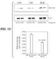
- FGF19 and/or FGFR4 polypeptide expression is detected using immunohistochemistry (IHC).
- IHC immunohistochemistry
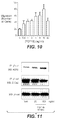
- FGF19 expression is scored at 2 or higher using an IHC.
- FGF19 and/or FGFR4 expression presence and/or absence and/or level of FGF 19 and/or FGFR4 expression may be detected. FGF 19 and/or FGFR4 expression may be increased. It is understood that absence of FGF19 and/or FGFR4 expression includes insignificant, or de minimus levels.
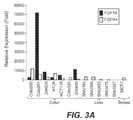
- FGF19 expression in the test biological sample is higher than that observed for a control biological sample (or control or reference level of expression). In some embodiments, FGF19 expression is at least about 2-fold, 5-fold, 10-fold, 20-fold, 30-fold, 40-fold, 50-fold, 75-fold, 100-fold, 150-fold higher, or higher in the test biological sample than in the control biological sample.
- FGF19 polypeptide expression is determined in an immunohistochemistry ("IHC") assay to score at least 2 or higher for staining intensity. In some embodiments, FGF19 polypeptide expression is determined in an IHC assay to score at least 1 or higher, or at least 3 or higher for staining intensity. In some embodiments, FGF 19 expression in the test biological sample is lower than that observed for a control biological sample (or control expression level).
- IHC immunohistochemistry
- the disclosure provides an isolated polynucleotide comprising, consisting of, or consisting essentially of one or more of the following polynucleotide sequences: GAT CCC CCC TCG TGA GTC TAG ATC TAT TCA AGA GAT AGA TCT AGA CTC ACG AGG TTT TTT GGA AA (SEQ ID NO:41); AGC TTT TCC AAA AAA CCT CGT GAG TCT AGA TCT ATC TCT TGA ATA GAT CTA GAC TCA CGA GGG GG (SEQ ID NO:42); GAT CCC CGA ACC GCA TTG GAG GCA TTA TCA AGA GAA ATG CCT CCA ATG CGG TTC TTT TTT GGA AA (SEQ ID NO:43); or AGC TTT TCC AAA AAA GAA CCG CAT TGG AGG CAT TTC TCT TGA TAA TGC CTC CAA TGC GGT TCG GG (SEQ ID NO:44).
- the invention herein provides anti-FGF19 antibodies, which are useful for, e.g., treatment or prevention of disease states associated with expression and/or activity of FGF19, such as increased expression and/or activity or undesired expression and/or activity.
- the antibodies of the invention are used to treat a tumor, a cancer, and/or a cell proliferative disorder.
- the anti-FGF19 antibodies of the invention find utility as reagents for detection and/or isolation of FGF 19, such as detention of FGF 19 in various tissues and cell type.
- the invention further provides methods of making anti-FGF 19 antibodies, polynucleotides encoding anti-FGF19 antibodies, and cells comprising polynucleotides encoding anti-FGF19 antibodies.
- the invention provides methods comprising detection of FGF 19 and/or FGFR4
- an “isolated” antibody is one which has been identified and separated and/or recovered from a component of its natural environment. Contaminant components of its natural environment are materials which would interfere with diagnostic or therapeutic uses for the antibody, and may include enzymes, hormones, and other proteinaceous or nonproteinaceous solutes.
- the antibody will be purified (1) to greater than 95% by weight of antibody as determined by the Lowry method, and most preferably more than 99% by weight, (2) to a degree sufficient to obtain at least 15 residues ofN-terminal or internal amino acid sequence by use of a spinning cup sequenator, or (3) to homogeneity by SDS-PAGE under reducing or nonreducing conditions using Coomassie blue or, preferably, silver stain.
- Isolated antibody includes the antibody in situ within recombinant cells since at least one component of the antibody's natural environment will not be present. Ordinarily, however, isolated antibody will be prepared by at least one purification step.
- an "isolated" nucleic acid molecule is a nucleic acid molecule that is identified and separated from at least one contaminant nucleic acid molecule with which it is ordinarily associated in the natural source of the antibody nucleic acid.
- An isolated nucleic acid molecule is other than in the form or setting in which it is found in nature. Isolated nucleic acid molecules therefore are distinguished from the nucleic acid molecule as it exists in natural cells.
- an isolated nucleic acid molecule includes a nucleic acid molecule contained in cells that ordinarily express the antibody where, for example, the nucleic acid molecule is in a chromosomal location different from that of natural cells.
- variable domain residue numbering as in Kabat or "amino acid position numbering as in Kabat”, and variations thereof, refers to the numbering system used for heavy chain variable domains or light chain variable domains of the compilation of antibodies in Kabat et al., Sequences of Proteins of Immunological Interest, 5th Ed. Public Health Service, National Institutes of Health, Bethesda, MD. (1991 ). Using this numbering system, the actual linear amino acid sequence may contain fewer or additional amino acids corresponding to a shortening of, or insertion into, a FR or CDR of the variable domain.
- a heavy chain variable domain may include a single amino acid insert (residue 52a according to Kabat) after residue 52 of H2 and inserted residues (e.g. residues 82a, 82b, and 82c, etc according to Kabat) after heavy chain FR residue 82.
- the Kabat numbering of residues may be determined for a given antibody by alignment at regions of homology of the sequence of the antibody with a "standard" Kabat numbered sequence.
- substantially similar denotes a sufficiently high degree of similarity between two numeric values (generally one associated with an antibody of the invention and the other associated with a reference/comparator antibody) such that one of skill in the art would consider the difference between the two values to be of little or no biological and/or statistical significance within the context of the biological characteristic measured by said values (e.g., Kd values).
- the difference between said two values is preferably less than about 50%, preferably less than about 40%, preferably less than about 30%, preferably less than about 20%, preferably less than about 10% as a function of the value for the reference/comparator antibody.
- Binding affinity generally refers to the strength of the sum total of noncovalent interactions between a single binding site of a molecule (e.g., an antibody) and its binding partner (e.g., an antigen). Unless indicated otherwise, as used herein, "binding affinity” refers to intrinsic binding affinity which reflects a 1:1 interaction between members of a binding pair (e.g., antibody and antigen).
- the affinity of a molecule X for its partner Y can generally be represented by the dissociation constant (Kd). Affinity can be measured by common methods known in the art, including those described herein. Low-affinity antibodies generally bind antigen slowly and tend to dissociate readily, whereas high-affinity antibodies generally bind antigen faster and tend to remain bound longer. A variety of methods of measuring binding affinity are known in the art, any of which can be used for purposes of the present invention. Specific illustrative embodiments are described in the following.
- the "Kd" or "Kd value” according to this invention is measured by a radiolabeled antigen binding assay (RIA) performed with the Fab version of an antibody of interest and its antigen as described by the following assay that measures solution binding affinity of Fabs for antigen by equilibrating Fab with a minimal concentration of ( 125 I)-labeled antigen in the presence of a titration series of unlabeled antigen, then capturing bound antigen with an anti-Fab antibody-coated plate ( Chen, et al., (1999) J. Mol Biol 293:865-881 ).
- RIA radiolabeled antigen binding assay
- microtiter plates (Dynex) are coated overnight with 5 ug/ml of a capturing anti-Fab antibody (Cappel Labs) in 50 mM sodium carbonate (pH 9.6), and subsequently blocked with 2% (w/v) bovine serum albumin in PBS for two to five hours at room temperature (approximately 23°C).
- a non-adsorbant plate (Nunc #269620) 100 pM or 26 pM [ 125 I]-antigen are mixed with serial dilutions of a Fab of interest (e.g., consistent with assessment of an anti-VEGF antibody, Fab-12, in Presta et al., (1997) Cancer Res.
- the Fab of interest is then incubated overnight; however, the incubation may continue for a longer period (e.g., 65 hours) to insure that equilibrium is reached. Thereafter, the mixtures are transferred to the capture plate for incubation at room temperature (e.g., for one hour). The solution is then removed and the plate washed eight times with 0.1% Tween-20 in PBS. When the plates have dried, 150 ul/well of scintillant (MicroScint-20; Packard) is added, and the plates are counted on a Topcount gamma counter (Packard) for ten minutes. Concentrations of each Fab that give less than or equal to 20% of maximal binding are chosen for use in competitive binding assays.
- the Kd or Kd value is measured by using surface plasmon resonance assays using a BIAcoreTM-2000 or a BlAcoreTM-3000 (BIAcore, Inc., Piscataway, NJ) at 25C with immobilized antigen CM5 chips at ⁇ 10 response units (RU).
- CM5 carboxymethylated dextran biosensor chips
- EDC N-ethyl-N'-(3-dimethylaminopropyl)-carbodiimide hydrochloride
- NHS N-hydroxysuccinimide
- Antigen is diluted with 10mM sodium acetate, pH 4.8, into 5ug/ml ( ⁇ 0.2uM) before injection at a flow rate of 5ul/minute to achieve approximately 10 response units (RU) of coupled protein. Following the injection of antigen, 1M ethanolamine is injected to block unreacted groups. For kinetics measurements, two-fold serial dilutions of Fab (0.78 nM to 500 nM) are injected in PBS with 0.05% Tween 20 (PBST) at 25°C at a flow rate of approximately 25ul/min.
- PBST Tween 20
- association rates (k on ) and dissociation rates (k off ) are calculated using a simple one-to-one Langmuir binding model (BIAcore Evaluation Software version 3.2) by simultaneous fitting the association and dissociation sensorgram.
- the equilibrium dissociation constant (Kd) is calculated as the ratio k off /k on . See, e.g., Chen, Y., et al., (1999) J. Mol Biol 293:865-881 .
- CM5 chips ⁇ 10 response units (RU).
- CM5 carboxymethylated dextran biosensor chips
- EDC N-ethyl-N'- (3-dimethylaminopropyl)-carbodiimide hydrochloride
- NHS N-hydroxysuccinimide
- Antigen is diluted with 10mM sodium acetate, pH 4.8, into 5ug/ml ( ⁇ 0.2uM) before injection at a flow rate of 5ul/minute to achieve approximately 10 response units (RU) of coupled protein. Following the injection of antigen, 1M ethanolamine is injected to block unreacted groups. For kinetics measurements, two-fold serial dilutions of Fab (0.78 nM to 500 nM) are injected in PBS with 0.05% Tween 20 (PBST) at 25°C at a flow rate of approximately 25ul/min.
- PBST Tween 20
- association rates (k on ) and dissociation rates (k off ) are calculated using a simple one-to-one Langmuir binding model (BIAcore Evaluation Software version 3.2) by simultaneous fitting the association and dissociation sensorgram.
- the equilibrium dissociation constant (Kd) was calculated as the ratio k off /k on . See, e.g., Chen, Y., et al., (1999) J. Mol Biol 293:865-881 .
- vector is intended to refer to a nucleic acid molecule capable of transporting another nucleic acid to which it has been linked.
- plasmid refers to a circular double stranded DNA loop into which additional DNA segments may be ligated.
- phage vector Another type of vector is a viral vector, wherein additional DNA segments may be ligated into the viral genome.
- viral vector is capable of autonomous replication in a host cell into which they are introduced (e.g., bacterial vectors having a bacterial origin of replication and episomal mammalian vectors).
- vectors e.g., non-episomal mammalian vectors
- vectors can be integrated into the genome of a host cell upon introduction into the host cell, and thereby are replicated along with the host genome.
- certain vectors are capable of directing the expression of genes to which they are operatively linked.
- Such vectors are referred to herein as "recombinant expression vectors” (or simply, “recombinant vectors”).
- expression vectors of utility in recombinant DNA techniques are often in the form of plasmids.
- plasmid and “vector” may be used interchangeably as the plasmid is the most commonly used form of vector.
- Polynucleotide or “nucleic acid,” as used interchangeably herein, refer to polymers of nucleotides of any length, and include DNA and RNA.
- the nucleotides can be deoxyribonucleotides, ribonucleotides, modified nucleotides or bases, and/or their analogs, or any substrate that can be incorporated into a polymer by DNA or RNA polymerase, or by a synthetic reaction.
- a polynucleotide may comprise modified nucleotides, such as methylated nucleotides and their analogs. If present, modification to the nucleotide structure may be imparted before or after assembly of the polymer.
- the sequence of nucleotides may be interrupted by non-nucleotide components.
- a polynucleotide may be further modified after synthesis, such as by conjugation with a label.
- Other types of modifications include, for example, "caps", substitution of one or more of the naturally occurring nucleotides with an analog, internucleotide modifications such as, for example, those with uncharged linkages (e.g., methyl phosphonates, phosphotriesters, phosphoamidates, carbamates, etc.) and with charged linkages (e.g., phosphorothioates, phosphorodithioates, etc.), those containing pendant moieties, such as, for example, proteins (e.g., nucleases, toxins, antibodies, signal peptides, ply-L-lysine, etc.), those with intercalators (e.g., acridine, psoralen, etc.), those containing chelators (e.g., metals
- any of the hydroxyl groups ordinarily present in the sugars may be replaced, for example, by phosphonate groups, phosphate groups, protected by standard protecting groups, or activated to prepare additional linkages to additional nucleotides, or may be conjugated to solid or semi-solid supports.
- the 5' and 3' terminal OH can be phosphorylated or substituted with amines or organic capping group moieties of from 1 to 20 carbon atoms.
- Other hydroxyls may also be derivatized to standard protecting groups.
- Polynucleotides can also contain analogous forms of ribose or deoxyribose sugars that are generally known in the art, including, for example, 2'-O-methyl-, 2'-O-allyl, 2'-fluoro- or 2'-azido-ribose, carbocyclic sugar analogs, alpha-anomeric sugars, epimeric sugars such as arabinose, xyloses or lyxoses, pyranose sugars, furanose sugars, sedoheptuloses, acyclic analogs and a basic nucleoside analogs such as methyl riboside.
- One or more phosphodiester linkages may be replaced by alternative linking groups.
- linking groups include, but are not limited to, embodiments wherein phosphate is replaced by P(O)S ("thioate”), P(S)S ("dithioate”), "(O)NR 2 ("amidate"), P(O)R, P(O)OR', CO or CH 2 ("formacetal"), in which each R or R' is independently H or substituted or unsubstituted alkyl (1-20 C) optionally containing an ether (-O-) linkage, aryl, alkenyl, cycloalkyl, cycloalkenyl or araldyl. Not all linkages in a polynucleotide need be identical. The preceding description applies to all polynucleotides referred to herein, including RNA and DNA.
- Oligonucleotide generally refers to short, generally single stranded, generally synthetic polynucleotides that are generally, but not necessarily, less than about 200 nucleotides in length.
- oligonucleotide and “polynucleotide” are not mutually exclusive. The description above for polynucleotides is equally and fully applicable to oligonucleotides.
- FGF19 refers, unless specifically or contextually indicated otherwise, to any native or variant (whether native or synthetic) FGF19 polypeptide.
- native sequence specifically encompasses naturally occurring truncated or secreted forms (e.g., an extracellular domain sequence), naturally occurring variant forms (e.g., alternatively spliced forms) and naturally-occurring allelic variants.
- wild type FGF 19 generally refers to a polypeptide comprising the amino acid sequence of a naturally occurring FGF19 protein.
- wild type FGF19 sequence generally refers to an amino acid sequence found in a naturally occurring FGF19.
- FGFR4 (interchangeably termed "Fibroblast growth factor receptor 4"), as used herein, refers, unless specifically or contextually indicated otherwise, to any native or variant (whether native or synthetic) FGFR4 polypeptide.
- native sequence specifically encompasses naturally occurring truncated or secreted forms (e.g., an extracellular domain sequence), naturally occurring variant forms (e.g., alternatively spliced forms) and naturally-occurring allelic variants.
- wild type FGFR4 generally refers to a polypeptide comprising the amino acid sequence of a naturally occurring FGFR4 protein.
- wild type FGFR4 sequence generally refers to an amino acid sequence found in a naturally occurring FGFR4.
- antibody and “immunoglobulin” are used interchangeably in the broadest sense and include monoclonal antibodies (for e.g., full length or intact monoclonal antibodies), polyclonal antibodies, multivalent antibodies, multispecific antibodies (e.g., bispecific antibodies so long as they exhibit the desired biological activity) and may also include certain antibody fragments (as described in greater detail herein).
- An antibody can be human, humanized and/or affinity matured.
- variable refers to the fact that certain portions of the variable domains differ extensively in sequence among antibodies and are used in the binding and specificity of each particular antibody for its particular antigen. However, the variability is not evenly distributed throughout the variable domains of antibodies. It is concentrated in three segments called complementarity-determining regions (CDRs) or hypervariable regions both in the light-chain and the heavy-chain variable domains. The more highly conserved portions of variable domains are called the framework (FR).
- CDRs complementarity-determining regions
- FR framework
- the variable domains of native heavy and light chains each comprise four FR regions, largely adopting a ⁇ - sheet configuration, connected by three CDRs, which form loops connecting, and in some cases forming part of, the ⁇ - sheet structure.
- the CDRs in each chain are held together in close proximity by the FR regions and, with the CDRs from the other chain, contribute to the formation of the antigen-binding site of antibodies (see Kabat et al., Sequences of Proteins of Immunological Interest, Fifth Edition, National Institute of Health, Bethesda, MD (1991 )).
- the constant domains are not involved directly in binding an antibody to an antigen, but exhibit various effector functions, such as participation of the antibody in antibody-dependent cellular toxicity.
- Papain digestion of antibodies produces two identical antigen-binding fragments, called “Fab” fragments, each with a single antigen-binding site, and a residual "Fc” fragment, whose name reflects its ability to crystallize readily. Pepsin treatment yields an F(ab') 2 fragment that has two antigen-combining sites and is still capable of crosslinking antigen.
- Fv is the minimum antibody fragment which contains a complete antigen-recognition and -binding site.
- this region consists of a dimer of one heavy- and one light-chain variable domain in tight, non-covalent association.
- one heavy- and one light-chain variable domain can be covalently linked by a flexible peptide linker such that the light and heavy chains can associate in a "dimeric" structure analogous to that in a two-chain Fv species. It is in this configuration that the three CDRs of each variable domain interact to define an antigen-binding site on the surface of the VH-VL dimer.
- the six CDRs confer antigen-binding specificity to the antibody.
- the Fab fragment also contains the constant domain of the light chain and the first constant domain (CH1) of the heavy chain.
- Fab' fragments differ from Fab fragments by the addition of a few residues at the carboxy terminus of the heavy chain CH1 domain including one or more cysteines from the antibody hinge region.
- Fab'-SH is the designation herein for Fab' in which the cysteine residue(s) of the constant domains bear a free thiol group.
- F(ab') 2 antibody fragments originally were produced as pairs of Fab' fragments which have hinge cysteines between them. Other chemical couplings of antibody fragments are also known.
- the "light chains" of antibodies (immunoglobulins) from any vertebrate species can be assigned to one of two clearly distinct types, called kappa ( ⁇ ) and lambda ( ⁇ ), based on the amino acid sequences of their constant domains.
- immunoglobulins can be assigned to different classes. There are five major classes of immunoglobulins: IgA, IgD, IgE, IgG, and IgM, and several of these can be further divided into subclasses (isotypes), e.g., IgG 1 , IgG 2 , IgG 3 , IgG 4 , IgA 1 , and IgA 2 .
- the heavy-chain constant domains that correspond to the different classes of immunoglobulins are called ⁇ , ⁇ , ⁇ , ⁇ , and ⁇ , respectively.
- the subunit structures and three-dimensional configurations of different classes of immunoglobulins are well known.
- Antibody fragments comprise only a portion of an intact antibody, wherein the portion preferably retains at least one, preferably most or all, of the functions normally associated with that portion when present in an intact antibody.
- antibody fragments include Fab, Fab', F(ab')2, and Fv fragments; diabodies; linear antibodies; single-chain antibody molecules; and multispecific antibodies formed from antibody fragments.
- an antibody fragment comprises an antigen binding site of the intact antibody and thus retains the ability to bind antigen.
- an antibody fragment for example one that comprises the Fc region, retains at least one of the biological functions normally associated with the Fc region when present in an intact antibody, such as FcRn binding, antibody half life modulation, ADCC function and complement binding.
- an antibody fragment is a monovalent antibody that has an in vivo half life substantially similar to an intact antibody.
- such an antibody fragment may comprise on antigen binding arm linked to an Fc sequence capable of conferring in vivo stability to the fragment.
- hypervariable region when used herein refers to the regions of an antibody variable domain which are hypervariable in sequence and/or form structurally defined loops.
- antibodies comprise six hypervariable regions; three in the VH (H1, H2, H3), and three in the VL (L1, L2, L3).
- a number of hypervariable region delineations are in use and are encompassed herein.
- the Kabat Complementarity Determining Regions are based on sequence variability and are the most commonly used ( Kabat et al., Sequences of Proteins of Immunological Interest, 5th Ed. Public Health Service, National Institutes of Health, Bethesda, MD. (1991 )).
- Chothia refers instead to the location of the structural loops ( Chothia and Lesk J. Mol. Biol. 196:901-917 (1987 )).
- the AbM hypervariable regions represent a compromise between the Kabat CDRs and Chothia structural loops, and are used by Oxford Molecular's AbM antibody modeling software.
- the "contact" hypervariable regions are based on an analysis of the available complex crystal structures.
- Hypervariable regions may comprise "extended hypervariable regions” as follows: 24-36 (L1), 46-56 (L2) and 89-97 (L3) in the VL and 26-35 (H1), 49-65 or 50 to 65 (H2) and 93-102 (H3) in the VH.
- the variable domain residues are numbered according to Kabat et al., supra for each of these definitions.
- Framework or "FR” residues are those variable domain residues other than the hypervariable region residues as herein defined.
- Humanized forms of non-human (e.g., murine) antibodies are chimeric antibodies that contain minimal sequence derived from non-human immunoglobulin.
- humanized antibodies are human immunoglobulins (recipient antibody) in which residues from a hypervariable region of the recipient are replaced by residues from a hypervariable region of a non-human species (donor antibody) such as mouse, rat, rabbit or nonhuman primate having the desired specificity, affinity, and capacity.
- donor antibody such as mouse, rat, rabbit or nonhuman primate having the desired specificity, affinity, and capacity.
- framework region (FR) residues of the human immunoglobulin are replaced by corresponding non-human residues.
- humanized antibodies may comprise residues that are not found in the recipient antibody or in the donor antibody. These modifications are made to further refine antibody performance.
- the humanized antibody will comprise substantially all of at least one, and typically two, variable domains, in which all or substantially all of the hypervariable loops correspond to those of a non-human immunoglobulin and all or substantially all of the FRs are those of a human immunoglobulin sequence.
- the humanized antibody optionally will also comprise at least a portion of an immunoglobulin constant region (Fc), typically that of a human immunoglobulin.
- Fc immunoglobulin constant region
- “Chimeric” antibodies have a portion of the heavy and/or light chain identical with or homologous to corresponding sequences in antibodies derived from a particular species or belonging to a particular antibody class or subclass, while the remainder of the chain(s) is identical with or homologous to corresponding sequences in antibodies derived from another species or belonging to another antibody class or subclass, as well as fragments of such antibodies, so long as they exhibit the desired biological activity ( U.S. Patent No. 4,816,567 ; and Morrison et al., Proc. Natl. Acad. Sci. USA 81:6851-6855 (1984 )). Humanized antibody as used herein is a subset of chimeric antibodies.
- Single-chain Fv or “scFv” antibody fragments comprise the VH and VL domains of antibody, wherein these domains are present in a single polypeptide chain.
- the scFv polypeptide further comprises a polypeptide linker between the VH and VL domains which enables the scFv to form the desired structure for antigen binding.
- an “antigen” is a predetermined antigen to which an antibody can selectively bind.
- the target antigen may be polypeptide, carbohydrate, nucleic acid, lipid, hapten or other naturally occurring or synthetic compound.
- the target antigen is a polypeptide.
- diabodies refers to small antibody fragments with two antigen-binding sites, which fragments comprise a heavy-chain variable domain (VH) connected to a light-chain variable domain (VL) in the same polypeptide chain (VH - VL).
- VH heavy-chain variable domain
- VL light-chain variable domain
- a "human antibody” is one which possesses an amino acid sequence which corresponds to that of an antibody produced by a human and/or has been made using any of the techniques for making human antibodies as disclosed herein. This definition of a human antibody specifically excludes a humanized antibody comprising non-human antigen-binding residues.
- affinity matured antibody is one with one or more alterations in one or more CDRs thereof which result in an improvement in the affinity of the antibody for antigen, compared to a parent antibody which does not possess those alteration(s).
- Preferred affinity matured antibodies will have nanomolar or even picomolar affinities for the target antigen.
- Affinity matured antibodies are produced by procedures known in the art. Marks et al. Bio/Technology 10:779-783 (1992 ) describes affinity maturation by VH and VL domain shuffling. Random mutagenesis of CDR and/or framework residues is described by: Barbas et al. Proc Nat. Acad. Sci, USA 91:3809-3813 (1994 ); Schier et al.
- Antibody effector functions refer to those biological activities attributable to the Fc region (a native sequence Fc region or amino acid sequence variant Fc region) of an antibody, and vary with the antibody isotype. Examples of antibody effector functions include: C1q binding and complement dependent cytotoxicity; Fc receptor binding; antibody-dependent cell-mediated cytotoxicity (ADCC); phagocytosis; down regulation of cell surface receptors ( e.g. B cell receptor); and B cell activation.
- Antibody-dependent cell-mediated cytotoxicity refers to a form of cytotoxicity in which secreted Ig bound onto Fc receptors (FcRs) present on certain cytotoxic cells (e.g. Natural Killer (NK) cells, neutrophils, and macrophages) enable these cytotoxic effector cells to bind specifically to an antigen-bearing target cell and subsequently kill the target cell with cytotoxins.
- cytotoxic cells e.g. Natural Killer (NK) cells, neutrophils, and macrophages
- the antibodies “arm” the cytotoxic cells and are absolutely required for such killing.
- the primary cells for mediating ADCC NK cells, express Fc ⁇ RIII only, whereas monocytes express Fc ⁇ RI, Fc ⁇ RII and Fc ⁇ RIII.
- FcR expression on hematopoietic cells is summarized in Table 3 on page 464 of Ravetch and Kinet, Annu. Rev. Immunol 9:457-92 (1991 ).
- an in vitro ADCC assay such as that described in US Patent No. 5,500,362 or 5,821,337 or Presta U.S. Patent No. 6,737,056 may be performed.
- Useful effector cells for such assays include peripheral blood mononuclear cells (PBMC) and Natural Killer (NK) cells.
- PBMC peripheral blood mononuclear cells
- NK Natural Killer
- ADCC activity of the molecule of interest may be assessed in vivo, e.g., in a animal model such as that disclosed in Clynes et al. PNAS (USA) 95:652-656 (1998 ).
- Human effector cells are leukocytes which express one or more FcRs and perform effector functions. Preferably, the cells express at least Fc ⁇ RIII and perform ADCC effector function. Examples of human leukocytes which mediate ADCC include peripheral blood mononuclear cells (PBMC), natural killer (NK) cells, monocytes, cytotoxic T cells and neutrophils; with PBMCs and NK cells being preferred.
- PBMC peripheral blood mononuclear cells
- NK natural killer cells
- monocytes cytotoxic T cells and neutrophils
- the effector cells may be isolated from a native source, e.g. from blood.
- Fc receptor or “FcR” describes a receptor that binds to the Fc region of an antibody.
- the preferred FcR is a native sequence human FcR.
- a preferred FcR is one which binds an IgG antibody (a gamma receptor) and includes receptors of the Fc ⁇ RI, Fc ⁇ RII, and Fc ⁇ RIII subclasses, including allelic variants and alternatively spliced forms of these receptors.
- Fc ⁇ RII receptors include Fc ⁇ RIIA (an “activating receptor”) and Fc ⁇ RIIB (an “inhibiting receptor”), which have similar amino acid sequences that differ primarily in the cytoplasmic domains thereof.
- Activating receptor Fc ⁇ RIIA contains an immunoreceptor tyrosine-based activation motif (ITAM) in its cytoplasmic domain.
- Inhibiting receptor Fc ⁇ RIIB contains an immunoreceptor tyrosine-based inhibition motif (ITIM) in its cytoplasmic domain.
- ITAM immunoreceptor tyrosine-based activation motif
- ITIM immunoreceptor tyrosine-based inhibition motif
- FcR FcR
- the term also includes the neonatal receptor, FcRn, which is responsible for the transfer of maternal IgGs to the fetus ( Guyer et al., J. Immunol. 117:587 (1976 ) and Kim et al., J. Immunol. 24:249 (1994 )) and regulates homeostasis of immunoglobulins.
- WO00/42072 (Presta) describes antibody variants with improved or diminished binding to FcRs. The content of that patent publication is specifically incorporated herein by reference. See, also, Shields et al. J. Biol Chem. 9(2): 6591-6604 (2001 ).
- Binding to human FcRn in vivo and serum half life of human FcRn high affinity binding polypeptides can be assayed, e.g., in transgenic mice or transfected human cell lines expressing human FcRn, or in primates administered with the Fc variant polypeptides.
- “Complement dependent cytotoxicity” or “CDC” refers to the lysis of a target cell in the presence of complement. Activation of the classical complement pathway is initiated by the binding of the first component of the complement system (C1q) to antibodies (of the appropriate subclass) which are bound to their cognate antigen.
- C1q first component of the complement system
- a CDC assay e.g. as described in Gazzano-Santoro et al., J. Immunol. Methods 202:163 (1996 ), may be performed.
- blocking antibody or an “antagonist” antibody is one which inhibits or reduces biological activity of the antigen it binds.
- Preferred blocking antibodies or antagonist antibodies substantially or completely inhibit the biological activity of the antigen.
- sample encompasses a variety of sample types obtained from an individual and can be used in a diagnostic or monitoring assay.
- the definition encompasses blood and other liquid samples of biological origin, solid tissue samples such as a biopsy specimen or tissue cultures or cells derived therefrom, and the progeny thereof.
- the definition also includes samples that have been manipulated in any way after their procurement, such as by treatment with reagents, solubilization, or enrichment for certain components, such as proteins or polynucleotides, or embedding in a semi-solid or solid matrix for sectioning purposes.
- biological sample encompasses a clinical sample, and also includes cells in culture, cell supernatants, cell lysates, serum, plasma, biological fluid, and tissue samples.
- the source of the biological sample may be solid tissue as from a fresh, frozen and/or preserved organ or tissue sample or biopsy or aspirate; blood or any blood constituents; bodily fluids such as cerebral spinal fluid, amniotic fluid, peritoneal fluid, or interstitial fluid; cells from any time in gestation or development of the subject.
- the biological sample is obtained from a primary or metastatic tumor.
- the biological sample may contain compounds which are not naturally intermixed with the tissue in nature such as preservatives, anticoagulants, buffers, fixatives, nutrients, antibiotics, or the like.
- a "section" of a tissue sample is meant a single part or piece of a tissue sample, e.g. a thin slice of tissue or cells cut from a tissue sample. It is understood that multiple sections of tissue samples may be taken and subjected to analysis according to the present invention. In some embodiments, the same section of tissue sample is analyzed at both morphological and molecular levels, or is analyzed with respect to both protein and nucleic acid.
- label when used herein refers to a compound or composition which is conjugated or fused directly or indirectly to a reagent such as a nucleic acid probe or an antibody and facilitates detection of the reagent to which it is conjugated or fused.
- the label may itself be detectable (e.g., radioisotope labels or fluorescent labels) or, in the case of an enzymatic label, may catalyze chemical alteration of a substrate compound or composition which is detectable.
- a “medicament” is an active drug to treat the disorder in question or its symptoms, or side effects.
- a “disorder” or “disease” is any condition that would benefit from treatment with a substance/molecule or method of the invention. This includes chronic and acute disorders or diseases including those pathological conditions which predispose the mammal to the disorder in question.
- disorders to be treated herein include malignant and benign tumors; carcinoma, blastoma, and sarcoma.
- cell proliferative disorder and “proliferative disorder” refer to disorders that are associated with some degree of abnormal cell proliferation.
- the cell proliferative disorder is cancer.
- tumor refers to all neoplastic cell growth and proliferation, whether malignant or benign, and all pre-cancerous and cancerous cells and tissues.
- cancer refers to all neoplastic cell growth and proliferation, whether malignant or benign, and all pre-cancerous and cancerous cells and tissues.
- cancer refers to all neoplastic cell growth and proliferation, whether malignant or benign, and all pre-cancerous and cancerous cells and tissues.
- cancer cancer
- cancer cancer
- cancer cancer
- cancer cancer
- cancer cancer
- cancer and “cancerous” refer to or describe the physiological condition in mammals that is typically characterized by unregulated cell growth/proliferation.
- examples of cancer include but are not limited to, carcinoma, lymphoma, blastoma, sarcoma, and leukemia.
- cancers include squamous cell cancer, small-cell lung cancer, pituitary cancer, esophageal cancer, astrocytoma, soft tissue sarcoma, non-small cell lung cancer, adenocarcinoma of the lung, squamous carcinoma of the lung, cancer of the peritoneum, hepatocellular cancer, gastrointestinal cancer, pancreatic cancer, glioblastoma, cervical cancer, ovarian cancer, liver cancer, bladder cancer, hepatoma, breast cancer, colon cancer, colorectal cancer, endometrial or uterine carcinoma, salivary gland carcinoma, kidney cancer, liver cancer, prostate cancer, vulval cancer, thyroid cancer, hepatic carcinoma, brain cancer, endometrial cancer, testis cancer, cholangiocarcinoma, gallbladder carcinoma, gastric cancer, melanoma, and various types of head and neck cancer.
- Dysregulation of angiogenesis can lead to many disorders that can be treated by compositions and methods of the invention. These disorders include both non-neoplastic and neoplastic conditions.
- Neoplastics include but are not limited those described above.
- Non-neoplastic disorders include but are not limited to undesired or aberrant hypertrophy, arthritis, rheumatoid arthritis (RA), psoriasis, psoriatic plaques, sarcoidosis, atherosclerosis, atherosclerotic plaques, diabetic and other proliferative retinopathies including retinopathy of prematurity, retrolental fibroplasia, neovascular glaucoma, age-related macular degeneration, diabetic macular edema, corneal neovascularization, corneal graft neovascularization, corneal graft rejection, retinal/choroidal neovascularization, neovascularization of the angle (rubeosis), ocular
- wasting disorders refers to a disorder caused by undesirable and/or unhealthy loss of weight or loss of body cell mass.
- wasting disease can result in undesired loss of body weight, including both the fat and the fat-free compartments.
- Wasting diseases can be the result of inadequate intake of food and/or metabolic changes related to illness and/or the aging process.
- Cachexia is additionally characterized by hypermetabolism and hypercatabolism.
- cachexia and wasting disease are frequently used interchangeably to refer to wasting conditions, there is at least one body of research which differentiates cachexia from wasting syndrome as a loss of fat-free mass, and particularly, body cell mass ( Mayer, 1999, J. Nutr. 129(1S Suppl.):256S-259S ).
- Sarcopenia yet another such disorder which can affect the aging individual, is typically characterized by loss of muscle mass. End stage wasting disease as described above can develop in individuals suffering from either cachexia or sarcopenia.
- treatment refers to clinical intervention in an attempt to alter the natural course of the individual or cell being treated, and can be performed either for prophylaxis or during the course of clinical pathology. Desirable effects of treatment include preventing occurrence or recurrence of disease, alleviation of symptoms, diminishment of any direct or indirect pathological consequences of the disease, decreasing the rate of disease progression, amelioration or palliation of the disease state, and remission or improved prognosis.
- antibodies of the invention are used to delay development of a disease or disorder.
- an "anti-angiogenesis agent” or “angiogenesis inhibitor” refers to a small molecular weight substance, a polynucleotide, a polypeptide, an isolated protein, a recombinant protein, an antibody, or conjugates or fusion proteins thereof, that inhibits angiogenesis, vasculogenesis, or undesirable vascular permeability, either directly or indirectly.
- an anti-angiogenesis agent is an antibody or other antagonist to an angiogenic agent as defined above, e.g., antibodies to VEGF, antibodies to VEGF receptors, small molecules that block VEGF receptor signaling (e.g., PTK787/ZK2284, SU6668, SUTENT/SU11248 (sunitinib malate), AMG706).
- Anti-angiogensis agents also include native angiogenesis inhibitors, e.g., angiostatin, endostatin, etc. See, e.g., Klagsbrun and D'Amore, Annu. Rev.
- mammals include, but are not limited to, farm animals (such as cows), sport animals, pets (such as cats, dogs and horses), primates, mice and rats.
- mammal for purposes of treatment refers to any animal classified as a mammal, including humans, domestic and farm animals, and zoo, sports, or pet animals, such as dogs, horses, cats, cows, etc. Preferably, the mammal is human.
- an “effective amount” refers to an amount effective, at dosages and for periods of time necessary, to achieve the desired therapeutic or prophylactic result.
- a “therapeutically effective amount” of a substance/molecule of the invention, agonist or antagonist may vary according to factors such as the disease state, age, sex, and weight of the individual, and the ability of the substance/molecule, agonist or antagonist to elicit a desired response in the individual.
- a therapeutically effective amount is also one in which any toxic or detrimental effects of the substance/molecule, agonist or antagonist are outweighed by the therapeutically beneficial effects.
- a “prophylactically effective amount” refers to an amount effective, at dosages and for periods of time necessary, to achieve the desired prophylactic result. Typically but not necessarily, since a prophylactic dose is used in subjects prior to or at an earlier stage of disease, the prophylactically effective amount will be less than the therapeutically effective amount.
- cytotoxic agent refers to a substance that inhibits or prevents the function of cells and/or causes destruction of cells.
- the term is intended to include radioactive isotopes (e.g., At 211 , I 131 , I 125 , Y 90 , R 186 , Re 188 , Sm 153 , Bi 212 , P 32 and radioactive isotopes of Lu), chemotherapeutic agents e.g.
- methotrexate adriamicin, vinca alkaloids (vincristine, vinblastine, etoposide), doxorubicin, melphalan, mitomycin C, chlorambucil, daunorubicin or other intercalating agents, enzymes and fragments thereof such as nucleolytic enzymes, antibiotics, and toxins such as small molecule toxins or enzymatically active toxins of bacterial, fungal, plant or animal origin, including fragments and/or variants thereof, and the various antitumor or anticancer agents disclosed below. Other cytotoxic agents are described below.
- a tumoricidal agent causes destruction of tumor cells.
- chemotherapeutic agent is a chemical compound useful in the treatment of cancer.
- examples of chemotherapeutic agents include alkylating agents such as thiotepa and CYTOXAN® cyclosphosphamide; alkyl sulfonates such as busulfan, improsulfan and piposulfan; aziridines such as benzodopa, carboquone, meturedopa, and uredopa; ethylenimines and methylamelamines including altretamine, triethylenemelamine, trietylenephosphoramide, triethiylenethiophosphoramide and trimethylolomelamine; acetogenins (especially bullatacin and bullatacinone); delta-9-tetrahydrocannabinol (dronabinol, MARINOL®); beta-lapachone; lapachol; colchicines; betulinic acid; a camptothecin (including the synthetic analogue topote
- calicheamicin especially calicheamicin gamma1I and calicheamicin omegaI1 (see, e.g., Agnew, Chem Intl. Ed. Engl., 33: 183-186 (1994 )); dynemicin, including dynemicin A; an esperamicin; as well as neocarzinostatin chromophore and related chromoprotein enediyne antiobiotic chromophores), aclacinomysins, actinomycin, authramycin, azaserine, bleomycins, cactinomycin, carabicin, carminomycin, carzinophilin, chromomycinis, dactinomycin, daunombicin, detorubicin, 6-diazo-5-oxo-L-norleucine, ADRIAMYCIN® doxorubicin (including morpholino-doxorubicin, cyan
- anti-hormonal agents that act to regulate, reduce, block, or inhibit the effects of hormones that can promote the growth of cancer, and are often in the form of systemic, or whole-body treatment. They may be hormones themselves.
- SERMs selective estrogen receptor modulators
- tamoxifen including NOLVADEX® tamoxifen
- EVISTA® raloxifene droloxifene
- 4-hydroxytamoxifen trioxifene, keoxifene, LY117018, onapristone, and FARESTON® toremifene
- anti-progesterones anti-progesterones
- estrogen receptor down-regulators ETDs
- agents that function to suppress or shut down the ovaries for example, leutinizing hormone-releasing hormone (LHRH) agonists such as LUPRON® and ELIGARD® leuprolide acetate, goserelin acetate, buserelin acetate and
- LHRH leutinizing
- chemotherapeutic agents includes bisphosphonates such as clodronate (for example, BONEFOS® or OSTAC®), DIDROCAL® etidronate, NE-58095, ZOMETA® zoledronic acid/zoledronate, FOSAMAX® alendronate, AREDIA® pamidronate, SKELID® tiludronate, or ACTONEL® risedronate; as well as troxacitabine (a 1,3-dioxolane nucleoside cytosine analog); antisense oligonucleotides, particularly those that inhibit expression of genes in signaling pathways implicated in abherant cell proliferation, such as, for example, PKC-alpha, Raf, H-Ras, and epidermal growth factor receptor (EGF-R); vaccines such as THERATOPE® vaccine and gene therapy vaccines, for example, ALLOVECTIN® vaccine, LEUVECTIN® vaccine, and VAXID® vaccine; LURTOTECAN
- a “growth inhibitory agent” when used herein refers to a compound or composition which inhibits growth of a cell (such as a cell expressing FGF19) either in vitro or in vivo.
- the growth inhibitory agent may be one which significantly reduces the percentage of cells (such as a cell expressing FGF19) in S phase.
- growth inhibitory agents include agents that block cell cycle progression (at a place other than S phase), such as agents that induce G1 arrest and M-phase arrest.
- Classical M-phase blockers include the vincas (vincristine and vinblastine), taxanes, and topoisomerase II inhibitors such as doxorubicin, epirubicin, daunorubicin, etoposide, and bleomycin.
- DNA alkylating agents such as tamoxifen, prednisone, dacarbazine, mechlorethamine, cisplatin, methotrexate, 5-fluorouracil, and ara-C.
- DNA alkylating agents such as tamoxifen, prednisone, dacarbazine, mechlorethamine, cisplatin, methotrexate, 5-fluorouracil, and ara-C.
- DNA alkylating agents such as tamoxifen, prednisone, dacarbazine, mechlorethamine, cisplatin, methotrexate, 5-fluorouracil, and ara-C.
- Docetaxel (TAXOTERE®, Rhone-Poulenc Rorer), derived from the European yew, is a semisynthetic analogue of paclitaxel (TAXOL®, Bristol-Myers Squibb). Paclitaxel and docetaxel promote the assembly of microtubules from tubulin dimers and stabilize microtubules by preventing depolymerization, which results in the inhibition of mitosis in cells.
- Doxorubicin is an anthracycline antibiotic.
- the full chemical name of doxorubicin is (8S-cis)-10-[(3-amino-2,3,6-trideoxy- ⁇ -L-lyxo-hexapyranosyl)oxy]-7,8,9,10-tetrahydro-6,8,11-trihydroxy-8-(hydroxyacetyl)-1-methoxy-5,12-naphthacenedione.
- Fc region-comprising polypeptide refers to a polypeptide, such as an antibody or immunoadhesin (see definitions below), which comprises an Fc region.
- the C-terminal lysine (residue 447 according to the EU numbering system) of the Fc region may be removed, for example, during purification of the polypeptide or by recombinant engineering the nucleic acid encoding the polypeptide.
- a composition comprising a polypeptide having an Fc region according to this invention can comprise polypeptides with K447, with all K447 removed, or a mixture of polypeptides with and without the K447 residue.
- compositions of the invention and methods of making same
- compositions including pharmaceutical compositions, comprising an anti-FGF19 antibody; and polynucleotides comprising sequences encoding an anti-FGF19 antibody.
- compositions comprise one or more antibodies that bind to FGF19, and/or one or more polynucleotides comprising sequences encoding one or more antibodies that bind to FGF19.
- suitable carriers such as pharmaceutically acceptable excipients including buffers, which are well known in the art.
- the invention also encompasses isolated antibody and polynucleotide embodiments.
- the invention also encompasses substantially pure antibody and polynucleotide embodiments.
- the anti-FGF19 antibodies of the invention are preferably monoclonal. Also encompassed within the scope of the invention are Fab, Fab', Fab'-SH and F(ab') 2 fragments of the anti-FGF19 antibodies provided herein. These antibody fragments can be created by traditional means, such as enzymatic digestion, or may be generated by recombinant techniques. Such antibody fragments may be chimeric or humanized. These fragments are useful for the diagnostic and therapeutic purposes set forth below.
- Monoclonal antibodies are obtained from a population of substantially homogeneous antibodies, i.e., the individual antibodies comprising the population are identical except for possible naturally occurring mutations that may be present in minor amounts.
- the modifier "monoclonal" indicates the character of the antibody as not being a mixture of discrete antibodies.
- the anti-FGF19 monoclonal antibodies of the invention can be made using the hybridoma method first described by Kohler et al., Nature, 256:495 (1975 ), or may be made by recombinant DNA methods ( U.S. Patent No. 4,816,567 ).
- a mouse or other appropriate host animal such as a hamster
- Antibodies to FGF19 generally are raised in animals by multiple subcutaneous (sc) or intraperitoneal (ip) injections of FGF19 and an adjuvant.
- FGF19 may be prepared using methods well-known in the art, some of which are further described herein. For example, recombinant production of FGF19 is described below.
- animals are immunized with a derivative of FGF19 that contains the extracellular domain (ECD) of FGF 19 fused to the Fc portion of an immunoglobulin heavy chain.
- ECD extracellular domain
- animals are immunized with an FGF19-IgG1 fusion protein.
- Animals ordinarily are immunized against immunogenic conjugates or derivatives of FGF19 with monophosphoryl lipid A (MPL)/trehalose dicrynomycolate (TDM) (Ribi Immunochem. Research, Inc., Hamilton, MT) and the solution is injected intradermally at multiple sites. Two weeks later the animals are boosted. 7 to 14 days later animals are bled and the serum is assayed for anti-FGF19 titer. Animals are boosted until titer plateaus.
- MPL monophosphoryl lipid A
- TDM trehalose dicrynomycolate
- lymphocytes may be immunized in vitro .
- Lymphocytes then are fused with myeloma cells using a suitable fusing agent, such as polyethylene glycol, to form a hybridoma cell ( Goding, Monoclonal Antibodies: Principles and Practice, pp.59-103 (Academic Press, 1986 )).
- a suitable fusing agent such as polyethylene glycol
- the hybridoma cells thus prepared are seeded and grown in a suitable culture medium that preferably contains one or more substances that inhibit the growth or survival of the unfused, parental myeloma cells.
- a suitable culture medium that preferably contains one or more substances that inhibit the growth or survival of the unfused, parental myeloma cells.
- the culture medium for the hybridomas typically will include hypoxanthine, aminopterin, and thymidine (HAT medium), which substances prevent the growth of HGPRT-deficient cells.
- Preferred myeloma cells are those that fuse efficiently, support stable high-level production of antibody by the selected antibody-producing cells, and are sensitive to a medium such as HAT medium.
- preferred myeloma cell lines are murine myeloma lines, such as those derived from MOPC-21 and MPC-11 mouse tumors available from the Salk Institute Cell Distribution Center, San Diego, California USA, and SP-2 or X63-Ag8-653 cells available from the American Type Culture Collection, Rockville, Maryland USA.
- Human myeloma and mouse-human heteromyeloma cell lines also have been described for the production of human monoclonal antibodies ( Kozbor, J. Immunol., 133:3001 (1984 ); Brodeur et al., Monoclonal Antibody Production Techniques and Applications, pp. 51-63 (Marcel Dekker, Inc., New York, 1987 )).
- Culture medium in which hybridoma cells are growing is assayed for production of monoclonal antibodies directed against FGF19.
- the binding specificity of monoclonal antibodies produced by hybridoma cells is determined by immunoprecipitation or by an in vitro binding assay, such as radioimmunoassay (RIA) or enzyme-linked immunoadsorbent assay (ELISA).
- RIA radioimmunoassay
- ELISA enzyme-linked immunoadsorbent assay
- the binding affinity of the monoclonal antibody can, for example, be determined by the Scatchard analysis of Munson et al., Anal. Biochem., 107:220 (1980 ).
- the clones may be subcloned by limiting dilution procedures and grown by standard methods ( Goding, Monoclonal Antibodies: Principles and Practice, pp.59-103 (Academic Press, 1986 )). Suitable culture media for this purpose include, for example, D-MEM or RPMI-1640 medium.
- the hybridoma cells may be grown in vivo as ascites tumors in an animal.
- the monoclonal antibodies secreted by the subclones are suitably separated from the culture medium, ascites fluid, or serum by conventional immunoglobulin purification procedures such as, for example, protein A-Sepharose, hydroxylapatite chromatography, gel electrophoresis, dialysis, or affinity chromatography.
- the anti-FGF19 antibodies of the invention can be made by using combinatorial libraries to screen for synthetic antibody clones with the desired activity or activities.
- synthetic antibody clones are selected by screening phage libraries containing phage that display various fragments of antibody variable region (Fv) fused to phage coat protein. Such phage libraries are panned by affinity chromatography against the desired antigen. Clones expressing Fv fragments capable of binding to the desired antigen are adsorbed to the antigen and thus separated from the non-binding clones in the library. The binding clones are then eluted from the antigen, and can be further enriched by additional cycles of antigen adsorption/elution.
- Fv antibody variable region
- any of the anti-FGF 19 antibodies of the invention can be obtained by designing a suitable antigen screening procedure to select for the phage clone of interest followed by construction of a full length anti-FGF 19 antibody clone using the Fv sequences from the phage clone of interest and suitable constant region (Fc) sequences described in Kabat et al., Sequences of Proteins of Immunological Interest, Fifth Edition, NIH Publication 91-3242, Bethesda MD (1991), vols. 1-3 .
- the antigen-binding domain of an antibody is formed from two variable (V) regions of about 110 amino acids, one each from the light (VL) and heavy (VH) chains, that both present three hypervariable loops or complementarity-determining regions (CDRs).
- V variable
- VH variable
- CDRs complementarity-determining regions
- Variable domains can be displayed functionally on phage, either as single-chain Fv (scFv) fragments, in which VH and VL are covalently linked through a short, flexible peptide, or as Fab fragments, in which they are each fused to a constant domain and interact non-covalently, as described in Winter et al., Ann. Rev. Immunol., 12: 433-455 (1994 ).
- scFv encoding phage clones and Fab encoding phage clones are collectively referred to as "Fv phage clones" or "Fv clones”.
- Repertoires of VH and VL genes can be separately cloned by polymerase chain reaction (PCR) and recombined randomly in phage libraries, which can then be searched for antigen-binding clones as described in Winter et al., Ann. Rev. Immunol., 12: 433-455 (1994 ).
- Libraries from immunized sources provide high-affinity antibodies to the immunogen without the requirement of constructing hybridomas.
- the naive repertoire can be cloned to provide a single source of human antibodies to a wide range of non-self and also self antigens without any immunization as described by Griffiths et al., EMBO J, 12: 725-734 (1993 ).
- naive libraries can also be made synthetically by cloning the unrearranged V-gene segments from stem cells, and using PCR primers containing random sequence to encode the highly variable CDR3 regions and to accomplish rearrangement in vitro as described by Hoogenboom and Winter, J. Mol. Biol., 227: 381-388 (1992 ).
- Filamentous phage is used to display antibody fragments by fusion to the minor coat protein pIII.
- the antibody fragments can be displayed as single chain Fv fragments, in which VH and VL domains are connected on the same polypeptide chain by a flexible polypeptide spacer, e.g. as described by Marks et al., J. Mol. Biol., 222: 581-597 (1991 ), or as Fab fragments, in which one chain is fused to pIII and the other is secreted into the bacterial host cell periplasm where assembly of a Fab-coat protein structure which becomes displayed on the phage surface by displacing some of the wild type coat proteins, e.g. as described in Hoogenboom et al., Nucl. Acids Res., 19: 4133-4137 (1991 ).
- nucleic acids encoding antibody gene fragments are obtained from immune cells harvested from humans or animals. If a library biased in favor of anti-FGF19 clones is desired, the subject is immunized with FGF19 to generate an antibody response, and spleen cells and/or circulating B cells other peripheral blood lymphocytes (PBLs) are recovered for library construction.
- a human antibody gene fragment library biased in favor of anti-human FGF19 clones is obtained by generating an anti-human FGF19 antibody response in transgenic mice carrying a functional human immunoglobulin gene array (and lacking a functional endogenous antibody production system) such that FGF 19 immunization gives rise to B cells producing human antibodies against FGF19. The generation of human antibody-producing transgenic mice is described below.
- Additional enrichment for anti-FGF19 reactive cell populations can be obtained by using a suitable screening procedure to isolate B cells expressing FGF19-specific membrane bound antibody, e.g., by cell separation with FGF19 affinity chromatography or adsorption of cells to fluorochrome-labeled FGF19 followed by flow-activated cell sorting (FACS).
- FACS flow-activated cell sorting
- spleen cells and/or B cells or other PBLs from an unimmunized donor provides a better representation of the possible antibody repertoire, and also permits the construction of an antibody library using any animal (human or non-human) species in which FGF19 is not antigenic.
- stem cells are harvested from the subject to provide nucleic acids encoding unrearranged antibody gene segments.
- the immune cells of interest can be obtained from a variety of animal species, such as human, mouse, rat, lagomorpha, luprine, canine, feline, porcine, bovine, equine, and avian species, etc.
- Nucleic acid encoding antibody variable gene segments are recovered from the cells of interest and amplified.
- the desired DNA can be obtained by isolating genomic DNA or mRNA from lymphocytes followed by polymerase chain reaction (PCR) with primers matching the 5' and 3' ends of rearranged VH and VL genes as described in Orlandi et al., Proc. Natl. Acad. Sci. (USA), 86: 3833-3837 (1989 ), thereby making diverse V gene repertoires for expression.
- the V genes can be amplified from cDNA and genomic DNA, with back primers at the 5' end of the exon encoding the mature V-domain and forward primers based within the J-segment as described in Orlandi et al. (1989) and in Ward et al., Nature, 341: 544-546 (1989 ).
- back primers can also be based in the leader exon as described in Jones et al., Biotechnol., 9: 88-89 (1991 ), and forward primers within the constant region as described in Sastry et al., Proc. Natl. Acad. Sci. (USA), 86: 5728-5732 (1989 ).
- degeneracy can be incorporated in the primers as described in Orlandi et al. (1989) or Sastry et al. (1989).
- the library diversity is maximized by using PCR primers targeted to each V-gene family in order to amplify all available VH and VL arrangements present in the immune cell nucleic acid sample, e.g. as described in the method of Marks et al., J. Mod. Biol., 222: 581-597 (1991 ) or as described in the method of Orum et al., Nucleic Acids Res., 21: 4491-4498 (1993 ).
- rare restriction sites can be introduced within the PCR primer as a tag at one end as described in Orlandi et al. (1989), or by further PCR amplification with a tagged primer as described in Clackson et al., Nature, 352: 624-628 (1991 ).
- Repertoires of synthetically rearranged V genes can be derived in vitro from V gene segments.
- Most of the human VH-gene segments have been cloned and sequenced (reported in Tomlinson et al., J. Mol Biol., 227: 776-798 (1992 )), and mapped (reported in Matsuda et al., Nature Genet., 3: 88-94 (1993 ); these cloned segments (including all the major conformations of the H1 and H2 loop) can be used to generate diverse VH gene repertoires with PCR primers encoding H3 loops of diverse sequence and length as described in Hoogenboom and Winter, J. Mol. Biol., 227: 381-388 (1992 ).
- VH repertoires can also be made with all the sequence diversity focused in a long H3 loop of a single length as described in Barbas et al., Proc. Natl. Acad. Sci. USA, 89: 4457-4461 (1992 ).
- Human V ⁇ and V ⁇ segments have been cloned and sequenced (reported in Williams and Winter, Eur. J. Immunol., 23: 1456-1461 (1993 )) and can be used to make synthetic light chain repertoires.
- Synthetic V gene repertoires based on a range of VH and VL folds, and L3 and H3 lengths, will encode antibodies of considerable structural diversity.
- germline V-gene segments can be rearranged in vitro according to the methods of Hoogenboom and Winter, J Mol. Biol., 227: 381-388 (1992 ).
- Repertoires of antibody fragments can be constructed by combining VH and VL gene repertoires together in several ways. Each repertoire can be created in different vectors, and the vectors recombined in vitro, e.g., as described in Hogrefe et al., Gene, 128: 119-126 (1993 ), or in vivo by combinatorial infection, e.g., the loxP system described in Waterhouse et al., Nucl. Acids Res., 21: 2265-2266 (1993 ). The in vivo recombination approach exploits the two-chain nature of Fab fragments to overcome the limit on library size imposed by E . coli transformation efficiency.
- Naive VH and VL repertoires are cloned separately, one into a phagemid and the other into a phage vector.
- the two libraries are then combined by phage infection of phagemid-containing bacteria so that each cell contains a different combination and the library size is limited only by the number of cells present (about 10 12 clones).
- Both vectors contain in vivo recombination signals so that the VH and VL genes are recombined onto a single replicon and are co-packaged into phage virions.
- These huge libraries provide large numbers of diverse antibodies of good affinity (K d -1 of about 10 -8 M).
- the repertoires may be cloned sequentially into the same vector, e.g. as described in Barbas et al., Proc. Natl. Acad. Sci. USA, 88: 7978-7982 (1991 ), or assembled together by PCR and then cloned, e.g. as described in Clackson et al., Nature, 352: 624-628 (1991 ).
- PCR assembly can also be used to join VH and VL DNAs with DNA encoding a flexible peptide spacer to form single chain Fv (scFv) repertoires.
- in cell PCR assembly is used to combine VH and VL genes within lymphocytes by PCR and then clone repertoires of linked genes as described in Embleton et al., Nucl. Acids Res., 20: 3831-3837 (1992 ).
- the antibodies produced by naive libraries can be of moderate affinity (K d -1 of about 10 6 to 10 7 M -1 ), but affinity maturation can also be mimicked in vitro by constructing and reselecting from secondary libraries as described in Winter et al. (1994), supra.
- mutation can be introduced at random in vitro by using error-prone polymerase (reported in Leung et al., Technique, 1: 11-15 (1989 )) in the method of Hawkins et al., J. Mol. Biol., 226: 889-896 (1992 ) or in the method of Gram et al., Proc. Natl. Acad. Sci USA, 89: 3576-3580 (1992 ).
- affinity maturation can be performed by randomly mutating one or more CDRs, e.g. using PCR with primers carrying random sequence spanning the CDR of interest, in selected individual Fv clones and screening for higher affinity clones.
- WO 9607754 (published 14 March 1996 ) described a method for inducing mutagenesis in a complementarity determining region of an immunoglobulin light chain to create a library of light chain genes.
- VH or VL domains selected by phage display with repertoires of naturally occurring V domain variants obtained from unimmunized donors and screen for higher affinity in several rounds of chain reshuffling as described in Marks et al., Biotechnol., 10: 779-783 (1992 ).
- This technique allows the production of antibodies and antibody fragments with affinities in the 10 -9 M range.
- Nucleic acid sequence encoding an FGF19 can be designed using the amino acid sequence of the desired region of FGF19. Alternatively, the cDNA sequence (or fragments thereof) may be used. Additional FGF19 sequences are further disclosed in, e.g., NM_022963, and Xie et al. (1999) Cytokine 11:729-735 . DNAs encoding FGF19 can be prepared by a variety of methods known in the art. These methods include, but are not limited to, chemical synthesis by any of the methods described in Engels et al., Anew. Chem. Int. Ed.
- codons preferred by the expression host cell are used in the design of the FGF19-encoding DNA.
- DNA encoding FGF19 can be isolated from a genomic or cDNA library.
- the DNA molecule is operably linked to an expression control sequence in an expression vector, such as a plasmid, wherein the control sequence is recognized by a host cell transformed with the vector.
- an expression vector such as a plasmid
- plasmid vectors contain replication and control sequences which are derived from species compatible with the host cell.
- the vector ordinarily carries a replication site, as well as sequences which encode proteins that are capable of providing phenotypic selection in transformed cells.
- Suitable vectors for expression in prokaryotic and eukaryotic host cells are known in the art and some are further described herein. Eukaryotic organisms, such as yeasts, or cells derived from multicellular organisms, such as mammals, may be used.
- the DNA encoding FGF19 is operably linked to a secretory leader sequence resulting in secretion of the expression product by the host cell into the culture medium.
- secretory leader sequences include stII, ecotin, lamB, herpes GD, lpp, alkaline phosphatase, invertase, and alpha factor.
- secretory leader sequences include stII, ecotin, lamB, herpes GD, lpp, alkaline phosphatase, invertase, and alpha factor.
- secretory leader sequences include stII, ecotin, lamB, herpes GD, lpp, alkaline phosphatase, invertase, and alpha factor.
- 36 amino acid leader sequence of protein A Abrahmsen et al., EMBO J., 4: 3901 (1985 )).
- Host cells are transfected and preferably transformed with the above-described expression or cloning vectors of this invention and cultured in conventional nutrient media modified as appropriate for inducing promoters, selecting transformants, or amplifying the genes encoding the desired sequences.
- Transfection refers to the taking up of an expression vector by a host cell whether or not any coding sequences are in fact expressed. Numerous methods of transfection are known to the ordinarily skilled artisan, for example, CaPO 4 precipitation and electroporation. Successful transfection is generally recognized when any indication of the operation of this vector occurs within the host cell. Methods for transfection are well known in the art, and some are further described herein.
- Transformation means introducing DNA into an organism so that the DNA is replicable, either as an extrachromosomal element or by chromosomal integrant. Depending on the host cell used, transformation is done using standard techniques appropriate to such cells. Methods for transformation are well known in the art, and some are further described herein.
- Prokaryotic host cells used to produce FGF19 can be cultured as described generally in Sambrook et al, supra.
- the mammalian host cells used to produce FGF19 can be cultured in a variety of media, which is well known in the art and some of which is described herein.
- the host cells referred to in this disclosure encompass cells in in vitro culture as well as cells that are within a host animal.
- FGF19 Purification of FGF19 may be accomplished using art-recognized methods.
- the purified FGF19 can be attached to a suitable matrix such as agarose beads, acrylamide beads, glass beads, cellulose, various acrylic copolymers, hydroxyl methacrylate gels, polyacrylic and polymethacrylic copolymers, nylon, neutral and ionic carriers, and the like, for use in the affinity chromatographic separation of phage display clones.
- a suitable matrix such as agarose beads, acrylamide beads, glass beads, cellulose, various acrylic copolymers, hydroxyl methacrylate gels, polyacrylic and polymethacrylic copolymers, nylon, neutral and ionic carriers, and the like.
- Attachment of the FGF19 protein to the matrix can be accomplished by the methods described in Method in Enzymology, vol. 44 (1976 ).
- a commonly employed technique for attaching protein ligands to polysaccharide matrices, e.g. agarose, dextran or cellulose involves activation of the carrier with cyanogen halides and subsequent coupling
- FGF 19 can be used to coat the wells of adsorption plates, expressed on host cells affixed to adsorption plates or used in cell sorting, or conjugated to biotin for capture with streptavidin-coated beads, or used in any other art-known method for panning phage display libraries.
- the phage library samples are contacted with immobilized FGF19 under conditions suitable for binding of at least a portion of the phage particles with the adsorbent. Normally, the conditions, including pH, ionic strength, temperature and the like are selected to mimic physiological conditions.
- the phages bound to the solid phase are washed and then eluted by acid, e.g. as described in Barbas et al., Proc. Natl. Acad. Sci USA, 88: 7978-7982 (1991 ), or by alkali, e.g. as described in Marks et al., J. Mol. Biol., 222: 581-597 (1991 ), or by FGF19 antigen competition, e.g.
- Phages can be enriched 20-1,000-fold in a single round of selection. Moreover, the enriched phages can be grown in bacterial culture and subjected to further rounds of selection.
- the efficiency of selection depends on many factors, including the kinetics of dissociation during washing, and whether multiple antibody fragments on a single phage can simultaneously engage with antigen.
- Antibodies with fast dissociation kinetics (and weak binding affinities) can be retained by use of short washes, multivalent phage display and high coating density of antigen in solid phase. The high density not only stabilizes the phage through multivalent interactions, but favors rebinding of phage that has dissociated.
- phage antibodies of different affinities can be selected between phage antibodies of different affinities, even with affinities that differ slightly, for FGF19.
- random mutation of a selected antibody e.g. as performed in some of the affinity maturation techniques described above
- FGF19 Rare high affinity phage could be competed out.
- phages can be incubated with excess biotinylated FGF19, but with the biotinylated FGF19 at a concentration of lower molarity than the target molar affinity constant for FGF19.
- the high affinity-binding phages can then be captured by streptavidin-coated paramagnetic beads.
- streptavidin-coated paramagnetic beads Such "equilibrium capture” allows the antibodies to be selected according to their affinities of binding, with sensitivity that permits isolation of mutant clones with as little as two-fold higher affinity from a great excess of phages with lower affinity.
- Conditions used in washing phages bound to a solid phase can also be manipulated to discriminate on the basis of dissociation kinetics.
- Anti-FGF19 clones may also be activity selected.
- DNA encoding the hybridoma-derived monoclonal antibodies or phage display Fv clones of the invention is readily isolated and sequenced using conventional procedures (e.g. by using oligonucleotide primers designed to specifically amplify the heavy and light chain coding regions of interest from hybridoma or phage DNA template).
- the DNA can be placed into expression vectors, which are then transfected into host cells such as E. coli cells, simian COS cells, Chinese hamster ovary (CHO) cells, or myeloma cells that do not otherwise produce immunoglobulin protein, to obtain the synthesis of the desired monoclonal antibodies in the recombinant host cells.
- DNA encoding the Fv clones of the invention can be combined with known DNA sequences encoding heavy chain and/or light chain constant regions (e.g. the appropriate DNA sequences can be obtained from Kabat et al., supra) to form clones encoding full or partial length heavy and/or light chains.
- constant regions of any isotype can be used for this purpose, including IgG, IgM, IgA, IgD, and IgE constant regions, and that such constant regions can be obtained from any human or animal species.
- a Fv clone derived from the variable domain DNA of one animal (such as human) species and then fused to constant region DNA of another animal species to form coding sequence(s) for "hybrid", full length heavy chain and/or light chain is included in the definition of "chimeric” and "hybrid” antibody as used herein.
- a Fv clone derived from human variable DNA is fused to human constant region DNA to form coding sequence(s) for all human, full or partial length heavy and/or light chains.
- DNA encoding anti-FGF19 antibody derived from a hybridoma of the invention can also be modified, for example, by substituting the coding sequence for human heavy- and light-chain constant domains in place of homologous murine sequences derived from the hybridoma clone (e.g. as in the method of Morrison et al., Proc. Natl. Acad. Sci. USA, 81: 6851-6855 (1984 )).
- DNA encoding a hybridoma or Fv clone-derived antibody or fragment can be further modified by covalently joining to the immunoglobulin coding sequence all or part of the coding sequence for a non-immunoglobulin polypeptide. In this manner, "chimeric" or “hybrid” antibodies are prepared that have the binding specificity of the Fv clone or hybridoma clone-derived antibodies of the invention.
- the present invention encompasses antibody fragments. In certain circumstances there are advantages of using antibody fragments, rather than whole antibodies. The smaller size of the fragments allows for rapid clearance, and may lead to improved access to solid tumors.
- F(ab') 2 fragments can be isolated directly from recombinant host cell culture.
- Fab and F(ab') 2 fragment with increased in vivo half-life comprising a salvage receptor binding epitope residues are described in U.S. Pat. No. 5,869,046 .
- Other techniques for the production of antibody fragments will be apparent to the skilled practitioner.
- the antibody of choice is a single chain Fv fragment (scFv). See WO 93/16185 ; U.S. Pat. Nos.
- Fv and sFv are the only species with intact combining sites that are devoid of constant regions; thus, they are suitable for reduced nonspecific binding during in vivo use.
- sFv fusion proteins may be constructed to yield fusion of an effector protein at either the amino or the carboxy terminus of an sFv. See Antibody Engineering, ed. Borrebaeck, supra.
- the antibody fragment may also be a "linear antibody", e.g., as described in U.S. Pat. No. 5,641,870 for example. Such linear antibody fragments may be monospecific or bispecific.
- the present invention encompasses humanized antibodies.
- a humanized antibody can have one or more amino acid residues introduced into it from a source which is non-human. These non-human amino acid residues are often referred to as "import" residues, which are typically taken from an "import” variable domain. Humanization can be essentially performed following the method of Winter and co-workers ( Jones et al. (1986) Nature 321:522-525 ; Riechmann et al. (1988) Nature 332:323-327 ; Verhoeyen et al.
- humanized antibodies are chimeric antibodies ( U.S. Patent No. 4,816,567 ) wherein substantially less than an intact human variable domain has been substituted by the corresponding sequence from a non-human species.
- humanized antibodies are typically human antibodies in which some hypervariable region residues and possibly some FR residues are substituted by residues from analogous sites in rodent antibodies.
- variable domains both light and heavy
- sequence of the variable domain of a rodent antibody is screened against the entire library of known human variable-domain sequences.
- the human sequence which is closest to that of the rodent is then accepted as the human framework for the humanized antibody ( Sims et al. (1993) J. Immunol. 151:2296 ; Chothia et al. (1987) J. Mol. Biol. 196:901 .
- Another method uses a particular framework derived from the consensus sequence of all human antibodies of a particular subgroup of light or heavy chains.
- the same framework may be used for several different humanized antibodies ( Carter et al. (1992) Proc. Natl. Acad. Sci. USA, 89:4285 ; Presta et al. (1993) J. Immunol., 151:2623 .
- humanized antibodies are prepared by a process of analysis of the parental sequences and various conceptual humanized products using three-dimensional models of the parental and humanized sequences.
- Three-dimensional immunoglobulin models are commonly available and are familiar to those skilled in the art.
- Computer programs are available which illustrate and display probable three-dimensional conformational structures of selected candidate immunoglobulin sequences. Inspection of these displays permits analysis of the likely role of the residues in the functioning of the candidate immunoglobulin sequence, i.e. , the analysis of residues that influence the ability of the candidate immunoglobulin to bind its antigen.
- FR residues can be selected and combined from the recipient and import sequences so that the desired antibody characteristic, such as increased affinity for the target antigen(s), is achieved.
- the hypervariable region residues are directly and most substantially involved in influencing antigen binding.
- Human anti-FGF19 antibodies of the invention can be constructed by combining Fv clone variable domain sequence(s) selected from human-derived phage display libraries with known human constant domain sequences(s) as described above.
- human monoclonal anti-FGF19 antibodies of the invention can be made by the hybridoma method.
- Human myeloma and mouse-human heteromyeloma cell lines for the production of human monoclonal antibodies have been described, for example, by Kozbor J. Immunol., 133: 3001 (1984 ); Brön et al., Monoclonal Antibody Production Techniques and Applications, pp. 51-63 (Marcel Dekker, Inc., New York, 1987 ); and Boerner et al., J. Immunol., 147: 86 (1991 ).
- transgenic animals e.g. mice
- transgenic animals e.g. mice
- JH antibody heavy-chain joining region
- transfer of the human germ-line immunoglobulin gene array in such germ-line mutant mice will result in the production of human antibodies upon antigen challenge.
- Jakobovits et al. Proc. Natl. Acad. Sci USA, 90: 2551 (1993 ); Jakobovits et al., Nature, 362: 255 (1993 ); Bruggermann et al., Year in Immunol, 7: 33 (1993 ).
- Gene shuffling can also be used to derive human antibodies from non-human, e.g. rodent, antibodies, where the human antibody has similar affinities and specificities to the starting non-human antibody.
- this method which is also called “epitope imprinting"
- either the heavy or light chain variable region of a non-human antibody fragment obtained by phage display techniques as described above is replaced with a repertoire of human V domain genes, creating a population of non-human chain/human chain scFv or Fab chimeras.
- Bispecific antibodies are monoclonal, preferably human or humanized, antibodies that have binding specificities for at least two different antigens. In the present case, one of the binding specificities is for FGF19 and the other is for any other antigen. Exemplary bispecific antibodies may bind to two different epitopes of the FGF19 protein. Bispecific antibodies may also be used to localize cytotoxic agents to cells which express FGF19. These antibodies possess an FGF19-binding arm and an arm which binds the cytotoxic agent (e.g. saporin, anti-interferon- ⁇ , vinca alkaloid, ricin A chain, methotrexate or radioactive isotope hapten). Bispecific antibodies can be prepared as full length antibodies or antibody fragments ( e.g. F(ab') 2 bispecific antibodies).
- cytotoxic agent e.g. saporin, anti-interferon- ⁇ , vinca alkaloid, ricin A chain, methotrexate or radioactive isotope hapten.
- bispecific antibodies are known in the art. Traditionally, the recombinant production of bispecific antibodies is based on the co-expression of two immunoglobulin heavy chain-light chain pairs, where the two heavy chains have different specificities ( Milstein and Cuello, Nature, 305: 537 (1983 )). Because of the random assortment of immunoglobulin heavy and light chains, these hybridomas (quadromas) produce a potential mixture of 10 different antibody molecules, of which only one has the correct bispecific structure. The purification of the correct molecule, which is usually done by affinity chromatography steps, is rather cumbersome, and the product yields are low. Similar procedures are disclosed in WO 93/08829 published May 13, 1993 , and in Traunecker et al., EMBO J., 10: 3655 (1991 ).
- antibody variable domains with the desired binding specificities are fused to immunoglobulin constant domain sequences.
- the fusion preferably is with an immunoglobulin heavy chain constant domain, comprising at least part of the hinge, CH2, and CH3 regions. It is preferred to have the first heavy-chain constant region (CH1), containing the site necessary for light chain binding, present in at least one of the fusions.
- DNAs encoding the immunoglobulin heavy chain fusions and, if desired, the immunoglobulin light chain are inserted into separate expression vectors, and are co-transfected into a suitable host organism.
- the bispecific antibodies are composed of a hybrid immunoglobulin heavy chain with a first binding specificity in one arm, and a hybrid immunoglobulin heavy chain-light chain pair (providing a second binding specificity) in the other arm. It was found that this asymmetric structure facilitates the separation of the desired bispecific compound from umwanted immunoglobulin chain combinations, as the presence of an immunoglobulin light chain in only one half of the bispecific molecule provides for a facile way of separation. This approach is disclosed in WO 94/04690 . For further details of generating bispecific antibodies see, for example, Suresh et al., Methods in Enzymology, 121:210 (1986 ).
- the interface between a pair of antibody molecules can be engineered to maximize the percentage of heterodimers which are recovered from recombinant cell culture.
- the preferred interface comprises at least a part of the C H 3 domain of an antibody constant domain.
- one or more small amino acid side chains from the interface of the first antibody molecule are replaced with larger side chains (e.g. tyrosine or tryptophan).
- Compensatory "cavities" of identical or similar size to the large side chain(s) are created on the interface of the second antibody molecule by replacing large amino acid side chains with smaller ones (e.g. alanine or threonine). This provides a mechanism for increasing the yield of the heterodimer over other umwanted end-products such as homodimers.
- Bispecific antibodies include cross-linked or "heteroconjugate" antibodies.
- one of the antibodies in the heteroconjugate can be coupled to avidin, the other to biotin.
- Such antibodies have, for example, been proposed to target immune system cells to unwanted cells ( US Patent No. 4,676,980 ), and for treatment of HIV infection ( WO 91/00360 , WO 92/00373 , and EP 03089 ).
- Heteroconjugate antibodies may be made using any convenient cross-linking methods. Suitable cross-linking agents are well known in the art, and are disclosed in US Patent No. 4,676,980 , along with a number of cross-linking techniques.
- bispecific antibodies can be prepared using chemical linkage.
- Brennan et al., Science, 229: 81 (1985 ) describe a procedure wherein intact antibodies are proteolytically cleaved to generate F(ab') 2 fragments. These fragments are reduced in the presence of the dithiol complexing agent sodium arsenite to stabilize vicinal dithiols and prevent intermolecular disulfide formation.
- the Fab' fragments generated are then converted to thionitrobenzoate (TNB) derivatives.
- One of the Fab'-TNB derivatives is then reconverted to the Fab'-thiol by reduction with mercaptoethylamine and is mixed with an equimolar amount of the other Fab'-TNB derivative to form the bispecific antibody.
- the bispecific antibodies produced can be used as agents for the selective immobilization of enzymes.
- bispecific antibodies have been produced using leucine zippers.
- the leucine zipper peptides from the Fos and Jun proteins were linked to the Fab' portions of two different antibodies by gene fusion.
- the antibody homodimers were reduced at the hinge region to form monomers and then re-oxidized to form the antibody heterodimers. This method can also be utilized for the production of antibody homodimers.
- the fragments comprise a heavy-chain variable domain (VH) connected to a light-chain variable domain (VL) by a linker which is too short to allow pairing between the two domains on the same chain. Accordingly, the VH and VL domains of one fragment are forced to pair with the complementary VL and VH domains of another fragment, thereby forming two antigen-binding sites.
- VH and VL domains of one fragment are forced to pair with the complementary VL and VH domains of another fragment, thereby forming two antigen-binding sites.
- sFv single-chain Fv
- Antibodies with more than two valencies are contemplated.
- trispecific antibodies can be prepared. Tutt et al. J. Immunol. 147: 60 (1991 ).
- a multivalent antibody may be internalized (and/or catabolized) faster than a bivalent antibody by a cell expressing an antigen to which the antibodies bind.
- the antibodies of the present invention can be multivalent antibodies (which are other than of the IgM class) with three or more antigen binding sites (e.g. tetravalent antibodies), which can be readily produced by recombinant expression of nucleic acid encoding the polypeptide chains of the antibody.
- the multivalent antibody can comprise a dimerization domain and three or more antigen binding sites.
- the preferred dimerization domain comprises (or consists of) an Fc region or a hinge region. In this scenario, the antibody will comprise an Fc region and three or more antigen binding sites amino-terminal to the Fe region.
- the preferred multivalent antibody herein comprises (or consists of) three to about eight, but preferably four, antigen binding sites.
- the multivalent antibody comprises at least one polypeptide chain (and preferably two polypeptide chains), wherein the polypeptide chain(s) comprise two or more variable domains.
- the polypeptide chain(s) may comprise VD1-(X1)n -VD2-(X2)n -Fc, wherein VD1 is a first variable domain, VD2 is a second variable domain, Fc is one polypeptide chain of an Fc region, X1 and X2 represent an amino acid or polypeptide, and n is 0 or 1.
- the polypeptide chain(s) may comprise: VH-CH1-flexible linker-VH-CH1-Fc region chain; or VH-CH1-VH-CH1-Fc region chain.
- the multivalent antibody herein preferably further comprises at least two (and preferably four) light chain variable domain polypeptides.
- the multivalent antibody herein may, for instance, comprise from about two to about eight light chain variable domain polypeptides.
- the light chain variable domain polypeptides contemplated here comprise a light chain variable domain and, optionally, further comprise a CL domain.
- amino acid sequence modification(s) of the antibodies described herein are contemplated.
- Amino acid sequence variants of the antibody are prepared by introducing appropriate nucleotide changes into the antibody nucleic acid, or by peptide synthesis. Such modifications include, for example, deletions from, and/or insertions into and/or substitutions of, residues within the amino acid sequences of the antibody. Any combination of deletion, insertion, and substitution is made to arrive at the final construct, provided that the final construct possesses the desired characteristics.
- the amino acid alterations may be introduced in the subject antibody amino acid sequence at the time that sequence is made.
- a useful method for identification of certain residues or regions of the antibody that are preferred locations for mutagenesis is called "alanine scanning mutagenesis" as described by Cunningham and Wells (1989) Science, 244:1081-1085 .
- a residue or group of target residues are identified (e.g., charged residues such as arg, asp, his, lys, and glu) and replaced by a neutral or negatively charged amino acid (most preferably alanine or polyalanine) to affect the interaction of the amino acids with antigen.
- Those amino acid locations demonstrating functional sensitivity to the substitutions then are refined by introducing further or other variants at, or for, the sites of substitution.
- the site for introducing an amino acid sequence variation is predetermined, the nature of the mutation per se need not be predetermined. For example, to analyze the performance of a mutation at a given site, ala scanning or random mutagenesis is conducted at the target codon or region and the expressed immunoglobulins are screened for the desired activity.
- Amino acid sequence insertions include amino- and/or carboxyl-terminal fusions ranging in length from one residue to polypeptides containing a hundred or more residues, as well as intrasequence insertions of single or multiple amino acid residues.
- terminal insertions include an antibody with an N-terminal methionyl residue or the antibody fused to a cytotoxic polypeptide.
- Other insertional variants of the antibody molecule include the fusion to the N- or C-terminus of the antibody to an enzyme (e.g. for ADEPT) or a polypeptide which increases the serum half-life of the antibody.
- Another type of amino acid variant of the antibody alters the original glycosylation pattern of the antibody. Such altering includes deleting one or more carbohydrate moieties found in the antibody, and/or adding one or more glycosylation sites that are not present in the antibody.
- N-linked refers to the attachment of the carbohydrate moiety to the side chain of an asparagine residue.
- the tripeptide sequences asparagine-X-serine and asparagine-X-threonine, where X is any amino acid except proline, are the recognition sequences for enzymatic attachment of the carbohydrate moiety to the asparagine side chain.
- O-linked glycosylation refers to the attachment of one of the sugars N-aceylgalactosamine, galactose, or xylose to a hydroxyamino acid, most commonly serine or threonine, although 5-hydroxyproline or 5-hydroxylysine may also be used.
- glycosylation sites to the antibody is conveniently accomplished by altering the amino acid sequence such that it contains one or more of the above-described tripeptide sequences (for N-linked glycosylation sites).
- the alteration may also be made by the addition of, or substitution by, one or more serine or threonine residues to the sequence of the original antibody (for O-linked glycosylation sites).
- the carbohydrate attached thereto may be altered.
- antibodies with a mature carbohydrate structure that lacks fucose attached to an Fc region of the antibody are described in US Pat Appl No US 2003/0157108 (Presta, L. ). See also US 2004/0093621 (Kyowa Hakko Kogyo Co., Ltd).
- Antibodies with a bisecting N-acetylglucosamine (GlcNAc) in the carbohydrate attached to an Fc region of the antibody are referenced in WO 2003/011878, Jean-Mairet et al. and US Patent No. 6,602,684, Umana et al.
- Antibodies with at least one galactose residue in the oligosaccharide attached to an Fc region of the antibody are reported in WO 1997/30087, Patel et al. See, also, WO 1998/58964 (Raju, S. ) and WO 1999/22764 (Raju, S. ) concerning antibodies with altered carbohydrate attached to the Fc region thereof. See also US 2005/0123546 (Umana et a/.) on antigen-binding molecules with modified glycosylation.
- the preferred glycosylation variant herein comprises an Fc region, wherein a carbohydrate structure attached to the Fc region lacks fucose.
- Such variants have improved ADCC function.
- the Fc region further comprises one or more amino acid substitutions therein which further improve ADCC, for example, substitutions at positions 298, 333, and/or 334 of the Fc region (Eu numbering of residues).
- Examples of publications related to "defucosylated” or “fucose-deficient” antibodies include: US 2003/0157108 ; WO 2000/61739 ; WO 2001/29246 ; US 2003/0115614 ; US 2002/0164328 ; US 2004/0093621 ; US 2004/0132140 ; US 2004/0110704 ; US 2004/0110282 ; US 2004/0109865 ; WO 2003/085119 ; WO 2003/084570 ; WO 2005/035586 ; WO 2005/035778 ; WO2005/053742 ; Okazaki et al. J. Mol. Biol.
- variants are an amino acid substitution variant. These variants have at least one amino acid residue in the antibody molecule replaced by a different residue.
- the sites of greatest interest for substitutional mutagenesis include the hypervariable regions, but FR alterations are also contemplated. Conservative substitutions are shown in Table 1 under the heading of "preferred substitutions". If such substitutions result in a change in biological activity, then more substantial changes, denominated "exemplary substitutions" in Table 1, or as further described below in reference to amino acid classes, may be introduced and the products screened.
- Substantial modifications in the biological properties of the antibody are accomplished by selecting substitutions that differ significantly in their effect on maintaining (a) the structure of the polypeptide backbone in the area of the substitution, for example, as a sheet or helical conformation, (b) the charge or hydrophobicity of the molecule at the target site, or (c) the bulk of the side chain.
- Naturally occurring residues are divided into groups based on common side-chain properties:
- Non-conservative substitutions will entail exchanging a member of one of these classes for another class.
- substitutional variant involves substituting one or more hypervariable region residues of a parent antibody (e.g. a humanized or human antibody).
- a parent antibody e.g. a humanized or human antibody
- the resulting variant(s) selected for further development will have improved biological properties relative to the parent antibody from which they are generated.
- a convenient way for generating such substitutional variants involves affinity maturation using phage display. Briefly, several hypervariable region sites (e.g. 6-7 sites) are mutated to generate all possible amino acid substitutions at each site.
- the antibodies thus generated are displayed from filamentous phage particles as fusions to the gene III product of M13 packaged within each particle. The phage-displayed variants are then screened for their biological activity (e.g. binding affinity) as herein disclosed.
- alanine scanning mutagenesis can be performed to identify hypervariable region residues contributing significantly to antigen binding.
- Nucleic acid molecules encoding amino acid sequence variants of the antibody are prepared by a variety of methods known in the art. These methods include, but are not limited to, isolation from a natural source (in the case of naturally occurring amino acid sequence variants) or preparation by oligonucleotide-mediated (or site-directed) mutagenesis, PCR mutagenesis, and cassette mutagenesis of an earlier prepared variant or a non-variant version of the antibody.
- the Fc region variant may comprise a human Fc region sequence (e.g., a human IgG1, IgG2, IgG3 or IgG4 Fc region) comprising an amino acid modification (e.g. a substitution) at one or more amino acid positions including that of a hinge cysteine.
- an antibody used in methods of the invention may comprise one or more alterations as compared to the wild type counterpart antibody, e.g. in the Fc region.
- the antibodies of the present invention can be further modified to contain additional nonproteinaceous moieties that are known in the art and readily available.
- the moieties suitable for derivatization of the antibody are water soluble polymers.
- water soluble polymers include, but are not limited to, polyethylene glycol (PEG), copolymers of ethylene glycol/propylene glycol, carboxymethylcellulose, dextran, polyvinyl alcohol, polyvinyl pyrrolidone, poly-1, 3-dioxolane, poly-1,3,6-trioxane, ethylene/maleic anhydride copolymer, polyaminoacids (either homopolymers or random copolymers), and dextran or poly(n-vinyl pyrrolidone)polyethylene glycol, propropylene glycol homopolymers, prolypropylene oxide/ethylene oxide copolymers, polyoxyethylated polyols (e.g., glycerol), polyvinyvinylene glyco
- Polyethylene glycol propionaldehyde may have advantages in manufacturing due to its stability in water.
- the polymer may be of any molecular weight, and may be branched or unbranched.
- the number of polymers attached to the antibody may vary, and if more than one polymers are attached, they can be the same or different molecules. In general, the number and/or type of polymers used for derivatization can be determined based on considerations including, but not limited to, the particular properties or functions of the antibody to be improved, whether the antibody derivative will be used in a therapy under defined conditions, etc.
- the antibodies of the invention bind FGF19, and in some embodiments, may modulate one or more aspects of FGF19-associated effects, including but not limited to FGFR4 activation, FGFR4 downstream molecular signaling, disruption of FGFR4 binding to FGF19, FGFR4 multimerization, expression of a CYP7 ⁇ 1 gene, phosphorylation of FGFR4, MAPK, FRS2 and/or ERK2, activation of ⁇ -catenin, FGF19-promoted cell migration, and/or disruption of any biologically relevant FGF19 and/or FGFR4 biological pathway, and/or treatment and/or prevention of a tumor, cell proliferative disorder or a cancer; and/or treatment or prevention of a disorder associated with FGF19 expression and/or activity (such as increased FGF19 expression and/or activity).
- FGF19-associated effects including but not limited to FGFR4 activation, FGFR4 downstream molecular signaling, disruption of FGFR4 binding to FGF19, FG
- the purified antibodies can be further characterized by a series of assays including, but not limited to, N-terminal sequencing, amino acid analysis, non-denaturing size exclusion high pressure liquid chromatography (HPLC), mass spectrometry, ion exchange chromatography and papain digestion.
- assays including, but not limited to, N-terminal sequencing, amino acid analysis, non-denaturing size exclusion high pressure liquid chromatography (HPLC), mass spectrometry, ion exchange chromatography and papain digestion.
- the antibodies produced herein are analyzed for their biological activity.
- the antibodies of the present invention are tested for their antigen binding activity.
- the antigen binding assays that are known in the art and can be used herein include without limitation any direct or competitive binding assays using techniques such as western blots, radioimmunoassays, ELISA (enzyme linked immunosorbent assay), "sandwich” immunoassays, immunoprecipitation assays, fluorescent immunoassays, and protein A immunoassays. Illustrative antigen binding assay are provided below in the Examples section.
- the invention provides anti-FGF19 monoclonal antibodies that compete with an antibody comprising a light chain variable domain having the sequence: DIKMTQSPSSMYASLGERVTIPCKASQDINSFLSWFQQKPGKSPKTLIYRANRLVDGVPSRFSGSGSGQDYSL TISSLEYEDMGIYYCLQYDEFPLTFGAGTKVEIKR (SEQ ID NO:4) and a heavy chain variable domain having the sequence: QVQLKQSGPGLVQPSQSLSITCTVSGFSLTTYGVHWVRQSPGKGLEWLGVIWPGGGTDYNAAFISRLSITKD NSKSQVFFKMNSLLANDTAIYFCVRKEYANLYAMDYWGQGTLLTVSA (SEQ ID NO:8) for binding to FGF19.
- a light chain variable domain having the sequence: DIKMTQSPSSMYASLGERVTIPCKASQDINSFLSWFQQKPGKSPKTLIYRANRLVDGVPSRFSGSGSGQDYSL TISS
- Such competitor antibodies include antibodies that recognize an FGF19 epitope that is the same as or overlaps with the FGF19 epitope recognized by the antibody.
- Such competitor antibodies can be obtained by screening anti-FGF19 hybridoma supernatants for binding to immobilized FGF19 in competition with labeled antibody comprising a light chain variable domain having the sequence: DIKMTQSPSSMYASLGERVTIPCKASQDINSFLSWFQQKPGKSPKTLIYRANRLVDGVPSRFSGSGSGQDYSL TISSLEYEDMGIYYCLQYDEFPLTFGAGTKVEIKR (SEQ ID NO:4) and a heavy chain variable domain having the sequence: QVQLKQSGPGLVQPSQSLSITCTVSGFSLTTYGVHWVRQSPGKGLEWLGVIWPGGGTDYNAAFISRLSITKD NSKSQVFFKMNSLLANDTAIYFCVRKEYANLYAMDYWGQGTLLTVSA (SEQ ID NO:8).
- a hybridoma supernatant containing competitor antibody will reduce the amount of bound, labeled antibody detected in the subject competition binding mixture as compared to the amount of bound, labeled antibody detected in a control binding mixture containing irrelevant (or no) antibody.
- Any of the competition binding assays described herein are suitable for use in the foregoing procedure.
- Anti-FGF19 antibodies of the invention possessing the properties described herein can be obtained by screening anti-FGF19 hybridoma clones for the desired properties by any convenient method. For example, if an anti-FGF19 monoclonal antibody that blocks or does not block the binding of FGFR4 to FGF19 is desired, the candidate antibody can be tested in a binding competition assay. Competition assays are well known in the art, and one such assay is described in the Examples.
- the present invention contemplates altered antibodies that possess some but not all effector functions, which make it a desired candidate for many applications in which the half life of the antibody in vivo is important yet certain effector functions (such as complement and ADCC) are unnecessary or deleterious.
- the Fc activities of the produced immunoglobulin are measured to ensure that only the desired properties are maintained.
- In vitro and/or in vivo cytotoxicity assays can be conducted to confirm the reduction/depletion of CDC and/or ADCC activities.
- Fc receptor (FcR) binding assays can be conducted to ensure that the antibody lacks Fc ⁇ R binding (hence likely lacking ADCC activity), but retains FcRn binding ability.
- NK cells express Fc ⁇ RIII only, whereas monocytes express Fc ⁇ RI, Fc ⁇ RII and Fc ⁇ RIII.
- FcR expression on hematopoietic cells is summarized in Table 3 on page 464 of Ravetch and Kinet, Annu. Rev. Immunol 9:457-92 (1991 ).
- An example of an in vitro assay to assess ADCC activity of a molecule of interest is described in US Patent No. 5,500,362 or 5,821,337 .
- Useful effector cells for such assays include peripheral blood mononuclear cells (PBMC) and Natural Killer (NK) cells.
- ADCC activity of the molecule of interest may be assessed in vivo, e.g., in a animal model such as that disclosed in Clynes et al. PNAS (USA) 95:652-656 (1998 ).
- C1q binding assays may also be carried out to confirm that the antibody is unable to bind C1q and hence lacks CDC activity.
- a CDC assay e.g. as described in Gazzano-Santoro et al., J. Immunol. Methods 202:163 (1996 ), may be performed.
- FcRn binding and in vivo clearance/half life determinations can also be performed using methods known in the art.
- the invention provides altered antibodies that possess increased effector functions and/or increased half-life.
- the nucleic acid encoding it is isolated and inserted into a replicable vector for further cloning (amplification of the DNA) or for expression.
- DNA encoding the antibody is readily isolated and sequenced using conventional procedures (e.g., by using oligonucleotide probes that are capable of binding specifically to genes encoding the heavy and light chains of the antibody).
- Many vectors are available. The choice of vector depends in part on the host cell to be used. Generally, preferred host cells are of either prokaryotic or eukaryotic (generally mammalian) origin. It will be appreciated that constant regions of any isotype can be used for this purpose, including IgG, IgM, IgA, IgD, and IgE constant regions, and that such constant regions can be obtained from any human or animal species.
- Polynucleotide sequences encoding polypeptide components of the antibody of the invention can be obtained using standard recombinant techniques. Desired polynucleotide sequences may be isolated and sequenced from antibody producing cells such as hybridoma cells. Alternatively, polynucleotides can be synthesized using nucleotide synthesizer or PCR techniques. Once obtained, sequences encoding the polypeptides are inserted into a recombinant vector capable of replicating and expressing heterologous polynucleotides in prokaryotic hosts. Many vectors that are available and known in the art can be used for the purpose of the present invention.
- Selection of an appropriate vector will depend mainly on the size of the nucleic acids to be inserted into the vector and the particular host cell to be transformed with the vector.
- Each vector contains various components, depending on its function (amplification or expression of heterologous polynucleotide, or both) and its compatibility with the particular host cell in which it resides.
- the vector components generally include, but are not limited to: an origin of replication, a selection marker gene, a promoter, a ribosome binding site (RBS), a signal sequence, the heterologous nucleic acid insert and a transcription termination sequence.
- plasmid vectors containing replicon and control sequences which are derived from species compatible with the host cell are used in connection with these hosts.
- the vector ordinarily carries a replication site, as well as marking sequences which are capable of providing phenotypic selection in transformed cells.
- E. coli is typically transformed using pBR322, a plasmid derived from an E. coli species.
- pBR322 contains genes encoding ampicillin (Amp) and tetracycline (Tet) resistance and thus provides easy means for identifying transformed cells.
- pBR322 its derivatives, or other microbial plasmids or bacteriophage may also contain, or be modified to contain, promoters which can be used by the microbial organism for expression of endogenous proteins.
- promoters which can be used by the microbial organism for expression of endogenous proteins. Examples of pBR322 derivatives used for expression of particular antibodies are described in detail in Carter et al., U.S. Patent No. 5,648,237 .
- phage vectors containing replicon and control sequences that are compatible with the host microorganism can be used as transforming vectors in connection with these hosts.
- bacteriophage such as ⁇ GEM.TM.-11 may be utilized in making a recombinant vector which can be used to transform susceptible host cells such as E. coli LE392.
- the expression vector of the invention may comprise two or more promoter-cistron pairs, encoding each of the polypeptide components.
- a promoter is an untranslated regulatory sequence located upstream (5') to a cistron that modulates its expression.
- Prokaryotic promoters typically fall into two classes, inducible and constitutive. Inducible promoter is a promoter that initiates increased levels of transcription of the cistron under its control in response to changes in the culture condition, e.g. the presence or absence of a nutrient or a change in temperature.
- the selected promoter can be operably linked to cistron DNA encoding the light or heavy chain by removing the promoter from the source DNA via restriction enzyme digestion and inserting the isolated promoter sequence into the vector of the invention.
- Both the native promoter sequence and many heterologous promoters may be used to direct amplification and/or expression of the target genes.
- heterologous promoters are utilized, as they generally permit greater transcription and higher yields of expressed target gene as compared to the native target polypeptide promoter.
- Promoters suitable for use with prokaryotic hosts include the PhoA promoter, the ⁇ -galactamase and lactose promoter systems, a tryptophan (trp) promoter system and hybrid promoters such as the tac or the trc promoter.
- trp tryptophan
- other promoters that are functional in bacteria such as other known bacterial or phage promoters
- Their nucleotide sequences have been published, thereby enabling a skilled worker operably to ligate them to cistrons encoding the target light and heavy chains ( Siebenlist et al. (1980) Cell 20: 269 ) using linkers or adaptors to supply any required restriction sites.
- each cistron within the recombinant vector comprises a secretion signal sequence component that directs translocation of the expressed polypeptides across a membrane.
- the signal sequence may be a component of the vector, or it may be a part of the target polypeptide DNA that is inserted into the vector.
- the signal sequence selected for the purpose of this invention should be one that is recognized and processed (i.e. cleaved by a signal peptidase) by the host cell.
- the signal sequence is substituted by a prokaryotic signal sequence selected, for example, from the group consisting of the alkaline phosphatase, penicillinase, Ipp, or heat-stable enterotoxin II (STII) leaders, LamB, PhoE, PelB, OmpA and MBP.
- STII heat-stable enterotoxin II
- LamB, PhoE, PelB, OmpA and MBP are STII signal sequences or variants thereof.
- the production of the immunoglobulins according to the invention can occur in the cytoplasm of the host cell, and therefore does not require the presence of secretion signal sequences within each cistron.
- immunoglobulin light and heavy chains are expressed, folded and assembled to form functional immunoglobulins within the cytoplasm.
- Certain host strains e.g., the E. coli trxB- strains
- Prokaryotic host cells suitable for expressing antibodies of the invention include Archaebacteria and Eubacteria, such as Gram-negative or Gram-positive organisms.
- useful bacteria include Escherichia (e.g., E. coli), Bacilli (e.g., B. subtilis), Enterobacteria, Pseudomonas species (e.g., P. aeruginosa), Salmonella typhimurium, Serratia marcescans, Klebsiella, Proteus, Shigella, Rhizobia, Vitreoscilla, or Paracoccus.
- gram-negative cells are used.
- E. coli cells are used as hosts for the invention. Examples of E.
- coli strains include strain W3110 ( Bachmann, Cellular and Molecular Biology, vol. 2 (Washington, D.C.: American Society for Microbiology, 1987), pp. 1190-1219 ; ATCC Deposit No. 27,325) and derivatives thereof, including strain 33D3 having genotype W3110 ⁇ fhuA ( ⁇ tonA) ptr3 lac Iq lacL8 ⁇ ompT ⁇ (nmpc-fepE) degP41 kanR ( U.S. Pat. No. 5,639,635 ).
- Other strains and derivatives thereof such as E. coli 294 (ATCC 31,446), E. coli B, E. coli ⁇ 1776 (ATCC 31,537) and E.
- coli RV308(ATCC 31,608) are also suitable. These examples are illustrative rather than limiting. Methods for constructing derivatives of any of the above-mentioned bacteria having defined genotypes are known in the art and described in, for example, Bass et al., Proteins, 8:309-314 (1990 ). It is generally necessary to select the appropriate bacteria taking into consideration replicability of the replicon in the cells of a bacterium.
- E. coli, Serratia, or Salmonella species can be suitably used as the host when well known plasmids such as pBR322, pBR325, pACYC177, or pKN410 are used to supply the replicon.
- the host cell should secrete minimal amounts of proteolytic enzymes, and additional protease inhibitors may desirably be incorporated in the cell culture.
- Host cells are transformed with the above-described expression vectors and cultured in conventional nutrient media modified as appropriate for inducing promoters, selecting transformants, or amplifying the genes encoding the desired sequences.
- Transformation means introducing DNA into the prokaryotic host so that the DNA is replicable, either as an extrachromosomal element or by chromosomal integrant.
- transformation is done using standard techniques appropriate to such cells.
- the calcium treatment employing calcium chloride is generally used for bacterial cells that contain substantial cell-wall barriers.
- Another method for transformation employs polyethylene glycol/DMSO.
- Yet another technique used is electroporation.
- Prokaryotic cells used to produce the polypeptides of the invention are grown in media known in the art and suitable for culture of the selected host cells.
- suitable media include luria broth (LB) plus necessary nutrient supplements.
- the media also contains a selection agent, chosen based on the construction of the expression vector, to selectively permit growth of prokaryotic cells containing the expression vector. For example, ampicillin is added to media for growth of cells expressing ampicillin resistant gene.
- any necessary supplements besides carbon, nitrogen, and inorganic phosphate sources may also be included at appropriate concentrations introduced alone or as a mixture with another supplement or medium such as a complex nitrogen source.
- the culture medium may contain one or more reducing agents selected from the group consisting of glutathione, cysteine, cystamine, thioglycollate, dithioerythritol and dithiothreitol.
- the prokaryotic host cells are cultured at suitable temperatures.
- the preferred temperature ranges from about 20°C to about 39°C, more preferably from about 25°C to about 37°C, even more preferably at about 30°C.
- the pH of the medium may be any pH ranging from about 5 to about 9, depending mainly on the host organism.
- the pH is preferably from about 6.8 to about 7.4, and more preferably about 7.0.
- an inducible promoter is used in the expression vector of the invention, protein expression is induced under conditions suitable for the activation of the promoter.
- PhoA promoters are used for controlling transcription of the polypeptides.
- the transformed host cells are cultured in a phosphate-limiting medium for induction.
- the phosphate-limiting medium is the C.R.A.P medium (see, e.g., Simmons et al., J. Immunol. Methods (2002), 263:133-147 ).
- a variety of other inducers may be used, according to the vector construct employed, as is known in the art.
- the expressed polypeptides of the present invention are secreted into and recovered from the periplasm of the host cells.
- Protein recovery typically involves disrupting the microorganism, generally by such means as osmotic shock, sonication or lysis. Once cells are disrupted, cell debris or whole cells may be removed by centrifugation or filtration. The proteins may be further purified, for example, by affinity resin chromatography. Alternatively, proteins can be transported into the culture media and isolated therein. Cells may be removed from the culture and the culture supernatant being filtered and concentrated for further purification of the proteins produced. The expressed polypeptides can be further isolated and identified using commonly known methods such as polyacrylamide gel electrophoresis (PAGE) and Western blot assay.
- PAGE polyacrylamide gel electrophoresis
- antibody production is conducted in large quantity by a fermentation process.
- Various large-scale fed-batch fermentation procedures are available for production of recombinant proteins.
- Large-scale fermentations have at least 1000 liters of capacity, preferably about 1,000 to 100,000 liters of capacity. These fermentors use agitator impellers to distribute oxygen and nutrients, especially glucose (the preferred carbon/energy source).
- Small scale fermentation refers generally to fermentation in a fermentor that is no more than approximately 100 liters in volumetric capacity, and can range from about 1 liter to about 100 liters.
- induction of protein expression is typically initiated after the cells have been grown under suitable conditions to a desired density, e.g., an OD550 of about 180-220, at which stage the cells are in the early stationary phase.
- a desired density e.g., an OD550 of about 180-220
- inducers may be used, according to the vector construct employed, as is known in the art and described above. Cells may be grown for shorter periods prior to induction. Cells are usually induced for about 12-50 hours, although longer or shorter induction time may be used.
- various fermentation conditions can be modified.
- additional vectors overexpressing chaperone proteins such as Dsb proteins (DsbA, DsbB, DsbC, DsbD and or DsbG) or FkpA (a peptidylprolyl cis,trans-isomerase with chaperone activity) can be used to co-transform the host prokaryotic cells.
- the chaperone proteins have been demonstrated to facilitate the proper folding and solubility of heterologous proteins produced in bacterial host cells. Chen et al.
- certain host strains deficient for proteolytic enzymes can be used for the present invention.
- host cell strains may be modified to effect genetic mutation(s) in the genes encoding known bacterial proteases such as Protease III, OmpT, DegP, Tsp, Protease I, Protease Mi, Protease V, Protease VI and combinations thereof.
- E. coli protease-deficient strains are available and described in, for example, Joly et al. (1998), supra; Georgiou et al., U.S. Patent No. 5,264,365 ; Georgiou et al., U.S. Patent No. 5,508,192 ; Hara et al., Microbial Drug Resistance, 2:63-72(1996 ).
- E. coli strains deficient for proteolytic enzymes and transformed with plasmids overexpressing one or more chaperone proteins are used as host cells in the expression system of the invention.
- Standard protein purification methods known in the art can be employed.
- the following procedures are exemplary of suitable purification procedures: fractionation on immunoaffinity or ion-exchange columns, ethanol precipitation, reverse phase HPLC, chromatography on silica or on a cation-exchange resin such as DEAE, chromatofocusing, SDS-PAGE, ammonium sulfate precipitation, and gel filtration using, for example, Sephadex G-75.
- Protein A immobilized on a solid phase is used for immunoaffinity purification of the full length antibody products of the invention.
- Protein A is a 41kD cell wall protein from Staphylococcus aureas which binds with a high affinity to the Fc region of antibodies. Lindmark et al (1983) J. Immunol. Meth. 62:1-13 .
- the solid phase to which Protein A is immobilized is preferably a column comprising a glass or silica surface, more preferably a controlled pore glass column or a silicic acid column. In some applications, the column has been coated with a reagent, such as glycerol, in an attempt to prevent nonspecific adherence of contaminants.
- the preparation derived from the cell culture as described above is applied onto the Protein A immobilized solid phase to allow specific binding of the antibody of interest to Protein A.
- the solid phase is then washed to remove contaminants non-specifically bound to the solid phase.
- the antibody of interest is recovered from the solid phase by elution.
- the vector components generally include, but are not limited to, one or more of the following: a signal sequence, an origin of replication, one or more marker genes, an enhancer element, a promoter, and a transcription termination sequence.
- a vector for use in a eukaryotic host cell may also contain a signal sequence or other polypeptide having a specific cleavage site at the N-terminus of the mature protein or polypeptide of interest.
- the heterologous signal sequence selected preferably is one that is recognized and processed (i.e., cleaved by a signal peptidase) by the host cell.
- mammalian signal sequences as well as viral secretory leaders, for example, the herpes simplex gD signal are available.
- the DNA for such precursor region is ligated in reading frame to DNA encoding the antibody.
- an origin of replication component is not needed for mammalian expression vectors.
- the SV40 origin may typically be used only because it contains the early promoter.
- Selection genes may contain a selection gene, also termed a selectable marker.
- Typical selection genes encode proteins that (a) confer resistance to antibiotics or other toxins, e.g., ampicillin, neomycin, methotrexate, or tetracycline, (b) complement auxotrophic deficiencies, where relevant, or (c) supply critical nutrients not available from complex media.
- One example of a selection scheme utilizes a drug to arrest growth of a host cell. Those cells that are successfully transformed with a heterologous gene produce a protein conferring drug resistance and thus survive the selection regimen. Examples of such dominant selection use the drugs neomycin, mycophenolic acid and hygromycin.
- Suitable selectable markers for mammalian cells are those that enable the identification of cells competent to take up the antibody nucleic acid, such as DHFR, thymidine kinase, metallothionein-I and -II, preferably primate metallothionein genes, adenosine deaminase, ornithine decarboxylase, etc.
- cells transformed with the DHFR selection gene are first identified by culturing all of the transformants in a culture medium that contains methotrexate (Mtx), a competitive antagonist of DHFR.
- Mtx methotrexate
- An appropriate host cell when wild-type DHFR is employed is the Chinese hamster ovary (CHO) cell line deficient in DHFR activity (e.g., ATCC CRL-9096).
- host cells particularly wild-type hosts that contain endogenous DHFR transformed or co-transformed with DNA sequences encoding an antibody, wild-type DHFR protein, and another selectable marker such as aminoglycoside 3'-phosphotransferase (APH) can be selected by cell growth in medium containing a selection agent for the selectable marker such as an aminoglycosidic antibiotic, e.g., kanamycin, neomycin, or G418. See U.S. Patent No. 4,965,199 .
- APH aminoglycoside 3'-phosphotransferase
- Expression and cloning vectors usually contain a promoter that is recognized by the host organism and is operably linked to the antibody polypeptide nucleic acid.
- Promoter sequences are known for eukaryotes.
- Virtually alleukaryotic genes have an AT-rich region located approximately 25 to 30 bases upstream from the site where transcription is initiated.
- Another sequence found 70 to 80 bases upstream from the start of transcription of many genes is a CNCAAT region where N may be any nucleotide.
- N may be any nucleotide.
- AATAAA sequence At the 3' end of most eukaryotic genes is an AATAAA sequence that may be the signal for addition of the poly A tail to the 3' end of the coding sequence. All of these sequences are suitably inserted into eukaryotic expression vectors.
- Antibody polypeptide transcription from vectors in mammalian host cells is controlled, for example, by promoters obtained from the genomes of viruses such as polyoma virus, fowlpox virus, adenovirus (such as Adenovirus 2), bovine papilloma virus, avian sarcoma virus, cytomegalovirus, a retrovirus, hepatitis-B virus and Simian Virus 40 (SV40), from heterologous mammalian promoters, e.g., the actin promoter or an immunoglobulin promoter, from heat-shock promoters, provided such promoters are compatible with the host cell systems.
- viruses such as polyoma virus, fowlpox virus, adenovirus (such as Adenovirus 2), bovine papilloma virus, avian sarcoma virus, cytomegalovirus, a retrovirus, hepatitis-B virus and Simian Virus 40 (SV40), from hetero
- the early and late promoters of the SV40 virus are conveniently obtained as an SV40 restriction fragment that also contains the SV40 viral origin of replication.
- the immediate early promoter of the human cytomegalovirus is conveniently obtained as a HindIII E restriction fragment.
- a system for expressing DNA in mammalian hosts using the bovine papilloma virus as a vector is disclosed in U.S. Patent No. 4,419,446 . A modification of this system is described in U.S. Patent No. 4,601,978 .
- the Rous Sarcoma Virus long terminal repeat can be used as the promoter.
- Enhancer sequences are now known from mammalian genes (globin, elastase, albumin, ⁇ -fetoprotein, and insulin). Typically, however, one will use an enhancer from a eukaryotic cell virus. Examples include the SV40 enhancer on the late side of the replication origin (bp 100-270), the cytomegalovirus early promoter enhancer, the polyoma enhancer on the late side of the replication origin, and adenovirus enhancers.
- the enhancer may be spliced into the vector at a position 5' or 3' to the antibody polypeptide-encoding sequence, but is preferably located at a site 5' from the promoter.
- Expression vectors used in eukaryotic host cells will typically also contain sequences necessary for the termination of transcription and for stabilizing the mRNA. Such sequences are commonly available from the 5' and, occasionally 3', untranslated regions of eukaryotic or viral DNAs or cDNAs. These regions contain nucleotide segments transcribed as polyadenylated fragments in the untranslated portion of the mRNA encoding an antibody.
- One useful transcription termination component is the bovine growth hormone polyadenylation region. See WO94/1102 and the expression vector disclosed therein.
- Suitable host cells for cloning or expressing the DNA in the vectors herein include higher eukaryote cells described herein, including vertebrate host cells. Propagation of vertebrate cells in culture (tissue culture) has become a routine procedure. Examples of useful mammalian host cell lines are monkey kidney CV1 line transformed by SV40 (COS-7, ATCC CRL 1651); human embryonic kidney line (293 or 293 cells subcloned for growth in suspension culture, Graham et al., J. Gen Virol. 36:59 (1977 )) ; baby hamster kidney cells (BHK, ATCC CCL 10); Chinese hamster ovary cells/-DHFR (CHO, Urlaub et al., Proc. Natl. Acad. Sci.
- COS-7 monkey kidney CV1 line transformed by SV40
- human embryonic kidney line (293 or 293 cells subcloned for growth in suspension culture, Graham et al., J. Gen Virol. 36:59 (1977 )
- mice sertoli cells TM4, Mather, Biol. Reprod. 23:243-251 (1980 )
- monkey kidney cells CV1 ATCC CCL 70); African green monkey kidney cells (VERO-76, ATCC CRL-1587); human cervical carcinoma cells (HELA, ATCC CCL 2); canine kidney cells (MDCK, ATCC CCL 34); buffalo rat liver cells (BRL 3A, ATCC CRL 1442); human lung cells (W138, ATCC CCL 75); human liver cells (Hep G2, HB 8065); mouse mammary tumor (MMT 060562, ATCC CCL51); TRI cells ( Mather et al., Annals N.Y. Acad. Sci. 383:44-68 (1982 )); MRC 5 cells; FS4 cells; and a human hepatoma line (Hep G2).
- Host cells are transformed with the above-described expression or cloning vectors for antibody production and cultured in conventional nutrient media modified as appropriate for inducing promoters, selecting transformants, or amplifying the genes encoding the desired sequences.
- the host cells used to produce an antibody of this invention may be cultured in a variety of media.
- Commercially available media such as Ham's F10 (Sigma), Minimal Essential Medium ((MEM), (Sigma), RPMI-1640 (Sigma), and Dulbecco's Modified Eagle's Medium ((DMEM), Sigma) are suitable for culturing the host cells.
- any of these media may be supplemented as necessary with hormones and/or other growth factors (such as insulin, transferrin, or epidermal growth factor), salts (such as sodium chloride, calcium, magnesium, and phosphate), buffers (such as HEPES), nucleotides (such as adenosine and thymidine), antibiotics (such as GENTAMYCINTM drug), trace elements (defined as inorganic compounds usually present at final concentrations in the micromolar range), and glucose or an equivalent energy source. Any other necessary supplements may also be included at appropriate concentrations that would be known to those skilled in the art.
- the culture conditions such as temperature, pH, and the like, are those previously used with the host cell selected for expression, and will be apparent to the ordinarily skilled artisan.
- the antibody can be produced intracellularly, or directly secreted into the medium. If the antibody is produced intracellularly, as a first step, the particulate debris, either host cells or lysed fragments, are removed, for example, by centrifugation or ultrafiltration. Where the antibody is secreted into the medium, supernatants from such expression systems are generally first concentrated using a commercially available protein concentration filter, for example, an Amicon or Millipore Pellicon ultrafiltration unit. A protease inhibitor such as PMSF may be included in any of the foregoing steps to inhibit proteolysis and antibiotics may be included to prevent the growth of adventitious contaminants.
- a protease inhibitor such as PMSF may be included in any of the foregoing steps to inhibit proteolysis and antibiotics may be included to prevent the growth of adventitious contaminants.
- the antibody composition prepared from the cells can be purified using, for example, hydroxylapatite chromatography, gel electrophoresis, dialysis, and affinity chromatography, with affinity chromatography being the preferred purification technique.
- affinity chromatography is the preferred purification technique.
- the suitability of protein A as an affinity ligand depends on the species and isotype of any immunoglobulin Fc domain that is present in the antibody.
- Protein A can be used to purify antibodies that are based on human ⁇ 1, ⁇ 2, or ⁇ 4 heavy chains ( Lindmark et al., J. Immunol. Meth. 62:1-13 (1983 )).
- Protein G is recommended for all mouse isotypes and for human ⁇ 3 ( Guss et al., EMBO J. 5:15671575 (1986 )).
- the matrix to which the affinity ligand is attached is most often agarose, but other matrices are available.
- Mechanically stable matrices such as controlled pore glass or poly(styrenedivinyl)benzene allow for faster flow rates and shorter processing times than can be achieved with agarose.
- the antibody comprises a CH3 domain
- the Bakerbond ABXTMresin J. T. Baker, Phillipsburg, NJ is useful for purification.
- the mixture comprising the antibody of interest and contaminants may be subjected to low pH hydrophobic interaction chromatography using an elution buffer at a pH between about 2.5-4.5, preferably performed at low salt concentrations (e.g., from about 0-0.25M salt).
- the invention also provides immunoconjugates (interchangeably termed “antibody-drug conjugates” or "ADC”), comprising any of the anti-FGF19 antibodies described herein conjugated to a cytotoxic agent such as a chemotherapeutic agent, a drug, a growth inhibitory agent, a toxin (e.g., an enzymatically active toxin of bacterial, fungal, plant, or animal origin, or fragments thereof), or a radioactive isotope (i.e., a radioconjugate).
- a cytotoxic agent such as a chemotherapeutic agent, a drug, a growth inhibitory agent, a toxin (e.g., an enzymatically active toxin of bacterial, fungal, plant, or animal origin, or fragments thereof), or a radioactive isotope (i.e., a radioconjugate).
- Toxins used in antibody-toxin conjugates include bacterial toxins such as diphtheria toxin, plant toxins such as ricin, small molecule toxins such as geldanamycin ( Mandler et al (2000) Jour. of the Nat. Cancer Inst. 92(19):1573-1581 ; Mandler et al (2000) Bioorganic & Med. Chem. Letters 10:1025-1028 ; Mandler et al (2002) Bioconjugate Chem. 13:786-791 ), maytansinoids ( EP 1391213 ; Liu et al., (1996) Proc. Natl. Acad. Sci.
- the toxins may effect their cytotoxic and cytostatic effects by mechanisms including tubulin binding, DNA binding, or topoisomerase inhibition. Some cytotoxic drugs tend to be inactive or less active when conjugated to large antibodies or protein receptor ligands.
- ZEVALIN® is an antibody-radioisotope conjugate composed of a murine IgG1 kappa monoclonal antibody directed against the CD20 antigen found on the surface of normal and malignant B lymphocytes and 111 In or 90 Y radioisotope bound by a thiourea linker-chelator ( Wiseman et al (2000) Eur. Jour. Nucl. Med. 27(7):766-77 ; Wiseman et al (2002) Blood 99(12):4336-42 ; Witzig et al (2002) J. Clin. Oncol.
- ZEVALIN has activity against B-cell non-Hodgkin's Lymphoma (NHL), administration results in severe and prolonged cytopenias in most patients.
- MYLOTARGTM (gemtuzumab ozogamicin, Wyeth Pharmaceuticals), an antibody drug conjugate composed of a hu CD33 antibody linked to calicheamicin, was approved in 2000 for the treatment of acute myeloid leukemia by injection ( Drugs of the Future (2000) 25(7):686 ; US Patent Nos.
- Cantuzumab mertansine (Immunogen, Inc.), an antibody drug conjugate composed of the huC242 antibody linked via the disulfide linker SPP to the maytansinoid drug moiety, DM1 is advancing into Phase II trials for the treatment of cancers that express CanAg, such as colon, pancreatic, gastric, and others.
- MLN-2704 (Millennium Pharm., BZL Biologics, Immunogen Inc.), an antibody drug conjugate composed of the anti-prostate specific membrane antigen (PSMA) monoclonal antibody linked to the maytansinoid drug moiety, DM1, is under development for the potential treatment of prostate tumors.
- PSMA anti-prostate specific membrane antigen
- auristatin peptides auristatin E (AE) and monomethylauristatin (MMAE), synthetic analogs of dolastatin, were conjugated to chimeric monoclonal antibodies cBR96 (specific to Lewis Y on carcinomas) and cAC10 (specific to CD30 on hematological malignancies) ( Doronina et al (2003) Nature Biotechnology 21(7):778-784 ) and are under therapeutic development.
- Enzymatically active toxins and fragments thereof that can be used include diphtheria A chain, nonbinding active fragments of diphtheria toxin, exotoxin A chain (from Pseudomonas aeruginosa), ricin A chain, abrin A chain, modeccin A chain, alpha-sarcin, Aleurites fordii proteins, dianthin proteins, Phytolaca americana proteins (PAPI, PAPII, and PAP-S), momordica charantia inhibitor, curcin, crotin, sapaonaria officinalis inhibitor, gelonin, mitogellin, restrictocin, phenomycin, enomycin, and the tricothecenes.
- Enzymatically active toxins and fragments thereof that can be used include diphtheria A chain, nonbinding active fragments of diphtheria toxin, exotoxin A chain (from Pseudomonas aeruginosa), ricin A
- radionuclides are available for the production of radioconjugated antibodies. Examples include 212 Bi, 131 I, 131 In, 90 Y, and 186 Re.
- Conjugates of the antibody and cytotoxic agent are made using a variety of bifunctional protein-coupling agents such as N-succinimidyl-3-(2-pyridyldithiol) propionate (SPDP), iminothiolane (IT), bifunctional derivatives of imidoesters (such as dimethyl adipimidate HCl), active esters (such as disuccinimidyl suberate), aldehydes (such as glutaraldehyde), bis-azido compounds (such as bis (p-azidobenzoyl) hexanediamine), bis-diazonium derivatives (such as bis-(p-diazoniumbenzoyl)-ethylenediamine), diisocyanates (such as toluene 2,6-diisocyanate), and bis-active fluorine compounds (such as 1,5-difluoro-2,4-dinitrobenzene).
- SPDP N-succinimidyl-3
- a ricin immunotoxin can be prepared as described in Vitetta et al., Science, 238: 1098 (1987 ).
- Carbon-14-labeled 1-isothiocyanatobenzyl-3-methyldiethylene triaminepentaacetic acid (MX-DTPA) is an exemplary chelating agent for conjugation of radionucleotide to the antibody. See WO94/1102
- Conjugates of an antibody and one or more small molecule toxins such as a calicheamicin, maytansinoids, dolastatins, aurostatins, a trichothecene, and CC1065, and the derivatives of these toxins that have toxin activity, are also contemplated herein.
- the immunoconjugate comprises an antibody (full length or fragments) of the invention conjugated to one or more maytansinoid molecules.
- Maytansinoids are mitototic inhibitors which act by inhibiting tubulin polymerization. Maytansine was first isolated from the east African shrub Maytenus serrata ( U.S. Patent No. 3,896,111 ). Subsequently, it was discovered that certain microbes also produce maytansinoids, such as maytansinol and C-3 maytansinol esters ( U.S. Patent No. 4,151,042 ). Synthetic maytansinol and derivatives and analogues thereof are disclosed, for example, in U.S. Patent Nos.
- Maytansinoid drug moieties are attractive drug moieties in antibody drug conjugates because they are: (i) relatively accessible to prepare by fermentation or chemical modification, derivatization of fermentation products, (ii) amenable to derivatization with functional groups suitable for conjugation through the non-disulfide linkers to antibodies, (iii) stable in plasma, and (iv) effective against a variety of tumor cell lines.
- Immunoconjugates containing maytansinoids, methods of making same, and their therapeutic use are disclosed, for example, in U.S. Patent Nos. 5,208,020 , 5,416,064 and European Patent EP 0 425 235 B1 , the disclosures of which are hereby expressly incorporated by reference.
- Liu et al., Proc. Natl. Acad. Sci. USA 93:8618-8623 (1996 ) described immunoconjugates comprising a maytansinoid designated DM1 linked to the monoclonal antibody C242 directed against human colorectal cancer.
- the conjugate was found to be highly cytotoxic towards cultured colon cancer cells, and showed antitumor activity in an in vivo tumor growth assay.
- the drug conjugate achieved a degree of cytotoxicity similar to the free maytansinoid drug, which could be increased by increasing the number of maytansinoid molecules per antibody molecule.
- the A7-maytansinoid conjugate showed low systemic cytotoxicity in mice.
- Antibody-maytansinoid conjugates are prepared by chemically linking an antibody to a maytansinoid molecule without significantly diminishing the biological activity of either the antibody or the maytansinoid molecule. See, e.g., U.S. Patent No. 5,208,020 (the disclosure of which is hereby expressly incorporated by reference). An average of 3-4 maytansinoid molecules conjugated per antibody molecule has shown efficacy in enhancing cytotoxicity of target cells without negatively affecting the function or solubility of the antibody, although even one molecule of toxin/antibody would be expected to enhance cytotoxicity over the use of naked antibody. Maytansinoids are well known in the art and can be synthesized by known techniques or isolated from natural sources.
- Suitable maytansinoids are disclosed, for example, in U.S. Patent No. 5,208,020 and in the other patents and nonpatent publications referred to hereinabove.
- Preferred maytansinoids are maytansinol and maytansinol analogues modified in the aromatic ring or at other positions of the maytansinol molecule, such as various maytansinol esters.
- Antibody-maytansinoid conjugates comprising the linker component SMCC may be prepared as disclosed in U.S. Patent Application No. 10/960,602, filed Oct. 8, 2004 .
- the linking groups include disulfide groups, thioether groups, acid labile groups, photolabile groups, peptidase labile groups, or esterase labile groups, as disclosed in the above-identified patents, disulfide and thioether groups being preferred. Additional linking groups are described and exemplified herein.
- Conjugates of the antibody and maytansinoid may be made using a variety of bifunctional protein coupling agents such as N-succinimidyl-3-(2-pyridyldithio) propionate (SPDP), succinimidyl-4-(N-maleimidomethyl) cyclohexane-1-carboxylate (SMCC), iminothiolane (IT), bifunctional derivatives of imidoesters (such as dimethyl adipimidate HCl), active esters (such as disuccinimidyl suberate), aldehydes (such as glutaraldehyde), bis-azido compounds (such as bis (p-azidobenzoyl) hexanediamine), bis-diazonium derivatives (such as bis-(p-diazoniumbenzoyl)-ethylenediamine), diisocyanates (such as toluene 2,6-diisocyanate), and bis-active fluorine compounds (
- Particularly preferred coupling agents include N-succinimidyl-3-(2-pyridyldithio) propionate (SPDP) ( Carlsson et al., Biochem. J. 173:723-737 (1978 )) and N-succinimidyl-4-(2-pyridylthio)pentanoate (SPP) to provide for a disulfide linkage.
- SPDP N-succinimidyl-3-(2-pyridyldithio) propionate
- SPP N-succinimidyl-4-(2-pyridylthio)pentanoate
- the linker may be attached to the maytansinoid molecule at various positions, depending on the type of the link.
- an ester linkage may be formed by reaction with a hydroxyl group using conventional coupling techniques. The reaction may occur at the C-3 position having a hydroxyl group, the C-14 position modified with hydroxymethyl, the C-15 position modified with a hydroxyl group, and the C-20 position having a hydroxyl group.
- the linkage is formed at the C-3 position of maytansinol or a maytansinol analogue.
- the immunoconjugate comprises an antibody of the invention conjugated to dolastatins or dolostatin peptidic analogs and derivatives, the auristatins ( US Patent Nos. 5635483 ; 5780588 ).
- Dolastatins and auristatin have been shown to interfere with microtubule dynamics, GTP hydrolysis, and nuclear and cellular division ( Woyke et al (2001) Antimicrob. Agents and Chemother. 45(12):3580-3584 ) and have anticancer ( US 5663149 ) and antifungal activity ( Pettit et al (1998) Antimicrob. Agents Chemother. 42:2961-2965 ).
- the dolastatin or auristatin drug moiety may be attached to the antibody through the N (amino) terminus or the C (carboxyl) terminus of the peptidic drug moiety ( WO 02/088172 ).
- Exemplary auristatin embodiments include the N-terminus linked monomethylauristatin drug moieties DE and DF, disclosed in "Monomethylvaline Compounds Capable of Conjugation to Ligands", US Ser. No. 10/983,340, filed Nov. 5, 2004 , the disclosure of which is expressly incorporated by reference in its entirety.
- peptide-based drug moieties can be prepared by forming a peptide bond between two or more amino acids and/or peptide fragments.
- Such peptide bonds can be prepared, for example, according to the liquid phase synthesis method (see E. Schröder and K. Lübke, "The Peptides", volume 1, pp 76-136, 1965, Academic Press ) that is well known in the field of peptide chemistry.
- the auristatin/dolastatin drug moieties may be prepared according to the methods of: US 5635483 ; US 5780588 ; Pettit et al (1989) J. Am. Chem. Soc.
- the immunoconjugate comprises an antibody of the invention conjugated to one or more calicheamicin molecules.
- the calicheamicin family of antibiotics are capable of producing double-stranded DNA breaks at sub-picomolar concentrations.
- For the preparation of conjugates of the calicheamicin family see U.S. patents 5,712,374 , 5,714,586 , 5,739,116 , 5,767,285 , 5,770,701 , 5,770,710 , 5,773,001 , 5,877,296 (all to American Cyanamid Company).
- Structural analogues of calicheamicin which may be used include, but are not limited to, ⁇ 1 I , ⁇ 2 I , ⁇ 3 I , N-acetyl- ⁇ 1 I , PSAG and ⁇ I 1 ( Hinman et al., Cancer Research 53:3336-3342 (1993 ), Lode et al., Cancer Research 58:2925-2928 (1998 ) and the aforementioned U.S. patents to American Cyanamid).
- Another anti-tumor drug that the antibody can be conjugated is QFA which is an antifolate.
- QFA is an antifolate.
- Both calicheamicin and QFA have intracellular sites of action and do not readily cross the plasma membrane. Therefore, cellular uptake of these agents through antibody mediated internalization greatly enhances their cytotoxic effects.
- Enzymatically active toxins and fragments thereof which can be used include diphtheria A chain, nonbinding active fragments of diphtheria toxin, exotoxin A chain (from Pseudomonas aeruginosa), ricin A chain, abrin A chain, modeccin A chain, alpha-sarcin, Aleurites fordii proteins, dianthin proteins, Phytolaca americana proteins (PAPI, PAPII, and PAP-S), momordica charantia inhibitor, curcin, crotin, sapaonaria officinalis inhibitor, gelonin, mitogellin, restrictocin, phenomycin, enomycin and the tricothecenes. See, for example, WO 93/21232 published October 28, 1993 .
- the present invention further contemplates an immunoconjugate formed between an antibody and a compound with nucleolytic activity (e.g., a ribonuclease or a DNA endonuclease such as a deoxyribonuclease; DNase).
- a compound with nucleolytic activity e.g., a ribonuclease or a DNA endonuclease such as a deoxyribonuclease; DNase.
- the antibody may comprise a highly radioactive atom.
- radioactive isotopes are available for the production of radioconjugated antibodies. Examples include At 211 , I 131 , I 125 , Y 90 , Re 186 , Re 188 , Sm 153 , Bi 212 , P 32 , Pb 212 and radioactive isotopes of Lu.
- the conjugate When used for detection, it may comprise a radioactive atom for scintigraphic studies, for example tc 99m or I 123 , or a spin label for nuclear magnetic resonance (NMR) imaging (also known as magnetic resonance imaging, mri), such as iodine-123 again, iodine-131, indium-111, fluorine-19, carbon-13, nitrogen-15, oxygen-17, gadolinium, manganese or iron.
- NMR nuclear magnetic resonance
- the radio- or other labels may be incorporated in the conjugate in known ways.
- the peptide may be biosynthesized or may be synthesized by chemical amino acid synthesis using suitable amino acid precursors involving, for example, fluorine-19 in place of hydrogen.
- Labels such as tc 99m or I 123 , .Re 186 , Re 188 and In 111 can be attached via a cysteine residue in the peptide.
- Yttrium-90 can be attached via a lysine residue.
- the IODOGEN method Fraker et al (1978) Biochem. Biophys. Res. Commun. 80: 49-57 can be used to incorporate iodine-123. " Monoclonal Antibodies in Immunoscintigraphy" (Chatal,CRC Press 1989 ) describes other methods in detail.
- Conjugates of the antibody and cytotoxic agent may be made using a variety of bifunctional protein coupling agents such as N-succinimidyl-3-(2-pyridyldithio) propionate (SPDP), succinimidyl-4-(N-maleimidomethyl) cyclohexane-1-carboxylate (SMCC), iminothiolane (IT), bifunctional derivatives of imidoesters (such as dimethyl adipimidate HCl), active esters (such as disuccinimidyl suberate), aldehydes (such as glutaraldehyde), bis-azido compounds (such as bis (p-azidobenzoyl) hexanediamine), bis-diazonium derivatives (such as bis-(p-diazoniumbenzoyl)-ethylenediamine), diisocyanates (such as toluene 2,6-diisocyanate), and bis-active fluorine compounds (such as
- a ricin immunotoxin can be prepared as described in Vitetta et al., Science 238:1098 (1987 ).
- Carbon-14-labeled 1-isothiocyanatobenzyl-3-methyldiethylene triaminepentaacetic acid (MX-DTPA) is an exemplary chelating agent for conjugation of radionucleotide to the antibody. See WO94/11026 .
- the linker may be a "cleavable linker" facilitating release of the cytotoxic drug in the cell.
- an acid-labile linker for example, an acid-labile linker, peptidase-sensitive linker, photolabile linker, dimethyl linker or disulfide-containing linker ( Chari et al., Cancer Research 52:127-131 (1992 ); U.S. Patent No. 5,208,020 ) may be used.
- the compounds of the invention expressly contemplate, but are not limited to, ADC prepared with crosslinker reagents: BMPS, EMCS, GMBS, HBVS, LC-SMCC, MBS, MPBH, SBAP, SIA, SIAB, SMCC, SMPB, SMPH, sulfo-EMCS, sulfo-GMBS, sulfo-KMUS, sulfo-MBS, sulfo-SIAB, sulfo-SMCC, and sulfo-SMPB, and SVSB (succinimidyl-(4-vinylsulfone)benzoate) which are commercially available (e.g., from Pierce Biotechnology, Inc., Rockford, IL., U.S.A). See pages 467-498, 2003-2004 Applications Handbook and Catalog .
- an antibody (Ab) is conjugated to one or more drug moieties (D), e.g. about 1 to about 20 drug moieties per antibody, through a linker (L).
- the ADC of Formula I may be prepared by several routes, employing organic chemistry reactions, conditions, and reagents known to those skilled in the art, including: (1) reaction of a nucleophilic group of an antibody with a bivalent linker reagent, to form Ab-L, via a covalent bond, followed by reaction with a drug moiety D; and (2) reaction of a nucleophilic group of a drug moiety with a bivalent linker reagent, to form D-L, via a covalent bond, followed by reaction with the nucleophilic group of an antibody. Additional methods for preparing ADC are described herein. Ab-(L-D) p I
- the linker may be composed of one or more linker components.
- exemplary linker components include 6-maleimidocaproyl ("MC”), maleimidopropanoyl ("MP”), valine-citrulline (“val-cit”), alanine-phenylalanine (“ala-phe”), p-aminobenzyloxycarbonyl (“PAB”), N-Succinimidyl 4-(2-pyridylthio) pentanoate (“SPP”), N-Succinimidyl 4-(N-maleimidomethyl) cyclohexane-1 carboxylate (“SMCC'), and N-Succinimidyl (4-iodo-acetyl) aminobenzoate (“SIAB”).
- MC 6-maleimidocaproyl
- MP maleimidopropanoyl
- val-cit valine-citrulline
- ala-phe alanine-phenylalanine
- PAB p
- the linker may comprise amino acid residues.
- Exemplary amino acid linker components include a dipeptide, a tripeptide, a tetrapeptide or a pentapeptide.
- Exemplary dipeptides include: valine-citrulline (vc or val-cit), alanine-phenylalanine (af or ala-phe).
- Exemplary tripeptides include: glycine-valine-citrulline (gly-val-cit) and glycine-glycine-glycine (gly-gly-gly).
- Amino acid residues which comprise an amino acid linker component include those occurring naturally, as well as minor amino acids and non-naturally occurring amino acid analogs, such as citrulline.
- Amino acid linker components can be designed and optimized in their selectivity for enzymatic cleavage by a particular enzymes, for example, a tumor-associated protease, cathepsin B, C and D, or a plasmin protease.
- Nucleophilic groups on antibodies include, but are not limited to: (i) N-terminal amine groups, (ii) side chain amine groups, e.g. lysine, (iii) side chain thiol groups, e.g. cysteine, and (iv) sugar hydroxyl or amino groups where the antibody is glycosylated.
- Amine, thiol, and hydroxyl groups are nucleophilic and capable of reacting to form covalent bonds with electrophilic groups on linker moieties and linker reagents including: (i) active esters such as NHS esters, HOBt esters, haloformates, and acid halides; (ii) alkyl and benzyl halides such as haloacetamides; (iii) aldehydes, ketones, carboxyl, and maleimide groups. Certain antibodies have reducible interchain disulfides, i.e. cysteine bridges. Antibodies may be made reactive for conjugation with linker reagents by treatment with a reducing agent such as DTT (dithiothreitol).
- a reducing agent such as DTT (dithiothreitol).
- Each cysteine bridge will thus form, theoretically, two reactive thiol nucleophiles.
- Additional nucleophilic groups can be introduced into antibodies through the reaction of lysines with 2-iminothiolane (Traut's reagent) resulting in conversion of an amine into a thiol.
- Reactive thiol groups may be introduced into the antibody (or fragment thereof) by introducing one, two, three, four, or more cysteine residues (e.g., preparing mutant antibodies comprising one or more non-native cysteine amino acid residues).
- Antibody drug conjugates of the invention may also be produced by modification of the antibody to introduce electrophilic moieties, which can react with nucleophilic substituents on the linker reagent or drug.
- the sugars of glycosylated antibodies may be oxidized, e.g. with periodate oxidizing reagents, to form aldehyde or ketone groups which may react with the amine group of linker reagents or drug moieties.
- the resulting imine Schiff base groups may form a stable linkage, or may be reduced, e.g. by borohydride reagents to form stable amine linkages.
- reaction of the carbohydrate portion of a glycosylated antibody with either glactose oxidase or sodium meta-periodate may yield carbonyl (aldehyde and ketone) groups in the protein that can react with appropriate groups on the drug (Hermanson, Bioconjugate Techniques).
- proteins containing N-terminal serine or threonine residues can react with sodium meta-periodate, resulting in production of an aldehyde in place of the first amino acid ( Geoghegan & Stroh, (1992) Bioconjugate Chem. 3:138-146 ; US 5362852 ).
- Such aldehyde can be reacted with a drug moiety or linker nucleophile.
- nucleophilic groups on a drug moiety include, but are not limited to: amine, thiol, hydroxyl, hydrazide, oxime, hydrazine, thiosemicarbazone, hydrazine carboxylate, and arylhydrazide groups capable of reacting to form covalent bonds with electrophilic groups on linker moieties and linker reagents including: (i) active esters such as NHS esters, HOBt esters, haloformates, and acid halides; (ii) alkyl and benzyl halides such as haloacetamides; (iii) aldehydes, ketones, carboxyl, and maleimide groups.
- a fusion protein comprising the antibody and cytotoxic agent may be made, e.g., by recombinant techniques or peptide synthesis.
- the length of DNA may comprise respective regions encoding the two portions of the conjugate either adjacent one another or separated by a region encoding a linker peptide which does not destroy the desired properties of the conjugate.
- the antibody may be conjugated to a "receptor” (such streptavidin) for utilization in tumor pre-targeting wherein the antibody-receptor conjugate is administered to the patient, followed by removal of unbound conjugate from the circulation using a clearing agent and then administration of a "ligand” (e.g., avidin) which is conjugated to a cytotoxic agent (e.g., a radionucleotide).
- a "receptor” such streptavidin
- a ligand e.g., avidin
- cytotoxic agent e.g., a radionucleotide
- Therapeutic formulations comprising an antibody of the invention are prepared for storage by mixing the antibody having the desired degree of purity with optional physiologically acceptable carriers, excipients or stabilizers ( Remington: The Science and Practice of Pharmacy 20th edition (2000 )), in the form of aqueous solutions, lyophilized or other dried formulations.
- Acceptable carriers, excipients, or stabilizers are nontoxic to recipients at the dosages and concentrations employed, and include buffers such as phosphate, citrate, histidine and other organic acids; antioxidants including ascorbic acid and methionine; preservatives (such as octadecyldimethylbenzyl ammonium chloride; hexamethonium chloride; benzalkonium chloride, benzethonium chloride; phenol, butyl or benzyl alcohol; alkyl parabens such as methyl or propyl paraben; catechol; resorcinol; cyclohexanol; 3-pentanol; and m-cresol); low molecular weight (less than about 10 residues) polypeptides; proteins, such as serum albumin, gelatin, or immunoglobulins; hydrophilic polymers such as polyvinylpyrrolidone; amino acids such as glycine, glutamine, asparagine, hist
- the formulation herein may also contain more than one active compound as necessary for the particular indication being treated, preferably those with complementary activities that do not adversely affect each other.
- Such molecules are suitably present in combination in amounts that are effective for the purpose intended.
- the active ingredients may also be entrapped in microcapsule prepared, for example, by coacervation techniques or by interfacial polymerization, for example, hydroxymethylcellulose or gelatin-microcapsule and poly-(methylmethacylate) microcapsule, respectively, in colloidal drug delivery systems (for example, liposomes, albumin microspheres, microemulsions, nano-particles and nanocapsules) or in macroemulsions.
- colloidal drug delivery systems for example, liposomes, albumin microspheres, microemulsions, nano-particles and nanocapsules
- the formulations to be used for in vivo administration must be sterile. This is readily accomplished by filtration through sterile filtration membranes.
- Sustained-release preparations may be prepared. Suitable examples of sustained-release preparations include semipermeable matrices of solid hydrophobic polymers containing the immunoglobulin of the invention, which matrices are in the form of shaped articles, e.g. , films, or microcapsule. Examples of sustained-release matrices include polyesters, hydrogels (for example, poly(2-hydroxyethyl-methacrylate), or poly(vinylalcohol)), polylactides ( U.S. Pat. No.
- copolymers of L-glutamic acid and ⁇ ethyl-L-glutamate copolymers of L-glutamic acid and ⁇ ethyl-L-glutamate, non-degradable ethylene-vinyl acetate, degradable lactic acid-glycolic acid copolymers such as the LUPRON DEPOTTM (injectable microspheres composed of lactic acid-glycolic acid copolymer and leuprolide acetate), and poly-D-(-)-3-hydroxybutyric acid. While polymers such as ethylene-vinyl acetate and lactic acid-glycolic acid enable release of molecules for over 100 days, certain hydrogels release proteins for shorter time periods.
- encapsulated immunoglobulins When encapsulated immunoglobulins remain in the body for a long time, they may denature or aggregate as a result of exposure to moisture at 37°C, resulting in a loss of biological activity and possible changes in immunogenicity. Rational strategies can be devised for stabilization depending on the mechanism involved. For example, if the aggregation mechanism is discovered to be intermolecular S-S bond formation through thio-disulfide interchange, stabilization may be achieved by modifying sulfhydryl residues, lyophilizing from acidic solutions, controlling moisture content, using appropriate additives, and developing specific polymer matrix compositions.
- An antibody of the present invention may be used in, for example, in nitro, ex vivo and in vivo therapeutic methods.
- the invention provides methods and compositions useful for modulating disease states associated with expression and/or activity of FGF 19 and/or FGFR4, such as increased expression and/or activity or undesired expression and/or activity, said methods comprising administration of an effective dose of an anti-FGF19 antibody to an individual in need of such treatment.
- the disease state is associated with increased expression of FGF19, and the disease state comprises cholestasis or dysregulation of bile acid metabolism.
- the invention provides methods for treating or preventing a tumor, a cancer, and/or a cell proliferative disorder, the methods comprising administering an effective amount of an anti-FGF 19 antibody to an individual in need of such treatment.
- the invention provides methods for treating or preventing a tumor, a cancer, and/or a cell proliferative disorder associated with increased expression and/or activity of FGF19, the methods comprising administering an effective amount of an anti-FGF19 antibody to an individual in need of such treatment.
- the invention provides methods for treating or preventing a tumor, a cancer, and/or a cell proliferative disorder associated with increased expression and/or activity of FGFR4, the methods comprising administering an effective amount of an anti-FGF19 antibody to an individual in need of such treatment.
- the invention provides methods for treating and/or preventing a liver disorder, the methods comprising administering an effective amount of an anti-FGF19 antibody to an individual in need of such treatment.
- the liver disorder is cirrhosis.
- the invention provides methods for treating and/or preventing a wasting disorder, the methods comprising administering an effective amount of an anti-FGF19 antibody to an individual in need of such treatment.
- the individual has a tumor, a cancer, and/or a cell proliferative disorder.
- any suitable anti-FGF19 antibody may be used in methods of treatment, including monoclonal and/or polyclonal antibodies, a human antibody, a chimeric antibody, an affinity-matured antibody, a humanized antibody, and/or an antibody fragment.
- any anti-FGF19 antibody described herein is used for treatment.
- the antibodies of the invention can bind antigen from other species. Accordingly, the antibodies of the invention can be used to bind specific antigen activity, e.g., in a cell culture containing the antigen, in human subjects or in other mammalian subjects having the antigen with which an antibody of the invention cross-reacts (e.g. chimpanzee, baboon, marmoset, cynomolgus and rhesus, pig or mouse). In one embodiment, the antibody of the invention can be used for inhibiting antigen activities by contacting the antibody with the antigen such that antigen activity is inhibited.
- the antigen is a human protein molecule.
- an antibody of the invention can be used in a method for binding an antigen in an individual suffering from a disorder associated with increased antigen expression and/or activity, comprising administering to the subject an antibody of the invention such that the antigen in the subject is bound.
- the antigen is a human protein molecule and the subject is a human subject.
- the subject can be a mammal expressing the antigen with which an antibody of the invention binds.
- the subject can be a mammal into which the antigen has been introduced (e.g., by administration of the antigen or by expression of an antigen transgene).
- An antibody of the invention can be administered to a human subject for therapeutic purposes.
- an antibody of the invention can be administered to a non-human mammal expressing an antigen with which the immunoglobulin cross-reacts (e.g., a primate, pig or mouse) for veterinary purposes or as an animal model of human disease.
- an antigen with which the immunoglobulin cross-reacts e.g., a primate, pig or mouse
- animal models may be useful for evaluating the therapeutic efficacy of antibodies of the invention (e.g., testing of dosages and time courses of administration).
- the antibodies of the invention can be used to treat, inhibit, delay progression of, prevent/delay recurrence of, ameliorate, or prevent diseases, disorders or conditions associated with expression and/or activity of one or more antigen molecules.
- an immunoconjugate comprising an antibody conjugated with one or more cytotoxic agent(s) is administered to the patient.
- the immunoconjugate and/or antigen to which it is bound is/are internalized by the cell, resulting in increased therapeutic efficacy of the immunoconjugate in killing the target cell to which it binds.
- the cytotoxic agent targets or interferes with nucleic acid in the target cell.
- the cytotoxic agent targets or interferes with microtubule polymerization.
- cytotoxic agents include any of the chemotherapeutic agents noted herein (such as a maytansinoid, auristatin, dolastatin, or a calicheamicin), a radioactive isotope, or a ribonuclease or a DNA endonuclease.
- a second medicament where the antibody herein is a first medicament
- a second medicament which is another active agent that can treat the condition in the subject that requires treatment.
- an antibody of the invention may be co-administered with another antibody, chemotherapeutic agent(s) (including cocktails of chemotherapeutic agents), anti-angiogenic agent(s), immunosuppressive agents(s), cytokine(s), cytokine antagonist(s), and/or growth-inhibitory agent(s).
- the type of such second medicament depends on various factors, including the type of disorder, such as cancer or an autoimmume disorder, the severity of the disease, the condition and age of the patient, the type and dose of first medicament employed, etc.
- an antibody of the invention inhibits tumor growth
- an antibody of the invention may be combined with an anti-angiogenic agent, such as an anti-VEGF antibody (e.g., AVASTIN®) and/or anti-ErbB antibodies (e.g. HERCEPTIN® trashizumab anti-HER2 antibody or an anti-HER2 antibody that binds to Domain II of HER2, such as OMNITARGTM perhizumab anti-HER2 antibody) in a treatment scheme, e.g. in treating any of the disease described herein, including colorectal cancer, lung cancer, hepatocellular carcinoma, breast cancer and/or pancreatic cancer.
- an anti-angiogenic agent such as an anti-VEGF antibody (e.g., AVASTIN®) and/or anti-ErbB antibodies (e.g. HERCEPTIN® trashizumab anti-HER2 antibody or an anti-HER2 antibody that binds to Domain II of HER2, such as OMNITARGTM perhizumab anti
- the patient may receive combined radiation therapy (e.g. external beam irradiation or therapy with a radioactive labeled agent, such as an antibody).
- combined therapies include combined administration (where the two or more agents are included in the same or separate formulations), and separate administration, in which case, administration of the antibody of the invention can occur prior to, and/or following, administration of the adjunct therapy or therapies.
- a relatively non-cytotoxic agent such as another biologic molecule, e.g., another antibody is expected to reduce cytotoxicity versus combining the antibody with a chemotherapeutic agent of other agent that is highly toxic to cells.
- Treatment with a combination of the antibody herein with one or more second medicaments preferably results in an improvement in the signs or symptoms of cancer.
- such therapy may result in an improvement in survival (overall survival and/or progression-free survival) relative to a patient treated with the second medicament only (e.g., a chemotherapeutic agent only), and/or may result in an objective response *(partial or complete, preferably complete).
- treatment with the combination of an antibody herein and one or more second medicament(s) preferably results in an additive, and more preferably synergistic (or greater than additive), therapeutic benefit to the patient.
- the timing between at least one administration of the second medicament and at least one administration of the antibody herein is about one month or less, more preferably, about two weeks or less.
- the second medicament is preferably another antibody, chemotherapeutic agent (including cocktails of chemotherapeutic agents), anti-angiogenic agent, immunosuppressive agent, prodrug, cytokine, cytokine antagonist, cytotoxic radiotherapy, corticosteroid, anti-emetic, cancer vaccine, analgesic, anti-vascular agent, and/or growth-inhibitory agent.
- chemotherapeutic agent including cocktails of chemotherapeutic agents
- anti-angiogenic agent include anti-angiogenic agent, immunosuppressive agent, prodrug, cytokine, cytokine antagonist, cytotoxic radiotherapy, corticosteroid, anti-emetic, cancer vaccine, analgesic, anti-vascular agent, and/or growth-inhibitory agent.
- the cytotoxic agent includes an agent interacting with DNA, the antimetabolites, the topoisomerase I or II inhibitors, or the spindle inhibitor or stabilizer agents (e.g., preferably vinca alkaloid, more preferably selected from vinblastine, deoxyvinblastine, vincristine, vindesine, vinorelbine, vinepidine, vinfosiltine, vinzolidine and vinfunine), or any agent used in chemotherapy such as 5-FU, a taxane, doxorubicin, or dexamethasone.
- the spindle inhibitor or stabilizer agents e.g., preferably vinca alkaloid, more preferably selected from vinblastine, deoxyvinblastine, vincristine, vindesine, vinorelbine, vinepidine, vinfosiltine, vinzolidine and vinfunine
- any agent used in chemotherapy such as 5-FU, a taxane, doxorubicin, or dexamethasone.
- the second medicament is another antibody used to treat cancers such as those directed against the extracellular domain of the HER2/neu receptor, e.g., trashizumab, or one of its functional fragments, pan-HER inhibitor, a Src inhibitor, a MEK inhibitor, or an EGFR inhibitor (e.g., an anti-EGFR antibody (such as one inhibiting the tyrosine kinase activity of the EGFR), which is preferably the mouse monoclonal antibody 225, its mouse-man chimeric derivative C225, or a humanized antibody derived from this antibody 225 or derived natural agents, dianilinophthalimides, pyrazolo- or pyrrolopyridopyrimidines, quinazilines, gefitinib, erlotinib, cetuximab, ABX-EFG, canertinib, EKB-569 and PKI-166), or dual-EGFR/HER-2 inhibitor such as lapatanib.
- an anti-EGFR antibody
- Additional second medicaments include alemtuzumab (CAMPATHTM), FavID (IDKLH), CD20 antibodies with altered glycosylation, such as GA-101/GLYCARTTM, oblimersen (GENASENSETM), thalidomide and analogs thereof, such as lenalidomide (REVLIMIDTM), imatinib, sorafenib, ofatumumab (HUMAX-CD20TM), anti-CD40 antibody, e.g. SGN-40, and anti-CD-80 antibody, e.g. galiximab.
- CAMPATHTM CAMPATHTM
- FavID IDKLH
- CD20 antibodies with altered glycosylation such as GA-101/GLYCARTTM
- oblimersen GAASENSETM
- thalidomide and analogs thereof such as lenalidomide (REVLIMIDTM), imatinib, sorafenib, ofatumumab (HUMAX-CD20TM), anti
- the anti-emetic agent is preferably ondansetron hydrochloride, granisetron hydrochloride, metroclopramide, domperidone, haloperidol, cyclizine, lorazepam, prochlorperazine, dexamethasone, levomepromazine, or tropisetron.
- the vaccine is preferably GM-CSF DNA and cell-based vaccines, dendritic cell vaccine, recombinant viral vaccines, heat shock protein (HSP) vaccines, allogeneic or autologous tumor vaccines.
- the analgesic agent preferably is ibuprofen, naproxen, choline magnesium trisalicylate, or oxycodone hydrochloride.
- the anti-vascular agent preferably is bevacizumab, or rhuMAb-VEGF.
- Further second medicaments include anti-proliferative agents such a farnesyl protein transferase inhibitors, anti-VEGF inhibitors, p53 inhibitors, or PDGFR inhibitors.
- the second medicament herein includes also biologic-targeted therapy such as treatment with antibodies as well as small-molecule-targeted therapy, for example, against certain receptors.
- an anti-FGF19 antibody is used in combination with an anti-VEGF neutralizing antibody (or fragment) and/or another VEGF antagonist or a VEGF receptor antagonist including, but not limited to, for example, soluble VEGF receptor (e.g., VEGFR-1, VEGFR-2, VEGFR-3, neuropillins (e.g., NRP1, NRP2)) fragments, aptamers capable of blocking VEGF or VEGFR, neutralizing anti-VEGFR antibodies, low molecule weight inhibitors of VEGFR tyrosine kinases (RTK), antisense strategies for VEGF, ribozymes against VEGF or VEGF receptors, antagonist variants of VEGF; and any combinations thereof.
- soluble VEGF receptor e.g., VEGFR-1, VEGFR-2, VEGFR-3, neuropillins (e.g., NRP1, NRP2)
- aptamers capable of blocking VEGF or VEGFR e.g., VEGFR
- two or more angiogenesis inhibitors may optionally be co-administered to the patient in addition to VEGF antagonist and other agent.
- one or more additional therapeutic agents e.g., anti-cancer agents, can be administered in combination with anti-FGF19 antibody, the VEGF antagonist, and an anti-angiogenesis agent.
- Chemotherapeutic agents useful herein are described supra, e.g., in the definition of "chemotherapeutic agent”.
- Exemplary second medicaments include an alkylating agent, a folate antagonist, a pyrimidine antagonist, a cytotoxic antibiotic, a platinum compound or platinum-based compound, a taxane, a vinca alkaloid, a c-Kit inhibitor, a topoisomerase inhibitor, an anti-angiogenesis inhibitor such as an anti-VEGF inhibitor, a HER-2 inhibitor, an EGFR inhibitor or dual EGFR/HER-2 kinase inhibitor, an anti-estrogen such as fulvestrant, and a hormonal therapy agent, such as carboplatin, cisplatin, gemcitabine, capecitabine, epirubicin, tamoxifen, an aromatase inhibitor, and prednisone.
- an alkylating agent such as an anti-VEGF inhibitor, a HER-2 inhibitor, an EGFR inhibitor or dual EGFR/HER-2 kinase inhibitor, an anti-estrogen such as fulvestrant, and a hormonal therapy agent, such as
- the cancer is colorectal cancer and the second medicament is an EGFR inhibitor such as erlotinib, an anti-VEGF inhibitor such as bevacizumab, or is cetuximab, arinotecan, irinotecan, or FOLFOX, or the cancer is breast cancer an the second medicament is an anti-estrogen modulator such as fulvestrant, tamoxifen or an aromatase inhibitor such as letrozole, exemestane, or anastrozole, or is a VEGF inhibitor such as bevacizumab, or is a chemotherapeutic agent such as doxorubicin, and/or a taxane such as paclitaxel, or is an anti-HER-2 inhibitor such as trashizumab, or a dual EGFR/HER-2 kinase inhibitor such as lapatinib or a HER-2 downregulator such as 17AAG (geldanamycin derivative that is a heat shock protein [Hsp]
- the cancer is lung cancer, such as small-cell lung cancer
- the second medicament is a VEGF inhibitor such as bevacizumab, or an EGFR inhibitor such as, e.g., erlotinib or a c-Kit inhibitor such as e.g., imatinib.
- the cancer is liver cancer, such as hepatocellular carcinoma
- the second medicament is an EGFR inhibitor such as erlotinib, a chemotherapeutic agent such as doxorubicin or irinotecan, a taxane such as paclitaxel, thalidomide and/or interferon.
- a preferred chemotherapeutic agent for front-line therapy of cancer is taxotere, alone in combination with other second medicaments. Most preferably, if chemotherapy is administered, it is given first, followed by the antibodies herein.
- Such second medicaments may be administered within 48 hours after the antibodies herein are administered, or within 24 hours, or within 12 hours, or within 3-12 hours after said agent, or may be administered over a preselected period of time, which is preferably about 1 to 2 days. Further, the dose of such agent may be sub-therapeutic.
- the antibodies herein can be administered concurrently, sequentially, or alternating with the second medicament or upon non-responsiveness with other therapy.
- the combined administration of a second medicament includes co-administration (concurrent administration), using separate formulations or a single pharmaceutical formulation, and consecutive administration in either order, wherein preferably there is a time period while both (or all) medicaments simultaneously exert their biological activities. All these second medicaments may be used in combination with each other or by themselves with the first medicament, so that the express "second medicament” as used herein does not mean it is the only medicament besides the first medicament, respectively.
- the second medicament need not be one medicament, but may constitute or comprise more than one such drug.
- second medicaments as set forth herein are generally used in the same dosages and with administration routes as the first medicaments, or about from 1 to 99% of the dosages of the first medicaments. If such second medicaments are used at all, preferably, they are used in lower amounts than if the first medicament were not present, especially in subsequent dosings beyond the initial dosing with the first medicament, so as to eliminate or reduce side effects caused thereby.
- the invention also provides methods and compositions for inhibiting or preventing relapse tumor growth or relapse cancer cell growth.
- Relapse tumor growth or relapse cancer cell growth is used to describe a condition in which patients undergoing or treated with one or more currently available therapies (e.g., cancer therapies, such as chemotherapy, radiation therapy, surgery, hormonal therapy and/or biological therapy/immunotherapy, anti-VEGF antibody therapy, particularly a standard therapeutic regimen for the particular cancer) is not clinically adequate to treat the patients or the patients are no longer receiving any beneficial effect from the therapy such that these patients need additional effective therapy.
- cancer therapies such as chemotherapy, radiation therapy, surgery, hormonal therapy and/or biological therapy/immunotherapy, anti-VEGF antibody therapy, particularly a standard therapeutic regimen for the particular cancer
- a cancer is relapse tumor growth or relapse cancer cell growth where the number of cancer cells has not been significantly reduced, or has increased, or tumor size has not been significantly reduced, or has increased, or fails any further reduction in size or in number of cancer cells.
- the determination of whether the cancer cells are relapse tumor growth or relapse cancer cell growth can be made either in vivo or in vitro by any method known in the art for assaying the effectiveness of treatment on cancer cells, using the art-accepted meanings of "relapse” or “refractory” or “non-responsive” in such a context.
- a tumor resistant to anti-VEGF treatment is an example of a relapse tumor growth.
- the invention provides methods of blocking or reducing relapse tumor growth or relapse cancer cell growth in a subject by administering one or more anti-FGF19 antibody to block or reduce the relapse tumor growth or relapse cancer cell growth in subject.
- the antagonist can be administered subsequent to the cancer therapeutic.
- the anti-FGF19 antibody is administered simultaneously with cancer therapy.
- the anti-FGF19 antibody therapy alternates with another cancer therapy, which can be performed in any order.
- the invention also encompasses methods for administering one or more inhibitory antibodies to prevent the onset or recurrence of cancer in patients predisposed to having cancer. Generally, the subject was or is concurrently undergoing cancer therapy.
- the cancer therapy is treatment with an anti-angiogenesis agent, e.g., a VEGF antagonist.
- the anti-angiogenesis agent includes those known in the art and those found under the Definitions herein.
- the anti-angiogenesis agent is an anti-VEGF neutralizing antibody or fragment (e.g., humanized A4.6.1, AVASTIN ® (Genentech, South San Francisco, CA), Y0317, M4, G6, B20, 2C3, etc.). See, e.g., U.S.
- Additional agents can be administered in combination with VEGF antagonist and an anti-FGF19 antibody for blocking or reducing relapse tumor growth or relapse cancer cell growth.
- the antibodies of the invention (and adjunct therapeutic agent) is/are administered by any suitable means, including parenteral, subcutaneous, intraperitoneal, intrapulmonary, and intranasal, and, if desired for local treatment, intralesional administration.
- Parenteral infusions include intramuscular, intravenous, intraarterial, intraperitoneal, or subcutaneous administration.
- the antibodies are suitably administered by pulse infusion, particularly with declining doses of the antibody. Dosing can be by any suitable route, e.g. by injections, such as intravenous or subcutaneous injections, depending in part on whether the administration is brief or chronic.
- the antibody composition of the invention will be formulated, dosed, and administered in a fashion consistent with good medical practice.
- Factors for consideration in this context include the particular disorder being treated, the particular mammal being treated, the clinical condition of the individual patient, the cause of the disorder, the site of delivery of the agent, the method of administration, the scheduling of administration, and other factors known to medical practitioners.
- the antibody need not be, but is optionally formulated with one or more agents currently used to prevent or treat the disorder in question.
- the effective amount of such other agents depends on the amount of antibodies of the invention present in the formulation, the type of disorder or treatment, and other factors discussed above. These are generally used in the same dosages and with administration routes as used hereinbefore or about from 1 to 99% of the heretofore employed dosages.
- an antibody of the invention when used alone or in combination with other agents will depend on the type of disease to be treated, the type of antibody, the severity and course of the disease, whether the antibody is administered for preventive or therapeutic purposes, previous therapy, the patient's clinical history and response to the antibody, and the discretion of the attending physician.
- the antibody is suitably administered to the patient at one time or over a series of treatments.
- about 1 ⁇ g/kg to 15 mg/kg (e.g. 0.1mg/kg-10mg/kg) of antibody is an initial candidate dosage for administration to the patient, whether, for example, by one or more separate administrations, or by continuous infusion.
- One typical daily dosage might range from about 1 ⁇ g/kg to 100 mg/kg or more, depending on the factors mentioned above. For repeated administrations over several days or longer, depending on the condition, the treatment is sustained until a desired suppression of disease symptoms occurs.
- One exemplary dosage of the antibody would be in the range from about 0.05mg/kg to about 10mg/kg.
- one or more doses of about 0.5mg/kg, 2.0mg/kg, 4.0mg/kg or 10mg/kg (or any combination thereof) may be administered to the patient.
- Such doses may be administered intermittently, e.g. every week or every three weeks (e.g. such that the patient receives from about two to about twenty, e.g . about six doses of the antibody).
- An initial higher loading dose, followed by one or more lower doses may be administered.
- An exemplary dosing regimen comprises administering an initial loading dose of about 4 mg/kg, followed by a weekly maintenance dose of about 2 mg/kg of the antibody.
- other dosage regimens may be useful. The progress of this therapy is easily monitored by conventional techniques and assays.
- the anti-FGF19 antibodies of the invention are useful in assays detecting FGF19 expression (such as diagnostic or prognostic assays) in specific cells or tissues wherein the antibodies are labeled as described below and/or are immobilized on an insoluble matrix.
- any suitable anti-FGF19 antibody may be used in embodiments involving detection and diagnosis.
- the invention provides methods for detection of FGF19, the methods comprising detecting FGF19-anti-FGF19 antibody complex in the sample.
- detection includes qualitative and/or quantitative detection (measuring levels) with or without reference to a control.
- the invention provides methods for diagnosing a disorder associated with FGF19 expression and/or activity, the methods comprising detecting FGF19-anti-FGF19 antibody complex in a biological sample from an individual having or suspected of having the disorder.
- the FGF19 expression is increased expression or abnormal (undesired) expression.
- the invention provides any of the anti-FGF19 antibodies described herein, wherein the anti-FGF19 antibody comprises a detectable label.
- the invention provides a complex of any of the anti-FGF19 antibodies described herein and FGF19.
- the complex is in vivo or in vitro.
- the complex comprises a cancer cell.
- the anti-FGF19 antibody is detectably labeled.
- Anti-FGF19 antibodies e.g., any of the FGF19 antibodies described herein
- the invention provides methods for detecting a disorder associated with FGF 19 expression and/or activity, the methods comprising detecting FGF19 in a biological sample from an individual.
- the FGF19 expression is increased expression or abnormal expression.
- the disorder is a tumor, cancer, and/or a cell proliferative disorder, such as colorectal cancer, lung cancer, hepatocellular carcinoma, breast cancer and/or pancreatic cancer.
- the biological sample is serum or of a tumor.
- the invention provides methods for selecting treatment for an individual, the methods comprising: (a) detecting FGF19 expression in an individual's biological sample, if any; and (b) subsequence to step (a), selecting treatment for the individual, wherein the selection of treatment is based on the FGF19 expression detected in step (a).
- increased FGF19 expression in the individual's biological sample relative to a reference value or control sample is detected.
- decreased FGF 19 expression in the individual's biological sample relative to a reference value or control sample is detected in the individual.
- FGF19 expression is detected and treatment with an anti-FGF19 antibody is selected.
- the invention provides methods for treating an individual having or suspected of having a cancer, a tumor, and/or a cell proliferative disorder or a liver disorder (such as cirrhosis) by administering an effective amount of an anti-FGF19 antibody, fiuther wherein FGF19 expression and/or FGFR4 is detected in cells and/or tissue from the human patient before, during or after administration of an anti-FGF 19 antibody.
- FGF19 over-expression is detected before, during and/or after administration of an anti-FGF19 antibody.
- FGFR4 expression is detected before, during and/or after administration of an anti-FGF 19 antibody.
- Expression may be detected before; during; after; before and during; before and after; during and after; or before, during and after administration of an anti-FGF19 antibody.
- Methods of treating a disorder with an anti-FGF19 antibody are described herein and some methods are exemplified herein.
- a biological sample may be assayed for FGF19 by obtaining the sample from a desired source, admixing the sample with anti-FGF19 antibody to allow the antibody to form antibody/ FGF19 complex with any FGF19 present in the mixture, and detecting any antibody/ FGF19 complex present in the mixture.
- the biological sample may be prepared for assay by methods known in the art which are suitable for the particular sample. The methods of admixing the sample with antibodies and the methods of detecting antibody/ FGF19 complex are chosen according to the type of assay used. Such assays include immunohistochemistry, competitive and sandwich assays, and steric inhibition assays.
- a tissue or cell sample from a mammal typically a human patient
- samples include, but are not limited to, cancer cells such as colon, breast, prostate, ovary, lung, stomach, pancreas, lymphoma, and leukemia cancer cells. FGF19 may also be measured in serum.
- the sample can be obtained by a variety of procedures known in the art including, but not limited to surgical excision, aspiration or biopsy.
- the tissue may be fresh or frozen.
- the sample is fixed and embedded in paraffin or the like.
- the tissue sample may be fixed (i.e. preserved) by conventional methodology (See e.g., " Manual of Histological Staining Method of the Armed Forces Institute of Pathology," 3rd edition (1960) Lee G.
- a fixative is determined by the purpose for which the sample is to be histologically stained or otherwise analyzed.
- the length of fixation depends upon the size of the tissue sample and the fixative used.
- neutral buffered formalin, Bouin's or paraformaldehyde may be used to fix a sample.
- the sample is first fixed and is then dehydrated through an ascending series of alcohols, infiltrated and embedded with paraffin or other sectioning media so that the tissue sample may be sectioned. Alternatively, one may section the tissue and fix the sections obtained.
- the tissue sample may be embedded and processed in paraffin by conventional methodology (See e.g., "Manual of Histological Staining Method of the Armed Forces Institute of Pathology", supra ).
- paraffin that may be used include, but are not limited to, Paraplast, Broloid, and Tissuemay.
- the sample may be sectioned by a microtome or the like (See e.g., "Manual of Histological Staining Method of the Armed Forces Institute of Pathology", supra ).
- sections may range from about three microns to about five microns in thickness.
- slide adhesives include, but are not limited to, silane, gelatin, poly-L-lysine and the like.
- the paraffin embedded sections may be attached to positively charged slides and/or slides coated with poly-L-lysine. If paraffin has been used as the embedding material, the tissue sections are generally deparaffinized and rehydrated to water.
- the tissue sections may be deparaffinized by several conventional standard methodologies. For example, xylenes and a gradually descending series of alcohols may be used (See e.g., "Manual of Histological Staining Method of the Armed Forces Institute of Pathology", supra ). Alternatively, commercially available deparaffinizing non-organic agents such as Hemo-De7 (CMS, Houston, Texas) may be used.
- CMS Hemo-De7
- Analytical methods for FGF19 all use one or more of the following reagents: labeled FGF19 analogue, immobilized FGF19 analogue, labeled anti-FGF19 antibody, immobilized anti-FGF19 antibody and steric conjugates.
- the labeled reagents also are known as "tracers.”
- the label used is any detectable functionality that does not interfere with the binding of FGF19 and anti-FGF19 antibody.
- Numerous labels are known for use in immunoassay, examples including moieties that may be detected directly, such as fluorochrome, chemiluminescent, and radioactive labels, as well as moieties, such as enzymes, that must be reacted or derivatized to be detected.
- the label used is any detectable functionality that does not interfere with the binding of FGF 19 and anti-FGF 19 antibody.
- Numerous labels are known for use in immunoassay, examples including moieties that may be detected directly, such as fluorochrome, chemiluminescent, and radioactive labels, as well as moieties, such as enzymes, that must be reacted or derivatized to be detected.
- radioisotopes 32 P, 14 C, 125 I, 3 H, and 131 I examples include the radioisotopes 32 P, 14 C, 125 I, 3 H, and 131 I, fluorophores such as rare earth chelates or fluorescein and its derivatives, rhodamine and its derivatives, dansyl, umbelliferone, luceriferases, e.g., firefly luciferase and bacterial luciferase ( U.S. Pat. No.
- luciferin 2,3-dihydrophthalazinediones, horseradish peroxidase (HRP), alkaline phosphatase, P-galactosidase, glucoamylase, lysozyme, saccharide oxidases, e.g., glucose oxidase, galactose oxidase, and glucose-6-phosphate dehydrogenase, heterocyclic oxidases such as uricase and xanthine oxidase, coupled with an enzyme that employs hydrogen peroxide to oxidize a dye precursor such as HRP, lactoperoxidase, or microperoxidase, biotin/avidin, spin labels, bacteriophage labels, stable free radicals, and the like.
- HRP horseradish peroxidase
- lactoperoxidase lactoperoxidase
- microperoxidase biotin/avidin
- spin labels bacteriophage labels, stable
- coupling agents such as dialdehydes, carbodiimides, dimaleimides, bis-imidates, bis-diazotized benzidine, and the like may be used to tag the antibodies with the above-described fluorescent, chemiluminescent, and enzyme labels. See, for example, U.S. Pat. Nos. 3,940,475 (fluorimetry) and 3,645,090 (enzymes); Hunter et al., Nature, 144: 945 (1962 ); David et al., Biochemistry, 13: 1014-1021 (1974 ); Pain et al., J. Immunol.
- Preferred labels herein are enzymes such as horseradish peroxidase and alkaline phosphatase.
- the conjugation of such label, including the enzymes, to the antibody is a standard manipulative procedure for one of ordinary skill in immunoassay techniques. See, for example, O'Sullivan et al., "Methods for the Preparation of Enzyme-antibody Conjugates for Use in Enzyme Immunoassay," in Methods in Enzymology, ed. J.J. Langone and H. Van Vunakis, Vol. 73 (Academic Press, New York, New York, 1981), pp. 147-166 .
- Immobilization of reagents is required for certain assay methods. Immobilization entails separating the anti-FGF19 antibody from any FGF19 that remains free in solution. This conventionally is accomplished by either insolubilizing the anti-FGF19 antibody or FGF19 analogue before the assay procedure, as by adsorption to a water-insoluble matrix or surface ( Bennich et al.., U.S. 3,720,760 ), by covalent coupling (for example, using glutaraldehyde cross-linking), or by insolubilizing the anti-FGF19 antibody or FGF19 analogue afterward, e.g., by immunoprecipitation.
- Immunohistochemistry techniques utilize an antibody to probe and visualize cellular antigens in situ, generally by chromogenic or fluorescent methods.
- tissue or cell sample from a mammal (typically a human patient) may be used.
- the sample can be obtained by a variety of procedures known in the art including, but not limited to surgical excision, aspiration or biopsy.
- the tissue may be fresh or frozen.
- the sample is fixed and embedded in paraffin or the like.
- the tissue sample may be fixed (i.e.
- fixative is determined by the purpose for which the sample is to be histologically stained or otherwise analyzed.
- length of fixation depends upon the size of the tissue sample and the fixative used.
- IHC may be performed in combination with additional techniques such as morphological staining and/or fluorescence in - situ hybridization.
- Two general methods of IHC are available; direct and indirect assays.
- binding of antibody to the target antigen e.g., FGF19
- This direct assay uses a labeled reagent, such as a fluorescent tag or an enzyme-labeled primary antibody, which can be visualized without further antibody interaction.
- a labeled primary antibody binds to the antigen and then a labeled secondary antibody binds to the primary antibody.
- a chromogenic or fluorogenic substrate is added to provide visualization of the antigen. Signal amplification occurs because several secondary antibodies may react with different epitopes on the primary antibody.
- the primary and/or secondary antibody used for immunohistochemistry typically will be labeled with a detectable moiety.
- Numerous labels are available which can be generally grouped into the following categories:
- tissue section prior to, during or following IHC may be desired.
- epitope retrieval methods such as heating the tissue sample in citrate buffer may be carried out (see, e.g., Leong et al. Appl. Immunohistochem. 4(3):201 (1996 )).
- the tissue section is exposed to primary antibody for a sufficient period of time and under suitable conditions such that the primary antibody binds to the target protein antigen in the tissue sample.
- Appropriate conditions for achieving this can be determined by routine experimentation.
- the extent of binding of antibody to the sample is determined by using any one of the detectable labels discussed above.
- the label is an enzymatic label (e.g. HRPO) which catalyzes a chemical alteration of the chromogenic substrate such as 3,3'-diaminobenzidine chromogen.
- the enzymatic label is conjugated to antibody which binds specifically to the primary antibody (e.g. the primary antibody is rabbit polyclonal antibody and secondary antibody is goat anti-rabbit antibody).
- Specimens thus prepared may be mounted and coverslipped. Slide evaluation is then determined, e.g. using a microscope, and staining intensity criteria, routinely used in the art, may be employed.
- Dose-response curves with known amounts of FGF19 are prepared and compared with the test results to quantitatively determine the amount of FGF19 present in the test sample. These assays are called ELISA systems when enzymes are used as the detectable markers.
- a conjugate of an enzyme with the FGF19 is prepared and used such that when anti-FGF19 antibody binds to the FGF19 the presence of the anti-FGF19 antibody modifies the enzyme activity.
- the FGF19 or its immunologically active fragments are conjugated with a bifunctional organic bridge to an enzyme such as peroxidase. Conjugates are selected for use with anti-FGF19 antibody so that binding of the anti-FGF19 antibody inhibits or potentiates the enzyme activity of the label. This method per se is widely practiced under the name of EMIT.
- Steric conjugates are used in steric hindrance methods for homogeneous assay. These conjugates are synthesized by covalently linking a low-molecular-weight hapten to a small FGF19 fragment so that antibody to hapten is substantially unable to bind the conjugate at the same time as anti-FGF19 antibody. Under this assay procedure the FGF19 present in the test sample will bind anti-FGF19 antibody, thereby allowing anti-hapten to bind the conjugate, resulting in a change in the character of the conjugate hapten, e.g., a change in fluorescence when the hapten is a fluorophore.
- Sandwich assays particularly are useful for the determination of FGF 19 or anti-FGF19 antibodies.
- an immobilized anti-FGF19 antibody is used to adsorb test sample FGF19, the test sample is removed as by washing, the bound FGF19 is used to adsorb a second, labeled anti-FGF19 antibody and bound material is then separated from residual tracer. The amount of bound tracer is directly proportional to test sample FGF19.
- sandwich assays the test sample is not separated before adding the labeled anti-FGF19.
- a sequential sandwich assay using an anti-FGF19 monoclonal antibody as one antibody and a polyclonal anti-FGF19 antibody as the other is useful in testing samples for FGF19.
- the invention provides methods to detect (e.g., presence or absence of or amount) a polynucleotide(s) (e.g., FGF19 polynucleotides) in a biological sample from an individual, such as a human subject.
- a polynucleotide(s) e.g., FGF19 polynucleotides
- a variety of methods for detecting polynucleotides can be employed and include, for example, RT-PCR, taqman, amplification methods, polynucleotide microarray, and the like.
- Methods for the detection of polynucleotides are well known and include, for example, hybridization assays using complementary DNA probes (such as in situ hybridization using labeled FGF19 riboprobes), Northern blot and related techniques, and various nucleic acid amplification assays (such as RT-PCR using complementary primers specific for FGF 19, and other amplification type detection methods, such as, for example, branched DNA, SPIA, Ribo-SPIA, SISBA, TMA and the like).
- complementary DNA probes such as in situ hybridization using labeled FGF19 riboprobes
- Northern blot and related techniques
- nucleic acid amplification assays such as RT-PCR using complementary primers specific for FGF 19, and other amplification type detection methods, such as, for example, branched DNA, SPIA, Ribo-SPIA, SISBA, TMA and the like.
- Biological samples from mammals can be conveniently assayed for, e.g., FGF 19 mRNAs using Northern, dot blot or PCR analysis.
- RT-PCR assays such as quantitative PCR assays are well known in the art.
- a method for detecting FGF19 mRNA in a biological sample comprises producing cDNA from the sample by reverse transcription using at least one primer; amplifying the cDNA so produced using an FGF19 polynucleotide as sense and antisense primers to amplify FGF19 cDNAs therein; and detecting the presence or absence of the amplified FGF19 cDNA.
- such methods can include one or more steps that allow one to determine the amount (levels) of FGF 19 mRNA in a biological sample (e.g. by simultaneously examining the levels a comparative control mRNA sequence of a housekeeping gene such as an actin family member).
- the sequence of the amplified FGF19 cDNA can be determined.
- Probes and/or primers may be labeled with a detectable marker, such as, for example, a radioisotope, fluorescent compound, bioluminescent compound, a chemiluminescent compound, metal chelator or enzyme. Such probes and primers can be used to detect the presence of FGF 19 polynucleotides in a sample and as a means for detecting a cell expressing FGF 19 proteins. As will be understood by the skilled artisan, a great many different primers and probes may be prepared (e.g., based on the sequences provided in herein) and used effectively to amplify, clone and/or determine the presence or absence of and/or amount of FGF 19 mRNAs.
- a detectable marker such as, for example, a radioisotope, fluorescent compound, bioluminescent compound, a chemiluminescent compound, metal chelator or enzyme.
- a detectable marker such as, for example, a radioisotope, fluorescent compound, biolumin
- Optional methods of the invention include protocols comprising detection of polynucleotides, such as FGF19 polynucleotide, in a tissue or cell sample using microarray technologies. For example, using nucleic acid microarrays, test and control mRNA samples from test and control tissue samples are reverse transcribed and labeled to generate cDNA probes. The probes are then hybridized to an array of nucleic acids immobilized on a solid support. The array is configured such that the sequence and position of each member of the array is known. For example, a selection of genes that have potential to be expressed in certain disease states may be arrayed on a solid support.
- polynucleotides such as FGF19 polynucleotide
- Hybridization of a labeled probe with a particular array member indicates that the sample from which the probe was derived expresses that gene.
- Differential gene expression analysis of disease tissue can provide valuable information.
- Microarray technology utilizes nucleic acid hybridization techniques and computing technology to evaluate the mRNA expression profile of thousands of genes within a single experiment. (see, e.g., WO 01/75166 published October 11, 2001 ; (See, for example, U.S. 5,700,637 , U.S. Patent 5,445,934 , and U.S. Patent 5,807,522 , Lockart, Nature Biotechnology, 14:1675-1680 (1996 ); Cheung, V.G.
- DNA microarrays are miniature arrays containing gene fragments that are either synthesized directly onto or spotted onto glass or other substrates. Thousands of genes are usually represented in a single array.
- a typical microarray experiment involves the following steps: 1. preparation of fluorescently labeled target from RNA isolated from the sample, 2. hybridization of the labeled target to the microarray, 3. washing, staining, and scanning of the array, 4. analysis of the scanned image and 5. generation of gene expression profiles.
- oligonucleotide usually 25 to 70 mers
- gene expression arrays containing PCR products prepared from cDNAs can be either prefabricated and spotted to the surface or directly synthesized on to the surface (in situ).
- the Affymetrix GeneChip® system is a commercially available microarray system which comprises arrays fabricated by direct synthesis of oligonucleotides on a glass surface.
- Probe/Gene Arrays Oligonucleotides, usually 25 mers, are directly synthesized onto a glass wafer by a combination of semiconductor-based photolithography and solid phase chemical synthesis technologies. Each array contains up to 400,000 different oligos and each oligo is present in millions of copies. Since oligonucleotide probes are synthesized in known locations on the array, the hybridization patterns and signal intensities can be interpreted in terms of gene identity and relative expression levels by the Affymetrix Microarray Suite software.
- Each gene is represented on the array by a series of different oligonucleotide probes.
- Each probe pair consists of a perfect match oligonucleotide and a mismatch oligonucleotide.
- the perfect match probe has a sequence exactly complimentary to the particular gene and thus measures the expression of the gene.
- the mismatch probe differs from the perfect match probe by a single base substitution at the center base position, disturbing the binding of the target gene transcript. This helps to determine the background and nonspecific hybridization that contributes to the signal measured for the perfect match oligo.
- the Microarray Suite software subtracts the hybridization intensities of the mismatch probes from those of the perfect match probes to determine the absolute or specific intensity value for each probe set.
- Probes are chosen based on current information from GenBank and other nucleotide repositories. The sequences are believed to recognize unique regions of the 3' end of the gene.
- a GeneChip Hybridization Oven (“rotisserie” oven) is used to carry out the hybridization of up to 64 arrays at one time.
- the fluidics station performs washing and staining of the probe arrays. It is completely automated and contains four modules, with each module holding one probe array. Each module is controlled independently through Microarray Suite software using preprogrammed fluidics protocols.
- the scanner is a confocal laser fluorescence scanner which measures fluorescence intensity emitted by the labeled cRNA bound to the probe arrays.
- the computer workstation with Microarray Suite software controls the fluidics station and the scanner.
- Microarray Suite software can control up to eight fluidics stations using preprogrammed hybridization, wash, and stain protocols for the probe array.
- the software also acquires and converts hybridization intensity data into a presence/absence call for each gene using appropriate algorithms.
- the software detects changes in gene expression between experiments by comparison analysis and formats the output into .txt files, which can be used with other software programs for further data analysis.
- FGF19 gene deletion, gene mutation, or gene amplification is detected.
- Gene deletion, gene mutation, or amplification may be measured by any one of a wide variety of protocols known in the art, for example, by conventional Southern blotting, Northern blotting to quantitate the transcription of mRNA ( Thomas, Proc. Natl. Acad. Sci. USA, 77:5201-5205 (1980 )), dot blotting (DNA analysis), or in situ hybridization (e.g., FISH), using an appropriately labeled probe, cytogenetic methods or comparative genomic hybridization (CGH) using an appropriately labeled probe.
- these methods may be employed to detect FGF19 ligand gene deletion, ligand mutation, or gene amplification.
- "detecting FGF19 expression” encompasses detection of FGF 19 gene deletion, gene mutation or gene amplification.
- methylation status of the FGF19 gene in a tissue or cell sample. Aberrant demethylation and/or hypermethylation of CpG islands in gene 5' regulatory regions frequently occurs in immortalized and transformed cells, and can result in altered expression of various genes.
- assays for examining methylation status of a gene are well known in the art. For example, one can utilize, in Southern hybridization approaches, methylation-sensitive restriction enzymes which cannot cleave sequences that contain methylated CpG sites to assess the methylation status of CpG islands.
- MSP methylation specific PCR
- FGFR4 is expressed in human primary liver, lung and colon tumors and in colon cancer cell lines, and further that FGF 19 and FGFR4 are co-expressed in human primary liver, lung and colon tumors and in colon cancer cell lines. Accordingly, in some embodiments, expression of FGFR4 polypeptide and/or polynucleotide is detected (alone or in conjunction (simultaneously and/or sequentially)) with FGF 19 expression) in a biological sample. As described above and in the art, it is presently believed that FGF19 binds to the FGFR4 receptor. Using methods known in the art, including those described herein, the polynucleotide and/or polypeptide expression of FGFR4 can be detected.
- the IHC techniques described above may be employed to detect the presence of one of more such molecules in the sample.
- "in conjunction" is meant to encompass any simultaneous and/or sequential detection.
- separate slides may be prepared from the same tissue or sample, and each slide tested with a reagent that binds to FGF 19 and/or FGFR4, respectively.
- a single slide may be prepared from the tissue or cell sample, and antibodies directed to FGF19 and FGFR4 may be used in connection with a multi-color staining protocol to allow visualization and detection of the FGF19 and FGFR4.
- the invention provides methods for diagnosing a disorder associated with FGFR4 expression and/or activity, the methods comprising detecting FGFR4 in a biological sample from an individual.
- FGFR4 expression is increased expression or abnormal expression.
- the disorder is a tumor, cancer, and/or a cell proliferative disorder, such as colorectal cancer, lung cancer, hepatocellular carcinoma, breast cancer and/or pancreatic cancer.
- the biological sample is serum or of a tumor.
- the invention provides methods for diagnosing a disorder associated with FGFR4 and FGF19 expression and/or activity, the methods comprising detecting FGFR4 and FGF19 in a biological sample from an individual.
- the FGF19 expression is increased expression or abnormal expression.
- FGFR4 expression is increased expression or abnormal expression.
- the disorder is a tumor, cancer, and/or a cell proliferative disorder, such as colorectal cancer, lung cancer, hepatocellular carcinoma, breast cancer and/or pancreatic cancer.
- the biological sample is serum or of a tumor.
- expression of FGFR4 is detected in a first biological sample, and expression of FGF19 is detected in a second biological sample.
- the invention provides methods for selecting treatment for an individual, the methods comprising: (a) detecting FGFR4 expression in an individual's biological sample, if any; and (b) subsequence to step (a), selecting treatment for the individual, wherein the selection of treatment is based on the FGFR4 expression detected in step (a).
- increased FGFR4 expression in the individual's biological sample relative to a reference value or control sample is detected.
- decreased FGFR4 expression in the individual's biological sample relative to a reference value or control sample is detected in the individual.
- FGFR4 expression is detected and treatment with an anti-FGF19 antibody is selected.
- the invention provides methods for selecting treatment for an individual, the methods comprising: (a) detecting FGF19 and FGFR4 expression in the biological sample, if any; and (b) subsequence to step (a), selecting treatment for the individual, wherein the selection of treatment is based on the FGF19 and FGFR4 expression detected in step (a).
- increased FGF19 expression in the individual's biological sample relative to a reference value or control sample is detected.
- decreased FGF19 expression in the individual's biological sample relative to a reference value or control sample is detected in the individual.
- increased FGFR4 expression in the individual's biological sample relative to a reference value or control sample is detected.
- decreased FGFR4 expression in the individual's biological sample relative to a reference value or control sample is detected in the individual.
- FGFR4 and FGF19 expression are detected and treatment with an anti-FGF19 antibody is selected.
- expression of FGFR4 is detected in a first biological sample, and expression of FGF19 is detected in a second biological sample.
- the invention provides methods for treating an individual having or suspected of having a cancer, a tumor, and/or a cell proliferative disorder or a liver disorder (such as cirrhosis) by administering an effective amount of an anti-FGF19 antibody, further wherein FGF19 expression and/or FGFR4 is detected in cells and/or tissue from the human patient before, during or after administration of an anti-FGF 19 antibody.
- FGF19 over-expression is detected before, during and/or after administration of an anti-FGF19 antibody.
- FGFR4 expression is detected before, during and/or after administration of an anti-FGF 19 antibody. Expression may be detected before; during; after; before and during; before and after; during and after; or before, during and after administration of an anti-FGF 19 antibody.
- expression of FGFR4 downstream molecular signaling is detected in addition to or as an alternative to detection of FGFR4 detection.
- detection of FGFR4 downstream molecular signaling comprises one or more of detection of phosphorylation of MAPK, FRS2 or ERK2.
- Some embodiments involving detection further comprise detection of Wnt pathway activation.
- detection of Wnt pathway activation comprises one or more of tyrosine phosphorylation of ⁇ -catenin, expression of Wnt target genes, ⁇ -catenin mutation, and E-cadherin binding to ⁇ -catenin. Detection of Wnt pathway activation is known in the art, and some examples are described and exemplified herein.
- the treatment is for a cancer selected from the group consisting of colorectal cancer, lung cancer, ovarian cancer, pituitary cancer, pancreatic cancer, mammary fibroadenoma, prostate cancer, head and neck squamous cell carcinoma, soft tissue sarcoma, breast cancer, neuroblastomas, melanoma, breast carcinoma, gastric cancer, colorectal cancer (CRC), epithelial carcinomas, brain cancer, endometrial cancer, testis cancer, cholangiocarcinoma, gallbladder carcinoma, and hepatocellular carcinoma.
- a cancer selected from the group consisting of colorectal cancer, lung cancer, ovarian cancer, pituitary cancer, pancreatic cancer, mammary fibroadenoma, prostate cancer, head and neck squamous cell carcinoma, soft tissue sarcoma, breast cancer, neuroblastomas, melanoma, breast carcinoma, gastric cancer, colorectal cancer (CRC), epithelial carcinomas, brain cancer, end
- Bio samples are described herein, e.g., in the definition of Biological Sample.
- the biological sample is serum or of a tumor.
- FGF19 and/or FGFR4 polynucleotide expression and/or FGF19 and/or FGFR4 polypeptide expression may be detected.
- FGF19 and/or FGFR4 mRNA expression is detected.
- FGF19 and/or FGFR4 polypeptide expression is detected using an anti-FGF19 agent and/or an anti-FGFR4 agent.
- FGF19 and/or FGFR4 polypeptide expression is detected using an antibody.
- Any suitable antibody may be used for detection and/or diagnosis, including monoclonal and/or polyclonal antibodies, a human antibody, a chimeric antibody, an affinity-matured antibody, a humanized antibody, and/or an antibody fragment.
- an anti-FGF19 antibody described herein is use for detection.
- FGF19 and/or FGFR4 polypeptide expression is detected using immunohistochemistry (IHC).
- IHC immunohistochemistry
- FGF19 expression is scored at 2 or higher using an IHC.
- FGF19 and/or FGFR4 expression may be detected. FGF19 and/or FGFR4 expression may be increased. It is understood that absence of FGF19 and/or FGFR4 expression includes insignificant, or de minimus levels.
- FGF19 expression in the test biological sample is higher than that observed for a control biological sample (or control or reference level of expression). In some embodiments, FGF19 expression is at least about 2-fold, 5-fold, 10-fold, 20-fold, 30-fold, 40-fold, 50-fold, 75-fold, 100-fold, 150-fold higher, or higher in the test biological sample than in the control biological sample.
- FGF19 polypeptide expression is determined in an immunohistochemistry ("IHC") assay to score at least 2 or higher for staining intensity. In some embodiments, FGF19 polypeptide expression is determined in an IHC assay to score at least 1 or higher, or at least 3 or higher for staining intensity. In some embodiments, FGF 19 expression in the test biological sample is lower than that observed for a control biological sample (or control expression level).
- IHC immunohistochemistry
- FGF19 expression is detected in serum and FGFR4 expression is detected in a tumor sample. In some embodiments, FGF19 expression and FGFR4 expression are detected in a tumor sample. In some embodiments, FGF19 expression is detected in serum or a tumor sample, and FGFR4 downstream molecular signaling and/or FGFR4 expression is detected in a tumor sample. In some embodiments, FGF19 expression is detected in serum or a tumor sample, and Wnt pathway activation is detected in a tumor sample. In some embodiments, FGF19 expression is detected in serum or a tumor sample, and FGFR4 downstream molecular signaling and/or FGFR4 expression and/or Wnt pathway activation is detected in a tumor sample.
- an article of manufacture containing materials useful for the treatment, prevention and/or diagnosis of the disorders described above comprises a container and a label or package insert on or associated with the container.
- Suitable containers include, for example, bottles, vials, syringes, etc.
- the containers may be formed from a variety of materials such as glass or plastic.
- the container holds a composition which is by itself or when combined with another composition(s) effective for treating, preventing and/or diagnosing the condition and may have a sterile access port (for example the container may be an intravenous solution bag or a vial having a stopper pierceable by a hypodermic injection needle).
- At least one active agent in the composition is an antibody of the invention.
- the label or package insert indicates that the composition is used for treating the condition of choice, such as cancer.
- the article of manufacture may comprise (a) a first container with a composition contained therein, wherein the composition comprises an antibody of the invention; and (b) a second container with a composition contained therein.
- the article of manufacture in this embodiment of the invention may further comprise a package insert indicating that the first and second antibody compositions can be used to treat a particular condition, e.g. cancer.
- the article of manufacture may further comprise a second (or third) container comprising a pharmaceutically-acceptable buffer, such as bacteriostatic water for injection (BWFI), phosphate-buffered saline, Ringer's solution and dextrose solution. It may further include other materials desirable from a commercial and user standpoint, including other buffers, diluents, filters, needles, and syringes.
- BWFI bacteriostatic water for injection
- phosphate-buffered saline such as bacterio
- RNA from frozen tissue samples was extracted using RNA STAT-60 according to the manufacturer's protocol (Tel-test "B” Inc.). Total RNA from cultured cells was isolated with RNeasy kit using the manufacturer's protocol (Qiagen). The contaminating DNA was removed using the DNA-free kit (Ambion; cat#1906)) and the samples were used for real-time PCR. Specific primers and fluorogenic probes for human FGF19, FGFR4 and RPL19 mRNAs (Table 2) were designed using Primer Express 1.1 (PE Applied Biosystems) and used to quantify gene expression. The gene specific signals were normalized to the signal of the RPL19 housekeeping gene. Triplicate sets of data were averaged for each condition. Table 2 Forward Primer Reverse Primer Probe CYP7 ⁇ 1 GAPDH FGF19 FGFR4 RPL19
- Tissue samples (50 mg) were homogenized in 500 ⁇ l extraction buffer (20 mM Tris pH 8, 137 mM NaCl, 1 mM EGTA, 1% Triton X-100, 10% glycerol, 1.5 mM MgCl 2 , complete protease inhibitor cocktail (Roche Applied Sciences)). Total proteins from cultured cells were extracted on ice for 30 min with the extraction buffer. Lysates were centrifuged (10,000 X g, 15 min) and then cleared with Cibacron blue-agarose and Protein G-agarose (GE Healthcare Life Sciences) overnight at 4 °C.
- Lysates (100 ⁇ g protein) were incubated in 1 ml PBS/0.1%Triton with 2 ug of the agarose coupled antibodies of interest for 1 h at 4 °C. The gel slurry was washed with the same buffer and eluted with 10 ⁇ l Elution buffer (Pierce Biotechnology). Samples were analyzed by Westerns blot using 2 ug of biotinylated FGF19 antibody (BAF969; R&D systems), FGFR4 antibody (Genentech, Inc.) and IRDye 800 conjugated secondary reagents and visualized using the Odyssey scanner (Li-Cor Biotechnology).
- Formalin fixed paraffin embedded tissue sections were treated for antigen retrieval using Trilogy (Cell Marque) and then incubated with 10 ug/ml FGF19 antibody (1D1; Genentech Inc). The immunostaining was accomplished using a biotinylated secondary antibody, an ABC-HRP reagent (Vector Labs) and a metal-enhanced DAB colorimetric peroxydase substrate (Pierce Laboratories).
- the surface of 8 ⁇ m porosity 24-well format PET membrane filters (BD Biosciences) was coated overnight at 4 °C with 50 ⁇ l of type 1 collagen (50 ⁇ g/ml; Sigma) in 0.02 M acetic acid. Cells (5 x 10 4 ) in serum free minimal essential medium containing 0.1% BSA were added to the upper chamber. The lower chamber was filled with the same media and the plates were incubated at 37 °C. The next day the upper chamber was wiped with a cotton swab and the cells that migrated to the lower side of the insert were stained and counted under a microscope. Triplicate sets of data were averaged for each condition.
- FGFR-Fc chimeric proteins 400 ng were incubated with 400 ng FGF19 or 400 ng FGF1 and heparin (200 ng) in 50:50 Dulbecco's Modified Essential Media:Ham F12 containing 10 mM HEPES pH 7.4 and 0.1% BSA for 1 h. Protein G-agarose (20 ⁇ l) was added and further incubated for 30 min. The matrix was washed with PBS/0.1% Triton-X100, eluted with SDS-PAGE sample buffer containing reducing agent and analyzed by Western blot using biotinylated FGF19 antibody (BAF969) or biotinylated FGF1 antibody (BAF232; R&D systems).
- Heparan sulfate proteoglycan (Sigma) was adsorbed to Maxisorb 96 well plates overnight at 4°C. The non-specific binding sites were saturated with PBS/3%BSA and the wells were incubated with FGF19 or FGF1 (1:3 serial dilutions from 1 ug/ml to 0.00017 ug/ml) (R & D Systems) for 1h. The non-specific binding was determined in the presence of an excess of heparin (10ug/ml). The binding was detected with biotinylated specific antibodies and TMB colorimetric substrate. The specific binding was calculated by subtracting the non-specific binding from the total binding.
- FGF19 and FGF1 protein were incubated with 20 ⁇ l heparin-agarose (GE Healthcare Life Sciences) in 50:50 Dulbecco's Modified Essential Media:Ham F12 containing 10 mM HEPES pH 7.4 and 0.1% BSA for 1 h.
- the gel slurry was washed with 1 ml of 20 mM Tris pH 7.4 containing various NaCl concentrations and then with 1 ml of the same buffer containing 20 mM NaCl.
- the bound proteins were eluted with SDS PAGE sample buffer containing reducing agent and analyzed by Western blot.
- Balb/c mice were sequentially immunized with FGF19-His.
- Balb/c mice were immunized into each hind footpad 9 times (at two week intervals) with 2.0 ⁇ g of hu FGF-19-His resuspended in MPL-TDM (Ribi Immunochemical Research, Inc., Hamilton, Mont.).
- MPL-TDM Ribi Immunochemical Research, Inc., Hamilton, Mont.
- spleens were harvested and popliteal lymph node cells were fused with murine myeloma cells P3X63Ag8.U.1 (ATCC CRL1597), using 35% polyethylene glycol. Hybridomas were selected in HAT medium.
- hybridoma culture supernatants were screened for mAbs binding to the hu FGF-19 by ELISA.
- Cell lines producing antibodies against human FGF-19 were cloned twice by limiting dilution.
- Selected FGF19 antibody producing hybridomas were subcloned twice to insure monoclonality.
- the clones were inoculated for ascites production and antibodies were purified by protein A-agarose affinity chromatography.
- VL and VH domains were amplified using RT-PCR with the degenerate primers to heavy and light chain.
- the forward primers were specific for the N-terminal amino acid sequence of the VL and VH region.
- the LC and HC reverse primers were designed to anneal to a region in the constant light (CL) and constant heavy domain 1 (CH1), which is highly conserved across species.
- Amplified VL and VH were cloned into mammalian expression vectors. The polynucleotide sequence of the inserts was determined using routine sequencing methods.
- SRP Surface Plasmon Resonance
- CM5 carboxymethylated dextran biosensor chips
- EDC N-ethyl-N'- (3-dimethylaminopropyl)-carbodiimide hydrochloride
- NHS N-hydroxysuccinimide
- FGF19 protein (10 ⁇ g) was incubated for 2 h in 50 mM Tris, pH 7.4 with 50 ⁇ l of agarose coupled-antibody. The resin was washed and digested with 0.1 ⁇ g trypsin (Promega) overnight at 37 °C in 100 mM ammonium bicarbonate pH 8. The gel slurry was washed and the bound peptides were eluted with 10% trifluoroacetic acid (TFA) and analyzed by MALDI-TOF-MS (Voyager; Applied Biosystems). Candidate peptides were subjected to collision induced dissociation (QSTAR) and manually sequenced to confirm the peptide mass mapping identification ( Figure S1).
- QSTAR collision induced dissociation
- Non-specific binding sites of HisGrab Nickel coated plates were saturated with PBS/3% BSA. The wells were incubated with 1 ⁇ g/ml FGF19-His in PBS/0.3% BSA for 1 h. The plates were washed and incubated for 1 h with FGF19 antibodies (at concentrations ranging from 1 ug/ml to 0.000017 ug/ml) in the presence or the absence of FGF19 peptides in PBS/0.3% BSA. The bound antibodies were detected using a HRP conjugated anti-mouse IgG (Jackson Immunoresearch) and the TMB peroxydase colorigenic substrate (KPL).
- HRP conjugated anti-mouse IgG Jackson Immunoresearch
- KPL TMB peroxydase colorigenic substrate
- HEP3B cells were starved overnight in serum free Dulbecco's Modified Essential Media:Ham F12 (50:50) and treated with 100 ng/ml FGF19 for 6 h in the presence or the absence of antibodies 1A6, 1A1 or isotype-matched control antibody (each at concentrations ranging from 10 ug/ml to 0.04 ug/ml).
- CYP7 ⁇ 1 expression was evaluated by semiquantitative RT-PCR using gene specific primers and probes (Taqman ABI PRISM 7700, Applied Biosystems) and normalized to GAPDH expression. Triplicate sets of data were averaged for each condition.
- HEP3B cells starved overnight in serum free media were treated with 40 ng/ml FGF19 for 10 min in the presence or the absence of antibodies.
- Cells were lysed in R27A buffer (Upstate) supplemented with 10 mM NaF, 1 mM sodium orthovanadate, and Complete protease inhibitor tablet (Roche). Lysates were prepared, electrophoresed and analyzed by Western blot using phospho-FRS2, phospho-MAPK and MAPK specific antibodies (Cell Signaling) and FRS2 specific antibody (Santa Cruz).
- mice All animal protocols were approved by an Institutional Animal Care and Use Committee.
- Six- to eight-week-old athymic BALB/c female mice (Charles Rivers Inc.) were inoculated subcutaneously with 5X10 6 cells (200 ⁇ l/mouse).
- Tumors excised from control (gp120) and anti-FGF19 (1A6) antibodies treated animals were homogenized in lysis buffer containing 50 mM Tris-HCl, pH 7.5, 150 mM Nail, 1% NP-40, 1 mM EDTA, 0.25% sodium deoxycholate, 1 mM NaF, 1 mM sodium orthovanadate, and mini protease inhibitor tablet (Roche). Protein concentrations of the lysates were determined using the BCA protein assay reagent (Pierce).
- Equal amounts of proteins (100 ⁇ g protein) were incubated with 1 ⁇ g of anti-FGFR4 antibody (clone 1G7; Genentech inc.) or anti-FRS2 (UpState) antibody immobilized onto protein A-Sepharose for 2 h at 4° C with gentle rotation. Matrix was washed with lysis buffer and immunocomplexes eluted in 2x Laemmli buffer, boiled, and microcentrifuged. Proteins were electrophoresed on SDS-PAGE, transferred to nitrocellulose membrane, and probed with phosphotyrosine antibody (1:1000 dilution, 4G10, UpState).
- Fenestra-LC a liver specific contrast agent.
- Fenestra-LC is an iodinated triglyceride that mimics chylomicron remnants and exploits endogenous lipid metabolic pathways resulting in hepatocyte contrast accumulation. These agents have been previously been previously employed as means to identify hepatic liver tunors.(Lee et al., 1997; Weichert et al., 1996) At 6-months of age, FGF19 transgenic mice were injected with Fenestra LC (Advanced Research Technologies Inc.
- 90-minute scans were obtained at 30- ⁇ m isotropic voxel size, with 512 projections at an integration time of 300 ms, energy of 45 keV, and tube current of 177 ⁇ A.
- volumetric image files were analyzed using image analysis software from AnalyzeDirect (Lenexa, KS).
- An intensity threshold of -16 Houndsfield Units (HU) was used to segment the tissue mass from the background signal (soybean oil).
- a second threshold (26 HU) was employed to estimate hepatic tissue volumes associated with functional hepatic tissue that accumulated the contrast agent resulting in hyper-intense regions defining normal hepatic tissue.
- HCT116 cells (ATCC, Rockville, MD) were routinely maintained at 37°C and 5% CO2 in RPMI 1640 containing 10% tetracycline-free fetal bovine serum and 4 mmol/L L-glutamine. Semm-starved cells were incubated with either vehicle or FGF19 (25-100 ng/ml, 10 min). In separate experiments, cells were treated with either control antibody (gp120) or FGF19 antibody (1A6, 10 ⁇ g/ml) for 3-24 hrs.
- control antibody gp120
- FGF19 antibody 1A6, 10 ⁇ g/ml
- ⁇ -catenin activation cells were pretreated with a proteasome inhibitor, MG132 (Biomol, Plymouth Meeting, PA) at 1 ⁇ M concentration for 4 hr followed by anti-FGF19 mab 1A6 treatment for 24 hrs to evaluate phosphorylation of Ser33/ Ser 37, Ser 45 and Thr41 on ⁇ -catenin. After incubation, cells were washed in cold PBS and lysed for either protein or RNA analysis.
- MG132 Biomol, Plymouth Meeting, PA
- Cells were lysed in modified RIPA buffer (50 mM Tris-Cl, pH 7.5; 150 mM NaCl; 1% IGEPAL; 1 mM EDTA; 0.25% sodium deoxycholate; 1 mM NaF; 1 mM Na 3 VO 4 ; protease inhibitors cocktail (Sigma-Aldrich, St. Louis, MO) and clarified by centrifugation. Protein concentrations of the lysates were determined using the BCA protein assay reagent (Pierce, Rockford, IL). Equal amounts of proteins were incubated with specific antibody immobilized onto protein A-Sepharose (Sigma-Aldrich) for 2 hours at 4°C with gentle rotation.
- modified RIPA buffer 50 mM Tris-Cl, pH 7.5; 150 mM NaCl; 1% IGEPAL; 1 mM EDTA; 0.25% sodium deoxycholate; 1 mM NaF; 1 mM Na 3 VO 4 ; protease inhibitor
- the antibodies used for immunoprecipitation and immunoblotting were anti- ⁇ -catenin mAbs from BD Transduction (San Diego, CA), anti-active- ⁇ -catenin antibody directed against N-terminally dephosphorylated ⁇ -catenin, anti-phosphotyrosine (4G10) and anti-E-cadherin antibody from UpState Biotech (Charlottesville, VA), anti-phospho- ⁇ -catenin (Ser33/Ser37 and Ser45/Thr41 specific) antibody from Cell Signaling (Danvers, MA), and anti-FGFR4 mAb (1G7) (Genentech, Inc.). Where indicated, the membranes were stripped (Pierce) and reprobed with another antibody.
- ⁇ -catenin was immunoprecipitated from cells pretreated with MG132 followed by treatment with control (gp120) or anti-FGF19 mab 1A6, and separated using Tris-Gly SDS PAGE. Gels were Coomassie stained and the P-catenin bands were cut out and reduced in 10 mM DTT for 30 min room temperature and cysteines were alkylated with 50 mM iodoacetamide 15 min at room temperature before tryptic digestion.
- the peptides were digested in trypsin (10 ng/ ⁇ l) in 50 mM sodium bicarbonate pH 8.0 and peptide mixtures (3mL) were loaded onto a 0.25 x 30 mm trapping cartridge packed with Vydac 214MS low-TFA C4 beads. This cartridge was placed in-line with a 0.1 x 100mm resolving column packed with Vydac 218MS C18 beads. The resolving column was constructed using a "picofrit" (New Objective) fused silica capillary pulled to a 15 mm metal-coated tip, which formed a micro-electrospray emitter.
- Peptides were eluted with 1 hour gradients of acetonitrile containing 0.1% formic acid at a rate of 0.3 mL/min. Data dependent tandem mass spectrometry was performed using a linear ion trap instrument (LTQ; Finnigan). The Sequest database searching program was used to generate cross correlation scores for each CID spectrum. Proteins matched by only a single peptide were confirmed by manual interpretation of the collision-induced dissociation spectra. Phosphorylated peptides were manually confirmed. Peak areas were then integrated to determine relative abundance of peptides.
- LTQ linear ion trap instrument
- RNA concentration was determined using ND-1000 spectrophotometer (Wilmington, DE). Real-time quantitative PCR was performed to determine the relative abundance of Wnt-target gene (cyclin D1, CD44, E-cadherin, c-jun) mRNAs. Probes were labeled with FAM (5' end) and TAMRA (3' end). The primers and probe sequences were as follows:
- RPL-19 Human specific ribosomal protein L-19
- Amplification reactions contained 100 ng of RNA template, 5 mmol/L of MgCl2, 1x buffer A, 1.2 mmol/L of dNTPs, 2.5 U of TaqGold polymerase, 20 U of RNase inhibitor, 12.5 U of MuLV reverse transcriptase, 2 ⁇ mol/L each forward and reverse primer, and 5 ⁇ mol/L of probe (Perkin Elmer).
- Thermal cycle Perkin Elmer ABI Prism 7700 sequence detector
- conditions were 48°C for 30 minutes, 95°C for 10 minutes, and 95°C for 15 seconds, and 60°C for 1 minute for 40 cycles. Analyses of data were performed using Sequence Detector 1.6.3 (PE Applied Biosystems) and results for genes of interest were normalized to the RPL19 gene.
- the pHUSH inducible vector system comprising a shRNA expression shuttle plasmid and a viral vector backbone containing a TetR-IRES-Puro cassette was used ( Hoeflich, KP et al, Cancer res 66:999-1006 (2006 )).
- FGFR4 knockdown vectors were constructed by designing custom siRNA sequences, converting them into shRNA, and testing their efficacy in transient co-transfection experiment in 293T cells.
- the following shRNA sequences were cloned into pShuttle-H1 and then H1-shRNA cassette was transferred into pHUSH-GW by a Gateway (Invitrogen, Carlsbad, CA) recombination reaction:
- HCT116 cells were transfected using LipofectAmine 2000 plus (Invitrogen).
- pmomycin resistance gene encoded in the vector is under the control of a constitutive ß-actin promoter, 5 ⁇ g/mL puromycin was used to select transfected cells expressing shRNA.
- Stable clones were isolated, treated with 1 ⁇ g/mL doxycycline (BD Clontech, San Jose, CA) for 7 days to induce expression of siRNA.
- Functional FGFR4 protein knockdown was assessed by Western blotting.
- FGF19 and FGFR4 protein expression was evaluated in human colon adenocarcinomas, lung squamous cell carcinomas (SCC), and hepatocellular carcinomas (HCC).
- FGF 19 was overexpressed in 6 out of 10 colon adenocarcinomas ( Fig. 2A ) and in 7 out of 10 lung SCC relative to normal tissues ( Fig. 2B ).
- FGFR4 expression was not significantly altered in colon tumors but appeared downregulated in SCC ( Fig. 2A and 2B ).
- Table 3 shows the presence or absence of co-expression of FGFR4 and/or FGF19 mRNAs in the colon adenocarcinoma tissue microarray samples: Table 3: Colon Adenocarcinoma FGFR4 + FGFR4 - FGF19 + 21 (60%) 5 (14%) FGF19 - 6 (17%) 3 (9%) Overlap between the presence of FGF 19 and FGFR4 in colon adenocarcinomas was observed in a majority of tumor samples. Of 14 lung SCC cases, 14 (100%) had positive signal for FGF19 mRNA and 13 (93%) had positive signal for FGFR4 mRNA.
- neoplastic epithelial cells showed strong FGF19 protein staining by immunohistochemistry in both colon adenocarcinomas ( Fig. 2C ) and lung SCC ( Fig. 2D ). These relatively high expression frequencies suggested a significant overlap between the presence of FGF19 and FGFR4 in lung SCCs.
- Table 4 shows the presence or absence of co-expression of FGFR4 and/or FGF19 mRNAs in the colon adenocarcinoma tissue microarray samples: Table 4: HCC FGFR4 + FGFR4 - FGF19 + 21 (41%) 4 (8%) FGF19 - 11 (22%) 15 (29%) Overlap between FGFR4 and FGF19 expression was observed in a large percentage of samples.
- FGF19 mRNA expression was evaluated in cirrhotic liver. These samples showed strong FGF19 mRNA and protein signals in regenerative nodule hepatocytes ( Fig. 2F ), suggesting that FGF19 expression occurs early during liver neoplastic progression.
- FGF19 mRNA expression was also evaluated in several types of primary epithelial tumors by in situ hybridization. FGF19 mRNA expression was detected in 16/38 (42%) cases of breast adenocarcinoma, 39/70 (56%) cases of ovarian adenocarcinoma, and 8/79 (10%) cases of pancreatic adenocarcinoma. These results showed that FGF19 mRNA was expressed in several types of primary tumor. In addition, a panel of colon adenocarcinoma was screened for expression of FRFR4 protein using immunohistochemistry, and 18/20 samples were positive for FGFR4 expression.
- Example 2 FGF 19 and FGFR4 are expressed in human tumor cell lines and xenograft tissues
- FGF19 and FGFR4 mRNA and protein expression was analyzed in a panel of colon, breast, and liver tumor cell lines.
- FGF19 mRNA expression was a subset of colon cancer cell lines, including Colo201, Colo205, SW620, SW480, and HCT116 ( Fig. 3A ).
- FGF 19 and FGFR$ protein expression was determined in a panel of cancer cell lines using western blot analysis. With the exception of HT29, which did not express FGF19 protein, the FGF19 and FGFR4 protein levels agreed with their mRNA expression in these cell lines ( Fig. 3B ).
- the electrophoretic mobility of the FGF19 secreted by the cell lines was consistent with the expected molecular mass of 24 kDa. However, additional lower molecular mass bands were also detected, possible representing truncated protein.
- colon cancer cell line-derived tumor xenografts were evaluated by immunohistochemistry ( FIG. 3C and 3D ).
- Colo205 xenograft tumor tissue had strong FGF19 expression in all neoplastic epithelial cells throughout the tumor, but not in the associated mouse stroma or adjacent normal tissue.
- SW620 and HCT116 xenografts showed positive immunoreactivity in scattered neoplastic cells.
- Immunohistochemistry of the FGF19 negative HT29 cell line xenograft did not show any staining.
- FGF 19 is not a heparin-binding factor
- FGF19 protein and heparin interact.
- FGF1 demonstrated a dose dependent binding to the surface-adsorbed purified heparan sulfate proteoglycan ( Fig. 4A ).
- FGF19 did not bind the coated material.
- FGF1 strongly bound to heparin-agarose affinity matrix. As previously reported FGF1 was desorbed only with buffers containing NaCl concentrations higher than 1M.
- FGF19 did not significantly bind to heparin agarose at the lowest concentration of NaCl tested (20 mM), and no protein could be detected after washes with higher NaCl concentrations ( Fig. 4B ). Together, these results indicate that FGF19 did not bind significantly to glycosaminoglycan and therefore can not be considered a heparin binding factor.
- heparin octasaccharide showed only a minimal effect on FGFR4 binding at the highest concentration (10 ⁇ g/ml) ( Fig. 4F ).
- the dose dependent promotion of FGF19 binding to FGFR4 was seen with heparin decasaccharide and longer fragments and this effect was proportional to the heparin molecular weight.
- these results showed that heparin constituted the most potent glycosaminoglycan supporting FGF19 binding to FGFR4, and that heparin's activity was proportional to its molecular weight.
- FGF19 binding affinity to FGFR4 was assessed by incubating increasing concentration of [ 125 I]FGF19 with immobilized FGFR4 and heparin in the presence or the absence of an excess of unlabeled ligand.
- FGF19 demonstrated a dose-dependent and saturable binding to FGFR4 ( Figure 4G , inset).
- a K D of 0.25 nM for the FGF19 binding to FGFR4 was determined using Scatchard analysis, confirming that the ligand and receptor interact with high affinity.
- a panel of anti-FGF19 mouse monoclonal antibodies was generated as described above.
- the polynucleotide sequence of the inserts was determined using routine sequencing methods.
- the anti-FGF19 mab 1A6 VL and VH amino acid sequences are shown in Figure 1 .
- Example 8 Analysis of anti-FGF19 monoclonal antibody binding affinity using surface plasmon resonance and enzyme-linked immunosorbent assays
- SRP surface plasmon resonance
- anti-FGF19 mabs 1A1 and 1A6 bound to FGF19 with a comparable EC50 of 40 pM whereas anti-FGF19 mab 1D1 bound with an EC50 of 400 pM ( Fig. 5A ).
- 1A6 blocked FGF19 binding to FGFR4 with an IC50 of 3 nM ( Fig. 5B ).
- 1A1, 1D1 and an irrelevant control antibody did not inhibit this interaction.
- FGF19 plays a role in cholesterol homeostasis by repressing hepatic expression of cholesterol-7- ⁇ -hydroxylase 1 (Cyp7 ⁇ 1), the rate-limiting enzyme for cholesterol and bile acid synthesis ( Gutierrez et al (2006) Arterioscler Thromb Vasc Biol 26, 301-306 ; Yu et al (2000) J Biol Chem 275, 15482-15489 ).
- anti-FGF19 antibodies 1A1 and 1A6, or isotype-matched negative control antibody at concentrations ranging from 10 ug/ml to 0.04 ug/ml
- FGF19 treatment reduced cyp7 ⁇ 1 expression by 75% ( Fig. 5D ).
- Treatment with 1.1 ⁇ g/ml mouse anti-FGF19 Mab 1A6 abolished FGF19-induced repression of cyp7 ⁇ 1 expression.
- mice anti-FGF19 Mab clone 1A1 only reduced the repression by 50% at the highest concentration tested (10 ⁇ g/ml), but not at lower antibody concentrations.
- the presence of a control antibody did not affect FGF19 activity.
- the IC50 for anti-FGF19 antibody 1A6 inhibition of FGF19-induced downregulation of cyp7 ⁇ 1 gene expression was about 0.4 ug/ml.
- the IC50 for anti-FGF19 antibody 1A1 inhibition of FGF19-induced downregulation of cyp7 ⁇ 1 gene expression was about 10 ug/ml.
- Anti-FGF19 mab 1A6 was also tested for its ability to block the FGF19-induced FGF pathway activation in hepatocellular carcinoma Hep3B cells ( Eswarakumar et al (2005) Cytokine Growth Factor Rev 16:139-149 ; Schlessinger, J (2004) Science 306:1506-1507 ). Serum starved hepatocellular carcinoma Hep3B cells were treated with FGF19 in the absence or the presence of a negative control antibody or with various concentrations of anti-FGF 19 monoclonal antibodies 1A6 or 1D1 (at 30, 10 and 3.3 ng/ml), and FRS2 and MAPK phosphorylation determined as described above.
- Example 10 Antibody 1A6 binding determinant is localized in the FGF19 binding interface with FGFR4
- a mass spectrometric approach was used to localize the epitopes of anti-FGF19 Mab 1A6 and 1A1 and to evaluate whether FGF 19 conformational components contributed to their binding.
- FGF19 was incubated with the agarose-coupled antibodies and the adsorbed protein was then digested with trypsin.
- the analysis of the total digest demonstrated a complete fragmentation of the adsorbed FGF19, without 1A6 masking of any trypsin cleavage sites ( Fig. 6A ).
- the matrix was washed extensively and the bound peptides were eluted and identified by mass spectrometry.
- the non-specifically adsorbed peptides were identified using an irrelevant control antibody coupled to agarose in a parallel experiment.
- the epitopes ofmabs 1A1, 1D1 and 1A6 were mapped onto the previously described structural model of FGF19 interaction with FGFR4 ( Harmer et al (2004) Biochemistry 43, 629-640 ).
- the epitopes of 1A1 and 1D1 are located in a distal portion of FGF19 that is not represented on this model due to its lack of ordered structure.
- the 1A6 epitope is localized in the FGF19 binding interface with FGFR4 ( Figure 6E ).
- Example 11 Treatment with anti-FGF19 monoclonal antibodies inhibited tumor growth in vivo
- anti-FGF 19 antibodies were tested in two tumor xenograph models as described above.
- the colon cancer cell lines HCT116 and colo201 were selected because they expressed both FGF19 and FGFR4 ( Fig. 3A ) and form tumors in vivo.
- anti-FGF19 antibody 1A6 showed a blocking activity on the FGF19-induced HCT116 cells migration in vitro.
- mice with established HCT116 xenograft tumors were treated twice weekly with 5 mg/kg of either anti-FGF19 mab 1A6 or a control antibody.
- tumors were examined at the end of the study for markers of FGFR4 signaling. Activation of FGFR4, FRS2, ERK and ⁇ -catenin was significantly decreased in tumors from animals treated with anti-FGF19 mab 1A6 compared to animals treated with the control antibody ( Fig. 7B ).
- Example 12 Treatment with anti-FGF19 monoclonal antibodies prevented hepatocellular carcinomas and weight loss in FGF19 transgenic mice
- FGF19 Over-expression of FGF19 in the skeletal muscle of transgenic mice resulted in development of hepatocellular carcinomas by 10-12 months of age ( Nicholes et al., Am J Pathol. 160:2295-2307, 2002 ).
- DEN diethylnitrosamine
- tumor volume was determined by micro-CT image analysis, corrected for tumor volume with normal FVB liver, and graphed as a percent of total liver volume ( Fig. 9B ).
- Example 13 FGF 19 treatment induced tyrosine phosphorylation of ⁇ -catenin and caused loss of E-cadherin binding to beta-catenin in HCT116 cells
- Hepatocellular carcinomas found in FGF19-expressing transgenic mice have neoplastic cell that show immunoreactivity with beta-cadherin ( ⁇ -catenin or b-cat) antibodies (Nicoles et al., supra ). Furthermore, it has been suggested that Wnt signaling can initiate or promote FGF signaling in various cell types and organs during a variety of cellular processes, including human colorectal carcinogenesis, and that co-activation of Wnt and FGF signaling pathways in tumors leads to more malignant phenotypes (see refs 7-12 in Cancer Biol & Therapy 5:9, 1059-64, 2006 ).
- FGF 19 or inhibition of FGF 19 signaling on the Wnt signaling pathway was tested using treatment with FGF19, treatment with anti-FGF19 monoclonal antibody 1A6, or FGFR4-directed shRNA knockdown in human colon cancer (HCT116) cells.
- HCT116 human colon cancer
- Example 14 Inhibition of FGF19 using anti-FGF19 antibody 1A6 reduced active- ⁇ -catenin levels in HCT116 cells
- HCT116 cells with FGF19 did not affect active- ⁇ -catenin levels at any dose or time point, indicating that endogenous FGF19 activated ⁇ -catenin at saturated levels in an autocrine fashion.
- treatment of HCT116 cells with anti-FGF19 antibody 1A6 significantly reduced active- ⁇ -catenin levels at timepoints as early as 3 hrs following treatment, and sustained decreased active- ⁇ -catenin levels for up to 24 hrs (71.8 ⁇ 1.5% decrease vs gp120, p ⁇ 0.001) when compared with control antibody (gp120) treated cells ( Figure 12 ).
- Example 15 Treatment with anti-FGF19 antibody induced Ser33/Ser37/Ser45 and Thr41 phosphorylation
- Ser-Thr phosphorylation in the N-terminus of ⁇ -catenin was further analyzed using linear ion trap mass spectrometry.
- Signal intensities of non-phosphorylated ⁇ -catenin peptide were determined using linear ion trap mass spectrometry in cells pretreated with a proteasome inhibitor followed by treatment with either anti-FGF19 antibody 1A6 or control anti-gp20 antibody.
- the data was normalized to non-related peptides (containing all 4 phosphorylation sites) that showed no difference in signal intensities from the treated and untreated samples.
- the ⁇ -catenin peptide isolated from anti-FGF19 antibody 1A6-treated cells showed lower signal intensity when compared with ⁇ -catenin peptide isolated from control anti-gp120 antibody- treated cells ( Figure 14 ), clearly indicating increased phosphorylation on the N-terminus of P-catenin in anti-FGF19 monoclonal antibody 1A6-treated cells.
- Example 16 Reduction of FGFR4 expression using shRNA resulted in reduced active- ⁇ -catenin levels
- stable cell lines expressing FGFR4-directed shRNA and control EGFP-directed shRNA were generated as described above.
- the stable cell line expressing FGFR4-directed shRNA showed effective knock-down of FGFR4 protein expression.
- Immunoblot analysis of cell lysates from a stable cell line expression FGFR4-directed shRNA showed almost complete reduction of active- ⁇ -catenin levels when compared with a control stable cell line expressing shRNA directed to EGFP ( Figure 15 ).
- Example 17 Treatment with anti-FGF19 antibody reduced Wnt-target gene transcription levels in colon cancer cells
- Wnt-target gene (cyclin D1, CD44, E-cad, c-jun) expression levels were determined using real-time PCR in anti-FGF19 antibody 1A6-treated HCT116 cells. As shown in Figure 16 , treatment with anti-FGF19 antibody 1A6 reduced cyclin D1, CD44, E-cad and c-jun mRNA expression levels at 6 hrs when compared with expression of those genes in control antibody (anti-gp120)-treated cells.
Landscapes
- Health & Medical Sciences (AREA)
- Chemical & Material Sciences (AREA)
- Organic Chemistry (AREA)
- General Health & Medical Sciences (AREA)
- Medicinal Chemistry (AREA)
- Life Sciences & Earth Sciences (AREA)
- Veterinary Medicine (AREA)
- Nuclear Medicine, Radiotherapy & Molecular Imaging (AREA)
- Pharmacology & Pharmacy (AREA)
- General Chemical & Material Sciences (AREA)
- Animal Behavior & Ethology (AREA)
- Public Health (AREA)
- Chemical Kinetics & Catalysis (AREA)
- Bioinformatics & Cheminformatics (AREA)
- Engineering & Computer Science (AREA)
- Biochemistry (AREA)
- Immunology (AREA)
- Biophysics (AREA)
- Genetics & Genomics (AREA)
- Molecular Biology (AREA)
- Proteomics, Peptides & Aminoacids (AREA)
- Gastroenterology & Hepatology (AREA)
- Peptides Or Proteins (AREA)
- Medicines Containing Antibodies Or Antigens For Use As Internal Diagnostic Agents (AREA)
- Measuring Or Testing Involving Enzymes Or Micro-Organisms (AREA)
- Preparation Of Compounds By Using Micro-Organisms (AREA)
- Micro-Organisms Or Cultivation Processes Thereof (AREA)
Applications Claiming Priority (4)
| Application Number | Priority Date | Filing Date | Title |
|---|---|---|---|
| US77231006P | 2006-02-10 | 2006-02-10 | |
| US78060806P | 2006-03-09 | 2006-03-09 | |
| US88586607P | 2007-01-19 | 2007-01-19 | |
| PCT/US2007/061936 WO2007136893A2 (en) | 2006-02-10 | 2007-02-09 | Anti-fgf19 antibodies and methods using same |
Publications (2)
| Publication Number | Publication Date |
|---|---|
| EP1989230A2 EP1989230A2 (en) | 2008-11-12 |
| EP1989230B1 true EP1989230B1 (en) | 2016-06-01 |
Family
ID=38723925
Family Applications (1)
| Application Number | Title | Priority Date | Filing Date |
|---|---|---|---|
| EP07797128.1A Active EP1989230B1 (en) | 2006-02-10 | 2007-02-09 | Anti-fgf19 antibodies and methods using same |
Country Status (11)
| Country | Link |
|---|---|
| US (4) | US7678373B2 (es) |
| EP (1) | EP1989230B1 (es) |
| JP (1) | JP5209505B2 (es) |
| AR (1) | AR059432A1 (es) |
| AU (1) | AU2007254005B2 (es) |
| CA (1) | CA2637988A1 (es) |
| ES (1) | ES2582652T3 (es) |
| HK (1) | HK1120273A1 (es) |
| NZ (1) | NZ569957A (es) |
| TW (1) | TWI388568B (es) |
| WO (1) | WO2007136893A2 (es) |
Families Citing this family (48)
| Publication number | Priority date | Publication date | Assignee | Title |
|---|---|---|---|---|
| US6737056B1 (en) * | 1999-01-15 | 2004-05-18 | Genentech, Inc. | Polypeptide variants with altered effector function |
| DE10254601A1 (de) | 2002-11-22 | 2004-06-03 | Ganymed Pharmaceuticals Ag | Differentiell in Tumoren exprimierte Genprodukte und deren Verwendung |
| EP2083081A1 (en) | 2005-07-22 | 2009-07-29 | Five Prime Therapeutics, Inc. | Compositions and methods of treating disease with FGFR fusion proteins |
| ES2582652T3 (es) * | 2006-02-10 | 2016-09-14 | Genentech, Inc. | Anticuerpos anti-FGF19 y métodos de uso de los mismos |
| JP5328018B2 (ja) * | 2006-06-05 | 2013-10-30 | 国立大学法人広島大学 | 抗cd38抗体を細胞表面に有する免疫担当細胞 |
| EP1918376A1 (en) * | 2006-11-03 | 2008-05-07 | Max-Planck-Gesellschaft zur Förderung der Wissenschaften e.V. | FGFR4 promotes cancer cell resistance in response to chemotherapeutic drugs |
| TW201446265A (zh) * | 2007-04-02 | 2014-12-16 | Genentech Inc | 克羅梭(klotho)beta調節劑之用途 |
| WO2009035786A1 (en) * | 2007-08-03 | 2009-03-19 | Genentech, Inc. | Humanized anti-fgf19 antagonists and methods using same |
| US20090309617A1 (en) * | 2007-08-24 | 2009-12-17 | Ye Fang | Biosensor antibody functional mapping |
| US20090226459A1 (en) * | 2008-01-29 | 2009-09-10 | Cold Spring Harbor Laboratory | Role of fgf-19 in cancer diagnosis and treatment |
| US20110124574A1 (en) * | 2008-04-30 | 2011-05-26 | Tommy Andersson | Restoration of estrogen receptor-(alpha) activity |
| BRPI0914789A2 (pt) * | 2008-06-16 | 2015-10-20 | Immunogen Inc | efeitos sinérgicos |
| FR2933702A1 (fr) * | 2008-07-08 | 2010-01-15 | Sanofi Aventis | Antagonistes specifiques du recepteur fgf-r4 |
| WO2010006214A1 (en) * | 2008-07-09 | 2010-01-14 | Ambrx, Inc. | Fgf-21 neutralizing antibodies and their uses |
| BRPI0916904A2 (pt) | 2008-08-04 | 2016-10-11 | Fiveprime Therapeutics Inc | polipeptídeos, moléculares de fusão ecd fgfr4, composições farmacêuticas, polinucleotídio e métodos de tratamento de desordem angiogênica, câncer em paciente e degeneração macular em paciente e respectivos usos |
| US20100136584A1 (en) * | 2008-09-22 | 2010-06-03 | Icb International, Inc. | Methods for using antibodies and analogs thereof |
| WO2010033913A1 (en) * | 2008-09-22 | 2010-03-25 | Icb International, Inc. | Antibodies, analogs and uses thereof |
| WO2011034940A1 (en) | 2009-09-15 | 2011-03-24 | Five Prime Therapeutics, Inc. | Hair growth methods using fgfr4 extracellular domains |
| RU2573896C2 (ru) * | 2009-10-15 | 2016-01-27 | Дженентек, Инк. | Химерные факторы роста фибробластов с измененной рецепторной специфичностью |
| PL2498799T3 (pl) | 2009-11-13 | 2017-03-31 | Five Prime Therapeutics, Inc. | Zastosowanie białek domeny zewnątrzkomórkowej FGFR1 do leczenia nowotworów charakteryzujących się mutacjami aktywującymi zależnymi od ligandów w FGFR2 |
| MX2012006406A (es) | 2009-12-04 | 2012-07-25 | Genentech Inc | Anticuerpos multiespecificos, analogos de anticuerpo, composiciones y metodos. |
| US8685931B2 (en) | 2009-12-17 | 2014-04-01 | Five Prime Therapeutics, Inc. | Hair growth methods using FGFR3 extracellular domains |
| JP4881428B2 (ja) * | 2009-12-25 | 2012-02-22 | 株式会社東芝 | 電子機器 |
| US8481038B2 (en) | 2010-11-15 | 2013-07-09 | Five Prime Therapeutics, Inc. | Treatment of cancer with elevated dosages of soluble FGFR1 fusion proteins |
| US8951972B2 (en) | 2010-12-09 | 2015-02-10 | Five Prime Therapeutics, Inc. | FGFR1 extracellular domain combination therapies for lung cancer |
| WO2012122484A1 (en) | 2011-03-09 | 2012-09-13 | Roberto Polakiewicz | Methods and reagents for creating monoclonal antibodies |
| WO2012177481A2 (en) * | 2011-06-24 | 2012-12-27 | University Of Miami | Fibroblast growth factor receptor inhibition for the treatment of disease |
| US10016484B2 (en) | 2011-11-14 | 2018-07-10 | Five Prime Therapeutics, Inc. | Methods of treating lung cancer |
| US10112988B2 (en) | 2012-01-09 | 2018-10-30 | Icb International, Inc. | Methods of assessing amyloid-beta peptides in the central nervous system by blood-brain barrier permeable peptide compositions comprising a vab domain of a camelid single domain heavy chain antibody against an anti-amyloid-beta peptide |
| US10112987B2 (en) | 2012-01-09 | 2018-10-30 | Icb International, Inc. | Blood-brain barrier permeable peptide compositions comprising a vab domain of a camelid single domain heavy chain antibody against an amyloid-beta peptide |
| CN104168920A (zh) * | 2012-01-18 | 2014-11-26 | 霍夫曼-拉罗奇有限公司 | 使用fgf19调控剂的方法 |
| WO2013167153A1 (en) * | 2012-05-09 | 2013-11-14 | Ganymed Pharmaceuticals Ag | Antibodies useful in cancer diagnosis |
| CN104428315B (zh) | 2012-07-13 | 2017-09-29 | 罗氏格黎卡特股份公司 | 双特异性抗‑vegf/抗‑ang‑2抗体及其在治疗眼血管疾病中的应用 |
| CN105073776B (zh) | 2012-11-13 | 2019-04-12 | 拜恩科技股份公司 | 用于治疗表达紧密连接蛋白的癌症疾病的制剂 |
| EA028819B1 (ru) | 2013-10-25 | 2018-01-31 | Новартис Аг | Производные бициклического пиридила с конденсированными кольцами в качестве ингибиторов fgfr4 |
| WO2015092021A1 (en) * | 2013-12-20 | 2015-06-25 | Spingotec Therapeutics Gmbh | Adrenomedullin binder for use in therapy of cancer |
| TWI670283B (zh) | 2013-12-23 | 2019-09-01 | 美商建南德克公司 | 抗體及使用方法 |
| EP3200786B1 (en) | 2014-10-03 | 2019-08-28 | Novartis AG | Use of ring-fused bicyclic pyridyl derivatives as fgfr4 inhibitors |
| UA123763C2 (uk) * | 2014-10-23 | 2021-06-02 | Енджіем Байофармасьютикалз, Інк. | Фармацевтична композиція для контролю або лікування захворювання або порушення, пов’язаного з fgf19 |
| WO2016151499A1 (en) | 2015-03-25 | 2016-09-29 | Novartis Ag | Formylated n-heterocyclic derivatives as fgfr4 inhibitors |
| JP6909220B2 (ja) | 2016-02-10 | 2021-07-28 | ダイイチサンキョウヨーロッパ ゲーエムベーハー | ヒト抗fgfr4抗体およびソラフェニブの組み合わせ |
| EP3453392A4 (en) * | 2016-05-17 | 2020-03-04 | Japanese Foundation For Cancer Research | THERAPEUTIC FOR LUNG CANCER THAT HAS OBTAINED EGFR-TKI RESISTANCE |
| WO2018055503A1 (en) | 2016-09-20 | 2018-03-29 | Novartis Ag | Combination comprising a pd-1 antagonist and an fgfr4 inhibitor |
| CN109922804B (zh) | 2016-11-02 | 2023-11-17 | 诺华股份有限公司 | Fgfr4抑制剂和胆汁酸螯合剂的组合 |
| CN109251983B (zh) * | 2018-08-15 | 2022-03-25 | 深圳市罗湖区人民医院 | Frs2基因的拷贝数扩增及其应用、检测拷贝数扩增的特异性引物对 |
| EP3917956A4 (en) * | 2019-02-02 | 2022-12-07 | Huahui Health Ltd. | ANTI-FGF19 ANTIBODIES |
| PL3865155T3 (pl) * | 2020-02-13 | 2023-01-30 | Orano Med | Sposób miejscowo-specyficznego modyfikowania przeciwciała |
| WO2023225334A1 (en) | 2022-05-19 | 2023-11-23 | Tyra Biosciences, Inc. | Therapies with ppar agonists and fgfr4 inhibitors |
Family Cites Families (17)
| Publication number | Priority date | Publication date | Assignee | Title |
|---|---|---|---|---|
| US5859205A (en) * | 1989-12-21 | 1999-01-12 | Celltech Limited | Humanised antibodies |
| US20020012961A1 (en) * | 1999-04-15 | 2002-01-31 | Genentech, Inc. | Fibroblast growth factor- 19 |
| US20020042367A1 (en) * | 1997-11-25 | 2002-04-11 | Genentech, Inc. | Fibroblast growth factor-19 (FGF-19) nucleic acids and polypeptides and methods of use for the treatment of obesity and related disorders |
| US20050026832A1 (en) * | 1997-11-25 | 2005-02-03 | Genentech, Inc. | Fibroblast growth factor-19 (FGF-19) nucleic acids and polypeptides and methods of use for the treatment of obesity and related disorders |
| US20040126852A1 (en) * | 1997-11-25 | 2004-07-01 | Genentech, Inc. | Fibroblast growth factor-19 (FGF-19) nucleic acids and polypeptides and methods of use for the treatment of obesity |
| KR20050009764A (ko) | 1997-11-25 | 2005-01-25 | 제넨테크, 인크. | 섬유아세포 성장 인자-19 |
| US20020155543A1 (en) * | 1997-11-25 | 2002-10-24 | Genentech, Inc. | Fibroblast growth factor-19 (FGF-19) nucleic acids and polypeptides and methods of use for the treatment of obesity and related disorders |
| US7459540B1 (en) | 1999-09-07 | 2008-12-02 | Amgen Inc. | Fibroblast growth factor-like polypeptides |
| DE60028054T2 (de) | 1999-09-08 | 2006-12-21 | Genentech, Inc., South San Francisco | Fibroblasten-wachstumsfaktor-19 (fgf-19) nukleinsäure und polypeptide und verfahren zu deren verwendung für behandlung von fettleibigkeit |
| WO2001070977A2 (en) | 2000-03-22 | 2001-09-27 | Amgen, Inc. | Fibroblast growth factor receptor-like molecules and uses thereof |
| EP1353947A2 (en) | 2000-12-08 | 2003-10-22 | Ipsogen | Gene expression profiling of primary breast carcinomas using arrays of candidate genes |
| WO2003025138A2 (en) | 2001-09-17 | 2003-03-27 | Protein Design Labs, Inc. | Methods of diagnosis of cancer compositions and methods of screening for modulators of cancer |
| US20030157082A1 (en) | 2002-01-31 | 2003-08-21 | Millennium Pharmaceuticals, Inc. | Methods and compositions for treating cancer using 140, 1470, 1686, 2089, 2427, 3702, 5891, 6428, 7181, 7660, 25641, 69583, 49863, 8897, 1682, 17667, 9235, 3703, 14171, 10359, 1660, 1450, 18894, 2088, 32427, 2160, 9252, 9389, 1642, 85269, 10297, 1584, 9525, 14124, 4469, 8990, 2100, 9288, 64698, 10480,20893, 33230,1586, 9943, 16334, 68862, 9011, 14031, 6178, 21225, 1420, 32236, 2099, 2150, 26583, 2784, 8941, 9811, 27444, 50566 or 66428 molecules |
| WO2003103725A1 (en) * | 2002-06-07 | 2003-12-18 | Genentech, Inc. | Compositions and methods for the diagnosis and treatment of tumor |
| US20050198642A1 (en) * | 2004-03-04 | 2005-09-08 | International Business Machines Corporation | Mechanism for assigning home nodes to newly created threads |
| US7837626B2 (en) * | 2005-08-05 | 2010-11-23 | Siemens Medical Solutions Usa, Inc. | Contrast agent manipulation with medical ultrasound imaging |
| ES2582652T3 (es) * | 2006-02-10 | 2016-09-14 | Genentech, Inc. | Anticuerpos anti-FGF19 y métodos de uso de los mismos |
-
2007
- 2007-02-09 ES ES07797128.1T patent/ES2582652T3/es active Active
- 2007-02-09 EP EP07797128.1A patent/EP1989230B1/en active Active
- 2007-02-09 AR ARP070100563A patent/AR059432A1/es not_active Application Discontinuation
- 2007-02-09 NZ NZ569957A patent/NZ569957A/en not_active IP Right Cessation
- 2007-02-09 TW TW096104900A patent/TWI388568B/zh not_active IP Right Cessation
- 2007-02-09 WO PCT/US2007/061936 patent/WO2007136893A2/en active Application Filing
- 2007-02-09 AU AU2007254005A patent/AU2007254005B2/en not_active Ceased
- 2007-02-09 JP JP2008554523A patent/JP5209505B2/ja not_active Expired - Fee Related
- 2007-02-09 US US11/673,411 patent/US7678373B2/en not_active Expired - Fee Related
- 2007-02-09 CA CA002637988A patent/CA2637988A1/en not_active Abandoned
-
2009
- 2009-01-16 HK HK09100473.7A patent/HK1120273A1/zh not_active IP Right Cessation
-
2010
- 2010-01-22 US US12/692,468 patent/US7846691B2/en not_active Expired - Fee Related
- 2010-10-27 US US12/913,660 patent/US8241633B2/en not_active Expired - Fee Related
-
2011
- 2011-12-08 US US13/315,220 patent/US8293241B2/en not_active Expired - Fee Related
Non-Patent Citations (2)
| Title |
|---|
| DAVIES J ET AL: "Affinity improvement of single antibody VH domains: residues in all three hypervariable regions affect antigen binding", IMMUNOTECHNOLOGY, ELSEVIER SCIENCE PUBLISHERS BV, NL, vol. 2, no. 3, 1 September 1996 (1996-09-01), pages 169 - 179, XP004070292, ISSN: 1380-2933, DOI: 10.1016/S1380-2933(96)00045-0 * |
| HOLT L J ET AL: "Domain antibodies: proteins for therapy", TRENDS IN BIOTECHNOLOGY, ELSEVIER PUBLICATIONS, CAMBRIDGE, GB, vol. 21, no. 11, 1 November 2003 (2003-11-01), pages 484 - 490, XP004467495, ISSN: 0167-7799, DOI: 10.1016/J.TIBTECH.2003.08.007 * |
Also Published As
| Publication number | Publication date |
|---|---|
| JP5209505B2 (ja) | 2013-06-12 |
| US8293241B2 (en) | 2012-10-23 |
| CA2637988A1 (en) | 2007-11-29 |
| US8241633B2 (en) | 2012-08-14 |
| AU2007254005B2 (en) | 2012-11-29 |
| US7846691B2 (en) | 2010-12-07 |
| HK1120273A1 (zh) | 2009-03-27 |
| US20070248604A1 (en) | 2007-10-25 |
| NZ569957A (en) | 2012-03-30 |
| TWI388568B (zh) | 2013-03-11 |
| EP1989230A2 (en) | 2008-11-12 |
| US20120128675A1 (en) | 2012-05-24 |
| US20110086032A1 (en) | 2011-04-14 |
| US20100129381A1 (en) | 2010-05-27 |
| WO2007136893A2 (en) | 2007-11-29 |
| JP2009525764A (ja) | 2009-07-16 |
| TW200808827A (en) | 2008-02-16 |
| AR059432A1 (es) | 2008-04-09 |
| AU2007254005A1 (en) | 2007-11-29 |
| US7678373B2 (en) | 2010-03-16 |
| WO2007136893A3 (en) | 2008-05-15 |
| ES2582652T3 (es) | 2016-09-14 |
Similar Documents
| Publication | Publication Date | Title |
|---|---|---|
| EP1989230B1 (en) | Anti-fgf19 antibodies and methods using same | |
| US20220089780A1 (en) | Klotho beta | |
| US8409579B2 (en) | Humanized anti-FGF19 antagonists and methods using same | |
| CA2654000A1 (en) | Anti-dll4 antibodies and methods using same | |
| AU2014203654B2 (en) | Klotho-beta for use in treating cancer, liver disorders, gallstones, wasting syndrome and obesity-related diseases |
Legal Events
| Date | Code | Title | Description |
|---|---|---|---|
| PUAI | Public reference made under article 153(3) epc to a published international application that has entered the european phase |
Free format text: ORIGINAL CODE: 0009012 |
|
| 17P | Request for examination filed |
Effective date: 20080902 |
|
| AK | Designated contracting states |
Kind code of ref document: A2 Designated state(s): AT BE BG CH CY CZ DE DK EE ES FI FR GB GR HU IE IS IT LI LT LU LV MC NL PL PT RO SE SI SK TR |
|
| AX | Request for extension of the european patent |
Extension state: AL BA HR MK RS |
|
| REG | Reference to a national code |
Ref country code: HK Ref legal event code: DE Ref document number: 1120273 Country of ref document: HK |
|
| R17D | Deferred search report published (corrected) |
Effective date: 20080515 |
|
| 17Q | First examination report despatched |
Effective date: 20130211 |
|
| GRAP | Despatch of communication of intention to grant a patent |
Free format text: ORIGINAL CODE: EPIDOSNIGR1 |
|
| INTG | Intention to grant announced |
Effective date: 20151217 |
|
| GRAS | Grant fee paid |
Free format text: ORIGINAL CODE: EPIDOSNIGR3 |
|
| GRAA | (expected) grant |
Free format text: ORIGINAL CODE: 0009210 |
|
| AK | Designated contracting states |
Kind code of ref document: B1 Designated state(s): AT BE BG CH CY CZ DE DK EE ES FI FR GB GR HU IE IS IT LI LT LU LV MC NL PL PT RO SE SI SK TR |
|
| AX | Request for extension of the european patent |
Extension state: AL BA HR MK RS |
|
| REG | Reference to a national code |
Ref country code: GB Ref legal event code: FG4D |
|
| REG | Reference to a national code |
Ref country code: CH Ref legal event code: EP Ref country code: AT Ref legal event code: REF Ref document number: 803851 Country of ref document: AT Kind code of ref document: T Effective date: 20160615 |
|
| REG | Reference to a national code |
Ref country code: IE Ref legal event code: FG4D |
|
| REG | Reference to a national code |
Ref country code: CH Ref legal event code: NV Representative=s name: F. HOFFMANN-LA ROCHE AG, CH |
|
| REG | Reference to a national code |
Ref country code: DE Ref legal event code: R096 Ref document number: 602007046524 Country of ref document: DE |
|
| REG | Reference to a national code |
Ref country code: ES Ref legal event code: FG2A Ref document number: 2582652 Country of ref document: ES Kind code of ref document: T3 Effective date: 20160914 |
|
| REG | Reference to a national code |
Ref country code: LT Ref legal event code: MG4D |
|
| REG | Reference to a national code |
Ref country code: NL Ref legal event code: MP Effective date: 20160601 |
|
| PG25 | Lapsed in a contracting state [announced via postgrant information from national office to epo] |
Ref country code: FI Free format text: LAPSE BECAUSE OF FAILURE TO SUBMIT A TRANSLATION OF THE DESCRIPTION OR TO PAY THE FEE WITHIN THE PRESCRIBED TIME-LIMIT Effective date: 20160601 Ref country code: LT Free format text: LAPSE BECAUSE OF FAILURE TO SUBMIT A TRANSLATION OF THE DESCRIPTION OR TO PAY THE FEE WITHIN THE PRESCRIBED TIME-LIMIT Effective date: 20160601 |
|
| REG | Reference to a national code |
Ref country code: AT Ref legal event code: MK05 Ref document number: 803851 Country of ref document: AT Kind code of ref document: T Effective date: 20160601 |
|
| PG25 | Lapsed in a contracting state [announced via postgrant information from national office to epo] |
Ref country code: SE Free format text: LAPSE BECAUSE OF FAILURE TO SUBMIT A TRANSLATION OF THE DESCRIPTION OR TO PAY THE FEE WITHIN THE PRESCRIBED TIME-LIMIT Effective date: 20160601 Ref country code: GR Free format text: LAPSE BECAUSE OF FAILURE TO SUBMIT A TRANSLATION OF THE DESCRIPTION OR TO PAY THE FEE WITHIN THE PRESCRIBED TIME-LIMIT Effective date: 20160902 Ref country code: NL Free format text: LAPSE BECAUSE OF FAILURE TO SUBMIT A TRANSLATION OF THE DESCRIPTION OR TO PAY THE FEE WITHIN THE PRESCRIBED TIME-LIMIT Effective date: 20160601 Ref country code: LV Free format text: LAPSE BECAUSE OF FAILURE TO SUBMIT A TRANSLATION OF THE DESCRIPTION OR TO PAY THE FEE WITHIN THE PRESCRIBED TIME-LIMIT Effective date: 20160601 |
|
| REG | Reference to a national code |
Ref country code: FR Ref legal event code: PLFP Year of fee payment: 11 |
|
| PG25 | Lapsed in a contracting state [announced via postgrant information from national office to epo] |
Ref country code: CZ Free format text: LAPSE BECAUSE OF FAILURE TO SUBMIT A TRANSLATION OF THE DESCRIPTION OR TO PAY THE FEE WITHIN THE PRESCRIBED TIME-LIMIT Effective date: 20160601 Ref country code: IS Free format text: LAPSE BECAUSE OF FAILURE TO SUBMIT A TRANSLATION OF THE DESCRIPTION OR TO PAY THE FEE WITHIN THE PRESCRIBED TIME-LIMIT Effective date: 20161001 Ref country code: EE Free format text: LAPSE BECAUSE OF FAILURE TO SUBMIT A TRANSLATION OF THE DESCRIPTION OR TO PAY THE FEE WITHIN THE PRESCRIBED TIME-LIMIT Effective date: 20160601 Ref country code: SK Free format text: LAPSE BECAUSE OF FAILURE TO SUBMIT A TRANSLATION OF THE DESCRIPTION OR TO PAY THE FEE WITHIN THE PRESCRIBED TIME-LIMIT Effective date: 20160601 Ref country code: RO Free format text: LAPSE BECAUSE OF FAILURE TO SUBMIT A TRANSLATION OF THE DESCRIPTION OR TO PAY THE FEE WITHIN THE PRESCRIBED TIME-LIMIT Effective date: 20160601 |
|
| PG25 | Lapsed in a contracting state [announced via postgrant information from national office to epo] |
Ref country code: BE Free format text: LAPSE BECAUSE OF FAILURE TO SUBMIT A TRANSLATION OF THE DESCRIPTION OR TO PAY THE FEE WITHIN THE PRESCRIBED TIME-LIMIT Effective date: 20160601 Ref country code: PL Free format text: LAPSE BECAUSE OF FAILURE TO SUBMIT A TRANSLATION OF THE DESCRIPTION OR TO PAY THE FEE WITHIN THE PRESCRIBED TIME-LIMIT Effective date: 20160601 Ref country code: AT Free format text: LAPSE BECAUSE OF FAILURE TO SUBMIT A TRANSLATION OF THE DESCRIPTION OR TO PAY THE FEE WITHIN THE PRESCRIBED TIME-LIMIT Effective date: 20160601 Ref country code: PT Free format text: LAPSE BECAUSE OF FAILURE TO SUBMIT A TRANSLATION OF THE DESCRIPTION OR TO PAY THE FEE WITHIN THE PRESCRIBED TIME-LIMIT Effective date: 20161003 |
|
| REG | Reference to a national code |
Ref country code: DE Ref legal event code: R097 Ref document number: 602007046524 Country of ref document: DE |
|
| PLBE | No opposition filed within time limit |
Free format text: ORIGINAL CODE: 0009261 |
|
| STAA | Information on the status of an ep patent application or granted ep patent |
Free format text: STATUS: NO OPPOSITION FILED WITHIN TIME LIMIT |
|
| 26N | No opposition filed |
Effective date: 20170302 |
|
| PG25 | Lapsed in a contracting state [announced via postgrant information from national office to epo] |
Ref country code: DK Free format text: LAPSE BECAUSE OF FAILURE TO SUBMIT A TRANSLATION OF THE DESCRIPTION OR TO PAY THE FEE WITHIN THE PRESCRIBED TIME-LIMIT Effective date: 20160601 Ref country code: SI Free format text: LAPSE BECAUSE OF FAILURE TO SUBMIT A TRANSLATION OF THE DESCRIPTION OR TO PAY THE FEE WITHIN THE PRESCRIBED TIME-LIMIT Effective date: 20160601 |
|
| REG | Reference to a national code |
Ref country code: HK Ref legal event code: GR Ref document number: 1120273 Country of ref document: HK |
|
| PG25 | Lapsed in a contracting state [announced via postgrant information from national office to epo] |
Ref country code: MC Free format text: LAPSE BECAUSE OF FAILURE TO SUBMIT A TRANSLATION OF THE DESCRIPTION OR TO PAY THE FEE WITHIN THE PRESCRIBED TIME-LIMIT Effective date: 20160601 |
|
| REG | Reference to a national code |
Ref country code: IE Ref legal event code: MM4A |
|
| PG25 | Lapsed in a contracting state [announced via postgrant information from national office to epo] |
Ref country code: LU Free format text: LAPSE BECAUSE OF NON-PAYMENT OF DUE FEES Effective date: 20170209 |
|
| REG | Reference to a national code |
Ref country code: FR Ref legal event code: PLFP Year of fee payment: 12 |
|
| PG25 | Lapsed in a contracting state [announced via postgrant information from national office to epo] |
Ref country code: IE Free format text: LAPSE BECAUSE OF NON-PAYMENT OF DUE FEES Effective date: 20170209 |
|
| PGFP | Annual fee paid to national office [announced via postgrant information from national office to epo] |
Ref country code: DE Payment date: 20180207 Year of fee payment: 12 Ref country code: GB Payment date: 20180125 Year of fee payment: 12 Ref country code: ES Payment date: 20180302 Year of fee payment: 12 Ref country code: CH Payment date: 20180125 Year of fee payment: 12 |
|
| PGFP | Annual fee paid to national office [announced via postgrant information from national office to epo] |
Ref country code: TR Payment date: 20180123 Year of fee payment: 12 Ref country code: IT Payment date: 20180216 Year of fee payment: 12 Ref country code: FR Payment date: 20180118 Year of fee payment: 12 |
|
| PG25 | Lapsed in a contracting state [announced via postgrant information from national office to epo] |
Ref country code: HU Free format text: LAPSE BECAUSE OF FAILURE TO SUBMIT A TRANSLATION OF THE DESCRIPTION OR TO PAY THE FEE WITHIN THE PRESCRIBED TIME-LIMIT; INVALID AB INITIO Effective date: 20070209 |
|
| PG25 | Lapsed in a contracting state [announced via postgrant information from national office to epo] |
Ref country code: BG Free format text: LAPSE BECAUSE OF FAILURE TO SUBMIT A TRANSLATION OF THE DESCRIPTION OR TO PAY THE FEE WITHIN THE PRESCRIBED TIME-LIMIT Effective date: 20160601 |
|
| REG | Reference to a national code |
Ref country code: DE Ref legal event code: R119 Ref document number: 602007046524 Country of ref document: DE |
|
| REG | Reference to a national code |
Ref country code: CH Ref legal event code: PL |
|
| GBPC | Gb: european patent ceased through non-payment of renewal fee |
Effective date: 20190209 |
|
| PG25 | Lapsed in a contracting state [announced via postgrant information from national office to epo] |
Ref country code: CY Free format text: LAPSE BECAUSE OF NON-PAYMENT OF DUE FEES Effective date: 20160601 |
|
| PG25 | Lapsed in a contracting state [announced via postgrant information from national office to epo] |
Ref country code: CH Free format text: LAPSE BECAUSE OF NON-PAYMENT OF DUE FEES Effective date: 20190228 Ref country code: LI Free format text: LAPSE BECAUSE OF NON-PAYMENT OF DUE FEES Effective date: 20190228 |
|
| PG25 | Lapsed in a contracting state [announced via postgrant information from national office to epo] |
Ref country code: GB Free format text: LAPSE BECAUSE OF NON-PAYMENT OF DUE FEES Effective date: 20190209 Ref country code: DE Free format text: LAPSE BECAUSE OF NON-PAYMENT OF DUE FEES Effective date: 20190903 |
|
| PG25 | Lapsed in a contracting state [announced via postgrant information from national office to epo] |
Ref country code: FR Free format text: LAPSE BECAUSE OF NON-PAYMENT OF DUE FEES Effective date: 20190228 Ref country code: IT Free format text: LAPSE BECAUSE OF NON-PAYMENT OF DUE FEES Effective date: 20190209 |
|
| REG | Reference to a national code |
Ref country code: ES Ref legal event code: FD2A Effective date: 20200330 |
|
| PG25 | Lapsed in a contracting state [announced via postgrant information from national office to epo] |
Ref country code: ES Free format text: LAPSE BECAUSE OF NON-PAYMENT OF DUE FEES Effective date: 20190210 |
|
| PG25 | Lapsed in a contracting state [announced via postgrant information from national office to epo] |
Ref country code: TR Free format text: LAPSE BECAUSE OF NON-PAYMENT OF DUE FEES Effective date: 20190209 |