EP1929299B1 - Verfahren zur isolierung von analyten aus einer probe - Google Patents
Verfahren zur isolierung von analyten aus einer probe Download PDFInfo
- Publication number
- EP1929299B1 EP1929299B1 EP20060804167 EP06804167A EP1929299B1 EP 1929299 B1 EP1929299 B1 EP 1929299B1 EP 20060804167 EP20060804167 EP 20060804167 EP 06804167 A EP06804167 A EP 06804167A EP 1929299 B1 EP1929299 B1 EP 1929299B1
- Authority
- EP
- European Patent Office
- Prior art keywords
- particles
- capture
- analyte
- proteins
- poly
- Prior art date
- Legal status (The legal status is an assumption and is not a legal conclusion. Google has not performed a legal analysis and makes no representation as to the accuracy of the status listed.)
- Active
Links
Images






Classifications
-
- G—PHYSICS
- G01—MEASURING; TESTING
- G01N—INVESTIGATING OR ANALYSING MATERIALS BY DETERMINING THEIR CHEMICAL OR PHYSICAL PROPERTIES
- G01N33/00—Investigating or analysing materials by specific methods not covered by groups G01N1/00 - G01N31/00
- G01N33/48—Biological material, e.g. blood, urine; Haemocytometers
- G01N33/50—Chemical analysis of biological material, e.g. blood, urine; Testing involving biospecific ligand binding methods; Immunological testing
- G01N33/53—Immunoassay; Biospecific binding assay; Materials therefor
- G01N33/543—Immunoassay; Biospecific binding assay; Materials therefor with an insoluble carrier for immobilising immunochemicals
- G01N33/54313—Immunoassay; Biospecific binding assay; Materials therefor with an insoluble carrier for immobilising immunochemicals the carrier being characterised by its particulate form
- G01N33/54346—Nanoparticles
-
- Y—GENERAL TAGGING OF NEW TECHNOLOGICAL DEVELOPMENTS; GENERAL TAGGING OF CROSS-SECTIONAL TECHNOLOGIES SPANNING OVER SEVERAL SECTIONS OF THE IPC; TECHNICAL SUBJECTS COVERED BY FORMER USPC CROSS-REFERENCE ART COLLECTIONS [XRACs] AND DIGESTS
- Y10—TECHNICAL SUBJECTS COVERED BY FORMER USPC
- Y10T—TECHNICAL SUBJECTS COVERED BY FORMER US CLASSIFICATION
- Y10T428/00—Stock material or miscellaneous articles
- Y10T428/29—Coated or structually defined flake, particle, cell, strand, strand portion, rod, filament, macroscopic fiber or mass thereof
- Y10T428/2982—Particulate matter [e.g., sphere, flake, etc.]
-
- Y—GENERAL TAGGING OF NEW TECHNOLOGICAL DEVELOPMENTS; GENERAL TAGGING OF CROSS-SECTIONAL TECHNOLOGIES SPANNING OVER SEVERAL SECTIONS OF THE IPC; TECHNICAL SUBJECTS COVERED BY FORMER USPC CROSS-REFERENCE ART COLLECTIONS [XRACs] AND DIGESTS
- Y10—TECHNICAL SUBJECTS COVERED BY FORMER USPC
- Y10T—TECHNICAL SUBJECTS COVERED BY FORMER US CLASSIFICATION
- Y10T428/00—Stock material or miscellaneous articles
- Y10T428/29—Coated or structually defined flake, particle, cell, strand, strand portion, rod, filament, macroscopic fiber or mass thereof
- Y10T428/2982—Particulate matter [e.g., sphere, flake, etc.]
- Y10T428/2991—Coated
-
- Y—GENERAL TAGGING OF NEW TECHNOLOGICAL DEVELOPMENTS; GENERAL TAGGING OF CROSS-SECTIONAL TECHNOLOGIES SPANNING OVER SEVERAL SECTIONS OF THE IPC; TECHNICAL SUBJECTS COVERED BY FORMER USPC CROSS-REFERENCE ART COLLECTIONS [XRACs] AND DIGESTS
- Y10—TECHNICAL SUBJECTS COVERED BY FORMER USPC
- Y10T—TECHNICAL SUBJECTS COVERED BY FORMER US CLASSIFICATION
- Y10T428/00—Stock material or miscellaneous articles
- Y10T428/29—Coated or structually defined flake, particle, cell, strand, strand portion, rod, filament, macroscopic fiber or mass thereof
- Y10T428/2982—Particulate matter [e.g., sphere, flake, etc.]
- Y10T428/2991—Coated
- Y10T428/2998—Coated including synthetic resin or polymer
Definitions
- biomarker discovery is the first challenge for biomarker discovery and measurement.
- the problem of low abundance protein detection extends from discovery to routine measurement. During the discovery phase, it is likely that large plasma or serum volumes, including pooled samples, can be available for analysis. In contrast, once a candidate marker is taken forward to clinical testing, the volume of blood available for an individual patient's assay may be less than 1.0 mL. Taking all of these factors into consideration, the analytical platform used to measure the candidate marker must have a detection sensitivity sufficient to reliably detect marker concentrations in the sub-femtomolar or attomolar concentration.
- candidate blood biomarkers are highly perishable. Circulating protein biomarkers are subject to rapid proteolytic cleavage and modification immediately following blood procurement. The level of degradation depends on the manner of collection and the storage conditions immediately following venipuncture. For collection of serum, proteins are subject to cleavage by active enzymes in the clotting cascade. Depending on the time and temperature of incubation during clotting and the temperature of subsequent storage of the separated serum, the constellation of proteolytic fragments can be quite variable (4). If plasma is collected, clotting enzyme activity is suppressed, but endogenous proteinases may still be active. Moreover, depending on the means of plasma stabilization (e.g., heparin versus citrate), there can be significant chemical modifications of plasma proteins. Questionable stability of plasma or serum proteomics markers has been a major cause of perceived bias during the clinical validation of candidate protein biomarkers (5).
- DHEA N'N'-(1,2-dihydroxyethylene) bisacrylamide
- the present invention provides a method of isolating analytes from a sample as set forth in the claims.
- the capture-particles used in the method comprise a molecular sieve portion as an outer shell and an analyte binding portion as an inner core, wherein the molecular sieve portion, analyte binding portion or both further comprise a cross-linked region having modifiable porosity, and the analyte binding portion has at least one type of moiety capable of chemically or electrostatically binding and/or sequestering an analyte in a selective manner, wherein the molecular sieve portion contracts in response to a physical and/or chemical treatment 60 as to entrap analyte within the capture particle.
- the analyte binding portion may comprise a carboxy group, amine group, lipid, phosphoprotein, phospholipids,amide group, hydroxyl group, ester group, acrylic group, thiol group, acrylic acid, antibodies, binding proteins, binding pairs, metals, chelating agents, nucleic acids, aptamers, enzyme-binding pockets, lectins, pharmacologic agent, synthetic peptides, antibody fragments, hydrophobic surface, hydrophyllic surface, any derivatives thereof or a combination thereof.
- the capture-particle may further comprise an analyte bound to the analyte binding portion, said analyte comprising: organic molecules, inorganic molecules, polypeptides, carbohydrates, nucleic acids, lipids, derivatives thereof or any combination thereof.
- the molecular sieve portion of the capture particles is an outer shell enclosing an inner core, said inner core comprising the analyte binding portion.
- the average particle size radius may be less than 100 ⁇ m.
- the molecular sieve portion, analyte binding portion or both may comprise: polyacrylamide, poly(N-isopropylacrylamide), N-alkyl substituted polyacrylamide, poly(N-vinylalkylamide), poly(methacrylic acid), poly(benzyl glutamate), poly(2- ethylacrylic acid), poly(4-vinylpyridine), derivatives thereof or any combination thereof.
- the cross-linked region may comprise N,N'- methylenebisacrylamide, ethylene glycol dimethacrylate, tetraethylene glycol dimethacrylate, poly(ethyleneglycol)dimethacrylate or any combination thereof.
- the molecular sieve portion, analyte binder portion or both may comprise a hydrogel.
- the molecular sieve portion, analyte binding portion or both may have an average pore size of between 2 to 20 mn.
- the molecular sieve portion, analyte binding portion or both may have an average pore size of less than 100 nm.
- the capture-particles may release a bonded or sequestered analyte, change volume or both when exposed to a physical or chemical treatment.
- the physical or chemical treatment may comprise exposure to: electrical charge, hydrostatic pressure, change in pH, change in temperature, acidic agent, basic agent, UV, ultrasound, x-ray, or a combination thereof.
- the capture particle optionally has the ability to uptake a 1404 Da peptide with substantially no uptake of albumin, has the ability to uptake insulin, or both.
- the current invention is to a method for isolating analytes from a sample according to claim 1 and dependent claims.
- the molecular sieve material is capable of contracting in volume in response to a physical or chemical treatment.
- the physical treatment can be applied energy.
- the physical or chemical treatment may comprise thermal, electrical, magnetic, ultrasound, pressure, radiant, laser, osmotic, pH, or enzymatic treatment.
- the capture-particles may further comprise an affinity ligand.
- the affinity ligand may comprise an antibody or protein, an aptamer, nucleic acid, a drug, a chemical, a metabolite, a lipid, a glycolipid, a phospholipid, a polypeptide, an affinity group, a metal group or any combination therof.
- the capture-particles may further comprise a detectable label.
- Methods of the invention may comprise the use of at least two different classes of capture-particles. Each class may comprise a different detectable label.
- the method may use capture-particles that are less than 100 nm diameter in size.
- Methods of the invention can be used in a method of diagnosing a disease comprising: a) contacting a sample comprising analytes with solution-phase capture-particles under conditions effective for said capture-particles to selectively bind analytes of a defined molecular mass, particle size, or defined affinity.and b)identifying the analytes selectively bound to the capture particles, wherein the presence of analytes in the sample at identified concentrations is characteristic of a disease state.
- Such methods may further comprises detecting the pressence of an anlyte using a method selected from the group consisting of: enzyme-linked immunosorbant assay (ELISA), mass spectrometry, radioimmunoassay (RIA), microarray methods, immunoflourescence northern blots, polymerase chain reaction (PCR), and in situ hybridization.
- ELISA enzyme-linked immunosorbant assay
- RIA radioimmunoassay
- microarray methods immunoflourescence northern blots
- PCR polymerase chain reaction
- Methods of the invention can be used to preserve an analyte.
- this invention aims to create and evaluate "smart" nanoparticles that harvest (accumulate) selected classes of proteins in solution when added to complex mixtures of proteins such as plasma.
- the deliverable technology will be novel porous harvesting particles that have a unique structure capable of sorting molecules in solution based on both size and/or affinity.
- the porosity of the particles may be thermally modifiable such that captured analyte (e.g. proteins) can later be released for analysis ( Figure 1 ).
- the proteins or chemical entities captured within the particles may be protected from degradation by enzymes or microbial growth.
- the proposed technology can address the need for a means to enrich, isolate, and preserve low-abundance proteins and peptides in blood, urine and tissues. Such low-abundance molecules are expected to contain the most specific information about the state of a small disease lesion.
- the proposed technology consists of smart nanoparticles that can be pre-dispensed into a collection tube. Once the nanoparticles are suspended within the body fluid, or tissue lysate for example, the particles automatically (in one step) perform affinity chromatography and/or size exclusion chromatography in solution. The proteins and other metabolites (candidate biomarkers) captured within the smart particles can be therefore bonded and/or sequestered and protected from substantial degradation.
- biomarkers By tuning the pore size and affinity properties of the smart particle populations, highly specific subsets of biomarkers can be captured and enriched from the entire volume of the procured fluid. This will enable room-temperature preservation and enrichment of low-molecular wight proteomic biomarkers.
- the nanoparticles can be easily isolated, so that the bound/sequestered biomarker cargo can be released for characterization using any analytical technique.
- the biomarkers may be accessed via destructive treatment of the nanoparticles.
- This technology can be of low cost and applicable in the routine clinical setting for seamless collection and immediate preservation of blood biomarkers. This transcends the large research hospital environment and extends most acutely to the private practice, where most patients receive therapy.
- the fabrication of large quantities of uniform "smart" one-micron-sized nanoparticles is certainly feasible, while other sizes larger or smaller are also possible and equally applicable.
- the particles can capture, accumulate, and purify labeled subsets of molecules from complex mixtures of molecules, such as serum.
- Thermoresponsive polymer gels are commonly referred to as 'smart gels' and display a controllable, nonlinear response to changes in local solution temperature, pH or external energy application (6, 7).
- Such polymers are comprised of crosslinked chains that undergo a thermodynamically favored phase separation leading to a change in gel volume (8, 9).
- the gels can be synthesized in bulk to take on the shape of the container or may be synthesized into particles ranging in diameter from 4 nm - 100 ⁇ m (10, 11).
- the internal structure of the material is composed of flexible chains creating a soft, porous structure that can reversibly expand or contract according to the local conditions of the solution.
- a “smart" polymer is poly( N- isopropylacrylamide) (pNIPAm), which has a lower critical solution temperature (LCST) of 31°C in water ( Figure 2 ) (12). Below this temperature, the polymer matrix is swollen with solvent molecules, where hydrogen bonding occurs between the water and amide groups along the polymer backbone (12). As the temperature is increased above the LCST, hydrogen bonds are broken and water is excluded from the internal matrix, while hydrophobic interactions begin to dominate between the isopropyl groups, leading to a decrease in overall volume. This technology can be applied to separating biomarkers for identification.
- LCST critical solution temperature
- a common means of fractionating complex mixtures of proteins is to use two classes of sequential chromatographic steps based on affinity and molecular sizing (14).
- Analysis of a complex and highly concentrated mixture such as plasma usually starts with dilution of the sample and removal of high-abundance proteins such as albumin prior to chromatography and gel electrophoresis.
- the smart particle technology disclosed herein accomplishes both steps of the separation without the use of chromatography or dilution. More specifically, added selectivity is enabled through the addition of bait molecules into the particle that bind/sequester a restricted population of biomarkers.
- Acrylic acid (AAo) for example, can be integrated into the particle and function as a tunable affinity resin.
- the AAc within the particle will be predominately protonated, bearing a positive charge at that pH.
- the AAc moieties will be either partially or predominately deprotonated, which will create an intrinsic, charge based affinity element for positively charged proteins.
- Sequestration/binding of analytes allows one to stabilize analytes (e.g candidate biomarkers) in solution at room temperature. This can accomplished by their sequestration within the porous nanoparticles. It is hypothesized that proteins or molecules sequestered within the nanoparticles will not be available for access by solution phase degradative enzymes. Such enzymes may not be able to penetrate the pores of the particle because of their lager size.
- Smart particles having the ability to specifically capture molecular species and having a defined molecular size, mass, and/or affinity characteristic are used to isolate molecules of interest from a sample typically containing a plurality of different molecular species with varying sizes.
- the particles can be added to the sample and then utilized to capture the molecular species of interest.
- the particles can have one or more of the following functionalities: a) an ability to select the size, mass, and/or affinity property of the molecule to be captured, and/or b) an ability to capture and/or release the desired molecule in response to a physical or chemical treatment.
- the particles can accomplish this task in microvolumes, eliminating the need for the conventional multi-step procedures that utilize affinity columns, reverse phase columns, and other standard purification reagents and devices.
- different classes of capture-particles can be used, each having different characteristics with respect to the molecule species they are able to capture, thus enabling a total extraction profile of multiple species to be performed in a single step.
- Solution-phase capture-particles are used to isolate analytes from a sample.
- the method comprises one or more of the following steps including contacting a sample comprising analytes with solution-phase capture-particles under conditions effective for said capture-particles to selectively and optionally reversibly, trap analytes of a defined molecular mass or particle size, wherein said capture-particles comprise a molecular sieve material which is capable of trapping and optionally releasing an analyte of a defined particle size or molecular mass.
- Specific capture-particles may non-reversibly trap analytes.
- analyte refers to any molecule of interest, including, organic molecules, inorganic molecules, polypeptides, carbohydrates, nucleic acids, lipids, derivatives thereof, and combinations thereof.
- Analytes include biomolecules which are shed from cell surfaces, released from cells (e.g., by exocytosis, lysis, etc.), metabolites, degradation products, protease digestion products, etc., without limitation.
- Methods of the invention can be utilized to entrap molecules in a biological fluid of a low molecular weight, especially those that would be excluded from the body by normal glomerular (kidney) filtration (e.g., molecules less that 30,000 Daltons) which are soluble and free-floating in the fluid or which are associated with carrier proteins.
- the capture-particles can also be used to detect exogenous molecules, i.e., a molecule that was introduced into the body of the subject from whom the sample was obtained.
- Exogenous molecules can be actively or passively introduced into the subject. Examples of exogenous molecules include molecules present in, or in the form of, drugs, foods, tobacco, environmental products and contaminants (e.g., pesticides, carbon monoxide, etc), and essentially any molecule that enters the subject body through any route. Exogenous molecules also include their metabolites, byproducts, and degradation products as processed or transformed in the body.
- the capture particles can be utilized in any environment, including in vivo, ex vivo, and in vitro.
- the particles can also be used as a tool to clear toxins from the blood in an in vivo or ex vivo context.
- the particles can be utilized to remove toxic wastes from the blood, such as creatinine and urea, replacing the need for conventional dialysis.
- the capture-particles are comprised of a molecular sieve material (or molecular sieve portion).
- a molecular sieve material or molecular sieve portion.
- the material is porous, lattice-like, honeycombed, or has other properties that permit passage of analytes of a defined molecular mass or weight while excluding others.
- the size of the sieve pore is a determinant of whether the analyte can penetrate the capture-particle.
- the particle itself, can be of any suitable size, e.g., less than 10 ⁇ m, between 10 ⁇ m and 1 ⁇ m, between 1 ⁇ m and 100 nm, between 1 nm and 100 nm, between about 5 nm and 50 nm; between 10 nm and 20 nm; between 10 nm and 1 nm; including all individual values within each recited range.
- Pores in the sieve material can be designed based on the provided methods to diameters necessary for exclusion of unwanted molecules. Average pore sizes of between 2 to 20 nm, 1 nm to 1 ⁇ m, 1 nm to 10 nm, 1 nm to 50 nm, 10 nm to 50 nm, 50 nm to 100 nm, 10 nm to 200 nm, 50 nm to 500 nm, 1 nm to 10 nm, 1 nm to 5 nm, and other ranges are envisioned.
- a feature of capture-particles is its ability to "trap" an analyte once it has entered the sieve material.
- the trapping is achieved by using sieve materials which are capable of contracting in response to a physical or chemical treatment.
- materials can be utilized which, when subjected to a chemical or physical treatment, contract or shrink, thereby trapping-the analyte inside.
- Such materials can also be referred to as “smart materials” which have the ability to change shape or size by subject to a physical or chemical treatment.
- Physical and/or chemical treatments that can be utilized to contract the sieve material can comprise thermal, electrical, magnetic, ultrasound, pressure, radiant, laser, osmotic, pH, salt, enzymatic, oxidation/reduction, dehydration/rehydration, ultraviolet, radiation, high intensity red light, treatments.
- the sieve material can reversibly or non-reversibly contract or shrink.
- the capture-particles can be placed in a solution where the analytes are permitted to penetrate, and then non-reversibly shrunk to capture the analyte. This could be useful where the objective is to remove a contaminant from a solution, and it is not necessary to analyze or further evaluate the nature of the captured analyte, thus not requiring it to be expanded.
- non-reversible capture-particle can be broken apart by sonication or other disruptive forces which destroy the integrity of the particle.
- the capture-particle is capable of contracting to allow for capture and/or sequestration of an analyte.
- the capture-particles can further comprise an analyte binding, affinity ligand or "bait.”
- analyte binding, affinity ligand or "bait” can refer to substances which are capable of specifically attaching to an analyte of interest.
- Typical examples include, but are not limited to antibodies and derivatives thereof (e.g., Fab fragments and single-chain antibodies); binding proteins (e.g., receptors or fragments thereof for specific ligands); binding pairs (such as streptavidin/biotin); substrates; metals; chelating agents; nucleic acids; aptamers; enzyme-binding pockets; lectins; and/or an affinity group that is specific for an analyte of interest.
- the term "specific” has a functional meaning that the affinity ligand can be use to selectively bind to an analyte of interest in a sample and distinguish it from non-target analytes. It is specific in the sense that it can be used to detect analytes above background noise ("non-specific binding").
- the affinity ligand can be selected such that it has a higher affinity for the analyte of interest than other components in the sample, allowing to out-compete any native binding proteins for the analyte.
- the affinity ligands can be associated with the capture-particle in any suitable way.
- they can be used as a nucleus around which the sieve material is overlayed or deposited/nucleated in order to form the capture-particle; they can be directly incorporated into the sieve material prior to forming the particle (i.e., where the ligand is a component of the sieve material); they can be conventionally coupled (covalently or noncovalently) to the pore surfaces of the sieve material; etc.
- the affinity ligands can also be loaded into the capture particle by expanding the sieve material through appropriate physical or chemical treatment to reach a porosity that is large enough to admit the ligand, and then contacting the sieve material with the ligand under conditions effective for it to enter the particle.
- the particle Once the particle is loaded with the affinity ligand, it can be shrunk by appropriate physical or chemical treatment, thereby reducing the sieve material's porosity, such that target analytes are still able to penetrate the particle, but larger analytes are excluded.
- the sieve porosity can be reduced after the affinity ligand loading step to pore size which is small enough to block the affinity ligand from diffusing out, making it unnecessary to link the affinity ligand to the sieve material.
- coupling processes can be used to link it to the sieve material.
- Capture-particles baited with affinity ligands provide an analyte selection step, in addition to selection for analyte size or mass.
- a capture-particle can be expanded to allow analytes to penetrate into it, and then the analytes can be further selected by their ability to specifically bind to an affinity ligand associated with the capture-particle.
- the particles can be separated and subjected to washing steps to remove unbound non-target analytes, and then shrunk by a chemical or physical treatment.
- the capture-particles can also further comprise antibodies as an affinity portion.
- Other candidate affinity portions include, but are not limited to, soluble receptors, polyamine analogs, antisense oligonucleotides, RNAi polynucleotides, ribozymes, and the like.
- Antibodies and soluble receptors are of particular interest as affinity portions where they target analytes of interest.
- Affinity portions include antibodies and functional equivalents thereof that specifically bind to analytes.
- Immunoglobulin and antibody are used interchangeably and in their broadest sense herein. Thus, they encompass intact monoclonal antibodies, polyclonal antibodies, multispecific antibodies (e.g ., bispecific antibodies) formed from at least two intact antibodies, and antibody fragments, so long as they exhibit the desired biological activity.
- monoclonal antibody refers to an antibody obtained from a population of substantially homogeneous antibodies. Monoclonal antibodies are highly specific, being directed against a single antigenic site. Furthermore, in contrast to polyclonal antibody preparations that typically include different antibodies directed against different determinants, each monoclonal antibody is directed against a single determinant on the antigen.
- monoclonal antibodies are advantageous in that they may be synthesized while uncontaminated by other immunoglobulins.
- monoclonal antibodies may be produced by the hybridoma method or by recombinant DNA methods.
- Monoclonal antibodies also may be isolated from phage antibody libraries.
- antibody fragments Traditionally, these fragments were derived via proteolytic digestion of intact antibodies. Two digestion methodologies that are well known in the art include papain digestion and pepsin treatment. Antibody fragments may now additionally be produced directly by recombinant host cells.
- the capture-particles can also further comprise detectable labels.
- detectable label it is meant any moiety or substance that can be detected by any means. These include, quantum dots, fluorescent labels, enzymes, magnetic particles, etc.
- the detectable label can be associated with any region of the capture-particle, including its pores and exterior surface. Detectable labels are useful in a number of ways, including for sorting different classes of capture-particles. For example, different classes of capture-particles can be produced, where each class possesses a different characteristic (e.g., a different pore size and/or a different affinity-ligand), and each carries a different detectable label associated with each class of particles.
- Forces between the analyte and the analyte binding region may be that of, covalent bonding, van der waals forces, hydrophobic-hydrophobic, hydrogen bonding, hydrophyllic attraction, ionic attraction, or any combination thereof.
- the capture particles may comprise pore sizes dimensioned to exclude molecules having sizes greater than about 60kDa.
- the capture particles may comprise pore sizes dimensioned to exclude albumin.
- NIPAm N- Isopropylacrylamide
- BIS N,N'- methylenebisacrylamide
- AAc acrylic acid
- the AAc will be predominately protonated, however, at higher pH conditions, the AAc moieties will be either partially or predominately deprotonated, which will create an intrinsic, charge based affinity element contained within the particles.
- Particles were examined using light microscopy and show a monodisperse uniform population (dia: 1 micron) ( Figure 6 ). Incubation of FITC with the particles and visualization using fluorescence microscopy showed internalization of the dye by the particles and a uniform population of particles ( Figure 7 ).
- FITC FITC
- fluorescein linked to a small peptide angiotensin II [MW 1404)
- FITC linked to albumin MW 66000.
- In-solution separation of three types of molecules was conducted.
- 0.2 mL of purified microgels were placed into a 1.5 mL centrifuge tube.
- 0.015 mL of either FITC, FITC-albumin, or Fluorescein-peptide normalized to emitted fluorescent signal intensity
- albumin was excluded. The finding that albumin is excluded from the particles is an important initial step, as the goal of the smart particle program is to generate particles that sequester and accumulate candidate low-abundance biomarker molecules away from high-abundance resident proteins such as albumin.
- the particles made with 2% crosslinking agent were incubated with serum diluted 1:10 in water and spiked with the fluorescein-peptide described above. After the incubation, the particles were washed and peptide uptake detected using flow cytometry. The results are shown in Figure 9 . Notably, the particles showed a marked shift in fluorescence intensity when compared with plain particles or particles incubated with the heterogenous protein mixture without the spiked peptide. This experiment provides a demonstration of the sequestration properties of the particles.
- This example shows the dose dependent uptake of FITC by 2% particles.
- volumes of 10 ⁇ l of 2% hydrogel particles were diluted in 90 ⁇ l of FITC aqueous solutions at different concentration (i.e. 0, 0.2, 1, 5, 20, 100, 500 ⁇ M). The samples were incubated overnight and then centrifuged at room temperature, at 16.1 rcf, and for 5 minutes. The pellet were then re-suspended in 100 ul of MilliQ water and centrifuged again with the same parameters. The pellet was re-suspended in 1 ml of water and fluorescence signal was analyzed by a flow cytometer to assess FITC uptake.
- This example shows uptake of Insulin-Fluoroscein or FITC by 2% particles.
- volumes of 10 ⁇ l of 2% hydrogel particles were diluted in 90 ⁇ l of aqueous solutions with either FITC or insulin-fluoroscein. The samples were incubated overnight and then centrifuged at room temperature, at 16.1 rcf, and for 5 minutes. The pellet were then re-suspended in 100 ul of MilliQ water and centrifuged again with the same parameters. The pellet was re-suspended in 1 ml of water and fluorescence signal was analyzed by a flow cytometer to assess FITC uptake.
- Control particles with no additional fluorescent molecules.
- Micron-sized microgels incorporating N- Isopropylacrylamide (NIPAm) and N,N '-methylenebisacrylamide (BIS) by surfactant-free precipitation polymerization can be prepared.
- BIS is used as the crosslinker.
- a preparation of particles containing acrylic acid (AAc) can be fabricated in order to incorporate a charge-based affinity bait into the particles.
- N -Isopropylacrylamide (NIPAm), N,N'- methylenebisacrylamide (BIS), ammonium persulfate (APS), acrylic acid (AAc) can be purchased from Sigma.
- Water for reactions, washing and loading is purified with a Millipore Milli-Q water purification system to a resistance of 18 M ⁇ and passed through a 0.2 ⁇ m filter.
- Microgels can be synthesized via precipitation polymerization in water as previously reported (12).
- the total monomer concentration (NIPAm and BIS) is 0.3 M.
- the BIS:NIPAm monomer ratio will determine the resultant network density and thus the average pore size.
- Particles can be made using varying amounts of crosslinker during polymerization, including 2% and 5% total concentration of crosslinking agent in order to vary the pore sizes of the particles See figure 11 .
- the monomers are fully dissolved in 100 mL of water inside of a round bottom 150 mL 3-neck flask fitted with a condenser and thermometer at a medium stir rate (magnetic stirrer). The solution is heated to 70 °C over the period of 1 hour under a stream of nitrogen.
- a stable maximum stir rate is achieved and polymerization initiated with a 1.0 mL solution of 6 mM APS.
- the reaction is allowed to proceed for 3 hours under nitrogen.
- 0.5 mL aliquots of the microgel solution are placed into individual 1.5 mL capacity centrifuge tubes and diluted with 1.0 mL of water.
- the samples are then centrifuged for 20 minutes at 23 °C and 16,100 rcf with an Eppendorf 5415R centrifuge.
- the supernant is decanted and the microgels redispersed in water, again to a volume of 1.5 mL. This process is repeated for a total of five concentration/redispersion steps.
- Uniformity and size range is assessed using light microscopy as well as uptake of fluorescent dyes with fluorescence microscopy visualization. Flow cytometry also enables relative size to be assigned through the use of commercially available fluorescently labeled sizing particles as standards.
- Poly(NIPAm- co -AAc) microgel particles can be fabricated that have pH-tunable affinity moieties within the particle matrix.
- NIPAm (1.3 g), BIS (0.10 g),and AAc (0.072 g) can be dissolved in 150 mL of H2O.
- APS is used as the initiator, as described above.
- charged chemical elements are part of structure of the particles. For example, at low pH (3.5), the AAc is predominately protonated, however, at higher pH conditions, the AAc moieties is either partially or predominately deprotonated, which will create an intrinsic, charge based affinity element contained within the particles.
- the capability of the particles to automatically (in one step) conduct size exclusion chromatography and/or affinity chromatography in solution is attainable.
- the proteomic tool kit for these studies includes capillary electrophoresis, flow cytometry, size chromatography separation of low molecular weight proteins by PrepCell gel elution, gel electrophoresis, and nanoflow reversed-phase liquid chromatography-tandem MS (nanoRPLC-MS/MS)
- FIG. 12 shows such an example panel, with proteins selected from 6000 Da to 45,000 Da (as well as albumin). These proteins have a range of isoelectric points, which provides a range of charge states at a given pH.
- This reference set can be supplemented with defined synthetic and natural peptides such as angiotensin and Pro-B-Type Natriuretic Peptide.
- Proteins from the study set can be labeled with fluorescent dyes, such as fluorescein and rhodamine. They can be purchased from commercially available sources or can be conjugated within the laboratory using standard conjugation techniques. The degree of fluorescent modification of the proteins can be measured and normalized. As described above, fluorescently labeled angiotensin peptide and albumin have been created and applied to feasibility studies using flow cytometry.
- fluorescent dyes such as fluorescein and rhodamine.
- Fluorescently labeled molecules of distinct sizes can be incubated with microgel harvesting particles in order to understand the protein uptake properties of two distinct particle populations: 2% crosslinker particles (larger pore size) and 5% crosslinker particles (smaller pore size).
- the particles are in a detectable size range, for flow cytometry systems. This provides a system in which proteins labeled with dyes such as rhodamine or fluorescein can be detected in association with the particles.
- One can demonstrate the exclusion of fluorescently labeled albumin from the particles while showing uptake of fluorescently labeled molecules smaller than albumin.
- the crosslinking concentration can be titrated to achieve size exclusion in the following categories a) ⁇ 50kDa, b) ⁇ 20kDa and c) ⁇ 5kDa using the reference set.
- molar equivalents of fluorescently labeled proteins can be incubated with the microgel particles for defined time periods (minutes to days), temperatures (24-37 C), and buffer conditions (aqueous [acidic, basic, physiologic pH] and organic solvents). The temperature and pH dependence of the particle pore sizes make them dynamic structures that require carefully controlled environmental conditions.
- the laden particles can then be separated from the supernatant by temperature-controlled centrifugation (Eppendorf temperature controlled microfuge, 16,100 rcf).
- the particles can then be washed in water followed by another round of temperature-controlled centrifugation. Uptake of the target proteins by the particles is then demonstrated by flow cytometry, using a Becton Dickinson FACScan cytometer. Retention profiles are measured based on the sizes and surface properties of the proteins as well as the temperatures and buffer conditions at which loading optimally occurs. An optimal "uptake" temperature can be identified at which target proteins are most efficiently sequestered by the particles.
- An additional discriminatory feature namely acrylic acid (AAc) charged affinity moieties, can be integrated into the particles within the three target size exclusion classes.
- AAc acrylic acid
- the charge state of the particle bait can be tuned based on the pH of the solution in which they are suspended. At a lower pH (3.5), the AAc moieties are predominantly protonated. On the other hand, at higher pH
- the AAc moieties are either partially or predominately deprotonated, which will create an intrinsic, charge based affinity element contained within the particles that is tunable based on the solvent conditions.
- fluorescently labeled molecules of distinct size classes can be incubated with microgel harvesting particles in order to evaluate the effect of particle concentration and exclusion size on protein uptake properties of the AAc particles: The uptake is compared with the protein uptake properties of the plain particles.
- the proteins are incubated with the microgel particles for defined time periods (minutes to days), temperatures (24-37 C), and buffer conditions with a range of pH, from 3.0 to 10. Retention profiles can measured based on the sizes and surface properties of the proteins as well as the temperatures and buffer conditions at which loading optimally occurs.
- a critical component of the harvesting particle technology is the controlled release of the target proteins for further study and measurement. Building on the selective uptake of proteins by the particles, the below example focuses on studying the release properties of the particles.
- Particles can be loaded with proteins from the protein study set using previously determined optimal temperature, pH, time, and particle concentrations for loading a given protein into the particles.
- Protein Release from Particles may be achieved thorough temperature change (control of pore size), pH change (alteration in charge of AAc moieties), chemical lysis of particles, or physical lysis of particles (such as sonication). Optimal release conditions of loaded conditions are determined depending on the affinity and abundance of the target analyte. Release of proteins from a particle preparation are measured by pelleting the particles by centrifugation and measuring the supernatant for the concentration of the proteins in the solution using gel electrophoresis, mass spectrometry and capillary electrophoresis. It is important to note that particles with captured proteins can be chemically or physically lysed following separation from the plasma to achieve 100 percent theoretical yield.
- Proteins eluted from particles can be fractionated using PrepCell technology.
- a large volume of a highly complex mixture, such as serum can be separated into discrete size categories of proteins using this technique.
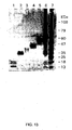
- 100 ⁇ l of serum was mixed with 1 ⁇ SDS-PAGE loading buffer, boiled for 10 minutes, and loaded to PrepCell (Bio-Rad). 2 hours of electrophoresis was conducted, and eluted under continuous flow off the end of the gel. Low molecular weight proteins migrated out of the gel at earlier times and were eluted to collection tubes. Virtually 100% yield was achieved, with sharp size segmentation. See figure 13 for an example gel from such a procedure.
- This technology can be applied to proteins loaded and subsequently released from the smart particles.
- the eluted fractions can be digested in solution with trypsin and sequenced by mass spectrometry.
- Eluted proteins from PrepCell can be passed through detergent clean-up micro kit ProteoSpin (Norgen, Canada) to remove the SDS in the elution buffer that could interfere with mass spectrometry analysis.
- the cleaned proteins are reduced by 10 mM DTT, alkylated by 50 mM iodoacetamide, and digested by trypsin (from Promega) at 37 °C overnight. Tryptic peptides are further purified by Sep-Pak cartridges (Waters) and analyzed by reversed-phase liquid chromatography nanospray tandem mass spectrometry using a linear ion-trap mass spectrometer (LTQ, ThermoElectron)).
- LTQ linear ion-trap mass spectrometer
- Separation columns are slurry-packed with 5 ⁇ m, 200 ⁇ pore size C 18 resin (Michrom BioResources) in 100 ⁇ m i.d. ⁇ 10 cm long fused silica capillary (Polymicro Technologies)) with a laser-pulled tip. After sample injection, the column is washed for 5 minutes with mobile phase A (0.4% acetic acid) and peptides eluted using a linear gradient of 0% mobile phase B (0.4% acetic acid, 80% acetonitrile) to 50% mobile phase B in 30 minutes at 250 nanoliter/min, then to 100% B in an additional 5 minutes.
- mobile phase A 0.4% acetic acid
- peptides eluted using a linear gradient of 0% mobile phase B (0.4% acetic acid, 80% acetonitrile) to 50% mobile phase B in 30 minutes at 250 nanoliter/min, then to 100% B in an additional 5 minutes.
- the LTQ mass spectrometer is operated in a data-dependent mode in which each full MS scan is followed by five MS/MS scans where the five most abundant molecular ions are dynamically selected for collision-induced dissociation (CID) using a normalized collision energy of 35%.
- Tandem mass spectra are searched against human database (downloaded from the National Center for Biotechnology Information) with SEQUEST using tryptic cleavage constraints. For a peptide to be considered legitimately identified, it has to achieve cross correlation scores of 1.5 for [NI+H]1+, 2.0 for [M+2H]2+, 2.5 for [M+3H]3+, and a maximum probabilities of randomized identification of 0.001.
- the generated protein lists is used to document release of proteins from the particles (16).
- Capillary Electrophoresis enables rapid fractionation of complex samples.
- a capillary is filled with a conductive fluid that is a buffer at a certain pH value.
- a sample is introduced from one end of the capillary using pressure and a high voltage is generated with separation based on size and charge.
- the migrating molecules pass through a light source that enables a spectrum of the molecules present to be generated.
- particles are loaded with proteins previously shown to be taken up by the particular particle in defined environmental conditions (temperature, etc.). Following loading, the particles will be washed in water followed by centrifugation (16,100 rcf for 15 minutes). The particles will be resuspended and protein eluted using temperature, pH, and chemical and/or physical disruption.
- CE are performed to detect the released protein. Prior to running this assay, the spectral profile of the target proteins will be measured on the CE.
- CE Using mass spectrometry-based detection, CE, and flow cytometry can demonstrate the sequestration, preservation, and controlled release of target protein analytes that have been added, or spiked, into heterogenous mixtures such as reference serum.
- Microgel particles can protect target analytes from enzymatic degradation after they are spiked into reference serum.
- Classes of microgel particles can incubated with serum that has been spiked with a fluorescent labeled protein of defined molecular weight.
- Optimal loading conditions can be defined (i.e. temperature, pH, time) as described above. After the incubation, the particles are centrifuged and washed once followed by centrifugation. Peptide uptake is detected using flow cytometry as described above. As described above, we have demonstrated that FITC labeled peptides spiked into serum can be sequestered with high efficiency into particles.
- PSA fluorescently labeled PSA
- PBNP pro-brain natriuretic peptide
- PSA can be spiked into female serum (ranging from 0.5 to 25 ng/ml, which represents a clinically relevant range of PSA spanning healthy to cancer-bearing patients).
- PBNP can be spiked into serum at a concentration of 0.5 to 5 ng/ml. Both of these molecules have clinical significance for patients with prostate disease and heart disease, respectively. Because of clinical interest in these molecules, a number of studies have been performed on the stability of these molecules under routine handling conditions (24,25). Uptake of these spiked molecules can be documented using the flow cytometry assay as described above.
- the presence of the target analytes in the eluates from the particles, PSA or PBNP, will therefore be detected with high sensitivity.
- Quantitation of PSA or PBNP analytes can be achieved through measuring protein eluates using commercially available ELISA formats. Not only will the ELISA format allow quantification of eluted biomarker from the particles, but analyte amplification studies can be performed as well.
- NIST reference sera spiked with PSA can be incubated with a class of microgel particles. Using previously defined environmental conditions suitable for loading, the particles can be harvested by centrifugation, and the remaining serum solution removed. An additional aliquot of the serum reference can be added to the particles.
- the particles can be incubated with serum and then isolated by centrifugation. This cycle can be continued “n” times to study the loading plateau.
- the particles are then be treated with eluent, or release, conditions (time, temperature, physical or chemical disruption), and the amount of PSA present in the eluent is compared with one round of serum exposure using ELISA.
- This type of analyte amplification mimics the role that carrier proteins play in the serum, by providing a site for sequestration and accumulation for small; low abundance analytes.
- the technology meets a critical need stated above, namely, the amplification and isolation of low abundance disease markers from serum.
- NIST serum reference standards spiked with PSA can be incubated for a range of time periods without the presence of protective microgel particles.
- the relative of whole PSA versus PSA fragmented by endogenous or exogenously added proteinases can be evaluated using a Bland Altman Plot (24,25)for a range of concentrations and times.
- PBNP can be studied in the same way.
- microgel particles sequester known and previously unknown low-abundance proteins and peptides in reference serum.
- microgel particles including particles containing AAc affinity bait
- Various classes of microgel particles can be incubated with serum diluted 1:10 in water at varying temperatures, times, and a range physical conditions (pressure or sonication). Loaded proteins can be entrapped by changing the temperature to 37C for varying time periods, which will markedly diminish the pore size. The particles can be washed multiple times at 37 C in order to remove unloaded proteins. In order to elute sequestered proteins, the particles can be incubated using a range of time, temperature, solvents and physical disruption techniques. Particles and particle fragments can be pelleted by centrifugation.
- the supernatant, or eluent can be run on a PrepCell device in order to size fractionate the eluted proteins.
- the fractionated eluent can then be analyzed by nanoRPLC-MS/MS. Tandem mass spectra are searched against human database (downloaded from the National Center for Biotechnology Information) with SEQUEST (16) using tryptic cleavage constraints. For a peptide to be considered legitimately identified, it will achieve cross correlation scores of 1.5 for [M+H]1+, 2.0 for [M+2H]2+, 2.5 for [M+3H]3+, and a maximum probabilities of randomized identification of 0.001. Sets of sequence identities can be cataloged and linked to the loading and release conditions used for the particular microgel assay.
- volumes of 10 ⁇ l of 2% hydrogel particles were diluted in 90 ⁇ l of FITC aqueous solutions at 20 ⁇ M. The samples were incubated overnight and then centrifuged at room temperature, at 16.1 rcf, and for 5 minutes. The pellet were then re-suspended in 100 ul of MilliQ water and centrifuged again with the same parameters. The pellet was re-suspended in 1 ml of water and fluorescence signal was analyzed by a flow cytometer to assess FITC uptake.
Landscapes
- Health & Medical Sciences (AREA)
- Life Sciences & Earth Sciences (AREA)
- Engineering & Computer Science (AREA)
- Immunology (AREA)
- Chemical & Material Sciences (AREA)
- Urology & Nephrology (AREA)
- Hematology (AREA)
- Biomedical Technology (AREA)
- Molecular Biology (AREA)
- Medicinal Chemistry (AREA)
- Analytical Chemistry (AREA)
- Cell Biology (AREA)
- Nanotechnology (AREA)
- Food Science & Technology (AREA)
- Biotechnology (AREA)
- Physics & Mathematics (AREA)
- Microbiology (AREA)
- Biochemistry (AREA)
- General Health & Medical Sciences (AREA)
- General Physics & Mathematics (AREA)
- Pathology (AREA)
- Solid-Sorbent Or Filter-Aiding Compositions (AREA)
- Peptides Or Proteins (AREA)
- Measuring Or Testing Involving Enzymes Or Micro-Organisms (AREA)
- Steroid Compounds (AREA)
Claims (13)
- Verfahren zum Isolieren von Analyten von einer Probe, wobei das Verfahren folgendes umfasst:a) das in Kontakt bringen einer Probe, die Analyte umfasst, mit Lösungsphasen-Auffangteilchen, wobei die Auffangteilchen folgendes umfassen:einen Molekularsiebteil als eine äußere Ummantelung und einen Analytbindungsteil als einen inneren Kern,wobei der Molekularsiebteil und optional der Analytbindungsteil eine vernetzte Region mit modifizierbarer Porosität umfassen,wobei der Analytbindungsteil mindestens einen Anteilstyp umfasst, der auf selektive Art und Weise ein Analyt chemisch oder elektrostatisch binden oder absondern kann;b) das Kontraktieren des Molekularsiebteils unter Verwendung einer physikalischen und/oder chemischen Behandlung, um Analyt in dem Auffangteilchen einzuschließen.
- Verfahren nach Anspruch 1, wobei der Analytbindungsteil der Auffangteilchen folgendes umfasst: eine Carboxylgruppe, eine Amingruppe, Lipid, Phosphoprotein, Phospholipide, eine Amidgruppe, eine Hydroxylgruppe, eine Estergruppe, eine Acrylgruppe, eine Thiolgruppe, Acrylsäure, Antikörper, Bindungsproteine, Bindungspaare, Metalle, Komplexbildner, Nukleinsäuren, Aptamere, Enzymbindungstaschen, Lektine, ein pharmakologisches Mittel, synthetische Peptide, Antikörperfragmente, eine hydrophobe Oberfläche, eine hydrophile Oberfläche, alle Derivate derer oder eine Kombination dieser.
- Verfahren nach Anspruch 1, wobei die Auffangteilchen ferner einen mit dem Analytbindungsteil gebundenen Analyt umfassen, wobei der genannte Analyt folgendes umfasst: organische Moleküle, anorganische Moleküle, Polypeptide, Kohlenhydrate, Nukleinsäuren, Lipide, Derivate derer oder eine beliebige Kombination dieser.
- Verfahren nach Anspruch 1, wobei die Auffangteilchen einen durchschnittlichen Teilchenradius von weniger als 100 µm aufweisen.
- Verfahren nach Anspruch 1, wobei der Molekularsiebteil, der Analytbindungsteil oder beide der Auffangteilchen folgendes umfassen: Polyacrylamid, Poly(N-isopropylacrylamid), N-alkyl-substituiertes Polyacrylamid, Poly(N-vinylalkylamid), Polymethacrylsäure, Polybenzylglutamat, Poly(2-ethylacrylsäure), Poly(4-vinylpyridin), Derivate derer oder jede Kombination dieser.
- Verfahren nach Anspruch 1, wobei die vernetzte Region der Auffangteilen folgendes umfasst: N,N'-methylenbisacrylamid, Ethylenglykoldimethacrylat, Tetraethylenglykoldimethylacrylat, Poly(ethylenglykol)dimethacrylat oder jede Kombination dieser.
- Verfahren nach Anspruch 1, wobei der Molekularsiebteil, der Analytbindungsteil oder beide der Auffangteilchen ein Hydrogel umfassen.
- Verfahren nach Anspruch 1, wobei der Molekularsiebteil, der Analytbindungsteil oder beide der Auffangteilchen eine durchschnittliche Porengröße zwischen 2 und 20 nm aufweisen.
- Verfahren nach Anspruch 1, wobei der Molekularsiebteil, der Analytbindungsteil oder beide der Auffangteilchen eine durchschnittliche Porengröße von weniger als 100 nm aufweisen.
- Verfahren nach Anspruch 1, wobei die Auffangteilchen einen gebundenen oder abgesonderten Analyt freisetzen, das Volumen ändern oder beides, wenn sie einer physikalischen oder chemischen Behandlung ausgesetzt werden.
- Verfahren nach Anspruch 10, wobei die physikalische oder chemische Behandlung das Aussetzen einer elektrischer Ladung, eines hydrostatischen Drucks, einer Änderung des pH-Wertes, einer Temperaturänderung, eines acidischen Mittels, eines basischen Mittels, einer UV-Strahlung, eines Ultraschalls, einer Röntgenstrahlung oder einer Kombination dieser umfasst.
- Verfahren nach Anspruch 1, wobei die Auffangteilchen ein 1404 Da Peptid aufnehmen können mit im Wesentlichen keiner Aufnahme von Albumin.
- Verfahren nach Anspruch 1, wobei die Auffangteilchen Insulin aufnehmen können,
Applications Claiming Priority (3)
| Application Number | Priority Date | Filing Date | Title |
|---|---|---|---|
| US72046605P | 2005-09-27 | 2005-09-27 | |
| US75957406P | 2006-01-18 | 2006-01-18 | |
| PCT/US2006/037508 WO2007038523A2 (en) | 2005-09-27 | 2006-09-27 | Method of isolating analytes from a sample |
Publications (2)
| Publication Number | Publication Date |
|---|---|
| EP1929299A2 EP1929299A2 (de) | 2008-06-11 |
| EP1929299B1 true EP1929299B1 (de) | 2010-12-22 |
Family
ID=37591893
Family Applications (1)
| Application Number | Title | Priority Date | Filing Date |
|---|---|---|---|
| EP20060804167 Active EP1929299B1 (de) | 2005-09-27 | 2006-09-27 | Verfahren zur isolierung von analyten aus einer probe |
Country Status (8)
| Country | Link |
|---|---|
| US (1) | US9005996B2 (de) |
| EP (1) | EP1929299B1 (de) |
| JP (1) | JP4942753B2 (de) |
| AT (1) | ATE492813T1 (de) |
| CA (1) | CA2625499A1 (de) |
| DE (1) | DE602006019111D1 (de) |
| MX (1) | MX2008004165A (de) |
| WO (1) | WO2007038523A2 (de) |
Cited By (1)
| Publication number | Priority date | Publication date | Assignee | Title |
|---|---|---|---|---|
| WO2012138429A1 (en) * | 2011-02-25 | 2012-10-11 | University Of South Florida | Molecularly imprinted polymers having affinity for natriuretic peptides |
Families Citing this family (33)
| Publication number | Priority date | Publication date | Assignee | Title |
|---|---|---|---|---|
| JP2009520989A (ja) * | 2005-12-20 | 2009-05-28 | ザ オハイオ ステイト ユニバーシティ リサーチ ファウンデーション | 分析法のためのナノ多孔性基材 |
| FR2902799B1 (fr) | 2006-06-27 | 2012-10-26 | Millipore Corp | Procede et unite de preparation d'un echantillon pour l'analyse microbiologique d'un liquide |
| GB0623043D0 (en) * | 2006-11-18 | 2006-12-27 | Univ Sheffield | Sensor devices |
| US8569464B2 (en) | 2006-12-21 | 2013-10-29 | Emd Millipore Corporation | Purification of proteins |
| WO2008079280A1 (en) | 2006-12-21 | 2008-07-03 | Millipore Corporation | Purification of proteins |
| US8362217B2 (en) | 2006-12-21 | 2013-01-29 | Emd Millipore Corporation | Purification of proteins |
| WO2009064389A2 (en) * | 2007-11-09 | 2009-05-22 | Alessandra Luchini | Smart hydrogel particles: biomarker harvesting: one-step affinity purification, size exclusion, and protection against degradation |
| WO2009151514A1 (en) | 2008-06-11 | 2009-12-17 | Millipore Corporation | Stirred tank bioreactor |
| KR101006562B1 (ko) | 2008-06-27 | 2011-01-07 | 포항공과대학교 산학협력단 | 핵산 추출 방법 |
| US9012240B2 (en) * | 2008-08-26 | 2015-04-21 | George Mason Research Foundation, Inc. | Hydrogel nanoparticle based immunoassay |
| CN102265155B (zh) | 2008-11-10 | 2014-08-13 | 卢米耐克斯公司 | 用于制备和使用多重检测中的大孔珠粒的方法和体系 |
| US20100190963A1 (en) | 2008-12-16 | 2010-07-29 | Millipore Corporation | Stirred Tank Reactor And Method |
| US8623496B2 (en) * | 2009-11-06 | 2014-01-07 | Wisconsin Alumni Research Foundation | Laser drilling technique for creating nanoscale holes |
| EP2571903B1 (de) | 2010-05-17 | 2019-09-04 | EMD Millipore Corporation | Stimulusreaktive polymere zur aufreinigung von biomolekülen |
| EP2691767B1 (de) * | 2011-03-31 | 2016-03-23 | GE Healthcare Bio-Sciences AB | Verfahren zur probenpräparation |
| US9746458B2 (en) | 2011-04-27 | 2017-08-29 | Loma Linda University Medical Center | Dynactin subunit p62 biomarker for neurological conditions |
| CN103930195A (zh) * | 2011-11-10 | 2014-07-16 | 谷纳米技术有限公司 | 吸收性的干燥生物流体采集基板 |
| US9322820B2 (en) | 2013-03-14 | 2016-04-26 | Wisconsin Alumni Research Foundation | System and apparatus for nanopore sequencing |
| CN105339791B (zh) * | 2013-03-14 | 2019-02-15 | 赛维德恩特有限公司 | 固相上的分子网 |
| US20180284108A1 (en) * | 2017-04-01 | 2018-10-04 | Michael Joseph Pugia | Method for complete and fragmented markers |
| WO2018218254A1 (en) * | 2017-05-26 | 2018-11-29 | George Mason University | Simultaneous parallel signal amplification and analyte-ligand capture functions |
| CN107540852B (zh) * | 2017-07-27 | 2020-07-14 | 西北工业大学 | 一种超声场制备核壳结构的粒子及中空粒子的方法 |
| EP3505934A1 (de) | 2017-12-29 | 2019-07-03 | Blink AG | Sensorkörper zur bindung und/oder anreicherung und/oder detektion eines analyten in einer probe |
| EP3578972A1 (de) * | 2018-06-06 | 2019-12-11 | Blink AG | Vorrichtung zur fraktionierung einer suspensionsprobe |
| CN109387641B (zh) * | 2018-12-20 | 2022-04-15 | 深圳职业技术学院 | 一种以磁性氧化铁纳米粒子为信号放大器检测天蚕素b的装置及检测方法 |
| BR112021011194A2 (pt) * | 2019-01-16 | 2021-08-31 | Regeneron Pharmaceuticals, Inc. | Método e sistema de identificar e quantificar a fragmentação do anticorpo |
| JP7228236B2 (ja) * | 2019-03-25 | 2023-02-24 | 株式会社日立ハイテクサイエンス | 遠心分離カラム及び分析対象物質の分析方法 |
| CN113203812A (zh) * | 2021-05-07 | 2021-08-03 | 杭州中一检测研究院有限公司 | 一种测定土壤中丙烯酰胺的检测方法 |
| US12253448B2 (en) * | 2022-05-03 | 2025-03-18 | University Of Utah Research Foundation | Isolation, storage, and delivery of extracellular vesicles using asymmetric depth filters |
| JPWO2024127742A1 (de) * | 2022-12-13 | 2024-06-20 | ||
| CN116251382A (zh) * | 2023-02-01 | 2023-06-13 | 南通市产品质量监督检验所(国家钢丝绳产品质量监督检验中心、江苏省电机产品质量监督检验中心、江苏省电动工具产品质量监督检验中心、江苏省缺陷产品管理技术中心南通分中心) | 用于水产品麻痹性贝类毒素净化富集的固相萃取柱及应用 |
| JP2024127129A (ja) * | 2023-03-08 | 2024-09-20 | Jnc株式会社 | 多孔質粒子、液体クロマトグラフィー担体、液体クロマトグラフィー装置、および生体高分子の分離精製方法 |
| WO2024228966A2 (en) * | 2023-04-29 | 2024-11-07 | Massachusetts Institute Of Technology | Charged hydrogels for concentrating proteins |
Family Cites Families (5)
| Publication number | Priority date | Publication date | Assignee | Title |
|---|---|---|---|---|
| CA1054050A (en) * | 1973-05-01 | 1979-05-08 | Wisconsin Alumni Research Foundation | Gel column device and use in blood purification and immunoassay |
| US4138474A (en) * | 1973-05-01 | 1979-02-06 | Wisconsin Alumni Research Foundation | Method and device for immunoassay |
| US7521019B2 (en) * | 2001-04-11 | 2009-04-21 | Lifescan, Inc. | Sensor device and methods for manufacture |
| WO2003106589A1 (en) | 2002-06-13 | 2003-12-24 | Lyotropic Therapeutics, Inc. | A nanoporous particle with a retained target |
| EP2142927A4 (de) * | 2002-06-13 | 2010-01-13 | Lyotropic Therapeutics Inc | Nanopor ses partikel mit zur ckgehaltenem targetmolek l |
-
2006
- 2006-09-27 EP EP20060804167 patent/EP1929299B1/de active Active
- 2006-09-27 CA CA002625499A patent/CA2625499A1/en not_active Abandoned
- 2006-09-27 MX MX2008004165A patent/MX2008004165A/es not_active Application Discontinuation
- 2006-09-27 WO PCT/US2006/037508 patent/WO2007038523A2/en not_active Ceased
- 2006-09-27 JP JP2008533525A patent/JP4942753B2/ja active Active
- 2006-09-27 US US11/527,727 patent/US9005996B2/en active Active
- 2006-09-27 DE DE200660019111 patent/DE602006019111D1/de active Active
- 2006-09-27 AT AT06804167T patent/ATE492813T1/de not_active IP Right Cessation
Cited By (1)
| Publication number | Priority date | Publication date | Assignee | Title |
|---|---|---|---|---|
| WO2012138429A1 (en) * | 2011-02-25 | 2012-10-11 | University Of South Florida | Molecularly imprinted polymers having affinity for natriuretic peptides |
Also Published As
| Publication number | Publication date |
|---|---|
| JP4942753B2 (ja) | 2012-05-30 |
| DE602006019111D1 (de) | 2011-02-03 |
| EP1929299A2 (de) | 2008-06-11 |
| JP2009510442A (ja) | 2009-03-12 |
| MX2008004165A (es) | 2008-11-12 |
| WO2007038523A3 (en) | 2007-05-24 |
| US9005996B2 (en) | 2015-04-14 |
| ATE492813T1 (de) | 2011-01-15 |
| US20100240543A1 (en) | 2010-09-23 |
| CA2625499A1 (en) | 2007-04-05 |
| WO2007038523A2 (en) | 2007-04-05 |
Similar Documents
| Publication | Publication Date | Title |
|---|---|---|
| US9005996B2 (en) | Method of isolating analytes from a sample | |
| EP2132256B1 (de) | Intelligente hydrogel-partikel zur gewinnung von biomarkern | |
| US9383299B2 (en) | Method for harvesting nanoparticles and sequestering biomarkers | |
| US8497137B2 (en) | Smart hydrogel particles for biomarker harvesting | |
| JP5896119B2 (ja) | ヒドロゲルナノ粒子に基づく免疫測定 | |
| Chou et al. | Nanoprobe-based affinity mass spectrometry for selected protein profiling in human plasma | |
| JP6515995B2 (ja) | モノクローナル抗体の検出のためのサンプル調製用キット | |
| Luchini et al. | Nanoparticle technology: addressing the fundamental roadblocks to protein biomarker discovery | |
| WO2016194114A1 (ja) | モノクローナル抗体の定量方法 | |
| EP2401619A1 (de) | Verfahren zur selektiven konzentration eines spezifischen low-abundance-biomoleküls | |
| EP3054297A1 (de) | Fluoreszierend markierte partikel | |
| EP3054283A1 (de) | Verfahren zur erkennung einer zielsubstanz | |
| Dvorakova et al. | An advanced conjugation strategy for the preparation of quantum dot-antibody immunoprobes | |
| Rembaum et al. | Design of polymeric immunomicrospheres for cell labelling and cell separation | |
| EP4348266A1 (de) | Verfahren und systeme zur einzelzellproteinanalyse | |
| Luchini et al. | Nanoparticle technology: Addressing the fundamental roadblocks to protein biomarker discovery | |
| CN117233384A (zh) | 检测外泌体的dna复合水凝胶及其制备方法和应用 | |
| Magni et al. | Application of hydrogel nanoparticles for the capture, concentration, and preservation of low-abundance biomarkers | |
| RU2684325C1 (ru) | Способ получения конъюгата на основе магнитных металл-углеродных наночастиц, пригодного для диагностических и аналитических целей, с использованием ЯМР-релаксометрии в качестве метода детекции | |
| KR101594379B1 (ko) | 면역크로마토그래피를 이용한 히스티딘-태그를 가진 단백질 검출방법 | |
| JP2006199798A (ja) | 反応性官能基を有する微粒子 | |
| WO2009064389A2 (en) | Smart hydrogel particles: biomarker harvesting: one-step affinity purification, size exclusion, and protection against degradation | |
| Ahmed | Use of Nanotechnology for Enhancing of Cancer Biomarker Discovery and Analysis: A Molecular Approach |
Legal Events
| Date | Code | Title | Description |
|---|---|---|---|
| PUAI | Public reference made under article 153(3) epc to a published international application that has entered the european phase |
Free format text: ORIGINAL CODE: 0009012 |
|
| AK | Designated contracting states |
Kind code of ref document: A2 Designated state(s): AT BE BG CH CY CZ DE DK EE ES FI FR GB GR HU IE IS IT LI LT LU LV MC NL PL PT RO SE SI SK TR |
|
| 17P | Request for examination filed |
Effective date: 20080327 |
|
| RIN1 | Information on inventor provided before grant (corrected) |
Inventor name: PETRICOIN, EMANUEL Inventor name: GEHO, DAVID Inventor name: LIOTTA, LANCE |
|
| RIN1 | Information on inventor provided before grant (corrected) |
Inventor name: PETRICOIN, EMANUEL Inventor name: LIOTTA, LANCE Inventor name: GEHO, DAVID |
|
| 17Q | First examination report despatched |
Effective date: 20090119 |
|
| RAP1 | Party data changed (applicant data changed or rights of an application transferred) |
Owner name: GEORGE MASON UNIVERSITY |
|
| RAP1 | Party data changed (applicant data changed or rights of an application transferred) |
Owner name: GEORGE MASON INTELLECTUAL PROPERTIES, INC. |
|
| 111L | Licence recorded |
Free format text: 0101 CERES NANOSCIENCES, LLLP Effective date: 20090814 |
|
| GRAP | Despatch of communication of intention to grant a patent |
Free format text: ORIGINAL CODE: EPIDOSNIGR1 |
|
| DAX | Request for extension of the european patent (deleted) | ||
| GRAS | Grant fee paid |
Free format text: ORIGINAL CODE: EPIDOSNIGR3 |
|
| GRAA | (expected) grant |
Free format text: ORIGINAL CODE: 0009210 |
|
| AK | Designated contracting states |
Kind code of ref document: B1 Designated state(s): AT BE BG CH CY CZ DE DK EE ES FI FR GB GR HU IE IS IT LI LT LU LV MC NL PL PT RO SE SI SK TR |
|
| REG | Reference to a national code |
Ref country code: GB Ref legal event code: FG4D |
|
| REG | Reference to a national code |
Ref country code: CH Ref legal event code: PK Free format text: ERGAENZUNG LIZENZEINTRAG: AUSSCHLIESSLICHE LIZENZ Ref country code: CH Ref legal event code: EP |
|
| REG | Reference to a national code |
Ref country code: IE Ref legal event code: FG4D |
|
| REF | Corresponds to: |
Ref document number: 602006019111 Country of ref document: DE Date of ref document: 20110203 Kind code of ref document: P |
|
| REG | Reference to a national code |
Ref country code: DE Ref legal event code: R096 Ref document number: 602006019111 Country of ref document: DE Effective date: 20110203 |
|
| REG | Reference to a national code |
Ref country code: NL Ref legal event code: VDEP Effective date: 20101222 |
|
| PG25 | Lapsed in a contracting state [announced via postgrant information from national office to epo] |
Ref country code: LT Free format text: LAPSE BECAUSE OF FAILURE TO SUBMIT A TRANSLATION OF THE DESCRIPTION OR TO PAY THE FEE WITHIN THE PRESCRIBED TIME-LIMIT Effective date: 20101222 |
|
| LTIE | Lt: invalidation of european patent or patent extension |
Effective date: 20101222 |
|
| PG25 | Lapsed in a contracting state [announced via postgrant information from national office to epo] |
Ref country code: AT Free format text: LAPSE BECAUSE OF FAILURE TO SUBMIT A TRANSLATION OF THE DESCRIPTION OR TO PAY THE FEE WITHIN THE PRESCRIBED TIME-LIMIT Effective date: 20101222 Ref country code: SE Free format text: LAPSE BECAUSE OF FAILURE TO SUBMIT A TRANSLATION OF THE DESCRIPTION OR TO PAY THE FEE WITHIN THE PRESCRIBED TIME-LIMIT Effective date: 20101222 Ref country code: FI Free format text: LAPSE BECAUSE OF FAILURE TO SUBMIT A TRANSLATION OF THE DESCRIPTION OR TO PAY THE FEE WITHIN THE PRESCRIBED TIME-LIMIT Effective date: 20101222 Ref country code: CY Free format text: LAPSE BECAUSE OF FAILURE TO SUBMIT A TRANSLATION OF THE DESCRIPTION OR TO PAY THE FEE WITHIN THE PRESCRIBED TIME-LIMIT Effective date: 20101222 Ref country code: SI Free format text: LAPSE BECAUSE OF FAILURE TO SUBMIT A TRANSLATION OF THE DESCRIPTION OR TO PAY THE FEE WITHIN THE PRESCRIBED TIME-LIMIT Effective date: 20101222 Ref country code: LV Free format text: LAPSE BECAUSE OF FAILURE TO SUBMIT A TRANSLATION OF THE DESCRIPTION OR TO PAY THE FEE WITHIN THE PRESCRIBED TIME-LIMIT Effective date: 20101222 Ref country code: BG Free format text: LAPSE BECAUSE OF FAILURE TO SUBMIT A TRANSLATION OF THE DESCRIPTION OR TO PAY THE FEE WITHIN THE PRESCRIBED TIME-LIMIT Effective date: 20110322 |
|
| PG25 | Lapsed in a contracting state [announced via postgrant information from national office to epo] |
Ref country code: EE Free format text: LAPSE BECAUSE OF FAILURE TO SUBMIT A TRANSLATION OF THE DESCRIPTION OR TO PAY THE FEE WITHIN THE PRESCRIBED TIME-LIMIT Effective date: 20101222 Ref country code: CZ Free format text: LAPSE BECAUSE OF FAILURE TO SUBMIT A TRANSLATION OF THE DESCRIPTION OR TO PAY THE FEE WITHIN THE PRESCRIBED TIME-LIMIT Effective date: 20101222 Ref country code: PT Free format text: LAPSE BECAUSE OF FAILURE TO SUBMIT A TRANSLATION OF THE DESCRIPTION OR TO PAY THE FEE WITHIN THE PRESCRIBED TIME-LIMIT Effective date: 20110422 Ref country code: BE Free format text: LAPSE BECAUSE OF FAILURE TO SUBMIT A TRANSLATION OF THE DESCRIPTION OR TO PAY THE FEE WITHIN THE PRESCRIBED TIME-LIMIT Effective date: 20101222 Ref country code: IS Free format text: LAPSE BECAUSE OF FAILURE TO SUBMIT A TRANSLATION OF THE DESCRIPTION OR TO PAY THE FEE WITHIN THE PRESCRIBED TIME-LIMIT Effective date: 20110422 Ref country code: ES Free format text: LAPSE BECAUSE OF FAILURE TO SUBMIT A TRANSLATION OF THE DESCRIPTION OR TO PAY THE FEE WITHIN THE PRESCRIBED TIME-LIMIT Effective date: 20110402 Ref country code: GR Free format text: LAPSE BECAUSE OF FAILURE TO SUBMIT A TRANSLATION OF THE DESCRIPTION OR TO PAY THE FEE WITHIN THE PRESCRIBED TIME-LIMIT Effective date: 20110323 |
|
| PG25 | Lapsed in a contracting state [announced via postgrant information from national office to epo] |
Ref country code: SK Free format text: LAPSE BECAUSE OF FAILURE TO SUBMIT A TRANSLATION OF THE DESCRIPTION OR TO PAY THE FEE WITHIN THE PRESCRIBED TIME-LIMIT Effective date: 20101222 Ref country code: NL Free format text: LAPSE BECAUSE OF FAILURE TO SUBMIT A TRANSLATION OF THE DESCRIPTION OR TO PAY THE FEE WITHIN THE PRESCRIBED TIME-LIMIT Effective date: 20101222 Ref country code: PL Free format text: LAPSE BECAUSE OF FAILURE TO SUBMIT A TRANSLATION OF THE DESCRIPTION OR TO PAY THE FEE WITHIN THE PRESCRIBED TIME-LIMIT Effective date: 20101222 Ref country code: RO Free format text: LAPSE BECAUSE OF FAILURE TO SUBMIT A TRANSLATION OF THE DESCRIPTION OR TO PAY THE FEE WITHIN THE PRESCRIBED TIME-LIMIT Effective date: 20101222 |
|
| PLBE | No opposition filed within time limit |
Free format text: ORIGINAL CODE: 0009261 |
|
| STAA | Information on the status of an ep patent application or granted ep patent |
Free format text: STATUS: NO OPPOSITION FILED WITHIN TIME LIMIT |
|
| PG25 | Lapsed in a contracting state [announced via postgrant information from national office to epo] |
Ref country code: DK Free format text: LAPSE BECAUSE OF FAILURE TO SUBMIT A TRANSLATION OF THE DESCRIPTION OR TO PAY THE FEE WITHIN THE PRESCRIBED TIME-LIMIT Effective date: 20101222 |
|
| 26N | No opposition filed |
Effective date: 20110923 |
|
| PG25 | Lapsed in a contracting state [announced via postgrant information from national office to epo] |
Ref country code: IT Free format text: LAPSE BECAUSE OF FAILURE TO SUBMIT A TRANSLATION OF THE DESCRIPTION OR TO PAY THE FEE WITHIN THE PRESCRIBED TIME-LIMIT Effective date: 20101222 |
|
| REG | Reference to a national code |
Ref country code: DE Ref legal event code: R097 Ref document number: 602006019111 Country of ref document: DE Effective date: 20110923 |
|
| PG25 | Lapsed in a contracting state [announced via postgrant information from national office to epo] |
Ref country code: MC Free format text: LAPSE BECAUSE OF NON-PAYMENT OF DUE FEES Effective date: 20110930 |
|
| REG | Reference to a national code |
Ref country code: CH Ref legal event code: PL |
|
| REG | Reference to a national code |
Ref country code: IE Ref legal event code: MM4A |
|
| PG25 | Lapsed in a contracting state [announced via postgrant information from national office to epo] |
Ref country code: CH Free format text: LAPSE BECAUSE OF NON-PAYMENT OF DUE FEES Effective date: 20110930 Ref country code: IE Free format text: LAPSE BECAUSE OF NON-PAYMENT OF DUE FEES Effective date: 20110927 Ref country code: LI Free format text: LAPSE BECAUSE OF NON-PAYMENT OF DUE FEES Effective date: 20110930 |
|
| PG25 | Lapsed in a contracting state [announced via postgrant information from national office to epo] |
Ref country code: LU Free format text: LAPSE BECAUSE OF NON-PAYMENT OF DUE FEES Effective date: 20110927 |
|
| PG25 | Lapsed in a contracting state [announced via postgrant information from national office to epo] |
Ref country code: TR Free format text: LAPSE BECAUSE OF FAILURE TO SUBMIT A TRANSLATION OF THE DESCRIPTION OR TO PAY THE FEE WITHIN THE PRESCRIBED TIME-LIMIT Effective date: 20101222 |
|
| PG25 | Lapsed in a contracting state [announced via postgrant information from national office to epo] |
Ref country code: HU Free format text: LAPSE BECAUSE OF FAILURE TO SUBMIT A TRANSLATION OF THE DESCRIPTION OR TO PAY THE FEE WITHIN THE PRESCRIBED TIME-LIMIT Effective date: 20101222 |
|
| REG | Reference to a national code |
Ref country code: FR Ref legal event code: CD Owner name: GEORGE MASON RESEARCH FOUNDATION, INC., US Effective date: 20140902 |
|
| REG | Reference to a national code |
Ref country code: DE Ref legal event code: R082 Ref document number: 602006019111 Country of ref document: DE Representative=s name: MEYER, THORSTEN, DIPL.-PHYS., DE |
|
| REG | Reference to a national code |
Ref country code: DE Ref legal event code: R082 Ref document number: 602006019111 Country of ref document: DE Representative=s name: MEYER, THORSTEN, DIPL.-PHYS., DE Effective date: 20141104 Ref country code: DE Ref legal event code: R081 Ref document number: 602006019111 Country of ref document: DE Owner name: GEORGE MASON RESEARCH FOUNDATION, INC., FAIRFA, US Free format text: FORMER OWNER: GEORGE MASON INTELLECTUAL PROPERTIES, INC., FAIRFAX, VA., US Effective date: 20141104 |
|
| REG | Reference to a national code |
Ref country code: FR Ref legal event code: PLFP Year of fee payment: 10 |
|
| REG | Reference to a national code |
Ref country code: FR Ref legal event code: PLFP Year of fee payment: 11 |
|
| REG | Reference to a national code |
Ref country code: FR Ref legal event code: PLFP Year of fee payment: 12 |
|
| PGFP | Annual fee paid to national office [announced via postgrant information from national office to epo] |
Ref country code: DE Payment date: 20250819 Year of fee payment: 20 |
|
| PGFP | Annual fee paid to national office [announced via postgrant information from national office to epo] |
Ref country code: GB Payment date: 20250818 Year of fee payment: 20 |
|
| PGFP | Annual fee paid to national office [announced via postgrant information from national office to epo] |
Ref country code: FR Payment date: 20250821 Year of fee payment: 20 |