EP1884771B1 - Membrane a proteine immobilisee, procede pour l'immobilisation de proteine, electrode a enzyme immobilisee, et capteur biologique - Google Patents
Membrane a proteine immobilisee, procede pour l'immobilisation de proteine, electrode a enzyme immobilisee, et capteur biologique Download PDFInfo
- Publication number
- EP1884771B1 EP1884771B1 EP06746594.8A EP06746594A EP1884771B1 EP 1884771 B1 EP1884771 B1 EP 1884771B1 EP 06746594 A EP06746594 A EP 06746594A EP 1884771 B1 EP1884771 B1 EP 1884771B1
- Authority
- EP
- European Patent Office
- Prior art keywords
- layer
- cell membrane
- enzyme
- protein
- immobilized
- Prior art date
- Legal status (The legal status is an assumption and is not a legal conclusion. Google has not performed a legal analysis and makes no representation as to the accuracy of the status listed.)
- Active
Links
Images


Classifications
-
- C—CHEMISTRY; METALLURGY
- C12—BIOCHEMISTRY; BEER; SPIRITS; WINE; VINEGAR; MICROBIOLOGY; ENZYMOLOGY; MUTATION OR GENETIC ENGINEERING
- C12N—MICROORGANISMS OR ENZYMES; COMPOSITIONS THEREOF; PROPAGATING, PRESERVING, OR MAINTAINING MICROORGANISMS; MUTATION OR GENETIC ENGINEERING; CULTURE MEDIA
- C12N11/00—Carrier-bound or immobilised enzymes; Carrier-bound or immobilised microbial cells; Preparation thereof
- C12N11/02—Enzymes or microbial cells immobilised on or in an organic carrier
- C12N11/08—Enzymes or microbial cells immobilised on or in an organic carrier the carrier being a synthetic polymer
- C12N11/082—Enzymes or microbial cells immobilised on or in an organic carrier the carrier being a synthetic polymer obtained by reactions only involving carbon-to-carbon unsaturated bonds
- C12N11/087—Acrylic polymers
-
- G—PHYSICS
- G01—MEASURING; TESTING
- G01N—INVESTIGATING OR ANALYSING MATERIALS BY DETERMINING THEIR CHEMICAL OR PHYSICAL PROPERTIES
- G01N27/00—Investigating or analysing materials by the use of electric, electrochemical, or magnetic means
- G01N27/26—Investigating or analysing materials by the use of electric, electrochemical, or magnetic means by investigating electrochemical variables; by using electrolysis or electrophoresis
- G01N27/28—Electrolytic cell components
- G01N27/30—Electrodes, e.g. test electrodes; Half-cells
- G01N27/327—Biochemical electrodes, e.g. electrical or mechanical details for in vitro measurements
- G01N27/3271—Amperometric enzyme electrodes for analytes in body fluids, e.g. glucose in blood
- G01N27/3272—Test elements therefor, i.e. disposable laminated substrates with electrodes, reagent and channels
-
- C—CHEMISTRY; METALLURGY
- C07—ORGANIC CHEMISTRY
- C07K—PEPTIDES
- C07K14/00—Peptides having more than 20 amino acids; Gastrins; Somatostatins; Melanotropins; Derivatives thereof
- C07K14/435—Peptides having more than 20 amino acids; Gastrins; Somatostatins; Melanotropins; Derivatives thereof from animals; from humans
- C07K14/705—Receptors; Cell surface antigens; Cell surface determinants
-
- C—CHEMISTRY; METALLURGY
- C07—ORGANIC CHEMISTRY
- C07K—PEPTIDES
- C07K14/00—Peptides having more than 20 amino acids; Gastrins; Somatostatins; Melanotropins; Derivatives thereof
- C07K14/795—Porphyrin- or corrin-ring-containing peptides
- C07K14/80—Cytochromes
-
- C—CHEMISTRY; METALLURGY
- C12—BIOCHEMISTRY; BEER; SPIRITS; WINE; VINEGAR; MICROBIOLOGY; ENZYMOLOGY; MUTATION OR GENETIC ENGINEERING
- C12N—MICROORGANISMS OR ENZYMES; COMPOSITIONS THEREOF; PROPAGATING, PRESERVING, OR MAINTAINING MICROORGANISMS; MUTATION OR GENETIC ENGINEERING; CULTURE MEDIA
- C12N11/00—Carrier-bound or immobilised enzymes; Carrier-bound or immobilised microbial cells; Preparation thereof
- C12N11/02—Enzymes or microbial cells immobilised on or in an organic carrier
- C12N11/06—Enzymes or microbial cells immobilised on or in an organic carrier attached to the carrier via a bridging agent
-
- C—CHEMISTRY; METALLURGY
- C12—BIOCHEMISTRY; BEER; SPIRITS; WINE; VINEGAR; MICROBIOLOGY; ENZYMOLOGY; MUTATION OR GENETIC ENGINEERING
- C12N—MICROORGANISMS OR ENZYMES; COMPOSITIONS THEREOF; PROPAGATING, PRESERVING, OR MAINTAINING MICROORGANISMS; MUTATION OR GENETIC ENGINEERING; CULTURE MEDIA
- C12N9/00—Enzymes; Proenzymes; Compositions thereof; Processes for preparing, activating, inhibiting, separating or purifying enzymes
- C12N9/0004—Oxidoreductases (1.)
- C12N9/0006—Oxidoreductases (1.) acting on CH-OH groups as donors (1.1)
-
- C—CHEMISTRY; METALLURGY
- C12—BIOCHEMISTRY; BEER; SPIRITS; WINE; VINEGAR; MICROBIOLOGY; ENZYMOLOGY; MUTATION OR GENETIC ENGINEERING
- C12Q—MEASURING OR TESTING PROCESSES INVOLVING ENZYMES, NUCLEIC ACIDS OR MICROORGANISMS; COMPOSITIONS OR TEST PAPERS THEREFOR; PROCESSES OF PREPARING SUCH COMPOSITIONS; CONDITION-RESPONSIVE CONTROL IN MICROBIOLOGICAL OR ENZYMOLOGICAL PROCESSES
- C12Q1/00—Measuring or testing processes involving enzymes, nucleic acids or microorganisms; Compositions therefor; Processes of preparing such compositions
- C12Q1/001—Enzyme electrodes
- C12Q1/005—Enzyme electrodes involving specific analytes or enzymes
- C12Q1/006—Enzyme electrodes involving specific analytes or enzymes for glucose
-
- G—PHYSICS
- G01—MEASURING; TESTING
- G01N—INVESTIGATING OR ANALYSING MATERIALS BY DETERMINING THEIR CHEMICAL OR PHYSICAL PROPERTIES
- G01N2333/00—Assays involving biological materials from specific organisms or of a specific nature
- G01N2333/795—Porphyrin- or corrin-ring-containing peptides
- G01N2333/80—Cytochromes
Definitions
- the present invention relates to a technique for immobilizing a protein containing cytochrome to an immobilization target material.
- Biosensors designed to analyze a sample by an electrochemical or optical method are widely used.
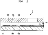
- An example of biosensors designed to analyze a sample by an electrochemical method is a biosensor 9 shown in, Fig. 12 of the present application.
- the illustrated biosensor) 9 includes a substrate 92 formed with a working electrode 90 and a counter electrode 91, and a cover 94 bonded to the substrate via a spacer 93.
- the biosensor 9 further includes a flow path 95 defined by the substrate 92, the spacer 93 and the cover 94.
- the flow path 95 is used for moving a sample by capillary force and formed with a reagent portion 96.
- the reagent portion 96 connects the ends of the working electrode 90 and the counter electrode 91 and contains oxidoreductase.
- the oxidoreductase catalyzes the reaction of taking electrons from glucose, for example.
- the electrons taken from the glucose are supplied to the working electrode 90.
- the amount of electrons supplied to the working electrode 90 is measured as the responsive current by utilizing the working electrode 90 and the counter electrode 91.
- a material liquid containing oxidoreductase is applied to an intended portion of a target, and then the material liquid is dried. In this way, the oxidoreductase is immobilized to the intended portion of the target.
- oxidoreductase is immobilized to an intended portion of a target by using a cross-linker such as glutaraldehyde.
- oxidoreductase is contained in a polymer such carboxymethylcellulose (CMC), and then the oxidoreductase is immobilized together with the polymer.
- CMC carboxymethylcellulose
- oxidoreductase is dispersed in a conductive material such as a carbon paste, and the resultant paste is applied to an intended portion of a target, to immobilize the oxidoreductase.
- oxidoreductase fails to be immobilized in a manner such that the active sites are oriented (located) to exhibit efficient activity of the oxidoreductase.
- the conventional methods have a drawback that the immobilization is not performed with the orientation of the oxidoreductase being controlled.
- active sites of oxidoreductase existing adjacent to each other may face each other or proteins may aggregate each other so that the active site exists within the aggregate.
- the ratio of the oxidoreductase (active site) which can be utilized efficiently is relatively low.
- the probability that oxidoreductase comes into contact with a substrate is relatively low, so that the activity of the immobilized oxidoreductase as a whole is low.
- the amount of oxidoreductase to be loaded needs to be increased, which is disadvantageous in terms of cost.
- oxidoreductases are generally expensive, the increase in the amount of oxidoreductase to be loaded leads to a considerably disadvantageous cost increase.
- the biosensor 9 provided by immobilizing oxidoreductase by a conventional method is disadvantageous in terms of cost, because it requires anelectronmediator.
- the electron mediator metal complexes such as potassium ferrocyanide are used some of which have an adverse effect on the human body. Thus, it is not desirable to use an electron mediator for such an analytical tool as the biosensor 9.
- Patent document 1 JP-B-H08-10208
- Non-patent document 1 MIZUTANI Fumio, "Application of enzyme-modified electrodes to biosensors," BUNSEKI KAGAKU, Vol. 48, No. 9 pp. 809-821, The Japan Society for Analytical Chemistry, September, 1999 .
- EP 1426757 describes an enzyme biosensor comprising an electrode and a reagent layer (membrane) which contains CyGDH.
- An obj ect of the present invention is to immobilize a protein such as oxidoreductase with good orientation and to cause the activity to be exhibited efficiently and advantageously in terms of cost with the use of a small amount of enzyme.
- Another object of the present invention is to provide a biosensor which is capable of properly measuring the concentration of a substrate such as glucose without using an electron mediator.
- a protein-immobilized membrane comprising:
- a method for immobilizing a protein comprising:
- the protein immobilization method according to the present invention further comprises a third step of subjecting the intended portion to hydrophilic treatment before the first step.
- an enzyme-immobilized electrode comprising:
- a biosensor comprising:
- the biosensor according to the present invention may further comprise a working electrode and a counter electrode which are partially exposed at the flow path and utilized for applying a voltage to a sample.
- a working electrode and a counter electrode which are partially exposed at the flow path and utilized for applying a voltage to a sample.
- at least part of the cell membrane homologous layer is formed on the working electrode.
- the reagent portion may contain a color former.
- the reagent portion may include a chromogenic layer containing a color former, a cell membrane homologous layer, and a layer containing an enzyme.
- the cell membrane homologous layer in the present invention contains a phospholipid polymer which is 2-methacryloyloxyethyl phosphorylcholine polymer.
- the cell membrane homologous layer in the present invention contains a silane coupling agent.
- a silane coupling agent it is preferable to use tetraethoxysilane.
- the protein such as an enzyme in the present invention is CyGDH containing an ⁇ subunit having a glucose dehydrogenase activity and cytochrome C having a function of electron transfer.
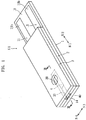
- the biosensor X1 shown in Figs. 1-3 is a disposable sensor to be mounted to a concentration measuring apparatus (not shown) to measure a blood glucose level.
- the biosensor X1 is adapted to measure the blood glucose level by an electrochemical method and includes a substrate 1, which is in the form of an elongated rectangle, and a cover 3 laminated on the substrate via a spacer 2.
- a capillary 4 extending in the longitudinal direction of the substrate 1 (N1, N2 directions in the figures) is defined by the elements 1-3.
- the capillary 4 is utilized for moving the blood introduced from an introduction port 40 in the longitudinal direction of the substrate 1 (N1, N2 directions in the figures) utilizing capillary action and retaining the introduced blood.
- the spacer 2 defines the distance from the upper surface 10 of the substrate 1 to the lower surface 30 of the cover 3, i.e., the height of the capillary 4 and may comprise a double-sided tape.
- the spacer 2 is formed with a slit 20 having an open end.
- the slit 20 defines the width of the capillary 4.
- the open end of the slid 20 serves as the introduction port 40 for introducing blood into the capillary 4.
- the clover 3 includes an exhaust port 30 for discharging gas from the capillary 4.
- the cover 3 is made of a thermoplastic resin having a high wettability, such as Vinylon or highly crystalline PVA.
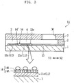
- Each of the working electrode 11 and the counter electrode 12 is L-shaped as a whole. Specifically, the working electrode 11 and the counter electrode 12 mostly extend in the longitudinal direction of the substrate 1 (N1, N2 directions in the figures) and respectively include ends 11a and 12a extending in the width direction (N3, N4 directions in the figures). The working electrode 11 and the counter electrode 12 further include ends 11b and 12b, respectively, which provide terminals for coming into contact with the terminals of the concentration measuring apparatus (not shown).
- the working electrode 11 and the counter electrode 12 may be formed by screen printing using carbon paste.
- the working electrode 11 and the counter electrode 12 may be made of a conductive material other than carbon by spin coating, thermal transfer, carbon rod slice, vapor deposition, sputtering or CVD.
- the insulating film 13 covers most part of the working electrode 11 and the counter electrode 12 while exposing the ends 11a, 12a, 11b and 12b of the working electrode 11 and the counter electrode 12.
- the insulating film 13 includes an opening 13a for exposing the ends 11a and 12a of the working electrode 11 and the counter electrode 12.
- the opening 13a defines the region for forming the reagent portion 14 and is in the form of a rectangle elongated in the longitudinal direction of the substrate 1 (N1, N2 directions in the figures).
- the insulating film 13 may be formed by screen printing using ink containing a material having high water repellency or photolithography using a photosensitive resin.
- the reagent portion 14 is arranged to bridge the ends 11a and 12a of the working electrode 11 and the counter electrode 12 at the opening 13a of the insulating film 13.
- the reagent portion 14 includes a cell membrane homologous layer 14A and a CyGDH layer 14B.
- the cell membrane homologous layer 14A is utilized for immobilizing CyGDH with controlled orientation.
- the cell membrane homologous layer 14A may be formed by applying a solution containing phospholipid polymer to the portion 14' of the working electrode 11 and the counter electrode 12 which is exposed through the opening 13a of the insulating film 13 (hereinafter, the portion 14' is referred to as "exposed portion 14''') and then drying the solution.
- phospholipid polymer use is made of 2-methacryloyloxyethylphosphorylcholine (MPC) polymer.
- MPC 2-methacryloyloxyethylphosphorylcholine
- use may be made of one prepared by polymerizing MPC alone or one prepared by copolymerizing MPC with a hydrophobic monomer such as methacrylate (e.g. butyl methacrylate).
- the phospholipid polymer it is preferable to use one to which a silane coupling agent is added. In this case, the phospholipid polymer is reliably bonded to the exposed portion 14'.
- the exposed portion 14' it is preferable to subject the exposed portion 14' to hydrophilic treatment in advance.
- hydrophilic groups such as a hydroxyl group or a carboxyl group enters the exposed portion 14' and is bonded to the silane coupling agent.
- phospholipid polymer is more strongly fixed to the exposed portion 14'.
- the amount of the silane coupling agent in the polymer may be set to 10 to 500 parts by weight relative to 100 parts by weight of the polymer component.
- silane coupling agent include: tetraethoxysilane; vinyltrichlorosilane; vinyl-tris(2-methoxyethoxy)silane; ⁇ -methacryloxypropyltrimethoxysilane; ⁇ -methacryloxypropyltriethoxysilane; ⁇ -(3,4-epoxycyclohexyl)ethyltrimethoxysilane; ⁇ -glycidoxypropyltriethoxysilane; ⁇ -aminopropyltriethoxysilane; N-phenyl- ⁇ -aminopropyltrimethoxysilane; ⁇ -chloropropyltrimethoxysilane; and ⁇ -mercaptopropyltrimethoxysilane. These silane coupling agents may be used solely or in combination.
- the hydrophilic treatment of the exposed portion 14' can be performed by various known techniques.
- hydrophilic treatment which can be employed in the present invention include VUV treatment, UV treatment, corona discharge and plasma treatment.
- the CyGDH layer 14B is provided by immobilizing CyGDH self-organizingly to the cell membrane homologous layer 14A.
- Fig. 3 shows the state in which CyGDH is immobilized to the surface of the cell membrane homologous layer 14A, this figure is a schematic view for describing the present invention.
- CyGDH is self-organizingly immobilized to the cell membrane homologous layer 14A
- the inventors have not yet found out how CyGDH is immobilized to the cell membrane homologous layer 14A.
- CyGDH derived from a microorganism belonging to the burkhorderia cepacia which will be described later, is a transmembrane protein.
- CyGDH may not be immobilized only at the surface of the cell membrane homologous layer 14A, as shown in Fig. 3 , but may be immobilized to the cell membrane homologous layer 14A while penetrating the cell membrane homologous layer 14A.
- the self-organizing immobilization of CyGDH to the cell membrane homologous layer 14A may be performed by immersing the substrate 1 provided with the cell membrane homologous layer 14A at the exposed portion 14' into an enzyme solution containing CYGDH or spraying the enzyme solution to the cell membrane homologous layer 14A and then drying the solution.
- CyGDH is self-organizingly immobilized to the cell membrane homologous layer 14A
- CYGDH is immobilized with controlled orientation.
- CyGDH is so immobilized to the cell membrane homologous layer 14A that the active site of the ⁇ subunit, is positioned at the surface of the reagent portion 14, whereas cytochrome C is positioned close to or in contact with the exposed portion 14' (working electrode 11).
- CyGDH use is made of those which at least contain an ⁇ subunit having a glucose dehydrogenase activity and cytochrome C having a function of electron transfer.
- CYGDH further containing a subunit other than ⁇ subunit and cytochrome C may be used. Examples of such CyGDH are disclosed in international publication WO02/36779 .
- the CyGDH disclosed in this international publication is derived from a microorganism belonging to the burkholderia cepacia and includes an ⁇ subunit, having a molecular weight of about 60 kDa in SDS-polyacrylamide gel electrophoresis under a reduced condition, including FAD as a cofactor and having a glucose dehydrogenase activity, and cytochrome C having a molecular weight of about 43 kDa in SDS-polyacrylamide gel electrophoresis under a reduced condition and having a function of electron transfer.
- the CyGDH in the present invention further includes one prepared by utilizing a transformant to which a gene encoding CYGDH taken from a microorganism belonging to the burkholderia cepacia is transferred.
- the CyGDH derived from a microorganism belonging to the burkhorderia cepacia is a transmembrane protein. That is, the CyGDH derived from this microorganism originally exists in a cell membrane.
- CyGDH is immobilized to the cell membrane homologous layer 14A by self organization with controlled orientation similarly to that in existing in a cell membrane.
- Such self-organizing immobilization of CyGDH is possible not only when CYGDH, derived from a microorganism belonging to the burkhorderia cepacia is used but also when CYGDH, originally existing in a cell membrane is used.
- the biosensor X1 having the above-described structure is mounted to a concentration measuring apparatus (not shown) and blood is introduced to the capillary through the introduction port 40 of the biosensor X1, the blood glucose level is measured automatically at the concentration measuring apparatus (not shown).
- the introduction of blood to the biosensor X1 may be performed either before or after the biosensor is mounted to the concentration measuring apparatus (not shown). Generally, blood is introduced by cutting the skin of the person to be tested to cause bleeding and then applying the blood to the introduction port 40 of the biosensor X1.
- the working electrode 11 and the counter electrode 12 of the biosensor) X1 come into contact with the terminals (not shown) of the concentration measuring apparatus.
- the blood applied to the introduction port 40 moves toward the exhaust port 30 due to capillary action at the capillary 4 and fills the capillary 4.
- CyGDH reacts specifically with the glucose in the blood to take electrons from the glucose.
- the electrons taken out by the CyGDH are transferred to the working electrode 11.
- the concentration measuring apparatus (not shown), when a voltage is applied to the working electrode 11 and the counter electrode 12, the amount of electrons transferred to the working electrode 11, for example, is measured as the responsive current. Based on the responsive current, the blood glucose level is computed.
- CYGDH is immobilized with controlled orientation so that the active site of the ⁇ subunit is positioned at the surface of the reagent portion 14.
- electrons are efficiently taken from glucose.
- intended activity is properly exhibited even with the use of a relatively small amount of CyGDH, which is advantageous in terms of cost.
- CyGDH is immobilized with controlled orientation in the biosensor X1, cytochrome C exists close to or in contact with the exposed portion 14' (working electrode 11). Thus, in the reagent portion 14, electrons taken from the glucose are efficiently transferred to the working electrode 11. Thus, in the biosensor X1, proper responsive current is obtained without using an electron mediator such as a metal complex.
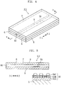
- the biosensor X2 shown in Figs. 4 and 5 is adapted to measure the blood glucose level by an optical method.
- the biosensor X2 includes a substrate 5, which is in the form of an elongated rectangle, and a cover 7 laminated on the substrate via a pair of spacers 6.
- a capillary 8 extending in the longitudinal direction of the substrate 5 (N1, N2 directions in the figures) is defined by the elements 5-7.
- the capillary 8 is used for moving the blood introduced from an introduction port 80 in the longitudinal direction of the substrate 5 (N1, N2 directions in the figures) utilizing capillary action and retaining the introduced blood.
- a reagent portion 50 is provided in the capillary 8.
- the reagent portion 50 includes a chromogenic layer 50A, and a cell membrane homologous layer 50B and a CyGDH layer 51C which are formed on the chromogenic layer 50A.
- the chromogenic layer 50A includes a color former and may be formed by applying a solution containing a color former to an intended portion of the substrate 5 and then drying the solution.
- the cell membrane homologous layer 50B and the CyGDH layer 50C can be formed similarly to those of the foregoing biosensor X1 (see Figs. 1-3 ).
- the reagent portion 51 includes a cell membrane homologous layer 50B and a CyGDH layer 50C, similarly to the biosensor X1 (see Figs. 1-3 ). Further, the cell membrane homologous layer 50B is held in contact with the chromogenic layer 50A.
- CyGDH is immobilized with controlled orientation, i. e. , with the active site of the ⁇ subunit positioned at the surface whereas cytochrome C is positioned in contact with or close to chromogenic layer 50A.
- the biosensor X2 has the same advantages as those of the biosensor X1 (see Figs. 1-3 ).
- the present invention is not limited to the foregoing embodiments and may be modified in various ways.
- the present invention is not limited to a disposable biosensor and is also applicable to a biosensor used for monitoring the blood glucose level with at least the electrode portion embedded in the human body.
- the invention is also applicable to a biosensor for measuring the concentration of a substrate other than glucose or to an enzyme electrode for measuring the concentration of a substrate such as glucose.
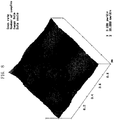
- a carbon electrode, a phospholipid polymer layer and a CyGDH layer were formed on a surface of a PET substrate.
- the conditions of the surface before and after the formation of these layers were observed using an atomic force microscope (AFM) (Tradename "D-3100” available from Digital Instruments).
- the carbon electrode was formed by screen printing using a carbon ink available from Acheson Japan Ltd.
- the AFM image of the carbon electrode is shown in Fig. 6 .
- the surface of the carbon electrode had relatively large irregularities, with carbon particles (having average particle size of about 100 nm) appearing on the surface.
- the surface of the carbonelectrode was first subjectedtoVUVtreatment (hydrophilic treatment). Then, MPC polymer solution was applied to the surface of the carbon electrode and then dried, whereby the phospholipid polymer layer was formed.
- the VUV treatment was performed by irradiating the surface of the carbon electrode with excimer laser having a wavelength of 172 nm in the atmosphere for 180 seconds with the irradiation distance of 1 mm by using "MECL-M3-750" (available from M.D. Excimer Inc.).
- MPC polymer solution use was made of a solution of MPC polymer containing tetraethaxysilane as a silane coupling agent (Tradename "LIPIDURER" available from NOF CORPORATION).
- the AFM image after the formation of the phospholipid polymer layer was shown in Fig. 7 .
- the phospholipid portion of the polymer appeared on the surface of the phospholipid polymer layer, the surface of the phospholipid polymer was smooth as compared with that of the carbon electrode layer (see Fig. 6 ), because the diameter of the phospholipid portion was about 2 to 3 nm which was smaller than that of carbon particles.
- the CyGDH layer was formed by immersing the carbon electrode formed with the phospholipid polymer layer in a CyGDH solution for ten minutes.
- the concentration of CyGDH in the CyGDH solution was 100U/ ⁇ L on the activity basis.
- the AFM image after the formation of the CyGDH layer is shown in Fig. 8 .
- the surface of the phospholipid polymer layer was formed with regularly arranged clusters (CyGDH,) each having a diameter of about 6 to 30 nm. That is, CyGDH was immobilized to the phospholipid polymer in such a manner that at least part of CyGDH appeared on the surface. From the fact that the clusters are arranged regularly, it is presumed that CyGDH is immobilized to the phospholipid polymer layer with controlled orientation.
- responsiveness was examined with respect to an electrode (inventive electrode) to which CyGDH is immobilized via a phospholipid polymer layer and to an electrode (comparative electrode) to which CYGDH, is immobilized without the intervention of a phospholipid polymer layer.
- the inventive electrode was prepared by forming a phospholipid polymer layer on a carbon electrode and then immobilizing CyGDH, similarly to Example 1.
- the comparative electrode was prepared similarly to the inventive electrode except that a phospholipid polymer layer was not formed.
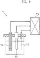
- the responsiveness of the inventive electrode and the comparative electrode was evaluated as the responsive current obtained when a voltage was applied to a glucose solution using a current measuring apparatus Y prepared as shown in Fig. 9 .
- the current measuring apparatus Y includes a working electrode Y1, a reference electrode Y2 and a counter electrode Y2, which are connected to a potentiostat Y4.
- the current measuring apparatus Y is designed to measure the responsive current by immersing the electrodes Y1-Y3 in a glucose solution and applying a voltage to the glucose solution.
- the working electrode Y1 is the inventive electrode or the comparative electrode prepared in the above-described manner.
- the reference electrode Y2 is a silver-silver chloride electrode (Tradename "RE-1B"; available from BAS Inc.).
- the counter electrode Y3 is a platinum electrode.
- the sweep voltage was 100 mV/sec, and the responsive current was measured with respect to the range of -400 mV to +700 mV.
- the glucose solutions had the concentrations of 0 mg/dL, 50 mg/dL, 100 mg/dL, 200 mg/dL, 400 mg/dL and 600 mg/dL, respectively.
- the voltage to be applied to the glucose solutions was set to +600 mV.
- the responsiveness of the inventive electrode and the comparative electrode was evaluated by measuring the time course of the responsive current with respect to each of the glucose solution of different concentrations.
- the measurement was performed using the above-described current measuring apparatus Y employing the inventive electrode or the comparative electrode as the working electrode Y1.
- the voltage of +600 mV was applied in measuring the responsive current.
- the concentration of the used glucose solutions were 0 mg/dL, 50 mg/dL, 100mg/dL, 200mg/dL, 400mg/dL and 600mg/dL, respectively.
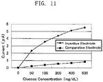
- the time course of the responsive current with respect to each of the glucose solutions is shown in Fig. 10 .
- the responsive current one second after the start of the measurement is shown in Fig. 11 in relation to the glucose level.
- the inventive electrode when the inventive electrode was used, the responsive current in the ⁇ order was measured. However, when the comparative electrode was used, merely the responsive current in the n order was measured. Specifically, the results obtained when the comparative electrode was used were similar to the conventionally reported measurement results (n order) of the responsive current obtained when use was made of a system which does not include an electron mediator such as a metal complex. When the inventive electrode was used, on the other hand, the responsive current in the ⁇ order which was much higher than the conventionally reported level was measured. Thus, it is demonstrated that the inventive electrode has high responsiveness (sensitivity).
- the inventive electrode when the inventive electrode is used, the difference in glucose level is properly reflected as the difference in responsive current.
- the glucose level is measured properly at least in the glucose level range (0 to 600 mg/dL) with respect to which the responsive current was measured in this example.
- the inventive electrode in which CyGDH is immobilized via a phospholipid polymer layer has sufficient responsiveness (sensitivity) to properly measure the glucose level without using an electron mediator such as a metal complex.
- an electron mediator such as a metal complex.
- the method for immobilizing CYGDH, which is employed for the inventive electrode, i.e., the application of a phospholipid polymer solution and the immersion in a CyGDH solution is a very easy work.
- this method is applicable to a biosensor including minute paths such as ⁇ TAS. Since the phospholipid polymer layer and the CyGDH layer formed at the minute paths are extremely thin, the formation of these layers does not considerably hinder the movement of a sample in the minute paths.
- the provision of a reagent portion, which is made up of a phospholipid polymer layer and a CyGDH layer, at most part of the minute paths does not cause any problems.
- the sensitivity of the ⁇ TAS which has been disadvantageously low, is improved. In this way, ⁇ ⁇ TAS having a high sensitivity can be provided.
Claims (21)
- Membrane à protéine immobilisée comprenant :une couche homologue de membrane cellulaire (14A, 50B) ; etune protéine (14B, 50C) immobilisée sur la couche homologue de membrane cellulaire (14A, 50B), la protéine (14B, 50C) contenant un cytochrome ou un complexe de cytochrome ;dans laquelle la couche homologue de membrane cellulaire (14A, 50B) contient un polymère phospholipidique qui est un polymère de 2-méthacryloyloxyéthyl-phosphorylcholine.
- Membrane à protéine immobilisée selon la revendication 1, dans laquelle la couche homologue de membrane cellulaire (14A, 50B) contient un agent de couplage de silane.
- Membrane à protéine immobilisée selon la revendication 2, dans laquelle l'agent de couplage de silane est le tétraéthoxysilane.
- Membrane à protéine immobilisée selon la revendication 1, dans laquelle la protéine est la CyGDH contenant une sous-unité α ayant une activité de déshydrogénase de glucose et un cytochrome C ayant une fonction de transfert d'électrons.
- Procédé d'immobilisation d'une protéine, le procédé comprenant :une première étape de formation d'une couche homologue de membrane cellulaire (14A, 50B) dans une partie projetée d'un élément cible d'immobilisation (5, 11) ; etune deuxième étape visant à amener une protéine (14B, 50C) à s'auto-organiser par rapport à la couche homologue de membrane cellulaire (14A, 50B), la protéine (14B, 50C) contenant un cytochrome ou un complexe de cytochrome ;dans lequel la couche homologue de membrane cellulaire (14A, 50B) contient un polymère phospholipidique qui est un polymère de 2-méthacryloyloxyéthyl-phosphorylcholine.
- Procédé d'immobilisation d'une protéine selon la revendication 5, comprenant en outre une troisième étape visant à soumettre la partie projetée à un traitement hydrophile avant la première étape.
- Procédé d'immobilisation d'une protéine selon la revendication 6, dans lequel, dans la deuxième étape, la couche homologue de membrane cellulaire (14A, 50B) est formée pour contenir un agent de couplage de silane.
- Procédé d'immobilisation de protéine selon la revendication 7, dans lequel l'agent de couplage de silane est le tétraéthoxysilane.
- Procédé d'immobilisation d'une protéine selon la revendication 5, dans lequel la protéine (14B, 50C) est le CyGDH contenant une sous-unité α ayant une activité de déshydrogénase de glucose et un cytochrome C ayant une fonction de transfert d'électrons.
- Electrode à enzyme immobilisé comprenant:un substrat (1) ;une électrode (11, 12) formée sur le substrat (1) ; etune couche (14) contenant un enzyme immobilisée sur l'électrode (11, 12);dans laquelle la couche contenant un enzyme comprend une couche homologue de membrane cellulaire (14A) et un enzyme (14B), l'enzyme (14B) contenant comme sous-unité un cytochrome C immobilisé sur la couche homologue de membrane cellulaire (14A) avec une orientation réglée ; etdans laquelle la couche homologue de membrane cellulaire (14A) contient un polymère phospholipidique qui est un polymère de 2-méthacryloyloxyéthyl-phosphorylcholine.
- Electrode à enzyme immobilisé selon la revendication 10, dans laquelle la couche homologue de membrane cellulaire (14A) contient un agent de couplage de silane.
- Electrode à enzyme immobilisé selon la revendication 11, dans laquelle l'agent de couplage de silane est le tétraéthoxysilane.
- Electrode à enzyme immobilisé selon la revendication 10, dans laquelle l'enzyme (14B) est le CyGDH contenant une sous-unité α ayant une activité de déshydrogénase de glucose et un cytochrome C ayant une fonction de transfert d'électrons.
- Biocapteur (X1, X2) comprenant :un trajet d'écoulement (4, 8) pour déplacer un échantillon ;une partie réactive (14, 50) fournie dans le trajet d'écoulement (4, 8);un substrat (1, 5) ; etune couche (14, 50) contenant un enzyme immobilisée sur le substrat (1, 5);dans lequel la couche (14, 50) contenant un enzyme comprend une couche homologue de membrane cellulaire (14A, 50B) et un enzyme (14B, 50C), l'enzyme (14B, 50C) contenant comme sous-unité un cytochrome C immobilisé sur la couche homologue de membrane cellulaire (14A, 50B) avec une orientation réglée ; etdans lequel la couche homologue de membrane cellulaire (14A, 50B) contient un polymère phospholipidique qui est un polymère de 2-méthacryloyloxyéthyl-phosphorylcholine.
- Biocapteur (X1, X2) selon la revendication 14, dans lequel la couche homologue de membrane cellulaire (14A, 50B) contient un agent de couplage de silane.
- Biocapteur (X1, X2) selon la revendication 15, dans lequel l'agent de couplage de silane est le tétraéthoxysilane.
- Biocapteur (X1, X2) selon la revendication 14, dans lequel l'enzyme (14B, 50C) est le CyGDH contenant une sous-unité α ayant une activité de déshydrogénase de glucose et un cytochrome C ayant une fonction de transfert d'électrons.
- Biocapteur (X1, X2) selon la revendication 14, comprenant en outre une électrode de travail (11) et une contre-électrode (12) qui sont partiellement exposées dans le trajet d'écoulement (4) et utilisées pour appliquer une tension à une échantillon.
- Biocapteur (X1, X2) selon la revendication 18, dans lequel au moins une partie de la couche homologue de membrane cellulaire (14A, 50B) est formée sur l'électrode de travail (11).
- Biocapteur (X1, X2) selon la revendication 14, dans lequel la partie réactive (50) contient un formateur de couleur.
- Biocapteur (X1, X2) selon la revendication 20, dans lequel la partie réactive (50) comprend une couche chromogène (50A) contenant le formateur de couleur, la couche homologue de membrane cellulaire (50B) et une couche (50C) contenant un enzyme qui contient l'enzyme.
Applications Claiming Priority (2)
| Application Number | Priority Date | Filing Date | Title |
|---|---|---|---|
| JP2005148253A JP5021183B2 (ja) | 2005-05-20 | 2005-05-20 | タンパク質固定化膜および固定化方法、ならびにバイオセンサ |
| PCT/JP2006/309906 WO2006123730A1 (fr) | 2005-05-20 | 2006-05-18 | Membrane a proteine immobilisee, procede pour l’immobilisation de proteine, electrode a enzyme immobilisee, et capteur biologique |
Publications (3)
| Publication Number | Publication Date |
|---|---|
| EP1884771A1 EP1884771A1 (fr) | 2008-02-06 |
| EP1884771A4 EP1884771A4 (fr) | 2011-10-19 |
| EP1884771B1 true EP1884771B1 (fr) | 2017-12-13 |
Family
ID=37431302
Family Applications (1)
| Application Number | Title | Priority Date | Filing Date |
|---|---|---|---|
| EP06746594.8A Active EP1884771B1 (fr) | 2005-05-20 | 2006-05-18 | Membrane a proteine immobilisee, procede pour l'immobilisation de proteine, electrode a enzyme immobilisee, et capteur biologique |
Country Status (5)
| Country | Link |
|---|---|
| US (2) | US20090101499A1 (fr) |
| EP (1) | EP1884771B1 (fr) |
| JP (1) | JP5021183B2 (fr) |
| CN (1) | CN101203748B (fr) |
| WO (1) | WO2006123730A1 (fr) |
Families Citing this family (14)
| Publication number | Priority date | Publication date | Assignee | Title |
|---|---|---|---|---|
| US20060091006A1 (en) * | 1999-11-04 | 2006-05-04 | Yi Wang | Analyte sensor with insertion monitor, and methods |
| JP4359595B2 (ja) * | 2003-09-02 | 2009-11-04 | 広司 早出 | グルコースセンサおよびグルコース濃度測定装置 |
| JP2008243380A (ja) * | 2007-03-23 | 2008-10-09 | Sony Corp | 酵素固定化電極、燃料電池、電子機器、酵素反応利用装置および酵素固定化基体 |
| US8268604B2 (en) * | 2007-12-20 | 2012-09-18 | Abbott Point Of Care Inc. | Compositions for forming immobilized biological layers for sensing |
| US9653006B2 (en) | 2008-09-17 | 2017-05-16 | Avery Dennison Corporation | Activatable adhesive, labels, and related methods |
| WO2010090271A1 (fr) | 2009-02-09 | 2010-08-12 | アークレイ株式会社 | Capteur électrochimique et son procédé de fabrication |
| JP5432575B2 (ja) * | 2009-04-21 | 2014-03-05 | グンゼ株式会社 | バイオセンサ及びその製造方法 |
| EP2589659B1 (fr) | 2009-04-30 | 2015-07-22 | Panasonic Healthcare Holdings Co., Ltd. | Médiateur d'électrons de type protéine |
| EP2393897B8 (fr) | 2009-09-17 | 2018-09-19 | Avery Dennison Corporation | Adhésifs activables, étiquettes, et procédés apparentés |
| JP5665070B2 (ja) * | 2009-09-25 | 2015-02-04 | 独立行政法人産業技術総合研究所 | 酸化還元タンパク質固定化ナノ構造電極 |
| CN103344639B (zh) * | 2013-07-11 | 2015-10-28 | 山东大学 | 一种快速检测尿中吡咯加合物的检测管 |
| EP3032250B1 (fr) * | 2013-08-07 | 2023-10-11 | ARKRAY, Inc. | Procédé de mesure de substance et dispositif de mesure employant un biocapteur électrochimique |
| US10974246B2 (en) | 2015-06-08 | 2021-04-13 | Japan Science And Technology Agency | High-density micro-chamber array and measurement method using same |
| CN105355530B (zh) * | 2015-10-30 | 2017-08-11 | 三诺生物传感股份有限公司 | 一种用于试条电极预处理的等离子处理装置和方法 |
Family Cites Families (14)
| Publication number | Priority date | Publication date | Assignee | Title |
|---|---|---|---|---|
| JPH0810208A (ja) | 1994-06-30 | 1996-01-16 | Toshiba Corp | 食器洗浄機 |
| US5650062A (en) * | 1995-03-17 | 1997-07-22 | Matsushita Electric Industrial Co., Ltd. | Biosensor, and a method and a device for quantifying a substrate in a sample liquid using the same |
| EP1130390A1 (fr) * | 1999-09-13 | 2001-09-05 | Matsushita Electric Industrial Co., Ltd. | Procede de production d'une enzyme modifiee par lipides, et biodetecteur |
| AU2001241531A1 (en) * | 2000-02-18 | 2001-08-27 | Aspira Biosystems, Inc. | Compositions and methods for surface imprinting |
| US6458599B1 (en) | 2000-02-18 | 2002-10-01 | Aspira Biosystems, Inc. | Compositions and methods for capturing, isolating, detecting, analyzing and quantifying macromolecules |
| WO2002009647A2 (fr) * | 2000-07-28 | 2002-02-07 | Emory University | Composant biologique comprenant une membrane artificielle |
| JP2002055076A (ja) * | 2000-09-08 | 2002-02-20 | Nec Corp | 電気化学センサ |
| WO2002036779A1 (fr) * | 2000-10-31 | 2002-05-10 | Koji Sode | Nouvelle glucose deshydrogenase et procede de production de la deshydrogenase |
| ATE505724T1 (de) * | 2001-09-14 | 2011-04-15 | Arkray Inc | Verfahren, gerät und vorrichtung zur konzentrationsmessung |
| CN1662660A (zh) * | 2002-06-17 | 2005-08-31 | 爱科来株式会社 | 使用葡萄糖脱氢酶的葡萄糖浓度测定方法及葡萄糖传感器 |
| JP4637579B2 (ja) * | 2002-07-25 | 2011-02-23 | パナソニック株式会社 | 電解質膜とこれを用いた膜電極接合体および燃料電池 |
| JP4250481B2 (ja) * | 2003-08-11 | 2009-04-08 | キヤノン株式会社 | 多孔質構造体 |
| JP4359595B2 (ja) * | 2003-09-02 | 2009-11-04 | 広司 早出 | グルコースセンサおよびグルコース濃度測定装置 |
| US8354112B2 (en) * | 2003-09-30 | 2013-01-15 | Arkray, Inc. | Glucose dehydrogenase/cytochrome fusion protein |
-
2005
- 2005-05-20 JP JP2005148253A patent/JP5021183B2/ja active Active
-
2006
- 2006-05-18 US US11/920,782 patent/US20090101499A1/en not_active Abandoned
- 2006-05-18 EP EP06746594.8A patent/EP1884771B1/fr active Active
- 2006-05-18 WO PCT/JP2006/309906 patent/WO2006123730A1/fr active Application Filing
- 2006-05-18 CN CN2006800226213A patent/CN101203748B/zh active Active
-
2015
- 2015-09-10 US US14/850,576 patent/US9702843B2/en active Active
Non-Patent Citations (1)
| Title |
|---|
| None * |
Also Published As
| Publication number | Publication date |
|---|---|
| CN101203748A (zh) | 2008-06-18 |
| CN101203748B (zh) | 2011-09-14 |
| JP2006322889A (ja) | 2006-11-30 |
| EP1884771A1 (fr) | 2008-02-06 |
| US20150377818A1 (en) | 2015-12-31 |
| US20090101499A1 (en) | 2009-04-23 |
| US9702843B2 (en) | 2017-07-11 |
| JP5021183B2 (ja) | 2012-09-05 |
| EP1884771A4 (fr) | 2011-10-19 |
| WO2006123730A1 (fr) | 2006-11-23 |
Similar Documents
| Publication | Publication Date | Title |
|---|---|---|
| EP1884771B1 (fr) | Membrane a proteine immobilisee, procede pour l'immobilisation de proteine, electrode a enzyme immobilisee, et capteur biologique | |
| JP2006322889A5 (fr) | ||
| US8758591B2 (en) | Electrochemical nanocomposite biosensor system | |
| JP5595038B2 (ja) | 分析物センサ装置ならびにその製造方法、組成物及びキット | |
| US5795774A (en) | Biosensor | |
| CA2822909C (fr) | Compositions enzymatiques en couches a utiliser avec des capteurs de substances a analyser | |
| JP4295615B2 (ja) | 埋設可能なデバイスと一緒に使用するためのセンサヘッド | |
| JP6000281B2 (ja) | 分析物センサと共に使用される電極組成物、分析物センサ装置及び分析物センサ装置を作製する方法 | |
| EP1113263A2 (fr) | Capteur contenant les microsphères | |
| EP1678492A1 (fr) | Bande de test electrochimique amelioree permettant de reduire l'effet d'un courant d'interference direct et medie | |
| EP1472537A2 (fr) | Bande biocapteur electrochimique pour analyse d'echantillons liquides | |
| JPWO2004061444A1 (ja) | 薄型分析用具 | |
| Ramanavicius | Amperometric biosensor for the determination of creatine | |
| JP2009500601A (ja) | 電極プリコンディショニング | |
| CN110514704B (zh) | 新型生物传感方法 | |
| JP5753720B2 (ja) | バイオセンサ | |
| US8417314B2 (en) | Ruthenium purple biosensor | |
| EP2395348B1 (fr) | Capteur Électrochimique et son procédé de fabrication. | |
| EP2518156B1 (fr) | Dispositif d'analyse | |
| Pedrosa et al. | Acetylcholinesterase Immobilization on 3‐Mercaptopropionic Acid Self Assembled Monolayer for Determination of Pesticides | |
| JP5164656B2 (ja) | センサ及びバイオセンサ | |
| Okawa et al. | Glucose sensor carrying monomolecular layer of glucose oxidase covalently bound to tin (IV) oxide electrode | |
| Genshaw | Enzyme electrode for determining glucose in whole blood. | |
| WO2004060297A2 (fr) | Agents de reticulation hydrophiles s'utilisant dans des capteurs enzymatiques | |
| Xie et al. | An interference-free implantable glucose microbiosensor based on use of a polymeric analyte-regulating membrane |
Legal Events
| Date | Code | Title | Description |
|---|---|---|---|
| PUAI | Public reference made under article 153(3) epc to a published international application that has entered the european phase |
Free format text: ORIGINAL CODE: 0009012 |
|
| 17P | Request for examination filed |
Effective date: 20071206 |
|
| AK | Designated contracting states |
Kind code of ref document: A1 Designated state(s): AT BE BG CH CY CZ DE DK EE ES FI FR GB GR HU IE IS IT LI LT LU LV MC NL PL PT RO SE SI SK TR |
|
| DAX | Request for extension of the european patent (deleted) | ||
| A4 | Supplementary search report drawn up and despatched |
Effective date: 20110919 |
|
| RIC1 | Information provided on ipc code assigned before grant |
Ipc: C12Q 1/00 20060101ALI20110913BHEP Ipc: C07K 14/80 20060101ALI20110913BHEP Ipc: C07K 14/705 20060101ALI20110913BHEP Ipc: G01N 33/487 20060101ALI20110913BHEP Ipc: G01N 27/327 20060101AFI20110913BHEP Ipc: G01N 27/416 20060101ALI20110913BHEP Ipc: C12M 1/34 20060101ALI20110913BHEP Ipc: C12N 11/08 20060101ALI20110913BHEP Ipc: C12N 9/04 20060101ALI20110913BHEP |
|
| RAP1 | Party data changed (applicant data changed or rights of an application transferred) |
Owner name: ARKRAY, INC. |
|
| 17Q | First examination report despatched |
Effective date: 20160425 |
|
| GRAP | Despatch of communication of intention to grant a patent |
Free format text: ORIGINAL CODE: EPIDOSNIGR1 |
|
| INTG | Intention to grant announced |
Effective date: 20170405 |
|
| GRAJ | Information related to disapproval of communication of intention to grant by the applicant or resumption of examination proceedings by the epo deleted |
Free format text: ORIGINAL CODE: EPIDOSDIGR1 |
|
| GRAL | Information related to payment of fee for publishing/printing deleted |
Free format text: ORIGINAL CODE: EPIDOSDIGR3 |
|
| GRAS | Grant fee paid |
Free format text: ORIGINAL CODE: EPIDOSNIGR3 |
|
| GRAP | Despatch of communication of intention to grant a patent |
Free format text: ORIGINAL CODE: EPIDOSNIGR1 |
|
| INTC | Intention to grant announced (deleted) | ||
| RIN1 | Information on inventor provided before grant (corrected) |
Inventor name: YAMAOKA, HIDEAKI Inventor name: KATSUKI, KOJI |
|
| INTG | Intention to grant announced |
Effective date: 20170817 |
|
| GRAA | (expected) grant |
Free format text: ORIGINAL CODE: 0009210 |
|
| AK | Designated contracting states |
Kind code of ref document: B1 Designated state(s): AT BE BG CH CY CZ DE DK EE ES FI FR GB GR HU IE IS IT LI LT LU LV MC NL PL PT RO SE SI SK TR |
|
| REG | Reference to a national code |
Ref country code: GB Ref legal event code: FG4D |
|
| REG | Reference to a national code |
Ref country code: AT Ref legal event code: REF Ref document number: 954874 Country of ref document: AT Kind code of ref document: T Effective date: 20171215 Ref country code: CH Ref legal event code: EP |
|
| REG | Reference to a national code |
Ref country code: IE Ref legal event code: FG4D |
|
| REG | Reference to a national code |
Ref country code: DE Ref legal event code: R096 Ref document number: 602006054333 Country of ref document: DE |
|
| REG | Reference to a national code |
Ref country code: NL Ref legal event code: MP Effective date: 20171213 |
|
| REG | Reference to a national code |
Ref country code: LT Ref legal event code: MG4D |
|
| PG25 | Lapsed in a contracting state [announced via postgrant information from national office to epo] |
Ref country code: FI Free format text: LAPSE BECAUSE OF FAILURE TO SUBMIT A TRANSLATION OF THE DESCRIPTION OR TO PAY THE FEE WITHIN THE PRESCRIBED TIME-LIMIT Effective date: 20171213 Ref country code: LT Free format text: LAPSE BECAUSE OF FAILURE TO SUBMIT A TRANSLATION OF THE DESCRIPTION OR TO PAY THE FEE WITHIN THE PRESCRIBED TIME-LIMIT Effective date: 20171213 Ref country code: SE Free format text: LAPSE BECAUSE OF FAILURE TO SUBMIT A TRANSLATION OF THE DESCRIPTION OR TO PAY THE FEE WITHIN THE PRESCRIBED TIME-LIMIT Effective date: 20171213 |
|
| REG | Reference to a national code |
Ref country code: AT Ref legal event code: MK05 Ref document number: 954874 Country of ref document: AT Kind code of ref document: T Effective date: 20171213 |
|
| REG | Reference to a national code |
Ref country code: FR Ref legal event code: PLFP Year of fee payment: 13 |
|
| PG25 | Lapsed in a contracting state [announced via postgrant information from national office to epo] |
Ref country code: BG Free format text: LAPSE BECAUSE OF FAILURE TO SUBMIT A TRANSLATION OF THE DESCRIPTION OR TO PAY THE FEE WITHIN THE PRESCRIBED TIME-LIMIT Effective date: 20180313 Ref country code: LV Free format text: LAPSE BECAUSE OF FAILURE TO SUBMIT A TRANSLATION OF THE DESCRIPTION OR TO PAY THE FEE WITHIN THE PRESCRIBED TIME-LIMIT Effective date: 20171213 Ref country code: GR Free format text: LAPSE BECAUSE OF FAILURE TO SUBMIT A TRANSLATION OF THE DESCRIPTION OR TO PAY THE FEE WITHIN THE PRESCRIBED TIME-LIMIT Effective date: 20180314 |
|
| PG25 | Lapsed in a contracting state [announced via postgrant information from national office to epo] |
Ref country code: NL Free format text: LAPSE BECAUSE OF FAILURE TO SUBMIT A TRANSLATION OF THE DESCRIPTION OR TO PAY THE FEE WITHIN THE PRESCRIBED TIME-LIMIT Effective date: 20171213 |
|
| PG25 | Lapsed in a contracting state [announced via postgrant information from national office to epo] |
Ref country code: ES Free format text: LAPSE BECAUSE OF FAILURE TO SUBMIT A TRANSLATION OF THE DESCRIPTION OR TO PAY THE FEE WITHIN THE PRESCRIBED TIME-LIMIT Effective date: 20171213 Ref country code: SK Free format text: LAPSE BECAUSE OF FAILURE TO SUBMIT A TRANSLATION OF THE DESCRIPTION OR TO PAY THE FEE WITHIN THE PRESCRIBED TIME-LIMIT Effective date: 20171213 Ref country code: EE Free format text: LAPSE BECAUSE OF FAILURE TO SUBMIT A TRANSLATION OF THE DESCRIPTION OR TO PAY THE FEE WITHIN THE PRESCRIBED TIME-LIMIT Effective date: 20171213 Ref country code: CZ Free format text: LAPSE BECAUSE OF FAILURE TO SUBMIT A TRANSLATION OF THE DESCRIPTION OR TO PAY THE FEE WITHIN THE PRESCRIBED TIME-LIMIT Effective date: 20171213 Ref country code: CY Free format text: LAPSE BECAUSE OF FAILURE TO SUBMIT A TRANSLATION OF THE DESCRIPTION OR TO PAY THE FEE WITHIN THE PRESCRIBED TIME-LIMIT Effective date: 20171213 |
|
| PG25 | Lapsed in a contracting state [announced via postgrant information from national office to epo] |
Ref country code: PL Free format text: LAPSE BECAUSE OF FAILURE TO SUBMIT A TRANSLATION OF THE DESCRIPTION OR TO PAY THE FEE WITHIN THE PRESCRIBED TIME-LIMIT Effective date: 20171213 Ref country code: RO Free format text: LAPSE BECAUSE OF FAILURE TO SUBMIT A TRANSLATION OF THE DESCRIPTION OR TO PAY THE FEE WITHIN THE PRESCRIBED TIME-LIMIT Effective date: 20171213 Ref country code: AT Free format text: LAPSE BECAUSE OF FAILURE TO SUBMIT A TRANSLATION OF THE DESCRIPTION OR TO PAY THE FEE WITHIN THE PRESCRIBED TIME-LIMIT Effective date: 20171213 Ref country code: IS Free format text: LAPSE BECAUSE OF FAILURE TO SUBMIT A TRANSLATION OF THE DESCRIPTION OR TO PAY THE FEE WITHIN THE PRESCRIBED TIME-LIMIT Effective date: 20180413 |
|
| REG | Reference to a national code |
Ref country code: DE Ref legal event code: R097 Ref document number: 602006054333 Country of ref document: DE |
|
| PLBE | No opposition filed within time limit |
Free format text: ORIGINAL CODE: 0009261 |
|
| STAA | Information on the status of an ep patent application or granted ep patent |
Free format text: STATUS: NO OPPOSITION FILED WITHIN TIME LIMIT |
|
| 26N | No opposition filed |
Effective date: 20180914 |
|
| PG25 | Lapsed in a contracting state [announced via postgrant information from national office to epo] |
Ref country code: DK Free format text: LAPSE BECAUSE OF FAILURE TO SUBMIT A TRANSLATION OF THE DESCRIPTION OR TO PAY THE FEE WITHIN THE PRESCRIBED TIME-LIMIT Effective date: 20171213 |
|
| REG | Reference to a national code |
Ref country code: CH Ref legal event code: PL |
|
| REG | Reference to a national code |
Ref country code: BE Ref legal event code: MM Effective date: 20180531 |
|
| PG25 | Lapsed in a contracting state [announced via postgrant information from national office to epo] |
Ref country code: MC Free format text: LAPSE BECAUSE OF FAILURE TO SUBMIT A TRANSLATION OF THE DESCRIPTION OR TO PAY THE FEE WITHIN THE PRESCRIBED TIME-LIMIT Effective date: 20171213 |
|
| REG | Reference to a national code |
Ref country code: IE Ref legal event code: MM4A |
|
| PG25 | Lapsed in a contracting state [announced via postgrant information from national office to epo] |
Ref country code: LI Free format text: LAPSE BECAUSE OF NON-PAYMENT OF DUE FEES Effective date: 20180531 Ref country code: CH Free format text: LAPSE BECAUSE OF NON-PAYMENT OF DUE FEES Effective date: 20180531 Ref country code: SI Free format text: LAPSE BECAUSE OF FAILURE TO SUBMIT A TRANSLATION OF THE DESCRIPTION OR TO PAY THE FEE WITHIN THE PRESCRIBED TIME-LIMIT Effective date: 20171213 |
|
| PG25 | Lapsed in a contracting state [announced via postgrant information from national office to epo] |
Ref country code: LU Free format text: LAPSE BECAUSE OF NON-PAYMENT OF DUE FEES Effective date: 20180518 |
|
| PG25 | Lapsed in a contracting state [announced via postgrant information from national office to epo] |
Ref country code: IE Free format text: LAPSE BECAUSE OF NON-PAYMENT OF DUE FEES Effective date: 20180518 |
|
| PG25 | Lapsed in a contracting state [announced via postgrant information from national office to epo] |
Ref country code: BE Free format text: LAPSE BECAUSE OF NON-PAYMENT OF DUE FEES Effective date: 20180531 |
|
| PG25 | Lapsed in a contracting state [announced via postgrant information from national office to epo] |
Ref country code: TR Free format text: LAPSE BECAUSE OF FAILURE TO SUBMIT A TRANSLATION OF THE DESCRIPTION OR TO PAY THE FEE WITHIN THE PRESCRIBED TIME-LIMIT Effective date: 20171213 |
|
| PG25 | Lapsed in a contracting state [announced via postgrant information from national office to epo] |
Ref country code: PT Free format text: LAPSE BECAUSE OF FAILURE TO SUBMIT A TRANSLATION OF THE DESCRIPTION OR TO PAY THE FEE WITHIN THE PRESCRIBED TIME-LIMIT Effective date: 20171213 Ref country code: HU Free format text: LAPSE BECAUSE OF FAILURE TO SUBMIT A TRANSLATION OF THE DESCRIPTION OR TO PAY THE FEE WITHIN THE PRESCRIBED TIME-LIMIT; INVALID AB INITIO Effective date: 20060518 |
|
| PGFP | Annual fee paid to national office [announced via postgrant information from national office to epo] |
Ref country code: IT Payment date: 20230526 Year of fee payment: 18 Ref country code: FR Payment date: 20230525 Year of fee payment: 18 Ref country code: DE Payment date: 20230519 Year of fee payment: 18 |
|
| PGFP | Annual fee paid to national office [announced via postgrant information from national office to epo] |
Ref country code: GB Payment date: 20230523 Year of fee payment: 18 |