EP1858562B1 - Substitut cartilagineux et osteochondral comprenant une structure multicouche et son utilisation - Google Patents
Substitut cartilagineux et osteochondral comprenant une structure multicouche et son utilisation Download PDFInfo
- Publication number
- EP1858562B1 EP1858562B1 EP06710487A EP06710487A EP1858562B1 EP 1858562 B1 EP1858562 B1 EP 1858562B1 EP 06710487 A EP06710487 A EP 06710487A EP 06710487 A EP06710487 A EP 06710487A EP 1858562 B1 EP1858562 B1 EP 1858562B1
- Authority
- EP
- European Patent Office
- Prior art keywords
- collagen
- multilayer structure
- hydroxylapatite
- substitute
- cartilaginous
- Prior art date
- Legal status (The legal status is an assumption and is not a legal conclusion. Google has not performed a legal analysis and makes no representation as to the accuracy of the status listed.)
- Active
Links
- 229920001436 collagen Polymers 0.000 claims abstract description 105
- 102000008186 Collagen Human genes 0.000 claims abstract description 104
- 108010035532 Collagen Proteins 0.000 claims abstract description 104
- XYJRXVWERLGGKC-UHFFFAOYSA-D pentacalcium;hydroxide;triphosphate Chemical compound [OH-].[Ca+2].[Ca+2].[Ca+2].[Ca+2].[Ca+2].[O-]P([O-])([O-])=O.[O-]P([O-])([O-])=O.[O-]P([O-])([O-])=O XYJRXVWERLGGKC-UHFFFAOYSA-D 0.000 claims abstract description 58
- 229910052588 hydroxylapatite Inorganic materials 0.000 claims abstract description 57
- 239000002131 composite material Substances 0.000 claims abstract description 31
- 239000011159 matrix material Substances 0.000 claims abstract description 30
- 238000002360 preparation method Methods 0.000 claims abstract description 10
- 238000000034 method Methods 0.000 claims description 29
- 210000004027 cell Anatomy 0.000 claims description 15
- 230000008569 process Effects 0.000 claims description 15
- 239000000203 mixture Substances 0.000 claims description 13
- 210000001612 chondrocyte Anatomy 0.000 claims description 10
- 230000024245 cell differentiation Effects 0.000 claims description 8
- KIUKXJAPPMFGSW-DNGZLQJQSA-N (2S,3S,4S,5R,6R)-6-[(2S,3R,4R,5S,6R)-3-Acetamido-2-[(2S,3S,4R,5R,6R)-6-[(2R,3R,4R,5S,6R)-3-acetamido-2,5-dihydroxy-6-(hydroxymethyl)oxan-4-yl]oxy-2-carboxy-4,5-dihydroxyoxan-3-yl]oxy-5-hydroxy-6-(hydroxymethyl)oxan-4-yl]oxy-3,4,5-trihydroxyoxane-2-carboxylic acid Chemical compound CC(=O)N[C@H]1[C@H](O)O[C@H](CO)[C@@H](O)[C@@H]1O[C@H]1[C@H](O)[C@@H](O)[C@H](O[C@H]2[C@@H]([C@@H](O[C@H]3[C@@H]([C@@H](O)[C@H](O)[C@H](O3)C(O)=O)O)[C@H](O)[C@@H](CO)O2)NC(C)=O)[C@@H](C(O)=O)O1 KIUKXJAPPMFGSW-DNGZLQJQSA-N 0.000 claims description 7
- 229920002674 hyaluronan Polymers 0.000 claims description 7
- 229960003160 hyaluronic acid Drugs 0.000 claims description 7
- 230000004069 differentiation Effects 0.000 claims description 6
- SXRSQZLOMIGNAQ-UHFFFAOYSA-N Glutaraldehyde Chemical compound O=CCCCC=O SXRSQZLOMIGNAQ-UHFFFAOYSA-N 0.000 claims description 5
- 239000004971 Cross linker Substances 0.000 claims description 3
- 239000003242 anti bacterial agent Substances 0.000 claims description 3
- 230000003110 anti-inflammatory effect Effects 0.000 claims description 3
- 239000008280 blood Substances 0.000 claims description 3
- 210000004369 blood Anatomy 0.000 claims description 3
- 210000000963 osteoblast Anatomy 0.000 claims description 3
- 239000013543 active substance Substances 0.000 claims description 2
- 230000002634 anti-blastic effect Effects 0.000 claims description 2
- 230000001028 anti-proliverative effect Effects 0.000 claims description 2
- 229940088710 antibiotic agent Drugs 0.000 claims description 2
- 239000003443 antiviral agent Substances 0.000 claims description 2
- 229940121357 antivirals Drugs 0.000 claims description 2
- 239000012141 concentrate Substances 0.000 claims description 2
- 239000003246 corticosteroid Substances 0.000 claims description 2
- 229960001334 corticosteroids Drugs 0.000 claims description 2
- GYZLOYUZLJXAJU-UHFFFAOYSA-N diglycidyl ether Chemical compound C1OC1COCC1CO1 GYZLOYUZLJXAJU-UHFFFAOYSA-N 0.000 claims description 2
- 239000003102 growth factor Substances 0.000 claims description 2
- 238000011065 in-situ storage Methods 0.000 claims description 2
- 230000001737 promoting effect Effects 0.000 claims description 2
- 239000003634 thrombocyte concentrate Substances 0.000 claims description 2
- 150000003839 salts Chemical class 0.000 claims 1
- 210000001519 tissue Anatomy 0.000 abstract description 38
- 210000005065 subchondral bone plate Anatomy 0.000 abstract description 10
- 238000011282 treatment Methods 0.000 abstract description 8
- 201000009859 Osteochondrosis Diseases 0.000 abstract description 7
- 230000007547 defect Effects 0.000 abstract description 6
- 239000010410 layer Substances 0.000 description 72
- 210000000988 bone and bone Anatomy 0.000 description 36
- 230000003902 lesion Effects 0.000 description 30
- 239000007943 implant Substances 0.000 description 23
- 241001465754 Metazoa Species 0.000 description 18
- 210000002808 connective tissue Anatomy 0.000 description 13
- 238000004458 analytical method Methods 0.000 description 12
- 230000006911 nucleation Effects 0.000 description 12
- 238000010899 nucleation Methods 0.000 description 12
- 241000283073 Equus caballus Species 0.000 description 10
- NBIIXXVUZAFLBC-UHFFFAOYSA-N Phosphoric acid Chemical compound OP(O)(O)=O NBIIXXVUZAFLBC-UHFFFAOYSA-N 0.000 description 10
- 239000000243 solution Substances 0.000 description 10
- 230000006870 function Effects 0.000 description 9
- 210000001188 articular cartilage Anatomy 0.000 description 8
- SHKUUQIDMUMQQK-UHFFFAOYSA-N 2-[4-(oxiran-2-ylmethoxy)butoxymethyl]oxirane Chemical compound C1OC1COCCCCOCC1CO1 SHKUUQIDMUMQQK-UHFFFAOYSA-N 0.000 description 7
- 229910019142 PO4 Inorganic materials 0.000 description 7
- 229920005615 natural polymer Polymers 0.000 description 7
- 238000012360 testing method Methods 0.000 description 7
- QTBSBXVTEAMEQO-UHFFFAOYSA-N Acetic acid Chemical compound CC(O)=O QTBSBXVTEAMEQO-UHFFFAOYSA-N 0.000 description 6
- WZUVPPKBWHMQCE-UHFFFAOYSA-N Haematoxylin Chemical compound C12=CC(O)=C(O)C=C2CC2(O)C1C1=CC=C(O)C(O)=C1OC2 WZUVPPKBWHMQCE-UHFFFAOYSA-N 0.000 description 6
- 239000002253 acid Substances 0.000 description 6
- 230000015572 biosynthetic process Effects 0.000 description 6
- 159000000007 calcium salts Chemical class 0.000 description 6
- 210000000845 cartilage Anatomy 0.000 description 6
- 238000006243 chemical reaction Methods 0.000 description 6
- 230000032798 delamination Effects 0.000 description 6
- 230000003993 interaction Effects 0.000 description 6
- 239000010452 phosphate Substances 0.000 description 6
- 238000010186 staining Methods 0.000 description 6
- 239000000126 substance Substances 0.000 description 6
- 238000001356 surgical procedure Methods 0.000 description 6
- 210000002435 tendon Anatomy 0.000 description 6
- 102000012422 Collagen Type I Human genes 0.000 description 5
- 108010022452 Collagen Type I Proteins 0.000 description 5
- 229910052586 apatite Inorganic materials 0.000 description 5
- 229940096422 collagen type i Drugs 0.000 description 5
- 238000004132 cross linking Methods 0.000 description 5
- 239000013078 crystal Substances 0.000 description 5
- 230000000694 effects Effects 0.000 description 5
- 238000004108 freeze drying Methods 0.000 description 5
- 238000000338 in vitro Methods 0.000 description 5
- 238000001727 in vivo Methods 0.000 description 5
- 239000002159 nanocrystal Substances 0.000 description 5
- VSIIXMUUUJUKCM-UHFFFAOYSA-D pentacalcium;fluoride;triphosphate Chemical compound [F-].[Ca+2].[Ca+2].[Ca+2].[Ca+2].[Ca+2].[O-]P([O-])([O-])=O.[O-]P([O-])([O-])=O.[O-]P([O-])([O-])=O VSIIXMUUUJUKCM-UHFFFAOYSA-D 0.000 description 5
- NBIIXXVUZAFLBC-UHFFFAOYSA-K phosphate Chemical compound [O-]P([O-])([O-])=O NBIIXXVUZAFLBC-UHFFFAOYSA-K 0.000 description 5
- 239000011148 porous material Substances 0.000 description 5
- 239000000725 suspension Substances 0.000 description 5
- 229920001059 synthetic polymer Polymers 0.000 description 5
- 206010011985 Decubitus ulcer Diseases 0.000 description 4
- 102000016611 Proteoglycans Human genes 0.000 description 4
- 108010067787 Proteoglycans Proteins 0.000 description 4
- 229910000147 aluminium phosphate Inorganic materials 0.000 description 4
- 150000001413 amino acids Chemical class 0.000 description 4
- 230000003592 biomimetic effect Effects 0.000 description 4
- TZSMWSKOPZEMAJ-UHFFFAOYSA-N bis[(2-methoxyphenyl)methyl] carbonate Chemical compound COC1=CC=CC=C1COC(=O)OCC1=CC=CC=C1OC TZSMWSKOPZEMAJ-UHFFFAOYSA-N 0.000 description 4
- 230000000740 bleeding effect Effects 0.000 description 4
- 210000001185 bone marrow Anatomy 0.000 description 4
- 210000004271 bone marrow stromal cell Anatomy 0.000 description 4
- AXCZMVOFGPJBDE-UHFFFAOYSA-L calcium dihydroxide Chemical compound [OH-].[OH-].[Ca+2] AXCZMVOFGPJBDE-UHFFFAOYSA-L 0.000 description 4
- 239000000920 calcium hydroxide Substances 0.000 description 4
- 229910001861 calcium hydroxide Inorganic materials 0.000 description 4
- 239000003431 cross linking reagent Substances 0.000 description 4
- 230000006378 damage Effects 0.000 description 4
- 210000000968 fibrocartilage Anatomy 0.000 description 4
- 229910052500 inorganic mineral Inorganic materials 0.000 description 4
- 239000011707 mineral Substances 0.000 description 4
- 235000010755 mineral Nutrition 0.000 description 4
- 229950003937 tolonium Drugs 0.000 description 4
- HNONEKILPDHFOL-UHFFFAOYSA-M tolonium chloride Chemical compound [Cl-].C1=C(C)C(N)=CC2=[S+]C3=CC(N(C)C)=CC=C3N=C21 HNONEKILPDHFOL-UHFFFAOYSA-M 0.000 description 4
- XLYOFNOQVPJJNP-UHFFFAOYSA-N water Substances O XLYOFNOQVPJJNP-UHFFFAOYSA-N 0.000 description 4
- 102000010834 Extracellular Matrix Proteins Human genes 0.000 description 3
- 108010037362 Extracellular Matrix Proteins Proteins 0.000 description 3
- 102000003886 Glycoproteins Human genes 0.000 description 3
- 108090000288 Glycoproteins Proteins 0.000 description 3
- 241001494479 Pecora Species 0.000 description 3
- 230000009471 action Effects 0.000 description 3
- 238000013459 approach Methods 0.000 description 3
- 239000007864 aqueous solution Substances 0.000 description 3
- 230000008901 benefit Effects 0.000 description 3
- 230000002308 calcification Effects 0.000 description 3
- 235000011116 calcium hydroxide Nutrition 0.000 description 3
- -1 citrate ions Chemical class 0.000 description 3
- 230000007850 degeneration Effects 0.000 description 3
- CGMRCMMOCQYHAD-UHFFFAOYSA-J dicalcium hydroxide phosphate Chemical compound [OH-].[Ca++].[Ca++].[O-]P([O-])([O-])=O CGMRCMMOCQYHAD-UHFFFAOYSA-J 0.000 description 3
- 238000001035 drying Methods 0.000 description 3
- 238000002389 environmental scanning electron microscopy Methods 0.000 description 3
- YQGOJNYOYNNSMM-UHFFFAOYSA-N eosin Chemical compound [Na+].OC(=O)C1=CC=CC=C1C1=C2C=C(Br)C(=O)C(Br)=C2OC2=C(Br)C(O)=C(Br)C=C21 YQGOJNYOYNNSMM-UHFFFAOYSA-N 0.000 description 3
- 210000002744 extracellular matrix Anatomy 0.000 description 3
- 230000005012 migration Effects 0.000 description 3
- 238000013508 migration Methods 0.000 description 3
- 230000011164 ossification Effects 0.000 description 3
- 230000001575 pathological effect Effects 0.000 description 3
- 206010002091 Anaesthesia Diseases 0.000 description 2
- VTYYLEPIZMXCLO-UHFFFAOYSA-L Calcium carbonate Chemical compound [Ca+2].[O-]C([O-])=O VTYYLEPIZMXCLO-UHFFFAOYSA-L 0.000 description 2
- BVKZGUZCCUSVTD-UHFFFAOYSA-L Carbonate Chemical compound [O-]C([O-])=O BVKZGUZCCUSVTD-UHFFFAOYSA-L 0.000 description 2
- DHMQDGOQFOQNFH-UHFFFAOYSA-N Glycine Chemical group NCC(O)=O DHMQDGOQFOQNFH-UHFFFAOYSA-N 0.000 description 2
- 229920002683 Glycosaminoglycan Polymers 0.000 description 2
- DGAQECJNVWCQMB-PUAWFVPOSA-M Ilexoside XXIX Chemical compound C[C@@H]1CC[C@@]2(CC[C@@]3(C(=CC[C@H]4[C@]3(CC[C@@H]5[C@@]4(CC[C@@H](C5(C)C)OS(=O)(=O)[O-])C)C)[C@@H]2[C@]1(C)O)C)C(=O)O[C@H]6[C@@H]([C@H]([C@@H]([C@H](O6)CO)O)O)O.[Na+] DGAQECJNVWCQMB-PUAWFVPOSA-M 0.000 description 2
- 229920000954 Polyglycolide Polymers 0.000 description 2
- 230000037005 anaesthesia Effects 0.000 description 2
- 238000001949 anaesthesia Methods 0.000 description 2
- 229920001222 biopolymer Polymers 0.000 description 2
- ZCCIPPOKBCJFDN-UHFFFAOYSA-N calcium nitrate Chemical compound [Ca+2].[O-][N+]([O-])=O.[O-][N+]([O-])=O ZCCIPPOKBCJFDN-UHFFFAOYSA-N 0.000 description 2
- OSGAYBCDTDRGGQ-UHFFFAOYSA-L calcium sulfate Chemical compound [Ca+2].[O-]S([O-])(=O)=O OSGAYBCDTDRGGQ-UHFFFAOYSA-L 0.000 description 2
- 239000003795 chemical substances by application Substances 0.000 description 2
- 238000002316 cosmetic surgery Methods 0.000 description 2
- 210000004207 dermis Anatomy 0.000 description 2
- 238000011161 development Methods 0.000 description 2
- 230000018109 developmental process Effects 0.000 description 2
- 239000000835 fiber Substances 0.000 description 2
- 239000012530 fluid Substances 0.000 description 2
- 229920000159 gelatin Polymers 0.000 description 2
- 235000019322 gelatine Nutrition 0.000 description 2
- 210000003035 hyaline cartilage Anatomy 0.000 description 2
- 230000002757 inflammatory effect Effects 0.000 description 2
- 230000000670 limiting effect Effects 0.000 description 2
- 239000000463 material Substances 0.000 description 2
- 239000012528 membrane Substances 0.000 description 2
- 238000011580 nude mouse model Methods 0.000 description 2
- 230000003287 optical effect Effects 0.000 description 2
- 238000000399 optical microscopy Methods 0.000 description 2
- 201000008482 osteoarthritis Diseases 0.000 description 2
- 230000036961 partial effect Effects 0.000 description 2
- 238000011458 pharmacological treatment Methods 0.000 description 2
- 229920001610 polycaprolactone Polymers 0.000 description 2
- 239000004632 polycaprolactone Substances 0.000 description 2
- 229920001223 polyethylene glycol Polymers 0.000 description 2
- 239000004633 polyglycolic acid Substances 0.000 description 2
- 229920000642 polymer Polymers 0.000 description 2
- 230000008929 regeneration Effects 0.000 description 2
- 238000011069 regeneration method Methods 0.000 description 2
- 230000008439 repair process Effects 0.000 description 2
- 239000000565 sealant Substances 0.000 description 2
- 239000011734 sodium Substances 0.000 description 2
- 229910052708 sodium Inorganic materials 0.000 description 2
- 125000001424 substituent group Chemical group 0.000 description 2
- 230000008961 swelling Effects 0.000 description 2
- 238000002411 thermogravimetry Methods 0.000 description 2
- 210000001364 upper extremity Anatomy 0.000 description 2
- 230000002792 vascular Effects 0.000 description 2
- IXPNQXFRVYWDDI-UHFFFAOYSA-N 1-methyl-2,4-dioxo-1,3-diazinane-5-carboximidamide Chemical compound CN1CC(C(N)=N)C(=O)NC1=O IXPNQXFRVYWDDI-UHFFFAOYSA-N 0.000 description 1
- XDNBGBASBLCTIN-UHFFFAOYSA-L 2-(dimethylamino)ethyl 4-(butylamino)benzoate;n,n-dimethylformamide;n-[2-ethyl-2-(3-methoxyphenyl)butyl]-4-hydroxybutanamide;trimethyl-[4-[[4-(trimethylazaniumyl)cyclohexyl]methyl]cyclohexyl]azanium;diiodide;hydrochloride Chemical compound Cl.[I-].[I-].CN(C)C=O.CCCCNC1=CC=C(C(=O)OCCN(C)C)C=C1.OCCCC(=O)NCC(CC)(CC)C1=CC=CC(OC)=C1.C1CC([N+](C)(C)C)CCC1CC1CCC([N+](C)(C)C)CC1 XDNBGBASBLCTIN-UHFFFAOYSA-L 0.000 description 1
- 239000011165 3D composite Substances 0.000 description 1
- QGZKDVFQNNGYKY-UHFFFAOYSA-O Ammonium Chemical compound [NH4+] QGZKDVFQNNGYKY-UHFFFAOYSA-O 0.000 description 1
- 241001327399 Andropogon gerardii Species 0.000 description 1
- 208000030016 Avascular necrosis Diseases 0.000 description 1
- 241000283690 Bos taurus Species 0.000 description 1
- 229910014497 Ca10(PO4)6(OH)2 Inorganic materials 0.000 description 1
- OYPRJOBELJOOCE-UHFFFAOYSA-N Calcium Chemical compound [Ca] OYPRJOBELJOOCE-UHFFFAOYSA-N 0.000 description 1
- BHPQYMZQTOCNFJ-UHFFFAOYSA-N Calcium cation Chemical compound [Ca+2] BHPQYMZQTOCNFJ-UHFFFAOYSA-N 0.000 description 1
- 229920001661 Chitosan Polymers 0.000 description 1
- 229920001287 Chondroitin sulfate Polymers 0.000 description 1
- 229920000742 Cotton Polymers 0.000 description 1
- 241000283086 Equidae Species 0.000 description 1
- 206010015548 Euthanasia Diseases 0.000 description 1
- 239000001828 Gelatine Substances 0.000 description 1
- 229920002148 Gellan gum Polymers 0.000 description 1
- WQZGKKKJIJFFOK-GASJEMHNSA-N Glucose Natural products OC[C@H]1OC(O)[C@H](O)[C@@H](O)[C@@H]1O WQZGKKKJIJFFOK-GASJEMHNSA-N 0.000 description 1
- PMMYEEVYMWASQN-DMTCNVIQSA-N Hydroxyproline Chemical compound O[C@H]1CN[C@H](C(O)=O)C1 PMMYEEVYMWASQN-DMTCNVIQSA-N 0.000 description 1
- 241000581650 Ivesia Species 0.000 description 1
- 229920000288 Keratan sulfate Polymers 0.000 description 1
- ONIBWKKTOPOVIA-BYPYZUCNSA-N L-Proline Chemical compound OC(=O)[C@@H]1CCCN1 ONIBWKKTOPOVIA-BYPYZUCNSA-N 0.000 description 1
- 241000289581 Macropus sp. Species 0.000 description 1
- FYYHWMGAXLPEAU-UHFFFAOYSA-N Magnesium Chemical compound [Mg] FYYHWMGAXLPEAU-UHFFFAOYSA-N 0.000 description 1
- 239000004677 Nylon Substances 0.000 description 1
- 206010031264 Osteonecrosis Diseases 0.000 description 1
- 239000002202 Polyethylene glycol Substances 0.000 description 1
- ZLMJMSJWJFRBEC-UHFFFAOYSA-N Potassium Chemical compound [K] ZLMJMSJWJFRBEC-UHFFFAOYSA-N 0.000 description 1
- ONIBWKKTOPOVIA-UHFFFAOYSA-N Proline Natural products OC(=O)C1CCCN1 ONIBWKKTOPOVIA-UHFFFAOYSA-N 0.000 description 1
- 208000002847 Surgical Wound Diseases 0.000 description 1
- 241000282898 Sus scrofa Species 0.000 description 1
- 238000003917 TEM image Methods 0.000 description 1
- 241000469816 Varus Species 0.000 description 1
- 241000251539 Vertebrata <Metazoa> Species 0.000 description 1
- 206010052428 Wound Diseases 0.000 description 1
- 208000027418 Wounds and injury Diseases 0.000 description 1
- 230000002411 adverse Effects 0.000 description 1
- 230000004075 alteration Effects 0.000 description 1
- PNEYBMLMFCGWSK-UHFFFAOYSA-N aluminium oxide Inorganic materials [O-2].[O-2].[O-2].[Al+3].[Al+3] PNEYBMLMFCGWSK-UHFFFAOYSA-N 0.000 description 1
- 210000003484 anatomy Anatomy 0.000 description 1
- 238000010171 animal model Methods 0.000 description 1
- 210000003423 ankle Anatomy 0.000 description 1
- 229940030225 antihemorrhagics Drugs 0.000 description 1
- QVGXLLKOCUKJST-UHFFFAOYSA-N atomic oxygen Chemical compound [O] QVGXLLKOCUKJST-UHFFFAOYSA-N 0.000 description 1
- 230000000975 bioactive effect Effects 0.000 description 1
- 230000003115 biocidal effect Effects 0.000 description 1
- 239000012620 biological material Substances 0.000 description 1
- 230000033558 biomineral tissue development Effects 0.000 description 1
- 238000001574 biopsy Methods 0.000 description 1
- 230000000903 blocking effect Effects 0.000 description 1
- 210000004204 blood vessel Anatomy 0.000 description 1
- 210000001124 body fluid Anatomy 0.000 description 1
- 239000010839 body fluid Substances 0.000 description 1
- 239000000316 bone substitute Substances 0.000 description 1
- 239000011575 calcium Substances 0.000 description 1
- 229910052791 calcium Inorganic materials 0.000 description 1
- VSGNNIFQASZAOI-UHFFFAOYSA-L calcium acetate Chemical compound [Ca+2].CC([O-])=O.CC([O-])=O VSGNNIFQASZAOI-UHFFFAOYSA-L 0.000 description 1
- 239000001639 calcium acetate Substances 0.000 description 1
- 235000011092 calcium acetate Nutrition 0.000 description 1
- 229960005147 calcium acetate Drugs 0.000 description 1
- 229910000019 calcium carbonate Inorganic materials 0.000 description 1
- 235000010216 calcium carbonate Nutrition 0.000 description 1
- 229910001424 calcium ion Inorganic materials 0.000 description 1
- 239000001175 calcium sulphate Substances 0.000 description 1
- 235000011132 calcium sulphate Nutrition 0.000 description 1
- 125000003178 carboxy group Chemical group [H]OC(*)=O 0.000 description 1
- 230000022159 cartilage development Effects 0.000 description 1
- 230000021164 cell adhesion Effects 0.000 description 1
- 239000000919 ceramic Substances 0.000 description 1
- 230000001684 chronic effect Effects 0.000 description 1
- 239000011248 coating agent Substances 0.000 description 1
- 238000000576 coating method Methods 0.000 description 1
- 230000000295 complement effect Effects 0.000 description 1
- 150000001875 compounds Chemical class 0.000 description 1
- 238000007906 compression Methods 0.000 description 1
- 230000006835 compression Effects 0.000 description 1
- 239000000470 constituent Substances 0.000 description 1
- 210000004087 cornea Anatomy 0.000 description 1
- 238000005520 cutting process Methods 0.000 description 1
- 230000003247 decreasing effect Effects 0.000 description 1
- 230000003412 degenerative effect Effects 0.000 description 1
- 238000009795 derivation Methods 0.000 description 1
- NBIIXXVUZAFLBC-UHFFFAOYSA-M dihydrogenphosphate Chemical compound OP(O)([O-])=O NBIIXXVUZAFLBC-UHFFFAOYSA-M 0.000 description 1
- 238000007598 dipping method Methods 0.000 description 1
- 208000037265 diseases, disorders, signs and symptoms Diseases 0.000 description 1
- 238000006073 displacement reaction Methods 0.000 description 1
- 238000009826 distribution Methods 0.000 description 1
- PMMYEEVYMWASQN-UHFFFAOYSA-N dl-hydroxyproline Natural products OC1C[NH2+]C(C([O-])=O)C1 PMMYEEVYMWASQN-UHFFFAOYSA-N 0.000 description 1
- 150000002148 esters Chemical class 0.000 description 1
- 238000011156 evaluation Methods 0.000 description 1
- 238000002474 experimental method Methods 0.000 description 1
- 238000000605 extraction Methods 0.000 description 1
- 210000003414 extremity Anatomy 0.000 description 1
- 210000003195 fascia Anatomy 0.000 description 1
- 102000034240 fibrous proteins Human genes 0.000 description 1
- 108091005899 fibrous proteins Proteins 0.000 description 1
- 238000011049 filling Methods 0.000 description 1
- 238000001914 filtration Methods 0.000 description 1
- 238000009472 formulation Methods 0.000 description 1
- 239000008103 glucose Substances 0.000 description 1
- 230000012010 growth Effects 0.000 description 1
- 230000000025 haemostatic effect Effects 0.000 description 1
- 230000035876 healing Effects 0.000 description 1
- 230000036541 health Effects 0.000 description 1
- 238000010438 heat treatment Methods 0.000 description 1
- 210000000003 hoof Anatomy 0.000 description 1
- 230000036571 hydration Effects 0.000 description 1
- 238000006703 hydration reaction Methods 0.000 description 1
- 239000001257 hydrogen Substances 0.000 description 1
- 229910052739 hydrogen Inorganic materials 0.000 description 1
- 229960002591 hydroxyproline Drugs 0.000 description 1
- 230000028993 immune response Effects 0.000 description 1
- 238000010348 incorporation Methods 0.000 description 1
- 230000001939 inductive effect Effects 0.000 description 1
- 230000008595 infiltration Effects 0.000 description 1
- 238000001764 infiltration Methods 0.000 description 1
- 208000027866 inflammatory disease Diseases 0.000 description 1
- 230000028709 inflammatory response Effects 0.000 description 1
- 208000014674 injury Diseases 0.000 description 1
- 238000007918 intramuscular administration Methods 0.000 description 1
- 238000010253 intravenous injection Methods 0.000 description 1
- 150000002500 ions Chemical class 0.000 description 1
- 230000002427 irreversible effect Effects 0.000 description 1
- 210000000281 joint capsule Anatomy 0.000 description 1
- KXCLCNHUUKTANI-RBIYJLQWSA-N keratan Chemical compound CC(=O)N[C@@H]1[C@@H](O)C[C@@H](COS(O)(=O)=O)O[C@H]1O[C@@H]1[C@@H](O)[C@H](O[C@@H]2[C@H](O[C@@H](O[C@H]3[C@H]([C@@H](COS(O)(=O)=O)O[C@@H](O)[C@@H]3O)O)[C@H](NC(C)=O)[C@H]2O)COS(O)(=O)=O)O[C@H](COS(O)(=O)=O)[C@@H]1O KXCLCNHUUKTANI-RBIYJLQWSA-N 0.000 description 1
- 210000003127 knee Anatomy 0.000 description 1
- 230000033001 locomotion Effects 0.000 description 1
- 238000007433 macroscopic evaluation Methods 0.000 description 1
- 239000011777 magnesium Substances 0.000 description 1
- 229910052749 magnesium Inorganic materials 0.000 description 1
- 159000000003 magnesium salts Chemical class 0.000 description 1
- 230000007246 mechanism Effects 0.000 description 1
- 210000002901 mesenchymal stem cell Anatomy 0.000 description 1
- 238000003801 milling Methods 0.000 description 1
- 238000012986 modification Methods 0.000 description 1
- 230000004048 modification Effects 0.000 description 1
- 230000000877 morphologic effect Effects 0.000 description 1
- 210000003205 muscle Anatomy 0.000 description 1
- 239000002105 nanoparticle Substances 0.000 description 1
- 210000001640 nerve ending Anatomy 0.000 description 1
- 208000015122 neurodegenerative disease Diseases 0.000 description 1
- 229920001778 nylon Polymers 0.000 description 1
- 210000000056 organ Anatomy 0.000 description 1
- 230000008520 organization Effects 0.000 description 1
- 238000012829 orthopaedic surgery Methods 0.000 description 1
- 230000000399 orthopedic effect Effects 0.000 description 1
- 230000000278 osteoconductive effect Effects 0.000 description 1
- 239000001301 oxygen Substances 0.000 description 1
- 229910052760 oxygen Inorganic materials 0.000 description 1
- 238000004806 packaging method and process Methods 0.000 description 1
- 230000036407 pain Effects 0.000 description 1
- 239000012188 paraffin wax Substances 0.000 description 1
- 230000009543 pathological alteration Effects 0.000 description 1
- 239000004033 plastic Substances 0.000 description 1
- 229920003023 plastic Polymers 0.000 description 1
- 239000004014 plasticizer Substances 0.000 description 1
- 229920000747 poly(lactic acid) Polymers 0.000 description 1
- 239000004626 polylactic acid Substances 0.000 description 1
- 239000013047 polymeric layer Substances 0.000 description 1
- 239000011591 potassium Substances 0.000 description 1
- 229910052700 potassium Inorganic materials 0.000 description 1
- 230000037452 priming Effects 0.000 description 1
- 238000000746 purification Methods 0.000 description 1
- 230000005855 radiation Effects 0.000 description 1
- 239000011541 reaction mixture Substances 0.000 description 1
- 230000008707 rearrangement Effects 0.000 description 1
- 230000002829 reductive effect Effects 0.000 description 1
- 230000001172 regenerating effect Effects 0.000 description 1
- 230000010076 replication Effects 0.000 description 1
- 206010039073 rheumatoid arthritis Diseases 0.000 description 1
- 238000004626 scanning electron microscopy Methods 0.000 description 1
- 239000002356 single layer Substances 0.000 description 1
- 238000002791 soaking Methods 0.000 description 1
- 235000010413 sodium alginate Nutrition 0.000 description 1
- 239000000661 sodium alginate Substances 0.000 description 1
- 229940005550 sodium alginate Drugs 0.000 description 1
- 230000002269 spontaneous effect Effects 0.000 description 1
- 230000006641 stabilisation Effects 0.000 description 1
- 238000011105 stabilization Methods 0.000 description 1
- 239000007858 starting material Substances 0.000 description 1
- 238000012916 structural analysis Methods 0.000 description 1
- 238000006467 substitution reaction Methods 0.000 description 1
- 239000003894 surgical glue Substances 0.000 description 1
- 238000003786 synthesis reaction Methods 0.000 description 1
- 230000002194 synthesizing effect Effects 0.000 description 1
- 230000003407 synthetizing effect Effects 0.000 description 1
- AWLILQARPMWUHA-UHFFFAOYSA-M thiopental sodium Chemical compound [Na+].CCCC(C)C1(CC)C(=O)NC([S-])=NC1=O AWLILQARPMWUHA-UHFFFAOYSA-M 0.000 description 1
- FGMPLJWBKKVCDB-UHFFFAOYSA-N trans-L-hydroxy-proline Natural products ON1CCCC1C(O)=O FGMPLJWBKKVCDB-UHFFFAOYSA-N 0.000 description 1
- 238000012546 transfer Methods 0.000 description 1
- 230000014599 transmission of virus Effects 0.000 description 1
- 230000008733 trauma Effects 0.000 description 1
- 238000011269 treatment regimen Methods 0.000 description 1
- 210000002268 wool Anatomy 0.000 description 1
Images


Classifications
-
- A—HUMAN NECESSITIES
- A61—MEDICAL OR VETERINARY SCIENCE; HYGIENE
- A61L—METHODS OR APPARATUS FOR STERILISING MATERIALS OR OBJECTS IN GENERAL; DISINFECTION, STERILISATION OR DEODORISATION OF AIR; CHEMICAL ASPECTS OF BANDAGES, DRESSINGS, ABSORBENT PADS OR SURGICAL ARTICLES; MATERIALS FOR BANDAGES, DRESSINGS, ABSORBENT PADS OR SURGICAL ARTICLES
- A61L27/00—Materials for grafts or prostheses or for coating grafts or prostheses
- A61L27/40—Composite materials, i.e. containing one material dispersed in a matrix of the same or different material
- A61L27/42—Composite materials, i.e. containing one material dispersed in a matrix of the same or different material having an inorganic matrix
- A61L27/425—Composite materials, i.e. containing one material dispersed in a matrix of the same or different material having an inorganic matrix of phosphorus containing material, e.g. apatite
-
- A—HUMAN NECESSITIES
- A61—MEDICAL OR VETERINARY SCIENCE; HYGIENE
- A61L—METHODS OR APPARATUS FOR STERILISING MATERIALS OR OBJECTS IN GENERAL; DISINFECTION, STERILISATION OR DEODORISATION OF AIR; CHEMICAL ASPECTS OF BANDAGES, DRESSINGS, ABSORBENT PADS OR SURGICAL ARTICLES; MATERIALS FOR BANDAGES, DRESSINGS, ABSORBENT PADS OR SURGICAL ARTICLES
- A61L24/00—Surgical adhesives or cements; Adhesives for colostomy devices
- A61L24/04—Surgical adhesives or cements; Adhesives for colostomy devices containing macromolecular materials
- A61L24/10—Polypeptides; Proteins
- A61L24/102—Collagen
-
- A—HUMAN NECESSITIES
- A61—MEDICAL OR VETERINARY SCIENCE; HYGIENE
- A61L—METHODS OR APPARATUS FOR STERILISING MATERIALS OR OBJECTS IN GENERAL; DISINFECTION, STERILISATION OR DEODORISATION OF AIR; CHEMICAL ASPECTS OF BANDAGES, DRESSINGS, ABSORBENT PADS OR SURGICAL ARTICLES; MATERIALS FOR BANDAGES, DRESSINGS, ABSORBENT PADS OR SURGICAL ARTICLES
- A61L27/00—Materials for grafts or prostheses or for coating grafts or prostheses
- A61L27/14—Macromolecular materials
- A61L27/22—Polypeptides or derivatives thereof, e.g. degradation products
- A61L27/24—Collagen
-
- A—HUMAN NECESSITIES
- A61—MEDICAL OR VETERINARY SCIENCE; HYGIENE
- A61L—METHODS OR APPARATUS FOR STERILISING MATERIALS OR OBJECTS IN GENERAL; DISINFECTION, STERILISATION OR DEODORISATION OF AIR; CHEMICAL ASPECTS OF BANDAGES, DRESSINGS, ABSORBENT PADS OR SURGICAL ARTICLES; MATERIALS FOR BANDAGES, DRESSINGS, ABSORBENT PADS OR SURGICAL ARTICLES
- A61L27/00—Materials for grafts or prostheses or for coating grafts or prostheses
- A61L27/14—Macromolecular materials
- A61L27/26—Mixtures of macromolecular compounds
-
- A—HUMAN NECESSITIES
- A61—MEDICAL OR VETERINARY SCIENCE; HYGIENE
- A61L—METHODS OR APPARATUS FOR STERILISING MATERIALS OR OBJECTS IN GENERAL; DISINFECTION, STERILISATION OR DEODORISATION OF AIR; CHEMICAL ASPECTS OF BANDAGES, DRESSINGS, ABSORBENT PADS OR SURGICAL ARTICLES; MATERIALS FOR BANDAGES, DRESSINGS, ABSORBENT PADS OR SURGICAL ARTICLES
- A61L27/00—Materials for grafts or prostheses or for coating grafts or prostheses
- A61L27/28—Materials for coating prostheses
- A61L27/30—Inorganic materials
- A61L27/32—Phosphorus-containing materials, e.g. apatite
-
- A—HUMAN NECESSITIES
- A61—MEDICAL OR VETERINARY SCIENCE; HYGIENE
- A61L—METHODS OR APPARATUS FOR STERILISING MATERIALS OR OBJECTS IN GENERAL; DISINFECTION, STERILISATION OR DEODORISATION OF AIR; CHEMICAL ASPECTS OF BANDAGES, DRESSINGS, ABSORBENT PADS OR SURGICAL ARTICLES; MATERIALS FOR BANDAGES, DRESSINGS, ABSORBENT PADS OR SURGICAL ARTICLES
- A61L27/00—Materials for grafts or prostheses or for coating grafts or prostheses
- A61L27/28—Materials for coating prostheses
- A61L27/34—Macromolecular materials
-
- A—HUMAN NECESSITIES
- A61—MEDICAL OR VETERINARY SCIENCE; HYGIENE
- A61L—METHODS OR APPARATUS FOR STERILISING MATERIALS OR OBJECTS IN GENERAL; DISINFECTION, STERILISATION OR DEODORISATION OF AIR; CHEMICAL ASPECTS OF BANDAGES, DRESSINGS, ABSORBENT PADS OR SURGICAL ARTICLES; MATERIALS FOR BANDAGES, DRESSINGS, ABSORBENT PADS OR SURGICAL ARTICLES
- A61L27/00—Materials for grafts or prostheses or for coating grafts or prostheses
- A61L27/40—Composite materials, i.e. containing one material dispersed in a matrix of the same or different material
- A61L27/44—Composite materials, i.e. containing one material dispersed in a matrix of the same or different material having a macromolecular matrix
- A61L27/46—Composite materials, i.e. containing one material dispersed in a matrix of the same or different material having a macromolecular matrix with phosphorus-containing inorganic fillers
-
- A—HUMAN NECESSITIES
- A61—MEDICAL OR VETERINARY SCIENCE; HYGIENE
- A61L—METHODS OR APPARATUS FOR STERILISING MATERIALS OR OBJECTS IN GENERAL; DISINFECTION, STERILISATION OR DEODORISATION OF AIR; CHEMICAL ASPECTS OF BANDAGES, DRESSINGS, ABSORBENT PADS OR SURGICAL ARTICLES; MATERIALS FOR BANDAGES, DRESSINGS, ABSORBENT PADS OR SURGICAL ARTICLES
- A61L27/00—Materials for grafts or prostheses or for coating grafts or prostheses
- A61L27/40—Composite materials, i.e. containing one material dispersed in a matrix of the same or different material
- A61L27/44—Composite materials, i.e. containing one material dispersed in a matrix of the same or different material having a macromolecular matrix
- A61L27/48—Composite materials, i.e. containing one material dispersed in a matrix of the same or different material having a macromolecular matrix with macromolecular fillers
-
- A—HUMAN NECESSITIES
- A61—MEDICAL OR VETERINARY SCIENCE; HYGIENE
- A61L—METHODS OR APPARATUS FOR STERILISING MATERIALS OR OBJECTS IN GENERAL; DISINFECTION, STERILISATION OR DEODORISATION OF AIR; CHEMICAL ASPECTS OF BANDAGES, DRESSINGS, ABSORBENT PADS OR SURGICAL ARTICLES; MATERIALS FOR BANDAGES, DRESSINGS, ABSORBENT PADS OR SURGICAL ARTICLES
- A61L27/00—Materials for grafts or prostheses or for coating grafts or prostheses
- A61L27/50—Materials characterised by their function or physical properties, e.g. injectable or lubricating compositions, shape-memory materials, surface modified materials
- A61L27/54—Biologically active materials, e.g. therapeutic substances
-
- A—HUMAN NECESSITIES
- A61—MEDICAL OR VETERINARY SCIENCE; HYGIENE
- A61L—METHODS OR APPARATUS FOR STERILISING MATERIALS OR OBJECTS IN GENERAL; DISINFECTION, STERILISATION OR DEODORISATION OF AIR; CHEMICAL ASPECTS OF BANDAGES, DRESSINGS, ABSORBENT PADS OR SURGICAL ARTICLES; MATERIALS FOR BANDAGES, DRESSINGS, ABSORBENT PADS OR SURGICAL ARTICLES
- A61L27/00—Materials for grafts or prostheses or for coating grafts or prostheses
- A61L27/50—Materials characterised by their function or physical properties, e.g. injectable or lubricating compositions, shape-memory materials, surface modified materials
- A61L27/56—Porous materials, e.g. foams or sponges
Definitions
- the present invention relates to a multilayer structure including a first upper layer consisting of an organic matrix consisting of collagen and at least a lower layer consisting of a composite matrix including hydroxylapatite and collagen. Furthermore, the present invention relates to a cartilaginous substitute including said multilayer structure as well as an osteochondral substitute including said multilayer structure. Finally, the present invention relates to the use of said multilayer structure for the preparation of said cartilaginous substitute, and said osteochondral substitute for the treatment of articular cartilaginous defects and osteochondral defects or for the neo-formation of a cartilaginous tissue and/or a subchondral bone tissue.
- articular cartilage (cartilaginous tissue) is a viscoelastic connective tissue formed by a complex, highly organized, non-vascular structure and free of nerve endings.
- the articular cartilage is placed outside the sub-chondral bone plate close to the joints.
- the cartilaginous tissue together with the bone tissue belong to the support skeletal tissues or connective tissues which have high mechanical properties.
- the cartilaginous tissue has high specific mechanical-elastic properties which allow to constantly reduce the friction induced by the loads on the articular surface during the normal movement activities of the human body. Moreover, the cartilaginous tissue is able to dissipate the peaks of mechanical stress on the sub-chondral bone.
- the connective tissue surrounding the cell cartilaginous component namely the chondrocytes, (2% of the total volume of the cartilage) is dipped in an extracellular matrix/lattice consisting of collagen, proteoglycans and glycoproteins.
- an extracellular matrix/lattice consisting of collagen, proteoglycans and glycoproteins.
- 2-40% of the connective tissue includes : 50-60% of collagen, 25-35% of proteoglycans and 10-15% of glycoproteins.
- the collagen molecules are uniformly distributed throughout the tissue and are responsible of its shape and mechanical strength.
- proteoglycans and the glycoproteins bind to the collagen by trapping the water within the matrix.
- the result of the self-reparative process of the cartilage is often a formation of fibrous tissue which certainly can not be compared, in terms of mechanical performances and physiological features, to a normal hyaline cartilage.
- mosaic-plastic surgery which consists of the transplant of multiple osteochondral cylinders withdrawn from a healthy cartilaginous zone of the joint not subjected to a load is generally carried out by arthroscopy.
- the mosaic-plastic surgery can be only suitable for osteochondral defects of small sizes, as such practice can produce some adverse side effects on the donor site.
- tissue engineering techniques wherein the cultured chondrocytes are transported through three-dimensional scaffolds based on synthetic or natural polymers.
- Said scaffolds have a complex structure, with well defined composition, porosity, architecture and mechanical properties.
- the outer surface generally consists of a polymeric layer having a porosity of a macroscopic dimension, while the more internal layer consists of a composite material having a polymeric matrix which incorporates the mineral component ( Sherwood J et al. Biomaterials 2002; 23(24):4739 ).
- WO 2005/018491 discloses an acellular matrix implant for cartilaginous/osteochondral defects comprising an organic matrix including collagen and a lower layer comprising a matrix of collagen and hydroxyapatite, in which said two layer are separated by a bottom sealant layer.
- This bottom sealant layer has the effect of protecting the implant from any influence of undesirable agents and of preventing chondrocyte migration into the sub-chondral space. Additionally, it has the further effect of preventing infiltration of blood vessels and undesirable cells and cell debris into the implant and also has the further effect of preventing the fibrocartilage ingrowing into the bone area.
- a first aim of the present invention is to provide novel chondral and osteochondral substitutes having a high biocompatibility as they do not give rise to any inflammatory response and exclude the ability of inducing an immune response from the human body.
- Another aim of the present invention is to provide novel substitutes in which there is a composite matrix including collagenous fibres or other kinds of natural and/or synthetic polymers, on which nanodimensional crystals (5-30 nm) of hydroxylapatite are nucleated.
- Another aim of the present invention is to provide a multilayer structure, capable of miming both the cartilaginous component and the sub-chondral bone component.
- Another aim of the present invention is to provide biomimetic structures.
- Another aim of the present invention is to provide osteoconductive structures.
- Another aim of the present invention is to provide degradable and bio-reabsorbable (with reference above all to the collagenous part) and osteointegrable (with reference above all to the inorganic part of hydroxylapatite) structures.
- Another aim of the present invention is to provide a medical device capable of acting as a support (scaffold) for the in situ attachment and differentiation of the undifferentiated mesenchymal cells coming from the subchondral compartment, according to the concept of the guided regeneration of the tissues ( Baran ET et al. J Master Sci Mater Med. febbraio 2004; 15(2):161-5 ).
- Another aim of the present invention is to provide a medical device capable of being loaded ex-vivo with a concentrate from medullary blood, with a platelet concentrate (PRP), growth factors or generally with factors capable of promoting the trophism and the cell differentiation, such as TGF, EGF, BMP and other factors.
- PRP platelet concentrate
- Another aim of the present invention is to provide a medical device capable of being loaded ex-vivo with undifferentiated mesenchymal cells or maintained in culture for a time period required for the multiplication and/or differentiation in parent cells of the osteoblasts and chondrocytes.
- Another aim of the present invention is to provide novel osteochondral and cartilaginous grafts/substitutes which can be: dimensioned and adapted as a function of the dimensions of the cartilaginous or osteochondral damage; loaded with pharmacologically active substances such as, for example, anti-inflammatory corticosteroids, FANS, immunosuppressors, antibiotics, antiblastics, antiproliferatives, antivirals; applied with arthroscopic techniques; fixed with the aid of assorbable and non-absorbable sutures, surgical glues of biological and synthetical origin.
- pharmacologically active substances such as, for example, anti-inflammatory corticosteroids, FANS, immunosuppressors, antibiotics, antiblastics, antiproliferatives, antivirals
- applied with arthroscopic techniques fixed with the aid of assorbable and non-absorbable sutures, surgical glues of biological and synthetical origin.
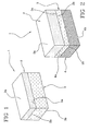
- the multilayer structure 1, subject of the present invention includes a first upper layer 2 presenting an upper surface 2a and a lower surface 2b.
- said multilayer structure 1 further includes at least a lower layer 3 associated with said upper layer 2.
- the lower layer 3 includes an upper surface 3a associated with said lower surface 2b of said upper layer 2, and a lower surface 3b opposite to said upper surface 3a.
- two lower layers 3, 4 can be foreseen.
- the lower layer 3 close to the upper layer 2 presents a respective upper surface 3a associated with said lower surface 2b of the upper layer 2 and a lower surface 3b associated with an upper surface 5a of the lower layer 4.
- the lower layer 3 forms an intermediate layer arranged between said upper layer 2 and said lower layer 4.
- multilayer structures 1, including a plurality of lower layers, for example in a number greater than two and smaller or equal to ten, are included.
- the multilayer structure 1 can show four lower layers 3, 4, 5 and 6, each of which showing a respective upper surface associated with the lower surface of the adjacent layer.
- the upper layer 2 consists of an organic matrix consisting of collagen.
- the lower layers 3, 4, 5 and 6 consist of a composite matrix including collagen and hydroxylapatite.
- the quantities of collagen and hydroxylapatite are expressed in weight percent.
- the lower layer 3 consists of a composite matrix including collagen in a quantity between 95 and 75% and hydroxylapatite in a quantity between 5 and 25%.
- the lower layer 3 consists of a composite matrix including collagen in a quantity between 95 and 75% and hydroxylapatite in a quantity between 5 and 25%
- the lower layer 4 consists of a composite material including collagen in a quantity between 75 and 45% and hydroxylapatite in a quantity between 25 and 55%.
- the lower layer 4 consists of a composite matrix including collagen in a quantity between 95 and 75% and hydroxylapatite in a quantity between 5 and 25%, at least a lower layer 5 consisting of a composite matrix including collagen in a quantity between 75 and 45% and hydroxylapatite in a quantity between 25 and 55% and at least a lower layer 6 consisting of a composite matrix including collagen in a quantity between 45 and 25% and hydroxylapatite in a quantity between 55 and 75%.
- the multilayer structure 1 is characterized by the presence of a collagen gradient extending from the upper layer 2, in which there is 100% of collagen in the lower layer 3, in which there is a collagen quantity from 90 to 70%, until the last lower layer is reached, for example the lower layer 10, in which the collagen quantity is lower than 10%.
- the hydroxylapatite quantity is complementary to the collagen quantity.
- the upper layer 2 has a thickness between 1 and 10 mm; preferably, from 2 to 8; still more preferably, from 3 to 5 mm.
- the lower layers 3-10 can have the same or a different thickness therebetween without any restriction.
- the thickness of the lower layer is between 1 and 10 mm; preferably, from 2 to 8; still more preferably, 3 to 5 mm.
- collagen is intended a fibrous protein of a mucopolysaccharide type which represents the greater component of the extra-cellular matrix.
- the collagen is recognized as the most important structural unit of the connective tissues of the human body.
- collagen it is actually intended a large and heterogeneous class of molecules which, besides structural functions, can have other important physiological direct or indirect functions/roles, such as cell adhesion and differentiation during the development of organs and tissues.
- the triple-helix molecular structure joins all the collagen types. It consists of three single chains, called ⁇ chaines, each of which contains a characteristic sequence of amino acids. These three single chains are wound on one another giving rise to the typical triple-helix structure.
- the collagen of type I represents the more abundant type of collagen existing by nature and is present in several adult connective tissues, such as the dermis, the bone, the tendon and the cornea.
- the collagen used in the present invention can be of type I or type II or type VI or mixtures thereof and can derive from different sources, for example by extraction from bovine, swine and horse derivation.
- the used collagen can be of a synthetic origin, for example can be recombinant.
- hydroxylapatite it is generally understood both the hydroxylapatite Ca 10 (PO4) 6 (OH) 2 with a high and low degree of crystallinity and the hydroxylapatite having chemical substitutions, as the inorganic/mineral component of the bone is formed by sodium, magnesium, carbonate and citrate ions.
- the synthetic hydroxylapatite represents the bone substitute of largest use in surgery.
- the clinical results expressed as the quality of the neo-formed bone not always are satisfactory results.
- the drawback is mostly bound to the fact that the synthetic hydroxylapatite does not have the same features of the human hydroxylapatite, which from a biochemical point of view is formed with a mineralization process on collagen fibrils.
- a bio-mimetic approach for synthetizing mineral inorganic components associated with the organic components, both based on collagen or other natural or synthetic polymers, with a different chemical composition and macromolecular structure has been widely used.
- the macromolecular matrixes mostly act as templates and induce the oriented deposit of the inorganic component by impeding the growth of the crystalline which remains with dimensions of 10-20 nm along the main axis (a measure completely similar to that of a natural human apatite).
- the interaction of HA with the collagen induces a spontaneous carbonation in the position B, which further increases the biomimetic ability and bioavailability degree of the apatite.
- the physical-chemical properties of the organic macromolecular components and the hydroxylapatites are influenced by the chemical interactions and the structural organization between the two components which, besides imparting geometry and morphology, establish the biomechanical properties of the device of the invention.
- crosslinking agents such as glutaraldehyde, formaldehyde, ethers, bis-epoxides and hyaluronic acid have been used for imparting higher mechanical and viscoelastic properties particularly in the part formed by the collagenous layer, for example in the upper layer 2 in figure 1, 2 or 3 .
- the multilayer structure 2 shown by way of example in the enclosed figures 1, 2 and 3 , is obtained through an association with an upper layer 2 formed by an organic matrix consisting of collagen and one or more lower layers formed by a composite matrix including hydroxylapatite and collagen.
- the upper layer 2 and a lower layer 3, for example in form of a gel, are contacted and subsequently the multilayer structure 1 is subjected to a freeze-drying or drying step.
- the multilayer structure 1 can be associated with:
- the multilayer structure can be used or associated with tissue engineering techniques which involve the use of autologous chondrocytes cultured in vitro, the use of mesenchymal cells from bone marrow pre-cultured and expanded in vitro or pre-differentiated or completely differentiated in an osteoblast or chondroblast sense, cells from the bone marrow withdrawn from the patient in the intraoperative step to be used as such or concentrated.
- the cartilaginous substitutes and the osteochondral substitutes can be used for surgical applications in which the reconstruction of cartilaginous and bone surfaces is required, for example in the epiphyseal laminae of the vertebral body of the spine, where the cartilaginous part consists of hyaline cartilage surrounded by a bone ring.
- the cartilaginous substitutes and the osteochondral substitutes can be used in the femoral articular region, in the articular region of the ankle, in the maxillo-facial region (in case of reconstruction of the condyle-mandibular branch), in the osteochondral defects, in the shoulder surgery and in all kinds of orthopaedic surgery which require the formation of a new bone tissue and cartilaginous tissue for regenerating/replacing such originally existing but subsequently damaged or surgically removed tissues.
- the novel three-dimensional composite, multilayer, bioactive and biomimetic structures can be used as cartilaginous substitutes and osteochondral substitutes.
- Such structures, three-dimensional matrixes or "scaffolds”, surgically arranged in the cartilaginous and sub-chondral compartment subjected to a lesion/degeneration will be able to promote the regenerative processes (chondrogenesis and osteogenesis) which lead to the restoration of the anatomy, morphology and the mechanical properties of a normal osteo-cartilaginous tissue of the articular surfaces.
- the composite matrix of the multilayer structure 1 presents an hydroxylapatite/collagen "gradient" between the different lower layers 3-10, each one containing a different quantity of collagen and nucleated hydroxylapatite independently from the thickness of each layer.
- the composites are repeatedly washed in order to purify them from possible acid or basic reaction residues, filtered with the aid of a thin mesh sieve and spread on a surface from which it is possible to easily remove them.
- the single layers are then stratified inside a mould and compacted with each other.
- the multilayer thus obtained is dried by freeze-drying so as to obtain a spongy but resistant and compact material of a desired thickness and gradient.
- the lower layer that is the component which during an operation is intimately contacting the subchondral bone surface
- the collagen and the hydroxylapatite and the relating mixtures are known. It is not known, on the contrary, a method for the preparation of a composite organic-ceramic three-dimensional matrix with properties similar to the human bone tissue.
- An advantage of the present invention is to provide a three-dimensional substitute having a lower layer (hydroxylapatite nucleated on collagen) with the same morphological features of the human bone.
- Such advantage is attained through a direct nucleation process of an apatite phase on collagen fibrils, wherein a solution of calcium salts, which promote the formation of hydroxylapatite, is reacted with similar collagenous natural polymers in an acid suspension.
- a solution of calcium salts which promote the formation of hydroxylapatite
- an aqueous solution of phosphoric component previously additioned with the acid suspension of collagen-like polymers, is dropped.
- the product thus obtained is subsequently subjected to freeze-drying and/or filtration and drying.
- the structure of the collagen-like natural polymer transfers to the molecular level those information which allow to chemically replace the hydroxylapatite, as it occurs in the human body, namely substituents, such as carbonate fractions and ionic fractions such as phosphate existing in the reaction mixture, are entered.
- substituents such as carbonate fractions and ionic fractions such as phosphate existing in the reaction mixture.
- the product obtained through such a process has identical features to those of the human bone tissue thanks to the presence of such substituents in the structure of the composite obtained.
- the direct nucleation acts as a mechanism of biological tendency by forming hydroxylapatite nanocrystals grown orientated parallel to the axis along the collagen fibrils, as it happens in the human body, and gives rise to an inorganic component with a low degree of crystallinity, almost amorphous and accordingly very soluble.
- the crystals thus formed do not grow but remain of dimensions of the order of nanometers.
- the nucleation of the hydroxylapatite in the collagen involves a carbonation of the inorganic phase, that is incorporation of CO 3 2- groups in the hydroxylapatite lattice during the nucleation of the same site B in a proportion similar to that of the natural calcified bone tissue. Furthermore, the carbonation can be mostly assigned to the position B.
- the interaction of the hydroxylapatite with the collagen prevents the carbonation in position A by probably blocking the access to the -OH groups.
- This deviation from the stoichiometry surprisingly increases the similarity with the natural bone tissue and therefore not only the microstructure but also the composition of the obtained artificial tissue is exactly the same as that of the natural bone tissue.
- the carbonation in position B is advantageous with respect to the carbonation in position A because, as a consequence, the bioactivity and the biodegradability of the obtained artificial bone tissue remarkably increase, which are essential features for the calcification as they allow a continuous dynamic exchange between the physiological fluid and the cell which uses the ions for the bone formation.
- the present invention uses an approach of biological tendency for synthesizing hydroxylapatite nanocrystals on self-assembling collagen fibrils similar to the nanocrystals of natural bones, by exploiting the ability of the negatively charged collagen carboxyl groups of binding calcium ions of the hydroxylapatite; following this approach, it has been successfully proved that the biological systems store and process information at a molecular level. For this reason, the collagen molecules of type I without telopeptides and free of glucosylated regions, capable of self-aggregating in fibrils without crosslinking agents, have been used as starting materials.
- the collagen of type I has been found on the market as a standard product. After the purification process, the collagen of type I is dissolved in an estimated volume of acetic acid until a homogeneous suspension is obtained (1% collagen).
- calcium salts in aqueous solution or SBF are used, which promote the hydroxylapatite formation, preferably selected from the group including calcium hydroxide, calcium nitrate, calcium acetate, calcium sulphate, calcium carbonate. More preferably, calcium hydroxide is used.
- an acid suspension of collagen-like natural polymer is used, obtained by treatment of animal collagen extracted from horse tendon, mouse tail, kangaroo tail or collagen-like synthetic polymer or chemically crosslinked gelatines.
- the phosphate component used has been selected among phosphoric acid in acqueous solution or SBF or a calcium, ammonium, sodium, potassium and magnesium salt in acqueous solution with a phosphate anion PO 4 (3-) , mono-hygrogen phosphate HPO 4 (2-) or dihydrogen phosphate H2PO 4 (-) . More preferably, phosphoric acid H 3 PO 4 is used.
- the calcium salts used by the present invention are in the range between 1 and 4 g/l (grams/litre), the phosphate component is in the range between 2 g/l and 4 g/l. More preferably, the calcium salts used in the present invention are in the range between 2 and 3 g/l, the phosphate component is the range between 3 and 4 g/l.
- the direct nucleation step occurs with a pH preferably between 9 and 12, more preferably between 9-11 (at the end of the reaction the pH must be between 7 and 8.5 or more preferably between 7 and 7.5) and at a temperature preferably between 25 to 45°C, more preferably between 35 and 40°C.
- aqueous or SBF solution of Ca(OH) 2 in an aqueous or SBF solution of Ca(OH) 2 . (147 g of Ca(OH)2 in 300 ml (cc) of H 2 O) a phosphoric acid solution (1.17 g Of H 3 PO 4 in 200 ml (cc) of H 2 O) and 50 g of collagen in acetic acid were dropped.
- the pH during the process was between 7-10, the temperature was maintained around 25°C, the dropping time of the phosphoric acid solution in the mixture of calcium salts in acqueous solution and acid suspension of collagen was maintained between 15 and 60 minutes and therefore the dropping rate was preferably between 0.0133 l/minute and 0.0033 l/minute, more preferably between 0.0100 and 0.0060 l/min.
- the obtained products, coded HA/Col nuc. col. were subsequently subjected to a freeze-drying or drying step.
- the composites have been characterized by crystal analysis with a x-ray diffractometer (Rigaku Miniflex); for analysing the orientation, some diffraction graphs have been photographically recorded with a sample-film distance of 70 mm by using a flat chamber (always with Cu-K ⁇ , radiation).
- thermogravimetric analysis has been carried out (Polymer STA 1660) by using an alumina crucible in air and using a heating rate of 10°C/min.
- the composites prepared according to the method of the present invention by direct nucleation of hydroxylapatite in the collagen fibrils had a hydroxylapatite/collagen nominal composition from 80/20 to 10/90 as a mass.
- thermogravimetric analysis it has been found that the actual composition was respectively 70/30 and 10/90. This discrepancy is due to the reaction yield which does not reach 100%.
- the water content in the compound is very similar to that of pure collagen (10%).
- This analysis has been successfully used in order to obtain information about the modifications of the structural relations between collagen fibrils and inorganic phase as a function of the mineral content. Previously, such analysis has been carried out on the flexor tendon of the turkey foot which is used as a pattern in the calcification process.
- the TG-DTG graphs seem modified with respect to those of the hydroxylapatite/collagen compounds, which show an interaction of the inorganic phase with the collagen fibres. This kind of interaction can be compared to the calcification process which naturally occurs. The comparison is carried out with a TG-DTG graph relating to a turkey calcified tendon, used as a bone theoretical pattern. The close similitude is apparent because in both cases the DTG peak at 450-500°C tends to disappear by forming an "enlarged shoulder".
- the XRD crystallogram shows a typical graph of hydroxylapatite with a very low crystallinity, the sizes of the estimated crystals along the axis is about 12-15 nm.
- the nucleation occurs according to the typical process of the natural bone, in which the nano-dimension of the crystals is responsible of the extension of the reflection in the graph.
- the reaction has been carried out at about 25°C, one is widely within the limits for forming nanocrystals of monocristalline hydroxylapatite.
- Such nanocrystals grow within the collagen fibres with their c axis preferably oriented parallel to the orientation direction of the fibres.
- the diffraction graph of the wide X-ray angle is obtained from a sample consisting of some calcified fibrils.
- the TEM micrograph shows nano-nuclei formed within the collagen fibrils which grow parallel to the fibrils.
- the analysis under the optical microscope shows the different crystalline morphology of the hydroxylapatite/collagen composite obtained by direct nucleation and subsequently freeze-dried from the one air dried.
- the freeze-dried composites show a three-dimensional network characterized by a very large pore distribution similar to that of the cotton and the wool.
- a two-dimensional network is formed and the structure looks like a gauze fill.
- the artificial bone tissue according to the present invention can be obtained with different quantities of hydroxylapatite/collagen and various formulations and is suitable to several clinical applications.
- Such artificial bone tissue can be used as a constituent of the lower HA-collagen layer/s of the cartilaginous/osteo-cartilaginous substitute of the present invention and also as a prosthetic material, for replacing or filling the bone, as a reconstructive membrane or haemostatic tissue in orthopedics, odontotherapy, maxillofacial surgery.
- the substitutes above mentioned can be used as an osteochondral substitute for cartilaginous defects of 3 and 4 Outerbridge degree or for cartilaginous defects of deep 4 Outerbridge degree.
- the chondral or osteochondral substitute (scaffold) of the present invention has been chemically modified so as to render all the general structure more elastic and more hydrophilic, therefore less subjected to delamination at the interface between the lower surface 2b of the upper layer 2 and the upper surface 3a of the lower layer 3 during the handling carried out by the operator.
- the delamination phenomenon of the layers is a drawback which occurs when the substitute, once prepared, is hydrated.
- the delamination represents a problem which has to be necessarily overcome.
- a multilayer structure 1 presents an upper layer 2 consisting of collagen of type I and a lower layer 3 consisting of hydroxylapatite/collagen type I (40%-60%).
- Another multilayer structure 1 presents an upper layer 2 consisting of collagen of type I and a lower layer 3 consisting of hydroxylapatite/collagen type I (40%-60%) and a lower layer 4 consisting of hydroxylapatite/collagen (70%-30%).
- a sterile portion has been subjected to the following tests:
- a sterile portion has been subjected to in vitro and in vivo tests and in vivo handling tests on sheep and on fetlock cartilage from a horse cadaver. Finally, an aliquot for carrying out further tests and chemical analysis has been kept.
- the wettability tests have been carried out in water, by dipping, handling and rehydrating each single scaffold portion and have observing it up to a week. For all the versions, a stability up to two days was observed.
- the prototypes B and C showed a fast hydration ability and a higher swelling than the prototypes A and D.
- the version D showed a greater rigidity.
- the optical microscope observations at 12x have been carried out on 2 mm sagittal sections of each configuration.
- the ESEM images have been acquired at 25 and 100x.
- Figure 4 shows a series of images of a scanning electron microscopy alone, relating to all the prototypes of the configuration I of the scaffold.
- the collagenous structure of the prototype A' shows a rather regular porosity (1), but with a greater enlargement the walls of the pores seem more indented (2).
- the interface collagen-hydroxylapatite gradient/collagen 40/60 is apparent (3).
- Figure 5 shows the porosity of the collagenous layer of the prototype B', which is regular and rather compact relative to the prototype A'. This compactness is probably to be intended as an artifact due, on a greater extent, to the blade action during the cut of the scaffold rather than to an action of the hyaluronic acid on the structure (1).
- the conservation of the pore regularity is noted, with a similar conservation of the surface regularity of the internal walls (2). Particular orientations of the pores are not visible. On the contrary, a greater uniformity and compactness of the single structures at the interface collagen-HA gradient/collagen (3) seems to be visible.
- plasticizers or crosslinking agents during the preparative step of the scaffold is able to impart a greater stability to their structure.
- the stability requirement for a collagen-based scaffold which can be engineered or not, represents the determining element.
- hyaluronic acid with a medium molecular weight a crosslinking agent belonging to the family of the bis-epoxides (diglycidyl ether) and glutaraldehyde vapours or its derivates have been used as agent capable of increasing the hydrophilic and plastic properties of the construct.
- the aim of the experiments was to check the in vivo behaviour of a configuration of chondral and osteochondral scaffold with a configuration with two (cartilaginous substitute) and three gradients (osteocartilaginous substitute) respectively, subjected to a crosslinking with BDDGE (1,4-butanediol-diglycidyl ether) in the preparation step.
- BDDGE 1,4-butanediol-diglycidyl ether
- the selected animal models were:
- the aim of the present study was to check the bone and cartilaginous neo-formation of the three-phase scaffold or with a configuration II (gradient 1 collagen type I, gradient 2 collagen type I/hydroxylapatite ratio 70%/30%, gradient 3 collagen type I/hydroxylapatite ratio 30%/70%) loaded with sheep stromal parent cells (BMSC) coming from a withdrawal of medullary blood.
- the scaffold has been implanted on an intramuscular site (extra-skeletal or heterotopic site) in an immune-depressed nude mouse for the purpose of checking the tissue differentiation within the gradients of the osteochondral scaffold.
- the aim of the present in vivo study was to check in no. 2 animals, after a cartilaginous and osteo-cartilaginous lesion in the distal epiphisys of the third metacarpal (fetlock) of a horse, the behaviour of the configurations with 2 and 3 gradients of the scaffold prepared with a nucleation process of hydroxylapatite nano-particles on collagen fibres subjected to a crosslinking process by BDDGE (1,4-butanediol-diglycidyl ether).
- the two forms of the scaffold were represented by: configuration I or two-phase scaffold (2 gradients) whose intended use is a cartilaginous substitute scaffold for the treatment of chondral lesions and configuration II or three-phase scaffold (3 gradients) whose intended use is an osteo-cartilaginous substitute scaffold for the treatment of severe degree or deep osteochondral lesions.
- the 2 animals were subjected to a radiographic control of the joints for excluding the presence of any pathological alterations.
- the horses are maintained without food the 12 hours before the operation.
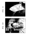
- the circulation was blocked with a vascular emptying of the carpus.
- a miniarthrotomy was performed on the dorsal face of the fetlock in correspondence with the distal end of the metacarpal III° ( figure 10 ), laterally and in an upper position with respect to the tendon of the dorsal extensor muscle of the digit.
- the miniarthrotomy was performed medial to the mentioned tendon.
- the scaffolds were placed within the holes and maintained in the seat for a few seconds through digital pression until a swelling was reached, which ensured its fixing to the walls of the lesions ( figure 13 ).
- the sutures of the surgical incisions were carried out by levels in the following way: separated-spot suture of the articular capsule with polyglycolic acid No. 0, continuous suture of the digital fascia with polyglycolic acid No. 00 and separated-spot suture of the dermis with nylon No. 0.
- the animals were sacrificed after 8 weeks and the implant site was accurately removed and processed for hematoxylin/eosin staining.
- the microscopic observations pointed out a bone neo-formation within the gradient with a prevalent hydroxylapatite component (gradient 3), while no bone exceeding in the collagenous portion (gradient 1) intended for a cartilaginous neo-formation ( figure 14 ) was observed.
- the data show how the hydroxylapatite can play a "priming" function, namely it is able to activate the cell differentiation process, in a bone sense, in the deepest layer, whereas within the more superficial layers the collagenous composition of the scaffold guides the cell differentiation in a cartilaginous sense.
- the arthroscopic check was carried out after 3 months from the first operation date.
- the images pointed out, within the chondral and osteochondral lesion site of both the forelimbs, a neo-formation of connective tissue which resulted of a good consistency at the sight, however it was not distinguishable from a fibrous tissue.
- scaffold traces were apparent. From the arthroscopic observations carried out on the animal no. 2 in correspondence with the lateral lesion, no reaction of an inflammatory type, nor a displacement of the device were apparent ( figure 15 ).
- the animals were killed in an average interval of 214 ⁇ 11 days.
- the animals were sacrificed through intravenous injection of a Tanax solution, by previous administration of general anaesthesia.
- both the limbs were removed 20 cm in a proximal portion relative to the joint (fetlock) where the surgical operations were carried out.
- Figure 15 relates to the arthroscopic "2 nd look”.
- Figure 16 relates to the "2 nd look" of the animal no. 1.
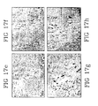
- Figure 17 relates to the histological analysis with a toluidine blue staining with different enlargements of the chondral implant (animal 1).
- the reported histological images relate to the animal no. 1, lateral lesion corresponding to the chondral implant.
- the operation zones were carefully removed, by also collecting a health cartilage portion surrounding the lesion.
- Each collection was divided in two parts and the cutting at the microtome was performed from the centre to the periphery and stained with toluidine blue for the microscopic observations.
- the surface collagenous layer was colonized by a fibro-cartilaginous tissue distinguishable from the healthy cartilaginous tissue and the presence of exceeding bone tissue within it was not found.
- the analysed sample of about 1 cm 3 , has been collected at the implant level and includes the surrounding area of articular cartilage.
- the sample has been decalcified and included in paraffin.
- the sections have been cut at a thickness of about 10 ⁇ m and stained with toluidine blue.
- the photographs concern the two ends of the implant, namely the zones of interest at the healthy articular cartilage-implant interface.
- the implant itself seems superficially formed with fibrocartilage and fibrous connective tissue without any trace of tissue morphologically referable to an articular and deep cartilage of bone tissue.
- the neo-formed bone tissue (visible in A and B) seems to be well integrated with the surrounding bone, the cartilaginous tissue presents an apparent demarcation line (arrow) which marks the passage from the pre-existing articular cartilage to the neo-formed fibrocartilage.
Landscapes
- Health & Medical Sciences (AREA)
- Chemical & Material Sciences (AREA)
- Life Sciences & Earth Sciences (AREA)
- Medicinal Chemistry (AREA)
- Public Health (AREA)
- Epidemiology (AREA)
- Animal Behavior & Ethology (AREA)
- General Health & Medical Sciences (AREA)
- Veterinary Medicine (AREA)
- Dermatology (AREA)
- Transplantation (AREA)
- Oral & Maxillofacial Surgery (AREA)
- Engineering & Computer Science (AREA)
- Materials Engineering (AREA)
- Composite Materials (AREA)
- Inorganic Chemistry (AREA)
- Biophysics (AREA)
- Dispersion Chemistry (AREA)
- Surgery (AREA)
- Molecular Biology (AREA)
- Biomedical Technology (AREA)
- Materials For Medical Uses (AREA)
- Prostheses (AREA)
- Road Paving Structures (AREA)
- Chemical Kinetics & Catalysis (AREA)
- Polymers & Plastics (AREA)
- Nitrogen And Oxygen Or Sulfur-Condensed Heterocyclic Ring Systems (AREA)
- Packages (AREA)
- Organic Chemistry (AREA)
- Furnace Housings, Linings, Walls, And Ceilings (AREA)
- Steroid Compounds (AREA)
- Saccharide Compounds (AREA)
- Superconductors And Manufacturing Methods Therefor (AREA)
- Laminated Bodies (AREA)
- Feedback Control In General (AREA)
Claims (19)
- Structure multicouche (1) incluant :- une première couche supérieure (2) consistant en une matrice organique consistant en collagène ; et- une ou plusieurs couches inférieures (3, 4, ... 10) consistant en une matrice composite incluant hydroxylapatite et collagène ;dans laquelle ladite couche supérieure (2) est en contact direct avec une première couche inférieure (3) ; et
dans laquelle ladite première couche inférieure (3) consiste en une matrice composite incluant une quantité pondérale de collagène entre 95 et 75% et une quantité pondérale d'hydroxylapatite entre 5 et 25%. - Structure multicouche selon la revendication 1, comprenant également une deuxième couche inférieure (4) consistant en une matrice composite incluant une quantité pondérale de collagène entre 75 et 45% et une quantité pondérale d'hydroxylapatite entre 25 et 55%.
- Structure multicouche selon la revendication 2, comprenant également une troisième couche inférieure (5) consistant en une matrice composite incluant une quantité pondérale de collagène entre 45 et 25% et une quantité d'hydroxylapatite entre 55 et 75%.
- Structure multicouche selon la revendication 2, comprenant également une dernière couche inférieure (10) consistant en une matrice composite incluant une quantité pondérale de collagène inférieure à 5% et une quantité pondérale d'hydroxylapatite telle d'obtenir 100%.
- Structure multicouche selon l'une quelconque des revendications précédentes 1 à 4, dans laquelle se trouve un gradient de collagène s'étendant de la couche supérieure (2), où se trouve le 100% de collagène, à la première couche inférieure (3), où se trouve une quantité de collagène de 95 à 75% jusqu'à atteindre une dernière couche inférieure (10), où la quantité de collagène est inférieure à 5%.
- Structure multicouche selon une ou plusieurs des revendications 1 à 5, dans laquelle l'hydroxylapatite de ladite matrice composite est directement nucléée sur les fibres de collagène.
- Structure multicouche selon une ou plusieurs des revendications précédentes, dans laquelle le collagène est sélectionné dans : collagène de type I, de type II, de type VI ou leur mélanges.
- Substitut cartilagineux incluant une structure multicouche selon une ou plusieurs des revendications 1 à 7.
- Substitut ostéochondral incluant une structure multicouche selon une ou plusieurs des revendications 1 à 7.
- Substitut selon les revendications 8 ou 9, dans lequel la structure multicouche représente un tuteur pour l'ancrage in situ et la différenciation des cellules mésenchymateuses.
- Substitut selon les revendications 8 ou 9, dans lequel la structure multicouche est chargée ex-vivo d'un concentré de sang médullaire ou d'un concentré plaquettaire (PRP) ou encore de facteurs de croissance ou de facteurs capables d'augmenter le trophisme et la différenciation cellulaire, comme TGF, EGF, BMP et autres facteurs.
- Substitut selon les revendications 8 ou 9, dans lequel la structure multicouche est chargée ex-vivo de cellules mésenchymateuses indifférenciées ou mises en culture pendant une durée nécessaire à la multiplication et/ou à la différenciation dans les cellules mères des ostéoblastes et des chondrocytes.
- Substitut selon les revendications 8 ou 9, dans lequel la structure multicouche est chargée de substances pharmacologiquement actives sélectionnées dans : corticostéroïdes anti-inflammatoires, FANS, immunosuppresseurs, antibiotiques, antiblastiques, antiprolifératifs et antiviraux.
- Procédé pour préparer une structure multicouche selon l'une quelconque des revendications 1 à 7, incluant au moins une étape dans laquelle une première couche supérieure (2) consistant en une matrice organique consistant en collagène, est associée au moins à une couche inférieure (3, 4, ..., 10) consistant en une matrice composite incluant hydroxylapatite et collagène.
- Substitut selon une ou plusieurs des revendications 8 à 13, dans lequel la structure multicouche est réticulée avec de l'acide hyaluronique ou un sel ou dérivé de cet acide ; de préférence avec un poids moléculaire compris entre 10 000 et 2 000 000 kD.
- Substitut selon une ou plusieurs des revendications 8 à 13, dans lequel la structure multicouche est réticulée avec du BDDGE ou un dérivé.
- Substitut selon une ou plusieurs des revendications 8 à 13, dans lequel la structure multicouche est réticulée avec du glutaraldéhyde ou un dérivé, ou avec un agent réticulant sélectionné dans les bis-époxydes (éther de diglycidyle).
- Substitut selon une ou plusieurs des revendications 8 à 17, sous forme lyophilisée ou déshydratée.
- Substitut selon une ou plusieurs des revendications 8 à 17, sous forme stérile.
Applications Claiming Priority (2)
| Application Number | Priority Date | Filing Date | Title |
|---|---|---|---|
| IT000343A ITMI20050343A1 (it) | 2005-03-04 | 2005-03-04 | Sostituto cartilagineo e osteocindrale comprendente una struttura multistrato e relativo impiego |
| PCT/IB2006/000452 WO2006092718A2 (fr) | 2005-03-04 | 2006-03-02 | Substitut cartilagineux et osteochondrial comprenant une structure multicouche et son utilisation |
Publications (2)
| Publication Number | Publication Date |
|---|---|
| EP1858562A2 EP1858562A2 (fr) | 2007-11-28 |
| EP1858562B1 true EP1858562B1 (fr) | 2009-11-11 |
Family
ID=36917317
Family Applications (1)
| Application Number | Title | Priority Date | Filing Date |
|---|---|---|---|
| EP06710487A Active EP1858562B1 (fr) | 2005-03-04 | 2006-03-02 | Substitut cartilagineux et osteochondral comprenant une structure multicouche et son utilisation |
Country Status (13)
| Country | Link |
|---|---|
| US (1) | US9155818B2 (fr) |
| EP (1) | EP1858562B1 (fr) |
| JP (1) | JP5289772B2 (fr) |
| CN (1) | CN101163512B (fr) |
| AT (1) | ATE447986T1 (fr) |
| BR (1) | BRPI0609392B8 (fr) |
| CA (1) | CA2600893C (fr) |
| DE (1) | DE602006010341D1 (fr) |
| DK (1) | DK1858562T3 (fr) |
| ES (1) | ES2336359T3 (fr) |
| IT (1) | ITMI20050343A1 (fr) |
| WO (1) | WO2006092718A2 (fr) |
| ZA (1) | ZA200707511B (fr) |
Cited By (4)
| Publication number | Priority date | Publication date | Assignee | Title |
|---|---|---|---|---|
| CN102580156A (zh) * | 2012-03-29 | 2012-07-18 | 陕西博鸿生物科技有限公司 | 一种组织工程软骨支架材料及其制备方法和装置 |
| US8435552B2 (en) | 2007-02-09 | 2013-05-07 | Royal College Of Surgeons In Ireland | Collagen/hydroxyapatite composite scaffold, and process for the production thereof |
| WO2013083844A2 (fr) | 2011-12-08 | 2013-06-13 | Julius-Maximilians-Universität Würzburg | Fabrication de matériaux structurés de manière anisotrope |
| US10576694B2 (en) | 2011-12-08 | 2020-03-03 | Julius-Maximilians-Universitaet Wuerzburg | Production of materials having an anisotropic structure |
Families Citing this family (42)
| Publication number | Priority date | Publication date | Assignee | Title |
|---|---|---|---|---|
| EP1237511B1 (fr) | 1999-12-15 | 2004-09-01 | Sulzer Orthopedics Ltd. | Preparation pour reparation d'articulations chez l'homme ou l'animal, suite a des anomalies cartilagineuses ou cartilagino-osseuses |
| US20020114795A1 (en) | 2000-12-22 | 2002-08-22 | Thorne Kevin J. | Composition and process for bone growth and repair |
| GB2424223C (en) * | 2005-03-07 | 2011-02-02 | Massachusetts Inst Technology | Biomaterial. |
| US7718616B2 (en) | 2006-12-21 | 2010-05-18 | Zimmer Orthobiologics, Inc. | Bone growth particles and osteoinductive composition thereof |
| PL2389204T3 (pl) * | 2009-01-23 | 2019-12-31 | Royal College Of Surgeons In Ireland | Rusztowanie warstwowe odpowiednie do naprawy kostno- chrzęstnej |
| WO2010102266A1 (fr) * | 2009-03-05 | 2010-09-10 | Biomimetic Therapeutics, Inc. | Compositions de facteur de croissance dérivé des plaquettes et méthodes de traitement de défauts ostéo-cartilagineux |
| JP5545563B2 (ja) * | 2009-09-08 | 2014-07-09 | 利江 土屋 | 軟骨用移植材 |
| IT1396934B1 (it) * | 2009-11-25 | 2012-12-20 | Fin Ceramica Faenza Spa | Materiali bio-mimetici compositi, relativo processo di preparazione e loro uso per la realizzazione di strutture mono-, bi- o multi-strato per la rigenerazione di tessuto osseo, cartilagineo o osteocartilagineo |
| US11786636B2 (en) * | 2010-06-15 | 2023-10-17 | Versitech Limited | Methods for complex tissue engineering |
| US9693954B2 (en) * | 2010-06-25 | 2017-07-04 | President And Fellows Of Harvard College | Co-delivery of stimulatory and inhibitory factors to create temporally stable and spatially restricted zones |
| WO2012017045A1 (fr) * | 2010-08-04 | 2012-02-09 | Technische Universität Dresden | Hydroxyapatite à croissance inhibée, procédé pour sa préparation et utilisation |
| US8435305B2 (en) | 2010-08-31 | 2013-05-07 | Zimmer, Inc. | Osteochondral graft delivery device and uses thereof |
| ITMI20102070A1 (it) | 2010-11-08 | 2012-05-09 | Consiglio Nazionale Ricerche | Impianti per sostituzioni ossee "load bearing" ad architettura gerarchicamente organizzata derivante dalla trasformazione di strutture vegetali |
| AU2011329054B2 (en) | 2010-11-15 | 2015-05-28 | Zimmer Orthobiologics, Inc. | Bone void fillers |
| KR101375828B1 (ko) * | 2011-03-31 | 2014-03-17 | 인제대학교 산학협력단 | 골-연골 재생용 복합 지지체, 이의 제조 방법 및 이를 유효성분으로 함유하는 골-연골 질환 치료용 조성물 |
| EP2786770B1 (fr) * | 2011-08-30 | 2017-04-19 | Kyoto University | Matériau d'ossature poreux et son procédé de production |
| JP6018477B2 (ja) * | 2011-11-11 | 2016-11-02 | Hoya株式会社 | 人工骨−軟骨複合体及びその製造方法 |
| CN103920190B (zh) * | 2013-01-14 | 2017-09-29 | 陕西博鸿生物科技集团有限公司 | 一种关节软骨移植物及其制备方法 |
| ITMI20130636A1 (it) * | 2013-04-18 | 2014-10-19 | Novagenit S R L | Metodo per la preparazione di biomateriali biocompatibili e biodegradabili a base di collagene e granuli di idrossiapatite/¿-tricalcio fosfato per uso in chirurgia ortopedica e biomateriali così ottenuti |
| EP2826495A1 (fr) * | 2013-07-19 | 2015-01-21 | Geistlich Pharma AG | Matériau composite en collagène-hydroxyapatite biomimétique |
| KR20150053606A (ko) * | 2013-11-08 | 2015-05-18 | 세원셀론텍(주) | 콜라겐과 히알루론산의 천연가교를 통한 고무성질의 물성이 강화된 생체재료물질 및 그 제조방법 |
| CN104324418B (zh) * | 2014-10-27 | 2016-04-06 | 东华大学 | 一种组织工程纳米纤维骨软骨修复支架及其制备方法 |
| CN104307047B (zh) * | 2014-10-29 | 2017-02-01 | 中国科学院长春应用化学研究所 | 一种双梯度仿生修复支架及其制备方法 |
| CN106139255A (zh) * | 2015-04-17 | 2016-11-23 | 天津市赛宁生物工程技术有限公司 | 一种胶原蛋白结合磷酸钙的医用海绵支架的新型材料和制备方法 |
| CN106806940A (zh) * | 2015-12-02 | 2017-06-09 | 中国科学院大连化学物理研究所 | 一种掺杂纳米羟基磷灰石的多孔仿生骨支架的制备方法 |
| EP3175869A1 (fr) | 2015-12-04 | 2017-06-07 | Geistlich Pharma AG | Membrane de forme stable réticulée résorbable |
| WO2018047785A1 (fr) * | 2016-09-07 | 2018-03-15 | ナガセケムテックス株式会社 | Polypeptide de type collagène |
| IT201600094601A1 (it) | 2016-09-21 | 2018-03-21 | Fin Ceram Faenza S P A | “apparecchiatura e kit per la realizzazione di fori a profondità controllata e di diverso diametro su superfici condrali ed osteocondrali” |
| EP3409303A1 (fr) * | 2017-06-02 | 2018-12-05 | Geistlich Pharma AG | Utilisation d'une composition à stabilité de forme réticulée résorbable pour préparer une membrane |
| CN107669371B (zh) * | 2017-08-02 | 2018-08-03 | 中南大学湘雅医院 | 三相组织工程支架 |
| CN107789668B (zh) * | 2017-11-03 | 2020-10-27 | 华中科技大学同济医学院附属协和医院 | 具有多层结构的仿生胶原蛋白骨修复材料及其制备方法 |
| CN108355174A (zh) * | 2018-05-06 | 2018-08-03 | 西北工业大学 | 一种多功能分层关节软骨支架的制备方法 |
| WO2020120805A2 (fr) | 2018-12-14 | 2020-06-18 | Institut Químic De Sarrià Cets Fundació Privada | Échafaudages hétérogènes et procédés de fonctionnalisation de surfaces |
| CN109628387A (zh) * | 2018-12-28 | 2019-04-16 | 广州赛莱拉干细胞科技股份有限公司 | 组合物、含有该组合物的分化诱导培养液及诱导方法 |
| CN110368529B (zh) * | 2019-08-19 | 2021-08-31 | 江苏地韵医疗科技有限公司 | 软骨或软骨下骨全层修复支架及其制备方法 |
| US11633879B2 (en) | 2020-01-21 | 2023-04-25 | United States Government As Represented By The Department Of Veterans Affairs | Anatomic tissue-engineered osteochondral implant and method for fabrication thereof |
| CN111330080B (zh) * | 2020-03-31 | 2021-12-07 | 江苏白衣缘生物工程有限公司 | 一种引导口腔骨再生的生物膜及其制备方法 |
| CN111375088B (zh) * | 2020-04-29 | 2022-06-14 | 陕西巨子生物技术有限公司 | 双层骨软骨组织修复支架及其制备方法 |
| KR20230104224A (ko) * | 2020-11-05 | 2023-07-07 | 데이텀 덴탈 엘티디. | 다중 층상 콜라겐 구조체 |
| CN113559329A (zh) * | 2021-09-03 | 2021-10-29 | 北京大学口腔医学院 | 一种仿牙周有序双层结构支架材料及其制备方法和应用 |
| CN114732957B (zh) * | 2022-03-16 | 2022-12-23 | 武汉理工大学 | 一种具有预应力的矿化肌腱复合材料及其制备方法与应用 |
| SK1172022A3 (sk) | 2022-12-05 | 2024-06-19 | Ústav materiálového výskumu Slovenskej akadémie vied, verejná výskumná inštitúcia | Kompozitný biocementový systém |
Family Cites Families (12)
| Publication number | Priority date | Publication date | Assignee | Title |
|---|---|---|---|---|
| JP3082503B2 (ja) * | 1993-02-26 | 2000-08-28 | 株式会社神戸製鋼所 | 人工骨製造用前駆体および人工骨の製法 |
| US6117979A (en) * | 1997-08-18 | 2000-09-12 | Medtronic, Inc. | Process for making a bioprosthetic device and implants produced therefrom |
| GB9727048D0 (en) * | 1997-12-22 | 1998-02-18 | Abonetics Ltd | Process for the preparation of magnesium and carbonate substituted hydroxyapatite |
| US20030114936A1 (en) * | 1998-10-12 | 2003-06-19 | Therics, Inc. | Complex three-dimensional composite scaffold resistant to delimination |
| CN1169494C (zh) * | 2001-05-23 | 2004-10-06 | 中国科学院化学研究所 | 组织工程用复合结构细胞支架及其制法和用途 |
| US6626950B2 (en) * | 2001-06-28 | 2003-09-30 | Ethicon, Inc. | Composite scaffold with post anchor for the repair and regeneration of tissue |
| GB0121985D0 (en) * | 2001-09-11 | 2001-10-31 | Isis Innovation | Tissue engineering scaffolds |
| JP3727059B2 (ja) * | 2002-05-01 | 2005-12-14 | 独立行政法人科学技術振興機構 | 多孔質複合材料の製造方法 |
| JP4416152B2 (ja) * | 2003-03-28 | 2010-02-17 | 独立行政法人物質・材料研究機構 | 生体組織補填材とその製造方法 |
| US8039090B2 (en) * | 2003-05-26 | 2011-10-18 | Hoya Corporation | Porous composite containing calcium phosphate and process for producing the same |
| US20050074877A1 (en) * | 2003-07-28 | 2005-04-07 | Mao Jeremy Jian | Biological engineering of articular structures containing both cartilage and bone |
| US7217294B2 (en) * | 2003-08-20 | 2007-05-15 | Histogenics Corp. | Acellular matrix implants for treatment of articular cartilage, bone or osteochondral defects and injuries and method for use thereof |
-
2005
- 2005-03-04 IT IT000343A patent/ITMI20050343A1/it unknown
-
2006
- 2006-03-02 WO PCT/IB2006/000452 patent/WO2006092718A2/fr active Application Filing
- 2006-03-02 ZA ZA200707511A patent/ZA200707511B/xx unknown
- 2006-03-02 BR BRPI0609392A patent/BRPI0609392B8/pt active IP Right Grant
- 2006-03-02 EP EP06710487A patent/EP1858562B1/fr active Active
- 2006-03-02 US US11/817,172 patent/US9155818B2/en active Active
- 2006-03-02 CA CA2600893A patent/CA2600893C/fr active Active
- 2006-03-02 JP JP2007557618A patent/JP5289772B2/ja active Active
- 2006-03-02 AT AT06710487T patent/ATE447986T1/de active
- 2006-03-02 DK DK06710487.7T patent/DK1858562T3/da active
- 2006-03-02 DE DE602006010341T patent/DE602006010341D1/de active Active
- 2006-03-02 CN CN2006800137243A patent/CN101163512B/zh active Active
- 2006-03-02 ES ES06710487T patent/ES2336359T3/es active Active
Cited By (7)
| Publication number | Priority date | Publication date | Assignee | Title |
|---|---|---|---|---|
| US8435552B2 (en) | 2007-02-09 | 2013-05-07 | Royal College Of Surgeons In Ireland | Collagen/hydroxyapatite composite scaffold, and process for the production thereof |
| US9138483B2 (en) | 2007-02-09 | 2015-09-22 | Royal College Of Surgeons In Ireland | Collagen/hydroxyapatite composite scaffold, and process for the production thereof |
| WO2013083844A2 (fr) | 2011-12-08 | 2013-06-13 | Julius-Maximilians-Universität Würzburg | Fabrication de matériaux structurés de manière anisotrope |
| US9649816B2 (en) | 2011-12-08 | 2017-05-16 | Julius-Maximilians-Universitaet Wuerzburg | Production of materials having an anisotropic structure |
| US10576694B2 (en) | 2011-12-08 | 2020-03-03 | Julius-Maximilians-Universitaet Wuerzburg | Production of materials having an anisotropic structure |
| CN102580156A (zh) * | 2012-03-29 | 2012-07-18 | 陕西博鸿生物科技有限公司 | 一种组织工程软骨支架材料及其制备方法和装置 |
| CN102580156B (zh) * | 2012-03-29 | 2014-10-08 | 陕西博鸿生物科技有限公司 | 一种组织工程软骨支架材料及其制备方法和装置 |
Also Published As
| Publication number | Publication date |
|---|---|
| ATE447986T1 (de) | 2009-11-15 |
| BRPI0609392B8 (pt) | 2021-06-22 |
| US20090232875A1 (en) | 2009-09-17 |
| CA2600893A1 (fr) | 2006-09-08 |
| BRPI0609392A2 (pt) | 2010-03-30 |
| ZA200707511B (en) | 2008-11-26 |
| US9155818B2 (en) | 2015-10-13 |
| WO2006092718A3 (fr) | 2007-01-25 |
| CA2600893C (fr) | 2014-04-29 |
| DK1858562T3 (da) | 2010-03-22 |
| CN101163512A (zh) | 2008-04-16 |
| ITMI20050343A1 (it) | 2006-09-05 |
| JP2008531171A (ja) | 2008-08-14 |
| ES2336359T3 (es) | 2010-04-12 |
| CN101163512B (zh) | 2010-11-17 |
| EP1858562A2 (fr) | 2007-11-28 |
| WO2006092718A2 (fr) | 2006-09-08 |
| JP5289772B2 (ja) | 2013-09-11 |
| BRPI0609392B1 (pt) | 2018-05-29 |
| DE602006010341D1 (de) | 2009-12-24 |
Similar Documents
| Publication | Publication Date | Title |
|---|---|---|
| EP1858562B1 (fr) | Substitut cartilagineux et osteochondral comprenant une structure multicouche et son utilisation | |
| AU2006221849B2 (en) | Biomaterial | |
| RU2292858C2 (ru) | Костный материал и коллагеновая композиция для восстановления поврежденных суставов | |
| US6139578A (en) | Preparation of cell seeded ceramic compositions | |
| EP1799277B1 (fr) | Composite de matiere de charge biologique poreuse et procede de fabrication correspondant | |
| JP2010500070A (ja) | 生体材料 | |
| Vijayalekha et al. | An overview of collagen-based composite scaffold for bone tissue engineering | |
| US20190015553A1 (en) | Preparation method of injectable extracellular matrix based hydrogel derived from decellularized porcine skin loaded with bi-phasic calcium phosphate | |
| KR20130115781A (ko) | 인체뼈 유래 무기질을 포함하는 천연 골재생재 | |
| JP3718723B2 (ja) | 生体組織由来吸収性リン酸カルシウム傾斜機能複合材料とその作製方法 | |
| Tetteh | Polyurethane-based Scaffolds for Bone Tissue Engineering. The Role of Hydroxyapatite Particles, Solvent Combinations, Electrospun Fibre Orientations, In Vivo & In Vitro Characterisation, and Particulate Leached Foams for creating 3-D Bone Models. | |
| Mahmood et al. | In vivo evaluation of the novel nanocomposite porous 3D scaffold in a rabbit model | |
| KR20160034557A (ko) | 골 재생을 유도하는 PLGA-Silk 하이브리드 구조체 제조 방법 | |
| Das et al. | An exploration on human compatible artificial bone placement through cell culture |
Legal Events
| Date | Code | Title | Description |
|---|---|---|---|
| PUAI | Public reference made under article 153(3) epc to a published international application that has entered the european phase |
Free format text: ORIGINAL CODE: 0009012 |
|
| 17P | Request for examination filed |
Effective date: 20071004 |
|
| AK | Designated contracting states |
Kind code of ref document: A2 Designated state(s): AT BE BG CH CY CZ DE DK EE ES FI FR GB GR HU IE IS IT LI LT LU LV MC NL PL PT RO SE SI SK TR |
|
| AX | Request for extension of the european patent |
Extension state: AL BA HR MK YU |
|
| 17Q | First examination report despatched |
Effective date: 20071219 |
|
| RIN1 | Information on inventor provided before grant (corrected) |
Inventor name: DI FEDE, SERGIO Inventor name: DE LUCA, CLAUDIO Inventor name: TAMPIERI, ANNA Inventor name: PRESSATO, DANIELE Inventor name: LANDI, ELENA |
|
| GRAP | Despatch of communication of intention to grant a patent |
Free format text: ORIGINAL CODE: EPIDOSNIGR1 |
|
| GRAS | Grant fee paid |
Free format text: ORIGINAL CODE: EPIDOSNIGR3 |
|
| GRAA | (expected) grant |
Free format text: ORIGINAL CODE: 0009210 |
|
| AK | Designated contracting states |
Kind code of ref document: B1 Designated state(s): AT BE BG CH CY CZ DE DK EE ES FI FR GB GR HU IE IS IT LI LT LU LV MC NL PL PT RO SE SI SK TR |
|
| AX | Request for extension of the european patent |
Extension state: AL BA HR MK YU |
|
| REG | Reference to a national code |
Ref country code: GB Ref legal event code: FG4D |
|
| REG | Reference to a national code |
Ref country code: CH Ref legal event code: EP |
|
| REG | Reference to a national code |
Ref country code: IE Ref legal event code: FG4D |
|
| REF | Corresponds to: |
Ref document number: 602006010341 Country of ref document: DE Date of ref document: 20091224 Kind code of ref document: P |
|
| REG | Reference to a national code |
Ref country code: CH Ref legal event code: NV Representative=s name: BUGNION S.A. |
|
| REG | Reference to a national code |
Ref country code: SE Ref legal event code: TRGR |
|
| REG | Reference to a national code |
Ref country code: DK Ref legal event code: T3 Ref country code: GR Ref legal event code: EP Ref document number: 20100400323 Country of ref document: GR |
|
| REG | Reference to a national code |
Ref country code: ES Ref legal event code: FG2A Ref document number: 2336359 Country of ref document: ES Kind code of ref document: T3 |
|
| LTIE | Lt: invalidation of european patent or patent extension |
Effective date: 20091111 |
|
| PG25 | Lapsed in a contracting state [announced via postgrant information from national office to epo] |
Ref country code: IS Free format text: LAPSE BECAUSE OF FAILURE TO SUBMIT A TRANSLATION OF THE DESCRIPTION OR TO PAY THE FEE WITHIN THE PRESCRIBED TIME-LIMIT Effective date: 20100311 Ref country code: PT Free format text: LAPSE BECAUSE OF FAILURE TO SUBMIT A TRANSLATION OF THE DESCRIPTION OR TO PAY THE FEE WITHIN THE PRESCRIBED TIME-LIMIT Effective date: 20100311 Ref country code: FI Free format text: LAPSE BECAUSE OF FAILURE TO SUBMIT A TRANSLATION OF THE DESCRIPTION OR TO PAY THE FEE WITHIN THE PRESCRIBED TIME-LIMIT Effective date: 20091111 Ref country code: LT Free format text: LAPSE BECAUSE OF FAILURE TO SUBMIT A TRANSLATION OF THE DESCRIPTION OR TO PAY THE FEE WITHIN THE PRESCRIBED TIME-LIMIT Effective date: 20091111 |
|
| PG25 | Lapsed in a contracting state [announced via postgrant information from national office to epo] |
Ref country code: SI Free format text: LAPSE BECAUSE OF FAILURE TO SUBMIT A TRANSLATION OF THE DESCRIPTION OR TO PAY THE FEE WITHIN THE PRESCRIBED TIME-LIMIT Effective date: 20091111 Ref country code: LV Free format text: LAPSE BECAUSE OF FAILURE TO SUBMIT A TRANSLATION OF THE DESCRIPTION OR TO PAY THE FEE WITHIN THE PRESCRIBED TIME-LIMIT Effective date: 20091111 Ref country code: PL Free format text: LAPSE BECAUSE OF FAILURE TO SUBMIT A TRANSLATION OF THE DESCRIPTION OR TO PAY THE FEE WITHIN THE PRESCRIBED TIME-LIMIT Effective date: 20091111 Ref country code: CY Free format text: LAPSE BECAUSE OF FAILURE TO SUBMIT A TRANSLATION OF THE DESCRIPTION OR TO PAY THE FEE WITHIN THE PRESCRIBED TIME-LIMIT Effective date: 20091111 |
|
| PG25 | Lapsed in a contracting state [announced via postgrant information from national office to epo] |
Ref country code: BG Free format text: LAPSE BECAUSE OF FAILURE TO SUBMIT A TRANSLATION OF THE DESCRIPTION OR TO PAY THE FEE WITHIN THE PRESCRIBED TIME-LIMIT Effective date: 20100211 Ref country code: EE Free format text: LAPSE BECAUSE OF FAILURE TO SUBMIT A TRANSLATION OF THE DESCRIPTION OR TO PAY THE FEE WITHIN THE PRESCRIBED TIME-LIMIT Effective date: 20091111 Ref country code: RO Free format text: LAPSE BECAUSE OF FAILURE TO SUBMIT A TRANSLATION OF THE DESCRIPTION OR TO PAY THE FEE WITHIN THE PRESCRIBED TIME-LIMIT Effective date: 20091111 |
|
| PG25 | Lapsed in a contracting state [announced via postgrant information from national office to epo] |
Ref country code: SK Free format text: LAPSE BECAUSE OF FAILURE TO SUBMIT A TRANSLATION OF THE DESCRIPTION OR TO PAY THE FEE WITHIN THE PRESCRIBED TIME-LIMIT Effective date: 20091111 Ref country code: CZ Free format text: LAPSE BECAUSE OF FAILURE TO SUBMIT A TRANSLATION OF THE DESCRIPTION OR TO PAY THE FEE WITHIN THE PRESCRIBED TIME-LIMIT Effective date: 20091111 |
|
| PLBE | No opposition filed within time limit |
Free format text: ORIGINAL CODE: 0009261 |
|
| STAA | Information on the status of an ep patent application or granted ep patent |
Free format text: STATUS: NO OPPOSITION FILED WITHIN TIME LIMIT |
|
| 26N | No opposition filed |
Effective date: 20100812 |
|
| PG25 | Lapsed in a contracting state [announced via postgrant information from national office to epo] |
Ref country code: MC Free format text: LAPSE BECAUSE OF NON-PAYMENT OF DUE FEES Effective date: 20100331 |
|
| PG25 | Lapsed in a contracting state [announced via postgrant information from national office to epo] |
Ref country code: IE Free format text: LAPSE BECAUSE OF NON-PAYMENT OF DUE FEES Effective date: 20100302 |
|
| PG25 | Lapsed in a contracting state [announced via postgrant information from national office to epo] |
Ref country code: HU Free format text: LAPSE BECAUSE OF FAILURE TO SUBMIT A TRANSLATION OF THE DESCRIPTION OR TO PAY THE FEE WITHIN THE PRESCRIBED TIME-LIMIT Effective date: 20100512 Ref country code: LU Free format text: LAPSE BECAUSE OF NON-PAYMENT OF DUE FEES Effective date: 20100302 |
|
| PG25 | Lapsed in a contracting state [announced via postgrant information from national office to epo] |
Ref country code: TR Free format text: LAPSE BECAUSE OF FAILURE TO SUBMIT A TRANSLATION OF THE DESCRIPTION OR TO PAY THE FEE WITHIN THE PRESCRIBED TIME-LIMIT Effective date: 20091111 |
|
| REG | Reference to a national code |
Ref country code: FR Ref legal event code: PLFP Year of fee payment: 11 |
|
| REG | Reference to a national code |
Ref country code: FR Ref legal event code: PLFP Year of fee payment: 12 |
|
| REG | Reference to a national code |
Ref country code: FR Ref legal event code: PLFP Year of fee payment: 13 |
|
| PGFP | Annual fee paid to national office [announced via postgrant information from national office to epo] |
Ref country code: DK Payment date: 20230323 Year of fee payment: 18 |
|
| PGFP | Annual fee paid to national office [announced via postgrant information from national office to epo] |
Ref country code: SE Payment date: 20230317 Year of fee payment: 18 |
|
| P01 | Opt-out of the competence of the unified patent court (upc) registered |
Effective date: 20230525 |
|
| PGFP | Annual fee paid to national office [announced via postgrant information from national office to epo] |
Ref country code: GR Payment date: 20240320 Year of fee payment: 19 |
|
| PGFP | Annual fee paid to national office [announced via postgrant information from national office to epo] |
Ref country code: NL Payment date: 20240326 Year of fee payment: 19 |
|
| PGFP | Annual fee paid to national office [announced via postgrant information from national office to epo] |
Ref country code: AT Payment date: 20240319 Year of fee payment: 19 |
|
| PGFP | Annual fee paid to national office [announced via postgrant information from national office to epo] |
Ref country code: DE Payment date: 20240328 Year of fee payment: 19 Ref country code: GB Payment date: 20240319 Year of fee payment: 19 |
|
| PGFP | Annual fee paid to national office [announced via postgrant information from national office to epo] |
Ref country code: IT Payment date: 20240328 Year of fee payment: 19 Ref country code: FR Payment date: 20240326 Year of fee payment: 19 Ref country code: BE Payment date: 20240326 Year of fee payment: 19 |
|
| PGFP | Annual fee paid to national office [announced via postgrant information from national office to epo] |
Ref country code: CH Payment date: 20240401 Year of fee payment: 19 |
|
| PGFP | Annual fee paid to national office [announced via postgrant information from national office to epo] |
Ref country code: ES Payment date: 20240412 Year of fee payment: 19 |