EP1800124B1 - Quantification par image de la translocation moleculaire - Google Patents
Quantification par image de la translocation moleculaire Download PDFInfo
- Publication number
- EP1800124B1 EP1800124B1 EP05760334A EP05760334A EP1800124B1 EP 1800124 B1 EP1800124 B1 EP 1800124B1 EP 05760334 A EP05760334 A EP 05760334A EP 05760334 A EP05760334 A EP 05760334A EP 1800124 B1 EP1800124 B1 EP 1800124B1
- Authority
- EP
- European Patent Office
- Prior art keywords
- cells
- cell
- image
- molecule
- nuclear
- Prior art date
- Legal status (The legal status is an assumption and is not a legal conclusion. Google has not performed a legal analysis and makes no representation as to the accuracy of the status listed.)
- Not-in-force
Links
Images


Classifications
-
- G—PHYSICS
- G01—MEASURING; TESTING
- G01N—INVESTIGATING OR ANALYSING MATERIALS BY DETERMINING THEIR CHEMICAL OR PHYSICAL PROPERTIES
- G01N33/00—Investigating or analysing materials by specific methods not covered by groups G01N1/00 - G01N31/00
- G01N33/48—Biological material, e.g. blood, urine; Haemocytometers
- G01N33/50—Chemical analysis of biological material, e.g. blood, urine; Testing involving biospecific ligand binding methods; Immunological testing
- G01N33/53—Immunoassay; Biospecific binding assay; Materials therefor
- G01N33/569—Immunoassay; Biospecific binding assay; Materials therefor for microorganisms, e.g. protozoa, bacteria, viruses
- G01N33/56966—Animal cells
Definitions
- the present disclosure relates generally to methods for detecting specific molecules in cells, and more specifically, to the use of imagery in methods for quantitating the movement of molecules within a cell, including adherent and non-adherent cells, such as movement to the nucleus, or to another cellular organelle or compartment.
- Intracellular signaling cascade often involves the translocation of transcription factors or second messengers from the cytoplasm to the nucleus.
- This measurement involves the following steps: (1) determining the boundaries of the nucleus which has been stained with a nuclear stain; (2) eroding the mask or area contained with in the boundaries to insure the entire area is within the nucleus; (3) summing up the total fluorescence intensity associated from the labeled molecules of interest (Total Nuclear Fluorescence); (4) dilating the nuclear boundary to determine an annular ring solely contained in the cytoplasm and integrate the fluorescence associated with the labeled molecule of interest (Annular Cytoplasm Fluorescence); and (5) calculating the difference between the Total Nuclear and Annular Cytoplasm Fluorescence to yield the Nuc-Cyt difference.
- WO-A-02/35474 (pages 40-41) describes a method from performing a translocation assay in which the emission of two or more fluorescently labelled species is detected simultaneously, excited by one or more illumination wavelengths.
- the translocation of interest is of one or more species, which may be proteins, lipids or other molecular complexes or sub-cellular structures such as vesicles, from one well-defined region of a cell to another - examples are i. a. transcription factors (NF-KB, NFAT, AP-1).
- the labelled location is the cell nucleus, the label being a fluorophore specific for DNA, such as Hoechst 33342.
- the second species is a transcription factor whose migration from the cytoplasm to the nucleus is the subject of the assay.
- This protein can be labelled by a variety of methods, including expression as a fusion with GFP, and contacting the sample with a fluorescently- labelled antibody specific to the transcription factor protein.
- the routine determines the amount of a first fluorescently-labelled species that is distributed in a correlated or anti-correlated manner with respect to a second fluorescently-labelled species.
- the analysis is used to assay the activity of a chemical compound and an XY stage scanning apparatus is used.
- a "Translocation Data Analysis routine" is disclosed in WO-A-02/35474 that can be used to determine the amount of a first fluorescently-labelled species that is distributed in a correlated or anti-correlated manner with respect to a second fluorescently-labelled species.
- the algorithm of the routine comprises steps 1-9 commencing with acquiring images of the first and second labelled species.
- an annular mask is produced which is modified to produce two daughter masks, one an annular extension of the primary mask, and one an eroded version of the primary mask.
- the latter (a central mask reliably inside the nucleus) defines an eroded annular mask region in which the average pixel intensity of the second species thereunder is calculated.
- a similar calculation of the average pixel intensity of the second species under the annular mask is also performed. Subsequently the ratio of the intensities under the eroded and the annular masks with respect to each other is determined as a measure of co-localisation of species one and species two.
- the instant disclosure relates to the use of multi-mode imagery of cells, including in non-adherent and adherent cell types, to monitor or identify molecular processes and movement in and between all cellular compartments.
- Compartmental Correlation Feature CCF
- CCF Compartmental Correlation Feature
- movement of a molecule in a cell encompasses movement or transport of a molecule or molecules into a cell, out of a cell, within a cell, or between subcellular compartments, and combinations thereof.
- An exemplary image system for use with the methods in the instant disclosure is an ImageStream® 100 multispectral imaging flow cytometer platform, which produces high-resolution brightfield, darkfield, and fluorescence images with the simplified sample handling and quantitative power of flow cytometry.
- the IDEAS TM analysis software can quantify over 200 photometric and morphometric parameters for each cell that passes through the imaging system, including parameters that can quantify the cellular and sub-cellular location of molecules, probes, and other indigenous or exogenous compounds within a cell.
- any concentration range, percentage range, ratio range, or integer range is to be understood to include the value of any integer within the recited range and, when appropriate, fractions thereof (such as one tenth and one hundredth of an integer, etc.), unless otherwise indicated.
- the term “about” means ⁇ 15%.
- the use of an indefinite article, such as “a” or “an”, should be understood to refer to the singular and the plural of a noun or noun phrase ( i.e. , meaning "one or more" of the enumerated elements or components).
- the use of the alternative (e.g. , "or") should be understood to mean either one, both or any combination thereof of the alternatives.
- U.S. Patent Application No. 2002/0146734 illustrates an exemplary imaging system (e.g. , the ImageStream platform).
- Cells are hydrodynamically focused into a core stream and orthogonally illuminated for both darkfield and fluorescence imaging.
- the cells are simultaneously trans-illuminated via a spectrally-limited source (e.g. , filtered white light or a light emitting diode) for brightfield imaging.
- a spectrally-limited source e.g. , filtered white light or a light emitting diode
- CCD charge-coupled detector
- the optical system has a numeric aperture of 0.75 and the CCD pixel size in object space is 0.5 microns square, allowing high resolution imaging at event rates of approximately 100 cells per second.
- Each pixel is digitized with 10 bits of intensity resolution, providing a minimum dynamic range of three decades per pixel.
- the spread of signals over multiple pixels results in an effective dynamic range that typically exceeds four decades per image.
- the sensitivity of the CCD can be independently controlled for each multispectral image, resulting in a total of approximately six decades of dynamic range across all the images associated with an object.
- the light Prior to projection on the CCD, the light is passed through a spectral decomposition optical system that directs different spectral bands to different lateral positions across the detector (see , e.g. , U.S. Patent No. 6,249,341 ).
- a spectral decomposition optical system that directs different spectral bands to different lateral positions across the detector (see , e.g. , U.S. Patent No. 6,249,341 ).
- an image is optically decomposed into a set of 6 sub-images, each corresponding to a different color component and spatially isolated from the remaining sub-images.
- This process allows for identification and quantitation of signals within the cell by physically separating on the detector signals that may originate from overlapping regions of the cell.
- Spectral decomposition also allows multimode imaging: the simultaneous detection of brightfield, darkfield, and multiple colors of fluorescence. This is exemplified in the figures of U.S. Patent Application No.
- the CCD may be operated using a technique called time-delay-integration (TDI), a specialized detector readout mode that preserves sensitivity and image quality even with fast relative movement between the detector and the objects being imaged.
- TDI time-delay-integration
- image photons are converted to photocharges in an array of pixels.
- the photocharges are continuously shifted from pixel to pixel down the detector, parallel to the axis of flow. If the photocharge shift rate is synchronized with the velocity of the flowing cell's image, the effect is similar to physically panning a camera: image streaking is avoided despite signal integration times that are orders of magnitude longer than in conventional flow cytometry.
- an instrument may operate at a continuous data rate of approximately 30 megapixels per second and integrate signals from each object for 10 milliseconds, allowing the detection of even faint fluorescent probes within cell images that are acquired at high-speed.
- Careful attention to pump and fluidic system design to achieve highly laminar, non-pulsatile flow eliminates any cell rotation or lateral translation on the time scale of the imaging process ( see , e.g. , U.S. Patent No. 6, 532,061 ).
- a real-time algorithm analyzes every pixel read from the CCD to detect the presence of object images and calculate a number of basic morphometric and photometric features, which can be used as criteria for data storage.
- Data files encompassing 10,000-20,000 cells are typically about 100 MB in size and, therefore, can be stored and analyzed using standard personal computers.
- the TDI readout process operates continuously without any "dead time", which means every cell can be imaged and the coincidental imaging of two or more cells at a time presents no barrier to data acquisition.
- Such an imaging system can be employed to determine morphological, photometric, and spectral characteristics of cells and other objects by measuring optical signals, including light scatter, reflection, absorption, fluorescence, phosphorescence, luminescence, etc.
- morphological parameters may be basic (e.g. , nuclear shape) or may be complex (e.g. , identifying cytoplasm size as the difference between cell size and nuclear size).
- morphological parameters may include nuclear area, perimeter, texture or spatial frequency content, centroid position, shape ( i.e. , round, elliptical, barbell-shaped, etc.), volume, and ratios of any of these parameters.
- Morphological parameters may also include cytoplasm size, texture or spatial frequency content, volume and the like, of cells. Morphological parameters may also be of other organelles (e.g. , mitochondria) or for other cellular compartments (e.g. , plasma membrane or organelle membrane).
- photometric measurements with the aforementioned imaging system can enable the determination of nuclear optical density, cytoplasm optical density, background optical density, and the ratios of any of these values.
- An object being imaged can be stimulated into fluorescence or phosphorescence to emit light, or may be luminescent wherein light is produced without stimulation.
- the light from the object may be imaged on a TDI detector of the imaging system to determine the presence and amplitude of the emitted light, the number of discrete positions in a cell or other object from which the light signal(s) originate(s), the relative placement of the signal sources, and the color (wavelength or waveband) of the light emitted at each position in the object.
- a light source can also be used to stimulate emission of light from the object.
- a cell having been contacted with probe conjugated to a fluorochrome e.g. , such as FITC, PE, APC, Cy5, or Cy5.5
- FITC fluorochrome
- APC APC
- Cy5 Cy5
- Light sources may alternatively be used for causing the excitation of fluorochrome probes on an object, enabling a TDI detector to image fluorescent spots produced by the probes on the TDI detector at different locations as a result of the spectral dispersion of the light from the object that is provided by prism.
- the disposition of these fluorescent spots on the TDI detector surface will depend upon their emission spectra and their location in the object.
- Each light source may produce light, which can either be coherent, non-coherent, broadband or narrowband light, depending upon the application of the imaging system desired.
- a tungsten filament light source can be used for applications in which a narrowband light source is not required.
- narrowband laser light is preferred, since it also enables a spectrally decomposed, non-distorted image of the object to be produced from light scattered by the object. This scattered light image will be separately resolved from the fluorescent spots produced on a TDI detector, so long as the emission spectra of any of the spots are at different wavelengths than the wavelength of the laser light.
- the light source can be either of the continuous wave (CW) or pulsed type, preferably a pulsed laser. If a pulsed type illumination source is employed, the extended integration period associated with TDI detection can allow the integration of signal from multiple pulses. Furthermore, it is not necessary for the light to be pulsed in synchronization with the TDI detector.
- cells may be eukaryotic or prokaryotic or viral, human, non-human animal, plant, unicellular, a primary cell culture or culture-adapted cell line, immortalized or immortalizable, differentiated or differentiatable, and the like.
- cells may be genetically engineered (transduced, transformed or transfected) with one or more chromosomally integrated or episomal recombinant nucleic acid sequences.
- the cells may have been exposed to one or more chemicals or compounds to induce or repress signaling pathways (e.g. , signal transduction pathway) or other cellular function.
- signaling pathways e.g. , signal transduction pathway
- these cells and exemplary methods might be used for imaging and distinguishing other moving objects that have identifiable photometric and morphometric features, such as systems biology structures (cytomic objects), organelles, liposomes, subcellular compartments, polymeric microspheres or capsules, nanostructures, nanomolecules, and the like.
- multiple images may be collected simultaneously.
- the detector is a time delay integration charge-coupled detector.
- the methods of the instant disclosure have been designed to overcome the shortcomings of the Nuc-Cyt difference calculation when monitoring, for example, nuclear translocation. That is, the Nuc-Cyt calculation requires, among other routines, accurate determination of a nuclear mask, subjective erosion and dilation routines that determine the nuclear and cytoplasmic boundaries, and subjective dilation of the cytoplasm mask to create an annular volume.
- the instant disclosure provides the use of an imaging system to track or correlate the movement of a molecule in a cell using a calculation referred to as Compartmental Correlation Feature (CCF). For example, using the multispectral imaging capabilities of an imaging system (e.g.
- At least two different spectral images are collected corresponding to the emission wavelengths of a fluorescent dye specific for a cellular compartment (e.g. , nucleus, mitochondria, cytoplasm, membrane) and a fluorescent dye specific for a translocated molecule.
- a cellular compartment mask may be generated based on the cellular compartment stain image, then a correlation measurement is made between the cellular compartment mask and the dye area of the translocated molecule. Consequently, molecules that are translocated to the targeted cellular compartment should have a high correlation (i.e. , the images should show significant overlap), whereas cells lacking cellular compartment translocation should have a low correlation ( i.e. , images that show less of an overlap).
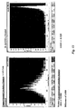
- the correlation value for each cell can be plotted as a histogram, which will display the degree of cellular compartment translocation of a molecule for a cell population.
- the CCF can be used to determine or analyze molecular movement within any cellular compartment, such as translocation to or from the nucleus, movement to or from the cytoplasm, or movement to or from a cellular membrane, etc., and combinations thereof.
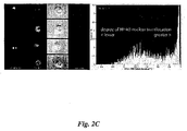
- Figure 2 illustrates the case of a nuclear translocation assay in which a fluorescent nucleic acid binding dye, 7-aminoactinomycin D (7-AAD, shown as red fluorescence), is used to stain the nucleus, while a different fluorescent marker (green; e.g. , a FITC conjugated antibody) is used to label a translocating molecule of interest (e.g. , NF- ⁇ B).
- a fluorescent nucleic acid binding dye 7-aminoactinomycin D (7-AAD, shown as red fluorescence
- a different fluorescent marker green; e.g. , a FITC conjugated antibody
- NF- ⁇ B translocating molecule of interest
- a nuclear mask is generated from the nuclear stain image and then a correlation measurement is made between the nuclear mask area of both fluorescence channels.
- Cells that exhibited nuclear translocation of NF- ⁇ B had a high correlation ( see , e.g. , Figure 2C , image rows 2-4), while cells with low nuclear translocation had a low correlation ( see , e.g. , Figure 2C , image row 1).
- the correlation value for each cell was plotted as a histogram, which displays the degree of NF- ⁇ B nuclear translocation for the cell population (see, e.g., Figure 2C , graph on right).
- Compartmental Correlation is a measurement based upon a statistical definition of correlation.
- the measurement p(X,Y) is also known as the correlation coefficient. Correlation is most effective in measuring relationships between X and Y that are linear.
- Similarity can be correlation, which is applied to imagery wherein X and Y are the pixel representations of imagery.
- the mask, M is defined, wherein M is the set of coordinates (ij).
- N can equal the number of elements in the set M.
- Cov X Y ⁇ ⁇ X i j - ⁇ X Y i j - ⁇ Y ) / N - 1
- Compartmental Correlation When Compartmental Correlation is applied to images that exhibit molecular movement or translocation, this value tends to shift closer to a value of 1.0. When the images reveal lack of molecular movement or translocation (untranslocation), this value tends to shift closer to a value of -1.0.
- the Compartmental Correlation measurement and the imagery indicate that the different degree of translocation of NF ⁇ B into the nucleus is a linear relationship. Therefore, Compartmental Correlation is optimal for measuring such a relationship.
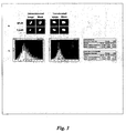
- FIG. 3 An exemplary embodiment of such a correlation is shown in Figure 3 .
- the degree of pixel intensity between the NF- ⁇ B and nuclear images of cell within a masked region of interest was analyzed.
- the NF- ⁇ B image of a cell with a high degree of translocation will look qualitatively similar to the nuclear image of that cell, resulting in a high degree of correlation between the two images.
- the NF- ⁇ B image of a cell without any translocation will have little signal in the nuclear space, resulting in an inverse correlation with the nuclear image.
- Figure 3A shows the masked areas used for the correlation analysis on an untranslocated and a translocated cell.
- Two features were calculated, the correlation coefficient ( ⁇ ) and, in this case, similarity is calculated as a logarithmic transformation of p, which features are represented by the following formula, respectively:
- ⁇ measures the degree to which the spatial distribution of intensities over two separate images is correlated, with a range from -1 (inverse correlation) to +1 (complete correlation).
- Similarity values range from - ⁇ to + ⁇ , allowing standard statistical comparisons (means and standard deviations) between groups to be made. Histogram overlays of NF- ⁇ B / 7-AAD correlation and similarity allows the differentiation of untreated (green) A-549 cells from TNF- ⁇ / IL-1 ⁇ treated (red) A-549 cells. As described herein, the fidelity of these classifiers can be validated by inspection of the image galleries of the cells.
- an appropriate sub-set of pixels should be selected over which the correlation is to be computed. If, for example, background pixels not belonging to the cell of interest are included in the set, a strong positive correlation may be found, even when the probes tend to separate within the cell because both probes are present in larger quantities within the cell than outside the cell. In general, a variety of chemical, morphological, and intensity-based methods may need to be applied in a given experiment to select the pixels of interest.
- the task of selecting the pixels of interest is simplified by the presence of the nuclear probe.
- the pixels of interest are those directly illuminated by the nucleus and cytoplasm.
- the presence of the nuclear probe means that all that is usually needed to get a sufficiently accurate set of pixels is a mask based on a blurred image of the nuclear probe, perhaps extended to include regions (near the nucleus) where the nuclear probe intensity is varying sufficiently rapidly.
- certain parameters should be chosen, such as a narrow band of pixels right on the edge of the cell, while excluding from consideration those either on the interior or exterior. Morphological criteria will play a role in constructing an appropriate set of pixels in the case of membranes and the morphology required is of two types.
- the first is a local constraint, requiring the band of pixels of increased intensity to be sufficiently narrow in order to qualify as a piece of the membrane.
- the second is a more global criterion, requiring that the band of pixels be sufficiently close to the global boundary defining the interior of the cell.
- a multispctral imaging system and CCF can be used in a variety of applications, including diagnostics, drug discovery, and the like.
- an imaging system may be used to identify compounds that affect or alter the activation of transcription factor NF- ⁇ B in cells of the immune system.
- Immune cells may be contacted with particular chemicals, cytokines, or environmental agents to examine whether translocation of the NF- ⁇ B molecule from the cytoplasm to the nucleus occurs as part of an immune response.
- the quantitative measurement of the amount of NF- ⁇ B in the nucleus versus the cytoplasm may, therefore, be extremely useful in the development of drugs that target immune function.
- the ImageStream platform eliminates these constraints with its ability to image non-adherent cells directly in suspension, its high resolution, and the statistical power (e.g. , use of CCF) associated with its ability to analyze tens of thousands of cells.
- TNF- ⁇ Tumor Necrosis Factor- ⁇
- IL-1 ⁇ Interleukin 1- ⁇
- FIG 11 an adherent human lung carcinoma cell line A-549 was either not treated or treated for 1 hr with IL-1 ⁇ and TNF- ⁇ . The cells were trypsinized and washed off the plate to adapt the cells to flow, and probed for NF- ⁇ B (stained with anti-NF- ⁇ B mAb - AF488 donkey anti-mouse IgG). The nucleus was also stained with 7-AAD.
- NF- ⁇ B resides predominantly in the cytoplasm in resting cells.
- Activating treatments e.g. , IL-1 ⁇ /TNF- ⁇ or LPS
- the ratio of nuclear to cytoplasmic NFkB increases with LPS treatment.
- NF- ⁇ B is translocated from the cytoplasm to the nucleus when the non-adherent human monocyte cell line, THP-1, is exposed to lipopolysaccharide (LPS).
- LPS lipopolysaccharide
- the CCF is an algorithmic feature that correlates the variation of pixels (from the mean) across two channels, in this case the 7-AAD (nuclear) and NF- ⁇ B channels, within a generous 75% 7-AAD mask.
- This feature reduces cell-to-cell variation judgment calls associated with integrated nuclear to cytoplasmic NF- ⁇ B intensity ratios.
- This feature also avoids cell-to-cell variation in the inclusion/expulsion of background-like pixels associated with user defined NK- ⁇ B masks ( see Figures 9 and 13 ).
- Human lung carcinoma cell line A-549 obtained from ATCC (Rockville, MD), was maintained in RPMI 1640 (Gibco, Grand Island, NY) containing 5% fetal bovine serum, 1 mM sodium pyruvate (Mediatech, Herndon, VA), 100 ⁇ M nonessential amino acids, 100 U/ml penicillin, 100 ⁇ g/ml streptomycin, and 2 mM L-glutamine (BioWhittaker, Walkersville, MD) in 5% CO 2 atmosphere at 37°C. The density of exponentially growing cells was less than 3x10 5 cells per ml at the time of all treatments. To induce NF- ⁇ B translocation into the nucleus from the cytoplasm, cells were treated for 1 hr with IL-1 ⁇ and TNF- ⁇ .
- A549 cells cultured in Dulbecco's MEM supplemented with 10% fetal calf serum in an incubator containing 5% CO 2 at 37.
- A-549 cells were stimulated with or without TNF- ⁇ and IL-1 ⁇ for 45 min to induce nuclear translocation of NF- ⁇ B.
- Human monocyte cell line THP-1 obtained from ATCC (Rockville, MD), were maintained in RPMI 1640 (Gibco, Grand Island, NY) containing 5% fetal bovine serum, 1 mM sodium pyruvate (Mediatech, Herndon, VA), 100 ⁇ M nonessential amino acids, 100 U/ml penicillin, 100 ⁇ g/ml streptomycin, and 2 mM L-glutamine (BioWhittaker, Walkersville, MD) in 5% CO 2 atmosphere at 37°C. The density of exponentially growing cells was less than 3x10 5 cells per ml at the time of all treatments. To induce NF- ⁇ B translocation into the nucleus from the cytoplasm, cells were treated for 1 hr with LPS.
- THP-1 cells cultured in RPMI supplemented with 10% fetal calf serum in an incubator containing 5% CO 2 at 37. THP-1 cells were stimulated with or without LPS and for 60 min to induce nuclear translocation of NF- ⁇ B.
- Control (untreated) cell and LPS or IL-1 ⁇ / TNF- ⁇ treated cells were independently counted and washed once in phosphate buffered saline (PBS, Fair Lawn, NJ). Each cell group was resuspended at 10 7 cells/ml in 10 ⁇ M 7-aminoactinomycin D (7-AAD, Molecular Probes) for 10 minutes at room temperature. Cells were additionally stained with anti-NF- ⁇ B mAb - AF488 donkey anti-mouse IgG. Each cell group was washed, fixed in 2% paraformaldehyde (Sigma), and analyzed by flow cytometry and immunofluorescence microscopy.
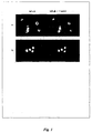
- FIG. 1 Figures in U.S. Patent Application No. 2002/0146734 provide an exemplary layout of the ImageStream TM platform.
- Cells are hydrodynamically focused into a core stream and orthogonally illuminated for both darkfield and fluorescence imaging.
- the cells are simultaneously trans-illuminated via a spectrally-limited source (e.g. , filtered white light or a light emitting diode) for brightfield imaging.
- a spectrally-limited source e.g. , filtered white light or a light emitting diode
- Light is collected from the cells with an imaging objective lens and is projected on a charge-coupled detector (CCD).
- CCD charge-coupled detector
- the optical system has a numeric aperture of 0.75 and the CCD pixel size in object space is 0.5 microns square, allowing high resolution imaging at event rates of approximately 100 cells per second.
- Each pixel is digitized with 10 bits of intensity resolution, providing a minimum dynamic range of three decades per pixel.
- the spread of signals over multiple pixels results in an effective dynamic range that typically exceeds four decades per image.
- the sensitivity of the CCD can be independently controlled for each multispectral image, resulting in a total of approximately six decades of dynamic range across all the images associated with an object.
- the light Prior to projection on the CCD, the light is passed through a spectral decomposition optical system that directs different spectral bands to different lateral positions across the detector (see , e.g. , U.S. PatentNo. 6,249,341).
- a spectral decomposition optical system that directs different spectral bands to different lateral positions across the detector.
- an image is optically decomposed into a set of 6 sub-images, each corresponding to a different color component and spatially isolated from the remaining sub-images.
- U.S. Patent Application No. 2002/0146734 depicts a red brightfield illumination source and the associated transmitted light images in the red detector channel adjacent to fluorescent and scattered light images in the other spectral channels.
- the process of spectral decomposition occurs during the image formation process rather than via digital image processing of a conventional composite image.
- the CCD is operated using time-delay-integration (TDI), in which image photons converted to photocharges in an array of pixels are continuously shifted (at a rate synchronized with the velocity of the flowing cell's image) from pixel to pixel down the detector and parallel to the axis of flow to avoid image streaking.
- TDI time-delay-integration
- the instrument can operate at a continuous data rate of approximately 30 megapixels per second and integrate signal from each object for 10 milliseconds, which allows the detection of even faint fluorescent probes within cell images that are acquired at high speed. Attention to pump and fluidic system design to achieve highly laminar, non-pulsatile flow can eliminate cell rotation or lateral translation on the time scale of the imaging process ( see , e.g. , U.S. Patent No.
- Every pixel read from the CCD is analyzed by a real-time algorithm that detects the presence of object images and calculates a number of basic morphometric and photometric features, which can be used as criteria for data storage.
- Data files encompassing 10,000-20,000 cells can be about 100 MB in size, and are stored and analyzed using standard personal computers.
Claims (12)
- Procédé pour mesurer le mouvement d'une molécule dans une cellule, comprenant les étapes consistant à : (a) mettre en contact la cellule avec un marqueur de compartiment cellulaire et un marqueur moléculaire différent du marqueur de compartiment pour former une cellule marquée ; (b) former une image, avec un détecteur, de la cellule marquée, alors que ladite cellule est dans le flux pour obtenir une image de compartiment et une image de l'emplacement de la molécule marquée à l'intérieur de l'image de la cellule ; (c) générer un masque à partir de l'image de compartiment ; et (d) effectuer, à l'intérieur du masque d'image de compartiment, une analyse de corrélation de la distribution spatiale des intensités entre l'image de compartiment et ladite image de l'emplacement de la molécule marquée, pour mesurer le mouvement de la molécule dans la cellule.
- Procédé selon la revendication 1, dans lequel il y a un mouvement relatif entre la cellule et le détecteur.
- Procédé selon la revendication 1, dans lequel le marqueur de la molécule est un anticorps marqué fluorescent.
- Procédé selon la revendication 1, dans lequel le marqueur de compartiment est une molécule fluorescente.
- Procédé selon la revendication 1, dans lequel le compartiment est le noyau, le cytoplasme, ou la membrane.
- Procédé selon la revendication 1, dans lequel la molécule marquée est NF-κB.
- Procédé selon la revendication 1, comprenant en outre l'étape consistant à induire un mouvement de la molécule dans la cellule.
- Procédé selon la revendication 7, dans lequel le mouvement induit de la molécule est une translocation nucléaire de la molécule.
- Procédé selon la revendication 7, dans lequel le mouvement de la molécule est induit avec un LPS ou IL-1β/ TNF-α.
- Procédé selon la revendication 1, dans lequel le marqueur de compartiment cellulaire est 7-AAD.
- Procédé selon l'une quelconque des revendications 1 à 10, dans lequel les images sont collectées simultanément.
- Procédé selon l'une quelconque des revendications 1 à 10, dans lequel le détecteur est un détecteur à couplage de charges avec intégration de temporisation.
Applications Claiming Priority (3)
| Application Number | Priority Date | Filing Date | Title |
|---|---|---|---|
| US55348404P | 2004-03-16 | 2004-03-16 | |
| US57287704P | 2004-05-19 | 2004-05-19 | |
| PCT/US2005/008866 WO2005098430A2 (fr) | 2004-03-16 | 2005-03-16 | Quantification par image de la translocation moleculaire |
Publications (2)
| Publication Number | Publication Date |
|---|---|
| EP1800124A2 EP1800124A2 (fr) | 2007-06-27 |
| EP1800124B1 true EP1800124B1 (fr) | 2011-12-21 |
Family
ID=34972698
Family Applications (1)
| Application Number | Title | Priority Date | Filing Date |
|---|---|---|---|
| EP05760334A Not-in-force EP1800124B1 (fr) | 2004-03-16 | 2005-03-16 | Quantification par image de la translocation moleculaire |
Country Status (5)
| Country | Link |
|---|---|
| US (2) | US8150136B2 (fr) |
| EP (1) | EP1800124B1 (fr) |
| AT (1) | ATE538138T1 (fr) |
| CA (1) | CA2598602A1 (fr) |
| WO (1) | WO2005098430A2 (fr) |
Families Citing this family (31)
| Publication number | Priority date | Publication date | Assignee | Title |
|---|---|---|---|---|
| US8406498B2 (en) | 1999-01-25 | 2013-03-26 | Amnis Corporation | Blood and cell analysis using an imaging flow cytometer |
| US7450229B2 (en) | 1999-01-25 | 2008-11-11 | Amnis Corporation | Methods for analyzing inter-cellular phenomena |
| US8131053B2 (en) * | 1999-01-25 | 2012-03-06 | Amnis Corporation | Detection of circulating tumor cells using imaging flow cytometry |
| US8885913B2 (en) | 1999-01-25 | 2014-11-11 | Amnis Corporation | Detection of circulating tumor cells using imaging flow cytometry |
| AU2002213157A1 (en) | 2000-10-12 | 2002-04-22 | Amnis Corporation | System and method for high numeric aperture imaging systems |
| US7280207B2 (en) | 2001-07-25 | 2007-10-09 | Applera Corporation | Time-delay integration in a flow cytometry system |
| US8953866B2 (en) | 2004-03-16 | 2015-02-10 | Amnis Corporation | Method for imaging and differential analysis of cells |
| US8103080B2 (en) * | 2004-03-16 | 2012-01-24 | Amnis Corporation | Method for imaging and differential analysis of cells |
| ATE538138T1 (de) | 2004-03-16 | 2012-01-15 | Amnis Corp | Auf bildlicher darstellung beruhende quantifizierung molekularer translokation |
| US7996188B2 (en) | 2005-08-22 | 2011-08-09 | Accuri Cytometers, Inc. | User interface for a flow cytometer system |
| US8017402B2 (en) | 2006-03-08 | 2011-09-13 | Accuri Cytometers, Inc. | Fluidic system for a flow cytometer |
| US8303894B2 (en) | 2005-10-13 | 2012-11-06 | Accuri Cytometers, Inc. | Detection and fluidic system of a flow cytometer |
| WO2007067999A2 (fr) * | 2005-12-09 | 2007-06-14 | Amnis Corporation | Systeme d'imagerie a profondeur de champ etendue pour analyse d'objets a grande vitesse |
| US7780916B2 (en) | 2006-03-08 | 2010-08-24 | Accuri Cytometers, Inc. | Flow cytometer system with unclogging feature |
| US8283177B2 (en) | 2006-03-08 | 2012-10-09 | Accuri Cytometers, Inc. | Fluidic system with washing capabilities for a flow cytometer |
| US8715573B2 (en) | 2006-10-13 | 2014-05-06 | Accuri Cytometers, Inc. | Fluidic system for a flow cytometer with temporal processing |
| US8445286B2 (en) | 2006-11-07 | 2013-05-21 | Accuri Cytometers, Inc. | Flow cell for a flow cytometer system |
| US7739060B2 (en) * | 2006-12-22 | 2010-06-15 | Accuri Cytometers, Inc. | Detection system and user interface for a flow cytometer system |
| US8432541B2 (en) | 2007-12-17 | 2013-04-30 | Accuri Cytometers, Inc. | Optical system for a flow cytometer with an interrogation zone |
| US8189884B2 (en) * | 2008-03-26 | 2012-05-29 | General Electric Company | Methods for assessing molecular expression of subcellular molecules |
| US8507279B2 (en) | 2009-06-02 | 2013-08-13 | Accuri Cytometers, Inc. | System and method of verification of a prepared sample for a flow cytometer |
| US8451524B2 (en) * | 2009-09-29 | 2013-05-28 | Amnis Corporation | Modifying the output of a laser to achieve a flat top in the laser's Gaussian beam intensity profile |
| US8237786B2 (en) * | 2009-12-23 | 2012-08-07 | Applied Precision, Inc. | System and method for dense-stochastic-sampling imaging |
| WO2011106402A1 (fr) * | 2010-02-23 | 2011-09-01 | Accuri Cytometers, Inc. | Procédé et système de détection de fluorochromes dans cytomètre de flux |
| US8817115B1 (en) | 2010-05-05 | 2014-08-26 | Amnis Corporation | Spatial alignment of image data from a multichannel detector using a reference image |
| US9551600B2 (en) | 2010-06-14 | 2017-01-24 | Accuri Cytometers, Inc. | System and method for creating a flow cytometer network |
| EP2633284B1 (fr) | 2010-10-25 | 2021-08-25 | Accuri Cytometers, Inc. | Systèmes et interface utilisateur pour collecter un ensemble de données dans un cytomètre de flux |
| US10209236B2 (en) | 2012-12-27 | 2019-02-19 | Sony Corporation | Cell analyzer system, cell analyzer program, and cell analyzing method |
| WO2015068329A1 (fr) | 2013-11-08 | 2015-05-14 | ソニー株式会社 | Système d'analyse de cellules, programme d'analyse de cellules et procédé d'analyse de cellules |
| EP3163287B1 (fr) | 2015-10-30 | 2022-03-23 | Sysmex Corporation | Procédé d'obtention d'informations de cellule et appareil d'obtention d'informations de cellule |
| SG11201908847TA (en) | 2017-03-31 | 2019-10-30 | Life Technologies Corp | Apparatuses, systems and methods for imaging flow cytometry |
Family Cites Families (165)
| Publication number | Priority date | Publication date | Assignee | Title |
|---|---|---|---|---|
| US3555280A (en) | 1966-04-19 | 1971-01-12 | Hycon Mfg Co | Automatic focus sensor and control |
| US3497690A (en) | 1967-09-21 | 1970-02-24 | Bausch & Lomb | Method and apparatus for classifying biological cells by measuring the size and fluorescent response thereof |
| DE1597211B1 (de) | 1967-09-26 | 1970-06-11 | Fernseh Gmbh | Farbfernsehkamera |
| JPS49133042A (fr) | 1973-04-09 | 1974-12-20 | ||
| NL7807532A (nl) | 1978-07-13 | 1980-01-15 | Akzo Nv | Metaal-immunotest. |
| US4414575A (en) | 1980-11-21 | 1983-11-08 | Hitachi Denshi Kabushiki Kaisha | Autofocus system |
| EP0104477B1 (fr) | 1982-08-31 | 1989-12-20 | Dai Nippon Insatsu Kabushiki Kaisha | Méthode pour inspecter une image |
| US4703017C1 (en) | 1984-02-14 | 2001-12-04 | Becton Dickinson Co | Solid phase assay with visual readout |
| JP2531605B2 (ja) | 1984-02-24 | 1996-09-04 | 株式会社東芝 | 画像の位置合せ装置 |
| US5122453A (en) | 1984-03-28 | 1992-06-16 | Technicon Instruments Corporation | Method for discriminating surface stained lymphocytes |
| JPS60258671A (ja) | 1984-06-05 | 1985-12-20 | Nec Corp | プロセツサ |
| US5096807A (en) | 1985-03-06 | 1992-03-17 | Murex Corporation | Imaging immunoassay detection system with background compensation and its use |
| US4662742A (en) | 1985-05-10 | 1987-05-05 | Becton, Dickinson And Company | Scatter/fluorescene beam splitter in a flow cytometry apparatus |
| US4845197A (en) | 1985-09-09 | 1989-07-04 | Agri-Diagnostics Associates | Monoclonal antibodies and methods for fungal pathogen detection |
| US5985549A (en) | 1985-10-22 | 1999-11-16 | University Of Massachusetts | Non-isotopic in-situ hybridization method for detection of nucleic acids |
| US4770992A (en) | 1985-11-27 | 1988-09-13 | Den Engh Gerrit J Van | Detection of specific DNA sequences by flow cytometry |
| US4777525A (en) | 1985-12-23 | 1988-10-11 | Preston Jr Kendall | Apparatus and method for a multi-resolution electro-optical imaging, display and storage/retrieval system |
| CA1338860C (fr) | 1987-04-13 | 1997-01-21 | Peter Parker | Marquage de proteines a l'aide de radio-isotopes |
| US4786165A (en) | 1986-07-10 | 1988-11-22 | Toa Medical Electronics Co., Ltd. | Flow cytometry and apparatus therefor |
| JPH0664061B2 (ja) | 1987-02-27 | 1994-08-22 | イーストマン コダック カンパニー | 免疫反応性試薬、その製造法及び免疫反応性種を測定するためのその用途 |
| EP0280559B1 (fr) | 1987-02-27 | 1993-10-20 | EASTMAN KODAK COMPANY (a New Jersey corporation) | Essai immunologique d'agglutination et trousse de réactifs pour la détermination d'une espèce immunollogique polyvalente utilisant une solution saline de nettoyage tamponnée |
| US4857453A (en) | 1987-04-07 | 1989-08-15 | Syntex (U.S.A.) Inc. | Immunoassay device |
| DE560410T1 (de) | 1987-04-27 | 2001-12-20 | Unilever Nv | Testgerät zur Durchführung von spezifischen Bindungsprüfungen. |
| US4959302A (en) | 1987-08-20 | 1990-09-25 | Endogen, Inc. | Method of evaluating immune system response to allografts, viral infection and immunosuppressive therapy |
| US5986061A (en) | 1988-10-28 | 1999-11-16 | Pbl Biomedical Laboratories | Phosphorylated fusion proteins |
| IL92124A (en) | 1988-10-28 | 1996-10-31 | Sidney Pestka | Recombinant proteins modified to contain post-phosphorylation that do not occur in nature |
| EP0462221A1 (fr) | 1989-03-07 | 1991-12-27 | Syngene, Inc. | Hybridation in situ en suspension pour la detection ou la separation de cellules |
| US5266486A (en) | 1989-05-12 | 1993-11-30 | Nvl Photronics Corporation | Method and apparatus for detecting biological activities in a specimen |
| US5107522A (en) | 1990-02-05 | 1992-04-21 | Sharp Kabushiki Kaisha | Automatic frequency control circuit |
| JP3181050B2 (ja) | 1990-04-20 | 2001-07-03 | 株式会社日立製作所 | 投影露光方法およびその装置 |
| US5159642A (en) | 1990-07-13 | 1992-10-27 | Toa Medical Electronics Co., Ltd. | Particle image analyzing apparatus |
| US5141609A (en) | 1990-11-16 | 1992-08-25 | The Trustees Of The Leland Stanford Junior University | Method and device employing time-delayed integration for detecting sample components after separation |
| US5257182B1 (en) | 1991-01-29 | 1996-05-07 | Neuromedical Systems Inc | Morphological classification system and method |
| US5784162A (en) | 1993-08-18 | 1998-07-21 | Applied Spectral Imaging Ltd. | Spectral bio-imaging methods for biological research, medical diagnostics and therapy |
| US5817462A (en) | 1995-02-21 | 1998-10-06 | Applied Spectral Imaging | Method for simultaneous detection of multiple fluorophores for in situ hybridization and multicolor chromosome painting and banding |
| JPH0734012B2 (ja) | 1991-02-27 | 1995-04-12 | 東亜医用電子株式会社 | フローイメージサイトメータ |
| JP3121849B2 (ja) | 1991-02-27 | 2001-01-09 | シスメックス株式会社 | フローイメージサイトメータ |
| JP3084296B2 (ja) | 1991-02-27 | 2000-09-04 | シスメックス株式会社 | フローイメージサイトメータ |
| JP3084295B2 (ja) | 1991-02-27 | 2000-09-04 | シスメックス株式会社 | フローイメージサイトメータ |
| US5436144A (en) | 1991-04-08 | 1995-07-25 | Health Research, Inc. | Process for performing PCR in mammalian cells |
| US5568315A (en) | 1991-05-28 | 1996-10-22 | Discovision Associates | Optical beamsplitter |
| US5548395A (en) | 1991-09-20 | 1996-08-20 | Toa Medical Electronics Co., Ltd. | Particle analyzer |
| JP3212647B2 (ja) | 1991-10-24 | 2001-09-25 | シスメックス株式会社 | イメージングフローサイトメータ |
| JP3102935B2 (ja) | 1991-11-20 | 2000-10-23 | シスメックス株式会社 | イメージングフローサイトメータ |
| US5686960A (en) | 1992-01-14 | 1997-11-11 | Michael Sussman | Image input device having optical deflection elements for capturing multiple sub-images |
| US5422712A (en) | 1992-04-01 | 1995-06-06 | Toa Medical Electronics Co., Ltd. | Apparatus for measuring fluorescent spectra of particles in a flow |
| JP3145486B2 (ja) | 1992-06-12 | 2001-03-12 | シスメックス株式会社 | イメージングフローサイトメータ |
| US5351311A (en) | 1992-07-28 | 1994-09-27 | The United States Of America As Represented By The Secretary Of The Navy | Neural network for detection and correction of local boundary misalignments between images |
| US6159686A (en) | 1992-09-14 | 2000-12-12 | Sri International | Up-converting reporters for biological and other assays |
| US5733721A (en) | 1992-11-20 | 1998-03-31 | The Board Of Regents Of The University Of Oklahoma | Cell analysis method using quantitative fluorescence image analysis |
| IL108497A0 (en) | 1993-02-01 | 1994-05-30 | Seq Ltd | Methods and apparatus for dna sequencing |
| US5547849A (en) | 1993-02-17 | 1996-08-20 | Biometric Imaging, Inc. | Apparatus and method for volumetric capillary cytometry |
| DE69434551T2 (de) | 1993-09-16 | 2006-06-14 | Sysmex Corp | Teilchen-Analysegerät |
| JP3290786B2 (ja) | 1993-11-26 | 2002-06-10 | シスメックス株式会社 | 粒子分析装置 |
| JP3344635B2 (ja) | 1993-12-27 | 2002-11-11 | シャープ株式会社 | カラー液晶表示装置 |
| AU1566995A (en) | 1994-01-21 | 1995-08-08 | Coulter Corporation | Viability probes for isolation, identification and/or analysis of cells |
| JPH07261149A (ja) | 1994-03-23 | 1995-10-13 | Seiko Instr Inc | 液晶装置 |
| JPH07286953A (ja) | 1994-04-19 | 1995-10-31 | Toa Medical Electronics Co Ltd | イメージングフローサイトメータ |
| JP3156503B2 (ja) | 1994-05-27 | 2001-04-16 | 松下電器産業株式会社 | 固体撮像装置の駆動方法及び固体撮像装置の信号処理回路 |
| US5658744A (en) | 1994-07-22 | 1997-08-19 | The United States Of America As Represented By The Department Of Health And Human Services | Methods of identifying patients having an altered immune status |
| WO1996009600A1 (fr) | 1994-09-20 | 1996-03-28 | Neopath, Inc. | Dispositif d'identification et d'integration de schemas cellulaires multiples |
| US5621460A (en) | 1994-10-11 | 1997-04-15 | Lockheed Martin Corporation | Optical differentiation between plants and background utilizing a single CCD camera |
| US5695934A (en) | 1994-10-13 | 1997-12-09 | Lynx Therapeutics, Inc. | Massively parallel sequencing of sorted polynucleotides |
| US5625048A (en) | 1994-11-10 | 1997-04-29 | The Regents Of The University Of California | Modified green fluorescent proteins |
| US5556790A (en) | 1994-12-05 | 1996-09-17 | Pettit; John W. | Method for Automated DNA sequencing |
| WO1996041304A1 (fr) | 1995-06-07 | 1996-12-19 | The Trustees Of Columbia University In The City Of New York | Appareil et procedes de determination de la forme tridimensionnelle d'un objet au moyen d'un eclairage dynamique et d'une diminution de nettete relative dans deux images due a la defocalisation |
| US5844670A (en) | 1995-07-28 | 1998-12-01 | Ricoh Co., Ltd. | Method of and systems for measuring eccentricity of an aspherical lens surface |
| FI97665C (fi) | 1995-11-21 | 1997-01-27 | Planmed Oy | Menetelmät ja laitteet kohteen kuvantamisessa |
| US6007994A (en) | 1995-12-22 | 1999-12-28 | Yale University | Multiparametric fluorescence in situ hybridization |
| US5874304A (en) | 1996-01-18 | 1999-02-23 | University Of Florida Research Foundation, Inc. | Humanized green fluorescent protein genes and methods |
| US5764792A (en) | 1996-01-19 | 1998-06-09 | Oncor, Inc. | Method and apparatus for processing images |
| JP3640461B2 (ja) | 1996-04-03 | 2005-04-20 | シスメックス株式会社 | 粒子分析装置 |
| US5989835A (en) | 1997-02-27 | 1999-11-23 | Cellomics, Inc. | System for cell-based screening |
| US5959953A (en) | 1996-07-03 | 1999-09-28 | Zen Research Nv | Methods and apparatus for performing cross-talk correction in a multi-track optical disk reader based on magnification error |
| US5929986A (en) | 1996-08-26 | 1999-07-27 | Kaiser Optical Systems, Inc. | Synchronous spectral line imaging methods and apparatus |
| US5760899A (en) | 1996-09-04 | 1998-06-02 | Erim International, Inc. | High-sensitivity multispectral sensor |
| US5939281A (en) | 1996-09-16 | 1999-08-17 | Case Western Reserve University | Detecting alloreactivity |
| US5754291A (en) | 1996-09-19 | 1998-05-19 | Molecular Dynamics, Inc. | Micro-imaging system |
| US5855753A (en) | 1996-11-26 | 1999-01-05 | The Trustees Of Princeton University | Method for electrohydrodynamically assembling patterned colloidal structures |
| EP0983498B1 (fr) | 1997-02-27 | 2004-05-26 | Cellomics, Inc. | Systeme de criblage de cellules |
| US6259807B1 (en) | 1997-05-14 | 2001-07-10 | Applied Imaging Corp. | Identification of objects of interest using multiple illumination schemes and finding overlap of features in corresponding multiple images |
| AU756945B2 (en) | 1997-05-23 | 2003-01-30 | Bioarray Solutions Ltd | Color-encoding and in-situ interrogation of matrix-coupled chemical compounds |
| EP0985142A4 (fr) | 1997-05-23 | 2006-09-13 | Lynx Therapeutics Inc | Systeme et appareil destines au traitement sequentiel des analytes |
| US20040241759A1 (en) | 1997-06-16 | 2004-12-02 | Eileen Tozer | High throughput screening of libraries |
| AU731476C (en) | 1997-07-12 | 2001-12-06 | Roper Scientific, Inc. | Multi-spectral two-dimensional imaging spectrometer |
| EP0998720A4 (fr) | 1997-07-31 | 2005-03-23 | Univ California | Dispositif et procedes de traitement d'images et de signaux |
| WO2000006989A2 (fr) | 1998-07-27 | 2000-02-10 | Ljl Biosystems, Inc. | Appareil et procedes d'identification des effets d'attenuation dans des essais de mesure de luminescence |
| US5900942A (en) | 1997-09-26 | 1999-05-04 | The United States Of America As Represented By Administrator Of National Aeronautics And Space Administration | Multi spectral imaging system |
| US6115119A (en) | 1997-10-21 | 2000-09-05 | Bigelow Laboratory For Ocean Science | Device and method for studying particles in a fluid |
| AUPP032897A0 (en) | 1997-11-12 | 1997-12-04 | University Of Queensland, The | Oligomer libraries |
| US6051835A (en) | 1998-01-07 | 2000-04-18 | Bio-Rad Laboratories, Inc. | Spectral imaging apparatus and methodology |
| US6510319B2 (en) | 1998-02-05 | 2003-01-21 | Lucent Technologies Inc. | Method for optimizing forward link power levels during soft handoffs in a wireless telecommunications network |
| JP3747621B2 (ja) | 1998-03-26 | 2006-02-22 | コニカミノルタオプト株式会社 | カラー投影表示装置 |
| JPH11295208A (ja) | 1998-04-13 | 1999-10-29 | Sysmex Corp | 粒子撮像装置 |
| US20010012620A1 (en) | 1998-06-03 | 2001-08-09 | Rich Ivan N. | Method of reducing cell proliferation by inducing apoptosis in differentially selected cell subpopulations |
| US6150176A (en) | 1998-06-09 | 2000-11-21 | The Regents Of The University Of California | Fluorescent protein sensors for measuring the pH of a biological sample |
| EP1110090B1 (fr) | 1998-09-03 | 2009-03-25 | Trellis Bioscience, Inc. | Etiquettes a nuances multiples |
| US6330081B1 (en) | 1998-11-20 | 2001-12-11 | Agfa Corporation | Crosstalk cancellation in a multi-color CCD signal processor |
| JP3871456B2 (ja) | 1998-12-10 | 2007-01-24 | シスメックス株式会社 | 粒子画像分析装置 |
| US6549664B1 (en) | 1998-12-31 | 2003-04-15 | Siros Technologies, Inc. | Sparse modulation codes for holographic data storage |
| US20010006416A1 (en) | 1999-01-11 | 2001-07-05 | Johnson Paul E. | Ribbon flow cytometry apparatus and methods |
| US6256096B1 (en) | 1999-01-11 | 2001-07-03 | Softray | Flow cytometry apparatus and method |
| US7450229B2 (en) | 1999-01-25 | 2008-11-11 | Amnis Corporation | Methods for analyzing inter-cellular phenomena |
| US6671044B2 (en) | 1999-01-25 | 2003-12-30 | Amnis Corporation | Imaging and analyzing parameters of small moving objects such as cells in broad flat flow |
| US6608682B2 (en) | 1999-01-25 | 2003-08-19 | Amnis Corporation | Imaging and analyzing parameters of small moving objects such as cells |
| US7057732B2 (en) | 1999-01-25 | 2006-06-06 | Amnis Corporation | Imaging platform for nanoparticle detection applied to SPR biomolecular interaction analysis |
| US6249341B1 (en) | 1999-01-25 | 2001-06-19 | Amnis Corporation | Imaging and analyzing parameters of small moving objects such as cells |
| US6975400B2 (en) | 1999-01-25 | 2005-12-13 | Amnis Corporation | Imaging and analyzing parameters of small moving objects such as cells |
| US6473176B2 (en) | 1999-01-25 | 2002-10-29 | Amnis Corporation | Imaging and analyzing parameters of small moving objects such as cells |
| US6707551B2 (en) | 2000-01-24 | 2004-03-16 | Amnis Corporation | Multipass cavity for illumination and excitation of moving objects |
| US6580504B1 (en) | 1999-01-25 | 2003-06-17 | Amnis Corporation | Multipass cavity for illumination and excitation of moving objects |
| US20060257884A1 (en) | 2004-05-20 | 2006-11-16 | Amnis Corporation | Methods for preparing and analyzing cells having chromosomal abnormalities |
| US6150121A (en) | 1999-02-16 | 2000-11-21 | Wisconsin Alumni Research Foundation | Assessing immunological state of transplant recipients |
| CA2372447A1 (fr) | 1999-02-19 | 2000-08-24 | Fox Chase Cancer Center | Techniques de decomposition de donnees complexes |
| US6381363B1 (en) | 1999-03-15 | 2002-04-30 | Grass Valley (U.S.), Inc. | Histogram-based segmentation of images and video via color moments |
| US6330361B1 (en) | 1999-03-16 | 2001-12-11 | Litton Systems, Inc. | Adaptively aligned optical correlator and method |
| US6156465A (en) | 1999-04-12 | 2000-12-05 | Cymbolic Sciences Inc. | Crosstalk correction |
| WO2001011341A2 (fr) * | 1999-08-05 | 2001-02-15 | Cellomics, Inc. | Systeme criblage de cellules |
| US6716588B2 (en) | 1999-12-09 | 2004-04-06 | Cellomics, Inc. | System for cell-based screening |
| CA2393658A1 (fr) | 1999-12-23 | 2001-06-28 | Illumina, Inc. | Decodage de capteurs de reseaux a microspheres |
| US6727066B2 (en) | 2000-07-28 | 2004-04-27 | Incyte Corporation | Genes expressed in treated human C3A liver cell cultures |
| US6608680B2 (en) | 2000-08-25 | 2003-08-19 | Amnis Corporation | TDI imaging system for kinetic studies |
| US6778263B2 (en) | 2000-08-25 | 2004-08-17 | Amnis Corporation | Methods of calibrating an imaging system using calibration beads |
| US6875973B2 (en) | 2000-08-25 | 2005-04-05 | Amnis Corporation | Auto focus for a flow imaging system |
| US6583865B2 (en) | 2000-08-25 | 2003-06-24 | Amnis Corporation | Alternative detector configuration and mode of operation of a time delay integration particle analyzer |
| US6934408B2 (en) | 2000-08-25 | 2005-08-23 | Amnis Corporation | Method and apparatus for reading reporter labeled beads |
| WO2002017219A1 (fr) | 2000-08-25 | 2002-02-28 | Amnis Corporation | Mesure de la vitesse de petits objets mobiles comme les cellules |
| AU2001284373A1 (en) * | 2000-08-29 | 2002-03-13 | Yeda Research And Development Co. Ltd. | Methods of isolating genes encoding proteins of specific function and of screening for pharmaceutically active agents |
| WO2002026195A2 (fr) | 2000-09-29 | 2002-04-04 | Clinomics Biosciences, Inc. | Micro-reseaux de tissus tumoraux |
| US6563583B2 (en) | 2000-10-12 | 2003-05-13 | Amnis Corporation | Multipass cavity for illumination and excitation of moving objects |
| ATE277386T1 (de) * | 2000-10-27 | 2004-10-15 | Amersham Biosciences Corp | Verfahren zum screening von chemischen verbindungen |
| US7033819B2 (en) | 2000-11-08 | 2006-04-25 | Surface Logix, Inc. | System for monitoring cell motility in real-time |
| US20020132274A1 (en) | 2001-01-17 | 2002-09-19 | Nevalainen Marja T. | Diagnostic and monitorings methods for cancer |
| US6873733B2 (en) | 2001-01-19 | 2005-03-29 | The Regents Of The University Of Colorado | Combined wavefront coding and amplitude contrast imaging systems |
| WO2002100157A2 (fr) | 2001-02-21 | 2002-12-19 | Amnis Corporation | Procede et dispositif servant a marquer et a analyser des constituants cellulaires |
| WO2002073200A1 (fr) * | 2001-03-12 | 2002-09-19 | Cellomics, Inc. | Methodes pouvant augmenter la capacite d'essais de criblage cellulaire a grande densite |
| US20020126275A1 (en) | 2001-03-12 | 2002-09-12 | Johnson Paul E. | LED illuminated particle detection apparatus and methods |
| US20030048931A1 (en) | 2001-03-23 | 2003-03-13 | Peter Johnson | Quantification and differentiation of tissue based upon quantitative image analysis |
| US8050868B2 (en) | 2001-03-26 | 2011-11-01 | Cellomics, Inc. | Methods for determining the organization of a cellular component of interest |
| US6519355B2 (en) | 2001-03-28 | 2003-02-11 | Alan C. Nelson | Optical projection imaging system and method for automatically detecting cells having nuclear and cytoplasmic densitometric features associated with disease |
| US7050620B2 (en) | 2001-03-30 | 2006-05-23 | Heckman Carol A | Method of assaying shape and structural features in cells |
| EP1383911A4 (fr) * | 2001-04-02 | 2004-12-15 | Cytoprint Inc | Methode et appareil permettant de decouvrir, d'identifier et de comparer des mecanismes d'activite biologique |
| WO2002086416A2 (fr) | 2001-04-25 | 2002-10-31 | Amnis Corporation | Procede et appareil de correction de la diaphonie et de la resolution spatiale de l'imagerie multicanal |
| US6618140B2 (en) | 2001-06-18 | 2003-09-09 | Amnis Corporation | Spectral deconvolution of fluorescent markers |
| WO2003009579A2 (fr) | 2001-07-17 | 2003-01-30 | Amnis Corporation | Procedes computationnels pour la segmentation d'images d'objets d'arriere-plan dans un instrument d'imagerie |
| CA2495021A1 (fr) | 2001-08-06 | 2003-07-03 | Vanderbilt University | Systeme et procedes de mesure d'au moins un taux metabolique de plusieurs cellules |
| US20030104439A1 (en) | 2001-11-30 | 2003-06-05 | Finch Rosalynde J. | Methods of identifying cellular target molecules |
| EP1316793A1 (fr) | 2001-12-03 | 2003-06-04 | Christian Leist | Procédé et appareil pour la détermination d'une collection de cellules |
| JP4497923B2 (ja) | 2001-12-05 | 2010-07-07 | ザ ジェイ. デビッド グラッドストーン インスティテューツ | ロボット顕微鏡検査システム |
| EP1468314A4 (fr) | 2001-12-18 | 2006-12-13 | Univ Rochester | Imagerie par lentille aspherique multifocale donnant une profondeur de champ accrue |
| US7476514B2 (en) | 2002-04-11 | 2009-01-13 | Cylex, Inc. | Method for monitoring the immune response and predicting clinical outcomes in transplant recipients |
| US6658143B2 (en) * | 2002-04-29 | 2003-12-02 | Amersham Biosciences Corp. | Ray-based image analysis for biological specimens |
| US7378386B2 (en) * | 2002-07-15 | 2008-05-27 | Board Of Regents, The University Of Texas System | Anti-viral treatment methods using phosphatidylethanolamine-binding peptide derivatives |
| WO2004025569A2 (fr) | 2002-09-13 | 2004-03-25 | Arcturus Bioscience, Inc. | Analyse interactive et automatique d'images tissulaires au moyen d'une base de donnees de formation generale et traitement a niveaux d'abstraction variables dans des applications de classification d'echantillons cytologiques et de microdissection laser |
| EP1599602B1 (fr) | 2003-02-14 | 2015-01-14 | Beth Israel Deaconess Medical Center, Inc. | Prediction des rejets de greffe |
| CN1795272B (zh) | 2003-03-27 | 2013-04-24 | 巴特朗医疗成像有限责任公司 | 快速识别病原体、细菌和异常细胞的系统和方法 |
| US7180673B2 (en) | 2003-03-28 | 2007-02-20 | Cdm Optics, Inc. | Mechanically-adjustable optical phase filters for modifying depth of field, aberration-tolerance, anti-aliasing in optical systems |
| US7106502B1 (en) | 2003-08-21 | 2006-09-12 | The United States Of America As Represented By The Administrator Of National Aeronautics And Space Administration | Operation of a Cartesian robotic system in a compact microscope imaging system with intelligent controls |
| WO2005027730A2 (fr) | 2003-09-19 | 2005-03-31 | The General Hospital Corporation | Dispositifs et procedes d'imagerie par polarisation de fluorescence |
| ATE538138T1 (de) | 2004-03-16 | 2012-01-15 | Amnis Corp | Auf bildlicher darstellung beruhende quantifizierung molekularer translokation |
| US8103080B2 (en) | 2004-03-16 | 2012-01-24 | Amnis Corporation | Method for imaging and differential analysis of cells |
| US7907769B2 (en) * | 2004-05-13 | 2011-03-15 | The Charles Stark Draper Laboratory, Inc. | Image-based methods for measuring global nuclear patterns as epigenetic markers of cell differentiation |
| GB2416043B (en) | 2004-07-06 | 2008-02-06 | Cairn Res Ltd | Optical imaging device |
| US8003333B2 (en) | 2007-09-28 | 2011-08-23 | Mayo Foundation For Medical Education And Research | Serum biomarkers for early detection of acute cellular rejection |
| WO2009114549A2 (fr) * | 2008-03-10 | 2009-09-17 | Mcw Research Foundation, Inc. | Peptide contenant 19 acides aminés marqué avec 99mtc pour utilisation en tant que sonde moléculaire se liant à la phosphatidyléthanolamine et produit radiopharmaceutique |
-
2005
- 2005-03-16 AT AT05760334T patent/ATE538138T1/de active
- 2005-03-16 EP EP05760334A patent/EP1800124B1/fr not_active Not-in-force
- 2005-03-16 CA CA002598602A patent/CA2598602A1/fr not_active Abandoned
- 2005-03-16 WO PCT/US2005/008866 patent/WO2005098430A2/fr active Application Filing
- 2005-03-16 US US10/593,018 patent/US8150136B2/en active Active
-
2012
- 2012-03-30 US US13/436,371 patent/US9528989B2/en active Active
Also Published As
| Publication number | Publication date |
|---|---|
| WO2005098430A2 (fr) | 2005-10-20 |
| US8150136B2 (en) | 2012-04-03 |
| US20090202130A1 (en) | 2009-08-13 |
| EP1800124A2 (fr) | 2007-06-27 |
| CA2598602A1 (fr) | 2005-10-20 |
| US9528989B2 (en) | 2016-12-27 |
| US20120244550A1 (en) | 2012-09-27 |
| WO2005098430A3 (fr) | 2005-12-15 |
| ATE538138T1 (de) | 2012-01-15 |
Similar Documents
| Publication | Publication Date | Title |
|---|---|---|
| EP1800124B1 (fr) | Quantification par image de la translocation moleculaire | |
| EP1886139B1 (fr) | Procedes d'analyse de phenomenes inter-cellulaires | |
| US6759206B1 (en) | System for cell-based screening | |
| JP3683591B2 (ja) | 細胞に基づくスクリーニングシステム | |
| EP1095277B1 (fr) | Systeme destine a un criblage a base de cellules | |
| US8571295B2 (en) | Method for imaging and differential analysis of cells | |
| US7117098B1 (en) | Machine-readable storage medium for analyzing distribution of macromolecules between the cell membrane and the cell cytoplasm | |
| US6727071B1 (en) | System for cell-based screening | |
| US7853411B2 (en) | System for cell-based screening | |
| US20040063162A1 (en) | System for cell-based screening | |
| CA2344567A1 (fr) | Systeme de criblage cellulaire | |
| EP1844426A2 (fr) | Analyse sanguine et cellulaire au moyen d'un cytometre en flux imageur | |
| WO2001011340A1 (fr) | Analyse de cellules par systeme optique | |
| WO2001035072A2 (fr) | Systeme de criblage base sur les cellules | |
| WO2000079241A2 (fr) | Systeme d'examen systematique de cellules | |
| US8953866B2 (en) | Method for imaging and differential analysis of cells | |
| EP1439384A2 (fr) | Système pour le criblage des cellules biologiques | |
| EP2660598A1 (fr) | Procédé d'imagerie et d'analyse différentielle de cellules |
Legal Events
| Date | Code | Title | Description |
|---|---|---|---|
| PUAI | Public reference made under article 153(3) epc to a published international application that has entered the european phase |
Free format text: ORIGINAL CODE: 0009012 |
|
| 17P | Request for examination filed |
Effective date: 20060906 |
|
| AK | Designated contracting states |
Kind code of ref document: A2 Designated state(s): AT BE BG CH CY CZ DE DK EE ES FI FR GB GR HU IE IS IT LI LT LU MC NL PL PT RO SE SI SK TR |
|
| 17Q | First examination report despatched |
Effective date: 20070718 |
|
| REG | Reference to a national code |
Ref country code: DE Ref legal event code: R079 Ref document number: 602005031771 Country of ref document: DE Free format text: PREVIOUS MAIN CLASS: G01N0033500000 Ipc: C07K0016280000 |
|
| GRAP | Despatch of communication of intention to grant a patent |
Free format text: ORIGINAL CODE: EPIDOSNIGR1 |
|
| RIC1 | Information provided on ipc code assigned before grant |
Ipc: G01N 33/569 20060101ALI20110525BHEP Ipc: C07K 16/28 20060101AFI20110525BHEP |
|
| GRAS | Grant fee paid |
Free format text: ORIGINAL CODE: EPIDOSNIGR3 |
|
| GRAA | (expected) grant |
Free format text: ORIGINAL CODE: 0009210 |
|
| AK | Designated contracting states |
Kind code of ref document: B1 Designated state(s): AT BE BG CH CY CZ DE DK EE ES FI FR GB GR HU IE IS IT LI LT LU MC NL PL PT RO SE SI SK TR |
|
| REG | Reference to a national code |
Ref country code: GB Ref legal event code: FG4D |
|
| REG | Reference to a national code |
Ref country code: CH Ref legal event code: EP |
|
| REG | Reference to a national code |
Ref country code: AT Ref legal event code: REF Ref document number: 538138 Country of ref document: AT Kind code of ref document: T Effective date: 20120115 |
|
| REG | Reference to a national code |
Ref country code: IE Ref legal event code: FG4D |
|
| REG | Reference to a national code |
Ref country code: DE Ref legal event code: R096 Ref document number: 602005031771 Country of ref document: DE Effective date: 20120322 |
|
| REG | Reference to a national code |
Ref country code: NL Ref legal event code: VDEP Effective date: 20111221 |
|
| PG25 | Lapsed in a contracting state [announced via postgrant information from national office to epo] |
Ref country code: LT Free format text: LAPSE BECAUSE OF FAILURE TO SUBMIT A TRANSLATION OF THE DESCRIPTION OR TO PAY THE FEE WITHIN THE PRESCRIBED TIME-LIMIT Effective date: 20111221 |
|
| LTIE | Lt: invalidation of european patent or patent extension |
Effective date: 20111221 |
|
| PG25 | Lapsed in a contracting state [announced via postgrant information from national office to epo] |
Ref country code: SI Free format text: LAPSE BECAUSE OF FAILURE TO SUBMIT A TRANSLATION OF THE DESCRIPTION OR TO PAY THE FEE WITHIN THE PRESCRIBED TIME-LIMIT Effective date: 20111221 Ref country code: GR Free format text: LAPSE BECAUSE OF FAILURE TO SUBMIT A TRANSLATION OF THE DESCRIPTION OR TO PAY THE FEE WITHIN THE PRESCRIBED TIME-LIMIT Effective date: 20120322 Ref country code: NL Free format text: LAPSE BECAUSE OF FAILURE TO SUBMIT A TRANSLATION OF THE DESCRIPTION OR TO PAY THE FEE WITHIN THE PRESCRIBED TIME-LIMIT Effective date: 20111221 Ref country code: SE Free format text: LAPSE BECAUSE OF FAILURE TO SUBMIT A TRANSLATION OF THE DESCRIPTION OR TO PAY THE FEE WITHIN THE PRESCRIBED TIME-LIMIT Effective date: 20111221 |
|
| PG25 | Lapsed in a contracting state [announced via postgrant information from national office to epo] |
Ref country code: CY Free format text: LAPSE BECAUSE OF FAILURE TO SUBMIT A TRANSLATION OF THE DESCRIPTION OR TO PAY THE FEE WITHIN THE PRESCRIBED TIME-LIMIT Effective date: 20111221 Ref country code: BE Free format text: LAPSE BECAUSE OF FAILURE TO SUBMIT A TRANSLATION OF THE DESCRIPTION OR TO PAY THE FEE WITHIN THE PRESCRIBED TIME-LIMIT Effective date: 20111221 |
|
| PG25 | Lapsed in a contracting state [announced via postgrant information from national office to epo] |
Ref country code: SK Free format text: LAPSE BECAUSE OF FAILURE TO SUBMIT A TRANSLATION OF THE DESCRIPTION OR TO PAY THE FEE WITHIN THE PRESCRIBED TIME-LIMIT Effective date: 20111221 Ref country code: CZ Free format text: LAPSE BECAUSE OF FAILURE TO SUBMIT A TRANSLATION OF THE DESCRIPTION OR TO PAY THE FEE WITHIN THE PRESCRIBED TIME-LIMIT Effective date: 20111221 Ref country code: BG Free format text: LAPSE BECAUSE OF FAILURE TO SUBMIT A TRANSLATION OF THE DESCRIPTION OR TO PAY THE FEE WITHIN THE PRESCRIBED TIME-LIMIT Effective date: 20120321 Ref country code: IS Free format text: LAPSE BECAUSE OF FAILURE TO SUBMIT A TRANSLATION OF THE DESCRIPTION OR TO PAY THE FEE WITHIN THE PRESCRIBED TIME-LIMIT Effective date: 20120421 Ref country code: EE Free format text: LAPSE BECAUSE OF FAILURE TO SUBMIT A TRANSLATION OF THE DESCRIPTION OR TO PAY THE FEE WITHIN THE PRESCRIBED TIME-LIMIT Effective date: 20111221 |
|
| PG25 | Lapsed in a contracting state [announced via postgrant information from national office to epo] |
Ref country code: PL Free format text: LAPSE BECAUSE OF FAILURE TO SUBMIT A TRANSLATION OF THE DESCRIPTION OR TO PAY THE FEE WITHIN THE PRESCRIBED TIME-LIMIT Effective date: 20111221 Ref country code: PT Free format text: LAPSE BECAUSE OF FAILURE TO SUBMIT A TRANSLATION OF THE DESCRIPTION OR TO PAY THE FEE WITHIN THE PRESCRIBED TIME-LIMIT Effective date: 20120423 Ref country code: RO Free format text: LAPSE BECAUSE OF FAILURE TO SUBMIT A TRANSLATION OF THE DESCRIPTION OR TO PAY THE FEE WITHIN THE PRESCRIBED TIME-LIMIT Effective date: 20111221 |
|
| REG | Reference to a national code |
Ref country code: AT Ref legal event code: MK05 Ref document number: 538138 Country of ref document: AT Kind code of ref document: T Effective date: 20111221 |
|
| PLBE | No opposition filed within time limit |
Free format text: ORIGINAL CODE: 0009261 |
|
| STAA | Information on the status of an ep patent application or granted ep patent |
Free format text: STATUS: NO OPPOSITION FILED WITHIN TIME LIMIT |
|
| PG25 | Lapsed in a contracting state [announced via postgrant information from national office to epo] |
Ref country code: MC Free format text: LAPSE BECAUSE OF NON-PAYMENT OF DUE FEES Effective date: 20120331 Ref country code: DK Free format text: LAPSE BECAUSE OF FAILURE TO SUBMIT A TRANSLATION OF THE DESCRIPTION OR TO PAY THE FEE WITHIN THE PRESCRIBED TIME-LIMIT Effective date: 20111221 |
|
| REG | Reference to a national code |
Ref country code: CH Ref legal event code: PL |
|
| 26N | No opposition filed |
Effective date: 20120924 |
|
| GBPC | Gb: european patent ceased through non-payment of renewal fee |
Effective date: 20120321 |
|
| PG25 | Lapsed in a contracting state [announced via postgrant information from national office to epo] |
Ref country code: IT Free format text: LAPSE BECAUSE OF FAILURE TO SUBMIT A TRANSLATION OF THE DESCRIPTION OR TO PAY THE FEE WITHIN THE PRESCRIBED TIME-LIMIT Effective date: 20111221 |
|
| REG | Reference to a national code |
Ref country code: FR Ref legal event code: ST Effective date: 20121130 |
|
| REG | Reference to a national code |
Ref country code: IE Ref legal event code: MM4A |
|
| REG | Reference to a national code |
Ref country code: DE Ref legal event code: R119 Ref document number: 602005031771 Country of ref document: DE Effective date: 20121002 Ref country code: DE Ref legal event code: R097 Ref document number: 602005031771 Country of ref document: DE Effective date: 20120924 |
|
| PG25 | Lapsed in a contracting state [announced via postgrant information from national office to epo] |
Ref country code: FR Free format text: LAPSE BECAUSE OF NON-PAYMENT OF DUE FEES Effective date: 20120402 Ref country code: IE Free format text: LAPSE BECAUSE OF NON-PAYMENT OF DUE FEES Effective date: 20120316 Ref country code: GB Free format text: LAPSE BECAUSE OF NON-PAYMENT OF DUE FEES Effective date: 20120321 Ref country code: LI Free format text: LAPSE BECAUSE OF NON-PAYMENT OF DUE FEES Effective date: 20120331 Ref country code: AT Free format text: LAPSE BECAUSE OF FAILURE TO SUBMIT A TRANSLATION OF THE DESCRIPTION OR TO PAY THE FEE WITHIN THE PRESCRIBED TIME-LIMIT Effective date: 20111221 Ref country code: CH Free format text: LAPSE BECAUSE OF NON-PAYMENT OF DUE FEES Effective date: 20120331 |
|
| REG | Reference to a national code |
Ref country code: GB Ref legal event code: S28 Free format text: APPLICATION FILED |
|
| PG25 | Lapsed in a contracting state [announced via postgrant information from national office to epo] |
Ref country code: ES Free format text: LAPSE BECAUSE OF FAILURE TO SUBMIT A TRANSLATION OF THE DESCRIPTION OR TO PAY THE FEE WITHIN THE PRESCRIBED TIME-LIMIT Effective date: 20120401 |
|
| PG25 | Lapsed in a contracting state [announced via postgrant information from national office to epo] |
Ref country code: FI Free format text: LAPSE BECAUSE OF FAILURE TO SUBMIT A TRANSLATION OF THE DESCRIPTION OR TO PAY THE FEE WITHIN THE PRESCRIBED TIME-LIMIT Effective date: 20111221 |
|
| REG | Reference to a national code |
Ref country code: GB Ref legal event code: S28 Free format text: RESTORATION ALLOWED Effective date: 20131017 |
|
| PG25 | Lapsed in a contracting state [announced via postgrant information from national office to epo] |
Ref country code: TR Free format text: LAPSE BECAUSE OF FAILURE TO SUBMIT A TRANSLATION OF THE DESCRIPTION OR TO PAY THE FEE WITHIN THE PRESCRIBED TIME-LIMIT Effective date: 20111221 |
|
| PG25 | Lapsed in a contracting state [announced via postgrant information from national office to epo] |
Ref country code: LU Free format text: LAPSE BECAUSE OF NON-PAYMENT OF DUE FEES Effective date: 20120316 |
|
| PG25 | Lapsed in a contracting state [announced via postgrant information from national office to epo] |
Ref country code: HU Free format text: LAPSE BECAUSE OF FAILURE TO SUBMIT A TRANSLATION OF THE DESCRIPTION OR TO PAY THE FEE WITHIN THE PRESCRIBED TIME-LIMIT Effective date: 20050316 |
|
| PG25 | Lapsed in a contracting state [announced via postgrant information from national office to epo] |
Ref country code: DE Free format text: LAPSE BECAUSE OF FAILURE TO SUBMIT A TRANSLATION OF THE DESCRIPTION OR TO PAY THE FEE WITHIN THE PRESCRIBED TIME-LIMIT Effective date: 20121002 |
|
| PG25 | Lapsed in a contracting state [announced via postgrant information from national office to epo] |
Ref country code: GB Free format text: THE PATENT HAS BEEN ANNULLED BY A DECISION OF A NATIONAL AUTHORITY Effective date: 20120321 |
|
| PGFP | Annual fee paid to national office [announced via postgrant information from national office to epo] |
Ref country code: GB Payment date: 20230327 Year of fee payment: 19 |