EP1773871B1 - Composés associés au facteur de différenciation tissulaire et leurs analogues - Google Patents
Composés associés au facteur de différenciation tissulaire et leurs analogues Download PDFInfo
- Publication number
- EP1773871B1 EP1773871B1 EP05762534.5A EP05762534A EP1773871B1 EP 1773871 B1 EP1773871 B1 EP 1773871B1 EP 05762534 A EP05762534 A EP 05762534A EP 1773871 B1 EP1773871 B1 EP 1773871B1
- Authority
- EP
- European Patent Office
- Prior art keywords
- compound
- tdfrp
- disease
- tissue
- cells
- Prior art date
- Legal status (The legal status is an assumption and is not a legal conclusion. Google has not performed a legal analysis and makes no representation as to the accuracy of the status listed.)
- Not-in-force
Links
Images


Classifications
-
- C—CHEMISTRY; METALLURGY
- C07—ORGANIC CHEMISTRY
- C07K—PEPTIDES
- C07K14/00—Peptides having more than 20 amino acids; Gastrins; Somatostatins; Melanotropins; Derivatives thereof
- C07K14/435—Peptides having more than 20 amino acids; Gastrins; Somatostatins; Melanotropins; Derivatives thereof from animals; from humans
- C07K14/475—Growth factors; Growth regulators
-
- A—HUMAN NECESSITIES
- A61—MEDICAL OR VETERINARY SCIENCE; HYGIENE
- A61K—PREPARATIONS FOR MEDICAL, DENTAL OR TOILETRY PURPOSES
- A61K38/00—Medicinal preparations containing peptides
- A61K38/04—Peptides having up to 20 amino acids in a fully defined sequence; Derivatives thereof
- A61K38/10—Peptides having 12 to 20 amino acids
-
- A—HUMAN NECESSITIES
- A61—MEDICAL OR VETERINARY SCIENCE; HYGIENE
- A61P—SPECIFIC THERAPEUTIC ACTIVITY OF CHEMICAL COMPOUNDS OR MEDICINAL PREPARATIONS
- A61P1/00—Drugs for disorders of the alimentary tract or the digestive system
- A61P1/02—Stomatological preparations, e.g. drugs for caries, aphtae, periodontitis
-
- A—HUMAN NECESSITIES
- A61—MEDICAL OR VETERINARY SCIENCE; HYGIENE
- A61P—SPECIFIC THERAPEUTIC ACTIVITY OF CHEMICAL COMPOUNDS OR MEDICINAL PREPARATIONS
- A61P1/00—Drugs for disorders of the alimentary tract or the digestive system
- A61P1/16—Drugs for disorders of the alimentary tract or the digestive system for liver or gallbladder disorders, e.g. hepatoprotective agents, cholagogues, litholytics
-
- A—HUMAN NECESSITIES
- A61—MEDICAL OR VETERINARY SCIENCE; HYGIENE
- A61P—SPECIFIC THERAPEUTIC ACTIVITY OF CHEMICAL COMPOUNDS OR MEDICINAL PREPARATIONS
- A61P11/00—Drugs for disorders of the respiratory system
-
- A—HUMAN NECESSITIES
- A61—MEDICAL OR VETERINARY SCIENCE; HYGIENE
- A61P—SPECIFIC THERAPEUTIC ACTIVITY OF CHEMICAL COMPOUNDS OR MEDICINAL PREPARATIONS
- A61P13/00—Drugs for disorders of the urinary system
- A61P13/10—Drugs for disorders of the urinary system of the bladder
-
- A—HUMAN NECESSITIES
- A61—MEDICAL OR VETERINARY SCIENCE; HYGIENE
- A61P—SPECIFIC THERAPEUTIC ACTIVITY OF CHEMICAL COMPOUNDS OR MEDICINAL PREPARATIONS
- A61P13/00—Drugs for disorders of the urinary system
- A61P13/12—Drugs for disorders of the urinary system of the kidneys
-
- A—HUMAN NECESSITIES
- A61—MEDICAL OR VETERINARY SCIENCE; HYGIENE
- A61P—SPECIFIC THERAPEUTIC ACTIVITY OF CHEMICAL COMPOUNDS OR MEDICINAL PREPARATIONS
- A61P15/00—Drugs for genital or sexual disorders; Contraceptives
-
- A—HUMAN NECESSITIES
- A61—MEDICAL OR VETERINARY SCIENCE; HYGIENE
- A61P—SPECIFIC THERAPEUTIC ACTIVITY OF CHEMICAL COMPOUNDS OR MEDICINAL PREPARATIONS
- A61P17/00—Drugs for dermatological disorders
-
- A—HUMAN NECESSITIES
- A61—MEDICAL OR VETERINARY SCIENCE; HYGIENE
- A61P—SPECIFIC THERAPEUTIC ACTIVITY OF CHEMICAL COMPOUNDS OR MEDICINAL PREPARATIONS
- A61P19/00—Drugs for skeletal disorders
-
- A—HUMAN NECESSITIES
- A61—MEDICAL OR VETERINARY SCIENCE; HYGIENE
- A61P—SPECIFIC THERAPEUTIC ACTIVITY OF CHEMICAL COMPOUNDS OR MEDICINAL PREPARATIONS
- A61P19/00—Drugs for skeletal disorders
- A61P19/02—Drugs for skeletal disorders for joint disorders, e.g. arthritis, arthrosis
-
- A—HUMAN NECESSITIES
- A61—MEDICAL OR VETERINARY SCIENCE; HYGIENE
- A61P—SPECIFIC THERAPEUTIC ACTIVITY OF CHEMICAL COMPOUNDS OR MEDICINAL PREPARATIONS
- A61P19/00—Drugs for skeletal disorders
- A61P19/04—Drugs for skeletal disorders for non-specific disorders of the connective tissue
-
- A—HUMAN NECESSITIES
- A61—MEDICAL OR VETERINARY SCIENCE; HYGIENE
- A61P—SPECIFIC THERAPEUTIC ACTIVITY OF CHEMICAL COMPOUNDS OR MEDICINAL PREPARATIONS
- A61P19/00—Drugs for skeletal disorders
- A61P19/08—Drugs for skeletal disorders for bone diseases, e.g. rachitism, Paget's disease
-
- A—HUMAN NECESSITIES
- A61—MEDICAL OR VETERINARY SCIENCE; HYGIENE
- A61P—SPECIFIC THERAPEUTIC ACTIVITY OF CHEMICAL COMPOUNDS OR MEDICINAL PREPARATIONS
- A61P19/00—Drugs for skeletal disorders
- A61P19/08—Drugs for skeletal disorders for bone diseases, e.g. rachitism, Paget's disease
- A61P19/10—Drugs for skeletal disorders for bone diseases, e.g. rachitism, Paget's disease for osteoporosis
-
- A—HUMAN NECESSITIES
- A61—MEDICAL OR VETERINARY SCIENCE; HYGIENE
- A61P—SPECIFIC THERAPEUTIC ACTIVITY OF CHEMICAL COMPOUNDS OR MEDICINAL PREPARATIONS
- A61P21/00—Drugs for disorders of the muscular or neuromuscular system
-
- A—HUMAN NECESSITIES
- A61—MEDICAL OR VETERINARY SCIENCE; HYGIENE
- A61P—SPECIFIC THERAPEUTIC ACTIVITY OF CHEMICAL COMPOUNDS OR MEDICINAL PREPARATIONS
- A61P25/00—Drugs for disorders of the nervous system
-
- A—HUMAN NECESSITIES
- A61—MEDICAL OR VETERINARY SCIENCE; HYGIENE
- A61P—SPECIFIC THERAPEUTIC ACTIVITY OF CHEMICAL COMPOUNDS OR MEDICINAL PREPARATIONS
- A61P25/00—Drugs for disorders of the nervous system
- A61P25/28—Drugs for disorders of the nervous system for treating neurodegenerative disorders of the central nervous system, e.g. nootropic agents, cognition enhancers, drugs for treating Alzheimer's disease or other forms of dementia
-
- A—HUMAN NECESSITIES
- A61—MEDICAL OR VETERINARY SCIENCE; HYGIENE
- A61P—SPECIFIC THERAPEUTIC ACTIVITY OF CHEMICAL COMPOUNDS OR MEDICINAL PREPARATIONS
- A61P27/00—Drugs for disorders of the senses
-
- A—HUMAN NECESSITIES
- A61—MEDICAL OR VETERINARY SCIENCE; HYGIENE
- A61P—SPECIFIC THERAPEUTIC ACTIVITY OF CHEMICAL COMPOUNDS OR MEDICINAL PREPARATIONS
- A61P27/00—Drugs for disorders of the senses
- A61P27/02—Ophthalmic agents
-
- A—HUMAN NECESSITIES
- A61—MEDICAL OR VETERINARY SCIENCE; HYGIENE
- A61P—SPECIFIC THERAPEUTIC ACTIVITY OF CHEMICAL COMPOUNDS OR MEDICINAL PREPARATIONS
- A61P29/00—Non-central analgesic, antipyretic or antiinflammatory agents, e.g. antirheumatic agents; Non-steroidal antiinflammatory drugs [NSAID]
-
- A—HUMAN NECESSITIES
- A61—MEDICAL OR VETERINARY SCIENCE; HYGIENE
- A61P—SPECIFIC THERAPEUTIC ACTIVITY OF CHEMICAL COMPOUNDS OR MEDICINAL PREPARATIONS
- A61P3/00—Drugs for disorders of the metabolism
- A61P3/08—Drugs for disorders of the metabolism for glucose homeostasis
- A61P3/10—Drugs for disorders of the metabolism for glucose homeostasis for hyperglycaemia, e.g. antidiabetics
-
- A—HUMAN NECESSITIES
- A61—MEDICAL OR VETERINARY SCIENCE; HYGIENE
- A61P—SPECIFIC THERAPEUTIC ACTIVITY OF CHEMICAL COMPOUNDS OR MEDICINAL PREPARATIONS
- A61P35/00—Antineoplastic agents
-
- A—HUMAN NECESSITIES
- A61—MEDICAL OR VETERINARY SCIENCE; HYGIENE
- A61P—SPECIFIC THERAPEUTIC ACTIVITY OF CHEMICAL COMPOUNDS OR MEDICINAL PREPARATIONS
- A61P37/00—Drugs for immunological or allergic disorders
- A61P37/02—Immunomodulators
-
- A—HUMAN NECESSITIES
- A61—MEDICAL OR VETERINARY SCIENCE; HYGIENE
- A61P—SPECIFIC THERAPEUTIC ACTIVITY OF CHEMICAL COMPOUNDS OR MEDICINAL PREPARATIONS
- A61P37/00—Drugs for immunological or allergic disorders
- A61P37/02—Immunomodulators
- A61P37/06—Immunosuppressants, e.g. drugs for graft rejection
-
- A—HUMAN NECESSITIES
- A61—MEDICAL OR VETERINARY SCIENCE; HYGIENE
- A61P—SPECIFIC THERAPEUTIC ACTIVITY OF CHEMICAL COMPOUNDS OR MEDICINAL PREPARATIONS
- A61P43/00—Drugs for specific purposes, not provided for in groups A61P1/00-A61P41/00
-
- A—HUMAN NECESSITIES
- A61—MEDICAL OR VETERINARY SCIENCE; HYGIENE
- A61P—SPECIFIC THERAPEUTIC ACTIVITY OF CHEMICAL COMPOUNDS OR MEDICINAL PREPARATIONS
- A61P7/00—Drugs for disorders of the blood or the extracellular fluid
-
- A—HUMAN NECESSITIES
- A61—MEDICAL OR VETERINARY SCIENCE; HYGIENE
- A61P—SPECIFIC THERAPEUTIC ACTIVITY OF CHEMICAL COMPOUNDS OR MEDICINAL PREPARATIONS
- A61P9/00—Drugs for disorders of the cardiovascular system
-
- A—HUMAN NECESSITIES
- A61—MEDICAL OR VETERINARY SCIENCE; HYGIENE
- A61P—SPECIFIC THERAPEUTIC ACTIVITY OF CHEMICAL COMPOUNDS OR MEDICINAL PREPARATIONS
- A61P9/00—Drugs for disorders of the cardiovascular system
- A61P9/10—Drugs for disorders of the cardiovascular system for treating ischaemic or atherosclerotic diseases, e.g. antianginal drugs, coronary vasodilators, drugs for myocardial infarction, retinopathy, cerebrovascula insufficiency, renal arteriosclerosis
-
- C—CHEMISTRY; METALLURGY
- C07—ORGANIC CHEMISTRY
- C07K—PEPTIDES
- C07K7/00—Peptides having 5 to 20 amino acids in a fully defined sequence; Derivatives thereof
- C07K7/04—Linear peptides containing only normal peptide links
- C07K7/06—Linear peptides containing only normal peptide links having 5 to 11 amino acids
-
- C—CHEMISTRY; METALLURGY
- C07—ORGANIC CHEMISTRY
- C07K—PEPTIDES
- C07K7/00—Peptides having 5 to 20 amino acids in a fully defined sequence; Derivatives thereof
- C07K7/04—Linear peptides containing only normal peptide links
- C07K7/08—Linear peptides containing only normal peptide links having 12 to 20 amino acids
-
- C—CHEMISTRY; METALLURGY
- C07—ORGANIC CHEMISTRY
- C07K—PEPTIDES
- C07K7/00—Peptides having 5 to 20 amino acids in a fully defined sequence; Derivatives thereof
- C07K7/64—Cyclic peptides containing only normal peptide links
-
- A—HUMAN NECESSITIES
- A61—MEDICAL OR VETERINARY SCIENCE; HYGIENE
- A61K—PREPARATIONS FOR MEDICAL, DENTAL OR TOILETRY PURPOSES
- A61K38/00—Medicinal preparations containing peptides
Definitions
- the present invention relates generally to tissue differentiation factor (TDF) analogs. More specifically, the invention relates to structure-based methods and compositions useful in identifying, designing, and producing molecules, which act as functional modulators of TDF-like receptors.
- TDF tissue differentiation factor
- Cell differentiation is the central characteristic of tissue morphogenesis, which initiates during embryogenesis, and continues to various degrees throughout the life of an organism in adult tissue repair and regeneration mechanisms.
- the degree of morphogenesis in adult tissue varies among different tissues and is related, among other things, to the degree of cell turnover in a given tissue.
- the cellular and molecular events, which govern the stimulus for differentiation of cells is an area of intensive research.
- discovery of the factor or factors which control cell differentiation and tissue morphogenesis will advance significantly the ability to repair and regenerate diseased, injured or damaged mammalian tissues and organs.
- Particularly useful areas for human and veterinary therapeutics include reconstructive surgery, the treatment of tissue degenerative diseases including, for example, arthritis, emphysema, osteoporosis, cardiomyopathy, cirrhosis, degenerative nerve diseases, inflammatory diseases, and cancer, and in the protection and/or regeneration of tissues, organs and limbs.
- tissue degenerative diseases including, for example, arthritis, emphysema, osteoporosis, cardiomyopathy, cirrhosis, degenerative nerve diseases, inflammatory diseases, and cancer
- morphogenetic and “morphogenic” are often used interchangeably.
- TGF-beta Transforming Growth Factor-beta
- tissue morphogenic polypeptides share substantial amino acid sequence homology within their morphogenetically active C-terminal domains, including a conserved six or seven cysteine skeleton, and share the in vivo activity of inducing tissue-specific morphogenesis in a variety of organs and tissues.
- the polypeptides apparently contact and interact with progenitor cells e.g., by binding suitable cell surface molecules, predisposing or otherwise stimulating the cells to proliferate and differentiate in a morphogenetically permissive environment.
- Type I and Type II receptors are both serine/threonine kinases, and share similar structures: an intracellular domain that consists essentially of the kinase, a short, extended hydrophobic sequence sufficient to span the membrane one time, and an extracellular domain characterized by a high concentration of conserved cysteines.
- Morphogenic polypeptides are capable of inducing the developmental cascade of cellular and molecular events that culminate in the formation of new organ-specific tissue, including any vascularization, connective tissue formation, and nerve innervation as required by the naturally occurring tissue.
- the polypeptides have been shown to induce morphogenesis of cartilage and bone, as well as, periodontal tissues, dentin, liver, heart, kidney and neural tissue, including retinal tissue.
- tissue morphogenic polypeptides are recognized in the art as a distinct subfamily of polypeptides different from other members of the TGF-beta superfamily in that they share a high degree of sequence identity in the C-terminal domain and in that the tissue morphogenic polypeptides are able to induce, on their own, the full cascade of events that result in formation of functional tissue rather than merely inducing formation of fibrotic (scar) tissue.
- members of the family of morphogenic polypeptides are capable of all of the following in a morphogenetically permissive environment: stimulating cell proliferation and cell differentiation, and supporting the growth and maintenance of differentiated cells.
- the morphogenic polypeptides also may act as endocrine, paracrine or autocrine factors.
- morphogen-based therapeutics for treating injured or diseased mammalian tissue, including, for example, therapeutic compositions for inducing regenerative healing of bone defects such as fractures, as well as therapeutic compositions for preserving or restoring healthy metabolic properties in diseased tissue, e.g ., osteopenic bone tissue.
- WO -A-03106972 discloses multiple domain TDF-related compounds and analogues thereof.
- WO-A-03106656 discloses single domain TDF-related compounds and analogues thereof.
- the present invention relates to compositions having properties similar to TGF-beta superfamily polypeptides, and methods for the prophylactic and therapeutic treatment of a subject having disease states characterized by aberrant levels of TGF-beta-like polypeptides. More particularly, the compositions and methods have bone morphogenetic protein-like properties, and are useful in treating or preventing diseases associated with the same.
- the invention includes tissue differentiation factor related polypeptides (TDFRP) compounds consisting of the amino acid sequence as defined by SEQ ID NO: 221, and C- or N-terminally capped derivatives, pegylated derivatives, or cyclised derivatives, analogs comprising non-natural amino acids, or analogs comprising d-amino acids thereof, (such sequences are also referred to as "amino acid sequences of the invention").
- TDFRP compounds modulate signal transduction across a membrane of a cell that expresses a tissue differentiation factor receptor (a TDFR), such as, but not limited to, a TGF-beta superfamily receptor.
- a TDFR tissue differentiation factor receptor
- the invention includes an isolated nucleic acid molecule encoding the TDFRP compounds.
- the isolated nucleic acid is a vector, and the vector may optionally include a promoter sequence that can be operably linked to the nucleic acid, where the promoter causes expression of the nucleic acid molecule.
- the promoter is inducible.
- the vector is transformed into a cell, such as a prokaryotic or eukaryotic cell, preferably a mammalian cell, or more preferably a human cell.
- the vector is a viral vector capable of infecting a mammalian cell and causing expression of an amino acid sequence of the invention in an animal infected with the virus.
- the invention includes a pharmaceutical composition having a TDFRP compound with or without a pharmaceutically acceptable carrier.
- the invention includes an antibody to a TDFRP compound that binds immunospecifically to a TDFRP compound.
- the antibody is an antibody fragment, such as but not limited to an Fab, (Fab)2, Fv or Fc fragment.
- the antibody or fragment thereof is a monoclonal antibody.
- the antibody or fragment thereof is a humanized antibody.
- the invention includes an antibody or antibody fragment immunospecific to an amino acid sequence of the invention, with or without a pharmaceutically acceptable carrier.
- the invention includes a pharmaceutical composition
- a pharmaceutical composition comprising a polypeptide having an amino acid sequence of the invention or the nucleic acid sequence encoding said polypeptide, an antibody or antibody fragment that binds immunospecifically to said polypeptide, with or without a pharmaceutically-acceptable carrier.
- Also described is a method for preparing a TDFRP compound the method having the steps of culturing a cell containing a nucleic acid encoding an amino acid sequence of the invention under conditions that provide for expression of the TDFRP compound; and recovering the expressed TDFRP compound.
- Also described is a method for determining the presence or amount of the TDFRP compound in a sample the method having the steps of providing the sample, contacting the sample with an antibody that binds immunospecifically to the TDFRP compound, and determining the presence or amount of the antibody bound to the TDFRP compound, thereby determining the presence or amount of the TDFRP compound in the sample.
- Also described is a method for determining the presence or amount of the nucleic acid molecule encoding an amino acid sequence of the invention in a sample the method having the steps of providing the sample, contacting the sample with a nucleic acid probe that hybridizes to the nucleic acid molecule, and determining the presence or amount of the probe hybridized to the nucleic acid molecule, thereby determining the presence or amount of the nucleic acid molecule in the sample.
- the invention provides use of a compound consisting of an amino acid sequence of the invention in the manufacture of a medicament for use in a method of treating or preventing a tissue differentiation factor associated disorder or disease wherein the tissue differentiation factor associated disorder is selected from the group consisting of tissue degenerative disease and tissue regeneration.
- tissue differentiation factor-associated disorder is selected from the group consisting of a tissue degenerative disease and tissue regeneration.
- the tissue degenerative disease is renal disease, heart disease, traumatic brain injury, stroke, atherosclerosis, arthritis, emphysema, osteoporosis, cardiomyopathy, cirrhosis, degenerative nerve disease, Holt-Oram disease, congenital disease, pulmonary disease, eye disease, diabetic nephropathy, degenerative bone disease, periodontal disease, chronic kidney disease diabetes, cardiovascular disease, inflammatory disease, immune disease, skeletal disease, reproductive disease, hematopoetic disease, or cancer.
- the tissue regeneration includes regeneration of muscle (particularly cardiac muscle), dendritic tissue, nerve, kidney, brain, bone, skin, lung, muscle, ovary, testes, heart, spleen, cartilage, nerve, periodontal, dentin, liver, vascular, connective, lymphatic, hematopoetic, or renal tissue.
- the invention includes use of a compound of the invention in the manufacture of a medicament for use in a method of treating or preventing a tissue differentiation factor-associated disorder, by administering to a subject in which such treatment or prevention is desired the nucleic acid of the invention in an amount sufficient to treat or prevent the tissue differentiation factor-associated disorder in the subject.
- a compound of the invention in the manufacture of a medicament for use in a method of treating or preventing a tissue differentiation factor-associated disorder, by administering to an explant or ex vivo culture a TDFRP compound in an amount sufficient to treat or prevent the tissue differentiation factor-associated disorder.
- the subject is a human subject. In another embodiment, the subject is an animal subject.
- kits having in one or more containers, a pharmaceutical TDFRP composition and instructions for using the contents therein.
- Also described is a method for determining the presence of or predisposition to a disease associated with altered levels of tissue differentiation factor receptor in a first mammalian subject having the steps of providing a test sample from the first mammalian subject; contacting the test sample from the first mammalian subject with a TDFRP compound selected from amino acid sequences of the invention, detecting the level of compound/tissue differentiation factor receptor complex, quantifying the level of expression of the tissue differentiation factor receptor in the sample from the first mammalian subject; and comparing the amount of the tissue differentiation factor receptor in the sample of step (a) to the amount of the tissue differentiation factor receptor present in a control sample from a second mammalian subject known not to have, or not to be predisposed to, the disease, wherein an alteration in the expression level of the tissue differentiation factor receptor in the first subject as compared to the control sample indicates the presence of or predisposition to the disease.
- a compound of the invention in the manufacture of a medicament for use in a method of treating a pathological state in a mammal, the method comprising administering to the mammal a compound in an amount that is sufficient to alleviate the pathological state, wherein the compound is a compound having an amino acid sequence at least 90% identical to a compound consisting of SEQ ID NOs: 221.
- the invention also provides use of such an amino acid sequence in the manufacture of a medicament for use in such a method.
- a compound of the invention in the manufacture of a medicament for use in a method of treating a pathological state in a mammal, the method comprising administering to the mammal an antibody or fragment thereof immunospecific to an amino acid sequence of the invention, in an amount sufficient to alleviate the pathological state.
- the invention provides use of such an antibody in the manufacture of a medicament for use in such a method.
- use of a compound of the invention in the manufacture of a medicament for use in a method of treating a tissue differentiation factor-associated disorder in a mammal the method including administering to the mammal at least one compound which modulates the expression or activity of a TDFRP compound.
- tissue differentiation factor-associated disorders include renal disease, traumatic brain injury, stroke, atherosclerosis, arthritis, emphysema, osteoporosis, cardiomyopathy, cirrhosis, degenerative nerve disease, Holt-Oram disease, congenital disease, pulmonary disease, eye disease, diabetic nephropathy, degenerative bone disease, renal disease, periodontal disease, chronic kidney disease, diabetes, atherosclerosis, cardiovascular disease, inflammatory disease, immune disease, skeletal disease, reproductive disease, hematopoetic disease, or cancer.
- the invention includes a compound for use in treating a tissue differentiation factor-associated disorder, wherein the compound consists of an amino acid sequence of the invention.
- Also described is a method of identifying a candidate compound, which binds to a TDFRP compound the method having the steps of, providing a candidate compound, contacting the candidate compound with the TDFRP compound under conditions where a complex is formed between the test compound and the TDFRP compound, incubating the complex under conditions where co-crystals of the complex form, determining the structural atomic coordinates of the complex by x-ray diffraction, and modeling the structure of the complex to determine the binding of the candidate compound to the TDFRP compound.
- the invention includes a crystalline preparation of a candidate compound and a TDFRP compound.
- the complex is not crystallized but the complex is subjected to nuclear magnetic spectroscopy or mass spectroscopy to determine binding of the complex.
- transgenic non-human mammal for example but not limited to a mouse, having genomically-integrated in non-human mammal cells, a nucleic acid encoding an amino acid sequence of the invention, having a first sequence segment which is a regulatory region and a second sequence segment which is a polynucleotide sequence encoding a TDFRP compound, wherein the first sequence segment is operably linked to the second sequence segment.
- the first segment is a regulatable expression element or elements, which are subject to cell- or tissue-specific regulation.
- the invention includes tissue or cells derived or cultured from the non-human transgenic mammal.
- coated implantable medical device which, when implanted in to a mammal it prevents or treats a disorder in the mammal.
- the coating on the implantable medical device comprises an amino acid sequence of the invention.
- the coating on the implantable device may comprise an amino acid sequence of the invention, or polynucleotides encoding such an amino acid sequence.
- the method includes identifying a mammal with a disorder, then obtaining an implantable medical device further having a coating as described above; and implanting the device into a mammal.
- the device is implanted into a mammal with a disorder of the cardiovascular system.
- a compound of the invention in the manufacture of a medicament for use in methods of treating or preventing a tissue differentiation factor-associated disorder or disease, the method comprising administering to a subject in which such treatment or prevention is desired at least one TDFRP compound in combination with one or more therapeutic agents in amounts sufficient to treat or prevent the tissue differentiation factor-associated disorder or disease in the subject.
- therapeutic agents include small molecule mimetics for TDF receptors, TDF agonists, TDF antagonists, bone morphogenetic proteins, ACE inhibitors, anti-neoplastic agents, antibiotics, vaccines, immunosuppressive agents, anti-hypertensive agents and mediators of the hedgehog signaling pathway.
- TGF-beta Transforming Growth Factor- beta superfamily of polypeptides
- TGF-beta Transforming Growth Factor- beta superfamily of polypeptides
- AMH anti-müllerian hormone
- MIS mullerian inhibiting substance
- BMPs bone morphogenetic proteins
- myostatin The highly similar TGF-ß isoforms TGF-ß1, TGF-ß2, and TGF-ß3 potently inhibit cellular proliferation of many cell types, including those from epithelial origin. Most mesenchymal cells, however, are stimulated in their growth by TGF-ß.
- TGF-ßs strongly induce extracellular matrix synthesis and integrin expression, and modulate immune responses.
- BMPs also known as osteogenic proteins (OPs)
- OPs osteogenic proteins
- Activins named after their initial identification as activators of follicle-stimulating hormone (FSH) secretion from pituitary glands, are also known to promote erythropoiesis, mediate dorsal mesoderm induction, and contribute to survival of nerve cells.
- FSH follicle-stimulating hormone
- tissue differentiation factor includes, but is not limited to, all members of the TGF-beta superfamily of polypeptides.
- TGF-beta superfamily polypeptides can be antagonists or agonists of TGF-beta superfamily receptors.
- TGF-beta Transforming Growth Factor- beta superfamily receptors
- TGF-ß transforming growth factor-ß
- TGF-ß tissue differentiation factor receptor
- Aromatic amino acid refers to a hydrophobic amino acid having a side chain containing at least one ring having a conjugated electron system (aromatic group).
- aromatic group may be further substituted with substituent groups such as alkyl, alkenyl, alkynyl, hydroxyl, sulfanyl, nitro and amino groups, as well as others.
- substituent groups such as alkyl, alkenyl, alkynyl, hydroxyl, sulfanyl, nitro and amino groups, as well as others.
- Examples of genetically encoded aromatic amino acids include phenylalanine, tyrosine and tryptophan.
- Non-genetically encoded aromatic amino acids include phenylglycine, 2-naphthylalanine, beta-2-thienylalanine, 1,2,3,4-tetrahydroisoquinoline-3-carboxylic acid, 4-chlorophenylalanine, 2-fluorophenylalanine, 3-fluorophenylalanine and 4-fluorophenylalanine.
- Aliphatic amino acid refers to an apolar amino acid having a saturated or unsaturated straight chain, branched or cyclic hydrocarbon side chain.
- genetically encoded aliphatic amino acids include alanine, leucine, valine and isoleucine.
- non-encoded aliphatic amino acids include norleucine (Nle).
- Acidic amino acid refers to a hydrophilic amino acid having a side chain pK value of less than 7. Acidic amino acids typically have negatively charged side chains at physiological pH due to loss of a hydrogen ion. Examples of genetically encoded acidic amino acids include aspartic acid (aspartate) and glutamic acid (glutamate).
- Basic amino acid refers to a hydrophilic amino acid having a side chain pK value of greater than 7.
- Basic amino acids typically have positively charged side chains at physiological pH due to association with hydronium ion.
- genetically encoded basic amino acids include arginine, lysine and histidine.
- non-genetically encoded basic amino acids include the non-cyclic amino acids ornithine, 2,3-diaminopropionic acid, 2,4-diaminobutyric acid and homoarginine.
- Poly amino acid refers to a hydrophilic amino acid having a side chain that is uncharged at physiological pH, but which has a bond in which the pair of electrons shared in common by two atoms is held more closely by one of the atoms.
- genetically encoded polar amino acids include asparagine and glutamine.
- non-genetically encoded polar amino acids include citrulline, N-acetyl lysine and methionine sulfoxide.
- tyrosine has both an aromatic ring and a polar hydroxyl group.
- tyrosine has dual properties and can be included in both the aromatic and polar categories.
- a "subject,” as used herein, is preferably a mammal, such as a human, but can also be an animal, e.g., domestic animals (e.g., dogs, cats and the like), farm animals (e.g., cows, sheep, pigs, horses and the like) and laboratory animals (e.g ., rats, mice, guinea pigs and the like).
- domestic animals e.g., dogs, cats and the like
- farm animals e.g., cows, sheep, pigs, horses and the like
- laboratory animals e.g ., rats, mice, guinea pigs and the like.
- an "effective amount" of a compound, as used herein, is a quantity sufficient to achieve a desired therapeutic and/or prophylactic effect, for example, an amount which results in the prevention of or a decrease in the symptoms associated with a disease that is being treated, e.g., the diseases associated with TGF-beta superfamily polypeptides listed above.
- the amount of compound administered to the subject will depend on the type and severity of the disease and on the characteristics of the individual, such as general health, age, sex, body weight and tolerance to drugs. It will also depend on the degree, severity and type of disease. The skilled artisan will be able to determine appropriate dosages depending on these and other factors.
- an effective amount of the compounds of the present invention sufficient for achieving a therapeutic or prophylactic effect, range from about 0.000001 mg per kilogram body weight per day, to about 10,000 mg per kilogram body weight per day.
- the dosage ranges are from about 0.0001 mg per kilogram body weight per day to about 100 mg per kilogram body weight per day.
- the compounds of the present invention can also be administered in combination with each other, or with one or more additional therapeutic compounds.
- an “isolated” or “purified” polypeptide or polypeptide or biologically-active portion thereof is substantially free of cellular material or other contaminating polypeptides from the cell or tissue source from which the tissue differentiation factor-related polypeptide is derived, or substantially free from chemical precursors or other chemicals when chemically synthesized.
- tissue differentiation factor-related polypeptide in which the polypeptide is separated from cellular components of the cells from which it is isolated or recombinantly-produced.
- the language “substantially free of cellular material” includes preparations of tissue differentiation factor-related polypeptides having less than about 30% (by dry weight) of non-tissue differentiation factor-related polypeptides (also referred to herein as a "contaminating polypeptide”), more preferably less than about 20% of non-tissue differentiation factor-related polypeptides, still more preferably less than about 10% of non-tissue differentiation factor-related polypeptides, and most preferably less than about 5% of non-tissue differentiation factor-related polypeptides.
- tissue differentiation factor-related polypeptide or biologically-active portion thereof is recombinantly-produced, it is also preferably substantially free of culture medium, i.e., culture medium represents less than about 20%, more preferably less than about 10%, and most preferably less than about 5% of the volume of the tissue differentiation factor-related polypeptides preparation.
- tissue differentiation factor-related polypeptides in which the polypeptide is separated from chemical precursors or other chemicals that are involved in the synthesis of the polypeptide.
- the language “substantially free of chemical precursors or other chemicals” includes preparations of tissue differentiation factor-related polypeptides having less than about 30% (by dry weight) of chemical precursors or non-tissue differentiation factor-related chemicals, more preferably less than about 20% chemical precursors or non-tissue differentiation factor-related chemicals, still more preferably less than about 10% chemical precursors or non-tissue differentiation factor-related chemicals, and most preferably less than about 5% chemical precursors or non-tissue differentiation factor-related chemicals.
- variant refers to a compound that differs from the compound of the present invention, but retains essential properties thereof.
- a non-limiting example of this is a polynucleotide or polypeptide compound having conservative substitutions with respect to the reference compound, commonly known as degenerate variants.
- Another non-limiting example of a variant is a compound that is structurally different, but retains the same active domain of the compounds of the present invention.
- Variants include N-terminal or C-terminal extensions, capped amino acids, modifications of reactive amino acid side chain functional groups, e.g ., branching from lysine residues, pegylation, and/or truncations of a polypeptide compound.
- variants are overall closely similar, and in many regions, identical to the compounds of the present invention. Accordingly, the variants may contain alterations in the coding regions, non-coding regions, or both.
- a "small molecule,” as used herein, refers to a composition that has a molecular weight of less than about 5 kDa and more preferably less than about 2 kDa.
- Small molecules can be, e.g ., nucleic acids, peptides, polypeptides, glycopeptides, peptidomimetics, carbohydrates, lipids, lipopolysaccharides, combinations of these, or other organic or inorganic molecules.
- ischemia or "ischemic episode,” as used herein, mean any circumstance that results in a deficient supply of blood or oxygen to a tissue.
- a central nervous system ischemic episode results from an insufficiency or interruption in the blood or oxygen supply to any locus of the brain such as, but not limited to, a locus of the cerebrum, cerebellum or brain stem.
- the spinal cord which is also a part of the central nervous system, is equally susceptible to ischemia resulting from diminished blood flow or lack of oxygen.
- An ischemic episode may be caused by a constriction or obstruction of a blood vessel, as occurs in the case of a thrombus or embolus.
- the ischemic episode may result from any form of compromised cardiac function, including cardiac arrest, as described above.
- the deficiency is sufficiently severe and prolonged, it can lead to disruption of physiologic function, subsequent death of neurons, and necrosis (infarction) of the affected areas.
- the extent and type of neurologic abnormality resulting from the injury depend on the location and size of the infarct or the focus of ischemia. Where the ischemia is associated with a stroke, it can be either global or focal in extent. Ischemia can occur in other tissues or organs including kidney. Restoration of blood flow or reperfusion leads to a series of cellular responses that are known to cause tissue damage.
- Type I and Type II receptors (Table 1).
- ALK Type I activin-like kinase receptors
- Type II Receptors ALK-1 (activin-like kinase receptor; TSR-1) ActR-IIA ALK-2 (Type I activin receptor; ActR-I) ActR-IIB ALK-3 (BMP receptor Type I/IA; BMPR-I/IA) T ⁇ R-II ALK-4 (Type IB activin receptor; ActR-IB) BMPR-II ALK-5 (Type I TGF- ⁇ receptor; T ⁇ R-I) AMHR-II ALK-6 (BMP receptor Type IB; BMPR-IB) ALK-7
- Type I and Type II receptors Ligand binding of the Type I and Type II receptors results in the phosphorylation and activation of the Type I receptor, which is required for downstream signal cascades. Smad proteins activated by Type I receptors carry signal to the nucleus, and together with other proteins direct transcriptional responses. Type I receptors for activin and TGF- ⁇ can only recognize ligand that is bound to the Type II receptor. In contrast, Type II receptors can bind ligand independently of the Type I receptor, but they are unable to signal without the Type I receptor. Specificity in signaling, therefore is determined by a combination of the Type I and Type II receptors (Table 2).
- Vertebrate Type I receptors can be divided into two different groups according to their sequence homology at the kinase domain and their signaling activities.
- One group includes activin-like receptor type 5 (hereinafter ALK-5), ALK-4, and ALK-7.
- the other group includes ALK-1, ALK-2, ALK-3, and ALK-6.
- the ALK receptors are differentially expressed among tissues and are regulated during embryo development (Table 3).
- the present invention provides compounds that are functional analogs of tissue differentiation factors, i.e., compounds that functionally mimic TGF-beta superfamily proteins, for example by acting as TGF-beta superfamily receptor agonists, and preferentially bind to select ALK receptor(s).
- the present compounds are polypeptides, with the general structure identified herein as amino acid sequences of the invention, i.e., TDF-related polypeptides (hereinafter "TDFRP"), as detailed below.
- TDFRP TDF-related polypeptides
- the TDFRP compound binds one ALK receptor with greater binding affinity than the binding affinity to other ALK receptors based on distinct binding sites or properties.
- the TDFRP compound binds ALK-3 receptor with greater binding affinity than its binding affinity to ALK-6 receptor. In another embodiment, the TDFRP compound binds one ALK receptor with greater binding affinity than the binding affinity to other ALK receptors based on differential affinities .Such compounds are suitable for administration to a subject where it is desirable, for example, to promote the growth and differentiation of cells in one tissue without promoting an undesirable cellular response in the same or another tissue. That is, the more specificity in the design of the TDFRP compound the more likely it will not interfere with related TDFRs ( e.g., its specificity to ALK-3 receptors but not ALK-6 receptors, or vice versa ). This will minimize potential side-effects due to unwanted interactions with other targets.
- related TDFRs e.g., its specificity to ALK-3 receptors but not ALK-6 receptors, or vice versa
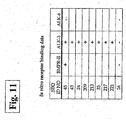
- ALK-3 receptors are more prevalent in kidney tissue, while ALK-6 receptors are more prevalent in bone tissue; the native BMP-7 protein binds to ALK-6 with a higher affinity, and a potential side effect of BMP-7 therapy in kidney disease is osteogenesis.
- the TDFRP compounds are selected and designed for increased specificity to ALK-3 and lowered affinity to ALK-6 receptors ( FIG. 11 ), thereby reducing undesirable osteogenesis in a subject being treated for kidney disorders or myocardial injury.
- TDFRP compounds of the present invention are suitable to promote the growth or differentiation of cells and tissues in the subject, such as kidney cells, mesenchymal cells, extracellular matrix synthesis, integrin expression, bone and cartilage formation, induction of ventral mesoderm, differentiation of neural tissue, to promote organogenesis, to promote erythropoiesis, to induce growth of dorsal mesoderm and nerve cells, to promote tissue homeostasis and to induce immune responses.
- pathological conditions such as fibrosis, rheumatoid arthritis, and carcinogenesis among others, are thought to be the result of excessive tissue differentiation factor-like activity. Accordingly, it is further an object of the invention to provide for compounds that are functional antagonists of TGF-beta superfamily receptors.
- the compounds of the present invention can be used to treat both acute and chronic renal disease, as well as stroke, myocardial injury and traumatic brain injury.
- TDFRP compounds can be used to treat one or more adverse consequences of central nervous system injury that arise from a variety of conditions.
- the term "stroke” connotes central nervous system injury resulting from sudden and dramatic neurologic deficits associated with, e.g ., but not limited to, thrombus, embolus, and systemic hypotension.
- Other injuries may be caused by hypertension, hypertensive cerebral vascular disease, rupture of an aneurysm, an angioma, blood dyscrasia, cardiac failure, cardiac arrest, cardiogenic shock, kidney failure, septic shock, head trauma, spinal cord trauma, seizure, bleeding from a tumor, or other loss of blood volume or pressure.
- These injuries lead to disruption of physiologic function, subsequent death of neurons, and necrosis (infarction) of the affected areas.
- the TDFRP compounds of the present invention can be useful for treating traumatic injuries to the central nervous system that are caused by mechanical forces, such as a blow to the head.
- traumatic tissue insults may involve, e.g ., but not limited to, an abrasion, incision, contusion, puncture, compression.
- Traumatic injuries to the central nervous system can arise from traumatic contact of a foreign object with any locus of or appurtenant to the mammalian head, neck or vertebral column.
- traumatic injury can arise from constriction or compression of mammalian CNS tissue by an inappropriate accumulation of fluid (e.g., a blockade or dysfunction of normal cerebrospinal fluid or vitreous humor fluid production, turnover or volume regulation, or a subdural or intracranial hematoma or edema).
- traumatic constriction or compression can arise from the presence of a mass of abnormal tissue, such as a metastatic or primary tumor.
- TDFRP compounds which act as functional modulators of select TGF-beta superfamily receptors.
- the TDFRP compounds of the present invention can be useful for treating injuries to tissues caused by infection or other insults.
- tissue insults may involve, e.g ., but are not limited to bacterial (e.g. endotoxin-mediated), fungal or viral infections.
- TDFRP compounds of the present invention may be capped on the N-terminus or the C-terminus, or on both the N-terminus and the C-terminus.
- the TDFRP compounds of the present invention may be pegylated,at any amino acid residue containing a reactive side chain, e.g ., lysine residue.
- the TDFRP compounds of the present invention may be linear or cyclized or otherwise constrained.
- the tail sequence of the TDFRP may vary in length.
- the TDFRP compounds can contain natural amino acids, non-natural amino acids, d-amino acids and 1-amino acids, and any combinations thereof.
- the compounds of the invention can include commonly encountered amino acids, which are not genetically encoded.
- These non-genetically encoded amino acids include, but are not limited to, ⁇ -alanine ( ⁇ -Ala) and other omega-amino acids such as 3-aminopropionic acid (Dap), 2,3-diaminopropionic acid (Dpr), 4-aminobutyric acid and so forth; ⁇ -aminoisobutyric acid (Aib); ⁇ -aminohexanoic acid (Aha); ⁇ -aminovaleric acid (Ava); N-methylglycine or sarcosine (MeGly); ornithine (Orn); citrulline (Cit); t-butylalanine (t-BuA); t-butylglycine (t-BuG); N-methyli
- Non-naturally occurring variants of the TDFRP compounds may be produced by mutagenesis techniques or by direct synthesis.
- the TDFRP compound of the present invention may be capped on the N-terminus or the C-terminus or on both the N-terminus and the C-terminus.
- the TDFRP compounds of the present invention may be pegylated, at any amino acid residue containing a reactive side chain, e.g ., lysine residue.
- the TDFRP compounds of the invention are prodrugs, i.e., the biological activity of the TDFRP compound is altered, e.g., increased, upon contacting a biological system in vivo or in vitro.
- TDFRP sequence based on, for example, but not limited to, permutations of SEQ ID NO:2 is identified herein as SEQ ID NO: 221 and is described below Table 4.
- the present compound includes the peptide, identified in part in Table 4. Variants, analogs, homologs, or fragments of these compounds, such as species homologs, are also included in the present invention, as well as degenerate forms thereof.
- Table 4 Sequence SEQ ID NO: CYFNDSSQVLCKRYRS 221
- the peptide has the sequence:
- the invention relates to use of a compound having an amino acid sequence at least 90% identical to a compound consisting of SEQ ID NOs: 221.
- Sequence identity can be measured using sequence analysis software (Sequence Analysis Software Package of the Genetics Computer Group, University of Wisconsin
- non-identical positions are preferably, but not necessarily, conservative substitutions for the reference sequence.
- Conservative substitutions typically include substitutions within the following groups: glycine and alanine; valine, isoleucine, and leucine; aspartic acid and glutamic acid; asparagine and glutamine; serine and threonine; lysine and arginine; and phenylalanine and tyrosine.
- peptides having mutated sequences such that they remain homologous, e.g ., in sequence, in structure, in function, and in antigenic character or other function, with a polypeptide having the corresponding parent sequence.
- Such mutations can, for example, be mutations involving conservative amino acid changes, e.g ., changes between amino acids of broadly similar molecular properties. For example, interchanges within the aliphatic group alanine, valine, leucine and isoleucine can be considered as conservative. Sometimes substitution of glycine for one of these can also be considered conservative.
- conservative interchanges include those within the aliphatic group aspartate and glutamate; within the amide group asparagine and glutamine; within the hydroxyl group serine and threonine; within the aromatic group phenylalanine, tyrosine and tryptophan; within the basic group lysine, arginine and histidine; and within the sulfur-containing group methionine and cysteine.
- substitution within the group methionine and leucine can also be considered conservative.
- Preferred conservative substitution groups are aspartate-glutamate; asparagine-glutamine; valine-leucine-isoleucine; alanine-valine; phenylalanine- tyrosine; and lysine-arginine.
- the invention also provides for compounds having altered sequences including insertions such that the overall amino acid sequence is lengthened, while the compound still retains the appropriate TDF agonist or antagonist properties.
- altered sequences may include random or designed internal deletions that truncate the overall amino acid sequence of the compound, however the compound still retains its TDF-like functional properties.
- one or more amino acid residues within SEQ ID NO:221 is replaced with other amino acid residues having physical and/or chemical properties similar to the residues they are replacing.
- conservative amino acid substitutions are those wherein an amino acid is replaced with another amino acid encompassed within the same designated class, as will be described more thoroughly below. Insertions, deletions, and substitutions are appropriate where they do not abrogate the functional properties of the compound. Functionality of the altered compound can be assayed according to the in vitro and in vivo assays described below that are designed to assess the TDF-like properties of the altered compound.
- amino acid residues of the compounds of the present invention may include genetically-encoded 1-amino acids, naturally occurring non-genetically encoded 1-amino acids, synthetic d-amino acids, or d-enantiomers of all of the above.
- Compounds described herein include the polynucleotide encoding SEQ ID NO:221, including degenerate variants thereof. Accordingly, nucleic acid sequences capable of hybridizing at low stringency with any nucleic acid sequences encoding SEQ ID NO:221 are also described.
- a typical prehybridization, hybridization, and wash protocol is as follows: (1) prehybridization: incubate nitrocellulose filters containing the denatured target DNA for 3-4 hours at 55°C in 5xDenhardt's solution, 6xSSC (20xSSC consists of 175 g NaCl, 88.2 g sodium citrate in 800 ml H 2 O adjusted to pH.
- the invention also encompasses allelic variants of the same, that is, naturally-occurring alternative forms of the isolated polynucleotides that encode polypeptides that are identical to those encoded by the polynucleotides.
- allelic variants may be produced by mutagenesis techniques or by direct synthesis techniques well known in the art.
- Another aspect of the invention includes vectors containing one or more nucleic acid sequences encoding a TDFRP compound of the invention.
- the nucleic acid containing the nucleotide sequence encoding the polypeptide is inserted into an appropriate cloning vector, or an expression vector (i.e., a vector that contains the necessary elements for the transcription and translation of the inserted polypeptide coding sequence) by recombinant DNA techniques well known in the art and as detailed below.
- expression vectors useful in recombinant DNA techniques are often in the form of plasmids.
- plasmid and “vector” can be used interchangeably as the plasmid is the most commonly used form of vector.
- the invention is intended to include such other forms of expression vectors that are not technically plasmids, such as viral vectors (e.g., replication defective retroviruses, adenoviruses and adeno-associated viruses), which serve equivalent functions.
- viral vectors e.g., replication defective retroviruses, adenoviruses and adeno-associated viruses
- the recombinant expression vectors of the invention comprise a nucleic acid encoding a compound of the invention in a form suitable for expression of the nucleic acid in a host cell, which means that the recombinant expression vectors include one or more regulatory sequences, selected on the basis of the host cells to be used for expression that is operatively-linked to the nucleic acid sequence to be expressed.
- "operably-linked" is intended to mean that the nucleotide sequence of interest is linked to the regulatory sequence(s) in a manner that allows for expression of the nucleotide sequence (e.g., in an in vitro transcription/translation system or in a host cell when the vector is introduced into the host cell).
- regulatory sequence is intended to include promoters, enhancers and other expression control elements (e.g., polyadenylation signals). Such regulatory sequences are described, for example, in Goeddel, GENE EXPRESSION TECHNOLOGY: METHODS IN ENZYMOLOGY 185, Academic Press, San Diego, Calif. (1990 ). Regulatory sequences include those that direct constitutive expression of a nucleotide sequence in many types of host cell and those that direct expression of the nucleotide sequence only in certain host cells (e.g ., tissue-specific regulatory sequences).
- the design of the expression vector can depend on such factors as the choice of the host cell to be transformed, the level of expression of polypeptide desired, etc.
- the expression vectors of the invention can be introduced into host cells to thereby produce polypeptides or peptides, including fusion polypeptides, encoded by nucleic acids as described herein ( e.g., TDFRP compounds and TDFRP-derived fusion polypeptides, etc. ).
- TDFRP-expressing host cells which contain a nucleic acid encoding one or more TDFRP compounds.
- the recombinant expression vectors of the invention can be designed for expression of TDFRP compounds in prokaryotic or eukaryotic cells.
- TDFRP compounds can be expressed in bacterial cells such as Escherichia coli, insect cells (using baculovirus expression vectors), fungal cells, e.g., yeast, yeast cells or mammalian cells. Suitable host cells are discussed further in Goeddel, GENE EXPRESSION TECHNOLOGY: METHODS IN ENZYMOLOGY 185, Academic Press, San Diego, Calif. (1990 ).
- the recombinant expression vector can be transcribed and translated in vitro, for example using T7 promoter regulatory sequences and T7 polymerase.
- Fusion vectors add a number of amino acids to a polypeptide encoded therein, usually to the amino terminus of the recombinant polypeptide.
- Such fusion vectors typically serve three purposes: (i) to increase expression of recombinant polypeptide; (ii) to increase the solubility of the recombinant polypeptide; and (iii) to aid in the purification of the recombinant polypeptide by acting as a ligand in affinity purification.
- a proteolytic cleavage site is introduced at the junction of the fusion moiety and the recombinant polypeptide to enable separation of the recombinant polypeptide from the fusion moiety subsequent to purification of the fusion polypeptide.
- enzymes, and their cognate recognition sequences include Factor Xa, thrombin and enterokinase.
- Typical fusion expression vectors include pGEX (Pharmacia Biotech Inc; Smith and Johnson, 1988.
- GST glutathione S-transferase
- E. coli expression vectors examples include pTrc ( Amrann et al., (1988) Gene 69:301-315 ) and pET 11d ( Studier et al., GENE EXPRESSION TECHNOLOGY: METHODS IN ENZYMOLOGY 185, Academic Press, San Diego, Calif. (1990) 60-89 ).
- One strategy to maximize recombinant polypeptide expression in E. coli is to express the polypeptide in host bacteria with an impaired capacity to proteolytically cleave the recombinant polypeptide. See, e.g., Gottesman, GENE EXPRESSION TECHNOLOGY: METHODS IN ENZYMOLOGY 185, Academic Press, San Diego, Calif. (1990) 119-128 .
- Another strategy is to alter the nucleic acid sequence of the nucleic acid to be inserted into an expression vector so that the individual codons for each amino acid are those preferentially utilized in the expression host, e.g., E. coli ( see, e.g., Wada, et al., 1992. Nucl. Acids Res. 20: 2111-2118 ).
- Such alteration of nucleic acid sequences of the invention can be carried out by standard DNA synthesis techniques.
- the TDFRP expression vector is a yeast expression vector.
- yeast Saccharomyces cerivisae examples include pYepSec1 ( Baldari, et al., 1987. EMBO J. 6: 229-234 ), pMFa ( Kurjan and Herskowitz, 1982. Cell 30: 933-943 ), pJRY88 ( Schultz et al., 1987. Gene 54: 113-123 ), pYES2 (Invitrogen Corporation, San Diego, Calif.), and picZ (Invitrogen Corp, San Diego, Calif.).
- TDFRP can be expressed in insect cells using baculovirus expression vectors.
- Baculovirus vectors available for expression of polypeptides in cultured insect cells include the pAc series ( Smith, et al., 1983. Mol. Cell. Biol. 3: 2156-2165 ) and the pVL series ( Lucklow and Summers, 1989. Virology 170: 31-39 ).
- a nucleic acid of the invention is expressed in mammalian cells using a mammalian expression vector.
- mammalian expression vectors include pCDM8 ( Seed, 1987. Nature 329: 840 ) and pMT2PC ( Kaufman, et al., 1987. EMBO J. 6: 187-195 ).
- the expression vector's control functions are often provided by viral regulatory elements.
- commonly used promoters are derived from polyoma, adenovirus 2, cytomegalovirus, and simian virus 40.
- the recombinant mammalian expression vector is capable of directing expression of the nucleic acid preferentially in a particular cell type (e.g. , tissue-specific regulatory elements are used to express the nucleic acid).
- tissue-specific regulatory elements are known in the art.
- suitable tissue-specific promoters include the albumin promoter (liver-specific; Pinkert, et al., 1987. Genes Dev. 1: 268-277 ), lymphoid-specific promoters ( Calame and Eaton, 1988. Adv. Immunol. 43: 235-275 ), in particular promoters of T cell receptors ( Winoto and Baltimore, 1989. EMBO J.
- promoters are also encompassed, e.g ., the murine hox promoters ( Kessel and Gruss, 1990. Science 249: 374-379 ) and the ⁇ -fetoprotein promoter ( Campes and Tilghman, 1989. Genes Dev. 3: 537-546 ).
- a recombinant expression vector comprising a DNA molecule of the invention cloned into the expression vector in an antisense orientation. That is, the DNA molecule is operatively-linked to a regulatory sequence in a manner that allows for expression (by transcription of the DNA molecule) of an RNA molecule that is antisense to a TDRFP mRNA.
- Regulatory sequences operatively linked to a nucleic acid cloned in the antisense orientation can be chosen that direct the continuous expression of the antisense RNA molecule in a variety of cell types, for instance viral promoters and/or enhancers, or regulatory sequences can be chosen that direct constitutive, tissue specific or cell type specific expression of antisense RNA.
- the antisense expression vector can be in the form of a recombinant plasmid, phagemid or attenuated virus in which antisense nucleic acids are produced under the control of a high efficiency regulatory region, the activity of which can be determined by the cell type into which the vector is introduced.
- a high efficiency regulatory region the activity of which can be determined by the cell type into which the vector is introduced.
- host cell and "recombinant host cell” are used interchangeably herein. It is understood that such terms refer not only to the particular subject cell but also to the progeny or potential progeny of such a cell. Because certain modifications may occur in succeeding generations due to either mutation or environmental influences, such progeny may not, in fact, be identical to the parent cell, but are still included within the scope of the term as used herein.
- a host cell can be any prokaryotic or eukaryotic cell.
- TDFRP can be expressed in bacterial cells such as E . coli, insect cells, yeast or mammalian cells (such as Chinese hamster ovary cells (CHO) or COS cells). Other suitable host cells are known to those skilled in the art.
- Vector DNA can be introduced into prokaryotic or eukaryotic cells via conventional transformation or transfection techniques.
- transformation and “transfection” are intended to refer to a variety of art-recognized techniques for introducing foreign nucleic acid (e.g., DNA) into a host cell, including calcium phosphate or calcium chloride co-precipitation, DEAE-dextran-mediated transfection, lipofection, or electroporation. Suitable methods for transforming or transfecting host cells can be found in Sambrook, et al. (MOLECULAR CLONING: A LABORATORY MANUAL. 2nd ed., Cold Spring Harbor Laboratory, Cold Spring Harbor Laboratory Press, Cold Spring Harbor, N.Y., 1989 ), and other laboratory manuals.
- a gene that encodes a selectable marker e.g ., resistance to antibiotics
- a selectable marker e.g ., resistance to antibiotics
- Various selectable markers include those that confer resistance to drugs, such as G418, hygromycin and methotrexate.
- Nucleic acid encoding a selectable marker can be introduced into a host cell on the same vector as that encoding TDFRP or can be introduced on a separate vector. Cells stably transfected with the introduced nucleic acid can be identified by drug selection (e.g. , cells that have incorporated the selectable marker gene will survive, while the other cells die).
- a host cell that includes a compound of the invention can be used to produce ( i.e., express) recombinant TDFRP.
- the method may comprise culturing the host cell of invention (into which a recombinant expression vector encoding TDFRP has been introduced) in a suitable medium such that TDFRP is produced.
- the method further comprises the step of isolating TDFRP from the medium or the host cell. Purification of recombinant polypeptides is well-known in the art and include ion-exchange purification techniques, or affinity purification techniques, for example with an antibody to the compound. Methods of creating antibodies to the compounds of the present invention are discussed below.
- TDFRP-derived chimeric or fusion polypeptides comprises a TDFRP operatively-linked to a polypeptide having an amino acid sequence corresponding to a polypeptide that is not substantially homologous to the TDFRP, e.g., a polypeptide that is different from the TDFRP and that is derived from the same or a different organism ( i.e., different organism ( i.e., non-TDFRP).
- the TDFRP can correspond to all or a portion of a TDFRP.
- Such a fusion protein might comprise one, two or three biologically-active portions of a TDFRP polypeptide.
- the term "operatively-linked" is intended to indicate that the TDFRP polypeptide and the non-TDFRP polypeptide are fused in-frame with one another.
- the non-TDFRP polypeptide can be fused to the N-terminus or C-terminus of the TDFRP.
- a fusion polypeptide is a GST-TDFRP fusion polypeptide in which the TDFRP sequences are fused to the N-or C-terminus of the GST (glutathione S-transferase) sequences.
- Such fusion polypeptides can facilitate the purification of recombinant TDFRP by affinity means.
- the fusion polypeptide is a TDFRP polypeptide containing a heterologous signal sequence at its N-terminus.
- TDFRP polypeptide containing a heterologous signal sequence at its N-terminus.
- expression and/or secretion of TDFRP can be increased through use of a heterologous signal sequence.
- the fusion polypeptide is a TDFRP-immunoglobulin fusion polypeptide in which the TDFRP sequences are fused to sequences derived from a member of the immunoglobulin superfamily.
- the TDFRP-immunoglobulin fusion polypeptides of the invention can be incorporated into pharmaceutical compositions and administered to a subject to inhibit an interaction between a TDF and a TDF receptor polypeptide on the surface of a cell, to thereby suppress TDF-mediated signal transduction in vivo.
- the TDFRP-immunoglobulin fusion polypeptides can be used to affect the bioavailability of a TDFRP, for example to target the compound to a particular cell or tissue having the requisite antigen.
- TDF/TDF receptor interaction can be useful therapeutically for both the treatment of proliferative and differentiative disorders, as well as modulating (e.g., promoting or inhibiting) cell survival.
- the TDFRP-immunoglobulin fusion polypeptides can be used as immunogens to produce anti-TDFRP antibodies in a subject, to purify TDFRP ligands, and in screening assays to identify molecules that inhibit the interaction of TDF with a TDF ligand.
- a TDFRP compound can be synthesized chemically using standard peptide synthesis techniques, e.g ., solid-phase or solution-phase peptide synthesis. That is, the compound disclosed as SEQ ID NO: 221 is chemically synthesized, for example, on a solid support or in solution using compositions and methods well known in the art, see, e.g ., Fields, G.B. (1997) Solid-Phase Peptide Synthesis. Academic Press, San Diego .
- the TDFRP compound may be prepared by either Fmoc (base labile protecting group) or -Boc (acid labile ⁇ -amino protecting group) peptide synthesis. Following synthesis, TDFRP compound can then be rendered substantially free of chemical precursors or other chemicals by an appropriate purification scheme using standard polypeptide purification techniques for example, ion exchange chromatography, affinity chromatography, reverse-phase HPLC, e.g ., using columns such as C-18, C-8, and C-4, size exclusion chromatography, chromatography based on hydrophobic interactions, or other polypeptide purification method.
- Fmoc base labile protecting group
- -Boc acid labile ⁇ -amino protecting group
- TDFRP compounds are produced by recombinant DNA techniques, for example, overexpression of the compounds in bacteria, yeast, baculovirus or eukaryotic cells yields sufficient quantities of the compounds.
- Purification of the compounds from heterogeneous mixtures of materials e.g ., reaction mixtures or cellular lysates or other crude fractions, is accomplished by methods well known in the art, for example, ion exchange chromatography, affinity chromatography or other polypeptide purification methods. These can be facilitated by expressing the compound described by SEQ ID NO: 221 as fusions to a cleavable or otherwise inert epitope or sequence.
- the choice of an expression system, as well as, methods of purification are well known to skilled artisans.
- polynucleotides provided by the present invention can be used to express recombinant compounds for analysis, characterization or therapeutic use; as markers for tissues in which the corresponding compound is preferentially expressed (either constitutively or at a particular stage of tissue differentiation or development or in disease states).
- the nucleic acid containing all or a portion of the nucleotide sequence encoding the peptide may be inserted into an appropriate expression vector (i.e., a vector that contains the necessary elements for the transcription and translation of the inserted peptide coding sequence).
- an appropriate expression vector i.e., a vector that contains the necessary elements for the transcription and translation of the inserted peptide coding sequence.
- the regulatory elements are heterologous ( i.e., not the native gene promoter).
- the necessary transcriptional and translational signals may also be supplied by the native promoter for the genes and/or their flanking regions.
- a variety of host-vector systems may be utilized to express the peptide coding sequence(s). These include, but are not limited to: (i) mammalian cell systems that are infected with vaccinia virus, adenovirus, and the like; (ii) insect cell systems infected with baculovirus and the like; (iii) yeast containing yeast vectors or (iv) bacteria transformed with bacteriophage, DNA, plasmid DNA, or cosmid DNA. Depending upon the host-vector system utilized, any one of a number of suitable transcription and translation elements may be used.
- Promoter/enhancer sequences within expression vectors may utilize plant, animal, insect, or fungus regulatory sequences, as provided in the invention.
- promoter/enhancer elements from yeast and other fungi can be used (e.g., the GAL4 promoter, the alcohol dehydrogenase promoter, the phosphoglycerol kinase promoter, the alkaline phosphatase promoter).
- they may include animal transcriptional control regions, e.g., ( i ) the insulin gene control region active within pancreatic cells ( see, e.g., Hanahan, et al., 1985.
- Expression vectors or their derivatives include, e.g. human or animal viruses (e.g., vaccinia virus or adenovirus); insect viruses (e.g. , baculovirus); yeast vectors; bacteriophage vectors ( e.g ., lambda phage); plasmid vectors and cosmid vectors.
- human or animal viruses e.g., vaccinia virus or adenovirus
- insect viruses e.g. , baculovirus
- yeast vectors e.g., bacteriophage vectors (e.g ., lambda phage); plasmid vectors and cosmid vectors.
- a host cell strain may be selected that modulates the expression of inserted sequences of interest, or modifies or processes expressed peptides encoded by the sequences in the specific manner desired.
- expression from certain promoters may be enhanced in the presence of certain inducers in a selected host strain; thus facilitating control of the expression of a genetically-engineered compounds.
- different host cells possess characteristic and specific mechanisms for the translational and post-translational processing and modification (e.g. , glycosylation, phosphorylation, and the like) of expressed peptides. Appropriate cell lines or host systems may thus be chosen to ensure the desired modification and processing of the foreign peptide is achieved. For example, peptide expression within a bacterial system can be used to produce an unglycosylated core peptide; whereas expression within mammalian cells ensures "native" glycosylation of a heterologous peptide.
- the invention provides polypeptides suitable for use as immunogens to raise anti-TDFRP compound antibodies.
- the compounds can be used to raise whole antibodies and antibody fragments, such as Fv, Fab or (Fab) 2 , that bind immunospecifically to any of the TDFRP compounds of the invention, including bispecific or other multivalent antibodies.
- TDFRP polypeptide compound or a portion or fragment thereof, can be used as an immunogen to generate antibodies that bind to TDFRP compound or TDF polypeptides using standard techniques for polyclonal and monoclonal antibody preparation.
- the full-length TFD polypeptides can be used or, alternatively, the invention provides for the use of compounds including TDFRP compounds or TDFRP fragments as immunogens.
- the TDFRP compound comprises at least 4 amino acid residues of the amino acid sequence shown in SEQ ID NO: 221 and encompasses an epitope of TDFRP compound such that an antibody raised against the peptide forms a specific immune complex with TDF polypeptide or TDFRP compound.
- the antigenic peptide comprises at least 5, 8, 10, 15, 20, or 30 amino acid residues. Longer antigenic peptides are sometimes preferable over shorter antigenic peptides, depending on use and according to methods well known to those skilled in the art.
- At least one epitope encompassed by the antigenic peptide is a region of TDF polypeptide that is located on the surface of the polypeptide ( e.g. , a hydrophilic region).
- hydropathy plots showing regions of hydrophilicity and hydrophobicity can be generated by any method well known in the art, including, for example, the Kyte Doolittle or the Hopp Woods methods, either with or without Fourier transformation ( see , e.g., Hopp and Woods, 1981. Proc. Nat. Acad. Sci. USA 78: 3824-3828 ; Kyte and Doolittle, 1982. J. Mol. Biol. 157: 105-142 ).
- TDFRP compounds or derivatives thereof can be utilized as immunogens in the generation of antibodies that immunospecifically-bind these polypeptide components.
- antibodies to human TDFRP polypeptides are disclosed.
- Various procedures known within the art can be used for the production of polyclonal or monoclonal antibodies to a TDFRP compound sequence of SEQ ID NO: 221 and C- or N-terminally capped derivatives, pegylated derivatives, cyclised derivatives, analogs comprising non-natural amino acids, or analogs comprising d-amino acids thereof. Some of these polypeptides are discussed below.
- polyclonal antibodies For the production of polyclonal antibodies, various suitable host animals (e.g ., rabbit, goat, mouse or other mammal) can be immunized by injection with the native polypeptide, or a synthetic variant thereof, or a derivative of the foregoing.
- An appropriate immunogenic preparation can contain, for example, recombinantly-expressed TDFRP compound or a chemically-synthesized TDFRP compound.
- the preparation can further include an adjuvant.
- adjuvants used to increase the immunological response include, but are not limited to, Freund's (complete and incomplete), mineral gels (e.g ., aluminum hydroxide), surface active substances (e.g ., lysolecithin, pluronic polyols, polyanions, peptides, oil emulsions, dinitrophenol, etc. ), human adjuvants such as Bacille Calmette-Guerin and Corynebacterium parvum, or similar immunostimulatory compounds.
- the antibody molecules directed against TDF or TDFRP compound can be isolated from the mammal (e.g., from the blood) and further purified by well-known techniques, such as polypeptide A chromatography to obtain the IgG fraction.
- any technique that provides for the production of antibody molecules by continuous cell line culture can be utilized.
- Such techniques include, but are not limited to, the hybridoma technique (see, e.g., Kohler & Milstein, 1975. Nature 256: 495-497 ); the trioma technique; the human B-cell hybridoma technique ( see, e.g., Kozbor, et al., 1983. Immunol. Today 4: 72 ) and the EBV hybridoma technique to produce human monoclonal antibodies ( see, e.g., Cole, et al., 198.
- Synthetic dendromeric trees can be added a reactive amino acid side chains, e.g ., lysine to enhance the immunogenic properties of TDFRP compounds.
- CPG-dinucleotide technique can be used to enhance the immunogenic properties of TDFRP compounds.
- techniques can be adapted for the production of single-chain antibodies specific to a TDFRP compound ( see, e.g., U.S. Pat. No. 4,946,778 ).
- methods can be adapted for the construction of Fab expression libraries ( see, e.g., Huse, et al., 1989.
- TDFRP compound e.g ., a polypeptide or derivatives, fragments, analogs or homologs thereof.
- TDFRP compound e.g ., a polypeptide or derivatives, fragments, analogs or homologs thereof.
- Non-human antibodies can be "humanized” by techniques well known in the art. See, e.g., U.S. Pat. No. 5,225,539 .
- Antibody fragments that contain the idiotypes to a TDFRP compound can be produced by techniques known in the art including, but not limited to: (i) an F(ab') 2 fragment produced by pepsin digestion of an antibody molecule; (ii) an Fab fragment generated by reducing the disulfide bridges of an F(ab') 2 fragment; (iii) an Fab fragment generated by the treatment of the antibody molecule with papain and a reducing compound; and (iv) Fv fragments.
- recombinant anti-TDFRP compound antibodies such as chimeric and humanized monoclonal antibodies, comprising both human and non-human portions, which can be made using standard recombinant DNA techniques, are within the scope of the invention.
- Such chimeric and humanized monoclonal antibodies can be produced by recombinant DNA techniques known in the art, for example using methods described in International Application No. PCT/US86/02269 ; European Patent Application No. 184,187 ; European Patent Application No. 171,496 ; European Patent Application No. 173,494 ; PCT International Publication No. WO 86/01533 ; U.S. Pat. Nos. 4,816,567 ; 5,225,539 ; European Patent Application No.
- methods for the screening of antibodies that possess the desired specificity to the TDFRP compounds include, but are not limited to, enzyme-linked immunosorbent assay (ELISA) and other immunologically-mediated techniques known within the art.
- ELISA enzyme-linked immunosorbent assay
- selection of antibodies that are specific to a particular domain of a TDFRP compound is facilitated by generation of hybridomas that bind to the fragment of a TDFRP compound possessing such a domain.
- antibodies that are specific for a desired domain within a TDFRP compound, or derivatives, fragments, analogs or homologs thereof, are also provided herein.
- Anti-TDFRP compound antibodies can be used in methods known within the art relating to the localization and/or quantitation of a TDF polypeptide or TDFRP compound (e.g., for use in measuring levels of the TDF polypeptide or TDFRP compound within appropriate physiological samples, for use in diagnostic methods, for use in imaging the polypeptide, and the like).
- antibodies for TDFRP compounds, or derivatives, fragments, analogs or homologs thereof that contain the antibody derived binding domain are utilized as pharmacologically-active compounds (hereinafter "Therapeutics").
- An anti-TDFRP compound antibody e.g.
- monoclonal antibody can be used to isolate a TDFRP compound or TDF polypeptide by standard techniques, such as affinity chromatography or immunoprecipitation.
- An anti-TDFRP compound antibody can facilitate the purification of natural TDF polypeptide from cells and of recombinantly-produced TDFRP compound expressed in host cells.
- an anti-TDFRP compound antibody can be used to detect TDF polypeptide or TDFRP compounds ( e.g ., in a cellular lysate or cell supernatant) in order to evaluate the abundance and pattern of expression of the TDF polypeptide or TDFRP compound.
- Anti-TDFRP compound antibodies can be used diagnostically to monitor polypeptide levels in tissue as part of a clinical testing procedure, e.g., to, for example, determine the efficacy of a given treatment regimen. Detection can be facilitated by coupling ( i.e., physically linking) the antibody to a detectable substance.
- detectable substances include various enzymes, prosthetic groups, fluorescent materials, luminescent materials, bioluminescent materials, and radioactive materials.
- suitable enzymes include horseradish peroxidase, alkaline phosphatase, beta-galactosidase, or acetylcholinesterase;
- suitable prosthetic group complexes include streptavidin/biotin and avidin/biotin;
- suitable fluorescent materials include umbelliferone, fluorescein, fluorescein isothiocyanate, rhodamine, dichlorotriazinylamine fluorescein, dansyl chloride or phycoerythrin;
- an example of a luminescent material includes luminol;
- examples of bioluminescent materials include luciferase, luciferin, and aequorin, and examples of suitable radioactive material include 32 P, 125 I, 131 I, 35 S, 33 P, 14 C, 13 C, or 3 H.
- TDFRP compounds can function as either TDF receptor agonists (i.e., mimetics) or as TDF receptor antagonists, as well as to the TDF, itself.
- An agonist of the TDFR (or TDF) can retain substantially the same, or a subset of, the biological activities of the naturally occurring form of the TDF polypeptide.
- An antagonist of the TDFRP compound (or TDF) can inhibit one or more of the activities of the naturally occurring form of the TDF polypeptide by, for example, competitively binding to a downstream or upstream member of a cellular signaling cascade, which includes the TDF receptor polypeptide.
- treatment of a subject with a variant having a subset of the biological activities of the naturally occurring form of the polypeptide has fewer side effects in a subject relative to treatment with the naturally occurring form of the TDF polypeptide.
- ALK-3 receptors are more prevalent in kidney tissue, while ALK-6 receptors are more prevalent in bone tissue; the native BMP-7 protein binds to ALK-6 with a higher affinity, and a potential side effect of BMP-7 therapy in kidney disease is osteogenesis.
- TDFRP compound that preferentially binds one ALK receptor compared with the binding affinity of the TDFRP compound for other ALK receptors, thereby reducing undesirable cellular response, e.g., side effects such as, but not limited to, cellular response in non-target tissue.
- TDFRP compounds are selected and designed for increased specificity to ALK-3 and lowered affinity to ALK-6 receptors, thereby reducing undesirable osteogenesis in a subject being treated for kidney disorders.
- the TDFRP compound may preferentially signal through specific ALK receptors by targeting the compounds to the desired tissues or cell populations.
- the compound disclosed as SEQ ID NO:221 may be used as agonists or antagonists of TDF polypeptides or TDF receptors, and may be used, for example, to modulate signal transduction across a cell membrane of a cell expressing, e.g., TDF Type I or TDF Type II receptors. Modulation of signal transduction in such cells appears to occur as a result of specific binding interaction of the compound disclosed as SEQ ID NOs:221 with one or more cell surface receptors, including co-receptors (i.e. DRAGON molecules and related family members). Specific interaction means binding of the compounds to a TDF receptor with an equilibrium dissociation constant greater than 10 6 M -1 . A cell surface bound membrane structure also may enhance the specificity of the binding interaction.
- Extracellular molecules may modulate the specificity and activity of compound interaction with the TDF receptors.
- Variants of the TDFRP compounds that function as either TDF agonists (i.e., mimetics) or as TDFRP antagonists can be identified by screening libraries of mutants (e.g ., truncation mutants) of the TDFRP compound for TDF agonist or antagonist activity.
- a TDFRP binding assay refers to a competitive assay format wherein a TDF receptor, its macromolecular ligand and a TDFRP are mixed under conditions suitable for binding between the TDF receptor and the ligand and assessing the amount of binding between the TDF receptor and its ligand.
- the amount of binding is compared with a suitable control, which can be the amount of binding in the absence of the TDFRP, the amount of the binding in the presence of a known inhibitor, or both.
- the amount of binding can be assessed by any suitable method.
- Binding assay methods include, for example, ELISA, radioreceptor binding assays, scintillation proximity assays, cell surface receptor binding assays, fluorescence energy transfer assays, liquid chromatography, membrane filtration assays, and the like.
- purified peptides having a known, quantifiable affinity for a pre-selected receptor is labeled with a detectable moiety, for example, a radiolabel, a chromogenic label, or a fluorogenic label.
- a detectable moiety for example, a radiolabel, a chromogenic label, or a fluorogenic label.
- the relative binding affinity of the peptide may be measured by quantitating the ability of the candidate (unlabeled peptide) to inhibit the binding of labeled peptide with the receptor.
- fixed concentrations of the receptor and the peptide are incubated in the presence and absence of unlabeled peptide.
- Sensitivity may be increased by pre-incubating the receptor with the TDFRP analog before adding labeled peptide. After the labeled competitor has been added, sufficient time is allowed for adequate competitor binding, and then free and bound labeled peptide are separated from one another, and one or the other measured.
- Labels useful in the practice of the screening procedures include radioactive labels (e.g ., 125 I, 131 I, 111 In or 77 Br), chromogenic labels, spectroscopic labels (such as those disclosed in Haughland (1994) "Handbook of Fluorescent and Research Chemicals 5 ed.” by Molecular Probes, Inc., Eugene, Oreg .), or conjugated enzymes having high turnover rates, for example, horseradish peroxidase, alkaline phosphatase, or beta-galactosidase, used in combination with chemiluminescent or fluorogenic substrates.
- Maximum binding signal is the signal measured in the presence of the native ligand, but without TDFRP present in the assay mixture. Background signal is the binding signal measured without the native ligand.
- purified reference compounds having a known, quantifiable affinity for a pre-selected receptor are labeled with a detectable moiety, for example, a radiolabel, a chromogenic label, or a fluorogenic label (see, for example, Ten Dijke et al. (1994) Science 264:101-103 , the disclosure of which is incorporated herein by reference).
- a detectable moiety for example, a radiolabel, a chromogenic label, or a fluorogenic label
- the relative binding affinity of the peptide may be measured by quantitating the ability of the candidate (unlabeled peptide) to inhibit the binding of labeled peptide with the receptor.
- the assay fixed concentrations of the receptor and the peptide are incubated in the presence and absence of unlabeled peptide.
- Sensitivity may be increased by pre-incubating the receptor with the TDFRP compound before adding labeled peptide. After the labeled competitor has been added, sufficient time is allowed for adequate competitor binding, and then free and bound labeled peptide are separated from one another, and one or the other measured.
- Labels useful in the practice of the screening procedures include radioactive labels (e.g., 125 I, 131 I, 51 Cr, 111 In or 77 Br), chromogenic labels, spectroscopic labels (such as those disclosed in Haughland (1994) "Handbook of Fluorescent and Research Chemicals 5 ed.” by Molecular Probes, Inc., Eugene, Oreg .), or conjugated enzymes having high turnover rates, for example, horseradish peroxidase, alkaline phosphatase, or beta-galactosidase, used in combination with chemiluminescent or fluorogenic substrates.
- radioactive labels e.g., 125 I, 131 I, 51 Cr, 111 In or 77 Br
- spectroscopic labels such as those disclosed in Haughland (1994) "Handbook of Fluorescent and Research Chemicals 5 ed.” by Molecular Probes, Inc., Eugene, Oreg .
- conjugated enzymes having high turnover rates for example, horserad
- a TDFRP binding assay refers to mixing a TDFRP binding ligand and a TDFRP compound under conditions suitable for binding between the TDFRP binding ligand and the TDFRP compound and assessing the degree of binding between the TDFRP binding ligand and the TDFRP compound, for example, measuring the dissociation constant and deriving the equilibrium binding constant through Scatchard or non-linear regression analysis. The amount of binding is compared with a suitable control the amount of the binding in the presence of a known inhibitor, or both. The amount of binding can be assessed by any suitable method.
- Binding assay methods include, for example, ELISA, radioreceptor binding assays, scintillation proximity assays, cell surface receptor binding assays, fluorescence energy transfer assays, liquid chromatography, membrane filtration assays, and the like.
- Biophysical assays for the direct measurement of TDFRP binding to TDFRP-binding ligands are, for example, nuclear magnetic resonance, fluorescence, fluorescence polarization, surface plasmon resonance (BIACORE chips) and the like.
- TDFRP binding ligands include, but are not limited to, TDF receptor, anti-TDFRP antibody, lipids, small molecules, and nucleic acids, e.g., DNA and RNA. Specific binding is determined by standard assays known in the art, for example, radioligand binding assays, ELISA, FRET, immunoprecipitation, SPR, NMR (2D-NMR), mass spectroscopy and the like.
- Co-crystals of the TDFRP compounds and TDFRP binding ligands for example, but not limited to, TDF receptor, anti-TDFRP antibody, lipids, small molecules, and nucleic acids, e.g., DNA and RNA, are also provided by the present invention as a method of determining molecular interactions.
- Conditions suitable for binding between the TDFRP ligand and a TDFRP compound will depend on the compound and its ligand and can be readily determined by one of ordinary skill in the art. Further, the methods disclosed in references summarized in Tables 2 and 3 are useful for the measurement of ligand binding to ALK receptors. These methods are useful in identifying ALK receptor-selective TDFRP compounds.
- the biological activity namely the agonist or antagonist properties of TDF polypeptides or TDFRP compounds can be characterized using any conventional in vivo and in vitro assays that have been developed to measure the biological activity of the TDFRP compound, a TDF polypeptide or a TDF signaling pathway component.
- a transgenic non-human animal e.g., a mammal having a nucleic acid encoding a TDFRP compound.
- the host cells described herein can also be used to produce non-human transgenic animals.
- a host cell of the invention is a fertilized oocyte or an embryonic stem cell into which TDFRP polypeptide-coding sequences have been introduced.
- Such host cells can then be used to create non-human transgenic animals in which exogenous TDFRP sequences have been introduced into their genome or homologous recombinant animals in which endogenous TDFRP sequences have been altered.
- Such animals are useful for studying the function and/or activity of TDFRP polypeptide and for identifying and/or evaluating modulators of TDFRP polypeptide activity.
- a transgenic non-human animal can be created by introducing a TDFRP-encoding nucleic acid into the male pronuclei of a fertilized oocyte (e.g., by microinjection, retroviral infection) and allowing the oocyte to develop in a pseudopregnant female foster animal.
- the TDFRP cDNA sequences of can be introduced as a transgene into the genome of a non-human animal. Intronic sequences and polyadenylation signals can also be included in the transgene to increase the efficiency of expression of the transgene.
- a tissue-specific regulatory sequence(s) can be operably-linked to the TDFRP transgene to direct expression of TDFRP polypeptide to particular cells.
- transgenic founder animal can be identified based upon the presence of the TDFRP transgene in its genome and/or expression of TDFRP mRNA in tissues or cells of the animals. A transgenic founder animal can then be used to breed additional animals carrying the transgene.
- transgenic animals carrying a transgene-encoding TDFRP polypeptide can further be bred to other transgenic animals carrying other transgenes.
- the TDFRP gene is flanked at its 5'- and 3'-termini by additional nucleic acid to allow for homologous recombination to occur between the exogenous TDFRP gene carried by the vector and an endogenous gene in an embryonic stem cell.
- the additional flanking nucleic acid is of sufficient length for successful homologous recombination with the endogenous gene.
- flanking DNA both at the 5'- and 3'-termini
- the vector is then introduced into an embryonic stem cell line ( e.g., by electroporation) and cells in which the introduced TDFRP gene has homologously-recombined with an endogenous gene are selected. See, e.g., Li, et al., 1992. Cell 69: 915 .
- the selected cells are then injected into a blastocyst of an animal (e.g. , a mouse) to form aggregation chimeras.
- an animal e.g. , a mouse
- a chimeric embryo can then be implanted into a suitable pseudopregnant female foster animal and the embryo brought to term.
- Progeny harboring the homologously-recombined DNA in their germ cells can be used to breed animals in which all cells of the animal contain the homologously-recombined DNA by germline transmission of the transgene.
- Transgenic non-human animals can be produced that contain selected systems that allow for regulated expression of the TRFRP transgene.
- a system is the cre/loxP recombinase system of bacteriophage P1.
- cre/loxP recombinase system See, e.g., Lakso, et al., 1992. Proc. Natl. Acad. Sci. USA 89: 6232-6236 .
- Another example of a recombinase system is the FLP recombinase system of Saccharomyces cerevisiae. See, O'Gorman, et al., 1991. Science 251:1351-4255 .
- mice containing transgenes encoding both the Cre recombinase and a selected polypeptide are required.
- Such animals can be provided through the construction of "double" transgenic animals, e.g., by mating two transgenic animals, one containing a transgene encoding a selected polypeptide and the other containing a transgene encoding a recombinase.
- Clones of the non-human transgenic animals described herein can also be produced according to the methods described in Wilmut, et al., 1997. Nature 385: 810-813 .
- a cell e.g., a somatic cell
- the quiescent cell can then be fused, e.g., through the use of electrical pulses, to an enucleated oocyte from an animal of the same species from which the quiescent cell is isolated.
- the reconstructed oocyte is then cultured such that it develops to morula or blastocyte and then transferred to pseudopregnant female foster animal.
- the offspring borne of this female foster animal will be a clone of the animal from which the cell ( e.g., the somatic cell) is isolated.
- compositions suitable for administration can be incorporated into pharmaceutical compositions suitable for administration.
- compositions typically comprise the nucleic acid molecule, polypeptide, or antibody with or without a pharmaceutically acceptable carrier.
- pharmaceutically acceptable carrier is intended to include any and all solvents, dispersion media, coatings, antibacterial and antifungal compounds, isotonic and absorption delaying compounds, and the like, compatible with pharmaceutical administration.
- Suitable carriers are described in the most recent edition of Remington's Pharmaceutical Sciences, a standard reference text in the field, which is incorporated herein by reference.
- Preferred examples of such carriers or diluents include, but are not limited to, water, saline, Ringer's solutions, dextrose solution, and 5% human serum albumin. Liposomes and non-aqueous vehicles such as fixed oils may also be used.
- the use of such media and compounds for pharmaceutically active substances is well known in the art. Except insofar as any conventional media or compound is incompatible with the active compound, use thereof in the compositions is contemplated. Supplementary active compounds can also be incorporated into the compositions.
- a pharmaceutical composition of the invention is formulated to be compatible with its intended route of administration.
- routes of administration include parenteral, e.g., intravenous, intradermal, subcutaneous, oral ( e.g., inhalation), transdermal ( i.e., topical), transmucosal, and rectal administration.
- Solutions or suspensions used for parenteral, intradermal, or subcutaneous application can include the following components: a sterile diluent such as water for injection, saline solution, fixed oils, polyethylene glycols, glycerine, propylene glycol or other synthetic solvents; antibacterial compounds such as benzyl alcohol or methyl parabens; antioxidants such as ascorbic acid or sodium bisulfite; chelating compounds such as ethylenediaminetetraacetic acid (EDTA); buffers such as acetates, citrates or phosphates, and compounds for the adjustment of tonicity such as sodium chloride or dextrose.
- the pH can be adjusted with acids or bases, such as hydrochloric acid or sodium hydroxide.
- the parenteral preparation can be enclosed in ampoules, disposable syringes or multiple dose vials made of glass or plastic.
- compositions suitable for injectable use include sterile aqueous solutions (where water soluble) or dispersions and sterile powders for the extemporaneous preparation of sterile injectable solutions or dispersion.
- suitable carriers include physiological saline, bacteriostatic water, Cremophor EL TM (BASF, Parsippany, N.J.) or phosphate buffered saline (PBS).
- the composition must be sterile and should be fluid to the extent that easy syringeability exists. It must be stable under the conditions of manufacture and storage and must be preserved against the contaminating action of microorganisms such as bacteria and fungi.
- the carrier can be a solvent or dispersion medium containing, for example, water, ethanol, polyol (for example, glycerol, propylene glycol, and liquid polyethylene glycol, and the like), and suitable mixtures thereof.
- the proper fluidity can be maintained, for example, by the use of a coating such as lecithin, by the maintenance of the required particle size in the case of dispersion and by the use of surfactants.
- Prevention of the action of microorganisms can be achieved by various antibacterial and antifungal compounds, for example, parabens, chlorobutanol, phenol, ascorbic acid, thimerosal, and the like.
- isotonic compounds for example, sugars, polyalcohols such as manitol, sorbitol, sodium chloride in the composition.
- Prolonged absorption of the injectable compositions can be brought about by including in the composition a compound, which delays absorption, for example, aluminum monostearate and gelatin.
- Sterile injectable solutions can be prepared by incorporating the active compound (e.g., a TDFRP compound or anti-TDFRP compound antibody) in the required amount in an appropriate solvent with one or a combination of ingredients enumerated above, as required, followed by filtered sterilization.
- the active compound e.g., a TDFRP compound or anti-TDFRP compound antibody
- dispersions are prepared by incorporating the active compound into a sterile vehicle that contains a basic dispersion medium and the required other ingredients from those enumerated above.
- methods of preparation are vacuum drying and freeze-drying that yields a powder of the active ingredient plus any additional desired ingredient from a previously sterile-filtered solution thereof.
- Oral compositions generally include an inert diluent or an edible carrier. They can be enclosed in gelatin capsules or compressed into tablets. For the purpose of oral therapeutic administration, the active compound can be incorporated with excipients and used in the form of tablets, troches, or capsules. Oral compositions can also be prepared using a fluid carrier for use as a mouthwash, wherein the compound in the fluid carrier is applied orally and swished and expectorated or swallowed. Pharmaceutically compatible binding compounds, and/or adjuvant materials can be included as part of the composition.
- the tablets, pills, capsules, troches and the like can contain any of the following ingredients, or compounds of a similar nature: a binder such as microcrystalline cellulose, gum tragacanth or gelatin; an excipient such as starch or lactose, a disintegrating compound such as alginic acid, Primogel, or corn starch; a lubricant such as magnesium stearate or Sterotes; a glidant such as colloidal silicon dioxide; a sweetening compound such as sucrose or saccharin; or a flavoring compound such as peppermint, methyl salicylate, or orange flavoring.
- a binder such as microcrystalline cellulose, gum tragacanth or gelatin
- an excipient such as starch or lactose, a disintegrating compound such as alginic acid, Primogel, or corn starch
- a lubricant such as magnesium stearate or Sterotes
- a glidant such as colloidal silicon dioxide
- the compounds are delivered in the form of an aerosol spray from pressured container or dispenser, which contains a suitable propellant, e.g., a gas such as carbon dioxide, or a nebulizer.
- a suitable propellant e.g., a gas such as carbon dioxide, or a nebulizer.
- Systemic administration can also be by transmucosal or transdermal means.
- penetrants appropriate to the barrier to be permeated are used in the formulation.
- penetrants are generally known in the art, and include, for example, for transmucosal administration, detergents, bile salts, and fusidic acid derivatives.
- Transmucosal administration can be accomplished through the use of nasal sprays or suppositories.
- the active compounds are formulated into ointments, salves, gels, or creams as generally known in the art.
- the compounds can also be prepared as pharmaceutical compositions in the form of suppositories (e.g ., with conventional suppository bases such as cocoa butter and other glycerides) or retention enemas for rectal delivery.
- suppositories e.g ., with conventional suppository bases such as cocoa butter and other glycerides
- retention enemas for rectal delivery.
- the compounds can be prepared for use in conditioning or treatment of ex vivo explants or implants.
- the active compounds are prepared with carriers that will protect the compound against rapid elimination from the body, such as a controlled release formulation, including implants and microencapsulated delivery systems.
- a controlled release formulation including implants and microencapsulated delivery systems.
- Biodegradable, biocompatible polymers can be used, such as ethylene vinyl acetate, polyanhydrides, polyglycolic acid, collagen, polyorthoesters, and polylactic acid. Methods for preparation of such formulations will be apparent to those skilled in the art.
- the materials can also be obtained commercially from Alza Corporation and Nova Pharmaceuticals, Inc.
- Liposomal suspensions (including liposomes targeted to infected cells with monoclonal antibodies to viral antigens) can also be used as pharmaceutically acceptable carriers. These can be prepared according to methods known to those skilled in the art, for example, as described in U.S. Pat. No. 4,522,811 .
- Dosage unit form refers to physically discrete units suited as unitary dosages for the subject to be treated; each unit containing a predetermined quantity of active compound calculated to produce the desired therapeutic effect in association with the required pharmaceutical carrier.
- the specification for the dosage unit forms of the invention are dictated by and directly dependent on the unique characteristics of the active compound and the particular therapeutic effect to be achieved, and the limitations inherent in the art of compounding such an active compound for the treatment of individuals.
- the nucleic acid molecules of the invention can be inserted into vectors and used as gene therapy vectors.
- Gene therapy vectors can be delivered to a subject by, for example, intravenous injection, local administration (see, e.g., U.S. Pat. No. 5,328,470 ) or by stereotactic injection (see, e.g., Chen, et al., 1994. Proc. Natl. Acad. Sci. USA 91: 3054-3057 ).
- the pharmaceutical preparation of the gene therapy vector can include the gene therapy vector in an acceptable diluent, or can comprise a slow release matrix in which the gene delivery vehicle is imbedded.
- the pharmaceutical preparation can include one or more cells that produce the gene delivery system.
- the pharmaceutical compositions can be included in a container, pack, or dispenser together with instructions for administration.
- the compounds of the invention can be used to express TDFRP compounds (e.g ., via a recombinant expression vector in a host cell in gene therapy applications), to detect TDFRP mRNA (e.g., in a biological sample) or a genetic lesion in a TDFRP gene, and to modulate TDFRP compound activity, as described further, below.
- the TDFRP polypeptides can be used to screen drugs or compounds that modulate the TDF polypeptide or TDFRP compound activity or expression as well as to treat disorders characterized by insufficient or excessive production of TDF polypeptides or production of TDF polypeptide forms that have decreased or aberrant activity compared to TDF wild-type polypeptide.
- anti-TDFRP compound antibodies of the invention can be used to detect and isolate TDF or TDFRP compounds and modulate their activity. Accordingly, the present invention further includes novel compounds identified by the screening assays described herein and uses thereof for treatments as described, supra.
- modulators i.e., candidate or test compounds or compounds (e.g ., peptides, peptidomimetics, small molecules or other drugs) that bind to TDFRP compound or TDF polypeptides or have a stimulatory or inhibitory effect on, e.g., TDFRP compound or TDF polypeptide expression or activity (also referred to herein as "screening assays").
- the invention also includes compounds identified in the screening assays described herein.
- the invention includes assays for screening candidate or test compounds, which bind to or modulate the activity TDFRP compound or TDF polypeptides or biologically-active portions thereof.
- the compounds of the invention can be obtained using any of the numerous approaches in combinatorial library methods known in the art, including: biological libraries; spatially addressable parallel solid phase or solution phase libraries; synthetic library methods requiring deconvolution; the "one-bead one-compound” library method; and synthetic library methods using affinity chromatography selection.
- the biological library approach is limited to peptide libraries, while the other four approaches are applicable to peptide, non-peptide oligomer or small molecule libraries of compounds. See, e.g., Lam, 1997. Anticancer Drug Design 12: 145 .
- Determining the ability of a compound to modulate the activity of a TDFRP polypeptide can be accomplished, for example, by determining the ability of the TDFRP compound to bind to or interact with a TDFRP compound target molecule.
- a target molecule is a molecule that a TDFRP compound binds to or interacts with, for example, a molecule on the surface of a cell which expresses a TDFRP interacting polypeptide, a molecule on the surface of a second cell, a molecule in the extracellular milieu, a molecule associated with the internal surface of a cell membrane or a cytoplasmic molecule.
- a TDFRP compound target molecule can be a non-TDFRP compound molecule or a TDFRP polypeptide or polypeptide of the invention.
- a TDFRP compound target molecule is a component of a signal transduction pathway that facilitates transduction of an extracellular signal (e.g., a signal generated by binding of a compound to a membrane-bound TDF receptor molecule) through the cell membrane and into the cell.
- the target for example, can be a second intracellular polypeptide that has catalytic activity or a polypeptide that facilitates the association of downstream signaling molecules with TDF receptor polypeptide.
- the compounds of the present invention either agonize or antagonize such interactions and the resultant biological responses, measured by the assays described.
- Determining the ability of the TDFRP compound to bind to or interact with a TDFRP compound target molecule can be accomplished by one of the methods described above for determining direct binding. In one embodiment, determining the ability of the TDFRP compound to bind to or interact with a TDFRP compound target molecule can be accomplished by determining the activity of the target molecule.
- the activity of the target molecule can be determined by detecting induction of a cellular second messenger of the target (i.e., intracellular Ca 2+ , diacylglycerol, IP3, etc .), detecting catalytic/enzymatic activity of the target and appropriate substrate, detecting the induction of a reporter gene (comprising a TDFRP-responsive regulatory element operatively linked to a nucleic acid encoding a detectable marker, e.g., luciferase), or detecting a cellular response, for example, cell survival, cellular differentiation, or cell proliferation.
- a cellular second messenger of the target i.e., intracellular Ca 2+ , diacylglycerol, IP3, etc .
- detecting catalytic/enzymatic activity of the target and appropriate substrate detecting the induction of a reporter gene (comprising a TDFRP-responsive regulatory element operatively linked to a nucleic acid encoding a detectable marker, e.g.,
- an assay is a cell-free assay comprising contacting a TDFRP compound or biologically-active portion thereof with a test compound and determining the ability of the test compound to bind to the TDFRP compound or biologically-active portion thereof. Binding of the test compound to the TDFRP compound can be determined either directly or indirectly as described above.
- the assay comprises contacting the TDFRP compound or biologically-active portion thereof with a known compound which binds TDFRP to form an assay mixture, contacting the assay mixture with a test compound, and determining the ability of the test compound to interact with a TDFRP compound, wherein determining the ability of the test compound to interact with a TDFRP compound comprises determining the ability of the test compound to preferentially bind to TDFRP or biologically-active portion thereof as compared to the known compound.
- the assay may be a cell-free assay comprising contacting TDFRP compound or biologically-active portion thereof with a test compound and determining the ability of the test compound to modulate (e.g. stimulate or inhibit) the activity of the TDFRP compound or biologically-active portion thereof. Determining the ability of the test compound to modulate the activity of TDFRP can be accomplished, for example, by determining the ability of the TDFRP compound to bind to a TDFRP compound target molecule by one of the methods described above for determining direct binding. In an alternative embodiment, determining the ability of the test compound to modulate the activity of TDFRP compound can be accomplished by determining the ability of the TDFRP compound to further modulate a TDFRP compound target molecule. For example, the catalytic/enzymatic activity of the target molecule on an appropriate substrate can be determined as described, supra.
- the cell-free assay comprises contacting the TDFRP compound or biologically-active portion thereof with a known compound which binds TDFRP compound to form an assay mixture, contacting the assay mixture with a test compound, and determining the ability of the test compound to interact with a TDFRP compound, wherein determining the ability of the test compound to interact with a TDFRP compound comprises determining the ability of the TDFRP compound to preferentially bind to or modulate the activity of a TDFRP compound target molecule.
- TDFRP compound or its target molecule it can be desirable to immobilize either TDFRP compound or its target molecule to facilitate separation of complexed from uncomplexed forms of one or both of the polypeptides, as well as to accommodate automation of the assay.
- Binding of a test compound to TDFRP compound, or interaction of TDFRP compound with a target molecule in the presence and absence of a candidate compound can be accomplished in any vessel suitable for containing the reactants. Examples of such vessels include microtiter plates, test tubes, and micro-centrifuge tubes.
- a fusion polypeptide can be provided that adds a domain that allows one or both of the polypeptides to be bound to a matrix.
- GST-TDFRP fusion polypeptides or GST-target fusion polypeptides can be adsorbed onto glutathione sepharose beads (Sigma Chemical, St. Louis, Mo.) or glutathione derivatized microtiter plates, that are then combined with the test compound or the test compound and either the non-adsorbed target polypeptide or TDFRP compound, and the mixture is incubated under conditions conducive to complex formation (e.g ., at physiological conditions for salt and pH). Following incubation, the beads or microtiter plate wells are washed to remove any unbound components, the matrix immobilized in the case of beads, complex determined either directly or indirectly, for example, as described, supra. Alternatively, the complexes can be dissociated from the matrix, and the level of TDFRP compound binding or activity determined using standard techniques.
- TDFRP compound or its target molecule can be immobilized utilizing conjugation of biotin and streptavidin.
- Biotinylated TDFRP compound or target molecules can be prepared from biotin-NHS (N-hydroxysuccinimide) using techniques well-known within the art (e.g., biotinylation kit, Pierce Chemicals, Rockford, Ill.), and immobilized in the wells of streptavidin-coated 96 well plates (Pierce Chemical).
- antibodies reactive with TDFRP compound or target molecules can be derivatized to the wells of the plate, and unbound target or TDFRP compound trapped in the wells by antibody conjugation.
- Methods for detecting such complexes include immunodetection of complexes using antibodies reactive with the TDFRP compound or target molecule, as well as enzyme-linked assays that rely on detecting an enzymatic activity associated with the TDFRP compound or target molecule.
- modulators of TDFRP compound expression are identified in a method wherein a cell is contacted with a candidate compound and the expression of TDFRP mRNA or polypeptide in the cell is determined. The level of expression of TDFRP mRNA or polypeptide in the presence of the candidate compound is compared to the level of expression of TDFRP mRNA or polypeptide in the absence of the candidate compound. The candidate compound can then be identified as a modulator of TDFRP mRNA or polypeptide expression based upon this comparison.
- TDFRP mRNA or polypeptide when expression of TDFRP mRNA or polypeptide is greater (i.e., statistically significantly greater) in the presence of the candidate compound than in its absence, the candidate compound is identified as a stimulator of TDFRP mRNA or polypeptide expression.
- TDFRP mRNA or polypeptide when expression of TDFRP mRNA or polypeptide is less (statistically significantly less) in the presence of the candidate compound than in its absence, the candidate compound is identified as an inhibitor of TDFRP mRNA or polypeptide expression.
- the level of TDFRP mRNA or polypeptide expression in the cells can be determined by methods described herein for detecting TDFRP mRNA or polypeptide.
- TDFRP compounds can be used as "bait polypeptides" in a two-hybrid assay or three hybrid assay (see, e.g., U.S. Pat. No. 5,283,317 ; Zervos, et al., 1993. Cell 72: 223-232 ; Madura, et al., 1993. J. Biol. Chem. 268: 12046-12054 ; Bartel, et al., 1993. Biotechniques 14: 920-924 ; Iwabuchi, et al., 1993.
- TDFRP-binding molecules bind to or interact with TDFRP
- TDFRP-binding molecules bind to or interact with TDFRP
- TDFRP-binding molecules are also likely to be involved in the propagation of signals by the TDFRP compounds as, for example, upstream or downstream elements of a TDF pathway.
- the two-hybrid system is based on the modular nature of most transcription factors, which consist of separable DNA-binding and activation domains.
- the assay utilizes two different DNA constructs.
- the gene that codes for TDFRP compound is fused to a gene encoding the DNA binding domain of a known transcription factor (e.g., GAL-4).
- a DNA sequence, from a library of DNA sequences, that encodes an unidentified polypeptide (“prey" or "sample”) is fused to a gene that codes for the activation domain of the known transcription factor.
- the DNA-binding and activation domains of the transcription factor are brought into close proximity. This proximity allows transcription of a reporter gene (e.g., LacZ) that is operably linked to a transcriptional regulatory site responsive to the transcription factor. Expression of the reporter gene can be detected and cell colonies containing the functional transcription factor can be isolated and used to obtain the cloned gene that encodes the polypeptide, which interacts with TDFRP compound.
- a reporter gene e.g., LacZ
- a system comprising structural information relating to the TDFRP compound atomic coordinates can be obtained by biophysical techniques, e.g., x-ray diffraction. Binding between a TDFRP compound and a compound can be assessed by x-ray diffraction to determine the x-ray crystal structure of the TDFRP compound complexes, e.g., target polypeptide/drug complex. Alternatively; NMR may be used to analyze the change in chemical shifts observed after a compound binds with the TDFRP compound. Such approaches may be used to screen for compounds based on their binding interactions with TDFRP compounds.
- An exemplary method for detecting the presence or absence of TDFRP compound in a biological sample involves obtaining a biological sample from a test subject and contacting the biological sample with a compound or a compound capable of detecting TDFRP compound or nucleic acid (e.g ., mRNA, genomic DNA) that encodes TDFRP compound such that the presence of TDFRP compound is detected in the biological sample.
- a compound for detecting TDFRP mRNA or genomic DNA is a labeled nucleic acid probe capable of hybridizing to TDFRP mRNA or genomic DNA.
- the nucleic acid probe can be, for example, a full-length TDFRP nucleic acid or a portion thereof, such as an oligonucleotide of at least 5, 15, 30, 50, 100, 250 or 500 nucleotides in length and sufficient to specifically hybridize under stringent conditions to TDFRP mRNA or genomic DNA.
- oligonucleotide of at least 5, 15, 30, 50, 100, 250 or 500 nucleotides in length and sufficient to specifically hybridize under stringent conditions to TDFRP mRNA or genomic DNA.
- Other suitable probes for use in the diagnostic assays of the invention are described herein.
- An example of a compound for detecting a TDFRP compound is an antibody raised against SEQ ID NO:221, capable of binding to the TDFRP compound, preferably an antibody with a detectable label.
- Antibodies can be polyclonal, or more preferably, monoclonal. An intact antibody, or a fragment thereof ( e.g ., Fab or F(ab')2) can be used.
- the term "labeled", with regard to the probe or antibody, is intended to encompass direct labeling of the probe or antibody by coupling ( i.e., physically linking) a detectable substance to the probe or antibody, as well as indirect labeling of the probe or antibody by reactivity with another compound that is directly labeled.
- Examples of indirect labeling include detection of a primary antibody using a fluorescently-labeled secondary antibody and end-labeling of a DNA probe with biotin such that it can be detected with fluorescently-labeled streptavidin.
- biological sample is intended to include tissues, cells and biological fluids isolated from a subject, as well as tissues, cells and fluids present within a subject. That is, the detection method can be used to detect TDFRP mRNA, polypeptide, or genomic DNA in a biological sample in vitro as well as in vivo.
- in vitro techniques for detection of TDFRP mRNA include RT-PCR, Northern hybridizations and in situ hybridizations.
- In vitro techniques for detection of TDFRP compound include enzyme linked immunosorbent assays (ELISAs), Western blots, immunoprecipitations, and immunofluorescence.
- In vitro techniques for detection of TDFRP genomic DNA include Southern hybridizations.
- in vivo techniques for detection of TDFRP compound include introducing into a subject a labeled anti-TDFRP antibody.
- the antibody can be labeled with a radioactive marker whose presence and location in a subject can be detected by standard imaging techniques.
- the biological sample contains polypeptide molecules from the test subject.
- the biological sample can contain mRNA molecules from the test subject or genomic DNA molecules from the test subject.
- a preferred biological sample is a peripheral blood leukocyte sample isolated by conventional means from a subject.
- the methods further involve obtaining a control biological sample from a control subject, contacting the control sample with a compound or compound capable of detecting TDFRP compound, mRNA, or genomic DNA, such that the presence of TDFRP compound, mRNA or genomic DNA is detected in the biological sample, and comparing the presence of TDFRP compound, mRNA or genomic DNA in the control sample with the presence of TDFRP compound, mRNA or genomic DNA in the test sample.
- kits for detecting the presence of TDFRP compound in a biological sample can comprise: a labeled compound or compound capable of detecting TDFRP compound or mRNA in a biological sample; means for determining the amount of TDFRP compound in the sample; and means for comparing the amount of TDFRP compound in the sample with a standard.
- the compound or compound can be packaged in a suitable container.
- the kit can further comprise instructions for using the kit to detect TDFRP compound or nucleic acid.
- diagnostic assays There are described diagnostic assays, prognostic assays, pharmacogenomics, and monitoring clinical trials are used for prognostic (predictive) purposes to treat prophylactically a subject. Accordingly, there are described diagnostic assays for determining TDFRP compound target molecule expression as well as TDFRP compound target molecule activity, in the context of a biological sample (e.g., blood, serum, cells, tissue) to thereby determine whether an individual is afflicted with a disease or disorder, or is at risk of developing a disorder, associated with aberrant TDFRP compound target molecule expression or activity.
- a biological sample e.g., blood, serum, cells, tissue
- Thre are also described prognostic (or predictive) assays for determining whether an individual is at risk of developing a disorder associated with TDFRP compound target molecule expression or activity. Such assays can be used for prognostic or predictive purpose to thereby prophylactically treat an individual prior to the onset of a disorder characterized by or associated with a TDFRP compound target polypeptide. Furthermore, the methods can also be used to assess whether an individual expresses a TDFRP compound target molecule or a polymorphic form of the target polypeptide in instances where a TDFRP compound of the present invention has greater affinity for the TDFRP compound target molecule for its polymorphic form (or vice versa ).
- the levels of certain polypeptides in a particular tissue (or in the blood) of a subject may be indicative of the toxicity, efficacy, rate of clearance, or rate of metabolism of a given drug when administered to the subject.
- the methods described herein can also be used to determine the levels of such polypeptide(s) in subjects to aid in predicting the response of such subjects to these drugs. Thre are described methods for determining TDFRP compound activity in an individual to thereby select appropriate therapeutic or prophylactic compounds for that individual (referred to herein as "pharmacogenomics").
- Pharmacogenomics allows for the selection of compounds (e.g., drugs) for therapeutic or prophylactic treatment of an individual based on the genotype of the individual (e.g ., the genotype of the individual examined to determine the ability of the individual to respond to a particular compound.)
- TDFRP compound target molecule e.g., TDF receptor
- TDF receptor TDF receptor
- the prognostic assays can be utilized to identify a subject having or at risk for developing the disease or disorder.
- a test sample refers to a biological sample obtained from a subject of interest.
- a test sample can be a biological fluid (e.g ., serum), cell sample, or tissue.
- the prognostic assays described herein can be used to determine whether a subject can be administered a compound (e.g ., an agonist, antagonist, peptidomimetic, polypeptide, peptide, nucleic acid, small molecule, or other drug candidate) to treat a TDF-associated disease or disorder associated with aberrant TDFRP compound target expression or activity.
- a compound e.g ., an agonist, antagonist, peptidomimetic, polypeptide, peptide, nucleic acid, small molecule, or other drug candidate
- such methods can be used to determine whether a subject can be effectively treated with a compound for a TDF-associated disorder.
- TDFRP compound target is detected using TDFRP compound (e.g ., wherein the presence of TDFRP compound target molecule is diagnostic for a subject that can be administered the compound to treat a disorder associated with aberrant TDFRP compound target molecule expression or activity).
- the level of the TDFRP compound target molecule in a blood or tissue sample obtained from a subject is determined and compared with the level found in a blood sample or a sample from the same tissue type obtained from an individual who is free of the disease.
- An overabundance (or underabundance) of the TDFRP compound target molecule in the sample obtained from the subject suspected of having the TDF-associated disease compared with the sample obtained from the healthy subject is indicative of the TDF-associated disease in the subject being tested. Further testing may be required to make a positive diagnosis.
- prognostic polypeptides there are a number of diseases in which the degree of overexpression (or underexpression) of certain TDFRP compound target molecules, referred to herein as "prognostic polypeptides", is known to be indicative of whether a subject with the disease is likely to respond to a particular type of therapy or treatment.
- the method of detecting a TDFRP compound target molecule in a sample can be used as a method of prognosis, e.g ., to evaluate the likelihood that the subject will respond to the therapy or treatment.
- the level of the relevant prognostic polypeptide in a suitable tissue or blood sample from the subject is determined and compared with a suitable control, e.g., the level in subjects with the same disease but who have responded favorably to the treatment.
- the degree to which the prognostic polypeptide is overexpressed (or underexpressed) in the sample compared with the control may be predictive of likelihood that the subject will not respond favorably to the treatment or therapy.
- predictive polypeptides There are a number of diseases in which the degree of overexpression (or underexpression) of certain target polypeptides, referred to herein as "predictive polypeptides", is known to be indicative of whether a subject will develop a disease.
- the method of detecting a TDFRP compound target molecule in a sample can be used as a method of predicting whether a subject will develop a disease.
- the level of the relevant predictive polypeptide in a suitable tissue or blood sample from a subject at risk of developing the disease is determined and compared with a suitable control, e.g., the level in subjects who are not at risk of developing the disease.
- the degree to which the predictive polypeptide is overexpressed (or underexpressed) in the sample compared with the control may be predictive of likelihood that the subject will develop the disease. The greater the overexpression (or underexpression) relative to the control, the more likely the subject will development the disease.
- the methods described herein can be performed, for example, by utilizing prepackaged diagnostic kits comprising at least one probe reagent, e.g., TDFRP compound described herein, which can be conveniently used, e.g., in clinical settings to diagnose patients exhibiting symptoms or family history of a disease or illness involving a TDFRP compound target molecule.
- probe reagent e.g., TDFRP compound described herein
- any cell type or tissue in which TDFRP compound target molecule is expressed can be utilized in the prognostic assays described herein.
- the invention provides use of the compounds of the invention for use in both prophylactic and therapeutic methods of treating a subject at risk of (or susceptible to) a disorder or having a disorder associated with aberrant TDF polypeptide or TDFRP compound target molecule expression or activity.
- TDF and TDFRP compound target molecules such as TDF receptors, play a role in cell differentiation.
- Cell differentiation is the central characteristic of tissue morphogenesis.
- Tissue morphogenesis is a process involved in adult tissue repair and regeneration mechanisms. The degree of morphogenesis in adult tissue varies among different tissues and is related, among other things, to the degree of cell turnover in a given tissue.
- the bone morphogenetic proteins are members of the transforming growth factor-beta superfamily. Ozkaynak et al. (EMBO J. 9: 2085-2093, 1990 ) purified a novel bovine osteogenic protein homolog, which they termed 'osteogenic protein-1' (OP-1; a.k.a., BMP-7). The authors used peptide sequences to clone the human genomic and cDNA clones of OP-1, later named BMP-7. The BMP-7 cDNAs predicted a 431-amino acid polypeptide that includes a secretory signal sequence.
- the TDFRP compounds described herein are structural mimetics of the biologically active regions of bone morphogenic proteins, for example, but not limited to, BMP-7 (OP-1), and related peptides.
- Biologically active regions include, for example, the Finger 1 and Finger 2 regions of BMP-7. Groppe et al. (Nature 420: 636-642, 2002 ) reported the crystal structure of the antagonist Noggin (602991) bound to BMP-7.
- TDFRP compounds are useful to treat diseases and disorders that are amenable to treatment with BMP polypeptides.
- the TDFRP compounds of the invention are useful to alter, e.g., inhibit or accelerate, the ability to repair and regenerate diseased or damaged tissues and organs, as well as, to treat TDF-associated disorders.
- TDFRP-based human and veterinary therapeutics include reconstructive surgery, the treatment of tissue degenerative diseases including, for example, renal disease, brain trauma, stroke, atherosclerosis, arthritis, emphysema, osteoporosis, cardiomyopathy, cirrhosis, degenerative nerve diseases, inflammatory diseases, and cancer, and in the regeneration of tissues, organs and limbs.
- tissue degenerative diseases including, for example, renal disease, brain trauma, stroke, atherosclerosis, arthritis, emphysema, osteoporosis, cardiomyopathy, cirrhosis, degenerative nerve diseases, inflammatory diseases, and cancer
- the TDFRP compounds of the invention can also be used to promote or inhibit the growth and differentiation of muscle, bone, skin, epithelial, heart, nerve, endocrine, vessel, cartilage, periodontal, liver, retinal, and connective tissue, or any tissue where functional TDRFP compound target moleclules are expressed.
- diseases associated with aberrant TDF polypeptide or TDFRP compound target molecule expression include viral infections, cancer, healing, neurodegenerative disorders, e.g., Alzheimer's Disease, Parkinson's Disorder, immune disorders, and bone disorders.
- TDFRP-based therapeutic compositions are useful to induce regenerative healing of bone defects such as fractures, as well as, to preserve or restoring healthy metabolic properties in diseased tissue, e.g., osteopenic bone tissue.
- TDFRP compounds can be used in the prophylaxis or treatment of coronary atherosclerosis ( FIG. 2 ).
- Induction of BMPs and subsequent inhibition of vascular smooth muscle cell growth and/or induction of vascular bone formation can contribute to the mechanisms by which statins increase plaque stability in patients with coronary atherosclerosis ( Emmanuele et al., Biochem Biophys Res Commun. 2003 Feb 28;302(1):67-72 ).
- studies by Davies et al. (J Am Soc Nephrol. 2003 Jun;14(6):1559-67 ) are consistent with BMP-7 deficiency as a pathophysiologic factor in chronic renal failure, and demonstrate its efficacy as a potential treatment of vascular calcification.
- Example 3 illustrates in vivo functional assays for TDFRP compounds as a potential treatment for chronic renal failure.
- TDFRP compounds can be used to treat cancer, e.g., breast cancer and prostate cancer ( FIG. 3 ).
- TDFRP compounds can be used to treat renal dysfunction, disease and injury, e.g. ureteral obstruction, acute and chronic renal failure, renal fibrosis, and diabetic nephropathy.
- ureteral obstruction e.g. ureteral obstruction, acute and chronic renal failure, renal fibrosis, and diabetic nephropathy.
- UUO ureteral obstruction
- BMP-7 also partially reversed the diabetic nephropathy induced in rats by a single dose of Streptozotocin (FIG. X). It restored glomerular filtration rate (GFR), decreased the excretion of protein, and restored histology towards normal.
- TDFRP can be used in the prophylaxis or treatment of renal disease, e.g., chronic renal failure ( FIG. 4-8 ).
- Studies by Klahr et al., (Kidney Int Suppl. 2002 May;(80):23-6 ) indicate that administration of BMP-7 maintains and restores renal function and structure in animals with ureteral obstruction and diabetic nephropathy.
- TDFRP compounds can be used in the prophylaxis or treatment of diabetic nephropathy ( FIG. 7 ).
- TDFRP compounds can be used in the prophylaxis or treatment of renal fibrosis ( FIG. 7 ).
- Exogenous administration of recombinant human bone morphogenetic protein (BMP)-7 was recently shown to ameliorate renal glomerular and interstitial fibrosis in rodents with experimental renal diseases ( Wang and Hirschberg, Am J Physiol Renal Physiol. 2003 May;284(5):F1006-13 ).
- TDFRP compounds can be used to facilitate tissue repair.
- Grande et al. (J Bone Joint Surg Am. 2003;85-A Suppl 2:111-6 ) demonstrated that the addition of either the BMP-7 or the Shh gene significantly enhanced the quality of the repair tissue, resulting in a much smoother surface and more hyaline-appearing cartilage.
- TDFRP compounds can be used to in the prophylaxis or treatment of diseases of the oral cavity, e.g., by affecting direct capping of bioactive molecules, or inducing the formation of reparative dentin and coronal or radicular pulp mineralization ( Goldberg et al., Am J Dent. 2003 Feb;16(1):66-76 ). Further, TDFRP can be used in the prophylaxis or treatment of periodontal disease. Osseous lesions treated by Ad-BMP-7 gene delivery demonstrated rapid chrondrogenesis, with subsequent osteogenesis, cementogenesis and predictable bridging of the periodontal bone defects.
- TDFRP compounds can be used in the prophylaxis or treatment of traumatic brain injury, e.g., stroke, see, e.g., Cairns and Finkelstein, Phys Med Rehabil Clin N Am. 2003 Feb;14(1 Suppl):S135-42).
- Intravenous administration of BMP-7 after ischemia improves motor function in stroke rats ( Chang et al., Stroke.
- TDFRP compounds may protect against or repair reperfusion injury.
- Chang et al. (Neuropharmacology 2002 Sep;43(3):418-26 ) have demonstrated that bone morphogenetic proteins are involved in fetal kidney tissue transplantation-induced neuroprotection in stroke rats.
- Rehabilitation after cell-based cartilage repair can be prolonged, leading to decreased patient productivity and quality of life by treating a subject with TDFRP compounds.
- Implantation of genetically modified chondrocytes expressing BMP-7 accelerates the appearance of hyaline-like repair tissue in experimental cartilage defects and that might be important cartilage homeostasis ( Hidaka et al.,J Orthop Res. 2003 Jul;21(4):573-83 ).
- synovium is a thin tissue lining the nonarticular surfaces of diarthrodial joints.
- Synovial tissues contain various types of cells, including type A cells, macrophage lineage cells and type B cells, which are specialized synovial fibroblasts. It is now widely recognized that synovial tissues are involved primarily in the pathogenesis of arthritic joint disorders by producing matrix-degenerating enzymes and proinflammatory cytokines. Accumulated evidence suggest that TGF-beta super family members play an essential role in bone and cartilage development.
- Wozney and co-workers Wozney, J.M. (1989) Prog. Growth factor Res. 1:267-280 ) reported that bone morphogenetic proteins (BMPs) induce early cartilage formation.
- BMPs bone morphogenetic proteins
- ALK3 signaling that mediates BMP action has both stimulatory and regulatory roles in chondrogenesis to induce the chondrogenic differentiation of synovial fibroblastic cells ( Seto et al (2004) J.Clin. Invest. 113:718-726 ).
- Thrasos compounds that bind to ALK3, a type I receptor for BMP are therefore analyzed to examine if they induce chondrogenic differentiation leading to extra cellular matrix deposition in synovial membrane-derived mesenchymal stem cells.
- compounds SEQ ID NO:16, 33, 45, 217 and 221 tested compounds SEQ ID NO 221 significantly increased the deposition of the extracellular matrix.
- TDFRP compounds have been examined in chondrogenesis ex vivo and cell culture assays that may have relevance in osteoarthritis applications, cartilage repair, protection, and/or homeostasis ( FIG. 9 ).
- TDFRP compounds can be used in bone tissue engineering. Lu et al., (Biochem Biophys Res Commun. 2003 Jun 13;305(4):882-90 have shown the efficacy of a BMP-polymer matrix in inducing the expression of the osteoblastic phenotype by muscle-derived cells and present a new paradigm for bone tissue engineering. TDFRP compounds may be used in bone transplantation ( Rees and Haddad, Hosp Med. 2003 Apr;64(4):205-9 ). TDFRP can also be used to promote bone healing. Maniscalco et al., (Acta Biomed Ateneo Parmense.
- TDFRP compounds can be used to treat skeletal defects e.g., acquired and congenital skeletal defects arise from trauma and developmental abnormalities as well as ablative cancer surgery.
- Rutherford et al. discusses recent advances in bone morphogenetic protein 7 ex vivo gene therapy for localized skeletal regeneration address these limitations.
- TDFRP compounds can be used in the prophylaxis or treatment of disorders of haematopoiesis.
- Studies by Detmer and Walker (Cytokine. 2002 Jan 7;17(1):36-42 ) indicate that individual BMPs form part of the complement of cytokines regulating the development of haematopoietic progenitors, and in particular, point to a role for BMP-4 in the control of definitive, as well as embryonic erythropoiesis.
- TDFRP compounds have been demonstrated to modulate cytokine production in various cell populations (e.g. HK-2 cell lines, cardiomyocyte, kidney tissues), repair kidney damage and/or protect kidneys from injury, which may affect the regulation and production of hematopoietic progenitor cells and growth factors ( FIG. 10 ).
- TDFRP compounds can be used in the treatment of reproductive disorders, e.g., sterility. Zhao et al., (Dev Biol. 2001 Dec 1;240(1):212-22 ) demonstrated that mutation in BMP-7 exacerbates the phenotype of BMP-8a mutants in spermatogenesis and epididymis. These indicate that, similar to BMP-8a, BMP-7 plays a role in both the maintenance of spermatogenesis and epididymal function and it further suggests that BMP-8 and BMP-7 signal through the same or similar receptors in these two systems.
- TDF polypeptides or TDFRP compound target molecules can be treated with TDFRP-based therapeutic compounds that antagonize (i.e ., reduce or inhibit) activity, which can be administered in a therapeutic or prophylactic manner.
- Therapeutic compounds that can be utilized include, but are not limited to: (i) an aforementioned TDFRP compound, or analogs, derivatives, fragments or homologs thereof; (ii) anti-TDFRP compound antibodies to an aforementioned peptide; (iii) nucleic acids encoding TDFRP compound; (iv) administration of antisense nucleic acid and nucleic acids that are "dysfunctional" ( i.e., due to a heterologous insertion within the coding sequences of coding sequences to a TDFRP compound) that are utilized to "knockout" endogenous function of TDFRP compound by homologous recombination ( see, e.g., Capecchi, 1989.
- modulators i.e., inhibitors, agonists and antagonists, including additional peptide mimetic of the invention or antibodies specific to a peptide of the invention
- modulators i.e., inhibitors, agonists and antagonists, including additional peptide mimetic of the invention or antibodies specific to a peptide of the invention
- TDFRP-based therapeutic compounds that increase (i.e., are agonists to) TDF activity.
- Therapeutics that upregulate activity can be administered in a therapeutic or prophylactic manner.
- Therapeutics that can be utilized include, but are not limited to, TDFRP compound or analogs, derivatives, fragments or homologs thereof; or an agonist that increases bioavailability.
- Increased or decreased levels can be readily detected by quantifying TDF-induced peptides and/or RNA, by obtaining a patient tissue sample (e.g., from biopsy tissue) and assaying it in vitro for RNA or peptide levels, structure and/or activity of the expressed peptides (or mRNAs of an aforementioned peptide).
- Methods that are well-known within the art include, but are not limited to, immunoassays (e.g ., by Western blot analysis, immunoprecipitation followed by sodium dodecyl sulfate (SDS) polyacrylamide gel electrophoresis, immunocytochemistry, etc .) and/or hybridization assays to detect expression of mRNAs (e.g., Northern assays, dot blots, in situ hybridization, and the like).
- immunoassays e.g ., by Western blot analysis, immunoprecipitation followed by sodium dodecyl sulfate (SDS) polyacrylamide gel electrophoresis, immunocytochemistry, etc .
- hybridization assays to detect expression of mRNAs (e.g., Northern assays, dot blots, in situ hybridization, and the like).
- Subjects at risk for a disease that is caused or contributed to by aberrant TDF polypeptide or TDFRP compound target molecule expression or activity can be identified by, for example, any or a combination of diagnostic or prognostic assays as described herein.
- Administration of a prophylactic compound can occur prior to the manifestation of symptoms characteristic of the aberrancy, such that a disease or disorder is prevented or, alternatively, delayed in its progression.
- a TDFRP compound, TDFRP compound mimetic, or anti-TDFRP compound antibody which acts as a TDF agonist or TDF antagonist compound can be used for treating the subject.
- the appropriate compound can be determined based on screening assays described herein.
- the modulatory method of the invention involves contacting a cell with a compound of the present invention, that modulates one or more of the activities of the TDF polypeptide or TDFRP compound target molecule activity associated with the cell.
- a compound that modulates a TDF polypeptide or TDFRP compound target molecule activity is described herein, such as a nucleic acid or a polypeptide, a naturally-occurring cognate ligand of a TDFRP compound, a TDFRP compound, an ariti-TDFRP compound antibody, a TDFRP compound mimetic, or a small molecule.
- the compound stimulates one or more TDF polypeptide or TDFRP compound target molecule activity.
- stimulatory compounds include a TDFRP compound and a nucleic acid molecule encoding TDFRP compound that has been introduced into the cell.
- the compound inhibits one or more TDF polypeptide or TDFRP compound target molecule activity, e.g., anti-TDFRP compound antibodies.
- the invention provides methods of treating an individual afflicted with a TDF-associated disease or disorder characterized by aberrant expression or activity of a TDF polypeptide or TDFRP compound target molecule or nucleic acid molecules encoding them.
- the method involves administering a compound (e.g., a compound identified by a screening assay described herein), or combination of compounds that modulates (e.g., up-regulates or down-regulates) TDF polypeptide or TDFRP compound target molecule expression or activity.
- the method involves administering a TDFRP compound or nucleic acid molecule encoding TDFRP as therapy to compensate for reduced or aberrant TDF polypeptide or TDFRP compound target molecule expression or activity.
- Stimulation of TDF polypeptide or TDFRP compound target molecule activity is desirable in situations in which TDF polypeptide or TDFRP compound target molecule is abnormally downregulated and/or in which increased TDF activity is likely to have a beneficial effect.
- TDF polypeptide or TDFRP compound target molecule is abnormally downregulated and/or in which increased TDF activity is likely to have a beneficial effect.
- a subject has a disorder characterized by aberrant cell proliferation and/or differentiation (e.g ., fibrosis).
- suitable in vitro or in vivo assays are performed to determine the effect of a specific TDFRP-based therapeutic and whether its administration is indicated for treatment of the affected tissue in a subject.
- in vitro assays can be performed with representative cells of the type(s) involved in the patient's disorder, to determine if a given TDFRP-based therapeutic exerts the desired effect upon the cell type(s).
- Compounds for use in therapy can be tested in suitable animal model systems including, but not limited to rats, mice, chicken, cows, monkeys, rabbits, and the like, prior to testing in human subjects.
- suitable animal model systems including, but not limited to rats, mice, chicken, cows, monkeys, rabbits, and the like, prior to testing in human subjects.
- any of the animal model system known in the art can be used prior to administration to human subjects.
- the TDFRP compounds of the present invention are useful in potential prophylactic and therapeutic applications implicated in a variety of disorders in a subject including, but not limited to: those involving development, differentiation, and activation of bone cells; in diseases or pathologies of cells in blood circulation such as red blood cells and platelets; various immunological disorders and/or pathologies; autoimmune and inflammatory diseases; cardiovascular diseases; metabolic diseases; reproductive diseases, renal diseases, diabetes, brain trauma, cancer growth and metastasis; viral infections, cancer therapy, periodontal disease; tissue regeneration; acute lymphoblastic leukemia; gliomas; neurologic diseases; neurodegenerative disorders; Alzheimer's disease; Parkinson's disorder; and hematopoietic disorders, see also, infra, Methods of Treatment.
- a cDNA encoding the TDFRP compound can be useful in gene therapy, and the TDFRP compound can be useful when administered to a subject in need thereof.
- the compositions of the invention will have efficacy for treatment of patients suffering from the above-mentioned disorders.
- Both the novel nucleic acid encoding the TDFRP compound, and the TDFRP compound, or fragments thereof, may also be useful in diagnostic applications.
- a further use is as an anti-bacterial molecule (i.e., some peptides have been found to possess anti-bacterial properties).
- These materials are further useful in the generation of antibodies, which immunospecifically-bind to the novel substances of the invention for use in therapeutic or diagnostic methods where a TGF-beta superfamily polypeptide is overexpressed or underexpressed in a subject.
- the TDFRP compounds described above are structural mimetics of bone morphogenic proteins, for example but not limited to BMP-7 (OP-1) and BMP-2, more particularly structural mimetics of the biologically active regions of these proteins (e.g ., finger 1, finger 2 and the like).
- BMP-7 OP-1
- BMP-2 structural mimetics of the biologically active regions of these proteins
- biologically active regions of these proteins e.g ., finger 1, finger 2 and the like.
- the present TDFRP compounds were designed and refined, in part, based on structural models such as x-ray crystallography and nuclear magnetic resonance, and the following references (all incorporated herein in there entirety) are suitable models for the crystallization, preparation and structural analysis of the TDFRP compounds disclosed herein.
- Methods of structure-based drug design using crystalline polypeptides are described in at least United States Patent Nos 6,329,184 and 6,403,330 both to Uppenberg .
- Methods for using x-ray topography and diffractometry to improve protein crystal growth are described in United States Patent 6,468,346 to Arnowitz, et al.
- Methods and apparatus for automatically selecting Bragg reflections, and systems for automatically determining crystallographic orientation are described by United States Patent No.
- the process of rational drug design of bone morphogenetic protein mimetics with nuclear magnetic resonance includes the steps of identifying a candidate TDFRP compound that is a potential ligand to the target molecule (such as a TDF receptor) using two-dimensional 15 N/ 1 H NMR correlation spectroscopy; b) forming a binary complex by binding the candidate TDFRP compound to the target molecule, c) determining the three dimensional structure of the binary complex and thus the spatial orientation of the candidate TDFRP compound on the target molecule.
- Refinements to the candidate TDFRP compound are then made to increase the affinity of the candidate TDFRP compound for the target molecule.
- Refinements include constraining and cyclizing the TDFRP compound or incorporation of non-classical amino acids that induce conformational constraints.
- a constrained, cyclic or rigidized TDFRP compound may be prepared synthetically, provided that in at least two positions in the sequence of the TDFRP compound, an amino acid or amino acid analog is inserted that provides a chemical functional group capable of crosslinking to constrain, cyclize or rigidize the TDFRP compound after treatment to form the crosslink. Cyclization will be favored when a turn-inducing amino acid is incorporated.
- amino acids capable of crosslinking a TDFRP compound are cysteine to form disulfides, aspartic acid to form a lactone or a lactam, and a chelator such as gamma.-carboxyl-glutamic acid (Gla) (Bachem) to chelate a transition metal and form a cross-link.
- a chelator such as gamma.-carboxyl-glutamic acid (Gla) (Bachem) to chelate a transition metal and form a cross-link.
- Protected gamma.-carboxyl glutamic acid may be prepared by modifying the synthesis described by Zee-Cheng and Olson (Biophys. Biochem. Res. Commun., 94:1128-1132 (1980 )).
- a TDFRP compound in which the peptide sequence comprises at least two amino acids capable of crosslinking may be treated, e.g., by oxidation of cysteine residues to form a disulfide or addition of a meal ion to form a chelate, so as to crosslink the peptide and form a constrained, cyclic or rigidized TDFRP compound.
- the present invention provides strategies to systematically prepare cross-links. For example, if four cysteine residues are incorporated in the peptide sequence, different protecting groups may be used (see, Hiskey, in The Peptides: Analysis, Synthesis, Biology, Vol. 3, Gross and Meienhofer, eds., Academic Press: New York, pp. 137-167 (1981 ); Ponsanti et al., Tetrahedron, 46:8255-8266 (1990 )).
- the first pair of cysteines may be deprotected and oxidized, then the second set may be deprotected and oxidized. In this way a defined set of disulfide cross-links may be formed.
- a pair of cysteines and a pair of chelating amino acid analogs may be incorporated so that the cross-links are of a different chemical nature.
- Non-classical amino acids may be incorporated in the TDFRP compound in order to introduce particular conformational motifs, for example but not limited to 1,2,3,4-tetrahydroisoquinoline-3-carboxylate ( Kazmierski et al., J. Am. Chem. Soc., 113:2275-2283 (1991 )); (2S,3S)-methyl-phenylalanine, (2S,3R)-methyl-phenylalanine, (2R,3S)-methylphenylalanine and (2R,3R)-methyl-phenylalanine ( Kazmierski and Hruby, Tetrahedron Lett. (1991 )); 2-aminotetrahydronaphthalene-2-carboxylic acid ( Landis, Ph.D.
- Amino acid analogs and peptidomimetics may be incorporated into a peptide to induce or favor specific secondary structures, including but not limited to: LL-Acp-(LL-3-amino-2-propenidone-6-carboxylic acid), a beta-turn inducing dipeptide analog ( Kemp et al., J. Org. Chem. 50:5834-5838 (1985 )); beta-sheet inducing analogs ( Kemp et al., Tetrahedron Lett.
- TDFRP compound (or a refinement of the same) is determined, its therapeutic potential (as an antagonist or agonist) can be examined through the use of computer modeling using a docking program such as GRAM, DOCK, or AUTODOCK.
- Computer programs that can be used to aid in solving the three-dimensional structure of the TDFRP compound and binding complexes thereof include QUANTA, CHARMM, INSIGHT, SYBYL, MACROMODE, and ICM, MOLMOL, RASMOL, AND GRASP ( Kraulis, J. Appl. Crystallogr. 24:946-950 (1991 )). Most if not all of these programs and others as well can be also obtained from the World Wide Web through the Internet.
- TDFRP compounds can include computer fitting of potential agents to the TDFRP compound to ascertain how well the shape and the chemical structure of the modified TDFRP compound will complement or interfere with the interaction between the TDFRP compound and its ligand.
- Computer programs can also be employed to estimate the attraction, repulsion, and steric hindrance of the potential therapeutic TDFRP compound to the TDFR binding site, for example.
- the tighter the fit e.g., the lower the steric hindrance, and/or the greater the attractive force
- the more potent the potential therapeutic TDFRP compound will be since these properties are consistent with a tighter binding constraint.
- the more specificity in the design of the TDFRP compound the more likely it will not interfere with related TDFRs (e.g.
- ALK3 receptors are more prevalent in kidney tissue, while ALK6 receptors are more prevalent in bone tissue; the native BMP-7 protein binds to ALK6 with a higher affinity, and a potential side effect of BMP-7 therapy in kidney disease is osteogenesis.
- the TDFRP compounds can be selected and designed for increased specificity to ALK3 and lowered affinity to ALK6 receptors, thereby reducing undesirable osteogenesis in a subject being treated for kidney disorders.
- a potential therapeutic TDFRP compound can be obtained by screening a random peptide library produced by recombinant bacteriophage for example, ( Scott and Smith, Science, 249:386-390 (1990 ); Cwirla et al., Proc. Natl. Acad. Sci., 87:6378-6382 (1990 ); Devlin et al., Science, 249:404-406 (1990 )) or a chemical library.
- a candidate therapeutic TDFRP compound selected in this manner is then systematically modified by computer modeling programs until one or more promising potential therapeutic TDFRP compounds are identified.
- Such computer modeling allows the selection of a finite number of rational chemical modifications, as opposed to the countless number of essentially random chemical modifications that could be made, any of which any one might lead to a useful drug.
- Each chemical modification requires additional chemical steps, which while being reasonable for the synthesis of a finite number of compounds, quickly becomes overwhelming if all possible modifications needed to be synthesized.
- a large number of these candidate TDFRP compounds can be rapidly screened, and a few likely candidate therapeutic TDFRP compounds can be determined without the laborious synthesis of untold numbers of TDFRP compounds.
- the candidate therapeutic TDFRP compounds can then be tested in any standard binding assay (including in high throughput binding assays) for its ability to bind to a TDFRP or fragment thereof.
- the potential drug can be tested for its ability to modulate (either inhibit or stimulate) the biological activity of a TDFRP.
- a second structural analysis can optionally be performed on the binding complex formed between the ligand and the candidate therapeutic TDFRP compound.
- further refinements to the structure of the candidate TDFRP therapeutic compound will generally be necessary and can be made by the successive iterations of any and/or all of the steps provided by the particular drug screening assay, including further structural analysis by x-ray crystallography or NMR, for example.
- Incubate plate for 1 hour at room temperature, and then add a fixed amount of 125 I-labeled TDF-1 (250,000 to 350,000 cpm) to the wells and further incubate in cold (4°C) for 20 hours. Aspirate the contents of the wells and wash the wells four times with a wash buffer, and count the receptor bound 125 I-labeled TDF-1 in an auto gamma counter.
- TDFRP compound binding to select ALK receptors are assessed. Accordingly, TDFRP compounds that preferentially bind select ALK receptors can be identified. For example, TDFRP compounds that preferentially bind to ALK-3 receptor compared with binding to other ALK receptors, e.g., ALK-6 ( FIG. 1 , 11 ).
- BMP interactions with Type I and Type II receptors can induce alkaline phosphatase activity, a marker for osteoblast activity, in ROS cells.
- An assay procedure as described earlier Maliakal, J.C., Asahina, I., Hauschka, P.V., and Sampath, T.K. (1994) Growth factors 11, 227-234 ) was used to determine the biological activities of TDFRP compounds.
- rat osteosarcoma (17/2.8) cells are plated in 96 well plates (3.0 x 10⁴ cells/well) and incubated overnight at 37 C in 5-6% CO 2 incubator.
- BMP-7 standard (1-10,000 ng/ml) served as positive control or TDFRP compounds (0.02-200 uM) prepared in medium containing 1% FBS and incubated for 2 days at 37 C in 5-6% CO 2 incubator.
- TDFRP compounds 0.02-200 uM
- the cellular content of alkaline phosphatase activity is determined by the method of Reddi and Huggins ( Reddi, A.H., and Huggins, C.B (1972) Proc. Natl. Acad. Sci. USA 69, 1601-1605 ). Enzyme estimations are performed in 96 well plates.
- BMP-7 induces bone formation in the rat when implanted subcutaneously by a procedure described in ( Sampath, T.K., Maliakal, J.C., Hauschka, P.V. et al. (1992) J. Bio. Chem. 267, 20352-20362 ).
- This assay is used to assess the bone induction capacity of TDFRP compounds. 25 mg of demineralized bone matrix is added to BMP-7 (concentration series), in 150 ul of 50% acetonitrile, 0.15% TFA, mixed, and then lyophilized as a positive control for bone induction activity. Demineralized bone matrix alone is used as negative control.
- TDFRP compounds are covalently coupled to alginate hydrogel, using carbodiimide reaction.
- the immortalized PTEC-derived HK-2 (Human Kidney-2) cells (ATCC number CRL-2190) were grown in serum-free keratinocyte medium (GIBCO number 17005-042) supplemented with epidermal growth factor (EGF: 5 ng/mL) and bovine pituitary extract (40 ug/mL) for 48 hours as described previously ( Ryan et al (1994) Kidney International 45:48-57 ). Cells were transferred to 24-well plates at a density of 3x10 5 cells per well. After 24 hours, cells were incubated with fresh medium containing TNF-alpha (5 ng/mL) for 20 hours. Controls received medium alone.
- GEBCO number 17005-042 serum-free keratinocyte medium
- EGF epidermal growth factor
- bovine pituitary extract 40 ug/mL
- cells were washed twice with fresh culture medium and further incubated with culture medium alone (medium control, and TNF control wells) or TDF-1 at three different concentrations (40, 200 or 1000 ng/mL) or TDFRP compounds at three different concentrations (4, 20 or 100 uM) for sixty hours.
- culture medium alone medium control, and TNF control wells
- TDF-1 three different concentrations (40, 200 or 1000 ng/mL) or TDFRP compounds at three different concentrations (4, 20 or 100 uM) for sixty hours.
- cells were kept in a 5% CO 2 humid atmosphere at 37°C.
- the media were removed and stored frozen until assayed.
- the concentrations of IL-6, IL-8 and sICAM-1 in culture supernatants were measured by specific ELISA ( FIG. 10 ).
- the capture antibody is diluted to the working concentration in PBS, pH 7.4 without carrier protein. 61 uL of stock antibody (360 ug/mL) is added to 10.939 mL of PBS, and gently vortexed. A 96 well microplate (Immulon 4 HBX) is coated with 100 uL per well of the diluted capture antibody. The plate is sealed and incubated overnight in cold (4° C). (PBS: 8.1 mM Na 2 HPO 4 , 1.5 mM KH 2 PO 4 , 137 mM NaCl, 2.7 mM KCI, pH 7.4, 0.2 um filtered). The wells are emptied and washed three times, each time using 340 uL of wash buffer per well. (wash buffer: PBS, pH 7.4 containing 0.05% Tween-20).
- Blocking buffer is prepared (35 mL per plate) by adding 0.35 grams of BSA and 1.75 grams of sucrose to 35 mL of PBS, pH 7.4, and mixed gently. 300 uL of blocking buffer is added to each well and incubated for a minimum of 1 hour at room temperature. The wells are emptied and washed three times, each time using 340 uL of wash buffer per well.
- An 8 point standard curve is prepared using 2-fold serial dilutions in reagent diluent.
- Recombinant IL-6, 70 ng/mL is diluted to an initial concentration of 2400 ng/mL in reagent diluent and then further diluted to a working range, 1200, 600, 300, 150, 75, 37.5, 18.75 and 9.375 ng/mL using reagent diluent.
- reagent diluent is prepared with 0.4 grams of BSA in 40 mL of PBS, pH 7.4 and gently vortexed). 24 uL of each sample (from HK-2 cell cultures) is diluted with 376 uL of reagent diluent.
- each well 50 uL of stop solution (2 N H 2 SO 4 ) is added to each well.
- the optical density of each well is determined immediately using a microplate reader (Dynex Revelation 4.22) set to 450 nm and a wavelength correction at 550 nm.
- the protocol for IL-8 ELISA is similar to the IL-6 methods described above with the following modifications: no sucrose is included in the blocking buffer, 200 uL samples are used and the working range is 4000 pg/mL down to 31.25 pg/mL.
- the capture antibody is diluted to the working concentration in PBS, pH 7.4 without carrier protein. 61 uL of stock antibody (720 ug/mL) is added to 10.939 mL of PBS, and vortexed. A 96 well microplate (Immulon 4 HBX) is coated with 100 uL per well of the diluted capture antibody. The plate is sealed and incubated overnight in cold (4°C). (PBS: 8.1 mM Na 2 HPO 4 , 1.5 mM KH 2 PO 4 , 137 mM NaCl, 2.7 mM KCI, pH 7.4, 0.2 um filtered).
- Blocking buffer 35 mL per plate
- Blocking buffer 35 mL per plate
- Blocking buffer 35 mL per plate
- BSA 0.35 grams of BSA and 1.75 grams of sucrose
- 35 mL of PBS pH 7.4.
- 300 uL of blocking buffer is added to each well and incubated for a minimum of 1 hour at room temperature.
- Wells are washed three times, each time using 340 uL of wash buffer per well.
- An 8-point standard curve is prepared using 2-fold serial dilutions in reagent diluent.
- Recombinant sICAM-1 55 ng/mL is used as a stock standard and diluted to an initial concentration of 2000 ng/mL in reagent diluent and to a working range of: 1000, 500, 250, 125, 62.5, 31.25, 15.625 and 7.813 ng/mL using reagent diluent.
- 160 uL of each sample medium is diluted (from HK-2 cell cultures) with 240 uL of reagent diluent.
- 100 uL of standard or sample is added per well, the plate is sealed and incubated overnight in cold (4° C). Wells are washed three times, each time using 340 uL of wash buffer per well.
- 61 uL of stock detection antibody (18 ug/mL) is added to 10.939 mL in reagent diluent and gently vortexed. 100 uL of working dilution of detection antibody is added to each well. The plate is sealed and incubated for 2 hours at room temperature. Wells are washed three times, each time using 340 uL of wash buffer per well. 55 uL of stock solution Streptavidin-HRP (# 890803, R&D Systems) is added to 10.945 mL of reagent diluent and gently vortexed. 100 uL of working dilution of streptavidin-HRP is added to each well. The plate is sealed and incubated for 20 minutes at room temperature.
- Apoptosis and cell viability are quantified using AnnexinV-FITC and propidium iodide staining of HK-2 cells following cisplatin treatment.
- Immortalized PTEC-derived HK-2 cells (ATCC number CRL-2190) are cultured in serum-free keratinocyte medium (GIBCO number 17005-042) supplemented with EGF and BPE for 48 hours ( Ryan et al. 1994 Kidney Int 45:48-57 ).
- Medium is aspirated and cells are washed with PBS and incubated in the dark for 15 min at room temperature with 10 ⁇ l of media binding agent and 1.25 ⁇ l of FITC conjugated AnnexinV.
- Propidium iodide (10 ⁇ l) is added, and samples are analyzed immediately. Quantification of Annexin V-FITC and propidium iodide signals is performed by fluorescence microscopy (Axiovert 135; Carl Zeiss). Phase contrast images are observed using transmitted light. Apoptotic cells show cytoplasmic staining and viable cells remain unstained ( FIG. 14 ).
- the capture antibody is diluted to a working concentration (200 ul of PBS added to a stock of 360ug/ml) in PBS, pH 7.4 without carrier protein. 61.2 uL is added of stock antibody (360ug/mL) to 10.939 mL of PBS, and gently vortexed. A 96 well microplate (Immulon 4 HBX) is coated with 100 uL per well of the diluted capture antibody. The plate is sealed and incubated overnight in cold (4° C). (PBS: 8.1mM Na 2 HPO 4 , 1.5mM KH 2 PO 4 , 137mM NaCl, 2.7mM KCI, pH 7.4, 0.2uM filtered).
- Wells are washed three times, each time using 300 uL of wash buffer per well.
- Wash buffer PBS, pH 7.4 containing 0.05% Tween-20.
- Blocking buffer 1% BSA, 5% Sucrose in PBS pH 7.2-7.4
- Blocking buffer 1% BSA, 5% Sucrose in PBS pH 7.2-7.4
- 300 uL of blocking buffer is added to each well and incubated for 2 hours at room temperature.
- Wells are washed three times, each time using 300 uL of Wash buffer per well.
- An 8 point standard curve is generated using 2-fold serial dilutions in diluent (1mM EDTA, 0.005% Tween20, 0.5% TritonX-100 in PBS pH 7.2-7.4).
- Recombinant Bax (270ng/mL) is diluted to an initial concentration of 20ng/mL in diluent and to a working range of: 10, 5, 2.5, 1.25, 0.625, 0.3125 and 0.1562ng/mL using diluent. 100 uL of standard or sample is added per well, sealed and incubated overnight at 4° C. Wells are washed three times, each time using 300 uL of Wash buffer per well.
- 100 uL of working dilution of detection antibody (11 mL per plate) is added to each well, sealed and incubated for 2 hours at room temperature. Wells are washed three times, each time using 300 uL of Wash buffer per well. 55 uL of stock solution Streptavidin-HRP (# 890803, R&D Systems) are added to 10.945 mL of diluent and gently vortexed. 100 uL of working dilution of streptavidin-HRP is added to each well, sealed and incubated for 20 minutes at room temperature. Wells are washed three times, each time using 300 uL of Wash buffer per well. Fresh substrate solution (11 mL per plate) is prepared.
- Cell Applications Primary neonatal rat cardiomyocytes (Cell Applications) are cultured in cardiomyocyte culture medium with 10% growth supplement (Cell Applications) for 24 hours at 37°C, 5% CO 2 .
- Cells are starved of growth supplement for 12 hours followed by treatment with either Doxorubicin (333nM) or Lipopolysaccharide (LPS) (100ng/ml) for 24h or 12h respectively.
- Controls receive medium alone.
- Cells are incubated with culture medium alone or BMP-7 (143nM) or TDFRP compounds at 4 different concentrations (4, 20, 100, 500uM) for 24-60h. Cells are kept in a 5% CO 2 humid atmosphere at 37°C.
- Akt phosphorylation at Serine 473 or Caspase-3 enzyme activity are assayed per manufacturer's protocols (Cell Signaling Technology or Calbiochem respectively).
- concentration of rat IL-6 in culture supernatants are measured by specific ELISAs as described above ( FIG. 12 ).
- the anti-apoptotic activity of TDFRP compounds is assayed using the increased phosphorylation of Akt as a marker of cardiomyocyte cell survival following treatment with doxorubicin.
- Primary neonatal rat cardiomyocytes (Cell Applications) are cultured in medium with 10% growth supplement (Cell Applications) for 24 hours at 37°C, 5% CO 2 . Cells are starved of growth supplement for 12 hours, followed by treatment with Doxorubicin (333nM) for 24h. Controls receive medium alone. Cells are incubated with culture medium alone, BMP-7 (143nM) or TDFRP compounds at 4 different concentrations (4, 20, 100, 500uM) for 60h to detect Akt phosphorylation at Serine 473 (Cell Signaling Technology).
- Cell lysates are prepared in manufacturer's lysis buffer (10X) diluted 1:10 in Milli-Q water, and PMSF is added to final concentration of 1mM. 200ul ice-cold lysis buffer is used per well of 12-well plate. Cells are incubated for 10min at 4°C and cell lysates are obtained by removing supernatant from wells and clarifying by centrifugation at 10,000rpm, 10min at 4°C. 100ul of each dilution is added to the microwell strips (Cell Signaling Technology), sealed and incubated overnight at 4°C. Wells are washed four times with 300ul of 1x Wash buffer. 100ul Detection antibody per well is added and incubated 1h at 37°C.
- manufacturer's lysis buffer (10X) diluted 1:10 in Milli-Q water, and PMSF is added to final concentration of 1mM. 200ul ice-cold lysis buffer is used per well of 12-well plate. Cells are incubated for 10min at 4°C and cell ly
- the anti-apoptotic activity of TDFRP compounds is determined by examining the ability to downregulate Caspase-3 activity that is induced by doxorubicin or LPS treatment of cardiomyocytes.
- Caspase-3 activity serves as an apoptosis marker.
- Cell lysates are prepared in manufacturer's lysis buffer. 200ul ice-cold lysis buffer is used per well of 12-well plate. Cells are incubated for 10min at 4°C and lysates are obtained by removing supernatant from wells and centrifuging at 10,000rpm for 10min at 4°C. 50ul of assay buffer (Calbiochem) is added to each well and 40ul of lysate is added and mixed. 10ul of Caspase-3 substrate is added per well and absorbance is read at 405nM in a microtiter plate reader ( FIG. 12 ).
- Phosphorylation and nuclear translocation of Smads occurs following the activation of Type I and Type II receptors and subsequent signal transduction to intracellular Smad targets.
- Phosphorylation and nuclear translocation of R-Smad in HK-2 cells by TDFRP compounds is assayed by immuno-fluorescent microscopy methods, using anti-phospho Smad 1/5 antibodies according to the procedure of H Aoki, M Fujii, T Imamura, K Yagi, K Takehara, M Kato and K Miyazono. J. of Cell Sci. 114, 1483-1489, 2001 ) and by Western blot analysis ( FIG. 15 ).
- HK2 human proximal tubule cells are incubated in culture medium alone or exposed TDFRP or control compounds for one hour ten minutes at 37 degree C.
- BMP-7 served as a positive control in the assay.
- Each well is rinsed with 1 mL prewarmed medium.
- 1 mL per well of cold Acetone/Methanol mix (1:1) is added and incubated for 2 minutes at room temperature to fix the cells.
- Wells are washed with 1 mL of cold PBS and blocked with 1 mL blocking buffer (3% BSA, 1% goat serum in PBS) for 7 minutes.
- Wells are aspirated without disturbing the cells, and 500 uL/well primary antibody is added (anti -phospho-Smad 1) at 0.056 ug/mL concentration in 3% BSA in PBS (no goat serum). Cells are incubated overnight at 4 degrees C. FITC labeled secondary antibody is diluted 1:10 in 1.5% goat serum in PBS and 500 uL/well of secondary antibody is added and incubated at room temperature for 40 minutes in the dark. Wells are washed twice each time with 1 mL of PBS and cells are observed with a fluorescent microscope.
- LNCaP prostate cancer cells
- EC vascular endothelial cells
- BMP-7 served as a positive control.
- Cells are lysed in RIPA lysis buffer (50 mM Tris-HCl at pH 8.0, 150 mM NaCl, 1% NP-40, 0.5% sodium deoxycholate, 0.1% SDS) containing 1 mM PMSF, 1mM sodium orthovanadate, and 10 ug/mL proteinase inhibitor cocktail. Lysates are incubated on ice for 15 minutes, centrifuged, and transfered supernatant to a clean tube.
- BMP bone morphogenetic protein
- LNCaP androgen-sensitive
- PC-3, DU-145 human prostate cancer cells.
- BMP signals inhibit the growth and the proliferation of human prostate tumor cells by upregulating the cyclin-dependent kinase inhibitor (CDKI) p21 (CIP1/WAF1) and decreasing the activity of Cdk2, leading to hypophosphorylation of Rb proteins.
- CDKI cyclin-dependent kinase inhibitor
- TDFRP compounds have been screened in in vitro prostate and bladder cancer bioassays to assess growth inhibitory activity of the compounds (FIG. X).
- 3 H-thymidine incorporation is used as a marker for cell proliferation activity in human prostate and bladder cancer cell lines. Cisplatin inhibits 3 H-thymidine incorporation, indicating a tumor-growth suppressive activity of cisplatin in both human prostate and bladder cancer cells. TDFRP compounds are assayed in these cancer cells for the ability to induce receptor-regulated Smad 1/5 phosphorylation in a dose-dependent manner ( FIG. 3 ).
- each rat was administered compound intravenously 2hr prior to surgery.
- Surgery was performed sequentially after intubation and ventilation with 0.5% isofluorane anesthetic.
- Lead II ECG was monitored throughout the surgery.
- a thoracotomy was performed and the main left coronary artery was occluded for 20 min using a snare technique ( FIG. 16 ).
- Successful occlusion of the artery was confirmed by ST elevation in lead II ECG within the first 5 minutes of ligation, and by frequent ventricular arrhythmia after 10 minutes of ligation. The snare was released to restore blood flow.
- each rat was anesthetized (ketamine:xyline), the chest cavity re-opened and a terminal blood sample (1ml) was withdrawn from the abdominal vein.
- Gross pathology i.e. inflammation
- the left coronary artery was then re-occluded using the existing stitch and a concentrated solution of Evans Blue dye in saline (20mg/ml; 0.5ml/rat) was injected into the heart apex to stain the non-ischemic cardiac tissue blue.
- the heart was then arrested by injection of a saturated solution of potassium chloride into the right ventricle.
- the heart was then excised, wrapped in cellophane and stored at -20°C for 1.5 hour.
- the cold heart was cut into 10 cross-sectional slices (2mm/slice) and each section was numbered and incubated separately in triphenyltetrazolium chloride (1% TTC in PBS) for 10 minutes at 37°C prior to fixing in 4% formaldehyde. Subsequently, each slice was photographed. For each slice, the "area at risk (AR)" and the "area occupied by necrosis (AN)” were measured by morphometry. The ratio of AN to AR is a measure of the degree of damage resulting from the occlusion ( FIG. 17 ).
- mice treated with PBS showed more severe pericarditis at necropsy as compared to minor pericarditis in rats treated with either test compound.
- the animals treated with 10mg/kg of the compound showed an 84% reduction in AN/AR ratio as compared to PBS-treated animals whereas animals treated with 160 ⁇ g/kg rhmBMP-7 (positive control) had only a 7% reduction in AN/AR ratio as compared to PBS-treated animals.
- animals treated with the compound showed a 78% reduction in inflammation score as compared to PBS-treated animals ( FIG. 17 ).
- TDFRP compound on cell damage due to severe hypoxic ischemic injury is measured by the method of Hughes and coworkers (Hughes et al., Methods of providing neuroprotection and/or neurorestoration via the neural activin type IIB receptor, US Patent Appl. No. 10/177,735, filed June 5, 2003 ).
- Unilateral hypoxic-ischemic brain injury is induced in 21-day-old Wistar rats using a modified version of the "Levine" rat preparation as described previously ( Sirimanne E. S., Guan J., Williams C. E., and Gluckman P. D. (1994). J. Neurosci. Meth., 55:7-14 ; Vannucci, R. C.
- control rats which did not undergo either ligation or hypoxia
- sham ligated rats, which underwent anesthesia and incision (but not ligation) and hypoxia.
- Administration of TDFRP compound and measurement of ischemia-induced brain damage by histological analysis and behavioral testing are performed as described below.
- MCA Middle Cerebral Artery
- the middle cerebral artery (MCA) occlusion model is a well-accepted model of a focal ischemic episode or stroke ( Gotti, et al., (1990) Brain Res. 522: 290-307 ).
- Focal ischemia is produced by obstructing blood flow through the MCA, resulting in infarction of the brain locus supplied by this artery.
- the MCA model is reasonably predictive of the ability and efficacy of drugs, such as TDFRP or BMP, to alter functional recovery in humans in whom central nervous system tissue has been damaged or lost due to stroke.
- the MCA model is deemed reasonably predictive of drug efficacy to restore or detectably improve motor coordination, sensory perception, speech or any other central nervous system function naturally contributed to by tissue within the territory of the MCA.
- TDFRP compound The effect of the TDFRP compound on cell damage due to hypoxic ischemic injury are measured by the method of Weaver and coworkers ( Weaver, et al., (1997) Proc Natl Acad Sci USA. 1997 Sep 16;94(19):10450-4 ), Chang and coworkers ( Chang et al., Stroke. 2003 Feb;34(2):558-64 ), or US Patent No. 6,407,060, issued June 18, 2002 .
- MCA occlusion is performed under halothane anesthesia in male Wistar rats (290-320 g, Charles River Breeding Laboratory) as described ( Oliff et al., (1995) Brain Res. 699, 329-331 ).
- a chronic in-dwelling catheter for the administration of steroid or vehicle is placed in the left jugular vein.
- Both common carotid arteries (CCA) are isolated, and a loose silk ligature is place around each artery.
- the left MCA is exposed, and after permanent ligation of the ipsilateral CCA, the MCA is coagulated from its origin to the olfactory tract.
- the contralateral CCA is occluded for a period of 2 h.
- adult Sprague-Dawley rats are anesthetized with chloral hydrate.
- the middle cerebral artery is transiently occluded by a filament inserted through the right internal carotid artery.
- the filament is removed after 60-minute ischemia to allow reperfusion.
- Sprague-Dawley rats weighing 250-300 grams are anesthetized with 2% halothane in 70% NO 2 /30% O 2 .
- the tail artery is cannulated in order to monitor blood gases and blood glucose.
- Body temperature is monitored using a rectal probe and is maintained at 37. +-.0.5°C. with a heating pad.
- the proximal right middle cerebral artery (MCA) is occluded permanently using a modification of the method of Tamura, et al. (1981, J. Cereb. Blood Flow Metab. 1: 53-60 ). Briefly, the proximal MCA is exposed transcranially without removing the zygomatic arch or transacting the facial nerve.
- the artery is then electrocoagulated using a bipolar microcoagulator from just proximal to the olfactory tract to the inferior cerebral vein, and is then transected ( Bederson, et al., (1986) Stroke 17: 472-476 ). Rats are observed until they regained consciousness and are then returned to their home cages. Cefazolin sodium (40 mg/kg, i.p.), an antibiotic, is administered to all animals on the day before and just after stroke surgery in order to prevent infection. During stroke surgery, there are no differences in the levels of blood gases or glucose among animals that subsequently received TDF, e.g., BMP-7, TDFRP, or vehicle treatment.
- TDF e.g., BMP-7, TDFRP, or vehicle treatment.
- TDFRP compound The effect of TDFRP compound on ischemia-mediated tissue damage is assessed by administration of the TDFRP compound alone or in combination with TDF 0-60 minutes before (prophylaxis) or after (therapeutic) occlusion of the contralateral CCA.
- rats are administered an i.v. loading dose 0.000001-10,000 mg/kg TDFRP compound or TDF followed by i.v. infusion of TDRF or TDF at 0.000001-10,000 mg/kg per h until sacrifice.
- Control rats are treated with vehicle (0.1 M phosphate buffer, pH 7.4/5% DMSO/10% cyclodextrin).
- animals receive a single dose (0.000001-10,0000 mg/kg) of intravenous TDF, e.g., BMP-7, TDFRP compound and/or vehicle at 0-24 h before (prophylaxis) or after (therapeutic) ischemia treatment.
- TDF intravenous TDF
- animals in the treatment group receive TDF, e.g., BMP-7, and/or TDFRP compound intracisternally at a dose of 0.1-100 ⁇ g/injection.
- Control animals receive vehicle solutions lacking TDFRP compound but with all other components at equivalent final concentrations.
- Intracisternal injections are made starting 24 hours after stroke and may be are repeated up to 30 days thereafter.
- TDFRP compound or TDF-containing solutions or vehicle-only solutions are administered by percutaneous injection ( ⁇ 10 ⁇ l/injection) into the cisterna magna using a Hamilton syringe fitted with a 26 gauge needle ( Yamada, et al., (1991) J. Cereb. Blood Flow Metab. 11: 472-478 ).
- CSF cerebrospinal fluid
- the modified balance beam test examines vestibular motor reflex activity as the animal balances on a narrow beam (30.times. 1.3 cm) for 60 seconds.
- the animal's ability to balance on the beam is scored as follows: 1-animal balances with all four paws on top of beam; 2-animal puts paws on side of beam or wavers on beam; 3-one or two limbs slip off beam; 4-three limbs slip off beam; 5-animal attempts to balance with paws on beam but falls off; 6-animal drapes over beam, then falls off; 7-animal falls off beam without an attempt to balance.
- the postural reflex test measures both reflex and sensorimotor function. Animals are first held by the tail suspended above the floor. Animals that reach symmetrically toward the floor with both forelimbs are scored “0.” Animals showing abnormal postures (flexing of a limb, rotation of the body) are then placed on a plastic-backed sheet of paper. Those animals able to resist side-to-side movement with gentle lateral pressure are scored "1,” while those unable to resist such movement are scored “2.” All functional/behavioral tests are administered just before stroke surgery and then every other day from post-stroke day 1 thereon. At each session, animals are allowed to adapt to the testing room for 30 minutes before testing is begun.
- animals Up to 30 or more days after MCA occlusion, animals are anesthetized deeply with pentobarbital and perfused transcardially with heparinized saline followed by 10% buffered formalin. Brains are removed, cut into three pieces, and stored in 10% buffered formalin before dehydration and embedding in paraffin. Coronal sections (5 .mu.m) are cut on a sliding microtome, mounted onto glass slides, and stained with hematoxylin and eosin.
- the area of cerebral infarcts on each of seven slices (+4.7, +2.7, +0.7, -1.3, -3.3, -5.3, and -7.3 compared to bregma) is determined using a computer interfaced imaging system (Rioquant, R&M Biometnix, Inc., Nashville, Teen.).
- Total infarct area per slice is determined by the "indirect method” as [the area of the intact contralateral hemisphere]- [the area of the intact ipsilateral hemisphere] to correct for brain shrinkage during processing ( Swanson, et al., (1990) J. Cereb. Blood Flow Metab. 10: 290-293 ).
- Infarct volume is then expressed as a percentage of the intact contralateral hemispheric volume.
- the volumes of infarction in cortex and striatum are also determined separately using these methods.
- TDFRP compound has a protective or therapeutic effect.
- Rats are euthanized with an overdose of pentobarbital up to 30 or more days following surgical treatment.
- Brains are sliced into 2 mm coronal sections, and stained with 2,3,5-triphenyl tetrazolium chloride by methods well known in the art.
- Cortical and subcortical infarct volumes are calculated by integration of the area of infarct and the distance between slices using an image analysis system (NIH IMAGE). Analysis is performed with the observer blind to the treatment group. Reduction in the infarct volume in the presence of TDFRP compound indicates that the TDFRP compound has a protective or therapeutic effect.
- NIH IMAGE image analysis system
- BMP-7 In vivo assays for biological activity of the full-length BMP-7, in rat models are described in Borovecki et al., The Role of Bone Morphogenetic Proteins in Kidney Development and Repair, pp 263-288 in Bone Morphogenetic Proteins, Sampath, K. ed., (Birkhauser Verlag, Basel Switzerland (2002 )), incorporated herein by reference.
- BMP-7 administered systemically to rats, halts the progression of end stage renal failure in a remnant kidney (5/6 nephrectomy).
- a second assay cited therein discloses short-term prophylaxis by BMP-7 when administered systemically to rats with unilateral ureteral obstruction (UUO).
- UUO unilateral ureteral obstruction
- rodent models provide for mammalian models of kidney disease, treatable prophylactically and therapeutically by the TDFRP compounds of the present invention ( FIG. 4-6 ).
- Compound (SEQ ID #45) was tested in male Sprague-Dawley rats (250 gm to 300 gm) to establish the dose range and the extent to which the compound is able to prophylactically and therapeutically ameliorate the renal toxic effects of cisplatin.
- the cisplatin dosing level was the chemotherapeutic level used in humans, 40 ug/kg bw.
- the end points were the level of creatinine in serum and kidney histology.
- the positive control used was 250 ug/kg bw of the mature form of recombinant human BMP-7 (rhmBMP-7).
- a stock solution (70 mg/ml) of the test article (SEQ ID # 45) was made by dissolving it in 20 mM Acetate (ph 4.2).
- the stock solution was further diluted into phosphate buffered saline (PBS, pH 7.2) plus 5% Mannitol to compound concentrations of 14 mg/ml (28 mg/kg dose), 1.4 mg/ml (2.8 mg/kg dose) and 0.14 mg/ml (0.28 mg/kg dose).
- Cisplatin was administered via i.p. injection (40 ug/kg bw) at time zero.
- Administration of therapeutic dosing of the compound, the rhmBMP-7 positive control and vehicle negative control were by slow bolus i.v. injection through the tail vein at five time points: 2 hours before the administration of cisplatin, then at 24 hours, 48 hours, 72 hours and 96 hours post cisplatin administration.
- Blood samples were collected through the tail vein at time points of 0, 24, 48, 72 and 96 hours. At seven day sample was collected by exsanguination at sacrifice. Serum was prepared from the blood samples and analyzed for creatinine concentration. The serum concentration of creatinine, a byproduct of muscle activity that is released into the blood stream and cleared through the kidneys, is a marker of kidney function that tends to rise when kidney function becomes severely compromised ( FIG. 18 ).
- kidneys Upon sacrifice at seven days, the kidneys were harvested, fixed in formalin and imbedded in paraffin. They were then sagitally sectioned and stained in H&E stain. Kidney sections were also stained with anti-bodies against proliferating cell nuclear antigen (PCNA) (to identify sites of cell regeneration), macrophages, ICAM-1, and smooth muscle. Also collected at sacrifice were tissues at and around the injection site ( FIG. 19 ).
- PCNA proliferating cell nuclear antigen
- the compound at the highest dose was more efficacious than the positive control at reducing the level serum creatinine rise following cisplatin administration ( FIG. 18 ) thereby showing that the compound was more efficacious than the positive control at preserving kidney function subsequent to cisplatin administration.
- the compound significantly reduced the level of cisplatin-induced inflammation of the kidney cortex ( FIG. 20 ), and reduced the amount if ICAM-1 release ( FIG. 21 ), macrophage infiltration, smooth muscle loss, and increased the degree of cellular regeneration ( FIG. 22-23 ).
- TDFRP compound The effects of TDFRP compound on cell damage due to renal fibrogenesis are measured by the method of Hruska and coworkers ( Hruska et al., (2000) Am. J. Physiol. Renal Physiol. 279 F130-F143 ). Briefly, Sprague-Dawley rats (250 g) undergo either a sham operation (ureter manipulated but not ligated) or unilateral ureteral ligation (UUO). Two ligatures, 5 mm apart, are placed in the upper two-thirds of the ureter over a section of polyethylene tubing placed around the ureter. Ureteral obstruction is confirmed by observation of dilation of the pelvis and proximal ureter and collapse of the distal ureter.
- the suture tied to obstruct the ureter is removed along with the tubing at day 5, relieving the obstruction.
- Urine cultures obtained at time of release are assessed for bacteria.
- the following groups are studied at day 10: sham-operated animals; animals with release of ureteral obstruction at 5 days; and animals with sustained unilateral ureteral obstruction for 10 days. Sham-operated rats and rats with unilateral uretheral obstruction of 5 days duration is studied by inulin andp-aminohippurate (PAH) clearances at day 10.
- PAH inulin andp-aminohippurate
- the rats receive one of the following 10 min before the unilateral ureteral obstruction TDFRP compound and TDF, e.g., BMP-7, are administered alone or in combination (0.000001-10,000 mg/kg up to every three days; enalapril ( ⁇ 25 mg/kg body wt/day ingested) in the drinking water; or vehicle.
- TDFRP compound and TDF e.g., BMP-7
- enalapril ⁇ 25 mg/kg body wt/day ingested
- TDFRP compound and TDF are compared with placebo (vehicle-treated) and to a positive control (enalapril treatment) known to ameliorate the fibrogenesis resulting from unilateral urethral obstruction (UUO) ( Ishidoya et al., (1996) Kidney Int., 49: 1110-1119 ).
- UUO unilateral urethral obstruction
- the enalapril dose has previously been demonstrated to be effective in ameliorating the fibrogenesis resulting from unilateral urethral obstruction ( Ishidoya et al., (1996) Kidney Int., 49: 1110-1119 , Kaneto et al., (1994) Kidney Int., 45: 1637-1647 ).
- Renal function is determined by measuring glomerular filtration rate (GFR) of individual kidneys by inulin clearances and by estimating renal blood flow (RBF) by PAH clearances as previously reported ( Miller et al., (1994) Am. J. Physiol. Renal Fluid Electrolyte Physiol., 266: F129-F134 ; Miller et al., (1994) Kidney Int., 46: 201-207 ). Rats are anesthetized with intraperitoneal Nembutal, and catheters are placed in the femoral artery and vein and both ureters. The animals are allowed to awaken in a Plexiglas restrainer, and the clearance studies are performed in awake animals.
- GFR glomerular filtration rate
- RBF renal blood flow
- HBSS Hanks' balanced salt solution
- Kidneys are rapidly removed and sliced on a cold glass plate.
- 2-mm coronal sections of the HBSS-perfused kidneys are immersed in Histochoice (Amresco, Solon, OH) or in buffered Formalin. Kidney sections are embedded in paraffin, and 5- ⁇ m sections are analyzed microscopically.
- Renal fibrosis is also assessed by determining interstitial collagen deposition and measuring interstitial volume, immunostaining for type IV collagen, immunostaining for ⁇ -smooth muscle actin ( ⁇ -SMA), and by quantitating monocyte/macrophage infiltration as well as cellular apoptosis, i.e., deoxynucleotidyl transferase-mediated dUTP nick-end labeling (TUNEL) assays, as previously reported ( Ishidoya et al., (1996) Kidney Int., 49: 1110-1119 ; Kaneto et al., (1994) Kidney Int., 45: 1637-1647 ; Modi et al., (1990) Kidney Int., 38: 843-850 ).
- TUNEL deoxynucleotidyl transferase-mediated dUTP nick-end labeling
- kidney diseases lead histological changes in the kidney such as the to the expansion of the tubulointerstitial area. Histological grading of the fibrosis of the tubulointerstitial area is closely correlated with loss of renal function.
- Stromal elements that contribute to the derangement of renal tubulointerstitium and to eventual development of fibrosis include myofibroblasts, infiltration monocytes and an overproduction of extracellular matrix proteins.
- Ureteral obstruction is characterized by a progressive increase of the interstitial volume of the kidney cortex. Even after release of the ligation, profibrotic forces still prevail such that the interstitial volume increases from the 7 to 8% found in the kidneys of sham-operated animals to the 17 to 20% found in the obstructed-released kidneys. If the obstruction is not released, the interstitial volume can increase to approximately 50% by 10 days of continuous obstruction ( Hruska et al., (2000) Am. J. Physiol., 280: F130-3 ; Ishidoya et al., (1996) Kidney Int., 49: 1110-1119 ).
- collagen IV is found in the basement membrane surrounding tubules, in capillaries, and in the mesangial matrix.
- collagen IV becomes an interstitial matrix protein ( Hruska et al., (2000) Am. J. Physiol., 280: F130-3 ; Ishidoya et al., (1996) Kidney Int., 49: 1110-1119 , Morrissey et al., (1998) Semin. Nephrol., 18: 603-11 ) in addition to its presence in basement membranes.
- the collagen IV matrix score is another means of quantifying interstitial fibrosis ( Hruska et al., (2000) Am. J. Physiol., 280: F130-315 , Morrissey et al., (1998) Semin. Nephrol., 18: 603-11 ).
- tubule epithelial cells there is an inexorable loss of tubule epithelial cells through apoptosis during the progression of kidney disease ( Truong et al., (1998) Semin. Nephrol., 18: 641-51 ; Savill et al., (1994) J. Am. Soc. Nephrol. 5: 12-21 ) resulting in tubule atrophy.
- This loss is measured at intervals of time by the terminal deoxynucleotidyl transferase-mediated dUTP nick-end labeling (TUNEL) assays ( Ishidoya et al., (1996) Kidney Int., 49: 1110-1119 ) as the number of cells undergoing apoptosis at that point in time.
- TUNEL terminal deoxynucleotidyl transferase-mediated dUTP nick-end labeling
- Another means by which tubule atrophy may be determined is by measuring tubule diameter
- a reduction epithelial cell apoptosis, interstitial collagen deposition, interstitial volume, type IV collagen, ⁇ -smooth muscle actin ( ⁇ -SMA), and/or monocyte/macrophage infiltration in UUO animals treated with TDFRP compound compared with the level observed in UUO animals not treated with TDFRP compound indicates that the TDFRP compound has a prophylactic or therapeutic effect on renal fibrogenesis (FIG. X).
- TDFRP compound The effects of TDFRP compound on tissue damage due to acute renal failure are measured by the bilateral clamp method of Vukicevic and coworkers ( Vukicevic et al., JCI 102(1):202 ). Briefly, Wistar male rats of 200-250 g (Pliva Breeding Laboratory, Zagreb, Croatia) are fasted for 12 h before surgery. After intraperitoneal administration of ketamin (20 mg/kg) anesthetic, both renal arteries are dorsally occluded for 60 min with microaneurysm clamps (Roboz). Vehicle buffer, TDFRP COMPOUND and/or TDF, e.g., BMP-7, containing 20 mM sodium acetate buffer (500 ⁇ l) is administered via the tail vein.
- Vehicle buffer, TDFRP COMPOUND and/or TDF e.g., BMP-7, containing 20 mM sodium acetate buffer (500 ⁇ l) is administered via the tail vein.
- Serum and urine creatinine is measured by a Jaffe method ( Whelton et al., (1994) In Clinical Chemistry. C.A. Burtis and E.R. Ashwood, Eds. W.B. Saunders, Philadelphia, PA. Pp. 1513-1575 .
- BUN is measured by a glutamate dehydrogenase ultraviolet procedure, phosphorus by a molybdate method, and calcium by an o-cresolphtaleine method.
- Serum electrolytes are measured by indirect potentiometry.
- BMP-7 and/or TDFRP compound (0.000001-10,000 mg/kg) is given 10 min before surgery and then up to 72 h thereafter and for a therapeutic mode, the first BMP-7 and/or TDFRP compound (0.000001-10,0000 mg/kg) injection is given either at 1-16 h, and then at 24 h intervals up to 96 h after reperfusion.
- Normalization of GFR, BUN, serum creatinine and or blood creatinine toward control levels in injured animals treated with TDFRP compound compared with the GFR, BUN, serum creatinine and or blood creatinine levels observed in injured animals not treated with TDFRP compound indicates that the TDFRP compound has a prophylactic or therapeutic effect on renal fibrogenesis.
- TDFRP compound and/or TDF e.g ., BMP-7
- TDFRP compound and/or TDF e.g ., BMP-7
- Kidney weight, glomerular filtration rate (GFR), urine albumin excretion, blood pressure, pathology, and BMP-7 expression are measured as described by Wang and coworkers (Wang et al., (2002) J. Am. Soc. Nephrol., 13: 45A) and as detailed above. Normalization of proteinuria, reduction in glomerular area and interstitial volume in the presence of TDFRP compound compared with the absence of TDFRP compound indicated that TDFRP compound has a prophylactic or therapeutic effect (FIG. X).
- Antagonists of TGF-beta1 induced epithelial to mesenchymal transition can ameliorate progression of TGF-beta1 induced chronic renal fibrosis.
- the TDFRP compounds unlike BMP-7, show the additional ability to reverse disease pathogenesis. EMT is necessary for embryonic development, but is also involved in pathological conditions such as tumor progression and organ fibrosis (deposition of collagen, elastin, tenacin, and other matrix molecules) in for example, the liver, lung, and kidney.
- EGF and TGF-beta1 have been demonstrated to induce EMT in renal tubular epithelial cells, and mammary ductal epithelial cells, producing among other changes, a loss or decrease of E-cadherin expression, inhibition of zonula occludens (ZO-1) protein expression, the loss of tight junctions, and the loss of epithelial cell morphology.
- Incubation with 100 ng/ml of recombinant human BMP-7 for 48 hours reverses TGF-beta1 induced EMT, resulting in restoration of E-cadherin and ZO-1 expression, the formation of tight junctions and the re-establishment of epithelial cell morphology.
- TDFRP compounds (CYFDDSSNVLCKKYRS, SEQ ID NO:45) at similar dosages, i.e., 0.1 to 100 milligrams/kg of TDFRP compounds (FIG. X).
- Smad proteins particularly Smad1, Smad5 and Smad8, which transduce both BMP-7 and TDFRP actions in epithelial cells.
- NP1 renal distal tubular epithelial cells
- the cells transfected to express constitutively active cDNA constructs expressing ALK5, Smad3 (to mimic TGF-beta1 and Smad dependant signalling pathways) or ALK3 and Smad5 (to mimic BMP-7 and Smad dependant signalling pathways), confirm the above (see, Zeisberg et al., Nature Medicine, 9; 7 pp 964-968, (2003 )).
- Nephrotoxic serum nephritis is a model of progressive chronic renal injury. Mice given injections of nephrotoxic serum (NTS) develop glomerulonephritis, leading to tubulointerstitial disease associated with EMT, and ultimately renal fibrosis, over a period of about six weeks.
- NTN nephrotoxic serum
- researchers have reported that treatment of mice with recombinant human BMP-7, about 300 micrograms/kg, starting after one week of NTN, is sufficient to prevent progression to chronic renal disease, and prevented tubular atrophy and accumulation of interstitial fibroblasts. Initiation of treatment from week 3 through week 6 and from week 4 through week 6 almost completely reversed renal pathology, showing significant improvements in renal function as measured by serum creatinine levels and blood urea nitrogen levels.
- BMP-7 was able to restore expression of E-cadherin proteins in previously damaged tubules.
- phosphorylated Smad2 and Smad3, indicative of TGF-beta1 signalling were observed in the nuclei of tubules with attenuated E-cadherin expression.
- Phosphorylated Smad1, indicative of BMP-7 signalling was observed in the nuclei of treated tubules.
- TDFRP compounds of the present invention can act as prophylactics and therapeutics, when administered as described above.
- a representative TDFRP compound disclosed herein CYFDDSSNVLCKKYRS, SEQ ID NO:45
- ALK3 BMP type I receptors
- Administration of CYFDDSSNVLCKKYRS, SEQ ID NO:45 can alleviate this inflammatory response (FIG. X).
- administration of CYFDDSSNVLCKKYRS, SEQ ID NO:45 can reduce or ameliorate the nephrotoxic effects of cisplatin exposure, in part by promoting induction of tissue specific regeneration pathways (FIG. X).
- the TDFRP compounds are prepared as pharmaceutical formulations for administration to a patient as described above.
- the TDFRP compounds are small molecules, including peptides and peptidomimetics, and can be prepared into any peptide-based pharmaceutical formulation, as would be known in the pharmaceutical arts.
- the compounds are potent inducers of tissue growth and differentiation, and as such it is desirable to incorporate the TDFRP compounds into sustained or controlled release formulations, for example, in controlled release microspheres.
- Exemplary microspheres include ProMaxx® microspheres (Epic Therapeutics) as described in US Patent 6,458,387 , incorporated herein by reference.
- the TDFRP microspheres have a generally uniform size and shape, ranging in size from about 0.5 microns to about 20 microns, depending upon the fabrication conditions.
- the characteristics of the microspheres may be altered during preparation by manipulating the water-soluble polymer concentration, reaction temperature, pH, protein concentration, cross linking agent, and/or the length of time the TDFRP compound is exposed to the cross linking agent and/or the energy source.
- the microspheres are prepared by mixing or dissolving TDFRP compounds with a water-soluble polymer or mixture of water-soluble polymers, such as linear or branched polymers, at a pH near the isoelectric point of the TDFRP compound.
- the TDFRP compound and polymer mixture is exposed to a cross linking agent and/or an energy source, such as heat, under conditions sufficient to stabilize the microspheres.
- the microspheres are then separated from the unincorporated reagents by separation methods such as filtration or centrifugation.
- the TDFRP compound is at least 40% and less than 100% by weight of the final weight of each microsphere.
- the polymer concentration in the microsphere is greater than 0% and less than or equal to 30% by weight.
- the outer surface of each microsphere is permeable to water and dissolved TDFRP compounds and not only allows aqueous fluids to enter the microsphere, but also allows solubilized TDFRP compound and polymer to exit the microsphere.
- the microspheres can be made to release TDFRP compounds and polymer from the interior of the microsphere when placed in an appropriate aqueous medium, such as body fluids or a physiologically acceptable buffer under physiological conditions over a prolonged period of time, thereby providing sustained release of TDFRP compounds.
- the microspheres can be made to release TDFRP compounds without an initial burst or rapid release, thereby minimizing the unwanted effects seen with larger quantities of localized TDFRP compounds. Sustained release is defined herein as release of TDFRP compounds over an extended period of time.
- the amount of time over which the TDFRP compounds continue to be released from the microsphere depends on the characteristics of the TDFRP compound being released and the parameters used to form the microspheres, but in all cases is longer than that of free aqueous diffusion of the TDFRP compound.
- Microspheres are produced by mixing TDFRP compounds in an aqueous mixture with a water-soluble polymer or mixture of polymers to form the microspheres and optionally, thereafter contacting the microspheres with a crosslinking agent and/or an energy source, preferably heat, under conditions sufficient to stabilize the microspheres.
- the solution is preferably an aqueous solution. Either the TDFRP compound in solution is added to the polymer or the polymer solution is added to the TDFRP compound in solution to cause removal of water from, or dehydration of, the TDFRP compound. This process is also referred to as volume exclusion.
- the pH of the TDFRP compound-polymer solution is adjusted, either before, after or during the mixing of the polymer with the TDFRP compound, to a pH near the isoelectric point (pI) of the TDFRP compound, preferably within 3 to 4 pH units of the pI of the TDFRP compound, most preferably within 1.5 to 2 pH units of the pI of the TDFRP compound.
- pI isoelectric point
- the pH adjustment may be made by adding an acid, base, either in solution or solid form, or a buffer or other pH-adjusting solution or salt, to either the TDFRP compound, the polymer solution, or to the mixture of TDFRP compound and polymer in accordance with methods well known to those skilled in the art.
- the polymer is dissolved in a buffer having a pH near the pI of the TDFRP compound, and then the pH-adjusted polymer solution is added to the TDFRP compound, which has been dissolved in an aqueous solution.
- the pH of the final solution should remain near the pI of the TDFRP compound.
- the TDFRP compound and polymer solution is then exposed to a crosslinking agent and/or an energy source, such as heat, radiation, or ionization, alone or in combination with sonication, vortexing, mixing or stirring, for a predetermined length of time to stabilize the microspheres.
- a crosslinking agent and/or an energy source such as heat, radiation, or ionization, alone or in combination with sonication, vortexing, mixing or stirring, for a predetermined length of time to stabilize the microspheres.
- an energy source such as heat, radiation, or ionization, alone or in combination with sonication, vortexing, mixing or stirring
- the length of incubation time is dependent upon the respective concentrations of polymer and TDFRP compound and the level of energy of the energy source. Microsphere stabilization can begin to occur immediately upon exposure to the energy source.
- the TDFRP compound and polymer mixture is heated at a temperature greater than room temperature for between approximately 5 minutes and 24 hours. Most preferably, the polymer and TDFRP compound are mixed, by stirring or rocking, for 30 minutes at a temperature between approximately 37 degrees C and 70 degrees C.
- a protein stabilizer such as glycerol, fatty acids, sugars such as sucrose, ions such as zinc, sodium chloride, or any other protein stabilizers known to those skilled in the art may be added prior to the addition of the polymers during microsphere formation to minimize protein denaturation.
- Molecules distinct from the TDFRP compounds of which the microspheres are composed, may be attached to the outer surface of the microspheres by methods known to those skilled in the art to "coat” or “decorate” the microspheres.
- the ability to attach molecules to the outer surface of the microsphere is due to the high concentration of TDFRP compound in the microsphere.
- These molecules are attached for purposes such as to facilitate targeting, enhance receptor mediation, and provide escape from endocytosis or destruction, for example, using parts of the Fc fragment of an antibody (see, US patents 6,485,726 and 6,538,124 ).
- biomolecules such as phospholipids may be attached to the surface of the microsphere to prevent endocytosis by endosomes; receptors, antibodies or hormones may be attached to the surface to promote or facilitate targeting of the microsphere to the desired organ, tissue or cells of the body; and polysaccharides, such as glucans, may be attached to the outer surface of the microsphere to enhance or to avoid uptake by macrophages.
- one or more cleavable molecules may be attached to the outer surface of or within the microspheres.
- the cleavable molecules are designed so that the microspheres are first targeted to a predetermined site under appropriate biological conditions and then, upon exposure to a change in the biological conditions, such as a pH change, the molecules are cleaved causing release of the microsphere from the target site.
- microspheres are attached to or taken up by cells due to the presence of the molecules attached to the surface of the microspheres.
- the microspheres remain in the desired location, such as within the cytoplasm or nucleus of a cell, and are free to release the TDFRP compounds of which the microspheres are composed. This is particularly useful for drug delivery, wherein the microspheres contain a drug that is targeted to a specific site requiring treatment, and the drug can be slowly released at that site.
- the preferred site of cleavage is a diester bond.
- microspheres may also be coated with one or more stabilizing substances, which may be particularly useful for long term depoting with parenteral administration or for oral delivery by allowing passage of the microspheres through the stomach or gut without dissolution.
- stabilizing substances may be particularly useful for long term depoting with parenteral administration or for oral delivery by allowing passage of the microspheres through the stomach or gut without dissolution.
- microspheres intended for oral delivery may be stabilized with a coating of a substance such as mucin, a secretion containing mucopolysaccharides produced by the goblet cells of the intestine, the submaxillary glands, and other mucous glandular cells.
- the particles can be non-covalently coated with compounds such as fatty acids or lipids.
- the coating may be applied to the microspheres by immersion in the solubilized coating substance, spraying the microspheres with the substance or other methods well known to those skilled in the art.
- the microspheres of the invention are formed by mixing the protein together with at least one water soluble polymer under suitable conditions which, preferably, permit the water soluble polymer to remove water from (“dehydrate") the protein within specified or preferred ratios (wt/wt) of protein to water soluble polymer (e.g., ratios range from about 1 protein: 1 polymer to about 1 protein: 1000 polymer).
- ratios range from about 1 protein: 1 polymer to about 1 protein: 1000 polymer.
- the preferred ratio of protein to water soluble polymer in the microsphere formation reaction is in the range from about 1 protein: 5 polymer to about 1 protein: 30 polymer.
- a "water soluble polymer” of the invention refers to a polymer or mixture of polymers, which preferably, are capable of interacting with the macromolecule (e.g., protein or other molecule) to cause volume exclusion.
- the preferred process involves using an entirely aqueous system with no oil or organic solvents involved.
- Suitable water-soluble polymers include soluble linear or branched polymers, preferably those having a high molecular weight. Polymers can be highly water-soluble, moderately-water soluble, or slightly water soluble (greater than 2% wt/vol water soluble).
- the preferred water-soluble polymers are water soluble or soluble in a water miscible solvent.
- the water-soluble polymers may be solubilized by first being dissolved in a water miscible solvent and then combining the polymer solution with an aqueous solvent.
- the water-soluble polymer is a carbohydrate-based polymer.
- the preferred polymer is polyvinylpyrrolidone, polyethylene glycol, dextran, polyoxyethylene-polyoxypropylene copolymer, polyvinyl alcohol, starch, hetastarch, or mixtures thereof, the characteristics of which are described in more detail below.
- the polymer or polymer mixture may be prepared in accordance with the methods set forth in U.S. Pat. No. 5,525,519 , or PCT Patent Application No. US93-00073 (International Publication No. WO 93/14110 ), in which the polymer is dissolved in water or an aqueous solution, such as a buffer, in a concentration between approximately 1 and 50 g/100 ml depending on the molecular weight of the polymer.
- the preferred total polymer concentration in the polymer solution is between 10% and 80%, expressed as weight/volume percent.
- the preferred concentration of each polymer in the polymer solution is between 5% and 50%.
- the pH of the polymer solution may be adjusted before being combined with the macromolecule so that the addition of the polymer causes a pH adjustment of the macromolecule solution, most preferably within one pH unit of the pI.
- the pH may be adjusted during the preparation of the polymer solution by preparing the polymer in a buffer having a predetermined pH. Alternatively, the pH may be adjusted after preparation of the polymer solution with an acid or a base.
- Polyoxyethylene-polyoxypropylene copolymer also known as poloxamer, is sold by BASF (Parsippany, N.J.) and is available in a variety of forms with different relative percentages of polyoxyethylene and polyoxypropylene within the copolymer.
- PVP is a non-ionogenic, hydrophilic polymer having a mean molecular weight ranging from approximately 10,000 to 700,000 and the chemical formula (C 6 H 9 NO)[n].
- PVP is also known as poly[1-(2-oxo-1-pyrrolidinyl)ethylene], Povidone, Polyvidone, RP 143, Kollidon, Peregal ST, Periston, Plasdone, Plasmosan, Protagent, Subtosan, and Vinisil.
- PVP is non-toxic, highly hygroscopic and readily dissolves in water or organic solvents.
- Polyethylene glycol (PEG) also known as poly(oxyethylene) glycol, is a condensation polymer of ethylene oxide and water having the general chemical formula HO(CH 2 CH 2 O)[n]H. Dextran is a term applied to polysaccharides produced by bacteria growing on a sucrose substrate.
- Polyvinyl alcohol is a polymer prepared from polyvinyl acetates by replacement of the acetate groups with hydroxyl groups and has the formula (CH 2 CHOH)[n]. Most polyvinyl alcohols are soluble in water. PEG, dextran, PVA and PVP are commercially available from chemical suppliers such as the Sigma Chemical Company (St. Louis, Mo.).
- the polymer is a polymer mixture containing an aqueous solution of PVP having a molecular weight between 10,000 and 360,000, most preferably 40,000, and PEG having a molecular weight between 200 and 35,000.
- PVP having a molecular weight of 40,000 and PEG having a molecular weight of 3500 is preferred.
- the PVP is dissolved in an acetate buffer and PEG is added to the aqueous PVP solution.
- the concentration of each polymer is preferably between 1 and 40 g/100 ml depending of the molecular weight of each polymer. Equal concentrations of PVP and PEG generally provide the most favorable polymer mixture for the formation of microspheres.
- An alternative preferred polymer is a dextran, having a molecular weight from approximately 3000 to 500,000 daltons.
- the volume of polymer added to the macromolecule varies depending on the size, quantity and concentration of the macromolecule.
- two volumes of the polymer mixture at a 5-50% total polymer concentration are added to one volume of a solution containing the macromolecule.
- the polymer is present in a liquid phase during the reaction with macromolecule.
- the water soluble polymer preferably is not PEG, PVP, dextran, nonylphenol-ethoxylates, and/or polyvinyl alcohol.
- a microsphere further including a complexing agent is provided.
- a complexing agent refers to a molecule, which is capable of facilitating loading, retaining and/or otherwise delaying the release of the therapeutic agent from the microsphere. These are well known in the pharmaceutical arts.
- the microspheres may be administered alone or in combination with other drug therapies as part of a pharmaceutical composition.
- a pharmaceutical composition may include the microspheres in combination with any standard physiologically and/or pharmaceutically acceptable carriers, which are known in the art, for example cisplatin administered with TDFRP compounds.
- the compositions should be sterile and contain a therapeutically effective amount of the microsphere in a unit of weight or volume suitable for administration to a patient.
- pharmaceutically-acceptable carrier as used herein means one or more compatible solid or liquid filler, diluents or encapsulating substances which are suitable for administration into a human or other animal.
- carrier denotes an organic or inorganic ingredient, natural or synthetic, with which the active ingredient is combined to facilitate the application.
- compositions also are capable of being co-mingled with the molecules of the present invention, and with each other, in a manner such that there is no interaction, which would substantially impair the desired pharmaceutical efficacy.
- Pharmaceutically acceptable further means a non-toxic material that is compatible with a biological system such as a cell, cell culture, tissue, or organism.
- the characteristics of the carrier will depend on the route of administration.
- Physiologically and pharmaceutically acceptable carriers include diluents, fillers, salts, buffers, stabilizers, desiccants, bulking agents, propellants, acidifying agents, coating agents, solubilizers, and other materials, which are well known in the art.
- Carrier formulations suitable for oral, subcutaneous, intravenous, intramuscularly, etc. administrations can be found in Remington's Pharmaceutical Sciences, Mack Publishing Co., Easton, Pa .
- a variety of administration routes are available. The particular mode selected will depend, of course, upon the TDFRP compound selected, the severity of the condition being treated, and the dosage required for therapeutic efficacy.
- the methods of the invention may be practiced using any mode of administration that is medically acceptable, meaning any mode that produces effective levels of the active compounds without causing clinically unacceptable adverse effects.
- modes of administration include oral, rectal, topical, nasal, interdermal, or parenteral routes.
- parenteral includes subcutaneous, intravenous, intramuscular, or infusion. Oral administration will be preferred for prophylactic treatment because of the convenience to the patient as well as the dosing schedule.
- the microspheres are useful for therapy or prophylaxis when it is desirable the TDFRP compound is delivered to a patient and slowly released from the microspheres over time. If the TDFRP compound cannot be formed into a particle, then it is complexed to a carrier, such as albumin, and the carrier-pharmaceutical compound complex is formed into a microsphere.
- the microsphere can either provide for the slow release of the TDFRP compound throughout the body or the microsphere can include an affinity molecule specific for a target tissue, and be injected into a patient for targeted controlled release of the TDFRP compound.
- the TDFRP compounds of the present invention are useful in the prevention or therapeutic treatment of, e.g., inflammation, apoptosis, cellular adhesion, and, in particular, atherosclerosis, and vascular restenosis after angioplasty.
- the TDFRP compound can be used in combination with medical devices, e.g., stents or catheters, to prevent or treat, for example, vascular restenosis after angioplasty.
- the present invention also provides stents and catheters, comprising a generally tubular structure (which includes for example, spiral shapes), the surface of which is coated with a TDFRP compound, a polynucleotide encoding a TDFRP compound or a virus carrying a construct encoding a TDFRP compound described above.
- a stent is a scaffolding, usually cylindrical in shape, that may be inserted into a body passageway (e.g., bile ducts) or a portion of a body passageway, which has been narrowed, irregularly contoured, obstructed, or occluded by a disease process (e.g., ingrowth by a tumor) in order to prevent closure or reclosure of the passageway.
- Stents act by physically holding open the walls of the body passage into which they are inserted.
- the invention provides a coated implantable medical device which, when implanted in to a mammal it prevents or treats a disorder in the mammal.
- the coating on the implantable medical device can contain an amino acid sequence of, SEQ ID NO:221, or polynucleotides encoding SEQ ID NO:221, and variants, analogs, homologs, or fragments thereof.
- An implantable medical device refers to, and includes, devices contained within the mammalian body such as a stent, or those partially included within the mammalian body such as a trochar, catheter, or port.
- the invention provides a method of making a device suitable form implantation into a mammal by obtaining an implantable medical device and coating the medical device.
- the coating on the implantable device can contain an amino acid sequence of, SEQ ID NO:1-221, or polynucleotides encoding SEQ ID NO:221, and variants, analogs, homologs, or fragments thereof.
- the invention provides a method of using a device to prevent or treat a disorder in a mammal.
- the method includes identifying a mammal with a disorder, then obtaining an implantable medical device further having a coating comprised of an amino acid sequence of, SEQ ID NO:221, or polynucleotides encoding SEQ ID NO:221, and variants, analogs, homologs, or fragments thereof; and implanting the device into a mammal.
- the device is implanted into a mammal with a disorder of the cardiovascular system.
- PEO and PAA poly(acrylic acid) gel-coated balloon angioplasty catheters
- PEO and PAA poly(acrylic acid) gel-coated balloon angioplasty catheters
- Loading of TDFRP compound, polynucleotides encoding TDFRP compounds, or virus carrying a vector encoding a TDFRP compound, in PEO-gel coatings can be approximately doubled with the addition of soluble dextran to the loading solution. Release of solutes, e.g., TDFRP compound or virus carrying polynucleotides encoding TDFRP compound, from gel coatings is diffusion limited, though resistance may be due to the boundary layer as well as the gel.
- stents and catheters may be utilized within the context of the present invention, including, for example, esophageal stents, vascular stents, biliary stents, pancreatic stents, ureteric and urethral stents, lacrimal stents, Eustachiana tube stents, fallopian tube stents and tracheal/bronchial stents, vascular catheters, and urethral catheters.
- Stents and catheters may be readily obtained from commercial sources, or constructed in accordance with well-known techniques.
- Representative examples of stents amenable to coating with TDFRP compound, polynucleotides encoding TDFRP compounds, or virus carrying a vector encoding a TDFRP compound include those described in U.S. Pat. No. 4,768,523 , entitled “Hydrogel Adhesive," U.S. Pat. No. 4,776,337 , entitled “Expandable Intraluminal Graft, and Method and Apparatus for Implanting and Expandable Intraluminal Graft;" U.S. Pat. No. 5,041,126 entitled “Endovascular Stent and Delivery System;” U.S. Pat. No.
- Stents and catheters may be coated with TDFRP compound, or polynucleotide encoding a TDFRP compound, or virus containing a vector encoding a TDFRP compound, in a variety of manners, including for example: (a) by directly affixing to the device a TDFRP compound, or polynucleotide encoding a TDFRP compound, or virus containing a vector encoding a TDFRP compound ( e.g., by either spraying the stent with a polymer/drug film, or by dipping the stent into a polymer/drug solution), (b) by coating the device with a substance such as a hydrogel which will in turn absorb the TDFRP compound, or polynucleotide encoding a TDFRP compound, or virus containing a vector encoding a TDFRP compound, (c) by interweaving TDFRP compound, or polynucleotide encoding a TDFRP compound, or virus
- the TDFRP compound, or polynucleotide encoding a TDFRP compound, or virus containing a vector encoding a TDFRP compound should firmly adhere to the device during storage and at the time of insertion.
- the TDFRP compounds, or polynucleotide encoding a TDFRP compound, or virus containing a vector encoding a TDFRP compound should also preferably not degrade during storage, prior to insertion, or when warmed to body temperature after expansion inside the body.
- the device should preferably coat the device smoothly and evenly, with a uniform distribution of TDFRP compounds, or polynucleotides encoding a TDFRP compound, or virus containing a vector encoding a TDFRP compound, while not changing the device contour.
- the release of the TDFRP compound, or polynucleotide encoding a TDFRP compound, or virus containing a vector encoding a TDFRP compound should be uniform, predictable, and may be prolonged into the tissue surrounding the device once it has been deployed.
- the TDFRP compound, or polynucleotide encoding a TDFRP compound, or virus containing a vector encoding a TDFRP compound should not render the stent or catheter thrombogenic (causing blood clots to form), or cause significant turbulence in blood flow (more than the stent itself would be expected to cause if it was uncoated).
- Patches may also be prepared from materials that contain TDFRP compound or polynucleotide encoding TDFRP compound, with or without a viral carrier.
- patch materials e.g., but not limited to, Gelfoam or Polyvinyl alcohol (PVA), or other suitable material, may be used.
- patches may be used prophylactically or therapeutically to deliver TDFRP compound or polynucleotide encoding TDFRP compound when contacted with a cell.
- TDFRP compound polynucleotides encoding a TDFRP compound, or a virus, e.g., but not limited to, adenovirus, containing a TDFRP compound-encoding construct (e.g., vector) are tested for their effect in a rat model of vessel balloon injury.
- a rat model of vessel balloon injury For example, adult Sprague-Dawley male rats weighing 450-600 g are anesthetized with intraperitoneal injections of ketamine (150 mg/kg body wt) and xylazine (15 mg/kg body wt).
- a 2F Fogarty balloon catheter (Baxter, Irvine, CA) is inserted via an arteriotomy into the left common carotid artery.
- the balloon catheter is inflated with a calibrated inflation device to a pressure of 2 ATM for 5 min.
- the balloon is passed back and forth three times and removed.
- a plastic catheter (27gaugel/2) is introduced through the external carotid arteriotomy, and the common carotid artery is flushed with PBS before introduction of a suitable vehicle alone, or vehicle containing TDFRP compound (0.000001 mg protein/ kg body weight - 100,000 mg protein/kg body weight).
- a vehicle containing polynucleotide encoding a TDFRP compound (0.000001 mg polynucleotide/ kg body weight - 100,000 mg polynucleotide/kg body weight), or a viral carrier, e.g., adenovirus, containing an appropriate polynucleotide construct encoding TDFRP (1 pfu/ml to 1X10 14 pfu/ml) is administered.
- a viral carrier e.g., adenovirus
- adenovirus stocks are used within 2 h of thawing.
- Fifty microliters of 1 pfu/ml to 1X10 14 pfu/ml adenoviral vector (AdLacZ or Ad-TDFRP) or DMEM are instilled into the injured isolated common carotid segment through the plastic catheter.
- AdLacZ or Ad-TDFRP pfu/ml adenoviral vector
- DMEM pfu/ml adenoviral vector
- the proximal external carotid artery is ligated, and blood flow through the common and internal carotid is reestablished.
- the contralateral control artery which received neither injury nor adenovirus treatment
- the balloon-injured artery with no adenovirus treatment (DMEM) or adenovirus treatment (Ad-LacZ or Ad-TDFRP) are harvested and fixed in 4% paraformaldehyde for 48h at 4°C, embedded in paraffin blocks, sectioned (5 gm), and stained either with hematoxylin and eosin or by Von Giesen method to reveal the internal and external elastic lamina. Images are acquired and analyzed for the cross-sectional areas of neointima and media using the NIH Image program, and the area ratio are calculated.
- a reduction in the neointima to media ratio in angioplasty-treated vessels receiving TDFRP compared with the neointima to media ratio observed in angioplasty-treated vessels receiving vehicle alone indicates that the TDFRP has an anti-restenosis effect.
- a reduction in the neointima to media ratio in angioplasty-treated vessels receiving a polynucleotide encoding a TDFRP compared with the neointima to media ratio observed in angioplasty-treated vessels receiving vehicle alone indicates that the polynucleotide encoding the TDFRP has an anti-restenosis effect.
- a reduction in the neointima to media ratio observed in angioplasty-treated vessels receiving a viral carrier containing a polynucleotide construct encoding a TDFRP compared with the neointima to media ratio observed in angioplasty-treated vessels receiving a viral carrier containing a polynucleotide construct that does not encode a TDFRP indicates that the TDFRP has an anti-restenosis effect.
- a Student t-test is employed to assess differences in the neointima to media ratios observed between treatment groups. "P" values less than or equal to 0.05 are considered significant.
- Adenovirus-mediated gene transfer is performed in the iliac arteries of domestic Hampshire pigs (15 kg), with adenovirus containing an polynucleotide sequence encoding a TDFRP compound (Ad-TDFRP) or with a reporter gene (Ad-LacZ).
- Ad-TDFRP polynucleotide sequence encoding a TDFRP compound
- Ad-LacZ reporter gene
- the pigs are intubated and anesthetized with Isoflurane/NO.
- a #3 French-balloon catheter is inserted into the iliac artery through the internal iliac artery and inflated to 2 atm for 5 min.
- the arterial segment is rinsed with 5 mL saline solution.
- Recombinant adenovirus stocks are used within 2 h of thawing.
- One ml of 10 10 pfu/ml adenoviral vector (Ad-LacZ or Ad-TDFRP) or DMEM is instilled into the injured isolated iliac artery segment through the plastic catheter. After 30 min, the adenovirus or DMEM is aspirated. After adenovirus treatment, the catheter is removed, and arterial flow is restored. Animals are killed three weeks after adenovirus treatment and the angioplastied segments of the iliac arteries are harvested along with more distal segments used as negative, normal control segments.
- the harvested vessel tissue is fixed in 4% paraformaldehyde for 48h at 4°C, embedded in paraffin blocks, sectioned (5 ⁇ m), and stained either with hematoxylin and eosin or by Von Giesen method to reveal the internal and external elastic lamina. Images are acquired and analyzed for the cross-sectional areas of neointima and media using the NIH Image program, and the area ratio was calculated. A Student t-test is employed to assess the statistical significance of the intima to media ratios observed the treatment groups. A "P" value less than or equal to 0.05 are considered significant.
- TDFRP compound polynucleotide encoding a TDFRP compound, or a virus, e.g., but not limited to, adenovirus, containing a TDFRP compound-encoding construct (e.g., vector) are tested for their effect in a rabbit model of atherosclerosis as described by Simari and coworkers ( Simari et al., Clin. Invest., 98: 225-35 (1996 ). Briefly, NZW rabbits are sedated with ketamine (35 mg/kg i.m.) and xylazine (5 mg/kg i.m.) and intubated. Anesthesia is maintained with isoflurane.
- ketamine 35 mg/kg i.m.
- xylazine 5 mg/kg i.m.
- an angioplasty balloon injury is performed in the right iliac artery. Serum cholesterol and triglyceride levels are measured. A midline abdominal incision is made, and the distal aorta and iliac common arteries are isolated. Side branches in the iliac arteries are isolated and ligated. A 2-2.75-mm balloon angioplasty catheter (SciMed, BSC, Maple Grove, MN) is advanced via a distal aortotomy into the right iliac artery. The angioplasty balloon is inflated to six atmospheres of pressure for 1 min and deflated. Balloon inflation and deflation is repeated two times.
- Treatment of the vessel with test agent is performed by withdrawing the balloon catheter to a position just proximal to the injury site.
- the arterial segment is isolated with temporary ligatures and rinsed with 5 ml of phosphate-buffered saline before introduction of a suitable vehicle alone, vehicle containing TDFRP compound (0.000001 mg protein/ kg body weight - 100,000 mg protein/kg body weight), or vehicle containing polynuceotide encoding TDFRP (0.000001 mg polynucleotide/ kg body weight - 100,000 mg polynucleotide/kg body weight).
- a viral carrier e.g., adenovirus
- an appropriate polynucleotide vector encoding TDFRP compound (1 pfu/ml to 1X10 14 pfu/ml) is administered.
- adenovirus stocks are used within 2 h of thawing.
- Fifty microliters of 1 pfu/ml to 1X10 14 pfu/ml adenoviral vector (AdLacZ or Ad-TDFRP) or DMEM are instilled into the injured isolated common carotid segment through the plastic catheter. After 15 min, the adenovirus or DMEM is aspirated.
- the proximal external carotid artery is ligated, and blood flow through the common and internal carotid is reestablished.
- the contralateral control artery which received neither injury nor adenovirus treatment
- the balloon-injured artery with no treatment (control) or test treatment i.e., TDFRP compound, polynucleotide encoding test compound or virus carrying a vector encoding a TDFRP compound
- TDFRP compound polynucleotide encoding test compound or virus carrying a vector encoding a TDFRP compound
- a reduction in the neointima to media ratio in angioplasty-treated vessels receiving TDFRP compound compared with the neointima to media ratio observed in angioplasty-treated vessels receiving vehicle alone indicates that the TDFRP compound has an anti-restenosis effect.
- reduction in the neointima to media ratio in angioplasty-treated vessels receiving polynucleotide encoding TDFRP compound compared with the neointima to media ratio observed in angioplasty-treated vessels receiving vehicle alone indicates that the TDFRP compound has an anti-restenosis effect.
- a reduction in the neointima to media ratio observed in angioplasty-treated vessels receiving a viral carrier containing a polynucleotide construct encoding a TDFRP compound compared with the neointima to media ratio observed in angioplasty-treated vessels receiving a viral carrier containing a polynucleotide construct that does not encode a TDFRP compound or vehicle alone indicates that the TDFRP compound has an anti-restenosis effect.
- a Student t-test is employed to assess differences in the neointima to media ratios observed between treatment groups. "P" values less than or equal to 0.05 are considered significant.
- TDFRP compound a polynucleotide encoding a TDFRP compound, or a virus, e.g., but not limited to, adenovirus, containing a TDFRP compound-encoding construct are tested for their effect, being delivered by a stent in a rabbit model of vessel balloon injury ( Rogers et al., Circulation 91:2995-3001 (1995 )).
- a virus e.g., but not limited to, adenovirus
- New Zealand White rabbits (Millbrook Farm Breeding Labs) weighing 3 to 4 kg, housed individually in steel mesh cages and fed rabbit chow and water ad libitum, are anesthetized with 35 mg/kg IM ketamine (Aveco Co) and 4 mg/kg IV sodium Nembutal (Abbott Laboratories).
- IM ketamine Aveco Co
- IV sodium Nembutal Abbott Laboratories.
- Each femoral artery is exposed and ligated, and iliac arterial endothelium is removed by a 3F balloon embolectomy catheter (Baxter Healthcare Corp, Edwards Division) passed via arteriotomy retrograde into the abdominal aorta and withdrawn inflated three times.
- a 7 mm long, stainless steel stent is mounted coaxially on a 3-mm angioplasty balloon and passed retrograde via arteriotomy into each iliac artery, and expand with A-10-second inflation at 2- to 10-atm pressure.
- Four stents are coated with 3- ⁇ m-thick coating of 25% (w/v) pluronic F-127 gel solution (BASF Wyandotte Co., Wyandotte, MI, USA) and another four stents are coated with the same gel solution with a vehicle containing TDFRP compound (0.000001 mg protein/ml - 100,000 mg protein/kg body weight) dissolved in it and another four stents are coated with the same gel solution with a vehicle alone.
- four stents are coated with 3- ⁇ m-thick coating of 25% (w/v) pluronic F-127 gel solution (BASF Wyandotte Co., Wyandotte, MI, USA) and another four stents are coated with the same gel solution with a vehicle containing a polynucleotide encoding TDFRP compound (0.000001 mg polynucleotide/ ml - 100,000 mg polynucleotide/ml) dissolved in it and another four stents are coated with the same gel solution with a vehicle.
- a polynucleotide encoding TDFRP compound 0.000001 mg polynucleotide/ ml - 100,000 mg polynucleotide/ml
- another four stents are coated with a 3- ⁇ m-thick coating of 25% (w/v) pluronic F-127 gel solution (BASF Wyandotte Co., Wyandotte, MI, USA) and another four stents are coated with the same gel solution with a vehicle containing with a viral carrier, e.g., adenovirus, containing an appropriate polynucleotide construct encoding TDFRP (1 pfu/ml to 1X10 14 pfu/ml).
- a viral carrier e.g., adenovirus
- Rabbits are given aspirin (Sigma Chemical Co) 0.07 mg/mL in drinking water 1 day before surgery to achieve an approximate dose of 5 mg/kg per day for the duration of the experiment and received a single bolus of standard anticoagulant heparin (100 U/kg, Elkin-Sinn Inc) intravenously at the time of surgery.
- Both iliac arteries are excised and fixed in 4% paraformaldehyde for 48h at 4°C, embedded in paraffin blocks, sectioned (5 gm), and stained either with hematoxylin and eosin or by Von Giesen method to reveal the internal and external elastic lamina. Images are acquired and analyzed for the cross-sectional areas of neointima and media using the NIH Image program, and the area ratio are calculated.
- a reduction in the neointima to media ratio observed in angioplasty-treated vessels receiving a stent coated with a gel mixed with a viral carrier containing a polynucleotide construct encoding a TDFRP compound compared with the neointima to media ratio observed in angioplasty-treated vessels receiving stent coated with a gel mixed with a viral carrier alone indicates that the viral carrier containing a polynucleotide encoding a TDFRP compound has an anti-restenosis effect.
- a Student t-test is employed to assess differences in the neointima to media ratios observed between treatment groups. "P" values less than or equal to 0.05 are considered significant.
- Reagents and equipment which are utilized within the following experiments include (medical grade stents obtained commercially from a variety of manufacturers; e.g., Cordis and Boston Scientific) and holding apparatus, 20 ml glass scintillation vial with cap (plastic insert type), TLC atomizer, Nitrogen gas tank, glass test tubes (various sizes from 1 ml and up), glass beakers (various sizes).
- EVA Ethylene vinyl acetate
- Poly (DL)lactic acid (“PLA”--mol wt 15,000 to 25,000; Polysciences), dichloromethane (“DCM”--HPLC grade, Fisher Scientific). It is to be understood that these procedures can be used to coat the surface of many different types of devices such as stents and catheters.
- the following describes a method using a 3 mm crimped diameter interleaving metal wire stent of approximately 3 cm length.
- larger volumes of polymer/drug solution are used. Briefly, a sufficient quantity of polymer is weighed directly into a 20 ml glass scintillation vial, and sufficient DCM added in order to achieve a 2 % w/v solution. The vial is then capped and mixed by hand in order to dissolve the polymer. The stent is then assembled in a vertical orientation, tying the stent to a retort stand with nylon.
- a suitable support e.g., inverted 2000 ml glass beaker
- a suitable volume (minimum 5 ml) of the 2% polymer solution is transferred to a separate 20 ml glass scintillation vial.
- An appropriate amount of TDFRP compound or polynucleotide encoding a TDFRP compound or a viral carrier, e.g., but not limited to, adenovirus, containing a vector encoding a TDFRP compound is then added to the solution and dissolved by hand shaking.
- the vial cap is removed and the barrel (only) of an TLC atomizer is dipped into the polymer solution.
- the reservoir of the atomizer need not be used in this procedure: the 20 ml glass vial acts as a reservoir.
- a nitrogen tank is connected to the gas inlet of the atomizer.
- the pressure is gradually increased until atomization and spraying begins. The pressure is noted and this pressure is used throughout the procedure.
- the stent is sprayed using 5 second oscillating sprays with a 15 second dry time between sprays. After 5 sprays, the stent is rotated 90° and sprayed again. This procedure is repeated until all sides of the stent have been sprayed.
- the gas line is finger crimped to avoid wastage of the spray.
- Spraying is continued until a suitable amount of polymer is deposited on the stents. The amount may be based on the specific stent application in vivo. To determine the amount, the stent is weighed after spraying has been completed and the stent has dried. The original weight of the stent is subtracted from the finished weight and this produces the amount of polymer (plus TDFRP compound) applied to the stent.
- the coated stents are typically stored in a sealed container until use.
- the following describes a method using a 3 mm crimped diameter interleaving metal wire stent of approximately 3 cm length.
- larger volumes of polymer/drug solution are used in larger sized test tubes.
- TDFRP compound or polynucleotide encoding a TDFRP compound is placed into a 1 ml glass test tube and 0.5 ml of the polymer solution of the polymer solution.
- the TDFRP compound, polynucleotide encoding a TDFRP compound, or a viral carrier, e.g., but not limited to, adenovirus, containing a vector encoding a TDFRP compound is suspended/dissolved by gently pumping the polymer solution.
- the test tube is held in a near horizontal position (the sticky polymer solution does not flow out).
- the stent is inserted into the tube all the way to the bottom using a pair of tweezers.
- the polymer-containing solution is allowed to flow almost to the mouth of the test tube by angling the mouth below horizontal and then restoring the test tube to an angle slightly above the horizontal.
- the stent is slowly rotated in the tube and then carefully removed (approximately 30 seconds).
- the coated stent is dried in a vertical position.
- the coated stents are typically stored in a sealed container until use.
- the TDFRP compounds are functional agonists and antagonists of ALK receptors, and mediate biological activities that are propagated through these receptors. Similar biological activities are seen with Bone Morphogenetic Proteins, particularly BMP-7 (OP-1) as discussed throughout this application.
- BMP-7 Bone Morphogenetic Proteins
- These biological effects, common to BMP family proteins and the TDFRP compounds described herein may be described generally as tissue differentiation-like effects, growth factor-like effects, lineage commitment, and tissue renewal and repair. Other effects are described above and in more detail.
- These biological effects are similar to those observed with mediators of the Hedgehog signalling pathway. For example, members of the Hedgehog family of signaling molecules mediate many important short- and long-range patterning processes during invertebrate and vertebrate development.
- Hedgehog gene regulates segmental and imaginal disc patterning. While only one Hedgehog gene has been found in Drosophila and other invertebrates, multiple Hedgehog genes are present in vertebrates, where a Hedgehog gene family is involved in the control of, for example, left-right asymmetry, polarity in the CNS, somites and limb, organogenesis, chondrogenesis and spermatogenesis. Likewise, several Hedgehog homologues have been isolated from various vertebrate species.
- the vertebrate family of Hedgehog genes includes at least four members, e.g., paralogs of the single Drosophila hedgehog gene.
- Exemplary Hedgehog genes and proteins are described in PCT publications WO 95/18856 and WO 96/17924 .
- Three of these members herein referred to as Desert Hedgehog (Dhh), Sonic Hedgehog (Shh) and Indian Hedgehog (Ihh), apparently exist in all vertebrates, including fish, birds, and mammals.
- a fourth member, herein referred to as tiggie-winkle Hedgehog (Thh) appears specific to fish.
- Desert Hedgehog is expressed principally in the testes, both in mouse embryonic development and in the adult rodent and human; Indian Hedgehog is involved in bone development during embryogenesis and in bone formation in the adult; and Sonic Hedgehog is primarily involved in morphogenic and neuroinductive activities. Given the critical inductive roles of Hedgehog polypeptides in the development and maintenance of vertebrate organs, the identification of Hedgehog interacting or modulating proteins is of paramount significance in both clinical and research contexts.
- Shh has received most of the experimental attention, as it is expressed in different organizing centers, which are the sources of signals that pattern neighboring tissues. Recent evidence indicates that Shh is involved in these interactions.
- the expression of Shh starts shortly after the onset of gastrulation in the presumptive midline mesoderm, the node in the mouse (Chang et al. (1994) supra; Echelard, Y. et al. (1993) Cell 75:1417-1430 ), the rat ( Roelink, H. et al. (1994) Cell 76:761-775 ) and the chick ( Riddle, R. D. et al.
- Shh protein induces floorplate and motor neuron development with distinct concentration thresholds, floor plate at high and motor neurons at lower concentrations (Roelink et al. (1995) supra; Mart et al. (1995) supra; Tanabe, Y. et al. (1995) Curr. Biol. 5:651-658 ).
- antibody blocking suggests that Shh produced by the notochord is required for notochord-mediated induction of motor neuron fates (Mart' et al. (1995) supra).
- high concentration of Shh on the surface of Shh-producing midline cells appears to account for the contact-mediated induction of floorplate observed in vitro ( Placzek, M.
- Shh is a common inducer of ventral specification over the entire length of the CNS.
- Shh from the midline also patterns the paraxial regions of the vertebrate embryo, the somites in the trunk (Fan et al. (1995) supra) and the head mesenchyme rostral of the somites (Hammerschmidt et al. (1996) supra).
- Shh promotes the expression of sclerotome specific markers like Pax1 and Twist, at the expense of the dermamyotomal marker Pax3.
- Shh mediates the induction of the sclerotome directly rather than by activation of a secondary signaling mechanism ( Fan, C.-M. and Tessier-Lavigne, M. (1994) Cell 79, 1175-1186 ).
- Shh also induces myotomal gene expression (Hammerschmidt et al. (1996) supra; Johnson, R. L. et al. (1994) Cell 79:1165-1173 ; Munsterberg, A. E. et al. (1995) Genes Dev. 9:2911-2922 ; Weinberg, E. S. et al.
- digit identity appears to depend primarily on Shh concentration, although it is possible that other signals may relay this information over the substantial distances that appear to be required for AP patterning (100-150 micrometers).
- Shh in the vertebrate limb bud activates the expression of Bmp2 ( Francis, P. H. et al. (1994) Development 120:209-218 ), a dpp homologue.
- Bmp2 fails to mimic the polarizing effect of Shh upon ectopic application in the chick limb bud (Francis et al. (1994) supra).
- Shh also appears to be involved in the regulation of the proximodistal outgrowth of the limbs by inducing the synthesis of the fibroblast growth factor FGF4 in the posterior apical ectodermal ridge ( Laufer, E. et al. (1994) Cell 79:993-1003 ; Niswander, L. et al. (1994) Nature 371:609-612 ).
- Hedgehog proteins and BMPs have been conserved at many, but probably not all sites of vertebrate Hedgehog expression.
- Shh has been shown to induce the expression of Bmp4, another vertebrate dpp homologue ( Roberts, D. J. et al. (1995) Development 121:3163-3174 ).
- Bmp4 another vertebrate dpp homologue
- Shh and Bmp2, 4, or 6 show a striking correlation in their expression in epithelial and mesenchymal cells of the stomach, the urogenital system, the lung, the tooth buds and the hair follicles ( Bitgood, M. J. and McMahon, A. P. (1995) Dev. Biol. 172:126-138 ).
- Ihh Indian hedgehog
- PTHrP parathyroid hormone-related protein
- TDFRP compounds described herein are suitable for administration to a mammal in conjunction with BMPs and other mediators of the Hedgehog pathway. This includes co-administration of TDFRPs with BMPs and Hedgehog mediators, for example as part of a single pharmaceutical formulation. Co-administration also includes providing the TDFRP compounds and the Hedgehog mediators and/or BMPs to the mammal in separate pharmaceutical formulations. Co-administration includes providing the TDFRP compounds, the Hedgehog mediators and/or BMPs concurrently or intermittently, for example multiple doses per day, one dose per day, one dose every other day, administration weekly or monthly, etc.
- Smad proteins are the substrates and the mediators of BMP bound serine/threonine receptor kinases. Ligand-induced activation of BMP receptors with intrinsic serine/threonine kinase activity trigger phosphorylation of receptor-regulated Smads (R-Smads) Smad 1, Smad 3 and Smad 8.
- R-Smads receptor-regulated Smads
- Smads Smad 1, Smad 3 and Smad 8.
- the multifunctional and context dependency of BMP responses are reflected in the function of Smads as signal integrators.
- Certain Smads are somatically mutated at high frequency in particular types of human cancers.
- the differential phosphorylation of R-Smads may be influenced by MAPK signaling. MAPK consensus sites are found in all Smads, including Smad 1, Smad 5 and Smad 8 that relay actions of BMPs.
- BMP-7 has been shown to act through Smad and p38 MAPK pathways in a dose-dependent activity gradient to control rates of cell proliferation, tubule growth, and branching during kidney development ( Hu et al. (2004) J Biol Chem. 279, 12051-12059 ).
- the p38 MAPK pathway appears to control responses to low doses of BMP-7 leading to increased cell proliferation and branching, whereas Smads control responses to higher doses of BMP-7 to suppress cell proliferation and branching.
- BMP-activated Smads and ATF2 (a down stream target for p38 MAPK) were shown to act cooperatively to control beta-myosin heavy chain gene expression and cardiomyocyte differentiation ( Monzen et al. (2001) J Cell Biol. 153, 687-698 ).
- the TDFRP compounds may activate both Smad and p38 MAPK signaling pathways.
- Hedgehog pathway mediators and/or BMPs are suitable for treating numerous cellular disorders.
- Hedgehog proteins suitable for use herein include Shh, Ihh or Dhh, and a Hedgehog mediator suitable for use herein includes compounds that upregulate or downregulate the expression of Shh, Ihh or Dhh.
- the BMPs suitable for co-administration with TDFRP compounds include BMP-1, BMP-2, BMP-3, BMP-4, BMP-5, BMP-6 (Vgr-1), BMP-7 (OP-1), BMP-8, BMP-9, BMP-10, BMP-11, BMP-12, BMP-13, BMP-14, BMP-15, and BMP-16 further including any dimeric proteins such as homodimers, heterodimers, or combinations thereof, alone or together with other molecules capable of inducing an upstream or downstream effect of a BMP such as Hedgehog proteins, TDFRPs or the DNA's encoding them and any combinations thereof.
- Hedgehog, BMP and TDFRP compounds are collectively referred to as "compounds" hereafter, and formulations comprising Hedgehog, BMP and TDFRP compounds in various combinations, are suitable for the following indications using the dosages described herein and in the cited references.
- hedgehog signaling pathway refers to the chain of events normally mediated by hedgehog, smoothened, ptc, and gli, among others, and resulting in a changes in gene expression and other phenotypic changes typical of hedgehog activity.
- Activating a downstream component can activate the hedgehog pathway even in the absence of a hedgehog protein.
- overexpression of smoothened will activate the pathway in the absence of hedgehog.
- gli and ptc gene expression are indicators of an active hedgehog-signaling pathway.
- hedgehog antagonist refers to an agent that potentiates or recapitulates the bioactivity of patched, such as to repress transcription of target genes.
- Preferred hedgehog antagonists can be used to overcome a ptc loss-of-function and/or a smoothened gain-of-function, the latter also being referred to as smoothened antagonists.
- Other preferred hedgehog antagonists can be used to overcome an inappropriate increase in hedgehog signal transduction, whether said increase in signal transduction is the result in a mutation/lesion in a component of the hedgehog signaling pathway (e.g., ptc, gli1, gli3, smoothened, etc) or whether said increase in signal transduction occurs in the context of a cell which does not comprise a mutation/lesion in a component of the hedgehog signaling pathway (e.g., a wildtype cell with respect to components of the hedgehog signaling pathway).
- hedgehog antagonist refers not only to any agent that may act by directly inhibiting the normal function of the hedgehog protein, but also to any agent that inhibits the hedgehog signalling pathway, and thus recapitulates the function of ptc.
- a hedgehog antagonist may be a small molecule, an antibody (including but not restricted to: a diabody, single chain antibody, monoclonal antibody, IgG, IgM, IgA, IgD, IgE, or an antibody fragment comprising at least one pair of variable regions), an antisense nucleic acid, PNA or ribozyme, RNAi construct, or a mutant hedgehog protein that can disrupt or inhibit hedgehog signaling.
- An antibody may be directed to a protein encoded by any of the genes in the hedgehog pathway, including sonic, indian or desert hedgehog, smoothened, ptc-1, ptc-2, gli-1, gli-2, gli-3, etc. In most cases, the antibody would inhibit the activity of the target protein, but in the case of patched, such an antibody would be an activator of patched.
- An antisense nucleic acid would likewise decrease production of a protein encoded by any of the genes in the hedgehog pathway, with the exception of patched or other genes encoding negative regulators of the hedgehog-signaling pathway.
- hedgehog gain-of-function refers to an aberrant modification or mutation of a ptc gene, hedgehog gene, or smoothened gene, or a decrease (or loss) in the level of expression of such a gene, which results in a phenotype which resembles contacting a cell with a hedgehog protein, e.g., aberrant activation of a hedgehog pathway.
- the gain-of-function may include a loss of the ability of the ptc gene product to regulate the level of expression of Ci genes, e.g., Gli1, Gli2, and Gli3.
- hedgehog gain-of-function is also used herein to refer to any similar cellular phenotype (e.g., exhibiting excess proliferation) that occurs due to an alteration anywhere in the hedgehog signal transduction pathway, including, but not limited to, a modification or mutation of hedgehog itself.
- a tumor cell with an abnormally high proliferation rate due to activation of the hedgehog signalling pathway would have a "hedgehog gain-of-function" phenotype, even if hedgehog is not mutated in that cell.
- One aspect of the present application relates to a method for modulating the growth state of an lung tissue, or a cell thereof, e.g., by ectopically contacting the tissue, in vitro or in vivo, with a Hedgehog mediator or a BMP, with a TDFRP compound in an amount effective to alter the rate (promote or inhibit) of proliferation of cells in the lung tissue, e.g., relative to the absence of administration of the Hedgehog mediator or BMP, with the TDFRP, or without any TDFRP.
- the subject method can be used, for example, to modulate the growth state of epithelial and/or mesenchymal cells of a lung tissue, such as may be useful as part of a regimen for prevention of a disease state, or in the treatment of an existing disease state or other damage to the lung tissue.
- the Hedgehog polypeptide is modified with one or more sterol moieties, e.g., cholesterol or a derivative thereof.
- the Hedgehog polypeptide is modified with one or more fatty acid moieties, such as a fatty acid moiety selected from the group consisting of myristoyl, palmitoyl, stearoyl, and arachidoyl.
- the Hedgehog mediator is a peptide that has an amino acid sequence at least 60, 75, 85, or 95 percent identical with a Hedgehog protein, preferably a polypeptide including a Hedgehog portion comprising at least a bioactive extracellular portion of a Hedgehog protein, e.g., the Hedgehog portion includes at least 50, 100 or 150 (contiguous) amino acid residues of an N-terminal half of a Hedgehog protein.
- the Hedgehog portion includes at least a portion of the Hedgehog protein corresponding to a 19 kDa fragment of the extracellular domain of a Hedgehog protein.
- the Hedgehog mediator is an antisense construct which inhibits the expression of a Hedgehog protein, which is involved in the signal transduction pathway of patched and the expression of which antagonizes Hedgehog-mediated signals.
- the antisense construct is preferably an oligonucleotide of about 20-30 nucleotides in length and having a GC content of at least 50 percent.
- the Hedgehog mediator is short interfering RNA (siRNA) of about 8-30 nucleotides, preferably 15-20 nucleotides, which causes the intracellular sequestration and catalysis of Hedgehog RNA, thereby reducing levels of Hedgehog in the cells.
- siRNA short interfering RNA
- the TGF-beta family of growth factors are specifically required to maintain germline stem cells and promote their division. Overexpression of dpp blocks germline stem cell differentiation. Mutations in dpp or its receptor saxophone accelerate stem cell loss and retard stem cell division. dpp signaling is directly received by germline stem cells, and thus dpp signaling helps define a niche that controls germline stem cell proliferation.
- BMPs bone morphogenetic protein-2/4 homolog decapentaplegic
- the present invention relates to the discovery that signal transduction pathways regulated by hedgehog, patched (ptc), gli and/or smoothened can be inhibited, at least in part, by hedgehog antagonists. While not wishing to be bound by any theory, in the case of small molecule antagonists, the modulation of a receptor may be the mechanism by which these agents act.
- the ability of these agents to inhibit proliferation of patched loss-of-function (ptc.sup.lof) cells may be due to the ability of such molecules to interact with hedgehog, patched, or smoothened, or at least to interfere with the ability of those proteins to activate a hedgehog, ptc, and/or smoothened-mediated signal transduction pathway. It is, therefore, specifically contemplated that these small molecules which interfere with aspects of hedgehog, ptc, or smoothened signal transduction activity will likewise be capable of changing the role of a cell in tissue development from what would otherwise occur.
- the cell has a substantially wild-type hedgehog-signaling pathway.
- hedgehog antagonists are particularly effective in treating disorders resulting from hyperactivation of the hedgehog pathway, either as a result of mutations in components of the hedgehog signaling pathway or as a result of inappropriate activation of the hedgehog signaling pathway in cell which do not comprise a mutation/lesion in a component of the hedgehog signaling pathway. Therefore, it is desirable to have a method for identifying those cells in which the hedgehog pathway is hyperactive such that antagonist treatment may be efficiently targeted.
- antagonists for use in the present invention can antagonize hedgehog signaling at any point in the hedgehog-signaling pathway.
- an exemplary antagonist can reduce hedgehog signaling by binding to and antagonizing hedgehog, as for example using a hedgehog antibody.
- an exemplary antagonist can interfere with the interaction between hedgehog and the hedgehog receptor patched.
- exemplary antagonists can interfere with hedgehog signaling by acting intracellularly, as for example using a small molecule antagonist that acts on an intracellular component of the hedgehog-signaling pathway. It is contemplated that the hedgehog antagonists of the present invention can be used to antagonize hedgehog signaling in a wild-type cell or in a cell comprising a mutation in a component of the hedgehog-signaling pathway.
- the subject antagonists are organic molecules having a molecular weight less than 2500 amu, more preferably less than 1500 amu, and even more preferably less than 750 amu, and are capable of inhibiting at least some of the biological activities of hedgehog proteins, preferably specifically in target cells.
- the methods of the present invention include the use of small molecules that agonize ptc inhibition of hedgehog signaling in the regulation of repair and/or functional performance of a wide range of cells, tissues and organs having the phenotype of hedgehog gain-of-function and in tissues with wild-type hedgehog activity.
- the subject method has therapeutic and cosmetic applications ranging from regulation of neural tissues, bone and cartilage formation and repair, regulation of spermatogenesis, regulation of smooth muscle, regulation of lung, liver and tissue of other organs arising from the primitive gut, regulation of hematopoietic function, regulation of skin and hair growth, etc.
- the subject methods can be performed on cells that are provided in culture (in vitro), or on cells in a whole animal (in vivo). See, for example, PCT publications WO 95/18856 and WO 96/17924 .
- the present invention provides pharmaceutical preparations comprising, as an active ingredient, a hedgehog antagonist or ptc agonist such as described herein, formulated in an amount sufficient to inhibit, in vivo, proliferation or other biological consequences of hedgehog gain-of-function.
- a hedgehog antagonist or ptc agonist such as described herein
- the subject treatments using hedgehog antagonists can be effective for both human and animal subjects.
- Animal subjects to which the invention is applicable extend to both domestic animals and livestock, raised either as pets or for commercial purposes. Examples are dogs, cats, cattle, horses, sheep, hogs, and goats.
- Hedgehog is known to stimulate angiogenesis.
- Hedgehog protein is also capable of increasing vascularization of the normally avascular mouse cornea.
- the ptc-1 gene is expressed in normal vascular tissues, including the endothelial cells of the aorta, vascular smooth muscle cells, adventitial fibroblasts of the aorta, the coronary vasculature and cardiomyocytes of the atria and ventricles. These tissues are also sensitive to hedgehog protein.
- hedgehog proteins stimulate proliferation of vascular smooth muscle cells in vivo. Hedgehog proteins also cause fibroblasts to increase expression of angiogenic growth factors such as VEGF, bFGF, Ang-1 and Ang-2. Lastly, hedgehog proteins are known to stimulate recovery from ischemic injury and stimulate formation of collateral vessels.
- hedgehog antagonists are expected to act as angiogenesis inhibitors, particularly in situations where some level of hedgehog signaling is necessary for angiogenesis.
- Angiogenesis is fundamental to many disorders. Persistent, unregulated angiogenesis occurs in a range of disease states, tumor metastases and abnormal growths by endothelial cells. The vasculature created as a result of angiogenic processes supports the pathological damage seen in these conditions. The diverse pathological states created due to unregulated angiogenesis have been grouped together as angiogenic dependent or angiogenic associated diseases. Therapies directed at control of the angiogenic processes could lead to the abrogation or mitigation of these diseases.
- Diseases caused by, supported by or associated with angiogenesis include ocular neovascular disease, age-related macular degeneration, diabetic retinopathy, retinopathy of prematurity, comeal graft rejection, neovascular glaucoma, retrolental fibroplasia, epidemnic keratoconjunctivitis, Vitamin A deficiency, contact lens overwear, atopic keratitis, superior limbic keratitis, pterygium keratitis sicca, sjogrens, acne rosacea, phylectenulosis, syphilis, Mycobacteria infections, lipid degeneration, chemical bums, bacterial ulcers, fungal ulcers, Herpes simplex infections, Herpes zoster infections, protozoan infections, Kaposi sarcoma, Mooren ulcer, Terrien's marginal degeneration, mariginal keratolysis, rheumatoid arthritis, systemic lup
- angiogenesis plays a critical role in cancer.
- a tumor cannot expand without a blood supply to provide nutrients and remove cellular wastes.
- Tumors in which angiogenesis is important include solid tumors such as rhabdomyosarcomas, retinoblastoma, Ewing sarcoma, neuroblastoma, and osteosarcoma, and benign tumors such as acoustic neuroma, neurofibroma, trachoma and pyogenic granulomas.
- Angiogenic factors have been found associated with several solid tumors. Prevention of angiogenesis could halt the growth of these tumors and the resultant damage to the animal due to the presence of the tumor.
- Angiogenesis is also associated with blood-born tumors such as leukemias, any of various acute or chronic neoplastic diseases of the bone marrow in which unrestrained proliferation of white blood cells occurs, usually accompanied by anemia, impaired blood clotting, and enlargement of the lymph nodes, liver, and spleen. It is believed that angiogenesis plays a role in the abnormalities in the bone marrow that give rise to leukemia-like tumors.
- angiogenesis is important in metastasis. Initially, angiogenesis is important is in the vascularization of the tumor which allows cancerous cells to enter the blood stream and to circulate throughout the body. After the tumor cells have left the primary site, and have settled into the secondary, metastasis site, angiogenesis must occur before the new tumor can grow and expand. Therefore, prevention of angiogenesis could lead to the prevention of metastasis of tumors and possibly contain the neoplastic growth at the primary site.
- Angiogenesis is also involved in normal physiological processes such as reproduction and wound healing. Angiogenesis is an important step in ovulation and also in implantation of the blastula after fertilization. Prevention of angiogenesis could be used to induce amenorrhea, to block ovulation or to prevent implantation by the blastula.
- Respiratory distress syndrome results from insufficient surfactant in the alveolae of the lungs.
- the lungs of vertebrates contain surfactant, a complex mixture of lipids and protein that causes surface tension to rise during lung inflation and decrease during lung deflation.
- surfactant decreases such that there are no surface forces that would otherwise promote alveolar collapse.
- Aerated alveoli that have not collapsed during expiration permit continuous oxygen and carbon dioxide transport between blood and alveolar gas and require much less force to inflate during the subsequent inspiration.
- lung surfactant increases surface tension as the alveolar surface area increases. A rising surface tension in expanding alveoli opposes over-inflation in those airspaces and tends to divert inspired air to less well-aerated alveoli, thereby facilitating even lung aeration.
- Respiratory distress syndrome is particularly prevalent among premature infants.
- Lung surfactant is normally synthesized at a very low rate until the last six weeks of fetal life.
- Human infants born more than six weeks before the normal term of a pregnancy have a high risk of being born with inadequate amounts of lung surfactant and inadequate rates of surfactant synthesis.
- the more prematurely an infant is born the more severe the surfactant deficiency is likely to be.
- Severe surfactant deficiency can lead to respiratory failure within a few minutes or hours of birth.
- the surfactant deficiency produces progressive collapse of alveoli (atelectasis) because of the decreasing ability of the lung to expand despite maximum inspiratory effort. As a result, inadequate amounts of oxygen reach the infant's blood.
- RDS can occur in adults as well, typically as a consequence of failure in surfactant biosynthesis.
- Lung tissue of premature infants shows high activity of the hedgehog-signaling pathway. Inhibition of this pathway using hedgehog antagonists increases the formation of lamellated bodies and increases the expression of genes involved in surfactant biosynthesis. Lamellar bodies are subcellular structures associated with surfactant biosynthesis. For these reasons, treatment of premature infants with a hedgehog antagonist should stimulate surfactant biosynthesis and ameliorate RDS. In cases where adult RDS is associated with hedgehog pathway activation, treatment with hedgehog antagonists should also be effective.
- hedgehog antagonists may be specifically targeted to disorders where the affected tissue and/or cells evince high hedgehog pathway activation.
- Expression of gli genes is activated by the hedgehog-signaling pathway, including gli-1, gli-2 and gli-3.
- gli-1 expression is most consistently correlated with hedgehog signaling activity across a wide range of tissues and disorders, while gli-3 is somewhat less so.
- the gli genes encode transcription factors that activate expression of many genes needed to elicit the full effects of hedgehog signaling.
- the Gli-3 transcription factor can also act as a repressor of hedgehog effector genes, and therefore, expression of gli-3 can cause a decreased effect of the hedgehog-signaling pathway.
- Gli-3 acts as a transcriptional activator or repressor depends on post-translational events, and therefore it is expected that methods for detecting the activating form (versus the repressing form) of Gli-3 protein would also be a reliable measure of hedgehog pathway activation.
- gli-2 gene expression is expected to provide a reliable marker for hedgehog pathway activation.
- the gli-1 gene is strongly expressed in a wide array of cancers, hyperplasias and immature lungs, and serves as a marker for the relative activation of the hedgehog pathway.
- tissues, such as immature lung, that have high gli gene expression are strongly affected by hedgehog inhibitors. Accordingly, it is contemplated that the detection of gli gene expression may be used as a powerful predictive tool to identify tissues and disorders that will particularly benefit from treatment with a hedgehog antagonist.
- gli-1 expression levels are detected, either by direct detection of the transcript or by detection of protein levels or activity.
- Transcripts may be detected using any of a wide range of techniques that depend primarily on hybridization of probes to the gli-1 transcripts or to cDNAs synthesized therefrom.
- Well known techniques include Northern blotting, reverse-transcriptase PCR (RT-PCR) and microarray analysis of transcript levels.
- Methods for detecting Gli protein levels include Western blotting, immunoprecipitation, two-dimensional polyacrylamide gel electrophoresis (2D SDS-PAGE) (preferably compared against a standard wherein the position of the Gli proteins has been determined), and mass spectroscopy.
- Mass spectroscopy may be coupled with a series of purification steps to allow high-throughput identification of many different protein levels in a particular sample. Mass spectroscopy and 2D SDS-PAGE can also be used to identify post-transcriptional modifications to proteins including proteolytic events, ubiquitination, phosphorylation, lipid modification etc. Gli activity may also be assessed by analyzing binding to substrate DNA or in vitro transcriptional activation of target promoters. Gel shift assays, DNA footprinting assays and DNA-protein crosslinking assays are all methods that may be used to assess the presence of a protein capable of binding to Gli binding sites on DNA. ( J Mol Med 1999 June; 77(6):459-68 ; Cell 2000 February 18; 100(4):423-34 ; Development 2000; 127(19) :4293-4301 )
- gli transcript levels are measured and diseased or disordered tissues showing abnormally high gli levels are treated with a hedgehog antagonist.
- a hedgehog antagonist Premature lung tissue, lung cancers (e.g., adenocarcinomas, broncho-alveolar adenocarcinomas, small cell carcinomas), breast cancers (e.g., inferior ductal carcinomas, inferior lobular carcinomas, tubular carcinomas), prostate cancers (e.g., adenocarcinomas), and benign prostatic hyperplasias all show strongly elevated gli-1 expression levels in certain cases. Accordingly, gli-1 expression levels are a powerful diagnostic device to determine which of these tissues should be treated with a hedgehog antagonist.
- cancers of urothelial cells e.g., bladder cancer, other urogenital cancers
- gli-1 levels in certain cases.
- loss of heterozygosity on chromosome 9q22 is common in bladder cancers.
- the ptc-1 gene is located at this position and ptc-1 loss of function is probably a partial cause of hyperproliferation, as in many other cancer types. Accordingly, such cancers would also show high gli expression and would be particularly amenable to treatment with a hedgehog antagonist.
- ptc-1 and ptc-2 are also activated by the hedgehog-signaling pathway, but these genes are inferior to the gli genes as markers of hedgehog pathway activation.
- ptc-1 or ptc-2 are expressed although the hedgehog pathway is highly active.
- Indian hedgehog plays an important role and the hedgehog pathway is activated, but only ptc-2 is expressed. Accordingly, these genes are individually unreliable as markers for hedgehog pathway activation, although simultaneous measurement of both genes is contemplated as a useful indicator for tissues to be treated with a hedgehog antagonist.
- gli should be expressed at a level at least twice as high as normal. In particularly preferred embodiments, expression is four, six, eight or ten times as high as normal.
- the subject method could be used as part of a process for generating and/or maintaining an array of different vertebrate tissue both in vitro and in vivo.
- the hedgehog antagonist whether inductive or anti-inductive with respect to proliferation or differentiation of a given tissue, can be, as appropriate, any of the preparations described above.
- the present method is applicable to cell culture techniques wherein it is desirable to reduce the level of hedgehog signaling.
- In vitro neuronal culture systems have proved to be fundamental and indispensable tools for the study of neural development, as well as the identification of neurotrophic factors such as nerve growth factor (NGF), ciliary trophic factors (CNTF), and brain derived neurotrophic factor (BDNF).
- NGF nerve growth factor
- CNTF ciliary trophic factors
- BDNF brain derived neurotrophic factor
- One use of the present method may be in cultures of neuronal stem cells, such as in the use of such cultures for the generation of new neurons and glia.
- the cultured cells can be contacted with a hedgehog antagonist of the present invention in order to alter the rate of proliferation of neuronal stem cells in the culture and/or alter the rate of differentiation, or to maintain the integrity of a culture of certain terminally differentiated neuronal cells.
- the subject method can be used to culture, for example, sensory neurons or, alternatively, motor neurons.
- Such neuronal cultures can be used as convenient assay systems as well as sources of implantable cells for therapeutic treatments.
- intracerebral grafting has emerged as an additional approach to central nervous system therapies.
- one approach to repairing damaged brain tissues involves the transplantation of cells from fetal or neonatal animals into the adult brain ( Dunnett et al. (1987) J Exp Biol 123:265-289 ; and Freund et al. (1985) J Neurosci 5:603-616 ).
- Fetal neurons from a variety of brain regions can be successfully incorporated into the adult brain, and such grafts can alleviate behavioral defects. For example, movement disorder induced by lesions of dopaminergic projections to the basal ganglia can be prevented by grafts of embryonic dopaminergic neurons.
- the subject method can be used to regulate the growth state in the culture, or where fetal tissue is used, especially neuronal stem cells, can be used to regulate the rate of differentiation of the stem cells.
- Stem cells useful in the present invention are generally known. For example, several neural crest cells have been identified, some of which are multipotent and likely represent uncommitted neural crest cells, and others of which can generate only one type of cell, such as sensory neurons, and likely represent committed progenitor cells.
- the role of hedgehog antagonists employed in the present method to culture such stem cells can be to regulate differentiation of the uncommitted progenitor, or to regulate further restriction of the developmental fate of a committed progenitor cell towards becoming a terminally differentiated neuronal cell.
- the present method can be used in vitro to regulate the differentiation of neural crest cells into glial cells, Schwann cells, chromaffin cells, cholinergic sympathetic or parasympathetic neurons, as well as peptidenergic and serotonergic neurons.
- the hedgehog antagonists can be used alone, or can be used in combination with other neurotrophic factors that act to more particularly enhance a particular differentiation fate of the neuronal progenitor cell.
- yet another aspect of the present invention concerns the therapeutic application of a hedgehog antagonist to regulate the growth state of neurons and other neuronal cells in both the central nervous system and the peripheral nervous system.
- a hedgehog antagonist to regulate the growth state of neurons and other neuronal cells in both the central nervous system and the peripheral nervous system.
- the ability of ptc, hedgehog, and smoothened to regulate neuronal differentiation during development of the nervous system and also presumably in the adult state indicates that, in certain instances, the subject hedgehog antagonists can be expected to facilitate control of adult neurons with regard to maintenance, functional performance, and aging of normal cells; repair and regeneration processes in chemically or mechanically lesioned cells; and treatment of degeneration in certain pathological conditions.
- the present invention specifically contemplates applications of the subject method to the treatment protocol of (prevention and/or reduction of the severity of) neurological conditions deriving from: (i) acute, subacute, or chronic injury to the nervous system, including traumatic injury, chemical injury, vascular injury and deficits (such as the ischemia resulting from stroke), together with infectious/inflammatory and tumor-induced injury; (ii) aging of the nervous system including Alzheimer's disease; (iii) chronic neurodegenerative diseases of the nervous system, including Parkinson's disease, Huntington's chorea, amyotrophic lateral sclerosis and the like, as well as spinocerebellar degenerations; and (iv) chronic immunological diseases of the nervous system or affecting the nervous system, including multiple sclerosis.
- the subject method can also be used in generating nerve prostheses for the repair of central and peripheral nerve damage.
- hedgehog antagonists can be added to the prosthetic device to regulate the rate of growth and regeneration of the dendritic processes.
- Exemplary nerve guidance channels are described in U.S. Pat. Nos. 5,092,871 and 4,955,892 .
- the subject method can be used in the treatment of neoplastic or hyperplastic transformations such as may occur in the central nervous system.
- the hedgehog antagonists can be utilized to cause such transformed cells to become either post-mitotic or apoptotic.
- the present method may, therefore, be used as part of a treatment for, e.g., malignant gliomas, meningiomas, medulloblastomas, neuroectodermal tumors, and ependymomas.
- the subject method can be used as part of a treatment regimen for malignant medulloblastoma and other primary CNS malignant neuroectodermal tumors.
- the subject method is used as part of treatment program for medulloblastoma.
- Medulloblastoma a primary brain tumor, is the most common brain tumor in children.
- a medulloblastoma is a primitive neuroectodermal tumor arising in the posterior fossa. They account for approximately 25% of all pediatric brain tumors (Miller). Histologically, they are small round cell tumors commonly arranged in true rosettes, but may display some differentiation to astrocytes, ependymal cells or neurons (Rorke; Kleihues). PNET's may arise in other areas of the brain including the pineal gland (pineoblastoma) and cerebrum. Those arising in the supratentorial region generally fare worse than their PF counterparts.
- Medulloblastoma/PNET's are known to recur anywhere in the CNS after resection, and can even metastasize to bone. Pretreatment evaluation should therefore include an examination of the spinal cord to exclude the possibility of "dropped metastases”.
- Gadolinium-enhanced MRI has largely replaced myelography for this purpose, and CSF cytology is obtained postoperatively as a routine procedure.
- the subject method is used as part of treatment program for ependymomas.
- Ependymomas account for approximately 10% of the pediatric brain tumors in children. Grossly, they are tumors that arise from the ependymal lining of the ventricles and microscopically form rosettes, canals, and perivascular rosettes.
- b ⁇ fraction (3/4) ⁇ were histologically benign, approximately 2/3 arose from the region of the 4th ventricle, and one third presented in the supratentorial region.
- the median age is about 5 years and the age at presentation peaks between birth and 4 years, as demonstrated by SEER data, as well as data from CHOP information.. Because so many children with this disease are babies, they often require multimodal therapy.
- compositions comprising hedgehog antagonists can also be utilized for both cell culture and therapeutic methods involving generation and maintenance of non-neuronal tissue.
- the present invention makes use of the discovery that ptc, hedgehog, and smoothened are apparently involved in controlling the development of stem cells responsible for formation of the digestive tract, liver, lungs, and other organs which derive from the primitive gut.
- Shh serves as an inductive signal from the endoderm to the mesoderm, which is critical to gut morphogenesis. Therefore, for example, hedgehog antagonists of the instant method can be employed for regulating the development and maintenance of an artificial liver that can have multiple metabolic functions of a normal liver.
- the subject method can be used to regulate the proliferation and differentiation of digestive tube stem cells to form hepatocyte cultures which can be used to populate extracellular matrices, or which can be encapsulated in biocompatible polymers, to form both implantable and extracorporeal artificial livers.
- compositions of hedgehog antagonists can be utilized in conjunction with transplantation of such artificial livers, as well as embryonic liver structures, to regulate uptake of intraperitoneal implantation, vascularization, and in vivo differentiation and maintenance of the engrafted liver tissue.
- the subject method can be employed therapeutically to regulate such organs after physical, chemical or pathological insult.
- therapeutic compositions comprising hedgehog antagonists can be utilized in liver repair subsequent to a partial hepatectomy.
- pancreas and small intestine from the embryonic gut depends on intercellular signalling between the endodermal and mesodermal cells of the gut.
- the differentiation of intestinal mesoderm into smooth muscle has been suggested to depend on signals from adjacent endodermal cells.
- Sonic hedgehog One candidate mediator of endodermally derived signals in the embryonic hindgut is Sonic hedgehog. See, for example, Apelqvist et al. (1997) Curr Biol 7:801-4 .
- the Shh gene is expressed throughout the embryonic gut endoderm with the exception of the pancreatic bud endoderm, which instead expresses high levels of the homeodomain protein Ipf1/Pdx1 (insulin promoter factor 1/pancreatic and duodenal homeobox 1), an essential regulator of early pancreatic development.
- Ipf1/Pdx1 insulin promoter factor 1/pancreatic and duodenal homeobox 1
- Apelqvist et al, supra have examined whether the differential expression of Shh in the embryonic gut tube controls the differentiation of the surrounding mesoderm into specialised mesoderm derivatives of the small intestine and pancreas. To test this, they used the promoter of the Ipfl/Pdxl gene to selectively express Shh in the developing pancreatic epithelium.
- pancreatic mesoderm developed into smooth muscle and interstitial cells of Cajal, characteristic of the intestine, rather than into pancreatic mesenchyme and spleen. Also, pancreatic explants exposed to Shh underwent a similar program of intestinal differentiation. These results provide evidence that the differential expression of endodermally derived Shh controls the fate of adjacent mesoderm at different regions of the gut tube.
- the subject hedgehog antagonists can be used to control or regulate the proliferation and/or differentiation of pancreatic tissue both in vivo and in vitro.
- hedgehog antagonists are used to generate endodermal tissue from non-endodermal stem cells including mesenchymal stem cells and stem cells derived from mesodermal tissues.
- exemplary mesodermal tissues from which stem cells may be isolated include skeletal muscle, cardiac muscle, kidney, bone, cartilage, and fat.
- the present invention relates to a method of inducing and/or maintaining a differentiated state, enhancing survival and/or affecting proliferation of pancreatic cells, by contacting the cells with the subject inhibitors.
- the subject method could be used as part of a technique to generate and/or maintain such tissue both in vitro and in vivo.
- modulation of the function of hedgehog can be employed in both cell culture and therapeutic methods involving generation and maintenance of .beta.-cells and possibly also for non-pancreatic tissue, such as in controlling the development and maintenance of tissue from the digestive tract, spleen, lungs, urogenital organs (e.g., bladder), and other organs which derive from the primitive gut.
- the present method can be used in the treatment of hyperplastic and neoplastic disorders effecting pancreatic tissue, particularly those characterized by aberrant proliferation of pancreatic cells.
- pancreatic cancers are marked by abnormal proliferation of pancreatic cells, which can result in alterations of insulin secretory capacity of the pancreas.
- pancreatic hyperplasias such as pancreatic carcinomas, can result in hypoinsulinemia due to dysfunction of beta-cells or decreased islet cell mass.
- the present invention makes use of the apparent involvement of ptc, hedgehog, and smoothened in regulating the development of pancreatic tissue.
- the subject method can be employed therapeutically to regulate the pancreas after physical, chemical or pathological insult.
- the subject method can be applied to cell culture techniques, and in particular, may be employed to enhance the initial generation of prosthetic pancreatic tissue devices.
- Manipulation of proliferation and differentiation of pancreatic tissue for example, by altering hedgehog activity, can provide a means for more carefully controlling the characteristics of a cultured tissue.
- the subject method can be used to augment production of prosthetic devices, which require beta-islet cells, such as may be used in the encapsulation devices described in, for example, the Aebischer et al. U.S. Pat. No. 4,892,538 , the Aebischer et al. U.S. Pat. No. 5,106,627 , the Lim U.S. Pat. No. 4,391,909 , and the Sefton U.S. Pat. No. 4,353,888 .
- Early progenitor cells to the pancreatic islets are multipotent, and apparently coactivate all the islet-specific genes from the time they first appear.
- islet-specific hormones such as insulin
- insulin becomes restricted to the pattern of expression characteristic of mature islet cells.
- the phenotype of mature islet cells is not stable in culture, as reappearance of embryonal traits in mature beta-cells can be observed.
- the differentiation path or proliferative index of the cells can be regulated.
- manipulation of the differentiative state of pancreatic tissue can be utilized in conjunction with transplantation of artificial pancreas.
- manipulation of hedgehog function to affect tissue differentiation can be utilized as a means of maintaining graft viability.
- the present method can be used to regulate regeneration of lung tissue, e.g., in the treatment of emphysema.
- tumors may, based on evidence such as involvement of the hedgehog pathway in these tumors, or detected expression of hedgehog or its receptor in these tissues during development, be affected by treatment with the subject compounds.
- Such tumors include, but are by no means limited to, tumors related to Gorlin's syndrome (e.g., medulloblastoma, meningioma, etc.), tumors evidenced in ptc knock-out mice (e.g., hemangioma, rhabdomyosarcoma, etc.), tumors resulting from gli-1 amplification (e.g., glioblastoma, sarcoma, etc.), tumors connected with TRC8, a ptc homolog (e.g., renal carcinoma, thyroid carcinoma, etc.), Ext-1-related tumors (e.g., bone cancer, etc.), Shh-induced tumors (e.g., lung cancer, chondrosarcomas, etc.), and other tumors (e.g.
- Exemplary forms of cancer which may be treated by the subject methods include, but are not limited to, prostate cancer, bladder cancer, lung cancer (including either small cell or non-small cell cancer), colon cancer, kidney cancer, liver cancer, breast cancer, cervical cancer, endometrial or other uterine cancer, ovarian cancer, testicular cancer, cancer of the penis, cancer of the vagina, cancer of the urethra, gall bladder cancer, esophageal cancer, or pancreatic cancer.
- Additional exemplary forms of cancer which may be treated by the subject methods include, but are not limited to, cancer of skeletal or smooth muscle, stomach cancer, cancer of the small intestine, cancer of the salivary gland, anal cancer, rectal cancer, thyroid cancer, parathyroid cancer, pituitary cancer, and nasopharyngeal cancer.
- Further exemplary forms of cancer, which can be treated with the hedgehog antagonists of the present invention include, cancers comprising hedgehog-expressing cells.
- Still further exemplary forms of cancer, which can be treated with the hedgehog antagonists of the present invention include, cancers comprising gli-expressing cells.
- the cancer is not characterized by a mutation in patched-1.
- compositions comprising hedgehog antagonists can be used in the in vitro generation of skeletal tissue, such as from skeletogenic stem cells, as well as the in vivo treatment of skeletal tissue deficiencies.
- the present invention particularly contemplates the use of hedgehog antagonists to regulate the rate of chondrogenesis and/or osteogenesis.
- skeletal tissue deficiency it is meant a deficiency in bone or other skeletal connective tissue at any site where it is desired to restore the bone or connective tissue, no matter how the deficiency originated, e.g., whether as a result of surgical intervention, removal of tumor, ulceration, implant, fracture, or other traumatic or degenerative conditions.
- the method of the present invention can be used as part of a regimen for restoring cartilage function to a connective tissue.
- Such methods are useful in, for example, the repair of defects or lesions in cartilage tissue which is the result of degenerative wear such as that which results in arthritis, as well as other mechanical derangements which may be caused by trauma to the tissue, such as a displacement of torn meniscus tissue, meniscectomy, a Taxation of a joint by a torn ligament, malignment of joints, bone fracture, or by hereditary disease.
- the present reparative method is also useful for remodeling cartilage matrix, such as in plastic or reconstructive surgery, as well as periodontal surgery.
- the present method may also be applied to improving a previous reparative procedure, for example, following surgical repair of a meniscus, ligament, or cartilage. Furthermore, it may prevent the onset or exacerbation of degenerative disease if applied early enough after trauma.
- the subject method comprises treating the afflicted connective tissue with a therapeutically sufficient amount of a hedgehog antagonist, particularly an antagonist selective for Indian hedgehog signal transduction, to regulate a cartilage repair response in the connective tissue by managing the rate of differentiation and/or proliferation of chondrocytes embedded in the tissue.
- a hedgehog antagonist particularly an antagonist selective for Indian hedgehog signal transduction
- Such connective tissues as articular cartilage, interarticular cartilage (menisci), costal cartilage (connecting the true ribs and the sternum), ligaments, and tendons are particularly amenable to treatment in reconstructive and/or regenerative therapies using the subject method.
- regenerative therapies include treatment of degenerative states which have progressed to the point of which impairment of the tissue is obviously manifest, as well as preventive treatments of tissue where degeneration is in its earliest stages or imminent.
- the subject method can be used as part of a therapeutic intervention in the treatment of cartilage of a diarthroidal joint, such as a knee, an ankle, an elbow, a hip, a wrist, a knuckle of either a finger or toe, or a tempomandibular joint.
- the treatment can be directed to the meniscus of the joint, to the articular cartilage of the joint, or both.
- the subject method can be used to treat a degenerative disorder of a knee, such as which might be the result of traumatic injury ( e.g., a sports injury or excessive wear) or osteoarthritis.
- the subject antagonists may be administered as an injection into the joint with, for instance, an arthroscopic needle.
- the injected agent can be in the form of a hydrogel or other slow release vehicle described above in order to permit a more extended and regular contact of the agent with the treated tissue.
- the present invention further contemplates the use of the subject method in the field of cartilage transplantation and prosthetic device therapies.
- problems arise, for instance, because the characteristics of cartilage and fibrocartilage varies between different tissue: such as between articular, meniscal cartilage, ligaments, and tendons, between the two ends of the same ligament or tendon, and between the superficial and deep parts of the tissue.
- the zonal arrangement of these tissues may reflect a gradual change in mechanical properties, and failure occurs when implanted tissue, which has not differentiated under those conditions, lacks the ability to appropriately respond.
- meniscal cartilage is used to repair anterior cruciate ligaments, the tissue undergoes a metaplasia to pure fibrous tissue.
- the subject method can be used to particularly address this problem, by helping to adaptively control the implanted cells in the new environment and effectively resemble hypertrophic chondrocytes of an earlier developmental stage of the tissue.
- the subject method can be applied to enhancing both the generation of prosthetic cartilage devices and to their implantation.
- the need for improved treatment has motivated research aimed at creating new cartilage that is based on collagen-glycosaminoglycan templates ( Stone et al. (1990) Clin Orthop Relat Red 252:129 ), isolated chondrocytes ( Grande et al. (1989) J Orthop Res 7:208 ; and Takigawa et al. (1987) Bone Miner 2:449 ), and chondrocytes attached to natural or synthetic polymers ( Walitani et al. (1989) J Bone Jt Surg 71B:74 ; Vacanti et al.
- chondrocytes can be grown in culture on biodegradable, biocompatible highly porous scaffolds formed from polymers such as polyglycolic acid, polylactic acid, agarose gel, or other polymers that degrade over time as function of hydrolysis of the polymer backbone into innocuous monomers.
- the matrices are designed to allow adequate nutrient and gas exchange to the cells until engraftment occurs.
- the cells can be cultured in vitro until adequate cell volume and density has developed for the cells to be implanted.
- One advantage of the matrices is that they can be cast or molded into a desired shape on an individual basis, so that the final product closely resembles the patient's own ear or nose (by way of example), or flexible matrices can be used which allow for manipulation at the time of implantation, as in a joint.
- the implants are contacted with a hedgehog antagonist during certain stages of the culturing process in order to manage the rate of differentiation of chondrocytes and the formation of hypertrophic chrondrocytes in the culture.
- the implanted device is treated with a hedgehog antagonist in order to actively remodel the implanted matrix and to make it more suitable for its intended function.
- a hedgehog antagonist in order to actively remodel the implanted matrix and to make it more suitable for its intended function.
- the artificial transplants suffer from the same deficiency of not being derived in a setting which is comparable to the actual mechanical environment in which the matrix is implanted.
- the ability to regulate the chondrocytes in the matrix by the subject method can allow the implant to acquire characteristics similar to the tissue for which it is intended to replace.
- the subject method is used to enhance attachment of prosthetic devices.
- the subject method can be used in the implantation of a periodontal prosthesis, wherein the treatment of the surrounding connective tissue stimulates formation of periodontal ligament about the prosthesis.
- the subject method can be employed as part of a regimen for the generation of bone (osteogenesis) at a site in the animal where such skeletal tissue is deficient.
- India hedgehog is particularly associated with the hypertrophic chondrocytes that are ultimately replaced by osteoblasts.
- administration of a hedgehog antagonists of the present invention can be employed as part of a method for regulating the rate of bone loss in a subject.
- preparations comprising hedgehog antagonists can be employed, for example, to control endochondral ossification in the formation of a "model" for ossification.
- a hedgehog antagonist can be used to regulate spermatogenesis.
- the hedgehog proteins particularly Dhh, have been shown to be involved in the differentiation and/or proliferation and maintenance of testicular germ cells. Dhh expression is initiated in Sertoli cell precursors shortly after the activation of Sry (testicular determining gene) and persists in the testis into the adult. Males are viable but infertile, owing to a complete absence of mature sperm. Examination of the developing testis in different genetic backgrounds suggests that Dhh regulates both early and late stages of spermatogenesis. Bitgood et al. (1996) Curr Biol 6:298 .
- the hedgehog antagonist can be used as a contraceptive. In similar fashion, hedgehog antagonists of the subject method are potentially useful for modulating normal ovarian function.
- the subject method also has wide applicability to the treatment or prophylaxis of disorders afflicting epithelial tissue, as well as in cosmetic uses.
- the method can be characterized as including a step of administering to an animal an amount of a hedgehog antagonist effective to alter the growth state of a treated epithelial tissue.
- the mode of administration and dosage regimens will vary depending on the epithelial tissue(s) that is to be treated. For example, topical formulations will be preferred where the treated tissue is epidermal tissue, such as dermal or mucosal tissues.
- a method that "promotes the healing of a wound” results in the wound healing more quickly as a result of the treatment than a similar wound heals in the absence of the treatment.
- “Promotion of wound healing” can also mean that the method regulates the proliferation and/or growth of, inter alia, keratinocytes, or that the wound heals with less scarring, less wound contraction, less collagen deposition and more superficial surface area.
- “promotion of wound healing” can also mean that certain methods of wound healing have improved success rates, (e.g., the take rates of skin grafts,) when used together with the method of the present invention.
- scarring can be an important obstacle in regaining normal function and appearance of healed skin. This is particularly true when pathologic scarring such as keloids or hypertrophic scars of the hands or face causes functional disability or physical deformity. In the severest circumstances, such scarring may precipitate psychosocial distress and a life of economic deprivation.
- Wound repair includes the stages of hemostasis, inflammation, proliferation, and remodeling. The proliferative stage involves multiplication of fibroblasts and endothelial and epithelial cells. Through the use of the subject method, the rate of proliferation of epithelial cells in and proximal to the wound can be controlled in order to accelerate closure of the wound and/or minimize the formation of scar tissue.
- the present treatment can also be effective as part of a therapeutic regimen for treating oral and paraoral ulcers, e.g., resulting from radiation and/or chemotherapy.
- Such ulcers commonly develop within days after chemotherapy or radiation therapy.
- These ulcers usually begin as small, painful irregularly shaped lesions usually covered by a delicate gray necrotic membrane and surrounded by inflammatory tissue.
- lack of treatment results in proliferation of tissue around the periphery of the lesion on an inflammatory basis.
- the epithelium bordering the ulcer usually demonstrates proliferative activity, resulting in loss of continuity of surface epithelium.
- a treatment for such ulcers that includes application of a hedgehog antagonist can reduce the abnormal proliferation and differentiation of the affected epithelium, helping to reduce the severity of subsequent inflammatory events.
- the subject method and compositions can also be used to treat wounds resulting from dermatological diseases, such as lesions resulting from autoimmune disorders such as psoriasis.
- Atopic dermatitis refers to skin trauma resulting from allergies associated with an immune response caused by allergens such as pollens, foods, dander, insect venoms and plant toxins.
- antiproliferative preparations of hedgehog antagonists can be used to inhibit lens epithelial cell proliferation to prevent post-operative complications of extracapsular cataract extraction.
- Cataract is an intractable eye disease and various studies on a treatment of cataract have been made. But at present, the treatment of cataract is attained by surgical operations. Cataract surgery has been applied for a long time and various operative methods have been examined. Extracapsular lens extraction has become the method of choice for removing cataracts. The major medical advantages of this technique over intracapsular extraction are lower incidence of aphakic cystoid macular edema and retinal detachment. Extracapsular extraction is also required for implantation of posterior chamber-type intraocular lenses, which are now considered to be the lenses of choice in most cases.
- a disadvantage of extracapsular cataract extraction is the high incidence of posterior lens capsule opacification, often called after-cataract, which can occur in up to 50% of cases within three years after surgery.
- After-cataract is caused by proliferation of equatorial and anterior capsule lens epithelial cells that remain after extracapsular lens extraction. These cells proliferate to cause Sommerling rings, and along with fibroblasts, which also deposit and occur on the posterior capsule, cause opacification of the posterior capsule, which interferes with vision. Prevention of after-cataract would be preferable to treatment.
- the subject method provides a means for inhibiting proliferation of the remaining lens epithelial cells.
- such cells can be induced to remain quiescent by instilling a solution containing a hedgehog antagonist preparation into the anterior chamber of the eye after lens removal.
- the solution can be osmotically balanced to provide minimal effective dosage when instilled into the anterior chamber of the eye, thereby inhibiting subcapsular epithelial growth with some specificity.
- the subject method can also be used in the treatment of comeopathies marked by corneal epithelial cell proliferation, as for example in ocular epithelial disorders such as epithelial downgrowth or squamous cell carcinomas of the ocular surface, or for preventing corneal epithelialization such as following a LASIK procedure.
- comeopathies marked by corneal epithelial cell proliferation as for example in ocular epithelial disorders such as epithelial downgrowth or squamous cell carcinomas of the ocular surface, or for preventing corneal epithelialization such as following a LASIK procedure.
- Hair is basically composed of keratin, a tough and insoluble protein; its chief strength lies in its disulfide bond of cystine.
- Each individual hair comprises a cylindrical shaft and a root, and is contained in a follicle, a flask-like depression in the skin.
- the bottom of the follicle contains a finger-like projection termed the papilla, which consists of connective tissue from which hair grows, and through which blood vessels supply the cells with nourishment.
- the shaft is the part that extends outwards from the skin surface, whilst the root has been described as the buried part of the hair.
- the base of the root expands into the hair bulb, which rests upon the papilla.
- Hair “growth” refers to the formation and elongation of the hair fiber by the dividing cells.
- the common hair cycle is divided into three stages: anagen, catagen and telogen.
- anagen the epidermal stem cells of the dermal papilla divide rapidly.
- Daughter cells move upward and differentiate to form the concentric layers of the hair itself.
- the transitional stage, catagen is marked by the cessation of mitosis of the stem cells in the follicle.
- the resting stage is known as telogen, where the hair is retained within the scalp for several weeks before an emerging new hair developing below it dislodges the telogen-phase shaft from its follicle. From this model it has become clear that the larger the pool of dividing stem cells that differentiate into hair cells, the more hair growth occurs. Accordingly, methods for increasing or reducing hair growth can be carried out by potentiating or inhibiting, respectively, the proliferation of these stem cells.
- the subject method can be employed as a way of reducing the growth of human hair as opposed to its conventional removal by cutting, shaving, or depilation.
- the present method can be used in the treatment of trichosis characterized by abnormally rapid or dense growth of hair, e.g., hypertrichosis.
- hedgehog antagonists can be used to manage hirsutism, a disorder marked by abnormal hairiness.
- the subject method can also provide a process for extending the duration of depilation.
- a hedgehog antagonist will often be cytostatic to epithelial cells, rather than cytotoxic, such agents can be used to protect hair follicle cells from cytotoxic agents that require progression into S-phase of the cell cycle for efficacy, e.g., radiation-induced death.
- Treatment by the subject method can provide protection by causing the hair follicle cells to become quiescent, e.g., by inhibiting the cells from entering S phase, and thereby preventing the follicle cells from undergoing mitotic catastrophe or programmed cell death.
- hedgehog antagonists can be used for patients undergoing chemo- or radiation-therapies that ordinarily result in hair loss.
- the subject treatment can protect hair follicle cells from death, which might otherwise result from activation of cell death programs.
- the instant method can also be removed with concomitant relief of the inhibition of follicle cell proliferation.
- the subject method can also be used in the treatment of folliculitis, such as folliculitis decalvans, folliculitis ulerythematosa reticulata or keloid folliculitis.
- folliculitis such as folliculitis decalvans, folliculitis ulerythematosa reticulata or keloid folliculitis.
- a cosmetic preparation of a hedgehog antagonist can be applied topically in the treatment of pseudofolliculitis, a chronic disorder occurring most often in the submandibular region of the neck and associated with shaving, the characteristic lesions of which are erythematous papules and pustules containing buried hairs.
- the subject method can be used to induce differentiation and/or inhibit proliferation of epithelial derived tissue.
- Such forms of these molecules can provide a basis for differentiation therapy for the treatment of hyperplastic and/or neoplastic conditions involving epithelial tissue.
- preparations can be used for the treatment of cutaneous diseases in which there is abnormal proliferation or growth of cells of the skin.
- the pharmaceutical preparations of the invention are intended for the treatment of hyperplastic epidermal conditions, such as keratosis, as well as for the treatment of neoplastic epidermal conditions such as those characterized by a high proliferation rate for various skin cancers, as for example squamous cell carcinoma.
- the subject method can also be used in the treatment of autoimmune diseases affecting the skin, in particular, of dermatological diseases involving morbid proliferation and/or keratinization of the epidermis, as for example, caused by psoriasis or atopic dermatosis.
- psoriasis squamous cell carcinoma
- keratoacanthoma actinic keratosis
- psoriasis which is characterized by scaly, red, elevated plaques on the skin
- the keratinocytes are known to proliferate much more rapidly than normal and to differentiate less completely.
- the preparations of the present invention are suitable for the treatment of dermatological ailments linked to keratinization disorders causing abnormal proliferation of skin cells, which disorders may be marked by either inflammatory or noninflammatory components.
- a hedgehog antagonist e.g., which promotes quiescence or differentiation
- Psoriasis as described above, is typically characterized by epidermal keratinocytes that display marked proliferative activation and differentiation along a "regenerative" pathway.
- Treatment with an antiproliferative embodiment of the subject method can be used to reverse the pathological epidermal activation and can provide a basis for sustained remission of the disease.
- keratotic lesions are also candidates for treatment with the subject method.
- Actinic keratoses for example, are superficial inflammatory premalignant tumors arising on sun-exposed and irradiated skin. The lesions are erythematous to brown with variable scaling.
- Current therapies include excisional and cryosurgery. These treatments are painful, however, and often produce cosmetically unacceptable scarring.
- treatment of keratosis can include application, preferably topical, of a hedgehog antagonist composition in amounts sufficient to inhibit hyperproliferation of epidermal/epidermoid cells of the lesion.
- Acne represents yet another dermatologic ailment which may be treated by the subject method.
- Acne vulgaris for instance, is a multifactor disease most commonly occurring in teenagers and young adults, and is characterized by the appearance of inflammatory and noninflammatory lesions on the face and upper trunk.
- the basic defect which gives rise to acne vulgaris is hypercomification of the duct of a hyperactive sebaceous gland. Hypercomification blocks the normal mobility of skin and follicle microorganisms, and in so doing, stimulates the release of lipases by Propinobacterium acnes and Staphylococcus epidennidis bacteria and Pitrosporum ovale, a yeast.
- Treatment with an antiproliferative hedgehog antagonist may be useful for preventing the transitional features of the ducts, e.g., hypercomification, which lead to lesion formation.
- the subject treatment may further include, for example, antibiotics, retinoids and antiandrogens.
- the present invention also provides a method for treating various forms of dermatitis.
- Dermatitis is a descriptive term referring to poorly demarcated lesions that are either pruritic, erythematous, scaly, blistered, weeping, fissured or crusted. These lesions arise from any of a wide variety of causes.
- the most common types of dermatitis are atopic, contact and diaper dermatitis.
- seborrheic dermatitis is a chronic, usually pruritic, dermatitis with erythema, dry, moist, or greasy scaling, and yellow-crusted patches on various areas, especially the scalp, with exfoliation of an excessive amount of dry scales.
- the subject method can also be used in the treatment of stasis dermatitis, an often chronic, usually eczematous dermatitis.
- Actinic dermatitis is dermatitis that due to exposure to actinic radiation such as that from the sun, ultraviolet waves, or x- or gamma-radiation.
- the subject method can be used in the treatment and/or prevention of certain symptoms of dermatitis caused by unwanted proliferation of epithelial cells.
- Such therapies for these various forms of dermatitis can also include topical and systemic corticosteroids, antipruritics, and antibiotics.
- Ailments that may be treated by the subject method are disorders specific to non-humans, such as mange.
- the subject method can be used in the treatment of human cancers, such as tumors of epithelial tissues such as the skin.
- hedgehog antagonists can be employed in the subject method as part of a treatment for human carcinomas, adenocarciriomas, sarcomas and the like.
- Exemplary forms of cancer which may be treated by the subject methods include, but are not limited to, prostate cancer, bladder cancer, lung cancer (including either small cell or non-small cell cancer), colon cancer, kidney cancer, liver cancer, breast cancer, cervical cancer, endometrial or other uterine cancer, ovarian cancer, testicular cancer, cancer of the penis, cancer of the vagina, cancer of the urethra, gall bladder cancer, esophageal cancer, or pancreatic cancer.
- Additional exemplary forms of cancer which may be treated by the subject methods include, but are not limited to, cancer of skeletal or smooth muscle, stomach cancer, cancer of the small intestine, cancer of the salivary gland, anal cancer, rectal cancer, thyroid cancer, parathyroid cancer, pituitary cancer, and nasopharyngeal cancer.
- Further exemplary forms of cancer, which can be treated with the hedgehog antagonists of the present invention include, cancers comprising hedgehog-expressing cells.
- Still further exemplary forms of cancer, which can be treated with the hedgehog antagonists of the present invention include, cancers comprising gli-expressing cells.
- the cancer is not characterized by a mutation in patched-1.
- the present invention provides pharmaceutical preparations comprising hedgehog antagonists.
- the hedgehog antagonists for use in the subject method may be conveniently formulated for administration with a biologically acceptable medium, such as water, buffered saline, polyol (for example, glycerol, propylene glycol, liquid polyethylene glycol and the like) or suitable mixtures thereof.
- a biologically acceptable medium such as water, buffered saline, polyol (for example, glycerol, propylene glycol, liquid polyethylene glycol and the like) or suitable mixtures thereof.
- the optimum concentration of the active ingredient(s) in the chosen medium can be determined empirically, according to procedures well known to medicinal chemists.
- biologically acceptable medium includes any and all solvents, dispersion media, and the like which may be appropriate for the desired route of administration of the pharmaceutical preparation. The use of such media for pharmaceutically active substances is known in the art.
- compositions of the present invention can also include veterinary compositions, e.g., pharmaceutical preparations of the hedgehog antagonists suitable for veterinary uses, e.g., for the treatment of livestock or domestic animals, e.g., dogs. Methods of introduction may also be provided by rechargeable or biodegradable devices.
- biocompatible polymers including hydrogels
- biodegradable and non-degradable polymers can be used to form an implant for the sustained release of a hedgehog antagonist at a particular target site.
- the preparations of the present invention may be given orally, parenterally, topically, or rectally. They are, of course, given by forms suitable for each administration route. For example, they are administered in tablets or capsule form, by injection, inhalation, eye lotion, ointment, suppository, controlled release patch, etc.
- parenteral administration means modes of administration other than enteral and topical administration, usually by injection, and includes, without limitation, intravenous, intramuscular, intraarterial, intrathecal, intracapsular, intraorbital, intracardiac, intradermal, intraperitoneal, transtracheal, subcutaneous, subcuticular, intraarticular, subcapsular, subarachnoid, intraspinal and intrasternal injection and infusion.
- systemic administration means the administration of a compound, drug or other material other than directly into the central nervous system, such that it enters the patient's system and, thus, is subject to metabolism and other like processes, for example, subcutaneous administration.
- Hedgehog, BMP and TDFRP compounds may be administered to humans and other animals for therapy by any suitable route of administration, including orally, nasally, as by, for example, a spray, rectally, intravaginally, parenterally, intracistemally and topically, as by powders, ointments or drops, including buccally and sublingually.
- routes of administration including orally, nasally, as by, for example, a spray, rectally, intravaginally, parenterally, intracistemally and topically, as by powders, ointments or drops, including buccally and sublingually.
- the compounds of the present invention which may be used in a suitable hydrated form, and/or the pharmaceutical compositions of the present invention, are formulated into pharmaceutically acceptable dosage forms such as described below or by other conventional methods known to those of skill in the art.
- Actual dosage levels of the active ingredients in the pharmaceutical compositions of this invention may be varied so as to obtain an amount of the active ingredient that is effective to achieve the desired therapeutic response for a particular patient, composition, and mode of administration, without being toxic to the patient.
- the selected dosage level will depend upon a variety of factors including the activity of the particular compound of the present invention employed, or the ester, salt or amide thereof, the route of administration, the time of administration, the rate of excretion of the particular compound being employed, the duration of the treatment, other drugs, compounds and/or materials used in combination with the particular hedgehog antagonist employed, the age, sex, weight, condition, general health and prior medical history of the patient being treated, and like factors well known in the medical arts.
- a physician or veterinarian having ordinary skill in the art can readily determine and prescribe the effective amount of the pharmaceutical composition required.
- the physician or veterinarian could start doses of the compounds of the invention employed in the pharmaceutical composition at levels lower than that required to achieve the desired therapeutic effect and gradually increase the dosage until the desired effect is achieved.
- a suitable daily dose of a compound of the invention will be that amount of the compound that is the lowest dose effective to produce a therapeutic effect.
- Such an effective dose will generally depend upon the factors described above.
- intravenous, intracerebroventricular and subcutaneous doses of the compounds of this invention for a patient will range from about 0.0001 to about 100 mg per kilogram of body weight per day.
- the effective daily dose of the active compound may be administered as two, three, four, five, six or more sub-doses administered separately at appropriate intervals throughout the day, optionally, in unit dosage forms.
- treatment is intended to encompass also prophylaxis, therapy and cure.
- the patient receiving this treatment is any animal in need, including primates, in particular humans, and other mammals such as equines, cattle, swine and sheep; and poultry and pets in general.
- the compound of the invention can be administered as such or in admixtures with pharmaceutically acceptable and/or sterile carriers and can also be administered in conjunction with other antimicrobial agents such as penicillins, cephalosporins, aminoglycosides and glycopeptides.
- Conjunctive therapy thus includes sequential, simultaneous and separate administration of the active compound in a way that the therapeutic effects of the first administered one is not entirely disappeared when the subsequent is administered.
- Osteoarthritis is a degenerative joint disease, which commonly affects both axial and peripheral diarthrodial joints in humans.
- the TDFRP compounds disclosed herein are useful in the prevention and treatment of osteoarthritis.
- a preferred TDFRP is CYFDDSSNVLCKKYRS, SEQ ID NO:45.
- Joints are the intersections of two bones. The ends of the bones are covered with cartilage. Cartilage also acts as a cushioning device to absorb force applied to the joints. Synovial fluid carries nutrients to the cartilage, preventing it from becoming dry and brittle and keeping its surface lubricated so the joints can work smoothly. A structure called the joint capsule keeps the synovial fluid within the joint. The capsule encloses the joint and protects it. Osteoarthritis is the deterioration of cartilage in the joints, resulting in pain and soreness. It produces little inflammation and the pain and soreness is caused by deterioration of cartilage in the joints.
- Osteoarthritis is the most common type of arthritis, a disease of cartilage, resulting from non-inflammatory deterioration of the joint. It occurs with the normal process of aging. Another cause is trauma, such as an injury to a joint. A patient who fractures an anklebone is more likely to develop osteoarthritis in the ankle later. Overuse may also cause cartilage to break down.
- Osteoarthritis is difficult to treat. There is no present cure save that described herein, and treatment focuses on relieving pain and preventing the affected joint from becoming deformed.
- Nonsteroidal anti-inflammatory drugs NSAIDs
- NSAIDs Nonsteroidal anti-inflammatory drugs
- U.S. Pat. No. 4,997,850 and U.S. Pat. No. 4,944,949 many such nonsteroidal anti-inflammatory drugs are known and are frequently effective in reducing the symptoms of osteoarthritis. If these measures are unsuccessful, physicians may prescribe steroid injections to reduce swelling. Physical therapy, orthotic shoe inserts such as described in U.S. Pat. No. 5,727,335 and custom-fitted braces also may also be recommended.
- the podiatrist may recommend a larger, stiffer shoe with an inflexible sole. This limits the toe's movement and reduces weight bearing, particularly in walking, and prevents the surfaces of the toe joint from grinding painfully against each other. In severe cases, joint replacement may be necessary. This is especially true in the hips and knees. Joints in the lower leg are smaller, more complex, and difficult to replace. If a joint is extremely painful and cannot be replaced, it may be fused. This procedure stops the pain, but results in the permanent loss of joint function, making walking and bending difficult.
- a method for the treatment of osteoarthritis which comprises administering a therapeutically effective amount of cetyl myristoleate, is known from U.S. Pat. No. 5,569,676 .
- Rheumatoid arthritis is a common autoimmune disease characterized by joint swelling, deformation and, ultimately, destruction, culminating in severe physical disability.
- RA Rheumatoid arthritis
- De Graaf et al. in The Epidemiolog of Chronic Rheumatism, Dellgren and Ball, eds. (Blackwell, Oxford, 1963), pp. 446-56 ; Meenam et al., Arthritis Rheum., 24:544-50 (1981 ); Gabriel et al., J. Rheumatol, 26:1269-74 (1999 ); James, Clin Exp. Rheumatol, 17:392-93 (1999 ).
- RA is a progressive condition with well-recognized symptoms including symmetrical peripheral joint swelling and synovial inflammation while sparing the axial skeleton; the presence of rheumatoid factor (RF) auto antibodies; increased concentrations of interleukin-6 (IL-6), interleukin-1beta (IL-1beta), and granulocyte/macrophage colony-stimulating factor (GM-CSF) in serum and synovial fluid; low concentrations of interleukin-ra (IL-ra); and pregnancy-induced disease remission followed by severe postpartum flares, that is, while women with RA commonly undergo remission during pregnancy, the disease returns and may be even more severe and show a new onset or more accelerated course after delivery.
- RF rheumatoid factor
- This RA mouse model is strictly dependent on the presence of the KRN transgene and is characterized by several inherent symptomological features of RA that distinguish it from human RA (hRA), however, including: 100% penetrance, early ( i.e ., 25-35 days) onset of disease, attack of the distal interphalangeal joints, inflammation of the spine, large excess of myeloid cells over T lymphocytes and plasma cells in the synovial membrane, a total absence of rheumatoid factor (RF) auto antibodies, and a coating of IgG deposits on internal organs.
- RF rheumatoid factor
- Human RA has preferential disease expression in middle-aged females, peripheral disease sparing of the DIP joints, rheumatoid factor auto antibodies, similar peripheral and joint cytokine derangements, etc.
- the mechanism of development of arthritis-like disease in NOD/TCR mice differs dramatically from that of natural RA expression in humans, limiting the utility of this RA mouse as a model for hRA.
- mice can be used to produce offspring that exhibit a physical symptomology closely matching the symptomology of hRA, including: incomplete penetrance with increased incidence in females comparable to that of hRA, disease onset later in life (5-8 months) with exacerbation or early onset due to pregnancy comparable to that of hRA, no inflammation of the spine comparable to that of hRA, and histological and serological profiles comparable to that of hRA.
- a mouse model for rheumatoid arthritis is described in United States Patent Application 20040031066 .
- a patient preferably a human subject is given the TDFRP compounds as described above.
- Assays for determining the integrity of connective tissue in the joint are well known in the art, and the physician monitors the patient for a change in the competency of the joint tissue in response to the dosing regimens discussed. A halt in the progression of degeneration of the joint, or an increase in the amount and integrity of the joint tissues indicate a therapeutic effect is being obtained. Secondary considerations including diminishment of pain, and an increase in flexibility of the joints.
- the compounds of the present invention can be synthesized using standard synthetic methods known to those skilled in the art. Preferred methods include but are not limited to those methods described below.
- peptides are elongated by deprotecting the alpha-amine of the C-terminal residue and coupling the next suitably protected amino acid through a peptide linkage using the methods described. This deprotection and coupling procedure is repeated until the desired sequence is obtained.
- This coupling can be performed with the constituent amino acids in a stepwise fashion, or condensation of fragments (two to several amino acids), or combination of both processes, or by solid phase peptide synthesis according to the method originally described by Merrifield, J. Am. Chem. Soc., 85, 2149-2154 (1963 ).
- the compounds of the invention may also be synthesized using automated peptide synthesizing equipment.
- procedures for peptide synthesis are described in Stewart and Young, "Solid Phase Peptide Synthesis", 2nd ed, Pierce Chemical Co., Rockford, Ill. (1984 ); Gross, Meienhofer, Udenfriend, Eds., "The Peptides: Analysis, Synthesis, Biology, Vol. 1, 2, 3, 5, and 9, Academic Press, New York, (1980-1987 ); Chan and White, “Fmoc Solid Phase Peptide Synthesis", Oxford University Press, Oxford (2000 ); Bodanszky, “Peptide Chemistry: A Practical Textbook", Springer-Verlag, New York (1988 ); and Bodanszky et al. "The Practice of Peptide Synthesis” Springer-Verlag, New York (1984 ).
- the coupling between two amino acid derivatives, an amino acid and a peptide, two peptide fragments, or the cyclization of a peptide can be carried out using standard coupling procedures such as the azide method, mixed carbonic acid anhydride (isobutyl chloroformate) method, carbodiimide (dicyclohexylcarbodiimide, diisopropylcarbodiimide, or water-soluble carbodiimides) method, active ester (p-nitrophenyl ester, N-hydroxysuccinic imido ester) method, Woodward reagent K method, carbonyldiimidazole method, phosphorus reagents such as BOP-Cl, or oxidation-reduction method. Some of these methods (especially the carbodiimide) can be enhanced by the addition of 1-hydroxybenzotriazole. These coupling reactions may be performed in either solution (liquid phase) or solid phase.
- the functional groups of the constituent amino acids must be protected during the coupling reactions to avoid undesired bonds being formed.
- the protecting groups that can be used are listed in Greene, "Protective Groups in Organic Synthesis” John Wiley & Sons, New York (1981 ) and " The Peptides: Analysis, Synthesis, Biology, Vol. 3, Academic Press, New York (1981 ).
- the alpha-carboxyl group of the C-terminal residue is usually protected by an ester that can be cleaved to give the carboxylic acid.
- These protecting groups include: 1) alkyl esters such as methyl and t-butyl, 2) aryl esters such as benzyl and substituted benzyl, or 3) esters which can be cleaved by mild base treatment or mild reductive means such as trichloroethyl and phenacyl esters.
- an insoluble carrier usually polystyrene
- insoluble carriers contain a group, which will react with the carboxyl group to form a bond, which is stable to the elongation conditions but readily cleaved later.
- examples of which are: oxime resin ( DeGrado and Kaiser (1980) J. Org. Chem. 45, 1295-1300 ) chloro or bromomethyl resin, hydroxymethyl resin, and aminomethyl resin.
- oxime resin DeGrado and Kaiser (1980) J. Org. Chem. 45, 1295-1300
- chloro or bromomethyl resin hydroxymethyl resin
- aminomethyl resin aminomethyl resin.
- Many of these resins are commercially available with the desired C-terminal amino acid already incorporated.
- alpha-amino group of each amino acid must be protected. Any protecting group known in the art can be used. Examples of these are: 1) acyl types such as formyl, trifluoroacetyl, phthalyl, and p-toluenesulfonyl; 2) aromatic carbamate types such as benzyloxycarbonyl (Cbz) and substituted benzyloxycarbonyls, 1-(p-biphenyl)-1-methylethoxycarbonyl, and 9-fluorenylmethyloxycarbonyl (Fmoc); 3) aliphatic carbamate types such as tert-butloxycarbonyl (Boc), ethoxycarbonyl, diisopropylmethoxycarbonyl, and allyloxycarbonyl; 4) cyclic alkyl carbamate types such as cyclopentyloxycarbonyl and adamantyloxycarbonyl; 5) alkyl types such as triphenylmethyl and benz
- the alpha-amino protecting group is cleaved prior to the coupling of the next amino acid.
- the methods of choice are trifluoroacetic acid, neat or in dichloromethane, or HCl in dioxane.
- the resulting ammonium salt is then neutralized either prior to the coupling or in situ with basic solutions such as aqueous buffers, or tertiary amines in dichloromethane or dimethylformamide.
- the reagents of choice are piperidine or substituted piperidines in dimethylformamide, but any secondary amine or aqueous basic solutions can be used.
- the deprotection is carried out at a temperature between 0 degrees C and room temperature.
- any of the amino acids bearing side chain functionalities must be protected during the preparation of the peptide using any of the above-identified groups.
- Those skilled in the art will appreciate that the selection and use of appropriate protecting groups for these side chain functionalities will depend upon the amino acid and presence of other protecting groups in the peptide. The selection of such a protecting group is important in that it must not be removed during the deprotection and coupling of the alpha-amino group.
- Boc when Boc is chosen for the alpha-amine protection the following protecting groups are acceptable: p-toluenesulfonyl (tosyl) moieties and nitro for arginine; benzyloxycarbonyl, substituted benzyloxycarbonyls, or tosyl for lysine; benzyl or alkyl esters such as cyclopentyl for glutamic and aspartic acids; benzyl ethers for serine and threonine; benzyl ethers, substituted benzyl ethers or 2-bromobenzyloxycarbonyl for tyrosine; p-methylbenzyl, p-methoxybenzyl, acetamidomethyl, benzyl, or t-butylsulfonyl for cysteine; and the indole of tryptophan can either be left unprotected or protected with a formyl group.
- tert-butyl based protecting groups is acceptable.
- Boc can be used for lysine, tert-butyl ether for serine, threonine and tyrosine, and tert-butyl ester for glutamic and aspartic acids.
- the protecting groups are removed in whatever manner as dictated by the choice of protecting groups. These procedures are well known to those skilled in the art.
- the peptide should be removed from the resin without simultaneously removing protecting groups from functional groups that might interfere with the cyclization process.
- the cleavage conditions need to be chosen such that a free alpha-carboxylate and a free alpha-amino group are generated without simultaneously removing other protecting groups.
- the peptide may be removed from the resin by hydrazinolysis, and then coupled by the azide method.
- Another very convenient method involves the synthesis of peptides on an oxime resin, followed by intramolecular nucleophilic displacement from the resin, which generates a cyclic peptide ( Osapay, Profit, and Taylor (1990) Tetrahedron Letters 43, 6121-6124 ).
- the oxime resin is employed, the Boc protection scheme is generally chosen.
- the preferred method for removing side chain protecting groups generally involves treatment with anhydrous HF containing additives such as dimethyl sulfide, anisole, thioanisole, or p-cresol at 0 degrees C.
- the cleavage of the peptide can also be accomplished by other acid reagents such as trifluoromethanesulfonic acid/trifluoroacetic acid mixtures.
- Unusual amino acids used in this invention can be synthesized by standard methods familiar to those skilled in the art (" The Peptides: Analysis, Synthesis, Biology, Vol. 5, pp. 342-449, Academic Press, New York (1981 )). N-Alkyl amino acids can be prepared using procedures described in previously ( Cheung et al., (1977) Can. J. Chem. 55, 906 ; Freidinger et al., (1982) J. Org. Chem. 48, 77 (1982 )).
- Additional unusual amino acids can be prepared using procedures described previously ( Sandberg et al., (1998) J. Med. Chem. 41: 2481 ; Kawahata et al., (1999) J. Org. Chem. 64: 4362 ; Weerapana and Imperiali, (2003) Org. Biomol. Chem. 1: 93 ; Kim and Ahn, (1997) Bull. Korean Chem. Soc. 18: 461 ; Ghosh et al., (1999) Chem. Commun. 1025 ; Scott et al., (2002) J. Org. Chem. 67: 2960 ; Han et al., (2002) J. Org. Chem.
- amino acid sequences pertaining to the invention are summarised below.
Claims (20)
- Composé consistant en une séquence d'acides aminés telle que définie par SEQ ID NO:221 ou un dérivé ou analogue de celle-ci qui est un dérivé C- ou N-terminal bloqué, un dérivé pégylé, dérivé cyclysé, analogue comprenant un ou plusieurs acides aminés non naturels, ou analogue comprenant un ou plusieurs acides d-aminés ou une combinaison de ceux-ci.
- Séquence de nucléotides qui code pour une séquence d'acides aminés telle que définie dans la revendication 1.
- Vecteur comprend l'acide nucléique selon la revendication 2.
- Vecteur selon la revendication 3, comprenant en outre un promoteur fonctionnellement lié à la molécule d'acide nucléique.
- Cellule comprenant le vecteur de la revendication 4.
- Anticorps qui se lie d'une manière immuno-spécifique à un composé de la revendication 1.
- Anticorps selon la revendication 6, où l'anticorps est un anticorps monoclonal.
- Anticorps selon la revendication 7, où l'anticorps est un anticorps humanisé.
- Composition pharmaceutique comprenant un composé selon la revendication 1, ou un anticorps selon la revendication 6, et un support acceptable du point de vue pharmaceutique.
- Utilisation d'un composé selon la revendication 1 pour la fabrication d'un médicament pour utilisation dans une méthode de traitement ou de prévention d'une affection ou d'une maladie associée au facteur de différenciation tissulaire,
où le trouble associé au facteur de différenciation tissulaire est sélectionné dans le groupe consistant en une maladie de dégénération du tissu et une régénération du tissu,
où le médicament est destiné à être utilisé dans une méthode qui comprend l'administration à un sujet, pour lequel un tel traitement ou une telle prévention est souhaité, d'au moins un composé selon la revendication 1 en une quantité suffisante pour traiter ou prévenir l'affection ou la maladie associée au facteur de différenciation tissulaire chez le sujet. - Utilisation selon la revendication 10, où la régénération du tissu est sélectionné dans le groupe consistant en muscle, tissu dendritique, nerfs, rein, cerveau, os, peau, poumon, muscle, ovaires, testicules, coeur, rate, cartilage, nerf, tissu périodontique, dentine, foie, tissu vasculaire, connectif, lymphatique, hématopoïétique et tissu rénal.
- Utilisation selon la revendication 10, où le sujet est un être humain.
- Utilisation d'un composé ayant une séquence d'acides aminés au moins à 90% identique à un composé consistant en SEQ ID NO: 221 dans la fabrication d'un médicament pour l'utilisation dans une méthode de traitement d'un état pathologique chez un mammifère,
où le médicament est destiné à être utilisé dans une méthode qui comprend l'administration au mammifère d'un composé en une quantité qui est suffisante pour soulager l'état pathologique. - Utilisation d'un anticorps selon la revendication 7 dans la fabrication d'un médicament pour utilisation dans une méthode de traitement d'un état pathologique chez un mammifère, où le médicament est destiné à être utilisé dans une méthode qui comprend l'administration au mammifère de l'anticorps selon la revendication 7 en une quantité suffisante pour soulager l'état pathologique.
- Utilisation d'un composé selon la revendication 1 dans la fabrication d'un médicament pour utilisation dans une méthode de traitement ou de prévention d'un trouble ou d'une maladie associé au facteur de différenciation tissulaire,
où le médicament est destiné à être utilisé dans une méthode qui comprend l'administration à un sujet, pour lequel un tel traitement ou une telle prévention est souhaité, d'au moins un composé selon la revendication 1 en combinaison avec un ou plusieurs agents thérapeutiques en des quantités suffisantes pour traiter ou prévenir le trouble ou la maladie associé au facteur de différenciation tissulaire chez le sujet. - Utilisation selon la revendication 15, où l'agent thérapeutique utilisé en combinaison avec au moins un des composés de la revendication 1 est sélectionné dans le groupe consistant en petits mimétiques de molécule pour des récepteurs TDF, des agonistes TDF, des antagonistes TDF, des protéines morphogénétiques d'os, des inhibiteurs ACE, des agents antinéoplasiques, des antibiotiques, des vaccins, des agents immunosuppresseurs, des agents antihypertenseurs et des médiateurs de la voie de signalisation de Hedgehog.
- Composé pour utilisation comme médicament, où le composé comprend une séquence d'acides aminés telle que définie dans la revendication 1, ou un anticorps selon la revendication 7.
- Composé selon la revendication 1, ou un anticorps selon la revendication 7 pour utilisation dans :le diagnostic, le pronostique ou le traitement d'un trouble ou d'une maladie associé au facteur de différenciation tissulaire, où le trouble ou la maladie associé au facteur de différenciation tissulaire est sélectionné dans le groupe consistant en un trouble ou une maladie de dégénération du tissu ou d'une régénération du tissu.
- Composé ou anticorps pour utilisation selon la revendication 17 ou 18, où la régénération du tissu est sélectionné dans le groupe consistant en tissu de muscle, tissu dendritique, nerfs, rein, cerveau, os, peau, poumon, muscle, ovaires, testicules, coeur, rate, cartilage, nerf, tissu périodontique, dentine, foie, tissu vasculaire, connectif, lymphatique, hématopoïétique et tissu rénal.
- Composé ou anticorps pour utilisation selon la revendication 17 ou 18, où la maladie de dégénération du tissu est sélectionnée dans le groupe consistant un maladie rénale, blessure traumatique de cerveau ou de la moelle épinière, accident cérébral-vasculaire, athérosclérose, arthrite, emphysème, ostéoporose, cardiomyopathie, cirrhose, affection dégénérative des nerfs, maladie de Holt-Oram, affection des yeux, névropathie diabétique, maladie dégénérative des os, affection du foie, affection périodontique, névropathie diabétique, diabète, athérosclérose, maladie cardiovasculaire, maladie inflammatoire, maladie immunitaire, maladie du squelette, maladie de reproduction, affection hématopoïétique, endommagement cellulaire dû à un rayonnement d'ionisation, endommagement cellulaire dû à l'hypoxie et cancer.
Priority Applications (2)
| Application Number | Priority Date | Filing Date | Title |
|---|---|---|---|
| EP20140167540 EP2789342A1 (fr) | 2004-06-17 | 2005-06-17 | Composés liés au TDF et analogues correspondants |
| EP10182714A EP2319526A1 (fr) | 2004-06-17 | 2005-06-17 | Composés apparentés au TDF et leur analogues |
Applications Claiming Priority (5)
| Application Number | Priority Date | Filing Date | Title |
|---|---|---|---|
| US58051104P | 2004-06-17 | 2004-06-17 | |
| US60862504P | 2004-09-10 | 2004-09-10 | |
| US61005404P | 2004-09-15 | 2004-09-15 | |
| US61873504P | 2004-10-14 | 2004-10-14 | |
| PCT/US2005/021444 WO2006009836A2 (fr) | 2004-06-17 | 2005-06-17 | Composes associes au facteur de differenciation tissulaire et leurs analogues |
Related Child Applications (3)
| Application Number | Title | Priority Date | Filing Date |
|---|---|---|---|
| EP20140167540 Division EP2789342A1 (fr) | 2004-06-17 | 2005-06-17 | Composés liés au TDF et analogues correspondants |
| EP20140167540 Division-Into EP2789342A1 (fr) | 2004-06-17 | 2005-06-17 | Composés liés au TDF et analogues correspondants |
| EP10182714A Division-Into EP2319526A1 (fr) | 2004-06-17 | 2005-06-17 | Composés apparentés au TDF et leur analogues |
Publications (3)
| Publication Number | Publication Date |
|---|---|
| EP1773871A2 EP1773871A2 (fr) | 2007-04-18 |
| EP1773871A4 EP1773871A4 (fr) | 2008-07-16 |
| EP1773871B1 true EP1773871B1 (fr) | 2014-10-15 |
Family
ID=35785692
Family Applications (3)
| Application Number | Title | Priority Date | Filing Date |
|---|---|---|---|
| EP10182714A Withdrawn EP2319526A1 (fr) | 2004-06-17 | 2005-06-17 | Composés apparentés au TDF et leur analogues |
| EP20140167540 Withdrawn EP2789342A1 (fr) | 2004-06-17 | 2005-06-17 | Composés liés au TDF et analogues correspondants |
| EP05762534.5A Not-in-force EP1773871B1 (fr) | 2004-06-17 | 2005-06-17 | Composés associés au facteur de différenciation tissulaire et leurs analogues |
Family Applications Before (2)
| Application Number | Title | Priority Date | Filing Date |
|---|---|---|---|
| EP10182714A Withdrawn EP2319526A1 (fr) | 2004-06-17 | 2005-06-17 | Composés apparentés au TDF et leur analogues |
| EP20140167540 Withdrawn EP2789342A1 (fr) | 2004-06-17 | 2005-06-17 | Composés liés au TDF et analogues correspondants |
Country Status (6)
| Country | Link |
|---|---|
| US (4) | US8410246B2 (fr) |
| EP (3) | EP2319526A1 (fr) |
| JP (5) | JP4971149B2 (fr) |
| AU (1) | AU2005265098B2 (fr) |
| CA (2) | CA2571218C (fr) |
| WO (1) | WO2006009836A2 (fr) |
Families Citing this family (25)
| Publication number | Priority date | Publication date | Assignee | Title |
|---|---|---|---|---|
| JP4971149B2 (ja) | 2004-06-17 | 2012-07-11 | スラソス セラピューティックス インコーポレーテッド | Tdf関連化合物およびその類似体 |
| NZ565172A (en) * | 2005-07-19 | 2012-12-21 | Stemgen S P A | Inhibition of the tumorigenic potential of tumor stem cells by LIF and BMPS |
| AU2012201060B2 (en) * | 2005-09-20 | 2015-01-22 | Thrasos Innovation, Inc. | TDF-related compounds and analogs thereof |
| DK2497780T3 (en) * | 2005-09-20 | 2015-06-01 | Thrasos Innovation Inc | TDF-related compounds and analogs thereof |
| WO2008150550A1 (fr) * | 2007-06-05 | 2008-12-11 | Cornell University | Composés pour augmenter l'expression du p21 et procédés d'utilisation de ceux-ci |
| DE102007045919B4 (de) | 2007-09-26 | 2018-07-05 | Bayer Intellectual Property Gmbh | Wirkstoffkombinationen mit insektiziden und akariziden Eigenschaften |
| DE102007045956A1 (de) | 2007-09-26 | 2009-04-09 | Bayer Cropscience Ag | Wirkstoffkombination mit insektiziden und akariziden Eigenschaften |
| DE102007045957A1 (de) | 2007-09-26 | 2009-04-09 | Bayer Cropscience Ag | Wirkstoffkombinationen mit insektiziden und akarziden Eigenschaften |
| WO2011140441A2 (fr) | 2010-05-06 | 2011-11-10 | Children's Hospital Medical Center | Procédés et systèmes de conversion de cellules précurseurs en tissus intestinaux par différenciation dirigée |
| EP2734220A4 (fr) * | 2011-07-19 | 2015-01-21 | Thrasos Innovation Inc | Peptides anti-fibrotiques et leur utilisation dans des procédés de traitement de maladies et de troubles caractérisés par la fibrose |
| ES2860423T3 (es) | 2014-05-28 | 2021-10-05 | Childrens Hospital Med Ct | Métodos y sistemas para convertir células precursoras en tejidos gástricos mediante diferenciación dirigida |
| WO2016010844A1 (fr) * | 2014-07-14 | 2016-01-21 | Tarix Pharmaceuticals Ltd | Procédés et compositions pour le traitement de l'accident vasculo-cérébral |
| US11584916B2 (en) | 2014-10-17 | 2023-02-21 | Children's Hospital Medical Center | Method of making in vivo human small intestine organoids from pluripotent stem cells |
| WO2016077539A1 (fr) * | 2014-11-12 | 2016-05-19 | Thrasos Innovation, Inc. | Peptides à stabilité améliorée, et leur utilisation dans des méthodes de traitement de maladies |
| WO2017032856A2 (fr) | 2015-08-25 | 2017-03-02 | Histide Ag | Composés destinés à induire une formation de tissu et utilisations de ces composés |
| WO2017046219A2 (fr) * | 2015-09-17 | 2017-03-23 | Histide Ag | Association pharmaceutique pour convertir une cellule néoplasique en cellule non-néoplasique et ses utilisations |
| KR20180052682A (ko) * | 2015-09-17 | 2018-05-23 | 히스티드 아게 | 신생물성 세포를 비-신생물성 세포로 전환시키기 위한 약제학적 회합물 및 이것의 용도 |
| EA038283B1 (ru) | 2016-04-13 | 2021-08-04 | Юниверситейт Маастрихт | Способ лечения или предупреждения остеоартрита |
| US11066650B2 (en) | 2016-05-05 | 2021-07-20 | Children's Hospital Medical Center | Methods for the in vitro manufacture of gastric fundus tissue and compositions related to same |
| EP3548507A4 (fr) | 2016-12-05 | 2020-07-15 | Children's Hospital Medical Center | Organoïdes du côlon et leurs procédés de préparation et d'utilisation |
| US10767164B2 (en) | 2017-03-30 | 2020-09-08 | The Research Foundation For The State University Of New York | Microenvironments for self-assembly of islet organoids from stem cells differentiation |
| KR101822633B1 (ko) * | 2017-08-22 | 2018-01-26 | (주)진셀팜 | 폴리스타틴 유래 생리활성 펩타이드, 및 이의 용도 |
| EP3870203A4 (fr) * | 2018-10-22 | 2022-07-20 | William D. Carlson | Associations thérapeutiques de tdfrp et d'agents complémentaires et méthodes d'utilisation |
| EP3981431A4 (fr) * | 2019-03-14 | 2023-06-28 | Rena Therapeutics Inc. | Complexe d'acide nucléique pour moduler l'expression d'ihh |
| WO2022020711A1 (fr) * | 2020-07-24 | 2022-01-27 | The Texas A&M University System | Inhibiteurs de protéase principale de sars-cov-2 |
Family Cites Families (93)
| Publication number | Priority date | Publication date | Assignee | Title |
|---|---|---|---|---|
| US4391909A (en) | 1979-03-28 | 1983-07-05 | Damon Corporation | Microcapsules containing viable tissue cells |
| US4353888A (en) | 1980-12-23 | 1982-10-12 | Sefton Michael V | Encapsulation of live animal cells |
| US4768523A (en) | 1981-04-29 | 1988-09-06 | Lifecore Biomedical, Inc. | Hydrogel adhesive |
| US4873191A (en) | 1981-06-12 | 1989-10-10 | Ohio University | Genetic transformation of zygotes |
| US4522811A (en) | 1982-07-08 | 1985-06-11 | Syntex (U.S.A.) Inc. | Serial injection of muramyldipeptides and liposomes enhances the anti-infective activity of muramyldipeptides |
| US4870009A (en) | 1982-11-22 | 1989-09-26 | The Salk Institute For Biological Studies | Method of obtaining gene product through the generation of transgenic animals |
| US4816567A (en) | 1983-04-08 | 1989-03-28 | Genentech, Inc. | Recombinant immunoglobin preparations |
| US4736866B1 (en) | 1984-06-22 | 1988-04-12 | Transgenic non-human mammals | |
| JPS6147500A (ja) | 1984-08-15 | 1986-03-07 | Res Dev Corp Of Japan | キメラモノクロ−ナル抗体及びその製造法 |
| EP0173494A3 (fr) | 1984-08-27 | 1987-11-25 | The Board Of Trustees Of The Leland Stanford Junior University | Récepteurs chimériques par liaison et expression de l'ADN |
| GB8422238D0 (en) | 1984-09-03 | 1984-10-10 | Neuberger M S | Chimeric proteins |
| JPS61134325A (ja) | 1984-12-04 | 1986-06-21 | Teijin Ltd | ハイブリツド抗体遺伝子の発現方法 |
| US4733665C2 (en) | 1985-11-07 | 2002-01-29 | Expandable Grafts Partnership | Expandable intraluminal graft and method and apparatus for implanting an expandable intraluminal graft |
| US5225539A (en) | 1986-03-27 | 1993-07-06 | Medical Research Council | Recombinant altered antibodies and methods of making altered antibodies |
| DE122007000007I1 (de) | 1986-04-09 | 2007-05-16 | Genzyme Corp | Genetisch transformierte Tiere, die ein gewünschtes Protein in Milch absondern |
| US4946778A (en) | 1987-09-21 | 1990-08-07 | Genex Corporation | Single polypeptide chain binding molecules |
| US5041138A (en) | 1986-11-20 | 1991-08-20 | Massachusetts Institute Of Technology | Neomorphogenesis of cartilage in vivo from cell culture |
| GB8630273D0 (en) | 1986-12-18 | 1987-01-28 | Til Medical Ltd | Pharmaceutical delivery systems |
| US5092871A (en) | 1987-03-13 | 1992-03-03 | Brown University Research Foundation | Electrically-charged nerve guidance channels |
| US5041126A (en) | 1987-03-13 | 1991-08-20 | Cook Incorporated | Endovascular stent and delivery system |
| US4873316A (en) | 1987-06-23 | 1989-10-10 | Biogen, Inc. | Isolation of exogenous recombinant proteins from the milk of transgenic mammals |
| US5080891A (en) | 1987-08-03 | 1992-01-14 | Ddi Pharmaceuticals, Inc. | Conjugates of superoxide dismutase coupled to high molecular weight polyalkylene glycols |
| US4892538A (en) | 1987-11-17 | 1990-01-09 | Brown University Research Foundation | In vivo delivery of neurotransmitters by implanted, encapsulated cells |
| US5106627A (en) | 1987-11-17 | 1992-04-21 | Brown University Research Foundation | Neurological therapy devices |
| US5213580A (en) | 1988-08-24 | 1993-05-25 | Endoluminal Therapeutics, Inc. | Biodegradable polymeric endoluminal sealing process |
| US5223409A (en) | 1988-09-02 | 1993-06-29 | Protein Engineering Corp. | Directed evolution of novel binding proteins |
| US4955892A (en) | 1988-10-24 | 1990-09-11 | Louisiana State University | Neural cell adhesion protein nerve prosthesis |
| US5089606A (en) | 1989-01-24 | 1992-02-18 | Minnesota Mining And Manufacturing Company | Water-insoluble polysaccharide hydrogel foam for medical applications |
| FR2646438B1 (fr) | 1989-03-20 | 2007-11-02 | Pasteur Institut | Procede de remplacement specifique d'une copie d'un gene present dans le genome receveur par l'integration d'un gene different de celui ou se fait l'integration |
| US5328470A (en) | 1989-03-31 | 1994-07-12 | The Regents Of The University Of Michigan | Treatment of diseases by site-specific instillation of cells or site-specific transformation of cells and kits therefor |
| JPH085780B2 (ja) | 1989-04-28 | 1996-01-24 | 呉羽化学工業株式会社 | 変形性関節症治療剤 |
| AU633958B2 (en) | 1989-07-25 | 1993-02-11 | Cell Genesys, Inc. | Homologous recombination for universal donor cells and chimeric mammalian hosts |
| US5439446A (en) | 1994-06-30 | 1995-08-08 | Boston Scientific Corporation | Stent and therapeutic delivery system |
| US5052998A (en) | 1990-04-04 | 1991-10-01 | Zimmon David S | Indwelling stent and method of use |
| US5064435A (en) | 1990-06-28 | 1991-11-12 | Schneider (Usa) Inc. | Self-expanding prosthesis having stable axial length |
| AU8229291A (en) | 1990-07-10 | 1992-02-04 | Smithkline Beecham Corporation | Oxamides |
| WO1992013878A2 (fr) | 1991-02-07 | 1992-08-20 | Board Of Trustees Of The University Of Illinois | Produits mimetiques de conformation restreinte de tours beta et bombements beta, et peptides les contenant |
| WO1992015323A1 (fr) | 1991-03-11 | 1992-09-17 | Creative Biomolecules, Inc. | Morphogenese induite par des proteines |
| US5147370A (en) | 1991-06-12 | 1992-09-15 | Mcnamara Thomas O | Nitinol stent for hollow body conduits |
| WO1993004169A1 (fr) | 1991-08-20 | 1993-03-04 | Genpharm International, Inc. | Ciblage de genes dans des cellules animales au moyen de produits de synthese d'adn isogeniques |
| CA2116562C (fr) | 1991-08-30 | 1999-04-13 | Thangavel Kuberasampath | Modulation de la reponse inflammatoire induite par des morphogenes |
| CA2116559C (fr) | 1991-08-30 | 2005-05-10 | Thangavel Kuberasampath | Traitement pour prevenir la perte de et/ou augmenter la masse osseuse des maladies metaboliques des os |
| US5525519A (en) | 1992-01-07 | 1996-06-11 | Middlesex Sciences, Inc. | Method for isolating biomolecules from a biological sample with linear polymers |
| US5176626A (en) | 1992-01-15 | 1993-01-05 | Wilson-Cook Medical, Inc. | Indwelling stent |
| US5683448A (en) | 1992-02-21 | 1997-11-04 | Boston Scientific Technology, Inc. | Intraluminal stent and graft |
| US5233409A (en) | 1992-02-25 | 1993-08-03 | Schwab Karl W | Color analysis of organic constituents in sedimentary rocks for thermal maturity |
| ES2201059T5 (es) | 1992-07-31 | 2007-11-01 | Curis, Inc. | Regeneracion y reparacion de nervios inducida por morfogenos. |
| ATE198049T1 (de) | 1992-09-15 | 2000-12-15 | Creative Biomolecules Inc | Behandlung von magen- und darmgeschwüren mit morphogenen |
| WO1994006449A2 (fr) | 1992-09-16 | 1994-03-31 | Creative Biomolecules, Inc. | Regeneration du foie induite par des morphogenes |
| DE69333969T2 (de) | 1992-10-30 | 2006-09-14 | The General Hospital Corp., Boston | Wechselwirkendes Fallensystem zur Isolierung von Proteinen |
| JP3362151B2 (ja) | 1992-11-17 | 2003-01-07 | ルードヴィッヒ・インスティテュート・フォア・キャンサー・リサーチ | アクチビンレセプター状キナーゼ活性を有するタンパク質をコードする単離核酸分子、及び、これを用いた発現ベクタ、組み換え細胞、単離タンパク質、単離抗体 |
| US5579591A (en) | 1993-06-29 | 1996-12-03 | Limited Responsibility Company Frontier | Footwear for patients of osteoarthritis of the knee |
| EP0741783B1 (fr) | 1993-12-30 | 2009-03-18 | President And Fellows Of Harvard College | Proteines de type "hedgehog" induisant une structure embryonnaire chez les vertebres |
| JPH10511840A (ja) | 1994-04-29 | 1998-11-17 | クリエイティブ バイオモレキュールズ,インコーポレイテッド | 形態形成タンパク質特異細胞表面レセプターおよびその使用 |
| US5614609A (en) | 1994-10-20 | 1997-03-25 | Carlos F. Ibanez | Serine threonine kinase receptor |
| US6306622B1 (en) | 1994-11-04 | 2001-10-23 | The Procter & Gamble Co. | cDNA encoding a BMP type II receptor |
| US6210899B1 (en) | 1994-11-04 | 2001-04-03 | The Procter & Gamble Company | Use of a BMP protein receptor complex for screening bone metabolism actives and cells co-transfected with a type II BMP receptor and type I BMP receptor |
| US6281332B1 (en) | 1994-12-02 | 2001-08-28 | The Johns Hopkins University School Of Medicine | Hedgehog-derived polypeptides |
| US6485726B1 (en) | 1995-01-17 | 2002-11-26 | The Brigham And Women's Hospital, Inc. | Receptor specific transepithelial transport of therapeutics |
| US5681274A (en) | 1995-03-31 | 1997-10-28 | Boston Scientific Corporation | Variable length uretheral stent |
| US5569676A (en) | 1995-05-24 | 1996-10-29 | Diehl; Harry W. | Method for the treatment of osteoarthritis |
| EP0845006A2 (fr) | 1995-08-14 | 1998-06-03 | Creative Biomolecules, Inc. | Liaison de la proteine oestrogene 1 (op-1) et de ses analogues avec le recepteur alk-1 de surface de cellule et ses analogues |
| US5989827A (en) | 1995-11-14 | 1999-11-23 | Abbott Laboratories | Use of nuclear magnetic resonance to design ligands to target biomolecules |
| US20030185792A1 (en) * | 1996-01-22 | 2003-10-02 | Curis, Inc. | Morphogen analogs of bone morphogenic proteins |
| JP2000501744A (ja) * | 1996-01-22 | 2000-02-15 | クリエイティブ バイオモレキゥルズ,インコーポレーテッド | モルホゲン類似体およびその製法 |
| KR20000064752A (ko) | 1996-03-22 | 2000-11-06 | 더 제네랄 호스피탈 코포레이션 | 중추신경계허혈또는외상의발현후폴리펩티드성장인자를투여하는방법 |
| CA2665139A1 (fr) | 1997-03-07 | 1998-09-17 | Mount Sinai Hospital | Methodes de diagnostic d'une regulation necessaire de l'invasion trophoblastique |
| US6818440B2 (en) | 1997-04-28 | 2004-11-16 | Sugen, Inc. | Diagnosis and treatment of alk-7 related disorders |
| US6111066A (en) | 1997-09-02 | 2000-08-29 | Martek Biosciences Corporation | Peptidic molecules which have been isotopically substituted with 13 C, 15 N and 2 H in the backbone but not in the sidechains |
| US6198281B1 (en) | 1997-11-12 | 2001-03-06 | The Research Foundation Of State University Of New York | NMR spectroscopy of large proteins |
| US6194551B1 (en) | 1998-04-02 | 2001-02-27 | Genentech, Inc. | Polypeptide variants |
| JP3561738B2 (ja) | 1998-06-02 | 2004-09-02 | 株式会社リガク | Bragg反射自動選出方法および装置並びに結晶方位自動決定方法およびシステム |
| AU773012B2 (en) | 1998-07-24 | 2004-05-13 | Carnegie Institution Of Washington | Method for maintenance and propagation of germline stem cells using members of the TGF-beta family of growth factors |
| AU761298C (en) | 1998-09-11 | 2005-04-21 | President And Fellows Of Harvard College | Regulation of lung tissue by hedgehog-like polypeptides, and formulations and uses related thereto |
| US6329184B1 (en) | 1998-12-18 | 2001-12-11 | Pharmacia & Upjohn Ab | Crystalline form of activated tartrate-resistant and purple acid phosphatase |
| US6444802B1 (en) * | 1999-09-30 | 2002-09-03 | Millenium Pharmaceuticals, Inc. | Human aminopeptidase |
| US6458387B1 (en) | 1999-10-18 | 2002-10-01 | Epic Therapeutics, Inc. | Sustained release microspheres |
| US6468346B2 (en) | 1999-12-10 | 2002-10-22 | Bsi Proteomics Corporation | Applying x-ray topography and diffractometry to improve protein crystal growth |
| WO2001064238A2 (fr) | 2000-02-29 | 2001-09-07 | Curis, Inc. | Procédés et compositions permettant de réguler les adipocytes |
| US20020072792A1 (en) | 2000-09-22 | 2002-06-13 | Robert Burgermeister | Stent with optimal strength and radiopacity characteristics |
| EP1334365B1 (fr) | 2000-11-06 | 2007-02-21 | Thrasos, Inc. | Procedes de criblage de mimetiques morphogenetiques osseux |
| EP1334442A2 (fr) | 2000-11-06 | 2003-08-13 | Thrasos, Inc. | Procede et appareil informatique de classification d'objets |
| US20040031066A9 (en) | 2001-01-18 | 2004-02-12 | Faustman Denise L. | Mouse model for rheumatoid arthritis |
| US6679911B2 (en) | 2001-03-01 | 2004-01-20 | Cordis Corporation | Flexible stent |
| US20040171153A1 (en) | 2001-03-23 | 2004-09-02 | Peter Andrews | Stem cell |
| US6673106B2 (en) | 2001-06-14 | 2004-01-06 | Cordis Neurovascular, Inc. | Intravascular stent device |
| US20030103959A1 (en) | 2001-06-22 | 2003-06-05 | Hughes Paul E. | Methods of providing neuroprotection and/or neurorestoration via the neural activin type IIB receptor |
| JP4654029B2 (ja) * | 2002-06-17 | 2011-03-16 | トラソス インコーポレイテッド | 単一ドメインtdf関連化合物およびその類似体 |
| WO2003106972A2 (fr) * | 2002-06-17 | 2003-12-24 | Thrasos, Inc. | Composes a domaines multiples parents du tdf, a domaines multiples et leurs analogues |
| US20040093164A1 (en) * | 2002-11-08 | 2004-05-13 | Carlson William D. | Computer system and methods for producing morphogen analogs of human TDF-1 |
| JP4971149B2 (ja) | 2004-06-17 | 2012-07-11 | スラソス セラピューティックス インコーポレーテッド | Tdf関連化合物およびその類似体 |
| GB0414093D0 (en) | 2004-06-23 | 2004-07-28 | Glaxo Group Ltd | Novel compounds |
| IT1395574B1 (it) | 2009-09-14 | 2012-10-16 | Guala Dispensing Spa | Dispositivo di erogazione |
-
2005
- 2005-06-17 JP JP2007516772A patent/JP4971149B2/ja not_active Expired - Fee Related
- 2005-06-17 CA CA2571218A patent/CA2571218C/fr not_active Expired - Fee Related
- 2005-06-17 WO PCT/US2005/021444 patent/WO2006009836A2/fr active Application Filing
- 2005-06-17 AU AU2005265098A patent/AU2005265098B2/en not_active Ceased
- 2005-06-17 US US11/629,558 patent/US8410246B2/en not_active Expired - Fee Related
- 2005-06-17 EP EP10182714A patent/EP2319526A1/fr not_active Withdrawn
- 2005-06-17 EP EP20140167540 patent/EP2789342A1/fr not_active Withdrawn
- 2005-06-17 EP EP05762534.5A patent/EP1773871B1/fr not_active Not-in-force
- 2005-06-17 CA CA2897218A patent/CA2897218A1/fr not_active Abandoned
-
2011
- 2011-07-15 JP JP2011156396A patent/JP5513449B2/ja not_active Expired - Fee Related
-
2012
- 2012-09-04 JP JP2012193673A patent/JP5829193B2/ja not_active Expired - Fee Related
-
2013
- 2013-02-26 US US13/777,658 patent/US8916686B2/en not_active Expired - Fee Related
-
2014
- 2014-09-19 JP JP2014191625A patent/JP2015013894A/ja active Pending
- 2014-11-12 US US14/539,417 patent/US9181301B2/en not_active Expired - Fee Related
-
2015
- 2015-10-02 US US14/873,264 patent/US20160152674A1/en not_active Abandoned
-
2016
- 2016-05-06 JP JP2016093053A patent/JP2016155855A/ja not_active Withdrawn
Also Published As
| Publication number | Publication date |
|---|---|
| US20150132355A1 (en) | 2015-05-14 |
| WO2006009836A2 (fr) | 2006-01-26 |
| JP5829193B2 (ja) | 2015-12-09 |
| US20140044754A1 (en) | 2014-02-13 |
| JP2012019788A (ja) | 2012-02-02 |
| AU2005265098B2 (en) | 2012-02-23 |
| EP2319526A1 (fr) | 2011-05-11 |
| EP2789342A1 (fr) | 2014-10-15 |
| US9181301B2 (en) | 2015-11-10 |
| US20160152674A1 (en) | 2016-06-02 |
| CA2571218C (fr) | 2015-11-03 |
| US8916686B2 (en) | 2014-12-23 |
| EP1773871A4 (fr) | 2008-07-16 |
| US8410246B2 (en) | 2013-04-02 |
| AU2005265098A1 (en) | 2006-01-26 |
| JP5513449B2 (ja) | 2014-06-04 |
| CA2571218A1 (fr) | 2006-01-26 |
| EP1773871A2 (fr) | 2007-04-18 |
| WO2006009836A3 (fr) | 2007-08-23 |
| JP2008512087A (ja) | 2008-04-24 |
| JP2013034477A (ja) | 2013-02-21 |
| JP4971149B2 (ja) | 2012-07-11 |
| CA2897218A1 (fr) | 2006-01-26 |
| US20100015150A1 (en) | 2010-01-21 |
| JP2015013894A (ja) | 2015-01-22 |
| JP2016155855A (ja) | 2016-09-01 |
Similar Documents
| Publication | Publication Date | Title |
|---|---|---|
| EP1773871B1 (fr) | Composés associés au facteur de différenciation tissulaire et leurs analogues | |
| US8911953B2 (en) | TDF-related compounds and analogs thereof, analogs and bioactive fragments | |
| AU2012200042B2 (en) | TDF-related compounds and analogs thereof | |
| AU2012201060B2 (en) | TDF-related compounds and analogs thereof | |
| AU2015200812A1 (en) | TDF-related compounds and analogs thereof |
Legal Events
| Date | Code | Title | Description |
|---|---|---|---|
| PUAI | Public reference made under article 153(3) epc to a published international application that has entered the european phase |
Free format text: ORIGINAL CODE: 0009012 |
|
| 17P | Request for examination filed |
Effective date: 20070116 |
|
| AK | Designated contracting states |
Kind code of ref document: A2 Designated state(s): AT BE BG CH CY CZ DE DK EE ES FI FR GB GR HU IE IS IT LI LT LU MC NL PL PT RO SE SI SK TR |
|
| AX | Request for extension of the european patent |
Extension state: AL BA HR LV MK YU |
|
| DAX | Request for extension of the european patent (deleted) | ||
| R17D | Deferred search report published (corrected) |
Effective date: 20070823 |
|
| RIC1 | Information provided on ipc code assigned before grant |
Ipc: A61K 38/12 20060101AFI20070927BHEP |
|
| A4 | Supplementary search report drawn up and despatched |
Effective date: 20080616 |
|
| RIC1 | Information provided on ipc code assigned before grant |
Ipc: C07K 7/08 20060101ALI20080610BHEP Ipc: C07K 7/06 20060101ALI20080610BHEP Ipc: A61K 38/12 20060101AFI20070927BHEP Ipc: C07K 14/475 20060101ALI20080610BHEP Ipc: G01N 33/68 20060101ALI20080610BHEP |
|
| 17Q | First examination report despatched |
Effective date: 20080905 |
|
| RAP1 | Party data changed (applicant data changed or rights of an application transferred) |
Owner name: THRASOS, INC. |
|
| GRAP | Despatch of communication of intention to grant a patent |
Free format text: ORIGINAL CODE: EPIDOSNIGR1 |
|
| INTG | Intention to grant announced |
Effective date: 20131015 |
|
| GRAP | Despatch of communication of intention to grant a patent |
Free format text: ORIGINAL CODE: EPIDOSNIGR1 |
|
| INTG | Intention to grant announced |
Effective date: 20140509 |
|
| RAP1 | Party data changed (applicant data changed or rights of an application transferred) |
Owner name: THRASOS INNOVATION, INC. |
|
| GRAS | Grant fee paid |
Free format text: ORIGINAL CODE: EPIDOSNIGR3 |
|
| GRAA | (expected) grant |
Free format text: ORIGINAL CODE: 0009210 |
|
| AK | Designated contracting states |
Kind code of ref document: B1 Designated state(s): AT BE BG CH CY CZ DE DK EE ES FI FR GB GR HU IE IS IT LI LT LU MC NL PL PT RO SE SI SK TR |
|
| REG | Reference to a national code |
Ref country code: GB Ref legal event code: FG4D Ref country code: CH Ref legal event code: EP |
|
| REG | Reference to a national code |
Ref country code: CH Ref legal event code: NV Representative=s name: KIRKER AND CIE S.A., CH |
|
| REG | Reference to a national code |
Ref country code: IE Ref legal event code: FG4D |
|
| REG | Reference to a national code |
Ref country code: AT Ref legal event code: REF Ref document number: 691315 Country of ref document: AT Kind code of ref document: T Effective date: 20141115 |
|
| REG | Reference to a national code |
Ref country code: DE Ref legal event code: R096 Ref document number: 602005044936 Country of ref document: DE Effective date: 20141127 |
|
| REG | Reference to a national code |
Ref country code: NL Ref legal event code: VDEP Effective date: 20141015 |
|
| REG | Reference to a national code |
Ref country code: AT Ref legal event code: MK05 Ref document number: 691315 Country of ref document: AT Kind code of ref document: T Effective date: 20141015 |
|
| REG | Reference to a national code |
Ref country code: LT Ref legal event code: MG4D |
|
| PG25 | Lapsed in a contracting state [announced via postgrant information from national office to epo] |
Ref country code: NL Free format text: LAPSE BECAUSE OF FAILURE TO SUBMIT A TRANSLATION OF THE DESCRIPTION OR TO PAY THE FEE WITHIN THE PRESCRIBED TIME-LIMIT Effective date: 20141015 |
|
| PG25 | Lapsed in a contracting state [announced via postgrant information from national office to epo] |
Ref country code: IS Free format text: LAPSE BECAUSE OF FAILURE TO SUBMIT A TRANSLATION OF THE DESCRIPTION OR TO PAY THE FEE WITHIN THE PRESCRIBED TIME-LIMIT Effective date: 20150215 Ref country code: LT Free format text: LAPSE BECAUSE OF FAILURE TO SUBMIT A TRANSLATION OF THE DESCRIPTION OR TO PAY THE FEE WITHIN THE PRESCRIBED TIME-LIMIT Effective date: 20141015 Ref country code: PT Free format text: LAPSE BECAUSE OF FAILURE TO SUBMIT A TRANSLATION OF THE DESCRIPTION OR TO PAY THE FEE WITHIN THE PRESCRIBED TIME-LIMIT Effective date: 20150216 Ref country code: FI Free format text: LAPSE BECAUSE OF FAILURE TO SUBMIT A TRANSLATION OF THE DESCRIPTION OR TO PAY THE FEE WITHIN THE PRESCRIBED TIME-LIMIT Effective date: 20141015 Ref country code: ES Free format text: LAPSE BECAUSE OF FAILURE TO SUBMIT A TRANSLATION OF THE DESCRIPTION OR TO PAY THE FEE WITHIN THE PRESCRIBED TIME-LIMIT Effective date: 20141015 |
|
| PG25 | Lapsed in a contracting state [announced via postgrant information from national office to epo] |
Ref country code: GR Free format text: LAPSE BECAUSE OF FAILURE TO SUBMIT A TRANSLATION OF THE DESCRIPTION OR TO PAY THE FEE WITHIN THE PRESCRIBED TIME-LIMIT Effective date: 20150116 Ref country code: PL Free format text: LAPSE BECAUSE OF FAILURE TO SUBMIT A TRANSLATION OF THE DESCRIPTION OR TO PAY THE FEE WITHIN THE PRESCRIBED TIME-LIMIT Effective date: 20141015 Ref country code: SE Free format text: LAPSE BECAUSE OF FAILURE TO SUBMIT A TRANSLATION OF THE DESCRIPTION OR TO PAY THE FEE WITHIN THE PRESCRIBED TIME-LIMIT Effective date: 20141015 Ref country code: AT Free format text: LAPSE BECAUSE OF FAILURE TO SUBMIT A TRANSLATION OF THE DESCRIPTION OR TO PAY THE FEE WITHIN THE PRESCRIBED TIME-LIMIT Effective date: 20141015 Ref country code: CY Free format text: LAPSE BECAUSE OF FAILURE TO SUBMIT A TRANSLATION OF THE DESCRIPTION OR TO PAY THE FEE WITHIN THE PRESCRIBED TIME-LIMIT Effective date: 20141015 |
|
| REG | Reference to a national code |
Ref country code: DE Ref legal event code: R097 Ref document number: 602005044936 Country of ref document: DE |
|
| PG25 | Lapsed in a contracting state [announced via postgrant information from national office to epo] |
Ref country code: SK Free format text: LAPSE BECAUSE OF FAILURE TO SUBMIT A TRANSLATION OF THE DESCRIPTION OR TO PAY THE FEE WITHIN THE PRESCRIBED TIME-LIMIT Effective date: 20141015 Ref country code: DK Free format text: LAPSE BECAUSE OF FAILURE TO SUBMIT A TRANSLATION OF THE DESCRIPTION OR TO PAY THE FEE WITHIN THE PRESCRIBED TIME-LIMIT Effective date: 20141015 Ref country code: CZ Free format text: LAPSE BECAUSE OF FAILURE TO SUBMIT A TRANSLATION OF THE DESCRIPTION OR TO PAY THE FEE WITHIN THE PRESCRIBED TIME-LIMIT Effective date: 20141015 Ref country code: RO Free format text: LAPSE BECAUSE OF FAILURE TO SUBMIT A TRANSLATION OF THE DESCRIPTION OR TO PAY THE FEE WITHIN THE PRESCRIBED TIME-LIMIT Effective date: 20141015 Ref country code: EE Free format text: LAPSE BECAUSE OF FAILURE TO SUBMIT A TRANSLATION OF THE DESCRIPTION OR TO PAY THE FEE WITHIN THE PRESCRIBED TIME-LIMIT Effective date: 20141015 |
|
| PLBE | No opposition filed within time limit |
Free format text: ORIGINAL CODE: 0009261 |
|
| STAA | Information on the status of an ep patent application or granted ep patent |
Free format text: STATUS: NO OPPOSITION FILED WITHIN TIME LIMIT |
|
| PG25 | Lapsed in a contracting state [announced via postgrant information from national office to epo] |
Ref country code: IT Free format text: LAPSE BECAUSE OF FAILURE TO SUBMIT A TRANSLATION OF THE DESCRIPTION OR TO PAY THE FEE WITHIN THE PRESCRIBED TIME-LIMIT Effective date: 20141015 |
|
| 26N | No opposition filed |
Effective date: 20150716 |
|
| REG | Reference to a national code |
Ref country code: DE Ref legal event code: R119 Ref document number: 602005044936 Country of ref document: DE |
|
| PG25 | Lapsed in a contracting state [announced via postgrant information from national office to epo] |
Ref country code: MC Free format text: LAPSE BECAUSE OF FAILURE TO SUBMIT A TRANSLATION OF THE DESCRIPTION OR TO PAY THE FEE WITHIN THE PRESCRIBED TIME-LIMIT Effective date: 20141015 |
|
| REG | Reference to a national code |
Ref country code: CH Ref legal event code: PL |
|
| GBPC | Gb: european patent ceased through non-payment of renewal fee |
Effective date: 20150617 |
|
| PG25 | Lapsed in a contracting state [announced via postgrant information from national office to epo] |
Ref country code: LU Free format text: LAPSE BECAUSE OF FAILURE TO SUBMIT A TRANSLATION OF THE DESCRIPTION OR TO PAY THE FEE WITHIN THE PRESCRIBED TIME-LIMIT Effective date: 20150617 Ref country code: SI Free format text: LAPSE BECAUSE OF FAILURE TO SUBMIT A TRANSLATION OF THE DESCRIPTION OR TO PAY THE FEE WITHIN THE PRESCRIBED TIME-LIMIT Effective date: 20141015 |
|
| REG | Reference to a national code |
Ref country code: IE Ref legal event code: MM4A |
|
| REG | Reference to a national code |
Ref country code: FR Ref legal event code: ST Effective date: 20160229 |
|
| PG25 | Lapsed in a contracting state [announced via postgrant information from national office to epo] |
Ref country code: LI Free format text: LAPSE BECAUSE OF NON-PAYMENT OF DUE FEES Effective date: 20150630 Ref country code: CH Free format text: LAPSE BECAUSE OF NON-PAYMENT OF DUE FEES Effective date: 20150630 Ref country code: GB Free format text: LAPSE BECAUSE OF NON-PAYMENT OF DUE FEES Effective date: 20150617 Ref country code: IE Free format text: LAPSE BECAUSE OF NON-PAYMENT OF DUE FEES Effective date: 20150617 Ref country code: DE Free format text: LAPSE BECAUSE OF NON-PAYMENT OF DUE FEES Effective date: 20160101 |
|
| PG25 | Lapsed in a contracting state [announced via postgrant information from national office to epo] |
Ref country code: FR Free format text: LAPSE BECAUSE OF NON-PAYMENT OF DUE FEES Effective date: 20150630 |
|
| PG25 | Lapsed in a contracting state [announced via postgrant information from national office to epo] |
Ref country code: HU Free format text: LAPSE BECAUSE OF FAILURE TO SUBMIT A TRANSLATION OF THE DESCRIPTION OR TO PAY THE FEE WITHIN THE PRESCRIBED TIME-LIMIT; INVALID AB INITIO Effective date: 20050617 Ref country code: BG Free format text: LAPSE BECAUSE OF FAILURE TO SUBMIT A TRANSLATION OF THE DESCRIPTION OR TO PAY THE FEE WITHIN THE PRESCRIBED TIME-LIMIT Effective date: 20141015 |
|
| PG25 | Lapsed in a contracting state [announced via postgrant information from national office to epo] |
Ref country code: TR Free format text: LAPSE BECAUSE OF FAILURE TO SUBMIT A TRANSLATION OF THE DESCRIPTION OR TO PAY THE FEE WITHIN THE PRESCRIBED TIME-LIMIT Effective date: 20141015 |
|
| PG25 | Lapsed in a contracting state [announced via postgrant information from national office to epo] |
Ref country code: BE Free format text: LAPSE BECAUSE OF FAILURE TO SUBMIT A TRANSLATION OF THE DESCRIPTION OR TO PAY THE FEE WITHIN THE PRESCRIBED TIME-LIMIT Effective date: 20141015 |