EP1684635B1 - Dispositif et procede de reglage des parametres d'imagerie d'un appareil radiologique - Google Patents
Dispositif et procede de reglage des parametres d'imagerie d'un appareil radiologique Download PDFInfo
- Publication number
- EP1684635B1 EP1684635B1 EP04770269A EP04770269A EP1684635B1 EP 1684635 B1 EP1684635 B1 EP 1684635B1 EP 04770269 A EP04770269 A EP 04770269A EP 04770269 A EP04770269 A EP 04770269A EP 1684635 B1 EP1684635 B1 EP 1684635B1
- Authority
- EP
- European Patent Office
- Prior art keywords
- image
- imaging parameters
- roi
- interest
- image region
- Prior art date
- Legal status (The legal status is an assumption and is not a legal conclusion. Google has not performed a legal analysis and makes no representation as to the accuracy of the status listed.)
- Expired - Lifetime
Links
- 238000003384 imaging method Methods 0.000 title claims abstract description 60
- 238000000034 method Methods 0.000 title claims abstract description 25
- 230000005855 radiation Effects 0.000 claims description 18
- 238000004364 calculation method Methods 0.000 claims description 8
- 238000001914 filtration Methods 0.000 claims description 6
- 230000002452 interceptive effect Effects 0.000 claims description 4
- 230000008859 change Effects 0.000 claims description 3
- 230000008569 process Effects 0.000 claims description 3
- 230000001419 dependent effect Effects 0.000 description 3
- 230000006872 improvement Effects 0.000 description 2
- 230000009467 reduction Effects 0.000 description 2
- 230000011218 segmentation Effects 0.000 description 2
- 230000000007 visual effect Effects 0.000 description 2
- 238000010521 absorption reaction Methods 0.000 description 1
- 210000003484 anatomy Anatomy 0.000 description 1
- 210000000746 body region Anatomy 0.000 description 1
- 230000008094 contradictory effect Effects 0.000 description 1
- 238000011161 development Methods 0.000 description 1
- 230000018109 developmental process Effects 0.000 description 1
- 238000002405 diagnostic procedure Methods 0.000 description 1
- 238000006073 displacement reaction Methods 0.000 description 1
- 239000003814 drug Substances 0.000 description 1
- 230000000694 effects Effects 0.000 description 1
- 230000010354 integration Effects 0.000 description 1
- 230000001678 irradiating effect Effects 0.000 description 1
- 210000005240 left ventricle Anatomy 0.000 description 1
- 238000012544 monitoring process Methods 0.000 description 1
- 210000000056 organ Anatomy 0.000 description 1
- 238000003672 processing method Methods 0.000 description 1
- 238000002601 radiography Methods 0.000 description 1
- 230000001105 regulatory effect Effects 0.000 description 1
- 238000002560 therapeutic procedure Methods 0.000 description 1
Images
Classifications
-
- A—HUMAN NECESSITIES
- A61—MEDICAL OR VETERINARY SCIENCE; HYGIENE
- A61B—DIAGNOSIS; SURGERY; IDENTIFICATION
- A61B6/00—Apparatus or devices for radiation diagnosis; Apparatus or devices for radiation diagnosis combined with radiation therapy equipment
- A61B6/48—Diagnostic techniques
- A61B6/488—Diagnostic techniques involving pre-scan acquisition
-
- A—HUMAN NECESSITIES
- A61—MEDICAL OR VETERINARY SCIENCE; HYGIENE
- A61B—DIAGNOSIS; SURGERY; IDENTIFICATION
- A61B6/00—Apparatus or devices for radiation diagnosis; Apparatus or devices for radiation diagnosis combined with radiation therapy equipment
-
- A—HUMAN NECESSITIES
- A61—MEDICAL OR VETERINARY SCIENCE; HYGIENE
- A61B—DIAGNOSIS; SURGERY; IDENTIFICATION
- A61B6/00—Apparatus or devices for radiation diagnosis; Apparatus or devices for radiation diagnosis combined with radiation therapy equipment
- A61B6/46—Arrangements for interfacing with the operator or the patient
- A61B6/467—Arrangements for interfacing with the operator or the patient characterised by special input means
- A61B6/469—Arrangements for interfacing with the operator or the patient characterised by special input means for selecting a region of interest [ROI]
-
- A—HUMAN NECESSITIES
- A61—MEDICAL OR VETERINARY SCIENCE; HYGIENE
- A61B—DIAGNOSIS; SURGERY; IDENTIFICATION
- A61B6/00—Apparatus or devices for radiation diagnosis; Apparatus or devices for radiation diagnosis combined with radiation therapy equipment
- A61B6/54—Control of apparatus or devices for radiation diagnosis
- A61B6/542—Control of apparatus or devices for radiation diagnosis involving control of exposure
- A61B6/544—Control of apparatus or devices for radiation diagnosis involving control of exposure dependent on patient size
Definitions
- the invention relates to an adjusting device and method for adjusting imaging parameters, such as in particular the X-ray dose, the tube current, the tube voltage, the pulse length and the filter settings of an X-ray apparatus in order to minimize the radiation load on a patient.
- imaging parameters such as in particular the X-ray dose, the tube current, the tube voltage, the pulse length and the filter settings of an X-ray apparatus in order to minimize the radiation load on a patient.
- APR Anatomical Programmed Radiography
- the values also include parameters-of real-time dose control by means of which the dose is substantially controlled such that the mean brightness in a predefined field on the detector or image amplifier (e.g. a circle in the center of the detector) has a predefmed value.
- the user normally has no possibility of better adjusting the APR settings for a particular situation.
- Such an adjustability would be desirable, however, since the predetermined imaging parameters in particular situations can be sub-optimal, for instance if the absorption model on which they are based is inappropriate under the latest clinical conditions.
- a typical example of such a situation is a catheter of large diameter used in electrophysiological examinations. A catheter of this type stands out clearly from its background, so that the X-ray images generated using standard parameters typically use a larger X-ray dose than necessary.
- JP-11299765 In order to adjust imaging parameters of an X-ray apparatus, it is known from JP-11299765 to calculate imaging parameters for a given maximum X-ray dose such that the contrast-to-noise ratio of an interesting object is maximized.
- the device according to the invention serves to adjust imaging parameters of an X-ray apparatus and comprises the following components:
- the device described allows to set imaging parameters of an X-ray apparatus in relation to a concrete application situation, while a desired visibility criterion for an interesting image region such as, for instance, a catheter is taken as a reference.
- a desired visibility criterion for an interesting image region such as, for instance, a catheter is taken as a reference.
- the user is provided with X-ray images that meet his requirements with regard to visibility of interesting structures, whereby the imaging parameters and therefore the radiation load are automatically set such that the desired result is achieved.
- the user may thereby achieve, in particular, that the images are generated with precisely the minimum required dose. This avoids both exposures with a high dose, which generate an unnecessarily high visibility of interesting structures, as well as exposures with too low a dose that would have to be repeated.
- the data processing apparatus may, in particular, be arranged for determining the current value of the visibility criterion for a given image region in a preliminary image.
- a step of this type may be carried out, in particular, within the framework of the calculation of adjusted imaging parameters, so that a current and a desired value of the visibility criterion are available for this.
- conclusions concerning the adjusted imaging parameters may often be drawn from the ratio of the two variables.
- the intensity of the X-ray radiation during an exposure and/or the quality of the X-ray radiation during an exposure is, in general, the fundamental variable on whose stipulation the values of intensity or radiation quality depend.
- the intensity of the X-ray radiation is typically determined by the tube current of the X-ray source, while the quality of the X-ray radiation is determined by the tube voltage and/or the setting values of filter elements of the X-ray source.
- the contrast-to-noise ratio CNR of the interesting image region is used. This is defined as the quotient of the contrast of the image region to the noise in a predefined relevant region of the image.
- the "contrast of the image region” may be defined, for instance, as the difference between the (mean) gray value of the image region (or the mean gray value of the edge of the image region) and the (mean) gray value of a (nearer) surrounding area of the image region. Use of a mean gray value suggests itself since in X-ray images, the image background is not homogeneous and may vary greatly.
- the relevant region of the image in which the noise is determined preferably extends to the image region and a surrounding area. This takes account of the fact that the image noise in an X-ray image is normally not constant, but varies locally. However, the image noise may possibly also be determined globally and taken as the basis for the whole image. The noise is typically quantified by its associated gray-value range in the relevant region.
- an interesting image region may be carried out by the user, in that, for instance, he completely delimits the region of interest with suitable input means or predefines corner points for predefined sectional geometries (rectangular window, etc.).
- the data processing device is arranged to support the user in a semiautomatic process in that, by means of at least one pixel predefined via the user interface, it segments an interesting image region on a preliminary image. For instance, the user could stipulate the end points of a catheter section and the data processing device could automatically segment the piece of the catheter lying between these points.
- the data processing device is arranged to take account of the influence of image manipulation procedures when calculating adjusted imaging parameters.
- a typical image manipulation procedure is noise filtration to reduce image noise. Since the contrast-to-noise ratio is taken as a basis for the visibility criterion, it is appropriate for the data processing device not to start from the noise values in the original image, but from the noise values after suitable noise filtration.
- the device also preferably contains a regulating module for feedback control of imaging parameters of the X-ray apparatus during an X-ray image.
- the adjusted imaging parameters calculated by the device may represent basic target values, such as the X-ray dose per image, where "dynamic" imaging parameters such as, for instance, the tube current or the tube voltage during an image are subject to constant feedback monitoring.
- the imaging parameters calculated by the device may also include starting values for feedback-controlled variables.
- the control module may contain an image brightness-control in order to end the X-ray image when a predetermined threshold for image brightness is achieved.
- the device contains means for detecting changes in the imaging geometry. Changes of this type may, for instance, come about through displacement of the patient table or rotation of the X-ray apparatus. With the imaging geometry, the effect of previously calculated imaging parameters also changes, so that the data processing device is preferably designed such that it adjusts these imaging parameters on detecting a change in the imaging geometry such that the predetermined visibility criterion is (probably) also achieved under the new imaging geometry. For an adjustment of this type, the data processing device may for instance determine the patient's thickness and take it into account.
- the invention also concerns a method for adjusting imaging parameters of an X-ray apparatus, including the following steps:
- the method implements, in a general form, the steps executable with a device of the type described above.
- the invention also concerns an X-ray apparatus having an adjusting device as disclosed in the claims 1 to 9.
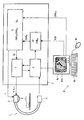
- the single Figure shows schematically the components of a device according to the invention for adjusting imaging parameters of an X-ray apparatus.
- an X-ray apparatus 1 comprising a C-arm with an X-ray source 2 and an X-ray detector 4 are shown.
- 3 X-ray projection images of a patient may be generated, these being passed on to an image recording module 9 in an attached data processing device 5 (workstation).
- the data processing device 5 also contains a generator-control module 7, which is linked on its output side to the X-ray source 2 in order to control imaging parameters such as, in particular, the X-ray tube current I, the tube voltage V and the pulse length L of the X-ray pulses.
- the generator-control module 7 is also linked to the image recording module 9 in order, for instance, to carry out feedback image brightness control during an X-ray image.
- the data processing device 5 is also linked to a user interface 6 which contains a monitor 6a, a keyboard 6b and a mouse 6c. On the monitor 6a, an image generated with the X-ray apparatus 1 may be displayed.
- a suitable adjusting procedure on the data processing device 5.
- a current preliminary image generated with the X-ray apparatus 1 is displayed on the monitor 6a.
- a predetermined APR setting is used which was previously selected by the user according to the underlying clinical situation and is set, through a plurality of control parameters, to standard settings.
- the user is then required to indicate a certain region of interest ROI or an interesting object (an anatomical detail or a certain medical device, such as a catheter) on the image and to stipulate a desired value for a visibility criterion of this region ROI.
- region of interest ROI By means of the interactive intervention by the user, definite identification of a region of interest ROI is, for instance, possible even if the interesting object itself has few characteristics or is ambiguous as, for instance, in the case of a plurality of medical instruments in the visual region with different requirements regarding image quality.
- the region of interest ROI may naturally also be incoherent or may include a plurality of individual objects, for instance an anatomical object such as the left ventricle and a catheter.
- the user may stipulate the start point A and the end point B of an interesting object ROI.
- the data processing device 5 may then extend the given points A, B to a more detailed object definition using suitable segmentation algorithms.
- a desired reference value must be stipulated by the user for a visibility criterion of the object.
- a suitable visibility criterion in this context is the contrast-to-noise ratio CNR, since it places the image noise in relation to the contrast between the object and its background.
- the (mean) contrast of the object relative to a surrounding area around the object may be placed in relation to the mean noise in a surrounding area around the object.
- the user may either stipulate a particular minimum reference value CNR ref for the contrast-to-noise ratio, or a standard value from the system may be used for this, predefined, for instance, in the APR settings.
- the data processing device 5 can then determine optimum imaging parameters for the given application, the precise patient and the interesting image region ROI. For this purpose, initially in a module 10 of the data processing device, for the current preliminary image, the contrast between the interesting object ROI and its surroundings is measured and the image noise determined. From these values, the current value CNR m of the contrast-to-noise ratio can be calculated. In a further module 8 of the data processing device 5, a comparison between the measured contrast-to-noise ratio CNR m and the desired value CNR ref is subsequently carried out.
- the imaging parameters of the X-ray apparatus 1 must be altered such that in the subsequent image recordings, a high X-ray dose is used. If, however, the measured value CNR m is greater than the reference value CNR ref , so that the region of interest ROI is imaged better than required, the X-ray dose can be reduced by a corresponding amount.
- the ratio between the measured and desired contrast-to-noise ratio, CNR m :CNR ref is calculated.
- the basic specifications for the X-ray images such as the value Q 0 of the desired dose per image may be made available, adjusted dependent upon the calculated ratio and the generator-control module 7. Furthermore, the module 8 may also give commands f to the collimator of the X-ray source 2 in order to control the setting of filter elements.
- dynamic imaging parameters such as tube current I and tube voltage V are controlled with a brightness-based dose check, whereby the adjusted imaging parameters Q 0 are taken into account, in order to depict the interesting object ROI with an optimum X-ray dose.
- the X-ray dose Q may generally be influenced by two parameters:
- the radiation intensity is adjusted, dependent upon the clinical conditions. This means that the number of X-ray photons irradiating the patient is increased linearly in relation to the ratio between the desired and the measured contrast-to-noise ratios CNR ref :CNR m .
- the tube current I it is not only the tube current I, but also the radiation quality that is modulated in order to achieve the desired contrast-to-noise ratio between the interesting object ROI and its surroundings.
- various compromises have to be found between different, partially contradictory requirements, in order to find the optimum imaging conditions, e.g.:
- the noise filtration during processing of X-ray images may be taken into account.
- the noise component of an image signal increases.
- the noise may be partially eliminated and the image quality thereby improved. For this reason, it is advantageous for the exposure parameters to be determined taking account of noise filtration.
- the method may be further developed such that the calculated imaging parameters, such as the dose setting Q 0 , may be adjusted to changes in the acceptance angle and the system geometry.
- the physician may, for instance, displace the patient table or change the position of the C-arm of the X-ray apparatus 1 in order to depict a different perspective of the patient's anatomy.
- the patient thickness may be determined from the preliminary image, used to determine the measured contrast-to-noise ratio CNR m .
- the patient thickness and the quotient between the actual contrast-to-noise ratio CNR m and the desired value CNR ref must then be recalculated to update the dose settings Q 0 based on these calculations and to pass them on to the generator-control module 7. In this way, despite an altered geometry, the system can continue to operate at an optimum balance between image quality and radiation dose used.
Landscapes
- Health & Medical Sciences (AREA)
- Life Sciences & Earth Sciences (AREA)
- Engineering & Computer Science (AREA)
- Medical Informatics (AREA)
- Radiology & Medical Imaging (AREA)
- Molecular Biology (AREA)
- Biophysics (AREA)
- Nuclear Medicine, Radiotherapy & Molecular Imaging (AREA)
- Optics & Photonics (AREA)
- Pathology (AREA)
- Physics & Mathematics (AREA)
- Biomedical Technology (AREA)
- Heart & Thoracic Surgery (AREA)
- High Energy & Nuclear Physics (AREA)
- Surgery (AREA)
- Animal Behavior & Ethology (AREA)
- General Health & Medical Sciences (AREA)
- Public Health (AREA)
- Veterinary Medicine (AREA)
- Human Computer Interaction (AREA)
- Apparatus For Radiation Diagnosis (AREA)
Claims (12)
- Dispositif de réglage pour régler des paramètres d'imagerie (I, V, L, f, Q0 ) d'un appareil radiologique (1), comprenant :- une interface utilisateur (6) apte à permettre à un utilisateur de spécifier une zone image à analyser (ROI) sur une image préliminaire et de spécifier une valeur de référence (CNRref ) d'un critère de visibilité pour cette zone image ;- un dispositif de traitement de données (5) agencé pour effectuer les étapes suivantes :a) le calcul des paramètres d'imagerie réglés (I, V, L, f, Q0 ) de l'appareil radiologique (1), par l'usage desquels la valeur de référence (CNRref ) du critère de visibilité est réalisée pour la zone image à analyser (ROI) ;b) la commande de l'appareil radiologique (1) sur la base des paramètres d'imagerie calculés et réglés (I, V, L, f, Q0 ) ; caractérisé en ce que le critère de visibilité est le rapport contraste/bruit de la zone image à analyser (ROI).
- Dispositif selon la revendication 1, caractérisé en ce que le dispositif de traitement de données (5) est agencé pour déterminer, dans l'image préliminaire, la valeur actuelle du critère de visibilité (CNRm ) pour la zone image à analyser (ROI).
- Dispositif selon la revendication 1, caractérisé en ce que les paramètres d'imagerie influencent la dose (Q0 ) par exposition, l'intensité et/ou la qualité du rayonnement de rayons X généré avec l'appareil radiologique (1).
- Dispositif selon la revendication 3, caractérisé en ce que les paramètres d'imagerie comprennent l'intensité de tube (I), la tension de tube (V), la longueur d'impulsion (L) et/ou les valeurs de réglage (f) des éléments de filtre.
- Dispositif selon la revendication 1, caractérisé en ce que ledit dispositif de traitement de données (5) est configuré pour calculer les paramètres d'imagerie réglés (I, V, L, f, Q0 ) à la condition qu'une image radiologique générée par ledit appareil radiologique (1) soit acquise avec la dose de rayonnement minimum nécessaire pour réaliser ladite valeur de référence (CNRref ) du critère de visibilité à l'intérieur de la zone image à analyser (ROI).
- Dispositif selon la revendication 1, caractérisé en ce que, dans l'image préliminaire, sur la base d'au moins un pixel (A, B) prédéfini par le biais de l'interface utilisateur (6), le dispositif de traitement de données (5) est agencé pour segmenter la zone image à analyser (ROI).
- Dispositif selon la revendication 1, caractérisé en ce que le dispositif de traitement de données (5) est agencé pour calculer les paramètres d'imagerie réglés (I, V, L, f, Q0 ) à partir des valeurs d'image à réduction de bruit à l'intérieur de la zone image à analyser (ROI) après avoir soumis ladite zone image à analyser (ROI) à une procédure de manipulation d'image donnée par l'étape de filtrage de bruit.
- Dispositif selon la revendication 1, caractérisé en ce que ledit dispositif de réglage comprend un module de commande (7) pour commander par rétroaction les paramètres d'imagerie (I, V, L) de l'appareil radiologique (1) au cours du processus d'imagerie radiologique.
- Dispositif selon la revendication 1, caractérisé en ce que ledit dispositif de réglage comprend des moyens pour détecter des changements de la géométrie d'imagerie et en ce que le dispositif de traitement de données (5) est agencé pour régler les paramètres d'imagerie calculés (I, V, L, f, Q0 ) dans le cas d'un changement de la géométrie d'imagerie de sorte que la valeur de référence (CNRref ) du critère de visibilité soit toujours réalisée.
- Procédé de réglage des paramètres d'imagerie (I, V, L, f, Q0 ) d'un appareil radiologique (1), comprenant les étapes suivantes :a) la génération d'une image préliminaire avec des valeurs de départ pour les paramètres d'imagerie ;b) la stipulation interactive par un utilisateur d'une zone image à analyser (ROI) sur l'image préliminaire et la stipulation interactive par un utilisateur d'une valeur de référence (CNRref ) d'un critère de visibilité pour cette zone image ;c) le calcul des paramètres d'imagerie réglés (I, V, L, f, Q0 ) pour l'appareil radiologique (1), pendant l'utilisation desquels la valeur de référence (CNRref ) du critère de visibilité est réalisée pour la zone image à analyser (ROI) ;d) la commande de l'appareil radiologique (1) sur la base des paramètres d'imagerie calculés et réglés (I, V, L, f, Q0 ) ; caractérisé en ce que le critère de visibilité est donné par le rapport contraste/bruit de la zone image à analyser (ROI).
- Procédé selon la revendication 10, caractérisé en ce que le calcul des paramètres d'imagerie réglés (I, V, L, f, Q0 ) est effectué à la condition qu'une image radiologique générée par ledit appareil radiologique (1) soit acquise avec la dose de rayonnement minimum nécessaire pour réaliser ladite valeur de référence (CNRref ) du critère de visibilité à l'intérieur de la zone image à analyser (ROI).
- Appareil radiologique ayant un dispositif de réglage selon l'une des revendications 1 à 9.
Priority Applications (1)
| Application Number | Priority Date | Filing Date | Title |
|---|---|---|---|
| EP04770269A EP1684635B1 (fr) | 2003-10-29 | 2004-10-15 | Dispositif et procede de reglage des parametres d'imagerie d'un appareil radiologique |
Applications Claiming Priority (3)
| Application Number | Priority Date | Filing Date | Title |
|---|---|---|---|
| EP03104003 | 2003-10-29 | ||
| PCT/IB2004/052111 WO2005041775A1 (fr) | 2003-10-29 | 2004-10-15 | Dispositif et procede de reglage des parametres d'imagerie d'un appareil radiologique |
| EP04770269A EP1684635B1 (fr) | 2003-10-29 | 2004-10-15 | Dispositif et procede de reglage des parametres d'imagerie d'un appareil radiologique |
Publications (2)
| Publication Number | Publication Date |
|---|---|
| EP1684635A1 EP1684635A1 (fr) | 2006-08-02 |
| EP1684635B1 true EP1684635B1 (fr) | 2011-07-20 |
Family
ID=34530769
Family Applications (1)
| Application Number | Title | Priority Date | Filing Date |
|---|---|---|---|
| EP04770269A Expired - Lifetime EP1684635B1 (fr) | 2003-10-29 | 2004-10-15 | Dispositif et procede de reglage des parametres d'imagerie d'un appareil radiologique |
Country Status (6)
| Country | Link |
|---|---|
| US (1) | US7519155B2 (fr) |
| EP (1) | EP1684635B1 (fr) |
| JP (1) | JP4528781B2 (fr) |
| CN (1) | CN100581462C (fr) |
| AT (1) | ATE516754T1 (fr) |
| WO (1) | WO2005041775A1 (fr) |
Families Citing this family (56)
| Publication number | Priority date | Publication date | Assignee | Title |
|---|---|---|---|---|
| DE102004030833A1 (de) * | 2004-06-25 | 2006-01-26 | Siemens Ag | Röngtendiagnostikverfahren und zugehörige Vorrichtung |
| JP2008539853A (ja) * | 2005-05-04 | 2008-11-20 | コーニンクレッカ フィリップス エレクトロニクス エヌ ヴィ | X線画像形成装置及び方法 |
| JP2007054372A (ja) * | 2005-08-25 | 2007-03-08 | Ge Medical Systems Global Technology Co Llc | X線ct装置 |
| WO2008015611A2 (fr) * | 2006-07-31 | 2008-02-07 | Philips Intellectual Property & Standards Gmbh | Système de planification de balayage rotatif aux rayons x |
| EP2103258B1 (fr) * | 2006-12-20 | 2013-03-13 | Hitachi Medical Corporation | Appareil de tomodensitométrie à rayons x |
| JP4909188B2 (ja) * | 2007-06-20 | 2012-04-04 | 株式会社日立メディコ | X線ct装置 |
| US20090086911A1 (en) * | 2007-09-27 | 2009-04-02 | General Electric Company | Inspection tool for radiographic systems |
| JP5464799B2 (ja) * | 2007-11-16 | 2014-04-09 | キヤノン株式会社 | 画像処理装置、画像処理方法及びプログラム |
| DE102008014738A1 (de) * | 2008-03-18 | 2009-09-24 | Siemens Aktiengesellschaft | Verfahren zur medizinischen Bildgebung sowie medizinische Bildgebungsvorrichtung |
| EP2349004B1 (fr) * | 2008-10-10 | 2013-08-21 | Philips Intellectual Property & Standards GmbH | Systeme et procede d'acquisition d'image angiographique avec adaptation automatique de l'obturateur permettant d'obtenir un champ d'observation reduit couvrant une lesion ou structure cible segmentee afin de reduire la dose de rayons x dans le cadre d'interventions mini-invasives guidees par rayons x |
| EP2408375B1 (fr) | 2009-03-20 | 2017-12-06 | Orthoscan Incorporated | Appareil mobile d'imagerie |
| US8755490B2 (en) * | 2009-04-07 | 2014-06-17 | Shimadzu Corporation | X-ray imaging device |
| EP2449530B1 (fr) | 2009-06-30 | 2016-04-20 | Koninklijke Philips N.V. | Analyse par perfusion quantitative |
| JP5572040B2 (ja) * | 2009-09-28 | 2014-08-13 | 富士フイルム株式会社 | 放射線撮影装置 |
| EP2490593B1 (fr) * | 2009-10-22 | 2019-11-06 | Koninklijke Philips N.V. | Appareil d'évaluation de protocole d'acquisition |
| US8821017B2 (en) | 2010-04-13 | 2014-09-02 | Carestream Health, Inc. | Projector as collimator light |
| US8827554B2 (en) | 2010-04-13 | 2014-09-09 | Carestream Health, Inc. | Tube alignment for mobile radiography system |
| US8824634B2 (en) | 2010-04-13 | 2014-09-02 | Carestream Health, Inc. | Configurable AEC sensor for an X-ray system |
| US8873712B2 (en) | 2010-04-13 | 2014-10-28 | Carestream Health, Inc. | Exposure control using digital radiography detector |
| FR2960762B1 (fr) * | 2010-06-07 | 2013-04-05 | Designers Developers Distributors Associates D3A Medical Systems | Procedes et systemes d'imagerie et de caracterisation d'un tissu osseux. |
| US8861679B2 (en) | 2010-06-11 | 2014-10-14 | Palodex Group Oy | X-ray imaging systems and methods |
| CN105232072A (zh) | 2010-09-07 | 2016-01-13 | 株式会社日立医疗器械 | X射线ct装置及管电流决定方法 |
| US8845190B2 (en) | 2010-10-06 | 2014-09-30 | Carestream Health, Inc. | Low-dose automatic exposure control system for digital portable X-ray imaging |
| US10165992B2 (en) | 2010-10-18 | 2019-01-01 | Carestream Health, Inc. | X-ray imaging systems and devices |
| DE102010043709B4 (de) * | 2010-11-10 | 2018-12-27 | Siemens Healthcare Gmbh | Verfahren zur Ermittlung des Wertes einer Röhrenspannung, Röntgeneinrichtung, Rechenprogramm und Datenträger |
| DE102010043712B4 (de) * | 2010-11-10 | 2021-03-18 | Siemens Healthcare Gmbh | Verfahren zur Ermittlung des Wertes einer Röhrenspannung, Röntgeneinrichtung, Rechenprogramm und Datenträger |
| WO2012082799A1 (fr) | 2010-12-13 | 2012-06-21 | Orthoscan, Inc. | Système d'imagerie fluoroscopique mobile |
| US20120155609A1 (en) * | 2010-12-20 | 2012-06-21 | General Electric Company | System and method of low dose exposure aided positioning (leap) for digital radiography |
| KR101895090B1 (ko) * | 2011-01-07 | 2018-09-04 | 제너럴 일렉트릭 캄파니 | 형광투시 시스템 및 방법 |
| US8681942B2 (en) | 2011-01-07 | 2014-03-25 | General Electric Company | Fluoroscopy systems and methods |
| FR2970618B1 (fr) * | 2011-01-17 | 2013-08-30 | Gen Electric | Procede de controle d'emission dans un dispositif d'imagerie a rayons x |
| US8821015B2 (en) | 2011-03-08 | 2014-09-02 | Carestream Health, Inc. | Alignment apparatus for X-ray imaging system |
| JP5699842B2 (ja) * | 2011-07-22 | 2015-04-15 | 株式会社島津製作所 | 画質評価方法およびそれを用いたx線透視撮影装置 |
| JP6175070B2 (ja) * | 2011-12-14 | 2017-08-02 | コーニンクレッカ フィリップス エヌ ヴェKoninklijke Philips N.V. | 高線量cアーチ幾何学的位置を防ぐためのリアルタイム・フィードバック |
| ES2658965T3 (es) | 2012-02-22 | 2018-03-13 | Carestream Health, Inc. | Aparatos/procedimientos radiográficos móviles con capacidad de tomosínteis |
| US20140328447A1 (en) * | 2013-05-01 | 2014-11-06 | Duke University | Systems and methods for computed tomography (ct) imaging using variable image quality factors or image capture settings in a single acquisition |
| KR101666943B1 (ko) * | 2013-06-11 | 2016-10-28 | 삼성전자주식회사 | 대상체의 관심 영역(roi)에 대한 x선 이미지를 획득하는 방법 및 장치 |
| WO2014200257A1 (fr) * | 2013-06-11 | 2014-12-18 | Samsung Electronics Co., Ltd. | Procédé et appareil d'obtention d'une radiographie d'une région d'intérêt d'un objet |
| CN104460181A (zh) * | 2013-09-25 | 2015-03-25 | 深圳市蓝韵实业有限公司 | 数字放射成像曝光剂量的评价方法 |
| ITBO20130669A1 (it) | 2013-12-02 | 2015-06-03 | Cefla Coop | Metodo e apparato per effettuare una scout in un apparecchio radiografico digitale dentale |
| JP2017051395A (ja) * | 2015-09-09 | 2017-03-16 | 富士フイルム株式会社 | 放射線画像処理装置、方法およびプログラム |
| US9962134B2 (en) * | 2015-10-28 | 2018-05-08 | Medtronic Navigation, Inc. | Apparatus and method for maintaining image quality while minimizing X-ray dosage of a patient |
| US9895130B2 (en) | 2015-11-19 | 2018-02-20 | General Electric Company | Water equivalent diameter determination from scout images |
| US10702235B2 (en) | 2017-06-08 | 2020-07-07 | Shanghai United Imaging Healthcare Co., Ltd. | Systems and methods for medical imaging |
| CN107049346B (zh) * | 2017-06-08 | 2021-06-15 | 上海联影医疗科技股份有限公司 | 医疗摄影控制方法、医疗摄影控制装置和医疗摄影设备 |
| CN107295736B (zh) * | 2017-06-26 | 2019-02-19 | 南京普爱医疗设备股份有限公司 | 一种用于x光机的自动脉冲调整高压电路 |
| CN109745060B (zh) * | 2017-11-06 | 2023-03-28 | 上海西门子医疗器械有限公司 | X-射线成像的自动曝光控制方法、存储介质和医疗设备 |
| DE102018217221A1 (de) * | 2018-10-09 | 2020-04-09 | Siemens Healthcare Gmbh | Verfahren zur Dosisreduzierung bei einem Röntgengerät unter Berücksichtigung einer späteren Darstellung; Bildgebungsanlage; Computerprogramm sowie Datenträger |
| CN109512446A (zh) * | 2018-12-08 | 2019-03-26 | 余姚德诚科技咨询有限公司 | 心血管图案验证机构 |
| JP7585334B2 (ja) * | 2020-09-25 | 2024-11-18 | 富士フイルム株式会社 | 設定装置、設定方法、及び設定プログラム |
| DE102020213035A1 (de) * | 2020-10-15 | 2022-04-21 | Siemens Healthcare Gmbh | Verfahren zur Ansteuerung eines Röntgengerätes und medizinisches System |
| CN116630222B (zh) * | 2022-02-10 | 2026-01-27 | 上海西门子医疗器械有限公司 | X射线图像获取方法、系统、x射线设备及存储介质 |
| US12333738B2 (en) * | 2022-06-30 | 2025-06-17 | Siemens Healthineers International Ag | Methods, systems and computer readable mediums for evaluating and displaying a breathing motion |
| DE102022206846A1 (de) * | 2022-07-05 | 2024-01-11 | Siemens Healthcare Gmbh | Verfahren und System zum Ausführen einer bildbasierten Aufgabe |
| WO2025100313A1 (fr) * | 2023-11-06 | 2025-05-15 | 株式会社島津製作所 | Dispositif d'imagerie fluoroscopique à rayons x |
| CN119257618A (zh) * | 2024-09-25 | 2025-01-07 | 中国人民解放军总医院第一医学中心 | 一种智能成像系统 |
Family Cites Families (9)
| Publication number | Priority date | Publication date | Assignee | Title |
|---|---|---|---|---|
| US4942596A (en) * | 1988-08-31 | 1990-07-17 | General Electric Company | Adaptive enhancement of x-ray images |
| US5319696A (en) * | 1992-10-05 | 1994-06-07 | General Electric Company | X-ray dose reduction in pulsed systems by adaptive X-ray pulse adjustment |
| JP3670439B2 (ja) * | 1997-05-09 | 2005-07-13 | 株式会社日立メディコ | X線装置 |
| JP3763967B2 (ja) * | 1998-04-17 | 2006-04-05 | 株式会社日立メディコ | X線装置 |
| US6222907B1 (en) * | 1999-07-12 | 2001-04-24 | General Electric Company | Image quality optimization using an X-ray model based optimization |
| US6501819B2 (en) * | 2000-12-18 | 2002-12-31 | Ge Medical Systems Global Technology Company, Llc | Medical diagnostic method and apparatus to control dual energy exposure techniques based on image information |
| US6459765B1 (en) * | 2000-12-28 | 2002-10-01 | Ge Medical Systems Global Technology Company, Llc | Automatic exposure control and optimization in digital x-ray radiography |
| DE10163583A1 (de) * | 2001-12-21 | 2003-07-03 | Philips Intellectual Property | Verfahren und Vorrichtung zur Belichtung von Röntgenaufnahmen |
| US6775352B2 (en) * | 2002-08-16 | 2004-08-10 | Ge Medical Systems Global Technology Company, Llc | Method and system for implementing variable x-ray intensity modulation schemes for imaging systems |
-
2004
- 2004-10-15 EP EP04770269A patent/EP1684635B1/fr not_active Expired - Lifetime
- 2004-10-15 JP JP2006537495A patent/JP4528781B2/ja not_active Expired - Lifetime
- 2004-10-15 CN CN200480032028A patent/CN100581462C/zh not_active Expired - Lifetime
- 2004-10-15 US US10/577,098 patent/US7519155B2/en not_active Expired - Lifetime
- 2004-10-15 WO PCT/IB2004/052111 patent/WO2005041775A1/fr not_active Ceased
- 2004-10-15 AT AT04770269T patent/ATE516754T1/de not_active IP Right Cessation
Also Published As
| Publication number | Publication date |
|---|---|
| EP1684635A1 (fr) | 2006-08-02 |
| JP2007509687A (ja) | 2007-04-19 |
| US7519155B2 (en) | 2009-04-14 |
| ATE516754T1 (de) | 2011-08-15 |
| WO2005041775A1 (fr) | 2005-05-12 |
| JP4528781B2 (ja) | 2010-08-18 |
| CN100581462C (zh) | 2010-01-20 |
| US20070071172A1 (en) | 2007-03-29 |
| CN1874726A (zh) | 2006-12-06 |
Similar Documents
| Publication | Publication Date | Title |
|---|---|---|
| EP1684635B1 (fr) | Dispositif et procede de reglage des parametres d'imagerie d'un appareil radiologique | |
| US8111895B2 (en) | Locally adaptive image enhancement for digital subtraction X-ray imaging | |
| US9700209B2 (en) | Medical imaging device for providing an image representation supporting in positioning an intervention device | |
| US20050089143A1 (en) | X-ray diagnosis apparatus and method for creating image data | |
| EP2427113B1 (fr) | Procédé d'acquisition d'une radiographie et dispositif d'acquisition de radiographie comprenant un positionnement de cale automatique | |
| JP2003284708A (ja) | ディジタル画像取得システム用の自動照射制御 | |
| US9044197B2 (en) | Method for x-ray dose tracking | |
| JP2003190139A (ja) | デフォルト・ノイズ指数オーバーライド能力を持つ医用イメージング・システム及び方法 | |
| US8509379B2 (en) | Method and X-ray device for adapting greyscale windowing | |
| US6744849B2 (en) | Image processing apparatus, image processing method, program, and storage medium | |
| US20010048734A1 (en) | Method for dynamic range management of digital radiographic images | |
| CN115018940A (zh) | 一种获取放射图像方法、系统、装置及存储介质 | |
| US7660390B2 (en) | X-ray diagnostics method and device | |
| CN115511723B (zh) | 用于x射线图像的校正的方法和系统以及x射线设备 | |
| US11707246B2 (en) | Method and apparatus for automatic determination of object and background region of interest for real-time automatic dose rate control in dynamic imaging systems | |
| EP4633473B1 (fr) | Collimation et filtrage contrôlés pour réduction de dose et qualité d'image en imagerie par rayons x | |
| US7460642B2 (en) | Method for generating an x-ray image sequence | |
| Rudin et al. | Real‐time equalization of region‐of‐interest fluoroscopic images using binary masks | |
| JP7588904B2 (ja) | 医用イメージングシステムおよびコンピュータプログラム | |
| EP4571651A1 (fr) | Réduction d'artefacts métalliques pour objets métalliques à l'extérieur du champ de vision de balayage | |
| Sarycheva et al. | X-ray equipment manufacturers contribution in patient and staff dose reduction | |
| WO2024081822A1 (fr) | Système et procédé de régulation de dose de rayonnement ionisant dans des applications médicales à l'aide de localisateurs synthétiques |
Legal Events
| Date | Code | Title | Description |
|---|---|---|---|
| PUAI | Public reference made under article 153(3) epc to a published international application that has entered the european phase |
Free format text: ORIGINAL CODE: 0009012 |
|
| 17P | Request for examination filed |
Effective date: 20060529 |
|
| AK | Designated contracting states |
Kind code of ref document: A1 Designated state(s): AT BE BG CH CY CZ DE DK EE ES FI FR GB GR HU IE IT LI LU MC NL PL PT RO SE SI SK TR |
|
| DAX | Request for extension of the european patent (deleted) | ||
| 17Q | First examination report despatched |
Effective date: 20090513 |
|
| GRAP | Despatch of communication of intention to grant a patent |
Free format text: ORIGINAL CODE: EPIDOSNIGR1 |
|
| RIN1 | Information on inventor provided before grant (corrected) |
Inventor name: WEESE, JUERGEN,PHILIPS INTELLECTUAL PROPERTY&STAND Inventor name: MOLLUS,SABINE,PHILIPS INTELLECTUAL PROPERTY&STANDA |
|
| GRAS | Grant fee paid |
Free format text: ORIGINAL CODE: EPIDOSNIGR3 |
|
| GRAA | (expected) grant |
Free format text: ORIGINAL CODE: 0009210 |
|
| AK | Designated contracting states |
Kind code of ref document: B1 Designated state(s): AT BE BG CH CY CZ DE DK EE ES FI FR GB GR HU IE IT LI LU MC NL PL PT RO SE SI SK TR |
|
| REG | Reference to a national code |
Ref country code: GB Ref legal event code: FG4D |
|
| REG | Reference to a national code |
Ref country code: CH Ref legal event code: EP |
|
| REG | Reference to a national code |
Ref country code: DE Ref legal event code: R084 Ref document number: 602004033563 Country of ref document: DE |
|
| REG | Reference to a national code |
Ref country code: NL Ref legal event code: T3 |
|
| REG | Reference to a national code |
Ref country code: DE Ref legal event code: R096 Ref document number: 602004033563 Country of ref document: DE Effective date: 20110908 |
|
| REG | Reference to a national code |
Ref country code: DE Ref legal event code: R084 Ref document number: 602004033563 Country of ref document: DE Effective date: 20110803 Ref country code: DE Ref legal event code: R084 Ref document number: 602004033563 Country of ref document: DE Effective date: 20110726 |
|
| REG | Reference to a national code |
Ref country code: AT Ref legal event code: MK05 Ref document number: 516754 Country of ref document: AT Kind code of ref document: T Effective date: 20110720 |
|
| PG25 | Lapsed in a contracting state [announced via postgrant information from national office to epo] |
Ref country code: PT Free format text: LAPSE BECAUSE OF FAILURE TO SUBMIT A TRANSLATION OF THE DESCRIPTION OR TO PAY THE FEE WITHIN THE PRESCRIBED TIME-LIMIT Effective date: 20111121 Ref country code: SE Free format text: LAPSE BECAUSE OF FAILURE TO SUBMIT A TRANSLATION OF THE DESCRIPTION OR TO PAY THE FEE WITHIN THE PRESCRIBED TIME-LIMIT Effective date: 20110720 Ref country code: BE Free format text: LAPSE BECAUSE OF FAILURE TO SUBMIT A TRANSLATION OF THE DESCRIPTION OR TO PAY THE FEE WITHIN THE PRESCRIBED TIME-LIMIT Effective date: 20110720 Ref country code: FI Free format text: LAPSE BECAUSE OF FAILURE TO SUBMIT A TRANSLATION OF THE DESCRIPTION OR TO PAY THE FEE WITHIN THE PRESCRIBED TIME-LIMIT Effective date: 20110720 |
|
| PG25 | Lapsed in a contracting state [announced via postgrant information from national office to epo] |
Ref country code: GR Free format text: LAPSE BECAUSE OF FAILURE TO SUBMIT A TRANSLATION OF THE DESCRIPTION OR TO PAY THE FEE WITHIN THE PRESCRIBED TIME-LIMIT Effective date: 20111021 Ref country code: CY Free format text: LAPSE BECAUSE OF FAILURE TO SUBMIT A TRANSLATION OF THE DESCRIPTION OR TO PAY THE FEE WITHIN THE PRESCRIBED TIME-LIMIT Effective date: 20110720 Ref country code: SI Free format text: LAPSE BECAUSE OF FAILURE TO SUBMIT A TRANSLATION OF THE DESCRIPTION OR TO PAY THE FEE WITHIN THE PRESCRIBED TIME-LIMIT Effective date: 20110720 Ref country code: AT Free format text: LAPSE BECAUSE OF FAILURE TO SUBMIT A TRANSLATION OF THE DESCRIPTION OR TO PAY THE FEE WITHIN THE PRESCRIBED TIME-LIMIT Effective date: 20110720 Ref country code: PL Free format text: LAPSE BECAUSE OF FAILURE TO SUBMIT A TRANSLATION OF THE DESCRIPTION OR TO PAY THE FEE WITHIN THE PRESCRIBED TIME-LIMIT Effective date: 20110720 |
|
| PG25 | Lapsed in a contracting state [announced via postgrant information from national office to epo] |
Ref country code: SK Free format text: LAPSE BECAUSE OF FAILURE TO SUBMIT A TRANSLATION OF THE DESCRIPTION OR TO PAY THE FEE WITHIN THE PRESCRIBED TIME-LIMIT Effective date: 20110720 Ref country code: CZ Free format text: LAPSE BECAUSE OF FAILURE TO SUBMIT A TRANSLATION OF THE DESCRIPTION OR TO PAY THE FEE WITHIN THE PRESCRIBED TIME-LIMIT Effective date: 20110720 |
|
| PLBE | No opposition filed within time limit |
Free format text: ORIGINAL CODE: 0009261 |
|
| STAA | Information on the status of an ep patent application or granted ep patent |
Free format text: STATUS: NO OPPOSITION FILED WITHIN TIME LIMIT |
|
| PG25 | Lapsed in a contracting state [announced via postgrant information from national office to epo] |
Ref country code: IT Free format text: LAPSE BECAUSE OF FAILURE TO SUBMIT A TRANSLATION OF THE DESCRIPTION OR TO PAY THE FEE WITHIN THE PRESCRIBED TIME-LIMIT Effective date: 20110720 Ref country code: RO Free format text: LAPSE BECAUSE OF FAILURE TO SUBMIT A TRANSLATION OF THE DESCRIPTION OR TO PAY THE FEE WITHIN THE PRESCRIBED TIME-LIMIT Effective date: 20110720 Ref country code: EE Free format text: LAPSE BECAUSE OF FAILURE TO SUBMIT A TRANSLATION OF THE DESCRIPTION OR TO PAY THE FEE WITHIN THE PRESCRIBED TIME-LIMIT Effective date: 20110720 Ref country code: MC Free format text: LAPSE BECAUSE OF NON-PAYMENT OF DUE FEES Effective date: 20111031 |
|
| REG | Reference to a national code |
Ref country code: CH Ref legal event code: PL |
|
| 26N | No opposition filed |
Effective date: 20120423 |
|
| PG25 | Lapsed in a contracting state [announced via postgrant information from national office to epo] |
Ref country code: DK Free format text: LAPSE BECAUSE OF FAILURE TO SUBMIT A TRANSLATION OF THE DESCRIPTION OR TO PAY THE FEE WITHIN THE PRESCRIBED TIME-LIMIT Effective date: 20110720 |
|
| PG25 | Lapsed in a contracting state [announced via postgrant information from national office to epo] |
Ref country code: CH Free format text: LAPSE BECAUSE OF NON-PAYMENT OF DUE FEES Effective date: 20111031 Ref country code: LI Free format text: LAPSE BECAUSE OF NON-PAYMENT OF DUE FEES Effective date: 20111031 |
|
| REG | Reference to a national code |
Ref country code: IE Ref legal event code: MM4A |
|
| REG | Reference to a national code |
Ref country code: DE Ref legal event code: R097 Ref document number: 602004033563 Country of ref document: DE Effective date: 20120423 |
|
| PG25 | Lapsed in a contracting state [announced via postgrant information from national office to epo] |
Ref country code: IE Free format text: LAPSE BECAUSE OF NON-PAYMENT OF DUE FEES Effective date: 20111015 |
|
| PG25 | Lapsed in a contracting state [announced via postgrant information from national office to epo] |
Ref country code: ES Free format text: LAPSE BECAUSE OF FAILURE TO SUBMIT A TRANSLATION OF THE DESCRIPTION OR TO PAY THE FEE WITHIN THE PRESCRIBED TIME-LIMIT Effective date: 20111031 |
|
| PG25 | Lapsed in a contracting state [announced via postgrant information from national office to epo] |
Ref country code: LU Free format text: LAPSE BECAUSE OF NON-PAYMENT OF DUE FEES Effective date: 20111015 |
|
| PG25 | Lapsed in a contracting state [announced via postgrant information from national office to epo] |
Ref country code: BG Free format text: LAPSE BECAUSE OF FAILURE TO SUBMIT A TRANSLATION OF THE DESCRIPTION OR TO PAY THE FEE WITHIN THE PRESCRIBED TIME-LIMIT Effective date: 20111020 |
|
| PG25 | Lapsed in a contracting state [announced via postgrant information from national office to epo] |
Ref country code: TR Free format text: LAPSE BECAUSE OF FAILURE TO SUBMIT A TRANSLATION OF THE DESCRIPTION OR TO PAY THE FEE WITHIN THE PRESCRIBED TIME-LIMIT Effective date: 20110720 |
|
| PG25 | Lapsed in a contracting state [announced via postgrant information from national office to epo] |
Ref country code: HU Free format text: LAPSE BECAUSE OF FAILURE TO SUBMIT A TRANSLATION OF THE DESCRIPTION OR TO PAY THE FEE WITHIN THE PRESCRIBED TIME-LIMIT Effective date: 20110720 |
|
| REG | Reference to a national code |
Ref country code: DE Ref legal event code: R081 Ref document number: 602004033563 Country of ref document: DE Owner name: PHILIPS GMBH, DE Free format text: FORMER OWNER: PHILIPS INTELLECTUAL PROPERTY &, KONINKLIJKE PHILIPS ELECTRONICS, , NL Effective date: 20110719 Ref country code: DE Ref legal event code: R081 Ref document number: 602004033563 Country of ref document: DE Owner name: PHILIPS GMBH, DE Free format text: FORMER OWNER: PHILIPS INTELLECTUAL PROPERTY & STANDARDS GMBH, 20099 HAMBURG, DE Effective date: 20140327 Ref country code: DE Ref legal event code: R081 Ref document number: 602004033563 Country of ref document: DE Owner name: PHILIPS GMBH, DE Free format text: FORMER OWNERS: PHILIPS INTELLECTUAL PROPERTY & STANDARDS GMBH, 20099 HAMBURG, DE; KONINKLIJKE PHILIPS ELECTRONICS N.V., EINDHOVEN, NL Effective date: 20110719 Ref country code: DE Ref legal event code: R081 Ref document number: 602004033563 Country of ref document: DE Owner name: PHILIPS DEUTSCHLAND GMBH, DE Free format text: FORMER OWNER: PHILIPS INTELLECTUAL PROPERTY & STANDARDS GMBH, 20099 HAMBURG, DE Effective date: 20140327 Ref country code: DE Ref legal event code: R081 Ref document number: 602004033563 Country of ref document: DE Owner name: PHILIPS DEUTSCHLAND GMBH, DE Free format text: FORMER OWNER: PHILIPS INTELLECTUAL PROPERTY &, KONINKLIJKE PHILIPS ELECTRONICS, , NL Effective date: 20110719 |
|
| REG | Reference to a national code |
Ref country code: FR Ref legal event code: CD Owner name: PHILIPS INTELLECTUAL PROPERTY & STANDARDS GMBH, DE Effective date: 20141126 Ref country code: FR Ref legal event code: CD Owner name: KONINKLIJKE PHILIPS ELECTRONICS N.V., NL Effective date: 20141126 Ref country code: FR Ref legal event code: CA Effective date: 20141126 |
|
| REG | Reference to a national code |
Ref country code: DE Ref legal event code: R082 Ref document number: 602004033563 Country of ref document: DE Representative=s name: MEISSNER, BOLTE & PARTNER GBR, DE Ref country code: DE Ref legal event code: R081 Ref document number: 602004033563 Country of ref document: DE Owner name: PHILIPS GMBH, DE Free format text: FORMER OWNER: PHILIPS DEUTSCHLAND GMBH, 20099 HAMBURG, DE Ref country code: DE Ref legal event code: R082 Ref document number: 602004033563 Country of ref document: DE Representative=s name: MEISSNER BOLTE PATENTANWAELTE RECHTSANWAELTE P, DE |
|
| REG | Reference to a national code |
Ref country code: FR Ref legal event code: PLFP Year of fee payment: 12 |
|
| REG | Reference to a national code |
Ref country code: FR Ref legal event code: PLFP Year of fee payment: 13 |
|
| REG | Reference to a national code |
Ref country code: FR Ref legal event code: PLFP Year of fee payment: 14 |
|
| PGFP | Annual fee paid to national office [announced via postgrant information from national office to epo] |
Ref country code: FR Payment date: 20171031 Year of fee payment: 14 |
|
| PGFP | Annual fee paid to national office [announced via postgrant information from national office to epo] |
Ref country code: NL Payment date: 20171025 Year of fee payment: 14 Ref country code: GB Payment date: 20171101 Year of fee payment: 14 |
|
| REG | Reference to a national code |
Ref country code: NL Ref legal event code: MM Effective date: 20181101 |
|
| GBPC | Gb: european patent ceased through non-payment of renewal fee |
Effective date: 20181015 |
|
| PG25 | Lapsed in a contracting state [announced via postgrant information from national office to epo] |
Ref country code: NL Free format text: LAPSE BECAUSE OF NON-PAYMENT OF DUE FEES Effective date: 20181101 |
|
| PG25 | Lapsed in a contracting state [announced via postgrant information from national office to epo] |
Ref country code: FR Free format text: LAPSE BECAUSE OF NON-PAYMENT OF DUE FEES Effective date: 20181031 Ref country code: GB Free format text: LAPSE BECAUSE OF NON-PAYMENT OF DUE FEES Effective date: 20181015 |
|
| REG | Reference to a national code |
Ref country code: DE Ref legal event code: R082 Ref document number: 602004033563 Country of ref document: DE Representative=s name: MEISSNER BOLTE PATENTANWAELTE RECHTSANWAELTE P, DE Ref country code: DE Ref legal event code: R081 Ref document number: 602004033563 Country of ref document: DE Owner name: PHILIPS GMBH, DE Free format text: FORMER OWNER: PHILIPS GMBH, 20099 HAMBURG, DE |
|
| PGFP | Annual fee paid to national office [announced via postgrant information from national office to epo] |
Ref country code: DE Payment date: 20231027 Year of fee payment: 20 |
|
| REG | Reference to a national code |
Ref country code: DE Ref legal event code: R071 Ref document number: 602004033563 Country of ref document: DE |