EP1681565A1 - Zellulärer RhoGTPase Aktivierungs-Assay - Google Patents
Zellulärer RhoGTPase Aktivierungs-Assay Download PDFInfo
- Publication number
- EP1681565A1 EP1681565A1 EP06000726A EP06000726A EP1681565A1 EP 1681565 A1 EP1681565 A1 EP 1681565A1 EP 06000726 A EP06000726 A EP 06000726A EP 06000726 A EP06000726 A EP 06000726A EP 1681565 A1 EP1681565 A1 EP 1681565A1
- Authority
- EP
- European Patent Office
- Prior art keywords
- cell
- cell line
- ngr
- functional
- rho
- Prior art date
- Legal status (The legal status is an assumption and is not a legal conclusion. Google has not performed a legal analysis and makes no representation as to the accuracy of the status listed.)
- Granted
Links
- 230000004913 activation Effects 0.000 title claims abstract description 27
- 230000001413 cellular effect Effects 0.000 title claims abstract description 16
- 238000003556 assay Methods 0.000 title claims description 37
- 210000004027 cell Anatomy 0.000 claims abstract description 238
- 102000005781 Nogo Receptor Human genes 0.000 claims abstract description 100
- 108020003872 Nogo receptor Proteins 0.000 claims abstract description 100
- 210000004292 cytoskeleton Anatomy 0.000 claims abstract description 39
- 239000012636 effector Substances 0.000 claims abstract description 39
- 238000000034 method Methods 0.000 claims abstract description 38
- 150000007523 nucleic acids Chemical group 0.000 claims abstract description 38
- 239000003446 ligand Substances 0.000 claims abstract description 33
- 102000005962 receptors Human genes 0.000 claims abstract description 29
- 108020003175 receptors Proteins 0.000 claims abstract description 29
- 239000013598 vector Substances 0.000 claims abstract description 25
- 238000010998 test method Methods 0.000 claims abstract description 17
- 230000001419 dependent effect Effects 0.000 claims abstract description 16
- 230000003834 intracellular effect Effects 0.000 claims abstract description 16
- 230000019491 signal transduction Effects 0.000 claims abstract description 15
- 238000012360 testing method Methods 0.000 claims abstract description 13
- 108091028043 Nucleic acid sequence Proteins 0.000 claims abstract description 12
- 238000002952 image-based readout Methods 0.000 claims abstract description 12
- 239000012528 membrane Substances 0.000 claims abstract description 12
- 230000008859 change Effects 0.000 claims abstract description 11
- 230000000694 effects Effects 0.000 claims abstract description 11
- 230000009689 neuronal regeneration Effects 0.000 claims abstract description 10
- 239000005557 antagonist Substances 0.000 claims abstract description 8
- 230000008521 reorganization Effects 0.000 claims abstract description 7
- 238000012258 culturing Methods 0.000 claims abstract description 3
- 230000006698 induction Effects 0.000 claims abstract description 3
- 102100027609 Rho-related GTP-binding protein RhoD Human genes 0.000 claims description 59
- 239000012634 fragment Substances 0.000 claims description 23
- 230000027455 binding Effects 0.000 claims description 20
- 108010077641 Nogo Proteins Proteins 0.000 claims description 18
- 102000010410 Nogo Proteins Human genes 0.000 claims description 17
- 102000007469 Actins Human genes 0.000 claims description 15
- 108010085238 Actins Proteins 0.000 claims description 15
- 230000001086 cytosolic effect Effects 0.000 claims description 13
- 239000012491 analyte Substances 0.000 claims description 11
- 230000037361 pathway Effects 0.000 claims description 11
- 238000012216 screening Methods 0.000 claims description 10
- 238000010191 image analysis Methods 0.000 claims description 9
- 230000001939 inductive effect Effects 0.000 claims description 8
- 201000010099 disease Diseases 0.000 claims description 7
- 208000037265 diseases, disorders, signs and symptoms Diseases 0.000 claims description 7
- 108010033674 rho GTP-Binding Proteins Proteins 0.000 claims description 7
- 230000005764 inhibitory process Effects 0.000 claims description 6
- 238000004519 manufacturing process Methods 0.000 claims description 6
- 102000007339 Nerve Growth Factor Receptors Human genes 0.000 claims description 5
- 108010032605 Nerve Growth Factor Receptors Proteins 0.000 claims description 5
- 208000027418 Wounds and injury Diseases 0.000 claims description 5
- 230000001464 adherent effect Effects 0.000 claims description 5
- 230000003376 axonal effect Effects 0.000 claims description 5
- 230000006378 damage Effects 0.000 claims description 5
- 208000014674 injury Diseases 0.000 claims description 5
- 102000007268 rho GTP-Binding Proteins Human genes 0.000 claims description 5
- 230000004075 alteration Effects 0.000 claims description 4
- 210000002950 fibroblast Anatomy 0.000 claims description 4
- 210000000020 growth cone Anatomy 0.000 claims description 4
- 210000004962 mammalian cell Anatomy 0.000 claims description 4
- 230000007514 neuronal growth Effects 0.000 claims description 4
- 230000031146 intracellular signal transduction Effects 0.000 claims description 3
- 230000008929 regeneration Effects 0.000 claims description 3
- 238000011069 regeneration method Methods 0.000 claims description 3
- 210000003734 kidney Anatomy 0.000 claims description 2
- 125000003275 alpha amino acid group Chemical group 0.000 claims 3
- 230000001131 transforming effect Effects 0.000 claims 1
- UYTPUPDQBNUYGX-UHFFFAOYSA-N guanine Chemical compound O=C1NC(N)=NC2=C1N=CN2 UYTPUPDQBNUYGX-UHFFFAOYSA-N 0.000 abstract description 3
- 230000008569 process Effects 0.000 abstract description 2
- 108030003004 Triphosphatases Proteins 0.000 abstract 1
- 230000010534 mechanism of action Effects 0.000 abstract 1
- 230000000324 neuroprotective effect Effects 0.000 abstract 1
- 108090000623 proteins and genes Proteins 0.000 description 65
- 101710187830 Tumor necrosis factor receptor superfamily member 1B Proteins 0.000 description 40
- 102000004169 proteins and genes Human genes 0.000 description 40
- 102100029946 Sialic acid-binding Ig-like lectin 7 Human genes 0.000 description 38
- 230000014509 gene expression Effects 0.000 description 32
- 108020004414 DNA Proteins 0.000 description 20
- 102000039446 nucleic acids Human genes 0.000 description 20
- 108020004707 nucleic acids Proteins 0.000 description 20
- 239000013612 plasmid Substances 0.000 description 19
- 230000000638 stimulation Effects 0.000 description 19
- 101100356682 Caenorhabditis elegans rho-1 gene Proteins 0.000 description 18
- 101150111584 RHOA gene Proteins 0.000 description 18
- 150000001413 amino acids Chemical group 0.000 description 16
- 239000000499 gel Substances 0.000 description 16
- 102000013446 GTP Phosphohydrolases Human genes 0.000 description 15
- 108091006109 GTPases Proteins 0.000 description 15
- 229920001184 polypeptide Polymers 0.000 description 14
- 102000004196 processed proteins & peptides Human genes 0.000 description 14
- 108090000765 processed proteins & peptides Proteins 0.000 description 14
- 230000001105 regulatory effect Effects 0.000 description 14
- XKMLYUALXHKNFT-UUOKFMHZSA-N Guanosine-5'-triphosphate Chemical compound C1=2NC(N)=NC(=O)C=2N=CN1[C@@H]1O[C@H](COP(O)(=O)OP(O)(=O)OP(O)(O)=O)[C@@H](O)[C@H]1O XKMLYUALXHKNFT-UUOKFMHZSA-N 0.000 description 13
- 108091034117 Oligonucleotide Proteins 0.000 description 13
- 238000001514 detection method Methods 0.000 description 12
- LOKCTEFSRHRXRJ-UHFFFAOYSA-I dipotassium trisodium dihydrogen phosphate hydrogen phosphate dichloride Chemical compound P(=O)(O)(O)[O-].[K+].P(=O)(O)([O-])[O-].[Na+].[Na+].[Cl-].[K+].[Cl-].[Na+] LOKCTEFSRHRXRJ-UHFFFAOYSA-I 0.000 description 12
- 239000002953 phosphate buffered saline Substances 0.000 description 12
- 238000002360 preparation method Methods 0.000 description 11
- XLYOFNOQVPJJNP-UHFFFAOYSA-N water Substances O XLYOFNOQVPJJNP-UHFFFAOYSA-N 0.000 description 11
- JLCPHMBAVCMARE-UHFFFAOYSA-N [3-[[3-[[3-[[3-[[3-[[3-[[3-[[3-[[3-[[3-[[3-[[5-(2-amino-6-oxo-1H-purin-9-yl)-3-[[3-[[3-[[3-[[3-[[3-[[5-(2-amino-6-oxo-1H-purin-9-yl)-3-[[5-(2-amino-6-oxo-1H-purin-9-yl)-3-hydroxyoxolan-2-yl]methoxy-hydroxyphosphoryl]oxyoxolan-2-yl]methoxy-hydroxyphosphoryl]oxy-5-(5-methyl-2,4-dioxopyrimidin-1-yl)oxolan-2-yl]methoxy-hydroxyphosphoryl]oxy-5-(6-aminopurin-9-yl)oxolan-2-yl]methoxy-hydroxyphosphoryl]oxy-5-(6-aminopurin-9-yl)oxolan-2-yl]methoxy-hydroxyphosphoryl]oxy-5-(6-aminopurin-9-yl)oxolan-2-yl]methoxy-hydroxyphosphoryl]oxy-5-(6-aminopurin-9-yl)oxolan-2-yl]methoxy-hydroxyphosphoryl]oxyoxolan-2-yl]methoxy-hydroxyphosphoryl]oxy-5-(5-methyl-2,4-dioxopyrimidin-1-yl)oxolan-2-yl]methoxy-hydroxyphosphoryl]oxy-5-(4-amino-2-oxopyrimidin-1-yl)oxolan-2-yl]methoxy-hydroxyphosphoryl]oxy-5-(5-methyl-2,4-dioxopyrimidin-1-yl)oxolan-2-yl]methoxy-hydroxyphosphoryl]oxy-5-(5-methyl-2,4-dioxopyrimidin-1-yl)oxolan-2-yl]methoxy-hydroxyphosphoryl]oxy-5-(6-aminopurin-9-yl)oxolan-2-yl]methoxy-hydroxyphosphoryl]oxy-5-(6-aminopurin-9-yl)oxolan-2-yl]methoxy-hydroxyphosphoryl]oxy-5-(4-amino-2-oxopyrimidin-1-yl)oxolan-2-yl]methoxy-hydroxyphosphoryl]oxy-5-(4-amino-2-oxopyrimidin-1-yl)oxolan-2-yl]methoxy-hydroxyphosphoryl]oxy-5-(4-amino-2-oxopyrimidin-1-yl)oxolan-2-yl]methoxy-hydroxyphosphoryl]oxy-5-(6-aminopurin-9-yl)oxolan-2-yl]methoxy-hydroxyphosphoryl]oxy-5-(4-amino-2-oxopyrimidin-1-yl)oxolan-2-yl]methyl [5-(6-aminopurin-9-yl)-2-(hydroxymethyl)oxolan-3-yl] hydrogen phosphate Polymers Cc1cn(C2CC(OP(O)(=O)OCC3OC(CC3OP(O)(=O)OCC3OC(CC3O)n3cnc4c3nc(N)[nH]c4=O)n3cnc4c3nc(N)[nH]c4=O)C(COP(O)(=O)OC3CC(OC3COP(O)(=O)OC3CC(OC3COP(O)(=O)OC3CC(OC3COP(O)(=O)OC3CC(OC3COP(O)(=O)OC3CC(OC3COP(O)(=O)OC3CC(OC3COP(O)(=O)OC3CC(OC3COP(O)(=O)OC3CC(OC3COP(O)(=O)OC3CC(OC3COP(O)(=O)OC3CC(OC3COP(O)(=O)OC3CC(OC3COP(O)(=O)OC3CC(OC3COP(O)(=O)OC3CC(OC3COP(O)(=O)OC3CC(OC3COP(O)(=O)OC3CC(OC3COP(O)(=O)OC3CC(OC3COP(O)(=O)OC3CC(OC3CO)n3cnc4c(N)ncnc34)n3ccc(N)nc3=O)n3cnc4c(N)ncnc34)n3ccc(N)nc3=O)n3ccc(N)nc3=O)n3ccc(N)nc3=O)n3cnc4c(N)ncnc34)n3cnc4c(N)ncnc34)n3cc(C)c(=O)[nH]c3=O)n3cc(C)c(=O)[nH]c3=O)n3ccc(N)nc3=O)n3cc(C)c(=O)[nH]c3=O)n3cnc4c3nc(N)[nH]c4=O)n3cnc4c(N)ncnc34)n3cnc4c(N)ncnc34)n3cnc4c(N)ncnc34)n3cnc4c(N)ncnc34)O2)c(=O)[nH]c1=O JLCPHMBAVCMARE-UHFFFAOYSA-N 0.000 description 10
- 239000000872 buffer Substances 0.000 description 10
- 239000002609 medium Substances 0.000 description 10
- 150000003839 salts Chemical class 0.000 description 10
- 238000004458 analytical method Methods 0.000 description 9
- 230000000295 complement effect Effects 0.000 description 9
- 208000025380 Logopenic progressive aphasia Diseases 0.000 description 8
- 230000003115 biocidal effect Effects 0.000 description 8
- 238000010367 cloning Methods 0.000 description 8
- 239000002299 complementary DNA Substances 0.000 description 8
- 239000002773 nucleotide Substances 0.000 description 8
- 125000003729 nucleotide group Chemical group 0.000 description 8
- 239000000523 sample Substances 0.000 description 8
- 239000002543 antimycotic Substances 0.000 description 7
- 239000011324 bead Substances 0.000 description 7
- 238000006243 chemical reaction Methods 0.000 description 7
- 238000002474 experimental method Methods 0.000 description 7
- 239000013604 expression vector Substances 0.000 description 7
- 238000000605 extraction Methods 0.000 description 7
- BRZYSWJRSDMWLG-CAXSIQPQSA-N geneticin Natural products O1C[C@@](O)(C)[C@H](NC)[C@@H](O)[C@H]1O[C@@H]1[C@@H](O)[C@H](O[C@@H]2[C@@H]([C@@H](O)[C@H](O)[C@@H](C(C)O)O2)N)[C@@H](N)C[C@H]1N BRZYSWJRSDMWLG-CAXSIQPQSA-N 0.000 description 7
- 239000001963 growth medium Substances 0.000 description 7
- 239000003550 marker Substances 0.000 description 7
- 239000011435 rock Substances 0.000 description 7
- 108091026890 Coding region Proteins 0.000 description 6
- KPKZJLCSROULON-QKGLWVMZSA-N Phalloidin Chemical compound N1C(=O)[C@@H]([C@@H](O)C)NC(=O)[C@H](C)NC(=O)[C@H](C[C@@](C)(O)CO)NC(=O)[C@H](C2)NC(=O)[C@H](C)NC(=O)[C@@H]3C[C@H](O)CN3C(=O)[C@@H]1CSC1=C2C2=CC=CC=C2N1 KPKZJLCSROULON-QKGLWVMZSA-N 0.000 description 6
- FAPWRFPIFSIZLT-UHFFFAOYSA-M Sodium chloride Chemical compound [Na+].[Cl-] FAPWRFPIFSIZLT-UHFFFAOYSA-M 0.000 description 6
- 108010084455 Zeocin Proteins 0.000 description 6
- 238000007792 addition Methods 0.000 description 6
- 230000015572 biosynthetic process Effects 0.000 description 6
- 230000008602 contraction Effects 0.000 description 6
- 239000000203 mixture Substances 0.000 description 6
- 238000010369 molecular cloning Methods 0.000 description 6
- CWCMIVBLVUHDHK-ZSNHEYEWSA-N phleomycin D1 Chemical compound N([C@H](C(=O)N[C@H](C)[C@@H](O)[C@H](C)C(=O)N[C@@H]([C@H](O)C)C(=O)NCCC=1SC[C@@H](N=1)C=1SC=C(N=1)C(=O)NCCCCNC(N)=N)[C@@H](O[C@H]1[C@H]([C@@H](O)[C@H](O)[C@H](CO)O1)O[C@@H]1[C@H]([C@@H](OC(N)=O)[C@H](O)[C@@H](CO)O1)O)C=1N=CNC=1)C(=O)C1=NC([C@H](CC(N)=O)NC[C@H](N)C(N)=O)=NC(N)=C1C CWCMIVBLVUHDHK-ZSNHEYEWSA-N 0.000 description 6
- 108091033319 polynucleotide Proteins 0.000 description 6
- 239000013615 primer Substances 0.000 description 6
- 230000008707 rearrangement Effects 0.000 description 6
- 108091003079 Bovine Serum Albumin Proteins 0.000 description 5
- 238000012413 Fluorescence activated cell sorting analysis Methods 0.000 description 5
- 241000700159 Rattus Species 0.000 description 5
- 101000702488 Rattus norvegicus High affinity cationic amino acid transporter 1 Proteins 0.000 description 5
- 102000004142 Trypsin Human genes 0.000 description 5
- 108090000631 Trypsin Proteins 0.000 description 5
- 239000002253 acid Chemical class 0.000 description 5
- 239000003153 chemical reaction reagent Substances 0.000 description 5
- 238000011156 evaluation Methods 0.000 description 5
- 239000012091 fetal bovine serum Substances 0.000 description 5
- MHMNJMPURVTYEJ-UHFFFAOYSA-N fluorescein-5-isothiocyanate Chemical compound O1C(=O)C2=CC(N=C=S)=CC=C2C21C1=CC=C(O)C=C1OC1=CC(O)=CC=C21 MHMNJMPURVTYEJ-UHFFFAOYSA-N 0.000 description 5
- 239000011521 glass Substances 0.000 description 5
- 230000001965 increasing effect Effects 0.000 description 5
- 230000007246 mechanism Effects 0.000 description 5
- 239000002157 polynucleotide Substances 0.000 description 5
- 102000040430 polynucleotide Human genes 0.000 description 5
- 238000003786 synthesis reaction Methods 0.000 description 5
- 239000012588 trypsin Substances 0.000 description 5
- 102000002260 Alkaline Phosphatase Human genes 0.000 description 4
- 108020004774 Alkaline Phosphatase Proteins 0.000 description 4
- 102000011068 Cdc42 Human genes 0.000 description 4
- 108050001278 Cdc42 Proteins 0.000 description 4
- 241000196324 Embryophyta Species 0.000 description 4
- 241000233866 Fungi Species 0.000 description 4
- 102000005720 Glutathione transferase Human genes 0.000 description 4
- 108010070675 Glutathione transferase Proteins 0.000 description 4
- 102100035044 Myosin light chain kinase, smooth muscle Human genes 0.000 description 4
- 108010074596 Myosin-Light-Chain Kinase Proteins 0.000 description 4
- 102100035124 Rhotekin Human genes 0.000 description 4
- 101710122991 Rhotekin Proteins 0.000 description 4
- 238000013459 approach Methods 0.000 description 4
- 210000004556 brain Anatomy 0.000 description 4
- 210000000170 cell membrane Anatomy 0.000 description 4
- 210000003169 central nervous system Anatomy 0.000 description 4
- 150000001875 compounds Chemical class 0.000 description 4
- 231100000673 dose–response relationship Toxicity 0.000 description 4
- 230000006870 function Effects 0.000 description 4
- 230000004927 fusion Effects 0.000 description 4
- 238000003119 immunoblot Methods 0.000 description 4
- 244000005700 microbiome Species 0.000 description 4
- 230000004660 morphological change Effects 0.000 description 4
- 230000002018 overexpression Effects 0.000 description 4
- YBYRMVIVWMBXKQ-UHFFFAOYSA-N phenylmethanesulfonyl fluoride Chemical compound FS(=O)(=O)CC1=CC=CC=C1 YBYRMVIVWMBXKQ-UHFFFAOYSA-N 0.000 description 4
- 230000011218 segmentation Effects 0.000 description 4
- 239000000243 solution Substances 0.000 description 4
- 238000010186 staining Methods 0.000 description 4
- 238000010561 standard procedure Methods 0.000 description 4
- UCSJYZPVAKXKNQ-HZYVHMACSA-N streptomycin Chemical compound CN[C@H]1[C@H](O)[C@@H](O)[C@H](CO)O[C@H]1O[C@@H]1[C@](C=O)(O)[C@H](C)O[C@H]1O[C@@H]1[C@@H](NC(N)=N)[C@H](O)[C@@H](NC(N)=N)[C@H](O)[C@H]1O UCSJYZPVAKXKNQ-HZYVHMACSA-N 0.000 description 4
- 239000006228 supernatant Substances 0.000 description 4
- 210000001519 tissue Anatomy 0.000 description 4
- QRXMUCSWCMTJGU-UHFFFAOYSA-L (5-bromo-4-chloro-1h-indol-3-yl) phosphate Chemical compound C1=C(Br)C(Cl)=C2C(OP([O-])(=O)[O-])=CNC2=C1 QRXMUCSWCMTJGU-UHFFFAOYSA-L 0.000 description 3
- FWBHETKCLVMNFS-UHFFFAOYSA-N 4',6-Diamino-2-phenylindol Chemical compound C1=CC(C(=N)N)=CC=C1C1=CC2=CC=C(C(N)=N)C=C2N1 FWBHETKCLVMNFS-UHFFFAOYSA-N 0.000 description 3
- QTBSBXVTEAMEQO-UHFFFAOYSA-N Acetic acid Chemical compound CC(O)=O QTBSBXVTEAMEQO-UHFFFAOYSA-N 0.000 description 3
- HJCMDXDYPOUFDY-WHFBIAKZSA-N Ala-Gln Chemical compound C[C@H](N)C(=O)N[C@H](C(O)=O)CCC(N)=O HJCMDXDYPOUFDY-WHFBIAKZSA-N 0.000 description 3
- 241000283707 Capra Species 0.000 description 3
- PEDCQBHIVMGVHV-UHFFFAOYSA-N Glycerine Chemical compound OCC(O)CO PEDCQBHIVMGVHV-UHFFFAOYSA-N 0.000 description 3
- 108010052285 Membrane Proteins Proteins 0.000 description 3
- 102000006386 Myelin Proteins Human genes 0.000 description 3
- 108010083674 Myelin Proteins Proteins 0.000 description 3
- 108010013731 Myelin-Associated Glycoprotein Proteins 0.000 description 3
- 102100021831 Myelin-associated glycoprotein Human genes 0.000 description 3
- 229930193140 Neomycin Natural products 0.000 description 3
- 108010025020 Nerve Growth Factor Proteins 0.000 description 3
- MUBZPKHOEPUJKR-UHFFFAOYSA-N Oxalic acid Chemical compound OC(=O)C(O)=O MUBZPKHOEPUJKR-UHFFFAOYSA-N 0.000 description 3
- 108010009711 Phalloidine Proteins 0.000 description 3
- 101710178747 Phosphatidate cytidylyltransferase 1 Proteins 0.000 description 3
- 102100022122 Ras-related C3 botulinum toxin substrate 1 Human genes 0.000 description 3
- 239000006146 Roswell Park Memorial Institute medium Substances 0.000 description 3
- 240000004808 Saccharomyces cerevisiae Species 0.000 description 3
- 102100031075 Serine/threonine-protein kinase Chk2 Human genes 0.000 description 3
- 230000001580 bacterial effect Effects 0.000 description 3
- 230000033228 biological regulation Effects 0.000 description 3
- 210000004899 c-terminal region Anatomy 0.000 description 3
- 239000013592 cell lysate Substances 0.000 description 3
- 238000012512 characterization method Methods 0.000 description 3
- 238000010790 dilution Methods 0.000 description 3
- 239000012895 dilution Substances 0.000 description 3
- 239000003623 enhancer Substances 0.000 description 3
- 210000003527 eukaryotic cell Anatomy 0.000 description 3
- 238000011534 incubation Methods 0.000 description 3
- 210000003292 kidney cell Anatomy 0.000 description 3
- 238000005259 measurement Methods 0.000 description 3
- 229960004927 neomycin Drugs 0.000 description 3
- FSVCQIDHPKZJSO-UHFFFAOYSA-L nitro blue tetrazolium dichloride Chemical compound [Cl-].[Cl-].COC1=CC(C=2C=C(OC)C(=CC=2)[N+]=2N(N=C(N=2)C=2C=CC=CC=2)C=2C=CC(=CC=2)[N+]([O-])=O)=CC=C1[N+]1=NC(C=2C=CC=CC=2)=NN1C1=CC=C([N+]([O-])=O)C=C1 FSVCQIDHPKZJSO-UHFFFAOYSA-L 0.000 description 3
- 239000008188 pellet Substances 0.000 description 3
- 239000011780 sodium chloride Substances 0.000 description 3
- 239000000126 substance Substances 0.000 description 3
- 238000006467 substitution reaction Methods 0.000 description 3
- 238000001890 transfection Methods 0.000 description 3
- 230000009466 transformation Effects 0.000 description 3
- 238000001262 western blot Methods 0.000 description 3
- LWTDZKXXJRRKDG-KXBFYZLASA-N (-)-phaseollin Chemical compound C1OC2=CC(O)=CC=C2[C@H]2[C@@H]1C1=CC=C3OC(C)(C)C=CC3=C1O2 LWTDZKXXJRRKDG-KXBFYZLASA-N 0.000 description 2
- 108091032973 (ribonucleotides)n+m Proteins 0.000 description 2
- 102000015693 Actin Depolymerizing Factors Human genes 0.000 description 2
- 108010038798 Actin Depolymerizing Factors Proteins 0.000 description 2
- 229920000936 Agarose Polymers 0.000 description 2
- QGZKDVFQNNGYKY-UHFFFAOYSA-N Ammonia Chemical compound N QGZKDVFQNNGYKY-UHFFFAOYSA-N 0.000 description 2
- 102100025051 Cell division control protein 42 homolog Human genes 0.000 description 2
- 108020004705 Codon Proteins 0.000 description 2
- 102000010831 Cytoskeletal Proteins Human genes 0.000 description 2
- 108010037414 Cytoskeletal Proteins Proteins 0.000 description 2
- 102000053602 DNA Human genes 0.000 description 2
- 102000012410 DNA Ligases Human genes 0.000 description 2
- 108010061982 DNA Ligases Proteins 0.000 description 2
- 102000016928 DNA-directed DNA polymerase Human genes 0.000 description 2
- 108010014303 DNA-directed DNA polymerase Proteins 0.000 description 2
- 102100024426 Dihydropyrimidinase-related protein 2 Human genes 0.000 description 2
- KCXVZYZYPLLWCC-UHFFFAOYSA-N EDTA Chemical compound OC(=O)CN(CC(O)=O)CCN(CC(O)=O)CC(O)=O KCXVZYZYPLLWCC-UHFFFAOYSA-N 0.000 description 2
- 102000004190 Enzymes Human genes 0.000 description 2
- 108090000790 Enzymes Proteins 0.000 description 2
- 102000018898 GTPase-Activating Proteins Human genes 0.000 description 2
- 108091006094 GTPase-accelerating proteins Proteins 0.000 description 2
- 108010067218 Guanine Nucleotide Exchange Factors Proteins 0.000 description 2
- 102000016285 Guanine Nucleotide Exchange Factors Human genes 0.000 description 2
- 101001053503 Homo sapiens Dihydropyrimidinase-related protein 2 Proteins 0.000 description 2
- 101000736088 Homo sapiens PC4 and SFRS1-interacting protein Proteins 0.000 description 2
- 101000666657 Homo sapiens Rho-related GTP-binding protein RhoQ Proteins 0.000 description 2
- 101000801254 Homo sapiens Tumor necrosis factor receptor superfamily member 16 Proteins 0.000 description 2
- 101000801232 Homo sapiens Tumor necrosis factor receptor superfamily member 1B Proteins 0.000 description 2
- VEXZGXHMUGYJMC-UHFFFAOYSA-N Hydrochloric acid Chemical compound Cl VEXZGXHMUGYJMC-UHFFFAOYSA-N 0.000 description 2
- XEEYBQQBJWHFJM-UHFFFAOYSA-N Iron Chemical compound [Fe] XEEYBQQBJWHFJM-UHFFFAOYSA-N 0.000 description 2
- 102000018697 Membrane Proteins Human genes 0.000 description 2
- 101150111783 NTRK1 gene Proteins 0.000 description 2
- 101150117329 NTRK3 gene Proteins 0.000 description 2
- 206010028980 Neoplasm Diseases 0.000 description 2
- 102000007072 Nerve Growth Factors Human genes 0.000 description 2
- 206010029260 Neuroblastoma Diseases 0.000 description 2
- 238000000636 Northern blotting Methods 0.000 description 2
- 101150056950 Ntrk2 gene Proteins 0.000 description 2
- 108010005298 Oligodendrocyte-Myelin Glycoprotein Proteins 0.000 description 2
- 102100026746 Oligodendrocyte-myelin glycoprotein Human genes 0.000 description 2
- 102100037602 P2X purinoceptor 7 Human genes 0.000 description 2
- 101710189965 P2X purinoceptor 7 Proteins 0.000 description 2
- 229930182555 Penicillin Natural products 0.000 description 2
- JGSARLDLIJGVTE-MBNYWOFBSA-N Penicillin G Chemical compound N([C@H]1[C@H]2SC([C@@H](N2C1=O)C(O)=O)(C)C)C(=O)CC1=CC=CC=C1 JGSARLDLIJGVTE-MBNYWOFBSA-N 0.000 description 2
- 102000006486 Phosphoinositide Phospholipase C Human genes 0.000 description 2
- 108010044302 Phosphoinositide phospholipase C Proteins 0.000 description 2
- NQRYJNQNLNOLGT-UHFFFAOYSA-N Piperidine Chemical compound C1CCNCC1 NQRYJNQNLNOLGT-UHFFFAOYSA-N 0.000 description 2
- 102000009516 Protein Serine-Threonine Kinases Human genes 0.000 description 2
- 108010009341 Protein Serine-Threonine Kinases Proteins 0.000 description 2
- 102000016611 Proteoglycans Human genes 0.000 description 2
- 108010067787 Proteoglycans Proteins 0.000 description 2
- 101150058540 RAC1 gene Proteins 0.000 description 2
- 102100038339 Rho-related GTP-binding protein RhoQ Human genes 0.000 description 2
- 101150108294 Rtn4r gene Proteins 0.000 description 2
- 102000014105 Semaphorin Human genes 0.000 description 2
- 108050003978 Semaphorin Proteins 0.000 description 2
- 238000012300 Sequence Analysis Methods 0.000 description 2
- 102100032889 Sortilin Human genes 0.000 description 2
- 108091081024 Start codon Proteins 0.000 description 2
- QAOWNCQODCNURD-UHFFFAOYSA-N Sulfuric acid Chemical compound OS(O)(=O)=O QAOWNCQODCNURD-UHFFFAOYSA-N 0.000 description 2
- -1 Tenascins Proteins 0.000 description 2
- 150000007513 acids Chemical class 0.000 description 2
- 230000003213 activating effect Effects 0.000 description 2
- 125000002252 acyl group Chemical group 0.000 description 2
- 125000003277 amino group Chemical class 0.000 description 2
- AVKUERGKIZMTKX-NJBDSQKTSA-N ampicillin Chemical compound C1([C@@H](N)C(=O)N[C@H]2[C@H]3SC([C@@H](N3C2=O)C(O)=O)(C)C)=CC=CC=C1 AVKUERGKIZMTKX-NJBDSQKTSA-N 0.000 description 2
- 229960000723 ampicillin Drugs 0.000 description 2
- 230000001857 anti-mycotic effect Effects 0.000 description 2
- 239000000427 antigen Substances 0.000 description 2
- 108091007433 antigens Proteins 0.000 description 2
- 102000036639 antigens Human genes 0.000 description 2
- 210000003050 axon Anatomy 0.000 description 2
- 230000008901 benefit Effects 0.000 description 2
- 230000004071 biological effect Effects 0.000 description 2
- 230000000903 blocking effect Effects 0.000 description 2
- 125000003178 carboxy group Chemical class [H]OC(*)=O 0.000 description 2
- 125000002843 carboxylic acid group Chemical group 0.000 description 2
- 238000005119 centrifugation Methods 0.000 description 2
- 230000003436 cytoskeletal effect Effects 0.000 description 2
- 230000003247 decreasing effect Effects 0.000 description 2
- 238000012217 deletion Methods 0.000 description 2
- 230000037430 deletion Effects 0.000 description 2
- 230000030609 dephosphorylation Effects 0.000 description 2
- 238000006209 dephosphorylation reaction Methods 0.000 description 2
- 239000000539 dimer Substances 0.000 description 2
- 238000005516 engineering process Methods 0.000 description 2
- 210000002919 epithelial cell Anatomy 0.000 description 2
- DNJIEGIFACGWOD-UHFFFAOYSA-N ethyl mercaptane Natural products CCS DNJIEGIFACGWOD-UHFFFAOYSA-N 0.000 description 2
- 238000002073 fluorescence micrograph Methods 0.000 description 2
- 108020001507 fusion proteins Proteins 0.000 description 2
- 102000037865 fusion proteins Human genes 0.000 description 2
- 238000001502 gel electrophoresis Methods 0.000 description 2
- 230000002068 genetic effect Effects 0.000 description 2
- 230000012010 growth Effects 0.000 description 2
- 102000052073 human NGFR Human genes 0.000 description 2
- 235000003642 hunger Nutrition 0.000 description 2
- 238000009396 hybridization Methods 0.000 description 2
- 238000003703 image analysis method Methods 0.000 description 2
- 230000002401 inhibitory effect Effects 0.000 description 2
- 230000003993 interaction Effects 0.000 description 2
- 108020004999 messenger RNA Proteins 0.000 description 2
- 210000003632 microfilament Anatomy 0.000 description 2
- 239000003068 molecular probe Substances 0.000 description 2
- 210000005036 nerve Anatomy 0.000 description 2
- 210000004126 nerve fiber Anatomy 0.000 description 2
- 208000033510 neuroaxonal dystrophy Diseases 0.000 description 2
- 229940049954 penicillin Drugs 0.000 description 2
- 239000000137 peptide hydrolase inhibitor Substances 0.000 description 2
- 230000026731 phosphorylation Effects 0.000 description 2
- 238000006366 phosphorylation reaction Methods 0.000 description 2
- 230000008488 polyadenylation Effects 0.000 description 2
- 239000013641 positive control Substances 0.000 description 2
- 239000002987 primer (paints) Substances 0.000 description 2
- 238000012545 processing Methods 0.000 description 2
- 230000000644 propagated effect Effects 0.000 description 2
- 102000016914 ras Proteins Human genes 0.000 description 2
- 108010014186 ras Proteins Proteins 0.000 description 2
- 238000011160 research Methods 0.000 description 2
- 238000003757 reverse transcription PCR Methods 0.000 description 2
- 238000012552 review Methods 0.000 description 2
- 210000002966 serum Anatomy 0.000 description 2
- 239000012679 serum free medium Substances 0.000 description 2
- 238000004904 shortening Methods 0.000 description 2
- 239000011734 sodium Substances 0.000 description 2
- 238000002415 sodium dodecyl sulfate polyacrylamide gel electrophoresis Methods 0.000 description 2
- 108010014657 sortilin Proteins 0.000 description 2
- 230000037351 starvation Effects 0.000 description 2
- 229960005322 streptomycin Drugs 0.000 description 2
- 210000003518 stress fiber Anatomy 0.000 description 2
- 241000701447 unidentified baculovirus Species 0.000 description 2
- 238000012800 visualization Methods 0.000 description 2
- DGVVWUTYPXICAM-UHFFFAOYSA-N β‐Mercaptoethanol Chemical compound OCCS DGVVWUTYPXICAM-UHFFFAOYSA-N 0.000 description 2
- OWEGMIWEEQEYGQ-UHFFFAOYSA-N 100676-05-9 Natural products OC1C(O)C(O)C(CO)OC1OCC1C(O)C(O)C(O)C(OC2C(OC(O)C(O)C2O)CO)O1 OWEGMIWEEQEYGQ-UHFFFAOYSA-N 0.000 description 1
- PRDFBSVERLRRMY-UHFFFAOYSA-N 2'-(4-ethoxyphenyl)-5-(4-methylpiperazin-1-yl)-2,5'-bibenzimidazole Chemical compound C1=CC(OCC)=CC=C1C1=NC2=CC=C(C=3NC4=CC(=CC=C4N=3)N3CCN(C)CC3)C=C2N1 PRDFBSVERLRRMY-UHFFFAOYSA-N 0.000 description 1
- JKMHFZQWWAIEOD-UHFFFAOYSA-N 2-[4-(2-hydroxyethyl)piperazin-1-yl]ethanesulfonic acid Chemical compound OCC[NH+]1CCN(CCS([O-])(=O)=O)CC1 JKMHFZQWWAIEOD-UHFFFAOYSA-N 0.000 description 1
- IDDDVXIUIXWAGJ-DDSAHXNVSA-N 4-[(1r)-1-aminoethyl]-n-pyridin-4-ylcyclohexane-1-carboxamide;dihydrochloride Chemical compound Cl.Cl.C1CC([C@H](N)C)CCC1C(=O)NC1=CC=NC=C1 IDDDVXIUIXWAGJ-DDSAHXNVSA-N 0.000 description 1
- OLEKMOOQFWTQGD-UHFFFAOYSA-N 4-[[2-[[2-[[2-[2-[[2-[[2-[[2-[[2-[[2-[[2-[2-[[2-[[2-[[1-[2-[[2-[[2-[[2-[[2-[[2-[[2-[[2-[2-[[2-[[2-[[2-[[2-[[2-[[2-[[2-[(2-acetamido-5-carbamimidamidopentanoyl)amino]-3-methylpentanoyl]amino]-3-(4-hydroxyphenyl)propanoyl]amino]-6-aminohexanoyl]amino]acetyl]amino]-3-methylbutanoyl]amino]-3-methylpentanoyl]amino]-5-amino-5-oxopentanoyl]amino]propanoylamino]-3-methylpentanoyl]amino]-5-amino-5-oxopentanoyl]amino]-6-aminohexanoyl]amino]-3-hydroxypropanoyl]amino]-3-carboxypropanoyl]amino]-4-carboxybutanoyl]amino]acetyl]amino]-3-(1H-imidazol-4-yl)propanoyl]pyrrolidine-2-carbonyl]amino]-3-phenylpropanoyl]amino]-5-carbamimidamidopentanoyl]amino]propanoylamino]-3-(4-hydroxyphenyl)propanoyl]amino]-4-methylpentanoyl]amino]-4-carboxybutanoyl]amino]-3-hydroxypropanoyl]amino]-4-carboxybutanoyl]amino]-3-methylbutanoyl]amino]propanoylamino]-3-methylpentanoyl]amino]-3-hydroxypropanoyl]amino]-4-carboxybutanoyl]amino]-5-[[1-[[1-[[5-amino-1-[[6-amino-1-[[1-[[1-[[4-amino-1-[(1-amino-3-hydroxy-1-oxopropan-2-yl)amino]-1,4-dioxobutan-2-yl]amino]-3-hydroxy-1-oxopropan-2-yl]amino]-3-(4-hydroxyphenyl)-1-oxopropan-2-yl]amino]-1-oxohexan-2-yl]amino]-1,5-dioxopentan-2-yl]amino]-3-methyl-1-oxobutan-2-yl]amino]-4-methyl-1-oxopentan-2-yl]amino]-5-oxopentanoic acid Chemical compound CCC(C)C(NC(=O)C(C)NC(=O)C(NC(=O)C(CCC(O)=O)NC(=O)C(CO)NC(=O)C(CCC(O)=O)NC(=O)C(CC(C)C)NC(=O)C(Cc1ccc(O)cc1)NC(=O)C(C)NC(=O)C(CCCNC(N)=N)NC(=O)C(Cc1ccccc1)NC(=O)C1CCCN1C(=O)C(Cc1c[nH]cn1)NC(=O)CNC(=O)C(CCC(O)=O)NC(=O)C(CC(O)=O)NC(=O)C(CO)NC(=O)C(CCCCN)NC(=O)C(CCC(N)=O)NC(=O)C(NC(=O)C(C)NC(=O)C(CCC(N)=O)NC(=O)C(NC(=O)C(NC(=O)CNC(=O)C(CCCCN)NC(=O)C(Cc1ccc(O)cc1)NC(=O)C(NC(=O)C(CCCNC(N)=N)NC(C)=O)C(C)CC)C(C)C)C(C)CC)C(C)CC)C(C)C)C(=O)NC(CO)C(=O)NC(CCC(O)=O)C(=O)NC(CCC(O)=O)C(=O)NC(CC(C)C)C(=O)NC(C(C)C)C(=O)NC(CCC(N)=O)C(=O)NC(CCCCN)C(=O)NC(Cc1ccc(O)cc1)C(=O)NC(CO)C(=O)NC(CC(N)=O)C(=O)NC(CO)C(N)=O OLEKMOOQFWTQGD-UHFFFAOYSA-N 0.000 description 1
- FWMNVWWHGCHHJJ-SKKKGAJSSA-N 4-amino-1-[(2r)-6-amino-2-[[(2r)-2-[[(2r)-2-[[(2r)-2-amino-3-phenylpropanoyl]amino]-3-phenylpropanoyl]amino]-4-methylpentanoyl]amino]hexanoyl]piperidine-4-carboxylic acid Chemical compound C([C@H](C(=O)N[C@H](CC(C)C)C(=O)N[C@H](CCCCN)C(=O)N1CCC(N)(CC1)C(O)=O)NC(=O)[C@H](N)CC=1C=CC=CC=1)C1=CC=CC=C1 FWMNVWWHGCHHJJ-SKKKGAJSSA-N 0.000 description 1
- WRDABNWSWOHGMS-UHFFFAOYSA-N AEBSF hydrochloride Chemical compound Cl.NCCC1=CC=C(S(F)(=O)=O)C=C1 WRDABNWSWOHGMS-UHFFFAOYSA-N 0.000 description 1
- 241000589158 Agrobacterium Species 0.000 description 1
- 241001136792 Alle Species 0.000 description 1
- 208000024827 Alzheimer disease Diseases 0.000 description 1
- QGZKDVFQNNGYKY-UHFFFAOYSA-O Ammonium Chemical compound [NH4+] QGZKDVFQNNGYKY-UHFFFAOYSA-O 0.000 description 1
- 108010039627 Aprotinin Proteins 0.000 description 1
- 239000004475 Arginine Substances 0.000 description 1
- 102000019260 B-Cell Antigen Receptors Human genes 0.000 description 1
- 108010012919 B-Cell Antigen Receptors Proteins 0.000 description 1
- 241000894006 Bacteria Species 0.000 description 1
- 241000283690 Bos taurus Species 0.000 description 1
- 208000003174 Brain Neoplasms Diseases 0.000 description 1
- 102000004219 Brain-derived neurotrophic factor Human genes 0.000 description 1
- 108090000715 Brain-derived neurotrophic factor Proteins 0.000 description 1
- OYPRJOBELJOOCE-UHFFFAOYSA-N Calcium Chemical compound [Ca] OYPRJOBELJOOCE-UHFFFAOYSA-N 0.000 description 1
- 101100348617 Candida albicans (strain SC5314 / ATCC MYA-2876) NIK1 gene Proteins 0.000 description 1
- 102000014914 Carrier Proteins Human genes 0.000 description 1
- 102000000844 Cell Surface Receptors Human genes 0.000 description 1
- 108010001857 Cell Surface Receptors Proteins 0.000 description 1
- 241000282693 Cercopithecidae Species 0.000 description 1
- 108020004638 Circular DNA Proteins 0.000 description 1
- KRKNYBCHXYNGOX-UHFFFAOYSA-K Citrate Chemical compound [O-]C(=O)CC(O)(CC([O-])=O)C([O-])=O KRKNYBCHXYNGOX-UHFFFAOYSA-K 0.000 description 1
- 108700010070 Codon Usage Proteins 0.000 description 1
- 206010010539 Congenital megacolon Diseases 0.000 description 1
- 241000699800 Cricetinae Species 0.000 description 1
- KDXKERNSBIXSRK-RXMQYKEDSA-N D-lysine Chemical compound NCCCC[C@@H](N)C(O)=O KDXKERNSBIXSRK-RXMQYKEDSA-N 0.000 description 1
- 102000004594 DNA Polymerase I Human genes 0.000 description 1
- 108010017826 DNA Polymerase I Proteins 0.000 description 1
- 239000003155 DNA primer Substances 0.000 description 1
- 206010013033 Diplegia Diseases 0.000 description 1
- 239000006144 Dulbecco’s modified Eagle's medium Substances 0.000 description 1
- 102000050554 Eph Family Receptors Human genes 0.000 description 1
- 108091008815 Eph receptors Proteins 0.000 description 1
- YQYJSBFKSSDGFO-UHFFFAOYSA-N Epihygromycin Natural products OC1C(O)C(C(=O)C)OC1OC(C(=C1)O)=CC=C1C=C(C)C(=O)NC1C(O)C(O)C2OCOC2C1O YQYJSBFKSSDGFO-UHFFFAOYSA-N 0.000 description 1
- 241000588724 Escherichia coli Species 0.000 description 1
- 108700007698 Genetic Terminator Regions Proteins 0.000 description 1
- 206010071602 Genetic polymorphism Diseases 0.000 description 1
- 102000034615 Glial cell line-derived neurotrophic factor Human genes 0.000 description 1
- 108091010837 Glial cell line-derived neurotrophic factor Proteins 0.000 description 1
- 239000012571 GlutaMAX medium Substances 0.000 description 1
- 102100031181 Glyceraldehyde-3-phosphate dehydrogenase Human genes 0.000 description 1
- 102000003886 Glycoproteins Human genes 0.000 description 1
- 108090000288 Glycoproteins Proteins 0.000 description 1
- 239000007995 HEPES buffer Substances 0.000 description 1
- 241000238631 Hexapoda Species 0.000 description 1
- 208000004592 Hirschsprung disease Diseases 0.000 description 1
- 101000684275 Homo sapiens ADP-ribosylation factor 3 Proteins 0.000 description 1
- 101001130437 Homo sapiens Ras-related protein Rap-2b Proteins 0.000 description 1
- 101000727477 Homo sapiens Reticulon-4 receptor Proteins 0.000 description 1
- 101001094547 Homo sapiens Rhotekin Proteins 0.000 description 1
- 108010001336 Horseradish Peroxidase Proteins 0.000 description 1
- 208000023105 Huntington disease Diseases 0.000 description 1
- DGAQECJNVWCQMB-PUAWFVPOSA-M Ilexoside XXIX Chemical compound C[C@@H]1CC[C@@]2(CC[C@@]3(C(=CC[C@H]4[C@]3(CC[C@@H]5[C@@]4(CC[C@@H](C5(C)C)OS(=O)(=O)[O-])C)C)[C@@H]2[C@]1(C)O)C)C(=O)O[C@H]6[C@@H]([C@H]([C@@H]([C@H](O6)CO)O)O)O.[Na+] DGAQECJNVWCQMB-PUAWFVPOSA-M 0.000 description 1
- 208000017463 Infantile neuroaxonal dystrophy Diseases 0.000 description 1
- ODKSFYDXXFIFQN-BYPYZUCNSA-P L-argininium(2+) Chemical compound NC(=[NH2+])NCCC[C@H]([NH3+])C(O)=O ODKSFYDXXFIFQN-BYPYZUCNSA-P 0.000 description 1
- KDXKERNSBIXSRK-YFKPBYRVSA-N L-lysine Chemical compound NCCCC[C@H](N)C(O)=O KDXKERNSBIXSRK-YFKPBYRVSA-N 0.000 description 1
- GDBQQVLCIARPGH-UHFFFAOYSA-N Leupeptin Natural products CC(C)CC(NC(C)=O)C(=O)NC(CC(C)C)C(=O)NC(C=O)CCCN=C(N)N GDBQQVLCIARPGH-UHFFFAOYSA-N 0.000 description 1
- KDXKERNSBIXSRK-UHFFFAOYSA-N Lysine Natural products NCCCCC(N)C(O)=O KDXKERNSBIXSRK-UHFFFAOYSA-N 0.000 description 1
- 239000004472 Lysine Substances 0.000 description 1
- GUBGYTABKSRVRQ-PICCSMPSSA-N Maltose Natural products O[C@@H]1[C@@H](O)[C@H](O)[C@@H](CO)O[C@@H]1O[C@@H]1[C@@H](CO)OC(O)[C@H](O)[C@H]1O GUBGYTABKSRVRQ-PICCSMPSSA-N 0.000 description 1
- 206010027202 Meningitis bacterial Diseases 0.000 description 1
- 206010027260 Meningitis viral Diseases 0.000 description 1
- 241000699666 Mus <mouse, genus> Species 0.000 description 1
- 101100027996 Mus musculus Omg gene Proteins 0.000 description 1
- 208000003926 Myelitis Diseases 0.000 description 1
- 102100031900 Neogenin Human genes 0.000 description 1
- 102000015336 Nerve Growth Factor Human genes 0.000 description 1
- 208000001738 Nervous System Trauma Diseases 0.000 description 1
- 102000002111 Neuropilin Human genes 0.000 description 1
- 108050009450 Neuropilin Proteins 0.000 description 1
- 101100442582 Neurospora crassa (strain ATCC 24698 / 74-OR23-1A / CBS 708.71 / DSM 1257 / FGSC 987) spe-1 gene Proteins 0.000 description 1
- 108090000742 Neurotrophin 3 Proteins 0.000 description 1
- 102100029268 Neurotrophin-3 Human genes 0.000 description 1
- 239000000020 Nitrocellulose Substances 0.000 description 1
- 229930040373 Paraformaldehyde Natural products 0.000 description 1
- 206010033892 Paraplegia Diseases 0.000 description 1
- 208000018737 Parkinson disease Diseases 0.000 description 1
- 206010057249 Phagocytosis Diseases 0.000 description 1
- 101710163504 Phaseolin Proteins 0.000 description 1
- 108091000080 Phosphotransferase Proteins 0.000 description 1
- 208000000474 Poliomyelitis Diseases 0.000 description 1
- 208000010366 Postpoliomyelitis syndrome Diseases 0.000 description 1
- 108010076504 Protein Sorting Signals Proteins 0.000 description 1
- 102000000033 Purinergic Receptors Human genes 0.000 description 1
- 108010080192 Purinergic Receptors Proteins 0.000 description 1
- 206010037714 Quadriplegia Diseases 0.000 description 1
- 102100031421 Ras-related protein Rap-2b Human genes 0.000 description 1
- 102000007056 Recombinant Fusion Proteins Human genes 0.000 description 1
- 108010008281 Recombinant Fusion Proteins Proteins 0.000 description 1
- 102000017481 Repulsive guidance molecule Human genes 0.000 description 1
- 108050005592 Repulsive guidance molecule Proteins 0.000 description 1
- 102100029831 Reticulon-4 Human genes 0.000 description 1
- 102100039643 Rho-related GTP-binding protein Rho6 Human genes 0.000 description 1
- 101710199571 Rho-related GTP-binding protein Rho6 Proteins 0.000 description 1
- 101150054980 Rhob gene Proteins 0.000 description 1
- 229910003798 SPO2 Inorganic materials 0.000 description 1
- 101100434411 Saccharomyces cerevisiae (strain ATCC 204508 / S288c) ADH1 gene Proteins 0.000 description 1
- 101100007329 Saccharomyces cerevisiae (strain ATCC 204508 / S288c) COS1 gene Proteins 0.000 description 1
- 101100221606 Saccharomyces cerevisiae (strain ATCC 204508 / S288c) COS7 gene Proteins 0.000 description 1
- 101100478210 Schizosaccharomyces pombe (strain 972 / ATCC 24843) spo2 gene Proteins 0.000 description 1
- 108091081021 Sense strand Proteins 0.000 description 1
- 108020004682 Single-Stranded DNA Proteins 0.000 description 1
- 238000002105 Southern blotting Methods 0.000 description 1
- 201000010829 Spina bifida Diseases 0.000 description 1
- 208000006097 Spinal Dysraphism Diseases 0.000 description 1
- 208000006011 Stroke Diseases 0.000 description 1
- QAOWNCQODCNURD-UHFFFAOYSA-L Sulfate Chemical compound [O-]S([O-])(=O)=O QAOWNCQODCNURD-UHFFFAOYSA-L 0.000 description 1
- 108700005078 Synthetic Genes Proteins 0.000 description 1
- 101710183280 Topoisomerase Proteins 0.000 description 1
- 208000030886 Traumatic Brain injury Diseases 0.000 description 1
- GSEJCLTVZPLZKY-UHFFFAOYSA-N Triethanolamine Chemical compound OCCN(CCO)CCO GSEJCLTVZPLZKY-UHFFFAOYSA-N 0.000 description 1
- 239000007983 Tris buffer Substances 0.000 description 1
- 229920004890 Triton X-100 Polymers 0.000 description 1
- 239000013504 Triton X-100 Substances 0.000 description 1
- 102000014384 Type C Phospholipases Human genes 0.000 description 1
- 108010079194 Type C Phospholipases Proteins 0.000 description 1
- 108090000848 Ubiquitin Proteins 0.000 description 1
- 102000044159 Ubiquitin Human genes 0.000 description 1
- 241000700605 Viruses Species 0.000 description 1
- 206010073696 Wallerian degeneration Diseases 0.000 description 1
- 239000012190 activator Substances 0.000 description 1
- 101150102866 adc1 gene Proteins 0.000 description 1
- 238000001042 affinity chromatography Methods 0.000 description 1
- 238000000787 affinity precipitation Methods 0.000 description 1
- 239000000556 agonist Substances 0.000 description 1
- 125000001931 aliphatic group Chemical group 0.000 description 1
- 150000001408 amides Chemical class 0.000 description 1
- 150000001412 amines Chemical class 0.000 description 1
- 230000006229 amino acid addition Effects 0.000 description 1
- 125000000539 amino acid group Chemical group 0.000 description 1
- 229910021529 ammonia Inorganic materials 0.000 description 1
- 230000003321 amplification Effects 0.000 description 1
- 206010002026 amyotrophic lateral sclerosis Diseases 0.000 description 1
- 230000000692 anti-sense effect Effects 0.000 description 1
- 229940019748 antifibrinolytic proteinase inhibitors Drugs 0.000 description 1
- 229960004405 aprotinin Drugs 0.000 description 1
- ODKSFYDXXFIFQN-UHFFFAOYSA-N arginine Natural products OC(=O)C(N)CCCNC(N)=N ODKSFYDXXFIFQN-UHFFFAOYSA-N 0.000 description 1
- 238000003149 assay kit Methods 0.000 description 1
- 201000009904 bacterial meningitis Diseases 0.000 description 1
- 108091008324 binding proteins Proteins 0.000 description 1
- 238000010256 biochemical assay Methods 0.000 description 1
- 230000008827 biological function Effects 0.000 description 1
- 230000031018 biological processes and functions Effects 0.000 description 1
- 208000029028 brain injury Diseases 0.000 description 1
- 239000011575 calcium Substances 0.000 description 1
- 229910052791 calcium Inorganic materials 0.000 description 1
- 201000011510 cancer Diseases 0.000 description 1
- 238000000423 cell based assay Methods 0.000 description 1
- 238000004113 cell culture Methods 0.000 description 1
- 230000003915 cell function Effects 0.000 description 1
- 230000004663 cell proliferation Effects 0.000 description 1
- 230000005754 cellular signaling Effects 0.000 description 1
- 210000000782 cerebellar granule cell Anatomy 0.000 description 1
- 239000012707 chemical precursor Substances 0.000 description 1
- 230000035605 chemotaxis Effects 0.000 description 1
- 238000012411 cloning technique Methods 0.000 description 1
- 238000000975 co-precipitation Methods 0.000 description 1
- 238000004040 coloring Methods 0.000 description 1
- 238000002742 combinatorial mutagenesis Methods 0.000 description 1
- 238000009833 condensation Methods 0.000 description 1
- 230000005494 condensation Effects 0.000 description 1
- 210000003618 cortical neuron Anatomy 0.000 description 1
- 230000008878 coupling Effects 0.000 description 1
- 238000010168 coupling process Methods 0.000 description 1
- 238000005859 coupling reaction Methods 0.000 description 1
- 239000013078 crystal Substances 0.000 description 1
- 210000004748 cultured cell Anatomy 0.000 description 1
- 230000007423 decrease Effects 0.000 description 1
- 238000011161 development Methods 0.000 description 1
- 230000018109 developmental process Effects 0.000 description 1
- 238000006471 dimerization reaction Methods 0.000 description 1
- 229940042399 direct acting antivirals protease inhibitors Drugs 0.000 description 1
- 229940079593 drug Drugs 0.000 description 1
- 239000003814 drug Substances 0.000 description 1
- 238000004520 electroporation Methods 0.000 description 1
- 230000008030 elimination Effects 0.000 description 1
- 238000003379 elimination reaction Methods 0.000 description 1
- 239000003480 eluent Substances 0.000 description 1
- 238000010828 elution Methods 0.000 description 1
- 230000002255 enzymatic effect Effects 0.000 description 1
- 108060002566 ephrin Proteins 0.000 description 1
- 102000012803 ephrin Human genes 0.000 description 1
- 238000011049 filling Methods 0.000 description 1
- 238000012757 fluorescence staining Methods 0.000 description 1
- 238000001943 fluorescence-activated cell sorting Methods 0.000 description 1
- 210000005153 frontal cortex Anatomy 0.000 description 1
- 230000009760 functional impairment Effects 0.000 description 1
- 210000000609 ganglia Anatomy 0.000 description 1
- 238000005227 gel permeation chromatography Methods 0.000 description 1
- 238000012239 gene modification Methods 0.000 description 1
- 238000012812 general test Methods 0.000 description 1
- 230000005017 genetic modification Effects 0.000 description 1
- 235000013617 genetically modified food Nutrition 0.000 description 1
- ZDXPYRJPNDTMRX-UHFFFAOYSA-N glutamine Natural products OC(=O)C(N)CCC(N)=O ZDXPYRJPNDTMRX-UHFFFAOYSA-N 0.000 description 1
- RWSXRVCMGQZWBV-WDSKDSINSA-N glutathione Chemical compound OC(=O)[C@@H](N)CCC(=O)N[C@@H](CS)C(=O)NCC(O)=O RWSXRVCMGQZWBV-WDSKDSINSA-N 0.000 description 1
- 229960003180 glutathione Drugs 0.000 description 1
- 235000003969 glutathione Nutrition 0.000 description 1
- 150000004676 glycans Chemical class 0.000 description 1
- 108020004445 glyceraldehyde-3-phosphate dehydrogenase Proteins 0.000 description 1
- 230000013595 glycosylation Effects 0.000 description 1
- 239000003102 growth factor Substances 0.000 description 1
- 238000010438 heat treatment Methods 0.000 description 1
- 238000013537 high throughput screening Methods 0.000 description 1
- 102000052521 human RTKN Human genes 0.000 description 1
- 102000048720 human RTN4R Human genes 0.000 description 1
- 230000007062 hydrolysis Effects 0.000 description 1
- 238000006460 hydrolysis reaction Methods 0.000 description 1
- 125000002887 hydroxy group Chemical group [H]O* 0.000 description 1
- 230000001900 immune effect Effects 0.000 description 1
- 238000010166 immunofluorescence Methods 0.000 description 1
- 238000000338 in vitro Methods 0.000 description 1
- 208000015181 infectious disease Diseases 0.000 description 1
- 230000002458 infectious effect Effects 0.000 description 1
- 239000003112 inhibitor Substances 0.000 description 1
- ZPNFWUPYTFPOJU-LPYSRVMUSA-N iniprol Chemical compound C([C@H]1C(=O)NCC(=O)NCC(=O)N[C@H]2CSSC[C@H]3C(=O)N[C@@H](CCCCN)C(=O)N[C@@H](C)C(=O)N[C@@H](CCCNC(N)=N)C(=O)N[C@H](C(N[C@H](C(=O)N[C@@H](CCCNC(N)=N)C(=O)N[C@@H](CC=4C=CC(O)=CC=4)C(=O)N[C@@H](CC=4C=CC=CC=4)C(=O)N[C@@H](CC=4C=CC(O)=CC=4)C(=O)N[C@@H](CC(N)=O)C(=O)N[C@@H](C)C(=O)N[C@@H](CCCCN)C(=O)N[C@@H](C)C(=O)NCC(=O)N[C@@H](CC(C)C)C(=O)N[C@@H](CSSC[C@H](NC(=O)[C@H](CC(O)=O)NC(=O)[C@H](CCC(O)=O)NC(=O)[C@H](C)NC(=O)[C@H](CO)NC(=O)[C@H](CCCCN)NC(=O)[C@H](CC=4C=CC=CC=4)NC(=O)[C@H](CC(N)=O)NC(=O)[C@H](CC(N)=O)NC(=O)[C@H](CCCNC(N)=N)NC(=O)[C@H](CCCCN)NC(=O)[C@H](C)NC(=O)[C@H](CCCNC(N)=N)NC2=O)C(=O)N[C@@H](CCSC)C(=O)N[C@@H](CCCNC(N)=N)C(=O)N[C@@H]([C@@H](C)O)C(=O)N[C@@H](CSSC[C@H](NC(=O)[C@H](CC=2C=CC=CC=2)NC(=O)[C@H](CC(O)=O)NC(=O)[C@H]2N(CCC2)C(=O)[C@@H](N)CCCNC(N)=N)C(=O)N[C@@H](CC(C)C)C(=O)N[C@@H](CCC(O)=O)C(=O)N2[C@@H](CCC2)C(=O)N2[C@@H](CCC2)C(=O)N[C@@H](CC=2C=CC(O)=CC=2)C(=O)N[C@@H]([C@@H](C)O)C(=O)NCC(=O)N2[C@@H](CCC2)C(=O)N3)C(=O)NCC(=O)NCC(=O)N[C@@H](C)C(O)=O)C(=O)N[C@@H](CCC(N)=O)C(=O)N[C@H](C(=O)N[C@@H](CC=2C=CC=CC=2)C(=O)N[C@H](C(=O)N1)C(C)C)[C@@H](C)O)[C@@H](C)CC)=O)[C@@H](C)CC)C1=CC=C(O)C=C1 ZPNFWUPYTFPOJU-LPYSRVMUSA-N 0.000 description 1
- 229910052500 inorganic mineral Inorganic materials 0.000 description 1
- 238000003780 insertion Methods 0.000 description 1
- 230000037431 insertion Effects 0.000 description 1
- 210000004020 intracellular membrane Anatomy 0.000 description 1
- 229940125425 inverse agonist Drugs 0.000 description 1
- 238000011835 investigation Methods 0.000 description 1
- 229910052742 iron Inorganic materials 0.000 description 1
- 208000037906 ischaemic injury Diseases 0.000 description 1
- 208000028867 ischemia Diseases 0.000 description 1
- 238000002955 isolation Methods 0.000 description 1
- GDBQQVLCIARPGH-ULQDDVLXSA-N leupeptin Chemical compound CC(C)C[C@H](NC(C)=O)C(=O)N[C@@H](CC(C)C)C(=O)N[C@H](C=O)CCCN=C(N)N GDBQQVLCIARPGH-ULQDDVLXSA-N 0.000 description 1
- 108010052968 leupeptin Proteins 0.000 description 1
- 150000002632 lipids Chemical class 0.000 description 1
- 239000006166 lysate Substances 0.000 description 1
- 239000012139 lysis buffer Substances 0.000 description 1
- 239000000463 material Substances 0.000 description 1
- 230000001404 mediated effect Effects 0.000 description 1
- 238000013508 migration Methods 0.000 description 1
- 230000005012 migration Effects 0.000 description 1
- 239000011707 mineral Substances 0.000 description 1
- 230000004048 modification Effects 0.000 description 1
- 238000012986 modification Methods 0.000 description 1
- 210000002161 motor neuron Anatomy 0.000 description 1
- 201000006417 multiple sclerosis Diseases 0.000 description 1
- 238000002703 mutagenesis Methods 0.000 description 1
- 231100000350 mutagenesis Toxicity 0.000 description 1
- 230000035772 mutation Effects 0.000 description 1
- 210000003007 myelin sheath Anatomy 0.000 description 1
- 239000013642 negative control Substances 0.000 description 1
- 108010076969 neogenin Proteins 0.000 description 1
- 208000028412 nervous system injury Diseases 0.000 description 1
- 230000001537 neural effect Effects 0.000 description 1
- 230000004770 neurodegeneration Effects 0.000 description 1
- 201000007599 neurodegeneration with brain iron accumulation 2a Diseases 0.000 description 1
- 208000015122 neurodegenerative disease Diseases 0.000 description 1
- 229920001220 nitrocellulos Polymers 0.000 description 1
- 230000009871 nonspecific binding Effects 0.000 description 1
- 238000012758 nuclear staining Methods 0.000 description 1
- 238000003199 nucleic acid amplification method Methods 0.000 description 1
- 230000003287 optical effect Effects 0.000 description 1
- 150000007524 organic acids Chemical class 0.000 description 1
- 235000005985 organic acids Nutrition 0.000 description 1
- 150000007530 organic bases Chemical class 0.000 description 1
- 235000006408 oxalic acid Nutrition 0.000 description 1
- 208000002593 pantothenate kinase-associated neurodegeneration Diseases 0.000 description 1
- 229920002866 paraformaldehyde Polymers 0.000 description 1
- 239000004031 partial agonist Substances 0.000 description 1
- 235000019271 petrolatum Nutrition 0.000 description 1
- 230000008782 phagocytosis Effects 0.000 description 1
- 230000000144 pharmacologic effect Effects 0.000 description 1
- 238000004634 pharmacological analysis method Methods 0.000 description 1
- LWTDZKXXJRRKDG-UHFFFAOYSA-N phaseollin Natural products C1OC2=CC(O)=CC=C2C2C1C1=CC=C3OC(C)(C)C=CC3=C1O2 LWTDZKXXJRRKDG-UHFFFAOYSA-N 0.000 description 1
- 102000020233 phosphotransferase Human genes 0.000 description 1
- 102000002022 plexin Human genes 0.000 description 1
- 108050009312 plexin Proteins 0.000 description 1
- 229920002401 polyacrylamide Polymers 0.000 description 1
- 229920000642 polymer Polymers 0.000 description 1
- 238000003752 polymerase chain reaction Methods 0.000 description 1
- 102000054765 polymorphisms of proteins Human genes 0.000 description 1
- 229920001282 polysaccharide Polymers 0.000 description 1
- 239000005017 polysaccharide Substances 0.000 description 1
- 239000002244 precipitate Substances 0.000 description 1
- 238000001556 precipitation Methods 0.000 description 1
- 239000002243 precursor Substances 0.000 description 1
- 125000002924 primary amino group Chemical group [H]N([H])* 0.000 description 1
- 210000001236 prokaryotic cell Anatomy 0.000 description 1
- XJMOSONTPMZWPB-UHFFFAOYSA-M propidium iodide Chemical compound [I-].[I-].C12=CC(N)=CC=C2C2=CC=C(N)C=C2[N+](CCC[N+](C)(CC)CC)=C1C1=CC=CC=C1 XJMOSONTPMZWPB-UHFFFAOYSA-M 0.000 description 1
- 210000001938 protoplast Anatomy 0.000 description 1
- 238000010379 pull-down assay Methods 0.000 description 1
- 238000010188 recombinant method Methods 0.000 description 1
- 230000006798 recombination Effects 0.000 description 1
- 238000005215 recombination Methods 0.000 description 1
- 230000009467 reduction Effects 0.000 description 1
- 230000002829 reductive effect Effects 0.000 description 1
- 239000013074 reference sample Substances 0.000 description 1
- 230000010076 replication Effects 0.000 description 1
- 230000004044 response Effects 0.000 description 1
- 230000001177 retroviral effect Effects 0.000 description 1
- 102000000568 rho-Associated Kinases Human genes 0.000 description 1
- 108010041788 rho-Associated Kinases Proteins 0.000 description 1
- 238000007423 screening assay Methods 0.000 description 1
- 150000003335 secondary amines Chemical class 0.000 description 1
- 230000011664 signaling Effects 0.000 description 1
- 230000037432 silent mutation Effects 0.000 description 1
- 229910052708 sodium Inorganic materials 0.000 description 1
- 239000001488 sodium phosphate Substances 0.000 description 1
- 229910000162 sodium phosphate Inorganic materials 0.000 description 1
- 208000020431 spinal cord injury Diseases 0.000 description 1
- 206010062261 spinal cord neoplasm Diseases 0.000 description 1
- 208000002320 spinal muscular atrophy Diseases 0.000 description 1
- 230000000087 stabilizing effect Effects 0.000 description 1
- 239000000758 substrate Substances 0.000 description 1
- 230000001629 suppression Effects 0.000 description 1
- 238000001308 synthesis method Methods 0.000 description 1
- 230000008685 targeting Effects 0.000 description 1
- 238000002560 therapeutic procedure Methods 0.000 description 1
- 238000013518 transcription Methods 0.000 description 1
- 230000035897 transcription Effects 0.000 description 1
- 230000002103 transcriptional effect Effects 0.000 description 1
- 238000003151 transfection method Methods 0.000 description 1
- 238000012546 transfer Methods 0.000 description 1
- 238000003146 transient transfection Methods 0.000 description 1
- 238000013519 translation Methods 0.000 description 1
- 230000009529 traumatic brain injury Effects 0.000 description 1
- 150000004684 trihydrates Chemical class 0.000 description 1
- LENZDBCJOHFCAS-UHFFFAOYSA-N tris Chemical compound OCC(N)(CO)CO LENZDBCJOHFCAS-UHFFFAOYSA-N 0.000 description 1
- RYFMWSXOAZQYPI-UHFFFAOYSA-K trisodium phosphate Chemical compound [Na+].[Na+].[Na+].[O-]P([O-])([O-])=O RYFMWSXOAZQYPI-UHFFFAOYSA-K 0.000 description 1
- 241000701161 unidentified adenovirus Species 0.000 description 1
- 238000011144 upstream manufacturing Methods 0.000 description 1
- 229940099259 vaseline Drugs 0.000 description 1
- 201000010044 viral meningitis Diseases 0.000 description 1
- 230000008734 wallerian degeneration Effects 0.000 description 1
- 238000005406 washing Methods 0.000 description 1
- 230000003313 weakening effect Effects 0.000 description 1
- 150000003751 zinc Chemical class 0.000 description 1
Images






Classifications
-
- C—CHEMISTRY; METALLURGY
- C12—BIOCHEMISTRY; BEER; SPIRITS; WINE; VINEGAR; MICROBIOLOGY; ENZYMOLOGY; MUTATION OR GENETIC ENGINEERING
- C12Q—MEASURING OR TESTING PROCESSES INVOLVING ENZYMES, NUCLEIC ACIDS OR MICROORGANISMS; COMPOSITIONS OR TEST PAPERS THEREFOR; PROCESSES OF PREPARING SUCH COMPOSITIONS; CONDITION-RESPONSIVE CONTROL IN MICROBIOLOGICAL OR ENZYMOLOGICAL PROCESSES
- C12Q1/00—Measuring or testing processes involving enzymes, nucleic acids or microorganisms; Compositions therefor; Processes of preparing such compositions
- C12Q1/34—Measuring or testing processes involving enzymes, nucleic acids or microorganisms; Compositions therefor; Processes of preparing such compositions involving hydrolase
- C12Q1/42—Measuring or testing processes involving enzymes, nucleic acids or microorganisms; Compositions therefor; Processes of preparing such compositions involving hydrolase involving phosphatase
-
- C—CHEMISTRY; METALLURGY
- C07—ORGANIC CHEMISTRY
- C07K—PEPTIDES
- C07K14/00—Peptides having more than 20 amino acids; Gastrins; Somatostatins; Melanotropins; Derivatives thereof
- C07K14/435—Peptides having more than 20 amino acids; Gastrins; Somatostatins; Melanotropins; Derivatives thereof from animals; from humans
- C07K14/46—Peptides having more than 20 amino acids; Gastrins; Somatostatins; Melanotropins; Derivatives thereof from animals; from humans from vertebrates
- C07K14/47—Peptides having more than 20 amino acids; Gastrins; Somatostatins; Melanotropins; Derivatives thereof from animals; from humans from vertebrates from mammals
-
- G—PHYSICS
- G01—MEASURING; TESTING
- G01N—INVESTIGATING OR ANALYSING MATERIALS BY DETERMINING THEIR CHEMICAL OR PHYSICAL PROPERTIES
- G01N33/00—Investigating or analysing materials by specific methods not covered by groups G01N1/00 - G01N31/00
- G01N33/48—Biological material, e.g. blood, urine; Haemocytometers
- G01N33/50—Chemical analysis of biological material, e.g. blood, urine; Testing involving biospecific ligand binding methods; Immunological testing
- G01N33/5005—Chemical analysis of biological material, e.g. blood, urine; Testing involving biospecific ligand binding methods; Immunological testing involving human or animal cells
- G01N33/5008—Chemical analysis of biological material, e.g. blood, urine; Testing involving biospecific ligand binding methods; Immunological testing involving human or animal cells for testing or evaluating the effect of chemical or biological compounds, e.g. drugs, cosmetics
- G01N33/502—Chemical analysis of biological material, e.g. blood, urine; Testing involving biospecific ligand binding methods; Immunological testing involving human or animal cells for testing or evaluating the effect of chemical or biological compounds, e.g. drugs, cosmetics for testing non-proliferative effects
- G01N33/5041—Chemical analysis of biological material, e.g. blood, urine; Testing involving biospecific ligand binding methods; Immunological testing involving human or animal cells for testing or evaluating the effect of chemical or biological compounds, e.g. drugs, cosmetics for testing non-proliferative effects involving analysis of members of signalling pathways
Definitions
- the invention relates to a cellular RhoGTPase activation assay based on the determination of changes in the actin cytoskeleton (Actin Cytoskeletal Rearrangement (ACR) assay); the recombinant cell lines used to perform this assay; the constructs needed to make these cell lines; the use of these recombinant cell lines for the detection of effectors of Nogo receptor (NgR) -dependent signal transduction involving RhoGTPase, in particular for the determination of effectors of neuronal regeneration; and test methods using these recombinant cell lines.
- ACR Actin Cytoskeletal Rearrangement
- Rho is a member of the Ras superfamily of small GTP-binding proteins. Like Ras, Rho serves as a molecular switch that shuttles between an inactive GDP-bound and an active, GTP-bound state. These proteins, also called GTPases, bind and hydrolyze guanine nucleotides. The exchange of hydrolyzed GDP by GTP results in a conformational change of the GTPases, which allows binding to specific target proteins, the so-called effectors. This mechanism allows the Rho GTPases to cyclize between a biochemically active, GTP-bound and an inactive, GDP-bound conformation.
- GEFs guanine nucleotide exchange factors
- GAPs GTPase-activating proteins
- RhoGTPases fourteen different RhoGTPases have been described (Rac1 to 3, Cdc42, TC10, RhoA to E, G, H, and Rnd1, see also review by Hall and Bar-Sagi Cell 2000, 103 (2), pp. 227 et seq to which reference is hereby expressly made), the family members Rac1, Cdc42 and RhoA are best characterized. These GTPases regulate a wide range of cellular functions. The influence of Rac, Rho, and Cdc42 on stimulus-dependent changes in the actin cytoskeleton, which is the basis of many dynamic processes, such as cell proliferation. Chemotaxis and phagocytosis is a major function of Rho GTPases.
- RhoA activated RhoA induces a signal cascade leading to significant changes in the cytoskeleton.
- the formation of stress fibers, migration or cell contraction can be observed in different cell types (see Kaibuchi, K. et al., Annu., Rev. Biochem. (1999), 68, 459-486).
- a hypothetical model for the biochemical mechanism underlying these changes, which eventually leads to actin reorganization, is described by Narumiya, S. et al., In FEBS Letters, 410 (1997), 68-72
- RhoA appears to play an important role in inhibiting axonal nerve growth and collapsing the neuronal growth cone.
- Crowth cone collapse see, eg, Lee et al Nature Reviews, November 2003, Volume 2, 1-7; Schwab, ME Current Opinion in Neurobiology (2004), 14, 118-124; et al., J. Neurosci. (2002), 22 (23), 10368-10376).
- the precise cellular signaling mechanisms that activate Rho and affect axonal regeneration in the adult brain are not yet fully understood.
- Nogo66 a 66 amino acid fragment of the membrane protein NogoA
- MAG myelin -associated glycoprotein
- OMgp oligodendrocyte myelin glycoprotein
- the neurotrophin receptor p75 which is known to regulate Rho activation in various tissues, has been identified as a possible co-receptor for NgR (Wang, KC et al., Nature (2002) 420, 74-78). ,
- RhoGTP i.e., activated Rho
- myelin proteins such as Nogo66
- the RBD assay is known, which allows the detection of active Rho by affinity precipitation with the Rho-binding domain of the effector protein Rhotekin (RBD).
- RBD effector protein Rhotekin
- This assay is not suitable for high throughput because it involves many time-consuming operations, such as the production of cell homogenate upon activation, precipitation of activated Rho, and detection of the precipitated protein.
- the achievable results are difficult to reproduce and poorly quantifiable; whereby a pharmacological characterization of chemical leads or antibodies is virtually impossible.
- the new test method should be faster and more reliable (reproducible) feasible and thus allow a higher sample throughput and possibly also be standardized and / or quantifiable.
- the above object has surprisingly been achieved by providing recombinant cell lines which have the ability to stably overexpress a functional intracellular factor involved in a signaling pathway inducing a change in the cytoskeleton of the cell and also after coexpression of an intracellular factor interacting cellular Receptors reproducible and quantitatively analysable changes in the actin cytoskeleton of recombinant cells show.
- RhoGTPase stably expressing cell lines have been produced on the basis of human embryonic kidney cells (e.g., HEK293).
- RhoGTPase activation assay ACR assay
- the ACR assay allows screening and characterization of chemical compounds and / or biomolecules in multi-well plate format, and these assays can be designed for both high-throughput and high-content screening ,
- the use of high-content screening systems with image analysis allows the elimination of the known from the prior art technical disadvantages.
- RhoGTPase a RhoGTPase in the presence or absence of the nogo receptor and / or the neurotrophin receptor p75 have not been previously known.
- Rho GTPase is a RhoA GTPase.
- Particularly suitable for the production of recombinant cell lines according to the invention are those which are capable of reorganizing their actin cytoskeleton induced by the binding of a ligand to NgR, the ligand binding being e.g. induced an activation of Rho.
- a useful ligand may include Nogo-A or an NgR-derived N-binding fragment or derivative.
- Cell lines according to the invention can be derived, in particular, from an adherent mammalian cell line with a propagated cell morphology. Examples of these are human epithelial or fibroblast cell lines.
- the cell lines are derived from a human embryonic kidney (HEK) cell line.
- HEK human embryonic kidney
- Another object of the invention relates to the use of a recombinant cell line according to the invention for the determination of effectors of NgR-dependent signal transduction, in particular the activation of GTPase Rho.
- the invention furthermore relates to test methods for determining changes in the cytoskeleton of a cell line to be investigated, wherein a recombinant cell line as defined above or a corresponding primary cell line is cultivated with a ligand interacting with the cells, optionally modifying the change in the cytoskeleton via the induction of an intracellular signal pathway causes, and Changes in the reorganization of the cytoskeleton of the thus-treated cell lines determined.
- a ligand is used which interacts with a membrane-bound receptor.
- a "corresponding primary cell line” is understood as meaning a non-recombinant, otherwise functionally equivalent, cell line in which a determinable change in its cytoskeleton can be induced by one of the mechanisms described above.
- Primary cells suitable according to the invention are e.g. following cells in question:
- the cell line is cultured in the presence or absence of an analyte suspected of containing an effector that modulates the effect of the ligand on the cell. Culturing with the analyte may be simultaneous or offset in time (i.e., sooner or later) for culture with the receptor-binding ligand.
- this method is suitable as a test for effectors of NgR-dependent activation of the GTPase Rho, cultivating a recombinant cell line as defined above or a corresponding primary cell line in the presence of an NgR binding ligand and in the presence or absence of an analyte suspected of being that it contains the effector, and determines differences in the reorganization of the cytoskeleton of the cell lines so treated.
- the determination is made on the basis of optical methods, if necessary after carrying out a suitable staining of the cells.
- Such test methods are advantageously used to determine effectors of neuronal regeneration.
- Such test methods for determining an effector which partially or completely neutralizes a disease or injury-induced inhibition of axonal regeneration and / or a disease or injury-related collapse of the neuronal growth cone.
- effectors are in particular NgR antagonists.
- NgR antagonists are eg used in the therapy of the following diseases: spinal cord injury, Brain injury, traumatic brain injury, stroke, mechanically induced central nervous system injury, central nervous system ischemic injury, central nervous system infectious injury, Parkinson's disease, Huntington's disease, amyotrophic lateral sclerosis and other motoneuron diseases, Alzheimer's disease, poliomyelitis, Hirschsprung's disease, paraplegia, diplegia , Quadriplegia, infantile neuroaxonal dystrophy (sammlunglberg disease), localized neuroaxonal dystrophy (Hallervorden-Spatz disease), Wallerian degeneration, viral meningitis, bacterial meningitis and axon ischemia. and neurodegenerative diseases such as, in particular, multiple sclerosis, post-polio syndrome, spina bif
- the invention furthermore relates to vectors comprising a nucleic acid sequence coding for functional RhoGTPase or comprising nucleic acid sequences coding for functional p75 and functional NgR.
- the invention also provides methods for producing recombinant cell lines according to the invention, wherein a wild-type cell line of the type described above having at least one coding nucleic acid sequence, such as e.g. one of the vectors described above, which codes for a functional RhoGTPase and optionally for p75 and NgR.
- the invention also relates to high-content screening methods for determining the activation of the GTPase Rho, using a test method as defined above.
- the invention relates to a device for carrying out such a method, but also test kits comprising a recombinant cell line according to the above definition or a corresponding primary cell line and a ligand for the cellular receptor.
- “Functional” protein factors in the context of the invention are those which are involved in a signaling pathway which in a change of the cytoskeleton one of the factor expressing eukaryotic cell opens. Absence or blocking of this functional protein factor would cause inhibition of the particular signaling pathway.
- expression or “overexpression” in the context of the invention describe the production or enhancement of the intracellular or cell membrane-associated activity of one or more proteins used according to the invention, such as e.g. Rho GTPase, NgR or p75.
- the degree of overexpression is e.g. controllable by the copy number of the genes used or by the strength of the promoter used.
- Intracellular factors are those which are involved as non-membrane bound, translocatable, soluble factors in the cell pathway inducing a change in the cytoskeleton of the cell.
- Cellular factors or “cellular receptors” are those which, as membrane-bound, possibly non-soluble factors, are involved in the pathway that induces a change in the cytoskeleton of the cell. They may be bound to the cell membrane superficially (i.e., reversibly or irreversibly) or anchored via a transmembrane region (i.e., irreversibly). They may be directed outwards or into the cell interior or bidirectionally.
- a cellular factor of the invention either alone or in combination (in the complex) with one or more other optionally membrane-bound factors, should be able to interact with an extracellular ligand and, as a result, a binding signal to the intracellular portion of the brain in the cytoskeleton of the cell-inducing signaling pathway.
- Ligands in the sense of the present invention are high or low molecular weight molecules, such as proteins, glycoproteins, nucleic acids or natural or engineered fragments thereof, which interact with, for example, to form a non-covalent bond with at least one of the present invention or capable of being indicated as useful cellular receptors or coreceptors.
- effectors are in particular those which modulate the receptor-dependent signal transduction causing the alteration of the cytoskeleton of the cells. Effectors may in particular also be the above “ligands”. “Modulation” is to be understood in particular as a weakening or complete suppression of the changes in the cell morphology.
- the effector may be an agonist, an antagonist an inverse agonist or a partial agonist.
- the effector may be a high or low molecular weight chemical compound isolated from a natural source or synthesized chemically or biochemically. However, the effector can also be a biomolecule, e.g. an antibody or an antigen-binding fragment thereof. Other effectors may also be membrane proteins or soluble fragments thereof.
- polysaccharides, growth factors, proteoglycans, semaphorins and ephrins are potential effectors.
- an "analyte” is a sample to be tested containing a natural or artificial mixture or a pure substance. In the analyte, one presumes the presence of an effector of the type described above.
- High-content screening stands for detection methods that provide analytical information with high density or complexity. In general, this means functional, but especially cellular screening methods, which are carried out with the aid of an automated microscope system and analyzed and evaluated with the aid of automated image processing systems / algorithms.
- hNgR (SEQ ID NO: 11) (membrane-bound receptor) (NM_023004)
- hp75 SEQ ID NO: 9 or 12
- hRhoAGTPase SEQ ID NO: 14 or 16
- AP-Nogo66 receptor ligand
- RhoA alternatives to RhoA include the GTPases Rac1 to 3 (Brandtlow C Exp Gerontol., 2003; 38 (1-2): 79-86); Cdc42; TC10; RhoB to E, G, H. (Tanaka et al EMBO J. 2004 10; 23 (5): 1075-88)
- all semaphorin receptors such as e.g. Plexin, neuropilin, all ephrin receptors, e.g. Eph A4 (Rajagopalan et al., Nat Cell Biol. 2004 Aug; 6 (8): 756-62), (Moreno-Flores et al., Mol Cell Neurosci., 2002; 20 (3): 429-46).
- Eph A4 Rajagopalan et al., Nat Cell Biol. 2004 Aug; 6 (8): 756-62
- NgR NgR
- Nogo-A Nogo-A, MAG, Omgp and fragments thereof, such as NEP1-40
- anti-NgR antibodies anti-p75 antibodies and binding fragments thereof
- otherwise tagged Nogo66 such as GST-Nogo66, His-Nogo66, Semaphorine, Ephrine, Chrondoitin Sulfate Proteoglycans (CSPGs), RGM, Tenascins, Nerve Growth Factors (e.g., NGF, BDNF, NT-3/4, GDNF).
- CSPGs Chrondoitin Sulfate Proteoglycans
- RGM Tenascins
- Nerve Growth Factors e.g., NGF, BDNF, NT-3/4, GDNF.
- the present invention is not limited to the use of the protein factors concretely described by citing sequences (for establishing a recombinant cell line as the basis for performing an ACR assay) or ligands (for inducing the intracellular signaling cascade in the established cell line).
- “Functional equivalents” or analogues of the polypeptides specifically disclosed are, within the scope of the present invention, various polypeptides which further retain the desired biological activity, e.g. Receptor or ligand binding capacity or GTPase activity.
- “functional equivalents” are understood as meaning, in particular, mutants which, in at least one of the sequence positions of the abovementioned specific sequences, have a different amino acid than the one specifically mentioned but nevertheless still possess the desired biological activity.
- “Functional equivalents” thus include those represented by one or more, e.g. 1 to 10, amino acid additions, - substitutions, deletions and / or inversions available mutants, said changes can occur in any sequence position, as long as they lead to a mutant having the property profile desired according to the invention.
- Functional equivalence is especially given when the activity patterns between mutant and unmodified polypeptide are qualitatively consistent, i. For example, the same substrates are reacted at different speeds or bind same ligands with different strengths.
- N-terminal Met groups of the above sequences may be deleted or substituted by another amino acid.
- Salts means both salts of carboxyl groups and acid addition salts of amino groups of the protein molecules according to the invention.
- Salts of carboxyl groups can be prepared in a manner known per se and include inorganic salts such as sodium, calcium, ammonium, iron and zinc salts, as well as salts with organic bases such as amines such as triethanolamine, arginine, lysine , Piperidine and the like.
- Acid addition salts such as, for example, salts with mineral acids, such as hydrochloric acid or sulfuric acid, and salts with organic acids, such as acetic acid and oxalic acid, are likewise provided by the invention.
- “Functional derivatives” of polypeptides of the invention may also be produced at functional amino acid side groups or at their N- or C-terminal end by known techniques.
- Such derivatives include, for example aliphatic esters of carboxylic acid groups, amides of carboxylic acid groups, obtainable by reaction with ammonia or with a primary or secondary amine; N-acyl derivatives of free amino groups prepared by reaction with acyl groups; or O-acyl derivatives of free hydroxy groups prepared by reaction with acyl groups.
- “functional equivalents” also include polypeptides that are accessible from other organisms, as well as naturally occurring variants.
- regions of homologous sequence regions can be determined by sequence comparison and corresponding proteins can be determined on the basis of the specific requirements of the invention.
- “Functional equivalents” also include fragments, preferably single domains or sequence motifs, of the polypeptides of the invention which are e.g. have the desired biological function.
- Fusion proteins are also fusion proteins comprising one of the abovementioned polypeptide sequences or functional equivalents derived therefrom and at least one further functionally different heterologous sequence thereof in functional, N- or C-terminal linkage (ie without substantial functional impairment of the fusion protein portions). exhibit.
- heterologous sequences are e.g. Signal peptides, enzymes, immunoglobulin, surface antigens, receptors or receptor ligands.
- “functional equivalents” may be at least 60%, such as at least 75%, in particular at least 85%, such as e.g. 90%, 95, 96, 97, 98 or 99%, homology to one of the specifically disclosed sequences, calculated according to the algorithm of Pearson and Lipman, Proc. Natl. Acad, Sci. (USA) 85 (8), 1988, 2444-2448.
- Percent homology of a homologous polypeptide of the invention means, in particular, percent identity of the amino acid residues relative to the total length of one of the amino acid sequences specifically described herein.
- functional equivalents according to the invention also include proteins of the abovementioned type in deglycosylated or glycosylated form Form as well as by modification of the Glykosyl mecanics available, modified forms.
- Homologs of the proteins or polypeptides used in the invention can be generated by mutagenesis, e.g. by point mutation, extension or shortening of the protein.
- Homologs of the proteins used in the invention can be prepared by screening combinatorial libraries of mutants, e.g. Shortening mutants, to be identified.
- a variegated library of protein variants can be generated by combinatorial mutagenesis at the nucleic acid level, e.g. by enzymatic ligation of a mixture of synthetic oligonucleotides.
- degenerate gene set enables the provision of all sequences in a mixture that encode the desired set of potential protein sequences.
- Methods for the synthesis of degenerate oligonucleotides are known to those skilled in the art (eg Narang, SA (1983) Tetrahedron 39: 3; Itakura et al. (1984) Annu. Rev. Biochem. 53: 323; Itakura et al., (1984) Science 198: 1056; Lke et al. (1983) Nucleic Acids Res. 11: 477).
- nucleic acid sequences mentioned herein can be prepared in a manner known per se by chemical synthesis from the nucleotide units, for example by fragment condensation of individual overlapping, complementary nucleic acid units of the double helix.
- the chemical synthesis of oligonucleotides can be carried out, for example, in a known manner by the phosphoamidite method (Voet, Voet, 2nd edition, Wiley Press New York, pages 896-897).
- nucleic acid molecules of the invention may also contain untranslated sequences from the 3 'and / or 5' end of the coding gene region
- isolated nucleic acid sequence means a gene sequence in a non-naturally occurring state. Generally this means that it has been isolated from its natural state or synthesized or produced in a non-naturally occurring environment. In particular, this includes nucleic acid molecules that have been formed or obtained in vitro ; including genomic DNA fragments, recombinant or synthetic molecules and nucleic acids in combination with heterologous nucleic acids. The term also extends to the genomic DNA, cDNA, RNA or parts thereof.
- the nucleic acid sequences considered here also comprise oligonucleotides which are suitable as genetic probes and are suitable, for example, for identifying coding sequences of functionally equivalent protein factors which can be used according to the invention.
- nucleic acid molecule is separated from other nucleic acid molecules present in the natural source of the nucleic acid and, moreover, may be substantially free of other cellular material or culture medium when produced by recombinant techniques, or free from chemical precursors or other chemicals if it is synthesized chemically.
- gene or “gene sequences” is used herein in its broadest sense and encompasses all consecutive series of nucleotide bases encoding, directly or through a complementary base series, the amino acid sequence of a protein useful in the invention, particularly RhoGTPases and cellular receptors interacting therewith.
- amino acid sequences may be a full-length protein or an active truncated form thereof, or they may correspond to a particular region, such as an N-terminal, C-terminal, or internal region of the protein.
- a nucleic acid molecule according to the invention can be isolated by means of standard molecular biological techniques and the sequence information provided according to the invention become.
- cDNA can be isolated from a suitable cDNA library by using one of the specifically disclosed complete sequences, or a portion thereof, as a hybridization probe and standard hybridization techniques (such as described in Sambrook, J., Fritsch, EF and Maniatis, T. Molecular Cloning: A Laboratory Manual 2nd Ed., Cold Spring Harbor Laboratory, Cold Spring Harbor Laboratory Press, Cold Spring Harbor, NY, 1989).
- a nucleic acid molecule comprising one of the disclosed sequences or a portion thereof can be isolated by polymerase chain reaction, using the oligonucleotide primers prepared on the basis of this sequence.
- the thus amplified nucleic acid can be cloned into a suitable vector and characterized by DNA sequence analysis.
- the oligonucleotides used according to the invention can furthermore be prepared by standard synthesis methods, for example using an automatic DNA synthesizer.
- the invention further comprises the nucleic acid molecules complementary to the specifically described nucleotide sequences or a portion thereof.
- nucleotide sequences disclosed in this invention enable the generation of probes and primers useful for the identification and / or cloning of homologous sequences in other cell types and organisms.
- probes or primers usually comprise a nucleotide sequence region which under stringent conditions is at least about 12, preferably at least about 25, e.g. about 40, 50 or 75 consecutive nucleotides of a sense strand of a nucleic acid sequence of the invention or a corresponding antisense strand hybridizes.
- nucleic acid sequences which can be used according to the invention are derived from the sequences described herein and differ therefrom by addition, substitution, insertion or deletion of single or multiple nucleotides, but furthermore encode polypeptides having the desired property profile.
- nucleic acid sequences which comprise so-called silent mutations or are modified in accordance with the codon usage of a specific source or host organism, in comparison to a specifically mentioned sequence, as well as naturally occurring variants, such as, for example, splice variants or allelic variants, of which.
- the subject is also afforded by conservative nucleotide substitutions (ie, the amino acid in question is replaced by an amino acid of like charge, size, polarity, and / or solubility).
- the invention also relates to the molecules derived by sequence polymorphisms from the specifically disclosed nucleic acids. These genetic polymorphisms may exist between individuals within a population due to natural variation. These natural variations usually cause a variance of 1 to 5% in the nucleotide sequence of a gene.
- the invention also encompasses nucleic acid sequences which hybridize with or are complementary to the abovementioned coding sequences.
- These polynucleotides can be found by screening genomic or cDNA libraries and optionally multiply therefrom with suitable primers by means of PCR and then isolate, for example, with suitable probes.
- Another possibility is the transformation of suitable microorganisms with polynucleotides or vectors according to the invention, the multiplication of the microorganisms and thus of the polynucleotides and their subsequent isolation.
- polynucleotides of the invention can also be chemically synthesized.
- the ability to "hybridize” to polynucleotides is understood to be the ability of a poly- or oligonucleotide to bind under stringent conditions to a nearly complementary sequence, while under these conditions nonspecific binding between non-complementary partners is avoided.
- the sequences should be 70-100%, preferably 90-100%, complementary.
- the property of complementary sequences to be able to specifically bind to one another for example, in the Northern or Southern Blot technique or in the primer binding in PCR or RT-PCR advantage.
- oligonucleotides are used from a length of 30 base pairs.
- a 50-70 ° C, preferably 60-65 ° C, warm wash solution for example 0.1X SSC buffer with 0.1% SDS (20x SSC: 3M NaCl, 0.3 M Na citrate, pH 7.0) for the elution of nonspecifically hybridized cDNA probes or oligonucleotides.
- 0.1X SSC buffer with 0.1% SDS 20x SSC: 3M NaCl, 0.3 M Na citrate, pH 7.0
- only highly complementary nucleic acids remain bound to each other.
- the setting of stringent conditions is known to the person skilled in the art and is for example: in Ausubel et al., Current Protocols in Molecular Biology, John Wiley & Sons, NY (1989), 6.3.1-6.3.6. described.
- the invention also relates to expression constructs comprising, under the genetic control of regulatory nucleic acid sequences, a nucleic acid sequence coding for a polypeptide used according to the invention; and vectors comprising at least one of these expression constructs.
- Such constructs according to the invention preferably comprise a promoter 5'-upstream of the respective coding sequence and a terminator sequence 3'-downstream and optionally further customary regulatory elements, in each case operatively linked to the coding sequence.
- "Operational linkage” is understood to mean the sequential arrangement of promoter, coding sequence, terminator and optionally further regulatory elements in such a way that each of the regulatory elements can fulfill its function as intended in the expression of the coding sequence.
- operably linked sequences are targeting sequences as well as enhancers, polyadenylation signals and the like.
- Other regulatory elements include selectable markers such as resistance genes, amplification signals, origins of replication, and the like. Suitable regulatory sequences are e.g. in Goeddel, Gene Expression Technology: Methods in Enzymology 185, Academic Press, San Diego, CA (1990).
- the natural regulatory sequence may still be present before the actual structural gene. By genetic modification, this natural regulation can optionally be switched off and the expression of the genes increased or decreased.
- the gene construct can also be constructed more simply, that is, no additional regulatory signals are inserted in front of the structural gene and the natural promoter with its regulation is not removed. Instead, the natural regulatory sequence is mutated so that regulation stops and gene expression is increased or decreased.
- the nucleic acid sequences may be contained in one or more copies in the gene construct.
- Examples of useful promoters are: cos, tac, trp, tet, trp-tet, lpp, lac, ipplac, laclq, T7, T5, T3, gal, trc, ara, SP6, lambda PR or in the lambda PL promoter, which are advantageously used in gram-negative bacteria; and the gram-positive promoters amy and SPO2, the yeast promoters ADC1, MFalpha, AC, P-60, CYC1, GAPDH or the plant promoters CaMV / 35S, SSU, OCS, lib4, usp, STLS1, B33, not or the ubiquitin or phaseolin promoter.
- inducible promoters such as light and in particular hybridnduztierbarer promoters, such as the P r P l promoter.
- inducible promoters such as light and in particular hybridnduztierbarer promoters, such as the P r P l promoter.
- all natural promoters can be used with their regulatory sequences.
- synthetic promoters can also be used to advantage.
- the regulatory sequences or factors can thereby preferably positively influence the expression and thereby increase or decrease.
- enhancement of the regulatory elements can advantageously be done at the transcriptional level by using strong transcription signals such as promoters and / or enhancers.
- an enhancement of the translation is possible by, for example, the stability of the mRNA is improved.
- An expression cassette is produced by fusion of a suitable promoter with a suitable coding nucleotide sequence and a terminator or polyadenylation signal.
- common recombination and cloning techniques are used, as described, for example, in T. Maniatis, E.F. Fritsch and J. Sambrook, Molecular Cloning: A Laboratory Manual, Cold Spring Harbor Laboratory, Cold Spring Harbor, NY (1989) and T.J. Silhavy, M.L. Berman and L.W. Enquist, Experiments with Gene Fusion, Cold Spring Harbor Laboratory, Cold Spring Harbor, NY (1984) and Ausubel, F.M. et al., Current Protocols in Molecular Biology, Greene Publishing Assoc. and Wiley Interscience (1987).
- the recombinant nucleic acid construct or gene construct is advantageously inserted into a vector for expression in a suitable host organism, which enables optimal expression of the genes in the host.
- Vectors are well known to those skilled in the art and may be inferred, for example, from “Cloning Vectors” (Pouwels PH et al., Eds. Elsevier, Amsterdam-New York-Oxford, 1985).
- vectors include all other vectors known to those skilled in the art, such as, for example, phages, viruses, such as SV40, CMV, baculovirus and adenovirus, transposons, To understand IS elements, phasmids, cosmids, and linear or circular DNA. These vectors can be autonomously replicated in the host organism or replicated chromosomally.
- fusion expression vectors such as pGEX (Pharmacia Biotech, Smith, DB and Johnson, KS (1988) Gene 67: 31-40), pMAL (New England Biolabs, Beverly, MA) and pRIT 5 (Pharmacia, Piscataway, NJ) glutathione S-transferase (GST), maltose E-binding protein or protein A is fused to the recombinant target protein.
- GST glutathione S-transferase
- Non-fusion protein expression vectors such as pTrc (Amann et al., (1988) Gene 69: 301-315) and pET 11d (Studier et al., Gene Expression Technology: Methods in Enzymology 185, Academic Press, San Diego, California (1990). 60-89).
- Yeast expression vector for expression in the yeast S. cerevisiae such as pYepSec1 (Baldari et al., (1987) Embo J. 6: 229-234), pMFa (Kurjan and Herskowitz (1982) Cell 30: 933-943), pJRY88 (Schultz et al. (1987) Gene 54: 113-123) and pYES2 (Invitrogen Corporation, San Diego, CA).
- Vectors and methods for constructing vectors suitable for use in other fungi, such as filamentous fungi include those detailed in: van den Hondel, CAMJJ & Punt, PJ (1991) Gene transfer systems and vector development for filamentous fungi, in: Applied Molecular Genetics of Fungi, JF Peberdy et al., Eds., pp. 1-28, Cambridge University Press: Cambridge.
- Baculovirus vectors which are available for expression of proteins in cultured insect cells include the pAc series (Smith et al., (1983) Mol. Cell Biol. 3: 2156-2165) and the pVL series (Lucklow and Summers (1989) Virology 170: 31-39).
- Plant expression vectors such as those described in detail in: Becker, D., Kemper, E., Schell, J. and Masterson, R. (1992) "New plant binary vectors with selectable markers located proximal to the left border” , Plant Mol. Biol. 20: 1195-1197; and Bevan, MW (1984) "Binary Agrobacterium vectors for plant transformation", Nucl. Acids Res. 12: 8711-8721.
- Mammalian expression vectors such as pCDM8 (Seed, B. (1987) Nature 329: 840) and pMT2PC (Kaufman et al. (1987) EMBO J. 6: 187-195).
- recombinant microorganisms / cell lines can be produced, which are transformed, for example, with at least one vector according to the invention.
- the above-described recombinant constructs according to the invention are introduced into a suitable host system and expressed.
- Suitable systems are described, for example, in Current Protocols in Molecular Biology, F. Ausubel et al., Ed., Wiley Interscience, New York 1997, or Sambrook et al. Molecular Cloning: A Laboratory Manual. 2nd ed., Cold Spring Harbor Laboratory, Cold Spring Harbor Laboratory Press, Cold Spring Harbor, NY, 1989.
- nucleic acids according to the invention As host organisms are in principle all organisms suitable, which allow expression of the nucleic acids according to the invention, their allelic variants, their functional equivalents or derivatives.
- the cell lines which are suitable for establishing the ACR test system according to the invention are those in which a microscopically observable reorganization of the actin cytostele is inducible.
- Such cell lines are in particular of an adherent Mammalian cell line derived with outspread cell morphology, such as in particular, preferably human, epithelial or fibroblast cell line.
- mammalian cell lines of non-human origin such as mouse, rat, monkey, goat, stem, bovine, hamster, and the like cell lines may also be used.
- Other examples include primary cells (eg cortical neurons) for which nerve outgrowth has been frequently described in connection with Rho (cf., for example, Dergham et al., 2003, J. Neuroscience 22 (15), page 6570).
- HEK293 cells are particularly suitable.
- the selection of successfully transformed organisms can be carried out by marker genes, which are also contained in the vector or in the expression cassette.
- marker genes are antibiotic resistance genes and enzymes that catalyze a coloring reaction that causes staining of the transformed cell. These can then be selected by means of automatic cell sorting.
- Successfully vector-transformed microorganisms carrying a corresponding antibiotic resistance gene e.g., G418 or hygromycin
- Marker proteins presented on the cell surface can be used for selection by affinity chromatography.
- the detection method according to the invention can be used as a single examination or as part of large-scale screenings. In principle, however, the procedure is essentially the same.
- the cells are then treated with an effective Amount of receptor-binding ligand (for AP-Nogo66 eg 1 to 50, ng / ml, in particular about 10 ng / ml), for example 1 to 30 minutes, at a suitable temperature, eg in the range of about 20 to 40 ° C ,
- a suitable temperature eg in the range of about 20 to 40 ° C .
- Stimulation occurs in the presence or absence of an analyte of interest containing an effector of ligand-receptor interaction. Usually, different concentrations of the analyte are used. The addition of the analyte can take place before or after the ligand addition or simultaneously with it.
- the stimulated cells preferably after fixation on a support, e.g. the microtiter plate used, stained and visualized.
- a support e.g. the microtiter plate used
- fixation, staining and visualization of the cells are known to the person skilled in the art (cf., for example, cell and tissue culture, 4th ed., Sektrum Academic Publishing, Fischer).
- the changes in the cytoskeleton are usually so significant that a qualitative statement on the test result can already be made by comparison with a visualized reference sample.
- Quantitative statements can be obtained using automatic image analysis systems that determine suitable measurement parameters for the cell population under investigation and compare them between the individual samples.
- Such parameters are e.g. Cell circumference, cell length, cell width, cell area, length of the cell spurs, a mathematical combination of at least two of these parameters or a corresponding reciprocal value of such a parameter.
- Screening methods can also be automated with regard to sample preparation, execution and evaluation of the measurement, so that the time required for each individual analysis can be significantly reduced.
- Suitable devices are available from several manufacturers, such as Becton Dickinson, Molecular Devices, Cellomics, Amersham Biosciences.
- the cells were incubated in RPMI-glutamax medium supplemented with 5% heat-inactivated fetal bovine serum (FBS), HEPES (10 mM), penicillin (100 U / ml), streptomycin (100 mM) and either neomycin (HEK293-RhoA) or neomycin Combined with Zeocin (HEK293-RhoA / NgR / p75).
- FBS heat-inactivated fetal bovine serum
- HEPES 10 mM
- penicillin 100 U / ml
- streptomycin 100 mM
- Zeocin HEK293-RhoA / NgR / p75
- Nogo66 containing an N-terminal alkaline phosphatase tag (AP-Nogo66) L-alpha-lysophosphatonic acid (LPA) Sigma, Kunststoff, DE Phosphatidylinositol-specific phospholipase C (PI-PLC) Sigma Coated with Rho binding domain (RBD) from Rhotekin (amino acids 7-89) Upstate Biotechnology, Lake Placid, USA Beads Anti-RhoA antibodies Santa Cruz Biotechnology, Santa Cruz, USA Anti-Nogo66 antibody (NogoA12-A) Alpha Diagnostics International, San Antonio, Texas Alexa phalloidin 568 or 488 (dye for fluorescence staining of F-actin) Molecular Probes, Eugene, USA ECL (Western Blotting Detection Reagent, Electro-Minuemencecence), Pierce, Rockford, USA DAPI (Hoechst 33342, Trihydrochloride, trihydrate) Molecular Probes Inc., Eugene, USA
- HEK cells either remained unstimulated or were stimulated (eg with LPA or AP-Nogo66) for 5 to 10 minutes after a 2-hour starvation period (by incubation with serum-free RPMI glutamax).
- HEK cells (1 ⁇ 10 6 per well) were cultured in 6-well plates (batch volume 1000 ⁇ l / well).
- a specific volume eg 100 ⁇ l
- medium was taken out per well corresponding to the concentration of AP-Nogo66 batch used and filled up again with AP-Nogo66 up to 1000 ⁇ l, so that a defined final concentration of AP-Nogo66 on revealed to the cells.
- the AP-Nogo66 medium solution was completely aspirated.
- the cells were then washed with ice-cold PBS (phosphate buffered saline) and suspended in 50 mM Tris, pH 7.5 (containing NaCl (500 mM), EDTA (1 mM), MgCl 2 (10 nM), 10% glycerol, NaF (5 mM). , DTT (1 mM), 1% Nonidet P-40 in combination with the protease inhibitors PMSF (1 mM), leupeptin (1 ⁇ g / ml) and aprotinin (1 ⁇ g / ml)).
- PBS phosphate buffered saline
- Cell lysates (eg from HEK293-RhoA and HEK293-RhoA / NgR / p75, each 400 ⁇ g total protein) were incubated for 1 hour at 4 ° C with 40 ⁇ g of the GST-RBD beads (containing recombinant fusion protein from glutathione S-transferase and RBD with the amino acids 7 to 89 of human Rhotekin). After washing 3 times Beads with lysis buffer, the bound protein (GTP-bound, biochemically active Rho) was analyzed by SDS-PAGE and immunoblotting. Immunoblotting was performed with anti-Rho antibody and visualized by incubation with a horseradish peroxidase-conjugated secondary antibody and chemiluminescent enhancer (ECL).
- ECL horseradish peroxidase-conjugated secondary antibody and chemiluminescent enhancer
- the HEK293 cells were washed with PBS.
- Adherent cells were detached with PBS containing 5 mM EDTA.
- 2 ⁇ 10 6 cells were transferred to an Eppendorf tube and centrifuged for 2 minutes at 1300 rpm.
- the cells were in PBS containing 1% BSA (1 ml / vial) and centrifuged for 2 minutes at 1300 rpm.
- pellets were resuspended with 0.1 ml PBS / 1% BSA including primary antibody (mouse anti-human p75 1: 100, or goat anti-human NogoR 1:50) and incubated for 90 minutes at 4 ° C. Then 0.1 ml of PBS / 1% BSA was added, mixed and centrifuged for 2 minutes at 1300 rpm.
- primary antibody mouse anti-human p75 1: 100, or goat anti-human NogoR 1:50
- pellets were incubated in PBS containing 1% BSA (0.1 ml / vial) and secondary antibodies (anti-mouse FITC 1: 100 or anti-goat FITC 1: 100); resuspended and incubated at 4 ° C for 60 minutes.
- BSA 0.1 ml / vial
- secondary antibodies anti-mouse FITC 1: 100 or anti-goat FITC 1: 100
- cDNA was used as a template in frontal cortex (made with Superscript first strand synthesis system for RT-PCR, Invitrogen, Carlsbad, CA) (Novak Brain, 2002, 107 (2): 183-189) carried out a PCR.
- the reaction was carried out by standard method, as described by Innis et al. (PCR Protocols, A Guide to Methods and Applications, Academic Press (1990)) with Herculase (Stratagene, La Jolla, USA).
- the 207 bp DNA fragment obtained was purified using the QIAEX II Gel Extraction Kit (QIAGEN GmbH, Hilden, Germany) according to the manufacturer's instructions.
- the amplified, purified fragment was placed in pcDNA3.1V5-His TOPO (SEQ ID NO: 6) (pcDNA3.1 / V5-His TOPO TA Expression Kit, # K4800-01).
- SEQ ID NO: 6 pcDNA3.1 / V5-His TOPO TA Expression Kit, # K4800-01.
- E. coli TOP10 cells (Invitrogen, Carlsbad, CA) were isolated by standard methods as described in Sambrook et al. (Molecular Cloning, A Laboratory Manual, Cold Spring Harbor, (1989)). Selection for plasmid-carrying cells was achieved by the antibiotic ampicillin.
- the coding Nogo66 sequence was excised with XbaI from the above plasmid pcDNA3.1V5-His hNOGO66 and cloned into the plasmid pAP-tag5 (GenHunter Cooperation, Ten, TN, USA) via XbaI.
- the plasmid pAP-tag5 / PPC / hNOGO66 no.5 is obtained. (SEQ ID NO: 7) shown in FIG. 8.
- HEK293 cells were cultured in growth medium (DMEM with 10% fetal bovine serum with the addition of penicillin / streptomycin) at 37 ° C and 5% CO 2 .
- growth medium DMEM with 10% fetal bovine serum with the addition of penicillin / streptomycin
- the cells were seeded in plates with 6 wells (1x10 6 per well) and incubated overnight.
- Cells were transfected with a cocktail containing 1 ⁇ g of plasmid DNA (pAP-tag5 / PPC / hNOGO66 # 5) and 3 ⁇ l of Fugene6 (ROCHE Diagnostics, Mannheim) according to the manufacturer's instructions.
- a HEK293 clone (clone # 5) producing AP-Nogo66 was propagated in culture medium (RPMI Glutamax + 10% FCS, 150 ⁇ g / ml Zeocin, Antibiotic / Antimycotic) by repeated passage (approximately 20) in culture flasks.
- the AP-Nogo66 expression was checked after every second or third passage in the Dot Blot Test (with NBT / BCIP reagent).
- the cells were first cultured to near confluency in triple flasks, then changed to expression medium (Pro293a-CDM medium (Biowhittaker, # 12-764Q), 2 mM glutamine, antibiotic / antimycotic). Subsequently, the cells were cultured for 3 to 4 days at 37 ° C, the supernatants were removed and centrifuged (1500 rpm, 5 min). The supernatants were collected at -80 ° C.
- expression medium Pro293a-CDM medium (Biowhittaker, # 12-764Q)
- 2 mM glutamine 2 mM glutamine
- antibiotic / antimycotic antibiotic / antimycotic
- the AP-Nogo66 pool was immunoprecipitated with anti-AP agarose.
- the precipitate was denatured by heating for 10 minutes at 95 ° C in the presence of 5% mercaptoethanol and thus removed from the agarose beads, finally verified by Western analysis using antibodies specific for AP and Nogo66.
- An SDS gel is shown in FIG.
- Example 2 Production of constructs for the generation of recombinant HEK cell lines
- PCR analogous to Nogo66 cloning, Example 1 with the primer pair Mey 35/71 in cDNA from the cell line SH-SY5Y (human neuroblastoma cell line ATCC # CRL-2266) was performed. PCR with Mey35 / 71 provided a 1348 bp fragment. This was cleaned.
- a subsequent PCR with the primer pair Mey 35/36 in the Mey 35/71 fragment revealed a clear band (fragment size: 1284 bp). This was cleaned. Then this band was used uncut in pcDNA3.1 / V5-His TOPO Vector (pcDNA3.1 (V5-His) TOPO TA Expression Kit Invitrogen # K4800-01) (topoisomerase principle).
- pcDNA3.1 hp75 TOP10 cells were transformed by standard methods (analogously to Nogo66 cloning, Example 1). 13 clones of this transformation were checked and four clones with the correct orientation (numbers 13, 15, 16 and 18) could be identified. Clone # 16 was reused.
- Plasmid pIRES (Clontech, Palo Alto, USA) was probed with Xba I and Not I (Roche Diagnostics) using a restriction assay (40 ⁇ l, 1.5 h at 37 ° C) containing 5 ⁇ g DNA, 2 ⁇ l Xba 1.2 ⁇ l Not 1, 4 ⁇ l 10x Buffer H (Roche) and 32 ⁇ l bidistilled water. cut: The excised fragment was recovered from a DNA gel in a conventional manner using the Qiagen Gel Extraction Kit.
- Plasmid pcDNA3.1 hp75 prepared as described above, was probed with Spe I and Not I (Roche Diagnostics) using a restriction assay (40 ⁇ l, 1.5 h at 37 ° C) containing 5 ⁇ g of DNA, 2 ⁇ l Spe 1, 2 ⁇ l Not I, 4 ⁇ l 10x buffer H (Roche), 32 ⁇ l bidistilled water.
- the excised fragment was recovered from a DNA gel in a conventional manner using the Qiagen Gel Extraction Kit.
- restriction fragments thus prepared were ligated overnight at 16 ° C using the following approach: 5 ⁇ l hp75 construct, 2 ⁇ l cut pIRES, 1.5 ⁇ l T4 DNA ligase (Roche) 3 ⁇ l 10x buffer T4 (Roche) , 20.5 ⁇ l Water bidest.
- This ligation called pIRES hp75, was then transformed into the bacterial strain SuperComp XL2 Blue (Stratagene).
- pcDNA hNgR CDS1 commercially purchased from RZPD (German Resource Center for Genome Research GmbH, clone No .: IRAL-p962L1427Q2), containing the human Nogo receptor coding sequence, was prepared using a restriction assay of 13 ⁇ l, 1 h at 37 ° C). containing 5 ⁇ g of DNA, 1.5 ⁇ l of Hind III (Roche), 1.5 ⁇ l of 10 ⁇ buffer B (Roche) and 10 ⁇ l of water are digested twice (blunt end). The excised fragment was recovered from a DNA gel in a conventional manner using the Qiagen Gel Extraction Kit.
- the purified pcDNA h NgR Hind II blunt fragment was then digested with EcoR1 using a restriction assay (15 ⁇ l, 1 h at 37 ° C) containing 10 ⁇ l eluate (from Qiagen Hit clean-up), 1.5 ⁇ l EcoR I (Roche ), 1.5 ⁇ l 10x Buffer H (Roche) and 2.0 ⁇ l bidistilled water, digested and restriction digested using the Qiagen gel extraction kit from a DNA gel.
- pIRES hp75 (prepared as above) was digested with EcoR I and Hind III using a restriction assay (27 ⁇ l, 1.5 h at 37 ° C) containing 5 ⁇ g DNA, 1.5 ⁇ l EcoR I, 1.5 ⁇ l Hind III , 4 ⁇ l 10x buffer H (Roche) and 20 ⁇ l bidistilled water. open.
- the desired hP75 fragment was made from the Qiagen Gel Extraction Kit DNA-gel obtained after restriction and with the above-prepared hNgR construct using a ligation batch containing 5 ⁇ NgR construct, 2 ⁇ l of cut pIRES hp75, 1.5 ⁇ l T4-DNA ligase (Roche), 3 ⁇ l 10x buffer T4 (Roche ) and 20.5 .mu.l of water redist. ligated overnight at 16 ° C.
- the resulting construct pIRES hNgR hp75 ( Figure 3a, SEQ ID NO: 10) was then transformed into the bacterial strain SuperComp XL2 Blue (Stratagene).
- CMV prom 108-857 NOGO R 1202-2620
- IRES 2658-3238 hp75 3294-4574
- SV40pA 4648-4869 f1ori 4964-5419 Neo 5483-6856 amp 7261-8121 pUC ori 8266-93 compl.
- the plasmid pOTB7 RhoA (purchased commercially from RZPD, German Resource Center for Genome Research GmbH, clone No .: IRAL-p962A174) containing the human RhoA GTPase coding sequence was amplified using the following restriction assay (25 ⁇ l, 1.5 h at 37 ° C) containing 5 ⁇ g of DNA, 1.5 ⁇ l of EcoR I, 1.5 ⁇ l of Xho I (Roche), 3 ⁇ l of 10 ⁇ buffer H (Roche) and 19 ⁇ l of bidistilled water. digested.
- pcDNA4 (mycHis) (Invitrogen, Carlsbad, USA) was prepared using the following restriction approach (25 ⁇ l, 1.5 h at 37 ° C) containing 5 ⁇ g DNA, 1.5 ⁇ l EcoR I, 1.5 ⁇ l Xho I (Roche), 3 ⁇ l 10x buffer H (Roche) and 19 ⁇ l bidistilled water. also digested.
- the production takes place analogously to pcDNA4 (mycHis) A hRhoA wt, but instead of pcDNA4 (mycHis) the above-described plasmid pcDNA3.1V5-His TOPO is used.
- HEK293 wild-type cells (cultured in RPMI-glutamax + 10% dial FCS + 1% antibiotic-antimycotic) were transfected in a first transfection step with the plasmid piRES hNgR hp75.
- 2 ⁇ g of plasmid DNA in 100 ⁇ l of serum-free RPMI-Gutamax medium and 2 ⁇ 10 6 cells in 12 ⁇ l of LIPOFECTAMINE in 100 ⁇ l serum-free RPMI-Gutamax medium were mixed in culture dishes with 10 wells per batch and incubated for 15 to 20 minutes at room temperature , It was then filled with serum-free medium to a total volume of 1 ml per Trans Stammionsanthesis.
- Clones were isolated from the dilution giving the first separated clones.
- Sterile mini-glass cylinders BASF
- BASF Sterile mini-glass cylinders
- the end of the mini-glass cylinder wetted with petroleum jelly was carefully placed on the previously selected individual clones.
- the single clone should be completely enclosed by the mini glass cylinder.
- 40 ⁇ l of trypsin Gibco, trypsin-EDTA
- the trypsin was allowed to act on the cells for 1-2 minutes.
- the cells were resuspended by repeatedly drawing (3-4 times) and dispensing the trypsin with an Eppendorf pipette (sterile tip).
- the resuspended cells were transferred with the Eppendorf pipette completely from the mini-glass cylinder into a 24-well plate (tissue culture plate, Falcon, Becton Dickinson, each well containing 1 ml medium RPMI-glutamax + 5% dialysed FCS).
- a positive clone (clone 5) of the HEK293 NgR / p75 cell line prepared in accordance with a) is transfected in an analogous manner with the plasmid pcDNA4 (mycHis) A hRhoA.
- the mixtures were spliced into RPMI-glutamax (+ 10% dialysed FCS + 1% antibiotic-antimycotic + G418; 800 ⁇ g / ml, + Zeocin 125 ⁇ g / ml)
- RhoA detection a cell homogenate derived from each clone was separated by SDS-PAGE gel electrophoresis (NuPAGE polyacrylamide gel 4-12%, 1.5 mm thick (Invitrogen Carlsbad, USA), the protein samples were denatured with 5% mercaptoethanol) and after immunoblotting with mouse monoclonal anti-RhoA antibody tested by standard method. A typical result is shown in FIG. 4a. For positive clones, a RhoA band with a molecular weight of about 21 kD is observed.
- HEK293 wt cells are transfected with the plasmid pcDNA3 hRhoA wt.
- the samples were split into RPMI-glutamax (+ 10% dialysed.
- Example 4 Stimulation of NgR with AP-Nogo66 activates Rho
- Rho is activated after stimulation of NgR with myelin proteins such as Nogo66 in HEK293-Rho cells expressing NgR and p75 (HEK293-Rho / NgR / p75). The results are shown in FIG. 5a
- Step I HEK293-RhoA / NgR / p75 cells were either unstimulated (-) or stimulated with L-alpha-lysophosphatidic acid (LPA) for 5 minutes (+).
- LPA is a general rho activator that serves as a positive control of the activity of GTPase in different cells. Stimulation with LPA resulted in rapid activation of Rho in HEK293-RhoA / NgR / p75 cells and HEK wild-type cells (data not shown). This shows that the LPA-induced Rho activation in the cell system according to the invention is not dependent on NgR expression.
- HEK293-RhoA / NgR / p75 cells were either unstimulated (-) or stimulated with AP-Nogo66 for 5 minutes (+). Compared to LPA treatment, stimulation with Nogo66 induced Rho activity with a similar time course.
- Step III HEK293-RhoA / NgR / p75 cells were treated with 1 U / ml PI-PLC 30 minutes before the start of the experiment. Subsequently, the cells were either not stimulated (-) or stimulated with AP-Nogo66 (+) as described above. Stimulation with AP-Nogo66 after release of NgR from the cell membrane did not induce Rho activation. This illustrates that this GTPase is part of a signaling cascade that is specifically induced via the ligand-activated NgR.
- HEK293-Rho / NgR / p75 cells were inoculated into 96-well microtiter plates (Biocoat Poly D-Lysine Black Clear Plates from Becton Dickinson, Heidelberg, order number 356640). After a 2 hour starvation period (see above), cells were preincubated or untreated for 30 minutes with PI-PLC (1 U / ml). Subsequently, the cells were stimulated with LPA or AP-Nogo66 or remained unstimulated. fluorescence micrograms were obtained as described above. Representative figures are shown in FIG. 5b.
- Figures 5b illustrate the surprising finding that a significant number of Rho-expressing HEK293-RhoA / NgR / p75 cells exhibit alterations in the cytoskeleton after stimulation, such as rounding of cells accompanied by cell shrinkage and the formation of more dynamic cell protrusions. Examples of representative cells are indicated by arrows in FIG. 5b.
- Example 5 Image analysis of microscopic images from ACR assays according to the invention
- analysis of the data of the invention with image analysis techniques provides the ability to obtain rapid information about potential NgR antagonists.
- High Content Image Analysis Screening Systems allows the circumvention of problems such as lack of reproducibility and time, typically associated with the classical RBD test.
- Fluorescence micrographs obtained from an ACR assay carried out according to the invention (stimulation of HEK293 RhoA / NgR / p75 cells with 10 ⁇ g / ml AP-Nogo66) were incubated with different high-content screening systems, eg with the image analysis system Atto Pathway HT by BD Bioimages (Becton Dickinson) or Discovery-1 by Molecular Devices, for changes in cell morphology.
- Figure 6b shows the result of determining the average percentage cell length as determined by the Discovery 1 system.
- cytoplasmic Verticies are a measure of changes in the cytoskeleton, which among other things describes the contraction of actin filaments and the formation of stress fibers.
- FIG. 6c A significant, optically analyzable morphological change is observed in the cells according to the invention after stimulation with the known NgR ligand.
- Rho Activated GTP-bound Rho is capable of recruiting and activating the serine-threonine kinase ROCK, a specific downstream target of Rho. ROCK activation results in phosphorylation of several cytoskeletal proteins, resulting in cell contraction (Mills et al., 1998, J. Cell Biol. 140 (3): 627-636).
Landscapes
- Health & Medical Sciences (AREA)
- Life Sciences & Earth Sciences (AREA)
- Chemical & Material Sciences (AREA)
- Engineering & Computer Science (AREA)
- Organic Chemistry (AREA)
- Immunology (AREA)
- Molecular Biology (AREA)
- General Health & Medical Sciences (AREA)
- Proteomics, Peptides & Aminoacids (AREA)
- Biomedical Technology (AREA)
- Zoology (AREA)
- Biochemistry (AREA)
- Wood Science & Technology (AREA)
- Bioinformatics & Cheminformatics (AREA)
- Physics & Mathematics (AREA)
- Analytical Chemistry (AREA)
- Medicinal Chemistry (AREA)
- Biotechnology (AREA)
- Toxicology (AREA)
- Microbiology (AREA)
- Biophysics (AREA)
- Urology & Nephrology (AREA)
- Hematology (AREA)
- Genetics & Genomics (AREA)
- Tropical Medicine & Parasitology (AREA)
- Cell Biology (AREA)
- General Engineering & Computer Science (AREA)
- Gastroenterology & Hepatology (AREA)
- Food Science & Technology (AREA)
- General Physics & Mathematics (AREA)
- Pathology (AREA)
- Measuring Or Testing Involving Enzymes Or Micro-Organisms (AREA)
Abstract
Description
- Die Erfindung betrifft einen zellulären RhoGTPase Aktivierungs-Assay, basierend auf der Bestimmung von Veränderungen im Actin-Cytoskelett (Actin Cytoskeletal Rearrangement (ACR)-Assay); die zur Durchführung dieses Assays verwendeten rekombinanten Zelllinien; die zur Herstellung dieser Zelllinien benötigten Konstrukte; die Verwendung dieser rekombinanten Zelllinien zum Nachweis von Effektoren der Nogo-Rezeptor (NgR)-abhängigen Signaltransduktion unter Beteiligung der RhoGTPase, insbesondere zur Bestimmung von Effektoren der neuronalen Regeneration; sowie Testverfahren unter Verwendung dieser rekombinanten Zelllinien.
- Rho ist Mitglied der Ras-Superfamilie kleiner GTP-bindender Proteine. Wie Ras so dient auch Rho als molekularer Schalter, der zwischen einem inaktiven GDP-gebundenen und einem aktiven, GTP-gebundenen Zustand pendelt. Diese Poteine, auch GTPasen genannt, binden und hydrolysieren Guanin-Nukleotide. Der Austausch von hydrolysiertem GDP durch GTP resultiert in einer Konformationsänderung der GTPasen, die die Bindung an spezifische Zielproteine, die sogenannten Effektoren erlaubt. Dieser Mechanismus ermöglicht den Rho GTPasen, zwischen einer biochemisch aktiven, GTP-gebundenen und einer inaktiven, GDP-gebundenen Konformation zu zyklisieren. Der Austausch von GDP zu GTP wird von Guaninnukleotid-Austauschfaktoren (engl.: guanine nucleotide exchance factors, GEFs) katalysiert, indem diese die Freisetzung von GDP erleichtern und die nukleotid-freie Form der GTPase transient stabilisieren. Auf der anderen Seite fördern sogenannte GTPasenaktivierende Proteine (engl.: GTPase-activating proteins, GAPs) die GTP-Hydrolyseaktivität von Rho Proteinen und führen somit zu einer raschen Umwandlung in die inaktive, GDP-gebundene Form. Aufgrund dieses empfindlich regulierten Mechanismus sind Rho GTPasen im Sinne "molekularer Schalter" in der Lage, in Gegenwart extrazellulärer Stimuli spezifische Signalantworten gezielt einzuleiten.
- Insgesamt ermöglicht die spezifische Interaktion aktivierter GTPasen mit unterschiedlichen zellulären Downstream-Targets die Ausbildung eines kooperativen Signalnetzwerkes, das die Kontrolle komplexer biologischer Prozesse auf unterschiedlichen Ebenen erlaubt.
- Bisher sind vierzehn verschiedene RhoGTPasen beschrieben worden (Rac1 bis 3; Cdc42; TC10; RhoA bis E, G, H und Rnd1,2; vgl. auch Übersichtsartikel von Hall und Bar-Sagi Cell 2000, 103 (2), S. 227 ff. worauf hiermit ausdrücklich Bezug genommen wird), wobei die Familienmitglieder Rac1, Cdc42 und RhoA am besten charakterisiert sind. Diese GTPasen regulieren ein breites Spektrum zellulärer Funktionen. Der Einfluss von Rac, Rho und Cdc42 auf stimulusabhängige Veränderungen des Aktin-Cytoskeletts, der die Grundlage vieler dynamischer Prozesse, wie z.B. Chemotaxis und Phagozytose bildet, stellt eine Hauptfunktion der Rho GTPasen dar.
- Beispielsweise induziert aktiviertes RhoA eine Signalkaskade, welche zu signifikanten Veränderungen im Cytoskelett führt. So ist beispielsweise die Bildung von Stressfasern, eine Migration oder Zellkontraktion bei verschiedenen Zelltypen zu beobachten (vgl. Kaibuchi, K. et al., Annu. Rev. Biochem. (1999), 68, 459-486). Ein hypothetisches Modell für den diesen Veränderungen zugrundeliegenden biochemischen Mechanismus, der schließlich zu einer Aktin-Reorganisation führt, wird von Narumiya, S. et al., in FEBS Letters, 410 (1997), 68-72 beschrieben
- Im zentralen Nervensystem eines Säugers scheint RhoA eine wichtige Rolle bei der Inhibition des axonalen Nervenwachstums und beim Kollabieren des neuronalen Wachstumskegels. ("growth cone collapse") zu spielen (vgl. z.B. Lee et al Nature Reviews, November 2003, Volume 2, 1-7; Schwab, M.E. Current Opinion in Neurobiology (2004), 14, 118-124; Niederöst, B. et al., J. Neurosci. (2002), 22 (23), 10368-10376). Die genauen zellulären Signalübertragungsmechanismen, mit deren Hilfe Rho aktiviert und die axonale Regeneration im adulten Gehirn beeinträchtigt wird, werden derzeit noch nicht vollständig verstanden. Neuere Befunde weisen aber darauf hin, dass bestimmte Komponenten der Myelinscheide, wie z.B. Nogo66 (ein 66 Aminosäuren umfassendes Fragment des Membranproteins NogoA), MAG (Myelin-assiziiertes Glycoprotein) oder OMgp (Oligodendrocyten Myelin Glycoprotein) das axonale Wachstum inhibieren, indem sie an ein neuronales, auf der Axonmembran verankertes Protein mit der Bezeichnung Nogo-Rezeptor (NgR) binden. Da NgR an die extrazelluläre Oberfläche über einen Lipidanker gebunden ist, ist ein Co-Rezeptor zur Kopplung an intrazelluläre Signalwege erforderlich. Kürzlich wurde der Neurotrophin-Rezeptor p75, von dem bekannt ist, dass er die Rho-Aktivierung in verschiedenen Geweben reguliert, als möglicher Co-Rezeptor für NgR identifiziert (Wang, K.C. et al., Nature (2002) 420, 74-78).
- Da das adulte menschliche Gehirn nur eine begrenzte Kapazität zur neuronalen Regeneration und zur strukturellen Plastizität (d.h. die Fähigkeit einer Gruppe von Nervenfasern, die Funktion einer anderen, geschädigten Gruppe von Nervenfasern zu übernehmen) besitzt, besteht ein zunehmendes pharmazeutisches Interesse an einer Blockade der NgR-induzierten Rho-vermittelten Aktivierung der Signalkaskade, welche diese neuronale Regeneration unterbindet. Um potentielle NgR-Antagonisten zu evaluieren, wäre es wünschenswert, RhoGTP (d.h. aktiviertes Rho) nach Stimulation von NgR mit Myelin-Proteinen, wie Nogo66, zu bestimmen. Entscheidende Voraussetzung für einen derartigen Test ist aber die Erzeugung von Zelllinien, welche Rho stabil überexprimieren, da die meisten Zellen nur eine geringe endogene Expression dieser GTPase zeigen (vgl. N. Teusch, Dissertation, Die Funktion von RhoGTPasen in den von Toll-Rezeptoren induzierten Signaltransduktionswegen, 2002).
- Aus dem Stand der Technik ist der RBD-Assay bekannt, welcher die Detektion von aktivem Rho durch Affinitätspräzipitation mit der Rho-bindenden Domäne des Effektorproteins Rhotekin (RBD) ermöglicht. Dieser Assay ist für einen hohen Durchsatz nicht geeignet, da zahlreiche zeitaufwändige Arbeitsschritte, wie die Herstellung eines Zellhomogenats nach Aktivierung, Präzipitation von aktiviertem Rho und Nachweis des präzipitierten Proteins, umfasst. Außerdem sind die damit erzielbaren Ergebnisse schwer reproduzierbar und schlecht quantifizierbar; wodurch eine pharmakologische Charakterisierung von chemischen Leitstrukturen oder Antikörpern praktisch unmöglich wird.
- Es ist deshalb Aufgabe der vorliegenden Erfindung, ein verbessertes Testverfahren zur Bestimmung von Effektoren der NgR-abhängigen Rho-Aktivierung bereitzustellen. Insbesondere sollte das neue Testverfahren schneller und zuverlässiger (reproduzierbarer) durchführbar sein und damit einen höheren Probendurchsatz ermöglichen und gegebenenfalls auch standardisierbar und/oder quantifizierbar sein.
- Obige Aufgabe konnte überraschenderweise durch Bereitstellung von rekombinanten Zelllinien gelöst werden, welche die Fähigkeit besitzen, einen funktionalen intrazellulären Faktor, der an einem eine Veränderung im Cytoskelett der Zelle induzierenden Signalweg beteiligt ist, stabil überzuexprimieren, und außerdem nach Coexpression eines mit dem intrazellulären Faktor wechselwirkenden zellulären Rezeptors reproduzierbare und quantitativ analysierbare Veränderungen im Actin-Cytoskelett der rekombinanten Zellen zeigen.
- Erfindungsgemäß wurden insbesondere verschiedene, die RhoGTPase stabil exprimierende Zelllinien auf der Basis von humanen embryonalen Nierenzellen (z.B. HEK293) erzeugt.
- Auf dieser Basis wurde ein neuartiger RhoGTPase-Aktivierungstest (ACR-Assay) entwickelt, der auf NgR-induzierten Veränderungen im Cytoskelett basiert.
- Eine überraschende Beobachtung war dabei eine erhöhte Anzahl rekombinanter Rhoexprimierender Zellen, die Veränderungen im Actin-Cytoskelett nach NgR-abhängiger Stimulation zeigten. Die Erzeugung stabil überexprimierender Rho-Zelllinien war dazu Voraussetzung, da die meisten Epithelzellen nur eine niedrige endogene Expression der GTPase zeigen.
- Im Gegensatz zu herkömmlichen biochemischen Assays erlaubt der ACR-Assay Screening und Charakterisierung von chemischen Verbindungen und/ oder Biomolekülen im multi-well-Platten-Format, wobei diese Tests sowohl auf das high through-putals auch das high-content-Screening ausgelegt sein können. Insbesondere die Verwendung von high-content Screening Systemen mit Bildanalyse erlaubt die Eliminierung der aus dem Stand der Technik bekannten technischen Nachteile.
- Zelllinien, welche eine RhoGTPase in Gegenwart oder Abwesenheit des nogo Rezeptors und / oder des Neurotrophin Rezeptors p75 stabil überexprimieren, waren bisher nicht bekannt. Außerdem wurde bisher die NgR-induzierte, Rho-abhängige Aktin-Cytoskelett-Umordnung, wie eine Zell-Kontraktion und -Schrumpfung in Epithelzellen nicht beschrieben, ebenso wenig wie die Implementierung eines Screening Assays auf der Grundlage dieser beobachteten zellulären Veränderungen zum Zweck des Screenings und/ oder der Charakterisierung von Wirkstoffen.
- Die Erfindung wird nun unter Bezugnahme auf beiliegende Figuren näher beschrieben, dabei zeigt:
- Figur 1 ein Modell der an der Inhibition des neuronalen Wachstums beteiligten Rhoabhängigen Signalkaskade, die durch den NgR/p75-Rezeptorkomplex induziert wird. Die Stimulation des NgR/p75-Rezeptorkomplexes mit Myelin-Proteinen, wie Nogo66, aktiviert die GTPase Rho welche GTP (Guanosintriphosphat) bindet. Aktivierte RhoGTPase aktiviert ihrerseits die Serin-Threonin-Kinase ROCK (Rho-assoziierte Kinase), ein spezifisches Downstream-Target von Rho. Die Aktivierung von ROCK führt zur Phosphorylierung von mehreren Cytoskelettproteinen (z.B. Myosin-light chain kinase (MLCK), CRMP2, Cofilin.; hier nicht gezeigt), resultierend in einer Veränderung der Zellmorphologie, wie insbesondere einer Zellkontraktion. Wie für ein GPI (Glycan-Phosphatidylinositol)-verknüpftes Protein zu erwarten, kann NgR von der Zellmembran durch Behandlung mit Phosphatidylinositol-spezifischer Phospholipase C (PI-PLC) freigesetzt werden.
- Figur 2 das Prinzip des RBD-Assays zum Nachweis der Aktivierung von niedermolekularen GTPasen durch extrazelluläre Stimuli. Dabei nutzt man aus, dass nur die GTPgebundene Form der GTPase (Rho-GTP) mit downstream-Effektoren wechselwirkt. Im Falle von Rho wird die Rho-bindende Domäne (RBD) des Effektorproteins Rhotekin zur Präzipitation von aktivem, GTP-gebundenem Rho aus Zelllysaten verwendet. GST-RBD-Beads (Polymerbeads mit über Glutathion S-Transferase verankertem RBD) werden mit Rho-haltigem Zelllysat inkubiert, nach Abzentrifugieren findet man bei Vorliegen von aktiviertem Rho-GTP im Lysat RBD-gebundenes Rho-GTP im Zentrifugat. Nicht-aktiviertes Rho (Rho-GDP) verbleibt dagegen im Überstand.
- Figur 3 die Plasmidkarten von verschiedenen erfindungsgemäß verwendeten Plasmiden:


- Figur 4a den immunologischen Nachweis von exprimiertem Rho durch erfindungsgemäß isolierte positive Klone (#1 bis #8) der Tripletransfektante HEK293 RhoA/NgR/p75 (Klone 1 bis 9), positive Kontrolle (C) erhalten durch transiente Transfektion mit 100 ng pcDNA4(mycHis)A hRhoA wt; als Marker (M) wurde verwendet: Benchmark Prestain Protein Ladder, Invitrogen, Bestellnummer #10748-010; Figur 4b das Ergebnis einer FACS-Analyse auf exprimierte Zelloberflächenrezeptoren hNgR und hp75 einer erfindungemäß hergestellten HEK293-Tripletransfektante (Klon #4 und #8) sowie von nichttransformierten HEK293-Zellen (oben), jeweils für Analysen mit Anti-p75- bzw. Anti-NgR- Antikörpern.
- Figur 5a das Ergebnis eines RBD-Assays mit erfindungsgemäßen rekombinanten HEK-Zelllinien; Figur 5b das Ergebnis eines ACR-Assays mit erfindungsgemäßen rekombinanten HEK-Zelllinien
- Figur 6 Ergebnisse von Auswertungen eines erfindungsgemäßen ACR-Assays durch verschiedene automatische Bildanalyseverfahren. In Figur 6a sind Fluoreszensmikrogramme stimulierter und nichtstimulierter HEK293-RhoA/p75/NgR-Zellen zu sehen. Darunter sind die zugehörigen Balkendiagramme angegeben, welches das Bildanalyse-Ergebnis darstellen, und zwar in Figur 6b die durchschnittliche prozentuale Zelllänge ermittelt mit Hilfe des Discovery-1 Systems von Molecular Devices und Analyse mit der Bildbearbeitungssoftware Metamorph, Molecular Devices; und in Figur 6c die Anzahl von "cytoplasmic verticies" (diese sind ein Maß für bzw. repräsentieren den Umfang des Aktinz-Cytoskeletts einer Zelle; dies entspricht aber nicht dem Zellumfang) ermittelt mit Hilfe des Atto Pathway HT Systems von BD Bioimages (Becton Dickinson). Figur 6 d zeigt repräsentative Abbildungen, welche eine Algorhythmus-Detektion veranschaulichen. Die Bilder wurden mit Hilfe von Pathway HT aufgenommen und die Segmentierung erfolgte mit Hilfe von Attovision. HEK293-RhoA/NgR/p75-Zellen waren entweder unstimuliert (-) oder mit 20 nM AP-Nogo66 stimuliert, (+). Die angegebenen Zahlen geben die einzelnen interessierenden Bereiche (regions of interest (ROIs)) an, welche nach der Segmentierung detektiert werden. Figur 6e zeigt das Dosis-Ansprechverhalten für AP-Nogo66. HEK293-RhoA/NgR/p75-Zellen waren entweder nicht stimuliert (-) oder wurden 10 Minuten bei verschiedenen AP-Nogo66-Konzentrationen stimuliert. Aus einer Segmentierung gemäß Figur 6d wurden die Mittelwerte der cytoplasmic verticies mit Hilfe von Attovision bestimmt und deren Abhängigkeit von der AP-Novo66-Konzentration dargestellt. In ähnlicher Weise wurde der zeitliche Verlauf der Aktin-Cytoskelett-Umordnung, induziert durch AP-Novo66 dargestellt. HEK293-RhoA/NgR/p75-Zellen waren entweder unstimuliert (-) oder wurden mit 20 nM AP-Novo66 unterschiedlich lange stimuliert. Die Ergebnisse sind in Figur 6f dargestellt. Figur 6g zeigt das Dosis-Ansprechverhalten des ROCK-Inhibitors Y-27632. HEK293-RhoA/NgR/p75-Zellen wurden entweder nicht behandelt (-) oder wurden 15 Minuten bei verschiedenen Konzentrationen von Y-27632 vorbehandelt, bevor sie mit 20 nM AP-Nogo66 stimuliert wurden. Die dargestellten Ergebnisse stellen wenigstens drei unabhängige Experimente dar.
- Figur 7 die Western-Analyse von AP-Nogo66 mit verschiedenen Antiseren (anti-AP; Anti-Nogo66) nach SDS-Gelelektrophorese. Pro 293 steht für die Negativkontrolle für Blot und Antikörper-Behandlung, da hier nur das verwendete Serum aufgetragen wurde.
- Figur 8 die Plasmidkarte des verwendeten Plasmids pAP-tag5/PPC/hNOGO66Nr.5
- Ein erster Gegenstand der Erfindung betrifft rekombinante Zelllinien, welche stabil exprimieren:
- a) einen funktionalen intrazellulären Faktor, der an einem eine Veränderung im Cytoskelett der Zelle induzierenden Signalweg beteiligt ist; und / oder
- b) einen mit dem intrazellulären Faktor wechselwirkenden membrangebundenen zellulären Rezeptor.
- Die Erfindung betrifft insbesondere Zelllinien, welche stabil exprimieren
- a) eine funktionale, intrazelluläre RhoGTPase; und /oder
- b) einen mit der RhoGTPase wechselwirkenden membrangebundenen Rezeptor, wie insbesondere einen Rezeptorkomplex, umfassend:
- einen funktionalen, menbrangebundenen Neurotrophin Rezeptor p75; und
- einen funktionalen, menbrangebundenen Nogo Rezeptor (NgR);
- Beispielsweise ist dabei die Rho GTPase eine RhoA GTPase.
- Insbesondere werden Zelllinien bereitgestellt, wobei
- a) die funktionale RhoGTPase eine Aminosäuresequenz gemäß SEQ ID NO: 16,
- b) der funktionale p75 eine Aminosäuresequenz gemäß SEQ ID NO:9 , und
- c) der funktionale NgR eine Aminosäuresequenz gemäß SEQ ID NO:11 umfasst;
- Zur Herstellung erfindungsgemäßer rekombinanter Zelllinien sind insbesondere solche geeignet, die zu einer Reorganisation ihres Aktin-Cytoskeletts, induziert durch die Bindung eines Liganden an NgR, befähigt sind, wobei die Ligandenbindung z.B. eine Aktivierung von Rho induziert.
- Ohne darauf beschränkt zu sein, kann ein brauchbarer Ligand Nogo-A oder ein davon abgeleitetes NgR bindendes Fragment oder Derivat umfassen.
- Erfindungsgemäße Zelllinien sind insbesondere von einer adhärenten Säugerzelllinie mit ausgespreiteter Zellmorphologie ableitbar. Beispiele hierfür sind humane Epithelial- oder Fibroblastenzelllinien.
- In einer speziellen Ausführungsform sind die Zelllinien von einer humanen embryonalen Nieren (HEK)-Zelllinie abgeleitet.
- Ein weiterer Gegenstand der Erfindung betrifft die Verwendung einer erfindungsgemäßen rekombinanten Zelllinie zur Bestimmung von Effektoren der NgR-abhängigen Signaltransduktion, insbesondere der Aktivierung der GTPase Rho.
- Gegenstand der Erfindung sind weiterhin Testmethoden zur Bestimmung von Veränderungen im Cytoskelett einer zu untersuchenden Zelllinie, wobei man eine rekombinante Zelllinie gemäß obiger Definition oder eine korrespondierende Primärzellline mit einem mit den Zellen wechselwirkenden Liganden kultiviert, der gegebenenfalls die Veränderung im Cytoskelett über die Induktion eines intrazellulären Signalwegs bewirkt, und Veränderungen in der Reorganisation des Cytoskeletts der so behandelten Zelllinien bestimmt. Insbesondere wird dabei ein Ligand verwendet, der mit einem membrangebunden Rezeptor wechselwirkt.
- Unter einer "korrespondierende Primärzellline" versteht man in diesem Zusammenhand eine nicht rekombinante ansonsten funktional äquivalente Zelllinie, in welcher über einen der oben beschriebenen Mechanismen eine bestimmbare Veränderung in ihrem Cytoskelett induzierbar ist.
- Zellen aus dem Hinterwurzelganglion der Ratte (Li & Strittmatter J Neurosci. 2003 15; 23(10):4219-27) oder Körnerzellen aus dem Cerebellum der Ratte (Madura et al. EMBO Rep. 2004; 5(4):412-7).
- In einer bevorzugten Ausführungsform dieser Testmethode wird die Zelllinie in Gegenwart oder Abwesenheit eines Analyten kultiviert, von dem man vermutet, dass er einen Effektor enthält, der die Wirkung des Liganden auf die Zelle moduliert. Die Kultivierung mit dem Analyten kann dabei gleichzeitig oder zeitlich versetzt (d.h. früher oder später) zur Kultivierung mit dem rezeptorbindenen Liganden erfolgt.
- Insbesondere eignet sich diese Methode als Test für Effektoren der NgR-abhängigen Aktivierung der GTPase Rho, wobei man eine rekombinante Zelllinie gemäß obiger Definition oder eine korrespondierende Primärzelllinie in Gegenwart eines NgR bindenden Liganden und in Gegenwart oder Abwesenheit eines Analyten kultiviert, von dem man vermutet, dass er den Effektor enthält, und Unterschiede in der Reorganisation des Cytoskeletts der so behandelten Zelllinien bestimmt.
- Die erfindungsgemäße Testmethode basiert insbesondere auf einer Testauswertung, wobei man den Mittelwert wenigstens eines der folgenden Parameter in einer oder für eine repräsentative Zellpopulation bestimmt:
- a) Zellumfang
- b) Zelllänge
- c) Zellbreite
- d) Zellfläche
- e) Länge der Zellausläufer
- f) Zelldichte
- g) cytoplasmatische Fläche
- h) cytoplasmic verticies (bestimmbar nach Visualisierung des Cytoskeletts und ein von a) verschiedenes Maß für den Zellumfang)
- i) Brechungsindex
- j) zellfreie Fläche einer Messfläche
- k) mit Zellen belegte Fläche einer Messfläche
- I) eine mathematische Verknüpfung von wenigstens zwei der Parameter a) bis k), oder einen von wenigstens einem der Parameter a) bis k) ableitbaren Parameter, wie insbesondere das Verhältnis von Zelllänge zu Zellbreite, oder das Verhältnis von Zellumfang im Quadrat zu Zellfläche; oder
- m) einen entsprechenden reziproken Wert eines der Parameter a) bis I).
- Vorzugsweise erfolgt die Bestimmung auf der Grundlage optischen Methoden, falls erforderlich nach Durchführung einer geeigneten Anfärbung der Zellen.
- Der Einsatz automatischer Bildanalyseverfahren ist dabei besonders vorteilhaft.
- Derartige Testverfahren dienen vorteilhaft zur Bestimmung von Effektoren der neuronalen Regeneration.
- So können z.B. Effektoren getestet werden, die die Rho-Aktivierung teilweise oder vollständig inhibieren und/oder die neuronale Regeneration fördern.
- Erfindungsgemäß bevorzugt ist insbesondere die Verwendung solcher Testmethoden zur Bestimmung eines Effektors, der eine krankheits- oder verletzungsbedingte Inhibition der axonalen Regeneration und/oder einen krankheits- oder verletzungsbedingten Kollaps der des neuronalen Wachstumskegels teilweise oder vollständig neutralisiert. Derartige Effektoren sind insbesondere NgR-Antagonisten.
- Das erfindungsgemäße Zellsystem findet auch Anwendung in einem Screeningverfahren, insbesondere High-Content-Screeningverfahren für Effektoren, insbesondere NgR-Antagonisten, der neuronalen Regeneration. Erfindungsgemäß nachweisbare NgR-Antagonisten sind z.B. einsetzbar bei der Therapie folgender Erkrankungen: Rückenmarksverletzung, Hirnverletzung, Schädel-Hirn-Trauma, Schlaganfall, mechanisch bedingter Verletzung des Zentralnervensystems, ischämisch bedingter Verletzung des Zentralnervensystems, infektionsbedingter Verletzung des Zentralnervensystems, Morbus Parkinson, Chorea Huntington, amyotropher Lateralsklerose und anderen Motoneuronenerkrankungen, Morbus Alzheimer, Poliomyelitis, Hirschsprung's Krankheit, Paraplegie, Diplegie, Tetraplegie, infantiler neuroaxonaler Dystrophie (Seitelberg-Krankheit), lokalisierter neuroaxonaler Dystrophie (Hallervorden-Spatz-Krankheit), Waller-Degeneration, viraler Meningitis, bakterieller Meningitis und Axon-Ischämie. und neurodegenerativen Erkrankungen, wie insbesondere Multiple Sklerose, post-Polio Syndrom, Spina bifida, spinale Muskelatrophie, Tumoren , wie Hirntumor und Rückenmarkstumor, und transverser Myelitis.
- Gegenstand der Erfindung sind weiterhin Vektoren, umfassend eine Nukleinsäuresequenz, kodierend für funktionale RhoGTPase oder umfassend Nukleinsäuresequenzen, kodierend für funktionalen p75 und funktionalen NgR.
- Gegenstand der Erfindung sind auch Verfahren zur Herstellung erfindungsgemäßer rekombinanter Zelllinien, wobei man eine Wildtyp-Zelllinie des oben beschriebenen Typs mit wenigstens einer kodierenden Nukleinsäuresequenz, wie z.B. einem der oben beschriebenen Vektoren transformiert, welche für eine funktionale RhoGTPase und gegebenenfalls für p75 und NgR kodiert.
- Gegenstand der Erfindung sind außerdem High Content Screening-Vefahren zur Bestimmung der Aktivierung der GTPase Rho, wobei man eine Testmethode nach obiger Definition anwendet.
- Schließlich betrifft die Erfindung eine Vorrichtung zur Durchführung eines solchen Verfahrens, aber auch Testkits, umfassend eine rekombinante Zelllinie nach obiger Defintion oder eine korrespondierende Primärzelllinie sowie einen Liganden für den zellulären Rezeptor.
- "Funktionale" Proteinfaktoren sind im Rahmen der Erfindung solche, die an einem Signalweg beteiligt sind, der in einer Veränderung des Cytoskeletts einer den Faktor exprimierenden eukaryotischen Zelle mündet. Ein Fehlen oder Blockieren dieses funktionalen Proteinfaktors würde die Inhibition des betreffenden Signalwegs bewirken.
- Die Begriffe "Expression" oder "Überexpression" beschreiben im Kontext der Erfindung die Produktion bzw. Erhöhung der intrazellulären oder zellmembangebundenen Aktivität eines oder mehrerer erfindungsgemäß verwendeter Proteine, wie z.B. Rho GTPase, NgR oder p75. Der Grad der Überexpression ist z.B. durch die Kopienzahl der verwendeten Gene oder durch die Stärke des verwendeten Promotors steuerbar.
- Eine "stabile" Expression oder Überexpression ist erreicht, wenn die rekombinante eukaryotische Zelle die Fähigkeit zur Expression oder Überexpression des Proteins dauerhaft besitzt.
- "Intrazelluläre Faktoren" sind solche, welche als nicht-membrangebundene, translozierbare, lösliche Faktoren an dem eine Veränderung im Cytoskelett der Zelle induzierenden Signalweg beteiligt sind.
- "Zelluläre Faktoren" oder "zelluläre Rezeptoren" sind solche, welche als membrangebundene, gegebenenfalls nicht-lösliche Faktoren an dem eine Veränderung im Cytoskelett der Zelle induzierenden Signalweg beteiligt sind. Sie können an der Zellmembran oberflächlich (i.e. reversibel oder irreversibel) gebunden oder über eine Transmembranregion (i.e. irreversibel) verankert sein. Sie können nach außen oder ins Zellinnere oder bidirektional orientiert sein. Ein erfindungsgemäßer zellulärer Faktor soll für sich oder in Kombination (im Komplex) mit einem oder mehreren weiteren gegebenenfalls membrangebundenen Faktoren dazu in der Lage sein, mit einem extrazellulären Liganden in Wechselwirkung zu treten und als Folge davon ein Bindungssignal auf den intrazellulären Teil des, eine Veränderung im Cytoskelett der Zelle induzierenden Signalwegs zu übertragen.
- "Liganden" im Sinnen der vorliegenden Erfindung sind hoch- oder niedermolekulare Moleküle, wie z.B. Proteine, Glycoproteine, Nukleinsäuren oder natürliche oder künstlich erzeugte Fragmente davon, welche zur Wechselwirkung, wie z.B. unter Ausbildung einer nicht-kovalenten Bindung, mit wenigstens einem der erfindungsgemäß verwendeten oder als brauchbar angegebenen zellulären Rezeptoren oder Corezeptoren befähigt sind.
- "Effektoren" im Sinne der vorliegenden Erfindung sind insbesondere solche, welche die Rezeptor-abhängige, die Veränderung des Cytoskeletts der Zellen bewirkende Signaltransduktion, modulieren. Effektoren können dabei insbesondere auch obige "Liganden" sein. "Modulation" ist dabei insbesondere als Abschwächung oder vollständige Unterbindung der Veränderungen in der Zellmorphologie zu verstehen. Der Effektor kann dabei ein Agonist, ein Antagonist ein inverser Agonist oder ein Partialagonist sein. Der Effektor kann dabei eine hoch- oder niedermolekulare chemische Verbindung sein die aus einer natürlichen Quelle isoliert oder chemisch oder biochemisch synthetisiert wurde. Der Effektor kann aber auch ein Biomolekül, wie z.B. ein Antikörper oder ein antigenbindendes Fragmente davon, sein. Weitere Effektoren können auch Membranproteine oder lösliche Fragmente davon sein. Außerdem sind Polysaccharide, Wachstumsfaktoren, Proteoglykane, Semaphorine und Ephrine potentielle Effektoren.
- Ein "Analyt" ist eine zu untersuchende Probe, enthaltend ein natürliches oder künstliches Stoffgemisch oder eine Reinsubstanz. In dem Analyten vermutet man des Vorliegen eines Effektors des oben beschriebenen Typs.
- "High-content screening" (HCS) steht für Nachweisverfahren das analytische Informationen mit große Dichte oder Komplexität liefert. Im allgemeinen sind hiermit funktionelle, vor allem aber zelluläre Screening-Verfahren gemeint, die mit Hilfe eines automatisierten Mikroskopiersystems durchgeführt und mit Hilfe von automatisierten Bildbearbeitungssystemen/Algorithmen analysiert und ausgewertet werden.
- Als bevorzugte, im Rahmen der Erfindung angewendete Proteine sind zu nennen: hNgR (SEQ ID NO:11) (membrangebundener Rezeptor) (NM_023004) hp75 (SEQ ID NO:9 oder 12) (membrangebundener Co-Rezeptor) (NM_002507) hRhoAGTPase (SEQ ID NO:14 oder 16) (intrazellulärer Faktor) (NM_001664) AP-Nogo66 (Rezeptorligand) (NogoA: NM_020532, NP_065393)
- Als Alternativen zu RhoA sind zu nennen die GTPasen Rac1 bis 3 (Brandtlow C Exp Gerontol. 2003;38(1-2):79-86) ; Cdc42; TC10; RhoB bis E, G, H. (Tanaka et al. EMBO J. 2004 10;23(5):1075-88)
- Als Alternativen zu NgR sind zu nennen Neogenin (Rezeptor für RGM = Repulsive Guidance Molecule), alle Semaphorinrezeptoren, wie z.B. Plexin, Neuropilin, alle Ephrinrezeptoren, wie z.B. Eph A4 (Rajagopalan et al. Nat Cell Biol. 2004 Aug;6(8):756-62), (Moreno-Flores et al. Mol Cell Neurosci. 2002;20(3):429-46)..( Hanbali et al. J Neurochem. 2004;90(6):1423-31).
- Als Alternativen zu p75 sind zu nennen:
- die Nervenwachstumsfaktorrezeptoren TrkA, TrkB, TrkC; (Williams et al. J Biol Chem. 2004 30)
- Purinrezeptoren, die RhoGTPasen aktivieren können, wie z.B. P2X7 (Wang et al. Nat Med. 2004;10(8):821-7)
- Sortilin, (Bronfman et al. EMBO Rep. 2004;5(9):867-71.)
- alle Proteine der Lingo Familie, wie z.B. Lingo1, Lingo 2, Lingo3 (Mi et al. Nat Neurosci. 2004;7(3):221-8).
- Als Alternativen zu AP-Nogo66 sind Faktoren zu nennen, welche zur Bindung an NgR befähigt sind: Nogo-A, MAG, Omgp und Fragmente davon, wie NEP1-40; anti-NgR-Antikörper, anti-p75-Antikörper und bindene Fragmente davon; anders getaggtes Nogo66 wie z.B. GST-Nogo66, His-Nogo66, Semaphorine, Ephrine, Chrondoitin Sulfat Proteoglykane (CSPGs), RGM, Tenascine, Nervenwachstumsfaktoren (z.B. NGF, BDNF, NT-3/4, GDNF).
- Die vorliegende Erfindung ist aber nicht auf die Verwendung der konkret unter Angabe von Sequenzen beschriebenen Proteinfaktoren (zur Etablierung einer rekombinanten Zelllinie als Grundlage für die Durchführung eines ACR-Assays) oder Liganden (zur Induzierung der intrazellulären Signalkaskade in der etablierten Zelllinie) beschränkt.
- Erfindungsgemäß mit umfasst sind ebenfalls "funktionale Äquivalente" der konkret offenbarten Proteine/Polypeptide.
- "Funktionale Äquivalente" oder Analoga der konkret offenbarten Polypeptide sind im Rahmen der vorliegenden Erfindung davon verschiedene Polypeptide, welche weiterhin die gewünschte biologische Aktivität, wie z.B. Rezeptor- oder Ligandenbindungsvermögen oder GTPase-Aktivität, besitzen.
- Unter "funktionalen Äquivalenten" versteht man erfindungsgemäß insbesondere Mutanten, welche in wenigstens einer der Sequenzpositionen der oben genannten konkreten Sequenzen eine andere als die konkret genannte Aminosäure aufweisen aber trotzdem noch die gewünschte biologische Aktivität besitzen. "Funktionale Äquivalente" umfassen somit die durch eine oder mehrere, wie z.B. 1 bis 10, Aminosäure-Additionen, - Substitutionen, -Deletionen und/oder -Inversionen erhältlichen Mutanten, wobei die genannten Veränderungen in jeglicher Sequenzposition auftreten können, solange sie zu einer Mutante mit dem erfindungsgemäß gewünschten Eigenschaftsprofil führen. Funktionale Äquivalenz ist insbesondere auch dann gegeben, wenn die Aktivitätsmuster zwischen Mutante und unverändertem Polypeptid qualitativ übereinstimmen, d.h. beispielsweise gleiche Substrate mit unterschiedlicher Geschwindigkeit umgesetzt werden oder gleiche Liganden unterschiedlich stark binden. Beispielsweise können N-terminale Met-Gruppen der oben genannten Sequenzen deletiert oder durch eine andere Aminosäure substituiert sein.
- "Funktionale Äquivalente" im obigen Sinne sind auch Präkursoren der beschriebenen Polypeptide sowie funktionale Derivate und Salze der Polypeptide. Unter dem Ausdruck "Salze" versteht man sowohl Salze von Carboxylgruppen als auch Säureadditionssalze von Aminogruppen der erfindungsgemäßen Proteinmoleküle. Salze von Carboxylgruppen können in an sich bekannter Weise hergestellt werden und umfassen anorganische Salze, wie zum Beispiel Natrium-, Calcium-, Ammonium-, Eisen- und Zinksalze, sowie Salze mit organischen Basen, wie zum Beispiel Aminen, wie Triethanolamin, Arginin, Lysin, Piperidin und dergleichen. Säureadditionssalze, wie zum Beispiel Salze mit Mineralsäuren, wie Salzsäure oder Schwefelsäure und Salze mit organischen Säuren, wie Essigsäure und Oxalsäure sind ebenfalls Gegenstand der Erfindung.
- "Funktionale Derivate" erfindungsgemäßer Polypeptide können an funktionellen Aminosäure-Seitengruppen oder an deren N- oder C-terminalen Ende mit Hilfe bekannter Techniken ebenfalls hergestellt werden. Derartige Derivate umfassen beispielsweise aliphatische Ester von Carbonsäuregruppen, Amide von Carbonsäuregruppen, erhältlich durch Umsetzung mit Ammoniak oder mit einem primären oder sekundären Amin; N-Acylderivate freier Aminogruppen, hergestellt durch Umsetzung mit Acylgruppen; oder O-Acylderivate freier Hydroxygruppen, hergestellt durch Umsetzung mit Acylgruppen.
- "Funktionale Äquivalente" umfassen natürlich auch Polypeptide welche aus anderen Organismen zugänglich sind, sowie natürlich vorkommende Varianten. Beispielsweise lassen sich durch Sequenzvergleich Bereiche homologer Sequenzregionen festlegen und in Anlehnung an die konkreten Vorgaben der Erfindung äquivalente Proteine ermitteln.
- "Funktionale Äquivalente" umfassen ebenfalls Fragmente, vorzugsweise einzelne Domänen oder Sequenzmotive, der erfindungsgemäßen Polypeptide, welche z.B. die gewünschte biologische Funktion aufweisen.
- "Funktionale Äquivalente" sind außerdem Fusionsproteine, welche ein der oben genannten Polypeptidsequenzen oder davon abgeleitete funktionale Äquivalente und wenigstens eine weitere, davon funktionell verschiedene, heterologe Sequenz in funktioneller, N- oder C-terminaler Verknüpfung (d.h. ohne gegenseitigen wesentliche funktionelle Beeinträchtigung der Fusionsproteinteile) aufweisen. Nichtlimitiernde Beispiele für derartige heterologe Sequenzen sind z.B. Signalpeptide, Enzyme, Immunogiobuline, Oberflächenantigene, Rezeptoren oder Rezeptorliganden.
- Erfindungsgemäß mit umfasste "funktionale Äquivalente" können wenigstens 60 %, wie wenigstens 75% ins besondere wenigsten 85 %, wie z.B. 90%, 95, 96, 97, 98 oder 99%, Homologie zu einer der konkret offenbarten Sequenzen, berechnet nach dem Algorithmus von Pearson und Lipman, Proc. Natl. Acad, Sci. (USA) 85(8), 1988, 2444-2448 aufweisen. Eine prozentuale Homologie eines erfindungsgemäßen homologen Polypeptids bedeutet insbesondere prozentuale Identität der Aminosäurereste bezogen auf die Gesamtlänge einer der hierin konkret beschriebenen Aminosäuresequenzen.
- Im Falle einer möglichen Proteinglykosylierung umfassen erfindungsgemäße funktionale Äquivalente auch Proteine des oben bezeichneten Typs in deglykosylierter bzw. glykosylierter Form sowie durch Veränderung des Glykosylierungsmusters erhältliche, abgewandelte Formen.
- Homologe der erfindungsgemäß verwendeten Proteine oder Polypeptide können durch Mutagenese erzeugt werden, z.B. durch Punktmutation, Verlängerung oder Verkürzung des Proteins.
- Homologe der erfindungsgemäß verwendeten Proteine können durch Screening kombinatorischer Banken von Mutanten, wie z.B. Verkürzungsmutanten, identifiziert werden. Beispielsweise kann eine variegierte Bank von Protein-Varianten durch kombinatorische Mutagenese auf Nukleinsäureebene erzeugt werden, wie z.B. durch enzymatisches Ligieren eines Gemisches synthetischer Oligonukleotide. Es gibt eine Vielzahl von Verfahren, die zur Herstellung von Banken potentieller Homologer aus einer degenerierten Oligonukleotidsequenz verwendet werden können. Die chemische Synthese einer degenerierten Gensequenz kann in einem DNA-Syntheseautomaten durchgeführt werden, und das synthetische Gen kann dann in einen geeigneten Expressionsvektor ligiert werden. Die Verwendung eines degenerierten Gensatzes ermöglicht die Bereitstellung sämtlicher Sequenzen in einem Gemisch, die den gewünschten Satz an potentiellen Proteinsequenzen codieren. Verfahren zur Synthese degenerierter Oligonukleotide sind dem Fachmann bekannt (Z.B. Narang, S.A. (1983) Tetrahedron 39:3; Itakura et al. (1984) Annu. Rev. Biochem. 53:323; Itakura et al., (1984) Science 198:1056; lke et al. (1983) Nucleic Acids Res. 11:477).
- Alle hierin erwähnten Nukleinsäuresequenzen (einzel- und doppelsträngige DNA- und RNA-Sequenzen, wie z.B. cDNA und mRNA) sind in an sich bekannter Weise durch chemische Synthese aus den Nukleotidbausteinen, wie beispielsweise durch Fragmentkondensation einzelner überlappender, komplementärer Nukleinsäurebausteine der Doppelhelix herstellbar. Die chemische Synthese von Oligonukleotiden kann beispielsweise, in bekannter Weise, nach der Phosphoamiditmethode (Voet, Voet, 2. Auflage, Wiley Press New York, Seiten 896-897) erfolgen. Die Anlagerung synthetischer Oligonukleotide und Auffüllen von Lücken mit Hilfe des Klenow-Fragmentes der DNA-Polymerase und Ligationsreaktionen sowie allgemeine Klonierungsverfahren werden in Sambrook et al. (1989), Molecular Cloning: A laboratory manual, Cold Spring Harbor Laboratory Press, beschrieben.
- Die erfindungsgemäßen Nukleinsäuremoleküle können zudem untranslatierte Sequenzen vom 3'- und/oder 5'-Ende des kodierenden Genbereichs enthalten
- Der Begriff "isolierte Nukleinsäuresequenz " steht für eine Gensequenz in einem nicht natürlich vorkommenden Zustand. Allgemein bedeutet dies, dass sie aus ihrem natürlichen Zustand isoliert wurde oder in einer nicht natürlich vorkommenden Umgebung synthetisiert oder hergestellt wurde. Insbesondere umfasst dies Nukleinsäuremoleküle, die in vitro gebildet oder erhalten wurden; einschließlich genomische DNA-Fragmente, rekombinante oder synthetische Moleküle sowie Nukleinsäuren in Kombination mit heterologen Nukleinsäuren. Der Begriff erstreckt sich auch auf die genomische DNA , cDNA, RNA oder Teile davon. Die hier betrachteten Nukleinsäuresequenzen umfassen auch Oligonukleotide, die sich als genetische Sonden eignen und z.B. zur Identifizierung kodierender Sequenzen funktional äquivalenter, erfindungsgemäß verwendbarer Proteinfaktoren geeignet sind.
- Ein "isoliertes" Nukleinsäuremolekül wird von anderen Nukleinsäuremolekülen abgetrennt, die in der natürlichen Quelle der Nukleinsäure zugegen sind und kann überdies im wesentlichen frei von anderem zellulären Material oder Kulturmedium sein, wenn es durch rekombinante Techniken hergestellt wird, oder frei von chemischen Vorstufen oder anderen Chemikalien sein, wenn es chemisch synthetisiert wird.
- Der Begriff "Gen" oder "Gensequenzen" wird hier in seiner allgemeinsten Bedeutung verwendet und umfasst alle aufeinander folgenden Reihen von Nukleotidbasen, die direkt oder über eine komplementäre Basenreihe für die Aminosäuresequenz eines erfindungsgemäß verwendbaren Proteins, insbesondere RhoGTPasen und damit wechselwirkende zelluläre Rezeptoren, kodieren. Solche Aminosäuresequenzen können ein full-length Protein oder eine aktive verkürzte Form davon sein oder sie können einem bestimmten Bereich, wie einem N-terminalen, C-terminalen oder einem inneren Bereich des Proteins entsprechen.
- Ein erfindungsgemäßes Nukleinsäuremolekül kann mittels molekularbiologischer Standard-Techniken und der erfindungsgemäß bereitgestellten Sequenzinformation isoliert werden. Beispielsweise kann cDNA aus einer geeigneten cDNA-Bank isoliert werden, indem eine der konkret offenbarten vollständigen Sequenzen oder ein Abschnitt davon als Hybridisierungssonde und Standard-Hybridisierungstechniken (wie z.B. beschrieben in Sambrook, J., Fritsch, E.F. und Maniatis, T. Molecular Cloning: A Laboratory Manual. 2. Aufl., Cold Spring Harbor Laboratory, Cold Spring Harbor Laboratory Press, Cold Spring Harbor, NY, 1989) verwendet werden. Überdies lässt sich ein Nukleinsäuremolekül, umfassend eine der offenbarten Sequenzen oder ein Abschnitt davon, durch Polymerasekettenreaktion isolieren, wobei die Oligonukleotidprimer, die auf der Basis dieser Sequenz erstellt wurden, verwendet werden. Die so amplifizierte Nukleinsäure kann in einen geeigneten Vektor kloniert werden und durch DNA-Sequenzanalyse charakterisiert werden. Die erfindungsgemäß verwendeten Oligonukleotide können ferner durch Standard-Syntheseverfahren, z.B. mit einem automatischen DNA-Synthesegerät, hergestellt werden.
- Die Erfindung umfasst weiterhin die zu den konkret beschriebenen Nukleotidsequenzen komplementären Nukleinsäuremoleküle oder einen Abschnitt davon.
- Die erfindungsgemäß offenbarten Nukleotidsequenzen ermöglichen die Erzeugung von Sonden und Primern, die zur Identifizierung und/oder Klonierung von homologer Sequenzen in anderen Zelltypen und Organismen verwendbar sind. Solche Sonden bzw. Primer umfassen gewöhnlich einen Nukleotidsequenzbereich, der unter stringenten Bedingungen an mindestens etwa 12, vorzugsweise mindestens etwa 25, wie z.B. etwa 40, 50 oder 75 aufeinanderfolgende Nukleotide eines Sense-Stranges einer erfindungsgemäßen Nukleinsäuresequenz oder eines entsprechenden Antisense-Stranges hybridisiert.
- Weitere erfindungsgemäß verwendbare Nukleinsäuresequenzen sind abgeleitet von den hierin beschriebenen Sequenzen und unterscheiden sich davon durch Addition, Substitution, Insertion oder Deletion einzelner oder mehrerer Nukleotide, kodieren aber weiterhin für Polypeptide mit dem gewünschten Eigenschaftsprofil.
- Erfindungsgemäß umfasst sind auch solche Nukleinsäuresequenzen, die sogenannte stumme Mutationen umfassen oder entsprechend der Codon-Nutzung eins speziellen Ursprungs- oder Wirtsorganismus, im Vergleich zu einer konkret genannten Sequenz verändert sind, ebenso wie natürlich vorkommende Varianten, wie z.B. Spleißvarianten oder Allelvarianten, davon. Gegenstand sind ebenso durch konservative Nukleotidsubstutionen (d.h. die betreffende Aminosäure wird durch eine Aminosäure gleicher Ladung, Größe, Polarität und/oder Löslichkeit ersetzt) erhältliche Sequenzen.
- Gegenstand der Erfindung sind auch die durch Sequenzpolymorphismen von den konkret offenbarten Nukleinsäuren abgeleiteten Moleküle. Diese genetischen Polymorphismen können zwischen Individuen innerhalb einer Population aufgrund der natürlichen Variation existieren. Diese natürlichen Variationen bewirken üblicherweise eine Varianz von 1 bis 5 % in der Nukleotidsequenz eines Gens.
- Weiterhin umfasst die Erfindung auch Nukleinsäuresequenzen, welchen mit oben genannten kodierenden Sequenzen hybridisieren oder dazu komplementär sind. Diese Polynukleotide lassen sich bei Durchmusterung von genomischen oder cDNA-Banken auffinden und gegebenenfalls daraus mit geeigneten Primern mittels PCR vermehren und anschließend beispielsweise mit geeigneten Sonden isolieren. Eine weitere Möglichkeit bietet die Transformation geeigneter Mikroorganismen mit erfindungsgemäßen Polynukleotiden oder Vektoren, die Vermehrung der Mikroorganismen und damit der Polynukleotide und deren anschließende Isolierung. Darüber hinaus können erfindungsgemäße Polynukleotide auch auf chemischem Wege synthetisiert werden.
- Unter der Eigenschaft, an Polynukleotide "hybridisieren" zu können, versteht man die Fähigkeit eines Poly- oder Oligonukleotids unter stringenten Bedingungen an eine nahezu komplementäre Sequenz zu binden, während unter diesen Bedingungen unspezifische Bindungen zwischen nicht-komplementären Partnern unterbleiben. Dazu sollten die Sequenzen zu 70-100%, vorzugsweise zu 90-100%, komplementär sein. Die Eigenschaft komplementärer Sequenzen, spezifisch aneinander binden zu können, macht man sich beispielsweise in der Northern- oder Southern-Blot-Technik oder bei der Primerbindung in PCR oder RT-PCR zunutze. Üblicherweise werden dazu Oligonukleotide ab einer Länge von 30 Basenpaaren eingesetzt. Unter stringenten Bedingungen versteht man beispielsweise in der Northern-Blot-Technik die Verwendung einer 50 - 70 °C, vorzugsweise 60 - 65 °C warmen Waschlösung, beispielsweise 0,1 x SSC-Puffer mit 0,1% SDS (20x SSC: 3M NaCl, 0,3M Na-Citrat, pH 7,0) zur Elution unspezifisch hybridisierter cDNA-Sonden oder Oligonukleotide. Dabei bleiben, wie oben erwähnt, nur in hohem Maße komplementäre Nukleinsäuren aneinander gebunden. Die Einstellung stringenter Bedingungen ist dem Fachmann bekannt und ist z:B. in Ausubel et al., Current Protocols in Molecular Biology, John Wiley & Sons, N.Y. (1989), 6.3.1-6.3.6. beschrieben.
- Gegenstand der Erfindung sind außerdem Expressionskonstrukte, enthaltend unter der genetischen Kontrolle regulativer Nukleinsäuresequenzen eine für ein erfindungsgemäß verwendetes Polypeptid kodierende Nukleinsäuresequenz; sowie Vektoren, umfassend wenigstens eines dieser Expressionskonstrukte. Vorzugsweise umfassen solche erfindungsgemäßen Konstrukte 5'-stromaufwärts von der jeweiligen kodierenden Sequenz einen Promotor und 3'-stromabwärts eine Terminatorsequenz sowie gegebenenfalls weitere übliche regulative Elemente, und zwar jeweils operativ verknüpft mit der kodierenden Sequenz. Unter einer "operativen Verknüpfung" versteht man die sequentielle Anordnung von Promotor, kodierender Sequenz, Terminator und gegebenenfalls weiterer regulativer Elemente derart, dass jedes der regulativen Elemente seine Funktion bei der Expression der kodierenden Sequenz bestimmungsgemäß erfüllen kann. Beispiele für operativ verknüpfbare Sequenzen sind Targeting-Sequenzen sowie Enhancer, Polyadenylierungssignale und dergleichen. Weitere regulative Elemente umfassen selektierbare Marker, wie Resistenzgene, Amplifikationssignale, Replikationsursprünge und dergleichen. Geeignete regulatorische Sequenzen sind z.B. beschrieben in Goeddel, Gene Expression Technology: Methods in Enzymology 185, Academic Press, San Diego, CA (1990).
- Zusätzlich zu den artifiziellen Regulationssequenzen kann die natürliche Regulationssequenz vor dem eigentlichen Strukturgen noch vorhanden sein. Durch genetische Veränderung kann diese natürliche Regulation gegebenenfalls ausgeschaltet und die Expression der Gene erhöht oder erniedrigt werden. Das Genkonstrukt kann aber auch einfacher aufgebaut sein, das heißt es werden keine zusätzlichen Regulationssignale vor das Strukturgen insertiert und der natürliche Promotor mit seiner Regulation wird nicht entfernt. Statt dessen wird die natürliche Regulationssequenz so mutiert, dass keine Regulation mehr erfolgt und die Genexpression gesteigert oder verringert wird. Die Nukleinsäuresequenzen können in einer oder mehreren Kopien im Genkonstrukt enthalten sein.
- Beispiele für brauchbare Promotoren sind: cos-, tac-, trp-, tet-, trp-tet-, lpp-, lac-, Ipplac-, laclq-, T7-, T5-, T3-, gal-, trc-, ara-, SP6-, lambda-PR- oder im lambda-PL-Promotor, die vorteilhafterweise in gram-negativen Bakterien Anwendung finden; sowie die gram-positiven Promotoren amy und SPO2, die Hefepromotoren ADC1, MFalpha , AC, P-60, CYC1, GAPDH oder die Pflanzenpromotoren CaMV/35S, SSU, OCS, lib4, usp, STLS1, B33, not oder der Ubiquitin- oder Phaseolin-Promotor. Zu nenne sind auch induzierbare Promotoren, wie z.B. licht- und insbesondere temperaturinduztierbarer Promotoren, wie der PrPl-Promotor. Prinzipiell können alle natürlichen Promotoren mit ihren Regulationssequenzen verwendet werden. Darüber hinaus können auch synthetische Promotoren vorteilhaft verwendet werden.
- Die regulatorischen Sequenzen bzw. Faktoren können dabei vorzugsweise die Expression positiv beeinflussen und dadurch erhöhen oder erniedrigen. So kann eine Verstärkung der regulatorischen Elemente vorteilhafterweise auf der Transkriptionsebene erfolgen, indem starke Transkriptionssignale wie Promotoren und/oder "Enhancer" verwendet werden. Daneben ist aber auch eine Verstärkung der Translation möglich, indem beispielsweise die Stabilität der mRNA verbessert wird.
- Die Herstellung einer Expressionskassette erfolgt durch Fusion eines geeigneten Promotors mit einer geeigneten kodierenden Nukleotidsequenz sowie einem Terminator- oder Polyadenylierungssignal. Dazu verwendet man gängige Rekombinations- und Klonierungstechniken, wie sie beispielsweise in T. Maniatis, E.F. Fritsch und J. Sambrook, Molecular Cloning: A Laboratory Manual, Cold Spring Harbor Laboratory, Cold Spring Harbor, NY (1989) sowie in T.J. Silhavy, M.L. Berman und L.W. Enquist, Experiments with Gene Fusions, Cold Spring Harbor Laboratory, Cold Spring Harbor, NY (1984) und in Ausubel, F.M. et al., Current Protocols in Molecular Biology, Greene Publishing Assoc. and Wiley Interscience (1987) beschrieben sind.
- Das rekombinante Nukleinsäurekonstrukt bzw. Genkonstrukt wird zur Expression in einem geeigneten Wirtsorganismus vorteilhafterweise in einen Vektor insertiert, der eine optimale Expression der Gene im Wirt ermöglicht. Vektoren sind dem Fachmann wohl bekannt und können beispielsweise aus "Cloning Vectors" (Pouwels P. H. et al., Hrsg, Elsevier, Amsterdam-New York-Oxford, 1985) entnommen werden. Unter Vektoren sind außer Plasmiden auch alle anderen dem Fachmann bekannten Vektoren, wie beispielsweise Phagen, Viren, wie SV40, CMV, Baculovirus und Adenovirus, Transposons, IS-Elemente, Phasmide, Cosmide, und lineare oder zirkuläre DNA zu verstehen. Diese Vektoren können autonom im Wirtsorganismus repliziert oder chromosomal repliziert werden.
- Als Beispiele für geeignete Expressionsvektoren können genannt werden:
- Übliche Fusionsexpressionsvektoren, wie pGEX (Pharmacia Biotech Inc; Smith, D.B. und Johnson, K.S. (1988) Gene 67:31-40), pMAL (New England Biolabs, Beverly, MA) und pRIT 5 (Pharmacia, Piscataway, NJ), bei denen Glutathion-S-Transferase (GST), Maltose E-bindendes Protein bzw. Protein A an das rekombinante Zielprotein fusioniert wird.
- Nicht-Fusionsprotein-Expressionsvektoren wie pTrc (Amann et al., (1988) Gene 69:301-315) und pET 11d (Studier et al. Gene Expression Technology: Methods in Enzymology 185, Academic Press, San Diego, Kalifornien (1990) 60-89).
- Hefe-Expressionsvektor zur Expression in der Hefe S. cerevisiae, wie pYepSec1 (Baldari et al., (1987) Embo J. 6:229-234), pMFa (Kurjan und Herskowitz (1982) Cell 30:933-943), pJRY88 (Schultz et al. (1987) Gene 54:113-123) sowie pYES2 (Invitrogen Corporation, San Diego, CA). Vektoren und Verfahren zur Konstruktion von Vektoren, die sich zur Verwendung in anderen Pilzen, wie filamentösen Pilzen, eignen, umfassen diejenigen, die eingehend beschrieben sind in: van den Hondel, C.A.M.J.J. & Punt, P.J. (1991) "Gene transfer systems and vector development for filamentous fungi, in: Applied Molecular Genetics of Fungi, J.F. Peberdy et al., Hrsg., S. 1-28, Cambridge University Press: Cambridge.
- Baculovirus-Vektoren, die zur Expression von Proteinen in gezüchteten Insektenzellen (bspw. Sf9-Zellen) verfügbar sind, umfassen die pAc-Reihe (Smith et al., (1983) Mol. Cell Biol.. 3:2156-2165) und die pVL-Reihe (Lucklow und Summers (1989) Virology 170:31-39).
- Pflanzen-Expressionsvektoren, wie solche, die eingehend beschrieben sind in: Becker, D., Kemper, E., Schell, J. und Masterson, R. (1992) "New plant binary vectors with selectable markers located proximal to the left border", Plant Mol. Biol. 20:1195-1197; und Bevan, M.W. (1984) "Binary Agrobacterium vectors for plant transformation", Nucl. Acids Res. 12:8711-8721.
- Säugetier-Expressionsvektoren, wie pCDM8 (Seed, B. (1987) Nature 329:840) und pMT2PC (Kaufman et al. (1987) EMBO J. 6:187-195).
- Weitere geeignete Expressionssysteme für prokaryontische und eukaryotische Zellen sind in Kapitel 16 und 17 von Sambrook, J., Fritsch, E.F. und Maniatis, T., Molecular cloning: A Laboratory Manual, 2. Auflage, Cold Spring Harbor Laboratory, Cold Spring Harbor Laboratory Press, Cold Spring Harbor, NY, 1989 beschrieben
- Mit Hilfe der erfindungsgemäßen Vektoren sind rekombinante Mikroorganismen/Zelllinien herstellbar, welche beispielsweise mit wenigstens einem erfindungsgemäßen Vektor transformiert sind.
- Vorteilhafterweise werden die oben beschriebenen erfindungsgemäßen rekombinanten Konstrukte in ein geeignetes Wirtssystem eingebracht und exprimiert. Dabei werden vorzugsweise dem Fachmann bekannte geläufige Klonierungs- und Transfektionsmethoden, wie beispielsweise Co-Präzipitation, Protoplastenfusion, Elektroporation, retrovirale Transfektion und dergleichen, verwendet, um die genannten Nukleinsäuren im jeweiligen Expressionssystem zur Expression zu bringen. Geeignete Systeme werden beispielsweise in Current Protocols in Molecular Biology, F. Ausubel et al., Hrsg., Wiley Interscience, New York 1997, oder Sambrook et al. Molecular Cloning: A Laboratory Manual. 2. Aufl., Cold Spring Harbor Laboratory, Cold Spring Harbor Laboratory Press, Cold Spring Harbor, NY, 1989 beschrieben.
- Als Wirtsorganismen sind prinzipiell alle Organismen geeignet, die eine Expression der erfindungsgemäßen Nukleinsäuren, ihrer Allelvarianten, ihrer funktionellen Äquivalente oder Derivate ermöglichen.
- Die für die Etablierung des erfindungsgemäßen ACR-Testsystems geeigneten Zelllinien sind solche, in denen eine mikroskopisch beobachtbare Reorganisation des Aktin-Cytosteletts induzierbar ist. Solche Zelllinien sind insbesondere von einer adhärenten Säugerzelllinie mit ausgespreiteter Zellmorphologie abgeleitet, wie insbesondere, vorzugsweise humanen, Epithelial- oder Fibroblastenzelllinie. Es können aber auch Säuger-Zelllinien nicht-humanen Ursprungs, wie z.B. Zelllinien von Maus, Ratte, Affen, Ziege, Schaft, Rind, Hamster und dergleichen verwendet werden. Als Beispiele können genannt werden: COS1, COS7, SWISS oder NIH 3T3, CHO, PC12. Als weitere Beispiele sind primäre Zellen (z.B. kortikale Neuronen) zu nennen, für welche ein Nervenauswachsen im Zusammenhang mit Rho vielfach beschrieben wurde (vgl. z.B. Dergham et al. 2003, J. Neuroscience 22(15), S. 6570).
- Besonders geeignet sind humanen embryonalen Nieren-Zelllinien: Als Beispiele können insbesondere HEK293-zellen genannt werden.. Weiterhin sind alle adhärenten Epithel- und Fibroblastenzelllinien, auch Krebszelllinien, wie z.B. die Neurogliomazelllinie U87MG (Liao et al. 2004, J. Neurochem. 90 (5), S. 1156), SH-SY5Y (=Neuroblastoma), HeLa, CACO-2 grundsätzlich geeignet.
- Die Selektion erfolgreich transformierter Organismen kann durch Markergene erfolgen, die ebenfalls im Vektor oder in der Expressionskassette enthalten sind. Beispiele für solche Markergene sind Gene für Antibiotikaresistenz und für Enzyme, die eine farbgebende Reaktion katalysieren, die ein Anfärben der transformierten Zelle bewirkt. Diese können dann mittels automatischer Zellsortierung selektiert werden. Erfolgreich mit einem Vektor transformierte Mikroorganismen, die ein entsprechendes Antibiotikaresistenzgen (z.B. G418 oder Hygromycin) tragen, lassen sich durch entsprechende Antibiotika-enthaltende Medien oder Nährböden selektieren. Markerproteine, die an der Zelloberfläche präsentiert werden, können zur Selektion mittels Affinitätschromatographie genutzt werden.
- Das erfindungsgemäße Nachweisverfahren kann als Einzeluntersuchung oder im Rahmen groß angelegter Screenings eingesetzt werden. Grundsätzlich wird dabei aber nach im Wesentlichen gleichem Schema verfahren.
- Es wird eine definierte Anzahl (z.B. 102 bis 106) von rekombinanten, vorzugsweise frisch kultivierten Zellen in einem geeigneten Testgefäß, z.B. Vertiefung (Well) einer Mikrotiterplatte, in Kulturmedium vorgelegt. Die Zellen werden dann mit einer wirksamen Menge rezeptorbindenen Liganden (für AP-Nogo66 z.B. 1 bis 50, ng/ml, wie insbesondere etwa 10 ng/ml) inkubiert, wie z.B. 1 bis 30 Minuten, bei einer geeigneten Temperatur, wie z.B. im Bereich von etwa 20 bis 40 °C. Optimale Testbedingungen sind vom Fachmann durch wenige Vorversuche ohne weiteres feststellbar.
- Die Stimulation erfolgt in Gegenwart oder Abwesenheit eines zu untersuchenden Analyten, der einen Effektor der Ligand-Rezeptor-Wechselwirkung enthält. Gewöhnlich werden verschiedene Konzentrationen des Analyten eingesetzt. Die Zugabe des Analyten kann dabei zeitlich vor oder nach der Ligandenzugabe oder gleichzeitig damit erfolgen.
- Zur Auswertung werden die stimulierten Zellen, vorzugsweise nach Fixierung auf einem Träger, wie z.B. der verwendeten Mikrotiterplatte, angefärbt und visualisiert. Geeignete Methoden zur Fixierung, Färbung und Visualisierung der Zellen sind dem Fachmann bekannt (vgl. z.B. Zell- und Gewebekultur, 4. Aufl. Sektrum akademischer Verlag, Fischer).
- Die Veränderungen im Cytoskelett sind in der Regel so signifikant, dass eine qualitative Aussage zum Testergebnis bereits durch Vergleich mit einer visualisierten Referenzprobe getroffen werden kann.
- Quantitative Aussagen können unter Verwendung automatischer Bildanalysesysteme gewonnen werden, die für die untersuchte Zellpopulation geeignete Messparameter bestimmen und zwischen den einzelnen Proben vergleichen. Derartige Parameter sind z.B. Zellumfang, Zelllänge, Zellbreite, Zellfläche, Länge der Zellausläufer, eine mathematische Verknüpfung von wenigstens zwei dieser Parameter oder ein entsprechender reziproken Wert eines solchen Parameters.
- Screeningverfahren können außerdem hinsichtlich Probenvorbereitung, Durchführung und Auswertung der Messung automatisiert werden, so dass der Zeitaufwand je Einzelanalyse signifikant verringert werden kann. Geeignete Vorrichtungen werden von verschiedenen Herstellern angeboten, wie z.B. von den Firmen Becton Dickinson, Molecular Devices, Cellomics, Amersham Biosciences.
- Die vorliegende Erfindung wird nun unter Bezugnahme auf die folgenden nichtlimitierenden Ausführungsbeispiele näher beschrieben.
- RPMI-Glutamax (Invitrogen, Carlsbad, USA)
Geneticin (G418); (Antibiotikum; Selektionsmarker) (Invitrogen)
Fetal Bovine Serum (FBS) Dialyzed, (Invitrogen)
Antibiotic-Antimycotic (Invitrogen)
Zeocin (Antibiotikum; Selektionsmarker) (Invitrogen)
LIPOFECTAMINE (Invitrogen) - Menschliche embryonale Nierenzellen (HEK293) (ATCC = American Tissue Culture Collection, Bestellnr.: CRL-1573) wurden mit Rho (HEK293-RhoA) oder mit Rho in Kombination mit dem Rezeptorkomplex NgR/p75 (HEK293-RhoA/NgR/p75) stabil transfiziert. Die Zellen wurden in RPMI-Glutamax Medium, ergänzt mit 5 % hitzeinaktiviertem fötalem Rinderserum (FBS), HEPES (10 mM), Penicillin (100 U/ml), Streptomycin (100 mM) und entweder Neomycin (HEK293-RhoA) oder Neomycin in Kombination mit Zeocin (HEK293-RhoA/NgR/p75), gehalten.
-
Nogo66, enthaltend ein N-terminales alkalische Phosphatase-Tag (AP-Nogo66) L-alpha-Lysophosphatitsäure (LPA) Sigma, München, DE Phosphatidylinositol-spezifische Phospholipase C (PI-PLC) Sigma Mit Rho-Bindungsdomäne (RBD) von Rhotekin (Aminosäuren 7-89) beschichtete Upstate Biotechnology, Lake Placid, USA Beads Anti-RhoA-Antikörper Santa Cruz Biotechnology, Santa Cruz, USA Anti-Nogo66 Antikörper (NogoA12-A) Alpha Diagnostics International, San Antonio, Texas, USA Alexa Phalloidin 568 oder 488 (Farbstoff zur Fluoreszenz-Färbung von F-Aktin) Molecular Probes, Eugene, USA ECL (Western Blotting Detection Reagent, Elektocheminumileszenz), Pierce, Rockford, USA DAPI (Hoechst 33342, Trihydrochlorid, trihydrat) Molecular Probes Inc., Eugene, USA - Die HEK-Zellen blieben entweder unstimuliert oder wurden (z.B. mit LPA oder AP-Nogo66) für die Dauer von 5 bis 10 Minuten nach einer 2-stündigen Hungerperiode (durch Inkubation mit serumfreiem RPMI-Glutamax) stimuliert. HEK-Zellen (1x 10⁶ pro Well) wurden in 6-well-Platten (Ansatzvolumen 1000 µl/well) kultiviert. Vor der Stimulation wurde entsprechend der Konzentration der eingesetzten AP-Nogo66 Batch pro Well ein bestimmtes Volumen (z.B. 100 µl) an Medium entnommen und mit AP-Nogo66 entsprechend wieder bis auf 1000 µl aufgefüllt, so dass sich eine definierte Endkonzentration von AP-Nogo66 auf den Zellen ergab. Im Anschluss an die Stimulation wurde die AP-Nogo66-Medium Lösung komplett abgesaugt. Die Zellen wurden anschließend mit eiskalter PBS (phosphatgepufferter Kochsalzlösung) gewaschen und in 50 mM Tris , pH 7.5 (enthaltend NaCl (500 mM), EDTA (1 mM), MgCl2 (10 nM), 10 % Glycerin, NaF (5 mM), DTT (1 mM), 1 % Nonidet P-40 in Kombination mit den Protease Inhibitoren PMSF (1 mM), Leupeptin (1 µg/ml) und Aprotinin (1 µg/ml)) lysiert.
- Zelllysate (z.B. von HEK293-RhoA und HEK293-RhoA/NgR/p75; jeweils 400 µg Gesamtprotein) wurden 1 Stunde bei 4 °C mit 40 µg der GST-RBD Beads (enthaltend rekombinantes Fusionsprotein aus Glutathion S-Transferase und RBD mit den Aminosäuren 7 bis 89 von humanem Rhotekin) inkubiert. Nach 3-maligem Waschen der Beads mit Lysepuffer wurde das gebundene Protein (GTP-gebundenes, biochemisch aktives Rho) durch SDS-PAGE und Immunblotting analysiert. Das Immunoblotting wurde mit anti-Rho-Antikörper durchgeführt und die Visualisierung erfolgte durch Inkubation mit einem Meerettichperoxidase-konjugiertem sekundären Antikörper und Chemilumineszenzverstärker (ECL).
- 1 x 10⁴ Zellen wurden in Mikrotiterplatten mit 96 Vertiefungen zwei Tage vor der Stimulierung überimpft. Die Stimulation der Zellen erfolgte mit einem geeigneten Reagens (z.B. LPA oder AP-Nogo66) in Kulturmedium (RPMI-Glutamax) enthaltend 5% FCS. Nach einer 5- bis 10-minütigen Stimulationsperiode wurde die Aktivierung mit kalter PBS unterbrochen. Die Zellen wurden mit 3 - 4 %-iger Paraformaldehydlösung fixiert, mit PBS, enthaltend 0,2 % Triton X-100 permeabilisiert und 30 - 45 Minuten mit Phalloidin Alexa 568 oder 488 inkubiert. Zusätzlich erfolgte eine 5 minütige Inkubation mit DAPI zur Kernfärbung. Die Zellen wurden mit hilfe eines Epifluoreszenzmikroskops (Axiovert 25) visualisiert. Fluoreszenzmikrogramme wurden mit einer gekühlten CCD Kamera von Zeiss aufgenommen.
-
Maus-anti-Human p75; IgG Monoklonaler Antikörper Sigma N-5408 Anti-Maus-FITC Sigma F-5262 Ziege-anti-Human NogoR R&D Systems; AF1208 Anti-Ziege-FITC Sigma F7367 - Zunächst wurden die HEK293-Zellen mit PBS gewaschen. Adhärente Zellen wurden mit PBS, enthaltend 5 mM EDTA abgelöst. Anschließend wurden 2 x 10⁶ Zellen in ein Eppendorff-Gefäß überführt und 2 Minuten bei 1300 Upm zentrifugiert. Die Zellen wurden in PBS, enthaltend 1 % BSA (1 ml/Gefäß) resuspendiert und 2 Minuten bei 1300 Upm zentrifugiert.
- Die Pellets wurden erneut mit 0,1 ml PBS/1 % BSA einschließlich Primärantikörper (Maus-anti-Human p75 1:100, oder Ziege-anti-Human NogoR 1:50) resuspendiert und 90 Minuten bei 4°C inkubiert. Anschließend gab man 0,1 ml PBS/1 % BSA hinzu, mischte und zentrifugierte 2 Minuten bei 1300 Upm.
- Die Pellets wurden in PBS, enthaltend 1 % BSA (0,1 ml/Gefäß) und Sekundärantikörper (anti-Maus-FITC 1:100; oder anti-Ziege-FITC 1:100); resuspendiert und 60 Minuten bei 4°C inkubiert.
- Man gab PBS, enthaltend 1 % BSA (1 ml/Gefäß) hinzu, mischte und zentrifugierte 2 Minuten bei 1300 Upm. Nach Abzentrifugation wurden die Pellets erneut in PBS/ 0,1 % Propidiumjodid zur Detektion toter Zellen resuspendiert. Nach Resuspendieren erfolgte eine FACS-Analyse (FACScan, Firma Becton Dickinson, Heidelberg, Deutschland).
- Es wurde ausgegangen von der publizierten hNogoA Sequenz (NCBI): AF148537. Die beiden folgenden synthetischen Oligonukleotide wurden von der publizierten Sequenz abgeleitet:
- Mez 402:
- GCCACC ATG AGGATATACAAGGGTGTGATCC (SEQ ID NO:1); Oligo ab Aminosäure R1055 (kursiv) mit Start ATG (unterstrichen) und Kozak Sequenz (fett)
- Mez 404:
- CTTCAGAGAATCAACTAAATCATC (SEQ ID NO:2); Oligo bis Aminosäure K1120 (fett)
- Mit diesen beiden Oligos wurde in frontal cortex cDNA als Template (hergestellt mit Superscript first strand synthesis system for RT-PCR; Invitrogen, Carlsbad, CA) (Novak et al., Brain Res. Mol. Brain, 2002, 107(2): 183-189) eine PCR durchgeführt. Die Reaktion wurde nach Standardmethode, wie Innis et al. (PCR Protocols. A Guide to Methods and Applications, Academic Press (1990)) mit Herculase (Stratagene, La Jolla, USA), durchgeführt. Das erhaltene DNA Fragment mit einer Grösse von 207 bp wurde mit dem QIAEX II Gel Extraction Kit (QIAGEN GmbH, Hilden, Germany) nach Angaben des Herstellers gereinigt. Das amplifizierte, gereinigte Fragment wurde in pcDNA3.1V5-His TOPO (SEQ ID NO:6) (pcDNA3.1/V5-His TOPO TA Expression Kit, #K4800-01) gesetzt. Mit dem so erhaltenen Konstrukt pcDNA3.1V5-His hNOGO66 wurden E.coli TOP10 Zellen (Invitrogen, Carlsbad, CA) nach Standardmethoden wie in Sambrook et al. (Molecular Cloning. A Laboratory Manual, Cold Spring Harbor, (1989)) beschrieben, transformiert. Eine Selektion auf Plasmid tragende Zellen wurde durch das Antibiotikum Ampicillin erreicht.
- Die Identifikation von neun Klonen mittels PCR zeigte bei drei Klonen eine Bande in der richtigen Größe von 207 bp. Die Richtigkeit der Sequenz wurde Sequenzanalyse überprüft.
- Die kodierende Nogo66-Sequenz wurde mit Xbal aus obigem Plasmid pcDNA3.1V5-His hNOGO66 herausgeschnitten und in das Plasmid pAP-tag5 (GenHunter Cooperation, Nashville, TN, USA) über Xbal einkloniert. Man erhält das Plasmid pAP-tag5/PPC/hNOGO66 Nr.5. (SEQ ID NO:7) dargestellt in Figur 8.
- HEK293 Zellen wurden in Wachstumsmedium (DMEM mit 10% fötalem Rinderserum unter Zusatz von Penicillin/Streptomycin) bei 37°C und 5% CO2 kultiviert. Für die Transfektion wurden die Zellen in Platten mit 6 Kavitäten (1x 10⁶ pro Well) ausgesät und über Nacht inkubiert. Die Zellen wurden mit einem Cocktail, der 1µg Plasmid DNA (pAP-tag5/PPC/hNOGO66 Nr.5) und 3µl Fugene6 (ROCHE Diagnostics, Mannheim) enthielt, nach Angaben der Herstellers transfiziert. Zwei Tage später wurden die Zellen durch Behandlung mit Trypsin abgelöst, in eine Zellkulturflasche mit 175cm2 Grundfläche überführt und die Selektion durch Zugabe von 150µg/ml Zeocin in Wachstumsmedium gestartet. Heranwachsende Zeocin-resistente Zellkolonien wurden nach etwa 4 Wochen mit Trypsin abgelöst und mit einem Zellsorter in 96-well Platten vereinzelt. Nach etwa drei Wochen wurde ein Aliquot des Mediumüberstands der herangewachsenen Einzellzellkolonien, deren Konfluenz zu diesem Zeitpunkt zwischen 5 und 85% abgeschätzt wurde, auf Nitrozellulose pipettiert. Durch Zugabe einer Lösung von NBT (Nitroblau-Tetrazoliumsalz) und BCIP (5-Brom-4-chlor-3-indolylphosphat) wurde die Proteinexpression über die Aktivität der Alkalischen Phosphatase anhand der entstehenden Färbung nachgewiesen.
- Ein HEK293 Klon (Klon Nr. 5), welcher AP-Nogo66 produziert, wurde in Kulturmedium (RPMI Glutamax +10% FCS, 150 µg/ml Zeocin, Antibiotic / Antimycotic) durch mehrmalige Passage (ca. 20) in Kulturflaschen vermehrt. Die AP-Nogo66-Expression wurde nach jeder zweiten oder dritten Passage im Dot Blot Test (mit NBT/BCIP Reagens) überprüft.
- Um AP-Nogo66 in Serum-freiem Medium zu produzieren wurden die Zelle zunächst bis fast zur Konfluenz in Triple-Flasks kultiviert, dann erfolgte ein Mediumwechsel zu Expressionsmedium (Pro293a - CDM Medium (Biowhittaker; # 12-764Q),2mM Glutamin, Antibiotic / Antimycotic). Daraufhin wurden die Zellen 3 - 4 Tage bei 37 °C kultiviert, die Überstände wurden abgenommen und abzentrifugiert (1500 Upm, 5 min). Die Überstände wurden bei -80°C gesammelt. Zur weiteren Verarbeitung wurden sie aufgetaut, mit Proteinaseinhibitoren (PMSF 0,1 mM, Pefabloc SC 1 mM) versetzt und in Amicon Tubes (Millipore) aufkonzentriert und die AP-Nogo66- Konzentration wurde bestimmt (ca. 3 µg/ml) (Mw von monomerer AP: 67 kDa, und monomerem AP-Nogo66 etwa 75 kDa in SDS Gelen).
- Der AP-Nogo66-Pool wurde mit anti-AP-Agarose immunopräzipitiert. Das Präzipitat wurde durch 10-minütiges Erhitzen bei 95°C in Gegenwart von 5% Mercaptoethanol denaturiert und so von den Agarose Beads entfernt, schließlich durch Western-Analyse mittels Antikörpern mit Spezifität für AP und Nogo66 verifiziert. Ein SDS-Gel ist in Figur 7 dargestellt.
- Durch Gelchromatographie (Superose 12, Amersham Biosciences. Laufmittel: 20 mM Natriumphosphat, 140 mM Natriumchlorid, pH 7.4) wurde bestimmt, dass AP-Nogo66 als Dimer läuft und ein Mw von etwa 140 kDa aufweist.
- In Vorversuchen wurde festgestellt, dass AP-Nogo66, nicht aber GST-Nogo66 funktionell aktiv war (Versuche nicht gezeigt). Es wird daher vermutet, dass NgR-Liganden womöglich als Dimer vorliegen müssen, um funktionell zu sein. Wie die Kristallstruktur der Alkalischen Phosphatase zeigt, wird die Dimerisierung vom AP-Tag induziert.
- Es wurde von den publizierten Sequenzen für humanes p75 ausgegangen:
- NM_002507; M14764 (die beide 100% identisch sind ). Die folgenden Oligonukleotide wurden von der publizierten Sequenz abgeleitet:
- Mey 36: GCCACCATGGGGGCAGGTGCCACC (SEQ ID NO:4) ; Oligo mit Start-Codon (fett) mit Kozak Sequenz (kursiv)
- Mey 35: TCACACTGGGGATGTGGCAG (SEQ ID NO:3); Oligo mit Stop Codon (fett) ein Basenaustausch T statt C (unterstrichen) da das Oligo von der publizierten Ratten Sequenz abgeleitet ist
- Mey 71: GCAGCCCCATCAGTCCGC (SEQ ID NO:5); Oligo beginnend 64 Basenpaare vor Start ATG
- Anschließend wurde eine PCR (analog zur Nogo66-Klonierung, Beispiel 1) mit dem Primerpaar Mey 35/71 in cDNA aus der Zelllinie SH-SY5Y (humane Neuroblastomazelllinie ATCC # CRL-2266) durchgeführt. Die PCR mit Mey35/71 lieferte ein 1348 bp Fragment. Dieses wurde gereinigt.
- Eine anschließende PCR mit dem Primerpaar Mey 35/36 im Mey 35/71-Fragment ergab eine deutliche Bande (Fragmentgröße: 1284 bp). Diese wurde gereinigt. Dann wurde diese Bande in pcDNA3.1/V5-His TOPO Vector ( pcDNA3.1(V5-His)TOPO TA Expression Kit Invitrogen #K4800-01) ungeschnitten eingesetzt (Prinzip Topoisomerase). Mit dem so erhaltenen Konstrukt (pcDNA3.1 hp75) wurden TOP10 Zellen nach Standardmethoden (analog zur Nogo66-Klonierung, Beispiel 1) transformiert. 13 Klone dieser Transformation wurden überprüft und vier Klone mit der richtigen Orientierung ( Nr. 13 , 15 , 16 und 18 ) konnten identifiziert werden. Klon Nr. 16 wurde weiterverwendet. Die Sequenz des enthaltenen Konstruktes (pcDNA3.1(V5-His)hp75 Nr.16 ; vgl. auch Figur 3d) ist in SEQ ID NO:8 dargestellt. Weitere Informationen zu dieser Sequenz sind im folgenden Abschnitt zusammengefasst.
Human p75 start: 40 stop:1320 Neomycin start:2596 stop:3390 Ampicillin start:4897 stop:5757 compl. Bgh pA start:1487 stop:1701 SV40 pA start:3416 stop:3654 His tag start:1441 stop:1458 V5 Epitope start: 1397 stop:1438 F1 ori start:1764 stop:2177 SV40 promoter + ori start:2242 stop:2567 pUC ori start:4086 stop:4759 - Plasmid pIRES (Clontech, Palo Alto, USA) wurde mit Xba I und Not I (Roche Diagnostics) unter Verwendung eines Restriktionsansatzes (40 µl;1,5 h bei 37 °C) enthaltend 5 µg DNA, 2 µl Xba 1, 2 µl Not 1, 4 µl 10x Puffer H (Roche) und 32 µl Wasser bidest. geschnitten: Das ausgeschnittene Fragment wurde mit Hilfe des Qiagen Gelextraktionskit aus einem DNA-Gel in herkömmlicher Weise gewonnen.
- Plasmid pcDNA3.1 hp75, hergestellt wie oben beschrieben, wurde mit Spe I und und Not I (Roche Diagnostics) unter Verwendung eines Restriktionsansatzes (40 µl;1,5 h bei 37 °C) enthaltend 5 µg DNA, 2 µl Spe 1, 2 µl Not I, 4 µl 10x Puffer H (Roche), 32 µl Wasser bidest geschnitten. Das ausgeschnittene Fragment wurde mit Hilfe des Qiagen Gelextraktionskit aus einem DNA-Gel in herkömmlicher Weise gewonnen.
- Die auf diese Weise hergestellten Restriktionsfragmente wurden unter Verwendung des folgenden Ansatzes über Nacht bei 16 °C ligiert: 5 µl hp75-Konstrukt, 2 µl geschnittener pIRES, 1,5 µl T4-DNA Ligase (Roche) 3 µl 10x Puffer T4 (Roche), 20,5 µl Wasser bidest. Diese Ligation mit der Bezeichnung pIRES hp75 wurde dann in den Bakterienstamm SuperComp XL2 Blue (Stratagene) transformiert.
- pcDNA hNgR CDS1, kommerziell erworben von RZPD (Deutsches Resourcenzentrum für Genomforschung GmbH, Klon.Nr.: IRAL-p962L1427Q2), enthaltend die kodierende Sequenz für humanen Nogo-Rezeptor, wurde unter Verwendung eines Restriktionsansatzes 13 µl;1 h bei 37 °C) enthaltend 5 µg DNA, 1,5 µl Hind III (Roche), 1,5 µl 10x Puffer B (Roche) und 10 µl Wasser bidest verdaut (blunt end). Das ausgeschnittene Fragment wurde mit Hilfe des Qiagen Gelextraktionskit aus einem DNA-Gel in herkömmlicher Weise gewonnen.
- Zum Auffüllen der Enden wurden 20 µl des geschnittenes pcDNA h NgR CDS1-Konstrukt in einem Ansatz (100µl; 30 min 11 °C), enthaltend 2 µl T4-DNA Polymerase (Roche), 10 µl 10x Puffer T4 (Roche), 1µl 100 mM DTT (Gibco), 10 µl 20 mM NTP Mix (Stratagene) und 77 µl Wasser bidest. inkubiert. Das T4-fill-in wurde mit Hilfe des Qiagen Gelextraktionskit aus einem DNA-Gel gewonnen.
- Zur Dephosphorylierung, um eine Re-Ligierung der blunt ends zu vermeiden, wurden 50 µl des T4 fill-ins des h NgR CDS1-Konstruktes mit 2 µl Alkalische Phosphatase (Roche), 10 µl 10x Puffer (Roche) und 38 µl Wasser bidest. zunächst 30 min bei 37 °C und anschließend 15 min bei 65 °C inkubiert. Das so erhaltenen Dephosphorylierungskonstrukt wurde schließlich mit Qiagen Hit clean-up aufgereinigt.
- Das gereinigte pcDNA h NgR Hind II blunt Fragment wurde dann mit EcoR1 unter Verwendung eines Restriktionsansatzes (15 µl, 1 h bei 37 °C), enthaltend 10 µl Eluat (aus Qiagen Hit clean-up), 1,5 µl EcoR I (Roche), 1,5 µl 10x Puffer H (Roche) und 2,0 µl Wasser bidest., verdaut und mit Hilfe des Qiagen Gelextraktionskit aus einem DNA-Gel nach Restriktion gewonnen.
- pIRES hp75 (wie oben hergestellt) wurde mit EcoR I und Hind III unter Verwendung eines Restriktionsansatzes (27 µl, 1,5 h bei 37 °C), enthaltend 5 µg DNA, 1,5 µl EcoR I, 1,5 µl Hind III, 4 µl 10x Puffer H (Roche) und 20 µl Wasser bidest. geöffnet. Das gewünschte hP75 Fragment wurde mit Hilfe des Qiagen Gelextraktionskit aus einem DNA-Gel nach Restriktion gewonnen und mit dem oben hergestellten hNgR-Konstrukt unter Verwendung eines Ligationsansatzes, enthaltend 5 µNgR-Konstrukt, 2 µl geschnittener pIRES hp75, 1,5 µl T4-DNA Ligase (Roche), 3 µl 10x Puffer T4 (Roche) und 20,5 µl Wasser bidest. über Nacht 16 °C ligiert. Das erhaltene Konstrukt pIRES hNgR hp75 (Fig 3a; SEQ ID NO:10) wurde dann in den Bakterienstamm SuperComp XL2 Blue (Stratagene) transformiert.
- Weitere Sequenzinformationen zu pIRES hNgR hp75 sind in folgendem Abschnitt zusammengefasst:
CMV prom. 108 - 857 NOGO R 1202-2620 IRES 2658-3238 hp75 3294-4574 SV40pA 4648-4869 f1ori 4964-5419 Neo 5483-6856 amp 7261-8121 pUC ori 8266-93 compl. - Das Plasmid pOTB7 RhoA (kommerziell erworben von RZPD, Deutsches Resourcenzentrum für Genomforschung GmbH, Klon.Nr.: IRAL-p962A174), das die kodierende Sequenz für humane RhoA GTPase enthält, wurde unter Verwendung des folgenden Restriktionsansatzes (25 µl, 1,5 h bei 37 °C), enthaltend 5 µg DNA, 1,5 µl EcoR I, 1,5 µl Xho I (Roche), 3 µl 10x Puffer H (Roche) und 19 µl Wasser bidest. verdaut.
- In einem zweiten Ansatz wurde pcDNA4(mycHis) (Invitrogen, Carlsbad, USA) unter Verwendung des folgenden Restriktionsansatzes (25 µl, 1,5 h bei 37 °C), enthaltend 5 µg DNA, 1,5 µl EcoR I, 1,5 µl Xho I (Roche), 3 µl 10x Puffer H (Roche) und 19 µl Wasser bidest. ebenfalls verdaut.
- Anschließend wurden die geschnittenen Fragmente, wie oben beschrieben gereinigt und ligiert, wobei man das Plasmid pcDNA4(mycHis)A h RhoA wt (vgl. Fig. 3c; SEQ ID NO:13.) erhält. Das erhaltene Konstrukt wurde dann in den Bakterienstamm SuperComp XL2 Blue (Stratagene) transformiert.
- Weitere Sequenzinformationen zu pcDNA4(mycHis)A hRhoA wt sind in folgendem Abschnitt zusammengefasst:
CMV prom. 197-851 RhoA 1058-1636 His 2437-2454 myc 2392-2421 Bgh pA 2480-2707 SV40 pA 4143-4273 f1ori 2753-3181 amp 5477-6334 compl. pUC ori 4656-5329 Zeo 3639-4010 EM7 prom. 3565-3620 SV40 prom. 3209-3517 - Die Herstellung erfolgt in Analogie zu pcDNA4(mycHis)A hRhoA wt, wobei man aber anstelle von pcDNA4(mycHis) das oben bereits beschriebenen Plasmid pcDNA3.1V5-His TOPO verwendet.
- Weitere Sequenzinformationen zu pcDNA3 hRhoA wt (vgl. Fig. 3b; SEQ ID NO15) sind in folgendem Abschnitt zusammengefasst:
CMV prom. 6124-6778 h RhoA 127-705 Bgh pA 1478-1718 SV40 pA 3402-3597 SV40 ori 2259-2584 ColE1 ori 4141-4515 f1ori 1788-2201 Neo 2620-3411 amp 4919-5779 compl. - HEK293 Wildtyp- Zellen (kultiviert in RPMI -Glutamax + 10% dial. FCS + 1% Antibiotic-Antimycotic) wurden in einem ersten Transfektionsschritt mit dem Plasmid piRES hNgR hp75 transfiziert. Dazu wurden in Kulturschalen mit 10 Vertiefungen je Ansatz 2 µg Plasmid-DNA in 100 µl serumfreien RPMI-Gutamax Medium und 2 x 10⁶ Zellen in 12 µl LIPOFECTAMINE in 100 µl serumfreien RPMI-Gutamax Medium vermischt, und 15 bis 20 Minuten bei Raumtemperatur inkubiert. Man füllte dann mit serumfreien Medium auf ein Gesamtvolumen von 1 ml je Transfektionsansatz auf. Anschließend gab man zu jeder Schale 2 ml serumfreies RPMI-Gutamax Medium und man inkubierte 6 h bei 37 °C. Dann erfolgte ein Mediumwechsel auf RPMI-Glutamax + 5% dialysiertes FCS und inkubierte 1 Tag bei 37 °C. Anschließend wurden die Schaleninhalte gesplittet (in unterschiedlichen Verdünnungen: 1:10; 1:50, 1:100, 1:250, 1:500, 1:1000; 1 Ansatz/ Schale pro Verdünnung) (in RPMI-Glutamax + 10% dialysiertes FCS + 1% Antibiotic-Antimycotic + G418; 800 µg/ml)
- Klone wurden aus derjenigen Verdünnung isoliert, die die ersten separierten Klone ergab. Sterile Mini-Glaszylinder (BASF) wurden mit einer sterilen Pinzette mit einem Ende in sterile Vaseline (BASF) vorsichtig eingetaucht. Das mit der Vaseline benetzte Ende der Mini-Glaszylinder wurde auf die zuvor ausgewählten Einzelklone vorsichtig aufgesetzt. Der Einzelklon sollte vollständig vom Mini-Glaszylinder umschlossen sein. In die Mini-Glaszylinder wurden nun mit einer Eppendorf-Pipette mit steriler Spitze 40 µl Trypsin (Gibco, Trypsin-EDTA) zugegeben. Das Trypsin ließ man 1-2 Minuten auf die Zellen einwirken. Durch mehrmaliges (3-4 Mal) Anziehen und Abgeben des Trypsins mit einer Eppendorf-Pipette (sterile Spitze) wurden die Zellen resuspendiert. Die resuspendierten Zellen wurden mit der Eppendorf-Pipette vollständig vom Mini-Glaszylinder in eine 24-well Platte (Tissue culture plate, Falcon, Becton Dickinson, jedes well enthielt 1 ml Medium RPMI-Glutamax + 5% dialysiertes FCS) überführt.
- Der Nachweis von positiven Klonen erfolgte mittels FACS wie oben beschrieben. Dabei wurde die Überexpression der Rezeptoren NgR und p75 an der Zelloberfläche bestimmt (Ergebnisse nicht gezeigt).
- Ein gemäß a) hergestellter positiver Klon (Klon 5) der Zelllinie HEK293 NgR/p75 wird in analoger Weise mit dem Plasmid pcDNA4(mycHis)A hRhoA transfiziert. Im letzten Schritt erfolgte eine Splittung der Ansätze in RPMI-Glutamax (+ 10% dialysiertes. FCS + 1% Antibiotic-Antimycotic + G418; 800 µg/ml, + Zeocin 125 µg/ml)
- Zum Nachweis positiver Klone wurde diese durch Immunoblotting auf RhoA Expression und durch FACS Analyse auf Expression der Rezeptoren NgR und p75 getestet.
- Zum RhoA-Nachweis wurde ein von dem jeweiligen Klon abgeleitetes Zellhomogenat durch SDS-PAGE-Gelelektrophorese (NuPAGE Polyacrylamid Gel 4-12%, 1,5 mm stark (Invitrogen Carlsbad, USA); die Proteinproben wurden mit 5% Mercaptoethanol denaturiert) aufgetrennt und nach Immunoblotting mit monoklonalem Maus-anti-RhoA Antikörper nach Standardmethode getestet. Ein typisches Ergebnis ist in Figur 4a dargestellt. Bei positiven Klonen beobachtet man eine RhoA-Bande mit einem Molekulargewicht von etwa 21 kD.
- Zum Nachweis der Expression von NgR und p75 wurde eine FACS-Analyse wie oben beschrieben durchgeführt. Ein typisches Ergebnis ist in Figur 4b dargestellt.
- HEK293 wt Zellen werden mit dem Plasmid pcDNA3 hRhoA wt transfiziert. Im letzten Schritt erfolgte eine Splittung der Ansätze in RPMI-Glutamax (+ 10% dialysiertes. FCS + 1 % Antibiotic-Antimycotic + G418; 800 µg/ml). Ansonsten wurde in Analogie zu a) und b) verfahren.
- Unter Verwendung des oben beschriebenen RBD-Assays wurde untersucht, ob Rho nach einer Stimulation von NgR mit Myelinproteinen, wie Nogo66, in HEK293-Rho-Zellen, die NgR und p75 exprimieren (HEK293-Rho/NgR/p75), aktiviert wird. Die Ergebnisse sind in beiliegender Figur 5a dargestellt
- Ansatz I: HEK293-RhoA/NgR/p75-Zellen wurden entweder nicht stimuliert (-) oder 5 Minuten mit L-alpha-Lysophosphatidsäure (LPA) stimuliert (+). LPA ist ein allgemeiner Rho-Aktivator, der als positive Kontrolle der Aktivität der GTPase in verschiedenen Zellen dient. Eine Stimulation mit LPA führte zu einer raschen Aktivierung von Rho in HEK293-RhoA/NgR/p75-Zellen sowie HEK-Wildtypzellen (Daten nicht gezeigt). Dies zeigt, dass die LPA-induzierte Rho-Aktivierung im erfindungsgemäßen Zellsystem nicht von der NgR-Expression abhängig ist.
- Ansatz II: HEK293-RhoA/NgR/p75-Zellen wurden entweder nicht stimuliert (-) oder 5 Minuten mit AP-Nogo66 stimuliert (+). Im Vergleich zur LPA-Behandlung induzierte eine Stimulation mit Nogo66 Rho-Aktivität mit ähnlichem zeitlichen Verlauf.
- Ansatz III: HEK293-RhoA/NgR/p75-Zellen wurden mit 1 U/ml PI-PLC 30 Minuten vor Beginn des Experiments behandelt. Anschließend wurden die Zellen entweder nicht stimuliert (-) oder mit AP-Nogo66 (+) wie oben beschrieben stimuliert. Eine Stimulation mit AP-Nogo66 nach Freisetzung von NgR aus der Zellmembran führte zu keiner Rho-Aktivierung. Dies veranschaulicht, dass diese GTPase Teil einer Signalkaskade ist, die spezifisch über den Ligand-aktivierten NgR induziert wird.
- Jedes Experiment wurde wenigstens 3 Mal wiederholt.
- HEK293-Rho/NgR/p75-Zellen wurden in Mikrotiterplatten mit 96 Vertiefungen (Biocoat Poly D-Lysine Black Clear Plates von Becton Dickinson, Heidelberg, Bestellnummer 356640) überimpft. Nach einer 2-stündigen Hungerperiode (vgl. oben) wurden die Zellen 30 Minuten mit PI-PLC (1 U/ml) vorinkubiert oder nicht behandelt. Anschließend wurden die Zellen mit LPA oder AP-Nogo66 stimuliert oder blieben unstimuliert. Fluoreszenzmikrogramme wurden wie oben beschrieben erhalten. Repräsentative Abbildungen sind in Figur 5b dargestellt.
- Die Abbildungen gemäß Figur 5b belegen den überraschenden Befund, dass eine signifikante Anzahl von Rho-exprimierenden HEK293-RhoA/NgR/p75-Zellen nach Stimulation Veränderungen im Cytoskelett zeigen, wie zum Beispiel eine Abrundung der Zellen, begleitet von einer Zellschrumpfung und der Ausbildung dynamischer Zellvorsprünge. Beispiele repräsentativer Zellen sind in der Figur 5b mit Pfeilen gekennzeichnet.
- Wie die obigen Versuche belegen, sind diese morphologischen Veränderungen abhängig von der Rho-Aktivierung und sind nicht in unstimulierten, Rezeptorkompetenten Zellen zu beobachten (Figur 5b II, linke Abbildung). Auch sind diese Veränderungen nicht in NgR-exprimierenden Zellen nach PI-PLC-Behandlung und AP-Nogo66-Stimulation zu beobachten (Figur 5b III, rechtes Bild).
- Wie durch beiliegende Figuren 6a bis 6g veranschaulicht, eröffnet eine Analyse der erfindungsgemäßen Daten mit Bildanalysentechniken die Möglichkeit, rasche Information über potentielle NgR-Antagonisten zu erhalten. Die Verwendung von High Content Image Analysis Screening Systemen ermöglicht die Umgehung von Problemen, wie mangelnder Reproduzierbarkeit und hohem Zeitaufwand, mit welchem typischerweise der klassische RBD-Test verknüpft ist.
- Fluoreszenzmikrogramme (Figur 6a), die aus einem erfindungsgemäß durchgeführten ACR-Assay erhalten wurden (Stimulation von HEK293 RhoA/NgR/p75 Zellen mit 10µg/ml AP-Nogo66), wurden mit unterschiedlichen High content screening Systemen, z.B. mit dem Bildanalysesystem Atto Pathway HT von BD Bioimages (Becton Dickinson) oder dem Discovery-1 von Molecular Devices, auf Veränderungen in der Zellmorphologie untersucht. Figur 6b zeigt das Ergebnis der Bestimmung der durchschnittlichen prozentualen Zelllänge, ermittelt mit Hilfe des Discovery-1-Systems. Mit dem Pathway HT wurde die Anzahl von sogenannten "cytoplasmic verticies" (cytoplasmic verticies sind ein Maß an Veränderungen am Cytoskelett, die u.a. die Kontraktion von Aktinfilamenten und die Bildung von Stressfasern beschreibt.) bestimmt. Das Ergebnis dieser Auswertung ist in Figur 6c dargestellt. Man beobachtet eine signifikante, optisch analysierbare morphologische Veränderung in den erfindungsgemäßen Zellen nach Stimulation mit dem bekannten NgR-Liganden.
- Ein weiteres spezifisches Beispiel für einen zellbasierenden Test, der auf einem spezifischen Algorithmus beruht, welcher eine robuste und reproduzierbare Bildanalyse erlaubt, wird nun im Folgenden beschrieben.
- Um die bereits genannten morphologischen Änderungen zu analysieren, wurden 1 x 10⁴ Zellen pro Vertiefung in 96er Platten angeimpft und fixiert. Die Aktinfilamente wurden mit FITC-markiertem Alexa Phalloidin angefärbt, und die Kerne wurden mit DAPI visualisiert. Die Platten wurden anschließend dem erfindungsgemäßen High-Content-Screening-(HCS)-System, dem Pathway HT (Becton Dickinson) und der entsprechenden Bildanalyse-Software Attovision unterzogen. Eine Analyse des Umfangs des Aktin-Cytoskeletts ergab die signifikantesten Unterschiede bei Vergleich von stimulierten und nichtstimulierten Proben. Repräsentative Abbildungen, welche die Segmentierung und Algorhythmus-Detektion veranschaulichen, sind in Figur 6d gezeigt. Der Umfang des Aktin-Cytoskeletts wurde über die "cytoplasmic verticies" bestimmt, welche ein Maß für die Anzahl von Richtungsänderungen darstellen.
- Unter Verwendung dieses Parameters wurde das Dosis-Ansprechverhalten von AP-Nogo66 in HEK293-RhoA/NgR/p75 untersucht (Figur 6e). Die mittlere Anzahl von cytoplasmic verticies unstimulierter Zellen, die als Kontrolle diente, wurde bestimmt und mit der entsprechenden mittleren Anzahl für Zellen verglichen, welche mit steigenden AP-Nogo66-Konzentrationen stimuliert wurden. Die Attovisionsanalyse der AP-Nogo66-induzierten Zellkontraktion ergab eine maximale Verringerung des mittleren Durchmeters von etwa 80 bis 90 % im Vergleich zu unbehandelten Kontrollzellen nach Stimulation mit 20 nM AP-Nogo66 (Figur 6e). Jeder Balken veranschaulicht den Mittelwert aus wenigstens drei unabhängigen Vertiefungen. Die bestimmte Standardabweichung veranschaulicht die Signifikanz und Robustheit des evaluierten Parameterunterschieds.
- Die Zeitabhängigkeit der AP-Nogo66-induzierten Aktin-Cytoskelett-Umordnung wurde ebenfalls untersucht (Figur 6f). Jeder Balken veranschaulicht wiederum den Mittelwert von drei unabhängigen Vertiefungen. Die Stimulation von HEK293-RhoA/NgR/p75-Zellen führte zu raschen morphologischen Veränderungen innerhalb von 5 Minuten und erreichte ein Maximum nach 15 Minuten. Dieser zeitliche Verlauf bestätigt den zeitlichen Verlauf der Rho-Aktivierung, bestimmt mit Hilfe des herkömmlichen Pulldown-Assays mit der gleichen Zelllinie.
- Aufgrund der Verfügbarkeit von verschiedenen Rho überexprimierenden Zelllinien wurden Signaltransduktionsuntersuchungen in kontrollierter Weise durchgeführt und wurden zur Validierung des Aktin-Cytoskelett-Rearrangement-(ACR)-Assays verwendet. Aktiviertes GTP-gebundenes Rho ist zur Rekrutierung und Aktivierung der Serin-Threonin-Kinase ROCK, einem spezifischen Downstream-Target von Rho befähigt. Die ROCK-Aktivierung führt zu einer Phosphorylierung mehrerer Cytoskelett-Proteine, was zu einer Zellkontraktion führt (Mills et al., 1998, J. Cell. Biol. 140(3): 627-636). Die Inhibition von ROCK mit Y-27632 (bezogen von Calbiochem, La Jolla, Kalifornien, USA) hob die AP-Nogo66-induzierte, Rho-abhängige Zellabrundung und -schrumpfung in dosisabhängiger Weise auf (Figur 6 g). Dies bestätigt, dass die NgR-induzierten Veränderungen der Zellmorphologie die Aktivität dieses speziellen Effektorproteins erfordern, welche die Rho-Aktivierung direkt mit dem Aktin-Cytoskelett verknüpfen. Darüber hinaus unterstrich die pharmakologische Analyse des inhibitorischen Effektes von Y-27632 auf die Aktin-Cytoskelett-Umordnung die gute Reproduzierbarkeit. Der bestimmte IC50-Wert für Y-27532 lag bei dieser Testanordnung in der Größenordnung von dem, was für diese Verbindung bereits beschrieben worden war (Mills et al., 1998 a.a.O.). Darüber hinaus ergab die Bestimmung eines Z'-Faktors von 0,6 (n = 30 Vertiefungen wurden analysiert) eine zufriedenstellende Robustheit des beschriebenen Tests für die Analyse von Verbindungen.


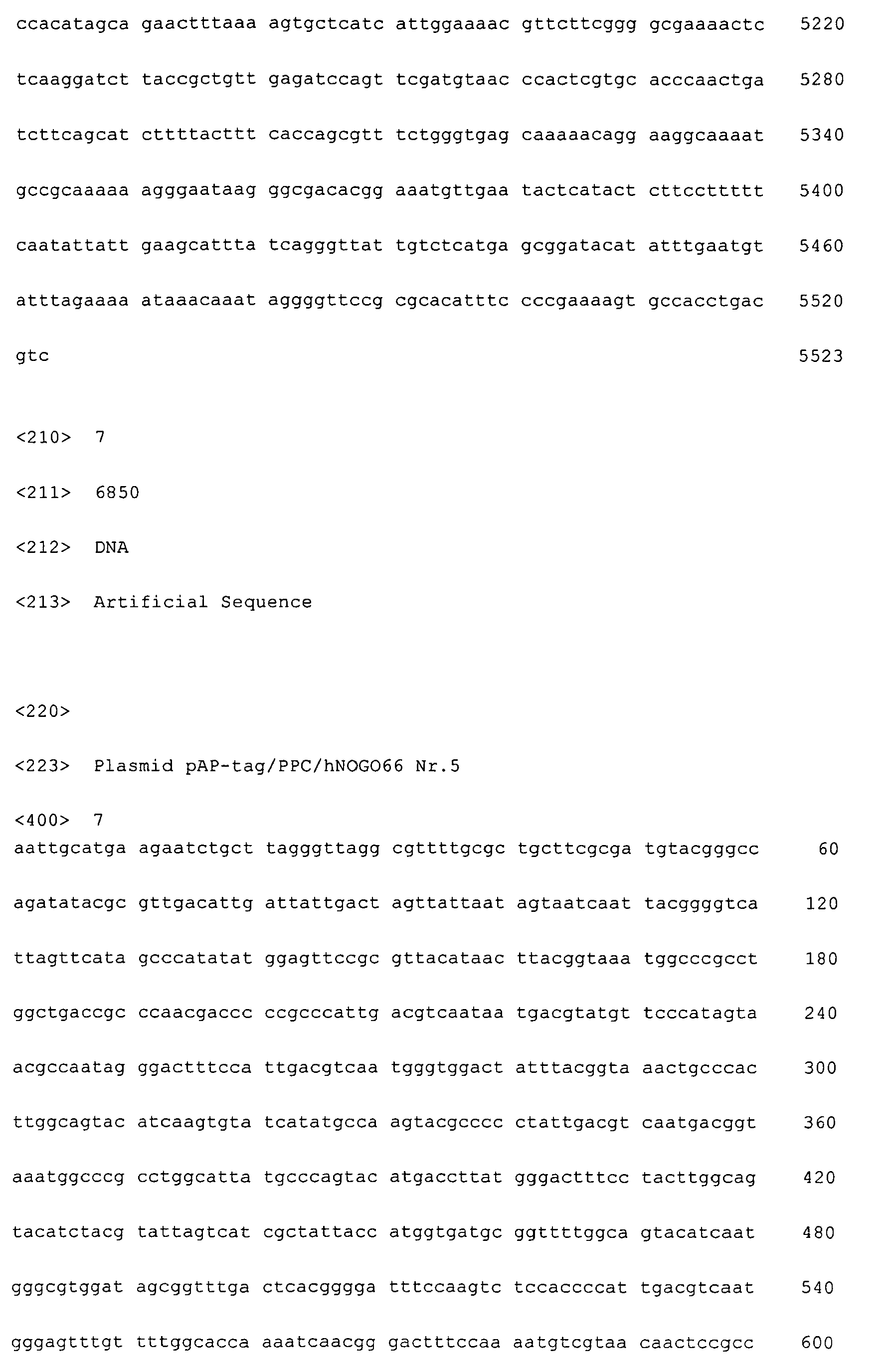


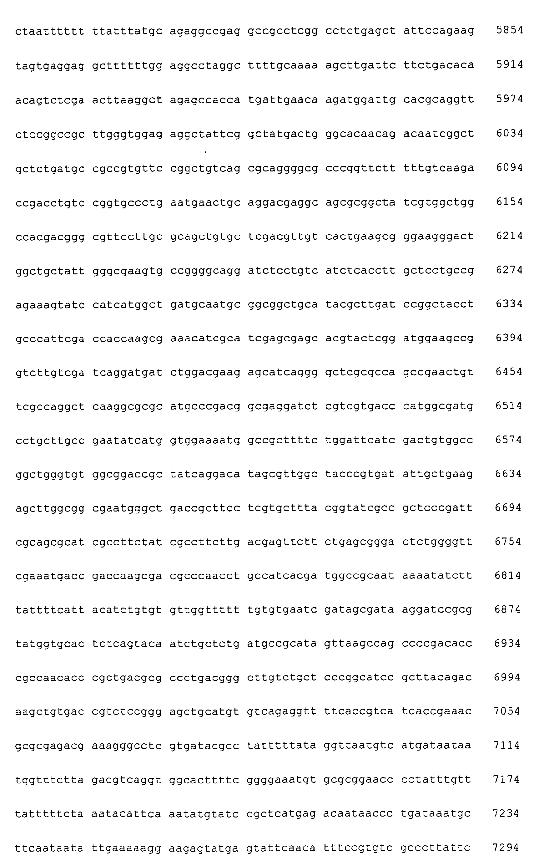


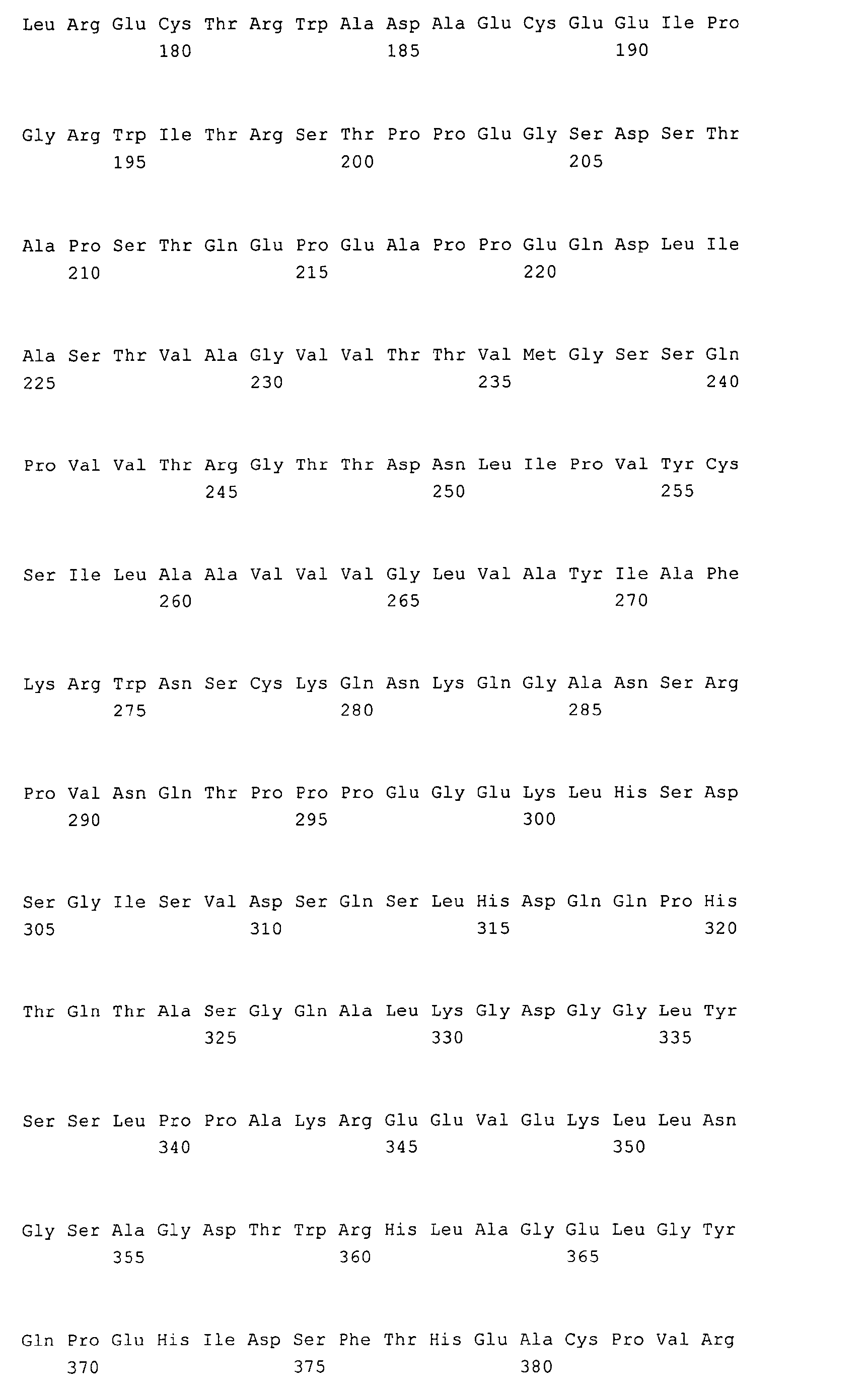


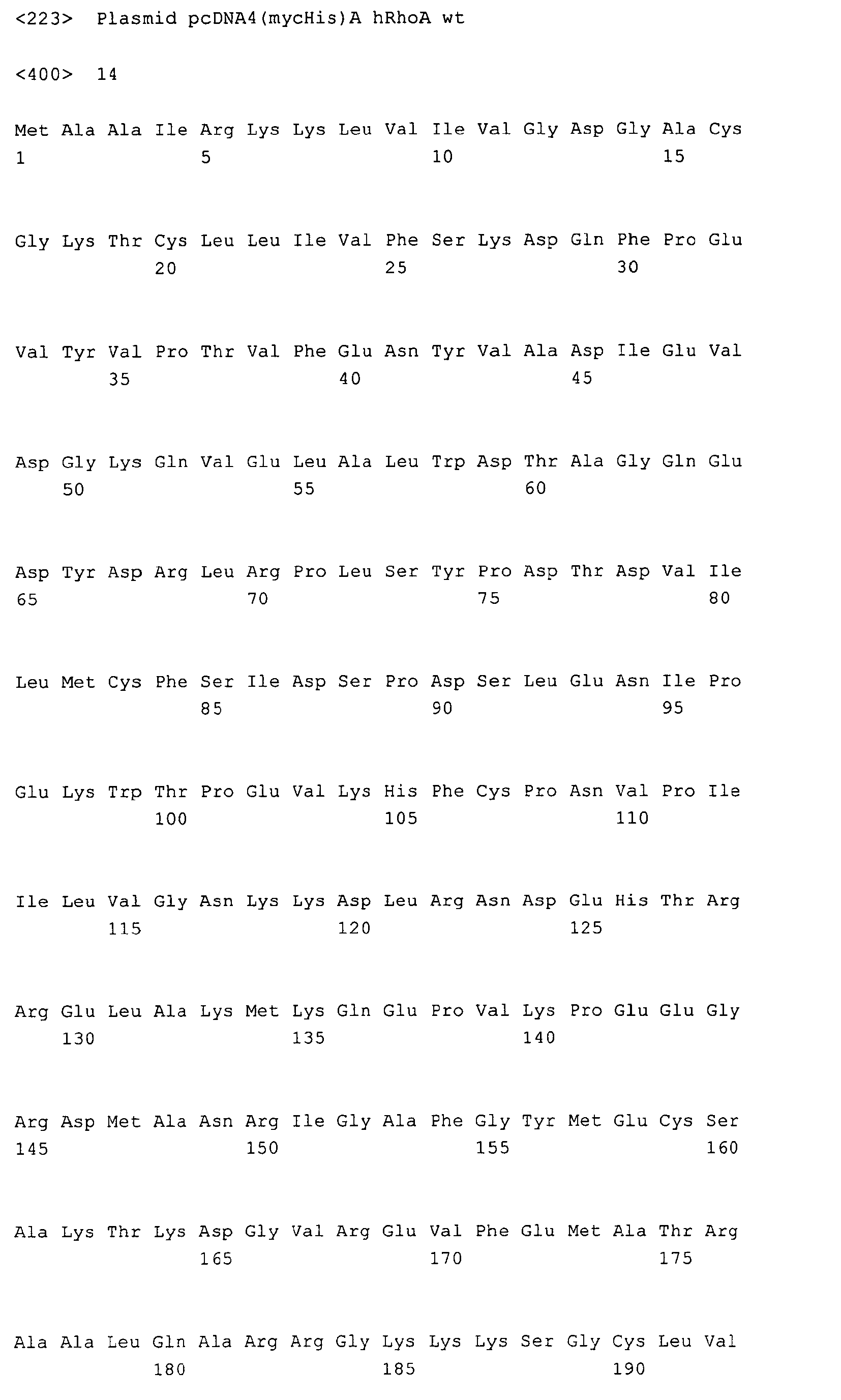


Claims (29)
- Rekombinante Zelllinie, welche exprimiert:a) einen funktionalen intrazellulären Faktor, der an einem eine Veränderung im Cytoskelett der Zelle induzierenden Signalweg beteiligt ist; und / oderb) einen mit dem intrazellulären Faktor wechselwirkenden membrangebundenen Rezeptor.
- Zelllinie nach Anspruch 1, wobei der intrazelluläre Faktor eine Rho GTPase ist, und /oder der membrangebundene Rezeptor einen funktionalen Neurotrophin Rezeptor p75 und einen funktionalen Nogo Rezeptor (NgR) umfasst.
- Zelllinie nach Anspruch 2, wobeia) die funktionale RhoGTPase eine Aminosäuresequenz gemäß SEQ ID NO:16 umfasst;b) der funktionale p75 eine Aminosäuresequenz gemäß SEQ ID NO:9 umfasst; undc) der funktionale NgR eine Aminosäuresequenz gemäß SEQ ID NO:11 umfasst.
- Zelllinie nach einem der vorhergehenden Ansprüche, wobei diese zu einer Reorganisation ihres Aktin-Cytoskeletts, induziert durch die Bindung eines Liganden an NgR befähigt sind.
- Zelllinie nach Anspruch 4, wobei die Ligandenbindung eine Aktivierung von Rho induziert.
- Zelllinie nach Anspruch 4 oder 5, wobei der Ligand Nogo-A oder ein davon abgeleitetes NgR bindendes Fragment oder Derivat umfasst.
- Zelllinie nach einem der vorhergehenden Ansprüche, abgeleitet von einer adhärenten Säugerzelllinie mit ausgespreiteter Zellmorphologie.
- Zelllinie nach Anspruch 7, abgeleitet von einer humanen Epithelial- oder Fibroblastenzelllinie.
- Zelllinie nach Anspruch 8, abgeleitet von einer humanen embryonalen Nieren (HEK)-Zelllinie.
- Verwendung einer Zelllinie nach einem der Ansprüche 1 bis 9 zur Bestimmung von Effektoren der NgR-abhängigen Signaltransduktion.
- Testmethode zur Bestimmung von Veränderungen im Cytoskelett einer zu untersuchenden Zelllinie, wobei man eine rekombinante Zelllinie gemäß der Definition nach einem der Ansprüche 1 bis 9 oder eine korrespondierende Primärzellline mit einem mit den Zellen wechselwirkenden Liganden kultiviert, der gegebenenfalls die Veränderung im Cytoskelett über die Induktion eines intrazellulären Signalwegs bewirkt, und Veränderungen in der Reorganisation des Cytoskeletts der so behandelten Zelllinien bestimmt.
- Testmethode nach Anspruch 11, wobei der Ligand mit einem membrangebunden Rezeptor wechselwirkt.
- Testmethode nach Anspruch 11 oder 12, wobei man die Zelllinie in Gegenwart oder Abwesenheit eines Analyten kultiviert, von dem man vermutet, dass er einen Effektor enthält, der die Wirkung des Liganden auf die Zelle moduliert.
- Testmethode nach Anspruch 13, wobei die Kultivierung mit dem Analyten gleichzeitig oder zeitlich versetzt zur Kultivierung mit dem rezeptorbindenen Liganden erfolgt.
- Testmethode nach einem der Ansprüche 11 bis 14 zur Bestimmung von Effektoren der NgR-abhängigen Aktivierung der GTPase Rho, wobei man die rekombinante Zelllinie oder eine korrespondierende Primärzellline in Gegenwart eines NgR bindenden Liganden und in Gegenwart oder Abwesenheit eines Analyten kultiviert, von dem man vermutet, dass er den Effektor enthält, und Unterschiede in der Reorganisation des Cytoskeletts der so behandelten Zelllinien bestimmt.
- Testmethode nach einem der Ansprüche 11 bis 15, wobei man den Mittelwert wenigstens eines der folgenden Parameter für eine repräsentative Zellpopulation bestimmt:a) Zellumfangb) Zelllängec) Zellbreited) Zellflächee) Länge der Zellausläuferf) Zelldichteg) Cytoplasmatische Flächeh) cytoplasmic verticiesi) Brechungsindexj) zellfreie Flächek) mit Zellen belegte FlächeI) eine mathematische Verknüpfung von wenigstens zwei der Parameter a) bis k), oder einen von wenigstens einem der Parameter a) bis k) ableitbaren Parameter, wie insbesondere das Verhältnis von Zelllänge zu Zellbreite, oder das Verhältnis von Zellumfang im Quadrat zu Zellfläche; oderm) einen entsprechenden reziproken Wert eines der Parameter a) bis I).
- Testmethode nach einem der Ansprüche 11 bis 16, wobei die Bestimmung mittels eines automatisierten Verfahrens, wie insbesondere mittels eines automatischen Bildanalysesystems, erfolgt.
- Verwendung eines Testverfahrens nach einem der Ansprüche 11 bis 17 zur Bestimmung von Effektoren der neuronalen Regeneration.
- Verwendung nach Anspruch 18, wobei der Effektor die Rho-Aktivierung teilweise oder vollständig inhibiert.
- Verwendung nach einem der Ansprüche 18 und 19, wobei der Effektor die neuronale Regeneration fördert.
- Verwendung nach Anspruch 10, wobei der Effektor eine krankheits- oder verletzungsbedingte Inhibition der axonalen Regeneration oder einen krankheits- oder verletzungsbedingten Kollaps des neuronalen Wachstumskegels teilweise oder vollständig neutralisiert.
- Verwendung nach einem der Ansprüche 18 bis 21, wobei der Effektor ein NgR-Antagonist ist.
- Verwendung nach einem der Ansprüche 18 bis 22 in einem Screeningverfahren, insbesondere high-content-Screeningverfahren für Effektoren der neuronalen Regeneration.
- Vektor, umfassend eine Nukleinsäuresequenz, kodierend für funktionale RhoGTPase oder umfassend Nukleinsäuresequenzen, kodierend für funktionalen p75 und funktionalen NgR.
- Verfahren zur Herstellung einer rekombinanten Zelllinie nach einem der Ansprüche 1 bis 9, wobei man eine Wildtyp-Zelllinie mit wenigstens einer kodierenden Nukleinsäuresequenz transformiert, welche für eine funktionale Rho GTPase und gegebenenfalls für p75 und NgR kodiert.
- Verfahren nach Anspruch 25, wobei man die Zelllinie mit wenigstens einem Vektor nach Anspruch 24 transformiert.
- High Content Screening-Vefahren zur Bestimmung von Veränderungen im Cytoskelett einer zu untersuchenden Zelllinie und insbesondere zur Bestimmung von Effektoren der NgR-abhängigen Aktivierung der GTPase Rho, wobei man eine Testmethode nach einem der Ansprüche 11 bis 17 anwendet.
- Vorrichtung zur Durchführung eines Verfahrens nach Anspruch 27.
- Testkit, umfassend eine rekombinante Zelllinie nach einem der Ansprüche 1 bis 9 oder eine korrespondierende Primärzelllinie sowie einen Liganden für den zellulären Rezeptor.
Applications Claiming Priority (2)
| Application Number | Priority Date | Filing Date | Title |
|---|---|---|---|
| US64431205P | 2005-01-14 | 2005-01-14 | |
| DE102005001885 | 2005-01-14 |
Publications (2)
| Publication Number | Publication Date |
|---|---|
| EP1681565A1 true EP1681565A1 (de) | 2006-07-19 |
| EP1681565B1 EP1681565B1 (de) | 2011-07-06 |
Family
ID=36282879
Family Applications (1)
| Application Number | Title | Priority Date | Filing Date |
|---|---|---|---|
| EP20060000726 Expired - Lifetime EP1681565B1 (de) | 2005-01-14 | 2006-01-13 | Zellulärer RhoGTPase Aktivierungs-Assay |
Country Status (1)
| Country | Link |
|---|---|
| EP (1) | EP1681565B1 (de) |
Citations (2)
| Publication number | Priority date | Publication date | Assignee | Title |
|---|---|---|---|---|
| WO2004085648A2 (en) * | 2003-03-19 | 2004-10-07 | Biogen Idec Ma Inc. | Nogo receptor binding protein |
| WO2004087744A2 (en) * | 2003-03-28 | 2004-10-14 | Intellectual Property Consulting Inc. | Composition and method for nerve regeneration |
-
2006
- 2006-01-13 EP EP20060000726 patent/EP1681565B1/de not_active Expired - Lifetime
Patent Citations (2)
| Publication number | Priority date | Publication date | Assignee | Title |
|---|---|---|---|---|
| WO2004085648A2 (en) * | 2003-03-19 | 2004-10-07 | Biogen Idec Ma Inc. | Nogo receptor binding protein |
| WO2004087744A2 (en) * | 2003-03-28 | 2004-10-14 | Intellectual Property Consulting Inc. | Composition and method for nerve regeneration |
Non-Patent Citations (14)
| Title |
|---|
| DATABASE UniProt [online] 1 June 2001 (2001-06-01), "Reticulon-4 receptor precursor (Nogo receptor) (NgR) (Nogo-66 receptor).", XP002381774, retrieved from EBI accession no. UNIPROT:Q9BZR6 Database accession no. Q9BZR6 * |
| FOURNIER A ET AL: "Identification of a receptor mediating Nogo-66 inhibition of axonal regeneration", NATURE, NATURE PUBLISHING GROUP, LONDON, GB, vol. 409, no. 6818, 18 January 2001 (2001-01-18), pages 341 - 346, XP000926532, ISSN: 0028-0836 * |
| HU F ET AL: "Regulating axon growth within the postnatal central nervous system", SEMINARS IN PERINATOLOGY, W.B. SAUNDERS, GB, vol. 28, no. 6, December 2004 (2004-12-01), pages 371 - 378, XP004689705, ISSN: 0146-0005 * |
| HUNT D ET AL: "The Nogo receptor, its ligands and axonal regeneration in the spinal cord; a review", JOURNAL OF NEUROCYTOLOGY, CHAPMAN AND HALL, GB, vol. 31, no. 2, February 2002 (2002-02-01), pages 93 - 120, XP002321234, ISSN: 0300-4864 * |
| MCGEE A W ET AL: "The Nogo-66 receptor: focusing myelin inhibition of axon regeneration", TRENDS IN NEUROSCIENCE, ELSEVIER, AMSTERDAM, NL, vol. 26, no. 4, April 2003 (2003-04-01), pages 193 - 198, XP004418152, ISSN: 0166-2236 * |
| MI S ET AL: "LINGO-1 IS A COMPONENT OF THE NOGO-66 RECEPTOR/P75 SIGNALING COMPLEX", NATURE NEUROSCIENCE, NATURE AMERICA, INC, US, vol. 7, no. 3, March 2004 (2004-03-01), pages 221 - 228, XP001183275, ISSN: 1097-6256 * |
| MI, S. ET AL., NATURE NEUROSCIENCE, vol. 7, no. 3, 2004, pages 221 - 228 |
| N. TEUSCH: "Die Funktion von RhoGTPasen in den von Toll-Rezeptoren induzierten Signaltransduktionswegen", DISSERTATION, 2002 |
| SCHWEIGREITER R ET AL: "Versican V2 and the central inhibitory domain of Nogo-A inhibit neurite growth via p75<NTR>/NgR-independent pathways that converge at RhoA", MOLECULAR AND CELLULAR NEUROSCIENCES, SAN DIEGO, US, vol. 27, no. 2, 1 October 2004 (2004-10-01), pages 163 - 174, XP004599339, ISSN: 1044-7431 * |
| WANG K C ET AL: "P75 INTERACTS WITH THE NOGO RECEPTOR AS A CO-RECEPTOR FOR NOGO, MAG AND OMGP", NATURE, NATURE PUBLISHING GROUP, LONDON, GB, vol. 420, no. 6911, 7 November 2002 (2002-11-07), pages 74 - 78, XP001183135, ISSN: 0028-0836 * |
| WANG, K.C. ET AL., NATURE, vol. 420, 2002, pages 74 - 78 |
| YAMASHITA TOSHIHIDE ET AL: "Neurotrophin binding to the p75 receptor modulates Rho activity and axonal outgrowth", NEURON, vol. 24, no. 3, November 1999 (1999-11-01), pages 585 - 593, XP002381427, ISSN: 0896-6273 * |
| YAMASHITA TOSHIHIDE ET AL: "The p75 receptor transduces the signal from myelin-associated glycoprotein to Rho", THE JOURNAL OF CELL BIOLOGY, ROCKEFELLER UNIVERSITY PRESS, US, vol. 157, no. 4, 13 May 2002 (2002-05-13), pages 565 - 570, XP002295646, ISSN: 0021-9525 * |
| YAMASHITA, T. ET AL., NEURON, vol. 24, 1999, pages 585 - 593 |
Also Published As
| Publication number | Publication date |
|---|---|
| EP1681565B1 (de) | 2011-07-06 |
Similar Documents
| Publication | Publication Date | Title |
|---|---|---|
| KR100643622B1 (ko) | 감각 정보 전달과 관련된 g-단백질 커플링된 수용체를암호화하는 핵산 | |
| KR100602857B1 (ko) | 감각 정보 전달과 관련된 g-단백질 커플링된 수용체를암호화하는 핵산 | |
| DE69431432T2 (de) | Untereinheiten von menschlichen n-methyl-d-aspartat rezeptoren, diese kodierende nukleinsäure und dessen verwendung | |
| Xie et al. | Phosphatidylinositol transfer protein-α in netrin-1-induced PLC signalling and neurite outgrowth | |
| DE69434166T2 (de) | Menschliche metabotropische glutamatrezeptor untertype hmglur7 und verwandte dns-verbindungen | |
| Callaerts et al. | Drosophila Pax‐6/eyeless is essential for normal adult brain structure and function | |
| MacNulty et al. | Alpha 2-C10 adrenergic receptors expressed in rat 1 fibroblasts can regulate both adenylylcyclase and phospholipase D-mediated hydrolysis of phosphatidylcholine by interacting with pertussis toxin-sensitive guanine nucleotide-binding proteins. | |
| DE60207254T2 (de) | Natürlicher ligand von gpcr chemr23 und dessen verwendung | |
| Hill et al. | Characterisation of recombinant rat TRPM2 and a TRPM2-like conductance in cultured rat striatal neurones | |
| DE69808743T2 (de) | Netrinrezeptoren | |
| Suwa et al. | Distribution of serotonin 4 (a) receptors in the juvenile rat brain and spinal cord | |
| DE19957065B4 (de) | Screening-Verfahren für Arzneistoffe | |
| Malmqvist et al. | Tau mRNA is present in axonal RNA granules and is associated with elongation factor 1A | |
| Liang et al. | LncRNA TubAR complexes with TUBB4A and TUBA1A to promote microtubule assembly and maintain myelination | |
| DE69533044T2 (de) | Für den Typ II-morphogenetischen-Knochenprotein-Rezeptor kodierte cDNS | |
| Fischer et al. | Conserved lysin and arginin residues in the extracellular loop of P2X3 receptors are involved in agonist binding | |
| EP1681565B1 (de) | Zellulärer RhoGTPase Aktivierungs-Assay | |
| Liu et al. | Nogo-A inhibits necdin-accelerated neurite outgrowth by retaining necdin in the cytoplasm | |
| DE69832156T2 (de) | Zusammensetzungen des synaptischen Aktivierungsproteins und Verfahren | |
| Fujiwara et al. | Microtubule association of a neuronal RNA-binding protein HuD through its binding to the light chain of MAP1B | |
| EP1436327B1 (de) | Ee3-proteinfamilie und zugrundeliegende dna-sequenzen | |
| Schulz et al. | Translocation of phosducin in living neuroblastoma× glioma hybrid cells (NG 108-15) monitored by red-shifted green fluorescent protein | |
| DE60113660T2 (de) | G-protein gekoppelter rezeptor | |
| DE60311097T2 (de) | Promotor sequenz des menschlichen 5-ht7 rezeptors | |
| EP1455190B1 (de) | Verfahren zur Bestimmung des hormonellen Effekts von Substanzen mittels der Interaktion zwischen dem Androgenrezeptor und Ewing Sarkoma Protein. |
Legal Events
| Date | Code | Title | Description |
|---|---|---|---|
| PUAI | Public reference made under article 153(3) epc to a published international application that has entered the european phase |
Free format text: ORIGINAL CODE: 0009012 |
|
| AK | Designated contracting states |
Kind code of ref document: A1 Designated state(s): AT BE BG CH CY CZ DE DK EE ES FI FR GB GR HU IE IS IT LI LT LU LV MC NL PL PT RO SE SI SK TR |
|
| AX | Request for extension of the european patent |
Extension state: AL BA HR MK YU |
|
| 17P | Request for examination filed |
Effective date: 20070119 |
|
| AKX | Designation fees paid |
Designated state(s): AT BE BG CH CY CZ DE DK EE ES FI FR GB GR HU IE IS IT LI LT LU LV MC NL PL PT RO SE SI SK TR |
|
| AXX | Extension fees paid |
Extension state: HR Payment date: 20070119 |
|
| 17Q | First examination report despatched |
Effective date: 20070627 |
|
| GRAP | Despatch of communication of intention to grant a patent |
Free format text: ORIGINAL CODE: EPIDOSNIGR1 |
|
| GRAS | Grant fee paid |
Free format text: ORIGINAL CODE: EPIDOSNIGR3 |
|
| GRAA | (expected) grant |
Free format text: ORIGINAL CODE: 0009210 |
|
| AK | Designated contracting states |
Kind code of ref document: B1 Designated state(s): AT BE BG CH CY CZ DE DK EE ES FI FR GB GR HU IE IS IT LI LT LU LV MC NL PL PT RO SE SI SK TR |
|
| AX | Request for extension of the european patent |
Extension state: HR |
|
| REG | Reference to a national code |
Ref country code: GB Ref legal event code: FG4D Free format text: NOT ENGLISH |
|
| REG | Reference to a national code |
Ref country code: CH Ref legal event code: EP |
|
| REG | Reference to a national code |
Ref country code: IE Ref legal event code: FG4D Free format text: LANGUAGE OF EP DOCUMENT: GERMAN |
|
| REG | Reference to a national code |
Ref country code: DE Ref legal event code: R096 Ref document number: 502006009775 Country of ref document: DE Effective date: 20110901 |
|
| REG | Reference to a national code |
Ref country code: NL Ref legal event code: VDEP Effective date: 20110706 |
|
| PG25 | Lapsed in a contracting state [announced via postgrant information from national office to epo] |
Ref country code: SI Free format text: LAPSE BECAUSE OF FAILURE TO SUBMIT A TRANSLATION OF THE DESCRIPTION OR TO PAY THE FEE WITHIN THE PRESCRIBED TIME-LIMIT Effective date: 20110706 |
|
| PG25 | Lapsed in a contracting state [announced via postgrant information from national office to epo] |
Ref country code: IS Free format text: LAPSE BECAUSE OF FAILURE TO SUBMIT A TRANSLATION OF THE DESCRIPTION OR TO PAY THE FEE WITHIN THE PRESCRIBED TIME-LIMIT Effective date: 20111106 Ref country code: NL Free format text: LAPSE BECAUSE OF FAILURE TO SUBMIT A TRANSLATION OF THE DESCRIPTION OR TO PAY THE FEE WITHIN THE PRESCRIBED TIME-LIMIT Effective date: 20110706 Ref country code: SE Free format text: LAPSE BECAUSE OF FAILURE TO SUBMIT A TRANSLATION OF THE DESCRIPTION OR TO PAY THE FEE WITHIN THE PRESCRIBED TIME-LIMIT Effective date: 20110706 Ref country code: LT Free format text: LAPSE BECAUSE OF FAILURE TO SUBMIT A TRANSLATION OF THE DESCRIPTION OR TO PAY THE FEE WITHIN THE PRESCRIBED TIME-LIMIT Effective date: 20110706 Ref country code: FI Free format text: LAPSE BECAUSE OF FAILURE TO SUBMIT A TRANSLATION OF THE DESCRIPTION OR TO PAY THE FEE WITHIN THE PRESCRIBED TIME-LIMIT Effective date: 20110706 Ref country code: PT Free format text: LAPSE BECAUSE OF FAILURE TO SUBMIT A TRANSLATION OF THE DESCRIPTION OR TO PAY THE FEE WITHIN THE PRESCRIBED TIME-LIMIT Effective date: 20111107 |
|
| REG | Reference to a national code |
Ref country code: IE Ref legal event code: FD4D |
|
| PG25 | Lapsed in a contracting state [announced via postgrant information from national office to epo] |
Ref country code: PL Free format text: LAPSE BECAUSE OF FAILURE TO SUBMIT A TRANSLATION OF THE DESCRIPTION OR TO PAY THE FEE WITHIN THE PRESCRIBED TIME-LIMIT Effective date: 20110706 Ref country code: CY Free format text: LAPSE BECAUSE OF FAILURE TO SUBMIT A TRANSLATION OF THE DESCRIPTION OR TO PAY THE FEE WITHIN THE PRESCRIBED TIME-LIMIT Effective date: 20110706 Ref country code: GR Free format text: LAPSE BECAUSE OF FAILURE TO SUBMIT A TRANSLATION OF THE DESCRIPTION OR TO PAY THE FEE WITHIN THE PRESCRIBED TIME-LIMIT Effective date: 20111007 Ref country code: LV Free format text: LAPSE BECAUSE OF FAILURE TO SUBMIT A TRANSLATION OF THE DESCRIPTION OR TO PAY THE FEE WITHIN THE PRESCRIBED TIME-LIMIT Effective date: 20110706 |
|
| PG25 | Lapsed in a contracting state [announced via postgrant information from national office to epo] |
Ref country code: IE Free format text: LAPSE BECAUSE OF FAILURE TO SUBMIT A TRANSLATION OF THE DESCRIPTION OR TO PAY THE FEE WITHIN THE PRESCRIBED TIME-LIMIT Effective date: 20110706 Ref country code: CZ Free format text: LAPSE BECAUSE OF FAILURE TO SUBMIT A TRANSLATION OF THE DESCRIPTION OR TO PAY THE FEE WITHIN THE PRESCRIBED TIME-LIMIT Effective date: 20110706 Ref country code: SK Free format text: LAPSE BECAUSE OF FAILURE TO SUBMIT A TRANSLATION OF THE DESCRIPTION OR TO PAY THE FEE WITHIN THE PRESCRIBED TIME-LIMIT Effective date: 20110706 |
|
| PLBE | No opposition filed within time limit |
Free format text: ORIGINAL CODE: 0009261 |
|
| STAA | Information on the status of an ep patent application or granted ep patent |
Free format text: STATUS: NO OPPOSITION FILED WITHIN TIME LIMIT |
|
| PG25 | Lapsed in a contracting state [announced via postgrant information from national office to epo] |
Ref country code: EE Free format text: LAPSE BECAUSE OF FAILURE TO SUBMIT A TRANSLATION OF THE DESCRIPTION OR TO PAY THE FEE WITHIN THE PRESCRIBED TIME-LIMIT Effective date: 20110706 Ref country code: IT Free format text: LAPSE BECAUSE OF FAILURE TO SUBMIT A TRANSLATION OF THE DESCRIPTION OR TO PAY THE FEE WITHIN THE PRESCRIBED TIME-LIMIT Effective date: 20110706 Ref country code: RO Free format text: LAPSE BECAUSE OF FAILURE TO SUBMIT A TRANSLATION OF THE DESCRIPTION OR TO PAY THE FEE WITHIN THE PRESCRIBED TIME-LIMIT Effective date: 20110706 |
|
| 26N | No opposition filed |
Effective date: 20120411 |
|
| PG25 | Lapsed in a contracting state [announced via postgrant information from national office to epo] |
Ref country code: DK Free format text: LAPSE BECAUSE OF FAILURE TO SUBMIT A TRANSLATION OF THE DESCRIPTION OR TO PAY THE FEE WITHIN THE PRESCRIBED TIME-LIMIT Effective date: 20110706 |
|
| BERE | Be: lapsed |
Owner name: ABBOTT G.M.B.H. & CO. KG Effective date: 20120131 |
|
| REG | Reference to a national code |
Ref country code: DE Ref legal event code: R097 Ref document number: 502006009775 Country of ref document: DE Effective date: 20120411 |
|
| PG25 | Lapsed in a contracting state [announced via postgrant information from national office to epo] |
Ref country code: MC Free format text: LAPSE BECAUSE OF NON-PAYMENT OF DUE FEES Effective date: 20120131 |
|
| REG | Reference to a national code |
Ref country code: CH Ref legal event code: PL |
|
| GBPC | Gb: european patent ceased through non-payment of renewal fee |
Effective date: 20120113 |
|
| REG | Reference to a national code |
Ref country code: FR Ref legal event code: ST Effective date: 20120928 |
|
| PG25 | Lapsed in a contracting state [announced via postgrant information from national office to epo] |
Ref country code: DE Free format text: LAPSE BECAUSE OF NON-PAYMENT OF DUE FEES Effective date: 20120801 Ref country code: GB Free format text: LAPSE BECAUSE OF NON-PAYMENT OF DUE FEES Effective date: 20120113 Ref country code: CH Free format text: LAPSE BECAUSE OF NON-PAYMENT OF DUE FEES Effective date: 20120131 Ref country code: LI Free format text: LAPSE BECAUSE OF NON-PAYMENT OF DUE FEES Effective date: 20120131 |
|
| REG | Reference to a national code |
Ref country code: DE Ref legal event code: R119 Ref document number: 502006009775 Country of ref document: DE Effective date: 20120801 |
|
| PG25 | Lapsed in a contracting state [announced via postgrant information from national office to epo] |
Ref country code: BE Free format text: LAPSE BECAUSE OF NON-PAYMENT OF DUE FEES Effective date: 20120131 Ref country code: FR Free format text: LAPSE BECAUSE OF NON-PAYMENT OF DUE FEES Effective date: 20120131 |
|
| REG | Reference to a national code |
Ref country code: AT Ref legal event code: MM01 Ref document number: 515696 Country of ref document: AT Kind code of ref document: T Effective date: 20120113 |
|
| PG25 | Lapsed in a contracting state [announced via postgrant information from national office to epo] |
Ref country code: ES Free format text: LAPSE BECAUSE OF FAILURE TO SUBMIT A TRANSLATION OF THE DESCRIPTION OR TO PAY THE FEE WITHIN THE PRESCRIBED TIME-LIMIT Effective date: 20111017 |
|
| PG25 | Lapsed in a contracting state [announced via postgrant information from national office to epo] |
Ref country code: BG Free format text: LAPSE BECAUSE OF FAILURE TO SUBMIT A TRANSLATION OF THE DESCRIPTION OR TO PAY THE FEE WITHIN THE PRESCRIBED TIME-LIMIT Effective date: 20111006 Ref country code: AT Free format text: LAPSE BECAUSE OF NON-PAYMENT OF DUE FEES Effective date: 20120113 |
|
| PG25 | Lapsed in a contracting state [announced via postgrant information from national office to epo] |
Ref country code: TR Free format text: LAPSE BECAUSE OF FAILURE TO SUBMIT A TRANSLATION OF THE DESCRIPTION OR TO PAY THE FEE WITHIN THE PRESCRIBED TIME-LIMIT Effective date: 20110706 |
|
| PG25 | Lapsed in a contracting state [announced via postgrant information from national office to epo] |
Ref country code: LU Free format text: LAPSE BECAUSE OF NON-PAYMENT OF DUE FEES Effective date: 20120113 |
|
| PG25 | Lapsed in a contracting state [announced via postgrant information from national office to epo] |
Ref country code: HU Free format text: LAPSE BECAUSE OF FAILURE TO SUBMIT A TRANSLATION OF THE DESCRIPTION OR TO PAY THE FEE WITHIN THE PRESCRIBED TIME-LIMIT Effective date: 20060113 |