EP1643447B1 - Method and system for CT reconstruction with pre-correction - Google Patents
Method and system for CT reconstruction with pre-correction Download PDFInfo
- Publication number
- EP1643447B1 EP1643447B1 EP05255628A EP05255628A EP1643447B1 EP 1643447 B1 EP1643447 B1 EP 1643447B1 EP 05255628 A EP05255628 A EP 05255628A EP 05255628 A EP05255628 A EP 05255628A EP 1643447 B1 EP1643447 B1 EP 1643447B1
- Authority
- EP
- European Patent Office
- Prior art keywords
- detector
- ray
- correction
- sinogram data
- data
- Prior art date
- Legal status (The legal status is an assumption and is not a legal conclusion. Google has not performed a legal analysis and makes no representation as to the accuracy of the status listed.)
- Expired - Lifetime
Links
Images


Classifications
-
- G—PHYSICS
- G06—COMPUTING OR CALCULATING; COUNTING
- G06T—IMAGE DATA PROCESSING OR GENERATION, IN GENERAL
- G06T5/00—Image enhancement or restoration
- G06T5/10—Image enhancement or restoration using non-spatial domain filtering
-
- G—PHYSICS
- G06—COMPUTING OR CALCULATING; COUNTING
- G06T—IMAGE DATA PROCESSING OR GENERATION, IN GENERAL
- G06T11/00—2D [Two Dimensional] image generation
- G06T11/003—Reconstruction from projections, e.g. tomography
- G06T11/005—Specific pre-processing for tomographic reconstruction, e.g. calibration, source positioning, rebinning, scatter correction, retrospective gating
-
- G—PHYSICS
- G06—COMPUTING OR CALCULATING; COUNTING
- G06T—IMAGE DATA PROCESSING OR GENERATION, IN GENERAL
- G06T11/00—2D [Two Dimensional] image generation
- G06T11/003—Reconstruction from projections, e.g. tomography
- G06T11/006—Inverse problem, transformation from projection-space into object-space, e.g. transform methods, back-projection, algebraic methods
-
- G—PHYSICS
- G06—COMPUTING OR CALCULATING; COUNTING
- G06T—IMAGE DATA PROCESSING OR GENERATION, IN GENERAL
- G06T2211/00—Image generation
- G06T2211/40—Computed tomography
- G06T2211/421—Filtered back projection [FBP]
Definitions
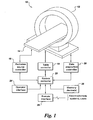
- system controller 22 is also coupled to an operator interface 24 and to one or more memory devices 26.
- the operator interface may be integral with the system controller 22, and will generally include an operator workstation for initiating imaging sequences, controlling such sequences, and manipulating projection data acquired during imaging sequences.
- the memory devices 26 may be local to the imaging system, or may be partially or completely remote from the system. Thus, memory devices 26 may include local, magnetic or optical memory, or local or remote repositories for measured data for reconstruction. Moreover, the memory devices may be configured to receive raw, partially processed or fully processed projection data for reconstruction.
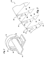
- Fig. 3 is a diagrammatical representation of an exemplary distributed X-ray source of a type that may be employed in the CT system 10 of Fig. 1.
- the distributed X-ray source 30 may include a series of electron beam emitters 32 that are coupled to radiation source controller 16 shown in Fig. 1, and are triggered by the source controller during operation of the scanner.
- the electron beam emitters 32 are positioned adjacent to a distributed target 34.
- the electron beam emitters 32 may emit electron beams 36 toward target 34.
- the target 34 which may, for example, be a tungsten rail or element, emits X-ray radiation, as indicated at reference numeral 38, upon impact of the electron beams.
- X-rays In reflection mode, X-rays are meant to be produced primarily on the same side of the target as where the electrons impact. In transmission mode, X-rays are produced at the opposite side of the target.
- the X-ray beams 38 are directed, then toward a collimator 40, which is generally opaque to the X-ray radiation, but which includes openings or apertures 42.
- the apertures 42 may be fixed in dimension, or may be adjustable. Apertures 42 permit a portion of the X-ray beams to penetrate through the collimator to form collimated beams 44 that will be directed to the imaging volume of the scanner, through the subject of interest, and that will impact detector elements on an opposite side of the scanner.
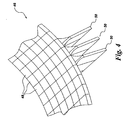
- Fig. 4 is a diagrammatical representation of a portion of a detector that may be employed by the CT system 10 of Fig. 1.
- the detector arrangement may be generally similar to detectors used in conventional rotational CT systems.
- the detector may be extended around a greater portion or the entire inner surface of the scanner in certain embodiments.
- Each detector may be comprised of detector elements with varying resolution to satisfy a particular imaging application.
- the detector 46 includes a series of detector elements 48 and associated signal processing circuitry 50. These detector elements may be of one, two or more sizes, resulting in different spatial resolution characteristics in different portions of the measured projection data.
- Exemplary detector elements include an array of photodiodes and associated thin film transistors.
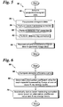
- Fig. 5 is a flowchart 52 depicting exemplary logic including exemplary steps for reconstructing image data from measured sinogram data acquired by the CT system 10 of Fig. 1.
- the projection data measurements are acquired from the CT system 10.
- the term "projection data measurements” also refers to a sinogram.
- the measured sinogram data is pre-processed.
- pre-processing the measured sinogram data in step 56 further includes performing a beam hardening correction on the measured sinogram data in step 58, performing a detector point spread function (PSF) correction in step 60 and performing a detector lag correction in step 62 on the measured sinogram data as will be described in greater detail below.
- PSF detector point spread function
- conventional X-ray sources for CT imaging typically comprise rotating anode tubes (or X-ray tubes) that possess polychromatic spectrums. That is, the X-ray photons emitted from such X-ray tubes do not all possess the same X-ray energy levels.
- attenuation processes in matter are typically energy dependent. Non-uniform attenuation of different energies results in the preferential depletion of X-rays in energy ranges with higher attenuation coefficients.
- X-rays in energy ranges that are more easily attenuated are referred to as soft X-rays while those in ranges that are more penetrating are referred to as hard X-rays.
Landscapes
- Physics & Mathematics (AREA)
- General Physics & Mathematics (AREA)
- Engineering & Computer Science (AREA)
- Theoretical Computer Science (AREA)
- Algebra (AREA)
- Mathematical Analysis (AREA)
- Mathematical Optimization (AREA)
- Mathematical Physics (AREA)
- Pure & Applied Mathematics (AREA)
- Analysing Materials By The Use Of Radiation (AREA)
- Apparatus For Radiation Diagnosis (AREA)
Applications Claiming Priority (1)
| Application Number | Priority Date | Filing Date | Title |
|---|---|---|---|
| US10/955,623 US7215732B2 (en) | 2004-09-30 | 2004-09-30 | Method and system for CT reconstruction with pre-correction |
Publications (2)
| Publication Number | Publication Date |
|---|---|
| EP1643447A1 EP1643447A1 (en) | 2006-04-05 |
| EP1643447B1 true EP1643447B1 (en) | 2007-12-12 |
Family
ID=35447174
Family Applications (1)
| Application Number | Title | Priority Date | Filing Date |
|---|---|---|---|
| EP05255628A Expired - Lifetime EP1643447B1 (en) | 2004-09-30 | 2005-09-14 | Method and system for CT reconstruction with pre-correction |
Country Status (4)
| Country | Link |
|---|---|
| US (1) | US7215732B2 (enExample) |
| EP (1) | EP1643447B1 (enExample) |
| JP (1) | JP4656413B2 (enExample) |
| DE (1) | DE602005003738T2 (enExample) |
Cited By (1)
| Publication number | Priority date | Publication date | Assignee | Title |
|---|---|---|---|---|
| CN101533098B (zh) * | 2009-04-08 | 2011-08-03 | 西北工业大学 | Ct射束硬化校正中的噪声抑制方法 |
Families Citing this family (42)
| Publication number | Priority date | Publication date | Assignee | Title |
|---|---|---|---|---|
| US7208717B2 (en) * | 2002-10-16 | 2007-04-24 | Varian Medical Systems Technologies, Inc. | Method and apparatus for correcting excess signals in an imaging system |
| US7376255B2 (en) * | 2004-06-23 | 2008-05-20 | General Electric Company | System and method for image reconstruction |
| US7251306B2 (en) * | 2004-11-17 | 2007-07-31 | General Electric Company | Methods, apparatus, and software to facilitate iterative reconstruction of images |
| DE102005019572A1 (de) * | 2005-04-27 | 2006-11-09 | Siemens Ag | Verfahren zur Auswertung und Darstellung von Röntgenprojektionsbildern und Röntgendurchsichtgerät |
| DE102005028216A1 (de) * | 2005-06-17 | 2006-12-28 | Siemens Ag | Vorrichtung und Verfahren für die Computertomographie |
| DE102005028225A1 (de) * | 2005-06-17 | 2007-05-24 | Siemens Ag | Vorrichtung und Verfahren für die Computertomographie |
| EP1946622A2 (en) * | 2005-10-06 | 2008-07-23 | Imaging Sciences International, Inc. | Scatter correction |
| US7298812B2 (en) * | 2006-03-31 | 2007-11-20 | General Electric Company | Image-based material decomposition |
| CN101405597B (zh) * | 2006-04-13 | 2012-05-23 | 株式会社岛津制作所 | 使用透过x射线的三维定量方法 |
| JP2008073342A (ja) * | 2006-09-22 | 2008-04-03 | Konica Minolta Medical & Graphic Inc | 放射線画像撮影システム及び放射線画像撮影方法 |
| DE102006046047A1 (de) * | 2006-09-28 | 2008-04-03 | Siemens Ag | Verfahren zur kombinierten Knochenaufhärtungs- und Streustrahlungskorrektur in der Röntgen-Computertomographie |
| US7551708B2 (en) * | 2007-02-07 | 2009-06-23 | General Electric Company | Method of iterative reconstruction for energy discriminating computed tomography systems |
| WO2008103435A1 (en) * | 2007-02-22 | 2008-08-28 | Indiana University Research & Technology Corporation | Imaging resolution recovery techniques |
| US8045776B2 (en) * | 2007-03-06 | 2011-10-25 | General Electric Company | Geometry-dependent filtering in CT method and apparatus |
| JP5076240B2 (ja) * | 2008-02-05 | 2012-11-21 | 富士フイルム株式会社 | 撮像装置、撮像方法、およびプログラム |
| US8660330B2 (en) | 2008-06-27 | 2014-02-25 | Wolfram Jarisch | High efficiency computed tomography with optimized recursions |
| CA2729166A1 (en) * | 2008-06-27 | 2009-12-30 | Wolfram R. Jarisch | High efficiency computed tomography |
| US8194820B2 (en) * | 2009-02-06 | 2012-06-05 | The Board Of Trustees Of The Leland Stanford Junior University | Optimal weights for measuring spectral x-ray data |
| US8655033B2 (en) * | 2009-10-28 | 2014-02-18 | General Electric Company | Iterative reconstruction |
| JP5828841B2 (ja) * | 2010-10-14 | 2015-12-09 | 株式会社日立メディコ | X線ct装置及び画像再構成方法 |
| JP5922892B2 (ja) * | 2011-08-26 | 2016-05-24 | Ntn株式会社 | 転動体の検査方法および転動体の製造方法 |
| EP2792303A4 (en) * | 2011-12-18 | 2015-08-05 | Nat Univ Corp Kyoto Univ | MOTION-TRACKING X-RAY CT IMAGE PROCESSING AND MOTION-TRACKING X-RAY CT IMAGE PROCESSING DEVICE |
| EP2810245A4 (en) | 2012-02-03 | 2015-12-16 | Univ New York State Res Found | METHOD AND SYSTEMS FOR INVERSE PROBLEM RECONSTRUCTION AND APPLICATION FOR ECT RECONSTRUCTION |
| JP5883689B2 (ja) * | 2012-03-14 | 2016-03-15 | 株式会社日立製作所 | X線撮像装置およびx線撮像方法 |
| JP6031339B2 (ja) * | 2012-11-21 | 2016-11-24 | 富士フイルム株式会社 | 透視画像濃度補正方法、非破壊検査方法、及び画像処理装置 |
| KR101527656B1 (ko) * | 2013-12-12 | 2015-06-10 | 한국과학기술원 | 의료 영상에서 비강체 영상 정합을 이용한 점상 강도 분포 함수를 보간하는 방법 및 시스템 |
| US9415546B2 (en) * | 2014-01-29 | 2016-08-16 | Xerox Corporation | System and method for controlling material drop volume in three dimensional object printing |
| US9351697B2 (en) | 2014-03-04 | 2016-05-31 | General Electric Company | Method and system for dimensional analysis of an object |
| KR101783001B1 (ko) * | 2015-01-20 | 2017-09-28 | 삼성전자주식회사 | 단층 영상 장치 및 단층 영상의 이미징 방법 |
| CN104751429B (zh) * | 2015-01-27 | 2018-02-02 | 南方医科大学 | 一种基于字典学习的低剂量能谱ct图像处理方法 |
| CN106353828B (zh) * | 2015-07-22 | 2018-09-21 | 清华大学 | 在安检系统中估算被检查物体重量的方法和装置 |
| US10429323B2 (en) * | 2015-07-24 | 2019-10-01 | Photo Diagnostic Systems, Inc. | Method and apparatus for performing multi-energy (including dual energy) computed tomography (CT) imaging |
| KR101850871B1 (ko) * | 2015-08-26 | 2018-04-23 | 주식회사 디알텍 | 방사선 영상의 처리방법 및 방사선 촬영시스템 |
| KR101717433B1 (ko) * | 2015-09-01 | 2017-03-17 | 연세대학교 산학협력단 | 엑스레이 컴퓨터 단층촬영 환경에서 빔 경화현상에 의한 인공물 보정방법 |
| CN108242066B (zh) * | 2016-12-26 | 2023-04-14 | 通用电气公司 | Ct图像的空间分辨率增强装置和方法以及ct成像系统 |
| US10475214B2 (en) * | 2017-04-05 | 2019-11-12 | General Electric Company | Tomographic reconstruction based on deep learning |
| US10573030B2 (en) | 2017-04-07 | 2020-02-25 | Photo Diagnostic Systems, Inc. | Method for artifact reduction using monoenergetic data in computed tomography |
| US10426424B2 (en) | 2017-11-21 | 2019-10-01 | General Electric Company | System and method for generating and performing imaging protocol simulations |
| DE102018103714A1 (de) * | 2018-02-20 | 2019-08-22 | Volume Graphics Gmbh | Verfahren zur Bestimmung von Fehlern von aus digitalen Objektdarstellungen abgeleiteten Parametern |
| EP3571997B1 (de) | 2018-05-23 | 2022-11-23 | Siemens Healthcare GmbH | Verfahren und vorrichtung zum bestimmen eines patientenge-wichts und/oder eines body-mass-index |
| JP7220777B2 (ja) * | 2019-03-29 | 2023-02-10 | 富士フイルム株式会社 | 画像処理装置、放射線画像撮影システム、画像処理方法、及び画像処理プログラム |
| CN114152637B (zh) * | 2022-02-07 | 2022-04-26 | 东莞市志橙半导体材料有限公司 | 一种硬质碳化硅材料打孔检测装置与方法 |
Family Cites Families (19)
| Publication number | Priority date | Publication date | Assignee | Title |
|---|---|---|---|---|
| JPS63147440A (ja) * | 1986-12-12 | 1988-06-20 | 横河メディカルシステム株式会社 | X線断層撮影装置のデコンボリユ−シヨン処理方法 |
| DE69129008T2 (de) * | 1990-07-02 | 1998-08-20 | Varian Associates | Röntgenstrahlentherapiesimulator |
| US5099505A (en) * | 1990-07-02 | 1992-03-24 | Varian Associates | Method for increasing the accuracy of a radiation therapy apparatus |
| US5265013A (en) | 1990-11-19 | 1993-11-23 | General Electric Company | Compensation of computed tomography data for X-ray detector afterglow artifacts |
| US5359638A (en) * | 1992-03-30 | 1994-10-25 | General Electric Company | Method for recursive filtering residual afterglow from previous computed tomography scans |
| US5878108A (en) * | 1995-11-30 | 1999-03-02 | Hitachi Medical Corporation | Method for generating X-ray image and apparatus therefor |
| US5774521A (en) * | 1996-07-08 | 1998-06-30 | Cedars-Sinai Medical Center | Regularization technique for densitometric correction |
| US6067342A (en) * | 1997-10-30 | 2000-05-23 | Analogic Corporation | Digital filmless X-ray projection imaging system and method |
| US6928182B1 (en) * | 1998-10-15 | 2005-08-09 | Kui Ming Chui | Imaging |
| US6285799B1 (en) * | 1998-12-15 | 2001-09-04 | Xerox Corporation | Apparatus and method for measuring a two-dimensional point spread function of a digital image acquisition system |
| US6345113B1 (en) * | 1999-01-12 | 2002-02-05 | Analogic Corporation | Apparatus and method for processing object data in computed tomography data using object projections |
| EP1072861B1 (en) | 1999-05-10 | 2006-05-10 | GE Inspection Technologies GmbH | Method for measuring the wall thickness of a tubular object |
| US6421409B1 (en) | 2000-02-02 | 2002-07-16 | Ut-Battelle Llc | Ultra-high resolution computed tomography imaging |
| US6842502B2 (en) * | 2000-02-18 | 2005-01-11 | Dilliam Beaumont Hospital | Cone beam computed tomography with a flat panel imager |
| JP3837495B2 (ja) * | 2002-02-28 | 2006-10-25 | 独立行政法人産業技術総合研究所 | 光イメージングシステム |
| US6850589B2 (en) * | 2002-03-27 | 2005-02-01 | Agilent Technologies, Inc. | Tomography of curved surfaces |
| US7254209B2 (en) * | 2003-11-17 | 2007-08-07 | General Electric Company | Iterative CT reconstruction method using multi-modal edge information |
| US7376255B2 (en) * | 2004-06-23 | 2008-05-20 | General Electric Company | System and method for image reconstruction |
| US7203267B2 (en) * | 2004-06-30 | 2007-04-10 | General Electric Company | System and method for boundary estimation using CT metrology |
-
2004
- 2004-09-30 US US10/955,623 patent/US7215732B2/en not_active Expired - Fee Related
-
2005
- 2005-09-14 DE DE602005003738T patent/DE602005003738T2/de not_active Expired - Lifetime
- 2005-09-14 EP EP05255628A patent/EP1643447B1/en not_active Expired - Lifetime
- 2005-09-22 JP JP2005274871A patent/JP4656413B2/ja not_active Expired - Fee Related
Cited By (1)
| Publication number | Priority date | Publication date | Assignee | Title |
|---|---|---|---|---|
| CN101533098B (zh) * | 2009-04-08 | 2011-08-03 | 西北工业大学 | Ct射束硬化校正中的噪声抑制方法 |
Also Published As
| Publication number | Publication date |
|---|---|
| EP1643447A1 (en) | 2006-04-05 |
| US7215732B2 (en) | 2007-05-08 |
| DE602005003738D1 (de) | 2008-01-24 |
| JP4656413B2 (ja) | 2011-03-23 |
| JP2006105975A (ja) | 2006-04-20 |
| US20060067461A1 (en) | 2006-03-30 |
| DE602005003738T2 (de) | 2008-12-04 |
Similar Documents
| Publication | Publication Date | Title |
|---|---|---|
| EP1643447B1 (en) | Method and system for CT reconstruction with pre-correction | |
| CN101138501B (zh) | 用于生成目标的多光谱图像的方法和系统 | |
| US7376255B2 (en) | System and method for image reconstruction | |
| Lasio et al. | Statistical reconstruction for x-ray computed tomography using energy-integrating detectors | |
| US7386088B2 (en) | Method and system for iterative image reconstruction | |
| US8885903B2 (en) | Method and apparatus for statistical iterative reconstruction | |
| US10902650B2 (en) | X-ray beam-hardening correction in tomographic reconstruction using the Alvarez-Macovski attenuation model | |
| US7362843B2 (en) | System and method for reconstruction of cone beam tomographic projections with missing data | |
| Reiter et al. | SimCT: a simulation tool for X-ray imaging | |
| US20140218362A1 (en) | Monte carlo modeling of field angle-dependent spectra for radiographic imaging systems | |
| US20080165920A1 (en) | Method and apparatus for reduction of metal artifacts in ct imaging | |
| EP2652709B1 (en) | Imaging system for imaging a region of interest | |
| Wiegert et al. | Model based scatter correction for cone-beam computed tomography | |
| Banjak | X-ray computed tomography reconstruction on non-standard trajectories for robotized inspection | |
| JP2006231058A (ja) | X線装置において検査対象の作成された画像からビームハードニングアーチファクトを低減する方法およびコンピュータ断層撮影装置 | |
| Stolfi et al. | Error sources | |
| JPS61269046A (ja) | Ct装置 | |
| US7519143B2 (en) | Method and system for generating a scatter corrected X-ray image | |
| US12131409B2 (en) | Calibration method for a spectral computerized tomography system | |
| US12076173B2 (en) | System and method for controlling errors in computed tomography number | |
| US7801266B2 (en) | Method for speeding up the scattered radiation correction in a computed tomography system | |
| US20110141111A1 (en) | 3d reconstruction from oversampled 2d projections | |
| US20070058771A1 (en) | Systems and methods for filtering data in a medical imaging system | |
| Alsaffar et al. | Multi-Material Blind Beam Hardening Correction Based on Non-Linearity Adjustment of Projections | |
| Flisch et al. | Correction of scattered radiation for cone-beam computed tomography at high X-ray energies |
Legal Events
| Date | Code | Title | Description |
|---|---|---|---|
| PUAI | Public reference made under article 153(3) epc to a published international application that has entered the european phase |
Free format text: ORIGINAL CODE: 0009012 |
|
| AK | Designated contracting states |
Kind code of ref document: A1 Designated state(s): AT BE BG CH CY CZ DE DK EE ES FI FR GB GR HU IE IS IT LI LT LU LV MC NL PL PT RO SE SI SK TR |
|
| AX | Request for extension of the european patent |
Extension state: AL BA HR MK YU |
|
| 17P | Request for examination filed |
Effective date: 20061005 |
|
| 17Q | First examination report despatched |
Effective date: 20061108 |
|
| AKX | Designation fees paid |
Designated state(s): DE FR GB |
|
| GRAP | Despatch of communication of intention to grant a patent |
Free format text: ORIGINAL CODE: EPIDOSNIGR1 |
|
| GRAS | Grant fee paid |
Free format text: ORIGINAL CODE: EPIDOSNIGR3 |
|
| GRAA | (expected) grant |
Free format text: ORIGINAL CODE: 0009210 |
|
| AK | Designated contracting states |
Kind code of ref document: B1 Designated state(s): DE FR GB |
|
| REG | Reference to a national code |
Ref country code: GB Ref legal event code: FG4D |
|
| REF | Corresponds to: |
Ref document number: 602005003738 Country of ref document: DE Date of ref document: 20080124 Kind code of ref document: P |
|
| ET | Fr: translation filed | ||
| PLBE | No opposition filed within time limit |
Free format text: ORIGINAL CODE: 0009261 |
|
| STAA | Information on the status of an ep patent application or granted ep patent |
Free format text: STATUS: NO OPPOSITION FILED WITHIN TIME LIMIT |
|
| 26N | No opposition filed |
Effective date: 20080915 |
|
| PGFP | Annual fee paid to national office [announced via postgrant information from national office to epo] |
Ref country code: DE Payment date: 20130927 Year of fee payment: 9 |
|
| PGFP | Annual fee paid to national office [announced via postgrant information from national office to epo] |
Ref country code: FR Payment date: 20130919 Year of fee payment: 9 Ref country code: GB Payment date: 20130927 Year of fee payment: 9 |
|
| REG | Reference to a national code |
Ref country code: DE Ref legal event code: R119 Ref document number: 602005003738 Country of ref document: DE |
|
| GBPC | Gb: european patent ceased through non-payment of renewal fee |
Effective date: 20140914 |
|
| REG | Reference to a national code |
Ref country code: FR Ref legal event code: ST Effective date: 20150529 |
|
| PG25 | Lapsed in a contracting state [announced via postgrant information from national office to epo] |
Ref country code: GB Free format text: LAPSE BECAUSE OF NON-PAYMENT OF DUE FEES Effective date: 20140914 Ref country code: DE Free format text: LAPSE BECAUSE OF NON-PAYMENT OF DUE FEES Effective date: 20150401 |
|
| PG25 | Lapsed in a contracting state [announced via postgrant information from national office to epo] |
Ref country code: FR Free format text: LAPSE BECAUSE OF NON-PAYMENT OF DUE FEES Effective date: 20140930 |