EP1598019A2 - Behandungsvorrichtung für lebendes Gewebe - Google Patents
Behandungsvorrichtung für lebendes Gewebe Download PDFInfo
- Publication number
- EP1598019A2 EP1598019A2 EP05010979A EP05010979A EP1598019A2 EP 1598019 A2 EP1598019 A2 EP 1598019A2 EP 05010979 A EP05010979 A EP 05010979A EP 05010979 A EP05010979 A EP 05010979A EP 1598019 A2 EP1598019 A2 EP 1598019A2
- Authority
- EP
- European Patent Office
- Prior art keywords
- stopper
- ligation
- ligating
- ligating member
- ligature
- Prior art date
- Legal status (The legal status is an assumption and is not a legal conclusion. Google has not performed a legal analysis and makes no representation as to the accuracy of the status listed.)
- Withdrawn
Links
Images


Classifications
-
- A—HUMAN NECESSITIES
- A61—MEDICAL OR VETERINARY SCIENCE; HYGIENE
- A61B—DIAGNOSIS; SURGERY; IDENTIFICATION
- A61B17/00—Surgical instruments, devices or methods
- A61B17/12—Surgical instruments, devices or methods for ligaturing or otherwise compressing tubular parts of the body, e.g. blood vessels or umbilical cord
- A61B17/12009—Implements for ligaturing other than by clamps or clips, e.g. using a loop with a slip knot
- A61B17/12013—Implements for ligaturing other than by clamps or clips, e.g. using a loop with a slip knot for use in minimally invasive surgery, e.g. endoscopic surgery
-
- A—HUMAN NECESSITIES
- A61—MEDICAL OR VETERINARY SCIENCE; HYGIENE
- A61B—DIAGNOSIS; SURGERY; IDENTIFICATION
- A61B17/00—Surgical instruments, devices or methods
- A61B17/04—Surgical instruments, devices or methods for suturing wounds; Holders or packages for needles or suture materials
- A61B17/0487—Suture clamps, clips or locks, e.g. for replacing suture knots; Instruments for applying or removing suture clamps, clips or locks
-
- A—HUMAN NECESSITIES
- A61—MEDICAL OR VETERINARY SCIENCE; HYGIENE
- A61B—DIAGNOSIS; SURGERY; IDENTIFICATION
- A61B17/00—Surgical instruments, devices or methods
- A61B17/04—Surgical instruments, devices or methods for suturing wounds; Holders or packages for needles or suture materials
- A61B17/0467—Instruments for cutting sutures
-
- A—HUMAN NECESSITIES
- A61—MEDICAL OR VETERINARY SCIENCE; HYGIENE
- A61B—DIAGNOSIS; SURGERY; IDENTIFICATION
- A61B17/00—Surgical instruments, devices or methods
- A61B17/04—Surgical instruments, devices or methods for suturing wounds; Holders or packages for needles or suture materials
- A61B17/0401—Suture anchors, buttons or pledgets, i.e. means for attaching sutures to bone, cartilage or soft tissue; Instruments for applying or removing suture anchors
- A61B2017/0404—Buttons
-
- A—HUMAN NECESSITIES
- A61—MEDICAL OR VETERINARY SCIENCE; HYGIENE
- A61B—DIAGNOSIS; SURGERY; IDENTIFICATION
- A61B17/00—Surgical instruments, devices or methods
- A61B17/04—Surgical instruments, devices or methods for suturing wounds; Holders or packages for needles or suture materials
- A61B17/0401—Suture anchors, buttons or pledgets, i.e. means for attaching sutures to bone, cartilage or soft tissue; Instruments for applying or removing suture anchors
- A61B2017/0446—Means for attaching and blocking the suture in the suture anchor
- A61B2017/0448—Additional elements on or within the anchor
- A61B2017/045—Additional elements on or within the anchor snug fit within the anchor
-
- A—HUMAN NECESSITIES
- A61—MEDICAL OR VETERINARY SCIENCE; HYGIENE
- A61B—DIAGNOSIS; SURGERY; IDENTIFICATION
- A61B17/00—Surgical instruments, devices or methods
- A61B17/04—Surgical instruments, devices or methods for suturing wounds; Holders or packages for needles or suture materials
- A61B17/0401—Suture anchors, buttons or pledgets, i.e. means for attaching sutures to bone, cartilage or soft tissue; Instruments for applying or removing suture anchors
- A61B2017/0446—Means for attaching and blocking the suture in the suture anchor
- A61B2017/0458—Longitudinal through hole, e.g. suture blocked by a distal suture knot
-
- A—HUMAN NECESSITIES
- A61—MEDICAL OR VETERINARY SCIENCE; HYGIENE
- A61B—DIAGNOSIS; SURGERY; IDENTIFICATION
- A61B17/00—Surgical instruments, devices or methods
- A61B17/04—Surgical instruments, devices or methods for suturing wounds; Holders or packages for needles or suture materials
- A61B17/0401—Suture anchors, buttons or pledgets, i.e. means for attaching sutures to bone, cartilage or soft tissue; Instruments for applying or removing suture anchors
- A61B2017/0464—Suture anchors, buttons or pledgets, i.e. means for attaching sutures to bone, cartilage or soft tissue; Instruments for applying or removing suture anchors for soft tissue
-
- A—HUMAN NECESSITIES
- A61—MEDICAL OR VETERINARY SCIENCE; HYGIENE
- A61B—DIAGNOSIS; SURGERY; IDENTIFICATION
- A61B17/00—Surgical instruments, devices or methods
- A61B17/04—Surgical instruments, devices or methods for suturing wounds; Holders or packages for needles or suture materials
- A61B17/0487—Suture clamps, clips or locks, e.g. for replacing suture knots; Instruments for applying or removing suture clamps, clips or locks
- A61B2017/049—Instruments for removing suture clamps, clips or locks
-
- A—HUMAN NECESSITIES
- A61—MEDICAL OR VETERINARY SCIENCE; HYGIENE
- A61B—DIAGNOSIS; SURGERY; IDENTIFICATION
- A61B17/00—Surgical instruments, devices or methods
- A61B17/04—Surgical instruments, devices or methods for suturing wounds; Holders or packages for needles or suture materials
- A61B2017/0496—Surgical instruments, devices or methods for suturing wounds; Holders or packages for needles or suture materials for tensioning sutures
Definitions
- the present invention relates to a treatment system for living tissues, which is designed for use in combination with an endoscope and another type of an instrument to perform an endoscopic treatment such as suture and ligation on the living tissues in the body.
- U.S. Patent Application Publication No. 2003/0236535A1 discloses a ligation apparatus for use in endoscopic treatment, i.e., the apparatus that a surgeon uses to suture or ligate the living tissues in the body while observing the tissues through an endoscope.
- the apparatus disclosed in the publication enables the surgeon to suture or ligate the tissues by passing the ligature attached to a holding member, through the tissues.
- the surgeon cuts the ligature, or grasps and removes the holding member from the tissues.
- the tissues may not be sutured or ligated at desired parts.
- the surgeon cuts the ligature to release the tissues from the sutured or ligated state.
- the surgeon uses, for example, surgical scissors, while observing the tissues through an endoscope.
- the surgeon may grasp the holding member with a grasping forceps or the like and then remove the holding member from the tissues, while observing the tissues through the endoscope.
- the present invention has been proposed to solve the above-described drawbacks of the conventional techniques, and an object thereof is to provide a medical treatment device capable of ligating living tissue under endoscopic observation, and capable of releasing ligation of living tissue by a simple operation, as well as a treatment system for living tissue.
- a medical treatment device includes a ligating member, a stopper and a ligation releasing member.
- the ligating member has a distal end portion and a proximal end portion, that ligates biological tissue.
- the stopper is provided to be movable forward or backward with respect to the ligating member, and stoppable by friction on the ligating member to maintain the biological tissue in a ligated state by the ligating member.
- the ligation releasing member is provided on the ligating member to release the ligation state between the ligating member and the stopper by moving the ligating member to the distal end side with respect to the stopper.
- the treatment system 10 for living tissues is a system designed to ligate living tissues.
- the system 10 may be endoscopically used, or in combination with an endoscope, to ligate a polyp or the like before removing it, thereby to control bleeding.
- the system 10 comprises a ligation apparatus 12 and a ligation instrument 14.
- the instrument 14 is used in combination with the ligation apparatus 12.
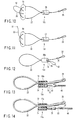
- the ligation apparatus 12 comprises a ligature (ligating member) 22, a silicone tube (stopper) 26, a proximal loop 28, and a flexible wire (ligation-releasing member) 30.
- the proximal loop 28 is formed at the proximal ends of the ligature 22.
- the ligature 22 is provided in the form of a loop. Therefore, its two end portions pass through the silicone tube 26.
- the silicone tube 26 is held, by virtue of a frictional force, on any desired part of the ligature 22.
- the tube 26 can be moved when a force greater than a prescribed value is applied along the ligature 22.
- the silicone tube 26 performs the function of a stopper that holds the ligature 22 at a desired position with respect to the tissue that is to be treated.
- the flexible wire 30 is connected to the ligature 22, at a position close to the distal end of the ligature 22 and deviated from the axis X of the lumen into which the ligature 22 should be inserted.
- One flexible wire is used in this embodiment. Nonetheless, two, three or more flexible wires may be connected to the ligature 22. It is desirable to connect one or two flexible wires to the ligature 22. If too many flexible wires are connected to the ligature 22, they may entangle while the ligation apparatus 12 remains in the body.
- the flexible wire 30 may be replaced by a member of a different shape, such as a band.
- the flexible wire 30 can be made of any material that is flexible and strong enough to withstand a pulling force applied to release the tissue from a ligated state. It may be made of the same material as suture thread, e.g., drawn polyamide synthetic fiber, polypropylene, polyethyleneterephthalate, polytetrafluoroethylene. Alternatively, it may be made of bioabsorbable material such as polyglycolic acid. The wire may be either a monofilament one or a twisted thread.
- the flexible wire 30 is colored not similar to the living tissues, not white, red or yellow, and different in color from the ligature 22. Hence, the surgeon can well distinguish the wire 30 when he or she sees it through the endoscope.
- the ligation instrument 14 comprises a ligation sheath 38, a hook wire 40, and an operation handle (not shown).
- the hook wire 40 is connected, at its distal end, to a hook 40a, and extends through the ligation sheath 38.
- the hook 40a catches the proximal loop 28 of the ligature 22.
- the operation handle is provided at the proximal end of the ligation sheath 38 and that of the hook wire 40.
- the ligation instrument 14 is coupled to the ligation apparatus 12 (see FIG. 2).
- the proximal loop 28 of the ligation apparatus 12 is set into engagement with the hook 40a fastened to the hook wire 40 of the ligation instrument 14.
- the silicone tube 26 of the ligation apparatus 12 is moved until it abuts on the distal end of the ligation sheath 38 of the ligation instrument 14.
- FIG. 3 shows, the looped ligature 22 of the treatment system 10 is rapped around the tissue 60 that is to be treated.
- the silicone tube 26 is pushed at the distal end of the ligation sheath 38. As a result, the silicone tube 26 moves toward the distal end of the ligature 22.
- the ligature 22 squeezes the tissue 60 as the loop of the ligature 22 becomes smaller.
- the tissue 60 is thereby ligated.
- the silicone tube 26 is prevented from moving relative to the ligature 22, by virtue of a frictional force.
- the tissue 60 therefore remains ligated.
- the parts of the ligature 22, which protrude from the proximal end of the silicone tube 26, are cut off.
- an endoscope 50 having a channel 50a that can guide a grasping forceps 48 is used to release the tissue 60 from the ligated state.
- the grasping forceps 48 extending through the channel 50a holds the flexible wire 30.
- the forceps 48 is pulled, thus pulling the flexible wire 30 with a force greater than the frictional force that the silicone tube 26 exerts on the wire 30.
- the flexible wire 30 is connected to the distal part of the looped ligature 22, at a position deviated from the axis X of the lumen (see FIG. 2).
- the flexible wire 30 is pulled, one of the end portions of the ligature 22, which extend through the silicone tube 26, is pulled from the silicone tube 26.
- the diameter of the looped ligature 22 increases, and the ligature 22 is removed from the tissue 60.
- the flexible wire 30 is pulled by manipulating the gasping forceps 48, the portions of the ligature 22 is pulled out of the silicone tube 26, one after the other.
- the ligature 22 can be released from the silicone tube 26 with a smaller force than in the case where both end portions are pulled at the same time.
- FIG. 7 shows, the ligature 22 and the silicone tube 26 are moved away from the tissue, while the flexible wire 30 remains held by the grasping forceps 48.
- the flexible wire 30 is held with the grasping forceps 48. It is easier for the surgeon to hold the flexible wire 30 than to hold the ligature 22 directly, while observing the tissue through the endoscope 50. This prevents any damage to the tissue 60. Further, the surgeon would not fail to hold the flexible wire 30 with the grasping forceps 48, because the flexible wire 30 has a loop 31 at the distal end.
- the ligature 22 can be removed from the tissue 60 and pulled from the silicone tube 26, in whichever direction the flexible wire 30 is pulled.
- the second embodiment of the invention will be described, with reference to FIG. 8.
- the embodiment is a modification of the first embodiment.
- the components identical to those of the first embodiment are designated at the same reference numerals and will not be described in detail.
- the ligation apparatus 12a differs from the ligation apparatus 12 (see FIG. 2) according to the first embodiment in that the flexible wire 30 has a looped part 32 and is connected to the ligature 22.
- the apparatus 12a When the apparatus 12a is used, it performs the same operation as the first embodiment and can achieve the same result as the first embodiment.
- the flexible wire 30 can be easily held with, for example, a grasping forceps 48, because the flexible wire 30 has a looped part 32. Thanks to the looped part 32, the grasping forceps 48 can hold the flexible wire 30 at two parts. Hence, the flexible wire 30 can be pulled by using the grasping forceps 48, with a greater force than when the forceps 48 holds the wire 30 at only one part (see FIG. 2).
- the third embodiment will be described, with reference to FIG. 9.
- the embodiment is a modification of the first embodiment.
- the components identical to those of the first embodiment are designated at the same reference numerals and will not be described in detail.
- the ligation apparatus 12b according to the embodiment differs from the ligation apparatus 12 (see FIG. 2) according to the first embodiment in several respects.
- a distal pledget 54 is arranged near the distal end of the looped ligature 22 and positioned symmetrical with respect to the axis X of the lumen of the silicone tube 26 (see FIG. 2).
- the distal pledget 54 has a pair of holes, which are symmetrical to each other with respect to axis X of the lumen of the silicone tube 26. Two parts of the ligature 22 pass through these holes, respectively.
- a proximal pledget 56 is provided between the distal pledget 54 and the silicone tube 26 and located near the silicone tube 26.
- the proximal pledget 56 has a hole made in the center part. Two parts of the looped ligature 22 pass through this hole. Hence, the tissue 60 can be ligated, while being held between the distal pledget 54 and the proximal pledget 56.
- the apparatus 12b When the apparatus 12b is used, it performs the same operation as the first embodiment and can achieve the same result as the first embodiment.
- the flexible wire 30 can be prevented from contacting the tissue 60 when the tissue 60 is ligated, because the distal pledget 54 contacts the tissue 60 at a large area.
- the flexible wire 30 can be easily grasped with the grasping forceps 48 in order to remove the ligature 22 from the tissue 60 that has been ligated by using the ligation apparatus 12b.
- the distal pledget 54 contacts the tissue 60 at a large area. This reliably prevents the ligature 22 from being embedded in the tissue 22. Further, the proximal pledget 56 contacts the tissue 60 at a large area once the tissue 60 has been ligated. This also reliably prevents the ligature 22 from being embedded in the tissue 22.
- the fourth embodiment will be described, with reference to FIG. 10.
- the embodiment is a modification of the third embodiment.
- the components identical to those of the third embodiment are denoted at the same reference numerals and will not be described in detail.
- the ligation apparatus 12c differs from the ligation apparatus 12b (see FIG. 9) according to the third embodiment in that the flexible wire 30 has a looped part 32 that is connected to the distal pledget 54.
- the flexible wire 30 can slide with respect to the distal pledget 54.
- the ligation apparatus 12c has no proximal pledgets 56.
- this apparatus 12c When this apparatus 12c is used, it performs the same operation as the third embodiment and can achieve the same result as the third embodiment.
- the flexible wire 30 can be easily held with, for example, a grasping forceps 48, because the flexible wire 30 has a looped part 32. Thanks to the looped part 32, the grasping forceps 48 can hold the flexible wire 30 at two parts. Hence, the flexible wire 30 can be pulled by using the grasping forceps 48, with a greater force than when the forceps 48 holds the wire 30 at only one part (see FIG. 9).
- tissue 60 can therefore be ligated more reliably.
- the fifth embodiment will be described, with reference to FIG. 11.
- the embodiment is a modification of the fourth embodiment.
- the components identical to those of the fourth embodiment are denoted at the same reference numerals and will not be described in detail.
- the ligation apparatus 12d according to the embodiment differs from the ligation apparatus 12c (see FIG. 10) according to the fourth embodiment in that a flexible wire 30 having an expanded part 31 is connected to the distal pledget 54.
- this apparatus 12d When this apparatus 12d is used, it performs the same operation as the fourth embodiment and can achieve the same result as the fourth embodiment.
- the flexible wire 30 is so shaped not to occupy much space, when the apparatus 12d is endoscopically left in a body cavity. This prevents the flexible wire 30 from entangling with the food being swallowed, the endoscope 50 being removed, or the grasping forceps 48 being removed.
- the sixth embodiment will be described, with reference to FIGS. 12 to 14.
- the embodiment is a modification of the first embodiment.
- the components identical to those of the first embodiment are designated at the same reference numerals and will not be described in detail.
- the ligation apparatus 12e differs from the ligation apparatus 12 (see FIG. 2) according to the first embodiment in some respects.
- the silicone tube 26a has two lumens, i.e., a ligature-guiding lumen 72 and a plug-receiving lumen 74.
- the lumens 72 and 74 are arranged adjacent to each other and extend parallel to the axis X (see FIG. 2) of the silicone tube 26.
- a ligature 22 passes through the ligature-guiding lumen 72.
- the ligature-guiding lumen 72 has an inside diameter large, allowing the ligature 22 to slide smoothly.
- a plug 76 has been pushed into the plug-receiving lumen 74.
- the plug-receiving lumen 74 has an inside diameter small than the diameter of the plug 76. Nonetheless, the plug 76 can be inserted into the plug-receiving lumen 74.
- the hook wire 40 of the ligation instrument 14 is pulled with respect to the ligation sheath 38.
- the silicone tube 26 is therefore pushed at the distal end and moved toward the distal end of the ligature 22 along the ligature 22.
- the apparatus 12e shown in FIG. 13 gradually reduces the diameter of the looped part of the ligature 22, clamping the tissue 60 with the ligature 22.
- the apparatus 12e ligates the tissue 60.
- FIG. 4 shows, the parts of the ligature 22, which protrude from the proximal end of the silicone tube 26, are cut off after the tissue 60 has been thus ligated.
- the endoscope 50 (see FIG. 5) is used, which has a channel 50a that can guide a grasping forceps 48.
- the flexible wire 30 is held with the grasping forceps 48 that extends through the channel 50a.
- the flexible wire 30 is pulled with a force greater than the frictional force that retains the plug 76 in the plug-receiving lumen 74.
- the ligature-guiding lumen 72 expands, having its inside diameter increases.
- the ligature 22 can now slide. That is, there is no long a ligature-holding force resulting from the friction between the silicone tube 26 and the ligature 22.
- Either the silicone tube 26 or the ligature 22 is held with the grasping forceps 48, the tube 26 is move away from the ligature 22 or vice versa.
- the tissue 60 is thereby released from the ligated state.
- this apparatus 12e When this apparatus 12e is used, it performs the same operation as the first embodiment and can achieve the same result as the first embodiment.
- the looped part of the ligature 22 can easily expand because the ligature 22 smoothly moves in the ligature-guiding lumen 72. Because of this, it is easy to release the tissue 60 from the ligated state.
- the ligature 22 can be removed from the tissue 60, not influencing the tissue 60 at all.
- the seventh embodiment will be described, with reference to FIGS. 15 to 17.
- the embodiment is a modification of the first embodiment.
- the components identical to those of the first embodiment are designated at the same reference numerals and will not be described in detail.
- the ligation apparatus 12e according to this embodiment differs from the ligation apparatus 12 (see FIG. 2) according to the first embodiment in some respects.
- the silicone tube 26b has a slit 80 that extends at right angles to the axis X (see FIG. 2) of the lumen.
- a flexible wire 30 having a looped part 32 passes through the slit 80.
- the endoscope 50 is used, which has a channel 50a that can guide a grasping forceps 48. As shown in FIG. 16, the flexible wire 30 is held with the grasping forceps 48 that extends through the channel 50a. Then, the flexible wire 30 is held with a grasping forceps 48 that extends through the channel 50a. As the flexible wire 30 is pulled, the silicone tube 26b is split, from the slit 80 toward the proximal end of the silicone tube 26b.
- the silicone tube 26b splits into two parts as illustrated in FIG. 17.
- the ligature 22 can no longer remain looped.
- the ligature 22 is easily removed from the tissue 60, releasing the tissue 60 from the ligated state.
- the apparatus 12f When the apparatus 12f is used, it performs the same operation as the first embodiment and can achieve the same result as the first embodiment.
- the silicone tube 26b can slits into two parts along the axis X of the lumen to release the tissue 60 from the ligated state, when the flexible wire 30 is held with the grasping forceps 48 and pulled toward its proximal end.
- the tissue 60 can easily be released from the ligated state.
- the eighth embodiment will be described, with reference to FIGS. 18 to 19.
- the embodiment is a modification of the first embodiment.
- the components identical to those of the first embodiment are designated at the same reference numerals and will not be described in detail.
- the ligation apparatus 12g differs from the ligation apparatus 12 (see FIG. 2) according to the first embodiment in that a silicone tube 26c is used in place of the silicone tube 26.
- the tube 26c has the same length as the silicone tube 26, but is composed of three silicone tubes 82, 84 and 86. In other words, the tube 26c is divided into three parts, along lines perpendicular to the longitudinal axis.
- the endoscope 50 is used as shown in FIG. 19, which has a channel 50a that can guide a grasping forceps 48.
- the first silicone tube 82 is held with the grasping forceps 48 that extends through the channel 50a. Then, the first silicone tube 82 is pulled toward the proximal end of the ligature 22.
- the second silicone tube 84 and the third silicone tube 86 are pulled toward the proximal end of the ligature 22, by using the grasping forceps 48.
- the friction between the ligature 22 and each of the silicone tubes 82, 84 and 86 is smaller than the frictional force between the ligature 22 and the silicone tube 26 of the first embodiment.
- the silicone tubes 82, 84 and 86 can therefore be moved toward the proximal end of the ligature 22. This makes it easy to remove the ligature 22 from the tissue 60 and, hence, to release the tissue 60 from the ligated state. As long as the ligature 22 extends through the first to third silicone tubes 82, 84 and 86, a desired frictional force of course remains exerted on the ligature 22.
- the apparatus 12g When the apparatus 12g is used, it performs the same operation as the first embodiment and can achieve the same result as the first embodiment.
- the silicone tubes 82, 84 and 86 can be removed, one by one, from the ligature 22, to release the tissue 60 from the ligated state.
- a small grasping force suffices to hold one silicone tube in the process of removing the tube from the ligature 22.
- the surgeon would not fail to hold each silicone tube with the grasping forceps 48 and can therefore easily release the tissue 60 from the ligated state.
- one, two or three tubes that constitute the silicone tube 26c may be removed.
- the ligating force applied to the tissue 60 can be adjusted.
Landscapes
- Health & Medical Sciences (AREA)
- Surgery (AREA)
- Life Sciences & Earth Sciences (AREA)
- Heart & Thoracic Surgery (AREA)
- Nuclear Medicine, Radiotherapy & Molecular Imaging (AREA)
- Engineering & Computer Science (AREA)
- Biomedical Technology (AREA)
- Medical Informatics (AREA)
- Molecular Biology (AREA)
- Animal Behavior & Ethology (AREA)
- General Health & Medical Sciences (AREA)
- Public Health (AREA)
- Veterinary Medicine (AREA)
- Vascular Medicine (AREA)
- Reproductive Health (AREA)
- Surgical Instruments (AREA)
Applications Claiming Priority (2)
| Application Number | Priority Date | Filing Date | Title |
|---|---|---|---|
| US57296804P | 2004-05-20 | 2004-05-20 | |
| US572968P | 2004-05-20 |
Publications (2)
| Publication Number | Publication Date |
|---|---|
| EP1598019A2 true EP1598019A2 (de) | 2005-11-23 |
| EP1598019A3 EP1598019A3 (de) | 2006-10-11 |
Family
ID=34936761
Family Applications (1)
| Application Number | Title | Priority Date | Filing Date |
|---|---|---|---|
| EP05010979A Withdrawn EP1598019A3 (de) | 2004-05-20 | 2005-05-20 | Behandungsvorrichtung für lebendes Gewebe |
Country Status (3)
| Country | Link |
|---|---|
| US (1) | US20050261709A1 (de) |
| EP (1) | EP1598019A3 (de) |
| JP (1) | JP4674114B2 (de) |
Families Citing this family (15)
| Publication number | Priority date | Publication date | Assignee | Title |
|---|---|---|---|---|
| AU2006304894B2 (en) * | 2005-10-14 | 2012-10-04 | Applied Medical Resources Corporation | Tissue retrieval apparatus |
| US7815659B2 (en) | 2005-11-15 | 2010-10-19 | Ethicon Endo-Surgery, Inc. | Suture anchor applicator |
| US8105355B2 (en) | 2006-05-18 | 2012-01-31 | C.R. Bard, Inc. | Suture lock fastening device |
| US9943302B2 (en) * | 2008-08-12 | 2018-04-17 | Covidien Lp | Medical device for wound closure and method of use |
| US9119615B2 (en) | 2011-12-15 | 2015-09-01 | Ethicon Endo-Surgery, Inc. | Devices and methods for endoluminal plication |
| US9173657B2 (en) | 2011-12-15 | 2015-11-03 | Ethicon Endo-Surgery, Inc. | Devices and methods for endoluminal plication |
| US8992547B2 (en) | 2012-03-21 | 2015-03-31 | Ethicon Endo-Surgery, Inc. | Methods and devices for creating tissue plications |
| JP6332998B2 (ja) * | 2014-02-28 | 2018-05-30 | オリンパス株式会社 | 心耳結紮用処置具 |
| EP3395260A4 (de) * | 2015-12-24 | 2019-08-28 | Zeon Corporation | Endoskopisches nahtligationswerkzeug |
| US10675015B2 (en) | 2017-07-12 | 2020-06-09 | Ethicon, Inc. | Systems, devices and methods for delivering transfascial suture implants for securing surgical mesh to tissue |
| JP7266840B2 (ja) * | 2018-10-01 | 2023-05-01 | 国立大学法人 筑波大学 | 内視鏡用結紮装置および内視鏡 |
| WO2020146725A1 (en) * | 2019-01-11 | 2020-07-16 | United States Endoscopy Group, Inc. | Cinch ligating assembly |
| CN112315519B (zh) * | 2020-10-23 | 2025-02-11 | 宋来春 | 一种根部组织结扎装置 |
| US11389290B1 (en) * | 2021-04-08 | 2022-07-19 | Integrity Orthopaedics, Inc. | Delivery device for implanting knotless micro-suture anchors and anchor arrays for attachment of soft tissue to bone |
| US20250000556A1 (en) * | 2023-06-27 | 2025-01-02 | DePuy Synthes Products, Inc. | Methods and devices for suture anchor continuous compression |
Family Cites Families (15)
| Publication number | Priority date | Publication date | Assignee | Title |
|---|---|---|---|---|
| US4018229A (en) * | 1974-09-13 | 1977-04-19 | Olympus Optical Co., Ltd. | Apparatus for ligation of affected part in coeloma |
| US4705040A (en) * | 1985-11-18 | 1987-11-10 | Medi-Tech, Incorporated | Percutaneous fixation of hollow organs |
| US5123913A (en) * | 1989-11-27 | 1992-06-23 | Wilk Peter J | Suture device |
| JPH0617712U (ja) * | 1992-08-19 | 1994-03-08 | オリンパス光学工業株式会社 | 結紮具 |
| US5383905A (en) * | 1992-10-09 | 1995-01-24 | United States Surgical Corporation | Suture loop locking device |
| US5304188A (en) * | 1993-01-26 | 1994-04-19 | Marogil Joseph B | Surgical clamp and method |
| US5312436A (en) * | 1993-03-11 | 1994-05-17 | Coffey William R | Suture for use in endoscopic surgery |
| AU5370396A (en) * | 1995-03-24 | 1996-10-16 | Life Resuscitation Technologies, Inc. | Vessel and duct salvage device and method |
| JP4136020B2 (ja) * | 1996-04-17 | 2008-08-20 | オリンパス株式会社 | 医療用結紮具 |
| JPH1081358A (ja) * | 1996-07-18 | 1998-03-31 | Nobuyuki Azuma | 結束用ゴムバンド |
| US6010525A (en) * | 1997-08-01 | 2000-01-04 | Peter M. Bonutti | Method and apparatus for securing a suture |
| JP3544288B2 (ja) * | 1997-09-30 | 2004-07-21 | Ykk株式会社 | スライドファスナー用スライダーの引手連結具 |
| US8932330B2 (en) * | 2000-03-13 | 2015-01-13 | P Tech, Llc | Method and device for securing body tissue |
| JP2002068277A (ja) * | 2000-09-01 | 2002-03-08 | Nisshin Denki Koji Kk | 結束紐 |
| US7625387B2 (en) * | 2003-11-05 | 2009-12-01 | Applied Medical Resources Corporation | Suture securing device and method |
-
2005
- 2005-05-17 JP JP2005143645A patent/JP4674114B2/ja not_active Expired - Fee Related
- 2005-05-19 US US11/133,003 patent/US20050261709A1/en not_active Abandoned
- 2005-05-20 EP EP05010979A patent/EP1598019A3/de not_active Withdrawn
Also Published As
| Publication number | Publication date |
|---|---|
| JP4674114B2 (ja) | 2011-04-20 |
| JP2005329239A (ja) | 2005-12-02 |
| EP1598019A3 (de) | 2006-10-11 |
| US20050261709A1 (en) | 2005-11-24 |
Similar Documents
| Publication | Publication Date | Title |
|---|---|---|
| EP1598018B1 (de) | Behandlungssystem für lebendes Gewebe | |
| JP4746348B2 (ja) | 治療用処置装置 | |
| JP2922638B2 (ja) | 内視鏡外科手術に使用されるための結紮システムおよびそのシステムのための結紮器具 | |
| US5447512A (en) | Controller for intracorporeal knot tying apparatus | |
| US5480405A (en) | Anchor applier instrument for use in suturing tissue | |
| EP0664688B1 (de) | Greifereinrichtung für nahtmaterial | |
| US5908429A (en) | Methods of anatomical tissue ligation | |
| US5403330A (en) | Suture knot pusher | |
| US5207694A (en) | Method for performing a surgical occlusion, and kit and applicator for carrying out the method | |
| JP4776881B2 (ja) | 内視鏡縫合のための装置 | |
| US6045561A (en) | Surgical knot manipulator | |
| US5741281A (en) | Suture securing apparatus | |
| US5181919A (en) | Suture ligating device for use with an endoscope | |
| US7967832B2 (en) | Tying knots | |
| EP1598019A2 (de) | Behandungsvorrichtung für lebendes Gewebe | |
| US20050216036A1 (en) | Endoscopic fastening system with multiple fasteners | |
| US20040116943A1 (en) | Method and apparatus for endoscopically ligating an elongate tissue structure at multiple sites | |
| KR20230145455A (ko) | 재배치 기능을 갖는 오버 더 스코프 클립 | |
| KR20260003378A (ko) | 재배치 가능한 오버 더 스코프 클립 | |
| US12150643B2 (en) | Endoscopic treatment device | |
| US20220240964A1 (en) | Dissection and ligation cartridge | |
| US5776152A (en) | Intracorporeal ligature device | |
| WO1998040019A9 (en) | Intracorporeal ligature device |
Legal Events
| Date | Code | Title | Description |
|---|---|---|---|
| PUAI | Public reference made under article 153(3) epc to a published international application that has entered the european phase |
Free format text: ORIGINAL CODE: 0009012 |
|
| AK | Designated contracting states |
Kind code of ref document: A2 Designated state(s): AT BE BG CH CY CZ DE DK EE ES FI FR GB GR HU IE IS IT LI LT LU MC NL PL PT RO SE SI SK TR |
|
| AX | Request for extension of the european patent |
Extension state: AL BA HR LV MK YU |
|
| PUAL | Search report despatched |
Free format text: ORIGINAL CODE: 0009013 |
|
| AK | Designated contracting states |
Kind code of ref document: A3 Designated state(s): AT BE BG CH CY CZ DE DK EE ES FI FR GB GR HU IE IS IT LI LT LU MC NL PL PT RO SE SI SK TR |
|
| AX | Request for extension of the european patent |
Extension state: AL BA HR LV MK YU |
|
| PUAF | Information related to the publication of a search report (a3 document) modified or deleted |
Free format text: ORIGINAL CODE: 0009199SEPU |
|
| 17P | Request for examination filed |
Effective date: 20060320 |
|
| D17D | Deferred search report published (deleted) | ||
| PUAL | Search report despatched |
Free format text: ORIGINAL CODE: 0009013 |
|
| AK | Designated contracting states |
Kind code of ref document: A3 Designated state(s): AT BE BG CH CY CZ DE DK EE ES FI FR GB GR HU IE IS IT LI LT LU MC NL PL PT RO SE SI SK TR |
|
| AX | Request for extension of the european patent |
Extension state: AL BA HR LV MK YU |
|
| AKX | Designation fees paid |
Designated state(s): AT BE BG CH CY CZ DE DK EE ES FI FR GB GR HU IE IS IT LI LT LU MC NL PL PT RO SE SI SK TR |
|
| 17Q | First examination report despatched |
Effective date: 20110601 |
|
| STAA | Information on the status of an ep patent application or granted ep patent |
Free format text: STATUS: THE APPLICATION IS DEEMED TO BE WITHDRAWN |
|
| 18D | Application deemed to be withdrawn |
Effective date: 20120411 |