EP1576157B1 - Fluoreszenzproteine aus copepoda-spezies und verfahren zur verwendung davon - Google Patents
Fluoreszenzproteine aus copepoda-spezies und verfahren zur verwendung davon Download PDFInfo
- Publication number
- EP1576157B1 EP1576157B1 EP03781176A EP03781176A EP1576157B1 EP 1576157 B1 EP1576157 B1 EP 1576157B1 EP 03781176 A EP03781176 A EP 03781176A EP 03781176 A EP03781176 A EP 03781176A EP 1576157 B1 EP1576157 B1 EP 1576157B1
- Authority
- EP
- European Patent Office
- Prior art keywords
- nucleic acid
- protein
- proteins
- sequence
- acid molecule
- Prior art date
- Legal status (The legal status is an assumption and is not a legal conclusion. Google has not performed a legal analysis and makes no representation as to the accuracy of the status listed.)
- Expired - Lifetime
Links
Images


Classifications
-
- C—CHEMISTRY; METALLURGY
- C07—ORGANIC CHEMISTRY
- C07K—PEPTIDES
- C07K16/00—Immunoglobulins [IG], e.g. monoclonal or polyclonal antibodies
- C07K16/18—Immunoglobulins [IG], e.g. monoclonal or polyclonal antibodies against material from animals or humans
-
- C—CHEMISTRY; METALLURGY
- C07—ORGANIC CHEMISTRY
- C07K—PEPTIDES
- C07K14/00—Peptides having more than 20 amino acids; Gastrins; Somatostatins; Melanotropins; Derivatives thereof
- C07K14/435—Peptides having more than 20 amino acids; Gastrins; Somatostatins; Melanotropins; Derivatives thereof from animals; from humans
- C07K14/43504—Peptides having more than 20 amino acids; Gastrins; Somatostatins; Melanotropins; Derivatives thereof from animals; from humans from invertebrates
- C07K14/43509—Peptides having more than 20 amino acids; Gastrins; Somatostatins; Melanotropins; Derivatives thereof from animals; from humans from invertebrates from crustaceans
Definitions
- This invention relates generally to the field of biology and chemistry. More particularly, the invention is directed to fluorescent proteins.
- Labeling of a protein, cell, or organism of interest plays a prominent role in many biochemical, molecular biological and medical diagnostic applications.
- a variety of different labels have been developed and used in the art, including radiolabels, chromolabels, fluorescent labels, chemiluminescent labels, and the like, with varying properties and optimal uses.
- fluorescent protein labels include fluorescent protein labels.
- Fluorescent proteins or fluoroprotein are proteins that exhibit low, medium or intense fluorescence upon irradiation with light of the appropriate excitation wavelength. The fluorescent characteristic of these proteins is one that arises from the interaction of two or more amino acid residues of the protein, and not from a single amino acid residue.
- the fluorescent proteins do not include proteins that exhibit fluorescence only from residues that act by themselves as intrinsic fluors, i.e., tryptophan, tyrosine and phenylalanine.
- the term "fluorescent protein” does not include luciferases, such as Renilla luciferase.
- Green Fluorescent Protein (GFP), its mutants and homologs are widely known today due to their intensive use as in vivo fluorescent markers in biomedical sciences discussed in detail by Lippincott-Schwartz and Patterson in Science (2003) 300(5616):87-91 ).
- the GFP from hydromedusa Aequorea aequorea (synonym A. victoria ) , discovered by Johnson et al. in J Cell Comp Physiol. (1962), 60:85-104 , was found as a part of bioluminescent system of the jellyfish where GFP played role of a secondary emitter transforming blue light from photoprotein aequorin into green light.
- cDNA encoding A A.
- the GFP was applied for wide range of applications including the study of gene expression and protein localization ( Chalfie et al., Science 263 (1994), 802-805 , and Heim et al. in Proc. Nat. Acad. Sci. (1994), 91: 12501-12504 ), as a tool for visualizing subcellular organelles in cells ( Rizzuto et al., Curr. Biology (1995), 5: 635-642 ), for the visualization of protein transport along the secretory pathway ( Kaether and Gerdes, FEBS Letters (1995), 369: 267-271 ).
- GFP GFP DNA
- EGFP enhanced green fluorescent protein
- GFP-like proteins showed great spectral diversity including cyan, green, yellow, red fluorescent proteins and purple-blue non-fluorescent chromoproteins (CPs) ( Matz et al., Bioessays (2002), 24(10):953-959 ).
- CPs purple-blue non-fluorescent chromoproteins
- cDNA of GFP homologs were cloned from several Hydroid medusae, including Aequorea macrodactyla (GenBank accession numbers AF435427-AF435433) and Aequorea coerulescens ( Gurskaya et al., Biochem J. (2003), 373(Pt 2): 403-408 ).
- Aequorea macrodactyla GenBank accession numbers AF435427-AF435433
- Gurskaya et al., Biochem J. (2003), 373(Pt 2): 403-408 Thus far, the 40-years history of GFP research revealed G
- an isolated nucleic acid molecule which encodes a fluorescent protein, selected from the group consisting of:
- said nucleic acid is isolated from an organism from a phylum Arthropoda.
- said nucleic acid is isolated from an organism from a subclass Copepoda.
- said nucleic acid is isolated from a family Pontellidae.
- the nucleic acid of the present invention is isolated from copepods (phylum Arthropoda; subphylum Crustacea; class Maxillopoda; subclass Copepoda ) or mutants or derivatives thereof.
- Also provided according to the present invention is a vector comprising the nucleic acid molecule according to claim 1.
- an expression cassette comprising (a) the nucleic acid molecule according to the present invention; and (b) regulatory elements for the expression of said nucleic acid molecule in the desired host-cell.
- Also provided according to the present invention is a cell comprising the nucleic acid molecule, the vector, or the expression cassette according to the present invention.
- Also provided according to the present invention is a stable cell line comprising the nucleic acid molecule, the vector, or the expression cassette according the present invention.
- transgenic plant comprising the nucleic acid molecule, the vector, or the expression cassette according to the present invention.
- transgenic non-human animal comprising the nucleic acid molecule, the vector, or the expression cassette according to the present invention.
- Also provided according to the present invention is method for producing a fluorescent protein, said method comprising (a) providing a nucleic acid molecule according to the present invention operably linked to suitable expression regulatory elements (b) expressing the fluorescent protein from said nucleic acid molecule, and (c) isolating the protein substantially free of other proteins.
- an isolated fluorescent protein selected from the group consisting of:
- fusion protein comprising the protein according to the present invention.
- Also provided according to the present invention is an antibody specifically binding to the protein according to the present invention at (a) above.
- kits comprising the nucleic acid, the vector, the expression cassette, the protein, or the fusion protein according to the present invention, or a means for producing the same.
- an oligonucleotide probe or primer comprising the nucleotide sequence capable of hybridizing to the nucleotide sequence selected from the group consisting of SEQ ID NOs. 1, 3, 5, 7, 9, 11, 13, 15, 17, 19, 21, 23, 25, 27.
- the present invention is directed to nucleic acid molecules encoding a fluorescent proteins and mutants, variants and derivatives thereof, as well as proteins and peptides encoded by these nucleic acids.
- the nucleic acid molecules and proteins of interest are isolated from copepod species.
- the proteins of interest include green fluorescent proteins, ppluGFP1 (SEQ ID NO: 2), ppluGFP2 (i.e.
- CopGFP SEQ ID NO: 4
- laesGFP SEQ ID NO: 6
- pmeaGFP1 SEQ ID NO: 8
- pmeaGFP2 SEQ ID NO: 10
- pdaelGFP SEQ ID NO: 16
- pmedGFP1 SEQ ID NO: 12
- pmedGFP2 SEQ ID NO: 14
- proteins that are substantially similar to, or derivatives, or homologues, or mutants of, the above-referenced specific proteins.
- nucleic acid molecules are provided.
- the subject protein and nucleic acid compositions find use in a variety of different applications and methods, particularly protein labeling applications.
- kits for use in such methods and applications are provided.
- the present invention provides nucleic acid molecules encoding fluorescent proteins from copepods, derivatives, mutants, and homologues of these proteins, as well as fragments thereof.
- a nucleic acid molecule as used herein is DNA molecules, such as genomic DNA molecules or cDNA molecules, or RNA molecules, such as mRNA molecules.
- said nucleic acid molecules is cDNA molecules having an open reading frame that encodes a copepod fluorescent protein of the invention or fragment thereof and is capable, under appropriate conditions, of being expressed as a fluorescent protein or protein fragment (peptide) according to the invention.
- the invention also encompasses nucleic acids that are homologous, substantially similar to, identical to, derived from, or mimetics of the nucleic acids encoding proteins or protein fragments of the present invention.
- the subject nucleic acids are present in an environment other than their natural environment; e.g., they are isolated, present in enriched amounts, or are present or expressed in vitro or in a cell or organism other than their naturally occurring environment.
- Specific nucleic acid molecules of interest may be isolated from an organism from phylum Arthropoda, preferably from subphylum Crustacea, more preferably from class Maxillopoda, more preferably from subclass Copepoda, more preferably from order Calanoida and even more preferably from family Pontellidae.
- nucleic acid molecules of interest include nucleic acid molecules that encode following copepod green fluorescent proteins (and homologs/derivates/mutants thereof): ppluGFP1, ppluGFP2 proteins from Pontellina plumata, laesGFP from Labidocera aestiva, pmeaGFP1 and pmeaGFP2 from cf. Pontella meadi Wheeler, pmedGFP1 and pmedGFP2 from Pontella mediterranea and pdae1GFP from an unidentified Pontellidae species.
- copepod green fluorescent proteins and homologs/derivates/mutants thereof
- nucleic acid of the present invention has a sequence identity to corresponding homologs on the nucleotide or amino acid levels of at least about 40%, and, preferably about 50%, 55%, 60%, 65%, 70%, or higher, including 75%, 80%, 85%, 90% and 95% or higher.
- a reference sequence will usually be at least about 30 nucleotides long, more usually at least about 60 nucleotides long, and may extend to the complete sequence that is being compared.
- Sequence similarity is calculated based on a reference sequence.
- a fragment of a cDNA of the present invention may be used as a hybridization probe against a cDNA library from a target organism using low stringency conditions.
- the probe may be a large fragment, or one or more short degenerate primers.
- Nucleic acids having sequence similarity are detected by hybridization under low stringency conditions, for example, at 50°C and 6xSSC (0.9 M sodium chloride/0.9 M sodium citrate) followed by washing at 55°C in 1xSSC (150 mM sodium chloride/ 15 mM sodium citrate).
- Sequence identity may be determined by hybridization under high stringency conditions, for example, at 50°C or higher and 0.1xSSC (15 mM sodium chloride/1.5 mM sodium citrate). Nucleic acids having a region of substantial identity to the provided sequences, e.g., allelic variants, genetically-altered versions of the nucleic acid, etc., bind to the provided sequences under high stringency hybridization conditions. By using probes, particularly labeled probes of DNA sequences, one can isolate homologous or related genes.
- nucleic acids that hybridize to the above-described nucleic acids under stringent conditions, preferably under high stringency conditions (i.e., complements of the previously-described nucleic acids).
- stringent conditions i.e., hybridization at 50°C or higher and 0.1xSSC (15 mM sodium chloride/1.5 mM sodium citrate).
- high stringency hybridization conditions is overnight incubation at 42°C in a solution of 50% formamide, 5xSSC, 50 mM sodium phosphate (pH7.6), 5 x Denhardt's solution, 10% destran sulfate, and 20 ⁇ g/ml denatured, sheared salmon sperm DNA, followed by washing in 0.1xSSC at about 65°C.
- Other high stringency hybridization conditions are known in the art and may also be used to identify nucleic acids of the invention.
- Nucleic acids encoding variants, mutants or derivatives of the proteins of the invention also are provided. Mutants or derivates can be generated on a template nucleic acid selected from the described-above nucleic acids by modifying, deleting or adding one or more nucleotides in the template sequence, or a combination thereof, to generate a variant of the template nucleic acid.
- the modifications, additions or deletions can be introduced by any method known in the art (see for example Gustin et al., Biotechniques (1993) 14: 22 ; Barany, Gene (1985) 37: 111-123 ; and Colicelli et al., Mol. Gen. Genet.
- modifications, additions or deletions may be also introduced by a method comprising recombination, recursive sequence recombination, phosphothioate- modified DNA mutagenesis, uracil-containing template mutagenesis, gapped duplex mutagenesis, point mismatch repair mutagenesis, repair-deficient host strain mutagenesis, chemical mutagenesis, radiogenic mutagenesis, deletion mutagenesis, restriction-selection mutagenesis, restriction-purification mutagenesis, artificial gene synthesis, ensemble mutagenesis, chimeric nucleic acid multimer creation and a combination thereof.
- fluorescent proteins encoded by mutant or derived nucleic acids have the same fluorescent or biochemical properties as the wild type fluorescent protein.
- mutant or derived nucleic acids encode fluorescent proteins with altered properties, as described in more detail for mutants CopCFP, CopGFP-NA1-3, infra.
- degenerated variants of the nucleic acids that encode the proteins of the present invention comprise replacements of the codons of the nucleic acid with other codons encoding the same amino acids.
- degenerated variants of the nucleic acids are generated to increase its expression in a host cell.
- codons of the nucleic acid that are non-preferred or a less preferred in genes in the host cell are replaced with the codons over-represented in coding sequences in genes in the host cell, wherein said replaced codons encodes the same amino acid.
- Humanized versions of the nucleic acids of the present invention are under particular interest.
- humanized refers to changes made to the nucleic acid sequence to optimize the codons for expression of the protein in mammalian (human) cells ( Yang et al., Nucleic Acids Research (1996) 24: 4592-4593 ). See also U.S. Patent No. 5,795,737 which describes humanization of proteins. Examples of degenerated variants of interest are described in more details in experimental part, infra.
- cDNA as used herein is intended to include nucleic acids that share the arrangement of sequence elements found in native mature mRNA species, where sequence elements are exons and 5' and 3' non-coding regions. Normally mRNA species have contiguous exons, with the intervening introns, when present, being removed by nuclear RNA splicing, to create a continuous open reading frame encoding the protein.
- a genomic sequence of interest may comprise the nucleic acid present between the initiation codon and the stop codon, as defined in the listed sequences, including all of the introns that are normally present in a native chromosome.
- the genomic sequence of interest further may include 5' an 3' non-translated regions found in the mature mRNA, as well as specific transcriptional and translational regulatory sequences, such as promoters, enhancers, etc., including about 1kb, but possibly more, of flanking genomic DNA at either the 5' or 3' end of the transcribed region.
- the nucleic acid molecules of the invention may encode all or a part of the subject proteins. Double- or single-stranded fragments may be obtained from the DNA sequence by chemically synthesizing oligonucleotides in accordance with conventional methods, by restriction enzyme digestion, by PCR amplification, etc. For the most part, DNA fragments will be at least about 15 nucleotides in length, usually at least about 18 nucleotides in length or about 25 nucleotides in length, and may be at least about 50 nucleotides in length. In some embodiments, the subject nucleotide acid molecules may be about 100, about 200, about 300, about 400, about 500, about 600, about 700 nucleotides or greater in length.
- the subject nucleic acids may encode fragments of the subject proteins or the full-length proteins; e.g., the subject nucleic acids may encode polypeptides of about 25 amino acids, about 50, about 75, about 100, about 125, about 150, about 200 amino acids up to the full length protein.
- the subject nucleic acids may be isolated and obtained in substantially purified form.
- substantially purified form means that the nucleic acids are at least about 50% pure, usually at least about 90% pure and are typically "recombinant", i.e., flanked by one ore more nucleotides with which it is not normally associated on a naturally-occurring chromosome in its natural host organism.
- nucleic acids of the present invention e.g. having the sequence of SEQ ID NOs: 1, 3, 5, 7, 9, 11, 13, 15, 17, 19, 21, 23, 25, or 27, the corresponding cDNAs, full-length genes and constructs can be generated synthetically by a number of different protocols known to those of skill in the art.
- Appropriate nucleic acid constructs are purified using standard recombinant DNA techniques as described in, for example, Sambrook et al., Molecular Cloning: A Laboratory Manual, 2nd Ed., (1989) Cold Spring Harbor Press, Cold Spring Harbor, NY , and under regulations described in, e.g., United States Dept, of HHS, National Institute of Health (NIH) Guidelines for Recombinant DNA Research.
- nucleic acids that:encode fusion proteins comprising a protein of the present invention, or fragments thereof that are discussed in more details below.
- vector and other nucleic acid constructs comprising the subject nucleic acids.
- Suitable vectors include viral and non-viral vectors, plasmids, cosmids, phages, etc., preferably plasmids, and used for cloning, amplifying, expressing, transferring etc. of the nucleic acid sequence of the present invention in the appropriate host.
- the choice of appropriate vector is well within the skill of the art, and many such vectors are available commercially.
- the partial or full-length nucleic acid is inserted into a vector typically by means of DNA ligase attachment to a cleaved restriction enzyme site in the vector.
- the desired nucleotide sequence can be inserted by homologous recombination in vivo, typically by attaching regions of homology to the vector on the flanks of the desired nucleotide sequence. Regions of homology are added by ligation of oligonucleotides, or by polymerase chain reaction using primers comprising both the region of homology and a portion of the desired nucleotide sequence, for example.
- expression cassettes or systems used inter alia for the production of the subject chromogenic or fluorescent proteins or fusion proteins thereof or for replication of the subject nucleic acid molecules.
- the expression cassette may exist as an extrachromosomal element or may be integrated into the genome of the cell as a result of introduction of said expression cassette into the cell.
- the gene product encoded by the nucleic acid of the invention is expressed in any convenient expression system, including, for example, bacterial, yeast, insect, amphibian, or mammalian systems.
- a subject nucleic acid is operably linked to a regulatory sequence that can include promoters, enhancers, terminators, operators, repressors and inducers. Methods for preparing expression cassettes or systems capable of expressing the desired product are known for a person skilled in the art.
- Cell lines which stably express the proteins of present invention, can be selected by the methods known in the art (e.g. the co-transfection with a selectable marker such as dhfr, gpt, neomycin, hygromycin allows the identification and isolation of the transfected cells that contain the gene integrated into a genome).
- a selectable marker such as dhfr, gpt, neomycin, hygromycin allows the identification and isolation of the transfected cells that contain the gene integrated into a genome).
- the above-described expression systems may be used in prokaryotic or eukaryotic hosts.
- Host-cells such as E . coli, B. subtilis, S. cerevisiae, insect cells in combination with baculovirus vectors, or cells of a higher organism such as vertebrates, e.g., COS 7 cells, HEK 293, CHO, Xenopus oocytes, etc., may be used for production of the protein.
- the resulting replicated nucleic acid, expressed protein or polypeptide is within the scope of the invention as a product of the host cell or organism.
- the product may be recovered by an appropriate means known in the art.
- promoter sequences of the genomic sequences of the present invention where the sequence of the 5' flanking region may be utilized for promoter elements, including enhancer binding sites, that, for example, provide for regulation of expression in cells/tissues where the subject proteins gene are expressed.
- small DNA fragments of the subject nucleic acids that are useful as primers for PCR, hybridization screening probes, etc. Larger DNA fragments are useful for production of the encoded polypeptide, as described previously.
- a pair of small DNA fragments i.e., primers
- the exact composition of the primer sequences is not critical for the invention, but for most applications, the primers will hybridize to the subject sequence under stringent conditions, as is known in the art. It is preferable to choose a pair of primers that will generate an amplification product of at least about 50 nucleotides, preferably at least about 100 nucleotides and may extend to the complete sequence of the nucleic acid. Algorithms for the selection of primer sequences are generally known, and are available in commercial software packages. Amplification primers hybridize to complementary strands of DNA and will prime toward each other.
- the nucleic acid molecules of the present invention also may be used to identify expression of a gene in a biological specimen.
- the manner in which one probes cells for the presence of particular nucleotide sequences, such as genomic DNA or RNA, is well established in the art. Briefly, DNA or mRNA is isolated from a cell sample. The mRNA may be amplified by RT-PCR, using reverse transcriptase to form a complementary DNA strand, followed by polymerase chain reaction amplification using primers specific for the subject DNA sequences. Alternatively, the mRNA sample is separated by gel electrophoresis, transferred to a suitable support, e.g., nitrocellulose, nylon, etc., and then probed with a fragment of the subject DNA as a probe.
- a suitable support e.g., nitrocellulose, nylon, etc.
- oligonucleotide ligation assays such as in situ hybridizations, and hybridization to DNA probes arrayed on a solid chip may also be used. Detection of mRNA hybridizing to the subject sequence is indicative of gene expression in the sample.
- the subject nucleic acids may be mutated in various ways known in the art to generate targeted changes in promoter strength or to vary the sequence of the encoded protein or properties of the encoded protein, including the fluorescent properties of the encoded protein.
- copepod fluorescent proteins, derivates, and mutants thereof including full-length proteins, as well as portions or fragments thereof.
- variants of the naturally occurring protein where such variants are homologous or substantially similar to the naturally occurring protein, and mutants of the naturally occurring proteins, as described in greater detail below.
- the subject proteins have an absorbance maximum ranging from about 300 nm to 700 nm, usually from about 350 nm to 550 nm and more usually from about 450 to 550 nm, and often from about 470 to 520 nm, e.g., 470 to 500 nm while the emission spectra of the subject proteins typically ranges from about 400 nm to 700 nm, usually from about 450 nm to 650 nm and more usually from about 480 to 600 nm while in many embodiments the emission spectra ranges from about 480 to 550 nm, e.g., 490 to 520 nm, or 490 to 510 nm.
- the subject proteins generally have a maximum extinction coefficient that ranges from about 25,000 to 150,000 and usually from about 45,000 to 120,000, e.g., 50,000 to 100,000.
- the subject proteins typically range in length from about 150 to 300 amino acids and usually from about 200 to 300 amino acid residues, and generally have a molecular weight ranging from about 15 to 35 kDa, usually from about 17.5 to 32.5 kDa.
- the subject proteins are bright, where by bright is meant that the chromo- and fluorescent proteins can be detected by common methods (e. g., visual screening, spectrophotometry, spectrofluorometry, fluorescent microscopy, by FACS machines, etc.) Fluorescence brightness of particular fluorescent proteins is determined by its quantum yield multiplied by maximal extinction coefficient. Brightness of a chromoproteins may be expressed by its maximal extinction coefficient.
- the subject proteins fold rapidly following expression in the host cell.
- rapidly folding is meant that the proteins achieve their tertiary structure that gives rise to their chromo-or fluorescent quality in a short period of time.
- the proteins fold in a period of time that generally does not exceed about 3 days, usually does not exceed about 2 days and more usually does not exceed about 1 day.
- Specific proteins of interest are fluoroproteins (and homologs, mutants, and derivates thereof) from the phylum Arthropoda, preferably from subphylum Crustacea, more preferably from class Maxillopoda, more preferably from subclass Copepoda, more preferably from order Calanoida and even more preferably from family Pontellidae.
- Specific proteins of interest include following copepod green fluorescent proteins (and homologs/derivates/mutants thereof): ppluGFP1, ppluGFP2 proteins from Pontellina plumata, laesGFP from Labidocera aestiva, pmeaGFP1 and pmeaGFP2 from cf. Pontella meadi Wheeler, pmedGFP1 and pmedGFP2 from Pontella mediterranea and pdae1GFP from an unidentified Pontellidae species.
- Each of these particular types of proteins of interest is discussed in more details in the experimental part, infra.
- the wild type amino acid sequences for these proteins are depicted in SEQ ID NOs: 2, 4, 6, 8, 10, 12, 14, 16. Homologues/mutants/derivates of these proteins such as CopCFP, CopGFP-NA1, CopGFP-NA2, CopGFP-NA3 described below in more details in the experimental part are also of particular interest.
- homologs or proteins that vary in sequence from the above provided specific amino acid sequences of the subject invention, i. e., SEQ ID NOs: 2, 4, 6, 8, 10, 12, 14, 16, 18, 20, 22, 24, 26, or 28, are also provided.
- homolog is meant a protein having at least about a protein having at least about 50%, usually at least about 55% and more usually at least about 60% amino acid sequence identity to amino acid sequences of SEQ ID NOs: 2, 4, 6, 8, 10, 12, 14, 16, 18, 20, 22, 24, 26, or 28 as determined using MegAlign, DNAstar clustal algorithm as described in D.G. Higgins and P.M. Sharp, "Fast and Sensitive multiple Sequence Alignments on a Microcomputer," CABIOS, 5 pp.
- homologs of interest have much higher sequence identity e.g., 70%, 75%, 80%, 85%, 90% (e.g., 92%, 93%, 94%) or higher, e.g., 95%, 96%, 97%, 98%, 99%, 99.5%, particularly for the amino acid sequence that provides the functional regions of the protein.
- proteins that are substantially identical to the wild type protein, where by substantially identical is meant that the protein has an amino acid sequence identity to the sequence of wild type protein of at least about 60%, usually at least about 65% and more usually at least about 70%, where in some instances the identity may be much higher, e. g., 75%, 80%, 85%, 90%, 95% or higher.
- Proteins that are derivatives or mutants of the above-described naturally occurring proteins are also provided. Mutants and derivatives may retain biological properties of the wild type (e.g., naturally occurring) proteins, or may have biological properties which differ from the wild type proteins.
- biological property refers to, but is not limited to, spectral properties, such as absorbance maximum, emission maximum, maximum extinction coefficient, brightness (e.g., as compared to the wild type protein or another reference protein such as green fluorescent protein (GFP) from A.
- GFP green fluorescent protein
- victoria a victoria
- biochemical properties such as in vivo and/or in vitro stability (e.g., half-life); maturation speed, aggregation tendency and oligomerization tendency and other such properties.
- Mutations include single amino acid changes, deletions or insertions of one or more amino acids, N-terminal truncations or extensions, C-terminal truncations or extensions and the like.
- Mutants and derivates can be generated using standard techniques of molecular biology as described in details in the section "Nucleic acid molecules" above. Several mutants are described herein. Given the guidance provided in the Examples, and using standard techniques, those skilled in the art can readily generate a wide variety of additional mutants and test whether a biological (e.g. biochemical, spectral, etc.) property has been altered. For example, fluorescence intensity can be measured using a spectrophotometer at various excitation wavelengths.
- Derivatives can be also generated using standard techniques that includes RNA-editing, chemical modifications, posttranslational and posttranscriptional modifications and the like. For instance, derivatives can be generated by processes such as altered phosphorylation, or glycosylation, or acetylation, or lipidation, or by different types of maturation cleavage and the like.
- proteins of the subject invention that are naturally-occurring proteins are present in a non-naturally occurring environment, e.g., are separated from their naturally-occurring environment.
- purified protein is provided, where "purified” means that the protein is present in a mixture that is substantially free of non-chromogenic or fluorescent proteins of interest, where “substantially free” means that less than 90%, usually less than 60% and more usually less than 50% of the mixture content is non-chromogenic or fluorescent proteins or mutants thereof.
- the proteins of the present invention also may be present in the isolated form, by which is meant that the protein is substantially free of other proteins and other naturally-occurring biological molecules, such as oligosaccharides, nucleic acids and fragments thereof, and the like, where the term “substantially free” in this instance means that less than 70%, usually less than 60% and more usually less than 50% of the composition containing the isolated protein is some other natural occurring biological molecule.
- the proteins are present in substantially purified form, where by "substantially purified form” means at least 95%, usually at least 97% and more usually at least 99% pure.
- Fragments of the naturally-occurring proteins as well as of the mutant and derivate proteins described above are also provided.
- Biologically active fragments and/or fragments corresponding to functional domains, and the like are in a particular interest.
- Fragments of interest are polypeptides that are typically at least about 30 amino acids in length, usually at least about 50 amino acids in length, preferably of at least about 75 or 100 amino acids in length and may be as long as 300 amino acids in length or longer, but will usually not exceed about 250 amino acids in length, where the fragment will have a stretch of amino acids that is identical to the subject protein of at least about 25 amino acids, and usually at least about 45 amino acids, and in many embodiments at least about 50 amino acids in length.
- the subject polypeptides are about 25 amino acids, about 50, about 75, about 100, about 125, about 150, about 200, or about 250 amino acids in length, up to the entire length of the protein.
- a protein fragment retains all or substantially all of the specific property of the wild type protein.
- the subject proteins and polypeptides may be obtained from naturally occurring sources or synthetically produced.
- wild type proteins may be derived from biological sources which express the proteins, e.g., copepod species, such as the specific ones listed above.
- the subject proteins may also be derived from synthetic means, e.g. by expressing a recombinant nucleic acid coding sequence encoding the protein of interest in a suitable host, as described above.
- Any convenient protein purification procedures may be employed, where suitable protein purification methodologies are described in Guide to Protein Purification, (Deuthser ed., Academic Press, 1990 ).
- a lysate may be prepared from the original source and purified using HPLC, exclusion chromatography, gel electrophoresis, affinity chromatography, and the like.
- Fusion proteins comprising a protein of the present invention, or fragments thereof, fused, for example, to a degradation sequence, a sequence of subcellular localization (e.g. nuclear localization signal, peroximal targeting signal, Golgi apparatus targeting sequence, mitochondrial targeting sequence, etc.), a signal peptide, or any protein or polypeptide of interest.
- Fusion proteins may comprise for example, a fluorescent protein of subject invention polypeptide and a second polypeptide ("the fusion partner") fused in-frame at the N-terminus and/or C-terminus of the fluorescent protein.
- Fusion partners include, but are not limited to, polypeptides that can bind antibodies specific to the fusion partner (e.g., epitope tags), antibodies or binding fragments thereof, polypeptides that provide a catalytic function or induce a cellular response, ligands or receptors or mimetics thereof, and the like.
- the fusion partner is generally not naturally associated with the fluoro/chromo- protein portion of the fusion protein, and is typically not a copepod fluorescent proteins of subject invention or derivative/fragment thereof; i.e., it is not found in copepod species.
- antibodies that bind specifically to the fluorescent or chromo- proteins of the present invention may be produced using the techniques known in the art. For example, polyclonal antibodies may be obtained as described in ( Harlow and Lane Antibodies: A Laboratory Manual, (1988) Cold Spring Harbor Laboratory Press, Cold Spring Harbor, New York ) and monoclonal antibodies may be obtained as described in ( Goding Monoclonal Antibodies: Principles and Practice: Production and Application of Monoclonal Antibodies in Cell Biology, Biochemistry and Immunology; 3rd edition, (1996) Academic Press ). Chimeric antibodies including humanized antibodies as well as single-chain antibodies and antibody fragments such as Fv, F(ab') 2 and Fab are also of interest.
- the nucleic acids of the present invention can be used to generate transformants including transgenic organisms or site-specific gene modifications in cell lines.
- Transgenic cells of the subject invention include one or more nucleic acids according to the subject invention present as a transgene.
- any suitable host cell may be used including prokaryotic (e.g. Escherichia coli, Streptomyces sp., Bacillus subtilis, Lactobacillus acidophilus, etc ) or eukaryotic host-cells.
- Transgenic organism of the subject invention can be prokaryotic or a eukaryotic organism including bacteria, cyanobacteria, fungi, plants and animals, in which one or more of the cells of the organism contains heterologous nucleic acid of subject invention introduced by way of human intervention, such as by transgenic techniques well known in the art.
- the isolated nucleic acid of the present invention can be introduced into the host by methods known in the art, for example infection, transfection, transformation or transconjugation.
- Techniques for transferring the nucleic acid molecules (i.e. DNA) into such organisms are widely known and provided in references such as Sambrook et al. (Molecular Cloning: A Laboratory Manual, 3nd Ed., (2001) Cold Spring Harbor Press, Cold Spring Harbor, NY ).
- the transgenic organism can be a prokaryotic organism.
- Methods on the transformation of prokaryotic hosts are well documented in the art (for example see Sambrook et al. Molecular Cloning: A Laboratory Manual, 2nd edition (1989) Cold Spring Harbor Laboratory Press and Ausubel et al., Current Protocols in Molecular Biology (1995) John Wiley & Sons, Inc ).
- the transgenic organism can be a fungus, for example yeast.
- Yeast is widely used as a vehicle for heterologous gene expression (for example see Goodey et al Yeast biotechnology, D R Berry et al, eds, (1987) Allen and Unwin, London, pp 401-429 ) and by King et al Molecular and Cell Biology of Yeasts, E F Walton and G T Yarronton, eds, Blackie, Glasgow (1989) pp 107-133 ).
- yeast vectors including integrative vectors, which require recombination with the host genome for their maintenance, and autonomously replicating plasmid vectors.
- Transgenic animals can be obtained by transgenic techniques well known in the art and provided in references such as Pinkert, Transgenic Animal Technology: a Laboratory Handbook, 2nd edition (2203) San Diego: Academic Press ; Gersenstein and Vintersten, Manipulating the Mouse Embryo: A Laboratory Manual, 3rd ed, (2002) Nagy A. (Ed), Cold Spring Harbor Laboratory ; Blau et al., Laboratory Animal Medicine, 2nd Ed., (2002) Fox J.G., Anderson L.C., Loew F.M., Quimby F.W. (Eds), American Medical Association, American Psychological Association ; Gene Targeting: A Practical Approach by Alexandra L. Joyner (Ed.) Oxford University Press; 2nd edition (2000 ).
- transgenic animals can be obtained through homologous recombination, where the endogenous locus is altered.
- a nucleic acid construct is randomly integrated into the genome.
- Vectors for stable integration include plasmids, retroviruses and other animal viruses, YACs, and the like.
- the nucleic acid can be introduced into the cell, directly or indirectly by introduction into a precursor of the cell, by way of deliberate genetic manipulation, such as by microinjection or by infection with a recombinant virus or with a recombinant viral vector and the like.
- the term genetic manipulation does not include classical cross-breeding, or in vitro fertilization, but rather is directed to the introduction of a recombinant nucleic acid molecule.
- This nucleic acid molecule may be integrated within a chromosome, or it may be extrachromosomally replicating DNA.
- DNA constructs for homologous recombination will comprise at least a portion of a nucleic acid of the present invention, wherein the gene has the desired genetic modification(s), and includes regions of homology to the target locus.
- DNA constructs for random integration need not include regions of homology to mediate recombination. Conveniently, markers for positive and negative selection may be included. Methods for generating cells having targeted gene modifications through homologous recombination are known in the art. For various techniques for transfecting mammalian cells, see Keown et al., Meth. Enzymol. (1990) 185:527-537 .
- an ES cell line may be employed, or embryonic cells may be obtained freshly from a host, such as a mouse, rat, guinea pig, etc. Such cells are grown on an appropriate fibroblast-feeder layer or grown in the presence of leukemia inhibiting factor (LIF). Tansformed ES or embryonic cells may be used to produce transgenic animals using the appropriate technique described in the art.
- LIF leukemia inhibiting factor
- the transgenic animals may be any non-human animals including non-human mammal (e.g. mouse, rat), a bird or an amphibian, etc., and used in functional studies, drug screening and the like. Representative examples of the use of transgenic animals include those described infra.
- Transgenic plants also may be produced. Methods of preparing transgenic plant cells and plants are described in U.S. Patent Nos. 5,767,367 ; 5,750,870 ; 5,739,409 ; 5,689,049 ; 5,689,045 ; 5,674,731 ; 5,656,466 ; 5,633,155 ; 5,629,470 ; 5,595,896 ; 5,576,198 ; 5,538,879 ; 5,484,956 . Methods of producing transgenic plants also are reviewed in Plant Biochemistry and Molecular Biology (eds. Lea and Leegood, John Wiley & Sons) (1993) pp. 275-295 and in Plant Biotechnology and Transgenic Plants (eds. Oksman-Caldentey and Barz), (2002) 719 p .
- embryogenic explants comprising somatic cells may be used for preparation of the transgenic host.
- exogenous DNA of interest is introduced into the plant cells, where a variety of different techniques is available for such introduction.
- the opportunity arises for introduction via DNA-mediated gene transfer protocols including incubation of the protoplasts with naked DNA, such as plasmids comprising the exogenous coding sequence of interest in the presence of polyvalent cations (for example, PEG or PLO); or electroporation of the protoplasts in the presence of naked DNA comprising the exogenous sequence of interest.
- Protoplasts that have successfully taken up the exogenous DNA are then selected, grown into a callus, and ultimately into a transgenic plant through contact with the appropriate amounts and ratios of stimulatory factors, such as auxins and cytokinins.
- the fluorescent proteins of the present invention find use in a variety of different applications. For example, they may be used in the methods for labeling, analyzing or detecting a biological molecule, cell or cell organelle. Representative uses for each of these types of proteins will be described below, where the uses described herein are merely exemplary and are in no way meant to limit the use of the proteins of the present invention to those described.
- the subject proteins find use as in vivo labels (or reporter molecules) in cell and molecular biology assays.
- the assays of interest include but not limited to assays for gene expression, protein localization and co-localization, protein-protein interactions, protein-nucleic acid interactions, nucleic acid-nucleic acid interactions, cell and cell organelle localization and interactions, etc.
- the fluorescent proteins of the present invention find use as a biomolecule labels, or cell organelle labels in living and fixed cells; as a markers in cell or organelle fusion, as a cell or organelle integrity markers, as a transfection markers (e.g. as labels for selection of transfected cells containing an expression vector encoding at least one fluorescent protein of the invention), as real-time probe working at near physiological concentrations, etc.
- the subject proteins may be used in the method for analyzing a biological molecule. For example, they find use for identifying and/or measuring the expression of protein or polypeptide of interest in biological material.
- This method comprises: i) introducing into a cell a nucleic acid molecule comprising a nucleotide sequence encoding a fluorescent protein according to the present invention wherein said nucleic acid molecule is operably linked to and under the control of an expression control sequence which moderates expression of said protein or polypeptide of interest; ii) expression of the said nucleic acid under suitable condition; and iii) detecting the fluorescence emission of the fluorescent protein as a means of measuring the expression of the protein of interest.
- the subject proteins find use for identifying and/or measuring the expression and/or localization of protein or polypeptide of interest in biological material.
- This method comprises: i) introducing into a cell a nucleic acid molecule comprising a nucleotide sequence encoding a fluorescent protein according to the present invention wherein said nucleic acid molecule is fused with sequence encoding protein or polypeptide of interest and operably linked to and under the control of an expression control sequence which moderates expression of said protein or polypeptide of interest; ii) culturing the cell under conditions suitable for the expression of the protein of interest; and iii) detecting the fluorescence emission of the fluorescent protein as a means of measuring the expression/localization of the protein of interest.
- the applications of interest include the use of the subject proteins in fluorescence resonance energy transfer (FRET) methods.
- FRET fluorescence resonance energy transfer
- the subject proteins serve as donor and/or acceptors in combination with a second fluorescent protein or dye, for example, a fluorescent protein as described in Matz et al., Nature Biotechnology 17:969-973 (1999 ); a red-shifted mutants of green fluorescent protein from Aequorea victoria, for example, as described in U.S. Patent No.
- FRET assays employing the subject fluorescent proteins include, but are not limited to, the detection of protein-protein interactions, such as in a mammalian two-hybrid system, transcription factor dimerization, membrane protein multimerization, multiprotein complex formation; as a biosensor for a number of different events, where a peptide or protein covalently links a FRET fluorescent combination including the subject fluorescent proteins and the linking peptide or protein is, for example, a protease- specific substrate for caspase-mediated cleavage, a peptide that undergoes conformational change upon receiving a signal which increases or decreases FRET, such as a PKA regulatory domain (cAMP-sensor), a phosphorylation site (for example, where there is a phosphorylation site in the peptide or the peptide has binding specificity to phosphorylated/ dephosphorylated domain of another protein), or the peptide has Ca 2+ binding domain.
- a protease-specific substrate for caspase-mediated cleavage
- fluorescence resonance energy transfer or FRET applications in which the proteins of the present invention find use include, but are not limited to, those described in: U.S. Patent Nos. 6,008,373 ; 5,998,146 ; 5,981,200 ; 5,945,526 ; 5,945,283 ; 5,911,952 ; 5,869,255 ; 5,866,336 ; 5,863,727 ; 5,728,528 ; 5,707,804 ; 5,688,648 ; 5,439,797 .
- the fluorescent proteins of the present invention find use in a method for detecting the effects of a test substance on the regulation of expression and/or translocation of one or more proteins of interest in a cell. Alternatively, they find use in a method for detecting the expression of a protein of interest and the simultaneous activity of an expression control sequence in response to a test substance.
- the fluorescent proteins find also use in a method to compare the activity of two or more expression control sequences in a cell in response to a test substance. Such methods may be performed in the presence and in the absence of a test substance whose effect on the process is to be measured.
- the fluorescent proteins of the present invention also find use in applications involving the automated screening of arrays of cells expressing fluorescent reporting groups by using microscopic imaging and electronic analysis. Screening can be used for drug discovery and in the field of functional genomics where the subject proteins are used as markers of whole cells to detect changes in multicellular reorganization and migration, for example in the formation of multicellular tubules (blood vessel formation) by endothelial cells, migration of cells through the Fluoroblok Insert system (Becton Dickinson Co.), wound healing, or neurite outgrowth.
- Screening can be used for drug discovery and in the field of functional genomics where the subject proteins are used as markers of whole cells to detect changes in multicellular reorganization and migration, for example in the formation of multicellular tubules (blood vessel formation) by endothelial cells, migration of cells through the Fluoroblok Insert system (Becton Dickinson Co.), wound healing, or neurite outgrowth.
- Proteins of the present invention are used as markers fused to peptides (such as targeting sequences) or proteins that detect changes in intracellular location as an indicator for cellular activity, for example in signal transduction, such as kinase and transcription factor translocation upon stimuli.
- Examples include protein kinase C, protein kinase A, transcription factor NFkB, and NFAT; cell cycle proteins, such as cyclin A, cyclin B1 and cyclin E; protease cleavage with subsequent movement of cleaved substrate; phospholipids, with markers for intracellular structures such as the endoplasmic reticulum, Golgi apparatus, mitochondria, peroxisomes, nucleus, nucleoli, plasma membrane, histones, endosomes, lysosomes, or microtubules.
- the proteins of the present invention also can be used in high content screening to detect co-localization of other fluorescent fusion proteins with localization markers as indicators of movements of intracellular fluorescent proteins/peptides or as markers alone.
- Examples of applications involving the automated screening of arrays of cells in which the subject fluorescent proteins find use include U.S. Patent No. 5,989,835 ; as well as WO 0017624 ; WO 00/26408 ; WO 00/17643 ; and WO 00/03246 .
- the fluorescent proteins of the present invention also find use in high throughput screening assays.
- the subject fluorescent proteins are stable proteins with half-lives of more than 24 hours.
- destabilized versions of the subject fluorescent proteins with decreased half-lives that can be used as transcription reporters for drug discovery.
- a protein according to the subject invention can be fused with a putative proteolytic signal sequence derived from a protein with shorter half-life, such as a PEST sequence from the mouse ornithine decarboxylase gene, a mouse cyclin B1 destruction box or ubiquitin, etc.
- Promoters in signal transduction pathways can be detected using destabilized versions of the subject fluorescent proteins for drug screening such as, for example, AP1, NFAT, NFkB, Smad, STAT, p53, E2F, Rb, myc, CRE, ER, GR and TRE, and the like.
- the subject proteins can be used as second messenger detectors by fusing the subject proteins to specific domains such as the PKCgamma Ca binding domain, PKCgamma DAG binding domain, SH2 domain or SH3 domain, etc.
- Secreted forms of the subject proteins which in turn can be used in a variety of different applications can be prepared by fusing secreted leading sequences to the subject proteins.
- the subject proteins also find use in fluorescence activated cell sorting (FACS) applications.
- FACS fluorescence activated cell sorting
- the subject fluorescent protein is used as a label to mark a poplulation of cells and the resulting labeled population of cells is then sorted with a fluorescent activated cell sorting device, as is known in the art.
- FACS methods are described in U.S. Patent Nos. 5,968,738 and 5,804,387 .
- the subject proteins also find use as in vivo labels in transgenic animals.
- expression of the subject protein can be driven by tissue-specific promoters, where such methods find use in research for gene therapy, such as testing efficiency of transgenic expression, among other applications.
- a representative application of fluorescent proteins in transgenic animals that illustrates such applications is found in WO 00/02997 .
- proteins of the present invention include use as markers following injection into cells or animals and in calibration for quantitative measurements; as markers or reporters in oxygen biosensor devices for monitoring cell viability; as markers or labels for animals, pets, toys, food, and the like.
- the subject fluorescent proteins also find use in protease cleavage assays.
- cleavage-inactivated fluorescence assays can be developed using the subject proteins, where the subject proteins are engineered to include a protease-specific cleavage sequence without destroying the fluorescent character of the protein. Upon cleavage of the fluorescent protein by an activated protease, fluorescence would sharply decrease due to the destruction of the functional chromophore.
- cleavage-activated fluorescence can be developed using the proteins of the present invention where the proteins are engineered to contain an additional spacer sequence in close proximity/or inside the chromophore.
- This variant is significantly decreased in its fluorescent activity, because parts of the functional chromophore are divided by the spacer.
- the spacer is framed by two identical protease-specific cleavage sites. Upon cleavage via the activated protease, the spacer would be cut out and the two residual "subunits" of the fluorescent protein would be able to reassemble to generate a functional fluorescent protein.
- Both of the above applications could be developed in assays for a variety of different types of proteases, such as caspases and others.
- the subject proteins also can be used in assays to determine the phospholipid composition in biological membranes.
- fusion proteins of the subject proteins or any other kind of covalent or non-covalent modification of the subject proteins that allows binding to specific phospholipids to localize/visualize patterns of phospholipid distribution in biological membranes, while allowing co-localization of membrane proteins in specific phospholipid rafts, can be accomplished with the subject proteins.
- the subject fluorescent proteins also find use as biosensors in prokaryotic and eukaryotic cells, such as a Ca 2+ ion indicator, a pH indicator, a phosphorylation indicator, or as an indicator of other ions, such as magnesium, sodium, potassium, chloride and halides.
- Methods of using fluorescent proteins as biosensors also include those described in U.S. Patent Nos. 5,972,638 ; 5,824,485 and 5,650,135 (as well as the references cited therein).
- the antibodies of the subject invention also find use in a number of applications, including the differentiation of the subject proteins from other fluorescent proteins.
- kits for use in practicing one or more of the above-described applications.
- kits may be used for labeling a biological molecule.
- Kits typically include the protein of the invention as such, or a nucleic acid encoding the same preferably with the elements for expressing the subject proteins, for example, a construct such as a vector comprising a nucleic acid encoding the subject protein.
- the invention also encompasses means for producing such kit components. Said means may include the cDNA from copepods and pair of oligonucleotide primers to produce nucleic acid of subject invention, e.g.
- kits by PCR, or said means may include a number of the nucleic acid fragments, that when ligated can produce the nucleic acid encoding fluorescent protein of the present invention, etc.
- the kit components are typically present in a suitable storage medium, such as a buffered solution, typically in a suitable container. Also present in the kits may be antibodies specific to the provided protein.
- the kit comprises a plurality of different vectors each encoding the subject protein, where the vectors are designed for expression in different environments and/or under different conditions, for example, constitutive expression where the vector includes a strong promoter for expression in mammalian cells or a promoterless vector with a multiple cloning site for custom insertion of a promoter and tailored expression, etc.
- the subject kits will further include instructions for practicing the subject methods. These instructions may be present in the subject kits in a variety of forms, one or more of which may be present in the kit.
- Copepoda specimens (phylum Arthropoda; subphylum Crustacea; class Maxillopoda; subclass Copepoda; order Calanoida; family Pontellidae) that are possessed bright green fluorescence were selected from plankton samples.
- Copepod GFPs shared approximately 25% and 18% amino acid identity with DsRed and A. victoria GFP, respectively ( Figure 1 ). Table 1. Levels of amino acid identity between copepod GFPs.
- ppluGFP1 ppluGFP2 laesGFP pmeaGFP1 pmeaGFP2 pdaelGFP pmedGFP1 ppluGFP2 97% laesGFP 71% 70% pmeaGFP1 64% 63% 82% pmeaGFP2 65% 64% 80% 93% pdaelGFP 75% 75% 76% 71% 72% pmedGFP1 68% 67% 86% 84% 83% 76% pmedGFP2 68% 68% 87% 85% 85% 75% 95%
- the nucleic acid coding sequences of copepod fluorescent proteins were obtained as described above in the Example 1 and cloned into a pQE30 expressing vector (Qiagen), so that recombinant proteins contained a six-histidine tag at its N-terminus. After expression in E. coli, the proteins were purified via a metal-affinity resin TALON (Clontech) and characterized.
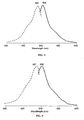
- ppluGFP2 was investigated in more detail. Purified ppluGFP2 possessed a molar extinction coefficient of 70,000 M -1 cm -1 and a fluorescence quantum yield of 0,60. For the molar extinction coefficient determination, mature chromophore concentration was estimated. Protein was alkali-denatured with an equal volume of 2M NaOH. Under these conditions, the GFP-like chromophore absorbs at 446 nm and its molar extinction coefficient is 44,000 M -1 cm -1 ( Ward, W. W., Bioluminescence and Chemiluminescence (1981), Academic Press, 235-242 ). The absorption spectra for native and alkali-denatured ppluGFP2 were measured.
- the molar extinction coefficient for the native state protein was estimated based on the absorption of the denatured protein.
- quantum yield determination the fluorescence of ppluGFP2 was compared to equally absorbing EGFP with quantum yield 0.60 ( Patterson, G., et al., J. Cell. Sci. (2001) 114:837-838 ).
- ppluGFP2 is monomeric protein since it demonstrated the same mobility as EGFP.
- Purified protein samples ( ⁇ 1 mg/ml) were loaded onto a Sephadex-100 column (0.7 x 60 cm) and eluted with a solution of 50 mM phosphate buffer (pH 7.0) and 100 mM NaCl.
- EGFP, HcRed1 and DsRed2 (Clontech) were used as monomer, dimer and tetramer standards, respectively.
- the wild type ppluGFP2 nucleic acid coding sequence was obtained as described above in the Example 1.
- To enhance expression in mammalian cells we synthesized "humanized" version of ppluGFP2 using mammalian- optimised codons (SEQ ID NOs: 17 and 18).
- To enhance expression in Saccharomyces cerevisiae yeast-optimized version of ppluGFP2 was synthesized using S. cerevisiae- optimised codons (SEQ ID NOs: 19 and 20).
- the humanized version of the ppluGFP2 was used as template for non-aggregated variants generation.
- the first version (CopGFP-NA1, SEQ ID NOs: 23 and 24) contains K5E substitution (numbering is based on wild type) and elongated negatively charged amino acid tail at N-terminus of the protein that shields the positive charge on the outside interface of the ppluGFP2 barrel and prevents charge interaction with another ppluGFP2 protein molecule.
- the second version (CopGFP-NA2, SEQ ID NOs: 25 and 26) contains additional tail at C-terminus.
- the third variant, CopGFP-NA3, comprise all changes present in CopGFP-NA1 and CopGFP-NA2 versions (SEQ ID NOs: 27 and 28). All versions display reduced ability to aggregate in in vivo and in vitro tests.
- Coding regions of nucleic acids of ppluGFP2 prepared as described above in the Examples 1 was cloned into pQE30 expressing vector (Qiagen), so that recombinant protein contained six-histidine tag at its N-terminus.
- protein was purified by metal-affinity resin TALON (Clontech) under denaturing conditions. Rabbits were immunized and boosted four times at monthly intervals with recombinant polypeptides emulsified in complete Freund's adjuvant. Ten or 11 days after each boost the animals were bled. Polyclonal antiserum was tested on recombinant protein by ELISA and by Western immunobloting.
- the humanised versions of ppluGFP2 prepared as described above in the Examples 3 was cloned into pEGFP-C1 vector (CLONTECH) between Age I and Bgl II restriction sites (in lieu of the EGFP-coding region).
- the following cell lines were used: 293T human kidney epithelial cells, 3T3 mouse embryo fibroblasts, L929 murine subcutaneous fibroblasts, Vero African green monkey kidney epithelial cells and COS1 African green monkey kidney fibroblasts. Cells were transfected using LipofectAMINE reagent (Invitrogen) and were tested 20 h after transfection.
- the humanised versions of ppluGFP2 prepared as described above in the Examples 3 was fused to human cytoplasmic beta-actin and human nucleolar protein, fibrillarin. Transfection of 293T human kidney epithelial cells with plasmids expressing ppluGFP2 -tagged fused constructs resulted in bright fluorescence that revealed pattern characteristic for the correspondent fusion partners.
- the humanised versions of ppluGFP2 prepared as described above in the Examples 3 was fused to the following subcellular localization signals: mitochondrial targeting sequence (MTS) from subunit VIII of human cytochrome c oxidase; sequence encoding the N-terminal 81 amino acids of human beta 1,4-galactosyltransferase (GT; Watzele & Berger (1990) Nucleic Acids. Res. 18:7174); peroximal targeting signal 1 ( Gould et al. J. Biol. Chem. (1989) 108: 1657-1664 ; Gould et al. EMBO J. (1990) 9: 85-90 ; Monosov et al. J. Histo. Cytochem.
- MTS mitochondrial targeting sequence
Landscapes
- Health & Medical Sciences (AREA)
- Chemical & Material Sciences (AREA)
- Organic Chemistry (AREA)
- Life Sciences & Earth Sciences (AREA)
- Proteomics, Peptides & Aminoacids (AREA)
- Biophysics (AREA)
- General Health & Medical Sciences (AREA)
- Genetics & Genomics (AREA)
- Medicinal Chemistry (AREA)
- Molecular Biology (AREA)
- Biochemistry (AREA)
- Tropical Medicine & Parasitology (AREA)
- Insects & Arthropods (AREA)
- Immunology (AREA)
- Toxicology (AREA)
- Zoology (AREA)
- Gastroenterology & Hepatology (AREA)
- Peptides Or Proteins (AREA)
- Micro-Organisms Or Cultivation Processes Thereof (AREA)
- Measuring Or Testing Involving Enzymes Or Micro-Organisms (AREA)
- Breeding Of Plants And Reproduction By Means Of Culturing (AREA)
- Preparation Of Compounds By Using Micro-Organisms (AREA)
- Investigating, Analyzing Materials By Fluorescence Or Luminescence (AREA)
Claims (16)
- Isoliertes Nukleinsäuremolekül, das ein fluoreszierendes Protein kodiert, ausgewählt aus der Gruppe bestehend aus:(a) einer Nukleinsäure, die ein Protein kodiert, das die Aminosäure-Sequenz umfasst, die in SEQ ID NO: 2, 4, 6, 8, 10, 12, 14, 16, 22, 24, 26 oder 28 dargestellt ist,(b) einer Nukleinsäure, die eine Nukleotidsequenz umfasst, die in SEQ ID NO: 1, 3, 5, 7, 9, 11, 13, 15, 17, 19, 21, 23, 25 oder 27 dargestellt ist;(c) einer Nukleinsäure, die unter stringenten Bedingungen mit der Nukleinsäure von (a) oder (b) hybridisiert, die oben beschrieben ist, wobei die stringenten Bedingungen mindestens so stringent sind wie die Hybridisierung bei 42°C in einer Lösung, die 50% Formamid, 5xSSC, 50mM Natriumphosphat, 5xDenhardt's Lösung, und 10% Dextransulfat umfasst, gefolgt vom Waschen der Filter in 0,1mal SSC bei etwa 65°C;(d) einer Nukleinsäure, die ein Protein kodiert, das mindestens etwa eine 65%-ige Sequenzidentität mit der oben beschriebenen Aminosäure-Sequenz (a) besitzt,(e) einer Nukleinsäure, die mindestens etwa eine 75%-ige Sequenzidentität mit der oben beschriebenen Nukleotidsequenz (b) besitzt; und(f) einer Nukleinsäure, die sich von der oben beschriebenen Nukleinsäure (b), infolge der Degeneration des genetischen Kodes unterscheidet;
- Nukleinsäuremolekül nach Anspruch 1, wobei die Nukleinsäure aus einem Organismus vom Typ Arthropoda isoliert ist.
- Nukleinsäuremolekül nach Anspruch 1, wobei die Nukleinsäure aus einem Organismus der Subklasse Copepoda isoliert ist.
- Nukleinsäuremolekül nach Anspruch 1, wobei die Nukleinsäure aus der Familie Pontellidae isoliert ist.
- Vektor, der das Nukleinsäuremolekül nach Anspruch 1 umfasst.
- Expressionskassette, umfassend (a) das Nukleinsäuremolekül nach Anspruch 1; und (b) Regulatorelemente für die Expression des Nukleinsäuremoleküls in der gewünschten Wirtszelle.
- Zelle, die das Nukleinsäuremolekül nach Anspruch 1, den Vektor nach Anspruch 5 oder die Expressionskassette nach Anspruch 6 umfasst.
- Stabile Zellinie, die das Nukleinsäuremolekül nach Anspruch 1, den Vektor nach Anspruch 5 oder die Expressionskassette nach Anspruch 6 umfasst.
- Transgene Pflanze, die das Nukleinsäuremolekül nach Anspruch 1, den Vektor nach Anspruch 5 oder die Expressionskassette nach Anspruch 6 umfasst.
- Transgenes nicht-menschliches Tier, das das Nukleinsäuremolekül nach Anspruch 1, den Vektor nach Anspruch 5 oder die Expressionskassette nach Anspruch 6 umfasst.
- Verfahren zur Herstellung eines fluoreszierenden Proteins, das Verfahren umfassend: (a) Bereitstellen eines Nukleinsäuremoleküls nach Anspruch 1, das mit den passenden Elementen der Expressionsregulation funktionell verbunden ist, (b) Expression des fluoreszierenden Proteins aus dem Nukleinsäuremolekül und (c) Isolieren des Proteins, das im Wesentlichen keine anderen Proteine umfasst.
- Isoliertes fluoreszierendes Protein, ausgewählt aus der Gruppe bestehend aus:(a) einem Protein, das die Aminosäure-Sequenz umfasst, die in SEQ ID NO: 2, 4, 6, 8, 10, 12, 14, 16, 18, 20, 22, 24, 26 oder 28 dargestellt ist;(b) einem Protein, das vom Nukleinsäuremolekül kodiert wird, das eine Nukleotidsequenz umfasst, die in SEQ ID NO: 1, 3, 5, 7, 9, 11, 13, 15, 17, 19, 21, 23, 25 oder 27 dargestellt ist; und(c) einem Protein, das mindestens eine etwa 65%-ige Sequenzidentität zur Aminosäure-Sequenz von (a) oder (b) aufweist, die oben beschrieben ist;
- Fusionsprotein, das das Protein nach Anspruch 12 umfasst.
- Antikörper, der sich mit dem Protein nach Anspruch 12 (a) spezifisch verbindet.
- Satz, der die Nukleinsäure nach Anspruch 1, den Vektor nach Anspruch 5, die Expressionskassette nach Anspruch 6, das Protein nach Anspruch 12, das Fusionsprotein nach Anspruch 13 oder ein Mittel zur Herstellung derselben umfasst.
- Oligonukleotidsonde oder Primer, umfassend die Nukleotidsequenz, die zur Hybridisierung unter stringenten Bedingungen mit der Nukleotidsequenz fähig ist, ausgewählt aus der Gruppe bestehend aus SEQ ID NO: 1, 3, 5, 7, 9, 11, 13, 15, 17, 19, 21, 23, 25 oder 27, wobei die stringenten Bedingungen mindestens so stringent sind wie die Hybridisierung bei 42°C in einer Lösung, die 50% Formamid, 5xSSC, 50mM Natriumphosphat, 5xDenhardt's Lösung, und 10% Dextransulfat umfasst, gefolgt vom Waschen der Filter in 0,1mal SSC bei etwa 65°C.
Applications Claiming Priority (5)
| Application Number | Priority Date | Filing Date | Title |
|---|---|---|---|
| US43685702P | 2002-12-26 | 2002-12-26 | |
| US436857P | 2002-12-26 | ||
| US45967903P | 2003-04-02 | 2003-04-02 | |
| US459679P | 2003-04-02 | ||
| PCT/RU2003/000525 WO2004058973A1 (en) | 2002-12-26 | 2003-11-26 | Fluorescent proteins from copepoda species and methods for using same |
Publications (3)
| Publication Number | Publication Date |
|---|---|
| EP1576157A1 EP1576157A1 (de) | 2005-09-21 |
| EP1576157A4 EP1576157A4 (de) | 2006-06-07 |
| EP1576157B1 true EP1576157B1 (de) | 2010-07-21 |
Family
ID=32685468
Family Applications (1)
| Application Number | Title | Priority Date | Filing Date |
|---|---|---|---|
| EP03781176A Expired - Lifetime EP1576157B1 (de) | 2002-12-26 | 2003-11-26 | Fluoreszenzproteine aus copepoda-spezies und verfahren zur verwendung davon |
Country Status (11)
| Country | Link |
|---|---|
| US (1) | US7678893B2 (de) |
| EP (1) | EP1576157B1 (de) |
| JP (1) | JP4480674B2 (de) |
| AT (1) | ATE474922T1 (de) |
| AU (1) | AU2003287115A1 (de) |
| CA (1) | CA2510884A1 (de) |
| DE (2) | DE60333486D1 (de) |
| DK (1) | DK1576157T3 (de) |
| ES (1) | ES2254044T3 (de) |
| RU (1) | RU2345137C2 (de) |
| WO (1) | WO2004058973A1 (de) |
Families Citing this family (15)
| Publication number | Priority date | Publication date | Assignee | Title |
|---|---|---|---|---|
| WO2005100565A1 (ja) | 2004-03-31 | 2005-10-27 | Nec Soft Ltd. | 新規な蛍光性タンパク質とそれをコードする遺伝子 |
| JP4863280B2 (ja) * | 2004-03-31 | 2012-01-25 | Necソフト株式会社 | 新規な蛍光性タンパク質とそれをコードする遺伝子 |
| US8278120B2 (en) | 2006-01-26 | 2012-10-02 | Nec Soft, Ltd. | Method of changing fluorescence wavelength of fluorescent protein |
| US8609393B2 (en) | 2006-06-08 | 2013-12-17 | Innoventus Project Ab | Fluorescent proteins and genes encoding them |
| EP2304047A4 (de) | 2008-05-07 | 2012-12-26 | Eutropics Pharmaceuticals Inc | Gegen heterodimere der bcl-2-familie gerichtete antikörper und ihre verwendungen |
| WO2010107639A2 (en) * | 2009-03-20 | 2010-09-23 | Board Of Regents, The University Of Texas System | Isolation and characterization of novel green fluorescent proteins from copepods |
| JP6242345B2 (ja) | 2012-02-01 | 2017-12-06 | ダウ アグロサイエンシィズ エルエルシー | グリホサート抵抗性植物および関連する方法 |
| JP6388575B2 (ja) | 2012-06-06 | 2018-09-12 | アメリカ合衆国 | 内分泌かく乱物質(edc)を検出およびモニターするためのキット |
| IN2014DN11205A (de) | 2012-06-20 | 2015-10-02 | Eutropics Pharmaceuticals Inc | |
| US20160038503A1 (en) | 2012-11-21 | 2016-02-11 | David Richard | Methods and compositions useful for treating diseases involving bcl-2 family proteins with isoquinoline and quinoline derivatives |
| WO2015017788A1 (en) | 2013-08-01 | 2015-02-05 | Eutropics Pharmaceuticals, Inc. | Method for predicting cancer sensitivity |
| AU2014342269B2 (en) | 2013-10-30 | 2020-02-27 | Eutropics Pharmaceuticals, Inc. | Methods for determining chemosensitivity and chemotoxicity |
| EP3245301B1 (de) | 2015-01-12 | 2020-11-11 | Eutropics Pharmaceuticals, Inc. | Kontextabhängiger diagnostischer test zur führung einer krebsbehandlung |
| CA3022981C (en) | 2017-11-01 | 2025-05-06 | Queen's University At Kingston | HIPPO BIOLUMINOUS TRAIL BIOSENSOR |
| CN116179670B (zh) * | 2023-02-10 | 2026-03-20 | 扬州大学 | 一种快速鉴定外源cop-GFP的方法 |
Family Cites Families (50)
| Publication number | Priority date | Publication date | Assignee | Title |
|---|---|---|---|---|
| US5583024A (en) | 1985-12-02 | 1996-12-10 | The Regents Of The University Of California | Recombinant expression of Coleoptera luciferase |
| US5221623A (en) | 1986-07-22 | 1993-06-22 | Boyce Thompson Institute For Plant Research, Inc. | Use of bacterial luciferase structural genes for cloning and monitoring gene expression in microorganisms and for tagging and identification of genetically engineered organisms |
| US5182202A (en) | 1987-11-30 | 1993-01-26 | Kikkoman Corporation | Purified luciferase from luciola cruciata |
| US5292658A (en) | 1989-12-29 | 1994-03-08 | University Of Georgia Research Foundation, Inc. Boyd Graduate Studies Research Center | Cloning and expressions of Renilla luciferase |
| US5219737A (en) | 1990-03-27 | 1993-06-15 | Kikkoman Corporation | Mutant luciferase of a firefly, mutant luciferase genes, recombinant dnas containing the genes and a method of producing mutant luciferase |
| JP3208486B2 (ja) | 1990-07-02 | 2001-09-10 | ザ リージェンツ オブ ザ ユニバーシティ オブ カリフォルニア | 蛍光エネルギー転移を利用する分析物の検出 |
| US5229285A (en) | 1991-06-27 | 1993-07-20 | Kikkoman Corporation | Thermostable luciferase of firefly, thermostable luciferase gene of firefly, novel recombinant dna, and process for the preparation of thermostable luciferase of firefly |
| JP2651651B2 (ja) | 1993-04-21 | 1997-09-10 | チッソ株式会社 | 蛍酵素ルシフェラーゼ遺伝子 |
| US5491084A (en) | 1993-09-10 | 1996-02-13 | The Trustees Of Columbia University In The City Of New York | Uses of green-fluorescent protein |
| US5654419A (en) | 1994-02-01 | 1997-08-05 | The Regents Of The University Of California | Fluorescent labels and their use in separations |
| US5869255A (en) | 1994-02-01 | 1999-02-09 | The Regents Of The University Of California | Probes labeled with energy transfer couples dyes exemplified with DNA fragment analysis |
| JP3448090B2 (ja) | 1994-02-16 | 2003-09-16 | 浜松ホトニクス株式会社 | エネルギー移動検出法およびその装置 |
| US5650135A (en) | 1994-07-01 | 1997-07-22 | The Board Of Trustees Of The Leland Stanford Junior University | Non-invasive localization of a light-emitting conjugate in a mammal |
| JP3466765B2 (ja) | 1994-07-27 | 2003-11-17 | キッコーマン株式会社 | ビオチン化ホタルルシフェラーゼ、ビオチン化ホタルルシフェラーゼ遺伝子、新規な組み換え体dna、ビオチン化ホタルルシフェラーゼの製造法及び生物発光分析法 |
| US5795737A (en) | 1994-09-19 | 1998-08-18 | The General Hospital Corporation | High level expression of proteins |
| US5625048A (en) | 1994-11-10 | 1997-04-29 | The Regents Of The University Of California | Modified green fluorescent proteins |
| US5958713A (en) | 1995-01-31 | 1999-09-28 | Novo Nordisk A/S | Method of detecting biologically active substances by using green fluorescent protein |
| JPH11504218A (ja) | 1995-04-24 | 1999-04-20 | クロマゾーム コーポレーション | 新規代謝経路の生成およびスクリーニングのための方法 |
| US6008373A (en) | 1995-06-07 | 1999-12-28 | Carnegie Mellon University | Fluorescent labeling complexes with large stokes shift formed by coupling together cyanine and other fluorochromes capable of resonance energy transfer |
| US5728528A (en) | 1995-09-20 | 1998-03-17 | The Regents Of The University Of California | Universal spacer/energy transfer dyes |
| US5968738A (en) | 1995-12-06 | 1999-10-19 | The Board Of Trustees Of The Leland Stanford Junior University | Two-reporter FACS analysis of mammalian cells using green fluorescent proteins |
| IL124967A (en) | 1995-12-18 | 2000-07-26 | Univ Washington | Method for nucleic acid analysis using fluorescence resonance energy transfer |
| US5874304A (en) | 1996-01-18 | 1999-02-23 | University Of Florida Research Foundation, Inc. | Humanized green fluorescent protein genes and methods |
| US6020192A (en) | 1996-01-18 | 2000-02-01 | University Of Florida | Humanized green fluorescent protein genes and methods |
| US6803188B1 (en) | 1996-01-31 | 2004-10-12 | The Regents Of The University Of California | Tandem fluorescent protein constructs |
| AR006928A1 (es) * | 1996-05-01 | 1999-09-29 | Pioneer Hi Bred Int | Una molecula de adn aislada que codifica una proteina fluorescente verde como marcador rastreable para la transformacion de plantas, un metodo para laproduccion de plantas transgenicas, un vector de expresion, una planta transgenica y celulas de dichas plantas. |
| US5945526A (en) | 1996-05-03 | 1999-08-31 | Perkin-Elmer Corporation | Energy transfer dyes with enhanced fluorescence |
| US5863727A (en) | 1996-05-03 | 1999-01-26 | The Perkin-Elmer Corporation | Energy transfer dyes with enhanced fluorescence |
| US5989835A (en) | 1997-02-27 | 1999-11-23 | Cellomics, Inc. | System for cell-based screening |
| US5866336A (en) | 1996-07-16 | 1999-02-02 | Oncor, Inc. | Nucleic acid amplification oligonucleotides with molecular energy transfer labels and methods based thereon |
| US5976796A (en) * | 1996-10-04 | 1999-11-02 | Loma Linda University | Construction and expression of renilla luciferase and green fluorescent protein fusion genes |
| TW371617B (en) | 1996-10-09 | 1999-10-11 | Of Animal And Plant Health Inspection And Quarantine Council Of Agriculture Executive Yuan Bureau | Method to transplant GFP into autographa californica multiple nuclear polyhedrosis virus for inflicting pest in an attempt to detect and flow up it existence and to improve life span against UV |
| US5972638A (en) | 1997-01-31 | 1999-10-26 | Lockheed Martin Energy Research Corp. | Method for detection of buried explosives using a biosensor |
| JPH10234382A (ja) | 1997-02-27 | 1998-09-08 | Deinabetsuku Kenkyusho:Kk | 蛍光タンパク質 |
| US6130313A (en) | 1997-10-02 | 2000-10-10 | Clontech Laboratories, Inc. | Rapidly degrading GFP-fusion proteins |
| WO1999037142A1 (en) | 1998-01-27 | 1999-07-29 | Novo Nordisk A/S | Method for producing transgenic animals |
| EP1064360B1 (de) * | 1998-03-27 | 2008-03-05 | Prolume, Ltd. | Luciferase, gfp fluoreszenzproteine, kodierende nukleinsaüre und ihre verwendung in der diagnose |
| US6501003B1 (en) | 1998-07-08 | 2002-12-31 | Wisconsin Alumni Research Foundation | Transgentic mouse expressing green fluorescent protein in glial cells |
| DK1095277T3 (da) | 1998-07-13 | 2003-04-22 | Cellomics Inc | System til cellebaseret screening |
| US5998146A (en) | 1998-07-17 | 1999-12-07 | Wallac Oy | Homogeneous luminescence assay method based on energy transfer |
| JP2002525603A (ja) | 1998-09-18 | 2002-08-13 | セロミックス インコーポレイテッド | 細胞ベースのスクリーニングのためのシステム |
| EP1123499A2 (de) | 1998-09-22 | 2001-08-16 | Cellomics, Inc. | Miniaturisierte zellmatrixverfahren und vorrichtung für zellbasierte reihenuntersuchungen |
| US5985577A (en) | 1998-10-14 | 1999-11-16 | The Trustees Of Columbia University In The City Of New York | Protein conjugates containing multimers of green fluorescent protein |
| ATE225406T1 (de) | 1998-10-30 | 2002-10-15 | Cellomics Inc | Ein screeningsystem auf zellbasis |
| WO2000034325A1 (en) | 1998-12-11 | 2000-06-15 | Clontech Laboratories, Inc. | Fluorescent proteins from non-bioluminescent species of class anthozoa, genes encoding such proteins and uses thereof |
| EP1305412A2 (de) | 1999-10-14 | 2003-05-02 | Clontech Laboratories Inc. | Chromo/fluoroproteine aus anthozoa und verwendung der gleichen |
| US20040171107A1 (en) * | 2000-02-23 | 2004-09-02 | David Nelson | Modified flourescent proteins |
| WO2002059309A2 (en) | 2000-12-13 | 2002-08-01 | Clontech Laboratories, Inc. | Anthozoa derived chromoproteins, fluorescent mutants thereof and methods for using the same |
| WO2002068605A2 (en) * | 2001-02-26 | 2002-09-06 | The Regents Of The University Of California | Non-oligomerizing tandem fluorescent proteins |
| WO2003062270A2 (en) | 2002-01-22 | 2003-07-31 | Evrogen, Jsc | Fluorescent protein from aequorea coerulscens and uses therof |
-
2003
- 2003-11-26 AT AT03781176T patent/ATE474922T1/de not_active IP Right Cessation
- 2003-11-26 AU AU2003287115A patent/AU2003287115A1/en not_active Abandoned
- 2003-11-26 US US10/533,781 patent/US7678893B2/en not_active Expired - Lifetime
- 2003-11-26 RU RU2005123689/13A patent/RU2345137C2/ru active
- 2003-11-26 WO PCT/RU2003/000525 patent/WO2004058973A1/en not_active Ceased
- 2003-11-26 CA CA002510884A patent/CA2510884A1/en not_active Abandoned
- 2003-11-26 ES ES03781176T patent/ES2254044T3/es not_active Expired - Lifetime
- 2003-11-26 DE DE60333486T patent/DE60333486D1/de not_active Expired - Lifetime
- 2003-11-26 EP EP03781176A patent/EP1576157B1/de not_active Expired - Lifetime
- 2003-11-26 DK DK03781176.7T patent/DK1576157T3/da active
- 2003-11-26 JP JP2005509951A patent/JP4480674B2/ja not_active Expired - Fee Related
- 2003-11-26 DE DE03781176T patent/DE03781176T1/de active Pending
Also Published As
| Publication number | Publication date |
|---|---|
| ES2254044T1 (es) | 2006-06-16 |
| EP1576157A1 (de) | 2005-09-21 |
| CA2510884A1 (en) | 2004-07-15 |
| DK1576157T3 (da) | 2010-11-01 |
| US7678893B2 (en) | 2010-03-16 |
| EP1576157A4 (de) | 2006-06-07 |
| ATE474922T1 (de) | 2010-08-15 |
| AU2003287115A1 (en) | 2004-07-22 |
| DE03781176T1 (de) | 2006-06-22 |
| WO2004058973A1 (en) | 2004-07-15 |
| RU2345137C2 (ru) | 2009-01-27 |
| RU2005123689A (ru) | 2006-01-20 |
| ES2254044T3 (es) | 2010-12-30 |
| JP2006512093A (ja) | 2006-04-13 |
| DE60333486D1 (de) | 2010-09-02 |
| US20080104721A1 (en) | 2008-05-01 |
| JP4480674B2 (ja) | 2010-06-16 |
Similar Documents
| Publication | Publication Date | Title |
|---|---|---|
| US8138320B2 (en) | Fluorescent proteins and methods for using same | |
| US7888113B2 (en) | Modified green fluorescent proteins and methods for using same | |
| EP1576157B1 (de) | Fluoreszenzproteine aus copepoda-spezies und verfahren zur verwendung davon | |
| EP1565559B1 (de) | Fluoreszenzproteine und chromoproteine aus nicht-aequorea-hydrozoa-spezies sowie verfahren zur verwendung davon | |
| US20030013849A1 (en) | Renilla reniformis green fluorescent protein | |
| US20130344591A1 (en) | Modified Fluorescent Proteins and Methods for Using Same | |
| US7972834B2 (en) | Modified fluorescent proteins and methods for using same | |
| WO2001032688A9 (en) | Renilla reniformis green fluorescent protein | |
| US8563703B2 (en) | Fluorescent proteins and methods for using same | |
| RU2338785C2 (ru) | Флуоресцирующие белки и хромопротеины из видов hydrozoa, не относящихся к aequorea, и способы их получения |
Legal Events
| Date | Code | Title | Description |
|---|---|---|---|
| PUAI | Public reference made under article 153(3) epc to a published international application that has entered the european phase |
Free format text: ORIGINAL CODE: 0009012 |
|
| 17P | Request for examination filed |
Effective date: 20050627 |
|
| AK | Designated contracting states |
Kind code of ref document: A1 Designated state(s): AT BE BG CH CY CZ DE DK EE ES FI FR GB GR HU IE IT LI LU MC NL PT RO SE SI SK TR |
|
| AX | Request for extension of the european patent |
Extension state: AL LT LV MK |
|
| DAX | Request for extension of the european patent (deleted) | ||
| TCNL | Nl: translation of patent claims filed | ||
| A4 | Supplementary search report drawn up and despatched |
Effective date: 20060425 |
|
| DET | De: translation of patent claims | ||
| REG | Reference to a national code |
Ref country code: DE Ref legal event code: 8581 |
|
| 17Q | First examination report despatched |
Effective date: 20070320 |
|
| EL | Fr: translation of claims filed | ||
| GRAP | Despatch of communication of intention to grant a patent |
Free format text: ORIGINAL CODE: EPIDOSNIGR1 |
|
| GRAS | Grant fee paid |
Free format text: ORIGINAL CODE: EPIDOSNIGR3 |
|
| GRAA | (expected) grant |
Free format text: ORIGINAL CODE: 0009210 |
|
| AK | Designated contracting states |
Kind code of ref document: B1 Designated state(s): AT BE BG CH CY CZ DE DK EE ES FI FR GB GR HU IE IT LI LU MC NL PT RO SE SI SK TR |
|
| REG | Reference to a national code |
Ref country code: GB Ref legal event code: FG4D |
|
| REG | Reference to a national code |
Ref country code: CH Ref legal event code: EP Ref country code: CH Ref legal event code: NV Representative=s name: RITSCHER & PARTNER AG |
|
| REG | Reference to a national code |
Ref country code: IE Ref legal event code: FG4D |
|
| REF | Corresponds to: |
Ref document number: 60333486 Country of ref document: DE Date of ref document: 20100902 Kind code of ref document: P |
|
| REG | Reference to a national code |
Ref country code: DK Ref legal event code: T3 |
|
| REG | Reference to a national code |
Ref country code: NL Ref legal event code: T3 |
|
| REG | Reference to a national code |
Ref country code: ES Ref legal event code: FG2A Effective date: 20101217 |
|
| PG25 | Lapsed in a contracting state [announced via postgrant information from national office to epo] |
Ref country code: AT Free format text: LAPSE BECAUSE OF FAILURE TO SUBMIT A TRANSLATION OF THE DESCRIPTION OR TO PAY THE FEE WITHIN THE PRESCRIBED TIME-LIMIT Effective date: 20100721 |
|
| PG25 | Lapsed in a contracting state [announced via postgrant information from national office to epo] |
Ref country code: CY Free format text: LAPSE BECAUSE OF FAILURE TO SUBMIT A TRANSLATION OF THE DESCRIPTION OR TO PAY THE FEE WITHIN THE PRESCRIBED TIME-LIMIT Effective date: 20100721 Ref country code: SI Free format text: LAPSE BECAUSE OF FAILURE TO SUBMIT A TRANSLATION OF THE DESCRIPTION OR TO PAY THE FEE WITHIN THE PRESCRIBED TIME-LIMIT Effective date: 20100721 Ref country code: PT Free format text: LAPSE BECAUSE OF FAILURE TO SUBMIT A TRANSLATION OF THE DESCRIPTION OR TO PAY THE FEE WITHIN THE PRESCRIBED TIME-LIMIT Effective date: 20101122 Ref country code: BG Free format text: LAPSE BECAUSE OF FAILURE TO SUBMIT A TRANSLATION OF THE DESCRIPTION OR TO PAY THE FEE WITHIN THE PRESCRIBED TIME-LIMIT Effective date: 20101021 |
|
| PG25 | Lapsed in a contracting state [announced via postgrant information from national office to epo] |
Ref country code: SE Free format text: LAPSE BECAUSE OF FAILURE TO SUBMIT A TRANSLATION OF THE DESCRIPTION OR TO PAY THE FEE WITHIN THE PRESCRIBED TIME-LIMIT Effective date: 20100721 Ref country code: GR Free format text: LAPSE BECAUSE OF FAILURE TO SUBMIT A TRANSLATION OF THE DESCRIPTION OR TO PAY THE FEE WITHIN THE PRESCRIBED TIME-LIMIT Effective date: 20101022 Ref country code: BE Free format text: LAPSE BECAUSE OF FAILURE TO SUBMIT A TRANSLATION OF THE DESCRIPTION OR TO PAY THE FEE WITHIN THE PRESCRIBED TIME-LIMIT Effective date: 20100721 |
|
| PLBE | No opposition filed within time limit |
Free format text: ORIGINAL CODE: 0009261 |
|
| STAA | Information on the status of an ep patent application or granted ep patent |
Free format text: STATUS: NO OPPOSITION FILED WITHIN TIME LIMIT |
|
| PG25 | Lapsed in a contracting state [announced via postgrant information from national office to epo] |
Ref country code: SK Free format text: LAPSE BECAUSE OF FAILURE TO SUBMIT A TRANSLATION OF THE DESCRIPTION OR TO PAY THE FEE WITHIN THE PRESCRIBED TIME-LIMIT Effective date: 20100721 Ref country code: IT Free format text: LAPSE BECAUSE OF FAILURE TO SUBMIT A TRANSLATION OF THE DESCRIPTION OR TO PAY THE FEE WITHIN THE PRESCRIBED TIME-LIMIT Effective date: 20100721 Ref country code: RO Free format text: LAPSE BECAUSE OF FAILURE TO SUBMIT A TRANSLATION OF THE DESCRIPTION OR TO PAY THE FEE WITHIN THE PRESCRIBED TIME-LIMIT Effective date: 20100721 Ref country code: EE Free format text: LAPSE BECAUSE OF FAILURE TO SUBMIT A TRANSLATION OF THE DESCRIPTION OR TO PAY THE FEE WITHIN THE PRESCRIBED TIME-LIMIT Effective date: 20100721 Ref country code: CZ Free format text: LAPSE BECAUSE OF FAILURE TO SUBMIT A TRANSLATION OF THE DESCRIPTION OR TO PAY THE FEE WITHIN THE PRESCRIBED TIME-LIMIT Effective date: 20100721 |
|
| 26N | No opposition filed |
Effective date: 20110426 |
|
| PG25 | Lapsed in a contracting state [announced via postgrant information from national office to epo] |
Ref country code: MC Free format text: LAPSE BECAUSE OF NON-PAYMENT OF DUE FEES Effective date: 20101130 |
|
| REG | Reference to a national code |
Ref country code: DE Ref legal event code: R097 Ref document number: 60333486 Country of ref document: DE Effective date: 20110426 |
|
| PG25 | Lapsed in a contracting state [announced via postgrant information from national office to epo] |
Ref country code: IE Free format text: LAPSE BECAUSE OF NON-PAYMENT OF DUE FEES Effective date: 20101126 |
|
| PG25 | Lapsed in a contracting state [announced via postgrant information from national office to epo] |
Ref country code: HU Free format text: LAPSE BECAUSE OF FAILURE TO SUBMIT A TRANSLATION OF THE DESCRIPTION OR TO PAY THE FEE WITHIN THE PRESCRIBED TIME-LIMIT Effective date: 20110122 Ref country code: LU Free format text: LAPSE BECAUSE OF NON-PAYMENT OF DUE FEES Effective date: 20101126 |
|
| PG25 | Lapsed in a contracting state [announced via postgrant information from national office to epo] |
Ref country code: TR Free format text: LAPSE BECAUSE OF FAILURE TO SUBMIT A TRANSLATION OF THE DESCRIPTION OR TO PAY THE FEE WITHIN THE PRESCRIBED TIME-LIMIT Effective date: 20100721 |
|
| REG | Reference to a national code |
Ref country code: CH Ref legal event code: PFA Owner name: ZAKRYTOE AKTSIONERNOE OBSCHESTVO " EVROGEN", RU Free format text: FORMER OWNER: ZAKRYTOE AKTSIONERNOE OBSCHESTVO " EVROGEN", RU |
|
| REG | Reference to a national code |
Ref country code: FR Ref legal event code: PLFP Year of fee payment: 13 |
|
| REG | Reference to a national code |
Ref country code: FR Ref legal event code: PLFP Year of fee payment: 14 |
|
| REG | Reference to a national code |
Ref country code: FR Ref legal event code: PLFP Year of fee payment: 15 |
|
| PGFP | Annual fee paid to national office [announced via postgrant information from national office to epo] |
Ref country code: FI Payment date: 20191121 Year of fee payment: 17 |
|
| PGFP | Annual fee paid to national office [announced via postgrant information from national office to epo] |
Ref country code: DK Payment date: 20191125 Year of fee payment: 17 |
|
| REG | Reference to a national code |
Ref country code: FI Ref legal event code: MAE |
|
| REG | Reference to a national code |
Ref country code: DK Ref legal event code: EBP Effective date: 20201130 |
|
| PG25 | Lapsed in a contracting state [announced via postgrant information from national office to epo] |
Ref country code: FI Free format text: LAPSE BECAUSE OF NON-PAYMENT OF DUE FEES Effective date: 20201126 |
|
| PG25 | Lapsed in a contracting state [announced via postgrant information from national office to epo] |
Ref country code: DK Free format text: LAPSE BECAUSE OF NON-PAYMENT OF DUE FEES Effective date: 20201130 |
|
| PGFP | Annual fee paid to national office [announced via postgrant information from national office to epo] |
Ref country code: NL Payment date: 20211125 Year of fee payment: 19 |
|
| PGFP | Annual fee paid to national office [announced via postgrant information from national office to epo] |
Ref country code: FR Payment date: 20221007 Year of fee payment: 20 |
|
| PGFP | Annual fee paid to national office [announced via postgrant information from national office to epo] |
Ref country code: GB Payment date: 20221006 Year of fee payment: 20 Ref country code: ES Payment date: 20221201 Year of fee payment: 20 Ref country code: DE Payment date: 20221018 Year of fee payment: 20 |
|
| PGFP | Annual fee paid to national office [announced via postgrant information from national office to epo] |
Ref country code: CH Payment date: 20221201 Year of fee payment: 20 |
|
| REG | Reference to a national code |
Ref country code: NL Ref legal event code: MM Effective date: 20221201 |
|
| PG25 | Lapsed in a contracting state [announced via postgrant information from national office to epo] |
Ref country code: NL Free format text: LAPSE BECAUSE OF NON-PAYMENT OF DUE FEES Effective date: 20221201 |
|
| REG | Reference to a national code |
Ref country code: DE Ref legal event code: R071 Ref document number: 60333486 Country of ref document: DE |
|
| REG | Reference to a national code |
Ref country code: CH Ref legal event code: PL |
|
| REG | Reference to a national code |
Ref country code: ES Ref legal event code: FD2A Effective date: 20231201 |
|
| REG | Reference to a national code |
Ref country code: GB Ref legal event code: PE20 Expiry date: 20231125 |
|
| PG25 | Lapsed in a contracting state [announced via postgrant information from national office to epo] |
Ref country code: GB Free format text: LAPSE BECAUSE OF EXPIRATION OF PROTECTION Effective date: 20231125 |
|
| PG25 | Lapsed in a contracting state [announced via postgrant information from national office to epo] |
Ref country code: ES Free format text: LAPSE BECAUSE OF EXPIRATION OF PROTECTION Effective date: 20231127 |
|
| PG25 | Lapsed in a contracting state [announced via postgrant information from national office to epo] |
Ref country code: GB Free format text: LAPSE BECAUSE OF EXPIRATION OF PROTECTION Effective date: 20231125 Ref country code: ES Free format text: LAPSE BECAUSE OF EXPIRATION OF PROTECTION Effective date: 20231127 |