EP1533616A1 - Detektionskitt, darin zu verwendende assay-platte, detektionsverfahren, auswertungsverfahren, polyklonalantikörper von frosch-vitellogenin und prozess zur herstellung davon - Google Patents
Detektionskitt, darin zu verwendende assay-platte, detektionsverfahren, auswertungsverfahren, polyklonalantikörper von frosch-vitellogenin und prozess zur herstellung davon Download PDFInfo
- Publication number
- EP1533616A1 EP1533616A1 EP03736119A EP03736119A EP1533616A1 EP 1533616 A1 EP1533616 A1 EP 1533616A1 EP 03736119 A EP03736119 A EP 03736119A EP 03736119 A EP03736119 A EP 03736119A EP 1533616 A1 EP1533616 A1 EP 1533616A1
- Authority
- EP
- European Patent Office
- Prior art keywords
- vitellogenin
- frog
- antibody
- sample
- well
- Prior art date
- Legal status (The legal status is an assumption and is not a legal conclusion. Google has not performed a legal analysis and makes no representation as to the accuracy of the status listed.)
- Withdrawn
Links
- 108010090932 Vitellogenins Proteins 0.000 title claims abstract description 346
- 238000001514 detection method Methods 0.000 title claims description 122
- 238000011156 evaluation Methods 0.000 title claims description 44
- 238000003556 assay Methods 0.000 title description 8
- 238000006243 chemical reaction Methods 0.000 claims abstract description 56
- 238000012360 testing method Methods 0.000 claims abstract description 41
- 239000012916 chromogenic reagent Substances 0.000 claims abstract description 18
- 238000004040 coloring Methods 0.000 claims abstract description 10
- 238000000034 method Methods 0.000 claims description 152
- 239000007788 liquid Substances 0.000 claims description 67
- 210000002381 plasma Anatomy 0.000 claims description 55
- 210000002966 serum Anatomy 0.000 claims description 53
- 239000008280 blood Substances 0.000 claims description 37
- 210000003494 hepatocyte Anatomy 0.000 claims description 37
- 238000004519 manufacturing process Methods 0.000 claims description 34
- 239000000427 antigen Substances 0.000 claims description 33
- 108091007433 antigens Proteins 0.000 claims description 33
- 102000036639 antigens Human genes 0.000 claims description 33
- 238000005259 measurement Methods 0.000 claims description 33
- 230000002860 competitive effect Effects 0.000 claims description 26
- 238000002372 labelling Methods 0.000 claims description 23
- 230000008569 process Effects 0.000 claims description 16
- 150000001875 compounds Chemical class 0.000 claims description 15
- 238000005070 sampling Methods 0.000 claims description 12
- 230000003053 immunization Effects 0.000 claims description 10
- 230000004044 response Effects 0.000 claims description 10
- 239000000203 mixture Substances 0.000 claims description 9
- 108010017384 Blood Proteins Proteins 0.000 claims description 8
- 102000004506 Blood Proteins Human genes 0.000 claims description 8
- 241000124008 Mammalia Species 0.000 claims description 7
- 239000007795 chemical reaction product Substances 0.000 claims description 4
- 239000002981 blocking agent Substances 0.000 claims description 3
- 102000004190 Enzymes Human genes 0.000 abstract description 23
- 108090000790 Enzymes Proteins 0.000 abstract description 23
- 238000002965 ELISA Methods 0.000 description 102
- 239000000243 solution Substances 0.000 description 101
- 239000000523 sample Substances 0.000 description 70
- 239000000126 substance Substances 0.000 description 64
- 241000269368 Xenopus laevis Species 0.000 description 54
- XLYOFNOQVPJJNP-UHFFFAOYSA-N water Substances O XLYOFNOQVPJJNP-UHFFFAOYSA-N 0.000 description 45
- 239000012895 dilution Substances 0.000 description 44
- 238000010790 dilution Methods 0.000 description 44
- 230000035945 sensitivity Effects 0.000 description 41
- 238000002360 preparation method Methods 0.000 description 38
- 210000004369 blood Anatomy 0.000 description 34
- 108010001336 Horseradish Peroxidase Proteins 0.000 description 33
- 238000004140 cleaning Methods 0.000 description 32
- 238000011088 calibration curve Methods 0.000 description 29
- 239000012898 sample dilution Substances 0.000 description 28
- 238000010586 diagram Methods 0.000 description 27
- YBJHBAHKTGYVGT-ZKWXMUAHSA-N (+)-Biotin Chemical compound N1C(=O)N[C@@H]2[C@H](CCCCC(=O)O)SC[C@@H]21 YBJHBAHKTGYVGT-ZKWXMUAHSA-N 0.000 description 24
- 108091003079 Bovine Serum Albumin Proteins 0.000 description 24
- 229940098773 bovine serum albumin Drugs 0.000 description 24
- 238000011084 recovery Methods 0.000 description 24
- 229940088598 enzyme Drugs 0.000 description 22
- 230000015572 biosynthetic process Effects 0.000 description 19
- 230000007613 environmental effect Effects 0.000 description 19
- 238000003786 synthesis reaction Methods 0.000 description 19
- 238000011002 quantification Methods 0.000 description 18
- 241000269350 Anura Species 0.000 description 17
- 229920000136 polysorbate Polymers 0.000 description 17
- 238000000746 purification Methods 0.000 description 17
- 210000004185 liver Anatomy 0.000 description 16
- 238000005406 washing Methods 0.000 description 16
- FAPWRFPIFSIZLT-UHFFFAOYSA-M Sodium chloride Chemical compound [Na+].[Cl-] FAPWRFPIFSIZLT-UHFFFAOYSA-M 0.000 description 15
- 230000000903 blocking effect Effects 0.000 description 15
- 230000009260 cross reactivity Effects 0.000 description 15
- 238000002347 injection Methods 0.000 description 15
- 239000007924 injection Substances 0.000 description 15
- 230000006698 induction Effects 0.000 description 14
- 241000894007 species Species 0.000 description 14
- 239000003593 chromogenic compound Substances 0.000 description 13
- 230000000694 effects Effects 0.000 description 13
- 230000001076 estrogenic effect Effects 0.000 description 13
- PEDCQBHIVMGVHV-UHFFFAOYSA-N Glycerine Chemical compound OCC(O)CO PEDCQBHIVMGVHV-UHFFFAOYSA-N 0.000 description 12
- 238000001261 affinity purification Methods 0.000 description 12
- 229960002685 biotin Drugs 0.000 description 12
- 235000020958 biotin Nutrition 0.000 description 12
- 239000011616 biotin Substances 0.000 description 12
- NKLPQNGYXWVELD-UHFFFAOYSA-M coomassie brilliant blue Chemical compound [Na+].C1=CC(OCC)=CC=C1NC1=CC=C(C(=C2C=CC(C=C2)=[N+](CC)CC=2C=C(C=CC=2)S([O-])(=O)=O)C=2C=CC(=CC=2)N(CC)CC=2C=C(C=CC=2)S([O-])(=O)=O)C=C1 NKLPQNGYXWVELD-UHFFFAOYSA-M 0.000 description 12
- 231100000507 endocrine disrupting Toxicity 0.000 description 12
- 238000010186 staining Methods 0.000 description 12
- 230000009471 action Effects 0.000 description 11
- 229940088597 hormone Drugs 0.000 description 11
- 239000005556 hormone Substances 0.000 description 11
- 238000001262 western blot Methods 0.000 description 11
- 108010090804 Streptavidin Proteins 0.000 description 10
- 230000037396 body weight Effects 0.000 description 10
- 238000009395 breeding Methods 0.000 description 10
- 230000001488 breeding effect Effects 0.000 description 10
- 239000003153 chemical reaction reagent Substances 0.000 description 10
- 239000000598 endocrine disruptor Substances 0.000 description 10
- 231100000049 endocrine disruptor Toxicity 0.000 description 10
- 239000000262 estrogen Substances 0.000 description 10
- 238000001179 sorption measurement Methods 0.000 description 10
- DNIAPMSPPWPWGF-UHFFFAOYSA-N Propylene glycol Chemical compound CC(O)CO DNIAPMSPPWPWGF-UHFFFAOYSA-N 0.000 description 9
- 230000001419 dependent effect Effects 0.000 description 9
- 239000012153 distilled water Substances 0.000 description 9
- 239000000843 powder Substances 0.000 description 9
- 210000001519 tissue Anatomy 0.000 description 9
- 238000002835 absorbance Methods 0.000 description 8
- 229940011871 estrogen Drugs 0.000 description 8
- 238000002649 immunization Methods 0.000 description 8
- 238000003317 immunochromatography Methods 0.000 description 8
- 238000005457 optimization Methods 0.000 description 8
- 238000012545 processing Methods 0.000 description 8
- 108090000623 proteins and genes Proteins 0.000 description 8
- 102000004169 proteins and genes Human genes 0.000 description 8
- 238000002415 sodium dodecyl sulfate polyacrylamide gel electrophoresis Methods 0.000 description 8
- VOXZDWNPVJITMN-ZBRFXRBCSA-N 17β-estradiol Chemical compound OC1=CC=C2[C@H]3CC[C@](C)([C@H](CC4)O)[C@@H]4[C@@H]3CCC2=C1 VOXZDWNPVJITMN-ZBRFXRBCSA-N 0.000 description 7
- 241000283973 Oryctolagus cuniculus Species 0.000 description 7
- IISBACLAFKSPIT-UHFFFAOYSA-N bisphenol A Chemical compound C=1C=C(O)C=CC=1C(C)(C)C1=CC=C(O)C=C1 IISBACLAFKSPIT-UHFFFAOYSA-N 0.000 description 7
- 238000007865 diluting Methods 0.000 description 7
- 238000002474 experimental method Methods 0.000 description 7
- 239000011780 sodium chloride Substances 0.000 description 7
- 239000012086 standard solution Substances 0.000 description 7
- 241000251468 Actinopterygii Species 0.000 description 6
- IAZDPXIOMUYVGZ-UHFFFAOYSA-N Dimethylsulphoxide Chemical compound CS(C)=O IAZDPXIOMUYVGZ-UHFFFAOYSA-N 0.000 description 6
- 241000276569 Oryzias latipes Species 0.000 description 6
- 210000004027 cell Anatomy 0.000 description 6
- 230000000052 comparative effect Effects 0.000 description 6
- 238000007710 freezing Methods 0.000 description 6
- 230000008014 freezing Effects 0.000 description 6
- 230000006872 improvement Effects 0.000 description 6
- 230000005764 inhibitory process Effects 0.000 description 6
- 239000000463 material Substances 0.000 description 6
- 238000002156 mixing Methods 0.000 description 6
- 239000006228 supernatant Substances 0.000 description 6
- 241000269339 Bombina bombina Species 0.000 description 5
- ZMXDDKWLCZADIW-UHFFFAOYSA-N N,N-Dimethylformamide Chemical compound CN(C)C=O ZMXDDKWLCZADIW-UHFFFAOYSA-N 0.000 description 5
- 229920001213 Polysorbate 20 Polymers 0.000 description 5
- 229930006000 Sucrose Natural products 0.000 description 5
- CZMRCDWAGMRECN-UGDNZRGBSA-N Sucrose Chemical compound O[C@H]1[C@H](O)[C@@H](CO)O[C@@]1(CO)O[C@@H]1[C@H](O)[C@@H](O)[C@H](O)[C@@H](CO)O1 CZMRCDWAGMRECN-UGDNZRGBSA-N 0.000 description 5
- 241000269457 Xenopus tropicalis Species 0.000 description 5
- 239000002671 adjuvant Substances 0.000 description 5
- 210000004204 blood vessel Anatomy 0.000 description 5
- 239000012295 chemical reaction liquid Substances 0.000 description 5
- 239000006185 dispersion Substances 0.000 description 5
- 238000001035 drying Methods 0.000 description 5
- 238000012417 linear regression Methods 0.000 description 5
- 239000012528 membrane Substances 0.000 description 5
- 239000000256 polyoxyethylene sorbitan monolaurate Substances 0.000 description 5
- 235000010486 polyoxyethylene sorbitan monolaurate Nutrition 0.000 description 5
- 239000012089 stop solution Substances 0.000 description 5
- 239000005720 sucrose Substances 0.000 description 5
- QKNYBSVHEMOAJP-UHFFFAOYSA-N 2-amino-2-(hydroxymethyl)propane-1,3-diol;hydron;chloride Chemical compound Cl.OCC(N)(CO)CO QKNYBSVHEMOAJP-UHFFFAOYSA-N 0.000 description 4
- 206010000117 Abnormal behaviour Diseases 0.000 description 4
- 102000029816 Collagenase Human genes 0.000 description 4
- 108060005980 Collagenase Proteins 0.000 description 4
- DNXHEGUUPJUMQT-CBZIJGRNSA-N Estrone Chemical compound OC1=CC=C2[C@H]3CC[C@](C)(C(CC4)=O)[C@@H]4[C@@H]3CCC2=C1 DNXHEGUUPJUMQT-CBZIJGRNSA-N 0.000 description 4
- 241001465754 Metazoa Species 0.000 description 4
- IGFHQQFPSIBGKE-UHFFFAOYSA-N Nonylphenol Natural products CCCCCCCCCC1=CC=C(O)C=C1 IGFHQQFPSIBGKE-UHFFFAOYSA-N 0.000 description 4
- 229920005654 Sephadex Polymers 0.000 description 4
- 239000012507 Sephadex™ Substances 0.000 description 4
- 229920002684 Sepharose Polymers 0.000 description 4
- 238000005571 anion exchange chromatography Methods 0.000 description 4
- AFYNADDZULBEJA-UHFFFAOYSA-N bicinchoninic acid Chemical compound C1=CC=CC2=NC(C=3C=C(C4=CC=CC=C4N=3)C(=O)O)=CC(C(O)=O)=C21 AFYNADDZULBEJA-UHFFFAOYSA-N 0.000 description 4
- 230000027455 binding Effects 0.000 description 4
- 239000000872 buffer Substances 0.000 description 4
- 229960002424 collagenase Drugs 0.000 description 4
- 230000036244 malformation Effects 0.000 description 4
- 239000008267 milk Substances 0.000 description 4
- 235000013336 milk Nutrition 0.000 description 4
- 210000004080 milk Anatomy 0.000 description 4
- SNQQPOLDUKLAAF-UHFFFAOYSA-N nonylphenol Chemical compound CCCCCCCCCC1=CC=CC=C1O SNQQPOLDUKLAAF-UHFFFAOYSA-N 0.000 description 4
- HRPVXLWXLXDGHG-UHFFFAOYSA-N Acrylamide Chemical compound NC(=O)C=C HRPVXLWXLXDGHG-UHFFFAOYSA-N 0.000 description 3
- 108090001008 Avidin Proteins 0.000 description 3
- 241000252210 Cyprinidae Species 0.000 description 3
- 108010000912 Egg Proteins Proteins 0.000 description 3
- 102000002322 Egg Proteins Human genes 0.000 description 3
- 241000270980 Rugosa rugosa Species 0.000 description 3
- 108010071390 Serum Albumin Proteins 0.000 description 3
- 102000007562 Serum Albumin Human genes 0.000 description 3
- HEMHJVSKTPXQMS-UHFFFAOYSA-M Sodium hydroxide Chemical compound [OH-].[Na+] HEMHJVSKTPXQMS-UHFFFAOYSA-M 0.000 description 3
- BFNBIHQBYMNNAN-UHFFFAOYSA-N ammonium sulfate Chemical compound N.N.OS(O)(=O)=O BFNBIHQBYMNNAN-UHFFFAOYSA-N 0.000 description 3
- OHDRQQURAXLVGJ-HLVWOLMTSA-N azane;(2e)-3-ethyl-2-[(e)-(3-ethyl-6-sulfo-1,3-benzothiazol-2-ylidene)hydrazinylidene]-1,3-benzothiazole-6-sulfonic acid Chemical compound [NH4+].[NH4+].S/1C2=CC(S([O-])(=O)=O)=CC=C2N(CC)C\1=N/N=C1/SC2=CC(S([O-])(=O)=O)=CC=C2N1CC OHDRQQURAXLVGJ-HLVWOLMTSA-N 0.000 description 3
- 238000007413 biotinylation Methods 0.000 description 3
- 230000006287 biotinylation Effects 0.000 description 3
- 239000007853 buffer solution Substances 0.000 description 3
- 230000008859 change Effects 0.000 description 3
- 238000012790 confirmation Methods 0.000 description 3
- 238000012258 culturing Methods 0.000 description 3
- PROQIPRRNZUXQM-ZXXIGWHRSA-N estriol Chemical compound OC1=CC=C2[C@H]3CC[C@](C)([C@H]([C@H](O)C4)O)[C@@H]4[C@@H]3CCC2=C1 PROQIPRRNZUXQM-ZXXIGWHRSA-N 0.000 description 3
- 230000003100 immobilizing effect Effects 0.000 description 3
- 239000003550 marker Substances 0.000 description 3
- 230000007246 mechanism Effects 0.000 description 3
- 230000031787 nutrient reservoir activity Effects 0.000 description 3
- 230000036961 partial effect Effects 0.000 description 3
- 102000013415 peroxidase activity proteins Human genes 0.000 description 3
- 108040007629 peroxidase activity proteins Proteins 0.000 description 3
- 239000004033 plastic Substances 0.000 description 3
- 229920003023 plastic Polymers 0.000 description 3
- 238000004321 preservation Methods 0.000 description 3
- 239000008399 tap water Substances 0.000 description 3
- 235000020679 tap water Nutrition 0.000 description 3
- 239000013076 target substance Substances 0.000 description 3
- PROQIPRRNZUXQM-UHFFFAOYSA-N (16alpha,17betaOH)-Estra-1,3,5(10)-triene-3,16,17-triol Natural products OC1=CC=C2C3CCC(C)(C(C(O)C4)O)C4C3CCC2=C1 PROQIPRRNZUXQM-UHFFFAOYSA-N 0.000 description 2
- UAIUNKRWKOVEES-UHFFFAOYSA-N 3,3',5,5'-tetramethylbenzidine Chemical compound CC1=C(N)C(C)=CC(C=2C=C(C)C(N)=C(C)C=2)=C1 UAIUNKRWKOVEES-UHFFFAOYSA-N 0.000 description 2
- YLZOPXRUQYQQID-UHFFFAOYSA-N 3-(2,4,6,7-tetrahydrotriazolo[4,5-c]pyridin-5-yl)-1-[4-[2-[[3-(trifluoromethoxy)phenyl]methylamino]pyrimidin-5-yl]piperazin-1-yl]propan-1-one Chemical compound N1N=NC=2CN(CCC=21)CCC(=O)N1CCN(CC1)C=1C=NC(=NC=1)NCC1=CC(=CC=C1)OC(F)(F)F YLZOPXRUQYQQID-UHFFFAOYSA-N 0.000 description 2
- 241000283707 Capra Species 0.000 description 2
- 241001560072 Dryophytes japonicus Species 0.000 description 2
- KCXVZYZYPLLWCC-UHFFFAOYSA-N EDTA Chemical compound OC(=O)CN(CC(O)=O)CCN(CC(O)=O)CC(O)=O KCXVZYZYPLLWCC-UHFFFAOYSA-N 0.000 description 2
- 238000008157 ELISA kit Methods 0.000 description 2
- BFPYWIDHMRZLRN-SLHNCBLASA-N Ethinyl estradiol Chemical compound OC1=CC=C2[C@H]3CC[C@](C)([C@](CC4)(O)C#C)[C@@H]4[C@@H]3CCC2=C1 BFPYWIDHMRZLRN-SLHNCBLASA-N 0.000 description 2
- 241000149140 Fejervarya limnocharis Species 0.000 description 2
- WQZGKKKJIJFFOK-GASJEMHNSA-N Glucose Natural products OC[C@H]1OC(O)[C@H](O)[C@@H](O)[C@@H]1O WQZGKKKJIJFFOK-GASJEMHNSA-N 0.000 description 2
- 101000882584 Homo sapiens Estrogen receptor Proteins 0.000 description 2
- 241001415417 Litoria caerulea Species 0.000 description 2
- NBIIXXVUZAFLBC-UHFFFAOYSA-N Phosphoric acid Chemical compound OP(O)(O)=O NBIIXXVUZAFLBC-UHFFFAOYSA-N 0.000 description 2
- 241000269435 Rana <genus> Species 0.000 description 2
- UIIMBOGNXHQVGW-UHFFFAOYSA-M Sodium bicarbonate Chemical compound [Na+].OC([O-])=O UIIMBOGNXHQVGW-UHFFFAOYSA-M 0.000 description 2
- QAOWNCQODCNURD-UHFFFAOYSA-N Sulfuric acid Chemical compound OS(O)(=O)=O QAOWNCQODCNURD-UHFFFAOYSA-N 0.000 description 2
- 210000000683 abdominal cavity Anatomy 0.000 description 2
- 230000003187 abdominal effect Effects 0.000 description 2
- 230000005856 abnormality Effects 0.000 description 2
- 238000011481 absorbance measurement Methods 0.000 description 2
- 238000010521 absorption reaction Methods 0.000 description 2
- 229910052921 ammonium sulfate Inorganic materials 0.000 description 2
- 235000011130 ammonium sulphate Nutrition 0.000 description 2
- 230000003042 antagnostic effect Effects 0.000 description 2
- 230000008901 benefit Effects 0.000 description 2
- 238000010241 blood sampling Methods 0.000 description 2
- 238000004364 calculation method Methods 0.000 description 2
- 238000005119 centrifugation Methods 0.000 description 2
- 238000004737 colorimetric analysis Methods 0.000 description 2
- 238000004440 column chromatography Methods 0.000 description 2
- 239000000470 constituent Substances 0.000 description 2
- 238000005859 coupling reaction Methods 0.000 description 2
- ATDGTVJJHBUTRL-UHFFFAOYSA-N cyanogen bromide Chemical compound BrC#N ATDGTVJJHBUTRL-UHFFFAOYSA-N 0.000 description 2
- 238000000354 decomposition reaction Methods 0.000 description 2
- 239000012470 diluted sample Substances 0.000 description 2
- 235000013601 eggs Nutrition 0.000 description 2
- 238000005516 engineering process Methods 0.000 description 2
- 239000003256 environmental substance Substances 0.000 description 2
- 229960005309 estradiol Drugs 0.000 description 2
- 229960001348 estriol Drugs 0.000 description 2
- 229960002568 ethinylestradiol Drugs 0.000 description 2
- 238000001914 filtration Methods 0.000 description 2
- 238000005194 fractionation Methods 0.000 description 2
- 238000002523 gelfiltration Methods 0.000 description 2
- 239000008103 glucose Substances 0.000 description 2
- 230000036046 immunoreaction Effects 0.000 description 2
- 238000012744 immunostaining Methods 0.000 description 2
- 230000002401 inhibitory effect Effects 0.000 description 2
- NOESYZHRGYRDHS-UHFFFAOYSA-N insulin Chemical compound N1C(=O)C(NC(=O)C(CCC(N)=O)NC(=O)C(CCC(O)=O)NC(=O)C(C(C)C)NC(=O)C(NC(=O)CN)C(C)CC)CSSCC(C(NC(CO)C(=O)NC(CC(C)C)C(=O)NC(CC=2C=CC(O)=CC=2)C(=O)NC(CCC(N)=O)C(=O)NC(CC(C)C)C(=O)NC(CCC(O)=O)C(=O)NC(CC(N)=O)C(=O)NC(CC=2C=CC(O)=CC=2)C(=O)NC(CSSCC(NC(=O)C(C(C)C)NC(=O)C(CC(C)C)NC(=O)C(CC=2C=CC(O)=CC=2)NC(=O)C(CC(C)C)NC(=O)C(C)NC(=O)C(CCC(O)=O)NC(=O)C(C(C)C)NC(=O)C(CC(C)C)NC(=O)C(CC=2NC=NC=2)NC(=O)C(CO)NC(=O)CNC2=O)C(=O)NCC(=O)NC(CCC(O)=O)C(=O)NC(CCCNC(N)=N)C(=O)NCC(=O)NC(CC=3C=CC=CC=3)C(=O)NC(CC=3C=CC=CC=3)C(=O)NC(CC=3C=CC(O)=CC=3)C(=O)NC(C(C)O)C(=O)N3C(CCC3)C(=O)NC(CCCCN)C(=O)NC(C)C(O)=O)C(=O)NC(CC(N)=O)C(O)=O)=O)NC(=O)C(C(C)CC)NC(=O)C(CO)NC(=O)C(C(C)O)NC(=O)C1CSSCC2NC(=O)C(CC(C)C)NC(=O)C(NC(=O)C(CCC(N)=O)NC(=O)C(CC(N)=O)NC(=O)C(NC(=O)C(N)CC=1C=CC=CC=1)C(C)C)CC1=CN=CN1 NOESYZHRGYRDHS-UHFFFAOYSA-N 0.000 description 2
- 238000000691 measurement method Methods 0.000 description 2
- 235000016709 nutrition Nutrition 0.000 description 2
- 230000035764 nutrition Effects 0.000 description 2
- 230000003647 oxidation Effects 0.000 description 2
- 238000007254 oxidation reaction Methods 0.000 description 2
- 238000012856 packing Methods 0.000 description 2
- KHIWWQKSHDUIBK-UHFFFAOYSA-N periodic acid Chemical compound OI(=O)(=O)=O KHIWWQKSHDUIBK-UHFFFAOYSA-N 0.000 description 2
- 239000012466 permeate Substances 0.000 description 2
- 239000002243 precursor Substances 0.000 description 2
- 239000000047 product Substances 0.000 description 2
- 239000002994 raw material Substances 0.000 description 2
- 230000009257 reactivity Effects 0.000 description 2
- 238000003571 reporter gene assay Methods 0.000 description 2
- 238000011160 research Methods 0.000 description 2
- 238000003118 sandwich ELISA Methods 0.000 description 2
- 238000000926 separation method Methods 0.000 description 2
- 239000012279 sodium borohydride Substances 0.000 description 2
- 229910000033 sodium borohydride Inorganic materials 0.000 description 2
- 239000002689 soil Substances 0.000 description 2
- 239000002904 solvent Substances 0.000 description 2
- 239000000758 substrate Substances 0.000 description 2
- 238000010998 test method Methods 0.000 description 2
- 231100000419 toxicity Toxicity 0.000 description 2
- 230000001988 toxicity Effects 0.000 description 2
- DNXHEGUUPJUMQT-UHFFFAOYSA-N (+)-estrone Natural products OC1=CC=C2C3CCC(C)(C(CC4)=O)C4C3CCC2=C1 DNXHEGUUPJUMQT-UHFFFAOYSA-N 0.000 description 1
- AZUYLZMQTIKGSC-UHFFFAOYSA-N 1-[6-[4-(5-chloro-6-methyl-1H-indazol-4-yl)-5-methyl-3-(1-methylindazol-5-yl)pyrazol-1-yl]-2-azaspiro[3.3]heptan-2-yl]prop-2-en-1-one Chemical compound ClC=1C(=C2C=NNC2=CC=1C)C=1C(=NN(C=1C)C1CC2(CN(C2)C(C=C)=O)C1)C=1C=C2C=NN(C2=CC=1)C AZUYLZMQTIKGSC-UHFFFAOYSA-N 0.000 description 1
- 229930182834 17alpha-Estradiol Natural products 0.000 description 1
- VOXZDWNPVJITMN-SFFUCWETSA-N 17α-estradiol Chemical compound OC1=CC=C2[C@H]3CC[C@](C)([C@@H](CC4)O)[C@@H]4[C@@H]3CCC2=C1 VOXZDWNPVJITMN-SFFUCWETSA-N 0.000 description 1
- JKMHFZQWWAIEOD-UHFFFAOYSA-N 2-[4-(2-hydroxyethyl)piperazin-1-yl]ethanesulfonic acid Chemical compound OCC[NH+]1CCN(CCS([O-])(=O)=O)CC1 JKMHFZQWWAIEOD-UHFFFAOYSA-N 0.000 description 1
- 108010088751 Albumins Proteins 0.000 description 1
- 102000009027 Albumins Human genes 0.000 description 1
- 240000003291 Armoracia rusticana Species 0.000 description 1
- 235000011330 Armoracia rusticana Nutrition 0.000 description 1
- 241000269348 Bombina Species 0.000 description 1
- BVKZGUZCCUSVTD-UHFFFAOYSA-L Carbonate Chemical compound [O-]C([O-])=O BVKZGUZCCUSVTD-UHFFFAOYSA-L 0.000 description 1
- 229920000742 Cotton Polymers 0.000 description 1
- 241000196324 Embryophyta Species 0.000 description 1
- LFQSCWFLJHTTHZ-UHFFFAOYSA-N Ethanol Chemical compound CCO LFQSCWFLJHTTHZ-UHFFFAOYSA-N 0.000 description 1
- TWLLPUMZVVGILS-UHFFFAOYSA-N Ethyl 2-aminobenzoate Chemical compound CCOC(=O)C1=CC=CC=C1N TWLLPUMZVVGILS-UHFFFAOYSA-N 0.000 description 1
- 239000007995 HEPES buffer Substances 0.000 description 1
- 241000282412 Homo Species 0.000 description 1
- 241000270878 Hyla Species 0.000 description 1
- 206010021432 Immunisation reaction Diseases 0.000 description 1
- 102000004877 Insulin Human genes 0.000 description 1
- 108090001061 Insulin Proteins 0.000 description 1
- 241000544034 Limnocharis Species 0.000 description 1
- 241001285260 Microhyla ornata Species 0.000 description 1
- 206010029719 Nonspecific reaction Diseases 0.000 description 1
- 239000004677 Nylon Substances 0.000 description 1
- 208000002193 Pain Diseases 0.000 description 1
- 241000270959 Pelophylax nigromaculatus Species 0.000 description 1
- BELBBZDIHDAJOR-UHFFFAOYSA-N Phenolsulfonephthalein Chemical compound C1=CC(O)=CC=C1C1(C=2C=CC(O)=CC=2)C2=CC=CC=C2S(=O)(=O)O1 BELBBZDIHDAJOR-UHFFFAOYSA-N 0.000 description 1
- LCTONWCANYUPML-UHFFFAOYSA-M Pyruvate Chemical compound CC(=O)C([O-])=O LCTONWCANYUPML-UHFFFAOYSA-M 0.000 description 1
- 241000270930 Rana japonica Species 0.000 description 1
- 241001124583 Rhacophorus Species 0.000 description 1
- 241000220317 Rosa Species 0.000 description 1
- 101710141012 Secreted protein A Proteins 0.000 description 1
- 108090000901 Transferrin Proteins 0.000 description 1
- 102000004338 Transferrin Human genes 0.000 description 1
- 241000269370 Xenopus <genus> Species 0.000 description 1
- 230000002411 adverse Effects 0.000 description 1
- 230000004075 alteration Effects 0.000 description 1
- XAGFODPZIPBFFR-UHFFFAOYSA-N aluminium Chemical compound [Al] XAGFODPZIPBFFR-UHFFFAOYSA-N 0.000 description 1
- 229910052782 aluminium Inorganic materials 0.000 description 1
- WNROFYMDJYEPJX-UHFFFAOYSA-K aluminium hydroxide Chemical compound [OH-].[OH-].[OH-].[Al+3] WNROFYMDJYEPJX-UHFFFAOYSA-K 0.000 description 1
- 229910000147 aluminium phosphate Inorganic materials 0.000 description 1
- 229960004050 aminobenzoic acid Drugs 0.000 description 1
- 230000003444 anaesthetic effect Effects 0.000 description 1
- 230000008485 antagonism Effects 0.000 description 1
- 239000005557 antagonist Substances 0.000 description 1
- 239000003242 anti bacterial agent Substances 0.000 description 1
- 230000001833 anti-estrogenic effect Effects 0.000 description 1
- 229940088710 antibiotic agent Drugs 0.000 description 1
- 230000000740 bleeding effect Effects 0.000 description 1
- 210000000601 blood cell Anatomy 0.000 description 1
- 210000001124 body fluid Anatomy 0.000 description 1
- 239000010839 body fluid Substances 0.000 description 1
- IYYIVELXUANFED-UHFFFAOYSA-N bromo(trimethyl)silane Chemical compound C[Si](C)(C)Br IYYIVELXUANFED-UHFFFAOYSA-N 0.000 description 1
- 210000005242 cardiac chamber Anatomy 0.000 description 1
- KRKNYBCHXYNGOX-UHFFFAOYSA-N citric acid Substances OC(=O)CC(O)(C(O)=O)CC(O)=O KRKNYBCHXYNGOX-UHFFFAOYSA-N 0.000 description 1
- 239000011248 coating agent Substances 0.000 description 1
- 238000000576 coating method Methods 0.000 description 1
- 238000012875 competitive assay Methods 0.000 description 1
- 239000012141 concentrate Substances 0.000 description 1
- 230000021615 conjugation Effects 0.000 description 1
- 238000005520 cutting process Methods 0.000 description 1
- 230000007423 decrease Effects 0.000 description 1
- 238000011161 development Methods 0.000 description 1
- RGLYKWWBQGJZGM-ISLYRVAYSA-N diethylstilbestrol Chemical compound C=1C=C(O)C=CC=1C(/CC)=C(\CC)C1=CC=C(O)C=C1 RGLYKWWBQGJZGM-ISLYRVAYSA-N 0.000 description 1
- 238000009792 diffusion process Methods 0.000 description 1
- 210000000750 endocrine system Anatomy 0.000 description 1
- 229960003399 estrone Drugs 0.000 description 1
- 239000000706 filtrate Substances 0.000 description 1
- 238000012757 fluorescence staining Methods 0.000 description 1
- 238000001502 gel electrophoresis Methods 0.000 description 1
- 239000003193 general anesthetic agent Substances 0.000 description 1
- 239000011521 glass Substances 0.000 description 1
- PCHJSUWPFVWCPO-UHFFFAOYSA-N gold Chemical compound [Au] PCHJSUWPFVWCPO-UHFFFAOYSA-N 0.000 description 1
- 210000002989 hepatic vein Anatomy 0.000 description 1
- 230000013632 homeostatic process Effects 0.000 description 1
- 108091008039 hormone receptors Proteins 0.000 description 1
- 230000008105 immune reaction Effects 0.000 description 1
- 238000003018 immunoassay Methods 0.000 description 1
- 229940125396 insulin Drugs 0.000 description 1
- 239000003446 ligand Substances 0.000 description 1
- 230000004060 metabolic process Effects 0.000 description 1
- 238000010369 molecular cloning Methods 0.000 description 1
- 239000003147 molecular marker Substances 0.000 description 1
- 238000010899 nucleation Methods 0.000 description 1
- 229920001778 nylon Polymers 0.000 description 1
- XNGIFLGASWRNHJ-UHFFFAOYSA-N o-dicarboxybenzene Natural products OC(=O)C1=CC=CC=C1C(O)=O XNGIFLGASWRNHJ-UHFFFAOYSA-N 0.000 description 1
- 230000017448 oviposition Effects 0.000 description 1
- 229960003531 phenolsulfonphthalein Drugs 0.000 description 1
- 239000008363 phosphate buffer Substances 0.000 description 1
- -1 phthalic acid ester Chemical class 0.000 description 1
- 239000012286 potassium permanganate Substances 0.000 description 1
- 239000002244 precipitate Substances 0.000 description 1
- 230000001376 precipitating effect Effects 0.000 description 1
- 230000035935 pregnancy Effects 0.000 description 1
- 238000002731 protein assay Methods 0.000 description 1
- 238000001742 protein purification Methods 0.000 description 1
- 239000012264 purified product Substances 0.000 description 1
- 238000009877 rendering Methods 0.000 description 1
- 238000010079 rubber tapping Methods 0.000 description 1
- 238000012216 screening Methods 0.000 description 1
- 238000007789 sealing Methods 0.000 description 1
- 239000010865 sewage Substances 0.000 description 1
- 229910000030 sodium bicarbonate Inorganic materials 0.000 description 1
- JQWHASGSAFIOCM-UHFFFAOYSA-M sodium periodate Chemical compound [Na+].[O-]I(=O)(=O)=O JQWHASGSAFIOCM-UHFFFAOYSA-M 0.000 description 1
- 230000000392 somatic effect Effects 0.000 description 1
- 230000000087 stabilizing effect Effects 0.000 description 1
- 238000007447 staining method Methods 0.000 description 1
- 239000012192 staining solution Substances 0.000 description 1
- 230000001954 sterilising effect Effects 0.000 description 1
- 238000004659 sterilization and disinfection Methods 0.000 description 1
- 239000012581 transferrin Substances 0.000 description 1
- AQLJVWUFPCUVLO-UHFFFAOYSA-N urea hydrogen peroxide Chemical compound OO.NC(N)=O AQLJVWUFPCUVLO-UHFFFAOYSA-N 0.000 description 1
- 239000002351 wastewater Substances 0.000 description 1
- 239000003643 water by type Substances 0.000 description 1
- 238000005303 weighing Methods 0.000 description 1
Images


Classifications
-
- G—PHYSICS
- G01—MEASURING; TESTING
- G01N—INVESTIGATING OR ANALYSING MATERIALS BY DETERMINING THEIR CHEMICAL OR PHYSICAL PROPERTIES
- G01N33/00—Investigating or analysing materials by specific methods not covered by groups G01N1/00 - G01N31/00
- G01N33/48—Biological material, e.g. blood, urine; Haemocytometers
- G01N33/50—Chemical analysis of biological material, e.g. blood, urine; Testing involving biospecific ligand binding methods; Immunological testing
- G01N33/68—Chemical analysis of biological material, e.g. blood, urine; Testing involving biospecific ligand binding methods; Immunological testing involving proteins, peptides or amino acids
-
- C—CHEMISTRY; METALLURGY
- C07—ORGANIC CHEMISTRY
- C07K—PEPTIDES
- C07K16/00—Immunoglobulins [IGs], e.g. monoclonal or polyclonal antibodies
- C07K16/18—Immunoglobulins [IGs], e.g. monoclonal or polyclonal antibodies against material from animals or humans
-
- G—PHYSICS
- G01—MEASURING; TESTING
- G01N—INVESTIGATING OR ANALYSING MATERIALS BY DETERMINING THEIR CHEMICAL OR PHYSICAL PROPERTIES
- G01N2333/00—Assays involving biological materials from specific organisms or of a specific nature
- G01N2333/435—Assays involving biological materials from specific organisms or of a specific nature from animals; from humans
- G01N2333/46—Assays involving biological materials from specific organisms or of a specific nature from animals; from humans from vertebrates
- G01N2333/4606—Assays involving biological materials from specific organisms or of a specific nature from animals; from humans from vertebrates from amphibians
Definitions
- the present invention relates to a technical field that evaluates an environment with, for instance, frog vitellogenin.
- the present invention relates to, in particular, a detection kit of frog vitellogenin, a measurement plate, a method of detecting vitellogenin, an evaluation method and polyclonal antibodies to frog vitellogenin.
- the endocrine disruptor (hereinafter, referred to also as environmental hormone) generates actions similar to hormones that a living thing originally has, or disturbs the actions, and thereby causes the abnormality to the living thing.
- various phases such as binding with a hormone receptor, binding with a hormone (ligand), synthesis of hormones in a living thing and hormone metabolism are pointed out; however, since action mechanisms of the endocrine disruptors on living things are diversified, at present, the mechanism of the endocrine disrupting actions due to the endocrine disruptors are not yet clearly understood.
- Influences of the endocrine disruptors on the living things have been confirmed mainly with the malformation and the behavior disorder as an indicator.
- these are quantified with difficulty and both the sensitivity and the accuracy thereof are low; accordingly, it is very risky and difficult to estimate the actions of the chemicals from the malformation or the behavior disorder.
- a molecular marker that can quantitatively evaluate, before the abnormality in the phenotype of living things such as the malformation or the behavior disorder are generated, the actions of the chemicals or the environment on living things is in demand.
- vitellogenin that are yolk protein precursors of an egg-laying animal are gathering attention. Vitellogenin is normally actively synthesized in breeding times in a liver of a female individual; on the contrary, the vitellogenin is not detected or present originally in male blood. From these characteristics, vitellogenin is gathering attention as a marker that can evaluate with the sensitivity and the quantitativity the endocrine disrupting action of the chemicals or environment.
- vitellogenin In order to detect vitellogenin, there are descriptions in, for instance, JP-A Nos. 2001-218582, 2001-122899, and 2000-125867, on methods of detecting vitellogenin of fishes such as carps and Oryzias latipes, and in actuality detection kits of vitellogenin is practically used.
- vitellogenin is very complicated and rather diversified depending on the species; accordingly, it is very difficult to measure vitellogenin due to other species with an existing detection kit that is constituted for a particular species.
- the present invention was achieved in view of the above situations and intends to provide a technology that can detect vitellogenin of amphibians, in particular, frogs with the quantitativity and sensitivity to precisely evaluate the chemicals and the environment.
- a detection kit of frog vitellogenin includes a measurement plate having a plate body that has a bottomed well wherein a sample is injected and primary antibodies that are solid-phased on a surface of the well and recognizes the frog vitellogenin; standard frog vitellogenin that are injected in the well where the primary antibodies are solid-phased; and secondary antibodies that are injected in the well where the sample or standard is injected to recognize the frog vitellogenin.
- a detection kit includes a plate body that has a bottomed well wherein a sample is injected; primary antibodies that are solid-phased on a surface of the well and recognizes the frog vitellogenin; standard frog vitellogenin that are injected in the well where the primary antibodies are solid-phased; and secondary antibodies that are injected in the well where the sample or the standard is injected to recognize the frog vitellogenin.
- the environment means the chemicals present in the environment or the environment that is polluted with the chemicals.
- the sample here is a blood plasma or blood serum, a tissue and a cell of a frog.
- the frog that can be used in the invention include Rana japonica, Rana nigoromaculata, Rana rugosa, Microhyla ornate, Bombina bombina, Xenopuas laevis, and Xenopus tropicalis.
- the Xenopus laevis when used, irrespective of seasons, a large amount of eggs, resultantly, adults can be obtained and individuals can be easily maintained. Accordingly, it can be preferably used.
- the frog vitellogenin can be speedily and easily detected.
- primary antibodies that recognize the frog vitellogenin is solid-phased on a surface of a well of a plate; accordingly, according to, for instance, sandwich enzyme-linked immunosorbent assay (ELISA) involving a detection method according to the invention described below, vitellogenin can be detected.
- ELISA sandwich enzyme-linked immunosorbent assay
- vitellogenin detection kit is for use in fishes alone; however, according to the invention, since vitellogenins of the amphibians including frogs can be measured with accuracy, evaluation of the chemicals and so on in these species can be properly carried out.
- vitellogenin is a precursor of the yolk protein and normally synthesized in a liver of a female individual; namely, it is not synthesized in a liver of a male individual.
- vitellogenin that is not originally synthesized are synthesized in a liver. Accordingly, vitellogenin, even when the endocrine disrupting action mechanism due to the endocrine disruptors is not clear, are effective as a marker that can evaluate the endocrine disrupting properties in the environment with the sensitivity.
- frogs are characteristic in that they are exposed not only to chemicals in water sites such as rivers, lakes, and underground waters or environment soils but also to chemicals in air. Accordingly, when influences of the chemicals such as the environmental hormones on wildlife are evaluated, the frogs can be advantageously used.
- the sample is blood plasma or blood serum of a frog.
- samples due to blood plasma or blood serum or a certain kind of tissues or cells of a frog that was exposed to the wildlife or the environment for a definite period, or of a frog that was exposed to the chemicals in a laboratory can be used.
- a plurality of wells can be preferably disposed and various known plates can be used. Owing to the presence of the plurality of wells, various kinds of samples can be simultaneously processed, resulting in an improvement in the processing efficiency.
- the secondary antibody is covalently coupled with a labeling compound.
- the labeling compound means enzymes such as HRP (horseradish peroxidase), biotin and so on.
- the enzyme-labeled secondary antibodies or the secondary antibodies and the third enzyme-labeled detection compounds that recognize the secondary antibodies are injected to the plate followed by reacting with a chromogenic reagent, the labeling enzymes develop a color, and when an amount of stained is measured in terms of the absorbance, an amount of vitellogenin can be quantified.
- the primary antibodies are adsorbed and solid-phased on a surface of the well and blocked by a blocking reagent.
- a detection kit involving still another aspect of the invention includes a first plate that has a bottomed well in which a sample and antibodies that recognize frog vitellogenin and are conjugated with a labeling compound are injected and mixed; a second plate body that has a bottomed well in which a mixture liquid of the sample and antibodies is injected; and standard frog vitellogenin that are solid-phased as antigens on a surface of the well of the second plate.
- the detection kit becomes one that is particularly suitable for an competitive method among enzyme-linked immunosorbent assay, the processing can be efficiently carried out according to the method and proper results can be obtained.
- the sample is preferable to be blood plasma or blood serum of a frog. Thereby, vitellogenin can be efficiently detected.
- the antigens are adsorbed and solid-phased on a surface of the well and blocked by a blocking reagent.
- the measurement plate according to the invention includes a plate body having bottomed wells wherein a sample is injected; and primary antibodies that are solid-phased on a surface of the well and recognize the frog vitellogenin.
- the frog vitellogenin when it is used as a measurement plate of a detection kit involving a sandwich method, the frog vitellogenin can be efficiently detected.
- a measurement plate involving another aspect according to the invention includes a plate body that has bottomed wells in each of which a mixture of a sample and antibodies that recognize frog vitellogenin and are labeled with a labeling compound is injected; and frog vitellogenin that are solid-phased as antigens on a surface of each of the wells of the plate.
- antigens are solid-phased on a surface of the plate, it can be used as a measurement plate of a detection kit involving competitive binding assay antagonism, thereby frog vitellogenin can be efficiently detected.
- the detection method according to the invention detects the frog vitellogenin with the abovementioned detection kit.
- a detection method of the frog vitellogenin involving another aspect includes reacting a sample and antibodies that recognize vitellogenin contained in the sample; and reacting a complex of the vitellogenin contained in the sample and the antibody with a secondary antibody that is labeled with a labeling compound and recognizes the vitellogenin.
- vitellogenin is detected by means the enzyme-linked immunosorbent assay, the detection accuracy can be improved and a processing time can be shortened.
- the detection method according to the invention is suitable for the sandwich method and can improve the detection accuracy of vitellogenin.
- Still another aspect involving the detection method of the frog vitellogenin according to the invention includes reacting a sample and antibodies that are labeled with an enzyme and recognize vitellogenin contained in the sample to obtain a complex; and competitively reacting the complex and the vitellogenin.
- the detection method involving the aspect is an competitive method, and according to the configuration like this, the detection sensitivity of vitellogenin can be improved in accordance with a shape of the sample.
- the detection method further includes reacting a reaction product obtained owing to the competitive reaction and a chromogenic reagent and measuring an amount of vitellogenin in the sample based on a coloring reaction.
- vitellogenin detected owing to the competitive reaction can be assuredly quantified.
- the detection method according to the invention is a so-called enzyme-linked immunosorbent assay (ELISA method) and can be preferably applied to both the sandwich method and the competitive method.
- ELISA method enzyme-linked immunosorbent assay
- the enzyme-linked immunosorbent assay excellent in the sensitivity in accordance with a sample shape can be used.
- an antibody that is used in the detection method may be any one of a polyclonal antibody and a monoclonal antibody.
- the sample is preferably blood plasma or blood serum of a frog.
- the detection sensitivity can be improved.
- An evaluation method includes reacting a sample and antibodies that recognize vitellogenin contained in the sample; reacting secondary antibodies that are labeled with a labeling compound and recognize the vitellogenin to complexes of vitellogenin contained in the sample and the antibodies; reacting labeling enzymes in the secondary antibodies bonded to the complexes and a chromogenic reagent to measure a stained amount; and calculating an amount of vitellogenin from the stained amount and evaluating an environment based on the amount of vitellogenin.
- the sample is preferably blood plasma or blood serum of a frog.
- an evaluation method of another aspect involving the invention includes reacting a sample and antibodies that are labeled with a labeling compound and recognize vitellogenin contained in the sample to obtain complexes; causing the complexes and vitellogenin to competitively react; and reacting reaction products obtained owing to the competitive reaction and a chromogenic reagent, calculating an amount of vitellogenins based on the coloring reaction to evaluate an environment based on the amount of vitellogenins.
- the sample is preferably blood plasma or blood serum of a frog.
- both the sandwich method and the competitive method can be appropriately applied; accordingly, in accordance with various sample shapes, evaluations can be performed.
- Polyclonal antibodies of frog vitellogenin according to the invention can be obtained in such a manner that a mammal is immunized with frog vitellogenin as an antigen, antiserum are sampled from the immunized mammal, and IgG are isolated from the antiserum.
- the frog vitellogenin can be detected specifically and with sensitivity, resulting in properly evaluating the environment.
- IgG obtained from antiserum that are sampled from a mammal immunized with frog vitellogenin as an antigen are purified by use of an affinity column, and thereby polyclonal antibodies can be obtained.
- the affinity column is bonded with male frog serum proteins.
- the affinity column is bonded with frog vitellogenin.
- An evaluation method involving another aspect of the invention includes cultivating hepatocytes of an amphibian; administering a sample chemical to the hepatocytes; and detecting a response to a sample chemical of the cultivated hepatocytes.
- hepatocytes ones from amphibians including adults and larvae can be used.
- evaluation samples here indicate various kinds of chemicals, and environmental samples such as river water, factory wastewater, water processed at sewage plants or extracted components from soil.
- the response of the hepatocytes can be directly detected economically, speedily, conveniently, and without being influenced by hormones in living things, and a strict exposure condition also can be set.
- a detection method involving still another aspect of the invention detects vitellogenin by use of the abovementioned detection kit.
- the response mainly indicates induction of vitellogenin synthesis.
- transferrin, albumin, and so on as a marker, actions of the chemicals belonging to other environmental hormones can be variously evaluated.
- the detection kit is one that can accurately detect frog vitellogenin by enzyme-linked immunosorbent assay (ELISA) and can cope with both a sandwich method and an competitive method of the ELISA.
- ELISA enzyme-linked immunosorbent assay
- Fig. 1 is a diagram schematically showing a configuration of the whole of a vitellogenin detection kit involving the present embodiment.
- a vitellogenin detection kit 1 includes a plate 10 having bottomed wells 101 in each of which a primary antibody 12 that recognize vitellogenin is solid-phased and therein a sample is injected; reference frog vitellogenin 11 as a specimen; and secondary antibodies 13 labeled with an enzyme or a biotin.
- a test body dilution liquid 14 for diluting a sample an antibody dilution liquid 15 for diluting antibodies, a base solution and a chromogenic reagent 16 for developing color due to labels of the secondary antibodies 13, a buffer solution 17 for stabilizing a liquid in process, a reaction stop liquid 18 for suppressing an excessive reaction or a cleaning liquid 19 for washing the plate at a predetermined time, and a reagent normally used in the ELISA method.
- the detection kit 1 In the detection kit 1, according to the configuration like this, into the wells 101 of the plate 10, samples prepared from frogs exposed to an environment are injected, an antigen-antibody reaction between vitellogenin contained in the sample or the specimen 11 and the primary antibodies 12 or secondary antibodies 13 is caused, followed by detecting it based on the ELISA method, and thereby vitellogenin can be detected with high sensitivity.
- samples that are used in the detection kit 1 samples derived from body fluids or tissues or cells of a frog that is exposed to the wildlife or environment for a definite period, or a frog that is exposed to the chemicals in a laboratory can be used.
- blood plasmas, a homogenate of liver, or hepatocytes in primary culture of a frog can be preferably used.
- Vitellogenin is synthesized in a liver and transferred by blood; accordingly, with these as samples, vitellogenin can be detected with accuracy.
- the sample is preferable to be frog blood plasmas or blood serum. Still furthermore, the sample is preferable to be media of cultured hepatocytes.
- liver homogenate in the case of the sample being the tissue, a liver homogenate can be preferably used, and in the case of it being the cell, (x laevis) hepatocytes in primary culture are preferable.
- HRP horseradish peroxidase
- biotin various other known labels can be used.
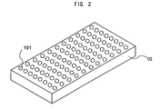
- Fig. 2 is a schematic diagram of a plate 10 of the detection kit 1 involving the embodiment.
- a plurality of wells 101 in which a sample is discharged.
- 12 plates on each of which, for instance, 8 wells are disposed in one row are connected and thereby 96 wells are disposed, and in each of the wells 101 a series of processes is carried out.
- a partial sectional view of the well is shown in Fig. 3.
- a sandwich method of detection methods described below antibodies are solid-phased beforehand.
- Fig. 3 as an example, a state where primary antibodies 111 are solid-phased is shown.
- a configuration of a detection kit that is used in an competitive method will be roughly explained.
- Reagents and so on that are used as a kit are substantially similar to those of the sandwich method.
- a sample and labeled antibodies are beforehand mixed, a mixture thereof is injected in the plate 20 followed by measuring the reaction, and thereby vitellogenin can be detected.
- the detection kit involving the embodiment when used, even when a concentration of vitellogenin contained in the sample is low, it can be detected with sensitivity and thereby more refined environmental evaluation can be carried out. Furthermore, when antigens or antibodies are solid-phased beforehand, the convenience also can be improved, the processing can be sped up and the reliability of obtained results can be improved as well.
- vitellogenin of the amphibians, frogs can be detected, and thereby over-all environmental evaluation can be performed.
- the abovementioned detection kit is used, and, based on the enzyme immunoassay, that is, the ELISA method, vitellogenin is detected.
- the detection method can cope with both.
- Fig. 4 is a process chart for explaining steps of the detection method according to the competitive method.
- Fig. 5 is a process chart for explaining steps of the detection method according to the sandwich method.
- a preparation is solid-phased.
- a preparation vitellogenin is diluted, followed by dispensing on a micro-plate, further followed by incubating (step 401), still further followed by immobilizing.
- the plate is washed (step 402) and a blocking reagent is dispensed to apply the blocking on a surface of a well of the plate (step 403). Thereafter, the plate is washed to remove an excessive chemical (step 404).
- step 4001 HRP labeled antibodies and a sample (or antigens) derived from a frog exposed to an environment are mixed (step 4001), followed by incubating (step 4002).
- step 4002 antigen-antibody complexes are dispensed on a antigen-solid-phased plate to cause an competitive reaction (step 405).
- step 406 the plate is washed (step 406) followed by injecting a chromogenic substrate to cause a coloring reaction (step 407).
- the absorbance is measured with a microreader or the like (step 408) and from a value thereof an amount of vitellogenin is calculated (step 409).
- a diluted solution of primary antibodies is dispensed in each of wells of a micro-plate followed by incubating for a predetermined period (step 501). Thereafter, the plate is washed (step 502), a blocking reagent is dispensed to complete immobilization (step 503), followed by dispensing a sample derived from frogs in an environment or a preparation vitellogenin (diluted solution) to cause reacting (step 504). Thereafter, the plate is washed (step 505), followed by dispensing secondary antibodies to react (step 506). Subsequently, the plate is washed (step 507) followed by dispensing a chromogenic substrate to cause a coloring reaction (step 508). After the coloring reaction, the absorbance is measured with a microreader or the like (step 509) and from a result thereof an amount of vitellogenin is calculated (step 510).
- the detection method two types of detection method can be used and various samples can be used. Furthermore, thereby, the detection sensitivity as well can be improved.
- An environmental evaluation method involving the embodiment by use of the abovementioned detection kit and the detection method, enables to use frog vitellogenin that has not been detected. Thereby, influences of the endocrine disruptors such as environmental hormones and various kinds of chemicals in the environments can be evaluated.
- a method of exposing a frog that is used as a sample to an environment methods such as addition of a target substance to breeding water and direct injection to individuals can be used. Furthermore, by capturing a frog in wildlife and obtaining a sample therefrom, the natural environment as that can be easily and accurately evaluated; accordingly, evaluations of various environments can be performed.
- samples are prepared from each of a thus obtained test body and a test body bred in a reference area, an amount of vitellogenin in the test body is measured and compared with that of the test body in the reference area, and therefrom the endocrine disrupting action of the target substance and the toxicity of the chemicals can be judged. That is, a male frog is bred in the presence of a chemical material (such as bisphenol A and phthalic acid ester) that is a target of evaluation, and an amount of vitellogenins in blood plasma is measured with time and compared with that of vitellogenin in blood serum obtained from a frog in the reference area.
- a chemical material such as bisphenol A and phthalic acid ester
- vitellogenin contained in the sample are reacted with antibodies according to the invention followed by measuring an amount of vitellogenins, and from the magnitudes of obtained measurements the toxicity (such as the endocrine disrupting influences) of the chemical substance can be evaluated.
- the intensity of the endocrine disruption effect of the chemical substance that is a target of evaluation can be evaluated relative to the intensity of estrogen.
- the endocrine disruption effect of the chemical substance can be judged as 10 times stronger than estrogen.
- the evaluation method when an amount of vitellogenins in a body liquid of a male frog living in a river or a lake that is a target of evaluation is measured, a situation of pollution of the environment owing to the chemical substance having the endocrine disruption effect can be evaluated.
- a polyclonal antibody of the frog vitellogenin can be manufactured according to any one of so far known methods (such as Sambrook, J et al., Molecular Cloning , Cold Spring Harbor Laboratory Press (1989)). However, an antibody manufactured according to a manufacturing method of polyclonal antibody described below is preferable. In what follows, based on the manufacturing method, the polyclonal antibody will be described.
- frog vitellogenin is administered as an antigen to immunize.
- An amount of administration of the antigen per one animal is, when, for instance, an adjuvant is used, 500 to 1000 ⁇ g.
- an adjuvant As the adjuvant, Freund's complete adjuvant (FCA), Freund's incomplete adjuvant (FIA) and aluminum hydroxide adjuvant can be cited.
- FCA Freund's complete adjuvant
- FIA Freund's incomplete adjuvant
- aluminum hydroxide adjuvant aluminum hydroxide adjuvant.
- the immunization is mainly performed by hypodermic injection.
- an interval of the immunization is not particularly restricted; however, it may be applied with an interval from several days to several weeks, and preferably after 3 weeks from a first immunization, 1 to 2 times at an interval of 2 weeks.
- antiserum After 5 to 20 days from the last immunization day, preferably after from 7 to 14 days, antiserum are sampled. Subsequently, from the obtained antiserum, by means of ammonium sulfate fractionation and DEAE-Sephadex column chromatography, IgG are obtained.
- vitellogeninpecific antibodies by use of an absorption column covalently coupled with frog serum proteins, adsorption purification is carried out, and by use of a frog vitellogenin column affinity purification is carried out.
- IgG are isolated from the antiserum but also by applying the affinity purification, an improvement in the specificity and sensitivity of the polyclonal antibody can be realized.
- vitellogenin synthesis is carried out in a frog body, blood is sampled from the frog followed by sampling a serum, and the serum is fractionated and purified.
- a method of isolating and purifying a serum centrifugal separation, gel-filtration column chromatography and so on can be properly combined, and thereby vitellogenin can be efficiently and accurately manufactured.
- Example 1a competitive method
- the detection sensitivities were verified of two methods according to the ELISA by setting a standard vitellogenin concentration in the range of 0.1 to 4000 ng/ml.
- the minimum detection limit (3SD) according to the competitive method was substantially 20 ng/ml (Fig. 7A)
- the minimum detection limit (3SD) according to the sandwich method was substantially 3 ng/ml. Accordingly, it was found that the sandwich method is higher in the sensitivity when the vitellogenin is detected on a lower concentration side.
- a quantification limit that shows the linearity was substantially 30 ng/ml.
- the Western blotting method was applied to detect the concentration and substantially 10 ng could be detected (Fig.8).
- the detection limit of the ELISA-competitive method was substantially 20 ng/ml
- the detection limit of the ELISA-sandwich method was substantially 3 ng/ml. Dispersion of measurements on a lower concentration side was large in the competitive method and small in the sandwich method. Accordingly, the sandwich method was more effective in detecting low concentration antigens. Furthermore, it is considered that when antibodies are sufficiently solid-phased and thereby a collision frequency between antigens and antibodies is heightened, the sensitivity can be effectively heightened. Accordingly, it is considered effective to process a plate with a reagent and so on to improve an immobilization efficiency of antibodies.
- the detection sensitivity of the Western blotting was studied. As a result, quantitative detection was possible at substantially 10 ng or more.
- the Western blotting tends to be lower in the detection sensitivity than the ELISA method, is troublesome in operations and takes a longer time to detect; however, it can advantageously qualitatively confirm antigens.
- the vitellogenin is likely to be decomposed, and in the western blotting, decomposition products may not be detected. Accordingly, it is considered that by combining both the ELISA and the western blotting method, influences of environmental chemicals on the synthesis of vitellogenin can be correctly evaluated.
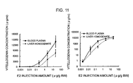
- the diluted sample was quantified of the vitellogenin according to the ELISA sandwich method.
- the diluted sample was quantified of the vitellogenin by ELISA sandwich.
- a production quantity of the vitellogenin was substantially 13000 ⁇ g/ml at 100 nM and that owing to the injection method was substantially 14000 ⁇ g/ml at 73 nM.
- the quantified vitellogenin concentrations were plotted logarithmically, the E2 concentration dependency on a lower concentration side could be visually expressed (right graphs of Figs. 9 and 10).
- Example 2-2 Test with primary hepatocyte culture
- the processing is carried out according to a method in which a test substance is added to a culture liquid to culture.
- a hepatocyte retrieves its function, adheres to a plate followed by extending; accordingly, from substantially second day in culture a culture liquid is exchanged to a culture liquid containing a material to be tested; on the eighth day, a culture liquid is sampled followed by detecting vitellogenin according to the ELISA method.
- the culture liquid is exchanged once every three days (when too many cells are seeded, frequent exchange is necessary).
- vitellogenin it is known that from the third day on after the processing vitellogenin can be detected in the culture liquid; however, since survived hepatocytes in culture are stable for three weeks, for the test of a substance low in the activity, evaluation is carried out for a longer period.
- example 3 in order to evaluate influences of chemicals having a estrogenic hormone action on amphibians, the vitellogenin (VTG) assay that uses a male Xenopus laevis was tried to make more sensitive.
- vitellogenin (VTG) assay that uses a male Xenopus laevis was tried to make more sensitive.
- polyclonal antibody against Xenopus laevis VTG was prepared anew, an ELISA kit for Xenopus laevis VTG was optimized and the ELISA kit was prepared, followed by evaluating the measurement accuracy.
- a gel filtration carrier (Sephadex-G25) was used.
- a BCA Bovine serum albumin
- BSA bovine serum albumin
- SDS-7.5% acrylamide gel electrophoresis/CBB oomassie brilliant blue staining was carried out.
- the VTG-purified product was used to prepare immunizing antigen and an ELISA standard, and the rest was preserved as a 50% glycerol solution of 0.5 mg/ml at -20 degrees centigrade.
- the IgG fraction of immunized rabbit blood serum (substantially 200 mg) was mixed with the adsorption purification column, followed by shaking at room temperature for 30 min. This was filtered and thereby antibodies to proteins in the blood serum of a normal male Xenopus laevis were eliminated. Furthermore, the filtrate was mixed with the affinity purification column and shaken at 4 degrees centigrade overnight, followed by eluting antibodies bonded to the column. A rise in the specific antibody titer due to purification was confirmed by means of the ELISA method.
- affinity-purified polyclonal antibodies were partially covalently bonded with horseradish peroxidase (HRP) according to a periodate oxidation method (Conjugation of Horseradish Peroxidase to Antibodies: Current Protocols in Molecular Biology, 11.1.2), and thereby HRP-labeled polyclonal antibodies were prepared.
- HRP horseradish peroxidase
- a SDS-7.5% acrylamide gel was prepared, 0.025 ⁇ l equivalent of each of blood serum of an adult male Xenopus laevis injected with E2 (substantially 1 mg) and blood serum of a normal male Xenopus laevis and 100 ng equivalent of purified VTG antigens were electrophoresed, and proteins separated in the gel were blotted on a membrane according to a semi-dry blotting method.
- An immunozation reaction was applied at anti-VTG affinity-purified polyclonal antibodies 1 ⁇ g/ml, followed by reacting with HRP-labeled anti-rabbit IgG goat antibodies (secondary antibodies), and the VTG was detected on an X-ray film according to a chemical fluorescence staining method. Thereby, the specificity of the antibodies was verified.
- a concentration of the affinity-purified VTG polyclonal antibody that is immobilized on a plate was set at 1.25, 2.5 and 5 ⁇ g/ml, the immobilization was carried out at room temperature for 1 hr, thereby in the range of 0 to 1000 ng/ml of the standard VTG a calibration curve was prepared. Furthermore, with one that was solid-phased at an appropriated concentration of 1.4 ⁇ g/ml at 4 degrees centigrade overnight, an experiment to get calibration curve was simultaneously carried out. By comparing results, an optimum concentration to get solid-phased VTG antibodies was studied.
- the detection sensitivity according to the ELISA method depends on a labeling efficiency of HRP to the VTG polyclonal antibody. Accordingly, a concentration of the HRP-labeled Anti-VTG antibody used has to be optimized. Of the HRP-labeled Anti-VTG antibody prepared this time, a concentration was set at 1, 2 and 4 ⁇ g/ml and an optimum concentration of the HRP-labeled Anti-VTG antibody in the ELISA method was studied.
- VTG concentrations in the samples were quantitatively determined and thereby recovery rates to added amounts were calculated. Furthermore, in order to confirm the dilution-dependent linear regression, diluted with a dilution solution to which 1% BSA was added, VTG in blood plasma samples 1 and 2 (both are male blood plasma exposed in water to 10 nM E2 for 7 days) were measured in a dilution sequence from 1/400 to 1/2 dilution, thereby VTG quantification was carried out.
- E2 substantially 1 mg was injected to a Xenopus tropicalis that is a closely-related species of the Xenopus genus, a Bombina bombina of the Bombina genus and a Rana rugosa, a Rana limnocharis Boie and a Rana nigromaculata of the Rana genus, and a Japanese tree frog of the Hyla genus, and a Schelegel's green tree frog of the Rhacophorus genus, followed by breeding for 6 days, further followed by sampling blood serum.
- each of the blood serum was diluted stepwise from 1/200 to quantify the VTG according to the ELISA, from the VTG calibration curve of the Xenopus laevis a VTG concentration was estimated, and thereby an immuno reactive amount of VTG of a frog of other genus was obtained.
- each of frog blood serum proteins was separated with SDS-PAGE so that an amount of the VTG that is recognized by the antibody may be equivalent, followed by CBB staining. By comparing a density of a CBB stained band at a position of substantially 200 kDa in a CBB stained gel photograph, a degree of the cross-reactivity was expressed.
- hepatocytes of an adult male Xenopus laevis were primarily cultured, followed by culturing in a culture liquid containing a known estrogenic substance such as E2, Estrone (E1) or Estriol (E3).
- E2 Estrone
- E3 Estriol
- Substantially 10 fractions including a peak due to the VTG were confirmed by with SDS-PAGE/CBB staining method (Fig. 14) and fractions that do not contain the serum albumin were collected.
- VTG standard other than one that was used for preparation of the ELISA KIT was rendered a 50% glycerol solution and preserved at -20 degrees centigrade. It was found that, thereby, the VTG could be inhibited from denaturing and precipitating; accordingly, it could be preserved for a long period. Finally, from blood serum of an adult male Xenopus laevis injected with E2 by an amount corresponding to 30 ⁇ g/g body weight, substantially 13 mg of the VTG was purified.
- the affinity-purified Anti-VTG antibody that is used for immobilization on a plate was set to immobilize at concentrations of 1.25, 2.5 and 5 ⁇ g/ml for 1 hr and at a concentration of 1.4 ⁇ g/ml overnight, and thereby calibration curves were prepared (Fig. 21).
- a sufficient reaction was obtained; however, even at a concentration of 1.4 ⁇ g/ml, by immobilizing overnight, the reactivity close to results under the immobilization conditions at 5 ⁇ g/ml could be obtained.
- the immobilization conditions at the ELISA were set at 50 ⁇ l/well of affinity purified Anti-VTG antibody, at a concentration of 1 ⁇ g/ml, at 4 degrees centigrade overnight.
- the ELISA was performed, and thereby calibration curves were prepared (Fig. 22).
- the concentration of the HRP-labeled antibody became higher, the stained became stronger as a whole; however, in view of an increase in a nonspecific reaction of a negative reference when the antibody concentration is too high and an amount of antibody that can be prepared from the same lot, in the lot, the antibody was judged optimally used at a concentration of 2 ⁇ g/ml.
- Blood plasma of a Xenopus laevis normal male was diluted in series to 1/20 to 1/100000, to each thereof standard vitellogenin was added so as to be 500, 100 and 20 ng/ml, and thereby samples were prepared and quantified.
- the blood plasma exhibited inhibiting influences, and at 1/20 dilution substantially 85 percent was inhibited.
- the recovery rate became higher, and at a dilution factor of 100000 or more, a quantification value of the vitellogenin close to an expected value was obtained (Fig. 23A).
- the recovery rate was substantially constant for each of the blood plasma dilution factors, and error was substantially ⁇ 10 percent. Vitellogenin in the normal male blood serum was below the detection limit of the ELISA method.
- the blood drawing from the nail-removed surface was also judged inappropriate as the blood drawing method because it was found that 1 ⁇ an amount of blood that oozes is slight and 2 ⁇ the nail-removed surface takes a long time to heal.
- a method of drawing blood by stinging a needle to a position from an underarm portion to a flank was judged appropriate as the blood drawing method because 1 ⁇ the blood drawing is easy because oozed blood becomes ball-like and 2 ⁇ a sting is easy to heal.
- a method below was cited as an example of the blood drawing method.
- a calibration curve was prepared. From a VTG solution containing 1000 ng/ml of VTG, a 1/2 dilution sequence (1000 to 2, and 0 ng/ml) was prepared, an ELISA KIT protocol (attached at the end) was followed, and thereby a calibration curve such as shown in Fig. 26 was obtained.
- E2 substantially 1 mg was injected to various kinds of frogs, and after the breeding of 6 days, blood serum were collected. From each of the blood serum diluted to 1/200, a dilution sequence of the serum was prepared, followed by applying the ELISA (Fig. 29A). From a calibration curve that uses a VTG standard of Xenopus laevis, in various kinds of frog blood serum, Xenopus laevis VTG-equivalent values were estimated.
- Example 2-2 primary cultured hepatocytes of an adult male Xenopus laevis were prepared, followed by culturing in a culture liquid containing one of the chemicals, and with a culture liquid after 6 days, according to the ELISA, a VTG concentration in the culture liquid was quantified.
- the antagonist activity of a target substance could be evaluated. For instance, when the hepatocyte was cultured in a culture liquid to which 5 nM E2 and BPA, NP or OP were added, to the VTG synthesis induction due to 5 nM E2 alone, as a concentration of added BPA, NP or OP becomes higher, the antagonistic activity was exhibited (Fig. 31). From the results, it is indicated that the vitellogenin assay that uses a hepatocyte culture can evaluate, of the various chemicals, not only the estrogenic activity but also the anti-estrogenic activity.
- a Xenopus laevis VTG polyclonal antibody was newly prepared and an ELISA KIT was devised.
- a quantification range of the VTG concentration in the present ELISA KIT was 2 to 1000 ng/ml.
- the 95% reliable minimum detection limit was substantially 2 ng/ml and the quantitativity was maintained up to substantially 1000 ng/ml; that is, the sensitivity was improved.
- An expansion in the quantification range is assumed due to a change of an antibody solid-phased on a plate from an adsorption purification antibody to an affinity purification antibody.
- the blood plasma exhibited rather high reaction inhibition effect and at a dilution factor of 20 substantially 85% inhibition was found.
- the recovery rate became higher, and, at a dilution factor of 100000 or more, quantitatively determined vitellogenin values became values close to expected values.
- a composition of a sample dilution solution was studied. It is considered that owing to a protein contained in the blood plasma, a reaction between VTG and anti-VTG antibody is inhibited for some reasons. Since serum albumin is much contained in the blood plasma, when BSA was beforehand added to a sample dilution solution, the recovery rate of VTG became substantially 100% when the blood plasma was diluted at a factor of 200 or more.
- the minimum detection limit in the standard VTG is 2 ng/ml
- the minimum detection limit of the VTG concentration in blood plasma becomes 400 ng/ml.
- the recovery rate is substantially 60% at a dilution factor of 20 of the blood plasma
- a sample preparation is set at 1/20 dilution of the blood plasma, a quantitatively determined value can be calibrated at the recovery rate of 60%.
- the minimum detection limit of the VTG concentration in blood plasma becomes substantially 70 ng/ml.
- the present ELISA KIT when an adult male Xenopus laevis was exposed to E2 of 1 nM or more in water for 7 days, can detect blood plasma VTG.
- the antibody that was used in the ELISA KIT was confirmed to exhibit the cross-reactivity also to VTGs of frog species (Xenopus tropicalis, Bombina bombina, Rana rugosa, Rana limnocharis limnocharis, Japanese tree frog, Schlegel ⁇ s green tree frog, and Black-spotted pond frog) other than Xenopuas laevis.
- frog species Xenopus tropicalis, Bombina bombina, Rana rugosa, Rana limnocharis limnocharis, Japanese tree frog, Schlegel ⁇ s green tree frog, and Black-spotted pond frog
- vitellogenin antibody was followed.
- Peroxidase (Trade name POD, manufactured by Boeringer-ingelheim Co., for use in EIA, Code No. 814393) was dissolved at a concentration of 20 mg/ml in 0.5 ml of a 0.1 M carbonate buffer at pH9.2, followed by blending 0.5 ml of a NaIO4 solution and 0.5 ml of a HRP solution, further followed by reacting at room temperature in a dark place for 2 hr. Thereto, 1.0 ml of an antibody solution (3 mg/ml in 0.1 M phosphate buffer, pH 6.8) was added, followed by keeping for 3 hr in a dark place at room temperature.
- a NaBH 4 solution (5 mg/ml in 0.1 mM NaOH) was added, followed by keeping for 30 min in a dark place at room temperature, further followed by adding 179 ⁇ l of a NaBH 4 solution and standing in a dark place for 60 min. Thereto, 2 ml of saturated ammonium sulfate was added, followed by agitating for 30 min over ice. The solution was centrifuged at 4 degrees centigrade and 15000 rpm for 10 min and precipitate was dissolved in 1 ml of a TEN buffer (50 mM Tris-HCl, 1 mM EDTA and 0.9% NaCl).
- a TEN buffer 50 mM Tris-HCl, 1 mM EDTA and 0.9% NaCl
- BSA was dissolved at a concentration of 20 mg/ml.
- a 50% glycerol solution was prepared and preserved at -20 degrees centigrade.
- An anti-vitellogenin antibody dissolved in Dulbecco's PBS(-) (Code No. 041-20211, manufactured by Wako Pure Chemical Industries, Ltd) (1 ⁇ g/ml) was dispensed on an immobilization plate (trade name EIA/RIA plate strip 8, #2592, manufactured by Costar) so as to be 50 ⁇ l/well, followed by standing at 4 degrees centigrade overnight, further followed by washing three times with 300 ⁇ l of cleaning liquid (0.05% Tween 20 containing PBS).
- a blocking liquid ((1% Block Ace (trade name: UK-B80, manufactured by Snow Brand Milk Prod.
- a vitellogenin dilution solution (0.15 M NaCl + 0.01% Slaoff 72N (manufactured by Takeda Chemical Industries, Ltd.) + 4% BSA + 10% sucrose in 50 mM HEPES-Na (pH 7.4) was diluted so as to be 5 ⁇ g/ml, followed by dispensing every 200 ⁇ l, further followed by freezing and drying.
- BSA trade name 7638, manufactured by Sigma
- an enzyme-labeled antibody dilution solution (0.15 M NaCl + 0.01% Slaoff 72N (manufactured by Takeda Chemical Industries, Ltd.) + 4% BSA + 10% sucrose in 50 mM HEPES-Na (pH7.4)
- an enzyme-labeled antibody was diluted so as to be 60 ⁇ g/ml, followed by dispensing 100 ⁇ l each, further followed by freezing and drying.
- a solution of 1 N phosphoric acid was prepared, followed by dispensing 15 ml each in a proper container and applying a cap thereto, further followed by preserving at room temperature.
- Quantification by use of a frog vitellogenin ELISA KIT prepared according to example 4 is carried out as follows.
- sample dilution solution (a concentration factor is three) and distilled water are mixed at a mixing ratio of 1: 2 to prepare a "sample dilution solution".
- Blood plasma or blood serum is diluted with the "sample dilution solution" prepared according to (1) so as to come into the quantifiable range (3 to 1,000 ng/mL).
- the "powder of vitellogenin standard" is dissolved with 200 ⁇ L of distilled water (5,000 ng/mL).
- sample dilution solution a vitellogenin standard solution having a necessary concentration is prepared.
- a necessary amount of the 5,000 ng/mL solution is sampled and the rest is refrigerated and preserved.
- the 5,000 ng/mL solution is diluted to 1/5 with the "sample dilution solution" to 1,000 ng/mL.
- the sample dilution solution is diluted in sequence to 1/4 to prepare 1,000, 250, 62.5, 15.6, 3.9 and 0.98 ng/mL.
- the sample dilution solution is used as that.
- each of the “sample” and “vitellogenin standard solution” respectively prepared in (2) and (3) is added at 50 ⁇ L/well, followed by reacting at room temperature (18 to 25 degrees centigrade) for 60 min.
- the "cleaning liquid (concentration factor: 6)" and distilled water are mixed at a blending ratio of 1: 5 to prepare a "cleaning liquid".
- a reaction liquid is discarded, and 300 ⁇ L/well of the cleaning liquid prepared in (5) is used three-times to wash the inside of the well. After the third cleaning liquid is discarded, a microplate that is turned upside-down is lightly tapped with paper towel or the like to completely remove the cleaning liquid.
- a 50 ⁇ L aliquot of the "labeled antibody solution" prepared in (7) is added to each well, followed by reacting at room temperature (18 to 25 degrees centigrade) for 60 min.
- a reaction liquid is discarded, and an aliquot of 300 ⁇ L/well of the cleaning liquid prepared in (5) is used three-times to wash the inside of the well. After the third cleaning liquid is discarded, the microplate that is turned upside-down is lightly tapped with paper towel or the like to completely remove the cleaning liquid.
- the "chromogenic substrate solution” and the “chromogenic dilution solution” are mixed at a mixing ratio of 1: 100 to prepare a "chromogenic reagent”.
- the absorbance at a wavelength 450 nm is measured.
- a vitellogenin concentration in the sample is calculated from a calibration curve (Fig. 32).
- a vitellogenin recovery rate curve (the VTG recovery rate versus sample dilution factor) (Fig. 33)
- measurements are corrected.
- a quantification range according to the procedure was 3 to 1000 ng/ml. As shown in Fig. 33, it was found that when the blood plasma was diluted at a factor of 200 or more, the recovery rate of 90 to 100% could be obtained.
- vitellogenin antibody was followed.
- a quantification method by use of a high sensitivity ELISA KIT prepared according to example 6 is carried out as follows.
- the "powder of vitellogenin standard" is diluted with 200 ⁇ L of distilled water (5,000 ng/mL).
- sample dilution solution a vitellogenin standard solution having a necessary concentration is prepared.
- a necessary amount of the 5,000 ng/mL solution is fractioned and the rest is refrigerated and preserved.
- the 5,000 ng/mL solution is diluted to 1/80 with the "sample dilution solution" to 62.5 ng/mL.
- the sample dilution solution is diluted in sequence to 1/2 to prepare 31.3, 15.6, 7.8, 3.9, 2, 0.98, 0.49, 0.24, 0.12, 0.06, 0.03 and 0 ng/mL.
- the "sample dilution solution” is used as that.
- each of the “sample” and “vitellogenin standard solution” respectively prepared in (2) and (3) is added at 50 ⁇ L/well, followed by reacting at room temperature (18 to 25 degrees centigrade) for 60 min.
- the "cleaning liquid (concentration factor: 6)" and distilled water are mixed at a ratio of 1: 5 to prepare a "cleaning liquid".
- a reaction liquid is discarded, and 300 ⁇ L/well of the cleaning liquid prepared in (5) is used three-times to wash the inside of the well. After the third cleaning liquid is discarded, a microplate that is turned upside-down is lightly tapped with paper towel or the like to completely remove the cleaning liquid.
- a reaction liquid is discarded, and an aliquot of 300 ⁇ L of the cleaning liquid prepared in (5) is used three-times to wash the inside of the well. After the third cleaning liquid is discarded, the microplate that is turned upside-down is lightly tapped with paper towel or the like to completely remove the cleaning liquid.
- a reaction liquid is discarded, and a 300 ⁇ L of the cleaning liquid prepared in (5) is used three-times to wash the inside of the well. After the third cleaning liquid is discarded, the microplate is turned upside-down and lightly tapped with paper towel or the like to completely remove the cleaning liquid.
- the "chromogenic substrate solution” and the “chromogenic substrate dilution solution” are mixed at a ratio of 1: 100 to prepare a "chromogenic reagent”.
- the absorbance at a wavelength 450 nm is measured.
- a vitellogenin concentration in the sample is calculated from a calibration curve (Fig. 34).
- a vitellogenin recovery rate curve (the VTG recovery rate versus sample dilution factor)
- measurements are corrected. It was found that a quantification range according to the procedure was 0.06 to 62.4 ng/ml and the present procedure was 50 times more sensitive than an existing procedure. As shown in Fig. 35, it was found that when the blood plasma was diluted at a factor of 50 or more, the dilution-dependent linear regression could be obtained.
- the present invention is not restricted to the abovementioned examples.
- an immunochromatography method may be used.
- the immunochromatography method uses, as a principle, a sandwich method and is widely used in such as pregnancy determination and so on. Now, an immunochromatography strip 1007 in the present embodiment will be described based on Fig. 36.
- an immunochromatography strip 1007 has a structure where between plastics covers 1001a and 1001b, a sample pad 1002, a conjugate pad 1003, a membrane 1006 and an absorption pad 1008 are interposed.
- a judgment line 1004 antibody and a control line 1005 antibody are coated on the respective predetermined places.
- a test body when dropped inside of a region covered with the plastics covers 1001a and 1001b, permeates inside of the sample pad 1002 and the conjugate pad 1003 positioned therebelow and there permeates in a horizontal direction (from left to right in the drawing) while reacting with a gold colloid-labeled Anti-VTG antibody.
- the judgment line 1004 has solid-phased antibodies. When the test body passes through there, an antigen-antibody reaction occurs to develop red color, and part of the test body reacts with labeled antibody-specific antibodies solid-phased ahead thereof in the control line 1005 and develops red color. In a positive reaction, two of the judgment line and the control line develop red color and in a negative reaction only one of control line develops red color.
- judgment line 1004 antibodies (anti-VTG antibodies) and control line 1005 antibodies (anti-rabbit IgG antibodies) are coated in lines, followed by drying to immobilize, further followed by applying blocking to suppress the non-specific adsorption, still further followed by washing, and thereby coating of the antibodies comes to completion.
- Fig. 36 when various kinds of members including these are sandwiched with the plastics covers 1001a and 1001b, an immunochromatography device is manufactured.
- a measurement with a vitellogenin sample of a known concentration confirmed to be in the measurement range of 20 ng/ml to 10 mg/ml.
- blood was sampled from an adult male Xenopus laevis exposed to 1 nM E2 for one week, obtained blood serum was diluted in three levels (x 1, x 100 and x 10,000), and 100 ⁇ l thereof was dropped in a sample pad 1002.
- a concentration range of vitellogenin was specified.
- a concentration range specified according to the present kit when compared with the concentration quantified according to the ELISA method, exhibited complete coincidence.
- frog vitellogenin can be detected with excellent sensitivity and accuracy. Accordingly, by use thereof, influences of endocrine disruptors in environments can be comprehensively evaluated.
Landscapes
- Life Sciences & Earth Sciences (AREA)
- Health & Medical Sciences (AREA)
- Chemical & Material Sciences (AREA)
- Immunology (AREA)
- Engineering & Computer Science (AREA)
- Molecular Biology (AREA)
- General Health & Medical Sciences (AREA)
- Medicinal Chemistry (AREA)
- Organic Chemistry (AREA)
- Proteomics, Peptides & Aminoacids (AREA)
- Biochemistry (AREA)
- Urology & Nephrology (AREA)
- Biomedical Technology (AREA)
- Hematology (AREA)
- Physics & Mathematics (AREA)
- Food Science & Technology (AREA)
- Analytical Chemistry (AREA)
- Biotechnology (AREA)
- Microbiology (AREA)
- General Physics & Mathematics (AREA)
- Pathology (AREA)
- Biophysics (AREA)
- Genetics & Genomics (AREA)
- Cell Biology (AREA)
- Peptides Or Proteins (AREA)
Applications Claiming Priority (3)
| Application Number | Priority Date | Filing Date | Title |
|---|---|---|---|
| JP2002167920 | 2002-06-07 | ||
| JP2002167920 | 2002-06-07 | ||
| PCT/JP2003/007296 WO2003104806A1 (ja) | 2002-06-07 | 2003-06-09 | 検出キット、それに用いられる測定プレート、検出方法、評価方法、カエルビテロジェニンのポリクローナル抗体及びその製造方法 |
Publications (2)
| Publication Number | Publication Date |
|---|---|
| EP1533616A1 true EP1533616A1 (de) | 2005-05-25 |
| EP1533616A4 EP1533616A4 (de) | 2007-01-17 |
Family
ID=29727676
Family Applications (1)
| Application Number | Title | Priority Date | Filing Date |
|---|---|---|---|
| EP03736119A Withdrawn EP1533616A4 (de) | 2002-06-07 | 2003-06-09 | Detektionskitt, darin zu verwendende assay-platte, detektionsverfahren, auswertungsverfahren, polyklonalantikörper von frosch-vitellogenin und prozess zur herstellung davon |
Country Status (5)
| Country | Link |
|---|---|
| US (1) | US20050244906A1 (de) |
| EP (1) | EP1533616A4 (de) |
| JP (1) | JPWO2003104806A1 (de) |
| AU (1) | AU2003242106A1 (de) |
| WO (1) | WO2003104806A1 (de) |
Families Citing this family (9)
| Publication number | Priority date | Publication date | Assignee | Title |
|---|---|---|---|---|
| KR100682395B1 (ko) * | 2004-12-31 | 2007-02-15 | 부산대학교 산학협력단 | 호스라디시 페록시다제가 컨쥬게이션된 붕어 비텔로제닌의모노클로랄 항체와 이를 이용한 비텔로제닌 측정 방법 |
| CN100595565C (zh) * | 2007-09-27 | 2010-03-24 | 中国人民解放军第三军医大学第一附属医院 | 一种基于ERα激活效应的环境内分泌干扰物的PCR定量检测方法与试剂盒 |
| CN102768282B (zh) * | 2012-07-20 | 2014-11-26 | 湖北省农业科学院农产品加工与核农技术研究所 | 一种检测鲢鱼卵黄蛋白原试剂盒的制备及检测方法 |
| US20170089927A1 (en) * | 2015-09-25 | 2017-03-30 | Iontox, Llc | Endocrine disruptor field tester |
| CN109297867B (zh) * | 2018-10-15 | 2023-10-13 | 成都理工大学 | 一种污染区现场快速检测管及检测方法 |
| CN111273036B (zh) * | 2020-03-06 | 2021-06-15 | 中国农业科学院兰州兽医研究所 | 检测口蹄疫非结构蛋白抗体的单抗竞争化学发光试剂盒 |
| CN112158452A (zh) * | 2020-08-27 | 2021-01-01 | 新疆卡纳思生物技术有限公司 | 一种便于标记的低密度脂蛋白胆固醇测定试剂盒 |
| JPWO2022138460A1 (de) * | 2020-12-24 | 2022-06-30 | ||
| CN112760325B (zh) * | 2021-02-05 | 2022-05-20 | 中国水产科学研究院黄海水产研究所 | 多棘海盘车卵黄蛋白原的基因、蛋白、多克隆抗体及应用 |
Family Cites Families (2)
| Publication number | Priority date | Publication date | Assignee | Title |
|---|---|---|---|---|
| US5153117A (en) * | 1990-03-27 | 1992-10-06 | Genetype A.G. | Fetal cell recovery method |
| US5304496A (en) * | 1991-06-10 | 1994-04-19 | Children's Hospital Of Philadelphia | Biological regulation of mineralization |
-
2003
- 2003-06-09 AU AU2003242106A patent/AU2003242106A1/en not_active Abandoned
- 2003-06-09 US US10/516,823 patent/US20050244906A1/en not_active Abandoned
- 2003-06-09 JP JP2004511826A patent/JPWO2003104806A1/ja active Pending
- 2003-06-09 WO PCT/JP2003/007296 patent/WO2003104806A1/ja not_active Ceased
- 2003-06-09 EP EP03736119A patent/EP1533616A4/de not_active Withdrawn
Non-Patent Citations (8)
| Title |
|---|
| DENSLOW NANCY D ET AL: "Vitellogenin as a biomarker of exposure for estrogen or estrogen mimics" ECOTOXICOLOGY, vol. 8, no. 5, October 1999 (1999-10), pages 385-398, XP002399650 ISSN: 0963-9292 * |
| HIRAMATSU NAOSHI ET AL: "Transition of serum vitellogenin cycle in Sakhalin Taimen (Hucho perryi)" COMPARATIVE BIOCHEMISTRY AND PHYSIOLOGY C PHARMACOLOGY TOXICOLOGY AND ENDOCRINOLOGY, vol. 118, no. 2, October 1997 (1997-10), pages 149-157, XP002399652 ISSN: 0742-8413 * |
| HURTER E ET AL: "Validation of an ex vivo Xenopus liver slice bioassay for environmental estrogens and estrogen mimics" ECOTOXICOLOGY AND ENVIRONMENTAL SAFETY, vol. 53, no. 1, September 2002 (2002-09), pages 178-187, XP002399653 ISSN: 0147-6513 * |
| JOBLING S ET AL: "DETERGENT COMPONENTS IN SEWAGE EFFLUENT ARE WEAKLY OESTROGENIC TO FISH: AN IN VITRO STUDY USING RAINBOW TROUT (ONCORHYNCHUS MYKISS) HEPATOCYTES" AQUATIC TOXICOLOGY, ELSEVIER SCIENCE PUBLISHERS BV, vol. 27, no. 3/4, 1 December 1993 (1993-12-01), pages 361-372, XP000565379 ISSN: 0166-445X * |
| KLOAS WERNER ET AL: "Amphibians as a model to study endocrine disruptors: II. Estrogenic activity of environmental chemicals in vitro and in vivo" SCIENCE OF THE TOTAL ENVIRONMENT, vol. 225, no. 1-2, 12 January 1999 (1999-01-12), pages 59-68, XP002409925 ISSN: 0048-9697 * |
| MITSUI NAOKO ET AL: "Sandwich ELISAs for quantification of Xenopus laevis vitellogenin and albumin and their application to measurement of estradiol-17beta effects on whole animals and primary-cultured hepatocytes." COMPARATIVE BIOCHEMISTRY AND PHYSIOLOGY PART C TOXICOLOGY AND PHARMACOLOGY, vol. 135C, no. 3, July 2003 (2003-07), pages 305-313, XP002399654 ISSN: 1532-0456 * |
| MOSCONI G ET AL: "Seasonal changes in plasma growth hormone and prolactin concentrations of the frog Rana esculenta" GENERAL AND COMPARATIVE ENDOCRINOLOGY, vol. 93, no. 3, 1994, pages 380-387, XP002399651 ISSN: 0016-6480 * |
| See also references of WO03104806A1 * |
Also Published As
| Publication number | Publication date |
|---|---|
| WO2003104806A1 (ja) | 2003-12-18 |
| EP1533616A4 (de) | 2007-01-17 |
| AU2003242106A8 (en) | 2003-12-22 |
| AU2003242106A1 (en) | 2003-12-22 |
| JPWO2003104806A1 (ja) | 2005-10-06 |
| US20050244906A1 (en) | 2005-11-03 |
Similar Documents
| Publication | Publication Date | Title |
|---|---|---|
| Yulaev et al. | Development of a potentiometric immunosensor for herbicide simazine and its application for food testing | |
| JP2011232361A (ja) | 動物において早期腎疾患を検出する方法 | |
| EP3394618B1 (de) | Vorrichtung für den nachweis von neurotoxinen und verfahren zur herstellung davon | |
| EP1533616A1 (de) | Detektionskitt, darin zu verwendende assay-platte, detektionsverfahren, auswertungsverfahren, polyklonalantikörper von frosch-vitellogenin und prozess zur herstellung davon | |
| EP2511707B1 (de) | Verfahren zur detektion eines basischen peptids und reagenz zur detektion eines basischen peptids | |
| VanVunakis et al. | Use of the double antibody and nitrocellulose membrane filtration techniques to separate free antigen from antibody bound antigen in radioimmunoassays | |
| Nakajima et al. | Development of an enzyme-linked immunosorbent assay for metallothionein-I and-II in plasma of humans and experimental animals | |
| US7763433B2 (en) | Analyte detection system | |
| JP2019053068A (ja) | 可溶型線維芽細胞増殖因子受容体の高感度多重イムノアッセイ | |
| EP1071956B1 (de) | Verfahren zum bestimmen von autoantikörpern | |
| AU769179B2 (en) | Assays for mastitis detecting haptoglobin in milk | |
| US20200209252A1 (en) | Method and test kit for the quantitative determination of biomarkers in fecal samples | |
| CN102944682A (zh) | 蟹类抗体谱检测试剂盒 | |
| EP1360499B1 (de) | Bestimmung des zytosolischen nadp-abhängigen malatdecarboxylase-isoenzyms | |
| WO2012022285A2 (de) | Verfahren zum nachweis von l-pk-tumormarkern im stuhl zur erkennung von lebererkrankungen | |
| US8039226B2 (en) | Anti NC1 monoclonal antibody | |
| KR100585423B1 (ko) | 내분비 교란물질에 대한 효소면역 진단방법과 진단키트 | |
| CN120040588B (zh) | 抗T3抗体mAb68或抗原结合片段、制备方法和应用 | |
| KR101070526B1 (ko) | 페난트렌 오염 검출을 위한 마커 및 키트 | |
| Pradhan et al. | Detection of ricin by comparative indirect ELISA using fluorescence probe in blood and red blood cells | |
| WO2026003878A1 (en) | Colorimetric urine test for early pregnancy detection in livestocks | |
| JP2004219111A (ja) | 免疫組織化学染色用ブロッキング試薬 | |
| CN113072637A (zh) | 氨基脲抗原、胶体金免疫层析检测装置与检测判定方法 | |
| Taioli et al. | Molecular markers of exposure to cadmium and nickel among alkaline battery workers | |
| Palleschi et al. | Rapid detection of seafood toxins |
Legal Events
| Date | Code | Title | Description |
|---|---|---|---|
| PUAI | Public reference made under article 153(3) epc to a published international application that has entered the european phase |
Free format text: ORIGINAL CODE: 0009012 |
|
| 17P | Request for examination filed |
Effective date: 20050107 |
|
| AK | Designated contracting states |
Kind code of ref document: A1 Designated state(s): AT BE BG CH CY CZ DE DK EE ES FI FR GB GR HU IE IT LI LU MC NL PT RO SE SI SK TR |
|
| AX | Request for extension of the european patent |
Extension state: AL LT LV MK |
|
| DAX | Request for extension of the european patent (deleted) | ||
| A4 | Supplementary search report drawn up and despatched |
Effective date: 20061219 |
|
| 17Q | First examination report despatched |
Effective date: 20070718 |
|
| RAP1 | Party data changed (applicant data changed or rights of an application transferred) |
Owner name: TOWA KAGAKU CO., LTD. |
|
| R17C | First examination report despatched (corrected) |
Effective date: 20070911 |
|
| STAA | Information on the status of an ep patent application or granted ep patent |
Free format text: STATUS: THE APPLICATION IS DEEMED TO BE WITHDRAWN |
|
| 18D | Application deemed to be withdrawn |
Effective date: 20090609 |