EP1522289A2 - Sub-tenon drug delivery - Google Patents
Sub-tenon drug delivery Download PDFInfo
- Publication number
- EP1522289A2 EP1522289A2 EP05100176A EP05100176A EP1522289A2 EP 1522289 A2 EP1522289 A2 EP 1522289A2 EP 05100176 A EP05100176 A EP 05100176A EP 05100176 A EP05100176 A EP 05100176A EP 1522289 A2 EP1522289 A2 EP 1522289A2
- Authority
- EP
- European Patent Office
- Prior art keywords
- cannula
- distal portion
- tip
- tenon
- lumen
- Prior art date
- Legal status (The legal status is an assumption and is not a legal conclusion. Google has not performed a legal analysis and makes no representation as to the accuracy of the status listed.)
- Withdrawn
Links
Images


Classifications
-
- A—HUMAN NECESSITIES
- A61—MEDICAL OR VETERINARY SCIENCE; HYGIENE
- A61F—FILTERS IMPLANTABLE INTO BLOOD VESSELS; PROSTHESES; DEVICES PROVIDING PATENCY TO, OR PREVENTING COLLAPSING OF, TUBULAR STRUCTURES OF THE BODY, e.g. STENTS; ORTHOPAEDIC, NURSING OR CONTRACEPTIVE DEVICES; FOMENTATION; TREATMENT OR PROTECTION OF EYES OR EARS; BANDAGES, DRESSINGS OR ABSORBENT PADS; FIRST-AID KITS
- A61F9/00—Methods or devices for treatment of the eyes; Devices for putting in contact-lenses; Devices to correct squinting; Apparatus to guide the blind; Protective devices for the eyes, carried on the body or in the hand
- A61F9/0008—Introducing ophthalmic products into the ocular cavity or retaining products therein
- A61F9/0017—Introducing ophthalmic products into the ocular cavity or retaining products therein implantable in, or in contact with, the eye, e.g. ocular inserts
-
- A—HUMAN NECESSITIES
- A61—MEDICAL OR VETERINARY SCIENCE; HYGIENE
- A61F—FILTERS IMPLANTABLE INTO BLOOD VESSELS; PROSTHESES; DEVICES PROVIDING PATENCY TO, OR PREVENTING COLLAPSING OF, TUBULAR STRUCTURES OF THE BODY, e.g. STENTS; ORTHOPAEDIC, NURSING OR CONTRACEPTIVE DEVICES; FOMENTATION; TREATMENT OR PROTECTION OF EYES OR EARS; BANDAGES, DRESSINGS OR ABSORBENT PADS; FIRST-AID KITS
- A61F2250/00—Special features of prostheses classified in groups A61F2/00 - A61F2/26 or A61F2/82 or A61F9/00 or A61F11/00 or subgroups thereof
- A61F2250/0058—Additional features; Implant or prostheses properties not otherwise provided for
- A61F2250/0067—Means for introducing or releasing pharmaceutical products into the body
Definitions
- the present invention generally pertains to the delivery of ophthalmically acceptable pharmaceutically active agents to the back of the eye. More particularly, but not by way of limitation, the present invention pertains to apparatus for sub-Tenon delivery of a drug depot to the posterior segment of a human eye proximate the macula.
- Age related macular degeneration (ARMD), choroidal neovascularization (CNV), retinopathies (e.g., diabetic retinopathy, vitreoretinopathy), retinitis (e.g., cytomegalovirus (CMV) retinitis), uveitis, macular edema, and glaucoma are several examples.
- AMD Age related macular degeneration
- CNV choroidal neovascularization
- retinopathies e.g., diabetic retinopathy, vitreoretinopathy
- retinitis e.g., cytomegalovirus (CMV) retinitis
- uveitis macular edema
- macular edema glaucoma
- Age related macular degeneration is the leading cause of blindness in the elderly. ARMD attacks the center of vision and blurs it, making reading, driving, and other detailed tasks difficult or impossible. About 200,000 new cases of ARMD occur each year in the United States alone. Current estimates reveal that approximately forty percent of the population over age 75, and approximately twenty percent of the population over age 60. suffer from some degree of macular degeneration. "Wet" ARMD is the type of ARMD that most often causes blindness. In wet ARMD, newly formed choroidal blood vessels (choroidal neovascularization (CNV)) leak fluid and cause progressive damage to the retina.
- CNV choroidal neovascularization
- CNV in ARMD two main methods of treatment are currently being developed, (a) photocoagulation and (b) the use of angiogenesis inhibitors.
- photocoagulation can be harmful to the retina and is impractical when the CNV is in proximity of the fovea.
- photocoagulation often results in recurrent CNV over time.
- Oral administration of anti-angiogenic compounds is also being tested as a systemic treatment for ARMD.
- systemic administration usually provides sub- therapeutic drug levels to the eye. Therefore, to achieve effective intraocular drug concentrations, either an unacceptably high dose or repetitive conventional doses are required.
- Various implants have also been developed for delivery of anti-angiogenic compounds locally to the eye.
- a physician attempts to dispose the tip of the needle near the macula but without penetrating the posterior ciliary arteries or the optic nerve.
- the physician cannot see the tip, as well as movement of the eyeball within the orbit due to contact with the straight needle, it is very difficult to precisely place the tip at the desired location near the macula.
- it is also very difficult to determine whether the tip is correctly positioned below the Tenon's capsule.
- Such methods do not insure a consistent delivery of a specific quantity of drug to a region over the macula.
- the literature reports that only about 57 percent of injections using this method result in drug being placed in the sub-Tenon space overlying the macular area.
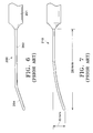
- FIG. 6 it is also known to use a blunt 19 gage cannula 200 having a hub 201, a straight proximal portion 202, and an angled distal portion 204 to perform sub-Tenon injection of anaesthesia for cataract and vitreoretinal surgery. See “Local Anaesthesia for Vitreoretinal Surgery", Calvin E. Mein and Michael G.
- cannulae also suffer from the above-described "tenting" and penetration problems if used to deliver drugs into the sub-Tenon's space above the macula.
- a 24 gauge cannula that has a straight proximal portion and a curved distal portion that is disposed at a 90 degree angle to the straight portion to inject a local anaesthetic solution below the Tenon's capsule.
- the straight portion has a length of 5 mm.
- the curved portion has a radius of curvature of 14 mm and an arc length of 27 mm. See "A Modified Sub-Tenon's Cannula for Local Anaesthesia", P. Muthusamy and Richard F. Hommersom, Asia-Pacific Journal of Ophthalmology, Volume 8, No. 3 (July 1996).
- this cannula is not suitable for the delivery of drugs in the form of suspensions, emulsions, ointments, or gels, or drugs in such forms including bioerodable polymers or non-bioerodable polymers.
- Document US-A-5817075 discloses a cannula comprising a straight proximal portion which curves distally into a curved arc with an aperture at the tip of the cannula.
- US 4, 759, 746 is another example of a type of needle that can be used in ocular surgery and is specifically directed towards an instrument and method for administering retro-bulbar (or peri-bulbar) anaesthetics. It comprises a curved needle portion that terminates in a straight needle portion.
- the improved apparatus and methods should be safe for the patient, easy for the physician to use, capable of delivering a wide spectrum of formulations, and capable of being performed in an outpatient setting.
- the present invention provides a cannula as defined in claim 1.
- the cannula includes a distal portion having a radius of curvature substantially equal to a radius of curvature of a globe of the human eye, a proximal portion, and a bend separating the distal portion and the proximal portion.
- a tangent of the distal portion at the bend is disposed at an angle of no more than about 56 degrees with respect to the proximal portion.
- the present invention allows the delivery of a drug to the human eye.
- a cannula is inserted below the Tenon's capsule and above the sclera of the human eye at a point posterior to a limbus of the eye.
- the cannula includes a distal portion having a radius of curvature substantially equal to a radius of curvature of the globe of the human eye.
- a drug is injected through the cannula to form a drug depot on an outer surface of the sclera.
- the drug comprises a pharmaceutically active agent selected from the group consisting of 4,9(11)- Pregnadien-17 ⁇ ,21-diol-3,20-dione and 4,9(11) -Pregnadien-17 ⁇ ,21-diol-3,20-dione- 21-acetate.
- the present invention comprises a cannula including a hub for removably coupling to a syringe, a proximal portion, and a distal portion.
- the distal portion has a tip, an orifice proximate the tip, and a radius of curvature substantially equal to a radius of curvature of the globe of the human eye.
- the part of the distal portion proximate the tip comprises a plastic.
- FIGS. 1 through 5 of the drawings like numerals being used for like and corresponding parts of the various drawings.
- FIG. 1 schematically illustrates a human eye 10.
- Eye 10 has a cornea 12, a lens 14, a sclera 16, a choroid 18, a retina 20, and an optic nerve 22.
- An anterior segment 24 of eye 10 generally includes the portions of eye 10 anterior of line 25.
- a posterior segment 26 of eye 10 generally includes the portions of eye 10 posterior of line 25.
- Retina 20 is physically attached to choroid 18 in a circumferential manner proximate pars plana 28.
- Retina 20 has a macula 30 located slightly lateral to optic nerve 22. As is well known in the ophthalmic art, macula 30 is comprised primarily of retinal cones and is the region of maximum visual acuity in retina 20.
- a Tenon's capsule or Tenon's membrane 34 is disposed on sclera 16.
- a conjunctiva 36 covers a short area of the globe of eye 10 posterior to limbus 32 (the bulbar conjunctiva) and folds up (the upper cul-de-sac) or down (the lower cul-de-sac) to cover the inner areas of upper eyelid 35 and lower eyelid 37, respectively.
- Conjunctiva 36 is disposed on top of Tenon's capsule 34.
- Sclera 16 and Tenon's capsule 34 define the exterior surface of the globe of eye 10.
- ARMD CNV
- retinopathies retinitis
- retinitis retinitis
- uveitis cystoid macular edema
- CME cystoid macular edema
- glaucoma glaucoma
- depot 38 of a specific quantity of an ophthalmically acceptable pharmaceutically active agent directly on the outer surface of sclera 16 and below Tenon's capsule 34.
- depot 38 directly on the outer surface of sclera 16, below Tenon's capsule 34, and generally above macula 30.
- Such a drug depot resulted in a concentration of the angiostatic steroid, averaged over the entire retina and measured the day after the injection, about ten times greater than a similar concentration delivered by a depot located below the conjunctiva but above the Tenon's capsule of the rabbit eyes.
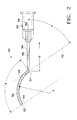
- Cannula 50 for creating drug depot 38 according to a preferred embodiment of the present invention is schematically illustrated.
- Cannula 50 generally includes a distal portion 52, a proximal portion 54, and a hub 56.
- a bend 57 separates distal portion 52 and proximal portion 54.
- a hollow bore 58 runs axially within distal portion 52 and proximal portion 54 and is fluidly coupled with a hollow bore 60 within hub 56.
- Distal portion 52 preferably has a blunt tip 62 to prevent damage to blood vessels in the periocular tissues and to pass smoothly over sclera 16.
- An orifice 64 is located proximate tip 62 for delivery of a drug formulation.
- Orifice 64 is preferably located about 1 mm from tip 62 on the lower or interior side of distal portion 52 to minimize the possibility of connective tissue blockage.
- Orifice 64 may alternatively be located on the end, on the upper or exterior side, or on other portions of distal portion 52.
- distal portion 52 may have multiple orifices, if desired.
- Orifice 64 is preferably circular and preferably has a 0.025 inch diameter that insures a smooth, controlled delivery of drug. Alternatively, other shapes and sizes of orifice 64 may be used.
- Distal portion 52 and proximal portion 54 are preferably formed out of 19 gauge needle stock. However, other sizes of tubing may be utilized depending on the viscosity and/or volume of material to be injected. Distal portion 52 and proximal portion 54 are preferably made of surgical stainless steel. Other conventional materials such as Teflon, other metals, metal alloys, polyethylene, polypropylene, other conventional plastics, or combinations of the foregoing may also be used. For example, distal portion 52 may be made from a plastic. As another example, a part of distal portion 52 proximate tip 62 may be made from plastic, and the remainder of distal portion 52 and proximal portion 54 may be made from metal.
- the plastic preferably has sufficient softness and/or flexibility to minimize the possibility of penetration of sclera 16 or Tenon's capsule 34 when cann ula 50 is inserted into eye 10, as described hereinbelow.
- the length of the plastic portion of distal portion 52, as well as the specific plastic, are preferably selected so that distal portion 52 maintains its radius of curvature B when cannula 50 is inserted into eye 10.
- Hub 56 is for removably coupling to a conventional syringe (not shown).
- Hub 56 preferably complies with Luer Taper Specification 70.1 of the American Standards Association.
- Hub 56 preferably includes a locator protuberance 66 that is coplanar with distal portion 52 and proximal portion 54. Protuberance 66 allows a physician to know the orientation of distal portion 52 even when it is inserted below Tenon's capsule 34.
- Hub 56 is preferably made of conventional plastics. Referring to both FIGS.
- distal portion 52 preferably has an arc length A and a radius of curvature B that closely approximate the curvature of sclera 16 of an adult human eye 10 from insertion points 70a or 70b, each of which is about 5 mm to about 10 mm posterior of limbus 32.
- Arc length A and radius of curvature B insure that drug depot 38, and more specifically, a specific quantity of pharmaceutically active agent, is deposited on the outer surface of sclera 16 generally above macula 30.
- Arc length A and bend 57 also limit the depth of insertion of cannula 50 along sclera 16, preventing tip 62 from accidentally contacting and damaging posterior ciliary arteries 40 or optic nerve 22.
- arc length A is preferably about 15 mm to about 18 mm.
- Arc length A may be varied for patient's with smaller or larger than average adult eyes, for paediatric patient's with smaller eyes, or for different insertion points into Tenon's capsule 34.
- a tangent 72 of distal portion 52 at bend 57 is preferably formed at an angle C with respect to proximal portion 54.
- angle C In addition to making bend 57 a physical limit to the insertion of cannula 50, angle C also raises the angle of hub 56 so that the face, bridge of the nose, and eyebrows of a patient to not interfere with the attached syringe. Angle C is also important to the successful delivery of drugs in the form of suspensions, emulsions, ointments, or gels, or drugs in such forms including bioerodable polymers or non-bioerodable polymers. Angle C is preferably no more than about 56 degrees. Angle C is most preferably about 56 degrees.
- Proximal portion 54 preferably has a length D of about 15 mm. Other angles and lengths may be used for angle C and length D for specific applications of cannula 50.
- Radius of curvature B insures that distal portion 52 does not drag or put pressure on sclera 16 as cannula 50 is advanced to the proper position, minimizing the risk of sceral penetration.
- radius of curvature B eliminates the "tenting" or pulling away of Tenon's capsule 34 from sclera 16, minimizing the risk of penetration into the periocular tissues.
- radius of curvature B is preferably about 11.5 mm to about 14 mm, and most preferably about 12.5 mm. Radius of curvature B may be varied for patients with smaller or larger than average adult eyes, for paediatric patients with smaller eyes, or for different insertion points into Tenon's capsule 34.
- Cannula 50 may be used to inject a wide variety of drug formulations using the following preferred techniques.
- a physician first anaesthetizes eye 10 using conventional topical anaesthetic drops. The patient is then instructed to look down and toward his or her nose.
- the physician uses a 25 gage, 5/8 inch needle to penetrate both conjunctiva 36 and Tenon's capsule 34 at a point about 4 mm posterior to limbus 32 in the superior temporal quadrant of eye 10.
- the needle is then advanced along the outer surface of sclera 16 to a point about 8 mm to about 9 mm posterior of limbus 32.
- the physician then makes a small bleb of anaesthesia, preferably about 1 mm to about 2 mm long, at this point.
- the physician grasps the tissue raised by the bleb with a forceps, and then punctures a hole through conjunctiva 36 and Tenon's capsule 34 using an introducer needle.
- the introducer needle preferably has an outer diameter with the same gage as cannula 50 or one gage larger than cannula 50.
- the physician draws a drug formulation into a conventional syringe using a conventional straight needle. The needle is removed and cannula 50 is attached to the syringe. All air is removed from the syringe and cannula 50 so that the drug formulation is at tip 62.
- cannula 50 through the hole made by the introducer needle, with orifice 64 facing sclera 16.
- cannula 50 With distal portion 52 in close contact with the outer surface of sclera 16, cannula 50 is advanced toward the back of the eye until bend 57 is at the site of the hole made by the introducer needle.
- tip 62 is preferably located about 5 mm to about 6 mm from the center of optic nerve 22, and about 2 mm to about 3 mm from macula 30.
- the physician then injects the drug formulation by actuating the syringe plunger, creating drug depot 38 on the outer surface of sclera 16 generally above macula 30.
- the above-described technique may be performed in the inferior temporal quadrant of eye 10, in which case the patient is instructed to look up and toward his or her nose.
- a physician first anaesthetizes eye 10 using conventional topical anaesthetic drops. Next, the patient is instructed to look down and toward his or her nose. Next, the physician creates a small incision in conjuctiva 36 and Tenon's capsule 34 at a point about 8 mm to about 9 mm posterior to limbus 32 in the superior temporal quadrant of eye 10 using fine scissors. The physician then draws a drug formulation into a conventional syringe, and then attaches cannula 50 to the syringe, as described above. Cannula 50 is then inserted through the incision with orifice 64 facing sclera 16.
- cannula 50 With distal portion 52 in close contact with the outer surface of sclera 16, cannula 50 is advanced toward the back of the eye until bend 57 is at the site of the incision. At this point, tip 62 is preferably located about 5 mm to about 6 mm from the center of optic nerve 22, and about 2 mm to about 3 mm from macula 30.
- the physician then injects the drug formulation by actuating the syringe plunger, creating drug depot 38 on the outer surface of sclera 16 generally above macula 30. If necessary, the incision may be sealed around cannula 50 using a purse suture to prevent reflux of the injected drug formulation. Alternatively, the above- described technique may be performed in the inferior temporal quadrant of eye 10, in which case the patient is instructed to 1 ook up and toward his or her nose.
- drug depot 38 preferably provides controlled release of a pharmaceutically active agent to macula 30 and retina 20 via sclera 16 and choroid 18 for a period of weeks or months.
- cannula 50 causes no "tenting" or substantial stretching of Tenon's capsule 34, cannula 50 should result in significantly less trauma to eye 10 than conventional cannulae when repeated injections are required.
- Cannula 50 can be used to deliver a wide variety of drug formulations to treat a wide variety of diseases of posterior segment 26.
- the drug formulation used to form drug depot 38 may be a solution, a suspension, an emulsion, an ointment, a gel forming solution, a gel, a bioerodable polymer, or a non-bioerodable polymer.
- the drug formulation used to form drug depot 38 may include one or more ophthalmically acceptable pharmaceutically active agents, and may also include conventional non- active incipients.
- antiinfectives including, without limitation, antibiotics, antivirals, and antifungals; antiallergenic agents and mast cell stabilizers; steroidal and non- steroidal anti-inflammatory agents; cyclooxygenase inhibitors, including, without limitation, Cox I and Cox II inhibitors; combinations of anti-infective and anti-inflammatory agents; decongestants; anti-glaucoma agents, including, without limitation, adrenergics, ⁇ -adrenergic blocking agents, ⁇ -adrenergic agonists, parasypathomimetic agents, cholinesterase inhibitors, carbonic anhydrase inhibitors, and prostaglandins; combinations of anti-glaucoma agents; antioxidants; nutritional supplements; drugs for the treatment of cystoid macular edema including, without limitation, non-steroidal anti-inflammatory agents; drugs for the treatment of ARMD, including, without limitation, angiogenesis inhibitors and nutritional supplements; drugs for the treatment of
- Such angiostatic steroids are more fully disclosed in U.S. Patent Nos. 5,679,666 and 5,770,592.
- Preferred ones of such angiostatic steroids include 4,9(11) -Pregnadien-17 ⁇ ,21-diol-3,20-dione and 4,9(11)-Pregnadien-17 ⁇ ,21-diol-3,20-dione-21-acetate.
- These preferred angiostatic steroids are preferably formulated as a suspension.
- a preferred non-steroidal anti-inflammatory for the treatment of cystoid macular edema is nepafenac.
- the conventional non-active excipients may include, but are not limited to, ingredients to enhance the stability, solubility, penetrability, or other properties of the pharmaceutically active agent or drug depot 38.
- FIGS. 3A through 3E show additional preferred embodiments of cannulae for the creation of drug depot 38.
- Each of these cannulae have a proximal portion 54, hub 56, bend 57, and hollow bore 58 substantially identical to that described above for cannula 50 of FIG. 2.
- each of these cannulae has a unique distal portion.
- Hub 56 of each of these cannulae is removably coupled to a conventional syringe 80.
- Each of these cannulae can be used to create drug depot 38 directly on the outer surface of sclera 16 generally above macula 30 in a manner substantially similar to the techniques described above for cannula 50.
- Cannula 82 of FIG. 3A has a distal portion 52a with a geometry substantially identical to distal portion 52 of cannula 50, except that an orifice 64a is located on the end of tip 62 of distal portion 52a.
- cannula 84 of FIG. 3B has a distal portion 52b with a geometry substantially identical to distal portian 52 of cannula 50, except that tip 62 has two orifices, 64 and 64b.
- Orifice 64b is located on the upper or exterior side of distal portion 52b.
- orifices 64 and 64b may be located laterally, on opposite sides of distal portion 52b.
- Cannula 86 of FIG. 3C has a distal portion 52c with a geometry substantially identical to distal portion 52 of cannula 50, except that a plurality of orifices 64c, 64d, 64e, and 64f are disposed on distal portion 52c proximate tip 62. Orifices 64c through 64f are preferably disposed on distal portion 52c in the alternating pattern shown in FIG. 3C. Cannula 86 is useful when it is desirable to create a larger drug depot 38. Distal portion 52c may be formed with more or less than the four orifices shown in FIG. 3C, or with a different pattern of orifices than shown in FIG. 3C, if desired.
- Cannula 88 of FIG. 3D has a distal portion 52d with a geometry substantially identical to distal portion 52a of cannula 82 of FIG. 3A, except that tip 62d has a globular or olive shape.
- Tip 62 thus serves as a scleral depressor, allowing a physician to view tip 62d through an ophthalmoscope as he or she guides cannula 88 along the outer surface of sclera 16.
- Tip 62 is preferably sized to create as small a pathway as possible between Tenon's capsule 34 and sclera 16, but still function as a scleral depressor. A small pathway minimizes the possibility of drug formulation flowing anteriorly from drug depot 38.
- Cannula 90 of FIG. 3E has a distal portion 52e with a geometry substantially identical to distal portion 52 of cannula 50 of FIG. 2, except that tip 62e is also equipped with a fiber optic light source 92, allowing a physician to view tip 62e through an ophthalmoscope as he or she guides cannula 90 along the outer surface of sclera 16.
- a conventional power source 94 is electrically coupled to fiber optic light source 92 via conventional electrical wiring 96 that is preferably at least partially disposed within the wall of distal portion 52e and proximal portion 54. Fiber optic light source 92, power source 94, and wiring 96 may be incorporated into any of the cannulae disclosed in this application, if desired.
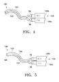
- FIG. 4 schematically illustrates a preferred embodiment of a cannula 100 for the creation of drug depot 38.
- Cannula 100 includes a lumen 102 having a geometry substantially identical to cannula 82 of FIG. 3A.
- Cannula 100 also includes a second, separate lumen 104 disposed adjacent to lumen 102 and having a geometry substantially identical to cannula 82 of FIG. 3A.
- Lumen 102 has a hub 56 removably coupled to a lumen 106a of a dual lumen syringe 106.
- Lumen 104 has a hub 56 removably coupled to a lumen 106b of dual lumen syringe 106.
- Cannula 100 can be used to create drug depot 38 directly on the outer surface of sclera 16 generally above macula 30 in a manner substantially similar to the techniques described above for cannula 50. However, cannula 100 allows the delivery of two separate drug formulations while creating drug depot 38. Alternatively, lumen 104 of cannula 100 can be used to aspirate a non-bioerodable drug depot 38 that has dispensed all of its pharmaceutically active agent, and lumen 102 of cannula 100 can be used to inject a new drug depot 38.
- FIG. 5 schematically illustrates a preferred embodiment of a cannula 120 for the creation of drug depot 38.
- Cannula 120 has a geometry substantially identical to that of cannula 100, except that lumen 102 and lumen 104 join at a point 122 proximate a single orifice 64f near the distal portions of the cannula.
- Cannula 120 can be used to create drug depot 38 directly on the outer surface of sclera 16 generally above macula 30 in a manner substantially similar to the techniques described above for cannula 50. However, cannula 120 allows the delivery of two separate formulations that require mixing just prior to injection out of orifice 64f.
- the present invention provides an improved apparatus for sub-Tenon delivery of a drug depot to the posterior segment of a human eye proximate the macula.
- the apparatus and methods of the present invention increase patient safety, are easy for the physician to use, are capable of delivering a wide spectrum of formulations, and are capable of being performed in an outpatient setting.
- the apparatus and methods of the present invention are especially useful for localized delivery of pharmaceutically active agents to the posterior segment of the eye to combat ARMD, CNV, retinopathies, retinitis, uveitis, macular edema, glaucoma, and other posterior segment diseases.
- the apparatus of the present invention are also particularly useful for the sub-Tenon delivery of drugs in the form of suspensions, emulsions, ointments, or gels, or drugs in such forms including bioerodable polymers or non-bioerodable polymers.
- the present invention is illustrated herein by example, and various modifications may be made by a person of ordinary skill in the art.
- the cannulae of the present invention have been described above in connection with the preferred sub-Tenon drug delivery generally above the macula
- the cannulae can be used to deliver drugs directly on the outer surface of the sclera, below the Tenon's capsule, and generally above portions of the retina other than the macula.
- the arc length and/or radius of curvature of the distal portions of the cannulae may be modified to deliver drugs within the Tenon's capsule or the sclera, generally above the macula or other portions of the retina, if desired.
Landscapes
- Health & Medical Sciences (AREA)
- Life Sciences & Earth Sciences (AREA)
- General Health & Medical Sciences (AREA)
- Biomedical Technology (AREA)
- Heart & Thoracic Surgery (AREA)
- Vascular Medicine (AREA)
- Ophthalmology & Optometry (AREA)
- Animal Behavior & Ethology (AREA)
- Engineering & Computer Science (AREA)
- Public Health (AREA)
- Veterinary Medicine (AREA)
- Infusion, Injection, And Reservoir Apparatuses (AREA)
- Pharmaceuticals Containing Other Organic And Inorganic Compounds (AREA)
- Medicinal Preparation (AREA)
- Prostheses (AREA)
Abstract
Description
Claims (17)
- A sub-Tenon drug delivery cannula (50, 82, 84, 86, 88, 90, 100, 120), the cannula being dimensioned to facilitate the formation of a drug depot (38) below a Tenon's capsule (34), on an outer surface of a sclera (16), and in a posterior segment (26) of a human eye, the cannula comprising:a distal portion (52, 52a, 52b, 52c, 52d, 52e) having a radius of curvature substantially equal to a radius of curvature of a globe of a human eye (10);a straight proximal portion (54); anda bend (57) separating said distal portion and said proximal portion, andwherein a tangent of said distal portion at said bend is disposed at an angle of no more than about 56 degrees with respect to said proximal portion.
- The cannula of claim 1, wherein said distal por tion comprises an arc having a length of about 15 mm to about 18 mm.
- The cannula of claim 1, further comprising:a hub (56) coupled to said proximal portion for removably coupling to a syringe (80); anda hollow bore (58) disposed axially within said distal portion and said proximal portion.
- The cannula of claim 1, wherein said distal portion comprises an interior side and a tip (62, 62d, 62e) having an orifice (64, 64a, 64b, 64c, 64d, 64e, 64f) on said interior side.
- The cannula of claim 1, wherein said distal portion (52) has a radius of curvature substantially equal to a radius of curvature of a sclera of said eye.
- The cannula of claim 1, wherein said radius of curvature of said distal portion (52) is about 11.5 mm to about 14 mm.
- The cannula of claim 1, wherein said distal portion (52) comprises a plastic.
- The cannula of claim 1, wherein said distal portion (52) comprises a tip (62) and an orifice (64) proximate said tip, and wherein a part of said distal portion proximate said t ip comprises a plastic, and a remainder of said distal portion comprises a metal.
- The cannula of claim 1, wherein said angle is about 56 degrees.
- The cannula of claim 3, wherein said distal portion (52c) comprises a plurality of orifices (64c, 64d , 64e, 64f), each of said orifices communicating with said hollow bore (58).
- The cannula of claim 1, wherein said distal portion (52d) comprises a tip (62d) having a generally olive shape.
- The cannula of claim 1, wherein said distal portion (52e) comprises:a tip (62e); anda fiber optic light source (92) disposed proximate said tip.
- The cannula of claim 1, wherein said distal portion, said proximal portion, and said bend comprise a first lumen (102), and further comprising:a second lumen (104) having a geometry substantially similar to said first lumen and disposed adjacent said first lumen.
- The cannula of claim 13, wherein said first lumen (102) and second lumen (104) are not in communication.
- The cannula of claim 13, further comprising a tip, and wherein said first lumen (102) and said second lumen (104) are in communication (122) proximate said tip.
- The cannula of claim 3, wherein said bend (57) prevents said syringe (80) from interfering with a patient's face, nose, and eyebrows when said distal portion is inserted into said eye (10).
- The cannula of claim 1, wherein said cannula facilitates forming said drug depot below said Tenon's capsule, on said outer surface of said sclera, and proximate a macula (30) of said hu man eye.
Applications Claiming Priority (3)
| Application Number | Priority Date | Filing Date | Title |
|---|---|---|---|
| US16166099P | 1999-10-21 | 1999-10-21 | |
| US161660P | 1999-10-21 | ||
| EP00968673A EP1221918B1 (en) | 1999-10-21 | 2000-10-04 | Sub-tenon drug delivery |
Related Parent Applications (1)
| Application Number | Title | Priority Date | Filing Date |
|---|---|---|---|
| EP00968673A Division EP1221918B1 (en) | 1999-10-21 | 2000-10-04 | Sub-tenon drug delivery |
Publications (2)
| Publication Number | Publication Date |
|---|---|
| EP1522289A2 true EP1522289A2 (en) | 2005-04-13 |
| EP1522289A3 EP1522289A3 (en) | 2008-01-23 |
Family
ID=22582174
Family Applications (2)
| Application Number | Title | Priority Date | Filing Date |
|---|---|---|---|
| EP00968673A Expired - Lifetime EP1221918B1 (en) | 1999-10-21 | 2000-10-04 | Sub-tenon drug delivery |
| EP05100176A Withdrawn EP1522289A3 (en) | 1999-10-21 | 2000-10-04 | Sub-tenon drug delivery |
Family Applications Before (1)
| Application Number | Title | Priority Date | Filing Date |
|---|---|---|---|
| EP00968673A Expired - Lifetime EP1221918B1 (en) | 1999-10-21 | 2000-10-04 | Sub-tenon drug delivery |
Country Status (15)
| Country | Link |
|---|---|
| US (1) | US6413245B1 (en) |
| EP (2) | EP1221918B1 (en) |
| JP (1) | JP2003511204A (en) |
| AR (4) | AR026075A1 (en) |
| AT (1) | ATE290837T1 (en) |
| AU (1) | AU775149B2 (en) |
| BR (1) | BR0014930B1 (en) |
| CA (1) | CA2383572C (en) |
| DE (1) | DE60018777T2 (en) |
| ES (1) | ES2240180T3 (en) |
| HK (1) | HK1049438B (en) |
| MX (1) | MXPA02002376A (en) |
| PT (1) | PT1221918E (en) |
| TW (1) | TW467740B (en) |
| WO (1) | WO2001028473A1 (en) |
Cited By (3)
| Publication number | Priority date | Publication date | Assignee | Title |
|---|---|---|---|---|
| DE102017209202A1 (en) * | 2017-05-31 | 2018-12-06 | Geuder Ag | Device for the preparation of a human or animal tissue for the introduction of a graft or implant |
| WO2020201884A1 (en) | 2019-04-01 | 2020-10-08 | Amo Development, Llc | Corneal lenticule extraction tool |
| WO2024073759A1 (en) * | 2022-09-30 | 2024-04-04 | FUJIFILM Cellular Dynamics, Inc. | Apparatus and methods for delivery of cell compositions |
Families Citing this family (185)
| Publication number | Priority date | Publication date | Assignee | Title |
|---|---|---|---|---|
| US6936053B1 (en) * | 1998-07-02 | 2005-08-30 | Jeffrey N. Weiss | Ocular implant needle |
| KR20020035476A (en) * | 1999-04-26 | 2002-05-11 | 지엠피 비젼 솔루션즈 인코포레이티드 | Shunt device and method for treating glaucoma |
| US7943162B2 (en) * | 1999-10-21 | 2011-05-17 | Alcon, Inc. | Drug delivery device |
| CA2369149A1 (en) * | 2000-02-02 | 2001-08-09 | Meditsinsky Nauchno-Issledovatelsky Oftalmologichesky Tsentr "Novy Vzgjyad" | Kourenkov's cannula used for a method for an operation of refractory-correlating eximer-laser intrastromal keratectomy (reik) |
| US7431710B2 (en) | 2002-04-08 | 2008-10-07 | Glaukos Corporation | Ocular implants with anchors and methods thereof |
| DE60217679T2 (en) | 2001-07-23 | 2007-10-25 | Alcon Inc. | DEVICE FOR RELEASING AN OPHTHALMIC MEDICINAL PRODUCT |
| EP1385452B1 (en) * | 2001-07-23 | 2006-09-13 | Alcon, Inc. | Ophthalmic drug delivery device |
| JP4892800B2 (en) * | 2001-08-24 | 2012-03-07 | 株式会社ジェイ・エム・エス | Needle tube for injecting medicinal solution and syringe equipped with the needle tube for injecting medicinal solution |
| US7153316B1 (en) | 2001-11-09 | 2006-12-26 | Mcdonald Marguerite B | Surgical instruments and method for corneal reformation |
| US6802829B2 (en) * | 2001-11-16 | 2004-10-12 | Infinite Vision, Llc | Spray device |
| WO2003068253A1 (en) * | 2002-02-14 | 2003-08-21 | Merck Patent Gmbh | Methods and compositions for the treatment of eye diseases |
| JP2004041492A (en) * | 2002-07-12 | 2004-02-12 | Scitec Kk | Medical needle |
| DE10238310A1 (en) * | 2002-08-21 | 2004-03-04 | Erich Jaeger Gmbh | electrode assembly |
| EP1539066B1 (en) * | 2002-09-17 | 2012-11-07 | Iscience Surgical Corporation | Apparatus surgical bypass of aqueous humor |
| CA2689424A1 (en) * | 2002-09-29 | 2004-04-08 | Surmodics, Inc. | Methods for treatment and/or prevention of retinal disease |
| US7141048B1 (en) * | 2002-10-17 | 2006-11-28 | Alcon, Inc. | Vitreoretinal instrument |
| US7285107B1 (en) | 2002-10-17 | 2007-10-23 | Alcon, Inc. | Vitreoretinal instrument |
| ZA200505990B (en) * | 2003-02-20 | 2006-12-27 | Alcon Inc | Formulations of glucocorticoids to treat pathologic ocular angiogenesis |
| MXPA05008561A (en) * | 2003-02-20 | 2005-11-04 | Alcon Inc | Use of steroids to treat ocular disorders. |
| US20040199130A1 (en) * | 2003-04-03 | 2004-10-07 | Chornenky Victor I. | Apparatus and method for treatment of macular degeneration |
| WO2004112772A1 (en) * | 2003-06-13 | 2004-12-29 | Alcon, Inc. | Formulations of non-steroidal anti-inflammatory agents to treat pathologic ocular angiogenesis |
| JP2007521274A (en) * | 2003-06-20 | 2007-08-02 | アルコン,インコーポレイテッド | Treatment of age-related macular degeneration using a combination of multiple components |
| KR20060082792A (en) * | 2003-07-10 | 2006-07-19 | 알콘, 인코퍼레이티드 | Ophthalmic Drug Delivery Device |
| DE10337863A1 (en) | 2003-08-18 | 2005-03-17 | Merck Patent Gmbh | Use of chromene-4-one derivatives |
| CA2536185C (en) * | 2003-08-20 | 2012-06-26 | Santen Pharmaceutical Co., Ltd. | Drug delivery system by administrating fine particles to sub-tenon |
| JP4869930B2 (en) * | 2003-08-26 | 2012-02-08 | ヴィスタ サイエンティフィック エルエルシー | Ophthalmic drug supply device |
| JP2007505932A (en) | 2003-09-18 | 2007-03-15 | マクサイト, インコーポレイテッド | Transscleral delivery |
| US20050098470A1 (en) * | 2003-11-12 | 2005-05-12 | Davis Michael J. | Kit for administration of a drug |
| US20090148527A1 (en) * | 2007-12-07 | 2009-06-11 | Robinson Michael R | Intraocular formulation |
| WO2005107664A2 (en) * | 2004-04-29 | 2005-11-17 | Iscience Interventional Corporation | Apparatus and method for surgical enhancement of aqueous humor drainage |
| US20080058704A1 (en) * | 2004-04-29 | 2008-03-06 | Michael Hee | Apparatus and Method for Ocular Treatment |
| WO2006002366A2 (en) * | 2004-06-24 | 2006-01-05 | Surmodics, Inc. | Biodegradable ocular devices, methods and systems |
| US20060110428A1 (en) | 2004-07-02 | 2006-05-25 | Eugene Dejuan | Methods and devices for the treatment of ocular conditions |
| US20060047250A1 (en) * | 2004-08-30 | 2006-03-02 | Hickingbotham Dyson W | Fluid delivery device |
| US7402156B2 (en) | 2004-09-01 | 2008-07-22 | Alcon, Inc. | Counter pressure device for ophthalmic drug delivery |
| US7226435B2 (en) * | 2004-10-14 | 2007-06-05 | Alcon, Inc. | Drug delivery device |
| EP1819325A2 (en) * | 2004-12-22 | 2007-08-22 | Alcon, Inc. | Device for ophthalmic drug delivery |
| US8663639B2 (en) | 2005-02-09 | 2014-03-04 | Santen Pharmaceutical Co., Ltd. | Formulations for treating ocular diseases and conditions |
| WO2006086750A1 (en) | 2005-02-09 | 2006-08-17 | Macusight, Inc. | Liquid formulations for treatment of diseases or conditions |
| WO2006088134A1 (en) * | 2005-02-18 | 2006-08-24 | Santen Pharmaceutical Co., Ltd. | Method of relieving or avoiding side effect of steroid compound |
| JP2006257080A (en) * | 2005-02-18 | 2006-09-28 | Santen Pharmaceut Co Ltd | How to reduce or avoid side effects of steroid compounds |
| US20100196509A1 (en) | 2005-02-28 | 2010-08-05 | Jonathan Braun | Methods for Diagnosis and Treatment of Endometrial Cancer |
| CN101180086A (en) * | 2005-04-08 | 2008-05-14 | 苏尔莫迪克斯公司 | Sustained-release implants for subretinal delivery |
| US8318906B2 (en) | 2005-04-15 | 2012-11-27 | The Regents Of The University Of California | EMP2 antibodies and their therapeutic uses |
| US20070038174A1 (en) * | 2005-08-09 | 2007-02-15 | Hopkins Mark A | Ophthalmic injector system |
| US20070060887A1 (en) * | 2005-08-22 | 2007-03-15 | Marsh David A | Ophthalmic injector |
| US20070134244A1 (en) * | 2005-10-14 | 2007-06-14 | Alcon, Inc. | Combination treatment for pathologic ocular angiogenesis |
| WO2007052662A1 (en) * | 2005-10-31 | 2007-05-10 | Terumo Kabushiki Kaisha | Puncture device, dosing device and puncture method |
| US7611492B2 (en) * | 2005-11-10 | 2009-11-03 | Terumo Kabushiki Kaisha | Puncture device |
| US20070156096A1 (en) * | 2005-11-10 | 2007-07-05 | Terumo Kabushiki Kaisha | Puncture device |
| KR20080087814A (en) * | 2005-12-22 | 2008-10-01 | 알콘 리서치, 리미티드 | C3-Convertase Inhibitors for the Prevention and Treatment of Age-Related Macular Degeneration in Patients with Risk Variants of Complement Factor H |
| US8492400B2 (en) | 2006-02-09 | 2013-07-23 | Santen Pharmaceutical Co., Ltd. | Stable formulations, and methods of their preparation and use |
| US20070202186A1 (en) | 2006-02-22 | 2007-08-30 | Iscience Interventional Corporation | Apparatus and formulations for suprachoroidal drug delivery |
| CN101443004B (en) | 2006-03-23 | 2013-03-06 | 参天制药株式会社 | Agents for diseases or conditions associated with vascular permeability |
| US8197435B2 (en) * | 2006-05-02 | 2012-06-12 | Emory University | Methods and devices for drug delivery to ocular tissue using microneedle |
| US20210393436A1 (en) * | 2006-05-02 | 2021-12-23 | Emory University | Methods and devices for drug delivery to ocular tissue using microneedle |
| US20070270750A1 (en) * | 2006-05-17 | 2007-11-22 | Alcon, Inc. | Drug delivery device |
| US7871399B2 (en) * | 2006-05-17 | 2011-01-18 | Alcon Research, Ltd. | Disposable ophthalmic injection device |
| US7862540B2 (en) * | 2006-05-17 | 2011-01-04 | Alcon Research, Ltd. | Ophthalmic injection device using shape memory alloy |
| US7811252B2 (en) * | 2006-05-17 | 2010-10-12 | Alcon Research, Ltd. | Dosage control device |
| US7887521B2 (en) * | 2006-05-17 | 2011-02-15 | Alcon Research, Ltd. | Ophthalmic injection system |
| US20070270768A1 (en) * | 2006-05-17 | 2007-11-22 | Bruno Dacquay | Mechanical Linkage Mechanism For Ophthalmic Injection Device |
| US7674243B2 (en) * | 2006-05-17 | 2010-03-09 | Alcon Inc. | Ophthalmic injection device using piezoelectric array |
| US20070268340A1 (en) * | 2006-05-17 | 2007-11-22 | Bruno Dacquay | Ophthalmic Injection System and Method Using Piezoelectric Array |
| US20080097379A1 (en) * | 2006-09-26 | 2008-04-24 | Alcon Manufacturing, Ltd. | Ophthalmic injection method |
| US20080125712A1 (en) * | 2006-09-26 | 2008-05-29 | Alcon Manufacturing, Ltd. | Ophthalmic injection system |
| US20080097390A1 (en) * | 2006-09-27 | 2008-04-24 | Alcon Manufacturing, Ltd. | Spring actuated delivery system |
| BRMU8602090Y8 (en) * | 2006-10-05 | 2021-06-22 | Hexsel Doris | cannula-applied disposition for skin fillings |
| US9022970B2 (en) | 2006-10-16 | 2015-05-05 | Alcon Research, Ltd. | Ophthalmic injection device including dosage control device |
| ATE490752T1 (en) * | 2006-10-16 | 2010-12-15 | Alcon Res Ltd | UNIVERSAL RECHARGEABLE ASSEMBLY WITH LIMITED REUSABILITY FOR AN OPHTHALMIC HANDPIECE |
| BRPI0717644A2 (en) * | 2006-10-16 | 2013-11-12 | Alcon Res Ltd | METHOD OF OPERATING MANUAL OF DISPOSABLE ENDALMAL PARTS. |
| US20080281292A1 (en) * | 2006-10-16 | 2008-11-13 | Hickingbotham Dyson W | Retractable Injection Port |
| US8969415B2 (en) * | 2006-12-01 | 2015-03-03 | Allergan, Inc. | Intraocular drug delivery systems |
| US20080265343A1 (en) * | 2007-04-26 | 2008-10-30 | International Business Machines Corporation | Field effect transistor with inverted t shaped gate electrode and methods for fabrication thereof |
| RU2009144142A (en) * | 2007-04-30 | 2011-06-10 | Алькон Рисерч, Лтд. (Us) | TREATMENT OF AGE YELLOW SPOT DEGENERATION WITH APPLICATION OF INHIBITORS OF FACTOR D OF COMPLEMENT |
| US20090018512A1 (en) * | 2007-07-13 | 2009-01-15 | Charles Steven T | Pneumatically-Powered Ophthalmic Injector |
| US20090018548A1 (en) * | 2007-07-13 | 2009-01-15 | Charles Steven T | Pneumatically-Powered Intraocular Lens Injection Device with Removable Cartridge |
| US7740619B2 (en) * | 2007-08-01 | 2010-06-22 | Alcon Research, Ltd. | Spring driven ophthalmic injection device with safety actuator lockout feature |
| US7629768B2 (en) * | 2007-08-03 | 2009-12-08 | Alcon Research, Ltd. | Easy cleaning C-shaped charging base |
| US20090036842A1 (en) * | 2007-08-03 | 2009-02-05 | Raffi Pinedjian | Consumable Activation Lever For Injection Device |
| USD592746S1 (en) | 2007-11-08 | 2009-05-19 | Alimera Sciences | Ocular implantation device |
| US9849027B2 (en) | 2007-11-08 | 2017-12-26 | Alimera Sciences, Inc. | Ocular implantation device |
| US8790366B2 (en) * | 2007-11-13 | 2014-07-29 | Alcon Research, Ltd. | Fan-shaped cannula for sealing ophthalmic incisions |
| US8608632B1 (en) * | 2009-07-03 | 2013-12-17 | Salutaris Medical Devices, Inc. | Methods and devices for minimally-invasive extraocular delivery of radiation and/or pharmaceutics to the posterior portion of the eye |
| US9056201B1 (en) * | 2008-01-07 | 2015-06-16 | Salutaris Medical Devices, Inc. | Methods and devices for minimally-invasive delivery of radiation to the eye |
| KR101634983B1 (en) * | 2008-01-07 | 2016-07-01 | 살루타리스 메디컬 디바이스즈, 인코퍼레이티드 | Methods and devices for minimally-invasive extraocular delivery of radiation to the posterior portion of the eye |
| US10022558B1 (en) * | 2008-01-07 | 2018-07-17 | Salutaris Medical Devices, Inc. | Methods and devices for minimally-invasive delivery of radiation to the eye |
| US8602959B1 (en) | 2010-05-21 | 2013-12-10 | Robert Park | Methods and devices for delivery of radiation to the posterior portion of the eye |
| AU2015204094B2 (en) * | 2008-01-07 | 2017-02-23 | Salutaris Medical Devices, Inc | Methods and devices for minimally-invasive extraocular delivery of radiation to the posterior portion of the eye |
| US9873001B2 (en) * | 2008-01-07 | 2018-01-23 | Salutaris Medical Devices, Inc. | Methods and devices for minimally-invasive delivery of radiation to the eye |
| GB0802044D0 (en) * | 2008-02-05 | 2008-03-12 | Helica Instr Ltd | Needle for opthalmic procedures |
| TWI607748B (en) * | 2008-04-24 | 2017-12-11 | 莎魯塔理斯醫療設備股份有限公司 | Methods and devices for minimally-invasive extraocular delivery of radiation to the posterior portion of the eye |
| US8945086B2 (en) * | 2008-07-01 | 2015-02-03 | Bruce Becker | Retrobulbar needle and methods of use |
| US8821870B2 (en) | 2008-07-18 | 2014-09-02 | Allergan, Inc. | Method for treating atrophic age related macular degeneration |
| WO2010019839A1 (en) | 2008-08-15 | 2010-02-18 | The United States Of America, As Represented By The Secretary, Department Of Health And Human Services | Methods for using interferon gamma to absorb fluid from the subretinal space |
| US20100098772A1 (en) * | 2008-10-21 | 2010-04-22 | Allergan, Inc. | Drug delivery systems and methods for treating neovascularization |
| US8702677B2 (en) * | 2008-10-31 | 2014-04-22 | Warsaw Orthopedic, Inc. | Device and method for directional delivery of a drug depot |
| US9095506B2 (en) | 2008-11-17 | 2015-08-04 | Allergan, Inc. | Biodegradable alpha-2 agonist polymeric implants and therapeutic uses thereof |
| AU2009322409A1 (en) | 2008-12-04 | 2011-06-23 | Sanofi | Crystalline forms |
| AR074776A1 (en) | 2008-12-18 | 2011-02-09 | Sanofi Aventis | METHOD TO TREAT MACULAR DEGENERATION; MODULATING THE PATIENT'S IMMUNE SYSTEM |
| USD691269S1 (en) | 2009-01-07 | 2013-10-08 | Salutaris Medical Devices, Inc. | Fixed-shape cannula for posterior delivery of radiation to an eye |
| USD691268S1 (en) | 2009-01-07 | 2013-10-08 | Salutaris Medical Devices, Inc. | Fixed-shape cannula for posterior delivery of radiation to eye |
| USD691270S1 (en) | 2009-01-07 | 2013-10-08 | Salutaris Medical Devices, Inc. | Fixed-shape cannula for posterior delivery of radiation to an eye |
| USD691267S1 (en) | 2009-01-07 | 2013-10-08 | Salutaris Medical Devices, Inc. | Fixed-shape cannula for posterior delivery of radiation to eye |
| USD642266S1 (en) | 2010-08-24 | 2011-07-26 | Marsteller Laurence J | Brachytherapy device |
| US8425473B2 (en) | 2009-01-23 | 2013-04-23 | Iscience Interventional Corporation | Subretinal access device |
| US20100191177A1 (en) * | 2009-01-23 | 2010-07-29 | Iscience Interventional Corporation | Device for aspirating fluids |
| USD616088S1 (en) | 2009-02-06 | 2010-05-18 | Luca Brigatti | Fixed-shaped cannula for posterior delivery of radiation to eye |
| USD615645S1 (en) | 2009-02-06 | 2010-05-11 | Luca Brigatti | Fixed-shape cannula for posterior delivery of radiation to eye |
| USD616540S1 (en) | 2009-02-06 | 2010-05-25 | Luca Brigatti | Fixed-shape cannula for posterior delivery of radiation to eye |
| USD616087S1 (en) | 2009-02-06 | 2010-05-18 | Luca Brigatti | Fixed-shape cannula for posterior delivery of radiation to eye |
| US8372036B2 (en) | 2009-05-06 | 2013-02-12 | Alcon Research, Ltd. | Multi-layer heat assembly for a drug delivery device |
| US10206813B2 (en) | 2009-05-18 | 2019-02-19 | Dose Medical Corporation | Implants with controlled drug delivery features and methods of using same |
| US12478503B2 (en) | 2009-05-18 | 2025-11-25 | Glaukos Corporation | Implants with controlled drug delivery features and methods of using same |
| JP5937004B2 (en) | 2009-05-18 | 2016-06-22 | ドーズ メディカル コーポレーションDose Medical Corporation | Drug-eluting intraocular implant |
| WO2011011753A1 (en) | 2009-07-23 | 2011-01-27 | Baxter International Inc. | Manufacture of factor h (fh) and fh-derivatives from plasma |
| RU2405575C1 (en) * | 2009-08-17 | 2010-12-10 | Владимир Константинович Гаврилов | Medical needle |
| CA2781290A1 (en) | 2009-11-20 | 2011-05-26 | Lynn K. Gordon | Epithelial membrane protein-2 (emp2) and proliferative vitreoretinopathy (pvr) |
| US8177747B2 (en) | 2009-12-22 | 2012-05-15 | Alcon Research, Ltd. | Method and apparatus for drug delivery |
| SI2515774T1 (en) | 2009-12-23 | 2014-06-30 | Alcon Research, Ltd. | Ophthalmic valved trocar cannula |
| US8529492B2 (en) | 2009-12-23 | 2013-09-10 | Trascend Medical, Inc. | Drug delivery devices and methods |
| US8343106B2 (en) | 2009-12-23 | 2013-01-01 | Alcon Research, Ltd. | Ophthalmic valved trocar vent |
| EP2536433B1 (en) | 2010-02-19 | 2017-08-02 | The Regents of The University of California | Epithelial membrane protein 2 (emp2) binding reagents and their therapeutic uses in ocular diseases |
| US8772462B2 (en) | 2010-05-26 | 2014-07-08 | Baxter International Inc. | Removal of serine proteases by treatment with finely divided silicon dioxide |
| AU2010202125B1 (en) | 2010-05-26 | 2010-09-02 | Takeda Pharmaceutical Company Limited | A method to produce an immunoglobulin preparation with improved yield |
| JP2012020042A (en) * | 2010-07-16 | 2012-02-02 | Japan Imposing Co Ltd | Hypodermic injection needle |
| CN103153316B (en) | 2010-08-05 | 2015-08-19 | 弗赛特影像4股份有限公司 | Combination drug delivery methods and devices |
| EP2627292B1 (en) | 2010-10-15 | 2018-10-10 | Clearside Biomedical, Inc. | Device for ocular access |
| US9668915B2 (en) | 2010-11-24 | 2017-06-06 | Dose Medical Corporation | Drug eluting ocular implant |
| US10245178B1 (en) | 2011-06-07 | 2019-04-02 | Glaukos Corporation | Anterior chamber drug-eluting ocular implant |
| US9102105B2 (en) | 2011-09-13 | 2015-08-11 | Vista Scientific Llc | Method for forming an ocular drug delivery device |
| WO2014056895A1 (en) | 2012-10-08 | 2014-04-17 | Universität Leipzig | A device for a medical treatment of a sclera |
| EP2916859B1 (en) | 2012-11-02 | 2017-06-28 | The U.S.A. as represented by the Secretary, Department of Health and Human Services | Method of reducing adverse effects in a cancer patient undergoing treatment with a mek inhibitor |
| CN104994814B (en) | 2012-11-07 | 2020-03-24 | Ip自由视觉公司 | Optical radiation device for ophthalmology |
| WO2014159679A1 (en) | 2013-03-12 | 2014-10-02 | The United States Of America, As Represented By The Secretary, Department Of Health & Human Services | Methods for using lubiprostone to absorb fluid from the subretinal space |
| AU2013202965B2 (en) | 2013-03-15 | 2016-07-21 | Takeda Pharmaceutical Company Limited | Improved method for producing factor h from a plasma precipitation fraction |
| AU2013203048A1 (en) | 2013-03-15 | 2014-10-02 | Baxalta GmbH | Isolation of factor h from fraction i paste |
| AU2014259694B2 (en) | 2013-05-03 | 2018-11-08 | Clearside Biomedical, Inc. | Apparatus and methods for ocular injection |
| US10010447B2 (en) | 2013-12-18 | 2018-07-03 | Novartis Ag | Systems and methods for subretinal delivery of therapeutic agents |
| US9730834B2 (en) | 2013-12-20 | 2017-08-15 | Novartis Ag | Variable stiffness cannula and methods for a surgical system |
| US10117578B2 (en) * | 2013-12-31 | 2018-11-06 | Ip Liberty Vision Corporation | Luminescent ophthalmic device |
| US10166143B2 (en) | 2013-12-31 | 2019-01-01 | Ip Liberty Vision Corporation | Versatile light-guided ophthalmic treatment system |
| PL3104814T3 (en) | 2014-02-12 | 2021-10-11 | Gyroscope Therapeutics Limited | Apparatus for suprachoroidal administration of therapeutic agent |
| CN103919642A (en) * | 2014-03-14 | 2014-07-16 | 中国人民解放军第三军医大学第一附属医院 | Posterior sclera injection system |
| EP3128966A1 (en) * | 2014-04-09 | 2017-02-15 | Universität Leipzig | A device for a medical treatment of a sclera |
| JP6392162B2 (en) * | 2014-05-26 | 2018-09-19 | 株式会社スズキプレシオン | Injection needle device and injection instrument set |
| US20150342875A1 (en) | 2014-05-29 | 2015-12-03 | Dose Medical Corporation | Implants with controlled drug delivery features and methods of using same |
| USD760381S1 (en) | 2014-06-25 | 2016-06-28 | Donald Fox | Orbital injection cannula |
| WO2016006342A1 (en) * | 2014-07-08 | 2016-01-14 | テルモ株式会社 | Injection needle |
| US9764122B2 (en) | 2014-07-25 | 2017-09-19 | Warsaw Orthopedic, Inc. | Drug delivery device and methods having an occluding member |
| US9775978B2 (en) | 2014-07-25 | 2017-10-03 | Warsaw Orthopedic, Inc. | Drug delivery device and methods having a retaining member |
| US10258502B2 (en) | 2014-09-18 | 2019-04-16 | Orbit Biomedical Limited | Therapeutic agent delivery device |
| US9867952B2 (en) * | 2014-10-07 | 2018-01-16 | N&M Surgical Advancements, LLC | Delivery tool of a viscoelastic syringe assembly |
| EP3081198A1 (en) * | 2015-04-14 | 2016-10-19 | Eyevensys | Elektroporation device for the eye with a support and with a needle electrode |
| US11045665B2 (en) | 2015-05-06 | 2021-06-29 | Ip Liberty Vision Corporation | Light-guided ophthalmic radiation device |
| US10010502B2 (en) | 2015-05-19 | 2018-07-03 | Amorphex Therapeutics Llc | Device that delivers a sustained low-dose of a myopia-suppressing drug, while preserving pupillary function and accommodation |
| US10507134B2 (en) * | 2015-05-27 | 2019-12-17 | Novartis Ag | Systems and methods for pulsed posterior vitreous detachment creation |
| WO2017040853A1 (en) | 2015-09-02 | 2017-03-09 | Glaukos Corporation | Drug delivery implants with bi-directional delivery capacity |
| US10182939B2 (en) | 2015-09-16 | 2019-01-22 | Novartis Ag | Hydraulic injector and methods for intra-ocular lens insertion |
| WO2017053885A1 (en) | 2015-09-25 | 2017-03-30 | Glaukos Corporation | Punctal implants with controlled drug delivery features and methods of using same |
| US10076650B2 (en) | 2015-11-23 | 2018-09-18 | Warsaw Orthopedic, Inc. | Enhanced stylet for drug depot injector |
| US10478553B2 (en) | 2016-03-09 | 2019-11-19 | Orbit Biomedical Limited | Apparatus for subretinal administration of therapeutic agent via a curved needle |
| US11318043B2 (en) | 2016-04-20 | 2022-05-03 | Dose Medical Corporation | Bioresorbable ocular drug delivery device |
| WO2017192565A1 (en) | 2016-05-02 | 2017-11-09 | Clearside Biomedical, Inc. | Systems and methods for ocular drug delivery |
| USD814637S1 (en) | 2016-05-11 | 2018-04-03 | Salutaris Medical Devices, Inc. | Brachytherapy device |
| USD814638S1 (en) | 2016-05-11 | 2018-04-03 | Salutaris Medical Devices, Inc. | Brachytherapy device |
| USD815285S1 (en) | 2016-05-11 | 2018-04-10 | Salutaris Medical Devices, Inc. | Brachytherapy device |
| USD802755S1 (en) | 2016-06-23 | 2017-11-14 | Warsaw Orthopedic, Inc. | Drug pellet cartridge |
| US10973681B2 (en) | 2016-08-12 | 2021-04-13 | Clearside Biomedical, Inc. | Devices and methods for adjusting the insertion depth of a needle for medicament delivery |
| USD808529S1 (en) | 2016-08-31 | 2018-01-23 | Salutaris Medical Devices, Inc. | Holder for a brachytherapy device |
| USD808528S1 (en) | 2016-08-31 | 2018-01-23 | Salutaris Medical Devices, Inc. | Holder for a brachytherapy device |
| US10434261B2 (en) | 2016-11-08 | 2019-10-08 | Warsaw Orthopedic, Inc. | Drug pellet delivery system and method |
| KR20230126745A (en) | 2016-12-07 | 2023-08-30 | 메이오 파운데이션 포 메디칼 에쥬케이션 앤드 리써치 | Methods and materials for using fibrin supports for retinal pigment epithelium transplantation |
| JP7153650B2 (en) | 2016-12-19 | 2022-10-14 | ニュー ワールド メディカル インコーポレイテッド | Ocular therapy device and related method of use |
| US11273072B2 (en) | 2017-01-13 | 2022-03-15 | Gyroscope Therapeutics Limited | Suprachoroidal injection device |
| US11439772B2 (en) * | 2017-03-17 | 2022-09-13 | Jasperate, Inc. | Hollow needle for access in non-linear path |
| US12285627B2 (en) | 2018-10-19 | 2025-04-29 | Colorado School Of Mines | Fabrication and irradiation of a radioactive isotope skin patch |
| KR102729952B1 (en) * | 2019-02-08 | 2024-11-13 | 메이오 파운데이션 포 메디칼 에쥬케이션 앤드 리써치 | Transplant device |
| US20210338481A1 (en) * | 2020-04-20 | 2021-11-04 | Bruce B. Becker | Lacrimal gland implant for drug delivery and method |
| US12214160B2 (en) | 2021-01-25 | 2025-02-04 | Alcon Inc. | Method and apparatus for subretinal injection |
| CN113041019B (en) * | 2021-03-11 | 2022-11-22 | 沈健 | Room corner separator |
| WO2022240791A1 (en) * | 2021-05-10 | 2022-11-17 | Munim Amjad | Introducer and/or cannulation needle and methods of making and using the same |
| EP4346720A4 (en) | 2021-05-28 | 2025-01-01 | Sight Sciences, Inc. | INTRAOCULAR DEVICES, SYSTEMS AND METHODS |
| WO2024091586A1 (en) * | 2022-10-28 | 2024-05-02 | Salutaris Medical Devices, Inc. | Radionuclide brachytherapy source systems for ocular radiotherapy |
| CN121285359A (en) * | 2023-06-22 | 2026-01-06 | 爱尔康公司 | Methods and apparatus for subretinal delivery |
Family Cites Families (50)
| Publication number | Priority date | Publication date | Assignee | Title |
|---|---|---|---|---|
| US3416530A (en) | 1966-03-02 | 1968-12-17 | Richard A. Ness | Eyeball medication dispensing tablet |
| US3439675A (en) | 1966-06-14 | 1969-04-22 | Becton Dickinson Co | Deformable needle assembly |
| US3828777A (en) | 1971-11-08 | 1974-08-13 | Alza Corp | Microporous ocular device |
| US4014335A (en) | 1975-04-21 | 1977-03-29 | Alza Corporation | Ocular drug delivery device |
| US4300557A (en) | 1980-01-07 | 1981-11-17 | The United States Of America As Represented By The Secretary Of The Department Of Health And Human Services | Method for treating intraocular malignancies |
| US4327725A (en) | 1980-11-25 | 1982-05-04 | Alza Corporation | Osmotic device with hydrogel driving member |
| US4759756A (en) | 1984-09-14 | 1988-07-26 | Baxter Travenol Laboratories, Inc. | Reconstitution device |
| DE3672981D1 (en) * | 1985-11-27 | 1990-08-30 | Thomas C White | TISSUE-IMPLANTABLE LIQUID DISTRIBUTION DEVICE. |
| US5147647A (en) | 1986-10-02 | 1992-09-15 | Sohrab Darougar | Ocular insert for the fornix |
| US5322691A (en) | 1986-10-02 | 1994-06-21 | Sohrab Darougar | Ocular insert with anchoring protrusions |
| US4759746A (en) * | 1987-05-14 | 1988-07-26 | Straus Jeffrey G | Retro-bulbar needle |
| US4853224A (en) | 1987-12-22 | 1989-08-01 | Visionex | Biodegradable ocular implants |
| US4997652A (en) | 1987-12-22 | 1991-03-05 | Visionex | Biodegradable ocular implants |
| DE3905050A1 (en) | 1989-02-18 | 1990-08-30 | Lohmann Therapie Syst Lts | THERAPEUTIC SYSTEM FOR DELAYED AND CONTROLLED TRANSDERMAL OR TRANSMUCOSAL ADMINISTRATION OF ACTIVE SUBSTANCES (II) |
| US4946450A (en) | 1989-04-18 | 1990-08-07 | Biosource Genetics Corporation | Glucan/collagen therapeutic eye shields |
| US5817075A (en) * | 1989-08-14 | 1998-10-06 | Photogenesis, Inc. | Method for preparation and transplantation of planar implants and surgical instrument therefor |
| US5164188A (en) | 1989-11-22 | 1992-11-17 | Visionex, Inc. | Biodegradable ocular implants |
| US5290892A (en) | 1990-11-07 | 1994-03-01 | Nestle S.A. | Flexible intraocular lenses made from high refractive index polymers |
| US5378475A (en) | 1991-02-21 | 1995-01-03 | University Of Kentucky Research Foundation | Sustained release drug delivery devices |
| US5167618A (en) * | 1991-02-22 | 1992-12-01 | Kershner Robert M | Capsulotomy forceps |
| US5127831A (en) | 1991-06-03 | 1992-07-07 | Bab Itai | Flexible-end irrigation probe |
| US5679666A (en) | 1991-11-22 | 1997-10-21 | Alcon Laboratories, Inc. | Prevention and treatment of ocular neovascularization by treatment with angiostatic steroids |
| US5770592A (en) | 1991-11-22 | 1998-06-23 | Alcon Laboratories, Inc. | Prevention and treatment of ocular neovascularization using angiostatic steroids |
| AU4282793A (en) | 1992-04-10 | 1993-11-18 | State Of Oregon Acting By And Through The Oregon State Board Of Higher Education On Behalf Of The Oregon Health Sciences University | A microneedle for injection of ocular blood vessels |
| US5178635A (en) | 1992-05-04 | 1993-01-12 | Allergan, Inc. | Method for determining amount of medication in an implantable device |
| FR2690846B1 (en) | 1992-05-05 | 1995-07-07 | Aiache Jean Marc | GALENIC FORM FOR EYE ADMINISTRATION AND METHOD OF PREPARATION. |
| AU5092493A (en) | 1992-09-08 | 1994-03-29 | Allergan, Inc. | Sustained release of ophthalmic drugs from a soluble polymer drug delivery vehicle |
| WO1995003009A1 (en) | 1993-07-22 | 1995-02-02 | Oculex Pharmaceuticals, Inc. | Method of treatment of macular degeneration |
| US5443505A (en) | 1993-11-15 | 1995-08-22 | Oculex Pharmaceuticals, Inc. | Biocompatible ocular implants |
| US5516522A (en) | 1994-03-14 | 1996-05-14 | Board Of Supervisors Of Louisiana State University | Biodegradable porous device for long-term drug delivery with constant rate release and method of making the same |
| NZ283658A (en) | 1994-04-04 | 1999-09-29 | William R Freeman | Compositions and treatment of increased intraocular pressure with phosphonyl-alkyloxy-pyrimidines/purines (nucleosides) |
| US5466233A (en) | 1994-04-25 | 1995-11-14 | Escalon Ophthalmics, Inc. | Tack for intraocular drug delivery and method for inserting and removing same |
| AUPM897594A0 (en) | 1994-10-25 | 1994-11-17 | Daratech Pty Ltd | Controlled release container |
| ATE232089T1 (en) | 1994-11-10 | 2003-02-15 | Univ Kentucky Res Found | CONTROLLED RELEASE IMPLANTABLE REFILLABLE DEVICE FOR ADMINISTERING DRUGS IMMEDIATELY TO AN INTERNAL PART OF THE BODY |
| US5725493A (en) * | 1994-12-12 | 1998-03-10 | Avery; Robert Logan | Intravitreal medicine delivery |
| WO1996036377A1 (en) | 1995-05-14 | 1996-11-21 | Optonol Ltd. | Intraocular implant, delivery device, and method of implantation |
| US5773019A (en) | 1995-09-27 | 1998-06-30 | The University Of Kentucky Research Foundation | Implantable controlled release device to deliver drugs directly to an internal portion of the body |
| US5824073A (en) * | 1996-03-18 | 1998-10-20 | Peyman; Gholam A. | Macular indentor for use in the treatment of subretinal neovascular membranes |
| US5743274A (en) | 1996-03-18 | 1998-04-28 | Peyman; Gholam A. | Macular bandage for use in the treatment of subretinal neovascular members |
| US5904144A (en) | 1996-03-22 | 1999-05-18 | Cytotherapeutics, Inc. | Method for treating ophthalmic diseases |
| JP3309175B2 (en) | 1996-03-25 | 2002-07-29 | 参天製薬株式会社 | Scleral plug containing proteinaceous drug |
| US5797898A (en) | 1996-07-02 | 1998-08-25 | Massachusetts Institute Of Technology | Microchip drug delivery devices |
| US5665069A (en) | 1996-07-19 | 1997-09-09 | Cumer; Patricia Lynn | Pressure-directed peribulbar anesthesia delivery device |
| DE69827138T2 (en) | 1997-08-11 | 2006-02-23 | Allergan, Inc., Irvine | Sterile retinoid-containing biodegradable implant with improved biocompatibility and process for its preparation |
| US5902598A (en) | 1997-08-28 | 1999-05-11 | Control Delivery Systems, Inc. | Sustained release drug delivery devices |
| US6028099A (en) | 1998-03-13 | 2000-02-22 | John Hopkins University, School Of Medicine | Use of an inhibitor of the protein tyrosine kinase pathway in the treatment of choroidal neovascularization |
| EP1061913B1 (en) | 1998-03-13 | 2007-06-27 | The Johns Hopkins University School Of Medicine | The use of a protein tyrosine inhibitor such as genistein in the treatment of diabetic retinopathy |
| US6309374B1 (en) | 1998-08-03 | 2001-10-30 | Insite Vision Incorporated | Injection apparatus and method of using same |
| US6378526B1 (en) | 1998-08-03 | 2002-04-30 | Insite Vision, Incorporated | Methods of ophthalmic administration |
| US6135984A (en) | 1999-01-06 | 2000-10-24 | Dishler; Jon G. | Cannula for use in corrective laser eye surgery |
-
2000
- 2000-10-04 ES ES00968673T patent/ES2240180T3/en not_active Expired - Lifetime
- 2000-10-04 PT PT00968673T patent/PT1221918E/en unknown
- 2000-10-04 MX MXPA02002376A patent/MXPA02002376A/en active IP Right Grant
- 2000-10-04 US US09/677,656 patent/US6413245B1/en not_active Expired - Lifetime
- 2000-10-04 AT AT00968673T patent/ATE290837T1/en active
- 2000-10-04 EP EP00968673A patent/EP1221918B1/en not_active Expired - Lifetime
- 2000-10-04 AU AU78548/00A patent/AU775149B2/en not_active Ceased
- 2000-10-04 HK HK03100463.5A patent/HK1049438B/en not_active IP Right Cessation
- 2000-10-04 CA CA002383572A patent/CA2383572C/en not_active Expired - Fee Related
- 2000-10-04 EP EP05100176A patent/EP1522289A3/en not_active Withdrawn
- 2000-10-04 DE DE60018777T patent/DE60018777T2/en not_active Expired - Lifetime
- 2000-10-04 JP JP2001531070A patent/JP2003511204A/en active Pending
- 2000-10-04 BR BRPI0014930-6A patent/BR0014930B1/en not_active IP Right Cessation
- 2000-10-04 WO PCT/US2000/027367 patent/WO2001028473A1/en not_active Ceased
- 2000-10-18 AR ARP000105478A patent/AR026075A1/en active IP Right Grant
- 2000-10-20 TW TW089122136A patent/TW467740B/en not_active IP Right Cessation
-
2003
- 2003-03-25 AR ARP030101032A patent/AR036066A2/en unknown
- 2003-03-25 AR ARP030101036A patent/AR039133A2/en active IP Right Grant
- 2003-03-25 AR ARP030101031A patent/AR036065A2/en unknown
Cited By (4)
| Publication number | Priority date | Publication date | Assignee | Title |
|---|---|---|---|---|
| DE102017209202A1 (en) * | 2017-05-31 | 2018-12-06 | Geuder Ag | Device for the preparation of a human or animal tissue for the introduction of a graft or implant |
| WO2020201884A1 (en) | 2019-04-01 | 2020-10-08 | Amo Development, Llc | Corneal lenticule extraction tool |
| EP3946184A4 (en) * | 2019-04-01 | 2023-03-29 | AMO Development, LLC | LENTICULUS EXTRACTION TOOL |
| WO2024073759A1 (en) * | 2022-09-30 | 2024-04-04 | FUJIFILM Cellular Dynamics, Inc. | Apparatus and methods for delivery of cell compositions |
Also Published As
| Publication number | Publication date |
|---|---|
| EP1221918A1 (en) | 2002-07-17 |
| AR036066A2 (en) | 2004-08-11 |
| BR0014930A (en) | 2003-07-15 |
| BR0014930B1 (en) | 2009-01-13 |
| WO2001028473A1 (en) | 2001-04-26 |
| TW467740B (en) | 2001-12-11 |
| ATE290837T1 (en) | 2005-04-15 |
| CA2383572C (en) | 2007-12-11 |
| PT1221918E (en) | 2005-06-30 |
| HK1049438A1 (en) | 2003-05-16 |
| MXPA02002376A (en) | 2002-08-28 |
| EP1221918B1 (en) | 2005-03-16 |
| AU7854800A (en) | 2001-04-30 |
| AU775149B2 (en) | 2004-07-22 |
| AR039133A2 (en) | 2005-02-09 |
| JP2003511204A (en) | 2003-03-25 |
| DE60018777T2 (en) | 2006-02-02 |
| DE60018777D1 (en) | 2005-04-21 |
| ES2240180T3 (en) | 2005-10-16 |
| EP1522289A3 (en) | 2008-01-23 |
| US6413245B1 (en) | 2002-07-02 |
| HK1049438B (en) | 2005-08-26 |
| AR036065A2 (en) | 2004-08-11 |
| CA2383572A1 (en) | 2001-04-26 |
| AR026075A1 (en) | 2002-12-26 |
Similar Documents
| Publication | Publication Date | Title |
|---|---|---|
| CA2383572C (en) | Sub-tenon drug delivery | |
| JP4261343B2 (en) | Ophthalmic drug administration device | |
| AU768400B2 (en) | Drug delivery device | |
| KR100752821B1 (en) | Ophthalmic Drug Delivery Device | |
| JP2020049361A (en) | AB EXTERNO intraocular shunt placement | |
| US20100114039A1 (en) | Juxtascleral drug delivery and ocular implant system | |
| JP2003518987A (en) | Guide means for intraocular injection | |
| KR20060082792A (en) | Ophthalmic Drug Delivery Device | |
| US20070092570A1 (en) | Drug delivery device | |
| US20080140024A1 (en) | Drug delivery device | |
| CN105902341B (en) | Medicine-filling device under Tenon capsule | |
| HK1076705A (en) | Sub-tenon drug delivery | |
| US20060034890A1 (en) | Device for ocular delivery of active principles by the transpalpebral route | |
| CN206315188U (en) | Medicine-filling device under Tenon capsules | |
| US20250195267A1 (en) | Methods of treating intraocular diseases by drug injection using intraocular tube | |
| Kessing et al. | Intrastromal diathermal keratostomy: a new micropenetrating clear-cornea glaucoma procedure | |
| Peyman et al. | Internal trans-pars plana filtering procedure in humans |
Legal Events
| Date | Code | Title | Description |
|---|---|---|---|
| PUAI | Public reference made under article 153(3) epc to a published international application that has entered the european phase |
Free format text: ORIGINAL CODE: 0009012 |
|
| AC | Divisional application: reference to earlier application |
Ref document number: 1221918 Country of ref document: EP Kind code of ref document: P |
|
| AK | Designated contracting states |
Kind code of ref document: A2 Designated state(s): AT BE CH CY DE DK ES FI FR GB GR IE IT LI LU MC NL PT SE |
|
| RIN1 | Information on inventor provided before grant (corrected) |
Inventor name: DAHLIN, DAVID C. Inventor name: YAACOBI, YOSEPH Inventor name: MARSH, DAVID ALLEN Inventor name: STRUBLE, CRAIG B. Inventor name: CLARK, ABBOT F. Inventor name: YORK, BILLIE M. |
|
| REG | Reference to a national code |
Ref country code: HK Ref legal event code: DE Ref document number: 1076705 Country of ref document: HK |
|
| PUAL | Search report despatched |
Free format text: ORIGINAL CODE: 0009013 |
|
| AK | Designated contracting states |
Kind code of ref document: A3 Designated state(s): AT BE CH CY DE DK ES FI FR GB GR IE IT LI LU MC NL PT SE |
|
| 17P | Request for examination filed |
Effective date: 20080319 |
|
| AKX | Designation fees paid |
Designated state(s): AT BE CH CY DE DK ES FI FR GB GR IE IT LI LU MC NL PT SE |
|
| STAA | Information on the status of an ep patent application or granted ep patent |
Free format text: STATUS: THE APPLICATION IS DEEMED TO BE WITHDRAWN |
|
| 18D | Application deemed to be withdrawn |
Effective date: 20100501 |
|
| REG | Reference to a national code |
Ref country code: HK Ref legal event code: WD Ref document number: 1076705 Country of ref document: HK |