EP1498485A1 - Cells with modified genome - Google Patents
Cells with modified genome Download PDFInfo
- Publication number
- EP1498485A1 EP1498485A1 EP03720897A EP03720897A EP1498485A1 EP 1498485 A1 EP1498485 A1 EP 1498485A1 EP 03720897 A EP03720897 A EP 03720897A EP 03720897 A EP03720897 A EP 03720897A EP 1498485 A1 EP1498485 A1 EP 1498485A1
- Authority
- EP
- European Patent Office
- Prior art keywords
- antibody
- cell
- sugar chain
- antibody composition
- manufactured
- Prior art date
- Legal status (The legal status is an assumption and is not a legal conclusion. Google has not performed a legal analysis and makes no representation as to the accuracy of the status listed.)
- Withdrawn
Links
Images









Classifications
-
- C—CHEMISTRY; METALLURGY
- C12—BIOCHEMISTRY; BEER; SPIRITS; WINE; VINEGAR; MICROBIOLOGY; ENZYMOLOGY; MUTATION OR GENETIC ENGINEERING
- C12N—MICROORGANISMS OR ENZYMES; COMPOSITIONS THEREOF; PROPAGATING, PRESERVING, OR MAINTAINING MICROORGANISMS; MUTATION OR GENETIC ENGINEERING; CULTURE MEDIA
- C12N5/00—Undifferentiated human, animal or plant cells, e.g. cell lines; Tissues; Cultivation or maintenance thereof; Culture media therefor
- C12N5/06—Animal cells or tissues; Human cells or tissues
- C12N5/0602—Vertebrate cells
-
- C—CHEMISTRY; METALLURGY
- C07—ORGANIC CHEMISTRY
- C07K—PEPTIDES
- C07K16/00—Immunoglobulins [IG], e.g. monoclonal or polyclonal antibodies
- C07K16/18—Immunoglobulins [IG], e.g. monoclonal or polyclonal antibodies against material from animals or humans
- C07K16/28—Immunoglobulins [IG], e.g. monoclonal or polyclonal antibodies against material from animals or humans against receptors, cell surface antigens or cell surface determinants
- C07K16/30—Immunoglobulins [IG], e.g. monoclonal or polyclonal antibodies against material from animals or humans against receptors, cell surface antigens or cell surface determinants from tumour cells
- C07K16/3076—Immunoglobulins [IG], e.g. monoclonal or polyclonal antibodies against material from animals or humans against receptors, cell surface antigens or cell surface determinants from tumour cells against structure-related tumour-associated moieties
- C07K16/3084—Immunoglobulins [IG], e.g. monoclonal or polyclonal antibodies against material from animals or humans against receptors, cell surface antigens or cell surface determinants from tumour cells against structure-related tumour-associated moieties against tumour-associated gangliosides
-
- A—HUMAN NECESSITIES
- A61—MEDICAL OR VETERINARY SCIENCE; HYGIENE
- A61P—SPECIFIC THERAPEUTIC ACTIVITY OF CHEMICAL COMPOUNDS OR MEDICINAL PREPARATIONS
- A61P29/00—Non-central analgesic, antipyretic or antiinflammatory agents, e.g. antirheumatic agents; Non-steroidal antiinflammatory drugs [NSAID]
-
- A—HUMAN NECESSITIES
- A61—MEDICAL OR VETERINARY SCIENCE; HYGIENE
- A61P—SPECIFIC THERAPEUTIC ACTIVITY OF CHEMICAL COMPOUNDS OR MEDICINAL PREPARATIONS
- A61P31/00—Antiinfectives, i.e. antibiotics, antiseptics, chemotherapeutics
- A61P31/04—Antibacterial agents
-
- A—HUMAN NECESSITIES
- A61—MEDICAL OR VETERINARY SCIENCE; HYGIENE
- A61P—SPECIFIC THERAPEUTIC ACTIVITY OF CHEMICAL COMPOUNDS OR MEDICINAL PREPARATIONS
- A61P31/00—Antiinfectives, i.e. antibiotics, antiseptics, chemotherapeutics
- A61P31/12—Antivirals
-
- A—HUMAN NECESSITIES
- A61—MEDICAL OR VETERINARY SCIENCE; HYGIENE
- A61P—SPECIFIC THERAPEUTIC ACTIVITY OF CHEMICAL COMPOUNDS OR MEDICINAL PREPARATIONS
- A61P35/00—Antineoplastic agents
-
- A—HUMAN NECESSITIES
- A61—MEDICAL OR VETERINARY SCIENCE; HYGIENE
- A61P—SPECIFIC THERAPEUTIC ACTIVITY OF CHEMICAL COMPOUNDS OR MEDICINAL PREPARATIONS
- A61P37/00—Drugs for immunological or allergic disorders
-
- A—HUMAN NECESSITIES
- A61—MEDICAL OR VETERINARY SCIENCE; HYGIENE
- A61P—SPECIFIC THERAPEUTIC ACTIVITY OF CHEMICAL COMPOUNDS OR MEDICINAL PREPARATIONS
- A61P37/00—Drugs for immunological or allergic disorders
- A61P37/02—Immunomodulators
-
- A—HUMAN NECESSITIES
- A61—MEDICAL OR VETERINARY SCIENCE; HYGIENE
- A61P—SPECIFIC THERAPEUTIC ACTIVITY OF CHEMICAL COMPOUNDS OR MEDICINAL PREPARATIONS
- A61P37/00—Drugs for immunological or allergic disorders
- A61P37/08—Antiallergic agents
-
- A—HUMAN NECESSITIES
- A61—MEDICAL OR VETERINARY SCIENCE; HYGIENE
- A61P—SPECIFIC THERAPEUTIC ACTIVITY OF CHEMICAL COMPOUNDS OR MEDICINAL PREPARATIONS
- A61P9/00—Drugs for disorders of the cardiovascular system
-
- C—CHEMISTRY; METALLURGY
- C07—ORGANIC CHEMISTRY
- C07K—PEPTIDES
- C07K16/00—Immunoglobulins [IG], e.g. monoclonal or polyclonal antibodies
- C07K16/18—Immunoglobulins [IG], e.g. monoclonal or polyclonal antibodies against material from animals or humans
- C07K16/28—Immunoglobulins [IG], e.g. monoclonal or polyclonal antibodies against material from animals or humans against receptors, cell surface antigens or cell surface determinants
- C07K16/2866—Immunoglobulins [IG], e.g. monoclonal or polyclonal antibodies against material from animals or humans against receptors, cell surface antigens or cell surface determinants against receptors for cytokines, lymphokines, interferons
-
- C—CHEMISTRY; METALLURGY
- C07—ORGANIC CHEMISTRY
- C07K—PEPTIDES
- C07K16/00—Immunoglobulins [IG], e.g. monoclonal or polyclonal antibodies
- C07K16/18—Immunoglobulins [IG], e.g. monoclonal or polyclonal antibodies against material from animals or humans
- C07K16/28—Immunoglobulins [IG], e.g. monoclonal or polyclonal antibodies against material from animals or humans against receptors, cell surface antigens or cell surface determinants
- C07K16/2896—Immunoglobulins [IG], e.g. monoclonal or polyclonal antibodies against material from animals or humans against receptors, cell surface antigens or cell surface determinants against molecules with a "CD"-designation, not provided for elsewhere
-
- C—CHEMISTRY; METALLURGY
- C12—BIOCHEMISTRY; BEER; SPIRITS; WINE; VINEGAR; MICROBIOLOGY; ENZYMOLOGY; MUTATION OR GENETIC ENGINEERING
- C12N—MICROORGANISMS OR ENZYMES; COMPOSITIONS THEREOF; PROPAGATING, PRESERVING, OR MAINTAINING MICROORGANISMS; MUTATION OR GENETIC ENGINEERING; CULTURE MEDIA
- C12N15/00—Mutation or genetic engineering; DNA or RNA concerning genetic engineering, vectors, e.g. plasmids, or their isolation, preparation or purification; Use of hosts therefor
- C12N15/09—Recombinant DNA-technology
- C12N15/63—Introduction of foreign genetic material using vectors; Vectors; Use of hosts therefor; Regulation of expression
- C12N15/79—Vectors or expression systems specially adapted for eukaryotic hosts
- C12N15/85—Vectors or expression systems specially adapted for eukaryotic hosts for animal cells
-
- C—CHEMISTRY; METALLURGY
- C12—BIOCHEMISTRY; BEER; SPIRITS; WINE; VINEGAR; MICROBIOLOGY; ENZYMOLOGY; MUTATION OR GENETIC ENGINEERING
- C12N—MICROORGANISMS OR ENZYMES; COMPOSITIONS THEREOF; PROPAGATING, PRESERVING, OR MAINTAINING MICROORGANISMS; MUTATION OR GENETIC ENGINEERING; CULTURE MEDIA
- C12N9/00—Enzymes; Proenzymes; Compositions thereof; Processes for preparing, activating, inhibiting, separating or purifying enzymes
- C12N9/10—Transferases (2.)
- C12N9/1048—Glycosyltransferases (2.4)
- C12N9/1051—Hexosyltransferases (2.4.1)
-
- C—CHEMISTRY; METALLURGY
- C12—BIOCHEMISTRY; BEER; SPIRITS; WINE; VINEGAR; MICROBIOLOGY; ENZYMOLOGY; MUTATION OR GENETIC ENGINEERING
- C12Y—ENZYMES
- C12Y204/00—Glycosyltransferases (2.4)
- C12Y204/01—Hexosyltransferases (2.4.1)
- C12Y204/01068—Glycoprotein 6-alpha-L-fucosyltransferase (2.4.1.68), i.e. FUT8
-
- A—HUMAN NECESSITIES
- A01—AGRICULTURE; FORESTRY; ANIMAL HUSBANDRY; HUNTING; TRAPPING; FISHING
- A01K—ANIMAL HUSBANDRY; AVICULTURE; APICULTURE; PISCICULTURE; FISHING; REARING OR BREEDING ANIMALS, NOT OTHERWISE PROVIDED FOR; NEW BREEDS OF ANIMALS
- A01K2217/00—Genetically modified animals
- A01K2217/07—Animals genetically altered by homologous recombination
- A01K2217/075—Animals genetically altered by homologous recombination inducing loss of function, i.e. knock out
-
- C—CHEMISTRY; METALLURGY
- C07—ORGANIC CHEMISTRY
- C07K—PEPTIDES
- C07K2317/00—Immunoglobulins specific features
- C07K2317/20—Immunoglobulins specific features characterized by taxonomic origin
- C07K2317/24—Immunoglobulins specific features characterized by taxonomic origin containing regions, domains or residues from different species, e.g. chimeric, humanized or veneered
-
- C—CHEMISTRY; METALLURGY
- C07—ORGANIC CHEMISTRY
- C07K—PEPTIDES
- C07K2317/00—Immunoglobulins specific features
- C07K2317/40—Immunoglobulins specific features characterized by post-translational modification
- C07K2317/41—Glycosylation, sialylation, or fucosylation
-
- C—CHEMISTRY; METALLURGY
- C07—ORGANIC CHEMISTRY
- C07K—PEPTIDES
- C07K2317/00—Immunoglobulins specific features
- C07K2317/70—Immunoglobulins specific features characterized by effect upon binding to a cell or to an antigen
- C07K2317/73—Inducing cell death, e.g. apoptosis, necrosis or inhibition of cell proliferation
- C07K2317/732—Antibody-dependent cellular cytotoxicity [ADCC]
-
- C—CHEMISTRY; METALLURGY
- C12—BIOCHEMISTRY; BEER; SPIRITS; WINE; VINEGAR; MICROBIOLOGY; ENZYMOLOGY; MUTATION OR GENETIC ENGINEERING
- C12N—MICROORGANISMS OR ENZYMES; COMPOSITIONS THEREOF; PROPAGATING, PRESERVING, OR MAINTAINING MICROORGANISMS; MUTATION OR GENETIC ENGINEERING; CULTURE MEDIA
- C12N2510/00—Genetically modified cells
-
- C—CHEMISTRY; METALLURGY
- C12—BIOCHEMISTRY; BEER; SPIRITS; WINE; VINEGAR; MICROBIOLOGY; ENZYMOLOGY; MUTATION OR GENETIC ENGINEERING
- C12N—MICROORGANISMS OR ENZYMES; COMPOSITIONS THEREOF; PROPAGATING, PRESERVING, OR MAINTAINING MICROORGANISMS; MUTATION OR GENETIC ENGINEERING; CULTURE MEDIA
- C12N2510/00—Genetically modified cells
- C12N2510/02—Cells for production
Definitions
- the present invention relates to a cell in which genome is modified so as to have more decreased or deleted activity of an enzyme relating to modification of a sugar chain in which 1-position of fucose is bound to 6-position of N -acetylglucosamine in the reducing end through ⁇ -bond in a complex N -glycoside-linked sugar chain than its parent cell and a process for producing an antibody molecule using the cell.
- sugar chain genes namely a gene for a glycosyltransferase which synthesizes a sugar chain and a gene for a glycolytic enzyme which hydrolyzes the sugar chain.
- Glycoproteins are modified with a sugar chain in the endoplasmic reticulum (hereinafter referred to as "ER") lumen.
- ER endoplasmic reticulum
- a relatively large sugar chain is transferred to the polypeptide chain which is elongating in the ER lumen.
- the sugar chain is firstly added in succession to phosphate groups of a long chain lipid carrier comprising about 20 ⁇ -isoprene units, which is called dolichol phosphate (hereinafter sometiems referred to as "P-Dol").
- N -acetylglucosamine is transferred to dolichol phosphate to thereby form GlcNAc-P-P-Dol and then one more GlcNAc is transferred to form GlcNAc-GlcNAc-P-P-Dol.
- Man mannose
- Man mannose
- Man mannose
- Man Man's and three glucoses
- Glc glucose
- a sugar chain precursor (Glc) 3 -(Man) 9 -(GlcNAc) 2 -P-P-Dol, called core oligosaccharide is formed.
- the sugar chain precursor comprising 14 sugars is transferred as a mass to a polypeptide having an asparagine-X-serine or asparagine-X-threonine sequence in the ER lumen.
- dolichol pyrophosphate (P-P-Dol) bound to the core oligosaccharide is released but again becomes dolichol phosphate by hydrolysis with pyrophosphatase and is recycled. Trimming of the sugar chain immediately starts after the sugar chain binds to the polypeptide.
- N -acetylglucosamine phosphotransferase which relates to addition of mannose phosphate, N -acetylglucosamine 1-phosphodiester ⁇ - N -acetylglucosaminidase and ⁇ -mannosidase I are present and reduce the Man residues to 5.
- N -acetylglucosamine transferase I which relates to addition of the first outside GlcNAc of the complex type N -glycoside-linked sugar chain, ⁇ -mannosidase II which relates to elimination of two Man's, N -acetylglucosamine transferase II (GnTII) which relates to addition of the second GlcNAc from the outside and ⁇ 1,6-fucosyltransferase which relates to addition of fucose to the reducing end N -acetylglucosamine are present.
- galactose transferase which relates to addition of galactose and sialyltransferase which relates to addition of sialic acid such as N -acetylneuraminic acid or the like are present. It is known that N -glycoside-linked sugar chain is formed by activities of these various enzymes.
- ADCC activity antibody-dependent cell-mediated cytotoxic activity
- the present invention relates to the following (1) to (43).
- a method of genome modification in a cell in which genome is modified so as to have more decreased or deleted activity of an enzyme relating to modification of a sugar chain in which 1-position of fucose is bound to 6-position of N -acetylglucosamine in the reducing end through ⁇ -bond in a complex N -glycoside-linked sugar chain than its parent cell (hereinafter referred to as "cell of the present invention") is not particularly limited, so long as the genome of the cell is modified so as to have more decreased or deleted activity of an enzyme relating to modification of a sugar chain in which 1-position of fucose is bound to 6-position of N -acetylglucosamine in the reducing end through ⁇ -bond in a complex N -glycoside-linked sugar chain (hereinafter referred to as " ⁇ 1,6-fucose modifying enzyme”) than its parent cell.
- the parent cell is a cell before a method for decreasing or deleting activity of the ⁇ 1,6-fucose modifying enzyme is applied to the genome.
- the parent cell is not particularly limited, and includes the following cells.
- NS0 cell The parent cell of NS0 cell includes NS0 cells described in literatures such as BIO / TECHNOLOGY, 10, 169 (1992) and Biotechnol. Bioeng., 73 261 (2001), NS0 cell line (RCB 0213) registered at RIKEN Cell Bank, The Institute of Physical and Chemical Research, sub-cell lines obtained by naturalizing these cell lines to media in which they can grow, and the like.
- the parent cell of SP2/0-Ag14 cell includes SP2/0-Ag14 cells described in literatures such as J . Immunol., 126, 317 (1981), Nature, 276, 269 (1978) and Human Antibodies and Hybridomas, 3, 129 (1992), SP2/0-Ag14 cell (ATCC CRL-1581) registered at ATCC, sub-cell lines obtained by naturalizing these cell lines to media in which they can grow (ATCC CRL-1581.1), and the like.
- the parent cell of CHO cell derived from Chinese hamster ovary tissue includes CHO cells described in literatures such as Journal of Experimental Medicine (Jikken Igaku), 108, 945 (1958), Proc. Natl. Acad. Sci. USA, 60, 1275 (1968), Genetics, 55, 513 (1968), Chromosoma, 41, 129 (1973), Methods in Cell Science, 18 , 115 (1996), Radiation Research, 148, 260 (1997), Proc. Natl. Acad. Sci. USA, 77, 4216 (1980), Proc. Natl. Acad. Sci. USA, 60, 1275 (1968), Cell, 6 , 121 (1975) and Molecular Cell Genetics, Appendix I, II (p.
- cell line CHO-K1 ATCC CCL-61
- cell line DUXB11 ATCC CRL-9096
- cell line Pro-5 ATCC CRL-1781 registered at ATCC
- commercially available cell line CHO-S Cat # 11619 of Life Technologies
- sub-cell lines obtained by naturalizing these cell lines to media in which they can grow, and the like.
- the parent cell of a rat myeloma cell line YB2/3HL.P2.G11.16Ag.20 cell includes cell lines established from Y3/Ag1.2.3 cell (ATCC CRL-1631), YB2/3HL.P2.G11.16Ag.20 cell described in literatures such as J. Cell. Biol., 93, 576 (1982) and Methods Enzymol., 73B, 1 (1981), YB2/3HL.P2.G11.16Ag.20 cell (ATCC CRL-1662) registered at ATCC, sub-lines obtained by naturalizing these cell lines to media in which they can grow, and the like.
- the ⁇ 1,6-fucose modifying enzyme includes any enzyme, so long as it is an enzyme relating to the reaction of binding of 1-position of fucose to 6-position of N -acetylglucosamine in the reducing end through ⁇ -bond in the complex N -glycoside-linked sugar chain.
- the enzyme relating to the reaction of binding of 1-position of fucose to 6-position of N -acetylglucosamine in the reducing end through ⁇ -bond in the complex N -glycoside-linked sugar chain includes an enzyme which has influence on the reaction of binding of 1-position of fucose to 6-position of N -acetylglucosamine in the reducing end through ⁇ -bond in the complex N -glycoside-linked sugar chain.
- the 1,6-fucose modifying enzyme includes ⁇ 1,6-fucosyltransferase, ⁇ -L-fucosidase and the like.
- the enzyme having influence on the reaction of binding of 1-position of fucose to 6-position of N -acetylglucosamine in the reducing end through ⁇ -bond in the complex N -glycoside-linked sugar chain includes an enzyme which has influence on the activity the enzyme relating to the reaction of binding of 1-position of fucose to 6-position of N -acetylglucosamine in the reducing end through ⁇ -bond in the complex N -glycoside-linked sugar chain and an enzyme which has influence on the structure of substances as the substrate of the enzyme.
- the ⁇ 1,6-fucose modifying enzyme includes a protein encoded by a DNA of the following (a), (b), (c) or (d) and a protein of the following (e), (f), (g), (h), (i) or (j):
- a DNA which hybridizes under stringent conditions is a DNA obtained, e.g., by a method such as colony hybridization, plaque hybridization or Southern blot hybridization using a DNA such as the DNA having the nucleotide sequence represented by SEQ ID NO:1 or 2 or a partial fragment thereof as the probe, and specifically includes a DNA which can be identified by carrying out hybridization at 65°C in the presence of 0.7 to 1.0 M sodium chloride using a filter to which colony- or plaque-derived DNA fragments are immobilized, and then washing the filter at 65°C using 0.1 to 2 ⁇ SSC solution (composition of the 1 ⁇ SSC solution comprising 150 mM sodium chloride and 15 mM sodium citrate).
- the hybridization can be carried out in accordance with the methods described, e.g., in Molecular Cloning, A Laboratory Manual, Second Edition, Cold Spring Harbor Laboratory Press (1989) (hereinafter referred to as "Molecular Cloning, Second Edition” ), Current Protocols in Molecular Biology, John Wiley & Sons, 1987-1997 (hereinafter referred to as “Current Protocols in Molecular Biology” ); DNA Cloning 1: Core Techniques, A Practical Approach, Second Edition, Oxford University (1995); and the like.
- the hybridizable DNA includes a DNA having at least 60% or more, preferably 70% or more, more preferably 80% or more, still more preferably 90% or more, far more preferably 95% or more, and most preferably 98% or more, of homology with the nucleotide sequence represented by SEQ ID NO:1 or 2.
- the protein which comprises an amino acid sequence in which at least one amino acid is deleted, substituted, inserted and/or added in the amino acid sequence represented by SEQ ID NO:4 or 5 and has ⁇ 1,6-fucosyltransferase activity can be obtained, e.g., by introducing a site-directed mutation into a DNA encoding a protein having the amino acid sequence represented by SEQ ID NO:4 or 5, respectively, using the site-directed mutagenesis described, e.g., in Molecular Cloning, Second Edition; Current Protocols in Molecular Biology; Nucleic Acids Research, 10, 6487 (1982); Proc. Natl. Acad. Sci.
- the number of amino acids to be deleted, substituted, inserted and/or added is one or more, and the number is not particularly limited, but is a number which can be deleted, substituted or added by a known technique such as the site-directed mutagenesis, e.g., it is 1 to several tens, preferably 1 to 20, more preferably 1 to 10, and most preferably 1 to 5.
- the protein which comprises an amino acid sequence having a homology of 80% or more with the amino acid sequence represented by SEQ ID NO:4 or 5 and has ⁇ 1,6-fucosyltransferase activity is a protein having at least 80% or more, preferably 85% or more, more preferably 90% or more, still more preferably 95% or more, far more preferably 97% or more, and most preferably 99% or more, of homology with the amino acid sequence represented by SEQ ID NO:4 or 5, when calculated by using an analyzing soft such as BLAST [ J. Mol. Biol., 215 , 403 (1990)], FASTA [ Methods in Enzymology, 183 , 63 (1990)] or the like.
- BLAST J. Mol. Biol., 215 , 403 (1990)
- FASTA Methods in Enzymology, 183 , 63 (1990)] or the like.
- modification of genome so as to have more decreased or deleted activity of an enzyme relating to modification of a sugar chain in which 1-position of fucose is bound to 6-position of N -acetylglucosamine in the reducing end through ⁇ -bond in a complex N -glycoside-linked sugar chain than its parent cell means that mutation is introduced into an expression-controlling region of the enzyme so as to decrease the expression of the enzyme, or that mutation is introduced into an amino acid sequence of the gene so as to decrease the function of the enzyme.
- Introduction of the mutation means that modification such as deletion, substitution, insertion and/or addition is carried out in the nucleotide sequence of the genome.
- a cell in which genomic gene is knocked out means that the expression or function of the genomic gene is completely inhibited in the cell.
- the cell in which genomic gene is knocked out includes a cell in which a target gene is completely or partly deleted from the genome.
- any technique can be used, so long as the genome of interest can be modified.
- genetic engineering techniques are preferred. Examples include:
- the cell of the present invention can be obtained by using a method for selecting a clone resistant to a lectin which recognizes a sugar chain in which 1-position of fucose is bound to 6-position of N -acetylglucosamine in the reducing end through ⁇ -bond in the complex N -glycoside-linked sugar chain.
- the growth of lectin-resistant cell is not inhibited in the presence of a lectin at an effective concentration during cell culturing.
- the effective concentration is a concentration in which the parent cell cannot normally grow or higher than the concentration, and is a concentration which is preferably similar to, more preferably 2 to 5 times, still more preferably at least 10 times, and most preferably at least 20 times, higher than the concentration in which the parent cell cannot normally grow.
- the effective concentration of a lectin in which does not inhibit the growth can be decided depending on the cell line, and is generally 10 ⁇ g/ml to 10.0 mg/ml, preferably 0.5 to 2.0 mg/ml.
- any lectin can be used, so long as it can recognize the sugar chain structure.
- Examples include a Lens culinaris lectin LCA (lentil agglutinin derived from Lens culinaris), a pea lectin PSA (pea lectin derived from Pisum sativum), a broad bean lectin VFA (agglutinin derived from Vicia faba ), an Aleuria aurantia lectin AAL (lectin derived from Aleuria aurantia ) and the like.
- LCA lentil agglutinin derived from Lens culinaris
- pea lectin PSA pea lectin derived from Pisum sativum
- a broad bean lectin VFA agglutinin derived from Vicia faba
- an Aleuria aurantia lectin AAL lectin derived from Aleuria aurantia
- the cell of the present invention may be any cell, so long as it can express an antibody molecule.
- Examples include a yeast, an animal cell, an insect cell, a plant cell and the like, and specific examples include those described in the item 3 below.
- the animal cell includes a CHO cell derived from a Chinese hamster ovary tissue, a rat myeloma cell line YB2/3HL.P2.G11.16Ag.20 cell, a mouse myeloma cell line NS0 cell, a mouse myeloma SP2/0-Ag14 cell, a BHK cell derived from a syrian hamster kidney tissue, an antibody producing-hybridoma cell, a human leukemia cell line Namalwa cell, an embryonic stem cell, a fertilized egg cell and the like.
- Preferable examples include the above myeloma cell and hybridoma cell used for producing an antibody composition, a host cell for producing a humanized antibody and a human antibody, an embryonic stem cell and fertilized egg cell for preparing a non-human transgenic animal which produces a human antibody, a plant cell for preparing a transgenic plant which produces a humanized antibody and a human antibody, and the like.
- the cell of the present invention can produce an antibody composition having higher ADCC activity than that of an antibody composition produced by a parent cell.
- the cell of the present invention can produce an antibody composition wherein among the total complex N -glycoside-linked sugar chains bound to the Fc region in the composition, the ratio of a sugar chain in which fucose is not bound to N -acetylglucosamine in the reducing end in the sugar chain is higher than that of an antibody composition produced by a parent cell.
- the present invention relates to a process for producing an antibody composition, which is characterized by using the cell of the present invention.
- the antibody composition is a composition which comprises an antibody molecule having a complex N -glycoside-linked sugar chain in the Fc region.
- the antibody is a tetramer in which two molecules of each of two polypeptide chains, a heavy chain (hereinafter referred to as "H chain”) and a light chain (hereinafter referred to as "L chain”), are respectively associated.
- H chain heavy chain
- L chain light chain
- Each of about a quarter of the N -terminal side of the H chain and about a quarter of the N -terminal side of the L chain (more than 100 amino acids for each) is called V region which is rich in diversity and directly relates to the binding to an antigen.
- the greater part of the moiety other than the V region is called C region. Based on homology with the C region, antibody molecules are classified into classes IgG, IgM, IgA, IgD and IgE.
- the IgG class is further classified into subclasses IgG1 to IgG4 based on homology with the C region.
- the H chain is classified into four immunoglobulin domains VH, CH1, CH2 and CH3 from its N -terminal side, and a highly flexible peptide region called hinge region is present between CH1 and CH2 to divide CH1 and CH2.
- a structural unit comprising CH2 and CH3 after the hinge region is called Fc region to which a complex N -glycoside-linked sugar chain is bound and is also a region to which an Fc receptor, a complement and the like are bound ( Immunology Illustrated, the Original, 5th edition, published on February 10, 2000, by Nankodo; Handbook of Antibody Technology ( Kotai Kogaku Nyumon), 1st edition on January 25, 1994, by Chijin Shokan).
- N-glycoside-linked sugar chain a sugar chain which binds to asparagine
- O glycoside-linked sugar chain a sugar chain which binds to other amino acid such as serine, threonine
- the N -glycoside-linked sugar chains have a basic common core structure shown by the following structural formula (I) [ Biochemical Experimentation Method 23 - Method for Studying Glycoprotein Sugar Chain (Gakujutsu Shuppan Center), edited by Reiko Takahashi (1989)]:
- the sugar chain terminus which binds to asparagine is called a reducing end, and the opposite side is called a non-reducing end.
- the N -glycoside-linked sugar chain may be any N -glycoside-linked sugar chain, so long as it comprises the core structure of formula (I).
- Examples include a high mannose type in which mannose alone binds to the non-reducing end of the core structure; a complex type in which the non-reducing end side of the core structure has at least one parallel branches of galactose- N -acetylglucosamine (hereinafter referred to as "Gal-GlcNAc”) and the non-reducing end side of Gal-GlcNAc has a structure of sialic acid, bisecting N -acetylglucosamine or the like; a hybrid type in which the non-reducing end side of the core structure comprises branches of both of the high mannose type and complex type; and the like.
- the Fc region in the antibody molecule has positions to which N -glycoside-linked sugar chains are separately bound, two sugar chains are bound per one antibody molecule. Since the N-glycoside-linked sugar chain which binds to an antibody molecule includes any sugar chain comprising the core structure represented by formula (I), a number of combinations of sugar chains may be possible for the two N -glycoside-linked sugar chains which bind to the antibody.
- the antibody composition of the present invention which is prepared by using the cell of the present invention comprises an antibody having the same sugar chain structure or an antibody having different sugar chain structures, so long as the effect of the present invention is obtained from the composition.
- the antibody composition of the present invention is preferably an antibody composition in which, among the total complex N -glycoside-linked sugar chains bound to the Fc region in the antibody composition, the ratio of a sugar chain in which fucose is not bound to N -acetylglucosamine in the reducing end in the sugar chain is higher than that of an antibody composition produced by a parent cell in which genome is not modified.
- the antibody composition which is prepared by using a non-human animal or plant or the progenies thereof in which genome is modified so as to have more decreased or deleted activity of the ⁇ 1,6-fucose modifying enzyme may comprise an antibody having the same sugar chain structure or an antibody having different sugar chain structures, so long as the effect of the present invention is obtained from the composition.
- the antibody composition of the present invention is preferably an antibody composition in which, among the total complex N -glycoside-linked sugar chains bound to the Fc region in the antibody composition, the ratio of a sugar chain in which fucose is not bound to N -acetylglucosamine in the reducing end in the sugar chain is higher than that of an antibody composition prepared by using a non-human animal or plant or the progenies thereof (hereinafter referred to as "parent individual") in which genome is not modified.
- the transgenic non-human animal or plant or the progenies thereof in which genome is modified so as to have a more decreased or deleted activity of the ⁇ 1,6-fucose modifying enzyme can be prepared by using an embryonic stem cell, a fertilized egg or a plant cell.
- the ratio of a sugar chain in which fucose is not bound to N -acetylglucosamine in the reducing end in the sugar chain among the total complex N -glycoside-linked sugar chains bound to the Fc region contained in the antibody composition is a ratio of the number of a sugar chain in which fucose is not bound to N -acetylglucosamine in the reducing end in the sugar chain to the total number of the complex N -glycoside-linked sugar chains bound to the Fc region contained in the antibody composition.
- the sugar chain in which fucose is not bound to N -acetylglucosamine in the reducing end in the complex N- glycoside-linked sugar chain is a sugar chain in which fucose is not bound to N -acetylglucosamine in the reducing end through ⁇ -bond in the complex N -glycoside-linked sugar chain.
- it is a complex N -glycoside-linked sugar chain in which 1-position of fucose is not bound to 6-position of N -acetylglucosamine through ⁇ -bond.
- the ratio of a sugar chain in which fucose is not bound to N -acetylglucosamine in the reducing end in the sugar chain among the total complex N -glycoside-linked sugar chains bound to the Fc region contained in the antibody composition of the present invention is preferably 20% or more, more preferably 30% or more, still more preferably 40% or more, most preferably 50% or more, and far most preferably 100%.
- the antibody composition having higher ADCC activity than the antibody composition produced by the parent cell or parent individual includes those in which, among total complex N -glycoside-linked sugar chains bound to the Fc region in the antibody composition, the ratio of a sugar chain in which fucose is not bound to N -acetylglucosamine in the reducing end in the sugar chain is higher than the ratio in an antibody composition produced by the parent cell or parent individual.
- Examples include an antibody composition in which the activity is at least 2 times, preferably at least 3 times, more preferably at least 5 times, and still more preferably 10 times or higher.
- the antibody composition having a ratio of a sugar chain in which fucose is not bound to N -acetylglucosamine in the reducing end in the sugar chain of 100% or the antibody composition in which all of complex N -glycoside-linked sugar chains bound to the Fc region in the antibody composition are sugar chains in which 1-position of the fucose is not bound to 6-position of N -acetylglucosamine in the reducing end contains those in which fucose in such a degree that it cannot be detected by the sugar chain analysis described in the following item 5.
- the antibody composition obtained in the present invention when, among total complex N -glycoside-linked sugar chains bound to the Fc region, the ratio of a sugar chain in which fucose is not bound to N -acetylglucosamine in the reducing end in the sugar chain is higher than that in an antibody composition produced by the parent cell or parent individual, the antibody composition obtained in the present invention has higher ADCC activity than the antibody composition comprising an antibody molecule produced by the parent cell or parent individual.
- the ADCC activity is a cytotoxic activity in which an antibody bound to a cell surface antigen existed on a tumor cell in the living body activate an effector cell through an Fc receptor existing on the antibody Fc region and effector cell surface and thereby obstruct the tumor cell and the like [ Monoclonal Antibodies: Principles and Applications, Wiley-Liss, Inc., Chapter 2.1 (1955)].
- the effector cell includes a killer cell, a natural killer cell, an activated macrophage and the like.
- the ratio of a sugar chain in which fucose is not bound to N -acetylglucosamine in the reducing end in the sugar chain contained in the composition which comprises an antibody molecule having complex N -glycoside-linked sugar chains in the Fc region can be determined by releasing the sugar chain from the antibody molecule by using a known method such as hydrazinolysis, enzyme digestion or the like [ Biochemical Experimentation Methods 23 - Method for Studying Glycoprotein Sugar Chain (Japan Scientific Societies Press), edited by Reiko Takahashi (1989)], carrying out fluorescence labeling or radioisotope labeling of the released sugar chain and then separating the labeled sugar chain by chromatography. Also, the released sugar chain can also be determined by analyzing it with the HPAED-PAD method [ J . Liq. Chromatogr., 6 , 1577 (1983)].
- the antibody of the present invention is preferably an antibody which recognizes a tumor-related antigen, an antibody which recognizes an allergy- or inflammation-related antigen, an antibody which recognizes cardiovascular disease-related antigen or an antibody which recognizes a viral or bacterial infection-related antigen are exemplified below, and preferably belongs to IgG class.
- the antibody which recognizes a tumor-related antigen includes anti-GD2 antibody [ Anticancer Res., 13 , 331-336 (1993)], anti-GD3 antibody [ Cancer Immunol. Immunother., 36, 260-266 (1993)], anti-GM2 antibody [ Cancer Res., 54, 1511-1516 (1994)], anti-HER2 antibody [ Proc. Natl. Acad. Sci. USA, 89, 4285-4289 (1992)], anti-CD52 antibody [ Proc. Natl. Acad. Sci. USA, 89, 4285-4289 (1992)], anti-MAGE antibody [ British J.
- the antibody which recognizes an allergy- or inflammation-related antigen includes anti-interleukin 6 antibody [ Immunol. Rev., 127, 5-24 (1992)], anti-interleukin 6 receptor antibody [ Molecular Immunol., 31, 371-381 (1994)], anti-interleukin 5 antibody [ Immunol. Rev., 127, 5-24 (1992)], anti-interleukin 5 receptor antibody and anti-interleukin 4 antibody [ Cytokine, 3 , 562-567 (1991)], anti-interleukin 4 antibody [ J. Immunol.
- the antibody which recognizes a cardiovascular disease-related antigen includes anti-GpIIb/IIIa antibody [ J. Immunol., 152 , 2968-2976 (1994)], anti-platelet-derived growth factor antibody [ Science, 253 , 1129-1132 (1991)], anti-platelet-derived growth factor receptor antibody [ J. Biol. Chem., 272, 17400-17404 (1997)], anti-blood coagulation factor antibody [ Circulation, 101, 1158-1164 (2000)] and the like.
- the antibody which recognizes an antigen relating to autoimmune diseases includes an anti-auto-DNA antibody [ Immunol. Letters, 72 , 61-68 (2000)] and the like.
- the antibody which recognizes a viral or bacterial infection-related antigen includes anti-gp120 antibody [ Structure, 8, 385-395 (2000)], anti-CD4 antibody [ J . Rheumatology, 25, 2065-2076 (1998)], anti-CCR4 antibody, anti-Vero toxin antibody [ J . Clin. Microbiol., 37, 396-399 (1999)], antibody against autoimmune diseases (psoriasis, rheumarthritis, Crohn's diseases, colitis ulcerosa, systemic erythematodes, disseminated sclerosis, etc.
- anti-CD11a antibody anti-ICAM3 antibody, anti-CD80 antibody, anti-CD2 antibody, anti-CD3 antibody, anti-CD4 antibody, anti-integrin ⁇ 4 ⁇ 7 antibody, anti-GD40L antibody, anti-IL-2 antibody [ Immunology Today, 21 (8), 403-410 (2000)], and the like.
- the antibody molecule may be any antibody molecule, so long as it comprises the Fc region of an antibody. Examples include an antibody, an antibody fragment, a fusion protein comprising an Fc region, and the like.
- the antibody is a protein which is produced in the living body by immune reaction as a result of exogenous antigen stimulation and has an activity to specifically bind to the antigen.
- Examples include an antibody secreted by a hybridoma cell prepared from a spleen cell of an animal immunized with an antigen; an antibody prepared by a genetic recombination technique, namely an antibody obtained by introducing an antibody gene-inserted antibody expression vector into a host cell; and the like.
- Specific examples include an antibody produced by a hybridoma, a humanized antibody, a human antibody and the like.
- a hybridoma is a cell which is obtained by cell fusion between a B cell obtained by immunizing a non-human mammal with an antigen and a myeloma cell derived from mouse or the like and can produce a monoclonal antibody having the desired antigen specificity.
- the humanized antibody includes a human chimeric antibody, a human CDR-grafted antibody and the like.
- a human chimeric antibody is an antibody which comprises an H chain V region (hereinafter referred to as "HV” or “VH”) and an L chain V region (hereinafter referred to as “LV” or “VL”), both of a non-human animal antibody, a human antibody H chain C region (hereinafter also referred to as "CH”) and a human antibody L chain C region (hereinafter also referred to as "CL”).
- the non-human animal may be any animal such as mouse, rat, hamster, rabbit or the like, so long as a hybridoma can be prepared therefrom.
- the human chimeric antibody can be produced by obtaining cDNA's encoding VH and VL from a monoclonal antibody-producing hybridoma, inserting them into an expression vector for host cell having genes encoding human antibody CH and human antibody CL to thereby construct a vector for expression of human chimeric antibody, and then introducing the vector into a host cell to express the antibody.
- the CH of human chimeric antibody may be any CH, so long as it belongs to human immunoglobulin (hereinafter referred to as "hIg") can be used. Those belonging to the hIgG class are preferred and any one of the subclasses belonging to the hIgG class, such as hIgG 1, hIgG2, hIgG3 and hIgG4, can be used. Also, as the CL of human chimeric antibody, any CL can be used, so long as it belongs to the hIg class, and those belonging to the ⁇ class or ⁇ class can also be used.
- hIg human immunoglobulin
- a human CDR-grafted antibody is an antibody in which amino acid sequences of CDRs of VH and VL of a non-human animal antibody are grafted into appropriate positions of VH and VL of a human antibody.
- the human CDR-grafted antibody can be produced by constructing cDNA's encoding V regions in which CDRs of VH and VL of a non-human animal antibody are grafted into CDRs of VH and VL of a human antibody, inserting them into an expression vector for host cell having genes encoding human antibody CH and human antibody CL to thereby construct a human CDR-grafted antibody expression vector, and then introducing the expression vector into a host cell to express the human CDR-grafted antibody.
- the CH of human CDR-grafted antibody may be any CH, so long as it belongs to the hIg. Those of the hIgG class are preferred and any one of the subclasses belonging to the hIgG class, such as hIgG1, hIgG2, hIgG3 and hIgG4, can be used. Also, as the CL of human CDR-grafted antibody, any CL can be used, so long as it belongs to the hIg class, and those belonging to the ⁇ class or ⁇ class can also be used.
- a human antibody is originally an antibody naturally existing in the human body, but it also includes antibodies obtained from a human antibody phage library, a human antibody-producing transgenic non-human animal and a human antibody-producing transgenic plant, which are prepared based on the recent advance in genetic engineering, cell engineering and developmental engineering techniques.
- a lymphocyte capable of producing the antibody can be cultured by isolating a human peripheral blood lymphocyte, immortalizing it by its infection with EB virus or the like and then cloning it, and the antibody can be purified from the culture.
- the human antibody phage library is a library in which antibody fragments such as Fab, single chain antibody and the like are expressed on the phage surface by inserting a gene encoding an antibody prepared from a human B cell into a phage gene.
- a phage expressing an antibody fragment having the desired antigen binding activity can be recovered from the library based on its activity to bind to an antigen-immobilized substrate.
- the antibody fragment can be converted further into a human antibody molecule comprising two full H chains and two full L chains by genetic engineering techniques.
- a human antibody-producing transgenic non-human animal is a non-human animal in which a human antibody gene is introduced into cells.
- a human antibody-producing transgenic animal can be prepared by introducing a human antibody gene into embryonic stem cell of a mouse, transplanting the embryonic stem cell into an early stage embryo of other mouse and then developing it.
- the transgenic non-human animal can be also prepared.
- the preparation method of a human antibody from the human antibody-producing transgenic non-human animal the human antibody can be produced and accumulated in a culture by obtaining a human antibody-producing hybridoma by a hybridoma preparation method usually carried out in non-human mammals and then culturing it.
- the transgenic non-human animal includes cattle, sheep, goat, pig, horse, mouse, rat, fowl, monkey, rabbit and the like.
- the antibody is an antibody which recognizes a tumor-related antigen, an antibody which recognizes an allergy- or inflammation-related antigen, an antibody which recognizes cardiovascular disease-related antigen, an antibody which recognizes an autoimmune disease-related antigen or an antibody which recognizes a viral or bacterial infection-related antigen, and a human antibody which belongs to the IgG class is preferred.
- An antibody fragment is a fragment which comprises a part of Fc region of an antibody.
- the Fc region is a region at the C-terminal of H chain of an antibody, and includes a natural type and a mutant type.
- the part of Fc region of the IgG class is from Cys at position 226 to the C-terminal or from Pro at position 230 to the C-terminal according to the numbering of EU Index of Kabat et al. [ Sequences of Proteins of Immunological Interest, 5 th Ed., Public Health Service, National Institutes of Health, Bethesda, MD. (1991)].
- Examples include an antibody, an antibody fragment, a fusion protein comprising an Fc region, and the like.
- the antibody fragment includes an H chain monomer, an H chain dimer and the like.
- a fusion protein comprising a part of Fc region is a protein in which an antibody comprising the Fc region of an antibody or the antibody fragment is fused with a protein such as an enzyme or a cytokine (hereinafter referred to as "Fc fusion protein").
- the cell of the present invention can be prepared by the following techniques.
- the cell of the present invention can be prepared by using a gene disruption technique by targeting a gene encoding 1,6-fucose modifying enzyme.
- the enzyme relating to the modification of a sugar chain wherein 1-position of fucose is bound to 6-position of N -acetylglucosamine in the reducing end through ⁇ -bond in the complex N -glycoside-linked sugar chain includes ⁇ 1,6-fucosyltransferase, ⁇ -L-fucosidase and the like.
- the gene disruption method may be any method, so long as it can disrupt the gene of the target enzyme is included. Examples include a homologous recombination method, an RNA-DNA oligonucleotide (RDO) method, a method using retrovirus, a method using transposon, and the like. The methods are specifically described below.
- RDO RNA-DNA oligonucleotide
- the cell of the present invention can be produced by modifying a target gene on chromosome through a homologous recombination technique for targeting a gene encoding the ⁇ 1,6-fucose modifying enzyme.
- the target gene on the chromosome can be modified by using a method described in Manipulating the Mouse Embryo, A Laboratory Manual, Second Edition, Cold Spring Harbor Laboratory Press (1994) (hereinafter referred to as " Manipulating the Mouse Embryo, A Laboratory Manual” ); Gene Targeting, A Practical Approach, IRL Press at Oxford University Press (1993); Biomanual Series 8, Gene Targeting, Preparation of Mutant Mice using ES Cells, Yodo-sha (1995) (hereinafter referred to as "Preparation of Mutant Mice using ES Cells” ); or the like, for example, as follows.
- a cDNA encoding the ⁇ 1,6-fucose modifying enzyme is prepared.
- a genomic DNA encoding the ⁇ 1,6-fucose modifying enzyme is prepared.
- a target vector is prepared for homologous recombination of a target gene to be modified (e.g., structural gene of the ⁇ 1,6-fucose modifying enzyme, or a promoter gene).
- a target gene to be modified e.g., structural gene of the ⁇ 1,6-fucose modifying enzyme, or a promoter gene.
- the host cell of the present invention can be produced by introducing the prepared target vector into a host cell and selecting a cell in which homologous recombination occurred between the target gene and target vector.
- any cell such as yeast, an animal cell, an insect cell or a plant cell can be used, so long as it has the target gene encoding the ⁇ 1,6-fucose modifying enzyme.
- Examples include cells described in the following item 3.
- the method for obtaining a cDNA or a genomic DNA encoding the ⁇ 1,6-fucosyltransferase includes the method described below.
- a total RNA or mRNA is prepared from various host cells.
- a cDNA library is prepared from the prepared total RNA or mRNA.
- Degenerative primers are produced based on the ⁇ 1,6-fucose modifying enzyme, e.g., human amino acid sequence, and a gene fragment encoding the ⁇ 1,6-fucose modifying enzyme is obtained by PCR using the prepared cDNA library as the template.
- ⁇ 1,6-fucose modifying enzyme e.g., human amino acid sequence
- a cDNA encoding the ⁇ 1,6-fucose modifying enzyme can be obtained by screening the cDNA library by using the obtained gene fragment as a probe.
- RNA of various cells As the mRNA of various cells, a commercially available product (e.g., manufactured by Clontech) may be used or may be prepared from various host cells as follows.
- the method for preparing a total RNA from various host cells includes the guanidine thiocyanate-cesium trifluoroacetate method [ Methods in Enzymology, 154, 3 (1987)], the acidic guanidine thiocyanate phenol chloroform (AGPC) method [ Analytical Biochemistry, 162, 156 (1987); Experimental Medicine (Jikken Igaku), 9, 1937 (1991)] and the like.
- the method for preparing mRNA as poly(A) + RNA from a total RNA includes the oligo(dT)-immobilized cellulose column method ( Molecular Cloning, Second Edition ) and the like.
- mRNA can be prepared by using a kit such as Fast Track mRNA Isolation Kit (manufactured by Invitrogen), Quick Prep mRNA Purification Kit (manufactured by Pharmacia) or the like.
- kit such as Fast Track mRNA Isolation Kit (manufactured by Invitrogen), Quick Prep mRNA Purification Kit (manufactured by Pharmacia) or the like.
- a cDNA library is prepared from the prepared mRNA of a human or non-human animal tissue or cell.
- the method for preparing cDNA libraries includes the methods described in Molecular Cloning, Second Edition; Current Protocols in Molecular Biology; A Laboratory Manual, Second Edition (1989); and the like, or methods using commercially available kits such as Superscript Plasmid System for cDNA Synthesis and Plasmid Cloning (manufactured by Life Technologies), ZAP-cDNA Synthesis Kit (manufactured by STRATAGENE) and the like.
- any vector such as a phage vector, a plasmid vector or the like can be used, so long as it is autonomously replicable in Escherichia coli K12.
- Examples include ZAP Express [manufactured by STRATAGENE, Strategies, 5 , 58 (1992)], pBluescript II SK(+) [ Nucleic Acids Research, 17, 9494 (1989)], Lambda ZAP II (manufactured by STRATAGENE), ⁇ gt10 and ⁇ gt11 [DNA Cloning, A Practical Approach, 1 , 49 (1985)], ⁇ TriplEx (manufactured by Clontech), ⁇ ExCell (manufactured by Pharmacia), pcD2 [ Mol. Cell. Biol., 3 , 280 (1983)], pUC18 [ Gene, 33 , 103 (1985)] and the like.
- Escherichia coli is preferably used.
- Escherichia coli XLI-Blue MRF' manufactured by STRATAGENE, Strategies, 5, 81 (1992)]
- Escherichia coli C600 Genetics, 39 , 440 (1954)]
- Escherichia coli Y1088 [ Science, 222, 778 (1983)]
- Escherichia coli Y1090 [ Science, 222, 778 (1983)]
- Escherichia coli NM522 [ J. Mol. Biol., 166, 1 (1983)]
- Escherichia coli K802 [ J. Mol. Biol., 16, 118 (1966)]
- Escherichia coli JM105 Gene, 38, 275 (1985)] and the like.
- the cDNA library can be used as such in the subsequent analysis, and in order to obtain a full length cDNA as efficient as possible by decreasing the ratio of an infull length cDNA, a cDNA library prepared by using the oligo cap method developed by Sugano et al. [ Gene, 138, 171 (1994); Gene, 200, 149 (1997); Protein, Nucleic Acid, Protein , 41, 603 (1996); Experimental Medicine (Jikken Igaku), 11 , 2491 (1993); cDNA Cloning (Yodo-sha) (1996); Methods for Preparing Gene Libraries (Yodo-sha) (1994)] can be used in the following analysis.
- degenerative primers specific for the 5'-terminal and 3'-terminal nucleotide sequences of a nucleotide sequence presumed to encode the amino acid sequence are prepared, and DNA is amplified by PCR [ PCR Protocols, Academic Press (1990)] using the prepared cDNA library as the template to obtain a gene fragment encoding the ⁇ 1,6-fucose modifying enzyme.
- the obtained gene fragment is a DNA encoding the ⁇ 1,6-fucose modifying enzyme by a method generally used for analyzing a nucleotide, such as the dideoxy method of Sanger et al. [ Proc. Natl. Acad. Sci. USA, 74, 5463 (1977)], a nucleotide sequence analyzer such as ABIPRISM 377 DNA Sequencer (manufactured by PE Biosystems) or the like.
- a DNA encoding the ⁇ 1,6-fucose modifying enzyme can be obtained by carrying out colony hybridization or plaque hybridization ( Molecular Cloning, Second Edition ) for the cDNA or cDNA library synthesized from the mRNA contained in the human or non-human animal tissue or cell, using the gene fragment as a DNA probe.
- a DNA encoding the ⁇ 1,6-fucose modifying enzyme can also be obtained by carrying out screening by PCR using the cDNA or cDNA library synthesized from the mRNA contained in a human or non-human animal tissue or cell as the template and using the primers used for obtaining the gene fragment encoding the ⁇ 1,6-fucose modifying enzyme.
- the nucleotide sequence of the obtained DNA encoding the ⁇ 1,6-fucose modifying enzyme is analyzed from its terminus and determined by a method generally used for analyzing a nucleotide, such as the dideoxy method of Sanger et al. [ Proc. Natl. Acad. Sci. USA, 74, 5463 (1977)], a nucleotide sequence analyzer such as ABIPRISM 377 DNA Sequencer (manufactured by PE Biosystems) or the like.
- a gene encoding the ⁇ 1,6-fucose modifying enzyme can also be determined from genes in data bases by searching nucleotide sequence data bases such as GenBank, EMBL, DDBJ and the like by using a homology retrieving program such as BLAST based on the determined cDNA nucleotide sequence.
- the nucleotide sequence of the gene encoding the ⁇ 1,6-fucose modifying enzyme includes the nucleotide sequence represented by SEQ ID NO:1 or 2.
- the cDNA encoding the ⁇ 1,6-fucose modifying enzyme can also be obtained by chemically synthesizing it with a DNA synthesizer such as DNA Synthesizer model 392 manufactured by Perkin Elmer or the like by using the phosphoamidite method, based on the determined DNA nucleotide sequence.
- a DNA synthesizer such as DNA Synthesizer model 392 manufactured by Perkin Elmer or the like by using the phosphoamidite method, based on the determined DNA nucleotide sequence.
- genomic DNA encoding the ⁇ 1,6-fucose modifying enzyme can also be isolated by using a kit such as Genome DNA Library Screening System (manufactured by Genome Systems), Universal Genome WalkerTM Kits (manufactured by CLONTECH) or the like.
- the nucleotide sequence of the genomic DNA encoding the ⁇ 1,6-fucose modifying enzyme includes the nucleotide sequence represented by SEQ ID NO:3.
- the target vector used in the homologous recombination of the target gene can be prepared in accordance with a method described in Gene Targeting, A Practical Approach, IRL Press at Oxford University Press (1993); Biomanual Series 8, Gene Targeting, Preparation of Mutant Mice using ES Cells, Yodo-sha (1995); or the like.
- the target vector can be used as both a replacement type and an insertion type.
- the methods for introducing recombinant vectors suitable for various host cells described in the following item 3 can be used.
- the method for efficiently selecting a homologous recombinant includes a method such as the positive selection, promoter selection, negative selection or polyA selection described in Gene Targeting, A Practical Approach, IRL Press at Oxford University Press (1993); Biomanual Series 8, Gene Targeting, Preparation of Mutant Mice using ES Cells, Yodo-sha (1995); or the like.
- the method for selecting the homologous recombinant of interest from the selected clones includes the Southern hybridization method for genomic DNA ( Molecular Cloning, Second Edition ), PCR [ PCR Protocols, Academic Press (1990)], and the like.
- a homologous recombinant can be obtained based on the change of the activity of an enzyme relating to the modification of a sugar chain wherein 1-position of fucose is bound to 6-position of N -acetylglucosamine in the reducing end through ⁇ -bond in the complex N -glycoside-linked sugar chain.
- the following method is exemplified as a method for selecting a transformant as described below.
- the method for selecting a cell in which the activity of the ⁇ 1,6-fucose modifying enzyme is decreased or deleted includes biochemical methods or genetic engineering techniques described in New Biochemical Experimentation Series 3-Saccharides I, Glycoprotein (Tokyo Kagaku Dojin), edited by Japanese Biochemical society (1988); Cell Engineering, Supplement, Experimental Protocol Series, Glycobiology Experimental Protocol, Glycoprotein, Glycolipid and Proteoglycan (Shujun-sha), edited by Naoyuki Taniguchi, Akemi Suzuki, Kiyoshi Furukawa and Kazuyuki Sugawara (1996); Molecular Cloning, Second Edition; Current Protocols in Molecular Biology; and the like.
- the biochemical method includes a method in which the enzyme activity is evaluated by using an enzyme-specific substrate and the like.
- the genetic engineering technique includes the Northern analysis, RT-PCR and the like which measures the amount of mRNA of a gene encoding the enzyme.
- the method for confirming that the lectin-resistant cell includes a method for confirming expression of GDP-fucose synthase, ⁇ 1,6-fucose modifying enzyme or GDP-fucose transport enzyme, a method for culturing cells in a medium to which lectin is directly added. Specifically, the expression amount of mRNA of ⁇ 1,6-fucosyltransferase which is one of ⁇ 1,6-fucose modifying enzymes in the cells. Cells in which the activity of the ⁇ 1,6-fucose modifying enzyme is decreased are resistant to lectin.
- the method for selecting a cell based on morphological change caused by decrease of the activity of the ⁇ 1,6-fucose modifying agent includes a method for selecting a transformant based on the sugar chain structure of a produced antibody molecule, a method for selecting a transformant using the sugar chain structure of a glycoprotein on a cell membrane, and the like.
- the method for selecting a transformant using the sugar chain structure of an antibody-producing molecule includes method described in the following item 5.
- the method for selecting a transformant using the sugar chain structure of a glycoprotein on a cell membrane includes a method selecting a clone resistant to a lectin which recognizes a sugar chain structure wherein 1-position of fucose is bound to 6-position of N -acetylglucosamine in the reducing end through ⁇ -bond in the complex N -glycoside-linked sugar chain. Examples include a method using a lectin described in Somatic Cell Mol. Genet., 12, 51 (1986).
- any lectin can be used, so long as it is a lectin which recognizes a sugar chain structure in which 1-position of fucose is bound to 6-position of N -acetylglucosamine in the reducing end through ⁇ -bond in the N -glycoside-linked sugar chain.
- Examples include a Lens culinaris lectin LCA (lentil agglutinin derived from Lens culinaris), a pea lectin PSA (pea lectin derived from Pisum sativum), a broad bean lectin VFA (agglutinin derived from Vicia faba), an Aleuria aurantia lectin AAL (lectin derived from Aleuria aurantia ) and the like.
- LCA lentil agglutinin derived from Lens culinaris
- pea lectin PSA pea lectin derived from Pisum sativum
- a broad bean lectin VFA agglutinin derived from Vicia faba
- an Aleuria aurantia lectin AAL lectin derived from Aleuria aurantia
- the host cell of the present invention can be selected by culturing cells for 1 day to 2 weeks, preferably 3 days to 1 week, in a medium comprising the lectin at a concentration of several ten ⁇ g/ml to 10 mg/ml, preferably 0.5 to 2.0 mg/ml, subculturing surviving cells or picking up a colony and transferring it into a culture vessel, and subsequently continuing the culturing in the lectin-containing medium.
- the cell of the present invention can be prepared by an RDO method by targeting a gene encoding the ⁇ 1,6-fucose modifying enzyme, for example, as follows.
- a cDNA or a genomic DNA encoding the ⁇ 1,6-fucose modifying enzyme is prepared.
- the nucleotide sequence of the prepared cDNA or genomic DNA is determined.
- an RDO construct of an appropriate length comprising a part of a translation region, a part of an untranslated region or a part of intron of the target gene, is designed and synthesized.
- the cell of the present invention can be obtained by introducing the synthesized RDO into a host cell and then selecting a transformant in which a mutation occurred in the target enzyme, namely the ⁇ 1,6-fucose modifying enzyme.
- any cell such as yeast, an animal cell, an insect cell or a plant cell can be used, so long as it has a gene encoding the ⁇ 1,6-fucose modifying enzyme.
- Examples include the host cells described in the following item 3.
- the method for introducing RDO into various host cells includes the methods for introducing recombinant vectors suitable for various host cells, described in the following item 3.
- the method for preparing cDNA encoding the ⁇ 1,6-fucose modifying enzyme includes the methods for "Preparation method of cDNA" described in the item 1(1)(a) and the like.
- the method for preparing a genomic DNA encoding the ⁇ 1,6-fucose modifying enzyme includes the methods for "Preparation method of genomic DNA" described in the item 1(1)(a) and the like.
- the nucleotide sequence of the DNA can be determined by digesting it with appropriate restriction enzymes, cloning the DNA fragments into a plasmid such as pBluescript SK(-) (manufactured by Stratagene), subjecting the clones to the reaction generally used as a method for analyzing a nucleotide sequence such as the dideoxy method of Sanger et al. [ Proc. Natl. Acad. Sci. USA, 74 , 5463 (1977)] or the like, and then analyzing the clones by using an automatic nucleotide sequence analyzer such as A.L.F. DNA Sequencer (manufactured by Pharmacia) or the like.
- the RDO can be prepared by a usual method or using a DNA synthesizer.
- the method for selecting a cell in which a mutation occurred, by introducing the RDO into the host cell, in the target enzyme, the gene encoding the ⁇ 1,6-fucosyltransferase includes the methods for directly detecting mutations in chromosomal genes described in Molecular Cloning, Second Edition, Current Protocols in Molecular Biology and the like.
- Method for selecting transformant described in the item 1(1)(a) based on the change of the activity of the ⁇ 1,6-fucose modifying enzyme can also be used.
- the construct of the RDO can be designed in accordance with the methods described in Science, 273, 1386 (1996); Nature Medicine, 4 , 285 (1998); Hepatology, 25, 1462 (1997); Gene Therapy, 5 , 1960 (1999); J. Mol. Med., 75, 829 (1997); Proc. Natl. Acad. Sci. USA, 96 8774 (1999); Proc. Natl. Acad. Sci. USA, 96, 8768 (1999); Nuc. Acids. Res., 27, 1323 (1999); Invest. Dematol., 111, 1172 (1998); Nature Biotech., 16, 1343 (1998); Nature Biotech., 18, 43 (2000); Nature Biotech., 18, 555 (2000); and the like.
- the cell of the present invention can be prepared by inducing mutation using a transposon system described in Nature Genet., 25, 35 (2000) or the like, and then by selecting a mutant based on the activity of the ⁇ 1,6-fucose modifying enzyme, or the sugar chain structure of a produced antibody molecule or of a glycoprotein on the cell membrane.
- the transposon system is a system in which a mutation is induced by randomly inserting an exogenous gene into chromosome, wherein an exogenous gene interposed between transposons is generally used as a vector for inducing a mutation, and a transposase expression vector for randomly inserting the gene into chromosome is introduced into the cell at the same time.
- transposase Any transposase can be used, so long as it is suitable for the sequence of the transposon to be used.
- any gene can be used, so long as it can induce a mutation in the DNA of a host cell.
- any cell such as yeast, an animal cell, an insect cell or a plant cell can be used, so long as it has a gene encoding the target ⁇ 1,6-fucose modifying enzyme.
- Examples include the host cells described in the following item 3.
- the method for introducing recombinant vectors suitable for various host cells described in the following item 3 can be used.
- the method for selecting a mutant based on the activity of the ⁇ 1,6-modifying enzyme includes "Method for selecting transformant" described in the item 1(1)(a) based on change of the activity of the ⁇ 1,6-fucose modifying enzyme.
- the cell of the present invention can be prepared by the antisense method or the ribozyme method described in Cell Technology, 12, 239 (1993); BIO / TECHNOLOGY, 17 , 1097 (1999); Hum. Mol. Genet., 5 , 1083 (1995); Cell Technology, 13, 255 (1994); Proc. Natl. Acad. Sci. USA, 96 1886 (1999); or the like, e.g., in the following manner by targeting the gene encoding the ⁇ 1,6-fucose modifying enzyme.
- a cDNA or a genomic DNA of the target gene is prepared.
- the nucleotide sequence of the prepared cDNA or genomic DNA is determined.
- an antisense gene or ribozyme construct of an appropriate length comprising a part of a translation region, a part of an untranslated region or a part of an intron of the target gene is designed.
- a recombinant vector is prepared by inserting a fragment or total length of the prepared DNA into downstream of the promoter of an appropriate expression vector.
- a transformant is obtained by introducing the recombinant vector into a host cell suitable for the expression vector.
- the cell of the present invention can be obtained by selecting a transformant based on the activity of the protein encoded by the target gene.
- the cell of the present invention can also be obtained by selecting a transformant based on the sugar chain structure of a glycoprotein on the cell membrane or the sugar chain structure of the produced antibody molecule.
- any cell such as yeast, an animal cell, an insect cell or a plant cell can be used, so long as it has the target gene.
- Examples include host cells described in the following item 3.
- a vector which is autonomously replicable in the host cell or can be integrated into the chromosome and comprises a promoter at such a position that the designed antisense gene or ribozyme can be transferred is used.
- Examples include expression vectors described in the following item 3.
- the methods for introducing a gene into various host cells can be used.
- the method for obtaining the cDNA or genomic DNA of the target gene includes the methods described in "Preparation method of cDNA” and “Preparation method of genomic DNA” in the item 1(1)(a).
- the method for selecting based on the activity of the protein encoded by the target gene includes the methods described in "Method for selecting transformant" in the item 1(1)(a).
- the method for selecting a cell based on morphological change caused by decrease of the activity of a protein encoded by the target gene includes a method for selecting a transformant based on the sugar chain structure of a produced antibody molecule, a method for selecting a transformant based on the sugar chain structure of a glycoprotein on a cell membrane, and the like.
- the method for selecting a transformant based on the sugar chain structure of an antibody-producing molecule includes the method described in the following item 5.
- the method for selecting a transformant based on the sugar chain structure of a glycoprotein on a cell membrane includes the above method selecting a strain resistant to a lectin which recognizes a sugar chain structure wherein 1-position of fucose is bound to 6-position of N -acetylglucosamine in the reducing end through ⁇ -bond in the complex N -glycoside-linked sugar chain. Examples include a method using a lectin described in Somatic Cell Mol. Genet., 12 , 51 (1986).
- the cell of the present invention can also be obtained without using an expression vector, by directly introducing an antisense oligonucleotide or ribozyme into a host cell, which is designed based on the nucleotide sequence of the target gene.
- the antisense oligonucleotide or ribozyme can be prepared by the usual method or using a DNA synthesizer. Specifically, it can be prepared based on the sequence information of an oligonucleotide having a corresponding sequence of continued 5 to 150 bases, preferably 5 to 60 bases, and more preferably 10 to 40 bases, among nucleotide sequences of a cDNA and a genomic DNA of the target gene by synthesizing an oligonucleotide which corresponds to a sequence complementary to the oligonucleotide (antisense oligonucleotide) or a ribozyme comprising the oligonucleotide sequence.
- the oligonucleotide includes oligo RNA and derivatives of the oligonucleotide (hereinafter referred to as "oligonucleotide derivatives").
- the oligonucleotide derivatives includes oligonucleotide derivatives in which a phosphodiester bond in the oligonucleotide is converted into a phosphorothioate bond, an oligonucleotide derivative in which a phosphodiester bond in the oligonucleotide is converted into an N3'-P5' phosphoamidate bond, an oligonucleotide derivative in which ribose and a phosphodiester bond in the oligonucleotide are converted into a peptide-nucleic acid bond, an oligonucleotide derivative in which uracil in the oligonucleotide is substituted with C-5 propynyluracil, an oligonucleotide derivative in which uracil in the oligonucleotide is substituted with C-5 thiazoleuracil, an oligonucleotide derivative in which cytosine in the oligon
- the cell of the present invention can be prepared by the RNAi (RNA interference) method by targeting a gene encoding the protein of the present invention, for example, as follows.
- the RNAi method means a method in which double stranded RNA is introduced into a cell and mRNA present in the cell homologous to the sequence of the RNA is decomposed and destroyed to thereby inhibit gene expression.
- a cDNA of the target gene is prepared.
- the nucleotide sequence of the prepared cDNA is determined.
- RNAi gene construct of an appropriate length comprising a part of the translation region or untranslated region of the target gene, is designed.
- a recombinant vector is prepared by inserting a fragment or full length of the prepared DNA into downstream of the promoter of an appropriate expression vector.
- a transformant is obtained by introducing the recombinant vector into a host cell suitable for the expression vector.
- the cell of the present invention can be obtained by selecting a transformant based on the activity of the protein encoded by the target gene or the sugar chain structure of the produced antibody molecule or of a glycoprotein on the cell membrane.
- any cell such as yeast, an animal cell, an insect cell or a plant cell can be used, so long as it has a gene encoding the target of the produced antibody molecule.
- Examples include the host cells described in the following item 3.
- RNAi gene a vector which is autonomously replicable in the host cell or can be integrated into the chromosome and comprises a promoter at such a position that the designed RNAi gene can be transferred is used.
- Examples include the expression vectors described in the following item 3.
- the methods for introducing a gene into various host cells can be used.
- the method for selecting a transformant based on the activity of the protein encoded by the target gene or the method for selecting a transformant based on the sugar chain structure of a glycoprotein on the cell membrane includes the method described in the item 1(1)(a).
- the method for selecting a transformant based on the sugar chain structure of a produced antibody molecule includes the method described in the following item 5.
- the method for preparing cDNA of the protein encoded by the target gene includes the method described in "Preparation method of cDNA" in the item 1(1)(a) and the like.
- the cell of the present invention can also be obtained without using an expression vector, by directly introducing an RNAi gene designed based on the nucleotide sequence of the target gene.
- RNAi gene can be prepared by the usual method or using a DNA synthesizer.
- RNAi gene construct can be designed in accordance with the methods described in Nature, 391, 806 (1998); Proc. Natl. Acad. Sci. USA, 95, 15502 (1998); Nature, 395, 854 (1998); Proc. Natl. Acad. Sci. USA, 96, 5049 (1999); Cell, 95, 1017 (1998); Proc. Natl. Acad. Sci. USA, 96, 1451 (1999); Proc. Natl. Acad. Sci. USA, 95 , 13959 (1998); Nature Cell Biol., 2, 70 (2000); and the like.
- the RNA used in the RNAi method of the present invention includes RNA corresponding to DNA encoding an enzyme protein relating to the modification of a sugar chain wherein 1-position of fucose is bound to 6-position of N -acetylglucosamine in the reducing end through ⁇ -bond in the complex N -glycoside-linked sugar chain, and is preferably RNA corresponding to DNA encoding ⁇ 1,6-fucosyltransferase as the enzyme protein relating to the modification of a sugar chain wherein 1-position of fucose is bound to 6-position of N -acetylglucosamine in the reducing end through ⁇ -bond in the complex N -glycoside-linked sugar chain.
- any RNA can be used, so long as it is a double stranded RNA consisting of RNA and its complementary RNA and capable of decreasing the amount of mRNA of ⁇ 1,6-fucosyltransferase.
- the RNA is a continuous RNA of preferably 1 to 30, more preferably 5 to 29, still more preferably 10 to 29, and most preferably 15 to 29.
- the cell of the present invention can be prepared by targeting the ⁇ 1,6-fucose modifying enzyme by using a technique for introducing a dominant negative mutant of the protein.
- the dominant negative mutant a protein relating to transport of an intracellular sugar nucleotide, GDP-fucose, to the Golgi body, is exemplified.
- a transporter of an intracellular sugar nucleotide functions in the form of a dimer on the membrane of endoplasmic reticulum or the Golgi body [ J . Biol. Chem., 275, 17718 (2000)]. Also, it is reported that, when a mutant of a transporter of an intracellular sugar nucleotide is compulsorily expressed intracellularly, a heterodimer is formed with a wild type transporter, and the formed heterodimer has an activity to inhibit a wild type homodimer [ J. Biol. Chem., 275, 17718 (2000)].
- a mutant of a transporter of an intracellular sugar nucleotide is prepared and introduced into a cell so that it can function as a dominant negative mutant.
- the mutant can be prepared by using site-directed mutagenesis method described in Molecular Cloning, Second Edition, Current Protocols in Molecular Biology and the like.
- the cell of the present invention can be prepared by using the prepared dominant negative mutant gene of the ⁇ 1,6-fucose modifying enzyme prepared in the above according to the method described in Molecular Cloning, Second Edition, Current Protocols in Molecular Biology, Manipulating the Mouse Embryo or the like, for example, as follows.
- a dominant negative mutant gene of the ⁇ 1,6-fucose modifying enzyme is prepared.
- a DNA fragment of an appropriate length containing a moiety encoding the protein is prepared, if necessary.
- a recombinant vector is prepared by inserting the DNA fragment or full length DNA into downstream of the promoter of an appropriate expression vector.
- a transformant is obtained by introducing the recombinant vector into a host cell suitable for the expression vector.
- the host cell of the present invention can be prepared by selecting a transformant based on the activity of the ⁇ 1,6-fucose modifying enzyme or the sugar chain structure of a produced antibody molecule or of a glycoprotein on the cell membrane.
- any cell such as yeast, an animal cell, an insect cell or a plant cell can be used, so long as it has the gene encoding the target protein.
- Examples include the host cells described in the following item 3.
- a vector which is autonomously replicable in the host cell or can be integrated into the chromosome and comprises a promoter at a position where transcription of the DNA encoding the dominant negative mutant of interest can be effected is used.
- Examples include the expression vectors described in the following item 3.
- the method for introducing recombinant vectors suitable for various host cells described in the following item 3 can be used.
- the method for selecting a mutant based on the activity of the target protein or the method for selecting a mutant based on the sugar chain structure of a glycoprotein on the cell membrane includes the method described in the above item 1(1).
- the method for selecting a mutant based on the sugar chain structure of a produced antibody molecule includes the methods described in the following item 4.
- the cell of the present invention can be prepared by introducing a mutation into a gene encoding the ⁇ 1,6-fucose modifying enzyme, and then by selecting a clone of interest in which the mutation occurred in the enzyme.
- the enzyme relating to the modification of a sugar chain wherein 1-position of fucose is bound to 6-position of N -acetylglucosamine in the reducing end through ⁇ -bond in the complex N -glycoside-linked sugar chain includes ⁇ 1,6-fucosyltransferase, ⁇ -L-fucosidase and the like.
- the method includes 1) a method in which a desired clone is selected from mutants obtained by a mutation-inducing treatment of a parent cell line with a mutagen or spontaneously generated mutants based on the activity of the ⁇ 1,6-fucose modifying enzyme, 2) a method in which a desired clone is selected from mutants obtained by a mutation-inducing treatment of a parent cell line with a mutagen or spontaneously generated mutants based on the sugar chain structure of a produced antibody molecule and 3) a method in which a desired clone is selected from mutants obtained by a mutation-inducing treatment of a parent cell line with a mutagen or spontaneously generated mutants based on the sugar chain structure of a glycoprotein on the cell membrane.
- any treatment can be used, so long as it can induce a point mutation, a deletion or frame shift mutation in the DNA of the parent cell line.
- examples include treatment with ethyl nitrosourea, nitrosoguanidine, benzopyrene or an acridine pigment and treatment with radiation.
- various alkylating agents and carcinogens can be used as mutagens.
- the method for allowing a mutagen to act upon cells includes the methods described in Tissue Culture Techniques, 3rd edition (Asakura Shoten), edited by Japanese Tissue Culture Association (1996), Nature Genet., 24, 314 (2000) and the like.
- the spontaneously generated mutant includes mutants which are spontaneously formed by continuing subculture under general cell culture conditions without applying special mutation-inducing treatment.
- the method for selecting a clone of interest based on the activity of the ⁇ 1,6-fucose modifying enzyme, the method for selecting a sugar chain of interest based on the sugar chain structure of a prepared antibody molecule and the method for selecting a clone of interest based on the sugar chain structure of a glycoprotein on the cell membrane include "Method for selecting transformant" described in the item 1(1)(a) based on change of the activity of the ⁇ 1,6-fucose modifying enzyme.
- the host cell of the present invention can be prepared by targeting a gene encoding the ⁇ 1,6-fucose modifying enzyme and inhibiting transcription or translation of the target gene using the antisense RNA/DNA technique [ Bioscience and Industry, 50, 322 (1992); Chemistry (Kagaku), 46, 681 (1991); Biotechnology, 9, 358 (1992); Trends in Biotechnology, 10, 87 (1992), Trends in Biotechnology, 10, 152 (1992), Cell Technology, 16 , 1463 (1997)], triple-herix technique [ Trends in Biotechnology, 10, 132 (1992)] or the like.
- the transgenic non-human animal or plant or the progenies thereof of the present invention is a transgenic non-human animal or plant or the progenies thereof in which a genomic gene is modified in such a manner that the activity of an enzyme relating to the modification of a sugar chain of an antibody molecule can be controlled, and it can be prepared from an embryonic stem cell, fertilized egg cell or plant cell according to the method described in the item 1, by targeting a gene encoding the ⁇ 1,6-fucose modifying enzyme.
- the enzyme relating to the modification of a sugar chain wherein 1-position of fucose is bound to 6-position of N -acetylglucosamine in the reducing end through ⁇ -bond in the complex N -glycoside-linked sugar chain includes ⁇ 1,6-fucosyltransferase, ⁇ -L-fucosidase and the like.
- the embryonic stem cell of the present invention in which the activity of the ⁇ 1,6-fucose modifying enzyme is controlled can be prepared by applying the method described in the item 1 to an embryonic stem cell of the intended non-human animal such as cattle, sheep, goat, pig, horse, mouse, rat, fowl, monkey, rabbit or the like.
- a mutant clone is prepared in which a gene encoding the ⁇ 1,6-fucose modifying enzyme is inactivated or substituted with any sequence, by a known homologous recombination technique [e.g., Nature, 326, 6110, 295 (1987); Cell, 51, 3, 503 (1987); or the like].
- a chimeric individual comprising an embryonic stem cell clone and a normal cell can be prepared by an injection chimera method into blastocyst of fertilized egg of an animal or by an aggregation chimera method. The chimeric individual is crossed with a normal individual, so that a transgenic non-human animal in which the activity of the ⁇ 1,6-fucose modifying enzyme is decreased in the whole body cells can be obtained.
- the target vector for the homologous recombination of the target gene can be prepared in accordance with a method described in Gene Targeting, A Practical Approach, IRL Press at Oxford University Press (1993); Biomanual Series 8, Gene Targeting, Preparation of Mutant Mice using ES Cells, Yodo-sha (1995) or the like.
- the target vector can be used as any of a replacement type, an insertion type and a gene trap type.
- any method can be used, so long as it can introduce DNA into an animal cell. Examples include electroporation [ Cytotechnology, 3 , 133 (1990)], the calcium phosphate method (Japanese Published Unexamined Patent Application No. 227075/90), the lipofection method [ Proc. Natl. Acad. Sci. USA, 84, 7413 (1987)], the injection method [ Manipulating the Mouse Embryo, A Laboratory Manual ], a method using particle gun (gene gun) (Japanese Patent No. 2606856, Japanese Patent No.
- the method for efficiently selecting a homologous recombinant includes a method such as the positive selection, promoter selection, negative selection or polyA selection described in Gene Targeting, A Practical Approach, IRL Press at Oxford University Press (1993), or the like.
- the target vector containing hprt gene it is introduced into the hprt gene-defected embryonic stem cell, the embryonic stem cell is cultured in a medium containing aminopterin, hypoxanthine and thymidine, and positive selection which selects the homologous recombinant of the hprt gene can be carried out by selecting a homogenous recombinant containing an aminopterin-resistant clone.
- the vector-introduced embryonic stem cell is cultured in a medium containing G418, and positive selection can be carried out by selecting a homogenous recombinant containing a neomycin-resistant gene.
- the vector-introduced embryonic stem cell is cultured, and negative a DT gene-free homogenous recombinant can be carried out by selecting the grown clone (since the DT gene is expressed while integrated in the chromosome, the recombinants introduced into a chromosome at random other than the homogenous recombination cannot grow due to the toxicity of DT).
- the method for selecting the homogenous recombinant of interest among the selected clones include the Southern hybridization for genomic DNA ( Molecular Cloning, Second Edition), PCR [ PCR Protocols, Academic Press (1990)] and the like.
- a fertilized egg at the development stage before 8-cell stage is preferably used.
- a fertilized egg at the development stage from 8-cell stage to blastocyst stage is preferably used.
- a fertilized egg obtained from a pseudopregnant female mouse in which fertility is induced by mating with a male non-human mammal which is subjected to vasoligation is artificially transplanted or implanted.
- the pseudopregnant female mouse in which fertility is induced can be obtained by mating with a male mouse after administration of a luteinizing hormone-releasing hormone (hereinafter referred to as "LHRH”) or its analogue thereof.
- LHRH luteinizing hormone-releasing hormone
- the analogue of LHRH includes [3,5-Dil-Tyr5]-LHRH, [Gln8]-LHRH, [D-Ala6]-LHRH, des-Gly10-[D-His(Bzl)6]-LHRH ethylamide and the like.
- a fertilized egg cell of the present invention in which the activity of the ⁇ 1,6-fucose modifying enzyme is decreased can be prepared by applying the method described in the item 1 to fertilized egg of a non-human animal of interest such as cattle, sheep, goat, pig, horse, mouse, rat, fowl, monkey, rabbit or the like.
- a transgenic non-human animal in which the activity of the ⁇ 1,6-fucose modifying enzyme is decreased can be prepared by transplanting the prepared fertilized egg cell into the oviduct or uterus of a pseudopregnant female by using the embryo transplantation method described in Manipulating Mouse Embryo, Second Edition or the like, followed by childbirth by the animal.
- the callus of the present invention in which the activity of the ⁇ 1,6-fucose modifying enzyme is decreased can be prepared by applying the method described in the item 1 to a callus or cell of the plant of interest.
- a transgenic plant in which the activity of ⁇ 1,6-fucose modifying enzyme is decreased can be prepared by culturing the prepared callus using a medium comprising auxin and cytokinin to redifferentite it in accordance with a known method [ Tissue Culture, 20 (1994); Tissue Culture, 21 (1995); Trends in Biotechnology, 15, 45 (1997)].
- the antibody composition can be obtained by expressing it in a host cell using the methods described in Molecular Cloning, Second Edition; Current Protocols in Molecular Biology; Antibodies, A Laboratory Manual, Cold Spring Harbor Laboratory, 1988 (hereinafter sometiems referred to as “Antibodies”); Monoclonal Antibodies: Principles and Practice, Third Edition, Acad. Press, 1993 (hereinafter sometiems referred to as “Monoclonal Antibodies”); and Antibody Engineering, A Practical Approach, IRL Press at Oxford University Press (hereinafter sometiems referred to as "Antibody Engineering”), for example, as follows.
- a cDNA of an antibody molecule is prepared.
- an appropriate length of a DNA fragment comprising a moiety encoding the protein is prepared, if necessary.
- a recombinant vector is prepared by inserting the DNA fragment or the full length cDNA into downstream of the promoter of an appropriate expression vector.
- a transformant which produces the antibody molecule can be obtained by introducing the recombinant vector into a host cell suitable for the expression vector.
- any of yeast, an animal cell, an insect cell, a plant cell or the like can be used, so long as it can express the gene of interest.
- a cell such as yeast, animal cell, insect cell, plant cell or the like into which an enzyme relating to the modification of an N -glycoside-linked sugar chain which binds to the Fc region of the antibody molecule is introduced by a genetic engineering technique can also be used as the host cell.
- the host cell used for the production of the antibody composition includes the cell of the present invention prepared in the above 1.
- a vector which is autonomously replicable in the host cell or can be integrated into the chromosome and comprises a promoter at such a position that the DNA encoding the antibody molecule of interest can be transferred is used.
- the cDNA can be prepared from a human or non-human tissue or cell using, e.g., a probe primer specific for the antibody molecule of interest, in accordance with the methods described in "Preparation method of cDNA" in the item 1(1)(a).
- the expression vector When yeast is used as the host cell, the expression vector includes YEP13 (ATCC 37115), YEp24 (ATCC 37051), YCp50 (ATCC 37419) and the like.
- Any promoter can be used, so long as it can function in yeast.
- Examples include a promoter of a gene of the glycolytic pathway such as a hexose kinase gene, PHO5 promoter, PGK promoter, GAP promoter, ADH promoter, gal 1 promoter, gal 10 promoter, heat shock protein promoter, MF ⁇ 1 promoter, CUP 1 promoter and the like.
- the host cell includes microorganisms belonging to the genus Saccharomyces, the genus Schizosaccharomyces, the genus Kluyveromyces, the genus Trichosporon, the genus Schwanniomyces and the like, such as Saccharomyces cerevisiae, Schizosaccharomyces pombe, Kluyveromyces lactis, Trichosporon pullulans and Schwanniomyces alluvius.
- any method can be used, so long as it can introduce DNA into yeast.
- Examples include electroporation [ Methods in Enzymology, 194, 182 (1990)], the spheroplast method [ Proc. Natl. Acad. Sci. USA, 84 , 1929 (1978)], the lithium acetate method [ J. Bacteriol., 153 , 163 (1983)], the method described in Proc. Natl. Acad. Sci. USA, 75, 1929 (1978) and the like.
- the expression vector includes pcDNAI, pcDM8 (available from Funakoshi), pAGE107 [Japanese Published Unexamined Patent Application No. 22979/91; Cytotechnology, 3 , 133 (1990)], pAS3-3 (Japanese Published Unexamined Patent Application No. 227075/90), pCDM8 [ Nature, 329, 840 (1987)], pcDNAI/Amp (manufactured by Invitrogen), pREP4 (manufactured by Invitrogen), PAGE103 [ J . Biochemistry, 101, 1307 (1987)], pAGE210 and the like.
- Any promoter can be used, so long as it can function in an animal cell.
- Examples include a promoter of IE (immediate early) gene of cytomegalovirus (CMV), an early promoter of SV40, a promoter of retrovirus, a promoter of metallothionein, a heat shock promoter, an SR ⁇ promoter and the like.
- an enhancer of the IE gene of human CMV may be used together with the promoter.
- the host cell includes a human cell such as Namalwa cell, a monkey cell such as COS cell, a Chinese hamster cell such as CHO cell or HBT5637 (Japanese Published Unexamined Patent Application No. 299/88), a rat myeloma cell, a mouse myeloma cell, a cell derived from syrian hamster kidney, an embryonic stem cell, a fertilized egg cell and the like.
- a human cell such as Namalwa cell
- a monkey cell such as COS cell
- a Chinese hamster cell such as CHO cell or HBT5637 (Japanese Published Unexamined Patent Application No. 299/88)
- a rat myeloma cell a mouse myeloma cell
- a cell derived from syrian hamster kidney an embryonic stem cell
- a fertilized egg cell and the like a fertilized egg cell and the like.
- any method can be used, so long as it can introduce DNA into an animal cell.
- Examples include electroporation [ Cytotechnology, 3 , 133 (1990)], the calcium phosphate method (Japanese Published Unexamined Patent Application No. 227075/90), the lipofection method [ Proc. Natl. Acad. Sci. USA, 84, 7413 (1987)], the injection method [ Manipulating the Mouse Embryo, A Laboratory Manual ], a method using particle gun (gene gun) (Japanese Patent No. 2606856, Japanese Patent No.
- the protein When an insect cell is used as the host, the protein can be expressed by the method described in Current Protocols in Molecular Biology, Baculovirus Expression Vectors, A Laboratory Manual, W.H. Freeman and Company, New York (1992), Bio / Technology, 6 , 47 (1988) or the like.
- the protein can be expressed by obtaining a recombinant virus in an insect cell culture supernatant and then infecting the insect cell with the recombinant virus.
- the gene introducing vector used in the method includes pVL1392, pVL1393, pBlueBacIII (all manufactured by Invitrogen) and the like.
- the baculovirus includes Autographa californica nuclear polyhedrosis virus which is infected by an insect of the family Barathra.
- the insect cell includes Spodoptera frugiperda oocytes Sf9 and Sf21 [ Current Protocols in Molecular Biology, Baculovirus Expression Vectors, A Laboratory Manual, W.H. Freeman and Company, New York (1992)], a Trichoplusia ni oocyte High 5 (manufactured by Invitrogen) and the like.
- the method for the co-introducing the recombinant gene-introducing vector and the baculovirus for preparing the recombinant virus includes the calcium phosphate method (Japanese Published Unexamined Patent Application No. 227075/90), the lipofection method [ Proc. Natl. Acad. Sci. USA, 84, 7413 (1987)] and the like.
- the expression vector includes Ti plasmid, tobacco mosaic virus vector and the like.
- any promoter can be used, so long as it can function in a plant cell.
- Examples include cauliflower mosaic virus (CaMV) 35S promoter, rice actin 1 promoter and the like.
- the host cell includes plant cells of tobacco, potato, tomato, carrot, soybean, rape, alfalfa, rice, wheat, barley and the like.
- any method can be used, so long as it can introduce DNA into a plant cell.
- Examples include a method using Agrobacterium (Japanese Published Unexamined Patent Application No. 140885/84, Japanese Published Unexamined Patent Application No. 70080/85, WO 94/00977), electroporation (Japanese Published Unexamined Patent Application No. 251887/85), a method using a particle gun (gene gun) (Japanese Patent No. 2606856, Japanese Patent No. 2517813) and the like.
- An antibody composition can be obtained by culturing the obtained transformant in a medium to produce and accumulate the antibody molecule in the culture and then recovering it from the resulting culture.
- the method for culturing the transformant using a medium can be carried out in accordance with a general method which is used for the culturing of host cells.
- carbon source those which can be assimilated by the organism can be used.
- examples include carbohydrates such as glucose, fructose, sucrose, molasses containing them, starch and starch hydrolysate; organic acids such as acetic acid and propionic acid; alcohols such as ethanol and propanol; and the like.
- the nitrogen source includes ammonia; ammonium salts of inorganic acid or organic acid such as ammonium chloride, ammonium sulfate, ammonium acetate and ammonium phosphate; other nitrogen-containing compounds; peptone; meat extract; yeast extract; corn steep liquor; casein hydrolysate; soybean meal; soybean meal hydrolysate; various fermented cells and hydrolysates thereof; and the like.
- the inorganic material includes potassium dihydrogen phosphate, dipotassium hydrogen phosphate, magnesium phosphate, magnesium sulfate, sodium chloride, ferrous sulfate, manganese sulfate, copper sulfate, calcium carbonate, and the like.
- the culturing is carried out generally under aerobic conditions such as shaking culture or submerged-aeration stirring culture.
- the culturing temperature is preferably 15 to 40°C, and the culturing time is generally 16 hours to 7 days.
- the pH is maintained at 3.0 to 9.0.
- the pH is adjusted using an inorganic or organic acid, an alkali solution, urea, calcium carbonate, ammonia or the like.
- an antibiotic such as ampicillin or tetracycline may be added to the medium during the culturing.
- an inducer may be added to the medium, if necessary.
- an inducer may be added to the medium, if necessary.
- isopropyl- ⁇ -D-thiogalactopyranoside may be added to the medium
- indoleacrylic acid may be added to the medium.
- the medium When a transformant obtained by using an animal cell as the host cell is cultured, the medium includes generally used RPMI 1640 medium [ The Journal of the American Medical Association, 199, 519 (1967)], Eagle's MEM medium [ Science, 122, 501 (1952)], Dulbecco's modified MEM medium [ Virology, 8 , 396 (1959)], 199 medium [ Proceeding of the Society for the Biological Medicine, 73, 1 (1950)] and Whitten's medium [ Developmental Engineering Experimentation Manual-Preparation of Transgenic Mice (Kodan-sha), edited by M. Katsuki (1987)], the media to which fetal calf serum, etc. is added, and the like.
- the culturing is carried out generally at a pH of 6 to 8 and 30 to 40°C for 1 to 7 days in the presence of 5% CO 2 .
- an antibiotic such as kanamycin or penicillin may be added to the medium during the culturing.
- the medium for the culturing of a transformant obtained by using an insect cell as the host includes generally used TNM-FH medium (manufactured by Pharmingen), Sf-900 II SFM medium (manufactured by Life Technologies), ExCell 400 and ExCell 405 (both manufactured by JRH Biosciences), Grace's Insect Medium [ Nature, 195, 788 (1962)] and the like.
- the culturing is carried out generally at a medium pH of 6 to 7 and 25 to 30°C for 1 to 5 days.
- antibiotics such as gentamicin may be added to the medium during the culturing, if necessary.
- a transformant obtained by using a plant cell as the host cell can be cultured as a cell or after differentiating it into a plant cell or organ.
- the medium for culturing the transformant includes generally used Murashige and Skoog (MS) medium and White medium, the media to which a plant hormone such as auxin, cytokinin, etc. is added, and the like.
- the culturing is carried out generally at a pH of 5 to 9 and 20 to 40°C for 3 to 60 days.
- an antibiotic such as kanamycin or hygromycin may be added to the medium during the culturing.
- an antibody composition can be produced by culturing a transformant derived from yeast, an animal cell or a plant cell, which comprises a recombinant vector into which a DNA encoding an antibody molecule is inserted, in accordance with a general culturing method, to thereby produce and accumulate the antibody composition, and then recovering the antibody composition from the culture.
- the method for producing an antibody composition includes a method of intracellular expression in a host cell, a method of extracellular secretion from a host cell, and a method of production on a host cell membrane outer envelope.
- the method can be selected by changing the host cell used or the structure of the antibody composition produced.
- the antibody composition of the present invention When the antibody composition of the present invention is produced in a host cell or on a host cell membrane outer envelope, it can be positively secreted extracellularly in accordance with the method of Paulson et al. [ J. Biol. Chem., 264, 17619 (1989)], the method of Lowe et al. [ Proc. Natl. Acad Sci. USA, 86, 8227 (1989), Genes Develop., 4 , 1288 (1990)], the methods described in Japanese Published Unexamined Patent Application No. 336963/93 and Japanese Published Unexamined Patent Application No. 823021/94 and the like.
- an antibody molecule of interest can be positively secreted extracellularly from a host cell by inserting a DNA encoding the antibody molecule and a DNA encoding a signal peptide suitable for the expression of the antibody molecule into an expression vector using a gene recombination technique, introducing the expression vector into the host cell and then expressing the antibody molecule.
- the antibody composition can also be produced by using a gene-introduced animal individual (transgenic non-human animal) or a plant individual (transgenic plant) which is constructed by the redifferentiation of an animal or plant cell into which the gene is introduced.
- an antibody composition can be produced in accordance with a general method by rearing or cultivating it to thereby produce and accumulate the antibody composition and then recovering the antibody composition from the animal or plant individual.
- the method for producing an antibody composition by using an animal individual includes a method in which the antibody composition of interest is produced in an animal constructed by introducing a gene in accordance with a known method [ American Journal of Clinical Nutrition, 63, 627S (1996); Biol / Technology, 9, 830 (1991)].
- an antibody composition in the case of an animal individual, can be produced by rearing a transgenic non-human animal into which a DNA encoding an antibody molecule is introduced to thereby produce and accumulate the antibody composition in the animal, and then recovering the antibody composition from the animal.
- the place of the animal where the composition is produced and accumulated includes milk (Japanese Published Unexamined Patent Application No. 309192/88) and eggs of the animal.
- the promoter used in this case any promoter can be used, so long as it can function in an animal.
- Preferred examples include mammary gland cell-specific promoters such as ⁇ casein promoter, ⁇ casein promoter, ⁇ lactoglobulin promoter, whey acidic protein promoter and the like.
- the method for producing an antibody composition by using a plant individual includes a method in which an antibody composition is produced by cultivating a transgenic plant into which a DNA encoding an antibody molecule is introduced by a known method [ Tissue Culture, 20 (1994); Tissue Culture, 21 (1995); Trends in Biotechnology, 15, 45 (1997)] to produce and accumulate the antibody composition in the plant, and then recovering the antibody composition from the plant.
- an antibody composition produced by a transformant into which a gene encoding an antibody molecule is introduced for example, when the antibody composition is intracellularly expressed in a dissolved state, the cells after culturing are recovered by centrifugation, suspended in an aqueous buffer and then disrupted with ultrasonic oscillator, French press, Manton Gaulin homogenizer, dynomill or the like to obtain a cell-free extract.
- a purified product of the antibody composition can be obtained from a supernatant obtained by centrifuging the cell-free extract, by using a general enzyme isolation purification techniques such as solvent extraction; salting out and desalting with ammonium sulfate, etc.; precipitation with an organic solvent; anion exchange chromatography using a resin such as diethylaminoethyl (DEAE)-Sepharose or DIAION HPA-75 (manufactured by Mitsubishi Chemical); cation exchange chromatography using a resin such as S-Sepharose FF (manufactured by Pharmacia); hydrophobic chromatography using a resin such as butyl-Sepharose, phenyl-Sepharose; gel filtration using a molecular sieve; affinity chromatography; chromatofocusing; electrophoresis such as isoelectric focusing; and the like which may be used alone or in combination.
- a general enzyme isolation purification techniques such as solvent extraction; salting out and desalting with am
- the antibody composition when expressed intracellularly by forming an insoluble body, the cells are recovered, disrupted and centrifuged in the same manner, and the insoluble body of the antibody composition is recovered as a precipitation fraction.
- the recovered insoluble body of the antibody composition is solubilized using a protein denaturing agent.
- the antibody composition is made into a normal three-dimensional structure by diluting or dialyzing the solubilized solution, and then a purified product of the antibody composition is obtained by the same isolation purification method.
- the antibody composition or derivatives thereof can be recovered from the culture supernatant. That is, the culture is treated by a technique such as centrifugation to obtain a soluble fraction, and a purified preparation of the antibody composition can be obtained from the soluble fraction by the same isolation purification method.
- the thus obtained antibody composition includes an antibody, the fragment of the antibody, a fusion protein comprising the Fc region of the antibody, and the like.
- antibody composition methods for producing a humanized antibody composition and an Fc fusion protein is described below in detail, but other antibody compositions can also be obtained in a manner similar to the method.
- a vector for humanized antibody expression is an expression vector for animal cell into which genes encoding the H chain and L chain C regions of a human antibody are inserted, which can be constructed by cloning each of genes encoding CH and CL of a human antibody into an expression vector for animal cell.
- the C regions of a human antibody may be CH and CL of any human antibody.
- Examples include the C region belonging to IgG1 subclass in the H chain of a human antibody (hereinafter referred to as "hC ⁇ 1”), the C region belonging to ⁇ class in the L chain of a human antibody (hereinafter referred to as "hC ⁇ ”), and the like.
- a chromosomal DNA comprising an exon and an intron
- a cDNA can also be used.
- any vector can be used, so long as a gene encoding the C region of a human antibody can be inserted thereinto and expressed therein.
- Examples include pAGE107 [ Cytotechnology, 3 , 133 (1990)], pAGE103 [ J. Biochem., 101 , 1307 (1987)], pHSG274 [ Gene, 27, 223 (1984)], pKCR [ Proc. Natl. Acad. Sci. USA, 78, 1527 (1981), pSG1 ⁇ d2-4 [ Cytotechnology, 4 , 173 (1990)] and the like.
- the promoter and enhancer in the expression vector for animal cell includes SV40 early promoter and enhancer [ J.
- the vector for humanized antibody expression may be either of a type in which genes encoding the H chain and L chain of an antibody exist on separate vectors or of a type in which both genes exist on the same vector (hereinafter referred to as "tandem type").
- tandem type In respect of easiness of construction of a vector for humanized antibody expression, easiness of introduction into animal cells, and balance between the expression amounts of the H and L chains of an antibody in animal cells, a tandem type of the vector for humanized antibody expression is more preferred [ J. Immunol. Methods, 167, 271 (1994)].
- the constructed vector for humanized antibody expression can be used for expression of a human chimeric antibody and a human CDR-grafted antibody in animal cells.
- cDNAs encoding VH and VL of a non-human animal antibody, such as a mouse antibody can be obtained in the following manner.
- a cDNA is synthesized from mRNA extracted from a hybridoma cell which produces the mouse antibody of interest.
- the synthesized cDNA is cloned into a vector such as a phage or a plasmid to obtain a cDNA library.
- a recombinant phage or recombinant plasmid comprising a cDNA encoding VH and a recombinant phage or recombinant plasmid comprising a cDNA encoding VL is isolated from the library by using a C region part or a V region part of an existing mouse antibody as the probe.
- VH and VL of the mouse antibody of interest on the recombinant phage or recombinant plasmid are determined, and full length amino acid sequences of VH and VL are deduced from the nucleotide sequences.
- any animal such as mouse, rat, hamster or rabbit can be used so long as a hybridoma cell can be produced therefrom.
- the method for preparing total RNA from a hybridoma cell includes the guanidine thiocyanate-cesium trifluoroacetate method [ Methods in Enzymology, 154, 3 (1987)] and the like, and the method for preparing mRNA from total RNA includes an oligo(dT)-immobilized cellulose column method [ Molecular Cloning, A Laboratory Manual, Second Edition, Cold Spring Harbor Laboratory Press (1989)] and the like.
- examples of a kit for preparing mRNA from a hybridoma cell include Fast Track mRNA Isolation Kit (manufactured by Invitrogen), Quick Prep mRNA Purification Kit (manufactured by Pharmacia) and the like.
- the method for synthesizing cDNA and preparing a cDNA library includes the usual methods [ Molecular Cloning, A Laboratory Manual, Second Edition, Cold Spring Harbor Laboratory Press (1989), Current Protocols in Molecular Biology, Supplement 1-34], methods using a commercially available kit such as SuperScriptTM, Plasmid System for cDNA Synthesis and Plasmid Cloning (manufactured by GIBCO BRL) or ZAP-cDNA Synthesis Kit (manufactured by Stratagene), and the like.
- the vector into which a cDNA synthesized by using mRNA extracted from a hybridoma cell as the template is inserted may be any vector so long as the cDNA can be inserted.
- Examples include ZAP Express [ Strategies, 5, 58 (1992)], pBluescript II SK(+) [ Nucleic Acids Research, 17, 9494 (1989)], ⁇ zapII (manufactured by Stratagene), ⁇ gt10 and ⁇ gt11 [ DNA Cloning, A Practical Approach, I , 49 (1985)], Lambda BlueMid (manufactured by Clontech), ⁇ ExCell, pT7T3 18U (manufactured by Pharmacia), pcD2 [ Mol. Cell. Biol., 3 , 280 (1983)], pUC18 [ Gene, 33 , 103 (1985)] and the like.
- any Escherichia coli can be used, so long as the cDNA library can be introduced, expressed and maintained.
- Examples include XL1-Blue MRF' [ Strategies, 5, 81 (1992)], C600 [ Genetics , 39 , 440 (1954)], Y1088 and Y1090 [ Science, 222, 778 (1983)], NM522 [ J. Mol. Biol., 166 , 1 (1983)], K802 [ J. Mol. Biol., 16, 118 (1966)], JM105 [ Gene, 38, 275 (1985)] and the like.
- a colony hybridization or a plaque hybridization using an isotope- or fluorescence-labeled probe can be used [ Molecular Cloning, A Laboratory Manual, Second Edition, Cold Spring Harbor Laboratory Press (1989)].
- the cDNA encoding VH and VL can also be prepared by preparing primers and carrying out polymerase chain reaction [hereinafter referred to as "PCR"; Molecular Cloning, A Laboratory Manual, Second Edition, Cold Spring Harbor Laboratory Press (1989); Current Protocols in Molecular Biology, Supplement 1-34] using a cDNA synthesized from mRNA or a cDNA library as the template.
- the nucleotide sequences of the cDNAs can be determined by digesting the selected cDNAs with appropriate restriction enzymes, cloning the DNA fragments into a plasmid such as pBluescript SK(-) (manufactured by Stratagene), carrying out the reaction of a generally used nucleotide sequence analyzing method such as the dideoxy method of Sanger et al. [ Proc. Natl. Acad. Sci. USA, 74 5463 (1977)] or the like and then analyzing the clones using an automatic nucleotide sequence analyzer such as A.L.F. DNA Sequencer (manufactured by Pharmacia) or the like.
- the obtained cDNAs encode the full length amino acid sequences of VH and VL of the antibody containing a secretory signal sequence can be confirmed by deducing the full length amino acid sequences of VH and VL from the determined nucleotide sequence and comparing them with the full length amino acid sequences of VH and VL of known antibodies [ Sequences of Proteins of Immunological Interest, US Dep. Health and Human Services (1991)].
- the length of the secretory signal sequence and the N -terminal amino acid sequences can be deduced and subgroups to which they belong can also be found, by comparing them with the full length amino acid sequences of VH and VL of known antibodies [ Sequences of Proteins of Immunological Interest, US Dep. Health and Human Services, (1991)].
- the amino acid sequences of each CDR pf VH and VL can also be found by comparing them with the amino acid sequences of VH and VL of known antibodies [ Sequences of Proteins of Immunological Interest, US Dep. Health and Human Services, (1991)].
- a vector for human chimeric antibody expression can be constructed by cloning cDNAs encoding VH and VL of a non-human animal antibody into upstream of genes encoding CH and CL of a human antibody in the vector for expression of humanized antibody described in the item 3(1).
- a vector for human chimeric antibody expression can be constructed by linking each of cDNAs encoding VH and VL of a non-human animal antibody to a synthetic DNA comprising nucleotide sequences at the 3'-terminals of VH and VL of a non-human animal antibody and nucleotide sequences at the 5'-terminals of CH and CL of a human antibody and also having a recognition sequence of an appropriate restriction enzyme at both terminals, and by cloning them into upstream of genes encoding CH and CL of a human antibody contained in the vector for expression of humanized antibody described in the item 3(1).
- cDNAs encoding VH and VL of a human CDR-grafted antibody can be obtained as follows. First, amino acid sequences of the frameworks (hereinafter referred to as "FR") of VH and VL of a human antibody for grafting CDR of VH and VL of a non-human animal antibody is selected. As the amino acid sequences of FRs of VH and VL of a human antibody, any amino acid sequences can be used so long as they are derived from a human antibody.
- Examples include amino acid sequences of FRs of VH and VL of human antibodies registered at databases such as Protein Data Bank, etc., amino acid sequences common in each subgroup of FRs of the VH and VL of human antibodies [ Sequences of Proteins of Immunological Interest, US Dep. Health and Human Services (1991)] and the like.
- amino acid sequences of FRs of VH and VL of human antibodies registered at databases such as Protein Data Bank, etc.
- amino acid sequences common in each subgroup of FRs of the VH and VL of human antibodies [ Sequences of Proteins of Immunological Interest, US Dep. Health and Human Services (1991)] and the like.
- the amino acid sequences of CDRs of VH and VL of the non-human animal antibody of interest are grafted to the selected amino acid sequences of FRs of VH and VL of a human antibody to design amino acid sequences of VH and VL of the human CDR-grafted antibody.
- the designed amino acid sequences are converted into DNA sequences by considering the frequency of codon usage found in nucleotide sequences of antibody genes [ Sequences of Proteins of Immunological Interest, US Dep. Health and Human Services (1991)], and the DNA sequences encoding the amino acid sequences of VH and VL of the human CDR-grafted antibody are designed.
- the amplified product is cloned into a plasmid such as pBluescript SK(-) (manufactured by Stratagene) and the nucleotide sequences are determined by the method in the item 3(2) to thereby obtain a plasmid having DNA sequences encoding the amino acid sequences of VH and VL of the desired human CDR-grafted antibody.
- a plasmid such as pBluescript SK(-) (manufactured by Stratagene) and the nucleotide sequences are determined by the method in the item 3(2) to thereby obtain a plasmid having DNA sequences encoding the amino acid sequences of VH and VL of the desired human CDR-grafted antibody.
- an amino acid residue which directly relates to binding to an antigen, or an amino acid residue which indirectly relates to binding to an antigen by interacting with an amino acid residue in CDR or by maintaining the three-dimensional structure of an antibody is identified and modified to an amino acid residue which is found in the original non-human animal antibody to thereby increase the antigen binding activity which has been decreased [ BIO / TECHNOLOGY, 9 , 266 (1991)].
- the modification of the selected amino acid sequence of FRs in VH and VL of a human antibody can be accomplished using various synthetic DNA for modification according to PCR as described in the item 3(5).
- the nucleotide sequence is determined according to the method as described in the item 3(2) to thereby confirm whether the objective modification has been carried out.
- a human CDR-grafted antibody expression vector can be constructed by cloning the cDNAs encoding VH and VL of the human CDR-grafted antibody constructed in the items 3(5) and (6) into upstream of the gene encoding CH and CL of a human antibody in the vector for expression of humanized antibody described in the item 3(1).
- recognizing sequences of an appropriate restriction enzyme are introduced into the 5'-terminals of both terminals of a synthetic DNA fragment, among the synthetic DNA fragments which are used in the items 3(5) and (6) for constructing the VH and VL of the human CDR-grafted antibody, so that they are cloned into upstream of the genes encoding CH and CL of a human antibody in the vector for expression of humanized antibody described in the item 3(1) in such a manner that they can be expressed in a suitable form, to thereby construct the human CDR-grafted antibody expression vector.
- a transformant capable of stably producing a human chimeric antibody and a human CDR-grafted antibody can be obtained by introducing the vectors for humanized antibody expression described in the items 3(4) and (7) into an appropriate animal cell.
- the method for introducing a vector for humanized antibody expression into an animal cell includes electroporation [Japanese Published Unexamined Patent Application No. 257891/90, Cytotechnology, 3 , 133 (1990)] and the like.
- any cell can be used so long as it is an animal cell which can produce the humanized antibody.
- mice myeloma cells such as NS0 cell and SP2/0 cell
- Chinese hamster ovary cells such as CHO/dhfr - cell and CHO/DG44 cell
- rat myeloma such as YB2/0 cell and IR983F cell
- BHK cell derived from a syrian hamster kidney a human myeloma cell such as Namalwa cell, and the like
- a Chinese hamster ovary cell CHO/DG44 cell, a rat myeloma YB2/0 cell and the cells described in the item 1 are preferred.
- a transformant capable of stably producing the humanized antibody can be selected by using a medium for animal cell culture comprising an agent such as G418 sulfate (hereinafter referred to as "G418"; manufactured by SIGMA) and the like in accordance with the method described in Japanese Published Unexamined Patent Application No. 257891/90.
- G418 G418 sulfate
- the medium to culture animal cells includes RPMI 1640 medium (manufactured by Nissui Pharmaceutical), GIT medium (manufactured by Nihon Pharmaceutical), EX-CELL 302 medium (manufactured by JRH), IMDM medium (manufactured by GIBCO BRL), Hybridoma-SFM medium (manufactured by GIBCO BRL), media obtained by adding various additives such as fetal bovine serum (hereinafter referred to as "FBS”) to these media, and the like.
- FBS fetal bovine serum
- the amount of production and antigen binding activity of the humanized antibody in the culture supernatant can be measured by a method such as enzyme-linked immunosorbent assay [hereinafter referred to as "ELISA”; Antibodies: A Laboratory Manual, Cold Spring Harbor Laboratory, Chapter 14 (1998), Monoclonal Antibodies: Principles and Practice, Academic Press Limited (1996)] or the like.
- ELISA enzyme-linked immunosorbent assay
- the amount of the humanized antibody produced by the transformant can be increased by using a DHFR gene amplification system in accordance with the method described in Japanese Published Unexamined Patent Application No. 257891/90.
- the humanized antibody can be purified from a culture supernatant of the transformant using a protein A column [ Antibodies, A Laboratory Manual, Cold Spring Harbor Laboratory, Chapter 8 (1988), Monoclonal Antibodies: Principles and Practice, Academic Press Limited (1996)].
- purification methods generally used for the purification of proteins can also be used.
- the purification can be carried out through the combination of a gel filtration, an ion exchange chromatography and an ultrafiltration.
- the molecular weight of the H chain, L chain or antibody molecule as a whole of the purified humanized antibody can be measured, e.g., by polyacrylamide gel electrophoresis [hereinafter referred to as "SDS-PAGE”; Nature, 227, 680 (1970)], Western blotting [ Antibodies, A Laboratory Manual, Chapter 12, (1988), Monoclonal Antibodies ] or the like.
- SDS-PAGE polyacrylamide gel electrophoresis
- An Fc fusion protein expression vector is an expression vector for animal cell into which genes encoding the Fc region of a human antibody and a protein to be fused are inserted, which can be constructed by cloning each of genes encoding the Fc region of a human antibody and the protein to be fused into an expression vector for animal cell.
- the Fc region of a human antibody includes those containing a part of a hinge region and/or CH1 in addition to regions containing CH2 and CH3 regions. Also, it can be any Fc region so long as at least one amino acid of CH2 or CH3 may be deleted, substituted, added or inserted, and substantially has the binding activity to the Fc ⁇ receptor.
- a chromosomal DNA comprising an exon and an intron
- a cDNA can also be used.
- the method for linking the genes and the Fc region includes PCR using each of the gene sequences as the template ( Molecular Cloning, Second Edition; Current Protocols in Molecular Biology, Supplement 1-34).
- the expression vector for animal cell any vector can be used, so long as a gene encoding the C region of a human antibody can be inserted thereinto and expressed therein. Examples include pAGE107 [ Cytotechnology, 3 , 133 (1990)], pAGE103 [ J .
- the promoter and enhancer in the expression vector for animal cell include SV40 early promoter and enhancer [ J . Biochem., 101, 1307 (1987)], Moloney mouse leukemia virus LTR promoter [ Biochem. Biophys. Res. Commun., 149 , 960 (1987)], immunoglobulin H chain promoter [ Cell, 41, 479 (1985)] and enhancer [ Cell, 33 , 717 (1983)], and the like.
- a DNA encoding the Fc region of a human antibody and the protein to be fused can be obtained in the following manner.
- a cDNA is synthesized from mRNA extracted from a cell or tissue which expresses the protein of interest to be fused with Fc.
- the synthesized cDNA is cloned into a vector such as a phage or a plasmid to obtain a cDNA library.
- a recombinant phage or recombinant plasmid comprising cDNA encoding the protein of interest is isolated from the library by using the gene sequence part of the protein of interest as the probe.
- a full nucleotide sequence of the antibody of interest on the recombinant phage or recombinant plasmid is determined, and a full length amino acid sequence is deduced from the nucleotide sequence.
- any animal such as mouse, rat, hamster or rabbit can be used so long as a cell or tissue can be removed therefrom.
- the method for preparing a total RNA from a cell or tissue includes the guanidine thiocyanate-cesium trifluoroacetate method [ Methods in Enzymology, 154, 3 (1987)] and the like, and the method for preparing mRNA from total RNA includes an oligo (dT)-immobilized cellulose column method ( Molecular Cloning, Second Edition) and the like.
- a kit for preparing mRNA from a cell or tissue includes Fast Track mRNA Isolation Kit (manufactured by Invitrogen), Quick Prep mRNA Purification Kit (manufactured by Pharmacia) and the like.
- the method for synthesizing a cDNA and preparing a cDNA library includes the usual methods ( Molecular Cloning, Second Edition; Current Protocols in Molecular Biology, Supplement 1-34); methods using a commercially available kit such as SuperScriptTM, Plasmid System for cDNA Synthesis and Plasmid Cloning (manufactured by GIBCO BRL) or ZAP-cDNA Synthesis Kit (manufactured by Stratagene); and the like.
- the vector into which a cDNA synthesized by using mRNA extracted from a cell or tissue as the template is inserted may be any vector so long as the cDNA can be inserted.
- Examples include ZAP Express [ Strategies, 5, 58 (1992)], pBluescript II SK(+) [ Nucleic Acids Research, 17, 9494 (1989)], ⁇ zapII (manufactured by Stratagene), ⁇ gt10 and ⁇ gt11 [ DNA Cloning, A Practical Approach, I , 49 (1985)], Lambda BlueMid (manufactured by Clontech), ⁇ ExCell, pT7T3 18U (manufactured by Pharmacia), pcD2 [ Mol. Cell. Biol., 3 , 280 (1983)], pUC18 [ Gene, 33 , 103 (1985)] and the like.
- any Escherichia coli can be used, so long as the cDNA library can be introduced, expressed and maintained.
- Examples include XL1-Blue MRF' [ Strategies, 5, 81 (1992)], C600 [ Genetics, 39, 440 (1954)], Y1088 and Y1090 [ Science, 222, 778 (1983)], NM522 [ J . Mol. Biol., 166 , 1 (1983)], K802 [ J. Mol. Biol., 16 , 118 (1966)], JM105 [ Gene, 38, 275 (1985)] and the like.
- a colony hybridization or a plaque hybridization using an isotope- or fluorescence-labeled probe can be used ( Molecular Cloning, Second Edition).
- the cDNA encoding the protein of interest can also be prepared by preparing primers and using a cDNA synthesized from mRNA or a cDNA library as the template according to PCR.
- the method for fusing the protein of interest with the Fc region of a human antibody includes PCR.
- synthesized oligo DNAs are designed at the 5'-terminal and 3'-terminal of the gene sequence encoding the protein of interest, and PCR is carried out to prepare a PCR product.
- primers are designed for the gene sequence encoding the Fc region of a human antibody to be fused to prepare a PCR product.
- the primers are designed in such a manner that the same restriction enzyme site or the same gene sequence is present between the 3'-terminal of the PCR product of the protein to be fused and the 5'-terminal of the PCR product of the Fc region.
- mutation is introduced by using the primer into which the mutation is introduced.
- PCR is further carried out by using the two kinds of the obtained PCR fragments to link the genes. Also, they can be linked by carrying out ligation after treatment with the same restriction enzyme.
- the nucleotide sequence of the DNA can be determined by digesting the gene sequence linked by the above method with appropriate restriction enzymes, cloning the DNA fragments into a plasmid such as pBluescript SK(-) (manufactured by Stratagene), carrying out analysis by using a generally used nucleotide sequence analyzing method such as the dideoxy method of Sanger et al. [ Proc. Natl. Acad Sci. USA, 74, 5463 (1977)] or an automatic nucleotide sequence analyzer such as A.L.F. DNA Sequencer (manufactured by Pharmacia).
- the obtained cDNA encodes the full length amino acid sequences of the Fc fusion protein containing a secretory signal sequence can be confirmed by deducing the full length amino acid sequence of the Fc fusion protein from the determined nucleotide sequence and comparing it with the amino acid sequence of interest.
- a transformant capable of stably producing an Fc fusion protein can be obtained by introducing the Fc fusion protein expression vector described in the item (1) into an appropriate animal cell.
- the method for introducing the Fc fusion protein expression vector into an animal cell include electroporation [Japanese Published Unexamined Patent Application No. 257891/90, Cytotechnology, 3 , 133 (1990)] and the like.
- any cell can be used, so long as it is an animal cell which can produce the Fc fusion protein.
- mice myeloma cells such as NS0 cell and SP2/0 cell
- Chinese hamster ovary cells such as CHO/dhfr - cell and CHO/DG44 cell
- rat myeloma such as YB2/0 cell and IR983F cell
- BHK cell derived from a syrian hamster kidney a human myeloma cell such as Namalwa cell, and the like
- preferred are a Chinese hamster ovary cell CHO/DG44 cell, a rat myeloma YB2/0 cell and the host cells used in the method of the present invention described in the item 1.
- a transformant capable of stably producing the Fc fusion protein expression vector can be selected by using a medium for animal cell culture comprising an agent such as G418 and the like in accordance with the method described in Japanese Published Unexamined Patent Application No. 257891/90.
- the medium to culture animal cells includes RPMI 1640 medium (manufactured by Nissui Pharmaceutical), GIT medium (manufactured by Nihon Pharmaceutical), EX-CELL 302 medium (manufactured by JRH), IMDM medium (manufactured by GIBCO BRL), Hybridoma-SFM medium (manufactured by GIBCO BRL), media obtained by adding various additives such as fetal bovine serum to these media, and the like.
- the Fc fusion protein can be produced and accumulated in the culture supernatant by culturing the obtained transformant in a medium.
- the amount of production and antigen binding activity of the Fc fusion protein in the culture supernatant can be measured by a method such as ELISA. Also, the amount of the Fc fusion protein produced by the transformant can be increased by using a dhfr gene amplification system in accordance with the method described in Japanese Published Unexamined Patent Application No. 257891/90.
- the Fc fusion protein can be purified from a culture supernatant culturing the transformant by using a protein A column or a protein G column ( Antibodies, Chapter 8; Monoclonal Antibodies ).
- purification methods generally used for the purification of proteins can also be used.
- the purification can be carried out through the combination of a gel filtration, an ion exchange chromatography and an ultrafiltration.
- the molecular weight as a whole of the purified Fc fusion protein molecule can be measured by SDS-PAGE [ Nature, 227, 680 (1970)], Western blotting ( Antibodies, Chapter 12, Monoclonal Antibodies ) or the like.
- the antibody composition can also be produced by a yeast, an insect cell, a plant cell, an animal individual or a plant individual by the same methods on the animal cell.
- an antibody composition of interest can be produced by preparing an antibody-producing cell using the method described in the item 1, culturing the cell and then purifying the antibody composition from the resulting culture.
- the method for measuring the amount of the purified antibody composition the activity of the purified antibody to bind to an antigen and the effector function of the purified antibody composition, the known method described in Monoclonal Antibodies, Antibody Engineering and the like can be used.
- the binding activity to an antigen and the binding activity to an antigen-positive cultured clone can be measured by methods such as ELISA and an immunofluorescent method [ Cancer Immunol. Immunother., 36, 373 (1993)].
- the cytotoxic activity against an antigen-positive cultured clone can be evaluated by measuring complement-dependent cytotoxic activity (hereinafter referred to as "CDC activity"), ADCC activity [ Cancer Immunol. Immunother., 36 , 373 (1993)] and the like.
- safety and therapeutic effect of the antibody composition in human can be evaluated by using an appropriate model of animal species relatively close to human, such as Macaca fascicularis.
- the sugar chain structure binding to an antibody molecule expressed in various cells can be analyzed in accordance with the general analysis of the sugar chain structure of a glycoprotein.
- the sugar chain which is bound to IgG molecule comprises a neutral sugar such as galactose, mannose or fucose, an amino sugar such as N -acetylglucosamine and an acidic sugar such as sialic acid, and can be analyzed by a method, such as a sugar chain structure analysis, using sugar composition analysis, two dimensional sugar chain mapping or the like.
- the sugar chain composition binding to an antibody molecule can be analyzed by carrying out acid hydrolysis of sugar chains with trifluoroacetic acid or the like to release a neutral sugar or an amino sugar and measuring the composition ratio.
- BioLC sugar composition analyzer
- HPAEC-PAD high performance anion-exchange chromatography-pulsed amperometric detection
- the composition ratio can also be analyzed by a fluorescence labeling method using 2-aminopyridine. Specifically, the composition ratio can be calculated in accordance with a known method [ Agric. Biol. Chem., 55(1) , 283-284 (1991)], by labeling an acid-hydrolyzed sample with a fluorescence with 2-aminopyridylation and then analyzing the composition by HPLC.
- the sugar chain structure binding to an antibody molecule can be analyzed by the two dimensional sugar chain mapping method [ Anal. Biochem., 171 , 73 (1988), Biochemical Experimentation Methods 23 - Methods for Studying Glycoprotein Sugar Chains (Japan Scientific Societies Press) edited by Reiko Takahashi (1989)].
- the two dimensional sugar chain mapping method is a method for deducing a sugar chain structure by, e.g., plotting the retention time or elution position of a sugar chain by reverse phase chromatography as the X axis and the retention time or elution position of the sugar chain by normal phase chromatography as the Y axis, respectively, and comparing them with those of known sugar chains.
- sugar chains are released from an antibody by subjecting the antibody to hydrazinolysis, and the released sugar chain is subjected to fluorescence labeling with 2-aminopyridine (hereinafter referred to as "PA") [ J. Biochem., 95, 197 (1984)], and then the sugar chains are separated from an excess PA-treating reagent by gel filtration, and subjected to reverse phase chromatography. Thereafter, each peak of the separated sugar chains are subjected to normal phase chromatography.
- the sugar chain structure can be deduced by plotting the results on a two dimensional sugar chain map and comparing them with the spots of a sugar chain standard (manufactured by Takara Shuzo) or a literature [ Anal. Biochem., 171, 73 (1988)].
- the structure deduced by the two dimensional sugar chain mapping method can be confirmed by further carrying out mass spectrometry such as MALDI-TOF-MS of each sugar chain.
- the antibody composition obtained in the present invention has high ADCC activity.
- An antibody having high ADCC activity is useful for preventing and treating various diseases including cancers, inflammatory diseases, immune diseases such as autoimmune diseases, allergies and the like, cardiovascular diseases and various diseases which are caused by such as virus and infections.
- cancers namely malignant tumors
- General anti-tumor agents inhibit the growth of cancer cells.
- an antibody having high ADCC activity can treat cancers by injuring cancer cells through its cell killing effect, and therefore, it is more effective as a therapeutic agent than the general anti-tumor agents.
- an anti-tumor effect of an antibody medicament alone is not sufficient, so that combination therapy with chemotherapy has been carried out [ Science, 280 , 1197 (1998)]. If higher anti-tumor effect is found by the antibody composition of the present invention alone, the dependency on chemotherapy will be decreased and side effects will be reduced.
- the cardiovascular diseases include arteriosclerosis and the like.
- the arteriosclerosis is treated using balloon catheter at present, but cardiovascular diseases can be prevented and treated by inhibiting growth of arterial cells in restricture after balloon catheter treatment using an antibody having high ADCC activity.
- Various diseases including viral and bacterial infections can be prevented and treated by inhibiting proliferation of cells infected with a virus or bacterium using an antibody having high ADCC activity.
- An antibody which recognizes a tumor-related antigen, an antibody which recognizes an allergy- or inflammation-related antigen, an antibody which recognizes cardiovascular disease-related antigen and an antibody which recognizes a viral or bacterial infection-related antigen are exemplified below.
- the antibody which recognizes a tumor-related antigen includes anti-GD2 antibody [ Anticancer Res., 13, 331-336 (1993)], anti-GD3 antibody [ Cancer Immunol. Immunother., 36, 260-266 (1993)], anti-GM2 antibody [ Cancer Res., 54, 1511-1516 (1994)], anti-HER2 antibody [ Proc. Natl. Acad. Sci. USA, 89, 4285-4289 (1992)], anti-CD52 antibody [ Proc. Natl. Acad. Sci. USA, 89, 4285-4289 (1992)], anti-MAGE antibody [ British J.
- the antibody which recognizes an allergy- or inflammation-related antigen includes anti-interleukin 6 antibody [ Immunol. Rev., 127 , 5-24 (1992)], anti-interleukin 6 receptor antibody [ Molecular Immunol., 31, 371-381 (1994)], anti-interleukin 5 antibody [ Immunol. Rev., 127, 5-24 (1992)], anti-interleukin 5 receptor antibody and anti-interleukin 4 antibody [ Cytokine, 3, 562-567 (1991)], anti-interleukin 4 receptor antibody [ J . Immunol.
- the antibody which recognizes a cardiovascular disease-related antigen includes anti-GpIIb/IIIa antibody [ J. Immunol., 152 , 2968-2976 (1994)], anti-platelet-derived growth factor antibody [ Science, 253, 1129-1132 (1991)], anti-platelet-derived growth factor receptor antibody [ J . Biol. Chem., 272 , 17400-17404 (1997)] and anti-blood coagulation factor antibody [ Circulation, 101 , 1158-1164 (2000)] and the like.
- the antibody which recognizes an antigen relating to autoimmune diseases includes an anti-auto-DNA antibody [ Immunol. Letters, 72, 61-68 (2000)] and the like.
- the antibody which recognizes a viral or bacterial infection-related antigen includes anti-gp120 antibody [ Structure, 8, 385-395 (2000)], anti-CD4 antibody [ J . Rheumatology, 25, 2065-2076 (1998)], anti-CCR4 antibody, anti-Vero toxin antibody [ J. Clin. Microbiol., 37 , 396-399 (1999)], antibody against autoimmune diseases (psoriasis, rheumarthritis, Crohn's diseases, colitis ulcerosa, systemic erythematodes, disseminated sclerosis, etc.
- anti-CD11a antibody anti-ICAM3 antibody, anti-CD80 antibody, anti-CD2 antibody, anti-CD3 antibody, anti-CD4 antibody, anti-integrin ⁇ 4 ⁇ 7 antibody, anti-GD40L antibody, anti-IL-2 antibody [ Immunology Today, 21 (8), 403-410 (2000)], and the like.
- the medicament comprising the antibody composition obtained in the present invention can be administered as a therapeutic agent alone, but generally, it is preferred to provide it as a pharmaceutical formulation produced by an appropriate method well known in the technical field of manufacturing pharmacy, by mixing it with at least one pharmaceutically acceptable carrier.
- a route of administration which is most effective in treatment.
- routes of administration include oral administration and parenteral administration, such as buccal, tracheal, rectal, subcutaneous, intramuscular or intravenous.
- parenteral administration such as buccal, tracheal, rectal, subcutaneous, intramuscular or intravenous.
- intravenous administration is preferred.
- the dosage form includes sprays, capsules, tablets, granules, syrups, emulsions, suppositories, injections, ointments, tapes and the like.
- the pharmaceutical preparation suitable for oral administration includes emulsions, syrups, capsules, tablets, powders, granules and the like.
- Liquid preparations such as emulsions and syrups, can be produced using, as additives, water; sugars such as sucrose, sorbitol and fructose; glycols, such as polyethylene glycol and propylene glycol; oils, such as sesame oil, olive oil and soybean oil; antiseptics, such as p-hydroxybenzoic acid esters; flavors, such as strawberry flavor and peppermint; and the like.
- sugars such as sucrose, sorbitol and fructose
- glycols such as polyethylene glycol and propylene glycol
- oils such as sesame oil, olive oil and soybean oil
- antiseptics such as p-hydroxybenzoic acid esters
- flavors such as strawberry flavor and peppermint; and the like.
- Capsules, tablets, powders, granules and the like can be produced using, as additive, excipients, such as lactose, glucose, sucrose and mannitol; disintegrating agents, such as starch and sodium alginate; lubricants, such as magnesium stearate and talc; binders, such as polyvinyl alcohol, hydroxypropylcellulose and gelatin; surfactants, such as fatty acid ester; plasticizers, such as glycerine; and the like.
- excipients such as lactose, glucose, sucrose and mannitol
- disintegrating agents such as starch and sodium alginate
- lubricants such as magnesium stearate and talc
- binders such as polyvinyl alcohol, hydroxypropylcellulose and gelatin
- surfactants such as fatty acid ester
- plasticizers such as glycerine
- the pharmaceutical preparation suitable for parenteral administration includes injections, suppositories, sprays and the like.
- Injections can be prepared using a carrier, such as a salt solution, a glucose solution, a mixture of both thereof or the like. Also, powdered injections can be prepared by freeze-drying the antibody composition in the usual way and adding sodium chloride thereto.
- a carrier such as a salt solution, a glucose solution, a mixture of both thereof or the like.
- Suppositories can be prepared using a carrier such as cacao butter, hydrogenated fat, carboxylic acid or the like.
- Sprays can be prepared using the antibody composition as such or using the antibody composition together with a carrier which does not stimulate the buccal or airway mucous membrane of the patient and can facilitate absorption of the antibody composition by dispersing it as fine particles.
- the carrier includes lactose, glycerol and the like. Depending on the properties of the antibody composition and the carrier, it is possible to produce pharmaceutical preparations such as aerosols, dry powders and the like. In addition, the components exemplified as additives for oral preparations can also be added to the parenteral preparations.
- the clinical dose or the frequency of administration varies depending on the objective therapeutic effect, administration method, treating period, age, body weight and the like, it is usually 10 ⁇ g/kg to 20 mg/kg per day and per adult.
- in vitro tests include CDC activity measuring method, ADCC activity measuring method and the like
- in vivo tests include antitumor experiments using a tumor system in an experimental animal such as a mouse, and the like.
- CDC activity and ADCC activity measurements and antitumor experiments can be carried out in accordance with the methods described in Cancer Immunology Immunotherapy, 36, 373 (1993); Cancer Research, 54, 1511 (1994) and the like.
- Fig. 1 shows ADCC activities of anti-CCR4 chimeric antibodies produced by rat myeloma YB2/0 cell-derived clone KM2760#58-35-16 and clone 1-15 to CCR4/EL-4 cells.
- the ordinate and the abscissa show the cytotoxic activity and the antibody concentration, respectively.
- " ⁇ " and " ⁇ ” show the activities of an anti-CCR4 chimeric antibody KM2760-1 produced by the clone KM2760#58-35-16 and an anti-CCR4 chimeric antibody KM2760-2 produced by the clone 1-15, respectively.
- Fig. 2 shows elution patterns of PA-treated sugar chains prepared from antibodies produced by mfFUT8-6 and pAGE249-introduced clones, obtained by analyzing them by reverse phase HPLC.
- Fig. 2A and Fig. 2B show elution patterns of PA-treated sugar chains prepared from an antibody produced by mfFUT8-6-introduced clone and PA-treated sugar chains prepared from an antibody produced by pAGE249-introduced clone, respectively.
- the ordinate and the abscissa show the relative fluorescence intensity and the elution time, respectively.
- Fig. 3 shows a photograph of the determined levels of FUT8 and ⁇ -actin transcription products in each host clone using RT-PCR.
- cDNAs prepared from the clone KM2760#58-35-16 capable of producing KM2760-1 the clone 1-15 capable of producing KM2760-2 and rat myeloma YB2/0 cell which was a parent cell were used as templates, PCR was carried out by using FUT8-specific primer set (SEQ ID NOs:13 and 14) or ⁇ -actin-specific primer set (SEQ ID NOs:11 and 12), and the obtained results by subjecting the reaction solution to agarose gel electrophoresis are given.
- Fig. 4 shows construction of a plasmid ploxPPuro.
- Fig. 5 shows construction of a plasmid pKOFUT8gE2-1.
- Fig. 6 shows construction of a plasmid pKOFUT8gE2-2.
- Fig. 7 shows construction of a plasmid pscFUT8gE2-3.
- Fig. 8 shows construction of a plasmid pKOFUT8gE2-3.
- Fig. 9 shows construction of a plasmid pKOFUT8gE2-4.
- Fig. 10 shows construction of a plasmid pKOFUT8gE2-5.
- Fig. 11 shows construction of a plasmid pKOFUT8Puro.
- Fig. 12 shows a photograph of genome Southern analyses of the clone
- Fig. 13 shows genome Southern analyses of the clone 1st. ⁇ FUT8 2-46 and the clone 1st. ⁇ FUT8 2-46-H10 as ⁇ 1,6-fucosyltransferase gene-disrupted CHO clones
- Fig. 14 shows ADCC activities of an anti-CCR4 chimeric antibody purified from an FUT8 allele-disrupted clone.
- the ordinate and the abscissa show the cytotoxic activity and the antibody concentration, respectively.
- " ⁇ " and " ⁇ ” show the activities of a purified antibody derived from an anti-CCR4 chimeric antibody-producing CHO cell clone 5-03 and a purified antibody derived from clone 1st. ⁇ FUT8 2-46-1, respectively.
- Fig. 15 shows construction of a plasmid pKOFUT8Neo.
- Fig. 16 shows a photograph of genome Southern analysis of clones in which one copy of FUT8 allele of CHO/DG44 cell was disrupted.
- Fig. 17 shows a photograph of genome Southern analysis of clones in which both FUT8 alleles of CHO/DG44 cell were disrupted.
- Fig. 18 shows a photograph of genome Southern analysis of clones in which a drug-resistant gene was removed from both FUT8 alleles of CHO/DG44 cell.
- Fig. 19 shows construction of a plasmid pBS-2B8L.
- Fig. 20 shows construction of a plasmid pBS-2B8H and a plasmid pBS-28B8Hm.
- Fig. 21 shows construction of a plasmid pKANTEX2B8P.
- Fig. 22 shows ADCC activities of an anti-CD20 chimeric antibody purified from a FUT8 gene double knockout CHP/DG44 clone to human B lymphocyte cultured cell line Raji cell.
- the ordinate and the abscissa show the cytotoxic activity and the antibody concentration, respectively.
- Fig. 23 shows construction of a plasmid CHfFUT8-pCR2.1.
- Cells which capable of stably producing an anti-CCR4 chimeric antibody were prepared as follows by using a tandem type expression vector pKANTEX2160 for an anti-CCR4 chimeric antibody described in WO 01/64754.
- the cells were suspended in 40 ml of Hybridoma-SFM-FBS(5) [Hybridoma-SFM medium (manufactured by Invitrogen) supplemented with 5% FBS (manufactured by PAA Laboratories)] and dispensed in 200 ⁇ l/well into a 96 well culture plate (manufactured by Sumitomo Bakelite).
- the MTX concentration was increased by the same method, and a transformant capable of growing in the Hybridoma-SFM-FBS(5) medium supplemented with 200 nM MTX and capable of highly producing the anti-CCR4 chimeric antibody was finally obtained.
- the obtained transformant was made into a single cell (cloning) by limiting dilution twice, and the obtained cloned clone was named clone KM2760#58-35-16.
- IMDM-dFBS(10)-HT(1) IMDM medium (manufactured by Invitrogen) supplemented with 10% dFBS (manufactured by Invitrogen) and 1 ⁇ concentration of HT supplement (manufactured by Invitrogen)] and dispensed in 100 ⁇ l/well into a 96 well culture plate (manufactured by Iwaki Glass).
- IMDM-dFBS(10) IMDM medium supplemented with 10% of dialyzed FBS
- Culture supernatant was recovered from wells in which the growth was observed due to formation of a transformant showing HT-independent growth, and an amount of production of the anti-CCR4 chimeric antibody in the supernatant was measured by the ELISA described in the item (2) of Example 2.
- each of them was suspended in the IMDM-dFBS(10) medium supplemented with 50 nM MTX to give a density of 1 to 2 ⁇ 10 5 cells/ml, and the suspension was dispensed in 0.5 ml into wells of a 24 well plate (manufactured by Iwaki Glass). After culturing at 37°C for 1 to 2 weeks in a 5% CO 2 incubator, transformants showing 50 nM MTX resistance were induced.
- the MTX concentration was increased to 200 nM by the same method, and a transformant capable of growing in the IMDM-dFBS(10) medium supplemented with 200 nM MTX and of producing the anti-CCR4 chimeric antibody in a large amount was finally obtained.
- the obtained transformant was named clone 5-03.
- ADCC activities were compared between an antibody composition produced by a cell in which genome is modified so as to have a decreased activity of the ⁇ 1,6-fucose modifying enzyme with an antibody composition produced by its parent cell.
- an antibody composition KM2760-1 purified from culture supernatant of KM2760#58-35-16 in which the transcription product of FUT8 gene was low described in the item (1) of Example 1 was used.
- An antibody composition produced by rat myeloma cell YB2/0 cell (ATCC CRL1662) which was a parent cell was prepared as follows.
- the MTX concentration was increased by the same method, and a transformant capable of growing in the Hybridoma-SFM-FBS(5) medium supplemented with 200 nM MTX and of producing the anti-CCR4 chimeric antibody in a large amount was finally obtained.
- the obtained clone was named clone 1-15.
- the clone 1-15 was not made into a single cell (cloning) by limiting dilution.
- An anti-CCR4 chimeric antibody produced by the transformant clone 1-15 was purified as follows.
- the anti-CCR4 chimeric antibody-expressing transformant the clone 1-15 was suspended in Hybridoma-SFM (manufactured by Invitrogen) medium supplemented with 200 nM MTX and 5% of Daigo's GF21 (manufactured by Wako Pure Chemical Industries) to give a density of 2 ⁇ 10 5 cells/ml and subjected to fed-batch shaking culturing by using a spinner bottle (manufactured by Iwaki Glass) in a constant temperature chamber of 37°C.
- Hybridoma-SFM manufactured by Invitrogen
- Daigo's GF21 manufactured by Wako Pure Chemical Industries
- the anti-CCR4 chimeric antibody was purified from the culture supernatant recovered by using Prosep-A (manufactured by Millipore) column and gel filtration.
- the purified anti-CCR4 chimeric antibody was named clone KM2760-2.
- Antibody-binding activity of the two antibody composition obtained in the item (1) of this Example was measured by ELISA shown below.
- Compound 1 (SEQ ID NO:6) was selected as a human CCR4 extracellular region peptide capable of reacting with the anti-CCR4 chimeric antibody.
- a conjugate with BSA bovine serum albumin
- Nacalai Tesque was prepared by the following method and used as the antigen.
- a DMSO solution comprising 25 mg/ml SMCC [4-(N-maleimidomethyl)-cyclohexane-1-carboxylic acid N -hydroxysuccinimide ester] (manufactured by Sigma) was added dropwise to 900 ml of a 10 mg BSA-containing PBS solution under stirring by using a vortex, followed by gently stirring for 30 minutes.
- SMCC 4-(N-maleimidomethyl)-cyclohexane-1-carboxylic acid N -hydroxysuccinimide ester
- BSA-SMCC solution BSA concentration was calculated by the absorbance at 280 nM.
- 250 ml of PBS was added to 0.5 mg of Compound 1 and then completely dissolved by adding 250 ml of DMF, and the BSA-SMCC solution was added thereto under vortex, followed by gently stirring for 3 hours.
- the reaction solution was dialyzed against PBS at 4°C overnight, sodium azide was added thereto to give a final concentration of 0.05%, and the mixture was filtered through a 0.22 mm filter to be used as a BSA-Compound 1 solution.
- the prepared conjugate was dispensed at 0.05 ⁇ g/ml and 50 ⁇ l/well into a 96 well EIA plate (manufactured by Greiner) and incubated for adhesion at 4°C overnight. After washing each well with PBS, 1% BSA-PBS was added thereto in 100 ⁇ l/well and allowed to react at room temperature to block the remaining active groups. After washing each well with PBS containing 0.05% Tween 20 (hereinafter referred to as "Tween-PBS”), a culture supernatant of a transformant was added at 50 ⁇ l/well and allowed to react at room temperature for 1 hour.
- Tween-PBS 0.05% Tween 20
- each well was washed with Tween-PBS, and then a peroxidase-labeled goat anti-human IgG( ⁇ ) antibody solution (manufactured by American Qualex) diluted 6000 times with 1% BSA-PBS as the secondary antibody was added at 50 ⁇ l/well and allowed to react at room temperature for 1 hour.
- the ABTS substrate solution was added at 50 ⁇ l/well for color development, and 20 minutes thereafter, the reaction was stopped by adding a 5% SDS solution at 50 ⁇ l/well. Thereafter, the absorbance at 415 nm was measured.
- the binding activity to the CCR4 partial peptide was similar between the purified KM2760-1 and KM2760-2.
- ADCC activity of the purified KM2760-1 and KM2760-2 was measured as follows.
- ADCC activity of an anti-CCR4 chimeric antibody against CCR4/EL-4 cell (WO 01/64754) which is a human CCR4-high expression cell.
- CCR4/EL-4 cell described in WO 01/64754 were prepared, a 5.55 MBq equivalent of a radioactive substance Na 2 51 CrO 4 was added thereto, followed by reaction at 37°C for 1.5 hours to thereby label the cells with a radioisotope. After the reaction, the cells were washed three times by suspension in a medium and subsequent centrifugation, resuspended in the medium and then incubated at 4°C for 30 minutes on ice for spontaneous dissociation of the radioactive substance. After centrifugation, the cells were adjusted to give a density of 2 ⁇ 10 5 cells/ml by adding 15 ml of the medium and used as a target cell suspension.
- the target cell suspension prepared in the item (1) was dispensed at 50 ⁇ l (1 ⁇ 10 4 cells/well) into each well of a 96 well U-bottom plate (manufactured by Falcon). Next, 100 ⁇ l of the human effector cell suspension prepared in the item (2) was added thereto (5 ⁇ 10 5 cells/well, ratio of the human effector cells to the target cells was 50 : 1). Furthermore, each of the anti-CCR4 chimeric antibodies was added thereto to give a final concentration of 0.0001 to 10 ⁇ g/ml, followed by reaction at 37°C for 4 hours. After the reaction, the plate was centrifuged and the amount of 51 Cr in the supernatant was measured by using a ⁇ -counter.
- An amount of the spontaneously dissociated 51 Cr was calculated by carrying out the same procedure using the medium alone instead of the human effector cell suspension and antibody solution, and measuring the amount of 51 Cr in the supernatant.
- An amount of the total dissociated 51 Cr was calculated by carrying out the same procedure using a 1 mol/L hydrochloric acid solution instead of the antibody solution and human effector cell suspension, and measuring the amount of 51 Cr in the supernatant.
- the ADCC activity (%) was calculated based on equation (I).
- ADCC activity (%) 51 Cr in sample supernatant - spontaneously released 51 Cr total released 51 Cr - spontaneously released 51 Cr X 100
- Each of KM2760-1 and KM2760-2 was subjected to hydrazinolysis to cleave sugar chains from proteins [ Method of Enzymology, 83, 263 (1982)]. After removing hydrazine by evaporation under a reduced pressure, N -acetylation was carried out by adding an aqueous ammonium acetate solution and acetic anhydride. After freeze-drying, fluorescence labeling by 2-aminopyridine [ J. Biochem., 95, 197 (1984)] was carrying out. A fluorescence-labeled sugar chain group (PA-treated sugar chain group) was separated from excess reagents by using Superdex Peptide HR 10/30 column (manufactured by Pharmacia).
- the sugar chain fractions were dried by using a centrifugation concentrator and used as a purified PA-treated sugar chain group.
- the purified PA-treated sugar chain group was subjected to reverse phase HPLC analysis by using a CLC-ODS column (manufactured by Shimadzu) (Fig. 2).
- Peaks (1) to (8) shown in Fig. 2 have the following structures.
- GlcNAc, Gal, Man, Fuc and PA indicate N -acetylglucosamine, galactose, mannose, fucose and a pyridylamino group, respectively.
- the ratio of the ⁇ 1,6-fucose-free sugar chain group was calculated from the area occupied by the peaks (1) to (4) among (1) to (8), and the ratio of the ⁇ 1,6-fucose-bound sugar chain group was calculated from the area occupied by the peaks (5) to (8) among (1) to (8).
- the ratio of the ⁇ 1,6-fucose-free sugar chain of KM2760-1 differed from that of KM2760-2.
- Fig. 3 shows results of determined levels of transcriptional products of ⁇ 1,6-fucosyltransferase and ⁇ -actin genes of rat myeloma YB2/0 cell which was a parent cell, the clone KM2760#58-35-16 capable of producing KM2760-1 and the clone 1-15 capable of KM2760-2 which were measured according to the method described in Example 8 of WO 00/61739.
- the amount of production of ⁇ 1,6-fucosyltransferase gene of the clone 1-15 was similar to that of rat myeloma YB2/0 cell which was a parent cell, but the amount of production of the clone KM2760#58-35-16 was clearly less than the other clones.
- the antibody-producing cell in which genome is modified so as to have more decreased or deleted activity of the ⁇ 1,6-fucose modifying enzyme than its parent cell can produce an antibody composition having higher ADCC activity than an antibody composition produced by the parent cell.
- a CHO cell from which the genome region comprising the CHO cell FUT8 gene exon 2 was deleted was prepared and the ADCC activity of an antibody produced by the cell was evaluated.
- a plasmid ploxPPuro was constructed by the following procedure (Fig. 4).
- NEBuffer 4 manufactured by New England Biolabs
- a plasmid pKOSelectPuro manufactured by Lexicon
- 20 units of a restriction enzyme Asc I manufactured by New England Biolabs
- the solution was subjected to 0.8% (w/v) agarose gel electrophoresis to purify a DNA fragment of about 1.5 Kb containing a puromycin-resistant gene expression unit.
- a plasmid ploxP described in Japanese Published Unexamined Patent Application No. 314512/99 was dissolved in 35 ⁇ l of NEBuffer 4 (manufactured by New England Biolabs), and 20 units of a restriction enzyme Asc I (manufactured by New England Biolabs) were added thereto, followed by digestion at 37°C for 2 hours. After the digestion, the solution was subjected to 0.8% (w/v) agarose gel electrophoresis to purify a DNA fragment of about 2.0 Kb.
- the plasmid is referred to as ploxPPuro.
- a plasmid pKOFUT8gE2-1 was constructed by the following procedure by using the plasmid pFUT8fgE2-2 obtained in Reference Example (2) having a genome region comprising exon 2 of Chinese hamster FUT8 (Fig. 5).
- NEBuffer 1 manufactured by New England Biolabs
- BSA manufactured by New England Biolabs
- 20 units of a restriction enzyme Sac I were added thereto, followed by digestion at 37°C for 2 hours.
- a DNA fragment was recovered from the reaction solution by ethanol precipitation and dissolved in 35 ⁇ l of NEBuffer 2 (manufactured by New England Biolabs) containing 100 ⁇ g/ml of BSA (manufactured by New England Biolabs), and 20 units of a restriction enzyme Eco RV (manufactured by New England Biolabs) were added thereto, followed by digestion at 37°C for 2 hours. After the digestion, the solution was subjected to 0.8% (w/v) agarose gel electrophoresis to purify a DNA fragment of about 1.5 Kb.
- a plasmid LITMUS28 (manufactured by New England Biolabs) was dissolved in 35 ⁇ l of NEBuffer 1 (manufactured by New England Biolabs) containing 100 ⁇ g/ml of BSA (manufactured by New England Biolabs), and 20 units of a restriction enzyme Sac I (manufactured by New England Biolabs) were added thereto, followed by digestion at 37°C for 2 hours.
- a DNA fragment was recovered from the reaction solution by ethanol precipitation and dissolved in 35 ⁇ l of NEBuffer 2 (manufactured by New England Biolabs) containing 100 ⁇ g/ml of BSA (manufactured by New England Biolabs), and 20 units of a restriction enzyme Eco RV (manufactured by New England Biolabs) were added thereto, followed by digestion at 37°C for 2 hours. After the digestion, the solution was subjected to 0.8% (w/v) agarose gel electrophoresis to purify a DNA fragment of about 2.8 Kb.
- the plasmid is referred to as pKOFUT8gE2-1.
- a plasmid pKOFUT8gE2-2 was constructed by the following procedure by using the plasmid pKOFUT8gE2-1 obtained in the item (2) (Fig. 6).
- a DNA fragment was recovered from the reaction solution by ethanol precipitation and dissolved in 30 ⁇ l of NEBuffer 1 (manufactured by New England Biolabs) containing 100 ⁇ g/ml of BSA (manufactured by New England Biolabs), and 20 units of a restriction enzyme Kpn I (manufactured by New England Biolabs) were added thereto, followed by digestion at 37°C for 2 hours. After the digestion, the solution was subjected to 0.8% (w/v) agarose gel electrophoresis to purify a DNA fragment of about 1.5 Kb.
- plasmid ploxPPuro 1.0 ⁇ g of the plasmid ploxPPuro was dissolved in 30 ⁇ l of NEBuffer 4 (manufactured by New England Biolabs), and 20 units of a restriction enzyme Hpa I (manufactured by New England Biolabs) were added thereto, followed by digestion at 37°C for 2 hours.
- a DNA fragment was recovered from the reaction solution by ethanol precipitation and dissolved in 30 ⁇ l of NEBuffer 1 (manufactured by New England Biolabs) containing 100 ⁇ g/ml of BSA (manufactured by New England Biolabs), and 20 units of a restriction enzyme Kpn I (manufactured by New England Biolabs) were added thereto, followed by digestion at 37°C for 2 hours. After the digestion, the solution was subjected to 0.8% (w/v) agarose gel electrophoresis to purify a DNA fragment of about 3.5 Kb.
- a plasmid pscFUT8gE2-3 was constructed by the following procedure by using the plasmid pFUT8fgE2-4 obtained in Reference Example (2) having a genome region comprising exon 2 of Chinese hamster FUT8 (Fig. 7).
- the DNA fragment was recovered by carrying out phenol/chloroform extraction and ethanol precipitation and dissolved in 35 ⁇ l of NEBuffer 2 (manufactured by New England Biolabs), and 20 units of a restriction enzyme Hind III (manufactured by New England Biolabs) were added thereto, followed by digestion at 37°C for 2 hours. After the digestion, the solution was subjected to 0.8% (w/v) agarose gel electrophoresis to purify a DNA fragment of about 3.5 Kb.
- a plasmid LITMUS39 (manufactured by New England Biolabs) was dissolved in 35 ⁇ l of NEBuffer 2 (manufactured by New England Biolabs), and the solution was mixed with 20 units of a restriction enzyme Eco RV (manufactured by New England Biolabs) and 20 units of a restriction enzyme Hind III (manufactured by New England Biolabs) and subjected to digestion at 37°C for 2 hours. After the digestion, the solution was subjected to 0.8% (w/v) agarose gel electrophoresis to purify a DNA fragment of about 2.8 Kb.
- Hpa II -Hind III fragment (4.0 ⁇ l, about 3.5 Kb) derived from the plasmid pFUT8fgE2-4, 1.0 ⁇ l of the Eco RV -Hind III fragment (about 2.8 Kb) derived from the plasmid LITMUS39 and 5.0 ⁇ l of Ligation High (manufactured by Toyobo) were mixed, followed by ligation at 16°C for 30 minutes.
- E. coli DH5 ⁇ strain was transformed by using the reaction solution, and a plasmid DNA was isolated in accordance with a known method from the obtained ampicillin-resistant clones.
- the plasmid is referred to as pscFUT8gE2-3.
- a plasmid pKOFUT8gE2-3 was constructed by the following procedure by using the plasmid pFUT8fgE2-4 obtained in Reference Example (2) having a genome region comprising exon 2 of Chinese hamster FUT8 (Fig. 8).
- a plasmid pBluescript II KS(+) (manufactured by Stratagene) was dissolved in 35 ⁇ l of NEBuffer for Eco RI (manufactured by New England Biolabs), and 20 units of a restriction enzyme Eco RI (manufactured by New England Biolabs) and 20 units of a restriction enzyme Hind III (manufactured by New England Biolabs) were added thereto, followed by digestion at 37°C for 2 hours. After the digestion, the solution was subjected to 0.8% (w/v) agarose gel electrophoresis to purify a DNA fragment of about 3.0 Kb.
- Hind III -Eco RI fragment (4.0 ⁇ l, about 1.8 Kb) derived from the plasmid pFUT8fgE2-4, 1.0 ⁇ l of the Hind III -Eco RI fragment (about 3.0 Kb) derived from the plasmid pBluescript II KS(+) and 5.0 ⁇ l of Ligation High (manufactured by Toyobo) were mixed, followed by ligation at 16°C for 30 minutes.
- E. coli DH5 ⁇ strain was transformed by using the reaction solution, and a plasmid DNA was isolated in accordance with a known method from the obtained ampicillin-resistant clones.
- the plasmid is referred to as pKOFUT8gE2-3.
- a plasmid pKOFUT8gE2-4 was constructed by the following procedure by using the plasmids pscFUT8fgE2-3 and pKOFUT8gE2-3 obtained in the items (4) and (5) (Fig. 9).
- a DNA fragment was recovered from the reaction solution by ethanol precipitation and dissolved in 30 ⁇ l of NEBuffer 2 (manufactured by New England Biolabs) containing 100 ⁇ g/ml of BSA (manufactured by New England Biolabs), and 20 units of a restriction enzyme Hind III (manufactured by New England Biolabs) were added thereto, followed by digestion at 37°C for 2 hours. After the digestion, the solution was subjected to 0.8% (w/v) agarose gel electrophoresis to purify a DNA fragment of about 3.6 Kb.
- plasmid pKOFUT8gE2-3 was dissolved in 35 ⁇ l of NEBuffer for Sal I (manufactured by New England Biolabs), and 20 units of a restriction enzyme Sal I (manufactured by New England Biolabs) were added thereto, followed by digestion at 37°C for 2 hours.
- a DNA fragment was recovered from the reaction solution by ethanol precipitation and dissolved in 35 ⁇ l of NEBuffer 2 (manufactured by New England Biolabs), and 20 units of a restriction enzyme Hind III (manufactured by New England Biolabs) were added thereto, followed by digestion at 37°C for 2 hours.
- the plasmid is referred to as pKOFUT8gE2-4.
- a plasmid pKOFUT8gE2-5 was constructed by the following procedure by using the plasmids pKOFUT8gE2-2 and pKOFUT8gE2-4 obtained in the items (3) and (6) (Fig. 10).
- NEBuffer 4 manufactured by New England Biolabs
- a restriction enzyme Sma I manufactured by New England Biolabs
- a DNA fragment was recovered from the reaction solution by ethanol precipitation and dissolved in 30 ⁇ l of NEBuffer 2 (manufactured by New England Biolabs), and 20 units of a restriction enzyme Bam HI (manufactured by New England Biolabs) were added thereto, followed by digestion at 37°C for 2 hours.
- plasmid pKOFUT8gE2-4 was dissolved in 30 ⁇ l of NEBuffer 4 (manufactured by New England Biolabs), and 20 units of a restriction enzyme Sma I (manufactured by New England Biolabs) were added thereto, followed by digestion at 25°C for 2 hours.
- a DNA fragment was recovered from the reaction solution by ethanol precipitation and dissolved in 30 ⁇ l of NEBuffer 2 (manufactured by New England Biolabs), and 20 units of a restriction enzyme Bam HI (manufactured by New England Biolabs) were added thereto, followed by digestion at 37°C for 2 hours. After the digestion, the solution was subjected to 0.8% (w/v) agarose gel electrophoresis to purify a DNA fragment of about 5.2 Kb.
- E. coli DH5 ⁇ strain was transformed by using the reaction solution, and a plasmid DNA was isolated in accordance with a known method from the obtained ampicillin-resistant clones.
- the plasmid is referred to as pKOFUT8gE2-5.
- a plasmid pKOFUT8Puro was constructed by the following procedure by using the plasmid pKOFUT8gE2-5 obtained in the item (7) (Fig. 11).
- NEBuffer 4 manufactured by New England Biolabs
- a plasmid pKOSelectDT manufactured by Lexicon
- a restriction enzyme Rsr II manufactured by New England Biolabs
- plasmid pKOFUT8gE2-5 was dissolved in 50 ⁇ l of NEBuffer 4 (manufactured by New England Biolabs), and 16 units of a restriction enzyme Rsr II (manufactured by New England Biolabs) were added thereto, followed by digestion at 37°C for 2 hours. After the digestion, 30 ⁇ l of 1 mol/l Tris-HCl buffer (pH 8.0) and 3.0 ⁇ l of E. coli C15-derived alkaline phosphatase (manufactured by Takara Shuzo) were added thereto, followed by reaction at 65°C for 1 hour to dephosphorylate the DNA termini. After the dephosphorylation treatment, the DNA fragment was recovered by carrying out phenol/chloroform extraction and ethanol precipitation and dissolved in 10 ⁇ l of sterile water.
- the obtained Rsr II -Rsr II fragment (1.0 ⁇ l, about 1.2 Kb) derived from the plasmid pKOSelectDT, 1.0 ⁇ l of the Rsr II -Rsr II fragment (about 10.4 Kb) derived from the plasmid pKOFUT8gE2-5, 3.0 ⁇ l of sterile water and 5.0 ⁇ l of Ligation High (manufactured by Toyobo) were mixed, followed by ligation at 16°C for 30 minutes. E. coli DH5 ⁇ strain was transformed by using the reaction solution, and a plasmid DNA was isolated in accordance with a known method from the obtained ampicillin-resistant clones.
- the plasmid is referred to as pKOFUT8Puro.
- a Chinese hamster FUT8 genome region targeting vector pKOFUT8Puro constructed in the item 1 of this Example was introduced into the clone 5-03 prepared in the item (2) of Example 1.
- a gene of the plasmid pKOFUT8Puro was introduced into the clone 5-03 as described below in accordance with electroporation [ Cytotechnology, 3 , 133 (1990)].
- 150 ⁇ g of the plasmid pKOFUT8Puro was dissolved in 1.8 ml of NEBuffer for Sal I (manufactured by New England Biolabs), and 600 units of a restriction enzyme Sal I (manufactured by New England Biolabs) were added thereto, followed by digestion at 37°C for 5 hours to obtain a linear fragment.
- the reaction solution was extracted with phenol/chloroform extraction, followed by ethanol precipitation, and the recovered linear plasmid was made into a 1 ⁇ g/ ⁇ l aqueous solution.
- the clone 5-03 was suspended in a K-PBS buffer (137 mmol/l KCI, 2.7 mmol/l NaCl, 8.1 mmol/l Na 2 HPO 4 , 1.5 mmol/l KH 2 PO 4 , 4.0 mmol/l MgCl 2 ) to give a density of 8 ⁇ 10 7 cells/ml.
- the cell suspension was suspended in IMDM medium (manufactured by Life Technologies) supplemented with 10% fetal bovine serum (manufactured by Life Technologies) and 1 ⁇ concentration HT supplement (manufactured by Life Technologies) and inoculated into 30 adhesion cell culture dishes of 10 cm in diameter (manufactured by Falcon).
- IMDM medium manufactured by Life Technologies
- fetal bovine serum manufactured by Life Technologies
- 1 ⁇ concentration HT supplement manufactured by Life Technologies
- IMDM medium manufactured by Life Technologies
- puromycin manufactured by SIGMA
- 10% fetal bovine dialyzed serum manufactured by Life Technologies
- Arbitrary 900 colonies were obtained as follows among the puromycin-resistant clones obtained in the item (1).
- culture supernatant was removed from the 10 cm dish in which colony of puromycin-resistant clones were formed and 7 ml of a phosphate buffer was added to the dish which was subsequently set under a stereoscopic microscope.
- each colony was scratched and sucked up using Pipetteman (manufactured by GILSON) and transferred into a 96 well round-bottom plate (manufactured by Falcon).
- each clone was inoculated into a 96 well flat-bottom plate for adhesion cell culture use (manufactured by Iwaki Glass) and cultured for 1 week in IMDM medium (manufactured by Life Technologies) supplemented with 15 ⁇ g/ml puromycin (manufactured by SIGMA) and 10% fetal bovine dialyzed serum (manufactured by Life Technologies).
- each clone in the plate was subjected to trypsin treatment and then mixed with two volumes of a freezing medium (20% DMSO, 40% fetal bovine serum, 40% IMDM).
- a freezing medium (20% DMSO, 40% fetal bovine serum, 40% IMDM).
- a half of the mixture was inoculated into a 96 well flat-bottom plate for adhesion cell culture (manufactured by Iwaki Glass) as a replica plate, while the remaining half of the mixture was subjected to cryopreservation as master plates.
- the replica plate was cultured for 1 week in IMDM medium (manufactured by Life Technologies) supplemented with 15 ⁇ g/ml puromycin (manufactured by SIGMA) and 10% fetal bovine dialyzed serum (manufactured by Life Technologies).
- Diagnosis of homologous recombination in the 900 clones obtained in the item (2) was carried out by genome PCR.
- genomic DNA of each clone was prepared from the replica plate prepared in the item (2) in accordance with a known method [ Analytical Biochemistry, 201, 331 (1992)] and dissolved overnight in 30 ⁇ l of a TE-RNase buffer (pH 8.0) (10 mmol/l Tris-HCl, 1 mmol/l EDTA, 200 ⁇ g/ml RNase A). Also, a primer (represented by SEQ ID NO:15) which binds to a sequence outside the targeting vector homologous region among the FUT8 genome region obtained in Reference Example and a primer (represented by SEQ ID NO:16) which binds to the loxP sequence in the vector were designed.
- a primer represented by SEQ ID NO:15
- SEQ ID NO:16 which binds to the loxP sequence in the vector were designed.
- the reaction solution was subjected to 0.8% (w/v) agarose gel electrophoresis, and a specifically amplifying fragment of about 1.7 Kb containing a border region between the CHO cell genome region and the targeting vector homologous region was identified as a positive clone.
- a specifically amplifying fragment of about 1.7 Kb containing a border region between the CHO cell genome region and the targeting vector homologous region was identified as a positive clone.
- One positive clone was found by the method.
- Diagnosis of homologous recombination in the one clone in which positive signal was confirmed in the item (3) was carried out by genome Southern blotting.
- a 96 well plate containing the positive clone found in the item (3) was selected and incubated at 37°C for 10 minutes in 5% CO 2 . After the incubation, cells were collected from a well corresponding to the positive clone and inoculated into a 24 well flat bottom plate for adhesion cell (manufactured by Greiner).
- IMDM medium manufactured by Life Technologies
- puromycin manufactured by SIGMA
- 10% fetal bovine dialyzed serum manufactured by Life Technologies
- a genomic DNA of each of the clones was prepared from the plate in accordance with a known method [ Nucleic Acids Research, 3, 2303 (1976)] and dissolved overnight in 150 ⁇ l of a TE-RNase buffer (pH 8.0) (10 mmol/l Tris-HCl, 1 mmol/l EDTA, 200 ⁇ g/ml RNase A).
- NEBuffer 3 manufactured by New England Biolabs
- 12 ⁇ g of the obtained genomic DNA was dissolved, and 25 unites of a restriction enzyme Pst I (manufactured by New England Biolabs) were added thereto, followed by digestion at 37°C overnight.
- a DNA fragment was recovered from the reaction solution by ethanol precipitation, dissolved in 20 ⁇ l of TE buffer (pH 8.0, 10 mmol/l Tris-HCl, 1 mmol/l EDTA) and then subjected to 0.8% (w/v) agarose gel electrophoresis. After the electrophoresis, the genomic DNA was transferred onto a nylon membrane in accordance with a known method [ Proc. Natl . Acad. Sci. USA, 76, 3683 (1979)]. After completion of the transfer, the nylon membrane was heated at 80°C for 2 hours.
- a probe used in the Southern blotting was prepared as follows. First, primers (SEQ ID NOs:9 and 10) which bind to a sequence outside the targeting vector homologous region with the FUT8 genome region obtained in Reference Example (2) were designed.
- the reaction solution was subjected to 1.75% (w/v) agarose gel electrophoresis to purify a probe DNA fragment of about 230 bp.
- the obtained probe DNA solution (5 ⁇ l) was labeled with a radioisotope using 1.75 MBq of [ ⁇ - 32 P]dCTP and Megaprime DNA Labeling System, dCTP (manufactured by Amersham Pharmacia Biotech).
- the hybridization was carried out as follows. First, the nylon membrane was sealed in a roller bottle, and pre-hybridization was carried out at 65°C for 3 hours by adding 15 ml of a hybridization solution [5 ⁇ SSPE, 50 ⁇ Denhaldt's solution, 0.5% (w/v) SDS, 100 ⁇ g/ml salmon sperm DNA]. Next, the 32 P-labeled probe DNA was heat-denatured and put into the bottle. Then, the nylon membrane was heated at 65°C overnight.
- the nylon membrane was soaked in 50 ml of 2 ⁇ SSC-0.1% (w/v) SDS and heated at 65°C for 15 minutes. After repeating the washing step twice, the membrane was soaked in 50 ml of 0.2 ⁇ SSC-0.1% (w/v) SDS and heated at 65°C for 15 minutes. After the washing, the nylon membrane was exposed to an X-ray film at -80°C for two nights for development.
- a DNA fragment of about 4.4 Kb is formed from a wild type FUT8 allele.
- a DNA fragment of about 6.0 Kb is formed from an allele in which homologous recombination with a targeting vector was generated.
- a Cre recombinase expression vector pBS185 (manufactured by Life Technologies) was introduced into the clone 1st. ⁇ FUT8 2-46 prepared in the item 2 of this Example.
- the plasmid pBS 185 was introduced into the clone 1st. ⁇ FUT8 2-46 as follows in accordance with electroporation [ Cytotechnology, 3 , 133 (1990)]. First, the clone 1st. ⁇ FUT8 2-46 was suspended in a K-PBS buffer (137 mmol/l KCI, 2.7 mmol/l NaCl, 8.1 mmol/l Na 2 HPO 4 , 1.5 mmol/l KH 2 PO 4 , 4.0 mmol/l MgCl 2 ) to give a density of 8 ⁇ 10 7 cells/ml.
- a K-PBS buffer 137 mmol/l KCI, 2.7 mmol/l NaCl, 8.1 mmol/l Na 2 HPO 4 , 1.5 mmol/l KH 2 PO 4 , 4.0 mmol/l MgCl 2
- the cell suspension was suspended in 10 ml of IMDM medium (manufactured by Life Technologies) supplemented with 10% fetal bovine serum (manufactured by Life Technologies) and 1 ⁇ concentration HT supplement (manufactured by Life Technologies) and further diluted 20,000-fold by using the same medium.
- IMDM medium manufactured by Life Technologies
- fetal bovine serum manufactured by Life Technologies
- 1 ⁇ concentration HT supplement manufactured by Life Technologies
- IMDM medium manufactured by Life Technologies
- 10% fetal bovine dialyzed serum manufactured by Life Technologies
- Arbitrary 400 colonies were obtained as follows among the clones obtained in the item (1).
- culture supernatant was removed from the 10 cm dish and 7 ml of a phosphate buffer was added to the dish which was subsequently set under a stereoscopic microscope.
- each colony was scratched and sucked up by using Pipetteman (manufactured by GILSON) and transferred into a 96 well round-bottom plate (manufactured by Falcon).
- Pipetteman manufactured by GILSON
- each clone was inoculated into a 96 well flat-bottom plate for adhesion cell culture (manufactured by Iwaki Glass) and cultured for 1 week in IMDM medium (manufactured by Life Technologies) supplemented with 10% fetal bovine dialyzed serum (manufactured by Life Technologies).
- each clone in the plate was treated with trypsin and then mixed with two volumes of a freezing medium (20% DMSO, 40% fetal bovine serum, 40% IMDM). A half of the mixture was inoculated into a 96 well flat-bottom plate for adhesion cell culture use (manufactured by Iwaki Glass) to prepare a replica plate, while the remaining half was subjected to cryopreservation as a master plate.
- a freezing medium (20% DMSO, 40% fetal bovine serum, 40% IMDM.
- the replica plate was cultured for 6 days in IMDM medium (manufactured by Life Technologies) supplemented with 15 ⁇ g/ml puromycin (manufactured by SIGMA) and 10% fetal bovine dialyzed serum (manufactured by Life Technologies).
- IMDM medium manufactured by Life Technologies
- puromycin manufactured by SIGMA
- 10% fetal bovine dialyzed serum manufactured by Life Technologies.
- 96 well plates containing the 6 positive clones were selected and incubated at 37°C for 10 minutes in 5% CO 2 . After the incubation, cells were collected from a well corresponding to each positive clone and inoculated into 24 well flat bottom plates for adhesion cell use (manufactured by Greiner). After culturing for 1 week in IMDM medium (manufactured by Life Technologies) supplemented with 10% fetal bovine dialyzed serum (manufactured by Life Technologies), the cells were inoculated into 6 well flat bottom plates for adhesion cell use (manufactured by Greiner).
- IMDM medium manufactured by Life Technologies
- fetal bovine dialyzed serum manufactured by Life Technologies
- a genomic DNA of each of the clones was prepared from the plates in accordance with a known method [ Nucleic Acids Research, 3, 2303 (1976)] and dissolved overnight in 150 ⁇ l of a TE-RNase buffer (pH 8.0) (10 mmol/l Tris-HCl, 1 mmol/l EDTA, 200 ⁇ g/ml RNase A).
- a probe used in the Southern blotting was prepared as follows. First, primers (SEQ ID NOs:9 and 10) which bind to a sequence outside the targeting vector homologous region among the FUT8 genome region obtained in Reference Example were designed. Next, polymerase chain reaction (PCR) was carried out by using a DNA polymerase ExTaq (manufactured by Takara Shuzo), by preparing 20 ⁇ l of a reaction solution [ExTaq buffer (manufactured by Takara Shuzo), 0.2 mmol/l dNTPs and 0.5 ⁇ mol/l gene-specific primers (SEQ ID NOs:9 and 10)] containing 4.0 ng of the plasmid pFUT8fgE2-2 obtained in Reference Example (2).
- PCR polymerase chain reaction
- the PCR was carried out by heating at 94°C for 1 minute and subsequent 25 cycles of heating at 94°C for 30 seconds, 55°C for 30 seconds and 74°C for 1 minute as one cycle.
- the reaction solution was subjected to 1.75% (w/v) agarose gel electrophoresis to purify a probe DNA fragment of about 230 bp.
- dCTP [manufactured by Amersham Pharmacia Biotech)
- 5 ⁇ l of the obtained probe DNA solution was radioisotope-labeled.
- the hybridization was carried out as follows. First, the nylon membrane was sealed in a roller bottle, and pre-hybridization was carried out at 65°C for 3 hours by adding 15 ml of a hybridization solution [5 ⁇ SSPE, 50 ⁇ Denhaldt's solution, 0.5% (w/v) SDS, 100 ⁇ g/ml salmon sperm DNA]. Next, the 32 P-labeled probe DNA was heat-denatured and put into the bottle and the nylon membrane was heated overnight at 65°C.
- the nylon membrane was soaked in 50 ml of 2 ⁇ SSC-0.1% (w/v) SDS and heated at 65°C for 15 minutes. After repeating the washing step twice, the membrane was soaked in 50 ml of 0.2 ⁇ SSC-0.1% (w/v) SDS and heated at 65°C for 15 minutes. After washing the nylon membrane, it was exposed to an X-ray film two nights at -80°C for development.
- a DNA fragment of about 25.5 Kb was formed from a wild type FUT8 allele. Also, a DNA fragment of about 20.0 Kb was formed from an allele in which homologous recombination with a targeting vector was generated. Furthermore, when the puromycin-resistant gene (about 1.5 Kb) was deleted from the allele in which homologous recombination was generated, a DNA fragment of about 18.5 Kb was formed by the same treatment.
- the specific fragments of about 25.5 Kb and about 18.5 Kb were found from the genomic DNA of 5 clones among the 6 clones. Since the quantitative ratio of both fragments was 1 : 1, it was shown that the puromycin-resistant gene was deleted from the clones in which one copy of the FUT8 genome region was disrupted.
- the clone is referred to as 1st. ⁇ FUT8 2-46-1.
- results of the genome Southern blotting of the clone 1st. ⁇ FUT8 2-46-1, the clone 1st. ⁇ FUT8 2-46 and the clone 5-03 are shown in Fig. 12.
- the clone 1st. ⁇ FUT8 2-46-1 has been deposited on September 26, 2001, as FERM BP-7755 in International Patent Organism Depositary, National Institute of Advanced Industrial Science and Technology (Tsukuba Central 6, 1, Higashi 1-Chome Tsukuba-shi, Ibaraki-ken 305-8566 Japan).
- the clone 1st. ⁇ FUT8 2-46-1 obtained in the item 3 of this Example by disrupting one copy of the FUT8 allele was suspended in IMDM medium (manufactured by Life Technologies) supplemented with 15 ⁇ g/ml of puromycin (manufactured by SIGMA) and 10% fetal bovine dialyzed serum (manufactured by Life Technologies) to give a density of 3 ⁇ 10 5 cells/ml, and then 60 ml in total of the suspension was inoculated into two T182 flasks for adhesion cell culture use (manufactured by Greiner). After culturing for 3 days, the supernatant was discarded and changed to a total of 60 ml of EXCELL301 medium (manufactured by JRH Biosciences).
- Prosep-A High Capacity (manufactured by bioPROCESSING) was packed in a 0.8 cm diameter column to a thickness of 2 cm and washed with 10 ml of 0.1 mol/l citrate buffer (pH 3.0) and 10 ml of 1 mol/l glycine/NaOH-0.15 mol/l NaCl buffer (pH 8.6) in this order to effect equilibrate the carrier. Next, 100 ml of each of the culture supernatant was passed through the column and washed with 50 ml of 1 mol/l glycine/NaOH-0.15 mol/l NaCl buffer (pH 8.6).
- the antibody absorbed to Prosep-A was eluted by using 2.5 ml of 0.1 mol/l citrate buffer (pH 3.0), the eluate was fractionated at 500 ⁇ l and each fraction was neutralized by mixing with 100 ⁇ l of 2 mol/l Tris-HCl (pH 8.5). Two fractions containing the antibody at a high concentration (1.2 ml in total) were selected by the BCA method [ Anal. Biochem., 150 , 76 (1985)], combined and then dialyzed against 10 mol/l citrate-0.15 mol/l NaCl buffer (pH 6.0) at 4°C for a whole day and night. After the dialysis, the antibody solution was recovered and subjected to sterile filtration by using Millex GV having a pore size of 0.22 ⁇ m (manufactured by MILLIPORE).
- the ADCC activity was measured by using the CCR4-positive clone CCR4/EL-4 described in WO 01/34754.
- RPMI1640-FBS(10) fetal bovine serum
- the washing step was repeated three times and then the cell suspension was incubated for 30 minutes on ice for spontaneous dissociation of the radioactive substance.
- the washing step was again repeated twice and then the cells were suspended in 5 ml of RPMI1640-FBS(10) to thereby prepare 2.0 ⁇ 10 5 cells/ml of a target cell suspension.
- venous blood was collected from a healthy doner, gently mixed with 0.5 ml of heparin sodium (manufactured by Shimizu Pharmaceutical) and then mixed with 30 ml of physiological saline (manufactured by Otsuka Pharmaceutical). After mixing them, 10 ml of the mixture was gently overlaid on 4 ml of Lymphoprep (manufactured by NYCOMED PHARMA AS) and centrifuged at room temperature at 2,000 rpm for 30 minutes. The separated mononuclear cell fractions were collected from the centrifugation tubes, combined and then suspended in 30 ml of RPMI1640-FBS(10).
- the target cell suspension was dispensed at 50 ⁇ l (1 ⁇ 10 4 cells/well) into each well of a 96 well U-bottom plate (manufactured by Falcon). Subsequently, the effector cell suspension was dispensed at 100 ⁇ l (2.5 ⁇ 10 5 cells/well) into each well to thereby adjust the ratio of the effector cells to the target cells to 25 : 1.
- a series of dilution solution of 0.01 ⁇ g/ml, 0.1 ⁇ g/ml, 1 ⁇ g/ml and 10 ⁇ g/ml was prepared from each of the anti-CCR4 antibodies obtained in the item 4 of this Example, and the diluted solutions were dispensed at 50 ⁇ l into the wells to give final concentrations of 0.0025 ⁇ g/ml, 0.025 ⁇ g/ml, 0.25 ⁇ g/ml and 2.5 ⁇ g/ml, respectively.
- the plate was centrifuged at 1,200 rpm for 5 minutes.
- the amount of the spontaneously dissociated 51 Cr was calculated by carrying out the same reaction in a reaction mixture in which 150 ⁇ l of RPMI1640-FBS(10) was added, instead of the effector cell suspension and antibody solution.
- the amount of the total dissociated 51 Cr was calculated by carrying out the same reaction in a reaction mixture in which 100 ⁇ l of 1 N hydrochloric acid and 50 ⁇ l of RPMI1640-FBS(10) were added, instead of the effector cell suspension and antibody solution. Using these values, the ADCC activity was calculated based on equation (I) described in the item (3) of Example 2.
- Fig. 13 shows ADCC activity of each of the anti-CCR4 antibodies.
- the antibody obtained from the clone 1st. ⁇ FUT8 2-46-1 in which one copy of the FUT8 allele was disrupted showed a significantly higher ADCC activity than the antibody produced by the clone 5-03 which is the CHO clone before gene disruption. Also, changes in the antigen binding activity of these antibodies were not observed. Based on the results, it was confirmed that the ADCC activity of produced antibodies can be improved by disrupting the FUT8 allele in host cells.
- a clone in which both alleles were destroyed is obtained by culturing a cell in which one allele of a genomic gene obtained by a homologous recombination technique using a target vector is destroyed, in a medium in which the agent concentration used for positive selection in selecting a target vector-inserted clone is increased to about 10 times, and then isolating a clone resistant to the drug [ Manipulating the Mouse Embryo, A Laboratory Manual; Gene Targeting, A Practical Approach, IRL Press at Oxford University Press (1993), Preparation of Mutant Mice using ES Cells ].
- a transformant in which 2 copies of the FUT8 gene were destroyed was prepared as follows according to a known method [ Molecular and Cellular Biology, 12, 2391 (1992)] using the clone 1st. ⁇ FUT8 2-46 obtained in the item 2(3) of Example 3.
- the clone 1st. ⁇ FUT8 2-46 was suspended in an amount of 1 ⁇ 10 8 cells in IMDM medium (manufactured by Life Technologies) supplemented with 10% dialyzed fetal bovine serum (manufactured by Life Technologies), inoculated into 20 dishes of a 10 cm dish for adhered cell culture (manufactured by Falcon) and then cultured at 37°C for 24 hours in the presence of 5% CO 2 .
- IMDM medium manufactured by Life Technologies
- puromycin manufactured by SIGMA
- 10% dialyzed fetal bovine serum manufactured by Life Technologies
- the formed 90 drug-resistant colonies were collected by .the following procedure. First, culture supernatant was discarded from the 10 cm dish and replaced with 7 ml of a phosphate buffer and then the dish was placed under a stereoscopic microscope. Next, the colonies were peeled off and sucked up by using Pipetteman (manufactured by GILSON) and transferred into 96 well round bottom plates (manufactured by Falcon).
- each clone was inoculated into 96 well flat bottom plates for adhered cell (manufactured by Iwaki Glass) and cultured for 1 week in IMDM medium (manufactured by Life Technologies) supplemented with 150 ⁇ g/ml puromycin (manufactured by SIGMA) and 10% dialyzed fetal bovine serum (manufactured by Life Technologies).
- each clone on the plate was subjected to a trypsin treatment and mixed with two volumes of a medium for freezing (20% DMSO, 40% fetal bovine serum, 40% IMDM). A half volume thereof was inoculated into a 96 well flat bottom plate for adhered cell (manufactured by Iwaki Glass) to prepare a replicate, and the remaining half volume was subjected to cryopreservation as the master plate.
- the replicate was cultured for 1 week in IMDM medium (manufactured by Life Technologies) supplemented with 15 mg/ml puromycin (manufactured by SIGMA) and 10% dialyzed fetal bovine serum (manufactured by Life Technologies).
- a genomic DNA of each clone was prepared from the plate according to a known method [ Nucleic Acids Research, 3 , 2303 (1976)] and dissolved overnight in 150 ⁇ l of TE-RNase buffer (pH 8.0) (10 mmol/l Tris-HCl, 1 mmol/l EDTA, 200 mg/ml RNase A).
- a probe for Southern blotting was prepared as follows. Using a DNA polymerase ExTaq (manufactured by Takara Shuzo), polymerase chain reaction (PCR) was carried out by preparing 20 ⁇ l of a reaction solution [ExTaq buffer (manufactured by Takara Shuzo), 0.2 mmol/l dNTPs, 0.5 ⁇ mol/l of the above gene-specific primers (SEQ ID NOs:9 and 10)]. PCR was carried out by heating at 94°C for 1 minute and subsequent 25 cycles of at 94°C for 30 seconds, 55°C for 30 seconds and 74°C for 1 minute as one cycle.
- the reaction solution was subjected to 1.75% (w/v) agarose gel electrophoresis to purify a probe DNA fragment of about 230 bp.
- dCTP Megaprime DNA Labeling System
- the hybridization was carried out as follows. First, the above nylon membrane was sealed in a roller bottle, and pre-hybridization was carried out at 65°C for 3 hours by adding 15 ml of a hybridization solution [5 ⁇ SSPE, 50 ⁇ Denhardt's solution, 0.5% (w/v) SDS, 100 mg/ml salmon sperm DNA]. Next, the 32 P-labeled probe DNA was heat-denatured, put into the bottle and heated overnight at 65°C.
- the nylon membrane was soaked in 50 ml of 2 ⁇ SSC-0.1% (w/v) SDS and heated at 65°C for 15 minutes. After repeating this washing step twice, the membrane was soaked in 50 ml of 0.2 ⁇ SSC-0.1% (w/v) SDS and heated at 65°C for 15 minutes. After the washing, the nylon membrane was developed by exposing it to an X-ray film at -80°C two nights.
- a DNA fragment of about 25.5 Kb is obtained from the wild type FUT8 allele by the above restriction enzyme Bam HI treatment.
- a DNA fragment of about 20.0 Kb is formed from the allele resulting from homologous recombination with the targeting vector by the same restriction enzyme treatment.
- a plasmid pKOFUT8Neo was constructed by substituting the puromycin-resistant gene expression unit contained in the targeting vector plasmid pKOFUT8Puro obtained in paragraph 1 of Example 3 with a neomycin-resistant gene expression unit by the following procedure (Fig. 15).
- DNA fragments were collected using the ethanol precipitation method and dissolved in 50 ⁇ l of NEBuffer 4 (manufactured by New England Biolabs) containing 100 ⁇ g/ml BSA (manufactured by New England Biolabs), and 20 units of a restriction enzyme Apa LI (manufactured by New England Biolabs) was added thereto, followed by digestion at 25°C for 2 hours. After the digestion, the obtained liquid was subjected to 0.8 % (w/v) agarose gel electrophoresis and the DNA fragment of about 1.6 Kb containing the neomycin-resistant gene expression unit was purified.
- plasmid pKOFUT8Puro 1.0 ⁇ g of the plasmid pKOFUT8Puro was dissolved in 50 ⁇ l of NEBuffer 4 (manufactured by New England Biolabs), 16 units of a restriction enzyme Asc I (manufactured by New England Biolabs) were added thereto, followed by digestion at 37°C for 2 hours. After the digestion, 30 ⁇ L of 1 mol/l Tris-HCl buffer of pH 8.0 and 3.0 ⁇ l of alkaline phosphatase derived from E. coli C15 (manufactured by Takara Shuzo) were added thereto, and the reaction was carried out at 65°C for 1 hour to dephosphorylate the DNA termini. After the dephosphorylation, a phenol/chloroform extraction and an ethanol precipitation were carried out and the recovered DNA fragments were dissolved in 10 ⁇ l of sterilized water.
- NEBuffer 4 16 units of a restriction enzyme Asc I (manu
- the CHO/DG44 cell was suspended in K-PBS buffer (137 mmol/l KCl, 2.7 mmol/l NaCl, 8.1 mmol/l Na 2 HPO 4 , 1.5 mmo/l KH 2 PO 4 , 4.0 mmol/l MgCl 2 ) to give a density of 8 ⁇ 10 7 cells/ml.
- the gene-introduced cell suspension was suspended in IMDM medium (manufactured by Invitrogen) supplemented with 10% fetal bovine serum (manufactured by Invitrogen) and 1 ⁇ concentration HT supplement (manufactured by Invitrogen), and inoculated onto 10 cm dishes for adhesion cell culture (manufactured by Falcon).
- IMDM medium manufactured by Invitrogen
- G418 manufactured by Nacalai Tesque
- 1 ⁇ concentration HT supplement manufactured by Invitrogen
- 10% fetal bovine serum manufactured by Invitrogen
- the diagnosis of the homologous recombination of the G418-resistant clone obtained in the item (1) was carried out by genome PCR according to the following procedure.
- the G418-resistant clones obtained in 96 well plates were treated with trypsin, and then mixed with the 2-fold amount of a freezing culture medium (20% DMSO, 40% fetal bovine serum, 40% IMDM). The half amount of the mixture was inoculated onto a flat bottom 96 well plates for adhesion cell (manufactured by Asahi Technoglass) as a replica plate, while the remaining half was used for cryopreservation as a master plate.
- a freezing culture medium (20% DMSO, 40% fetal bovine serum, 40% IMDM.
- the replica plate was incubated for 1 week in IMDM medium (manufactured by Invitrogen) supplemented with 600 ⁇ g/ml G418 (manufactured by Nacalai Tesque), 1 ⁇ concentration HT supplement (manufactured by Invitrogen) and 10% fetal bovine serum (manufactured by Invitrogen), a genomic DNA of each clone was prepared according to a known method [ Analytical Biochemistry, 201 , 331 (1992)], and each of them was dissolved overnight in 30 ⁇ l of TE-RNase buffer (pH 8.0) (10 mmol/l Tris-HCl, 1 mmol/l ETDA, 200 ⁇ g/ml RNase A).
- TE-RNase buffer pH 8.0
- PCR polymerase chain reaction
- the reaction solution was subjected to 0.8 % (w/v) agarose gel electrophoresis and the observed specific amplification of about 1.7 Kb, including the boundary part of the CHO cell genome region and the targeting vector homologous region, was determined as a positive clone (50-10-104).
- the diagnosis of homologous recombination of the positive confirmed clone obtained in the item (2) was carried out by genome Southern blotting according to the following procedure.
- a 96 well plate containing the positive clone found in the item (2) was selected and allowed to stand for 10 minutes at 5% CO 2 and 37°C. After the standing, cells from the wells corresponding to the positive clone were inoculated onto a flat bottom 24 well plate for adherent cell (manufactured by Greiner).
- IMDM medium manufactured by Invitrogen
- G418 manufactured by Nacalai Tesque
- 1 ⁇ concentration HT supplement manufactured by Invitrogen
- 10% fetal bovine serum manufactured by Invitrogen
- a genomic DNA of each clone was prepared from the plate according to a known method [ Nucleic Acids Research, 3, 2303, (1976)] and dissolved overnight in 150 ⁇ l of TE-RNase buffer solution (pH 8.0) (10 mmol/l Tris-HCl, 1 mmol/l ETDA, 200 ⁇ g/ml RNase A).
- the genomic DNA was transferred onto a nylon membrane by a known method [ Proc. Natl. Acad. Sci. USA, 76 , 3683, (1979)]. After the transfer, the nylon membrane was subjected to a heat treatment at 80°C for 2 hours.
- the probe for Southern blotting was prepared as follows. First, PCR was carried out according to the following procedure using the primer (SEQ ID NOs:9 and 10) combining with the sequence of the part which exceeded the targeting vector homologous region within the FUT8 genome region obtained by Reference Example.
- a reaction solution [DNA polymerase ExTaq (manufactured by Takara Shuzo), ExTaq buffer (manufactured by Takara Shuzo), 0.2 mmol/l dNTPs, 0.5 ⁇ mol/l of the above described gene specific primer (SEQ ID NOs:9 and 10)] containing 4.0 ng of the plasmid pFUT8fgE2-2 obtained in Reference Example (2) were prepared and PCR was carried out by 25 cycles of heating at 94°C for 1 minute, 94°C for 30 seconds, 55°C for 30 seconds and 74°C for 1 minute as one cycle.
- the reaction solution was subjected to 1.75% (w/v) agarose gel electrophoresis and a probe DNA fragment of about 230 bp was purified.
- radiation labeling was carried out by using [ ⁇ - 32 P]dCTP 1.75 MBq and Megaprime DNA Labeling system, dCTP (manufactured by Amersham Pharmacia Biotech).
- Hybridization was carried out as follows. First, the above-described nylon membrane was encapsulated in a roller bottle, and 15 ml of a hybridization solution [5 ⁇ SSPE, 50 ⁇ Denhalt's solution, 0.5 % (w/v) SDS, 100 ⁇ g/ml salmon sperm DNA] was added thereto, followed by hybridization at 65°C for 3 hours. Next, the probe DNA labeled with 32 P was heat-degenerated and poured into a bottle, followed by heating overnight at 65°C.
- a hybridization solution [5 ⁇ SSPE, 50 ⁇ Denhalt's solution, 0.5 % (w/v) SDS, 100 ⁇ g/ml salmon sperm DNA] was added thereto, followed by hybridization at 65°C for 3 hours.
- the probe DNA labeled with 32 P was heat-degenerated and poured into a bottle, followed by heating overnight at 65°C.
- the nylon membrane was soaked into 50 ml of 0.2 ⁇ SSC-0.1 % (w/v) SDS and heated at 65°C for 15 minutes. After the above-described washing operation was repeated twice, the membrane was soaked into 50 ml of 0.2 ⁇ SSC-0.1% (w/v) SDS and heated at 65°C for 15 minutes. After the washing, the nylon membrane was developed by exposing it to an X-ray film at -80°C.
- the above-described specific fragments of about 25.5 Kb and about 20.0 Kb were found from the genomic DNA of the positive clone 50-10-104 (Fig. 16). Since the quantity ratio of both fragments was 1 : 1, it was confirmed that the clone is a hemi-knockout clone in which one copy of the FUT8 allele was disrupted.
- the Chinese hamster FUT8 genome region targeting vector pKOFUT8Puro constructed in the item 1 of Example 3 was introduced in the FUT8 hemi-knockout clone 50-10-104 obtained in the item 2 of this Example.
- the gene of the plasmid pKOFUT8Puro was introduced by electroporation [ Cytotechnology, 3 , 133 (1990)] by the following procedure.
- 440 ⁇ g of the plasmid pKOFUT8Puro was dissolved in 2.4 ml of NEBuffer for Sal I (manufactured by New England Biolabs) containing 100 ⁇ g/ml BSA (manufactured by New England Biolabs), 800 units of a restriction enzyme Sal I (manufactured by New England Biolabs) were added thereto, and the linearization was carried out by digestion at 37°C for 5 hours.
- reaction solution was extracted with phenol/chloroform extraction, followed by ethanol precipitation, and the recovered linear plasmid was made into a 1 ⁇ g/ ⁇ l aqueous solution.
- 50-10-104 was suspended in K-PBS buffer (137 mmol/l KCI, 2.7 mmol/l NaCl, 8.1 mmol/l Na 2 HPO 4 , 1.5 mmol/l KH 2 PO 4 , 4.0 mmol/l MgCl 2 ) to give a density of 8 ⁇ 10 7 cells/ml.
- the gene-introduced cell suspension was suspended in IMDM medium (manufactured by Invitrogen) supplemented with 10% fetal bovine serum (manufactured by Invitrogen) and 1 ⁇ concentration HT supplement (manufactured by Invitrogen), and inoculated onto a 10 cm Dish for adherent cell culturing (manufactured by Falcon).
- IMDM medium manufactured by Invitrogen
- puromycin manufactured by SIGMA
- 1 ⁇ concentration HT supplement manufactured by Invitrogen
- 10% fetal bovine serum manufactured by Invitrogen
- the diagnosis of the homologous recombination of the drug-resistant clone obtained in the item (1) was carried out by genome Southern blotting concerning according to the following procedure.
- the culture supernatant was removed from the 10 cm dish in which the puromycin-resistant clone was found, and 7 ml of a phosphate buffer was injected and then transferred under a stereoscopic microscope. Next, the colony was scratched off and sucked up with Pipetman (manufactured by GILSON) and collected in a round-bottom 96 well plate (manufactured by Falcon).
- each clone was inoculated onto a flat-bottom 96 well plate for adhesion cell (manufactured by Asahi Technoglass) and cultured for 1 week in IMDM medium (manufactured by Invitrogen) supplemented with 15 ⁇ g/ml puromycin (manufactured by SIGMA), 1 ⁇ concentration HT supplement (manufactured by Invitrogen) and 10% fetal bovine serum (manufactured by Invitrogen).
- IMDM medium manufactured by Invitrogen
- SIGMA puromycin
- 1 ⁇ concentration HT supplement manufactured by Invitrogen
- 10% fetal bovine serum manufactured by Invitrogen
- each clone of the plate was treated with trypsin, and then they were seeded on a flat-bottom 24 well plate for adhesion cell (manufactured by Greiner).
- IMDM medium manufactured by Invitrogen
- SIGMA puromycin
- 1 ⁇ concentration HT supplement manufactured by Invitrogen
- 10% fetal bovine serum manufactured by Invitrogen
- a genomic DNA of each clone was prepared according to a known method [ Nucleic Acids Research, 3 , 2303, (1976)] and dissolved overnight in 150 ⁇ l of TE-RNase buffer (pH 8.0) (10 mmol/l Tris-HCl, 1 mmol/l ETDA, 200 ⁇ g/ml RNase A).
- the genomic DNA was transferred onto a nylon membrane in accordance with a known method [ Proc. Natl. Acad. Sci. USA, 76, 3683, (1979)]. After the transfer, the nylon membrane was heated at 80°C for 2 hours.
- the probe used for Southern blotting was prepared as follows. First, the PCR was carried out according to the following procedure using the primer (SEQ ID NOs:11 and 12) combining with the sequence of the part which exceeded the targeting vector homologous region within the FUT8 genome region.
- a reaction solution [DNA polymerase ExTaq (manufactured by Takara Shuzo), ExTaq buffer (manufactured by Takara Shuzo), 0.2 mmol/l dNTPs, 0.5 ⁇ mol/l of the above-described gene specific primer (SEQ ID NOs:11 and 12)] containing 4.0 ng of the plasmid pFUT8fgE2-2 obtained in Reference Example (2) was prepared and the PCR was carried out by heating at 94°C for 1 minute and subsequent 25 cycles of a reaction at 94°C for 30 seconds, 55°C for 30 seconds and 74°C for 1 minute as one cycle.
- the reaction solution was subjected to 1.75 % (w/v) agarose gel electrophoresis and a probe DNA fragment of about 230 bp was purified.
- the radiation labeling was carried out by using [ ⁇ - 32 P]dCTP 1.75 MBq and Megaprime DNA Labeling system, dCTP (manufactured by Amersham Pharmacia Biotech).
- Hybridization was carried out as follows. First, the above described nylon membrane was encapsulated into a roller bottle, 15 ml of the hybridization solution [5 ⁇ SSPE, 50 ⁇ Denhalt's solution, 0.5 % (w/v) SDS, 100 ⁇ g/ml salmon sperm DNA] was added thereto and prehybridization was carried out at 65°C for 3 hours. Then, the probe DNA labeled with 32 P was heat degenerated and poured into a bottle and heated overnight at 65°C.
- the hybridization solution [5 ⁇ SSPE, 50 ⁇ Denhalt's solution, 0.5 % (w/v) SDS, 100 ⁇ g/ml salmon sperm DNA] was added thereto and prehybridization was carried out at 65°C for 3 hours. Then, the probe DNA labeled with 32 P was heat degenerated and poured into a bottle and heated overnight at 65°C.
- the nylon membrane was soaked into 50 ml of 0.2 ⁇ SSC-0.1% (w/v) SDS and heated at 65°C for 15 minutes. The above-described washing operation is repeated twice, and then the membrane is washed with 50 ml of 0.2 ⁇ SSC-0.1% (w/v) SDS and heated at 65°C for 15 minutes. After the washing, the nylon membrane was developed by exposing it to an X-ray film at -80°C.
- the Cre recombinase expression vector pBS185 (manufactured by Life Technologies) was introduced.
- each FUT8 double knockout clone was suspended in K-PBS buffer (137 mmol/l KCl, 2.7 mmol/l NaCl, 8.1 mmol/l Na 2 HPO 4 , 1.5 mmol/l KH 2 PO 4 , 4.0 mmol/l MgCl 2 ) to give a density of 8 ⁇ 10 7 cells/ml.
- each cell suspension was suspended in 10 ml of IMDM medium (manufactured by Invitrogen) supplemented with 10 % fetal bovine serum (manufactured by Invitrogen) and 1 ⁇ concentration HT supplement (manufactured by Invitrogen), and further diluted 20,000-folds with the same culture medium.
- IMDM medium manufactured by Invitrogen
- fetal bovine serum manufactured by Invitrogen
- 1 ⁇ concentration HT supplement manufactured by Invitrogen
- Optional clones were collected from the colony obtained by the gene introduction to WK704 according to the following procedure. First, the culture supernatant was removed from the 10 cm dish, 7 ml of a phosphate buffer was injected and then transferred under a stereoscopic microscope. Then, the colony was scratched off and sucked up with Pipetman (manufactured by GILSON) and collected in a round-bottom 96 well plate (manufactured by Falcon).
- Pipetman manufactured by GILSON
- each clone was inoculated onto a flat-bottom 96 well plate for adhesion cell (manufactured by Iwaki Glass) and cultured for 1 week in IMDM medium (manufactured by Invitrogen) supplemented with 10% fetal bovine serum (manufactured by Invitrogen) and 1 ⁇ concentration HT supplement (manufactured by Invitrogen).
- each clone of the above plate was treated with trypsin and mixed with the 2-fold amount of a freezing culture medium (20% DMSO, 40% fetal bovine serum, 40% IMDM).
- the half amount thereof was inoculated onto a flat bottom 96 well plate for adhesion cell (manufactured by Asahi Technoglass) as a replica plate, on the other hand, the remaining half was submitted to cryopreservation as a master plate.
- the replica plate was incubated for 7 days in IMDM medium (manufactured by Invitrogen) supplemented with 600 ⁇ g/ml G418 (manufactured by Nacalai Tesque), 15 ⁇ g/ml puromycin (manufactured by SIGMA), 10% fetal bovine serum (manufactured by Invitrogen) and 1 ⁇ concentration HT supplement (manufactured by Invitrogen).
- IMDM medium manufactured by Invitrogen
- G418 manufactured by Nacalai Tesque
- puromycin manufactured by SIGMA
- 10% fetal bovine serum manufactured by Invitrogen
- 1 ⁇ concentration HT supplement manufactured by Invitrogen
- the diagnosis of the removal of the drug-resistant gene of the positive clone (4-5-C3) obtained in the item (2) was carried out by genome Southern blotting according to the following procedure.
- the 96 well plate containing the above positive clone was selected and allowed to stand for 10 minutes at 5% CO 2 and 37°C. After the standing, cells from the wells corresponding to the above clone were inoculated onto a flat bottom 24 well plate for adhesion cell (manufactured by Greiner). After culturing for 1 week in IMDM medium (manufactured by Invitrogen) supplemented with 10% fetal bovine serum (manufactured by Invitrogen) and 1 ⁇ concentration HT supplement (manufactured by Invitrogen).
- a genomic DNA of each clone was prepared according to a known method [ Nucleic Acids Research, 3 , 2303, (1976)] and dissolved overnight in 150 ⁇ l of TE-RNase buffer solution (pH 8.0) (10 mmol/l Tris-HCl, 1 mmol/l ETDA, 200 ⁇ g/ml RNase A).
- the genomic DNA was transferred onto a nylon membrane by a known method [ Proc. Natl. Acad. Sci. USA, 76, 3683, (1979)]. After the transfer, the nylon membrane was heated at 80°C for 2 hours.
- the probe which is used for Southern blotting, was prepared as follows. First, PCR was carried out according to the following procedure using the primer (SEQ ID NO:11 or 12) combining with the sequence of the part which exceeded the targeting vector homologous region within the FUT8 genome region.
- a reaction solution [DNA polymerase ExTaq (manufactured by Takara Shuzo), ExTaq buffer (manufactured by Takara Shuzo), 0.2 mmol/l dNTPs, 0.5 ⁇ mol/l of the above-described gene specific primer (SEQ ID NOs:1 and 12)] containing 4.0 ng of the plasmid pFUT8fgE2-2 obtained in Reference Example (2) was prepared and the PCR was carried out by heating at 94°C for 1 minute and subsequent 25 cycles of a reaction at 94°C for 30 minutes, 55°C for 30 minutes and 74°C for 1 minute as one cycle.
- the reaction solution was subjected to 1.75 % (w/v) agarose gel electrophoresis and a probe DNA fragment of about 230 bp was purified.
- the radiation labeling was carried out by using [ ⁇ - 32 P]dCTP 1.75 MBq and Megaprime DNA Labeling system, dCTP (manufactured by Amersham Pharmacia Biotech).
- Hybridization was carried out as follows. First, the above described nylon membrane was encapsulated into a roller bottle, 15 ml of the hybridization solution [5 ⁇ SSPE, 50 ⁇ Denhalt's solution, 0.5 % (w/v) SDS, 100 ⁇ g/ml salmon sperm DNA] was added and the hybridization was carried out at 65°C for 3 hours. Then, the probe DNA labeled with 32 P was heat degenerated and poured into a bottle and heated overnight at 65°C.
- the hybridization solution [5 ⁇ SSPE, 50 ⁇ Denhalt's solution, 0.5 % (w/v) SDS, 100 ⁇ g/ml salmon sperm DNA] was added and the hybridization was carried out at 65°C for 3 hours. Then, the probe DNA labeled with 32 P was heat degenerated and poured into a bottle and heated overnight at 65°C.
- the nylon membrane was soaked into 50 ml of 2 ⁇ SSC-0.1% (w/v) SDS and heated at 65°C for 15 minutes. After the above-described washing operation was repeated twice, the membrane was soaked into 50 ml of 0.2 ⁇ SSC-0.1 % (w/v) SDS and heated at 65°C for 15 minutes. After the washing, the nylon membrane was developed by exposing it to an X-ray film at -80°C.
- a DNA fragment of about 8.0 Kb was obtained from the wild type FUT8 allele. Furthermore, by the same restriction enzyme treatment, a DNA fragment of about 9.5 Kb was obtained from the alleles in which a homologous recombination with the targeting vector had occurred. Moreover, when the neomycin resistant gene (about 1.6 kb) or puromycin resistant gene (about 1.5 kb) was removed from the alleles in which a homologous recombination had occurred, a DNA fragment of about 8.0 Kb was obtained by the same treatment.
- the cDNA (SEQ ID NO:17) encoding the amino acid sequence of VL in anti-CD20 mouse monoclonal antibody 2B8 described in WO94/11026 was constructed using PCR as follows.
- the binding nucleotide sequence (including a restriction enzyme recognizing site for cloning to a vector for humanized antibody expression) of the amplification primer for PCR was added to the 5'-terminal and 3'-terminal of the nucleotide sequence of VL described in WO94/11026.
- the designed nucleotide sequence was divided into total 6 nucleotide sequences from the 5'-terminal side with about 100 bases each (the adjacent nucleotide sequences were adjusted in such a manner that their ends have a common sequence of about 20 bases at their termini) and 6 synthetic DNA of SEQ ID NOs:19, 20, 21, 22, 23 and 24 were prepared (consignment to GENSET company) in alternate order of a sense chain and an antisense chain.
- Each oligonucleotide was added to 50 ⁇ L of a reaction solution [KOD DNA Polymerase affixture PCR Buffer #1 (manufactured by Toyobo), 0.2 mM dNTPs, 1 mM magnesium chloride, 0.5 ⁇ M M13 primer M4 (manufactured by Takara Shuzo), 0.5 ⁇ M M13 primer RV (manufactured by Takara Shuzo)] to give a final concentration of 0.1 ⁇ M.
- a reaction solution [KOD DNA Polymerase affixture PCR Buffer #1 (manufactured by Toyobo), 0.2 mM dNTPs, 1 mM magnesium chloride, 0.5 ⁇ M M13 primer M4 (manufactured by Takara Shuzo), 0.5 ⁇ M M13 primer RV (manufactured by Takara Shuzo)] to give a final concentration of 0.1 ⁇ M.
- the reaction solution was heated at 94°C for 3 minutes, subsequent 25 cycles of heating at 94°C for 30 seconds, 55°C for 30 seconds and 74°C for 1 minute as one cycle after 2.5 units of KOD DNA Polymerase (manufactured by Toyobo) were added, and further heated at 72°C for 10 minutes. Then, 25 ⁇ L of the reaction solution was subjected to agarose gel electrophoresis by using a QIAquick Gel Extraction Kit (manufactured by QIAGEN) to collect a PCR product of about 0.44 kb of VL.
- a DNA thermal cycler GeneAmp PCR System 9600 manufactured by Perkin Elmer
- coli DH5 ⁇ strain (manufactured by Toyobo) was transformed. From the clones of the transformant, each plasmid DNA was prepared and was allowed to react by using a BigDye Terminator Cycle Sequencing Ready Reaction Kit v2.0 (manufactured by Applied Biosystems) according to the attached manufacture's instruction, and then the nucleotide sequence was analyzed by a DNA sequencer ABI PRISM 377 of the same company. Thus, a plasmid pBS-2B8L having the nucleotide sequence of interest shown in Fig. 19 was obtained.
- cDNA (SEQ ID NO:18) encoding the amino acid sequence of VH of anti-CD20 mouse monoclonal antibody 2B8 described in WO94/11026 was constructed using PCR as follows.
- the binding nucleotide sequence (including a restriction enzyme recognition sequence for cloning to a vector for humanized antibody expression) of the amplification primer for PCR was added to the 5'-terminal and 3'-terminal of the nucleotide sequence of VH described in WO94/11026.
- the designed nucleotide sequence was divided into total 6 nucleotide sequences from the 5'-terminal side with about 100 bases each (the adjacent nucleotide sequences were adjusted in such a manner that their ends have a common sequence of about 20 bases at their termini) and 6 synthetic DNA of SEQ ID NOs:25, 26, 27, 28, 29 and 30 were prepared (consignment to GENSET company) in alternate order of a sense chain and an antisense chain.
- Each oligonucleotide was added to 50 ⁇ L of the reaction solution [KOD DNA Polymerase affixture PCR Buffer #1 (manufactured by Toyobo), 0.2 mM dNTPs, 1 mM magnesium chloride, 0.5 ⁇ M M13 primer M4 (manufactured by Takara Shuzo), 0.5 ⁇ M M13 primer RV (manufactured by Takara Shuzo)] to give a final concentration of 0.1 ⁇ M, and, using a DNA thermal cycler GeneAmp PCR System 9600 (manufactured by Perkin Elmer), heated at 94°C for 3 minutes, subsequent 25 cycles of heating at 94°C for 30 seconds, 55°C for 30 seconds and 74°C for 1 minute as one cycle after 2.5 units KOD DNA Polymerase (manufactured by Toyobo) was added thereto, and further heated at 72°C for 10 minutes.
- KOD DNA Polymerase affixture PCR Buffer #1 (man
- 0.1 ⁇ g of the DNA obtained from the plasmid pBluescriptII SK(-) (manufactured by Stratagene) by a restriction enzyme Sma I (manufactured by Takara Shuzo) and about 0.1 ⁇ g of the PCR product obtained as described above were added to sterilized water to give a total volume of 7.5 ⁇ L, and 7.5 ⁇ L solution I of a TAKARA ligation kit ver. 2 (manufactured by Takara Shuzo Co) and 0.3 ⁇ L of a restriction enzyme Sma I (manufactured by Takara Shuzo) were added thereto, followed by reaction overnight at 22°C.
- E . coli DH5 ⁇ strain (manufactured by Toyobo) was transformed.
- Each plasmid DNA from the clones of the transformant was prepared and was allowed to react by using a BigDye Terminator Cycle Sequencing Ready Reaction Kit v2.0 (manufactured by Applied Biosystems) according to the attached manufacture's instruction, and then the nucleotide sequence was analyzed by a DNA sequencer ABI PRISM 377 of the same company.
- a plasmid pBS-2B8H having the nucleotide sequence of interest shown in Fig. 20 was obtained.
- the synthetic DNA represented by SEQ ID NO:31 was designed in order to substitute the amino acid residue at position 14 from Ala to Pro, and the substitution was carried out as follows by PCR using a LA PCR in vitro Mutagenesis Primer Set for pBluescriptII (manufactured by Takara Shuzo).
- reaction solution [LA PCR Buffer II (manufactured by Takara Shuzo), 2.5 units TAKARA LA Taq, 0.4 mM dNTPs, 2.5 mM magnesium chloride, 50 nM T3 BcaBEST Sequencing primer (manufactured by Takara Shuzo), 50 nM of the above-described primer for introducing mutation (SEQ ID NO:31, produced by GENSET)] containing 1 ng of the above-described plasmid pBS-2B8H was prepared and, using a DNA thermal cycler GeneAmp PCR System 9600 (manufactured by Perkin Elmer), the reaction solution was allowed to react by 25 cycles of heating at 94°C for 30 seconds, 55°C for 2 minutes and 72°C for one and a half minute as one cycle.
- PCR was carried out in the same manner using 50 ⁇ L of a reaction solution [LA PCR Buffer II (manufactured by Takara Shuzo Co., Ltd.), 2.5 units of TAKARA LA Taq, 0.4 mM dNTPs, 2.5 mM magnesium chloride, 50 nM T7 BcaBEST Sequencing primer (manufactured by Takara Shuzo Co., Ltd.), 50 nM MUT B1 primer (manufactured by Takara Shuzo Co., Ltd.)] containing 1 ng of the above-described plasmid pBS-2B8H.
- LA PCR Buffer II manufactured by Takara Shuzo Co., Ltd.
- TAKARA LA Taq 2.5 units
- 0.4 mM dNTPs 2.5 mM magnesium chloride
- 50 nM T7 BcaBEST Sequencing primer manufactured by Takara Shuzo Co., Ltd.
- 50 nM MUT B1 primer manufactured by Takara Shuzo Co.,
- PCR product of about 0.44 kb and the PCR product of about 0.63 kb, respectively, were added to 47.5 ⁇ L of a reaction solution [LA PCR Buffer II (manufactured by Takara Shuzo), 0.4 mM dNTPs, 2.5 mM magnesium chloride] and, using a DNA thermal cycler GeneAmp PCR System 9600 (manufactured by Perkin Elmer), the reaction solution was allowed to react by heating at 90°C for 10 minutes, subsequent cooling to 37°C over 60 minutes, and then maintaining the temperature at 37°C for 15 minutes to thereby carry out annealing of DNA.
- LA PCR Buffer II manufactured by Takara Shuzo
- GeneAmp PCR System 9600 manufactured by Perkin Elmer
- TAKARA LA Taq (manufactured by Takara Shuzo) were added and allowed to react at 72°C for 3 minutes.
- reaction solution 25 ⁇ L was purified with QIA quick PCR purification kit (manufactured by QIAGEN) and half the amount was allowed to react at 37°C for 1 hour using 10 units of a restriction enzyme Kpn I (manufactured by Takara Shuzo Co., Ltd.) and 10 units of a restriction enzyme Sac I (manufactured by Takara Shuzo Co., Ltd.).
- the reaction solution was fractionated by agarose gel electrophoresis and a Kpn I -Sac I fragment of about 0.59 kb was collected.
- Each plasmid DNA was prepared from the clones of the transformant and allowed to react by using a BigDye Terminator Cycle Sequencing Ready Reaction Kit v2.0 (manufactured by Applied Biosystems) according to the attached manufacture's instruction, and then, the nucleotide sequence was analyzed with a DNA sequencer ABI PRISM 377 of the same company.
- anti-CD20 chimeric antibody Using vector pKANTEX93 for humanized antibody expression [ Mol. Immunol., 37 , 1035 (2000)] and plasmid pBS-2B8L and pBS-2B8Hm obtained in the items (1) and (2), the expression vector pKANTEX2B8P of the anti-CD20 human chimeric antibody (hereinafter referred to as "anti-CD20 chimeric antibody”) was constructed as follows.
- the nucleotide sequence was analyzed by using a BigDye Terminator Cycle Sequencing Ready Reaction Kit v2.0 (manufactured by Applied Biosystems) with DNA sequencer 377 of the same company. As a result, it was confirmed that a plasmid pKANTEX2B8P cloned with the DNA of interest shown in Fig. 21 was obtained.
- the expression vector pKANTEX2B8P of the anti-CD20 antibody obtained in the item 1 of this Example was introduced into the FUT8 gene double knockout clone WK704 prepared in the item 3 of Example 5.
- the gene introduction into WK704 of the plasmid pKANTEX2B8P was carried out by electroporation [ Cytotechnology, 3 , 133 (1990)] by the procedure as follows. First, 10 ⁇ g of the plasmid pKANTEX2B8P was dissolved in 100 ⁇ l of NEBuffer 4 (manufactured by New England Biolabs), and 40 units of a restriction enzyme Aat II (manufactured by New England Biolabs) were added thereto, then the linearization was carried out by digestion at 37°C for 2 hours.
- the reaction solution was extracted with phenol/chloroform extraction, followed by ethanol precipitation, and the recovered linear plasmid was made into a 1 ⁇ g/ ⁇ l aqueous solution.
- WK704 was suspended into K-PBS buffer (137 mmol/l KCI, 2.7 mmol/l NaCl, 8.1 mmol/l Na 2 HPO 4 , 1.5 mmol/l KH 2 PO 4 , 4.0 mmol/l MgCl 2 ) to give a concentration of 8 ⁇ 10 7 cells/ml.
- the total cell-DNA mixture was transferred to Gene Pulser Cuvette (electrode distance: 2 mm) (manufactured by BIO-RAD) and the gene introduction was carried out by using Gene Pulser cell fusion device (manufactured by BIO-RAD) at a pulse voltage of 350 V and a capacity of 250 ⁇ F.
- the cell suspension was suspended in IMDM medium (manufactured by Invitrogen) supplemented with 10 % fetal bovine serum (manufactured by Invitrogen) and 1 ⁇ concentration HT supplement (manufactured by Invitrogen), and inoculated onto T75 flasks for adhesion cell culture (manufactured by Greiner). After culturing at 5 % CO 2 and 37°C for 24 hours, the culture supernatant was removed and 10 ml IMDM medium (manufactured by Invitrogen) supplemented with 10% fetal bovine dialysis serum (manufactured by Invitrogen) were filled therein.
- IMDM medium manufactured by Invitrogen
- 10 fetal bovine serum manufactured by Invitrogen
- 1 ⁇ concentration HT supplement manufactured by Invitrogen
- the culturing was carried out for 15 days while the medium exchange process was repeated every 3 to 4 days, and a transformant WK704-2B8P was obtained. Furthermore, the clone WK704-2B8P, as a name of WK704-2B8P, has been deposited on March 20, 2003, as FERM BP-8337 in International Patent Organism Depositary, National Institute of Advanced Industrial Science and Technology (Tsukuba Central 6, 1, Higashi 1-Chome Tsukuba-shi, Ibaraki-ken 305-8566 Japan).
- the vector plasmid pKANTEX641 for expression of the anti-ganglioside GD3 chimeric antibody was introduced into the FUT8 gene double knockout clone WK704 prepared in the item 3 of Example 5 and a stable expression clone of anti-GD3 chimeric antibody was prepared.
- the pKANTEX641 is a derivative comprising the vector plasmid pChi641LHGM4 for expression of the anti-GD3 chimeric antibody described in WO00/61739 and the vector pKANTEX93 for expression of humanized antibody [ Mol.
- the gene introduction into WK704 of the plasmid pKANTEX641 was carried out by electroporation [ Cytotechnology, 3 , 133 (1990)] by the procedure as follows. First, 10 ⁇ g of the plasmid pKANTEX641 was dissolved in 100 ⁇ l of NEBuffer 4 (manufactured by New England Biolabs), and 40 units of a restriction enzyme Aat II (manufactured by New England Biolabs) were added thereto, and then the linearization was carried out by digestion at 37°C for 2 hours. The reaction solution was extracted with phenol/chloroform extraction, followed by ethanol precipitation, and the recovered linear plasmid was made into a 1 ⁇ g/ ⁇ l aqueous solution.
- WK704 was suspended into K-PBS buffer (137 mmol/l KCI, 2.7 mmol/l NaCl, 8.1 mmol/l Na 2 HPO 4 , 1.5 mmol/l KH 2 PO 4 , 4.0 mmol/l MgCl 2 ) to give a density of 8 ⁇ 10 7 cells/ml.
- the whole cell-DNA mixture was transferred to Gene Pulser Cuvette (electrode distance: 2 mm) (manufactured by BIO-RAD) and the gene introduction was carried out by using Gene Pulser cell fusion device (manufactured by BIO-RAD) at a pulse voltage of 350 V and a capacity of 250 ⁇ F.
- the cell suspension was suspended in IMDM medium (manufactured by Invitrogen) supplemented with 10 % fetal bovine serum (manufactured by Invitrogen) and 1 ⁇ concentration HT supplement (manufactured by Invitrogen), and inoculated onto T75 flasks for adhesion cell culture (manufactured by Greiner). After culturing at 5 % CO 2 and 37°C for 24 hours, the culture supernatant was removed and 10 ml of IMDM medium (manufactured by Invitrogen) supplemented with 10% fetal bovine dialysis serum (manufactured by Invitrogen) were filled therein.
- IMDM medium manufactured by Invitrogen
- 10 fetal bovine serum manufactured by Invitrogen
- 1 ⁇ concentration HT supplement manufactured by Invitrogen
- the culturing was carried out for 15 days while the medium exchange process was repeated every 3 to 4 days, and a transformant WK704-2871 was obtained. Furthermore, the clone WK704-2871, as a name of WK704-2871, has been deposited on March 20, 2003, as FERM BP-8336 in International Patent Organism Depositary, National Institute of Advanced Industrial Science and Technology (Tsukuba Central 6, 1, Higashi 1-Chome Tsukuba-shi, Ibaraki-ken 305-8566 Japan).
- the vector pKANTEX2160 for expression of anti-CCR4 chimeric antibody described in W001/64754 was introduced into the FUT8 gene double knockout clone WK704 prepared in the item 3 of Example 5 and a stable expression clone of the anti CCR4 chimeric antibody was prepared.
- the gene introduction into the WK704 of the plasmid pKANTEX2160 was carried out the electroporation technique [ Cytotechnology, 3 , 133 (1990)] by the procedure as follows. First, 15 ⁇ g of the plasmid pKANTEX2160 were dissolved in 100 ⁇ l of NEBuffer 4 (manufactured by New England Biolabs), 40 units of a restriction enzyme Aat II (manufactured by New England Biolabs) were added thereto, and then the linearization was carried out by digestion at 37°C for 2 hours.
- the reaction solution was extracted with phenol/chloroform extraction, followed by ethanol precipitation, and the recovered linear plasmid was made into a 1 ⁇ g/ ⁇ l aqueous solution.
- WK704 was suspended in K-PBS buffer (137 mmol/l KCl, 2.7 mmol/l NaCl, 8.1 mmol/l Na 2 HPO 4 , 1.5 mmol/l KH 2 PO 4 , 4.0 mmol/l MgCl 2 ) to give a density of 8 ⁇ 10 7 cells/ml.
- the whole cell-DNA mixture was transferred to Gene Pulser Cuvette (electrode distance: 2 mm) (manufactured by BIO-RAD) and the gene introduction was carried out by using Gene Pulser cell fusion device (manufactured by BIO-RAD) at a pulse voltage of 350 V and a capacity of 250 ⁇ F.
- the cell suspension was suspended in IMDM medium (manufactured by Invitrogen) supplemented with 10% fetal bovine serum (manufactured by Invitrogen) and 1 ⁇ concentration HT supplement (manufactured by Invitrogen), and inoculated onto T75 flasks for adhesion cell culture (manufactured by Greiner). After culturing at 5 % CO 2 and 37°C for 24 hours, the culture supernatant was removed and 10 ml IMDM medium (manufactured by Invitrogen) supplemented with 10 % fetal bovine dialysis serum (manufactured by Invitrogen) were filled therein.
- the culturing was carried out for 15 days while this medium exchange process was repeated every 3 to 4 days, and a transformant WK704-2760 was obtained. Furthermore, the clone WK704-2760, as a name of WK704-2760, has been deposited on March 20, 2003, as FERM BP-8335 in International Patent Organism Depositary, National Institute of Advanced Industrial Science and Technology (Tsukuba Central 6, 1, Higashi 1-Chome Tsukuba-shi, Ibaraki-ken 305-8566 Japan).
- the clone WK704-2B8P for expression of anti-CD20 antibody obtained in the item 2 of this Example was suspended in IMDM medium (manufactured by Invitrogen) supplemented with 10% fetal bovine dialysis serum (manufactured by Invitrogen) to give a density of 3 ⁇ 10 5 cells/ml and a total volume of 300 ml was inoculated onto 10 bottles of T182 flasks for adhesion cell culture (manufactured by Greiner).
- the clone WK704-2871 for expression of anti-GD3 antibody obtained in the item 3 of this Example and the clone WK704-2760 for expression of anti-CCR4 antibody obtained in the item 4 of this Example were inoculated in the same manner. After culturing for 3 days, all culture supernatants of each clone were removed and exchanged to EXCELL301 medium (manufactured by JRH Biosciences). They were cultured for 7 days at 37°C in a 5% CO 2 incubator, and then each cell suspension was collected.
- the obtained eluted solution was dialyzed overnight at 4°C by using 10 mol/l citric acid - 0.15 mol/l NaCl buffer (pH 6.0). After the dialysis, the antibody solution was recovered and was sterilized and filtered by using Millex GV having a pore size of 0.22 ⁇ m (manufactured by MILLIPORE).
- ADCC activity In vitro cytotoxic activity (ADCC activity) of antibody composition produced by CHO/DG44 cell in which FUT8 allele was double knocked out:
- the ADCC activity was measured as follows.
- heparin sodium manufactured by Shimizu Seiyaku
- Lymphoprep manufactured by AXIS SHIELD
- the cells were washed 3 times with RPMI1640-FCS(5) medium by centrifugal separation and re-suspended to give a density of 4 ⁇ 10 6 cells/ml by using the same medium, and the resulting suspension was used as effector cell suspension.
- LDH lactate dehydrogenase
- CytoTox96 Non-Radioactive Cytotoxicity Assay manufactured by Promega.
- the spontaneously released LDH amount of the target cells was calculated by carrying out the same procedure as described above, except for using only medium instead of the effector cell suspension and the antibody solution and measuring the LDH activity in the supernatant.
- the absorbance data of the spontaneously release of the effector cells was obtained by carrying out the same procedure as described above, except for using only medium instead of the effector cell suspension and the antibody solution.
- the total free LDH amount involved in all targeted cytoclasis was calculated by the measurement of the LDH activity in the supernatant, conducting the same procedure as described above, except for using only medium instead of the effector cell suspension and the antibody solution, and adding 15 ⁇ l of 9% Triton X-100 solution 45 minutes before the end of the reaction.
- the ADCC activity was calculated according to the following formula (II) by using these values.
- the ADCC activity of each anti-CD20 antibody is shown in Fig. 22.
- the antibodies obtained from FUT8 gene double knockout clone WK704-2B8P showed a higher ADCC activity than commercially available RituxanTM in all antibody concentrations and the maximum cytotoxic activity value was also higher.
- RituxanTM is an anti-CD20 chimeric antibody produced by using CHO cell, as the host cell, in which the FUT8 gene was not disrupted.
- the sugar chain analysis of the anti-CD20 antibody, anti-GD3 antibody and anti-CCR4 antibody produced by the FUT8 gene double knockout clone obtained in Example 6 was carried out according to the method described in the item (4) of Example 2. Furthermore, regarding the sugar chain composition of the antibodies, a monosaccharide composition analysis was carried out according to a known method [ Journal of Liquid Chromatography, 6 , 1577, (1983)]. As the result, a sugar chain structure containing fucose was not found in any of the antibodies obtained from FUT8 gene double knockout clone WK704.
- a forward primer specific for a 5'-terminal untranslated region represented by SEQ ID NO:7
- a reverse primer specific for a 3'-terminal untranslated region represented by SEQ ID NO:8
- a reaction solution [ExTaq buffer (manufactured by Takara Shuzo), 0.2 mmol/l dNTPs, 4% DMSO and 0.5 ⁇ mol/l specific primers (SEQ ID NOs:7 and 8)] containing I ⁇ l of the CHO/DG44 cell-derived cDNA was prepared and PCR was carried out by using a DNA polymerase ExTaq (manufactured by Takara Shuzo). The PCR was carried out by heating at 94°C for 1 minute, subsequent 30 cycles of a reaction at 94°C for 30 seconds, at 55°C for 30 seconds and at 72°C for 2 minutes as one cycle, and further heating at 72°C for 10 minutes.
- the reaction solution was subjected to 0.8% agarose gel electrophoresis, and a specific amplified fragment of about 2 Kb was purified.
- a specific amplified fragment of about 2 Kb was purified.
- 4 ⁇ l of the DNA fragment was employed to insert in accordance with the instructions attached to TOPO TA Cloning Kit (manufactured by Invitrogen), and E. coli DH5 ⁇ strain was transformed with the reaction solution. Plasmid DNAs were isolated in accordance with a known method from cDNA-inserted 8 clones among the obtained kanamycin-resistant colonies.
- the nucleotide sequence of each cDNA inserted into the plasmid was determined using DNA Sequencer 377 (manufactured by Parkin Elmer) and BigDye Terminator Cycle Sequencing FS Ready Reaction Kit (manufactured by Parkin Elmer) in accordance with the method of the manufacture's instructions. It was confirmed by the method that all of the inserted cDNAs encode a sequence containing the full ORF of CHO cell FUT8. Among these, a plasmid DNA containing absolutely no reading error of bases by the PCR in the sequences was selected. Herein, the plasmid is referred to as CHfFUT8-pCR2.1.
- the determined nucleotide sequence and the amino acid sequence of the cDNA of CHO FUT8 are represented by SEQ ID NOs:1 and 4, respectively.
- a CHO cell FUT8 genome clone was obtained in accordance with a known genome screening method described, e.g., in Molecular Cloning, Second Edition, Current Protocols in Molecular Biology, A Laboratory Manual , Second Edition (1989).
- the Southern hybridization was carried out by using an Afa I -Sau 3AI fragment (about 280 bp) containing initiation codon of the CHO cell FUT8 cDNA as a probe, and then a Xba I -Xba I fragment (about 2.5 Kb) and a Sac I -Sac I fragment (about 6.5 Kb) were selected from restriction enzyme fragments showing positive reaction, inserted into pBluescript II KS(+) (manufactured by Stratagene), respectively.
- the nucleotide sequence of each of the obtained genome fragments was determined using DNA Sequencer 377 (manufactured by Parkin Elmer) and BigDye Terminator Cycle Sequencing FS Ready Reaction Kit (manufactured by Parkin Elmer) in accordance with the method of the manufacture's instructions. Thereby, it was confirmed that the Xba I -Xba I fragment encodes a sequence of an upstream intron of about 2.5 Kb containing exon 2 of the CHO cell FUT8, and the Sac I -Sac I fragment encodes a sequence of a downstream intron of about 6.5 Kb containing exon 2 of the CHO cell FUT8.
- pFUT8fgE2-2 the plasmid containing Xba I -Xba I fragment
- pFUT8fgE2-4 the plasmid containing Sac I -Sac I fragment
- SEQ ID NO:3 The determined nucleotide sequence (about 9.0 Kb) of the genome region containing exon 2 of the CHO cell FUT8 is represented by SEQ ID NO:3.
- the present invention provides a cell in which genome is modified so as to have a more decreased or deleted activity of an enzyme relating to modification of a sugar chain in which 1-position of fucose is bound to 6-position of N -acetylglucosamine in the reducing end through ⁇ -bond in a complex N -glycoside-linked sugar chain than its parent cell, a production process of an antibody using the cell, a tnrasgenic non-human animal or plant prepared by using the cell, an antibody composition produced by the production process, and a medicament comprising the antibody composition.
Landscapes
- Health & Medical Sciences (AREA)
- Chemical & Material Sciences (AREA)
- Organic Chemistry (AREA)
- Life Sciences & Earth Sciences (AREA)
- General Health & Medical Sciences (AREA)
- Genetics & Genomics (AREA)
- Immunology (AREA)
- Medicinal Chemistry (AREA)
- Engineering & Computer Science (AREA)
- Biochemistry (AREA)
- Bioinformatics & Cheminformatics (AREA)
- Molecular Biology (AREA)
- Zoology (AREA)
- Wood Science & Technology (AREA)
- Biophysics (AREA)
- Proteomics, Peptides & Aminoacids (AREA)
- General Engineering & Computer Science (AREA)
- Pharmacology & Pharmacy (AREA)
- Chemical Kinetics & Catalysis (AREA)
- General Chemical & Material Sciences (AREA)
- Nuclear Medicine, Radiotherapy & Molecular Imaging (AREA)
- Animal Behavior & Ethology (AREA)
- Public Health (AREA)
- Biomedical Technology (AREA)
- Veterinary Medicine (AREA)
- Biotechnology (AREA)
- Microbiology (AREA)
- Cell Biology (AREA)
- Communicable Diseases (AREA)
- Oncology (AREA)
- Pain & Pain Management (AREA)
- Virology (AREA)
- Pulmonology (AREA)
- Heart & Thoracic Surgery (AREA)
- Rheumatology (AREA)
- Cardiology (AREA)
- Physics & Mathematics (AREA)
- Plant Pathology (AREA)
- Preparation Of Compounds By Using Micro-Organisms (AREA)
- Peptides Or Proteins (AREA)
Abstract
Description

| Time (minute) | 0 | 80 | 90 | 90.1 | 120 |
| Buffer B (%) | 0 | 60 | 60 | 0 | 0 |


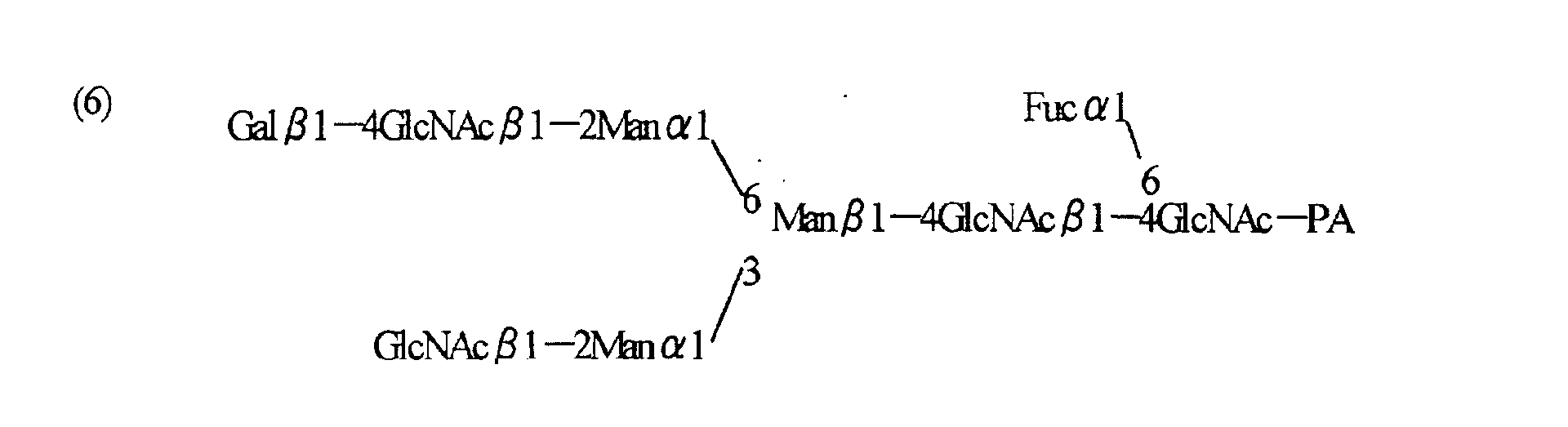
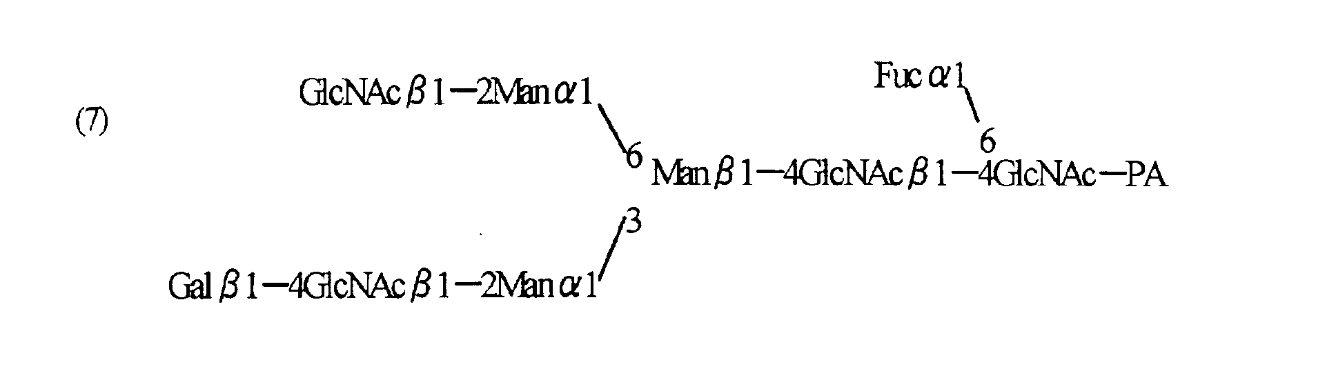
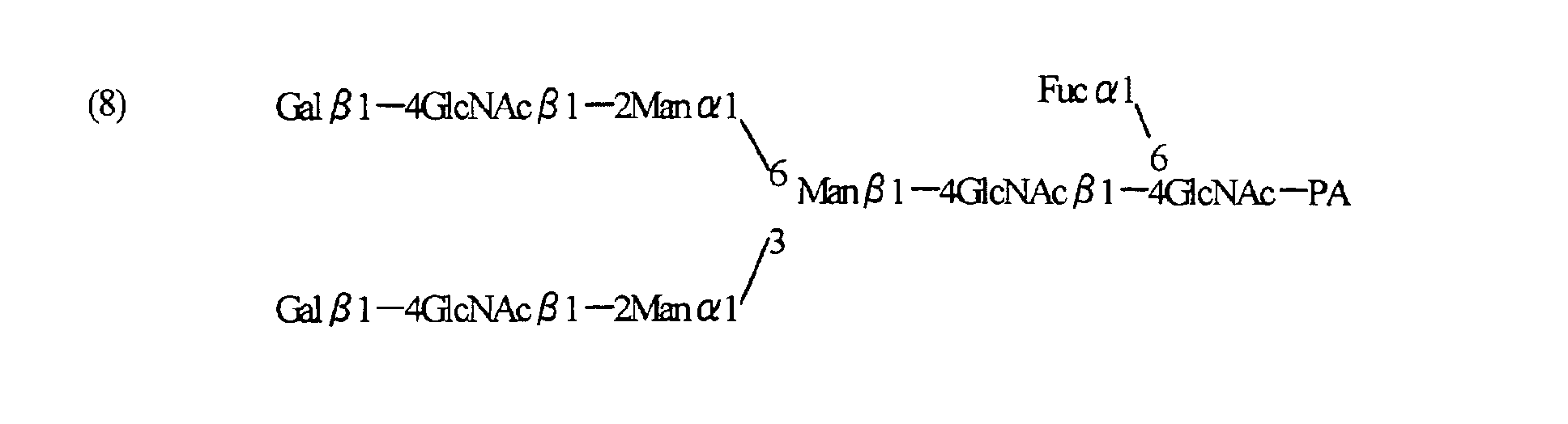


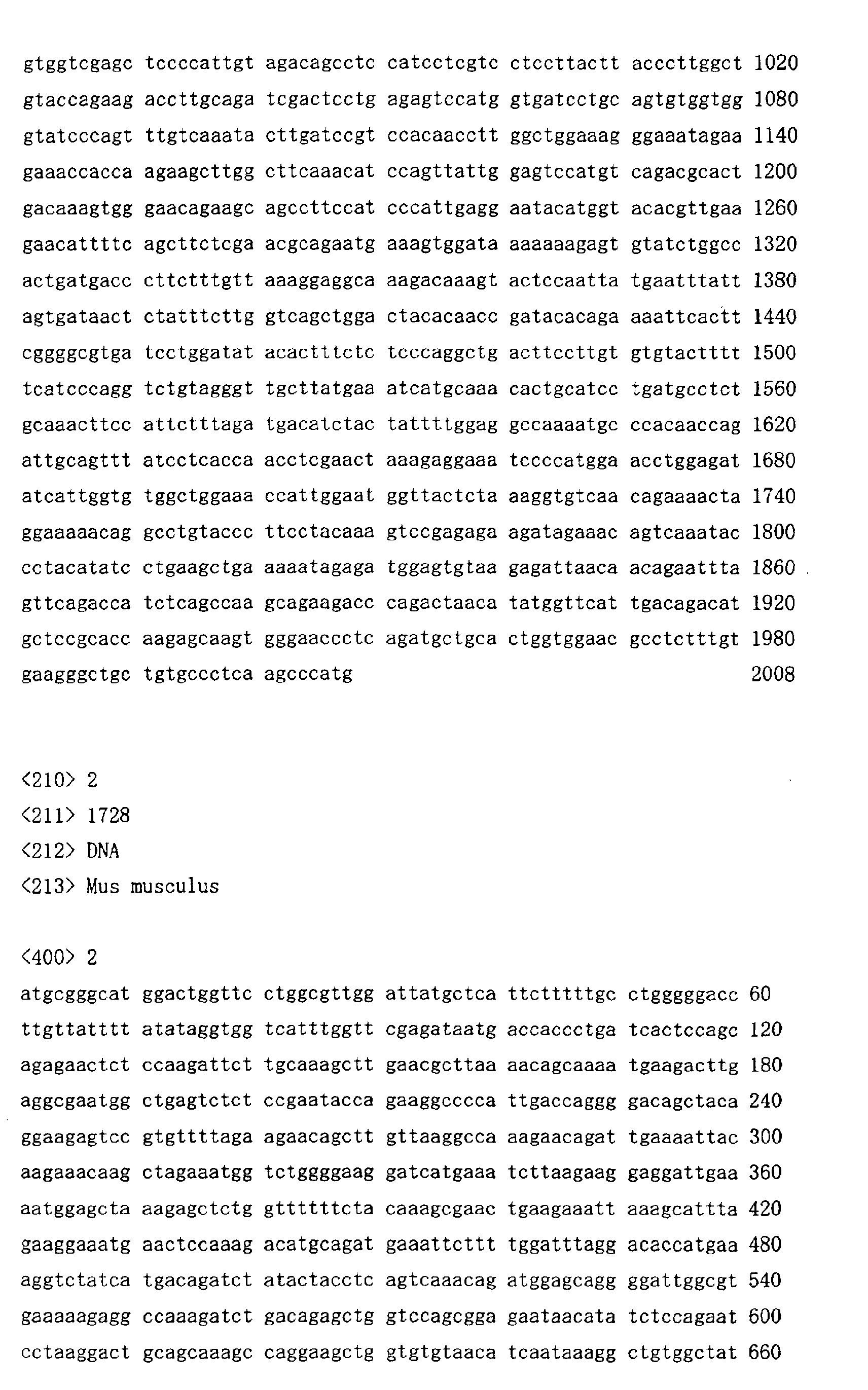


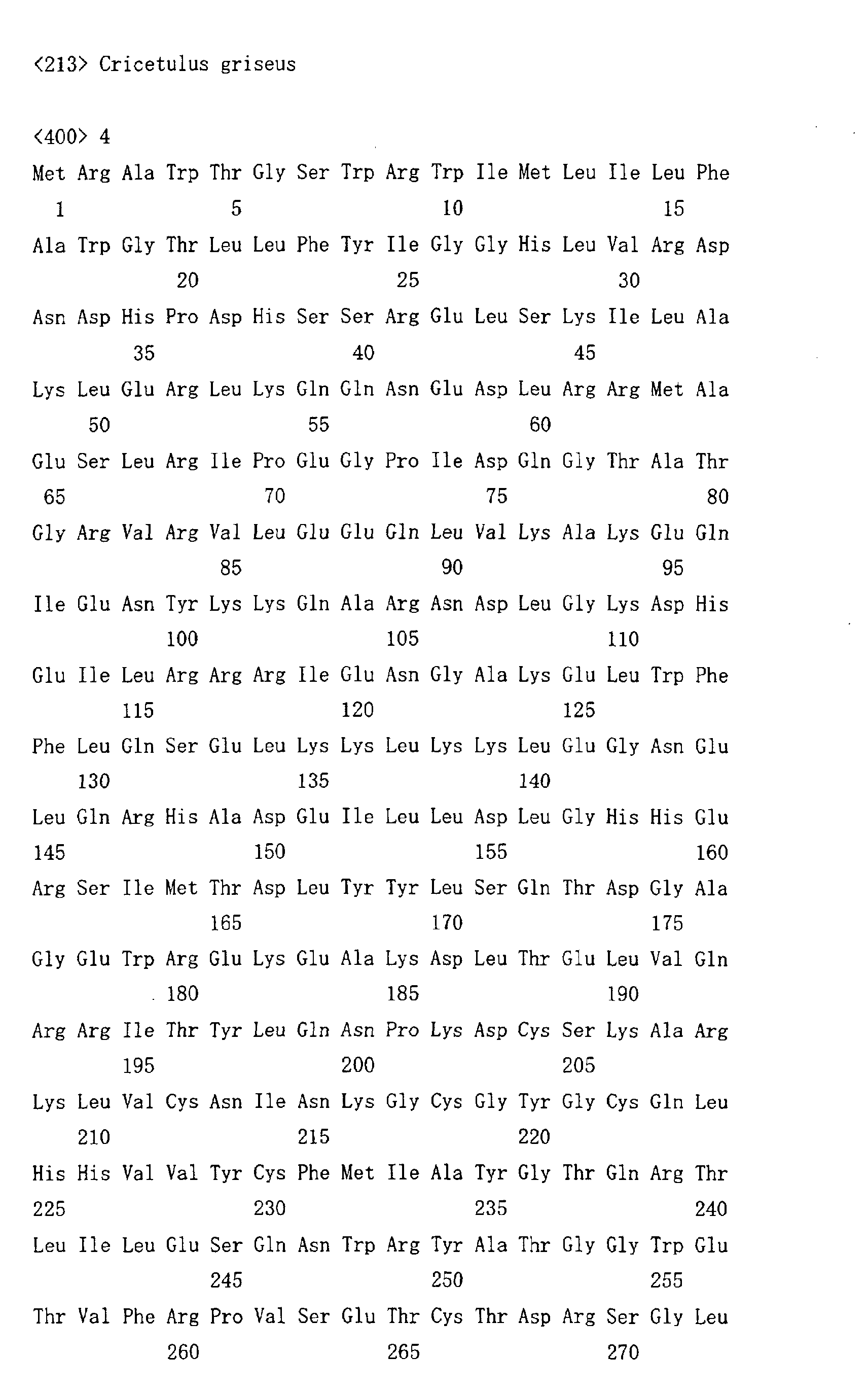
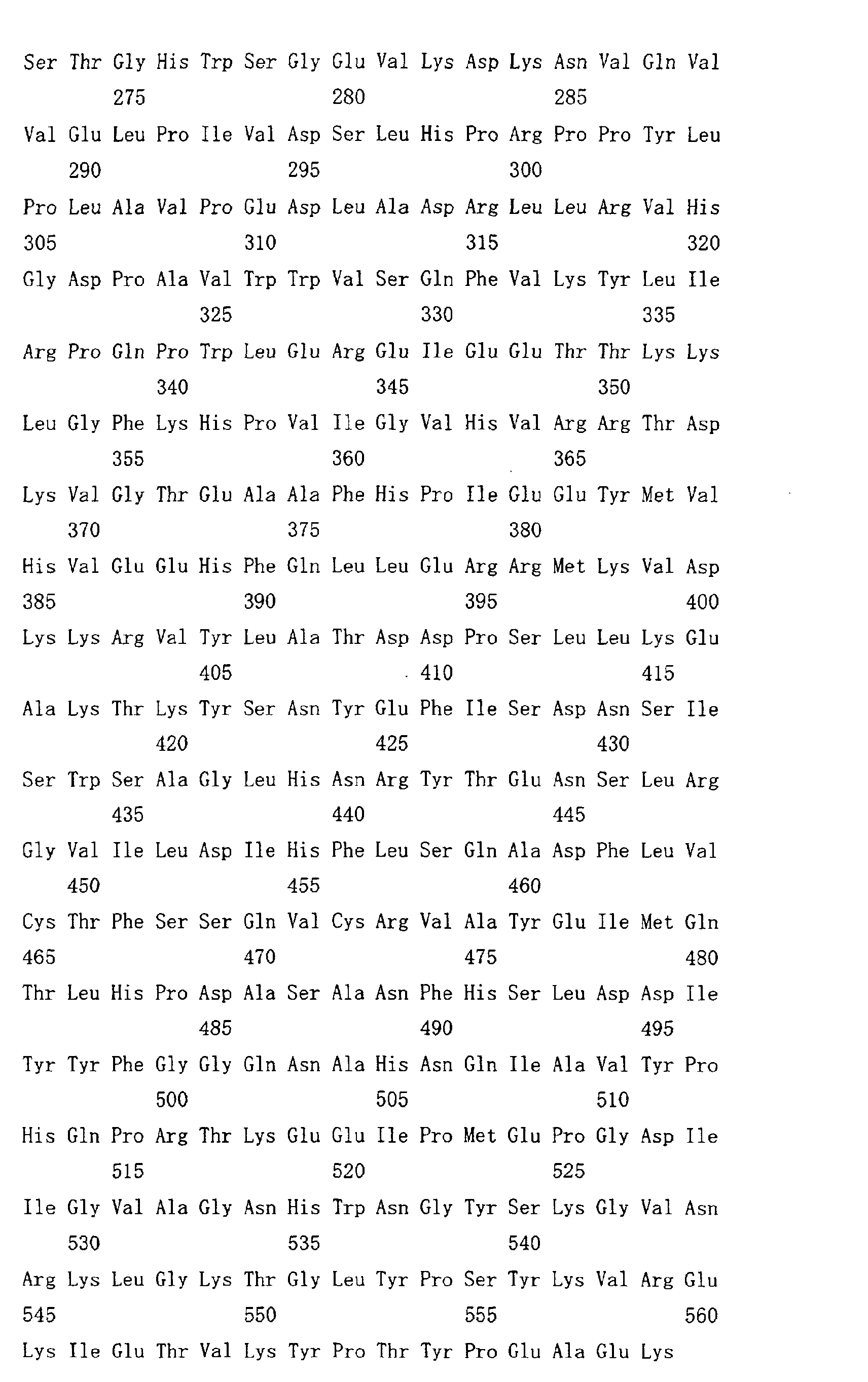

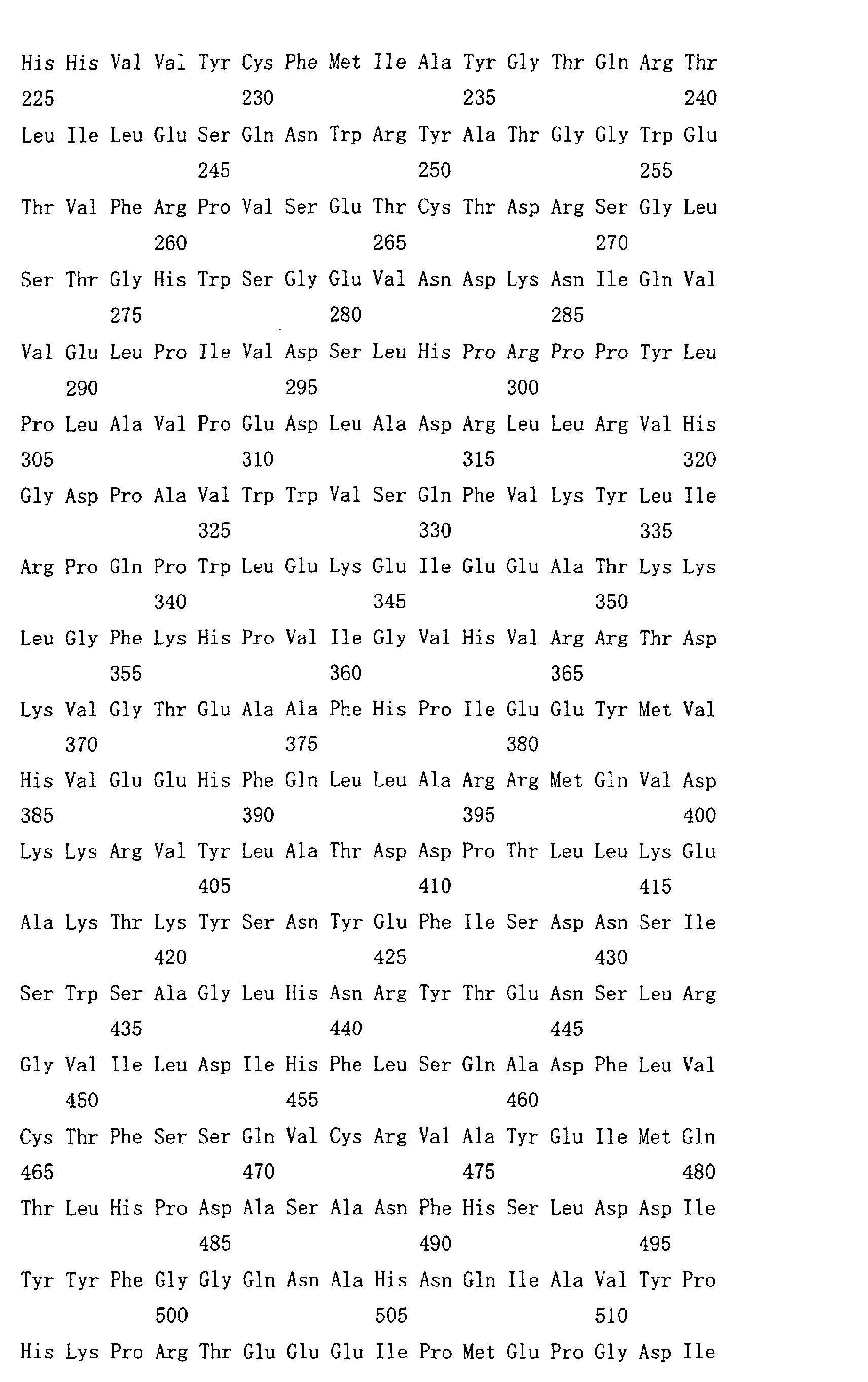


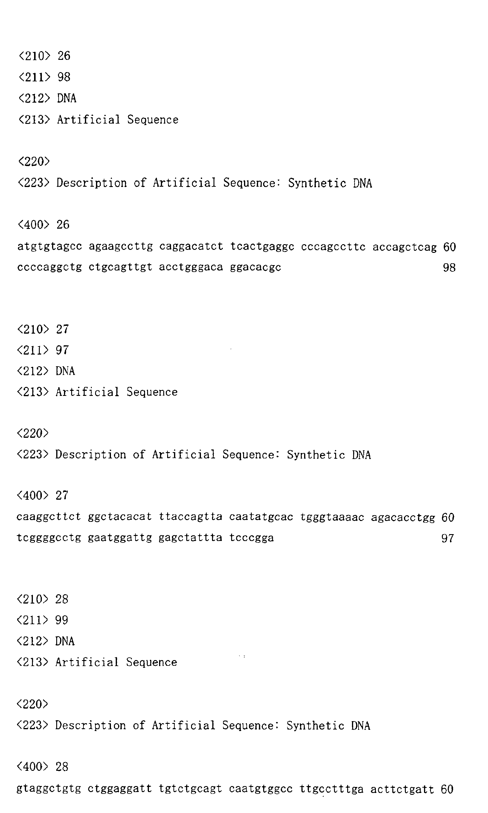
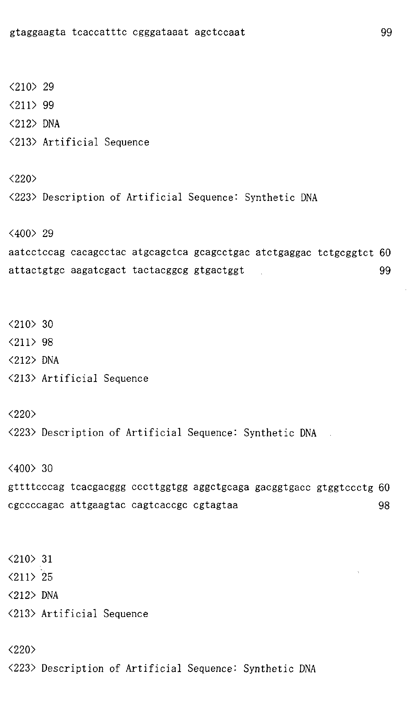
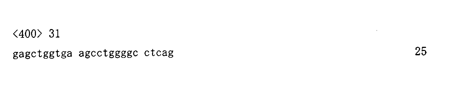
Claims (43)
- A cell in which genome is modified so as to have a more decreased or deleted activity of an enzyme relating to modification of a sugar chain in which 1-position of fucose is bound to 6-position of N-acetylglucosamine in the reducing end through α-bond in a complex N-glycoside-linked sugar chain than its parent cell.
- The cell according to claim 1, wherein a genomic gene encoding an enzyme relating to modification of a sugar chain in which 1-position of fucose is bound to 6-position of N-acetylglucosamine in the reducing end through α-bond in a complex N-glycoside-linked sugar chain is knocked out.
- The cell according to claim 1 or 2, wherein all of alleles on a genome encoding an enzyme relating to modification of a sugar chain in which 1-position of fucose is bound to 6-position of N-acetylglucosamine in the reducing end through α-bond in a complex N-glycoside-linked sugar chain are knocked out.
- The cell according to any one of claims 1 to 3, wherein the enzyme relating to modification of a sugar chain in which 1-position of fucose is bound to 6-position of N-acetylglucosamine in the reducing end through α-bond in a complex N-glycoside-linked sugar chain is α1,6-fucosyltransferase.
- The cell according to claim 4, wherein the α1,6-fucosyltransferase is a protein encoded by a DNA selected from the group consisting of the following (a) to (d):(a) a DNA comprising the nucleotide sequence represented by SEQ ID NO:1;(b) a DNA comprising the nucleotide sequence represented by SEQ ID NO:2;(c) a DNA which hybridizes with a DNA comprising the nucleotide sequence represented by SEQ ID NO:1 under stringent conditions and having an α1,6-fucosyltransferase activity;(d) a DNA which hybridizes with a DNA comprising the nucleotide sequence represented by SEQ ID NO:2 under stringent conditions and having an α1,6-fucosyltransferase activity.
- The cell according to claim 4, wherein the α1,6-fucosyltransferase is a protein selected from the group consisting of the following (a), (b), (c), (d), (e) and (f):(a) a protein comprising the amino acid sequence represented by SEQ ID NO:4;(b) a protein comprising the amino acid sequence represented by SEQ ID NO:5;(c) a protein which comprises an amino acid sequence in which at least one amino acid is deleted, substituted, inserted and/or added in the amino acid sequence represented by SEQ ID NO:4 and has an α1,6-fucosyltransferase activity;(d) a protein which comprises an amino acid sequence in which at least one amino acid is deleted, substituted, inserted and/or added in the amino acid sequence represented by SEQ ID NO:5 and has an α1,6-fucosyltransferase activity;(e) a protein which comprises an amino acid sequence having a homology of 80% or more with the amino acid sequence represented by SEQ ID NO:4 and has an α1,6-fucosyltransferase activity;(f) a protein which comprises an amino acid sequence having a homology of 80% or more with the amino acid sequence represented by SEQ ID NO:5 and has an α1,6-fucosyltransferase activity.
- The cell according to any one of claims 1 to 6, which is resistant to a lectin which recognizes a sugar chain in which 1-position of fucose is bound to 6-position of N-acetylglucosamine in the reducing end through α-bond in a complex N-glycoside-linked sugar chain.
- The cell according to claim 7, which is resistant to at least one lectin selected from the group consisting of the following (a) to (d):(a) a Lens culinaris lectin;(b) a Pisum sativum lectin;(c) a Vicia faba lectin;(d) an Aleuria aurantia lectin.
- The cell according to any one of claims 1 to 8, which is selected from the group consisting of the following (a) to (j):(a) a CHO cell derived from a Chinese hamster ovary tissue;(b) a rat myeloma cell line YB2/3HL.P2.G11.16Ag.20 cell;(c) a mouse myeloma cell line NS0 cell;(d) a mouse myeloma cell line SP2/0-AgI4 cell;(e) a BHK cell derived from a Syrian hamster kidney tissue;(f) a hybridoma cell which produces an antibody;(g) a human leukemic cell line Namalwa cell;(h) an embryonic stem cell;(i) a fertilized egg cell;(j) a plant cell.
- The cell according to any one of claims 1 to 9, which contains a gene encoding an antibody molecule.
- The cell according to claim 10, wherein the antibody molecule is selected from the group consisting of the following (a) to (d):(a) a human antibody;(b) a humanized antibody;(c) an antibody fragment comprising the Fc region of (a) or (b);(d) a fusion protein comprising the Fc region of (a) or (b).
- The cell according to claim 10 or 11, wherein the antibody molecule belongs to an IgG class.
- The cell according to any one of claims 1 to 12, which produces an antibody composition having a higher antibody-dependent cell-mediated cytotoxic activity than an antibody composition produced by its parent cell.
- The cell according to claim 13, wherein the antibody composition having a higher antibody-dependent cell-mediated cytotoxic activity has a higher ratio of a sugar chain in which fucose is not bound to N-acetylglucosamine in the reducing end in the sugar chain among total complex N-glycoside-linked sugar chains bound to the Fc region in the antibody composition than an antibody composition produced by its parent cell.
- The cell according to claim 14, wherein the sugar chain in which fucose is not bound is a sugar chain in which 1-position of the fucose is not bound to 6-position of N-acetylglucosamine in the reducing end through α-bond in a complex N-glycoside-linked sugar chain.
- The cell according to any one of claims 13 to 15, wherein the ratio of a sugar chain in which fucose is not bound to N-acetylglucosamine in the reducing end through α-bond is 20% or more of total complex N-glycoside-linked sugar chains bound to the Fc region in the antibody composition.
- The cell according to any one of claims 13 to 16, wherein the antibody composition having a higher antibody-dependent cell-mediated cytotoxic activity is an antibody composition which has no sugar chain in which fucose is bound to N-acetylglucosamine in the reducing end in the sugar chain.
- A process for producing an antibody composition, which comprises using the cell according to any one of claims 10 to 17.
- A process for producing an antibody composition, which comprises culturing the cell according to any one of claims 10 to 18 in a medium to form and accumulate an antibody composition in the culture; and recovering the antibody composition from the culture.
- The process according to claim 18 or 19, wherein the antibody composition is an antibody composition having a higher antibody-dependent cell-mediated cytotoxic activity than an antibody composition produced by its parent cell.
- The process according to claim 20, wherein the antibody composition having a higher antibody-dependent cell-mediated cytotoxic activity has a higher ratio of a sugar chain in which fucose is not bound to N-acetylglucosamine in the reducing end in the sugar chain among total complex N-glycoside-linked sugar chains bound to the Fc region in the antibody composition than an antibody composition produced by its parent cell.
- The process according to claim 21, wherein the sugar chain in which fucose is not bound is a sugar chain in which 1-position of the fucose is not bound to 6-position of N-acetylglucosamine in the reducing end through α-bond in a complex N-glycoside-linked sugar chain.
- The cell according to any one of claims 20 to 22, wherein the antibody composition having a higher antibody-dependent cell-mediated cytotoxic activity is a an antibody composition having a ratio of a sugar chain in which fucose is not bound to N-acetylglucosamine in the reducing end through α-bond of 20% or more of total complex N-glycoside-linked sugar chains bound to the Fc region in the antibody composition.
- The cell according to any one of claims 20 to 23, wherein the antibody composition having a higher antibody-dependent cell-mediated cytotoxic activity is an antibody composition which has no sugar chain in which fucose is bound to N-acetylglucosamine in the reducing end in the sugar chain.
- A transgenic non-human animal or plant or the progenies thereof, which is produced by using the cell according to any one of claims 1 to 9.
- The transgenic non-human animal or plant or the progenies thereof according to claim 24, wherein the transgenic non-human animal is an animal selected from the group consisting of cattle, sheep, goat, pig, horse, mouse, rat, fowl, monkey and rabbit.
- The transgenic non-human animal or plant or the progenies thereof according to claim 25 or 26, which is introduced with a gene encoding an antibody molecule.
- The transgenic non-human animal or plant or the progenies thereof according to claim 27, wherein the antibody molecule is selected from the group consisting of the following (a) to (d):(a) a human antibody;(b) a humanized antibody;(c) an antibody fragment comprising the Fc region of (a) or (b);(d) a fusion protein comprising the Fc region of (a) or (b).
- The transgenic non-human animal or plant or the progenies thereof according to claim 27 or 28, wherein the antibody molecule belongs to an IgG class.
- A process for producing an antibody composition, which comprises rearing the transgenic non-human animal or plant according to any one of claims 27 to 29; isolating a tissue or body fluid comprising an antibody molecule introduced from the reared animal or plant; and recovering an antibody composition comprising a desired antibody molecule from the isolated tissue or body fluid.
- A process for producing an antibody composition, which comprises isolating an antibody-producing cell from the transgenic non-human animal or plant or the progenies thereof according to any one of claims 26 to 29; culturing the isolated antibody-producing cell in a medium to form and accumulate an antibody composition in the culture; and recovering the antibody composition from the culture.
- The process according to claim 30 or 31, wherein the antibody composition is an antibody composition having a higher antibody-dependent cell-mediated cytotoxic activity than an antibody composition produced by a transgenic non-human animal or plant or the progenies thereof in which genome is not modified.
- The process according to claim 32, wherein the antibody composition having a higher antibody-dependent cell-mediated cytotoxic activity has a higher ratio of a sugar chain in which fucose is not bound to N-acetylglucosamine in the reducing end in the sugar chain among total complex N-glycoside-linked sugar chains bound to the Fc region in the antibody composition than an antibody composition produced by a transgenic non-human animal or plant or the progenies thereof in which genome is not modified.
- The process according to claim 33, wherein the sugar chain in which fucose is not bound is a sugar chain in which 1-position of the fucose is not bound to 6-position of N-acetylglucosamine in the reducing end through α-bond in a complex N-glycoside-linked sugar chain.
- The process according to any one of claims 32 to 34, wherein the antibody composition having a higher antibody-dependent cell-mediated cytotoxic activity is an antibody composition in which a ratio of a sugar chain in which fucose is not bound to N-acetylglucosamine in the reducing end in the sugar chain is 20% or more of total complex N-glycoside-linked sugar chains bound to the Fc region in the antibody composition.
- The cell according to any one of claims 32 to 35, wherein the antibody composition having a higher antibody-dependent cell-mediated cytotoxic activity is an antibody composition which has no sugar chain in which fucose is bound to N-acetylglucosamine in the reducing end in the sugar chain.
- An antibody composition comprising an antibody molecule having an N-glycoside-linked sugar chain in the Fc region, which has a sugar chain in which fucose is not bound to N-acetylglucosamine in the reducing end in the sugar chain among total complex N-glycoside-linked sugar chains bound to the Fc region in the antibody composition.
- The antibody composition according to claim 37, wherein the sugar chain in which fucose is not bound is a sugar chain in which 1-position of the fucose is not bound to 6-position of N-acetylglucosamine in the reducing end through α-bond in a complex N-glycoside-linked sugar chain.
- An antibody composition produced by the process of any one of claims 18 to 24.
- An antibody composition produced by the process of any one of claims 18 to 24.
- A medicament comprising as an active ingredient the antibody composition according to any one of claims 37 to 40.
- The medicament according to claim 41, which is a diagnostic agent, an preventing agent or a treating agent for tumor-accompanied diseases, allergy-accompanied diseases, inflammatory-accompanied diseases, autoimmune diseases, cardiovascular diseases, viral infection-accompanied diseases or bacterial infection-accompanied diseases.
- Use of the antibody composition according to any one of claims 37 to 40 in the manufacture of the medicament according to claim 41 or 42.
Applications Claiming Priority (3)
| Application Number | Priority Date | Filing Date | Title |
|---|---|---|---|
| JP2002106953 | 2002-04-09 | ||
| JP2002106953 | 2002-04-09 | ||
| PCT/JP2003/004507 WO2003085107A1 (en) | 2002-04-09 | 2003-04-09 | Cells with modified genome |
Publications (2)
| Publication Number | Publication Date |
|---|---|
| EP1498485A1 true EP1498485A1 (en) | 2005-01-19 |
| EP1498485A4 EP1498485A4 (en) | 2006-09-06 |
Family
ID=28786451
Family Applications (1)
| Application Number | Title | Priority Date | Filing Date |
|---|---|---|---|
| EP03720897A Withdrawn EP1498485A4 (en) | 2002-04-09 | 2003-04-09 | MODIFIED GENOME CELLS |
Country Status (12)
| Country | Link |
|---|---|
| US (2) | US20040110704A1 (en) |
| EP (1) | EP1498485A4 (en) |
| JP (1) | JPWO2003085107A1 (en) |
| KR (1) | KR20050000380A (en) |
| CN (3) | CN104530225A (en) |
| AU (1) | AU2003236022A1 (en) |
| BR (1) | BR0309145A (en) |
| CA (1) | CA2481657A1 (en) |
| EA (1) | EA200401325A1 (en) |
| MX (1) | MXPA04009924A (en) |
| PL (1) | PL373256A1 (en) |
| WO (1) | WO2003085107A1 (en) |
Cited By (7)
| Publication number | Priority date | Publication date | Assignee | Title |
|---|---|---|---|---|
| EP1500400A4 (en) * | 2002-04-09 | 2006-10-11 | Kyowa Hakko Kogyo Kk | MEDICAMENT CONTAINING ANTIBODY COMPOSITION |
| WO2008077545A1 (en) * | 2006-12-22 | 2008-07-03 | F. Hoffmann-La Roche Ag | Selection method |
| US7655228B2 (en) | 1999-04-09 | 2010-02-02 | Kyowa Hakko Kirin Co., Ltd | Method of modulating the activity of functional immune molecules to GM2 |
| US7741442B2 (en) | 2000-10-06 | 2010-06-22 | Kyowa Hakko Kirin Co., Ltd | Antibody composition exhibiting increased cellular cytotoxicity due to glycosylation |
| WO2012041768A1 (en) * | 2010-09-27 | 2012-04-05 | Boehringer Ingelheim International Gmbh | Production of low fucose antibodies in h4-ii-e rat cells |
| US8207303B2 (en) | 2005-02-18 | 2012-06-26 | Medarex, Inc. | Monoclonal antibodies against CD30 lacking in fucosyl residues |
| US8461308B2 (en) | 2005-02-18 | 2013-06-11 | Medarex, Inc. | Monoclonal antibodies against prostate specific membrane antigen (PSMA) lacking in fucosyl residues |
Families Citing this family (1003)
| Publication number | Priority date | Publication date | Assignee | Title |
|---|---|---|---|---|
| FR2807767B1 (en) | 2000-04-12 | 2005-01-14 | Lab Francais Du Fractionnement | MONOCLONAL ANTIBODIES ANTI-D |
| EP1471938A4 (en) * | 2002-01-09 | 2008-03-05 | Medarex Inc | Human monoclonal antibodies against cd30 |
| AU2004279739A1 (en) * | 2003-10-08 | 2005-04-21 | Kyowa Hakko Kirin Co., Ltd. | Composition of antibody capable of specifically binding CCR4 |
| AU2004279740A1 (en) * | 2003-10-08 | 2005-04-21 | Kyowa Hakko Kirin Co., Ltd. | Antibody composition specifically binding to IL-5 receptor |
| JPWO2005035586A1 (en) * | 2003-10-08 | 2007-11-22 | 協和醗酵工業株式会社 | Fusion protein composition |
| JPWO2005035577A1 (en) * | 2003-10-08 | 2007-11-22 | 協和醗酵工業株式会社 | Antibody composition that specifically binds to ganglioside GD3 |
| AU2004280064A1 (en) * | 2003-10-09 | 2005-04-21 | Kyowa Hakko Kirin Co., Ltd. | Genomically modified cell neutralized to serum-free system |
| JPWO2005035581A1 (en) * | 2003-10-09 | 2007-11-22 | 協和醗酵工業株式会社 | Antibody composition that specifically binds to human VEGF receptor Flt-1 |
| JPWO2005035741A1 (en) * | 2003-10-09 | 2006-12-21 | 協和醗酵工業株式会社 | Genome-modified cells |
| US20060021071A1 (en) * | 2003-10-09 | 2006-01-26 | Kyowa Hakko Kogyo Co., Ltd. | Cell in which genome is modified |
| CN1890262B (en) * | 2003-10-09 | 2015-01-28 | 协和发酵麒麟株式会社 | Method for producing antithrombin III composition |
| JPWO2005035578A1 (en) * | 2003-10-09 | 2007-11-22 | 協和醗酵工業株式会社 | Antibody composition that specifically binds to ganglioside GM2 |
| US7691810B2 (en) * | 2003-10-09 | 2010-04-06 | Kyowa Hakko Kirin Co., Ltd | Method of producing recombinant antithrombin III composition |
| JPWO2005053742A1 (en) * | 2003-12-04 | 2007-06-28 | 協和醗酵工業株式会社 | Medicament containing antibody composition |
| DE602004029934D1 (en) | 2003-12-04 | 2010-12-16 | Kyowa Hakko Kirin Co Ltd | A GENETICALLY MODIFIED ANTIBODY AGAINST CHEMOCIN RECEPTOR-CCR4 CONTAINING MEDICAMENT |
| SG183012A1 (en) | 2004-06-04 | 2012-08-30 | Genentech Inc | Method for treating multiple sclerosis |
| US20060223147A1 (en) * | 2004-08-05 | 2006-10-05 | Kyowa Hakko Kogyo Co., Ltd., | Process for producing glycoprotein composition |
| CN101087807A (en) | 2004-10-05 | 2007-12-12 | 健泰科生物技术公司 | Method for treating vasculitis |
| FR2879204B1 (en) * | 2004-12-15 | 2007-02-16 | Lab Francais Du Fractionnement | CYTOTOXIC ANTIBODY AGAINST HEMATOPOIETIC B-TYPE HEMATOPOIETIC PROLIFERATIONS |
| BRPI0607315B1 (en) | 2005-02-07 | 2022-05-17 | Roche Glycart Ag | Humanized anti-egfr antigen binding molecule and its uses, pharmaceutical composition, and method for detecting the presence of egfr in a sample |
| ES2386366T3 (en) * | 2005-02-18 | 2012-08-17 | Medarex, Inc. | Human monoclonal antibody against prostate specific membrane antigen (PSMA) |
| BRPI0608468A2 (en) * | 2005-03-25 | 2010-01-05 | Glycart Biotechnology Ag | mcsp-directed antigen binding molecules having increased fc receptor binding affinity and effector function |
| WO2006109696A1 (en) * | 2005-04-06 | 2006-10-19 | Kyowa Hakko Kogyo Co., Ltd. | Composition comprising genetically engineered follicle-stimulating hormone |
| WO2006109698A1 (en) * | 2005-04-06 | 2006-10-19 | Kyowa Hakko Kogyo Co., Ltd. | Composition comprising genetically engineered erythropoietin |
| WO2006109695A1 (en) * | 2005-04-06 | 2006-10-19 | Kyowa Hakko Kogyo Co., Ltd. | Composition comprising genetically engineered haptoglobin |
| WO2006116246A2 (en) * | 2005-04-25 | 2006-11-02 | Medarex, Inc. | Method of treating cd30 positive lymphomas |
| EP1888649A2 (en) * | 2005-05-09 | 2008-02-20 | GlycArt Biotechnology AG | Antigen binding molecules having modified fc regions and altered binding to fc receptors |
| EP3530736A3 (en) | 2005-05-09 | 2019-11-06 | ONO Pharmaceutical Co., Ltd. | Human monoclonal antibodies to programmed death 1 (pd-1) and methods for treating cancer using anti-pd-1 antibodies alone or in combination with other immunotherapeutics |
| KR20080032065A (en) * | 2005-06-03 | 2008-04-14 | 제넨테크, 인크. | Methods for Producing Antibodies with Modulated Fucosylation Levels |
| WO2007002223A2 (en) * | 2005-06-20 | 2007-01-04 | Medarex, Inc. | Cd19 antibodies and their uses |
| KR101704734B1 (en) | 2005-07-01 | 2017-02-09 | 이. 알. 스퀴부 앤드 선즈, 엘.엘.씨. | Human monoclonal antibodies to programmed death ligand 1(pd-l1) |
| AU2006287224A1 (en) * | 2005-08-31 | 2007-03-08 | Centocor, Inc. | Host cell lines for production of antibody constant region with enhanced effector function |
| MY149159A (en) | 2005-11-15 | 2013-07-31 | Hoffmann La Roche | Method for treating joint damage |
| JP2009518314A (en) | 2005-12-02 | 2009-05-07 | ジェネンテック・インコーポレーテッド | Compositions and methods for the treatment of diseases and disorders associated with cytokine signaling comprising antibodies that bind IL-22 and antibodies that bind IL-22R |
| EA017491B1 (en) | 2005-12-08 | 2012-12-28 | Медарекс, Инк. | HUMAN MONOCLONAL ANTIBODIES TO FUKOSIL-GM1 AND METHODS OF APPLICATION OF ANTIFUKOSIL-GM1 ANTIBODIES |
| US8101365B2 (en) | 2006-01-05 | 2012-01-24 | Genentech, Inc. | Anti-EphB4 antibodies and methods using same |
| US20090175886A1 (en) * | 2006-01-17 | 2009-07-09 | Medarex, Inc. | Monoclonal antibodies against cd30 lacking in fucosyl and xylosyl residues |
| WO2007099988A1 (en) * | 2006-02-28 | 2007-09-07 | Kyowa Hakko Kogyo Co., Ltd. | α-1,6-FUCOSYLTRANSFERASE MUTANT AND USE THEREOF |
| AR059851A1 (en) | 2006-03-16 | 2008-04-30 | Genentech Inc | ANTIBODIES OF EGFL7 AND METHODS OF USE |
| US20080118978A1 (en) | 2006-04-28 | 2008-05-22 | Takashi Sato | Anti-tumor agent |
| PT2032606E (en) | 2006-05-30 | 2014-02-05 | Genentech Inc | Antibodies and immunoconjugates and uses therefor |
| JP2009539870A (en) * | 2006-06-06 | 2009-11-19 | ジェネンテック・インコーポレーテッド | Compositions and methods for modulating angiogenesis |
| AU2007319672B2 (en) * | 2006-06-06 | 2011-06-30 | Genentech, Inc. | Anti-DLL4 antibodies and methods using same |
| DK2046809T3 (en) | 2006-07-19 | 2017-03-13 | Univ Pennsylvania | WSX-1 / IL-27 AS A TARGET OBJECTIVE FOR ANTI-INFLAMMATORY REACTIONS |
| AR062223A1 (en) * | 2006-08-09 | 2008-10-22 | Glycart Biotechnology Ag | MOLECULES OF ADHESION TO THE ANTIGEN THAT ADHER TO EGFR, VECTORS THAT CODE THEM, AND THEIR USES OF THESE |
| CN101627055A (en) | 2006-09-05 | 2010-01-13 | 梅达雷克斯公司 | Antibodies to bone morphogenetic proteins and their receptors and methods of use thereof |
| SI2066351T1 (en) | 2006-10-02 | 2016-02-29 | E.R. Squibb & Sons, L.L.C. | Human antibodies that bind cxcr4 and uses thereof |
| NZ576032A (en) | 2006-10-12 | 2012-03-30 | Genentech Inc | Antibodies to lymphotoxin-alpha |
| PT2845866T (en) | 2006-10-27 | 2017-08-09 | Genentech Inc | ANTIBODIES AND IMMUNOCONUELS AND UTILIZATIONS THEREOF |
| MX2009005189A (en) | 2006-11-15 | 2009-06-30 | Medarex Inc | Human monoclonal antibodies to btla and methods of use. |
| SG177168A1 (en) | 2006-12-01 | 2012-01-30 | Medarex Inc | Human antibodies that bind cd22 and uses thereof |
| CL2007003622A1 (en) | 2006-12-13 | 2009-08-07 | Medarex Inc | Human anti-cd19 monoclonal antibody; composition comprising it; and tumor cell growth inhibition method. |
| US20100150950A1 (en) | 2006-12-14 | 2010-06-17 | Medarex, Inc. | Human antibodies that bind cd70 and uses thereof |
| US20100028949A1 (en) * | 2006-12-22 | 2010-02-04 | Vincent Beuger | Shrna-mediated inhibition of expression of alpha 1,6-fucosyltransferase |
| EP2107115A1 (en) | 2007-01-24 | 2009-10-07 | Kyowa Hakko Kirin Co., Ltd. | Genetically recombinant antibody composition capable of binding specifically to ganglioside gm2 |
| CA2676529C (en) | 2007-01-24 | 2014-03-25 | Kyowa Kirin Co., Ltd. | Genetically recombinant antibody composition having enhanced effector activity |
| EP2125013A4 (en) * | 2007-01-26 | 2010-04-07 | Bioinvent Int Ab | DLL4 SIGNALING INHIBITORS AND USES THEREOF |
| US7834154B2 (en) | 2007-02-09 | 2010-11-16 | Genentech, Inc. | Anti-ROBO4 antibodies and uses therefor |
| EP3118221B1 (en) | 2007-02-26 | 2019-08-21 | Oxford BioTherapeutics Ltd | Proteins |
| WO2008104803A2 (en) | 2007-02-26 | 2008-09-04 | Oxford Genome Sciences (Uk) Limited | Proteins |
| BRPI0811466A2 (en) | 2007-05-07 | 2014-10-14 | Medimmune Llc | ISOLATED ANTIBODY, NUCLEIC ACID, VECTOR, ISOLATED CELL, METHODS FOR PRODUCTING AN ANTIBODY, TO TREAT DISEASE OR DISORDER, TO TREAT OR PREVENT REJECTION IN A HUMAN TRANSPLANT PATIENT TO BE TURNED TO HUMAN, TO EXHAUST T CELLS EXPRESSING ICOS IN A HUMAN PATIENT, TO BREAK THE GERMINAL CENTER ARCHITECTURE IN A PRIMARY SECONDARY LYMPHID ORGAN, TO DEPLETE GERMAN CENTRAL LYMPHIDE ORGAN B CELLS WITH A PRIMATE BULTA IN CURRENT CLASSES IN A PRIMATE, AND, PHARMACEUTICAL COMPOSITION. |
| PT3072525T (en) | 2007-05-14 | 2018-04-20 | Astrazeneca Ab | Methods of reducing basophil levels |
| EP2586788B1 (en) | 2007-07-09 | 2017-11-29 | Genentech, Inc. | Prevention of disulfide bond reduction during recombinant production of polypeptides |
| AU2008275649B2 (en) | 2007-07-12 | 2013-09-05 | Sangamo Therapeutics, Inc. | Methods and compositions for inactivating alpha 1,6 fucosyltransferase (FUT 8) gene expression |
| SI2769729T1 (en) | 2007-09-04 | 2019-06-28 | Compugen Ltd. | Polypeptidine and polynucleotides and their uses as the target of medicines for medicinal products and biological medicines |
| ES2687808T3 (en) | 2007-09-26 | 2018-10-29 | Chugai Seiyaku Kabushiki Kaisha | Constant region of modified antibody |
| US20090202475A1 (en) | 2007-11-07 | 2009-08-13 | Genentech, Inc. | Compositions and methods for treatment of microbial disorders |
| US8637435B2 (en) * | 2007-11-16 | 2014-01-28 | Merck Sharp & Dohme Corp. | Eukaryotic cell display systems |
| TWI468417B (en) | 2007-11-30 | 2015-01-11 | Genentech Inc | Anti-vegf antibodies |
| WO2009100110A1 (en) | 2008-02-05 | 2009-08-13 | Medarex, Inc. | Alpha 5 - beta 1 antibodies and their uses |
| CA2715212A1 (en) | 2008-03-03 | 2009-09-11 | Glycofi, Inc. | Surface display of recombinant proteins in lower eukaryotes |
| NZ601815A (en) * | 2008-03-31 | 2014-10-31 | Genentech Inc | Compositions and methods for treating and diagnosing asthma |
| KR101054362B1 (en) * | 2008-07-03 | 2011-08-05 | 재단법인 목암생명공학연구소 | How to reduce the fucose content of recombinant protein |
| US8067339B2 (en) * | 2008-07-09 | 2011-11-29 | Merck Sharp & Dohme Corp. | Surface display of whole antibodies in eukaryotes |
| AR072999A1 (en) | 2008-08-11 | 2010-10-06 | Medarex Inc | HUMAN ANTIBODIES THAT JOIN GEN 3 OF LYMPHOCYTARY ACTIVATION (LAG-3) AND THE USES OF THESE |
| JP5662149B2 (en) * | 2008-08-13 | 2015-01-28 | 協和発酵キリン株式会社 | Genetically modified protein S composition |
| AR073295A1 (en) | 2008-09-16 | 2010-10-28 | Genentech Inc | METHODS TO TREAT PROGRESSIVE MULTIPLE SCLEROSIS. MANUFACTURING ARTICLE. |
| AU2009303526B2 (en) | 2008-10-14 | 2015-01-15 | Genentech, Inc. | Immunoglobulin variants and uses thereof |
| WO2010067308A2 (en) | 2008-12-08 | 2010-06-17 | Compugen Ltd. | Polypeptides and polynucleotides, and uses thereof as a drug target for producing drugs and biologics |
| WO2010075249A2 (en) | 2008-12-22 | 2010-07-01 | Genentech, Inc. | A method for treating rheumatoid arthritis with b-cell antagonists |
| MX2011006870A (en) | 2008-12-23 | 2011-07-19 | Genentech Inc | Immunoglobulin variants with altered binding to protein a. |
| EP2379598B1 (en) | 2009-01-19 | 2015-03-11 | Innate Pharma | Anti-kir3d antibodies |
| CA2750581A1 (en) | 2009-01-21 | 2010-07-29 | Oxford Biotherapeutics Ltd. | Pta089 protein |
| JP5816558B2 (en) | 2009-03-05 | 2015-11-18 | メダレックス・リミテッド・ライアビリティ・カンパニーMedarex, L.L.C. | Fully human antibody specific for CADM1 |
| TWI544077B (en) | 2009-03-19 | 2016-08-01 | 中外製藥股份有限公司 | Antibody constant region change body |
| KR20150036824A (en) | 2009-03-20 | 2015-04-07 | 제넨테크, 인크. | Bispecific anti-her antibodies |
| CN103755808B (en) | 2009-03-25 | 2016-02-10 | 霍夫曼-拉罗奇有限公司 | Anti-alpha 5 beta 1 antibodies and application thereof |
| ES2549769T3 (en) | 2009-03-25 | 2015-11-02 | Genentech, Inc. | Anti-FGFR3 antibodies and methods that employ them |
| PT2417156E (en) | 2009-04-07 | 2015-04-29 | Roche Glycart Ag | Trivalent, bispecific antibodies |
| NZ616382A (en) | 2009-04-20 | 2015-04-24 | Oxford Biotherapeutics Ltd | Antibodies specific to cadherin-17 |
| US8815242B2 (en) * | 2009-05-27 | 2014-08-26 | Synageva Biopharma Corp. | Avian derived antibodies |
| US9676845B2 (en) | 2009-06-16 | 2017-06-13 | Hoffmann-La Roche, Inc. | Bispecific antigen binding proteins |
| WO2010146059A2 (en) | 2009-06-16 | 2010-12-23 | F. Hoffmann-La Roche Ag | Biomarkers for igf-1r inhibitor therapy |
| MX337468B (en) | 2009-06-18 | 2016-03-02 | Pfizer | Anti notch-1 antibodies. |
| TW201106972A (en) | 2009-07-27 | 2011-03-01 | Genentech Inc | Combination treatments |
| CA2769473A1 (en) | 2009-07-31 | 2011-02-03 | N.V. Organon | Fully human antibodies to btla |
| ES2513292T3 (en) | 2009-07-31 | 2014-10-24 | Genentech, Inc. | Inhibition of tumor metastases using anti-G-CSF antibodies |
| MX355650B (en) | 2009-08-11 | 2018-04-26 | Genentech Inc | Production of proteins in glutamine-free cell culture media. |
| WO2011021146A1 (en) | 2009-08-20 | 2011-02-24 | Pfizer Inc. | Osteopontin antibodies |
| CA2770174A1 (en) * | 2009-08-31 | 2011-03-03 | Roche Glycart Ag | Affinity-matured humanized anti cea monoclonal antibodies |
| JP5887270B2 (en) | 2009-09-02 | 2016-03-16 | ジェネンテック, インコーポレイテッド | Mutant SMOOTHENED AND METHOD OF USING THE SAME |
| EP2476438B1 (en) | 2009-09-10 | 2014-07-23 | Kyowa Hakko Kirin Co., Ltd. | Medicament including antibody composition specifically bound to human cc chemokine receptor 4 (ccr4) |
| US20120231004A1 (en) | 2009-10-13 | 2012-09-13 | Oxford Biotherapeutic Ltd. | Antibodies |
| EP2491059B1 (en) | 2009-10-22 | 2015-02-25 | F.Hoffmann-La Roche Ag | Anti-hepsin antibodies and methods using same |
| WO2011056502A1 (en) | 2009-10-26 | 2011-05-12 | Genentech, Inc. | Bone morphogenetic protein receptor type ii compositions and methods of use |
| WO2011056494A1 (en) | 2009-10-26 | 2011-05-12 | Genentech, Inc. | Activin receptor-like kinase-1 antagonist and vegfr3 antagonist combinations |
| WO2011056497A1 (en) | 2009-10-26 | 2011-05-12 | Genentech, Inc. | Activin receptor type iib compositions and methods of use |
| WO2011054007A1 (en) | 2009-11-02 | 2011-05-05 | Oxford Biotherapeutics Ltd. | Ror1 as therapeutic and diagnostic target |
| CA2780221A1 (en) | 2009-11-04 | 2011-05-12 | Fabrus Llc | Methods for affinity maturation-based antibody optimization |
| EP3594356A1 (en) | 2009-11-04 | 2020-01-15 | Merck Sharp & Dohme Corp. | Engineered anti-tslp antibody |
| KR101968766B1 (en) | 2009-11-05 | 2019-04-12 | 제넨테크, 인크. | Methods and composition for secretion of heterologous polypeptides |
| WO2011068136A1 (en) * | 2009-12-01 | 2011-06-09 | 協和発酵キリン株式会社 | Pharmaceutical agent comprising antibody capable of binding specifically to ganglioside gd3 |
| WO2011067711A2 (en) | 2009-12-01 | 2011-06-09 | Compugen Ltd | Novel heparanase splice variant |
| US8486397B2 (en) | 2009-12-11 | 2013-07-16 | Genentech, Inc. | Anti-VEGF-C antibodies and methods using same |
| MX2012007379A (en) | 2009-12-23 | 2012-08-31 | Genentech Inc | Anti-bv8 antibodies and uses thereof. |
| AR080154A1 (en) | 2010-02-10 | 2012-03-14 | Immunogen Inc | CD20 ANTIBODIES AND ITS USE |
| ES2519348T3 (en) | 2010-02-18 | 2014-11-06 | Genentech, Inc. | Neurregulin antagonists and their use in cancer treatment |
| US20110200595A1 (en) | 2010-02-18 | 2011-08-18 | Roche Glycart | TREATMENT WITH A HUMANIZED IgG CLASS ANTI EGFR ANTIBODY AND AN ANTIBODY AGAINST INSULIN LIKE GROWTH FACTOR 1 RECEPTOR |
| BR112012020882A2 (en) | 2010-02-24 | 2015-11-03 | Merck Sharp & Dohme | methods for producing a heterologous glycoprotein and for producing a glycoprotein composition, host cell, glycoprotein composition, and use of recombinant pichia pastoris. |
| CA2791652C (en) | 2010-03-02 | 2018-06-12 | Kyowa Kirin Co., Ltd. | Modified antibody composition |
| KR101899835B1 (en) | 2010-03-24 | 2018-09-19 | 제넨테크, 인크. | Anti-lrp6 antibodies |
| CN103119054B (en) | 2010-03-26 | 2017-01-18 | 达特茅斯大学理事会 | VISTA regulatory T cell mediator protein, VISTA binding agent and use thereof |
| US20150231215A1 (en) | 2012-06-22 | 2015-08-20 | Randolph J. Noelle | VISTA Antagonist and Methods of Use |
| US10745467B2 (en) | 2010-03-26 | 2020-08-18 | The Trustees Of Dartmouth College | VISTA-Ig for treatment of autoimmune, allergic and inflammatory disorders |
| WO2011147834A1 (en) | 2010-05-26 | 2011-12-01 | Roche Glycart Ag | Antibodies against cd19 and uses thereof |
| NZ701208A (en) | 2010-06-03 | 2016-05-27 | Genentech Inc | Immuno-pet imaging of antibodies and immunoconjugates and uses thereof |
| CN103080136B (en) | 2010-06-18 | 2015-08-12 | 霍夫曼-拉罗奇有限公司 | Anti-Axl antibody and using method |
| CA2803391C (en) | 2010-06-22 | 2021-11-09 | Neogenix Oncology, Inc. | Npc1 antibodies that bind a muc5ac epitope |
| WO2011161119A1 (en) | 2010-06-22 | 2011-12-29 | F. Hoffmann-La Roche Ag | Antibodies against insulin-like growth factor i receptor and uses thereof |
| WO2011161189A1 (en) | 2010-06-24 | 2011-12-29 | F. Hoffmann-La Roche Ag | Anti-hepsin antibodies and methods of use |
| NZ605449A (en) | 2010-07-09 | 2015-03-27 | Genentech Inc | Anti-neuropilin antibodies and methods of use |
| WO2012010582A1 (en) | 2010-07-21 | 2012-01-26 | Roche Glycart Ag | Anti-cxcr5 antibodies and methods of use |
| KR20130045914A (en) | 2010-08-03 | 2013-05-06 | 에프. 호프만-라 로슈 아게 | Chronic lymphocytic leukemia (cll) biomarkers |
| BR112013002532A2 (en) | 2010-08-05 | 2016-05-31 | Hoffmann La Roche | anti-mhc antibody anti-viral cytokine fusion protein |
| PE20171512A1 (en) | 2010-08-13 | 2017-10-20 | Roche Glycart Ag | ANTI-FAP ANTIBODIES |
| BR112013002444A2 (en) | 2010-08-13 | 2016-05-24 | Roche Glycart Ag | isolated antibody, polynucleotide and polypeptide, composition, vector, host cell, antibody conjugate, pharmaceutical formulation, use of the antibody, methods of producing an antibody, treating an individual, inducing cell lysis of a tumor cell and diagnosing a disease in an individual |
| KR101603001B1 (en) | 2010-08-25 | 2016-03-11 | 에프. 호프만-라 로슈 아게 | Antibodies against il-18r1 and uses thereof |
| SG10201408229WA (en) | 2010-08-31 | 2015-02-27 | Genentech Inc | Biomarkers and methods of treatment |
| EP2616100B1 (en) | 2010-09-17 | 2016-08-31 | Compugen Ltd. | Compositions and methods for treatment of drug resistant multiple myeloma |
| EP2622091B1 (en) | 2010-09-23 | 2019-03-13 | Precision Biologics, Inc. | Colon and pancreas cancer peptidomimetics |
| US8481680B2 (en) | 2010-10-05 | 2013-07-09 | Genentech, Inc. | Mutant smoothened and methods of using the same |
| AU2011326564A1 (en) | 2010-11-10 | 2013-05-09 | Genentech, Inc. | Methods and compositions for neural disease immunotherapy |
| US8992908B2 (en) | 2010-11-23 | 2015-03-31 | Alderbio Holdings Llc | Anti-IL-6 antibodies for the treatment of oral mucositis |
| WO2012080926A2 (en) | 2010-12-15 | 2012-06-21 | Wyeth Llc | Anti-notch1 antibodies |
| KR101615420B1 (en) | 2010-12-16 | 2016-04-26 | 제넨테크, 인크. | Diagnosis and treatments relating to th2 inhibition |
| AU2011349443B2 (en) | 2010-12-20 | 2015-12-24 | Genentech, Inc. | Anti-mesothelin antibodies and immunoconjugates |
| CN103261230A (en) | 2010-12-22 | 2013-08-21 | 霍夫曼-拉罗奇有限公司 | Anti-PCSK9 antibodies and methods of use |
| WO2012092539A2 (en) | 2010-12-31 | 2012-07-05 | Takeda Pharmaceutical Company Limited | Antibodies to dll4 and uses thereof |
| RU2013139267A (en) | 2011-02-10 | 2015-03-20 | Рош Гликарт Аг | IMPROVED IMMUNOTHERAPY |
| WO2012116926A1 (en) | 2011-02-28 | 2012-09-07 | F. Hoffmann-La Roche Ag | Antigen binding proteins |
| JP5768147B2 (en) | 2011-02-28 | 2015-08-26 | エフ.ホフマン−ラ ロシュ アーゲーF. Hoffmann−La Roche Aktiengesellschaft | Monovalent antigen binding protein |
| KR101981342B1 (en) | 2011-03-02 | 2019-05-22 | 로슈 글리카트 아게 | Cea antibodies |
| CA2828890A1 (en) | 2011-04-07 | 2012-10-11 | Genentech, Inc. | Anti-fgfr4 antibodies and methods of use |
| RU2623161C2 (en) | 2011-04-15 | 2017-06-22 | Компуджен Лтд. | Polypeptides and polynucleotides and their use for treatment of immune disorders and cancer |
| WO2012146630A1 (en) | 2011-04-29 | 2012-11-01 | F. Hoffmann-La Roche Ag | N-terminal acylated polypeptides, methods for their production and uses thereof |
| EA201892619A1 (en) | 2011-04-29 | 2019-04-30 | Роше Гликарт Аг | IMMUNOCONJUGATES CONTAINING INTERLEUKIN-2 MUTANT POLYPETIPS |
| EP2707723B1 (en) | 2011-05-12 | 2016-02-10 | Genentech, Inc. | Multiple reaction monitoring lc-ms/ms method to detect therapeutic antibodies in animal samples using framework signature pepides |
| WO2012158704A1 (en) | 2011-05-16 | 2012-11-22 | Genentech, Inc. | Fgfr1 agonists and methods of use |
| US9067997B2 (en) | 2011-05-25 | 2015-06-30 | Innate Pharma Sa | Anti-KIR antibodies for the treatment of inflammatory and autoimmune disorders |
| WO2012171996A1 (en) | 2011-06-15 | 2012-12-20 | F. Hoffmann-La Roche Ag | Anti-human epo receptor antibodies and methods of use |
| PL2723773T3 (en) | 2011-06-22 | 2018-07-31 | F.Hoffmann-La Roche Ag | Removal of target cells by circulating virus-specific cytotoxic t-cells using mhc class i comprising complexes |
| US9447178B2 (en) | 2011-06-28 | 2016-09-20 | Oxford Biotherapeutics Ltd. | Therapeutic and diagnostic target |
| EA031181B1 (en) | 2011-06-28 | 2018-11-30 | Оксфорд Байотерепьютикс Лтд. | Antibody or an antigen binding fragment thereof that specifically binds to bst1, nucleic acids encoding chains thereof, host cell and method provided for making this antibody and use thereof for treating cancer and inflammatory diseases |
| RU2013155695A (en) | 2011-06-30 | 2015-08-10 | Дженентек, Инк. | ANTIBODIES AGAINST C-MET |
| EP2726503B1 (en) | 2011-06-30 | 2019-09-04 | Compugen Ltd. | Polypeptides and uses thereof for treatment of autoimmune disorders and infection |
| JP2014525412A (en) | 2011-08-17 | 2014-09-29 | ジェネンテック, インコーポレイテッド | Inhibition of angiogenesis in refractory tumors |
| BR112014003431A2 (en) | 2011-08-17 | 2017-06-13 | Genentech Inc | antibody, nucleic acid, host cell, method of producing an antibody, immunoconjugate, pharmaceutical formulation, pharmaceutical agent, use of the antibody, method of treating an individual who has cancer, and time-lapse method for tumor recurrence |
| JP2014534806A (en) | 2011-08-23 | 2014-12-25 | ロシュ グリクアート アーゲー | Anti-MCSP antibody |
| KR101721301B1 (en) | 2011-08-23 | 2017-03-29 | 로슈 글리카트 아게 | Bispecific antigen binding molecules |
| CN103930781A (en) | 2011-09-15 | 2014-07-16 | 霍夫曼-拉罗奇有限公司 | Methods of promoting differentiation |
| AU2012312515A1 (en) | 2011-09-19 | 2014-03-13 | Genentech, Inc. | Combination treatments comprising c-met antagonists and B-raf antagonists |
| EP2759551B1 (en) | 2011-09-21 | 2019-05-22 | Fujirebio Inc. | Antibodies binding to an affinity complex comprising 25oh vitamin d2 or d3 and an antibody thereto |
| US20130089562A1 (en) | 2011-10-05 | 2013-04-11 | Genenthech, Inc. | Methods of treating liver conditions using notch2 antagonists |
| PE20141562A1 (en) | 2011-10-14 | 2014-11-12 | Genentech Inc | ANTI-HTRA1 ANTIBODIES AND METHODS OF USE |
| US9358250B2 (en) | 2011-10-15 | 2016-06-07 | Genentech, Inc. | Methods of using SCD1 antagonists |
| WO2013059531A1 (en) | 2011-10-20 | 2013-04-25 | Genentech, Inc. | Anti-gcgr antibodies and uses thereof |
| WO2013059740A1 (en) | 2011-10-21 | 2013-04-25 | Foundation Medicine, Inc. | Novel alk and ntrk1 fusion molecules and uses thereof |
| KR20140097205A (en) | 2011-10-28 | 2014-08-06 | 제넨테크, 인크. | Therapeutic combinations and methods of treating melanoma |
| SG11201402485UA (en) | 2011-11-21 | 2014-06-27 | Genentech Inc | Purification of anti-c-met antibodies |
| EP2788024A1 (en) | 2011-12-06 | 2014-10-15 | F.Hoffmann-La Roche Ag | Antibody formulation |
| SG10201700169PA (en) | 2011-12-22 | 2017-02-27 | Hoffmann La Roche | Expression vector element combinations, novel production cell generation methods and their use for the recombinant production of polypeptides |
| EP2794662A1 (en) | 2011-12-22 | 2014-10-29 | F.Hoffmann-La Roche Ag | Full length antibody display system for eukaryotic cells and its use |
| CN107881196A (en) | 2011-12-22 | 2018-04-06 | 弗·哈夫曼-拉罗切有限公司 | Expression vector tissue, new Cells for production production method and its purposes in restructuring produces polypeptide |
| AR089434A1 (en) | 2011-12-23 | 2014-08-20 | Genentech Inc | PROCEDURE TO PREPARE FORMULATIONS WITH HIGH CONCENTRATION OF PROTEINS |
| WO2013101771A2 (en) | 2011-12-30 | 2013-07-04 | Genentech, Inc. | Compositions and method for treating autoimmune diseases |
| EP2802601B1 (en) | 2012-01-09 | 2019-11-13 | The Scripps Research Institute | Humanized antibodies with ultralong cdr3s |
| US20140050720A1 (en) | 2012-01-09 | 2014-02-20 | The Scripps Research Institute | Ultralong complementarity determining regions and uses thereof |
| RU2014133547A (en) | 2012-01-18 | 2016-03-10 | Дженентек, Инк. | WAYS OF APPLICATION OF FGF19 MODULATORS |
| SG10201601747XA (en) | 2012-01-18 | 2016-04-28 | Genentech Inc | Anti-LRP5 Antibodies And Methods Of Use |
| WO2013116287A1 (en) | 2012-01-31 | 2013-08-08 | Genentech, Inc. | Anti-ig-e m1' antibodies and methods using same |
| AU2013216320A1 (en) | 2012-02-01 | 2014-04-03 | Compugen Ltd. | C10RF32 antibodies, and uses thereof for treatment of cancer |
| BR112014019741A2 (en) | 2012-02-11 | 2020-12-22 | Genentech, Inc | USES OF AN ANTAGONIST OF THE WNT VIA, USE OF ANTI-CANCER THERAPY, METHOD OF IDENTIFICATION OF AN INDIVIDUAL WITH CANCER, METHODS FOR PREVENTING, METHOD OF INHIBITION OF A CANCER CELL PROLIFERATION, USE OF AN ANGONIST ANTAGONIST TRANSLOCATION OF ISOLATED R-SPONDINA |
| KR102069397B1 (en) | 2012-02-15 | 2020-01-22 | 에프. 호프만-라 로슈 아게 | Fc-receptor based affinity chromatography |
| WO2013148315A1 (en) | 2012-03-27 | 2013-10-03 | Genentech, Inc. | Diagnosis and treatments relating to her3 inhibitors |
| AR090549A1 (en) | 2012-03-30 | 2014-11-19 | Genentech Inc | ANTI-LGR5 AND IMMUNOCATE PLAYERS |
| KR20150006000A (en) | 2012-05-01 | 2015-01-15 | 제넨테크, 인크. | Anti-pmel17 antibodies and immunoconjugates |
| WO2013170191A1 (en) | 2012-05-11 | 2013-11-14 | Genentech, Inc. | Methods of using antagonists of nad biosynthesis from nicotinamide |
| EP2852840B1 (en) | 2012-05-23 | 2019-10-09 | F.Hoffmann-La Roche Ag | Selection method for therapeutic agents |
| AR091462A1 (en) | 2012-06-15 | 2015-02-04 | Genentech Inc | ANTI-PCSK9 ANTIBODIES, FORMULATIONS, DOSAGE AND METHODS OF USE |
| US9890215B2 (en) | 2012-06-22 | 2018-02-13 | King's College London | Vista modulators for diagnosis and treatment of cancer |
| CN104619722B (en) | 2012-06-22 | 2022-10-04 | 达特茅斯大学理事会 | Novel VISTA-IG constructs and use of VISTA-IG for treatment of autoimmune, allergic and inflammatory diseases |
| AR091649A1 (en) | 2012-07-02 | 2015-02-18 | Bristol Myers Squibb Co | OPTIMIZATION OF ANTIBODIES THAT FIX THE LYMPHOCYTE ACTIVATION GEN 3 (LAG-3) AND ITS USES |
| EP2869837B1 (en) | 2012-07-04 | 2016-09-14 | F. Hoffmann-La Roche AG | Anti-theophylline antibodies and methods of use |
| SG11201407420RA (en) | 2012-07-04 | 2015-02-27 | Hoffmann La Roche | Covalently linked antigen-antibody conjugates |
| MX354303B (en) | 2012-07-04 | 2018-02-23 | Hoffmann La Roche | Anti-biotin antibodies and methods of use. |
| PL2870247T3 (en) | 2012-07-05 | 2019-10-31 | Hoffmann La Roche | Expression and secretion system |
| AU2013288932A1 (en) | 2012-07-09 | 2014-12-11 | Genentech, Inc. | Immunoconjugates comprising anti - CD79b antibodies |
| KR20150027829A (en) | 2012-07-09 | 2015-03-12 | 제넨테크, 인크. | Immunoconjucates comprising anti-CD22 antibodies |
| CA2874904A1 (en) | 2012-07-09 | 2014-01-16 | Genentech, Inc. | Immunoconjugates comprising anti-cd22 antibodies |
| BR112015000437A2 (en) | 2012-07-09 | 2017-06-27 | Genentech Inc | immunoconjugates, pharmaceutical formulation, methods of treating and inhibiting cell proliferation |
| GB201213652D0 (en) | 2012-08-01 | 2012-09-12 | Oxford Biotherapeutics Ltd | Therapeutic and diagnostic target |
| PL3434695T3 (en) | 2012-08-07 | 2021-05-17 | Roche Glycart Ag | Improved immunotherapy |
| CA2884704C (en) | 2012-09-07 | 2023-04-04 | Randolph J. Noelle | Vista modulators for diagnosis and treatment of cancer |
| EP3919079A1 (en) | 2012-09-07 | 2021-12-08 | Genentech, Inc. | Combination therapy of a type ii anti-cd20 antibody with a selective bcl-2 inhibitor |
| US10260089B2 (en) | 2012-10-29 | 2019-04-16 | The Research Foundation Of The State University Of New York | Compositions and methods for recognition of RNA using triple helical peptide nucleic acids |
| EP2914621B1 (en) | 2012-11-05 | 2023-06-07 | Foundation Medicine, Inc. | Novel ntrk1 fusion molecules and uses thereof |
| EP4223770A3 (en) | 2012-11-05 | 2023-10-18 | Foundation Medicine, Inc. | Novel fusion molecules and uses thereof |
| CN104755500B (en) | 2012-11-08 | 2020-10-02 | 霍夫曼-拉罗奇有限公司 | HER3 antigen binding proteins that bind to the HER3 beta-hairpin |
| BR112015010817A8 (en) | 2012-11-13 | 2018-01-23 | Genentech Inc | isolated anti-hemagglutinin antibodies, method of treating, inhibiting or preventing infections, composition, pharmaceutical composition, isolated nucleic acid, host cell, antibody production method and use of anti-hemagglutinin antibody |
| SMT201900242T1 (en) | 2012-12-03 | 2019-05-10 | Bristol Myers Squibb Co | Enhancing anti-cancer activity of immunomodulatory fc fusion proteins |
| CN104884617B (en) | 2012-12-07 | 2019-02-19 | 协和发酵麒麟株式会社 | Anti-FOLR1 antibody |
| JP6475167B2 (en) | 2012-12-21 | 2019-02-27 | エフ.ホフマン−ラ ロシュ アーゲーF. Hoffmann−La Roche Aktiengesellschaft | Multifunctional proteins containing disulfide-linked multivalent MHC class I |
| EP3336104A1 (en) | 2012-12-28 | 2018-06-20 | Precision Biologics, Inc. | Humanized monoclonal antibodies and methods of use for the diagnosis and treatment of colon and pancreas cancer |
| CA2898326C (en) | 2013-01-18 | 2022-05-17 | Foundation Medicine, Inc. | Methods of treating cholangiocarcinoma |
| WO2014116749A1 (en) | 2013-01-23 | 2014-07-31 | Genentech, Inc. | Anti-hcv antibodies and methods of using thereof |
| GB201302447D0 (en) | 2013-02-12 | 2013-03-27 | Oxford Biotherapeutics Ltd | Therapeutic and diagnostic target |
| WO2014125041A1 (en) | 2013-02-14 | 2014-08-21 | Innate Pharma | Treatment of peripheral t cell lymphoma |
| DK3896088T3 (en) | 2013-02-20 | 2025-09-29 | Innate Pharma | TREATMENT OF PERIPHERAL T-CELL LYMPHOMA |
| WO2014128235A1 (en) | 2013-02-22 | 2014-08-28 | F. Hoffmann-La Roche Ag | Methods of treating cancer and preventing drug resistance |
| MX2015010789A (en) | 2013-02-26 | 2015-11-26 | Roche Glycart Ag | Anti-mcsp antibodies. |
| JP2016510751A (en) | 2013-03-06 | 2016-04-11 | ジェネンテック, インコーポレイテッド | Methods of treating and preventing anticancer drug resistance |
| US9562099B2 (en) | 2013-03-14 | 2017-02-07 | Genentech, Inc. | Anti-B7-H4 antibodies and immunoconjugates |
| MX2015010777A (en) | 2013-03-14 | 2016-04-25 | Genentech Inc | Anti-b7-h4 antibodies and immunoconjugates. |
| EP2968565A2 (en) | 2013-03-14 | 2016-01-20 | Genentech, Inc. | Methods of treating cancer and preventing cancer drug resistance |
| WO2014152358A2 (en) | 2013-03-14 | 2014-09-25 | Genentech, Inc. | Combinations of a mek inhibitor compound with an her3/egfr inhibitor compound and methods of use |
| KR20150131269A (en) | 2013-03-15 | 2015-11-24 | 제넨테크, 인크. | Biomarkers and methods of treating pd-1 and pd-l1 related conditions |
| HK1214533A1 (en) | 2013-03-15 | 2016-07-29 | 基因泰克公司 | Methods of treating cancer and preventing cancer drug resistance |
| BR112015021521A2 (en) | 2013-03-15 | 2017-10-10 | Genentech Inc | anti-crth2 antibodies and methods for their use |
| MX2015011444A (en) | 2013-03-15 | 2015-12-16 | Genentech Inc | Compositions and methods for diagnosis and treatment of hepatic cancers. |
| MX374811B (en) | 2013-03-15 | 2025-03-06 | Ac Immune Sa | ANTI-TAU ANTIBODIES AND METHODS OF USE. |
| PL2970422T3 (en) | 2013-03-15 | 2018-09-28 | F.Hoffmann-La Roche Ag | Il-22 polypeptides and il-22 fc fusion proteins and methods of use |
| UA118028C2 (en) | 2013-04-03 | 2018-11-12 | Рош Глікарт Аг | Bispecific antibodies specific for fap and dr5, antibodies specific for dr5 and methods of use |
| CA2904806C (en) | 2013-04-29 | 2021-11-23 | F. Hoffmann-La Roche Ag | Human fcrn-binding modified antibodies and methods of use |
| TW201920285A (en) | 2013-04-29 | 2019-06-01 | 瑞士商赫孚孟拉羅股份公司 | FcRn-binding abolished anti-IGF-1R antibodies and their use in the treatment of vascular eye diseases |
| CA2904805A1 (en) | 2013-04-29 | 2014-11-06 | F. Hoffmann-La Roche Ag | Fc-receptor binding modified asymmetric antibodies and methods of use |
| SG11201509566RA (en) | 2013-05-20 | 2015-12-30 | Genentech Inc | Anti-transferrin receptor antibodies and methods of use |
| MX369758B (en) | 2013-05-31 | 2019-11-20 | Genentech Inc | Anti-wall teichoic antibodies and conjugates. |
| PE20160715A1 (en) | 2013-05-31 | 2016-08-01 | Genentech Inc | ANTIBODIES AGAINST WALL AND CONJUGATED TEICOIC ACIDS |
| JP6687520B2 (en) | 2013-07-18 | 2020-04-22 | トーラス バイオサイエンシズ リミテッド ライアビリティ カンパニー | Humanized antibody with extremely long complementarity determining regions |
| WO2015017146A2 (en) | 2013-07-18 | 2015-02-05 | Fabrus, Inc. | Antibodies with ultralong complementarity determining regions |
| MX374785B (en) | 2013-08-01 | 2025-03-06 | Five Prime Therapeutics Inc | AFUCOSYLATED ANTI-FGFR2IIIB ANTIBODIES. |
| MX2016001854A (en) | 2013-08-12 | 2016-09-06 | Genentech Inc | Compositions and method for treating complement-associated conditions. |
| BR112016004450A2 (en) | 2013-09-17 | 2017-10-17 | Genentech Inc | methods for treating a disease, for increasing the effectiveness of a treatment, for postponing and / or preventing the development of recurrence and / or resistance of a disease, for increasing sensitivity to an inhibitor, for extending the sensitivity period for an inhibitor and for extend response time to an inhibitor |
| WO2015054670A1 (en) | 2013-10-11 | 2015-04-16 | Genentech, Inc. | Nsp4 inhibitors and methods of use |
| EP3054987B1 (en) | 2013-10-11 | 2019-10-09 | The United States of America, represented by the Secretary, Department of Health and Human Services | Tem8 antibodies and their use |
| BR112016006929A2 (en) | 2013-10-11 | 2017-09-19 | Hoffmann La Roche | ANTIBODY, NUCLEIC ACID, EXPRESSION VECTOR, HOST CELL, METHODS OF PREPARING ANTIBODY, TREATMENT OF PATIENTS AND GENERATION OF AN ANTIBODY, PHARMACEUTICAL COMPOSITION AND USE OF THE ANTIBODY |
| US20150147333A1 (en) | 2013-10-18 | 2015-05-28 | Genentech, Inc. | Anti-rspo antibodies and methods of use |
| JP6715767B2 (en) | 2013-10-23 | 2020-07-01 | ジェネンテック, インコーポレイテッド | Method for diagnosing and treating eosinophilic disease |
| LT3071597T (en) | 2013-11-21 | 2020-10-12 | F. Hoffmann-La Roche Ag | ANTIBODIES TO ALPHA-SUNUCLEIN AND THEIR USES |
| KR20210125114A (en) | 2013-12-09 | 2021-10-15 | 알라코스 인크. | Anti-siglec-8 antibodies and methods of use thereof |
| AR098743A1 (en) | 2013-12-13 | 2016-06-08 | Genentech Inc | ANTI-CD33 ANTIBODIES AND IMMUNOCATION |
| HUE050159T2 (en) | 2013-12-17 | 2020-11-30 | Genentech Inc | Anti-CD3 antibodies and methods of administration |
| MX2016007965A (en) | 2013-12-17 | 2016-10-28 | Genentech Inc | Combination therapy comprising ox40 binding agonists and pd-1 axis binding antagonists. |
| US20150210772A1 (en) | 2013-12-17 | 2015-07-30 | Genentech, Inc. | Methods of treating cancer using pd-1 axis binding antagonists and an anti-cd20 antibody |
| ES2763429T5 (en) | 2013-12-17 | 2023-06-21 | Hoffmann La Roche | Methods of treating cancers using PD-1 axis binding antagonists and taxanes |
| TWI728373B (en) | 2013-12-23 | 2021-05-21 | 美商建南德克公司 | Antibodies and methods of use |
| US11014987B2 (en) | 2013-12-24 | 2021-05-25 | Janssen Pharmaceutics Nv | Anti-vista antibodies and fragments, uses thereof, and methods of identifying same |
| US10273301B2 (en) | 2013-12-24 | 2019-04-30 | Janssen Pharmaceutica Nv | Anti-vista antibodies and fragments |
| WO2015101586A1 (en) | 2014-01-03 | 2015-07-09 | F. Hoffmann-La Roche Ag | Bispecific anti-hapten/anti-blood brain barrier receptor antibodies, complexes thereof and their use as blood brain barrier shuttles |
| BR112016014945A2 (en) | 2014-01-03 | 2018-01-23 | F. Hoffmann-La Roche Ag | conjugate, pharmaceutical formulation and use |
| WO2015103549A1 (en) | 2014-01-03 | 2015-07-09 | The United States Of America, As Represented By The Secretary Department Of Health And Human Services | Neutralizing antibodies to hiv-1 env and their use |
| EP3089758B1 (en) | 2014-01-03 | 2021-01-27 | F.Hoffmann-La Roche Ag | Covalently linked helicar-anti-helicar antibody conjugates and uses thereof |
| PL3092251T3 (en) | 2014-01-06 | 2021-08-02 | F. Hoffmann-La Roche Ag | Monovalent blood brain barrier shuttle modules |
| CN110903398B (en) | 2014-01-15 | 2023-08-15 | 豪夫迈·罗氏有限公司 | Fc region variants with modified FCRN and retained protein A binding properties |
| WO2015110923A2 (en) | 2014-01-21 | 2015-07-30 | Acerta Pharma B.V. | Methods of treating chronic lymphocytic leukemia and small lymphocytic leukemia usng a btk inhibitor |
| WO2015112909A1 (en) | 2014-01-24 | 2015-07-30 | Genentech, Inc. | Methods of using anti-steap1 antibodies and immunoconjugates |
| US20170044232A1 (en) | 2014-02-04 | 2017-02-16 | Genentech, Inc. | Mutant smoothened and methods of using the same |
| TW202239429A (en) | 2014-02-08 | 2022-10-16 | 美商建南德克公司 | Methods of treating alzheimer’s disease |
| NZ723884A (en) | 2014-02-08 | 2023-02-24 | Genentech Inc | Methods of treating alzheimer’s disease |
| SI3105253T1 (en) | 2014-02-12 | 2018-10-30 | F. Hoffmann-La Roche Ag | Anti-jagged1 antibodies and methods of use |
| JP2017507939A (en) | 2014-02-21 | 2017-03-23 | ジェネンテック, インコーポレイテッド | Anti-IL-13 / IL-17 bispecific antibody and use thereof |
| TWI558399B (en) | 2014-02-26 | 2016-11-21 | 美國禮來大藥廠 | Combination therapy for cancer |
| JP6825909B2 (en) | 2014-02-28 | 2021-02-03 | アラコス インコーポレイテッド | Methods and Compositions for Treating SIGLEC-8 Related Diseases |
| TW201622744A (en) | 2014-03-04 | 2016-07-01 | 美國禮來大藥廠 | Combination therapy for cancer |
| SG11201607519VA (en) | 2014-03-14 | 2016-10-28 | Genentech Inc | Methods and compositions for secretion of heterologous polypeptides |
| RU2688349C2 (en) | 2014-03-21 | 2019-05-21 | Ф. Хоффманн-Ля Рош Аг | In vitro prediction of antibody half-life in vivo |
| US20170107294A1 (en) | 2014-03-21 | 2017-04-20 | Nordlandssykehuset Hf | Anti-cd14 antibodies and uses thereof |
| EP3122900A1 (en) | 2014-03-24 | 2017-02-01 | F. Hoffmann-La Roche AG | Cancer treatment with c-met antagonists and correlation of the latter with hgf expression |
| SG11201607969XA (en) | 2014-03-31 | 2016-10-28 | Genentech Inc | Anti-ox40 antibodies and methods of use |
| KR20160146747A (en) | 2014-03-31 | 2016-12-21 | 제넨테크, 인크. | Combination therapy comprising anti-angiogenesis agents and ox40 binding agonists |
| WO2015164615A1 (en) | 2014-04-24 | 2015-10-29 | University Of Oslo | Anti-gluten antibodies and uses thereof |
| JP2017522861A (en) | 2014-05-22 | 2017-08-17 | ジェネンテック, インコーポレイテッド | Anti-GPC3 antibody and immunoconjugate |
| KR20170005016A (en) | 2014-05-23 | 2017-01-11 | 제넨테크, 인크. | Mit biomarkers and methods using the same |
| JP6997619B2 (en) | 2014-06-11 | 2022-01-17 | キャシー・エイ・グリーン | Use of VISTA agonists and VISTA antagonists for suppression or enhancement of humoral immunity |
| MX2016016233A (en) | 2014-06-11 | 2017-03-31 | Genentech Inc | ANTI-LGR5 ANTIBODIES AND ITS USES. |
| WO2015191986A1 (en) | 2014-06-13 | 2015-12-17 | Genentech, Inc. | Methods of treating and preventing cancer drug resistance |
| CN106687476B (en) | 2014-06-26 | 2020-11-13 | 豪夫迈·罗氏有限公司 | Anti-BRDU antibody and method of use |
| EP3166627A1 (en) | 2014-07-11 | 2017-05-17 | Genentech, Inc. | Notch pathway inhibition |
| CN106604933B (en) | 2014-07-11 | 2021-08-10 | 文塔纳医疗系统公司 | Anti-PD-L1 antibody and its diagnostic use |
| EP3169778B1 (en) * | 2014-07-14 | 2023-10-25 | Washington State University | Nanos knock-out that ablates germline cells |
| TW201609099A (en) | 2014-08-11 | 2016-03-16 | 艾森塔製藥公司 | Methods of treating chronic lymphocytic leukemia and small lymphocytic leukemia using a BTK inhibitor |
| RS63364B1 (en) | 2014-08-11 | 2022-07-29 | Acerta Pharma Bv | THERAPEUTIC COMBINATIONS OF BTK INHIBITORS, PD-1 INHIBITORS AND/OR PD-L1 INHIBITORS |
| WO2016030488A1 (en) | 2014-08-27 | 2016-03-03 | Innate Pharma | Treatment of celiac disease |
| DK3186284T3 (en) | 2014-08-28 | 2022-05-09 | Bioatla Inc | CONDITIONALLY ACTIVE CHIMERIC ANTIGEN RECEPTORS FOR MODIFIED T-CELLS |
| TWI751102B (en) | 2014-08-28 | 2022-01-01 | 美商奇諾治療有限公司 | Antibodies and chimeric antigen receptors specific for cd19 |
| MA40579A (en) | 2014-09-12 | 2016-03-17 | Genentech Inc | ANTI-CLL-1 ANTIBODIES AND IMMUNOCONJUGATES |
| SI3191135T1 (en) | 2014-09-12 | 2021-01-29 | Genentech, Inc. | Anti-her2 antibodies and immunoconjugates |
| TW201625689A (en) | 2014-09-12 | 2016-07-16 | 建南德克公司 | anti-B7-H4 antibody and immunoconjugate |
| CA2957148A1 (en) | 2014-09-17 | 2016-03-24 | Genentech, Inc. | Immunoconjugates comprising anti-her2 antibodies and pyrrolobenzodiazepines |
| CA2979671C (en) | 2014-09-23 | 2020-03-10 | Genentech, Inc. | Methods of using anti-cd79b immunoconjugates |
| WO2016054598A2 (en) | 2014-10-03 | 2016-04-07 | Massachusetts Institute Of Technology | Antibodies that bind ebola glycoprotein and uses thereof |
| JP2017536102A (en) | 2014-10-16 | 2017-12-07 | ジェネンテック, インコーポレイテッド | Anti-alpha-synuclein antibodies and methods of use |
| US10626176B2 (en) | 2014-10-31 | 2020-04-21 | Jounce Therapeutics, Inc. | Methods of treating conditions with antibodies that bind B7-H4 |
| KR20170086540A (en) | 2014-11-03 | 2017-07-26 | 제넨테크, 인크. | Assays for detecting t cell immune subsets and methods of use thereof |
| CA2966507A1 (en) | 2014-11-03 | 2016-05-12 | Genentech, Inc. | Methods and biomarkers for predicting efficacy and evaluation of an ox40 agonist treatment |
| CA2961439A1 (en) | 2014-11-05 | 2016-05-12 | Genentech, Inc. | Anti-fgfr2/3 antibodies and methods using same |
| RU2713131C1 (en) | 2014-11-06 | 2020-02-03 | Ф. Хоффманн-Ля Рош Аг | EMBODIMENTS OF Fc-REGION WITH MODIFIED BINDING PROPERTIES OF FcRn AND PROTEIN A |
| PT3215528T (en) | 2014-11-06 | 2019-10-11 | Hoffmann La Roche | Fc-region variants with modified fcrn-binding and methods of use |
| CA2963974A1 (en) | 2014-11-06 | 2016-05-12 | Genentech, Inc. | Combination therapy comprising ox40 binding agonists and tigit inhibitors |
| JP2018500882A (en) | 2014-11-10 | 2018-01-18 | ジェネンテック, インコーポレイテッド | Animal model of nephropathy and drug for treating it |
| JP6929771B2 (en) | 2014-11-10 | 2021-09-01 | ジェネンテック, インコーポレイテッド | Anti-interleukin-33 antibody and its use |
| US10160795B2 (en) | 2014-11-14 | 2018-12-25 | The United States Of America, As Represented By The Secretary, Department Of Health And Human Services | Neutralizing antibodies to Ebola virus glycoprotein and their use |
| CN106999583A (en) | 2014-11-17 | 2017-08-01 | 豪夫迈·罗氏有限公司 | Combination treatment comprising OX40 combinations activator and the axle binding antagonists of PD 1 |
| EP3221361B1 (en) | 2014-11-19 | 2021-04-21 | Genentech, Inc. | Anti-transferrin receptor / anti-bace1 multispecific antibodies and methods of use |
| JP6779876B2 (en) | 2014-11-19 | 2020-11-04 | ジェネンテック, インコーポレイテッド | Anti-transferrin receptor antibody and how to use it |
| EP3221364B1 (en) | 2014-11-19 | 2020-12-16 | Genentech, Inc. | Antibodies against bace1 and use thereof for neural disease immunotherapy |
| SG11201704056XA (en) | 2014-11-20 | 2017-06-29 | Hoffmann La Roche | Combination therapy of t cell activating bispecific antigen binding molecules cd3 abd folate receptor 1 (folr1) and pd-1 axis binding antagonists |
| WO2016087416A1 (en) | 2014-12-03 | 2016-06-09 | F. Hoffmann-La Roche Ag | Multispecific antibodies |
| MX2017007231A (en) | 2014-12-03 | 2017-11-08 | Genentech Inc | Anti-staphylococcus aureus antibody rifamycin conjugates and uses thereof. |
| KR20170086536A (en) | 2014-12-03 | 2017-07-26 | 제넨테크, 인크. | Anti-staphylococcus aureus antibody rifamycin conjugates and uses thereof |
| MX2017007136A (en) | 2014-12-05 | 2017-12-04 | Immunext Inc | IDENTIFICATION OF VSIG8 AS THE PUTATIVE VISTA RECEIVER AND ITS USE TO PRODUCE VISTA / VSIG8 MODULATORS. |
| MX386587B (en) | 2014-12-05 | 2025-03-19 | Genentech Inc | ANTI-CD79b ANTIBODIES AND METHODS OF USE. |
| SG11201703750XA (en) | 2014-12-10 | 2017-06-29 | Genentech Inc | Blood brain barrier receptor antibodies and methods of use |
| EP3233912B1 (en) | 2014-12-19 | 2021-05-19 | Regenesance B.V. | Antibodies that bind human c6 and uses thereof |
| EA201791366A1 (en) | 2014-12-19 | 2018-02-28 | Чугаи Сейяку Кабусики Кайся | ANTIBODIES TO C5 AND METHODS OF THEIR APPLICATION |
| MX2017005774A (en) | 2014-12-19 | 2017-07-28 | Chugai Pharmaceutical Co Ltd | Anti-myostatin antibodies, polypeptides containing variant fc regions, and methods of use. |
| CN105821003A (en) * | 2014-12-31 | 2016-08-03 | 三生国健药业(上海)股份有限公司 | Genetically engineered cell and application thereof |
| US20160200815A1 (en) | 2015-01-05 | 2016-07-14 | Jounce Therapeutics, Inc. | Antibodies that inhibit tim-3:lilrb2 interactions and uses thereof |
| US20160208018A1 (en) | 2015-01-16 | 2016-07-21 | Juno Therapeutics, Inc. | Antibodies and chimeric antigen receptors specific for ror1 |
| US20180016327A1 (en) | 2015-01-22 | 2018-01-18 | Chugai Seiyaku Kabushiki Kaisha | A Combination of Two or More Anti-C5 Antibodies and Methods of Use |
| CN107636170A (en) | 2015-02-04 | 2018-01-26 | 健泰科生物技术公司 | Saltant type Smoothened and its application method |
| CN114773469B (en) | 2015-02-05 | 2025-12-19 | 中外制药株式会社 | Antibodies comprising ion concentration-dependent antigen binding domains, variants of the FC region, IL-8-binding antibodies and uses thereof |
| WO2016128912A1 (en) | 2015-02-12 | 2016-08-18 | Acerta Pharma B.V. | Therapeutic combinations of a btk inhibitor, a pi3k inhibitor, a jak-2 inhibitor, a pd-1 inhibitor, and/or a pd-l1 inhibitor |
| IL254039B2 (en) | 2015-02-19 | 2023-12-01 | Compugen Ltd | Anti-PVRIG antibodies and methods of use |
| US10550173B2 (en) | 2015-02-19 | 2020-02-04 | Compugen, Ltd. | PVRIG polypeptides and methods of treatment |
| US10301377B2 (en) | 2015-02-24 | 2019-05-28 | The United States Of America, As Represented By The Secretary, Department Of Health And Human Services | Middle east respiratory syndrome coronavirus immunogens, antibodies, and their use |
| EP3271723A1 (en) | 2015-03-16 | 2018-01-24 | F. Hoffmann-La Roche AG | Methods of detecting and quantifying il-13 and uses in diagnosing and treating th2-associated diseases |
| WO2016146833A1 (en) | 2015-03-19 | 2016-09-22 | F. Hoffmann-La Roche Ag | Biomarkers for nad(+)-diphthamide adp ribosyltransferase resistance |
| HRP20200583T1 (en) | 2015-03-20 | 2020-07-10 | The United States Of America, As Represented By The Secretary, Department Of Health And Human Services | Neutralizing antibodies to gp120 and their use |
| MA53297A (en) | 2015-03-23 | 2022-05-04 | Jounce Therapeutics Inc | ANTI-ICOS ANTIBODIES |
| US10472412B2 (en) | 2015-03-25 | 2019-11-12 | The United States Of America, As Represented By The Secretary, Department Of Health And Human Services | Bispecific multivalent fusion proteins |
| CN114456272A (en) | 2015-04-03 | 2022-05-10 | 优瑞科生物技术公司 | Constructs targeting AFP peptide/MHC complexes and their uses |
| EP3991748A3 (en) | 2015-04-07 | 2022-08-24 | Alector LLC | Anti-sortilin antibodies and methods of use thereof |
| EP3286315B1 (en) | 2015-04-24 | 2021-05-26 | F. Hoffmann-La Roche AG | Methods of identifying bacteria comprising binding polypeptides |
| EP3778640A1 (en) | 2015-05-01 | 2021-02-17 | Genentech, Inc. | Masked anti-cd3 antibodies and methods of use |
| WO2016179194A1 (en) | 2015-05-04 | 2016-11-10 | Jounce Therapeutics, Inc. | Lilra3 and method of using the same |
| CN116196414A (en) | 2015-05-11 | 2023-06-02 | 豪夫迈·罗氏有限公司 | Compositions and methods for treating lupus nephritis |
| DK3294770T4 (en) | 2015-05-12 | 2024-05-21 | Hoffmann La Roche | Therapeutic and diagnostic approaches to cancer |
| US11267899B2 (en) * | 2015-05-13 | 2022-03-08 | Zumutor Biologics Inc. | Afucosylated protein, cell expressing said protein and associated methods |
| EP3302563A1 (en) | 2015-05-29 | 2018-04-11 | H. Hoffnabb-La Roche Ag | Humanized anti-ebola virus glycoprotein antibodies and methods of use |
| EP3303632B2 (en) | 2015-05-29 | 2023-05-10 | F. Hoffmann-La Roche AG | Therapeutic and diagnostic methods for cancer |
| MX388405B (en) | 2015-05-29 | 2025-03-19 | Genentech Inc | AN ANTI-PD-L1 ANTIBODY FOR USE IN THE TREATMENT OF CANCER IN SUBJECTS WHO HAVE MEDIUM OR LOW LEVELS OF METHYLATION IN THE PD-L1 PROMOTER REGION. |
| HK1249016A1 (en) | 2015-06-02 | 2018-10-26 | 豪夫迈‧罗氏有限公司 | Compositions and methods for using anti-il-34 antibodies to treat neurological diseases |
| WO2016196975A1 (en) | 2015-06-03 | 2016-12-08 | The United States Of America, As Represented By The Secretary Department Of Health & Human Services | Neutralizing antibodies to hiv-1 env and their use |
| NZ775762A (en) | 2015-06-05 | 2025-02-28 | Genentech Inc | Anti-tau antibodies and methods of use |
| AU2016274585A1 (en) | 2015-06-08 | 2017-12-14 | Genentech, Inc. | Methods of treating cancer using anti-OX40 antibodies |
| EP3303397A1 (en) | 2015-06-08 | 2018-04-11 | H. Hoffnabb-La Roche Ag | Methods of treating cancer using anti-ox40 antibodies and pd-1 axis binding antagonists |
| US10017577B2 (en) | 2015-06-15 | 2018-07-10 | Genentech, Inc. | Antibodies and immunoconjugates |
| EP3310811B1 (en) | 2015-06-16 | 2021-06-16 | Genentech, Inc. | Anti-cd3 antibodies and methods of use |
| US10323094B2 (en) | 2015-06-16 | 2019-06-18 | Genentech, Inc. | Humanized and affinity matured antibodies to FcRH5 and methods of use |
| TW201718647A (en) | 2015-06-16 | 2017-06-01 | 建南德克公司 | Anti-CLL-1 antibodies and methods of use |
| HRP20260689T1 (en) | 2015-06-17 | 2026-07-17 | F. Hoffmann-La Roche Ag | Methods of treating locally advanced or metastatic breast cancers using pd-1 axis binding antagonists and taxanes |
| HK1254432A1 (en) | 2015-06-17 | 2019-07-19 | Allakos Inc. | Methods and compositions for treating fibrotic diseases |
| ES3043088T3 (en) | 2015-06-17 | 2025-11-24 | Hoffmann La Roche | Anti-her2 antibodies and methods of use |
| JP7021955B2 (en) | 2015-06-24 | 2022-03-03 | エフ・ホフマン-ラ・ロシュ・アクチェンゲゼルシャフト | Trispecific antibodies specific for HER2 and blood-brain barrier receptors and how to use them |
| AU2016282334B2 (en) | 2015-06-24 | 2022-07-21 | F. Hoffmann-La Roche Ag | Anti-transferrin receptor antibodies with tailored affinity |
| EP3722314A1 (en) | 2015-06-24 | 2020-10-14 | Janssen Pharmaceutica NV | Anti-vista antibodies and fragments |
| ES2878316T3 (en) | 2015-06-29 | 2021-11-18 | Ventana Med Syst Inc | Materials and Procedures for Histochemical Assays for Human Pro-epiregulin and Ampheregulin |
| WO2017004091A1 (en) | 2015-06-29 | 2017-01-05 | Genentech, Inc. | Type ii anti-cd20 antibody for use in organ transplantation |
| EP3971211A1 (en) | 2015-07-13 | 2022-03-23 | Compugen Ltd. | Hide1 compositions and methods |
| CN105384825B (en) | 2015-08-11 | 2018-06-01 | 南京传奇生物科技有限公司 | A kind of bispecific chimeric antigen receptor and its application based on single domain antibody |
| US20190008859A1 (en) | 2015-08-21 | 2019-01-10 | Acerta Pharma B.V. | Therapeutic Combinations of a MEK Inhibitor and a BTK Inhibitor |
| EP3341415B1 (en) | 2015-08-28 | 2021-03-24 | H. Hoffnabb-La Roche Ag | Anti-hypusine antibodies and uses thereof |
| WO2017046746A1 (en) | 2015-09-15 | 2017-03-23 | Acerta Pharma B.V. | Therapeutic combinations of a btk inhibitor and a gitr binding molecule, a 4-1bb agonist, or an ox40 agonist |
| EP3747472B1 (en) | 2015-09-15 | 2024-04-03 | Acerta Pharma B.V. | Therapeutic combinations of a cd19 inhibitor and a btk inhibitor |
| JP6904947B2 (en) | 2015-09-22 | 2021-07-21 | スプリング バイオサイエンス コーポレーション | Anti-OX40 antibody and its diagnostic applications |
| JP6959912B2 (en) | 2015-09-23 | 2021-11-05 | ジェネンテック, インコーポレイテッド | Optimized variant of anti-VEGF antibody |
| ES2768957T3 (en) | 2015-09-24 | 2020-06-24 | Abvitro Llc | HIV Antibody Compositions and Methods of Use |
| SG10202007764TA (en) | 2015-09-25 | 2020-09-29 | Genentech Inc | Anti-tigit antibodies and methods of use |
| AR106189A1 (en) | 2015-10-02 | 2017-12-20 | Hoffmann La Roche | BIESPECTIFIC ANTIBODIES AGAINST HUMAN A-b AND THE HUMAN TRANSFERRINE RECEIVER AND METHODS OF USE |
| MX2018003822A (en) | 2015-10-02 | 2018-06-22 | Hoffmann La Roche | Bispecific anti-human cd20/human transferrin receptor antibodies and methods of use. |
| EP3150636A1 (en) | 2015-10-02 | 2017-04-05 | F. Hoffmann-La Roche AG | Tetravalent multispecific antibodies |
| CR20180243A (en) | 2015-10-02 | 2018-07-31 | Genentech Inc | PIRROLOBENZODIAZEPIN ANTIBODY-DRUG CONJUGATES AND METHODS OF USE |
| JP2018529747A (en) | 2015-10-06 | 2018-10-11 | ジェネンテック, インコーポレイテッド | Methods for treating multiple sclerosis |
| AU2016335750B2 (en) | 2015-10-07 | 2023-05-25 | The United States Of America, As Represented By The Secretary, Department Of Health And Human Services | IL-7R-alpha specific antibodies for treating acute lymphoblastic leukemia |
| WO2017066714A1 (en) | 2015-10-16 | 2017-04-20 | Compugen Ltd. | Anti-vsig1 antibodies and drug conjugates |
| US10604577B2 (en) | 2015-10-22 | 2020-03-31 | Allakos Inc. | Methods and compositions for treating systemic mastocytosis |
| US10968277B2 (en) | 2015-10-22 | 2021-04-06 | Jounce Therapeutics, Inc. | Gene signatures for determining ICOS expression |
| EP3184547A1 (en) | 2015-10-29 | 2017-06-28 | F. Hoffmann-La Roche AG | Anti-tpbg antibodies and methods of use |
| EP4015533A1 (en) | 2015-10-29 | 2022-06-22 | F. Hoffmann-La Roche AG | Anti-variant fc-region antibodies and methods of use |
| TW201730211A (en) | 2015-10-30 | 2017-09-01 | 建南德克公司 | Anti-Factor D antibodies and conjugates |
| BR112018007703A2 (en) | 2015-10-30 | 2018-11-06 | Genentech Inc | antibodies, isolated nucleic acid, methods of producing an antibody and treating a disorder, pharmaceutical composition, combination therapy and use of the antibody |
| EP4011911A1 (en) | 2015-11-03 | 2022-06-15 | The United States of America as represented by The Secretary Department of Health and Human Services | Neutralizing antibodies to hiv-1 gp41 and their use |
| WO2017079768A1 (en) | 2015-11-08 | 2017-05-11 | Genentech, Inc. | Methods of screening for multispecific antibodies |
| SG10201913538VA (en) | 2015-11-23 | 2020-02-27 | Five Prime Therapeutics Inc | Fgfr2 inhibitors alone or in combination with immune stimulating agents in cancer treatment |
| US10556948B2 (en) | 2015-11-30 | 2020-02-11 | Bristol-Myers Squibb Company | IP-10 antibodies and their uses |
| EP3178848A1 (en) | 2015-12-09 | 2017-06-14 | F. Hoffmann-La Roche AG | Type ii anti-cd20 antibody for reducing formation of anti-drug antibodies |
| IL313608A (en) | 2015-12-09 | 2024-08-01 | Hoffmann La Roche | Type ii anti-cd20 antibody for reducing formation of anti-drug antibodies |
| EA201891420A1 (en) | 2015-12-18 | 2019-02-28 | Чугаи Сейяку Кабусики Кайся | ANTIBODIES TO MYOSTATIN, POLYPEPTIDES, CONTAINING OPTIONS OF FC-REGIONS, AND METHODS OF THEIR APPLICATION |
| CN108401431B (en) | 2015-12-18 | 2022-01-25 | 中外制药株式会社 | anti-C5 antibodies and methods of use |
| US10525137B2 (en) | 2015-12-30 | 2020-01-07 | Genentech, Inc. | Formulations with reduced degradation of polysorbate |
| BR112018011029A2 (en) | 2016-01-08 | 2018-11-21 | Hoffmann La Roche | methods for treating or delaying cancer progression and improving immune function in a cancer individual, uses of a binding antagonist and a bispecific antibody, compositions and kits |
| EP3402503B1 (en) | 2016-01-13 | 2020-10-21 | Acerta Pharma B.V. | Therapeutic combinations of an antifolate and a btk inhibitor |
| EP3405489A1 (en) | 2016-01-20 | 2018-11-28 | Genentech, Inc. | High dose treatments for alzheimer's disease |
| AU2017213826A1 (en) | 2016-02-04 | 2018-08-23 | Curis, Inc. | Mutant smoothened and methods of using the same |
| US10899836B2 (en) | 2016-02-12 | 2021-01-26 | Janssen Pharmaceutica Nv | Method of identifying anti-VISTA antibodies |
| WO2017151502A1 (en) | 2016-02-29 | 2017-09-08 | Genentech, Inc. | Therapeutic and diagnostic methods for cancer |
| CA3016765A1 (en) | 2016-03-15 | 2017-09-21 | Innate Pharma | Anti-mica antibodies |
| CN109153719B (en) | 2016-03-15 | 2022-12-30 | 中外制药株式会社 | Methods of treating cancer using PD-1 axis binding antagonists and anti-GPC 3 antibodies |
| CN108700598A (en) | 2016-03-25 | 2018-10-23 | 豪夫迈·罗氏有限公司 | The drug of the total antibody of multichannel and antibody conjugate quantifies measuring method |
| EP3443004A1 (en) | 2016-04-14 | 2019-02-20 | H. Hoffnabb-La Roche Ag | Anti-rspo3 antibodies and methods of use |
| US11104739B2 (en) | 2016-04-14 | 2021-08-31 | Bristol-Myers Squibb Company | Combination therapy using an anti-fucosyl-GM1 antibody and an anti-CD137 antibody |
| WO2017181079A2 (en) | 2016-04-15 | 2017-10-19 | Genentech, Inc. | Methods for monitoring and treating cancer |
| PE20190115A1 (en) | 2016-04-15 | 2019-01-16 | Immunext Inc | ANTIHUMAN VISTA ANTIBODIES AND THEIR USE |
| EP4656204A3 (en) | 2016-04-15 | 2026-06-03 | BioAtla, Inc. | Anti-axl antibodies, antibody fragments and their immunoconjugates and uses thereof |
| PL3443350T3 (en) | 2016-04-15 | 2021-05-31 | F. Hoffmann-La Roche Ag | Methods of monitoring and treating cancer |
| EP3445393B1 (en) | 2016-04-20 | 2026-01-14 | Merck Sharp & Dohme LLC | Cmv neutralizing antigen binding proteins |
| JP6675017B2 (en) | 2016-05-02 | 2020-04-01 | エフ.ホフマン−ラ ロシュ アーゲーF. Hoffmann−La Roche Aktiengesellschaft | Contrast body-single chain target binding substance |
| WO2017192589A1 (en) | 2016-05-02 | 2017-11-09 | The United States Of America, As Represented By The Secretary, Department Of Health And Human Services | Neutralizing antibodies to influenza ha and their use and identification |
| JP7089483B2 (en) | 2016-05-11 | 2022-06-22 | エフ・ホフマン-ラ・ロシュ・アクチェンゲゼルシャフト | Modified anti-tenascin antibody and usage |
| EP4122958A1 (en) | 2016-05-13 | 2023-01-25 | BioAtla, Inc. | Anti-ror2 antibodies, antibody fragments, their immunoconjugates and uses thereof |
| CN109152843A (en) | 2016-05-20 | 2019-01-04 | 豪夫迈·罗氏有限公司 | PROTAC antibody conjugates and methods of using the same |
| CN109313200B (en) | 2016-05-27 | 2022-10-04 | 豪夫迈·罗氏有限公司 | Bioanalytical methods for characterizing site-specific antibody-drug conjugates |
| EP3252078A1 (en) | 2016-06-02 | 2017-12-06 | F. Hoffmann-La Roche AG | Type ii anti-cd20 antibody and anti-cd20/cd3 bispecific antibody for treatment of cancer |
| TWI760396B (en) | 2016-06-17 | 2022-04-11 | 日商中外製藥股份有限公司 | Anti-myostatin antibodies and methods of use |
| JP7133477B2 (en) | 2016-06-24 | 2022-09-08 | ジェネンテック, インコーポレイテッド | Anti-polyubiquitin multispecific antibody |
| WO2018014260A1 (en) | 2016-07-20 | 2018-01-25 | Nanjing Legend Biotech Co., Ltd. | Multispecific antigen binding proteins and methods of use thereof |
| EP3490676A1 (en) | 2016-07-29 | 2019-06-05 | Eli Lilly and Company | Combination therapy with merestinib and anti-pd-l1 or anti-pd-1 inhibitors for use in the treatment of cancer |
| CA3027018A1 (en) | 2016-07-29 | 2018-02-01 | Chugai Seiyaku Kabushiki Kaisha | Bispecific antibody exhibiting increased alternative fviii-cofactor-function activity |
| MA45784A (en) | 2016-07-29 | 2019-06-05 | Juno Therapeutics Inc | ANTI-BODY ANTI-IDIOTYPES DIRECTED AGAINST ANTI-CD19 ANTIBODY |
| US11649285B2 (en) | 2016-08-03 | 2023-05-16 | Bio-Techne Corporation | Identification of VSIG3/VISTA as a novel immune checkpoint and use thereof for immunotherapy |
| CN116271014A (en) | 2016-08-05 | 2023-06-23 | 中外制药株式会社 | Composition for preventing or treating IL-8 related diseases |
| WO2018029124A1 (en) | 2016-08-08 | 2018-02-15 | F. Hoffmann-La Roche Ag | Therapeutic and diagnostic methods for cancer |
| PL3896091T3 (en) | 2016-08-10 | 2024-04-29 | Legend Biotech Ireland Limited | Chimeric antigen receptors targeting bcma and methods of use thereof |
| KR102585976B1 (en) | 2016-08-17 | 2023-10-05 | 컴퓨젠 엘티디. | Anti-tigit antibodies, anti-pvrig antibodies and combinations thereof |
| CA3035081A1 (en) | 2016-09-02 | 2018-03-08 | Dana-Farber Cancer Institute, Inc. | Composition and methods of treating b cell disorders |
| US11168148B2 (en) | 2016-09-07 | 2021-11-09 | The Regents Of The University Of California | Antibodies to oxidation-specific epitopes |
| SG10201607778XA (en) | 2016-09-16 | 2018-04-27 | Chugai Pharmaceutical Co Ltd | Anti-Dengue Virus Antibodies, Polypeptides Containing Variant Fc Regions, And Methods Of Use |
| CN116731197A (en) | 2016-09-19 | 2023-09-12 | 豪夫迈·罗氏有限公司 | Complement factor based affinity chromatography |
| TWI787203B (en) | 2016-09-23 | 2022-12-21 | 美商建南德克公司 | Uses of il-13 antagonists for treating atopic dermatitis |
| KR20190062505A (en) | 2016-10-03 | 2019-06-05 | 주노 쎄러퓨티크스 인코퍼레이티드 | HPV-specific binding molecules |
| JP7050770B2 (en) | 2016-10-05 | 2022-04-08 | エフ・ホフマン-ラ・ロシュ・アクチェンゲゼルシャフト | Method for preparing antibody drug conjugate |
| KR102804118B1 (en) | 2016-10-06 | 2025-05-09 | 제넨테크, 인크. | Treatment and Diagnosis of Cancer |
| WO2018068201A1 (en) | 2016-10-11 | 2018-04-19 | Nanjing Legend Biotech Co., Ltd. | Single-domain antibodies and variants thereof against ctla-4 |
| CA3040189A1 (en) | 2016-10-13 | 2018-04-19 | Massachusetts Institute Of Technology | Antibodies that bind zika virus envelope protein and uses thereof |
| WO2018073363A1 (en) | 2016-10-21 | 2018-04-26 | Innate Pharma | Treatment with anti-kir3dl2 agents |
| EP3532091A2 (en) | 2016-10-29 | 2019-09-04 | H. Hoffnabb-La Roche Ag | Anti-mic antibidies and methods of use |
| TWI788307B (en) | 2016-10-31 | 2023-01-01 | 美商艾歐凡斯生物治療公司 | Engineered artificial antigen presenting cells for tumor infiltrating lymphocyte expansion |
| LT3535298T (en) | 2016-11-02 | 2021-12-27 | Jounce Therapeutics, Inc. | Antibodies to pd-1 and uses thereof |
| CN110072890B (en) | 2016-11-03 | 2022-11-29 | 百时美施贵宝公司 | Activatable anti-CTLA-4 antibodies and uses thereof |
| EP3541843A1 (en) | 2016-11-15 | 2019-09-25 | Genentech, Inc. | Dosing for treatment with anti-cd20/anti-cd3 bispecific antibodies |
| US20210283249A1 (en) | 2016-11-16 | 2021-09-16 | Eli Lilly And Company | Combination therapy for cancer with exon 14 skipping mutation(s) or exon 14 skipping phenotype |
| JOP20190100A1 (en) | 2016-11-19 | 2019-05-01 | Potenza Therapeutics Inc | Anti-gitr antigen-binding proteins and methods of use thereof |
| CA3036983A1 (en) | 2016-11-21 | 2018-05-24 | Cureab Gmbh | Anti-gp73 antibodies and immunoconjugates |
| EP3551220B1 (en) | 2016-12-07 | 2025-01-29 | Genentech, Inc. | Anti-tau antibodies and methods of use |
| EP3551655A2 (en) | 2016-12-07 | 2019-10-16 | Genentech, Inc. | Anti-tau antibodies and methods of their use |
| WO2018111890A1 (en) | 2016-12-12 | 2018-06-21 | Genentech, Inc. | Methods of treating cancer using anti-pd-l1 antibodies and antiandrogens |
| JOP20190134A1 (en) | 2016-12-23 | 2019-06-02 | Potenza Therapeutics Inc | Anti-neuropilin antigen-binding proteins and methods of use thereof |
| TW201825515A (en) | 2017-01-04 | 2018-07-16 | 美商伊繆諾金公司 | MET antibody and immunoconjugate and use thereof |
| AU2018205234B2 (en) | 2017-01-06 | 2024-09-19 | Iovance Biotherapeutics, Inc. | Expansion of tumor infiltrating lymphocytes (TILs) with tumor necrosis factor receptor superfamily (TNFRSF) agonists and therapeutic combinations of TILs and TNFRSF agonists |
| ES3055314T3 (en) | 2017-01-06 | 2026-02-11 | Iovance Biotherapeutics Inc | Expansion of tumor infiltrating lymphocytes with potassium channel agonists and therapeutic uses thereof |
| US11034667B2 (en) | 2017-01-09 | 2021-06-15 | Shuttle Pharmaceuticals, Inc. | Selective histone deacetylase inhibitors for the treatment of human disease |
| US11584733B2 (en) | 2017-01-09 | 2023-02-21 | Shuttle Pharmaceuticals, Inc. | Selective histone deacetylase inhibitors for the treatment of human disease |
| TW201831517A (en) | 2017-01-12 | 2018-09-01 | 美商優瑞科生物技術公司 | Construct for targeting tissue protein H3 peptide/MHC complex and use thereof |
| EP3580235B1 (en) | 2017-02-10 | 2024-05-01 | The United States of America, as represented by the Secretary, Department of Health and Human Services | Neutralizing antibodies to plasmodium falciparum circumsporozoite protein and their use |
| CN116715775A (en) | 2017-02-10 | 2023-09-08 | 豪夫迈·罗氏有限公司 | Anti-tryptase antibodies, compositions thereof and uses thereof |
| SG11201907867TA (en) | 2017-02-28 | 2019-09-27 | Bristol Myers Squibb Co | Use of anti-ctla-4 antibodies with enhanced adcc to enhance immune response to a vaccine |
| MX2019010295A (en) | 2017-03-01 | 2019-11-21 | Genentech Inc | DIAGNOSTIC AND THERAPEUTIC METHODS FOR CANCER. |
| EP3600442A1 (en) | 2017-03-22 | 2020-02-05 | Genentech, Inc. | Optimized antibody compositions for treatment of ocular disorders |
| CN117205311A (en) | 2017-03-28 | 2023-12-12 | 基因泰克公司 | Approaches to Treating Neurodegenerative Diseases |
| JOP20190203A1 (en) | 2017-03-30 | 2019-09-03 | Potenza Therapeutics Inc | Anti-tigit antigen-binding proteins and methods of use thereof |
| PE20200010A1 (en) | 2017-04-03 | 2020-01-06 | Hoffmann La Roche | ANTIBODIES THAT JOIN STEAP-1 |
| UA128387C2 (en) | 2017-04-05 | 2024-07-03 | Ф. Хоффманн-Ля Рош Аг | Anti-lag3 antibodies |
| TWI796329B (en) | 2017-04-07 | 2023-03-21 | 美商默沙東有限責任公司 | Anti-ilt4 antibodies and antigen-binding fragments |
| CN110621787A (en) | 2017-04-14 | 2019-12-27 | 豪夫迈·罗氏有限公司 | Methods for diagnosis and treatment of cancer |
| TW201841656A (en) | 2017-04-21 | 2018-12-01 | 美商建南德克公司 | Use of klk5 antagonists for treatment of a disease |
| EP3615574B1 (en) | 2017-04-26 | 2026-06-24 | Eureka Therapeutics, Inc. | Constructs specifically recognizing glypican 3 and uses thereof |
| TWI791519B (en) | 2017-04-27 | 2023-02-11 | 美商提薩羅有限公司 | Antibody agents directed against lymphocyte activation gene-3 (lag-3) and uses thereof |
| BR112019022912A2 (en) | 2017-05-05 | 2020-05-26 | Allakos Inc. | METHODS AND COMPOSITIONS TO TREAT ALLERGIC EYE DISEASES |
| WO2018209115A1 (en) | 2017-05-10 | 2018-11-15 | Iovance Biotherapeutics, Inc. | Expansion of tumor infiltrating lymphocytes from liquid tumors and therapeutic uses thereof |
| WO2018209125A1 (en) | 2017-05-10 | 2018-11-15 | Fred Hutchinson Cancer Research Center | Epstein barr virus antibodies, vaccines, and uses of the same |
| DK3624837T3 (en) | 2017-05-16 | 2025-09-29 | Five Prime Therapeutics Inc | ANTI-FGFR2 ANTIBODIES IN COMBINATION WITH CHEMOTHERAPEUTIC AGENTS FOR CANCER TREATMENT |
| JP7348072B2 (en) | 2017-06-01 | 2023-09-20 | コンピュジェン リミテッド | Triple combination antibody therapy |
| WO2018226750A1 (en) | 2017-06-06 | 2018-12-13 | Relinia, Inc. | Single-chain tnf receptor 2 agonist fusion proteins |
| AU2018304458B2 (en) | 2017-07-21 | 2021-12-09 | Foundation Medicine, Inc. | Therapeutic and diagnostic methods for cancer |
| EP3658589B1 (en) | 2017-07-26 | 2023-09-27 | Forty Seven, Inc. | Anti-sirp-alpha antibodies and related methods |
| CN107881160A (en) * | 2017-08-11 | 2018-04-06 | 百奥泰生物科技(广州)有限公司 | There are recombinant antibodies of unique sugar spectrum and preparation method thereof caused by a kind of CHO host cells edited as genome |
| JP7382922B2 (en) | 2017-09-20 | 2023-11-17 | 中外製薬株式会社 | Dosing regimen for combination therapy using PD-1 system binding antagonists and GPC3 targeting agents |
| US11952408B2 (en) | 2017-10-03 | 2024-04-09 | Juno Therapeutics, Inc. | HPV-specific binding molecules |
| CN119264274A (en) | 2017-10-12 | 2025-01-07 | 免疫苏醒公司 | VEGFR-antibody light chain fusion protein |
| MA49911A (en) | 2017-11-01 | 2020-06-24 | Juno Therapeutics Inc | ANTIBODIES AND CHEMERICAL ANTIGENIC RECEPTORS SPECIFIC TO THE B-LYMPHOCYTE MATURATION ANTIGEN |
| WO2019090263A1 (en) | 2017-11-06 | 2019-05-09 | Genentech, Inc. | Diagnostic and therapeutic methods for cancer |
| EP4640703A3 (en) | 2017-11-14 | 2026-04-08 | Chugai Seiyaku Kabushiki Kaisha | Anti-c1s antibodies and methods of use |
| JP2021503885A (en) | 2017-11-22 | 2021-02-15 | アイオバンス バイオセラピューティクス,インコーポレイテッド | Expanded culture of peripheral blood lymphocytes (PBL) from peripheral blood |
| CN111615520A (en) | 2017-12-01 | 2020-09-01 | 辉瑞大药厂 | Anti-CXCR5 antibodies and compositions and uses thereof |
| EP3724885A2 (en) | 2017-12-15 | 2020-10-21 | Iovance Biotherapeutics, Inc. | Systems and methods for determining the beneficial administration of tumor infiltrating lymphocytes, and methods of use thereof and beneficial administration of tumor infiltrating lymphocytes, and methods of use thereof |
| EP3724225A1 (en) | 2017-12-15 | 2020-10-21 | Juno Therapeutics, Inc. | Anti-cct5 binding molecules and methods of use thereof |
| PE20201149A1 (en) | 2017-12-21 | 2020-10-26 | Hoffmann La Roche | HLA-A2 / WT1 BINDING ANTIBODIES |
| EP4219559A3 (en) | 2017-12-22 | 2023-10-18 | Jounce Therapeutics, Inc. | Antibodies for lilrb2 |
| US20190211098A1 (en) | 2017-12-22 | 2019-07-11 | Genentech, Inc. | Use of pilra binding agents for treatment of a disease |
| WO2019129137A1 (en) | 2017-12-27 | 2019-07-04 | 信达生物制药(苏州)有限公司 | Anti-lag-3 antibody and uses thereof |
| WO2019129136A1 (en) | 2017-12-27 | 2019-07-04 | 信达生物制药(苏州)有限公司 | Anti-pd-l1 antibody and uses thereof |
| CN109970857B (en) | 2017-12-27 | 2022-09-30 | 信达生物制药(苏州)有限公司 | anti-PD-L1 antibodies and uses thereof |
| SG11202004158QA (en) | 2017-12-28 | 2020-06-29 | Nanjing Legend Biotech Co Ltd | Single-domain antibodies and variants thereof against tigit |
| KR102845020B1 (en) | 2017-12-28 | 2025-08-12 | 난징 레전드 바이오테크 씨오., 엘티디. | Antibodies and variants against PD-L1 |
| CN111886246B (en) | 2017-12-29 | 2024-12-17 | 艾莱克特有限责任公司 | Anti-TMEM 106B antibodies and methods of use thereof |
| WO2019129677A1 (en) | 2017-12-29 | 2019-07-04 | F. Hoffmann-La Roche Ag | Anti-vegf antibodies and methods of use |
| US11976109B2 (en) | 2018-01-02 | 2024-05-07 | The United States Of America, As Represented By The Secretary, Department Of Health And Human Services | Neutralizing antibodies to ebola virus glycoprotein and their use |
| KR20240134250A (en) | 2018-01-04 | 2024-09-06 | 아이코닉 테라퓨틱스, 인코포레이티드 | Anti-tissue factor antibodies, antibody-drug conjugates, and related methods |
| US11466077B2 (en) | 2018-01-05 | 2022-10-11 | Ac Immune Sa | Misfolded TDP-43 binding molecules |
| US12247060B2 (en) | 2018-01-09 | 2025-03-11 | Marengo Therapeutics, Inc. | Calreticulin binding constructs and engineered T cells for the treatment of diseases |
| WO2019139921A1 (en) | 2018-01-09 | 2019-07-18 | Shuttle Pharmaceuticals, Inc. | Selective histone deacetylase inhibitors for the treatment of human disease |
| CA3084518A1 (en) | 2018-01-15 | 2019-07-18 | Nanjing Legend Biotech Co., Ltd. | Single-domain antibodies and variants thereof against pd-1 |
| EP3740505A1 (en) | 2018-01-16 | 2020-11-25 | Lakepharma Inc. | Bispecific antibody that binds cd3 and another target |
| PL3743088T3 (en) | 2018-01-26 | 2023-03-20 | F. Hoffmann-La Roche Ag | Il-22 fc compositions and methods of use |
| CN111655717A (en) | 2018-01-26 | 2020-09-11 | 豪夫迈·罗氏有限公司 | IL-22 Fc fusion protein and method of use |
| EP3746484A4 (en) | 2018-01-31 | 2021-11-17 | Alector LLC | ANTI-MS4A6A ANTIBODIES AND PROCESSES FOR USE |
| EP3746476A1 (en) | 2018-01-31 | 2020-12-09 | Alector LLC | Anti-ms4a4a antibodies and methods of use thereof |
| AU2019214183B2 (en) | 2018-02-01 | 2022-04-07 | Innovent Biologics (Suzhou) Co., Ltd. | Fully human anti-B cell maturation antigen (BCMA) single chain variable fragment, and application thereof |
| US11787857B2 (en) | 2018-02-02 | 2023-10-17 | Bio-Techne Corporation | Compounds that modulate the interaction of VISTA and VSIG3 and methods of making and using |
| CR20200391A (en) | 2018-02-08 | 2020-10-19 | Genentech Inc | Bispecific antigen-binding molecules and methods of use |
| KR20220098056A (en) | 2018-02-09 | 2022-07-08 | 제넨테크, 인크. | Therapeutic and diagnostic methods for mast cell-mediated inflammatory diseases |
| TWI829667B (en) | 2018-02-09 | 2024-01-21 | 瑞士商赫孚孟拉羅股份公司 | Antibodies binding to gprc5d |
| CA3090795A1 (en) | 2018-02-13 | 2019-08-22 | Iovance Biotherapeutics, Inc. | Expansion of tumor infiltrating lymphocytes (tils) with adenosine a2a receptor antagonists and therapeutic combinations of tils and adenosine a2a receptor antagonists |
| CA3091646A1 (en) | 2018-02-14 | 2019-08-22 | Abba Therapeutics Ag | Anti-human pd-l2 antibodies |
| CN111971299A (en) | 2018-02-21 | 2020-11-20 | 美国政府(由卫生和人类服务部的部长所代表) | Neutralizing antibodies to HIV-1Env and uses thereof |
| CA3091139A1 (en) | 2018-02-21 | 2019-08-29 | Genentech, Inc. | Dosing for treatment with il-22 fc fusion proteins |
| US20200399376A1 (en) | 2018-02-26 | 2020-12-24 | Genentech, Inc. | Dosing for treatment with anti-tigit and anti-pd-l1 antagonist antibodies |
| EP3765517A1 (en) | 2018-03-14 | 2021-01-20 | Elstar Therapeutics, Inc. | Multifunctional molecules that bind to calreticulin and uses thereof |
| US20200040103A1 (en) | 2018-03-14 | 2020-02-06 | Genentech, Inc. | Anti-klk5 antibodies and methods of use |
| CA3087423A1 (en) | 2018-03-14 | 2019-09-19 | Beijing Xuanyi Pharmasciences Co., Ltd. | Anti-claudin 18.2 antibodies |
| TWI827585B (en) | 2018-03-15 | 2024-01-01 | 日商中外製藥股份有限公司 | Anti-dengue virus antibodies having cross-reactivity to zika virus and methods of use |
| JP7351845B2 (en) | 2018-03-23 | 2023-09-27 | ブリストル-マイヤーズ スクイブ カンパニー | Antibodies against MICA and/or MICB and their uses |
| JP2021519073A (en) | 2018-03-29 | 2021-08-10 | ジェネンテック, インコーポレイテッド | Regulation of lactogenic activity in mammalian cells |
| AU2019241350B2 (en) | 2018-03-30 | 2025-10-02 | Nanjing Legend Biotech Co., Ltd. | Single-domain antibodies against LAG-3 and uses thereof |
| TW202011029A (en) | 2018-04-04 | 2020-03-16 | 美商建南德克公司 | Methods for detecting and quantifying FGF21 |
| CN112566698A (en) | 2018-04-05 | 2021-03-26 | 朱诺治疗学股份有限公司 | T cell receptor and engineered cells expressing the same |
| CR20200566A (en) | 2018-05-25 | 2021-02-19 | Alector Llc | Anti-sirpa antibodies and methods of use thereof |
| WO2019227490A1 (en) | 2018-06-01 | 2019-12-05 | Tayu Huaxia Biotech Medical Group Co., Ltd. | Compositions and methods for imaging |
| SG11202011461TA (en) | 2018-06-01 | 2020-12-30 | Compugen Ltd | Anti-pvrig/anti-tigit bispecific antibodies and methods of use |
| JP2021525806A (en) | 2018-06-01 | 2021-09-27 | タユー ファシャ バイオテック メディカル グループ カンパニー, リミテッド | Compositions for treating diseases or conditions and their use |
| CN112566938B (en) | 2018-06-03 | 2025-04-01 | 拉姆卡普生物测试有限公司 | Bispecific antibodies targeting CEACAM5 and CD47 |
| EP3805400A4 (en) | 2018-06-04 | 2022-06-29 | Chugai Seiyaku Kabushiki Kaisha | Antigen-binding molecule showing changed half-life in cytoplasm |
| TWI920652B (en) | 2018-06-07 | 2026-04-01 | 美商思進公司 | Camptothecin conjugates |
| CN112469440B (en) | 2018-06-18 | 2024-09-06 | 优瑞科生物技术公司 | Constructs targeting prostate-specific membrane antigen (PSMA) and uses thereof |
| TWI890148B (en) | 2018-06-23 | 2025-07-11 | 美商建南德克公司 | Methods of treating lung cancer with a pd-1 axis binding antagonist, a platinum agent, and a topoisomerase ii inhibitor |
| AU2019293589A1 (en) | 2018-06-29 | 2021-01-21 | Alector Llc | Anti-SIRP-beta1 antibodies and methods of use thereof |
| WO2020010250A2 (en) | 2018-07-03 | 2020-01-09 | Elstar Therapeutics, Inc. | Anti-tcr antibody molecules and uses thereof |
| WO2020014306A1 (en) | 2018-07-10 | 2020-01-16 | Immunogen, Inc. | Met antibodies and immunoconjugates and uses thereof |
| SMT202300110T1 (en) | 2018-07-13 | 2023-05-12 | Alector Llc | Anti-sortilin antibodies and methods of use thereof |
| EP3823611A1 (en) | 2018-07-18 | 2021-05-26 | Genentech, Inc. | Methods of treating lung cancer with a pd-1 axis binding antagonist, an antimetabolite, and a platinum agent |
| JP7072715B2 (en) | 2018-07-20 | 2022-05-20 | サーフィス オンコロジー インコーポレイテッド | Anti-CD112R Compositions and Methods |
| US12435137B2 (en) | 2018-08-03 | 2025-10-07 | Chugai Seiyaku Kabushiki Kaisha | Antigen-binding molecule containing two antigen-binding domains that are linked to each other |
| US12172106B2 (en) | 2018-08-09 | 2024-12-24 | Regeneron Pharmaceuticals, Inc. | Methods for assessing binding affinity of an antibody variant to the neonatal Fc receptor |
| MX2021001431A (en) | 2018-08-10 | 2021-05-12 | Chugai Pharmaceutical Co Ltd | MOLECULE FOR BINDING ANTI-DIFFERENTIATION GROUP ANTIGEN 137 (CD137) AND ITS USE. |
| US12259395B2 (en) | 2018-08-17 | 2025-03-25 | Ab Studio Inc. | Catabodies and methods of use thereof |
| TW202021618A (en) | 2018-08-17 | 2020-06-16 | 美商23與我有限公司 | Anti-il1rap antibodies and methods of use thereof |
| WO2020042015A1 (en) * | 2018-08-29 | 2020-03-05 | United Biopharma Inc | Afucosylated antibodies and manufacture thereof |
| MY198590A (en) * | 2018-08-29 | 2023-09-07 | United Biopharma Inc | Afucosylated antibodies and manufacture thereof |
| TW202031273A (en) | 2018-08-31 | 2020-09-01 | 美商艾歐凡斯生物治療公司 | Treatment of nsclc patients refractory for anti-pd-1 antibody |
| GB201814281D0 (en) | 2018-09-03 | 2018-10-17 | Femtogenix Ltd | Cytotoxic agents |
| WO2020052543A1 (en) | 2018-09-10 | 2020-03-19 | Nanjing Legend Biotech Co., Ltd. | Single-domain antibodies against cd33 and constructs thereof |
| EP3849565A4 (en) | 2018-09-12 | 2022-12-28 | Fred Hutchinson Cancer Research Center | REDUCING CD33 EXPRESSION FOR SELECTIVE PROTECTION OF THERAPEUTIC CELLS |
| AU2019339469A1 (en) | 2018-09-13 | 2021-03-11 | Immune-Onc Therapeutics, Inc. | Novel LILRB4 antibodies and uses thereof |
| CA3111401A1 (en) | 2018-09-19 | 2020-03-26 | Genentech, Inc. | Therapeutic and diagnostic methods for bladder cancer |
| SG11202101787XA (en) | 2018-09-20 | 2021-04-29 | Iovance Biotherapeutics Inc | Expansion of tils from cryopreserved tumor samples |
| AU2019342133B8 (en) | 2018-09-21 | 2025-08-07 | Genentech, Inc. | Diagnostic methods for triple-negative breast cancer |
| CN113286812B (en) | 2018-09-27 | 2025-05-30 | 西里欧发展公司 | Masked cytokine peptides |
| PE20211213A1 (en) | 2018-10-05 | 2021-07-05 | Five Prime Therapeutics Inc | ANTI-FGFR2 ANTIBODY FORMULATIONS |
| WO2020081493A1 (en) | 2018-10-16 | 2020-04-23 | Molecular Templates, Inc. | Pd-l1 binding proteins |
| EP3867646A1 (en) | 2018-10-18 | 2021-08-25 | F. Hoffmann-La Roche AG | Diagnostic and therapeutic methods for sarcomatoid kidney cancer |
| CA3118493A1 (en) | 2018-11-05 | 2020-05-14 | Iovance Biotherapeutics, Inc. | Expansion of tils utilizing akt pathway inhibitors |
| BR112021008549A2 (en) | 2018-11-05 | 2022-01-04 | Iovance Biotherapeutics Inc | Method of treating non-small cell lung cancer with a population of tumor-infiltrating lymphocytes |
| JP7627216B2 (en) | 2018-11-16 | 2025-02-05 | メモリアル スローン ケタリング キャンサー センター | Antibodies to mucin-16 and methods of using same - Patents.com |
| CN116003597A (en) | 2018-11-27 | 2023-04-25 | 舒泰神(北京)生物制药股份有限公司 | Antibody specifically recognizing granulocyte-macrophage colony-stimulating factor receptor alpha and use thereof |
| MX2021006578A (en) | 2018-12-05 | 2021-07-07 | Genentech Inc | Diagnostic methods and compositions for cancer immunotherapy. |
| BR112021010908A2 (en) | 2018-12-06 | 2021-08-31 | Genentech, Inc. | METHOD FOR TREATMENT OF DIFFUSED LARGE B-CELL LYMPHOMA, KIT AND IMMUNOCONJUGATE |
| EP3894427A1 (en) | 2018-12-10 | 2021-10-20 | Genentech, Inc. | Photocrosslinking peptides for site specific conjugation to fc-containing proteins |
| EP3883609A2 (en) | 2018-12-20 | 2021-09-29 | The United States of America, as represented by the Secretary, Department of Health and Human Services | Ebola virus glycoprotein-specific monoclonal antibodies and uses thereof |
| CN113227131B (en) | 2018-12-20 | 2025-03-04 | 豪夫迈·罗氏有限公司 | Modified antibody Fc and methods of using the same |
| AR117327A1 (en) | 2018-12-20 | 2021-07-28 | 23Andme Inc | ANTI-CD96 ANTIBODIES AND METHODS OF USE OF THEM |
| SG11202102859TA (en) | 2018-12-21 | 2021-04-29 | Hoffmann La Roche | Antibodies binding to cd3 |
| MX2021007589A (en) | 2018-12-21 | 2021-12-10 | 23Andme Inc | Anti-il-36 antibodies and methods of use thereof. |
| WO2020127873A1 (en) | 2018-12-21 | 2020-06-25 | F. Hoffmann-La Roche Ag | Antibody that binds to vegf and il-1beta and methods of use |
| BR112021012631A2 (en) | 2018-12-26 | 2021-12-14 | Xilio Dev Inc | Anti-ctla4 antibodies and methods of using them |
| WO2020141145A1 (en) | 2018-12-30 | 2020-07-09 | F. Hoffmann-La Roche Ag | Anti-rabbit cd19 antibodies and methods of use |
| US20220112557A1 (en) | 2019-01-10 | 2022-04-14 | Iovance Biotherapeutics, Inc. | System and methods for monitoring adoptive cell therapy clonality and persistence |
| CN115120716A (en) | 2019-01-14 | 2022-09-30 | 健泰科生物技术公司 | Methods of treating cancer with PD-1 axis binding antagonists and RNA vaccines |
| CN109762067B (en) | 2019-01-17 | 2020-02-28 | 北京天广实生物技术股份有限公司 | Antibodies that bind human Claudin18.2 and uses thereof |
| US11414496B2 (en) | 2019-01-23 | 2022-08-16 | Takeda Pharmaceutical Company Limited | Anti-CD38 binding domains |
| CN113795511B (en) | 2019-01-23 | 2024-07-23 | 大有华夏生物医药集团有限公司 | Anti-PD-L1 double antibody and its use |
| WO2020153467A1 (en) | 2019-01-24 | 2020-07-30 | 中外製薬株式会社 | Novel cancer antigens and antibodies of said antigens |
| GB201901197D0 (en) | 2019-01-29 | 2019-03-20 | Femtogenix Ltd | G-A Crosslinking cytotoxic agents |
| EA202192103A1 (en) | 2019-01-29 | 2021-12-15 | Джуно Терапьютикс, Инк. | ANTIBODIES AND CHIMERIC ANTIGEN RECEPTORS SPECIFIC TO ORPHAN RECEPTOR TYPE OF RECEPTOR TYROSINE KINASE 1 (ROR1) |
| AU2020226893B2 (en) | 2019-02-21 | 2025-02-27 | Marengo Therapeutics, Inc. | Multifunctional molecules that bind to T cell related cancer cells and uses thereof |
| JP7579795B2 (en) | 2019-02-21 | 2024-11-08 | マレンゴ・セラピューティクス,インコーポレーテッド | Antibody molecules that bind to NKp30 and uses thereof |
| CN113710706A (en) | 2019-02-27 | 2021-11-26 | 豪夫迈·罗氏有限公司 | Administration for anti-TIGIT antibody and anti-CD 20 antibody or anti-CD 38 antibody treatment |
| SG11202109331QA (en) | 2019-03-01 | 2021-09-29 | Iovance Biotherapeutics Inc | Expansion of tumor infiltrating lymphocytes from liquid tumors and therapeutic uses thereof |
| US10570210B1 (en) | 2019-03-04 | 2020-02-25 | Beijing Mabworks Biotech Co.Ltd | Antibodies binding CD40 and uses thereof |
| BR112021017144A2 (en) | 2019-03-08 | 2021-11-09 | Genentech Inc | Assay to detect a membrane-associated protein, methods to quantify the concentration of circulating protein, to determine whether a patient with b-cell lymphoma is likely to exhibit a response to an anti-cd20 therapy, to determine the affinity of an anti-cd20 antibody cd20, to determine the activation of t cells and method of treating a tumor |
| MA55296A (en) | 2019-03-14 | 2022-03-23 | Hoffmann La Roche | CANCER TREATMENT WITH BISPECIFIC ANTIBODIES TO HER2XCD3 IN COMBINATION WITH AN ANTI-HER2 MAB |
| JP7720785B2 (en) | 2019-04-18 | 2025-08-08 | ブリストル-マイヤーズ スクイブ カンパニー | Ipilimumab variants with enhanced specificity due to binding at low pH |
| KR20220002967A (en) | 2019-04-19 | 2022-01-07 | 제넨테크, 인크. | Anti-MERTK antibodies and methods of use thereof |
| JPWO2020213724A1 (en) | 2019-04-19 | 2020-10-22 | ||
| WO2020227228A2 (en) | 2019-05-03 | 2020-11-12 | The United States Of America, As Represented By The Secretary, Department Of Health And Human Services | Neutralizing antibodies to plasmodium falciparum circumsporozoite protein and their use |
| MX2021013222A (en) | 2019-05-03 | 2022-01-06 | Genentech Inc | Methods of treating cancer with an anti-pd-l1 antibody. |
| BR112021022815A2 (en) | 2019-05-14 | 2021-12-28 | Genentech Inc | Methods to treat follicular lymphoma, kits, immunoconjugates and polatuzumab vedotin |
| CA3137649A1 (en) | 2019-05-15 | 2020-11-19 | Chugai Seiyaku Kabushiki Kaisha | An antigen-binding molecule, a pharmaceutical composition, and a method |
| CN118359709A (en) | 2019-05-21 | 2024-07-19 | 佐治亚大学研究基金会有限公司 | Antibodies binding to human metapneumovirus fusion protein and uses thereof |
| CN114008073B (en) | 2019-05-23 | 2025-08-12 | Ac免疫有限公司 | Anti-TDP-43 binding molecules and uses thereof |
| WO2020243021A2 (en) | 2019-05-24 | 2020-12-03 | Foundation Medicine, Inc. | Ntrk fusion molecules and uses thereof |
| TWI877170B (en) | 2019-06-11 | 2025-03-21 | 美商阿列克特有限責任公司 | Methods of use of anti-sortilin antibodies |
| MX2022000174A (en) | 2019-07-02 | 2022-05-20 | Us Health | MONOCLONAL ANTIBODIES THAT BIND TO EGFRVIII AND THEIR USES. |
| EP3997121A4 (en) | 2019-07-09 | 2024-03-27 | Beijing SoloBio Genetechnology Company Ltd. | ANTIBODIES SPECIFICALLY RECOGNIZING PSEUDOMONAS PCRV AND THEIR USES |
| WO2021010326A1 (en) | 2019-07-12 | 2021-01-21 | 中外製薬株式会社 | Anti-mutation type fgfr3 antibody and use therefor |
| AR119382A1 (en) | 2019-07-12 | 2021-12-15 | Hoffmann La Roche | PRE-TARGETING ANTIBODIES AND METHODS OF USE |
| AR119393A1 (en) | 2019-07-15 | 2021-12-15 | Hoffmann La Roche | ANTIBODIES THAT BIND NKG2D |
| BR112021005478A2 (en) | 2019-07-24 | 2021-06-15 | H. Lundbeck A/S | anti-mglur5 antibodies and their use |
| US20220280643A1 (en) | 2019-07-29 | 2022-09-08 | Mark White | Anti-pvrig antibodies formulations and uses thereof |
| JP2022543553A (en) | 2019-07-31 | 2022-10-13 | エフ・ホフマン-ラ・ロシュ・アクチェンゲゼルシャフト | Antibody that binds to GPRC5D |
| PH12022550239A1 (en) | 2019-07-31 | 2022-12-19 | Alector Llc | Anti-ms4a4a antibodies and methods of use thereof |
| WO2021018925A1 (en) | 2019-07-31 | 2021-02-04 | F. Hoffmann-La Roche Ag | Antibodies binding to gprc5d |
| TWI832183B (en) | 2019-08-06 | 2024-02-11 | 香港商新旭生技股份有限公司 | Antibodies that bind to pathological tau species and uses thereof |
| ES3064633T3 (en) | 2019-09-12 | 2026-04-28 | Hoffmann La Roche | Compositions and methods of treating lupus nephritis |
| AU2020349509A1 (en) | 2019-09-18 | 2022-03-31 | Genentech, Inc. | Anti-KLK7 antibodies, anti-KLK5 antibodies, multispecific anti-KLK5/KLK7 antibodies, and methods of use |
| MX2022003266A (en) | 2019-09-20 | 2022-04-11 | Genentech Inc | Dosing for anti-tryptase antibodies. |
| AU2020351734A1 (en) | 2019-09-27 | 2022-04-14 | Genentech, Inc. | Dosing for treatment with anti-TIGIT and anti-PD-L1 antagonist antibodies |
| CR20220166A (en) | 2019-10-18 | 2022-06-15 | Genentech Inc | METHODS FOR USING ANTI-CD79B IMMUNOCONJUGATES TO TREAT DIFFUSE LARGE B-CELL LYMPHOMA |
| WO2021091605A1 (en) | 2019-11-04 | 2021-05-14 | Compugen Ltd. | Combination therapy with anti-pvrig antibodies formulations and anti-pd-1 antibodies |
| IL292458A (en) | 2019-11-06 | 2022-06-01 | Genentech Inc | Diagnostic and therapeutic methods for treatment of hematologic cancers |
| US20220396839A1 (en) | 2019-11-12 | 2022-12-15 | Foundation Medicine, Inc. | Methods of detecting a fusion gene encoding a neoantigen |
| EP3831849A1 (en) | 2019-12-02 | 2021-06-09 | LamKap Bio beta AG | Bispecific antibodies against ceacam5 and cd47 |
| WO2021113831A1 (en) | 2019-12-05 | 2021-06-10 | Compugen Ltd. | Anti-pvrig and anti-tigit antibodies for enhanced nk-cell based tumor killing |
| WO2021113780A1 (en) | 2019-12-06 | 2021-06-10 | Juno Therapeutics, Inc. | Anti-idiotypic antibodies to gprc5d-targeted binding domains and related compositions and methods |
| WO2021113776A1 (en) | 2019-12-06 | 2021-06-10 | Juno Therapeutics, Inc. | Anti-idiotypic antibodies to bcma-targeted binding domains and related compositions and methods |
| PE20221326A1 (en) | 2019-12-13 | 2022-09-09 | Alector Llc | ANTI-MERTK ANTIBODIES AND METHODS OF USE THEREOF |
| CR20220330A (en) | 2019-12-13 | 2022-08-04 | Genentech Inc | ANTI-LY6G6D ANTIBODIES AND METHODS OF USE. |
| CN114828965B (en) | 2019-12-18 | 2025-09-05 | 豪夫迈·罗氏有限公司 | Antibodies that bind to HLA-A2/MAGE-A4 |
| EP4076490A1 (en) | 2019-12-20 | 2022-10-26 | Bristol-Myers Squibb Company | Use of fucosylation inhibitor for producing afucosylated antibody |
| CN111109248A (en) * | 2019-12-24 | 2020-05-08 | 武汉博士德生物工程有限公司 | Hybridoma cell cryopreservation liquid and 96-well plate cryopreservation recovery method |
| MY202559A (en) | 2019-12-27 | 2024-05-08 | Chugai Pharmaceutical Co Ltd | Anti-ctla-4 antibody and use thereof |
| JP7702954B2 (en) | 2019-12-27 | 2025-07-04 | ハイファイバイオ, インコーポレイテッド | Anti-OX40 Antibodies and Uses Thereof |
| TWI877278B (en) | 2019-12-30 | 2025-03-21 | 美商思進公司 | Methods of treating cancer with nonfucosylated anti-cd70 antibodies |
| JP7456075B2 (en) | 2020-01-03 | 2024-03-27 | ビオシオン インコーポレイテッド | Antibody-conjugated BCMA and its uses |
| WO2021138474A2 (en) | 2020-01-03 | 2021-07-08 | Marengo Therapeutics, Inc. | Anti-tcr antibody molecules and uses thereof |
| CN110818795B (en) | 2020-01-10 | 2020-04-24 | 上海复宏汉霖生物技术股份有限公司 | anti-TIGIT antibodies and methods of use |
| WO2022050954A1 (en) | 2020-09-04 | 2022-03-10 | Genentech, Inc. | Dosing for treatment with anti-tigit and anti-pd-l1 antagonist antibodies |
| WO2021194481A1 (en) | 2020-03-24 | 2021-09-30 | Genentech, Inc. | Dosing for treatment with anti-tigit and anti-pd-l1 antagonist antibodies |
| US12617858B2 (en) | 2020-01-31 | 2026-05-05 | The Cleveland Clinic Foundation | Anti-Müllerian hormone receptor 2 antibodies and methods of use |
| BR112022015077A2 (en) | 2020-01-31 | 2022-10-04 | Genentech Inc | METHODS TO INDUCE NEOEPITOPE-SPECIFIC CD8+ T CELLS IN AN INDIVIDUAL WITH A TUMOR AND TO INDUCE CD8+ T CELL TRAFFICKING, RNA VACCINES, RNA VACCINE FOR USE AND BINDING ANTAGONISTS |
| IL295382B1 (en) | 2020-02-12 | 2026-08-01 | Chugai Seiyaku Kk | Anti-cd137 antigen-binding molecule for use in cancer treatment |
| TWI890283B (en) | 2020-02-14 | 2025-07-11 | 美商基利科學股份有限公司 | Antibodies and fusion proteins that bind to ccr8 and uses thereof |
| WO2021168292A1 (en) | 2020-02-20 | 2021-08-26 | The United States Of America, As Represented By The Secretary, Department Of Health And Human Services | Epstein-barr virus monoclonal antibodies and uses thereof |
| CN119708230A (en) | 2020-02-27 | 2025-03-28 | 正大天晴药业集团股份有限公司 | Antibodies binding to IL4R and uses thereof |
| JP7715722B2 (en) | 2020-02-28 | 2025-07-30 | 上海復宏漢霖生物技術股▲フン▼有限公司 | Anti-CD137 constructs and uses thereof |
| KR20220145859A (en) | 2020-02-28 | 2022-10-31 | 상하이 헨리우스 바이오테크, 인크. | Anti-CD137 constructs, multispecific antibodies and uses thereof |
| CN115551894A (en) | 2020-03-12 | 2022-12-30 | 以明生物免疫医疗公司 | Novel Anti-LILRB4 Antibodies and Derivatives |
| EP4118114A1 (en) | 2020-03-13 | 2023-01-18 | Genentech, Inc. | Anti-interleukin-33 antibodies and uses thereof |
| CA3169626A1 (en) | 2020-03-19 | 2021-09-23 | Genentech, Inc. | Isoform-selective anti-tgf-beta antibodies and methods of use |
| AU2021244200A1 (en) | 2020-03-23 | 2022-11-24 | Bristol-Myers Squibb Company | Anti-CCR8 antibodies for treating cancer |
| CA3169967A1 (en) | 2020-03-24 | 2021-09-30 | Genentech, Inc. | Tie2-binding agents and methods of use |
| US12098365B2 (en) | 2020-03-26 | 2024-09-24 | Genentech, Inc. | Modified mammalian cells |
| CA3173213A1 (en) | 2020-03-31 | 2021-10-07 | Roland B. WALTER | Anti-cd33 antibodies and uses thereof |
| WO2021202590A1 (en) | 2020-03-31 | 2021-10-07 | Alector Llc | Anti-mertk antibodies and methods of use thereof |
| CN115698717A (en) | 2020-04-03 | 2023-02-03 | 基因泰克公司 | Cancer treatment and diagnosis methods |
| WO2021207662A1 (en) | 2020-04-10 | 2021-10-14 | Genentech, Inc. | Use of il-22fc for the treatment or prevention of pneumonia, acute respiratory distress syndrome, or cytokine release syndrome |
| US12559565B2 (en) | 2020-04-13 | 2026-02-24 | Biosion Inc. | Antibodies binding CTLA4 and uses thereof |
| MX2022013198A (en) | 2020-04-24 | 2022-11-14 | Genentech Inc | Methods of using anti-cd79b immunoconjugates. |
| JP2023523145A (en) | 2020-04-27 | 2023-06-02 | ザ リージェンツ オブ ザ ユニバーシティ オブ カリフォルニア | Isotype-independent antibody against lipoprotein (a) |
| EP4143345A1 (en) | 2020-04-28 | 2023-03-08 | Genentech, Inc. | Methods and compositions for non-small cell lung cancer immunotherapy |
| JP2023527962A (en) | 2020-05-03 | 2023-07-03 | レヴィーナ (スジョウ) バイオファーマ カンパニー, リミテッド | Antibody Drug Conjugates (ADCs) Comprising Anti-TROP2 Antibodies, Compositions Comprising Such ADCs, and Methods of Making and Using Them |
| CN121914272A (en) | 2020-05-12 | 2026-04-24 | 正大天晴药业集团股份有限公司 | ST2 antigen binding proteins |
| WO2021238886A1 (en) | 2020-05-27 | 2021-12-02 | Staidson (Beijing) Biopharmaceuticals Co., Ltd. | Antibodies specifically recognizing nerve growth factor and uses thereof |
| PE20230460A1 (en) | 2020-05-29 | 2023-03-10 | 23Andme Inc | ANTI-CD200R1 ANTIBODIES AND METHODS OF USE OF THESE |
| WO2021247769A1 (en) | 2020-06-02 | 2021-12-09 | Dynamicure Biotechnology Llc | Anti-cd93 constructs and uses thereof |
| CN116529260A (en) | 2020-06-02 | 2023-08-01 | 当康生物技术有限责任公司 | anti-CD 93 constructs and uses thereof |
| CA3184495A1 (en) | 2020-06-08 | 2021-12-16 | F. Hoffmann-La Roche Ag | Anti-hbv antibodies and methods of use |
| JP2023529206A (en) | 2020-06-12 | 2023-07-07 | ジェネンテック, インコーポレイテッド | Methods and compositions for cancer immunotherapy |
| WO2021257503A1 (en) | 2020-06-16 | 2021-12-23 | Genentech, Inc. | Methods and compositions for treating triple-negative breast cancer |
| WO2021257124A1 (en) | 2020-06-18 | 2021-12-23 | Genentech, Inc. | Treatment with anti-tigit antibodies and pd-1 axis binding antagonists |
| WO2021255146A1 (en) | 2020-06-19 | 2021-12-23 | F. Hoffmann-La Roche Ag | Antibodies binding to cd3 and cea |
| IL296089A (en) | 2020-06-19 | 2022-11-01 | Hoffmann La Roche | Antibodies binding to cd3 |
| CR20220637A (en) | 2020-06-19 | 2023-01-31 | Hoffmann La Roche | ANTIBODIES THAT BIND CD3 AND CD19 |
| AR122656A1 (en) | 2020-06-19 | 2022-09-28 | Hoffmann La Roche | ANTIBODIES THAT BIND TO CD3/FOLR1 |
| US20230235040A1 (en) | 2020-06-22 | 2023-07-27 | Almirall, S.A. | Anti-il-36 antibodies and methods of use thereof |
| EP4172192A1 (en) | 2020-06-24 | 2023-05-03 | Genentech, Inc. | Apoptosis resistant cell lines |
| CN116723856A (en) | 2020-06-25 | 2023-09-08 | 默沙东有限责任公司 | High-affinity antibody targeting TAU phosphorylated at serine 413 |
| EP4173638A1 (en) | 2020-06-30 | 2023-05-03 | Jiangsu Hengrui Pharmaceuticals Co., Ltd. | Anti-cd70 antibody and application thereof |
| WO2022008688A1 (en) | 2020-07-10 | 2022-01-13 | F. Hoffmann-La Roche Ag | Antibodies which bind to cancer cells and target radionuclides to said cells |
| JP7846667B2 (en) | 2020-07-16 | 2026-04-15 | レジェンド バイオテック アイルランド リミテッド | CD20-binding molecules and their use |
| CA3186065A1 (en) | 2020-07-16 | 2022-01-20 | Xiaohu FAN | Cd19 binding molecules and uses thereof |
| CA3185122A1 (en) | 2020-07-17 | 2022-01-20 | Cecilia Pui Chi Chiu | Anti-notch2 antibodies and methods of use |
| GB2597532A (en) | 2020-07-28 | 2022-02-02 | Femtogenix Ltd | Cytotoxic compounds |
| IL300121A (en) | 2020-07-29 | 2023-03-01 | Dynamicure Biotechnology Llc | Anti-cd93 constructs and uses thereof |
| JP7904195B2 (en) | 2020-08-03 | 2026-08-12 | ジェネンテック, インコーポレイテッド | Diagnostic and treatment methods for lymphoma |
| KR20230095918A (en) | 2020-08-05 | 2023-06-29 | 주노 쎄러퓨티크스 인코퍼레이티드 | Anti-idiotype antibodies to the ROR1-target binding domain and related compositions and methods |
| TW202221016A (en) | 2020-08-07 | 2022-06-01 | 美商建南德克公司 | Flt3 ligand fusion proteins and methods of use |
| US20230416364A1 (en) | 2020-08-13 | 2023-12-28 | Bristol-Myers Squibb Company | Methods of redirecting of il-2 to target cells of interest |
| CN116615452A (en) | 2020-08-14 | 2023-08-18 | Ac免疫有限公司 | Humanized anti-TDP-43 binding molecules and uses thereof |
| EP4204448A2 (en) | 2020-08-27 | 2023-07-05 | cureab GmbH | Anti-golph2 antibodies for macrophage and dendritic cell differentiation |
| JP2023539201A (en) | 2020-08-28 | 2023-09-13 | ジェネンテック, インコーポレイテッド | CRISPR/Cas9 multiplex knockout of host cell proteins |
| CN116323663A (en) | 2020-09-04 | 2023-06-23 | 豪夫迈·罗氏有限公司 | Antibodies that bind VEGF-A and ANG2 and methods of use thereof |
| PE20231575A1 (en) | 2020-09-14 | 2023-10-04 | Ichnos Sciences SA | ANTIBODIES THAT BIND IL1RAP AND USES OF THESE |
| MX2023002191A (en) | 2020-09-18 | 2023-03-03 | Chugai Pharmaceutical Co Ltd | Anti-hla-dq2.5 antibody and its use for the treatment of celiac disease. |
| PH12023550779A1 (en) | 2020-09-28 | 2023-11-20 | Angitia Biomedicines Ltd | Anti-sclerostin constructs and uses thereof |
| US20230365680A1 (en) | 2020-09-30 | 2023-11-16 | Compugen Ltd. | Combination therapy with anti-pvrig antibodies formulations, anti-tigit antibodies, and anti-pd-1 antibodies |
| EP4221752A4 (en) | 2020-09-30 | 2024-12-04 | Dren Bio, Inc. | ANTI-CD94 ANTIBODIES AND METHODS OF USE THEREOF |
| TW202446417A (en) | 2020-10-05 | 2024-12-01 | 美商建南德克公司 | Dosing for treatment with anti-fcrh5/anti-cd3 bispecific antibodies |
| WO2022076606A1 (en) | 2020-10-06 | 2022-04-14 | Iovance Biotherapeutics, Inc. | Treatment of nsclc patients with tumor infiltrating lymphocyte therapies |
| CA3195019A1 (en) | 2020-10-06 | 2022-04-14 | Maria Fardis | Treatment of nsclc patients with tumor infiltrating lymphocyte therapies |
| EP4225373A4 (en) | 2020-10-07 | 2025-01-08 | Dren Bio, Inc. | ANTI-DECTIN-1 ANTIBODIES AND METHODS OF USE THEREOF |
| AU2021363536A1 (en) | 2020-10-20 | 2023-02-23 | F. Hoffmann-La Roche Ag | Combination therapy of PD-1 axis binding antagonists and LRRK2 inhitibors |
| WO2022086957A1 (en) | 2020-10-20 | 2022-04-28 | Genentech, Inc. | Peg-conjugated anti-mertk antibodies and methods of use |
| EP4232475A1 (en) | 2020-10-20 | 2023-08-30 | Kantonsspital St. Gallen | Antibodies or antigen-binding fragments specifically binding to gremlin-1 and uses thereof |
| EP4232822A2 (en) | 2020-10-26 | 2023-08-30 | Compugen Ltd. | Pvrl2 and/or pvrig as biomarkers for treatment |
| WO2022093981A1 (en) | 2020-10-28 | 2022-05-05 | Genentech, Inc. | Combination therapy comprising ptpn22 inhibitors and pd-l1 binding antagonists |
| WO2022098638A2 (en) | 2020-11-04 | 2022-05-12 | Genentech, Inc. | Dosing for treatment with anti-cd20/anti-cd3 bispecific antibodies |
| EP4240493A2 (en) | 2020-11-04 | 2023-09-13 | Genentech, Inc. | Dosing for treatment with anti-cd20/anti-cd3 bispecific antibodies and anti-cd79b antibody drug conjugates |
| JP7716473B2 (en) | 2020-11-04 | 2025-07-31 | ジェネンテック, インコーポレイテッド | Subcutaneous administration of anti-CD20/anti-CD3 bispecific antibodies |
| US20240002513A1 (en) | 2020-11-06 | 2024-01-04 | Bristol-Myers Squibb Company | Dosing and administration of non-fucosylated anti-ctla-4 antibody as monotherapy |
| EP4251187A4 (en) | 2020-11-25 | 2025-09-10 | Xilio Dev Inc | Tumor-specific cleavable linkers |
| JP2023553399A (en) | 2020-12-02 | 2023-12-21 | アレクトル エルエルシー | How to use anti-Sortilin antibodies |
| CN116723861A (en) | 2020-12-02 | 2023-09-08 | 上海复宏汉霖生物技术股份有限公司 | Anti-GARP/TGFβ antibodies and how to use them |
| EP4255451A4 (en) | 2020-12-03 | 2025-01-01 | The Board Of Regents Of The University Of Texas System | METHODS FOR IDENTIFICATION OF LILRB BLOCKING ANTIBODIES |
| TW202237639A (en) | 2020-12-09 | 2022-10-01 | 日商武田藥品工業股份有限公司 | Compositions of guanylyl cyclase c (gcc) antigen binding agents and methods of use thereof |
| TW202237638A (en) | 2020-12-09 | 2022-10-01 | 日商武田藥品工業股份有限公司 | Compositions of guanylyl cyclase c (gcc) antigen binding agents and methods of use thereof |
| JP2024501452A (en) | 2020-12-11 | 2024-01-12 | アイオバンス バイオセラピューティクス,インコーポレイテッド | Treatment of cancer patients with tumor-infiltrating lymphocyte therapy in combination with BRAF inhibitors and/or MEK inhibitors |
| WO2022132904A1 (en) | 2020-12-17 | 2022-06-23 | The United States Of America, As Represented By The Secretary, Department Of Health And Human Services | Human monoclonal antibodies targeting sars-cov-2 |
| US20240123067A1 (en) | 2020-12-17 | 2024-04-18 | Iovance Biotherapeutics, Inc. | Treatment of cancers with tumor infiltrating lymphocyte therapies |
| JP2023554395A (en) | 2020-12-17 | 2023-12-27 | アイオバンス バイオセラピューティクス,インコーポレイテッド | Treatment with tumor-infiltrating lymphocyte therapy in combination with CTLA-4 and PD-1 inhibitors |
| EP4740951A3 (en) | 2020-12-22 | 2026-08-05 | Foundation Medicine, Inc. | Igh rearrangements and uses thereof |
| WO2022140797A1 (en) | 2020-12-23 | 2022-06-30 | Immunowake Inc. | Immunocytokines and uses thereof |
| CN114685669A (en) | 2020-12-30 | 2022-07-01 | 和铂医药(苏州)有限公司 | Antibodies that bind TROP2 and uses thereof |
| JP2024501845A (en) | 2020-12-31 | 2024-01-16 | アイオバンス バイオセラピューティクス,インコーポレイテッド | Devices and processes for automated production of tumor-infiltrating lymphocytes |
| PE20240690A1 (en) | 2021-01-12 | 2024-04-10 | Hoffmann La Roche | SPLIT ANTIBODIES THAT BIND TO CANCER CELLS AND DIRECT RADIONUCLIDES TO THESE CELLS |
| AU2022207624A1 (en) | 2021-01-13 | 2023-07-13 | F. Hoffmann-La Roche Ag | Combination therapy |
| US20240076373A1 (en) | 2021-01-28 | 2024-03-07 | Compugen Ltd. | Combination therapy with anti-pvrig antibodies formulations and anti-pd-1 antibodies |
| KR20230147099A (en) | 2021-01-28 | 2023-10-20 | 백신벤트 게엠베하 | METHOD AND MEANS FOR MODULATING B-CELL MEDIATED IMMUNE RESPONSES |
| CN117120084A (en) | 2021-01-28 | 2023-11-24 | 维肯芬特有限责任公司 | Methods and means for modulating B cell-mediated immune responses |
| WO2022165266A1 (en) | 2021-01-28 | 2022-08-04 | Compugen Ltd. | Anti-pvrig antibodies formulations and uses thereof |
| KR20230147098A (en) | 2021-01-28 | 2023-10-20 | 백신벤트 게엠베하 | METHOD AND MEANS FOR MODULATING B-CELL MEDIATED IMMUNE RESPONSES |
| CA3206549A1 (en) | 2021-01-29 | 2022-08-04 | Frederick G. Vogt | Methods of making modified tumor infiltrating lymphocytes and their use in adoptive cell therapy |
| EP4288458A1 (en) | 2021-02-03 | 2023-12-13 | Genentech, Inc. | Multispecific binding protein degrader platform and methods of use |
| AU2022221297A1 (en) | 2021-02-09 | 2023-08-03 | The United States Of America, As Represented By The Secretary, Department Of Health And Human Services | Antibodies targeting the spike protein of coronaviruses |
| CN117396502A (en) | 2021-02-09 | 2024-01-12 | 佐治亚大学研究基金会有限公司 | Human monoclonal antibodies against pneumococcal antigens |
| WO2022172085A2 (en) | 2021-02-15 | 2022-08-18 | Takeda Pharmaceutical Company Limited | Cell therapy compositions and methods for modulating tgf-b signaling |
| TW202317612A (en) | 2021-03-01 | 2023-05-01 | 美商艾希利歐發展股份有限公司 | Combination of ctla4 and pd1/pdl1 antibodies for treating cancer |
| WO2022187272A1 (en) | 2021-03-01 | 2022-09-09 | Xilio Development, Inc. | Combination of masked ctla4 and pd1/pdl1 antibodies for treating cancer |
| WO2022184082A1 (en) | 2021-03-03 | 2022-09-09 | Sorrento Therapeutics, Inc. | Antibody-drug conjugates comprising an anti-bcma antibody |
| CN117715933A (en) | 2021-03-05 | 2024-03-15 | 当康生物技术有限责任公司 | Anti-VISTA constructs and their uses |
| JP2024509184A (en) | 2021-03-05 | 2024-02-29 | アイオバンス バイオセラピューティクス,インコーポレイテッド | Tumor preservation and cell culture composition |
| EP4294927A2 (en) | 2021-03-10 | 2023-12-27 | Immunowake Inc. | Immunomodulatory molecules and uses thereof |
| EP4304732A1 (en) | 2021-03-12 | 2024-01-17 | Genentech, Inc. | Anti-klk7 antibodies, anti-klk5 antibodies, multispecific anti-klk5/klk7 antibodies, and methods of use |
| EP4454652A3 (en) | 2021-03-15 | 2025-05-21 | F. Hoffmann-La Roche AG | Compositions and methods for treating lupus nephritis |
| US20240174746A1 (en) | 2021-03-18 | 2024-05-30 | Alector Llc | Anti-tmem106b antibodies and methods of use thereof |
| WO2022197877A1 (en) | 2021-03-19 | 2022-09-22 | Genentech, Inc. | Methods and compositions for time delayed bio-orthogonal release of cytotoxic agents |
| TW202304480A (en) | 2021-03-19 | 2023-02-01 | 美商艾歐凡斯生物治療公司 | Methods for tumor infiltrating lymphocyte (til) expansion related to cd39/cd69 selection and gene knockout in tils |
| CN118019546A (en) | 2021-03-23 | 2024-05-10 | 艾欧凡斯生物治疗公司 | CISH gene editing of tumor-infiltrating lymphocytes and its use in immunotherapy |
| EP4314063A1 (en) | 2021-03-23 | 2024-02-07 | Alector LLC | Anti-tmem106b antibodies for treating and preventing coronavirus infections |
| EP4314253A2 (en) | 2021-03-25 | 2024-02-07 | Iovance Biotherapeutics, Inc. | Methods and compositions for t-cell coculture potency assays and use with cell therapy products |
| WO2022204724A1 (en) | 2021-03-25 | 2022-09-29 | Dynamicure Biotechnology Llc | Anti-igfbp7 constructs and uses thereof |
| AR125344A1 (en) | 2021-04-15 | 2023-07-05 | Chugai Pharmaceutical Co Ltd | ANTI-C1S ANTIBODY |
| JP2024514222A (en) | 2021-04-19 | 2024-03-28 | ジェネンテック, インコーポレイテッド | Modified Mammalian Cells |
| AU2022263418A1 (en) | 2021-04-19 | 2023-10-26 | Iovance Biotherapeutics, Inc. | Chimeric costimulatory receptors, chemokine receptors, and the use of same in cellular immunotherapies |
| JP2024514281A (en) | 2021-04-23 | 2024-04-01 | エフ・ホフマン-ラ・ロシュ・アクチェンゲゼルシャフト | Prevention or reduction of adverse effects related to NK cell-engaging agents |
| CA3213632A1 (en) | 2021-04-30 | 2022-11-03 | F. Hoffmann-La Roche Ag | Dosing for combination treatment with anti-cd20/anti-cd3 bispecific antibody and anti-cd79b antibody drug conjugate |
| EP4337266A1 (en) | 2021-05-12 | 2024-03-20 | Genentech, Inc. | Methods of using anti-cd79b immunoconjugates to treat diffuse large b-cell lymphoma |
| KR20240007171A (en) | 2021-05-14 | 2024-01-16 | 제넨테크, 인크. | Methods for treating CD20 positive proliferative disorders using mosunetuzumab and polatuzumab vedotin |
| WO2022241082A1 (en) | 2021-05-14 | 2022-11-17 | Genentech, Inc. | Agonists of trem2 |
| WO2022245877A1 (en) | 2021-05-17 | 2022-11-24 | Curia Ip Holdings, Llc | Sars-cov-2 spike protein antibodies |
| US20240269180A1 (en) | 2021-05-17 | 2024-08-15 | Iovance Biotherapeutics, Inc. | Pd-1 gene-edited tumor infiltrating lymphocytes and uses of same in immunotherapy |
| US20220372114A1 (en) | 2021-05-17 | 2022-11-24 | Curia Ip Holdings, Llc | Sars-cov-2 spike protein antibodies |
| CN117396599A (en) | 2021-05-21 | 2024-01-12 | 基因泰克公司 | Modified cells used to produce recombinant products of interest |
| JP2024520261A (en) | 2021-06-04 | 2024-05-24 | 中外製薬株式会社 | Anti-DDR2 Antibodies and Uses Thereof |
| IL308015A (en) | 2021-06-09 | 2023-12-01 | Hoffmann La Roche | A combination of a specific BRAF inhibitor (paradox breaker) and a PD-1 spindle-binding antagonist for use in cancer treatment |
| WO2022266221A1 (en) | 2021-06-16 | 2022-12-22 | Alector Llc | Monovalent anti-mertk antibodies and methods of use thereof |
| CN117642426A (en) | 2021-06-16 | 2024-03-01 | 艾莱克特有限责任公司 | Bispecific anti-MerTK and anti-PDL1 antibodies and methods of use |
| EP4355785A1 (en) | 2021-06-17 | 2024-04-24 | Amberstone Biosciences, Inc. | Anti-cd3 constructs and uses thereof |
| CA3222880A1 (en) | 2021-06-18 | 2022-12-22 | Jeffrey Stavenhagen | Antibodies which bind human fibrin or fibrinogen ?c domain and methods of use |
| CN117616123B (en) | 2021-06-25 | 2024-11-08 | 中外制药株式会社 | Anti-CTLA-4 antibodies |
| IL308633B2 (en) | 2021-06-25 | 2025-12-01 | Chugai Pharmaceutical Co Ltd | Use of anti-ctla-4 antibodies |
| US20240165229A1 (en) | 2021-06-29 | 2024-05-23 | Seagen Inc. | Methods of treating cancer with a combination of a nonfucosylated anti-cd70 antibody and a cd47 antagonist |
| EP4363450A1 (en) | 2021-07-01 | 2024-05-08 | Compugen Ltd. | Anti-tigit and anti-pvrig in monotherapy and combination treatments |
| CN118103397A (en) | 2021-07-08 | 2024-05-28 | 舒泰神(加州)生物科技有限公司 | Antibodies that specifically recognize TNFR2 and uses thereof |
| EP4370545A1 (en) | 2021-07-12 | 2024-05-22 | Genentech, Inc. | Structures for reducing antibody-lipase binding |
| EP4372001A4 (en) | 2021-07-14 | 2025-02-12 | Jiangsu Hengrui Pharmaceuticals Co., Ltd. | Antigen-binding molecule specifically binding to hgfr and egfr, and pharmaceutical use thereof |
| PE20240638A1 (en) | 2021-07-14 | 2024-03-27 | Genentech Inc | ANTI-CHEMOKINE RECEPTOR ANTIBODIES OF C-C MOTIQUE 8 (CCR8) AND METHODS OF USE |
| JP7833531B2 (en) | 2021-07-14 | 2026-03-19 | 舒泰神(北京)生物製薬股フン有限公司 | Antibodies that specifically recognize CD40 and their use |
| JP2024526880A (en) | 2021-07-22 | 2024-07-19 | ジェネンテック, インコーポレイテッド | Brain targeting compositions and methods of use thereof |
| JP2024528631A (en) | 2021-07-22 | 2024-07-30 | エフ・ホフマン-ラ・ロシュ・アクチェンゲゼルシャフト | Heterodimeric Fc domain antibodies |
| CA3226111A1 (en) | 2021-07-22 | 2023-01-26 | Iovance Biotherapeutics, Inc. | Method for cryopreservation of solid tumor fragments |
| CA3226942A1 (en) | 2021-07-28 | 2023-02-02 | Iovance Biotherapeutics, Inc. | Treatment of cancer patients with tumor infiltrating lymphocyte therapies in combination with kras inhibitors |
| JP2024528217A (en) | 2021-08-03 | 2024-07-26 | エフ・ホフマン-ラ・ロシュ・アクチェンゲゼルシャフト | Bispecific antibodies and methods of use |
| US20240336697A1 (en) | 2021-08-07 | 2024-10-10 | Genentech, Inc. | Methods of using anti-cd79b immunoconjugates to treat diffuse large b-cell lymphoma |
| CN117897409A (en) | 2021-08-13 | 2024-04-16 | 基因泰克公司 | Administration of anti-tryptase antibodies |
| WO2023021055A1 (en) | 2021-08-19 | 2023-02-23 | F. Hoffmann-La Roche Ag | Multivalent anti-variant fc-region antibodies and methods of use |
| JP2024534265A (en) | 2021-08-23 | 2024-09-18 | イミュニタス セラピューティクス,インコーポレーテッド | Anti-CD161 antibodies and uses thereof |
| WO2023028591A1 (en) | 2021-08-27 | 2023-03-02 | Genentech, Inc. | Methods of treating tau pathologies |
| TW202325727A (en) | 2021-08-30 | 2023-07-01 | 美商建南德克公司 | Anti-polyubiquitin multispecific antibodies |
| CA3231018A1 (en) | 2021-09-09 | 2023-03-16 | Iovance Biotherapeutics, Inc. | Processes for generating til products using pd-1 talen knockdown |
| AU2022345251A1 (en) | 2021-09-17 | 2024-03-28 | The United States Of America, As Represented By The Secretary, Department Of Health And Human Services | Synthetic humanized llama nanobody library and use thereof to identify sars-cov-2 neutralizing antibodies |
| US20250000903A1 (en) | 2021-09-24 | 2025-01-02 | Iovance Biotherapeutics, Inc. | Expansion processes and agents for tumor infiltrating lymphocytes |
| WO2023056403A1 (en) | 2021-09-30 | 2023-04-06 | Genentech, Inc. | Methods for treatment of hematologic cancers using anti-tigit antibodies, anti-cd38 antibodies, and pd-1 axis binding antagonists |
| EP4413998A4 (en) | 2021-10-08 | 2026-02-25 | Chugai Pharmaceutical Co Ltd | METHOD FOR PRODUCING A PRE-FILLED SYRINGE FORMULATION |
| WO2023064958A1 (en) | 2021-10-15 | 2023-04-20 | Compugen Ltd. | Combination therapy with anti-pvrig antibodies formulations, anti-tigit antibodies, and anti-pd-1 antibodies |
| EP4419558A1 (en) | 2021-10-19 | 2024-08-28 | Alector LLC | Anti-cd300lb antibodies and methods of use thereof |
| US20230187042A1 (en) | 2021-10-27 | 2023-06-15 | Iovance Biotherapeutics, Inc. | Systems and methods for coordinating manufacturing of cells for patient-specific immunotherapy |
| JP2024540374A (en) | 2021-11-05 | 2024-10-31 | アメリカン ダイアグノスティックス アンド セラピー, エルエルシー (エーディーエックスアールエックス) | Monoclonal antibodies against carcinoembryonic antigens and their uses |
| AU2022388729A1 (en) | 2021-11-10 | 2024-05-16 | Iovance Biotherapeutics, Inc. | Methods of expansion treatment utilizing cd8 tumor infiltrating lymphocytes |
| WO2023086807A1 (en) | 2021-11-10 | 2023-05-19 | Genentech, Inc. | Anti-interleukin-33 antibodies and uses thereof |
| EP4433167A1 (en) | 2021-11-16 | 2024-09-25 | AC Immune SA | Novel molecules for therapy and diagnosis |
| WO2023091887A1 (en) | 2021-11-16 | 2023-05-25 | Genentech, Inc. | Methods and compositions for treating systemic lupus erythematosus (sle) with mosunetuzumab |
| CN118159286A (en) | 2021-11-25 | 2024-06-07 | 正大天晴药业集团股份有限公司 | Anti-Siglec-15 antibodies and their applications |
| CN118284625A (en) | 2021-11-26 | 2024-07-02 | 豪夫迈·罗氏有限公司 | Combination therapy of anti-TYRP1/anti-CD3 bispecific antibody and TYRP1-specific antibody |
| EP4445911A4 (en) | 2021-12-06 | 2025-10-22 | Beijing Solobio Genetechnology Co Ltd | BISPECIFIC ANTIBODY WITH SPECIFIC BINDING TO KLEBSIELLA PNEUMONIAE O2 AND O1 ANTIGENS AND COMPOSITION |
| AR127887A1 (en) | 2021-12-10 | 2024-03-06 | Hoffmann La Roche | ANTIBODIES THAT BIND CD3 AND PLAP |
| CN118488965A (en) | 2021-12-17 | 2024-08-13 | 上海复宏汉霖生物技术股份有限公司 | Anti-OX 40 antibodies, multispecific antibodies, and methods of use thereof |
| AU2022411573A1 (en) | 2021-12-17 | 2024-06-27 | Shanghai Henlius Biologics Co., Ltd. | Anti-ox40 antibodies and methods of use |
| UY40097A (en) | 2022-01-07 | 2023-07-14 | Johnson & Johnson Entpr Innovation Inc | MATERIALS AND METHODS FOR IL-1B BINDING PROTEINS |
| WO2023141445A1 (en) | 2022-01-19 | 2023-07-27 | Genentech, Inc. | Anti-notch2 antibodies and conjugates and methods of use |
| EP4469065A1 (en) | 2022-01-28 | 2024-12-04 | Iovance Biotherapeutics, Inc. | Cytokine associated tumor infiltrating lymphocytes compositions and methods |
| JP2025503987A (en) | 2022-01-28 | 2025-02-06 | アイオバンス バイオセラピューティクス,インコーポレイテッド | Tumor-infiltrating lymphocytes engineered to express a payload |
| CA3251161A1 (en) | 2022-02-10 | 2023-08-17 | The United States Of America, As Represented By The Secretary, Department Of Health And Human Services | Human monoclonal antibodies broadly targeting coronaviruses |
| AU2023221539A1 (en) | 2022-02-16 | 2024-08-22 | Ac Immune Sa | Humanized anti-tdp-43 binding molecules and uses thereof |
| CA3252736A1 (en) | 2022-02-18 | 2023-08-24 | Rakuten Medical, Inc. | Anti-programmed death-ligand 1 (pd-l1) antibody molecules, encoding polynucleotides, and methods of use |
| JP2025509286A (en) | 2022-03-10 | 2025-04-11 | ビバソル, インコーポレイテッド | Antibody-drug conjugates and uses thereof |
| CA3259975A1 (en) | 2022-03-15 | 2023-09-21 | Compugen Ltd. | IL-18BP ANTAGONIST ANTIBODIES AND THEIR USE IN MONOTHERAPY AND POLYTHERAPY IN CANCER TREATMENT |
| EP4245374A3 (en) | 2022-03-18 | 2024-07-10 | Compugen Ltd. | Pvrl2 and/or pvrig as biomarkers for treatment |
| US20230414750A1 (en) | 2022-03-23 | 2023-12-28 | Hoffmann-La Roche Inc. | Combination treatment of an anti-cd20/anti-cd3 bispecific antibody and chemotherapy |
| KR20240163119A (en) | 2022-03-25 | 2024-11-18 | 상하이 헨리우스 바이오테크, 인크. | Anti-MSLN antibodies and methods of use |
| WO2023192827A1 (en) | 2022-03-26 | 2023-10-05 | The United States Of America, As Represented By The Secretary, Department Of Health And Human Services | Bispecific antibodies to hiv-1 env and their use |
| WO2023192881A1 (en) | 2022-03-28 | 2023-10-05 | The United States Of America, As Represented By The Secretary, Department Of Health And Human Services | Neutralizing antibodies to hiv-1 env and their use |
| WO2023191816A1 (en) | 2022-04-01 | 2023-10-05 | Genentech, Inc. | Dosing for treatment with anti-fcrh5/anti-cd3 bispecific antibodies |
| KR20240170833A (en) | 2022-04-04 | 2024-12-04 | 드렌 바이오, 인크. | Multispecific binding protein binding to dectin-1 and CD20 and methods of using the same |
| JP2025512313A (en) | 2022-04-06 | 2025-04-17 | アイオバンス バイオセラピューティクス,インコーポレイテッド | Treatment of NSCLC patients with tumor-infiltrating lymphocyte therapy |
| IL316153A (en) | 2022-04-08 | 2024-12-01 | Ac Immune Sa | Molecules that bind TDP-43 |
| TW202545564A (en) | 2022-04-13 | 2025-12-01 | 美商建南德克公司 | Pharmaceutical compositions of therapeutic proteins and methods of use |
| WO2023198727A1 (en) | 2022-04-13 | 2023-10-19 | F. Hoffmann-La Roche Ag | Pharmaceutical compositions of anti-cd20/anti-cd3 bispecific antibodies and methods of use |
| EP4507704A1 (en) | 2022-04-15 | 2025-02-19 | Iovance Biotherapeutics, Inc. | Til expansion processes using specific cytokine combinations and/or akti treatment |
| AU2023258146A1 (en) | 2022-04-20 | 2024-09-05 | Kantonsspital St. Gallen | Antibodies or antigen-binding fragments pan-specifically binding to gremlin-1 and gremlin-2 and uses thereof |
| PE20242350A1 (en) | 2022-05-03 | 2024-12-16 | Genentech Inc | ANTI-LY6E ANTIBODIES, IMMUNOCONJUGATES AND THEIR USES |
| EP4524158A4 (en) | 2022-05-09 | 2025-12-17 | Staidson Beijing Biopharmaceuticals Co Ltd | ANTIBODIES SPECIFICALLY RECOGNIZING GDF15 AND ITS USE |
| EP4522202A1 (en) | 2022-05-10 | 2025-03-19 | Iovance Biotherapeutics, Inc. | Treatment of cancer patients with tumor infiltrating lymphocyte therapies in combination with an il-15r agonist |
| AR129268A1 (en) | 2022-05-11 | 2024-08-07 | Hoffmann La Roche | ANTIBODY THAT BINDS TO VEGF-A AND IL6 AND METHODS OF USE |
| JP2025517650A (en) | 2022-05-11 | 2025-06-10 | ジェネンテック, インコーポレイテッド | Administration for Treatment with Anti-FcRH5/Anti-CD3 Bispecific Antibody |
| WO2023235699A1 (en) | 2022-05-31 | 2023-12-07 | Jounce Therapeutics, Inc. | Antibodies to lilrb4 and uses thereof |
| JP2025523387A (en) | 2022-06-07 | 2025-07-23 | ジェネンテック, インコーポレイテッド | Methods for determining efficacy of lung cancer treatments including anti-PD-L1 antagonist and anti-TIGIT antagonist antibodies |
| US20250382354A1 (en) | 2022-06-08 | 2025-12-18 | Institute For Research In Biomedicine (Irb) | Cross-specific antibodies, uses and methods for discovery thereof |
| WO2024011114A1 (en) | 2022-07-06 | 2024-01-11 | Iovance Biotherapeutics, Inc. | Devices and processes for automated production of tumor infiltrating lymphocytes |
| US12600801B2 (en) | 2022-07-11 | 2026-04-14 | Fred Hutchinson Cancer Center | Single-domain antibodies that bind ROR1 |
| CN119585308A (en) | 2022-07-13 | 2025-03-07 | 基因泰克公司 | Administration of anti-FCRH5/anti-CD3 bispecific antibodies for treatment |
| EP4558226A1 (en) | 2022-07-19 | 2025-05-28 | Staidson Biopharma Inc. | Antibodies specifically recognizing b- and t-lymphocyte attenuator (btla) and uses thereof |
| KR20250040020A (en) | 2022-07-19 | 2025-03-21 | 제넨테크, 인크. | Dosage regimen for treatment with anti-FCRH5/anti-CD3 bispecific antibodies |
| CN119654349A (en) | 2022-07-22 | 2025-03-18 | 基因泰克公司 | Anti-STEAP1 antigen binding molecules and uses thereof |
| US20260035460A1 (en) | 2022-07-28 | 2026-02-05 | Adeboye Henry ADEWOYE, MD | Combination therapy with anti-pvrig antibodies formulations and anti-pd-1 antibodies |
| AU2023314795A1 (en) | 2022-07-29 | 2025-03-06 | Alector Llc | Anti-gpnmb antibodies and methods of use thereof |
| EP4565329A1 (en) | 2022-08-01 | 2025-06-11 | The United States of America, as represented by the Secretary, Department of Health and Human Services | Monoclonal antibodies that bind to the underside of influenza viral neuraminidase |
| EP4565683A1 (en) | 2022-08-01 | 2025-06-11 | Iovance Biotherapeutics, Inc. | Chimeric costimulatory receptors, chemokine receptors, and the use of same in cellular immunotherapies |
| US20260048119A1 (en) | 2022-08-05 | 2026-02-19 | Bristol-Myers Squibb Company | Anti-ctla4 antibodies for treatment of kras mutant cancers |
| EP4565331A1 (en) | 2022-08-05 | 2025-06-11 | Janssen Biotech, Inc. | Cd98 binding constructs for treating brain tumors |
| CN120077064A (en) | 2022-08-05 | 2025-05-30 | 詹森生物科技公司 | Transferrin receptor binding proteins for the treatment of brain tumors |
| KR20250049554A (en) | 2022-08-19 | 2025-04-11 | 이바이브 바이오테크놀로지 (상하이) 엘티디 | Formulations containing G-CSF and uses thereof |
| CN121293357A (en) | 2022-08-22 | 2026-01-09 | 雅博得乐医疗公司 | DLL3-binding molecules and their applications |
| US20260061051A1 (en) | 2022-08-26 | 2026-03-05 | Juno Therapeutics, Inc. | Antibodies and chimeric antigen receptors specific for delta-like ligand 3 (dll3) |
| EP4581366A1 (en) | 2022-09-01 | 2025-07-09 | Genentech, Inc. | Therapeutic and diagnostic methods for bladder cancer |
| US20260078185A1 (en) | 2022-09-07 | 2026-03-19 | Dynamicure Biotechnology Llc | Anti-vista constructs and uses thereof |
| WO2024054822A1 (en) | 2022-09-07 | 2024-03-14 | The United States Of America, As Represented By The Secretary, Department Of Health And Human Services | Engineered sars-cov-2 antibodies with increased neutralization breadth |
| WO2024064826A1 (en) | 2022-09-22 | 2024-03-28 | The United States Of America, As Represented By The Secretary, Department Of Health And Human Services | Neutralizing antibodies to plasmodium falciparum circumsporozoite protein and their use |
| WO2024067344A1 (en) | 2022-09-27 | 2024-04-04 | 舒泰神(北京)生物制药股份有限公司 | Antibody for specifically recognizing light and use thereof |
| WO2024077239A1 (en) | 2022-10-07 | 2024-04-11 | Genentech, Inc. | Methods of treating cancer with anti-c-c motif chemokine receptor 8 (ccr8) antibodies |
| WO2024086796A1 (en) | 2022-10-20 | 2024-04-25 | Alector Llc | Anti-ms4a4a antibodies with amyloid-beta therapies |
| CN120693395A (en) | 2022-10-20 | 2025-09-23 | 北京三诺佳邑生物技术有限责任公司 | Antibody combinations and bispecific antibodies that specifically bind to TRAIL or FasL |
| JP2026500084A (en) | 2022-10-25 | 2026-01-06 | ジェネンテック, インコーポレイテッド | Methods for treating and diagnosing multiple myeloma |
| US20240165227A1 (en) | 2022-11-04 | 2024-05-23 | Gilead Sciences, Inc. | Anticancer therapies using anti-ccr8 antibody, chemo and immunotherapy combinations |
| US20260124242A1 (en) | 2022-11-04 | 2026-05-07 | Iovance Biotherapeutics, Inc. | Expansion of tumor infiltrating lymphocytes from liquid tumors and therapeutic uses thereof |
| EP4612277A1 (en) | 2022-11-04 | 2025-09-10 | Iovance Biotherapeutics, Inc. | Methods for tumor infiltrating lymphocyte (til) expansion related to cd39/cd103 selection |
| IL320029A (en) | 2022-11-08 | 2025-06-01 | Genentech Inc | Compositions and methods of treating childhood onset idiopathic nephrotic syndrome |
| WO2024100200A1 (en) | 2022-11-09 | 2024-05-16 | Cis Pharma Ag | Anti-l1-cam antibodies and their uses for diagnostic and therapeutic applications |
| WO2024100170A1 (en) | 2022-11-11 | 2024-05-16 | F. Hoffmann-La Roche Ag | Antibodies binding to hla-a*02/foxp3 |
| WO2024108053A1 (en) | 2022-11-17 | 2024-05-23 | Sanofi | Ceacam5 antibody-drug conjugates and methods of use thereof |
| WO2024112711A2 (en) | 2022-11-21 | 2024-05-30 | Iovance Biotherapeutics, Inc. | Methods for assessing proliferation potency of gene-edited t cells |
| WO2024112571A2 (en) | 2022-11-21 | 2024-05-30 | Iovance Biotherapeutics, Inc. | Two-dimensional processes for the expansion of tumor infiltrating lymphocytes and therapies therefrom |
| WO2024111657A1 (en) | 2022-11-25 | 2024-05-30 | 中外製薬株式会社 | Method for producing protein |
| EP4588936A4 (en) | 2022-12-08 | 2026-08-12 | Changchun Bcht Biotechnology Co | Antibodies specifically binding to RSV |
| WO2024137381A1 (en) | 2022-12-19 | 2024-06-27 | The United States Of America, As Represented By The Secretary, Department Of Health And Human Services | Monoclonal antibodies for treating sars-cov-2 infection |
| JP2026503002A (en) | 2023-01-06 | 2026-01-27 | アレクトル エルエルシー | Anti-IL18 binding protein antibodies and methods of use thereof |
| WO2024151885A1 (en) | 2023-01-13 | 2024-07-18 | Iovance Biotherapeutics, Inc. | Use of til as maintenance therapy for nsclc patients who achieved pr/cr after prior therapy |
| PE20252286A1 (en) | 2023-01-18 | 2025-09-18 | Genentech Inc | MULTI-SPECIFIC ANTIBODIES AND THEIR USES |
| CN120603850A (en) | 2023-01-19 | 2025-09-05 | 广州百济神州生物制药有限公司 | Anti-CMET antibodies and methods of use |
| EP4655322A1 (en) | 2023-01-25 | 2025-12-03 | F. Hoffmann-La Roche AG | Antibodies binding to csf1r and cd3 |
| PE20260929A1 (en) | 2023-02-17 | 2026-05-06 | Apogee Therapeutics Inc | ANTIBODIES THAT BIND TO THE INTERLEUKIN-4 ALPHA RECEPTOR AND METHODS OF USE |
| WO2024182443A2 (en) | 2023-02-27 | 2024-09-06 | Compugen Ltd. | Triple combination therapy with anti-pvrig antibodies, anti-tigit antibodies, and pembrolizumab |
| WO2024184494A1 (en) | 2023-03-08 | 2024-09-12 | Ac Immune Sa | Anti-tdp-43 binding molecules and uses thereof |
| EP4676951A1 (en) | 2023-03-10 | 2026-01-14 | Genentech Inc. | Fusions with proteases and uses thereof |
| WO2024206738A1 (en) | 2023-03-31 | 2024-10-03 | Immunai Inc. | Humanized anti-trem2 antibodies |
| KR20250167591A (en) | 2023-03-31 | 2025-12-01 | 제넨테크, 인크. | Anti-alpha V beta 8 integrin antibodies and methods of use |
| WO2024211234A1 (en) | 2023-04-05 | 2024-10-10 | Sorrento Therapeutics, Inc. | Antibody-drug conjugates and uses thereof |
| WO2024211236A2 (en) | 2023-04-05 | 2024-10-10 | Sorrento Therapeutics, Inc. | Antibody-drug conjugates and uses thereof |
| CN121985963A (en) | 2023-04-05 | 2026-05-05 | 维硕公司 | Antibody-drug conjugates and their uses |
| EP4695282A1 (en) | 2023-04-12 | 2026-02-18 | Shanghai Kangabio Co., Limited | Multifunctional molecules comprising masked interleukin 12 and methods of use |
| KR20260012304A (en) | 2023-04-17 | 2026-01-26 | 피크 바이오, 인크. | Antibodies and antibody-drug conjugates and methods of use and synthetic processes and intermediates |
| JP2026514143A (en) | 2023-04-28 | 2026-05-01 | アポジー セラピューティクス, インコーポレイテッド | Antibody that binds to OX40L and method of use thereof |
| WO2024233341A1 (en) | 2023-05-05 | 2024-11-14 | Genentech, Inc. | Dosing for treatment with anti-fcrh5/anti-cd3 bispecific antibodies |
| AU2024269754A1 (en) | 2023-05-08 | 2025-10-23 | F. Hoffmann-La Roche Ag | Targeted interferon alpha fusion proteins and methods of use |
| AU2024268933A1 (en) | 2023-05-10 | 2025-11-20 | Chugai Seiyaku Kabushiki Kaisha | Methods and compositions for treating cancer |
| JP2026517390A (en) | 2023-05-16 | 2026-05-29 | エフ・ホフマン-ラ・ロシュ・アクチェンゲゼルシャフト | PD-1-regulated IL-2 immune cytokine and its use |
| WO2024243355A1 (en) | 2023-05-24 | 2024-11-28 | The United States Of America, As Represented By The Secretary, Department Of Health And Human Services | Human monoclonal antibodies that target the rh5 complex of blood-stage plasmodium falciparum |
| WO2024254455A1 (en) | 2023-06-08 | 2024-12-12 | Genentech, Inc. | Macrophage signatures for diagnostic and therapeutic methods for lymphoma |
| EP4725501A1 (en) | 2023-06-09 | 2026-04-15 | Staidson (Beijing) Biopharmaceuticals Co., Ltd. | Antibody specifically binding to masp3, and multi-specific antibody specifically binding to masp3 and masp2 |
| EP4729546A1 (en) | 2023-06-16 | 2026-04-22 | Jiangsu BioJeTay Biotechnology Co., Ltd. | Antibody specifically recognizing factor xiia and use thereof |
| TW202504929A (en) | 2023-06-22 | 2025-02-01 | 美商建南德克公司 | Methods and compositions for cancer treatment |
| JP2026522631A (en) | 2023-06-22 | 2026-07-08 | ジェネンテック, インコーポレイテッド | Antibodies and their use |
| TW202515608A (en) | 2023-06-26 | 2025-04-16 | 以色列商坎布根有限公司 | Il-18bp antagonist antibodies and their use in monotherapy and combination therapy in the treatment of cancer |
| EP4735020A1 (en) | 2023-06-30 | 2026-05-06 | Evive Biotechnology (Shanghai) Ltd | G-csf dimer for use in the treatment or prevention of chemotherapy or radiotherapy induced neutropenia |
| WO2025014896A1 (en) | 2023-07-07 | 2025-01-16 | The United States Of America, As Represented By The Secretary, Department Of Health And Human Services | Humanized 40h3 antibody |
| IL325907A (en) | 2023-07-13 | 2026-03-01 | Iovance Biotherapeutics Inc | Lentiviral vectors encoding cytokines and their use for the production of tumor-infiltrating lymphocytes |
| WO2025019790A1 (en) | 2023-07-19 | 2025-01-23 | Iovance Biotherapeutics, Inc. | Treatment of cancer patients with tumor infiltrating lymphocyte therapies in combination with trop-2 targeting adc |
| EP4747276A1 (en) | 2023-07-21 | 2026-05-27 | The United States of America, as represented by The Secretary, Department of Health and Human Services | Bispecific antibodies that broadly target coronaviruses |
| PE20260581A1 (en) | 2023-07-26 | 2026-03-11 | Hoffmann La Roche | ANTIBODIES THAT BIND TO CD3 |
| WO2025034806A1 (en) | 2023-08-08 | 2025-02-13 | Wisconsin Alumni Research Foundation | Single-domain antibodies and variants thereof against fibroblast activation protein |
| CR20260043A (en) | 2023-08-09 | 2026-03-26 | Hoffmann La Roche | Anti-a-beta protein antibodies, methods and uses thereof |
| AU2024323186A1 (en) | 2023-08-09 | 2026-01-15 | F. Hoffmann-La Roche Ag | Mono and multispecific anti-trem2 antibodies, methods and uses thereof |
| AU2024320793A1 (en) | 2023-08-09 | 2026-03-12 | F. Hoffmann-La Roche Ag | Mono and multispecific anti-trem2 antibodies, methods and uses thereof |
| AU2024324215A1 (en) | 2023-08-11 | 2026-02-26 | Abalytics Oncology, Inc. | Anti-ctla-4 antibodies and related binding molecules and methods and uses thereof |
| CN122003435A (en) | 2023-09-03 | 2026-05-08 | 科越医药(美国)有限责任公司 | Multispecific constructs comprising anti-factor D moieties |
| WO2025064539A1 (en) | 2023-09-19 | 2025-03-27 | The United States Of America, As Represented By The Secretary, Dept. Of Health And Human Services | Herv-e antibodies and methods of their use |
| AR133909A1 (en) | 2023-09-25 | 2025-11-12 | Hoffmann La Roche | ANTIBODY THAT BINDS TO C3bBb |
| WO2025067330A1 (en) | 2023-09-27 | 2025-04-03 | 南京维立志博生物科技股份有限公司 | Anti-pd-l1 and anti-4-1bb bispecific antibody for treating tumors |
| EP4786494A1 (en) | 2023-09-28 | 2026-08-05 | Nanjing Leads Biolabs Co., Ltd. | Anti-fgfr2 antibody and multispecific binding molecule comprising same |
| IL327431A (en) | 2023-09-28 | 2026-05-01 | Nanjing Leads Biolabs Co Ltd | Bdca2 monoclonal antibody, fusion protein composed of same and taci, and method and use therefor |
| CN122295357A (en) | 2023-09-29 | 2026-06-26 | 崔克斯生物公司 | TNF-α variant fusion molecule |
| IL328125A (en) | 2023-11-06 | 2026-06-01 | Iovance Biotherapeutics Inc | Treatment of endometrial cancer using tumor-infiltrating lymphocytes |
| CN122161849A (en) | 2023-11-09 | 2026-06-05 | 豪夫迈·罗氏有限公司 | Multispecific antibodies with conditional activity |
| WO2025106427A1 (en) | 2023-11-14 | 2025-05-22 | The United States Of America, As Represented By The Secretary, Department Of Health And Human Services | Neutralizing and protective monoclonal antibodies against respiratory syncytial virus (rsv) |
| WO2025106474A1 (en) | 2023-11-14 | 2025-05-22 | Genentech, Inc. | Therapeutic and diagnostic methods for treating cancer with anti-fcrh5/anti-cd3 bispecific antibodies |
| WO2025111402A1 (en) | 2023-11-21 | 2025-05-30 | Board Of Regents Of The University Of Nebraska | Anti-amyloid beta antibodies and related compositions and methods thereof |
| WO2025117384A1 (en) | 2023-12-01 | 2025-06-05 | The United States Of America, As Represented By The Secretary, Department Of Health And Human Services | Broadly neutralizing influenza hemagglutinin stem-directed antibodies |
| CN122422352A (en) | 2023-12-11 | 2026-07-17 | 豪夫迈·罗氏有限公司 | Protease activatable Fc domain binding molecules |
| CN122459340A (en) | 2023-12-14 | 2026-07-24 | 豪夫迈·罗氏有限公司 | Antibodies that bind to FOLR1 and their usage |
| IL328386A (en) | 2023-12-20 | 2026-07-01 | Apogee Therapeutics Inc | Pharmaceutical formulations of antibodies that bind interleukin 13 |
| CN122396699A (en) | 2023-12-20 | 2026-07-14 | 顶峰疗法股份有限公司 | Pharmaceutical compositions of anti-IL-13 antibodies with and without C-terminal lysine |
| CN122422356A (en) | 2023-12-20 | 2026-07-17 | 豪夫迈·罗氏有限公司 | Antibodies that bind to CEACAM5 |
| AU2024406222A1 (en) | 2023-12-20 | 2026-06-04 | Bristol-Myers Squibb Company | Antibodies targeting il-18 receptor beta (il-18rβ) and related methods |
| IL328694A (en) | 2023-12-21 | 2026-07-01 | Us Health | Broadly neutralizing antibodies against sars-cov-2 and sars-cov variants |
| WO2025133290A1 (en) | 2023-12-21 | 2025-06-26 | Temper Bio | Protein for immune regulation |
| AU2024402334A1 (en) | 2023-12-22 | 2026-05-28 | F. Hoffmann-La Roche Ag | Activatable fusion proteins and methods of use |
| CN122459678A (en) | 2023-12-29 | 2026-07-24 | 百时美施贵宝公司 | Combination therapy of KRAS inhibitors and Treg depletion agents |
| WO2025151752A1 (en) | 2024-01-10 | 2025-07-17 | Xilio Development, Inc. | Masked il-2 cytokines and methods of use thereof |
| WO2025149633A1 (en) | 2024-01-12 | 2025-07-17 | Laigo Bio B.V. | Bispecific antigen binding proteins |
| WO2025155877A2 (en) | 2024-01-18 | 2025-07-24 | The Regents Of The University Of California | Antibodies binding to pad4 and uses thereof |
| WO2025167631A1 (en) | 2024-02-05 | 2025-08-14 | 北京三诺佳邑生物技术有限责任公司 | Multi-specific antibody specifically binding to hbv pre-s1 antigen, pd-l1 antigen and cd3 antigen and use thereof |
| WO2025171182A1 (en) | 2024-02-08 | 2025-08-14 | Iovance Biotherapeutics, Inc. | Treatment of cancer patients with tumor infiltrating lymphocyte therapies in combination with cancer vaccine |
| AU2025225534A1 (en) | 2024-02-23 | 2026-08-13 | The United States Of America, As Represented By The Secretary, Department Of Health And Human Services | Treatment of cardiovascular disease with antxr1 antibodies |
| WO2025184416A1 (en) | 2024-02-27 | 2025-09-04 | The United States Of America, As Represented By The Secretary, Department Of Health And Human Services | Single-domain antibodies and bispecific antibodies against hiv-1 and their use |
| WO2025184421A1 (en) | 2024-02-28 | 2025-09-04 | Juno Therapeutics, Inc. | Chimeric antigen receptors and antibodies specific for delta-like ligand 3 (dll3) and related methods |
| WO2025181189A1 (en) | 2024-03-01 | 2025-09-04 | F. Hoffmann-La Roche Ag | Antibodies binding to cd3 |
| WO2025199352A2 (en) | 2024-03-20 | 2025-09-25 | Juno Therapeutics, Inc. | Antibodies specific for solute carrier family 34 member 2 (slc34a2) |
| US20250295801A1 (en) | 2024-03-21 | 2025-09-25 | Seagen Inc. | Cd25 antibodies, antibody drug conjugates, and uses thereof |
| WO2025215060A1 (en) | 2024-04-11 | 2025-10-16 | F. Hoffmann-La Roche Ag | Antibodies that specifically bind modified oligonucleotides |
| TW202604943A (en) | 2024-04-24 | 2026-02-01 | 美商建南德克公司 | Compositions and methods of treating lupus nephritis |
| WO2025238187A1 (en) | 2024-05-15 | 2025-11-20 | Cis Biopharma Ag | Immunoconjugates targeting l1-cam |
| WO2025240670A2 (en) | 2024-05-15 | 2025-11-20 | Abalytics Oncology, Inc. | Anti-pd-1 antibodies and related binding molecules and methods and uses thereof |
| WO2025242909A1 (en) | 2024-05-24 | 2025-11-27 | Paul Scherrer Institut | CD30-targeting antibody-radioligand conjugates and their therapeutic use |
| TW202606758A (en) | 2024-05-31 | 2026-02-16 | 美商維泰克斯製藥公司 | Anti-cd74 antibody compositions and methods |
| WO2025255353A1 (en) | 2024-06-06 | 2025-12-11 | Apogee Therapeutics, Inc. | Dosage and administration of an anti-ox40l antibody |
| WO2025262564A1 (en) | 2024-06-17 | 2025-12-26 | Pfizer Inc. | Use of anti-cxcr5 antibodies |
| TW202617771A (en) | 2024-06-21 | 2026-05-01 | 美商艾普吉醫療股份有限公司 | Antibodies that bind il-13 and antibodies that bind ox40l |
| WO2025264972A1 (en) | 2024-06-21 | 2025-12-26 | Apogee Therapeutics, Inc. | Antibodies that bind il-4r alpha and antibodies that bind |
| WO2026006784A1 (en) | 2024-06-28 | 2026-01-02 | Iovance Biotherapeutics, Inc. | Methods of making tumor reactive peripheral blood lymphocytes (trpbl) |
| WO2026013218A1 (en) | 2024-07-10 | 2026-01-15 | Ac Immune Sa | Anti-tdp-43 vectors, binding molecules and uses thereof |
| WO2026021476A1 (en) | 2024-07-24 | 2026-01-29 | Beone Guangzhou Biologics Manufacturing Co., Ltd. | Anti-cmet and anti-egfr multispecific antibody drug conjugates |
| EP4684803A1 (en) | 2024-07-25 | 2026-01-28 | CeMM - Forschungszentrum für Molekulare Medizin GmbH | Antibody conjugated chemical inducers of degradation of rbm39 and therapeutic uses thereof |
| WO2026030464A1 (en) | 2024-07-30 | 2026-02-05 | Genentech, Inc. | Dosage regimen for reducing cytokine release syndrome (crs) with anti-fcrh5/anti-cd3 bispecific antibodies in multiple myeloma therapy |
| WO2026030473A1 (en) | 2024-07-31 | 2026-02-05 | The United States Of America, As Represented By The Secretary, Department Of Health And Human Services | West nile virus neutralizing monoclonal antibodies |
| WO2026035866A1 (en) | 2024-08-07 | 2026-02-12 | Iovance Biotherapeutics, Inc. | Treatment of cancer patients with tumor infiltrating lymphocyte therapies in combination with a lag-3 inhibitor and a pd-1 inhibitor |
| WO2026041568A1 (en) | 2024-08-20 | 2026-02-26 | F. Hoffmann-La Roche Ag | Antibodies binding to cd3 and dotam |
| WO2026050244A1 (en) | 2024-08-26 | 2026-03-05 | Angitia Incorporated Limited | Methods of treating or preventing osteoporosis and low bone mass |
| WO2026072685A1 (en) | 2024-09-25 | 2026-04-02 | Genentech, Inc. | Compositions and methods of treating lupus nephritis |
| WO2026072794A2 (en) | 2024-09-26 | 2026-04-02 | Iovance Biotherapeutics, Inc. | Expansion processes for til product enriched with neoantigen- reactive t cells (narts) |
| WO2026080829A1 (en) | 2024-10-10 | 2026-04-16 | Apogee Therapeutics, Inc. | Methods of administering antibodies that bind interleukin 13 |
| WO2026085417A2 (en) | 2024-10-18 | 2026-04-23 | Apogee Therapeutics, Inc. | Antibodies that bind interleukin 4 receptor alpha in combination with hyaluronidase or a variant thereof |
| WO2026085414A1 (en) | 2024-10-18 | 2026-04-23 | Apogee Therapeutics, Inc. | Antibodies that bind tslp and antibodies that bind interleukin 4 receptor alpha in combination with hyaluronidase or a variant thereof |
| WO2026085415A1 (en) | 2024-10-18 | 2026-04-23 | Apogee Therapeutics, Inc. | Antibodies that bind il-13 and antibodies that bind tslp in combination with hyaluronidase or a variant thereof |
| WO2026085419A2 (en) | 2024-10-18 | 2026-04-23 | Apogee Therapeutics, Inc. | Antibodies that bind il-13 in combination with hyaluronidase or a variant thereof |
| WO2026102349A1 (en) | 2024-11-08 | 2026-05-15 | Genentech, Inc. | Anti-interleukin-23 receptor vhh antibodies and uses thereof |
| WO2026104330A1 (en) | 2024-11-12 | 2026-05-21 | F. Hoffmann-La Roche Ag | Pd-1-regulated il-2 immunoconjugates and uses thereof |
| WO2026112418A2 (en) | 2024-11-22 | 2026-05-28 | Apogee Therapeutics, Inc. | Ox40l antibody formulation |
| WO2026112477A1 (en) | 2024-11-22 | 2026-05-28 | Apogee Therapeutics, Inc. | Co-formulation of ox40l antibodies and il-13 antibodies |
| WO2026128512A1 (en) | 2024-12-09 | 2026-06-18 | The United States Of America, As Represented By The Secretary, Department Of Health And Human Services | Monoclonal antibodies to nipah virus and their use |
| WO2026148120A2 (en) | 2024-12-31 | 2026-07-09 | The United States Of America, As Represented By The Secretary, Department Of Health And Human Services | Car with dual-variable domain and enhanced tcr-signaling targeting ct-rcc-1 peptide and methods of use |
| WO2026151705A1 (en) | 2025-01-07 | 2026-07-16 | Apogee Therapeutics, Inc. | Dosage and administration of interleukin-13 antibody to treat asthma |
| WO2026159103A1 (en) | 2025-01-23 | 2026-07-30 | F. Hoffmann-La Roche Ag | Antibodies binding to cd3 |
| WO2026165109A1 (en) | 2025-01-28 | 2026-08-06 | The United States Of America, As Represented By The Secretary, Department Of Health And Human Services | Tem8 monoclonal antibodies and their use |
Family Cites Families (6)
| Publication number | Priority date | Publication date | Assignee | Title |
|---|---|---|---|---|
| US5272070A (en) * | 1991-03-08 | 1993-12-21 | Board Of Regents, The University Of Texas System | Method for the preparation of cell lines producing Man3 GlcNac 2 asparagine-linked gylcans and cell lines produced thereby |
| EP2275540B1 (en) * | 1999-04-09 | 2016-03-23 | Kyowa Hakko Kirin Co., Ltd. | Method for controlling the activity of immunologically functional molecule |
| GB0018462D0 (en) * | 2000-07-27 | 2000-09-13 | Aew Thurne Ltd | Adhesive tapes and tiles |
| CN103333860B (en) * | 2000-10-06 | 2015-07-08 | 协和发酵麒麟株式会社 | Cells producing antibody compositions |
| US6946292B2 (en) * | 2000-10-06 | 2005-09-20 | Kyowa Hakko Kogyo Co., Ltd. | Cells producing antibody compositions with increased antibody dependent cytotoxic activity |
| US20040093621A1 (en) * | 2001-12-25 | 2004-05-13 | Kyowa Hakko Kogyo Co., Ltd | Antibody composition which specifically binds to CD20 |
-
2003
- 2003-04-09 KR KR10-2004-7016096A patent/KR20050000380A/en not_active Ceased
- 2003-04-09 CN CN201410524492.4A patent/CN104530225A/en active Pending
- 2003-04-09 AU AU2003236022A patent/AU2003236022A1/en not_active Abandoned
- 2003-04-09 CN CN201210201592.4A patent/CN102911987B/en not_active Expired - Lifetime
- 2003-04-09 EA EA200401325A patent/EA200401325A1/en unknown
- 2003-04-09 CA CA002481657A patent/CA2481657A1/en not_active Abandoned
- 2003-04-09 BR BR0309145-7A patent/BR0309145A/en not_active Application Discontinuation
- 2003-04-09 EP EP03720897A patent/EP1498485A4/en not_active Withdrawn
- 2003-04-09 US US10/409,609 patent/US20040110704A1/en not_active Abandoned
- 2003-04-09 WO PCT/JP2003/004507 patent/WO2003085107A1/en not_active Ceased
- 2003-04-09 PL PL03373256A patent/PL373256A1/en not_active Application Discontinuation
- 2003-04-09 MX MXPA04009924A patent/MXPA04009924A/en not_active Application Discontinuation
- 2003-04-09 JP JP2003582285A patent/JPWO2003085107A1/en active Pending
- 2003-04-09 CN CN038133997A patent/CN1930288B/en not_active Expired - Lifetime
-
2005
- 2005-05-12 US US11/127,173 patent/US20050216958A1/en not_active Abandoned
Cited By (14)
| Publication number | Priority date | Publication date | Assignee | Title |
|---|---|---|---|---|
| US7763246B2 (en) | 1999-04-09 | 2010-07-27 | Kyowa Hakko Kirin Co., Ltd. | Method of modulating the activity of functional immune molecules to platelet-derived growth factor receptor |
| US10233247B2 (en) | 1999-04-09 | 2019-03-19 | Kyowa Hakko Kirin Co., Ltd | Method of modulating the activity of functional immune molecules |
| US7655228B2 (en) | 1999-04-09 | 2010-02-02 | Kyowa Hakko Kirin Co., Ltd | Method of modulating the activity of functional immune molecules to GM2 |
| US7682610B2 (en) | 1999-04-09 | 2010-03-23 | Kyowa Hakko Kirin Co., Ltd | Method of modulating the activity of functional immune molecules |
| US7687061B2 (en) | 1999-04-09 | 2010-03-30 | Kyowa Hakko Kirin Co., Ltd | Method of modulating the activity of functional immune molecules to Her-2 |
| US7741442B2 (en) | 2000-10-06 | 2010-06-22 | Kyowa Hakko Kirin Co., Ltd | Antibody composition exhibiting increased cellular cytotoxicity due to glycosylation |
| US10233475B2 (en) | 2000-10-06 | 2019-03-19 | Kyowa Hakko Kirin Co., Ltd | Antibody composition-producing cell |
| EP1500400A4 (en) * | 2002-04-09 | 2006-10-11 | Kyowa Hakko Kogyo Kk | MEDICAMENT CONTAINING ANTIBODY COMPOSITION |
| US7691568B2 (en) | 2002-04-09 | 2010-04-06 | Kyowa Hakko Kirin Co., Ltd | Antibody composition-containing medicament |
| US8207303B2 (en) | 2005-02-18 | 2012-06-26 | Medarex, Inc. | Monoclonal antibodies against CD30 lacking in fucosyl residues |
| US8461308B2 (en) | 2005-02-18 | 2013-06-11 | Medarex, Inc. | Monoclonal antibodies against prostate specific membrane antigen (PSMA) lacking in fucosyl residues |
| US8491898B2 (en) | 2005-02-18 | 2013-07-23 | Medarex, L.L.C. | Monoclonal antibodies against CD30 lacking in fucosyl residues |
| WO2008077545A1 (en) * | 2006-12-22 | 2008-07-03 | F. Hoffmann-La Roche Ag | Selection method |
| WO2012041768A1 (en) * | 2010-09-27 | 2012-04-05 | Boehringer Ingelheim International Gmbh | Production of low fucose antibodies in h4-ii-e rat cells |
Also Published As
| Publication number | Publication date |
|---|---|
| CA2481657A1 (en) | 2003-10-16 |
| MXPA04009924A (en) | 2005-07-01 |
| CN102911987B (en) | 2015-09-30 |
| EA200401325A1 (en) | 2005-04-28 |
| JPWO2003085107A1 (en) | 2005-08-11 |
| CN1930288B (en) | 2012-08-08 |
| CN1930288A (en) | 2007-03-14 |
| WO2003085107A1 (en) | 2003-10-16 |
| CN104530225A (en) | 2015-04-22 |
| EP1498485A4 (en) | 2006-09-06 |
| US20040110704A1 (en) | 2004-06-10 |
| AU2003236022A1 (en) | 2003-10-20 |
| HK1181815A1 (en) | 2013-11-15 |
| BR0309145A (en) | 2005-02-01 |
| KR20050000380A (en) | 2005-01-03 |
| PL373256A1 (en) | 2005-08-22 |
| CN102911987A (en) | 2013-02-06 |
| US20050216958A1 (en) | 2005-09-29 |
Similar Documents
| Publication | Publication Date | Title |
|---|---|---|
| US20050216958A1 (en) | Cells of which genome is modified | |
| US8361751B2 (en) | Cells in which activity of the protein involved in transportation of GDP-fucose is reduced or lost | |
| JP5770783B2 (en) | Antibody composition | |
| US7846725B2 (en) | Antibody composition-producing cell in which enzyme expression is inhibited by RNAi | |
| US20070134759A1 (en) | Process for producing antibody composition by using rna inhibiting the function of alpha1,6-fucosyltransferase | |
| EP1498490A1 (en) | Process for producing antibody composition | |
| US20060223147A1 (en) | Process for producing glycoprotein composition | |
| EP1676910A1 (en) | Genomically modified cell | |
| US20070128691A1 (en) | Genomically modified cell neutralized to serum-free system | |
| US20060021071A1 (en) | Cell in which genome is modified | |
| HK1072619A (en) | Cells with modified genome | |
| HK1181815B (en) | Cells with modified genome |
Legal Events
| Date | Code | Title | Description |
|---|---|---|---|
| PUAI | Public reference made under article 153(3) epc to a published international application that has entered the european phase |
Free format text: ORIGINAL CODE: 0009012 |
|
| 17P | Request for examination filed |
Effective date: 20041108 |
|
| AK | Designated contracting states |
Kind code of ref document: A1 Designated state(s): AT BE BG CH CY CZ DE DK EE ES FI FR GB GR HU IE IT LI LU MC NL PT RO SE SI SK TR |
|
| AX | Request for extension of the european patent |
Extension state: AL LT LV MK |
|
| REG | Reference to a national code |
Ref country code: HK Ref legal event code: DE Ref document number: 1072619 Country of ref document: HK |
|
| A4 | Supplementary search report drawn up and despatched |
Effective date: 20060809 |
|
| 17Q | First examination report despatched |
Effective date: 20090302 |
|
| RAP1 | Party data changed (applicant data changed or rights of an application transferred) |
Owner name: KYOWA HAKKO KIRIN CO., LTD. |
|
| STAA | Information on the status of an ep patent application or granted ep patent |
Free format text: STATUS: THE APPLICATION IS DEEMED TO BE WITHDRAWN |
|
| 18D | Application deemed to be withdrawn |
Effective date: 20090714 |
|
| REG | Reference to a national code |
Ref country code: HK Ref legal event code: WD Ref document number: 1072619 Country of ref document: HK |