EP3658589B1 - Anti-sirp-alpha antibodies and related methods - Google Patents
Anti-sirp-alpha antibodies and related methods Download PDFInfo
- Publication number
- EP3658589B1 EP3658589B1 EP18765224.3A EP18765224A EP3658589B1 EP 3658589 B1 EP3658589 B1 EP 3658589B1 EP 18765224 A EP18765224 A EP 18765224A EP 3658589 B1 EP3658589 B1 EP 3658589B1
- Authority
- EP
- European Patent Office
- Prior art keywords
- antibody
- human
- antibodies
- cells
- binding
- Prior art date
- Legal status (The legal status is an assumption and is not a legal conclusion. Google has not performed a legal analysis and makes no representation as to the accuracy of the status listed.)
- Active
Links
- 238000000034 method Methods 0.000 title claims description 137
- 230000027455 binding Effects 0.000 claims description 130
- 206010028980 Neoplasm Diseases 0.000 claims description 87
- 239000000427 antigen Substances 0.000 claims description 78
- 108091007433 antigens Proteins 0.000 claims description 78
- 102000036639 antigens Human genes 0.000 claims description 78
- 208000037265 diseases, disorders, signs and symptoms Diseases 0.000 claims description 63
- 201000011510 cancer Diseases 0.000 claims description 50
- 235000001014 amino acid Nutrition 0.000 claims description 48
- 238000006467 substitution reaction Methods 0.000 claims description 48
- 201000010099 disease Diseases 0.000 claims description 46
- 125000003275 alpha amino acid group Chemical group 0.000 claims description 42
- 239000008194 pharmaceutical composition Substances 0.000 claims description 42
- 230000004048 modification Effects 0.000 claims description 30
- 238000012986 modification Methods 0.000 claims description 30
- 239000013598 vector Substances 0.000 claims description 24
- 208000015181 infectious disease Diseases 0.000 claims description 19
- 108091033319 polynucleotide Proteins 0.000 claims description 19
- 102000040430 polynucleotide Human genes 0.000 claims description 19
- 239000002157 polynucleotide Substances 0.000 claims description 19
- 239000000546 pharmaceutical excipient Substances 0.000 claims description 14
- DCXYFEDJOCDNAF-REOHCLBHSA-N L-asparagine Chemical compound OC(=O)[C@@H](N)CC(N)=O DCXYFEDJOCDNAF-REOHCLBHSA-N 0.000 claims description 13
- 230000002829 reductive effect Effects 0.000 claims description 11
- 108010087819 Fc receptors Proteins 0.000 claims description 10
- 102000009109 Fc receptors Human genes 0.000 claims description 10
- DCXYFEDJOCDNAF-UHFFFAOYSA-N Asparagine Natural products OC(=O)C(N)CC(N)=O DCXYFEDJOCDNAF-UHFFFAOYSA-N 0.000 claims description 6
- 235000009582 asparagine Nutrition 0.000 claims description 6
- 229960001230 asparagine Drugs 0.000 claims description 6
- 208000036142 Viral infection Diseases 0.000 claims description 5
- 230000009385 viral infection Effects 0.000 claims description 5
- 208000035143 Bacterial infection Diseases 0.000 claims description 4
- 206010016654 Fibrosis Diseases 0.000 claims description 4
- 208000022362 bacterial infectious disease Diseases 0.000 claims description 4
- 230000004761 fibrosis Effects 0.000 claims description 4
- 208000019691 hematopoietic and lymphoid cell neoplasm Diseases 0.000 claims description 4
- 201000004792 malaria Diseases 0.000 claims description 4
- 208000030852 Parasitic disease Diseases 0.000 claims description 3
- 230000001419 dependent effect Effects 0.000 claims 1
- 210000004027 cell Anatomy 0.000 description 170
- 102100029948 Tyrosine-protein phosphatase non-receptor type substrate 1 Human genes 0.000 description 91
- 101000863873 Homo sapiens Tyrosine-protein phosphatase non-receptor type substrate 1 Proteins 0.000 description 90
- 239000003814 drug Substances 0.000 description 40
- 101000868279 Homo sapiens Leukocyte surface antigen CD47 Proteins 0.000 description 37
- 101150036449 SIRPA gene Proteins 0.000 description 36
- 108090000623 proteins and genes Proteins 0.000 description 36
- 206010057249 Phagocytosis Diseases 0.000 description 33
- 230000008782 phagocytosis Effects 0.000 description 33
- 229940124597 therapeutic agent Drugs 0.000 description 33
- 102100032913 Leukocyte surface antigen CD47 Human genes 0.000 description 32
- 239000012634 fragment Substances 0.000 description 32
- 102000004169 proteins and genes Human genes 0.000 description 32
- 210000002540 macrophage Anatomy 0.000 description 31
- 235000018102 proteins Nutrition 0.000 description 31
- 108010021625 Immunoglobulin Fragments Proteins 0.000 description 28
- 102000008394 Immunoglobulin Fragments Human genes 0.000 description 28
- 230000013595 glycosylation Effects 0.000 description 27
- 238000006206 glycosylation reaction Methods 0.000 description 27
- 239000000203 mixture Substances 0.000 description 27
- 238000011282 treatment Methods 0.000 description 25
- 238000003556 assay Methods 0.000 description 23
- 150000007523 nucleic acids Chemical class 0.000 description 22
- 241000699666 Mus <mouse, genus> Species 0.000 description 21
- 108090000765 processed proteins & peptides Proteins 0.000 description 20
- 230000006870 function Effects 0.000 description 19
- 102000039446 nucleic acids Human genes 0.000 description 19
- 108020004707 nucleic acids Proteins 0.000 description 19
- 108060003951 Immunoglobulin Proteins 0.000 description 18
- 102000018358 immunoglobulin Human genes 0.000 description 18
- 102000004196 processed proteins & peptides Human genes 0.000 description 18
- 230000001225 therapeutic effect Effects 0.000 description 18
- 208000035475 disorder Diseases 0.000 description 17
- 229920001184 polypeptide Polymers 0.000 description 17
- 101000835928 Homo sapiens Signal-regulatory protein gamma Proteins 0.000 description 16
- 230000001965 increasing effect Effects 0.000 description 16
- 230000035772 mutation Effects 0.000 description 16
- 102100025795 Signal-regulatory protein gamma Human genes 0.000 description 15
- 239000003795 chemical substances by application Substances 0.000 description 15
- 108020001507 fusion proteins Proteins 0.000 description 15
- 102000037865 fusion proteins Human genes 0.000 description 15
- 230000003993 interaction Effects 0.000 description 15
- 102000005962 receptors Human genes 0.000 description 15
- 108020003175 receptors Proteins 0.000 description 15
- 229960004641 rituximab Drugs 0.000 description 15
- 229940024606 amino acid Drugs 0.000 description 14
- 150000001413 amino acids Chemical class 0.000 description 14
- 230000010056 antibody-dependent cellular cytotoxicity Effects 0.000 description 14
- 125000000539 amino acid group Chemical group 0.000 description 13
- 210000004408 hybridoma Anatomy 0.000 description 13
- 102100024216 Programmed cell death 1 ligand 1 Human genes 0.000 description 12
- 102100040678 Programmed cell death protein 1 Human genes 0.000 description 12
- 239000012636 effector Substances 0.000 description 12
- 230000000694 effects Effects 0.000 description 12
- 125000005647 linker group Chemical group 0.000 description 12
- 102100022005 B-lymphocyte antigen CD20 Human genes 0.000 description 11
- 101000897405 Homo sapiens B-lymphocyte antigen CD20 Proteins 0.000 description 11
- 238000000684 flow cytometry Methods 0.000 description 11
- 239000003446 ligand Substances 0.000 description 11
- 210000004369 blood Anatomy 0.000 description 10
- 239000008280 blood Substances 0.000 description 10
- 230000001404 mediated effect Effects 0.000 description 10
- 150000002482 oligosaccharides Chemical class 0.000 description 10
- 210000001539 phagocyte Anatomy 0.000 description 10
- 241000588724 Escherichia coli Species 0.000 description 9
- 206010025323 Lymphomas Diseases 0.000 description 9
- 229960005395 cetuximab Drugs 0.000 description 9
- 238000012217 deletion Methods 0.000 description 9
- 230000037430 deletion Effects 0.000 description 9
- 102000052116 epidermal growth factor receptor activity proteins Human genes 0.000 description 9
- 108700015053 epidermal growth factor receptor activity proteins Proteins 0.000 description 9
- 238000001727 in vivo Methods 0.000 description 9
- 238000004519 manufacturing process Methods 0.000 description 9
- 210000001616 monocyte Anatomy 0.000 description 9
- YOHYSYJDKVYCJI-UHFFFAOYSA-N n-[3-[[6-[3-(trifluoromethyl)anilino]pyrimidin-4-yl]amino]phenyl]cyclopropanecarboxamide Chemical compound FC(F)(F)C1=CC=CC(NC=2N=CN=C(NC=3C=C(NC(=O)C4CC4)C=CC=3)C=2)=C1 YOHYSYJDKVYCJI-UHFFFAOYSA-N 0.000 description 9
- 229920001542 oligosaccharide Polymers 0.000 description 9
- 235000000346 sugar Nutrition 0.000 description 9
- 210000001519 tissue Anatomy 0.000 description 9
- -1 Anticalins®) Proteins 0.000 description 8
- 108010074708 B7-H1 Antigen Proteins 0.000 description 8
- LFQSCWFLJHTTHZ-UHFFFAOYSA-N Ethanol Chemical compound CCO LFQSCWFLJHTTHZ-UHFFFAOYSA-N 0.000 description 8
- 101001012157 Homo sapiens Receptor tyrosine-protein kinase erbB-2 Proteins 0.000 description 8
- 206010035226 Plasma cell myeloma Diseases 0.000 description 8
- 102100030086 Receptor tyrosine-protein kinase erbB-2 Human genes 0.000 description 8
- 240000004808 Saccharomyces cerevisiae Species 0.000 description 8
- 235000014680 Saccharomyces cerevisiae Nutrition 0.000 description 8
- 230000004540 complement-dependent cytotoxicity Effects 0.000 description 8
- 239000002552 dosage form Substances 0.000 description 8
- 229940072221 immunoglobulins Drugs 0.000 description 8
- 238000000338 in vitro Methods 0.000 description 8
- 230000003834 intracellular effect Effects 0.000 description 8
- 201000000050 myeloid neoplasm Diseases 0.000 description 8
- 210000004881 tumor cell Anatomy 0.000 description 8
- NFGXHKASABOEEW-UHFFFAOYSA-N 1-methylethyl 11-methoxy-3,7,11-trimethyl-2,4-dodecadienoate Chemical compound COC(C)(C)CCCC(C)CC=CC(C)=CC(=O)OC(C)C NFGXHKASABOEEW-UHFFFAOYSA-N 0.000 description 7
- 101150013553 CD40 gene Proteins 0.000 description 7
- 102100039498 Cytotoxic T-lymphocyte protein 4 Human genes 0.000 description 7
- 238000002965 ELISA Methods 0.000 description 7
- 208000015914 Non-Hodgkin lymphomas Diseases 0.000 description 7
- 101710089372 Programmed cell death protein 1 Proteins 0.000 description 7
- 206010039491 Sarcoma Diseases 0.000 description 7
- 102100040245 Tumor necrosis factor receptor superfamily member 5 Human genes 0.000 description 7
- 210000001185 bone marrow Anatomy 0.000 description 7
- 201000009020 malignant peripheral nerve sheath tumor Diseases 0.000 description 7
- 239000002609 medium Substances 0.000 description 7
- 208000029974 neurofibrosarcoma Diseases 0.000 description 7
- 239000006201 parenteral dosage form Substances 0.000 description 7
- 229940124531 pharmaceutical excipient Drugs 0.000 description 7
- 230000002062 proliferating effect Effects 0.000 description 7
- 230000011664 signaling Effects 0.000 description 7
- 208000024891 symptom Diseases 0.000 description 7
- 241000894006 Bacteria Species 0.000 description 6
- 108020004414 DNA Proteins 0.000 description 6
- 241000282412 Homo Species 0.000 description 6
- KDXKERNSBIXSRK-UHFFFAOYSA-N Lysine Natural products NCCCCC(N)C(O)=O KDXKERNSBIXSRK-UHFFFAOYSA-N 0.000 description 6
- 102000014128 RANK Ligand Human genes 0.000 description 6
- 108010025832 RANK Ligand Proteins 0.000 description 6
- 108010073929 Vascular Endothelial Growth Factor A Proteins 0.000 description 6
- 102000005789 Vascular Endothelial Growth Factors Human genes 0.000 description 6
- 108010019530 Vascular Endothelial Growth Factors Proteins 0.000 description 6
- 230000004913 activation Effects 0.000 description 6
- 230000001270 agonistic effect Effects 0.000 description 6
- 210000000612 antigen-presenting cell Anatomy 0.000 description 6
- 230000004071 biological effect Effects 0.000 description 6
- 210000003995 blood forming stem cell Anatomy 0.000 description 6
- 239000000872 buffer Substances 0.000 description 6
- 210000004899 c-terminal region Anatomy 0.000 description 6
- 230000007423 decrease Effects 0.000 description 6
- 230000003247 decreasing effect Effects 0.000 description 6
- 210000003958 hematopoietic stem cell Anatomy 0.000 description 6
- 230000001976 improved effect Effects 0.000 description 6
- 208000032839 leukemia Diseases 0.000 description 6
- YBYRMVIVWMBXKQ-UHFFFAOYSA-N phenylmethanesulfonyl fluoride Chemical compound FS(=O)(=O)CC1=CC=CC=C1 YBYRMVIVWMBXKQ-UHFFFAOYSA-N 0.000 description 6
- 230000004481 post-translational protein modification Effects 0.000 description 6
- 238000010188 recombinant method Methods 0.000 description 6
- 239000002904 solvent Substances 0.000 description 6
- 201000003076 Angiosarcoma Diseases 0.000 description 5
- 208000003174 Brain Neoplasms Diseases 0.000 description 5
- 102100024217 CAMPATH-1 antigen Human genes 0.000 description 5
- 108010065524 CD52 Antigen Proteins 0.000 description 5
- 108010021064 CTLA-4 Antigen Proteins 0.000 description 5
- 201000009030 Carcinoma Diseases 0.000 description 5
- SHZGCJCMOBCMKK-UHFFFAOYSA-N D-mannomethylose Natural products CC1OC(O)C(O)C(O)C1O SHZGCJCMOBCMKK-UHFFFAOYSA-N 0.000 description 5
- PNNNRSAQSRJVSB-SLPGGIOYSA-N Fucose Natural products C[C@H](O)[C@@H](O)[C@H](O)[C@H](O)C=O PNNNRSAQSRJVSB-SLPGGIOYSA-N 0.000 description 5
- DHMQDGOQFOQNFH-UHFFFAOYSA-N Glycine Chemical compound NCC(O)=O DHMQDGOQFOQNFH-UHFFFAOYSA-N 0.000 description 5
- 208000017604 Hodgkin disease Diseases 0.000 description 5
- 208000021519 Hodgkin lymphoma Diseases 0.000 description 5
- 208000010747 Hodgkins lymphoma Diseases 0.000 description 5
- 101000934338 Homo sapiens Myeloid cell surface antigen CD33 Proteins 0.000 description 5
- 101000851376 Homo sapiens Tumor necrosis factor receptor superfamily member 8 Proteins 0.000 description 5
- SHZGCJCMOBCMKK-DHVFOXMCSA-N L-fucopyranose Chemical compound C[C@@H]1OC(O)[C@@H](O)[C@H](O)[C@@H]1O SHZGCJCMOBCMKK-DHVFOXMCSA-N 0.000 description 5
- 239000004472 Lysine Substances 0.000 description 5
- 241000699670 Mus sp. Species 0.000 description 5
- 102100025243 Myeloid cell surface antigen CD33 Human genes 0.000 description 5
- 108010003723 Single-Domain Antibodies Proteins 0.000 description 5
- 201000008754 Tenosynovial giant cell tumor Diseases 0.000 description 5
- 102100022153 Tumor necrosis factor receptor superfamily member 4 Human genes 0.000 description 5
- 102100036857 Tumor necrosis factor receptor superfamily member 8 Human genes 0.000 description 5
- 230000008901 benefit Effects 0.000 description 5
- 238000010367 cloning Methods 0.000 description 5
- 230000003750 conditioning effect Effects 0.000 description 5
- 229940127089 cytotoxic agent Drugs 0.000 description 5
- 230000004069 differentiation Effects 0.000 description 5
- LOKCTEFSRHRXRJ-UHFFFAOYSA-I dipotassium trisodium dihydrogen phosphate hydrogen phosphate dichloride Chemical compound P(=O)(O)(O)[O-].[K+].P(=O)(O)([O-])[O-].[Na+].[Na+].[Cl-].[K+].[Cl-].[Na+] LOKCTEFSRHRXRJ-UHFFFAOYSA-I 0.000 description 5
- 231100000673 dose–response relationship Toxicity 0.000 description 5
- 230000012010 growth Effects 0.000 description 5
- 239000012678 infectious agent Substances 0.000 description 5
- 210000004698 lymphocyte Anatomy 0.000 description 5
- 239000000463 material Substances 0.000 description 5
- 238000005259 measurement Methods 0.000 description 5
- 201000001441 melanoma Diseases 0.000 description 5
- 244000052769 pathogen Species 0.000 description 5
- 239000002953 phosphate buffered saline Substances 0.000 description 5
- 241000894007 species Species 0.000 description 5
- MTCFGRXMJLQNBG-REOHCLBHSA-N (2S)-2-Amino-3-hydroxypropansäure Chemical compound OC[C@H](N)C(O)=O MTCFGRXMJLQNBG-REOHCLBHSA-N 0.000 description 4
- 102100031585 ADP-ribosyl cyclase/cyclic ADP-ribose hydrolase 1 Human genes 0.000 description 4
- 229940045513 CTLA4 antagonist Drugs 0.000 description 4
- 241000283707 Capra Species 0.000 description 4
- 208000008334 Dermatofibrosarcoma Diseases 0.000 description 4
- 206010057070 Dermatofibrosarcoma protuberans Diseases 0.000 description 4
- RTZKZFJDLAIYFH-UHFFFAOYSA-N Diethyl ether Chemical compound CCOCC RTZKZFJDLAIYFH-UHFFFAOYSA-N 0.000 description 4
- 208000001258 Hemangiosarcoma Diseases 0.000 description 4
- 101000777636 Homo sapiens ADP-ribosyl cyclase/cyclic ADP-ribose hydrolase 1 Proteins 0.000 description 4
- 101000633784 Homo sapiens SLAM family member 7 Proteins 0.000 description 4
- 101000851007 Homo sapiens Vascular endothelial growth factor receptor 2 Proteins 0.000 description 4
- 108010091358 Hypoxanthine Phosphoribosyltransferase Proteins 0.000 description 4
- QNAYBMKLOCPYGJ-REOHCLBHSA-N L-alanine Chemical compound C[C@H](N)C(O)=O QNAYBMKLOCPYGJ-REOHCLBHSA-N 0.000 description 4
- 241001465754 Metazoa Species 0.000 description 4
- OVRNDRQMDRJTHS-UHFFFAOYSA-N N-acelyl-D-glucosamine Natural products CC(=O)NC1C(O)OC(CO)C(O)C1O OVRNDRQMDRJTHS-UHFFFAOYSA-N 0.000 description 4
- MBLBDJOUHNCFQT-LXGUWJNJSA-N N-acetylglucosamine Natural products CC(=O)N[C@@H](C=O)[C@@H](O)[C@H](O)[C@H](O)CO MBLBDJOUHNCFQT-LXGUWJNJSA-N 0.000 description 4
- 101150038994 PDGFRA gene Proteins 0.000 description 4
- 208000031839 Peripheral nerve sheath tumour malignant Diseases 0.000 description 4
- 102100029198 SLAM family member 7 Human genes 0.000 description 4
- FAPWRFPIFSIZLT-UHFFFAOYSA-M Sodium chloride Chemical compound [Na+].[Cl-] FAPWRFPIFSIZLT-UHFFFAOYSA-M 0.000 description 4
- 206010068771 Soft tissue neoplasm Diseases 0.000 description 4
- 206010043276 Teratoma Diseases 0.000 description 4
- IQFYYKKMVGJFEH-XLPZGREQSA-N Thymidine Chemical compound O=C1NC(=O)C(C)=CN1[C@@H]1O[C@H](CO)[C@@H](O)C1 IQFYYKKMVGJFEH-XLPZGREQSA-N 0.000 description 4
- 102100033177 Vascular endothelial growth factor receptor 2 Human genes 0.000 description 4
- 241000700605 Viruses Species 0.000 description 4
- 230000009824 affinity maturation Effects 0.000 description 4
- 235000004279 alanine Nutrition 0.000 description 4
- 230000009830 antibody antigen interaction Effects 0.000 description 4
- 239000002518 antifoaming agent Substances 0.000 description 4
- 230000001580 bacterial effect Effects 0.000 description 4
- 230000000903 blocking effect Effects 0.000 description 4
- 150000001720 carbohydrates Chemical group 0.000 description 4
- 210000004978 chinese hamster ovary cell Anatomy 0.000 description 4
- 208000013056 classic Hodgkin lymphoma Diseases 0.000 description 4
- 230000000295 complement effect Effects 0.000 description 4
- 230000000875 corresponding effect Effects 0.000 description 4
- 239000000824 cytostatic agent Substances 0.000 description 4
- 210000004443 dendritic cell Anatomy 0.000 description 4
- 230000029087 digestion Effects 0.000 description 4
- 238000010494 dissociation reaction Methods 0.000 description 4
- 230000005593 dissociations Effects 0.000 description 4
- 230000002708 enhancing effect Effects 0.000 description 4
- 230000002255 enzymatic effect Effects 0.000 description 4
- 235000019441 ethanol Nutrition 0.000 description 4
- 239000003102 growth factor Substances 0.000 description 4
- 230000028993 immune response Effects 0.000 description 4
- 239000007924 injection Substances 0.000 description 4
- 238000002347 injection Methods 0.000 description 4
- 206010024627 liposarcoma Diseases 0.000 description 4
- 208000003747 lymphoid leukemia Diseases 0.000 description 4
- 238000013507 mapping Methods 0.000 description 4
- 230000001717 pathogenic effect Effects 0.000 description 4
- 229920001223 polyethylene glycol Polymers 0.000 description 4
- 238000002360 preparation method Methods 0.000 description 4
- 238000000746 purification Methods 0.000 description 4
- 230000009467 reduction Effects 0.000 description 4
- 238000002415 sodium dodecyl sulfate polyacrylamide gel electrophoresis Methods 0.000 description 4
- 239000000243 solution Substances 0.000 description 4
- 230000009870 specific binding Effects 0.000 description 4
- 238000010186 staining Methods 0.000 description 4
- 239000000126 substance Substances 0.000 description 4
- 239000006228 supernatant Substances 0.000 description 4
- 238000002198 surface plasmon resonance spectroscopy Methods 0.000 description 4
- 230000004083 survival effect Effects 0.000 description 4
- 238000012360 testing method Methods 0.000 description 4
- 238000002560 therapeutic procedure Methods 0.000 description 4
- 238000002054 transplantation Methods 0.000 description 4
- XLYOFNOQVPJJNP-UHFFFAOYSA-N water Chemical group O XLYOFNOQVPJJNP-UHFFFAOYSA-N 0.000 description 4
- KBPLFHHGFOOTCA-UHFFFAOYSA-N 1-Octanol Chemical compound CCCCCCCCO KBPLFHHGFOOTCA-UHFFFAOYSA-N 0.000 description 3
- HZAXFHJVJLSVMW-UHFFFAOYSA-N 2-Aminoethan-1-ol Chemical compound NCCO HZAXFHJVJLSVMW-UHFFFAOYSA-N 0.000 description 3
- UHOVQNZJYSORNB-UHFFFAOYSA-N Benzene Chemical compound C1=CC=CC=C1 UHOVQNZJYSORNB-UHFFFAOYSA-N 0.000 description 3
- 108700022150 Designed Ankyrin Repeat Proteins Proteins 0.000 description 3
- 102000004190 Enzymes Human genes 0.000 description 3
- 108090000790 Enzymes Proteins 0.000 description 3
- LYCAIKOWRPUZTN-UHFFFAOYSA-N Ethylene glycol Chemical compound OCCO LYCAIKOWRPUZTN-UHFFFAOYSA-N 0.000 description 3
- 201000008808 Fibrosarcoma Diseases 0.000 description 3
- 241000233866 Fungi Species 0.000 description 3
- 208000007990 Giant Cell Tumor of Tendon Sheath Diseases 0.000 description 3
- WHUUTDBJXJRKMK-UHFFFAOYSA-N Glutamic acid Natural products OC(=O)C(N)CCC(O)=O WHUUTDBJXJRKMK-UHFFFAOYSA-N 0.000 description 3
- 101001117317 Homo sapiens Programmed cell death 1 ligand 1 Proteins 0.000 description 3
- 101000809875 Homo sapiens TYRO protein tyrosine kinase-binding protein Proteins 0.000 description 3
- 102100029098 Hypoxanthine-guanine phosphoribosyltransferase Human genes 0.000 description 3
- 108010054477 Immunoglobulin Fab Fragments Proteins 0.000 description 3
- 102000001706 Immunoglobulin Fab Fragments Human genes 0.000 description 3
- WHUUTDBJXJRKMK-VKHMYHEASA-N L-glutamic acid Chemical compound OC(=O)[C@@H](N)CCC(O)=O WHUUTDBJXJRKMK-VKHMYHEASA-N 0.000 description 3
- ZDXPYRJPNDTMRX-VKHMYHEASA-N L-glutamine Chemical compound OC(=O)[C@@H](N)CCC(N)=O ZDXPYRJPNDTMRX-VKHMYHEASA-N 0.000 description 3
- ROHFNLRQFUQHCH-YFKPBYRVSA-N L-leucine Chemical compound CC(C)C[C@H](N)C(O)=O ROHFNLRQFUQHCH-YFKPBYRVSA-N 0.000 description 3
- AYFVYJQAPQTCCC-GBXIJSLDSA-N L-threonine Chemical compound C[C@@H](O)[C@H](N)C(O)=O AYFVYJQAPQTCCC-GBXIJSLDSA-N 0.000 description 3
- 208000018142 Leiomyosarcoma Diseases 0.000 description 3
- 206010024612 Lipoma Diseases 0.000 description 3
- 206010027406 Mesothelioma Diseases 0.000 description 3
- 206010027476 Metastases Diseases 0.000 description 3
- 206010066948 Myxofibrosarcoma Diseases 0.000 description 3
- OVRNDRQMDRJTHS-RTRLPJTCSA-N N-acetyl-D-glucosamine Chemical compound CC(=O)N[C@H]1C(O)O[C@H](CO)[C@@H](O)[C@@H]1O OVRNDRQMDRJTHS-RTRLPJTCSA-N 0.000 description 3
- 241000283973 Oryctolagus cuniculus Species 0.000 description 3
- 102000057297 Pepsin A Human genes 0.000 description 3
- 108090000284 Pepsin A Proteins 0.000 description 3
- 108010079855 Peptide Aptamers Proteins 0.000 description 3
- 239000004698 Polyethylene Substances 0.000 description 3
- 102100024213 Programmed cell death 1 ligand 2 Human genes 0.000 description 3
- DNIAPMSPPWPWGF-UHFFFAOYSA-N Propylene glycol Chemical compound CC(O)CO DNIAPMSPPWPWGF-UHFFFAOYSA-N 0.000 description 3
- 241000700159 Rattus Species 0.000 description 3
- 108020004511 Recombinant DNA Proteins 0.000 description 3
- MTCFGRXMJLQNBG-UHFFFAOYSA-N Serine Natural products OCC(N)C(O)=O MTCFGRXMJLQNBG-UHFFFAOYSA-N 0.000 description 3
- VYPSYNLAJGMNEJ-UHFFFAOYSA-N Silicium dioxide Chemical compound O=[Si]=O VYPSYNLAJGMNEJ-UHFFFAOYSA-N 0.000 description 3
- 210000001744 T-lymphocyte Anatomy 0.000 description 3
- 102100038717 TYRO protein tyrosine kinase-binding protein Human genes 0.000 description 3
- 102000002933 Thioredoxin Human genes 0.000 description 3
- 239000004473 Threonine Substances 0.000 description 3
- 101710165473 Tumor necrosis factor receptor superfamily member 4 Proteins 0.000 description 3
- 238000007792 addition Methods 0.000 description 3
- 238000001042 affinity chromatography Methods 0.000 description 3
- 229960000548 alemtuzumab Drugs 0.000 description 3
- 230000004075 alteration Effects 0.000 description 3
- 239000003242 anti bacterial agent Substances 0.000 description 3
- 239000002246 antineoplastic agent Substances 0.000 description 3
- 229950002916 avelumab Drugs 0.000 description 3
- 210000003719 b-lymphocyte Anatomy 0.000 description 3
- 229960000397 bevacizumab Drugs 0.000 description 3
- 210000000988 bone and bone Anatomy 0.000 description 3
- 210000000481 breast Anatomy 0.000 description 3
- 229960000455 brentuximab vedotin Drugs 0.000 description 3
- 230000003915 cell function Effects 0.000 description 3
- 210000004671 cell-free system Anatomy 0.000 description 3
- 230000001413 cellular effect Effects 0.000 description 3
- 230000001684 chronic effect Effects 0.000 description 3
- 210000001072 colon Anatomy 0.000 description 3
- 238000002648 combination therapy Methods 0.000 description 3
- 239000002299 complementary DNA Substances 0.000 description 3
- 150000001875 compounds Chemical class 0.000 description 3
- 239000000356 contaminant Substances 0.000 description 3
- GVJHHUAWPYXKBD-UHFFFAOYSA-N d-alpha-tocopherol Natural products OC1=C(C)C(C)=C2OC(CCCC(C)CCCC(C)CCCC(C)C)(C)CCC2=C1C GVJHHUAWPYXKBD-UHFFFAOYSA-N 0.000 description 3
- 229960001251 denosumab Drugs 0.000 description 3
- 238000003745 diagnosis Methods 0.000 description 3
- 235000014113 dietary fatty acids Nutrition 0.000 description 3
- 239000000539 dimer Substances 0.000 description 3
- 238000005516 engineering process Methods 0.000 description 3
- 229940088598 enzyme Drugs 0.000 description 3
- 239000013604 expression vector Substances 0.000 description 3
- 229930195729 fatty acid Natural products 0.000 description 3
- 239000000194 fatty acid Substances 0.000 description 3
- 230000033581 fucosylation Effects 0.000 description 3
- 229960000578 gemtuzumab Drugs 0.000 description 3
- 239000001963 growth medium Substances 0.000 description 3
- 102000044459 human CD47 Human genes 0.000 description 3
- 210000005260 human cell Anatomy 0.000 description 3
- 210000004969 inflammatory cell Anatomy 0.000 description 3
- 238000003780 insertion Methods 0.000 description 3
- 230000037431 insertion Effects 0.000 description 3
- 238000007918 intramuscular administration Methods 0.000 description 3
- 238000007912 intraperitoneal administration Methods 0.000 description 3
- 238000001990 intravenous administration Methods 0.000 description 3
- 230000000670 limiting effect Effects 0.000 description 3
- 210000004072 lung Anatomy 0.000 description 3
- TWXDDNPPQUTEOV-FVGYRXGTSA-N methamphetamine hydrochloride Chemical compound Cl.CN[C@@H](C)CC1=CC=CC=C1 TWXDDNPPQUTEOV-FVGYRXGTSA-N 0.000 description 3
- 244000005700 microbiome Species 0.000 description 3
- 239000011859 microparticle Substances 0.000 description 3
- 238000010369 molecular cloning Methods 0.000 description 3
- 210000003205 muscle Anatomy 0.000 description 3
- 210000000066 myeloid cell Anatomy 0.000 description 3
- 208000025113 myeloid leukemia Diseases 0.000 description 3
- 239000002105 nanoparticle Substances 0.000 description 3
- 201000008968 osteosarcoma Diseases 0.000 description 3
- 210000001672 ovary Anatomy 0.000 description 3
- 210000000496 pancreas Anatomy 0.000 description 3
- 229960001972 panitumumab Drugs 0.000 description 3
- 229940111202 pepsin Drugs 0.000 description 3
- 229960002087 pertuzumab Drugs 0.000 description 3
- 230000002265 prevention Effects 0.000 description 3
- 230000008569 process Effects 0.000 description 3
- 230000000069 prophylactic effect Effects 0.000 description 3
- 230000005855 radiation Effects 0.000 description 3
- 150000003839 salts Chemical class 0.000 description 3
- 239000000523 sample Substances 0.000 description 3
- 210000002966 serum Anatomy 0.000 description 3
- 238000002741 site-directed mutagenesis Methods 0.000 description 3
- 238000007920 subcutaneous administration Methods 0.000 description 3
- 239000004094 surface-active agent Substances 0.000 description 3
- 206010042863 synovial sarcoma Diseases 0.000 description 3
- 108060008226 thioredoxin Proteins 0.000 description 3
- 229940094937 thioredoxin Drugs 0.000 description 3
- 229960005267 tositumomab Drugs 0.000 description 3
- 239000001993 wax Substances 0.000 description 3
- YIWUKEYIRIRTPP-UHFFFAOYSA-N 2-ethylhexan-1-ol Chemical compound CCCCC(CC)CO YIWUKEYIRIRTPP-UHFFFAOYSA-N 0.000 description 2
- FWBHETKCLVMNFS-UHFFFAOYSA-N 4',6-Diamino-2-phenylindol Chemical compound C1=CC(C(=N)N)=CC=C1C1=CC2=CC=C(C(N)=N)C=C2N1 FWBHETKCLVMNFS-UHFFFAOYSA-N 0.000 description 2
- UZOVYGYOLBIAJR-UHFFFAOYSA-N 4-isocyanato-4'-methyldiphenylmethane Chemical compound C1=CC(C)=CC=C1CC1=CC=C(N=C=O)C=C1 UZOVYGYOLBIAJR-UHFFFAOYSA-N 0.000 description 2
- 229920000936 Agarose Polymers 0.000 description 2
- 102000054930 Agouti-Related Human genes 0.000 description 2
- 208000037540 Alveolar soft tissue sarcoma Diseases 0.000 description 2
- 208000032791 BCR-ABL1 positive chronic myelogenous leukemia Diseases 0.000 description 2
- DWRXFEITVBNRMK-UHFFFAOYSA-N Beta-D-1-Arabinofuranosylthymine Natural products O=C1NC(=O)C(C)=CN1C1C(O)C(O)C(CO)O1 DWRXFEITVBNRMK-UHFFFAOYSA-N 0.000 description 2
- 108010029697 CD40 Ligand Proteins 0.000 description 2
- 102100032937 CD40 ligand Human genes 0.000 description 2
- 102100032912 CD44 antigen Human genes 0.000 description 2
- 108060001253 CD99 Proteins 0.000 description 2
- 102000024905 CD99 Human genes 0.000 description 2
- 241000282693 Cercopithecidae Species 0.000 description 2
- 208000005243 Chondrosarcoma Diseases 0.000 description 2
- 208000010833 Chronic myeloid leukaemia Diseases 0.000 description 2
- 241000699800 Cricetinae Species 0.000 description 2
- 241000699802 Cricetulus griseus Species 0.000 description 2
- 102000004127 Cytokines Human genes 0.000 description 2
- 108090000695 Cytokines Proteins 0.000 description 2
- SRBFZHDQGSBBOR-IOVATXLUSA-N D-xylopyranose Chemical compound O[C@@H]1COC(O)[C@H](O)[C@H]1O SRBFZHDQGSBBOR-IOVATXLUSA-N 0.000 description 2
- 206010059352 Desmoid tumour Diseases 0.000 description 2
- 208000008743 Desmoplastic Small Round Cell Tumor Diseases 0.000 description 2
- 206010064581 Desmoplastic small round cell tumour Diseases 0.000 description 2
- 239000006144 Dulbecco’s modified Eagle's medium Substances 0.000 description 2
- 102000001301 EGF receptor Human genes 0.000 description 2
- 101000609473 Ecballium elaterium Trypsin inhibitor 2 Proteins 0.000 description 2
- 208000006168 Ewing Sarcoma Diseases 0.000 description 2
- 102000010834 Extracellular Matrix Proteins Human genes 0.000 description 2
- 108010037362 Extracellular Matrix Proteins Proteins 0.000 description 2
- 201000003364 Extraskeletal myxoid chondrosarcoma Diseases 0.000 description 2
- WQZGKKKJIJFFOK-GASJEMHNSA-N Glucose Natural products OC[C@H]1OC(O)[C@H](O)[C@@H](O)[C@@H]1O WQZGKKKJIJFFOK-GASJEMHNSA-N 0.000 description 2
- PEDCQBHIVMGVHV-UHFFFAOYSA-N Glycerine Chemical compound OCC(O)CO PEDCQBHIVMGVHV-UHFFFAOYSA-N 0.000 description 2
- 239000004471 Glycine Substances 0.000 description 2
- 102000003886 Glycoproteins Human genes 0.000 description 2
- 108090000288 Glycoproteins Proteins 0.000 description 2
- 102000009465 Growth Factor Receptors Human genes 0.000 description 2
- 108010009202 Growth Factor Receptors Proteins 0.000 description 2
- 229920002907 Guar gum Polymers 0.000 description 2
- 101000868273 Homo sapiens CD44 antigen Proteins 0.000 description 2
- 101000889276 Homo sapiens Cytotoxic T-lymphocyte protein 4 Proteins 0.000 description 2
- 101001137987 Homo sapiens Lymphocyte activation gene 3 protein Proteins 0.000 description 2
- 101001117312 Homo sapiens Programmed cell death 1 ligand 2 Proteins 0.000 description 2
- 101000610551 Homo sapiens Prominin-1 Proteins 0.000 description 2
- 101000632467 Homo sapiens Pulmonary surfactant-associated protein D Proteins 0.000 description 2
- 101000596234 Homo sapiens T-cell surface protein tactile Proteins 0.000 description 2
- 101000800116 Homo sapiens Thy-1 membrane glycoprotein Proteins 0.000 description 2
- 101000610604 Homo sapiens Tumor necrosis factor receptor superfamily member 10B Proteins 0.000 description 2
- 206010020751 Hypersensitivity Diseases 0.000 description 2
- 102100026120 IgG receptor FcRn large subunit p51 Human genes 0.000 description 2
- 102000037982 Immune checkpoint proteins Human genes 0.000 description 2
- 108091008036 Immune checkpoint proteins Proteins 0.000 description 2
- 102000012745 Immunoglobulin Subunits Human genes 0.000 description 2
- 108010079585 Immunoglobulin Subunits Proteins 0.000 description 2
- 108010067060 Immunoglobulin Variable Region Proteins 0.000 description 2
- 102000017727 Immunoglobulin Variable Region Human genes 0.000 description 2
- 102100039688 Insulin-like growth factor 1 receptor Human genes 0.000 description 2
- 101710184277 Insulin-like growth factor 1 receptor Proteins 0.000 description 2
- 244000285963 Kluyveromyces fragilis Species 0.000 description 2
- 241001138401 Kluyveromyces lactis Species 0.000 description 2
- 241000235058 Komagataella pastoris Species 0.000 description 2
- CKLJMWTZIZZHCS-REOHCLBHSA-N L-aspartic acid Chemical compound OC(=O)[C@@H](N)CC(O)=O CKLJMWTZIZZHCS-REOHCLBHSA-N 0.000 description 2
- AGPKZVBTJJNPAG-WHFBIAKZSA-N L-isoleucine Chemical compound CC[C@H](C)[C@H](N)C(O)=O AGPKZVBTJJNPAG-WHFBIAKZSA-N 0.000 description 2
- FFEARJCKVFRZRR-BYPYZUCNSA-N L-methionine Chemical compound CSCC[C@H](N)C(O)=O FFEARJCKVFRZRR-BYPYZUCNSA-N 0.000 description 2
- COLNVLDHVKWLRT-QMMMGPOBSA-N L-phenylalanine Chemical compound OC(=O)[C@@H](N)CC1=CC=CC=C1 COLNVLDHVKWLRT-QMMMGPOBSA-N 0.000 description 2
- OUYCCCASQSFEME-QMMMGPOBSA-N L-tyrosine Chemical compound OC(=O)[C@@H](N)CC1=CC=C(O)C=C1 OUYCCCASQSFEME-QMMMGPOBSA-N 0.000 description 2
- 102000017578 LAG3 Human genes 0.000 description 2
- GUBGYTABKSRVRQ-QKKXKWKRSA-N Lactose Natural products OC[C@H]1O[C@@H](O[C@H]2[C@H](O)[C@@H](O)C(O)O[C@@H]2CO)[C@H](O)[C@@H](O)[C@H]1O GUBGYTABKSRVRQ-QKKXKWKRSA-N 0.000 description 2
- ROHFNLRQFUQHCH-UHFFFAOYSA-N Leucine Natural products CC(C)CC(N)C(O)=O ROHFNLRQFUQHCH-UHFFFAOYSA-N 0.000 description 2
- 101000680845 Luffa aegyptiaca Ribosome-inactivating protein luffin P1 Proteins 0.000 description 2
- 208000028018 Lymphocytic leukaemia Diseases 0.000 description 2
- 241000282560 Macaca mulatta Species 0.000 description 2
- 208000000172 Medulloblastoma Diseases 0.000 description 2
- XUMBMVFBXHLACL-UHFFFAOYSA-N Melanin Chemical compound O=C1C(=O)C(C2=CNC3=C(C(C(=O)C4=C32)=O)C)=C2C4=CNC2=C1C XUMBMVFBXHLACL-UHFFFAOYSA-N 0.000 description 2
- 241001529936 Murinae Species 0.000 description 2
- 208000033761 Myelogenous Chronic BCR-ABL Positive Leukemia Diseases 0.000 description 2
- 108091028043 Nucleic acid sequence Proteins 0.000 description 2
- 229910019142 PO4 Inorganic materials 0.000 description 2
- 208000037581 Persistent Infection Diseases 0.000 description 2
- 241000224016 Plasmodium Species 0.000 description 2
- 239000002202 Polyethylene glycol Substances 0.000 description 2
- 208000006664 Precursor Cell Lymphoblastic Leukemia-Lymphoma Diseases 0.000 description 2
- ONIBWKKTOPOVIA-UHFFFAOYSA-N Proline Natural products OC(=O)C1CCCN1 ONIBWKKTOPOVIA-UHFFFAOYSA-N 0.000 description 2
- 102100040120 Prominin-1 Human genes 0.000 description 2
- 108010029485 Protein Isoforms Proteins 0.000 description 2
- 102000001708 Protein Isoforms Human genes 0.000 description 2
- 108010076504 Protein Sorting Signals Proteins 0.000 description 2
- 108010007100 Pulmonary Surfactant-Associated Protein A Proteins 0.000 description 2
- 102100027773 Pulmonary surfactant-associated protein A2 Human genes 0.000 description 2
- 239000012980 RPMI-1640 medium Substances 0.000 description 2
- 239000008156 Ringer's lactate solution Substances 0.000 description 2
- 230000018199 S phase Effects 0.000 description 2
- 241000607142 Salmonella Species 0.000 description 2
- 229920002684 Sepharose Polymers 0.000 description 2
- 206010042971 T-cell lymphoma Diseases 0.000 description 2
- 102100035268 T-cell surface protein tactile Human genes 0.000 description 2
- AYFVYJQAPQTCCC-UHFFFAOYSA-N Threonine Natural products CC(O)C(N)C(O)=O AYFVYJQAPQTCCC-UHFFFAOYSA-N 0.000 description 2
- 102100033523 Thy-1 membrane glycoprotein Human genes 0.000 description 2
- 102100023935 Transmembrane glycoprotein NMB Human genes 0.000 description 2
- 108060008682 Tumor Necrosis Factor Proteins 0.000 description 2
- 102100040113 Tumor necrosis factor receptor superfamily member 10A Human genes 0.000 description 2
- 102100040112 Tumor necrosis factor receptor superfamily member 10B Human genes 0.000 description 2
- 208000015778 Undifferentiated pleomorphic sarcoma Diseases 0.000 description 2
- 108091008605 VEGF receptors Proteins 0.000 description 2
- KZSNJWFQEVHDMF-UHFFFAOYSA-N Valine Natural products CC(C)C(N)C(O)=O KZSNJWFQEVHDMF-UHFFFAOYSA-N 0.000 description 2
- 102000009484 Vascular Endothelial Growth Factor Receptors Human genes 0.000 description 2
- 241000251539 Vertebrata <Metazoa> Species 0.000 description 2
- 230000002159 abnormal effect Effects 0.000 description 2
- 230000003213 activating effect Effects 0.000 description 2
- 239000004480 active ingredient Substances 0.000 description 2
- 208000009956 adenocarcinoma Diseases 0.000 description 2
- OIRDTQYFTABQOQ-KQYNXXCUSA-N adenosine Chemical compound C1=NC=2C(N)=NC=NC=2N1[C@@H]1O[C@H](CO)[C@@H](O)[C@H]1O OIRDTQYFTABQOQ-KQYNXXCUSA-N 0.000 description 2
- 239000000556 agonist Substances 0.000 description 2
- 208000026935 allergic disease Diseases 0.000 description 2
- 230000007815 allergy Effects 0.000 description 2
- WQZGKKKJIJFFOK-PHYPRBDBSA-N alpha-D-galactose Chemical compound OC[C@H]1O[C@H](O)[C@H](O)[C@@H](O)[C@H]1O WQZGKKKJIJFFOK-PHYPRBDBSA-N 0.000 description 2
- 208000008524 alveolar soft part sarcoma Diseases 0.000 description 2
- 230000003110 anti-inflammatory effect Effects 0.000 description 2
- 229940088710 antibiotic agent Drugs 0.000 description 2
- 230000005888 antibody-dependent cellular phagocytosis Effects 0.000 description 2
- 239000013059 antihormonal agent Substances 0.000 description 2
- 230000006907 apoptotic process Effects 0.000 description 2
- 229960003852 atezolizumab Drugs 0.000 description 2
- SESFRYSPDFLNCH-UHFFFAOYSA-N benzyl benzoate Chemical compound C=1C=CC=CC=1C(=O)OCC1=CC=CC=C1 SESFRYSPDFLNCH-UHFFFAOYSA-N 0.000 description 2
- IQFYYKKMVGJFEH-UHFFFAOYSA-N beta-L-thymidine Natural products O=C1NC(=O)C(C)=CN1C1OC(CO)C(O)C1 IQFYYKKMVGJFEH-UHFFFAOYSA-N 0.000 description 2
- SQVRNKJHWKZAKO-UHFFFAOYSA-N beta-N-Acetyl-D-neuraminic acid Natural products CC(=O)NC1C(O)CC(O)(C(O)=O)OC1C(O)C(O)CO SQVRNKJHWKZAKO-UHFFFAOYSA-N 0.000 description 2
- 238000012575 bio-layer interferometry Methods 0.000 description 2
- 230000008512 biological response Effects 0.000 description 2
- 230000015572 biosynthetic process Effects 0.000 description 2
- 238000002619 cancer immunotherapy Methods 0.000 description 2
- 235000014633 carbohydrates Nutrition 0.000 description 2
- 238000004113 cell culture Methods 0.000 description 2
- 230000004663 cell proliferation Effects 0.000 description 2
- 238000005119 centrifugation Methods 0.000 description 2
- 238000002512 chemotherapy Methods 0.000 description 2
- 238000004587 chromatography analysis Methods 0.000 description 2
- 239000006184 cosolvent Substances 0.000 description 2
- 230000000139 costimulatory effect Effects 0.000 description 2
- 230000009260 cross reactivity Effects 0.000 description 2
- 230000001086 cytosolic effect Effects 0.000 description 2
- 239000002254 cytotoxic agent Substances 0.000 description 2
- 231100000599 cytotoxic agent Toxicity 0.000 description 2
- 230000006378 damage Effects 0.000 description 2
- 229960002204 daratumumab Drugs 0.000 description 2
- 230000002950 deficient Effects 0.000 description 2
- 230000022811 deglycosylation Effects 0.000 description 2
- 201000006827 desmoid tumor Diseases 0.000 description 2
- 239000012502 diagnostic product Substances 0.000 description 2
- 238000000502 dialysis Methods 0.000 description 2
- 208000035647 diffuse type tenosynovial giant cell tumor Diseases 0.000 description 2
- 229960004497 dinutuximab Drugs 0.000 description 2
- POULHZVOKOAJMA-UHFFFAOYSA-N dodecanoic acid Chemical compound CCCCCCCCCCCC(O)=O POULHZVOKOAJMA-UHFFFAOYSA-N 0.000 description 2
- 238000012874 electrostatic modification Methods 0.000 description 2
- 229960004137 elotuzumab Drugs 0.000 description 2
- 239000003623 enhancer Substances 0.000 description 2
- 210000002744 extracellular matrix Anatomy 0.000 description 2
- 150000002191 fatty alcohols Chemical class 0.000 description 2
- 210000002950 fibroblast Anatomy 0.000 description 2
- 206010016629 fibroma Diseases 0.000 description 2
- 239000000945 filler Substances 0.000 description 2
- 238000009472 formulation Methods 0.000 description 2
- 229930182830 galactose Natural products 0.000 description 2
- 150000002270 gangliosides Chemical class 0.000 description 2
- 201000011243 gastrointestinal stromal tumor Diseases 0.000 description 2
- 230000000762 glandular Effects 0.000 description 2
- 208000005017 glioblastoma Diseases 0.000 description 2
- 235000013922 glutamic acid Nutrition 0.000 description 2
- 239000004220 glutamic acid Substances 0.000 description 2
- ZDXPYRJPNDTMRX-UHFFFAOYSA-N glutamine Natural products OC(=O)C(N)CCC(N)=O ZDXPYRJPNDTMRX-UHFFFAOYSA-N 0.000 description 2
- 230000001279 glycosylating effect Effects 0.000 description 2
- 239000000665 guar gum Substances 0.000 description 2
- 235000010417 guar gum Nutrition 0.000 description 2
- 229960002154 guar gum Drugs 0.000 description 2
- 230000036541 health Effects 0.000 description 2
- 201000005787 hematologic cancer Diseases 0.000 description 2
- 239000005556 hormone Substances 0.000 description 2
- 229940088597 hormone Drugs 0.000 description 2
- FDGQSTZJBFJUBT-UHFFFAOYSA-N hypoxanthine Chemical compound O=C1NC=NC2=C1NC=N2 FDGQSTZJBFJUBT-UHFFFAOYSA-N 0.000 description 2
- 210000002865 immune cell Anatomy 0.000 description 2
- 210000000987 immune system Anatomy 0.000 description 2
- 208000026278 immune system disease Diseases 0.000 description 2
- 230000003053 immunization Effects 0.000 description 2
- 230000003308 immunostimulating effect Effects 0.000 description 2
- 238000002650 immunosuppressive therapy Methods 0.000 description 2
- 238000011534 incubation Methods 0.000 description 2
- 230000001939 inductive effect Effects 0.000 description 2
- 238000001802 infusion Methods 0.000 description 2
- 230000002401 inhibitory effect Effects 0.000 description 2
- NOESYZHRGYRDHS-UHFFFAOYSA-N insulin Chemical compound N1C(=O)C(NC(=O)C(CCC(N)=O)NC(=O)C(CCC(O)=O)NC(=O)C(C(C)C)NC(=O)C(NC(=O)CN)C(C)CC)CSSCC(C(NC(CO)C(=O)NC(CC(C)C)C(=O)NC(CC=2C=CC(O)=CC=2)C(=O)NC(CCC(N)=O)C(=O)NC(CC(C)C)C(=O)NC(CCC(O)=O)C(=O)NC(CC(N)=O)C(=O)NC(CC=2C=CC(O)=CC=2)C(=O)NC(CSSCC(NC(=O)C(C(C)C)NC(=O)C(CC(C)C)NC(=O)C(CC=2C=CC(O)=CC=2)NC(=O)C(CC(C)C)NC(=O)C(C)NC(=O)C(CCC(O)=O)NC(=O)C(C(C)C)NC(=O)C(CC(C)C)NC(=O)C(CC=2NC=NC=2)NC(=O)C(CO)NC(=O)CNC2=O)C(=O)NCC(=O)NC(CCC(O)=O)C(=O)NC(CCCNC(N)=N)C(=O)NCC(=O)NC(CC=3C=CC=CC=3)C(=O)NC(CC=3C=CC=CC=3)C(=O)NC(CC=3C=CC(O)=CC=3)C(=O)NC(C(C)O)C(=O)N3C(CCC3)C(=O)NC(CCCCN)C(=O)NC(C)C(O)=O)C(=O)NC(CC(N)=O)C(O)=O)=O)NC(=O)C(C(C)CC)NC(=O)C(CO)NC(=O)C(C(C)O)NC(=O)C1CSSCC2NC(=O)C(CC(C)C)NC(=O)C(NC(=O)C(CCC(N)=O)NC(=O)C(CC(N)=O)NC(=O)C(NC(=O)C(N)CC=1C=CC=CC=1)C(C)C)CC1=CN=CN1 NOESYZHRGYRDHS-UHFFFAOYSA-N 0.000 description 2
- 238000001361 intraarterial administration Methods 0.000 description 2
- 229960005386 ipilimumab Drugs 0.000 description 2
- 210000003734 kidney Anatomy 0.000 description 2
- 239000008101 lactose Substances 0.000 description 2
- 125000003588 lysine group Chemical group [H]N([H])C([H])([H])C([H])([H])C([H])([H])C([H])([H])C([H])(N([H])[H])C(*)=O 0.000 description 2
- 230000003211 malignant effect Effects 0.000 description 2
- 210000004962 mammalian cell Anatomy 0.000 description 2
- 229950001869 mapatumumab Drugs 0.000 description 2
- 239000003550 marker Substances 0.000 description 2
- 239000012528 membrane Substances 0.000 description 2
- 229930182817 methionine Natural products 0.000 description 2
- 235000006109 methionine Nutrition 0.000 description 2
- 125000001360 methionine group Chemical group N[C@@H](CCSC)C(=O)* 0.000 description 2
- 229950002142 minretumomab Drugs 0.000 description 2
- 210000004980 monocyte derived macrophage Anatomy 0.000 description 2
- 229960000513 necitumumab Drugs 0.000 description 2
- 230000009826 neoplastic cell growth Effects 0.000 description 2
- 229960003301 nivolumab Drugs 0.000 description 2
- 239000002773 nucleotide Substances 0.000 description 2
- 125000003729 nucleotide group Chemical group 0.000 description 2
- 229960003347 obinutuzumab Drugs 0.000 description 2
- 229960002450 ofatumumab Drugs 0.000 description 2
- 229950008516 olaratumab Drugs 0.000 description 2
- 230000003647 oxidation Effects 0.000 description 2
- 238000007254 oxidation reaction Methods 0.000 description 2
- 238000004806 packaging method and process Methods 0.000 description 2
- 230000000242 pagocytic effect Effects 0.000 description 2
- 229960002621 pembrolizumab Drugs 0.000 description 2
- 210000005259 peripheral blood Anatomy 0.000 description 2
- 239000011886 peripheral blood Substances 0.000 description 2
- 210000003819 peripheral blood mononuclear cell Anatomy 0.000 description 2
- 210000001322 periplasm Anatomy 0.000 description 2
- NBIIXXVUZAFLBC-UHFFFAOYSA-K phosphate Chemical compound [O-]P([O-])([O-])=O NBIIXXVUZAFLBC-UHFFFAOYSA-K 0.000 description 2
- 239000010452 phosphate Substances 0.000 description 2
- 208000007420 pigmented villonodular synovitis Diseases 0.000 description 2
- 239000004033 plastic Substances 0.000 description 2
- 229920003023 plastic Polymers 0.000 description 2
- 229920000642 polymer Polymers 0.000 description 2
- 229920001451 polypropylene glycol Polymers 0.000 description 2
- 208000029340 primitive neuroectodermal tumor Diseases 0.000 description 2
- 239000000047 product Substances 0.000 description 2
- 210000001236 prokaryotic cell Anatomy 0.000 description 2
- 210000002307 prostate Anatomy 0.000 description 2
- 238000001959 radiotherapy Methods 0.000 description 2
- 229960002633 ramucirumab Drugs 0.000 description 2
- 238000002708 random mutagenesis Methods 0.000 description 2
- 230000007115 recruitment Effects 0.000 description 2
- 239000011347 resin Substances 0.000 description 2
- 229920005989 resin Polymers 0.000 description 2
- 230000004044 response Effects 0.000 description 2
- 230000004043 responsiveness Effects 0.000 description 2
- 230000028327 secretion Effects 0.000 description 2
- SQVRNKJHWKZAKO-OQPLDHBCSA-N sialic acid Chemical compound CC(=O)N[C@@H]1[C@@H](O)C[C@@](O)(C(O)=O)OC1[C@H](O)[C@H](O)CO SQVRNKJHWKZAKO-OQPLDHBCSA-N 0.000 description 2
- 230000009450 sialylation Effects 0.000 description 2
- 239000000377 silicon dioxide Substances 0.000 description 2
- 210000004872 soft tissue Anatomy 0.000 description 2
- 206010041823 squamous cell carcinoma Diseases 0.000 description 2
- 210000000130 stem cell Anatomy 0.000 description 2
- 150000008163 sugars Chemical class 0.000 description 2
- 230000005737 synergistic response Effects 0.000 description 2
- HLZKNKRTKFSKGZ-UHFFFAOYSA-N tetradecan-1-ol Chemical compound CCCCCCCCCCCCCCO HLZKNKRTKFSKGZ-UHFFFAOYSA-N 0.000 description 2
- 229940104230 thymidine Drugs 0.000 description 2
- 238000013519 translation Methods 0.000 description 2
- 108091007466 transmembrane glycoproteins Proteins 0.000 description 2
- 102000003390 tumor necrosis factor Human genes 0.000 description 2
- 238000000108 ultra-filtration Methods 0.000 description 2
- 239000003981 vehicle Substances 0.000 description 2
- 239000008215 water for injection Substances 0.000 description 2
- 208000022752 well-differentiated liposarcoma Diseases 0.000 description 2
- PUPZLCDOIYMWBV-UHFFFAOYSA-N (+/-)-1,3-Butanediol Chemical compound CC(O)CCO PUPZLCDOIYMWBV-UHFFFAOYSA-N 0.000 description 1
- JNYAEWCLZODPBN-JGWLITMVSA-N (2r,3r,4s)-2-[(1r)-1,2-dihydroxyethyl]oxolane-3,4-diol Chemical class OC[C@@H](O)[C@H]1OC[C@H](O)[C@H]1O JNYAEWCLZODPBN-JGWLITMVSA-N 0.000 description 1
- KYBXNPIASYUWLN-WUCPZUCCSA-N (2s)-5-hydroxypyrrolidine-2-carboxylic acid Chemical compound OC1CC[C@@H](C(O)=O)N1 KYBXNPIASYUWLN-WUCPZUCCSA-N 0.000 description 1
- ALSTYHKOOCGGFT-KTKRTIGZSA-N (9Z)-octadecen-1-ol Chemical compound CCCCCCCC\C=C/CCCCCCCCO ALSTYHKOOCGGFT-KTKRTIGZSA-N 0.000 description 1
- 101150084750 1 gene Proteins 0.000 description 1
- OKMWKBLSFKFYGZ-UHFFFAOYSA-N 1-behenoylglycerol Chemical compound CCCCCCCCCCCCCCCCCCCCCC(=O)OCC(O)CO OKMWKBLSFKFYGZ-UHFFFAOYSA-N 0.000 description 1
- 239000000263 2,3-dihydroxypropyl (Z)-octadec-9-enoate Substances 0.000 description 1
- JKMHFZQWWAIEOD-UHFFFAOYSA-N 2-[4-(2-hydroxyethyl)piperazin-1-yl]ethanesulfonic acid Chemical compound OCC[NH+]1CCN(CCS([O-])(=O)=O)CC1 JKMHFZQWWAIEOD-UHFFFAOYSA-N 0.000 description 1
- 125000000979 2-amino-2-oxoethyl group Chemical group [H]C([*])([H])C(=O)N([H])[H] 0.000 description 1
- GOJUJUVQIVIZAV-UHFFFAOYSA-N 2-amino-4,6-dichloropyrimidine-5-carbaldehyde Chemical group NC1=NC(Cl)=C(C=O)C(Cl)=N1 GOJUJUVQIVIZAV-UHFFFAOYSA-N 0.000 description 1
- RZRNAYUHWVFMIP-GDCKJWNLSA-N 3-oleoyl-sn-glycerol Chemical compound CCCCCCCC\C=C/CCCCCCCC(=O)OC[C@H](O)CO RZRNAYUHWVFMIP-GDCKJWNLSA-N 0.000 description 1
- TVZGACDUOSZQKY-LBPRGKRZSA-N 4-aminofolic acid Chemical compound C1=NC2=NC(N)=NC(N)=C2N=C1CNC1=CC=C(C(=O)N[C@@H](CCC(O)=O)C(O)=O)C=C1 TVZGACDUOSZQKY-LBPRGKRZSA-N 0.000 description 1
- HIQIXEFWDLTDED-UHFFFAOYSA-N 4-hydroxy-1-piperidin-4-ylpyrrolidin-2-one Chemical compound O=C1CC(O)CN1C1CCNCC1 HIQIXEFWDLTDED-UHFFFAOYSA-N 0.000 description 1
- 229940117976 5-hydroxylysine Drugs 0.000 description 1
- ODHCTXKNWHHXJC-VKHMYHEASA-N 5-oxo-L-proline Chemical compound OC(=O)[C@@H]1CCC(=O)N1 ODHCTXKNWHHXJC-VKHMYHEASA-N 0.000 description 1
- 208000002008 AIDS-Related Lymphoma Diseases 0.000 description 1
- QTBSBXVTEAMEQO-UHFFFAOYSA-M Acetate Chemical compound CC([O-])=O QTBSBXVTEAMEQO-UHFFFAOYSA-M 0.000 description 1
- 208000024893 Acute lymphoblastic leukemia Diseases 0.000 description 1
- 208000031261 Acute myeloid leukaemia Diseases 0.000 description 1
- 208000005748 Aggressive Fibromatosis Diseases 0.000 description 1
- 108010088751 Albumins Proteins 0.000 description 1
- 102000009027 Albumins Human genes 0.000 description 1
- 108700028369 Alleles Proteins 0.000 description 1
- 102100021266 Alpha-(1,6)-fucosyltransferase Human genes 0.000 description 1
- 206010073478 Anaplastic large-cell lymphoma Diseases 0.000 description 1
- 206010002412 Angiocentric lymphomas Diseases 0.000 description 1
- 208000009945 Angiomatoid fibrous histiocytoma Diseases 0.000 description 1
- 108010049777 Ankyrins Proteins 0.000 description 1
- 102000008102 Ankyrins Human genes 0.000 description 1
- 239000004475 Arginine Substances 0.000 description 1
- 206010003445 Ascites Diseases 0.000 description 1
- 208000017925 Askin tumor Diseases 0.000 description 1
- 241000228212 Aspergillus Species 0.000 description 1
- 241000351920 Aspergillus nidulans Species 0.000 description 1
- 241000228245 Aspergillus niger Species 0.000 description 1
- 206010003571 Astrocytoma Diseases 0.000 description 1
- 208000010839 B-cell chronic lymphocytic leukemia Diseases 0.000 description 1
- 241000194108 Bacillus licheniformis Species 0.000 description 1
- 235000014469 Bacillus subtilis Nutrition 0.000 description 1
- 206010004146 Basal cell carcinoma Diseases 0.000 description 1
- 208000035821 Benign schwannoma Diseases 0.000 description 1
- 206010005969 Bone giant cell tumour Diseases 0.000 description 1
- BTBUEUYNUDRHOZ-UHFFFAOYSA-N Borate Chemical compound [O-]B([O-])[O-] BTBUEUYNUDRHOZ-UHFFFAOYSA-N 0.000 description 1
- 241000283690 Bos taurus Species 0.000 description 1
- 101000984722 Bos taurus Pancreatic trypsin inhibitor Proteins 0.000 description 1
- 241000589562 Brucella Species 0.000 description 1
- 208000011691 Burkitt lymphomas Diseases 0.000 description 1
- 239000002126 C01EB10 - Adenosine Substances 0.000 description 1
- 102000001902 CC Chemokines Human genes 0.000 description 1
- 108010040471 CC Chemokines Proteins 0.000 description 1
- 208000016778 CD4+/CD56+ hematodermic neoplasm Diseases 0.000 description 1
- 201000004085 CLL/SLL Diseases 0.000 description 1
- OYPRJOBELJOOCE-UHFFFAOYSA-N Calcium Chemical compound [Ca] OYPRJOBELJOOCE-UHFFFAOYSA-N 0.000 description 1
- 241000282832 Camelidae Species 0.000 description 1
- 241000222120 Candida <Saccharomycetales> Species 0.000 description 1
- 241000222122 Candida albicans Species 0.000 description 1
- 241000282472 Canis lupus familiaris Species 0.000 description 1
- BVKZGUZCCUSVTD-UHFFFAOYSA-L Carbonate Chemical compound [O-]C([O-])=O BVKZGUZCCUSVTD-UHFFFAOYSA-L 0.000 description 1
- 102000005367 Carboxypeptidases Human genes 0.000 description 1
- 108010006303 Carboxypeptidases Proteins 0.000 description 1
- 208000009458 Carcinoma in Situ Diseases 0.000 description 1
- 244000062995 Cassia occidentalis Species 0.000 description 1
- 206010007953 Central nervous system lymphoma Diseases 0.000 description 1
- LZZYPRNAOMGNLH-UHFFFAOYSA-M Cetrimonium bromide Chemical compound [Br-].CCCCCCCCCCCCCCCC[N+](C)(C)C LZZYPRNAOMGNLH-UHFFFAOYSA-M 0.000 description 1
- 229920001661 Chitosan Polymers 0.000 description 1
- 241000606161 Chlamydia Species 0.000 description 1
- 241000282552 Chlorocebus aethiops Species 0.000 description 1
- KRKNYBCHXYNGOX-UHFFFAOYSA-K Citrate Chemical compound [O-]C(=O)CC(O)(CC([O-])=O)C([O-])=O KRKNYBCHXYNGOX-UHFFFAOYSA-K 0.000 description 1
- 206010073140 Clear cell sarcoma of soft tissue Diseases 0.000 description 1
- 206010009944 Colon cancer Diseases 0.000 description 1
- 206010052360 Colorectal adenocarcinoma Diseases 0.000 description 1
- 208000035473 Communicable disease Diseases 0.000 description 1
- 102000014447 Complement C1q Human genes 0.000 description 1
- 108010078043 Complement C1q Proteins 0.000 description 1
- 108010047041 Complementarity Determining Regions Proteins 0.000 description 1
- 241001445332 Coxiella <snail> Species 0.000 description 1
- FBPFZTCFMRRESA-FSIIMWSLSA-N D-Glucitol Natural products OC[C@H](O)[C@H](O)[C@@H](O)[C@H](O)CO FBPFZTCFMRRESA-FSIIMWSLSA-N 0.000 description 1
- FBPFZTCFMRRESA-KVTDHHQDSA-N D-Mannitol Chemical compound OC[C@@H](O)[C@@H](O)[C@H](O)[C@H](O)CO FBPFZTCFMRRESA-KVTDHHQDSA-N 0.000 description 1
- FBPFZTCFMRRESA-JGWLITMVSA-N D-glucitol Chemical compound OC[C@H](O)[C@@H](O)[C@H](O)[C@H](O)CO FBPFZTCFMRRESA-JGWLITMVSA-N 0.000 description 1
- WQZGKKKJIJFFOK-QTVWNMPRSA-N D-mannopyranose Chemical compound OC[C@H]1OC(O)[C@@H](O)[C@@H](O)[C@@H]1O WQZGKKKJIJFFOK-QTVWNMPRSA-N 0.000 description 1
- 238000007399 DNA isolation Methods 0.000 description 1
- 238000001712 DNA sequencing Methods 0.000 description 1
- 206010061818 Disease progression Diseases 0.000 description 1
- 208000006402 Ductal Carcinoma Diseases 0.000 description 1
- KCXVZYZYPLLWCC-UHFFFAOYSA-N EDTA Chemical compound OC(=O)CN(CC(O)=O)CCN(CC(O)=O)CC(O)=O KCXVZYZYPLLWCC-UHFFFAOYSA-N 0.000 description 1
- 108060006698 EGF receptor Proteins 0.000 description 1
- 238000012286 ELISA Assay Methods 0.000 description 1
- 239000001692 EU approved anti-caking agent Substances 0.000 description 1
- 241000605314 Ehrlichia Species 0.000 description 1
- LVGKNOAMLMIIKO-UHFFFAOYSA-N Elaidinsaeure-aethylester Natural products CCCCCCCCC=CCCCCCCCC(=O)OCC LVGKNOAMLMIIKO-UHFFFAOYSA-N 0.000 description 1
- 208000027666 Endometrial Stromal Tumors Diseases 0.000 description 1
- 241000588914 Enterobacter Species 0.000 description 1
- 241000588921 Enterobacteriaceae Species 0.000 description 1
- 208000002460 Enteropathy-Associated T-Cell Lymphoma Diseases 0.000 description 1
- 206010014967 Ependymoma Diseases 0.000 description 1
- 102000050554 Eph Family Receptors Human genes 0.000 description 1
- 108091008815 Eph receptors Proteins 0.000 description 1
- 102400001368 Epidermal growth factor Human genes 0.000 description 1
- 101800003838 Epidermal growth factor Proteins 0.000 description 1
- 208000007207 Epithelioid hemangioendothelioma Diseases 0.000 description 1
- 201000005231 Epithelioid sarcoma Diseases 0.000 description 1
- 241000283086 Equidae Species 0.000 description 1
- 241001125671 Eretmochelys imbricata Species 0.000 description 1
- 241000588698 Erwinia Species 0.000 description 1
- 241000588722 Escherichia Species 0.000 description 1
- 241001522878 Escherichia coli B Species 0.000 description 1
- 241001302584 Escherichia coli str. K-12 substr. W3110 Species 0.000 description 1
- 206010015848 Extraskeletal osteosarcomas Diseases 0.000 description 1
- 241000282326 Felis catus Species 0.000 description 1
- 102100037362 Fibronectin Human genes 0.000 description 1
- 102000002090 Fibronectin type III Human genes 0.000 description 1
- 108050009401 Fibronectin type III Proteins 0.000 description 1
- 108010067306 Fibronectins Proteins 0.000 description 1
- 229920001917 Ficoll Polymers 0.000 description 1
- 241000589601 Francisella Species 0.000 description 1
- 241000287828 Gallus gallus Species 0.000 description 1
- 102100032518 Gamma-crystallin B Human genes 0.000 description 1
- 101710092798 Gamma-crystallin B Proteins 0.000 description 1
- 206010051066 Gastrointestinal stromal tumour Diseases 0.000 description 1
- 108700028146 Genetic Enhancer Elements Proteins 0.000 description 1
- 208000021309 Germ cell tumor Diseases 0.000 description 1
- 241000224470 Giardia sp. Species 0.000 description 1
- 206010018338 Glioma Diseases 0.000 description 1
- 229940121672 Glycosylation inhibitor Drugs 0.000 description 1
- 239000007995 HEPES buffer Substances 0.000 description 1
- 241000590002 Helicobacter pylori Species 0.000 description 1
- 208000006050 Hemangiopericytoma Diseases 0.000 description 1
- 208000002250 Hematologic Neoplasms Diseases 0.000 description 1
- HTTJABKRGRZYRN-UHFFFAOYSA-N Heparin Chemical compound OC1C(NC(=O)C)C(O)OC(COS(O)(=O)=O)C1OC1C(OS(O)(=O)=O)C(O)C(OC2C(C(OS(O)(=O)=O)C(OC3C(C(O)C(O)C(O3)C(O)=O)OS(O)(=O)=O)C(CO)O2)NS(O)(=O)=O)C(C(O)=O)O1 HTTJABKRGRZYRN-UHFFFAOYSA-N 0.000 description 1
- 102100022623 Hepatocyte growth factor receptor Human genes 0.000 description 1
- 101000819490 Homo sapiens Alpha-(1,6)-fucosyltransferase Proteins 0.000 description 1
- 101000851181 Homo sapiens Epidermal growth factor receptor Proteins 0.000 description 1
- 101000935587 Homo sapiens Flavin reductase (NADPH) Proteins 0.000 description 1
- 101000972946 Homo sapiens Hepatocyte growth factor receptor Proteins 0.000 description 1
- 101000878605 Homo sapiens Low affinity immunoglobulin epsilon Fc receptor Proteins 0.000 description 1
- 101100477531 Homo sapiens SIRPA gene Proteins 0.000 description 1
- 101000830603 Homo sapiens Tumor necrosis factor ligand superfamily member 11 Proteins 0.000 description 1
- 101000610605 Homo sapiens Tumor necrosis factor receptor superfamily member 10A Proteins 0.000 description 1
- 241000713772 Human immunodeficiency virus 1 Species 0.000 description 1
- 241000701806 Human papillomavirus Species 0.000 description 1
- 229920002153 Hydroxypropyl cellulose Polymers 0.000 description 1
- SHBUUTHKGIVMJT-UHFFFAOYSA-N Hydroxystearate Chemical compound CCCCCCCCCCCCCCCCCC(=O)OO SHBUUTHKGIVMJT-UHFFFAOYSA-N 0.000 description 1
- UGQMRVRMYYASKQ-UHFFFAOYSA-N Hypoxanthine nucleoside Natural products OC1C(O)C(CO)OC1N1C(NC=NC2=O)=C2N=C1 UGQMRVRMYYASKQ-UHFFFAOYSA-N 0.000 description 1
- 101710177940 IgG receptor FcRn large subunit p51 Proteins 0.000 description 1
- 108091008028 Immune checkpoint receptors Proteins 0.000 description 1
- 102000037978 Immune checkpoint receptors Human genes 0.000 description 1
- 102000018071 Immunoglobulin Fc Fragments Human genes 0.000 description 1
- 108010091135 Immunoglobulin Fc Fragments Proteins 0.000 description 1
- 102000006496 Immunoglobulin Heavy Chains Human genes 0.000 description 1
- 108010019476 Immunoglobulin Heavy Chains Proteins 0.000 description 1
- 206010062016 Immunosuppression Diseases 0.000 description 1
- 201000003803 Inflammatory myofibroblastic tumor Diseases 0.000 description 1
- 206010067917 Inflammatory myofibroblastic tumour Diseases 0.000 description 1
- 102000004877 Insulin Human genes 0.000 description 1
- 108090001061 Insulin Proteins 0.000 description 1
- 108010042918 Integrin alpha5beta1 Proteins 0.000 description 1
- 108010047852 Integrin alphaVbeta3 Proteins 0.000 description 1
- 208000007766 Kaposi sarcoma Diseases 0.000 description 1
- 241000588748 Klebsiella Species 0.000 description 1
- 241000235649 Kluyveromyces Species 0.000 description 1
- QIVBCDIJIAJPQS-VIFPVBQESA-N L-tryptophane Chemical compound C1=CC=C2C(C[C@H](N)C(O)=O)=CNC2=C1 QIVBCDIJIAJPQS-VIFPVBQESA-N 0.000 description 1
- KZSNJWFQEVHDMF-BYPYZUCNSA-N L-valine Chemical compound CC(C)[C@H](N)C(O)=O KZSNJWFQEVHDMF-BYPYZUCNSA-N 0.000 description 1
- 241000481961 Lachancea thermotolerans Species 0.000 description 1
- 241000235651 Lachancea waltii Species 0.000 description 1
- JVTAAEKCZFNVCJ-UHFFFAOYSA-M Lactate Chemical compound CC(O)C([O-])=O JVTAAEKCZFNVCJ-UHFFFAOYSA-M 0.000 description 1
- 208000031671 Large B-Cell Diffuse Lymphoma Diseases 0.000 description 1
- 208000032004 Large-Cell Anaplastic Lymphoma Diseases 0.000 description 1
- 239000005639 Lauric acid Substances 0.000 description 1
- 108090001090 Lectins Proteins 0.000 description 1
- 102000004856 Lectins Human genes 0.000 description 1
- 241000589248 Legionella Species 0.000 description 1
- 208000007764 Legionnaires' Disease Diseases 0.000 description 1
- 241001137872 Leishmania sp. Species 0.000 description 1
- 241000713666 Lentivirus Species 0.000 description 1
- 102000019298 Lipocalin Human genes 0.000 description 1
- 108050006654 Lipocalin Proteins 0.000 description 1
- 241000186781 Listeria Species 0.000 description 1
- 102100038007 Low affinity immunoglobulin epsilon Fc receptor Human genes 0.000 description 1
- 102000043136 MAP kinase family Human genes 0.000 description 1
- 108091054455 MAP kinase family Proteins 0.000 description 1
- FYYHWMGAXLPEAU-UHFFFAOYSA-N Magnesium Chemical compound [Mg] FYYHWMGAXLPEAU-UHFFFAOYSA-N 0.000 description 1
- 108700018351 Major Histocompatibility Complex Proteins 0.000 description 1
- 208000006644 Malignant Fibrous Histiocytoma Diseases 0.000 description 1
- 239000005913 Maltodextrin Substances 0.000 description 1
- 229920002774 Maltodextrin Polymers 0.000 description 1
- 241000124008 Mammalia Species 0.000 description 1
- 229930195725 Mannitol Natural products 0.000 description 1
- 208000025205 Mantle-Cell Lymphoma Diseases 0.000 description 1
- 208000034450 Multilocular cystic renal neoplasm of low malignant potential Diseases 0.000 description 1
- 101100407308 Mus musculus Pdcd1lg2 gene Proteins 0.000 description 1
- 241000186359 Mycobacterium Species 0.000 description 1
- 206010073137 Myxoid liposarcoma Diseases 0.000 description 1
- 208000020258 Myxoid/round cell liposarcoma Diseases 0.000 description 1
- FXHOOIRPVKKKFG-UHFFFAOYSA-N N,N-Dimethylacetamide Chemical compound CN(C)C(C)=O FXHOOIRPVKKKFG-UHFFFAOYSA-N 0.000 description 1
- OVRNDRQMDRJTHS-CBQIKETKSA-N N-Acetyl-D-Galactosamine Chemical compound CC(=O)N[C@H]1[C@@H](O)O[C@H](CO)[C@H](O)[C@@H]1O OVRNDRQMDRJTHS-CBQIKETKSA-N 0.000 description 1
- MBLBDJOUHNCFQT-UHFFFAOYSA-N N-acetyl-D-galactosamine Natural products CC(=O)NC(C=O)C(O)C(O)C(O)CO MBLBDJOUHNCFQT-UHFFFAOYSA-N 0.000 description 1
- OVRNDRQMDRJTHS-FMDGEEDCSA-N N-acetyl-beta-D-glucosamine Chemical compound CC(=O)N[C@H]1[C@H](O)O[C@H](CO)[C@@H](O)[C@@H]1O OVRNDRQMDRJTHS-FMDGEEDCSA-N 0.000 description 1
- 230000004988 N-glycosylation Effects 0.000 description 1
- 208000002454 Nasopharyngeal Carcinoma Diseases 0.000 description 1
- 206010061306 Nasopharyngeal cancer Diseases 0.000 description 1
- 241000588653 Neisseria Species 0.000 description 1
- 208000034176 Neoplasms, Germ Cell and Embryonal Diseases 0.000 description 1
- 206010029260 Neuroblastoma Diseases 0.000 description 1
- 201000004404 Neurofibroma Diseases 0.000 description 1
- 241000221961 Neurospora crassa Species 0.000 description 1
- 230000004989 O-glycosylation Effects 0.000 description 1
- 201000010133 Oligodendroglioma Diseases 0.000 description 1
- 108020005187 Oligonucleotide Probes Proteins 0.000 description 1
- 208000000035 Osteochondroma Diseases 0.000 description 1
- 206010033128 Ovarian cancer Diseases 0.000 description 1
- 206010061535 Ovarian neoplasm Diseases 0.000 description 1
- 108090000526 Papain Proteins 0.000 description 1
- 235000019483 Peanut oil Nutrition 0.000 description 1
- 241001494479 Pecora Species 0.000 description 1
- 241000228143 Penicillium Species 0.000 description 1
- 208000027190 Peripheral T-cell lymphomas Diseases 0.000 description 1
- 206010073144 Peripheral primitive neuroectodermal tumour of soft tissue Diseases 0.000 description 1
- 241000235061 Pichia sp. Species 0.000 description 1
- 208000007913 Pituitary Neoplasms Diseases 0.000 description 1
- 201000010395 Pleomorphic liposarcoma Diseases 0.000 description 1
- 229920003171 Poly (ethylene oxide) Polymers 0.000 description 1
- 229920001214 Polysorbate 60 Polymers 0.000 description 1
- 206010057846 Primitive neuroectodermal tumour Diseases 0.000 description 1
- 108700030875 Programmed Cell Death 1 Ligand 2 Proteins 0.000 description 1
- 101710094000 Programmed cell death 1 ligand 1 Proteins 0.000 description 1
- 239000004365 Protease Substances 0.000 description 1
- 229940124158 Protease/peptidase inhibitor Drugs 0.000 description 1
- 102000004022 Protein-Tyrosine Kinases Human genes 0.000 description 1
- 108090000412 Protein-Tyrosine Kinases Proteins 0.000 description 1
- 241000588769 Proteus <enterobacteria> Species 0.000 description 1
- 241000589516 Pseudomonas Species 0.000 description 1
- ODHCTXKNWHHXJC-GSVOUGTGSA-N Pyroglutamic acid Natural products OC(=O)[C@H]1CCC(=O)N1 ODHCTXKNWHHXJC-GSVOUGTGSA-N 0.000 description 1
- 238000002123 RNA extraction Methods 0.000 description 1
- 208000037323 Rare tumor Diseases 0.000 description 1
- 101710100969 Receptor tyrosine-protein kinase erbB-3 Proteins 0.000 description 1
- 102100029986 Receptor tyrosine-protein kinase erbB-3 Human genes 0.000 description 1
- 208000006265 Renal cell carcinoma Diseases 0.000 description 1
- 208000007660 Residual Neoplasm Diseases 0.000 description 1
- 241000606701 Rickettsia Species 0.000 description 1
- 241000235070 Saccharomyces Species 0.000 description 1
- 241000293869 Salmonella enterica subsp. enterica serovar Typhimurium Species 0.000 description 1
- 241000235347 Schizosaccharomyces pombe Species 0.000 description 1
- 241000311088 Schwanniomyces Species 0.000 description 1
- 208000034189 Sclerosis Diseases 0.000 description 1
- 241000607720 Serratia Species 0.000 description 1
- 241000607768 Shigella Species 0.000 description 1
- XUIMIQQOPSSXEZ-UHFFFAOYSA-N Silicon Chemical compound [Si] XUIMIQQOPSSXEZ-UHFFFAOYSA-N 0.000 description 1
- 206010041067 Small cell lung cancer Diseases 0.000 description 1
- VMHLLURERBWHNL-UHFFFAOYSA-M Sodium acetate Chemical compound [Na+].CC([O-])=O VMHLLURERBWHNL-UHFFFAOYSA-M 0.000 description 1
- DBMJMQXJHONAFJ-UHFFFAOYSA-M Sodium laurylsulphate Chemical compound [Na+].CCCCCCCCCCCCOS([O-])(=O)=O DBMJMQXJHONAFJ-UHFFFAOYSA-M 0.000 description 1
- 239000004147 Sorbitan trioleate Substances 0.000 description 1
- PRXRUNOAOLTIEF-ADSICKODSA-N Sorbitan trioleate Chemical compound CCCCCCCC\C=C/CCCCCCCC(=O)OC[C@@H](OC(=O)CCCCCCC\C=C/CCCCCCCC)[C@H]1OC[C@H](O)[C@H]1OC(=O)CCCCCCC\C=C/CCCCCCCC PRXRUNOAOLTIEF-ADSICKODSA-N 0.000 description 1
- 235000021355 Stearic acid Nutrition 0.000 description 1
- 241000187747 Streptomyces Species 0.000 description 1
- QAOWNCQODCNURD-UHFFFAOYSA-N Sulfuric acid Chemical compound OS(O)(=O)=O QAOWNCQODCNURD-UHFFFAOYSA-N 0.000 description 1
- 230000006044 T cell activation Effects 0.000 description 1
- 230000006052 T cell proliferation Effects 0.000 description 1
- 208000031673 T-Cell Cutaneous Lymphoma Diseases 0.000 description 1
- 208000031672 T-Cell Peripheral Lymphoma Diseases 0.000 description 1
- 208000000389 T-cell leukemia Diseases 0.000 description 1
- 108091007178 TNFRSF10A Proteins 0.000 description 1
- 102100038126 Tenascin Human genes 0.000 description 1
- 108010008125 Tenascin Proteins 0.000 description 1
- 102100024554 Tetranectin Human genes 0.000 description 1
- 241001149964 Tolypocladium Species 0.000 description 1
- 101710120037 Toxin CcdB Proteins 0.000 description 1
- 241000223996 Toxoplasma Species 0.000 description 1
- 102000004338 Transferrin Human genes 0.000 description 1
- 108090000901 Transferrin Proteins 0.000 description 1
- 241000223259 Trichoderma Species 0.000 description 1
- 239000007983 Tris buffer Substances 0.000 description 1
- 241000223093 Trypanosoma sp. Species 0.000 description 1
- QIVBCDIJIAJPQS-UHFFFAOYSA-N Tryptophan Natural products C1=CC=C2C(CC(N)C(O)=O)=CNC2=C1 QIVBCDIJIAJPQS-UHFFFAOYSA-N 0.000 description 1
- 108060008683 Tumor Necrosis Factor Receptor Proteins 0.000 description 1
- 102100024568 Tumor necrosis factor ligand superfamily member 11 Human genes 0.000 description 1
- 101710183617 Tyrosine-protein phosphatase non-receptor type substrate 1 Proteins 0.000 description 1
- 108090000848 Ubiquitin Proteins 0.000 description 1
- 102000044159 Ubiquitin Human genes 0.000 description 1
- 206010046798 Uterine leiomyoma Diseases 0.000 description 1
- 229930003427 Vitamin E Natural products 0.000 description 1
- 208000033559 Waldenström macroglobulinemia Diseases 0.000 description 1
- 241000235013 Yarrowia Species 0.000 description 1
- 241000607734 Yersinia <bacteria> Species 0.000 description 1
- IEDXPSOJFSVCKU-HOKPPMCLSA-N [4-[[(2S)-5-(carbamoylamino)-2-[[(2S)-2-[6-(2,5-dioxopyrrolidin-1-yl)hexanoylamino]-3-methylbutanoyl]amino]pentanoyl]amino]phenyl]methyl N-[(2S)-1-[[(2S)-1-[[(3R,4S,5S)-1-[(2S)-2-[(1R,2R)-3-[[(1S,2R)-1-hydroxy-1-phenylpropan-2-yl]amino]-1-methoxy-2-methyl-3-oxopropyl]pyrrolidin-1-yl]-3-methoxy-5-methyl-1-oxoheptan-4-yl]-methylamino]-3-methyl-1-oxobutan-2-yl]amino]-3-methyl-1-oxobutan-2-yl]-N-methylcarbamate Chemical compound CC[C@H](C)[C@@H]([C@@H](CC(=O)N1CCC[C@H]1[C@H](OC)[C@@H](C)C(=O)N[C@H](C)[C@@H](O)c1ccccc1)OC)N(C)C(=O)[C@@H](NC(=O)[C@H](C(C)C)N(C)C(=O)OCc1ccc(NC(=O)[C@H](CCCNC(N)=O)NC(=O)[C@@H](NC(=O)CCCCCN2C(=O)CCC2=O)C(C)C)cc1)C(C)C IEDXPSOJFSVCKU-HOKPPMCLSA-N 0.000 description 1
- 239000002253 acid Substances 0.000 description 1
- ODHCTXKNWHHXJC-UHFFFAOYSA-N acide pyroglutamique Natural products OC(=O)C1CCC(=O)N1 ODHCTXKNWHHXJC-UHFFFAOYSA-N 0.000 description 1
- 230000002378 acidificating effect Effects 0.000 description 1
- 208000004064 acoustic neuroma Diseases 0.000 description 1
- 239000013543 active substance Substances 0.000 description 1
- 230000001154 acute effect Effects 0.000 description 1
- 108091005764 adaptor proteins Proteins 0.000 description 1
- 102000035181 adaptor proteins Human genes 0.000 description 1
- 229950009084 adecatumumab Drugs 0.000 description 1
- 229960005305 adenosine Drugs 0.000 description 1
- 208000020990 adrenal cortex carcinoma Diseases 0.000 description 1
- 208000007128 adrenocortical carcinoma Diseases 0.000 description 1
- 230000002411 adverse Effects 0.000 description 1
- 238000012867 alanine scanning Methods 0.000 description 1
- 125000001931 aliphatic group Chemical group 0.000 description 1
- 150000005215 alkyl ethers Chemical class 0.000 description 1
- 206010065867 alveolar rhabdomyosarcoma Diseases 0.000 description 1
- 229960003896 aminopterin Drugs 0.000 description 1
- 238000012870 ammonium sulfate precipitation Methods 0.000 description 1
- 230000003321 amplification Effects 0.000 description 1
- 239000012491 analyte Substances 0.000 description 1
- 230000033115 angiogenesis Effects 0.000 description 1
- 239000004037 angiogenesis inhibitor Substances 0.000 description 1
- 238000010171 animal model Methods 0.000 description 1
- 230000003042 antagnostic effect Effects 0.000 description 1
- 239000005557 antagonist Substances 0.000 description 1
- 230000000118 anti-neoplastic effect Effects 0.000 description 1
- 230000005809 anti-tumor immunity Effects 0.000 description 1
- 238000011091 antibody purification Methods 0.000 description 1
- 210000000628 antibody-producing cell Anatomy 0.000 description 1
- 239000003429 antifungal agent Substances 0.000 description 1
- 229940121375 antifungal agent Drugs 0.000 description 1
- 230000030741 antigen processing and presentation Effects 0.000 description 1
- 239000003963 antioxidant agent Substances 0.000 description 1
- 235000006708 antioxidants Nutrition 0.000 description 1
- 230000025194 apoptotic cell clearance Effects 0.000 description 1
- 238000013459 approach Methods 0.000 description 1
- 239000008135 aqueous vehicle Substances 0.000 description 1
- PYMYPHUHKUWMLA-UHFFFAOYSA-N arabinose Natural products OCC(O)C(O)C(O)C=O PYMYPHUHKUWMLA-UHFFFAOYSA-N 0.000 description 1
- ODKSFYDXXFIFQN-UHFFFAOYSA-N arginine Natural products OC(=O)C(N)CCCNC(N)=N ODKSFYDXXFIFQN-UHFFFAOYSA-N 0.000 description 1
- 206010003246 arthritis Diseases 0.000 description 1
- 125000003118 aryl group Chemical group 0.000 description 1
- 125000000613 asparagine group Chemical group N[C@@H](CC(N)=O)C(=O)* 0.000 description 1
- 235000003704 aspartic acid Nutrition 0.000 description 1
- 201000010878 atypical lipomatous tumor Diseases 0.000 description 1
- 230000005784 autoimmunity Effects 0.000 description 1
- 244000052616 bacterial pathogen Species 0.000 description 1
- 230000004888 barrier function Effects 0.000 description 1
- 229960000686 benzalkonium chloride Drugs 0.000 description 1
- UREZNYTWGJKWBI-UHFFFAOYSA-M benzethonium chloride Chemical compound [Cl-].C1=CC(C(C)(C)CC(C)(C)C)=CC=C1OCCOCC[N+](C)(C)CC1=CC=CC=C1 UREZNYTWGJKWBI-UHFFFAOYSA-M 0.000 description 1
- 229960001950 benzethonium chloride Drugs 0.000 description 1
- 229960002903 benzyl benzoate Drugs 0.000 description 1
- CADWTSSKOVRVJC-UHFFFAOYSA-N benzyl(dimethyl)azanium;chloride Chemical compound [Cl-].C[NH+](C)CC1=CC=CC=C1 CADWTSSKOVRVJC-UHFFFAOYSA-N 0.000 description 1
- SRBFZHDQGSBBOR-UHFFFAOYSA-N beta-D-Pyranose-Lyxose Natural products OC1COC(O)C(O)C1O SRBFZHDQGSBBOR-UHFFFAOYSA-N 0.000 description 1
- OQFSQFPPLPISGP-UHFFFAOYSA-N beta-carboxyaspartic acid Natural products OC(=O)C(N)C(C(O)=O)C(O)=O OQFSQFPPLPISGP-UHFFFAOYSA-N 0.000 description 1
- 210000000601 blood cell Anatomy 0.000 description 1
- 210000004204 blood vessel Anatomy 0.000 description 1
- 201000011143 bone giant cell tumor Diseases 0.000 description 1
- 201000009480 botryoid rhabdomyosarcoma Diseases 0.000 description 1
- 210000004556 brain Anatomy 0.000 description 1
- 210000005013 brain tissue Anatomy 0.000 description 1
- 201000008275 breast carcinoma Diseases 0.000 description 1
- 239000006227 byproduct Substances 0.000 description 1
- 239000011575 calcium Substances 0.000 description 1
- 229910052791 calcium Inorganic materials 0.000 description 1
- BPKIGYQJPYCAOW-FFJTTWKXSA-I calcium;potassium;disodium;(2s)-2-hydroxypropanoate;dichloride;dihydroxide;hydrate Chemical compound O.[OH-].[OH-].[Na+].[Na+].[Cl-].[Cl-].[K+].[Ca+2].C[C@H](O)C([O-])=O BPKIGYQJPYCAOW-FFJTTWKXSA-I 0.000 description 1
- BMLSTPRTEKLIPM-UHFFFAOYSA-I calcium;potassium;disodium;hydrogen carbonate;dichloride;dihydroxide;hydrate Chemical compound O.[OH-].[OH-].[Na+].[Na+].[Cl-].[Cl-].[K+].[Ca+2].OC([O-])=O BMLSTPRTEKLIPM-UHFFFAOYSA-I 0.000 description 1
- 230000005907 cancer growth Effects 0.000 description 1
- 239000000969 carrier Substances 0.000 description 1
- 210000000845 cartilage Anatomy 0.000 description 1
- 230000015556 catabolic process Effects 0.000 description 1
- 230000030833 cell death Effects 0.000 description 1
- 230000017455 cell-cell adhesion Effects 0.000 description 1
- 229960002798 cetrimide Drugs 0.000 description 1
- 229960001927 cetylpyridinium chloride Drugs 0.000 description 1
- YMKDRGPMQRFJGP-UHFFFAOYSA-M cetylpyridinium chloride Chemical compound [Cl-].CCCCCCCCCCCCCCCC[N+]1=CC=CC=C1 YMKDRGPMQRFJGP-UHFFFAOYSA-M 0.000 description 1
- 238000012512 characterization method Methods 0.000 description 1
- 239000002738 chelating agent Substances 0.000 description 1
- 238000006243 chemical reaction Methods 0.000 description 1
- 239000003153 chemical reaction reagent Substances 0.000 description 1
- 201000001902 chondroid lipoma Diseases 0.000 description 1
- 238000011098 chromatofocusing Methods 0.000 description 1
- 208000023738 chronic lymphocytic leukemia/small lymphocytic lymphoma Diseases 0.000 description 1
- 229950006647 cixutumumab Drugs 0.000 description 1
- 201000000292 clear cell sarcoma Diseases 0.000 description 1
- 238000003776 cleavage reaction Methods 0.000 description 1
- 238000011260 co-administration Methods 0.000 description 1
- 238000012761 co-transfection Methods 0.000 description 1
- 230000009137 competitive binding Effects 0.000 description 1
- 230000024203 complement activation Effects 0.000 description 1
- 230000004154 complement system Effects 0.000 description 1
- 210000002808 connective tissue Anatomy 0.000 description 1
- 239000005289 controlled pore glass Substances 0.000 description 1
- 238000013270 controlled release Methods 0.000 description 1
- 238000007796 conventional method Methods 0.000 description 1
- NKLPQNGYXWVELD-UHFFFAOYSA-M coomassie brilliant blue Chemical compound [Na+].C1=CC(OCC)=CC=C1NC1=CC=C(C(=C2C=CC(C=C2)=[N+](CC)CC=2C=C(C=CC=2)S([O-])(=O)=O)C=2C=CC(=CC=2)N(CC)CC=2C=C(C=CC=2)S([O-])(=O)=O)C=C1 NKLPQNGYXWVELD-UHFFFAOYSA-M 0.000 description 1
- 239000013256 coordination polymer Substances 0.000 description 1
- 239000002285 corn oil Substances 0.000 description 1
- 235000005687 corn oil Nutrition 0.000 description 1
- 230000002596 correlated effect Effects 0.000 description 1
- 235000012343 cottonseed oil Nutrition 0.000 description 1
- 239000002385 cottonseed oil Substances 0.000 description 1
- 230000008878 coupling Effects 0.000 description 1
- 238000010168 coupling process Methods 0.000 description 1
- 238000005859 coupling reaction Methods 0.000 description 1
- 239000012228 culture supernatant Substances 0.000 description 1
- 238000012258 culturing Methods 0.000 description 1
- 238000009109 curative therapy Methods 0.000 description 1
- 201000007241 cutaneous T cell lymphoma Diseases 0.000 description 1
- 208000030381 cutaneous melanoma Diseases 0.000 description 1
- 235000018417 cysteine Nutrition 0.000 description 1
- XUJNEKJLAYXESH-UHFFFAOYSA-N cysteine Natural products SCC(N)C(O)=O XUJNEKJLAYXESH-UHFFFAOYSA-N 0.000 description 1
- 238000004163 cytometry Methods 0.000 description 1
- 230000006240 deamidation Effects 0.000 description 1
- 238000006731 degradation reaction Methods 0.000 description 1
- 230000002939 deleterious effect Effects 0.000 description 1
- YSMODUONRAFBET-UHFFFAOYSA-N delta-DL-hydroxylysine Natural products NCC(O)CCC(N)C(O)=O YSMODUONRAFBET-UHFFFAOYSA-N 0.000 description 1
- 239000008356 dextrose and sodium chloride injection Substances 0.000 description 1
- 239000008355 dextrose injection Substances 0.000 description 1
- 229940039227 diagnostic agent Drugs 0.000 description 1
- 239000000032 diagnostic agent Substances 0.000 description 1
- 238000002405 diagnostic procedure Methods 0.000 description 1
- ZBCBWPMODOFKDW-UHFFFAOYSA-N diethanolamine Chemical compound OCCNCCO ZBCBWPMODOFKDW-UHFFFAOYSA-N 0.000 description 1
- 238000002050 diffraction method Methods 0.000 description 1
- 206010012818 diffuse large B-cell lymphoma Diseases 0.000 description 1
- 239000003085 diluting agent Substances 0.000 description 1
- 238000010790 dilution Methods 0.000 description 1
- 239000012895 dilution Substances 0.000 description 1
- 238000006471 dimerization reaction Methods 0.000 description 1
- 229940008099 dimethicone Drugs 0.000 description 1
- 239000004205 dimethyl polysiloxane Substances 0.000 description 1
- 235000013870 dimethyl polysiloxane Nutrition 0.000 description 1
- 235000019329 dioctyl sodium sulphosuccinate Nutrition 0.000 description 1
- AMTWCFIAVKBGOD-UHFFFAOYSA-N dioxosilane;methoxy-dimethyl-trimethylsilyloxysilane Chemical compound O=[Si]=O.CO[Si](C)(C)O[Si](C)(C)C AMTWCFIAVKBGOD-UHFFFAOYSA-N 0.000 description 1
- 238000000375 direct analysis in real time Methods 0.000 description 1
- 238000011037 discontinuous sequential dilution Methods 0.000 description 1
- 230000005750 disease progression Effects 0.000 description 1
- 239000002270 dispersing agent Substances 0.000 description 1
- 238000004090 dissolution Methods 0.000 description 1
- 229960000878 docusate sodium Drugs 0.000 description 1
- 238000012063 dual-affinity re-targeting Methods 0.000 description 1
- 210000003981 ectoderm Anatomy 0.000 description 1
- 229940121647 egfr inhibitor Drugs 0.000 description 1
- 239000012149 elution buffer Substances 0.000 description 1
- 201000009409 embryonal rhabdomyosarcoma Diseases 0.000 description 1
- 239000003995 emulsifying agent Substances 0.000 description 1
- 239000000839 emulsion Substances 0.000 description 1
- 230000002124 endocrine Effects 0.000 description 1
- 210000001900 endoderm Anatomy 0.000 description 1
- 238000006911 enzymatic reaction Methods 0.000 description 1
- 229940116977 epidermal growth factor Drugs 0.000 description 1
- 210000002919 epithelial cell Anatomy 0.000 description 1
- 210000000981 epithelium Anatomy 0.000 description 1
- YSMODUONRAFBET-UHNVWZDZSA-N erythro-5-hydroxy-L-lysine Chemical compound NC[C@H](O)CC[C@H](N)C(O)=O YSMODUONRAFBET-UHNVWZDZSA-N 0.000 description 1
- 210000003743 erythrocyte Anatomy 0.000 description 1
- 150000002148 esters Chemical class 0.000 description 1
- 229950009569 etaracizumab Drugs 0.000 description 1
- 238000012869 ethanol precipitation Methods 0.000 description 1
- LVGKNOAMLMIIKO-QXMHVHEDSA-N ethyl oleate Chemical compound CCCCCCCC\C=C/CCCCCCCC(=O)OCC LVGKNOAMLMIIKO-QXMHVHEDSA-N 0.000 description 1
- 229940093471 ethyl oleate Drugs 0.000 description 1
- 210000003527 eukaryotic cell Anatomy 0.000 description 1
- 238000002474 experimental method Methods 0.000 description 1
- 201000008815 extraosseous osteosarcoma Diseases 0.000 description 1
- 229950009929 farletuzumab Drugs 0.000 description 1
- 150000004665 fatty acids Chemical class 0.000 description 1
- 206010049444 fibromatosis Diseases 0.000 description 1
- 201000010103 fibrous dysplasia Diseases 0.000 description 1
- 239000011888 foil Substances 0.000 description 1
- 201000003444 follicular lymphoma Diseases 0.000 description 1
- 238000005194 fractionation Methods 0.000 description 1
- 210000004475 gamma-delta t lymphocyte Anatomy 0.000 description 1
- WIGCFUFOHFEKBI-UHFFFAOYSA-N gamma-tocopherol Natural products CC(C)CCCC(C)CCCC(C)CCCC1CCC2C(C)C(O)C(C)C(C)C2O1 WIGCFUFOHFEKBI-UHFFFAOYSA-N 0.000 description 1
- 239000000499 gel Substances 0.000 description 1
- 238000001502 gel electrophoresis Methods 0.000 description 1
- 238000001641 gel filtration chromatography Methods 0.000 description 1
- 239000003349 gelling agent Substances 0.000 description 1
- 238000003205 genotyping method Methods 0.000 description 1
- 210000004602 germ cell Anatomy 0.000 description 1
- 210000001654 germ layer Anatomy 0.000 description 1
- 239000011521 glass Substances 0.000 description 1
- 239000008103 glucose Substances 0.000 description 1
- 230000036252 glycation Effects 0.000 description 1
- 235000011187 glycerol Nutrition 0.000 description 1
- 229940049654 glyceryl behenate Drugs 0.000 description 1
- 125000003712 glycosamine group Chemical group 0.000 description 1
- 210000003714 granulocyte Anatomy 0.000 description 1
- 230000003394 haemopoietic effect Effects 0.000 description 1
- 210000004209 hair Anatomy 0.000 description 1
- 229940037467 helicobacter pylori Drugs 0.000 description 1
- 230000002489 hematologic effect Effects 0.000 description 1
- 208000024200 hematopoietic and lymphoid system neoplasm Diseases 0.000 description 1
- 229960002897 heparin Drugs 0.000 description 1
- 229920000669 heparin Polymers 0.000 description 1
- 206010073071 hepatocellular carcinoma Diseases 0.000 description 1
- 231100000844 hepatocellular carcinoma Toxicity 0.000 description 1
- 210000003630 histaminocyte Anatomy 0.000 description 1
- HNDVDQJCIGZPNO-UHFFFAOYSA-N histidine Natural products OC(=O)C(N)CC1=CN=CN1 HNDVDQJCIGZPNO-UHFFFAOYSA-N 0.000 description 1
- 210000003701 histiocyte Anatomy 0.000 description 1
- 238000002744 homologous recombination Methods 0.000 description 1
- 230000006801 homologous recombination Effects 0.000 description 1
- 102000054510 human SIRPG Human genes 0.000 description 1
- 238000011577 humanized mouse model Methods 0.000 description 1
- 238000004191 hydrophobic interaction chromatography Methods 0.000 description 1
- XLYOFNOQVPJJNP-UHFFFAOYSA-M hydroxide Chemical compound [OH-] XLYOFNOQVPJJNP-UHFFFAOYSA-M 0.000 description 1
- WGCNASOHLSPBMP-UHFFFAOYSA-N hydroxyacetaldehyde Natural products OCC=O WGCNASOHLSPBMP-UHFFFAOYSA-N 0.000 description 1
- 125000002349 hydroxyamino group Chemical group [H]ON([H])[*] 0.000 description 1
- 238000012872 hydroxylapatite chromatography Methods 0.000 description 1
- 229920003063 hydroxymethyl cellulose Polymers 0.000 description 1
- 229940031574 hydroxymethyl cellulose Drugs 0.000 description 1
- 239000001863 hydroxypropyl cellulose Substances 0.000 description 1
- 235000010977 hydroxypropyl cellulose Nutrition 0.000 description 1
- 229940072106 hydroxystearate Drugs 0.000 description 1
- 230000003463 hyperproliferative effect Effects 0.000 description 1
- 230000001900 immune effect Effects 0.000 description 1
- 230000036039 immunity Effects 0.000 description 1
- 238000002649 immunization Methods 0.000 description 1
- 238000003018 immunoassay Methods 0.000 description 1
- 229940127121 immunoconjugate Drugs 0.000 description 1
- 230000005847 immunogenicity Effects 0.000 description 1
- 230000001506 immunosuppresive effect Effects 0.000 description 1
- 201000004933 in situ carcinoma Diseases 0.000 description 1
- 238000000099 in vitro assay Methods 0.000 description 1
- 238000010874 in vitro model Methods 0.000 description 1
- 238000005462 in vivo assay Methods 0.000 description 1
- 230000006698 induction Effects 0.000 description 1
- 208000027866 inflammatory disease Diseases 0.000 description 1
- 239000004615 ingredient Substances 0.000 description 1
- 230000005764 inhibitory process Effects 0.000 description 1
- 108091008042 inhibitory receptors Proteins 0.000 description 1
- 150000002484 inorganic compounds Chemical class 0.000 description 1
- 229910010272 inorganic material Inorganic materials 0.000 description 1
- 229940125396 insulin Drugs 0.000 description 1
- 210000000936 intestine Anatomy 0.000 description 1
- 238000007919 intrasynovial administration Methods 0.000 description 1
- 238000007913 intrathecal administration Methods 0.000 description 1
- 230000002601 intratumoral effect Effects 0.000 description 1
- 238000005342 ion exchange Methods 0.000 description 1
- 238000004255 ion exchange chromatography Methods 0.000 description 1
- 238000002955 isolation Methods 0.000 description 1
- AGPKZVBTJJNPAG-UHFFFAOYSA-N isoleucine Natural products CCC(C)C(N)C(O)=O AGPKZVBTJJNPAG-UHFFFAOYSA-N 0.000 description 1
- 229960000310 isoleucine Drugs 0.000 description 1
- 229940074928 isopropyl myristate Drugs 0.000 description 1
- 210000003292 kidney cell Anatomy 0.000 description 1
- 238000012933 kinetic analysis Methods 0.000 description 1
- 229950000518 labetuzumab Drugs 0.000 description 1
- 239000002523 lectin Substances 0.000 description 1
- 201000010260 leiomyoma Diseases 0.000 description 1
- 210000000265 leukocyte Anatomy 0.000 description 1
- 150000002632 lipids Chemical class 0.000 description 1
- 208000010033 lipoblastoma Diseases 0.000 description 1
- 239000002502 liposome Substances 0.000 description 1
- 239000007788 liquid Substances 0.000 description 1
- 210000004185 liver Anatomy 0.000 description 1
- 238000011068 loading method Methods 0.000 description 1
- 201000009546 lung large cell carcinoma Diseases 0.000 description 1
- 201000000966 lung oat cell carcinoma Diseases 0.000 description 1
- 210000001165 lymph node Anatomy 0.000 description 1
- 208000012804 lymphangiosarcoma Diseases 0.000 description 1
- 210000004324 lymphatic system Anatomy 0.000 description 1
- 201000011649 lymphoblastic lymphoma Diseases 0.000 description 1
- 229960003511 macrogol Drugs 0.000 description 1
- 239000011777 magnesium Substances 0.000 description 1
- 229910052749 magnesium Inorganic materials 0.000 description 1
- 239000000395 magnesium oxide Substances 0.000 description 1
- CPLXHLVBOLITMK-UHFFFAOYSA-N magnesium oxide Inorganic materials [Mg]=O CPLXHLVBOLITMK-UHFFFAOYSA-N 0.000 description 1
- AXZKOIWUVFPNLO-UHFFFAOYSA-N magnesium;oxygen(2-) Chemical compound [O-2].[Mg+2] AXZKOIWUVFPNLO-UHFFFAOYSA-N 0.000 description 1
- 238000012423 maintenance Methods 0.000 description 1
- 229940049920 malate Drugs 0.000 description 1
- BJEPYKJPYRNKOW-UHFFFAOYSA-N malic acid Chemical compound OC(=O)C(O)CC(O)=O BJEPYKJPYRNKOW-UHFFFAOYSA-N 0.000 description 1
- 230000036210 malignancy Effects 0.000 description 1
- 229940035034 maltodextrin Drugs 0.000 description 1
- 239000000594 mannitol Substances 0.000 description 1
- 235000010355 mannitol Nutrition 0.000 description 1
- 201000007924 marginal zone B-cell lymphoma Diseases 0.000 description 1
- 208000021937 marginal zone lymphoma Diseases 0.000 description 1
- 238000004949 mass spectrometry Methods 0.000 description 1
- 239000011159 matrix material Substances 0.000 description 1
- 210000002752 melanocyte Anatomy 0.000 description 1
- 206010027191 meningioma Diseases 0.000 description 1
- 210000003716 mesoderm Anatomy 0.000 description 1
- 230000009401 metastasis Effects 0.000 description 1
- 206010061289 metastatic neoplasm Diseases 0.000 description 1
- 239000000693 micelle Substances 0.000 description 1
- 230000001617 migratory effect Effects 0.000 description 1
- 239000002480 mineral oil Substances 0.000 description 1
- 235000010446 mineral oil Nutrition 0.000 description 1
- 238000000302 molecular modelling Methods 0.000 description 1
- RZRNAYUHWVFMIP-UHFFFAOYSA-N monoelaidin Natural products CCCCCCCCC=CCCCCCCCC(=O)OCC(O)CO RZRNAYUHWVFMIP-UHFFFAOYSA-N 0.000 description 1
- LPUQAYUQRXPFSQ-DFWYDOINSA-M monosodium L-glutamate Chemical compound [Na+].[O-]C(=O)[C@@H](N)CCC(O)=O LPUQAYUQRXPFSQ-DFWYDOINSA-M 0.000 description 1
- 239000004223 monosodium glutamate Substances 0.000 description 1
- 235000013923 monosodium glutamate Nutrition 0.000 description 1
- 201000010274 multilocular clear cell renal cell carcinoma Diseases 0.000 description 1
- 201000006417 multiple sclerosis Diseases 0.000 description 1
- 238000002703 mutagenesis Methods 0.000 description 1
- 231100000350 mutagenesis Toxicity 0.000 description 1
- 201000005962 mycosis fungoides Diseases 0.000 description 1
- 210000000651 myofibroblast Anatomy 0.000 description 1
- 229940043348 myristyl alcohol Drugs 0.000 description 1
- RKISUIUJZGSLEV-UHFFFAOYSA-N n-[2-(octadecanoylamino)ethyl]octadecanamide Chemical compound CCCCCCCCCCCCCCCCCC(=O)NCCNC(=O)CCCCCCCCCCCCCCCCC RKISUIUJZGSLEV-UHFFFAOYSA-N 0.000 description 1
- 229950006780 n-acetylglucosamine Drugs 0.000 description 1
- 201000011216 nasopharynx carcinoma Diseases 0.000 description 1
- 230000037125 natural defense Effects 0.000 description 1
- 210000000822 natural killer cell Anatomy 0.000 description 1
- 108010068617 neonatal Fc receptor Proteins 0.000 description 1
- 210000005036 nerve Anatomy 0.000 description 1
- 230000000955 neuroendocrine Effects 0.000 description 1
- 210000002569 neuron Anatomy 0.000 description 1
- 230000003472 neutralizing effect Effects 0.000 description 1
- 229950010203 nimotuzumab Drugs 0.000 description 1
- 206010065988 nodular fasciitis Diseases 0.000 description 1
- 208000025275 nodular sclerosis classical Hodgkin lymphoma Diseases 0.000 description 1
- 230000009835 non selective interaction Effects 0.000 description 1
- 208000002154 non-small cell lung carcinoma Diseases 0.000 description 1
- 239000002687 nonaqueous vehicle Substances 0.000 description 1
- 230000009871 nonspecific binding Effects 0.000 description 1
- 238000003199 nucleic acid amplification method Methods 0.000 description 1
- QIQXTHQIDYTFRH-UHFFFAOYSA-N octadecanoic acid Chemical compound CCCCCCCCCCCCCCCCCC(O)=O QIQXTHQIDYTFRH-UHFFFAOYSA-N 0.000 description 1
- OQCDKBAXFALNLD-UHFFFAOYSA-N octadecanoic acid Natural products CCCCCCCC(C)CCCCCCCCC(O)=O OQCDKBAXFALNLD-UHFFFAOYSA-N 0.000 description 1
- 230000009437 off-target effect Effects 0.000 description 1
- 239000003921 oil Substances 0.000 description 1
- 235000019198 oils Nutrition 0.000 description 1
- 239000003883 ointment base Substances 0.000 description 1
- 229940055577 oleyl alcohol Drugs 0.000 description 1
- XMLQWXUVTXCDDL-UHFFFAOYSA-N oleyl alcohol Natural products CCCCCCC=CCCCCCCCCCCO XMLQWXUVTXCDDL-UHFFFAOYSA-N 0.000 description 1
- 238000006384 oligomerization reaction Methods 0.000 description 1
- 239000002751 oligonucleotide probe Substances 0.000 description 1
- 238000005457 optimization Methods 0.000 description 1
- 229950007283 oregovomab Drugs 0.000 description 1
- 230000002611 ovarian Effects 0.000 description 1
- 229950007318 ozogamicin Drugs 0.000 description 1
- 229940055729 papain Drugs 0.000 description 1
- 235000019834 papain Nutrition 0.000 description 1
- 239000012188 paraffin wax Substances 0.000 description 1
- 230000001575 pathological effect Effects 0.000 description 1
- 230000007170 pathology Effects 0.000 description 1
- 230000037361 pathway Effects 0.000 description 1
- 239000000312 peanut oil Substances 0.000 description 1
- 201000009612 pediatric lymphoma Diseases 0.000 description 1
- 229960005570 pemtumomab Drugs 0.000 description 1
- 230000035515 penetration Effects 0.000 description 1
- XYJRXVWERLGGKC-UHFFFAOYSA-D pentacalcium;hydroxide;triphosphate Chemical compound [OH-].[Ca+2].[Ca+2].[Ca+2].[Ca+2].[Ca+2].[O-]P([O-])([O-])=O.[O-]P([O-])([O-])=O.[O-]P([O-])([O-])=O XYJRXVWERLGGKC-UHFFFAOYSA-D 0.000 description 1
- 239000000137 peptide hydrolase inhibitor Substances 0.000 description 1
- 238000002823 phage display Methods 0.000 description 1
- COLNVLDHVKWLRT-UHFFFAOYSA-N phenylalanine Natural products OC(=O)C(N)CC1=CC=CC=C1 COLNVLDHVKWLRT-UHFFFAOYSA-N 0.000 description 1
- 229940067631 phospholipid Drugs 0.000 description 1
- 150000003904 phospholipids Chemical class 0.000 description 1
- 230000026731 phosphorylation Effects 0.000 description 1
- 238000006366 phosphorylation reaction Methods 0.000 description 1
- 239000000049 pigment Substances 0.000 description 1
- 208000004333 pleomorphic adenoma Diseases 0.000 description 1
- 201000002217 pleomorphic lipoma Diseases 0.000 description 1
- 230000010287 polarization Effects 0.000 description 1
- 229920000435 poly(dimethylsiloxane) Polymers 0.000 description 1
- 229920000573 polyethylene Polymers 0.000 description 1
- 229920002704 polyhistidine Polymers 0.000 description 1
- 229920000575 polymersome Polymers 0.000 description 1
- 229920001296 polysiloxane Polymers 0.000 description 1
- 239000002243 precursor Substances 0.000 description 1
- 239000003755 preservative agent Substances 0.000 description 1
- 230000003449 preventive effect Effects 0.000 description 1
- 125000002924 primary amino group Chemical group [H]N([H])* 0.000 description 1
- 208000016800 primary central nervous system lymphoma Diseases 0.000 description 1
- 208000025638 primary cutaneous T-cell non-Hodgkin lymphoma Diseases 0.000 description 1
- 238000012545 processing Methods 0.000 description 1
- 238000004393 prognosis Methods 0.000 description 1
- 230000000770 proinflammatory effect Effects 0.000 description 1
- 230000001902 propagating effect Effects 0.000 description 1
- 238000011321 prophylaxis Methods 0.000 description 1
- 238000001742 protein purification Methods 0.000 description 1
- 230000017854 proteolysis Effects 0.000 description 1
- 230000002797 proteolythic effect Effects 0.000 description 1
- 244000079416 protozoan pathogen Species 0.000 description 1
- 201000006081 pseudosarcomatous fibromatosis Diseases 0.000 description 1
- 230000005180 public health Effects 0.000 description 1
- 230000002685 pulmonary effect Effects 0.000 description 1
- 239000012521 purified sample Substances 0.000 description 1
- 238000003127 radioimmunoassay Methods 0.000 description 1
- 239000011541 reaction mixture Substances 0.000 description 1
- 210000001350 reed-sternberg cell Anatomy 0.000 description 1
- 230000001105 regulatory effect Effects 0.000 description 1
- 230000010076 replication Effects 0.000 description 1
- 238000004007 reversed phase HPLC Methods 0.000 description 1
- 230000002441 reversible effect Effects 0.000 description 1
- 201000009410 rhabdomyosarcoma Diseases 0.000 description 1
- HNMATTJJEPZZMM-BPKVFSPJSA-N s-[(2r,3s,4s,6s)-6-[[(2r,3s,4s,5r,6r)-5-[(2s,4s,5s)-5-[acetyl(ethyl)amino]-4-methoxyoxan-2-yl]oxy-6-[[(2s,5z,9r,13e)-13-[2-[[4-[(2e)-2-[1-[4-(4-amino-4-oxobutoxy)phenyl]ethylidene]hydrazinyl]-2-methyl-4-oxobutan-2-yl]disulfanyl]ethylidene]-9-hydroxy-12-(m Chemical compound C1[C@H](OC)[C@@H](N(CC)C(C)=O)CO[C@H]1O[C@H]1[C@H](O[C@@H]2C\3=C(NC(=O)OC)C(=O)C[C@@](C/3=C/CSSC(C)(C)CC(=O)N\N=C(/C)C=3C=CC(OCCCC(N)=O)=CC=3)(O)C#C\C=C/C#C2)O[C@H](C)[C@@H](NO[C@@H]2O[C@H](C)[C@@H](SC(=O)C=3C(=C(OC)C(O[C@H]4[C@@H]([C@H](OC)[C@@H](O)[C@H](C)O4)O)=C(I)C=3C)OC)[C@@H](O)C2)[C@@H]1O HNMATTJJEPZZMM-BPKVFSPJSA-N 0.000 description 1
- 206010039667 schwannoma Diseases 0.000 description 1
- 230000007017 scission Effects 0.000 description 1
- 150000003335 secondary amines Chemical class 0.000 description 1
- 230000003248 secreting effect Effects 0.000 description 1
- 238000012163 sequencing technique Methods 0.000 description 1
- 229950008834 seribantumab Drugs 0.000 description 1
- 210000000717 sertoli cell Anatomy 0.000 description 1
- 239000008159 sesame oil Substances 0.000 description 1
- 235000011803 sesame oil Nutrition 0.000 description 1
- 210000002107 sheath cell Anatomy 0.000 description 1
- 229950008684 sibrotuzumab Drugs 0.000 description 1
- 230000019491 signal transduction Effects 0.000 description 1
- 229910052710 silicon Inorganic materials 0.000 description 1
- 239000010703 silicon Substances 0.000 description 1
- 229940083037 simethicone Drugs 0.000 description 1
- 210000003491 skin Anatomy 0.000 description 1
- 201000003708 skin melanoma Diseases 0.000 description 1
- 208000000649 small cell carcinoma Diseases 0.000 description 1
- 208000000587 small cell lung carcinoma Diseases 0.000 description 1
- 239000000344 soap Substances 0.000 description 1
- 239000001632 sodium acetate Substances 0.000 description 1
- 235000017281 sodium acetate Nutrition 0.000 description 1
- 239000011780 sodium chloride Substances 0.000 description 1
- 239000008354 sodium chloride injection Substances 0.000 description 1
- 239000001509 sodium citrate Substances 0.000 description 1
- NLJMYIDDQXHKNR-UHFFFAOYSA-K sodium citrate Chemical compound O.O.[Na+].[Na+].[Na+].[O-]C(=O)CC(O)(CC([O-])=O)C([O-])=O NLJMYIDDQXHKNR-UHFFFAOYSA-K 0.000 description 1
- APSBXTVYXVQYAB-UHFFFAOYSA-M sodium docusate Chemical compound [Na+].CCCCC(CC)COC(=O)CC(S([O-])(=O)=O)C(=O)OCC(CC)CCCC APSBXTVYXVQYAB-UHFFFAOYSA-M 0.000 description 1
- 235000019333 sodium laurylsulphate Nutrition 0.000 description 1
- 208000014653 solitary fibrous tumor Diseases 0.000 description 1
- 235000019337 sorbitan trioleate Nutrition 0.000 description 1
- 229960000391 sorbitan trioleate Drugs 0.000 description 1
- 239000000600 sorbitol Substances 0.000 description 1
- 201000002245 spindle cell lipoma Diseases 0.000 description 1
- 210000004989 spleen cell Anatomy 0.000 description 1
- 239000003381 stabilizer Substances 0.000 description 1
- 230000000087 stabilizing effect Effects 0.000 description 1
- 238000010561 standard procedure Methods 0.000 description 1
- 239000008117 stearic acid Substances 0.000 description 1
- 230000000638 stimulation Effects 0.000 description 1
- 238000003860 storage Methods 0.000 description 1
- KDYFGRWQOYBRFD-UHFFFAOYSA-L succinate(2-) Chemical compound [O-]C(=O)CCC([O-])=O KDYFGRWQOYBRFD-UHFFFAOYSA-L 0.000 description 1
- 239000013589 supplement Substances 0.000 description 1
- 230000003319 supportive effect Effects 0.000 description 1
- 230000020382 suppression by virus of host antigen processing and presentation of peptide antigen via MHC class I Effects 0.000 description 1
- 239000000725 suspension Substances 0.000 description 1
- 230000002195 synergetic effect Effects 0.000 description 1
- 230000009885 systemic effect Effects 0.000 description 1
- 230000008685 targeting Effects 0.000 description 1
- 210000001550 testis Anatomy 0.000 description 1
- 108010013645 tetranectin Proteins 0.000 description 1
- 125000000341 threoninyl group Chemical group [H]OC([H])(C([H])([H])[H])C([H])(N([H])[H])C(*)=O 0.000 description 1
- 229960000984 tocofersolan Drugs 0.000 description 1
- 231100000331 toxic Toxicity 0.000 description 1
- 230000002588 toxic effect Effects 0.000 description 1
- 230000001988 toxicity Effects 0.000 description 1
- 231100000419 toxicity Toxicity 0.000 description 1
- 239000011573 trace mineral Substances 0.000 description 1
- 235000013619 trace mineral Nutrition 0.000 description 1
- 238000013518 transcription Methods 0.000 description 1
- 230000035897 transcription Effects 0.000 description 1
- 230000005030 transcription termination Effects 0.000 description 1
- 238000001890 transfection Methods 0.000 description 1
- 239000012096 transfection reagent Substances 0.000 description 1
- 239000012581 transferrin Substances 0.000 description 1
- 230000009261 transgenic effect Effects 0.000 description 1
- 238000003146 transient transfection Methods 0.000 description 1
- 206010044412 transitional cell carcinoma Diseases 0.000 description 1
- 229960000575 trastuzumab Drugs 0.000 description 1
- 230000001960 triggered effect Effects 0.000 description 1
- 239000013638 trimer Substances 0.000 description 1
- LENZDBCJOHFCAS-UHFFFAOYSA-N tris Chemical compound OCC(N)(CO)CO LENZDBCJOHFCAS-UHFFFAOYSA-N 0.000 description 1
- 102000003298 tumor necrosis factor receptor Human genes 0.000 description 1
- 230000007306 turnover Effects 0.000 description 1
- OUYCCCASQSFEME-UHFFFAOYSA-N tyrosine Natural products OC(=O)C(N)CC1=CC=C(O)C=C1 OUYCCCASQSFEME-UHFFFAOYSA-N 0.000 description 1
- 241001529453 unidentified herpesvirus Species 0.000 description 1
- 241001430294 unidentified retrovirus Species 0.000 description 1
- 230000003827 upregulation Effects 0.000 description 1
- 210000003932 urinary bladder Anatomy 0.000 description 1
- VBEQCZHXXJYVRD-GACYYNSASA-N uroanthelone Chemical compound C([C@@H](C(=O)N[C@H](C(=O)N[C@@H](CS)C(=O)N[C@@H](CC(N)=O)C(=O)N[C@@H](CS)C(=O)N[C@H](C(=O)N[C@@H]([C@@H](C)CC)C(=O)NCC(=O)N[C@@H](CC=1C=CC(O)=CC=1)C(=O)N[C@@H](CO)C(=O)NCC(=O)N[C@@H](CC(O)=O)C(=O)N[C@@H](CCCNC(N)=N)C(=O)N[C@@H](CS)C(=O)N[C@@H](CCC(N)=O)C(=O)N[C@@H]([C@@H](C)O)C(=O)N[C@@H](CCCNC(N)=N)C(=O)N[C@@H](CC(O)=O)C(=O)N[C@@H](CC(C)C)C(=O)N[C@@H](CCCNC(N)=N)C(=O)N[C@@H](CC=1C2=CC=CC=C2NC=1)C(=O)N[C@@H](CC=1C2=CC=CC=C2NC=1)C(=O)N[C@@H](CCC(O)=O)C(=O)N[C@@H](CC(C)C)C(=O)N[C@@H](CCCNC(N)=N)C(O)=O)C(C)C)[C@@H](C)O)NC(=O)[C@H](CO)NC(=O)[C@H](CC(O)=O)NC(=O)[C@H](CC(C)C)NC(=O)[C@H](CO)NC(=O)[C@H](CCC(O)=O)NC(=O)[C@@H](NC(=O)[C@H](CC=1NC=NC=1)NC(=O)[C@H](CCSC)NC(=O)[C@H](CS)NC(=O)[C@@H](NC(=O)CNC(=O)CNC(=O)[C@H](CC(N)=O)NC(=O)[C@H](CC(C)C)NC(=O)[C@H](CS)NC(=O)[C@H](CC=1C=CC(O)=CC=1)NC(=O)CNC(=O)[C@H](CC(O)=O)NC(=O)[C@H](CC=1C=CC(O)=CC=1)NC(=O)[C@H](CO)NC(=O)[C@H](CO)NC(=O)[C@H]1N(CCC1)C(=O)[C@H](CS)NC(=O)CNC(=O)[C@H]1N(CCC1)C(=O)[C@H](CC=1C=CC(O)=CC=1)NC(=O)[C@H](CO)NC(=O)[C@@H](N)CC(N)=O)C(C)C)[C@@H](C)CC)C1=CC=C(O)C=C1 VBEQCZHXXJYVRD-GACYYNSASA-N 0.000 description 1
- 201000007954 uterine fibroid Diseases 0.000 description 1
- 239000004474 valine Substances 0.000 description 1
- 210000005167 vascular cell Anatomy 0.000 description 1
- 210000003556 vascular endothelial cell Anatomy 0.000 description 1
- 235000015112 vegetable and seed oil Nutrition 0.000 description 1
- 239000008158 vegetable oil Substances 0.000 description 1
- 230000003612 virological effect Effects 0.000 description 1
- 229940046009 vitamin E Drugs 0.000 description 1
- 235000019165 vitamin E Nutrition 0.000 description 1
- 239000011709 vitamin E Substances 0.000 description 1
- 229950001212 volociximab Drugs 0.000 description 1
- 238000005406 washing Methods 0.000 description 1
- 239000008136 water-miscible vehicle Substances 0.000 description 1
- 238000001262 western blot Methods 0.000 description 1
- 239000000230 xanthan gum Substances 0.000 description 1
- 229920001285 xanthan gum Polymers 0.000 description 1
- 235000010493 xanthan gum Nutrition 0.000 description 1
- 229940082509 xanthan gum Drugs 0.000 description 1
- 239000002076 α-tocopherol Substances 0.000 description 1
- 235000004835 α-tocopherol Nutrition 0.000 description 1
- GVJHHUAWPYXKBD-IEOSBIPESA-N α-tocopherol Chemical compound OC1=C(C)C(C)=C2O[C@@](CCC[C@H](C)CCC[C@H](C)CCCC(C)C)(C)CCC2=C1C GVJHHUAWPYXKBD-IEOSBIPESA-N 0.000 description 1
- DGVVWUTYPXICAM-UHFFFAOYSA-N β‐Mercaptoethanol Chemical compound OCCS DGVVWUTYPXICAM-UHFFFAOYSA-N 0.000 description 1
Images

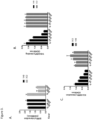
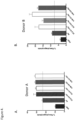


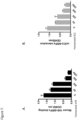


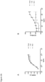


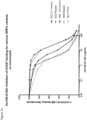

Classifications
-
- C—CHEMISTRY; METALLURGY
- C07—ORGANIC CHEMISTRY
- C07K—PEPTIDES
- C07K16/00—Immunoglobulins [IGs], e.g. monoclonal or polyclonal antibodies
- C07K16/18—Immunoglobulins [IGs], e.g. monoclonal or polyclonal antibodies against material from animals or humans
- C07K16/28—Immunoglobulins [IGs], e.g. monoclonal or polyclonal antibodies against material from animals or humans against receptors, cell surface antigens or cell surface determinants
- C07K16/2803—Immunoglobulins [IGs], e.g. monoclonal or polyclonal antibodies against material from animals or humans against receptors, cell surface antigens or cell surface determinants against the immunoglobulin superfamily
-
- C—CHEMISTRY; METALLURGY
- C07—ORGANIC CHEMISTRY
- C07K—PEPTIDES
- C07K16/00—Immunoglobulins [IGs], e.g. monoclonal or polyclonal antibodies
- C07K16/18—Immunoglobulins [IGs], e.g. monoclonal or polyclonal antibodies against material from animals or humans
- C07K16/28—Immunoglobulins [IGs], e.g. monoclonal or polyclonal antibodies against material from animals or humans against receptors, cell surface antigens or cell surface determinants
- C07K16/2896—Immunoglobulins [IGs], e.g. monoclonal or polyclonal antibodies against material from animals or humans against receptors, cell surface antigens or cell surface determinants against molecules with a "CD"-designation, not provided for elsewhere
-
- A—HUMAN NECESSITIES
- A61—MEDICAL OR VETERINARY SCIENCE; HYGIENE
- A61K—PREPARATIONS FOR MEDICAL, DENTAL OR TOILETRY PURPOSES
- A61K39/00—Medicinal preparations containing antigens or antibodies
- A61K39/395—Antibodies; Immunoglobulins; Immune serum, e.g. antilymphocytic serum
- A61K39/39533—Antibodies; Immunoglobulins; Immune serum, e.g. antilymphocytic serum against materials from animals
- A61K39/3955—Antibodies; Immunoglobulins; Immune serum, e.g. antilymphocytic serum against materials from animals against proteinaceous materials, e.g. enzymes, hormones, lymphokines
-
- A—HUMAN NECESSITIES
- A61—MEDICAL OR VETERINARY SCIENCE; HYGIENE
- A61P—SPECIFIC THERAPEUTIC ACTIVITY OF CHEMICAL COMPOUNDS OR MEDICINAL PREPARATIONS
- A61P37/00—Drugs for immunological or allergic disorders
- A61P37/02—Immunomodulators
-
- A—HUMAN NECESSITIES
- A61—MEDICAL OR VETERINARY SCIENCE; HYGIENE
- A61K—PREPARATIONS FOR MEDICAL, DENTAL OR TOILETRY PURPOSES
- A61K39/00—Medicinal preparations containing antigens or antibodies
- A61K2039/505—Medicinal preparations containing antigens or antibodies comprising antibodies
-
- C—CHEMISTRY; METALLURGY
- C07—ORGANIC CHEMISTRY
- C07K—PEPTIDES
- C07K2317/00—Immunoglobulins specific features
- C07K2317/20—Immunoglobulins specific features characterized by taxonomic origin
- C07K2317/21—Immunoglobulins specific features characterized by taxonomic origin from primates, e.g. man
-
- C—CHEMISTRY; METALLURGY
- C07—ORGANIC CHEMISTRY
- C07K—PEPTIDES
- C07K2317/00—Immunoglobulins specific features
- C07K2317/20—Immunoglobulins specific features characterized by taxonomic origin
- C07K2317/24—Immunoglobulins specific features characterized by taxonomic origin containing regions, domains or residues from different species, e.g. chimeric, humanized or veneered
-
- C—CHEMISTRY; METALLURGY
- C07—ORGANIC CHEMISTRY
- C07K—PEPTIDES
- C07K2317/00—Immunoglobulins specific features
- C07K2317/30—Immunoglobulins specific features characterized by aspects of specificity or valency
- C07K2317/33—Crossreactivity, e.g. for species or epitope, or lack of said crossreactivity
-
- C—CHEMISTRY; METALLURGY
- C07—ORGANIC CHEMISTRY
- C07K—PEPTIDES
- C07K2317/00—Immunoglobulins specific features
- C07K2317/50—Immunoglobulins specific features characterized by immunoglobulin fragments
- C07K2317/51—Complete heavy chain or Fd fragment, i.e. VH + CH1
-
- C—CHEMISTRY; METALLURGY
- C07—ORGANIC CHEMISTRY
- C07K—PEPTIDES
- C07K2317/00—Immunoglobulins specific features
- C07K2317/50—Immunoglobulins specific features characterized by immunoglobulin fragments
- C07K2317/515—Complete light chain, i.e. VL + CL
-
- C—CHEMISTRY; METALLURGY
- C07—ORGANIC CHEMISTRY
- C07K—PEPTIDES
- C07K2317/00—Immunoglobulins specific features
- C07K2317/50—Immunoglobulins specific features characterized by immunoglobulin fragments
- C07K2317/52—Constant or Fc region; Isotype
-
- C—CHEMISTRY; METALLURGY
- C07—ORGANIC CHEMISTRY
- C07K—PEPTIDES
- C07K2317/00—Immunoglobulins specific features
- C07K2317/50—Immunoglobulins specific features characterized by immunoglobulin fragments
- C07K2317/56—Immunoglobulins specific features characterized by immunoglobulin fragments variable (Fv) region, i.e. VH and/or VL
- C07K2317/565—Complementarity determining region [CDR]
-
- C—CHEMISTRY; METALLURGY
- C07—ORGANIC CHEMISTRY
- C07K—PEPTIDES
- C07K2317/00—Immunoglobulins specific features
- C07K2317/70—Immunoglobulins specific features characterized by effect upon binding to a cell or to an antigen
- C07K2317/71—Decreased effector function due to an Fc-modification
-
- C—CHEMISTRY; METALLURGY
- C07—ORGANIC CHEMISTRY
- C07K—PEPTIDES
- C07K2317/00—Immunoglobulins specific features
- C07K2317/70—Immunoglobulins specific features characterized by effect upon binding to a cell or to an antigen
- C07K2317/73—Inducing cell death, e.g. apoptosis, necrosis or inhibition of cell proliferation
-
- C—CHEMISTRY; METALLURGY
- C07—ORGANIC CHEMISTRY
- C07K—PEPTIDES
- C07K2317/00—Immunoglobulins specific features
- C07K2317/70—Immunoglobulins specific features characterized by effect upon binding to a cell or to an antigen
- C07K2317/76—Antagonist effect on antigen, e.g. neutralization or inhibition of binding
-
- C—CHEMISTRY; METALLURGY
- C07—ORGANIC CHEMISTRY
- C07K—PEPTIDES
- C07K2317/00—Immunoglobulins specific features
- C07K2317/90—Immunoglobulins specific features characterized by (pharmaco)kinetic aspects or by stability of the immunoglobulin
- C07K2317/92—Affinity (KD), association rate (Ka), dissociation rate (Kd) or EC50 value
Definitions
- Turnover of cells begins with the induction of an apoptotic program or other cellular changes that mark them for removal, and the subsequent recognition of markers by phagocytes, including macrophages, dendritic cells, and the like. This process requires a specific and selective removal of unwanted cells. Unlike healthy cells, the unwanted/aged/dying cells display markers or ligands called "eat-me” signals, i.e. "altered self', which can in turn be recognized by receptors on the phagocytes.
- Healthy cells may display "don't eat-me” signals that actively inhibit phagocytosis; these signals are either downregulated in the dying cells, are present in an altered conformation or they are superseded by the upregulation of "eat-me” or pro-phagocytic signals.
- the cell surface protein CD47 on healthy cells and its engagement of a phagocyte receptor, SIRP ⁇ constitutes a key "don't eat-me” signal that can turn off engulfment mediated by multiple modalities, including apoptotic cell clearance and FcR mediated phagocytosis. Blocking the CD47 mediated engagement of SIRP ⁇ on a phagocyte can cause removal of live cells bearing "eat me” signals.
- CD47 is a broadly expressed transmembrane glycoprotein with a single Ig-like domain and five membrane spanning regions, which functions as a cellular ligand for SIRP ⁇ with binding mediated through the NH2-terminal V-like domain of SIRP ⁇ .
- SIRP ⁇ is expressed primarily on myeloid cells, including macrophages, granulocytes, myeloid dendritic cells (DCs), mast cells, and their precursors, including hematopoietic stem cells. Structural determinants on SIRP ⁇ that mediate CD47 binding are discussed by Lee et al. (2007) J. Immunol. 179:7741-7750 ; Hatherley et al. (2007) J.B.C.
- US 2014/242095 describes antibodies and antibody fragments targeting SIRP ⁇ and their use in treating hematologic cancers.
- WO 2017/068164 describes methods and compositions for modifying macrophage polarization into pro-inflammatory cells to treat cancer.
- Seiffert et al. (1999) American Society of Hematology 94(11):3633-3643 reports that human signal-regulatory protein is expressed on normal, but not on subsets of leukemic myeloid cells and mediates cellular adhesion involving its counter-receptor CD47.
- Zhao et al. (2011) PNAS 108(45):18342-18347 reports that CD47-signal regulatory protein-alpha (SIRP ⁇ ) interaction form a barrier for antibody-mediated tumor cell destruction.
- SIRP ⁇ CD47-signal regulatory protein-alpha
- the invention provides an isolated humanized antibody, comprising: a VH sequence of SEQ ID NO: 7 and a VL sequence of SEQ ID NO: 8.
- the invention further provides an isolated polynucleotide or set of polynucleotides encoding the isolated antibody of any of the above claims, a V H thereof, a V L thereof, a light chain thereof, a heavy chain thereof, or an antigen-binding portion thereof.
- the invention further provides a vector or set of vectors comprising the polynucleotide or set of polynucleotides of the invention.
- the invention further provides a host cell comprising the polynucleotide or set of polynucleotides of the invention or the vector or set of vectors of the invention.
- the invention further provides a method of producing an antibody comprising expressing the antibody with the host cell of the invention and isolating the expressed antibody.
- the invention further provides a pharmaceutical composition comprising the antibody of the invention and a pharmaceutically acceptable excipient.
- the invention further provides the antibody or pharmaceutical composition of the invention for use in a method of treating or preventing a disease or condition in a subject in need thereof, comprising administering to the subject an effective amount of the antibody of or the pharmaceutical composition of the invention.
- Any reference in the description to methods of treatment or in vivo diagnosis refer to the compounds, pharmaceutical compositions and medicaments of the present invention for use in method of treatment of the human or animal body by therapy or for in vivo diagnosis.
- Disclosed herein is an isolated humanized, human, or chimeric antibody that: specifically binds human SIRP ⁇ ; does not specifically bind human SIRP ⁇ ; and optionally comprises a human Fc region comprising at least one modification that reduces binding to a human Fc receptor.
- the antibody comprises: a CDR-H1 comprising the sequence set forth in SEQ ID NO: 1; a CDR-H2 comprising the sequence set forth in SEQ ID NO:2; a CDR-H3 comprising the sequence set forth in SEQ ID NO:3; a CDR-L1 comprising the sequence set forth in SEQ ID NO:4; a CDR-L2 comprising the sequence set forth in SEQ ID NO:5; and a CDR-L3 comprising the sequence set forth in SEQ ID NO:6; or a CDR-H1 comprising the sequence set forth in SEQ ID NO:9; a CDR-H2 comprising the sequence set forth in SEQ ID NO: 10; a CDR-H3 comprising the sequence set forth in SEQ ID NO: 11; a CDR-L1 comprising the sequence set forth in SEQ ID NO:12; a CDR-L2 comprising the sequence set forth in SEQ ID NO: 13; and a CDR-L3 comprising the sequence set forth
- the antibody comprises: a V H sequence of SEQ ID NO:7 and a V L sequence of SEQ ID NO:8; or a V H sequence of SEQ ID NO:15 and a V L sequence of SEQ ID NO:16.
- the antibody comprises: a heavy chain of SEQ ID NO:17 and a light chain of SEQ ID NO:18; or a heavy chain of SEQ ID NO:19 and a light chain of SEQ ID NO:20.
- an isolated humanized, human, or chimeric antibody comprising: a CDR-H1 comprising the sequence set forth in SEQ ID NO: 1; a CDR-H2 comprising the sequence set forth in SEQ ID NO:2; a CDR-H3 comprising the sequence set forth in SEQ ID NO:3; a CDR-L1 comprising the sequence set forth in SEQ ID NO: 4; a CDR-L2 comprising the sequence set forth in SEQ ID NO:5; and a CDR-L3 comprising the sequence set forth in SEQ ID NO:6.
- Also disclosed herein is an isolated humanized, human, or chimeric antibody, comprising: a V H sequence of SEQ ID NO: 7 and a V L sequence of SEQ ID NO:8.
- Also disclosed herein is an isolated humanized, human, or chimeric antibody, comprising: a heavy chain of SEQ ID NO:17 and a light chain of SEQ ID NO:18.
- an isolated humanized, human, or chimeric antibody comprising: a CDR-H1 comprising the sequence set forth in SEQ ID NO:9; a CDR-H2 comprising the sequence set forth in SEQ ID NO: 10; a CDR-H3 comprising the sequence set forth in SEQ ID NO:11; a CDR-L1 comprising the sequence set forth in SEQ ID NO:12; a CDR-L2 comprising the sequence set forth in SEQ ID NO: 13; and a CDR-L3 comprising the sequence set forth in SEQ ID NO: 14.
- Also disclosed herein is an isolated humanized, human, or chimeric antibody, comprising: a V H sequence of SEQ ID NO: 15 and a V L sequence of SEQ ID NO: 16.
- Also disclosed herein is an isolated humanized, human, or chimeric antibody, comprising: a heavy chain of SEQ ID NO: 19 and a light chain of SEQ ID NO:20.
- an antibody disclosed herein comprises a human Fc region comprising at least one modification that reduces binding to a human Fc receptor.
- an antibody disclosed herein (a) competes for binding to human SIRP ⁇ with an antibody selected from 1H9 and 3C2; (b) does not compete for binding to human SIRP ⁇ with KWar antibody; (c) partially competes for binding to human SIRP ⁇ with KWar antibody; (d) inhibits binding of human CD47 to human SIRP ⁇ ; (e) inhibits binding of human SP-A to human SIRP ⁇ ; (f) inhibits binding of human SP-D to human SIRP ⁇ ; (g) binds to rhesus monkey SIRP ⁇ ; (h) binds to cynomolgus SIRP ⁇ ; (i) increases phagocytosis relative to control; or (j) is capable of any combination of (a)-(i).
- an antibody disclosed herein is pan-specific for human SIRP ⁇ isotypes.
- An antibody disclosed herein, such as 1H9 can bind to multiple human SIRP ⁇ isotypes including one or more of V1, V2, and V1/V5.
- An antibody disclosed herein can bind to each of human SIRP ⁇ isotypes V1 and V2.
- An antibody disclosed herein can bind to human SIRP ⁇ isotype V1, including homozygous.
- An antibody disclosed herein can bind to human SIRP ⁇ isotype V2, including homozygous.
- An antibody disclosed herein can bind to human SIRP ⁇ isotypes V1/V5 (heterozygous).
- an antibody disclosed herein can bind to multiple human SIRP ⁇ isotypes including each of V1, V2, and V1/V5. Such antibodies can include 1H9 and 3C2. Binding to the human SIRP ⁇ variants can be measured using assays known in the art including PCR and/or flow cytometry. For example, a given sample can be genotyped to determine SIRP status and binding to SIRP can be determined using flow cytometry.
- an antibody disclosed herein is specific for a human SIRP ⁇ isotype.
- human SIRP ⁇ is expressed on a professional antigen presenting cell. In some aspects, human SIRP ⁇ is expressed on a macrophage.
- An antibody disclosed herein can bind to human SIRP ⁇ on the cell surface.
- the binding of an antibody disclosed herein to SIRP ⁇ can be stable, e.g., for 1, 2, 3, 4, 5, 6, 7, 8, 9, 10, 11, 12, 13, 14, 15, 16, 17, 18, 19, 20, 21, 22, 23, 24, or greater than 24 hours.
- An antibody disclosed herein can avoid substantial internalization upon binding to SIRP ⁇ .
- Such antibodies can include 1H9 and 3C2, including humanized and/or Fc engineered versions of such antibodies. Binding to the human SIRP ⁇ can be measured using assays known in the art including flow cytometry and/or IHC.
- the antibody is 1H9 or 3C2.
- the human Fc region is IgG1 or IgG4, optionally modified with a modification.
- glycosylation of the antibody is reduced by enzymatic deglycosylation, expression in a bacterial host, or modification of an amino acid residue utilized for glycosylation.
- a modification disclosed herein reduces glycosylation of the human Fc region.
- the human Fc region modification comprises a modification at EU index position asparagine 297.
- the human Fc region modification comprises an amino acid substitution at EU index position asparagine 297.
- the human Fc region modification comprises an N297A amino acid substitution, numbering according to EU index.
- the modification comprises one or more amino acid substitutions at: N297A; L234A/L235A; C220S/C226S/C229S/P238S; C226S/C229S/E3233P/L234V/L235A; or L234F/L235E/P331S, numbering according to EU index.
- the modification comprises one or more amino acid substitutions at: N297; L234/L235; C220/C226/C229/P238; C226/C229/E3233/L234/L235; or L234/L235/P331, numbering according to EU index.
- the modification comprises one or more amino acid substitutions in the CH2 region at EU index positions 234, 235, and/or 237. In some aspects, the modification comprises one or both amino acid substitutions L234A and L235A, and optionally P331S and/or K322A and/or G237A, numbering according to EU index. In some aspects, the modification comprises amino acid substitution K322A, numbering according to EU index. In some aspects, the modification comprises E233P/L234V/L235A/G236 + A327G/A330S/P331S, numbering according to EU index.
- the antibody is a monoclonal antibody.
- the antibody is multispecific. In some aspects, the antibody binds greater than one antigen or greater than one epitope on a single antigen.
- the antibody comprises heavy chain constant region of a class selected from IgG, IgA, IgD, IgE, and IgM. In some aspects, the antibody comprises a heavy chain constant region of the class IgG and a subclass selected from IgG1, IgG4, IgG2, and IgG3.
- the antibody binds human SIRP ⁇ with a K D of less than or equal to about 1, 1-6, 1-5, 1-4, 1-3, 2, 3, 4, 5, 6, 7, 8, 9, or 10 ⁇ 10 -9 M, as measured by Biacore assay.
- an antibody disclosed herein is for use as a medicament. In some aspects, an antibody disclosed herein is for use in the treatment of a cancer or infection. In some aspects, an antibody disclosed herein is for use in the treatment of a cancer, wherein the cancer is selected from a solid tumor and a hematological tumor. In some aspects, an antibody disclosed herein is for use in increasing phagocytosis.
- Also disclosed herein is an isolated humanized, human, or chimeric antibody that competes for binding to human SIRP ⁇ with an antibody disclosed herein.
- Also disclosed herein is an isolated humanized, human, or chimeric antibody that binds the human SIRP ⁇ epitope bound by an antibody disclosed herein.
- Also disclosed herein is an isolated polynucleotide or set of polynucleotides encoding an isolated antibody disclosed herein, a V H thereof, a V L thereof, a light chain thereof, a heavy chain thereof, or an antigen-binding portion thereof.
- Also disclosed herein is a vector or set of vectors comprising a polynucleotide or set of polynucleotides disclosed herein.
- Also disclosed herein is a host cell comprising a polynucleotide or set of polynucleotides disclosed herein or a vector or set of vectors disclosed herein.
- Also disclosed herein is a method of producing an antibody comprising expressing the antibody with a host cell disclosed herein and isolating the expressed antibody.
- composition comprising an antibody disclosed herein and a pharmaceutically acceptable excipient.
- Also disclosed herein is a method of treating or preventing a disease or condition in a subject in need thereof, comprising administering to the subject an effective amount of an antibody disclosed herein or a pharmaceutical composition disclosed herein.
- the disease or condition is: cancer; infection; a viral infection; a bacterial infection; a fungual infection; fibrosis; artherosclerosis; a parasitic infection, optionally malaria; and depletion or reduction of endogenous blood-forming stem cells from bone marrow to allow radiation and/or chemotherapy -free or -reduced conditioning for transplantation of blood-forming stem cells, optionally in combination with anti-CKIT (CD117) antibody.
- the disease or condition is a cancer
- the cancer is selected from a solid tumor and a hematological tumor.
- Also disclosed herein is a method of increasing phagocytosis in a subject in need thereof, comprising administering to the subject an effective amount of an antibody disclosed herein or a pharmaceutical composition disclosed herein.
- Also disclosed herein is a method of modulating an immune response in a subject in need thereof, comprising administering to the subject an effective amount of an antibody disclosed herein or a pharmaceutical composition disclosed herein.
- a method disclosed herein further comprises administering one or more additional therapeutic agents to the subject.
- the additional therapeutic agent is an antibody. In some aspects, the additional therapeutic agent is an antibody that binds a protein or proteins on a tumor cell surface. In some aspects, the additional therapeutic agent is an antibody that binds: HER2 (ERBB2/neu), CD52, PD-L1, VEGF, CD30, EGFR, CD38, RANKL (CD254), GD2 (ganglioside), SLAMF7 (CD319), CD20, EGFR, PDGFRa, VEGFR2, CD33, CD44, CD99, CD96, CD90, CD133, CKIT (CD117 for CKIT positive tumors); CTLA-4, PD-1, PD-L1, CD40 (agonistic), LAG3 (CD223), 41BB (CD137 agonistic), OX40 (CD134, agonistic); and/or CKIT (CD117) to deplete blood-forming stem cells for transplantation therapy.
- HER2 ERBB2/neu
- CD52 PD-L1, VEGF
- CD30
- the additional therapeutic agent is at least one of: Rituximab, Cetuximab, Alemtuzumab (CD52), Atezolizumab (PD-L1), Avelumab (PD-L1), Bevacizumab (VEGF), Brentuximab (CD30), Daratumumab (CD38), Denosumab (RANKL), Dinutuximab (GD2), Elotuzumab (SLAMF7), Ibritumomab (CD20), Ipilimumab (CTLA-4), Necitumumab (EGFR), Nivolumab (PD-1), Obinutuzumab (CD20), Ofatumumab (CD20), Olaratumab (PDGFRa), Panitumumab (EGFR), Pembrolizumab (PD-1), Pertuzumab (HER2), Ramucirumab (VEGFR2), Tositumomab (CD20), and Gemtuzumab (
- the additional therapeutic agent is formulated in the same pharmaceutical composition as the antibody. In some aspects, the additional therapeutic agent is formulated in a different pharmaceutical composition from the antibody.
- the additional therapeutic agent is administered prior to administering the antibody. In some aspects, the additional therapeutic agent is administered after administering the antibody. In some aspects, the additional therapeutic agent is administered contemporaneously with the antibody.
- kits comprising an antibody disclosed herein or a pharmaceutical composition disclosed herein; and instructions for use.
- a multispecific antibody “comprising a diabody” includes a multispecific antibody “consisting of a diabody” and a multispecific antibody “consisting essentially of a diabody.”
- the term “about” indicates and encompasses an indicated value and a range above and below that value. In certain embodiments, the term “about” indicates the designated value ⁇ 10%, ⁇ 5%, or ⁇ 1%. In certain embodiments, where applicable, the term “about” indicates the designated value(s) ⁇ one standard deviation of that value(s).
- SIRP ⁇ 1 (PTPNS1, SHPS1), is a transmembrane glycoprotein, expressed primarily on myeloid and neuronal cells. SIRP ⁇ interacts with the widely distributed membrane protein CD47. In addition to SIRP ⁇ , there are two closely related proteins in the SIRP family: SIRP ⁇ and SIRP ⁇ . All three have three immunoglobulin superfamily (IgSF) domains in their extracellular region. In humans, the SIRP ⁇ protein is found in two major forms. One form, the variant 1 or V1 form, has the amino acid sequence set out as NCBI RefSeq NP_542970.1 (residues 27-504 constitute the mature form).
- variant 2 or V2 form differs by 13 amino acids and has the amino acid sequence set out in GenBank as CAA71403.1 (residues 30-504 constitute the mature form).
- These two forms of SIRP ⁇ constitute about 80% of the forms of SIRP ⁇ present in humans, and both are embraced herein by the term "human SIRP ⁇ ".
- human SIRP ⁇ Also embraced by the term “human SIRP ⁇ ” are the minor forms thereof that are endogenous to humans and have the same property of triggering signal transduction through CD47 upon binding thereto.
- Sequences of human SIRPa variants may be accessed through public databases, including Genbank accession numbers: ref
- Antibodies that specifically bind to human SIRP ⁇ are known and used in the art, and may be adapted by the use of an engineered Fc region as disclosed herein.
- Exemplary antibodies include those described in international patent application WO 2015/138600 ; in published US application 2014/0242095 (University Health Networks) ; published application CN103665165 (JIANGSU KUANGYA BIOLOGICAL MEDICAL SCIENCE & TECHNOLOGY ; Zhao XW et al. Proc Natl Acad Sci USA 108:18342-7 (2011 ).
- An anti-SIRP ⁇ antibody may be pan-specific, i.e. binding to two or more different human SIRP ⁇ isoforms; or may be specific for one isoform.
- the antibody 1.23A described by Zhang et al., supra. is reported to be specific for the SIRPa1 variant, while the 12C4 antibody is pan-specific.
- Anti-SIRP ⁇ antibodies can also be specific for SIRP ⁇ and lack binding to SIRP ⁇ and/or SIRP ⁇ .
- Anti-SIRPa antibodies can be pan-specific with respect to SIRP ⁇ and/or SIRP ⁇ .
- immunoglobulin refers to a class of structurally related proteins generally comprising two pairs of polypeptide chains: one pair of light (L) chains and one pair of heavy (H) chains. In an "intact immunoglobulin," all four of these chains are interconnected by disulfide bonds. The structure of immunoglobulins has been well characterized. See, e.g., Paul, Fundamental Immunology 7th ed., Ch. 5 (2013) Lippincott Williams & Wilkins, Philadelphia, PA . Briefly, each heavy chain typically comprises a heavy chain variable region (V H ) and a heavy chain constant region (C H ). The heavy chain constant region typically comprises three domains, abbreviated C H1 , C H2 , and C H3 . Each light chain typically comprises a light chain variable region (V L ) and a light chain constant region. The light chain constant region typically comprises one domain, abbreviated C L .
- antibody is used herein in its broadest sense and includes certain types of immunoglobulin molecules comprising one or more antigen-binding domains that specifically bind to an antigen or epitope.
- An antibody specifically includes intact antibodies (e.g., intact immunoglobulins), antibody fragments, and multi-specific antibodies.
- the antibody comprises an alternative scaffold.
- the antibody comprises an antibody fragment.
- the antibody consists of an antibody fragment.
- the antibody consists essentially of an antibody fragment.
- SIRP-ALPHA antibody “anti-SIRP-ALPHA antibody,” or “SIRP-ALPHA-specific antibody” is an antibody, as provided herein, which specifically binds to the antigen SIRP-ALPHA. In some embodiments, the antibody binds the extracellular domain of SIRP-ALPHA. In certain embodiments, a SIRP-ALPHA antibody provided herein binds to an epitope of SIRP-ALPHA that is conserved between or among SIRP-ALPHA proteins from different species.
- alternative scaffold refers to a molecule in which one or more regions may be diversified to produce one or more antigen-binding domains that specifically bind to an antigen or epitope.
- the antigen-binding domain binds the antigen or epitope with specificity and affinity similar to that of an antibody.
- Exemplary alternative scaffolds include those derived from fibronectin (e.g., Adnectins TM ), the ⁇ -sandwich (e.g., iMab), lipocalin (e.g., Anticalins ® ), EETI-II/AGRP, BPTI/LACI-D1/ITI-D2 (e.g., Kunitz domains), thioredoxin peptide aptamers, protein A (e.g., Affibody ® ), ankyrin repeats (e.g., DARPins), gamma-B-crystallin/ubiquitin (e.g., Affilins), CTLD 3 (e.g., Tetranectins), Fynomers, and (LDLR-A module) (e.g., Avimers).
- fibronectin e.g., Adnectins TM
- the ⁇ -sandwich e.g., iMab
- An alternative scaffold is one type of antibody.
- antigen-binding domain means the portion of an antibody that is capable of specifically binding to an antigen or epitope.
- an antigen-binding domain is an antigen-binding domain formed by a V H -V L dimer of an antibody.
- Another example of an antigen-binding domain is an antigen-binding domain formed by diversification of certain loops from the tenth fibronectin type III domain of an Adnectin.
- An antigen-binding domain can include CDRs 1, 2, and 3 from a heavy chain in that order; and CDRs 1, 2, and 3 from a light chain in that order.
- full length antibody is an antibody having a structure substantially similar to a naturally occurring antibody structure and having heavy chains that comprise an Fc region.
- a “full length antibody” is an antibody that comprises two heavy chains and two light chains.
- Fc region or "Fc” means the C-terminal region of an immunoglobulin heavy chain that, in naturally occurring antibodies, interacts with Fc receptors and certain proteins of the complement system.
- the structures of the Fc regions of various immunoglobulins, and the glycosylation sites contained therein, are known in the art. See Schroeder and Cavacini, J. Allergy Clin. Immunol., 2010, 125:S41-52 .
- the Fc region may be a naturally occurring Fc region, or an Fc region modified as described in the art or elsewhere in this disclosure.
- the V H and V L regions may be further subdivided into regions of hypervariability ("hypervariable regions (HVRs);” also called “complementarity determining regions” (CDRs)) interspersed with regions that are more conserved.
- the more conserved regions are called framework regions (FRs).
- Each V H and V L generally comprises three CDRs and four FRs, arranged in the following order (from N-terminus to C-terminus): FR1 - CDR1 - FR2 - CDR2 - FR3 - CDR3 - FR4.
- the CDRs are involved in antigen binding, and influence antigen specificity and binding affinity of the antibody. See Kabat et al., Sequences of Proteins of Immunological Interest 5th ed. (1991) Public Health Service, National Institutes of Health, Bethesda, MD .
- the light chain from any vertebrate species can be assigned to one of two types, called kappa ( ⁇ ) and lambda ( ⁇ ), based on the sequence of its constant domain.
- the heavy chain from any vertebrate species can be assigned to one of five different classes (or isotypes): IgA, IgD, IgE, IgG, and IgM. These classes are also designated ⁇ , ⁇ , ⁇ , ⁇ , and ⁇ , respectively.
- the IgG and IgA classes are further divided into subclasses on the basis of differences in sequence and function. Humans express the following subclasses: IgG1, IgG2, IgG3, IgG4, IgA1, and IgA2.
- the amino acid sequence boundaries of a CDR can be determined by one of skill in the art using any of a number of known numbering schemes, including those described by Kabat et al., supra ("Kabat” numbering scheme); Al-Lazikani et al., 1997, J. Mol. Biol., 273:927-948 ("Chothia” numbering scheme); MacCallum et al., 1996, J. Mol. Biol. 262:732-745 ("Contact” numbering scheme); Lefranc et al., Dev. Comp. Immunol., 2003, 27:55-77 (“IMGT” numbering scheme); and Honegge and Plückthun, J. Mol. Biol., 2001, 309:657-70 (“AHo” numbering scheme).
- Table 1 provides the positions of CDR-L1, CDR-L2, CDR-L3, CDR-H1, CDR-H2, and CDR-H3 as identified by the Kabat and Chothia schemes.
- residue numbering is provided using both the Kabat and Chothia numbering schemes.
- CDRs may be assigned, for example, using antibody numbering software, such as Abnum, available at www.bioinf.org.uk/abs/abnum/, and described in Abhinandan and Martin, Immunology, 2008, 45:3832-3839 .
- Table 1 Residues in CDRs according to Kabat and Chothia numbering schemes.
- EU numbering scheme is generally used when referring to a residue in an antibody heavy chain constant region (e.g., as reported in Kabat et al., supra ) . Unless stated otherwise, the EU numbering scheme is used to refer to residues in antibody heavy chain constant regions described herein.
- an “antibody fragment” comprises a portion of an intact antibody, such as the antigen-binding or variable region of an intact antibody.
- Antibody fragments include, for example, Fv fragments, Fab fragments, F(ab') 2 fragments, Fab' fragments, scFv (sFv) fragments, and scFv-Fc fragments.
- Fv fragments comprise a non-covalently-linked dimer of one heavy chain variable domain and one light chain variable domain.
- Fab fragments comprise, in addition to the heavy and light chain variable domains, the constant domain of the light chain and the first constant domain (C H1 ) of the heavy chain.
- Fab fragments may be generated, for example, by recombinant methods or by papain digestion of a full-length antibody.
- F(ab') 2 " fragments contain two Fab' fragments joined, near the hinge region, by disulfide bonds.
- F(ab') 2 fragments may be generated, for example, by recombinant methods or by pepsin digestion of an intact antibody.
- the F(ab') fragments can be dissociated, for example, by treatment with ⁇ -mercaptoethanol.
- Single-chain Fv or “sFv” or “scFv” antibody fragments comprise a V H domain and a V L domain in a single polypeptide chain.
- the V H and V L are generally linked by a peptide linker.
- Any suitable linker may be used.
- the linker is a (GGGGS) n (SEQ ID NO: 127).
- n 1, 2, 3, 4, 5, or 6. See Antibodies from Escherichia coli. In Rosenberg M. & Moore G.P. (Eds.), The Pharmacology of Monoclonal Antibodies vol. 113 (pp. 269-315). Springer-Verlag, New York .
- scFv-Fc fragments comprise an scFv attached to an Fc domain.
- an Fc domain may be attached to the C-terminal of the scFv.
- the Fc domain may follow the V H or V L , depending on the orientation of the variable domains in the scFv (i.e., V H -V L or V L -V H ). Any suitable Fc domain known in the art or described herein may be used.
- the Fc domain comprises an IgG4 Fc domain.
- single domain antibody refers to a molecule in which one variable domain of an antibody specifically binds to an antigen without the presence of the other variable domain.
- Single domain antibodies, and fragments thereof, are described in Arabi Ghahroudi et al., FEBS Letters, 1998, 414:521-526 and Muyldermans et al., Trends in Biochem. Sci., 2001, 26:230-245 .
- Single domain antibodies are also known as sdAbs or nanobodies.
- a “multispecific antibody” is an antibody that comprises two or more different antigen-binding domains that collectively specifically bind two or more different epitopes.
- the two or more different epitopes may be epitopes on the same antigen (e.g., a single SIRP-ALPHA molecule expressed by a cell) or on different antigens (e.g., different SIRP-ALPHA molecules expressed by the same cell, or a SIRP-ALPHA molecule and a non-SIRP-ALPHA molecule).
- a multi-specific antibody binds two different epitopes (i.e., a "bispecific antibody”).
- a multi-specific antibody binds three different epitopes (i.e., a "trispecific antibody”).
- a “monospecific antibody” is an antibody that comprises one or more binding sites that specifically bind to a single epitope.
- An example of a monospecific antibody is a naturally occurring IgG molecule which, while divalent (i.e., having two antigen-binding domains), recognizes the same epitope at each of the two antigen-binding domains.
- the binding specificity may be present in any suitable valency.
- a monoclonal antibody refers to an antibody from a population of substantially homogeneous antibodies.
- a population of substantially homogeneous antibodies comprises antibodies that are substantially similar and that bind the same epitope(s), except for variants that may normally arise during production of the monoclonal antibody. Such variants are generally present in only minor amounts.
- a monoclonal antibody is typically obtained by a process that includes the selection of a single antibody from a plurality of antibodies.
- the selection process can be the selection of a unique clone from a plurality of clones, such as a pool of hybridoma clones, phage clones, yeast clones, bacterial clones, or other recombinant DNA clones.
- the selected antibody can be further altered, for example, to improve affinity for the target ("affinity maturation"), to humanize the antibody, to improve its production in cell culture, and/or to reduce its immunogenicity in a subject.
- chimeric antibody refers to an antibody in which a portion of the heavy and/or light chain is derived from a particular source or species, while the remainder of the heavy and/or light chain is derived from a different source or species.
- Humanized forms of non-human antibodies are chimeric antibodies that contain minimal sequence derived from the non-human antibody.
- a humanized antibody is generally a human antibody (recipient antibody) in which residues from one or more CDRs are replaced by residues from one or more CDRs of a non-human antibody (donor antibody).
- the donor antibody can be any suitable non-human antibody, such as a mouse, rat, rabbit, chicken, or non-human primate antibody having a desired specificity, affinity, or biological effect.
- selected framework region residues of the recipient antibody are replaced by the corresponding framework region residues from the donor antibody.
- Humanized antibodies may also comprise residues that are not found in either the recipient antibody or the donor antibody. Such modifications may be made to further refine antibody function.
- a "human antibody” is one which possesses an amino acid sequence corresponding to that of an antibody produced by a human or a human cell, or derived from a non-human source that utilizes a human antibody repertoire or human antibody-encoding sequences (e.g., obtained from human sources or designed de novo ) .
- Human antibodies specifically exclude humanized antibodies.
- affinity refers to the strength of the sum total of non-covalent interactions between a single binding site of a molecule (e.g., an antibody) and its binding partner (e.g., an antigen or epitope).
- affinity refers to intrinsic binding affinity, which reflects a 1:1 interaction between members of a binding pair (e.g., antibody and antigen or epitope).
- the affinity of a molecule X for its partner Y can be represented by the dissociation equilibrium constant (K D ). The kinetic components that contribute to the dissociation equilibrium constant are described in more detail below.
- Affinity can be measured by common methods known in the art, including those described herein, such as surface plasmon resonance (SPR) technology (e.g., BIACORE ® ) or biolayer interferometry (e.g., FORTEBIO ® ).
- SPR surface plasmon resonance
- BIACORE ® BIACORE ®
- biolayer interferometry e.g., FORTEBIO ®
- the terms “bind,” “specific binding,” “specifically binds to,” “specific for,” “selectively binds,” and “selective for” a particular antigen (e.g., a polypeptide target) or an epitope on a particular antigen mean binding that is measurably different from a non-specific or non-selective interaction (e.g., with a non-target molecule).
- Specific binding can be measured, for example, by measuring binding to a target molecule and comparing it to binding to a non-target molecule.
- Specific binding can also be determined by competition with a control molecule that mimics the epitope recognized on the target molecule.
- the affinity of a SIRP-ALPHA antibody for a non-target molecule is less than about 50% of the affinity for SIRP-ALPHA. In some aspects, the affinity of a SIRP-ALPHA antibody for a non-target molecule is less than about 40% of the affinity for SIRP-ALPHA. In some aspects, the affinity of a SIRP-ALPHA antibody for a non-target molecule is less than about 30% of the affinity for SIRP-ALPHA. In some aspects, the affinity of a SIRP-ALPHA antibody for a non-target molecule is less than about 20% of the affinity for SIRP-ALPHA.
- the affinity of a SIRP-ALPHA antibody for a non-target molecule is less than about 10% of the affinity for SIRP-ALPHA. In some aspects, the affinity of a SIRP-ALPHA antibody for a non-target molecule is less than about 1% of the affinity for SIRP-ALPHA. In some aspects, the affinity of a SIRP-ALPHA antibody for a non-target molecule is less than about 0.1% of the affinity for SIRP-ALPHA.
- k d (sec -1 ), as used herein, refers to the dissociation rate constant of a particular antibody - antigen interaction. This value is also referred to as the k off value.
- k a (M -1 ⁇ sec -1 ), as used herein, refers to the association rate constant of a particular antibody -antigen interaction. This value is also referred to as the k on value.
- affinity of an antibody is described in terms of the K D for an interaction between such antibody and its antigen. For clarity, as known in the art, a smaller K D value indicates a higher affinity interaction, while a larger K D value indicates a lower affinity interaction.
- an “immunoconjugate” is an antibody conjugated to one or more heterologous molecule(s), such as a therapeutic (cytokine, for example) or diagnostic agent.
- Antibody effector functions refer to those biological activities mediated by the Fc region of an antibody, which activities may vary depending on the antibody isotype. Examples of antibody effector functions include C1q binding to activate complement dependent cytotoxicity (CDC), Fc receptor binding to activate antibody-dependent cellular cytotoxicity (ADCC), and antibody dependent cellular phagocytosis (ADCP).
- CDC complement dependent cytotoxicity
- ADCC antibody-dependent cellular cytotoxicity
- ADCP antibody dependent cellular phagocytosis
- the term "competes with” or “cross-competes with” indicates that the two or more antibodies compete for binding to an antigen (e.g., SIRP-ALPHA).
- SIRP-ALPHA is coated on a surface and contacted with a first SIRP-ALPHA antibody, after which a second SIRP-ALPHA antibody is added.
- a first SIRP-ALPHA antibody is coated on a surface and contacted with SIRP-ALPHA, and then a second SIRP-ALPHA antibody is added.
- the antibodies compete with each other.
- the term "competes with” also includes combinations of antibodies where one antibody reduces binding of another antibody, but where no competition is observed when the antibodies are added in the reverse order.
- the first and second antibodies inhibit binding of each other, regardless of the order in which they are added.
- one antibody reduces binding of another antibody to its antigen by at least 25%, at least 50%, at least 60%, at least 70%, at least 80%, at least 85%, at least 90%, or at least 95%.
- a skilled artisan can select the concentrations of the antibodies used in the competition assays based on the affinities of the antibodies for SIRP-ALPHA and the valency of the antibodies.
- the assays described in this definition are illustrative, and a skilled artisan can utilize any suitable assay to determine if antibodies compete with each other.
- Suitable assays are described, for example, in Cox et al., "Immunoassay Methods," in Assay Guidance Manual [Internet], Updated December 24, 2014 (www.ncbi.nlm.nih.gov/books/NBK92434/; accessed September 29, 2015 ); Silman et al., Cytometry, 2001, 44:30-37 ; and Finco et al., J. Pharm. Biomed. Anal., 2011, 54:351-358 .
- epitope means a portion of an antigen that specifically binds to an antibody.
- Epitopes frequently consist of surface-accessible amino acid residues and/or sugar side chains and may have specific three dimensional structural characteristics, as well as specific charge characteristics. Conformational and non-conformational epitopes are distinguished in that the binding to the former but not the latter may be lost in the presence of denaturing solvents.
- An epitope may comprise amino acid residues that are directly involved in the binding, and other amino acid residues, which are not directly involved in the binding.
- the epitope to which an antibody binds can be determined using known techniques for epitope determination such as, for example, testing for antibody binding to SIRP-ALPHA variants with different point-mutations, or to chimeric SIRP-ALPHA variants.
- Percent “identity” between a polypeptide sequence and a reference sequence is defined as the percentage of amino acid residues in the polypeptide sequence that are identical to the amino acid residues in the reference sequence, after aligning the sequences and introducing gaps, if necessary, to achieve the maximum percent sequence identity. Alignment for purposes of determining percent amino acid sequence identity can be achieved in various ways that are within the skill in the art, for instance, using publicly available computer software such as BLAST, BLAST-2, ALIGN, MEGALIGN (DNASTAR), CLUSTALW, CLUSTAL OMEGA, or MUSCLE software. Those skilled in the art can determine appropriate parameters for aligning sequences, including any algorithms needed to achieve maximal alignment over the full length of the sequences being compared.
- a “conservative substitution” or a “conservative amino acid substitution,” refers to the substitution an amino acid with a chemically or functionally similar amino acid.
- Conservative substitution tables providing similar amino acids are well known in the art.
- the groups of amino acids provided in Tables 2-4 are, in some embodiments, considered conservative substitutions for one another.
- Table 2. Selected groups of amino acids that are considered conservative substitutions for one another, in certain embodiments.
- amino acid refers to the twenty common naturally occurring amino acids.
- Naturally occurring amino acids include alanine (Ala; A), arginine (Arg; R), asparagine (Asn; N), aspartic acid (Asp; D), cysteine (Cys; C); glutamic acid (Glu; E), glutamine (Gln; Q), Glycine (Gly; G); histidine (His; H), isoleucine (Ile; I), leucine (Leu; L), lysine (Lys; K), methionine (Met; M), phenylalanine (Phe; F), proline (Pro; P), serine (Ser; S), threonine (Thr; T), tryptophan (Trp; W), tyrosine (Tyr; Y), and valine (Val; V).
- Naturally occurring amino acids include alanine (Ala; A), arginine (Arg; R), asparagine (Asn; N), as
- vector refers to a nucleic acid molecule capable of propagating another nucleic acid to which it is linked.
- the term includes the vector as a self-replicating nucleic acid structure as well as the vector incorporated into the genome of a host cell into which it has been introduced.
- Certain vectors are capable of directing the expression of nucleic acids to which they are operatively linked. Such vectors are referred to herein as "expression vectors.”
- host cell refers to cells into which an exogenous nucleic acid has been introduced, and the progeny of such cells.
- Host cells include “transformants” (or “transformed cells”) and “transfectants” (or “transfected cells”), which each include the primary transformed or transfected cell and progeny derived therefrom.
- Such progeny may not be completely identical in nucleic acid content to a parent cell, and may contain mutations.
- treating refers to clinical intervention in an attempt to alter the natural course of a disease or condition in a subject in need thereof. Treatment can be performed both for prophylaxis and during the course of clinical pathology. Desirable effects of treatment include preventing occurrence or recurrence of disease, alleviation of symptoms, diminishment of any direct or indirect pathological consequences of the disease, preventing metastasis, decreasing the rate of disease progression, amelioration or palliation of the disease state, and remission or improved prognosis.
- terapéuticaally effective amount refers to an amount of an antibody or pharmaceutical composition provided herein that, when administered to a subject, is effective to treat a disease or disorder.
- the term "subject” means a mammalian subject. Exemplary subjects include humans, monkeys, dogs, cats, mice, rats, cows, horses, camels, goats, rabbits, and sheep. In certain embodiments, the subject is a human. In some embodiments the subject has a disease or condition that can be treated with an antibody provided herein. In some aspects, the disease or condition is a cancer. In some aspects, the disease or condition is a viral infection.
- kits are used to refer to instructions customarily included in commercial packages of therapeutic or diagnostic products (e.g., kits) that contain information about the indications, usage, dosage, administration, combination therapy, contraindications and/or warnings concerning the use of such therapeutic or diagnostic products.
- cytotoxic agent refers to a substance that inhibits or prevents a cellular function and/or causes cell death or destruction.
- chemotherapeutic agent refers to a chemical compound useful in the treatment of cancer.
- Chemotherapeutic agents include "anti-hormonal agents” or “endocrine therapeutics” which act to regulate, reduce, block, or inhibit the effects of hormones that can promote the growth of cancer.
- cytostatic agent refers to a compound or composition which arrests growth of a cell either in vitro or in vivo.
- a cytostatic agent is an agent that reduces the percentage of cells in S phase.
- a cytostatic agent reduces the percentage of cells in S phase by at least about 20%, at least about 40%, at least about 60%, or at least about 80%.
- tumor refers to all neoplastic cell growth and proliferation, whether malignant or benign, and all pre-cancerous and cancerous cells and tissues.
- cancer refers to all neoplastic cell growth and proliferation, whether malignant or benign, and all pre-cancerous and cancerous cells and tissues.
- cancer refers to all neoplastic cell growth and proliferation, whether malignant or benign, and all pre-cancerous and cancerous cells and tissues.
- cancer cancer
- cancer cancer
- cancer cancer
- the tumor is a solid tumor.
- the tumor is a hematologic malignancy.
- pharmaceutical composition refers to a preparation which is in such form as to permit the biological activity of an active ingredient contained therein to be effective in treating a subject, and which contains no additional components which are unacceptably toxic to the subject in the amounts provided in the pharmaceutical composition.
- co-administration include the administration of two or more therapeutic agents either simultaneously, concurrently or sequentially within no specific time limits.
- the agents are present in the cell or in the subject's body at the same time or exert their biological or therapeutic effect at the same time.
- the therapeutic agents are in the same composition or unit dosage form. In other embodiments, the therapeutic agents are in separate compositions or unit dosage forms.
- a first agent can be administered prior to (e.g., minutes, 15 minutes, 30 minutes, 45 minutes, 1 hour, 2 hours, 4 hours, 6 hours, 12 hours, 24 hours, 48 hours, 72 hours, 96 hours, 1 week, 2 weeks, 3 weeks, 4 weeks, 5 weeks, 6 weeks, 8 weeks, or 12 weeks before), concomitantly with, or subsequent to (e.g., 5 minutes, 15 minutes, 30 minutes, 45 minutes, 1 hour, 2 hours, 4 hours, 6 hours, 12 hours, 24 hours, 48 hours, 72 hours, 96 hours, 1 week, 2 weeks, 3 weeks, 4 weeks, 5 weeks, 6 weeks, 8 weeks, or 12 weeks after) the administration of a second therapeutic agent.
- modulate and “modulation” refer to reducing or inhibiting or, alternatively, activating or increasing, a recited variable.
- increase and activate refer to an increase of 10%, 20%, 30%, 40%, 50%, 60%, 70%, 75%, 80%, 85%, 90%, 95%, 100%, 2-fold, 3-fold, 4-fold, 5-fold, 10-fold, 20-fold, 50-fold, 100-fold, or greater in a recited variable.
- reduce and “inhibit” refer to a decrease of 10%, 20%, 30%, 40%, 50%, 60%, 70%, 75%, 80%, 85%, 90%, 95%, 2-fold, 3-fold, 4-fold, 5-fold, 10-fold, 20-fold, 50-fold, 100-fold, or greater in a recited variable.
- agonist refers to the activation of receptor signaling to induce a biological response associated with activation of the receptor.
- agonist is an entity that binds to and agonizes a receptor.
- antagonist refers to the inhibition of receptor signaling to inhibit a biological response associated with activation of the receptor.
- An "antagonist” is an entity that binds to and antagonizes a receptor.
- the SIRP-ALPHA is human SIRP-ALPHA.
- the antibodies provided herein specifically bind to the extracellular domain of SIRP-ALPHA.
- the SIRP-ALPHA may be expressed on the surface of any suitable target cell.
- the target cell is a professional antigen presenting cell.
- the target cell is a macrophage.
- An antibody can be pan-specific for human SIRP ⁇ isotypes.
- An antibody can be specific for a human SIRP ⁇ isotype.
- an antibody is 1H9. In certain instances an antibody is 3C2.
- the antibodies provided herein comprise a light chain.
- the light chain is a kappa light chain.
- the light chain is a lambda light chain.
- the antibodies provided herein comprise a heavy chain.
- the heavy chain is an IgA.
- the heavy chain is an IgD.
- the heavy chain is an IgE.
- the heavy chain is an IgG.
- the heavy chain is an IgM.
- the heavy chain is an IgG1.
- the heavy chain is an IgG2.
- the heavy chain is an IgG3.
- the heavy chain is an IgG4.
- the heavy chain is an IgA1. In some aspects, the heavy chain is an IgA2.
- an antibody binds human SIRP ⁇ with a K D of less than or equal to about 1, 1-6, 1-5, 1-4, 1-3, 2, 3, 4, 5, 6, 7, 8, 9, or 10 ⁇ 10 -9 M, as measured by Biacore assay.
- the antibodies provided herein comprise an antibody fragment. In some embodiments, the antibodies provided herein consist of an antibody fragment. In some embodiments, the antibodies provided herein consist essentially of an antibody fragment. In some aspects, the antibody fragment is an Fv fragment. In some aspects, the antibody fragment is a Fab fragment. In some aspects, the antibody fragment is a F(ab') 2 fragment. In some aspects, the antibody fragment is a Fab' fragment. In some aspects, the antibody fragment is an scFv (sFv) fragment. In some aspects, the antibody fragment is an scFv-Fc fragment. In some aspects, the antibody fragment is a fragment of a single domain antibody.
- an antibody fragment provided herein is derived from an illustrative antibody provided herein. In some embodiments, an antibody fragments provided herein is not derived from an illustrative antibody provided herein and may, for example, be isolated de novo according to the methods provided herein for obtaining antibody fragments.
- an antibody fragment provided herein retains the ability to antagonize SIRP-ALPHA, as measured by one or more assays or biological effects described herein. In some embodiments, an antibody fragment provided herein retains the ability to prevent SIRP-ALPHA from interacting with one or more of its ligands, as described herein.
- an antibody fragment provided herein competes for binding to SIRP-ALPHA with 1H9 and/or 3C2. In some embodiments, a fragment of an antibody provided herein binds the same epitope of SIRP-ALPHA as such antibody.
- an antibody can be engineered to lack Fc sequences, e.g., by producing an antibody fragment such as a F(ab')2 fragment.
- an antibody fragment such as a F(ab')2 fragment.
- the purified antibody is suspended with Pierce F(ab')2 Preparation pepsin immobilized on settled resin, according to the manufacturer's instructions.
- Pepsin digestion typically produces a F(ab')2 fragment ( ⁇ 110kDa by SDS-PAGE under non-reducing conditions) and numerous small peptides of the Fc portion.
- the resulting F(ab')2 fragment is composed of a pair of Fab' units connected by two disulfide bonds.
- the Fc fragment is extensively degraded and separated from F(ab')2 by dialysis, gel filtration, or ion exchange chromatography.
- the antibodies provided herein are monoclonal antibodies. In some embodiments, the antibodies provided herein are polyclonal antibodies.
- the antibody of the invention is an isolated humanized antibody comprising a VH sequence of SEQ ID NO: 7 and a VL sequence of SEQ ID NO: 8.
- the antibodies provided herein comprise an alternative scaffold.
- Any suitable alternative scaffold may be used.
- the alternative scaffold is selected from an Adnectin TM , an iMab, an Anticalin ® , an EETI-II/AGRP, a Kunitz domain, a thioredoxin peptide aptamer, an Affibody ® , a DARPin, an Affilin, a Tetranectin, a Fynomer, and an Avimer.
- an antibody provided herein inhibits binding of SIRP-ALPHA to one or more ligands of SIRP-ALPHA.
- an antibody does not bind to SIRP-Gamma. In certain aspects, an antibody does not substantially bind to SIRP-Gamma.
- an antibody disclosed herein is pan-specific for human SIRP ⁇ isotypes.
- An antibody disclosed herein, such as 1H9 can bind to multiple human SIRP ⁇ isotypes including one or more of V1, V2, and V1/V5.
- Exemplary V2 sequence shown in SEQ ID NO:49. See also Polymorphism in Sirpa modulates engraftment of human hematopoietic stem cells. Nature Immunology, 8; 1313, 2007.
- An antibody disclosed herein can bind to each of human SIRP ⁇ isotypes V1 and V2.
- An antibody disclosed herein can bind to human SIRP ⁇ isotype V1, including homozygous.
- An antibody disclosed herein can bind to human SIRP ⁇ isotype V2, including homozygous.
- An antibody disclosed herein can bind to human SIRP ⁇ isotypes V1/V5 (heterozygous).
- An antibody disclosed herein, such as 1H9 can bind to multiple human SIRP ⁇ isotypes including each of V1, V2, and V1/V5.
- Such antibodies can include 1H9 and 3C2, including humanized and/or Fc engineered versions of such antibodies.
- 1H9 can bind to each of human SIRP ⁇ isotypes V1 and V2.
- 1H9 can bind to human SIRP ⁇ isotype V1, including homozygous.
- 1H9 can bind to human SIRP ⁇ isotype V2, including homozygous.
- 1H9 can bind to human SIRP ⁇ isotypes V1/V5 (heterozygous). 1H9 can bind to multiple human SIRP ⁇ isotypes including each of V1, V2, and V1/V5. Binding to the human SIRP ⁇ variants can be measured using assays known in the art including PCR and/or flow cytometry. For example, a given sample can be genotyped to determine SIRP status and binding to SIRP can be determined using flow cytometry.
- an antibody competes for binding to human SIRP ⁇ with an antibody selected from 1H9 and 3C2. In certain aspects, an antibody binds to the same human SIRP ⁇ epitope as bound by 1H9 or 3C2. In certain aspects, an antibody binds to an overlapping human SIRP ⁇ epitope as bound by 1H9 or 3C2. In certain aspects, an antibody binds to a distinct human SIRP ⁇ epitope as bound by 1H9 or 3C2.
- an antibody does not compete for binding to human SIRP ⁇ with KWar antibody.
- an antibody partially competes for binding to human SIRP ⁇ with KWar antibody.
- an antibody inhibits binding of human CD47 to human SIRP ⁇ .
- an antibody inhibits binding of human SP-A to human SIRP ⁇ .
- an antibody inhibits binding of human SP-D to human SIRP ⁇ .
- an antibody binds to rhesus monkey SIRP ⁇ .
- an antibody binds to cynomolgus SIRP ⁇ .
- an antibody increases phagocytosis relative to control.
- Also disclosed herein is an isolated humanized, human, or chimeric antibody that competes for binding to human SIRP ⁇ with an antibody disclosed herein.
- Also disclosed herein is an isolated humanized, human, or chimeric antibody that binds the human SIRP ⁇ epitope bound by an antibody disclosed herein.
- an antibody comprises a human Fc region comprising at least one modification that reduces binding to a human Fc receptor.
- an antibody is an antibody that competes with an illustrative antibody provided herein, e.g., 1H9 and/or 3C2. In some aspects, the antibody that competes with the illustrative antibody provided herein binds the same epitope as an illustrative antibody provided herein.
- an antibody when an antibody is expressed in cells, the antibody is modified after translation.
- posttranslational modification include cleavage of lysine at the C terminal of the heavy chain by a carboxypeptidase; modification of glutamine or glutamic acid at the N terminal of the heavy chain and the light chain to pyroglutamic acid by pyroglutamylation; glycosylation; oxidation; deamidation; and glycation, and it is known that such posttranslational modifications occur in various antibodies ( See Journal of Pharmaceutical Sciences, 2008, Vol. 97, p. 2426-2447 ).
- an antibody is an antibody or antigen-binding fragment thereof which has undergone posttranslational modification.
- an antibody or antigen-binding fragment thereof which have undergone posttranslational modification examples include an antibody or antigen-binding fragments thereof which have undergone pyroglutamylation at the N terminal of the heavy chain variable region and/or deletion of lysine at the C terminal of the heavy chain. It is known in the art that such posttranslational modification due to pyroglutamylation at the N terminal and deletion of lysine at the C terminal does not have any influence on the activity of the antibody or fragment thereof ( Analytical Biochemistry, 2006, Vol. 348, p. 24-39 ).
- An antibody can comprise: a CDR-H1 comprising the sequence set forth in SEQ ID NO:1; a CDR-H2 comprising the sequence set forth in SEQ ID NO:2; a CDR-H3 comprising the sequence set forth in SEQ ID NO:3; a CDR-L1 comprising the sequence set forth in SEQ ID NO:4; a CDR-L2 comprising the sequence set forth in SEQ ID NO:5; and a CDR-L3 comprising the sequence set forth in SEQ ID NO:6.
- An antibody can comprise: a V H sequence of SEQ ID NO:7 and a V L sequence of SEQ ID NO:8.
- An antibody can comprise: a heavy chain of SEQ ID NO:17 and a light chain of SEQ ID NO:18.
- An antibody can comprise: a CDR-H1 comprising the sequence set forth in SEQ ID NO:9; a CDR-H2 comprising the sequence set forth in SEQ ID NO:10; a CDR-H3 comprising the sequence set forth in SEQ ID NO:11; a CDR-L1 comprising the sequence set forth in SEQ ID NO:12; a CDR-L2 comprising the sequence set forth in SEQ ID NO:13; and a CDR-L3 comprising the sequence set forth in SEQ ID NO:14.
- An antibody can comprise: a V H sequence of SEQ ID NO:15 and a V L sequence of SEQ ID NO:16.
- An antibody can comprise: a heavy chain of SEQ ID NO:19 and a light chain of SEQ ID NO:20.
- An antibody can comprise: a CDR-H1 comprising the sequence set forth in SEQ ID NO:21; a CDR-H2 comprising the sequence set forth in SEQ ID NO:22; a CDR-H3 comprising the sequence set forth in SEQ ID NO:23; a CDR-L1 comprising the sequence set forth in SEQ ID NO:24; a CDR-L2 comprising the sequence set forth in SEQ ID NO:25; and a CDR-L3 comprising the sequence set forth in SEQ ID NO:26.
- An antibody can comprise: a V H sequence of SEQ ID NO:27 and a V L sequence of SEQ ID NO:28.
- An antibody can comprise: a CDR-H1 comprising the sequence set forth in SEQ ID NO:29; a CDR-H2 comprising the sequence set forth in SEQ ID NO:30; a CDR-H3 comprising the sequence set forth in SEQ ID NO:31; a CDR-L1 comprising the sequence set forth in SEQ ID NO:32; a CDR-L2 comprising the sequence set forth in SEQ ID NO:33; and a CDR-L3 comprising the sequence set forth in SEQ ID NO:34.
- An antibody can comprise: a V H sequence of SEQ ID NO:35 and a V L sequence of SEQ ID NO:36.
- an antibody can comprise one or more CDRs of 1H9. In certain aspects, an antibody can comprise all CDRs of 1H9. In certain aspects, an antibody can comprise one or more variable sequences of 1H9. In certain aspects, an antibody can comprise each variable sequence of 1H9. In certain aspects, an antibody can comprise the heavy chain of 1H9. In certain aspects, an antibody can comprise the light chain of 1H9. In certain aspects, an antibody can comprise the heavy chain and the light chain of 1H9. In certain aspects, an antibody is 1H9.
- an antibody can comprise one or more CDRs of 3C2. In certain aspects, an antibody can comprise all CDRs of 3C2. In certain aspects, an antibody can comprise one or more variable sequences of 3C2. In certain aspects, an antibody can comprise each variable sequence of 3C2. In certain aspects, an antibody can comprise the heavy chain of 3C2. In certain aspects, an antibody can comprise the light chain of 3C2. In certain aspects, an antibody can comprise the heavy chain and the light chain of 3C2. In certain aspects, an antibody is 3C2.
- an antibody can comprise one or more CDRs of 9B11. In certain aspects, an antibody can comprise all CDRs of 9B1 1. In certain aspects, an antibody can comprise one or more variable sequences of 9B11. In certain aspects, an antibody can comprise each variable sequence of 9B11. In certain aspects, an antibody can comprise the heavy chain of 9B11. In certain aspects, an antibody can comprise the light chain of 9B11. In certain aspects, an antibody can comprise the heavy chain and the light chain of 9B11. In certain aspects, an antibody is 9B11.
- an antibody can comprise one or more CDRs of 7E11. In certain aspects, an antibody can comprise all CDRs of 7E11. In certain aspects, an antibody can comprise one or more variable sequences of 7E11. In certain aspects, an antibody can comprise each variable sequence of 7E11. In certain aspects, an antibody can comprise the heavy chain of 7E1 1. In certain aspects, an antibody can comprise the light chain of 7E11. In certain aspects, an antibody can comprise the heavy chain and the light chain of 7E1 1. In certain aspects, an antibody is 7E11.
- an antibody provided herein comprises a sequence having at least about 50%, 60%, 70%, 80%, 90%, 95%, or 99% identity to an illustrative sequence provided in SEQ ID NOs: 1-36.
- an antibody provided herein comprises a sequence provided in SEQ ID NOs: 1-36, with up to 1, 2, 3, 4, 5, 6, 7, 8, 9, 10, 11, 12, 13, 14, 15, 16, 17, 18, 19, 20, 21, 22, 23, 24, or 25 amino acid substitutions.
- the amino acid substitutions are conservative amino acid substitutions.
- the antibodies described in this paragraph are referred to herein as "variants.”
- such variants are derived from a sequence provided herein, for example, by affinity maturation, site directed mutagenesis, random mutagenesis, or any other method known in the art or described herein.
- such variants are not derived from a sequence provided herein and may, for example, be isolated de novo according to the methods provided herein for obtaining antibodies.
- the antibodies provided herein are monospecific antibodies.
- the antibodies provided herein are multispecific antibodies.
- a multispecific antibody provided herein binds more than one antigen. In some embodiments, a multispecific antibody binds 2 antigens. In some embodiments, a multispecific antibody binds 3 antigens. In some embodiments, a multispecific antibody binds 4 antigens. In some embodiments, a multispecific antibody binds 5 antigens.
- a multispecific antibody provided herein binds more than one epitope on a SIRP-ALPHA antigen. In some embodiments, a multispecific antibody binds 2 epitopes on a SIRP-ALPHA antigen. In some embodiments, a multispecific antibody binds 3 epitopes on a SIRP-ALPHA antigen.
- the multispecific antibody comprises an immunoglobulin comprising at least two different heavy chain variable regions each paired with a common light chain variable region (i.e., a "common light chain antibody").
- the common light chain variable region forms a distinct antigen-binding domain with each of the two different heavy chain variable regions. See Merchant et al., Nature Biotechnol., 1998, 16:677-681 .
- the multispecific antibody comprises an immunoglobulin comprising an antibody or fragment thereof attached to one or more of the N- or C-termini of the heavy or light chains of such immunoglobulin. See Coloma and Morrison, Nature Biotechnol., 1997, 15:159-163 . In some aspects, such antibody comprises a tetravalent bispecific antibody.
- the multispecific antibody comprises a hybrid immunoglobulin comprising at least two different heavy chain variable regions and at least two different light chain variable regions. See Milstein and Cuello, Nature, 1983, 305:537-540 ; and Staerz and Bevan, Proc. Natl. Acad. Sci. USA, 1986, 83:1453-1457 .
- the multispecific antibody comprises immunoglobulin chains with alterations to reduce the formation of side products that do not have multi specificity.
- the antibodies comprise one or more "knobs-into-holes" modifications as described in U.S. Pat. No. 5,731,168 .
- the multispecific antibody comprises immunoglobulin chains with one or more electrostatic modifications to promote the assembly of Fc hetero-multimers. See WO 2009/089004 .
- the multispecific antibody comprises a bispecific single chain molecule. See Traunecker et al., EMBO J., 1991, 10:3655-3659 ; and Gruber et al., J. Immunol., 1994, 152:5368-5374 .
- the multispecific antibody comprises a heavy chain variable domain and a light chain variable domain connected by a polypeptide linker, where the length of the linker is selected to promote assembly of multispecific antibody with the desired multi specificity.
- monospecific scFvs generally form when a heavy chain variable domain and light chain variable domain are connected by a polypeptide linker of more than 12 amino acid residues. See U.S. Pat. Nos. 4,946,778 and 5,132,405 .
- reduction of the polypeptide linker length to less than 12 amino acid residues prevents pairing of heavy and light chain variable domains on the same polypeptide chain, thereby allowing pairing of heavy and light chain variable domains from one chain with the complementary domains on another chain.
- the resulting antibody therefore has multi specificity, with the specificity of each binding site contributed by more than one polypeptide chain.
- Polypeptide chains comprising heavy and light chain variable domains that are joined by linkers between 3 and 12 amino acid residues form predominantly dimers (termed diabodies). With linkers between 0 and 2 amino acid residues, trimers (termed triabodies) and tetramers (termed tetrabodies) are favored. However, the exact type of oligomerization appears to depend on the amino acid residue composition and the order of the variable domain in each polypeptide chain (e.g., V H -linker-V L vs. V L -linker-V H ), in addition to the linker length. A skilled person can select the appropriate linker length based on the desired multi specificity.
- An antibody provided herein may be altered to increase, decrease or eliminate the extent to which it is glycosylated.
- Glycosylation of polypeptides is typically either "N-linked” or “O-linked.”
- glycosylation of the antibody is reduced by enzymatic deglycosylation, expression in a bacterial host, or modification of an amino acid residue utilized for glycosylation. Modifications such as mutations can be used to alter glycosylation.
- N-linked glycosylation refers to the attachment of a carbohydrate moiety to the side chain of an asparagine residue.
- the tripeptide sequences asparagine-X-serine and asparagine-X-threonine, where X is any amino acid except proline, are the recognition sequences for enzymatic attachment of the carbohydrate moiety to the asparagine side chain.
- X is any amino acid except proline
- O-linked glycosylation refers to the attachment of one of the sugars N-acetylgalactosamine, galactose, or xylose to a hydroxyamino acid, most commonly serine or threonine, although 5-hydroxyproline or 5-hydroxylysine may also be used.
- Addition or deletion of N-linked glycosylation sites to or from an antibody provided herein may be accomplished by altering the amino acid sequence such that one or more of the above-described tripeptide sequences is created or removed.
- Addition or deletion of O-linked glycosylation sites may be accomplished by addition, deletion, or substitution of one or more serine or threonine residues in or to (as the case may be) the sequence of an antibody.
- an antibody provided herein comprises a glycosylation motif that is different from a naturally occurring antibody. Any suitable naturally occurring glycosylation motif can be modified in the antibody provided herein.
- the structural and glycosylation properties of immunoglobulins, for example, are known in the art and summarized, for example, in Schroeder and Cavacini, J. Allergy Clin. Immunol., 2010, 125:S41-52 .
- an antibody provided herein comprises an IgG1 Fc region with modification to the oligosaccharide attached to asparagine 297 (Asn 297).
- Naturally occurring IgG1 antibodies produced by mammalian cells typically comprise a branched, biantennary oligosaccharide that is generally attached by an N-linkage to Asn 297 of the C H2 domain of the Fc region. See Wright et al., TIBTECH, 1997, 15:26-32 .
- the oligosaccharide attached to Asn 297 may include various carbohydrates such as mannose, N-acetyl glucosamine (GlcNAc), galactose, and sialic acid, as well as a fucose attached to a GlcNAc in the "stem" of the biantennary oligosaccharide structure.
- various carbohydrates such as mannose, N-acetyl glucosamine (GlcNAc), galactose, and sialic acid, as well as a fucose attached to a GlcNAc in the "stem" of the biantennary oligosaccharide structure.
- the oligosaccharide attached to Asn 297 is modified to create antibodies having altered ADCC. In some embodiments, the oligosaccharide is altered to improve ADCC. In some embodiments, the oligosaccharide is altered to reduce ADCC.
- an antibody provided herein comprises an IgG1 domain with reduced fucose content at position Asn 297 compared to a naturally occurring IgG1 domain.
- Such Fc domains are known to have improved ADCC. See Shields et al., J. Biol. Chem., 2002, 277:26733-26740 .
- such antibodies do not comprise any fucose at position Asn 297.
- the amount of fucose may be determined using any suitable method, for example as described in WO 2008/077546 .
- an antibody provided herein comprises a bisected oligosaccharide, such as a biantennary oligosaccharide attached to the Fc region of the antibody that is bisected by GlcNAc.
- a bisected oligosaccharide such as a biantennary oligosaccharide attached to the Fc region of the antibody that is bisected by GlcNAc.
- Such antibody variants may have reduced fucosylation and/or improved ADCC function. Examples of such antibody variants are described, for example, in WO 2003/011878 ; U.S. Pat. No. 6,602,684 ; and U.S. Pat. Pub. No. 2005/0123546 .
- an antibody provided herein comprises an Fc region with at least one galactose residue in the oligosaccharide attached to the Fc region.
- Such antibody variants may have improved CDC function. Examples of such antibody variants are described, for example, in WO 1997/30087 ; WO 1998/58964 ; and WO 1999/22764 .
- Examples of cell lines capable of producing defucosylated antibody include Lec13 CHO cells, which are deficient in protein fucosylation ( see Ripka et al., Arch. Biochem. Biophys., 1986, 249:533-545 ; U.S. Pat. Pub. No. 2003/0157108 ; WO 2004/056312 ), and knockout cell lines, such as alpha-1,6-fucosyltransferase gene or FUT8 knockout CHO cells ( see Yamane-Ohnuki et al., Biotech. Bioeng., 2004, 87: 614-622 ; Kanda et al., Biotechnol. Bioeng., 2006, 94:680-688 ; and WO 2003/085107 ).
- an antibody provided herein is an aglycosylated antibody.
- An aglycosylated antibody can be produced using any method known in the art or described herein.
- an aglycosylated antibody is produced by modifying the antibody to remove all glycosylation sites.
- the glycosylation sites are removed only from the Fc region of the antibody.
- an aglycosylated antibody is produced by expressing the antibody in an organism that is not capable of glycosylation, such as E. coli, or by expressing the antibody in a cell-free reaction mixture.
- an antibody provided herein has a constant region with reduced effector function compared to a native IgG1 antibody.
- the affinity of a constant region of an Fc region of an antibody provided herein for Fc receptor is less than the affinity of a native IgG1 constant region for such Fc receptor.
- an antibody provided herein comprises an Fc region. In certain embodiments, an antibody provided herein comprises an Fc region with one or more amino acid substitutions, insertions, or deletions in comparison to a naturally occurring Fc region. In some aspects, such substitutions, insertions, or deletions yield antibody with altered stability, glycosylation, or other characteristics. In some aspects, such substitutions, insertions, or deletions yield aglycosylated antibody.
- a “variant Fc region” or “engineered Fc region” comprises an amino acid sequence that differs from that of a native-sequence Fc region by virtue of at least one amino acid modification, preferably one or more amino acid substitution(s).
- the variant Fc region has at least one amino acid substitution compared to a native-sequence Fc region or to the Fc region of a parent polypeptide, e.g., from about one to about ten amino acid substitutions, and preferably from about one to about five amino acid substitutions in a native-sequence Fc region or in the Fc region of the parent polypeptide.
- the variant Fc region herein will preferably possess at least about 80% homology with a native-sequence Fc region and/or with an Fc region of a parent polypeptide, and most preferably at least about 90% homology therewith, more preferably at least about 95% homology therewith.
- Variant Fc sequences for a "dead Fc” can include three amino acid substitutions in the CH2 region to reduce FcyRI binding at EU index positions 234, 235, and 237 (see Duncan et al., (1988) Nature 332:563 ). Two amino acid substitutions in the complement C1q binding site at EU index positions 330 and 331 reduce complement fixation (see Tao et al., J. Exp. Med. 178:661 (1993 ) and Canfield and Morrison, J. Exp. Med. 173:1483 (1991 )).
- Binding of IgG to the FcyRs or C1q depends on residues located in the hinge region and the CH2 domain. Two regions of the CH2 domain are critical for FcyRs and C1q binding, and have unique sequences in IgG2 and IgG4. Substitutions into human IgG1 or IgG2 residues at positions 233-236 and IgG4 residues at positions 327, 330 and 331 have been shown to greatly reduce ADCC and CDC. Numerous mutations have been made in the CH2 domain of human IgG1.
- the triple amino acid substitution L234A, L235A, and G237A largely eliminates FcyR and complement effector functions (see, for example, US20100266505 ).
- the Fc region has been modified by the choice of expression host, enzymatic treatment of amino acid substitutions to have reduced glycosylation and binding to FcyR, relative to the native protein.
- Mutations that reduce binding to FcyR include, without limitation, modification of the glycosylation on asparagine 297 of the Fc domain, which is known to be required for optimal FcR interaction.
- known amino acid substitutions include N297A or N297G, which results in the loss of a glycosylation site on the protein.
- Enzymatically deglycosylated Fc domains recombinantly expressed antibodies in the presence of a glycosylation inhibitor and the expression of Fc domains in bacteria have a similar loss of glycosylation and consequent binding to FcyRs.
- the LALA variant, L234A/L235A also has significantly reduced FcyR binding; as does E233P/L234V/L235A/G236 + A327G/A330S/P331S. See, for example, Armour et al. (1999) Eur J Immunol. 29(8):2613-24 .
- the set of mutations: K322A, L234A and L235A are sufficient to almost completely abolish FcyR and C1q binding.
- a set of three mutations, L234F/L235E/P331S (dubbed TM) have a very similar effect.
- Fc variants are possible, including without limitation one in which a region capable of forming a disulfide bond is deleted, or in which certain amino acid residues are eliminated at the N-terminal end of a native Fc form or a methionine residue is added thereto.
- the Fc may be in the form of having native sugar chains, increased sugar chains compared to a native form or decreased sugar chains compared to the native form, or may be in an aglycosylated or deglycosylated form.
- the increase, decrease, removal or other modification of the sugar chains may be achieved by methods common in the art, such as a chemical method, an enzymatic method or by expressing it in a genetically engineered production cell line.
- Such cell lines can include microorganisms, e.g. Pichia Pastoris, and mammalians cell line, e.g. CHO cells, that naturally express glycosylating enzymes.
- microorganisms or cells can be engineered to express glycosylating enzymes, or can be rendered unable to express glycosylation enzymes (See e.g., Hamilton, et al., Science, 313:1441 (2006 ); Kanda, et al, J. Biotechnology, 130:300 (2007 ); Kitagawa, et al., J. Biol. Chem., 269 (27): 17872 (1994 ); Ujita-Lee et al., J. Biol. Chem., 264 (23): 13848 (1989 ); Imai-Nishiya, et al, BMC Biotechnology 7:84 (2007 ); and WO 07/055916 ).
- the alpha-2,6-sialyltransferase 1 gene has been engineered into Chinese Hamster Ovary cells and into sf9 cells. Antibodies expressed by these engineered cells are thus sialylated by the exogenous gene product.
- a further method for obtaining Fc molecules having a modified amount of sugar residues compared to a plurality of native molecules includes separating said plurality of molecules into glycosylated and non-glycosylated fractions, for example, using lectin affinity chromatography (See e.g., WO 07/117505 ). The presence of particular glycosylation moieties has been shown to alter the function of Immunoglobulins.
- the removal of sugar chains from an Fc molecule results in a sharp decrease in binding affinity to the C1q part of the first complement component C1 and a decrease or loss in antibody-dependent cell-mediated cytotoxicity (ADCC) or complement-dependent cytotoxicity (CDC), thereby not inducing unnecessary immune responses in vivo.
- Additional important modifications include sialylation and fucosylation: the presence of sialic acid in IgG has been correlated with antiinflammatory activity (See e.g., Kaneko, et al, Science 313:760 (2006 )), whereas removal of fucose from the IgG leads to enhanced ADCC activity (See e.g., Shoj-Hosaka, et al, J. Biochem., 140:777 (2006 )).
- Fc-region-comprising antibody refers to an antibody that comprises an Fc region.
- the C-terminal lysine (residue 447 according to the EU numbering system) of the Fc region may be removed, for example, during purification of the antibody or by recombinant engineering the nucleic acid encoding the antibody. Accordingly, an antibody having an Fc region can comprise an antibody with or without K447.
- the Fc region of an antibody provided herein is modified to yield an antibody with altered affinity for an Fc receptor, or an antibody that is more immunologically inert.
- the antibody variants provided herein possess some, but not all, effector functions. Such antibodies may be useful, for example, when the half-life of the antibody is important in vivo, but when certain effector functions (e.g., complement activation and ADCC) are unnecessary or deleterious.
- the Fc region of an antibody provided herein is a human IgG4 Fc region comprising one or more of the hinge stabilizing mutations S228P and L235E. See Aalberse et al., Immunology, 2002, 105:9-19 .
- the IgG4 Fc region comprises one or more of the following mutations: E233P, F234V, and L235A. See Armour et al., Mol. Immunol., 2003, 40:585-593 .
- the IgG4 Fc region comprises a deletion at position G236.
- the Fc region of an antibody provided herein is a human IgG1 Fc region comprising one or more mutations to reduce Fc receptor binding.
- the one or more mutations are in residues selected from S228 (e.g., S228A), L234 (e.g., L234A), L235 (e.g., L235A), D265 (e.g., D265A), and N297 (e.g., N297A).
- the antibody comprises a PVA236 mutation.
- PVA236 means that the amino acid sequence ELLG, from amino acid position 233 to 236 of IgG1 or EFLG of IgG4, is replaced by PVA. See U.S. Pat. No. 9,150,641 .
- the Fc region of an antibody provided herein is modified as described in Armour et al., Eur. J. Immunol., 1999, 29:2613-2624 ; WO 1999/058572 ; and/or U.K. Pat. App. No. 98099518 .
- the Fc region of an antibody provided herein is a human IgG2 Fc region comprising one or more of mutations A330S and P331S.
- the Fc region of an antibody provided herein has an amino acid substitution at one or more positions selected from 238, 265, 269, 270, 297, 327 and 329. See U.S. Pat. No. 6,737,056 .
- Such Fc mutants include Fc mutants with substitutions at two or more of amino acid positions 265, 269, 270, 297 and 327, including the so-called "DANA" Fc mutant with substitution of residues 265 and 297 with alanine. See U.S. Pat. No. 7,332,581 .
- the antibody comprises an alanine at amino acid position 265.
- the antibody comprises an alanine at amino acid position 297.
- an antibody provided herein comprises an Fc region with one or more amino acid substitutions which improve ADCC, such as a substitution at one or more of positions 298, 333, and 334 of the Fc region.
- an antibody provided herein comprises an Fc region with one or more amino acid substitutions at positions 239, 332, and 330, as described in Lazar et al., Proc. Natl. Acad. Sci. USA, 2006,103:4005-4010 .
- an antibody provided herein comprises one or more alterations that improves or diminishes C1q binding and/or CDC. See U.S. Pat. No. 6,194,551 ; WO 99/51642 ; and Idusogie et al., J. Immunol., 2000, 164:4178-4184 .
- an antibody provided herein comprises one or more alterations to increase half-life.
- Antibodies with increased half-lives and improved binding to the neonatal Fc receptor (FcRn) are described, for example, in Hinton et al., J. Immunol., 2006, 176:346-356 ; and U.S. Pat. Pub. No. 2005/0014934 .
- Such Fc variants include those with substitutions at one or more of Fc region residues: 238, 250, 256, 265, 272, 286, 303, 305, 307, 311, 312, 314, 317, 340, 356, 360, 362, 376, 378, 380, 382, 413, 424, 428, and 434 of an IgG.
- an antibody provided herein comprises one or more Fc region variants as described in U.S. Pat. Nos. 7,371,826 5,648,260 , and 5,624,821 ; Duncan and Winter, Nature, 1988, 322:738-740 ; and WO 94/29351 .
- isolated nucleic acids encoding SIRP-ALPHA antibodies, vectors comprising the nucleic acids, and host cells comprising the vectors and nucleic acids, as well as recombinant techniques for the production of the antibodies.
- a nucleic acid sequence is provided that encodes a sequence having at least about 50%, 60%, 70%, 80%, 90%, 95%, or 99% identity to an illustrative sequence provided in SEQ ID NOs: 1-36.
- a nucleic acid sequence is provided that encodes a sequence provided in SEQ ID NOs: 1-36, with up to 1, 2, 3, 4, 5, 6, 7, 8, 9, 10, 11, 12, 13, 14, 15, 16, 17, 18, 19, 20, 21, 22, 23, 24, or 25 amino acid substitutions.
- an antibody provided herein comprises a sequence having at least about 50%, 60%, 70%, 80%, 90%, 95%, or 99% identity to an illustrative sequence provided in SEQ ID NOs: 37-44.
- an antibody provided herein comprises a sequence provided in SEQ ID NOs: 37-44, with up to 1, 2, 3, 4, 5, 6, 7, 8, 9, 10, 11, 12, 13, 14, 15, 16, 17, 18, 19, 20, 21, 22, 23, 24, or 25 mutations.
- the amino acid substitutions are conservative amino acid substitutions.
- the antibodies described in this paragraph are referred to herein as "variants.”
- such variants are derived from a sequence provided herein, for example, by affinity maturation, site directed mutagenesis, random mutagenesis, or any other method known in the art or described herein.
- such variants are not derived from a sequence provided herein and may, for example, be isolated de novo according to the methods provided herein for obtaining antibodies.
- the nucleic acid(s) encoding it may be isolated and inserted into a replicable vector for further cloning (i.e., amplification of the DNA) or expression.
- the nucleic acid may be produced by homologous recombination, for example as described in U.S. Patent No. 5,204,244 .
- the vector components generally include one or more of the following: a signal sequence, an origin of replication, one or more marker genes, an enhancer element, a promoter, and a transcription termination sequence, for example as described in U.S. Patent No. 5,534,615 .
- Suitable host cells are provided below. These host cells are not meant to be limiting, and any suitable host cell may be used to produce the antibodies provided herein.
- Suitable host cells include any prokaryotic (e.g., bacterial), lower eukaryotic (e.g., yeast), or higher eukaryotic (e.g., mammalian) cells.
- Suitable prokaryotes include eubacteria, such as Gram-negative or Gram-positive organisms, for example, Enterobacteriaceae such as Escherichia (E. coli), Enterobacter, Erwinia, Klebsiella, Proteus, Salmonella (S. typhimurium ) , Serratia ( S. marcescans ) , Shigella, Bacilli ( B. subtilis and B. licheniformis ), Pseudomonas ( P.
- eubacteria such as Gram-negative or Gram-positive organisms, for example, Enterobacteriaceae such as Escherichia (E. coli), Enterobacter, Erwinia, Klebsiella, Proteus, Salmonella (S.
- E. coli 294 One useful E. coli cloning host is E. coli 294, although other strains such as E. coli B, E. coli X1776, and E. coli W3110 are also suitable.
- eukaryotic microbes such as filamentous fungi or yeast are also suitable cloning or expression hosts for SIRP-ALPHA antibody-encoding vectors.
- Saccharomyces cerevisiae or common baker's yeast, is a commonly used lower eukaryotic host microorganism.
- a number of other genera, species, and strains are available and useful, such as Schizosaccharomyces pombe, Kluyveromyces ( K. lactis, K. fragilis, K. bulgaricus K. wickeramii, K. waltii, K. drosophilarum, K. thermotolerans, and K.
- Useful mammalian host cells include COS-7 cells, HEK293 cells; baby hamster kidney (BHK) cells; Chinese hamster ovary (CHO); mouse sertoli cells; African green monkey kidney cells (VERO-76), and the like.
- the host cells used to produce the SIRP-ALPHA antibody may be cultured in a variety of media.
- Commercially available media such as, for example, Ham's F10, Minimal Essential Medium (MEM), RPMI-1640, and Dulbecco's Modified Eagle's Medium (DMEM) are suitable for culturing the host cells.
- any of the media described in Ham et al., Meth. Enz., 1979, 58:44 ; Barnes et al., Anal. Biochem., 1980, 102:255 ; and U.S. Patent Nos. 4,767,704 , 4,657,866 , 4,927,762 , 4,560,655 , and 5,122,469 ; or WO 90/03430 and WO 87/00195 may be used.
- any of these media may be supplemented as necessary with hormones and/or other growth factors (such as insulin, transferrin, or epidermal growth factor), salts (such as sodium chloride, calcium, magnesium, and phosphate), buffers (such as HEPES), nucleotides (such as adenosine and thymidine), antibiotics, trace elements (defined as inorganic compounds usually present at final concentrations in the micromolar range), and glucose or an equivalent energy source. Any other necessary supplements may also be included at appropriate concentrations that would be known to those skilled in the art.
- growth factors such as insulin, transferrin, or epidermal growth factor
- salts such as sodium chloride, calcium, magnesium, and phosphate
- buffers such as HEPES
- nucleotides such as adenosine and thymidine
- antibiotics such as adenosine and thymidine
- trace elements defined as inorganic compounds usually present at final concentrations in the micromolar range
- the culture conditions such as temperature, pH, and the like, are those previously used with the host cell selected for expression, and will be apparent to the ordinarily skilled artisan.
- the antibody can be produced intracellularly, in the periplasmic space, or directly secreted into the medium. If the antibody is produced intracellularly, as a first step, the particulate debris, either host cells or lysed fragments, is removed, for example, by centrifugation or ultrafiltration.
- the particulate debris either host cells or lysed fragments
- the particulate debris is removed, for example, by centrifugation or ultrafiltration.
- Carter et al. Bio/Technology, 1992, 10:163-167
- cell paste is thawed in the presence of sodium acetate (pH 3.5), EDTA, and phenylmethylsulfonylfluoride (PMSF) over about 30 min. Cell debris can be removed by centrifugation.
- the antibody is produced in a cell-free system.
- the cell-free system is an in vitro transcription and translation system as described in Yin et al., mAbs, 2012, 4:217-225 .
- the cell-free system utilizes a cell-free extract from a eukaryotic cell or from a prokaryotic cell.
- the prokaryotic cell is E. coli.
- Cell-free expression of the antibody may be useful, for example, where the antibody accumulates in a cell as an insoluble aggregate, or where yields from periplasmic expression are low.
- supernatants from such expression systems are generally first concentrated using a commercially available protein concentration filter, for example, an Amicon ® or Millipore ® Pellcon ® ultrafiltration unit.
- a protease inhibitor such as PMSF may be included in any of the foregoing steps to inhibit proteolysis and antibiotics may be included to prevent the growth of adventitious contaminants.
- the antibody composition prepared from the cells can be purified using, for example, hydroxylapatite chromatography, gel electrophoresis, dialysis, and affinity chromatography, with affinity chromatography being a particularly useful purification technique.
- the suitability of protein A as an affinity ligand depends on the species and isotype of any immunoglobulin Fc domain that is present in the antibody.
- Protein A can be used to purify antibodies that comprise human ⁇ 1, ⁇ 2, or ⁇ 4 heavy chains ( Lindmark et al., J. Immunol. Meth., 1983, 62:1-13 ).
- Protein G is useful for all mouse isotypes and for human ⁇ 3 ( Guss et al., EMBO J., 1986, 5:1567-1575 ).
- the matrix to which the affinity ligand is attached is most often agarose, but other matrices are available.
- Mechanically stable matrices such as controlled pore glass or poly(styrenedivinyl)benzene allow for faster flow rates and shorter processing times than can be achieved with agarose.
- the antibody comprises a C H3 domain
- the BakerBond ABX ® resin is useful for purification.
- the mixture comprising the antibody of interest and contaminants may be subjected to low pH hydrophobic interaction chromatography using an elution buffer at a pH between about 2.5 to about 4.5, generally performed at low salt concentrations (e.g., from about 0 to about 0.25 M salt).
- the SIRP-ALPHA antigen used for isolation or creation of the antibodies provided herein may be intact SIRP-ALPHA or a fragment of SIRP-ALPHA.
- the SIRP-ALPHA antigen may be, for example, in the form of an isolated protein or a protein expressed on the surface of a cell.
- the SIRP-ALPHA antigen is a non-naturally occurring variant of SIRP-ALPHA, such as a SIRP-ALPHA protein having an amino acid sequence or post-translational modification that does not occur in nature.
- the SIRP-ALPHA antigen is truncated by removal of, for example, intracellular or membrane-spanning sequences, or signal sequences. In some embodiments, the SIRP-ALPHA antigen is fused at its C-terminus to a human IgG1 Fc domain or a polyhistidine tag.
- Monoclonal antibodies may be obtained, for example, using the hybridoma method first described by Kohler et al., Nature, 1975, 256:495-497 , and/or by recombinant DNA methods ( see e.g., U.S. Patent No. 4,816,567 ). Monoclonal antibodies may also be obtained, for example, using phage or yeast-based libraries. See e.g., U.S. Patent Nos. 8,258,082 and 8,691,730 .
- lymphocytes that produce or are capable of producing antibodies that will specifically bind to the protein used for immunization.
- lymphocytes may be immunized in vitro. Lymphocytes are then fused with myeloma cells using a suitable fusing agent, such as polyethylene glycol, to form a hybridoma cell.
- a suitable fusing agent such as polyethylene glycol
- the hybridoma cells are seeded and grown in a suitable culture medium that contains one or more substances that inhibit the growth or survival of the unfused, parental myeloma cells.
- a suitable culture medium that contains one or more substances that inhibit the growth or survival of the unfused, parental myeloma cells.
- the culture medium for the hybridomas typically will include hypoxanthine, aminopterin, and thymidine (HAT medium), which substances prevent the growth of HGPRT-deficient cells.
- Useful myeloma cells are those that fuse efficiently, support stable high-level production of antibody by the selected antibody-producing cells, and are sensitive media conditions, such as the presence or absence of HAT medium.
- preferred myeloma cell lines are murine myeloma lines, such as those derived from MOP-21 and MC-11 mouse tumors (available from the Salk Institute Cell Distribution Center, San Diego, CA), and SP-2 or X63-Ag8-653 cells (available from the American Type Culture Collection, Rockville, MD).
- Human myeloma and mouse-human heteromyeloma cell lines also have been described for the production of human monoclonal antibodies. See e.g., Kozbor, J. Immunol., 1984, 133:3001 .
- hybridoma cells After the identification of hybridoma cells that produce antibodies of the desired specificity, affinity, and/or biological activity, selected clones may be subcloned by limiting dilution procedures and grown by standard methods. See Goding, supra. Suitable culture media for this purpose include, for example, D-MEM or RPMI-1640 medium. In addition, the hybridoma cells may be grown in vivo as ascites tumors in an animal.
- DNA encoding the monoclonal antibodies may be readily isolated and sequenced using conventional procedures (e.g., by using oligonucleotide probes that are capable of binding specifically to genes encoding the heavy and light chains of the monoclonal antibodies).
- the hybridoma cells can serve as a useful source of DNA encoding antibodies with the desired properties.
- the DNA may be placed into expression vectors, which are then transfected into host cells such as bacteria (e.g., E. coli ) , yeast (e.g., Saccharomyces or Pichia sp .), COS cells, Chinese hamster ovary (CHO) cells, or myeloma cells that do not otherwise produce antibody, to produce the monoclonal antibodies.
- a chimeric antibody is made by using recombinant techniques to combine a non-human variable region (e.g., a variable region derived from a mouse, rat, hamster, rabbit, or non-human primate, such as a monkey) with a human constant region.
- a non-human variable region e.g., a variable region derived from a mouse, rat, hamster, rabbit, or non-human primate, such as a monkey
- Humanized antibodies may be generated by replacing most, or all, of the structural portions of a non-human monoclonal antibody with corresponding human antibody sequences. Consequently, a hybrid molecule is generated in which only the antigen-specific variable, or CDR, is composed of non-human sequence.
- Methods to obtain humanized antibodies include those described in, for example, Winter and Milstein, Nature, 1991, 349:293-299 ; Rader et al., Proc. Nat. Acad. Sci. U.S.A., 1998, 95:8910-8915 ; Steinberger et al., J. Biol. Chem., 2000, 275:36073-36078 ; Queen et al., Proc. Natl. Acad. Sci. U.S.A., 1989, 86:10029-10033 ; and U.S. Patent Nos. 5,585,089 , 5,693,761 , 5,693,762 , and 6,180,370 .
- Human antibodies can be generated by a variety of techniques known in the art, for example by using transgenic animals (e.g., humanized mice). See, e.g., Jakobovits et al., Proc. Natl. Acad. Sci. U.S.A., 1993, 90:2551 ; Jakobovits et al., Nature, 1993, 362:255-258 ; Bruggermann et al., Year in Immuno., 1993, 7:33 ; and U.S. Patent Nos. 5,591,669 , 5,589,369 and 5,545,807 .
- Human antibodies can also be derived from phage-display libraries (see e.g., Hoogenboom et al., J. Mol. Biol., 1991, 227:381-388 ; Marks et al., J. Mol. Biol., 1991, 222:581-597 ; and U.S. Pat. Nos. 5,565,332 and 5,573,905 ). Human antibodies may also be generated by in vitro activated B cells ( see e.g., U.S. Patent. Nos. 5,567,610 and 5,229,275 ). Human antibodies may also be derived from yeast-based libraries ( see e.g., U.S. Patent No. 8,691,730 ).
- the antibody fragments provided herein may be made by any suitable method, including the illustrative methods described herein or those known in the art. Suitable methods include recombinant techniques and proteolytic digestion of whole antibodies. Illustrative methods of making antibody fragments are described, for example, in Hudson et al., Nat. Med., 2003, 9:129-134 . Methods of making scFv antibodies are described, for example, in Plückthun, in The Pharmacology of Monoclonal Antibodies, vol. 113, Rosenburg and Moore eds., Springer-Verlag, New York, pp. 269-315 (1994 ); WO 93/16185 ; and U.S. Pat. Nos. 5,571,894 and 5,587,458 .
- the alternative scaffolds provided herein may be made by any suitable method, including the illustrative methods described herein or those known in the art.
- methods of preparing Adnectins TM are described in Emanuel et al., mAbs, 2011, 3:38-48 .
- Methods of preparing iMabs are described in U.S. Pat. Pub. No. 2003/0215914 .
- Methods of preparing Anticalins ® are described in Vogt and Skerra, Chem. Biochem., 2004, 5:191-199 .
- Methods of preparing Kunitz domains are described in Wagner et al., Biochem. & Biophys. Res. Comm., 1992, 186:118-1145 .
- Methods of preparing thioredoxin peptide aptamers are provided in Geyer and Brent, Meth. Enzymol., 2000, 328:171-208 . Methods of preparing Affibodies are provided in Fernandez, Curr. Opinion in Biotech., 2004, 15:364-373 . Methods of preparing DARPins are provided in Zahnd et al., J. Mol. Biol., 2007, 369:1015-1028 . Methods of preparing Affilins are provided in Ebersbach et al., J. Mol. Biol., 2007, 372:172-185 . Methods of preparing Tetranectins are provided in Graversen et al., J. Biol.
- the multispecific antibodies provided herein may be made by any suitable method, including the illustrative methods described herein or those known in the art. Methods of making common light chain antibodies are described in Merchant et al., Nature Biotechnol., 1998, 16:677-681 . Methods of making tetravalent bispecific antibodies are described in Coloma and Morrison, Nature Biotechnol., 1997, 15:159-163 . Methods of making hybrid immunoglobulins are described in Milstein and Cuello, Nature, 1983, 305:537-540 ; and Staerz and Bevan, Proc. Natl. Acad. Sci. USA, 1986, 83:1453-1457 .
- DVD-Igs TM are described in U.S. Pat. No. 7,612,181 .
- Methods of making DARTs TM are described in Moore et al., Blood, 2011, 117:454-451 .
- Methods of making DuoBodies ® are described in Labrijn et al., Proc. Natl. Acad. Sci. USA, 2013, 110:5145-5150 ; Gramer et al., mAbs, 2013, 5:962-972 ; and Labrijn et al., Nature Protocols, 2014, 9:2450-2463 .
- Fcab antibodies are described in Wozniak-Knopp et al., Protein Eng. Des. Sel., 2010, 23:289-297 .
- Methods of making TandAb ® antibodies are described in Kipriyanov et al., J. Mol. Biol., 1999, 293:41-56 and Zhukovsky et al., Blood, 2013, 122:5116 .
- Methods of making tandem Fabs are described in WO 2015/103072 .
- Methods of making Zybodies TM are described in LaFleur et al., mAbs, 2013, 5:208-218 .
- Any suitable method can be used to introduce variability into a polynucleotide sequence(s) encoding an antibody, including error-prone PCR, chain shuffling, and oligonucleotide-directed mutagenesis such as trinucleotide-directed mutagenesis (TRIM).
- TAM trinucleotide-directed mutagenesis
- CDR residues e.g., 4-6 residues at a time
- CDR residues involved in antigen binding may be specifically identified, for example, using alanine scanning mutagenesis or modeling.
- CDR-H3 and CDR-L3 in particular are often targeted for mutation.
- variable regions and/or CDRs can be used to produce a secondary library.
- the secondary library is then screened to identify antibody variants with improved affinity.
- Affinity maturation by constructing and reselecting from secondary libraries has been described, for example, in Hoogenboom et al., Methods in Molecular Biology, 2001, 178:1-37 .
- antigen-binding activity of an antibody provided herein may be evaluated by any suitable method, including using SPR, BLI, RIA and MSD-SET, as described elsewhere in this disclosure. Additionally, antigen-binding activity may be evaluated by ELISA assays and Western blot assays.
- the epitope is determined by peptide competition. In some embodiments, the epitope is determined by mass spectrometry. In some embodiments, the epitope is determined by crystallography.
- Effector function following treatment with an antibody provided herein may be evaluated using a variety of in vitro and in vivo assays known in the art, including those described in Ravetch and Kinet, Annu. Rev. Immunol., 1991, 9:457-492 ; U.S. Pat. Nos. 5,500,362 , 5,821,337 ; Hellstrom et al., Proc. Nat'lAcad. Sci. USA, 1986, 83:7059-7063 ; Hellstrom et al., Proc. Nat'l Acad. Sci. USA, 1985, 82:1499-1502 ; Bruggemann et al., J. Exp.
- An antibody provided herein can be formulated in any appropriate pharmaceutical composition and administered by any suitable route of administration.
- Suitable routes of administration include, but are not limited to, the intraarterial, intradermal, intramuscular, intraperitoneal, intravenous, nasal, parenteral, pulmonary, and subcutaneous routes.
- the pharmaceutical composition may comprise one or more pharmaceutical excipients. Any suitable pharmaceutical excipient may be used, and one of ordinary skill in the art is capable of selecting suitable pharmaceutical excipients. Accordingly, the pharmaceutical excipients provided below are intended to be illustrative, and not limiting. Additional pharmaceutical excipients include, for example, those described in the Handbook of Pharmaceutical Excipients, Rowe et al. (Eds.) 6th Ed. (2009 ).
- the pharmaceutical composition comprises an anti-foaming agent.
- Any suitable anti-foaming agent may be used.
- the anti-foaming agent is selected from an alcohol, an ether, an oil, a wax, a silicone, a surfactant, and combinations thereof.
- the anti-foaming agent is selected from a mineral oil, a vegetable oil, ethylene bis stearamide, a paraffin wax, an ester wax, a fatty alcohol wax, a long chain fatty alcohol, a fatty acid soap, a fatty acid ester, a silicon glycol, a fluorosilicone, a polyethylene glycol-polypropylene glycol copolymer, polydimethylsiloxane-silicon dioxide, ether, octyl alcohol, capryl alcohol, sorbitan trioleate, ethyl alcohol, 2-ethyl-hexanol, dimethicone, oleyl alcohol, simethicone, and combinations thereof.
- the pharmaceutical composition comprises a cosolvent.
- cosolvents include ethanol, poly(ethylene) glycol, butylene glycol, dimethylacetamide, glycerin, propylene glycol, and combinations thereof.
- the pharmaceutical composition comprises a buffer.
- buffers include acetate, borate, carbonate, lactate, malate, phosphate, citrate, hydroxide, diethanolamine, monoethanolamine, glycine, methionine, guar gum, monosodium glutamate, and combinations thereof.
- the pharmaceutical composition comprises a carrier or filler.
- carriers or fillers include lactose, maltodextrin, mannitol, sorbitol, chitosan, stearic acid, xanthan gum, guar gum, and combinations thereof.
- the pharmaceutical composition comprises a surfactant.
- surfactants include d -alpha tocopherol, benzalkonium chloride, benzethonium chloride, cetrimide, cetylpyridinium chloride, docusate sodium, glyceryl behenate, glyceryl monooleate, lauric acid, macrogol 15 hydroxystearate, myristyl alcohol, phospholipids, polyoxyethylene alkyl ethers, polyoxyethylene sorbitan fatty acid esters, polyoxyethylene stearates, polyoxylglycerides, sodium lauryl sulfate, sorbitan esters, vitamin E polyethylene(glycol) succinate, and combinations thereof.
- the pharmaceutical composition comprises an anti-caking agent.
- anti-caking agents include calcium phosphate (tribasic), hydroxymethyl cellulose, hydroxypropyl cellulose, magnesium oxide, and combinations thereof.
- excipients that may be used with the pharmaceutical compositions include, for example, albumin, antioxidants, antibacterial agents, antifungal agents, bioabsorbable polymers, chelating agents, controlled release agents, diluents, dispersing agents, dissolution enhancers, emulsifying agents, gelling agents, ointment bases, penetration enhancers, preservatives, solubilizing agents, solvents, stabilizing agents, sugars, and combinations thereof. Specific examples of each of these agents are described, for example, in the Handbook of Pharmaceutical Excipients, Rowe et al. (Eds.) 6th Ed. (2009), The Pharmaceutical Press .
- the pharmaceutical composition comprises a solvent.
- the solvent is saline solution, such as a sterile isotonic saline solution or dextrose solution.
- the solvent is water for injection.
- the pharmaceutical compositions are in a particulate form, such as a microparticle or a nanoparticle.
- Microparticles and nanoparticles may be formed from any suitable material, such as a polymer or a lipid.
- the microparticles or nanoparticles are micelles, liposomes, or polymersomes.
- anhydrous pharmaceutical compositions and dosage forms comprising an antibody, since water can facilitate the degradation of some antibodies.
- Anhydrous pharmaceutical compositions and dosage forms provided herein can be prepared using anhydrous or low moisture containing ingredients and low moisture or low humidity conditions.
- Pharmaceutical compositions and dosage forms that comprise lactose and at least one active ingredient that comprises a primary or secondary amine can be anhydrous if substantial contact with moisture and/or humidity during manufacturing, packaging, and/or storage is expected.
- anhydrous pharmaceutical composition should be prepared and stored such that its anhydrous nature is maintained. Accordingly, anhydrous compositions can be packaged using materials known to prevent exposure to water such that they can be included in suitable formulary kits. Examples of suitable packaging include, but are not limited to, hermetically sealed foils, plastics, unit dose containers (e.g. , vials), blister packs, and strip packs.
- an antibody provided herein is formulated as parenteral dosage forms.
- Parenteral dosage forms can be administered to subjects by various routes including, but not limited to, subcutaneous, intravenous (including infusions and bolus injections), intramuscular, and intra-arterial. Because their administration typically bypasses subjects' natural defenses against contaminants, parenteral dosage forms are typically, sterile or capable of being sterilized prior to administration to a subject. Examples of parenteral dosage forms include, but are not limited to, solutions ready for injection, dry (e.g., lyophilized) products ready to be dissolved or suspended in a pharmaceutically acceptable vehicle for injection, suspensions ready for injection, and emulsions.
- Suitable vehicles that can be used to provide parenteral dosage forms are well known to those skilled in the art. Examples include, but are not limited to: Water for Injection USP; aqueous vehicles such as, but not limited to, Sodium Chloride Injection, Ringer's Injection, Dextrose Injection, Dextrose and Sodium Chloride Injection, and Lactated Ringer's Injection; water miscible vehicles such as, but not limited to, ethyl alcohol, polyethylene glycol, and polypropylene glycol; and non-aqueous vehicles such as, but not limited to, corn oil, cottonseed oil, peanut oil, sesame oil, ethyl oleate, isopropyl myristate, and benzyl benzoate.
- aqueous vehicles such as, but not limited to, Sodium Chloride Injection, Ringer's Injection, Dextrose Injection, Dextrose and Sodium Chloride Injection, and Lactated Ringer's Injection
- Excipients that increase the solubility of one or more of the antibodies disclosed herein can also be incorporated into the parenteral dosage forms.
- the parenteral dosage form is lyophilized.
- Exemplary lyophilized formulations are described, for example, in U.S. Pat. Nos. 6,267,958 and 6,171,586 ; and WO 2006/044908 .
- the doctor will determine the posology which he considers most appropriate according to a preventive or curative treatment and according to the age, weight, condition and other factors specific to the subject to be treated.
- composition provided herein is a pharmaceutical composition or a single unit dosage form.
- Pharmaceutical compositions and single unit dosage forms provided herein comprise a prophylactically or therapeutically effective amount of one or more prophylactic or therapeutic antibody.
- the amount of the antibody or composition which will be effective in the prevention or treatment of a disorder or one or more symptoms thereof will vary with the nature and severity of the disease or condition, and the route by which the antibody is administered.
- the frequency and dosage will also vary according to factors specific for each subject depending on the specific therapy (e.g. , therapeutic or prophylactic agents) administered, the severity of the disorder, disease, or condition, the route of administration, as well as age, body, weight, response, and the past medical history of the subject.
- Effective doses may be extrapolated from dose-response curves derived from in vitro or animal model test systems.
- dosage amounts and dose frequency schedules provided herein.
- the dosage administered to the subject may be increased to improve the prophylactic or therapeutic effect of the composition or it may be decreased to reduce one or more side effects that a particular subject is experiencing.
- treatment or prevention can be initiated with one or more loading doses of an antibody or composition provided herein followed by one or more maintenance doses.
- a dose of an antibody or composition provided herein can be administered to achieve a steady-state concentration of the antibody in blood or serum of the subject.
- the steady-state concentration can be determined by measurement according to techniques available to those of skill or can be based on the physical characteristics of the subject such as height, weight and age.
- an antibody provided herein may optionally be administered with one or more additional agents useful to prevent or treat a disease or disorder.
- the effective amount of such additional agents may depend on the amount of antibody present in the formulation, the type of disorder or treatment, and the other factors known in the art or described herein.
- antibodies are administered to a mammal, generally a human, in a pharmaceutically acceptable dosage form such as those known in the art and those discussed above.
- antibodies may be administered to a human intravenously as a bolus or by continuous infusion over a period of time, by intramuscular, intraperitoneal, intra-cerebrospinal, subcutaneous, intra-articular, intrasynovial, intrathecal, or intratumoral routes.
- the antibodies also are suitably administered by peritumoral, intralesional, or perilesional routes, to exert local as well as systemic therapeutic effects.
- the intraperitoneal route may be particularly useful, for example, in the treatment of ovarian tumors.
- the diseases or condition can be useful for the treatment of any disease or condition involving SIRP-ALPHA.
- the disease or condition is a disease or condition that can benefit from treatment with an anti-SIRP-ALPHA antibody.
- the disease or condition is a tumor.
- the disease or condition is a cell proliferative disorder.
- the disease or condition is a cancer.
- the disease or condition is an infection.
- Examples of symptoms, illnesses, and/or diseases that can be treated with a subject anti-SIRP ⁇ antibody include, but are not limited to cancer (any form of cancer, including but not limited to: carcinomas, soft tissue tumors, sarcomas, teratomas, melanomas, leukemias, lymphomas, brain cancers, solid tumors, mesothelioma (MSTO), etc.); infection (e.g., chronic infection); and an immunological disease or disorder (e.g., an inflammatory disease)(e.g., multiple sclerosis, arthritis, and the like, e.g., for immunosuppressive therapy).
- cancer any form of cancer, including but not limited to: carcinomas, soft tissue tumors, sarcomas, teratomas, melanomas, leukemias, lymphomas, brain cancers, solid tumors, mesothelioma (MSTO), etc.
- infection e.g., chronic infection
- an immunological disease or disorder e.
- a subject anti-SIRP ⁇ antibody can also be used for transplant conditioning (e.g., stem cell transplant, bone marrow transplant, etc.) (e.g., to destroy malignant cells, to provide immunosuppression to prevent the patient's body from rejecting the donor's cells/stem cells, etc.).
- transplant conditioning e.g., stem cell transplant, bone marrow transplant, etc.
- a subject antibody combination or bispecific antibody e.g., anti-SIRP ⁇ in combination with anti-CD117 finds use for transplant conditioning.
- a subject antibody combination or bispecific antibody e.g., anti-SIRP ⁇ in combination with anti-CD117
- a subject anti-SIRP ⁇ antibody e.g., an antibody combination
- can be used for immunosuppressive therapy e.g., an antibody combination
- the antibodies provided herein are provided for use as a medicament. In some embodiments, the antibodies provided herein are provided for use in the manufacture or preparation of a medicament. In some embodiments, the medicament is for the treatment of a disease or condition that can benefit from an anti-SIRP-ALPHA antibody. In some embodiments, the disease or condition is a tumor. In some embodiments, the disease or condition is a cell proliferative disorder. In some embodiments, the disease or condition is a cancer. In some embodiments, the disease or condition is an infection.
- a disease or condition can be cancer; infection; a viral infection; a bacterial infection; a fungual infection; fibrosis; artherosclerosis; a parasitic infection, optionally malaria; and/or depletion or reduction of endogenous blood-forming stem cells from bone marrow to allow radiation and/or chemotherapy -free or -reduced conditioning for transplantation of blood-forming stem cells, optionally in combination with anti-CKIT (CD117) antibody.
- CD117 anti-CKIT
- the disease or condition is a cancer.
- the disease or condition is an infection.
- disclosed herein is a method of increasing phagocytosis in a subject in need thereof, comprising administering to the subject an effective amount of an antibody or a pharmaceutical composition disclosed herein.
- disclosed herein is a method of modulating an immune response in a subject in need thereof, comprising administering to the subject an effective amount of an antibody or a pharmaceutical composition disclosed herein.
- Any suitable cancer may be treated with the antibodies provided herein.
- cancer includes any form of cancer, including but not limited to solid tumor cancers (e.g., lung, prostate, breast, bladder, colon, ovarian, pancreas, kidney, liver, glioblastoma, medulloblastoma, leiomyosarcoma, head & neck squamous cell carcinomas, melanomas, neuroendocrine; etc.) and liquid cancers (e.g., hematological cancers); carcinomas; soft tissue tumors; sarcomas; teratomas; melanomas; leukemias; lymphomas; and brain cancers, including minimal residual disease, and including both primary and metastatic tumors.
- solid tumor cancers e.g., lung, prostate, breast, bladder, colon, ovarian, pancreas, kidney, liver, glioblastoma, medulloblastoma, leiomyosarcoma, head & neck squamous cell carcinomas, melanomas, neuroendocrine; etc
- any cancer where the cancer cells express CD47 (e.g., in some cases, the cancer cells exhibit increased expression of CD47 compared to non-cancer cells), is a suitable cancer to be treated by the subject methods and compositions (e.g., a subject anti-SIRP ⁇ antibody).
- Carcinomas are malignancies that originate in the epithelial tissues. Epithelial cells cover the external surface of the body, line the internal cavities, and form the lining of glandular tissues. Examples of carcinomas include, but are not limited to: adenocarcinoma (cancer that begins in glandular (secretory) cells), e.g., cancers of the breast, pancreas, lung, prostate, and colon can be adenocarcinomas; adrenocortical carcinoma; hepatocellular carcinoma; renal cell carcinoma; ovarian carcinoma; carcinoma in situ; ductal carcinoma; carcinoma of the breast; basal cell carcinoma; squamous cell carcinoma; transitional cell carcinoma; colon carcinoma; nasopharyngeal carcinoma; multilocular cystic renal cell carcinoma; oat cell carcinoma; large cell lung carcinoma; small cell lung carcinoma; non-small cell lung carcinoma; and the like. Carcinomas may be found in prostrate, pancreas, colon, brain (usually as secondary metastases), lung, breast, skin, etc
- Soft tissue tumors are a highly diverse group of rare tumors that are derived from connective tissue.
- soft tissue tumors include, but are not limited to: alveolar soft part sarcoma; angiomatoid fibrous histiocytoma; chondromyoxid fibroma; skeletal chondrosarcoma; extraskeletal myxoid chondrosarcoma; clear cell sarcoma; desmoplastic small round-cell tumor; dermatofibrosarcoma protuberans; endometrial stromal tumor; Ewing's sarcoma; fibromatosis (Desmoid); fibrosarcoma, infantile; gastrointestinal stromal tumor; bone giant cell tumor; tenosynovial giant cell tumor; inflammatory myofibroblastic tumor; uterine leiomyoma; leiomyosarcoma; lipoblastoma; typical lipoma; spindle cell or pleomorphic lipoma; atypical lipo
- a sarcoma is a rare type of cancer that arises in cells of mesenchymal origin, e.g., in bone or in the soft tissues of the body, including cartilage, fat, muscle, blood vessels, fibrous tissue, or other connective or supportive tissue.
- Different types of sarcoma are based on where the cancer forms. For example, osteosarcoma forms in bone, liposarcoma forms in fat, and rhabdomyosarcoma forms in muscle.
- sarcomas include, but are not limited to: askin's tumor; sarcoma botryoides; chondrosarcoma; ewing's sarcoma; malignant hemangioendothelioma; malignant schwannoma; osteosarcoma; and soft tissue sarcomas (e.g., alveolar soft part sarcoma; angiosarcoma; cystosarcoma phyllodesdermatofibrosarcoma protuberans (DFSP); desmoid tumor; desmoplastic small round cell tumor; epithelioid sarcoma; extraskeletal chondrosarcoma; extraskeletal osteosarcoma; fibrosarcoma; gastrointestinal stromal tumor (GIST); hemangiopericytoma; hemangiosarcoma (more commonly referred to as "angiosarcoma”); kaposi's sarcoma; leiomyosarcoma; lipos
- a teratomas is a type of germ cell tumor that may contain several different types of tissue (e.g., can include tissues derived from any and/or all of the three germ layers: endoderm, mesoderm, and ectoderm), including for example, hair, muscle, and bone. Teratomas occur most often in the ovaries in women, the testicles in men, and the tailbone in children.
- Melanoma is a form of cancer that begins in melanocytes (cells that make the pigment melanin). It may begin in a mole (skin melanoma), but can also begin in other pigmented tissues, such as in the eye or in the intestines.
- Leukemias are cancers that start in blood-forming tissue, such as the bone marrow, and causes large numbers of abnormal blood cells to be produced and enter the bloodstream.
- leukemias can originate in bone marrow-derived cells that normally mature in the bloodstream.
- Leukemias are named for how quickly the disease develops and progresses (e.g., acute versus chronic) and for the type of white blood cell that is effected (e.g., myeloid versus lymphoid).
- Myeloid leukemias are also called myelogenous or myeloblastic leukemias.
- Lymphoid leukemias are also called lymphoblastic or lymphocytic leukemia.
- Lymphoid leukemia cells may collect in the lymph nodes, which can become swollen.
- leukemias include, but are not limited to: Acute myeloid leukemia (AML), Acute lymphoblastic leukemia (ALL), Chronic myeloid leukemia (CML), and Chronic lymphocytic leukemia (CLL).
- Lymphomas are cancers that begin in cells of the immune system.
- lymphomas can originate in bone marrow-derived cells that normally mature in the lymphatic system.
- One kind is Hodgkin lymphoma (HL), which is marked by the presence of a type of cell called the Reed-Sternberg cell.
- HL Hodgkin lymphoma
- Examples of Hodgkin lymphomas include: nodular sclerosis classical Hodgkin lymphoma (CHL), mixed cellularity CHL, lymphocyte-depletion CHL, lymphocyte-rich CHL, and nodular lymphocyte predominant HL.
- NHL non-Hodgkin lymphomas
- non-Hodgkin lymphomas include, but are not limited to: AIDS-related Lymphomas, anaplastic large-cell lymphoma, angioimmunoblastic lymphoma, blastic NK-cell lymphoma, Burkitt's lymphoma, Burkitt-like lymphoma (small non-cleaved cell lymphoma), chronic lymphocytic leukemia/small lymphocytic lymphoma, cutaneous T-Cell lymphoma, diffuse large B-Cell lymphoma, enteropathy-type T-Cell lymphoma, follicular lymphoma, hepatosplenic gamma-delta T-Cell lymphomas, T-Cell leukemias, lymphoblastic lymphoma, mantle cell lymphoma, marginal zone lymphoma, nasal T-Cell lymphoma, pediatric lymphoma, peripheral T-Cell lymphomas, primary central nervous system lymphoma, transformed lymphomas
- Brain cancers include any cancer of the brain tissues.
- Examples of brain cancers include, but are not limited to: gliomas (e.g., glioblastomas, astrocytomas, oligodendrogliomas, ependymomas, and the like), meningiomas, pituitary adenomas, vestibular schwannomas, primitive neuroectodermal tumors (medulloblastomas), etc.
- infectious agent refers to a foreign biological entity (i.e. a pathogen) that induces CD47 expression (e.g., increased CD47 expression) in at least one cell of the infected organism.
- infectious agents include, but are not limited to bacteria, viruses, protozoans, and fungi.
- Intracellular pathogens are of particular interest. Infectious diseases are disorders caused by infectious agents.
- infectious agents cause no recognizable symptoms or disease under certain conditions, but have the potential to cause symptoms or disease under changed conditions.
- the subject methods can be used in the treatment of chronic pathogen infections, for example including but not limited to viral infections, e . g . retrovirus, lentivirus, hepadna virus, herpes viruses, pox viruses, human papilloma viruses, etc. ; intracellular bacterial infections, e.g. Mycobacterium, Chlamydophila, Ehrlichia, Rickettsia, Brucella, Legionella, Francisella, Listeria, Coxiella, Neisseria, Salmonella, Yersinia sp, Helicobacter pylori etc.
- intracellular protozoan pathogens e.g. Plasmodium sp, Trypanosoma sp., Giardia sp., Toxoplasma sp., Leishmania sp., etc.
- disclosed herein is a method of enhancing an immune response in a subject in need thereof by administering an effective amount of an antibody provided herein to the subject.
- disclosed herein is a method delaying the onset of a tumor in a subject in need thereof by administering an effective amount of an antibody provided herein to the subject.
- disclosed herein is a method preventing the recurrence or onset of a tumor in a subject in need thereof by administering an effective amount of an antibody provided herein to the subject.
- disclosed herein is a method of delaying the onset of a cancer in a subject in need thereof by administering an effective amount of an antibody provided herein to the subject.
- disclosed herein is a method of preventing the recurrence or onset of a cancer in a subject in need thereof by administering an effective amount of an antibody provided herein to the subject.
- disclosed herein is a method of reducing the size of a tumor in a subject in need thereof by administering an effective amount of an antibody provided herein to the subject.
- disclosed herein is a method of reducing the number of metastases in a subject in need thereof by administering an effective amount of an antibody provided herein to the subject.
- disclosed herein is a method of delaying the onset of an infection in a subject in need thereof by administering an effective amount of an antibody provided herein to the subject.
- disclosed herein is a method of preventing the recurrence or onset of an infection in a subject in need thereof by administering an effective amount of an antibody provided herein to the subject.
- disclosed herein is a method of reducing viral titer a subject in need thereof by administering an effective amount of an antibody provided herein to the subject.
- disclosed herein is a method of eliminating a virus from subject in need thereof by administering an effective amount of an antibody provided herein to the subject.
- disclosed herein is a method for extending the period of overall survival, median survival time, or progression-free survival in a subject in need thereof by administering an effective amount of an antibody provided herein to the subject.
- an antibody provided herein is administered with at least one additional therapeutic agent.
- Any suitable additional therapeutic agent may be administered with an antibody provided herein.
- the additional therapeutic agent is selected from radiation, a cytotoxic agent, a chemotherapeutic agent, a cytostatic agent, an anti-hormonal agent, an EGFR inhibitor, an immunostimulatory agent, an anti-angiogenic agent, and combinations thereof.
- the additional therapeutic agent comprises an immunostimulatory agent.
- the additional therapeutic agent is an antibody.
- Anti-SIRP ⁇ antibodies may be used therapeutically in combination with a second antibody or agent that selectively binds to a target cell.
- the term "target cell” can be used in different ways depending on context. Typically a “target cell” is a cell that will be phagocytosed by a phagocytic cell (e.g., a phagocyte), where the phagocytosis is enhanced as a result of administering a subject anti-SIRP ⁇ antibody.
- target cell can refer to a CD47-expressing cell, because a subject anti-SIRP ⁇ antibody, by inhibiting the interaction between the CD47-expressing cell and the SIRP ⁇ expressing phagocytic cell, facilitates phagocytosis of the CD47-expressing cell.
- the target cell need not express high levels of CD47 (and in some cases need not express CD47 at all) in order for a subject multispecific antibody to induce phagocytosis of the target cell.
- a subject multispecific (e.g., bispecific) antibody binds to SIRP ⁇ on a phagocytic cell (e.g., a macrophage), which allows the multispecific antibody to function as a tether to bring the phagocytic cell into the vicinity of a cell expressing an antigen (e.g., a marker of a cancer cell) that is recognized by (specifically bound by) a second binding region of the multispecific antibody (e.g., the second binding region of a bispecific antibody).
- an antigen e.g., a marker of a cancer cell
- a target cell in the context of a multispecific antibody, can be a cell that does not express high levels of CD47 (and can also be a cell that does not express CD47).
- a target cell is a mammalian cell, for example a human cell.
- a target cell can be from any individual (e.g., patient, subject, and the like) as described below.
- a target cell is an "inflicted” cell (e.g., a cell from an "inflicted” individual), where the term “inflicted” is used herein to refer to a subject with symptoms, an illness, or a disease that can be treated with a subject anti-SIRP ⁇ antibody.
- An “inflicted” subject can have cancer, can harbor an infection (e.g., a chronic infection), and/or can have other hyper-proliferative conditions, for example sclerosis, fibrosis, and the like, etc.
- “Inflicted cells” can be those cells that cause the symptoms, illness, or disease.
- the inflicted cells of an inflicted patient can be CD47 expressing cancer cells, infected cells, inflammatory cells, immune cells, and the like.
- One indication that an illness or disease can be treated with a subject anti-SIRP ⁇ antibody is that the involved cells (i.e., the inflicted cells, e.g., the cancerous cells, the infected cells, the inflammatory cells, the immune cells, etc.) express CD47 (e.g., in some cases, an increased level of CD47 compared to normal cells of the same cell type).
- the additional therapeutic agent is an antibody that binds a protein or proteins on a tumor cell surface.
- the anti-SIRP ⁇ antibody may be combined with one or more antibodies specific for a tumor antigen.
- TAAs tumor-associated antigens
- TSAs and TAAs typically are portions of intracellular molecules expressed on the cell surface as part of the major histocompatibility complex.
- Tissue specific differentiation antigens are molecules present on tumor cells and their normal cell counterparts. Tumor-associated antigens known to be recognized by therapeutic mAbs fall into several different categories. Hematopoietic differentiation antigens are glycoproteins that are usually associated with cluster of differentiation (CD) groupings and include CD20, CD30, CD33 and CD52. Cell surface differentiation antigens are a diverse group of glycoproteins and carbohydrates that are found on the surface of both normal and tumor cells. Antigens that are involved in growth and differentiation signaling are often growth factors and growth factor receptors.
- Growth factors that are targets for antibodies in cancer patients include CEA, epidermal growth factor receptor (EGFR; also known as ERBB1)' ERBB2 (also known as HER2), ERBB3, MET (also known as HGFR), insulin-like growth factor 1 receptor (IGF1R), ephrin receptor A3 (EPHA3), tumor necrosis factor (TNF)-related apoptosis-inducing ligand receptor 1 (TRAILR1; also known as TNFRSF10A), TRAILR2 (also known as TNFRSF10B) and receptor activator of nuclear factor- ⁇ B ligand (RANKL; also known as TNFSF11).
- EGFR epidermal growth factor receptor
- ERBB1 epidermal growth factor receptor
- ERBB2 also known as HER2
- ERBB3, MET also known as HGFR
- IGF1R insulin-like growth factor 1 receptor
- EPHA3 ephrin receptor A3
- TNF tumor necrosis factor
- Antigens involved in angiogenesis are usually proteins or growth factors that support the formation of new microvasculature, including vascular endothelial growth factor (VEGF), VEGF receptor (VEGFR), integrin ⁇ V ⁇ 3 and integrin ⁇ 5 ⁇ 1.
- VEGF vascular endothelial growth factor
- V ⁇ 3 VEGF receptor
- integrin ⁇ 5 ⁇ 5 ⁇ 1 Tumor stroma and the extracellular matrix are indispensable support structures for a tumor.
- Stromal and extracellular matrix antigens that are therapeutic targets include fibroblast activation protein (FAP) and tenascin.
- FAP fibroblast activation protein
- tenascin tenascin
- therapeutic antibodies useful in bispecific configurations or as combination therapy include, without limitation, rituximab; Ibritumomab; tiuxetan; tositumomab; Brentuximab; vedotin; Gemtuzumab; ozogamicin; Alemtuzumab; IGN101; adecatumumab; Labetuzumab; huA33; Pemtumomab; oregovomab; CC49 (minretumomab); cG250; J591; MOv18; MORAb-003 (farletuzumab); 3F8, ch14.18; KW-2871; hu3S193; IgN311; Bevacizumab; IM-2C6; CDP791; Etaracizumab; Volociximab; Cetuximab, panitumumab, nimotuzumab; 806
- the anti-SIRP ⁇ antibody may be combined with one or more antibodies that inhibit immune checkpoint proteins.
- immune checkpoint proteins displayed on the surface of a tumor cell.
- CTL4 cytotoxic T-lymphocyte-associated antigen 4
- PD1 programmed cell death protein 1
- the clinical activity of antibodies that block either of these receptors implies that antitumor immunity can be enhanced at multiple levels and that combinatorial strategies can be intelligently designed, guided by mechanistic considerations and preclinical models.
- the two ligands for PD1 are PD1 ligand 1 (PDL1; also known as B7-H1 and CD274) and PDL2 (also known as B7-DC and CD273).
- PDL1 is expressed on cancer cells and through binding to its receptor PD1 on T cells it inhibits T cell activation/function. See, for example, Avelumab as a therapeutic antibody.
- CD40 is a costimulatory protein found on antigen presenting cells (APCs) and is required for their activation. These APCs include phagocytes (macrophages and dendritic cells) and B cells. CD40 is part of the TNF receptor family. The primary activating signaling molecules for CD40 are IFN ⁇ and CD40 ligand (CD40L). Stimulation through CD40 activates macrophages.
- APCs antigen presenting cells
- CD40 is part of the TNF receptor family.
- the primary activating signaling molecules for CD40 are IFN ⁇ and CD40 ligand (CD40L). Stimulation through CD40 activates macrophages.
- Anti CCR4 (CD194) antibodies of interest include humanized monoclonal antibodies directed against C-C chemokine receptor 4 (CCR4) with potential antiinflammatory and antineoplastic activities.
- the additional therapeutic agent is an antibody that binds: HER2 (ERBB2/neu), CD52, PD-L1, VEGF, CD30, EGFR, CD38, RANKL (CD254), GD2 (ganglioside), SLAMF7 (CD319), CD20, EGFR, PDGFRa, VEGFR2, CD33, CD44, CD99, CD96, CD90, CD133, CKIT (CD117 for CKIT positive tumors); CTLA-4, PD-1, PD-L1, CD40 (agonistic), LAG3 (CD223), 41BB (CD137 agonistic), OX40 (CD134, agonistic); and/or CKIT (CD117) to deplete blood-forming stem cells for transplantation therapy.
- the additional therapeutic agent is at least one of: Rituximab, Cetuximab, Alemtuzumab (CD52), Atezolizumab (PD-L1), Avelumab (PD-L1), Bevacizumab (VEGF), Brentuximab (CD30), Daratumumab (CD38), Denosumab (RANKL), Dinutuximab (GD2), Elotuzumab (SLAMF7), Ibritumomab (CD20), Ipilimumab (CTLA-4), Necitumumab (EGFR), Nivolumab (PD-1), Obinutuzumab (CD20), Ofatumumab (CD20), Olaratumab (PDGFRa), Panitumumab (EGFR), Pembrolizumab (PD-1), Pertuzumab (HER2), Ramucirumab (VEGFR2), Tositumomab (CD20), and Gemtu
- the additional therapeutic agent can be administered by any suitable means.
- an antibody provided herein and the additional therapeutic agent are included in the same pharmaceutical composition.
- an antibody provided herein and the additional therapeutic agent are included in different pharmaceutical compositions.
- administration of the antibody can occur prior to, simultaneously, and/or following, administration of the additional therapeutic agent.
- administration of an antibody provided herein and the additional therapeutic agent occur within about one month of each other.
- administration of an antibody provided herein and the additional therapeutic agent occur within about one week of each other.
- administration of an antibody provided herein and the additional therapeutic agent occur within about one day of each other.
- administration of an antibody provided herein and the additional therapeutic agent occur within about twelve hours of each other.
- administration of an antibody provided herein and the additional therapeutic agent occur within about one hour of each other.
- a blood sample is obtained from a subject and the fraction of cells expressing SIRP-ALPHA is determined.
- the relative amount of SIRP-ALPHA expressed by such cells is determined.
- the fraction of cells expressing SIRP-ALPHA and the relative amount of SIRP-ALPHA expressed by such cells can be determined by any suitable method.
- flow cytometry is used to make such measurements.
- fluorescence assisted cell sorting FACS is used to make such measurement. See Li et al., J. Autoimmunity, 2003, 21:83-92 for methods of evaluating expression of SIRP-ALPHA in peripheral blood.
- kits comprising an antibody provided herein.
- the kits may be used for the treatment, prevention, and/or diagnosis of a disease or disorder, as described herein.
- the kit comprises a container and a label or package insert on or associated with the container.
- Suitable containers include, for example, bottles, vials, syringes, and IV solution bags.
- the containers may be formed from a variety of materials, such as glass or plastic.
- the container holds a composition that is by itself, or when combined with another composition, effective for treating, preventing and/or diagnosing a disease or disorder.
- the container may have a sterile access port. For example, if the container is an intravenous solution bag or a vial, it may have a port that can be pierced by a needle. At least one active agent in the composition is an antibody provided herein.
- the label or package insert indicates that the composition is used for treating the selected condition.
- the kit comprises (a) a first container with a first composition contained therein, wherein the first composition comprises an antibody provided herein; and (b) a second container with a second composition contained therein, wherein the second composition comprises a further therapeutic agent.
- the kit in this embodiment can further comprise a package insert indicating that the compositions can be used to treat a particular condition.
- the kit may further comprise a second (or third) container comprising a pharmaceutically-acceptable excipient.
- the excipient is a buffer.
- the kit may further include other materials desirable from a commercial and user standpoint, including filters, needles, and syringes.
- a cDNA fragment of human SIRPa encoding the extracellular domain was synthesized and was fused to mouse Fc to generate a SIRPa-Fc fusion protein, which was used to immunize mice to produce monoclonal mouse anti-human CD47 antibodies.
- Hybridomas were generated using standard protocols. In brief, 4-6 week old Balb/c mice were immunized with purified recombinant a SIRPa-Fc fusion protein twice a week for a total of 4 weeks. Titers were assessed thereafter and the spleen cells were fused with SP2/0 cells. Hybridomas were selected and supernatants from the resulting clones were screened by enzyme linked immunosorbent assay (ELISA).
- ELISA enzyme linked immunosorbent assay
- Antibody V cloning and sequencing involved an initial RNA isolation from hybridoma cells (Qiagen).
- the cDNA sequences encoding the heavy and light chain variable regions of 1H9 and 3C2 monoclonal antibodies were obtained using 5' RACE-PCR techniques (Clontech) and were sequenced using standard DNA sequencing techniques.
- Humanization of 1H9 and 3C2 was performed by installing CDR residues from mouse antibodies onto human germline frameworks (FRs). Briefly, mouse 1H9 and 3C2 was humanized by judicious recruitment of corresponding CDR residues. Differences between mouse 1H9 and 3C2 and the human FR residues were individually modeled to investigate their possible influence on CDR conformation.
- 293F cells were cultured under FreeStyle TM 293 Expression Medium (Invitrogen). Transient transfection was performed by co-transfection of expression vectors encoding antibody heavy chain and light chain using 293fectin transfection reagent (Invitrogen), according to the manufacturer's instructions. Four to five days later, supernatants from the transfected cells were harvested and tested for antibody secretion by ELISA. Briefly, 96-well plates (Nunc, Roskilde, Denmark) were coated with 1 ⁇ g/ml goat anti-human Fc gamma antibody in phosphate-buffered saline (PBS) for 16 hr at 4°C.
- PBS phosphate-buffered saline
- Antibody purification and characterization The culture supernatant was applied to protein A Sepharose columns (GE Healthcare). The column was washed with PBS, and protein was then eluted with eluting buffer (0.1 M sodium citrate buffer, pH 3.0). Collected fractions were neutralized with 1 M Tris pH 9.0. Finally, purified samples were dialyzed against PBS. Purity of the eluted antibody fraction was analyzed by sodium dodecyl sulfate polyacrylamide gel electrophoresis (SDS-PAGE) on 10% gels under reducing or nonreducing conditions. Bands were visualized by Coomassie brilliant blue staining.
- SDS-PAGE sodium dodecyl sulfate polyacrylamide gel electrophoresis
- Human SIRPa-His fusion protein was made by fusing the extracellular domain of human SIRPa to His-tag and used for measuring monomeric binding affinity to 1H9 and 3C2. Binding experiments were performed on Biacore 3000 at 25°C. Goat anti-human capture antibody was immobilized (as indicated in the table) on the surface of the chip by direct immobilization using EDC/NHS coupling chemistry on flow cell2,3 and 4 of the CM5 chip. The unoccupied sites were blocked with 1M ethanolamine. Flow cell1 was untreated and used as reference for subtraction of any non-specific binding of the Ag to the chip surface. The test Abs were captured on flow cell 2, 3 and 4 at an RU as indicated.
- Antigen was flowed over the chip at single analyte concentration. Binding of antigen to the ligand was monitored in real time to obtain on (ka) and off (kd) rates. The equilibrium constant (K D ) was calculated from the observed ka and kd. For the fast off rate interactions, KD was determined by steady state kinetic analysis.
- Genotyping SIRPa variants Genomic DNA was isolated from human donor blood samples using QIAamp DNA isolation kit (Qiagen). PCR was performed by using the isolated genomic DNA and primers of TAG AAT ACA GGC TCA TGT TGC AGG T and GCC TTC AGC AAA TAG CAT GAC GT. PCR fragments were purified and sequenced. Different SIRPa variants were analyzed and identified according to SIRPa reference sequences ( Polymorphism in Sirpa modulates engraftment of human hematopoietic stem cells. Nature Immunology, 8; 1313, 2007 ).
- PBMCs Human peripheral blood mononuclear cells
- 5x10 5 cells were incubated with 1 ug/ml of AF488-conjugated human CD47-Fc fusion protein in the absence or presence of increasing concentrations of hHu1H9-G1. Binding of CD47 on the cells was measured and analyzed by flow cytometry.
- Hu1H9-G1 Internalization of Hu1H9-G1. Internalization of humanized 1H9 was tested by incubating 10 ug/ml of the antibody with macrophage cells differentiated from normal human blood at 37°C. Cells were then fixed and permeabilized at each time point (0, 20 min, 1h, 2h, 4h, 6h, and 24h). PE-labeled anti-human IgG1 antibody was used to detect 1H9. DAPI was used to stain nuclei. Incubation at 4°C was used as a control for surface staining of 1H9.
- SIRPa-Fc fusion protein (SEQ ID NO:45), which was used to immunize mice to produce monoclonal mouse anti-human SIRPa antibodies.
- SEQ ID NO:45 The specificity of selected hybridoma clones was examined by ELISA binding to human SIRPa. Two of the positive clones were obtained and designated as 1H9 and 3C2.
- the variable regions of heavy and light chains were cloned and sequenced, and the sequences of VH and VL of 1H9 ( Figure 1 ) and 3C2 ( Figure 2 ) were determined.
- Chimeric 1H9 and 3C2 were constructed by fusing their light and heavy chain variable domains to the constant regions of human kappa, human IgG4, or human IgG1 which has a N297A mutation to abrogate interaction with FcgR.
- the resulting antibodies were then tested in an in vitro phagocytosis assay in combination with rituximab (Rx). The effects of donor variation were observed.
- Humanization of 1H9 and 3C2 was done by CDR-grafting, and the humanized sequences of VH and VL of 1H9 and 3C2 are shown in Figure 5 and 6 , respectively. Full length sequences are shown in SEQ ID NOs 37-40.
- humanized 1H9 and 3C2 To assess the antigen binding specificity of humanized 1H9 and 3C2, competition binding between humanized and parental mouse 1H9 or 3C2 was conducted by ELISA. It demonstrated that humanized 1H9 and 3C2 competed with mouse 1H9 and 3C2 for SIRPa binding in a dose-dependent manner, respectively ( Figure 7 ). Thus, humanized 1H9 and 3C2 possesses the same antigen binding specificity as their parental antibodies. The antigen binding affinities of humanized 1H9 and 3C2 were then measured using surface plasmon resonance. Humanized 1H9 bound to monomeric human SIRPa antigen with a K D of 1.15 ⁇ 10 -9 M, and humanized 3C2 bound to monomeric human SIRPa with a K D of 5.53 ⁇ 10 -9 M ( Figure 8 ).
- Example 4 Humanized 1H9 and 3C3 synergize with therapeutic antibodies to promote macrophage-mediated phagocytosis
- humanized 1H9 and 3C2 synergized with cetuximab (Cx) to induce phagocytosis of a human colorectal adenocarcinoma cell line HT-29, and the synergistic activity was observed across a range of concentrations of humanized 1H9 and 3C2 that were tested ( Figure 9B ).
- SIRPB In addition to SIRP alpha there are two closely related proteins in the SIRP family namely (SIRPB, accession number NM_001083910.3) and SIRP gamma (SIRPG, accession number NM_001039508.1).
- SIRPB although closely related to SIRPa, does not appear to bind CD47 and lacks cytoplasmic ITIMs or any other recognizable cytosolic motifs for signaling. Instead, SIRPB contains a trans-membrane region with a positively charged lysine residue that mediates association with DAP12, an adaptor protein that carries an ITAM.
- SIRPG the third member of the human SIRP family, is expressed on T cells and activated NK cells. It can bind CD47, albeit with 10-fold lower affinity as compared with SIRPa. Moreover, SIRPg-CD47 interaction mediates cell-cell adhesion and supports APC-T cell contact, enhancing antigen presentation, the consequent T cell proliferation, and cytokine secretion. It is unlikely that SIRPG itself generates intracellular signals because it does not have any known signaling motifs. Instead, SIRPG may trigger signaling of CD47 in APCs.
- SIRPB and SIRPG His-fusion protein were generated and binding of 1H9 and 3C2 to SIRPB and SIRPG was tested.
- 1H9 and 3C2 bound to SIRPB as compared to that of Kwar ( Figure 10A ).
- Kwar no binding of 1H9 or 3C2 to SIRPG was detected ( Figure 10B ).
- SIRPA has more restricted expression relative to SIRPG which decreases risk of off-target effects
- there is a decreased risk of toxicity e.g., given the increased specificity for SIRPA
- Additional antibodies were raised to human SIRPa by immunizing mice as outlined above. Two monoclonal antibody clones were designated - 9B11 and 7E11, respectively. See SEQ ID NOs 21-36 and 41-44.
- the mouse variable regions were joined as a chimera to human IgG4 Fc region (designated as 7E11-G4 or 9B11-G4), or to a human IgG1 Fc region comprising N297A mutation to abrogate interaction with human FcyRs (designated as 7E11-G1 or 9B11-G1).
- the 9B11 and 7E11 antibodies showed a synergistic response in enhancing phagocytosis of cancer cells when combined with Rituximab.
- macrophages were differentiated from monocytes of donor A (A) and donor B (B) in the presence of human serum for 7 days.
- Raji cells were labeled with CFSE and incubated with the macrophages in the presence of 10 ⁇ g/ml rituximab (Rx) alone or in combination with 10 ⁇ g/ml of 9B11-G4, 9B11-G1, 7E11-G4, or 7E11-G1.
- Phagocytosis percentage was calculated by Flow Cytometry analysis looking for GFP+ Macrophages.
- Example 7 Hu1H9-G1 binding to different SIRP-alpha variants on primary human cells
- Humanized 1H9-G1 was next tested to determine if it can block interaction of CD47 and SIRP-alpha that is expressed as different variants.
- Monocytes were isolated from donors expressing V1, V2, and V1/V5 and incubated with CD47-Fc fusion protein either in the absence or presence of increasing concentrations of humanized 1H9 ( Figure 14 ).
- the data shows that humanized 1H9 blocked the interaction of CD47 and SIRP-alpha in a dose-dependent manner, and the blocking activities were comparable among different SIRP-alpha variants tested.
- humanized 1H9 Internalization of humanized 1H9 was tested by incubating 10 ug/ml of the antibody with macrophage cells differentiated from normal human blood at 37C. Cells were then fixed and permeabilized at each time point (0, 20 min, 1h, 2h, 4h, 6h, and 24h). PE-labeled anti-human IgG1 antibody was used to detect 1H9. DAPI was used to stain nuclei. Incubation at 4C was used as a control for surface staining of 1H9.
- the data shows that humanized 1H9 does not internalize into the cells and the surface staining of 1H9 was detectable at each time point including 24 hours (data not shown). This data indicates that humanized 1H9 is stable on the cell surface, which may be indicative of greater in vivo therapeutic efficacy.
Landscapes
- Health & Medical Sciences (AREA)
- Chemical & Material Sciences (AREA)
- Immunology (AREA)
- Organic Chemistry (AREA)
- Medicinal Chemistry (AREA)
- Life Sciences & Earth Sciences (AREA)
- General Health & Medical Sciences (AREA)
- Biochemistry (AREA)
- Genetics & Genomics (AREA)
- Molecular Biology (AREA)
- Proteomics, Peptides & Aminoacids (AREA)
- Biophysics (AREA)
- Engineering & Computer Science (AREA)
- Bioinformatics & Cheminformatics (AREA)
- Animal Behavior & Ethology (AREA)
- Veterinary Medicine (AREA)
- Public Health (AREA)
- Pharmacology & Pharmacy (AREA)
- Endocrinology (AREA)
- Microbiology (AREA)
- Mycology (AREA)
- Epidemiology (AREA)
- Nuclear Medicine, Radiotherapy & Molecular Imaging (AREA)
- General Chemical & Material Sciences (AREA)
- Chemical Kinetics & Catalysis (AREA)
- Peptides Or Proteins (AREA)
- Medicines Containing Antibodies Or Antigens For Use As Internal Diagnostic Agents (AREA)
- Preparation Of Compounds By Using Micro-Organisms (AREA)
- Micro-Organisms Or Cultivation Processes Thereof (AREA)
- Medicinal Preparation (AREA)
Description
- Turnover of cells begins with the induction of an apoptotic program or other cellular changes that mark them for removal, and the subsequent recognition of markers by phagocytes, including macrophages, dendritic cells, and the like. This process requires a specific and selective removal of unwanted cells. Unlike healthy cells, the unwanted/aged/dying cells display markers or ligands called "eat-me" signals, i.e. "altered self', which can in turn be recognized by receptors on the phagocytes. Healthy cells may display "don't eat-me" signals that actively inhibit phagocytosis; these signals are either downregulated in the dying cells, are present in an altered conformation or they are superseded by the upregulation of "eat-me" or pro-phagocytic signals. The cell surface protein CD47 on healthy cells and its engagement of a phagocyte receptor, SIRPα, constitutes a key "don't eat-me" signal that can turn off engulfment mediated by multiple modalities, including apoptotic cell clearance and FcR mediated phagocytosis. Blocking the CD47 mediated engagement of SIRPα on a phagocyte can cause removal of live cells bearing "eat me" signals.
- CD47 is a broadly expressed transmembrane glycoprotein with a single Ig-like domain and five membrane spanning regions, which functions as a cellular ligand for SIRPα with binding mediated through the NH2-terminal V-like domain of SIRPα. SIRPα is expressed primarily on myeloid cells, including macrophages, granulocytes, myeloid dendritic cells (DCs), mast cells, and their precursors, including hematopoietic stem cells. Structural determinants on SIRPα that mediate CD47 binding are discussed by Lee et al. (2007) J. Immunol. 179:7741-7750; Hatherley et al. (2007) J.B.C. 282:14567-75; and the role of SIRPα cis dimerization in CD47 binding is discussed by Lee et al. (2010) J.B.C. 285:37953-63. In keeping with the role of CD47 to inhibit phagocytosis of normal cells, there is evidence that it is transiently upregulated on hematopoietic stem cells (HSCs) and progenitors just prior to and during their migratory phase, and that the level of CD47 on these cells determines the probability that they are engulfed in vivo.
-
US 2014/242095 describes antibodies and antibody fragments targeting SIRPα and their use in treating hematologic cancers.WO 2017/068164 describes methods and compositions for modifying macrophage polarization into pro-inflammatory cells to treat cancer. Seiffert et al. (1999) American Society of Hematology 94(11):3633-3643 reports that human signal-regulatory protein is expressed on normal, but not on subsets of leukemic myeloid cells and mediates cellular adhesion involving its counter-receptor CD47. Zhao et al. (2011) PNAS 108(45):18342-18347 reports that CD47-signal regulatory protein-alpha (SIRPα) interaction form a barrier for antibody-mediated tumor cell destruction. Strohl et al. (2009) Current Opinion in Biotechnology 20(6):685-691 describes optimization of Fc-mediated effector functions of monoclonal antibodies.WO 2011/066501 describes antibody Fc mutants with ablated effector functions. Hezareh et al. (2001) Journal of Virology 75(24):12161-12168 describes effector function activities of a panel of mutants of a broadly neutralizing antibody against human immunodeficiency virus type 1. Kodjo et al. (2016) Infection and Immunity 84(7):2002-2011 reports that CD47-SIRPα interactions regulate macrophage uptake of Plasmodium falciparum-infected erythrocytes and clearance of malaria in vivo. Tadahiko et al. (2017) JCI Insight 2(1): 1-15 describes anti-SIRPα antibodies as a potential new tool for cancer immunotherapy.WO 2015/138600 describes anti-SIRPα antibodies and bi-specific macrophage enhancing antibodies. - The invention provides an isolated humanized antibody, comprising: a VH sequence of SEQ ID NO: 7 and a VL sequence of SEQ ID NO: 8.
- The invention further provides an isolated polynucleotide or set of polynucleotides encoding the isolated antibody of any of the above claims, a VH thereof, a VL thereof, a light chain thereof, a heavy chain thereof, or an antigen-binding portion thereof.
- The invention further provides a vector or set of vectors comprising the polynucleotide or set of polynucleotides of the invention.
- The invention further provides a host cell comprising the polynucleotide or set of polynucleotides of the invention or the vector or set of vectors of the invention.
- The invention further provides a method of producing an antibody comprising expressing the antibody with the host cell of the invention and isolating the expressed antibody.
- The invention further provides a pharmaceutical composition comprising the antibody of the invention and a pharmaceutically acceptable excipient.
- The invention further provides the antibody or pharmaceutical composition of the invention for use in a method of treating or preventing a disease or condition in a subject in need thereof, comprising administering to the subject an effective amount of the antibody of or the pharmaceutical composition of the invention. Any reference in the description to methods of treatment or in vivo diagnosis refer to the compounds, pharmaceutical compositions and medicaments of the present invention for use in method of treatment of the human or animal body by therapy or for in vivo diagnosis.
- Disclosed herein is an isolated humanized, human, or chimeric antibody that: specifically binds human SIRPα; does not specifically bind human SIRPγ; and optionally comprises a human Fc region comprising at least one modification that reduces binding to a human Fc receptor.
- In some aspects, the antibody comprises: a CDR-H1 comprising the sequence set forth in SEQ ID NO: 1; a CDR-H2 comprising the sequence set forth in SEQ ID NO:2; a CDR-H3 comprising the sequence set forth in SEQ ID NO:3; a CDR-L1 comprising the sequence set forth in SEQ ID NO:4; a CDR-L2 comprising the sequence set forth in SEQ ID NO:5; and a CDR-L3 comprising the sequence set forth in SEQ ID NO:6; or a CDR-H1 comprising the sequence set forth in SEQ ID NO:9; a CDR-H2 comprising the sequence set forth in SEQ ID NO: 10; a CDR-H3 comprising the sequence set forth in SEQ ID NO: 11; a CDR-L1 comprising the sequence set forth in SEQ ID NO:12; a CDR-L2 comprising the sequence set forth in SEQ ID NO: 13; and a CDR-L3 comprising the sequence set forth in SEQ ID NO:14.
- In some aspects, the antibody comprises: a VH sequence of SEQ ID NO:7 and a VL sequence of SEQ ID NO:8; or a VH sequence of SEQ ID NO:15 and a VL sequence of SEQ ID NO:16.
- In some aspects, the antibody comprises: a heavy chain of SEQ ID NO:17 and a light chain of SEQ ID NO:18; or a heavy chain of SEQ ID NO:19 and a light chain of SEQ ID NO:20.
- Also disclosed herein is an isolated humanized, human, or chimeric antibody, comprising: a CDR-H1 comprising the sequence set forth in SEQ ID NO: 1; a CDR-H2 comprising the sequence set forth in SEQ ID NO:2; a CDR-H3 comprising the sequence set forth in SEQ ID NO:3; a CDR-L1 comprising the sequence set forth in SEQ ID NO: 4; a CDR-L2 comprising the sequence set forth in SEQ ID NO:5; and a CDR-L3 comprising the sequence set forth in SEQ ID NO:6.
- Also disclosed herein is an isolated humanized, human, or chimeric antibody, comprising: a VH sequence of SEQ ID NO: 7 and a VL sequence of SEQ ID NO:8.
- Also disclosed herein is an isolated humanized, human, or chimeric antibody, comprising: a heavy chain of SEQ ID NO:17 and a light chain of SEQ ID NO:18.
- Also disclosed herein is an isolated humanized, human, or chimeric antibody, comprising: a CDR-H1 comprising the sequence set forth in SEQ ID NO:9; a CDR-H2 comprising the sequence set forth in SEQ ID NO: 10; a CDR-H3 comprising the sequence set forth in SEQ ID NO:11; a CDR-L1 comprising the sequence set forth in SEQ ID NO:12; a CDR-L2 comprising the sequence set forth in SEQ ID NO: 13; and a CDR-L3 comprising the sequence set forth in SEQ ID NO: 14.
- Also disclosed herein is an isolated humanized, human, or chimeric antibody, comprising: a VH sequence of SEQ ID NO: 15 and a VL sequence of SEQ ID NO: 16.
- Also disclosed herein is an isolated humanized, human, or chimeric antibody, comprising: a heavy chain of SEQ ID NO: 19 and a light chain of SEQ ID NO:20.
- In some aspects, an antibody disclosed herein comprises a human Fc region comprising at least one modification that reduces binding to a human Fc receptor.
- In some aspects, an antibody disclosed herein: (a) competes for binding to human SIRPα with an antibody selected from 1H9 and 3C2; (b) does not compete for binding to human SIRPα with KWar antibody; (c) partially competes for binding to human SIRPα with KWar antibody; (d) inhibits binding of human CD47 to human SIRPα; (e) inhibits binding of human SP-A to human SIRPα; (f) inhibits binding of human SP-D to human SIRPα; (g) binds to rhesus monkey SIRPα; (h) binds to cynomolgus SIRPα; (i) increases phagocytosis relative to control; or (j) is capable of any combination of (a)-(i).
- In some aspects, an antibody disclosed herein is pan-specific for human SIRPα isotypes. An antibody disclosed herein, such as 1H9, can bind to multiple human SIRPα isotypes including one or more of V1, V2, and V1/V5. An antibody disclosed herein can bind to each of human SIRPα isotypes V1 and V2. An antibody disclosed herein can bind to human SIRPα isotype V1, including homozygous. An antibody disclosed herein can bind to human SIRPα isotype V2, including homozygous. An antibody disclosed herein can bind to human SIRPα isotypes V1/V5 (heterozygous). An antibody disclosed herein, such as 1H9, can bind to multiple human SIRPα isotypes including each of V1, V2, and V1/V5. Such antibodies can include 1H9 and 3C2. Binding to the human SIRPα variants can be measured using assays known in the art including PCR and/or flow cytometry. For example, a given sample can be genotyped to determine SIRP status and binding to SIRP can be determined using flow cytometry.
- In some aspects, an antibody disclosed herein is specific for a human SIRPα isotype.
- In some aspects, human SIRPα is expressed on a professional antigen presenting cell. In some aspects, human SIRPα is expressed on a macrophage.
- An antibody disclosed herein, such as 1H9, can bind to human SIRPα on the cell surface. The binding of an antibody disclosed herein to SIRPα can be stable, e.g., for 1, 2, 3, 4, 5, 6, 7, 8, 9, 10, 11, 12, 13, 14, 15, 16, 17, 18, 19, 20, 21, 22, 23, 24, or greater than 24 hours. An antibody disclosed herein can avoid substantial internalization upon binding to SIRPα. Such antibodies can include 1H9 and 3C2, including humanized and/or Fc engineered versions of such antibodies. Binding to the human SIRPα can be measured using assays known in the art including flow cytometry and/or IHC.
- In some aspects, the antibody is 1H9 or 3C2.
- In some aspects, the human Fc region is IgG1 or IgG4, optionally modified with a modification.
- In some aspects, glycosylation of the antibody is reduced by enzymatic deglycosylation, expression in a bacterial host, or modification of an amino acid residue utilized for glycosylation. In some aspects, a modification disclosed herein reduces glycosylation of the human Fc region. In some aspects, the human Fc region modification comprises a modification at EU index position asparagine 297. In some aspects, the human Fc region modification comprises an amino acid substitution at EU index position asparagine 297. In some aspects, the human Fc region modification comprises an N297A amino acid substitution, numbering according to EU index. In some aspects, the modification comprises one or more amino acid substitutions at: N297A; L234A/L235A; C220S/C226S/C229S/P238S; C226S/C229S/E3233P/L234V/L235A; or L234F/L235E/P331S, numbering according to EU index. In some aspects, the modification comprises one or more amino acid substitutions at: N297; L234/L235; C220/C226/C229/P238; C226/C229/E3233/L234/L235; or L234/L235/P331, numbering according to EU index. In some aspects, the modification comprises one or more amino acid substitutions in the CH2 region at EU index positions 234, 235, and/or 237. In some aspects, the modification comprises one or both amino acid substitutions L234A and L235A, and optionally P331S and/or K322A and/or G237A, numbering according to EU index. In some aspects, the modification comprises amino acid substitution K322A, numbering according to EU index. In some aspects, the modification comprises E233P/L234V/L235A/G236 + A327G/A330S/P331S, numbering according to EU index.
- In some aspects, the antibody is a monoclonal antibody.
- In some aspects, the antibody is multispecific. In some aspects, the antibody binds greater than one antigen or greater than one epitope on a single antigen.
- In some aspects, the antibody comprises heavy chain constant region of a class selected from IgG, IgA, IgD, IgE, and IgM. In some aspects, the antibody comprises a heavy chain constant region of the class IgG and a subclass selected from IgG1, IgG4, IgG2, and IgG3.
- In some aspects, the antibody binds human SIRPα with a KD of less than or equal to about 1, 1-6, 1-5, 1-4, 1-3, 2, 3, 4, 5, 6, 7, 8, 9, or 10×10-9 M, as measured by Biacore assay.
- In some aspects, an antibody disclosed herein is for use as a medicament. In some aspects, an antibody disclosed herein is for use in the treatment of a cancer or infection. In some aspects, an antibody disclosed herein is for use in the treatment of a cancer, wherein the cancer is selected from a solid tumor and a hematological tumor. In some aspects, an antibody disclosed herein is for use in increasing phagocytosis.
- Also disclosed herein is an isolated humanized, human, or chimeric antibody that competes for binding to human SIRPα with an antibody disclosed herein.
- Also disclosed herein is an isolated humanized, human, or chimeric antibody that binds the human SIRPα epitope bound by an antibody disclosed herein.
- Also disclosed herein is an isolated polynucleotide or set of polynucleotides encoding an isolated antibody disclosed herein, a VH thereof, a VL thereof, a light chain thereof, a heavy chain thereof, or an antigen-binding portion thereof.
- Also disclosed herein is a vector or set of vectors comprising a polynucleotide or set of polynucleotides disclosed herein.
- Also disclosed herein is a host cell comprising a polynucleotide or set of polynucleotides disclosed herein or a vector or set of vectors disclosed herein.
- Also disclosed herein is a method of producing an antibody comprising expressing the antibody with a host cell disclosed herein and isolating the expressed antibody.
- Also disclosed herein is a pharmaceutical composition comprising an antibody disclosed herein and a pharmaceutically acceptable excipient.
- Also disclosed herein is a method of treating or preventing a disease or condition in a subject in need thereof, comprising administering to the subject an effective amount of an antibody disclosed herein or a pharmaceutical composition disclosed herein.
- In some aspects, the disease or condition is: cancer; infection; a viral infection; a bacterial infection; a fungual infection; fibrosis; artherosclerosis; a parasitic infection, optionally malaria; and depletion or reduction of endogenous blood-forming stem cells from bone marrow to allow radiation and/or chemotherapy -free or -reduced conditioning for transplantation of blood-forming stem cells, optionally in combination with anti-CKIT (CD117) antibody.
- In some aspects, the disease or condition is a cancer, and the cancer is selected from a solid tumor and a hematological tumor.
- Also disclosed herein is a method of increasing phagocytosis in a subject in need thereof, comprising administering to the subject an effective amount of an antibody disclosed herein or a pharmaceutical composition disclosed herein.
- Also disclosed herein is a method of modulating an immune response in a subject in need thereof, comprising administering to the subject an effective amount of an antibody disclosed herein or a pharmaceutical composition disclosed herein.
- In some aspects, a method disclosed herein further comprises administering one or more additional therapeutic agents to the subject.
- In some aspects, the additional therapeutic agent is an antibody. In some aspects, the additional therapeutic agent is an antibody that binds a protein or proteins on a tumor cell surface. In some aspects, the additional therapeutic agent is an antibody that binds: HER2 (ERBB2/neu), CD52, PD-L1, VEGF, CD30, EGFR, CD38, RANKL (CD254), GD2 (ganglioside), SLAMF7 (CD319), CD20, EGFR, PDGFRa, VEGFR2, CD33, CD44, CD99, CD96, CD90, CD133, CKIT (CD117 for CKIT positive tumors); CTLA-4, PD-1, PD-L1, CD40 (agonistic), LAG3 (CD223), 41BB (CD137 agonistic), OX40 (CD134, agonistic); and/or CKIT (CD117) to deplete blood-forming stem cells for transplantation therapy. In some aspects, the additional therapeutic agent is at least one of: Rituximab, Cetuximab, Alemtuzumab (CD52), Atezolizumab (PD-L1), Avelumab (PD-L1), Bevacizumab (VEGF), Brentuximab (CD30), Daratumumab (CD38), Denosumab (RANKL), Dinutuximab (GD2), Elotuzumab (SLAMF7), Ibritumomab (CD20), Ipilimumab (CTLA-4), Necitumumab (EGFR), Nivolumab (PD-1), Obinutuzumab (CD20), Ofatumumab (CD20), Olaratumab (PDGFRa), Panitumumab (EGFR), Pembrolizumab (PD-1), Pertuzumab (HER2), Ramucirumab (VEGFR2), Tositumomab (CD20), and Gemtuzumab (CD33).
- In some aspects, the additional therapeutic agent is formulated in the same pharmaceutical composition as the antibody. In some aspects, the additional therapeutic agent is formulated in a different pharmaceutical composition from the antibody.
- In some aspects, the additional therapeutic agent is administered prior to administering the antibody. In some aspects, the additional therapeutic agent is administered after administering the antibody. In some aspects, the additional therapeutic agent is administered contemporaneously with the antibody.
- Also disclosed herein is a kit comprising an antibody disclosed herein or a pharmaceutical composition disclosed herein; and instructions for use.
- These and other features, aspects, and advantages of the present invention will become better understood with regard to the following description, and accompanying drawings, where:
-
Figure 1 . Heavy (A) and light (B) chain variable region sequences of 1H9. CDRs are underlined. -
Figure 2 . Heavy (A) and light (B) chain variable region sequences of 3C2. CDRs are underlined. -
Figure 3 . 1H9 and 3C2 recognize distinct epitopes. (A) SIRPa-Fc fusion protein was coated in a 96-well plate and incubated with 1H9 or 3C2 in the absence or presence of 50- or 100-times excess amounts of mouse Kwar. (B) SIRPa-Fc fusion protein was coated in a 96-wells plate and incubated with mouse 1H9 in the absence or presence of 5-, 10-, 50- and 100-times excess amounts of 1H9 or 3C2. (C) SIRPa-Fc fusion protein was coated in a 96-wells plate and incubated with mouse 3C2 in the absence or presence of 5-, 10-, 50- and 100-times excess amounts of 3C2 or 1H9. -
Figure 4 . 1H9 and 3C2 synergize with rituximab to promote macrophage-mediated phagocytosis of Raji cells. Macrophages were differentiated from monocytes of donor A (A) and donor B (B) in the presence of human serum for 7 days. Raji cells were labeled with CFSE and incubated with the macrophages in the presence of 10 ug/ml rituximab alone or in combination with 10 ug/ml of 1H9-G4, 1H9-G1, 3C2-G4, or 3C2-G1. Two hours later, Phagocytosis percentage was calculated by Flow Cytometry analysis looking for GFP+ Macrophages. -
Figure 5 . Heavy (A) and light (B) chain variable region sequences of humanized 1H9. CDRs are underlined. -
Figure 6 . Heavy (A) and light (B) chain variable region sequences of humanized 3C2. CDRs are underlined. -
Figure 7 . Humanized 1H9 and 3C2 possesses the same antigen binding specificity as their parental antibodies. (A) SIRPa-Fc fusion protein was coated in a 96-wells plate and incubated with mouse 1H9 in the absence or presence of 5-, 10-, 50- and 100-times excess amounts of humanized 1H9. (B) SIRPa-Fc fusion protein was coated in a 96-wells plate and incubated with mouse 3C2 in the absence or presence of 5-, 10-, 50- and 100-times excess amounts of humanized 3C2. -
Figure 8 . Biacore affinity measurement of humanized 1H9 and 3C2. -
Figure 9 . Humanized 1H9 and 3C2 synergize with therapeutic antibodies to promote phagocytosis. (A) Raji cells were labeled with CFSE and incubated with human monocyte derived macrophages in the presence of 10 ug/ml rituximab alone or in combination with 10 ug/ml of Hu1H9-G1 or Hu3C2-G1. (B) HT29 cells were labeled with CFSE and incubated with human monocyte derived macrophages in the presence of 0.1 ug/ml cetuximab alone or in combination with 0.5 ug/ml, 5 ug/ml and 10 ug/ml of Hu1H9-G1 or Hu3C2-G1. Two hours later, Phagocytosis percentage was calculated by Flow Cytometry analysis looking for GFP+ Macrophages. -
Figure 10 . Cross-reactivity to SIRPB and SIRPG. (A) Binding of Kwar, 1H9, and 3C2 to human SIRPB-His fusion protein was determined by ELISA. (B) Binding of Kwar, 1H9, and 3C2 to human SIRPG-His fusion protein was determined by ELISA. -
Figure 11 . 9B11 and 7E11 synergize with rituximab to promote macrophage-mediated phagocytosis of Raji cells. -
Figure 12 . 7E11 and 9B11 epitope binding. 7E11 recognizes an overlapping epitope as compared with Kwar (similar to 3C2) and 9B11 recognizes a very similar or identical epitope as compared with Kwar. -
Figure 13 . Hu1H9-G1 Binds to Both V1 and V2 Variants of SIRPa on Cells. -
Figure 14 . Hu1H9-G1 Blocks the Binding of CD47 to Monocytes from Different Donors. -
Figure 15 . Hu1H9-G1 Synergizes with Cetuximab to Promote Phagocytosis across Different Donors. - Unless otherwise defined, all terms of art, notations and other scientific terminology used herein are intended to have the meanings commonly understood by those of skill in the art. In some cases, terms with commonly understood meanings are defined herein for clarity and/or for ready reference, and the inclusion of such definitions herein should not necessarily be construed to represent a difference over what is generally understood in the art. The techniques and procedures described or referenced herein are generally well understood and commonly employed using conventional methodologies by those skilled in the art, such as, for example, the widely utilized molecular cloning methodologies described in Sambrook et al., Molecular Cloning: A Laboratory Manual 4th ed. (2012) Cold Spring Harbor Laboratory Press, Cold Spring Harbor, NY. As appropriate, procedures involving the use of commercially available kits and reagents are generally carried out in accordance with manufacturer-defined protocols and conditions unless otherwise noted.
- As used herein, the singular forms "a," "an," and "the" include the plural referents unless the context clearly indicates otherwise. The terms "include," "such as," and the like are intended to convey inclusion without limitation, unless otherwise specifically indicated.
- As used herein, the term "comprising" also specifically includes embodiments "consisting of" and "consisting essentially of" the recited elements, unless specifically indicated otherwise. For example, a multispecific antibody "comprising a diabody" includes a multispecific antibody "consisting of a diabody" and a multispecific antibody "consisting essentially of a diabody."
- The term "about" indicates and encompasses an indicated value and a range above and below that value. In certain embodiments, the term "about" indicates the designated value ± 10%, ± 5%, or ± 1%. In certain embodiments, where applicable, the term "about" indicates the designated value(s) ± one standard deviation of that value(s).
- SIRPα1 (PTPNS1, SHPS1), is a transmembrane glycoprotein, expressed primarily on myeloid and neuronal cells. SIRPα interacts with the widely distributed membrane protein CD47. In addition to SIRPα, there are two closely related proteins in the SIRP family: SIRPβ and SIRPγ. All three have three immunoglobulin superfamily (IgSF) domains in their extracellular region. In humans, the SIRPα protein is found in two major forms. One form, the variant 1 or V1 form, has the amino acid sequence set out as NCBI RefSeq NP_542970.1 (residues 27-504 constitute the mature form). Another form, the
variant 2 or V2 form, differs by 13 amino acids and has the amino acid sequence set out in GenBank as CAA71403.1 (residues 30-504 constitute the mature form). These two forms of SIRPα constitute about 80% of the forms of SIRPα present in humans, and both are embraced herein by the term "human SIRPα". Also embraced by the term "human SIRPα" are the minor forms thereof that are endogenous to humans and have the same property of triggering signal transduction through CD47 upon binding thereto. Sequences of human SIRPa variants may be accessed through public databases, including Genbank accession numbers: ref|NP_542970.1; gb|EAX10606.1; ref|XP_005260726.1; gb|EAX10606.1; XP_005260726.1; gb|EAX10611.1; gb|EAX10609.1; dbj|BAA12974.1; gb|AAH26692.1; ref|XP_011527475.1. See, for example Lee et al. (2007) J. Immunol. 179(11):7741-7750. - Antibodies that specifically bind to human SIRPα are known and used in the art, and may be adapted by the use of an engineered Fc region as disclosed herein. Exemplary antibodies include those described in international patent application
WO 2015/138600 ; in publishedUS application 2014/0242095 (University Health Networks) ; published applicationCN103665165 (JIANGSU KUANGYA BIOLOGICAL MEDICAL SCIENCE & TECHNOLOGY ; Zhao XW et al. Proc Natl Acad Sci USA 108:18342-7 (2011). An anti-SIRPα antibody may be pan-specific, i.e. binding to two or more different human SIRPα isoforms; or may be specific for one isoform. For example, the antibody 1.23A described by Zhang et al., supra. is reported to be specific for the SIRPa1 variant, while the 12C4 antibody is pan-specific. Anti-SIRPα antibodies can also be specific for SIRPα and lack binding to SIRPβ and/or SIRPγ. Anti-SIRPa antibodies can be pan-specific with respect to SIRPβ and/or SIRPγ. - The term "immunoglobulin" refers to a class of structurally related proteins generally comprising two pairs of polypeptide chains: one pair of light (L) chains and one pair of heavy (H) chains. In an "intact immunoglobulin," all four of these chains are interconnected by disulfide bonds. The structure of immunoglobulins has been well characterized. See, e.g., Paul, Fundamental Immunology 7th ed., Ch. 5 (2013) Lippincott Williams & Wilkins, Philadelphia, PA. Briefly, each heavy chain typically comprises a heavy chain variable region (VH) and a heavy chain constant region (CH). The heavy chain constant region typically comprises three domains, abbreviated CH1, CH2, and CH3. Each light chain typically comprises a light chain variable region (VL) and a light chain constant region. The light chain constant region typically comprises one domain, abbreviated CL.
- The term "antibody" is used herein in its broadest sense and includes certain types of immunoglobulin molecules comprising one or more antigen-binding domains that specifically bind to an antigen or epitope. An antibody specifically includes intact antibodies (e.g., intact immunoglobulins), antibody fragments, and multi-specific antibodies. In some embodiments, the antibody comprises an alternative scaffold. In some embodiments, the antibody comprises an antibody fragment. In some embodiments, the antibody consists of an antibody fragment. In some embodiments, the antibody consists essentially of an antibody fragment. A "SIRP-ALPHA antibody," "anti-SIRP-ALPHA antibody," or "SIRP-ALPHA-specific antibody" is an antibody, as provided herein, which specifically binds to the antigen SIRP-ALPHA. In some embodiments, the antibody binds the extracellular domain of SIRP-ALPHA. In certain embodiments, a SIRP-ALPHA antibody provided herein binds to an epitope of SIRP-ALPHA that is conserved between or among SIRP-ALPHA proteins from different species.
- The term "alternative scaffold" refers to a molecule in which one or more regions may be diversified to produce one or more antigen-binding domains that specifically bind to an antigen or epitope. In some embodiments, the antigen-binding domain binds the antigen or epitope with specificity and affinity similar to that of an antibody. Exemplary alternative scaffolds include those derived from fibronectin (e.g., Adnectins™), the β-sandwich (e.g., iMab), lipocalin (e.g., Anticalins®), EETI-II/AGRP, BPTI/LACI-D1/ITI-D2 (e.g., Kunitz domains), thioredoxin peptide aptamers, protein A (e.g., Affibody®), ankyrin repeats (e.g., DARPins), gamma-B-crystallin/ubiquitin (e.g., Affilins), CTLD3 (e.g., Tetranectins), Fynomers, and (LDLR-A module) (e.g., Avimers). Additional information on alternative scaffolds is provided in Binz et al., Nat. Biotechnol., 2005 23:1257-1268; Skerra, Current Opin. in Biotech., 2007 18:295-304; and Silacci et al., J. Biol. Chem., 2014, 289:14392-14398. An alternative scaffold is one type of antibody.
- The term "antigen-binding domain" means the portion of an antibody that is capable of specifically binding to an antigen or epitope. One example of an antigen-binding domain is an antigen-binding domain formed by a VH -VL dimer of an antibody. Another example of an antigen-binding domain is an antigen-binding domain formed by diversification of certain loops from the tenth fibronectin type III domain of an Adnectin. An antigen-binding domain can include
CDRs 1, 2, and 3 from a heavy chain in that order; andCDRs 1, 2, and 3 from a light chain in that order. - The terms "full length antibody," "intact antibody," and "whole antibody" are used herein interchangeably to refer to an antibody having a structure substantially similar to a naturally occurring antibody structure and having heavy chains that comprise an Fc region. For example, when used to refer to an IgG molecule, a "full length antibody" is an antibody that comprises two heavy chains and two light chains.
- The term "Fc region" or "Fc" means the C-terminal region of an immunoglobulin heavy chain that, in naturally occurring antibodies, interacts with Fc receptors and certain proteins of the complement system. The structures of the Fc regions of various immunoglobulins, and the glycosylation sites contained therein, are known in the art. See Schroeder and Cavacini, J. Allergy Clin. Immunol., 2010, 125:S41-52. The Fc region may be a naturally occurring Fc region, or an Fc region modified as described in the art or elsewhere in this disclosure.
- The VH and VL regions may be further subdivided into regions of hypervariability ("hypervariable regions (HVRs);" also called "complementarity determining regions" (CDRs)) interspersed with regions that are more conserved. The more conserved regions are called framework regions (FRs). Each VH and VL generally comprises three CDRs and four FRs, arranged in the following order (from N-terminus to C-terminus): FR1 - CDR1 - FR2 - CDR2 - FR3 - CDR3 - FR4. The CDRs are involved in antigen binding, and influence antigen specificity and binding affinity of the antibody. See Kabat et al., Sequences of Proteins of Immunological Interest 5th ed. (1991) Public Health Service, National Institutes of Health, Bethesda, MD.
- The light chain from any vertebrate species can be assigned to one of two types, called kappa (κ) and lambda (λ), based on the sequence of its constant domain.
- The heavy chain from any vertebrate species can be assigned to one of five different classes (or isotypes): IgA, IgD, IgE, IgG, and IgM. These classes are also designated α, δ, ε, γ, and µ, respectively. The IgG and IgA classes are further divided into subclasses on the basis of differences in sequence and function. Humans express the following subclasses: IgG1, IgG2, IgG3, IgG4, IgA1, and IgA2.
- The amino acid sequence boundaries of a CDR can be determined by one of skill in the art using any of a number of known numbering schemes, including those described by Kabat et al., supra ("Kabat" numbering scheme); Al-Lazikani et al., 1997, J. Mol. Biol., 273:927-948 ("Chothia" numbering scheme); MacCallum et al., 1996, J. Mol. Biol. 262:732-745 ("Contact" numbering scheme); Lefranc et al., Dev. Comp. Immunol., 2003, 27:55-77 ("IMGT" numbering scheme); and Honegge and Plückthun, J. Mol. Biol., 2001, 309:657-70 ("AHo" numbering scheme).
- Table 1 provides the positions of CDR-L1, CDR-L2, CDR-L3, CDR-H1, CDR-H2, and CDR-H3 as identified by the Kabat and Chothia schemes. For CDR-H1, residue numbering is provided using both the Kabat and Chothia numbering schemes.
- CDRs may be assigned, for example, using antibody numbering software, such as Abnum, available at www.bioinf.org.uk/abs/abnum/, and described in Abhinandan and Martin, Immunology, 2008, 45:3832-3839.
Table 1. Residues in CDRs according to Kabat and Chothia numbering schemes. CDR Kabat Chothia L1 L24-L34 L24-L34 L2 L50-L56 L50-L56 L3 L89-L97 L89-L97 H1 (Kabat Numbering) H31-H35B H26-H32 or H34* H1 (Chothia Numbering) H31-H35 H26-H32 H2 H50-H65 H52-H56 H3 H95-H102 H95-H102 * The C-terminus of CDR-H1, when numbered using the Kabat numbering convention, varies between H32 and H34, depending on the length of the CDR. - The "EU numbering scheme" is generally used when referring to a residue in an antibody heavy chain constant region (e.g., as reported in Kabat et al., supra). Unless stated otherwise, the EU numbering scheme is used to refer to residues in antibody heavy chain constant regions described herein.
- An "antibody fragment" comprises a portion of an intact antibody, such as the antigen-binding or variable region of an intact antibody. Antibody fragments include, for example, Fv fragments, Fab fragments, F(ab')2 fragments, Fab' fragments, scFv (sFv) fragments, and scFv-Fc fragments.
- "Fv" fragments comprise a non-covalently-linked dimer of one heavy chain variable domain and one light chain variable domain.
- "Fab" fragments comprise, in addition to the heavy and light chain variable domains, the constant domain of the light chain and the first constant domain (CH1) of the heavy chain. Fab fragments may be generated, for example, by recombinant methods or by papain digestion of a full-length antibody.
- "F(ab')2" fragments contain two Fab' fragments joined, near the hinge region, by disulfide bonds. F(ab')2 fragments may be generated, for example, by recombinant methods or by pepsin digestion of an intact antibody. The F(ab') fragments can be dissociated, for example, by treatment with β-mercaptoethanol.
- "Single-chain Fv" or "sFv" or "scFv" antibody fragments comprise a VH domain and a VL domain in a single polypeptide chain. The VH and VL are generally linked by a peptide linker. See Plückthun A. (1994). Any suitable linker may be used. In some embodiments, the linker is a (GGGGS)n(SEQ ID NO: 127). In some embodiments, n = 1, 2, 3, 4, 5, or 6. See Antibodies from Escherichia coli. In Rosenberg M. & Moore G.P. (Eds.), The Pharmacology of Monoclonal Antibodies vol. 113 (pp. 269-315). Springer-Verlag, New York.
- "scFv-Fc" fragments comprise an scFv attached to an Fc domain. For example, an Fc domain may be attached to the C-terminal of the scFv. The Fc domain may follow the VH or VL, depending on the orientation of the variable domains in the scFv (i.e., VH -VL or VL -VH ). Any suitable Fc domain known in the art or described herein may be used. In some cases, the Fc domain comprises an IgG4 Fc domain.
- The term "single domain antibody" refers to a molecule in which one variable domain of an antibody specifically binds to an antigen without the presence of the other variable domain. Single domain antibodies, and fragments thereof, are described in Arabi Ghahroudi et al., FEBS Letters, 1998, 414:521-526 and Muyldermans et al., Trends in Biochem. Sci., 2001, 26:230-245. Single domain antibodies are also known as sdAbs or nanobodies.
- A "multispecific antibody" is an antibody that comprises two or more different antigen-binding domains that collectively specifically bind two or more different epitopes. The two or more different epitopes may be epitopes on the same antigen (e.g., a single SIRP-ALPHA molecule expressed by a cell) or on different antigens (e.g., different SIRP-ALPHA molecules expressed by the same cell, or a SIRP-ALPHA molecule and a non-SIRP-ALPHA molecule). In some aspects, a multi-specific antibody binds two different epitopes (i.e., a "bispecific antibody"). In some aspects, a multi-specific antibody binds three different epitopes (i.e., a "trispecific antibody").
- A "monospecific antibody" is an antibody that comprises one or more binding sites that specifically bind to a single epitope. An example of a monospecific antibody is a naturally occurring IgG molecule which, while divalent (i.e., having two antigen-binding domains), recognizes the same epitope at each of the two antigen-binding domains. The binding specificity may be present in any suitable valency.
- The term "monoclonal antibody" refers to an antibody from a population of substantially homogeneous antibodies. A population of substantially homogeneous antibodies comprises antibodies that are substantially similar and that bind the same epitope(s), except for variants that may normally arise during production of the monoclonal antibody. Such variants are generally present in only minor amounts. A monoclonal antibody is typically obtained by a process that includes the selection of a single antibody from a plurality of antibodies. For example, the selection process can be the selection of a unique clone from a plurality of clones, such as a pool of hybridoma clones, phage clones, yeast clones, bacterial clones, or other recombinant DNA clones. The selected antibody can be further altered, for example, to improve affinity for the target ("affinity maturation"), to humanize the antibody, to improve its production in cell culture, and/or to reduce its immunogenicity in a subject.
- The term "chimeric antibody" refers to an antibody in which a portion of the heavy and/or light chain is derived from a particular source or species, while the remainder of the heavy and/or light chain is derived from a different source or species.
- "Humanized" forms of non-human antibodies are chimeric antibodies that contain minimal sequence derived from the non-human antibody. A humanized antibody is generally a human antibody (recipient antibody) in which residues from one or more CDRs are replaced by residues from one or more CDRs of a non-human antibody (donor antibody). The donor antibody can be any suitable non-human antibody, such as a mouse, rat, rabbit, chicken, or non-human primate antibody having a desired specificity, affinity, or biological effect. In some instances, selected framework region residues of the recipient antibody are replaced by the corresponding framework region residues from the donor antibody. Humanized antibodies may also comprise residues that are not found in either the recipient antibody or the donor antibody. Such modifications may be made to further refine antibody function. For further details, see Jones et al., Nature, 1986, 321:522-525; Riechmann et al., Nature, 1988, 332:323-329; and Presta, Curr. Op. Struct. Biol., 1992, 2:593-596.
- A "human antibody" is one which possesses an amino acid sequence corresponding to that of an antibody produced by a human or a human cell, or derived from a non-human source that utilizes a human antibody repertoire or human antibody-encoding sequences (e.g., obtained from human sources or designed de novo). Human antibodies specifically exclude humanized antibodies.
- "Affinity" refers to the strength of the sum total of non-covalent interactions between a single binding site of a molecule (e.g., an antibody) and its binding partner (e.g., an antigen or epitope). Unless indicated otherwise, as used herein, "affinity" refers to intrinsic binding affinity, which reflects a 1:1 interaction between members of a binding pair (e.g., antibody and antigen or epitope). The affinity of a molecule X for its partner Y can be represented by the dissociation equilibrium constant (KD). The kinetic components that contribute to the dissociation equilibrium constant are described in more detail below. Affinity can be measured by common methods known in the art, including those described herein, such as surface plasmon resonance (SPR) technology (e.g., BIACORE®) or biolayer interferometry (e.g., FORTEBIO®).
- With regard to the binding of an antibody to a target molecule, the terms "bind," "specific binding," "specifically binds to," "specific for," "selectively binds," and "selective for" a particular antigen (e.g., a polypeptide target) or an epitope on a particular antigen mean binding that is measurably different from a non-specific or non-selective interaction (e.g., with a non-target molecule). Specific binding can be measured, for example, by measuring binding to a target molecule and comparing it to binding to a non-target molecule. Specific binding can also be determined by competition with a control molecule that mimics the epitope recognized on the target molecule. In that case, specific binding is indicated if the binding of the antibody to the target molecule is competitively inhibited by the control molecule. In some aspects, the affinity of a SIRP-ALPHA antibody for a non-target molecule is less than about 50% of the affinity for SIRP-ALPHA. In some aspects, the affinity of a SIRP-ALPHA antibody for a non-target molecule is less than about 40% of the affinity for SIRP-ALPHA. In some aspects, the affinity of a SIRP-ALPHA antibody for a non-target molecule is less than about 30% of the affinity for SIRP-ALPHA. In some aspects, the affinity of a SIRP-ALPHA antibody for a non-target molecule is less than about 20% of the affinity for SIRP-ALPHA. In some aspects, the affinity of a SIRP-ALPHA antibody for a non-target molecule is less than about 10% of the affinity for SIRP-ALPHA. In some aspects, the affinity of a SIRP-ALPHA antibody for a non-target molecule is less than about 1% of the affinity for SIRP-ALPHA. In some aspects, the affinity of a SIRP-ALPHA antibody for a non-target molecule is less than about 0.1% of the affinity for SIRP-ALPHA.
- The term "kd" (sec-1), as used herein, refers to the dissociation rate constant of a particular antibody - antigen interaction. This value is also referred to as the koff value.
- The term "ka" (M-1×sec-1), as used herein, refers to the association rate constant of a particular antibody -antigen interaction. This value is also referred to as the kon value.
- The term "KD" (M), as used herein, refers to the dissociation equilibrium constant of a particular antibody -antigen interaction. KD = kd/ka. In some embodiments, the affinity of an antibody is described in terms of the KD for an interaction between such antibody and its antigen. For clarity, as known in the art, a smaller KD value indicates a higher affinity interaction, while a larger KD value indicates a lower affinity interaction.
- The term "KA" (M-1), as used herein, refers to the association equilibrium constant of a particular antibody-antigen interaction. KA = ka/kd.
- An "immunoconjugate" is an antibody conjugated to one or more heterologous molecule(s), such as a therapeutic (cytokine, for example) or diagnostic agent.
- "Effector functions" refer to those biological activities mediated by the Fc region of an antibody, which activities may vary depending on the antibody isotype. Examples of antibody effector functions include C1q binding to activate complement dependent cytotoxicity (CDC), Fc receptor binding to activate antibody-dependent cellular cytotoxicity (ADCC), and antibody dependent cellular phagocytosis (ADCP).
- When used herein in the context of two or more antibodies, the term "competes with" or "cross-competes with" indicates that the two or more antibodies compete for binding to an antigen (e.g., SIRP-ALPHA). In one exemplary assay, SIRP-ALPHA is coated on a surface and contacted with a first SIRP-ALPHA antibody, after which a second SIRP-ALPHA antibody is added. In another exemplary assay, a first SIRP-ALPHA antibody is coated on a surface and contacted with SIRP-ALPHA, and then a second SIRP-ALPHA antibody is added. If the presence of the first SIRP-ALPHA antibody reduces binding of the second SIRP-ALPHA antibody, in either assay, then the antibodies compete with each other. The term "competes with" also includes combinations of antibodies where one antibody reduces binding of another antibody, but where no competition is observed when the antibodies are added in the reverse order. However, in some instances, the first and second antibodies inhibit binding of each other, regardless of the order in which they are added. In some instances, one antibody reduces binding of another antibody to its antigen by at least 25%, at least 50%, at least 60%, at least 70%, at least 80%, at least 85%, at least 90%, or at least 95%. A skilled artisan can select the concentrations of the antibodies used in the competition assays based on the affinities of the antibodies for SIRP-ALPHA and the valency of the antibodies. The assays described in this definition are illustrative, and a skilled artisan can utilize any suitable assay to determine if antibodies compete with each other. Suitable assays are described, for example, in Cox et al., "Immunoassay Methods," in Assay Guidance Manual [Internet], Updated December 24, 2014 (www.ncbi.nlm.nih.gov/books/NBK92434/; accessed September 29, 2015); Silman et al., Cytometry, 2001, 44:30-37; and Finco et al., J. Pharm. Biomed. Anal., 2011, 54:351-358.
- The term "epitope" means a portion of an antigen that specifically binds to an antibody. Epitopes frequently consist of surface-accessible amino acid residues and/or sugar side chains and may have specific three dimensional structural characteristics, as well as specific charge characteristics. Conformational and non-conformational epitopes are distinguished in that the binding to the former but not the latter may be lost in the presence of denaturing solvents. An epitope may comprise amino acid residues that are directly involved in the binding, and other amino acid residues, which are not directly involved in the binding. The epitope to which an antibody binds can be determined using known techniques for epitope determination such as, for example, testing for antibody binding to SIRP-ALPHA variants with different point-mutations, or to chimeric SIRP-ALPHA variants.
- Percent "identity" between a polypeptide sequence and a reference sequence, is defined as the percentage of amino acid residues in the polypeptide sequence that are identical to the amino acid residues in the reference sequence, after aligning the sequences and introducing gaps, if necessary, to achieve the maximum percent sequence identity. Alignment for purposes of determining percent amino acid sequence identity can be achieved in various ways that are within the skill in the art, for instance, using publicly available computer software such as BLAST, BLAST-2, ALIGN, MEGALIGN (DNASTAR), CLUSTALW, CLUSTAL OMEGA, or MUSCLE software. Those skilled in the art can determine appropriate parameters for aligning sequences, including any algorithms needed to achieve maximal alignment over the full length of the sequences being compared.
- A "conservative substitution" or a "conservative amino acid substitution," refers to the substitution an amino acid with a chemically or functionally similar amino acid. Conservative substitution tables providing similar amino acids are well known in the art. By way of example, the groups of amino acids provided in Tables 2-4 are, in some embodiments, considered conservative substitutions for one another.
Table 2. Selected groups of amino acids that are considered conservative substitutions for one another, in certain embodiments. Acidic Residues D and E Basic Residues K, R, and H Hydrophilic Uncharged Residues S, T, N, and Q Aliphatic Uncharged Residues G, A, V, L, and I Non-polar Uncharged Residues C, M, and P Aromatic Residues F, Y, and W Table 3. Additional selected groups of amino acids that are considered conservative substitutions for one another, in certain embodiments. Group 1 A, S, and T Group 2 D and E Group 3 N and Q Group 4 EZ and K Group 5 I, L, and M Group 6 F, Y, and W Table 4. Further selected groups of amino acids that are considered conservative substitutions for one another, in certain embodiments. Group A A and G Group B D and E Group C N and Q Group D R, K, and H Group E I, L, M, V Group F F, Y, and W Group G S and T Group H C and M - Additional conservative substitutions may be found, for example, in Creighton, Proteins: Structures and Molecular Properties 2nd ed. (1993) W. H. Freeman & Co., New York, NY. An antibody generated by making one or more conservative substitutions of amino acid residues in a parent antibody is referred to as a "conservatively modified variant."
- The term "amino acid" refers to the twenty common naturally occurring amino acids. Naturally occurring amino acids include alanine (Ala; A), arginine (Arg; R), asparagine (Asn; N), aspartic acid (Asp; D), cysteine (Cys; C); glutamic acid (Glu; E), glutamine (Gln; Q), Glycine (Gly; G); histidine (His; H), isoleucine (Ile; I), leucine (Leu; L), lysine (Lys; K), methionine (Met; M), phenylalanine (Phe; F), proline (Pro; P), serine (Ser; S), threonine (Thr; T), tryptophan (Trp; W), tyrosine (Tyr; Y), and valine (Val; V).
- The term "vector," as used herein, refers to a nucleic acid molecule capable of propagating another nucleic acid to which it is linked. The term includes the vector as a self-replicating nucleic acid structure as well as the vector incorporated into the genome of a host cell into which it has been introduced. Certain vectors are capable of directing the expression of nucleic acids to which they are operatively linked. Such vectors are referred to herein as "expression vectors."
- The terms "host cell," "host cell line," and "host cell culture" are used interchangeably and refer to cells into which an exogenous nucleic acid has been introduced, and the progeny of such cells. Host cells include "transformants" (or "transformed cells") and "transfectants" (or "transfected cells"), which each include the primary transformed or transfected cell and progeny derived therefrom. Such progeny may not be completely identical in nucleic acid content to a parent cell, and may contain mutations.
- The term "treating" (and variations thereof such as "treat" or "treatment") refers to clinical intervention in an attempt to alter the natural course of a disease or condition in a subject in need thereof. Treatment can be performed both for prophylaxis and during the course of clinical pathology. Desirable effects of treatment include preventing occurrence or recurrence of disease, alleviation of symptoms, diminishment of any direct or indirect pathological consequences of the disease, preventing metastasis, decreasing the rate of disease progression, amelioration or palliation of the disease state, and remission or improved prognosis.
- As used herein, the term "therapeutically effective amount" or "effective amount" refers to an amount of an antibody or pharmaceutical composition provided herein that, when administered to a subject, is effective to treat a disease or disorder.
- As used herein, the term "subject" means a mammalian subject. Exemplary subjects include humans, monkeys, dogs, cats, mice, rats, cows, horses, camels, goats, rabbits, and sheep. In certain embodiments, the subject is a human. In some embodiments the subject has a disease or condition that can be treated with an antibody provided herein. In some aspects, the disease or condition is a cancer. In some aspects, the disease or condition is a viral infection.
- The term "package insert" is used to refer to instructions customarily included in commercial packages of therapeutic or diagnostic products (e.g., kits) that contain information about the indications, usage, dosage, administration, combination therapy, contraindications and/or warnings concerning the use of such therapeutic or diagnostic products.
- The term "cytotoxic agent," as used herein, refers to a substance that inhibits or prevents a cellular function and/or causes cell death or destruction.
- A "chemotherapeutic agent" refers to a chemical compound useful in the treatment of cancer. Chemotherapeutic agents include "anti-hormonal agents" or "endocrine therapeutics" which act to regulate, reduce, block, or inhibit the effects of hormones that can promote the growth of cancer.
- The term "cytostatic agent" refers to a compound or composition which arrests growth of a cell either in vitro or in vivo. In some embodiments, a cytostatic agent is an agent that reduces the percentage of cells in S phase. In some embodiments, a cytostatic agent reduces the percentage of cells in S phase by at least about 20%, at least about 40%, at least about 60%, or at least about 80%.
- The term "tumor" refers to all neoplastic cell growth and proliferation, whether malignant or benign, and all pre-cancerous and cancerous cells and tissues. The terms "cancer," "cancerous," "cell proliferative disorder," "proliferative disorder" and "tumor" are not mutually exclusive as referred to herein. The terms "cell proliferative disorder" and "proliferative disorder" refer to disorders that are associated with some degree of abnormal cell proliferation. In some embodiments, the cell proliferative disorder is a cancer. In some aspects, the tumor is a solid tumor. In some aspects, the tumor is a hematologic malignancy.
- The term "pharmaceutical composition" refers to a preparation which is in such form as to permit the biological activity of an active ingredient contained therein to be effective in treating a subject, and which contains no additional components which are unacceptably toxic to the subject in the amounts provided in the pharmaceutical composition.
- The terms "co-administration", "co-administer", and "in combination with" include the administration of two or more therapeutic agents either simultaneously, concurrently or sequentially within no specific time limits. In one embodiment, the agents are present in the cell or in the subject's body at the same time or exert their biological or therapeutic effect at the same time. In one embodiment, the therapeutic agents are in the same composition or unit dosage form. In other embodiments, the therapeutic agents are in separate compositions or unit dosage forms. In certain embodiments, a first agent can be administered prior to (e.g., minutes, 15 minutes, 30 minutes, 45 minutes, 1 hour, 2 hours, 4 hours, 6 hours, 12 hours, 24 hours, 48 hours, 72 hours, 96 hours, 1 week, 2 weeks, 3 weeks, 4 weeks, 5 weeks, 6 weeks, 8 weeks, or 12 weeks before), concomitantly with, or subsequent to (e.g., 5 minutes, 15 minutes, 30 minutes, 45 minutes, 1 hour, 2 hours, 4 hours, 6 hours, 12 hours, 24 hours, 48 hours, 72 hours, 96 hours, 1 week, 2 weeks, 3 weeks, 4 weeks, 5 weeks, 6 weeks, 8 weeks, or 12 weeks after) the administration of a second therapeutic agent.
- The terms "modulate" and "modulation" refer to reducing or inhibiting or, alternatively, activating or increasing, a recited variable.
- The terms "increase" and "activate" refer to an increase of 10%, 20%, 30%, 40%, 50%, 60%, 70%, 75%, 80%, 85%, 90%, 95%, 100%, 2-fold, 3-fold, 4-fold, 5-fold, 10-fold, 20-fold, 50-fold, 100-fold, or greater in a recited variable.
- The terms "reduce" and "inhibit" refer to a decrease of 10%, 20%, 30%, 40%, 50%, 60%, 70%, 75%, 80%, 85%, 90%, 95%, 2-fold, 3-fold, 4-fold, 5-fold, 10-fold, 20-fold, 50-fold, 100-fold, or greater in a recited variable.
- The term "agonize" refers to the activation of receptor signaling to induce a biological response associated with activation of the receptor. An "agonist" is an entity that binds to and agonizes a receptor.
- The term "antagonize" refers to the inhibition of receptor signaling to inhibit a biological response associated with activation of the receptor. An "antagonist" is an entity that binds to and antagonizes a receptor.
- Provided herein are antibodies that specifically bind to SIRP-ALPHA. In some aspects, the SIRP-ALPHA is human SIRP-ALPHA. In some embodiments, the antibodies provided herein specifically bind to the extracellular domain of SIRP-ALPHA. The SIRP-ALPHA may be expressed on the surface of any suitable target cell. In some embodiments, the target cell is a professional antigen presenting cell. In some embodiments, the target cell is a macrophage. An antibody can be pan-specific for human SIRPα isotypes. An antibody can be specific for a human SIRPα isotype.
- In certain embodiments an antibody is 1H9. In certain instances an antibody is 3C2.
- In some embodiments, the antibodies provided herein comprise a light chain. In some aspects, the light chain is a kappa light chain. In some aspects, the light chain is a lambda light chain.
- In some embodiments, the antibodies provided herein comprise a heavy chain. In some aspects, the heavy chain is an IgA. In some aspects, the heavy chain is an IgD. In some aspects, the heavy chain is an IgE. In some aspects, the heavy chain is an IgG. In some aspects, the heavy chain is an IgM. In some aspects, the heavy chain is an IgG1. In some aspects, the heavy chain is an IgG2. In some aspects, the heavy chain is an IgG3. In some aspects, the heavy chain is an IgG4. In some aspects, the heavy chain is an IgA1. In some aspects, the heavy chain is an IgA2.
- In some embodiments, an antibody binds human SIRPα with a KD of less than or equal to about 1, 1-6, 1-5, 1-4, 1-3, 2, 3, 4, 5, 6, 7, 8, 9, or 10×10-9 M, as measured by Biacore assay.
- In some embodiments, the antibodies provided herein comprise an antibody fragment. In some embodiments, the antibodies provided herein consist of an antibody fragment. In some embodiments, the antibodies provided herein consist essentially of an antibody fragment. In some aspects, the antibody fragment is an Fv fragment. In some aspects, the antibody fragment is a Fab fragment. In some aspects, the antibody fragment is a F(ab')2 fragment. In some aspects, the antibody fragment is a Fab' fragment. In some aspects, the antibody fragment is an scFv (sFv) fragment. In some aspects, the antibody fragment is an scFv-Fc fragment. In some aspects, the antibody fragment is a fragment of a single domain antibody.
- In some embodiments, an antibody fragment provided herein is derived from an illustrative antibody provided herein. In some embodiments, an antibody fragments provided herein is not derived from an illustrative antibody provided herein and may, for example, be isolated de novo according to the methods provided herein for obtaining antibody fragments.
- In some embodiments, an antibody fragment provided herein retains the ability to antagonize SIRP-ALPHA, as measured by one or more assays or biological effects described herein. In some embodiments, an antibody fragment provided herein retains the ability to prevent SIRP-ALPHA from interacting with one or more of its ligands, as described herein.
- In some embodiments, an antibody fragment provided herein competes for binding to SIRP-ALPHA with 1H9 and/or 3C2. In some embodiments, a fragment of an antibody provided herein binds the same epitope of SIRP-ALPHA as such antibody.
- As an alternative to the use of an antibody comprising a human Fc region with reduced affinity for an Fcy receptor, an antibody can be engineered to lack Fc sequences, e.g., by producing an antibody fragment such as a F(ab')2 fragment. To generate an F(ab)2 fragment, the purified antibody is suspended with Pierce F(ab')2 Preparation pepsin immobilized on settled resin, according to the manufacturer's instructions. Pepsin digestion typically produces a F(ab')2 fragment (~110kDa by SDS-PAGE under non-reducing conditions) and numerous small peptides of the Fc portion. The resulting F(ab')2 fragment is composed of a pair of Fab' units connected by two disulfide bonds. The Fc fragment is extensively degraded and separated from F(ab')2 by dialysis, gel filtration, or ion exchange chromatography.
- In some embodiments, the antibodies provided herein are monoclonal antibodies. In some embodiments, the antibodies provided herein are polyclonal antibodies.
- The antibody of the invention is an isolated humanized antibody comprising a VH sequence of SEQ ID NO: 7 and a VL sequence of SEQ ID NO: 8.
- In some embodiments, the antibodies provided herein comprise an alternative scaffold. Any suitable alternative scaffold may be used. In some aspects, the alternative scaffold is selected from an Adnectin™, an iMab, an Anticalin®, an EETI-II/AGRP, a Kunitz domain, a thioredoxin peptide aptamer, an Affibody®, a DARPin, an Affilin, a Tetranectin, a Fynomer, and an Avimer.
- In some embodiments, an antibody provided herein inhibits binding of SIRP-ALPHA to one or more ligands of SIRP-ALPHA.
- In certain aspects, an antibody does not bind to SIRP-Gamma. In certain aspects, an antibody does not substantially bind to SIRP-Gamma.
- In some aspects, an antibody disclosed herein is pan-specific for human SIRPα isotypes. An antibody disclosed herein, such as 1H9, can bind to multiple human SIRPα isotypes including one or more of V1, V2, and V1/V5. Exemplary V1 sequence shown in SEQ ID NO:48. Exemplary V2 sequence shown in SEQ ID NO:49. See also Polymorphism in Sirpa modulates engraftment of human hematopoietic stem cells. Nature Immunology, 8; 1313, 2007. An antibody disclosed herein can bind to each of human SIRPα isotypes V1 and V2. An antibody disclosed herein can bind to human SIRPα isotype V1, including homozygous. An antibody disclosed herein can bind to human SIRPα isotype V2, including homozygous. An antibody disclosed herein can bind to human SIRPα isotypes V1/V5 (heterozygous). An antibody disclosed herein, such as 1H9, can bind to multiple human SIRPα isotypes including each of V1, V2, and V1/V5. Such antibodies can include 1H9 and 3C2, including humanized and/or Fc engineered versions of such antibodies. 1H9 can bind to each of human SIRPα isotypes V1 and V2. 1H9 can bind to human SIRPα isotype V1, including homozygous. 1H9 can bind to human SIRPα isotype V2, including homozygous. 1H9 can bind to human SIRPα isotypes V1/V5 (heterozygous). 1H9 can bind to multiple human SIRPα isotypes including each of V1, V2, and V1/V5. Binding to the human SIRPα variants can be measured using assays known in the art including PCR and/or flow cytometry. For example, a given sample can be genotyped to determine SIRP status and binding to SIRP can be determined using flow cytometry.
- In certain aspects, an antibody competes for binding to human SIRPα with an antibody selected from 1H9 and 3C2. In certain aspects, an antibody binds to the same human SIRPα epitope as bound by 1H9 or 3C2. In certain aspects, an antibody binds to an overlapping human SIRPα epitope as bound by 1H9 or 3C2. In certain aspects, an antibody binds to a distinct human SIRPα epitope as bound by 1H9 or 3C2.
- In certain aspects, an antibody does not compete for binding to human SIRPα with KWar antibody.
- In certain aspects, an antibody partially competes for binding to human SIRPα with KWar antibody.
- In certain aspects, an antibody inhibits binding of human CD47 to human SIRPα.
- In certain aspects, an antibody inhibits binding of human SP-A to human SIRPα.
- In certain aspects, an antibody inhibits binding of human SP-D to human SIRPα.
- In certain aspects, an antibody binds to rhesus monkey SIRPα.
- In certain aspects, an antibody binds to cynomolgus SIRPα.
- In certain aspects, an antibody increases phagocytosis relative to control.
- Also disclosed herein is an isolated humanized, human, or chimeric antibody that competes for binding to human SIRPα with an antibody disclosed herein.
- Also disclosed herein is an isolated humanized, human, or chimeric antibody that binds the human SIRPα epitope bound by an antibody disclosed herein.
- In certain aspects, an antibody comprises a human Fc region comprising at least one modification that reduces binding to a human Fc receptor.
- In some embodiments, an antibody is an antibody that competes with an illustrative antibody provided herein, e.g., 1H9 and/or 3C2. In some aspects, the antibody that competes with the illustrative antibody provided herein binds the same epitope as an illustrative antibody provided herein.
- It is known that when an antibody is expressed in cells, the antibody is modified after translation. Examples of the posttranslational modification include cleavage of lysine at the C terminal of the heavy chain by a carboxypeptidase; modification of glutamine or glutamic acid at the N terminal of the heavy chain and the light chain to pyroglutamic acid by pyroglutamylation; glycosylation; oxidation; deamidation; and glycation, and it is known that such posttranslational modifications occur in various antibodies (See Journal of Pharmaceutical Sciences, 2008, Vol. 97, p. 2426-2447). In some embodiments, an antibody is an antibody or antigen-binding fragment thereof which has undergone posttranslational modification. Examples of an antibody or antigen-binding fragment thereof which have undergone posttranslational modification include an antibody or antigen-binding fragments thereof which have undergone pyroglutamylation at the N terminal of the heavy chain variable region and/or deletion of lysine at the C terminal of the heavy chain. It is known in the art that such posttranslational modification due to pyroglutamylation at the N terminal and deletion of lysine at the C terminal does not have any influence on the activity of the antibody or fragment thereof (Analytical Biochemistry, 2006, Vol. 348, p. 24-39).
- An antibody can comprise: a CDR-H1 comprising the sequence set forth in SEQ ID NO:1; a CDR-H2 comprising the sequence set forth in SEQ ID NO:2; a CDR-H3 comprising the sequence set forth in SEQ ID NO:3; a CDR-L1 comprising the sequence set forth in SEQ ID NO:4; a CDR-L2 comprising the sequence set forth in SEQ ID NO:5; and a CDR-L3 comprising the sequence set forth in SEQ ID NO:6.
- An antibody can comprise: a VH sequence of SEQ ID NO:7 and a VL sequence of SEQ ID NO:8.
- An antibody can comprise: a heavy chain of SEQ ID NO:17 and a light chain of SEQ ID NO:18.
- An antibody can comprise: a CDR-H1 comprising the sequence set forth in SEQ ID NO:9; a CDR-H2 comprising the sequence set forth in SEQ ID NO:10; a CDR-H3 comprising the sequence set forth in SEQ ID NO:11; a CDR-L1 comprising the sequence set forth in SEQ ID NO:12; a CDR-L2 comprising the sequence set forth in SEQ ID NO:13; and a CDR-L3 comprising the sequence set forth in SEQ ID NO:14.
- An antibody can comprise: a VH sequence of SEQ ID NO:15 and a VL sequence of SEQ ID NO:16.
- An antibody can comprise: a heavy chain of SEQ ID NO:19 and a light chain of SEQ ID NO:20.
- An antibody can comprise: a CDR-H1 comprising the sequence set forth in SEQ ID NO:21; a CDR-H2 comprising the sequence set forth in SEQ ID NO:22; a CDR-H3 comprising the sequence set forth in SEQ ID NO:23; a CDR-L1 comprising the sequence set forth in SEQ ID NO:24; a CDR-L2 comprising the sequence set forth in SEQ ID NO:25; and a CDR-L3 comprising the sequence set forth in SEQ ID NO:26.
- An antibody can comprise: a VH sequence of SEQ ID NO:27 and a VL sequence of SEQ ID NO:28.
- An antibody can comprise: a CDR-H1 comprising the sequence set forth in SEQ ID NO:29; a CDR-H2 comprising the sequence set forth in SEQ ID NO:30; a CDR-H3 comprising the sequence set forth in SEQ ID NO:31; a CDR-L1 comprising the sequence set forth in SEQ ID NO:32; a CDR-L2 comprising the sequence set forth in SEQ ID NO:33; and a CDR-L3 comprising the sequence set forth in SEQ ID NO:34.
- An antibody can comprise: a VH sequence of SEQ ID NO:35 and a VL sequence of SEQ ID NO:36.
- In certain aspects, an antibody can comprise one or more CDRs of 1H9. In certain aspects, an antibody can comprise all CDRs of 1H9. In certain aspects, an antibody can comprise one or more variable sequences of 1H9. In certain aspects, an antibody can comprise each variable sequence of 1H9. In certain aspects, an antibody can comprise the heavy chain of 1H9. In certain aspects, an antibody can comprise the light chain of 1H9. In certain aspects, an antibody can comprise the heavy chain and the light chain of 1H9. In certain aspects, an antibody is 1H9.
- In certain aspects, an antibody can comprise one or more CDRs of 3C2. In certain aspects, an antibody can comprise all CDRs of 3C2. In certain aspects, an antibody can comprise one or more variable sequences of 3C2. In certain aspects, an antibody can comprise each variable sequence of 3C2. In certain aspects, an antibody can comprise the heavy chain of 3C2. In certain aspects, an antibody can comprise the light chain of 3C2. In certain aspects, an antibody can comprise the heavy chain and the light chain of 3C2. In certain aspects, an antibody is 3C2.
- In certain aspects, an antibody can comprise one or more CDRs of 9B11. In certain aspects, an antibody can comprise all CDRs of 9B1 1. In certain aspects, an antibody can comprise one or more variable sequences of 9B11. In certain aspects, an antibody can comprise each variable sequence of 9B11. In certain aspects, an antibody can comprise the heavy chain of 9B11. In certain aspects, an antibody can comprise the light chain of 9B11. In certain aspects, an antibody can comprise the heavy chain and the light chain of 9B11. In certain aspects, an antibody is 9B11.
- In certain aspects, an antibody can comprise one or more CDRs of 7E11. In certain aspects, an antibody can comprise all CDRs of 7E11. In certain aspects, an antibody can comprise one or more variable sequences of 7E11. In certain aspects, an antibody can comprise each variable sequence of 7E11. In certain aspects, an antibody can comprise the heavy chain of 7E1 1. In certain aspects, an antibody can comprise the light chain of 7E11. In certain aspects, an antibody can comprise the heavy chain and the light chain of 7E1 1. In certain aspects, an antibody is 7E11.
- In some instances, an antibody provided herein comprises a sequence having at least about 50%, 60%, 70%, 80%, 90%, 95%, or 99% identity to an illustrative sequence provided in SEQ ID NOs: 1-36. In some instances, an antibody provided herein comprises a sequence provided in SEQ ID NOs: 1-36, with up to 1, 2, 3, 4, 5, 6, 7, 8, 9, 10, 11, 12, 13, 14, 15, 16, 17, 18, 19, 20, 21, 22, 23, 24, or 25 amino acid substitutions. In some aspects, the amino acid substitutions are conservative amino acid substitutions. In some embodiments, the antibodies described in this paragraph are referred to herein as "variants." In some embodiments, such variants are derived from a sequence provided herein, for example, by affinity maturation, site directed mutagenesis, random mutagenesis, or any other method known in the art or described herein. In some embodiments, such variants are not derived from a sequence provided herein and may, for example, be isolated de novo according to the methods provided herein for obtaining antibodies.
- In some embodiments, the antibodies provided herein are monospecific antibodies.
- In some embodiments, the antibodies provided herein are multispecific antibodies.
- In some embodiments, a multispecific antibody provided herein binds more than one antigen. In some embodiments, a multispecific antibody binds 2 antigens. In some embodiments, a multispecific antibody binds 3 antigens. In some embodiments, a multispecific antibody binds 4 antigens. In some embodiments, a multispecific antibody binds 5 antigens.
- In some embodiments, a multispecific antibody provided herein binds more than one epitope on a SIRP-ALPHA antigen. In some embodiments, a multispecific antibody binds 2 epitopes on a SIRP-ALPHA antigen. In some embodiments, a multispecific antibody binds 3 epitopes on a SIRP-ALPHA antigen.
- Many multispecific antibody constructs are known in the art, and the antibodies provided herein may be provided in the form of any suitable multispecific suitable construct.
- In some embodiments, the multispecific antibody comprises an immunoglobulin comprising at least two different heavy chain variable regions each paired with a common light chain variable region (i.e., a "common light chain antibody"). The common light chain variable region forms a distinct antigen-binding domain with each of the two different heavy chain variable regions. See Merchant et al., Nature Biotechnol., 1998, 16:677-681.
- In some embodiments, the multispecific antibody comprises an immunoglobulin comprising an antibody or fragment thereof attached to one or more of the N- or C-termini of the heavy or light chains of such immunoglobulin. See Coloma and Morrison, Nature Biotechnol., 1997, 15:159-163. In some aspects, such antibody comprises a tetravalent bispecific antibody.
- In some embodiments, the multispecific antibody comprises a hybrid immunoglobulin comprising at least two different heavy chain variable regions and at least two different light chain variable regions. See Milstein and Cuello, Nature, 1983, 305:537-540; and Staerz and Bevan, Proc. Natl. Acad. Sci. USA, 1986, 83:1453-1457.
- In some embodiments, the multispecific antibody comprises immunoglobulin chains with alterations to reduce the formation of side products that do not have multi specificity. In some aspects, the antibodies comprise one or more "knobs-into-holes" modifications as described in
U.S. Pat. No. 5,731,168 . - In some embodiments, the multispecific antibody comprises immunoglobulin chains with one or more electrostatic modifications to promote the assembly of Fc hetero-multimers. See
WO 2009/089004 . - In some embodiments, the multispecific antibody comprises a bispecific single chain molecule. See Traunecker et al., EMBO J., 1991, 10:3655-3659; and Gruber et al., J. Immunol., 1994, 152:5368-5374.
- In some embodiments, the multispecific antibody comprises a heavy chain variable domain and a light chain variable domain connected by a polypeptide linker, where the length of the linker is selected to promote assembly of multispecific antibody with the desired multi specificity. For example, monospecific scFvs generally form when a heavy chain variable domain and light chain variable domain are connected by a polypeptide linker of more than 12 amino acid residues. See
U.S. Pat. Nos. 4,946,778 and5,132,405 . In some embodiments, reduction of the polypeptide linker length to less than 12 amino acid residues prevents pairing of heavy and light chain variable domains on the same polypeptide chain, thereby allowing pairing of heavy and light chain variable domains from one chain with the complementary domains on another chain. The resulting antibody therefore has multi specificity, with the specificity of each binding site contributed by more than one polypeptide chain. Polypeptide chains comprising heavy and light chain variable domains that are joined by linkers between 3 and 12 amino acid residues form predominantly dimers (termed diabodies). With linkers between 0 and 2 amino acid residues, trimers (termed triabodies) and tetramers (termed tetrabodies) are favored. However, the exact type of oligomerization appears to depend on the amino acid residue composition and the order of the variable domain in each polypeptide chain (e.g., VH-linker-VL vs. VL-linker-VH), in addition to the linker length. A skilled person can select the appropriate linker length based on the desired multi specificity. - An antibody provided herein may be altered to increase, decrease or eliminate the extent to which it is glycosylated. Glycosylation of polypeptides is typically either "N-linked" or "O-linked." In some aspects, glycosylation of the antibody is reduced by enzymatic deglycosylation, expression in a bacterial host, or modification of an amino acid residue utilized for glycosylation. Modifications such as mutations can be used to alter glycosylation.
- "N-linked" glycosylation refers to the attachment of a carbohydrate moiety to the side chain of an asparagine residue. The tripeptide sequences asparagine-X-serine and asparagine-X-threonine, where X is any amino acid except proline, are the recognition sequences for enzymatic attachment of the carbohydrate moiety to the asparagine side chain. Thus, the presence of either of these tripeptide sequences in a polypeptide creates a potential glycosylation site.
- "O-linked" glycosylation refers to the attachment of one of the sugars N-acetylgalactosamine, galactose, or xylose to a hydroxyamino acid, most commonly serine or threonine, although 5-hydroxyproline or 5-hydroxylysine may also be used.
- Addition or deletion of N-linked glycosylation sites to or from an antibody provided herein may be accomplished by altering the amino acid sequence such that one or more of the above-described tripeptide sequences is created or removed. Addition or deletion of O-linked glycosylation sites may be accomplished by addition, deletion, or substitution of one or more serine or threonine residues in or to (as the case may be) the sequence of an antibody.
- In some embodiments, an antibody provided herein comprises a glycosylation motif that is different from a naturally occurring antibody. Any suitable naturally occurring glycosylation motif can be modified in the antibody provided herein. The structural and glycosylation properties of immunoglobulins, for example, are known in the art and summarized, for example, in Schroeder and Cavacini, J. Allergy Clin. Immunol., 2010, 125:S41-52.
- In some embodiments, an antibody provided herein comprises an IgG1 Fc region with modification to the oligosaccharide attached to asparagine 297 (Asn 297). Naturally occurring IgG1 antibodies produced by mammalian cells typically comprise a branched, biantennary oligosaccharide that is generally attached by an N-linkage to Asn 297 of the CH2 domain of the Fc region. See Wright et al., TIBTECH, 1997, 15:26-32. The oligosaccharide attached to Asn 297 may include various carbohydrates such as mannose, N-acetyl glucosamine (GlcNAc), galactose, and sialic acid, as well as a fucose attached to a GlcNAc in the "stem" of the biantennary oligosaccharide structure.
- In some embodiments, the oligosaccharide attached to Asn 297 is modified to create antibodies having altered ADCC. In some embodiments, the oligosaccharide is altered to improve ADCC. In some embodiments, the oligosaccharide is altered to reduce ADCC.
- In some aspects, an antibody provided herein comprises an IgG1 domain with reduced fucose content at position Asn 297 compared to a naturally occurring IgG1 domain. Such Fc domains are known to have improved ADCC. See Shields et al., J. Biol. Chem., 2002, 277:26733-26740. In some aspects, such antibodies do not comprise any fucose at position Asn 297. The amount of fucose may be determined using any suitable method, for example as described in
WO 2008/077546 . - In some embodiments, an antibody provided herein comprises a bisected oligosaccharide, such as a biantennary oligosaccharide attached to the Fc region of the antibody that is bisected by GlcNAc. Such antibody variants may have reduced fucosylation and/or improved ADCC function. Examples of such antibody variants are described, for example, in
WO 2003/011878 ;U.S. Pat. No. 6,602,684 ; andU.S. Pat. Pub. No. 2005/0123546 . - Other illustrative glycosylation variants which may be incorporated into an antibody provided herein are described, for example, in
U.S. Pat. Pub. Nos. 2003/0157108 ,2004/0093621 ,2003/0157108 ,2003/0115614 ,2002/0164328 ,2004/0093621 ,2004/0132140 ,2004/0110704 ,2004/0110282 ,2004/0109865 ; International Pat. Pub. Nos.2000/61739 2001/29246 2003/085119 ,2003/084570 ,2005/035586 ,2005/035778 ;2005/053742 ,2002/031140 ; Okazaki et al., J. Mol. Biol., 2004, 336:1239-1249; and Yamane-Ohnuki et al., Biotech. Bioeng., 2004, 87: 614-622. - In some embodiments, an antibody provided herein comprises an Fc region with at least one galactose residue in the oligosaccharide attached to the Fc region. Such antibody variants may have improved CDC function. Examples of such antibody variants are described, for example, in
WO 1997/30087 WO 1998/58964 WO 1999/22764 - Examples of cell lines capable of producing defucosylated antibody include Lec13 CHO cells, which are deficient in protein fucosylation (see Ripka et al., Arch. Biochem. Biophys., 1986, 249:533-545;
U.S. Pat. Pub. No. 2003/0157108 ;WO 2004/056312 ), and knockout cell lines, such as alpha-1,6-fucosyltransferase gene or FUT8 knockout CHO cells (see Yamane-Ohnuki et al., Biotech. Bioeng., 2004, 87: 614-622; Kanda et al., Biotechnol. Bioeng., 2006, 94:680-688; andWO 2003/085107 ). - In some embodiments, an antibody provided herein is an aglycosylated antibody. An aglycosylated antibody can be produced using any method known in the art or described herein. In some aspects, an aglycosylated antibody is produced by modifying the antibody to remove all glycosylation sites. In some aspects, the glycosylation sites are removed only from the Fc region of the antibody. In some aspects, an aglycosylated antibody is produced by expressing the antibody in an organism that is not capable of glycosylation, such as E. coli, or by expressing the antibody in a cell-free reaction mixture.
- In some embodiments, an antibody provided herein has a constant region with reduced effector function compared to a native IgG1 antibody. In some embodiments, the affinity of a constant region of an Fc region of an antibody provided herein for Fc receptor is less than the affinity of a native IgG1 constant region for such Fc receptor.
- In certain embodiments, an antibody provided herein comprises an Fc region. In certain embodiments, an antibody provided herein comprises an Fc region with one or more amino acid substitutions, insertions, or deletions in comparison to a naturally occurring Fc region. In some aspects, such substitutions, insertions, or deletions yield antibody with altered stability, glycosylation, or other characteristics. In some aspects, such substitutions, insertions, or deletions yield aglycosylated antibody.
- A "variant Fc region" or "engineered Fc region" comprises an amino acid sequence that differs from that of a native-sequence Fc region by virtue of at least one amino acid modification, preferably one or more amino acid substitution(s). Preferably, the variant Fc region has at least one amino acid substitution compared to a native-sequence Fc region or to the Fc region of a parent polypeptide, e.g., from about one to about ten amino acid substitutions, and preferably from about one to about five amino acid substitutions in a native-sequence Fc region or in the Fc region of the parent polypeptide. The variant Fc region herein will preferably possess at least about 80% homology with a native-sequence Fc region and/or with an Fc region of a parent polypeptide, and most preferably at least about 90% homology therewith, more preferably at least about 95% homology therewith.
- Variant Fc sequences for a "dead Fc" can include three amino acid substitutions in the CH2 region to reduce FcyRI binding at EU index positions 234, 235, and 237 (see Duncan et al., (1988) Nature 332:563). Two amino acid substitutions in the complement C1q binding site at EU index positions 330 and 331 reduce complement fixation (see Tao et al., J. Exp. Med. 178:661 (1993) and Canfield and Morrison, J. Exp. Med. 173:1483 (1991)). Substitution into human IgG1 of IgG2 residues at positions 233-236 and IgG4 residues at positions 327, 330 and 331 greatly reduces ADCC and CDC (see, for example, Armour KL. et al., 1999 Eur J Immunol. 29(8):2613-24; and Shields RL. et al., 2001. J Biol Chem. 276(9):6591-604).
- Binding of IgG to the FcyRs or C1q depends on residues located in the hinge region and the CH2 domain. Two regions of the CH2 domain are critical for FcyRs and C1q binding, and have unique sequences in IgG2 and IgG4. Substitutions into human IgG1 or IgG2 residues at positions 233-236 and IgG4 residues at positions 327, 330 and 331 have been shown to greatly reduce ADCC and CDC. Numerous mutations have been made in the CH2 domain of human IgG1.
- The triple amino acid substitution L234A, L235A, and G237A largely eliminates FcyR and complement effector functions (see, for example,
US20100266505 ). - In some embodiments the Fc region has been modified by the choice of expression host, enzymatic treatment of amino acid substitutions to have reduced glycosylation and binding to FcyR, relative to the native protein. Mutations that reduce binding to FcyR include, without limitation, modification of the glycosylation on asparagine 297 of the Fc domain, which is known to be required for optimal FcR interaction. For example known amino acid substitutions include N297A or N297G, which results in the loss of a glycosylation site on the protein. Enzymatically deglycosylated Fc domains, recombinantly expressed antibodies in the presence of a glycosylation inhibitor and the expression of Fc domains in bacteria have a similar loss of glycosylation and consequent binding to FcyRs.
- The LALA variant, L234A/L235A, also has significantly reduced FcyR binding; as does E233P/L234V/L235A/G236 + A327G/A330S/P331S. See, for example, Armour et al. (1999) Eur J Immunol. 29(8):2613-24. The set of mutations: K322A, L234A and L235A are sufficient to almost completely abolish FcyR and C1q binding. A set of three mutations, L234F/L235E/P331S (dubbed TM), have a very similar effect.
- Other Fc variants are possible, including without limitation one in which a region capable of forming a disulfide bond is deleted, or in which certain amino acid residues are eliminated at the N-terminal end of a native Fc form or a methionine residue is added thereto.
- The Fc may be in the form of having native sugar chains, increased sugar chains compared to a native form or decreased sugar chains compared to the native form, or may be in an aglycosylated or deglycosylated form. The increase, decrease, removal or other modification of the sugar chains may be achieved by methods common in the art, such as a chemical method, an enzymatic method or by expressing it in a genetically engineered production cell line. Such cell lines can include microorganisms, e.g. Pichia Pastoris, and mammalians cell line, e.g. CHO cells, that naturally express glycosylating enzymes. Further, microorganisms or cells can be engineered to express glycosylating enzymes, or can be rendered unable to express glycosylation enzymes (See e.g., Hamilton, et al., Science, 313:1441 (2006); Kanda, et al, J. Biotechnology, 130:300 (2007); Kitagawa, et al., J. Biol. Chem., 269 (27): 17872 (1994); Ujita-Lee et al., J. Biol. Chem., 264 (23): 13848 (1989); Imai-Nishiya, et al, BMC Biotechnology 7:84 (2007); and
WO 07/055916 WO 07/117505 - The term "Fc-region-comprising antibody" refers to an antibody that comprises an Fc region. The C-terminal lysine (residue 447 according to the EU numbering system) of the Fc region may be removed, for example, during purification of the antibody or by recombinant engineering the nucleic acid encoding the antibody. Accordingly, an antibody having an Fc region can comprise an antibody with or without K447.
- In some aspects, the Fc region of an antibody provided herein is modified to yield an antibody with altered affinity for an Fc receptor, or an antibody that is more immunologically inert. In some embodiments, the antibody variants provided herein possess some, but not all, effector functions. Such antibodies may be useful, for example, when the half-life of the antibody is important in vivo, but when certain effector functions (e.g., complement activation and ADCC) are unnecessary or deleterious.
- In some embodiments, the Fc region of an antibody provided herein is a human IgG4 Fc region comprising one or more of the hinge stabilizing mutations S228P and L235E. See Aalberse et al., Immunology, 2002, 105:9-19. In some embodiments, the IgG4 Fc region comprises one or more of the following mutations: E233P, F234V, and L235A. See Armour et al., Mol. Immunol., 2003, 40:585-593. In some embodiments, the IgG4 Fc region comprises a deletion at position G236.
- In some embodiments, the Fc region of an antibody provided herein is a human IgG1 Fc region comprising one or more mutations to reduce Fc receptor binding. In some aspects, the one or more mutations are in residues selected from S228 (e.g., S228A), L234 (e.g., L234A), L235 (e.g., L235A), D265 (e.g., D265A), and N297 (e.g., N297A). In some aspects, the antibody comprises a PVA236 mutation. PVA236 means that the amino acid sequence ELLG, from amino acid position 233 to 236 of IgG1 or EFLG of IgG4, is replaced by PVA. See
U.S. Pat. No. 9,150,641 - In some embodiments, the Fc region of an antibody provided herein is modified as described in Armour et al., Eur. J. Immunol., 1999, 29:2613-2624;
WO 1999/058572 ; and/orU.K. Pat. App. No. 98099518 - In some embodiments, the Fc region of an antibody provided herein is a human IgG2 Fc region comprising one or more of mutations A330S and P331S.
- In some embodiments, the Fc region of an antibody provided herein has an amino acid substitution at one or more positions selected from 238, 265, 269, 270, 297, 327 and 329. See
U.S. Pat. No. 6,737,056 . Such Fc mutants include Fc mutants with substitutions at two or more of amino acid positions 265, 269, 270, 297 and 327, including the so-called "DANA" Fc mutant with substitution of residues 265 and 297 with alanine. SeeU.S. Pat. No. 7,332,581 . In some embodiments, the antibody comprises an alanine at amino acid position 265. In some embodiments, the antibody comprises an alanine at amino acid position 297. - In certain embodiments, an antibody provided herein comprises an Fc region with one or more amino acid substitutions which improve ADCC, such as a substitution at one or more of positions 298, 333, and 334 of the Fc region. In some embodiments, an antibody provided herein comprises an Fc region with one or more amino acid substitutions at positions 239, 332, and 330, as described in Lazar et al., Proc. Natl. Acad. Sci. USA, 2006,103:4005-4010.
- In some embodiments, an antibody provided herein comprises one or more alterations that improves or diminishes C1q binding and/or CDC. See
U.S. Pat. No. 6,194,551 ;WO 99/51642 - In some embodiments, an antibody provided herein comprises one or more alterations to increase half-life. Antibodies with increased half-lives and improved binding to the neonatal Fc receptor (FcRn) are described, for example, in Hinton et al., J. Immunol., 2006, 176:346-356; and
U.S. Pat. Pub. No. 2005/0014934 . Such Fc variants include those with substitutions at one or more of Fc region residues: 238, 250, 256, 265, 272, 286, 303, 305, 307, 311, 312, 314, 317, 340, 356, 360, 362, 376, 378, 380, 382, 413, 424, 428, and 434 of an IgG. - In some embodiments, an antibody provided herein comprises one or more Fc region variants as described in
U.S. Pat. Nos. 7,371,826 5,648,260 , and5,624,821 ; Duncan and Winter, Nature, 1988, 322:738-740; andWO 94/29351 - Also provided are isolated nucleic acids encoding SIRP-ALPHA antibodies, vectors comprising the nucleic acids, and host cells comprising the vectors and nucleic acids, as well as recombinant techniques for the production of the antibodies.
- In some instances, a nucleic acid sequence is provided that encodes a sequence having at least about 50%, 60%, 70%, 80%, 90%, 95%, or 99% identity to an illustrative sequence provided in SEQ ID NOs: 1-36. In some instances, a nucleic acid sequence is provided that encodes a sequence provided in SEQ ID NOs: 1-36, with up to 1, 2, 3, 4, 5, 6, 7, 8, 9, 10, 11, 12, 13, 14, 15, 16, 17, 18, 19, 20, 21, 22, 23, 24, or 25 amino acid substitutions. In some instances, an antibody provided herein comprises a sequence having at least about 50%, 60%, 70%, 80%, 90%, 95%, or 99% identity to an illustrative sequence provided in SEQ ID NOs: 37-44. In some instances, an antibody provided herein comprises a sequence provided in SEQ ID NOs: 37-44, with up to 1, 2, 3, 4, 5, 6, 7, 8, 9, 10, 11, 12, 13, 14, 15, 16, 17, 18, 19, 20, 21, 22, 23, 24, or 25 mutations. In some aspects, the amino acid substitutions are conservative amino acid substitutions. In some embodiments, the antibodies described in this paragraph are referred to herein as "variants." In some embodiments, such variants are derived from a sequence provided herein, for example, by affinity maturation, site directed mutagenesis, random mutagenesis, or any other method known in the art or described herein. In some embodiments, such variants are not derived from a sequence provided herein and may, for example, be isolated de novo according to the methods provided herein for obtaining antibodies.
- For recombinant production of an antibody, the nucleic acid(s) encoding it may be isolated and inserted into a replicable vector for further cloning (i.e., amplification of the DNA) or expression. In some aspects, the nucleic acid may be produced by homologous recombination, for example as described in
U.S. Patent No. 5,204,244 . - Many different vectors are known in the art. The vector components generally include one or more of the following: a signal sequence, an origin of replication, one or more marker genes, an enhancer element, a promoter, and a transcription termination sequence, for example as described in
U.S. Patent No. 5,534,615 . - Illustrative examples of suitable host cells are provided below. These host cells are not meant to be limiting, and any suitable host cell may be used to produce the antibodies provided herein.
- Suitable host cells include any prokaryotic (e.g., bacterial), lower eukaryotic (e.g., yeast), or higher eukaryotic (e.g., mammalian) cells. Suitable prokaryotes include eubacteria, such as Gram-negative or Gram-positive organisms, for example, Enterobacteriaceae such as Escherichia (E. coli), Enterobacter, Erwinia, Klebsiella, Proteus, Salmonella (S. typhimurium), Serratia (S. marcescans), Shigella, Bacilli (B. subtilis and B. licheniformis), Pseudomonas (P. aeruginosa), and Streptomyces. One useful E. coli cloning host is E. coli 294, although other strains such as E. coli B, E. coli X1776, and E. coli W3110 are also suitable.
- In addition to prokaryotes, eukaryotic microbes such as filamentous fungi or yeast are also suitable cloning or expression hosts for SIRP-ALPHA antibody-encoding vectors. Saccharomyces cerevisiae, or common baker's yeast, is a commonly used lower eukaryotic host microorganism. However, a number of other genera, species, and strains are available and useful, such as Schizosaccharomyces pombe, Kluyveromyces (K. lactis, K. fragilis, K. bulgaricus K. wickeramii, K. waltii, K. drosophilarum, K. thermotolerans, and K. marxianus), Yarrowia, Pichia pastoris, Candida (C. albicans), Trichoderma reesia, Neurospora crassa, Schwanniomyces (S. occidentalis), and filamentous fungi such as, for example Penicillium, Tolypocladium, and Aspergillus (A. nidulans and A. niger).
- Useful mammalian host cells include COS-7 cells, HEK293 cells; baby hamster kidney (BHK) cells; Chinese hamster ovary (CHO); mouse sertoli cells; African green monkey kidney cells (VERO-76), and the like.
- The host cells used to produce the SIRP-ALPHA antibody may be cultured in a variety of media. Commercially available media such as, for example, Ham's F10, Minimal Essential Medium (MEM), RPMI-1640, and Dulbecco's Modified Eagle's Medium (DMEM) are suitable for culturing the host cells. In addition, any of the media described in Ham et al., Meth. Enz., 1979, 58:44; Barnes et al., Anal. Biochem., 1980, 102:255; and
U.S. Patent Nos. 4,767,704 ,4,657,866 ,4,927,762 ,4,560,655 , and5,122,469 ; orWO 90/03430 WO 87/00195 - Any of these media may be supplemented as necessary with hormones and/or other growth factors (such as insulin, transferrin, or epidermal growth factor), salts (such as sodium chloride, calcium, magnesium, and phosphate), buffers (such as HEPES), nucleotides (such as adenosine and thymidine), antibiotics, trace elements (defined as inorganic compounds usually present at final concentrations in the micromolar range), and glucose or an equivalent energy source. Any other necessary supplements may also be included at appropriate concentrations that would be known to those skilled in the art.
- The culture conditions, such as temperature, pH, and the like, are those previously used with the host cell selected for expression, and will be apparent to the ordinarily skilled artisan.
- When using recombinant techniques, the antibody can be produced intracellularly, in the periplasmic space, or directly secreted into the medium. If the antibody is produced intracellularly, as a first step, the particulate debris, either host cells or lysed fragments, is removed, for example, by centrifugation or ultrafiltration. For example, Carter et al. (Bio/Technology, 1992, 10:163-167) describes a procedure for isolating antibodies which are secreted to the periplasmic space of E. coli. Briefly, cell paste is thawed in the presence of sodium acetate (pH 3.5), EDTA, and phenylmethylsulfonylfluoride (PMSF) over about 30 min. Cell debris can be removed by centrifugation.
- In some embodiments, the antibody is produced in a cell-free system. In some aspects, the cell-free system is an in vitro transcription and translation system as described in Yin et al., mAbs, 2012, 4:217-225. In some aspects, the cell-free system utilizes a cell-free extract from a eukaryotic cell or from a prokaryotic cell. In some aspects, the prokaryotic cell is E. coli. Cell-free expression of the antibody may be useful, for example, where the antibody accumulates in a cell as an insoluble aggregate, or where yields from periplasmic expression are low.
- Where the antibody is secreted into the medium, supernatants from such expression systems are generally first concentrated using a commercially available protein concentration filter, for example, an Amicon® or Millipore® Pellcon® ultrafiltration unit. A protease inhibitor such as PMSF may be included in any of the foregoing steps to inhibit proteolysis and antibiotics may be included to prevent the growth of adventitious contaminants.
- The antibody composition prepared from the cells can be purified using, for example, hydroxylapatite chromatography, gel electrophoresis, dialysis, and affinity chromatography, with affinity chromatography being a particularly useful purification technique. The suitability of protein A as an affinity ligand depends on the species and isotype of any immunoglobulin Fc domain that is present in the antibody. Protein A can be used to purify antibodies that comprise human γ1, γ2, or γ4 heavy chains (Lindmark et al., J. Immunol. Meth., 1983, 62:1-13). Protein G is useful for all mouse isotypes and for human γ3 (Guss et al., EMBO J., 1986, 5:1567-1575).
- The matrix to which the affinity ligand is attached is most often agarose, but other matrices are available. Mechanically stable matrices such as controlled pore glass or poly(styrenedivinyl)benzene allow for faster flow rates and shorter processing times than can be achieved with agarose. Where the antibody comprises a CH3 domain, the BakerBond ABX® resin is useful for purification.
- Other techniques for protein purification, such as fractionation on an ionexchange column, ethanol precipitation, Reverse Phase HPLC, chromatography on silica, chromatography on heparin Sepharose®, chromatofocusing, SDS-PAGE, and ammonium sulfate precipitation are also available, and can be applied by one of skill in the art.
- Following any preliminary purification step(s), the mixture comprising the antibody of interest and contaminants may be subjected to low pH hydrophobic interaction chromatography using an elution buffer at a pH between about 2.5 to about 4.5, generally performed at low salt concentrations (e.g., from about 0 to about 0.25 M salt).
- The SIRP-ALPHA antigen used for isolation or creation of the antibodies provided herein may be intact SIRP-ALPHA or a fragment of SIRP-ALPHA. The SIRP-ALPHA antigen may be, for example, in the form of an isolated protein or a protein expressed on the surface of a cell.
- In some embodiments, the SIRP-ALPHA antigen is a non-naturally occurring variant of SIRP-ALPHA, such as a SIRP-ALPHA protein having an amino acid sequence or post-translational modification that does not occur in nature.
- In some embodiments, the SIRP-ALPHA antigen is truncated by removal of, for example, intracellular or membrane-spanning sequences, or signal sequences. In some embodiments, the SIRP-ALPHA antigen is fused at its C-terminus to a human IgG1 Fc domain or a polyhistidine tag.
- Monoclonal antibodies may be obtained, for example, using the hybridoma method first described by Kohler et al., Nature, 1975, 256:495-497, and/or by recombinant DNA methods (see e.g.,
U.S. Patent No. 4,816,567 ). Monoclonal antibodies may also be obtained, for example, using phage or yeast-based libraries. See e.g.,U.S. Patent Nos. 8,258,082 and8,691,730 . - In the hybridoma method, a mouse or other appropriate host animal is immunized to elicit lymphocytes that produce or are capable of producing antibodies that will specifically bind to the protein used for immunization. Alternatively, lymphocytes may be immunized in vitro. Lymphocytes are then fused with myeloma cells using a suitable fusing agent, such as polyethylene glycol, to form a hybridoma cell. See Goding J.W., Monoclonal Antibodies: Principles and Practice 3rd ed. (1986) Academic Press, San Diego, CA.
- The hybridoma cells are seeded and grown in a suitable culture medium that contains one or more substances that inhibit the growth or survival of the unfused, parental myeloma cells. For example, if the parental myeloma cells lack the enzyme hypoxanthine guanine phosphoribosyl transferase (HGPRT or HPRT), the culture medium for the hybridomas typically will include hypoxanthine, aminopterin, and thymidine (HAT medium), which substances prevent the growth of HGPRT-deficient cells.
- Useful myeloma cells are those that fuse efficiently, support stable high-level production of antibody by the selected antibody-producing cells, and are sensitive media conditions, such as the presence or absence of HAT medium. Among these, preferred myeloma cell lines are murine myeloma lines, such as those derived from MOP-21 and MC-11 mouse tumors (available from the Salk Institute Cell Distribution Center, San Diego, CA), and SP-2 or X63-Ag8-653 cells (available from the American Type Culture Collection, Rockville, MD). Human myeloma and mouse-human heteromyeloma cell lines also have been described for the production of human monoclonal antibodies. See e.g., Kozbor, J. Immunol., 1984, 133:3001.
- After the identification of hybridoma cells that produce antibodies of the desired specificity, affinity, and/or biological activity, selected clones may be subcloned by limiting dilution procedures and grown by standard methods. See Goding, supra. Suitable culture media for this purpose include, for example, D-MEM or RPMI-1640 medium. In addition, the hybridoma cells may be grown in vivo as ascites tumors in an animal.
- DNA encoding the monoclonal antibodies may be readily isolated and sequenced using conventional procedures (e.g., by using oligonucleotide probes that are capable of binding specifically to genes encoding the heavy and light chains of the monoclonal antibodies). Thus, the hybridoma cells can serve as a useful source of DNA encoding antibodies with the desired properties. Once isolated, the DNA may be placed into expression vectors, which are then transfected into host cells such as bacteria (e.g., E. coli), yeast (e.g., Saccharomyces or Pichia sp.), COS cells, Chinese hamster ovary (CHO) cells, or myeloma cells that do not otherwise produce antibody, to produce the monoclonal antibodies.
- Illustrative methods of making chimeric antibodies are described, for example, in
U.S. Pat. No. 4,816,567 ; and Morrison et al., Proc. Natl. Acad. Sci. USA, 1984, 81:6851-6855. In some instances, a chimeric antibody is made by using recombinant techniques to combine a non-human variable region (e.g., a variable region derived from a mouse, rat, hamster, rabbit, or non-human primate, such as a monkey) with a human constant region. - Humanized antibodies may be generated by replacing most, or all, of the structural portions of a non-human monoclonal antibody with corresponding human antibody sequences. Consequently, a hybrid molecule is generated in which only the antigen-specific variable, or CDR, is composed of non-human sequence. Methods to obtain humanized antibodies include those described in, for example, Winter and Milstein, Nature, 1991, 349:293-299; Rader et al., Proc. Nat. Acad. Sci. U.S.A., 1998, 95:8910-8915; Steinberger et al., J. Biol. Chem., 2000, 275:36073-36078; Queen et al., Proc. Natl. Acad. Sci. U.S.A., 1989, 86:10029-10033; and
U.S. Patent Nos. 5,585,089 ,5,693,761 ,5,693,762 , and6,180,370 . - Human antibodies can be generated by a variety of techniques known in the art, for example by using transgenic animals (e.g., humanized mice). See, e.g., Jakobovits et al., Proc. Natl. Acad. Sci. U.S.A., 1993, 90:2551; Jakobovits et al., Nature, 1993, 362:255-258; Bruggermann et al., Year in Immuno., 1993, 7:33; and
U.S. Patent Nos. 5,591,669 ,5,589,369 and5,545,807 . Human antibodies can also be derived from phage-display libraries (see e.g., Hoogenboom et al., J. Mol. Biol., 1991, 227:381-388; Marks et al., J. Mol. Biol., 1991, 222:581-597; andU.S. Pat. Nos. 5,565,332 and5,573,905 ). Human antibodies may also be generated by in vitro activated B cells (see e.g.,U.S. Patent. Nos. 5,567,610 and5,229,275 ). Human antibodies may also be derived from yeast-based libraries (see e.g.,U.S. Patent No. 8,691,730 ). - The antibody fragments provided herein may be made by any suitable method, including the illustrative methods described herein or those known in the art. Suitable methods include recombinant techniques and proteolytic digestion of whole antibodies. Illustrative methods of making antibody fragments are described, for example, in Hudson et al., Nat. Med., 2003, 9:129-134. Methods of making scFv antibodies are described, for example, in Plückthun, in The Pharmacology of Monoclonal Antibodies, vol. 113, Rosenburg and Moore eds., Springer-Verlag, New York, pp. 269-315 (1994);
WO 93/16185 U.S. Pat. Nos. 5,571,894 and5,587,458 . - The alternative scaffolds provided herein may be made by any suitable method, including the illustrative methods described herein or those known in the art. For example, methods of preparing Adnectins™ are described in Emanuel et al., mAbs, 2011, 3:38-48. Methods of preparing iMabs are described in
U.S. Pat. Pub. No. 2003/0215914 . Methods of preparing Anticalins® are described in Vogt and Skerra, Chem. Biochem., 2004, 5:191-199. Methods of preparing Kunitz domains are described in Wagner et al., Biochem. & Biophys. Res. Comm., 1992, 186:118-1145. Methods of preparing thioredoxin peptide aptamers are provided in Geyer and Brent, Meth. Enzymol., 2000, 328:171-208. Methods of preparing Affibodies are provided in Fernandez, Curr. Opinion in Biotech., 2004, 15:364-373. Methods of preparing DARPins are provided in Zahnd et al., J. Mol. Biol., 2007, 369:1015-1028. Methods of preparing Affilins are provided in Ebersbach et al., J. Mol. Biol., 2007, 372:172-185. Methods of preparing Tetranectins are provided in Graversen et al., J. Biol. Chem., 2000, 275:37390-37396. Methods of preparing Avimers are provided in Silverman et al., Nature Biotech., 2005, 23:1556-1561. Methods of preparing Fynomers are provided in Silacci et al., J. Biol. Chem., 2014, 289:14392-14398. Further information on alternative scaffolds is provided in Binz et al., Nat. Biotechnol., 2005 23:1257-1268; and Skerra, Current Opin. in Biotech., 2007 18:295-304. - The multispecific antibodies provided herein may be made by any suitable method, including the illustrative methods described herein or those known in the art. Methods of making common light chain antibodies are described in Merchant et al., Nature Biotechnol., 1998, 16:677-681. Methods of making tetravalent bispecific antibodies are described in Coloma and Morrison, Nature Biotechnol., 1997, 15:159-163. Methods of making hybrid immunoglobulins are described in Milstein and Cuello, Nature, 1983, 305:537-540; and Staerz and Bevan, Proc. Natl. Acad. Sci. USA, 1986, 83:1453-1457. Methods of making immunoglobulins with knobs-into-holes modification are described in
U.S. Pat. No. 5,731,168 . Methods of making immunoglobulins with electrostatic modifications are provided inWO 2009/089004 . Methods of making bispecific single chain antibodies are described in Traunecker et al., EMBO J., 1991, 10:3655-3659; and Gruber et al., J. Immunol., 1994, 152:5368-5374. Methods of making single-chain antibodies, whose linker length may be varied, are described inU.S. Pat. Nos. 4,946,778 and5,132,405 . Methods of making diabodies are described in Hollinger et al., Proc. Natl. Acad. Sci. USA, 1993, 90:6444-6448. Methods of making triabodies and tetrabodies are described in Todorovska et al., J. Immunol. Methods, 2001, 248:47-66. Methods of making trispecific F(ab')3 derivatives are described in Tutt et al. J. Immunol., 1991, 147:60-69. Methods of making cross-linked antibodies are described inU.S. Patent No. 4,676,980 ; Brennan et al., Science, 1985, 229:81-83; Staerz, et al. Nature, 1985, 314:628-631; andEP 0453082 . Methods of making antigen-binding domains assembled by leucine zippers are described in Kostelny et al., J. Immunol., 1992, 148:1547-1553. Methods of making antibodies via the DNL approach are described inU.S. Pat. Nos. 7,521,056 ;7,550,143 ;7,534,866 ; and7,527,787 . Methods of making hybrids of antibody and non-antibody molecules are described inWO 93/08829 U.S. Pat. Pub. No. 2008/0069820 . Methods of making antibodies via reduction and oxidation are described in Carlring et al., PLoS One, 2011, 6:e22533. Methods of making DVD-Igs™ are described inU.S. Pat. No. 7,612,181 . Methods of making DARTs™ are described in Moore et al., Blood, 2011, 117:454-451. Methods of making DuoBodies® are described in Labrijn et al., Proc. Natl. Acad. Sci. USA, 2013, 110:5145-5150; Gramer et al., mAbs, 2013, 5:962-972; and Labrijn et al., Nature Protocols, 2014, 9:2450-2463. Methods of making antibodies comprising scFvs fused to the C-terminus of the CH3 from an IgG are described in Coloma and Morrison, Nature Biotechnol., 1997, 15:159-163. Methods of making antibodies in which a Fab molecule is attached to the constant region of an immunoglobulin are described in Miler et al., J. Immunol., 2003, 170:4854-4861. Methods of making CovX-Bodies are described in Doppalapudi et al., Proc. Natl. Acad. Sci. USA, 2010, 107:22611-22616. Methods of making Fcab antibodies are described in Wozniak-Knopp et al., Protein Eng. Des. Sel., 2010, 23:289-297. Methods of making TandAb® antibodies are described in Kipriyanov et al., J. Mol. Biol., 1999, 293:41-56 and Zhukovsky et al., Blood, 2013, 122:5116. Methods of making tandem Fabs are described inWO 2015/103072 . Methods of making Zybodies™ are described in LaFleur et al., mAbs, 2013, 5:208-218. - Any suitable method can be used to introduce variability into a polynucleotide sequence(s) encoding an antibody, including error-prone PCR, chain shuffling, and oligonucleotide-directed mutagenesis such as trinucleotide-directed mutagenesis (TRIM). In some aspects, several CDR residues (e.g., 4-6 residues at a time) are randomized. CDR residues involved in antigen binding may be specifically identified, for example, using alanine scanning mutagenesis or modeling. CDR-H3 and CDR-L3 in particular are often targeted for mutation.
- The introduction of diversity into the variable regions and/or CDRs can be used to produce a secondary library. The secondary library is then screened to identify antibody variants with improved affinity. Affinity maturation by constructing and reselecting from secondary libraries has been described, for example, in Hoogenboom et al., Methods in Molecular Biology, 2001, 178:1-37.
- A variety of assays known in the art may be used to identify and characterize an SIRP-ALPHA antibody provided herein.
- Specific antigen-binding activity of an antibody provided herein may be evaluated by any suitable method, including using SPR, BLI, RIA and MSD-SET, as described elsewhere in this disclosure. Additionally, antigen-binding activity may be evaluated by ELISA assays and Western blot assays.
- Assays for measuring competition between two antibodies, or an antibody and another molecule (e.g., one or more ligands of SIRP-ALPHA) are described elsewhere in this disclosure and, for example, in Harlow and Lane, Antibodies: A Laboratory Manual ch.14, 1988, Cold Spring Harbor Laboratory, Cold Spring Harbor, N.Y.
- Assays for mapping the epitopes to which an antibody provided herein bind are described, for example, in Morris "Epitope Mapping Protocols," in Methods in Molecular Biology vol. 66, 1996, Humana Press, Totowa, N.J. In some embodiments, the epitope is determined by peptide competition. In some embodiments, the epitope is determined by mass spectrometry. In some embodiments, the epitope is determined by crystallography.
- Effector function following treatment with an antibody provided herein may be evaluated using a variety of in vitro and in vivo assays known in the art, including those described in Ravetch and Kinet, Annu. Rev. Immunol., 1991, 9:457-492;
U.S. Pat. Nos. 5,500,362 ,5,821,337 ; Hellstrom et al., Proc. Nat'lAcad. Sci. USA, 1986, 83:7059-7063; Hellstrom et al., Proc. Nat'l Acad. Sci. USA, 1985, 82:1499-1502; Bruggemann et al., J. Exp. Med., 1987, 166:1351-1361; Clynes et al., Proc. Nat'l Acad. Sci. USA, 1998, 95:652-656;WO 2006/029879 ;WO 2005/100402 ; Gazzano-Santoro et al., J. Immunol. Methods, 1996, 202:163-171; Cragg et al., Blood, 2003, 101:1045-1052; Cragg et al. Blood, 2004, 103:2738-2743; and Petkova et al., Int'l. Immunol., 2006, 18:1759-1769. - An antibody provided herein can be formulated in any appropriate pharmaceutical composition and administered by any suitable route of administration. Suitable routes of administration include, but are not limited to, the intraarterial, intradermal, intramuscular, intraperitoneal, intravenous, nasal, parenteral, pulmonary, and subcutaneous routes.
- The pharmaceutical composition may comprise one or more pharmaceutical excipients. Any suitable pharmaceutical excipient may be used, and one of ordinary skill in the art is capable of selecting suitable pharmaceutical excipients. Accordingly, the pharmaceutical excipients provided below are intended to be illustrative, and not limiting. Additional pharmaceutical excipients include, for example, those described in the Handbook of Pharmaceutical Excipients, Rowe et al. (Eds.) 6th Ed. (2009).
- In some embodiments, the pharmaceutical composition comprises an anti-foaming agent. Any suitable anti-foaming agent may be used. In some aspects, the anti-foaming agent is selected from an alcohol, an ether, an oil, a wax, a silicone, a surfactant, and combinations thereof. In some aspects, the anti-foaming agent is selected from a mineral oil, a vegetable oil, ethylene bis stearamide, a paraffin wax, an ester wax, a fatty alcohol wax, a long chain fatty alcohol, a fatty acid soap, a fatty acid ester, a silicon glycol, a fluorosilicone, a polyethylene glycol-polypropylene glycol copolymer, polydimethylsiloxane-silicon dioxide, ether, octyl alcohol, capryl alcohol, sorbitan trioleate, ethyl alcohol, 2-ethyl-hexanol, dimethicone, oleyl alcohol, simethicone, and combinations thereof.
- In some embodiments, the pharmaceutical composition comprises a cosolvent. Illustrative examples of cosolvents include ethanol, poly(ethylene) glycol, butylene glycol, dimethylacetamide, glycerin, propylene glycol, and combinations thereof.
- In some embodiments, the pharmaceutical composition comprises a buffer. Illustrative examples of buffers include acetate, borate, carbonate, lactate, malate, phosphate, citrate, hydroxide, diethanolamine, monoethanolamine, glycine, methionine, guar gum, monosodium glutamate, and combinations thereof.
- In some embodiments, the pharmaceutical composition comprises a carrier or filler. Illustrative examples of carriers or fillers include lactose, maltodextrin, mannitol, sorbitol, chitosan, stearic acid, xanthan gum, guar gum, and combinations thereof.
- In some embodiments, the pharmaceutical composition comprises a surfactant. Illustrative examples of surfactants include d-alpha tocopherol, benzalkonium chloride, benzethonium chloride, cetrimide, cetylpyridinium chloride, docusate sodium, glyceryl behenate, glyceryl monooleate, lauric acid,
macrogol 15 hydroxystearate, myristyl alcohol, phospholipids, polyoxyethylene alkyl ethers, polyoxyethylene sorbitan fatty acid esters, polyoxyethylene stearates, polyoxylglycerides, sodium lauryl sulfate, sorbitan esters, vitamin E polyethylene(glycol) succinate, and combinations thereof. - In some embodiments, the pharmaceutical composition comprises an anti-caking agent. Illustrative examples of anti-caking agents include calcium phosphate (tribasic), hydroxymethyl cellulose, hydroxypropyl cellulose, magnesium oxide, and combinations thereof.
- Other excipients that may be used with the pharmaceutical compositions include, for example, albumin, antioxidants, antibacterial agents, antifungal agents, bioabsorbable polymers, chelating agents, controlled release agents, diluents, dispersing agents, dissolution enhancers, emulsifying agents, gelling agents, ointment bases, penetration enhancers, preservatives, solubilizing agents, solvents, stabilizing agents, sugars, and combinations thereof. Specific examples of each of these agents are described, for example, in the Handbook of Pharmaceutical Excipients, Rowe et al. (Eds.) 6th Ed. (2009), The Pharmaceutical Press.
- In some embodiments, the pharmaceutical composition comprises a solvent. In some aspects, the solvent is saline solution, such as a sterile isotonic saline solution or dextrose solution. In some aspects, the solvent is water for injection.
- In some embodiments, the pharmaceutical compositions are in a particulate form, such as a microparticle or a nanoparticle. Microparticles and nanoparticles may be formed from any suitable material, such as a polymer or a lipid. In some aspects, the microparticles or nanoparticles are micelles, liposomes, or polymersomes.
- Further provided herein are anhydrous pharmaceutical compositions and dosage forms comprising an antibody, since water can facilitate the degradation of some antibodies.
- Anhydrous pharmaceutical compositions and dosage forms provided herein can be prepared using anhydrous or low moisture containing ingredients and low moisture or low humidity conditions. Pharmaceutical compositions and dosage forms that comprise lactose and at least one active ingredient that comprises a primary or secondary amine can be anhydrous if substantial contact with moisture and/or humidity during manufacturing, packaging, and/or storage is expected.
- An anhydrous pharmaceutical composition should be prepared and stored such that its anhydrous nature is maintained. Accordingly, anhydrous compositions can be packaged using materials known to prevent exposure to water such that they can be included in suitable formulary kits. Examples of suitable packaging include, but are not limited to, hermetically sealed foils, plastics, unit dose containers (e.g., vials), blister packs, and strip packs.
- In certain embodiments, an antibody provided herein is formulated as parenteral dosage forms. Parenteral dosage forms can be administered to subjects by various routes including, but not limited to, subcutaneous, intravenous (including infusions and bolus injections), intramuscular, and intra-arterial. Because their administration typically bypasses subjects' natural defenses against contaminants, parenteral dosage forms are typically, sterile or capable of being sterilized prior to administration to a subject. Examples of parenteral dosage forms include, but are not limited to, solutions ready for injection, dry (e.g., lyophilized) products ready to be dissolved or suspended in a pharmaceutically acceptable vehicle for injection, suspensions ready for injection, and emulsions.
- Suitable vehicles that can be used to provide parenteral dosage forms are well known to those skilled in the art. Examples include, but are not limited to: Water for Injection USP; aqueous vehicles such as, but not limited to, Sodium Chloride Injection, Ringer's Injection, Dextrose Injection, Dextrose and Sodium Chloride Injection, and Lactated Ringer's Injection; water miscible vehicles such as, but not limited to, ethyl alcohol, polyethylene glycol, and polypropylene glycol; and non-aqueous vehicles such as, but not limited to, corn oil, cottonseed oil, peanut oil, sesame oil, ethyl oleate, isopropyl myristate, and benzyl benzoate.
- Excipients that increase the solubility of one or more of the antibodies disclosed herein can also be incorporated into the parenteral dosage forms.
- In some embodiments, the parenteral dosage form is lyophilized. Exemplary lyophilized formulations are described, for example, in
U.S. Pat. Nos. 6,267,958 and6,171,586 ; andWO 2006/044908 . - In human therapeutics, the doctor will determine the posology which he considers most appropriate according to a preventive or curative treatment and according to the age, weight, condition and other factors specific to the subject to be treated.
- In certain embodiments, a composition provided herein is a pharmaceutical composition or a single unit dosage form. Pharmaceutical compositions and single unit dosage forms provided herein comprise a prophylactically or therapeutically effective amount of one or more prophylactic or therapeutic antibody.
- The amount of the antibody or composition which will be effective in the prevention or treatment of a disorder or one or more symptoms thereof will vary with the nature and severity of the disease or condition, and the route by which the antibody is administered. The frequency and dosage will also vary according to factors specific for each subject depending on the specific therapy (e.g., therapeutic or prophylactic agents) administered, the severity of the disorder, disease, or condition, the route of administration, as well as age, body, weight, response, and the past medical history of the subject. Effective doses may be extrapolated from dose-response curves derived from in vitro or animal model test systems.
- Different therapeutically effective amounts may be applicable for different diseases and conditions, as will be readily known by those of ordinary skill in the art. Similarly, amounts sufficient to prevent, manage, treat or ameliorate such disorders, but insufficient to cause, or sufficient to reduce, adverse effects associated with the antibodies provided herein are also encompassed by the dosage amounts and dose frequency schedules provided herein. Further, when a subject is administered multiple dosages of a composition provided herein, not all of the dosages need be the same. For example, the dosage administered to the subject may be increased to improve the prophylactic or therapeutic effect of the composition or it may be decreased to reduce one or more side effects that a particular subject is experiencing.
- In certain embodiments, treatment or prevention can be initiated with one or more loading doses of an antibody or composition provided herein followed by one or more maintenance doses.
- In certain embodiments, a dose of an antibody or composition provided herein can be administered to achieve a steady-state concentration of the antibody in blood or serum of the subject. The steady-state concentration can be determined by measurement according to techniques available to those of skill or can be based on the physical characteristics of the subject such as height, weight and age.
- As discussed in more detail elsewhere in this disclosure, an antibody provided herein may optionally be administered with one or more additional agents useful to prevent or treat a disease or disorder. The effective amount of such additional agents may depend on the amount of antibody present in the formulation, the type of disorder or treatment, and the other factors known in the art or described herein.
- For therapeutic applications, antibodies are administered to a mammal, generally a human, in a pharmaceutically acceptable dosage form such as those known in the art and those discussed above. For example, antibodies may be administered to a human intravenously as a bolus or by continuous infusion over a period of time, by intramuscular, intraperitoneal, intra-cerebrospinal, subcutaneous, intra-articular, intrasynovial, intrathecal, or intratumoral routes. The antibodies also are suitably administered by peritumoral, intralesional, or perilesional routes, to exert local as well as systemic therapeutic effects. The intraperitoneal route may be particularly useful, for example, in the treatment of ovarian tumors.
- The antibodies provided herein can be useful for the treatment of any disease or condition involving SIRP-ALPHA. In some embodiments, the disease or condition is a disease or condition that can benefit from treatment with an anti-SIRP-ALPHA antibody. In some embodiments, the disease or condition is a tumor. In some embodiments, the disease or condition is a cell proliferative disorder. In some embodiments, the disease or condition is a cancer. In some embodiments, the disease or condition is an infection.
- Examples of symptoms, illnesses, and/or diseases that can be treated with a subject anti-SIRPα antibody include, but are not limited to cancer (any form of cancer, including but not limited to: carcinomas, soft tissue tumors, sarcomas, teratomas, melanomas, leukemias, lymphomas, brain cancers, solid tumors, mesothelioma (MSTO), etc.); infection (e.g., chronic infection); and an immunological disease or disorder (e.g., an inflammatory disease)(e.g., multiple sclerosis, arthritis, and the like, e.g., for immunosuppressive therapy). A subject anti-SIRPα antibody can also be used for transplant conditioning (e.g., stem cell transplant, bone marrow transplant, etc.) (e.g., to destroy malignant cells, to provide immunosuppression to prevent the patient's body from rejecting the donor's cells/stem cells, etc.). For example, in some cases, a subject antibody combination or bispecific antibody (e.g., anti-SIRPα in combination with anti-CD117) finds use for transplant conditioning. For example, a subject antibody combination or bispecific antibody (e.g., anti-SIRPα in combination with anti-CD117) can be used for bone marrow transplant conditioning. In some cases, a subject anti-SIRPα antibody (e.g., an antibody combination) can be used for immunosuppressive therapy.
- In some embodiments, the antibodies provided herein are provided for use as a medicament. In some embodiments, the antibodies provided herein are provided for use in the manufacture or preparation of a medicament. In some embodiments, the medicament is for the treatment of a disease or condition that can benefit from an anti-SIRP-ALPHA antibody. In some embodiments, the disease or condition is a tumor. In some embodiments, the disease or condition is a cell proliferative disorder. In some embodiments, the disease or condition is a cancer. In some embodiments, the disease or condition is an infection. A disease or condition can be cancer; infection; a viral infection; a bacterial infection; a fungual infection; fibrosis; artherosclerosis; a parasitic infection, optionally malaria; and/or depletion or reduction of endogenous blood-forming stem cells from bone marrow to allow radiation and/or chemotherapy -free or -reduced conditioning for transplantation of blood-forming stem cells, optionally in combination with anti-CKIT (CD117) antibody.
- In some instances, disclosed herein is a method of treating a disease or condition in a subject in need thereof by administering an effective amount of an antibody provided herein to the subject. In some aspects, the disease or condition is a cancer. In some aspects, the disease or condition is an infection.
- In some instances, disclosed herein is a method of treating a disease or condition in a subject in need thereof by administering an effective amount of an antibody provided herein to the subject, wherein the disease or condition is a cancer, and the cancer is selected from a solid tumor and a hematological tumor.
- In some instances, disclosed herein is a method of increasing phagocytosis in a subject in need thereof, comprising administering to the subject an effective amount of an antibody or a pharmaceutical composition disclosed herein.
- In some instances, disclosed herein is a method of modulating an immune response in a subject in need thereof, comprising administering to the subject an effective amount of an antibody or a pharmaceutical composition disclosed herein.
- Any suitable cancer may be treated with the antibodies provided herein.
- For example, any cancer, where the cancer cells exhibit increased expression of CD47 compared to non-cancer cells, is a suitable cancer to be treated by the subject methods and compositions. As used herein "cancer" includes any form of cancer, including but not limited to solid tumor cancers (e.g., lung, prostate, breast, bladder, colon, ovarian, pancreas, kidney, liver, glioblastoma, medulloblastoma, leiomyosarcoma, head & neck squamous cell carcinomas, melanomas, neuroendocrine; etc.) and liquid cancers (e.g., hematological cancers); carcinomas; soft tissue tumors; sarcomas; teratomas; melanomas; leukemias; lymphomas; and brain cancers, including minimal residual disease, and including both primary and metastatic tumors. Any cancer, where the cancer cells express CD47 (e.g., in some cases, the cancer cells exhibit increased expression of CD47 compared to non-cancer cells), is a suitable cancer to be treated by the subject methods and compositions (e.g., a subject anti-SIRPα antibody).
- Carcinomas are malignancies that originate in the epithelial tissues. Epithelial cells cover the external surface of the body, line the internal cavities, and form the lining of glandular tissues. Examples of carcinomas include, but are not limited to: adenocarcinoma (cancer that begins in glandular (secretory) cells), e.g., cancers of the breast, pancreas, lung, prostate, and colon can be adenocarcinomas; adrenocortical carcinoma; hepatocellular carcinoma; renal cell carcinoma; ovarian carcinoma; carcinoma in situ; ductal carcinoma; carcinoma of the breast; basal cell carcinoma; squamous cell carcinoma; transitional cell carcinoma; colon carcinoma; nasopharyngeal carcinoma; multilocular cystic renal cell carcinoma; oat cell carcinoma; large cell lung carcinoma; small cell lung carcinoma; non-small cell lung carcinoma; and the like. Carcinomas may be found in prostrate, pancreas, colon, brain (usually as secondary metastases), lung, breast, skin, etc.
- Soft tissue tumors are a highly diverse group of rare tumors that are derived from connective tissue. Examples of soft tissue tumors include, but are not limited to: alveolar soft part sarcoma; angiomatoid fibrous histiocytoma; chondromyoxid fibroma; skeletal chondrosarcoma; extraskeletal myxoid chondrosarcoma; clear cell sarcoma; desmoplastic small round-cell tumor; dermatofibrosarcoma protuberans; endometrial stromal tumor; Ewing's sarcoma; fibromatosis (Desmoid); fibrosarcoma, infantile; gastrointestinal stromal tumor; bone giant cell tumor; tenosynovial giant cell tumor; inflammatory myofibroblastic tumor; uterine leiomyoma; leiomyosarcoma; lipoblastoma; typical lipoma; spindle cell or pleomorphic lipoma; atypical lipoma; chondroid lipoma; well-differentiated liposarcoma; myxoid/round cell liposarcoma; pleomorphic liposarcoma; myxoid malignant fibrous histiocytoma; high-grade malignant fibrous histiocytoma; myxofibrosarcoma; malignant peripheral nerve sheath tumor; mesothelioma; neuroblastoma; osteochondroma; osteosarcoma; primitive neuroectodermal tumor; alveolar rhabdomyosarcoma; embryonal rhabdomyosarcoma; benign or malignant schwannoma; synovial sarcoma; Evan's tumor; nodular fasciitis; desmoid-type fibromatosis; solitary fibrous tumor; dermatofibrosarcoma protuberans (DFSP); angiosarcoma; epithelioid hemangioendothelioma; tenosynovial giant cell tumor (TGCT); pigmented villonodular synovitis (PVNS); fibrous dysplasia; myxofibrosarcoma; fibrosarcoma; synovial sarcoma; malignant peripheral nerve sheath tumor; neurofibroma; and pleomorphic adenoma of soft tissue; and neoplasias derived from fibroblasts, myofibroblasts, histiocytes, vascular cells/endothelial cells and nerve sheath cells.
- A sarcoma is a rare type of cancer that arises in cells of mesenchymal origin, e.g., in bone or in the soft tissues of the body, including cartilage, fat, muscle, blood vessels, fibrous tissue, or other connective or supportive tissue. Different types of sarcoma are based on where the cancer forms. For example, osteosarcoma forms in bone, liposarcoma forms in fat, and rhabdomyosarcoma forms in muscle. Examples of sarcomas include, but are not limited to: askin's tumor; sarcoma botryoides; chondrosarcoma; ewing's sarcoma; malignant hemangioendothelioma; malignant schwannoma; osteosarcoma; and soft tissue sarcomas (e.g., alveolar soft part sarcoma; angiosarcoma; cystosarcoma phyllodesdermatofibrosarcoma protuberans (DFSP); desmoid tumor; desmoplastic small round cell tumor; epithelioid sarcoma; extraskeletal chondrosarcoma; extraskeletal osteosarcoma; fibrosarcoma; gastrointestinal stromal tumor (GIST); hemangiopericytoma; hemangiosarcoma (more commonly referred to as "angiosarcoma"); kaposi's sarcoma; leiomyosarcoma; liposarcoma; lymphangiosarcoma; malignant peripheral nerve sheath tumor (MPNST); neurofibrosarcoma; synovial sarcoma; undifferentiated pleomorphic sarcoma, and the like).
- A teratomas is a type of germ cell tumor that may contain several different types of tissue (e.g., can include tissues derived from any and/or all of the three germ layers: endoderm, mesoderm, and ectoderm), including for example, hair, muscle, and bone. Teratomas occur most often in the ovaries in women, the testicles in men, and the tailbone in children.
- Melanoma is a form of cancer that begins in melanocytes (cells that make the pigment melanin). It may begin in a mole (skin melanoma), but can also begin in other pigmented tissues, such as in the eye or in the intestines.
- Leukemias are cancers that start in blood-forming tissue, such as the bone marrow, and causes large numbers of abnormal blood cells to be produced and enter the bloodstream. For example, leukemias can originate in bone marrow-derived cells that normally mature in the bloodstream. Leukemias are named for how quickly the disease develops and progresses (e.g., acute versus chronic) and for the type of white blood cell that is effected (e.g., myeloid versus lymphoid). Myeloid leukemias are also called myelogenous or myeloblastic leukemias. Lymphoid leukemias are also called lymphoblastic or lymphocytic leukemia. Lymphoid leukemia cells may collect in the lymph nodes, which can become swollen. Examples of leukemias include, but are not limited to: Acute myeloid leukemia (AML), Acute lymphoblastic leukemia (ALL), Chronic myeloid leukemia (CML), and Chronic lymphocytic leukemia (CLL).
- Lymphomas are cancers that begin in cells of the immune system. For example, lymphomas can originate in bone marrow-derived cells that normally mature in the lymphatic system. There are two basic categories of lymphomas. One kind is Hodgkin lymphoma (HL), which is marked by the presence of a type of cell called the Reed-Sternberg cell. There are currently 6 recognized types of HL. Examples of Hodgkin lymphomas include: nodular sclerosis classical Hodgkin lymphoma (CHL), mixed cellularity CHL, lymphocyte-depletion CHL, lymphocyte-rich CHL, and nodular lymphocyte predominant HL.
- The other category of lymphoma is non-Hodgkin lymphomas (NHL), which includes a large, diverse group of cancers of immune system cells. Non-Hodgkin lymphomas can be further divided into cancers that have an indolent (slow-growing) course and those that have an aggressive (fast-growing) course. There are currently 61 recognized types of NHL. Examples of non-Hodgkin lymphomas include, but are not limited to: AIDS-related Lymphomas, anaplastic large-cell lymphoma, angioimmunoblastic lymphoma, blastic NK-cell lymphoma, Burkitt's lymphoma, Burkitt-like lymphoma (small non-cleaved cell lymphoma), chronic lymphocytic leukemia/small lymphocytic lymphoma, cutaneous T-Cell lymphoma, diffuse large B-Cell lymphoma, enteropathy-type T-Cell lymphoma, follicular lymphoma, hepatosplenic gamma-delta T-Cell lymphomas, T-Cell leukemias, lymphoblastic lymphoma, mantle cell lymphoma, marginal zone lymphoma, nasal T-Cell lymphoma, pediatric lymphoma, peripheral T-Cell lymphomas, primary central nervous system lymphoma, transformed lymphomas, treatment-related T-Cell lymphomas, and Waldenstrom's macroglobulinemia.
- Brain cancers include any cancer of the brain tissues. Examples of brain cancers include, but are not limited to: gliomas (e.g., glioblastomas, astrocytomas, oligodendrogliomas, ependymomas, and the like), meningiomas, pituitary adenomas, vestibular schwannomas, primitive neuroectodermal tumors (medulloblastomas), etc.
- As used herein, the term "infection" refers to any state in at least one cell of an organism (i.e., a subject) is infected by an infectious agent (e.g., a subject has an intracellular pathogen infection, e.g., a chronic intracellular pathogen infection). As used herein, the term "infectious agent" refers to a foreign biological entity (i.e. a pathogen) that induces CD47 expression (e.g., increased CD47 expression) in at least one cell of the infected organism. For example, infectious agents include, but are not limited to bacteria, viruses, protozoans, and fungi. Intracellular pathogens are of particular interest. Infectious diseases are disorders caused by infectious agents. Some infectious agents cause no recognizable symptoms or disease under certain conditions, but have the potential to cause symptoms or disease under changed conditions. The subject methods can be used in the treatment of chronic pathogen infections, for example including but not limited to viral infections, e.g. retrovirus, lentivirus, hepadna virus, herpes viruses, pox viruses, human papilloma viruses, etc.; intracellular bacterial infections, e.g. Mycobacterium, Chlamydophila, Ehrlichia, Rickettsia, Brucella, Legionella, Francisella, Listeria, Coxiella, Neisseria, Salmonella, Yersinia sp, Helicobacter pylori etc.; and intracellular protozoan pathogens, e.g. Plasmodium sp, Trypanosoma sp., Giardia sp., Toxoplasma sp., Leishmania sp., etc.
- In some instances, disclosed herein is a method of antagonizing SIRP-ALPHA in a target cell of a subject in need thereof by administering an effective amount of an antibody provided herein to the subject.
- In some instances, disclosed herein is a method of enhancing an immune response in a subject in need thereof by administering an effective amount of an antibody provided herein to the subject.
- In some instances, disclosed herein is a method delaying the onset of a tumor in a subject in need thereof by administering an effective amount of an antibody provided herein to the subject.
- In some instances, disclosed herein is a method preventing the recurrence or onset of a tumor in a subject in need thereof by administering an effective amount of an antibody provided herein to the subject.
- In some instances, disclosed herein is a method of delaying the onset of a cancer in a subject in need thereof by administering an effective amount of an antibody provided herein to the subject.
- In some instances, disclosed herein is a method of preventing the recurrence or onset of a cancer in a subject in need thereof by administering an effective amount of an antibody provided herein to the subject.
- In some instances, disclosed herein is a method of reducing the size of a tumor in a subject in need thereof by administering an effective amount of an antibody provided herein to the subject.
- In some instances, disclosed herein is a method of reducing the number of metastases in a subject in need thereof by administering an effective amount of an antibody provided herein to the subject.
- In some instances, disclosed herein is a method of delaying the onset of an infection in a subject in need thereof by administering an effective amount of an antibody provided herein to the subject.
- In some instances, disclosed herein is a method of preventing the recurrence or onset of an infection in a subject in need thereof by administering an effective amount of an antibody provided herein to the subject.
- In some instances, disclosed herein is a method of reducing viral titer a subject in need thereof by administering an effective amount of an antibody provided herein to the subject.
- In some instances, disclosed herein is a method of eliminating a virus from subject in need thereof by administering an effective amount of an antibody provided herein to the subject.
- In some instances, disclosed herein is a method for extending the period of overall survival, median survival time, or progression-free survival in a subject in need thereof by administering an effective amount of an antibody provided herein to the subject.
- In some embodiments, an antibody provided herein is administered with at least one additional therapeutic agent. Any suitable additional therapeutic agent may be administered with an antibody provided herein. In some aspects, the additional therapeutic agent is selected from radiation, a cytotoxic agent, a chemotherapeutic agent, a cytostatic agent, an anti-hormonal agent, an EGFR inhibitor, an immunostimulatory agent, an anti-angiogenic agent, and combinations thereof.
- In some embodiments, the additional therapeutic agent comprises an immunostimulatory agent.
- In some embodiments, the additional therapeutic agent is an antibody.
- Anti-SIRPα antibodies may be used therapeutically in combination with a second antibody or agent that selectively binds to a target cell. The term "target cell" can be used in different ways depending on context. Typically a "target cell" is a cell that will be phagocytosed by a phagocytic cell (e.g., a phagocyte), where the phagocytosis is enhanced as a result of administering a subject anti-SIRPα antibody. Thus, the term "target cell" can refer to a CD47-expressing cell, because a subject anti-SIRPα antibody, by inhibiting the interaction between the CD47-expressing cell and the SIRPα expressing phagocytic cell, facilitates phagocytosis of the CD47-expressing cell.
- However, in some cases, the target cell need not express high levels of CD47 (and in some cases need not express CD47 at all) in order for a subject multispecific antibody to induce phagocytosis of the target cell. For example, in the context of a multispecific (e.g., bispecific) antibody, the SIRPα binding region (the first binding region) of a subject multispecific (e.g., bispecific) antibody binds to SIRPα on a phagocytic cell (e.g., a macrophage), which allows the multispecific antibody to function as a tether to bring the phagocytic cell into the vicinity of a cell expressing an antigen (e.g., a marker of a cancer cell) that is recognized by (specifically bound by) a second binding region of the multispecific antibody (e.g., the second binding region of a bispecific antibody). Therefore, in the context of a multispecific antibody, a target cell can be a cell that does not express high levels of CD47 (and can also be a cell that does not express CD47). In some embodiments, a target cell is a mammalian cell, for example a human cell. A target cell can be from any individual (e.g., patient, subject, and the like) as described below.
- In some cases, a target cell is an "inflicted" cell (e.g., a cell from an "inflicted" individual), where the term "inflicted" is used herein to refer to a subject with symptoms, an illness, or a disease that can be treated with a subject anti-SIRPα antibody. An "inflicted" subject can have cancer, can harbor an infection (e.g., a chronic infection), and/or can have other hyper-proliferative conditions, for example sclerosis, fibrosis, and the like, etc. "Inflicted cells" can be those cells that cause the symptoms, illness, or disease. As nonlimiting examples, the inflicted cells of an inflicted patient can be CD47 expressing cancer cells, infected cells, inflammatory cells, immune cells, and the like. One indication that an illness or disease can be treated with a subject anti-SIRPα antibody is that the involved cells (i.e., the inflicted cells, e.g., the cancerous cells, the infected cells, the inflammatory cells, the immune cells, etc.) express CD47 (e.g., in some cases, an increased level of CD47 compared to normal cells of the same cell type).
- In some embodiments, the additional therapeutic agent is an antibody that binds a protein or proteins on a tumor cell surface.
- For the treatment of cancer, the anti-SIRPα antibody may be combined with one or more antibodies specific for a tumor antigen. Of these, tumor-associated antigens (TAAs) are relatively restricted to tumor cells, whereas tumor-specific antigens (TSAs) are unique to tumor cells. TSAs and TAAs typically are portions of intracellular molecules expressed on the cell surface as part of the major histocompatibility complex.
- Tissue specific differentiation antigens are molecules present on tumor cells and their normal cell counterparts. Tumor-associated antigens known to be recognized by therapeutic mAbs fall into several different categories. Hematopoietic differentiation antigens are glycoproteins that are usually associated with cluster of differentiation (CD) groupings and include CD20, CD30, CD33 and CD52. Cell surface differentiation antigens are a diverse group of glycoproteins and carbohydrates that are found on the surface of both normal and tumor cells. Antigens that are involved in growth and differentiation signaling are often growth factors and growth factor receptors. Growth factors that are targets for antibodies in cancer patients include CEA, epidermal growth factor receptor (EGFR; also known as ERBB1)' ERBB2 (also known as HER2), ERBB3, MET (also known as HGFR), insulin-like growth factor 1 receptor (IGF1R), ephrin receptor A3 (EPHA3), tumor necrosis factor (TNF)-related apoptosis-inducing ligand receptor 1 (TRAILR1; also known as TNFRSF10A), TRAILR2 (also known as TNFRSF10B) and receptor activator of nuclear factor-κB ligand (RANKL; also known as TNFSF11). Antigens involved in angiogenesis are usually proteins or growth factors that support the formation of new microvasculature, including vascular endothelial growth factor (VEGF), VEGF receptor (VEGFR), integrin αVβ3 and integrin α5β1. Tumor stroma and the extracellular matrix are indispensable support structures for a tumor. Stromal and extracellular matrix antigens that are therapeutic targets include fibroblast activation protein (FAP) and tenascin.
- Examples of therapeutic antibodies useful in bispecific configurations or as combination therapy include, without limitation, rituximab; Ibritumomab; tiuxetan; tositumomab; Brentuximab; vedotin; Gemtuzumab; ozogamicin; Alemtuzumab; IGN101; adecatumumab; Labetuzumab; huA33; Pemtumomab; oregovomab; CC49 (minretumomab); cG250; J591; MOv18; MORAb-003 (farletuzumab); 3F8, ch14.18; KW-2871; hu3S193; IgN311; Bevacizumab; IM-2C6; CDP791; Etaracizumab; Volociximab; Cetuximab, panitumumab, nimotuzumab; 806; Trastuzumab; pertuzumab; MM-121;
AMG 102, METMAB; SCH 900105; AVE1642, IMC-A12, MK-0646, R1507; CP 751871; KB004; IIIA4; Mapatumumab (HGS-ETR1); HGS-ETR2; CS-1008; Denosumab; Sibrotuzumab; F19; and 81C6. A bispecific antibody may use the Fc region that activates an Fcy receptor. - For the treatment of cancer, the anti-SIRPα antibody may be combined with one or more antibodies that inhibit immune checkpoint proteins. Of particular interest are immune checkpoint proteins displayed on the surface of a tumor cell. The immune-checkpoint receptors that have been most actively studied in the context of clinical cancer immunotherapy, cytotoxic T-lymphocyte-associated antigen 4 (CTLA4; also known as CD152) and programmed cell death protein 1 (PD1; also known as CD279) - are both inhibitory receptors. The clinical activity of antibodies that block either of these receptors implies that antitumor immunity can be enhanced at multiple levels and that combinatorial strategies can be intelligently designed, guided by mechanistic considerations and preclinical models.
- The two ligands for PD1 are PD1 ligand 1 (PDL1; also known as B7-H1 and CD274) and PDL2 (also known as B7-DC and CD273). PDL1 is expressed on cancer cells and through binding to its receptor PD1 on T cells it inhibits T cell activation/function. See, for example, Avelumab as a therapeutic antibody.
- Agents that agonize an immune costimulatory molecule are also useful in the methods disclosed herein. Such agents include agonists or CD40 and OX40. CD40 is a costimulatory protein found on antigen presenting cells (APCs) and is required for their activation. These APCs include phagocytes (macrophages and dendritic cells) and B cells. CD40 is part of the TNF receptor family. The primary activating signaling molecules for CD40 are IFNγ and CD40 ligand (CD40L). Stimulation through CD40 activates macrophages.
- Anti CCR4 (CD194) antibodies of interest include humanized monoclonal antibodies directed against C-C chemokine receptor 4 (CCR4) with potential antiinflammatory and antineoplastic activities.
- In some embodiments, the additional therapeutic agent is an antibody that binds: HER2 (ERBB2/neu), CD52, PD-L1, VEGF, CD30, EGFR, CD38, RANKL (CD254), GD2 (ganglioside), SLAMF7 (CD319), CD20, EGFR, PDGFRa, VEGFR2, CD33, CD44, CD99, CD96, CD90, CD133, CKIT (CD117 for CKIT positive tumors); CTLA-4, PD-1, PD-L1, CD40 (agonistic), LAG3 (CD223), 41BB (CD137 agonistic), OX40 (CD134, agonistic); and/or CKIT (CD117) to deplete blood-forming stem cells for transplantation therapy.
- In some embodiments, the additional therapeutic agent is at least one of: Rituximab, Cetuximab, Alemtuzumab (CD52), Atezolizumab (PD-L1), Avelumab (PD-L1), Bevacizumab (VEGF), Brentuximab (CD30), Daratumumab (CD38), Denosumab (RANKL), Dinutuximab (GD2), Elotuzumab (SLAMF7), Ibritumomab (CD20), Ipilimumab (CTLA-4), Necitumumab (EGFR), Nivolumab (PD-1), Obinutuzumab (CD20), Ofatumumab (CD20), Olaratumab (PDGFRa), Panitumumab (EGFR), Pembrolizumab (PD-1), Pertuzumab (HER2), Ramucirumab (VEGFR2), Tositumomab (CD20), and Gemtuzumab (CD33).
- The additional therapeutic agent can be administered by any suitable means. In some embodiments, an antibody provided herein and the additional therapeutic agent are included in the same pharmaceutical composition. In some embodiments, an antibody provided herein and the additional therapeutic agent are included in different pharmaceutical compositions.
- In embodiments where an antibody provided herein and the additional therapeutic agent are included in different pharmaceutical compositions, administration of the antibody can occur prior to, simultaneously, and/or following, administration of the additional therapeutic agent. In some aspects, administration of an antibody provided herein and the additional therapeutic agent occur within about one month of each other. In some aspects, administration of an antibody provided herein and the additional therapeutic agent occur within about one week of each other. In some aspects, administration of an antibody provided herein and the additional therapeutic agent occur within about one day of each other. In some aspects, administration of an antibody provided herein and the additional therapeutic agent occur within about twelve hours of each other. In some aspects, administration of an antibody provided herein and the additional therapeutic agent occur within about one hour of each other.
- Also provided are methods for detecting the presence of SIRP-ALPHA on cells from a subject. Such methods may be used, for example, to predict and evaluate responsiveness to treatment with an antibody provided herein.
- In some embodiments, a blood sample is obtained from a subject and the fraction of cells expressing SIRP-ALPHA is determined. In some aspects, the relative amount of SIRP-ALPHA expressed by such cells is determined. The fraction of cells expressing SIRP-ALPHA and the relative amount of SIRP-ALPHA expressed by such cells can be determined by any suitable method. In some embodiments, flow cytometry is used to make such measurements. In some embodiments, fluorescence assisted cell sorting (FACS) is used to make such measurement. See Li et al., J. Autoimmunity, 2003, 21:83-92 for methods of evaluating expression of SIRP-ALPHA in peripheral blood.
- Also provided are kits comprising an antibody provided herein. The kits may be used for the treatment, prevention, and/or diagnosis of a disease or disorder, as described herein.
- In some embodiments, the kit comprises a container and a label or package insert on or associated with the container. Suitable containers include, for example, bottles, vials, syringes, and IV solution bags. The containers may be formed from a variety of materials, such as glass or plastic. The container holds a composition that is by itself, or when combined with another composition, effective for treating, preventing and/or diagnosing a disease or disorder. The container may have a sterile access port. For example, if the container is an intravenous solution bag or a vial, it may have a port that can be pierced by a needle. At least one active agent in the composition is an antibody provided herein. The label or package insert indicates that the composition is used for treating the selected condition.
- In some embodiments, the kit comprises (a) a first container with a first composition contained therein, wherein the first composition comprises an antibody provided herein; and (b) a second container with a second composition contained therein, wherein the second composition comprises a further therapeutic agent. The kit in this embodiment can further comprise a package insert indicating that the compositions can be used to treat a particular condition.
- Alternatively, or additionally, the kit may further comprise a second (or third) container comprising a pharmaceutically-acceptable excipient. In some aspects, the excipient is a buffer. The kit may further include other materials desirable from a commercial and user standpoint, including filters, needles, and syringes.
- Below are examples of specific embodiments for carrying out the present invention. The examples are offered for illustrative purposes only, and are not intended to limit the scope of the present invention in any way. Efforts have been made to ensure accuracy with respect to numbers used (e.g., amounts, temperatures, etc.), but some experimental error and deviation should, of course, be allowed for.
- The practice of the present invention will employ, unless otherwise indicated, conventional methods of protein chemistry, biochemistry, recombinant DNA techniques and pharmacology, within the skill of the art. Such techniques are explained fully in the literature. See, e.g., T.E. Creighton, Proteins: Structures and Molecular Properties (W.H. Freeman and Company, 1993); A.L. Lehninger, Biochemistry (Worth Publishers, Inc., current addition); Sambrook, et al., Molecular Cloning: A Laboratory Manual (2nd Edition, 1989); Methods In Enzymology (S. Colowick and N. Kaplan eds., Academic Press, Inc.); Remington's Pharmaceutical Sciences, 18th Edition (Easton, Pennsylvania: Mack Publishing Company, 1990); Carey and Sundberg Advanced Organic Chemistry 3rd Ed. (Plenum Press) Vols A and B(1992).
- Antibody generation. A cDNA fragment of human SIRPa encoding the extracellular domain was synthesized and was fused to mouse Fc to generate a SIRPa-Fc fusion protein, which was used to immunize mice to produce monoclonal mouse anti-human CD47 antibodies. Hybridomas were generated using standard protocols. In brief, 4-6 week old Balb/c mice were immunized with purified recombinant a SIRPa-Fc fusion protein twice a week for a total of 4 weeks. Titers were assessed thereafter and the spleen cells were fused with SP2/0 cells. Hybridomas were selected and supernatants from the resulting clones were screened by enzyme linked immunosorbent assay (ELISA).
- Antibody V cloning and sequencing. The cloning strategy used here involved an initial RNA isolation from hybridoma cells (Qiagen). The cDNA sequences encoding the heavy and light chain variable regions of 1H9 and 3C2 monoclonal antibodies were obtained using 5' RACE-PCR techniques (Clontech) and were sequenced using standard DNA sequencing techniques.
- Molecular modeling and antibody humanization. Humanization of 1H9 and 3C2 was performed by installing CDR residues from mouse antibodies onto human germline frameworks (FRs). Briefly, mouse 1H9 and 3C2 was humanized by judicious recruitment of corresponding CDR residues. Differences between mouse 1H9 and 3C2 and the human FR residues were individually modeled to investigate their possible influence on CDR conformation.
- Cell transfection. 293F cells were cultured under FreeStyle™ 293 Expression Medium (Invitrogen). Transient transfection was performed by co-transfection of expression vectors encoding antibody heavy chain and light chain using 293fectin transfection reagent (Invitrogen), according to the manufacturer's instructions. Four to five days later, supernatants from the transfected cells were harvested and tested for antibody secretion by ELISA. Briefly, 96-well plates (Nunc, Roskilde, Denmark) were coated with 1 µg/ml goat anti-human Fc gamma antibody in phosphate-buffered saline (PBS) for 16 hr at 4°C. After blocking for 1 hr with 0.4% BSA in PBS at room temperature, isolated supernatants were added in 1/3 sequential dilutions, and incubated for 1 hr at room temperature. Plates were subsequently washed three times and incubated with HRP-conjugated goat anti-human kappa-specific antibody for 1 hr at room temperature. After washing, plates were developed with TMB. The reaction was stopped with 2M H2SO4, and OD was measured at 450 nM.
- Antibody purification and characterization. The culture supernatant was applied to protein A Sepharose columns (GE Healthcare). The column was washed with PBS, and protein was then eluted with eluting buffer (0.1 M sodium citrate buffer, pH 3.0). Collected fractions were neutralized with 1 M Tris pH 9.0. Finally, purified samples were dialyzed against PBS. Purity of the eluted antibody fraction was analyzed by sodium dodecyl sulfate polyacrylamide gel electrophoresis (SDS-PAGE) on 10% gels under reducing or nonreducing conditions. Bands were visualized by Coomassie brilliant blue staining.
- Antibody affinity measurement. Human SIRPa-His fusion protein was made by fusing the extracellular domain of human SIRPa to His-tag and used for measuring monomeric binding affinity to 1H9 and 3C2. Binding experiments were performed on Biacore 3000 at 25°C. Goat anti-human capture antibody was immobilized (as indicated in the table) on the surface of the chip by direct immobilization using EDC/NHS coupling chemistry on flow cell2,3 and 4 of the CM5 chip. The unoccupied sites were blocked with 1M ethanolamine. Flow cell1 was untreated and used as reference for subtraction of any non-specific binding of the Ag to the chip surface. The test Abs were captured on
flow cell 2, 3 and 4 at an RU as indicated. Antigen was flowed over the chip at single analyte concentration. Binding of antigen to the ligand was monitored in real time to obtain on (ka) and off (kd) rates. The equilibrium constant (KD) was calculated from the observed ka and kd. For the fast off rate interactions, KD was determined by steady state kinetic analysis. - In vitro phagocytosis assay. Raji and HT29 cancer cells were washed and counted, then 25 µL containing 1 × 10⁵ cells in serum-free IMDM were added to each well. Antibody treatment (in 25 µL) with a final concentration of 10 µg/mL of 1H9, 3C2 (otherwise indicated in the figures), rituximab or 0.1 µg/mL of cetuximab was added to the wells and incubated at 37°C for 30 minutes. At 30 minutes, Macrophages that had previously been harvested with TrypLE were counted and plated with 5 × 10⁴ cells in 50 µL of serum-free IMDM. Plates were incubated at 37°C for 2 hours (Effector : Target = 1:2). Phagocytosis percentage was calculated by Flow Cytometry analysis looking for GFP+ Macrophages.
- Genotyping SIRPa variants. Genomic DNA was isolated from human donor blood samples using QIAamp DNA isolation kit (Qiagen). PCR was performed by using the isolated genomic DNA and primers of TAG AAT ACA GGC TCA TGT TGC AGG T and GCC TTC AGC AAA TAG CAT GAC GT. PCR fragments were purified and sequenced. Different SIRPa variants were analyzed and identified according to SIRPa reference sequences (Polymorphism in Sirpa modulates engraftment of human hematopoietic stem cells. Nature Immunology, 8; 1313, 2007).
- Blocking human CD47 binding on monocytes isolated from human donors. Human peripheral blood mononuclear cells (PBMCs) were isolated from human blood using Ficoll. 5x10⁵ cells were incubated with 1 ug/ml of AF488-conjugated human CD47-Fc fusion protein in the absence or presence of increasing concentrations of hHu1H9-G1. Binding of CD47 on the cells was measured and analyzed by flow cytometry.
- Internalization of Hu1H9-G1. Internalization of humanized 1H9 was tested by incubating 10 ug/ml of the antibody with macrophage cells differentiated from normal human blood at 37°C. Cells were then fixed and permeabilized at each time point (0, 20 min, 1h, 2h, 4h, 6h, and 24h). PE-labeled anti-human IgG1 antibody was used to detect 1H9. DAPI was used to stain nuclei. Incubation at 4°C was used as a control for surface staining of 1H9.
- A cDNA fragment of human SIRPa encoding the extracellular domain was fused to mouse Fc to generate a SIRPa-Fc fusion protein (SEQ ID NO:45), which was used to immunize mice to produce monoclonal mouse anti-human SIRPa antibodies. The specificity of selected hybridoma clones was examined by ELISA binding to human SIRPa. Two of the positive clones were obtained and designated as 1H9 and 3C2. The variable regions of heavy and light chains were cloned and sequenced, and the sequences of VH and VL of 1H9 (
Figure 1 ) and 3C2 (Figure 2 ) were determined. - To determine epitopes recognized by 1H9 and 3C2, human SIRPa-Fc fusion protein was coated in a 96-well plate. Binding of SIRPa with 1H9 and 3C2 was measured in the absence or presence of increasing concentrations of an anti-SIRPa antibody, KWar (disclosed in International Application
WO 2015/138600 ; Vh and Vl sequences shown in SEQ ID NOs 46-47). As shown inFigure 3A , Kwar did not compete with 1H9 for SIRPa binding, indicating that 1H9 recognizes a distinct epitope than KWar. In contrast, Kwar competed 3C2 for SIRPa binding; however, the binding of 3C2 to SIRPa was only partially blocked even when 100-times excess amounts of KWar was used. This indicates that 3C2 likely recognizes an overlapping but not identical epitope as compared with KWar (Figure 3A ). Competitive binding was also performed between 1H9 and 3C2, and it was shown that binding of 1H9 with SIRPa was competed by 1H9 itself in a dose-dependent manner but not by 3C2 (Figure 3B ). Similarly, 3C2 competed itself in a dose-dependent manner but not by 1H9 (Figure 3C ). As such, 1H9 and 3C2 recognize distinct epitopes on SIRP-alpha. - Chimeric 1H9 and 3C2 were constructed by fusing their light and heavy chain variable domains to the constant regions of human kappa, human IgG4, or human IgG1 which has a N297A mutation to abrogate interaction with FcgR. The resulting antibodies were then tested in an in vitro phagocytosis assay in combination with rituximab (Rx). The effects of donor variation were observed. 1H9 synergized with rituximab to promote phagocytosis equally well in human IgG4 (1H9-G4) and IgG1 N297A (1H9-G1) formats using macrophages differentiated from monocytes of some donors (
Figure 4A ). While, using macrophages differentiated from monocytes of different donors, 1H9-G1 triggered better synergy with rituximab than that of 1H9-G4 (Figure 4B ). Similar results were also seen with 3C2 (Figure 4A-B ). It is possible that different allelic variations in FcgRs expressed on macrophages may cause the variations observed in the in vitro phagocytosis assay. These results demonstrate the general benefit of the dead-Fc construct for anti-SIRPα antibodies, in reducing variability of responsiveness, i.e., reducing the number of individuals that are non-responders in the enhancement of phagocytosis when combined with a cell-targeted antibody. - Humanization of 1H9 and 3C2 was done by CDR-grafting, and the humanized sequences of VH and VL of 1H9 and 3C2 are shown in
Figure 5 and6 , respectively. Full length sequences are shown in SEQ ID NOs 37-40. - To assess the antigen binding specificity of humanized 1H9 and 3C2, competition binding between humanized and parental mouse 1H9 or 3C2 was conducted by ELISA. It demonstrated that humanized 1H9 and 3C2 competed with mouse 1H9 and 3C2 for SIRPa binding in a dose-dependent manner, respectively (
Figure 7 ). Thus, humanized 1H9 and 3C2 possesses the same antigen binding specificity as their parental antibodies. The antigen binding affinities of humanized 1H9 and 3C2 were then measured using surface plasmon resonance. Humanized 1H9 bound to monomeric human SIRPa antigen with a KD of 1.15×10⁻⁹ M, and humanized 3C2 bound to monomeric human SIRPa with a KD of 5.53×10⁻⁹ M (Figure 8 ). - We next investigated the ability of humanized 1H9 and 3C2 to enable the phagocytosis of human cancer cells by human peripheral blood-derived macrophages in combination of therapeutic antibodies. Humanized 1H9 or 3C2 alone did not substantially induce phagocytosis; however, when combined with rituximab (Rx) both antibodies induced higher phagocytic activity of Raji cells than that of rituximab alone (
Figure 9A ). In addition, humanized 1H9 and 3C2 synergized with cetuximab (Cx) to induce phagocytosis of a human colorectal adenocarcinoma cell line HT-29, and the synergistic activity was observed across a range of concentrations of humanized 1H9 and 3C2 that were tested (Figure 9B ). - In addition to SIRP alpha there are two closely related proteins in the SIRP family namely (SIRPB, accession number NM_001083910.3) and SIRP gamma (SIRPG, accession number NM_001039508.1). SIRPB, although closely related to SIRPa, does not appear to bind CD47 and lacks cytoplasmic ITIMs or any other recognizable cytosolic motifs for signaling. Instead, SIRPB contains a trans-membrane region with a positively charged lysine residue that mediates association with DAP12, an adaptor protein that carries an ITAM. Phosphorylation of the DAP12 ITAM mediates recruitment of the protein tyrosine kinase Syk and consequent activation of the MAPK pathway that regulates various functions. Triggering of the murine SIRPB receptor, for instance, which also complexes with DAP12, promotes phagocytosis in macrophages. SIRPG, the third member of the human SIRP family, is expressed on T cells and activated NK cells. It can bind CD47, albeit with 10-fold lower affinity as compared with SIRPa. Moreover, SIRPg-CD47 interaction mediates cell-cell adhesion and supports APC-T cell contact, enhancing antigen presentation, the consequent T cell proliferation, and cytokine secretion. It is unlikely that SIRPG itself generates intracellular signals because it does not have any known signaling motifs. Instead, SIRPG may trigger signaling of CD47 in APCs.
- SIRPB and SIRPG His-fusion protein were generated and binding of 1H9 and 3C2 to SIRPB and SIRPG was tested. As shown in
Figure 10 , 1H9 and 3C2 bound to SIRPB as compared to that of Kwar (Figure 10A ). Unlike Kwar, no binding of 1H9 or 3C2 to SIRPG was detected (Figure 10B ). The lack of SIRPG binding by 1H9 and 3C2 confers the following potential advantages on these antibodies relative to state of the art anti-SIRPA antibodies: (1) SIRPA has more restricted expression relative to SIRPG which decreases risk of off-target effects, (2) there is a decreased risk of toxicity, e.g., given the increased specificity for SIRPA, (3) there is a decreased risk of developing an "antigen sink" phenomenon when the antibody is dosed in a subject, and (4) there is a decreased risk of interference with T cell and/or B cell function by the antibody. - Additional antibodies were raised to human SIRPa by immunizing mice as outlined above. Two monoclonal antibody clones were designated - 9B11 and 7E11, respectively. See SEQ ID NOs 21-36 and 41-44. The mouse variable regions were joined as a chimera to human IgG4 Fc region (designated as 7E11-G4 or 9B11-G4), or to a human IgG1 Fc region comprising N297A mutation to abrogate interaction with human FcyRs (designated as 7E11-G1 or 9B11-G1).
- As was found with 1H9 and 3C2, the 9B11 and 7E11 antibodies showed a synergistic response in enhancing phagocytosis of cancer cells when combined with Rituximab. Shown in
Figure 11 , macrophages were differentiated from monocytes of donor A (A) and donor B (B) in the presence of human serum for 7 days. Raji cells were labeled with CFSE and incubated with the macrophages in the presence of 10 µg/ml rituximab (Rx) alone or in combination with 10 µg/ml of 9B11-G4, 9B11-G1, 7E11-G4, or 7E11-G1. Two hours later, Phagocytosis percentage was calculated by Flow Cytometry analysis looking for GFP+ Macrophages. - The data show that while both the IgG4 formatted antibodies and mutated IgG1 formatted antibodies could provide for a synergistic response, the mutated IgG1 format provided a more consistent response across donors.
- To determine epitopes recognized by 9B11 and 7E11, human SIRPa-Fc fusion protein was coated in a 96-well plate. Binding of SIRPa with 9B11 and 7E11 was measured in the absence or presence of increasing concentrations of an anti-SIRPa antibody, KWar (disclosed in International Application
WO 2015/138600 ). As shown inFigure 12 , 7E11 recognizes an overlapping epitope as compared with Kwar (similar to 3C2) and 9B11 recognizes a very similar or identical epitope as compared with Kwar. - Human SIRP-α is highly polymorphic in the IgV domain, however, the majority of variants are variant 1 (V1) and variant 2 (V2).
- 22 normal human donors were screened and genotyped and donors were identified with V1 homozygous, V2 homozygous, and V1/V5 heterozygous status for SIPR-alpha. (Polymorphism in Sirpa modulates engraftment of human hematopoietic stem cells. Nature Immunology, 8; 1313, 2007; For reference: V1 sequence shown in SEQ ID NO:48; V2 sequence shown in SEQ ID NO:49) Humanized 1H9 was tested and found to bind each of V1, V2, and V1/V5 alleles using the donors' monocytes and macrophages (
Figure 13 ). This data indicates that humanized 1H9 can be used in a wide range of humans given its ability to bind to multiple, distinct SIRP-alpha variants on primary human donor cells. - Humanized 1H9-G1 was next tested to determine if it can block interaction of CD47 and SIRP-alpha that is expressed as different variants. Monocytes were isolated from donors expressing V1, V2, and V1/V5 and incubated with CD47-Fc fusion protein either in the absence or presence of increasing concentrations of humanized 1H9 (
Figure 14 ). The data shows that humanized 1H9 blocked the interaction of CD47 and SIRP-alpha in a dose-dependent manner, and the blocking activities were comparable among different SIRP-alpha variants tested. - In vitro phagocytosis using macrophages differentiated from different donors were performed. Humanized 1H9-G1 synergized with cetuximab to promote phagocytosis across donors having V1, V2, and V1/V5 variants (
Figure 15 ). - Internalization of humanized 1H9 was tested by incubating 10 ug/ml of the antibody with macrophage cells differentiated from normal human blood at 37C. Cells were then fixed and permeabilized at each time point (0, 20 min, 1h, 2h, 4h, 6h, and 24h). PE-labeled anti-human IgG1 antibody was used to detect 1H9. DAPI was used to stain nuclei. Incubation at 4C was used as a control for surface staining of 1H9.
- The data shows that humanized 1H9 does not internalize into the cells and the surface staining of 1H9 was detectable at each time point including 24 hours (data not shown). This data indicates that humanized 1H9 is stable on the cell surface, which may be indicative of greater in vivo therapeutic efficacy.
TABLE A - SEQUENCES SEQ ID NO ID Sequence 1 1H9 CDR-H1 SYWIT 2 1H9 CDR-H2 DIYPGSGSTNHIEKFKS 3 1H9 CDR-H3 GYGSSYGYFDY 4 1H9 CDR-L1 RASENIYSYLA 5 1H9 CDR-L2 TAKTLAE 6 1H9 CDR-L3 QHQYGPPFT 7 Humanize d 1H9 VH 
8 Humanize d 1H9 VL 
9 3C2 CDR-H1 SYWMH 10 3C2 CDR-H2 NIDPSDSDTHYNQKFKD 11 3C2 CDR-H3 GYSKYYAMDY 12 3C2 CDR-L1 RSSQSIVHSYGNTYLE 13 3C2 CDR-L2 KVSNRFS 14 3C2 CDR-L3 FQGSHVPYT 15 Humanize d 3C2 VH 
16 Humanize d 3C2 VL 
17 Humanize d 1H9 HC (full-length) 
18 Humanize d 1H9 LC (full-length) 
19 Humanize d 3C2 HC (full-length) 
20 Humanize d 3C2 LC (full-length) 
21 9B11 CDR-H1 DYYIH 22 9B11 CDR-H2 RIDPEDGETKYAPKFQG 23 9B11 CDR-H3 GGFAY 24 9B11 CDR-L1 ASSSVSSSYLY 25 9B11 CDR-L2 STSNLAS 26 9B11 CDR-L3 HQWSSHPYT 27 9B11 VH 
28 9B11 VL 
29 7E11 CDR-H1 SYWMH 30 7E11 CDR-H2 NIDPSDSDTHYNQKFKD 31 7E11 CDR-H3 SYGNYGENAMDY 32 7E11 CDR-L1 RSSQSIVHSYGNTYLE 33 7E11 CDR-L2 KVSNRFS 34 7E11 CDR-L3 FQGSHVPFT 35 7E11 VH 
36 7E11 VL 
37 Humanize d 1H9 heavy chain nucleic acid 
38 Humanize d 1H9 light chain nucleic acid 
39 Humanize d 3C2 heavy chain nucleic acid 
40 Humanize d 3C2 light chain nucleic acid 
41 9B11 VH nucleic acid 
42 9B11 VL nucleic acid 
43 7E11 VH nucleic acid 
44 7E11 VL nucleic acid 
45 SIRPa 
46 KWar VH 
47 KWar VL 
48 SIRPa V1 
49 SIRPa V2 
Claims (13)
- An isolated humanized antibody, comprising a VH sequence of SEQ ID NO:7 and a VL sequence of SEQ ID NO:8.
- The isolated antibody of claim 1, wherein the antibody comprises: a heavy chain of SEQ ID NO: 17 and a light chain of SEQ ID NO: 18.
- The isolated antibody of claim 1 or claim 2, wherein the antibody comprises a human Fc region with reduced Fc-dependent function(s), optionally comprising at least one modification that reduces binding to a human Fc receptor.
- The isolated antibody of any of the above claims, wherein the human Fc region modification comprises:(a) a modification at EU index position asparagine 297,(b) an N297A amino acid substitution, numbering according to EU index,(c) one or more amino acid substitutions at: N297A; L234A/L235A; C220S/C226S/C229S/P238S; C226S/C229S/E3233P/L234V/L235A; or L234F/L235E/P331S, numbering according to EU index, or(d) one or more amino acid substitutions at: N297; L234/L235; C220/C226/C229/P238; C226/C229/E3233/L234/L235; or L234/L235/P331, numbering according to EU index.
- The isolated antibody of any of the above claims, wherein:(a) the antibody is a monoclonal antibody, and/or(b) the antibody is a multispecific antibody, optionally a bispecific antibody, and/or(c) the antibody comprises a heavy chain constant region of the class IgG and a subclass selected from IgG1, IgG4, IgG2, and IgG3.
- An isolated polynucleotide or set of polynucleotides encoding the isolated antibody of any of the above claims, a VH and a VL thereof, a light chain and a heavy chain thereof, or an antigen-binding portion thereof.
- A vector or set of vectors comprising the polynucleotide or set of polynucleotides of claim 6.
- A host cell comprising the polynucleotide or set of polynucleotides of claim 6 or the vector or set of vectors of claim 7.
- A method of producing an antibody comprising expressing the antibody with the host cell of claim 8 and isolating the expressed antibody.
- A pharmaceutical composition comprising the antibody of any one of claims 1-5 and a pharmaceutically acceptable excipient.
- The antibody of any one of claims 1-5, or the pharmaceutical composition of claim 10, for use in a method of treating or preventing a disease or condition in a subject in need thereof, comprising administering to the subject an effective amount of the antibody of any one of claims 1-5 or a pharmaceutical composition of claim 10.
- The antibody or pharmaceutical composition for use of claim 11, wherein the disease or condition is selected from:(a) cancer;(b) infection;(c) a viral infection;(d) a bacterial infection;(e) a fungual infection;(f) fibrosis;(g) artherosclerosis;(h) a parasitic infection, optionally malaria.
- The antibody or pharmaceutical composition for use of claim 12, wherein the disease or condition is a cancer, and the cancer is selected from a solid tumor and a hematological tumor.
Applications Claiming Priority (2)
| Application Number | Priority Date | Filing Date | Title |
|---|---|---|---|
| US201762537207P | 2017-07-26 | 2017-07-26 | |
| PCT/US2018/043699 WO2019023347A1 (en) | 2017-07-26 | 2018-07-25 | Anti-sirp-alpha antibodies and related methods |
Publications (3)
| Publication Number | Publication Date |
|---|---|
| EP3658589A1 EP3658589A1 (en) | 2020-06-03 |
| EP3658589B1 true EP3658589B1 (en) | 2023-09-27 |
| EP3658589C0 EP3658589C0 (en) | 2023-09-27 |
Family
ID=63490671
Family Applications (1)
| Application Number | Title | Priority Date | Filing Date |
|---|---|---|---|
| EP18765224.3A Active EP3658589B1 (en) | 2017-07-26 | 2018-07-25 | Anti-sirp-alpha antibodies and related methods |
Country Status (14)
| Country | Link |
|---|---|
| US (2) | US10961318B2 (en) |
| EP (1) | EP3658589B1 (en) |
| JP (2) | JP7122370B2 (en) |
| KR (1) | KR102670957B1 (en) |
| CN (1) | CN111448210B (en) |
| AU (2) | AU2018308364C1 (en) |
| BR (1) | BR112020001653A2 (en) |
| CA (1) | CA3071193A1 (en) |
| ES (1) | ES2963157T3 (en) |
| IL (1) | IL272218A (en) |
| MX (1) | MX2020000957A (en) |
| PL (1) | PL3658589T3 (en) |
| SG (1) | SG11202000658PA (en) |
| WO (1) | WO2019023347A1 (en) |
Families Citing this family (66)
| Publication number | Priority date | Publication date | Assignee | Title |
|---|---|---|---|---|
| US20170151281A1 (en) | 2015-02-19 | 2017-06-01 | Batu Biologics, Inc. | Chimeric antigen receptor dendritic cell (car-dc) for treatment of cancer |
| DK3402820T3 (en) | 2016-01-11 | 2020-05-18 | Forty Seven Inc | HUMANIZED, MUSEUM OR CHEMICAL ANTI-CD47 MONOCLONAL ANTIBODIES |
| JOP20190009A1 (en) | 2016-09-21 | 2019-01-27 | Alx Oncology Inc | Antibodies against signal-regulatory protein alpha and methods of use |
| KR102650524B1 (en) | 2016-12-09 | 2024-03-26 | 알렉터 엘엘씨 | Anti-SIRP-alpha antibody and methods of use thereof |
| KR20240135066A (en) | 2017-04-13 | 2024-09-10 | 사이로파 비.브이. | Anti-sirp alpha antibodies |
| KR102670957B1 (en) | 2017-07-26 | 2024-05-31 | 포티 세븐, 인코포레이티드 | Anti-SIRP-alpha antibodies and related methods |
| KR20200119834A (en) | 2018-02-12 | 2020-10-20 | 포티 세븐, 인코포레이티드 | Anti-cancer regimen using anti-CD47 and anti-CD20 antibodies |
| JOP20200206A1 (en) | 2018-03-21 | 2020-08-27 | Alx Oncology Inc | Antibodies against signal-regulatory protein alpha and methods of use |
| CN110386984B (en) * | 2018-04-17 | 2022-04-22 | 杭州尚健生物技术有限公司 | Fusion protein combined with CD47 protein and application thereof |
| US11319373B2 (en) | 2018-05-25 | 2022-05-03 | Alector Llc | Anti-SIRPA antibodies and methods of use thereof |
| US11591390B2 (en) | 2018-09-27 | 2023-02-28 | Celgene Corporation | SIRP-α binding proteins and methods of use thereof |
| BR112021005585A2 (en) | 2018-09-27 | 2021-06-29 | Celgene Corporation | Sirpa binding proteins and methods of using them |
| EP3876977A1 (en) | 2018-11-06 | 2021-09-15 | The Regents Of The University Of California | Chimeric antigen receptors for phagocytosis |
| AU2019386976B2 (en) | 2018-11-26 | 2024-05-30 | Forty Seven, Inc. | Humanized antibodies against c-Kit |
| US11026973B2 (en) | 2019-04-30 | 2021-06-08 | Myeloid Therapeutics, Inc. | Engineered phagocytic receptor compositions and methods of use thereof |
| CN113906049A (en) * | 2019-05-24 | 2022-01-07 | 四十七公司 | Regimen for co-administration of immunotherapeutics against c-kit and CD47 |
| JP7561775B2 (en) | 2019-06-07 | 2024-10-04 | エーエルエックス オンコロジー インコーポレイテッド | Methods and reagents for reducing interference of drugs that bind to CD47 in serological assays - Patents.com |
| MX2021015452A (en) | 2019-06-25 | 2022-02-11 | Gilead Sciences Inc | Flt3l-fc fusion proteins and methods of use. |
| US20220288118A1 (en) * | 2019-07-31 | 2022-09-15 | Forty Seven, Inc. | Depletion regimes for engineered t-cell or nk-cell therapy |
| JP2022545300A (en) * | 2019-08-20 | 2022-10-27 | エルピサイエンス(スーチョウ)・バイオファーマ,リミテッド | Novel anti-SIRPA antibody |
| GB2628935A (en) | 2019-09-03 | 2024-10-09 | Myeloid Therapeutics Inc | Methods and compositions for genomic integration |
| US11795223B2 (en) | 2019-10-18 | 2023-10-24 | Forty Seven, Inc. | Combination therapies for treating myelodysplastic syndromes and acute myeloid leukemia |
| CN114599392A (en) | 2019-10-31 | 2022-06-07 | 四十七公司 | anti-CD 47 and anti-CD 20 based treatment of leukemia |
| IL293199A (en) | 2019-11-27 | 2022-07-01 | Alx Oncology Inc | Combination therapies for treating cancer |
| US10980836B1 (en) | 2019-12-11 | 2021-04-20 | Myeloid Therapeutics, Inc. | Therapeutic cell compositions and methods of manufacturing and use thereof |
| US11845723B2 (en) | 2019-12-24 | 2023-12-19 | Gilead Sciences, Inc. | Diacylglycerol kinase modulating compounds |
| CN114040925B (en) * | 2019-12-24 | 2023-04-28 | 礼新医药科技(上海)有限公司 | anti-SIRP alpha monoclonal antibody and application thereof |
| US11692038B2 (en) | 2020-02-14 | 2023-07-04 | Gilead Sciences, Inc. | Antibodies that bind chemokine (C-C motif) receptor 8 (CCR8) |
| CA3180665A1 (en) | 2020-05-19 | 2021-11-25 | Boehringer Ingelheim International Gmbh | Binding molecules for the treatment of cancer |
| MX2022014784A (en) | 2020-06-01 | 2023-01-16 | Alx Oncology Inc | Combination therapies comprising a hypomethylation agent for treating cancer. |
| WO2022048616A1 (en) * | 2020-09-04 | 2022-03-10 | 江苏恒瑞医药股份有限公司 | SIRPγ VARIANT AND FUSION PROTEIN THEREOF |
| TW202406932A (en) | 2020-10-22 | 2024-02-16 | 美商基利科學股份有限公司 | Interleukin-2-fc fusion proteins and methods of use |
| CA3200514A1 (en) | 2020-11-03 | 2022-05-12 | Rdiscovery, LLC | Therapies for treatment of cancer and phagocytosis-deficiency related diseases |
| KR20230133837A (en) | 2020-11-04 | 2023-09-19 | 마이얼로이드 테라퓨틱스, 인크. | Engineered chimeric fusion protein compositions and methods of using the same |
| US20240018255A1 (en) * | 2020-11-30 | 2024-01-18 | Qure Biotechnology (Shanghai) Co., Ltd. | ANTI-SIRPalpha ANTIBODY OR ANTIGEN-BINDING FRAGMENT THEREOF, AND USE THEREOF |
| WO2022120286A1 (en) | 2020-12-06 | 2022-06-09 | ALX Oncology Inc. | Multimers for reducing the interference of drugs that bind cd47 in serological assays |
| CN112574310B (en) | 2020-12-11 | 2023-05-05 | 浙江博锐生物制药有限公司 | anti-SIRP alpha antibodies and uses thereof |
| TW202302145A (en) | 2021-04-14 | 2023-01-16 | 美商基利科學股份有限公司 | Co-inhibition of cd47/sirpα binding and nedd8-activating enzyme e1 regulatory subunit for the treatment of cancer |
| JP2024520902A (en) | 2021-05-13 | 2024-05-27 | エーエルエックス オンコロジー インコーポレイテッド | Combination Therapies for Treating Cancer |
| WO2022245671A1 (en) | 2021-05-18 | 2022-11-24 | Gilead Sciences, Inc. | Methods of using flt3l-fc fusion proteins |
| IL308741A (en) | 2021-06-04 | 2024-01-01 | Boehringer Ingelheim Int | Anti-sirp-alpha antibodies |
| JP2024522594A (en) | 2021-06-23 | 2024-06-21 | ギリアード サイエンシーズ, インコーポレイテッド | Diacylglycerol kinase modulating compounds |
| WO2022271659A1 (en) | 2021-06-23 | 2022-12-29 | Gilead Sciences, Inc. | Diacylglyercol kinase modulating compounds |
| WO2022271677A1 (en) | 2021-06-23 | 2022-12-29 | Gilead Sciences, Inc. | Diacylglyercol kinase modulating compounds |
| AU2022299051A1 (en) | 2021-06-23 | 2023-12-07 | Gilead Sciences, Inc. | Diacylglyercol kinase modulating compounds |
| MX2023013915A (en) | 2021-06-29 | 2024-01-25 | Seagen Inc | Methods of treating cancer with a combination of a nonfucosylated anti-cd70 antibody and a cd47 antagonist. |
| JP2024530040A (en) | 2021-08-05 | 2024-08-14 | イムノス セラピューティクス アーゲー | Combination medicine containing HLA fusion protein |
| WO2023020459A1 (en) * | 2021-08-17 | 2023-02-23 | 杭州九源基因工程有限公司 | MONOCLONAL ANTIBODY TARGETING SIRPα AND USE THEREOF |
| WO2023076983A1 (en) | 2021-10-28 | 2023-05-04 | Gilead Sciences, Inc. | Pyridizin-3(2h)-one derivatives |
| WO2023077030A1 (en) | 2021-10-29 | 2023-05-04 | Gilead Sciences, Inc. | Cd73 compounds |
| CA3239528A1 (en) | 2021-12-22 | 2023-06-29 | Gilead Sciences, Inc. | Ikaros zinc finger family degraders and uses thereof |
| AU2022417491A1 (en) | 2021-12-22 | 2024-05-23 | Gilead Sciences, Inc. | Ikaros zinc finger family degraders and uses thereof |
| TW202340168A (en) | 2022-01-28 | 2023-10-16 | 美商基利科學股份有限公司 | Parp7 inhibitors |
| EP4245756B1 (en) | 2022-03-17 | 2024-10-09 | Gilead Sciences, Inc. | Ikaros zinc finger family degraders and uses thereof |
| US20230355796A1 (en) | 2022-03-24 | 2023-11-09 | Gilead Sciences, Inc. | Combination therapy for treating trop-2 expressing cancers |
| TW202345901A (en) | 2022-04-05 | 2023-12-01 | 美商基利科學股份有限公司 | Combination therapy for treating colorectal cancer |
| US20230374036A1 (en) | 2022-04-21 | 2023-11-23 | Gilead Sciences, Inc. | Kras g12d modulating compounds |
| WO2023213763A1 (en) | 2022-05-02 | 2023-11-09 | Transgene | Poxvirus encoding a binding agent comprising an anti- pd-l1 sdab |
| WO2023213764A1 (en) | 2022-05-02 | 2023-11-09 | Transgene | Fusion polypeptide comprising an anti-pd-l1 sdab and a member of the tnfsf |
| US20240010701A1 (en) | 2022-06-01 | 2024-01-11 | ALX Oncology Inc. | Combination therapies for treating urothelial carcinoma |
| US20240116928A1 (en) | 2022-07-01 | 2024-04-11 | Gilead Sciences, Inc. | Cd73 compounds |
| WO2024003353A1 (en) | 2022-07-01 | 2024-01-04 | Transgene | Fusion protein comprising a surfactant-protein-d and a member of the tnfsf |
| US20240091351A1 (en) | 2022-09-21 | 2024-03-21 | Gilead Sciences, Inc. | FOCAL IONIZING RADIATION AND CD47/SIRPa DISRUPTION ANTICANCER COMBINATION THERAPY |
| WO2024105180A1 (en) | 2022-11-16 | 2024-05-23 | Boehringer Ingelheim International Gmbh | Predictive efficacy biomarkers for anti-sirpa antibodies |
| WO2024137852A1 (en) | 2022-12-22 | 2024-06-27 | Gilead Sciences, Inc. | Prmt5 inhibitors and uses thereof |
| WO2024165671A1 (en) | 2023-02-08 | 2024-08-15 | Immunos Therapeutics Ag | FUSION PROTEINS OF ß2 MICROGLOBULIN, HLA HEAVY CHAIN POLYPEPTIDES, AND INHIBITOR OF CD47-SIRPA |
Family Cites Families (118)
| Publication number | Priority date | Publication date | Assignee | Title |
|---|---|---|---|---|
| US4560655A (en) | 1982-12-16 | 1985-12-24 | Immunex Corporation | Serum-free cell culture medium and process for making same |
| US4657866A (en) | 1982-12-21 | 1987-04-14 | Sudhir Kumar | Serum-free, synthetic, completely chemically defined tissue culture media |
| US4816567A (en) | 1983-04-08 | 1989-03-28 | Genentech, Inc. | Recombinant immunoglobin preparations |
| US4767704A (en) | 1983-10-07 | 1988-08-30 | Columbia University In The City Of New York | Protein-free culture medium |
| GB8516415D0 (en) | 1985-06-28 | 1985-07-31 | Celltech Ltd | Culture of animal cells |
| US4676980A (en) | 1985-09-23 | 1987-06-30 | The United States Of America As Represented By The Secretary Of The Department Of Health And Human Services | Target specific cross-linked heteroantibodies |
| US4927762A (en) | 1986-04-01 | 1990-05-22 | Cell Enterprises, Inc. | Cell culture medium with antioxidant |
| US4946778A (en) | 1987-09-21 | 1990-08-07 | Genex Corporation | Single polypeptide chain binding molecules |
| US5567610A (en) | 1986-09-04 | 1996-10-22 | Bioinvent International Ab | Method of producing human monoclonal antibodies and kit therefor |
| IL85035A0 (en) | 1987-01-08 | 1988-06-30 | Int Genetic Eng | Polynucleotide molecule,a chimeric antibody with specificity for human b cell surface antigen,a process for the preparation and methods utilizing the same |
| DE3883899T3 (en) | 1987-03-18 | 1999-04-22 | Sb2, Inc., Danville, Calif. | CHANGED ANTIBODIES. |
| US5132405A (en) | 1987-05-21 | 1992-07-21 | Creative Biomolecules, Inc. | Biosynthetic antibody binding sites |
| US5204244A (en) | 1987-10-27 | 1993-04-20 | Oncogen | Production of chimeric antibodies by homologous recombination |
| EP0435911B1 (en) | 1988-09-23 | 1996-03-13 | Cetus Oncology Corporation | Cell culture medium for enhanced cell growth, culture longevity and product expression |
| GB8823869D0 (en) | 1988-10-12 | 1988-11-16 | Medical Res Council | Production of antibodies |
| US5175384A (en) | 1988-12-05 | 1992-12-29 | Genpharm International | Transgenic mice depleted in mature t-cells and methods for making transgenic mice |
| US5530101A (en) | 1988-12-28 | 1996-06-25 | Protein Design Labs, Inc. | Humanized immunoglobulins |
| IL97459A0 (en) | 1990-03-09 | 1992-06-21 | Hybritech Inc | Trifunctional antibody-like compounds as a combined diagnostic and therapeutic agent |
| US5229275A (en) | 1990-04-26 | 1993-07-20 | Akzo N.V. | In-vitro method for producing antigen-specific human monoclonal antibodies |
| US5122469A (en) | 1990-10-03 | 1992-06-16 | Genentech, Inc. | Method for culturing Chinese hamster ovary cells to improve production of recombinant proteins |
| US5571894A (en) | 1991-02-05 | 1996-11-05 | Ciba-Geigy Corporation | Recombinant antibodies specific for a growth factor receptor |
| EP1400536A1 (en) | 1991-06-14 | 2004-03-24 | Genentech Inc. | Method for making humanized antibodies |
| ES2136092T3 (en) | 1991-09-23 | 1999-11-16 | Medical Res Council | PROCEDURES FOR THE PRODUCTION OF HUMANIZED ANTIBODIES. |
| US5587458A (en) | 1991-10-07 | 1996-12-24 | Aronex Pharmaceuticals, Inc. | Anti-erbB-2 antibodies, combinations thereof, and therapeutic and diagnostic uses thereof |
| WO1993008829A1 (en) | 1991-11-04 | 1993-05-13 | The Regents Of The University Of California | Compositions that mediate killing of hiv-infected cells |
| AU675929B2 (en) | 1992-02-06 | 1997-02-27 | Curis, Inc. | Biosynthetic binding protein for cancer marker |
| CA2128862C (en) | 1992-02-11 | 2008-05-20 | Jonathan G. Seidman | Homogenotization of gene-targeting events |
| US5573905A (en) | 1992-03-30 | 1996-11-12 | The Scripps Research Institute | Encoded combinatorial chemical libraries |
| WO1994029351A2 (en) | 1993-06-16 | 1994-12-22 | Celltech Limited | Antibodies |
| US5534615A (en) | 1994-04-25 | 1996-07-09 | Genentech, Inc. | Cardiac hypertrophy factor and uses therefor |
| US5731168A (en) | 1995-03-01 | 1998-03-24 | Genentech, Inc. | Method for making heteromultimeric polypeptides |
| US6267958B1 (en) | 1995-07-27 | 2001-07-31 | Genentech, Inc. | Protein formulation |
| GB9603256D0 (en) | 1996-02-16 | 1996-04-17 | Wellcome Found | Antibodies |
| JP2000512482A (en) | 1996-06-17 | 2000-09-26 | マックス―プランク―ゲゼルシャフト・ツア・フェルデルング・デア・ヴィッセンシャフテン・エー・ファオ | Novel PTP20, PCP-2, BDP-1, CLK, and SIRP proteins and related products and methods |
| US6171586B1 (en) | 1997-06-13 | 2001-01-09 | Genentech, Inc. | Antibody formulation |
| PT994903E (en) | 1997-06-24 | 2005-10-31 | Genentech Inc | METHODS AND COMPOSITIONS FOR GALACTOSILED GLICOPROTEINS |
| AU759779B2 (en) | 1997-10-31 | 2003-05-01 | Genentech Inc. | Methods and compositions comprising glycoprotein glycoforms |
| CA2226962A1 (en) | 1998-02-16 | 1999-08-16 | Marie Sarfati | Use of binding agents to cd47 and its ligands in the treatment or the prophylaxis of various inflammatory, autoimmune and allergic diseases and in the treatment of graft rejection |
| US6194551B1 (en) | 1998-04-02 | 2001-02-27 | Genentech, Inc. | Polypeptide variants |
| ES2532910T3 (en) | 1998-04-02 | 2015-04-01 | Genentech, Inc. | Antibody variants and fragments thereof |
| DE69942021D1 (en) | 1998-04-20 | 2010-04-01 | Glycart Biotechnology Ag | GLYCOSYLATION ENGINEERING OF ANTIBODIES TO IMPROVE ANTIBODY-DEPENDENT CELL-EMITTED CYTOTOXICITY |
| GB9809951D0 (en) | 1998-05-08 | 1998-07-08 | Univ Cambridge Tech | Binding molecules |
| WO2000024869A2 (en) | 1998-10-23 | 2000-05-04 | Yale University | Methods for modulating cell fusion |
| US6737056B1 (en) | 1999-01-15 | 2004-05-18 | Genentech, Inc. | Polypeptide variants with altered effector function |
| PL209392B1 (en) | 1999-01-15 | 2011-08-31 | Genentech Inc | Polypeptide variants with altered effector function |
| EP2278003B2 (en) | 1999-04-09 | 2020-08-05 | Kyowa Kirin Co., Ltd. | Method for controlling the activity of immunologically functional molecule |
| EP1048299A1 (en) | 1999-04-28 | 2000-11-02 | Faculteit der Geneeskunde van de Vrije Universiteit | Method for inhibiting cell functioning for use in anti-inflammatory and anti-tumour therapies |
| US7550143B2 (en) | 2005-04-06 | 2009-06-23 | Ibc Pharmaceuticals, Inc. | Methods for generating stably linked complexes composed of homodimers, homotetramers or dimers of dimers and uses |
| US7527787B2 (en) | 2005-10-19 | 2009-05-05 | Ibc Pharmaceuticals, Inc. | Multivalent immunoglobulin-based bioactive assemblies |
| US7534866B2 (en) | 2005-10-19 | 2009-05-19 | Ibc Pharmaceuticals, Inc. | Methods and compositions for generating bioactive assemblies of increased complexity and uses |
| JP4668498B2 (en) | 1999-10-19 | 2011-04-13 | 協和発酵キリン株式会社 | Method for producing polypeptide |
| AU1701001A (en) | 1999-11-30 | 2001-06-12 | Eberhard-Karls-Universitat Tubingen Universitatsklinikum | Antibodies against signal regulator proteins |
| GB9930706D0 (en) | 1999-12-24 | 2000-02-16 | Medical Res Council | Composition for inhibiting macrophage activity |
| GB0021484D0 (en) | 2000-09-01 | 2000-10-18 | Boehringer Ingelheim Pharma | Method for identifying substances which positively influence inflammatory conditions of chronic inflammatory airway diseases |
| US20020155511A1 (en) | 2000-09-08 | 2002-10-24 | Carolyn Horrocks | Novel antibody compositions for the negative selection of specific rat leukocyte subsets |
| BR0114475A (en) | 2000-10-06 | 2003-12-23 | Kyowa Hakko Kogyo Kk | Cell for the production of antibody composition |
| US7064191B2 (en) | 2000-10-06 | 2006-06-20 | Kyowa Hakko Kogyo Co., Ltd. | Process for purifying antibody |
| US6946292B2 (en) | 2000-10-06 | 2005-09-20 | Kyowa Hakko Kogyo Co., Ltd. | Cells producing antibody compositions with increased antibody dependent cytotoxic activity |
| ES2430857T3 (en) | 2000-12-18 | 2013-11-22 | Dyax Corp. | Targeted libraries of genetic packages |
| JP2005524379A (en) | 2001-08-03 | 2005-08-18 | グリカート バイオテクノロジー アクチェンゲゼルシャフト | Antibody glycosylation variants with enhanced antibody-dependent cytotoxicity |
| GB0124145D0 (en) | 2001-10-08 | 2001-11-28 | Bayer Ag | Genes and proteins for prevention,prediction,prognosis and therapy of cardiovascular disease |
| CN100423777C (en) | 2001-10-25 | 2008-10-08 | 杰南技术公司 | Glycoprotein compositions |
| US20030215914A1 (en) | 2001-12-10 | 2003-11-20 | Erwin Houtzager | Structure for presenting desired peptide sequences |
| US20040093621A1 (en) | 2001-12-25 | 2004-05-13 | Kyowa Hakko Kogyo Co., Ltd | Antibody composition which specifically binds to CD20 |
| EP1498491A4 (en) | 2002-04-09 | 2006-12-13 | Kyowa Hakko Kogyo Kk | METHOD OF ENHANCING ACTIVITY OF ANTIBODY COMPOSITION OF BINDING TO Fc GAMMA RECEPTOR IIIa |
| US7691568B2 (en) | 2002-04-09 | 2010-04-06 | Kyowa Hakko Kirin Co., Ltd | Antibody composition-containing medicament |
| WO2003085107A1 (en) | 2002-04-09 | 2003-10-16 | Kyowa Hakko Kogyo Co., Ltd. | Cells with modified genome |
| EP1498490A4 (en) | 2002-04-09 | 2006-11-29 | Kyowa Hakko Kogyo Kk | Process for producing antibody composition |
| AU2003236019A1 (en) | 2002-04-09 | 2003-10-20 | Kyowa Hakko Kirin Co., Ltd. | Drug containing antibody composition appropriate for patient suffering from Fc Gamma RIIIa polymorphism |
| EP1500698B1 (en) | 2002-04-09 | 2011-03-30 | Kyowa Hakko Kirin Co., Ltd. | Cell with depression or deletion of the activity of protein participating in gdp-fucose transport |
| EP2444409A2 (en) | 2002-09-16 | 2012-04-25 | Genentech, Inc. | Compositions and methods for the treatment of immune related diseases |
| US7361740B2 (en) | 2002-10-15 | 2008-04-22 | Pdl Biopharma, Inc. | Alteration of FcRn binding affinities or serum half-lives of antibodies by mutagenesis |
| ES2633311T3 (en) | 2002-12-16 | 2017-09-20 | Genentech, Inc. | Immunoglobulin variants and uses thereof |
| US20040213792A1 (en) | 2003-04-24 | 2004-10-28 | Clemmons David R. | Method for inhibiting cellular activation by insulin-like growth factor-1 |
| US8613922B2 (en) | 2003-04-24 | 2013-12-24 | The University Of North Carolina At Chapel Hill | Methods for inhibiting diabetic retinopathy with an antibody against integrin associated protein (IAP) |
| JP3936673B2 (en) | 2003-06-02 | 2007-06-27 | 国立大学法人群馬大学 | CD47 partial peptide and anti-SHPS-1 monoclonal antibody |
| AU2004279742A1 (en) | 2003-10-08 | 2005-04-21 | Kyowa Hakko Kirin Co., Ltd. | Fused protein composition |
| AU2004280065A1 (en) | 2003-10-09 | 2005-04-21 | Kyowa Hakko Kirin Co., Ltd. | Process for producing antibody composition by using RNA inhibiting the function of alpha1,6-fucosyltransferase |
| PL2380911T3 (en) | 2003-11-05 | 2018-10-31 | Roche Glycart Ag | Antigen binding molecules with increased Fc receptor binding affinity and effector function |
| JPWO2005053742A1 (en) | 2003-12-04 | 2007-06-28 | 協和醗酵工業株式会社 | Medicament containing antibody composition |
| PT1737891E (en) | 2004-04-13 | 2013-04-16 | Hoffmann La Roche | Anti-p-selectin antibodies |
| TWI380996B (en) | 2004-09-17 | 2013-01-01 | Hoffmann La Roche | Anti-ox40l antibodies |
| JO3000B1 (en) | 2004-10-20 | 2016-09-05 | Genentech Inc | Antibody Formulations. |
| EP1874824A4 (en) | 2005-04-06 | 2009-12-30 | Ibc Pharmaceuticals Inc | Methods for generating stably linked complexes composed of homodimers, homotetramers or dimers of dimers and uses |
| US7612181B2 (en) | 2005-08-19 | 2009-11-03 | Abbott Laboratories | Dual variable domain immunoglobulin and uses thereof |
| CA2627981A1 (en) | 2005-11-07 | 2007-05-18 | The Rockefeller University | Reagents, methods and systems for selecting a cytotoxic antibody or variant thereof |
| AU2007235413B2 (en) | 2006-04-05 | 2012-08-02 | The Rockefeller University | Polypeptides with enhanced anti-inflammatory and decreased cytotoxic properties and relating methods |
| DK2059533T3 (en) | 2006-08-30 | 2013-02-25 | Genentech Inc | MULTI-SPECIFIC ANTIBODIES |
| US20080226635A1 (en) | 2006-12-22 | 2008-09-18 | Hans Koll | Antibodies against insulin-like growth factor I receptor and uses thereof |
| CA2697193C (en) | 2007-09-14 | 2017-06-06 | Adimab, Inc. | Rationally designed, synthetic antibody libraries and uses therefor |
| CN103242444A (en) | 2007-10-11 | 2013-08-14 | 大学健康网络 | Modulation of SIRP-alpha - CD47 interaction for increasing human hematopoietic stem cell engraftment and compounds therefor |
| JO3076B1 (en) | 2007-10-17 | 2017-03-15 | Janssen Alzheimer Immunotherap | Immunotherapy regimes dependent on apoe status |
| WO2009089004A1 (en) | 2008-01-07 | 2009-07-16 | Amgen Inc. | Method for making antibody fc-heterodimeric molecules using electrostatic steering effects |
| DK3043181T3 (en) | 2008-01-15 | 2020-05-04 | Univ Leland Stanford Junior | MARKERS OF ACUTE MYELOID LEUKEMY STEM CELLS |
| CA3170329A1 (en) | 2008-01-15 | 2009-07-23 | Siddhartha Jaiswal | Methods for manilpulating phagocytosis mediated by cd47 |
| EP2111869A1 (en) | 2008-04-23 | 2009-10-28 | Stichting Sanquin Bloedvoorziening | Compositions and methods to enhance the immune system |
| CN102257001A (en) | 2008-12-19 | 2011-11-23 | 诺瓦提斯公司 | soluble polypeptides for use in treating autoimmune and inflammatory disorders |
| CA2761438C (en) | 2009-05-15 | 2017-12-12 | University Health Network | Compositions and methods for treating hematologic cancers targeting the sirp.alpha.-cd47 interaction |
| RS55460B1 (en) * | 2009-11-30 | 2017-04-28 | Janssen Biotech Inc | Antibody fc mutants with ablated effector functions |
| TW201130511A (en) | 2009-12-22 | 2011-09-16 | Novartis Ag | Soluble proteins for use as therapeutics |
| CN102666582B (en) | 2009-12-22 | 2016-03-30 | 弗·哈夫曼-拉罗切有限公司 | sequence dependent aggregation |
| ES2724198T3 (en) | 2010-08-27 | 2019-09-09 | Univ Health Network | Methods for enriching cardiomyocyte progenitor cells derived from pluripotent stem cells and cardiomyocyte cells based on SIRPA expression |
| WO2012162741A1 (en) | 2011-06-01 | 2012-12-06 | Monash University | Enrichment of cardiomyocytes |
| WO2013056352A1 (en) | 2011-10-19 | 2013-04-25 | University Health Network | Antibodies and antibody fragments targeting sirp-alpha and their use in treating hematologic cancers |
| EP4119153B1 (en) | 2013-02-05 | 2024-04-24 | The Board of Trustees of the Leland Stanford Junior University | Cd47 targeted therapies for the treatment of infectious disease |
| WO2014179132A1 (en) | 2013-04-29 | 2014-11-06 | The Board Of Trustees Of The Leland Stanford Junior University | Use of anti-cd47 agents to enhance immunization |
| CN103665165B (en) | 2013-08-28 | 2016-02-24 | 江苏匡亚生物医药科技有限公司 | Bi-specific antibody of a kind of targeted human CD47-SIRP signal α path and its production and use |
| US10329354B2 (en) | 2013-09-18 | 2019-06-25 | The Board Of Trustees Of The Leland Stanford Junior University | Modulation of efferocytosis pathways for treatment of atherosclerotic disease |
| CN106459182B (en) | 2013-12-30 | 2021-09-03 | 岸迈生物科技有限公司 | Tandem FAB immunoglobulins and uses thereof |
| CA2939293C (en) * | 2014-03-11 | 2023-10-03 | The Board Of Trustees Of The Leland Standford Junior University | Anti sirp-alpha antibodies and bi-specific macrophage enhancing antibodies |
| US10144916B2 (en) | 2014-05-09 | 2018-12-04 | Becton, Dickinson And Company | Cell surface signature for processing cardiomyocyte subsets from heterogeneous cell samples |
| US10969391B2 (en) | 2014-07-11 | 2021-04-06 | INSERM (Institut National de la Santé et de la Recherche Médicale) | Methods for diagnosing hematological cancers |
| US10618976B2 (en) | 2015-06-16 | 2020-04-14 | The Board Of Trustees Of The Leland Stanford Junior University | SIRP-α agonist antibody |
| EP3365370A1 (en) * | 2015-10-21 | 2018-08-29 | OSE Immunotherapeutics | Methods and compositions for modifying macrophage polarization into pro-inflammatory cells to treat cancer |
| DK3402820T3 (en) | 2016-01-11 | 2020-05-18 | Forty Seven Inc | HUMANIZED, MUSEUM OR CHEMICAL ANTI-CD47 MONOCLONAL ANTIBODIES |
| KR102355240B1 (en) * | 2016-04-14 | 2022-01-25 | 오제 이뮈노테라프틱스 | Novel anti-SIRPa antibodies and methods of their therapeutic application |
| CN109862910A (en) | 2016-08-03 | 2019-06-07 | 小利兰·斯坦福大学托管委员会 | The effect of Fc receptor engagement destroyed on macrophage enhances anti-SIRP Alpha antibodies therapy |
| KR102670957B1 (en) | 2017-07-26 | 2024-05-31 | 포티 세븐, 인코포레이티드 | Anti-SIRP-alpha antibodies and related methods |
-
2018
- 2018-07-25 KR KR1020207005520A patent/KR102670957B1/en active IP Right Grant
- 2018-07-25 BR BR112020001653-5A patent/BR112020001653A2/en not_active IP Right Cessation
- 2018-07-25 SG SG11202000658PA patent/SG11202000658PA/en unknown
- 2018-07-25 MX MX2020000957A patent/MX2020000957A/en unknown
- 2018-07-25 ES ES18765224T patent/ES2963157T3/en active Active
- 2018-07-25 CN CN201880060680.2A patent/CN111448210B/en active Active
- 2018-07-25 AU AU2018308364A patent/AU2018308364C1/en active Active
- 2018-07-25 PL PL18765224.3T patent/PL3658589T3/en unknown
- 2018-07-25 JP JP2020504234A patent/JP7122370B2/en active Active
- 2018-07-25 US US16/045,312 patent/US10961318B2/en active Active
- 2018-07-25 EP EP18765224.3A patent/EP3658589B1/en active Active
- 2018-07-25 CA CA3071193A patent/CA3071193A1/en active Pending
- 2018-07-25 WO PCT/US2018/043699 patent/WO2019023347A1/en unknown
-
2020
- 2020-01-23 IL IL272218A patent/IL272218A/en unknown
-
2021
- 2021-02-22 US US17/182,081 patent/US11753480B2/en active Active
-
2022
- 2022-05-11 JP JP2022078181A patent/JP7383074B2/en active Active
- 2022-06-29 AU AU2022204618A patent/AU2022204618A1/en active Pending
Non-Patent Citations (1)
| Title |
|---|
| ANONYMOUS: "Human Anti-SIRPA Recombinant Antibody (clone 29-AM3-35)", 21 June 2021 (2021-06-21), XP055816097, Retrieved from the Internet <URL:https://www.creativebiolabs.net/pdf/NS-080CN.pdf> [retrieved on 20210621] * |
Also Published As
| Publication number | Publication date |
|---|---|
| JP7383074B2 (en) | 2023-11-17 |
| KR102670957B1 (en) | 2024-05-31 |
| US10961318B2 (en) | 2021-03-30 |
| CA3071193A1 (en) | 2019-01-31 |
| BR112020001653A2 (en) | 2020-07-21 |
| PL3658589T3 (en) | 2024-03-18 |
| IL272218A (en) | 2020-03-31 |
| JP7122370B2 (en) | 2022-08-19 |
| WO2019023347A1 (en) | 2019-01-31 |
| ES2963157T3 (en) | 2024-03-25 |
| US11753480B2 (en) | 2023-09-12 |
| AU2018308364A1 (en) | 2020-03-12 |
| MX2020000957A (en) | 2020-07-22 |
| EP3658589C0 (en) | 2023-09-27 |
| US20210171654A1 (en) | 2021-06-10 |
| CN111448210B (en) | 2024-05-14 |
| AU2018308364B2 (en) | 2022-04-28 |
| US20190119396A1 (en) | 2019-04-25 |
| JP2022097652A (en) | 2022-06-30 |
| CN111448210A (en) | 2020-07-24 |
| EP3658589A1 (en) | 2020-06-03 |
| AU2022204618A1 (en) | 2022-07-21 |
| SG11202000658PA (en) | 2020-02-27 |
| AU2018308364C1 (en) | 2023-02-16 |
| KR20200067833A (en) | 2020-06-12 |
| JP2020528752A (en) | 2020-10-01 |
Similar Documents
| Publication | Publication Date | Title |
|---|---|---|
| US11753480B2 (en) | Anti-SIRP-alpha antibodies and related methods | |
| AU2017307198B2 (en) | Disrupting Fc receptor engagement on macrophages enhances efficacy of anti-SIRPalpha antibody therapy | |
| US20240124588A1 (en) | Compositions and methods for disabling myeloid cells expressing trem1 | |
| TW202202521A (en) | Anti-ccr8 antibodies for treating cancer | |
| US20220098312A1 (en) | Anti-neuropilin antigen-binding proteins and methods of use thereof | |
| AU2017206631A1 (en) | Humanized, mouse or chimeric anti-CD47 monoclonal antibodies | |
| EP4370553A1 (en) | Anti-c-c motif chemokine receptor 8 (ccr8) antibodies and methods of use |
Legal Events
| Date | Code | Title | Description |
|---|---|---|---|
| STAA | Information on the status of an ep patent application or granted ep patent |
Free format text: STATUS: UNKNOWN |
|
| STAA | Information on the status of an ep patent application or granted ep patent |
Free format text: STATUS: THE INTERNATIONAL PUBLICATION HAS BEEN MADE |
|
| PUAI | Public reference made under article 153(3) epc to a published international application that has entered the european phase |
Free format text: ORIGINAL CODE: 0009012 |
|
| STAA | Information on the status of an ep patent application or granted ep patent |
Free format text: STATUS: REQUEST FOR EXAMINATION WAS MADE |
|
| 17P | Request for examination filed |
Effective date: 20200226 |
|
| AK | Designated contracting states |
Kind code of ref document: A1 Designated state(s): AL AT BE BG CH CY CZ DE DK EE ES FI FR GB GR HR HU IE IS IT LI LT LU LV MC MK MT NL NO PL PT RO RS SE SI SK SM TR |
|
| AX | Request for extension of the european patent |
Extension state: BA ME |
|
| RIN1 | Information on inventor provided before grant (corrected) |
Inventor name: VOLKMER, JENS-PETER Inventor name: LIU, JIE |
|
| RAP1 | Party data changed (applicant data changed or rights of an application transferred) |
Owner name: FORTY SEVEN, INC. |
|
| DAV | Request for validation of the european patent (deleted) | ||
| DAX | Request for extension of the european patent (deleted) | ||
| REG | Reference to a national code |
Ref country code: HK Ref legal event code: DE Ref document number: 40031044 Country of ref document: HK |
|
| STAA | Information on the status of an ep patent application or granted ep patent |
Free format text: STATUS: EXAMINATION IS IN PROGRESS |
|
| 17Q | First examination report despatched |
Effective date: 20210628 |
|
| GRAP | Despatch of communication of intention to grant a patent |
Free format text: ORIGINAL CODE: EPIDOSNIGR1 |
|
| STAA | Information on the status of an ep patent application or granted ep patent |
Free format text: STATUS: GRANT OF PATENT IS INTENDED |
|
| INTG | Intention to grant announced |
Effective date: 20230403 |
|
| GRAS | Grant fee paid |
Free format text: ORIGINAL CODE: EPIDOSNIGR3 |
|
| GRAA | (expected) grant |
Free format text: ORIGINAL CODE: 0009210 |
|
| STAA | Information on the status of an ep patent application or granted ep patent |
Free format text: STATUS: THE PATENT HAS BEEN GRANTED |
|
| AK | Designated contracting states |
Kind code of ref document: B1 Designated state(s): AL AT BE BG CH CY CZ DE DK EE ES FI FR GB GR HR HU IE IS IT LI LT LU LV MC MK MT NL NO PL PT RO RS SE SI SK SM TR |
|
| REG | Reference to a national code |
Ref country code: GB Ref legal event code: FG4D |
|
| REG | Reference to a national code |
Ref country code: CH Ref legal event code: EP |
|
| REG | Reference to a national code |
Ref country code: DE Ref legal event code: R096 Ref document number: 602018058321 Country of ref document: DE |
|
| REG | Reference to a national code |
Ref country code: IE Ref legal event code: FG4D |
|
| U01 | Request for unitary effect filed |
Effective date: 20231024 |
|
| U07 | Unitary effect registered |
Designated state(s): AT BE BG DE DK EE FI FR IT LT LU LV MT NL PT SE SI Effective date: 20231031 |
|
| REG | Reference to a national code |
Ref country code: NO Ref legal event code: T2 Effective date: 20230927 |
|
| REG | Reference to a national code |
Ref country code: GR Ref legal event code: EP Ref document number: 20230402460 Country of ref document: GR Effective date: 20240110 |
|
| PG25 | Lapsed in a contracting state [announced via postgrant information from national office to epo] |
Ref country code: RS Free format text: LAPSE BECAUSE OF FAILURE TO SUBMIT A TRANSLATION OF THE DESCRIPTION OR TO PAY THE FEE WITHIN THE PRESCRIBED TIME-LIMIT Effective date: 20230927 Ref country code: HR Free format text: LAPSE BECAUSE OF FAILURE TO SUBMIT A TRANSLATION OF THE DESCRIPTION OR TO PAY THE FEE WITHIN THE PRESCRIBED TIME-LIMIT Effective date: 20230927 |
|
| REG | Reference to a national code |
Ref country code: SK Ref legal event code: T3 Ref document number: E 42895 Country of ref document: SK |
|
| REG | Reference to a national code |
Ref country code: ES Ref legal event code: FG2A Ref document number: 2963157 Country of ref document: ES Kind code of ref document: T3 Effective date: 20240325 |
|
| PG25 | Lapsed in a contracting state [announced via postgrant information from national office to epo] |
Ref country code: IS Free format text: LAPSE BECAUSE OF FAILURE TO SUBMIT A TRANSLATION OF THE DESCRIPTION OR TO PAY THE FEE WITHIN THE PRESCRIBED TIME-LIMIT Effective date: 20240127 |
|
| PG25 | Lapsed in a contracting state [announced via postgrant information from national office to epo] |
Ref country code: SM Free format text: LAPSE BECAUSE OF FAILURE TO SUBMIT A TRANSLATION OF THE DESCRIPTION OR TO PAY THE FEE WITHIN THE PRESCRIBED TIME-LIMIT Effective date: 20230927 Ref country code: RO Free format text: LAPSE BECAUSE OF FAILURE TO SUBMIT A TRANSLATION OF THE DESCRIPTION OR TO PAY THE FEE WITHIN THE PRESCRIBED TIME-LIMIT Effective date: 20230927 Ref country code: IS Free format text: LAPSE BECAUSE OF FAILURE TO SUBMIT A TRANSLATION OF THE DESCRIPTION OR TO PAY THE FEE WITHIN THE PRESCRIBED TIME-LIMIT Effective date: 20240127 |
|
| REG | Reference to a national code |
Ref country code: DE Ref legal event code: R097 Ref document number: 602018058321 Country of ref document: DE |
|
| PGFP | Annual fee paid to national office [announced via postgrant information from national office to epo] |
Ref country code: IE Payment date: 20240611 Year of fee payment: 7 |
|
| PGFP | Annual fee paid to national office [announced via postgrant information from national office to epo] |
Ref country code: GB Payment date: 20240606 Year of fee payment: 7 |
|
| U20 | Renewal fee paid [unitary effect] |
Year of fee payment: 7 Effective date: 20240605 |
|
| PGFP | Annual fee paid to national office [announced via postgrant information from national office to epo] |
Ref country code: GR Payment date: 20240617 Year of fee payment: 7 |
|
| PGFP | Annual fee paid to national office [announced via postgrant information from national office to epo] |
Ref country code: SK Payment date: 20240612 Year of fee payment: 7 |
|
| PLBE | No opposition filed within time limit |
Free format text: ORIGINAL CODE: 0009261 |
|
| STAA | Information on the status of an ep patent application or granted ep patent |
Free format text: STATUS: NO OPPOSITION FILED WITHIN TIME LIMIT |
|
| 26N | No opposition filed |
Effective date: 20240628 |