EP1457776B1 - Methods and devices for identifying biopolymers using mass spectroscopy - Google Patents
Methods and devices for identifying biopolymers using mass spectroscopy Download PDFInfo
- Publication number
- EP1457776B1 EP1457776B1 EP04003204A EP04003204A EP1457776B1 EP 1457776 B1 EP1457776 B1 EP 1457776B1 EP 04003204 A EP04003204 A EP 04003204A EP 04003204 A EP04003204 A EP 04003204A EP 1457776 B1 EP1457776 B1 EP 1457776B1
- Authority
- EP
- European Patent Office
- Prior art keywords
- mass
- dataset
- masses
- delta
- identifying
- Prior art date
- Legal status (The legal status is an assumption and is not a legal conclusion. Google has not performed a legal analysis and makes no representation as to the accuracy of the status listed.)
- Expired - Lifetime
Links
- 238000000034 method Methods 0.000 title claims description 105
- 229920001222 biopolymer Polymers 0.000 title claims description 40
- 238000004949 mass spectrometry Methods 0.000 title description 7
- 108090000765 processed proteins & peptides Proteins 0.000 claims description 100
- 229920001184 polypeptide Polymers 0.000 claims description 99
- 102000004196 processed proteins & peptides Human genes 0.000 claims description 99
- 239000000178 monomer Substances 0.000 claims description 75
- 239000012634 fragment Substances 0.000 claims description 36
- 238000013467 fragmentation Methods 0.000 claims description 35
- 238000006062 fragmentation reaction Methods 0.000 claims description 35
- 230000004048 modification Effects 0.000 claims description 35
- 238000012986 modification Methods 0.000 claims description 35
- 238000001819 mass spectrum Methods 0.000 claims description 16
- 230000007246 mechanism Effects 0.000 claims description 15
- 229920001282 polysaccharide Polymers 0.000 claims description 14
- 239000005017 polysaccharide Substances 0.000 claims description 14
- 150000004676 glycans Chemical class 0.000 claims description 13
- 108091033319 polynucleotide Proteins 0.000 claims description 12
- 102000040430 polynucleotide Human genes 0.000 claims description 12
- 239000002157 polynucleotide Substances 0.000 claims description 12
- 239000002773 nucleotide Substances 0.000 claims description 9
- 125000003729 nucleotide group Chemical group 0.000 claims description 9
- 235000000346 sugar Nutrition 0.000 claims description 9
- 238000001514 detection method Methods 0.000 claims description 6
- 230000007935 neutral effect Effects 0.000 claims description 3
- 238000004590 computer program Methods 0.000 claims description 2
- 125000003275 alpha amino acid group Chemical group 0.000 claims 1
- 150000002500 ions Chemical class 0.000 description 148
- 238000001228 spectrum Methods 0.000 description 98
- 150000001413 amino acids Chemical group 0.000 description 55
- 235000001014 amino acid Nutrition 0.000 description 46
- 235000013350 formula milk Nutrition 0.000 description 45
- 229940024606 amino acid Drugs 0.000 description 44
- 239000000523 sample Substances 0.000 description 38
- 108090000623 proteins and genes Proteins 0.000 description 29
- 102000004169 proteins and genes Human genes 0.000 description 29
- 235000018102 proteins Nutrition 0.000 description 28
- 238000012545 processing Methods 0.000 description 21
- 230000000875 corresponding effect Effects 0.000 description 20
- 238000004458 analytical method Methods 0.000 description 17
- 238000013459 approach Methods 0.000 description 16
- 230000003595 spectral effect Effects 0.000 description 16
- 239000000203 mixture Substances 0.000 description 15
- 230000008569 process Effects 0.000 description 12
- 238000000926 separation method Methods 0.000 description 11
- 238000004422 calculation algorithm Methods 0.000 description 9
- 238000003776 cleavage reaction Methods 0.000 description 8
- 230000007017 scission Effects 0.000 description 8
- DHMQDGOQFOQNFH-UHFFFAOYSA-N Glycine Chemical compound NCC(O)=O DHMQDGOQFOQNFH-UHFFFAOYSA-N 0.000 description 7
- 235000020616 amino acid formula Nutrition 0.000 description 7
- -1 e.g. Polymers 0.000 description 7
- 230000000155 isotopic effect Effects 0.000 description 7
- 238000004885 tandem mass spectrometry Methods 0.000 description 7
- 210000004027 cell Anatomy 0.000 description 5
- 239000000126 substance Substances 0.000 description 5
- 150000008163 sugars Chemical class 0.000 description 5
- 210000004899 c-terminal region Anatomy 0.000 description 4
- 239000003153 chemical reaction reagent Substances 0.000 description 4
- 235000018417 cysteine Nutrition 0.000 description 4
- 150000002333 glycines Chemical class 0.000 description 4
- 238000000816 matrix-assisted laser desorption--ionisation Methods 0.000 description 4
- 238000004611 spectroscopical analysis Methods 0.000 description 4
- 239000004471 Glycine Substances 0.000 description 3
- FFEARJCKVFRZRR-BYPYZUCNSA-N L-methionine Chemical compound CSCC[C@H](N)C(O)=O FFEARJCKVFRZRR-BYPYZUCNSA-N 0.000 description 3
- PYMYPHUHKUWMLA-UHFFFAOYSA-N arabinose Natural products OCC(O)C(O)C(O)C=O PYMYPHUHKUWMLA-UHFFFAOYSA-N 0.000 description 3
- SRBFZHDQGSBBOR-UHFFFAOYSA-N beta-D-Pyranose-Lyxose Natural products OC1COC(O)C(O)C1O SRBFZHDQGSBBOR-UHFFFAOYSA-N 0.000 description 3
- 238000001211 electron capture detection Methods 0.000 description 3
- 238000005259 measurement Methods 0.000 description 3
- 229930182817 methionine Natural products 0.000 description 3
- 102000039446 nucleic acids Human genes 0.000 description 3
- 108020004707 nucleic acids Proteins 0.000 description 3
- 150000007523 nucleic acids Chemical class 0.000 description 3
- 230000004481 post-translational protein modification Effects 0.000 description 3
- 238000007781 pre-processing Methods 0.000 description 3
- 238000003786 synthesis reaction Methods 0.000 description 3
- FSASIHFSFGAIJM-UHFFFAOYSA-N 3-methyladenine Chemical compound CN1C=NC(N)=C2N=CN=C12 FSASIHFSFGAIJM-UHFFFAOYSA-N 0.000 description 2
- PEHVGBZKEYRQSX-UHFFFAOYSA-N 7-deaza-adenine Chemical compound NC1=NC=NC2=C1C=CN2 PEHVGBZKEYRQSX-UHFFFAOYSA-N 0.000 description 2
- RGKBRPAAQSHTED-UHFFFAOYSA-N 8-oxoadenine Chemical compound NC1=NC=NC2=C1NC(=O)N2 RGKBRPAAQSHTED-UHFFFAOYSA-N 0.000 description 2
- 102100031680 Beta-catenin-interacting protein 1 Human genes 0.000 description 2
- SHZGCJCMOBCMKK-UHFFFAOYSA-N D-mannomethylose Natural products CC1OC(O)C(O)C(O)C1O SHZGCJCMOBCMKK-UHFFFAOYSA-N 0.000 description 2
- SRBFZHDQGSBBOR-IOVATXLUSA-N D-xylopyranose Chemical compound O[C@@H]1COC(O)[C@H](O)[C@H]1O SRBFZHDQGSBBOR-IOVATXLUSA-N 0.000 description 2
- 101000993469 Homo sapiens Beta-catenin-interacting protein 1 Proteins 0.000 description 2
- 108091093037 Peptide nucleic acid Proteins 0.000 description 2
- ISAKRJDGNUQOIC-UHFFFAOYSA-N Uracil Chemical compound O=C1C=CNC(=O)N1 ISAKRJDGNUQOIC-UHFFFAOYSA-N 0.000 description 2
- PYMYPHUHKUWMLA-WDCZJNDASA-N arabinose Chemical compound OC[C@@H](O)[C@@H](O)[C@H](O)C=O PYMYPHUHKUWMLA-WDCZJNDASA-N 0.000 description 2
- MSWZFWKMSRAUBD-UHFFFAOYSA-N beta-D-galactosamine Natural products NC1C(O)OC(CO)C(O)C1O MSWZFWKMSRAUBD-UHFFFAOYSA-N 0.000 description 2
- 230000015572 biosynthetic process Effects 0.000 description 2
- 210000004900 c-terminal fragment Anatomy 0.000 description 2
- 229910052799 carbon Inorganic materials 0.000 description 2
- 229910052729 chemical element Inorganic materials 0.000 description 2
- 230000000295 complement effect Effects 0.000 description 2
- 238000012864 cross contamination Methods 0.000 description 2
- XUJNEKJLAYXESH-UHFFFAOYSA-N cysteine Natural products SCC(N)C(O)=O XUJNEKJLAYXESH-UHFFFAOYSA-N 0.000 description 2
- 150000001945 cysteines Chemical class 0.000 description 2
- OPTASPLRGRRNAP-UHFFFAOYSA-N cytosine Chemical compound NC=1C=CNC(=O)N=1 OPTASPLRGRRNAP-UHFFFAOYSA-N 0.000 description 2
- 230000029087 digestion Effects 0.000 description 2
- 238000010494 dissociation reaction Methods 0.000 description 2
- 230000005593 dissociations Effects 0.000 description 2
- 238000010828 elution Methods 0.000 description 2
- 230000002255 enzymatic effect Effects 0.000 description 2
- 239000000284 extract Substances 0.000 description 2
- 230000006870 function Effects 0.000 description 2
- UYTPUPDQBNUYGX-UHFFFAOYSA-N guanine Chemical compound O=C1NC(N)=NC2=C1N=CN2 UYTPUPDQBNUYGX-UHFFFAOYSA-N 0.000 description 2
- 238000004895 liquid chromatography mass spectrometry Methods 0.000 description 2
- 229920000642 polymer Polymers 0.000 description 2
- 230000002441 reversible effect Effects 0.000 description 2
- 238000012799 strong cation exchange Methods 0.000 description 2
- 238000012360 testing method Methods 0.000 description 2
- RWQNBRDOKXIBIV-UHFFFAOYSA-N thymine Chemical compound CC1=CNC(=O)NC1=O RWQNBRDOKXIBIV-UHFFFAOYSA-N 0.000 description 2
- 125000000430 tryptophan group Chemical group [H]N([H])C(C(=O)O*)C([H])([H])C1=C([H])N([H])C2=C([H])C([H])=C([H])C([H])=C12 0.000 description 2
- MSWZFWKMSRAUBD-GASJEMHNSA-N 2-amino-2-deoxy-D-galactopyranose Chemical compound N[C@H]1C(O)O[C@H](CO)[C@H](O)[C@@H]1O MSWZFWKMSRAUBD-GASJEMHNSA-N 0.000 description 1
- MSWZFWKMSRAUBD-IVMDWMLBSA-N 2-amino-2-deoxy-D-glucopyranose Chemical compound N[C@H]1C(O)O[C@H](CO)[C@@H](O)[C@@H]1O MSWZFWKMSRAUBD-IVMDWMLBSA-N 0.000 description 1
- MSFSPUZXLOGKHJ-PGYHGBPZSA-N 2-amino-3-O-[(R)-1-carboxyethyl]-2-deoxy-D-glucopyranose Chemical compound OC(=O)[C@@H](C)O[C@@H]1[C@@H](N)C(O)O[C@H](CO)[C@H]1O MSFSPUZXLOGKHJ-PGYHGBPZSA-N 0.000 description 1
- ASJSAQIRZKANQN-CRCLSJGQSA-N 2-deoxy-D-ribose Chemical compound OC[C@@H](O)[C@@H](O)CC=O ASJSAQIRZKANQN-CRCLSJGQSA-N 0.000 description 1
- LQLQRFGHAALLLE-UHFFFAOYSA-N 5-bromouracil Chemical compound BrC1=CNC(=O)NC1=O LQLQRFGHAALLLE-UHFFFAOYSA-N 0.000 description 1
- KSNXJLQDQOIRIP-UHFFFAOYSA-N 5-iodouracil Chemical compound IC1=CNC(=O)NC1=O KSNXJLQDQOIRIP-UHFFFAOYSA-N 0.000 description 1
- ZLAQATDNGLKIEV-UHFFFAOYSA-N 5-methyl-2-sulfanylidene-1h-pyrimidin-4-one Chemical compound CC1=CNC(=S)NC1=O ZLAQATDNGLKIEV-UHFFFAOYSA-N 0.000 description 1
- LRSASMSXMSNRBT-UHFFFAOYSA-N 5-methylcytosine Chemical compound CC1=CNC(=O)N=C1N LRSASMSXMSNRBT-UHFFFAOYSA-N 0.000 description 1
- UJBCLAXPPIDQEE-UHFFFAOYSA-N 5-prop-1-ynyl-1h-pyrimidine-2,4-dione Chemical compound CC#CC1=CNC(=O)NC1=O UJBCLAXPPIDQEE-UHFFFAOYSA-N 0.000 description 1
- BXJHWYVXLGLDMZ-UHFFFAOYSA-N 6-O-methylguanine Chemical compound COC1=NC(N)=NC2=C1NC=N2 BXJHWYVXLGLDMZ-UHFFFAOYSA-N 0.000 description 1
- DCPSTSVLRXOYGS-UHFFFAOYSA-N 6-amino-1h-pyrimidine-2-thione Chemical compound NC1=CC=NC(S)=N1 DCPSTSVLRXOYGS-UHFFFAOYSA-N 0.000 description 1
- QNNARSZPGNJZIX-UHFFFAOYSA-N 6-amino-5-prop-1-ynyl-1h-pyrimidin-2-one Chemical compound CC#CC1=CNC(=O)N=C1N QNNARSZPGNJZIX-UHFFFAOYSA-N 0.000 description 1
- LOSIULRWFAEMFL-UHFFFAOYSA-N 7-deazaguanine Chemical compound O=C1NC(N)=NC2=C1CC=N2 LOSIULRWFAEMFL-UHFFFAOYSA-N 0.000 description 1
- UBKVUFQGVWHZIR-UHFFFAOYSA-N 8-oxoguanine Chemical compound O=C1NC(N)=NC2=NC(=O)N=C21 UBKVUFQGVWHZIR-UHFFFAOYSA-N 0.000 description 1
- MSSXOMSJDRHRMC-UHFFFAOYSA-N 9H-purine-2,6-diamine Chemical compound NC1=NC(N)=C2NC=NC2=N1 MSSXOMSJDRHRMC-UHFFFAOYSA-N 0.000 description 1
- RZVAJINKPMORJF-UHFFFAOYSA-N Acetaminophen Chemical compound CC(=O)NC1=CC=C(O)C=C1 RZVAJINKPMORJF-UHFFFAOYSA-N 0.000 description 1
- 229930024421 Adenine Natural products 0.000 description 1
- GFFGJBXGBJISGV-UHFFFAOYSA-N Adenine Chemical compound NC1=NC=NC2=C1N=CN2 GFFGJBXGBJISGV-UHFFFAOYSA-N 0.000 description 1
- DCXYFEDJOCDNAF-UHFFFAOYSA-N Asparagine Natural products OC(=O)C(N)CC(N)=O DCXYFEDJOCDNAF-UHFFFAOYSA-N 0.000 description 1
- 108090001008 Avidin Proteins 0.000 description 1
- OKTJSMMVPCPJKN-UHFFFAOYSA-N Carbon Chemical group [C] OKTJSMMVPCPJKN-UHFFFAOYSA-N 0.000 description 1
- 102000005367 Carboxypeptidases Human genes 0.000 description 1
- 108010006303 Carboxypeptidases Proteins 0.000 description 1
- AEMOLEFTQBMNLQ-AQKNRBDQSA-N D-glucopyranuronic acid Chemical compound OC1O[C@H](C(O)=O)[C@@H](O)[C@H](O)[C@H]1O AEMOLEFTQBMNLQ-AQKNRBDQSA-N 0.000 description 1
- WQZGKKKJIJFFOK-QTVWNMPRSA-N D-mannopyranose Chemical compound OC[C@H]1OC(O)[C@@H](O)[C@@H](O)[C@@H]1O WQZGKKKJIJFFOK-QTVWNMPRSA-N 0.000 description 1
- HMFHBZSHGGEWLO-SOOFDHNKSA-N D-ribofuranose Chemical compound OC[C@H]1OC(O)[C@H](O)[C@@H]1O HMFHBZSHGGEWLO-SOOFDHNKSA-N 0.000 description 1
- 244000187656 Eucalyptus cornuta Species 0.000 description 1
- 238000004252 FT/ICR mass spectrometry Methods 0.000 description 1
- GHASVSINZRGABV-UHFFFAOYSA-N Fluorouracil Chemical compound FC1=CNC(=O)NC1=O GHASVSINZRGABV-UHFFFAOYSA-N 0.000 description 1
- 229930091371 Fructose Natural products 0.000 description 1
- RFSUNEUAIZKAJO-ARQDHWQXSA-N Fructose Chemical compound OC[C@H]1O[C@](O)(CO)[C@@H](O)[C@@H]1O RFSUNEUAIZKAJO-ARQDHWQXSA-N 0.000 description 1
- 239000005715 Fructose Substances 0.000 description 1
- PNNNRSAQSRJVSB-SLPGGIOYSA-N Fucose Natural products C[C@H](O)[C@@H](O)[C@H](O)[C@H](O)C=O PNNNRSAQSRJVSB-SLPGGIOYSA-N 0.000 description 1
- IAJILQKETJEXLJ-UHFFFAOYSA-N Galacturonsaeure Natural products O=CC(O)C(O)C(O)C(O)C(O)=O IAJILQKETJEXLJ-UHFFFAOYSA-N 0.000 description 1
- WQZGKKKJIJFFOK-GASJEMHNSA-N Glucose Natural products OC[C@H]1OC(O)[C@H](O)[C@@H](O)[C@@H]1O WQZGKKKJIJFFOK-GASJEMHNSA-N 0.000 description 1
- DCXYFEDJOCDNAF-REOHCLBHSA-N L-asparagine Chemical compound OC(=O)[C@@H](N)CC(N)=O DCXYFEDJOCDNAF-REOHCLBHSA-N 0.000 description 1
- SHZGCJCMOBCMKK-DHVFOXMCSA-N L-fucopyranose Chemical compound C[C@@H]1OC(O)[C@@H](O)[C@H](O)[C@@H]1O SHZGCJCMOBCMKK-DHVFOXMCSA-N 0.000 description 1
- AGPKZVBTJJNPAG-WHFBIAKZSA-N L-isoleucine Chemical compound CC[C@H](C)[C@H](N)C(O)=O AGPKZVBTJJNPAG-WHFBIAKZSA-N 0.000 description 1
- ROHFNLRQFUQHCH-YFKPBYRVSA-N L-leucine Chemical compound CC(C)C[C@H](N)C(O)=O ROHFNLRQFUQHCH-YFKPBYRVSA-N 0.000 description 1
- SHZGCJCMOBCMKK-JFNONXLTSA-N L-rhamnopyranose Chemical compound C[C@@H]1OC(O)[C@H](O)[C@H](O)[C@H]1O SHZGCJCMOBCMKK-JFNONXLTSA-N 0.000 description 1
- PNNNRSAQSRJVSB-UHFFFAOYSA-N L-rhamnose Natural products CC(O)C(O)C(O)C(O)C=O PNNNRSAQSRJVSB-UHFFFAOYSA-N 0.000 description 1
- QIVBCDIJIAJPQS-VIFPVBQESA-N L-tryptophane Chemical compound C1=CC=C2C(C[C@H](N)C(O)=O)=CNC2=C1 QIVBCDIJIAJPQS-VIFPVBQESA-N 0.000 description 1
- ROHFNLRQFUQHCH-UHFFFAOYSA-N Leucine Natural products CC(C)CC(N)C(O)=O ROHFNLRQFUQHCH-UHFFFAOYSA-N 0.000 description 1
- MSFSPUZXLOGKHJ-UHFFFAOYSA-N Muraminsaeure Natural products OC(=O)C(C)OC1C(N)C(O)OC(CO)C1O MSFSPUZXLOGKHJ-UHFFFAOYSA-N 0.000 description 1
- OVRNDRQMDRJTHS-CBQIKETKSA-N N-Acetyl-D-Galactosamine Chemical compound CC(=O)N[C@H]1[C@@H](O)O[C@H](CO)[C@H](O)[C@@H]1O OVRNDRQMDRJTHS-CBQIKETKSA-N 0.000 description 1
- OVRNDRQMDRJTHS-UHFFFAOYSA-N N-acelyl-D-glucosamine Natural products CC(=O)NC1C(O)OC(CO)C(O)C1O OVRNDRQMDRJTHS-UHFFFAOYSA-N 0.000 description 1
- MBLBDJOUHNCFQT-UHFFFAOYSA-N N-acetyl-D-galactosamine Natural products CC(=O)NC(C=O)C(O)C(O)C(O)CO MBLBDJOUHNCFQT-UHFFFAOYSA-N 0.000 description 1
- OVRNDRQMDRJTHS-FMDGEEDCSA-N N-acetyl-beta-D-glucosamine Chemical compound CC(=O)N[C@H]1[C@H](O)O[C@H](CO)[C@@H](O)[C@@H]1O OVRNDRQMDRJTHS-FMDGEEDCSA-N 0.000 description 1
- SQVRNKJHWKZAKO-PFQGKNLYSA-N N-acetyl-beta-neuraminic acid Chemical compound CC(=O)N[C@@H]1[C@@H](O)C[C@@](O)(C(O)=O)O[C@H]1[C@H](O)[C@H](O)CO SQVRNKJHWKZAKO-PFQGKNLYSA-N 0.000 description 1
- MBLBDJOUHNCFQT-LXGUWJNJSA-N N-acetylglucosamine Natural products CC(=O)N[C@@H](C=O)[C@@H](O)[C@H](O)[C@H](O)CO MBLBDJOUHNCFQT-LXGUWJNJSA-N 0.000 description 1
- SUHQNCLNRUAGOO-UHFFFAOYSA-N N-glycoloyl-neuraminic acid Natural products OCC(O)C(O)C(O)C(NC(=O)CO)C(O)CC(=O)C(O)=O SUHQNCLNRUAGOO-UHFFFAOYSA-N 0.000 description 1
- FDJKUWYYUZCUJX-UHFFFAOYSA-N N-glycolyl-beta-neuraminic acid Natural products OCC(O)C(O)C1OC(O)(C(O)=O)CC(O)C1NC(=O)CO FDJKUWYYUZCUJX-UHFFFAOYSA-N 0.000 description 1
- FDJKUWYYUZCUJX-KVNVFURPSA-N N-glycolylneuraminic acid Chemical compound OC[C@H](O)[C@H](O)[C@@H]1O[C@](O)(C(O)=O)C[C@H](O)[C@H]1NC(=O)CO FDJKUWYYUZCUJX-KVNVFURPSA-N 0.000 description 1
- 108091034117 Oligonucleotide Proteins 0.000 description 1
- 102000015636 Oligopeptides Human genes 0.000 description 1
- 108010038807 Oligopeptides Proteins 0.000 description 1
- 229910019142 PO4 Inorganic materials 0.000 description 1
- PYMYPHUHKUWMLA-LMVFSUKVSA-N Ribose Natural products OC[C@@H](O)[C@@H](O)[C@@H](O)C=O PYMYPHUHKUWMLA-LMVFSUKVSA-N 0.000 description 1
- RYYWUUFWQRZTIU-UHFFFAOYSA-N Thiophosphoric acid Chemical class OP(O)(S)=O RYYWUUFWQRZTIU-UHFFFAOYSA-N 0.000 description 1
- 102000004142 Trypsin Human genes 0.000 description 1
- 108090000631 Trypsin Proteins 0.000 description 1
- QIVBCDIJIAJPQS-UHFFFAOYSA-N Tryptophan Natural products C1=CC=C2C(CC(N)C(O)=O)=CNC2=C1 QIVBCDIJIAJPQS-UHFFFAOYSA-N 0.000 description 1
- 230000021736 acetylation Effects 0.000 description 1
- 238000006640 acetylation reaction Methods 0.000 description 1
- 229960000643 adenine Drugs 0.000 description 1
- 238000001042 affinity chromatography Methods 0.000 description 1
- HMFHBZSHGGEWLO-UHFFFAOYSA-N alpha-D-Furanose-Ribose Natural products OCC1OC(O)C(O)C1O HMFHBZSHGGEWLO-UHFFFAOYSA-N 0.000 description 1
- WQZGKKKJIJFFOK-PHYPRBDBSA-N alpha-D-galactose Chemical compound OC[C@H]1O[C@H](O)[C@H](O)[C@@H](O)[C@H]1O WQZGKKKJIJFFOK-PHYPRBDBSA-N 0.000 description 1
- 229960001230 asparagine Drugs 0.000 description 1
- 235000009582 asparagine Nutrition 0.000 description 1
- 230000008901 benefit Effects 0.000 description 1
- WQZGKKKJIJFFOK-VFUOTHLCSA-N beta-D-glucose Chemical compound OC[C@H]1O[C@@H](O)[C@H](O)[C@@H](O)[C@@H]1O WQZGKKKJIJFFOK-VFUOTHLCSA-N 0.000 description 1
- SQVRNKJHWKZAKO-UHFFFAOYSA-N beta-N-Acetyl-D-neuraminic acid Natural products CC(=O)NC1C(O)CC(O)(C(O)=O)OC1C(O)C(O)CO SQVRNKJHWKZAKO-UHFFFAOYSA-N 0.000 description 1
- 239000012472 biological sample Substances 0.000 description 1
- 238000004364 calculation method Methods 0.000 description 1
- 238000005251 capillar electrophoresis Methods 0.000 description 1
- 150000001720 carbohydrates Chemical class 0.000 description 1
- 235000014633 carbohydrates Nutrition 0.000 description 1
- 238000004182 chemical digestion Methods 0.000 description 1
- 238000006243 chemical reaction Methods 0.000 description 1
- 238000001360 collision-induced dissociation Methods 0.000 description 1
- 238000004891 communication Methods 0.000 description 1
- 238000010276 construction Methods 0.000 description 1
- 230000002596 correlated effect Effects 0.000 description 1
- 229940104302 cytosine Drugs 0.000 description 1
- 102000038379 digestive enzymes Human genes 0.000 description 1
- 108091007734 digestive enzymes Proteins 0.000 description 1
- 238000001962 electrophoresis Methods 0.000 description 1
- 238000000132 electrospray ionisation Methods 0.000 description 1
- 230000006862 enzymatic digestion Effects 0.000 description 1
- 229960002949 fluorouracil Drugs 0.000 description 1
- 238000001997 free-flow electrophoresis Methods 0.000 description 1
- 229930182830 galactose Natural products 0.000 description 1
- 229960002442 glucosamine Drugs 0.000 description 1
- 239000008103 glucose Substances 0.000 description 1
- 229940097043 glucuronic acid Drugs 0.000 description 1
- 150000002386 heptoses Chemical class 0.000 description 1
- 150000002402 hexoses Chemical class 0.000 description 1
- 229910052739 hydrogen Inorganic materials 0.000 description 1
- 125000002887 hydroxy group Chemical group [H]O* 0.000 description 1
- 238000010348 incorporation Methods 0.000 description 1
- 238000005040 ion trap Methods 0.000 description 1
- 229960000310 isoleucine Drugs 0.000 description 1
- AGPKZVBTJJNPAG-UHFFFAOYSA-N isoleucine Natural products CCC(C)C(N)C(O)=O AGPKZVBTJJNPAG-UHFFFAOYSA-N 0.000 description 1
- 238000002372 labelling Methods 0.000 description 1
- 238000004811 liquid chromatography Methods 0.000 description 1
- 238000001840 matrix-assisted laser desorption--ionisation time-of-flight mass spectrometry Methods 0.000 description 1
- 230000011987 methylation Effects 0.000 description 1
- 238000007069 methylation reaction Methods 0.000 description 1
- 238000012544 monitoring process Methods 0.000 description 1
- 229950006780 n-acetylglucosamine Drugs 0.000 description 1
- 210000004898 n-terminal fragment Anatomy 0.000 description 1
- 229910052757 nitrogen Inorganic materials 0.000 description 1
- 230000005658 nuclear physics Effects 0.000 description 1
- 229920001542 oligosaccharide Polymers 0.000 description 1
- 150000002482 oligosaccharides Chemical class 0.000 description 1
- 229910052760 oxygen Inorganic materials 0.000 description 1
- NBIIXXVUZAFLBC-UHFFFAOYSA-K phosphate Chemical compound [O-]P([O-])([O-])=O NBIIXXVUZAFLBC-UHFFFAOYSA-K 0.000 description 1
- 239000010452 phosphate Substances 0.000 description 1
- 125000002467 phosphate group Chemical group [H]OP(=O)(O[H])O[*] 0.000 description 1
- 230000026731 phosphorylation Effects 0.000 description 1
- 238000006366 phosphorylation reaction Methods 0.000 description 1
- 238000012805 post-processing Methods 0.000 description 1
- 238000012552 review Methods 0.000 description 1
- 150000003291 riboses Chemical class 0.000 description 1
- 230000035945 sensitivity Effects 0.000 description 1
- 238000001542 size-exclusion chromatography Methods 0.000 description 1
- 125000002653 sulfanylmethyl group Chemical group [H]SC([H])([H])[*] 0.000 description 1
- 230000001360 synchronised effect Effects 0.000 description 1
- 125000003396 thiol group Chemical group [H]S* 0.000 description 1
- 229940113082 thymine Drugs 0.000 description 1
- 230000007704 transition Effects 0.000 description 1
- 239000012588 trypsin Substances 0.000 description 1
- 238000000539 two dimensional gel electrophoresis Methods 0.000 description 1
- 229940035893 uracil Drugs 0.000 description 1
- 238000012795 verification Methods 0.000 description 1
Images

Classifications
-
- G—PHYSICS
- G01—MEASURING; TESTING
- G01N—INVESTIGATING OR ANALYSING MATERIALS BY DETERMINING THEIR CHEMICAL OR PHYSICAL PROPERTIES
- G01N33/00—Investigating or analysing materials by specific methods not covered by groups G01N1/00 - G01N31/00
- G01N33/48—Biological material, e.g. blood, urine; Haemocytometers
- G01N33/50—Chemical analysis of biological material, e.g. blood, urine; Testing involving biospecific ligand binding methods; Immunological testing
- G01N33/68—Chemical analysis of biological material, e.g. blood, urine; Testing involving biospecific ligand binding methods; Immunological testing involving proteins, peptides or amino acids
- G01N33/6803—General methods of protein analysis not limited to specific proteins or families of proteins
- G01N33/6848—Methods of protein analysis involving mass spectrometry
-
- G—PHYSICS
- G01—MEASURING; TESTING
- G01N—INVESTIGATING OR ANALYSING MATERIALS BY DETERMINING THEIR CHEMICAL OR PHYSICAL PROPERTIES
- G01N30/00—Investigating or analysing materials by separation into components using adsorption, absorption or similar phenomena or using ion-exchange, e.g. chromatography or field flow fractionation
- G01N30/02—Column chromatography
- G01N30/62—Detectors specially adapted therefor
- G01N30/72—Mass spectrometers
-
- H—ELECTRICITY
- H01—ELECTRIC ELEMENTS
- H01J—ELECTRIC DISCHARGE TUBES OR DISCHARGE LAMPS
- H01J49/00—Particle spectrometers or separator tubes
- H01J49/004—Combinations of spectrometers, tandem spectrometers, e.g. MS/MS, MSn
- H01J49/0045—Combinations of spectrometers, tandem spectrometers, e.g. MS/MS, MSn characterised by the fragmentation or other specific reaction
-
- Y—GENERAL TAGGING OF NEW TECHNOLOGICAL DEVELOPMENTS; GENERAL TAGGING OF CROSS-SECTIONAL TECHNOLOGIES SPANNING OVER SEVERAL SECTIONS OF THE IPC; TECHNICAL SUBJECTS COVERED BY FORMER USPC CROSS-REFERENCE ART COLLECTIONS [XRACs] AND DIGESTS
- Y10—TECHNICAL SUBJECTS COVERED BY FORMER USPC
- Y10T—TECHNICAL SUBJECTS COVERED BY FORMER US CLASSIFICATION
- Y10T436/00—Chemistry: analytical and immunological testing
- Y10T436/14—Heterocyclic carbon compound [i.e., O, S, N, Se, Te, as only ring hetero atom]
- Y10T436/142222—Hetero-O [e.g., ascorbic acid, etc.]
- Y10T436/143333—Saccharide [e.g., DNA, etc.]
-
- Y—GENERAL TAGGING OF NEW TECHNOLOGICAL DEVELOPMENTS; GENERAL TAGGING OF CROSS-SECTIONAL TECHNOLOGIES SPANNING OVER SEVERAL SECTIONS OF THE IPC; TECHNICAL SUBJECTS COVERED BY FORMER USPC CROSS-REFERENCE ART COLLECTIONS [XRACs] AND DIGESTS
- Y10—TECHNICAL SUBJECTS COVERED BY FORMER USPC
- Y10T—TECHNICAL SUBJECTS COVERED BY FORMER US CLASSIFICATION
- Y10T436/00—Chemistry: analytical and immunological testing
- Y10T436/24—Nuclear magnetic resonance, electron spin resonance or other spin effects or mass spectrometry
Definitions
- MS Mass spectroscopy
- biopolymers e.g., polypeptides, polynucleotides, and polysaccharides due to its high sensitivity, speed, and capability for analysis of highly complex mixtures.
- a variety of techniques have been developed for identifying proteins in biological samples (e.g., cell extracts).
- the proteins in a sample of interest are first separated by two-dimensional gel electrophoresis (2D Gel).
- Selected gel spots are then excised and digested with one or more digestive enzymes (e.g., trypsin) to break the proteins into collections of shorter polypeptide chains.
- digestive enzymes e.g., trypsin
- digests are then analyzed via mass spectroscopy and the resulting spectra are compared to spectra predicted from amino acid sequence information contained in databases (e.g., SwissProt/TrEMBL, NCBI Protein Database, etc.). Identifications are made based on the improbability of more than one protein matching the observed spectra (e.g., see Strupat et al., Anal. Chem. 66:464, 1994 ).
- MDLC multi-dimensional liquid chromatography
- tandem mass spectroscopy is used to perform the analysis (e.g., see Ducret et al., Protein Sci. 7:706, 1998 ).
- polypeptides eluting from the separation stage are analyzed in the first stage of a tandem mass spectrometer that selects certain polypeptide ions for fragmentation and analysis in the second stage of the tandem mass spectrometer.
- the resulting spectra give more detailed information about the structure of the selected polypeptide ions, improving the identification.
- Algorithms used for selection make real-time decisions based on a variety of factors including relative abundance of an ion in the first stage spectra, and the time since a given mass has been selected. They may also have provisions for providing preference to specific masses or to exclude given masses, but these lists are generally manually constructed. The consequence of these selection approaches is that polypeptides with relatively high levels of abundance (i.e., polypeptides from relatively abundant proteins or common polypeptides that result from the digestion of several different proteins) are preferentially selected. Conversely, polypeptides resulting from proteins with relatively low levels of abundance or less than ideal ionization characteristics are frequently missed.
- Tandem mass spectroscopy has an additional problem in that it is difficult to accurately measure the relative amounts of different polypeptides present in a given sample due to the high levels of ion loss associated with the ion selection process.
- Described herein is a method of identifying a biopolymer in a sample that includes one or more biopolymcrs.
- the biopolymers may be polypeptides, polynucleotides, or polysaccharides.
- the method makes use of mass spectral dataset.
- a first dataset includes measured masses of the one or more biopolymers that are in the sample.
- a second dataset includes measured masses of a collection of fragments of the one or more biopolymers.
- the method selects a mass from the first dataset and then matches masses from the second dataset with the selected mass.
- the matched masses represent fragments of the biopolymer with the selected mass. Once the masses in the second dataset have been matched they are compared to determine a monomer sequence for the biopolymer with the selected mass.
- the method may be repeated with additional masses in the first dataset.
- the present invention provides methods and devices for identifying polypeptides in a sample of interest using high mass accuracy mass spectroscopy.
- inventive methods and devices may be used in combination with traditional approaches such as polypeptide mass fingerprinting and MS/MS; however, they do not depend on these methods.
- the inventive methods and devices may be used to identify polypeptides based on mass spectral data without making a comparison to a database of known protein sequences.
- the inventive methods and devices may be used to identify polypeptides based on mass spectra obtained with a single-stage mass spectrometer.
- the inventive methods involve analyzing ion masses from one or more sets of mass spectra.
- Each set of spectra includes at least two different spectra of the sample of interest, namely an "unfragmented” or "U” spectrum and a “fragmented” or "F” spectrum.
- a U spectrum includes peaks that correspond to some and preferably all of the polypeptides in the sample when these polypeptides are unfragmented.
- a U spectrum is obtained by detecting the polypeptides in the sample without exposing them to a fragmentation mechanism. It is to be understood that a U spectrum may, in certain embodiments, include peaks that represent fragments of these polypeptides, e.g., fragments that were inadvertently created as a consequence of the mechanism used to ionize and/or detect the polypeptides in the spectrometer.
- An F spectrum includes peaks that correspond to a collection of fragments of some and preferably all of the polypeptides in the sample.
- an F spectrum is obtained by detecting the polypeptides in the sample after these have been exposed to one or more fragmentation mechanisms. It is to be understood that an F spectrum may, in certain embodiments, include peaks that represent unfragmented polypeptides, e.g., polypeptides that survive exposure to the fragmentation mechanism. It will be appreciated that such situations are most likely to occur when the polypeptides are exposed to relatively low fragmentation energies.
- the spectral masses are extracted and analyzed using a variety of processing steps that are described in greater detail below. These processing steps take advantage of the structural information that is available in spectral data having a high level of mass accuracy.
- the results of the analysis are used to identify one, some or all of the polypeptides in the sample of interest.
- identifying a polypeptide involves determining the entire amino acid sequence of that polypeptide.
- partial amino acid sequences and/or a set of alternative sequences are determined.
- the nature, location and relative levels of various modifications are determined.
- the spectra are obtained with a single-stage spectrometer.
- a single-stage mass spectrometer also increases the throughput of analysis in an MDLC-MS setup.
- the spectra that are obtained according to these embodiments may be used to provide more accurate information on the relative quantity of the polypeptides present in the sample.
- the methods may be used in conjunction with mass tagging reagents to provide highly accurate measurements of changes in the levels of particular polypeptides between two different samples.
- polypeptide comprises a string of at least three amino acid monomers linked together by peptide bonds and terminated by an N-terminal group and a C-terrninal group (R N and R C , respectively in Figure 1 ).
- R N and R C C-terrninal group
- polypeptide encompasses full length proteins and fragments thereof, e.g., enzymatic or chemical digestion fragments.
- the polypeptides may contain commonly occurring amino acid monomers (e.g., those listed in Appendix A) and less commonly occurring amino acid monomers (i.e., amino acid monomers that are not commonly found in proteins but that can be incorporated into a polypeptide chain such as, but not limited to, those listed in Appendix B).
- the polypeptides may include any N-terminal group and any C-terminal group (e.g., but not limited to those listed in Appendix C).
- one or more of the amino acid monomers in a polypeptide may be modified, e.g., but not limited to a modification listed in Appendix D and E and/or by addition of a mass tag.
- the methods may be used to analyze samples produced by chemical "digestion" of one or more proteins, e.g., mixtures from N-terminal Edman and/or C-terminal carboxypeptidase cleavages.
- the methods may also be used to confirm the sequence of one or more polypeptides in a synthetic polypeptide mixture.
- the inventive methods and devices may be used to analyze different logical fractions of a polypeptide mixture.
- the inventive methods may be used to analyze different excised spots from a 2D Gel protein separation; different collected fractions from a capillary or free-flow electrophoresis separation, a size exclusion chromatography separation, a one- or multi-dimensional LC separation, e.g., in an LC-MS setup; etc.
- any technique that is capable of ionizing polypeptides may be used including, but not limited to, conventional matrix-assisted laser desorption ionization or MALDI, described by Hillenkamp et al., Anal. Chem. 63:193A, 1991 ; atmospheric pressure matrix-assisted laser desorption ionization or AP-MALDI, described by Moyer and Cotter, Anal. Chem. 74:468A 2002 ; electrospray ionization or ESI, described by Fenn et al., Mass Spectrom. Rev. 9:37, 1990 ; etc.).
- the invention is also independent of the detection technique that is used (i.e., any technique that is capable of detecting polypeptides may be used including, but not limited to, time-of-flight spectroscopy or TOF, described by Chemushevich et al., J. Mass Spectrom. 36:849, 2001 ; Fourier transform ion cyclotron resonance spectroscopy or FT-ICR, described by Hendrickson and Emmett, Annu. Rev. Phys. Chem. 50:517, 1999 ; ion trap spectroscopy, described by Jonscher and Yates, Anal. Biochem. 244:1, 1997 ; etc.).
- the invention is generally independent of the fragmentation technique (or combination of techniques) that is used to produce F spectra (i.e., any technique that is capable of fragmenting polypeptides may be used including, but not limited to, collision-induced dissociation or CID, described by Falick et al., J. Am. Soc. Mass Spectrom. 4:882, 1993 ; post-source decay or PSD, described by Spengler, J. Mass Spectrom. 32:1019, 1997 ; infrared multiphoton dissociation or IR-MPD, described by Little and McLafferty, J. Am. Soc. Mass Spectrom.
- collision-induced dissociation or CID described by Falick et al., J. Am. Soc. Mass Spectrom. 4:882, 1993
- post-source decay or PSD described by Spengler, J. Mass Spectrom. 32:1019, 1997
- infrared multiphoton dissociation or IR-MPD described by Little and McLaffer
- F ions in the F spectra will depend on many factors including amino acid sequence, fragmentation method, fragmentation energy, internal energy, charge state, etc.
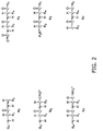
- the accepted nomenclature for F ions that result from a single backbone cleavage is depicted in Figures 1 and 2 and described in Johnson et al., Anal. Chem. 59:2621, 1987 . Briefly, N-terminal fragments are classed as either a, b, or c ; C-terminal fragments are classed as either x, y, or z ; and a subscript indicates the number of monomers in the fragment.
- the a / x , b / y, and c / z fragments are produced by cleavage of C a i /C i , C i /N i , and N i /C a i+1 backbone bonds, respectively.
- F spectra may include peaks from more than one F ion of a given type, e.g., one, some or all of the a series of a polypeptide with n monomers, i.e., a 1 , a 2 , a 3 , a 4 , a 5 , a 6 , a 7 , a 8 , a 9 , a 10 , ...
- F spectra may include different sets of complementary F ions, e.g., one, some or all of the a / x, b / y, and c / z pairs. It will further be appreciated that according to certain embodiments of the invention, F spectra may further include ions that result from a double backbone cleavage, a side chain cleavage, and/or a common neutral loss, e.g., loss of H 2 O, NH 3 , etc.
- F spectra are generated with a single or combination of low-energy mechanisms.
- Hashimoto et al. have recently described the combined use of CID and IR-MPD (Anal. Chem. Web release on December 24, 2002). It will be appreciated that the exact energy (or energies) used will depend on the sample under analysis and the fragmentation method (or methods) used. In general, appropriate fragmentation energies may be determined empirically, for example, by monitoring the proportion of unfragmented to fragmented peaks as a function of fragmentation energy. Additionally or alternatively one could select suitable fragmentation energies based on the average molecular weight and/or the weight distribution of the detected fragments.
- fragmentation energies may be set at a level below the threshold for double backbone and/or side chain cleavage.
- appropriate fragmentation energies when using CID at relatively high gas pressures will typically be less than about 1000 eV, more typically between about 100 and about 500 eV, even more typically between about 150 and about 250 eV.
- One having ordinary skill in the art can readily determine appropriate fragmentation energies for other mechanisms such as ECD and SID.
- the methods described herein are sensitive to the mass accuracy of the peaks in the U and F spectra.
- the mass accuracy of the U and F spectra will be affected by the specifications of the mass spectrometer and signal strength.
- the U and F spectra that are analyzed according to the methods described herein may be obtained under a broad range of operating conditions.
- the accuracy and completeness of the analysis will naturally improve as the mass accuracy is increased.
- the mass accuracy that is required may depend on the nature of the sample of interest and on the nature of the polypeptides within the sample, e.g., the average molecular weight and/or the range of molecular weights of the polypeptides.
- the choice of mass accuracy will depend on the desired quality of the analysis.
- the spectrometer or spectrometers that are used to obtain the U and F spectra may, for example, be operated under conditions that provide spectra with a mass accuracy of at least about 20 ppm, more preferably between about 10 and about 0.05 ppm, and even more preferably between about 3 and about 0.5 ppm.
- mass spectrometers that are capable of producing spectra of polypeptides with mass accuracies in these preferred ranges are available commercially, e.g., without limitation, the APEX III TM mass spectrometer from Bruker Daltonics of Billerica, MA; the HiResESI TM mass spectrometer from IonSpec of Lake Forest, CA; the Q-Tof ULtima TM mass spectrometer from Micromass of Milford, MA; the API QSTAR TM mass spectrometer from MDS Sciex of Concord, Canada; the AccuTOf TM mass spectrometer from JEOL of Peabody, MA; the AXIMA-QIT TM or AXIMA-MALDI TOF TM mass spectrometers from Shimadzu Biotech of Pleasanton, CA; etc.
- the U and F spectra are also obtained under conditions of high mass resolution.
- the mass resolution specification of a mass spectrometer provides a measure of its ability to resolve polypeptides that have similar molecular weights.
- the mass resolution depends on both the type of analyzer and the experimental conditions.
- the resolution in a given spectrum is defined as the experimentally determined ratio of the mass of an isolated singly charged peak divided by its full width at half the maximum height (FWHM). For example, if the FWHM values for a singly charged peak at m/z value of 1,658.752 is 0.237, then the resolution of that peak is equal to: 1,658.752/0.237 ⁇ 7,000.
- the mass resolution that is required may depend on the nature of the sample of interest and on the nature of the polypeptides within the sample, e.g., the average molecular weight and/or the range of molecular weights of the polypeptides.
- the spectrometer or spectrometers that are used to obtain the U and F spectra may be operated under conditions that are sufficient to resolve isotopes of a singly, preferably doubly, more preferably triply, and even more preferably quadruply charged ion.
- the U and F spectra within each set of spectra are obtained using the same sample or different aliquots of the same sample.
- different "aliquots" of the same sample have substantially the same polypeptide compositions, e.g., they are obtained by dividing a sample into two or more volumes.
- the U and F spectra in a given set are also obtained using the same spectrometer.
- the spectra are obtained back to back to provide the strongest mass accuracy correlation between the U and F spectra.
- the U and F spectra may be obtained using two or more spectrometers operating under similar, preferably near identical conditions, e.g., with mass accuracies that differ by less than a factor of 10, 5, 4, 3, or 2.
- the U and F spectra are obtained using a single-stage spectrometer, preferably the same single-stage spectrometer.
- Each U and F spectrum in a given set may correspond to a single spectral acquisition or to a summation over two or more spectral acquisitions.
- the individual spectra are stored in addition to (or instead of) the summed spectrum since the individual spectra may be used for later analysis.
- U and F spectra it will be appreciated that these may be obtained sequentially (e.g., U 1 , U 2 , U 3 , etc.
- F 1 , F 2 , F 3 , etc. in an interleaved manner (e.g., U 1 , F 1 , U 2 , F 2 , U 3 , F 3 , etc.), or some combination thereof (e.g., U 1 , U 2 , F 1 , F 2 , U 3 , U 4 , F 3 , F 4 , etc.).
- U 1 , U 2 , F 1 , F 2 , U 3 , U 4 , F 3 , F 4 , etc. may be summed to create an F spectrum without having to choose one specific fragmentation energy at the time of data acquisition.
- several spectra obtained with two or more different fragmentation mechanisms may be summed to create an F spectrum.
- U or F spectra will generally be correlated with the fragmentation conditions that are present within the spectrometer.
- the acquisition ofU and F spectra may be synchronized with transitions in these fragmentation conditions, e.g., changes in fragmentation energy.
- synchronization may prove advantageous in preventing the cross-contamination of unfragmented and fragmented masses between U and F spectra.
- a dead time may be placed in between acquisitions of U and F spectra to further prevent such cross-contamination.
- the particular choice in acquisition set up will depend on a number of factors including the nature of the sample, whether the U and F spectra are being obtained with a single-stage spectrometer or a multi-stage spectrometer, whether the U and F spectra are being obtained with the same spectrometer, whether several different samples are being analyzed in succession (e.g., in an LC-MS set up), the spectral acquisition time, the signal-to-noise ratio, the time required to toggle the fragmentation mechanism on and off, the energy and nature of the fragmentation mechanism, the time required to adjust the fragmentation energy, etc.
- the methods described herein may be modified to analyze sets of spectra that include more than one U spectrum and/or more than one F spectrum.
- the set of spectra for a given sample may include two or more F spectra that have been obtained with different fragmentation energies and/or two or more F spectra that have been obtained using different fragmentation mechanisms.
- different fragmentation energies and mechanisms tend to produce different types of F ions (e.g., see Papayannopoulos, Mass. Spectrom. Rev. 14:49, 1995 ).
- consideration of masses from a collection of F spectra that have been obtained with different energies and/or mechanisms may provide broader coverage across the different fragment ion series (i.e., the a, b, c, x, y, and z series).
- inventive methods may be used alone or may be used in tandem with other more traditional polypeptide mass fingerprinting techniques (e.g., those described in the prior art).
- inventive methods may prove advantageous to use the inventive methods to analyze a subset of masses that were unexplained by an earlier method.
- the methods described herein involve examining the masses in the one or more F spectra (i.e., corresponding to fragments of polypeptides) and attempting to match them with a mass in the one or more U spectra (i.e., corresponding to an unfragmented polypeptide).
- An F mass is said to match a U mass if it corresponds to a fragment that was generated from the polypeptide that corresponds to the U mass.
- the collection of F masses that have been matched with a U mass are then used to determine an amino acid sequence for the polypeptide that corresponds to the U mass.
- they are used to determine a set of alternative amino acid sequences for the corresponding polypeptide.
- they are used to determine the nature, location and relative levels of various modifications within the corresponding polypeptide.
- the processing steps that are used to match F masses with U masses involve making a "formula call" on a candidate mass, e.g., an F mass or a mass differential between two F masses.
- performing a "formula call" on a candidate mass involves treating amino acid monomers and terminal groups (i.e., R N and R C in Figure 1 and 2 ) as “elements” and using their theoretical masses to identify one or more "empirical amino acid formulae" that match the candidate mass.
- the empirical amino acid formula of an amino acid "stretch" between the b 2 ion above and a b 4 ion in the same series that includes a proton as an N-terminal group, two glycines, a methionine, and a tyrosinc is (Gly, Tyr).
- formula calls an empirical amino acid formula whose theoretical mass falls outside the range of actual mass possibilities around a candidate measured mass is considered to not "match". Conversely an empirical amino acid formula whose theoretical mass falls within the range of actual mass possibilities around a candidate measured mass is considered to "match”.
- a formula call can conceivably have, zero, one or more answers.
- formula calls that yield a single empirical formula are preferred, formula calls that yield two or more solutions are not entirely useless since they may lead to a single solution when combined with a separate formula call or other determination.
- a formula call that yields a single empirical amino acid formula for the measured mass of an F ion is termed a "successful formula call".
- a "successful formula call” provides (a) the amino acid composition of the F ion (e.g., one glycine and one methionine in the above example), (b) the series type of the F ion (e.g., b series in the above example), and (c) the position of the F ion within that series (e.g., position 2 in the above example).
- formula calls In general, the computational complexity of formula calls increase as the candidate mass is increased. Accordingly, in certain preferred embodiments, an attempt is made to minimize the masses for which formula calls are performed. As described in greater detail below, this is typically achieved by selecting specific ranges of masses to be examined. For example, in certain embodiments, formula calls may be limited to candidate masses that are greater than 50 Da and smaller than 1000, 500,400, 350, 300, 250, or 200 Da.
- formula calls may be achieved by consulting one or more databases of theoretical masses.
- the methods described herein are in no way limited to consulting specific databases of theoretical masses.
- the inventive methods may be used in combination with relatively simple databases that include the theoretical masses (e.g., the monoisotopic masses or some isotopic distribution of masses) of the twenty commonly occurring amino acid monomers (i.e., those listed in Appendix A).
- Another simple database might cover the commonly occurring a 2 , b 2 , c 2 , x 2 , y 2 , and z 2 type ions (i.e., a, b, c, x, y, or z type ions that include two amino acid monomers from Appendix A).
- databases that include larger ions e.g., ions that include 3, 4, 5, 6, 7, 8, 9, 10, etc. amino acid monomers
- databases that include less commonly occurring amino acid monomers e.g., those listed in Appendix B
- databases that include alternative N-terminal or C-terminal groups e.g., those listed in Appendix C.
- the databases may further take into account one or more modifications that are known to occur during polypeptide synthesis (e.g., without limitation those listed in Appendix E); post-translational protein modifications (e.g., without limitation those listed in Appendix F); and/or modifications that commonly occur within mass spectrometers as a consequence of the ionization and or detection process, e.g., loss of H 2 O, loss of NH 3 , loss of common side chains, etc.
- modifications that are known to occur during polypeptide synthesis e.g., without limitation those listed in Appendix E
- post-translational protein modifications e.g., without limitation those listed in Appendix F
- modifications that commonly occur within mass spectrometers e.g., loss of H 2 O, loss of NH 3 , loss of common side chains, etc.
- suitable databases can be constructed in a variety of ways.
- One approach might involve determining theoretical masses using the empirical chemical formulae of the molecules in the database in combination with elemental atomic masses (e.g., from Audi and Wapstra, Nuclear Physics A, 595:409, 1995 ), elemental atomic weights (e.g., from Coplen, Pure Appl. Chem., 73:667, 2001 ), and/or isotopic abundance data (e.g., from Rosman and Taylor, J. Phys. Chem. Ref. Data, 27:1275, 1998 ).
- databases may be constructed by performing "reverse" formula calls, i.e., by treating the amino acid monomers and terminal groups as "elements" with specific theoretical masses (e.g., those listed in Appendices A, B, and C) and then applying formulae that calculate the masses of the various empirical amino acid formulae in the database using the masses of these "elements” (e.g., for a, b, c, x, y, or z type ions by using the formulae provided in Appendix D).
- the latter approach is preferable since it reduces the computational complexity of the database construction process.
- the computational complexity of the inventive methods may be reduced by pre-processing the U and/or F spectra before the U and F masses are examined.
- This optional pre-processing of the spectra may involve one or some combination of the following approaches or equivalents thereof:
- the F masses are matched with U masses using one or more of the processing steps that are described in detail below. It is to be understood, and it will be readily apparent to one of ordinary skill in the art, that the matching procedure does not (a) require that each of the following processing steps be used or (b) require that the processing steps be used in the order presented. Further it will be appreciated that a given processing step may be repeated several times during the matching procedure, e.g., on either side of a different processing step. In particular, it is to be understood that the following describes a single exemplary embodiment of the matching procedure and that the inventive methods are in no way limited to this particular combination and order of processing steps. Further, for purposes of clarity only and without limitation, the following description of the various processing steps assumes that the U and F spectra have been deconvolved and deisotoped.
- Processing step 1 Identifying "seed" F masses
- the matching procedure may begin by identifying a collection of "seed" F masses that match a particular U mass.
- identifying a "seed" F mass requires determining the fragment series type of the corresponding F ion (i.e., whether the corresponding F ion belongs to an a, b, c, x, y, or z type ion series).
- the position of the corresponding F ion within the determined series is also determined (e.g., whether the corresponding F ion is a b 1 , b 2 , b 3 , b 4 , b 5 , etc. ion if it belongs to a b series).
- the amino acid composition of the corresponding F ion is also determined (e.g., whether the corresponding F ion includes two glycines or a glycine and a tryptophan if it is a b 2 ion). It will be appreciated that in certain embodiments the position and amino acid composition of the F ion may only be approximately defined, e.g., consider a b 2 ion that includes amino acid X and asparagine and a b 3 ion that includes X and two glycines - these will have near degenerate masses. For example, "seed" F masses may be identified using one or some combination of the following approaches or equivalents thereof:
- large seed F masses e.g., without limitation F masses that lie within 1000, 500, 400, 350, 300, 250, or 200 Da of the matched U mass
- large seed F masses that have been matched with a U mass will not be statistically likely to match for a different U mass.
- the smaller seed F masses should not be removed from further consideration as they may correspond to a fragment that has been generated from multiple U ions. The decision to remove matched seed F masses from consideration will typically depend on the number of U masses that were obtained from the original sample and hence on the complexity of the original sample.
- Processing step 2 Extending a seeded ion series using next adjacent ions
- the inventive methods may involve extending one or more of the seeded ion series by identifying the F masses of ions that are adjacent to a seed ion in the series. This may be accomplished by looking for F masses at each of the theoretical masses for adjacent ions in a seeded ion series. For example, in certain embodiments, this may involve adding or subtracting the theoretical mass of one, some or all of the possible amino acid monomers (e.g., those listed in Appendices A and B) from the theoretical ion mass that corresponds to the measured mass of the seed ion and then looking in the F spectral masses for a match.
- the theoretical mass of one, some or all of the possible amino acid monomers e.g., those listed in Appendices A and B
- Processing step 3 Extending a seeded ion series by scanning ahead
- the seeded ion series may additionally (or alternatively) be extended by identifying F masses of ions that are separated from a seed ion by two or more amino acid monomers. This approach will be particularly useful if the mass corresponding to the next ion in a series is missing from the F spectral masses (for example, if there is a mass for an a 3 ion, but no mass for the a 4 ion has been detected). This can be done using one or some combination of the following approaches or equivalents thereof:
- Processing step 4 Identifying F masses in the same position as a seed F mass in an adjacent series
- additional F masses may be matched with a U mass by identifying F masses that correspond to an ion in the same position as a seed ion but in an adjacent series (e.g., b 2 or c 2 when the seed ion is a 2 ). This can be accomplished by adding or subtracting one, some or all of the possible series offset masses from the theoretical ion mass that corresponds to the measured mass of the seed ion and then looking in the F spectral masses for a match.
- the series offset masses represent the mass deltas between the series of the seed ion (e.g., the a series) and the other related series (e.g., the b and c series).
- Processing step 5 Validating a seeded ion series
- Processing step 6 Creating one or more amino acid sequences for a U ion
- a set of possible amino acid sequences for the U ion can be created.
- the one or more fragment series are individually traversed and the amino acid monomer representing the difference in mass between adjacent ions in each series is identified via a formula call. It will be appreciated that this step may be shortened by using the results of processing steps 2 and 5 if they were performed.
- a consensus amino acid sequence by comparing the amino acid monomers that are predicted in different series to determine whether they add the same amino acid monomer to the same position in the sequence.
- the monomer at that position may be determined by examining a different series that includes that particular pair (e.g., b 4 and b 5 ).
- next adjacent ions for a particular pair of positions (e.g., positions 4 and 5) have not been identified in a single of the matched series, then the appropriate monomer may be determined by taking into account series offsets and performing a formula call on the mass delta between ions from next adjacent positions on two different series (e.g., b 4 and a 5 ). If none of the series have identified an ion at a given point in the series (e.g., position 4), then sets of alternative sequences are prepared, where the amino acid composition determined from the mass delta that spans that position (e.g., between a 3 and a 5 ) is represented in all possible permutations (and optionally all possible combinations if a formula call for the mass delta yields several solutions). Where exact amino acid calling is not possible due to identical mass (e.g., leucine vs. isoleucine) sets of alternative sequences are also prepared.
- the formula calling technique may be expanded to take into account one, some or all of the theoretical mass deltas of interest.
- the databases of theoretical masses may be expanded to include a sub-set of relevant theoretical mass deltas.
- the theoretical mass deltas may be included as additional "elements”.
- the choice of modifications that need to be considered will depend in part on the nature of the sample.
- the U and F spectral masses are then analyzed as described above, but formula calls are performed using these additional "elements" and/or expanded databases.
- the U and F spectral masses may be analyzed as described previously (i.e., without taking into account possible modifications) and then by post-processing the resulting data using one or some combination of the following approaches or equivalents thereof:
- the methods described herein may be modified to take into account the use of mass tagging techniques, e.g., for differential quantitation of proteins in different samples.
- mass tagging techniques e.g., for differential quantitation of proteins in different samples.
- the methods may be modified to take into account the use of isotope-coded affinity tags (ICAT).
- ICATs are a class of reagents, which are built of three main sections, namely an affinity tag, a linker for incorporation of stable isotopes, and a reactive group with specificity toward the thiol groups that are present in cysteines (e.g., see Gygi et al., Nat. Biotech. 17:994, 1999 ).
- a heavy form e.g., containing deuteriums on the carbon backbone
- a light form e.g., with no deuteriums
- the method typically consists of four steps, for example:
- the quantitative information comes from measuring and comparing the relative signal intensities of the pair of chemically identical polypeptides labeled with the light and heavy forms of the ICAT.
- the ratio of the polypeptide pairs provides quantitative information on the original protein of interest. This is due to the fact that the polypeptide fragments represent the ratio of the original amounts of the proteins in, for example, first and second cell states.
- a tag is effectively equivalent to a post-translational modification at the site where the tag exists. When extending the ion series, it can be treated as such.
- the presence of a tag also provides verification of the called empirical amino acid formula as only select amino acid monomers will react with the tag (e.g., cysteines in the example provided).
- the machine instructions may be produced using software including, for example, a programming language, third party software package, routines included as part of an operating system, and the like.
- the machine instructions, or a form thereof, may be stored in a computer program product that includes a computer readable medium (e.g., without limitation a floppy disk, a hard disk drive, a RAM, a CD-ROM, a tape, a cassette, etc.) with a set of machine executable instructions for performing the various steps of the inventive methods.
- the software is then loaded into a computer system (e.g., an offline computer or an online computer that also runs the mass spectrometer) using a removable storage drive, a hard drive, or a communications interface.
- a computer system e.g., an offline computer or an online computer that also runs the mass spectrometer
- the software when executed by one or more processors within the computer system, causes the processors to perform the functions of the invention as described herein. It should be noted that the foregoing may also be implemented in hardware using, for example, hardware components such as application specific integrated circuits. An embodiment may also implement the foregoing using a combination of hardware and/or software.
- a "polynucleotide” is a polymer of nucleotides, typically comprising at least two nucleotides linked together by phosphodiester bonds.
- the terms “polynucleotide”, “oligonucleotide”, and “nucleic acid” may be used interchangeably.
- DNA and RNA are exemplary polynucleotides that could be analyzed.
- the present invention also encompasses the analysis of peptide nucleic acids (PNAs), locked nucleic acid (LNAs), and unstructured nucleic acids (UNAs). As with polypeptides, measured U and F polynucleotide masses are analyzed using the inventive algorithms.
- Polynucleotides are identified by the sequence of nucleotides that they include. Formula calls are performed using nucleotide monomer "elements” instead of amino acid monomer “elements”. In certain embodiments, the theoretical masses of common nucleotides may be used (i.e., nucleotides that include the bases adenine, thymine, cytosine, guanine, or uracil).
- nucleotides that include the bases 2-aminoadenine, 2-thiothymine, 3-methyladenine, 5-propynylcytosine, 5-propynyluracil, 5-bromouracil, 5-fluorouracil, 5-iodouracil, 5-methylcytosine, 7-deazaadenine, 7-deazaguanine, 8-oxoadenine, 8-oxoguanine, O(6)-methylguanine, or 2-thiocytosine).
- mass deltas caused by sugar modifications e.g., 2'-fluororibose, arabinose, hexose, and riboses with a 2'-O, 4'-C-methylene bridge
- modified phosphate groups e.g., phosphorothioates and 5'-N-phosphoramidite linkages
- formula calls for polynucleotide ions will also need to take into account the theoretical masses of the terminal groups, typically these will be hydroxyl or phosphate.
- a "polysaccharide” is a polymer of sugars, typically comprising at least two sugars.
- the terms “polysaccharide”, “oligosaccharide”, and “carbohydrate” may be used interchangeably.
- the inventive methods may be used to analyze linear or branched polysaccharides. As with polypeptides, measured U and F polysaccharide masses are analyzed using the inventive algorithms. Polysaccharides are identified by the sequence of sugars that they include. Formula calls are performed using sugar monomers "elements” instead of amino acid monomers "elements”.
- the theoretical masses of common sugars may be used (e.g., arabinose, ribose, xylose, glucose, fructose, galactose, and mannose). Additionally or alternatively, the theoretical masses of less common or non-naturally occurring sugars may be used (e.g., deoxyribose, fucose, rhamnose, galactosamine, N-acetylgalactosamine, glucosamine, N-acetylglucosamine, glucuronic acid, muramic acid, N-acetylneuraminic acid, N-glycolylneuraminic acid, heptose, etc.).
- the theoretical masses of common sugars may be used (e.g., arabinose, ribose, xylose, glucose, fructose, galactose, and mannose).
- the theoretical masses of less common or non-naturally occurring sugars may be used (e.g., deoxyribos
- mass deltas caused by sugar modifications may be considered.
- formula calls for polysaccharide ions will also need to take into account the theoretical masses of the terminal groups, typically polysaccharides include free reducing ends or reduced reducing ends.
Landscapes
- Life Sciences & Earth Sciences (AREA)
- Health & Medical Sciences (AREA)
- Engineering & Computer Science (AREA)
- Chemical & Material Sciences (AREA)
- Physics & Mathematics (AREA)
- Molecular Biology (AREA)
- Immunology (AREA)
- Analytical Chemistry (AREA)
- Biochemistry (AREA)
- Bioinformatics & Computational Biology (AREA)
- Biomedical Technology (AREA)
- Pathology (AREA)
- General Physics & Mathematics (AREA)
- Hematology (AREA)
- General Health & Medical Sciences (AREA)
- Bioinformatics & Cheminformatics (AREA)
- Urology & Nephrology (AREA)
- Biophysics (AREA)
- Food Science & Technology (AREA)
- Medicinal Chemistry (AREA)
- Spectroscopy & Molecular Physics (AREA)
- Microbiology (AREA)
- Proteomics, Peptides & Aminoacids (AREA)
- Cell Biology (AREA)
- Biotechnology (AREA)
- Chemical Kinetics & Catalysis (AREA)
- Other Investigation Or Analysis Of Materials By Electrical Means (AREA)
- Investigating Or Analysing Biological Materials (AREA)
- Measuring Or Testing Involving Enzymes Or Micro-Organisms (AREA)
Applications Claiming Priority (2)
| Application Number | Priority Date | Filing Date | Title |
|---|---|---|---|
| US388088 | 2003-03-13 | ||
| US10/388,088 US8507285B2 (en) | 2003-03-13 | 2003-03-13 | Methods and devices for identifying biopolymers using mass spectroscopy |
Publications (3)
| Publication Number | Publication Date |
|---|---|
| EP1457776A2 EP1457776A2 (en) | 2004-09-15 |
| EP1457776A3 EP1457776A3 (en) | 2004-09-29 |
| EP1457776B1 true EP1457776B1 (en) | 2008-03-26 |
Family
ID=32771627
Family Applications (1)
| Application Number | Title | Priority Date | Filing Date |
|---|---|---|---|
| EP04003204A Expired - Lifetime EP1457776B1 (en) | 2003-03-13 | 2004-02-12 | Methods and devices for identifying biopolymers using mass spectroscopy |
Country Status (4)
| Country | Link |
|---|---|
| US (1) | US8507285B2 (ja) |
| EP (1) | EP1457776B1 (ja) |
| JP (1) | JP2004279424A (ja) |
| DE (1) | DE602004012637T2 (ja) |
Families Citing this family (13)
| Publication number | Priority date | Publication date | Assignee | Title |
|---|---|---|---|---|
| US20020115056A1 (en) * | 2000-12-26 | 2002-08-22 | Goodlett David R. | Rapid and quantitative proteome analysis and related methods |
| GB0305796D0 (en) | 2002-07-24 | 2003-04-16 | Micromass Ltd | Method of mass spectrometry and a mass spectrometer |
| JP4720254B2 (ja) * | 2005-03-31 | 2011-07-13 | 日本電気株式会社 | 分析方法、分析システム、及び分析プログラム |
| US7297940B2 (en) * | 2005-05-03 | 2007-11-20 | Palo Alto Research Center Incorporated | Method, apparatus, and program product for classifying ionized molecular fragments |
| GB2432712B (en) * | 2005-11-23 | 2007-12-27 | Micromass Ltd | Mass spectrometer |
| WO2008059567A1 (fr) * | 2006-11-15 | 2008-05-22 | Shimadzu Corporation | Procédé et dispositif de spectrométrie de masse |
| US8153961B2 (en) * | 2009-08-31 | 2012-04-10 | Thermo Finnigan Llc | Methods for acquisition and deductive analysis of mixed fragment peptide mass spectra |
| US8935101B2 (en) * | 2010-12-16 | 2015-01-13 | Thermo Finnigan Llc | Method and apparatus for correlating precursor and product ions in all-ions fragmentation experiments |
| US10446376B2 (en) | 2012-12-20 | 2019-10-15 | Dh Technologies Development Pte. Ltd. | Compound identification using multiple spectra at different collision energies |
| EP2936544B1 (en) * | 2012-12-20 | 2024-02-28 | DH Technologies Development Pte. Ltd. | Compound identification using multiple spectra at different collision energies |
| JP6718694B2 (ja) * | 2016-02-10 | 2020-07-08 | 日本電子株式会社 | マススペクトル解析装置、マススペクトル解析方法、および質量分析装置 |
| WO2019232520A1 (en) * | 2018-06-01 | 2019-12-05 | Cerno Bioscience Llc | Mass spectral analysis of large molecules |
| CN108776144B (zh) * | 2018-06-12 | 2020-10-09 | 南京大学 | 一种基于基团光谱特征的海洋溢油乳化物遥感识别方法 |
Family Cites Families (12)
| Publication number | Priority date | Publication date | Assignee | Title |
|---|---|---|---|---|
| ATE427493T1 (de) * | 1992-05-29 | 2009-04-15 | Univ Rockefeller | Verfahren zur bestimmung der folge von peptiden unter verwendung eines massenspektrometers |
| US5470753A (en) * | 1992-09-03 | 1995-11-28 | Selectide Corporation | Peptide sequencing using mass spectrometry |
| WO1994016101A2 (en) * | 1993-01-07 | 1994-07-21 | Koester Hubert | Dna sequencing by mass spectrometry |
| US5538897A (en) * | 1994-03-14 | 1996-07-23 | University Of Washington | Use of mass spectrometry fragmentation patterns of peptides to identify amino acid sequences in databases |
| CA2248084A1 (en) * | 1996-03-04 | 1997-09-12 | Genetrace Systems, Inc. | Methods of screening nucleic acids using mass spectrometry |
| US5885775A (en) * | 1996-10-04 | 1999-03-23 | Perseptive Biosystems, Inc. | Methods for determining sequences information in polynucleotides using mass spectrometry |
| DE19709086B4 (de) * | 1997-03-06 | 2007-03-15 | Bruker Daltonik Gmbh | Verfahren der Raumladungsregelung von Tochterionen in Ionenfallen |
| US6104027A (en) * | 1998-06-05 | 2000-08-15 | Hewlett-Packard Company | Deconvolution of multiply charged ions |
| US20020182649A1 (en) | 2001-02-01 | 2002-12-05 | Ciphergen Biosystems, Inc. | Methods for protein identification, characterization and sequencing by tandem mass spectrometry |
| EP1434874A4 (en) | 2001-06-21 | 2005-11-30 | Cell Signaling Technology Inc | IMMUNOAFFINITY ISOLATION OF MODIFIED PEPTIDES FROM COMPLEX MIXTURES |
| CA2453725A1 (en) * | 2001-07-13 | 2003-01-23 | Syngenta Participations Ag | System and method of determining proteomic differences |
| US7351957B2 (en) * | 2002-04-29 | 2008-04-01 | Mds Inc. | Broad ion fragmentation coverage in mass spectrometry by varying the collision energy |
-
2003
- 2003-03-13 US US10/388,088 patent/US8507285B2/en active Active
-
2004
- 2004-02-12 DE DE602004012637T patent/DE602004012637T2/de not_active Expired - Lifetime
- 2004-02-12 EP EP04003204A patent/EP1457776B1/en not_active Expired - Lifetime
- 2004-03-12 JP JP2004069966A patent/JP2004279424A/ja active Pending
Also Published As
| Publication number | Publication date |
|---|---|
| DE602004012637D1 (de) | 2008-05-08 |
| EP1457776A3 (en) | 2004-09-29 |
| DE602004012637T2 (de) | 2009-04-16 |
| JP2004279424A (ja) | 2004-10-07 |
| US8507285B2 (en) | 2013-08-13 |
| EP1457776A2 (en) | 2004-09-15 |
| US20040180446A1 (en) | 2004-09-16 |
Similar Documents
| Publication | Publication Date | Title |
|---|---|---|
| US7538321B2 (en) | Method of identifying substances using mass spectrometry | |
| JP4818270B2 (ja) | 選択されたイオンクロマトグラムを使用して先駆物質および断片イオンをグループ化するシステムおよび方法 | |
| Dayon et al. | Combining low-and high-energy tandem mass spectra for optimized peptide quantification with isobaric tags | |
| EP2279260B1 (en) | Techniques for performing retention-time matching of precursor and product ions and for constructing precursor and product ion spectra | |
| US8105838B2 (en) | Generation and use of a catalog of polypeptide-related information for chemical analyses | |
| US8426155B2 (en) | Proteome analysis in mass spectrometers containing RF ion traps | |
| JP5299060B2 (ja) | 糖ペプチド構造解析方法及び装置 | |
| EP2956775A1 (en) | Mass labels | |
| US8278115B2 (en) | Methods for processing tandem mass spectral data for protein sequence analysis | |
| WO2007144606A2 (en) | Mass spectrometry biomarker assay | |
| EP1457776B1 (en) | Methods and devices for identifying biopolymers using mass spectroscopy | |
| WO2009138179A2 (en) | Ms/ms data processing | |
| EP1756852A2 (en) | Method and apparatus for identifying proteins in mixtures | |
| EP1941280A2 (en) | Methods for the development of a biomolecule assay | |
| Van Riper et al. | Mass spectrometry-based proteomics: basic principles and emerging technologies and directions | |
| EP4047371A1 (en) | Method and apparatus for analysing samples of biomolecules using mass spectrometry with data-independent acquisition | |
| US11355330B2 (en) | Uses of isobaric tags in mass spectrometry | |
| CN112824894B (zh) | 糖肽解析装置 | |
| JP6065652B2 (ja) | 糖鎖構造解析方法及び装置 | |
| JP2009516842A (ja) | 質量分析計 | |
| JP4821400B2 (ja) | 構造解析システム | |
| EP3654364A1 (en) | Methods for multiplexed ms-3 analyses of peptides | |
| Zhang | Progress in mass spectrometry acquisition approach for quantitative proteomics | |
| JP7156207B2 (ja) | 糖ペプチド解析装置 | |
| EP3971943A1 (en) | Using real time search results to dynamically exclude product ions that may be present in the master scan |
Legal Events
| Date | Code | Title | Description |
|---|---|---|---|
| PUAI | Public reference made under article 153(3) epc to a published international application that has entered the european phase |
Free format text: ORIGINAL CODE: 0009012 |
|
| PUAL | Search report despatched |
Free format text: ORIGINAL CODE: 0009013 |
|
| AK | Designated contracting states |
Kind code of ref document: A2 Designated state(s): AT BE BG CH CY CZ DE DK EE ES FI FR GB GR HU IE IT LI LU MC NL PT RO SE SI SK TR |
|
| AX | Request for extension of the european patent |
Extension state: AL LT LV MK |
|
| AK | Designated contracting states |
Kind code of ref document: A3 Designated state(s): AT BE BG CH CY CZ DE DK EE ES FI FR GB GR HU IE IT LI LU MC NL PT RO SE SI SK TR |
|
| AX | Request for extension of the european patent |
Extension state: AL LT LV MK |
|
| RIC1 | Information provided on ipc code assigned before grant |
Ipc: 7C 12Q 1/68 B Ipc: 7G 01N 33/68 A |
|
| 17P | Request for examination filed |
Effective date: 20040916 |
|
| AKX | Designation fees paid |
Designated state(s): CH DE GB LI |
|
| 17Q | First examination report despatched |
Effective date: 20061208 |
|
| RAP1 | Party data changed (applicant data changed or rights of an application transferred) |
Owner name: AGILENT TECHNOLOGIES, INC. |
|
| GRAP | Despatch of communication of intention to grant a patent |
Free format text: ORIGINAL CODE: EPIDOSNIGR1 |
|
| GRAS | Grant fee paid |
Free format text: ORIGINAL CODE: EPIDOSNIGR3 |
|
| GRAA | (expected) grant |
Free format text: ORIGINAL CODE: 0009210 |
|
| AK | Designated contracting states |
Kind code of ref document: B1 Designated state(s): CH DE GB LI |
|
| REG | Reference to a national code |
Ref country code: GB Ref legal event code: FG4D |
|
| REG | Reference to a national code |
Ref country code: CH Ref legal event code: EP |
|
| REF | Corresponds to: |
Ref document number: 602004012637 Country of ref document: DE Date of ref document: 20080508 Kind code of ref document: P |
|
| PLBE | No opposition filed within time limit |
Free format text: ORIGINAL CODE: 0009261 |
|
| STAA | Information on the status of an ep patent application or granted ep patent |
Free format text: STATUS: NO OPPOSITION FILED WITHIN TIME LIMIT |
|
| 26N | No opposition filed |
Effective date: 20081230 |
|
| PGFP | Annual fee paid to national office [announced via postgrant information from national office to epo] |
Ref country code: GB Payment date: 20090211 Year of fee payment: 6 |
|
| REG | Reference to a national code |
Ref country code: CH Ref legal event code: PL |
|
| PG25 | Lapsed in a contracting state [announced via postgrant information from national office to epo] |
Ref country code: LI Free format text: LAPSE BECAUSE OF NON-PAYMENT OF DUE FEES Effective date: 20090228 Ref country code: CH Free format text: LAPSE BECAUSE OF NON-PAYMENT OF DUE FEES Effective date: 20090228 |
|
| GBPC | Gb: european patent ceased through non-payment of renewal fee |
Effective date: 20100212 |
|
| PG25 | Lapsed in a contracting state [announced via postgrant information from national office to epo] |
Ref country code: GB Free format text: LAPSE BECAUSE OF NON-PAYMENT OF DUE FEES Effective date: 20100212 |
|
| PGFP | Annual fee paid to national office [announced via postgrant information from national office to epo] |
Ref country code: DE Payment date: 20170207 Year of fee payment: 14 |
|
| REG | Reference to a national code |
Ref country code: DE Ref legal event code: R119 Ref document number: 602004012637 Country of ref document: DE |
|
| PG25 | Lapsed in a contracting state [announced via postgrant information from national office to epo] |
Ref country code: DE Free format text: LAPSE BECAUSE OF NON-PAYMENT OF DUE FEES Effective date: 20180901 |