EP1436617B1 - Classification de patient - Google Patents
Classification de patient Download PDFInfo
- Publication number
- EP1436617B1 EP1436617B1 EP02775804A EP02775804A EP1436617B1 EP 1436617 B1 EP1436617 B1 EP 1436617B1 EP 02775804 A EP02775804 A EP 02775804A EP 02775804 A EP02775804 A EP 02775804A EP 1436617 B1 EP1436617 B1 EP 1436617B1
- Authority
- EP
- European Patent Office
- Prior art keywords
- cells
- cell
- parameters
- factors
- assay
- Prior art date
- Legal status (The legal status is an assumption and is not a legal conclusion. Google has not performed a legal analysis and makes no representation as to the accuracy of the status listed.)
- Expired - Lifetime
Links
- 239000003814 drug Substances 0.000 claims abstract description 64
- 230000037361 pathway Effects 0.000 claims abstract description 63
- 201000010099 disease Diseases 0.000 claims abstract description 51
- 208000037265 diseases, disorders, signs and symptoms Diseases 0.000 claims abstract description 51
- 229940124597 therapeutic agent Drugs 0.000 claims abstract description 16
- 230000004043 responsiveness Effects 0.000 claims abstract description 13
- 238000003556 assay Methods 0.000 claims description 149
- 238000000034 method Methods 0.000 claims description 93
- 238000005259 measurement Methods 0.000 claims description 65
- 238000012360 testing method Methods 0.000 claims description 34
- 238000004113 cell culture Methods 0.000 claims description 31
- 239000000523 sample Substances 0.000 claims description 26
- 239000013610 patient sample Substances 0.000 claims description 16
- 150000007523 nucleic acids Chemical class 0.000 claims description 11
- 239000013543 active substance Substances 0.000 claims description 10
- 230000007613 environmental effect Effects 0.000 claims description 10
- 108020004707 nucleic acids Proteins 0.000 claims description 10
- 102000039446 nucleic acids Human genes 0.000 claims description 10
- 239000012530 fluid Substances 0.000 claims description 6
- 102000054765 polymorphisms of proteins Human genes 0.000 claims description 4
- 239000000284 extract Substances 0.000 claims description 3
- 230000001900 immune effect Effects 0.000 claims description 3
- 238000003759 clinical diagnosis Methods 0.000 claims 1
- 230000000694 effects Effects 0.000 abstract description 30
- 238000011282 treatment Methods 0.000 abstract description 17
- 206010071602 Genetic polymorphism Diseases 0.000 abstract description 4
- 238000003745 diagnosis Methods 0.000 abstract description 4
- 238000004393 prognosis Methods 0.000 abstract description 3
- 210000004027 cell Anatomy 0.000 description 273
- 108060008682 Tumor Necrosis Factor Proteins 0.000 description 66
- 239000003795 chemical substances by application Substances 0.000 description 63
- 150000001875 compounds Chemical class 0.000 description 56
- 102100040247 Tumor necrosis factor Human genes 0.000 description 53
- MZOFCQQQCNRIBI-VMXHOPILSA-N (3s)-4-[[(2s)-1-[[(2s)-1-[[(1s)-1-carboxy-2-hydroxyethyl]amino]-4-methyl-1-oxopentan-2-yl]amino]-5-(diaminomethylideneamino)-1-oxopentan-2-yl]amino]-3-[[2-[[(2s)-2,6-diaminohexanoyl]amino]acetyl]amino]-4-oxobutanoic acid Chemical compound OC[C@@H](C(O)=O)NC(=O)[C@H](CC(C)C)NC(=O)[C@H](CCCN=C(N)N)NC(=O)[C@H](CC(O)=O)NC(=O)CNC(=O)[C@@H](N)CCCCN MZOFCQQQCNRIBI-VMXHOPILSA-N 0.000 description 48
- 108010074328 Interferon-gamma Proteins 0.000 description 47
- 210000002889 endothelial cell Anatomy 0.000 description 47
- 102100037850 Interferon gamma Human genes 0.000 description 46
- 102000000589 Interleukin-1 Human genes 0.000 description 44
- 108010002352 Interleukin-1 Proteins 0.000 description 44
- 229940079593 drug Drugs 0.000 description 43
- 230000014509 gene expression Effects 0.000 description 41
- 206010028980 Neoplasm Diseases 0.000 description 40
- 230000004044 response Effects 0.000 description 37
- 238000004458 analytical method Methods 0.000 description 33
- 108090000623 proteins and genes Proteins 0.000 description 29
- 108010064593 Intercellular Adhesion Molecule-1 Proteins 0.000 description 28
- 102000015271 Intercellular Adhesion Molecule-1 Human genes 0.000 description 28
- 108090001007 Interleukin-8 Proteins 0.000 description 28
- 102000004890 Interleukin-8 Human genes 0.000 description 28
- 102100023471 E-selectin Human genes 0.000 description 27
- 108010024212 E-Selectin Proteins 0.000 description 26
- 210000001744 T-lymphocyte Anatomy 0.000 description 26
- 210000001519 tissue Anatomy 0.000 description 24
- 102000006354 HLA-DR Antigens Human genes 0.000 description 23
- 108010058597 HLA-DR Antigens Proteins 0.000 description 23
- 108010000134 Vascular Cell Adhesion Molecule-1 Proteins 0.000 description 23
- 102100023543 Vascular cell adhesion protein 1 Human genes 0.000 description 23
- 102100036170 C-X-C motif chemokine 9 Human genes 0.000 description 21
- 101710085500 C-X-C motif chemokine 9 Proteins 0.000 description 21
- 108090000978 Interleukin-4 Proteins 0.000 description 20
- 102000004388 Interleukin-4 Human genes 0.000 description 20
- HCZKYJDFEPMADG-TXEJJXNPSA-N masoprocol Chemical compound C([C@H](C)[C@H](C)CC=1C=C(O)C(O)=CC=1)C1=CC=C(O)C(O)=C1 HCZKYJDFEPMADG-TXEJJXNPSA-N 0.000 description 20
- 102000005962 receptors Human genes 0.000 description 18
- 108020003175 receptors Proteins 0.000 description 18
- 102100024616 Platelet endothelial cell adhesion molecule Human genes 0.000 description 16
- 102000000852 Tumor Necrosis Factor-alpha Human genes 0.000 description 16
- 201000011510 cancer Diseases 0.000 description 16
- 230000008859 change Effects 0.000 description 16
- 230000003247 decreasing effect Effects 0.000 description 16
- 102000004127 Cytokines Human genes 0.000 description 15
- 108090000695 Cytokines Proteins 0.000 description 15
- 108020004414 DNA Proteins 0.000 description 15
- 210000004369 blood Anatomy 0.000 description 14
- 239000008280 blood Substances 0.000 description 14
- 230000001413 cellular effect Effects 0.000 description 14
- 239000003112 inhibitor Substances 0.000 description 14
- 239000000047 product Substances 0.000 description 14
- 102000004169 proteins and genes Human genes 0.000 description 14
- 102000013462 Interleukin-12 Human genes 0.000 description 13
- 108010065805 Interleukin-12 Proteins 0.000 description 13
- 230000002068 genetic effect Effects 0.000 description 13
- -1 CD31 Proteins 0.000 description 12
- 108010002350 Interleukin-2 Proteins 0.000 description 12
- 102000000588 Interleukin-2 Human genes 0.000 description 12
- 108090001005 Interleukin-6 Proteins 0.000 description 12
- 102000004889 Interleukin-6 Human genes 0.000 description 12
- 238000001514 detection method Methods 0.000 description 12
- 238000001727 in vivo Methods 0.000 description 12
- 230000002757 inflammatory effect Effects 0.000 description 12
- 210000004698 lymphocyte Anatomy 0.000 description 12
- 238000000684 flow cytometry Methods 0.000 description 11
- 230000004054 inflammatory process Effects 0.000 description 11
- 102100021943 C-C motif chemokine 2 Human genes 0.000 description 10
- 101710155857 C-C motif chemokine 2 Proteins 0.000 description 10
- VSWDORGPIHIGNW-UHFFFAOYSA-N Pyrrolidine dithiocarbamic acid Chemical compound SC(=S)N1CCCC1 VSWDORGPIHIGNW-UHFFFAOYSA-N 0.000 description 10
- 230000009471 action Effects 0.000 description 10
- 210000004102 animal cell Anatomy 0.000 description 10
- HCZKYJDFEPMADG-UHFFFAOYSA-N erythro-nordihydroguaiaretic acid Natural products C=1C=C(O)C(O)=CC=1CC(C)C(C)CC1=CC=C(O)C(O)=C1 HCZKYJDFEPMADG-UHFFFAOYSA-N 0.000 description 10
- 230000006870 function Effects 0.000 description 10
- 210000005260 human cell Anatomy 0.000 description 10
- 229960003951 masoprocol Drugs 0.000 description 10
- 239000002609 medium Substances 0.000 description 10
- 238000012216 screening Methods 0.000 description 10
- 230000004913 activation Effects 0.000 description 9
- 230000003321 amplification Effects 0.000 description 9
- 230000006907 apoptotic process Effects 0.000 description 9
- 210000003630 histaminocyte Anatomy 0.000 description 9
- 208000027866 inflammatory disease Diseases 0.000 description 9
- 238000003199 nucleic acid amplification method Methods 0.000 description 9
- 230000019491 signal transduction Effects 0.000 description 9
- 101000979342 Homo sapiens Nuclear factor NF-kappa-B p105 subunit Proteins 0.000 description 8
- 102000003816 Interleukin-13 Human genes 0.000 description 8
- 108090000176 Interleukin-13 Proteins 0.000 description 8
- 102100023050 Nuclear factor NF-kappa-B p105 subunit Human genes 0.000 description 8
- 239000011324 bead Substances 0.000 description 8
- 239000003153 chemical reaction reagent Substances 0.000 description 8
- 239000012091 fetal bovine serum Substances 0.000 description 8
- 239000003446 ligand Substances 0.000 description 8
- 230000008569 process Effects 0.000 description 8
- 238000002560 therapeutic procedure Methods 0.000 description 8
- 201000004681 Psoriasis Diseases 0.000 description 7
- 230000033115 angiogenesis Effects 0.000 description 7
- 239000000427 antigen Substances 0.000 description 7
- 108091007433 antigens Proteins 0.000 description 7
- 102000036639 antigens Human genes 0.000 description 7
- 210000000601 blood cell Anatomy 0.000 description 7
- 230000010261 cell growth Effects 0.000 description 7
- 238000002512 chemotherapy Methods 0.000 description 7
- 238000004163 cytometry Methods 0.000 description 7
- 238000011161 development Methods 0.000 description 7
- 230000018109 developmental process Effects 0.000 description 7
- 210000002540 macrophage Anatomy 0.000 description 7
- 239000000203 mixture Substances 0.000 description 7
- 210000001616 monocyte Anatomy 0.000 description 7
- 230000003472 neutralizing effect Effects 0.000 description 7
- 210000005259 peripheral blood Anatomy 0.000 description 7
- 239000011886 peripheral blood Substances 0.000 description 7
- 230000000144 pharmacologic effect Effects 0.000 description 7
- 239000000243 solution Substances 0.000 description 7
- ZRKFYGHZFMAOKI-QMGMOQQFSA-N tgfbeta Chemical compound C([C@H](NC(=O)[C@H](C(C)C)NC(=O)CNC(=O)[C@H](CCC(O)=O)NC(=O)[C@H](CCCNC(N)=N)NC(=O)[C@H](CC(N)=O)NC(=O)[C@H](CC(C)C)NC(=O)[C@H]([C@@H](C)O)NC(=O)[C@H](CCC(O)=O)NC(=O)[C@H]([C@@H](C)O)NC(=O)[C@H](CC(C)C)NC(=O)CNC(=O)[C@H](C)NC(=O)[C@H](CO)NC(=O)[C@H](CCC(N)=O)NC(=O)[C@@H](NC(=O)[C@H](C)NC(=O)[C@H](C)NC(=O)[C@@H](NC(=O)[C@H](CC(C)C)NC(=O)[C@@H](N)CCSC)C(C)C)[C@@H](C)CC)C(=O)N[C@@H]([C@@H](C)O)C(=O)N[C@@H](C(C)C)C(=O)N[C@@H](CC=1C=CC=CC=1)C(=O)N[C@@H](C)C(=O)N1[C@@H](CCC1)C(=O)N[C@@H]([C@@H](C)O)C(=O)N[C@@H](CC(N)=O)C(=O)N[C@@H](CCC(O)=O)C(=O)N[C@@H](C)C(=O)N[C@@H](CC=1C=CC=CC=1)C(=O)N[C@@H](CCCNC(N)=N)C(=O)N[C@@H](C)C(=O)N[C@@H](CC(C)C)C(=O)N1[C@@H](CCC1)C(=O)N1[C@@H](CCC1)C(=O)N[C@@H](CCCNC(N)=N)C(=O)N[C@@H](CCC(O)=O)C(=O)N[C@@H](CCCNC(N)=N)C(=O)N[C@@H](CO)C(=O)N[C@@H](CCCNC(N)=N)C(=O)N[C@@H](CC(C)C)C(=O)N[C@@H](CC(C)C)C(O)=O)C1=CC=C(O)C=C1 ZRKFYGHZFMAOKI-QMGMOQQFSA-N 0.000 description 7
- YBJHBAHKTGYVGT-ZKWXMUAHSA-N (+)-Biotin Chemical group N1C(=O)N[C@@H]2[C@H](CCCCC(=O)O)SC[C@@H]21 YBJHBAHKTGYVGT-ZKWXMUAHSA-N 0.000 description 6
- 101150013553 CD40 gene Proteins 0.000 description 6
- IAZDPXIOMUYVGZ-UHFFFAOYSA-N Dimethylsulphoxide Chemical compound CS(C)=O IAZDPXIOMUYVGZ-UHFFFAOYSA-N 0.000 description 6
- 206010061218 Inflammation Diseases 0.000 description 6
- 241000699670 Mus sp. Species 0.000 description 6
- 102000004887 Transforming Growth Factor beta Human genes 0.000 description 6
- 108090001012 Transforming Growth Factor beta Proteins 0.000 description 6
- 102100040245 Tumor necrosis factor receptor superfamily member 5 Human genes 0.000 description 6
- 102000005789 Vascular Endothelial Growth Factors Human genes 0.000 description 6
- 108010019530 Vascular Endothelial Growth Factors Proteins 0.000 description 6
- 238000002835 absorbance Methods 0.000 description 6
- 230000003213 activating effect Effects 0.000 description 6
- 206010003246 arthritis Diseases 0.000 description 6
- 208000006673 asthma Diseases 0.000 description 6
- 150000001720 carbohydrates Chemical class 0.000 description 6
- 208000037976 chronic inflammation Diseases 0.000 description 6
- 238000005516 engineering process Methods 0.000 description 6
- 238000002474 experimental method Methods 0.000 description 6
- 238000000338 in vitro Methods 0.000 description 6
- 108010044426 integrins Proteins 0.000 description 6
- 102000006495 integrins Human genes 0.000 description 6
- 210000000265 leukocyte Anatomy 0.000 description 6
- 230000004048 modification Effects 0.000 description 6
- 238000012986 modification Methods 0.000 description 6
- 239000002773 nucleotide Substances 0.000 description 6
- 230000001105 regulatory effect Effects 0.000 description 6
- 238000012552 review Methods 0.000 description 6
- 238000007619 statistical method Methods 0.000 description 6
- 239000000126 substance Substances 0.000 description 6
- 102100028990 C-X-C chemokine receptor type 3 Human genes 0.000 description 5
- 102000019034 Chemokines Human genes 0.000 description 5
- 108010012236 Chemokines Proteins 0.000 description 5
- 208000011231 Crohn disease Diseases 0.000 description 5
- LFQSCWFLJHTTHZ-UHFFFAOYSA-N Ethanol Chemical compound CCO LFQSCWFLJHTTHZ-UHFFFAOYSA-N 0.000 description 5
- 108010017213 Granulocyte-Macrophage Colony-Stimulating Factor Proteins 0.000 description 5
- 102100039620 Granulocyte-macrophage colony-stimulating factor Human genes 0.000 description 5
- 101000916050 Homo sapiens C-X-C chemokine receptor type 3 Proteins 0.000 description 5
- HEFNNWSXXWATRW-UHFFFAOYSA-N Ibuprofen Chemical compound CC(C)CC1=CC=C(C(C)C(O)=O)C=C1 HEFNNWSXXWATRW-UHFFFAOYSA-N 0.000 description 5
- 208000022559 Inflammatory bowel disease Diseases 0.000 description 5
- 102000003814 Interleukin-10 Human genes 0.000 description 5
- 108090000174 Interleukin-10 Proteins 0.000 description 5
- 108010052285 Membrane Proteins Proteins 0.000 description 5
- 108091092878 Microsatellite Proteins 0.000 description 5
- 230000006052 T cell proliferation Effects 0.000 description 5
- 239000002260 anti-inflammatory agent Substances 0.000 description 5
- 239000002246 antineoplastic agent Substances 0.000 description 5
- 235000014633 carbohydrates Nutrition 0.000 description 5
- 230000004663 cell proliferation Effects 0.000 description 5
- 238000012512 characterization method Methods 0.000 description 5
- 238000006243 chemical reaction Methods 0.000 description 5
- 208000037893 chronic inflammatory disorder Diseases 0.000 description 5
- 230000004069 differentiation Effects 0.000 description 5
- 239000007850 fluorescent dye Substances 0.000 description 5
- 239000000499 gel Substances 0.000 description 5
- 238000009396 hybridization Methods 0.000 description 5
- 238000003018 immunoassay Methods 0.000 description 5
- 208000015181 infectious disease Diseases 0.000 description 5
- 238000002372 labelling Methods 0.000 description 5
- 238000004519 manufacturing process Methods 0.000 description 5
- 210000004925 microvascular endothelial cell Anatomy 0.000 description 5
- 238000012544 monitoring process Methods 0.000 description 5
- 230000009826 neoplastic cell growth Effects 0.000 description 5
- 210000000440 neutrophil Anatomy 0.000 description 5
- 125000003729 nucleotide group Chemical group 0.000 description 5
- 238000003752 polymerase chain reaction Methods 0.000 description 5
- 238000004088 simulation Methods 0.000 description 5
- 230000000007 visual effect Effects 0.000 description 5
- GOZMBJCYMQQACI-UHFFFAOYSA-N 6,7-dimethyl-3-[[methyl-[2-[methyl-[[1-[3-(trifluoromethyl)phenyl]indol-3-yl]methyl]amino]ethyl]amino]methyl]chromen-4-one;dihydrochloride Chemical compound Cl.Cl.C=1OC2=CC(C)=C(C)C=C2C(=O)C=1CN(C)CCN(C)CC(C1=CC=CC=C11)=CN1C1=CC=CC(C(F)(F)F)=C1 GOZMBJCYMQQACI-UHFFFAOYSA-N 0.000 description 4
- 208000023275 Autoimmune disease Diseases 0.000 description 4
- 108091003079 Bovine Serum Albumin Proteins 0.000 description 4
- 101100314454 Caenorhabditis elegans tra-1 gene Proteins 0.000 description 4
- 102000004631 Calcineurin Human genes 0.000 description 4
- 108010042955 Calcineurin Proteins 0.000 description 4
- 102100037362 Fibronectin Human genes 0.000 description 4
- 108010067306 Fibronectins Proteins 0.000 description 4
- NTYJJOPFIAHURM-UHFFFAOYSA-N Histamine Chemical compound NCCC1=CN=CN1 NTYJJOPFIAHURM-UHFFFAOYSA-N 0.000 description 4
- 102000003855 L-lactate dehydrogenase Human genes 0.000 description 4
- 108700023483 L-lactate dehydrogenases Proteins 0.000 description 4
- 102000043136 MAP kinase family Human genes 0.000 description 4
- 108091054455 MAP kinase family Proteins 0.000 description 4
- 102000035195 Peptidases Human genes 0.000 description 4
- 108091005804 Peptidases Proteins 0.000 description 4
- 230000006044 T cell activation Effects 0.000 description 4
- 229940124599 anti-inflammatory drug Drugs 0.000 description 4
- 210000003719 b-lymphocyte Anatomy 0.000 description 4
- 230000008901 benefit Effects 0.000 description 4
- 230000003915 cell function Effects 0.000 description 4
- 230000004640 cellular pathway Effects 0.000 description 4
- 230000002596 correlated effect Effects 0.000 description 4
- 230000016396 cytokine production Effects 0.000 description 4
- 230000001086 cytosolic effect Effects 0.000 description 4
- 231100000433 cytotoxic Toxicity 0.000 description 4
- 230000001472 cytotoxic effect Effects 0.000 description 4
- 238000009472 formulation Methods 0.000 description 4
- 229960001680 ibuprofen Drugs 0.000 description 4
- 230000003993 interaction Effects 0.000 description 4
- 150000002632 lipids Chemical class 0.000 description 4
- 239000003550 marker Substances 0.000 description 4
- 239000011159 matrix material Substances 0.000 description 4
- 230000007246 mechanism Effects 0.000 description 4
- 230000007170 pathology Effects 0.000 description 4
- 230000000770 proinflammatory effect Effects 0.000 description 4
- 239000002096 quantum dot Substances 0.000 description 4
- 206010039073 rheumatoid arthritis Diseases 0.000 description 4
- 150000003431 steroids Chemical class 0.000 description 4
- 210000004881 tumor cell Anatomy 0.000 description 4
- 210000003606 umbilical vein Anatomy 0.000 description 4
- 238000005406 washing Methods 0.000 description 4
- UAIUNKRWKOVEES-UHFFFAOYSA-N 3,3',5,5'-tetramethylbenzidine Chemical compound CC1=C(N)C(C)=CC(C=2C=C(C)C(N)=C(C)C=2)=C1 UAIUNKRWKOVEES-UHFFFAOYSA-N 0.000 description 3
- 102100035875 C-C chemokine receptor type 5 Human genes 0.000 description 3
- 101710149870 C-C chemokine receptor type 5 Proteins 0.000 description 3
- 102100025248 C-X-C motif chemokine 10 Human genes 0.000 description 3
- 101710098275 C-X-C motif chemokine 10 Proteins 0.000 description 3
- 102100032912 CD44 antigen Human genes 0.000 description 3
- 102100025475 Carcinoembryonic antigen-related cell adhesion molecule 5 Human genes 0.000 description 3
- 208000005623 Carcinogenesis Diseases 0.000 description 3
- 208000024172 Cardiovascular disease Diseases 0.000 description 3
- 102000009410 Chemokine receptor Human genes 0.000 description 3
- 108050000299 Chemokine receptor Proteins 0.000 description 3
- 206010013710 Drug interaction Diseases 0.000 description 3
- 102000004190 Enzymes Human genes 0.000 description 3
- 108090000790 Enzymes Proteins 0.000 description 3
- 108010037362 Extracellular Matrix Proteins Proteins 0.000 description 3
- 102000010834 Extracellular Matrix Proteins Human genes 0.000 description 3
- 101150021185 FGF gene Proteins 0.000 description 3
- 102000003974 Fibroblast growth factor 2 Human genes 0.000 description 3
- 108090000379 Fibroblast growth factor 2 Proteins 0.000 description 3
- 102100031547 HLA class II histocompatibility antigen, DO alpha chain Human genes 0.000 description 3
- HTTJABKRGRZYRN-UHFFFAOYSA-N Heparin Chemical compound OC1C(NC(=O)C)C(O)OC(COS(O)(=O)=O)C1OC1C(OS(O)(=O)=O)C(O)C(OC2C(C(OS(O)(=O)=O)C(OC3C(C(O)C(O)C(O3)C(O)=O)OS(O)(=O)=O)C(CO)O2)NS(O)(=O)=O)C(C(O)=O)O1 HTTJABKRGRZYRN-UHFFFAOYSA-N 0.000 description 3
- 101000868273 Homo sapiens CD44 antigen Proteins 0.000 description 3
- 101000866278 Homo sapiens HLA class II histocompatibility antigen, DO alpha chain Proteins 0.000 description 3
- 101000914514 Homo sapiens T-cell-specific surface glycoprotein CD28 Proteins 0.000 description 3
- 101000655352 Homo sapiens Telomerase reverse transcriptase Proteins 0.000 description 3
- 101000611023 Homo sapiens Tumor necrosis factor receptor superfamily member 6 Proteins 0.000 description 3
- 206010020751 Hypersensitivity Diseases 0.000 description 3
- 102000003810 Interleukin-18 Human genes 0.000 description 3
- 108090000171 Interleukin-18 Proteins 0.000 description 3
- 102000018697 Membrane Proteins Human genes 0.000 description 3
- 206010027476 Metastases Diseases 0.000 description 3
- OKKJLVBELUTLKV-UHFFFAOYSA-N Methanol Chemical compound OC OKKJLVBELUTLKV-UHFFFAOYSA-N 0.000 description 3
- 241000699666 Mus <mouse, genus> Species 0.000 description 3
- 102000003992 Peroxidases Human genes 0.000 description 3
- 102100033237 Pro-epidermal growth factor Human genes 0.000 description 3
- QAOWNCQODCNURD-UHFFFAOYSA-N Sulfuric acid Chemical compound OS(O)(=O)=O QAOWNCQODCNURD-UHFFFAOYSA-N 0.000 description 3
- 102100027213 T-cell-specific surface glycoprotein CD28 Human genes 0.000 description 3
- 102100040403 Tumor necrosis factor receptor superfamily member 6 Human genes 0.000 description 3
- 208000026935 allergic disease Diseases 0.000 description 3
- 230000007815 allergy Effects 0.000 description 3
- NIGUVXFURDGQKZ-UQTBNESHSA-N alpha-Neup5Ac-(2->3)-beta-D-Galp-(1->4)-[alpha-L-Fucp-(1->3)]-beta-D-GlcpNAc Chemical compound O[C@H]1[C@H](O)[C@H](O)[C@H](C)O[C@H]1O[C@H]1[C@H](O[C@H]2[C@@H]([C@@H](O[C@]3(O[C@H]([C@H](NC(C)=O)[C@@H](O)C3)[C@H](O)[C@H](O)CO)C(O)=O)[C@@H](O)[C@@H](CO)O2)O)[C@@H](CO)O[C@@H](O)[C@@H]1NC(C)=O NIGUVXFURDGQKZ-UQTBNESHSA-N 0.000 description 3
- 230000002491 angiogenic effect Effects 0.000 description 3
- 238000013459 approach Methods 0.000 description 3
- 230000033228 biological regulation Effects 0.000 description 3
- 229960002685 biotin Drugs 0.000 description 3
- 235000020958 biotin Nutrition 0.000 description 3
- 239000011616 biotin Substances 0.000 description 3
- 239000000872 buffer Substances 0.000 description 3
- 229940046731 calcineurin inhibitors Drugs 0.000 description 3
- 230000036952 cancer formation Effects 0.000 description 3
- 238000005251 capillar electrophoresis Methods 0.000 description 3
- 231100000504 carcinogenesis Toxicity 0.000 description 3
- 239000003246 corticosteroid Substances 0.000 description 3
- 229960001334 corticosteroids Drugs 0.000 description 3
- 206010012601 diabetes mellitus Diseases 0.000 description 3
- 210000003038 endothelium Anatomy 0.000 description 3
- 230000002255 enzymatic effect Effects 0.000 description 3
- 229940088598 enzyme Drugs 0.000 description 3
- 210000002744 extracellular matrix Anatomy 0.000 description 3
- 238000005194 fractionation Methods 0.000 description 3
- 238000012239 gene modification Methods 0.000 description 3
- 239000003102 growth factor Substances 0.000 description 3
- 210000003958 hematopoietic stem cell Anatomy 0.000 description 3
- 229960002897 heparin Drugs 0.000 description 3
- 229920000669 heparin Polymers 0.000 description 3
- 230000001506 immunosuppresive effect Effects 0.000 description 3
- 230000003834 intracellular effect Effects 0.000 description 3
- 230000010534 mechanism of action Effects 0.000 description 3
- 108020004999 messenger RNA Proteins 0.000 description 3
- 230000009401 metastasis Effects 0.000 description 3
- 238000002493 microarray Methods 0.000 description 3
- 239000003068 molecular probe Substances 0.000 description 3
- 239000000041 non-steroidal anti-inflammatory agent Substances 0.000 description 3
- 229940021182 non-steroidal anti-inflammatory drug Drugs 0.000 description 3
- 238000010606 normalization Methods 0.000 description 3
- 238000011275 oncology therapy Methods 0.000 description 3
- 210000000056 organ Anatomy 0.000 description 3
- 230000001575 pathological effect Effects 0.000 description 3
- 108040007629 peroxidase activity proteins Proteins 0.000 description 3
- 235000020030 perry Nutrition 0.000 description 3
- 230000004962 physiological condition Effects 0.000 description 3
- 230000035790 physiological processes and functions Effects 0.000 description 3
- 230000002829 reductive effect Effects 0.000 description 3
- 238000011160 research Methods 0.000 description 3
- 210000003491 skin Anatomy 0.000 description 3
- 238000010186 staining Methods 0.000 description 3
- 230000003068 static effect Effects 0.000 description 3
- 231100000617 superantigen Toxicity 0.000 description 3
- 239000013589 supplement Substances 0.000 description 3
- 230000001225 therapeutic effect Effects 0.000 description 3
- XLYOFNOQVPJJNP-UHFFFAOYSA-N water Substances O XLYOFNOQVPJJNP-UHFFFAOYSA-N 0.000 description 3
- 108091032973 (ribonucleotides)n+m Proteins 0.000 description 2
- OBYNJKLOYWCXEP-UHFFFAOYSA-N 2-[3-(dimethylamino)-6-dimethylazaniumylidenexanthen-9-yl]-4-isothiocyanatobenzoate Chemical compound C=12C=CC(=[N+](C)C)C=C2OC2=CC(N(C)C)=CC=C2C=1C1=CC(N=C=S)=CC=C1C([O-])=O OBYNJKLOYWCXEP-UHFFFAOYSA-N 0.000 description 2
- NJYVEMPWNAYQQN-UHFFFAOYSA-N 5-carboxyfluorescein Chemical compound C12=CC=C(O)C=C2OC2=CC(O)=CC=C2C21OC(=O)C1=CC(C(=O)O)=CC=C21 NJYVEMPWNAYQQN-UHFFFAOYSA-N 0.000 description 2
- BZTDTCNHAFUJOG-UHFFFAOYSA-N 6-carboxyfluorescein Chemical compound C12=CC=C(O)C=C2OC2=CC(O)=CC=C2C11OC(=O)C2=CC=C(C(=O)O)C=C21 BZTDTCNHAFUJOG-UHFFFAOYSA-N 0.000 description 2
- VDABVNMGKGUPEY-UHFFFAOYSA-N 6-carboxyfluorescein succinimidyl ester Chemical compound C=1C(O)=CC=C2C=1OC1=CC(O)=CC=C1C2(C1=C2)OC(=O)C1=CC=C2C(=O)ON1C(=O)CCC1=O VDABVNMGKGUPEY-UHFFFAOYSA-N 0.000 description 2
- HRPVXLWXLXDGHG-UHFFFAOYSA-N Acrylamide Chemical compound NC(=O)C=C HRPVXLWXLXDGHG-UHFFFAOYSA-N 0.000 description 2
- 108090001008 Avidin Proteins 0.000 description 2
- 206010006784 Burning sensation Diseases 0.000 description 2
- 102100024167 C-C chemokine receptor type 3 Human genes 0.000 description 2
- 101710149862 C-C chemokine receptor type 3 Proteins 0.000 description 2
- 241000283707 Capra Species 0.000 description 2
- 201000009030 Carcinoma Diseases 0.000 description 2
- 102000003952 Caspase 3 Human genes 0.000 description 2
- 108090000397 Caspase 3 Proteins 0.000 description 2
- 102000016289 Cell Adhesion Molecules Human genes 0.000 description 2
- 108010067225 Cell Adhesion Molecules Proteins 0.000 description 2
- 102000016950 Chemokine CXCL1 Human genes 0.000 description 2
- 108010014419 Chemokine CXCL1 Proteins 0.000 description 2
- 206010009900 Colitis ulcerative Diseases 0.000 description 2
- 102400000739 Corticotropin Human genes 0.000 description 2
- 101800000414 Corticotropin Proteins 0.000 description 2
- 108050006400 Cyclin Proteins 0.000 description 2
- 102100030497 Cytochrome c Human genes 0.000 description 2
- 108010075031 Cytochromes c Proteins 0.000 description 2
- 101000867232 Escherichia coli Heat-stable enterotoxin II Proteins 0.000 description 2
- 208000032612 Glial tumor Diseases 0.000 description 2
- 206010018338 Glioma Diseases 0.000 description 2
- 241000282412 Homo Species 0.000 description 2
- 101000914324 Homo sapiens Carcinoembryonic antigen-related cell adhesion molecule 5 Proteins 0.000 description 2
- 101000815628 Homo sapiens Regulatory-associated protein of mTOR Proteins 0.000 description 2
- 101000652747 Homo sapiens Target of rapamycin complex 2 subunit MAPKAP1 Proteins 0.000 description 2
- 101000648491 Homo sapiens Transportin-1 Proteins 0.000 description 2
- 101150106931 IFNG gene Proteins 0.000 description 2
- 108010073816 IgE Receptors Proteins 0.000 description 2
- 102000009438 IgE Receptors Human genes 0.000 description 2
- 206010062016 Immunosuppression Diseases 0.000 description 2
- 102000003812 Interleukin-15 Human genes 0.000 description 2
- 108090000172 Interleukin-15 Proteins 0.000 description 2
- 108010002386 Interleukin-3 Proteins 0.000 description 2
- 102000000646 Interleukin-3 Human genes 0.000 description 2
- 108010002586 Interleukin-7 Proteins 0.000 description 2
- 108010092694 L-Selectin Proteins 0.000 description 2
- 102100033467 L-selectin Human genes 0.000 description 2
- 229940122696 MAP kinase inhibitor Drugs 0.000 description 2
- 108010046938 Macrophage Colony-Stimulating Factor Proteins 0.000 description 2
- 102000007651 Macrophage Colony-Stimulating Factor Human genes 0.000 description 2
- 241001465754 Metazoa Species 0.000 description 2
- 101000686985 Mouse mammary tumor virus (strain C3H) Protein PR73 Proteins 0.000 description 2
- 241001529936 Murinae Species 0.000 description 2
- 108091028043 Nucleic acid sequence Proteins 0.000 description 2
- 206010030113 Oedema Diseases 0.000 description 2
- 108700020796 Oncogene Proteins 0.000 description 2
- 102100023472 P-selectin Human genes 0.000 description 2
- BIVQBWSIGJFXLF-UHFFFAOYSA-N PPM-18 Chemical compound C=1C(=O)C2=CC=CC=C2C(=O)C=1NC(=O)C1=CC=CC=C1 BIVQBWSIGJFXLF-UHFFFAOYSA-N 0.000 description 2
- 101710098940 Pro-epidermal growth factor Proteins 0.000 description 2
- 102100036691 Proliferating cell nuclear antigen Human genes 0.000 description 2
- 239000004365 Protease Substances 0.000 description 2
- 102100040969 Regulatory-associated protein of mTOR Human genes 0.000 description 2
- 108700008625 Reporter Genes Proteins 0.000 description 2
- 102000003800 Selectins Human genes 0.000 description 2
- 108090000184 Selectins Proteins 0.000 description 2
- 238000002105 Southern blotting Methods 0.000 description 2
- NKANXQFJJICGDU-QPLCGJKRSA-N Tamoxifen Chemical compound C=1C=CC=CC=1C(/CC)=C(C=1C=CC(OCCN(C)C)=CC=1)/C1=CC=CC=C1 NKANXQFJJICGDU-QPLCGJKRSA-N 0.000 description 2
- 210000000447 Th1 cell Anatomy 0.000 description 2
- 210000004241 Th2 cell Anatomy 0.000 description 2
- 241000030538 Thecla Species 0.000 description 2
- 102000003978 Tissue Plasminogen Activator Human genes 0.000 description 2
- 108090000373 Tissue Plasminogen Activator Proteins 0.000 description 2
- 201000006704 Ulcerative Colitis Diseases 0.000 description 2
- 108010042352 Urokinase Plasminogen Activator Receptors Proteins 0.000 description 2
- 102000004504 Urokinase Plasminogen Activator Receptors Human genes 0.000 description 2
- 244000000188 Vaccinium ovalifolium Species 0.000 description 2
- 108010031318 Vitronectin Proteins 0.000 description 2
- 102100035140 Vitronectin Human genes 0.000 description 2
- 239000011543 agarose gel Substances 0.000 description 2
- 239000002870 angiogenesis inducing agent Substances 0.000 description 2
- 239000004037 angiogenesis inhibitor Substances 0.000 description 2
- 229940121369 angiogenesis inhibitor Drugs 0.000 description 2
- 229940046836 anti-estrogen Drugs 0.000 description 2
- 230000001833 anti-estrogenic effect Effects 0.000 description 2
- 230000000259 anti-tumor effect Effects 0.000 description 2
- 210000002403 aortic endothelial cell Anatomy 0.000 description 2
- KZNIFHPLKGYRTM-UHFFFAOYSA-N apigenin Chemical compound C1=CC(O)=CC=C1C1=CC(=O)C2=C(O)C=C(O)C=C2O1 KZNIFHPLKGYRTM-UHFFFAOYSA-N 0.000 description 2
- 229940117893 apigenin Drugs 0.000 description 2
- XADJWCRESPGUTB-UHFFFAOYSA-N apigenin Natural products C1=CC(O)=CC=C1C1=CC(=O)C2=CC(O)=C(O)C=C2O1 XADJWCRESPGUTB-UHFFFAOYSA-N 0.000 description 2
- 235000008714 apigenin Nutrition 0.000 description 2
- 230000001640 apoptogenic effect Effects 0.000 description 2
- 238000000149 argon plasma sintering Methods 0.000 description 2
- 230000003305 autocrine Effects 0.000 description 2
- 230000009286 beneficial effect Effects 0.000 description 2
- 238000001574 biopsy Methods 0.000 description 2
- 230000015572 biosynthetic process Effects 0.000 description 2
- 210000000481 breast Anatomy 0.000 description 2
- 230000003185 calcium uptake Effects 0.000 description 2
- 238000012832 cell culture technique Methods 0.000 description 2
- 230000005779 cell damage Effects 0.000 description 2
- 230000030833 cell death Effects 0.000 description 2
- 208000037887 cell injury Diseases 0.000 description 2
- 210000003850 cellular structure Anatomy 0.000 description 2
- 238000005119 centrifugation Methods 0.000 description 2
- 210000000349 chromosome Anatomy 0.000 description 2
- 238000007621 cluster analysis Methods 0.000 description 2
- 238000003501 co-culture Methods 0.000 description 2
- 239000003086 colorant Substances 0.000 description 2
- 238000004624 confocal microscopy Methods 0.000 description 2
- 230000009073 conformational modification Effects 0.000 description 2
- 238000007796 conventional method Methods 0.000 description 2
- IDLFZVILOHSSID-OVLDLUHVSA-N corticotropin Chemical compound C([C@@H](C(=O)N[C@@H](CO)C(=O)N[C@@H](CCSC)C(=O)N[C@@H](CCC(O)=O)C(=O)N[C@@H](CC=1NC=NC=1)C(=O)N[C@@H](CC=1C=CC=CC=1)C(=O)N[C@@H](CCCNC(N)=N)C(=O)N[C@@H](CC=1C2=CC=CC=C2NC=1)C(=O)NCC(=O)N[C@@H](CCCCN)C(=O)N1[C@@H](CCC1)C(=O)N[C@@H](C(C)C)C(=O)NCC(=O)N[C@@H](CCCCN)C(=O)N[C@@H](CCCCN)C(=O)N[C@@H](CCCNC(N)=N)C(=O)N[C@@H](CCCNC(N)=N)C(=O)N1[C@@H](CCC1)C(=O)N[C@@H](C(C)C)C(=O)N[C@@H](CCCCN)C(=O)N[C@@H](C(C)C)C(=O)N[C@@H](CC=1C=CC(O)=CC=1)C(=O)N1[C@@H](CCC1)C(=O)N[C@@H](CC(N)=O)C(=O)NCC(=O)N[C@@H](C)C(=O)N[C@@H](CCC(O)=O)C(=O)N[C@@H](CC(O)=O)C(=O)N[C@@H](CCC(O)=O)C(=O)N[C@@H](CO)C(=O)N[C@@H](C)C(=O)N[C@@H](CCC(O)=O)C(=O)N[C@@H](C)C(=O)N[C@@H](CC=1C=CC=CC=1)C(=O)N1[C@@H](CCC1)C(=O)N[C@@H](CC(C)C)C(=O)N[C@@H](CCC(O)=O)C(=O)N[C@@H](CC=1C=CC=CC=1)C(O)=O)NC(=O)[C@@H](N)CO)C1=CC=C(O)C=C1 IDLFZVILOHSSID-OVLDLUHVSA-N 0.000 description 2
- 229960000258 corticotropin Drugs 0.000 description 2
- 210000000172 cytosol Anatomy 0.000 description 2
- 210000001151 cytotoxic T lymphocyte Anatomy 0.000 description 2
- 229940127089 cytotoxic agent Drugs 0.000 description 2
- 230000002950 deficient Effects 0.000 description 2
- 229960003957 dexamethasone Drugs 0.000 description 2
- UREBDLICKHMUKA-CXSFZGCWSA-N dexamethasone Chemical compound C1CC2=CC(=O)C=C[C@]2(C)[C@]2(F)[C@@H]1[C@@H]1C[C@@H](C)[C@@](C(=O)CO)(O)[C@@]1(C)C[C@@H]2O UREBDLICKHMUKA-CXSFZGCWSA-N 0.000 description 2
- 238000010586 diagram Methods 0.000 description 2
- 238000010790 dilution Methods 0.000 description 2
- 239000012895 dilution Substances 0.000 description 2
- 238000002651 drug therapy Methods 0.000 description 2
- 239000012636 effector Substances 0.000 description 2
- 231100000655 enterotoxin Toxicity 0.000 description 2
- 229940011871 estrogen Drugs 0.000 description 2
- 239000000262 estrogen Substances 0.000 description 2
- 239000000328 estrogen antagonist Substances 0.000 description 2
- 102000015694 estrogen receptors Human genes 0.000 description 2
- 108010038795 estrogen receptors Proteins 0.000 description 2
- MHMNJMPURVTYEJ-UHFFFAOYSA-N fluorescein-5-isothiocyanate Chemical compound O1C(=O)C2=CC(N=C=S)=CC=C2C21C1=CC=C(O)C=C1OC1=CC(O)=CC=C21 MHMNJMPURVTYEJ-UHFFFAOYSA-N 0.000 description 2
- 238000002060 fluorescence correlation spectroscopy Methods 0.000 description 2
- 238000002866 fluorescence resonance energy transfer Methods 0.000 description 2
- 239000012634 fragment Substances 0.000 description 2
- 238000001502 gel electrophoresis Methods 0.000 description 2
- 239000001963 growth medium Substances 0.000 description 2
- 238000013537 high throughput screening Methods 0.000 description 2
- 229960001340 histamine Drugs 0.000 description 2
- 229940088597 hormone Drugs 0.000 description 2
- 239000005556 hormone Substances 0.000 description 2
- 239000003667 hormone antagonist Substances 0.000 description 2
- 230000002519 immonomodulatory effect Effects 0.000 description 2
- 230000028993 immune response Effects 0.000 description 2
- 238000010348 incorporation Methods 0.000 description 2
- 230000001939 inductive effect Effects 0.000 description 2
- 210000004969 inflammatory cell Anatomy 0.000 description 2
- 230000002401 inhibitory effect Effects 0.000 description 2
- NOESYZHRGYRDHS-UHFFFAOYSA-N insulin Chemical compound N1C(=O)C(NC(=O)C(CCC(N)=O)NC(=O)C(CCC(O)=O)NC(=O)C(C(C)C)NC(=O)C(NC(=O)CN)C(C)CC)CSSCC(C(NC(CO)C(=O)NC(CC(C)C)C(=O)NC(CC=2C=CC(O)=CC=2)C(=O)NC(CCC(N)=O)C(=O)NC(CC(C)C)C(=O)NC(CCC(O)=O)C(=O)NC(CC(N)=O)C(=O)NC(CC=2C=CC(O)=CC=2)C(=O)NC(CSSCC(NC(=O)C(C(C)C)NC(=O)C(CC(C)C)NC(=O)C(CC=2C=CC(O)=CC=2)NC(=O)C(CC(C)C)NC(=O)C(C)NC(=O)C(CCC(O)=O)NC(=O)C(C(C)C)NC(=O)C(CC(C)C)NC(=O)C(CC=2NC=NC=2)NC(=O)C(CO)NC(=O)CNC2=O)C(=O)NCC(=O)NC(CCC(O)=O)C(=O)NC(CCCNC(N)=N)C(=O)NCC(=O)NC(CC=3C=CC=CC=3)C(=O)NC(CC=3C=CC=CC=3)C(=O)NC(CC=3C=CC(O)=CC=3)C(=O)NC(C(C)O)C(=O)N3C(CCC3)C(=O)NC(CCCCN)C(=O)NC(C)C(O)=O)C(=O)NC(CC(N)=O)C(O)=O)=O)NC(=O)C(C(C)CC)NC(=O)C(CO)NC(=O)C(C(C)O)NC(=O)C1CSSCC2NC(=O)C(CC(C)C)NC(=O)C(NC(=O)C(CCC(N)=O)NC(=O)C(CC(N)=O)NC(=O)C(NC(=O)C(N)CC=1C=CC=CC=1)C(C)C)CC1=CN=CN1 NOESYZHRGYRDHS-UHFFFAOYSA-N 0.000 description 2
- 238000012804 iterative process Methods 0.000 description 2
- 239000012528 membrane Substances 0.000 description 2
- 230000002503 metabolic effect Effects 0.000 description 2
- 230000004060 metabolic process Effects 0.000 description 2
- 230000001394 metastastic effect Effects 0.000 description 2
- 208000037819 metastatic cancer Diseases 0.000 description 2
- 208000011575 metastatic malignant neoplasm Diseases 0.000 description 2
- 206010061289 metastatic neoplasm Diseases 0.000 description 2
- 230000003278 mimic effect Effects 0.000 description 2
- 230000002438 mitochondrial effect Effects 0.000 description 2
- 239000002829 mitogen activated protein kinase inhibitor Substances 0.000 description 2
- 102000035118 modified proteins Human genes 0.000 description 2
- 108091005573 modified proteins Proteins 0.000 description 2
- 230000002107 myocardial effect Effects 0.000 description 2
- 210000000822 natural killer cell Anatomy 0.000 description 2
- 239000013642 negative control Substances 0.000 description 2
- 230000017128 negative regulation of NF-kappaB transcription factor activity Effects 0.000 description 2
- 210000005170 neoplastic cell Anatomy 0.000 description 2
- 230000001613 neoplastic effect Effects 0.000 description 2
- 208000015122 neurodegenerative disease Diseases 0.000 description 2
- 229920001542 oligosaccharide Polymers 0.000 description 2
- 150000002482 oligosaccharides Chemical class 0.000 description 2
- 238000005457 optimization Methods 0.000 description 2
- 238000003909 pattern recognition Methods 0.000 description 2
- 230000002974 pharmacogenomic effect Effects 0.000 description 2
- BQVCCPGCDUSGOE-UHFFFAOYSA-N phenylarsine oxide Chemical compound O=[As]C1=CC=CC=C1 BQVCCPGCDUSGOE-UHFFFAOYSA-N 0.000 description 2
- 230000004481 post-translational protein modification Effects 0.000 description 2
- 239000002243 precursor Substances 0.000 description 2
- 229960005205 prednisolone Drugs 0.000 description 2
- OIGNJSKKLXVSLS-VWUMJDOOSA-N prednisolone Chemical compound O=C1C=C[C@]2(C)[C@H]3[C@@H](O)C[C@](C)([C@@](CC4)(O)C(=O)CO)[C@@H]4[C@@H]3CCC2=C1 OIGNJSKKLXVSLS-VWUMJDOOSA-N 0.000 description 2
- 230000001023 pro-angiogenic effect Effects 0.000 description 2
- 102000004196 processed proteins & peptides Human genes 0.000 description 2
- 108090000765 processed proteins & peptides Proteins 0.000 description 2
- 230000035755 proliferation Effects 0.000 description 2
- AAEVYOVXGOFMJO-UHFFFAOYSA-N prometryn Chemical compound CSC1=NC(NC(C)C)=NC(NC(C)C)=N1 AAEVYOVXGOFMJO-UHFFFAOYSA-N 0.000 description 2
- 229940024999 proteolytic enzymes for treatment of wounds and ulcers Drugs 0.000 description 2
- 230000002285 radioactive effect Effects 0.000 description 2
- 238000003127 radioimmunoassay Methods 0.000 description 2
- PYWVYCXTNDRMGF-UHFFFAOYSA-N rhodamine B Chemical compound [Cl-].C=12C=CC(=[N+](CC)CC)C=C2OC2=CC(N(CC)CC)=CC=C2C=1C1=CC=CC=C1C(O)=O PYWVYCXTNDRMGF-UHFFFAOYSA-N 0.000 description 2
- 150000003839 salts Chemical class 0.000 description 2
- 238000003345 scintillation counting Methods 0.000 description 2
- 239000006152 selective media Substances 0.000 description 2
- 238000000926 separation method Methods 0.000 description 2
- 210000002966 serum Anatomy 0.000 description 2
- 210000000329 smooth muscle myocyte Anatomy 0.000 description 2
- 210000004989 spleen cell Anatomy 0.000 description 2
- 239000006228 supernatant Substances 0.000 description 2
- 210000001179 synovial fluid Anatomy 0.000 description 2
- 230000008685 targeting Effects 0.000 description 2
- MPLHNVLQVRSVEE-UHFFFAOYSA-N texas red Chemical compound [O-]S(=O)(=O)C1=CC(S(Cl)(=O)=O)=CC=C1C(C1=CC=2CCCN3CCCC(C=23)=C1O1)=C2C1=C(CCC1)C3=[N+]1CCCC3=C2 MPLHNVLQVRSVEE-UHFFFAOYSA-N 0.000 description 2
- 229960000187 tissue plasminogen activator Drugs 0.000 description 2
- 230000001988 toxicity Effects 0.000 description 2
- 231100000419 toxicity Toxicity 0.000 description 2
- 238000013518 transcription Methods 0.000 description 2
- 230000035897 transcription Effects 0.000 description 2
- 230000003827 upregulation Effects 0.000 description 2
- 230000029663 wound healing Effects 0.000 description 2
- IQFYYKKMVGJFEH-OFKYTIFKSA-N 1-[(2r,4s,5r)-4-hydroxy-5-(tritiooxymethyl)oxolan-2-yl]-5-methylpyrimidine-2,4-dione Chemical compound C1[C@H](O)[C@@H](CO[3H])O[C@H]1N1C(=O)NC(=O)C(C)=C1 IQFYYKKMVGJFEH-OFKYTIFKSA-N 0.000 description 1
- WQZIDRAQTRIQDX-UHFFFAOYSA-N 6-carboxy-x-rhodamine Chemical compound OC(=O)C1=CC=C(C([O-])=O)C=C1C(C1=CC=2CCCN3CCCC(C=23)=C1O1)=C2C1=C(CCC1)C3=[N+]1CCCC3=C2 WQZIDRAQTRIQDX-UHFFFAOYSA-N 0.000 description 1
- 208000036762 Acute promyelocytic leukaemia Diseases 0.000 description 1
- 108700028369 Alleles Proteins 0.000 description 1
- 102100022987 Angiogenin Human genes 0.000 description 1
- 102100034594 Angiopoietin-1 Human genes 0.000 description 1
- 108010048154 Angiopoietin-1 Proteins 0.000 description 1
- 201000001320 Atherosclerosis Diseases 0.000 description 1
- 206010003645 Atopy Diseases 0.000 description 1
- 102100026189 Beta-galactosidase Human genes 0.000 description 1
- 206010006187 Breast cancer Diseases 0.000 description 1
- 208000026310 Breast neoplasm Diseases 0.000 description 1
- 102100023702 C-C motif chemokine 13 Human genes 0.000 description 1
- 101710112613 C-C motif chemokine 13 Proteins 0.000 description 1
- 102100021935 C-C motif chemokine 26 Human genes 0.000 description 1
- 102100032367 C-C motif chemokine 5 Human genes 0.000 description 1
- 102100028989 C-X-C chemokine receptor type 2 Human genes 0.000 description 1
- 102100036150 C-X-C motif chemokine 5 Human genes 0.000 description 1
- 102100027207 CD27 antigen Human genes 0.000 description 1
- 102100027221 CD81 antigen Human genes 0.000 description 1
- 102100037904 CD9 antigen Human genes 0.000 description 1
- 108050007957 Cadherin Proteins 0.000 description 1
- 102000000905 Cadherin Human genes 0.000 description 1
- 101100289995 Caenorhabditis elegans mac-1 gene Proteins 0.000 description 1
- OYPRJOBELJOOCE-UHFFFAOYSA-N Calcium Chemical compound [Ca] OYPRJOBELJOOCE-UHFFFAOYSA-N 0.000 description 1
- 108010022366 Carcinoembryonic Antigen Proteins 0.000 description 1
- 108010082548 Chemokine CCL11 Proteins 0.000 description 1
- 108010083698 Chemokine CCL26 Proteins 0.000 description 1
- 108010055166 Chemokine CCL5 Proteins 0.000 description 1
- 108010035563 Chloramphenicol O-acetyltransferase Proteins 0.000 description 1
- 239000000055 Corticotropin-Releasing Hormone Substances 0.000 description 1
- PMATZTZNYRCHOR-CGLBZJNRSA-N Cyclosporin A Chemical compound CC[C@@H]1NC(=O)[C@H]([C@H](O)[C@H](C)C\C=C\C)N(C)C(=O)[C@H](C(C)C)N(C)C(=O)[C@H](CC(C)C)N(C)C(=O)[C@H](CC(C)C)N(C)C(=O)[C@@H](C)NC(=O)[C@H](C)NC(=O)[C@H](CC(C)C)N(C)C(=O)[C@H](C(C)C)NC(=O)[C@H](CC(C)C)N(C)C(=O)CN(C)C1=O PMATZTZNYRCHOR-CGLBZJNRSA-N 0.000 description 1
- 229930105110 Cyclosporin A Natural products 0.000 description 1
- 108010036949 Cyclosporine Proteins 0.000 description 1
- 206010012335 Dependence Diseases 0.000 description 1
- 102100025012 Dipeptidyl peptidase 4 Human genes 0.000 description 1
- 239000006144 Dulbecco’s modified Eagle's medium Substances 0.000 description 1
- 238000002965 ELISA Methods 0.000 description 1
- 244000133098 Echinacea angustifolia Species 0.000 description 1
- 102100031780 Endonuclease Human genes 0.000 description 1
- 108010042407 Endonucleases Proteins 0.000 description 1
- 102100023688 Eotaxin Human genes 0.000 description 1
- 101800003838 Epidermal growth factor Proteins 0.000 description 1
- 102000008946 Fibrinogen Human genes 0.000 description 1
- 108010049003 Fibrinogen Proteins 0.000 description 1
- 206010016654 Fibrosis Diseases 0.000 description 1
- 108700028146 Genetic Enhancer Elements Proteins 0.000 description 1
- 108010017080 Granulocyte Colony-Stimulating Factor Proteins 0.000 description 1
- 102000004269 Granulocyte Colony-Stimulating Factor Human genes 0.000 description 1
- 102000016355 Granulocyte-Macrophage Colony-Stimulating Factor Receptors Human genes 0.000 description 1
- 108010092372 Granulocyte-Macrophage Colony-Stimulating Factor Receptors Proteins 0.000 description 1
- 108010043121 Green Fluorescent Proteins Proteins 0.000 description 1
- 102000004144 Green Fluorescent Proteins Human genes 0.000 description 1
- 208000031886 HIV Infections Diseases 0.000 description 1
- 208000037357 HIV infectious disease Diseases 0.000 description 1
- 102100031573 Hematopoietic progenitor cell antigen CD34 Human genes 0.000 description 1
- 102100021866 Hepatocyte growth factor Human genes 0.000 description 1
- 108091027305 Heteroduplex Proteins 0.000 description 1
- 101000947186 Homo sapiens C-X-C motif chemokine 5 Proteins 0.000 description 1
- 101000914511 Homo sapiens CD27 antigen Proteins 0.000 description 1
- 101000914479 Homo sapiens CD81 antigen Proteins 0.000 description 1
- 101000738354 Homo sapiens CD9 antigen Proteins 0.000 description 1
- 101000914321 Homo sapiens Carcinoembryonic antigen-related cell adhesion molecule 7 Proteins 0.000 description 1
- 101000908391 Homo sapiens Dipeptidyl peptidase 4 Proteins 0.000 description 1
- 101000777663 Homo sapiens Hematopoietic progenitor cell antigen CD34 Proteins 0.000 description 1
- 101000898034 Homo sapiens Hepatocyte growth factor Proteins 0.000 description 1
- 101000959820 Homo sapiens Interferon alpha-1/13 Proteins 0.000 description 1
- 101001033312 Homo sapiens Interleukin-4 receptor subunit alpha Proteins 0.000 description 1
- 101001076408 Homo sapiens Interleukin-6 Proteins 0.000 description 1
- 101000617725 Homo sapiens Pregnancy-specific beta-1-glycoprotein 2 Proteins 0.000 description 1
- 101000738771 Homo sapiens Receptor-type tyrosine-protein phosphatase C Proteins 0.000 description 1
- 101000868152 Homo sapiens Son of sevenless homolog 1 Proteins 0.000 description 1
- 101000763314 Homo sapiens Thrombomodulin Proteins 0.000 description 1
- 101000851376 Homo sapiens Tumor necrosis factor receptor superfamily member 8 Proteins 0.000 description 1
- 108010001336 Horseradish Peroxidase Proteins 0.000 description 1
- 206010021143 Hypoxia Diseases 0.000 description 1
- 102100023915 Insulin Human genes 0.000 description 1
- 108090001061 Insulin Proteins 0.000 description 1
- 108090000723 Insulin-Like Growth Factor I Proteins 0.000 description 1
- 102000004218 Insulin-Like Growth Factor I Human genes 0.000 description 1
- 102100040019 Interferon alpha-1/13 Human genes 0.000 description 1
- 102100026720 Interferon beta Human genes 0.000 description 1
- 108010047761 Interferon-alpha Proteins 0.000 description 1
- 102000006992 Interferon-alpha Human genes 0.000 description 1
- 108090000467 Interferon-beta Proteins 0.000 description 1
- 102000008070 Interferon-gamma Human genes 0.000 description 1
- 108010050904 Interferons Proteins 0.000 description 1
- 102000014150 Interferons Human genes 0.000 description 1
- 102000010790 Interleukin-3 Receptors Human genes 0.000 description 1
- 108010038452 Interleukin-3 Receptors Proteins 0.000 description 1
- 102100039078 Interleukin-4 receptor subunit alpha Human genes 0.000 description 1
- 108010002616 Interleukin-5 Proteins 0.000 description 1
- 102000000743 Interleukin-5 Human genes 0.000 description 1
- 108010018951 Interleukin-8B Receptors Proteins 0.000 description 1
- 102000015696 Interleukins Human genes 0.000 description 1
- 108010063738 Interleukins Proteins 0.000 description 1
- 102000004310 Ion Channels Human genes 0.000 description 1
- PWKSKIMOESPYIA-BYPYZUCNSA-N L-N-acetyl-Cysteine Chemical compound CC(=O)N[C@@H](CS)C(O)=O PWKSKIMOESPYIA-BYPYZUCNSA-N 0.000 description 1
- FBOZXECLQNJBKD-ZDUSSCGKSA-N L-methotrexate Chemical compound C=1N=C2N=C(N)N=C(N)C2=NC=1CN(C)C1=CC=C(C(=O)N[C@@H](CCC(O)=O)C(O)=O)C=C1 FBOZXECLQNJBKD-ZDUSSCGKSA-N 0.000 description 1
- 241000283953 Lagomorpha Species 0.000 description 1
- 239000000867 Lipoxygenase Inhibitor Substances 0.000 description 1
- 108060001084 Luciferase Proteins 0.000 description 1
- 239000005089 Luciferase Substances 0.000 description 1
- 108010074338 Lymphokines Proteins 0.000 description 1
- 102000008072 Lymphokines Human genes 0.000 description 1
- 206010025323 Lymphomas Diseases 0.000 description 1
- 102000043131 MHC class II family Human genes 0.000 description 1
- 108091054438 MHC class II family Proteins 0.000 description 1
- 241000124008 Mammalia Species 0.000 description 1
- 102000002274 Matrix Metalloproteinases Human genes 0.000 description 1
- 108010000684 Matrix Metalloproteinases Proteins 0.000 description 1
- 108010007013 Melanocyte-Stimulating Hormones Proteins 0.000 description 1
- 102000029749 Microtubule Human genes 0.000 description 1
- 108091022875 Microtubule Proteins 0.000 description 1
- KWYHDKDOAIKMQN-UHFFFAOYSA-N N,N,N',N'-tetramethylethylenediamine Chemical compound CN(C)CCN(C)C KWYHDKDOAIKMQN-UHFFFAOYSA-N 0.000 description 1
- 229930192627 Naphthoquinone Natural products 0.000 description 1
- 206010029113 Neovascularisation Diseases 0.000 description 1
- 208000008589 Obesity Diseases 0.000 description 1
- 108091034117 Oligonucleotide Proteins 0.000 description 1
- 108020005187 Oligonucleotide Probes Proteins 0.000 description 1
- 108010038807 Oligopeptides Proteins 0.000 description 1
- 102000015636 Oligopeptides Human genes 0.000 description 1
- 102000043276 Oncogene Human genes 0.000 description 1
- 102000004264 Osteopontin Human genes 0.000 description 1
- 108010081689 Osteopontin Proteins 0.000 description 1
- 208000012868 Overgrowth Diseases 0.000 description 1
- 108010035766 P-Selectin Proteins 0.000 description 1
- 208000030852 Parasitic disease Diseases 0.000 description 1
- 206010057249 Phagocytosis Diseases 0.000 description 1
- 108700019535 Phosphoprotein Phosphatases Proteins 0.000 description 1
- 102000045595 Phosphoprotein Phosphatases Human genes 0.000 description 1
- 108010004729 Phycoerythrin Proteins 0.000 description 1
- 108010046644 Polymeric Immunoglobulin Receptors Proteins 0.000 description 1
- 102100035187 Polymeric immunoglobulin receptor Human genes 0.000 description 1
- 208000033826 Promyelocytic Acute Leukemia Diseases 0.000 description 1
- 102000001253 Protein Kinase Human genes 0.000 description 1
- 239000012979 RPMI medium Substances 0.000 description 1
- 102100037422 Receptor-type tyrosine-protein phosphatase C Human genes 0.000 description 1
- 239000006146 Roswell Park Memorial Institute medium Substances 0.000 description 1
- 206010039491 Sarcoma Diseases 0.000 description 1
- 108091008874 T cell receptors Proteins 0.000 description 1
- 102000016266 T-Cell Antigen Receptors Human genes 0.000 description 1
- 102100026966 Thrombomodulin Human genes 0.000 description 1
- 102000009618 Transforming Growth Factors Human genes 0.000 description 1
- 108010009583 Transforming Growth Factors Proteins 0.000 description 1
- 101710187743 Tumor necrosis factor receptor superfamily member 1A Proteins 0.000 description 1
- 102100033732 Tumor necrosis factor receptor superfamily member 1A Human genes 0.000 description 1
- 102100036857 Tumor necrosis factor receptor superfamily member 8 Human genes 0.000 description 1
- 102000008790 VE-cadherin Human genes 0.000 description 1
- 229930003316 Vitamin D Natural products 0.000 description 1
- QYSXJUFSXHHAJI-XFEUOLMDSA-N Vitamin D3 Natural products C1(/[C@@H]2CC[C@@H]([C@]2(CCC1)C)[C@H](C)CCCC(C)C)=C/C=C1\C[C@@H](O)CCC1=C QYSXJUFSXHHAJI-XFEUOLMDSA-N 0.000 description 1
- HCHKCACWOHOZIP-UHFFFAOYSA-N Zinc Chemical compound [Zn] HCHKCACWOHOZIP-UHFFFAOYSA-N 0.000 description 1
- 229960004308 acetylcysteine Drugs 0.000 description 1
- 239000000654 additive Substances 0.000 description 1
- 230000000996 additive effect Effects 0.000 description 1
- 102000019997 adhesion receptor Human genes 0.000 description 1
- 108010013985 adhesion receptor Proteins 0.000 description 1
- 239000000853 adhesive Substances 0.000 description 1
- 230000001070 adhesive effect Effects 0.000 description 1
- 210000001789 adipocyte Anatomy 0.000 description 1
- 238000003314 affinity selection Methods 0.000 description 1
- SHGAZHPCJJPHSC-YCNIQYBTSA-N all-trans-retinoic acid Chemical compound OC(=O)\C=C(/C)\C=C\C=C(/C)\C=C\C1=C(C)CCCC1(C)C SHGAZHPCJJPHSC-YCNIQYBTSA-N 0.000 description 1
- 239000013566 allergen Substances 0.000 description 1
- 108010004469 allophycocyanin Proteins 0.000 description 1
- 230000004075 alteration Effects 0.000 description 1
- 108010072788 angiogenin Proteins 0.000 description 1
- 238000010171 animal model Methods 0.000 description 1
- 230000003042 antagnostic effect Effects 0.000 description 1
- 239000005557 antagonist Substances 0.000 description 1
- 230000001772 anti-angiogenic effect Effects 0.000 description 1
- 229940121363 anti-inflammatory agent Drugs 0.000 description 1
- 230000003110 anti-inflammatory effect Effects 0.000 description 1
- 230000000692 anti-sense effect Effects 0.000 description 1
- 230000030741 antigen processing and presentation Effects 0.000 description 1
- 210000000612 antigen-presenting cell Anatomy 0.000 description 1
- 230000000890 antigenic effect Effects 0.000 description 1
- 229940041181 antineoplastic drug Drugs 0.000 description 1
- 238000002617 apheresis Methods 0.000 description 1
- 210000001130 astrocyte Anatomy 0.000 description 1
- 239000012298 atmosphere Substances 0.000 description 1
- 230000001363 autoimmune Effects 0.000 description 1
- 230000001580 bacterial effect Effects 0.000 description 1
- 239000007640 basal medium Substances 0.000 description 1
- 210000003651 basophil Anatomy 0.000 description 1
- 108010005774 beta-Galactosidase Proteins 0.000 description 1
- 230000000975 bioactive effect Effects 0.000 description 1
- 230000004071 biological effect Effects 0.000 description 1
- 239000013060 biological fluid Substances 0.000 description 1
- 230000008512 biological response Effects 0.000 description 1
- 229920001222 biopolymer Polymers 0.000 description 1
- 210000004556 brain Anatomy 0.000 description 1
- KQNZDYYTLMIZCT-KQPMLPITSA-N brefeldin A Chemical compound O[C@@H]1\C=C\C(=O)O[C@@H](C)CCC\C=C\[C@@H]2C[C@H](O)C[C@H]21 KQNZDYYTLMIZCT-KQPMLPITSA-N 0.000 description 1
- JUMGSHROWPPKFX-UHFFFAOYSA-N brefeldin-A Natural products CC1CCCC=CC2(C)CC(O)CC2(C)C(O)C=CC(=O)O1 JUMGSHROWPPKFX-UHFFFAOYSA-N 0.000 description 1
- 108010018828 cadherin 5 Proteins 0.000 description 1
- UHYPYGJEEGLRJD-UHFFFAOYSA-N cadmium(2+);selenium(2-) Chemical compound [Se-2].[Cd+2] UHYPYGJEEGLRJD-UHFFFAOYSA-N 0.000 description 1
- 235000020964 calcitriol Nutrition 0.000 description 1
- 239000011612 calcitriol Substances 0.000 description 1
- GMRQFYUYWCNGIN-NKMMMXOESA-N calcitriol Chemical compound C1(/[C@@H]2CC[C@@H]([C@]2(CCC1)C)[C@@H](CCCC(C)(C)O)C)=C\C=C1\C[C@@H](O)C[C@H](O)C1=C GMRQFYUYWCNGIN-NKMMMXOESA-N 0.000 description 1
- 229960005084 calcitriol Drugs 0.000 description 1
- 239000011575 calcium Substances 0.000 description 1
- 229910052791 calcium Inorganic materials 0.000 description 1
- 230000005907 cancer growth Effects 0.000 description 1
- 208000035269 cancer or benign tumor Diseases 0.000 description 1
- 239000002327 cardiovascular agent Substances 0.000 description 1
- 229940125692 cardiovascular agent Drugs 0.000 description 1
- 230000020411 cell activation Effects 0.000 description 1
- 238000000423 cell based assay Methods 0.000 description 1
- 230000023402 cell communication Effects 0.000 description 1
- 230000024245 cell differentiation Effects 0.000 description 1
- 230000006727 cell loss Effects 0.000 description 1
- 239000013592 cell lysate Substances 0.000 description 1
- 230000032341 cell morphogenesis Effects 0.000 description 1
- 230000036978 cell physiology Effects 0.000 description 1
- 230000008614 cellular interaction Effects 0.000 description 1
- 230000036755 cellular response Effects 0.000 description 1
- 230000007541 cellular toxicity Effects 0.000 description 1
- 210000003169 central nervous system Anatomy 0.000 description 1
- 210000001175 cerebrospinal fluid Anatomy 0.000 description 1
- 239000002975 chemoattractant Substances 0.000 description 1
- 230000014564 chemokine production Effects 0.000 description 1
- 210000001612 chondrocyte Anatomy 0.000 description 1
- 230000006020 chronic inflammation Effects 0.000 description 1
- 229960001265 ciclosporin Drugs 0.000 description 1
- 238000003776 cleavage reaction Methods 0.000 description 1
- 230000007748 combinatorial effect Effects 0.000 description 1
- 230000002301 combined effect Effects 0.000 description 1
- 230000000052 comparative effect Effects 0.000 description 1
- 239000002299 complementary DNA Substances 0.000 description 1
- 230000001010 compromised effect Effects 0.000 description 1
- 239000000470 constituent Substances 0.000 description 1
- 238000010924 continuous production Methods 0.000 description 1
- 230000000875 corresponding effect Effects 0.000 description 1
- 238000012258 culturing Methods 0.000 description 1
- 229930182912 cyclosporin Natural products 0.000 description 1
- 239000002254 cytotoxic agent Substances 0.000 description 1
- 238000007405 data analysis Methods 0.000 description 1
- 238000003066 decision tree Methods 0.000 description 1
- 230000007850 degeneration Effects 0.000 description 1
- 238000003935 denaturing gradient gel electrophoresis Methods 0.000 description 1
- 210000004443 dendritic cell Anatomy 0.000 description 1
- 230000001419 dependent effect Effects 0.000 description 1
- 238000013461 design Methods 0.000 description 1
- 239000003085 diluting agent Substances 0.000 description 1
- 208000022602 disease susceptibility Diseases 0.000 description 1
- BFMYDTVEBKDAKJ-UHFFFAOYSA-L disodium;(2',7'-dibromo-3',6'-dioxido-3-oxospiro[2-benzofuran-1,9'-xanthene]-4'-yl)mercury;hydrate Chemical compound O.[Na+].[Na+].O1C(=O)C2=CC=CC=C2C21C1=CC(Br)=C([O-])C([Hg])=C1OC1=C2C=C(Br)C([O-])=C1 BFMYDTVEBKDAKJ-UHFFFAOYSA-L 0.000 description 1
- 239000003534 dna topoisomerase inhibitor Substances 0.000 description 1
- 230000003828 downregulation Effects 0.000 description 1
- 238000007876 drug discovery Methods 0.000 description 1
- 230000000857 drug effect Effects 0.000 description 1
- 238000007877 drug screening Methods 0.000 description 1
- 239000000975 dye Substances 0.000 description 1
- 235000014134 echinacea Nutrition 0.000 description 1
- 230000001819 effect on gene Effects 0.000 description 1
- 210000003162 effector t lymphocyte Anatomy 0.000 description 1
- 239000003792 electrolyte Substances 0.000 description 1
- 230000008030 elimination Effects 0.000 description 1
- 238000003379 elimination reaction Methods 0.000 description 1
- 238000000295 emission spectrum Methods 0.000 description 1
- 210000003372 endocrine gland Anatomy 0.000 description 1
- 230000004528 endothelial cell apoptotic process Effects 0.000 description 1
- 230000010595 endothelial cell migration Effects 0.000 description 1
- 239000003623 enhancer Substances 0.000 description 1
- 238000006911 enzymatic reaction Methods 0.000 description 1
- 210000003979 eosinophil Anatomy 0.000 description 1
- 229940116977 epidermal growth factor Drugs 0.000 description 1
- 208000033068 episodic angioedema with eosinophilia Diseases 0.000 description 1
- 210000002919 epithelial cell Anatomy 0.000 description 1
- IFYLVUHLOOCYBG-UHFFFAOYSA-N eticyclidine Chemical compound C=1C=CC=CC=1C1(NCC)CCCCC1 IFYLVUHLOOCYBG-UHFFFAOYSA-N 0.000 description 1
- 239000012894 fetal calf serum Substances 0.000 description 1
- 229940012952 fibrinogen Drugs 0.000 description 1
- 210000002950 fibroblast Anatomy 0.000 description 1
- 230000004761 fibrosis Effects 0.000 description 1
- 238000001914 filtration Methods 0.000 description 1
- GNBHRKFJIUUOQI-UHFFFAOYSA-N fluorescein Chemical compound O1C(=O)C2=CC=CC=C2C21C1=CC=C(O)C=C1OC1=CC(O)=CC=C21 GNBHRKFJIUUOQI-UHFFFAOYSA-N 0.000 description 1
- 238000000198 fluorescence anisotropy Methods 0.000 description 1
- 238000000799 fluorescence microscopy Methods 0.000 description 1
- 238000002875 fluorescence polarization Methods 0.000 description 1
- 238000001943 fluorescence-activated cell sorting Methods 0.000 description 1
- 238000001215 fluorescent labelling Methods 0.000 description 1
- 230000007661 gastrointestinal function Effects 0.000 description 1
- 238000003500 gene array Methods 0.000 description 1
- 238000011223 gene expression profiling Methods 0.000 description 1
- 230000004077 genetic alteration Effects 0.000 description 1
- 231100000118 genetic alteration Toxicity 0.000 description 1
- 238000012252 genetic analysis Methods 0.000 description 1
- 230000005017 genetic modification Effects 0.000 description 1
- 230000001295 genetical effect Effects 0.000 description 1
- 235000013617 genetically modified food Nutrition 0.000 description 1
- 238000003205 genotyping method Methods 0.000 description 1
- 150000004676 glycans Chemical class 0.000 description 1
- 239000005090 green fluorescent protein Substances 0.000 description 1
- 230000012010 growth Effects 0.000 description 1
- 230000035876 healing Effects 0.000 description 1
- 230000036541 health Effects 0.000 description 1
- 210000003494 hepatocyte Anatomy 0.000 description 1
- 229940022353 herceptin Drugs 0.000 description 1
- 208000033519 human immunodeficiency virus infectious disease Diseases 0.000 description 1
- 230000004727 humoral immunity Effects 0.000 description 1
- 235000003642 hunger Nutrition 0.000 description 1
- 206010020718 hyperplasia Diseases 0.000 description 1
- 230000007954 hypoxia Effects 0.000 description 1
- 238000003384 imaging method Methods 0.000 description 1
- 210000000987 immune system Anatomy 0.000 description 1
- 229940125721 immunosuppressive agent Drugs 0.000 description 1
- 238000002650 immunosuppressive therapy Methods 0.000 description 1
- 238000007901 in situ hybridization Methods 0.000 description 1
- 238000000099 in vitro assay Methods 0.000 description 1
- 238000011534 incubation Methods 0.000 description 1
- 239000000411 inducer Substances 0.000 description 1
- 230000006698 induction Effects 0.000 description 1
- 230000004968 inflammatory condition Effects 0.000 description 1
- 230000004941 influx Effects 0.000 description 1
- 208000014674 injury Diseases 0.000 description 1
- 229910052500 inorganic mineral Inorganic materials 0.000 description 1
- 238000003780 insertion Methods 0.000 description 1
- 230000037431 insertion Effects 0.000 description 1
- 229940125396 insulin Drugs 0.000 description 1
- 108010042442 integrin alphaEbeta7 Proteins 0.000 description 1
- 229960003130 interferon gamma Drugs 0.000 description 1
- 229940047124 interferons Drugs 0.000 description 1
- 108040006732 interleukin-1 receptor activity proteins Proteins 0.000 description 1
- 102000014909 interleukin-1 receptor activity proteins Human genes 0.000 description 1
- 108040006849 interleukin-2 receptor activity proteins Proteins 0.000 description 1
- 229940047122 interleukins Drugs 0.000 description 1
- 230000009545 invasion Effects 0.000 description 1
- 238000011835 investigation Methods 0.000 description 1
- 150000002500 ions Chemical class 0.000 description 1
- 210000004153 islets of langerhan Anatomy 0.000 description 1
- 238000003064 k means clustering Methods 0.000 description 1
- 108010028309 kalinin Proteins 0.000 description 1
- 210000003292 kidney cell Anatomy 0.000 description 1
- 208000017169 kidney disease Diseases 0.000 description 1
- 230000003907 kidney function Effects 0.000 description 1
- 208000032839 leukemia Diseases 0.000 description 1
- 239000007788 liquid Substances 0.000 description 1
- 208000019423 liver disease Diseases 0.000 description 1
- 230000004807 localization Effects 0.000 description 1
- 230000007762 localization of cell Effects 0.000 description 1
- 206010025135 lupus erythematosus Diseases 0.000 description 1
- 210000002751 lymph Anatomy 0.000 description 1
- 239000006166 lysate Substances 0.000 description 1
- 230000003211 malignant effect Effects 0.000 description 1
- 238000004949 mass spectrometry Methods 0.000 description 1
- 239000000463 material Substances 0.000 description 1
- 238000012067 mathematical method Methods 0.000 description 1
- 230000001404 mediated effect Effects 0.000 description 1
- 210000003593 megakaryocyte Anatomy 0.000 description 1
- MIKKOBKEXMRYFQ-WZTVWXICSA-N meglumine amidotrizoate Chemical compound C[NH2+]C[C@H](O)[C@@H](O)[C@H](O)[C@H](O)CO.CC(=O)NC1=C(I)C(NC(C)=O)=C(I)C(C([O-])=O)=C1I MIKKOBKEXMRYFQ-WZTVWXICSA-N 0.000 description 1
- 102000006240 membrane receptors Human genes 0.000 description 1
- 108020004084 membrane receptors Proteins 0.000 description 1
- 229960000485 methotrexate Drugs 0.000 description 1
- 230000000813 microbial effect Effects 0.000 description 1
- 238000000386 microscopy Methods 0.000 description 1
- 210000001589 microsome Anatomy 0.000 description 1
- 210000004688 microtubule Anatomy 0.000 description 1
- 239000011707 mineral Substances 0.000 description 1
- 239000003607 modifier Substances 0.000 description 1
- 238000010369 molecular cloning Methods 0.000 description 1
- 238000000329 molecular dynamics simulation Methods 0.000 description 1
- NKAAEMMYHLFEFN-UHFFFAOYSA-M monosodium tartrate Chemical compound [Na+].OC(=O)C(O)C(O)C([O-])=O NKAAEMMYHLFEFN-UHFFFAOYSA-M 0.000 description 1
- 230000004899 motility Effects 0.000 description 1
- 201000006417 multiple sclerosis Diseases 0.000 description 1
- 238000007837 multiplex assay Methods 0.000 description 1
- 210000000663 muscle cell Anatomy 0.000 description 1
- 230000035772 mutation Effects 0.000 description 1
- OHDXDNUPVVYWOV-UHFFFAOYSA-N n-methyl-1-(2-naphthalen-1-ylsulfanylphenyl)methanamine Chemical compound CNCC1=CC=CC=C1SC1=CC=CC2=CC=CC=C12 OHDXDNUPVVYWOV-UHFFFAOYSA-N 0.000 description 1
- 210000004318 naive thymus-derived CD4-positive, alpha-beta T lymphocyte Anatomy 0.000 description 1
- 239000002159 nanocrystal Substances 0.000 description 1
- 150000002791 naphthoquinones Chemical class 0.000 description 1
- 210000004498 neuroglial cell Anatomy 0.000 description 1
- 210000002569 neuron Anatomy 0.000 description 1
- 230000007935 neutral effect Effects 0.000 description 1
- 239000003865 nucleic acid synthesis inhibitor Substances 0.000 description 1
- 239000002777 nucleoside Substances 0.000 description 1
- 150000003833 nucleoside derivatives Chemical class 0.000 description 1
- 210000004940 nucleus Anatomy 0.000 description 1
- 235000015097 nutrients Nutrition 0.000 description 1
- 235000020824 obesity Nutrition 0.000 description 1
- 239000002751 oligonucleotide probe Substances 0.000 description 1
- 210000003463 organelle Anatomy 0.000 description 1
- 230000002018 overexpression Effects 0.000 description 1
- 210000000496 pancreas Anatomy 0.000 description 1
- 238000004091 panning Methods 0.000 description 1
- 230000001991 pathophysiological effect Effects 0.000 description 1
- 210000004976 peripheral blood cell Anatomy 0.000 description 1
- 230000002093 peripheral effect Effects 0.000 description 1
- 230000008823 permeabilization Effects 0.000 description 1
- 230000008782 phagocytosis Effects 0.000 description 1
- 239000008177 pharmaceutical agent Substances 0.000 description 1
- 239000002831 pharmacologic agent Substances 0.000 description 1
- 108091005981 phosphorylated proteins Proteins 0.000 description 1
- 230000026731 phosphorylation Effects 0.000 description 1
- 238000006366 phosphorylation reaction Methods 0.000 description 1
- 230000035479 physiological effects, processes and functions Effects 0.000 description 1
- 230000010287 polarization Effects 0.000 description 1
- 229920002401 polyacrylamide Polymers 0.000 description 1
- 108091033319 polynucleotide Proteins 0.000 description 1
- 102000040430 polynucleotide Human genes 0.000 description 1
- 239000002157 polynucleotide Substances 0.000 description 1
- 229920001184 polypeptide Polymers 0.000 description 1
- 229920001282 polysaccharide Polymers 0.000 description 1
- 239000005017 polysaccharide Substances 0.000 description 1
- 239000013641 positive control Substances 0.000 description 1
- 239000000843 powder Substances 0.000 description 1
- 239000003755 preservative agent Substances 0.000 description 1
- 210000001948 pro-b lymphocyte Anatomy 0.000 description 1
- 210000002307 prostate Anatomy 0.000 description 1
- 238000002731 protein assay Methods 0.000 description 1
- 108060006633 protein kinase Proteins 0.000 description 1
- 230000017854 proteolysis Effects 0.000 description 1
- 230000003331 prothrombotic effect Effects 0.000 description 1
- 238000002601 radiography Methods 0.000 description 1
- 238000011084 recovery Methods 0.000 description 1
- 230000007115 recruitment Effects 0.000 description 1
- 238000000611 regression analysis Methods 0.000 description 1
- 210000003289 regulatory T cell Anatomy 0.000 description 1
- 102000037983 regulatory factors Human genes 0.000 description 1
- 108091008025 regulatory factors Proteins 0.000 description 1
- 230000003252 repetitive effect Effects 0.000 description 1
- 230000021670 response to stimulus Effects 0.000 description 1
- 108091008146 restriction endonucleases Proteins 0.000 description 1
- 229930002330 retinoic acid Natural products 0.000 description 1
- 230000002441 reversible effect Effects 0.000 description 1
- 210000003296 saliva Anatomy 0.000 description 1
- 238000013341 scale-up Methods 0.000 description 1
- 230000007017 scission Effects 0.000 description 1
- 238000007790 scraping Methods 0.000 description 1
- 238000007423 screening assay Methods 0.000 description 1
- 238000010845 search algorithm Methods 0.000 description 1
- 230000028327 secretion Effects 0.000 description 1
- 239000004065 semiconductor Substances 0.000 description 1
- 230000009758 senescence Effects 0.000 description 1
- 230000035945 sensitivity Effects 0.000 description 1
- 231100000004 severe toxicity Toxicity 0.000 description 1
- 230000035939 shock Effects 0.000 description 1
- 210000002363 skeletal muscle cell Anatomy 0.000 description 1
- PTLRDCMBXHILCL-UHFFFAOYSA-M sodium arsenite Chemical compound [Na+].[O-][As]=O PTLRDCMBXHILCL-UHFFFAOYSA-M 0.000 description 1
- 239000007787 solid Substances 0.000 description 1
- 239000002904 solvent Substances 0.000 description 1
- 241000894007 species Species 0.000 description 1
- 238000001228 spectrum Methods 0.000 description 1
- 210000000952 spleen Anatomy 0.000 description 1
- 230000037351 starvation Effects 0.000 description 1
- 230000004936 stimulating effect Effects 0.000 description 1
- 230000000638 stimulation Effects 0.000 description 1
- 210000002536 stromal cell Anatomy 0.000 description 1
- 239000000758 substrate Substances 0.000 description 1
- 235000011149 sulphuric acid Nutrition 0.000 description 1
- 230000000946 synaptic effect Effects 0.000 description 1
- 238000003786 synthesis reaction Methods 0.000 description 1
- 229960001603 tamoxifen Drugs 0.000 description 1
- ABZLKHKQJHEPAX-UHFFFAOYSA-N tetramethylrhodamine Chemical compound C=12C=CC(N(C)C)=CC2=[O+]C2=CC(N(C)C)=CC=C2C=1C1=CC=CC=C1C([O-])=O ABZLKHKQJHEPAX-UHFFFAOYSA-N 0.000 description 1
- 238000011285 therapeutic regimen Methods 0.000 description 1
- 210000001541 thymus gland Anatomy 0.000 description 1
- 229940044693 topoisomerase inhibitor Drugs 0.000 description 1
- 231100000027 toxicology Toxicity 0.000 description 1
- 238000012549 training Methods 0.000 description 1
- 230000002103 transcriptional effect Effects 0.000 description 1
- 230000008733 trauma Effects 0.000 description 1
- 230000004614 tumor growth Effects 0.000 description 1
- 238000011144 upstream manufacturing Methods 0.000 description 1
- VBEQCZHXXJYVRD-GACYYNSASA-N uroanthelone Chemical compound C([C@@H](C(=O)N[C@H](C(=O)N[C@@H](CS)C(=O)N[C@@H](CC(N)=O)C(=O)N[C@@H](CS)C(=O)N[C@H](C(=O)N[C@@H]([C@@H](C)CC)C(=O)NCC(=O)N[C@@H](CC=1C=CC(O)=CC=1)C(=O)N[C@@H](CO)C(=O)NCC(=O)N[C@@H](CC(O)=O)C(=O)N[C@@H](CCCNC(N)=N)C(=O)N[C@@H](CS)C(=O)N[C@@H](CCC(N)=O)C(=O)N[C@@H]([C@@H](C)O)C(=O)N[C@@H](CCCNC(N)=N)C(=O)N[C@@H](CC(O)=O)C(=O)N[C@@H](CC(C)C)C(=O)N[C@@H](CCCNC(N)=N)C(=O)N[C@@H](CC=1C2=CC=CC=C2NC=1)C(=O)N[C@@H](CC=1C2=CC=CC=C2NC=1)C(=O)N[C@@H](CCC(O)=O)C(=O)N[C@@H](CC(C)C)C(=O)N[C@@H](CCCNC(N)=N)C(O)=O)C(C)C)[C@@H](C)O)NC(=O)[C@H](CO)NC(=O)[C@H](CC(O)=O)NC(=O)[C@H](CC(C)C)NC(=O)[C@H](CO)NC(=O)[C@H](CCC(O)=O)NC(=O)[C@@H](NC(=O)[C@H](CC=1NC=NC=1)NC(=O)[C@H](CCSC)NC(=O)[C@H](CS)NC(=O)[C@@H](NC(=O)CNC(=O)CNC(=O)[C@H](CC(N)=O)NC(=O)[C@H](CC(C)C)NC(=O)[C@H](CS)NC(=O)[C@H](CC=1C=CC(O)=CC=1)NC(=O)CNC(=O)[C@H](CC(O)=O)NC(=O)[C@H](CC=1C=CC(O)=CC=1)NC(=O)[C@H](CO)NC(=O)[C@H](CO)NC(=O)[C@H]1N(CCC1)C(=O)[C@H](CS)NC(=O)CNC(=O)[C@H]1N(CCC1)C(=O)[C@H](CC=1C=CC(O)=CC=1)NC(=O)[C@H](CO)NC(=O)[C@@H](N)CC(N)=O)C(C)C)[C@@H](C)CC)C1=CC=C(O)C=C1 VBEQCZHXXJYVRD-GACYYNSASA-N 0.000 description 1
- 230000002792 vascular Effects 0.000 description 1
- 210000003556 vascular endothelial cell Anatomy 0.000 description 1
- 210000003462 vein Anatomy 0.000 description 1
- 239000011782 vitamin Substances 0.000 description 1
- 229930003231 vitamin Natural products 0.000 description 1
- 229940088594 vitamin Drugs 0.000 description 1
- 235000013343 vitamin Nutrition 0.000 description 1
- 235000019166 vitamin D Nutrition 0.000 description 1
- 239000011710 vitamin D Substances 0.000 description 1
- 150000003710 vitamin D derivatives Chemical class 0.000 description 1
- 229940046008 vitamin d Drugs 0.000 description 1
- 108010047303 von Willebrand Factor Proteins 0.000 description 1
- 102100036537 von Willebrand factor Human genes 0.000 description 1
- 229960001134 von willebrand factor Drugs 0.000 description 1
- 238000001262 western blot Methods 0.000 description 1
- 229910052725 zinc Inorganic materials 0.000 description 1
- 239000011701 zinc Substances 0.000 description 1
- SFVVQRJOGUKCEG-OPQSFPLASA-N β-MSH Chemical compound C1C[C@@H](O)[C@H]2C(COC(=O)[C@@](O)([C@@H](C)O)C(C)C)=CCN21 SFVVQRJOGUKCEG-OPQSFPLASA-N 0.000 description 1
Images

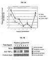
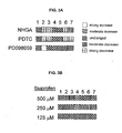


Classifications
-
- G—PHYSICS
- G01—MEASURING; TESTING
- G01N—INVESTIGATING OR ANALYSING MATERIALS BY DETERMINING THEIR CHEMICAL OR PHYSICAL PROPERTIES
- G01N33/00—Investigating or analysing materials by specific methods not covered by groups G01N1/00 - G01N31/00
- G01N33/48—Biological material, e.g. blood, urine; Haemocytometers
- G01N33/50—Chemical analysis of biological material, e.g. blood, urine; Testing involving biospecific ligand binding methods; Immunological testing
- G01N33/5005—Chemical analysis of biological material, e.g. blood, urine; Testing involving biospecific ligand binding methods; Immunological testing involving human or animal cells
- G01N33/5091—Chemical analysis of biological material, e.g. blood, urine; Testing involving biospecific ligand binding methods; Immunological testing involving human or animal cells for testing the pathological state of an organism
-
- G—PHYSICS
- G01—MEASURING; TESTING
- G01N—INVESTIGATING OR ANALYSING MATERIALS BY DETERMINING THEIR CHEMICAL OR PHYSICAL PROPERTIES
- G01N33/00—Investigating or analysing materials by specific methods not covered by groups G01N1/00 - G01N31/00
- G01N33/48—Biological material, e.g. blood, urine; Haemocytometers
- G01N33/50—Chemical analysis of biological material, e.g. blood, urine; Testing involving biospecific ligand binding methods; Immunological testing
- G01N33/5005—Chemical analysis of biological material, e.g. blood, urine; Testing involving biospecific ligand binding methods; Immunological testing involving human or animal cells
- G01N33/5008—Chemical analysis of biological material, e.g. blood, urine; Testing involving biospecific ligand binding methods; Immunological testing involving human or animal cells for testing or evaluating the effect of chemical or biological compounds, e.g. drugs, cosmetics
- G01N33/5011—Chemical analysis of biological material, e.g. blood, urine; Testing involving biospecific ligand binding methods; Immunological testing involving human or animal cells for testing or evaluating the effect of chemical or biological compounds, e.g. drugs, cosmetics for testing antineoplastic activity
-
- G—PHYSICS
- G01—MEASURING; TESTING
- G01N—INVESTIGATING OR ANALYSING MATERIALS BY DETERMINING THEIR CHEMICAL OR PHYSICAL PROPERTIES
- G01N33/00—Investigating or analysing materials by specific methods not covered by groups G01N1/00 - G01N31/00
- G01N33/48—Biological material, e.g. blood, urine; Haemocytometers
- G01N33/50—Chemical analysis of biological material, e.g. blood, urine; Testing involving biospecific ligand binding methods; Immunological testing
- G01N33/5005—Chemical analysis of biological material, e.g. blood, urine; Testing involving biospecific ligand binding methods; Immunological testing involving human or animal cells
- G01N33/5008—Chemical analysis of biological material, e.g. blood, urine; Testing involving biospecific ligand binding methods; Immunological testing involving human or animal cells for testing or evaluating the effect of chemical or biological compounds, e.g. drugs, cosmetics
- G01N33/502—Chemical analysis of biological material, e.g. blood, urine; Testing involving biospecific ligand binding methods; Immunological testing involving human or animal cells for testing or evaluating the effect of chemical or biological compounds, e.g. drugs, cosmetics for testing non-proliferative effects
- G01N33/5023—Chemical analysis of biological material, e.g. blood, urine; Testing involving biospecific ligand binding methods; Immunological testing involving human or animal cells for testing or evaluating the effect of chemical or biological compounds, e.g. drugs, cosmetics for testing non-proliferative effects on expression patterns
-
- G—PHYSICS
- G01—MEASURING; TESTING
- G01N—INVESTIGATING OR ANALYSING MATERIALS BY DETERMINING THEIR CHEMICAL OR PHYSICAL PROPERTIES
- G01N33/00—Investigating or analysing materials by specific methods not covered by groups G01N1/00 - G01N31/00
- G01N33/48—Biological material, e.g. blood, urine; Haemocytometers
- G01N33/50—Chemical analysis of biological material, e.g. blood, urine; Testing involving biospecific ligand binding methods; Immunological testing
- G01N33/5005—Chemical analysis of biological material, e.g. blood, urine; Testing involving biospecific ligand binding methods; Immunological testing involving human or animal cells
- G01N33/5008—Chemical analysis of biological material, e.g. blood, urine; Testing involving biospecific ligand binding methods; Immunological testing involving human or animal cells for testing or evaluating the effect of chemical or biological compounds, e.g. drugs, cosmetics
- G01N33/502—Chemical analysis of biological material, e.g. blood, urine; Testing involving biospecific ligand binding methods; Immunological testing involving human or animal cells for testing or evaluating the effect of chemical or biological compounds, e.g. drugs, cosmetics for testing non-proliferative effects
- G01N33/5041—Chemical analysis of biological material, e.g. blood, urine; Testing involving biospecific ligand binding methods; Immunological testing involving human or animal cells for testing or evaluating the effect of chemical or biological compounds, e.g. drugs, cosmetics for testing non-proliferative effects involving analysis of members of signalling pathways
-
- G—PHYSICS
- G01—MEASURING; TESTING
- G01N—INVESTIGATING OR ANALYSING MATERIALS BY DETERMINING THEIR CHEMICAL OR PHYSICAL PROPERTIES
- G01N33/00—Investigating or analysing materials by specific methods not covered by groups G01N1/00 - G01N31/00
- G01N33/48—Biological material, e.g. blood, urine; Haemocytometers
- G01N33/50—Chemical analysis of biological material, e.g. blood, urine; Testing involving biospecific ligand binding methods; Immunological testing
- G01N33/68—Chemical analysis of biological material, e.g. blood, urine; Testing involving biospecific ligand binding methods; Immunological testing involving proteins, peptides or amino acids
- G01N33/6863—Cytokines, i.e. immune system proteins modifying a biological response such as cell growth proliferation or differentiation, e.g. TNF, CNF, GM-CSF, lymphotoxin, MIF or their receptors
- G01N33/6866—Interferon
-
- G—PHYSICS
- G01—MEASURING; TESTING
- G01N—INVESTIGATING OR ANALYSING MATERIALS BY DETERMINING THEIR CHEMICAL OR PHYSICAL PROPERTIES
- G01N33/00—Investigating or analysing materials by specific methods not covered by groups G01N1/00 - G01N31/00
- G01N33/48—Biological material, e.g. blood, urine; Haemocytometers
- G01N33/50—Chemical analysis of biological material, e.g. blood, urine; Testing involving biospecific ligand binding methods; Immunological testing
- G01N33/68—Chemical analysis of biological material, e.g. blood, urine; Testing involving biospecific ligand binding methods; Immunological testing involving proteins, peptides or amino acids
- G01N33/6863—Cytokines, i.e. immune system proteins modifying a biological response such as cell growth proliferation or differentiation, e.g. TNF, CNF, GM-CSF, lymphotoxin, MIF or their receptors
- G01N33/6869—Interleukin
-
- G—PHYSICS
- G01—MEASURING; TESTING
- G01N—INVESTIGATING OR ANALYSING MATERIALS BY DETERMINING THEIR CHEMICAL OR PHYSICAL PROPERTIES
- G01N33/00—Investigating or analysing materials by specific methods not covered by groups G01N1/00 - G01N31/00
- G01N33/48—Biological material, e.g. blood, urine; Haemocytometers
- G01N33/50—Chemical analysis of biological material, e.g. blood, urine; Testing involving biospecific ligand binding methods; Immunological testing
- G01N33/68—Chemical analysis of biological material, e.g. blood, urine; Testing involving biospecific ligand binding methods; Immunological testing involving proteins, peptides or amino acids
- G01N33/6872—Intracellular protein regulatory factors and their receptors, e.g. including ion channels
-
- G—PHYSICS
- G16—INFORMATION AND COMMUNICATION TECHNOLOGY [ICT] SPECIALLY ADAPTED FOR SPECIFIC APPLICATION FIELDS
- G16B—BIOINFORMATICS, i.e. INFORMATION AND COMMUNICATION TECHNOLOGY [ICT] SPECIALLY ADAPTED FOR GENETIC OR PROTEIN-RELATED DATA PROCESSING IN COMPUTATIONAL MOLECULAR BIOLOGY
- G16B40/00—ICT specially adapted for biostatistics; ICT specially adapted for bioinformatics-related machine learning or data mining, e.g. knowledge discovery or pattern finding
- G16B40/30—Unsupervised data analysis
-
- G—PHYSICS
- G16—INFORMATION AND COMMUNICATION TECHNOLOGY [ICT] SPECIALLY ADAPTED FOR SPECIFIC APPLICATION FIELDS
- G16B—BIOINFORMATICS, i.e. INFORMATION AND COMMUNICATION TECHNOLOGY [ICT] SPECIALLY ADAPTED FOR GENETIC OR PROTEIN-RELATED DATA PROCESSING IN COMPUTATIONAL MOLECULAR BIOLOGY
- G16B45/00—ICT specially adapted for bioinformatics-related data visualisation, e.g. displaying of maps or networks
-
- G—PHYSICS
- G16—INFORMATION AND COMMUNICATION TECHNOLOGY [ICT] SPECIALLY ADAPTED FOR SPECIFIC APPLICATION FIELDS
- G16B—BIOINFORMATICS, i.e. INFORMATION AND COMMUNICATION TECHNOLOGY [ICT] SPECIALLY ADAPTED FOR GENETIC OR PROTEIN-RELATED DATA PROCESSING IN COMPUTATIONAL MOLECULAR BIOLOGY
- G16B5/00—ICT specially adapted for modelling or simulations in systems biology, e.g. gene-regulatory networks, protein interaction networks or metabolic networks
Definitions
- cancers In disease, as in health, there is a complex and changing cast of cells playing different roles. Functional capabilities of these cells can be altered, depending on the course of disease; as a result of underlying genetic differences; or due to drug exposure or other treatments. Even cancers, sometimes characterized as simple overgrowths of a single cell type, frequently show progression from one cell type to another. For example, in cancers of the breast and prostate there is a clear distinction between the steroid dependent and steroid independent cells, where the latter can emerge from the course of drug treatments. Similarly, the use of chemotherapeutics can select for resistant tumor cells, which are then able to persist through treatment. In other diseases, such as degenerative diseases, the loss of specific cell types is observed. For example, a key indicator of the severity of diabetes is the number of functioning islet cells that remain.
- Inflammatory diseases are characterized by the presence of a variety of leukocytes, including B cells, T cells, polymorphonuclear cells (eosinophils, basophils and neutrophils), macrophages, natural killer cells, megakaryocytes, and the like.
- leukocytes including B cells, T cells, polymorphonuclear cells (eosinophils, basophils and neutrophils), macrophages, natural killer cells, megakaryocytes, and the like.
- a Th1 type T cells and a Th2 type T cells can have opposite effects on the course of a disease; and genetic and environmental effects can determine the onset and course of T cell-mediated diseases.
- Angiogenesis is a process critical to both tumor growth and metastasis, and can be characterized by the presence of functionally distinct endothelial cells, which can vary in their responsiveness to cytokines and other growth and regulatory factors.
- angiogenesis is a continuous process, different consecutive steps can be identified, including release of pro-angiogenic factors and proteolytic enzymes, and endothelial cell migration, morphogenesis and proliferation.
- the microvasculature is maintained in a quiescent state.
- the acquisition of the angiogenic phenotype depends on the outcome of stimulatory and inhibitory regulation by the tumur and its microenvironment, features which are modified by genetic differences.
- the present invention satisfies this need and provides additional advantages.
- Cell based assays include a variety of methods to measure metabolic activities of cells including: uptake of tagged molecules or metabolic precursors, receptor binding methods, incorporation of tritiated thymidine as a measure of cellular proliferation, uptake of protein or lipid biosynthesis precursors, the binding of radiolabeled or otherwise labeled ligands; assays to measure calcium flux, and a variety of techniques to measure the expression of specific genes or their gene products.
- Methods utilizing genetic sequence microarrays allow the detection of changes in expression patterns in response to stimulus.
- a few examples include U.S. Patent no. 6,013,437 ; Luria et al ., "Method for identifying translationally regulated genes"; U.S. patent no. 6,004,755 , Wang, “Quantitative microarray hybridization assays”; and U.S. patent no. 5,994,076 , Chenchik et al ., “Methods of assaying differential expression”.
- U.S. Patent no. 6,146,830 Friend et al. "Method for determining the presence of a number of primary targets of a drug”.
- Proteomics techniques have potential for application to pharmaceutical drug screening. These methods require technically complex analysis and comparison of high resolution two-dimensional gels or other separation methods, often followed by mass spectrometry (for reviews see Hatzimanikatis et a/. (1999) Biotechnol Prog 15(3):312-8 ; Blackstock et al. (1999) Trends Biotechnol 17(3):121-7 . A discussion of the uses of proteomics in drug discovery may be found in Mullner et al. (1998) Arzneiffenbachforschung 48(1):93-5 .
- WO 01/51664 describes that the pattern of expression of clusters of genes is correlated to a cancerous state and provides a method for diagnosing a neoplasm in a subject by measuring a subject profile of tumour-associated genes and comparing the subject profile with a reference profile of tumour-associated genes.
- the present invention relates to methods for the classification of clinical samples, e.g. patient tissue samples, according to the physiological status of cells or constituents present in the sample.
- the information thus derived is useful In prognosis and diagnosis, and can further be used develop surrogate markers for disease states, and to investigate the effect of genetic polymorphisms in the responsiveness and state of cells involved in disease.
- the present invention thus provides a method of preparing a biomap dataset for the classification of a patient sample according to pathways associated with a disease state, the method comprising: contacting a patient sample comprising fluids or extracts of tissues in a test culture comprising non-patient indicator cells with a plurality of factors In an amount and incubating for a time sufficient to induce a plurality of pathways active In said cell culture; measuring at least four parameters associated with said plurality of pathways and comparing the measurement of said at least four parameters with the measurement from a control cell culture; and recording said measurements of said at least four parameters to produce a biomap dataset, wherein said biomap dataset is indicative of the pathways that are active in said cell culture.
- such cells are classified according to their ability to respond to therapeutic agents and treatments. In other embodiments, the cells are classified according to their status with respect to the activity of pathways of interest.
- patient tissue samples are evaluated for the presence of biologically active molecules, e.g. secreted factors and the like, by adding the patient sample to a cell culture responsive to the molecules.
- Patient samples are cultured in a panel of environments, where each environment can comprise combinations of factors, cells and therapeutically active agents. Generally at least one environment contains multiple factors that affect pathways of interest. The effect of altering the culture environment is assessed by monitoring multiple output parameters. The cells may also be treated with therapeutic agents in the presence or absence of factors, and the profile of output parameters determined. A sufficient number of markers are selected to provide a high confidence level that the pathways of interest are being monitored. When factors are employed, a sufficient number of factors are used to involve one or a plurality of pathways and a sufficient number of markers are determined to insure the cellular status is accurately being monitored.
- Methods are provided for the classification of clinical samples, e.g . patient tissue samples, cells, fluids, extracts of tissues, etc., according to the physiological status of cells present in the sample.
- the information thus derived is useful in prognosis and diagnosis, including susceptibility to disease(s), status of a diseased state and response to changes in the environment, such as the passage of time, treatment with drugs or other modalities.
- the state of the cells provided in the clinical sample may be classified according to the activation of pathways of interest, for example T cells can be classified as Th1, Th2, or Th3 type cells.
- the cells can also be classified as to their ability to respond to therapeutic agents and treatments.
- the sample is added to a culture of potentially responsive cells, where the response of such cells is then monitored.
- Changes in parameters in response to therapeutic agents provides information that is informative of a patient's ability to respond to a drug in the context of a physiologically relevant microenvironment. Changes in parameters in response to therapeutic agents can be correlated with databases of BioMAPs for classification or to BioMAPs from control samples. In addition to classification, BioMAPs derived from clinical samples and therapeutic agents can be used to compare drugs that act on different pathways is a physiologically relevant environment.
- the clinical samples can be further characterized by genetic analysis, proteomics, cell surface staining, or other means, in order to determine the presence of markers that are useful in classification.
- genetic polymorphisms such as single nucleotide polymorphisms or microsatellite repeats, can be causative of disease susceptibility or drug responsiveness, or can be linked to such phenotypes.
- Analysis of the genotype of a cell can be correlated to a BioMAP classification, and the information used in the development of genetic markers.
- markers as expression of cell surface proteins, presence of lipids, carbohydrates, etc. can be directly or indirectly associated with disease associated phenotypes, and with responsiveness to therapeutic agents and treatments.
- the invention is also useful for screening compounds for drug interactions.
- Drug interactions can be problematic in cancer therapy. For example, while steroids control the edema that occurs with glioma, they also interfere with chemotherapy efficacy.
- Cytotoxic drugs form the basis of many cancer therapies, thus interference with chemotherapy efficacy may offset any anti-tumor effects of angiogenesis inhibitors.
- Most cytotoxic drugs effect both normal and neoplastic cells, although at different concentrations, therefore, screening compounds in the presence of cytotoxic drugs can be performed and reveal unexpected interference or beneficial synergies. Interactions between a cytotoxic drug and any test compound is detected by the observation of BioMAPs obtained in the presence of both drugs that are inconsistent with additive effects.
- Patient samples are cultured in a panel of environments, where each environment can comprise combinations of factors, cells and therapeutically active agents. Generally at least one environment contains multiple factors that affect pathways of interest. The effect of altering the culture environment is assessed by monitoring multiple output parameters. The cells may also be treated with therapeutic agents in the presence or absence of factors, and the profile of output parameters determined. A sufficient number of markers are selected to provide a high confidence level that the pathways of interest are being monitored. When factors are employed, a sufficient number of factors are used to involve one or a plurality of pathways and a sufficient number of markers are determined to insure the cellular status is accurately being monitored.
- test cells For convenience, a clinical sample comprising cells may be referred to as "test cells", and will comprise one or more types of cells present in a clinical sample.
- a tissue sample may include endothelial cells, a variety of lymphocytes and other hematopoietic cells, tumor cells which may be clonal or polyclonal in origin, and the like.
- the test cells need not be directly involved In a disease of interest.
- a clinical sample may also be evaluated for the presence of biologically active factors and other molecules, where such non-cellular material is indicative of the physiological state of the tissue from which it is obtained.
- Such samples are evaluated for their effect on one or a panel of cells, as described in co-pending patent application no. 09/800,605 (published as U.S. Patent 6,656,695 ).
- test cells refers to such cultures, where test cells are contacted with medium and multiple combination of factors, agents and other culture variations. These cell cultures are created by the addition of a sufficient number of different factors to provoke a response that simulates cellular physiology of a state of interest, and to allow for the status of cells in culture to be determined in relation to a change in an environment.
- the state of interest will normally Involve a plurality of pathways where the pathways regulate a plurality of parameters or markers identifying a phenotype associated with the state of interest.
- one or more assay combinations are provided that simulate physiological cell states of interest, particularly physiological cell states in vivo, usually using the same type of cells or combinations of cells.
- Such a simulation will usually include at least three different regulated features (parameters) shared with in vivo cell counterparts in normal or diseased states.
- the simulation will include a cell culture system that allows discrimination of modifications in at least three different signaling pathways or cell functions operative in vivo under conditions of interest.
- a phenotype of the test cells that is useful for monitoring output parameters can be generated by including a plurality of factors, and optionally additional cells, e.g . stromal cells, endothelial cells, fibroblasts, etc ., that may interact with the patient tissue.
- the factors and cells inducing pathways induce a response in the test cells in vitro.
- factors are naturally occurring compounds, e.g . known compounds that have surface membrane receptors and induce a cellular signal that results in a modified phenotype; or synthetic compounds that mimic such naturally occurring factors.
- factors will act intracellularly by passing through the cell surface membrane and entering the cytosol with binding to components in the cytosol, nucleus or other organelle. In referring to factors, it is understood that it is the activities of the factors that are of interest and not necessarily a particular naturally occurring factor itself.
- markers For each test cell there are a number of markers that can be measured, which relate to specific pathways associated with the cell type and condition. As described in co-pending U.S. Patent application no. 09/800,605 (published as U.S. Patent 6,656,695 ), at least about 4 markers are identified that allow for evaluating the up or down regulation of at least 2 pathways, generally three or more pathways, where the total number of markers will usually not exceed 8.
- the markers are selected to provide a robust picture of the status of the cell, due to its condition, e.g . pro-inflammatory, immunosuppressive, neoplastic, etc ., its response to therapy, its response to a drug, or the like.
- Each set of markers will define a set of cell pathways and their response. However, there will normally be at least 2, usually at least 3, common markers for the particular determination.
- the nature and number of parameters measured generally reflects the response of a plurality of pathways.
- the subject approach provides for robust results having enhanced predictability in relation to the status of the test cells.
- the results may be compared to the basal condition, tissue matched normal controls, and/or the condition in the presence of one or more of the factors, particularly in comparison to all of the factors used in the presence and absence of agent.
- the effects of different environments are conveniently provided in BioMAPs, where the results can be mathematically compared.
- a BioMAP is prepared from values obtained by measuring parameters or markers of the test cells in the presence and absence of different factors, and/or by comparing the presence of an agent of interest and at least one other state, usually the control state, which may include the state without agent or with a different agent.
- Parameters include cellular products or epitopes thereof, as well as functional states, whose levels vary in the presence of the factors.
- the results are normalized against a standard, usually a "control value or state,” to provide a normalized data set. Values obtained from test conditions can be normalized by subtracting the unstimulated control values from the test values, and dividing the corrected test value by the corrected stimulated control value.
- BioMAP may comprise normalized data from one, two or multiple cell types and assay conditions.
- BioMAP By referring to a BioMAP is intended that the dataset will comprise values of the levels of at least two sets of parameters obtained under different assay combinations.
- the BioMAP may also include the parameter values for each the factors included in the assay combination, individually and/or together with fewer than the entire assay combination.
- the parameter values are usually created electronically and stored in a data processor for comparison with other BioMAPs and databases compiled from the BioMAPs.
- a graph of a BioMAP can be presented visually as numerical values, symbols, color gradations, or the like, indicating the parameter values.
- the graph is conveniently presented where color and/or design provide an indication of the level of the particular marker.
- the indicators may be vertical or horizontal as to the individual markers and the assay combinations, so that by looking at the graph, one can immediately compare the levels of the different markers for each of the combinations and discern patterns related to the assay combinations and the differences between assay combinations. In this way, one can rapidly relate different candidate pharmacologic agents, the pathways they affect and their efficacy in modulating the individual pathways.
- a BioMAP can be annotated to indicate information about the sources of information for the dataset.
- Annotations may include, for example, the number of assay conditions in a panel (n); controls used for normalization (N); parameters (P), which may be designated for the number and identity of the parameters; environmental changes, such as the addition of factors and/or agents or a change in the physical conditions (V); cell type (C); and the like.
- the annotation may further specify specific factors or conditions present in one of the assay combinations, e.g . n1, n2, n3, etc., where the presence of factors in the assay combination is designated (F), temperature may be designated (T), pH, etc .
- the annotation may be set forth as: (v) B ⁇ n; N; P; C; F ⁇ .
- a database of BioMAPs can be compiled from sets of experiments, for example, a database can contain BioMAPs obtained from clinical samples such as sites of inflammation, tumors, etc ., each in a panel of assay combinations, with multiple different environmental changes, where each change can be a series of related compounds, or compounds representing different classes of molecules.
- a database comprises BioMAPs from one compound, with multiple different cell panels.
- BioMAPs in the database can be analyzed by pattern recognition algorithms or clustering methods (e.g. hierarchical or k-means clustering, etc.) that use statistical analysis (correlation coefficients, etc.) to quantify relatedness of BioMAPs.
- pattern recognition algorithms or clustering methods e.g. hierarchical or k-means clustering, etc.
- statistical analysis correlation coefficients, etc.
- These methods can be modified (by weighting, employing classification strategies, etc.) to optimize the ability of a BioMAP to discriminate different functional effects.
- individual parameters can be given more or less weight when analyzing the dataset of the BioMAP, in order to enhance the discriminatory ability of the BioMAP. The effect of altering the weights assigned each parameter is assessed, and an iterative process is used to optimize pathway or cellular function discrimination.
- the literature has sufficient information to establish assay combinations to provide a useful BioMAP. Where the information is not available, by using the procedures described in the literature for identifying markers for diseases, microarrays for RNA transcription comparisons, proteomic or immunologic comparisons, between normal cells and cells in the disease state, one can ascertain the endogenous factors associated with the disease state and the markers that are produced by the cells associated with the disease state.
- Biomap analysis can be used to optimize cell culture conditions. Additional markers can be deduced and added as a marker to the map. The greater the number of individual markers that vary independently of each other, the more robust the BioMAP.
- the parameters of the BioMAP can be optimized by obtaining BioMAP parameters within an assay combination or panel of assay combinations using different sets of readout, and using pattern recognition algorithms and statistical analyses to compare and contrast different BioMAPs of different parameter sets. Parameters are selected that provide a BioMAP that discriminates between changes in the environment of the cell culture known to have different modes of action, i.e . the BioMAP is similar for agents with a common mode of action, and different for agents with a different mode of action.
- the optimization process allows the identification and selection of a minimal set of parameters, each of which provides a robust readout, and that together provide a BioMAP that enables discrimination of different modes of action of stimuli or agents.
- the iterative process focuses on optimizing the assay combinations and readout parameters to maximize efficiency and the number of signaling pathways and/or functionally different cell states produced in the assay configurations that can be identified and distinguished, while at the same time minimizing the number of parameters or assay combinations required for such discrimination.
- Clinical samples for use in the methods of the invention may be obtained from a variety of sources, including blood, lymph, cerebrospinal fluid, synovial fluid, tissue biopsies, skin, saliva, lavage, and the like.
- Such samples can comprise complex populations of cells, which can be assayed as a population, or separated into subpopulations, and can also comprise acellular samples.
- Such cellular and acellular sample can be separated by centrifugation, elutriation, density gradient separation, apheresis, affinity selection, panning, FACS, centrifugation with Hypaque, etc .
- Acellular samples can be separated according to immunologic or biochemical criteria, for example by various electrophoretic and chromatographic means, as is known in the art.
- a sample Once a sample is obtained, it can be used directly, frozen, or maintained in appropriate culture medium for short periods of time.
- Various media can be employed to maintain cells during the classification process. There are established protocols for the culture of diverse cell types that reflect their in vivo counterparts. Protocols may require the use of special conditions and selective media to enable cell growth or expression of specialized cellular functions.
- the methods of the invention find use in a wide variety of animal species, including mammalian species.
- Animal models, particularly small mammals, e.g. murine, lagomorpha, etc . are of interest for experimental investigations.
- Humans are of particular interest for both diagnostic and prognostic applications of the method.
- the samples may come from any organ or compartment of the body, to the extent the cells can be obtained by any convenient procedure, such as the drawing of blood, venipuncture, biopsy, or the like.
- Cell types that can find use in the subject invention, include endothelial cells, muscle cells, myocardial, smooth and skeletal muscle cells, mesenchymal cells, epithelial cells; hematopoietic cells, such as lymphocytes, including T-cells, such as Th1 T cells, Th2 T cells, Th0 T cells, cytotoxic T cells; B cells, pre- B cells, etc .; monocytes; dendritic cells; neutrophils; and macrophages; natural killer cells; mast cells;, etc.; adipocytes, cells involved with particular organs, such as thymus, endocrine glands, pancreas, brain, such as neurons, glia, astrocytes, dendrocytes, etc .
- Hematopoietic cells will be associated with inflammatory processes, autoimmune diseases, etc ., endothelial cells, smooth muscle cells, myocardial cells, etc . may be associated with cardiovascular diseases; almost any type of cell may be associated with neoplasias, such as sarcomas, carcinomas and lymphomas; liver diseases with hepatic cells; kidney diseases with kidney cells; etc .
- a sample will comprise at least about 10 2 cells, more usually at least about 10 3 cells, and preferable 10 4 or more cells.
- environment encompasses cells, media, factors, time and temperature. Environments may also include drugs and other compounds, particular atmospheric conditions, pH, salt composition, minerals, etc . The conditions will be controlled and the BioMAP will reflect the similarities and differences between each of the assay combinations involving a different environment or culture condition.
- Culture of cells is typically performed in a sterile environment, for example, at 37°C in an incubator containing a humidified 92-95% air/5-8% CO 2 atmosphere.
- Cell culture may be carried out in nutrient mixtures containing undefined biological fluids such as fetal calf serum, or media which is fully defined and serum free.
- Some preferred environments include environments that discriminate or emphasize cell or tissue states associated with pathology in one or more diseases, for example, Th1 versus Th2 polarization of effector T cells; prothrombotic; inflammatory (e.g. NF ⁇ B, upregulated TNF- ⁇ cytokine production, downregulated IL-10, TGF ⁇ , etc .; dysregulated proliferation (neoplasia); angiogenesis; etc.) Environments that facilitate discrimination of specific signaling pathways implicated in disease states are also of interest, e.g . NF ⁇ B, classic Th1 or Th2 induction environments, etc .
- a assay combinations are used in classifying and investigating complex states of cells, frequently resulting from cellular interactions, which may frequently involve at least about two, frequently three, or more different cell types and/or will involve a plurality of soluble factors that are present in a physiological fluid, particularly as the result of a physiological event, e.g. infection, neoplasia, autoimmune, etc. that is, frequently involving more than one cell type and more than one factor.
- the measured parameters may be obtained from one or more of the cell types.
- the cells in the assay combination either one or up to each of the different cell types, can have identifying characteristics allowing them to be distinguished during analysis. Various techniques may be employed to identify the cells in the assay combination for analysis of the parameters of interest.
- Conditions of interest include inflammatory processes that occur in response to infection, trauma, etc., autoimmune diseases, such as diabetes, lupus, arthritis, etc., cardiovascular diseases, such as stroke, atherosclerosis, etc., neoplasia, hyperplasia, addiction, infection, obesity, cellular degeneration, apoptosis, senescence, differentiation, and the like.
- Multifactorial usually involving multicellular, assay combinations, may reflect many of the conditions indicated above, such as inflammatory processes; autoimmune diseases; cardiovascular diseases; tumors, etc. That is, a multiplicity of factors are employed to influence a plurality of cellular pathways and a multiplicity of parameters are measured that reflect the status of the pathways.
- Degenerative diseases including affected tissues and surrounding areas, may be exploited to determine both the response of the affected tissue, and the interactions with other cell types or other parts of the body.
- Factors added to the cultures can be the products of other cell types, for example, expressed proteins associated with a disease, can be compounds that simulate naturally occurring factors, can be surface membrane proteins free of the membrane or as part of microsomes, or other reagent that induces the appropriate pathway to aid in the simulation of the phenotype or provides the appropriate environment to simulate the physiological condition.
- Factors can be added individually or in combination, from feeder cells, may be added as a bolus or continuously, where the factor is degraded by the culture, etc .
- Illustrative naturally occurring factors include cytokines, chemokines, and other factors, e.g . growth factors, such factors include GM-CSF, G-CSF, M-CSF, TGF, FGF, EGF, TNF-- ⁇ , GH, corticotropin, melanotropin, ACTH, etc ., extracellular matrix components, surface membrane proteins, such as integrins and adhesins, and other components that are expressed by the targeted cells or their surrounding milieu in vivo, etc ., that may be isolated from natural sources or produced by recombinant technology or synthesis, compounds that mimic the action of other compounds or cell types, e.g.
- an antibody which acts like a factor or mimics a factor such as synthetic drugs that act as ligands for target receptors.
- synthetic drugs that act as ligands for target receptors.
- T cell receptor the action of an oligopeptide processed from an antigen and presented by an antigen-presenting cell, etc. can be employed.
- a family of related factors are referred to with a single designation, e.g . IL-1, VEGF, IFN, etc ., in referring to the single description, any one or some or all of the members of the group are intended, where the literature will be aware of how the factors are to be used in the context of the assay combination.
- Components may also include soluble or immobilized recombinant or purified receptors, or antibodies against receptors or ligand mimetics.
- Cancer cells may be cultured with different factors based on the different cells in the environment of the tumor, as well as other factors in the blood induced by factors secreted by the neoplastic cells. Many of these factors will be the same factors described above, but additional factors include factors associated with angiogenesis, such as angiogenin, angiopoietin-1, HGF, PDGF, TNF- ⁇ , VEGF, IL-1, IL-4, IL-6, IL-8 and fibronectin.
- angiogenesis such as angiogenin, angiopoietin-1, HGF, PDGF, TNF- ⁇ , VEGF, IL-1, IL-4, IL-6, IL-8 and fibronectin.
- the BioMAP dataset will comprise data from a panel of assay combinations.
- the panel will be related to the purpose of the BioMAP and may include not only the information that has been developed substantially concurrently with the study, but also information that has been previously developed under comparable conditions. Frequently a panel will be used that is comprised of at least one assay combination that provides for simulation of multiple pathways of interest, while other assay combinations in the panel are variants thereof.
- the number of combinations in a panel may vary with the particular use. For example, the minimum number of assay combinations will be two for a panel for initial screening that would comprise a single assay combination.
- a panel for characterizing the mechanism of action of an active compound will usually comprise a plurality of assay combinations, usually at least about 4, more usually at least 6, frequently at least about 10 and may be as many as 20 or more unique combinations.
- the panel comprises culture conditions where multiple specific changes are made to the culture environment, e.g . two or more changes, usually not more than about 6, more usually not more than about 4. Such changes are associated with the additional information that is engendered by the indicated variations.
- the variations can include the addition of known inhibitors of specific pathways.
- Parameters are quantifiable components of cells, particularly components that can be accurately measured, desirably in a high throughput system.
- a parameter can be any cell component or cell product including cell surface determinant, receptor, protein or conformational or posttranslational modification thereof, lipid, carbohydrate, organic or inorganic molecule, nucleic acid, e.g. mRNA, DNA, etc . or a portion derived from such a cell component or combinations thereof. While most parameters will provide a quantitative readout, in some instances a semi-quantitative or qualitative result will be acceptable. Readouts may include a single determined value, or may include mean, median value or the variance, etc .
- Characteristically a range of parameter readout values will be obtained for each parameter from a multiplicity of the same assay combinations, usually at least about 2 of the same assay combination will be performed to provide a value. Variability is expected and a range of values for each of the set of test parameters will be obtained using standard statistical methods with a common statistical method used to provide single values.
- Markers are selected to serve as parameters based on the following criteria, where any parameter need not have all of the criteria: the parameter is modulated in the physiological condition that one is simulating with the assay combination; the parameter is modulated by a factor that is available and known to modulate the parameter in vitro analogous to the manner it is modulated in vivo ; the parameter has a robust response that can be easily detected and differentiated and is not too sensitive to concentration variation, that is, it will not substantially differ in its response to an over two-fold change; the parameter is secreted or is a surface membrane protein or other readily measurable component; the parameter desirably requires not more than two factors to be produced; the parameter is not co-regulated with another parameter, so as to be redundant in the information provided; and in some instances, changes in the parameter are indicative of toxicity leading to cell death.
- the set of parameters selected is sufficiently large to allow distinction between reference patterns, while sufficiently selective to fulfill computational requirements.
- Biomaps will generally comprise only functionally relevant parameter information, although a static parameter may serve as an internal control.
- a typical BioMAP will comprise data from at least 3 functionally relevant parameters, more usually at least about 5 functionally relevant parameters, and may include 10 or more functionally relevant parameters, usually not more than about 30, more usually not more than about 20, parameters. In analyzing the data from the BioMAP, all of the parameters need not be weighed equally.
- Those parameters that are closely functionally associated with the disease state or pathophysiologic response, and/or with modulation of cell pathways of interest may be given greater weight in evaluating a candidate drug or a readout, as compared to other parameters that are suggestive, but do not have as strong an association.
- Parameters of interest include detection of cytoplasmic, cell surface or secreted biomolecules, frequently biopolymers, e.g. polypeptides, polysaccharides, polynucleotides, lipids, etc .
- Cell surface and secreted molecules are a preferred parameter type as these mediate cell communication and cell effector responses and can be more readily assayed.
- parameters include specific epitopes. Epitopes are frequently identified using specific monoclonal antibodies or receptor probes.
- the molecular entities comprising the epitope are from two or more substances and comprise a defined structure; examples include combinatorially determined epitopes associated with heterodimeric integrins.
- a parameter may be detection of a specifically modified protein or oligosaccharide, e.g . a phosphorylated protein, such as a STAT transcriptional protein; or sulfated oligosaccharide, or such as the carbohydrate structure Sialyl Lewis x, a selectin ligand.
- a specifically modified protein or oligosaccharide e.g . a phosphorylated protein, such as a STAT transcriptional protein; or sulfated oligosaccharide, or such as the carbohydrate structure Sialyl Lewis x, a selectin ligand.
- the presence of the active conformation of a receptor may comprise one parameter while an inactive conformation of a receptor may comprise another, e.g . the active and inactive forms of heterodimeric integrin ⁇ M ⁇ 2 or Mac-1.
- a parameter may be defined by a specific monoclonal antibody or a ligand or receptor binding determinant.
- Parameters may include the presence of cell surface molecules such as CD antigens (CD1-CD247), cell adhesion molecules including ⁇ 4 ⁇ 7 and other integrins, selectin ligands, such as CLA and Sialyl Lewis x, and extracellular matrix components.
- Parameters may also include the presence of secreted products such as lymphokines, including IL-2, IL-4, IL-6, growth factors, etc .
- these parameters may include IL-1R, IL-2R, IL4R, IL-12R ⁇ , CD45RO, CD49E, tissue selective adhesion molecules, homing receptors, chemokine receptors, CD26, CD27, CD30 and other activation antigens.
- Additional parameters that are modulated during activation include MHC class II; functional activation of integrins due to clustering and/or conformational changes; T cell proliferation and cytokine production, including chemokine production.
- MHC class II MHC class II
- T cell proliferation and cytokine production including chemokine production.
- cytokine production including chemokine production.
- the best-characterized example being the production of IL-4 by Th2 cells, and interferon- ⁇ by Th1 T cells.
- cytokine production patterns in vivo is a powerful means of modulating pathologic immune responses, for example in models of EAE, diabetes, inflammatory bowel disease, etc .
- the expression of secreted cytokines may be a preferred class of parameters, detectable, for example, by ELISA analysis of the supernatants, etc .
- a patient is responsive to a particular therapeutic agent or regimen.
- efficacy of such therapies in a biologically relevant context, i.e. in the presence of factors and interacting cells. Included are pharmacologically active drugs, genetically active molecules, etc .
- Compounds of interest include chemotherapeutic agents, anti-inflammatory agents, hormones or hormone antagonists, ion channel modifiers, and neuroactive agents.
- Exemplary of pharmaceutical agents suitable for this invention are those described in, " The Pharmacological Basis of Therapeutics," Goodman and Gilman, McGraw-Hill, New York, New York, (1996), Ninth editi on, under the sections: Drugs Acting at Synaptic and Neuroeffector Junctional Sites; Drugs Acting on the Central Nervous System; Autacoids: Drug Therapy of Inflammation; Water, Salts and Ions; Drugs Affecting Renal Function and Electrolyte Metabolism; Cardiovascular Drugs; Drugs Affecting Gastrointestinal Function; Drugs Affecting Uterine Motility; Chemotherapy of Parasitic Infections; Chemotherapy of Microbial Diseases; Chemotherapy of Neoplastic Diseases; Drugs Used for Immunosuppression; Drugs Acting on Blood-Forming organs; Hormones and Hormone Antagonists; Vitamins, Dermatology; and Toxicology.
- Cells and samples are classified by adding a therapeutic agent or treatment; and/or by culturing cells in combinations of factors, in at least one and usually a plurality of assay combinations to form a panel of assay combinations, usually in conjunction with positive and negative controls.
- the change in parameter readout in response to an agent, sample or factors is measured, desirably normalized, and the resulting BioMAP may then be evaluated by comparison to reference BioMAPs.
- the reference BioMAPs may include basal readouts In the presence and absence of the factors, BioMAPs obtained with other agents, which may or may not include known inhibitors of known pathways, etc.
- Agents of interest for analysis include any biologically active molecule with the capability of modulating, directly or indirectly, the phenotype of interest of a cell of interest.
- the agents are conveniently added in solution, or readily soluble form, to the medium of cells in culture.
- the agents may be added in a flow-through system, as a stream, intermittent or continuous, or alternatively, adding a bolus of the compound, singly or incrementally, to an otherwise static solution.
- a flow-through system two fluids are used, where one is a physiologically neutral solution, and the other is the same solution with the test compound added. The first fluid is passed over the cells, followed by the second.
- a bolus of the test compound is added to the volume of medium surrounding the cells. The overall concentrations of the components of the culture medium should not change significantly with the addition of the bolus, or between the two solutions in a flow through method.
- Preferred agent formulations do not include additional components, such as preservatives, that may have a significant effect on the overall formulation.
- preferred formulations consist essentially of a biologically active compound and a physiologically acceptable carrier, e.g . water, ethanol, DMSO, etc .
- a physiologically acceptable carrier e.g . water, ethanol, DMSO, etc .
- the formulation may consist essentially of the compound itself.
- a plurality of assays may be run in parallel with different agent concentrations to obtain a differential response to the various concentrations.
- determining the effective concentration of an agent typically uses a range of concentrations resulting from 1:10, or other log scale, dilutions.
- the concentrations may be further refined with a second series of dilutions, if necessary.
- one of these concentrations serves as a negative control, i.e . at zero concentration or below the level of detection of the agent or at or below the concentration of agent that does not give a detectable change in the phenotype.
- a convenient method is to label a molecule with a detectable moiety, which may be fluorescent, luminescent, radioactive, enzymatically active, etc ., particularly a molecule specific for binding to the parameter with high affinity Fluorescent moieties are readily available for labeling virtually any biomolecule, structure, or cell type. Immunofluorescent moieties can be directed to bind not only to specific proteins but also specific conformations, cleavage products, or site modifications like phosphorylation. Individual peptides and proteins can be engineered to autofluoresce, e.g .
- antibodies can be genetically modified to provide a fluorescent dye as part of their structure
- quantum dots have been covalently coupled to biomolecules for use in ultrasensitive biological detection ( Stupp et al. (1997) Science 277(5330):1242-8 ; Chan et al. (1998) Science 281(5385):2016-8 ).
- quantum dot nanocrystals have a narrow, tunable, symmetric emission spectrum and are photochemically stable ( Bonadeo et al. (1998) Science 282(5393):1473-6 ).
- the advantage of quantum dots is the potential for exponentially large numbers of independent readouts from a single source or sample.
- Fluorescence photobleaching recovery FPR
- parameters may be measured using other than fluorescent labels, using such immunoassay techniques as radioimmunoassay (RIA) or enzyme linked immunosorbance assay (ELISA), homogeneous enzyme immunoassays, and related non-enzymatic techniques.
- RIA radioimmunoassay
- ELISA enzyme linked immunosorbance assay
- These techniques utilize specific antibodies as reporter molecules, which are particularly useful due to their high degree of specificity for attaching to a single molecular target.
- U.S. Pat. No. 4,568,649 describes ligand detection systems, which employ scintillation counting. These techniques are particularly useful for protein or modified protein parameters or epitopes, or carbohydrate determinants. Cell readouts for proteins and other cell determinants can be obtained using fluorescent or otherwise tagged reporter molecules.
- Cell based ELISA or related non-enzymatic or fluorescence-based methods enable measurement of cell surface parameters and secreted parameters.
- Capture ELISA and related non-enzymatic methods usually employ two specific antibodies or reporter molecules and are useful for measuring parameters in solution.
- Flow cytometry methods are useful for measuring cell surface and intracellular parameters, as well as shape change and granularity and for analyses of beads used as antibody- or probe-linked reagents. Readouts from such assays may be the mean fluorescence associated with individual fluorescent antibody-detected cell surface molecules or cytokines, or the average fluorescence intensity, the median fluorescence intensity, the variance in fluorescence intensity, or some relationship among these.
- Luminex beads or other fluorescent beads, or beads varying in light scattering parameters can be conjugated to antibodies to cytokines or other parameters, or conjugated to protein receptors for parameters.
- the conjugated beads are added to the cells, cell lysate, or to the removed supernatant, allowing bead binding to target parameters.
- fluorescent antibody to a distinct epitope of the target parameter is used to measure the level of target parameter bound.
- the fluorescence and light scatter characteristics of the beads constitute an identifier of the target parameter, and fluorescence derived from added antibody to the target parameter is an indication of the quantity of target parameter bound, and hence a readout of the individual parameter.
- Flow cytometry may be used to quantitate parameters such as the presence of cell surface proteins or conformational or posttranslational modification thereof; intracellular or secreted protein, where permeabilization allows antibody (or probe) access, and the like.
- Brefeldin A is commonly utilized to prevent secretion of intracellular substances.
- Flow cytometry methods are known in the art, and described in the following: Flow Cytometry and Cell Storing (Springer Lab Manual), Radbruch, Ed., Springer Verlag, 2000 ; Ormerod, Flow Cytometry, Springer Verlag, 1999 ; Flow Cytometry Protocols (Methods in Molecular Biology , No 91), Jaroszeski and Heller, Eds., Humana Press, 1998 ; Current Protocols in Cytometry, Robinson et al., eds, John Wiley & Sons, New York, NY, 2000 .
- the readouts of selected parameters are capable of being read simultaneously, or in sequence during a single analysis; as for example through the use of fluorescent antibodies to cell surface molecules.
- these can be tagged with different fluorochromes, fluorescent bead, tags, e . g . quantum dots, etc., allowing analysis of up to 4 or more fluorescent colors simultaneously by flow cytometry.
- Plug-flow flow cytometry that has the potential to automate the delivery of small samples from unpressurized sources at rates compatible with many screening and assay applications, may allow higher throughput, compatible with high throughput screening, Edwards et al. (1999) Cytometry 37:156-9 .
- nucleic acids especially messenger RNAs
- the quantitation of nucleic acids is also of interest as a parameter. These can be measured by hybridization techniques that depend on the sequence of nucleic acid nucleotides. Techniques include polymerase chain reaction methods as well as gene array techniques. See Current Protocols in Molecular Biology, Ausubel et al., eds, John Wiley & Sons, New York, NY, 2000 ; Freeman et al. (1999) Biotechniques 26(1):112-225 ; Kawamoto et al. (1999) Genome Res 9(12):1305-12 ; and Chen et al. (1998) Genomics 51(3):313-24 , for examples.
- Identifiers of individual cells may be fluorescent, as for example labeling of different unit cell types with different levels of a fluorescent compound, and the like. If two cell types are to be mixed, one may be labeled and the other not. If three or more are to be included, each may be labeled to different levels of fluorescence by incubation with different concentrations of a labeling compound, or for different times.
- a matrix of fluorescence labeling intensities of two or more different fluorescent colors may be used, such that the number of distinct unit cell types that are identified is a number of fluorescent levels of one color, e.g ., carboxyfluorescein succinimidyl ester (CFSE), times the number of fluorescence levels employed of the second color, e.g . tetramethylrhodamine isothiocyanate (TRITC), or the like, times the number of levels of a third color, etc .
- CFSE carboxyfluorescein succinimidyl ester
- TRITC tetramethylrhodamine isothiocyanate
- intrinsic light scattering properties of the different cell types, or characteristics of the BioMAPs of the test parameters included in the analysis can be used in addition to or in place of fluorescent labels as unit cell type identifiers.
- BioMAP BioMAP obtained from test cells
- a reference BioMAP(s) a BioMAP obtained from test cells
- the BioMAP is compared with a database of reference BioMAPs. Similarity to reference BioMAPs from normal cells, cells from similarly diseased tissue, from cell lines with responses induced by assay combinations involving known pathway stimuli or inhibitors, and the like, can provide an initial indication of the cellular pathways activated in the test cells, and the responsiveness of the test cells to a therapeutic regimen.
- a database of reference BioMAPs can be compiled. These databases may include reference BioMAPs from panels that include known agents or combinations of agents that target specific pathways, as well as references from the analysis of cells treated under environmental conditions in which single or multiple environmental conditions or parameters are removed or specifically altered. Reference BioMAPs may also be generated from panels containing cells with genetic constructs that selectively target or modulate specific cellular pathways. In this way, a database is developed that can reveal the contributions of individual pathways to a complex response.
- the effectiveness of pattern search algorithms in classifying BioMAPs can involve the optimization of the number of parameters and assay combinations.
- the disclosed techniques for selection of parameters provide for computational requirements resulting in physiologically relevant outputs.
- these techniques for pre-filtering data sets (or potential data sets) using cell activity and disease-relevant biological information improve the likelihood that the outputs returned from database searches will be relevant to predicting agent mechanisms and in vivo agent effects.
- a data matrix is generated, where each point of the data matrix corresponds to a readout from a parameter, where data for each parameter may come from replicate determinations, e.g . multiple individual cells of the same type.
- a data point may be quantitative, semi-quantitative, or qualitative, depending on the nature of the parameter.
- the readout may be a mean, average, median or the variance or other statistically or mathematically derived value associated with the measurement.
- the parameter readout information may be further refined by direct comparison with the corresponding reference readout.
- the absolute values obtained for each parameter under identical conditions will display a variability that is inherent in live biological systems and also reflects individual cellular variability as well as the variability inherent between individuals.
- Classification rules are constructed from sets of training data (i.e. data matrices) obtained from multiple repeated experiments. Classification rules are selected as correctly identifying repeated reference patterns and successfully distinguishing distinct reference patterns. Classification rule-learning algorithms may include decision tree methods, statistical methods, naive Bayesian algorithms, and the like.
- a knowledge database will be of sufficient complexity to permit novel test BioMAPs to be effectively identified and classified.
- Several approaches for generating a sufficiently encompassing set of classification patterns, and sufficiently powerful mathematical/statistical methods for discriminating between them can accomplish this.
- a database can be compiled by preparing BioMAPs using different combinations of a plurality of biologically active factors, in conjunction with BioMAPs involving the use of known agents having known effects and/or the use of genetically modified cells, where the genetic modification affects one or more of the pathways affected by one or more of the factors used to create the phenotype.
- the culture conditions selected to produce a specific in vitro reference pattern contain four biologically active agents, in addition to those present in the basal conditions of the normal or basal environment, a BioMAP would be generated from a panel of cells treated under all possible combinations of the 4 agents (15 assay conditions), typically using constant concentrations in each of the combinations.
- the extent of the database associated with assay combinations to screen candidates for specific phenotypes, e.g. indications, will vary with the nature of the phenotype, the amount of information desired, the complexity of the system, and the like.
- the data from cells treated with specific drugs known to interact with particular targets or pathways provide a more detailed set of classification readouts.
- Data generated from cells that are genetically modified using over-expression techniques and anti-sense techniques, permit testing the influence of individual genes on the phenotype.
- agents may be analyzed in the absence of any factors or with a limited number of factors.
- the assay is performed as previously described and the values of the parameters can be compared to the BioMAP reflecting the values for the parameters of the physiologic state of interest, the values of the parameters for the response to one or more factors, and the basal response. In this way, the effect of the agent under physiological conditions can be evaluated. Similarly, one may have datasets compiled from combinations of agents to determine their effect wen combined on cell physiology.
- a preferred knowledge database contains reference BioMAPs comprising data from optimized panels of cells, environments and parameters.
- reference BioMAPs comprising data from optimized panels of cells, environments and parameters.
- data reflecting small variations in the environment may also be included in the knowledge database, e.g. environments where one or more factors or cell types of interest are excluded or included or quantitatively altered in, for example, concentration or time of exposure, etc.
- Cells from a clinical sample that are suspected of having a marker that is associated with or causative of disease can be analyzed for the presence of genetic polymorphisms or other markers. Genetic characterization analyzes DNA or RNA, from any source, e.g . skin, cheek scrapings, blood samples, etc . The nucleic acids are screened for the presence of a polymorphism of interest, e.g. SNPs, microsatellite markers, and the like.
- a polymorphism of interest e.g. SNPs, microsatellite markers, and the like.
- CGH Comparative Genomic Hybridization
- the region of interest is cloned into a suitable vector and grown in sufficient quantity for analysis.
- Cells that express genes of interest may be used as a source of mRNA, which may be assayed directly or reverse transcribed into cDNA for analysis.
- the nucleic acid may be amplified by conventional techniques, such as the polymerase chain reaction (PCR), to provide sufficient amounts for analysis.
- PCR polymerase chain reaction
- the use of the polymerase chain reaction is described in Saiki, et al. (1985) Science 239:487 , and a review of techniques may be found in Sambrook, et al. Molecular Cloning: A Laboratory Manual, CSH Press 1989, pp.14.2 ⁇ 14.33 .
- a detectable label may be included in an amplification reaction.
- Suitable labels include fluorochromes, e . g . fluorescein isothiocyanate (FITC), rhodamine, Texas Red, phycoerythrin, allophycocyanin, 6-carboxyfluorescein (6-FAM), 2',7'-dimethoxy-4',5'-dichloro-6-carboxyfluorescein (JOE), 6-carboxy-X-rhodamine (ROX), 6-carboxy-2',4',7',4,7-hexachlorofluorescein (HEX), 5-carboxyfluorescein (5-FAM) or N,N,N',N'-tetramethyl-6-carboxyrhodamine (TAMRA), radioactive labels, e .
- fluorochromes e . g . fluorescein isothiocyanate (FITC), rhodamine, Texas Red,
- the label may be a two stage system, where the amplified DNA is conjugated to biotin, haptens, etc . having a high affinity binding partner, e.g . avidin, specific antibodies, etc., where the binding partner is conjugated to a detectable label.
- the label may be conjugated to one or both of the primers.
- the pool of nucleotides used in the amplification is labeled, so as to incorporate the label into the amplification product.
- the sample nucleic acid e.g . genomic DNA, amplification product or cloned fragment
- the nucleic acid may be sequenced by dideoxy or other methods, and the sequence of bases compared to the sequence of genes of interest. Hybridization with the variant sequence may also be used to determine its presence, by Southern blots, dot blots, etc .
- the hybridization pattern of a control and variant sequence to an array of oligonucleotide probes immobilised on a solid support, as described in US 5,445,934 , or in WO95/35505 may also be used as a means of detecting the presence of variant sequences.
- Single strand conformational polymorphism (SSCP) analysis, denaturing gradient gel electrophoresis (DGGE), and heteroduplex analysis in gel matrices are used to detect conformational changes created by DNA sequence variation as alterations in electrophoretic mobility.
- the sample is digested with that endonuclease, and the products size fractionated to determine whether the fragment was digested. Fractionation is performed by gel or capillary electrophoresis, particularly acrylamide or agarose gels.
- Changes in the promoter or enhancer sequence that may affect expression levels of genes of interest can be compared to expression levels of the normal allele by various methods known in the art.
- Methods for determining promoter or enhancer strength include quantitation of the expressed natural protein; insertion of the variant control element into a vector with a reporter gene such as ⁇ -galactosidase, luciferase, chloramphenicol acetyltransferase, etc . that provides for convenient quantitation; and the like.
- Microsatellite linkage analysis may be performed alone, or in combination with direct detection of polymorphisms.
- the use of microsatellite markers for genotyping is well documented. For examples, see Mansfield et al. (1994) Genomics 24:225-233 ; Ziegle et al. (1992) Genomics 14:1026-1031 ; Dib et al., supra .
- Microsatellite loci have non-repetitive flanking sequences that uniquely identify the particular locus, and a central repeat motif that is at least 2 nucleotides in length, up to 7, usually 2-4 nucleotides in length. Repeats can be simple or complex.
- the number of repeats at a specific locus are polymorphic in a population, thereby generating individual differences in the length of DNA that lies between the repeats.
- the number will vary from at least 1 repeat to as many as about 100 repeats or more.
- Primers can be used to amplify the region of genomic DNA that contains the repeats.
- a detectable label will be included in the amplification reaction.
- Multiplex amplification may be performed in which several sets of primers are combined in the same reaction tube. This is particularly advantageous when limited amounts of sample DNA are available for analysis.
- each of the sets of primers is labeled with a different fluorochrome. After amplification, the products are size fractionated.
- Fractionation may be performed by gel electrophoresis, particularly denaturing acrylamide or agarose gels.
- a convenient system uses denaturing polyacrylamide gels in combination with an automated DNA sequencer, see Hunkapillar et al. (1991) Science 254:59-74 .
- the automated sequencer is particularly useful with multiplex amplification or pooled products of separate PCR reactions.
- Capillary electrophoresis may also be used for fractionation. A review of capillary electrophoresis may be found in Landers, et al. (1993) BioTechniques 14:98-111 .
- the size of the amplification product is proportional to the number of repeats that are present at the locus specified by the primers. The size will be polymorphic in the population, and is therefore an allelic marker for that locus.
- Screening for markers may be based on the functional or antigenic characteristics of the protein.
- Various immunoassays may be used in screening.
- Functional protein assays are also effective screening tools, for example by detecting the specific phosphatase, kinase, protease, or other enzymatic activity in a sample. Alternatively, changes in electrophoretic mobility may be used.
- Antibodies may be used in staining or in immunoassays. For example, detection may utilize staining of cells or histological sections, performed in accordance with conventional methods. Cells are permeabilized to stain cytoplasmic molecules. The antibodies of interest are added to the cell sample, and incubated for a period of time sufficient to allow binding to the epitope, usually at least about 10 minutes. The antibody may be labeled with radioisotopes, enzymes, fluorescers, chemiluminescers, or other labels for direct detection. Alternatively, a second stage antibody or reagent is used to amplify the signal. Such reagents are well known in the art.
- the primary antibody may be conjugated to biotin, with horseradish peroxidase-conjugated avidin added as a second stage reagent.
- the secondary antibody conjugated to a flourescent compound e.g . flourescein, rhodamine, Texas red, etc .
- Final detection uses a substrate that undergoes a color change in the presence of the peroxidase.
- the absence or presence of antibody binding may be determined by various methods, including flow cytometry of dissociated cells, microscopy, radiography, scintillation counting, etc .
- An alternative method for diagnosis depends on the in vitro detection of binding between antibodies proteins in a lysate. A conventional sandwich type assay may be used.
- Endothelial cells are found in inflammatory tissues; they are highly responsive to environmental stimuli; and they are a cell type for which primary cells can be readily isolated and cultured.
- vascular endothelial cells participate in the inflammatory disease process by regulating the type of leukocytes that are recruited to the target tissue. The specificity of recruitment is determined by the combinatorial expression of adhesion molecules and chemokine.
- endothelial cells such as EGF, FGF, VEGF, insulin, etc .
- cytokines such as the interleukins, including IL-1 IL-3, IL-4, IL-8 and IL-13; interferons, including IFN- ⁇ , IFN- ⁇ , IFN- ⁇ ; chemokines; TNF- ⁇ , TGF ⁇ , proangiogenic and anti-angiogenic factors, etc .
- cytokines such as the interleukins, including IL-1 IL-3, IL-4, IL-8 and IL-13
- interferons including IFN- ⁇ , IFN- ⁇ , IFN- ⁇
- chemokines including TNF- ⁇ , TGF ⁇ , proangiogenic and anti-angiogenic factors, etc .
- Endothelial cells in inflammatory tissues from chronic inflammatory disease patients differ from endothelial cells in normal tissues by increased expression parameters including ICAM-1, E-selectin, IL-8 and HLA-DR [ Nakamura S, Lab Invest 1993, 69:77-85 ; Geboes K, Gastroenterology 1992, 103:439-47 ; Mazzucchelli L, J Pathol 1996, 178:201-6 ].
- ICAM-1 and E-selectin are cell adhesion molecules that contribute to the localization and activity of inflammatory cells including T cells, monocytes, and neutrophils.
- IL-8 is a neutrophil chemoattractant and HLA-DR participates in the activity of pathologic T cells.
- Other cell surface or secreted parameters include parameters that are known to be regulated by factors, such as VCAM-1, which is induced on endothelial cells by TNF- ⁇ or IFN- ⁇ ; IL-10 and MIG which are induced on endothelial cells by IFN- ⁇ ; or GRO- ⁇ or ENA-78 which are induced on endothelial cells by IL-1 and/or TNF- ⁇ [ Goebeler M, J Invest Dermatol 1997, 108:445-51 ; Piali L Eur J Immunol. 1998, 28:961-72 ].
- endothelial cells makes them particularly useful in co-culture, such as with patient samples, as their response due to factors from the patient sample or due to interactions with patient samples can give specific information regarding the state of the patient sample.
- Lymphokine-producing activated lymphocytes are a hallmark of inflammatory diseases including psoriasis, rheumatoid arthritis, Crohn's disease, ulcerative colitis, asthma, etc .
- activated lymphocytes can differ in their expression and function of adhesion molecules and other receptors, as well as in their production of various cytokines and other factors.
- the ability to selectively block lymphocyte activation associated with the inflammatory disease without inhibiting or suppressing lymphocyte activation associated with the ability to fight infection and neoplasia is a goal of inflammatory drug therapy.
- CLA cutaneous lymphocyte antigen
- inflammatory chemokine receptor such as CCR5 and CXCR3 and bonzo, etc.
- homing receptor parameters can be used to select immunomodulatory compounds capable of shifting patterns of homing receptor expression in a common microenvironment.
- modulators of lymphocyte targeting can be powerful immunosuppressives for localized immune pathologies, as in inflammatory bowel diseases, psoriasis, multiple sclerosis, arthritis, and the like; modulating patterns of lymphocyte homing/targeting molecules they would modulate in vivo immune responses therapeutically without the side effects associated with generalized immunosuppression.
- the assay conditions for these cells include (1) known activation conditions ((combinations of anti-CD3 + IL-2 +/- IL-4 +/- IFN- ⁇ +/- IL-12 +/- anti-IL-4 or anti-IFN- ⁇ ). Such conditions are given in: T Cell Protocols : Development and Activation ( Methods in Molecular Biology, 134), Kearse, Ed., Humana Press, 2000 ). Assay combinations and reference BioMAPs are identified for a variety of diseases, including psoriasis, arthritis, Crohn's disease, ulcerative colitis, asthma, etc .
- the disease environment in psoriasis includes IL-12, IFN- ⁇ and TNF- ⁇ ( Yawalker, 1998, J. Invest. Dermatol. 111:1053 ; Austin, 1999, J. Invest. Dermatol. 113:752 ), therefore an assay combination for psoriasis will include one or more, usually at least two, and frequently all of these factors.
- Inflammatory T cells in psoriasis express high levels of the CLA antigen, a carbohydrate antigen related to Sialyl Lewis x ( Berg, 1991, J. Exp. Med. 174:1461 ; Picker, 1990, Am. J. Pathol. 136:1053 ). Therefore a parameter set for psoriasis will contain the CLA antigen.
- the disease environment in Crohn's disease includes IL-1, TNF- ⁇ , IL-6, IL-8, IL-12, IL-18, and IFN- ⁇ (Daig, 1996; Woywodt, 1994; Kakazu, 1999; Pizarro, 1999; Monteleone, 1999), therefore an assay combination for Crohn's disease will include one or more of these factors, generally including at least two of the IL factors, by themselves or in combination with at least one of IFN- ⁇ and TNF- ⁇ .
- T cells in inflammatory bowel disease express high levels of the ⁇ E ⁇ 7 integrin ( Elewaut, 1998, Scand J. Gastroenterol, 33:743 ), therefore the parameter set for inflammatory bowel diseases preferentially contains ⁇ E ⁇ 7
- the disease environment in rheumatoid arthritis includes TNF- ⁇ , IL-1, IL-6, IL-10, IL-15, MIP1 ⁇ MCP-1, and TGF ⁇ ( Robinson, 1995, Clin. Exp. Immunol. 101:398 ; Thurkow, 1997, J. Pathol. 181:444 ; Suzuki, 1999, Int. Immunol, 11:553 ), therefore an assay combination for arthritis will include one or more of these factors, generally including at least two of the IL factors and at least one of MIP1 and MCP-1.
- T cells in rheumatoid arthritis synovial fluid express CCR5 and CXCR3 (Suzuki, 1999; Qin, 1998, J. Clin. Invest. 101:746 ; Loetscher, 1998, Nature 391:344 ), therefore the parameter set for rheumatoid arthritis preferentially contains CCR5 and CXCR3.
- the disease environment in asthma includes IL-1 ⁇ , IL-4, IL-5, IL-6 and GM-CSF (Miadonna, 1997; Walker, 1994), therefore, an assay combination for asthma will contain one or more of these factors, generally including at least two of the IL factors and GM-CSF.
- Peripheral blood monocytes, tissue macrophages and related cells can be screened pharmacologically active compounds/interventions.
- Monocytes/macrophages in different physiological settings have altered responses.
- IL-4 reduces production of IL-10 in LPS stimulated blood monocytes but not in synovial monocyte/macrophages ( Bonder (1999) Immunol. 96:529 ; Ju (1999) Int. Rev. Immunol. 18:485 ).
- monocytes/macrophages participate in a variety of disease processes, including inflammation, fibrosis, and wound healing, through their production of mediators, growth factors, phagocytosis and antigen presentation functions.
- Assay combinations e.g.
- IL-4 and other IL factors, M-CSF, and GM-CSF are used in combination with each other or other factors associated with the physiologic or disease environments of interest and readout parameter sets are selected that allow different states to be distinguished.
- Readout parameters include integrins, adhesion molecules, and the like.
- the present invention can be applied to the classification of mast cell activation, and the responsiveness of mast cells to therapeutic agents, e.g . in the treatment of allergy and asthma, where mast cell products mediate disease pathology ( Galli, 2000, Curr. Opin. Hematol. 7:32 ). Mast cells display altered responses depending on their environment. The ability of mast cells to produce IL-3 and GM-CSF is significantly increased in the presence of fibronectin or vitronectin ( Kruger-Krasagakes, 1999, Immunology, 98:253 ). Mast cells in allergen-induced late-phase cutaneous reactions in atopic patients express high levels of the high affinity IgE receptor compared with mast cells in control skin ( Ying, 1998, Immunology 93:281 ).
- Assay combinations including at least one of fibronectin and vitronectin are developed that reflect physiologic or disease environments and readout parameter sets, including at least one of IL-3, GM-CSF, and IgE-receptor, are selected that allow different states to be distinguished.
- Reference patterns, held in a knowledge database include those developed from the analysis of cells treated under environmental conditions in which single components are removed, or with known drugs that target specific pathways.
- the invention is also useful for classification of cancer cells and other associated cells present in tumors, e.g. endothelial cells, lymphocytes, etc., and for testing agents that have therapeutic activity.
- Drug interactions are highly important in cancer therapy. For example, while steroids control the edema that occurs with glioma, they also interfere with chemotherapy efficacy. Cytotoxic drugs are a main treatment for cancer and interference with the chemotherapy efficacy may offset the anti-tumor effect of an apoptosis inducer.
- synergy between individual drugs would be highly beneficial, perhaps allowing reduced doses of the individual drugs and reducing the side effects.
- Metastatic cancers have altered adhesive and invasive functions. Metastatic cancers are associated with certain features including expression of various oncogenes, such as H-ras, increased levels of proteolytic enzymes, such as TPA (tissue plasminogen activator), production of osteopontin, and altered adhesion molecule expression and function. For example, carcinomas preferentially express ⁇ 6 ⁇ 1 and less ⁇ 2 ⁇ 1, ⁇ 3 ⁇ 1, and ⁇ 5 ⁇ 1 ( Chambers 1993, Crit. Rev. Oncol. 4:95 ; Dedhar, 1995, Cancer Metastasis Rev. 14:165 ; Tuck, 1999, Oncogene 18:4237 ). Simultaneous multiplex analyses of normal and cancer cell lines allows discrimination of agents that selectively modulate the metastatic phenotype.
- the present invention can be applied determine the effectiveness compounds that induce apoptosis of tumor endothelial cells.
- Tumor endothelium differs from other endothelium by increased expression of ⁇ v ⁇ 3.
- a set of conditions that induce apoptosis of these cells is evaluated and a set of parameters that defines a BioMAP diagnostic of apoptosis is identified.
- Apoptotic conditions are identified as those that induce DNA laddering, and other well described features.
- Parameters that may be included in the selected set include a variety of molecules involved in adhesion and proteolysis (since a prominent feature of apoptotic endothelial cells is their release from the vessel wall), those that can be modulated by individual factors, such as E-selectin, ICAM-1, VCAM and HLA-DR, and molecules or determinants known to be modulated with apoptosis such as CD95, ICAM-1, CD44, and carbohydrate determinants ( Herbst, 1999, J. Cell Physiol. 181:295 ; Rapaport, 1999, Glycobiology 9:1337 ; Hirano (1999) Blood 93:2999 ; Thomas (1998) J. Immunol. 161:2195 ; Ma (1998) Eur. J. Hematol. 61:27 ; Pober (1998) Pathol. Biol. (Paris) 46:159 ).
- the present invention can be applied to the classification of cells with respect to potential for angiogenesis.
- Pharmacologic modulation of angiogenesis has applications to the treatment of cancer, where vascularization of tumors contributes to cancer growth; for inflammatory conditions such as arthritis where neovascularization supports inflammatory cell influx; wound healing; and others.
- a number of biologically active agents are known to induce or promote angiogenesis including VEGF, FGF, IL-8, IL-4, various extracellular matrix components, etc ., where at least 2, usually at least 3 of these factors may be used in an assay combination.
- vascularizing arthritis environments contain basic FGF and VEGF in addition to TNF- ⁇ , IL-1, IL-6, IL-10, IL-15, MIP1 ⁇ and MCP-1 ( Qu, 1995, Lab Invest., 73:339 ; Koch, J. Immunol. 1994, 152:4149 ; Robinson, 1995, Clin. Exp. Immunol. 101:398 ; Thurkow, 1997, J. Pathol. 181:444 ; Suzuki, 1999, Int. Immunol, 11:553 ).
- the disease environments of highly vascularized tumors includes hypoxia, VEGF, fibrinogen and TGF- ⁇ ( Senger, 1994 Invasion Metastasis, 95:385 ; Shweiki, 1992, Nature, 359:843 ).
- Parameters include adhesion molecules, receptors, chemokines, etc., that are known to be differentially expressed by angiogenic endothelium at the disease sites. These may include the expression of functional forms of adhesion molecules such as ⁇ v ⁇ 3, VCAM, proteases, such as matrix metalloproteinases, or other substances.
- kits For convenience, systems may be provided in kits.
- the kits would include the appropriate additives for providing the simulation, reagents for measuring the parameters, software for preparing the BioMAP, and comparison BioMAPs with patient information and outcome.
- the factors will be selected that in conjunction with the cells would provide the desired physiological state simulating the in vivo situation.
- the factors could be a mixture in the appropriate proportions or provided individually.
- IL-1, TNF- ⁇ , and IFN- ⁇ would be combined as a powder to be measured for addition to the cell medium and labeled antibodies to parameters, such as ICAM-1, VCAM-1 and E-selectin, in conjunction with second capture antibodies or using antibodies for homogeneous assays, where another reagent is present.
- the software will receive the results and create a BioMAP and will include data from other determinations of analogous situations for comparison.
- the software can also normalize the results with the results from a basal culture and/or the basal culture including the factors.
- the present invention is useful for identifying regulators of inflammation using human endothelial cells as an indicator cell type.
- a set of assay combinations that reproduces aspects of the response of the endothelial cells to different types of inflammatory processes is developed in vitro.
- HUVEC Primary human umbilical vein endothelial cells
- Other cells that may replace HUVEC in the screen include primary microvascular endothelial cells, aortic or arteriolar endothelial cells or endothelial cell lines such as EAhy926 or E6-E7 4-5-2G cells or human telomerase reverse transcriptase-expressing endothelial cells ( Simmons, J. Immunol., 148:267, 1992 ; Rhim, Carcinogenesis 19:673, 1998 ; Yang, J.Biol. Chem. 274:26141, 1999 ). 2 x 10 4 cells/ ml are cultured to confluence in EGM-2 (Clonetics).
- EGM-2 Other media that may replace EGM-2 include EGM (Clonetics) and Ham's F12K medium supplemented with 0.1 mg/ml heparin and 0.03-0.05 mg/ml endothelial cell growth supplement (ECGS) and 10% FBS, or medium M199 (Life Technologies, Inc.) containing 20% fetal bovine serum and 2 ng/ml basic fibroblast growth factor ( Jaffe, J. Clin. Invest. 52:2745, 1973 ; Hoshi, PNAS 81:6413, 1984 ).
- EGM EGM
- Ham's F12K medium supplemented with 0.1 mg/ml heparin and 0.03-0.05 mg/ml endothelial cell growth supplement (ECGS) and 10% FBS, or medium M199 (Life Technologies, Inc.) containing 20% fetal bovine serum and 2 ng/ml basic fibroblast growth factor ( Jaffe, J. Clin. Invest. 52:2745, 1973 ; Hoshi, PNAS 81:6413, 1984
- the disease environment present in chronic inflammatory diseases differs from the normal condition by increased presence of multiple biologically active agents including IL-1 ⁇ , TNF- ⁇ , and IFN- ⁇ (Woywodt, 1994; Kakazu, 1999).
- Other biologically active agents that may be increased in chronic inflammatory disease environments include IL-4, IL-6, IL-8, IL-12, IL-13, IL-18, TGFbeta, and histamine, as well as activated leukocytes and their products ( Daig, 1996, Gut 38:216 ; Woywodt, 1994, Eur. cytokine Netw. 5:387 ; Kakazu, 1999 Am J. Gastroenterol.
- Optimized assay combinations will contain at least two, and preferably three, four or more of these biologically active agents. Concentrations of agents are standard according to the literature, typically at physiologic concentrations. Concentrations may also be determined experimentally as the amount required to saturate the relevant receptor. A useful feature of the present invention is that combinatorial effects of multiple factors are observed over wide ranges of factor concentrations. Based on the factors included in an assay combination, a set of parameters for including in a BioMAP are selected.
- Selection of parameters is based on the following factors: 1) parameters that are modulated in vivo in the disease environment or condition; 2) parameters that are modulated by one of the components in the assay combination; 3) parameters that are modulated by more than one of the components in the assay combination; 4) parameters that are modulated by the combined action of two or more components in the assay combination; 5) parameters that participate in the disease process, such as validated disease targets; 6) cell surface and secreted molecules.
- Preferred parameters are functional and are downstream within signaling pathways, so as to provide information on effects of multiple pathways.
- parameters examined and chosen by these criteria include ICAM-1 (CD54), VCAM-1 (CD106), E-selectin (CD62E), IL-8, HLA-DR and MIG (CLCX9).
- Other parameters of interest for including in a Biomap include: IP-10, Eotaxin-1, Eotaxin-3, MCP-1, RANTES, Tarc, CD31, alphavbeta3, and P-selectin (CD62P).
- Parameters examined but not selected include: CD34, CD40, CD9, CXCR2, CD95, fibronectin, HLA-ABC, GROalpha, MCP-4, TAPA-1, alphaVbeta5, VE-Cadherin, CD44, von Willebrand factor, CD141, 142, 143, and CD151. Parameters are not selected for inclusion in a BioMAP for the following reasons: redundancy, function of parameter is not associated with disease pathology, function is upstream in a signaling pathway, parameter is not modulated in response to factors, modulation is not robust or reproducible. Cell death in inflammation, involved for example in cellular remodelling in healing, as well as the consequences of toxicity, involves apoptosis.
- Parameters of interest also include parameters indicative of cell damage and apoptosis including released cytoplasmic lactate dehydrogenase (LDH) or mitochondrial cytochrome c, appearance of AP02.7 epitope or active caspase-3 ( Zhang, J. Immunol., 157:3980, 1996 ; Bussing, Cytometry 37:133, 1999 ).
- Parameters indicative of cell proliferation are also of interest and include Ki-67 and PCNA (Landberg, Cytometry, 13:230, 1992).
- Figure 1A-1B illustrate the usefulness of the present invention in compound screening applications.
- Figure 1A shows the readout patterns from confluent cultures of HUVEC incubated with either of IFN- ⁇ (100 ng/ml), TNF- ⁇ (5 ng/ml), IL-1 (1 ng/ml), two or more of these in combination, or basal medium for 24 hours. After 24 hours, cultures are washed and evaluated for the presence of the parameters ICAM-1 (1), VCAM-1 (2), E-selectin (3), IL-8 (4), CD31 (5), HLA-DR (6) and MIG (7) by cell-based ELISA as described (Melrose, J. Immunol. 161:2457, 1998).
- Figure 1B shows a visual representation of the data from Figure 1A , where the measurement obtained for each parameter is classified according to its relative change from the value obtained in the optimized assay combination (containing IL-1 + TNF- ⁇ + IFN- ⁇ ), and represented by shaded squares.
- HUVEC cultured with IFN- ⁇ for 24 hours express increased levels of ICAM-1, HLA-DR and MIG, compared to cells cultured with media alone, as measured by cell-based ELISA.
- HUVEC cultured with TNF- ⁇ for 24 hours express increased levels of cell surface ICAM-1, VCAM-1, and E-selectin.
- HUVEC cultured in the presence of both TNF- ⁇ and IFN- ⁇ for 24 hours produce a combined phenotype where HUVEC express increased levels of ICAM-1, VCAM-1, E-selectin, HLA-DR and MIG.
- This phenotype is more similar to the in vivo phenotype of endothelial cells in chronic inflammation and moreover reflects the stimulation (and interaction) of two different known pathways of interest in regulation of inflammatory processes.
- Addition of IL-1 to the assay combination containing TNF- ⁇ and IFN- ⁇ further alters the phenotype, resulting in increased levels of E-selectin and IL-8 (shown in Figure 1A ), in addition to the increased levels of ICAM-1, VCAM-1, HLA-DR and MIG.
- E-selectin and IL-8 are particularly correlated with disease stage in chronic inflammatory diseases, including inflammatory bowel disease ( MacDermott, 1999, J. Clin. Immunol.
- Concentrations of IL-1 ⁇ , TNF- ⁇ and IFN- ⁇ employed and length of exposure are standard according to the literature. Concentrations and exposure length are also tested experimentally and conditions chosen to achieve an endothelial cell phenotype displaying multiple features of endothelial cells in chronic inflammatory diseases (e.g increased expression of ICAM-1, VCAM-1, E-selectin as well as HLA-DR and MIG).
- a particularly useful feature of of the invention is that the combined phenotype is observed over a wide range of concentrations of the individual biologically active factors.
- an assay combination containing IL-1, TNF- ⁇ and IFN- ⁇ represents an optimized assay combination.
- This assay combination is useful for screening for compounds that modulate aspects of IL-1, TNF- ⁇ or IFN- ⁇ signaling pathways.
- it provides a useful screen for selecting compounds that are active when a particular target pathway may be modified by the activity of other pathways or when the target is not known.
- IL-4, IL-6, IL-8, IL-12, IL-13, IL-18, TGFbeta, and histamine are applied; and/or neutralizing antibodies to autocrine factors such as IL-6, IL-1 and IL-8.
- Standard concentrations of agents are employed as described in the literature. Based on the factors selected, a set of parameters for including in a BioMAP is selected.
- BioMAPs are compiled for the optimized assay combination and parameter set of the example described in Figure 1 . These reference BioMAPs are developed from assay combinations in which specific modifications of the optimized assay combination are made. These modifications included: 1) elimination of one or more assay combination components, 2) addition of compounds or interventions to the assay combination.
- Biological responses, particularly responses in primary human cells can display significant variability from day to day and from donor to donor.
- One important aspect of the present invention is that while absolute amounts of parameters can vary substantially between assays, combinatorial responses provide for less variability and the process of normalization to produce a BioMAP provides cellular activity profiles that are robust and reproducible.
- TNF- ⁇ is an active compound in the optimized assay combination described above. Addition of neutralizing anti-TNF- ⁇ antibodies to this assay combination results in reduced expression levels of ICAM-1, VCAM-1, E-selectin, IL-8, and MIG, and increased expression levels of CD31 ( Figure 2A ). Confluent cultures of HUVEC cells are treated with TNF- ⁇ (5 ng/ml) + IFN- ⁇ (100 ng/ml) + IL-1 (1 ng/ml) in the presence or absence of neutralizing anti- TNF- ⁇ or control antibody (Goat anti-IgG).
- Figure 3B is a color-coded representation of the BioMAPs developed from the data shown in A.
- FIG. 3A shows results of assaying confluent cultures of HUVEC cells treated with TNF- ⁇ (5 ng/ml) + IFN- ⁇ (100 ng/ml) + IL-1 (1 ng/ml) in the presence or absence of (A) 10 ⁇ M NHGA, 200 ⁇ M PDTC or 9 ⁇ M PD098059 or (B) 125 - 500 ⁇ M ibuprofen. Compounds are tested at the highest concentration at which they are soluble, and do not result in cellular toxicity or loss of cells from the plate.
- Figure 3A shows how addition of the NF ⁇ B inhibitors nordihydroguaiaretic acid (NHGA) ( Brennen, Biochem. Pharmacol., 55:965, 1998 ) or pyrrolidine dithiocarbamate (PDTC) ( Boyle, Circulation, 98, (19 Suppl):II282, 1998 ) to the optimized assay combination results in altered BioMAPs that are distinct from the altered BioMAPs obtained with the p42/44 MAP kinase inhibitor, PD098059 ( Milanini, J. Biol. Chem. 273:18165, 1998 ). Active compounds that act with a similar mechanism of action as NHGA and PDTC will give a BioMAP that can be distinguished from active compounds that act with a similar mechanism of action as PD098059:
- NHGA nordihydroguaiaretic acid
- PDTC pyrrolidine dithiocarbamate
- BioMAPs from drug compounds tested at different concentrations also expands the usefulness of the database.
- ibuprofen gives visually distinct BioMAPs when tested at 500, 250 and 125 ⁇ M, as shown in Figure 3B , although regression analysis indicates they are highly related (correlation coefficients derived from the primary data range between 0.96 - 0.99).
- BioMAPs from assay combinations that include known drug compounds, agents, or with other specific modifications are developed for inclusion in a database. Biomaps from these assay combinations are developed so as to expand the usefulness of the database.
- Table 1 shows a list of agents or specific modifications evaluated, including N-acetylcysteine ( Faruqui, Am. J. Physiol. 273(2 Pt 2):H817, 1997 ), the corticosteroids dexamethasone and prednisolone, echinacea, AA861 ( Lee, J.Immunol. 158, 3401, 1997 ), apigenin ( Gerritsen, Am. J. Pathol.
- NHGA nordihydroguaiaretic acid
- PAO phenylarsine oxide
- PDTC pyrrolidine dithiocarbamate
- NSAID non-steroidal anti-inflammatory drug
- Control assay combinations for each agent include an appropriate concentration of the diluent buffer.
- Figure 3C shows a visual representation of how these reference BioMAPs can be compared by pattern similarity and cluster analysis.
- Readout patterns are analyzed by hierarchical clustering techniques, and are visualized as a tree diagram in which a) each terminal branch point represents the readout pattern from one assay combination in one experiment; b) the length of the vertical distance from the upper horizontal line (no change and control patterns) to the termini are related to the extent of difference in the readout pattern from the control environment pattern; and c) the distance along the branches from one terminal pattern value to another reflects the extent of difference between them. Similar patterns are thus clustered together.
- NF ⁇ B pathway inhibitors such as the 5-lipoxygenase inhibitors AA861 and nordihydroguaiaretic acid (NHGA) ( Lee, J.Immunol. 158, 3401, 1997 ), pyrrolidine dithiocarbamate (PDTC) ( Boyle, Circulation 98:(19 Suppl):II282, 1998 ), PPM-18, a chemically synthesized naphthoquinone derivative ( Yu, Biochem. J., 328:363, 1997 ) and the flavenoid apigenin ( Gerritsen, Am. J. Pathol. 147:278, 1995 ), have similar reference BioMAPs and cluster together.
- the corticosteroids, dexamethasone and prednisolone also yield a set of related reference BioMAPs that are distinct from those of NF ⁇ B pathway inhibitors.
- BioMAP analysis is useful for distinguishing the mode of action of a variety of compounds.
- BioMAPs are useful in distinguishing the mode of action of candidate compounds, so as to know whether combinations of candidate compounds act on the same pathway or different pathways, their combined effect on parameter levels and whether they provide synergy or act in an antagonistic way.
- assay combinations are highly useful for testing a large number of compounds or agents with many different or unknown mechanisms of action. This procedure balances the desirability of a screening assay that provides in depth information, with the advantages of an assay that is also amenable for scale-up high throughput screening.
- the assay combinations described are useful for general screening for compounds with anti-inflammatory or proinflammatory activity.
- Assay combinations tailored for specific inflammatory diseases are developed by altering the combination of input biologically active agents. For example, specific assay combinations useful for inflammatory diseases that are more Th2-like in nature, such as asthma or allergy should include additional agents, such as IL-4 or IL-13, that are preferably found in those disease conditions, and so forth.
- the present invention is applied for characterizing patient tissue samples.
- a set of assay combinations that includes patient samples is utilized to characterize the status of patient samples.
- EGM-2 primary human umbilical vein endothelial cells and the human T cell line, KIT255 are used.
- Other cells that may replace HUVEC in the screen include primary microvascular endothelial cells or aortic endothelial cells. 2 x 10 4 HUVEC/ ml were cultured to confluence in EGM-2 (Clonetics).
- Other media that may replace EGM-2 include EGM (Clonetics) and Ham's F12K medium supplemented with 0.1 mg/ml heparin and 0.03-0.05 mg/ml endothelial cell growth supplement (ECGS) and 10% FBS, or medium M199 (Life Technologies, Inc.) containing 20% fetal bovine serum and 2 ng/ml basic fibroblast growth factor ( Jaffe, J.
- FIG. 4 demonstrates that the BioMAPs derived from assay combinations containing KIT255 cells +/- IL-2 and IL-12 can be distinguished.
- Other cells that may replace KIT255 include human peripheral blood samples including peripheral bllod leukocytes, human peripheral blood T cells, human peripheral blood CD3+ cells, and the human T cell lines Jurkat and HUT78.
- PHA peripheral bllod leukocytes
- IL-6 IL-7
- activating antibody to CD3 activating antibody to CD28
- IL-1, TNF- ⁇ , IFN- ⁇ , IL-4, IL-13 or neutalizing antibodies to IL-1, IL-2, TNF- ⁇ , IFN- ⁇ , IL-12 and/or IL-4 are applied.
- BioMAPs markers of interest for adding to the BioMAP include MCP-1, IP-10, cutaneous lymphocyte antigen (CLA), CXCR3, CCR3, TNF- ⁇ , IFN- ⁇ , IL-2, IL-4, alpha4beta7, alphaEbeta7, and L-selectin.
- CLA cutaneous lymphocyte antigen
- CXCR3, CCR3, TNF- ⁇ , IFN- ⁇ , IL-2, IL-4, alpha4beta7, alphaEbeta7, and L-selectin Analytical methods that distinguish T cells from endothelial cells, such as flow cytometry or image analytical techniques can be employed.
- a database of BioMAPs is generated from a panel of assay combinations that include anti-inflammatory drug compounds including inhibitors of T cell activation and/or T cell proliferation, calcineurin inhibitors, etc. are screened and BioMAPs are generated that reflect the changes in the markers with the different agents. Such agents are given in The Pharmacologic Basis of Therapeutics.
- BioMAPs with the known agents are used to compare to candidate immunomodulatory agents. This allows the recognition of the pathway(s) the candidate test agent acts on, by comparing the changes in the level of the specific markers for known agents affecting known pathways and the changes observed with the candidate test agent.
- the present invention is useful for monitoring patients for characterizing their inflammatory status.
- HUVEC Primary human umbilical vein endothelial cells
- Other cells that may replace HUVEC in the screen include primary microvascular endothelial cells, aortic or arteriolar endothelial cells or endothelial cell lines such as EAhy926 or E6-E7 4-5-2G cells or human telomerase reverse transcriptase-expressing endothelial cells ( Simmons, J. Immunol., 148:267, 1992 ; Rhim, Carcinogenesis 19:673, 1998 ; Yang, J.Biol. Chem. 274:26141, 1999 ). 2 x 10 4 cells/ ml are cultured to confluence in base media, EGM-2 (Clonetics).
- human blood cells prepared from buffy coats obtained from citrated patient blood samples, washed and diluted 1/16), IL1 (1 ng/ml), TNF ⁇ (5 ng/ml), IFN ⁇ (100 ng/ml) and or base media. After 24 hours, cultures are washed and evaluated for the presence of the parameters ICAM-1 (1), VCAM-1 (2), E-selectin (3), IL-8 (4), CD31 (5), HLA-DR (6), MIG, (7), CD40 (8) and MCP-1 (9) by cell based ELISA.
- analysis performed by cell based ELISA provides readout patterns that combine HUVEC and blood cell readouts.
- Figure 5A demonstrates that the readout patterns derived from assay combinations containing endothelial cells and blood cells +/- IL-1, TNF ⁇ and IFN ⁇ can be distinguished.
- Figure 5B demonstrates that the readout patterns derived from assay combinations containing endothelial cells and either IL-1 (A), TNF (B), IFN ⁇ (C), IL-1+TNF+IFN ⁇ (D) or no exogenous cytokine (E) are distinct with and without addition of blood cells.
- IL-1 A
- TNF B
- IFN ⁇ C
- E no exogenous cytokine
- human peripheral blood cells are purified or partially purified blood cell subsets such as human peripheral blood T cells, human peripheral blood CD3+ cells, B cells, neutrophils.
- purified or partially purified blood cell subsets such as human peripheral blood T cells, human peripheral blood CD3+ cells, B cells, neutrophils.
- one or more of: PHA, ConA, activating antibody to CD3, bacterial superantigens, activating antibody to CD28, IL-2, IL-4, IL-12, IL-13 or neutalizing antibodies to IL-1, IL-2, TNF- ⁇ , IFN- ⁇ , IL-12 and/or IL-4 are applied.
- markers of interest for adding to the BioMAP include IP-10, cutaneous lymphocyte antigen (CLA), CXCR3, CCR3, TNF- ⁇ , IFN- ⁇ , IL-2, IL-4, alpha4beta7, alphaEbeta7, and L-selectin.
- CLA cutaneous lymphocyte antigen
- CXCR3, CCR3, TNF- ⁇ , IFN- ⁇ , IL-2, IL-4, alpha4beta7, alphaEbeta7, and L-selectin can be employed.
- a database of BioMAPs is generated from a panel of assay combinations that include anti-inflammatory drug compounds including corticosteroids, inhibitors of T cell activation and/or T cell proliferation, and inhibitors of calcineurin, TNF- ⁇ , IL-1, etc. are screened and BioMAPs are generated that reflect the changes in the markers with the different agents.
- BioMaps derived from blood samples from normal humans are used to compare BioMAPs derived from samples obtained from patients. In this way the inflammatory status of patients can be assessed. This allows the recognition of the pathway(s) that are active or inactive in the patient cells by comparing the changes in the level of the specific markers for known agents affecting known pathways in normal human cells and the changes observed with the patient cells.
- Patient groups for which this would be of interest include patients on immunosuppressive therapy such as transplant patients; and patients with inflammatory or autoimmune disease in treatment with therapies such as non-steroidal anti-inflammatories, methotrexate, cyclosporin, TNF ⁇ -antagonists; as well as otherwise immune compromised patients such as patients suffering from HIV infection, cancer, etc.
- immunosuppressive therapy such as transplant patients
- therapies such as non-steroidal anti-inflammatories, methotrexate, cyclosporin, TNF ⁇ -antagonists
- therapies such as non-steroidal anti-inflammatories, methotrexate, cyclosporin, TNF ⁇ -antagonists
- therapies such as non-steroidal anti-inflammatories, methotrexate, cyclosporin, TNF ⁇ -antagonists
- otherwise immune compromised patients such as patients suffering from HIV infection, cancer, etc.
- the present invention is useful for characterizing patients with genetic differences.
- the present examples describes how the invention allows differentiating and characterization of cells from patients with genetic differences.
- Jurkat T cells are employed.
- Jurkat cells are a human T cell line originally isolated from a patient with leukemia ( Schneider, Int. J. Cancer 19:621, 1977 ).
- a genetic mutant Jurkat cell line lacking the ⁇ chain of the TCR has been described ( Ohashi, Nature 316:606, 1985 ).
- Primary human umbillical vein endothelial cells (HUVEC) are also used.
- HUVEC human telomerase reverse transcriptase-expressing endothelial cells
- a database of BioMAPs is generated from a panel of assay combinations that include normal Jurkat T cells in the presence and absence of each biologically active factor; and reference drugs or agents including anti-inflammatory drug compounds including inhibitors of T cell activation and/or T cell proliferation, calcineurin inhibitors, inhibitors of signaling pathways such as NFAT, calcineurin, NF ⁇ B, MAP kinases, etc. are screened and BioMAPs generated that show the changes in the markers with the different agents. Many agents are given in The Pharmacologic Basis of Therapeutics. The BioMAPs with the known agents are used to compare to BioMAPs generated from mutant Jurkat cells.
- BioMAPs prepared with Jurkat T cells lacking the TCR ⁇ can be readily distinguished BioMAPs prepared from normal Jurkat cells.
- This application would be useful for characterizing patients that have genetic differences contributing to their susceptibility to disease or responsiveness to drugs.
- lymphocytes isolated from spleens of gene-deficient mice such as TNF ⁇ -deficient mice (TNF ⁇ -/-; Marino, PNAS 94:8093, 1997 ) and primary human umbilical vein endothelial cells (HUVEC) are employed.
- TNF ⁇ -deficient mice TNF ⁇ -/-; Marino, PNAS 94:8093, 1997
- HUVEC primary human umbilical vein endothelial cells
- Other cells that may replace HUVEC in the screen include primary microvascular endothelial cells or aortic endothelial cells. 2 x 10 4 HUVEC/ ml are cultured to confluence in EGM-2 (Clonetics).
- EGM-2 Other media that may replace EGM-2 include EGM (Clonetics) and Ham's F12K medium supplemented with 0.1 mg/ml heparin and 0.03-0.05 mg/ml endothelial cell growth supplement (ECGS) and 10% FBS, or medium M199 (Life Technologies, Inc.) containing 20% fetal bovine serum and 2 ng/ml basic fibroblast growth factor ( Jaffe, J. Clin. Invest. 52:2745, 1973 ; Hoshi, PNAS 81:6413, 1984 ).
- EGM EGM
- Ham's F12K medium supplemented with 0.1 mg/ml heparin and 0.03-0.05 mg/ml endothelial cell growth supplement (ECGS) and 10% FBS, or medium M199 (Life Technologies, Inc.) containing 20% fetal bovine serum and 2 ng/ml basic fibroblast growth factor ( Jaffe, J. Clin. Invest. 52:2745, 1973 ; Hoshi, PNAS 81:6413, 1984
- a database of BioMAPs is generated from a panel of assay combinations that include normal murine spleen cells and HUVEC in the presence and absence of each biologically active factor; and reference drugs or agents including anti-inflammatory drug compounds including inhibitors of T cell activation and/or T cell proliferation, calcineurin inhibitors, inhibitors of signaling pathways such as NFAT, calcineurin, NF ⁇ B, MAP kinases, etc. are screened and BioMAPs generated that show the changes in the markers with the different agents. Many agents are given in The Pharmacologic Basis of Therapeutics.
- the BioMAPs with the known agents are used to compare to BioMAPs generated from mutant mouse cells. This allows the recognition of the pathway(s) that are altered in the mutant mouse cells, by comparing the changes in the level of the specific markers for known drugs affecting known pathways from normal mice and the changes observed with mutant mouse cells.
- one or more of: PHA, IL-6, IL-7, activating antibody to CD3, activating antibody to CD28, IL-2, IL-12, IFN- ⁇ , IL-4, IL-13 or neutralizing antibodies to IL-1, IL-2, TNF- ⁇ , IFN- ⁇ , IL-12 and/or IL-4 are applied.
- This application would be useful for characterizing patients that have genetic differences contributing to their susceptibility to disease or responsiveness to drugs.
- Cancers vary widely in their etiologies and responsiveness to therapy, even for patients with cancers of the same type and stage. While there are many mechanisms that contribute to the cancer phenotype, all cancers result in part from genetic alterations that result in loss of the control of cell growth. Because so many factors act to promote and regulate cell growth, the mechanisms responsible in individual patients and, therefore, the appropriate treatments for individual patients can differ. Evaluating patient tumor cells in BioMAP assays is therefore useful for characterizing patients.
- the human breast cell lines MCF-7, UACC-812 and HCC38 are used to illustrate the application.
- MCF-7 expresses the estrogen receptor; UACC-812 is negative for the estrogen receptor, but positive for Her-2/neu antigen, and MCC38 does not express either.
- RPMI medium 10% FBS.
- Other media that may replace RPMI include Dulbecco's Modified Eagle's Medium containing 20% FBS. Following overnight serum starvation one or more the following are then applied for 24 hours: estrogen (10 -7 M), antibody to Her-2/neu, epidermal growth factor (10 ng/ml) and FGF (2 ng/ml).
- IGF-I 5 nM
- TNF- ⁇ 100 ng/ml
- IFN- ⁇ 200 U/ml
- IL-13 30 ng/ml
- TGF- ⁇ 10 ng/ml
- IL-1 ⁇ 10 ng/ml
- IL-6 neutralizing antibodies to autocrine factors
- Standard concentrations of agents are employed as described in the literature ( Jackson, JBC 273:9994, 1998 ; He, PNAS 97:5768, 2000 ).
- BioMAPs are generated for the parameters ICAM-1 (CD54), EGF-R, MCP-1, E-cadherin, HLA-DR (CD74), CD44, carcinoembryonic antigen (CEA, CD66e) and ⁇ 5 ⁇ 1 .
- Other markers of interest for adding to the BioMAP include HLA-I, poly-Ig-receptor, IL-8, CD40, CA-19-9, CD95, ⁇ 2 ⁇ 1 , ⁇ 3 ⁇ 1 , ⁇ 6 ⁇ 1 , ⁇ 6 ⁇ 4 , ⁇ v , laminin 5, urokinase-type plasminogen activator receptor (uPAR), and TNFR-I.
- Parameters of interest also include parameters indicative of cell damage and apoptosis including released cytoplasmic lactate dehydrogenase (LDH) or mitochondrial cytochrome c, appearance of APO2.7 epitope or active caspase-3 ( Koester, Cytometry, 33:324, 1998 ; Zhang, J. Immunol., 157:3980, 1996 ; Bussing, Cytometry 37:133, 1999 ).
- Parameters indicative of cell proliferation are also of interest and include Ki-67 and PCNA ( Landberg, Cytometry, 13:230, 1992 ).
- BioMAPs generated for MCF-7, UACC-812 and HCC38 are characteristic of the pathways active in each cell type and distinguish the responsiveness of cells to estrogen and/or Her-2/neu ligands and other factors.
- a database of BioMAPs is generated from a panel of assay combinations that include a panel of breast cancer cell lines with the differentiation-inducing agent calcitriol, and known anti-cancer agents, anti-estrogens, DNA synthesis inhibitors, nucleoside analogs, topoisomerase inhibitors, and microtubule function inhibitors are screened and a BioMAP generated that shows the changes in the markers with the different anti-cancer agents.
- Such compounds are given in Weinstein, 1997, and The Pharmacologic Basis of Therapeutics.
- the BioMAPs with cell lines are used to compare to patient tumor samples. This allows the recognition of the pathway(s) active in the patient tumor cells, by comparing the changes in the level of the specific markers for known drugs affecting known pathways and the changes observed with the patient cells.
Landscapes
- Health & Medical Sciences (AREA)
- Life Sciences & Earth Sciences (AREA)
- Engineering & Computer Science (AREA)
- Immunology (AREA)
- Molecular Biology (AREA)
- Biomedical Technology (AREA)
- Urology & Nephrology (AREA)
- Hematology (AREA)
- Chemical & Material Sciences (AREA)
- Cell Biology (AREA)
- Physics & Mathematics (AREA)
- Biotechnology (AREA)
- General Health & Medical Sciences (AREA)
- Medicinal Chemistry (AREA)
- Biochemistry (AREA)
- Pathology (AREA)
- General Physics & Mathematics (AREA)
- Analytical Chemistry (AREA)
- Food Science & Technology (AREA)
- Microbiology (AREA)
- Tropical Medicine & Parasitology (AREA)
- Bioinformatics & Cheminformatics (AREA)
- Proteomics, Peptides & Aminoacids (AREA)
- Toxicology (AREA)
- Medical Informatics (AREA)
- Data Mining & Analysis (AREA)
- Databases & Information Systems (AREA)
- Software Systems (AREA)
- Epidemiology (AREA)
- Computer Vision & Pattern Recognition (AREA)
- Biophysics (AREA)
- Bioethics (AREA)
- Evolutionary Biology (AREA)
- Artificial Intelligence (AREA)
- Public Health (AREA)
- Spectroscopy & Molecular Physics (AREA)
- Bioinformatics & Computational Biology (AREA)
- Evolutionary Computation (AREA)
- Theoretical Computer Science (AREA)
- Physiology (AREA)
Claims (6)
- Procédé de préparation d'un ensemble de données d'une biomap pour la classification d'un échantillon de patient selon des chemins associés à un état de maladie, le procédé comprenant:mettre en contact un échantillon du patient comprenant des fluides ou des extraits de tissu dans une culture test comprenant des cellules indicatrices non-patient avec une pluralité de facteurs en une quantité et incubées pendant un temps suffisant pour induire une pluralité de chemins actifs dans ladite culture de cellules;mesurer au moins quatre paramètres associés à ladite pluralité de chemins et comparer la mesure desdits au moins quatre paramètres avec la mesure d'une culture de cellules de contrôle; etenregistrer lesdites mesures desdits au moins quatre paramètres pour produire un ensemble de données de biomap, où ledit ensemble de données de biomap indique les chemins qui sont actifs dans ladite culture de cellules.
- Procédé selon la revendication 1, dans lequel ledit échantillon de patient est distribué dans un panneau de combinaisons d'essais de culture de cellules, où au moins une desdites combinaisons d'essais est une culture de cellules de contrôle qui diffère par au moins un composant de ladite culture de cellules test, où ledit composant peut être un facteur, un agent biologiquement actif ou autre condition environnementale.
- Procédé selon la revendication 1, dans lequel ladite culture test comprend un agent thérapeutique, et où ledit ensemble de données de biomap est indicatif de la réponse dudit échantillon du tissu du patient audit agent thérapeutique.
- Procédé selon la revendication 1, dans lequel des facteurs dans ledit échantillon de patient sont séparés selon des critères biochimiques ou immunologiques avant ladite étape de mise en contact.
- Procédé selon la revendication 1, comprenant en outre l'étape consistant à analyser ledit échantillon de tissu du patient en vue de la présence de polymorphismes d'acide nucléique.
- Procédé selon la revendication 1, comprenant en outre l'étape consistant à mettre en corrélation ledit ensemble de données de biomap avec l'historique du patient et le diagnostic clinique.
Applications Claiming Priority (3)
| Application Number | Priority Date | Filing Date | Title |
|---|---|---|---|
| US09/952,744 US6763307B2 (en) | 2000-03-06 | 2001-09-13 | Patient classification |
| US952744 | 2001-09-13 | ||
| PCT/US2002/029097 WO2003023573A2 (fr) | 2001-09-13 | 2002-09-13 | Classification de patient |
Publications (3)
| Publication Number | Publication Date |
|---|---|
| EP1436617A2 EP1436617A2 (fr) | 2004-07-14 |
| EP1436617A4 EP1436617A4 (fr) | 2008-07-02 |
| EP1436617B1 true EP1436617B1 (fr) | 2010-10-27 |
Family
ID=25493192
Family Applications (1)
| Application Number | Title | Priority Date | Filing Date |
|---|---|---|---|
| EP02775804A Expired - Lifetime EP1436617B1 (fr) | 2001-09-13 | 2002-09-13 | Classification de patient |
Country Status (5)
| Country | Link |
|---|---|
| US (2) | US6763307B2 (fr) |
| EP (1) | EP1436617B1 (fr) |
| AT (1) | ATE486281T1 (fr) |
| DE (1) | DE60238132D1 (fr) |
| WO (1) | WO2003023573A2 (fr) |
Families Citing this family (383)
| Publication number | Priority date | Publication date | Assignee | Title |
|---|---|---|---|---|
| US20030129654A1 (en) * | 1999-04-15 | 2003-07-10 | Ilya Ravkin | Coded particles for multiplexed analysis of biological samples |
| US20030207249A1 (en) * | 1999-04-15 | 2003-11-06 | Beske Oren E. | Connection of cells to substrates using association pairs |
| US20040018485A1 (en) * | 1999-04-15 | 2004-01-29 | Ilya Ravkin | Multiplexed analysis of cells |
| US20030166015A1 (en) * | 1999-04-15 | 2003-09-04 | Zarowitz Michael A. | Multiplexed analysis of cell-substrate interactions |
| US6656695B2 (en) * | 2000-03-06 | 2003-12-02 | Bioseek, Inc. | Biomap characterization of biologically active agents |
| US6763307B2 (en) * | 2000-03-06 | 2004-07-13 | Bioseek, Inc. | Patient classification |
| EP1270577B1 (fr) * | 2000-03-23 | 2006-12-06 | Takeda Pharmaceutical Company Limited | Derives de furoisoquinoleine, leur procede de production et leur utilisation |
| US8119138B2 (en) * | 2000-05-10 | 2012-02-21 | Signe Biopharma Inc. | Anti-estrogen and immune modulator combinations for treating breast cancer |
| US7863011B2 (en) * | 2000-05-10 | 2011-01-04 | Signe Biopharma, Inc. | Screening method for predicting susceptibility to breast cancer |
| EP1366366B1 (fr) | 2000-05-10 | 2010-10-20 | Signe BioPharma Inc. | Compositions et methodes de diagnostic, de traitement et de prevention de cancers sensibles aux hormones steroides |
| JP2004537712A (ja) * | 2000-10-18 | 2004-12-16 | バーチャル・アレイズ・インコーポレーテッド | 多重細胞分析システム |
| US7563584B2 (en) | 2001-07-10 | 2009-07-21 | The Board Of Trustees Of The Leland Stanford Junior University | Methods and compositions for detecting the activation state of multiple proteins in single cells |
| US7381535B2 (en) * | 2002-07-10 | 2008-06-03 | The Board Of Trustees Of The Leland Stanford Junior | Methods and compositions for detecting receptor-ligand interactions in single cells |
| US7393656B2 (en) | 2001-07-10 | 2008-07-01 | The Board Of Trustees Of The Leland Stanford Junior University | Methods and compositions for risk stratification |
| US7695926B2 (en) | 2001-07-10 | 2010-04-13 | The Board Of Trustees Of The Leland Stanford Junior University | Methods and compositions for detecting receptor-ligand interactions in single cells |
| WO2004094992A2 (fr) * | 2003-04-23 | 2004-11-04 | Bioseek, Inc. | Procedes servant a l'analyse de profils d'ensembles de donnees biologiques |
| US20070084897A1 (en) | 2003-05-20 | 2007-04-19 | Shelton Frederick E Iv | Articulating surgical stapling instrument incorporating a two-piece e-beam firing mechanism |
| US9060770B2 (en) | 2003-05-20 | 2015-06-23 | Ethicon Endo-Surgery, Inc. | Robotically-driven surgical instrument with E-beam driver |
| US20050112622A1 (en) * | 2003-08-11 | 2005-05-26 | Ring Brian Z. | Reagents and methods for use in cancer diagnosis, classification and therapy |
| US20060003391A1 (en) * | 2003-08-11 | 2006-01-05 | Ring Brian Z | Reagents and methods for use in cancer diagnosis, classification and therapy |
| AU2004271183A1 (en) * | 2003-09-03 | 2005-03-17 | Bioseek, Inc. | Cell-based assays for determining drug action |
| US7488451B2 (en) * | 2003-09-15 | 2009-02-10 | Millipore Corporation | Systems for particle manipulation |
| US20050084914A1 (en) * | 2003-09-15 | 2005-04-21 | Foulkes J. G. | Assays with primary cells |
| WO2005034736A2 (fr) * | 2003-10-10 | 2005-04-21 | Biophysical Corporation | Production d'images biochimiques et procedes d'utilisation |
| JP2007527239A (ja) * | 2004-02-27 | 2007-09-27 | バイオシーク インコーポレイティッド | 喘息およびアトピーの生物学的データセットプロファイリング方法 |
| US9072535B2 (en) | 2011-05-27 | 2015-07-07 | Ethicon Endo-Surgery, Inc. | Surgical stapling instruments with rotatable staple deployment arrangements |
| US11998198B2 (en) | 2004-07-28 | 2024-06-04 | Cilag Gmbh International | Surgical stapling instrument incorporating a two-piece E-beam firing mechanism |
| US11890012B2 (en) | 2004-07-28 | 2024-02-06 | Cilag Gmbh International | Staple cartridge comprising cartridge body and attached support |
| US8215531B2 (en) | 2004-07-28 | 2012-07-10 | Ethicon Endo-Surgery, Inc. | Surgical stapling instrument having a medical substance dispenser |
| US20080131916A1 (en) * | 2004-08-10 | 2008-06-05 | Ring Brian Z | Reagents and Methods For Use In Cancer Diagnosis, Classification and Therapy |
| US20060094028A1 (en) * | 2004-11-04 | 2006-05-04 | Welch Allyn, Inc. | Rapid diagnostic assay |
| CN101194260A (zh) * | 2005-01-24 | 2008-06-04 | 利兰·斯坦福青年大学托管委员会 | 使用贝叶斯网络建立细胞信号传导系统模型的方法 |
| WO2006105147A2 (fr) | 2005-03-28 | 2006-10-05 | Bioseek, Inc. | Etablissement de profils d'ensembles de donnees biologiques de maladie cardio-vasculaire et d'inflammation cardio-vasculaire |
| US7504225B2 (en) * | 2005-05-12 | 2009-03-17 | Applied Genomics, Inc. | Reagents and methods for use in cancer diagnosis, classification and therapy |
| US9237891B2 (en) | 2005-08-31 | 2016-01-19 | Ethicon Endo-Surgery, Inc. | Robotically-controlled surgical stapling devices that produce formed staples having different lengths |
| US7669746B2 (en) | 2005-08-31 | 2010-03-02 | Ethicon Endo-Surgery, Inc. | Staple cartridges for forming staples having differing formed staple heights |
| US10159482B2 (en) | 2005-08-31 | 2018-12-25 | Ethicon Llc | Fastener cartridge assembly comprising a fixed anvil and different staple heights |
| US11484312B2 (en) | 2005-08-31 | 2022-11-01 | Cilag Gmbh International | Staple cartridge comprising a staple driver arrangement |
| US11246590B2 (en) | 2005-08-31 | 2022-02-15 | Cilag Gmbh International | Staple cartridge including staple drivers having different unfired heights |
| US7934630B2 (en) | 2005-08-31 | 2011-05-03 | Ethicon Endo-Surgery, Inc. | Staple cartridges for forming staples having differing formed staple heights |
| US20070106317A1 (en) | 2005-11-09 | 2007-05-10 | Shelton Frederick E Iv | Hydraulically and electrically actuated articulation joints for surgical instruments |
| JP2009526519A (ja) * | 2006-01-17 | 2009-07-23 | セルーメン、インコーポレイテッド | 生物学的システム応答の予測方法 |
| US8820603B2 (en) | 2006-01-31 | 2014-09-02 | Ethicon Endo-Surgery, Inc. | Accessing data stored in a memory of a surgical instrument |
| US7753904B2 (en) | 2006-01-31 | 2010-07-13 | Ethicon Endo-Surgery, Inc. | Endoscopic surgical instrument with a handle that can articulate with respect to the shaft |
| US20120292367A1 (en) | 2006-01-31 | 2012-11-22 | Ethicon Endo-Surgery, Inc. | Robotically-controlled end effector |
| US11793518B2 (en) | 2006-01-31 | 2023-10-24 | Cilag Gmbh International | Powered surgical instruments with firing system lockout arrangements |
| US20110024477A1 (en) | 2009-02-06 | 2011-02-03 | Hall Steven G | Driven Surgical Stapler Improvements |
| US8186555B2 (en) | 2006-01-31 | 2012-05-29 | Ethicon Endo-Surgery, Inc. | Motor-driven surgical cutting and fastening instrument with mechanical closure system |
| US8708213B2 (en) | 2006-01-31 | 2014-04-29 | Ethicon Endo-Surgery, Inc. | Surgical instrument having a feedback system |
| US7845537B2 (en) | 2006-01-31 | 2010-12-07 | Ethicon Endo-Surgery, Inc. | Surgical instrument having recording capabilities |
| US11278279B2 (en) | 2006-01-31 | 2022-03-22 | Cilag Gmbh International | Surgical instrument assembly |
| US11224427B2 (en) | 2006-01-31 | 2022-01-18 | Cilag Gmbh International | Surgical stapling system including a console and retraction assembly |
| US20110295295A1 (en) | 2006-01-31 | 2011-12-01 | Ethicon Endo-Surgery, Inc. | Robotically-controlled surgical instrument having recording capabilities |
| US8992422B2 (en) | 2006-03-23 | 2015-03-31 | Ethicon Endo-Surgery, Inc. | Robotically-controlled endoscopic accessory channel |
| CN101484806A (zh) | 2006-05-17 | 2009-07-15 | 协乐民公司 | 一种对组织进行自动分析的方法 |
| WO2007143211A2 (fr) * | 2006-06-02 | 2007-12-13 | Bioseek, Inc. | Procédés d'identification d'agents et leur utilisation pour la prévention d'une resténose |
| US8322455B2 (en) | 2006-06-27 | 2012-12-04 | Ethicon Endo-Surgery, Inc. | Manually driven surgical cutting and fastening instrument |
| US10568652B2 (en) | 2006-09-29 | 2020-02-25 | Ethicon Llc | Surgical staples having attached drivers of different heights and stapling instruments for deploying the same |
| US11980366B2 (en) | 2006-10-03 | 2024-05-14 | Cilag Gmbh International | Surgical instrument |
| EP2095119A2 (fr) * | 2006-11-10 | 2009-09-02 | Cellumen, Inc. | Biocapteurs d'interaction proteine-proteine et leurs procedes d'utilisation |
| US8632535B2 (en) | 2007-01-10 | 2014-01-21 | Ethicon Endo-Surgery, Inc. | Interlock and surgical instrument including same |
| US8684253B2 (en) | 2007-01-10 | 2014-04-01 | Ethicon Endo-Surgery, Inc. | Surgical instrument with wireless communication between a control unit of a robotic system and remote sensor |
| US11291441B2 (en) | 2007-01-10 | 2022-04-05 | Cilag Gmbh International | Surgical instrument with wireless communication between control unit and remote sensor |
| US11039836B2 (en) | 2007-01-11 | 2021-06-22 | Cilag Gmbh International | Staple cartridge for use with a surgical stapling instrument |
| US7434717B2 (en) | 2007-01-11 | 2008-10-14 | Ethicon Endo-Surgery, Inc. | Apparatus for closing a curved anvil of a surgical stapling device |
| US20100093613A1 (en) * | 2007-03-09 | 2010-04-15 | Kunkel Eric J | Methods for identifying agents and their use for the prevention or stabilization of fibrosis |
| US7669747B2 (en) | 2007-03-15 | 2010-03-02 | Ethicon Endo-Surgery, Inc. | Washer for use with a surgical stapling instrument |
| WO2008121896A2 (fr) | 2007-03-30 | 2008-10-09 | Bioseek, Inc. | Procédés de classification d'agents toxiques et de contre-agents |
| US8931682B2 (en) | 2007-06-04 | 2015-01-13 | Ethicon Endo-Surgery, Inc. | Robotically-controlled shaft based rotary drive systems for surgical instruments |
| US11672531B2 (en) | 2007-06-04 | 2023-06-13 | Cilag Gmbh International | Rotary drive systems for surgical instruments |
| US7753245B2 (en) | 2007-06-22 | 2010-07-13 | Ethicon Endo-Surgery, Inc. | Surgical stapling instruments |
| US11849941B2 (en) | 2007-06-29 | 2023-12-26 | Cilag Gmbh International | Staple cartridge having staple cavities extending at a transverse angle relative to a longitudinal cartridge axis |
| US7819298B2 (en) | 2008-02-14 | 2010-10-26 | Ethicon Endo-Surgery, Inc. | Surgical stapling apparatus with control features operable with one hand |
| US8573465B2 (en) | 2008-02-14 | 2013-11-05 | Ethicon Endo-Surgery, Inc. | Robotically-controlled surgical end effector system with rotary actuated closure systems |
| US7866527B2 (en) | 2008-02-14 | 2011-01-11 | Ethicon Endo-Surgery, Inc. | Surgical stapling apparatus with interlockable firing system |
| US8636736B2 (en) | 2008-02-14 | 2014-01-28 | Ethicon Endo-Surgery, Inc. | Motorized surgical cutting and fastening instrument |
| US9179912B2 (en) | 2008-02-14 | 2015-11-10 | Ethicon Endo-Surgery, Inc. | Robotically-controlled motorized surgical cutting and fastening instrument |
| US11986183B2 (en) | 2008-02-14 | 2024-05-21 | Cilag Gmbh International | Surgical cutting and fastening instrument comprising a plurality of sensors to measure an electrical parameter |
| RU2493788C2 (ru) | 2008-02-14 | 2013-09-27 | Этикон Эндо-Серджери, Инк. | Хирургический режущий и крепежный инструмент, имеющий радиочастотные электроды |
| US9615826B2 (en) | 2010-09-30 | 2017-04-11 | Ethicon Endo-Surgery, Llc | Multiple thickness implantable layers for surgical stapling devices |
| US20090291458A1 (en) * | 2008-05-22 | 2009-11-26 | Nodality, Inc. | Method for Determining the Status of an Individual |
| US8399206B2 (en) | 2008-07-10 | 2013-03-19 | Nodality, Inc. | Methods for diagnosis, prognosis and methods of treatment |
| EP2304436A1 (fr) | 2008-07-10 | 2011-04-06 | Nodality, Inc. | Procédés de diagnostic, pronostic et traitement |
| EP2163899A1 (fr) * | 2008-09-10 | 2010-03-17 | Koninklijke Philips Electronics N.V. | Mesures de corrélation de fluorescence sensible de molécules d'adhésion soluble |
| US11648005B2 (en) | 2008-09-23 | 2023-05-16 | Cilag Gmbh International | Robotically-controlled motorized surgical instrument with an end effector |
| US9386983B2 (en) | 2008-09-23 | 2016-07-12 | Ethicon Endo-Surgery, Llc | Robotically-controlled motorized surgical instrument |
| US9005230B2 (en) | 2008-09-23 | 2015-04-14 | Ethicon Endo-Surgery, Inc. | Motorized surgical instrument |
| US8210411B2 (en) | 2008-09-23 | 2012-07-03 | Ethicon Endo-Surgery, Inc. | Motor-driven surgical cutting instrument |
| US8608045B2 (en) | 2008-10-10 | 2013-12-17 | Ethicon Endo-Sugery, Inc. | Powered surgical cutting and stapling apparatus with manually retractable firing system |
| EP2192197A1 (fr) * | 2008-11-27 | 2010-06-02 | Vereniging voor christelijk hoger onderwijs, wetenschappelijk onderzoek en patiëntenzorg | Prévision de la réponse clinique à un traitement avec un antagoniste du tnf soluble ou le tnf, ou un agoniste du récepteur du tnf |
| US8517239B2 (en) | 2009-02-05 | 2013-08-27 | Ethicon Endo-Surgery, Inc. | Surgical stapling instrument comprising a magnetic element driver |
| BRPI1008667A2 (pt) | 2009-02-06 | 2016-03-08 | Ethicom Endo Surgery Inc | aperfeiçoamento do grampeador cirúrgico acionado |
| US8851354B2 (en) | 2009-12-24 | 2014-10-07 | Ethicon Endo-Surgery, Inc. | Surgical cutting instrument that analyzes tissue thickness |
| US8783543B2 (en) | 2010-07-30 | 2014-07-22 | Ethicon Endo-Surgery, Inc. | Tissue acquisition arrangements and methods for surgical stapling devices |
| US11925354B2 (en) | 2010-09-30 | 2024-03-12 | Cilag Gmbh International | Staple cartridge comprising staples positioned within a compressible portion thereof |
| US9386988B2 (en) | 2010-09-30 | 2016-07-12 | Ethicon End-Surgery, LLC | Retainer assembly including a tissue thickness compensator |
| US11812965B2 (en) | 2010-09-30 | 2023-11-14 | Cilag Gmbh International | Layer of material for a surgical end effector |
| US9629814B2 (en) | 2010-09-30 | 2017-04-25 | Ethicon Endo-Surgery, Llc | Tissue thickness compensator configured to redistribute compressive forces |
| US9861361B2 (en) | 2010-09-30 | 2018-01-09 | Ethicon Llc | Releasable tissue thickness compensator and fastener cartridge having the same |
| US10945731B2 (en) | 2010-09-30 | 2021-03-16 | Ethicon Llc | Tissue thickness compensator comprising controlled release and expansion |
| US9839420B2 (en) | 2010-09-30 | 2017-12-12 | Ethicon Llc | Tissue thickness compensator comprising at least one medicament |
| US9016542B2 (en) | 2010-09-30 | 2015-04-28 | Ethicon Endo-Surgery, Inc. | Staple cartridge comprising compressible distortion resistant components |
| US11298125B2 (en) | 2010-09-30 | 2022-04-12 | Cilag Gmbh International | Tissue stapler having a thickness compensator |
| US8695866B2 (en) | 2010-10-01 | 2014-04-15 | Ethicon Endo-Surgery, Inc. | Surgical instrument having a power control circuit |
| CN107273712B (zh) | 2011-03-02 | 2021-12-03 | 博格有限责任公司 | 基于细胞的探询式分析及其应用 |
| EP3553527A1 (fr) | 2011-03-17 | 2019-10-16 | Cernostics, Inc. | Procédés et compositions pour le diagnostic de l' sophage de barrett et leurs procédés d'utilisation |
| RU2606493C2 (ru) | 2011-04-29 | 2017-01-10 | Этикон Эндо-Серджери, Инк. | Кассета со скобками, содержащая скобки, расположенные внутри ее сжимаемой части |
| US11207064B2 (en) | 2011-05-27 | 2021-12-28 | Cilag Gmbh International | Automated end effector component reloading system for use with a robotic system |
| CN104321024B (zh) | 2012-03-28 | 2017-05-24 | 伊西康内外科公司 | 包括多个层的组织厚度补偿件 |
| RU2644272C2 (ru) | 2012-03-28 | 2018-02-08 | Этикон Эндо-Серджери, Инк. | Узел ограничения, включающий компенсатор толщины ткани |
| BR112014024098B1 (pt) | 2012-03-28 | 2021-05-25 | Ethicon Endo-Surgery, Inc. | cartucho de grampos |
| BR112014024537A2 (pt) * | 2012-04-02 | 2017-08-08 | Berg Llc | métodos para identificar moduladores de um sistema biológico, de um processo de doença, e de angiogênese |
| US9101358B2 (en) | 2012-06-15 | 2015-08-11 | Ethicon Endo-Surgery, Inc. | Articulatable surgical instrument comprising a firing drive |
| CN104487005B (zh) | 2012-06-28 | 2017-09-08 | 伊西康内外科公司 | 空夹仓闭锁件 |
| US20140001231A1 (en) | 2012-06-28 | 2014-01-02 | Ethicon Endo-Surgery, Inc. | Firing system lockout arrangements for surgical instruments |
| US9289256B2 (en) | 2012-06-28 | 2016-03-22 | Ethicon Endo-Surgery, Llc | Surgical end effectors having angled tissue-contacting surfaces |
| US20140001234A1 (en) | 2012-06-28 | 2014-01-02 | Ethicon Endo-Surgery, Inc. | Coupling arrangements for attaching surgical end effectors to drive systems therefor |
| US9282974B2 (en) | 2012-06-28 | 2016-03-15 | Ethicon Endo-Surgery, Llc | Empty clip cartridge lockout |
| BR112014032776B1 (pt) | 2012-06-28 | 2021-09-08 | Ethicon Endo-Surgery, Inc | Sistema de instrumento cirúrgico e kit cirúrgico para uso com um sistema de instrumento cirúrgico |
| US11197671B2 (en) | 2012-06-28 | 2021-12-14 | Cilag Gmbh International | Stapling assembly comprising a lockout |
| US9700310B2 (en) | 2013-08-23 | 2017-07-11 | Ethicon Llc | Firing member retraction devices for powered surgical instruments |
| BR112015021082B1 (pt) | 2013-03-01 | 2022-05-10 | Ethicon Endo-Surgery, Inc | Instrumento cirúrgico |
| JP6382235B2 (ja) | 2013-03-01 | 2018-08-29 | エシコン・エンド−サージェリィ・インコーポレイテッドEthicon Endo−Surgery,Inc. | 信号通信用の導電路を備えた関節運動可能な外科用器具 |
| US9629629B2 (en) | 2013-03-14 | 2017-04-25 | Ethicon Endo-Surgey, LLC | Control systems for surgical instruments |
| US9888919B2 (en) | 2013-03-14 | 2018-02-13 | Ethicon Llc | Method and system for operating a surgical instrument |
| BR112015026109B1 (pt) | 2013-04-16 | 2022-02-22 | Ethicon Endo-Surgery, Inc | Instrumento cirúrgico |
| US9649110B2 (en) | 2013-04-16 | 2017-05-16 | Ethicon Llc | Surgical instrument comprising a closing drive and a firing drive operated from the same rotatable output |
| JP6416260B2 (ja) | 2013-08-23 | 2018-10-31 | エシコン エルエルシー | 動力付き外科用器具のための発射部材後退装置 |
| US9962161B2 (en) | 2014-02-12 | 2018-05-08 | Ethicon Llc | Deliverable surgical instrument |
| US9826977B2 (en) | 2014-03-26 | 2017-11-28 | Ethicon Llc | Sterilization verification circuit |
| US20150272557A1 (en) | 2014-03-26 | 2015-10-01 | Ethicon Endo-Surgery, Inc. | Modular surgical instrument system |
| BR112016021943B1 (pt) | 2014-03-26 | 2022-06-14 | Ethicon Endo-Surgery, Llc | Instrumento cirúrgico para uso por um operador em um procedimento cirúrgico |
| US9877721B2 (en) | 2014-04-16 | 2018-01-30 | Ethicon Llc | Fastener cartridge comprising tissue control features |
| US20150297225A1 (en) | 2014-04-16 | 2015-10-22 | Ethicon Endo-Surgery, Inc. | Fastener cartridges including extensions having different configurations |
| US10327764B2 (en) | 2014-09-26 | 2019-06-25 | Ethicon Llc | Method for creating a flexible staple line |
| BR112016023825B1 (pt) | 2014-04-16 | 2022-08-02 | Ethicon Endo-Surgery, Llc | Cartucho de grampos para uso com um grampeador cirúrgico e cartucho de grampos para uso com um instrumento cirúrgico |
| BR112016023807B1 (pt) | 2014-04-16 | 2022-07-12 | Ethicon Endo-Surgery, Llc | Conjunto de cartucho de prendedores para uso com um instrumento cirúrgico |
| JP6636452B2 (ja) | 2014-04-16 | 2020-01-29 | エシコン エルエルシーEthicon LLC | 異なる構成を有する延在部を含む締結具カートリッジ |
| BR112017004361B1 (pt) | 2014-09-05 | 2023-04-11 | Ethicon Llc | Sistema eletrônico para um instrumento cirúrgico |
| US11311294B2 (en) | 2014-09-05 | 2022-04-26 | Cilag Gmbh International | Powered medical device including measurement of closure state of jaws |
| US10016199B2 (en) | 2014-09-05 | 2018-07-10 | Ethicon Llc | Polarity of hall magnet to identify cartridge type |
| SG10201902151UA (en) | 2014-09-11 | 2019-04-29 | Berg Llc | Bayesian causal relationship network models for healthcare diagnosis and treatment based on patient data |
| US10105142B2 (en) | 2014-09-18 | 2018-10-23 | Ethicon Llc | Surgical stapler with plurality of cutting elements |
| US11523821B2 (en) | 2014-09-26 | 2022-12-13 | Cilag Gmbh International | Method for creating a flexible staple line |
| BR112017005981B1 (pt) | 2014-09-26 | 2022-09-06 | Ethicon, Llc | Material de escora para uso com um cartucho de grampos cirúrgicos e cartucho de grampos cirúrgicos para uso com um instrumento cirúrgico |
| US9924944B2 (en) | 2014-10-16 | 2018-03-27 | Ethicon Llc | Staple cartridge comprising an adjunct material |
| US11141153B2 (en) | 2014-10-29 | 2021-10-12 | Cilag Gmbh International | Staple cartridges comprising driver arrangements |
| US10517594B2 (en) | 2014-10-29 | 2019-12-31 | Ethicon Llc | Cartridge assemblies for surgical staplers |
| US9844376B2 (en) | 2014-11-06 | 2017-12-19 | Ethicon Llc | Staple cartridge comprising a releasable adjunct material |
| US10736636B2 (en) | 2014-12-10 | 2020-08-11 | Ethicon Llc | Articulatable surgical instrument system |
| US9844375B2 (en) | 2014-12-18 | 2017-12-19 | Ethicon Llc | Drive arrangements for articulatable surgical instruments |
| US9968355B2 (en) | 2014-12-18 | 2018-05-15 | Ethicon Llc | Surgical instruments with articulatable end effectors and improved firing beam support arrangements |
| BR112017012996B1 (pt) | 2014-12-18 | 2022-11-08 | Ethicon Llc | Instrumento cirúrgico com uma bigorna que é seletivamente móvel sobre um eixo geométrico imóvel distinto em relação a um cartucho de grampos |
| US9844374B2 (en) | 2014-12-18 | 2017-12-19 | Ethicon Llc | Surgical instrument systems comprising an articulatable end effector and means for adjusting the firing stroke of a firing member |
| US10085748B2 (en) | 2014-12-18 | 2018-10-02 | Ethicon Llc | Locking arrangements for detachable shaft assemblies with articulatable surgical end effectors |
| US9987000B2 (en) | 2014-12-18 | 2018-06-05 | Ethicon Llc | Surgical instrument assembly comprising a flexible articulation system |
| US11154301B2 (en) | 2015-02-27 | 2021-10-26 | Cilag Gmbh International | Modular stapling assembly |
| US10441279B2 (en) | 2015-03-06 | 2019-10-15 | Ethicon Llc | Multiple level thresholds to modify operation of powered surgical instruments |
| US9993248B2 (en) | 2015-03-06 | 2018-06-12 | Ethicon Endo-Surgery, Llc | Smart sensors with local signal processing |
| US10245033B2 (en) | 2015-03-06 | 2019-04-02 | Ethicon Llc | Surgical instrument comprising a lockable battery housing |
| US10052044B2 (en) | 2015-03-06 | 2018-08-21 | Ethicon Llc | Time dependent evaluation of sensor data to determine stability, creep, and viscoelastic elements of measures |
| JP2020121162A (ja) | 2015-03-06 | 2020-08-13 | エシコン エルエルシーEthicon LLC | 測定の安定性要素、クリープ要素、及び粘弾性要素を決定するためのセンサデータの時間依存性評価 |
| US10390825B2 (en) | 2015-03-31 | 2019-08-27 | Ethicon Llc | Surgical instrument with progressive rotary drive systems |
| GB201511419D0 (en) * | 2015-06-30 | 2015-08-12 | Ge Healthcare Uk Ltd | The use of bioinformatic data in autolobous cell therapies |
| US11058425B2 (en) | 2015-08-17 | 2021-07-13 | Ethicon Llc | Implantable layers for a surgical instrument |
| US10238386B2 (en) | 2015-09-23 | 2019-03-26 | Ethicon Llc | Surgical stapler having motor control based on an electrical parameter related to a motor current |
| US10105139B2 (en) | 2015-09-23 | 2018-10-23 | Ethicon Llc | Surgical stapler having downstream current-based motor control |
| US10299878B2 (en) | 2015-09-25 | 2019-05-28 | Ethicon Llc | Implantable adjunct systems for determining adjunct skew |
| US10524788B2 (en) | 2015-09-30 | 2020-01-07 | Ethicon Llc | Compressible adjunct with attachment regions |
| US11890015B2 (en) | 2015-09-30 | 2024-02-06 | Cilag Gmbh International | Compressible adjunct with crossing spacer fibers |
| US10433846B2 (en) | 2015-09-30 | 2019-10-08 | Ethicon Llc | Compressible adjunct with crossing spacer fibers |
| US10980539B2 (en) | 2015-09-30 | 2021-04-20 | Ethicon Llc | Implantable adjunct comprising bonded layers |
| EP4152001A1 (fr) | 2015-11-25 | 2023-03-22 | Cernostics, Inc. | Procédés permettant la classification de l'oesophage de barrett |
| US10265068B2 (en) | 2015-12-30 | 2019-04-23 | Ethicon Llc | Surgical instruments with separable motors and motor control circuits |
| US10368865B2 (en) | 2015-12-30 | 2019-08-06 | Ethicon Llc | Mechanisms for compensating for drivetrain failure in powered surgical instruments |
| US10292704B2 (en) | 2015-12-30 | 2019-05-21 | Ethicon Llc | Mechanisms for compensating for battery pack failure in powered surgical instruments |
| US11213293B2 (en) | 2016-02-09 | 2022-01-04 | Cilag Gmbh International | Articulatable surgical instruments with single articulation link arrangements |
| JP6911054B2 (ja) | 2016-02-09 | 2021-07-28 | エシコン エルエルシーEthicon LLC | 非対称の関節構成を備えた外科用器具 |
| US11224426B2 (en) | 2016-02-12 | 2022-01-18 | Cilag Gmbh International | Mechanisms for compensating for drivetrain failure in powered surgical instruments |
| US10448948B2 (en) | 2016-02-12 | 2019-10-22 | Ethicon Llc | Mechanisms for compensating for drivetrain failure in powered surgical instruments |
| US10357247B2 (en) | 2016-04-15 | 2019-07-23 | Ethicon Llc | Surgical instrument with multiple program responses during a firing motion |
| US10456137B2 (en) | 2016-04-15 | 2019-10-29 | Ethicon Llc | Staple formation detection mechanisms |
| US10492783B2 (en) | 2016-04-15 | 2019-12-03 | Ethicon, Llc | Surgical instrument with improved stop/start control during a firing motion |
| US10426467B2 (en) | 2016-04-15 | 2019-10-01 | Ethicon Llc | Surgical instrument with detection sensors |
| US10828028B2 (en) | 2016-04-15 | 2020-11-10 | Ethicon Llc | Surgical instrument with multiple program responses during a firing motion |
| US10335145B2 (en) | 2016-04-15 | 2019-07-02 | Ethicon Llc | Modular surgical instrument with configurable operating mode |
| US11179150B2 (en) | 2016-04-15 | 2021-11-23 | Cilag Gmbh International | Systems and methods for controlling a surgical stapling and cutting instrument |
| US11607239B2 (en) | 2016-04-15 | 2023-03-21 | Cilag Gmbh International | Systems and methods for controlling a surgical stapling and cutting instrument |
| US11317917B2 (en) | 2016-04-18 | 2022-05-03 | Cilag Gmbh International | Surgical stapling system comprising a lockable firing assembly |
| US10363037B2 (en) | 2016-04-18 | 2019-07-30 | Ethicon Llc | Surgical instrument system comprising a magnetic lockout |
| US20170296173A1 (en) | 2016-04-18 | 2017-10-19 | Ethicon Endo-Surgery, Llc | Method for operating a surgical instrument |
| WO2018038987A1 (fr) * | 2016-08-26 | 2018-03-01 | University Of Central Florida Research Foundation, Inc. | Système in vitro à composants multiples pour déduire des voies de signalisation cellulaires par des motifs de stimulation électronique |
| US10610224B2 (en) | 2016-12-21 | 2020-04-07 | Ethicon Llc | Lockout arrangements for surgical end effectors and replaceable tool assemblies |
| US10517596B2 (en) | 2016-12-21 | 2019-12-31 | Ethicon Llc | Articulatable surgical instruments with articulation stroke amplification features |
| JP7010956B2 (ja) | 2016-12-21 | 2022-01-26 | エシコン エルエルシー | 組織をステープル留めする方法 |
| US10542982B2 (en) | 2016-12-21 | 2020-01-28 | Ethicon Llc | Shaft assembly comprising first and second articulation lockouts |
| US10682138B2 (en) | 2016-12-21 | 2020-06-16 | Ethicon Llc | Bilaterally asymmetric staple forming pocket pairs |
| US10667811B2 (en) | 2016-12-21 | 2020-06-02 | Ethicon Llc | Surgical stapling instruments and staple-forming anvils |
| US11134942B2 (en) | 2016-12-21 | 2021-10-05 | Cilag Gmbh International | Surgical stapling instruments and staple-forming anvils |
| CN110099619B (zh) | 2016-12-21 | 2022-07-15 | 爱惜康有限责任公司 | 用于外科端部执行器和可替换工具组件的闭锁装置 |
| US10856868B2 (en) | 2016-12-21 | 2020-12-08 | Ethicon Llc | Firing member pin configurations |
| US10758230B2 (en) | 2016-12-21 | 2020-09-01 | Ethicon Llc | Surgical instrument with primary and safety processors |
| US20180168615A1 (en) | 2016-12-21 | 2018-06-21 | Ethicon Endo-Surgery, Llc | Method of deforming staples from two different types of staple cartridges with the same surgical stapling instrument |
| US10624635B2 (en) | 2016-12-21 | 2020-04-21 | Ethicon Llc | Firing members with non-parallel jaw engagement features for surgical end effectors |
| US11419606B2 (en) | 2016-12-21 | 2022-08-23 | Cilag Gmbh International | Shaft assembly comprising a clutch configured to adapt the output of a rotary firing member to two different systems |
| CN110087565A (zh) | 2016-12-21 | 2019-08-02 | 爱惜康有限责任公司 | 外科缝合系统 |
| US10537325B2 (en) | 2016-12-21 | 2020-01-21 | Ethicon Llc | Staple forming pocket arrangement to accommodate different types of staples |
| US10667810B2 (en) | 2016-12-21 | 2020-06-02 | Ethicon Llc | Closure members with cam surface arrangements for surgical instruments with separate and distinct closure and firing systems |
| JP7086963B2 (ja) | 2016-12-21 | 2022-06-20 | エシコン エルエルシー | エンドエフェクタロックアウト及び発射アセンブリロックアウトを備える外科用器具システム |
| US11382638B2 (en) | 2017-06-20 | 2022-07-12 | Cilag Gmbh International | Closed loop feedback control of motor velocity of a surgical stapling and cutting instrument based on measured time over a specified displacement distance |
| US10980537B2 (en) | 2017-06-20 | 2021-04-20 | Ethicon Llc | Closed loop feedback control of motor velocity of a surgical stapling and cutting instrument based on measured time over a specified number of shaft rotations |
| US11653914B2 (en) | 2017-06-20 | 2023-05-23 | Cilag Gmbh International | Systems and methods for controlling motor velocity of a surgical stapling and cutting instrument according to articulation angle of end effector |
| US10881399B2 (en) | 2017-06-20 | 2021-01-05 | Ethicon Llc | Techniques for adaptive control of motor velocity of a surgical stapling and cutting instrument |
| US11517325B2 (en) | 2017-06-20 | 2022-12-06 | Cilag Gmbh International | Closed loop feedback control of motor velocity of a surgical stapling and cutting instrument based on measured displacement distance traveled over a specified time interval |
| US10779820B2 (en) | 2017-06-20 | 2020-09-22 | Ethicon Llc | Systems and methods for controlling motor speed according to user input for a surgical instrument |
| US10307170B2 (en) | 2017-06-20 | 2019-06-04 | Ethicon Llc | Method for closed loop control of motor velocity of a surgical stapling and cutting instrument |
| US11090046B2 (en) | 2017-06-20 | 2021-08-17 | Cilag Gmbh International | Systems and methods for controlling displacement member motion of a surgical stapling and cutting instrument |
| US11071554B2 (en) | 2017-06-20 | 2021-07-27 | Cilag Gmbh International | Closed loop feedback control of motor velocity of a surgical stapling and cutting instrument based on magnitude of velocity error measurements |
| US11266405B2 (en) | 2017-06-27 | 2022-03-08 | Cilag Gmbh International | Surgical anvil manufacturing methods |
| US20180368844A1 (en) | 2017-06-27 | 2018-12-27 | Ethicon Llc | Staple forming pocket arrangements |
| US10993716B2 (en) | 2017-06-27 | 2021-05-04 | Ethicon Llc | Surgical anvil arrangements |
| US11324503B2 (en) | 2017-06-27 | 2022-05-10 | Cilag Gmbh International | Surgical firing member arrangements |
| US10903685B2 (en) | 2017-06-28 | 2021-01-26 | Ethicon Llc | Surgical shaft assemblies with slip ring assemblies forming capacitive channels |
| US11478242B2 (en) | 2017-06-28 | 2022-10-25 | Cilag Gmbh International | Jaw retainer arrangement for retaining a pivotable surgical instrument jaw in pivotable retaining engagement with a second surgical instrument jaw |
| US11564686B2 (en) | 2017-06-28 | 2023-01-31 | Cilag Gmbh International | Surgical shaft assemblies with flexible interfaces |
| US11246592B2 (en) | 2017-06-28 | 2022-02-15 | Cilag Gmbh International | Surgical instrument comprising an articulation system lockable to a frame |
| US10779824B2 (en) | 2017-06-28 | 2020-09-22 | Ethicon Llc | Surgical instrument comprising an articulation system lockable by a closure system |
| US11259805B2 (en) | 2017-06-28 | 2022-03-01 | Cilag Gmbh International | Surgical instrument comprising firing member supports |
| USD906355S1 (en) | 2017-06-28 | 2020-12-29 | Ethicon Llc | Display screen or portion thereof with a graphical user interface for a surgical instrument |
| US10765427B2 (en) | 2017-06-28 | 2020-09-08 | Ethicon Llc | Method for articulating a surgical instrument |
| EP3420947B1 (fr) | 2017-06-28 | 2022-05-25 | Cilag GmbH International | Instrument chirurgical comprenant des coupleurs rotatifs actionnables de façon sélective |
| US10932772B2 (en) | 2017-06-29 | 2021-03-02 | Ethicon Llc | Methods for closed loop velocity control for robotic surgical instrument |
| US11974742B2 (en) | 2017-08-03 | 2024-05-07 | Cilag Gmbh International | Surgical system comprising an articulation bailout |
| US11944300B2 (en) | 2017-08-03 | 2024-04-02 | Cilag Gmbh International | Method for operating a surgical system bailout |
| US11471155B2 (en) | 2017-08-03 | 2022-10-18 | Cilag Gmbh International | Surgical system bailout |
| US11304695B2 (en) | 2017-08-03 | 2022-04-19 | Cilag Gmbh International | Surgical system shaft interconnection |
| USD907648S1 (en) | 2017-09-29 | 2021-01-12 | Ethicon Llc | Display screen or portion thereof with animated graphical user interface |
| USD907647S1 (en) | 2017-09-29 | 2021-01-12 | Ethicon Llc | Display screen or portion thereof with animated graphical user interface |
| US11399829B2 (en) | 2017-09-29 | 2022-08-02 | Cilag Gmbh International | Systems and methods of initiating a power shutdown mode for a surgical instrument |
| US10743872B2 (en) | 2017-09-29 | 2020-08-18 | Ethicon Llc | System and methods for controlling a display of a surgical instrument |
| US11134944B2 (en) | 2017-10-30 | 2021-10-05 | Cilag Gmbh International | Surgical stapler knife motion controls |
| US11090075B2 (en) | 2017-10-30 | 2021-08-17 | Cilag Gmbh International | Articulation features for surgical end effector |
| US10842490B2 (en) | 2017-10-31 | 2020-11-24 | Ethicon Llc | Cartridge body design with force reduction based on firing completion |
| US11197670B2 (en) | 2017-12-15 | 2021-12-14 | Cilag Gmbh International | Surgical end effectors with pivotal jaws configured to touch at their respective distal ends when fully closed |
| US11071543B2 (en) | 2017-12-15 | 2021-07-27 | Cilag Gmbh International | Surgical end effectors with clamping assemblies configured to increase jaw aperture ranges |
| US11033267B2 (en) | 2017-12-15 | 2021-06-15 | Ethicon Llc | Systems and methods of controlling a clamping member firing rate of a surgical instrument |
| US10828033B2 (en) | 2017-12-15 | 2020-11-10 | Ethicon Llc | Handheld electromechanical surgical instruments with improved motor control arrangements for positioning components of an adapter coupled thereto |
| US10966718B2 (en) | 2017-12-15 | 2021-04-06 | Ethicon Llc | Dynamic clamping assemblies with improved wear characteristics for use in connection with electromechanical surgical instruments |
| US10779826B2 (en) | 2017-12-15 | 2020-09-22 | Ethicon Llc | Methods of operating surgical end effectors |
| US10835330B2 (en) | 2017-12-19 | 2020-11-17 | Ethicon Llc | Method for determining the position of a rotatable jaw of a surgical instrument attachment assembly |
| US11020112B2 (en) | 2017-12-19 | 2021-06-01 | Ethicon Llc | Surgical tools configured for interchangeable use with different controller interfaces |
| USD910847S1 (en) | 2017-12-19 | 2021-02-16 | Ethicon Llc | Surgical instrument assembly |
| US11311290B2 (en) | 2017-12-21 | 2022-04-26 | Cilag Gmbh International | Surgical instrument comprising an end effector dampener |
| US11129680B2 (en) | 2017-12-21 | 2021-09-28 | Cilag Gmbh International | Surgical instrument comprising a projector |
| US11076853B2 (en) | 2017-12-21 | 2021-08-03 | Cilag Gmbh International | Systems and methods of displaying a knife position during transection for a surgical instrument |
| US11883019B2 (en) | 2017-12-21 | 2024-01-30 | Cilag Gmbh International | Stapling instrument comprising a staple feeding system |
| US11291440B2 (en) | 2018-08-20 | 2022-04-05 | Cilag Gmbh International | Method for operating a powered articulatable surgical instrument |
| US11324501B2 (en) | 2018-08-20 | 2022-05-10 | Cilag Gmbh International | Surgical stapling devices with improved closure members |
| US11207065B2 (en) | 2018-08-20 | 2021-12-28 | Cilag Gmbh International | Method for fabricating surgical stapler anvils |
| US11045192B2 (en) | 2018-08-20 | 2021-06-29 | Cilag Gmbh International | Fabricating techniques for surgical stapler anvils |
| US10856870B2 (en) | 2018-08-20 | 2020-12-08 | Ethicon Llc | Switching arrangements for motor powered articulatable surgical instruments |
| US11039834B2 (en) | 2018-08-20 | 2021-06-22 | Cilag Gmbh International | Surgical stapler anvils with staple directing protrusions and tissue stability features |
| US11083458B2 (en) | 2018-08-20 | 2021-08-10 | Cilag Gmbh International | Powered surgical instruments with clutching arrangements to convert linear drive motions to rotary drive motions |
| USD914878S1 (en) | 2018-08-20 | 2021-03-30 | Ethicon Llc | Surgical instrument anvil |
| US11253256B2 (en) | 2018-08-20 | 2022-02-22 | Cilag Gmbh International | Articulatable motor powered surgical instruments with dedicated articulation motor arrangements |
| US10912559B2 (en) | 2018-08-20 | 2021-02-09 | Ethicon Llc | Reinforced deformable anvil tip for surgical stapler anvil |
| US11147553B2 (en) | 2019-03-25 | 2021-10-19 | Cilag Gmbh International | Firing drive arrangements for surgical systems |
| US11172929B2 (en) | 2019-03-25 | 2021-11-16 | Cilag Gmbh International | Articulation drive arrangements for surgical systems |
| US11147551B2 (en) | 2019-03-25 | 2021-10-19 | Cilag Gmbh International | Firing drive arrangements for surgical systems |
| US11696761B2 (en) | 2019-03-25 | 2023-07-11 | Cilag Gmbh International | Firing drive arrangements for surgical systems |
| US11471157B2 (en) | 2019-04-30 | 2022-10-18 | Cilag Gmbh International | Articulation control mapping for a surgical instrument |
| US11452528B2 (en) | 2019-04-30 | 2022-09-27 | Cilag Gmbh International | Articulation actuators for a surgical instrument |
| US11426251B2 (en) | 2019-04-30 | 2022-08-30 | Cilag Gmbh International | Articulation directional lights on a surgical instrument |
| US11648009B2 (en) | 2019-04-30 | 2023-05-16 | Cilag Gmbh International | Rotatable jaw tip for a surgical instrument |
| US11903581B2 (en) | 2019-04-30 | 2024-02-20 | Cilag Gmbh International | Methods for stapling tissue using a surgical instrument |
| US11432816B2 (en) | 2019-04-30 | 2022-09-06 | Cilag Gmbh International | Articulation pin for a surgical instrument |
| US11253254B2 (en) | 2019-04-30 | 2022-02-22 | Cilag Gmbh International | Shaft rotation actuator on a surgical instrument |
| US11229437B2 (en) | 2019-06-28 | 2022-01-25 | Cilag Gmbh International | Method for authenticating the compatibility of a staple cartridge with a surgical instrument |
| US11627959B2 (en) | 2019-06-28 | 2023-04-18 | Cilag Gmbh International | Surgical instruments including manual and powered system lockouts |
| US11298127B2 (en) | 2019-06-28 | 2022-04-12 | Cilag GmbH Interational | Surgical stapling system having a lockout mechanism for an incompatible cartridge |
| US11426167B2 (en) | 2019-06-28 | 2022-08-30 | Cilag Gmbh International | Mechanisms for proper anvil attachment surgical stapling head assembly |
| US11523822B2 (en) | 2019-06-28 | 2022-12-13 | Cilag Gmbh International | Battery pack including a circuit interrupter |
| US11660163B2 (en) | 2019-06-28 | 2023-05-30 | Cilag Gmbh International | Surgical system with RFID tags for updating motor assembly parameters |
| US11291451B2 (en) | 2019-06-28 | 2022-04-05 | Cilag Gmbh International | Surgical instrument with battery compatibility verification functionality |
| US11771419B2 (en) | 2019-06-28 | 2023-10-03 | Cilag Gmbh International | Packaging for a replaceable component of a surgical stapling system |
| US11298132B2 (en) | 2019-06-28 | 2022-04-12 | Cilag GmbH Inlernational | Staple cartridge including a honeycomb extension |
| US11553971B2 (en) | 2019-06-28 | 2023-01-17 | Cilag Gmbh International | Surgical RFID assemblies for display and communication |
| US11259803B2 (en) | 2019-06-28 | 2022-03-01 | Cilag Gmbh International | Surgical stapling system having an information encryption protocol |
| US11464601B2 (en) | 2019-06-28 | 2022-10-11 | Cilag Gmbh International | Surgical instrument comprising an RFID system for tracking a movable component |
| US11399837B2 (en) | 2019-06-28 | 2022-08-02 | Cilag Gmbh International | Mechanisms for motor control adjustments of a motorized surgical instrument |
| US11224497B2 (en) | 2019-06-28 | 2022-01-18 | Cilag Gmbh International | Surgical systems with multiple RFID tags |
| US11638587B2 (en) | 2019-06-28 | 2023-05-02 | Cilag Gmbh International | RFID identification systems for surgical instruments |
| US11478241B2 (en) | 2019-06-28 | 2022-10-25 | Cilag Gmbh International | Staple cartridge including projections |
| US11219455B2 (en) | 2019-06-28 | 2022-01-11 | Cilag Gmbh International | Surgical instrument including a lockout key |
| US11497492B2 (en) | 2019-06-28 | 2022-11-15 | Cilag Gmbh International | Surgical instrument including an articulation lock |
| US12004740B2 (en) | 2019-06-28 | 2024-06-11 | Cilag Gmbh International | Surgical stapling system having an information decryption protocol |
| US11376098B2 (en) | 2019-06-28 | 2022-07-05 | Cilag Gmbh International | Surgical instrument system comprising an RFID system |
| US11684434B2 (en) | 2019-06-28 | 2023-06-27 | Cilag Gmbh International | Surgical RFID assemblies for instrument operational setting control |
| US11246678B2 (en) | 2019-06-28 | 2022-02-15 | Cilag Gmbh International | Surgical stapling system having a frangible RFID tag |
| US11051807B2 (en) | 2019-06-28 | 2021-07-06 | Cilag Gmbh International | Packaging assembly including a particulate trap |
| US11559304B2 (en) | 2019-12-19 | 2023-01-24 | Cilag Gmbh International | Surgical instrument comprising a rapid closure mechanism |
| US11504122B2 (en) | 2019-12-19 | 2022-11-22 | Cilag Gmbh International | Surgical instrument comprising a nested firing member |
| US11529139B2 (en) | 2019-12-19 | 2022-12-20 | Cilag Gmbh International | Motor driven surgical instrument |
| US12035913B2 (en) | 2019-12-19 | 2024-07-16 | Cilag Gmbh International | Staple cartridge comprising a deployable knife |
| US11931033B2 (en) | 2019-12-19 | 2024-03-19 | Cilag Gmbh International | Staple cartridge comprising a latch lockout |
| US11701111B2 (en) | 2019-12-19 | 2023-07-18 | Cilag Gmbh International | Method for operating a surgical stapling instrument |
| US11607219B2 (en) | 2019-12-19 | 2023-03-21 | Cilag Gmbh International | Staple cartridge comprising a detachable tissue cutting knife |
| US11911032B2 (en) | 2019-12-19 | 2024-02-27 | Cilag Gmbh International | Staple cartridge comprising a seating cam |
| US11529137B2 (en) | 2019-12-19 | 2022-12-20 | Cilag Gmbh International | Staple cartridge comprising driver retention members |
| US11576672B2 (en) | 2019-12-19 | 2023-02-14 | Cilag Gmbh International | Surgical instrument comprising a closure system including a closure member and an opening member driven by a drive screw |
| US11844520B2 (en) | 2019-12-19 | 2023-12-19 | Cilag Gmbh International | Staple cartridge comprising driver retention members |
| US11304696B2 (en) | 2019-12-19 | 2022-04-19 | Cilag Gmbh International | Surgical instrument comprising a powered articulation system |
| US11234698B2 (en) | 2019-12-19 | 2022-02-01 | Cilag Gmbh International | Stapling system comprising a clamp lockout and a firing lockout |
| US11446029B2 (en) | 2019-12-19 | 2022-09-20 | Cilag Gmbh International | Staple cartridge comprising projections extending from a curved deck surface |
| US11464512B2 (en) | 2019-12-19 | 2022-10-11 | Cilag Gmbh International | Staple cartridge comprising a curved deck surface |
| US11291447B2 (en) | 2019-12-19 | 2022-04-05 | Cilag Gmbh International | Stapling instrument comprising independent jaw closing and staple firing systems |
| USD976401S1 (en) | 2020-06-02 | 2023-01-24 | Cilag Gmbh International | Staple cartridge |
| USD974560S1 (en) | 2020-06-02 | 2023-01-03 | Cilag Gmbh International | Staple cartridge |
| USD966512S1 (en) | 2020-06-02 | 2022-10-11 | Cilag Gmbh International | Staple cartridge |
| USD975851S1 (en) | 2020-06-02 | 2023-01-17 | Cilag Gmbh International | Staple cartridge |
| USD975278S1 (en) | 2020-06-02 | 2023-01-10 | Cilag Gmbh International | Staple cartridge |
| USD975850S1 (en) | 2020-06-02 | 2023-01-17 | Cilag Gmbh International | Staple cartridge |
| USD967421S1 (en) | 2020-06-02 | 2022-10-18 | Cilag Gmbh International | Staple cartridge |
| US11864756B2 (en) | 2020-07-28 | 2024-01-09 | Cilag Gmbh International | Surgical instruments with flexible ball chain drive arrangements |
| US11617577B2 (en) | 2020-10-29 | 2023-04-04 | Cilag Gmbh International | Surgical instrument comprising a sensor configured to sense whether an articulation drive of the surgical instrument is actuatable |
| US11452526B2 (en) | 2020-10-29 | 2022-09-27 | Cilag Gmbh International | Surgical instrument comprising a staged voltage regulation start-up system |
| US11844518B2 (en) | 2020-10-29 | 2023-12-19 | Cilag Gmbh International | Method for operating a surgical instrument |
| USD980425S1 (en) | 2020-10-29 | 2023-03-07 | Cilag Gmbh International | Surgical instrument assembly |
| US12053175B2 (en) | 2020-10-29 | 2024-08-06 | Cilag Gmbh International | Surgical instrument comprising a stowed closure actuator stop |
| US11534259B2 (en) | 2020-10-29 | 2022-12-27 | Cilag Gmbh International | Surgical instrument comprising an articulation indicator |
| USD1013170S1 (en) | 2020-10-29 | 2024-01-30 | Cilag Gmbh International | Surgical instrument assembly |
| US11896217B2 (en) | 2020-10-29 | 2024-02-13 | Cilag Gmbh International | Surgical instrument comprising an articulation lock |
| US11517390B2 (en) | 2020-10-29 | 2022-12-06 | Cilag Gmbh International | Surgical instrument comprising a limited travel switch |
| US11931025B2 (en) | 2020-10-29 | 2024-03-19 | Cilag Gmbh International | Surgical instrument comprising a releasable closure drive lock |
| US11717289B2 (en) | 2020-10-29 | 2023-08-08 | Cilag Gmbh International | Surgical instrument comprising an indicator which indicates that an articulation drive is actuatable |
| US11779330B2 (en) | 2020-10-29 | 2023-10-10 | Cilag Gmbh International | Surgical instrument comprising a jaw alignment system |
| US11890010B2 (en) | 2020-12-02 | 2024-02-06 | Cllag GmbH International | Dual-sided reinforced reload for surgical instruments |
| US11653915B2 (en) | 2020-12-02 | 2023-05-23 | Cilag Gmbh International | Surgical instruments with sled location detection and adjustment features |
| US11744581B2 (en) | 2020-12-02 | 2023-09-05 | Cilag Gmbh International | Powered surgical instruments with multi-phase tissue treatment |
| US11849943B2 (en) | 2020-12-02 | 2023-12-26 | Cilag Gmbh International | Surgical instrument with cartridge release mechanisms |
| US11627960B2 (en) | 2020-12-02 | 2023-04-18 | Cilag Gmbh International | Powered surgical instruments with smart reload with separately attachable exteriorly mounted wiring connections |
| US11678882B2 (en) | 2020-12-02 | 2023-06-20 | Cilag Gmbh International | Surgical instruments with interactive features to remedy incidental sled movements |
| US11653920B2 (en) | 2020-12-02 | 2023-05-23 | Cilag Gmbh International | Powered surgical instruments with communication interfaces through sterile barrier |
| US11944296B2 (en) | 2020-12-02 | 2024-04-02 | Cilag Gmbh International | Powered surgical instruments with external connectors |
| US11737751B2 (en) | 2020-12-02 | 2023-08-29 | Cilag Gmbh International | Devices and methods of managing energy dissipated within sterile barriers of surgical instrument housings |
| US11723657B2 (en) | 2021-02-26 | 2023-08-15 | Cilag Gmbh International | Adjustable communication based on available bandwidth and power capacity |
| US11744583B2 (en) | 2021-02-26 | 2023-09-05 | Cilag Gmbh International | Distal communication array to tune frequency of RF systems |
| US11751869B2 (en) | 2021-02-26 | 2023-09-12 | Cilag Gmbh International | Monitoring of multiple sensors over time to detect moving characteristics of tissue |
| US11950779B2 (en) | 2021-02-26 | 2024-04-09 | Cilag Gmbh International | Method of powering and communicating with a staple cartridge |
| US11793514B2 (en) | 2021-02-26 | 2023-10-24 | Cilag Gmbh International | Staple cartridge comprising sensor array which may be embedded in cartridge body |
| US11696757B2 (en) | 2021-02-26 | 2023-07-11 | Cilag Gmbh International | Monitoring of internal systems to detect and track cartridge motion status |
| US11730473B2 (en) | 2021-02-26 | 2023-08-22 | Cilag Gmbh International | Monitoring of manufacturing life-cycle |
| US11950777B2 (en) | 2021-02-26 | 2024-04-09 | Cilag Gmbh International | Staple cartridge comprising an information access control system |
| US11925349B2 (en) | 2021-02-26 | 2024-03-12 | Cilag Gmbh International | Adjustment to transfer parameters to improve available power |
| US11980362B2 (en) | 2021-02-26 | 2024-05-14 | Cilag Gmbh International | Surgical instrument system comprising a power transfer coil |
| US11749877B2 (en) | 2021-02-26 | 2023-09-05 | Cilag Gmbh International | Stapling instrument comprising a signal antenna |
| US11812964B2 (en) | 2021-02-26 | 2023-11-14 | Cilag Gmbh International | Staple cartridge comprising a power management circuit |
| US11701113B2 (en) | 2021-02-26 | 2023-07-18 | Cilag Gmbh International | Stapling instrument comprising a separate power antenna and a data transfer antenna |
| US11717291B2 (en) | 2021-03-22 | 2023-08-08 | Cilag Gmbh International | Staple cartridge comprising staples configured to apply different tissue compression |
| US11759202B2 (en) | 2021-03-22 | 2023-09-19 | Cilag Gmbh International | Staple cartridge comprising an implantable layer |
| US11826012B2 (en) | 2021-03-22 | 2023-11-28 | Cilag Gmbh International | Stapling instrument comprising a pulsed motor-driven firing rack |
| US11806011B2 (en) | 2021-03-22 | 2023-11-07 | Cilag Gmbh International | Stapling instrument comprising tissue compression systems |
| US11737749B2 (en) | 2021-03-22 | 2023-08-29 | Cilag Gmbh International | Surgical stapling instrument comprising a retraction system |
| US11826042B2 (en) | 2021-03-22 | 2023-11-28 | Cilag Gmbh International | Surgical instrument comprising a firing drive including a selectable leverage mechanism |
| US11723658B2 (en) | 2021-03-22 | 2023-08-15 | Cilag Gmbh International | Staple cartridge comprising a firing lockout |
| US11849944B2 (en) | 2021-03-24 | 2023-12-26 | Cilag Gmbh International | Drivers for fastener cartridge assemblies having rotary drive screws |
| US11793516B2 (en) | 2021-03-24 | 2023-10-24 | Cilag Gmbh International | Surgical staple cartridge comprising longitudinal support beam |
| US11744603B2 (en) | 2021-03-24 | 2023-09-05 | Cilag Gmbh International | Multi-axis pivot joints for surgical instruments and methods for manufacturing same |
| US11896218B2 (en) | 2021-03-24 | 2024-02-13 | Cilag Gmbh International | Method of using a powered stapling device |
| US11944336B2 (en) | 2021-03-24 | 2024-04-02 | Cilag Gmbh International | Joint arrangements for multi-planar alignment and support of operational drive shafts in articulatable surgical instruments |
| US11849945B2 (en) | 2021-03-24 | 2023-12-26 | Cilag Gmbh International | Rotary-driven surgical stapling assembly comprising eccentrically driven firing member |
| US11786243B2 (en) | 2021-03-24 | 2023-10-17 | Cilag Gmbh International | Firing members having flexible portions for adapting to a load during a surgical firing stroke |
| US11832816B2 (en) | 2021-03-24 | 2023-12-05 | Cilag Gmbh International | Surgical stapling assembly comprising nonplanar staples and planar staples |
| US11896219B2 (en) | 2021-03-24 | 2024-02-13 | Cilag Gmbh International | Mating features between drivers and underside of a cartridge deck |
| US11857183B2 (en) | 2021-03-24 | 2024-01-02 | Cilag Gmbh International | Stapling assembly components having metal substrates and plastic bodies |
| US11903582B2 (en) | 2021-03-24 | 2024-02-20 | Cilag Gmbh International | Leveraging surfaces for cartridge installation |
| US11786239B2 (en) | 2021-03-24 | 2023-10-17 | Cilag Gmbh International | Surgical instrument articulation joint arrangements comprising multiple moving linkage features |
| US11826047B2 (en) | 2021-05-28 | 2023-11-28 | Cilag Gmbh International | Stapling instrument comprising jaw mounts |
| US11980363B2 (en) | 2021-10-18 | 2024-05-14 | Cilag Gmbh International | Row-to-row staple array variations |
| US11957337B2 (en) | 2021-10-18 | 2024-04-16 | Cilag Gmbh International | Surgical stapling assembly with offset ramped drive surfaces |
| US11877745B2 (en) | 2021-10-18 | 2024-01-23 | Cilag Gmbh International | Surgical stapling assembly having longitudinally-repeating staple leg clusters |
| US11937816B2 (en) | 2021-10-28 | 2024-03-26 | Cilag Gmbh International | Electrical lead arrangements for surgical instruments |
| US12089841B2 (en) | 2021-10-28 | 2024-09-17 | Cilag CmbH International | Staple cartridge identification systems |
Family Cites Families (16)
| Publication number | Priority date | Publication date | Assignee | Title |
|---|---|---|---|---|
| US4568649A (en) | 1983-02-22 | 1986-02-04 | Immunex Corporation | Immediate ligand detection assay |
| US5464764A (en) | 1989-08-22 | 1995-11-07 | University Of Utah Research Foundation | Positive-negative selection methods and vectors |
| WO1995006132A2 (fr) | 1993-08-27 | 1995-03-02 | Myco Pharmaceuticals, Incorporated | Identification d'agents biologiquement actifs par changement de couleur |
| JPH10513048A (ja) | 1995-01-23 | 1998-12-15 | マイクロサイド・ファーマシューティカルズ・インコーポレーテッド | 生体分子の変調剤のためのスクリーニング |
| US5777888A (en) | 1995-08-09 | 1998-07-07 | Regents Of The University Of California | Systems for generating and analyzing stimulus-response output signal matrices |
| US5569588A (en) | 1995-08-09 | 1996-10-29 | The Regents Of The University Of California | Methods for drug screening |
| US6013437A (en) | 1996-11-12 | 2000-01-11 | Qbi Enterprises, Ltd. | Method for identifying translationally regulated genes |
| US5994076A (en) | 1997-05-21 | 1999-11-30 | Clontech Laboratories, Inc. | Methods of assaying differential expression |
| US6004755A (en) | 1998-04-07 | 1999-12-21 | Incyte Pharmaceuticals, Inc. | Quantitative microarray hybridizaton assays |
| US6146830A (en) | 1998-09-23 | 2000-11-14 | Rosetta Inpharmatics, Inc. | Method for determining the presence of a number of primary targets of a drug |
| US7033756B1 (en) * | 1998-11-30 | 2006-04-25 | Trustees Of Dartmouth College | Methods of diagnosing, prognosticating and treating matrix metalloproteinase-1 related diseases via a matrix metalloproteinase-1 single nucleotide polymorphism |
| EP1157274A1 (fr) * | 1999-03-02 | 2001-11-28 | The University of Dundee | Methodes de determination des fonctions ndpk modifiees |
| WO2001051664A2 (fr) * | 2000-01-12 | 2001-07-19 | Dana-Farber Cancer Institute, Inc. | Procede de detection et de caracterisation d'un neoplasme |
| US6763307B2 (en) * | 2000-03-06 | 2004-07-13 | Bioseek, Inc. | Patient classification |
| US6656695B2 (en) * | 2000-03-06 | 2003-12-02 | Bioseek, Inc. | Biomap characterization of biologically active agents |
| EP1221618A1 (fr) * | 2000-11-29 | 2002-07-10 | GeneScan Europe AG | Procédé de diagnostic des maladies allergiques |
-
2001
- 2001-09-13 US US09/952,744 patent/US6763307B2/en not_active Expired - Lifetime
-
2002
- 2002-09-13 AT AT02775804T patent/ATE486281T1/de not_active IP Right Cessation
- 2002-09-13 WO PCT/US2002/029097 patent/WO2003023573A2/fr not_active Application Discontinuation
- 2002-09-13 DE DE60238132T patent/DE60238132D1/de not_active Expired - Lifetime
- 2002-09-13 EP EP02775804A patent/EP1436617B1/fr not_active Expired - Lifetime
-
2004
- 2004-05-27 US US10/856,564 patent/US7908089B2/en active Active
Also Published As
| Publication number | Publication date |
|---|---|
| US20030017445A1 (en) | 2003-01-23 |
| US20040219592A1 (en) | 2004-11-04 |
| EP1436617A4 (fr) | 2008-07-02 |
| ATE486281T1 (de) | 2010-11-15 |
| US6763307B2 (en) | 2004-07-13 |
| DE60238132D1 (de) | 2010-12-09 |
| WO2003023573A2 (fr) | 2003-03-20 |
| EP1436617A2 (fr) | 2004-07-14 |
| WO2003023573A3 (fr) | 2003-11-20 |
| US7908089B2 (en) | 2011-03-15 |
Similar Documents
| Publication | Publication Date | Title |
|---|---|---|
| EP1436617B1 (fr) | Classification de patient | |
| US8467970B2 (en) | Function homology screening | |
| Su et al. | Single cell proteomics in biomedicine: High‐dimensional data acquisition, visualization, and analysis | |
| Lu et al. | High-throughput secretomic analysis of single cells to assess functional cellular heterogeneity | |
| Szodoray et al. | Circulating cytokines in Norwegian patients with psoriatic arthritis determined by a multiplex cytokine array system | |
| US20080064041A1 (en) | Biomap analysis | |
| Dhandapani et al. | Preclinical cancer models and biomarkers for drug development: new technologies and emerging tools | |
| Herzmark et al. | Bound attractant at the leading vs. the trailing edge determines chemotactic prowess | |
| Wei et al. | Microchip platforms for multiplex single-cell functional proteomics with applications to immunology and cancer research | |
| Hide et al. | Degranulation of individual mast cells in response to Ca2+ and guanine nucleotides: an all-or-none event. | |
| US20070072246A1 (en) | Cell-based assays for determining drug action | |
| House | Cytokine measurement techniques for assessing hypersensitivity | |
| Rocheville et al. | Mining the potential of label-free biosensors for seven-transmembrane receptor drug discovery | |
| Merbl et al. | Protein microarrays for genome‐wide posttranslational modification analysis | |
| Dagher et al. | nELISA: A high-throughput, high-plex platform enables quantitative profiling of the secretome | |
| AU2002341654A1 (en) | Patient classification | |
| Uitto et al. | Comparing SILAC and two-dimensional gel electrophoresis image analysis for profiling urokinase plasminogen activator signaling in ovarian cancer cells | |
| Reshef et al. | Axes of inter-sample variability among transcriptional neighborhoods reveal disease-associated cell states in single-cell data | |
| Wu et al. | Global trends of single cell sequence associated in cancer from 2011 to 2024: A bibliometric analysis | |
| Stockslager et al. | Functional drug susceptibility testing based on biophysical measurements predicts patient outcome in glioblastoma patient-derived neurosphere models | |
| Yang et al. | Rapid, High-Throughput Single-Cell Multiplex In Situ Tagging (MIST) Analysis of Immunological Disease with Machine Learning | |
| Ptacek et al. | A Systems Immunology Approach Identifies Cytokine-Induced STAT Signaling Pathways Critical to Rheumatoid Arthritis Disease Activity and Treatment Response | |
| Boroditsky et al. | A Lens Into Mass Cytometry: A Multifaceted Technology Used to Delve Deep into Single-Cell Analysis | |
| Rodríguez‐Caballero et al. | Flow Cytometric Analysis of Cytokine Responses in Stimulated Whole Blood: Simultaneous Quantitation of TNF‐α‐Secreting Cells and Soluble Cytokines |
Legal Events
| Date | Code | Title | Description |
|---|---|---|---|
| PUAI | Public reference made under article 153(3) epc to a published international application that has entered the european phase |
Free format text: ORIGINAL CODE: 0009012 |
|
| 17P | Request for examination filed |
Effective date: 20040405 |
|
| AK | Designated contracting states |
Kind code of ref document: A2 Designated state(s): AT BE BG CH CY CZ DE DK EE ES FI FR GB GR IE IT LI LU MC NL PT SE SK TR |
|
| AX | Request for extension of the european patent |
Extension state: AL LT LV MK RO SI |
|
| RIC1 | Information provided on ipc code assigned before grant |
Ipc: 7G 01N 33/53 A Ipc: 7G 01N 33/567 B |
|
| RIC1 | Information provided on ipc code assigned before grant |
Ipc: G01N 33/567 20060101ALI20080519BHEP Ipc: G01N 33/53 20060101AFI20040712BHEP Ipc: G01N 33/50 20060101ALI20080519BHEP |
|
| A4 | Supplementary search report drawn up and despatched |
Effective date: 20080529 |
|
| 17Q | First examination report despatched |
Effective date: 20081023 |
|
| GRAP | Despatch of communication of intention to grant a patent |
Free format text: ORIGINAL CODE: EPIDOSNIGR1 |
|
| GRAS | Grant fee paid |
Free format text: ORIGINAL CODE: EPIDOSNIGR3 |
|
| GRAA | (expected) grant |
Free format text: ORIGINAL CODE: 0009210 |
|
| AK | Designated contracting states |
Kind code of ref document: B1 Designated state(s): AT BE BG CH CY CZ DE DK EE ES FI FR GB GR IE IT LI LU MC NL PT SE SK TR |
|
| REG | Reference to a national code |
Ref country code: GB Ref legal event code: FG4D |
|
| REG | Reference to a national code |
Ref country code: CH Ref legal event code: EP |
|
| REG | Reference to a national code |
Ref country code: IE Ref legal event code: FG4D |
|
| REF | Corresponds to: |
Ref document number: 60238132 Country of ref document: DE Date of ref document: 20101209 Kind code of ref document: P |
|
| REG | Reference to a national code |
Ref country code: NL Ref legal event code: VDEP Effective date: 20101027 |
|
| PG25 | Lapsed in a contracting state [announced via postgrant information from national office to epo] |
Ref country code: NL Free format text: LAPSE BECAUSE OF FAILURE TO SUBMIT A TRANSLATION OF THE DESCRIPTION OR TO PAY THE FEE WITHIN THE PRESCRIBED TIME-LIMIT Effective date: 20101027 Ref country code: AT Free format text: LAPSE BECAUSE OF FAILURE TO SUBMIT A TRANSLATION OF THE DESCRIPTION OR TO PAY THE FEE WITHIN THE PRESCRIBED TIME-LIMIT Effective date: 20101027 Ref country code: PT Free format text: LAPSE BECAUSE OF FAILURE TO SUBMIT A TRANSLATION OF THE DESCRIPTION OR TO PAY THE FEE WITHIN THE PRESCRIBED TIME-LIMIT Effective date: 20110228 Ref country code: SE Free format text: LAPSE BECAUSE OF FAILURE TO SUBMIT A TRANSLATION OF THE DESCRIPTION OR TO PAY THE FEE WITHIN THE PRESCRIBED TIME-LIMIT Effective date: 20101027 Ref country code: FI Free format text: LAPSE BECAUSE OF FAILURE TO SUBMIT A TRANSLATION OF THE DESCRIPTION OR TO PAY THE FEE WITHIN THE PRESCRIBED TIME-LIMIT Effective date: 20101027 Ref country code: BG Free format text: LAPSE BECAUSE OF FAILURE TO SUBMIT A TRANSLATION OF THE DESCRIPTION OR TO PAY THE FEE WITHIN THE PRESCRIBED TIME-LIMIT Effective date: 20110127 |
|
| PG25 | Lapsed in a contracting state [announced via postgrant information from national office to epo] |
Ref country code: BE Free format text: LAPSE BECAUSE OF FAILURE TO SUBMIT A TRANSLATION OF THE DESCRIPTION OR TO PAY THE FEE WITHIN THE PRESCRIBED TIME-LIMIT Effective date: 20101027 Ref country code: GR Free format text: LAPSE BECAUSE OF FAILURE TO SUBMIT A TRANSLATION OF THE DESCRIPTION OR TO PAY THE FEE WITHIN THE PRESCRIBED TIME-LIMIT Effective date: 20110128 |
|
| PG25 | Lapsed in a contracting state [announced via postgrant information from national office to epo] |
Ref country code: EE Free format text: LAPSE BECAUSE OF FAILURE TO SUBMIT A TRANSLATION OF THE DESCRIPTION OR TO PAY THE FEE WITHIN THE PRESCRIBED TIME-LIMIT Effective date: 20101027 Ref country code: CZ Free format text: LAPSE BECAUSE OF FAILURE TO SUBMIT A TRANSLATION OF THE DESCRIPTION OR TO PAY THE FEE WITHIN THE PRESCRIBED TIME-LIMIT Effective date: 20101027 Ref country code: ES Free format text: LAPSE BECAUSE OF FAILURE TO SUBMIT A TRANSLATION OF THE DESCRIPTION OR TO PAY THE FEE WITHIN THE PRESCRIBED TIME-LIMIT Effective date: 20110207 |
|
| PG25 | Lapsed in a contracting state [announced via postgrant information from national office to epo] |
Ref country code: DK Free format text: LAPSE BECAUSE OF FAILURE TO SUBMIT A TRANSLATION OF THE DESCRIPTION OR TO PAY THE FEE WITHIN THE PRESCRIBED TIME-LIMIT Effective date: 20101027 Ref country code: SK Free format text: LAPSE BECAUSE OF FAILURE TO SUBMIT A TRANSLATION OF THE DESCRIPTION OR TO PAY THE FEE WITHIN THE PRESCRIBED TIME-LIMIT Effective date: 20101027 |
|
| PLBE | No opposition filed within time limit |
Free format text: ORIGINAL CODE: 0009261 |
|
| STAA | Information on the status of an ep patent application or granted ep patent |
Free format text: STATUS: NO OPPOSITION FILED WITHIN TIME LIMIT |
|
| 26N | No opposition filed |
Effective date: 20110728 |
|
| REG | Reference to a national code |
Ref country code: DE Ref legal event code: R097 Ref document number: 60238132 Country of ref document: DE Effective date: 20110728 |
|
| PG25 | Lapsed in a contracting state [announced via postgrant information from national office to epo] |
Ref country code: IT Free format text: LAPSE BECAUSE OF FAILURE TO SUBMIT A TRANSLATION OF THE DESCRIPTION OR TO PAY THE FEE WITHIN THE PRESCRIBED TIME-LIMIT Effective date: 20101027 |
|
| PG25 | Lapsed in a contracting state [announced via postgrant information from national office to epo] |
Ref country code: MC Free format text: LAPSE BECAUSE OF NON-PAYMENT OF DUE FEES Effective date: 20110930 |
|
| REG | Reference to a national code |
Ref country code: IE Ref legal event code: MM4A |
|
| PG25 | Lapsed in a contracting state [announced via postgrant information from national office to epo] |
Ref country code: IE Free format text: LAPSE BECAUSE OF NON-PAYMENT OF DUE FEES Effective date: 20110913 |
|
| PG25 | Lapsed in a contracting state [announced via postgrant information from national office to epo] |
Ref country code: CY Free format text: LAPSE BECAUSE OF EXPIRATION OF PROTECTION Effective date: 20101027 Ref country code: LU Free format text: LAPSE BECAUSE OF NON-PAYMENT OF DUE FEES Effective date: 20110913 |
|
| REG | Reference to a national code |
Ref country code: CH Ref legal event code: PUE Owner name: DISCOVERX CORPORATION, US Free format text: FORMER OWNER: BIOSEEK LLC, US Ref country code: CH Ref legal event code: PFUS Owner name: BIOSEEK LLC, US Free format text: FORMER OWNER: BIOSEEK, INC., US |
|
| REG | Reference to a national code |
Ref country code: DE Ref legal event code: R082 Ref document number: 60238132 Country of ref document: DE Representative=s name: LEINWEBER & ZIMMERMANN, DE |
|
| REG | Reference to a national code |
Ref country code: FR Ref legal event code: TP Owner name: DISCOVERX CORPORATION, US Effective date: 20130821 |
|
| REG | Reference to a national code |
Ref country code: GB Ref legal event code: 732E Free format text: REGISTERED BETWEEN 20130829 AND 20130904 |
|
| REG | Reference to a national code |
Ref country code: DE Ref legal event code: R081 Ref document number: 60238132 Country of ref document: DE Owner name: DISCOVERX CORP., US Free format text: FORMER OWNER: BIOSEEK, INC., BURLINGAME, US Effective date: 20130806 Ref country code: DE Ref legal event code: R082 Ref document number: 60238132 Country of ref document: DE Representative=s name: LEINWEBER & ZIMMERMANN, DE Effective date: 20130806 Ref country code: DE Ref legal event code: R081 Ref document number: 60238132 Country of ref document: DE Owner name: DISCOVERX CORP., FREMONT, US Free format text: FORMER OWNER: BIOSEEK, INC., BURLINGAME, CALIF., US Effective date: 20130806 |
|
| PG25 | Lapsed in a contracting state [announced via postgrant information from national office to epo] |
Ref country code: TR Free format text: LAPSE BECAUSE OF FAILURE TO SUBMIT A TRANSLATION OF THE DESCRIPTION OR TO PAY THE FEE WITHIN THE PRESCRIBED TIME-LIMIT Effective date: 20101027 |
|
| REG | Reference to a national code |
Ref country code: GB Ref legal event code: 732E Free format text: REGISTERED BETWEEN 20130905 AND 20130911 |
|
| REG | Reference to a national code |
Ref country code: FR Ref legal event code: PLFP Year of fee payment: 15 |
|
| REG | Reference to a national code |
Ref country code: FR Ref legal event code: PLFP Year of fee payment: 16 |
|
| REG | Reference to a national code |
Ref country code: FR Ref legal event code: PLFP Year of fee payment: 17 |
|
| PGFP | Annual fee paid to national office [announced via postgrant information from national office to epo] |
Ref country code: FR Payment date: 20190925 Year of fee payment: 18 |
|
| PGFP | Annual fee paid to national office [announced via postgrant information from national office to epo] |
Ref country code: GB Payment date: 20190927 Year of fee payment: 18 |
|
| PGFP | Annual fee paid to national office [announced via postgrant information from national office to epo] |
Ref country code: DE Payment date: 20190927 Year of fee payment: 18 |
|
| PGFP | Annual fee paid to national office [announced via postgrant information from national office to epo] |
Ref country code: CH Payment date: 20191002 Year of fee payment: 18 |
|
| REG | Reference to a national code |
Ref country code: DE Ref legal event code: R119 Ref document number: 60238132 Country of ref document: DE |
|
| REG | Reference to a national code |
Ref country code: CH Ref legal event code: PL |
|
| GBPC | Gb: european patent ceased through non-payment of renewal fee |
Effective date: 20200913 |
|
| PG25 | Lapsed in a contracting state [announced via postgrant information from national office to epo] |
Ref country code: DE Free format text: LAPSE BECAUSE OF NON-PAYMENT OF DUE FEES Effective date: 20210401 Ref country code: FR Free format text: LAPSE BECAUSE OF NON-PAYMENT OF DUE FEES Effective date: 20200930 |
|
| PG25 | Lapsed in a contracting state [announced via postgrant information from national office to epo] |
Ref country code: CH Free format text: LAPSE BECAUSE OF NON-PAYMENT OF DUE FEES Effective date: 20200930 Ref country code: GB Free format text: LAPSE BECAUSE OF NON-PAYMENT OF DUE FEES Effective date: 20200913 Ref country code: LI Free format text: LAPSE BECAUSE OF NON-PAYMENT OF DUE FEES Effective date: 20200930 |